Patent application title: DEPLETED ANTI-STAPHYLOCOCCAL ENTEROTOXINS POLYCLONAL ANTIBODIES, PREPARATION AND USES THEREOF
Inventors:
Gilles Prévost (Brumath, FR)
Charline Barasino (Mulhouse, FR)
Daniel Keller (Strasbourg, FR)
Khaldoun Masoud (Afamia Latakia, SY)
Assignees:
UNIVERSITE DE STRASBOURG
IPC8 Class: AG01N33569FI
USPC Class:
4241651
Class name: Immunoglobulin, antiserum, antibody, or antibody fragment, except conjugate or complex of the same with nonimmunoglobulin material binds bacterium or component thereof or substance produced by said bacterium staphylococcus or streptococcus (e.g., pneumococcus or streptococcus pneumoniae, streptococcus mutans, etc.)
Publication date: 2014-02-06
Patent application number: 20140037651
Abstract:
The present invention relates to the preparation of a set of depleted
polyclonal antibodies, each depleted polyclonal antibody being raised
against one specific staphylococcal enterotoxin, and its use for
multiplex detection.Claims:
1. Immunological composition comprising a mixture of at least two
depleted polyclonal antibodies each depleted polyclonal antibody being
raised against one staphylococcal enterotoxin, and a pharmaceutically
acceptable carrier.
2. Immunological composition according to claim 1, comprising a mixture of at least eight depleted polyclonal antibodies each depleted polyclonal antibody being raised against one specific staphylococcal enterotoxin, and a pharmaceutically acceptable carrier.
3. Immunological composition according to claim 1, wherein each depleted polyclonal antibody is raised against one staphylococcal enterotoxin chosen from the group consisting of the staphylococcal enterotoxins A, B, C, D, E, G, H, and I.
4. Method for multiplex detection of staphylococcal enterotoxins, comprising: a) contacting a sample with an immunological composition as defined in claim; b) detecting potential immunological complexes formed.
5. Use of an immunological composition as defined in claim 1, as a diagnostic tool of a staphylococcal enterotoxin contamination.
6. Kit for the multiplex detection of staphylococcal enterotoxins, comprising an immunological composition as defined in claim 1, and means for the detection of immunological complexes.
7. Method for the preparation of a depleted polyclonal antibody raised against a staphylococcal enterotoxin, comprising: a) providing an anti-enterotoxin polyclonal antibody previously obtained from the immunization of a non-human animal with a staphylococcal enterotoxin; b) purification of said anti-enterotoxin polyclonal antibody using at least two successive depletion steps against immunization-unrelated staphylococcal enterotoxins and which order is chosen to abolish cross-reactions with said immunization-unrelated staphylococcal enterotoxins from the strongest cross-reaction to the weaker cross-reaction.
8. Method according to claim 7 for the preparation of a depleted anti-enterotoxin A polyclonal antibody, comprising: a) providing an anti-enterotoxin A polyclonal antibody previously obtained from the immunization of a non-human animal with the staphylococcal enterotoxin A; b) purification of said anti-enterotoxin A polyclonal antibody following 5 successive depletion steps against the immunization-unrelated staphylococcal enterotoxin E, E, I, B, then D.
9. Method according to claim 7 for the preparation of a depleted anti-enterotoxin B polyclonal antibody, comprising: a) providing an anti-enterotoxin B polyclonal antibody previously obtained from the immunization of a non-human animal with the staphylococcal enterotoxin B; b) purification of said anti-enterotoxin B polyclonal antibody following 3 successive depletion steps against the immunization-unrelated staphylococcal enterotoxin C1, C1, then G.
10. Method according to claim 7 for the preparation of a depleted anti-enterotoxin C polyclonal antibody, comprising: a) providing an anti-enterotoxin C polyclonal antibody previously obtained from the immunization of a non-human animal with the staphylococcal enterotoxin C; b) purification of said anti-enterotoxin C1 polyclonal antibody following 3 successive depletion steps against immunization-unrelated staphylococcal enterotoxin B, B, then G;
11. Method according to claim 7 for the preparation of a depleted anti-enterotoxin D polyclonal antibody, comprising: a) providing an anti-enterotoxin D polyclonal antibody previously obtained from the immunization of a non-human animal with the staphylococcal enterotoxin D; b) purification of said anti-enterotoxin D polyclonal antibody following 3 successive depletion steps against the immunization-unrelated staphylococcal enterotoxin A, A, then E.
12. Method according to claim 7 for the preparation of a depleted anti-enterotoxin E polyclonal antibody, comprising: a) providing an anti-enterotoxin E polyclonal antibody previously obtained from the immunization of a non-human animal with the staphylococcal enterotoxin E; b) purification of said anti-enterotoxin E polyclonal antibody following 4 successive depletion steps against the immunization-unrelated staphylococcal enterotoxin A, A, I, then C1.
13. Method according to claim 7 for the preparation of a depleted anti-enterotoxin G polyclonal antibody, comprising: a) providing an anti-enterotoxin G polyclonal antibody previously obtained from the immunization of a non-human animal with the staphylococcal enterotoxin G; b) purification of said anti-enterotoxin A polyclonal antibody following 4 successive depletion steps against the immunization-unrelated staphylococcal enterotoxin A, B, I, then C1.
14. Method according to claim 7 for the preparation of a depleted anti-enterotoxin H polyclonal antibody, comprising: a) providing an anti-enterotoxin H polyclonal antibody previously obtained from the immunization of a non-human animal with the staphylococcal enterotoxin H; b) purification of said anti-enterotoxin A polyclonal antibody following 2 successive depletion steps against the immunization-unrelated staphylococcal enterotoxin B, then D.
15. Method according to claim 7 for the preparation of a depleted anti-enterotoxin I polyclonal antibody, comprising: a) providing an anti-enterotoxin I polyclonal antibody previously obtained from the immunization of a non-human animal with the staphylococcal enterotoxin I; b) purification of said anti-enterotoxin I polyclonal antibody following 5 successive depletion steps against immunization-unrelated staphylococcal enterotoxin E, C1, G, B, then A.
Description:
TECHNICAL DOMAIN
[0001] The present invention relates to the multiplex titration of staphylococcal enterotoxins allowing simultaneous specific and quantitative detections with lower limits and with a higher magnitude of response. To this aim, the present invention uses affinity-purified and step-by-step immunoabsorbed or depleted polyclonal antibodies raised against staphylococcal enterotoxins.
[0002] The present invention finds an application in dairy products but also in human clinics, veterinary clinics, drugs control, meat and egg products.
[0003] In the specification below, references in square brackets ([ ]) refer to the list of references presented at the end of the text.
STATE OF THE ART
[0004] An enterotoxin is a protein toxin released by a microorganism in the intestine. Enterotoxins are chromosomally encoded exotoxins that are produced and secreted from several bacterial organisms, for example Escherichia coli O157:H7, Clostridium perfringens, Vibrio cholerae, Staphylococcus aureus (also known as golden cluster seed, seed gold, or golden staph), Yersinia enterocolitica, Shigella dysenteriae. They are often heat-stable, and are of low molecular weight and water-soluble. Enterotoxins are frequently cytotoxic and kill cells by altering the apical membrane permeability of the mucosal (epithelial) cells of the intestinal wall. They are mostly pore-forming toxins (mostly chloride pores), secreted by bacteria, that assemble to form pores in cell membranes. This causes the cells to die.
[0005] Staphylococcal enterotoxins (SEs) are basic proteins highly toxic produced by certain Staphylococcus strains in a variety of environments, including food substrates. SEs are exoproteins with a molecular weight between 22 and 29 kDa showing neurotoxic properties in humans. In addition, SEs are powerful superantigens that stimulate non-specific T-cell proliferation, and share close phylogenetic relationships, with similar structures and activities. To date, 23 different SEs have been identified. Types of toxins SEA, SEB, SEC, SED and SEE represent "conventional" toxins because well-characterized and identified for many years, and which involvement in cases of food poisoning has been shown by many authors [Jones and Kahn, J. Bacteriol., 166: 29-33, 1986; Betley and Mekalanos, J. Bacteriol., 170(1): 34-41, 1988; Couch and al., J. Bacteriol., 170: 2954-2960, 1988; Bayles and Iandolo, J. Bacteriol., 171: 4799-4806, 1989; Dingues and al., Clin. Microbiol. Rev., 13: 16-34, 2000] [1-5]. Serotype C was subdivided in groups (SEC1, SEC2, SEC3, SECbovine, SECovine, SECgoat and SECcanine) classified according to differences of superantigen activity and depending on the host to which they are associated [Bergdoll and al., J. Bacteriol., 90(5): 1481-1485, 1965; Marr and al., Infect. Immun., 61: 4254-4262, 1993] [6, 7]. A staphylococcal enterotoxin SEF was described in 1981 [Bergdoll, Lancet, 1: 1017-1021, 1981] [8], but was renamed a few years later TSST1 given its lack of emetic activity unlike other conventional enterotoxins [Blomster-Hautamaa and al., J. Biol. Chem., 261: 15783-15786, 1986] [9]. Since the mid-1990 s, from the genome analysis of S. aureus, several genes with strong sequence homology with genes of conventional enterotoxins have been identified. These genes encode proteins with structural properties similar to those of enterotoxins whose emetic activity has rarely been demonstrated. In 2004, a new nomenclature for the superantigens expressed by S. aureus has been proposed for the designation of these new enterotoxins [Lina and al., J. Infect. Dis., 189(12): 2334-2336, 2004] [10]. Accordingly, only the toxins inducing emetic activity after oral administration in animals have been designated staphylococcal enterotoxins (SEs), namely types of toxins SEA, SEB, SEC, SED, SEE, SEG, SEH and SEI. Other toxins, for which no emetic activity has been demonstrated in vivo, have been called "staphylococcal enterotoxin-like" to indicate that their role in food poisoning was not confirmed [Ren et al., J. Exp. Med., 180(5): 1675-1683, 1994; Jarraud and al., J. Immunol., 166(1): 669-677, 2001; Orwin and al., Infect. Immun., 69(1): 360-366, 2001; Letertre and al., Mol. Cell Probes, 17: 227-235, 2003; Omoe and al., J. Clin. Microbiol., 40: 857-862, 2002; Su and Wong, J. Food Prot., 59(3): 327-330, 1996; Munson and al., Infect. Immun., 66: 3337-3348, 1998] [11-17]. In 2008, two new enterotoxins SES and SET were cloned and purified which show an emetic activity in animals, as newly confirmed for the enterotoxin SER [Ono and al., Infect. Immun., 76(11): 4999-5005, 2008] [18].
[0006] The most sensitive methods to detect toxins are the polymerase chain reaction (PCR) and enzyme-linked immunoabsorbent assay (ELISA). The PCR-related methods are more suitable for detection of organisms, such as bacteria and viruses, from which nucleic acids can be extracted for specific amplification. The ELISA-based methods are more robust for detection of toxins, which are often proteins in nature, using specific polyclonal or monoclonal antibodies when available. In comparison with the PCR method, the ELISA-based detection is less sensitive to the matrix effect and presumably gives fewer false-positive results, and ELISA can be addressed to thermostable proteins while bacteria may disappear in slightly heated foods.
[0007] All the staphylococcal toxins share similarities in amino acid sequence homology ranging from 15.5% (between SEB and SEK) to 81% (between SEA and SEE) which can lead to lack of specificity of tools used for their detection. Moreover up to date, the titration of staphylococcal enterotoxins is achieved antigen by antigen (which is extremely time consuming) and it is proposed to a limited number (4 or 5) of staphylococcal enterotoxins involved in food-born intoxination issued from dairy products. Commercial kits are qualitative but when quantitative, they generally lack specificity (which does not allow to easily conduct epidemiological links) and/or sensitivity (which puts the tests over the limits of human toxicity threshold of staphylococcal enterotoxins). Furthermore no commercial kit has been able to fulfil national and international standards (e.g. AFNOR in France).
[0008] Therefore, there is a real need to identify new methods for detection and specific titration of staphylococcal enterotoxins, that overcomes the shortcomings, disadvantages and obstacles of prior art, and thereby improving the management of staphylococcal enterotoxins contaminations, notably those induced by enterotoxins from Staphylococcus aureus, while reducing costs and time consumption.
DESCRIPTION OF THE INVENTION
[0009] The Inventors have now developed unexpectedly a multiplex assay that allows simultaneously the specific titration of at least staphylococcal enterotoxins A, B, C, D, E, G, H and I. Any of the cited antigens are expressed in a recombinant form that remains functional and does not harbour significant difference in their epitopes. These antigens are used for immunizations, and serve as controls in the test assays. The titration was based on the Luminex® technology and uses rabbit polyclonal antibodies that were affinity-purified, and secondarily step-by-step immunoabsorbed or depleted against cross-reacting enterotoxins to reach a strict specificity which preserves affinity and sensitivity of a bulk of specific polyclonal antibodies only for the corresponding antigen. The multiplex titration finally allows simultaneous specific and quantitative detections with lower limits close to 50-80 pg/g of initial products, e.g. culture supernatants, dairy products, human serum and with at least a more than two logarithmic magnitude of response. Such a simultaneous titration offers homogeneity and possibilities to detect toxin concentrations that would remain below the human toxicity threshold. Such a simultaneous titration conjoined to a low consummation of the developed products brings decreased cost compared to those actually observed for these assays and reduces time for results.
[0010] The present invention, therefore, relates to an immunological composition comprising a mixture of at least two depleted polyclonal antibodies each depleted polyclonal antibody being raised against one specific staphylococcal enterotoxin, and a pharmaceutically acceptable carrier. Preferably, it relates to an immunological composition comprising a mixture of at least three, four, five, six, seven or eight depleted polyclonal antibodies each depleted polyclonal antibody being raised or directed against one specific staphylococcal enterotoxin, and a pharmaceutically acceptable carrier. According to the present invention, it may be in particular depleted polyclonal antibodies each polyclonal antibody being raised against one specific staphylococcal enterotoxin chosen from the group consisting of the staphylococcal enterotoxins A, B, C, D, E, G, H, and I.
[0011] "Depleted polyclonal antibodies" within the meaning of the present invention means a set of polyclonal antibodies, each polyclonal antibody being raised and/or directed against a given staphylococcal enterotoxin (immunogen), different from each other, that is affinity-purified and secondarily step-by-step immunoabsorbed (or depleted) against cross-reacting enterotoxin(s) that are different from the staphylococcal enterotoxin used as immunogen, to reach a strict specificity which preserves affinity and sensitivity of a bulk of specific polyclonal antibodies only for the corresponding given staphylococcal enterotoxin (immunogen). Said step-by-step immunoabsorbed or depleted polyclonal antibodies are not conjugated to a support as conventional immunoabsorbed antibodies at the end of steps of purification and step-by-step immunoabsorbtion/depletion, but can be subsequently linked to a support if necessary.
[0012] "Pharmaceutically acceptable carrier" within the meaning of the present invention means any and all solvents, disintegrating agents, binders, excipients, lubricants, absorption delaying agents and the like.
[0013] "Staphylococcal enterotoxins A, B, C, D, E, G, H and I" within the meaning of the present invention means the staphylococcal enterotoxins (SEs), namely types of toxins SEA, SEB, SEC, SED, SEE, SEG, SEH and SEI, produced by Staphylococcus aureus.
[0014] The present invention also relates to a method for multiplex detection of staphylococcal enterotoxins, comprising:
[0015] a) contacting a sample with an immunological composition of the invention;
[0016] b) detecting potential immunological complexes formed.
[0017] "A sample" within the meaning of the present invention means any culture media, biological samples (e.g. human clinical samples) or food extracts that are susceptible of being contaminated by staphylococcal enterotoxins. For example, it includes culture supernatants, human serum, dairy/egg/meat/prepared meals/sea foods products/spices/drugs and surface of medical devices, and any extracts or protein preparation issued from the cited stuffs.
[0018] "Immunological complexes" within the meaning of the present invention means the complexes formed between the depleted polyclonal antibody and its immunogen (namely the given staphylococcal enterotoxin used for immunization).
[0019] The detection of said immunological complexes can be performed by any means known in the art. For example, it includes protein mass--based technologies (e.g. protein CHIPS assisted by SELDI-TOF or MALDI-TOF, fluorescence-based technologies (e.g. Luminex technology), surface plasmon resonnance-based technologies (biosensors).
[0020] According to a particular embodiment of the present invention, said method for multiplex detection can also include a step c) wherein the detection results obtained in step b) are compared to a negative and/or positive control.
[0021] "Positive and/or negative control" within the meaning of the present invention means a control sample comprising or not at least one staphylococcal enterotoxin, respectively. This control allows to compare the detection results obtained in step b) of the method of the invention and to detect a false positive or false negative, if any.
[0022] According to a particular embodiment of the present invention, said method of multiplex detection can also include a step cbis) wherein the detection results obtained in step b) are compared to a standard of measurement; allowing a qualitative and/or quantitative analysis of staphylococcal enterotoxins.
[0023] "Standard of measurement" within the meaning of the present invention means one or more analysis carried out on solutions manufactured for analytical purposes according to the invention and comprising a known staphylococcal enterotoxin(s) content. From the analysis results, it is thus possible to determine the presence of staphylococcal enterotoxin(s) in the sample as well as its concentration by comparison, for example using a standard curve made from standards or measurement.
[0024] According to the present invention, steps c) and cbis) can be both implemented, before or after steps a) and b) of the method of the invention. According to the present invention, steps c) and cbis) can be implemented simultaneously or sequentially, including to steps a) and b), for example on a multi-well plate for performing several analysis simultaneously or sequentially, including measures of standards.
[0025] The present invention also relates to the use of an immunological composition of the invention, as a diagnostic tool of a staphylococcal enterotoxin(s) contamination.
[0026] Such a diagnostic tool allows a qualitative and/or quantitative diagnosis of a staphylococcal enterotoxin(s) contamination.
[0027] The present invention also relates to a kit for the multiplex detection of staphylococcal enterotoxins, comprising an immunological composition of the invention and means for the detection of immunological complexes.
[0028] "Means for the detection of immunological complexes" within the meaning of the present invention means any means well known in the art. For example, it includes Cy5, phycoerythrin or any other fluorescent dyes, fluorescent metabolites issued from enzymes activity, Matrix assisted laser desorption ionization procedures, Fluorescent resonance energy transfer, surface plasmon resonance.
[0029] The present invention also relates to a method for the preparation of a depleted polyclonal antibody raised and/or directed against one specific staphylococcal enterotoxin, comprising:
[0030] a) providing an anti-enterotoxin polyclonal antibody previously obtained from the immunization of a non-human animal with a given staphylococcal enterotoxin;
[0031] b) purification of said anti-enterotoxin polyclonal antibody using at least two successive immunoabsorption (or depletion) steps against immunization-unrelated staphylococcal enterotoxins and which order is chosen to abolish cross-reactions with said immunization-unrelated staphylococcal enterotoxins from the strongest cross-reaction to the weaker cross-reaction.
[0032] "Immunization-unrelated staphylococcal enterotoxins" within the meaning of the present invention means given staphylococcal enterotoxin(s) Y (SEY) that differ(s) from a given staphylococcal enterotoxin X (SEX), used as immunogen for obtaining a polyclonal antibody raised against said given staphylococcal enterotoxin X (SEX), but that is responsible for a cross reaction with said anti-SEX polyclonal antibody.
[0033] For example, step b) is carried out by purification/depletion of an affinity purified anti-SE polyclonal antibody on an immunoabsorption column with a first immunization-unrelated staphylococcal enterotoxin immobilized thereon responsible for a cross reaction. If a cross reaction remains, a new purification/depletion is carried out on an immunoabsorption column with the same or another immunization-unrelated staphylococcal enterotoxin responsible for the cross reaction. These purification steps are repeated until obtaining monospecificity of depleted polyclonal antibody with respect to its immunogen. The order of successive columns can be easily determined by one skilled in the art according to the extent of each cross reaction detected, namely from the stronger cross reactivity to the weaker cross-reactivity. Preferably, said method for the preparation of a depleted anti-SE polyclonal antibody can also include washing steps between each purification step.
[0034] According to a particular embodiment of the present invention, said method for the preparation of a depleted anti-SE polyclonal antibody can also include a step c) wherein the specificity of said depleted polyclonal antibody is monitored by any means well known in the art. For example, the specificity of the depleted anti-SE polyclonal antibody can be controlled by ELISA, DOT-BLOT against its immunogen and/or one or more parent staphylococcal enterotoxin(s) to detect the presence of remaining cross reaction, if any. Preferably, one or more analysis is(are) carried out on solutions manufactured for analytical purposes according to the invention and comprising the known immunogen or an immunization-unrelated staphylococcal enterotoxin content. From the analysis results, it is thus possible to determine the presence of any cross reaction as well as its extent by comparison, for example using a standard curve made from standards of measurement.
[0035] According to the present invention, step c) can be implemented between each depletion step and/or at the end of step b). Preferably step c) is implemented between each depletion step to detect remaining cross reaction(s), determine the extent of each cross reaction detected, and thus determine the necessary next depletion step(s) to achieve monospecificity.
[0036] According to a particular embodiment of the present invention, the method for the preparation of a depleted polyclonal antibody against SEA comprises a step b) wherein 5 successive depletion steps against the immunization-unrelated staphylococcal enterotoxin E, E, I, B, then D are implemented.
[0037] According to a particular embodiment of the present invention, the method for the preparation of an immunoabsorbed polyclonal antibody against SEB comprises a step b) wherein 3 successive depletion steps against the immunization-unrelated staphylococcal enterotoxin C1, C1, then G are implemented.
[0038] According to a particular embodiment of the present invention, the method for the preparation of a depleted polyclonal antibody against SEC comprises a step b) wherein 3 successive depletion steps against immunization-unrelated staphylococcal enterotoxin B, B, then G are performed.
[0039] According to a particular embodiment of the present invention, the method for the preparation of a depleted polyclonal antibody against SED comprises a step b) wherein 3 successive depletion steps against the immunization-unrelated staphylococcal enterotoxin A, A, then E are performed.
[0040] According to a particular embodiment of the present invention, the method for the preparation of a depleted polyclonal antibody against SEE comprises a step b) wherein 4 successive depletion steps against the immunization-unrelated staphylococcal enterotoxin A, A, I, then C1 are performed.
[0041] According to a particular embodiment of the present invention, the method for the preparation of a depleted polyclonal antibody against SEG comprises a step b) wherein 4 successive depletion steps against the immunization-unrelated staphylococcal enterotoxin A, B, I, then C1 are performed.
[0042] According to a particular embodiment of the present invention, the method for the preparation of a depleted polyclonal antibody against SEH comprises a step b) wherein 2 successive depletion steps against the immunization-unrelated staphylococcal enterotoxin B, then D.
[0043] According to a particular embodiment of the present invention, the method for the preparation of a depleted polyclonal antibody against SEI comprises a step b) wherein 5 successive depletion steps against immunization-unrelated staphylococcal enterotoxin E, C1, G, B, then A are performed.
[0044] The present invention also relates to a depleted anti-SE polyclonal antibody raised against a staphylococcal enterotoxin obtained by a method for the preparation of a depleted anti-enterotoxin polyclonal antibody of the invention. Such depleted anti-enterotoxin polyclonal antibodies differ from those of the art by a different population of antibodies, affinity and specificity to the corresponding antigen.
[0045] For example depleted anti-SE polyclonal antibodies of the invention are available free in solutions or adsorbed on a support, for example a bead (microsphere or nanoparticle), but can be adsorbed also onto a protein microarray, a functionalized multiwell plate, etc. . . . Preferably, such depleted polyclonal antibodies are adsorbed on beads, more preferably on beads of an unique color-code according to the strict specificity of the depleted anti-SE polyclonal antibody of the invention adsorbed thereon.
BRIEF DESCRIPTION OF THE FIGURES
[0046] FIG. 1 represents the pGEX-6P-1 expression vector containing a resistance gene to ampicillin and two restriction sites for BamH1 (loc.945) and EcoR1 (loc. 954).
[0047] FIG. 2 represents the amino acids sequences of staphylococcal enterotoxinsSEA-I.
[0048] FIG. 3 represents the synthesis of the calibration curves for plates 100 to 104
[0049] FIG. 4 represents the titration curves of staphylococcal enterotoxins SEA-I.
[0050] FIG. 5 represents the LOD and LOQ for plates 100 to 104+/- standard deviation.
[0051] FIG. 6 represents the linearity of standard for plates 100 to 104.
[0052] FIG. 7 represents SEs titration on Munster after caseins precipitation and delipidation
[0053] FIG. 8 represents SEs titration on concentrated matrix after caseins precipitation and delipidation.
EXAMPLES
Example 1
Origins of the 8 Staphylococcal Enterotoxins
[0054] Sources of the Basic Staphylococcus aureus Strains Used for Cloning
[0055] The genes fragments corresponding to the 8 Staphylococcal enterotoxins (SEs) secreted sequences were obtained from strict amplification (Phusion® High Fidelity DNA Polymerase) of genes from strains listed in the table 1 below.
TABLE-US-00001 TABLE 1 Enterotoxin Strain (genotype) Source SEA FRI722 (sea) DeBuyser SEB S6 DeBuyser SEC FRI137 (sec1, seg, seh, sei) DeBuyser/AFSSA SED FRI361 (sec, sed, seg, sei)/ DeBuyser FRI1157m SEE FRI 326 DeBuyser SEG FRI137 (sec, seg, seh, sei) AFSSA SEH FRI137 (sec, seg, seh, sei) AFSSA SEI FRI137 (sec, seg, seh, sei) AFSSA
Cloned genes have been checked by nucleotide sequencing. Oligonucleotides adjustable to the plasmid pGEX-6P-1 were used for an insertion either in the EcoR1 cut site (protein+8 amino acids), or in the BamH1 cut site (protein+5 amino acids). Antigens are over-expressed from recombinant clones pGEX-6P-1 (SEA+5, SEB+5, SEC1+8, SEE+5, SEG+8, SEH+5 and SEI+5) transforming BL21 as Glutathion S-Transferase (GST) fusion proteins, then purified by glutathion affinity (Sepharose4B GSH) followed by a cation exchange chromatography (Mono S), adapted to each SE. All SEs have purity over 90-95% in SDS-PAGE. SEA+5, SEB+5, SEC1+8 are recognized by the kit Oxoid SET RPLA and SEE+5 by Oxoid anti-SEA from the same kit.
Bacterial Strains
[0056] Escherichia. coli XL1-Blue
Genotype: recA1 endA1 gyrA96 thi-1 hsdR17 supE44 relA1 lac [F' proAB laclqZΔM15 Tn 10 (Tetr)]
[0057] Escherichia coli BL21
Genotype: E. coli B F-- dcm ompT hsdS (rB- mB-) gal Plasmid Vector (pGEX-6P-1)
[0058] It is a 4984 pb vector (FIG. 1) containing an ampicillin resistance gene, a gene coding to the Glutathion-S-Transferase (GST) and a multiple cloning region located in 3' of that gene.
[0059] Cloned sequences in multiple restriction/insertion sites of pGEX-6P-1 vector can be expressed as GST fusion proteins located at its N-terminal extremity [Smith and Johnson, Gene, 67(1): 31-40, 1988] [19]. Expression is under the control of the ptac promoter, upstream the GST coding gene, which is induced by the isopropyl β-D thiogalactopyranoside (IPTG). The fusion protein expressed in the pGEX-6P-1® can easily be purified by affinity chromatography using a Glutathion Sepharose 4B® column (GE Healthcare, Orsay). The fusion protein contains a specific proteolysis site using PreScission® Protease (GE Healthcare, Orsay), and is located between the GST and its fusion partner.
Recombined Vectors
[0060] Recombined vectors used are made in the first part of work by the cloning method (table 2).
TABLE-US-00002 TABLE 2 Plasmids Relatives characteristics (Origin) pGEX-6P-1 Apr, GST expression vector (Pharmacia) pPAX1 Apr, pGEX-6P-1 carrying sea (Gravet et al.) pKBX1 Apr, pGEX-6P-1 carrying seb (That work) pKCX1 Apr, pGEX-6P-1 carrying sec1 (That work) pPDX1 Apr, pGEX-6P-1 carrying sed (That work) pKEX1 Apr, pGEX-6P-1 carrying see (That work) pKGX1 Apr, pGEX-6P-1 carrying seg (That work) pKHX1 Apr, pGEX-6P-1 carrying seh (That work) pKIX1 Apr, pGEX-6P-1 carrying sei (That work)
DNA Primers Used for Genetic Amplification (PCR)
[0061] Oligonucleotides used for the genetic amplification of the genes coding the staphylococcal enterotoxins are shown in the table 3 below.
TABLE-US-00003 TABLE 3 Accession Tm Gene No Position Primers Primers sequence 5'→3' (° C.) seb M11118 325-1041 BamH1-setBf AGTAAGGATCCGAGAGAGTCAAC 75 CAGATCCTAAACCA (SEQ ID NO: 1) EcoR1-setBr AAGTATAGAATTCACTAACACTTC 64 ATATTTACTTTCTAAAAT (SEQ ID NO: 2) sec1 XO5815 199-915 EcoR1-setC1f AAGTATAGAATTCGAGAGCCAAC 79 CAGACCCTACGCCAGA (SEQ ID NO: 3) EcoR1-setC1r AAGTATAGAATTCTACTTTCACTT 67 TTTATATCAAAATGG (SEQ ID NO: 4) sed M28521 445-1065 EcoR1-setDf AAGTATAGAATTCAAACATTCTTAT 64 GCAGATAAAAATCCAAT (SEQ ID NO: 5) EcoR1-setDr AAGTATAGAATTCTTGAAATGGCT 63 TTAGTGTCTGATGTT (SEQ ID NO: 6) see M21319 82-771 BamH1-setEf AGTAAGGATCCAGCGAAGAAATA 65 AATGAAAAAGATTT (SEQ ID NO: 7) EcoR1-setEr AAGTATAGAATTCAGTTGTGTATA 57 AATACAAATCAATAT (SEQ ID NO: 8) seg AF064773 229-927 EcoR1-seGf AGTAAGGATCCCAACCCGATCCT 68 AAATTAGACGAAC (SEQ ID NO: 9) EcoR1-seGr AAGTATGAATTCTCAGTGAGTATT 62 AAGAAATACTTCCATTTT (SEQ ID NO: 10) seh U11702 280-930 BamH1-setHf AGTAAGGATCCGAAGATTTACAC 64 GATAAAAGTGAGTT (SEQ ID NO: 11) EcoR1-setHr AAGTATAGAATTCTGATCTAGAAA 58 TTTTCATTGATTATA (SEQ ID NO: 12) sei AF064774 226-879 BamH1-setIf AGTAAGGATCCCAAGGTGATATT 65 GGTGTAGGTAACTT (SEQ ID NO: 13) EcoR1-setIr AAGTATAGAATTCGTTACTATCTA 57 CATATGATATTTCG (SEQ ID NO: 14)
Media
[0062] 2×TY (Trypcase-Yeast extract) medium: 1.6% (p/v) bio-trypcase (BioMerieux), 1% (p/v) yeast extract (Bio-Rad), 0.5% (p/v) NaCl pH 7.4. The agar medium contains 1.5% (p/v).
[0063] 2×TYA medium: 2×TY medium containing 100 μg/mL of ampicilline.
[0064] M9 medium: 15 g/L Na2HPO4-12H2O, 3 g/L KH2PO4, 1 g/L NH4Cl, 0.5 g/L NaCl, 15 g/L agar. Following the autoclave add: 1 mL 0.1 M CaCl2, 1 mL 1M MgSO4, 1 mL 1M Thiamine-HCl, 2 mL 20% (p/v) Glucose
[0065] Strains freezing medium: 90% (v/v) Brain Heart Infusion (Difco), 10% (v/v) glycerol (87%).
Reagents and Buffers
[0066] TEB 10×: 0.89 M Tris-base, 0.89 M boric acid, 25 mM EDTA-Na2 (Titriplex III), pH 8.3.
[0067] TE: 10 mM Tris-HCl, 1 mM EDTA-Na2, pH 8.0.
[0068] TEG: 25 mM Tris-HCl, 50 mM Glucose, 10 mM EDTA; pH 8.0.
[0069] Alkaline Lysis Buffer(ALB): 0.2 M NaOH, 1% (m/v) SDS.
[0070] Plasmid DNA preparation reagents (Plasmid-combi-kit, Qiagen):
[0071] Buffer P1: 50 mM Tris-HCl, 10 mM EDTA-Na2 pH 8.0; 100 μg/mL RNase A.
[0072] Buffer P2: 200 mM NaOH, 1% (m/v) SDS.
[0073] Buffer P3: 3.0 M Potassium acetate, pH 5.5.
[0074] Buffer QC: 50 mM MOPS, 1 M NaCl, pH 7.0, 15% (v/v) ethanol.
[0075] Buffer QBT: 50 mM MOPS, 750 mM NaCl, pH 7.0, 15% (v/v) ethanol, 0.15% (v/v) Triton X100.
[0076] Buffer QF: 50 mM Tris-HCl, 1.25 M NaCl, pH 8.5, 15% (v/v) ethanol.
[0077] ×5 DNA denaturating solution: 25 mM EDTA-Na2 pH 8.0, 20% (m/v) glycerol, 0.5% (m/v) lauroyl sarcosine, 0.1% (m/v) Bromophenol blue. This solution leads to DNA denaturation and can be used as a migration control in agarose gel electrophoresis.
[0078] SDS-PAGE loading buffer: 0.2 M Tricine, 0.2 M Tris-base, 0.55% (m/v) SDS, pH 8.0.
[0079] Coomassie blue coloration stock solution: 0.2 g Coomassie blue G250 R (GE Healthcare), 60% (v/v) methanol, QSP 200 mL with H2O.
[0080] Coomassie Blue coloration finale solution: 50% (v/v) of filtered stock solution, 10% (v/v) acetic acid, 30% (v/v) methanol.
[0081] PBS: 2.7 mM KCl, 10 mM, Na2HPO4, 1.8 mM KH2PO4, 140 mM NaCl, pH 7.4.
[0082] Lysis Buffer A: 20 mM HEPES, NaCl 150 mM, EDTA 1 mM pH 7.2.
Buffer B: 100 mM Acetic acid, 500 mM NaCl, pH 4.0.
[0083] Elution buffer C: 50 mM Tris-hydroxyamino methyl-HCl, 500 mM, NaCl, 30 mM GSH, pH 8.0.
Preparation of Staphylococcus aureus Total DNA
[0084] From a 5 mL preculture of the corresponding strain of S. aureus, inoculate 2×10 mL of 2×TY medium at 37° C. overnight. Spin 10 min at 5000×g. Wash the pellet with water. Resuspend in 7 mL of lysis buffer [50 mM Tris; 25% Sucrose; 2 mg/mL lysozyme; 0.05 mg/mL Lysostaphine; pH 7.5] and 100 μL of 2 μg/mL of boiled RNase A. Incubate 45 min at 37° C. Spin 10 min at 5000×g. Resuspend the pellet in 3852 μL of water, 300 μL of EDTA, NA2 250 mM pH 8.0 and 300 μL of SDS 10% (v/v). Agitate vigorously. Add 10 mL of phenol, equilibrated at pH 8.0. Agitate vigorously. Spin 5 min at 5000×g. The upper aqueous phase undergoes a second extraction with 10 mL of phenol/chloroform (1/1). Eventually repeat that step. Precipitate the DNA with 3 volumes of absolute ethanol. Save the precipitate and move it into a new tube. Wash with 70% ethanol (v/v) and spin 5 min at 5000×g. Repeat that step. Dry the DNA pellet. Dissolve in 300 μL of TE buffer. Store at -20° C.
DNA amplification by Polymerase Chain Reaction (PCR)
[0085] Assemble the reaction in ice, in that order: 35.5 μL of MQ water, 10 μL of PCR×5 buffer (which contains 7.5 mM MgCl2), 1 μL of 0.2 mM dNTP, 1 μL of each corresponding 25 μM primer solutions (forward and reverse), 1 μL of the corresponding S. aureus total DNA (about 100 μg) and 0.5 μL of the DNA polymerase (Phusion High Fidelity DNA polymerase). PCR cycles for the amplification of the SEs genes are shown in the table 4 below.
TABLE-US-00004 TABLE 4 Phase Length Temperature No of cycles Initial denaturation 40 s 94° C. 1 Initial denaturation 20 s 94° C. Hybridization 30 s 52° C. 35 Elongation 2 min 72° C. Final elongation 3 min 72° C. 1
[0086] Control of PCR Product on Agarose Gel (1% w/v)
The agarose is dissoluted in TBE buffer×1 (89 mM Tris-borate, 2 mM EDTA, pH 8.0) by heating. Ethidium Bromide is included in the gel matrix at 0.5 μg/mL to enable fluorescent visualisation of the DNA fragment under UV light (254 nm to 300 nm). The migration occurs at 110V during about 1 h.
[0087] Electroelution of the PCR product in low melting point agarose gel 1.8% at 100V
Make a phenol/chloroform extraction, then one ether extraction. Add 3 μL of 5M NaCl and precipitate the DNA with ice-cold absolute ethanol. Wash the pellet with 70% ethanol (v/v) and let dry. Resuspend the DNA in TE and store at -20° C.
Ligation of the Insert in the Plasmid
[0088] Restriction Hydrolysis
Assemble in a Eppendorf tube: 15 μL of the restriction enzyme buffer×10, 110 μL of water, 20 μL (10 μg) of the plasmid or insert from PCR and 5 μL of the corresponding enzyme restriction. Incubate 2 h at 37° C. Add 6 μL of 250 mM EDTA pH8.0. Make a phenol extraction, keep the aqueous phase (upper phase). Make three ether extractions, keep the aqueous phase (lower phase). Add 3 μL of 5M NaCl and 400 μL of ice-cold absolute ethanol. Spin 5 min at 7 000×g at 4° C., and discard the supernatant. Wash the DNA pellet with 70% ethanol (v/v), spin and discard the supernatant. Dry the DNA pellet. Continue to the following step: plasmid dephosphorylation.
[0089] Plasmid Dephosphorylation
To the dried DNA pellet, add 134 μL of MQ water, 15 μL of the buffer×10 and 1 μL of the CAIP enzyme. Incubate 1 h at 37° C. Add the same volumes and re-incubate 1 h at 37° C. Add 12 μL of 250 mM EDTA pH 8, vortex. Add 250 μL of phenol. Vortex and incubate 10 min at 65° C. Vortex and re-incubate 10 min at 65° C. Discard the phenol. Make a phenol/chloroform extraction, then four ether extractions. Add 3 μL of 5M NaCl and precipitate the DNA with ice-cold absolute ethanol. Wash the pellet with 70% ethanol (v/v) and let dry. Resuspend the DNA in 125 μL of TE and store at -20° C.
[0090] Ligation of the Plasmid and the DNA Fragment
In a final volume of 15 μL, assemble ligase buffer; a molar ratio insert/vector of 3 to 5 and 0.2 U of T4 DNA ligase. Incubate 16 h at 14° C. Transformation of Competent Bacteria with the Ligation Product
[0091] Preparation of Competent Cells
From 3 mL of 2×TY preculture, incubate a 100 mL culture of E. coli XL1 Blue at 37° C. under 200 rpm shaking in the same medium. When OD.sub.600nm reach 0.5 to 0.7, spin 10 min at 2500×g. Resuspend the pellet in 10 mL of 50 mM CaCl2. Leave the bacteria 30 min at 4° C., then spin 10 min at 2500×g at 4° C. Resuspend the pellet in 2.5 mL of 50 mM CaCl2.
[0092] Transformation of Bacteria by the Thermal Shock Method
Add 4 μL of ligation product to 150 μL of competent E. coli XL1 Blue cells. Incubate 40 min at 4° C., then a thermal shock of 2 min at 42° C. After 2 min of chilling at 4° C., spread out the preparation on TYA plate and incubate at 37° C. overnight.
Control of Ligation
[0093] Mini Preparation of Plasmid DNA from E. Coli
Inoculate a 2×TYA medium (3 mL) with an isolated colony and grow overnight at 37° C. Pellet cells at 12 000 xg for 30 sec. and discard the supernatant. Add 100 μL of TEG buffer [25 mM Tris, 50 mM glucose, 10 mM EDTA, pH 8.0] and resuspend cells. Add 200 μL of Alkaline Lysis Buffer [0.2N NaOH, 1% SDS], mix by inverting 5-6 times. Add 150 μL of 3M Potassium Acetate (pH 5.5). Spin 10 min at 12 000×g. Transfer 400 μL of supernatant to a new tube containing 1 mL of 24:1 Chloroform:Isoamyl Alcohol (CHCl3:IAA) and mix by inverting. Spin at 12 000×g for 10 min. Transfer aqueous phase to 800 μL of ice-cold absolute ethanol. Precipitate at -20° C. for at least 30 min. Spin at 7 000×g for 10 min. Discard the supernatant and wash the pellet with 1 mL 70% ethanol (v/v), then 1 mL 95% ethanol (v/v). Discard ethanol and dry the pellet (air-dry). Dissolve in 40 μL of TE containing 20 μg/mL of RNase A (Ribonuclease A SERVA®). Store at -20° C.
[0094] Control of Ligation Product by Agarose Gel Electrophoresis
DNA minipreps are digested by the restriction enzyme corresponding to the insertion sites. The restriction product is then analyzed by agarose gel electrophoresis.
[0095] Maxi preparation of DNA by chromatography (Plasmid Combi kit, Qiagen) Inoculate 100 mL of 2×TYA medium with transformed bacteria and grow overnight at 37° C. Spin 10 min at 5000×g. Resuspend the cells in 6 mL of P1 buffer. Transfer the solutions into ultracentrifugation tubes. Add 6 mL of P2 buffer in order to lyse the bacteria. Incubate 5 min and add 6 mL of P3 buffer (SDS is precipited and the mixture is becoming viscous). Leave the medium 5 min at room temperature then spin 40 min at 25 000×g at 4° C. in angular rotor (Ti 60-Beckman). Supernatant is then used for the chromatography.
[0096] Equilibrate the anions exchange column (DEAE-cellulose groups) with 10 mL of QBT buffer. Apply the supernatant to the column and then wash with 2×10 mL of QC buffer. Eluate the DNA with 7 mL of QF buffer. Add 20 mL of ethanol and spin 10 min at 5000×g at 4° C. Wash the pellet with 70% ethanol (v/v). Let the pellet dry and resuspend in 350 μL of TE buffer. Add 350 μL phenol, shake vigorously and spin 5 min at 5000×g. The upper phase, containing the DNA, undergoes another phenol extraction. Followed four ether extractions, in order to get rid of phenol in the aqueous phase. Precipitate the DNA by adding 15 μL of 5M NaCl and 1 mL of ice-cold absolute ethanol. Wash the pellet with 70% (v/v) ethanol. Resuspend the dried pellet in a necessary TE volume to get a DNA concentration of 1 μg/μL.
[0097] Control of the Insertion Direction of the Fragment
The insertion direction of the fragment of interest in the vector is checked by sequencing, with the help of vector specific primers. The surexpression of the SE is estimated by GST enzyme activity titration (see below).
[0098] Transformation of E. coli BL-21 with CaCl2 Treatment
When ligation product are checked, E. coli BL21 cells can be transformed in order to get a significant sur-expression of SE. Make a 100 mL 2×TY culture of E. coli BL21, grow overnight at 37° C. Inoculate another 100 mL 2×TY with the E. coli preculture. Let grow at 37° C. until OD.sub.600nm reach 0.5 to 0.7. Transfer the culture into a 50 mL tube. Spin at 3000 rpm 10 min at 4° C., discard the supernatant and add 10 mL 50 mM CaCl2 to the pellet. Spin 10 min, 3000 rpm and at 4° C. and discard the supernatant. Repeat that step: add CaCl2 and spin. Dissolve the pellet in 5 mL 50 mM CaCl2. Transfer 150 μL of competent BL21 bacteria into a new eppendorf tube. Add 1 to 3 μL (2 ng) of transformed plasmid DNA. Transform bacteria by thermal shock method. Incubate 40 min at 4° C., then 2 min at 42° C. After 2 min chilling at 4° C., spread out the cells on a 2×TYA plate. Incubate 24 h at 37° C. 1.1. E. coli Production of Staphylococcal Enterotoxins (Apart from SED)
[0099] 1.1.1. Preparation of Bacterial Lysate
[0100] Two 2 L flasks containing 400 mL of 2×TYA medium are inoculated with 50 mL of a BL21 E. coli preculture transformed by the pGEX-SE recombined strains, and incubated at 37° C. under 200 rpm shaking. When the OD.sub.600nm reaches 0.4 to 0.6, fusion gene expression is induced with 0.2 mM IPTG at 25° C. overnight. Next day (18-22 h), bacteria are centrifuged at 5000×g during 10 min, at 4° C. The pellet is resuspended in 35 mL of PBS-EDTA 1 mM buffer. Bacteria cells are then lysed by French Press (French Pressure Cell Press, SLM AMINCO®) under a 600 bars pressure. Bacterial lysate is centrifuged during 30 min at 29000×g, at 4° C. in a Ti60 rotor (Beckman).
[0101] 1.1.2. GST Enzyme Activity Titration
[0102] GST enzyme activity leads to an estimation of the fusion protein concentration in the environment. GST catalyzes a transfer reaction of the GSH on the CDNB (Chloro-1,2,4 Dinitrobenzene) [Habig and al., J. Biol. Chem., 249: 7130-7139, 1974] [20]. The product of the reaction absorbs at 340 nm whereas the free CDNB has its maximum at 270 nm. The kinetic occurs in a spectrophotometric bowl.
[0103] The enzyme buffer contains:
TABLE-US-00005 H2O (Milli Q) 880 μL × 10 reaction buffer (1M potassium phosphate, pH 6.5) 100 μL CDNB (100 mM in Ethanol) 10 μL Reduced Glutathion (100 mM in distilled water) 10 μL Sample or dilution 1/10, 1/20 10 μL
[0104] The bowl is then placed into the spectrophotometer and the absorption evolution is measured at 340 nm every minutes during 5 minutes against a negative control, containing all the components apart from the 10 μL sample, replaced by 10 μL of distilled water (even without GST, there is a degradation of the substrate). This reaction is linear until A340nm=0.8. The GST concentration must be adjusted in order to get a linear reaction during 5 minutes.
[0105] In those conditions, the GST concentration is proportional to the speed reaction (ΔA/min) in accordance to the following relation:
[GST]=vi×k×1/d for 10 μL of sample
where:
[0106] [GST] in μg/mL
[0107] vi=(ΔOD340-ΔOD340blanc)/min
[0108] 1/d=dilution inverse
[0109] k=constant=545 μgminmL-1 k is determined from a commercial GST solution (Sigma) with a known concentration.
[0110] 1.1.3. Purification of the Fusion Protein to GST, by GSH Affinity Chromatography
[0111] 4 mL of gel Gluthathione Sepharose 4B GSH® (GE Healthcare) (capacity of 20 mg of GST) is packed in a PD10 column (GE Healthcare). The column is equilibrated with 20 mL of buffer: PBS+EDTA 1 mM pH 7.5 per gravity. 16 mg equivalent GST (determined by the GST titration above) are applied in batch and left 30 min at +4° C. under smooth shaken. Then the gel is 10 min centrifuged at 200×g and at +4° C., and washed in PD10® with 20 mL of PBS-EDTA.
[0112] 6×2 mL fractions are eluted with the buffer: Tris 50 mM, GSH 30 mM, NaCl 500 mM pH8.0. Fractions with a significant DO280nm (>0.2) are assembled, 10 μL (20 u) of PreScission Protease® (GE Healthcare) are added to cleave the fusion protein and incubation one night at +4° C.
[0113] Note 1: in order to avoid contaminations, a different column for each enterotoxin should be used. Gels should also need to be washed with 10 mL of the solution: NaH2PO4 30 mM, NaCl 2M pH6.5 between two runs.
[0114] Note 2: For SEG+8 and SEH+5, the sample may be applied a second time on the column GSH affinity, after the protease action and desalting against PBS, in order to get a maximum of GST; because those toxins are hardly separable of the GST, in the conditions of exchange ions chromatography presented below.
[0115] 1.1.4. SEs Purification by Exchange Cations Chromatography
[0116] After the PreScission Protease® action, the sample is diluted at 40 mL with 10 mM of start buffer containing 40 mM (MES or Hac) adjusted with NaOH (see table 5 below) and then dialyzed against that buffer during 2 h at +4° C., with a Spectra Porl® membrane (Spectrum Laboratories) with a nominal molecular weight limit from 6 to 8000 Da. Then the sample is completed with 30 mM MES pH5.7 [from MES 500 mM pH5.7] and with 3 mM DTT [from DTT 1M]. After 5 min centrifugation at 5000×g at +4° C., different samples are applied on a 6 mL ReSource S® or MonoS® column (GE Healthcare), equilibrated in start buffer. After column washing, elution is made by a linear gradient of NaCl, supplied by the end buffer, i.e. start buffer+1M NaCl. The NaCl gradient slope goes from 0 to 300 mM maximum in 63 mL (flow rate 4 mL/min).
[0117] Note: between two runs and for each SE, the gel is washed with 2 mL de NaOH 0.5M.
TABLE-US-00006 TABLE 5 SEA + SEB + SEC1 + SEE + SEG + SEH + SEI + 5 5 8 5 8 5 5 Start MES MES MES MES Hac Hac MES buffer pH5.7 pH6.1 pH5.7 pH5.7 pH4.8 pH4.8 pH6.1 Elution 120 90 90 120 120 80 170 (mM NaCl) MES: 2-(N-Morpholino)ethane - sulfonic acid Hac: acetic acid
DTT: 1,4 Dithiothreitol
[0118] Note: Exchange ions separation shows different forms (enterotoxins found in several elution peaks), the prevalent form is kept.
[0119] Toxins are stored at -80° C. in aliquots of 2 mL maximum.
[0120] The purity is estimated in SDS-PAGE on a Phast System® apparatus following the instructions of GE Healthcare. Briefly, 500 ng in 1 μL of SE (50 μL denatured in SDS 2.5% and β-mercaptoethanol) are applied on a PhastGel® Gradient 10-15 and after electrophoretic migration, the gel is colored with blue coomassie (PhastGel Blue R).
1.2. SED Production
[0121] Some troubles appear upon the expression of the sed gene, that changed the protocole to an antigen purification from a referred strain of Staphylococcus aureus. SED is purified from S. aureus (87109 (FRI1157M) of the Institut Pasteur, Mrs Dr Debuyser collection):
[0122] BHI culture
[0123] From a preculture (6H), twelve 2 L flasks containing 200 mL of BHI are inoculated with 400 μL and incubated at 37° C. under 250 rpm overnight (15-18H). Bacteria are discarded by centrifugation at 7 000×g 10 min at +4° C.
[0124] Supernatant precipitation with ammonium sulfate
[0125] The supernatant is precipitated with solid ammonium sulfate (Sigma): 667 g to 1 L (90% of saturation) overnight (20-24H) at +4° C.
[0126] Capture by cations exchange chromatography at pH5.7 SP Sepharose FF gel
[0127] After centrifugation, the pellet is dissolved with 100 mL of water and dialysed in a SpectraPor1® membrane (Spectrum Laboratories) first 2H at +4° C. against 4 L of water then against 2 L of MES 10 mM pH5.7. The sample is then completed to 30 mM MES pH5.7 with MES 500 mM pH5.7 solution and to 3 mM DTT with a 1M solution and centrifugated at 5000×g 5 min at +4° C. The sample is applied on a 50 mL SP Sepharose FF® in a XK26 column (GE Healthcare) equilibrated with MES 40 mM+DTT 1 mM, pH5.7 buffer. After column washing, elution is made by direct application of a MES 40 mM+DTT 1 mM+300 mM NaCl, pH5.7 buffer.
[0128] Cations exchange chromatography at pH 5.7 Ressource S® or MonoS® column
[0129] After the first exchange chromatography, the sample is dialysed again in the same conditions as above. Then, it is applied on a 8 mL MonoS® Tricorn column (GE Healthcare) equilibrated in the same start buffer as above. After washing, elution is made by a linear gradient of NaCl in the same end buffer. The NaCl gradient slope goes from 0 to 160 mM in 60 mL.
[0130] Hydrophobic interactions chromatography at pH 7.0 Source-Phenyl gel.
[0131] This step is delicate, SED highly hydrophobic is barely holded by the column. Now the pH of the sample is adjusted to 7.0 with a K2HPO4 1.5M solution. Finally, the phosphate concentration of the sample is 350 mM (KH2PO4 1.5M pH7.0) with 1.7M ammonium sulfate ((NH4)2SO4 3M pH7.0). The sample is applied on a 1 mL ReSource S® or MonoS® PHE column (GE Healthcare) equilibrated in KH2PO4 50 mM+1.7M (NH4)2SO4+DTT 1 mM, pH7.0 buffer. After a short column washing with 3 mL, elution is made by a linear decreasing gradient of ammonium sulfate, supplied by the end buffer: KH2PO4 50 mM+DTT 1 mM pH7.0. The gradient slope goes from 1700 mM to 0 in 10 mL.
[0132] Storage
[0133] Several runs of SED purification are put together (˜4 mg) and after dialysis, SED is concentrated on a MonoS column as above. The toxin is stored at -80° C. after checking the purity on SDS PAGE as above.
[0134] SED is recognized by the Oxoid SET RPLA® kit (following the Oxoid instructions).
[0135] Enterotoxins A, B, C, D, E, G, H, and I are kept inside a box placed in a -80° C. freezer, located in a secured room. Amounts of enterotoxins stored and removed are recorded in a saved file and on a paper signed by the project responsible, all kept locked. A statement to the AFSSAPS for the B enterotoxin of Staphylococcus aureus is made. Every wild and recombinant microorganisms (even if they should be induced to get a toxin expression) are strictly controlled and used under a microbiological security post. Microorganisms used and toxins amounts which have to be used by the experience are denatured and sterilized, then confined on SharpSafe boxes before being evacuated to the Institute DASRI. Toxins amounts used and rejected are always less than 1 mg.
Example 2
Antibodies Anti-8SEs Origins
2.1. Procurement and Affinity Purification of the Antibodies
[0136] 2.1.1. Generation of Toxoids
[0137] The 8 SEs (1 to 2 mg/mL) were turned into a toxoid by a 60 mM or 330 mM formalin treatment during 48H at 37° C. in buffer: 50 mM NaH2PO4+150 mM NaCl pH 7.5 [protocol inspired from the thesis of M. PIEMONT or inspirited from Gampfer and al. [Vaccine, 20: 3675-3684, 2002] [21]. The excess of formaldehyde was discarded by desalting on PD 10® column (following the instructions of GE Healthcare) against PBS and the toxoids were stored at -20° C. During a second immunization drive, the 8 SEs were treated with 1% formol [inspirited from Gampfer, 2002, fore-mentioned] [21] in order to get bigger immune-serum stocks and antibodies for SEH and SEI with a better affinity. For each SE, two New-Zealand rabbits were injected in subcutaneous with 50 pg/500 μL (apart from SED: 25 μg then 100 μg) with the complete Freund's adjuvant to start and then the incomplete formulation was used.
[0138] 2.1.2. Toxoids Preparations for Injection
[0139] Animals were immunized with 10 to 200 μg per rabbit (50 μg in a first generation of antibodies). Separately, the antigens were emulsified in a 1:1 mixture of complete or incomplete Freund's adjuvant (CFA or IFA) and PBS with a 2 mL syringe and a 1.1 mm needle. Rabbits were shaved off and the preparations were injected with 0.7 mm needles in three subcutaneous sites with 500 μL. Three booster immunizations were given at about 3-weeks intervals (for a second generation of antibodies IFA was used after the first injection).
[0140] 2.1.3. Collect of Sera
[0141] Before the rabbits were bled, a sample of veinous blood from the ear was taken, about 500 μL. The sera was used in an Ouchterlony precipitation test. Briefly, in a plate with 0.6% agar in PBS, 40 μL of sera or 40 μL of SE at 150 μg/mL were put into 2 wells separeted from 1 cm. After overnight incubation at +4° C., a line of precipitation was considered like a hyper-immunisation. After that, rabbits were bled of 35 mL maximum under anesthesia: ketamin (20-40 mg/kg) and xylazin (3-5 mg/kg) [Borkowski and al., Clin. Tech. Small Anim. Pract., 14.(1): 44-49, 1999] [22] at the ear artery with a vacuum pump device. If necessary, the rabbits were killed with Dolethal®. After overnight blood clot retraction at +4° C., sera were centrifuged 5000×g 5 min and filtered (0.45 μm), and stored at -80° C. by 10 mL fractions.
[0142] Antibodies were purified by affinity on an activated "HiTrap NHS" support, where a native SE had been immobilized by injection-chromatography (Elution buffer: CitrateNa 0.1 M+NaCl 0.2 M, pH 2.5, then neutralization at pH 7.5 with Na2CO3 1M).
[0143] 2.1.4. Affinity Purification of Antibodies Anti-SE
[0144] The Akta Purifier® device (GE Healthcare) and Hi-TrapNHS gel 1 mL (GE Healthcare) were used wherein antigen corresponding to the antibody to purify was immobilized (see GE Healthcare protocol, "Ligand coupling procedure for HiTrap NHS-activated HP") (see table 6 below).
TABLE-US-00007 TABLE 6 Immobilized amount Ligand (mg) Date SEA 6.7 08/02/2008 SEB 5.4 26/10/2007 SEC1 3 02/04/2008 SED 4 15/04/2010 SEE 6.8 14/03/2008 SEG 5.8 23/04/2008 SEH 8.5 28/03/2008 SEI 5.3 28/01/2008
[0145] The column was equilibrated with 10 mL of PBS with a flow rate of 1 mL/min. The serum provided by the hyper-immunized rabbit, was centrifuged 5 min at 5000×g, and 5 mL of serum were applied at the flow rate of 0.5 mL/min. The column was washed with PBS. The antibodies were evaluated with citrate buffer [0.2M NaCl, 0.1M citric acid, pH 2.5], at a flow rate of 1 mL/min. Fractions were then neutralized to pH 8 with 1M Na2CO3 pH 9.5. The OD280nm was measured. Fractions with a significant OD280nm (>0.2) were assembled. Antibodies were then dialyzed against 2 L of 30 mM NaH2PO4, 100 mM NaCl, pH 7.3 at 4° C. overnight.
[0146] The antibodies purified by affinity were frozen and stored at -80° C. at a minimal concentration of 1 mg/mL.
2.2. Specificity of Affinity Purified Antibodies and Achievements of Mono SE-Specific Antibodies
[0147] 2.2.1. DOT-BLOT
[0148] A piece of nitrocellulose (Protan.sup.BA83® 0.22 μm Schleicher et Schuell) of about 7×3 cm was cut. 1 μL, i.e. 10 ng, of each SE (stock solutions at 10 μg/mL) and a blank (BSA) were put and left to dry several minutes. The membrane was drenched (˜20 mL) in the blocking buffer [PBS, 5% skimmed milk(w/v)], left at 4° C. overnight, then washed with PBS+0.05% Tween20 5 min by immersion. The immunoabsorbed Ab antiSE, which the specificity is to check, was added at 1 μg/mL in 20 mL of PBS+0.05% Tw20+1% skimmed milk and left 2 h under smooth shaken, then washed 3 times. Goat Ab-POD anti-rabbit IgG purified by affinity (Sigma A6154), was added at a 1/800 dilution in the same buffer that primary Ab and incubated in the same conditions, then washed 3 times. The reaction was revealed according to the Opti 4 CN® kit instructions (BioRad): 9 mL of water, 1 mL of the kit buffer and 0.2 mL of the kit 4 CN, for 30 min max., depending on the blank. The membrane was washed with water several times and finally scanned.
[0149] 2.2.2. Depletion of Antibodies Already Affinity Purified
[0150] The HiTrap® gel (1 mL) with an antigen (namely an immunization-unrelated staphylococcal enterotoxin) immobilized responsible for the cross reaction was chosen. The gel was equilibrated with 5 to 10 mL of PBS on AktaPurifier® device (GE Healthcare) at flow rate 1 mL/min. An amount of Ab was applied from one run of affinity chromatography (1 mL of gel), at a flow rate 0.5 mL/min, and Ab which passed through the immunoabsorption column were collected. Ab eluated by the citrate buffer were removed, about 10 mL at flow rate 1 mL/min. The column was washed with 5 to 10 mL of PBS. The column was stored in PBS+NaN3 0.1% at 4° C. After control of their specificity (by DOT-BLOT) against the immunogen and/or cross reactivity with an antigen (namely an immunization-unrelated staphylococcal enterotoxin), depleted Ab were stored at -80° C. at a concentration of 1 to 3 mg/mL.
[0151] If a cross reaction was still detected, the depletion step was again implemented as above described against the same antigen or another antigen as above defined (which is still not the immunogen) until obtaining the monospecificity of depleted Ab. The order of successive columns could be determined by one skilled in the art according to the extent of each cross reaction detected, namely from the stronger cross reactivity to the weaker cross-reactivity.
[0152] Step-by-step immunoabsorptions/depletions in the exact order of the successive columns made on the affinity purified antibodies are shown in the table 7 below.
[0153] Note 1: For IgG, DO280=1.0 means a concentration of 750 μg/mL.
[0154] Note 2: Concentration of Ab is possible by ultrafiltration, using Amicon® 4 mL or 15 mL, cut off 10 kDa units (according to Millipore® instructions).
TABLE-US-00008 TABLE 7 Ab > SEA Ab > SEB Ab > SEC1 Ab > SED Ab > SEE Ab > SEG Ab > SEH Ab > SEI E E I B D C1 C1 G B B G A A E A A I C1 A B I C1 B D E C1 G B A
[0155] Those depleted antibodies were frozen in the buffer: NaH2PO4 30 mM+NaCl 100 mM, pH 7.3.
[0156] Such antibodies may be used downstream for several kinds of applications (beads-based multiplex technologies, chip-based protein microarray, immunocapture coupled to MALDI-TOF, etc. . . . ).
Example 3
Coupling Of Anti-SEs Antibodies to Luminex Microspheres and Biotin
[0157] 3.1. Beads Sensitization with the Depleted Antibodies Anti-SEs
[0158] Each depleted antibody anti-SEs was combined with a microsphere of an unique color-code according to the table 8 below.
[0159] 3.1.1 Protocol (Inspired from Amine Coupling Kit of BioRad Ref 171-406001)
[0160] For each SE, 19.5×10⁶ carboxylated beads were activated at pH6.0 (Buffer: MES 0.1 M+NaCl 0.15 M pH 6.0) by 50 mg/mL of EDC with sNHS during 30 minutes under shaking at room temperature. After wash by centrifugation 10 min at 10 000×g, 360 μg of depleted antibodies (without NaN3 and Amine) were added, i.e. 18.5 μg for 1 million of beads, and were incubated in PBS during 2H under shaking, at room temperature and in darkness. After wash, free sites were blocked with ethanolamine (Buffer: ethanolamine 50 mM+NaCl 150 mM, pH 8.5) during 30 minutes. Then the blocking buffer was replaced by the storing one: PBS+NaN3 0.05% (p/v). The storage was made at +4° C. and in darkness during several months after assembling in one fraction the 8 types of beads at 1×10 beads of each color region/mL.
[0161] Finally, the fluorescence of the coupled beads was measured by the BioPlex-100® with biotin-coupled goat antigen (Sigma), rabbit anti-IgG and SEPE. The final suspension concentration was determined by a 1 mm3 hemacytometer.
[0162] 3.1.2. Acquired Results for the Validation of the Beads Set
TABLE-US-00009 TABLE 8 Beads Coupling COOH [beads]/ yield in % SEPE color- Coupling mL for the control Ab anti region date in 1.8 mL beads in UFI SEA 8 09/02/2010 7.77 106 72 22 798 (E E I B D) SEB 12 10/02/2010 9.06 106 84 24066 (C1 C1 G) SEC1 16 10/02/2010 8.95 106 83 21665 (B B G) SED 20 23/04/2010 7.53 106 70 23873 (A A E) SEE 30 19/02/2010 6.81 106 63 14436 (A A I C1) SEG 34 12/02/2010 6.48 106 60 8489 (A B I C1) SEH 38 12/02/2010 8.78 106 81 7050 (B D) SEI 42 19/02/2010 7.74 106 71 7949 (E C1 G B A)
[0163] According to the supplier BioRad, a bead was sufficiently coupled if UFI SEPE>2000 and UFI blank<100. This was the case for the 8 types of beads.
3.2. Biotin Coupling of Antibodies
[0164] Like the secondary antibodies, the simply affinity purified antibodies (8AbBt(X)) individually coupled to biotin were used. The test specificity was made by the primary antibody immobilized on the beads, since it ensured the antigens selection and all the test specificity.
[0165] The 8 depleted Ab individually biotin-coupled (AbBt(-)) were also used for the need of the test specificity (see below). That specificity was compared to the one of AbBt(x); the specificity deviation was not significant, but the sensibility seemed to be lightly strengthened by the use of affinity purified antibodies, for a low production cost.
[0166] Protocol of Biotin Coupling:
[0167] The protocol was unchanged whatever the Ab chosen (without NaN3 and Amine). 200 μg of Ab at 1 mg/mL in PBS were mixed to 10 μL of sNHS-Lc-Bt (Molecular Probes) at 2.67 mM during 2H at room-temperature, i.e. a initial molar ratio [Bt]/[Ac]=20. Free biotin was blocked by 10 μL of ethanolamine 100 mM, pH 7.5 during 30 min at room-temperature, then 5.5 μL of NaN3 were added at 2%, i.e. a final concentration of 0.05% (p/v). The AbBt(-) and the AbBt(x) on either side were assembled in 2 fractions (x) and (-) only after the specificity test (see below). Each fraction was aliquoted and stored at -20° C. at 100 μg/mL.
Example 4
General Instrumental Protocol: Luminex xMAP Technology
[0168] As a preamble, a reminder of the protocol of BioPIex100® utilization:
[0169] A plan of the 96-wells plate for a maximum of 31 samples was drawn, made in duplicate (with 1 blank for each type of cheese matrix), 12 standards (from 16384 to 8 pg/mL, 2 in 2 dilution), 3 identical standard blanks and 2 controls at 4000 and 400 pg/mL of the 8 SEs (those two points could be replaced by external control of the laboratory). Every points were in duplicate.
[0170] Antigen aliquot was defrosted.
[0171] From the stock suspension of beads coupled to the 8 depleted Ab anti-SEs at 1. 106 beads/region/mL, a final solution at 106 was prepared and 50 μL were splitted out in a 96-wells multiscreen plate (Millipore), i.e. 5000 beads/region/well.
[0172] Washing by aspiration with the manifold pump: 2 to 3 inches Hg of depression then 100 μL of TBS (Tris-HCl 40 mM+NaCl 140 mM, pH 7.5) were added and re-aspirated.
[0173] 50 μL/well of antigen (sample, standard, blank and control) were transferred according to the plate plan and incubated under high shaking (600) during 1 h30, at room-temperature and in darkness.
[0174] Then washed 3 times by aspiration.
[0175] 50 μL/well of the 8Ab-antiSEs-Bt(x) solution (non depleted) were added at 0.25 μg/mL in TBS and incubated 1 h, at room-temperature and in darkness.
[0176] Then washed 3 times by aspiration.
[0177] 50 μL/well of 0.5 μg/mL SEPE solution (stock solution at 1 mg/mL, Molecular Probes ref: S866) were added and incubated 15 min, at room-temperature and in darkness.
[0178] Then washed 3 times by inspiration.
[0179] Each well was resuspended in 125 μL of TBS+Tween 20 at 0.05% and stirred 1 min.
[0180] The plate was read at the BioPIex100® according to the BioRad instructions (calibration and start up).
[0181] Results were analyzed for each SE: the assays concentration was determined after subtraction of the cheese matrix blank, with the aid of the inverse function f-1 which is:
[0181] X = ( ( ( a - d ) ( y - d ) ) g - 1 - 1 b - 1 ) × c ##EQU00001##
x=concentration y=response (FI) a=estimated response at infinite concentration b=slope of tangent at midpoint c=midrange concentration or midpoint d=estimated response at zero concentration g=asymmetry factor
[0182] Note: Beads concentration (5000/SE/well) is chosen for a plate reading of 30 to 45 min. The sensibilization with 18.5 μg of Ab/million of beads produces a satisfactory maximal signal (>2000 UFI), apart from SEH and SEI which have a lower signal. 8 AbBt(x) and SEPE concentration produce the lowest limit of quantification (LOQ).
Example 5
Titration Test Validation
[0183] Results exposed there concern the xMAP technology application for the 8SEs titration besides of cheese matrix.
5.1. Specificity
[0184] The aim was to estimate the degree of cross-reaction between the primary depleted Ab coupled to beads on one hand, and the secondary biotin-coupled Ab in the other hand, towards the 8 SEs.
[0185] 5.1.1 Modification of the Protocol Exposed in EXAMPLE 4.
[0186] In that aim, those following ELISA sandwiches were made up:
[0187] A: beads 8Ab-antiSEs (depleted)+1 SE/well+8AbBt(x)+SEPE, with the Ag at about 19.6 ng/mL, a concentration equal to the 51 point (16.4 ng/mL) of the standard range (table 9).
[0188] B: beads 8Ab-antiSEs (depleted)+8 SE/well+1AbBt(x)+SEPE (table 10).
[0189] For results analyzing, the net value of fluorescence (Fl-blk) was noticed every time. The specificity, expressed in %, was given by the ratio of the detected signal (Fl-blk) by the specific signal for each SE.
[0190] C: beads 8Ab-antiSEs (depleted)+1 SE/well+8AbBt (depleted)+SEPE, with the Ag at about 19.6 ng/mL, a concentration equal to the S1 point (16.4 ng/mL) of the standard range (table 11).
[0191] 5.1.2. Specificity--Results--Conclusions
TABLE-US-00010 TABLE 9 ELISA specificity: A % SEA SEB SEC SED SEE SEG SEH SEI antiSEA 100 0 0 0 0 0 0 0 (E E I B D) antiSEB 1 100 0 0 0 0 0 0 (C1 C1 G) antiSEC1 (B B G) 1 0 100 0 0 0 0 0 antiSED (A A E) 0 0 0 100 0 0 0 0 antiSEE 0 0 0 0 100 0 0 0 (A A I C1) antiSEG 1 0 0 0 0 100 0 0 (A B I C1) antiSEH (B D) 2 0 1 1 0 0 100 0 antiSEI 0 0 0 0 0 0 0 100 (E C1 G B A)
[0192] Conclusion: the 8 depleted Ab anti-SEs did not produce significant cross-reaction between the 8 SEs. A depleted Ab immobilized to a bead, only fixed its own antigen.
TABLE-US-00011 TABLE 10 ELISA specificity: B % SEA SEB SEC SED SEE SEG SEH SEI antiSEA Bt(X) 100 2 5 1 62 3 2 7 AntiSEB 2 100 70 0 1 1 0 3 antiSEC1 3 66 100 0 4 12 0 3 antiSED 2 0 0 100 1 0 1 0 antiSEE 64 0 1 3 100 1 1 42 antiSEG 32 26 47 0 34 100 5 29 antiSEH 0 2 0 0 0 0 100 -1 antiSEI 1 0 0 -1 24 0 -5 100
[0193] Conclusion: B ELISA revealed cross-reactions with some SEs for the 8 simply affinity purified Ab anti-SEs. However, as the bead-immobilized depleted Ab were strictly specific, the simply affinity purified Ab as secondary Ab could be used. B ELISA also suggested a different immunoabsorption protocol from the one made up with the DOT BLOT results.
TABLE-US-00012 TABLE 11 ELISA specificity: C % SEA SEB SEC SED SEE SEG SEH SEI antiSEA 100 0 0 0 1 0 0 0 (E E I B D) antiSEB 0 100 0 0 0 0 0 0 (C1 C1 G) antiSEC1 (B B G) 1 0 100 0 0 0 0 0 antiSED (A A E) 0 0 0 100 0 0 0 0 antiSEE 0 0 0 0 100 0 0 0 (A A I C1) antiSEG 1 0 0 0 0 100 0 0 (A B I C1) antiSEH (B D) 1 0 0 0 0 0 100 0 antiSEI 0 0 0 0 0 0 0 100 (E C1 G B A)
[0194] Conclusion: identical to the table A conclusion.
[0195] Note: That type of specificity test did not announce any specific cross-reaction in cheese matrix, like with protein A. Such interactions with protein A could easily be circumvent by the addition of 10 μg/mL aspecific rabbit IgG.
5.2. Sensibility and Detection Limits
[0196] Aim: from the standard curve of the antigens titration, deduce the LOD (limit of detection) and the LOQ (limit of quantification).
[0197] 5.2.1. Standard Curve
[0198] The reference concentrations (Cref) of the 8 SEs were obtained from spectrophotometry at DO280nm according to the Beer-Lambert law. The extinction coefficients (ε) used were those given by the server software http://expasy.org as Protparam (see table 12 below), according to the corresponding primary sequences (FIG. 2):
TABLE-US-00013 TABLE 12 SE SEA SEB SEC1 SED SEE SEG SEH SEI ε en L.g-1 cm-1 0.725 0.779 0.915 1.021 0.736 0.912 0.964 0.727
[0199] 1 mL of a stock solution of the 8SEs at 6.55 μg/mL has been prepared in TBS and frozen at -40° C.
[0200] In TBS+Tween20+IgAasp [aspecific IgG, i.e. prepared from a non-immunized rabbit serum and affinity purified on Sepharose Fast Flow Protein G® (GE healthcare)] Tris 40 mM+NaCl 140 mM pH 7.5+Tween 20 at 0.05%+aspecific rabbit IgG at 10 μg/mL, purified on G protein (stock solution at 5200 μg/mL).
[0201] The stock solution of SEs was diluted at 1/100 with the same buffer, then a cascade dilution, 2 in 2 between 65.6 and 0.008 ng/mL. The standard range used was from 16.4 to 0.008 ng/mL, i.e. 12 points of measure.
[0202] A non linear Logistic-5PL regression was made with StatLIA® Immunoassays software of Brendan Scientific, included in BioPlex Manager 4.0.
[0203] Results: Standards curves: Observed concentrations (Cobs) (Plates 100 to 104, i.e. n=5) see FIGS. 3 and 4: "averaged standard curves and series of assays 100 to 104.
[0204] In theory, after several types of regression tested (linear, exponential, logistic, . . . ), the type kept was the one that reflected the best the measured magnitude (y) depending of the explanatory variable (x). If the 12 points of the standard range of the test plates (100 to 104) were linked by two types of regression: one linear and the other non linear Biologistic-5PL; the look of the non linear Biologistic 5-PL regression explained better the y=f(x) evolution than a straight line.
[0205] 5.2.2. Calculation of LOD (Limit of Detection), Lower LOQ (Limit of Quantification) and AWR (Assay Working Range).
[0206] The aberrant points among the 6 blank wells were removed with the Grubbs test.
[0207] LOD and lower LOQ were determined according to Soejima and al. [Int. J. Food Microbiol., 93(2): 185-194, 2004] [23] (see table 13).
[0208] If d was the estimated response at zero concentration and S the blanks standard deviation (n=6), then LOD=f-[(d+3.3*S), with f-1 the inverse function of Fl=f(conc), provided by StatLIA, and lower LOQ=f-1(d+10*S).
[0209] Results (plate 100 to 104, i.e. n=5) (see FIG. 5):
TABLE-US-00014 TABLE 13 pg/mL SEA SEB SEC1 SED SEE SEG SEH SEI LOD 10 +/- 2 +/- 4 +/- 9 +/- 4 +/- 15 +/- 26 +/- 25 +/- 4.6 1.1 1.2 3.4 2.9 6.1 18.2 18.2 Lower 29 +/- 7 +/- 11 +/- 24 +/- 12 +/- 43 +/- 76 +/- 75 + /- LOQ 13.9 3.2 3.6 9.7 8.1 18.1 54.8 55.1
[0210] That method did not include a higher limit of quantification, and was dependent of the blanks (base line).
[0211] 5.3. Linearity of the Titration: Cobs ═F(Cref)
[0212] Linearity of Cobs=f(Cref) tested on the 12 points of the standard range (n=5). Since R2>0.99, the response could be considered as linear for indicated values (see the table 14 below).
[0213] See Linearity of standard in FIG. 6.
TABLE-US-00015 TABLE 14 pg/mL SEA SEB SEC1 SED SEE SEG SEH SEI R2 > 0.99** 32 to 8 to 16 to 32 to 16 to 64 to 128 to 128 to 16400[ 16400[ 16400[ 4100] 16400[ 16400[ 4100[ 16400[ **Whatever PBS buffer or milk considered, strictly comparable values were obtained.
Example 6
Titration of the 8 SEs in Different Matrix
6.1. Protocol of Matrix Preparation
[0214] It was adapted from the method of Macaluso and Lapeyre [Analusis, 28(7): 610-615, 2000] [24], without the PEG 20 000 precipitation, and according to Soejima and al. [2004, fore-mentioned][23] for the concentration by ultrafiltration and delipidation.
[0215] Before hand, the 8 SEs were added to the matrix at 4 concentrations (640-320-160-80 pg/mL) likely to be found in contaminated cheese and which the lowest concentration was close to the lower LOQ (particularly for SEH and SEI).
[0216] 12.5 g of munster (1st price Auchan) were cut in small pieces and added to 25 mL of water in a 50 mL tube (PP) at room-temperature, stirred vigorously 15 sec and grinded by UltraTurrax® during 1 min. Freeze at -20° C. for several days.
[0217] The day of the preparation, homogenates were defrosted and fluidized at 37° C. during several min by shaking. Then the 8 SEs were added at 640-320-160-80 pg/mL in 4 different homogenates, the 5th is the "matrix blank", stirred vigorously 15 s and left aside for 15 min at room-temperature.
Caseins Precipitation:
[0218] Samples were acidified with HCl (˜5M) until pH 3.5 (check pH with pH indicator sticks pH2 to 9+/-0.5, with Hac solution 0.2M pH3.8 as a positive control). The HCl volume added was written: about 800 μL were necessary. Samples were stirred and centrifuged 10 min at 7000×g, at room-temperature. 20 mL of the supernatant were taken between the pellet and the cream and 50 mM of Tris (1 mL) and 1 mM of CaCl2 (20 μL) were added then the pH was adjusted at 7.5+/-0.3 with NaOH (˜2M) (pH to check with Lyphan® L669, with HEPES solution 0.5M pH 7.5 as a positive control). The NaOH volume was written: about 300 μL.
[0219] The resulting solution was stirred and centrifuged 10 min, room-temperature at 7000×g. ˜15 mL of the supernatant were kept (it may be slightly cloudy).
Delipidation:
[0220] 2 mL of HCCI3 were added to each supernatant, stirred vigorously and centrifuged 10 min at 7000×g at room-temperature and the aqueous phase was kept. A disk of precipitation appeared between the two phases and the supernatant was unclouded.
Desalting by Ultrafiltration:
[0221] 4 mL of each supernatant were placed in a ultrafiltration Amicon® 4 (Millipore®) system, with a nominal molecular weight limit of 10 kDa, and centrifugated ˜12 min at 7500 g at room-temperature (rotor 35°). The final volume was lower than 0.8 mL. The retentate was homogenized and the volume was adjusted to 4 mL with TBS (Tris-HCl 40 mM+NaCl 140 mM pH 7.5).
×5 Concentration by Ultrafiltration:
[0222] Each retentate was re-centrifugated with the same filter unit, then homogenized and the volume was adjusted to 800 μL. That concentrate was more "viscous" than before 5× concentration.
[0223] Tween 20 at 0.05% and rabbit aspecific IgG at 10 μg/mL were added to each sample, and frozen at -20° C.
[0224] Titration of the 8 SEs were performed the day after according to the general instrumental protocol 4.
6.2. Eight SEs Titration in Munster--Results--Conclusion
[0225] 6.2.1. Recovery Rate in Different Matrix
[0226] On the plate 037, there were 10 points (16384 to 32 pg/mL, 2 in 2 of 7SEs(without SED)), prepared (in 50% of whole milk centrifugated and filtered on 0.45 μm or 50% Brain Heart Infusion) in TBS+Tween 20 0.05%+rabbit IgG 100 μg/mL.
[0227] The bias % (Mu HCL) was obtained on Munster after caseins precipitation and delipidation (see also FIG. 7)
[0228] In order to turn the LOQ per g of cheese down, the matrix was concentrated after caseins precipitation and delipidation:
[0229] Plate 101: desalting then concentration as exposed in the matrix preparation protocole 6.1 (see also FIG. 8)
[0230] Plate 103: ultrafiltration of 10 mL for 1 h at 5000×g at room-temperature with a nominal molecular weight limit of 300 kDa to collect 6 mL of filtrate, of which 4 mLwere desalted and concentrated as exposed in the matrix preparation protocol 6.1. (see also FIG. 8)
[0231] Plate 104: ultracentrifugation of 10 mL during 1 h or 3 h at 100 000×g (33300 t/min 50 Ti) then 4 mL were desalted and concentrated as exposed in the matrix preparation protocol 6.1. There were no significant differences between the results of 1 h or 3 h of centrifugation. (see also FIG. 8)
[0232] In the table 15 below, the relative bias (%) in relation to TBS+Tween+IgG in standard range was presented:
TABLE-US-00016 TABLE 15 mBias % SEA SEB SEC SED SEE SEG SEH SEI Lait 50% -28.5 -18.0 -25.9 ND -22.5 -26.1 -21.2 -62.6 BHI 50% -40.9 -38.8 -39.2 ND -40.1 -43.3 -46.1 -68.0 Mu HCL -32.3 -30.2 -62.1 -44.6 -43.9 -37.8 -24.7 -65.5 Mu 101 -61.4 -71.6 -97.1 -64.0 -65.5 -58.9 -63.7 -82.3 Mu 103 -68.7 -64.5 -88.3 -76.1 -72.8 -70.9 -67.2 -79.3 Mu 104 -48.4 -53.9 -92.3 -58.8 -53.8 -54.4 -53.9 -83.0 ND: not determined
[0233] If average biases were compared, the bias was mostly lower in whole milk than in munster or BHI. SEC showed an important difference between the milk and the munster, and that led to the problem of the protein stability in its secretion environment. SEI had always a high bias, probably due to the insufficient Ab sensibility of that generation.
[0234] After concentration, for the 8 SEs absolute value of the biases raise compared with casein precipitation and delipidation: the inhibitory effect of the matrix raised with its concentration, whatever the conditions 101, 103 or 104 which had the less bad result.
[0235] 6.2.2 Proposal of the Correction of the Loss of 8Ses and Lower LOD in Munster
[0236] For all the SEs, an important negative bias has been evaluated, reflecting the inhibitor effect of the matrix. However, that bias could be corrected by a factor defined in the table below: (+/-s (standard deviation)):
[0237] After Casein Precipitation and Delipidation
TABLE-US-00017 TABLE 16 CF ± s SEA SEB SEC1 SED SEE SEG SEH SEI Correction 1.49 +/- 1.43 +/- 2.64 +/- 1.82 +/- 1.80 +/- 1.62 +/- 1.28 +/- 2.75 +/- factor 0.13 0.04 0.08 0.19 0.18 0.16 0.15 0.48
Note: the concentration Cobs had been introduced regardless the LOQ. Then, due to a negative bias, for SEH and SEI, Cobs are lower than the LOQ for the 72 pg/mL point, which did not have been deleted for that study.
[0238] Allowing the dilution factor of 1/3.3 from the matrix preparation and the negative bias correlated to Cf, a new LOQ per g of cheese could be revealed: LOQ pg/g=LOQ pg/mL×3.3×Cf (see the table 17 below).
TABLE-US-00018 TABLE 17 pg/g SEA SEB SEC1 SED SEE SEG SEH SEI LOQ 143 33 96 144 65 230 321 681
[0239] After Casein Precipitation Dilapidation and Concentration (Plate 104 Conditions)
TABLE-US-00019 TABLE 18 Cf* +/- s SEA SEB SEC1 SED SEE SEG SEH SEI Correction 2.04 +/- 2.25 +/- 15.63 +/- 2.49 +/- 2.24 +/- 2.28 +/- 2.23 +/- 5.99 +/- Factor* 0.60 0.51 6.85 0.47 0.53 0.54 0.44 1.03
[0240] Allowing the concentration factor of 5/3.3 resulting from the matrix preparation and the negative bias reflected by Cf*, a new lower LOQ per g of cheese could be revealed: Lower LOQ pg/g of cheese=lower LOQ pg/mL×3.3/5×Cf (see the table 19 below).
TABLE-US-00020 TABLE 19 pg/g SEA SEB SEC1 SED SEE SEG SEH SEI Lower LOQ 39 10 113 39 18 65 112 297
[0241] Overall, the lower LOQ per g of cheese drop after concentration, apart from SEC1. But since Cf*>Cf, the result uncertainty raises.
[0242] The depleted polyclonal antibodies developed here could be used in different tests, among which the Luminex® technology. These antibodies conserved a high affinity and gathered the possibility to detect all SEs already designed to be responsible for collective food-born intoxications.
Example 7
Titration of SEA and SED Produced by S. aureus in Cheese
[0243] This assay was carried out to test whether enterotoxins produced by different Staphylococcus aureus isolates could be detected in cheeses: Munster or Babybel®.
7.1. Strain 361F
[0244] This strain produces SEA and SED often implicated in foodborne disease. Bacteria were added on the first day (D0) of cheese making at four different concentrations: 103, 104, 105, 106 colonies forming units per milliliter. Samples were collected at different days:
[0245] D1 and D21 for Babybel®
[0246] D1 and D15 for Munster. At D15, the heart (H) of the cheese and crust (C) were separated into two separate samples.
[0247] The collected samples were extracted using the protocol described in 6.1 and titrated using the protocol described in 4. After correction for the extraction volume, the results were expressed as pg enterotoxin/g of cheese (n=3).
Munster
TABLE-US-00021
[0248] 103 CFU/mL 104 CFU/mL 105 CFU/mL 106 CFU/mL D1 SEA 7 ± 0.6 84 ± 6.1 787 ± 9.7 2582 ± 82.6 SED ND 26 ± 3.5 183 ± 7.0 532 ± 16.2 D15H SEA 6 ± 2.0 75 ± 6.4 588 ± 52.3 2086 ± 146.3 SED ND 14 ± 1.1 123 ± 15.1 364 ± 62.7 D15C SEA 9 ± 1.3 159 ± 10.6 1023 ± 132.2 2590 ± 367.0 SED ND 89 ± 26.3 190 ± 60.2 415 ± 127.0 ND: not detected
Babybel®
TABLE-US-00022
[0249] 103 CFU/mL 104 CFU/mL 105 CFU/mL 106 CFU/mL D1 SEA 105 ± 4.6 706 ± 60.2 1802 ± 5.0 2529 ± 27.5 SED 30 ± 4.6 230 ± 16.6 471 ± 11.6 640 ± 69.6 D21 SEA 249 ± 26.5 996 ± 33.6 4535 ± 653.6 7768 ± 1405 SED 65 ± 18.1 226 ± 16.0 917 ± 237.2 1364 ± 217.1
[0250] This assay detected enterotoxins SEA and SED produced by S. aureus in cheeses from densities greater than 103 CFU/mL. The final titer increased with the bacterial count, the length of the maturing period and the two enterotoxins rather accumulated in the crust of Munster.
7.2. Other Strains
[0251] In the same conditions the production of other Enterotoxins (pg/g) in cheese (Munster and Babybel®) contaminated with S. aureus was tested but only at 104 CFU/mL (n=3).
Strain A79: SEA+ and SEB+
TABLE-US-00023
[0252] Munster D1 D15H D15C SEA 72 ± 14.2 68 ± 10.3 73 ± 13.0 SEB 23 ± 2.0 14 ± 1.3 26 ± 7.1 Babybel ® D1 D21 SEA 945 ± 132.6 1893 ± 178.4 SEB 444 ± 31.0 756 ± 179.7
Strain P4: SEC+, SEG+, SEH+ and SEI+
TABLE-US-00024
[0253] Munster D1 D15H D15C SEC ND ND ND SEG ND ND ND SEH ND ND ND SEI ND ND ND ND: not detected
TABLE-US-00025 Babybel ® D1 D21 SEC ND ND SEG ND ND SEH 69 ± 22.5 416 ± 8.9 SEI ND ND ND: not detected
Strain A67: SEE+
TABLE-US-00026
[0254] Munster D1 D15H D15C SEE 531 ± 10.3 634 ± 20.1 1888 ± 285.2 Babybel ® D1 D21 SEE 1967 ± 78.0 6307 ± 611.4
Strain P6: SEH+
TABLE-US-00027
[0255] Munster D1 D15H D15C SEH ND ND 512 ± 280.3 Babybel ® D1 D21 SEH 130 ± 77.2 592 ± 102.8 ND: not detected
[0256] The search for the production of enterotoxins in cheese from the five strains above showed that this production was dependent on the strain. If the test detected SEA, SEB, SED, SEE and SEH, it did not reveal clearly the production of SEC, SEG and SEI.
Example 8
Titration of the 8 SES in Meat and Sauce
[0257] Four foods were selected based on animal proteins involved in the foodborne diseases: duck mousse (DM), liver mousse of pork (PM), custard sauce (CS) and pepper sauce (PS).
8.1. Protocol of Matrix Preparation
[0258] This protocol was adapted from that used for the cheese described in 6.1 with modifications:
[0259] 8 SEs were added to the matrix at 800 pg/mL and a blank was prepared for each food.
[0260] Precipitation of casein is performed only for pepper sauce.
[0261] In all cases, after centrifugation 10 min at 7000×g, the cloudy supernatants were non-filterable later during the washing steps of the ELISA. Also, lipids were extracted with two volumes of chloroform. After centrifugation, a disk of precipitation appeared between the two phases and the supernatants were unclouded and ready for desalting and concentration by ultrafiltration.
8.2. Eight SES Titration--Results--Conclusion
TABLE-US-00028
[0262] Bias (%) after desalting % SEA SEB SEC SED SEE SEG SEH SEI DM +36 +40 +45 +33 +1 +28 +36 -56 PM +7 +30 +3 +3 -11 +5 +5 -63 CS +11 +21 -6 +11 -16 +5 +12 -81 PS -21 -37 -21 -47 -43 -38 -16 -82
TABLE-US-00029 Bias (%) after concentration (×5) % SEA SEB SEC SED SEE SEG SEH SEI DM +8 +9 -7 +3 -30 -3 +16 -73 PM -25 -35 -25 -36 -43 -34 -6 -80 CS -13 -18 -20 -12 -32 -19 -22 -85 PS -51 -70 -60 -80 -71 -59 -53 -93
[0263] The bias after desalting was lower than after concentration.
Remark: generally, samples were more viscous after concentration, especially pepper sauce.
[0264] For several samples the bias was positive: duck mousse for example, this bias could come from a poor dissemination of toxins at the step of contamination. SEI had always a high bias, probably due to the insufficient Ab sensibility of that generation.
[0265] The depleted polyclonal antibodies developed here could be used in different tests, among which the Luminex® technology. These antibodies conserved specificity and affinity and authorize the possibility to detect all SEs already designed to be responsible for collective food-born intoxications.
LIST OF REFERENCES
[0266] 1. Jones and Kahn, J. Bacteriol., 166: 29-33, 1986
[0267] 2. Betley and Mekalanos, J. Bacteriol., 170(1): 34-41, 1988
[0268] 3. Couch and al., J. Bacteriol., 170: 2954-2960, 1988
[0269] 4. Bayles and landolo, J. Bacteriol., 171: 4799-4806, 1989
[0270] 5. Dingues and al., Clin. Microbiol. Rev., 13: 16-34, 2000
[0271] 6. Bergdoll and al., J. Bacteriol., 90(5): 1481-1485, 1965
[0272] 7. Marr and al., Infect. Immun., 61: 4254-4262, 1993
[0273] 8. Bergdoll, Lancet, 1: 1017-1021, 1981
[0274] 9. Blomster-Hautamaa and al., J. Biol. Chem., 261: 15783-15786, 1986
[0275] 10. Lina and al., J. Infect. Dis., 189(12): 2334-2336, 2004
[0276] 11. Ren et al., J. Exp. Med., 180(5): 1675-1683, 1994
[0277] 12. Jarraud and al., J. Immunol., 166(1): 669-677, 2001
[0278] 13. Orwin and al., Infect. Immun., 69(1): 360-366, 2001
[0279] 14. Letertre and al., Mol. Cell Probes, 17: 227-235, 2003
[0280] 15. Omoe and al., J. Clin. Microbiol., 40: 857-862, 2002
[0281] 16. Su and Wong, J. Food Prot., 59(3): 327-330, 1996
[0282] 17. Munson and al., Infect. Immun., 66: 3337-3348, 1998
[0283] 18. Ono and al., Infect. Immun., 76(11): 4999-5005, 2008
[0284] 19. Smith and Johnson, Gene, 67(1): 31-40, 1988
[0285] 20. Habig and al., J. Biol. Chem., 249: 7130-7139, 1974
[0286] 21. Gampfer et al., Vaccine, 20: 3675-3684, 2002
[0287] 22. Borkowski and al., Clin. Tech. Small Anim. Pract., 14.(1): 44-49, 1999
[0288] 23. Soejima and al., Int. J. Food Microbiol., 93: 185-194, 2004
[0289] 24. Macaluso et al., Analusis, 28(7): 610-615, 2000
Sequence CWU
1
1
22137DNAArtificial SequencePrimer BamH1-setBf 1agtaaggatc cgagagagtc
aaccagatcc taaacca 37242DNAArtificial
SequencePrimer EcoR1-setBr 2aagtatagaa ttcactaaca cttcatattt actttctaaa
at 42339DNAArtificial Sequenceprimer EcoR1-setC1f
3aagtatagaa ttcgagagcc aaccagaccc tacgccaga
39439DNAArtificial SequencePrimer EcoR1-setC1r 4aagtatagaa ttctactttc
actttttata tcaaaatgg 39542DNAArtificial
SequencePrimer EcoR1-setDf 5aagtatagaa ttcaaacatt cttatgcaga taaaaatcca
at 42639DNAArtificial SequencePrimer EcoR1-setDr
6aagtatagaa ttcttgaaat ggctttagtg tctgatgtt
39737DNAArtificial SequencePrimer EcoR1-setEf 7agtaaggatc cagcgaagaa
ataaatgaaa aagattt 37839DNAArtificial
SequencePrimer EcoR1-setEr 8aagtatagaa ttcagttgtg tataaataca aatcaatat
39936DNAArtificial SequencePrimer EcoR1-seGf
9agtaaggatc ccaacccgat cctaaattag acgaac
361042DNAArtificial SequencePrimer EcoR1-SeGr 10aagtatgaat tctcagtgag
tattaagaaa tacttccatt tt 421137DNAArtificial
SequencePrimer EcoR1-setHf 11agtaaggatc cgaagattta cacgataaaa gtgagtt
371239DNAArtificial SequencePrimer EcoR1-setHr
12aagtatagaa ttctgatcta gaaattttca ttgattata
391337DNAArtificial SequencePrimer BamH1-setIf 13agtaaggatc ccaaggtgat
attggtgtag gtaactt 371438DNAArtificial
SequencePrimer EcoR1-setIr 14aagtatagaa ttcgttacta tctacatatg atatttcg
3815238PRTStaphylococcus aureus 15Gly Pro Leu
Gly Ser Ser Glu Lys Ser Glu Glu Ile Asn Glu Lys Asp 1 5
10 15 Leu Arg Lys Lys Ser Glu Leu Gln
Gly Thr Ala Leu Gly Asn Leu Lys 20 25
30 Gln Ile Tyr Tyr Tyr Asn Glu Lys Ala Lys Thr Glu Asn
Lys Glu Ser 35 40 45
His Asp Gln Phe Leu Gln His Thr Ile Leu Phe Lys Gly Phe Phe Thr 50
55 60 Asp His Ser Trp
Tyr Asn Asp Leu Leu Val Asp Phe Asp Ser Lys Asp 65 70
75 80 Ile Val Asp Lys Tyr Lys Gly Lys Lys
Val Asp Leu Tyr Gly Ala Tyr 85 90
95 Tyr Gly Tyr Gln Cys Ala Gly Gly Thr Pro Asn Lys Thr Ala
Cys Met 100 105 110
Tyr Gly Gly Val Thr Leu His Asp Asn Asn Arg Leu Thr Glu Glu Lys
115 120 125 Lys Val Pro Ile
Asn Leu Trp Leu Asp Gly Lys Gln Asn Thr Val Pro 130
135 140 Leu Glu Thr Val Lys Thr Asn Lys
Lys Asn Val Thr Val Gln Glu Leu 145 150
155 160 Asp Leu Gln Ala Arg Arg Tyr Leu Gln Glu Lys Tyr
Asn Leu Tyr Asn 165 170
175 Ser Asp Val Phe Asp Gly Lys Val Gln Arg Gly Leu Ile Val Phe His
180 185 190 Thr Ser Thr
Glu Pro Ser Val Asn Tyr Asp Leu Phe Gly Ala Gln Gly 195
200 205 Gln Tyr Ser Asn Thr Leu Leu Arg
Ile Tyr Arg Asp Asn Lys Thr Ile 210 215
220 Asn Ser Glu Asn Met His Ile Asp Ile Tyr Leu Tyr Thr
Ser 225 230 235
16244PRTStaphylococcus aureus 16Gly Pro Leu Gly Ser Glu Ser Gln Pro Asp
Pro Lys Pro Asp Glu Leu 1 5 10
15 His Lys Ser Ser Lys Phe Thr Gly Leu Met Glu Asn Met Lys Val
Leu 20 25 30 Tyr
Asp Asp Asn His Val Ser Ala Ile Asn Val Lys Ser Ile Asp Gln 35
40 45 Phe Leu Tyr Phe Asp Leu
Ile Tyr Ser Ile Lys Asp Thr Lys Leu Gly 50 55
60 Asn Tyr Asp Asn Val Arg Val Glu Phe Lys Asn
Lys Asp Leu Ala Asp 65 70 75
80 Lys Tyr Lys Asp Lys Tyr Val Asp Val Phe Gly Ala Asn Tyr Tyr Tyr
85 90 95 Gln Cys
Tyr Phe Ser Lys Lys Thr Asn Asp Ile Asn Ser His Gln Thr 100
105 110 Asp Lys Arg Lys Thr Cys Met
Tyr Gly Gly Val Thr Glu His Asn Gly 115 120
125 Asn Gln Leu Asp Lys Tyr Arg Ser Ile Thr Val Arg
Val Phe Glu Asp 130 135 140
Gly Lys Asn Leu Leu Ser Phe Asp Val Gln Thr Asn Lys Lys Lys Val 145
150 155 160 Thr Ala Gln
Glu Leu Asp Tyr Leu Thr Arg His Tyr Leu Val Lys Asn 165
170 175 Lys Lys Leu Tyr Glu Phe Asn Asn
Ser Pro Tyr Glu Thr Gly Tyr Ile 180 185
190 Lys Phe Ile Glu Asn Glu Asn Ser Phe Trp Tyr Asp Met
Met Pro Ala 195 200 205
Pro Gly Asp Lys Phe Asp Gln Ser Lys Tyr Leu Met Met Tyr Asn Asp 210
215 220 Asn Lys Met Val
Asp Ser Lys Asp Val Lys Ile Glu Val Tyr Leu Thr 225 230
235 240 Thr Lys Lys Lys
17247PRTStaphylococcus aureus 17Gly Pro Leu Gly Ser Pro Glu Phe Glu Ser
Gln Pro Asp Pro Thr Pro 1 5 10
15 Asp Glu Leu His Lys Ala Ser Lys Phe Thr Gly Leu Met Glu Asn
Met 20 25 30 Lys
Val Leu Tyr Asp Asp His Tyr Val Ser Ala Thr Lys Val Lys Ser 35
40 45 Val Asp Lys Phe Leu Ala
His Asp Leu Ile Tyr Asn Ile Ser Asp Lys 50 55
60 Lys Leu Lys Asn Tyr Asp Lys Val Lys Thr Glu
Leu Leu Asn Glu Gly 65 70 75
80 Leu Ala Lys Lys Tyr Lys Asp Glu Val Val Asp Val Tyr Gly Ser Asn
85 90 95 Tyr Tyr
Val Asn Cys Tyr Phe Ser Ser Lys Asp Asn Val Gly Lys Val 100
105 110 Thr Gly Gly Lys Thr Cys Met
Tyr Gly Gly Ile Thr Lys His Glu Gly 115 120
125 Asn His Phe Asp Asn Gly Asn Leu Gln Asn Val Leu
Ile Arg Val Tyr 130 135 140
Glu Asn Lys Arg Asn Thr Ile Ser Phe Glu Val Gln Thr Asp Lys Lys 145
150 155 160 Ser Val Thr
Ala Gln Glu Leu Asp Ile Lys Ala Arg Asn Phe Leu Ile 165
170 175 Asn Lys Lys Asn Leu Tyr Glu Phe
Asn Ser Ser Pro Tyr Glu Thr Gly 180 185
190 Tyr Ile Lys Phe Ile Glu Asn Asn Gly Asn Thr Phe Trp
Tyr Asp Met 195 200 205
Met Pro Ala Pro Gly Asp Lys Phe Asp Gln Ser Lys Tyr Leu Met Met 210
215 220 Tyr Asn Asp Asn
Lys Thr Val Asp Ser Lys Ser Val Lys Ile Glu Val 225 230
235 240 His Leu Thr Thr Lys Asn Gly
245 18207PRTStaphylococcus aureus 18Lys His Ser Tyr Ala
Asp Lys Asn Pro Ile Ile Gly Glu Asn Lys Ser 1 5
10 15 Thr Gly Asp Gln Phe Leu Glu Asn Thr Leu
Leu Tyr Lys Lys Phe Phe 20 25
30 Thr Asp Leu Ile Asn Phe Glu Asp Leu Leu Ile Asn Phe Asn Ser
Lys 35 40 45 Glu
Met Ala Gln His Phe Lys Ser Lys Asn Val Asp Val Tyr Pro Ile 50
55 60 Arg Tyr Ser Ile Asn Cys
Tyr Gly Gly Glu Ile Asp Arg Thr Ala Cys 65 70
75 80 Thr Tyr Gly Gly Val Thr Pro His Glu Gly Asn
Lys Leu Lys Glu Arg 85 90
95 Lys Lys Ile Pro Ile Asn Leu Trp Ile Asn Gly Val Gln Lys Glu Val
100 105 110 Ser Leu
Asp Lys Val Gln Thr Asp Lys Lys Asn Val Thr Val Gln Glu 115
120 125 Leu Asp Ala Gln Ala Arg Arg
Tyr Leu Gln Lys Asp Leu Lys Leu Tyr 130 135
140 Asn Asn Asp Thr Leu Gly Gly Lys Ile Gln Arg Gly
Lys Ile Glu Phe 145 150 155
160 Asp Ser Ser Asp Gly Ser Lys Val Ser Tyr Asp Leu Phe Asp Val Lys
165 170 175 Gly Asp Phe
Pro Glu Lys Gln Leu Arg Ile Tyr Ser Asp Asn Lys Thr 180
185 190 Leu Ser Thr Glu His Leu His Ile
Asp Ile Tyr Leu Tyr Glu Lys 195 200
205 19235PRTStaphylococcus aureus 19Gly Pro Leu Gly Ser Ser Glu
Glu Ile Asn Glu Lys Asp Leu Arg Lys 1 5
10 15 Lys Ser Glu Leu Gln Arg Asn Ala Leu Ser Asn
Leu Arg Gln Ile Tyr 20 25
30 Tyr Tyr Asn Glu Lys Ala Ile Thr Glu Asn Lys Glu Ser Asp Asp
Gln 35 40 45 Phe
Leu Glu Asn Thr Leu Leu Phe Lys Gly Phe Phe Thr Gly His Pro 50
55 60 Trp Tyr Asn Asp Leu Leu
Val Asp Leu Gly Ser Lys Asp Ala Thr Asn 65 70
75 80 Lys Tyr Lys Gly Lys Lys Val Asp Leu Tyr Gly
Ala Tyr Tyr Gly Tyr 85 90
95 Gln Cys Ala Gly Gly Thr Pro Asn Lys Thr Ala Cys Met Tyr Gly Gly
100 105 110 Val Thr
Leu His Asp Asn Asn Arg Leu Thr Glu Glu Lys Lys Val Pro 115
120 125 Ile Asn Leu Trp Ile Asp Gly
Lys Gln Thr Thr Val Pro Ile Asp Lys 130 135
140 Val Lys Thr Ser Lys Lys Glu Val Thr Val Gln Glu
Leu Asp Leu Gln 145 150 155
160 Ala Arg His Tyr Leu His Gly Lys Phe Gly Leu Tyr Asn Ser Asp Ser
165 170 175 Phe Gly Gly
Lys Val Gln Arg Gly Leu Ile Val Phe His Ser Ser Glu 180
185 190 Gly Ser Thr Val Ser Tyr Asp Leu
Phe Asp Ala Gln Gly Gln Tyr Pro 195 200
205 Asp Thr Leu Leu Arg Ile Tyr Arg Asp Asn Lys Thr Ile
Asn Ser Glu 210 215 220
Asn Leu His Ile Asp Leu Tyr Leu Tyr Thr Thr 225 230
235 20241PRTStaphylococcus aureus 20Gly Pro Leu Gly Ser Pro
Glu Phe Gln Pro Asp Pro Lys Leu Asp Glu 1 5
10 15 Leu Asn Lys Val Ser Asp Tyr Lys Asn Asn Lys
Gly Thr Met Gly Asn 20 25
30 Val Met Asn Leu Tyr Thr Ser Pro Pro Val Glu Gly Arg Gly Val
Ile 35 40 45 Asn
Ser Arg Gln Phe Leu Ser His Asp Leu Ile Phe Pro Ile Glu Tyr 50
55 60 Lys Ser Tyr Asn Glu Val
Lys Thr Glu Leu Glu Asn Thr Glu Leu Ala 65 70
75 80 Asn Asn Tyr Lys Asp Lys Lys Val Asp Ile Phe
Gly Val Pro Tyr Phe 85 90
95 Tyr Thr Cys Ile Ile Pro Lys Ser Glu Pro Asp Ile Asn Gln Asn Phe
100 105 110 Gly Gly
Cys Cys Met Tyr Gly Gly Leu Thr Phe Asn Ser Ser Glu Asn 115
120 125 Glu Arg Asp Lys Leu Ile Thr
Val Gln Val Thr Ile Asp Asn Arg Gln 130 135
140 Ser Leu Gly Phe Thr Ile Thr Thr Asn Lys Asn Met
Val Thr Ile Gln 145 150 155
160 Glu Leu Asp Tyr Lys Ala Arg His Trp Leu Thr Lys Glu Lys Lys Leu
165 170 175 Tyr Glu Phe
Asp Gly Ser Ala Phe Glu Ser Gly Tyr Ile Lys Phe Thr 180
185 190 Glu Lys Asn Asn Thr Ser Phe Trp
Phe Asp Leu Phe Pro Lys Lys Glu 195 200
205 Leu Val Pro Phe Val Pro Tyr Lys Phe Leu Asn Ile Tyr
Gly Asp Asn 210 215 220
Lys Val Val Asp Ser Lys Ser Ile Lys Met Glu Val Phe Leu Asn Thr 225
230 235 240 His
21222PRTStaphylococcus aureus 21Gly Pro Leu Gly Ser Glu Asp Leu His Asp
Lys Ser Glu Leu Thr Asp 1 5 10
15 Leu Ala Leu Ala Asn Ala Tyr Gly Gln Tyr Asn His Pro Phe Ile
Lys 20 25 30 Glu
Asn Ile Lys Ser Asp Glu Ile Ser Gly Glu Lys Asp Leu Ile Phe 35
40 45 Arg Asn Gln Gly Asp Ser
Gly Asn Asp Leu Arg Val Lys Phe Ala Thr 50 55
60 Ala Asp Leu Ala Gln Lys Phe Lys Asn Lys Asn
Val Asp Ile Tyr Gly 65 70 75
80 Ala Ser Phe Tyr Tyr Lys Cys Glu Lys Ile Ser Glu Asn Ile Ser Glu
85 90 95 Cys Leu
Tyr Gly Gly Thr Thr Leu Asn Ser Glu Lys Leu Ala Gln Glu 100
105 110 Arg Val Ile Gly Ala Asn Val
Trp Val Asp Gly Ile Gln Lys Glu Thr 115 120
125 Glu Leu Ile Arg Thr Asn Lys Lys Asn Val Thr Leu
Gln Glu Leu Asp 130 135 140
Ile Lys Ile Arg Lys Ile Leu Ser Asp Lys Tyr Lys Ile Tyr Tyr Lys 145
150 155 160 Asp Ser Glu
Ile Ser Lys Gly Leu Ile Glu Phe Asp Met Lys Thr Pro 165
170 175 Arg Asp Tyr Ser Phe Asp Ile Tyr
Asp Leu Lys Gly Glu Asn Asp Tyr 180 185
190 Glu Ile Asp Lys Ile Tyr Glu Asp Asn Lys Thr Leu Lys
Ser Asp Asp 195 200 205
Ile Ser His Ile Asp Val Asn Leu Tyr Thr Lys Lys Lys Val 210
215 220 22223PRTStaphylococcus aureus
22Gly Pro Leu Gly Ser Gln Gly Asp Ile Gly Val Gly Asn Leu Arg Asn 1
5 10 15 Phe Tyr Thr Lys
His Asp Tyr Ile Asp Leu Lys Gly Val Thr Asp Lys 20
25 30 Asn Leu Pro Ile Ala Asn Gln Leu Glu
Phe Ser Thr Gly Thr Asn Asp 35 40
45 Leu Ile Ser Glu Ser Asn Asn Trp Asp Glu Ile Ser Lys Phe
Lys Gly 50 55 60
Lys Lys Leu Asp Ile Phe Gly Ile Asp Tyr Asn Gly Pro Cys Lys Ser 65
70 75 80 Lys Tyr Met Tyr Gly
Gly Ala Thr Leu Ser Gly Gln Tyr Leu Asn Ser 85
90 95 Ala Arg Lys Ile Pro Ile Asn Leu Trp Val
Asn Gly Lys His Lys Thr 100 105
110 Ile Ser Thr Asp Lys Ile Ala Thr Asn Lys Lys Leu Val Thr Ala
Gln 115 120 125 Glu
Ile Asp Val Lys Leu Arg Arg Tyr Leu Gln Glu Glu Tyr Asn Ile 130
135 140 Tyr Gly His Asn Asn Thr
Gly Lys Gly Lys Glu Tyr Gly Tyr Lys Ser 145 150
155 160 Lys Phe Tyr Ser Gly Phe Asn Asn Gly Lys Val
Leu Phe His Leu Asn 165 170
175 Asn Glu Lys Ser Phe Ser Tyr Asp Leu Phe Tyr Thr Gly Asp Gly Leu
180 185 190 Pro Val
Ser Phe Leu Lys Ile Tyr Glu Asp Asn Lys Ile Ile Glu Ser 195
200 205 Glu Lys Phe His Leu Asp Val
Glu Ile Ser Tyr Val Asp Ser Asn 210 215
220
User Contributions:
Comment about this patent or add new information about this topic:
 | 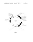 |
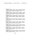 |  |
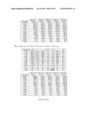 | 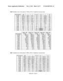 |
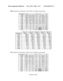 | 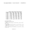 |
 |  |
 |  |
 | 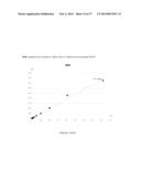 |
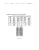 | 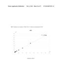 |
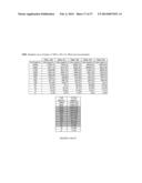 |  |
 |  |
 |  |
 |  |
 |  |
 |  |
 |  |
 |  |
 |  |
 |  |
 |  |
 | 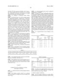 |
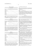 |  |
 |  |
 |  |
 |  |
 |  |
 |  |
 |  |
| Similar patent applications: | |
| Date | Title |
|---|---|
| 2013-12-12 | Multi-specific antigen-binding molecule having alternative function to function of blood coagulation factor viii |
| 2013-12-12 | Aqueous method of making magnetic iron oxide nanoparticles |
| 2013-12-12 | Levels of blys/april heterotrimers in serum and use in diagnostic methods |
| 2013-12-12 | Rapid generation of anti-idiotypic antibodies |
| Top Inventors for class "Drug, bio-affecting and body treating compositions" | |
| Rank | Inventor's name |
|---|---|
| 1 | David M. Goldenberg |
| 2 | Hy Si Bui |
| 3 | Lowell L. Wood, Jr. |
| 4 | Roderick A. Hyde |
| 5 | Yat Sun Or |