Patent application title: DETECTION OF EXTRACELLULAR JCV MICRORNAS
Inventors:
Christopher Sullivan (Austin, TX, US)
Chun Jun Chen (Austin, TX, US)
IPC8 Class: AC12Q168FI
USPC Class:
435 612
Class name: Measuring or testing process involving enzymes or micro-organisms; composition or test strip therefore; processes of forming such composition or test strip involving nucleic acid with significant amplification step (e.g., polymerase chain reaction (pcr), etc.)
Publication date: 2013-10-17
Patent application number: 20130273549
Abstract:
JCV-miRNA compositions and methods for detecting glia-derived JCV-miRNA
are provided. The compositions and methods of the present invention are
particularly useful as a non-invasive biomarker prognostic and/or
diagnostic for patients suffering from PML.Claims:
1. A method of detecting a glia-derived extracellular JCV-miRNA in a
sample derived from a subject, said method comprising: (i) isolating a
glia-derived extracellular JCV-miRNA within a sample derived from a
subject thereby producing isolated glia-derived extracellular JCV-miRNA;
(ii) reverse transcribing said isolated glia-derived extracellular
JCV-miRNA thereby producing glia-derived JCV-cDNA; (iii) amplifying said
glia-derived JCV-cDNA thereby forming a plurality of amplified
glia-derived JCV-cDNAs and a plurality of complementary glia-derived
JCV-cDNAs; and (iv) detecting the presence of said plurality of amplified
extracellular glia-derived JCV-cDNAs or said plurality of complementary
glia-derived JCV-cDNAs thereby detecting said glia-derived extracellular
JCV-miRNA in said sample.
2. The method of claim 1 wherein detecting said glia-derived extracellular JCV-miRNA in said sample indicates detecting a JCV-infected glia within said subject.
3. The method of claim 1, wherein said detecting comprises: (a) determining an amount of said plurality of amplified glia-derived extracellular JCV-cDNAs or said plurality of complementary glia-derived extracellular JCV-cDNAs; (b) based on said amount, determining a level of glia-derived extracellular JCV-miRNAs within said sample; and (c) comparing said level to a standard control level, wherein said level being higher than said standard control level is indicative of said subject having PML.
4. The method of one of claim 1, wherein said subject is a mammalian subject.
5. The method of one of claim 1, wherein said subject is a human subject.
6. The method of one of claim 1, wherein said subject is a PML patient.
7. The method of one of claim 1, wherein said sample is a fluid sample.
8. The method of one of claim 1, wherein said sample is a blood sample or a urine sample.
9. The method of claim 8, wherein said blood sample is a blood plasma sample.
10. The method of one of claim 8, wherein said blood sample is a blood serum sample.
11. The method of any one of claim 1, wherein said isolating comprises centrifuging said sample under conditions suitable to form an enriched glia-derived extracellular JCV-miRNA sample fraction.
12. The method of any one of claim 1, wherein said glia-derived extracellular JCV-miRNA within said sample forms part of a JCV-miRNA infected glia-derived exosome.
13. The method of claim 12, wherein said isolating comprises separating said JCV-miRNA infected glia-derived exosome from components of said sample.
14. The method of claim 13, wherein said separating comprises contacting said JCV-miRNA infected glia-derived exosome with a glia-derived exosome-binding protein to form an glia-derived exosome-binding protein complex and isolating said exosome-binding protein complex.
15. The method of claim 14, wherein said glia-derived exosome-binding protein is an glia-derived exosome binding antibody.
16. The method of claim 1, wherein said reverse transcribing comprises contacting said isolated glia-derived extracellular JCV-miRNA with a JCV-miRNA nucleic acid probe, a reverse transcriptase and nucleic acid monomers.
17. An isolated glia-derived extracellular JCV-miRNA.
18. The isolated glia-derived extracellular JCV-miRNA of claim 17, wherein said isolated glia-derived extracellular JCV-miRNA forms part of a JCV-miRNA infected glia exosome.
19. A JCV-miRNA infected glia-derived exosome.
Description:
CROSS-REFERENCES TO RELATED APPLICATIONS
[0001] This application claims priority to U.S. Provisional Patent Application No. 61/624,956, filed Apr. 16, 2012, entitled "DETECTION OF EXTRACELLULAR JCV MICRORNAS" the disclosure of which is incorporated by reference herein in its entirety.
REFERENCE TO A "SEQUENCE LISTING," A TABLE, OR A COMPUTER PROGRAM LISTING APPENDIX SUBMITTED ON A COMPACT DISK
[0002] The Sequence Listing written in file 93331-872292_ST25.TXT, created on Apr. 16, 2013, 24,116 bytes, machine format IBM-PC, MS-Windows operating system, is hereby incorporated by reference.
BACKGROUND OF THE INVENTION
[0003] Current diagnostics for Progressive Multifocal Leukoencephalopathy (PML) involve detection of JCV DNA via the polymerase chain reaction (PCR) from cerebrospinal fluid (CSF) or a direct brain biopsy (Brew et al., 2010; Major, 2010). Both of these assays are invasive and impractical for routine sampling. Other PML biomarker approaches have attempted to utilize PCR for JCV from isolated blood/serum or detection of immunoreactive antibodies against JCV. Both of these approaches fall short as biomarkers for PML since many healthy non-PML patients have been previously exposed to JCV and will periodically have JCV viremia. In fact, approximately 50 to 80% of the human population is seropositive for JCV antibodies as JCV is found to persistently infect the kidney and perhaps other non-neural tissues such as lymphocytes (Brew et al, 2010). Thus, being seropositive for JCV-reactive antibodies or even having viral DNA detected in bodily fluids via PCR is not predictive of the neural disease PML.
[0004] In patients with PML, magnetic resonance imaging (MRI) can sometimes be used to reveal changes in local brain features characteristic of this condition. However, this methodology misses many cases of PML and is not amenable as an early diagnostic of PML. Furthermore, other neurological disorders can cause white matter abnormalities such as multiple sclerosis and systemic lupus erythematosus (Brew et al., 2010). Therefore, at present, a definitive diagnosis for PML can only be confirmed by the detection of JCV DNA in the CSF or in brain biopsy, which is too invasive to be routinely performed as a prognostic diagnostic for rare incidences of PML associated with various drug regimens.
[0005] Several potentially important drugs that could benefit patients including: Natalizumab (trade name Tysabri) for multiple sclerosis, efalizumab (trade name Raptiva) for psoriasis and rituximab (trade name Rituxan) for arthritis are associated with rare occurrences of PML. The gold standard for the diagnosis of PML is through a brain biopsy, in combination with the detection of JCV DNA in the CSF via PCR. Both methodologies are invasive in nature. Thus, there is a need in the art for non-invasive and accurate methods for detecting JCV infections. The present invention addresses these and other needs in the art.
BRIEF SUMMARY OF THE INVENTION
[0006] Disclosed herein, inter alia, are methods and materials useful to PML detection in AIDS patients and patients on autoimmune anti-inflammatory drugs. The methods and materials provided herein are of significant importance, for example, for a non-invasive early detection assay for PML. In some embodiments, the methods and compositions disclosed herein allow detection of JCV microRNAs as a biomarker for PML providing a non-invasive and sensitive prognostic/diagnostic tool that is currently lacking for PML. In some other embodiments, the methods and compositions disclosed herein proved detection of glia-derived exosomes having JCV-miRNA for use as a prognostic/diagnostic tool for non-invasive detection of PML.
[0007] In one aspect, an isolated glia-derived extracellular JCV-miRNA is provided. In some embodiments, the glia-derived extracellular JCV-miRNA is included in a JCV-miRNA infected exosome. In certain embodiments the isolated glia-derived extracellular JCV-miRNA is obtained from a mammal. In certain embodiments the isolated glia-derived extracellular JCV-miRNA is obtained from a human. In some embodiments, the human is suffering from PML. In certain embodiments the isolated glia-derived extracellular JCV-miRNA is derived from a fluid sample. In some embodiments, the fluid sample is a blood sample or a urine sample. In some embodiments, the blood sample is a blood plasma sample or a blood serum sample.
[0008] In another aspect, a JCV-miRNA infected glia-derived exosome is provided. In some embodiments, the JCV-miRNA infected glia-derived exosome is isolated from a mammalian subject. In certain embodiments, the JCV-miRNA infected glia-derived exosome is isolated from a human subject. In some embodiments, the subject is suffering from PML.
[0009] In another aspect, a method of detecting a glia-derived extracellular JCV miRNA in a sample derived from a subject is provided. The method includes isolating a glia-derived extracellular JCV miRNA within a sample derived from a subject thereby producing isolated glia-derived extracellular JCV miRNA. The isolated glia-derived extracellular JCV miRNA is reversed transcribed, thereby producing glia derived JCV cDNA. The glia-derived JCV cDNA is amplified thereby forming a plurality of amplified glia-derived JCV cDNAs and a plurality of complementary glia-derived JCV cDNAs. The presence of the plurality of amplified extracellular glia-derived JCV cDNAs or the plurality of complementary glia-derived JCV cDNAs is detected thereby detecting the glia-derived extracellular JCV miRNA in the sample.
BRIEF DESCRIPTION OF THE DRAWINGS
[0010] FIG. 1: Exosomes isolated from JCV-infected glia cells. FIG. 1A and FIG. 1B: Exosomes were isolated from JCV Mad1 strain (one of the strains that cause PML) infected SVGA cells (astrocytes) by ultracentrifugation. FIGS. 1a and 1b show Transmission Electron Microscopy images taken from the exosomes preparation. Arrows indicate the exosomes isolated from the JCV infected SVGA culture supernatant. FIG. 1c shows a western blot analysis for CD63, a transmembrane protein that is enriched on exosomes.
[0011] FIG. 2: RNA gel analysis shows JCV miRNA can be detected in exosomes isolated from JCV Mad1 infected SVGA cell culture supernatant. The box indicates that JCV miRNA is detectable in exosomes from JCV Mad1 infected SVGA cell culture supernatant but not from exosomes from uninfected SVGA cell culture supernatant. Lines - and 1-12 from left to right correspond to the following samples: -) negative control; 1) mock-infected total RNA without DNAse treatment at room temperature (RT); 2) mock-infected total RNA without DNAse treatment no RT; 3) mock-infected total RNA with DNAse treatment at RT; 4) mock-infected total RNA with DNAse treatment no RT; 5) JCV-infected total RNA without DNAse treatment at RT; 6) JCV-infected total RNA without DNAse treatment no RT; 7) JCV-infected total RNA with DNAse treatment at RT; 8) JCV-infected total RNA with DNAse treatment no RT; 9) mock-infected ultracentrifuged RNA at RT; 10) mock-infected ultracentrifuged RNA no RT; 11) JCV-infected ultracentrifuged RNA at RT; 12) JCV-infected ultracentrifuged RNA no RT;
[0012] FIG. 3: Cartoon flow chart for a method of isolating JCV-miRNA from exosomes. Exosomes (1) that crosses the Blood-brain barrier (2) from a patient are isolated from either the blood serum (3) or from the urine samples (4). If exosomes of glial-origin is of high enough representation without enrichment in the blood serum or urine samples, quantitative stem-loop RT-PCR can be performed on the exosomes to detect JCV microRNAs. If enrichment is required, immunoaffinity pull-down (5) of glia exosomes can be performed and exosomes are captured (6) in microtiter plates for subsequent PCR analysis (7). After the enrichment process, quantitative stem-loop RT-PCR is performed to detect JCV microRNAs in those glia secreted exosomes.
DETAILED DESCRIPTION OF THE INVENTION
I. Definitions
[0013] The following definitions are provided to facilitate understanding of certain terms used frequently herein and are not meant to limit the scope of the present disclosure. Unless defined otherwise, all technical and scientific terms used herein have the same meaning as commonly understood by one of ordinary skill in the art to which this invention belongs.
[0014] A "glia" or "glia cell" as used herein, refers to any neuroglial cell, such as an astrocyte or an oligodendrocyte. Glia cells do not participate directly in synaptic contacts. Instead, glia cells function to maintain the ionic milieu of nerve cells, modulate the rate of signal propagation, modulate uptake of neurotransmitters, provide scaffolding for neural development, and providing myelin sheets to some axons. There are three types of glia cells: microglia, astrocytes, and oligodendrocytes. Astrocytes regulate chemical environments whereas oligodendrocytes provide myelin sheets for some axons. (Purves D, Augustine G J, Fitzpatrick D, et al., editors. Neuroscience. 2nd edition. Sunderland (MA): Sinauer Associates; 2001. Neuroglial Cells.)
[0015] A "JCV-infected glia" as used herein refers to any glia cell having active JCV infection. In some embodiments, the glia actively secretes exosomes containing JCV-miRNA.
[0016] The term "nucleic acid monomers" refers to nucleoside or nucleotide. The nucleic acid monomers are typically useful precursors for making nucleic acid polymers (polynucleotides) enzymatically. Exemplary nucleic acid monomers include adenosine, guanosine, cytidine, uridine, thymidine, which may be present as monophosphates, diphosphates, and triphosphates. The term "polynucleotide" refers to a linear sequence of nucleotides joined by a phosphodiester linkage between the 5' and 3' carbon atoms.
[0017] The words "complementary" or "complementarity" refer to the ability of a nucleic acid in a polynucleotide to form a base pair with another nucleic acid in a second polynucleotide. For example, the sequence A-G-T is complementary to the sequence T-C-A. Complementarity may be partial, in which only some of the nucleic acids match according to base pairing, or complete, where all the nucleic acids match according to base pairing.
[0018] The terms "identical" or percent "identity," in the context of two or more nucleic acids, refer to two or more sequences or subsequences that are the same or have a specified percentage of nucleotides that are the same (i.e., about 60% identity, preferably 65%, 70%, 75%, 80%, 85%, 90%, 91%, 92%, 93%, 94%, 95%, 96%, 97%, 98%, 99%, or higher identity over a specified region, when compared and aligned for maximum correspondence over a comparison window or designated region) as measured using a BLAST or BLAST 2.0 sequence comparison algorithms with default parameters described below, or by manual alignment and visual inspection (see, e.g., NCBI web site or the like). Such sequences are then said to be "substantially identical." This definition also refers to, or may be applied to, the compliment of a test sequence. The definition also includes sequences that have deletions and/or additions, as well as those that have substitutions. As described below, the preferred algorithms can account for gaps and the like. Preferably, identity exists over a region that is at least about 25 amino acids or nucleotides in length, or more preferably over a region that is 50-100 amino acids or nucleotides in length.
[0019] The phrase "stringent hybridization conditions" refers to conditions under which a probe will hybridize to its target sequence, typically in a complex mixture of nucleic acids, but not to other sequences. Stringent conditions are sequence-dependent and will be different in different circumstances. Longer sequences hybridize specifically at higher temperatures. An extensive guide to the hybridization of nucleic acids is found in Tijssen, TECHNIQUES IN BIOCHEMISTRY AND MOLECULAR BIOLOGY--HYBRIDIZATION WITH NUCLEIC PROBES, "Overview of principles of hybridization and the strategy of nucleic acid assays" (1993). Generally, stringent conditions are selected to be about 5-10° C. lower than the thermal melting point (Tm) for the specific sequence at a defined ionic strength pH. The Tm is the temperature (under defined ionic strength, pH, and nucleic concentration) at which 50% of the probes complementary to the target hybridize to the target sequence at equilibrium (as the target sequences are present in excess, at Tm, 50% of the probes are occupied at equilibrium). Stringent conditions may also be achieved with the addition of destabilizing agents such as formamide. For selective or specific hybridization, a positive signal is at least two times background, preferably 10 times background hybridization. Exemplary stringent hybridization conditions can be as following: 50% formamide, 5×SSC, and 1% SDS, incubating at 42° C., or, 5×SSC, 1% SDS, incubating at 65° C., with wash in 0.2×SSC, and 0.1% SDS at 65° C.
[0020] A variety of methods of specific DNA and RNA measurement that use nucleic acid hybridization techniques are known to those of skill in the art (see, Sambrook, supra). Some methods involve electrophoretic separation (e.g., Southern blot for detecting DNA, and Northern blot for detecting RNA), but measurement of DNA and RNA can also be carried out in the absence of electrophoretic separation (e.g., by dot blot).
[0021] The sensitivity of the hybridization assays may be enhanced through use of a nucleic acid amplification system that multiplies the target nucleic acid being detected. Examples of such systems include the polymerase chain reaction (PCR) system and the ligase chain reaction (LCR) system. Other methods recently described in the art are the nucleic acid sequence based amplification (NASBA, Cangene, Mississauga, Ontario) and Q Beta Replicase systems. These systems can be used to directly identify mutants where the PCR or LCR primers are designed to be extended or ligated only when a selected sequence is present. Alternatively, the selected sequences can be generally amplified using, for example, nonspecific PCR primers and the amplified target region later probed for a specific sequence indicative of a mutation. It is understood that various detection probes, including Taqman® and molecular beacon probes can be used to monitor amplification reaction products, e.g., in real time.
[0022] The word "polynucleotide" refers to a linear sequence of nucleotides. The nucleotides can be ribonucleotides, deoxyribonucleotides, or a mixture of both. Examples of polynucleotides contemplated herein include single and double stranded DNA, single and double stranded RNA (including miRNA), and hybrid molecules having mixtures of single and double stranded DNA and RNA.
[0023] The words "protein", "peptide", and "polypeptide" are used interchangeably to denote an amino acid polymer or a set of two or more interacting or bound amino acid polymers.
[0024] The term "gene" refers to the segment of DNA involved in producing a protein; it includes regions preceding and following the coding region (leader and trailer) as well as intervening sequences (introns) between individual coding segments (exons). The leader, the trailer as well as the introns include regulatory elements that are necessary during the transcription and the translation of a gene. Further, a "protein gene product" is a protein expressed from a particular gene.
[0025] The word "expression" or "expressed" as used herein in reference to a gene means the transcriptional and/or translational product of that gene. The level of expression of a DNA molecule in a cell may be determined on the basis of either the amount of corresponding mRNA that is present within the cell or the amount of protein encoded by that DNA produced by the cell (Sambrook et al., 1989 Molecular Cloning: A Laboratory Manual, 18.1-18.88).
[0026] The term "amplifying" refers to a process in which the nucleic acid is exposed to at least one round of extension, replication, or transcription in order to increase (e.g., exponentially increase) the number of copies (including complementary copies) of the nucleic acid. The process can be iterative including multiple rounds of extension, replication, or transcription. Various nucleic acid amplification techniques are known in the art, such as PCR amplification, primer extension qPCR, rolling circle miRNA amplification, and RT-PCR. RT-PCR amplification of miRNA may be done using stem-loop RT-PCR techniques as described by Chen, et al, 2005. As used herein, "reverse transcribing" a miRNA to cDNA is done by performing RNA transcription to form cDNA (e.g. via RT-PCR). The reverse transcribing may be performed using available miRNA through the action of a reverse transcriptase, such as SuperScript®III (Invitrogen).
[0027] A "primer", as used herein, refers to a nucleic acid that is capable of hybridizing to a complementary nucleic acid sequence in order to facilitate enzymatic extension, replication or transcription. The term "probe" or "primer", as used herein, is defined to be one or more nucleic acid fragments whose specific hybridization to a sample can be detected. A probe or primer can be of any length depending on the particular technique it will be used for. For example, PCR primers are generally between 10 and 40 nucleotides in length, while nucleic acid probes for, e.g., a Southern blot, can be more than a hundred nucleotides in length. The probe may be unlabeled or labeled so that its binding to the target or sample can be detected. The probe can be produced from a source of nucleic acids from one or more particular (preselected) portions of a chromosome, e.g., one or more clones, an isolated whole chromosome or chromosome fragment, or a collection of polymerase chain reaction (PCR) amplification products. Non-limiting examples of primers useful for the detection of glia-derived JCV-cDNA are set forth in Table 3. In some embodiments, the glia-derived JCV-cDNA is amplified using the primers set forth in Table 3.
[0028] An "PLP protein" as referred to herein includes any of the naturally-occurring forms of the myelin proteolipid protein, or variants thereof that maintain PLP protein activity (e.g. within at least 50%, 80%, 90%, 95%, 96%, 97%, 98%, 99% or 100% activity compared to PLP). In some embodiments, variants have at least 90%, 95%, 96%, 97%, 98%, 99% or 100% amino acid sequence identity across the whole sequence or a portion of the sequence (e.g. a 50, 100, 150 or 200 continuous amino acid portion) compared to a naturally occurring PLP polypeptide (e.g. SEQ ID NO:1). In other embodiments, the PLP protein is the protein as identified by the NCBI reference gi:187417 corresponding to SEQ ID NO:1.
[0029] An "CNP protein" as referred to herein includes any of the naturally-occurring forms of the 2',3'-cyclic-nucleotide 3'-phosphodiesterase protein, or variants thereof that maintain CNP protein activity (e.g. within at least 50%, 80%, 90%, 95%, 96%, 97%, 98%, 99% or 100% activity compared to CNP). In some embodiments, variants have at least 90%, 95%, 96%, 97%, 98%, 99% or 100% amino acid sequence identity across the whole sequence or a portion of the sequence (e.g. a 50, 100, 150 or 200 continuous amino acid portion) compared to a naturally occurring CNP polypeptide (e.g. SEQ ID NO:2). In other embodiments, the CNP protein is the protein as identified by the NCBI reference gi:94721261 corresponding to SEQ ID NO:2.
[0030] An "MOG protein" as referred to herein includes any of the naturally-occurring forms of the myelin oligodendrocyte glycoprotein, or variants thereof that maintain MOG protein activity (e.g. within at least 50%, 80%, 90%, 95%, 96%, 97%, 98%, 99% or 100% activity compared to MOG. In some embodiments, variants have at least 90%, 95%, 96%, 97%, 98%, 99% or 100% amino acid sequence identity across the whole sequence or a portion of the sequence (e.g. a 50, 100, 150 or 200 continuous amino acid portion) compared to a naturally occurring MOG polypeptide (e.g. SEQ ID NO:3). In other embodiments, the MOG protein is the protein as identified by the NCBI reference gi:984147 corresponding to SEQ ID NO:3.
[0031] An "MAG protein" as referred to herein includes any of the naturally-occurring forms of the myelin-associated glycoprotein, or variants thereof that maintain MAG protein activity (e.g. within at least 50%, 80%, 90%, 95%, 96%, 97%, 98%, 99% or 100% activity compared to MAG. In some embodiments, variants have at least 90%, 95%, 96%, 97%, 98%, 99% or 100% amino acid sequence identity across the whole sequence or a portion of the sequence (e.g. a 50, 100, 150 or 200 continuous amino acid portion) compared to a naturally occurring MAG polypeptide (e.g. SEQ ID NO:4). In other embodiments, the MAG protein is the protein as identified by the NCBI reference gi:62205282 corresponding to SEQ ID NO:4.
[0032] A "JCV-miRNA nucleic acid probe", as used herein, is a nucleic acid fragment used in combination with a RT and nucleic acid monomers for amplification and detection of JCV-miRNA. The JCV-miRNA nucleic acid probe is typically of sufficient length and complementarity to a JCV genomic sequence as to adequately serve as a primer for amplification. In some embodiments, the complementarity of the JCV-miRNA nucleic acid probe to a JCV genomic sequence is at least about 60%, preferably about 65%, 70%, 75%, 80%, 85%, 90%, 91%, 92%, 93%, 94%, 95%, 96%, 97%, 98%, 99%, or higher.
[0033] The term "sample" includes sections of tissues such as biopsy and autopsy samples, and frozen sections taken for histological purposes. Such samples include blood and blood fractions or products (e.g., serum, plasma, platelets, red blood cells, and the like), sputum, tissue, cultured cells (e.g., primary cultures, explants, and transformed cells), stool, urine, other biological fluids (e.g., prostatic fluid, gastric fluid, intestinal fluid, renal fluid, lung fluid, cerebrospinal fluid, and the like), etc. A sample is typically obtained from a "subject" such as a eukaryotic organism, most preferably a mammal such as a primate, e.g., chimpanzee or human; cow; dog; cat; a rodent, e.g., guinea pig, rat, mouse; rabbit; or a bird; reptile; or fish. In some embodiments, the sample is obtained from a human.
[0034] A "subject," "individual" or "patient" is used interchangeably herein, which refers to a vertebrate, preferably a mammal, more preferably a human. Mammals include, but are not limited to, murines, simians, humans, farm animals, sport animals, and pets. Tissues, cells and their progeny of a biological entity obtained in vitro or cultured in vitro are also encompassed.
[0035] A "control" sample or value refers to a sample that serves as a reference, usually a known reference, for comparison to a test sample. For example, a test sample can be taken from a test condition, e.g., in the presence of a test compound, and compared to samples from known conditions, e.g., in the absence of the test compound (negative control), or in the presence of a known compound (positive control). A control can also represent an average value gathered from a number of tests or results. One of skill in the art will recognize that controls can be designed for assessment of any number of parameters. For example, a control can be devised to compare therapeutic benefit based on pharmacological data (e.g., half-life) or therapeutic measures (e.g., comparison of side effects). One of skill in the art will understand which controls are valuable in a given situation and be able to analyze data based on comparisons to control values. Controls are also valuable for determining the significance of data. For example, if values for a given parameter are widely variant in controls, variation in test samples will not be considered as significant. Examples of control samples include but are not limited to; a control sample level from a healthy patient sample(s), a level taken from a known PML patient sample(s), a level from a patient with known JCV viremia, a level from a sample from the same subject taken at an earlier time, or any combination thereof.
[0036] The term "isolated" refers to any cell or cellular component such as a nucleotide, polynucleotide, peptide, polypeptide, or exosome, that is partially or completely separated from components with which it is naturally associated (such as proteins, nucleic acids, cells, tissues).
[0037] As used herein, "PML" or "progressive multifocal leukoencephalopathy" refers to a fatal neurological condition consistently associated with JC virus infection. PML can occur in any white matter regions of the brain resulting in demyelination of neurons. Atypical oligodendrocytes and extremely large astrocytes are found in the brains of PML patients. Previously a rare condition, generally occurs in patients with marked cellular immunodeficiency. Thus, PML has become more prevalent with the emergence of HIV. PML is also associated with transplantation rejection, leukemia patients, patients with chronic inflammation and has recently been associated with immunosuppressant antibody drugs such as Natalizumab. Detection of PML may be performed through brain biopsy, PCR detection of JCV DNA in the CSF, MRI detection of focal abnormalities, or through observation of worsening neurological symptoms.
[0038] "JC virus" or "John Cunningham Virus" or "JCV" is a member of the Polyomavirus family. JCV typically has a small circular DNA genome of approximately 5 kilobases. JCV infection is associated with PML. JCV infection, either latent or active, is common and it is estimated between 50-80% of the population is seropositive for JCV antibodies. JCV can cross the blood brain barrier and upon reactivation, typically due to immunodeficiency or immunosuppression, causes focal demyelination leading to PML.
[0039] As used herein, the term "miRNA" or "microRNA" is used herein according to its normal meaning as single-stranded RNA molecules of about 17-25 (e.g. 17-23) nucleotides in length typically capable of modifying gene expression and primary transcripts (pri-miRNAs), pre-cursors such as stem-loop precursors (pre-miRNAs) and variants thereof. Naturally-occurring miRNAs are typically transcribed from genes but are not translated into protein. miRNA's are typically first transcribed as a pri-miRNA (from about 45 nt's to 1000's of nts) that may include at least one hairpin structure (from about 37-120 nt). The pri-miRNA is typically processed to short pre-miRNA stem-loop precursors of about 40-80 nt. The sequence of the pre-miRNA may include the entire miRNA sequence. The sequence of the pre-miRNA may comprise the sequence of a hairpin loop. Pre-miRNA is typically processed by cleavage of a stem-loop, forming mature miRNAs of about 17-25 nt derived from either or both arms of the hairpin.
[0040] miRNA sequences are involved in various physiological and pathological conditions, including differentiation, development, tumorigenesis, and neurological disorders. miRNAs may function by binding to the 3'UTR of target mRNAs and inhibiting expression of protein from these transcripts. miRNAs may also direct cleavage of transcripts if they have perfect complementarity to such transcript.
[0041] As used herein, the term "viral miRNA" or "viral microRNA" refers to miRNA having a viral origin. Viral miRNA may be encoded by viruses with DNA genomes or retroviruses with RNA genomes. Viral miRNA may cleave early viral mRNA transcripts or host gene transcripts.
[0042] "JCV-miRNA" refers to a viral miRNA encoded by JC virus. In one such example, JCV encodes an approximately 60 nt pre-miRNA that is processed into different mature miRNAs approximately 22 nt in length.
[0043] As used herein, the term "glia-derived extracellular JCV-miRNA" refers to a miRNA derived from a JCV infected glial cell. Thus, the glia-derived extracellar JCV-miRNA typically has a JC viral origin in which the JCV has infected a glial cell (such as an oligodendrocyte or astrocyte). The glia-derived extracellular JCV-miRNA is typically present outside of the cellular matrix of the glial cell. An "isolated glia-derived extracellular JCV-miRNA" is a glia-derived extracellular JCV-miRNA that is partially or completely separated from components found in the sample in which the glia-derived extracellular JCV-miRNA is found. In some embodiments, the glia-derived extracellular JCV-miRNA includes a nucleotide sequence corresponding to SEQ ID NO:8 or a functional fragment thereof. In some embodiments, the glia-derived extracellular JCV-miRNA includes a nucleotide sequence corresponding to SEQ ID NO:9 or a functional fragment thereof. In some embodiments, the glia-derived extracellular JCV-miRNA includes a nucleotide sequence corresponding to SEQ ID NO:11 or a functional fragment thereof. In some embodiments, the glia-derived extracellular JCV-miRNA has the nucleotide sequence of SEQ ID NO:8. In some embodiments, the glia-derived extracellular JCV-miRNA has the nucleotide sequence of SEQ ID NO:9. In some embodiments, the glia-derived extracellular JCV-miRNA has the nucleotide sequence of SEQ ID NO:11.
[0044] An "enriched glia-derived extracellular JCV-miRNA sample fraction" is a collection of glia-derived extracellular JCV-miRNAs derived from a sample having glia-derived extracellular JCV-miRNAs, in which the concentration of the glia-derived extracellular JCV-miRNAs is increased relative to the concentration of the glia-derived extracellular JCV-miRNAs in the sample from which the sample fraction is derived. A variety of enrichment techniques may be used, such as differential centrifugation or column chromatography including size exclusion chromatography or affinity chromatography.
[0045] The term "glia-derived JCV-cDNAs" refers to a cDNA complementary to glia-derived JCV-miRNA (prepared e.g. via RT-PCR). In some embodiment, the glia-derived JCV-cDNA matches the original DNA sequence encoding the JCV-miRNA. "Complementary glia-derived JCV-cDNA" refers to synthesized strands of cDNA complementary to glia-derived JCV-cDNA. In some embodiments, the complementary glia-derived JCV-cDNA matches the original miRNA sequence.
[0046] As used herein, the term "exosome" refers to a microvesicle secreted into the extracellular milieu from a cell, such as a glial cell (e.g. oligodendrocytes or astrocytes). Exosome secretion typically occurs through reverse budding of the limiting membrane of multivesicular endosomes forming microvesicles (e.g. of about 50-100 nm in diameter). Exosomes may have the capability of crossing the blood-brain barrier. Exosomes may contain cytosol and extracellular domains of membrane-bound cellular proteins on their surface. As such, exosomes may also contain additional cellular components such as nucleic acids, peptides, and proteins. Exosomes may also contain miRNA from either or both the host or a virus. Exosomes may be released at higher rates during infection with certain viruses or other various stressors. The release of exosomes may increase intercellular communication. Exosomes may be extracted from biological fluids such as blood, serum, urine. As used herein, a "glia-derived exosome" refers to an exosome originating from a glial cell.
[0047] A "JCV-miRNA infected glia exosome", as used herein, refers to an exosome derived from a glial cell, such as an astrocyte or oligodendrocyte, which has at least one JCV-miRNA contained therein.
[0048] A "glia-derived exosome binding protein", as used herein, refers to protein (e.g. peptide or polypeptide) that binds to an exosome (e.g. to a protein found on the surface of an exosome in nature, commonly referred to as an exosomal surface protein). Examples of exosomal surface proteins useful for the invention provided herein including embodiments thereof are without limitation major myelin proteolipid protein (PLP), 2'3'-cyclic-nucleotide-phosphodiesterase (CNP), myelin oligodendrocyte glycoprotein (MOG) or myelin-associated glycoprotein (MAG). Glia-derived exosome binding proteins can be conjugated with exogenously added conjugates for detection or purification chromatographic techniques such as affinity chromatography or antibody-based immunoprecipitation. An "exosome-binding protein complex", as used herein, refers to a glia-derived exosome binding protein covalently or non-covalently bound to an exosome (e.g. to an exosomal surface protein). In some embodiments, complexing occurs when a glia-derived exosome binding protein binds to an exosomal surface protein. Such complexes may be formed in vitro and are useful for detection and purification. An "exosome binding antibody", as used herein, refers to an antibody capable of recognizing glia-derived exosomes. Exosome binding antibodies may be used for affinity chromatography or immunoprecipitation of glia-derived exosomes.
[0049] "Antibody" refers to a polypeptide comprising a framework region from an immunoglobulin gene or fragments thereof that specifically binds and recognizes an antigen. The recognized immunoglobulin genes include the kappa, lambda, alpha, gamma, delta, epsilon, and mu constant region genes, as well as the myriad immunoglobulin variable region genes. Light chains are classified as either kappa or lambda. Heavy chains are classified as gamma, mu, alpha, delta, or epsilon, which in turn define the immunoglobulin classes, IgG, IgM, IgA, IgD and IgE, respectively. Typically, the antigen-binding region of an antibody will be most critical in specificity and affinity of binding.
[0050] An exemplary immunoglobulin (antibody) structural unit comprises a tetramer. Each tetramer is composed of two identical pairs of polypeptide chains, each pair having one "light" (about 25 kD) and one "heavy" chain (about 50-70 kD). The N-terminus of each chain defines a variable region of about 100 to 110 or more amino acids primarily responsible for antigen recognition. The terms variable light chain (VL) and variable heavy chain (VH) refer to these light and heavy chains respectively.
[0051] Antibodies exist, e.g., as intact immunoglobulins or as a number of well-characterized fragments produced by digestion with various peptidases. Thus, for example, pepsin digests an antibody below the disulfide linkages in the hinge region to produce F(ab)'2, a dimer of Fab which itself is a light chain joined to VH-CH1 by a disulfide bond. The F(ab)'2 may be reduced under mild conditions to break the disulfide linkage in the hinge region, thereby converting the F(ab)'2 dimer into an Fab' monomer. The Fab' monomer is essentially Fab with part of the hinge region (see Fundamental Immunology (Paul ed., 3d ed. 1993). While various antibody fragments are defined in terms of the digestion of an intact antibody, one of skill will appreciate that such fragments may be synthesized de novo either chemically or by using recombinant DNA methodology. Thus, the term antibody, as used herein, also includes antibody fragments either produced by the modification of whole antibodies, or those synthesized de novo using recombinant DNA methodologies (e.g., single chain Fv) or those identified using phage display libraries (see, e.g., McCafferty et al., Nature 348:552-554 (1990)).
[0052] For preparation of suitable antibodies of the invention and for use according to the invention, e.g., recombinant, monoclonal, or polyclonal antibodies, many techniques known in the art can be used (see, e.g., Kohler & Milstein, Nature 256:495-497 (1975); Kozbor et al., Immunology Today 4: 72 (1983); Cole et al., pp. 77-96 in Monoclonal Antibodies and Cancer Therapy, Alan R. Liss, Inc. (1985); Coligan, Current Protocols in Immunology (1991); Harlow & Lane, Antibodies, A Laboratory Manual (1988); and Goding, Monoclonal Antibodies: Principles and Practice (2d ed. 1986)). The genes encoding the heavy and light chains of an antibody of interest can be cloned from a cell, e.g., the genes encoding a monoclonal antibody can be cloned from a hybridoma and used to produce a recombinant monoclonal antibody. Gene libraries encoding heavy and light chains of monoclonal antibodies can also be made from hybridoma or plasma cells. Random combinations of the heavy and light chain gene products generate a large pool of antibodies with different antigenic specificity (see, e.g., Kuby, Immunology (3rd ed. 1997)). Techniques for the production of single chain antibodies or recombinant antibodies (U.S. Pat. No. 4,946,778, U.S. Pat. No. 4,816,567) can be adapted to produce antibodies to polypeptides of this invention. Also, transgenic mice, or other organisms such as other mammals, may be used to express humanized or human antibodies (see, e.g., U.S. Pat. Nos. 5,545,807; 5,545,806; 5,569,825; 5,625,126; 5,633,425; 5,661,016, Marks et al., Bio/Technology 10:779-783 (1992); Lonberg et al., Nature 368:856-859 (1994); Morrison, Nature 368:812-13 (1994); Fishwild et al., Nature Biotechnology 14:845-51 (1996); Neuberger, Nature Biotechnology 14:826 (1996); and Lonberg & Huszar, Intern. Rev. Immunol. 13:65-93 (1995)). Alternatively, phage display technology can be used to identify antibodies and heteromeric Fab fragments that specifically bind to selected antigens (see, e.g., McCafferty et al., Nature 348:552-554 (1990); Marks et al., Biotechnology 10:779-783 (1992)). Antibodies can also be made bispecific, i.e., able to recognize two different antigens (see, e.g., WO 93/08829, Traunecker et al., EMBO J. 10:3655-3659 (1991); and Suresh et al., Methods in Enzymology 121:210 (1986)). Antibodies can also be heteroconjugates, e.g., two covalently joined antibodies, or immunotoxins (see, e.g., U.S. Pat. No. 4,676,980, WO 91/00360; WO 92/200373; and EP 03089).
[0053] Methods for humanizing or primatizing non-human antibodies are well known in the art. Generally, a humanized antibody has one or more amino acid residues introduced into it from a source which is non-human. These non-human amino acid residues are often referred to as import residues, which are typically taken from an import variable domain. Humanization can be essentially performed following the method of Winter and co-workers (see, e.g., Jones et al., Nature 321:522-525 (1986); Riechmann et al., Nature 332:323-327 (1988); Verhoeyen et al., Science 239:1534-1536 (1988) and Presta, Curr. Op. Struct. Biol. 2:593-596 (1992)), by substituting rodent CDRs or CDR sequences for the corresponding sequences of a human antibody. Accordingly, such humanized antibodies are chimeric antibodies (U.S. Pat. No. 4,816,567), wherein substantially less than an intact human variable domain has been substituted by the corresponding sequence from a non-human species. In practice, humanized antibodies are typically human antibodies in which some CDR residues and possibly some FR residues are substituted by residues from analogous sites in rodent antibodies.
[0054] A "chimeric antibody" is an antibody molecule in which (a) the constant region, or a portion thereof, is altered, replaced or exchanged so that the antigen binding site (variable region) is linked to a constant region of a different or altered class, effector function and/or species, or an entirely different molecule which confers new properties to the chimeric antibody, e.g., an enzyme, toxin, hormone, growth factor, drug, etc.; or (b) the variable region, or a portion thereof, is altered, replaced or exchanged with a variable region having a different or altered antigen specificity. The preferred antibodies of, and for use according to the invention include humanized and/or chimeric monoclonal antibodies.
II. Compositions
[0055] In one aspect, an isolated glia-derived extracellular JCV-miRNA is provided. In some embodiments, the glia-derived extracellular JCV-miRNA is included in a JCV-miRNA infected exosome. In certain embodiments the isolated glia-derived extracellular JCV-miRNA is obtained from a mammal. In certain embodiments the isolated glia-derived extracellular JCV-miRNA is obtained from a human. In some embodiments, the human is suffering from PML. In certain embodiments the isolated glia-derived extracellular JCV-miRNA is derived from a fluid sample. In some embodiments, the fluid sample is a blood sample or a urine sample. In some embodiments, the blood sample is a blood plasma sample or a blood serum sample.
[0056] In another aspect, a JCV-miRNA infected glia-derived exosome is provided. In some embodiments, the JCV-miRNA infected glia-derived exosome is isolated from a mammalian subject. In certain embodiments, the JCV-miRNA infected glia-derived exosome is isolated from a human subject. In some embodiments, the subject is suffering from PML.
III. Methods of Detection
[0057] In another aspect, method of detecting a glia-derived extracellular JCV miRNA in a sample derived from a subject is provided. The method includes isolating a glia-derived extracellular JCV miRNA within a sample derived from a subject thereby producing isolated glia-derived extracellular JCV miRNA. The isolated glia-derived extracellular JCV miRNA is reversed transcribed, thereby producing glia derived JCV cDNA. The glia-derived JCV cDNA is amplified thereby forming a plurality of amplified glia-derived JCV cDNAs and a plurality of complementary glia-derived JCV cDNAs. The presence of the plurality of amplified extracellular glia-derived JCV cDNAs or the plurality of complementary glia-derived JCV cDNAs is detected thereby detecting the glia-derived extracellular JCV miRNA in the sample. In embodiments, the sample is obtained from the subject.
[0058] In some embodiments, the detecting the glia-derived extracellular JCV-miRNA in the sample indicates detecting a JCV-infected glia within the subject. In other embodiments, the detecting includes determining an amount of the plurality of amplified glia-derived extracellular JCV-cDNAs or the plurality of complementary glia-derived extracellular JCV-cDNAs. Based on the amount, a level of glia-derived extracellular JCV-miRNAs is determined within the sample. And the level is compared to a standard control level, wherein the level being higher than the standard control level is indicative of the subject having PML.
[0059] In some embodiments, the subject is a mammalian subject. In other embodiments, the subject is a human subject. In some embodiments, the subject is a PML patient.
[0060] In some embodiments, the sample is a fluid sample. In other embodiments, the sample is a blood sample or a urine sample. In some embodiments, the blood sample is a blood plasma sample. In other embodiments the blood sample is a blood serum sample.
[0061] In some embodiments, the isolating includes centrifuging the sample under conditions suitable to form an enriched glia-derived extracellular JCV-miRNA sample fraction. In some embodiments, the glia-derived extracellular JCV-miRNA within the sample forms part of a JCV-miRNA infected glia-derived exosome. In other embodiments, the isolating includes separating the JCV-miRNA infected glia-derived exosome from components of the sample. In some embodiments, the separating includes contacting the JCV-miRNA infected glia-derived exosome with a glia-derived exosome-binding protein to form an glia-derived exosome-binding protein complex and isolating the exosome-binding protein complex. In some embodiments, the glia-derived exosome-binding protein is a glia-derived exosome binding antibody. In some embodiments, the glia-derived exosome binding antibody is capable of binding a myelin proteolipid (PLP) protein. In other embodiments, the glia-derived exosome binding antibody is capable of binding a 2',3'-cyclic-nucleotide 3'-phosphodiesterase (CNP). In other embodiments, the glia-derived exosome binding antibody is capable of binding a myelin oligodendrocyte glycoprotein (MOG). In other embodiments, the glia-derived exosome binding antibody is capable of binding a myelin-associated glycoprotein (MAG).
[0062] In some embodiments, the reverse transcribing includes contacting the isolated glia-derived extracellular JCV-miRNA with a JCV-miRNA nucleic acid probe, a reverse transcriptase and nucleic acid monomers.
IV. Examples
[0063] Cell Culture and Preparation of Culture Medium for Exosomes Isolation
[0064] SVGA cells (SV40 transformed astroglial cells, kindly provided by Walter Atwood, Brown University, Providence, R.I.) were maintained in Minimal Essential Medium Eagle with Earle's salts and L-glutamine (MEM, Cellgro, Manassa, Va.) supplemented with 10% heat inactivated fetal bovine serum (FBS, Sigma-Aldrich, St. Louis, Mo.), penicillin (100 IU/mL) and streptomycin (100 μg/mL) (Cellgro).
[0065] SVGA cells were infected with JCV (Mad1 strain, kindly provided by Walter Atwood) in MEM containing 2% heat inactivated FBS at 37° C. for 1 hour. The virus inoculum was replaced with 30 mL of regular MEM containing 10% heat inactivated FBS.
[0066] Ultracentrifugation Isolation of Exosomes
[0067] Exosomes were isolated from either mock-infected or JCV-infected SVGA cell culture media by first centrifugation at 300 g for 10 minutes at 4° C. (Avanti J-E centrifuge with JS 5.3 swinging bucket rotor, Beckman Coulter, Brea, Calif.). The supernatant was collected and centrifuge at 2000×g for 20 minutes at 4° C. (Avanti J-E centrifuge with a JS 5.3 swinging bucket rotor, Beckman Coulter). The supernatant was then transferred to a 75 mL polycarbonate bottles (ThermoFisher Scientific, Asheville, N.C.). The supernatant was centrifuged at 10,000×g for 30 minutes at 4° C. (Sorvall Ultra Pro 80 with a T-647.5 fixed angle rotor, ThermoFisher Scientific). The supernatant is transferred using a pipet to a fresh 75 mL polycarbonate bottle and centrifuged at 100,000 g for 2 hours and 30 minutes at 4° C. The supernatant was discarded by decanting. The pellet was washed with phosphate-buffered saline (PBS, Cellgro) and centrifuged at 100,000 g for 2 hours and 30 minutes at 4° C. The supernatant was then discarded by decanting. To remove the supernatant as completely as possible, the bottle was kept upside down and the remaining liquid on the side of the mouth of the bottle was removed with an aspirator. The pellet is suspended in 200 μL of PBS and divided into 50 μL aliquots and stored at -80° C.
[0068] RNA Isolation and Stem-Loop RT PCR Detection of JCV microRNA
[0069] Total RNA from mock or JCV-infected SVGA cells were harvested using an in-house PIG-B solution (2M guanidinium thiocyanate (EMD, Billerica, Mass.), 20 mM citrate buffer, pH4.5, 5 mM EDTA (Fisher Scientific, Pittsburgh, Pa.), 0.25% Sarkosyl (Sigma Aldrich), 48% saturated phenol, pH4.5 (Amresco, Solon, Ohio), 2.1% isoamyl alcohol (Fisher Scientific), 0.5% β-mercaptoethanol (Sigma Aldrich), 0.1% 8-hydroxyquinoline (EMD), and 0.0025% Coomassie blue (EMD)) as described previously (1, 2, 3). RNA from mock-infected or JCV-infected SVGA exosomes were isolated using the same protocol as above.
[0070] Prior to reverse transcription, total RNA was either left untreated or treated with TURBO DNase (Ambion, Austin, Tex.) according to the manufacturer's protocol. Reverse transcription was performed using SuperScript III reverse transcriptase (Invitrogen, Grand Island, N.Y.) according to the manufacturer's protocols. PCR was conducted on the reverse transcription product using Taq DNA polymerase (New England BioLabs, Ipswich, Mass.). The primers used were as follows:
TABLE-US-00001 JCV 5p miRNA stem-loop RT primer: GTCGTATCCAGTGCAGGGTCCGAGGTATTCGCACTGGATACGACATGCT TTTC, JCV 5p miRNA PCR forward primer: GGCCTCGTTCTGAGACCTGG, Stem-loop PCR reverse primer: GTGCAGGGTCCGAGGT.
[0071] The PCR program used was a touchdown PCR protocol with the annealing temperature starting at 62° C. for 30 seconds, decreasing by 0.5° C. each cycle for 20 cycles.
[0072] PCR was continued for another 25 cycles at an annealing temperature of 52° C. The extension step was performed at 68° C. for 30 seconds for each cycle. The PCR product was analyzed in a 3% agarose Lithium Borate gel (BioExpress, Kaysville, Utah). The agarose gel was stained by ethidium bromide (Fisher Scientific) and visualized using a Bio-Rad Universal Hood II gel imager (Bio-Rad, Hercules, Calif.).
[0073] Protein Isolation and Western Blot Analysis
[0074] Mock or JCV-infected SVGA cells were lysed in RIPA buffer (150 mM sodium chloride (Avantor Performance Materials, Center Valley, Pa.), 10 mM Tris, pH 7.2 (Fisher Scientific), 0.1% sodium dodecyl sulfate (Avantor Performance Materials), 1% Triton X-100 (Fisher Scientific), 1% sodium deoxycholate (Alfa Aesar, Ward Hill, Mass.), 5 mM EDTA (Fisher Scientific) and 1 tablet of complete mini, EDTA-free protease inhibitor (Roche, Indianapolis, Ind.) per 10 mL of RIPA buffer.
[0075] 1 μg of exosomes samples and 50 μg of mock or JCV-infected SVGA total protein were heated at 70° C. for 10 minutes in SDS sample buffer without β-mercaptoethanol (375 mM Tris-HCL, pH6.8 (Fisher Scientific), 6% (w/v) sodium dodecyl sulfate (Avantor Performance Materials), 48% (v/v) glycerol (Sigma-Aldrich), and 0.03% (w/v) bromophenol blue (Fisher Scientific) and subjected to electrophoresis using a 10% denaturing acrylamide gel (Bio-Rad). Proteins were transferred onto Immobilon-FL PVDF membrane (Millipore, Billerica, Mass.) using the Bio-Rad Mini Trans-Blot electrophoretic transfer cell (Bio-Rad). The membrane was blocked in 5% (w/v) skim milk powder (HEB, San Antonio, Tex.) in Tris-buffered saline with 0.1% (v/v) Tween-20 (TBST, Fisher Scientific) for 1 hour at room temperature. The membrane was probed for CD63 using a mouse monoclonal CD63 antibody (1:1000, Santa Cruz Biotechnology, Santa Cruz, Calif.), overnight at 4° C. After overnight incubation, the membrane was washed with TBST, 4 times, 15 minutes each, followed by incubation with anti-mouse secondary antibody IgG conjugated to HRP (1:5000, Invitrogen) for 1 hour at room temperature. The membrane was washed 4 times with TBST, 15 minutes each. The West Dura chemiluminescent substrate (Thermo Scientific) was used to generate the light signal for visualization on the Blue Ultra Autorad film (BioExpress).
[0076] Transmission Electron Microscopy (TEM)
[0077] Transmission electron microscopy imaging of exosome preparations was performed as described (4), with slight modifications. Briefly, 10 μL of the exosome preparation was mixed with 10 μL of 4% paraformaldehyde (PFA) solution in PBS (USB Corporation, Cleveland, Ohio) to achieve a final concentration of 2% PFA. A drop of the sample was spotted on a piece of parafilm (Pechiney Plastic Packaging, Menasha, Wis.) and a Formvar Carbon Film on 300 square mesh Nickel Grid (Electron Microscopy Sciences, Hatfield, Pa.) was floated on top of the sample. The excess liquid was blotted off and allowed to dry for 20 minutes at room temperature. The grids were washed 8 times with deionized water prior to staining, using the floating method as described above. To stain the samples, a drop of uranyl-acetate solution, pH4.2-4.5 (kindly provided by the Institute of Cellular and Molecular Biology Microscopy Facility, The University of Texas at Austin, Austin, Tex.) was spotted onto a piece of parafilm and the grid was floated on the drop for 5 minutes, followed by blotting off the excess uranyl-acetate solution. The grids were washed one time as described above. Imaging was performed using a Tecnai Spirit BioTwin transmission electron microscope (FEI, Hillsboro, Oreg.) at 80 kV.
[0078] Pre-Enrichment of Exosomes from Cell Culture Media
[0079] Prior to isolation of the neuronal exosomes via proteolipid protein (PLP) specific immunoaffinity capture, supernatant from SVGA cell cultures (mock or infected with JCV Mad1 strain) were subjected to ExoQuick-TC Exosome Precipitation (System Biosciences, Mountain View, Calif.), according to the manufacturer's protocol. Briefly, SVGA cell culture supernatant was centrifuged at 3000 g for 15 minutes at 4° C. 10 mL of the centrifuged SVGA cell culture supernatant was then mixed with 2 mL of ExoQuick-TC Exosome Precipitation Solution. The mixture was placed at 4° C. for overnight. After overnight incubation, the mixture was centrifuged at 1500 g for 30 minutes at 4° C. The resulting supernatant was aspirated. Another centrifugation step of 1500 g at 4° C. was done for an additional 5 minutes. The remaining trace of supernatant was aspirated. The exosome pellet was dissolved in 200 μL of PBS and stored at -80° C. As a positive control, anti-CD63 antibody which recognizes both neuronal and non-neuronal exosomes was used.
[0080] Immunoaffinity Capture of Exosomes from ExoQuick-TC Enriched Exosomes
[0081] 200 μL of either EcoMag® Protein L Magnetic Beads (Bioclone Inc., San Diego, Calif.) or Dynabeads® Protein G beads (Invitrogen, Grand Island, N.Y.) were washed twice with PBS+0.1% Tween 20 (ThermoFisher Scientific, Asheville, N.C.). 10 mg of the following PLP antibodies were added to 200 μL of the beads in a total volume of 400 μL of PBS+0.1% Tween 20 for conjugations to the beads, overnight, at 4° C., with rotation on a Labquake Shaker Rotisserie (ThermoFisher Scientific).
TABLE-US-00002 TABLE 1 Monoclonal antibodies (glia-derived exosome-binding antibodies) useful for isolation of JCV-miRNA infected glia-derived exosomes. Antibody Antigen Monoclonal Mouse IgM Clone#O10 Human PLP Antibody PLP (R&D Systems, Inc., Minneapolis, MN) protein Monoclonal Mouse IgG2a kappa Myelin PLP Antibody (2D7) PLP (Novus Biologicals, Littleton, CO) protein Polyclonal Goat PLP Antibody (G-17) (Santa Cruz Biotech- PLP nology, Inc., Dallas, TX) protein Monoclonal Rat Myelin PLP (Human) Antibody (ImmunoDiag- PLP nostics, Inc., Woburn, MA) protein
[0082] After overnight antibody conjugation onto the magnetic beads, the beads were washed twice with PBS+0.1% BSA (Sigma-Aldrich, St. Louis, Mo.). 50 μL of the ExoQuick-TC enriched exosomes and 50 μL of PBS+0.1% BSA were added to the antibody-conjugated magnetic beads. Immunoaffinity capturing of the neuronal exosomes was done overnight, at 4° C., with rotation as above. After overnight capturing of the neuronal exosomes, the magnetic beads were washed three times with PBS+0.1% BSA. The exosomes-captured magnetic beads are subjected to RNA isolation for stem-loop RT PCR detection of JCV microRNA or western blot analysis.
TABLE-US-00003 TABLE 2 Glia-derived extracellular JCV-miRNA. SEQ ID NO: microRNA Sequences (RNA) Sequences (DNA) Accession# SEQ ID NO: 8 jcv-mir-J1 ucugcaccagaggcuucugagaccug MI0009980 ggaaaagcauugugauugugauuca gugcuuugcuugauccauguccaga gucu ucugcuucaga SEQ ID NO: 9 jcv-mir-J1-5p uucugagaccugggaaaagcau MIMAT0009147 SEQ ID NO: 10 jcv-mir-J1-3p ugcuugauccauguccagaguc MIMAT0009148 SEQ ID NO: 11 jcv-mir-J1-5p TTCTGAGACCTGGGAAAA GCAT SEQ ID NO: 12 jcv-mir-J1-3p TGCTTGATCCATGTCCAG AGTC
TABLE-US-00004 TABLE 3 Primer Pairs for Amplification of glia-derived extracellular JCV-cDNAs. SEQ ID NO: microRNA Sequences (RNA) Length Counts SEQ ID NO: 13 5' miRNA TGTGTGTCTGCACCAGAGGC 20 5 SEQ ID NO: 14 GTGTGTCTGCACCAGAGGC 19 3 SEQ ID NO: 15 TGTGTCTGCACCAGAGGC 18 1 SEQ ID NO: 16 5p miRNA CTTCTGAGACCTGGGAAAAGCATT 24 1 SEQ ID NO: 17 TTCTGAGACCTGGGAAAAGCATTGTGATTG 30 1 SEQ ID NO: 18 TTCTGAGACCTGGGAAAAGCATTG 24 4 SEQ ID NO: 19 TTCTGAGACCTGGGAAAAGCATT 23 7 SEQ ID NO: 20 TTCTGAGACCTGGGAAAAGCAT 22 2 SEQ ID NO: 21 TTCTGAGACCTGGGAAAAGCA 21 1 SEQ ID NO: 22 TGAGACCTGGGAAAAGCATT 20 1 SEQ ID NO: 23 3p miRNA GTGCTTGATCCATGTCCAGAGT 22 6 SEQ ID NO: 24 TGCTTGATCCATGTCCAGAGTCT 23 1 SEQ ID NO: 25 TGCTTGATCCATGTCCAGAGTC 22 3 SEQ ID NO: 26 5' miRNA CTGTGTGTCTGCACCAGAGGC 21 1 SEQ ID NO: 27 TGTGTGTCTGCACCAGAGGC 20 14 SEQ ID NO: 28 TGTGTGTCTGCACCAGA 17 1 SEQ ID NO: 29 GTGTGTCTGCACCAGAGGC 19 16 SEQ ID NO: 30 TGTGTCTGCACCAGAGGC 18 2 SEQ ID NO: 31 GTGTCTGCACCAGAGGC 17 6 SEQ ID NO: 32 5p miRNA TTCTGAGACCTGGGAAAAGCATTGTGATTG 30 5 SEQ ID NO: 33 TTCTGAGACCTGGGAAAAGCATTGT 25 2 SEQ ID NO: 34 TTCTGAGACCTGGGAAAAGCATTG 24 4 SEQ ID NO: 35 TTCTGAGACCTGGGAAAAGCATT 23 10 SEQ ID NO: 36 TTCTGAGACCTGGGAAAAGCAT 22 3 SEQ ID NO: 37 TTCTGAGACCTGGGAAAAGCA 21 3 SEQ ID NO: 38 TTCTGAGACCTGGGAAAAGC 20 1 SEQ ID NO: 39 TTCTGAGACCTGGGAAAA 18 2 SEQ ID NO: 40 TTCTGAGACCTGGGAAA 17 1 SEQ ID NO: 41 TCTGAGACCTGGGAAAAGCATTGT 24 1 SEQ ID NO: 42 TCTGAGACCTGGGAAAAGCATTG 23 1 SEQ ID NO: 43 TCTGAGACCTGGGAAAAGCATT 22 1 SEQ ID NO: 44 3p miRNA TGTGATTGTGATTCAGTGCTTGATCCATGT 30 2 SEQ ID NO: 45 GTGATTGTGATTCAGTGCTTGATCCATGTC 30 25 SEQ ID NO: 46 ATTGTGATTCAGTGCTTGATCCATGTCCAG 30 3 SEQ ID NO: 47 GTGATTCAGTGCTTGATCCATGTCCAGAGT 30 1 SEQ ID NO: 48 AGTGCTTGATCCATGTCCAGAGTCTTCTGC 30 1 SEQ ID NO: 49 GTGCTTGATCCATGTCCAGAGT 22 1 SEQ ID NO: 50 TGCTTGATCCATGTCCAGAGTCTT 24 1 SEQ ID NO: 51 TGCTTGATCCATGTCCAGAGTCT 23 11 SEQ ID NO: 52 TGCTTGATCCATGTCCAGAGTC 22 42 SEQ ID NO: 53 TGCTTGATCCATGTCCAGAGT 21 25 SEQ ID NO: 54 TGCTTGATCCATGTCCAGAG 20 1 SEQ ID NO: 55 TGCTTGATCCATGTCCAGA 19 1 SEQ ID NO: 56 TGCTTGATCCATGTCCA 17 1 SEQ ID NO: 57 3' miRNA TTCTGCTTCAGAATCTTCCTCTCTAGGAAA 30 15
V. References
[0083] Brew, B. J., et al. 2010. Progressive multifocal leukoencephalopathy and other forms of JC virus disease. Nat. Rev. Neurol. Dec; 6(12): 667-679.
[0084] Major, E. O. Progressive multifocal leukoencephalopathy in patients on immunomodulatory therapies. Annu Rev. Med. 2010; 61: 35-47.
[0085] Lin, Y. T., et al. 2010. Small RNA profiling reveals antisense transcription throughout the KSHV genome and novel small RNAs. RNA 16: 1540-1558.
[0086] Seo, G. J., L. H. Fink, B. O'Hara, W. J. Atwood, and C. S. Sullivan. 2008. Evolutionarily conserved function of a viral microRNA. J. Virol. 82: 9823-9828
[0087] Weber, K., M. E. Bolander, and G. Sarkar. 1998. PIG-B: a homemade monophasic cocktail for the extraction of RNA Mol. Biotechnol. 9: 73-77.
[0088] Thery, C., A. Clayton, S. Amigorena, and G. Raposo. 2006. Curr. Prot. in Cell Biol. 3.22.1-3.22.29.
[0089] M Bakhti, C Winter, M Simons. 2011. Journal of Biological Chemistry, 286(1):787-96.
VI. Embodiments
Embodiment 1
[0090] A method of detecting a glia derived extracellular JCV miRNA in a sample derived from a subject, said method comprising:
(i) isolating a glia derived extracellular JCV miRNA within a sample derived from a subject thereby producing isolated glia derived extracellular JCV miRNA; (ii) reverse transcribing said isolated glia derived extracellular JCV miRNA thereby producing glia derived JCV cDNA; (iii) amplifying said glia derived JCV cDNA thereby forming a plurality of amplified glia derived JCV cDNAs and a plurality of complementary glia derived JCV cDNAs; and (iv) detecting the presence of said plurality of amplified extracellular glia derived JCV cDNAs or said plurality of complementary glia derived JCV cDNAs thereby detecting said glia derived extracellular JCV miRNA in said sample.
Embodiment 2
[0091] The method of embodiment 1 wherein detecting said glia-derived extracellular JCV-miRNA in said sample indicates detecting a JCV-infected glia within said subject.
Embodiment 3
[0092] The method of embodiment 1 or 2, wherein said detecting comprises:
(a) determining an amount of said plurality of amplified glia-derived extracellular JCV-cDNAs or said plurality of complementary glia-derived extracellular JCV-cDNAs; (b) based on said amount, determining a level of glia-derived extracellular JCV-miRNAs within said sample; and (c) comparing said level to a standard control level, wherein said level being higher than said standard control level is indicative of said subject having PML.
Embodiment 4
[0093] The method of one of embodiments 1 to 3, wherein said subject is a mammalian subject.
Embodiment 5
[0094] The method of one of embodiments 1 to 4, wherein said subject is a human subject.
Embodiment 6
[0095] The method of one of embodiments 1 to 5, wherein said subject is a PML patient.
Embodiment 7
[0096] The method of one of embodiments 1 to 6, wherein said sample is a fluid sample.
Embodiment 8
[0097] The method of one of embodiments 1 to 7, wherein said sample is a blood sample or a urine sample.
Embodiment 9
[0098] The method of embodiment 8, wherein said blood sample is a blood plasma sample.
Embodiment 10
[0099] The method of one of embodiments 8 or 9, wherein said blood sample is a blood serum sample.
Embodiment 11
[0100] The method of any one of embodiments 1 to 10, wherein said isolating comprises centrifuging said sample under conditions suitable to form an enriched glia-derived extracellular JCV-miRNA sample fraction.
Embodiment 12
[0101] The method of any one of embodiments 1 to 10, wherein said glia-derived extracellular JCV-miRNA within said sample forms part of a JCV-miRNA infected glia-derived exosome.
Embodiment 13
[0102] The method of embodiment 12, wherein said isolating comprises separating said JCV-miRNA infected glia-derived exosome from components of said sample.
Embodiment 14
[0103] The method of embodiment 13, wherein said separating comprises contacting said JCV-miRNA infected glia-derived exosome with a glia-derived exosome-binding protein to form an glia-derived exosome-binding protein complex and isolating said exosome-binding protein complex.
Embodiment 15
[0104] The method of embodiment 14, wherein said glia-derived exosome-binding protein is an glia-derived exosome binding antibody.
Embodiment 16
[0105] The method of embodiment 1, wherein said reverse transcribing comprises contacting said isolated glia-derived extracellular JCV-miRNA with a JCV-miRNA nucleic acid probe, a reverse transcriptase and nucleic acid monomers.
Embodiment 17
[0106] An isolated glia-derived extracellular JCV-miRNA.
Embodiment 18
[0107] The isolated glia-derived extracellular JCV-miRNA of embodiment 17, wherein said isolated glia-derived extracellular JCV-miRNA forms part of a JCV-miRNA infected glia exosome.
Embodiment 19
[0108] A JCV-miRNA infected glia-derived exosome.
Sequence CWU
1
1
571277PRTHomo sapiens 1Met Gly Leu Leu Glu Cys Cys Ala Arg Cys Leu Val Gly
Ala Pro Phe 1 5 10 15
Ala Ser Leu Val Ala Thr Gly Leu Cys Phe Phe Gly Val Ala Leu Phe
20 25 30 Cys Gly Cys Gly
His Glu Ala Leu Thr Gly Thr Glu Lys Leu Ile Glu 35
40 45 Thr Tyr Phe Ser Lys Asn Tyr Gln Asp
Tyr Glu Tyr Leu Ile Asn Val 50 55
60 Ile His Ala Phe Gln Tyr Val Ile Tyr Gly Thr Ala Ser
Phe Phe Phe 65 70 75
80 Leu Tyr Gly Ala Leu Leu Leu Ala Glu Gly Phe Tyr Thr Thr Gly Ala
85 90 95 Val Arg Gln Ile
Phe Gly Asp Tyr Lys Thr Thr Ile Cys Gly Lys Gly 100
105 110 Leu Ser Ala Thr Val Thr Gly Gly Gln
Lys Gly Arg Gly Ser Arg Gly 115 120
125 Gln His Gln Ala His Ser Leu Glu Arg Val Cys His Cys Leu
Gly Lys 130 135 140
Trp Leu Gly His Pro Asp Lys Phe Val Gly Ile Thr Tyr Ala Leu Thr 145
150 155 160 Val Val Trp Leu Leu
Val Phe Ala Cys Ser Ala Val Pro Val Tyr Ile 165
170 175 Tyr Phe Asn Thr Trp Thr Thr Cys Asp Ser
Ile Ala Phe Pro Ser Lys 180 185
190 Thr Ser Ala Ser Ile Gly Ser Leu Cys Ala Asp Ala Arg Met Tyr
Gly 195 200 205 Val
Leu Pro Trp Ile Ala Phe Pro Gly Lys Val Cys Gly Ser Asn Leu 210
215 220 Leu Ser Ile Cys Lys Thr
Ala Glu Phe Gln Met Thr Phe His Leu Phe 225 230
235 240 Ile Ala Ala Phe Val Gly Ala Ala Ala Thr Leu
Val Ser Leu Leu Thr 245 250
255 Phe Met Ile Ala Ala Thr Tyr Asn Phe Ala Val Leu Lys Leu Met Gly
260 265 270 Arg Gly
Thr Lys Phe 275 2421PRTHomo sapiens 2Met Asn Arg Gly Phe
Ser Arg Lys Ser His Thr Phe Leu Pro Lys Ile 1 5
10 15 Phe Phe Arg Lys Met Ser Ser Ser Gly Ala
Lys Asp Lys Pro Glu Leu 20 25
30 Gln Phe Pro Phe Leu Gln Asp Glu Asp Thr Val Ala Thr Leu Leu
Glu 35 40 45 Cys
Lys Thr Leu Phe Ile Leu Arg Gly Leu Pro Gly Ser Gly Lys Ser 50
55 60 Thr Leu Ala Arg Val Ile
Val Asp Lys Tyr Arg Asp Gly Thr Lys Met 65 70
75 80 Val Ser Ala Asp Ala Tyr Lys Ile Thr Pro Gly
Ala Arg Gly Ala Phe 85 90
95 Ser Glu Glu Tyr Lys Arg Leu Asp Glu Asp Leu Ala Ala Tyr Cys Arg
100 105 110 Arg Arg
Asp Ile Arg Ile Leu Val Leu Asp Asp Thr Asn His Glu Arg 115
120 125 Glu Arg Leu Glu Gln Leu Phe
Glu Met Ala Asp Gln Tyr Gln Tyr Gln 130 135
140 Val Val Leu Val Glu Pro Lys Thr Ala Trp Arg Leu
Asp Cys Ala Gln 145 150 155
160 Leu Lys Glu Lys Asn Gln Trp Gln Leu Ser Ala Asp Asp Leu Lys Lys
165 170 175 Leu Lys Pro
Gly Leu Glu Lys Asp Phe Leu Pro Leu Tyr Phe Gly Trp 180
185 190 Phe Leu Thr Lys Lys Ser Ser Glu
Thr Leu Arg Lys Ala Gly Gln Val 195 200
205 Phe Leu Glu Glu Leu Gly Asn His Lys Ala Phe Lys Lys
Glu Leu Arg 210 215 220
Gln Phe Val Pro Gly Asp Glu Pro Arg Glu Lys Met Asp Leu Val Thr 225
230 235 240 Tyr Phe Gly Lys
Arg Pro Pro Gly Val Leu His Cys Thr Thr Lys Phe 245
250 255 Cys Asp Tyr Gly Lys Ala Pro Gly Ala
Glu Glu Tyr Ala Gln Gln Asp 260 265
270 Val Leu Lys Lys Ser Tyr Ser Lys Ala Phe Thr Leu Thr Ile
Ser Ala 275 280 285
Leu Phe Val Thr Pro Lys Thr Thr Gly Ala Arg Val Glu Leu Ser Glu 290
295 300 Gln Gln Leu Gln Leu
Trp Pro Ser Asp Val Asp Lys Leu Ser Pro Thr 305 310
315 320 Asp Asn Leu Pro Arg Gly Ser Arg Ala His
Ile Thr Leu Gly Cys Ala 325 330
335 Ala Asp Val Glu Ala Val Gln Thr Gly Leu Asp Leu Leu Glu Ile
Leu 340 345 350 Arg
Gln Glu Lys Gly Gly Ser Arg Gly Glu Glu Val Gly Glu Leu Ser 355
360 365 Arg Gly Lys Leu Tyr Ser
Leu Gly Asn Gly Arg Trp Met Leu Thr Leu 370 375
380 Ala Lys Asn Met Glu Val Arg Ala Ile Phe Thr
Gly Tyr Tyr Gly Lys 385 390 395
400 Gly Lys Pro Val Pro Thr Gln Gly Ser Arg Lys Gly Gly Ala Leu Gln
405 410 415 Ser Cys
Thr Ile Ile 420 3247PRTHomo sapiens 3Met Ala Ser Leu Ser
Arg Pro Ser Leu Pro Ser Cys Leu Cys Ser Phe 1 5
10 15 Leu Leu Leu Leu Leu Leu Gln Val Ser Ser
Ser Tyr Ala Gly Gln Phe 20 25
30 Arg Val Ile Gly Pro Arg His Pro Ile Arg Ala Leu Val Gly Asp
Glu 35 40 45 Val
Glu Leu Pro Cys Arg Ile Ser Pro Gly Lys Asn Ala Thr Gly Met 50
55 60 Glu Val Gly Trp Tyr Arg
Pro Pro Phe Ser Arg Val Val His Leu Tyr 65 70
75 80 Arg Asn Gly Lys Asp Gln Asp Gly Asp Gln Ala
Pro Glu Tyr Arg Gly 85 90
95 Arg Thr Glu Leu Leu Lys Asp Ala Ile Gly Glu Gly Lys Val Thr Leu
100 105 110 Arg Ile
Arg Asn Val Arg Phe Ser Asp Glu Gly Gly Phe Thr Cys Phe 115
120 125 Phe Arg Asp His Ser Tyr Gln
Glu Glu Ala Ala Met Glu Leu Lys Val 130 135
140 Glu Asp Pro Phe Tyr Trp Val Ser Pro Gly Val Leu
Val Leu Leu Ala 145 150 155
160 Val Leu Pro Val Leu Leu Leu Gln Ile Thr Val Gly Leu Val Phe Leu
165 170 175 Cys Leu Gln
Tyr Arg Leu Arg Gly Lys Leu Arg Ala Glu Ile Glu Asn 180
185 190 Leu His Arg Thr Phe Asp Pro His
Phe Leu Arg Val Pro Cys Trp Lys 195 200
205 Ile Thr Leu Phe Val Ile Val Pro Val Leu Gly Pro Leu
Val Ala Leu 210 215 220
Ile Ile Cys Tyr Asn Trp Leu His Arg Arg Leu Ala Gly Gln Phe Leu 225
230 235 240 Glu Glu Leu Arg
Asn Pro Phe 245 4582PRTHomo sapiens 4Met Ile Phe
Leu Thr Ala Leu Pro Leu Phe Trp Ile Met Ile Ser Ala 1 5
10 15 Ser Arg Gly Gly His Trp Gly Ala
Trp Met Pro Ser Ser Ile Ser Ala 20 25
30 Phe Glu Gly Thr Cys Val Ser Ile Pro Cys Arg Phe Asp
Phe Pro Asp 35 40 45
Glu Leu Arg Pro Ala Val Val His Gly Val Trp Tyr Phe Asn Ser Pro 50
55 60 Tyr Pro Lys Asn
Tyr Pro Pro Val Val Phe Lys Ser Arg Thr Gln Val 65 70
75 80 Val His Glu Ser Phe Gln Gly Arg Ser
Arg Leu Leu Gly Asp Leu Gly 85 90
95 Leu Arg Asn Cys Thr Leu Leu Leu Ser Asn Val Ser Pro Glu
Leu Gly 100 105 110
Gly Lys Tyr Tyr Phe Arg Gly Asp Leu Gly Gly Tyr Asn Gln Tyr Thr
115 120 125 Phe Ser Glu His
Ser Val Leu Asp Ile Val Asn Thr Pro Asn Ile Val 130
135 140 Val Pro Pro Glu Val Val Ala Gly
Thr Glu Val Glu Val Ser Cys Met 145 150
155 160 Val Pro Asp Asn Cys Pro Glu Leu Arg Pro Glu Leu
Ser Trp Leu Gly 165 170
175 His Glu Gly Leu Gly Glu Pro Ala Val Leu Gly Arg Leu Arg Glu Asp
180 185 190 Glu Gly Thr
Trp Val Gln Val Ser Leu Leu His Phe Val Pro Thr Arg 195
200 205 Glu Ala Asn Gly His Arg Leu Gly
Cys Gln Ala Ser Phe Pro Asn Thr 210 215
220 Thr Leu Gln Phe Glu Gly Tyr Ala Ser Met Asp Val Lys
Tyr Pro Pro 225 230 235
240 Val Ile Val Glu Met Asn Ser Ser Val Glu Ala Ile Glu Gly Ser His
245 250 255 Val Ser Leu Leu
Cys Gly Ala Asp Ser Asn Pro Pro Pro Leu Leu Thr 260
265 270 Trp Met Arg Asp Gly Thr Val Leu Arg
Glu Ala Val Ala Glu Ser Leu 275 280
285 Leu Leu Glu Leu Glu Glu Val Thr Pro Ala Glu Asp Gly Val
Tyr Ala 290 295 300
Cys Leu Ala Glu Asn Ala Tyr Gly Gln Asp Asn Arg Thr Val Gly Leu 305
310 315 320 Ser Val Met Tyr Ala
Pro Trp Lys Pro Thr Val Asn Gly Thr Met Val 325
330 335 Ala Val Glu Gly Glu Thr Val Ser Ile Leu
Cys Ser Thr Gln Ser Asn 340 345
350 Pro Asp Pro Ile Leu Thr Ile Phe Lys Glu Lys Gln Ile Leu Ser
Thr 355 360 365 Val
Ile Tyr Glu Ser Glu Leu Gln Leu Glu Leu Pro Ala Val Ser Pro 370
375 380 Glu Asp Asp Gly Glu Tyr
Trp Cys Val Ala Glu Asn Gln Tyr Gly Gln 385 390
395 400 Arg Ala Thr Ala Phe Asn Leu Ser Val Glu Phe
Ala Pro Val Leu Leu 405 410
415 Leu Glu Ser His Cys Ala Ala Ala Arg Asp Thr Val Gln Cys Leu Cys
420 425 430 Val Val
Lys Ser Asn Pro Glu Pro Ser Val Ala Phe Glu Leu Pro Ser 435
440 445 Arg Asn Val Thr Val Asn Glu
Ser Glu Arg Glu Phe Val Tyr Ser Glu 450 455
460 Arg Ser Gly Leu Val Leu Thr Ser Ile Leu Thr Leu
Arg Gly Gln Ala 465 470 475
480 Gln Ala Pro Pro Arg Val Ile Cys Thr Ala Arg Asn Leu Tyr Gly Ala
485 490 495 Lys Ser Leu
Glu Leu Pro Phe Gln Gly Ala His Arg Leu Met Trp Ala 500
505 510 Lys Ile Gly Pro Val Gly Ala Val
Val Ala Phe Ala Ile Leu Ile Ala 515 520
525 Ile Val Cys Tyr Ile Thr Gln Thr Arg Arg Lys Lys Asn
Val Thr Glu 530 535 540
Ser Pro Ser Phe Ser Ala Gly Asp Asn Pro Pro Val Leu Phe Ser Ser 545
550 555 560 Asp Phe Arg Ile
Ser Gly Ala Pro Glu Lys Tyr Glu Ser Lys Glu Val 565
570 575 Ser Thr Leu Glu Ser His
580 553DNAArtificial SequenceSynthetic polynucleotide 5gtcgtatcca
gtgcagggtc cgaggtattc gcactggata cgacatgctt ttc
53620DNAArtificial SequenceSynthetic polynucleotide 6ggcctcgttc
tgagacctgg
20716DNAArtificial SequenceSynthetic polynucleotide 7gtgcagggtc cgaggt
16891RNAArtificial
SequenceSynthetic polynucleotide 8ucugcaccag aggcuucuga gaccugggaa
aagcauugug auugugauuc agugcuuugc 60uugauccaug uccagagucu ucugcuucag a
91922RNAArtificial SequenceSynthetic
polynucleotide 9uucugagacc ugggaaaagc au
221022DNAArtificial SequenceSynthetic polynucleotide
10ttctgagacc tgggaaaagc at
221122RNAArtificial SequenceSynthetic polynucleotide 11ugcuugaucc
auguccagag uc
221222DNAArtificial SequenceSynthetic polynucleotide 12tgcttgatcc
atgtccagag tc
221320DNAArtificial SequenceSynthetic polynucleotide 13tgtgtgtctg
caccagaggc
201419DNAArtificial SequenceSynthetic polynucleotide 14gtgtgtctgc
accagaggc
191518DNAArtificial SequenceSynthetic polynucleotide 15tgtgtctgca
ccagaggc
181624DNAArtificial SequenceSynthetic polynucleotide 16cttctgagac
ctgggaaaag catt
241730DNAArtificial SequenceSynthetic polynucleotide 17ttctgagacc
tgggaaaagc attgtgattg
301824DNAArtificial SequenceSynthetic polynucleotide 18ttctgagacc
tgggaaaagc attg
241923DNAArtificial SequenceSynthetic polynucleotide 19ttctgagacc
tgggaaaagc att
232022DNAArtificial SequenceSynthetic polynucleotide 20ttctgagacc
tgggaaaagc at
222121DNAArtificial SequenceSynthetic polynucleotide 21ttctgagacc
tgggaaaagc a
212220DNAArtificial SequenceSynthetic polynucleotide 22tgagacctgg
gaaaagcatt
202322DNAArtificial SequenceSynthetic polynucleotide 23gtgcttgatc
catgtccaga gt
222423DNAArtificial SequenceSynthetic polynucleotide 24tgcttgatcc
atgtccagag tct
232522DNAArtificial SequenceSynthetic polynucleotide 25tgcttgatcc
atgtccagag tc
222621DNAArtificial SequenceSynthetic polynucleotide 26ctgtgtgtct
gcaccagagg c
212720DNAArtificial SequenceSynthetic polynucleotide 27tgtgtgtctg
caccagaggc
202817DNAArtificial SequenceSynthetic polynucleotide 28tgtgtgtctg caccaga
172919DNAArtificial
SequenceSynthetic polynucleotide 29gtgtgtctgc accagaggc
193018DNAArtificial SequenceSynthetic
polynucleotide 30tgtgtctgca ccagaggc
183117DNAArtificial SequenceSynthetic polynucleotide
31gtgtctgcac cagaggc
173230DNAArtificial SequenceSynthetic polynucleotide 32ttctgagacc
tgggaaaagc attgtgattg
303325DNAArtificial SequenceSynthetic polynucleotide 33ttctgagacc
tgggaaaagc attgt
253424DNAArtificial SequenceSynthetic polynucleotide 34ttctgagacc
tgggaaaagc attg
243523DNAArtificial SequenceSynthetic polynucleotide 35ttctgagacc
tgggaaaagc att
233622DNAArtificial SequenceSynthetic polynucleotide 36ttctgagacc
tgggaaaagc at
223721DNAArtificial SequenceSynthetic polynucleotide 37ttctgagacc
tgggaaaagc a
213820DNAArtificial SequenceSynthetic polynucleotide 38ttctgagacc
tgggaaaagc
203918DNAArtificial SequenceSynthetic polynucleotide 39ttctgagacc
tgggaaaa
184017DNAArtificial SequenceSynthetic polynucleotide 40ttctgagacc tgggaaa
174124DNAArtificial
SequenceSynthetic polynucleotide 41tctgagacct gggaaaagca ttgt
244223DNAArtificial SequenceSynthetic
polynucleotide 42tctgagacct gggaaaagca ttg
234322DNAArtificial SequenceSynthetic polynucleotide
43tctgagacct gggaaaagca tt
224430DNAArtificial SequenceSynthetic polynucleotide 44tgtgattgtg
attcagtgct tgatccatgt
304530DNAArtificial SequenceSynthetic polynucleotide 45gtgattgtga
ttcagtgctt gatccatgtc
304630DNAArtificial SequenceSynthetic polynucleotide 46attgtgattc
agtgcttgat ccatgtccag
304730DNAArtificial SequenceSynthetic polynucleotide 47gtgattcagt
gcttgatcca tgtccagagt
304830DNAArtificial SequenceSynthetic polynucleotide 48agtgcttgat
ccatgtccag agtcttctgc
304922DNAArtificial SequenceSynthetic polynucleotide 49gtgcttgatc
catgtccaga gt
225024DNAArtificial SequenceSynthetic polynucleotide 50tgcttgatcc
atgtccagag tctt
245123DNAArtificial SequenceSynthetic polynucleotide 51tgcttgatcc
atgtccagag tct
235222DNAArtificial SequenceSynthetic polynucleotide 52tgcttgatcc
atgtccagag tc
225321DNAArtificial SequenceSynthetic polynucleotide 53tgcttgatcc
atgtccagag t
215420DNAArtificial SequenceSynthetic polynucleotide 54tgcttgatcc
atgtccagag
205519DNAArtificial SequenceSynthetic polynucleotide 55tgcttgatcc
atgtccaga
195617DNAArtificial SequenceSynthetic polynucleotide 56tgcttgatcc atgtcca
175730DNAArtificial
SequenceSynthetic polynucleotide 57ttctgcttca gaatcttcct ctctaggaaa
30
User Contributions:
Comment about this patent or add new information about this topic:
| People who visited this patent also read: | |
| Patent application number | Title |
|---|---|
| 20220099105 | DYNAMIC FLUID MACHINE WITH EXPONENTIAL INLET |
| 20220099104 | CENTRIFUGAL COMPRESSOR |
| 20220099103 | Mechanical Seal Including Recovery Port For Use With Pumps |
| 20220099102 | FLUID MACHINE |
| 20220099101 | CENTRIFUGAL COMPRESSOR |
 |  |
 |  |
 |  |
 |  |
 |  |
 |  |
 |  |
 | 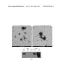 |
 |  |
 |  |
| Similar patent applications: | |
| Date | Title |
|---|---|
| 2015-02-26 | Detection of glutathionylated proteins |
| 2015-02-26 | Detection and typing of bacterial strains |
| 2015-02-26 | Process for the purification of recombinant albumin |
| 2015-02-26 | Devices and methods of cell capture and analysis |
| 2015-02-26 | Flow cytometer with digital holographic microscope |
| New patent applications in this class: | |
| Date | Title |
|---|---|
| 2022-05-05 | Isolation and detection of exosome-associated microbiome for diagnostic and therapeutic purposes |
| 2022-05-05 | New biomarkers and biotargets in renal cell carcinoma |
| 2022-05-05 | Method of predicting survival rates for cancer patients |
| 2022-05-05 | Biomarkers for autism spectrum disorders |
| 2022-05-05 | Method for preimplantation genetic screening of embryos for detection of structural rearrangements |
| Top Inventors for class "Chemistry: molecular biology and microbiology" | |
| Rank | Inventor's name |
|---|---|
| 1 | Marshall Medoff |
| 2 | Anthony P. Burgard |
| 3 | Mark J. Burk |
| 4 | Robin E. Osterhout |
| 5 | Rangarajan Sampath |