Patent application title: Therapeutic Antibodies against ROR-1 Protein and Methods for Use of Same
Inventors:
Thomas J. Kipps (San Diego, CA, US)
Thomas J. Kipps (San Diego, CA, US)
George F. Widhopf, Ii (San Diego, CA, US)
Bing Cui (San Diego, CA, US)
Assignees:
THE REGENTS OF THE UNIVERSITY OF CALIFORNIA
IPC8 Class: AC07K1640FI
USPC Class:
4241581
Class name: Drug, bio-affecting and body treating compositions immunoglobulin, antiserum, antibody, or antibody fragment, except conjugate or complex of the same with nonimmunoglobulin material binds hormone or other secreted growth regulatory factor, differentiation factor, or intercellular mediator (e.g., cytokine, vascular permeability factor, etc.); or binds serum protein, plasma protein, fibrin, or enzyme
Publication date: 2013-10-17
Patent application number: 20130273073
Abstract:
Therapeutic antibodies having binding specificity for ROR-1 expressed on
cancer cells (particularly leukemic and lymphomic cells) and
pharmaceutical compositions containing one or more such antibodies for
use in treating cancer. Methods for diagnosing such cancers through in
vitro detection of binding to ROR-1 protein expressed on putative cancer
cells are also provided.Claims:
1. An isolated antibody that specifically binds ROR1 protein and is
comprised of a heavy chain region coded by polynucleotides having at
least 90% sequence identity with any of the sequences of SEQ ID NOs: 1,
5, 9, 13 or 17, and a corresponding light chain region having at least
90% sequence identity with any of the sequences of SEQ ID NOs: 3, 7, 11,
15 or 19.
2. The antibody according to claim 1, wherein the heavy and light chain regions are encoded by, respectively, the nucleotide sequence of SEQ ID NO: 13 (heavy chain) and SEQ ID NO: 15 (light chain).
3. An isolated antibody that specifically binds ROR1 protein and is comprised of a heavy chain region consisting of polypeptides having at least 90% sequence identity with any of the sequences of SEQ ID NOs: 2, 6, 10, 14 or 18, and a corresponding light chain region having at least 90% sequence identity with any of the sequences of SEQ ID NOs: 4, 8, 12, 16 or 20.
4. The antibody according to claim 1, wherein it further specifically binds either the 3' or middle Ig-like region of the extracellular domain of human or murine ROR-1 protein.
5. The antibody according to claim 4, wherein it binds the 3' end of the Ig-like region of the extracellular domain of human or murine ROR-1 protein from position 1-147.
6. The antibody according to claim 5, wherein it further binds a glutamic acid residue corresponding to the one found in the extracellular domain of human ROR-1 protein at position 138.
7. The antibody according to claim 1, wherein it further reduces leukemic or lymphomic cell burden in an art-accepted animal model at a rate of 2-8 times, or at least 2, 3, 4, 5, 6, 7, or 8 times, that of wild-type human anti-ROR1 antibody or monoclonal 4A5 antibody.
8. The antibody according to claim 1, wherein it further inhibits CD5.sup.dullB220.sup.+ and ROR1.sup.brightB220.sup.+ leukemic B cell expansion.
9. The antibody according to claim 1, wherein it further is internalized into leukemic or lymphomic cells at a rate of at least 2 times, or at least 2, 3, 4, 5, 6, 7, 8, 9 or 10 times that of monoclonal antibody 4A5.
10. A pharmaceutically acceptable anti-ROR1 antibody composition comprising at least one antibody according to claim 1 and a pharmaceutically acceptable carrier.
11. A pharmaceutically acceptable anti-ROR1 antibody composition comprising at least one antibody according to claim 3 and a pharmaceutically acceptable carrier.
12. A method of treating cancer comprising administration to a human subject in need thereof of a therapeutically effective dose of an antibody composition according to claim 10.
13. The method according to claim 12, wherein the cancer is selected from the group consisting of lymphoma, CLL, small lymphocytic lymphoma, marginal cell B-Cell lymphoma, Burkett's Lymphoma, renal cell carcinoma, colon cancer, colorectal cancer, breast cancer, epithelial squamous cell cancer, melanoma, myeloma, stomach cancer, brain cancer, lung cancer, pancreatic cancer, cervical cancer, ovarian cancer, liver cancer, bladder cancer, prostate cancer, testicular cancer, thyroid cancer, and head and neck cancer.
14. The method according to claim 13, wherein the cancer is leukemia or lymphoma.
15. A method of diagnosing cancer comprising contacting putative cancer cells from a human subject with an antibody according to claim 1, and detecting binding with ROR-1 expressed on said cells, if present.
16. The method according to claim 15, wherein the cancer is selected from the group consisting of lymphoma, CLL, small lymphocytic lymphoma, marginal cell B-Cell lymphoma, Burkett's Lymphoma, renal cell carcinoma, colon cancer, colorectal cancer, breast cancer, epithelial squamous cell cancer, melanoma, myeloma, stomach cancer, brain cancer, lung cancer, pancreatic cancer, cervical cancer, ovarian cancer, liver cancer, bladder cancer, prostate cancer, testicular cancer, thyroid cancer, and head and neck cancer.
17. The method according to claim 16, wherein the cancer is leukemia or lymphoma.
18. The antibody of claim 1, wherein the heavy and light chain regions are encoded by, respectively, the nucleotide sequence of SEQ ID NO: 1 (heavy chain) and SEQ ID NO: 3 (light chain).
19. The antibody of claim 1, wherein the heavy and light chain regions are encoded by, respectively, the polypeptide sequence of SEQ ID NO: 14 (heavy chain) and SEQ ID NO: 16 (light chain).
20. The antibody of claim 1, wherein the heavy and light chain regions are encoded by, respectively, the polypeptide sequence of SEQ ID NO: 2 (heavy chain) and SEQ ID NO: 4 (light chain).
21. The antibody according to claim 4, wherein it binds residues 70-130 within the middle region of the Ig-like region of the extracellular domain of human or murine ROR-1 protein from position 1-147.
22. The antibody according to claim 21, wherein an isoleucine residue corresponding to the one found in the extracellular domain of human ROR-1 protein at position 111 is critical to binding of the antibody to ROR-1.
23. The antibody according to claim 4, wherein it binds residues 130-160 within the Ig-like region and adjacent linker region between the Ig-like domain and the CDR domain of the extracellular domain of human or murine ROR-1 protein from position 1-165.
24. The antibody according to claim 17, wherein it a glutamic acid residue corresponding to the one found in the extracellular domain of human ROR-1 protein at position 138 is critical to binding of the antibody to ROR-1.
25. An isolated polynucleotide which encodes an antibody that specifically binds ROR1 protein and is comprised of a heavy chain region coded by polynucleotides having at least 90% sequence identity with any of the sequences selected from the group consisting of SEQ ID NOs: 1, 5, 9, 13 or 17, and a corresponding light chain region encoded by polynucleotides having at least 90% sequence identity with any of the sequences selected from the group consisting of SEQ ID NOs: 3, 7, 11, 15 or 19.
26. An isolated polypeptide that specifically binds ROR1 protein and is comprised of a heavy chain region having at least 90% sequence identity with any of the sequences selected from the group consisting of SEQ ID NOs: 2, 6, 10, 14 or 18, and a corresponding light chain region having at least 90% sequence identity with any of the sequences selected from the group consisting of SEQ ID NOs: 4, 8, 12, 16 or 20.
27. An isolated antibody which binds the same epitope as antibody 4A5 or D10.
Description:
BACKGROUND
[0001] Tyrosine kinases are important mediators of the signaling cascade, determining key roles in diverse biological processes like growth, differentiation, metabolism and apoptosis in response to external and internal stimuli. Studies have implicated the role of tyrosine kinases in the pathophysiology of cancer. Schlessinger J. (2000) Cell, 103:211-225; and Robinson et al. (2000) Oncogene, 19:5548-5557. MacKeigan and colleagues used a large-scale RNAi approach to identify kinases that might regulate survival and apoptosis of a human tumor cell line (HeLa), RNAi to ROR1 was found as one of the most potent in inducing apoptosis among the set of RNAi targeting each of 73 different kinase-encoding genes. MacKeigan et al. (2005) Nat Cell Biol., 7:591-600. However, these investigators did not examine the expression or function of ROR1 protein in these cells.
[0002] ROR1, receptor tyrosine kinase like orphan receptor one, is a molecule expressed at high levels during embryogenesis that plays a major role in the development of the skeleton, lungs and nervous system. ROR1 expression is greatly decreased in postpartum mammalian cells to levels that are barely detectable. ROR1 is a membrane-receptor with an intracellular kinase-like domain and extracellular Frizzled-like cysteine-rich domain, which is common to receptors of members of the Wnt-family. ROR1 is member of the ROR family that is evolutionarily conserved among Caenorhavditis elegans, Drosophila, mice and humans. Wilson C, Goberdhan D C, Steller H. Dror, a potential neurotrophic receptor gene, encodes a Drosophila homolog of the vertebrate Ror family of Trk-related receptor tyrosine kinases. Proc Natl Acad Sci USA. 1993; 90:7109-7113; Oishi et al. (1997) J Biol Chem., 272:11916-11923; Masiakowski et al. (1992) J Biol Chem., 267:26181-26190; Forrester et al. (2002) Cell Mol Life Sci., 59:83-96; and Oishi et al. (1999) Genes Cells, 4:41-56. The actual functional role of the ROR1 protein during embryogenesis is unknown, although it is believed to be a receptor for Wnt proteins that regulate cellular polarity and cell-to-cell interactions.
[0003] Although principally an embryonic protein, ROR1 is expressed uniquely on certain cancer cells, including in CLL, small lymphocytic lymphoma, marginal cell B-Cell lymphoma, Burkett's Lymphoma, and other cancers (e.g., breast cancers), but not on normal adult tissues and cells. In a recent study, it was found that ROR1, at both mRNA and protein level, was highly expressed in CLL B cells but not normal B cells. Moreover, it was found that ROR1 is a receptor for Wnt5a, which could induce activation of NF-κB when co-expressed with ROR1 in HEK293 cells and enhance survival of CLL cells in vitro. This indicates that ROR1 is a CLL survival-signaling receptor for Wnt5a. Another study found that ROR1 was expressed in acute lymphocytic leukemia (ALL) as well. Shabani et al. (2007) Tumour Biol., 28:318-326; and Baskar et al. (2008) Clin Cancer Res., 14:396-404. Expression of ROR1 protein has now been demonstrated on a variety of hematologic and solid tumor cancers.
[0004] Therapeutic control of ROR1 expression is necessary. However, although polyclonal anti-ROR1 antibodies raised against ROR1 peptide are commercially available. The inventors developed a monoclonal anti-ROR1 antibody, terms 4A5, which reacts with the native ROR1 protein and is capable of detecting cell-surface expression of ROR1 for flow cytometric analysis. However, robustly therapeutic antibodies with demonstrable ability to inhibit ROR-1 mediated cancer cell proliferation to a degree that is therapeutically significant for slowing or preventing growth and metastasis have not been available.
SUMMARY OF THE INVENTION
[0005] The invention provides antibodies and combination of antibodies for in vivo and in vitro inhibition of ROR-1 cell mediated proliferation of cells from subjects with cancer, including lymphomas, CLL, small lymphocytic lymphoma, marginal cell B-Cell lymphoma, Burkett's Lymphoma, renal cell carcinoma, colon cancer, colorectal cancer, breast cancer, epithelial squamous cell cancer, melanoma, myeloma, stomach cancer, brain cancer, lung cancer, pancreatic cancer, cervical cancer, ovarian cancer, liver cancer, bladder cancer, prostate cancer, testicular cancer, thyroid cancer, and head and neck cancer, but not in blood or splenic lymphocytes of nonleukemic patients or normal adults.
[0006] The antibodies of the invention are also useful for differentiation between ROR1 expressing cancer cells ("ROR1 cancer") and normal cells. For example, an immunoassay that detects ROR1 in a sample from a subject by contacting the sample with a ROR1-specific antibody of the invention and detecting immunoreactivity between the antibody and ROR1 in the sample is provided.
[0007] In accordance with a further aspect of the invention, a ROR1 cancer is diagnosed in a subject by detecting the presence or quantity of ROR1 protein in a sample.
[0008] The present invention includes compositions that include purified, isolated monoclonal antibodies and combinations thereof that bind specifically to ROR1 receptor protein.
BRIEF DESCRIPTION OF THE DRAWINGS
[0009] FIG. 1 is a series of graphs illustrating the results of flow cytometric analysis of the expansion of CD5+B220low leukemia B cells in ROR1 Tg mice following the adoptive transfer of 1×10⁷ splenocytes from a ROR1 xTCL1 Tg mouse. Upper panel depicts the expansion from 2 to 4 weeks following adoptive transfer. Percentage of leukemic cells on the contour plot of mCD5 (x-axis) vs mB220 (y-axis) is indicated on above the gate on CD5+B220low lymphocytes. Bottom panel depicts the relative ROR1 expression (x axis) using the mouse anti-ROR1 4A5 mAb.
[0010] FIG. 2 is a diagram outlining the analysis of anti-ROR1 mAb on the adoptive transfer and engragment of ROR1 XTCL1 leukemic splenocytes. ROR1 Tg mice (4 mice/group) were given 250 ug of 4A5, D10 or control mIgG i.v. on day 0. The following day, 1×10⁷ splenocytes from a ROR1 x TCL1 Tg mouse were adoptively transferred i.v. All mice were subsequently monitor weekly for expansion of CD5+B220low leukemic B cells by flow cytometry beginning at 2 weeks post transfer.
[0011] FIG. 3 is a series of graphs illustrating the results of a flow cytometric analysis which demonstrate that anti-ROR1 antibodies of the invention inhibited the development of CLL-like leukemia in ROR1 Tg mice. 2 weeks after adoptive transfer, the PBMC facs analysis were performed. The data showed the anti-ROR1 antibody D10 but not anti-ROR1 antibody 4A5 could markedly inhibit the CD5.sup.dullB220+ and ROR1.sup.brightB220+ leukemic B cell expansion.
[0012] FIG. 4A is a series of graphs illustrating the results of in vivo testing in a murine model of human breast cancer. The anti-ROR1 antibodies inhibited breast cancer metastasis in rag-/-g-/- deficiency mice. 5E5 MDA-MB-231 breast cancer cell were transferred by i.v. injection to rag-/-g-/- mice on day 1. The rag-/-g-/- deficiency mice were also i.v. injected isotype control or anti-ROR1 antibody (4A5, D10, and 4A5 plus D10) on day 1, 3, 7 and 14 at 100 mg per mice. FIG. 4A (center) also provides images from IVIS in vivo imaging procedures on the above mice, which were performed every week. 5 weeks later, the mice were sacrificed and histology analysis were performed (FIG. 4B). The anti-ROR1 antibody D10 and the antibody combination (4A5 plus D10) both significantly inhibited metastasis of the breast cancer, with inhibition by D10 alone being greater than inhibition by 4a5 alone.
[0013] FIG. 5 provides a nucleotide coding sequence comparison of 4A5 Ig heavy chain (VH) to the closest germline mouse and human immunoglobulin (Ig) VH.
[0014] FIG. 6 provides a nucleotide coding sequence comparison of G6 Ig heavy chain (VH) to the closest germline mouse and human immunoglobulin (Ig) VH.
[0015] FIG. 7 provides a nucleotide coding sequence comparison of G3 Ig heavy chain (VH) to the closest germline mouse and human immunoglobulin (Ig) VH.
[0016] FIG. 8 provides a nucleotide coding sequence comparison of H10 Ig heavy chain (VH) to the closest germline mouse and human immunoglobulin (Ig) VH.
[0017] FIG. 9 provides a nucleotide coding sequence comparison of D10 Ig heavy chain (VH) to the closest germline mouse and human immunoglobulin (Ig) VH.
[0018] FIG. 10 is a diagram and chart depicting the highly conserved nature of human and murine ROR1.
[0019] FIG. 11 is a nucleotide comparison depicting the domain structure and sequence homology of human and murine ROR1 extracellular protein.
[0020] FIG. 12 is a chart indicating the extracellular domain which the anti-ROR1 mAbs bind the ROR1 protein.
[0021] FIG. 13 is a diagram depicting the chimeric ROR1 proteins generated to determine the binding domain of each of the anti-ROR1 mAbs.
[0022] FIG. 14 is a diagram depicting the truncated ROR1 proteins generated to determine the sub-regions which each of the anti-ROR1 mAbs binds.
[0023] FIG. 15 is a diagram depicting the amino acids which were murinized to determine residues critical for mAb binding to human ROR1 and a western blot showing that the 138 glutamic acid residue is critical for antibody D10 binding to human ROR1.
[0024] FIG. 16 is a graph indicating the KD values for antibody D10 (FIG. 16A) and 4A5 (FIG. 16B).
[0025] FIG. 17 is a series of graphs illustrating the anti-ROR1 antibody D10 is highly active in in vivo assays.
[0026] FIG. 18 is a diagram outlining the analysis of anti-ROR1 mAb on the adoptive transfer and engragment of ROR1XTCL1 leukemic splenocytes. ROR1 Tg mice (5 mice/group) were given 250 ug of 4A5, D10 or control mIgG i.v. on day 0. The following day, 5×10⁵ splenocytes from a ROR1 X TCL1 Tg mouse were adoptively transferred i.v. All mice were subsequently monitored weekly for expansion of CD5.sup.dullB200+ leukemic B cells by flow cytometery beginning at 2 weeks post transfer.
[0027] FIG. 19 a series of graphs illustrating the results of flow cytometric analysis of the anti-ROR1 antibodies inhibiting the development of CLL-like leukemia in ROR1 Tg mice. 2 weeks after adoptive transfer, the PBMC facs analysis were performed. The data showed the anti-ROR1 antibody D10 but not anti-ROR1 antibody 4A5 could markedly inhibit the CD5.sup.dullB220+ and ROR1.sup.brightB220+ leukemic B cell expansion.
[0028] FIG. 20 is a graph illustrating that anti-ROR1 antibody D10 inhibits the development and expansion of ROR1xTCL1 leukemic B cells in the blood of recipient animals until two weeks after receiving the last infusion of the mAb.
[0029] FIG. 21 is a depiction of the rapid internalization of the anti-ROR1 antibody D10 into CLL cells.
[0030] FIG. 22 is a series of graphs illustrating the results of flow cytometric analysis showing that anti-ROR1 antibodies D10 and 4A5 are both internalized into CLL cells. CLL cells were incubated with mouse anti-hROR1 Ab-Alex647 for 30 min at 4° C. Subsequently the cells were washed and either left at 4° C. or incubated for 4 hours at 37° C., followed by flow cytometry. The background signal with non-staining is also shown.
[0031] FIG. 23 is a graph illustrating the kinetics of the internalization of anti-ROR1 antibodies D10 and 4A5.
[0032] FIG. 24 is a diagram depicting the amino acids which were murinized to determine residues critical for mAb binding to human ROR1 and a western blot showing that the 111 isoleucine residue is critical for antibody 4A5 binding to human ROR1.
DETAILED DESCRIPTION OF THE PREFERRED EMBODIMENTS
[0033] The presently disclosed subject matter are described more fully below. However, the presently disclosed subject matter may be embodied in many different forms and should not be construed as limited to the embodiments set forth herein; rather, these embodiments are provided so that this disclosure will satisfy applicable legal requirements. Indeed, many modifications and other embodiments of the presently disclosed subject matter set forth herein will come to mind to one skilled in the art to which the presently disclosed subject matter pertains having the benefit of the teachings presented in the foregoing descriptions and the associated Figures. Therefore, it is to be understood that the presently disclosed subject matter is not to be limited to the specific embodiments disclosed and that modifications and other embodiments are intended to be included within the scope of the appended claims.
[0034] Antibodies of the invention were produced monoclonally using techniques as previously described. Briefly, Naturally occurring antibodies are generally tetramers containing two light chains and two heavy chains. Experimentally, antibodies can be cleaved with the proteolytic enzyme papain, which causes each of the heavy chains to break, producing three separate subunits. The two units that consist of a light chain and a fragment of the heavy chain approximately equal in mass to the light chain are called the Fab fragments (i.e., the antigen binding fragments). The third unit, consisting of two equal segments of the heavy chain, is called the Fc fragment. The Fc fragment is typically not involved in antigen-antibody binding, but is important in later processes involved in ridding the body of the antigen.
[0035] Because Fab and F(ab')2 fragments are smaller than intact antibody molecules, more antigen-binding domains are available than when whole antibody molecules are used. Proteolytic cleavage of a typical IgG molecule with papain is known to produce two separate antigen binding fragments called Fab fragments which contain an intact light chain linked to an amino terminal portion of the contiguous heavy chain via by disulfide linkage. The remaining portion of the papain-digested immunoglobin molecule is known as the Fc fragment and consists of the carboxy terminal portions of the antibody left intact and linked together via disulfide bonds. If an antibody is digested with pepsin, a fragment known as an F(ab')2 fragment is produced which lacks the Fc region but contains both antigen-binding domains held together by disulfide bonds between contiguous light and heavy chains (as Fab fragments) and also disulfide linkages between the remaining portions of the contiguous heavy chains (Handbook of Experimental Immunology. Vol 1: Immunochemistry, Weir, D. M., Editor, Blackwell Scientific Publications, Oxford (1986)).
[0036] As readily recognized by those of skill in the art, altered antibodies (e.g., chimeric, humanized, CDR-grafted, bifunctional, antibody polypeptide dimers (i.e., an association of two polypeptide chain components of an antibody, e.g., one arm of an antibody including a heavy chain and a light chain, or an Fab fragment including VL, VH, CL and CH antibody domains, or an Fv fragment comprising a VL domain and a VH domain), single chain antibodies (e.g., an scFv (i.e., single chain Fv) fragment including a VL domain linked to a VH domain by a linker, and the like) can also be produced by methods well known in the art.
[0037] Monoclonal antibody (mAb) technology can be used to obtain mAbs to ROR1. Briefly, hybridomas are produced using spleen cells from mice immunized with ROR1 antigens. The spleen cells of each immunized mouse are fused with mouse myeloma Sp 2/0 cells, for example using the polyethylene glycol fusion method of Galfre, G. and Milstein, C., Methods Enzymol., 73:3-46 (1981). Growth of hybridomas, selection in HAT medium, cloning and screening of clones against antigens are carried out using standard methodology (Galfre, G. and Milstein, C., Methods Enzymol., 73:3-46 (1981)):
[0038] HAT-selected clones are injected into mice to produce large quantities of mAb in ascites as described by Galfre, G. and Milstein, C., Methods Enzymol., 73:3-46 (1981), which can be purified using protein A column chromatography (BioRad, Hercules, Calif.). mAbs are selected on the basis of their (a) specificity for ROR1, (b) high binding affinity, (c) isotype, and (d) stability.
[0039] mAbs can be screened or tested for ROR1 specificity using any of a variety of standard techniques, including Western Blotting (Koren, E. et al., Biochim. Biophys. Acta 876:91-100 (1986)) and enzyme-linked immunosorbent assay (ELISA) (Koren, E. et al., Biochim. Biophys. Acta 876:91-100 (1986)).
[0040] Humanized forms of mouse antibodies can be generated by linking the CDR regions of non-human antibodies to human constant regions by recombinant DNA techniques (see, e.g., Queen et al., Proc. Natl. Acad. Sci. USA 86:10029-10033, 1989 and WO 90/07861, each incorporated by reference). Human antibodies can be obtained using phage-display methods (see, e.g., Dower et al., WO 91/17271; McCafferty et al., WO 92/01047). In these methods, libraries of phage are produced in which members display different antibodies on their outer surfaces. Antibodies are usually displayed as Fv or Fab fragments. Phage displaying antibodies with a desired specificity may be selected by affinity enrichment.
[0041] Human antibodies may be selected by competitive binding experiments, or otherwise, to have the same epitope specificity as a particular mouse antibody. Using these techniques, a humanized ROR1 antibody having the human IgG1 constant region domain and the human kappa light chain constant region domain with the mouse heavy and light chain variable regions. The humanized antibody has the binding specificity of a mouse ROR1 mAb, specifically the 4A5 mAb described in Examples 4 and 5.
[0042] It may be desirable to produce and use functional fragments of a mAb for a particular application. The well-known basic structure of a typical IgG molecule is a symmetrical tetrameric Y-shaped molecule of approximately 150,000 to 200,000 daltons consisting of two identical light polypeptide chains (containing about 220 amino acids) and two identical heavy polypeptide chains (containing about 440 amino acids). Heavy chains are linked to one another through at least one disulfide bond. Each light chain is linked to a contiguous heavy chain by a disulfide linkage. An antigen-binding site or domain is located in each arm of the Y-shaped antibody molecule and is formed between the amino terminal regions of each pair of disulfide linked light and heavy chains. These amino terminal regions of the light and heavy chains consist of approximately their first 110 amino terminal amino acids and are known as the variable regions of the light and heavy chains. In addition, within the variable regions of the light and heavy chains there are hypervariable regions which contain stretches of amino acid sequences, known as complementarity determining regions (CDRs). CDRs are responsible for the antibody's specificity for one particular site on an antigen molecule called an epitope. Thus, the typical IgG molecule is divalent in that it can bind two antigen molecules because each antigen-binding site is able to bind the specific epitope of each antigen molecule. The carboxy terminal regions of light and heavy chains are similar or identical to those of other antibody molecules and are called constant regions. The amino acid sequence of the constant region of the heavy chains of a particular antibody defines what class of antibody it is, for example, IgG, IgD, IgE, IgA or IgM. Some classes of antibodies contain two or more identical antibodies associated with each other in multivalent antigen-binding arrangements.
[0043] Fab and F(ab')2 fragments of mAbs that bind ROR1 can be used in place of whole mAbs. Because Fab and F(ab')2 fragments are smaller than intact antibody molecules, more antigen-binding domains are available than when whole antibody molecules are used. Proteolytic cleavage of a typical IgG molecule with papain is known to produce two separate antigen binding fragments called Fab fragments which contain an intact light chain linked to an amino terminal portion of the contiguous heavy chain via by disulfide linkage. The remaining portion of the papain-digested immunoglobin molecule is known as the Fc fragment and consists of the carboxy terminal portions of the antibody left intact and linked together via disulfide bonds. If an antibody is digested with pepsin, a fragment known as an F(ab')2 fragment is produced which lacks the Fc region but contains both antigen-binding domains held together by disulfide bonds between contiguous light and heavy chains (as Fab fragments) and also disulfide linkages between the remaining portions of the contiguous heavy chains (Handbook of Experimental Immunology. Vol 1: Immunochemistry, Weir, D. M., Editor, Blackwell Scientific Publications, Oxford (1986)).
[0044] With respect to particular antibodies, "specific binding" refers to antibody binding to a predetermined antigen. Typically, the antibody binds with an affinity corresponding to a KD of about 10-8 M or less, and binds to the predetermined antigen with an affinity (as expressed by KD) that is at least 10 fold less, and preferably at least 100 fold less than its affinity for binding to a non-specific antigen (e.g., BSA, casein) other than the predetermined antigen or a closely-related antigen. Alternatively, the antibody can bind with an affinity corresponding to a KA of about 106 M-1, or about 107 M-1, or about 108M-1, or 109 M-1 or higher, and binds to the predetermined antigen with an affinity (as expressed by KA) that is at least 10 fold higher, and preferably at least 100 fold higher than its affinity for binding to a non-specific antigen (e.g., BSA, casein) other than the predetermined antigen or a closely-related antigen.
[0045] Also, reference to "an antibody having binding specificity for ROR-1 protein" includes antibody fragments having at least 90% or 95% sequence identity to any of the polypeptide sequences disclosed in SEQ ID NOs: 2. 4 6, 8, 12, 14, 16, 18 and 20, including variants modified by mutation to improve the utility thereof (e.g., improved ability to target specific cell types and the like). Such variants include those wherein one or more conservative substitutions are introduced into the heavy chain and/or the light chain of the antibody.
[0046] Such variants include those wherein one or more substitutions are introduced into the heavy chain nucleotide sequence and/or the light chain nucleotide sequence of the antibody. In some embodiments the variant has a light chain and/or heavy chain having a nucleotide sequence at least 80% or at least 90% or at least 95% identical to any of the nucleotide sequences set forth in SEQ ID NOs: 1, 3, 5, 7, 11, 13, 15, 17 and 19.
[0047] Polynucleotide sequences which code structural features of the antibodies of the invention include those whose sequences are set forth below. Each polynucleotide sequence is followed by the amino acid sequence of the encoded polypeptide. The light chain sequences which are considered to be "corresponding" to heavy chain sequences are those listed as being for the same antibody; i.e., the F2 heavy chain sequences correspond to the F2 light chain sequences, the D10 heavy chain sequences correspond to the D10 light chain sequences, and so forth.
SEQ ID NO: 1 4A5 Mouse Anti-ROR1 mAb Heavy Chain Variable Region Coding Sequence:
TABLE-US-00001
[0048] GAAGTGAAACTGGTGGAGTCTGGGGGAGGCTTAGTGAAGCCTGGAGGGTCCCTGAAACTCTC CTGTGCAGCCTCTGGATT CACTTTCAGTAGCTATGCCATGTCTTGGGTTCGCCAGATTCCAGAGAAGAGGCTGGAGTGGG TCGCATCCATTAGTCGTG GTGGTACCACCTACTATCCAGACAGTGTGAAGGGCCGATTCACCATCTCCAGAGATAATGTC AGGAACATCCTGTACCTG CAAATGAGCAGTCTGAGGTCTGAGGACACGGCCATGTATTACTGTGGAAGATATGATTACGA CGGGTACTATGCAATGGA CTACTGGGGTCAAGGAACCTCAGTCACCGTCTCCTCA
SEQ ID NO: 2 4A5 Mouse Anti-ROR1 mAb Heavy Chain Variable Region Polypeptide Sequence:
TABLE-US-00002
[0049] EVKLVESGGGLVKPGGSLKLSCAASGFTFSSYAMSWVRQIPEKRLEWVASISRGGTTYYPDS VKGRFTISRDNVRNILYL QMSSLRSEDTAMYYCGRYDYDGYYAMDYWGQGTSVTVSS
SEQ ID NO: 3 4A5 Mouse Anti-ROR1 mAb Light Chain Variable Region Coding Sequence:
TABLE-US-00003
[0050] GACATCAAGATGACCCAGTCTCCATCTTCCATGTATGCATCTCTAGGAGAGAGAGTCACTAT CACTTGCAAGGCGAGTCC GGACATTAATAGCTATTTAAGCTGGTTCCAGCAGAAACCAGGGAAATCTCCTAAGACCCTGA TCTATCGTGCAAACAGAT TGGTTGATGGGGTCCCATCAAGGTTCAGTGGCGGTGGATCTGGGCAAGATTATTCTCTCACC ATCAACAGCCTGGAGTAT GAAGATATGGGAATTTATTATTGTCTACAGTATGATGAATTTCCGTACACGTTCGGAGGGGG GACCAAGCTGGAAATGAA AC
SEQ ID NO: 4 4A5 Mouse Anti-ROR1 mAb Light Chain Variable Region Polypeptide Sequence:
TABLE-US-00004
[0051] DIKMTQSPSSMYASLGERVTITCKASPDINSYLSWFQQKPGKSPKTLIYRANRLVDGVPSRF SGGGSGQDYSLTINSLEY EDMGIYYCLQYDEFPYTFGGGTKLEMK
SEQ ID NO: 5 F2, F12 and G6 Mouse Anti-ROR1 mAb Heavy Chain Variable Region Coding Sequence:
TABLE-US-00005
[0052] GAGGTCCAGCTACAGCAGTCTGGACCTGAGCTGGAGAAGCCTGGCGCTTCAGTGAAGATATC CTGCAAGGCTTCTGGTTT CGCATTCACTGGCTACAACATGAACTGGGTGAAACAGACCAATGGAAAGAGCCTTGAGTGGA TTGGAAGTATTGATCCTT ACTATGGTGGTTCTACCTACAACCAGAAGTTCAAGGACAAGGCCACATTGACTGTAGACAAA TCCTCCAGCACAGCCTAC ATGCAACTCAAGAGCCTCACATCTGATGACTCTGCAGTCTATTACTGTGCAAGATCCCCGGG GGGGGACTATGCTATGGA CTACTGGGGTCAAGGAACCTCAGTCACCGTCTCCTCA
SEQ ID NO: 6 F2, F12 and G6 Mouse Anti-ROR1 mAb Heavy Chain Variable Region Polypeptide Sequence:
TABLE-US-00006
[0053] EVQLQQSGPELEKPGASVKISCKASGFAFTGYNMNWVKQTNGKSLEWIGSIDPYYGGSTYNQ KFKDKATLTVDKSSSTAY MQLKSLTSDDSAVYYCARSPGGDYAMDYWGQGTSVTVSS
SEQ ID NO: 7 F2, F12 and G6 Mouse Anti-ROR1 mAb Light Chain Variable Region Coding Sequence:
TABLE-US-00007
[0054] GACATCAAGATGACCCAGTCTCCATCTTCCATGTATGCATCTGTAGGAGAGAGAGTCACTAT CACTTGTAAGGCGAGTCA GGGCATTAATAGCTATTCAGGCTGGTTCCAGCAGAAACCAGGGAAATCTCCTAAGACCCTGA TTTATCGTGGAAATAGAT TGGTGGATGGGGTCCCATCAAGGTTCAGTGGCAGTGGATCTGGGCAAGATTATTCTCTCACC ATCAGCAGCCTGGAGTAT GAAGATATGGGAATTTATTATTGTCTACAGTATGATGAGTTTCCGTACACGTTCGGAGGGGG GACCAAGCTGGAAATAAA AC
SEQ ID NOs: 8 F2, F12 and G6 Mouse Anti-ROR1 mAb Light Chain Variable Region Polypeptide Sequence:
TABLE-US-00008
[0055] DIKMTQSPSSMYASVGERVTITCKASQGINSYSGWFQQKPGKSPKTLIYRGNRLVDGVPSRF SGSGSGQDYSLTISSLEY EDMGIYYCLQYDEFPYTFGGGTKLEIK
SEQ ID NO: 9 G3 Mouse Anti-ROR1 mAb Heavy Chain Variable Region Coding Sequence:
TABLE-US-00009
[0056] CAGGTCCAACTGCAGCAGCCTGGGGCTGAGCTTGTGAAGCCTGGGACTTCAGTGAAGCTGTC CTGCAAGGCTTCTGGCTA CAACTTCACCAACTACTGGATAAACTGGGTGAAGCTGAGGCCTGGACAAGGCCTTGAGTGGA TTGGAGAAATTTATCCTG GTAGTGGTAGTACTAATTACAATGAGAAGTTCAAGAGCAAGGCCACACTGACTGCAGACACA TCCTCCAGCACAGCCTAC ATGCAACTCAGCAGCCTGGCATCTGAAGACTCTGCTCTCTATTACTGTGCAAGAGATGGTAA CTACTATGCTATGGACTA CTGGGGTCAAGGAACCTCAGTCACCGTCTCCTCA
SEQ ID NO: 10 G3 Mouse Anti-ROR1 mAb Heavy Chain Variable Region Polypeptide Sequence:
TABLE-US-00010
[0057] QVQLQQPGAELVKPGTSVKLSCKASGYNFTNYWINWVKLRPGQGLEWIGEIYPGSGSTNYNE KFKSKATLTADTSSSTAY MQLSSLASEDSALYYCARDGNYYAMDYWGQGTSVTVSS
SEQ ID NO: 11 G3 Mouse Anti-ROR1 mAb Light Chain Variable Region Coding Sequence:
TABLE-US-00011
[0058] GATATCCAGATGACACAGACTACATCCTCCCTGTCTGCCTCTCTGGGAGACAGAGTCACCAT CACTTGCAGGGCAAGTCA GGACATTAACAATTATTTAAACTGGTATCAACAGAAACCAGATGGAACTGTTAAACTCCTGA TCTACTACACATCAGCAT TACACTCAGGAGTCCCATCAAGGTTCAGTGGCAGTGGGTCTGGAACAGATTATTCTCTCACC ATTAGCAACCTGGAACAA GAAGATATTGCCACTTACTTTTGCCAACAGGGTAATACGCTTCCTCCGTACACGTTCGGAGG GGGGACCAAGCTGGAAAT AAAAC
SEQ ID NO: 12 G3 Mouse Anti-ROR1 mAb Light Chain Variable Region Polypeptide Sequence:
TABLE-US-00012
[0059] DIQMTQTTSSLSASLGDRVTITCRASQDINNYLNWYQQKPDGTVKLLIYYTSALHSGVPSRF SGSGSGTDYSLTISNLEQ EDIATYFCQQGNTLPPYTFGGGTKLEIK
SEQ ID NO: 13 D10 Mouse Anti-ROR1 mAb Heavy Chain Variable Region Coding Sequence:
TABLE-US-00013
[0060] CAGGTGCAGCTGAAGGAGTCAGGACCTGGCCTGGTGGCGCCCTCACAGACTCTGTCCATCAC TTGCACTGTCTCTGGGTT TTCATTAACCAGTTATGGTGTACACTGGGTTCGCCAGCCTCCAGGAAAGGGTCTGGAGTGGC TGGGAGTAATATGGGCTG GTGGATTCACAAATTATAATTCGGCTCTCAAGTCCAGACTGAGCATCAGCAAAGACAACTCC AAGAGCCAAGTTCTCTTA AAAATGACCAGTCTGCAAACTGATGACACAGCCATGTACTACTGTGCCAGGAGAGGTAGTTC CTATTCTATGGACTATTG GGGTCAAGGAACCTCAGTCACCGTCTCCTCA
SEQ ID NO: 14 D10 Mouse Anti-ROR1 mAb Heavy Chain Variable Region Polypeptide Sequence
TABLE-US-00014
[0061] QVQLKESGPGLVAPSQTLSITCTVSGFSLTSYGVHWVRQPPGKGLEWLGVIWAGGFTNYNSA LKSRLSISKDNSKSQVLL KMTSLQTDDTAMYYCARRGSSYSMDYWGQGTSVTVSS
SEQ ID NO: 15 D10 Mouse Anti-ROR1 mAb Light Chain Variable Region Coding Sequence:
TABLE-US-00015
[0062] GAAATTGTGCTCTCTCAGTCTCCAGCCATCACAGCTGCATCTCTGGGCCAAAAGGTCACCAT CACCTGCAGTGCCAGTTC AAATGTAAGTTACATCCACTGGTACCAGCAGAGGTCAGGCACCTCCCCCAGACCATGGATTT ATGAAATATCCAAACTGG CTTCTGGAGTCCCAGTTCGCTTCAGTGGCAGTGGGTCTGGGACCTCTTACTCTCTCACAATC AGCAGCATGGAGGCTGAA GATGCTGCCATTTATTATTGTCAGCAGTGGAATTATCCTCTTATCACGTTCGGCTCGGGGAC AAAGTTGGAAATACAA
SEQ ID NO: 16 D10 Mouse Anti-ROR1 mAb Light Chain Variable Region Polypeptide Sequence:
TABLE-US-00016
[0063] EIVLSQSPAITAASLGQKVTITCSASSNVSYIHWYQQRSGTSPRPWIYEISKLASGVPVRFS GSGSGTSYSLTISSMEAE DAAIYYCQQWNYPLITFGSGTKLEIQ
SEQ ID NO: 17 H10 and G11 Mouse Anti-ROR1 mAb Heavy Chain Variable Region Coding Sequence:
TABLE-US-00017
[0064] GAAGTGAAGCTGGTGGAGTCTGGGGGAGGCTTAGTGAAGCCTGGAGGGTCCCTGAAACTCTC CTGTGCAGCCTCTGGATT CACTTTCAGTAGCTATGCCATGTCTTGGGTTCGCCAGACTCCAGAGAAGAGGCTGGAGTGGG TCGCTTCCATTAGTACTG GTGCTAGCGCCTACTTTCCAGACAGTGTGAAGGGCCGATTCACCATCTCCAGAGATAATGCC AGGAACATCCTGTACCTG CAAATGAGCAGTCTGAGGTCTGAGGACACGGCCATGTATTATTGTGCAAGGATTACTACGTC TACCTGGTACTTCGATGT CTGGGGCGCAGGGACCACGGTCACCGTCTCCTCA
SEQ ID NO: 18 H10 and G11 Mouse Anti-ROR1 mAb Heavy Chain Variable Region Polypeptide Sequence:
TABLE-US-00018
[0065] EVKLVESGGGLVKPGGSLKLSCAASGFTFSSYAMSWVRQTPEKRLEWVASISTGASAYFPDS VKGRFTISRDNARNILYL QMSSLRSEDTAMYYCARITTSTWYFDVWGAGTTVTVSS
SEQ ID NO: 19 H10 and G11 Mouse Anti-ROR1 mAb Light Chain Variable Region Coding Sequence:
TABLE-US-00019
[0066] GACATCAAGATGACCCAGTCTCCATCTTCCATGTATGCATCTCTAGGAGAGAGAGTCACTAT CACTTGCAAGGCGAGTCA GGACATTAATAGTTATTTAAGCTGGTTCCAGCAGAAACCAGGGAAATCTCCTAAGACCCTGA TCTATCGTGCAAACAGAT TGGTAGATGGGGTCCCATCAAGGTTCAGTGGCAGTGGATCTGGGCAAGATTATTCTCTCACC ATCAGCAGCCTGGAGTAT GAAGATATGGGAATTTATTATTGTCTACAGTATGATGAGTTTCCGTACACGTTCGGAGGGGG GACCAAGCTGGAAATAAA AC
SEQ ID NO: 20 H10 and G11 Mouse Anti-ROR1 mAb Light Chain Variable Region Polypeptide Sequence:
TABLE-US-00020
[0067] DIKMTQSPSSMYASLGERVTITCKASQDINSYLSWFQQKPGKSPKTLIYRANRLVDGVPSRF SGSGSGQDYSLTISSLEY EDMGIYYCLQYDEFPYTFGGGTKLEIK
[0068] In one aspect, antibodies are provided in which a heavy chain encoded by the polynucleotide sequence of SEQ ID NO:13 and a light chain encoded by the polynucleotide sequence of SEQ ID NO:15.
[0069] In another aspect, an antibody of the present invention contains a heavy chain encoded by the polynucleotide sequence of SEQ ID NO:1 and a light chain encoded by the polynucleotide sequence of SEQ ID NO:3.
[0070] In further aspects, antibodies are provided which have a heavy chain encoded by the polynucleotide sequence of SEQ ID NO: 5 and a light chain encoded by the polynucleotide sequence of SEQ ID NO: 7; or by the polynucleotide sequence of SEQ ID NO: 9 and alight chain encoded by the polynucleotide sequence of SEQ ID NO: 11; or by the polynucleotide sequence of SEQ ID NO: 15 and a light chain encoded by the polynucleotide sequence of SEQ ID NO: 17.
[0071] In another aspect, antibodies are provided which contain a heavy chain with the polypepetide sequence of SEQ ID NO:14 and a light chain with the polypeptide sequence of SEQ ID NO:16.
[0072] In another aspect, antibodies are provided which contain a heavy chain with the polypeptide sequence of SEQ ID NO:2 and a light chain with the polypeptide sequence of SEQ ID NO:4.
[0073] In one embodiment, isolated polynucleotides which encode an antibody that specifically binds ROR1 protein are provided which are (a) comprised of a heavy chain region coded by polynucleotides having at least 90% sequence identity with any of the sequences selected from the group consisting of SEQ ID NOs: 1, 5, 9, 13 or 17, (b) comprised of a corresponding light chain region encoded by polynucleotides having at least 90% sequence identity with any of the sequences selected from the group consisting of SEQ ID NOs: 3, 7, 11, 15 or 19, and (c) specifically binds either the 3' end or middle portion of the Ig-like region of the extracellular domain of human or murine ROR-1 protein.
[0074] Also provided are antibodies which bind residues within the middle of the Ig-like region of the extracellular domain of human or murine ROR-1 protein (amino acids 1-147 in the human molecule). In one aspect, the antibodies of the present invention bind to amino acids 70-130 of human ROR1. Examples of such antibodies include 4A5, G11, H10 and G3.
[0075] Alternatively or additionally, a residue corresponding to the one found in the extracellular domain of human ROR-1 protein at position 111 is critical to the binding activity of the antibodies.
[0076] Further provided are antibodies that bind residues within the 3' Ig-like region and the linker region between the Ig-like domain and the CRD domain of human or murine ROR-1 protein (amino acids 1-165 in the human molecule). In one aspect, the antibodies of the present invention bind to amino acids 130-165 of human ROR1. Examples of such antibodies include D10, F2, F12 and G6.
[0077] Alternatively or additionally, the antibodies bind a glutamic acid residue corresponding to the one found in the extracellular domain of human ROR-1 protein at position 138.
[0078] Alternatively or additionally, a residue corresponding to the one found in the extracellular domain of human ROR-1 protein at position 138 is critical to the binding activity of the antibodies.
[0079] Alternatively or additionally, the encoded antibody has in vivo activity in reducing leukemic or lymphomic cell burden in an art-accepted animal model at a rate of 2-8 times, or at least 2, 3, 4, 5, 6, 7, or 8 times, that of wild-type human anti-ROR1 antibody or monoclonal 4A5 antibody (disclosed herein).
[0080] Alternatively or additionally, the encoded antibody has in vivo activity in inhibiting CD5.sup.dullB220+ and ROR1.sup.brightB220+ leukemic B cell expansion.
[0081] Alternatively or additionally, the encoded antibody is internalized into leukemic or lymphomic cells at a rate of at least 2 times, or at least 2, 3, 4, 5, 6, 7, 8, 9 or 10 times that of monoclonal antibody 4A5. Such antibodies are particularly useful as carriers for drug delivery into a targeted cell.
[0082] An example of an antibody possessing all of the afore-mentioned functional characteristics is D10, which has a heavy chain region encoded by SEQ ID NO: 13 and a light chain region encoded by SEQ ID NO: 15.
[0083] In another aspect, polypeptides are provided which consist of or comprise antibodies which specifically bind ROR1 protein and are (a) comprised of a heavy chain region having at least 90% sequence identity with any of the sequences of SEQ. ID. NOs: 2, 6, 10, 14 or 18, (b) comprised of a corresponding light chain region having at least 90% sequence identity with any of the sequences of SEQ ID NOs: 4, 8, 12, 16 or 20, and (c) specifically binds either the 3' end or middle portion of the Ig-like region of the extracellular domain of human or murine ROR-1 protein. In one aspect, the isolated polypeptide is an antibody. In a further aspect, the polypeptide is a Fab or F(ab)'2.
[0084] In certain embodiments, an antibody of the present invention may further contain a detectable label. Such labels are known in the art and include radio-isotopes and fluorescent labels. As such, internalization of a compound evidencing passage through transporters can be detected by detecting a signal from within a cell from any of a variety of reporters. The reporter can be a label such as a fluorophore, a chromophore, a radioisotope. Confocal imagining can also be used to detect internalization of a label as it provides sufficient spatial resolution to distinguish between fluorescence on a cell surface and fluorescence within a cell; alternatively, confocal imaging can be used to track the movement of compounds over time. In another approach, internalization of a compound is detected using a reporter that is a substrate for an enzyme expressed within a cell. Once the complex is internalized, the substrate is metabolized by the enzyme and generates an optical signal or radioactive decay that is indicative of uptake. Light emission can be monitored by commercial PMT-based instruments or by CCD-based imaging systems. In addition, assay methods utilizing LCMS detection of the transported compounds or electrophysiological signals indicative of transport activity are also employed.
[0085] In certain therapeutic embodiments, the selected antibody may be administered alone, in combination with another antibody of the invention, or with one or more combinatorial therapeutic agents to treat an ROR-1 cancer. When one or more the antibodies described herein are administered as therapeutic agents, they may exert a beneficial effect in the subject by a variety of mechanisms. For example, in certain embodiments, antibodies that specifically bind ROR1 are purified and administered to a patient to neutralize one or more forms of ROR1, to block one or more activities of ROR1, or to block or inhibit an interaction of one or more forms of ROR1 with another biomolecule; e.g., to treat CLL or other ROR1 cancers. All such therapeutic methods are practiced by delivery of a therapeutically effective dosage of a pharmaceutical composition containing the therapeutic antibodies and agents, which can be determined by a pharmacologist or clinician of ordinary skill in human cancer immunotherapy.
[0086] In one embodiment, the present invention provides for a method for of treating cancer by the administration to a human subject in need thereof of a therapeutically effective dose of an antibody according to the invention.
[0087] In another embodiment, the present invention provides a method for of treating cancer comprising administration to a human subject in need thereof of a therapeutically effective dose of an antibody according to the invention.
[0088] Advantageously, the methods of the invention provide for reduction of leukemic or lymphomic cell burden (as demonstrated in and equivalent to an art-accepted animal model) of 2-8 times, or at least 2, 3, 4, 5, 6, 7, or 8 times, that of wild-type human anti-ROR1 antibody or monoclonal 4A5 antibody (disclosed herein).
[0089] The methods of the invention further provide a therapeutic approach to inhibiting CD5.sup.dullB220+ and ROR1.sup.brightB220+ leukemic B cell expansion.
[0090] As discussed herein, the antibodies of the invention may include humanized antibodies, and can be combined for therapeutic use with additional active or inert ingredients, e.g., in conventional pharmaceutically acceptable carriers or diluents, e.g., immunogenic adjuvants, and optionally with adjunctive or combinatorially active molecules such as anti-inflammatory and anti-fibrinolytic drugs. Antibodies which readily internalize into cells as demonstrated herein with respect to the D10 antibody are also of particular use as carriers for drug delivery into target cells (for example, as shown in FIGS. 21-23). Those of ordinary skill in the art will be familiar with methods for producing antibody-drug conjugates useful in such drug delivery protocols.
[0091] In carrying out various assay, diagnostic, and therapeutic methods of the invention, it is desirable to prepare in advance kits comprises a combination of antibodies as described herein with other materials. For example, in the case of sandwich enzyme immunoassays, kits of the invention may contain an antibody that specifically binds ROR1 optionally linked to an appropriate carrier, a freeze-dried preparation or a solution of an enzyme-labeled monoclonal antibody which can bind to the same antigen together with the monoclonal antibody or of a polyclonal antibody labeled with the enzyme in the same manner, a standard solution of purified ROR1, a buffer solution, a washing solution, pipettes, a reaction container and the like. In addition, the kits optionally include labeling and/or instructional materials providing directions (i.e., protocols) for the practice of the methods described herein in an assay environment. While the instructional materials typically comprise written or printed materials, they are not limited to such. Any medium capable of storing such instructions and communicating them to an end user is contemplated. Such media include, but are not limited to electronic storage media (e.g., magnetic discs, tapes, cartridges, chips), optical media (e.g., CD ROM), and the like. Such media may include addresses to internet sites that provide such instructional materials.
[0092] In general, an in vitro method of diagnosing a ROR-1 cancer will comprise contacting putative cancer cells from a human subject with an antibody according to the invention, and detecting binding with ROR-1 expressed on said cells as compared to expression on post-embryonic human non-cancer cells. All such diagnostic methods are practiced by delivery of a diagnostically effect quantity of antibodies according to the invention, which can be determined by a diagnostician or in vitro diagnostic engineer of ordinary skill in human cancer diagnosis.
[0093] The following examples are intended to illustrate but not limit the invention.
Example 1
Generation of Monoclonal Anti-ROR1 Antibodies
[0094] For the production of the hybridoma-generated mAbs, mice were inoculated with DNA, protein and adenoviral constructs that express the extracellular portion (AA 1-406) of the ROR1 protein that include the Ig-like, CRD and Kringle domains and adjacent linker regions (FIGS. 10-11). Because of the high degree of homology between the murine and human molecules, a variety of cytokines and immune stimulatory agents, such as Freund's Complete Adjuvant, were co-injected to maximize the generation of anti-human ROR1 antibodies. Hybridoma-generated mAbs were generated and screened for binding to human and murine ROR1. An example of hybridoma derived mAbs is D10.
Example 2
Generation of Anti-ROR1 Antibodies Using Phage Display
[0095] A second set of antibodies was generated through the use of a proprietary enhanced phage library (Alere, Inc. San Diego). These anti-human ROR1 antibodies bind epitopes that span the entire length of the extra-cellular domain of the ROR1 protein (FIG. 12). An example of a phage display derived anti-ROR1 antibody is 4A5.
Example 3
In Vitro Analysis of Anti-ROR1 Antibodies
[0096] Antibodies generated through either hybridomas or phage display were screened for binding to human and murine ROR1. It was determined that the anti-ROR1 antibodies D10 and 4A5 bound only to human ROR1 and did not cross react with murine ROR1.
Example 4
Determination of Binding Sites for Anti-ROR1 Antibodies
[0097] Because the anti-ROR1 mAbs are species specific, a series of chimeric proteins were generated that were used to determine the binding site for each of the anti-ROR1 mAbs (FIG. 13). As a second level screen, a series of deletion constructs were generated to determine the actual extracellular ROR1 domain to which the mAbs bind. Once the binding domain was identified, truncated chimeric ROR1 molecules to identify specific sub-regions were generated that are recognized by the anti-human ROR1 mAbs (FIG. 14). As a final step, the actual amino acids targeted by these antibodies were determined. For this final screen, murinized human amino acids in the sub-domain fragments were generated to determine critical residues required for mAb binding (FIG. 15). From this screening paradigm, the binding sub-domains for the mAbs were determined (FIG. 15). It was determine that the D10 anti-human ROR1 mAb required the glutamic acid residue at position 138 for binding to the Ig-like domain of the human ROR1 molecule. When this amino acid is replaced with the murine molecule's lysine residue, the D10 molecule no longer bound to the ROR1 protein.
[0098] In a similar manner, it was determined that 4A5 anti-human ROR1 mAb required the isoleucine residue at position 111 for binding to human ROR1 molecule (FIG. 24). When this amino acid is replaced with the murine molecule's asparagine residue, the 4A5 molecule no longer bound to the ROR1 protein. It was also determined that the anti-ROR1 antibodies G11, H10 and G3 bind the same region as 4A5.
[0099] Using standard cross blocking techniques the binding sites for anti-ROR1 antibodies F2, F12 and G6 were determined. These experiments determined that antibodies F2, F12 and G6 cross block the anti-ROR1 antibody D10, indicating that they share a binding site.
Example 5
Determination of the KD Values for the Anti-ROR1 Antibodies D10 and 4A5
[0100] The KD values for the anti-ROR1 antibodies was determined using standard techniques. It was determined that the KD for the D10 antibody was 40 nM and for the antibody 4A5 was 4 nM (FIGS. 16A & B).
Example 6
In Vivo Analysis of Anti-ROR1 Antibodies
[0101] The D10 mAb was assessed in several in vivo models. In a murine in vivo xenograph, niche-dependent, activity model two doses of the mAb were administered at 10 mg/kg against 4 primary patient CLL cells in 76 mice. As shown in FIG. 17, D10 mAb substantially eliminated patient CLL cells in a dose dependent manner. In contrast, the 4A5 mAb had minimal activity in these studies even though the kDa of this mAb is 10 fold greater (4 vs. 40) for the D10 mAb.
[0102] In addition to this activity model, the D10 mAb was also tested in an immune competent transgenic mouse model that spontaneously generates leukemic cells expressing the human ROR1 protein (FIGS. 18-20). The ROR1-specific mAbs D10 and 4A5 or control IgG antibodies (10 mg/kg) were administered before and after adoptive transfer of ROR1xTCL1 CLL B cells into Balb C mice. The D10 mAb, but not control IgG or 4A5, was able to inhibit the development and expansion of the ROR1xTCL1 leukemic B cells in the blood of recipient animals until two weeks after receiving the last infusion of MAb.
[0103] Along with the anti-leukemic activity of this mAb, it has also been shown that the D10 anti-ROR1 antibody is internalized into patient CLL cells and B cell leukemia and lymphoma cell lines at a greater rate and degree than other anti-ROR1 MAbs that bind other antigenic sites on the extracellular portion of the ROR1 protein (FIGS. 21-23). Because of the absence of the ROR1 protein on post-partum tissues and its rapid rate of internalization, the D10 mAb may serve as an excellent carrier protein for drugs; for example, for use in directed antibody-drug conjugate (ADC) mediated cytotoxicity. Based on these preclinical findings, the D10 mAb has potential to have therapeutic activity against ROR1 expressing leukemias, lymphomas and solid tumor cancers as a targeted therapy and/or conjugated drug carrier.
[0104] Although the foregoing subject matter has been described in some detail by way of illustration and example for purposes of clarity of understanding, it will be understood by those skilled in the art that certain changes and modifications can be practiced within the scope of the appended claims.
Sequence CWU
1
1
371357DNAMouse 1gaagtgaaac tggtggagtc tgggggaggc ttagtgaagc ctggagggtc
cctgaaactc 60tcctgtgcag cctctggatt cactttcagt agctatgcca tgtcttgggt
tcgccagatt 120ccagagaaga ggctggagtg ggtcgcatcc attagtcgtg gtggtaccac
ctactatcca 180gacagtgtga agggccgatt caccatctcc agagataatg tcaggaacat
cctgtacctg 240caaatgagca gtctgaggtc tgaggacacg gccatgtatt actgtggaag
atatgattac 300gacgggtact atgcaatgga ctactggggt caaggaacct cagtcaccgt
ctcctca 3572119PRTMouse 2Glu Val Lys Leu Val Glu Ser Gly Gly Gly
Leu Val Lys Pro Gly Gly 1 5 10
15 Ser Leu Lys Leu Ser Cys Ala Ala Ser Gly Phe Thr Phe Ser Ser
Tyr 20 25 30 Ala
Met Ser Trp Val Arg Gln Ile Pro Glu Lys Arg Leu Glu Trp Val 35
40 45 Ala Ser Ile Ser Arg Gly
Gly Thr Thr Tyr Tyr Pro Asp Ser Val Lys 50 55
60 Gly Arg Phe Thr Ile Ser Arg Asp Asn Val Arg
Asn Ile Leu Tyr Leu 65 70 75
80 Gln Met Ser Ser Leu Arg Ser Glu Asp Thr Ala Met Tyr Tyr Cys Gly
85 90 95 Arg Tyr
Asp Tyr Asp Gly Tyr Tyr Ala Met Asp Tyr Trp Gly Gln Gly 100
105 110 Thr Ser Val Thr Val Ser Ser
115 3322DNAMouse 3gacatcaaga tgacccagtc
tccatcttcc atgtatgcat ctctaggaga gagagtcact 60atcacttgca aggcgagtcc
ggacattaat agctatttaa gctggttcca gcagaaacca 120gggaaatctc ctaagaccct
gatctatcgt gcaaacagat tggttgatgg ggtcccatca 180aggttcagtg gcggtggatc
tgggcaagat tattctctca ccatcaacag cctggagtat 240gaagatatgg gaatttatta
ttgtctacag tatgatgaat ttccgtacac gttcggaggg 300gggaccaagc tggaaatgaa
ac 3224107PRTMouse 4Asp Ile
Lys Met Thr Gln Ser Pro Ser Ser Met Tyr Ala Ser Leu Gly 1 5
10 15 Glu Arg Val Thr Ile Thr Cys
Lys Ala Ser Pro Asp Ile Asn Ser Tyr 20 25
30 Leu Ser Trp Phe Gln Gln Lys Pro Gly Lys Ser Pro
Lys Thr Leu Ile 35 40 45
Tyr Arg Ala Asn Arg Leu Val Asp Gly Val Pro Ser Arg Phe Ser Gly
50 55 60 Gly Gly Ser
Gly Gln Asp Tyr Ser Leu Thr Ile Asn Ser Leu Glu Tyr 65
70 75 80 Glu Asp Met Gly Ile Tyr Tyr
Cys Leu Gln Tyr Asp Glu Phe Pro Tyr 85
90 95 Thr Phe Gly Gly Gly Thr Lys Leu Glu Met Lys
100 105 5357DNAMouse 5gaggtccagc
tacagcagtc tggacctgag ctggagaagc ctggcgcttc agtgaagata 60tcctgcaagg
cttctggttt cgcattcact ggctacaaca tgaactgggt gaaacagacc 120aatggaaaga
gccttgagtg gattggaagt attgatcctt actatggtgg ttctacctac 180aaccagaagt
tcaaggacaa ggccacattg actgtagaca aatcctccag cacagcctac 240atgcaactca
agagcctcac atctgatgac tctgcagtct attactgtgc aagatccccg 300gggggggact
atgctatgga ctactggggt caaggaacct cagtcaccgt ctcctca 3576119PRTMouse
6Glu Val Gln Leu Gln Gln Ser Gly Pro Glu Leu Glu Lys Pro Gly Ala 1
5 10 15 Ser Val Lys Ile
Ser Cys Lys Ala Ser Gly Phe Ala Phe Thr Gly Tyr 20
25 30 Asn Met Asn Trp Val Lys Gln Thr Asn
Gly Lys Ser Leu Glu Trp Ile 35 40
45 Gly Ser Ile Asp Pro Tyr Tyr Gly Gly Ser Thr Tyr Asn Gln
Lys Phe 50 55 60
Lys Asp Lys Ala Thr Leu Thr Val Asp Lys Ser Ser Ser Thr Ala Tyr 65
70 75 80 Met Gln Leu Lys Ser
Leu Thr Ser Asp Asp Ser Ala Val Tyr Tyr Cys 85
90 95 Ala Arg Ser Pro Gly Gly Asp Tyr Ala Met
Asp Tyr Trp Gly Gln Gly 100 105
110 Thr Ser Val Thr Val Ser Ser 115
7322DNAMouse 7gacatcaaga tgacccagtc tccatcttcc atgtatgcat ctgtaggaga
gagagtcact 60atcacttgta aggcgagtca gggcattaat agctattcag gctggttcca
gcagaaacca 120gggaaatctc ctaagaccct gatttatcgt ggaaatagat tggtggatgg
ggtcccatca 180aggttcagtg gcagtggatc tgggcaagat tattctctca ccatcagcag
cctggagtat 240gaagatatgg gaatttatta ttgtctacag tatgatgagt ttccgtacac
gttcggaggg 300gggaccaagc tggaaataaa ac
3228107PRTMouse 8Asp Ile Lys Met Thr Gln Ser Pro Ser Ser Met
Tyr Ala Ser Val Gly 1 5 10
15 Glu Arg Val Thr Ile Thr Cys Lys Ala Ser Gln Gly Ile Asn Ser Tyr
20 25 30 Ser Gly
Trp Phe Gln Gln Lys Pro Gly Lys Ser Pro Lys Thr Leu Ile 35
40 45 Tyr Arg Gly Asn Arg Leu Val
Asp Gly Val Pro Ser Arg Phe Ser Gly 50 55
60 Ser Gly Ser Gly Gln Asp Tyr Ser Leu Thr Ile Ser
Ser Leu Glu Tyr 65 70 75
80 Glu Asp Met Gly Ile Tyr Tyr Cys Leu Gln Tyr Asp Glu Phe Pro Tyr
85 90 95 Thr Phe Gly
Gly Gly Thr Lys Leu Glu Ile Lys 100 105
9354DNAMouse 9caggtccaac tgcagcagcc tggggctgag cttgtgaagc ctgggacttc
agtgaagctg 60tcctgcaagg cttctggcta caacttcacc aactactgga taaactgggt
gaagctgagg 120cctggacaag gccttgagtg gattggagaa atttatcctg gtagtggtag
tactaattac 180aatgagaagt tcaagagcaa ggccacactg actgcagaca catcctccag
cacagcctac 240atgcaactca gcagcctggc atctgaagac tctgctctct attactgtgc
aagagatggt 300aactactatg ctatggacta ctggggtcaa ggaacctcag tcaccgtctc
ctca 35410118PRTMouse 10Gln Val Gln Leu Gln Gln Pro Gly Ala Glu
Leu Val Lys Pro Gly Thr 1 5 10
15 Ser Val Lys Leu Ser Cys Lys Ala Ser Gly Tyr Asn Phe Thr Asn
Tyr 20 25 30 Trp
Ile Asn Trp Val Lys Leu Arg Pro Gly Gln Gly Leu Glu Trp Ile 35
40 45 Gly Glu Ile Tyr Pro Gly
Ser Gly Ser Thr Asn Tyr Asn Glu Lys Phe 50 55
60 Lys Ser Lys Ala Thr Leu Thr Ala Asp Thr Ser
Ser Ser Thr Ala Tyr 65 70 75
80 Met Gln Leu Ser Ser Leu Ala Ser Glu Asp Ser Ala Leu Tyr Tyr Cys
85 90 95 Ala Arg
Asp Gly Asn Tyr Tyr Ala Met Asp Tyr Trp Gly Gln Gly Thr 100
105 110 Ser Val Thr Val Ser Ser
115 11325DNAMouse 11gatatccaga tgacacagac tacatcctcc
ctgtctgcct ctctgggaga cagagtcacc 60atcacttgca gggcaagtca ggacattaac
aattatttaa actggtatca acagaaacca 120gatggaactg ttaaactcct gatctactac
acatcagcat tacactcagg agtcccatca 180aggttcagtg gcagtgggtc tggaacagat
tattctctca ccattagcaa cctggaacaa 240gaagatattg ccacttactt ttgccaacag
ggtaatacgc ttcctccgta cacgttcgga 300ggggggacca agctggaaat aaaac
32512108PRTMouse 12Asp Ile Gln Met Thr
Gln Thr Thr Ser Ser Leu Ser Ala Ser Leu Gly 1 5
10 15 Asp Arg Val Thr Ile Thr Cys Arg Ala Ser
Gln Asp Ile Asn Asn Tyr 20 25
30 Leu Asn Trp Tyr Gln Gln Lys Pro Asp Gly Thr Val Lys Leu Leu
Ile 35 40 45 Tyr
Tyr Thr Ser Ala Leu His Ser Gly Val Pro Ser Arg Phe Ser Gly 50
55 60 Ser Gly Ser Gly Thr Asp
Tyr Ser Leu Thr Ile Ser Asn Leu Glu Gln 65 70
75 80 Glu Asp Ile Ala Thr Tyr Phe Cys Gln Gln Gly
Asn Thr Leu Pro Pro 85 90
95 Tyr Thr Phe Gly Gly Gly Thr Lys Leu Glu Ile Lys 100
105 13351DNAMouse 13caggtgcagc tgaaggagtc
aggacctggc ctggtggcgc cctcacagac tctgtccatc 60acttgcactg tctctgggtt
ttcattaacc agttatggtg tacactgggt tcgccagcct 120ccaggaaagg gtctggagtg
gctgggagta atatgggctg gtggattcac aaattataat 180tcggctctca agtccagact
gagcatcagc aaagacaact ccaagagcca agttctctta 240aaaatgacca gtctgcaaac
tgatgacaca gccatgtact actgtgccag gagaggtagt 300tcctattcta tggactattg
gggtcaagga acctcagtca ccgtctcctc a 35114117PRTMouse 14Gln Val
Gln Leu Lys Glu Ser Gly Pro Gly Leu Val Ala Pro Ser Gln 1 5
10 15 Thr Leu Ser Ile Thr Cys Thr
Val Ser Gly Phe Ser Leu Thr Ser Tyr 20 25
30 Gly Val His Trp Val Arg Gln Pro Pro Gly Lys Gly
Leu Glu Trp Leu 35 40 45
Gly Val Ile Trp Ala Gly Gly Phe Thr Asn Tyr Asn Ser Ala Leu Lys
50 55 60 Ser Arg Leu
Ser Ile Ser Lys Asp Asn Ser Lys Ser Gln Val Leu Leu 65
70 75 80 Lys Met Thr Ser Leu Gln Thr
Asp Asp Thr Ala Met Tyr Tyr Cys Ala 85
90 95 Arg Arg Gly Ser Ser Tyr Ser Met Asp Tyr Trp
Gly Gln Gly Thr Ser 100 105
110 Val Thr Val Ser Ser 115 15318DNAMouse
15gaaattgtgc tctctcagtc tccagccatc acagctgcat ctctgggcca aaaggtcacc
60atcacctgca gtgccagttc aaatgtaagt tacatccact ggtaccagca gaggtcaggc
120acctccccca gaccatggat ttatgaaata tccaaactgg cttctggagt cccagttcgc
180ttcagtggca gtgggtctgg gacctcttac tctctcacaa tcagcagcat ggaggctgaa
240gatgctgcca tttattattg tcagcagtgg aattatcctc ttatcacgtt cggctcgggg
300acaaagttgg aaatacaa
31816106PRTMouse 16Glu Ile Val Leu Ser Gln Ser Pro Ala Ile Thr Ala Ala
Ser Leu Gly 1 5 10 15
Gln Lys Val Thr Ile Thr Cys Ser Ala Ser Ser Asn Val Ser Tyr Ile
20 25 30 His Trp Tyr Gln
Gln Arg Ser Gly Thr Ser Pro Arg Pro Trp Ile Tyr 35
40 45 Glu Ile Ser Lys Leu Ala Ser Gly Val
Pro Val Arg Phe Ser Gly Ser 50 55
60 Gly Ser Gly Thr Ser Tyr Ser Leu Thr Ile Ser Ser Met
Glu Ala Glu 65 70 75
80 Asp Ala Ala Ile Tyr Tyr Cys Gln Gln Trp Asn Tyr Pro Leu Ile Thr
85 90 95 Phe Gly Ser Gly
Thr Lys Leu Glu Ile Gln 100 105
17354DNAMouse 17gaagtgaagc tggtggagtc tgggggaggc ttagtgaagc ctggagggtc
cctgaaactc 60tcctgtgcag cctctggatt cactttcagt agctatgcca tgtcttgggt
tcgccagact 120ccagagaaga ggctggagtg ggtcgcttcc attagtactg gtgctagcgc
ctactttcca 180gacagtgtga agggccgatt caccatctcc agagataatg ccaggaacat
cctgtacctg 240caaatgagca gtctgaggtc tgaggacacg gccatgtatt attgtgcaag
gattactacg 300tctacctggt acttcgatgt ctggggcgca gggaccacgg tcaccgtctc
ctca 35418118PRTMouse 18Glu Val Lys Leu Val Glu Ser Gly Gly Gly
Leu Val Lys Pro Gly Gly 1 5 10
15 Ser Leu Lys Leu Ser Cys Ala Ala Ser Gly Phe Thr Phe Ser Ser
Tyr 20 25 30 Ala
Met Ser Trp Val Arg Gln Thr Pro Glu Lys Arg Leu Glu Trp Val 35
40 45 Ala Ser Ile Ser Thr Gly
Ala Ser Ala Tyr Phe Pro Asp Ser Val Lys 50 55
60 Gly Arg Phe Thr Ile Ser Arg Asp Asn Ala Arg
Asn Ile Leu Tyr Leu 65 70 75
80 Gln Met Ser Ser Leu Arg Ser Glu Asp Thr Ala Met Tyr Tyr Cys Ala
85 90 95 Arg Ile
Thr Thr Ser Thr Trp Tyr Phe Asp Val Trp Gly Ala Gly Thr 100
105 110 Thr Val Thr Val Ser Ser
115 19322DNAMouse 19gacatcaaga tgacccagtc tccatcttcc
atgtatgcat ctctaggaga gagagtcact 60atcacttgca aggcgagtca ggacattaat
agttatttaa gctggttcca gcagaaacca 120gggaaatctc ctaagaccct gatctatcgt
gcaaacagat tggtagatgg ggtcccatca 180aggttcagtg gcagtggatc tgggcaagat
tattctctca ccatcagcag cctggagtat 240gaagatatgg gaatttatta ttgtctacag
tatgatgagt ttccgtacac gttcggaggg 300gggaccaagc tggaaataaa ac
32220107PRTMouse 20Asp Ile Lys Met Thr
Gln Ser Pro Ser Ser Met Tyr Ala Ser Leu Gly 1 5
10 15 Glu Arg Val Thr Ile Thr Cys Lys Ala Ser
Gln Asp Ile Asn Ser Tyr 20 25
30 Leu Ser Trp Phe Gln Gln Lys Pro Gly Lys Ser Pro Lys Thr Leu
Ile 35 40 45 Tyr
Arg Ala Asn Arg Leu Val Asp Gly Val Pro Ser Arg Phe Ser Gly 50
55 60 Ser Gly Ser Gly Gln Asp
Tyr Ser Leu Thr Ile Ser Ser Leu Glu Tyr 65 70
75 80 Glu Asp Met Gly Ile Tyr Tyr Cys Leu Gln Tyr
Asp Glu Phe Pro Tyr 85 90
95 Thr Phe Gly Gly Gly Thr Lys Leu Glu Ile Lys 100
105 21106PRTMousemisc_feature(10)..(10)Xaa can be any
naturally occurring amino acid 21Glu Val Lys Leu Val Glu Ser Gly Gly Xaa
Gly Leu Val Lys Pro Gly 1 5 10
15 Gly Ser Leu Lys Leu Ser Cys Ala Ala Ser Gly Phe Thr Phe Xaa
Xaa 20 25 30 Xaa
Xaa Ser Ser Tyr Ala Met Ser Trp Val Arg Gln Ile Pro Glu Lys 35
40 45 Arg Leu Glu Trp Val Ala
Ser Ile Ser Arg Gly Xaa Xaa Xaa Gly Thr 50 55
60 Thr Tyr Tyr Pro Asp Ser Val Lys Xaa Gly Arg
Phe Thr Ile Ser Arg 65 70 75
80 Asp Asn Val Arg Asn Ile Leu Tyr Leu Gln Met Ser Ser Leu Arg Ser
85 90 95 Glu Asp
Thr Ala Met Tyr Tyr Cys Gly Arg 100 105
22106PRTMousemisc_feature(10)..(10)Xaa can be any naturally occurring
amino acid 22Glu Val Lys Leu Val Glu Ser Gly Gly Xaa Gly Leu Val Lys Pro
Gly 1 5 10 15 Gly
Ser Leu Lys Leu Ser Cys Ala Ala Ser Gly Phe Ala Phe Xaa Xaa
20 25 30 Xaa Xaa Ser Ser Tyr
Asp Met Ser Trp Val Arg Gln Thr Pro Glu Lys 35
40 45 Arg Leu Glu Trp Val Ala Thr Ile Ser
Ser Gly Xaa Xaa Xaa Ser Tyr 50 55
60 Thr Tyr Tyr Pro Asp Ser Val Lys Xaa Gly Arg Phe Thr
Ile Ser Arg 65 70 75
80 Asp Asn Ala Arg Asn Thr Leu Tyr Leu Gln Met Ser Ser Leu Arg Ser
85 90 95 Glu Asp Thr Ala
Leu Tyr Tyr Cys Ala Arg 100 105
23106PRTHomo sapiensmisc_feature(10)..(10)Xaa can be any naturally
occurring amino acid 23Glu Val Gln Leu Val Glu Ser Gly Gly Xaa Gly Leu
Val Gln Pro Gly 1 5 10
15 Gly Ser Leu Arg Leu Ser Cys Ala Ala Ser Gly Phe Thr Phe Xaa Xaa
20 25 30 Xaa Xaa Ser
Ser Tyr Ser Met Asn Trp Val Arg Gln Ala Pro Gly Lys 35
40 45 Gly Leu Glu Trp Val Ser Tyr Ile
Ser Ser Ser Xaa Xaa Ser Ser Thr 50 55
60 Ile Tyr Tyr Ala Asp Ser Val Lys Xaa Gly Arg Phe Thr
Ile Ser Arg 65 70 75
80 Asp Asn Ala Lys Asn Ser Leu Tyr Leu Gln Met Asn Ser Leu Arg Ala
85 90 95 Glu Asp Thr Ala
Val Tyr Tyr Cys Ala Arg 100 105
24106PRTMousemisc_feature(10)..(10)Xaa can be any naturally occurring
amino acid 24Glu Val Gln Leu Gln Gln Ser Gly Pro Xaa Glu Leu Glu Lys Pro
Gly 1 5 10 15 Ala
Ser Val Lys Ile Ser Cys Lys Ala Ser Gly Phe Ala Phe Xaa Xaa
20 25 30 Xaa Xaa Thr Gly Tyr
Asn Met Asn Trp Val Lys Gln Thr Asn Gly Lys 35
40 45 Ser Leu Glu Trp Ile Gly Ser Ile Asp
Pro Tyr Xaa Xaa Tyr Gly Gly 50 55
60 Ser Thr Tyr Asn Gln Lys Phe Lys Xaa Asp Lys Ala Thr
Leu Thr Val 65 70 75
80 Asp Lys Ser Ser Ser Thr Ala Tyr Met Gln Leu Lys Ser Leu Thr Ser
85 90 95 Asp Asp Ser Ala
Val Tyr Tyr Cys Ala Arg 100 105
25106PRTMousemisc_feature(10)..(10)Xaa can be any naturally occurring
amino acid 25Glu Phe Gln Leu Gln Gln Ser Gly Pro Xaa Glu Leu Val Lys Pro
Gly 1 5 10 15 Ala
Ser Val Lys Ile Ser Cys Lys Ala Ser Gly Tyr Ser Phe Xaa Xaa
20 25 30 Xaa Xaa Thr Asp Tyr
Asn Met Asn Trp Val Lys Gln Ser Asn Gly Lys 35
40 45 Ser Leu Glu Trp Ile Gly Val Ile Asn
Pro Asn Xaa Xaa Xaa Xaa Thr 50 55
60 Thr Ser Tyr Asn Gln Lys Phe Lys Xaa Gly Lys Ala Thr
Leu Thr Val 65 70 75
80 Asp Gln Ser Ser Ser Thr Ala Tyr Met Gln Leu Asn Ser Leu Thr Ser
85 90 95 Ser Asp Ser Ala
Val Tyr Tyr Cys Ala Arg 100 105
26106PRTHomo sapiensmisc_feature(10)..(10)Xaa can be any naturally
occurring amino acid 26Gln Val Gln Leu Val Gln Ser Gly Ala Xaa Glu Val
Lys Lys Pro Gly 1 5 10
15 Ala Ser Val Lys Val Ser Cys Lys Ala Ser Gly Tyr Thr Phe Xaa Xaa
20 25 30 Xaa Xaa Thr
Gly Tyr Tyr Met His Trp Val Arg Gln Ala Pro Gly Gln 35
40 45 Gly Leu Glu Trp Met Gly Trp Ile
Asn Pro Asn Xaa Xaa Ser Gly Gly 50 55
60 Thr Asn Tyr Ala Gln Lys Phe Gln Xaa Gly Arg Val Thr
Met Thr Arg 65 70 75
80 Asp Thr Ser Ile Ser Thr Ala Tyr Met Glu Leu Ser Arg Leu Arg Ser
85 90 95 Asp Asp Thr Ala
Val Tyr Tyr Cys Ala Arg 100 105
27106PRTMousemisc_feature(10)..(10)Xaa can be any naturally occurring
amino acid 27Gln Val Gln Leu Gln Gln Pro Gly Ala Xaa Glu Leu Val Lys Pro
Gly 1 5 10 15 Thr
Ser Val Lys Leu Ser Cys Lys Ala Ser Gly Tyr Asn Phe Xaa Xaa
20 25 30 Xaa Xaa Thr Asn Tyr
Trp Ile Asn Trp Val Lys Leu Arg Pro Gly Gln 35
40 45 Gly Leu Glu Trp Ile Gly Glu Ile Tyr
Pro Gly Xaa Xaa Ser Gly Ser 50 55
60 Thr Asn Tyr Asn Glu Lys Phe Lys Xaa Ser Lys Ala Thr
Leu Thr Ala 65 70 75
80 Asp Thr Ser Ser Ser Thr Ala Tyr Met Gln Leu Ser Ser Leu Ala Ser
85 90 95 Glu Asp Ser Ala
Leu Tyr Tyr Cys Ala Arg 100 105
28106PRTMousemisc_feature(10)..(10)Xaa can be any naturally occurring
amino acid 28Gln Val Gln Leu Gln Gln Pro Gly Ala Xaa Glu Leu Val Lys Pro
Gly 1 5 10 15 Ala
Ser Val Lys Met Ser Cys Lys Ala Ser Gly Tyr Thr Phe Xaa Xaa
20 25 30 Xaa Xaa Thr Ser Tyr
Trp Ile Thr Trp Val Lys Gln Arg Pro Gly Gln 35
40 45 Gly Leu Glu Trp Ile Gly Asp Ile Tyr
Pro Gly Xaa Xaa Ser Gly Ser 50 55
60 Thr Asn Tyr Asn Glu Lys Phe Lys Xaa Ser Lys Ala Thr
Leu Thr Val 65 70 75
80 Asp Thr Ser Ser Ser Thr Ala Tyr Met Gln Leu Ser Ser Leu Thr Ser
85 90 95 Glu Asp Ser Ala
Val Tyr Tyr Cys Ala Arg 100 105
29106PRTHomo sapiensmisc_feature(10)..(10)Xaa can be any naturally
occurring amino acid 29Gln Val Gln Leu Val Gln Ser Gly Ala Xaa Glu Val
Lys Lys Pro Gly 1 5 10
15 Ala Ser Val Lys Val Ser Cys Lys Ala Ser Gly Tyr Thr Phe Xaa Xaa
20 25 30 Xaa Xaa Thr
Ser Tyr Tyr Met His Trp Val Arg Gln Ala Pro Gly Gln 35
40 45 Gly Leu Glu Trp Met Gly Ile Ile
Asn Pro Ser Xaa Xaa Gly Gly Ser 50 55
60 Thr Ser Tyr Ala Gln Lys Phe Gln Xaa Gly Arg Val Thr
Met Thr Arg 65 70 75
80 Asp Thr Ser Thr Ser Thr Val Tyr Met Glu Leu Ser Ser Leu Arg Ser
85 90 95 Glu Asp Thr Ala
Val Tyr Tyr Cys Ala Arg 100 105
30106PRTMousemisc_feature(10)..(10)Xaa can be any naturally occurring
amino acid 30Glu Val Lys Leu Val Glu Ser Gly Gly Xaa Gly Leu Val Lys Pro
Gly 1 5 10 15 Gly
Ser Leu Lys Leu Ser Cys Ala Ala Ser Gly Phe Thr Phe Xaa Xaa
20 25 30 Xaa Xaa Ser Ser Tyr
Ala Met Ser Trp Val Arg Gln Thr Pro Glu Lys 35
40 45 Arg Leu Glu Trp Val Ala Ser Ile Ser
Thr Gly Xaa Xaa Xaa Ala Ser 50 55
60 Thr Tyr Phe Pro Asp Ser Val Lys Xaa Gly Arg Phe Thr
Ile Ser Arg 65 70 75
80 Asp Asn Ala Arg Asn Ile Leu Tyr Leu Gln Met Ser Ser Leu Arg Ser
85 90 95 Glu Asp Thr Ala
Met Tyr Tyr Cys Ala Arg 100 105
31106PRTMousemisc_feature(10)..(10)Xaa can be any naturally occurring
amino acid 31Glu Val Lys Leu Val Glu Ser Gly Gly Xaa Gly Leu Val Lys Pro
Gly 1 5 10 15 Gly
Ser Leu Lys Leu Ser Cys Ala Ala Ser Gly Phe Ala Phe Xaa Xaa
20 25 30 Xaa Xaa Ser Ser Tyr
Asp Met Ser Trp Val Arg Gln Thr Pro Glu Lys 35
40 45 Arg Leu Glu Trp Val Ala Thr Ile Ser
Ser Gly Xaa Xaa Gly Ala Ser 50 55
60 Thr Tyr Tyr Pro Asp Ser Val Lys Xaa Gly Arg Phe Thr
Ile Ser Arg 65 70 75
80 Asp Asn Ala Arg Asn Thr Leu Tyr Leu Gln Met Ser Ser Leu Arg Ser
85 90 95 Glu Asp Thr Ala
Leu Tyr Tyr Cys Ala Arg 100 105
32106PRTHomo sapiensmisc_feature(10)..(10)Xaa can be any naturally
occurring amino acid 32Glu Val Gln Leu Val Glu Ser Gly Gly Xaa Gly Leu
Val Gln Pro Gly 1 5 10
15 Gly Ser Leu Arg Leu Ser Cys Ala Ala Ser Gly Phe Thr Phe Xaa Xaa
20 25 30 Xaa Xaa Ser
Ser Tyr Ala Met Ser Trp Val Arg Gln Ala Pro Gly Lys 35
40 45 Gly Leu Glu Trp Val Ser Ala Ile
Ser Gly Ser Xaa Xaa Gly Gly Ser 50 55
60 Thr Tyr Tyr Ala Asp Ser Val Lys Xaa Gly Arg Phe Thr
Ile Ser Arg 65 70 75
80 Asp Asn Ser Lys Asn Thr Leu Tyr Leu Gln Met Asn Ser Leu Arg Ala
85 90 95 Glu Asp Thr Ala
Val Tyr Tyr Cys Ala Lys 100 105
33106PRTMousemisc_feature(10)..(10)Xaa can be any naturally occurring
amino acid 33Gln Val Gln Leu Lys Glu Ser Gly Pro Xaa Gly Leu Val Ala Pro
Ser 1 5 10 15 Gln
Thr Leu Ser Ile Thr Cys Thr Val Ser Gly Phe Ser Leu Xaa Xaa
20 25 30 Xaa Xaa Thr Ser Tyr
Gly Val His Trp Val Arg Gln Pro Pro Gly Lys 35
40 45 Gly Leu Glu Trp Leu Gly Val Ile Trp
Ala Gly Xaa Xaa Xaa Gly Phe 50 55
60 Thr Asn Tyr Asn Ser Ala Leu Lys Xaa Ser Arg Leu Ser
Ile Ser Lys 65 70 75
80 Asp Asn Ser Lys Ser Gln Val Leu Leu Lys Met Thr Ser Leu Gln Thr
85 90 95 Asp Asp Thr Ala
Met Tyr Tyr Cys Ala Arg 100 105
34106PRTMousemisc_feature(10)..(10)Xaa can be any naturally occurring
amino acid 34Gln Val Gln Leu Lys Glu Ser Gly Pro Xaa Gly Leu Val Ala Pro
Ser 1 5 10 15 Gln
Ser Leu Ser Ile Thr Cys Thr Val Ser Gly Phe Ser Leu Xaa Xaa
20 25 30 Xaa Xaa Thr Ser Tyr
Gly Val His Trp Val Arg Gln Pro Pro Gly Lys 35
40 45 Gly Leu Glu Trp Leu Gly Val Ile Trp
Ala Gly Xaa Xaa Xaa Gly Ser 50 55
60 Thr Asn Tyr Asn Ser Ala Leu Met Xaa Ser Arg Leu Ser
Ile Ser Lys 65 70 75
80 Asp Asn Ser Lys Ser Gln Val Phe Leu Lys Met Asn Ser Leu Gln Thr
85 90 95 Asp Asp Thr Ala
Met Tyr Tyr Cys Ala Arg 100 105
35106PRTHomo sapiensmisc_feature(10)..(10)Xaa can be any naturally
occurring amino acid 35Gln Val Gln Leu Gln Glu Ser Gly Pro Xaa Gly Leu
Val Lys Pro Ser 1 5 10
15 Gln Thr Leu Ser Leu Thr Cys Ala Val Tyr Gly Gly Ser Phe Xaa Xaa
20 25 30 Xaa Xaa Ser
Gly Tyr Tyr Trp Ser Trp Ile Arg Gln Pro Pro Gly Lys 35
40 45 Gly Leu Glu Trp Ile Gly Glu Ile
Asn His Ser Xaa Xaa Xaa Gly Ser 50 55
60 Thr Asn Tyr Asn Pro Ser Leu Lys Xaa Ser Arg Val Thr
Ile Ser Val 65 70 75
80 Asp Thr Ser Lys Asn Gln Phe Ser Leu Lys Leu Ser Ser Val Thr Ala
85 90 95 Ala Asp Thr Ala
Val Tyr Tyr Cys Ala Arg 100 105
36406PRTHomo sapiens 36Met His Arg Pro Arg Arg Arg Gly Thr Arg Pro Pro
Leu Leu Ala Leu 1 5 10
15 Leu Ala Ala Leu Leu Leu Ala Ala Arg Gly Ala Ala Ala Gln Glu Thr
20 25 30 Glu Leu Ser
Val Ser Ala Glu Leu Val Pro Thr Ser Ser Trp Asn Ile 35
40 45 Ser Ser Glu Leu Asn Lys Asp Ser
Tyr Leu Thr Leu Asp Glu Pro Met 50 55
60 Asn Asn Ile Thr Thr Ser Leu Gly Gln Thr Ala Glu Leu
His Cys Lys 65 70 75
80 Val Ser Gly Asn Pro Pro Pro Thr Ile Arg Trp Phe Lys Asn Asp Ala
85 90 95 Pro Val Val Gln
Glu Pro Arg Arg Leu Ser Phe Arg Ser Thr Ile Tyr 100
105 110 Gly Ser Arg Leu Arg Ile Arg Asn Leu
Asp Thr Thr Asp Thr Gly Tyr 115 120
125 Phe Gln Cys Val Ala Thr Asn Gly Lys Glu Val Val Ser Ser
Thr Gly 130 135 140
Val Leu Phe Val Lys Phe Gly Pro Pro Pro Thr Ala Ser Pro Gly Tyr 145
150 155 160 Ser Asp Glu Tyr Glu
Glu Asp Gly Phe Cys Gln Pro Tyr Arg Gly Ile 165
170 175 Ala Cys Ala Arg Phe Ile Gly Asn Arg Thr
Val Tyr Met Glu Ser Leu 180 185
190 His Met Gln Gly Glu Ile Glu Asn Gln Ile Thr Ala Ala Phe Thr
Met 195 200 205 Ile
Gly Thr Ser Ser His Leu Ser Asp Lys Cys Ser Gln Phe Ala Ile 210
215 220 Pro Ser Leu Cys His Tyr
Ala Phe Pro Tyr Cys Asp Glu Thr Ser Ser 225 230
235 240 Val Pro Lys Pro Arg Asp Leu Cys Arg Asp Glu
Cys Glu Ile Leu Glu 245 250
255 Asn Val Leu Cys Gln Thr Glu Tyr Ile Phe Ala Arg Ser Asn Pro Met
260 265 270 Ile Leu
Met Arg Leu Lys Leu Pro Asn Cys Glu Asp Leu Pro Gln Pro 275
280 285 Glu Ser Pro Glu Ala Ala Asn
Cys Ile Arg Ile Gly Ile Pro Met Ala 290 295
300 Asp Pro Ile Asn Lys Asn His Lys Cys Tyr Asn Ser
Thr Gly Val Asp 305 310 315
320 Tyr Arg Gly Thr Val Ser Val Thr Lys Ser Gly Arg Gln Cys Gln Pro
325 330 335 Trp Asn Ser
Gln Tyr Pro His Thr His Thr Phe Thr Ala Leu Arg Phe 340
345 350 Pro Glu Leu Asn Gly Gly His Ser
Tyr Cys Arg Asn Pro Gly Asn Gln 355 360
365 Lys Glu Ala Pro Trp Cys Phe Thr Leu Asp Glu Asn Phe
Lys Ser Asp 370 375 380
Leu Cys Asp Ile Pro Ala Cys Asp Ser Lys Asp Ser Lys Glu Lys Asn 385
390 395 400 Lys Met Glu Ile
Leu Tyr 405 37406PRTMurine 37Met His Arg Pro Arg Arg
Arg Gly Thr Arg Pro Pro Pro Leu Ala Leu 1 5
10 15 Leu Ala Ala Leu Leu Leu Ala Ala Arg Gly Ala
Asp Ala Gln Glu Thr 20 25
30 Glu Leu Ser Val Ser Ala Glu Leu Val Pro Thr Ser Ser Trp Asn
Thr 35 40 45 Ser
Ser Glu Ile Asp Lys Gly Ser Tyr Leu Thr Leu Asp Glu Pro Met 50
55 60 Asn Asn Ile Thr Thr Ser
Leu Gly Gln Thr Ala Glu Leu His Cys Lys 65 70
75 80 Val Ser Gly Asn Pro Pro Pro Ser Ile Arg Trp
Phe Lys Asn Asp Ala 85 90
95 Pro Val Val Gln Glu Pro Arg Arg Ile Ser Phe Arg Ala Thr Asn Tyr
100 105 110 Gly Ser
Arg Leu Arg Ile Arg Asn Leu Asp Thr Thr Asp Thr Gly Tyr 115
120 125 Phe Gln Cys Val Ala Thr Asn
Gly Lys Lys Val Val Ser Thr Thr Gly 130 135
140 Val Leu Phe Val Lys Phe Gly Pro Pro Pro Thr Ala
Ser Pro Gly Ser 145 150 155
160 Ser Asp Glu Tyr Glu Glu Asp Gly Phe Cys Gln Pro Tyr Arg Gly Ile
165 170 175 Ala Cys Ala
Arg Phe Ile Gly Asn Arg Thr Val Tyr Met Glu Ser Leu 180
185 190 His Met Gln Gly Glu Ile Glu Asn
Gln Ile Thr Ala Ala Phe Thr Met 195 200
205 Ile Gly Thr Ser Ser His Leu Ser Asp Lys Cys Ser Gln
Phe Ala Ile 210 215 220
Pro Ser Leu Cys His Tyr Ala Phe Pro Tyr Cys Asp Glu Thr Ser Ser 225
230 235 240 Val Pro Lys Pro
Arg Asp Leu Cys Arg Asp Glu Cys Glu Val Leu Glu 245
250 255 Asn Val Leu Cys Gln Thr Glu Tyr Ile
Phe Ala Arg Ser Asn Pro Met 260 265
270 Ile Leu Met Arg Leu Lys Leu Pro Asn Cys Glu Asp Leu Pro
Gln Pro 275 280 285
Glu Ser Pro Glu Ala Ala Asn Cys Ile Arg Ile Gly Ile Pro Met Ala 290
295 300 Asp Pro Ile Asn Lys
Asn His Lys Cys Tyr Asn Ser Thr Gly Val Asp 305 310
315 320 Tyr Arg Gly Thr Val Ser Val Thr Lys Ser
Gly Arg Gln Cys Gln Pro 325 330
335 Trp Asn Ser Gln Tyr Pro His Ser His Thr Phe Thr Ala Leu Arg
Phe 340 345 350 Pro
Glu Leu Asn Gly Gly His Ser Tyr Cys Arg Asn Pro Gly Asn Gln 355
360 365 Lys Glu Ala Pro Trp Cys
Phe Thr Leu Asp Glu Asn Phe Lys Ser Asp 370 375
380 Leu Cys Asp Ile Pro Ala Cys Asp Ser Lys Asp
Ser Lys Glu Lys Asn 385 390 395
400 Lys Met Glu Ile Leu Tyr 405
User Contributions:
Comment about this patent or add new information about this topic:
| People who visited this patent also read: | |
| Patent application number | Title |
|---|---|
| 20130274525 | METHOD OF SUPPLEMENTING THE CATALYST IN CONTINUOUS HYDROFORMYLATION |
| 20130274524 | Oxidation of Alkylaromatic Compounds |
| 20130274523 | LOW MOLECULAR WEIGHT CATIONIC LIPIDS FOR OLIGONUCLEOTIDE DELIVERY |
| 20130274522 | PROCESSES FOR SEPARATING ONE OR MORE AMINE BYPRODUCTS FROM ONE OR MORE DESIRED AMINES |
| 20130274521 | PROCESS FOR THE PREPARATION OF AND CRYSTALLINE FORMS OF OPTICAL ENANTIOMERS OF MODAFINIL |
 |  |
 |  |
 | 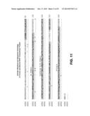 |
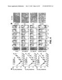 | 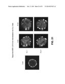 |
 |  |
 | 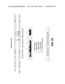 |
 |  |
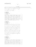 | 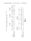 |
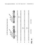 | 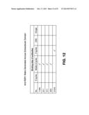 |
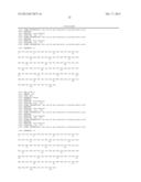 |  |
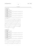 |  |
 |  |
 |  |
 |  |
 |  |
 |  |
 |  |
 | 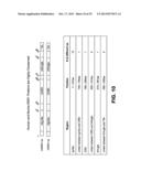 |
 | 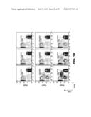 |
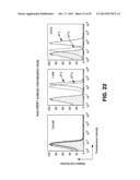 | 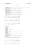 |
 |  |
 |  |
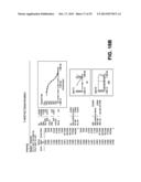 | 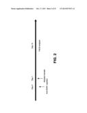 |
 |  |
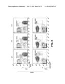 |
| Similar patent applications: | |
| Date | Title |
|---|---|
| 2013-04-11 | Anti-fcrh5 antibodies and immunoconjugates and methods of use |
| 2013-03-28 | Eggshell antimicrobial agent and method of use |
| 2013-04-11 | Therapeutic nuclease compositions and methods |
| 2013-04-11 | Compositions of jasmonate compounds and methods of use |
| 2013-04-11 | Tumor targeted antibodies and method for using the same |
| New patent applications in this class: | |
| Date | Title |
|---|---|
| 2019-05-16 | Binding members to tnf alpha |
| 2018-01-25 | Method for the treatment of multiple myeloma or non-hodgkins lymphoma with anti-cd38 antibody and bortezomib or carfilzomib |
| 2017-08-17 | Diagnosis of cancer |
| 2017-08-17 | Drug combinations and methods to stimulate embryonic-like regeneration to treat diabetes and other diseases |
| 2016-12-29 | Compositions and methods to treat inflammatory joint disease |
| New patent applications from these inventors: | |
| Date | Title |
|---|---|
| 2021-12-02 | Modulators of ror1-ror2 binding |
| 2016-05-26 | Methods for detecting antibodies |
| 2016-04-07 | Therapeutic antibodies against ror-1 protein and methods for use of same |
| 2015-08-20 | Antibodies and vaccines for use in treating ror1 cancers and inhibiting metastasis |
| Top Inventors for class "Drug, bio-affecting and body treating compositions" | |
| Rank | Inventor's name |
|---|---|
| 1 | David M. Goldenberg |
| 2 | Hy Si Bui |
| 3 | Lowell L. Wood, Jr. |
| 4 | Roderick A. Hyde |
| 5 | Yat Sun Or |