Patent application title: CLIP INHIBITORS AND METHODS OF MODULATING IMMUNE FUNCTION
Inventors:
Martha Karen Newell (Holland, TX, US)
Martha Karen Newell (Holland, TX, US)
Evan Newell (Singapore, SG)
Haig Keledjian (San Marino, CA, US)
Michael Agadjanyan (Huntington Beach, CA, US)
Assignees:
Viral Genetics, Inc.
IPC8 Class: AG01N3368FI
USPC Class:
424 852
Class name: Drug, bio-affecting and body treating compositions lymphokine interleukin
Publication date: 2013-10-03
Patent application number: 20130259829
Abstract:
The invention relates to methods for modulating the immune function
through targeting of CLIP molecules. The result is wide range of new
therapeutic regimens for treating, inhibiting the development of, or
otherwise dealing with, a multitude of illnesses and conditions,
including autoimmune disease, allergic disease transplant and cell graft
rejection, cancer, bacterial infection, HIV infection, and AIDS.Claims:
1.-60. (canceled)
61. A method for identifying a subject sensitive to treatment with a CLIP inhibitor, comprising, determining an MHC class I or II allele of the subject and determining a predicted binding value of the peptide for the MHC class I or II allele of the subject, wherein a predicted binding value greater than the predicted binding value of CLIP for MHC class I or II allele is indicative of whether a CLIP inhibitor is effective for displacing CLIP from the MHC class I or II allele of the subject and is a CLIP inhibitor for the subject.
62. A method of treating a subject with a CLIP inhibitor, comprising, determining an MHC class I or II allele of a subject and administering to the subject a CLIP inhibitor in an effective amount to displace CLIP from a surface of a cell.
63. (canceled)
64. The method of claim 62, wherein the subject is suspected of having Tuberculosis.
65.-66. (canceled)
67. The method of claim 62, wherein the subject is suspected of having Hepatitis C.
68.-70. (canceled)
71. The method of claim 62, wherein the subject is suspected of having Rheumatoid Arthritis.
72. The method of claim 62, wherein the subject is suspected of having a Proteus mirabilis infection.
73. (canceled)
74. The method of claim 62, wherein the subject is suspected of having Severe Acute Respiratory Syndrome.
75. (canceled)
76. The method of claim 62, wherein the subject is suspected of having Bacterial Meningitis.
77. (canceled)
78. The method of claim 62, wherein the subject is suspected of having Lyme Disease.
79. (canceled)
80. The method of claim 62, wherein the subject is suspected of having Malaria.
81. (canceled)
82. The method of claim 62, wherein the subject is suspected of having African trypanosomiasis.
83. (canceled)
84. The method of claim 62, wherein the subject is suspected of having Acquired immunodeficiency syndrome (AIDS).
85.-86. (canceled)
87. The method of claim 62, wherein the subject is suspected of having Rabies.
88. (canceled)
89. The method of claim 62, wherein the subject is suspected of having a Norovirus infection.
90.-91. (canceled)
92. The method of claim 62, wherein the subject is suspected of having poliomyelitis.
93. (canceled)
94. The method of claim 62, wherein the subject is suspected of having Reiter's syndrome.
95.-96. (canceled)
97. The method of claim 62, wherein the subject is suspected of having Hepatitis B.
98.-99. (canceled)
100. The method of claim 62, wherein the subject is suspected of having a Shigella flexneri infection.
101.-102. (canceled)
103. The method of claim 62, wherein the subject is suspected of having a Epstein-Barr Virus infection.
104.-120. (canceled)
Description:
RELATED APPLICATIONS
[0001] This application claims priority under 35 U.S.C. §119 from U.S. provisional application Ser. No. 61/135,942, filed Jul. 25, 2008, the contents of which are incorporated herein in their entirety.
BACKGROUND OF INVENTION
[0002] Major Histocompatiblity Complex (MHC)-encoded molecules are key components of T cell immunity. The significance of these molecules as tissue compatibility molecules was first observed in the late 1930s. Peter Gorer and George Snell observed that when tumors were transplanted from a genetically non-identical member of the same species, the tumors were always rejected, but when tumors were transplanted between genetically identical members of the same species, the tumor would "take" and would grow in the syngeneic animal. The genetic complex responsible for the rejection was subsequently found to be a series of genes that encode protein products known as Major Histocompatibility molecules. These genes, also known as immune response or IR genes, and their protein products are responsible for all graft rejection. There are two types of MHC molecules: MHC class I and MHC class II. All nucleated cells express cell surface MHC class I. A subset of specialized cells express class II MHC. Included in the specialized, professional antigen-presenting cells (APCs) are B cells, macrophages, microglia, dendritic cells, and Langerhans cells among others.
[0003] As stated above, B cells express MHC class II. Once antigen has been bound by the antigen receptor on the B cell, the antigen and its receptor are engulfed into an endosomal compartment. This compartment fuses with another compartment known as the lysosome. The B cell is very efficient at breaking down antigens into smaller parts and loading the parts into MHC class II in the lysosome. The MHC is then trafficked to the cell surface where the B cell can effectively "show" the antigen to a CD4+ T cell. The activated CD4 cell is also called a helper cell and there are two major categories, Th1 and Th2.
[0004] The MHC molecules are tightly protected in the endosomal/lysosomal compartments to insure that only antigens for which we need a response get presented to T cells. MHC class II molecules, prior to antigen loading, are associated with a molecule called invariant chain, also known as CD74. The invariant chain is associated with MHC class II (and recently shown to be associated with certain MHC class I molecules) prior to antigen loading into the antigen binding grooves of the MHC molecules. As antigen is processed, the invariant chain gets cleaved by proteases within the compartment. First an end piece is removed, and then another known as CLIP (class II invariant chain associated peptide). CLIP fills the groove that will ultimately hold the antigen until the antigen is properly processed. For a detailed review of the invariant chain, including CLIP, see Matza et al. (2003), incorporated herein in its entirety. Despite the fact that this "chaperone" role for invariant chain and CLIP has been identified, the full impact of these molecules on immune signaling and activation has yet to be determined.
SUMMARY OF INVENTION
[0005] The invention is based at least in part on the discovery that CLIP inhibitors are useful in the treatment of disorders such as HIV infection, autoimmune disease and tissue graft rejection.
[0006] The invention in some aspects is a method for treating a disorder associated with γδT cell expansion, activation and/or effector function by contacting a CLIP molecule expressing cell with a CLIP inhibitor in an effective amount to interfere with γδT cell expansion, activation and/or effector function by the CLIP molecule expressing cell. In some embodiments the γδT cell is a vγ9vδ2 T cell. Disorders associated with γδT cell expansion and/or activation include, for instance autoimmune disease, HIV infection, and cell, tissue and graft rejection.
[0007] The CLIP molecule expressing cell is a B cell in some embodiments. In other embodiments the CLIP compound expressing cell is a neuron, an oligodendrocyte, a microglial cell, or an astrocyte. In yet other embodiments the CLIP compound expressing cell is a heart cell, a pancreatic beta cell, an intestinal epithelial cell, a lung cell, an epithelial cell lining the uterine wall, and a skin cell. When the cell is a B cell, the method may further involve contacting the B cell with an anti-HLA class I or II antibody in an effective amount to kill the B cell.
[0008] The invention in some aspects is a composition of an isolated MHC class II CLIP inhibitor comprising a peptide of SEQ ID NO 49, 58, 59, 61, 62, 66, 67, 68, 69, 76, 77, 78, 81, 82, 86, 89, 90, 92, 104, 109, 110, 112, 117, 128, 129, 133, 136, 140, 141, 144, 146, 148, 149, 150, 154, 156, 157, 161, 162, 164, 168, 171, 172, 175, 177, 179, 186, 187, 188, 190, 191, 192, 196, 197, 201, 204, 205, 210, 217, 218, 220, 221, 222, 226, 227, 290, 291, 292, 293, 294, 295, 296, 297, 298, 299, 300, 301, 302, 303, 304, 305, 306, 307, 308, 309, 310, 311, 312, 313, 314, 315, 316, 317, 318, 319, 320, 321, 322, 323, 324, or a variant thereof and a carrier. The MHC class II CLIP inhibitor may be synthetic. In some embodiments the composition also includes an adjuvant, such as aluminum hydroxide or aluminum phosphate, calcium phosphate, mono phosphoryl lipid A, ISCOMs with Quil-A, and/or Syntex adjuvant formulations (SAFs) containing the threonyl derivative or muramyl dipeptide. In other embodiments the composition includes an anti-HIV agent and/or an antigen.
[0009] A method for treating a disease by administering to a subject a composition of a CLIP inhibitor and a pharmaceutically acceptable carrier is also provided. In some aspects the CLIP inhibitor is a MHC class II CLIP inhibitor. In these aspects the disease may be a viral infection, such as HIV, herpes, hepatitis A, B, or C, CMV, EBV, or Borrelia burgdorferi, a parasitic infection such as Leishmania or malaria, allergic disease, Alzheimer's disease, autoimmune disease or a cell or tissue graft. In these aspects of the invention a CLIP inhibitor that is a MHC class I CLIP inhibitor is also useful. In other aspects the CLIP inhibitor is a MHC class I CLIP inhibitor. In these aspects the disease may be cancer or bacterial infection.
[0010] In some embodiments the administration occurs over a period of eight weeks. In other embodiments the administration is bi-weekly which may occur on consecutive days. The administration may also be at least one of oral, parenteral, subcutaneous, intravenous, intranasal, pulmonary, intramuscular and mucosal administration.
[0011] In some embodiments the methods involve administering another medicament to the subject, such as an anti-HIV agent, an anti-viral agent, an anti-parasitic agent, an anti-bacterial agent, an anti-cancer agent, an anti allergic medicament, or an autoimmune medicament. In other embodiments the methods involve administering an adjuvant such as aluminum hydroxide or aluminum phosphate, calcium phosphate, nanoparticles, nucleotides ppGpp and pppGpp, killed Bordetella pertussis or its components, Corenybacterium derived P40 component, killed cholera toxin or its parts and/or killed mycobacteria or its parts.
[0012] In some embodiments the methods involve administering any of the compositions described herein.
[0013] In some embodiments the autoimmune disease is multiple sclerosis, systemic lupus erythematosus, type 1 diabetes, viral endocarditis, viral encephalitis, inflammatory bowel disease, rheumatoid arthritis, Graves' disease, autoimmune thyroiditis, autoimmune myositis, or discoid lupus erythematosus. In other embodiments the graft tissue or cell is heart, lung, kidney, skin, cornea, liver, neuronal tissue or cell, stem cell, including hematopoietic or embryonic stem cell.
[0014] In other aspects the invention is a kit housing a container housing a CLIP inhibitor comprising a peptide of SEQ ID NO 49, 58, 59, 61, 62, 66, 67, 68, 69, 76, 77, 78, 81, 82, 86, 89, 90, 92, 104, 109, 110, 112, 117, 128, 129, 133, 136, 140, 141, 144, 146, 148, 149, 150, 154, 156, 157, 161, 162, 164, 168, 171, 172, 175, 177, 179, 186, 187, 188, 190, 191, 192, 196, 197, 201, 204, 205, 210, 217, 218, 220, 221, 222, 226, 227, 290, 291, 292, 293, 294, 295, 296, 297, 298, 299, 300, 301, 302, 303, 304, 305, 306, 307, 308, 309, 310, 311, 312, 313, 314, 315, 316, 317, 318, 319, 320, 321, 322, 323, or 324; and instructions for administering the CLIP inhibitor to a subject.
[0015] In other aspects the invention is a peptide having a peptide sequence corresponding to any one of SEQ ID NOs. 49, 58, 59, 61, 62, 66, 67, 68, 69, 76, 77, 78, 81, 82, 86, 89, 90, 92, 104, 109, 110, 112, 117, 128, 129, 133, 136, 140, 141, 144, 146, 148, 149, 150, 154, 156, 157, 161, 162, 164, 168, 171, 172, 175, 177, 179, 186, 187, 188, 190, 191, 192, 196, 197, 201, 204, 205, 210, 217, 218, 220, 221, 222, 226, 227, 290, 291, 292, 293, 294, 295, 296, 297, 298, 299, 300, 301, 302, 303, 304, 305, 306, 307, 308, 309, 310, 311, 312, 313, 314, 315, 316, 317, 318, 319, 320, 321, 322, 323, and 324. A method of treating HIV by administering to a subject any of these peptides is also provided. In some embodiments, a method is provided of treating HIV by administering to a subject a peptide having a peptide sequence corresponding to SEQ ID NO 307 or 308.
[0016] In other aspects a method for identifying a subject sensitive to treatment with a CLIP inhibitor is provided by determining an MHC class I or II allele of the subject and determining a predicted binding value of the peptide for the MHC class I or II allele of the subject, wherein a predicted binding value greater than the predicted binding value of CLIP for MHC class I or II allele is indicative of whether a CLIP inhibitor is effective for displacing CLIP from the MHC class I or II allele of the subject and is a CLIP inhibitor for the subject.
[0017] In another aspect the invention is a method of treating a subject with a CLIP inhibitor by determining an MHC class I or II allele of a subject and administering to the subject a CLIP inhibitor in an effective amount to displace CLIP from a surface of a cell.
[0018] In other embodiments the CLIP inhibitor is a peptide that displaces CLIP. The peptide that displaces CLIP may be, for instance, a peptide selected from the group consisting of SEQ ID NOs. 49, 58, 59, 61, 62, 66, 67, 68, 69, 76, 77, 78, 81, 82, 86, 89, 90, 92, 104, 109, 110, 112, 117, 128, 129, 133, 136, 140, 141, 144, 146, 148, 149, 150, 154, 156, 157, 161, 162, 164, 168, 171, 172, 175, 177, 179, 186, 187, 188, 190, 191, 192, 196, 197, 201, 204, 205, 210, 217, 218, 220, 221, 222, 226, 227, 290, 291, 292, 293, 294, 295, 296, 297, 298, 299, 300, 301, 302, 303, 304, 305, 306, 307, 308, 309, 310, 311, 312, 313, 314, 315, 316, 317, 318, 319, 320, 321, 322, 323, and 324. In some embodiments the method further includes exposing the CLIP molecule expressing cell to an MHC class I or II loading peptide.
[0019] In some aspects, the invention provides methods of treating a subject with a CLIP inhibitor. In some embodiments, the methods involve determining an MHC class I or II allele of a subject and administering to the subject a CLIP inhibitor in an effective amount to displace CLIP from a surface of a cell. In some embodiments, MHC alleles which may be used in the methods, and CLIP inhibitors which may be used in association with these MHC alleles, are disclosed in Table 7.
[0020] In some aspects, the invention provides methods of treatment for a disease of Table 7. In some embodiments, the methods involve administering to a human suspected of having the disease a composition comprising a peptide selected from the group consisting of SEQ ID NOs. 290, 291, 292, 293, 294, 295, 296, 297, 298, 299, 300, 301, 302, 303, 304, 305, 306, 307, 308, 309, 310, 311, 312, 313, 314, 315, 316, 317, 318, 319, 320, 321, 322, 323, or 324 and a pharmaceutically acceptable carrier.
[0021] In some aspects, the invention provides methods of treatment of cancer. In some embodiments, the methods involve administering to a human suspected of having cancer a Toll Ligand Receptor (TLR) agonist, wherein the TLR is TLR2, TLR 7/8, TLR4, or TLR 9, and a CLP inhibitor in an effective amount to displace CLIP from a surface of a cell.
[0022] This invention is not limited in its application to the details of construction and the arrangement of components set forth in the following description or illustrated in the drawings. The invention is capable of other embodiments and of being practiced or of being carried out in various ways. Also, the phraseology and terminology used herein is for the purpose of description and should not be regarded as limiting. The use of "including," "comprising," or "having," "containing," "involving," and variations thereof herein, is meant to encompass the items listed thereafter and equivalents thereof as well as additional items.
BRIEF DESCRIPTION OF DRAWINGS
[0023] The accompanying drawings are not intended to be drawn to scale. In the drawings, each identical or nearly identical component that is illustrated in various figures is represented by a like numeral. For purposes of clarity, not every component may be labeled in every drawing. In the drawings:
[0024] FIG. 1 depicts % B Cell Death in resistant C57Bl6 versus sensitive Coxsackievirus infected mice from 1 to 5 days post infection.
[0025] FIGS. 2A and 2B are dot plots representing flow cytometric analysis of 5 day cultures in which CD40 Ligand activated B cells were co-cultured with autologous PMBCs for 5 days.
[0026] FIG. 3 depicts CLIP displacement from the surface of model B cells lines (Daudi and Raji) in response to thymic nuclear protein (TNP) mixture. FIG. 3A is a 3 hour reaction. FIG. 3B is a 24 hour reaction. FIG. 3c is a 48 hour reaction.
[0027] FIG. 4 depicts that 2-Deoxyglucose and dichloroacetate affects B cell surface CLIP.
[0028] FIG. 5 depicts CLIP displacement from the surface of Raji B cells lines in response to no treatment (5A and 5C) or treatment with MKN.5 (5B and 5D) for 4 (5A and 5B) and 24 hours (5C and 5D).
[0029] FIG. 6 depicts CLIP displacement from the surface of Daudi B cells lines in response to no treatment (6A and 6C) or treatment with MKN.5 (6B and 6D) for 4 (6A and 6B) and 24 hours (6C and 6D).
[0030] FIG. 7 depicts CLIP displacement from the surface of Raji (7B) or Daudi (7A) B cells lines in response to treatment with FRIMAVLAS for 24 hours.
[0031] FIG. 8 is a set of bar graphs depicting CLIP (8A), HLA DR, DP,DQ (8B) staining on the surface of Daudi cells in response to no treatment, or treatment with MKN.4 or MKN.6.
[0032] FIG. 9 depicts CLIP (y-axis) and HLA DR (x-axis) staining on the surface of B cells in response to no treatment, or treatment with MKN.4 or MKN.10.
[0033] FIG. 10 depicts CLIP (y-axis) and HLA DR (x-axis) staining on the surface of B cells in response to no treatment (10A) or DMSO (10G), or treatment with MKN.3, MKN5, MKN6, MKN.8 or MKN.10 (10B-10F respectively).
[0034] FIG. 11 depicts Treg in response to no treatment (11A), or treatment with MKN.6 (11B) or TNP (11C).
[0035] FIG. 12 depicts TLR activation of mouse splenic B cells results in ectopic CLIP in MHC class II. FIG. 12a: LPS activation of spleen cells from B6.129 mice (H-2b). B cells are detected by staining with PE conjugated anti-mouse B220, shown on Y-axis, versus staining with 15G4-FITC anti-mouse CLIP/I-Ab, as shown on the X-axis. A representative of four experiments at unique time points, 0 to 72 hours by 24-hour increments, left to right, is shown. FIG. 12b (linear representation from FIG. 12a): changes in percentages of CLIP+ B cells, left Y-axis, and quantitative depiction of increasing mean fluorescence intensity of CLIP/I-Ab staining, right Y-axis. FIG. 12c: antigen receptor engagement increases cell surface MHC class II but not ectopic CLIP. B6.129 splenocytes, untreated or treated in vitro with anti-immunoglobulin (as a surrogate for antigen) or CpG-ODN were stained with anti-mouse B220-PE versus 15G4-FITC anti-mouse CLIP/I-Ab (bars, left Y-axis) or with anti-mouse B220-PE versus anti-mouse MHC class II-FITC (I-Ab) (line graph, right Y-axis). FIG. 12d: toll ligands 9, 10 used to activate cells, listed from top down, Poly I:C, Pam3Cys, R848, LPS, CpG-ODN, and no treatment, as indicated. Shown are percentages of CLIP+ B cells in splenocytes stimulated in vivo from B6.129 mice, left panel; H2M-deficient mice, middle panel; Ii-deficient mice, right panel.
[0036] FIG. 13 depicts TLR activation results in ectopic CLIP expression on human B cells from peripheral blood mononuclear cells (PBMC) cultures. FIG. 13a: human PBMC from five donors were cultured for 24 hours with toll ligands (CpG-ODN, LPS, Pam3Cys, and Poly I:C) and were stained with a pan anti-HLA-DR-FITC antibody (values of isotype controls were subtracted from the specific stains, ΔMFI). FIG. 13b: cells were stained using anti-human CLIP-FITC versus CD19-PE (values of isotype controls were subtracted from the specific stains, ΔMFI). For FIGS. 13a & 13b: nil vs. CpG p=0.06821; nil vs. LPS p=0.0390; nil vs. PAM p=0.0124. FIG. 13c: cells were stained for CLIP-FITC versus CD19-PE as described for FIG. 13b. The data represent the percent of total PBMC that are CLIP+ B cells subsequent to treatment. For FIG. 13c: nil vs. CpG p=0.0058; nil vs. LPS p=0.0254. FIGS. 13d & 13e: PBMC from two individual donors (donor 1, FIG. 13d; donor 2, FIG. 13e) were stained for baseline levels of CLIP immediately ex vivo, after culture for 48 hours, or after culture in the presence of R848 (solid black bars, respectively). Cells were cultured in the presence of either VGV-hB (gray stippled bars) or with an MHC-dependent peptide, VGV-pB (white bars).
[0037] FIG. 14 depicts that administration of targeted peptide in combination with CpG-ODN reverses the inflammatory effects of TLR9 activation. FIG. 14a: B6.129 mice were injected with CpG-ODN, a TLR9 agonist without (black squares) or with VGV-hB (black circles). Total spleen cell recoveries (top panel) and lymph node cell recoveries (lower panel) at 24, 72, and 96 hours are reported. FIG. 14b: spleen cells from untreated B6.129 (upper left panel) mice, spleen cells from mice injected intraperitoneally with CpG-ODN for 48 hours (upper right panel), spleen cells from mice injected intraperitoneally with CpG-ODN+targeted peptide (VGV-hB) (lower left panel), or spleen cells from mice injected intraperitoneally with CpG-ODN+scrambled peptide (VGV-sP) (lower right panel), were harvested and stained using anti-mouse B220-PE Cy5 versus 15G4-FITC anti-mouse CLIP/I-Ab Cells were analyzed flow-cytometrically using two-dimensional dot plot analysis. FIGS. 14c & 14d: individual B6.129 animals were injected with CpG-ODN and one of three doses of peptide replacement, with either VGV-hB (FIG. 14c) or VGV-sB (FIG. 14D). As indicated, for each dose, four to six animals were injected with each of three doses, at 0.5, 5, and 50 μg per injection. Splenocytes were removed after 48 hours, stained using anti-mouse B220-PE versus 15G4-FITC anti-mouse CLIP/I-Ab. Cells were acquired and analyzed with flow cytometry, using two-dimensional dot plot analysis. Percentages of CLIP+ B cells were plotted using scatter plot analysis. The formula for the slope of each line is indicated in each of FIGS. 14c & 14d.
[0038] FIG. 15 depicts the effects of targeted peptides on the distribution of TLR activated lymphoid subsets of B cells, CD4+ T cells, CD8+ T cells, and on CD4+ T regulatory cells. FIG. 15a: B6.129 mice were injected with CpG-ODN without (squares) or with (circles) VGV-hB. Spleen (solid black symbols) and lymph nodes (open symbols) were harvested at 24, 72, and 96 hours, and stained using anti-mouse B220-PE versus 15G4-FITC anti-mouse CLIP/I-Ab. Data were plotted as percent CLIP+ B cells from either spleen or node. FIG. 15b: B6.129 mice were injected with CpG-ODN without (squares) or with (circles) VGV-hB. Spleen (solid black symbols) and lymph nodes (open symbols) were harvested at 24, 72, and 96 hours, and stained using anti-mouse CD8-PE. Data were plotted as percent CD8+ T cells from either spleen or node as indicated. FIG. 15c: B6.129 mice were injected with CpG-ODN without (squares) or with (circles) VGV-hB. Spleen (solid black symbols) and lymph nodes (open symbols) were harvested at 24, 72, and 96 hours, and stained using anti-mouse CD4 GK1.5-FITC. Data were plotted as percent CD4+ T cells from either spleen or node. FIG. 15d: B6.129 mice were injected with CpG-ODN without (squares) or with (circles) VGV-hB. Spleen (solid black symbols) and lymph nodes (open symbols) were harvested at 24, 72, and 96 hours, and stained using anti-mouse CD4 GK1.5-PE versus anti-mouse FoxP3-FITC. Data were plotted as percent CD4+ FoxP3+ T cells from either spleen or node. These data represent four experiments.
[0039] FIG. 16 depicts that TLR activation and peptide reversal differentially affect cell death of lymphocyte subsets. FIG. 16a: B6.129 mice were injected with CpG-ODN without (solid black lines) or with VGV-hB (dashed lines). Cells were counted and viability was determined flow cytometrically. T cell viability 48 hours subsequent to CPG-ODN treatment alone is indicated by solid circles and solid black line; T cell viability, 48 hours after treatment with CpG-ODN and VGV-hB, is indicated by solid squares and dashed line. B cell viability, CpG-ODN treatment alone, is indicated by black x's on a solid black line; B cell viability after CpG-ODN and VGV-hB is indicated by solid black triangles and dashed lines. FIG. 16b: Ag-specific T cell hybridomas induce apoptosis B cells. FIG. 16c: Ag-specific T cell hybridomas induce apoptosis in resting and in CpG-ODN stimulated splenocytes, but not in antigen receptor engaged B cells. Resting, anti-immunoglobulin primed, and in vivo activated B cells from AKR animals were cultured overnight with A6.A2 or 3A9 T cell hybridomas, either with or without the antigen for which the T cells are specific, hen egg lysozyme (HEL) peptide 46-61. Cells were harvested and viability was determined using the TUNEL assay. Results are presented as percent apoptosis with HEL minus percent apoptosis without the peptide HEL, as indicated. FIG. 16c: resting B cells from MRLlpr/lpr animals are refractory to T cell-induced apoptosis. Resting B cells from MRLlpr/lpr, MRL+/+ and AKR animals were cultured overnight with A6.A2 or 3A9 T cell hybridomas, either with or without HEL, p46-61. Each bar represents the difference between B cell apoptosis in the presence and absence of the antigen HEL. Positive values indicate that the addition of HEL antigen increased resting B cell apoptosis over that in the no HEL control, while negative values indicate that the addition of HEL decreased the B cell apoptosis below the level of "spontaneous" apoptosis seen in the no HEL antigen control.
DETAILED DESCRIPTION
[0040] For clarity of disclosure, and not by way of limitation, the detailed description of the invention is divided into the following subsections:
[0041] (i) CLIP/Tregs/Disease
[0042] (ii) CLIP inhibitors
[0043] (iii) Uses of the Compositions of the Invention
[0044] (iv) Infectious Disease
[0045] (v) Transplant/Graft Rejection
[0046] (vi) Autoimmune Disease
[0047] (vii) Cancer
[0048] (viii) Alzheimer's Disease
[0049] (ix) Allergic Disease
[0050] (x) Characterization and Demonstration of CLIP inhibitor activities
[0051] (xi) Combinations with Antibodies
[0052] (xii) Dosage Regimens
[0053] (xiii) Administrations, Formulations
[0054] (xiv) Preparation of Peptides (Purification, Recombinant, Peptide Synthesis)
[0055] (xv) Articles of Manufacture
(i) CLIP/Tregs/Disease
[0056] The present invention provides new insights into the role of invariant chain (CD74) and CLIP in disease and presents novel approaches to modulating the immune function through targeting of invariant chain/CD74 and CLIP. The result is a wide range of new therapeutic regimens for treating or inhibiting the development or progression of a multitude of illnesses and conditions, including autoimmune disease, transplant and cell graft rejection, viral infection such as HIV infection, cancer bacterial infection, as well as novel methods of diagnosis and of introducing a treatment regimen into a subject.
[0057] Many bacteria and viruses produce substances, collectively called Toll ligands, that elicit an immediate response from an individual's immune system. These Toll ligands appear to promote inflammation by activating a wide variety of immune cells to bring them rapidly into battle against the invading pathogen. In most cases, these events correlate with a healthy and productive immune response to the pathogen. However, in some cases the Toll ligand binds to a Toll-like Receptor (TLR) on lymphocytes and non-specifically activates immune cells called B and T lymphocytes that would normally respond to infectious pathogens with an exquisitely specific response. When Toll ligands activate B cells in a non-specific way, the non-specific activation is a pro-inflammatory event that may result in uncontrolled, or even auto-reactive, production of antibodies. When a B cell is activated non-specifically, we have discovered that the B cell expresses an important, small self-peptide called MHC class II invariant peptide, CLIP. In most individuals, a control cell, known as a T regulatory cell (Treg for short), has been shown, to kill the activated B cell.
[0058] During a viral or bacterial infection, non-antigen specific B cells in close proximity to an inflammatory or inciting lesion could manage to become activated in a bystander fashion. In those cases, CLIP would remain in the groove and get transported to the cell surface of the B cell. Its presence on the cell surface can be undesirable because if CLIP gets removed from the groove by a self antigen, the B cell would be in a position to present self antigens to self-reactive T cells, a process that could lead to autoreactivity and autoimmune disease. For some B cells this may result in death to the B cell by a nearby killer cell, perhaps a natural killer (NK) cell, unless the antigen receptor on the B cell has engaged antigen. Antigen recognition would thereby provide a survival signal for the B cell. However, if a killer cell doesn't remove the potentially autoreactive B cell and it encounters a CD4.sup.+ T cell that can recognize that antigen (most likely one that was not in the thymus) the B cell might receive additional help from a T cell specific for the antigen that now occupies the groove (antigen binding location in the MHC molecule). Alternatively, a nearby cell whose job it is to detect damaged self cells, may become activated by the self antigen-presenting B cell. Such a damage detecting cell is, for example, an effector T cell (Teff) such as a gamma delta T cell, also referred to as a γδT cell (γδ refers to the chains of its receptor). The γδT cell can then seek out other sites of inflammation (for example in the brain in MS, in the heart for autoimmune myocarditis, in the pancreas in the case of Type I Diabetes). Alternatively, the γδT cell might attempt to kill the CD4.sup.+ T cell that may respond to self antigens.
[0059] These discoveries have important implications in the treatment of infectious disease, cancer, autoimmune disease, allergic disease, Alzheimer's disease and graft rejection. For example, HIV disease is characterized by rapidly dividing, activated B cells that cause enlargement of the lymph nodes in the HIV infected individuals. It has been discovered herein that the B cells from an HIV infected lymph node have high levels of CLIP, indicating that the B cells have been non-specifically activated. In fact, the lymph node is filled with B cells intertwined with infected CD4 T cells. It has been discovered that replacement of the CLIP on the surface of the B cell with a target peptide having high affinity for the specific MHC of that individual, would result in activation of CD4 T cells. As described in more detail below, a group of thymus derived peptides can function as these specific target peptides. Also computational methods can be used to predict additional target peptides, as shown according to the invention. It has recently been shown that there is a strong correlation between the presence of Tregs and the length of time of infection prior to full-blown AIDS. Moreover, as Treg numbers decline, there is a concomitant rise in viral load in that individual. Thus, the invention involves the discovery that replacement of CLIP on the MHC with specific peptides described herein as well as custom-designed and computationally predicted "targeted peptides" could reactivate Tregs and dampen the pathological inflammation that is required for an increase in virally infected cells. Appropriate targeted peptides can be synthesized based on patent specific MHC information in order to treat HIV positive individuals with all different types of MHC fingerprints.
[0060] Further, an example of the necessity for selective B cell death when the antigen receptor has not been bound by a real bona fide antigen is in Coxsackievirus. Most people that contract Coxsackievirus get a flu-like disease and then they recover, but in a genetic manner, some people (especially young men) contract Coxsackievirus and then go on to develop autoimmune myocarditis. In some genetically inbred strains of mice, the mice are resistant to myocarditis post-infection; in other strains of mice, the mice succumb. One difference was that the mice that were susceptible had a particular isoform of MHC class II. Mice on the resistant background having the other isoform of class II inserted, both artificially and genetically, showed susceptibility simply on the basis of the isoform, and it was shown that susceptibility depended on the presence of γδT cells (Huber et al., 1999, J Virol. 1999 July; 73(7):5630-6.).
[0061] Moreover, it was observed that in the mice that did not develop autoimmune disease, during the course of infection, all of their B cells died. Even with such B cell death, the animals survive as new B cells are produced continually. However, the animals susceptible to autoimmune disease had no B cell death. Further support for this notion is the γδ knock-out mice (they genetically have no γδT cells) do not get EAE, the mouse version of multiple sclerosis, nor do they get Type 1 diabetes. NK cell knock-out animals get worse disease in both cases. In addition, the invariant chain knock-out animals are resistant to the animal models of autoimmune diseases as well.
[0062] Many therapies to block autoimmune and transplant disease involve eliminating or inhibiting B cells. No one knows the mechanism by which these B cell depleting therapies make people better. The inventor has observed that γδT cell activation is often associated with proteins that have been lipid modified. It turns out the invariant chain is fatty acid acylated (e.g., palmitoylated). As described in the examples below and in co-pending U.S. Ser. No. 12/011,643 filed Jan. 28, 2008, entitled METHODS OF MODULATING IMMUNE FUNCTION, and naming M. Karen Newell, Evan Newell and Joshua Cabrera as inventors, antigen non-specifically activated human B cells were treated with anti-CLIP antibodies and subjected to flow cytometry. It was surprisingly found that these antigen-non-specifically activated B cells express cell surface CLIP. Thus, the inventors of U.S. Ser. No. 12/011,643 recognized that B cell surface expression of CLIP is likely how γδT cells get activated. For example, if there is inflammation at a given site, the long-lived γδT cell kills the type of CD4 helper T cell that could improve disease (the Th2 CD4+ T cells), at the site of injury. These cells may attack the inflamed tissue as well as kill the Th2 cells, leaving behind B cells that can now present self antigens (antigens that load into the CLIP binding site) to Th1 cells. The Th1 cells go on to activate additional CD8 killer cells and to attack the tissues as well. Once the γδT cell is activated, it may search for damaged tissue. Importantly, CLIP may associate with certain isoforms of MHC class II (I-A, I-E in mouse, HLA-DR, HLA-DP, and HLA-DQ in humans) with relatively different binding constants and to certain MHC class I's (for example, but not limited to, CD1). Interestingly, many autoimmune diseases map to the same HLA-DR alleles and not to the other isoforms.
[0063] T lymphocytes, like B lymphocytes, arise from hematopoeitic stem cells in the bone marrow. However, unlike B cells, the pre-T cells travel to another peripheral lymphoid tissue, the thymus, where T lymphocyte maturation processes occur. Interestingly, the thymus, as a T cell development organ, reaches its maximum size and capacity in very early childhood around the age of 2 to 3 years and, at puberty, the thymus begins to involute--shrinking to a small rudiment of what it had been earlier. No one has unraveled exactly how the pre-T cell is recruited to homes to the thymus, but research has shown that once the cells arrive they may stay as long as two weeks before the mature, appropriate cells leave the thymus to circulate throughout the periphery.
[0064] The thymus is the place where the pre-T cell develops the ability to recognize an enormous repertoire of antigens presented by either MHC class I or MHC class II. The pre-T cells enter the thymus without receptors for antigen and MHC, without CD4, and without CD8. In the thymus, T cells acquire T cell receptors for antigen, and either CD4 or CD8. During the process, those T cells that will recognize antigen and MHC class I become CD8.sup.+ T cells and those that recognize MHC class II and antigen become CD4.sup.+ T cells. Both CD4 and CD8 positive cells have cell surface T cell receptors for antigen. If a T cell, either a CD4+ or a CD8+ T cell, recognizes "self" antigen and self MHC class I or self MHC class II in the thymus, that T cell is deleted. For most of the CD4.sup.+ and CD8.sup.+ T cells have T cell receptors that consist of an α chain and a β chain. There are other, more recently described T cells that express receptors that are called γδ T cell receptors. The developmental maturation of T cells in the thymus results in a high percentage of thymocyte cell death. Waves of cortisone kill many of the pre-T cells that don't meet the necessary requirements for recognition and survival. In addition to cortisone-dependent thymocyte cell death, recognition of antigen in the thymus deletes some potentially self-reactive T cells from the repertoire. The process of antigen-specific T cell death in the thymus is commonly referred to as "negative" selection. The CD4.sup.+ or CD8.sup.+ T cells that recognize self MHC class I or MHC class II plus self antigen (like CLIP) will be deleted in the thymus. Those that could recognize CLIP and someone else's MHC class I or class II will not have been deleted. The cells that meet all of the survival criterion, e.g. appropriate recognition of antigen and either MHC class I for the developing CD8.sup.+ T or MHC class II for the developing CD4.sup.+ T cell travel to other regions of the body.
[0065] Some subsets of T regulatory cells (Tregs) suppress immune responses of other cells, in order to keep the immune response in check and avoid attacking self tissue. Some Tregs express CD4 or CD8 and CD25 and Foxp3. Tregs have more diverse TCR expression than other T cells such as NKT or γδ T cells, both of which may be biased towards self-peptides. Although the process of Treg selection is still unknown, it appears to be regulated at some level by the affinity of interaction with the self-peptide MHC complex. For instance, T cells which receive strong signals will undergo apoptotic death; and those that receive a weak signal will survive and be selected to become effector T cells. The T cells that receive an intermediate signal may become Tregs. As a result of this process, all T cell populations will end up with a mixture of Teff and Treg cells, although, the relative proportions will be determined by the affinities of the T cell for the self-peptide-MHC.
[0066] A properly functioning immune system must discriminate between self and non-self. Failure of this process causes destruction of cells and tissues of the body in the form of autoimmune disease. An important function of Tregs is to actively suppress immune system activation and thus prevent pathological self-reactivity, i.e. autoimmune disease. Also it is believed that some pathogens may have evolved to manipulate Tregs to immunosuppress the host and thus potentiate their own survival. Treg activity has been reported to increase in response to several infectious agents including, retroviral, Leishmania and malaria.
[0067] According to our model, if an MHC molecule on an activated B cell surface binds a targeted peptide with greater affinity than the CLIP occupying the groove of the MHC molecule, the consequence will be activation of Treg cells. The Treg cells can dampen the immune response by killing aberrantly activated B cells. The specific role of Tregs in each of the disease models is discussed in more detail below.
(ii) CLIP Inhibitors
[0068] A CLIP inhibitor as used herein is any molecule that reduces the association of a CLIP molecule with MHC by binding to the MHC and blocking the CLIP-MHC interaction. The CLIP inhibitor may function by displacing CLIP from the surface of a CLIP molecule expressing cell. A CLIP molecule expressing cell is a cell that has MHC class I or II on the surface and includes a CLIP molecule within that MHC. Such cells include B cells, neurons, oligodendrocytes, microglial cells, astrocytes, heart cells, pancreatic beta cells, intestinal epithelial cells, lung cells, epithelial cells lining the uterine wall, and skin cells.
[0069] The CLIP molecule, as used herein, refers to intact CD74 (also referred to as invariant chain), as well as the naturally occurring proteolytic fragments thereof. CLIP is one of the naturally occurring proteolytic fragments thereof. The function of the CLIP molecule in this invention is mainly as an MHC class II chaperone. MHC class II molecules are heterodimeric complexes that present foreign antigenic peptides on the cell surface of antigen-presenting cells (APCs) to CD4.sup.+ T cells. MHC class II synthesis and assembly begins in the endoplasmic reticulum (ER) with the non-covalent association of the MHC α and β chains with trimers of CD74. CD74 is a non-polymorphic type II integral membrane protein; murine CD74 has a short (30 amino acid) N-terminal cytoplasmic tail, followed by a single 24 amino acid transmembrane region and an ˜150 amino acid long lumenal domain. Three MHC class II αβ dimers bind sequentially to a trimer of the CD74 to form a nonameric complex (αβIi)3, which then exits the ER. After being transported to the trans-Golgi, the αβIi complex is diverted from the secretory pathway to the endocytic system and ultimately to acidic endosome or lysosome-like structures called MHC class I or II compartments.
[0070] The N-terminal cytoplasmic tail of CD74 contains two extensively characterized dileucine-based endosomal targeting motifs. These motifs mediate internalization from the plasma membrane and from the trans-Golgi network. In the endocytic compartments, the CD74 chain is gradually proteolytically processed, leaving only a small fragment, the class II-associated CD74 chain peptide (CLIP), bound to the released αβ dimers. The final step for MHC class II expression requires interaction of αβ-CLIP complexes with another class II-related αβ dimer, called HLA-DM in the human system. This drives out the residual CLIP, rendering the αβ dimers ultimately competent to bind antigenic peptides, which are mainly derived from internalized antigens and are also delivered to the endocytic pathway. The peptide-loaded class II molecules then leave this compartment by an unknown route to be expressed on the cell surface and surveyed by CD4.sup.+ T cells.
[0071] CLIP inhibitors include peptides and small molecules that can replace CLIP. In some embodiments the CLIP inhibitor is a peptide. A number of peptides useful for displacing CLIP molecules are described herein. For instance a number of peptide sequences that function in this manner are disclosed in Table 1. The peptides disclosed in Table 1 are thymus derived peptides. The thymus derived peptides are present in subfractions of extracts obtained from thymus and have sometimes been described as "thymus nuclear protein (TNP)" or "thymus factors (TF)" when isolated from calf thymus (see for example US 20040018639). TNP or TF refers to those proteins that are produced in and found in the thymus. The peptides contributing to the therapeutic activity of TNP have now been identified and characterized and are useful for therapeutic purposes such as the treatment of infectious disease, cancer, autoimmune disease, Alzheimer's disease and transplant/graft rejection.
[0072] TNPs are typically purified from the thymus cells of freshly sacrificed, i.e., 4 hours or less after sacrifice, mammals such as monkeys, gorillas, chimpanzees, guinea pigs, cows, rabbits, dogs, mice and rats. Such methods can also be used to prepare a preparation of peptides of the invention. Alternatively, the thymus derived peptides can be synthesized using routine procedures known in the art in view of the peptide sequence information provided in Table 1. Such methods are preferred in some embodiments and such peptides are referred to herein as synthetic peptides. For instance, it is routine in the art to prepare peptides using recombinant technology. Additionally the peptides may be purchased from commercial vendors that synthesize proteins or they may be synthesized directly using known techniques for peptide synthesis. Each of these methods is described in more detail below.
[0073] A composition of a CLIP inhibitor may include one or more of the thymus derived peptides listed in Table 1. The compositions for therapeutic use can include, one or more, most or all of the peptides found in Table 1 as long as the composition is not a thymus nuclear protein extract or TNP extract. As used herein a "thymus nuclear protein extract" or "TNP extract" is a preparation of thymus peptides isolated and formulated according to the methods described in U.S. Ser. No. 11/973,920. A composition is not a thymus nuclear protein extract or TNP extract if it has additional components or less components or is all or partly synthetic. For instance a composition is not a thymus nuclear protein extract or TNP extract if the peptides included therein are prepared from natural sources but the composition does not include every peptide of a thymus nuclear protein extract as described in U.S. Ser. No. 11/973,920, for instance those listed in Table 1. Thus a single composition may include many of these peptides as long as all of the peptides found in Table 1 are not included if all of the peptides are derived from a natural thymus. However, the composition may include all of the peptides if one or more of the peptides in the mixture are synthetic. Additionally, it may include all of the peptides if one or more additional elements is added such as an extra synthetic peptide.
[0074] The peptides of Table 1 are also identified in co-pending application filed on even date with the instant application and entitled Proteins for Use in Diagnosing and Treating Infection and Disease, naming the instant inventors.
TABLE-US-00001 TABLE 1 Amino Acid Sequence SEQ ID NO. KALVQNDTLLQVKG 1 KAMDIMNSFVNDIFERI 2 KAMGIMKSFVNDIFERI 3 KAMGNMNSFVNDIFERI 4 KAMSIMNSFVNDLFERL 5 KASGPPVSELITKA 6 KDAFLGSFLYEYSRR 7 KDDPHACYSTVFDKL 8 KEFFQSAIKLVDFQDAKA 9 KESYSVYVYKV 10 KGLVLIAFSQYLQQCPFDEHVKL 11 KHLVDEPQNLIKQ 12 KHPDSSVNFAEFSKK 13 KKQTALVELLKH 14 KKVPEVSTPTLVEVSRN 15 KLFTFHADICTLPDTEKQ 16 KLGEYGFQNALIVRY 17 KLKPDPNTLCDEFKA 18 KLVNELTEFAKT 19 KLVVSTQTALA 20 KQTALVELLKH 21 KSLHTLFGDELCKV 22 KTITLEVEPSDTIENVKA 23 KTVMENFVAFVDKC 24 KTVMENFVAFVDKCCAADDKEACFAVEGPKL 25 KTVTAMDVVYALKR 26 KVFLENVIRD 27 KVPEVSTPTLVEVSRN 28 KYLYEIARR 29 MGIMNSFVNDIFERI 30 RAGLQFPVGRV 31 RDNIQGITKPAIRR 32 REIAQDFKTDLRF 33 RFQSAAIGALQEASEAYLVGLFEDTNLCAIHAKR 34 RILGLIYEETRR 35 RISGLIYEETRG 36 RISGLIYKETRR 37 RKENHSVYVYKV 38 RLLLPGELAKH 39 RNDEELNKLLGKV 40 RNECFLSHKDDSPDLPKL 41 RRPCFSALTPDETYVPKA 42 RTLYGFGG 43 RTSKLQNEIDVSSREKS 44 RVTIAQGGVLPNIQAVLLPKK 45 LPDTEKQKL 46 YSTVFDKLK 47 ITLEVEPSD 48 LVQNDTLLQ 49 IKAMGIMKS 50 IKAMSIMNS 51 YVYKVRLLL 52 IKAMGNMNS 53 VRLLLPGEL 54 VVYALKRKV 55 YEIARRMGI 56 FRFQSAAIG 57 VVSTQTALA 58 IMNSFVNDI 59 ICTLPDTEK 60 MGIMKSFVN 61 MGIMNSFVN 62 LVELLKHKS 63 FERIKAMGI 64 FERIKAMSI 65 VLIAFSQYL 66 IMNSFVNDL 67 IMKSFVNDI 68 IQGITKPAI 69 VYVYKVRLL 70 YVYKVKGLV 71 LIYKETRRR 72 VKGLVLIAF 73 IRRREIAQD 74 VYVYKVKGL 75 VTAMDVVYA 76 YGFQNALIV 77 LVNELTEFA 78 VRYKLKPDP 79 LKTVTAMDV 80 FQNALIVRY 81 MSIMNSFVN 82 VKAKTVMEN 83 FKAKLVNEL 84 LRFRFQSAA 85 LVLIAFSQY 86 LKASGPPVS 87 VIRDKVPEV 88 VQNDTLLQV 89 MGNMNSFVN 90 YVPKARTLY 91 FQSAIKLVD 92 LYGFGGRTS 93 YKVKGLVLI 94 LVELLKHKK 95 LKHKKVPEV 96 LLKHKSLHT 97 YKVRLLLPG 98 VRNECFLSH 99 IVRYKLKPD 100 LIVRYKLKP 101 LLGKVRNEC 102 FERIKAMGN 103 VAFVDKCCA 104 LIYEETRRR 105 LIYEETRGR 106 VYALKRKVF 107 YLYEIARRM 108 LVVSTQTAL 109 VFLENVIRD 110 LVEVSRNKL 111 LIAFSQYLQ 112 IRDKVPEVS 113 LCKVKTITL 114 LIKQKHPDS 115 FERIRAGLQ 116 FQSAAIGAL 117 LVEVSRNKY 118 VKLKHLVDE 119 VYKVKGLVL 120 YALKRKVFL 121 VELLKHKKV 122 LQVKGKAMD 123
LKHKSLHTL 124 VELLKHKSL 125 VPKARTLYG 126 FKTDLRFRF 127 MDIMNSFVN 128 IKLVDFQDA 129 FVDKCKTVM 130 IHAKRRILG 131 FLYEYSRRK 132 VMENFVAFV 133 YLVGLFEDT 134 VYKVRLLLP 135 YLQQCPFDE 136 IRAGLQFPV 137 LLKHKKVPE 138 IKQKHPDSS 139 VLPNIQAVL 140 VEPSDTIEN 141 FGGRTSKLQ 142 VAFVDKCKT 143 FFQSAIKLV 144 FQDAKAKES 145 IQAVLLPKK 146 LLQVKGKAM 147 IAFSQYLQQ 148 FLGSFLYEY 149 FVNDIFERI 150 VDEPQNLIK 151 LSHKDDSPD 152 FLSHKDDSP 153 LPNIQAVLL 154 LKRKVFLEN 155 LLPGELAKH 156 FVAFVDKCC 157 IFERIKAMS 158 IENVKAKTV 159 VSRNKLFTF 160 LKPDPNTLC 161 MENFVAFVD 162 YSRRKDDPH 163 LFGDELCKV 164 FERLKASGP 165 VSTQTALAK 166 FAKTKLVVS 167 VTIAQGGVL 168 LNKLLGKVR 169 LYEIARRMG 170 MKSFVNDIF 171 LFTFHADIC 172 LAKQTALVE 173 FVAFVDKCK 174 FVNDLFERL 175 VKTITLEVE 176 IAQGGVLPN 177 LRRPCFSAL 178 LGSFLYEYS 179 LCAIHAKRR 180 LPKLRRPCF 181 VEVSRNKLF 182 FLENVIRDK 183 IYKETRRRK 184 VEVSRNKYL 185 FVDKCCAAD 186 LFEDTNLCA 187 VNFAEFSKK 188 VGRVRDNIQ 189 MNSFVNDIF 190 MNSFVNDLF 191 LVDEPQNLI 192 FSKKKKQTA 193 YGFGGRTSK 194 LITKAKDAF 195 MDVVYALKR 196 LLLPGELAK 197 LQFPVGRVR 198 LKEFFQSAI 199 YEYSRRKDD 200 LTPDETYVP 201 LGKVRNECF 202 LKHLVDEPQ 203 LQNEIDVSS 204 LVDFQDAKA 205 FAVEGPKLK 206 VSELITKAK 207 IFERIRAGL 208 LENVIRDKV 209 VGLFEDTNL 210 VSSREKSRV 211 IYEETRRRI 212 IFERIKAMG 213 FGDELCKVK 214 LFERLKASG 215 IARRMGIMN 216 LGLIYEETR 217 ILGLIYEET 218 YEETRRRIS 219 IDVSSREKS 220 LHTLFGDEL 221 LVGLFEDTN 222 VKGKAMDIM 223 FPVGRVRDN 224 VSRNKYLYE 225 IAQDFKTDL 226 FHADICTLP 227 VRDNIQGIT 228 YKLKPDPNT 229 VDFQDAKAK 230 FAEFSKKKK 231 LYEYSRRKD 232 FDEHVKLKH 233 LTEFAKTKL 234 LQQCPFDEH 235 LEVEPSDTI 236 IGALQEASE 237 VDKCKTVME 238 VFDKLKEFF 239 FTFHADICT 240 VPEVSTPTL 241 FSALTPDET 242 ITKPAIRRR 243 YKETRRRKE 244 IYEETRGRI 245 VEGPKLKTV 246 FEDTNLCAI 247 VNELTEFAK 248
YSVYVYKVK 249 LQEASEAYL 250 ISGLIYKET 251 YEETRGRIS 252 FDKLKEFFQ 253 VSTPTLVEV 254 VNDLFERLK 255 LPGELAKHR 256 VNDIFERIK 257 FSQYLQQCP 258 ITKAKDAFL 259 LGEYGFQNA 260 LCDEFKAKL 261 VDKCCAADD 262 VNDIFERIR 263 ISGLIYEET 264 LAKHRNDEE 265
[0075] When the composition includes more than one thymus derived peptide, the ratio of the peptides in the composition can vary greatly. For instance if the composition includes two different peptides the ratio of the first peptide to the second peptide can range from 0.01 weight percent (wt %): 0.99 wt % to 0.99 wt %:0.1 wt % or any ratio there between.
[0076] In some embodiments, the compositions of the invention that are used in prevention or treatment of cancer and/or infectious diseases or other disorders comprise an enriched, an isolated, or a purified thymus derived peptide of Table 1 that is a CLIP inhibitor. In accordance with the methods described herein, a CLIP inhibitor employed in a composition of the invention can be in the range of 0.001 to 100 percent of the total mg protein, or at least 0.001%, at least 0.003%, at least 0.01%, at least 0.1%, at least 1%, at least 10%, at least 30%, at least 60%, or at least 90% of the total mg protein. In one embodiment, a CLIP inhibitor employed in a composition of the invention is at least 4% of the total protein. In another embodiment, a CLIP inhibitor is purified to apparent homogeneity, as assayed, e.g., by sodium dodecyl sulfate polyacrylamide gel electrophoresis.
[0077] In some instances the composition includes cystatin A and/or histones and in other instances the composition is free of cystatin A or histones. Histone encompasses all histone proteins including H1, H2A, H2B, H3, H4 and H5.
[0078] A targeted peptide therapy (TNP-1) has been tested in human clinical trials internationally in humans infected with HIV with documented success in lowering viral load, improving quality of life, and reducing quantifiable symptoms. The studies are described in co-pending application filed on even date with the instant application and entitled Proteins for use in diagnosing and treating infection and disease, naming the instant inventors (the peptides contained within TNP-1 are those shown in Table 1). TNP-1, is a sterile biopharmaceutical suspension formulated with aluminum phosphate for use by intramuscular injection and intended for treatment of the HIV-1 infected patients. The drug substance is TNP, which is isolated from the cell nuclei of bovine thymus by a series of isolation and purification procedures. The nuclear extract is subjected to detergent treatment and enzymatic digestion with subsequent purification, precipitation, and sterile filtration. VGV-1 (TNP-1) drug product is formulated as a sterile liquid suspension for intramuscular injection. Single-use, 2 mL vials will contain 8 mg/mL TNP protein, 9 mg/mL sodium chloride, 6.8 mg/mL sodium acetate, and 2.26 mg/mL aluminum phosphate. The TNP therapy resulted in positive clinical outcomes in a subset of HIV patients. The reason it worked in only a subset of patients was unexplained until the instant invention.
[0079] The discoveries of the invention suggest that the success of this treatment involves binding of targeted peptides from the TNP mixture to cell surface Major Histocompatibility Complex (MHC) molecules on the activated B cell surface. MHC molecules are genetically unique to individuals and are co-dominantly inherited from each parent. MHC molecules serve to display newly encountered antigens to antigen-specific T cells. According to the invention, if the MHC molecules bind a targeted peptide with greater affinity than the CLIP peptide occupying the groove of the MHC molecules on the activated B cell surface, the consequence will be activation of Treg cells that can dampen an inflammatory response. The activation of Tregs explains the positive results observed in the human clinical trials with TNP-1. Tregs usually have higher affinity for self and are selected in the thymus. Therefore, because TNP is derived from the thymus, it is reasonable to suggest that these epitopes could be involved in Treg selection. So then it follows that aberrantly activated B cells have switched to expression of non-thymically presented peptides. The TNP peptides may be represented in the pool that selects Tregs in the thymus. Loading of the thymic histone peptides onto activated B cells then provides a unique B cell/antigen presenting cell to activate the Treg. The targeted peptides of the invention, referred to herein as CLIP inhibitors, can be used to re-direct the pathological innate, inflammatory immune response and activate important immuno-suppressive Treg cells to reduce viral load and to diminish the loss of conventional, uninfected CD4+ T cells in HIV infection.
[0080] The invention also involves the discovery of various subsets of the CLIP inhibitors of the invention based on the ability of the inhibitor to bind to MHC class I or II generally or even to individual specific MHC. In some embodiments the CLIP inhibitor is a MHC class I CLIP inhibitor and in other embodiments the CLIP inhibitor is a MHC class II CLIP inhibitor. An MHC class I CLIP inhibitor, as used herein, refers to a molecule that binds to MHC class I with a higher binding affinity than the CLIP-MHC class I binding affinity. Thus, a MHC class I CLIP inhibitor displaces CLIP from MHC class I. An MHC class II CLIP inhibitor, as used herein, refers to a molecule that binds to MHC class II with a higher binding affinity than the CLIP-MHC class II binding affinity. Thus, a MHC class II CLIP inhibitor displaces CLIP from MHC class II. A subset of the peptides of Table 1 have been identified according to the invention to be MHC class II CLIP inhibitors. Those peptides which were selected based on the ability to interact with MHC class II are shown in Table 2. The following description refers to MHC class II but could also be performed for MHC class I for exemplary purposes. Thus the description is not limited to MHC class II.
[0081] A number of molecules that are able to displace CLIP as well as methods for generating a large number of molecules that have the ability to displace CLIP are disclosed herein. For instance, analysis of the binding interaction between MHC and CLIP or the MHC binding pocket provides information for identifying other molecules that may bind to MHC and displace CLIP. One method to achieve this involves feeding the peptide sequences into software that predicts, for instance, MHC Class II binding regions in an antigen sequence using quantitative matrices and comparing the binding of the peptides with MHC class II to that the binding of CLIP with MHC class II. We have utilized an established computer model that can predict binding affinities of candidate peptides from the histone peptide pool of TNP-1 to bind to the protein gene products of 51 out of 58 possible MHC class II HLA-DR alleles (Singh, H. and Raghava, G. P. S. (2001) ProPred: Prediction of HLA-DR binding sites. Bioinformatics, 17(12), 1236-37.). We have identified the histone peptides with the highest binding scores to 51 out of the 58 known HLA-DR alleles in the database. The most common HLA-DR alleles that have been identified are HLA-DR3 and HLA-DR7. There should be at least two different peptides found from the same protein that meet the following criteria: peptides should be at least 7 amino acids long, peptide probability should be lower than 0.05, XCor scores should be higher than 1.5 for peptides charged +1, higher than 2.0 for peptides charged +2 and higher than 2.5 for peptides charged +3.
[0082] The peptides with the highest affinity for these alleles have been synthesized. Those peptides have been synthesized by ELIM Pharmaceuticals with and without biotinylation. Several of the biotinylated peptides were tested for binding to the model B cell lines, Daudi and Raji, see FIGS. 5, 6, 8, and 9. These data show that computationally predicted peptides bind to model B cell lines that express the predicted MHC alleles.
[0083] HLA-DR is the human version of MHC Class II and is homologous to mouse I-E. Since the alpha chain is much less polymorphic than the beta chain of HLA-DR, the HLA-DR beta chain (hence, HLA-DRB) was studied in more detail. A review of HLA alleles is at Cano, P. et al, "Common and Well-Documented HLA Alleles", Human Immunology 68, 392-417 (2007). Peptide binding data for 51 common alleles is publicly available. Prediction matrices based on peptide binding data for each of the 51 common HLA-DRB alleles are available. The matrices can be obtained from http://www.imtech.res.in/raghava/propred/page4.html. These matrices weight the importance of each amino acid at each position of the peptide. Critical anchor residues require a very restricted set of amino acids for binding. Other positions are less important but still may influence MHC binding. A couple of the positions do not appear to influence binding at all. The analysis may be accomplished using an available open source MHC Class II binding peptide prediction server, which can be obtained online at: http://www.imtech.res.in/raghava/propred.
[0084] Briefly the analysis involves a peptide binding score matrix for each allele which is a 20 by 9 matrix. One axis represents the binding position on MHC. These are positions 1-9. The other axis represents the amino acid (20 different amino acid possibilities). At each position in this 20×9 matrix a score is given. A zero score means that the amino acid does not contribute to binding or inhibit binding. A positive score means that the amino acid contributes to binding and a negative score means that the amino acid inhibits binding if it is in that position. To choose the best amino acid at each position, and thus determine the sequence of the ideal binder, the scores of each amino acid at each position for all MHC alleles were averaged. The ability of peptides to bind to MHC class II and displace CLIP can be examined using these predicted binding values. Table 3 shows the best predicted binding scores for each of the MHC class II from the peptides of Table 2. Table 4, shows the predicted binding values for the peptides of Table 2.
[0085] The position referred to in FIG. 12 is the position in the peptide that starts binding the DR binding groove. For the 9mer (minimal length), the start is the first position. CLIP has a few overhanging amino acids. The amino acid sequence of the CLIP peptide that is part of the human invariant chain for HLA-DR is SEQ ID NO 271, which has the sequence in the one-letter system: MRMATPLLM, and in three-letter abbreviation as: Met Arg Met Ala Thr Pro Leu Leu Met. This peptide binds many HLA-DR alleles. A typical MHC binding peptide will bind a few alleles well and others not as well. This is consistent with the fact that natural peptides being loaded into MHC class II only need to be compatible with a given allele, rather than being polymorphic like DR alleles The immunology of MHC polymorphism and evolutionary selection provides particular alleles in different populations.
[0086] The peptides shown in Table 2 are ideal MHC class II CLIP inhibitors that were generated using the above-described methods based on the most common MHC class II alleles. For personalized therapies, specific MHC class II CLIP inhibitors can be selected based on an individual's actual MHC allele. In these methods a subject's MHC allele is identified using known methods in the art. The MHC can then be compared to a matrix such as that generated in FIG. 12 to identify the best scoring peptide for that particular MHC allele. The selected peptide may then be used in the therapy to provide the most effective therapy for the subject.
TABLE-US-00002 TABLE 2 Amino Acid Sequence SEQ ID NO. LVQNDTLLQ 49 VVSTQTALA 58 IMNSFVNDI 59 MGIMKSFVN 61 MGIMNSFVN 62 VLIAFSQYL 66 IMNSFVNDL 67 IMKSFVNDI 68 IQGITKPAI 69 VTAMDVVYA 76 YGFQNALIV 77 LVNELTEFA 78 FQNALIVRY 81 MSIMNSFVN 82 LVLIAFSQY 86 VQNDTLLQV 89 MGNMNSFVN 90 FQSAIKLVD 92 VAFVDKCCA 104 LVVSTQTAL 109 VFLENVIRD 110 LIAFSQYLQ 112 FQSAAIGAL 117 MDIMNSFVN 128 IKLVDFQDA 129 VMENFVAFV 133 YLQQCPFDE 136 VLPNIQAVL 140 VEPSDTIEN 141 FFQSAIKLV 144 IQAVLLPKK 146 IAFSQYLQQ 148 FLGSFLYEY 149 FVNDIFERI 150 LPNIQAVLL 154 LLPGELAKH 156 FVAFVDKCC 157 LKPDPNTLC 161 MENFVAFVD 162 LFGDELCKV 164 VTIAQGGVL 168 MKSFVNDIF 171 LFTFHADIC 172 FVNDLFERL 175 IAQGGVLPN 177 LGSFLYEYS 179 FVDKCCAAD 186 LFEDTNLCA 187 VNFAEFSKK 188 MNSFVNDIF 190 MNSFVNDLF 191 LVDEPQNLI 192 MDVVYALKR 196 LLLPGELAK 197 LTPDETYVP 201 LQNEIDVSS 204 LVDFQDAKA 205 VGLFEDTNL 210 LGLIYEETR 217 ILGLIYEET 218 IDVSSREKS 220 LHTLFGDEL 221 LVGLFEDTN 222 IAQDFKTDL 226 FHADICTLP 227
TABLE-US-00003 TABLE 3 SEQ ID NO FOR BEST MHC CLASS II SCORE BEST SCORE FROM SCORE HLA-DR ALLELE FOR CLIP PEPTIDES OF TABLE 2 PEPTIDE DRB1_0101 3.78 2.6 66 DRB1_0102 3.78 2.6 66 DRB1_0301 5.4 4.7 89 DRB1_0305 2.9 3 150 DRB1_0306 4.4 4.3 49 DRB1_0307 4.4 4.3 49 DRB1_0308 4.4 4.3 49 DRB1_0309 4.4 3.9 150 DRB1_0311 4.4 4.3 49 DRB1_0401 2.3 5.2 78 DRB1_0402 4.2 5.1 49 DRB1_0404 3.5 4.1 49 DRB1_0405 3.6 4.35 61 DRB1_0408 2.5 3.1 49 DRB1_0410 4.6 5.35 61 DRB1_0421 4.4 5.2 78 DRB1_0423 3.5 4.1 49 DRB1_0426 2.9 5.2 78 DRB1_0701 6.3 7 59 DRB1_0703 6.3 7 59 DRB1_0801 3.5 4.1 186 DRB1_0802 2.4 2.1 49 DRB1_0804 3.4 3.1 49 DRB1_0806 4.5 3.9 49 DRB1_0813 3 2.7 49 DRB1_0817 5.3 5.2 92 DRB1_1101 4.2 3.9 49 DRB1_1102 4.1 3.9 49 DRB1_1104 5.3 4.9 49 DRB1_1106 5.2 4.9 49 DRB1_1107 3.9 3.8 49 DRB1_1114 3.1 2.9 49 DRB1_1120 4.6 2.6 77 DRB1_1121 4.1 3.9 49 DRB1_1128 5.7 3.6 92 DRB1_1301 5.6 3.2 49 DRB1_1302 4.6 2.6 77 DRB1_1304 5.2 4.7 49 DRB1_1305 5.7 3.6 92 DRB1_1307 2.4 2.1 49 DRB1_1311 5.2 4.9 49 DRB1_1321 5.3 5.2 92 DRB1_1322 4.1 3.9 49 DRB1_1323 3.1 2.9 49 DRB1_1327 5.6 3.2 49 DRB1_1328 5.6 3.2 49 DRB1_1501 5.38 4.2 210 DRB1_1502 4.38 3.8 157 DRB1_1506 5.38 4.2 210 DRB1_5_0101 3.9 3.7 61 DRB1_5_0105 3.9 3.7 61
TABLE-US-00004 TABLE 4 Predicted binding values for the peptides of Table 2 Sequence Average Peptide row# Position DRB1_0101 DRB1_0102 DRB1_0301 MRMATPLLM 4.315686275 4 3.78 3.78 5.4 LVQNDTLLQ 3.2 1 3 -0.36 -0.36 3.1 VVSTQTALA 1.925882353 2 298 0.1 0.1 2.1 IMNSFVNDI 1.875294118 3 19 0.6 0.6 4.01 IMNSFVNDI 1.875294118 4 446 0.6 0.6 4.01 MGIMKSFVN 1.774509804 5 34 1.3 1.3 1.8 MGIMNSFVN 1.774509804 6 444 1.3 1.3 1.8 VLIAFSQYL 1.547058824 7 159 2.6 2.6 2.26 IMNSFVNDL 1.404705882 8 70 0.4 0.4 3.37 IMKSFVNDI 1.375294118 9 36 0.1 0.1 3.51 IQGITKPAI 1.343529412 10 473 0.67 0.67 4.5 VTAMDVVYA 1.105098039 11 397 0.4 0.4 1.4 YGFQNALIV 1.101960784 12 258 1 1 0.8 LVNELTEFA 1.065098039 13 285 0.4 0.4 2.2 FQNALINRY 0.998235294 14 260 -0.4 -0.4 1.9 MSIMNSFVN 0.974509804 15 68 0.5 0.5 1 LVLIAFSQY 0.852156863 16 158 0.4 0.4 2.57 VQNDTLLQV 0.805882353 17 4 -2.1 -2.1 4.7 MGNMNSFVN 0.774509804 18 51 0.3 0.3 0.8 FQSAIKLVD 0.735294118 19 130 -2.2 -2.2 1.3 VAFVDKCCA 0.503921569 20 371 -1.25 -1.25 2.1 LVVSTQTAL 0.332941176 21 297 0.69 0.69 2.86 VFLENVIRD 0.285098039 22 410 -1 -1 1.8 LIAFSQYLQ 0.258039216 23 160 -1.72 -1.72 -0.45 FQSAAIGAL 0.146078431 24 498 -1.1 -1.1 1.16 MDIMNSFVN -0.025490196 25 17 -0.5 -0.5 0 IKLVDFQDA -0.039215686 26 134 0.15 0.15 0.6 VMENFVAFV -0.09254902 27 352 -0.86 -0.86 1.6 VMENFVAFV -0.09254902 28 366 -0.86 -0.86 1.6 YLQQCPFDE -0.174509804 29 166 -1.7 -1.7 0.7 VLPNIQAVL -0.22 30 672 0.04 0.04 1.16 VEPSDTIEN -0.220392157 31 338 -2.1 -2.1 1.7 FFQSAIKLV -0.259803922 32 129 -1.7 -1.7 0.6 IQAVLLPKK -0.371764706 33 676 -2.48 -2.48 1.9 IAFSQYLQQ -0.376470588 34 161 -3.2 -3.2 0.1 FLGSFLYEY -0.396078431 35 100 -1.9 -1.9 1.35 FVNDIFERI -0.42745098 36 23 -1.8 -1.8 2.9 FVNDIFERI -0.42745098 37 40 -1.8 -1.8 2.9 FVNDIFERI -0.42745098 38 57 -1.8 -1.8 2.9 FVNDIFERI -0.42745098 39 450 -1.8 -1.8 2.9 LPNIQAVLL -0.43254902 40 673 1.7 1.7 0.66 LLPGELAKH -0.446470588 41 581 -1.87 -2.5 1.5 FVAFVDKCC -0.451764706 42 370 -0.72 -0.72 -3.2 LKPDPNTLC -0.52 43 270 -2.01 -2.01 3 MENFVAFVD -0.559607843 44 353 -0.92 -0.92 -0.5 MENFVAFVD -0.559607843 45 367 -0.92 -0.92 -0.5 LFGDELCKV -0.58627451 46 323 -3.1 -3.1 3.8 VTIAQGGVL -0.798039216 47 665 0.6 0.6 0.86 MKSFVNDIF -0.828235294 48 37 -2.12 -2.12 1.11E-16 LFTFHADIC -0.855686275 49 237 -1.12 -1.12 -0.8 FVNDLFERL -0.898039216 50 74 -2 -2 2.26 IAQGGVLPN -0.922941176 51 667 -2.07 -2.7 1.3 LGSFLYEYS -0.949803922 52 101 -2.12 -2.12 -0.8 FVDKCCAAD -1.054901961 53 373 -3.2 -3.2 -1.8 LFEDTNLCA -1.084313725 54 517 -3.6 -3.6 1.5 VNFAEFSKK -1.094509804 55 198 -2.4 -2.4 0.07 MNSFVNDIF -1.128235294 56 20 -2.42 -2.42 -0.3 MNSFVNDIF -1.128235294 57 54 -2.42 -2.42 -0.3 MNSFVNDLF -1.128235294 58 71 -2.42 -2.42 -0.3 MNSFVNDIF -1.128235294 59 447 -2.42 -2.42 -0.3 LVDEPQNLI -1.13254902 60 181 -0.6 -0.6 0.01 MDVVYALKR -1.184313725 61 400 -1.55 -1.55 -0.1 LLLPGELAK -1.194117647 62 580 -3.7 -3.7 -0.4 LTPDETYVP -1.268235294 63 628 -3.9 -3.9 3.05 LQNEIDVSS -1.310588235 64 651 -1 -1 -0.4 LVDFQDAKA -1.42627451 65 136 -1.82 -1.82 -2.6 VGLFEDTNL -1.534509804 66 515 -0.53 -0.53 -2.04 LGLIYEETR -1.59254902 67 533 -2 -2 0.9 ILGLIYEET -1.615686275 68 532 -0.7 -0.7 -0.9 IDVSSREKS -1.639215686 69 655 -4.2 -4.2 1.8 LHTLFGDEL -1.641176471 70 320 -0.1 -0.1 -0.54 LVGLFEDTN -1.658823529 71 514 -2.4 -2.4 -0.3 IAQDFKTDL -1.729803922 72 486 -3.81 -3.81 3.46 FHADICTLP -1.84745098 73 240 -2.61 -2.61 1.7 Sequence DRB1_0305 DRB1_0306 DRB1_0307 DRB1_0308 DRB1_0309 DRB1_0311 MRMATPLLM 2.9 4.4 4.4 4.4 4.4 4.4 LVQNDTLLQ 2.8 4.3 4.3 4.3 2.1 4.3 VVSTQTALA 1.1 2.1 2.1 2.1 1.1 2.1 IMNSFVNDI 2.11 3.8 3.8 3.8 3.01 3.8 IMNSFVNDI 2.11 3.8 3.8 3.8 3.01 3.8 MGIMKSFVN 0 -0.5 -0.5 -0.5 0.8 -0.5 MGIMNSFVN 0 -0.5 -0.5 -0.5 0.8 -0.5 VLIAFSQYL 0.3 1.4 1.4 1.4 1.26 1.4 IMNSFVNDL 1.41 3.1 3.1 3.1 2.37 3.1 IMKSFVNDI 1.61 3.3 3.3 3.3 2.51 3.3 IQGITKPAI 2.6 2.2 2.2 2.2 3.5 2.2 VTAMDVVYA 0.4 1.28 1.28 1.28 0.4 1.28 YGFQNALIV 0.8 0.3 0.3 0.3 1.8 0.3 LVNELTEFA 1.2 2.6 2.6 2.6 1.2 2.6 FQNALIVRY 1.6 0.48 0.48 0.48 2.9 0.48 MSIMNSFVN -0.8 -1.3 -1.3 -1.3 0 -1.3 LVLIAFSQY 0.27 1 1 1 1.57 1 VQNDTLLQV 2.7 4.2 4.2 4.2 3.7 4.2 MGNMNSFVN -1 -1.5 -1.5 -1.5 -0.2 -1.5 FQSAIKLVD 1.2 0.7 0.7 0.7 2.3 0.7 VAFVDKCCA 1.1 2.1 2.1 2.1 1.1 2.1 LVVSTQTAL 0.9 1.9 1.9 1.9 1.86 1.9 VFLENVIRD -0.3 0.38 0.38 0.38 0.8 0.38 LIAFSQYLQ -0.75 -0.9 -0.9 -0.9 -1.45 -0.9 FQSAAIGAL 1.2 -1.2 -1.2 -1.2 2.16 -1.2 MDIMNSFVN -1.8 -2.3 -2.3 -2.3 -1 -2.3 IKLVDFQDA -0.4 0.7 0.7 0.7 -0.4 0.7 VMENFVAFV -0.4 0.6 0.6 0.6 0.6 0.6 VMENFVAFV -0.4 0.6 0.6 0.6 0.6 0.6 YLQQCPFDE 0.3 -2.2 -2.2 -2.2 1.7 -2.2 VLPNIQAVL -0.8 0.2 0.2 0.2 0.16 0.2 VEPSDTIEN -0.1 0.58 0.58 0.58 0.7 0.58 FFQSAIKLV 0.6 0.2 0.2 0.2 1.6 0.2 IQAVLLPKK 0.8 0.4 0.4 0.4 0.9 0.4 IAFSQYLQQ -0.2 1.3 1.3 1.3 -0.9 1.3 FLGSFLYEY 1.05 -1.1 -1.1 -1.1 2.35 -1.1 FVNDIFERI 3 2.4 2.4 2.4 3.9 2.4 FVNDIFERI 3 2.4 2.4 2.4 3.9 2.4 FVNDIFERI 3 2.4 2.4 2.4 3.9 2.4 FVNDIFERI 3 2.4 2.4 2.4 3.9 2.4 LPNIQAVLL -1.3 -0.42 -0.42 -0.42 -0.34 -0.42 LLPGELAKH 1.08 2.08 2.08 2.08 0.5 2.08 FVAFVDKCC -2.2 -2.6 -2.6 -2.6 -2.2 -2.6 LKPDPNTLC 2 3 3 3 2 3 MENFVAFVD -2.6 -3.1 -3.1 -3.1 -1.5 -3.1 MENFVAFVD -2.6 -3.1 -3.1 -3.1 -1.5 -3.1 LFGDELCKV 1.8 2.8 2.8 2.8 2.8 2.8
VTIAQGGVL -1.1 -1.5 -1.5 -1.5 -0.14 -1.5 MKSFVNDIF -2.9 -1.6 -1.6 -1.6 -1 -1.6 LFTFHADIC -1.8 -0.5 -0.5 -0.5 -1.8 -0.5 FVNDLFERL 2.3 1.7 1.7 1.7 3.26 1.7 IAQGGVLPN -0.5 1 1 1 0.3 1 LGSFLYEYS -2.2 -0.8 -0.8 -0.8 -1.8 -0.8 FVDKCCAAD -1.9 -2.9 -2.9 -2.9 -0.8 -2.9 LFEDTNLCA 0.5 2 2 2 0.5 2 VNFAEFSKK -1.03 -0.3 -0.3 -0.3 -0.93 -0.3 MNSFVNDIF -3.2 -1.9 -1.9 -1.9 -1.3 -1.9 MNSFVNDIF -3.2 -1.9 -1.9 -1.9 -1.3 -1.9 MNSFVNDLF -3.2 -1.9 -1.9 -1.9 -1.3 -1.9 MNSFVNDIF -3.2 -1.9 -1.9 -1.9 -1.3 -1.9 LVDEPQNLI -1.89 -0.2 -0.2 -0.2 -0.99 -0.2 MDVVYALKR -2.6 -1.1 -1.1 -1.1 -1.1 -1.1 LLLPGELAK -1.5 5.55E-17 5.55E-17 5.55E-17 -1.4 5.55E-17 LTPDETYVP 1.05 0.9 0.9 0.9 2.05 0.9 LQNEIDVSS -1.8 -0.92 -0.92 -0.92 -1.4 -0.92 LVDFQDAKA -3.6 -2.6 -2.6 -2.6 -3.6 -2.6 VGLFEDTNL -4 -3 -3 -3 -3.04 -3 LGLIYEETR -1.6 -0.2 -0.2 -0.2 -0.1 -0.2 ILGLIYEET -2.6 -1.2 -1.2 -1.2 -1.9 -1.2 IDVSSREKS 0.4 1.8 1.8 1.8 0.8 1.8 LHTLFGDEL -2.5 -1.2 -1.2 -1.2 -1.54 -1.2 LVGLFEDTN -2.1 -0.8 -0.8 -0.8 -1.3 -0.8 IAQDFKTDL 1.5 2.5 2.5 2.5 2.46 2.5 FHADICTLP 1.7 0.7 0.7 0.7 2.7 0.7 Sequence DRB1_0401 DRB1_0402 DRB1_0404 DRB1_0405 DRB1_0408 DRB1_0410 MRMATPLLM 2.9 4.2 3.5 3.6 2.5 4.6 LVQNDTLLQ 5.1 5.1 4.1 3.9 3.1 4.9 VVSTQTALA 3.9 3.7 3.3 2.3 2.3 3.3 IMNSFVNDI 2.6 1.8 2.2 1.4 1.2 2.4 IMNSFVNDI 2.6 1.8 2.2 1.4 1.2 2.4 MGIMKSFVN 0.5 2.7 3.35 4.35 2.35 5.35 MGIMNSFVN 0.5 2.7 3.35 4.35 2.35 5.35 VLIAFSQYL 1.5 3.6 2.3 2.3 1.3 3.3 IMNSFVNDL 1.9 1.1 1.5 1.5 0.5 2.5 IMKSFVNDI 2.1 1.3 1.7 0.9 0.7 1.9 IQGITKPAI -2.2 -2.22 -1.2 -2 -2.2 -1 VTAMDVVYA 0.88 1.7 2.6 1.6 1.6 2.6 YGFQNALIV 2.1 0.2 -0.1 1.2 0.9 0.2 LVNELTEFA 5.2 1 2.7 1.7 1.7 2.7 FQNALIVRY 0.68 -0.2 -0.5 1.6 0.5 0.6 MSIMNSFVN -0.3 1.9 2.55 3.55 1.55 4.55 LVLIAFSQY 0.6 0.68 2.6 2.7 1.6 3.7 VQNDTLLQV 1 -1.4 -0.9 -1.6 -1.9 -0.6 MGNMNSFVN -0.5 1.7 2.35 3.35 1.35 4.35 FQSAIKLVD -2 -2.7 -3.4 0.3 -2.4 -0.7 VAFVDKCCA -3.5 -2.3 -1.1 -2.1 -2.1 -1.1 LVVSTQTAL -0.2 -0.8 -0.2 -0.2 -1.2 0.8 VFLENVIRD 1.58 -0.8 -0.02 1.68 -1.02 2.68 LIAFSQYLQ -3 1.3 0.9 0.7 -0.1 1.7 FQSAAIGAL -1 -2.8 -1.9 0.1 -0.9 -0.9 MDIMNSFVN -1.3 0.9 1.55 2.55 0.55 3.55 IKLVDFQDA -0.9 1.4 1.3 0.3 0.3 1.3 VMENFVAFV 2.22E-16 -0.3 -0.6 -1.3 -1.6 -0.3 VMENFVAFV 2.22E-16 -0.3 -0.6 -1.3 -1.6 -0.3 YLQQCPFDE -0.9 -1.6 -1.75 2.25 -0.75 1.25 VLPNIQAVL -1.3 -1.6 -1.9 -1.9 -2.9 -0.9 VEPSDTEIN 0.78 0.4 0.28 1.28 -0.72 2.28 FFQSAIKLV 0.5 -2 -3 -1.7 -2 -2.7 IQAVLLPKK -2.8 -1.9 -0.7 -2.4 -1.7 -1.4 IAFSQYLQQ 0.3 -0.2 -0.6 -0.8 -1.6 0.2 FLGSFLYEY -1.3 -2 -2.8 -0.7 -1.8 -1.7 FVNDIFERI 2.8 -3.3 -1.6 -0.4 -0.6 -1.4 FVNDIFERI 2.8 -3.3 -1.6 -0.4 -0.6 -1.4 FVNDIFERI 2.8 -3.3 -1.6 -0.4 -0.6 -1.4 FVNDIFERI 2.8 -3.3 -1.6 -0.4 -0.6 -1.4 LPNIQAVLL -1.12 -0.72 0 8.33E-17 -1 1 LLPGELAKH -2.32 -0.42 -2.12 -1.9 -3.12 -0.9 FVAFVDKCC -0.2 0.3 -0.3 0.7 0.7 -0.3 LKPDPNTLC 3 0.5 2 1 1 2 MENFVAFVD -4 -0.3 -0.15 1.55 -1.15 2.55 MENFVAFVD -4 -0.3 -0.15 1.55 -1.15 2.55 LFGDELCKV -0.4 -3.1 -1.9 -2.6 -2.9 -1.6 VTIAQGGVL -3.3 -3.1 -2.2 -2.2 -3.2 -1.2 MKSFVNDIF -0.6 -0.2 1.4 1.3 0.4 2.3 LFTFHADIC -1.4 -1 0.6 -0.4 -0.4 0.6 FVNDLFERL 2.1 -4 -2.3 -0.3 -1.3 -1.3 IAQGGVLPN -2.4 -0.2 -2.6 -1.6 -3.6 -0.6 LGSFLYEYS -1.8 -1 0.2 -0.8 -0.8 0.2 FVDKCCAAD -2.6 -1.8 -3.4 0.3 -2.4 -0.7 LFEDTNLCA 2 -0.4 0.1 -0.9 -0.9 0.1 VNFAEFSKK -1 -0.2 0.7 -1 -0.3 0 MNSFVNDIF -0.9 -0.5 1.1 1 0.1 2 MNSFVNDIF -0.9 -0.5 1.1 1 0.1 2 MNSFVNDLF -0.9 -0.5 1.1 1 0.1 2 MNSFVNDIF -0.9 -0.5 1.1 1 0.1 2 LVDEPQNLI 0.1 -2.7 -1.4 -2.2 -2.4 -1.2 MDVVYALKR -3 -1.5 -1 -2 -2 -1 LLLPGELAK -2.6 -1 -1.7 -3.4 -2.7 -2.4 LTPDETYVP 0.1 -0.5 -0.4 -1 -1.4 -1.11E-16 LQNEIDVSS 1.88 -0.8 0.1 -0.9 -0.9 0.1 LVDFQDAKA -2.2 0 0.7 -0.3 -0.3 0.7 VGLFEDTNL -2.6 -0.2 0.8 0.8 -0.2 1.8 LGLIYEETR -1.9 -3.02 -1.5 -2.5 -2.5 -1.5 ILGLIYEET -1.5 -3.3 -1.2 -1.3 -2.2 -0.3 IDVSSREKS -2.5 -4.7 -3.9 -4.9 -4.9 -3.9 LHTLFGDEL -2.2 -4.4 -1.9 -1.9 -2.9 -0.9 LVGLFEDTN -2 -4.2 -1.7 -0.7 -2.7 0.3 IAQDFKTDL -3.1 -5.6 -4.1 -4.1 -5.1 -3.1 FHADICTLP 0.8 -3.7 -2.2 -0.8 -1.2 -1.8 Sequence DRB1_0421 DRB1_0423 DRB1_0426 DRB1_0701 DRB1_0703 DRB1_0801 MRMATPLLM 4.4 3.5 2.9 6.3 6.3 3.5 LVQNDTLLQ 4.4 4.1 5.1 1.22 1.22 2.9 VVSTQTALA 3.9 3.3 3.9 2.62 2.62 1.1 IMNSFVNDI 3.5 2.2 2.6 7 7 -0.6 IMNSFVNDI 3.5 2.2 2.6 7 7 -0.6 MGIMKSFVN 1.3 3.35 0.5 2.8 2.8 2 MGIMNSFVN 1.3 3.35 0.5 2.8 2.8 2 VLIAFSQYL 2.46 2.3 1.5 6.6 6.6 0.2 IMNSFVNDL 2.86 1.5 1.9 7 7 -0.5 IMKSFVNDI 3 1.7 2.1 6.5 6.5 -1.1 IQGITKPAI -1.3 -1.2 -2.2 3.6 3.6 0.7 VTAMDVVYA 0.88 2.6 0.88 0.3 0.3 0.5 YGFQNALIV 3.1 -0.1 2.1 4 4 -0.4 LVNELTEFA 5.2 2.7 5.2 1.02 1.02 -1 FQNALIVRY 1.98 -0.5 0.68 3.9 3.9 1.4 MSIMNSFVN 0.5 2.55 -0.3 2 2 1.2 LVLIAFSQY 1.9 2.6 0.6 3.9 3.9 -0.2 VQNDTLLQV 2 -0.9 1 2.4 2.4 -0.8 MGNMNSFVN 0.3 2.35 -0.5 1.8 1.8 1 FQSAIKLVD -0.9 -3.4 -2 1.3 1.3 3.4 VAFVDKCCA -3.5 -1.1 -3.5 -0.4 -0.4 1.4 LVVSTQTAL 0.76 -0.2 -0.2 6.3 6.3 -2.1
VFLENVIRD 2.68 -0.02 1.58 0.7 0.7 1.2 LIAFSQYLQ -3.7 0.9 -3 -1.11E-16 -1.11E-16 0.5 FQSAAIGAL -0.04 -1.9 -1 4.3 4.3 1.6 MDIMNSFVN -0.5 1.55 -1.3 1 1 0.2 IKLVDFQDA -0.9 1.3 -0.9 2.3 2.3 -1.2 VMENFVAVF 1 -0.6 2.22E-16 -0.1 -0.1 -0.3 VMENFVAVF 1 -0.6 2.22E-16 -0.1 -0.1 -0.3 YLQQCPFDE 0.5 -1.75 -0.9 0.8 0.8 0.9 VLPNIQAVL -0.34 -1.9 -1.3 1.5 1.5 0 VEPSDTIEN 1.58 0.28 0.78 2.72 2.72 -1 FFQSAIKLV 1.5 -3 0.5 4.3 4.3 -0.7 IQAVLLPKK -2.7 -0.7 -2.8 -1.1 -1.1 -1.5 IAFSQYLQQ -0.4 -0.6 0.3 1.6 1.6 -1.6 FLGSFLYEY -5.55E-17 -2.8 -1.3 4.6 4.6 -0.6 FVNDIFERI 3.7 -1.6 2.8 4.5 4.5 -2.3 FVNDIFERI 3.7 -1.6 2.8 4.5 4.5 -2.3 FVNDIFERI 3.7 -1.6 2.8 4.5 4.5 -2.3 FVNDIFERI 3.7 -1.6 2.8 4.5 4.5 -2.3 LPNIQAVLL -0.16 0 -1.12 5.1 5.1 -1.8 LLPGELAKH -2.9 -2.12 -2.32 -1.9 -1.9 0.8 FVAFVDKCC -0.2 -0.3 -0.2 0.3 0.3 -0.6 LKPDPNTLC 3 2 3 -0.9 -0.9 -3.6 MENFVAFVD -2.9 -0.15 -4 0.7 0.7 0.2 MENFVAFVD -2.9 -0.15 -4 0.7 0.7 0.2 LFGDELCKV 0.6 -1.9 -0.4 -0.5 -0.5 -1.2 VTIAQGGVL -2.34 -2.2 -3.3 3.3 3.3 -0.7 MKSFVNDIF 1.3 1.4 -0.6 0.7 0.7 -2.3 LFTFHADIC -1.4 0.6 -1.4 -1.3 -1.3 -2.1 FVNDLFERL 3.06 -2.3 2.1 4.5 4.5 -2.2 IAQGGVLPN -1.6 -2.6 -2.4 -0.3 -0.3 -0.5 LGSFLYEYS -1.4 0.2 -1.8 -0.5 -0.5 -2.9 FVDKCCAAD -1.5 -3.4 -2.6 -1.7 -1.7 4.1 LFEDTNLCA 2 0.1 2 -1.4 -1.4 -3.3 VNFAEFSKK -0.9 0.7 -1 -0.9 -0.9 -3.1 MNSFVNDIF 1 1.1 -0.9 0.4 0.4 -2.6 MNSFVNDIF 1 1.1 -0.9 0.4 0.4 -2.6 MNSFVNDLF 1 1.1 -0.9 0.4 0.4 -2.6 MNSFVNDIF 1 1.1 -0.9 0.4 0.4 -2.6 LVDEPQNLI 1 -1.4 0.1 2.1 2.1 -2.9 MDVVYALKR -1.5 -1 -3 0.5 0.5 -2.8 LLLPGELAK -2.5 -1.7 -2.6 -1.6 -1.6 -2.7 LTPDETYVP 1.1 -0.4 0.1 -1.28 -1.28 -2.9 LQNEIDVSS 2.28 0.1 1.88 -1.9 -1.9 -3.1 LVDFQDAKA -2.2 0.7 -2.2 -2.5 -2.5 -2.1 VGLFEDTNL -1.64 0.8 -2.6 2.5 2.5 -3.8 LGLIYEETR -0.4 -1.5 -1.9 -0.8 -0.8 -4 ILGLIYEET -0.8 -1.2 -1.5 -0.3 -0.3 -3.2 IDVSSREKS -2.1 -3.9 -2.5 -0.8 -0.8 -3.5 LHTLFGDEL -1.24 -1.9 -2.2 0.5 0.5 -2.6 LVGLFEDTN -1.2 -1.7 -2 -3.8 -3.8 -1.2 IAQDFKTDL -2.14 -4.1 -3.1 0.6 0.6 -3.1 FHADICTLP 1.8 -2.2 0.8 -0.5 -0.5 -3.5 Sequence DRB1_0802 DRB1_0804 DRB1_0806 DRB1_0813 DRB1_0817 DRB1_1101 MRMATPLLM 2.4 3.4 4.5 3 5.3 4.2 LVQNDTLLQ 2.1 3.1 3.9 2.7 4.7 3.9 VVSTQTALA 1.1 2.1 2.1 1.1 1.1 1.5 IMNSFVNDI -0.8 0.2 0.4 1.2 -0.3 -0.2 IMNSFVNDI -0.8 0.2 0.4 1.2 -0.3 -0.2 MGIMKSFVN 0 1 3 0.85 2.5 0.1 MGIMKSFVN 0 1 3 0.85 2.5 0.1 VLIAFSQYL -0.8 0.2 1.2 0.2 0.3 -0.7 IMNSFVNDL -1.5 -0.5 0.5 0.5 -0.2 -0.9 IMNSFVNDI -1.3 -0.3 -0.1 0.7 -0.8 -0.7 IQGITKPAI 0.5 1.5 1.7 0.7 1.85 2:25 VTAMDVVYA 0.5 1.5 1.5 1.5 1 0.6 YGFQNALIV -0.7 -1.7 -1.4 -0.1 1.4 1.7 LVNELTEFA -1 4.44E-16 4.44E-16 0.7 0.1 -0.6 FQNALIVRY 0.3 -0.7 0.4 1.3 1.9 0.8 MSIMNSFVN -0.8 0.2 2.2 0.05 1.7 -0.7 LVLIAFSQY -1.3 -0.3 0.8 0.5 -0.2 -0.7 VQNDTLLQV -1.1 -0.1 0.2 -0.5 1 0 MGNMNSFVN -1 0 2 -0.15 1.5 -0.9 FQSAIKLVD 0.7 -0.3 2.4 1.3 5.2 2.5 VAFVDKCCA 1.4 2.4 2.4 1.4 1.4 1.5 LVVSTQTAL -3.1 -2.1 -1.1 -0.3 -1 -1.7 VFLENVIRD -1.5 -0.5 2.2 -0.62 2.8 -0.6 LIAFSQYLQ -0.3 0.7 1.5 0.6 1.2 0.3 FQSAAIGAL 0.6 -0.4 0.6 -0.1 1.7 0.7 MDIMNSFVN -1.8 -0.8 1.2 -0.95 0.7 -1.7 IKLVDFQDA -1.2 -0.2 -0.2 -0.2 -1.1 -1 VMENFVAFV -0.6 0.4 0.7 -0.6 -0.3 -0.6 VMENFVAFV -0.6 0.4 0.7 -0.6 -0.3 -0.6 YLQQCPFDE -2.1 -3.1 -0.1 -1.25 1.4 -1 VLPNIQAVL -1 0 1 -1 0 -1 VEPSDTIEN -3 -2 0 -2.12 0.6 -1.1 FFQSAIKLV -1 -2 -1.7 -1.5 -0.1 -0.1 IQAVLLPKK -0.8 0.2 -0.5 -0.6 -0.35 0.45 IAFSQYLQQ -2.4 -1.4 -0.6 -1.8 0.2 -0.3 FLGSFLYEY -1.7 -2.7 -1.6 -0.8 0.1 -0.7 FVNDIFERI -2.5 -3.5 -3.3 -0.8 -1.2 -2.1 FVNDIFERI -2.5 -3.5 -3.3 -0.8 -1.2 -2.1 FVNDIFERI -2.5 -3.5 -3.3 -0.8 -1.2 -2.1 FVNDIFERI -2.5 -3.5 -3.3 -0.8 -1.2 -2.1 LPNIQAVLL -2.8 -1.8 -0.8 -1.8 -1.3 -1.7 LLPGELAKH -0.42 0.58 1.8 -0.42 0.8 -1.12 FVAFVDKCC -0.6 -1.6 -1.6 -1.1 1.67E-16 -0.1 LKPDPNTLC -3.6 -2.6 -2.6 -0.8 -2.5 -3.2 MENFVAFVD -2.5 -1.5 1.2 -1.65 0.7 -2.1 MENFVAFVD -2.5 -1.5 1.2 -1.65 0.7 -2.1 LFGDELCKV -1.5 -0.5 -0.2 -1.5 -1.2 -2.2 VTIAQGGVL -1.7 -0.7 0.3 -2.4 -0.6 -1.6 MKSFVNDIF -3.2 -2.2 -1.3 -2 -2.6 -3.6 LFTFHADIC -2.1 -1.1 -1.1 -0.9 -2.4 -2.5 FVNDLFERL -3.2 -4.2 -3.2 -1.5 -1.1 -2.8 IAQGGVLPN -2.5 -1.5 0.5 -1.9 1.3 -1.4 LGSFLYEYS -2.9 -1.9 -1.9 -1.2 -1.8 -1.9 FVDKCCAAD 1.4 0.4 3.1 1.4 4.1 -1.4 LFEDTNLCA -3.3 -2.3 -2.3 -2.7 -1.5 -2.2 VNFAEFSKK -2.4 -1.4 -2.1 -0.6 -3.1 -2.4 MNSFVNDIF -3.5 -2.5 -1.6 -2.3 -2.9 -3.9 MNSFVNDIF -3.5 -2.5 -1.6 -2.3 -2.9 -3.9 MNSFVNDLF -3.5 -2.5 -1.6 -2.3 -2.9 -3.9 MNSFVNDIF -3.5 -2.5 -1.6 -2.3 -2.9 -3.9 LVDEPQNLI -3.1 -2.1 -1.9 -1.1 -2.6 -3.5 MDVVYALKR -2.8 -1.8 -1.8 -2.2 -1 -0.9 LLLPGELAK -2 -1 -1.7 -1.4 -0.9 -0.9 LTPDETYVP -3.3 -2.3 -1.9 -2.4 -2.2 -3.3 LQNEIDVSS -3.1 -2.1 -2.1 -2.1 -2.6 -3.3 LVDFQDAKA -2.1 -1.1 -1.1 -2.1 -2.1 -2.2 VGLFEDTNL -4.8 -3.8 -2.8 -2 -2.7 -3.8 LGLIYEETR -4 -3 -3 -2.3 -2.9 -2.3 ILGLIYEET -4.1 -3.1 -2.2 -2.4 -2.1 -2.6 IDVSSREKS -3.5 -2.5 -2.5 -1.8 -2.4 -2.1 LHTLFGDEL -3.6 -2.6 -1.6 -2.4 -2.9 -3.5 LVGLFEDTN -3.2 -2.2 -0.2 -2 -1.5 -3.1 IAQDFKTDL -4.1 -3.1 -2.1 -1.3 -2 -3.7
FHADICTLP -3.9 -4.9 -4.5 -1.1 -2.4 -3.5 Sequence DRB1_1102 DRB1_1104 DRB1_1106 DRB1_1107 DRB1_1114 DRB1_1120 MRMATPLLM 4.1 5.2 5.2 3.9 3.1 4.6 LVQNDTLLQ 3.9 4.9 4.9 3.8 2.9 2.2 VVSTQTALA 2 2.5 2.5 2.1 1 1 IMNSFVNDI 2 0.8 0.8 3.11 1 1.9 IMNSFVNDI 2 0.8 0.8 3.11 1 1.9 MGIMKSFVN 1.9 1.1 1.1 1 0.9 1.7 MGIMKSFVN 1.9 1.1 1.1 1 0.9 1.7 VLIAFSQYL 1 0.3 0.3 1.3 -1.11E-16 0.96 IMNSFVNDL 1.3 0.1 0.1 2.41 0.3 1.26 IMNSFVNDI 1.5 0.3 0.3 2.61 0.5 1.4 IQGITKPAI 1.3 3.25 3.25 3.6 0.3 1.2 VTAMDVVYA 1.9 1.6 1.6 1.4 0.9 0.9 YGFQNALIV 0.6 0.7 0.7 -0.2 1.6 2.6 LVNELTEGA 1.3 0.4 0.4 2.2 0.3 0.3 FQNALIVRY 0.3 -0.2 -0.2 0.6 1.3 2.6 MSIMNSFVN 1.1 0.3 0.3 0.2 0.1 0.9 LVLIAFSQY -0.8 0.3 0.3 1.27 -1.8 -0.5 VQNDTLLQV 0.2 1 1 3.7 -0.8 0.2 MGNMNSFVN 0.9 0.1 0.1 0 -0.1 0.7 FQSAIKLVD 0.4 1.5 1.5 0.2 1.4 2.5 VAFVDKCCA 1.2 2.5 2.5 2.1 0.2 0.2 LVVSTQTAL -0.2 -0.7 -0.7 1.9 -1.2 -0.24 VFLENVIRD -0.3 0.4 0.4 0.7 -1.3 -0.2 LIAFSQYLQ 1.9 1.3 1.3 0.25 0.9 0.2 FQSAAIGAL -1.4 -0.3 -0.3 0.2 -0.4 0.56 MDIMNSFVN 0.1 -0.7 -0.7 -0.8 -0.9 -0.1 IKLVDFQDA -0.6 0 0 0.6 -1.6 -1.6 VMENFVAFV 0.5 0.4 0.4 0.6 -0.5 0.5 VMENFVAFV 0.5 0.4 0.4 0.6 -0.5 0.5 YLQQCPFDE 2.22E-16 -2 -2 -0.7 1 2.4 VLPNIQAVL 0.1 0 0 0.2 -0.9 0.06 VEPSDTIEN -1.3 -0.1 -0.1 0.9 -2.3 -1.5 FFQSAIKLV -0.8 -1.1 -1.1 -0.4 0.2 1.2 IQAVLLPKK -0.3 1.45 1.45 1.8 -1.3 -1.2 IAFSQYLQQ -0.3 0.7 0.7 0.8 -1.3 -2 FLGSFLYEY -1.4 -1.7 -1.7 0.05 -0.4 0.9 FVNDIFERI -2.5 -3.1 -3.1 2 -1.5 -0.6 FVNDIFERI -2.5 -3.1 -3.1 2 -1.5 -0.6 FVNDIFERI -2.5 -3.1 -3.1 2 -1.5 -0.6 FVNDIFERI -2.5 -3.1 -3.1 2 -1.5 -0.6 LPNIQAVLL -1.7 -0.7 -0.7 -0.3 -2.7 -1.74 LLPGELAKH 0.08 -0.12 -0.12 2.08 -0.92 -1.5 FVAFVDKCC -0.5 -1.1 -1.1 -3.2 0.5 0.5 LKPDPNTLC -1.5 -2.2 -2.2 3 -2.5 -2.5 MENFVAFVD 0.3 -1.1 -1.1 -1.6 -0.7 0.4 MENFVAFVD 0.3 -1.1 -1.1 -1.6 -0.7 0.4 LFGDELCKV -0.9 -1.2 -1.2 2.8 -1.9 -0.9 VTIAQGGVL -1.7 -0.6 -0.6 -0.1 -2.7 -1.74 MKSFVNDIF -1 -2.6 -2.6 -1.9 -2 -0.1 LFTFHADIC 0.1 -1.5 -1.5 -0.8 -0.9 -0.9 FVNDLFERL -3.2 -3.8 -3.8 1.3 -2.2 -1.24 IAQGGVLPN -1.3 -0.4 -0.4 0.5 -2.3 -1.5 LGSFLYEYS -0.2 -0.9 -0.9 -1.2 -1.2 -0.8 FVDKCCAAD -1.1 -2.4 -2.4 -2.9 -0.1 1 LFEDTNLCA -2 -1.2 -1.2 1.5 -3 -3 VNFAEFSKK -1 -1.4 -1.4 -0.03 -2 -1.9 MNSFVNDIF -1.3 -2.9 -2.9 -2.2 -2.3 -0.4 MNSFVNDIF -1.3 -2.9 -2.9 -2.2 -2.3 -0.4 MNSFVNDLF -1.3 -2.9 -2.9 -2.2 -2.3 -0.4 MNSFVNDIF -1.3 -2.9 -2.9 -2.2 -2.3 -0.4 LVDEPQNLI -0.8 -2.5 -2.5 -0.89 -1.8 -0.9 MDVVYALKR -2.3 0.1 0.1 -1.6 -3.3 -1.8 LLLPGELAK -0.8 0.1 0.1 -0.5 -1.8 -1.7 LTPDETYVP -1.8 -2.3 -2.3 2.05 -2.8 -1.8 LQNEIDVSS -1.2 -2.3 -2.3 -0.8 -2.2 -1.8 LVDFQDAKA -0.8 -1.2 -1.2 -2.6 -1.8 -1.8 VGLFEDTNL -2 -2.8 -2.8 -3 -3 -2.04 LGLIYEETR -2.5 -1.3 -1.3 -0.6 -3.5 -2 ILGLIYEET -2 -1.6 -1.6 -1.6 -3 -2.3 IDVSSREKS -0.7 -1.1 -1.1 1.4 -1.7 -1.3 LHTLFGDEL -2 -2.5 -2.5 -1.5 -3 -2.04 LVGLFEDTN -1.6 -2.1 -2.1 -1.1 -2.6 -1.8 IAQDFKTDL -2 -2.7 -2.7 2.5 -3 -2.04 FHADICTLP -3.8 -4.5 -4.5 0.7 -2.8 -1.8 Sequence DRB1_1121 DRB1_1128 DRB1_1301 DRB1_1302 DRB1_1304 DRB1_1305 MRMATPLLM 4.1 5.7 5.6 4.6 5.2 5.7 LVQNDTLLQ 3.9 3.2 3.2 2.2 4.7 3.2 VVSTQTALA 2 1.5 2 1 2 1.5 IMNSFVNDI 2 0.7 2.9 1.9 2.2 0.7 IMNSFVNDI 2 0.7 2.9 1.9 2.2 0.7 MGIMKSFVN 1.9 0.9 2.7 1.7 3.9 0.9 MGIMKSFVN 1.9 0.9 2.7 1.7 3.9 0.9 VLIAFSQYL 1 0.26 1.96 0.96 2 0.26 IMNSFVNDL 1.3 0.06 2.26 1.26 2.3 0.06 IMNSFVNDI 1.5 0.2 2.4 1.4 1.7 0.2 IQGITKPAI 1.3 3.15 2.2 1.2 1.5 3.15 VTAMDVVYA 1.9 0.6 1.9 0.9 1.9 0.6 YGFQNALIV 0.6 2.7 1.6 2.6 0.9 2.7 LVNELTEFA 1.3 -0.6 1.3 0.3 1.3 -0.6 FQNALIVRY 0.3 2.1 1.6 2.6 1.4 2.1 MSIMNSFVN 1.1 0.1 1.9 0.9 3.1 0.1 LVLIAFSQY -0.8 0.6 0.5 -0.5 0.3 0.6 VQNDTLLQV 0.2 1 1.2 0.2 0.5 1 MGNMNSFVN 0.9 -0.1 1.7 0.7 2.9 -0.1 FQSAIKLVD 0.4 3.6 1.5 2.5 3.1 3.6 VAFVDKCCA 1.2 1.5 1.2 0.2 1.2 1.5 LVVSTQTAL -0.2 -0.74 0.76 -0.24 0.8 -0.74 VFLENVIRD -0.3 0.5 0.8 -0.2 2.4 0.5 LIAFSQYLQ 1.9 -0.4 1.2 0.2 2.7 -0.4 FQSAAIGAL -1.4 1.66 -0.44 0.56 -0.4 1.66 MDIMNSFVN 0.1 -0.9 0.9 -0.1 2.1 -0.9 IKLVDFQDA -0.6 -1 -0.6 -1.6 -0.6 -1 VMENFVAFV 0.5 0.4 1.5 0.5 0.8 0.4 VMENFVAFV 0.5 0.4 1.5 0.5 0.8 0.4 YLQQCPFDE 2.22E-16 0.4 1.4 2.4 3 0.4 VLPNIQAVL 0.1 -0.04 1.06 0.06 1.1 -0.04 VEPSDTIEN -1.3 -0.3 -0.5 -1.5 0.7 -0.3 FFQSAIKLV -0.8 0.9 0.2 1.2 -0.5 0.9 IQAVLLPKK -0.3 0.55 -0.2 -1.2 -1 0.55 IAFSQYLQQ -0.3 -1 -1 -2 0.5 -1 FLGSFLYEY -1.4 0.6 -0.1 0.9 -0.3 0.6 FVNDIFERI -2.5 -1.2 -1.6 -0.6 -2.3 -1.2 FVNDIFERI -2.5 -1.2 -1.6 -0.6 -2.3 -1.2 FVNDIFERI -2.5 -1.2 -1.6 -0.6 -2.3 -1.2 FVNDIFERI -2.5 -1.2 -1.6 -0.6 -2.3 -1.2 LPNIQAVLL -1.7 -0.74 -0.74 -1.74 -0.7 -0.74 LLPGELAKH 0.08 -1.7 -0.5 -1.5 1.3 -1.7 FVAFVDKCC -0.5 -0.1 -0.5 0.5 -0.5 -0.1 LKPDPNTLC -1.5 -3.2 -1.5 -2.5 -1.5 -3.2 MENFVAFVD 0.3 -1 1.4 0.4 3 -1 MENFVAFVD 0.3 -1 1.4 0.4 3 -1 LFGDELCKV -0.9 -1.2 0.1 -0.9 -0.6 -1.2 VTIAQGGVL -1.7 -0.64 -0.74 -1.74 -0.7 -0.64
MKSFVNDIF -1 -1.7 0.9 -0.1 -0.1 -1.7 LFTFHADIC 0.1 -2.5 0.1 -0.9 0.1 -2.5 FVNDLFERL -3.2 -1.84 -2.24 -1.24 -2.2 -1.84 IAQGGVLPN -1.3 -0.6 -0.5 -1.5 0.7 -0.6 LGSFLYEYS -0.2 -1.5 0.2 -0.8 -0.2 -1.5 FVDKCCAAD -1.1 -0.3 1.11E-16 1 1.6 -0.3 LFEDTNLCA -2 -2.2 -2 -3 -2 -2.2 VNFAEFSKK -1 -2.3 -0.9 -1.9 -1.7 -2.3 MNSFVNDIF -1.3 -2 0.6 -0.4 -0.4 -2 MNSFVNDIF -1.3 -2 0.6 -0.4 -0.4 -2 MNSFVNDLF -1.3 -2 0.6 -0.4 -0.4 -2 MNSFVNDIF -1.3 -2 0.6 -0.4 -0.4 -2 LVDEPQNLI -0.8 -2.6 0.1 -0.9 -0.6 -2.6 MDVVYALKR -2.3 0.6 -0.8 -1.8 -2.3 0.6 LLLPGELAK -0.8 -0.8 -0.7 -1.7 -1.5 -0.8 LTPDETYVP -1.8 -2.3 -0.8 -1.8 -1.4 -2.3 LQNEIDVSS -1.2 -2.9 -0.8 -1.8 -1.2 -2.9 LVDFQDAKA -0.8 -2.2 -0.8 -1.8 -0.8 -2.2 VGLFEDTNL -2 -2.84 -1.04 -2.04 -1 -2.84 LGLIYEETR -2.5 -0.8 -1 -2 -2.5 -0.8 ILGLIYEET -2 -1.9 -1.3 -2.3 -1.1 -1.9 IDVSSREKS -0.7 -1.7 -0.3 -1.3 -0.7 -1.7 LHTLFGDEL -2 -2.54 -1.04 -2.04 -1 -2.54 LVGLFEDTN -1.6 -2.3 -0.8 -1.8 0.4 -2.3 IAQDFKTDL -2 -2.74 -1.04 -2.04 -1 -2.74 FHADICTLP -3.8 -2.5 -2.8 -1.8 -3.4 -2.5 Sequence DRB1_1307 DRB1_1311 DRB1_1321 DRB1_1322 DRB1_1323 DRB1_1327 MRMATPLLM 2.4 5.2 5.3 4.1 3.1 5.6 LVQNDTLLQ 2.1 4.9 4.7 3.9 2.9 3.2 VVSTQTALA 1.5 2.5 1.5 2 1 2 IMNSFVNDI -0.5 0.8 8.33E-17 2 1 2.9 IMNSFVNDI -0.5 0.8 8.33E-17 2 1 2.9 MGIMKSFVN -0.4 1.1 2.1 1.9 0.9 2.7 MGIMKSFVN -0.4 1.1 2.1 1.9 0.9 2.7 VLIAFSQYL -0.8 0.3 0.3 1 -1.11E-16 1.96 IMNSFVNDL -1.2 0.1 0.1 1.3 0.3 2.26 IMNSFVNDI -1 0.3 -0.5 1.5 0.5 2.4 IQGITKPAI 1.1 3.25 2.45 1.3 0.3 2.2 VTAMDVVYA 0.1 1.6 0.6 1.9 0.9 1.9 YGFQNALIV -0.1 0.7 2 0.6 1.6 1.6 LVNELTEFA -1.7 0.4 -0.6 1.3 0.3 1.3 FQNALIVRY 0.3 -0.2 1.9 0.3 1.3 1.6 MSIMNSFVN -1.2 0.3 1.3 1.1 0.1 1.9 LVLIAFSQY -0.7 0.3 0.4 -0.8 -1.8 0.5 VQNDTLLQV -1.8 1 0.3 0.2 -0.8 1.2 MGNMNSFVN -1.4 0.1 1.1 0.9 -0.1 1.7 FQSAIKLVD 0.7 1.5 5.2 0.4 1.4 1.5 VAFVDKCCA 1.5 2.5 1.5 1.2 0.2 1.2 LVVSTQTAL -2.8 -0.7 -0.7 -0.2 -1.2 0.76 VFLENVIRD -2.2 0.4 2.1 -0.3 -1.3 0.8 LIAFSQYLQ -0.4 1.3 1.1 1.9 0.9 1.2 FQSAAIGAL 0.6 -0.3 1.7 -1.4 -0.4 -0.44 MDIMNSFVN -2.2 -0.7 0.3 0.1 -0.9 0.9 IKLVDFQDA -1.1 0 -1 -0.6 -1.6 -0.6 VMENFVAFV -0.6 0.4 -0.3 0.5 -0.5 1.5 VMENFVAFV -0.6 0.4 -0.3 0.5 -0.5 1.5 YLQQCPFDE -1.5 -2 2 2.22E-16 1 1.4 VLPNIQAVL -1 0 0 0.1 -0.9 1.06 VEPSDTIEN -2.7 -0.1 0.9 -1.3 -2.3 -0.5 FFQSAILKV -0.7 -1.1 0.2 -0.8 0.2 0.2 IQAVLLPKK -0.7 1.45 -0.25 -0.3 -1.3 -0.2 IAFSQYLQQ -2.1 0.7 0.5 -0.3 -1.3 -1 FLGSFLYEY -1.4 -1.7 0.4 -1.4 -0.4 -0.1 FVNDIFERI -3.2 -3.1 -1.9 -2.5 -1.5 -1.6 FVNDIFERI -3.2 -3.1 -1.9 -2.5 -1.5 -1.6 FVNDIFERI -3.2 -3.1 -1.9 -2.5 -1.5 -1.6 FVNDIFERI -3.2 -3.1 -1.9 -2.5 -1.5 -1.6 LPNIQAVLL -2.2 -0.7 -0.7 -1.7 -2.7 -0.74 LLPGELAKH -1.12 -0.12 0.1 0.08 -0.92 -0.5 FVAFVDKCC -0.7 -1.1 -0.1 -0.5 0.5 -0.5 LKPDPNTLC -4.3 -2.2 -3.2 -1.5 -2.5 -1.5 MENFVAFVD -2.6 -1.1 0.6 0.3 -0.7 1.4 MENFVAFVD -2.6 -1.1 0.6 0.3 -0.7 1.4 LFGDELCKV -2.2 -1.2 -1.9 -0.9 -1.9 0.1 VTIAQGGVL -1.7 -0.6 -0.6 -1.7 -2.7 -0.74 MKSFVNDIF -3.3 -2.6 -2.7 -1 -2 0.9 LFTFHADIC -2.2 -1.5 -2.5 0.1 -0.9 0.1 FVNDLFERL -3.9 -3.8 -1.8 -3.2 -2.2 -2.24 IAQGGVLPN -3.2 -0.4 0.6 -1.3 -2.3 -0.5 LGSFLYEYS -3 -0.9 -1.9 -0.2 -1.2 0.2 FVDKCCAAD -1.4 -2.4 1.3 -1.1 -0.1 1.11E-16 LFEDTNLCA -4 -1.2 -2.2 -2 -3 -2 VNFAEFSKK -2.4 -1.4 -3.1 -1 -2 -0.9 MNSFVNDIF -3.6 -2.9 -3 -1.3 -2.3 0.6 MNSFVNDIF -3.6 -2.9 -3 -1.3 -2.3 0.6 MNSFVNDLF -3.6 -2.9 -3 -1.3 -2.3 0.6 MNSFVNDIF -3.6 -2.9 -3 -1.3 -2.3 0.6 LVDEPQNLI -3.8 -2.5 -3.3 -0.8 -1.8 0.1 MDVVYALKR -2.7 0.1 -0.9 -2.3 -3.3 -0.8 LLLPGELAK -2.7 0.1 -1.6 -0.8 -1.8 -0.7 LTPDETYVP -4 -2.3 -2.9 -1.8 -2.8 -0.8 LQNEIDVSS -3.8 -2.3 -3.3 -1.2 -2.2 -0.8 LVDFQDAKA -2.2 -1.2 -2.2 -0.8 -1.8 -0.8 VGLFEDTNL -4.9 -2.8 -2.8 -2 -3 -1.04 LGLIYEETR -3.4 -1.3 -2.3 -2.5 -3.5 -1 ILGLIYEET -3.7 -1.6 -1.7 -2 -3 -1.3 IDVSSREKS -3.2 -1.1 -2.1 -0.7 -1.7 -0.3 LHTLFGDEL -3.2 -2.5 -2.5 -2 -3 -1.04 LVGLFEDTN -2.8 -2.1 -1.1 -1.6 -2.6 -0.8 IAQDFKTDL -4.8 -2.7 -2.7 -2 -3 -1.04 FHADICTLP -4.6 -4.5 -3.1 -3.8 -2.8 -2.8 Sequence DRB1_1328 DRB1_1501 DRB1_1502 DRB1_1506 DRB1_5_0101 DRB1_5_0105 MRMATPLLM 5.6 5.38 4.38 5.38 3.9 3.9 LVQNDTLLQ 3.2 2.16 1.16 2.16 -0.1 -0.1 VVSTQTALA 2 1.96 0.96 1.96 0.8 0.8 IMNSFVNDI 2.9 2.4 1.4 2.4 0.5 0.5 IMNSFVNDI 2.9 2.4 1.4 2.4 0.5 0.5 MGIMKSFVN 2.7 3.8 2.8 3.8 3.7 3.7 MGIMKSFVN 2.7 3.8 2.8 3.8 3.7 3.7 VLIAFSQYL 1.96 3.3 2.3 3.3 3 3 IMNSFVNDL 2.26 2.2 1.2 2.2 0.6 0.6 IMNSFVNDI 2.4 1.9 0.9 1.9 0 0 IQGITKPAI 2.2 2.7 1.7 2.7 0.8 0.8 VTAMDVVYA 1.9 1 0 1 -0.9 -0.9 YGFQNALIV 1.6 1.7 2.7 1.7 1 1 LVNELTEFA 1.3 1.26 0.26 1.26 -1.4 -1.4 FQNALIVRY 1.6 0.15 1.15 0.15 1.3 1.3 MSIMNSFVN 1.9 3 2 3 2.9 2.9 LVLIAFSQY 0.5 2.8 1.8 2.8 2.8 2.8 VQNDTLLQV 1.2 3.7 2.7 3.7 -1.8 -1.8 MGNMNSFVN 1.7 2.8 1.8 2.8 2.7 2.7 FQSAIKLVD 1.5 0.1 1.1 0.1 -1 -1 VAFVDKCCA 1.2 0.7 -0.3 0.7 -0.6 -0.6 LVVSTQTAL 0.76 2.2 1.2 2.2 1.3 1.3 VFLENVIRD 0.8 0.5 -0.5 0.5 -2.1 -2.1 LIAFSQYLQ 1.2 1.8 0.8 1.8 0 0
FQSAAIGAL -0.44 0.95 1.95 0.95 1.9 1.9 MDIMNSFVN 0.9 2 1 2 1.9 1.9 IKLVDFQDA -0.6 1.7 0.7 1.7 1.2 1.2 VMENFVAFV 1.5 -0.3 -1.3 -0.3 -4.3 -4.3 VMENFVAFV 1.5 -0.3 -1.3 -0.3 -4.3 -4.3 YLQQCPFDE 1.4 -1.5 -0.5 -1.5 1.4 1.4 VLPNIQAVL 1.06 0.8 -0.2 0.8 -1.5 -1.5 VEPSDTIEN -0.5 0.36 -0.64 0.36 -0.7 -0.7 FFQSAIKLV 0.2 -0.45 0.55 -0.45 -0.4 -0.4 IQAVLLPKK -0.2 0.2 -0.8 0.2 2.4 2.4 IAFSQYLQQ -1 1.2 0.2 1.2 -0.4 -0.4 FLGSFLYEY -0.1 -0.1 0.9 -0.1 2.2 2.2 FVNDIFERI -1.6 0.9 1.9 0.9 -0.7 -0.7 FVNDIFERI -1.6 0.9 1.9 0.9 -0.7 -0.7 FVNDIFERI -1.6 0.9 1.9 0.9 -0.7 -0.7 FVNDIFERI -1.6 0.9 1.9 0.9 -0.7 -0.7 LPNIQAVLL -0.74 1.4 0.4 1.4 1.3 1.3 LLPGELAKH -0.5 0.4 -0.6 0.4 -1.3 -1.3 FVAFVDKCC -0.5 2.8 3.8 2.8 0.4 0.4 LKPDPNTLC -1.5 1.9 0.9 1.9 -2.5 -2.5 MENFVAFVD 1.4 2.5 1.5 2.5 -1 -1 MENFVAFVD 1.4 2.5 1.5 2.5 -1 -1 LFGDELCKV 0.1 1.1 0.1 1.1 -3.1 -3.1 VTIAQGGVL -0.74 2.5 1.5 2.5 2.1 2.1 MKSFVNDIF 0.9 3.3 2.3 3.3 -1.9 -1.9 LFTFHADIC 0.1 2.5 1.5 2.5 -2.3 -2.3 FVNDLFERL -2.24 0.7 1.7 0.7 -0.6 -0.6 IAQGGVLPN -0.5 0.4 -0.6 0.4 -3.3 -3.3 LGSFLYEYS 0.2 2.5 1.5 2.5 -2.1 -2.1 FVDKCCAAD 1.11E-16 -2.8 -1.8 -2.8 -2.4 -2.4 LFEDTNLCA -2 1.8 0.8 1.8 -4 -4 VNFAEFSKK -0.9 -0.1 -1.1 -0.1 1.4 1.4 MNSFVNDIF 0.6 3 2 3 -2.2 -2.2 MNSFVNDIF 0.6 3 2 3 -2.2 -2.2 MNSFVNDLF 0.6 3 2 3 -2.2 -2.2 MNSFVNDIF 0.6 3 2 3 -2.2 -2.2 LVDEPQNLI 0.1 0.8 -0.2 0.8 -1.2 -1.2 MDVVYALKR -0.8 0.3 -0.7 0.3 2.4 2.4 LLLPGELAK -0.7 0.9 -0.1 0.9 0.8 0.8 LTPDETYVP -0.8 -0.54 -1.54 -0.54 -3 -3 LQNEIDVSS -0.8 0.7 -0.3 0.7 -2.2 -2.2 LVDFQDAKA -0.8 2.8 1.8 2.8 -2.8 -2.8 VGLFEDTNL -1.04 4.2 3.2 4.2 -0.5 -0.5 LGLIYEETR -1 -0.6 -1.6 -0.6 1.4 1.4 ILGLIYEET -1.3 1.2 0.2 1.2 -1.3 -1.3 IDVSSREKS -0.3 -1.1 -2.1 -1.1 -3.8 -3.8 LHTLFGDEL -1.04 1.6 0.6 1.6 -0.1 -0.1 LVGLFEDTN -0.8 -0.1 -1.1 -0.1 -1.6 -1.6 IAQDFKTDL -1.04 5.55E-17 -1 5.55E-17 -2.8 -2.8 FHADICTLP -2.8 -1.5 -0.5 -1.5 -1.6 -1.6
[0087] The invention provides thymus derived peptides which can be from the thymus of various animals or synthesized. Thus, it would be valuable if the structure of other thymus derived peptides or fragments thereof may be predicted based on the amino acid sequence. Structure prediction, analysis of crystallographic data, sequence alignment, as well as homology modeling, can be accomplished using computer software programs available in the art, such as BLAST, CHARMm release 21.2 for the Convex, and QUANTA v. 3.3, (Molecular Simulations, Inc., York, United Kingdom).
[0088] The thymus derived peptide sequence can be characterized by a hydrophilicity analysis (Hopp, T. and Woods, K., 1981, Proc. Natl. Acad. Sci. U.S.A. 78: 3824). A hydrophilicity profile can be used to identify the hydrophobic and hydrophilic regions of the thymus derived peptide and the corresponding regions of the gene sequence which encode such regions.
[0089] Secondary structural analysis (Chou, P. and Fasman, G., 1974, Biochemistry 13: 222) can also be done, to identify regions of the thymus derived peptide that assume specific secondary structures.
[0090] Other methods of structural analysis can also be employed. These include, but are not limited to, X-ray crystallography (Engstom, A., 1974, Biochem. Exp. Biol. 11: 7-13) and computer modeling (Fletterick, R. and Zoller, M. (eds.), 1986, Computer Graphics and Molecular Modeling, in Current Communications in Molecular Biology, Cold Spring Harbor Laboratory, Cold Spring Harbor, N.Y.).
[0091] The functional activity of a thymus derived peptide or a fragment thereof can be assayed by various methods known in the art.
[0092] The peptide may be cyclic or non-cyclic. Cyclic peptides in some instances have improved stability properties. Those of skill in the art know how to produce cyclic peptides.
[0093] The peptides may also be linked to other molecules. The two or more molecules may be linked directly to one another (e.g., via a peptide bond); linked via a linker molecule, which may or may not be a peptide; or linked indirectly to one another by linkage to a common carrier molecule, for instance.
[0094] Thus, linker molecules ("linkers") may optionally be used to link the peptide to another molecule. Linkers may be peptides, which consist of one to multiple amino acids, or non-peptide molecules. Examples of peptide linker molecules useful in the invention include glycine-rich peptide linkers (see, e.g., U.S. Pat. No. 5,908,626), wherein more than half of the amino acid residues are glycine. Preferably, such glycine-rich peptide linkers consist of about 20 or fewer amino acids.
[0095] Linker molecules may also include non-peptide or partial peptide molecules. For instance the peptide may be linked to other molecules using well known cross-linking molecules such as glutaraldehyde or EDC (Pierce, Rockford, Ill.). Bifunctional cross-linking molecules are linker molecules that possess two distinct reactive sites. For example, one of the reactive sites of a bifunctional linker molecule may be reacted with a functional group on a peptide to form a covalent linkage and the other reactive site may be reacted with a functional group on another molecule to form a covalent linkage. General methods for cross-linking molecules have been reviewed (see, e.g., Means and Feeney, Bioconjugate Chem., 1: 2-12 (1990)).
[0096] Homobifunctional cross-linker molecules have two reactive sites which are chemically the same. Examples of homobifunctional cross-linker molecules include, without limitation, glutaraldehyde; N,N'-bis(3-maleimido-propionyl-2-hydroxy-1,3-propanediol (a sulfhydryl-specific homobifunctional cross-linker); certain N-succinimide esters (e.g., discuccinimyidyl suberate, dithiobis(succinimidyl propionate), and soluble bis-sulfonic acid and salt thereof (see, e.g., Pierce Chemicals, Rockford, Ill.; Sigma-Aldrich Corp., St. Louis, Mo.).
[0097] Preferably, a bifunctional cross-linker molecule is a heterobifunctional linker molecule, meaning that the linker has at least two different reactive sites, each of which can be separately linked to a peptide or other molecule. Use of such heterobifunctional linkers permits chemically separate and stepwise addition (vectorial conjunction) of each of the reactive sites to a selected peptide sequence. Heterobifunctional linker molecules useful in the invention include, without limitation, m-maleimidobenzoyl-N-hydroxysuccinimide ester (see, Green et al., Cell, 28: 477-487 (1982); Palker et al., Proc. Natl. Acad. Sci (USA), 84: 2479-2483 (1987)); m-maleimido-benzoylsulfosuccinimide ester; γ-maleimidobutyric acid N-hydroxysuccinimide ester; and N-succinimidyl 3-(2-pyridyl-dithio)propionate (see, e.g., Carlos et al., Biochem. J., 173: 723-737 (1978); Sigma-Aldrich Corp., St. Louis, Mo.).
[0098] The carboxyl terminal amino acid residue of the peptides described herein may also be modified to block or reduce the reactivity of the free terminal carboxylic acid group, e.g., to prevent formation of esters, peptide bonds, and other reactions. Such blocking groups include forming an amide of the carboxylic acid group. Other carboxylic acid groups that may be present in polypeptide may also be blocked, again provided such blocking does not elicit an undesired immune reaction or significantly alter the capacity of the peptide to specifically function.
[0099] The peptide for instance, may be linked to a PEG molecule. Such a molecule is referred to as a PEGylated peptide.
[0100] The invention provides CLIP inhibitors which can be purified or synthesized. Thus, it would be valuable if the structure of other CLIP inhibitors or fragments thereof may be predicted based on the amino acid sequences provided herein. Structure prediction, analysis of crystallographic data, sequence alignment, as well as homology modeling, can be accomplished using computer software programs available in the art, such as BLAST, CHARMm release 21.2 for the Convex, and QUANTA v. 3.3, (Molecular Simulations, Inc., York, United Kingdom).
[0101] The invention further provides derivatives (including but not limited to fragments), and analogs of the CLIP inhibitors set forth in Table 1. The production and use of derivatives and analogs related to CLIP inhibitor are within the scope of the present invention. In a specific embodiment, the derivative or analog is functionally active, i.e., capable of exhibiting one or more functional activities associated with a full-length, wild-type CLIP inhibitor.
[0102] In particular, CLIP inhibitor derivatives can be made by altering CLIP inhibitor sequences by substitutions, insertions or deletions that provide for functionally equivalent molecules. The CLIP inhibitor derivatives of the invention include, but are not limited to, those containing, as a primary amino acid sequence, all or part of the amino acid sequence of a CLIP inhibitor including altered sequences in which functionally equivalent amino acid residues are substituted for residues within the sequence resulting in a silent change (i.e., conservative substitutions). For example, one or more amino acid residues within the sequence can be substituted by another amino acid of a similar polarity which acts as a functional equivalent, resulting in a silent alteration. Substitutes for an amino acid within the sequence may be selected from other members of the class to which the amino acid belongs. For example, the nonpolar (hydrophobic) amino acids include alanine, leucine, isoleucine, valine, proline, phenylalanine, tryptophan and methionine. The polar neutral amino acids include glycine, serine, threonine, cysteine, tyrosine, asparagine, and glutamine. The positively charged (basic) amino acids include arginine, lysine and histidine. The negatively charged (acidic) amino acids include aspartic acid and glutamic acid. CLIP inhibitor derivatives of the invention also include, but are not limited to, those containing, as a primary amino acid sequence, all or part of the amino acid sequence of a CLIP inhibitor including altered sequences in which amino acid residues are substituted for residues with similar chemical properties (i.e., conservative substitutions). In specific embodiments, 1, 2, 3, 4, or 5 amino acids are substituted.
[0103] Derivatives or analogs of CLIP inhibitor include, but are not limited to, those peptides which are substantially homologous to CLIP inhibitor or fragments thereof.
[0104] Included within the scope of the invention are CLIP inhibitor fragments or other derivatives or analogs which are differentially modified during or after translation, e.g., by glycosylation, acetylation, phosphorylation, amidation, derivatization by known protecting/blocking groups, proteolytic cleavage, linkage to an antibody molecule or other cellular ligand, etc. Any of numerous chemical modifications may be carried out by known techniques, including but not limited to, reagents useful for protection or modification of free NH2-groups, free COOH-groups, OH-groups, side groups of Trp-, Tyr-, Phe-, His-, Arg-, or Lys-; specific chemical cleavage by cyanogen bromide, hydroxylamine, BNPS-Skatole, acid, or alkali hydrolysis; enzymatic cleavage by trypsin, chymotrypsin, papain, V8 protease, NaBH4; acetylation, formylation, oxidation, reduction; metabolic synthesis in the presence of tunicamycin; etc.
[0105] Furthermore, if desired, nonclassical amino acids or chemical amino acid analogs can be introduced as a substitution or addition into the CLIP inhibitor sequence. Non-classical amino acids include, but are not limited to, the D-isomers of the common amino acids, α-amino isobutyric acid, 4-aminobutyric acid, hydroxyproline, sarcosine, citrulline, cysteic acid, t-butylglycine, t-butylalanine, phenylglycine, cyclohexylalanine, β-alanine, designer amino acids such as β-methyl amino acids, Cα-methyl amino acids, and Nα-methyl amino acids.
[0106] In a specific embodiment, the CLIP inhibitor derivative is a chimeric, or fusion, protein comprising a CLIP inhibitor or fragment thereof fused via a peptide bond at its amino- and/or carboxy-terminus to a non-CLIP inhibitor amino acid sequence. In an embodiment, the non-CLIP inhibitor amino acid sequence is fused at the amino-terminus of a CLIP inhibitor or a fragment thereof. In one embodiment, such a chimeric protein is produced by recombinant expression of a nucleic acid encoding the protein (comprising a CLIP inhibitor-coding sequence joined in-frame to a non-CLIP inhibitor coding sequence). Such a chimeric product can be custom made by a variety of companies (e.g., Retrogen, Operon, etc.) or made by ligating the appropriate nucleic acid sequences encoding the desired amino acid sequences to each other by methods known in the art, in the proper coding frame, and expressing the chimeric product by methods commonly known in the art. Alternatively, such a chimeric product may be made by protein synthetic techniques, e.g., by use of a peptide synthesizer. In a specific embodiment, such chimeric construction can be used to enhance one or more desired properties of a CLIP inhibitor, including but not limited to, CLIP inhibitor stability, solubility, or resistance to proteases. In another embodiment, chimeric construction can be used to target CLIP inhibitor to a specific site, e.g., a chimeric construction comprising a CLIP inhibitor fused to an antibody to a specific type of cancer allows CLIP inhibitor to be delivered to the cancer site. In yet another embodiment, chimeric construction can be used to identify or purify a CLIP inhibitor of the invention, such as a His-tag, a FLAG tag, a green fluorescence protein (GFP), β-galactosidase, a maltose binding protein (MalE), a cellulose binding protein (CenA) or a mannose protein, etc.
[0107] The CLIP inhibitor sequence can be characterized by a hydrophilicity analysis (Hopp, T. and Woods, K., 1981, Proc. Natl. Acad. Sci. U.S.A. 78: 3824). A hydrophilicity profile can be used to identify the hydrophobic and hydrophilic regions of the CLIP inhibitor.
[0108] Secondary structural analysis (Chou, P. and Fasman, G., 1974, Biochemistry 13: 222) can also be done, to identify regions of the CLIP inhibitor that assume specific secondary structures.
[0109] Other methods of structural analysis can also be employed. These include, but are not limited to, X-ray crystallography (Engstom, A., 1974, Biochem. Exp. Biol. 11: 7-13) and computer modeling (Fletterick, R. and Zoller, M. (eds.), 1986, Computer Graphics and Molecular Modeling, in Current Communications in Molecular Biology, Cold Spring Harbor Laboratory, Cold Spring Harbor, N.Y.).
[0110] The functional activity of a CLIP inhibitor or a fragment thereof can be assayed by various methods known in the art.
[0111] The peptides useful herein are isolated peptides. As used herein, the term "isolated" means that the referenced material is removed from its native environment, e.g., a cell. Thus, an isolated biological material can be free of some or all cellular components, i.e., components of the cells in which the native material is occurs naturally (e.g., cytoplasmic or membrane component). The isolated peptides may be substantially pure and essentially free of other substances with which they may be found in nature or in vivo systems to an extent practical and appropriate for their intended use. In particular, the peptides are sufficiently pure and are sufficiently free from other biological constituents of their hosts cells so as to be useful in, for example, producing pharmaceutical preparations or sequencing. Because an isolated peptide of the invention may be admixed with a pharmaceutically acceptable carrier in a pharmaceutical preparation, the peptide may comprise only a small percentage by weight of the preparation. The peptide is nonetheless substantially pure in that it has been substantially separated from at least one of the substances with which it may be associated in living systems.
[0112] The term "purified" in reference to a protein or a nucleic acid, refers to the separation of the desired substance from contaminants to a degree sufficient to allow the practitioner to use the purified substance for the desired purpose. Preferably this means at least one order of magnitude of purification is achieved, more preferably two or three orders of magnitude, most preferably four or five orders of magnitude of purification of the starting material or of the natural material. In specific embodiments, a purified CLIP inhibitor is at least 60%, at least 80%, or at least 90% of total protein or nucleic acid, as the case may be, by weight. In a specific embodiment, a purified CLIP inhibitor is purified to homogeneity as assayed by, e.g., sodium dodecyl sulfate polyacrylamide gel electrophoresis, or agarose gel electrophoresis.
(iii) Uses of the Compositions of the Invention
[0113] The instant invention is based at least in part on the discovery that specific peptides are CLIP inhibitors and are useful in the methods of the invention. The invention, thus, involves treatments for infectious disease, cancer, autoimmune disease, allergic disease, Alzheimer's disease and graft rejection by administering to a subject in need thereof a CLIP inhibitor. The invention also involved methods for promoting Treg development.
[0114] A subject shall mean a human or vertebrate mammal including but not limited to a dog, cat, horse, goat and primate, e.g., monkey. Thus, the invention can also be used to treat diseases or conditions in non human subjects. Preferably the subject is a human.
[0115] As used herein, the term treat, treated, or treating when used with respect to a disorder refers to a prophylactic treatment which increases the resistance of a subject to development of the disease or, in other words, decreases the likelihood that the subject will develop the disease as well as a treatment after the subject has developed the disease in order to fight the disease, prevent the disease from becoming worse, or slow the progression of the disease compared to in the absence of the therapy.
[0116] When used in combination with the therapies of the invention the dosages of known therapies may be reduced in some instances, to avoid side effects.
[0117] The CLIP inhibitor can be administered in combination with other therapeutic agents and such administration may be simultaneous or sequential. When the other therapeutic agents are administered simultaneously they can be administered in the same or separate formulations, but are administered at the same time. The administration of the other therapeutic agent and the CLIP inhibitor can also be temporally separated, meaning that the therapeutic agents are administered at a different time, either before or after, the administration of the CLIP inhibitor. The separation in time between the administration of these compounds may be a matter of minutes or it may be longer.
[0118] For instance the CLIP inhibitor may be administered in combination with an antibody such as an anti-MHC antibody or an anti-CLIP antibody. The purpose of exposing a cell to an anti-MHC class II antibody, for instance, is to prevent the cell, once CLIP has been removed, from picking up a self antigen, which could be presented in the context of MHC, if the cell does not pick up the CLIP inhibitor right away. A also an anti-MHC class II antibody may engage a B cell and kill it. Once CLIP has been removed, the antibody will be able to interact with the MHC and cause the B cell death. This prevents the B cell with an empty MHC from picking up and presenting self antigen or from getting another CLIP molecule in the surface that could lead to further γδ T cell expansion and activation.
[0119] The methods may also involve the removal of antigen non-specifically activated B cells and/or γδT cells from the subject to treat the disorder. The methods can be accomplished as described above alone or in combination with known methods for depleting such cells.
(iv) Infectious Disease
[0120] Infectious diseases and post-infectious disease syndromes, including disease characterized by chronic inflammatory processes, that can be treated or prevented by the methods of the present invention are caused by infectious agents including, but not limited to, viruses, bacteria, fungi, protozoa and parasites.
[0121] The present invention provides methods of preventing or treating an infectious disease, by administering to a subject in need thereof a composition comprising CLIP inhibitor alone or in combination with one or more prophylactic or therapeutic agents other than the CLIP inhibitor. Any agent or therapy which is known to be useful, or which has been used or is currently being used for the prevention or treatment of infectious disease can be used in combination with the composition of the invention in accordance with the methods described herein.
[0122] Viral diseases that can be treated or prevented by the methods of the present invention include, but are not limited to, those caused by hepatitis type A, hepatitis type B, hepatitis type C, influenza, varicella, adenovirus, herpes simplex type I (HSV-I), herpes simplex type II (HSV-II), rinderpest, rhinovirus, echovirus, rotavirus, respiratory syncytial virus, papilloma virus, papolomavirus, cytomegalovirus, echinovirus, arbovirus, huntavirus, coxsackie virus, mumps virus, measles virus, rubella virus, and polio virus. In accordance with the some preferred embodiments of the invention, the disease that is treated or prevented by the methods of the present invention is caused by a human immunodeficiency virus (human immunodeficiency virus type I (HIV-I), or human immunodeficiency virus type II (HIV-II); e.g., the related disease is AIDS). In other embodiments the disease that is treated or prevented by the methods of the present invention is caused by a Herpes virus, Hepatitis virus, Borrelia virus, Cytomegalovirus, or Epstein Barr virus.
[0123] AIDS or HIV Infection
[0124] According to an embodiment of the invention, the methods described herein are useful in treating AIDS or HIV infections. HIV stands for human immunodeficiency virus, the virus that causes AIDS. HIV is different from many other viruses because it attacks the immune system, and specifically white blood cell (T cells or CD4 cells) that are important for the immune system to fight disease. In a specific embodiment, treatment is by introducing one or more CLIP inhibitors into a subject infected with HIV. In particular, HIV intracellular entry into T cells can be blocked by treatment with the peptides of the invention.
[0125] Both B cell and T cell populations undergo dramatic changes following HIV-infection. During the early stages of HIV infection, peripheral B-cells undergo aberrant polyclonal activation in an antigen-independent manner[Lang, K. S., et al., Toll-like receptor engagement converts T-cell autoreactivity into overt autoimmune disease. Nat Med, 2005. 11(2): p. 138-45.], perhaps as a consequence of their activation by HIV gp120 (He, B., et al., HIV-1 envelope triggers polyclonal Ig class switch recombination through a CD40-independent mechanism involving BAFF and C-type lectin receptors. J Immunol, 2006. 176(7): p. 3931-41.). At early stages, the B cells appear to be resistant to T cell-mediated cytotoxicity [Liu, J. and M. Roederer, Differential susceptibility of leukocyte subsets to cytotoxic T cell killing: implications for HIV immunopathogenesis. Cytometry A, 2007. 71(2): p. 94-104]. However, later in infection, perhaps as a direct consequence of their antigen-independent activation [Cambier, J. C., et al., Differential transmembrane signaling in B lymphocyte activation. Ann N Y Acad Sci, 1987. 494: p. 52-64. Newell, M. K., et al., Ligation of major histocompatibility complex class II molecules mediates apoptotic cell death in resting B lymphocytes. Proc Natl Acad Sci USA, 1993. 90(22): p. 10459-63], B-cells become primed for apoptosis [Ho, J., et al., Two overrepresented B cell populations in HIV-infected individuals undergo apoptosis by different mechanisms. Proc Natl Acad Sci USA, 2006. 103(51): p. 19436-41]. The defining characteristic of HIV infection is the depletion of CD4+ T-cells. A number of mechanisms may contribute to killing, including direct killing of the infected CD4+ T-cells by the virus or "conventional" killing of HIV-infected cells by cytotoxic CD8+ lymphocytes. The effectiveness of cytotoxic T cell killing is dramatically impaired by down-regulation of class I MHC expression on the surface of the infected cell due to the action of the viral Tat and Nef proteins[Joseph, A. M., M. Kumar, and D. Mitra, Nef: "necessary and enforcing factor" in HIV infection. Curr HIV Res, 2005. 3(1): p. 87-94]. However, the same reduction in MHC class I expression that impairs cytotoxic T-cell mediated killing, in conjunction with increased expression of death inducing receptors, could mark infected cells, such as CD4.sup.+ macrophages and CD4.sup.+ T cells, instead as targets for NK or γδ T cell killing.
[0126] Recent work suggests that HIV-1 infection leads to a broad level of chronic activation of the immune system including changes in cytokines, redistribution of lymphocyte subpopulations, immune cell dysfunctions, and cell death [Biancotto, A., et al., Abnormal activation and cytokine spectra in lymph nodes of people chronically infected with HIV-1. Blood, 2007. 109(10): p. 4272-9.]. Our early work demonstrated that CD4 engagement prior to T cell receptor recognition of antigen and MHC class by CD4.sup.+ T cells primes CD4.sup.+ T cells for apoptotic cell death [Newell, M. K., et al., Death of mature T cells by separate ligation of CD4 and the T-cell receptor for antigen. Nature, 1990. 347(6290): p. 286-9]. As the CD4.sup.+ T cell levels decline, the ability to fight off minor infections declines, viremia increases, and symptoms of illness appear.
[0127] B cell activation is typically an exquisitely well-regulated process that requires interaction of the resting B cell with specific antigen. However, during the course of HIV infection, (and certain autoimmune diseases) peripheral B cells become polyclonally activated by an antigen-independent mechanism. Paradoxically, and in contrast to the polyclonal B cell activation and consequent hypergammaglobulinemia that is characteristic of early HIV infection, patients are impaired in their B cell response to immunological challenges, such as vaccination [Mason, R. D., R. De Rose, and S. J. Kent, CD4+ T-cell subsets: what really counts in preventing HIV disease? Expert Rev Vaccines, 2008. 7(2): p. 155-8]. At these early stages, the B cells appear to be resistant to T cell mediated cytotoxicity. At later stages in the course of infection, B cells from HIV infected patients become primed for apoptosis. The pathological role of polyclonal activated B cells and late stage B cell death in HIV is not known.
[0128] There have been conflicting reports on the role of Tregs in HIV infection. Some argue that Tregs prevent an adequate CD4 T cell response to infections and that diminished Tregs may contribute directly, or indirectly to the loss of CD4 T cells. Others have recognized a positive correlation between decreases in Tregs and viremia and advancing disease. These seemingly opposing functions of Tregs can likely be reconciled by the fact that HIV infection renders Tregs dysfunctional at two stages of disease: early Treg dysfunction prevents B cell death of polyclonally activated B cells and, in late stage disease, HIV-induced death of Treg correlates with late stage conventional CD4 T cell activation and activation induced cell death resulting in loss of activated, conventional CD4T cells. Therefore an important therapeutic intervention of the invention involves reversal of Treg dysfunction in both early and late stages of disease. These methods may be accomplished using the CLIP inhibitors of the invention. Although Applicant is not bound by a proposed mechanism of action, it is believed that the CLIP inhibitors may be peptide targets for Treg activation. Therefore, polyclonally activated B cells, having self antigens in the groove of MHC class I or II, may serve as antigen presenting cells for the targeted peptides (CLIP inhibitors) such that the targeted peptides replace CLIP. This results in the activation of Tregs.
[0129] Susceptibility or resistance to many diseases appears to be determined by the genes encoding Major Histocompatibilty Complex (MHC) molecules. Often referred to as immune response genes (or IR genes), these molecules are the key players in restricting T cell activation. T cells, both CD8 and CD4 positive T cells, recognize antigens only when the antigen is presented to the T cell in association with MHC class I (expressed on all nucleated cells) or MHC class II molecules (expressed on cells that present antigens to CD4+ T cells), respectively. MHC molecules are highly polymorphic, meaning there are many possible alleles at a given MHC locus. The polymorphism of MHC accounts for the great variations in immune responses between individual members of the same species. The ability of an antigen to bind to the MHC molecules is therefore genetically dependent on the MHC alleles of the individual person.
[0130] Viral Genetics Inc. has conducted six human clinical trials outside of the United States testing the safety and efficacy of a TNP extract (TNP-1, referred to as VGV-1 in the trials) in patients infected with HIV. In all 6 studies, subjects received 8 mg VGV-1 as an intramuscular injection of 2.0 mL of a 4.0 mg/mL suspension of TNP, twice a week for 8 weeks for a total of 16 doses. The studies are described in detail in the Examples section. The data suggested that TNP-1 treatment in HIV-1 infected patients was safe and well tolerated in human trials. There was a decrease in CD4 cells observed in the trials which trended consistently with the natural progression of disease. However, changes in HIV-1 RNA observed were less than expected during a natural course of HIV-1 infection.
[0131] The South African study demonstrated efficacy of TNP in various subsets of HIV/AIDS patients while providing additional verification of the compound being well-tolerated. In brief, TNP appeared to have a meaningful effect on levels of HIV virus in subsets of patients with more heavily damaged immune systems. The discoveries of the invention, specifically relating to CLIP inhibitors are consistent with and provide an explanation for some of the observations arising in the trials. For instance, the fact that TNP which has long been believed to be an immune-based drug, showed superior results in patients with a more damaged immune system was difficult to reconcile. However, the results of the invention specifically related to the ability of CLIP inhibitors to reverse Treg dysfunction in HIV disease, as discussed above.
[0132] Additionally, the transient, short-term anti-HIV effect of TNP in the clinical trials was difficult to explain. The results of the instant invention demonstrate that these results appears to be a simple dosing problem. The formulation used in the clinical trials was not the ideal dosage and the number of times it is administered was also likely not optimal. By extending the period of time TNP is dosed and increasing the dosage, it appears likely it can achieve a longer-lasting effect.
[0133] Another phenomena observed in the clinical trial related to the fact that TNP appeared to work in 25-40% of patients. The discoveries of the invention provide an explanation for this. It has been discovered that TNP includes several protein compounds that should be able to treat HIV in certain subgroups of human patients but not all of them. This is based on the specific MHC of the patient. The invention also relates to the discovery of subgroups of peptides that are MHC matched that will provide more effective treatment for a much larger group of patients. The differential binding affinity of the TNP peptides to widely variant MHC molecules between individuals may account for the variation in the ability of TNP peptides to modulate disease between various HIV-infected people. MHC polymorphisms may also account for the wide range that describes time between first infection with HIV and the time to onset of full-blown AIDS.
[0134] Because TNP is derived from the thymus, the epitopes in the TNP mixtures could be involved in Treg selection. The B cell would not be recognized by the Tregs until TNP peptides (CLIP inhibitors), or other appropriate self peptides, competitively replace the endogenous peptide in the groove of B cell MHC class II. The TNP peptides are likely enriched for the pool that selects Tregs in the thymus and these peptides are processed and presented in B cells differentially depending on disease state. Therefore, the partial success in reducing the HIV viral load that was observed in patients treated with the VGV-1 targeted peptide treatment is explained by the following series of observations: 1) gp120 from HIV polyclonally activates B cells that present conserved self antigens via MHC class II (or potentially MHC class I) and the activated B cells stimulate gamma delta T cells, 2) the VGV-1 targeted peptides bind with stronger affinity to the MHC molecules of the polyclonally activated B cell, 3) the consequence is activation and expansion of Tregs whose activation and expansion corresponds with decreased viral load, diminished γδ T cell activation, and improvement as a result of inhibition of activation-induced cell death of non-Treg (referred to as conventional) CD4+ T cells.
[0135] The discoveries of the invention suggest that the success of TNP extract treatment in HIV patients involves binding of targeted peptides from the TNP mixture to cell surface Major Histocompatibility Complex (MHC) molecules on the activated B cell surface. MHC molecules are genetically unique to individuals and are co-dominantly inherited from each parent. MHC molecules serve to display newly encountered antigens to antigen-specific T cells. According to our model, if the MHC molecules bind a targeted peptide that has been computationally predicted to bind the individual's MHC molecules with greater affinity than the peptide occupying the groove of the MHC molecules on the activated B cell surface, the consequence will be activation of Treg cells that can dampen an inflammatory response. Tregs usually have higher affinity for self and are selected in the thymus. Because TNP is derived from the thymus, it is reasonable to suggest that these epitopes could be involved in Treg selection. Aberrantly activated B cells have switched to expression of non-thymically presented peptides. The TNP peptides may be represented in the pool that selects Tregs in the thymus. Loading of the thymic derived peptides onto activated B cells then provides a unique B cell/antigen presenting cell to activate the Treg.
[0136] In accordance with another embodiment, the methods of this invention can be applied in conjunction with, or supplementary to, the customary treatments of AIDS or HIV infection. Historically, the recognized treatment for HIV infection is nucleoside analogs, inhibitors of HIV reverse transcriptase (RT). Intervention with these antiretroviral agents has led to a decline in the number of reported AIDS cases and has been shown to decrease morbidity and mortality associated with advanced AIDS. Prolonged treatment with these reverse transcriptase inhibitors eventually leads to the emergence of viral strains resistant to their antiviral effects. Recently, inhibitors of HIV protease have emerged as a new class of HIV chemotherapy. HIV protease is an essential enzyme for viral infectivity and replication. Protease inhibitors have exhibited greater potency against HIV in vitro than nucleoside analogs targeting HIV-1 RT Inhibition of HIV protease disrupts the creation of mature, infectious virus particles from chronically infected cells. This enzyme has become a viable target for therapeutic intervention and a candidate for combination therapy.
[0137] Knowledge of the structure of the HIV protease also has led to the development of novel inhibitors, such as saquinovir, ritonavir, indinivir and nelfinavir. NNRTIs (non-nucleoside reverse transcriptase inhibitors) have recently gained an increasingly important role in the therapy of HIV infection. Several NNRTIs have proceeded onto clinical development (i.e., tivirapine, loviride, MKC-422, HBY-097, DMP 266). Nevirapine and delaviridine have already been authorized for clinical use. Every step in the life cycle of HIV replication is a potential target for drug development.
[0138] Many of the antiretroviral drugs currently used in chemotherapy either are derived directly from natural products, or are synthetics based on a natural product model. The rationale behind the inclusion of deoxynucleoside as a natural based antiviral drugs originated in a series of publications dating back as early as 1950, wherein the discovery and isolation of thymine pentofuranoside from the air-dried sponges (Cryptotethia crypta) of the Bahamas was reported. A significant number of nucleosides were made with regular bases but modified sugars, or both acyclic and cyclic derivatives, including AZT and acyclovir. The natural spongy-derived product led to the first generation, and subsequent second--third generations of nucleosides (AZT, DDI, DDC, D4T, 3TC) antivirals specific inhibitors of HIV-1 RT.
[0139] A number of non-nucleoside agents (NNRTIs) have been discovered from natural products that inhibit RT allosterically. NNRTIs have considerable structural diversity but share certain common characteristics in their inhibitory profiles. Among NNRTIs isolated from natural products include: calanoid A from calophylum langirum; Triterpines from Maporonea African a. There are publications on natural HIV integrase inhibitors from the marine ascidian alkaloids, the lamellarin.
[0140] Lyme's Disease is a tick-borne disease caused by bacteria belonging to the genus Borrelia. Borrelia burgdorferi is a predominant cause of Lyme disease in the US, whereas Borrelia afzelii and Borrelia garinii are implicated in some European countries. Early manifestations of infection may include fever, headache, fatigue, and a characteristic skin rash called erythema migraines. Long-term the disease involves malfunctions of the joints, heart, and nervous system. Currently the disease is treated with antibiotics. The antibiotics generally used for the treatment of the disease are doxycycline (in adults), amoxicillin (in children), and ceftriaxone. Late, delayed, or inadequate treatment can lead to late manifestations of Lyme disease which can be disabling and difficult to treat.
[0141] A vaccine, called Lymerix, against a North American strain of the spirochetal bacteria was approved by the FDA and leter removed from the market. It was based on the outer surface protein A (OspA) of B. burgdorferi. It was discovered that patients with the genetic allele HLA-DR4 were susceptible to T-cell cross-reactivity between epitopes of OspA and lymphocyte function-associated antigen in these patients causing an autoimmune reaction.
[0142] It is believed according to the invention that Borrelia Bergdorf also produces a Toll ligand for TLR2. Replacement of the CLIP on the surface of the B cell by treatment with a CLIP inhibitor with high affinity for the MHC fingerprint of a particular individual, would result in activation of the important Tregs that can in turn cause reduction in antigen-non-specific B cells. Thus treatment with CLIP inhibitors could reactivate specific Tregs and dampen the pathological inflammation that is required for the chronic inflammatory condition characteristic of Lyme Disease. With the appropriate MHC analysis of the subject, a specific CLIP inhibitor can be synthesized to treat that subject. Thus individuals with all different types of MHC fingerprints could effectively be treated for Lymes disease.
[0143] Chronic Lyme disease is sometimes treated with a combinatin of a macrolide antibiotic such as clarithromycin (biaxin) with hydrochloroquine (plaquenil). It is thought that the hydroxychloroquine raises the pH of intracellular acidic vacuoles in which B. burgdorferi may reside; raising the pH is thought to activate the macrolide antibiotic, allowing it to inhibit protein synthesis by the spirochete.
[0144] At least four of the human herpes viruses, including herpes simplex virus type 1 (HSV-1), herpes simplex virus type 2 (HSV-2), cytomegalovirus (CMV), Epstein-Ban virus (EBV), and varicella zoster virus (VZV) are known to infect and cause lesions in tissues of certain infected individuals. Infection with the herpes virus is categorized into one of several distinct disorders based on the site of infection. For instance, together, these four viruses are the leading cause of infectious blindness in the developed world. Oral herpes, the visible symptoms of which are referred to as cold sores, infects the face and mouth. Infection of the genitals, commonly known as, genital herpes is another common form of herpes. Other disorders such as herpetic whitlow, herpes gladiatorum, ocular herpes (keratitis), cerebral herpes infection encephalitis, Mollaret's meningitis, and neonatal herpes are all caused by herpes simplex viruses. Herpes simplex is most easily transmitted by direct contact with a lesion or the body fluid of an infected individual. Transmission may also occur through skin-to-skin contact during periods of asymptomatic shedding.
[0145] HSV-1 primarily infects the oral cavity, while HSV-2 primarily infects genital sites. However, any area of the body, including the eye, skin and brain, can be infected with either type of HSV. Generally, HSV is transmitted to a non-infected individual by direct contact with the infected site of the infected individual.
[0146] VZV, which is transmitted by the respiratory route, is the cause of chickenpox, a disease which is characterized by a maculopapular rash on the skin of the infected individual. As the clinical infection resolves, the virus enters a state of latency in the ganglia, only to reoccur in some individuals as herpes zoster or "shingles". The reoccurring skin lesions remain closely associated with the dermatome, causing intense pain and itching in the afflicted individual.
[0147] CMV is more ubiquitous and may be transmitted in bodily fluids. The exact site of latency of CMV has not been precisely identified, but is thought to be leukocytes of the infected host. Although CMV does not cause vesicular lesions, it does cause a rash. Human CMVs (HCMV) are a group of related herpes viruses. After a primary infection, the viruses remain in the body in a latent state. Physical or psychic stress can cause reactivation of latent HCMV. The cell-mediated immune response plays an important role in the control and defense against the HCMV infection. When HCMV-specific CD8.sup.+ T cells were transferred from a donor to a patient suffering from HCMV, an immune response against the HCMV infection could be observed (P. D. Greenberg et al., 1991, Development of a treatment regimen for human cytomegalovirus (CMV) infection in bone marrow transplantation recipients by adoptive transfer of donor-derived CMV-specific T cell clones expanded in vitro. Ann N.Y. Acad. Sci., Vol.: 636, pp 184 195). In adults having a functional immune system, the infection has an uneventful course, at most showing non-specific symptoms, such as exhaustion and slightly increased body temperature. Such infections in young children are often expressed as severe respiratory infection, and in older children and adults, they are expressed as anicteric hepatitis and mononucleosis. Infection with HCMV during pregnancy can lead to congenital malformation resulting in mental retardation and deafness. In immunodeficient adults, pulmonary diseases and retinitis are associated with HCMV infections.
[0148] Epstein-Barr virus frequently referred to as EBV, is a member of the herpesvirus family and one of the most common human viruses. The virus occurs worldwide, and most people become infected with EBV sometime during their lives. Many children become infected with EBV, and these infections usually cause no symptoms or are indistinguishable from the other mild, brief illnesses of childhood. When infection with EBV occurs during adolescence or young adulthood, it can cause infectious mononucleosis. EBV also establishes a lifelong dormant infection in some cells of the body's immune system. A late event in a very few carriers of this virus is the emergence of Burkitt's lymphoma and nasopharyngeal carcinoma, two rare cancers that are not normally found in the United States. EBV appears to play an important role in these malignancies, but is probably not the sole cause of disease.
[0149] No treatment that can eradicate herpes virus from the body currently exists. Antiviral medications can reduce the frequency, duration, and severity of outbreaks. Antiviral drugs also reduce asymptomatic shedding. Antivirals used against herpes viruses work by interfering with viral replication, effectively slowing the replication rate of the virus and providing a greater opportunity for the immune response to intervene. Antiviral medicaments for controlling herpes simplex outbreaks, include aciclovir (Zovirax), valaciclovir (Valtrex), famciclovir (Famvir), and penciclovir. Topical lotions, gels and creams for application to the skin include Docosanol (Avanir Pharmaceuticals), Tromantadine, and Zilactin.
[0150] Various substances are employed for treatment against HCMV. For example, Foscarnet is an antiviral substance which exhibits selective activity, as established in cell cultures, against human herpes viruses, such as herpes simplex, varicella zoster, Epstein-Barr and cytomegaloviruses, as well as hepatitis viruses. The antiviral activity is based on the inhibition of viral enzymes, such as DNA polymerases and reverse transcriptases.
[0151] Hepatitis refers to inflammation of the liver and hepatitis infections affect the liver. The most common types are hepatitis A, hepatitis B, and hepatitis C. Hepatitis A is caused by the hepatitis A virus (HAV) and produces a self-limited disease that does not result in chronic infection or chronic liver disease. HAV infection is primarily transmitted by the fecal-oral route, by either person-to-person contact or through consumption of contaminated food or water. Hepatitis B is a caused by hepatitis B virus (HBV) and can cause acute illness, leading to chronic or lifelong infection, cirrhosis (scarring) of the liver, liver cancer, liver failure, and death. HBV is transmitted through percutaneous (puncture through the skin) or mucosal contact with infectious blood or body fluids. Hepatitis C is caused by the hepatitis C virus (HCV) that sometimes results in an acute illness, but most often becomes a silent, chronic infection that can lead to cirrhosis, liver failure, liver cancer, and death. Chronic HCV infection develops in a majority of HCV-infected persons. HCV is spread by contact with the blood of an infected person.
[0152] Presently, the most effective HCV therapy employs a combination of alpha-interferon and ribavirin. Recent clinical results demonstrate that pegylated alpha-interferon is superior to unmodified alpha-interferon as monotherapy. However, even with experimental therapeutic regimens involving combinations of pegylated alpha-interferon and ribavirin, a substantial fraction of patients do not have a sustained reduction in viral load.
[0153] Examples of antiviral agents that can be used in combination with CLIP inhibitor to treat viral infections include, but not limited to, amantadine, ribavirin, rimantadine, acyclovir, famciclovir, foscarnet, ganciclovir, trifluridine, vidarabine, didanosine (ddI), stavudine (d4T), zalcitabine (ddC), zidovudine (AZT), lamivudine, abacavir, delavirdine, nevirapine, efavirenz, saquinavir, ritonavir, indinavir, nelfinavir, amprenavir, lopinavir and interferon.
[0154] Parasitic diseases that can be treated or prevented by the methods of the present invention are caused by parasites including, but not limited to, leishmania, and malaria. Hisaeda H. et al Escape of malaria parasites from host immunity requires CD4.sup.+ CD25.sup.+ regulatory T cells Nature Medicine 10, 29-30 (2004) describes a study designed to understand why infection with malaria parasites frequently induced total immune suppression. Such immune suppression presents a challenge to the host in maintaining long-lasting immunity. Hisaeda et al demonstrated that depletion of Tregs protected mice from death when infected with a lethal strain of Plasmodium yoelii, and that this protection was associated with an increased T-cell responsiveness against parasite-derived antigens. The authors concluded that "activation of Treg cells contributes to immune suppression during malaria infection, and helps malaria parasites to escape from host immune responses." Suffia I. J., et al Infected site-restricted Foxp3.sup.+ natural regulatory T cells are specific for microbial antigens, JEM, Volume 203, Number 3, 777-788 (2006) describe the finding that natural Treg cells are able to respond specifically to Leishmania. The majority of natural Treg cells at the infected site were Leishmania specific. The findings suggest that Leishmania induces Tregs to help dampen the immune response of the subject upon infection. Thus the methods of the invention are useful for treating parasitic infection by activating Tregs and preventing the immune suppression caused by such parasites.
[0155] Parasiticides are agents that kill parasites directly. Such compounds are known in the art and are generally commercially available. Examples of parasiticides useful for human administration include but are not limited to albendazole, amphotericin B, benznidazole, bithionol, chloroquine HCl, chloroquine phosphate, clindamycin, dehydroemetine, diethylcarbamazine, diloxanide furoate, eflornithine, furazolidaone, glucocorticoids, halofantrine, iodoquinol, ivermectin, mebendazole, mefloquine, meglumine antimoniate, melarsoprol, metrifonate, metronidazole, niclosamide, nifurtimox, oxamniquine, paromomycin, pentamidine isethionate, piperazine, praziquantel, primaquine phosphate, proguanil, pyrantel pamoate, pyrimethanmine-sulfonamides, pyrimethanmine-sulfadoxine, quinacrine HCl, quinine sulfate, quinidine gluconate, spiramycin, stibogluconate sodium (sodium antimony gluconate), suramin, tetracycline, doxycycline, thiabendazole, tinidazole, trimethroprim-sulfamethoxazole, and tryparsamide.
[0156] Bacterial diseases that can be treated or prevented by the methods of the present invention are caused by bacteria including, but not limited to, mycobacteria, rickettsia, mycoplasma, neisseria, Borrelia and legionella.
[0157] In some embodiments, diseases and/or infections that can be treated or prevented by the methods of the present invention include viral infectious disease, AIDS (e.g., as associated HLA-B53 and HLA-B35 alleles, and gag as a virulence factor), Chickenpox (Varicella), Herpes Zoster, Common cold of Rhinovirus, Cytomegalovirus Infection, Colorado tick fever of parvovirus (e.g., as associated with HLA-DR and HLA-DR4 alleles), protein toxin in tick spit, Dengue haemorrhagic fever (DHF) (e.g., as associated with Flavivirus polymerase, as a virulence factor, and HLA-A*0207, which was associated with susceptibility to the more severe DHF in patients with secondary DEN-1 and DEN-2 infections; HLA-A*24, which was positively associated with DSS or DHF; HLA-B*51, which was associated with the development of DHF in patients with secondary infections, and HLA-B*52, which was associated with DF in patients with secondary DEN-1 and DEN-2 infections), ebola hemorrhagic fever, Hepatitis C (e.g., as associated with DRB1 (or DQB1*02) alleles and HVR1 protein or hepatitis C virus serine protease as virulence factors), Lassa fever (e.g., as associated with N, L, G, and Z protein as virulence factors, and HLA-A2 LV glycoprotein (GP)), measles (e.g., as associated with HLA-A11, Bw35, and B14, HLA-Aw33 and the measles virus P protein and two nonstructural proteins, C and V, which encode virulence functions in vivo), marburg hemorrhagic fever (e.g., as associated with Filovirus glycoprotein, as a virulence factor, and A68 and A2 haplotypes, and HLA class I A24, B8, B27 and B35), EBV Infectious mononucleosis (e.g., as associated with gp350, Maruya et al. 1993; Doxiadis et al. 1996, HLA-B8-restricted epitope, FLRGRAYGL (SEQ ID NO 325) and HLA-B44-restricted epitope, EENLLDFVRF (SEQ ID NO 326), which proved to be widely conserved in EBV isolates from different geographical locations, Apolloni et mal. 1992, and HLA-A11-restricted epitope IVTDFSVIK (SEQ ID NO 327), which is immunodominant in HLA-A11, and as associated with HLA-DQA1*01 and HLA-DRQB1*06 alleles), Mumps (e.g., as associated with mumps virus protein V and A3, B7, B44, B58, B62, and HLA-DR3 and HLA DR4 alleles), Norovirus (as associated with capsid protein VP1; the P2 domain; HLA-B*2705; and VPg of the Norwalk virus), Poliomyelitis (e.g., as associated with HLA-DRB1*1501; HLA,A3 and HLA,A7), progressive multifocal leukencephalopathy (e.g., as associated with poliovirus 3A protein, viral VP1 region and regulatory region (RR), NF-1 class D transcription factor, as virulence factors, and non-HLA-A2 alleles), (Rabies as associated with HLA-DR9 (DRB1*0901) and HLA-DR17 (DRB1*0301) alleles, which were increased in SAE patients, and Matrix (M) protein of negative-stranded RNA viruses, as a virulence factor), Rubella (e.g., as associated with HLA A1-B8 (DR3-DQ2) and HLA-DR3; HLA-A29 alleles and virulence factors (E1), (E2a), (E2b), and (C) viral proteins), and SARS, (e.g., as associated with sequences of the S gene in the SARS-associated coronavirus and HLA-B*4601). Other diseases and/or infections that can be treated or prevented by the methods of the present invention include but are not limited to Smallpox (variola), Viral encephalitis, Viral gastroenteritis; Viral meningitis; Viral pneumonia; West Nile disease; Yellow fever; Bacterial infectious diseases; Anthrax; Bacterial Meningitis, pneumococcal surface proteins A and C, Botulism; Brucellosis; Campylobacteriosis; Cat Scratch Disease; Cholera; Diphtheria; Epidemic Typhus; Gonorrhea; Impetigo; Legionellosis; Leprosy (Hansen's Disease); Leptospirosis-Listeriosis; Lyme disease; Rheumatic Fever; MRSA infection; Malaria; Nocardiosis; Pertussis (Whooping Cough); Plague; Pneumococcal pneumonia; Psittacosis; Q fever; Rocky Mountain Spotted Fever (RMSF); Salmonellosis; Scarlet Fever; Shigellosis; Syphilis; Tetanus; Trachoma; Tuberculosis; Tularemia; Typhoid Fever; Typhus; Urinary Tract Infections; Parasitic infectious diseases; African trypanosomiasis; Ascariasis; Babesiosis; Chagas Disease; Clonorchiasis; Cryptosporidiosis; Cysticercosis; Diphyllobothriasis; Dracunculiasis; Echinococcosis; Enterobiasis; Fascioliasis; Fasciolopsiasis; Filariasis; Free-living amebic infection; Giardiasis; Gnathostomiasis; Hymenolepiasis; Isosporiasis; Kala-azar; Leishmaniasis; Malaria; Metagonimiasis; Myiasis; Onchocerciasis; Pediculosis; Pinworm Infection; Scabies; Schistosomiasis; Taeniasis; Toxocariasis; Toxoplasmosis; Trichinellosis; Trichinosis; Trichuriasis; Trichomoniasis; Trypanosomiasis; Fungal infectious diseases; Aspergillosis; Blastomycosis; Candidiasis; Coccidioidomycosis; Cryptococcosis; Histoplasmosis; and Tinea pedis.
[0158] Tables 5-7 outline exemplary disease and allele associations, disease and virulence factor associations, and related MHC binding peptides, which were identified using the methods disclosed herein.
[0159] Although Applicant is not bound by a specific mechanism of action it is believed that the CLIP inhibitors of the invention displace CLIP from MHC class I and cause down regulation of Treg activity and/or activation of effector T cells such as γδ T cells. Downregulation of regulatory function of Treg activity prevents suppression of the immune response and enables the subject to mount an effective or enhanced immune response against the bacteria. At the same time the Treg cell may shift to an effector function, producing an antigen specific immune response. Thus, replacement of CLIP with a peptide of the invention results in the promotion of an antigen specific CD8+ response against the bacteria, particularly when the peptide is administered in conjunction with a tumor specific antigen. Activation of effector T cells also enhances the immune response against the bacteria, leading to a more effective treatment.
[0160] One component of the invention involves promoting an enhanced immune response against the bacteria by administering the compounds of the invention. The compounds may be administered in conjunction with an antigen to further promote a bacterial specific immune response. A "bacterial antigen" as used herein is a compound, such as a peptide or carbohydrate, associated with a bacteria surface and which is capable of provoking an immune response when expressed on the surface of an antigen presenting cell in the context of an MHC molecule. Preferably, the antigen is expressed at the cell surface of the bacteria.
[0161] The compounds of the invention may be used in combination with anti-bacterial agents. Examples of such agents to treat bacterial infections include, but are not limited to, folate antagonists (e.g., mafenide, silver sulfadiazine, succinylsulfathiazole, sulfacetamide, sulfadiazine, sulfamethoxazole, sulfasalazine, sulfisoxazole, pyrimethoamine, trimethoprim, co-trimoxazole), inhibitors of cell wall synthesis (e.g., penicillins, cephalosporins, carbapenems, monobactams, vacomycin, bacitracin, clavulanic acid, sulbactam, tazobactam), protein synthesis inhibitors (e.g., tetracyclines, aminoglycosides, macrolides, chloramphenicol, clindamycin), fluoroquinolones (e.g., ciproloxacin, enoxacin, lomefloxacin, norfloxacin, ofloxacin), nalidixic acid, methenamine, nitrofurantoin, aminosalicylic acid, cycloserine, ethambutol, ethionamide, isoniazid, pyrazinamide, rifampin, clofazimine, and dapsone.
(v) Transplant/Graft Rejection
[0162] According to an embodiment of the invention, the methods described herein are useful in inhibiting cell graft or tissue graft rejection. Thus, the methods are useful for such grafted tissue as heart, lung, kidney, skin, cornea, liver, neuronal tissue or cell, or with stem cells, including hematopoietic or embryonic stem cells, for example.
[0163] The success of surgical transplantation of organs and tissue is largely dependent on the ability of the clinician to modulate the immune response of the transplant recipient. Specifically the immunological response directed against the transplanted foreign tissue must be controlled if the tissue is to survive and function. Currently, skin, kidney, liver, pancreas, lung and heart are the major organs or tissues with which allogeneic transplantations are performed. It has long been known that the normally functioning immune system of the transplant recipient recognizes the transplanted organ as "non-self" tissue and thereafter mounts an immune response to the presence of the transplanted organ. Left unchecked, the immune response will generate a plurality of cells and proteins that will ultimately result in the loss of biological functioning or the death of the transplanted organ.
[0164] This tissue/organ rejection can be categorized into three types: hyperacute, acute and chronic. Hyperacute rejection is essentially caused by circulating antibodies in the blood that are directed against the tissue of the transplanted organ (transplant). Hyperacute rejection can occur in a very short time and leads to necrosis of the transplant. Acute graft rejection reaction is also immunologically mediated and somewhat delayed compared to hyperacute rejection. The chronic form of graft rejection that can occur years after the transplant is the result of a disease state commonly referred to as Graft Arterial Disease (GAD). GAD is largely a vascular disease characterized by neointimal proliferation of smooth muscle cells and mononuclear infiltrates in large and small vessels. This neointimal growth can lead to vessel fibrosis and occlusion, lessening blood flow to the graft tissue and resulting in organ failure. Current immunosuppressant therapies do not adequately prevent chronic rejection. Most of the gains in survival in the last decade are due to improvements in immunosuppressive drugs that prevent acute rejection. However, chronic rejection losses remain the same and drugs that can prevent it are a critical unmet medical need.
[0165] A clinical trial testing the use of Tregs obtained from umbilical cord blood to decrease the risk of immune reactions common in patients undergoing blood and marrow transplantation was recently initiated. It is expected that therapy will improve overall survival rates for blood cancer patients as well as offer a potential new mode for treating autoimmune diseases.
[0166] In a transplant situation, donor T-regs may suppress the recipient's immune system so that the healthy donor's blood-forming stem cells and immune cells can grow, helping ward off life-threatening graft-versus-host-disease (GVHD). GVHD occurs when the immune cells within the donated cells attack the body of the transplant recipient. In a recent study (Xia et al. Ex vivo-expanded natural CD4+CD25+ regulatory T cells synergize with host T-cell depletion to promote long-term survival of allografts. Am J Transplant. 2008 February; 8(2):298-306) the question of therapeutic utilization of T regulatory cells was asked in an animal model of heart transplantation. It was discovered that Tregs were capable of extending allograft survival in a donor specific manner.
[0167] The methods of the invention involve the specific activation of Tregs by replacement of the cell surface CLIP with a CLIP inhibitor of the invention. This activation should result in a dampening of the immune system to suppress rejection of the graft.
[0168] The methods of treating transplant/graft rejection can be applied in conjunction with, or supplementary to, the customary treatments of transplant/graft rejection. Tissue graft and organ transplant recipients are customarily treated with one or more cytotoxic agents in an effort to suppress the transplant recipient's immune response against the transplanted organ or tissue. Current immunosuppressant drugs include: cyclosporin, tacrolimus (FK506), sirolimus (rapamycin), methotrexate, mycophenolic acid (mycophenolate mofetil), everolimus, azathiprine, steroids and NOX-100. All of these drugs have side effects (detailed below) that complicate their long-term use. For example, cyclosporin (cyclosporin A), a cyclic polypeptide consisting of 11 amino acid residues and produced by the fungus species Tolypocladium inflatum Gams, is currently the drug of choice for administration to the recipients of allogeneic kidney, liver, pancreas and heart (i.e., wherein donor and recipient are of the same species of mammals) transplants. However, administration of cyclosporin is not without drawbacks as the drug can cause kidney and liver toxicity as well as hypertension. Moreover, use of cyclosporin can lead to malignancies (such as lymphoma) as well as opportunistic infection due to the "global" nature of the immunosuppression it induces in patients receiving long term treatment with the drug, i.e., the hosts normal protective immune response to pathogenic microorganisms is downregulated thereby increasing the risk of infections caused by these agents. FK506 (tacrolimus) has also been employed as an immunosuppressive agent as a stand-alone treatment or in combination. Although its immunosuppressive activity is 10-100 times greater than cyclosporin, it still has toxicity issues. Known side effects include kidney damage, seizures, tremors, high blood pressure, diabetes, high blood potassium, headache, insomnia, confusion, seizures, neuropathy, and gout. It has also been associated with miscarriages. Methotrexate is commonly added to the treatment of the cytotoxic agent. Methotrexate is given in small doses several times after the transplant. Although the combination of cyclosporin and methotrexate has been found to be effective in decreasing the severity of transplant rejection, there are side effects, such as mouth sores and liver damage. Severe transplant rejection can be treated with steroids. However, the side effects of steroids can be extreme, such as weight gain, fluid retention, elevated blood sugar, mood swings, and/or confused thinking.
[0169] Rapamycin, a lipophilic macrolide used as an anti-rejection medication can be taken in conjunction with other anti-rejection medicines (i.e., cyclosporin) to reduce the amount of toxicity of the primary cytotoxic agent, but it too has specific side effects, such as causing high cholesterol, high triglycerides, high blood pressure, rash and acne. Moreover, it has been associated with anemia, joint pain, diarrhea, low potassium and a decrease in blood platelets.
(vi) Autoimmune Disease
[0170] According to an embodiment of the invention, the methods described herein are useful in inhibiting the development of an autoimmune disease in a subject by administering a CLIP inhibitor to the subject. Thus, the methods are useful for such autoimmune diseases as multiple sclerosis, systemic lupus erythematosus, type 1 diabetes, viral endocarditis, viral encephalitis, rheumatoid arthritis, Graves' disease, autoimmune thyroiditis, autoimmune myositis, and discoid lupus erythematosus.
[0171] In autoimmune disease non-specifically activated B cells that do not undergo apoptosis are present. Although not being bound by a specific mechanism, it is believed that the CLIP inhibitors of the invention result in activation of Tregs and reduction in these non-specific activated B cells. While, at first glance, it might seem immunologically dangerous to lose a majority of B cells for instance during an infection, it is noted that B cells continually mature in the bone marrow and new B cells continually to exit to the periphery at least until old age. Collectively it is believed that a common feature in the development of autoimmune disease may be dysfunctional Tregs and a consequent failure of antigen non-specific B cells to die. Thus, the compounds of the invention produce a therapeutic result by activating Tregs and killing antigen non-specific B cells. It is also believed that, according to an aspect of the invention, cells having CLIP on the surface in the context of MHC may cause the expansion and/or activation of these cells. Once the γδT cells are activated they may recognize CLIP in the context of MHC on host tissue such as neurons, pancreatic B cells and heart tissue. The result of that recognition may be the killing of the host cell. The γδT cells may also cause further production of antigen non-specific B cells which are capable of picking up host antigen and further producing a host specific immune response.
[0172] "Autoimmune Disease" refers to those diseases which are commonly associated with the nonanaphylactic hypersensitivity reactions (Type II, Type III and/or Type IV hypersensitivity reactions) that generally result as a consequence of the subject's own humoral and/or cell-mediated immune response to one or more immunogenic substances of endogenous and/or exogenous origin. Such autoimmune diseases are distinguished from diseases associated with the anaphylactic (Type I or IgE-mediated) hypersensitivity reactions.
(vii) Cancer
[0173] In some embodiments, the present invention provides a method of treating a cancer comprising administering to a subject in whom such treatment is desired a therapeutically effective amount of a composition comprising a CLIP inhibitor. A composition of the invention may, for example, be used as a first, second, third or fourth line cancer treatment. In some embodiments, the invention provides methods for treating a cancer (including ameliorating a symptom thereof) in a subject refractory to one or more conventional therapies for such a cancer, said methods comprising administering to said subject a therapeutically effective amount of a composition comprising a CLIP inhibitor. A cancer may be determined to be refractory to a therapy when at least some significant portion of the cancer cells are not killed or their cell division are not arrested in response to the therapy. Such a determination can be made either in vivo or in vitro by any method known in the art for assaying the effectiveness of treatment on cancer cells, using the art-accepted meanings of "refractory" in such a context. In a specific embodiment, a cancer is refractory where the number of cancer cells has not been significantly reduced, or has increased.
[0174] Although Applicant is not bound by a specific mechanism of action it is believed that the CLIP inhibitors of the invention displace CLIP from MHC class I and cause down regulation of Treg activity and/or activation of effector T cells such as γδ T cells. Downregulation of regulatory function of Treg activity prevents suppression of the immune response and enables the subject to mount an effective or enhanced immune response against the cancer. At the same time the Treg cell may shift to an effector function, producing an antigen specific immune response. Thus, replacement of CLIP with a peptide of the invention results in the promotion of an antigen specific CD8+ response against the tumor, particularly when the peptide is administered in conjunction with a tumor specific antigen. Activation of effector T cells also enhances the immune response against the cancer, leading to a more effective treatment.
[0175] The invention provides methods for treating a cancer (including ameliorating one or more symptoms thereof) in a subject refractory to existing single agent therapies for such a cancer, said methods comprising administering to said subject a therapeutically effective amount of a composition comprising a CLIP inhibitor and a therapeutically effective amount of one or more therapeutic agents other than the CLIP inhibitor. The invention also provides methods for treating cancer by administering a composition comprising a CLIP inhibitor in combination with any other anti-cancer treatment (e.g., radiation therapy, chemotherapy or surgery) to a patient who has proven refractory to other treatments. The invention also provides methods for the treatment of a patient having cancer and immunosuppressed by reason of having previously undergone one or more other cancer therapies. The invention also provides alternative methods for the treatment of cancer where chemotherapy, radiation therapy, hormonal therapy, and/or biological therapy/immunotherapy has proven or may prove too toxic, i.e., results in unacceptable or unbearable side effects, for the subject being treated.
[0176] Cancers that can be treated by the methods encompassed by the invention include, but are not limited to, neoplasms, malignant tumors, metastases, or any disease or disorder characterized by uncontrolled cell growth such that it would be considered cancerous. The cancer may be a primary or metastatic cancer. Specific cancers that can be treated according to the present invention include, but are not limited to, those listed below (for a review of such disorders, see Fishman et al., 1985, Medicine, 2d Ed., J. B. Lippincott Co., Philadelphia).
[0177] Cancers include, but are not limited to, biliary tract cancer; bladder cancer; brain cancer including glioblastomas and medulloblastomas; breast cancer; cervical cancer; choriocarcinoma; colon cancer; endometrial cancer; esophageal cancer; gastric cancer; hematological neoplasms including acute lymphocytic and myelogenous leukemia; multiple myeloma; AIDS-associated leukemias and adult T-cell leukemia lymphoma; intraepithelial neoplasms including Bowen's disease and Paget's disease; liver cancer; lung cancer; lymphomas including Hodgkin's disease and lymphocytic lymphomas; neuroblastomas; oral cancer including squamous cell carcinoma; ovarian cancer including those arising from epithelial cells, stromal cells, germ cells and mesenchymal cells; pancreatic cancer; prostate cancer; rectal cancer; sarcomas including leiomyosarcoma, rhabdomyosarcoma, liposarcoma, fibrosarcoma, and osteosarcoma; skin cancer including melanoma, Kaposi's sarcoma, basocellular cancer, and squamous cell cancer; testicular cancer including germinal tumors such as seminoma, non-seminoma, teratomas, choriocarcinomas; stromal tumors and germ cell tumors; thyroid cancer including thyroid adenocarcinoma and medullar carcinoma; and renal cancer including adenocarcinoma and Wilms' tumor. Commonly encountered cancers include breast, prostate, lung, ovarian, colorectal, and brain cancer.
[0178] The compositions of the invention also can be administered to prevent progression to a neoplastic or malignant state. Such prophylactic use is indicated in conditions known or suspected of preceding progression to neoplasia or cancer, in particular, where non-neoplastic cell growth consisting of hyperplasia, metaplasia, or most particularly, dysplasia has occurred (for review of such abnormal growth conditions, see Robbins and Angell, 1976, Basic Pathology, 2d Ed., W.B. Saunders Co., Philadelphia, pp. 68-79.). Hyperplasia is a form of controlled cell proliferation involving an increase in cell number in a tissue or organ, without significant alteration in structure or function. Endometrial hyperplasia often precedes endometrial cancer. Metaplasia is a form of controlled cell growth in which one type of adult or fully differentiated cell substitutes for another type of adult cell. Metaplasia can occur in epithelial or connective tissue cells. A typical metaplasia involves a somewhat disorderly metaplastic epithelium. Dysplasia is frequently a forerunner of cancer, and is found mainly in the epithelia; it is the most disorderly form of non-neoplastic cell growth, involving a loss in individual cell uniformity and in the architectural orientation of cells. Dysplastic cells often have abnormally large, deeply stained nuclei, and exhibit pleomorphism. Dysplasia characteristically occurs where there exists chronic irritation or inflammation, and is often found in the cervix, respiratory passages, oral cavity, and gall bladder.
[0179] Alternatively or in addition to the presence of abnormal cell growth characterized as hyperplasia, metaplasia, or dysplasia, the presence of one or more characteristics of a transformed phenotype, or of a malignant phenotype, displayed in vivo or displayed in vitro by a cell sample from a patient, can indicate the desirability of prophylactic/therapeutic administration of the composition of the invention. Such characteristics of a transformed phenotype include morphology changes, looser substratum attachment, loss of contact inhibition, loss of anchorage dependence, protease release, increased sugar transport, decreased serum requirement, expression of fetal antigens, disappearance of the 250,000 dalton cell surface protein, etc. (see also id., at pp. 84-90 for characteristics associated with a transformed or malignant phenotype).
[0180] In a specific embodiment, leukoplakia, a benign-appearing hyperplastic or dysplastic lesion of the epithelium, or Bowen's disease, a carcinoma in situ, are pre-neoplastic lesions indicative of the desirability of prophylactic intervention.
[0181] In another embodiment, fibrocystic disease (cystic hyperplasia, mammary dysplasia, particularly adenosis (benign epithelial hyperplasia)) is indicative of the desirability of prophylactic intervention.
[0182] The prophylactic use of the compositions of the invention is also indicated in some viral infections that may lead to cancer. For example, human papilloma virus can lead to cervical cancer (see, e.g., Hernandez-Avila et al., Archives of Medical Research (1997) 28: 265-271), Epstein-Barr virus (EBV) can lead to lymphoma (see, e.g., Herrmann et al., J Pathol (2003) 199(2): 140-5), hepatitis B or C virus can lead to liver carcinoma (see, e.g., El-Serag, J Clin Gastroenterol (2002) 35(5 Suppl 2): S72-8), human T cell leukemia virus (HTLV)-I can lead to T-cell leukemia (see e.g., Mortreux et al., Leukemia (2003) 17(1): 26-38), and human herpesvirus-8 infection can lead to Kaposi's sarcoma (see, e.g., Kadow et al., Curr Opin Investig Drugs (2002) 3(11): 1574-9).
[0183] In other embodiments, a patient which exhibits one or more of the following predisposing factors for malignancy is treated by administration of an effective amount of a composition of the invention: a chromosomal translocation associated with a malignancy (e.g., the Philadelphia chromosome for chronic myelogenous leukemia, t(14; 18) for follicular lymphoma, etc.), familial polyposis or Gardner's syndrome (possible forerunners of colon cancer), benign monoclonal gammopathy (a possible forerunner of multiple myeloma), a first degree kinship with persons having a cancer or precancerous disease showing a Mendelian (genetic) inheritance pattern (e.g., familial polyposis of the colon, Gardner's syndrome, hereditary exostosis, polyendocrine adenomatosis, medullary thyroid carcinoma with amyloid production and pheochromocytoma, Peutz-Jeghers syndrome, neurofibromatosis of Von Recklinghausen, retinoblastoma, carotid body tumor, cutaneous melanocarcinoma, intraocular melanocarcinoma, xeroderma pigmentosum, ataxia telangiectasia, Chediak-Higashi syndrome, albinism, Fanconi's aplastic anemia, and Bloom's syndrome; see Robbins and Angell, 1976, Basic Pathology, 2d Ed., W.B. Saunders Co., Philadelphia, pp. 112-113) etc.), and exposure to carcinogens (e.g., smoking, and inhalation of or contacting with certain chemicals).
[0184] In one set of embodiments, the invention includes a method of treating a subject susceptible to or exhibiting symptoms of cancer. The cancer may be primary, metastatic, recurrent or multi-drug resistant. In some cases, the cancer is drug-resistant or multi-drug resistant. As used herein, a "drug-resistant cancer" is a cancer that is resistant to conventional commonly-known cancer therapies. Examples of conventional cancer therapies include treatment of the cancer with agents such as methotrexate, trimetrexate, adriamycin, taxotere, doxorubicin, 5-fluorouracil, vincristine, vinblastine, pamidronate disodium, anastrozole, exemestane, cyclophosphamide, epirubicin, toremifene, letrozole, trastuzumab, megestrol, tamoxifen, paclitaxel, docetaxel, capecitabine, goserelin acetate, etc. A "multi-drug resistant cancer" is a cancer that resists more than one type or class of cancer agents, i.e., the cancer is able to resist a first drug having a first mechanism of action, and a second drug having a second mechanism of action.
[0185] One component of the invention involves promoting an enhanced immune response against the cancer by administering the compounds of the invention. The compounds may be administered in conjunction with a cancer antigen to further promote an cancer specific immune response. A "cancer antigen" as used herein is a compound, such as a peptide or carbohydrate, associated with a tumor or cancer cell surface and which is capable of provoking an immune response when expressed on the surface of an antigen presenting cell in the context of an MHC molecule. Preferably, the antigen is expressed at the cell surface of the cancer cell. Even more preferably, the antigen is one which is not expressed by normal cells, or at least not expressed to the same level as in cancer cells. For example, some cancer antigens are normally silent (i.e., not expressed) in normal cells, some are expressed only at certain stages of differentiation and others are temporally expressed such as embryonic and fetal antigens. Other cancer antigens are encoded by mutant cellular genes, such as oncogenes (e.g., activated ras oncogene), suppressor genes (e.g., mutant p53), fusion proteins resulting from internal deletions or chromosomal translocations. Still other cancer antigens can be encoded by viral genes such as those carried on RNA and DNA tumor viruses. The differential expression of cancer antigens in normal and cancer cells can be exploited in order to target cancer cells. As used herein, the terms "cancer antigen" and "tumor antigen" are used interchangeably.
[0186] Cancer antigens, such as those present in cancer vaccines or those used to prepare cancer immunotherapies, can be prepared from crude cancer cell extracts, as described in Cohen, et al., 1994, Cancer Research, 54:1055, or by partially purifying the antigens, using recombinant technology, or de novo synthesis of known antigens. Cancer antigens can be used in the form of immunogenic portions of a particular antigen or in some instances a whole cell (killed) can be used as the antigen. Such antigens can be isolated or prepared recombinantly or by any other means known in the art.
[0187] Examples of cancer antigens include but are not limited to MAGE, MART-1/Melan-A, gp100, dipeptidyl peptidase IV (DPPIV), adenosine deaminase-binding protein (ADAbp), cyclophilin b, colorectal associated antigen (CRC)-0017-1A/GA733, carcinoembryonic antigen (CEA) and its immunogenic epitopes CAP-1 and CAP-2, etv6, am11, prostate specific antigen (PSA) and its immunogenic epitopes PSA-1, PSA-2, and PSA-3, prostate-specific membrane antigen (PSMA), T-cell receptor/CD3-zeta chain, MAGE-family of tumor antigens (e.g., MAGE-A1, MAGE-A2, MAGE-A3, MAGE-A4, MAGE-A5, MAGE-A6, MAGE-A7, MAGE-A8, MAGE-A9, MAGE-A10, MAGE-A11, MAGE-A12, MAGE-Xp2 (MAGE-B2), MAGE-Xp3 (MAGE-B3), MAGE-Xp4 (MAGE-B4), MAGE-C1, MAGE-C2, MAGE-C3, MAGE-C4, MAGE-05), GAGE-family of tumor antigens (e.g., GAGE-1, GAGE-2, GAGE-3, GAGE-4, GAGE-5, GAGE-6, GAGE-7, GAGE-8, GAGE-9), BAGE, RAGE, LAGE-1, NAG, GnT-V, MUM-1, CDK4, tyrosinase, p53, MUC family, HER2/neu, p21ras, RCAS1, α-fetoprotein, E-cadherin, α-catenin, β-catenin and γ-catenin, p120ctn, gp100.sup.Pmel117, PRAME, NY-ESO-1, cdc27, adenomatous polyposis coli protein (APC), fodrin, Connexin 37, Ig-idiotype, p15, gp75, GM2 and GD2 gangliosides, viral products such as human papillomavirus proteins, Smad family of tumor antigens, lmp-1, P1A, EBV-encoded nuclear antigen (EBNA)-1, brain glycogen phosphorylase, SSX-1, SSX-2 (HOM-MEL-40), SSX-1, SSX-4, SSX-5, SCP-1 and CT-7, and c-erbB-2. This list is not meant to be limiting.
[0188] Another form of anti-cancer therapy involves administering an antibody specific for a cell surface antigen of, for example, a cancer cell. In one embodiment, the antibody may be selected from the group consisting of Ributaxin, Herceptin, Rituximab, Quadramet, Panorex, IDEC-Y2B8, BEC2, C225, Oncolym, SMART M195, ATRAGEN, Ovarex, Bexxar, LDP-03, ior t6, MDX-210, MDX-11, MDX-22, OV103, 3622W94, anti-VEGF, Zenapax, MDX-220, MDX-447, MELIMMUNE-2, MELIMMUNE-1, CEACIDE, Pretarget, NovoMAb-G2, TNT, Gliomab-H, GNI-250, EMD-72000, LymphoCide, CMA 676, Monopharm-C, 4B5, ior egf.r3, ior c5, BABS, anti-FLK-2, MDX-260, ANA Ab, SMART 1D10 Ab, SMART ABL 364 Ab and ImmuRAIT-CEA. Other antibodies include but are not limited to anti-CD20 antibodies, anti-CD40 antibodies, anti-CD19 antibodies, anti-CD22 antibodies, anti-HLA-DR antibodies, anti-CD80 antibodies, anti-CD86 antibodies, anti-CD54 antibodies, and anti-CD69 antibodies. These antibodies are available from commercial sources or may be synthesized de novo.
[0189] In one embodiment, the methods of the invention can be used in conjunction with one or more other forms of cancer treatment, for example, in conjunction with an anti-cancer agent, chemotherapy, radiotherapy, etc. (e.g., simultaneously, or as part of an overall treatment procedure). The term "cancer treatment" as used herein, may include, but is not limited to, chemotherapy, radiotherapy, adjuvant therapy, vaccination, or any combination of these methods. Parameters of cancer treatment that may vary include, but are not limited to, dosages, timing of administration or duration or therapy; and the cancer treatment can vary in dosage, timing, or duration. Another treatment for cancer is surgery, which can be utilized either alone or in combination with any of the previously treatment methods. Any agent or therapy (e.g., chemotherapies, radiation therapies, surgery, hormonal therapies, and/or biological therapies/immunotherapies) which is known to be useful, or which has been used or is currently being used for the prevention or treatment of cancer can be used in combination with a composition of the invention in accordance with the invention described herein. One of ordinary skill in the medical arts can determine an appropriate treatment for a subject.
[0190] Examples of such agents (i.e., anti-cancer agents) include, but are not limited to, DNA-interactive agents including, but not limited to, the alkylating agents (e.g., nitrogen mustards, e.g. Chlorambucil, Cyclophosphamide, Isofamide, Mechlorethamine, Melphalan, Uracil mustard; Aziridine such as Thiotepa; methanesulphonate esters such as Busulfan; nitroso ureas, such as Carmustine, Lomustine, Streptozocin; platinum complexes, such as Cisplatin, Carboplatin; bioreductive alkylator, such as Mitomycin, and Procarbazine, Dacarbazine and Altretamine); the DNA strand-breakage agents, e.g., Bleomycin; the intercalating topoisomerase II inhibitors, e.g., Intercalators, such as Amsacrine, Dactinomycin, Daunorubicin, Doxorubicin, Idarubicin, Mitoxantrone, and nonintercalators, such as Etoposide and Teniposide; the nonintercalating topoisomerase II inhibitors, e.g., Etoposide and Teniposde; and the DNA minor groove binder, e.g., Plicamydin; the antimetabolites including, but not limited to, folate antagonists such as Methotrexate and trimetrexate; pyrimidine antagonists, such as Fluorouracil, Fluorodeoxyuridine, CB3717, Azacitidine and Floxuridine; purine antagonists such as Mercaptopurine, 6-Thioguanine, Pentostatin; sugar modified analogs such as Cytarabine and Fludarabine; and ribonucleotide reductase inhibitors such as hydroxyurea; tubulin Interactive agents including, but not limited to, colcbicine, Vincristine and Vinblastine, both alkaloids and Paclitaxel and cytoxan; hormonal agents including, but note limited to, estrogens, conjugated estrogens and Ethinyl Estradiol and Diethylstilbesterol, Chlortrianisen and Idenestrol; progestins such as Hydroxyprogesterone caproate, Medroxyprogesterone, and Megestrol; and androgens such as testosterone, testosterone propionate; fluoxymesterone, methyltestosterone; adrenal corticosteroid, e.g., Prednisone, Dexamethasone, Methylprednisolone, and Prednisolone; leutinizing hormone releasing hormone agents or gonadotropin-releasing hormone antagonists, e.g., leuprolide acetate and goserelin acetate; antihormonal antigens including, but not limited to, antiestrogenic agents such as Tamoxifen, antiandrogen agents such as Flutamide; and antiadrenal agents such as Mitotane and Aminoglutethimide; cytokines including, but not limited to, IL-1α, IL-1β, IL-2, IL-3, IL-4, IL-5, IL-6, IL-7, IL-8, IL-9, IL-10, IL-11, IL-12, IL-13, IL-18, TGF-β, GM-CSF, M-CSF, G-CSF, TNF-α, TNF-β, LAF, TCGF, BCGF, TRF, BAF, BDG, MP, LIF, OSM, TMF, PDGF, IFN-α, IFN-β, IFN-γ, and Uteroglobins (U.S. Pat. No. 5,696,092); anti-angiogenics including, but not limited to, agents that inhibit VEGF (e.g., other neutralizing antibodies (Kim et al., 1992; Presta et al., 1997; Sioussat et al., 1993; Kondo et al., 1993; Asano et al., 1995, U.S. Pat. No. 5,520,914), soluble receptor constructs (Kendall and Thomas, 1993; Aiello et al., 1995; Lin et al., 1998; Millauer et al., 1996), tyrosine kinase inhibitors (Siemeister et al., 1998, U.S. Pat. Nos. 5,639,757, and 5,792,771), antisense strategies, RNA aptamers and ribozymes against VEGF or VEGF receptors (Saleh et al., 1996; Cheng et al., 1996; Ke et al., 1998; Parry et al., 1999); variants of VEGF with antagonistic properties as described in WO 98/16551; compounds of other chemical classes, e.g., steroids such as the angiostatic 4,9(11)-steroids and C21-oxygenated steroids, as described in U.S. Pat. No. 5,972,922; thalidomide and related compounds, precursors, analogs, metabolites and hydrolysis products, as described in U.S. Pat. Nos. 5,712,291 and 5,593,990; Thrombospondin (TSP-1) and platelet factor 4 (PF4); interferons and metalloproteinsase inhibitors; tissue inhibitors of metalloproteinases (TIMPs); anti-Invasive Factor, retinoic acids and paclitaxel (U.S. Pat. No. 5,716,981); AGM-1470 (Ingber et al., 1990); shark cartilage extract (U.S. Pat. No. 5,618,925); anionic polyamide or polyurea oligomers (U.S. Pat. No. 5,593,664); oxindole derivatives (U.S. Pat. No. 5,576,330); estradiol derivatives (U.S. Pat. No. 5,504,074); thiazolopyrimidine derivatives (U.S. Pat. No. 5,599,813); and LM609 (U.S. Pat. No. 5,753,230); apoptosis-inducing agents including, but not limited to, bcr-abl, bcl-2 (distinct from bcl-1, cyclin D1; GenBank accession numbers M14745, X06487; U.S. Pat. Nos. 5,650,491; and 5,539,094) and family members including Bcl-x1, Mcl-1, Bak, A1, A20, and antisense nucleotide sequences (U.S. Pat. Nos. 5,650,491; 5,539,094; and 5,583,034); Immunotoxins and coaguligands, tumor vaccines, and antibodies.
[0191] Specific examples of anti-cancer agents which can be used in accordance with the methods of the invention include, but not limited to: acivicin; aclarubicin; acodazole hydrochloride; acronine; adozelesin; aldesleukin; altretamine; ambomycin; ametantrone acetate; aminoglutethimide; amsacrine; anastrozole; anthramycin; asparaginase; asperlin; azacitidine; azetepa; azotomycin; batimastat; benzodepa; bicalutamide; bisantrene hydrochloride; bisnafide dimesylate; bizelesin; bleomycin sulfate; brequinar sodium; bropirimine; busulfan; cactinomycin; calusterone; caracemide; carbetimer; carboplatin; carmustine; carubicin hydrochloride; carzelesin; cedefingol; chlorambucil; cirolemycin; cisplatin; cladribine; crisnatol mesylate; cyclophosphamide; cytarabine; dacarbazine; dactinomycin; daunorubicin hydrochloride; decitabine; dexormaplatin; dezaguanine; dezaguanine mesylate; diaziquone; docetaxel; doxorubicin; doxorubicin hydrochloride; droloxifene; droloxifene citrate; dromostanolone propionate; duazomycin; edatrexate; eflomithine hydrochloride; elsamitrucin; enloplatin; enpromate; epipropidine; epirubicin hydrochloride; erbulozole; esorubicin hydrochloride; estramustine; estramustine phosphate sodium; etanidazole; etoposide; etoposide phosphate; etoprine; fadrozole hydrochloride; fazarabine; fenretinide; floxuridine; fludarabine phosphate; fluorouracil; fluorocitabine; fosquidone; fostriecin sodium; gemcitabine; gemcitabine hydrochloride; hydroxyurea; idarubicin hydrochloride; ifosfamide; ilmofosine; interleukin II (including recombinant interieukin II, or rIL2), interferon alpha-2a; interferon alpha-2b; interferon alpha-n1; interferon alpha-n3; interferon beta-I a; interferon gamma-I b; iproplatin; irinotecan hydrochloride; lanreotide acetate; letrozole; leuprolide acetate; liarozole hydrochloride; lometrexol sodium; lomustine; losoxantrone hydrochloride; masoprocol; maytansine; mechlorethamine hydrochloride; megestrol acetate; melengestrol acetate; melphalan; menogaril; mercaptopurine; methotrexate; methotrexate sodium; metoprine; meturedepa; mitindomide; mitocarcin; mitocromin; mitogillin; mitomalcin; mitomycin; mitosper; mitotane; mitoxantrone hydrochloride; mycophenolic acid; nocodazole; nogalamycin; ormaplatin; oxisuran; paclitaxel; pegaspargase; peliomycin; pentamustine; peplomycin sulfate; perfosfamide; pipobroman; piposulfan; piroxantrone hydrochloride; plicamycin; plomestane; porfimer sodium; porfiromycin; prednimustine; procarbazine hydrochloride; puromycin; puromycin hydrochloride; pyrazofurin; riboprine; rogletimide; safingol; safingol hydrochloride; semustine; simtrazene; sparfosate sodium; sparsomycin; spirogermanium hydrochloride; spiromustine; spiroplatin; streptonigrin; streptozocin; sulofenur; talisomycin; tecogalan sodium; tegafur; teloxantrone hydrochloride; temoporfin; teniposide; teroxirone; testolactone; thiamiprine; thioguanine; thiotepa; tiazofurin; tirapazamine; toremifene citrate; trestolone acetate; triciribine phosphate; trimetrexate; trimetrexate glucuronate; triptorelin; tubulozole hydrochloride; uracil mustard; uredepa; vapreotide; verteporfin; vinblastine sulfate; vincristine sulfate; vindesine; vindesine sulfate; vinepidine sulfate; vinglycinate sulfate; vinleurosine sulfate; vinorelbine tartrate; vinrosidine sulfate; vinzolidine sulfate; vorozole; zeniplatin; zinostatin; and zorubicin hydrochloride.
[0192] Other anti-cancer drugs include, but are not limited to: 20-epi-1,25 dihydroxyvitamin D3; 5-ethynyluracil; angiogenesis inhibitors; anti-dorsalizing morphogenetic protein-1; ara-CDP-DL-PTBA; BCR/ABL antagonists; CaRest M3; CARN 700; casein kinase inhibitors (ICOS); clotrimazole; collismycin A; collismycin B; combretastatin A4; crambescidin 816; cryptophycin 8; curacin A; dehydrodidemnin B; didemnin B; dihydro-5-azacytidine; dihydrotaxol, duocarmycin SA; kahalalide F; lamellarin-N triacetate; leuprolide+estrogen+progesterone; lissoclinamide 7; monophosphoryl lipid A+myobacterium cell wall sk; N-acetyldinaline; N-substituted benzamides; O6-benzylguanine; placetin A; placetin B; platinum complex; platinum compounds; platinum-triamine complex; rhenium Re 186 etidronate; RII retinamide; rubiginone B 1; SarCNU; sarcophytol A; sargramostim; senescence derived inhibitor 1; spicamycin D; tallimustine; 5-fluorouracil; thrombopoietin; thymotrinan; thyroid stimulating hormone; variolin B; thalidomide; velaresol; veramine; verdins; verteporfin; vinorelbine; vinxaltine; vitaxin; zanoterone; zeniplatin; and zilascorb.
[0193] The invention also encompasses administration of a composition comprising CLIP inhibitor in combination with radiation therapy comprising the use of x-rays, gamma rays and other sources of radiation to destroy the cancer cells. In preferred embodiments, the radiation treatment is administered as external beam radiation or teletherapy wherein the radiation is directed from a remote source. In other preferred embodiments, the radiation treatment is administered as internal therapy or brachytherapy wherein a radioactive source is placed inside the body close to cancer cells or a tumor mass.
[0194] In specific embodiments, an appropriate anti-cancer regimen is selected depending on the type of cancer. For instance, a patient with ovarian cancer may be administered a prophylactically or therapeutically effective amount of a composition comprising CLIP inhibitor in combination with a prophylactically or therapeutically effective amount of one or more other agents useful for ovarian cancer therapy, including but not limited to, intraperitoneal radiation therapy, such as P32 therapy, total abdominal and pelvic radiation therapy, cisplatin, the combination of paclitaxel (Taxol) or docetaxel (Taxotere) and cisplatin or carboplatin, the combination of cyclophosphamide and cisplatin, the combination of cyclophosphamide and carboplatin, the combination of 5-FU and leucovorin, etoposide, liposomal doxorubicin, gemcitabine or topotecan. In a particular embodiment, a prophylactically or therapeutically effective amount of a composition of the invention is administered in combination with the administration of Taxol for patients with platinum-refractory disease. A further embodiment is the treatment of patients with refractory cancer including administration of: ifosfamide in patients with disease that is platinum-refractory, hexamethylmelamine (HMM) as salvage chemotherapy after failure of cisplatin-based combination regimens, and tamoxifen in patients with detectable levels of cytoplasmic estrogen receptor on their tumors.
[0195] Cancer therapies and their dosages, routes of administration and recommended usage are known in the art and have been described in such literature as the Physician's Desk Reference (56th ed., 2002).
(viii) Alzheimer's Disease
[0196] The thymic derived peptides of the invention are also useful in treating Alzheimer's disease Alzheimer's disease is a degenerative brain disorder characterized by cognitive and noncognitive neuropsychiatric symptoms, which accounts for approximately 60% of all cases of dementia for patients over 65 years old. Psychiatric symptoms are common in Alzheimer's disease, with psychosis (hallucinations and delusions) present in many patients. It is possible that the psychotic symptoms of Alzheimer's disease involve a shift in the concentration of dopamine or acetylcholine, which may augment a dopaminergic/cholinergic balance, thereby resulting in psychotic behavior. For example, it has been proposed that an increased dopamine release may be responsible for the positive symptoms of schizophrenia. This may result in a positive disruption of the dopaminergic/cholinergic balance. In Alzheimer's disease, the reduction in cholinergic neurons effectively reduces acetylcholine release resulting in a negative disruption of the dopaminergic/cholinergic balance. Indeed, antipsychotic agents that are used to relieve psychosis of schizophrenia are also useful in alleviating psychosis in Alzheimer's patients.
(ix) Allergic Disease
[0197] The thymic derived peptides of the invention are also useful in treating Allergic disease. A "subject having an allergic condition" shall refer to a subject that is currently experiencing or has previously experienced an allergic reaction in response to an allergen. An "allergic condition" or "allergy" refers to acquired hypersensitivity to a substance (allergen). Allergic conditions include but are not limited to eczema, allergic rhinitis or coryza, hay fever, allergic conjunctivitis, asthma, pet allergies, urticaria (hives) and food allergies, other atopic conditions including atopic dermatitis; anaphylaxis; drug allergy; and angioedema.
[0198] Allergy is typically an episodic condition associated with the production of antibodies from a particular class of immunoglobulin, IgE, against allergens. The development of an IgE-mediated response to common aeroallergens is also a factor which indicates predisposition towards the development of asthma. If an allergen encounters a specific IgE which is bound to an IgE Fc receptor (FcεR) on the surface of a basophil (circulating in the blood) or mast cell (dispersed throughout solid tissue), the cell becomes activated, resulting in the production and release of mediators such as histamine, serotonin, and lipid mediators.
[0199] An allergic reaction occurs when tissue-sensitizing immunoglobulin of the IgE type reacts with foreign allergen. The IgE antibody is bound to mast cells and/or basophils, and these specialized cells release chemical mediators (vasoactive amines) of the allergic reaction when stimulated to do so by allergens bridging the ends of the antibody molecule. Histamine, platelet activating factor, arachidonic acid metabolites, and serotonin are among the best known mediators of allergic reactions in man. Histamine and the other vasoactive amines are normally stored in mast cells and basophil leukocytes. The mast cells are dispersed throughout animal tissue and the basophils circulate within the vascular system. These cells manufacture and store histamine within the cell unless the specialized sequence of events involving IgE binding occurs to trigger its release.
[0200] Recently a role for mast cells in Treg-dependent peripheral tolerance has been suggested. Li-Fan Lu et al, Nature Mast cells are essential intermediaries in regulatory T-cell tolerance 442, 997-1002 (31 Aug. 2006). It has been proposed that the immune response to allergens in health and disease is the result of a balance between allergen-specific TReg cells and allergen-specific TH2 cells. Deviation to TReg cells suppresses the production of TH2-type pro-inflammatory cytokines, induces the production of allergen-specific IgG4 and IgA antibodies, and suppresses effector cells of allergy. The compounds of the invention are useful for regulating Treg activity and thus are useful in the treatment of allergy and asthma.
[0201] Symptoms of an allergic reaction vary, depending on the location within the body where the IgE reacts with the antigen. If the reaction occurs along the respiratory epithelium, the symptoms generally are sneezing, coughing and asthmatic reactions. If the interaction occurs in the digestive tract, as in the case of food allergies, abdominal pain and diarrhea are common. Systemic allergic reactions, for example following a bee sting or administration of penicillin to an allergic subject, can be severe and often life-threatening.
[0202] "Asthma" as used herein refers to an allergic disorder of the respiratory system characterized by inflammation and narrowing of the airways, and increased reactivity of the airways to inhaled agents. Symptoms of asthma include recurrent episodes of wheezing, breathlessness, chest tightness, and coughing, resulting from airflow obstruction. Airway inflammation associated with asthma can be detected through observation of a number of physiological changes, such as, denudation of airway epithelium, collagen deposition beneath basement membrane, edema, mast cell activation, inflammatory cell infiltration, including neutrophils, eosinophils, and lymphocytes. As a result of the airway inflammation, asthma patients often experience airway hyper-responsiveness, airflow limitation, respiratory symptoms, and disease chronicity. Airflow limitations include acute bronchoconstriction, airway edema, mucous plug formation, and airway remodeling, features which often lead to bronchial obstruction. In some cases of asthma, sub-basement membrane fibrosis may occur, leading to persistent abnormalities in lung function.
[0203] Asthma likely results from complex interactions among inflammatory cells, mediators, and other cells and tissues resident in the airways. Mast cells, eosinophils, epithelial cells, macrophage, and activated T cells all play an important role in the inflammatory process associated with asthma. Djukanovic R et al. (1990) Am Rev Respir Dis 142:434-457. It is believed that these cells can influence airway function through secretion of preformed and newly synthesized mediators which can act directly or indirectly on the local tissue. It has also been recognized that subpopulations of T lymphocytes (Th2) play an important role in regulating allergic inflammation in the airway by releasing selective cytokines and establishing disease chronicity. Robinson D S et al. (1992) N Engl J Med 326:298-304.
[0204] Asthma is a complex disorder which arises at different stages in development and can be classified based on the degree of symptoms as acute, subacute, or chronic. An acute inflammatory response is associated with an early recruitment of cells into the airway. The subacute inflammatory response involves the recruitment of cells as well as the activation of resident cells causing a more persistent pattern of inflammation. Chronic inflammatory response is characterized by a persistent level of cell damage and an ongoing repair process, which may result in permanent abnormalities in the airway.
[0205] A "subject having asthma" is a subject that has a disorder of the respiratory system characterized by inflammation and narrowing of the airways and increased reactivity of the airways to inhaled agents. Factors associated with initiation of asthma include, but are not limited to, allergens, cold temperature, exercise, viral infections, and SO2.
[0206] The composition of the invention may also be administered in conjunction with an anti-allergy therapy. Conventional methods for treating or preventing allergy have involved the use of allergy medicaments or desensitization therapies. Some evolving therapies for treating or preventing allergy include the use of neutralizing anti-IgE antibodies. Anti-histamines and other drugs which block the effects of chemical mediators of the allergic reaction help to regulate the severity of the allergic symptoms but do not prevent the allergic reaction and have no effect on subsequent allergic responses. Desensitization therapies are performed by giving small doses of an allergen, usually by injection under the skin, in order to induce an IgG-type response against the allergen. The presence of IgG antibody helps to neutralize the production of mediators resulting from the induction of IgE antibodies, it is believed. Initially, the subject is treated with a very low dose of the allergen to avoid inducing a severe reaction and the dose is slowly increased. This type of therapy is dangerous because the subject is actually administered the compounds which cause the allergic response and severe allergic reactions can result.
[0207] Allergy medicaments include, but are not limited to, anti-histamines, corticosteroids, and prostaglandin inducers. Anti-histamines are compounds which counteract histamine released by mast cells or basophils. These compounds are well known in the art and commonly used for the treatment of allergy. Anti-histamines include, but are not limited to, acrivastine, astemizole, azatadine, azelastine, betatastine, brompheniramine, buclizine, cetirizine, cetirizine analogues, chlorpheniramine, clemastine, CS 560, cyproheptadine, desloratadine, dexchlorpheniramine, ebastine, epinastine, fexofenadine, HSR 609, hydroxyzine, levocabastine, loratidine, methscopolamine, mizolastine, norastemizole, phenindamine, promethazine, pyrilamine, terfenadine, and tranilast. Corticosteroids include, but are not limited to, methylprednisolone, prednisolone, prednisone, beclomethasone, budesonide, dexamethasone, flunisolide, fluticasone propionate, and triamcinolone.
[0208] The composition of the invention may also be administered in conjunction with an asthma therapy. Conventional methods for treating or preventing asthma have involved the use of anti-allergy therapies (described above) and a number of other agents, including inhaled agents. Medications for the treatment of asthma are generally separated into two categories, quick-relief medications and long-term control medications. Asthma patients take the long-term control medications on a daily basis to achieve and maintain control of persistent asthma. Long-term control medications include anti-inflammatory agents such as corticosteroids, chromolyn sodium and nedocromil; long-acting bronchodilators, such as long-acting β2-agonists and methylxanthines; and leukotriene modifiers. The quick-relief medications include short-acting β2 agonists, anti-cholinergics, and systemic corticosteroids. Asthma medicaments include, but are not limited, PDE-4 inhibitors, bronchodilator/beta-2 agonists, K+ channel openers, VLA-4 antagonists, neurokin antagonists, thromboxane A2 (TXA2) synthesis inhibitors, xanthines, arachidonic acid antagonists, 5 lipoxygenase inhibitors, TXA2 receptor antagonists, TXA2 antagonists, inhibitor of 5-lipox activation proteins, and protease inhibitors. Bronchodilator/β2 agonists are a class of compounds which cause bronchodilation or smooth muscle relaxation. Bronchodilator/β2 agonists include, but are not limited to, salmeterol, salbutamol, albuterol, terbutaline, D2522/formoterol, fenoterol, bitolterol, pirbuerol methylxanthines and orciprenaline.
(x) Characterization and Demonstration of CLIP Inhibitor Activity
[0209] The activity of the CLIP inhibitors used in accordance with the present invention can be determined by any method known in the art. In one embodiment, the activity of a CLIP inhibitor is determined by using various experimental animal models, including but not limited to, cancer animal models such as scid mouse model or nude mice with human tumor grafts known in the art and described in Yamanaka, 2001, Microbiol Immunol 2001; 45(7): 507-14, which is incorporated herein by reference, animal models of infectious disease or other disorders.
[0210] Various in vitro and in vivo assays that test the activities of a CLIP inhibitor are used in purification processes of a CLIP inhibitor. The protocols and compositions of the invention are also preferably tested in vitro, and then in vivo, for the desired therapeutic or prophylactic activity, prior to use in humans.
[0211] For instance, the CLIP inhibitor binds to MHC, preferably in a selective manner. As used herein, the terms "selective binding" and "specific binding" are used interchangeably to refer to the ability of the peptide to bind with greater affinity to MHC and fragments thereof than to unrelated proteins.
[0212] Peptides can be tested for their ability to bind to MHC using standard binding assays known in the art or the assays experimental and computational described in the examples. As an example of a suitable assay, MHC can be immobilized on a surface (such as in a well of a multi-well plate) and then contacted with a labeled peptide. The amount of peptide that binds to the MHC (and thus becomes itself immobilized onto the surface) may then be quantitated to determine whether a particular peptide binds to MHC. Alternatively, the amount of peptide not bound to the surface may also be measured. In a variation of this assay, the peptide can be tested for its ability to bind directly to a MHC-expressing cell.
[0213] Compounds for use in therapy can be tested in suitable animal model systems prior to testing in humans, including but not limited to in rats, mice, chicken, cows, monkeys, rabbits, etc. The principle animal models for cancer known in the art and widely used include, but not limited to, mice, as described in Hann et al., 2001, Curr Opin Cell Biol 2001 December; 13(6): 778-84.
[0214] In one embodiment, the S-180 cell line (ATCC CCL 8, batch F4805) is chosen as the tumor model because the same line is capable of growing both in animals and in culture (in both serum-containing and serum-free conditions). Tumors are established in mice (BALB/c) by injection of cell suspensions obtained from tissue culture. Approximately 1×10⁶ to 3×10⁶ cells are injected intra-peritoneally per mouse. The tumor developed as multiple solid nodules at multiple sites within the peritoneal cavity and cause death in most of the animals within 10 to 15 days. In addition to monitoring animal survival, their condition is qualitatively assessed as tumor growth progressed and used to generate a tumor index as described in the following paragraph.
[0215] To establish an estimate of drug activity in tumor model experiments, an index can be developed that combines observational examination of the animals as well as their survival status. For example, mice are palpated once or twice weekly for the presence, establishment and terminal progression of the intraperitoneal S180 tumor. Tumor development and progression is assessed in these mice according to the following scale: "0"--no tumor palpated; "1"--initial tumor appears to be present; small in size (˜1 mm); no distended abdomen; "2"--tumor appears to be established; some distension of the abdomen; no apparent cachexia; "3"--tumor is well established, marked abdominal distension, animal exhibits cachexia; and, "4"--animal is dead. The index value for a treatment group is the average of the individual mouse indices in the group.
[0216] In vitro and animal models of HIV have also been described. For instance some animal models are described in McCune J. M., AIDS RESEARCH: Animal Models of HIV-1 Disease Science 19 Dec. 1997:Vol. 278. no. 5345. pp. 2141-2142 and K Uberla et al PNAS Animal model for the therapy of acquired immunodeficiency syndrome with reverse transcriptase inhibitors Aug. 29, 1995 vol. 92 no. 18 8210-8214. Uberla et al describes the development of an animal model for the therapy of the HIV-1 infection with RT inhibitors. In the study the RT of the simian immunodeficiency virus (SIV) was replaced by the RT of HIV-1. It was demonstrated that macaques infected with this SIV/HIV-1 hybrid virus developed AIDS-like symptoms and pathology. The authors concluded that "infection of macaques with the chimeric virus seems to be a valuable model to study the in vivo efficacy of new RT inhibitors, the emergence and reversal of drug resistance, the therapy of infections with drug-resistant viruses, and the efficacy of combination therapy."
[0217] Further, any assays known to those skilled in the art can be used to evaluate the prophylactic and/or therapeutic utility of the combinatorial therapies disclosed herein for treatment or prevention of cancer and/or infectious diseases.
(xi) Combinations with Antibodies and Other CLIP Inhibitors
[0218] In some aspects, the invention provides methods and kits that include anti-CLIP and anti-HLA binding molecules as well as B-cell binding molecules. Binding molecules include peptides, antibodies, antibody fragments and small molecules in addition to the peptides of the invention. CLIP and HLA binding molecules bind to CLIP molecules and HLA respectively on the surface of cells. The binding molecules are referred to herein as isolated molecules that selectively bind to molecules such as CLIP and HLA. A molecule that selectively binds to CLIP and HLA as used herein refers to a molecule, e.g, small molecule, peptide, antibody, fragment, that interacts with CLIP and HLA. In some embodiments the molecules are peptides.
[0219] The peptides minimally comprise regions that bind to CLIP and HLA. CLIP and HLA-binding regions, in some embodiments derive from the CLIP and HLA-binding regions of known or commercially available antibodies, or alternatively, they are functionally equivalent variants of such regions.
[0220] Antibodies that bind to other B cell surface molecules such as CD20 are also encompassed within this aspect of the invention. An anti-CD20 antibody approved for use in humans is a chimeric anti-CD20 antibody C2B8 (Rituximab; RITUXAN, IDEC Pharmaceuticals, San Diego, Calif.; Genentech, San Francisco, Calif.). Although not wishing to be bound by a mechanism, it is believed that such antibodies are good adjunctive therapies of the invention because they assist in killing the B cells.
[0221] The term "antibody" herein is used in the broadest sense and specifically covers intact monoclonal antibodies, polyclonal antibodies, multispecific antibodies (e.g. bispecific antibodies) formed from at least two intact antibodies, antibody fragments, so long as they exhibit the desired biological activity, and antibody like molecules such as scFv. A native antibody usually refers to heterotetrameric glycoproteins composed of two identical light (L) chains and two identical heavy (H) chains. Each heavy and light chain has regularly spaced intrachain disulfide bridges. Each heavy chain has at one end a variable domain (VH) followed by a number of constant domains. Each light chain has a variable domain at one end (VL) and a constant domain at its other end; the constant domain of the light chain is aligned with the first constant domain of the heavy chain, and the light-chain variable domain is aligned with the variable domain of the heavy chain. Particular amino acid residues are believed to form an interface between the light- and heavy-chain variable domains.
[0222] Numerous CLIP and HLA antibodies are available commercially for research purposes. Certain portions of the variable domains differ extensively in sequence among antibodies and are used in the binding and specificity of each particular antibody for its particular antigen. However, the variability is not evenly distributed throughout the variable domains of antibodies. It is concentrated in three or four segments called "complementarity-determining regions" (CDRs) or "hypervariable regions" in both in the light-chain and the heavy-chain variable domains. The more highly conserved portions of variable domains are called the framework (FR). The variable domains of native heavy and light chains each comprise four or five FR regions, largely adopting a β-sheet configuration, connected by the CDRs, which form loops connecting, and in some cases forming part of, the β-sheet structure. The CDRs in each chain are held together in close proximity by the FR regions and, with the CDRs from the other chain, contribute to the formation of the antigen-binding site of antibodies (see Kabat et al., NIH Publ. No. 91-3242, Vol. I, pages 647-669 (1991)). The constant domains are not necessarily involved directly in binding an antibody to an antigen, but exhibit various effector functions, such as participation of the antibody in antibody-dependent cellular toxicity.
[0223] A hypervariable region or CDR as used herein defines a subregion within the variable region of extreme sequence variability of the antibody, which form the antigen-binding site and are the main determinants of antigen specificity. According to one definition, they can be residues (Kabat nomenclature) 24-34 (L1), 50-56 (L2) and 89-97 (L3) in the light chain variable region and residues (Kabat nomenclature 31-35 (H1), 50-65 (H2), 95-102 (H3) in the heavy chain variable region. Kabat et al., Sequences of Proteins of Immunological Interest, 5th Ed. Public Health Service, National Institute of Health, Bethesda, Md.
[1991]).
[0224] An "intact" antibody is one which comprises an antigen-binding variable region as well as a light chain constant domain (CO and heavy chain constant domains, CH1, CH2 and CH3. The constant domains may be native sequence constant domains (e.g. human native sequence constant domains) or amino acid sequence variant thereof. Preferably, the intact antibody has one or more effector functions.
Various techniques have been developed for the production of antibody fragments. Traditionally, these fragments were derived via proteolytic digestion of intact antibodies (see, e.g., Morimoto et al., Journal of Biochemical and Biophysical Methods 24:107-117 (1992); and Brennan et al., Science, 229:81 (1985)). However, these fragments can now be produced directly by recombinant host cells. For example, the antibody fragments can be isolated from antibody phage libraries. Alternatively, Fab'-SH fragments can be directly recovered from E. coli and chemically coupled to form F(ab')2 fragments (Carter et al., Bio/Technology 10:163-167 (1992)). According to another approach, F(ab')2 fragments can be isolated directly from recombinant host cell culture.
[0225] "Antibody fragments" comprise a portion of an intact antibody, preferably the antigen binding or variable region of the intact antibody. Examples of antibody fragments include Fab, Fab', F(ab')2, and Fv fragments; diabodies; single-chain antibody molecules; and multispecific antibodies formed from antibody fragments. Papain digestion of antibodies produces two identical antigen-binding fragments, called "Fab" fragments, each with a single antigen-binding site, and a residual "Fc" fragment, whose name reflects its ability to crystallize readily. Pepsin treatment yields an F(ab')2 fragment that has two antigen-combining sites and is still capable of cross-linking antigen.
[0226] "Fv" is the minimum antibody fragment which contains a complete antigen-recognition and -binding site. This region consists of a dimer of one heavy- and one light-chain variable domain in tight, non-covalent association. It is in this configuration that the three CDRs of each variable domain interact to define an antigen-binding site on the surface of the VH-VL dimer. Collectively, the six CDRs confer antigen-binding specificity to the antibody. However, even a single variable domain (or half of an Fv comprising only three CDRs specific for an antigen) has the ability to recognize and bind antigen, although at a lower affinity than the entire binding site.
[0227] The Fab fragment also contains the constant domain of the light chain and the first constant domain (CH1) of the heavy chain. Fab' fragments differ from Fab fragments by the addition of a few residues at the carboxy terminus of the heavy chain CH1 domain including one or more cysteines from the antibody hinge region. Fab'-SH is the designation herein for Fab' in which the cysteine residue(s) of the constant domains bear a free thiol group. F(ab')2 antibody fragments originally were produced as pairs of Fab' fragments which have hinge cysteines between them. Other chemical couplings of antibody fragments are also known.
[0228] The term "Fc region" is used to define the C-terminal region of an immunoglobulin heavy chain which may be generated by papain digestion of an intact antibody. The Fc region may be a native sequence Fc region or a variant Fc region. Although the boundaries of the Fc region of an immunoglobulin heavy chain might vary, the human IgG heavy chain Fc region is usually defined to stretch from an amino acid residue at about position Cys226, or from about position Pro230, to the carboxyl-terminus of the Fc region. The Fc region of an immunoglobulin generally comprises two constant domains, a CH2 domain and a CH3 domain, and optionally comprises a CH4 domain. By "Fc region chain" herein is meant one of the two polypeptide chains of an Fc region.
[0229] The "hinge region," and variations thereof, as used herein, includes the meaning known in the art, which is illustrated in, for example, Janeway et al., Immuno Biology: the immune system in health and disease, (Elsevier Science Ltd., NY) (4th ed., 1999)
[0230] Depending on the amino acid sequence of the constant domain of their heavy chains, immunoglobulins can be assigned to different classes. There are five major classes of immunoglobulins: IgA, IgD, IgE, IgG, and IgM, and several of these may be further divided into subclasses (isotypes), e.g., IgG1, IgG2, IgG3, IgG4, IgA, and IgA2. The heavy-chain constant domains that correspond to the different classes of immunoglobulins are called α, δ, ε, γ, and μ, respectively. The subunit structures and three-dimensional configurations of different classes of immunoglobulins are well known.
[0231] The "light chains" of antibodies (immunoglobulins) from any vertebrate species can be assigned to one of two clearly distinct types, called kappa (K) and lambda (λ), based on the amino acid sequences of their constant domains.
[0232] The peptides useful herein are isolated peptides. As used herein, the term "isolated" means that the referenced material is removed from its native environment, e.g., a cell. Thus, an isolated biological material can be free of some or all cellular components, i.e., components of the cells in which the native material is occurs naturally (e.g., cytoplasmic or membrane component). The isolated peptides may be substantially pure and essentially free of other substances with which they may be found in nature or in vivo systems to an extent practical and appropriate for their intended use. In particular, the peptides are sufficiently pure and are sufficiently free from other biological constituents of their hosts cells so as to be useful in, for example, producing pharmaceutical preparations or sequencing. Because an isolated peptide of the invention may be admixed with a pharmaceutically acceptable carrier in a pharmaceutical preparation, the peptide may comprise only a small percentage by weight of the preparation. The peptide is nonetheless substantially pure in that it has been substantially separated from the substances with which it may be associated in living systems. In some embodiments, the peptide is a synthetic peptide.
[0233] The term "purified" in reference to a protein or a nucleic acid, refers to the separation of the desired substance from contaminants to a degree sufficient to allow the practioner to use the purified substance for the desired purpose. Preferably this means at least one order of magnitude of purification is achieved, more preferably two or three orders of magnitude, most preferably four or five orders of magnitude of purification of the starting material or of the natural material. In specific embodiments, a purified thymus derived peptide is at least 60%, at least 80%, or at least 90% of total protein or nucleic acid, as the case may be, by weight. In a specific embodiment, a purified thymus derived peptide is purified to homogeneity as assayed by, e.g., sodium dodecyl sulfate polyacrylamide gel electrophoresis, or agarose gel electrophoresis.
[0234] The CLIP and HLA binding molecules bind to CLIP and HLA, preferably in a selective manner. As used herein, the terms "selective binding" and "specific binding" are used interchangeably to refer to the ability of the peptide to bind with greater affinity to CLIP and HLA and fragments thereof than to non-CLIP and HLA derived compounds. That is, peptides that bind selectively to CLIP and HLA will not bind to non-CLIP and HLA derived compounds to the same extent and with the same affinity as they bind to CLIP and HLA and fragments thereof, with the exception of cross reactive antigens or molecules made to be mimics of CLIP and HLA such as peptide mimetics of carbohydrates or variable regions of anti-idiotype antibodies that bind to the CLIP and HLA-binding peptides in the same manner as CLIP and HLA. In some embodiments, the CLIP and HLA binding molecules bind solely to CLIP and HLA and fragments thereof.
[0235] "Isolated antibodies" as used herein refer to antibodies that are substantially physically separated from other cellular material (e.g., separated from cells which produce the antibodies) or from other material that hinders their use either in the diagnostic or therapeutic methods of the invention. Preferably, the isolated antibodies are present in a homogenous population of antibodies (e.g., a population of monoclonal antibodies). Compositions of isolated antibodies can however be combined with other components such as but not limited to pharmaceutically acceptable carriers, adjuvants, and the like.
[0236] In one embodiment, the CLIP and HLA peptides useful in the invention are isolated intact soluble monoclonal antibodies specific for CLIP and HLA. As used herein, the term "monoclonal antibody" refers to a homogenous population of immunoglobulins that specifically bind to an identical epitope (i.e., antigenic determinant).
[0237] In other embodiments, the peptide is an antibody fragment. As is well-known in the art, only a small portion of an antibody molecule, the paratope, is involved in the binding of the antibody to its epitope (see, in general, Clark, W. R. (1986) The Experimental Foundations of Modern Immunology Wiley & Sons, Inc., New York; Roitt, I. (1991) Essential Immunology, 7th Ed., Blackwell Scientific Publications, Oxford; and Pier G B, Lyczak J B, Wetzler L M, (eds) Immunology, Infection and Immunity (2004) 1st Ed. American Society for Microbiology Press, Washington D.C.). The pFc' and Fc regions of the antibody, for example, are effectors of the complement cascade and can mediate binding to Fc receptors on phagocytic cells, but are not involved in antigen binding. An antibody from which the pFc' region has been enzymatically cleaved, or which has been produced without the pFc' region, designated an F(ab')2 fragment, retains both of the antigen binding sites of an intact antibody. An isolated F(ab')2 fragment is referred to as a bivalent monoclonal fragment because of its two antigen binding sites. Similarly, an antibody from which the Fc region has been enzymatically cleaved, or which has been produced without the Fc region, designated an Fab fragment, retains one of the antigen binding sites of an intact antibody molecule. Proceeding further, Fab fragments consist of a covalently bound antibody light chain and a portion of the antibody heavy chain denoted Fd (heavy chain variable region). The Fd fragments are the major determinant of antibody specificity (a single Fd fragment may be associated with up to ten different light chains without altering antibody specificity) and Fd fragments retain epitope-binding ability in isolation.
[0238] The terms Fab, Fc, pFc', F(ab')2 and Fv are employed with either standard immunological meanings [Klein, Immunology (John Wiley, New York, N.Y., 1982); Clark, W. R. (1986) The Experimental Foundations of Modern Immunology (Wiley & Sons, Inc., New York); Roitt, I. (1991) Essential Immunology, 7th Ed., (Blackwell Scientific Publications, Oxford); and Pier G B, Lyczak J B, Wetzler L M, (eds). Immunology, Infection and Immunity (2004) 1St Ed. American Society for Microbiology Press, Washington D.C.].
[0239] The anti-CLIP and HLA antibodies of the invention may further comprise humanized antibodies or human antibodies. Humanized forms of non-human (e.g., murine) antibodies are chimeric immunoglobulins, immunoglobulin chains or fragments thereof (such as Fv, Fab, Fab', F(ab')2 or other antigen-binding subsequences of antibodies) which contain minimal sequence derived from non-human immunoglobulin. Humanized antibodies include human immunoglobulins (recipient antibody) in which residues from a complementary determining region (CDR) of the recipient are replaced by residues from a CDR of a non-human species (donor antibody) such as mouse, rat or rabbit having the desired specificity, affinity and capacity. In some instances, Fv framework residues of the human immunoglobulin are replaced by corresponding non-human residues. Humanized antibodies may also comprise residues which are found neither in the recipient antibody nor in the imported CDR or framework sequences. In general, the humanized antibody will comprise substantially all of at least one, and typically two, variable domains, in which all or substantially all of the CDR regions correspond to those of a non-human immunoglobulin and all or substantially all of the FR regions are those of a human immunoglobulin consensus sequence. The humanized antibody optimally also will comprise at least a portion of an immunoglobulin constant region (Fc), typically that of a human immunoglobulin [Jones et al., Nature, 321:522-525 (1986); Riechmann et al., Nature, 332:323-329 (1988); and Presta, Curr. Op. Struct. Biot, 2:593-596 (1992)].
[0240] Methods for humanizing non-human antibodies are well known in the art. Generally, a humanized antibody has one or more amino acid residues introduced into it from a source that is non-human. These non-human amino acid residues are often referred to as "import" residues, which are typically taken from an "import" variable domain. Humanization can be essentially performed following the method of Winter and co-workers [Jones et al., Nature, 321:522-525 (1986); Riechmann et al., Nature, 332:323-327 (1988); Verhoeyen et al., Science, 239:1534-1536 (1988)], by substituting rodent CDRs or CDR sequences for the corresponding sequences of a human antibody. Accordingly, such "humanized" antibodies are chimeric antibodies (U.S. Pat. No. 4,816,567), wherein substantially less than an intact human variable domain has been substituted by the corresponding sequence from a non-human species. In practice, humanized antibodies are typically human antibodies in which some CDR residues and possibly some FR residues are substituted by residues from analogous sites in rodent antibodies.
[0241] Various forms of the humanized antibody or affinity matured antibody are contemplated. For example, the humanized antibody or affinity matured antibody may be an antibody fragment, such as a Fab, which is optionally conjugated with one or more cytotoxic agent(s) in order to generate an immunoconjugate. Alternatively, the humanized antibody or affinity matured antibody may be an intact antibody, such as an intact IgG1 antibody.
[0242] As an alternative to humanization, human antibodies can be generated. A "human antibody" is one which possesses an amino acid sequence which corresponds to that of an antibody produced by a human and/or has been made using any techniques for making human antibodies. This definition of a human antibody specifically excludes a humanized antibody comprising non-human antigen-binding residues. For example, it is now possible to produce transgenic animals (e.g., mice) that are capable, upon immunization, of producing a full repertoire of human antibodies in the absence of endogenous immunoglobulin production. For example, it has been described that the homozygous deletion of the antibody heavy-chain joining region (JH) gene in chimeric and germ-line mutant mice results in complete inhibition of endogenous antibody production. Transfer of the human germ-line immunoglobulin gene array in such germ-line mutant mice will result in the production of human antibodies upon antigen challenge. See, e.g., Jakobovits et al., Proc. Natl. Acad. Sci. USA, 90:2551 (1993); Jakobovits et al., Nature, 362:255-258 (1993); Bruggermann et al., Year in Immuno., 7:33 (1993); and U.S. Pat. Nos. 5,591,669, 5,589,369 and 5,545,807.
[0243] Human monoclonal antibodies also may be made by any of the methods known in the art, such as those disclosed in U.S. Pat. No. 5,567,610, issued to Borrebaeck et al., U.S. Pat. No. 565,354, issued to Ostberg, U.S. Pat. No. 5,571,893, issued to Baker et al, Kozber, J. Immunol. 133: 3001 (1984), Brodeur, et al., Monoclonal Antibody Production Techniques and Applications, p. 51-63 (Marcel Dekker, Inc, new York, 1987), and Boerner et al., J. Immunol., 147: 86-95 (1991).
[0244] The invention also encompasses the use of single chain variable region fragments (scFv). Single chain variable region fragments are made by linking light and/or heavy chain variable regions by using a short linking peptide. Any peptide having sufficient flexibility and length can be used as a linker in a scFv. Usually the linker is selected to have little to no immunogenicity. An example of a linking peptide is multiple GGGGS residues, which bridge the carboxy terminus of one variable region and the amino terminus of another variable region. Other linker sequences may also be used.
[0245] All or any portion of the heavy or light chain can be used in any combination. Typically, the entire variable regions are included in the scFv. For instance, the light chain variable region can be linked to the heavy chain variable region. Alternatively, a portion of the light chain variable region can be linked to the heavy chain variable region, or portion thereof. Also contemplated are scFvs in which the heavy chain variable region is from the antibody of interest, and the light chain variable region is from another immunoglobulin.
[0246] The scFvs can be assembled in any order, for example, VH-linker-VL or VL-linker-VH. There may be a difference in the level of expression of these two configurations in particular expression systems, in which case one of these forms may be preferred. Tandem scFvs can also be made, such as (X)-linker-(X)-linker-(X), in which X are polypeptides form the antibodies of interest, or combinations of these polypeptides with other polypeptides. In another embodiment, single chain antibody polypeptides have no linker polypeptide, or just a short, inflexible linker. Possible configurations are VL-VH and VH-VL. The linkage is too short to permit interaction between VL and VH within the chain, and the chains form homodimers with a VL/VH antigen binding site at each end. Such molecules are referred to in the art as "diabodies".
[0247] Single chain variable regions may be produced either recombinantly or synthetically. For synthetic production of scFv, an automated synthesizer can be used. For recombinant production of scFv, a suitable plasmid containing polynucleotide that encodes the scFv can be introduced into a suitable host cell, either eukaryotic, such as yeast, plant, insect or mammalian cells, or prokaryotic, such as E. coli, and the expressed protein may be isolated using standard protein purification techniques.
[0248] The term "diabodies" refers to small antibody fragments with two antigen-binding sites, which fragments comprise a heavy-chain variable domain (VH) connected to a light-chain variable domain (VL) in the same polypeptide chain (VH-VL). By using a linker that is too short to allow pairing between the two domains on the same chain, the domains are forced to pair with the complementary domains of another chain and create two antigen-binding sites. Diabodies are described more fully in, for example, EP 404,097; WO 93/11161; and Hollinger et al., Proc. Natl. Acad. Sci. USA, 90: 6444-6448 (1993).
[0249] The monoclonal antibodies herein specifically include "chimeric" antibodies in which a portion of the heavy and/or light chain is identical with or homologous to corresponding sequences in antibodies derived from a particular species or belonging to a particular antibody class or subclass, while the remainder of the chain(s) is identical with or homologous to corresponding sequences in antibodies derived from another species or belonging to another antibody class or subclass, as well as fragments of such antibodies, so long as they exhibit the desired biological activity.
[0250] Peptides, including antibodies, can be tested for their ability to bind to CLIP and HLA using standard binding assays known in the art. As an example of a suitable assay, CLIP and HLA can be immobilized on a surface (such as in a well of a multi-well plate) and then contacted with a labeled peptide. The amount of peptide that binds to the CLIP and HLA (and thus becomes itself immobilized onto the surface) may then be quantitated to determine whether a particular peptide binds to CLIP and HLA. Alternatively, the amount of peptide not bound to the surface may also be measured. In a variation of this assay, the peptide can be tested for its ability to bind directly to a CLIP and HLA-expressing cell.
[0251] The invention also encompasses small molecules that bind to CLIP and HLA. Such binding molecules may be identified by conventional screening methods, such as phage display procedures (e.g. methods described in Hart et al., J. Biol. Chem. 269:12468 (1994)). Hart et al. report a filamentous phage display library for identifying novel peptide ligands. In general, phage display libraries using, e.g., M13 or fd phage, are prepared using conventional procedures such as those described in the foregoing reference. The libraries generally display inserts containing from 4 to 80 amino acid residues. The inserts optionally represent a completely degenerate or biased array of peptides. Ligands having the appropriate binding properties are obtained by selecting those phage which express on their surface a ligand that binds to the target molecule. These phage are then subjected to several cycles of reselection to identify the peptide ligand expressing phage that have the most useful binding characteristics. Typically, phage that exhibit the best binding characteristics (e.g., highest affinity) are further characterized by nucleic acid analysis to identify the particular amino acid sequences of the peptide expressed on the phage surface in the optimum length of the express peptide to achieve optimum binding. Phage-display peptide or antibody library is also described in Brissette R et al Curr Opin Drug Discov Devel. 2006 May; 9(3):363-9.
[0252] Alternatively, binding molecules can be identified from combinatorial libraries. Many types of combinatorial libraries have been described. For instance, U.S. Pat. No. 5,712,171 (which describes methods for constructing arrays of synthetic molecular constructs by forming a plurality of molecular constructs having the scaffold backbone of the chemical molecule and modifying at least one location on the molecule in a logically-ordered array); U.S. Pat. No. 5,962,412 (which describes methods for making polymers having specific physiochemical properties); and U.S. Pat. No. 5,962,736 (which describes specific arrayed compounds).
[0253] Other binding molecules may be identified by those of skill in the art following the guidance described herein. Library technology can be used to identify small molecules, including small peptides, which bind to CLIP and HLA and interrupt its function. One advantage of using libraries for antagonist identification is the facile manipulation of millions of different putative candidates of small size in small reaction volumes (i.e., in synthesis and screening reactions). Another advantage of libraries is the ability to synthesize antagonists which might not otherwise be attainable using naturally occurring sources, particularly in the case of non-peptide moieties.
[0254] Small molecule combinatorial libraries may also be generated. A combinatorial library of small organic compounds is a collection of closely related analogs that differ from each other in one or more points of diversity and are synthesized by organic techniques using multi-step processes. Combinatorial libraries include a vast number of small organic compounds. One type of combinatorial library is prepared by means of parallel synthesis methods to produce a compound array. A "compound array" as used herein is a collection of compounds identifiable by their spatial addresses in Cartesian coordinates and arranged such that each compound has a common molecular core and one or more variable structural diversity elements. The compounds in such a compound array are produced in parallel in separate reaction vessels, with each compound identified and tracked by its spatial address. Examples of parallel synthesis mixtures and parallel synthesis methods are provided in PCT published patent application WO95/18972, published Jul. 13, 1995 and U.S. Pat. No. 5,712,171 granted Jan. 27, 1998 and its corresponding PCT published patent application WO96/22529, which are hereby incorporated by reference.
[0255] The CLIP and HLA binding molecules described herein can be used alone or in conjugates with other molecules such as detection or cytotoxic agents in the detection and treatment methods of the invention, as described in more detail herein.
[0256] Typically, one of the components usually comprises, or is coupled or conjugated to a detectable label. A detectable label is a moiety, the presence of which can be ascertained directly or indirectly. Generally, detection of the label involves an emission of energy by the label. The label can be detected directly by its ability to emit and/or absorb photons or other atomic particles of a particular wavelength (e.g., radioactivity, luminescence, optical or electron density, etc.). A label can be detected indirectly by its ability to bind, recruit and, in some cases, cleave another moiety which itself may emit or absorb light of a particular wavelength (e.g., epitope tag such as the FLAG epitope, enzyme tag such as horseradish peroxidase, etc.). An example of indirect detection is the use of a first enzyme label which cleaves a substrate into visible products. The label may be of a chemical, peptide or nucleic acid molecule nature although it is not so limited. Other detectable labels include radioactive isotopes such as P32 or H3, luminescent markers such as fluorochromes, optical or electron density markers, etc., or epitope tags such as the FLAG epitope or the HA epitope, biotin, avidin, and enzyme tags such as horseradish peroxidase, β-galactosidase, etc. The label may be bound to a peptide during or following its synthesis. There are many different labels and methods of labeling known to those of ordinary skill in the art. Examples of the types of labels that can be used in the present invention include enzymes, radioisotopes, fluorescent compounds, colloidal metals, chemiluminescent compounds, and bioluminescent compounds. Those of ordinary skill in the art will know of other suitable labels for the peptides described herein, or will be able to ascertain such, using routine experimentation. Furthermore, the coupling or conjugation of these labels to the peptides of the invention can be performed using standard techniques common to those of ordinary skill in the art.
[0257] Another labeling technique which may result in greater sensitivity consists of coupling the molecules described herein to low molecular weight haptens. These haptens can then be specifically altered by means of a second reaction. For example, it is common to use haptens such as biotin, which reacts with avidin, or dinitrophenol, pyridoxal, or fluorescein, which can react with specific anti-hapten antibodies.
[0258] Conjugation of the peptides including antibodies or fragments thereof to a detectable label facilitates, among other things, the use of such agents in diagnostic assays. Another category of detectable labels includes diagnostic and imaging labels (generally referred to as in vivo detectable labels) such as for example magnetic resonance imaging (MRI): Gd(DOTA); for nuclear medicine: 201Tl, gamma-emitting radionuclide 99 mTc; for positron-emission tomography (PET): positron-emitting isotopes, (18)F-fluorodeoxyglucose ((18)FDG), (18)F-fluoride, copper-64, gadodiamide, and radioisotopes of Pb(II) such as 203Pb; 111In.
[0259] The conjugations or modifications described herein employ routine chemistry, which chemistry does not form a part of the invention and which chemistry is well known to those skilled in the art of chemistry. The use of protecting groups and known linkers such as mono- and hetero-bifunctional linkers are well documented in the literature and will not be repeated here.
[0260] As used herein, "conjugated" means two entities stably bound to one another by any physiochemical means. It is important that the nature of the attachment is such that it does not impair substantially the effectiveness of either entity. Keeping these parameters in mind, any covalent or non-covalent linkage known to those of ordinary skill in the art may be employed. In some embodiments, covalent linkage is preferred. Noncovalent conjugation includes hydrophobic interactions, ionic interactions, high affinity interactions such as biotin-avidin and biotin-streptavidin complexation and other affinity interactions. Such means and methods of attachment are well known to those of ordinary skill in the art.
[0261] A variety of methods may be used to detect the label, depending on the nature of the label and other assay components. For example, the label may be detected while bound to the solid substrate or subsequent to separation from the solid substrate. Labels may be directly detected through optical or electron density, radioactive emissions, nonradiative energy transfers, etc. or indirectly detected with antibody conjugates, streptavidin-biotin conjugates, etc. Methods for detecting the labels are well known in the art.
[0262] The conjugates also include an antibody conjugated to a cytotoxic agent such as a chemotherapeutic agent, toxin (e.g. an enzymatically active toxin of bacterial, fungal, plant or animal origin, or fragments thereof, or a small molecule toxin), or a radioactive isotope (i.e., a radioconjugate). Other antitumor agents that can be conjugated to the antibodies of the invention include BCNU, streptozoicin, vincristine and 5-fluorouracil, the family of agents known collectively LL-E33288 complex described in U.S. Pat. Nos. 5,053,394, 5,770,710, as well as esperamicins (U.S. Pat. No. 5,877,296). Enzymatically active toxins and fragments thereof which can be used in the conjugates include diphtheria A chain, nonbinding active fragments of diphtheria toxin, exotoxin A chain (from Pseudomonas aeruginosa), ricin A chain, abrin A chain, modeccin A chain, alpha-sarcin, Aleurites fordii proteins, dianthin proteins, Phytolaca americana proteins (PAPI, PAPII, and PAP-S), momordica charantia inhibitor, curcin, crotin, sapaonaria officinalis inhibitor, gelonin, mitogellin, restrictocin, phenomycin, enomycin and the tricothecenes.
[0263] For selective destruction of the cell, the antibody may comprise a highly radioactive atom. A variety of radioactive isotopes are available for the production of radioconjugated antibodies. Examples include At211, I131, I125, Y90, Re186, Re188, Sm153, Bi212, P32, Pb212 and radioactive isotopes of Lu. When the conjugate is used for detection, it may comprise a radioactive atom for scintigraphic studies, for example tc99m or I123, or a spin label for nuclear magnetic resonance (NMR) imaging (also known as magnetic resonance imaging, mri), such as iodine-123, iodine-131, indium-111, fluorine-19, carbon-13, nitrogen-15, oxygen-17, gadolinium, manganese or iron.
[0264] The radio- or other labels may be incorporated in the conjugate in known ways. For example, the peptide may be biosynthesized or may be synthesized by chemical amino acid synthesis using suitable amino acid precursors involving, for example, fluorine-19 in place of hydrogen. Labels such as tc99m or I123, Re186, Re188 and In111 can be attached via a cysteine residue in the peptide. Yttrium-90 can be attached via a lysine residue. The IODOGEN method (Fraker et al (1978) Biochem. Biophys. Res. Commun 80: 49-57 can be used to incorporate iodine-123. "Monoclonal Antibodies in Immunoscintigraphy" (Chatal, CRC Press 1989) describes other methods in detail. Conjugates of the antibody and cytotoxic agent may be made using a variety of bifunctional protein coupling agents such as N-succinimidyl-3-(2-pyridyldithio)propionate (SPDP), succinimidyl-4-(N-maleimidomethyl)cyclohexane-1-carboxylate, iminothiolane (IT), bifunctional derivatives of imidoesters (such as dimethyl adipimidate HCl), active esters (such as disuccinimidyl suberate), aldehydes (such as glutaraldehyde), bis-azido compounds (such as his (p-azidobenzoyl)hexanediamine), bis-diazonium derivatives (such as bis-(p-diazoniumbenzoyl)-ethylenediamine), diisocyanates (such as toluene 2,6-diisocyanate), and bis-active fluorine compounds (such as 1,5-difluoro-2,4-dinitrobenzene). For example, a ricin immunotoxin can be prepared as described in Vitetta et al., Science 238:1098 (1987). Carbon-14-labeled 1-isothiocyanatobenzyl-3-methyldiethylene triaminepentaacetic acid (MX-DTPA) is an exemplary chelating agent for conjugation of radionucleotide to the antibody. See WO94/11026. The linker may be a "cleavable linker" facilitating release of the cytotoxic drug in the cell. For example, an acid-labile linker, peptidase-sensitive linker, photolabile linker, dimethyl linker or disulfide-containing linker (Chari et al., Cancer Research 52:127-131 (1992); U.S. Pat. No. 5,208,020) may be used.
[0265] Additionally the peptides of the invention may be administered in combination with a glycolytic inhibitor and or a halogenated alky ester. The glycolytic inhibitor and or a halogenated alky ester also function as CLIP activity inhibitors that displace CLIP from the MHC on the cell surface. Preferred glycolytic inhibitors are 2-deoxyglucose compounds, defined herein as 2-deoxy-D-glucose, and homologs, analogs, and/or derivatives of 2-deoxy-D-glucose. While the levo form is not prevalent, and 2-deoxy-D-glucose is preferred, the term "2-deoxyglucose" is intended to cover inter alia either 2-deoxy-D-glucose and 2-deoxy-L-glucose, or a mixture thereof.
[0266] Examples of 2-deoxyglucose compounds useful in the invention are: 2-deoxy-D-glucose, 2-deoxy-L-glucose; 2-bromo-D-glucose, 2-fluoro-D-glucose, 2-iodo-D-glucose, 6-fluoro-D-glucose, 6-thio-D-glucose, 7-glucosyl fluoride, 3-fluoro-D-glucose, 4-fluoro-D-glucose, 1-O-propyl ester of 2-deoxy-D-glucose, 1-O-tridecyl ester of 2-deoxy-D-glucose, 1-O-pentadecyl ester of 2-deoxy-D-glucose, 3-O-propyl ester of 2-deoxy-D-glucose, 3-O-tridecyl ester of 2-deoxy-D-glucose, 3-O-pentadecyl ester of 2-deoxy-D-glucose, 4-O-propyl ester of 2-deoxy-D-glucose, 4-O-tridecyl ester of 2-deoxy-D-glucose, 4-O-pentadecyl ester of 2-deoxy-D-glucose, 6-O-propyl ester of 2-deoxy-D-glucose, 6-O-tridecyl ester of 2-deoxy-D-glucose, 6-O-pentadecyl ester of 2-deoxy-D-glucose, and 5-thio-D-glucose, and mixtures thereof.
[0267] Glycolytic inhibitors particularly useful herein can have the formula:
##STR00001##
wherein: X represents an O or S atom; R1 represents a hydrogen atom or a halogen atom; R2 represents a hydroxyl group, a halogen atom, a thiol group, or CO--R6; and R3, R4, and R5 each represent a hydroxyl group, a halogen atom, or CO--R6 wherein R6 represents an alkyl group of from 1 to 20 carbon atoms, and wherein at least two of R3, R4, and R5 are hydroxyl groups. The halogen atom is preferably F, and R6 is preferably a C3-C15 alkyl group. A preferred glycolytic inhibitor is 2-deoxy-D-glucose. Such glycolytic inhibitors are described in detail in application Ser. No. 10/866,541, filed Jun. 11, 2004, by M. K. Newell et al., the disclosure of which is incorporated herein by reference.
[0268] In some embodiments of the invention, one can remove CLIP by administering as a pharmacon a combination of a glycolytic inhibitor and a halogenated alky ester. The combination is preferably combined as a single bifunctional compound acting as a prodrug, which is hydrolyzed by one or more physiologically available eterases. Because of the overall availability of the various esterases in physiological conditions, one can form an ester by combining the glycolytic inhibitor and the halogenated alkyl ester. The prodrug will be hydrolyzed by a physiologically available esterase into its two functional form.
[0269] In other particular embodiments, the halogenated alkyl ester has the formula: R7mCH1-mX2R8nCOOY where R7 is methyl, ethyl, propyl or butyl, m and n are each is 0 or 1, R8 is CH or CHCH, X is a halogen, for example independently selected from chlorine, bromine, iodine and fluorine. When used as a separate compound, Y is an alkali metal or alkaline earth metal ion such as sodium, potassium, calcium, and magnesium, ammonium, and substituted ammonium where the substituent is a mono- or di-lower alkyl radical of 1-4 carbon atoms and ethylene diammonium. When used combined with the glycolytic inhibitor as a prodrug, Y is esterified with the glycolytic inhibitor as described in the Methods and Materials section below.
[0270] Preferred prodrugs are those prepared by esterification of dichloroacetic acid, exemplified by the following structures:
##STR00002##
(2S,4R,5S)-4,5-dihydroxy-6-(hydroxymethyl)tetrahydro-2H-pyran-2-yl dichloroacetate
##STR00003##
[0271] (3S,4R,6R)-3,6-dihydroxy-2-(hydroxymethyl)tetrahydro-2H-pyran-4-yl dichloroacetate
##STR00004##
[0272] (3S,4R,6R)-4,6-dihydroxy-2-(hydroxymethyl)tetrahydro-2H-pyran-3-yl dichloroacetate
##STR00005##
[0273] [(3S,4R,6R)-3,4,6-trihydroxytetrahydro-2H-pyran-2-yl]methyl dichloroacetate
[0274] In certain embodiments, the method for treating a subject involves administering to the subject in addition to the peptides described herein an effective amount of a nucleic acid such as a small interfering nucleic acid molecule such as antisense, RNAi, or siRNA oligonucleotide to reduce the level of CLIP molecule, HLA-DO, or HLA-DM expression. The nucleotide sequences of CLIP molecules, HLA-DO, and HLA-DM are all well known in the art and can be used by one of skill in the art using art recognized techniques in combination with the guidance set forth below to produce the appropriate siRNA molecules. Such methods are described in more detail below.
[0275] The invention features the use of small nucleic acid molecules, referred to as small interfering nucleic acid (siNA) that include, for example: microRNA (miRNA), small interfering RNA (siRNA), double-stranded RNA (dsRNA), and short hairpin RNA (shRNA) molecules. An siNA of the invention can be unmodified or chemically-modified. An siNA of the instant invention can be chemically synthesized, expressed from a vector or enzymatically synthesized as discussed herein. The instant invention also features various chemically-modified synthetic small interfering nucleic acid (siNA) molecules capable of modulating gene expression or activity in cells by RNA interference (RNAi). The use of chemically-modified siNA improves various properties of native siNA molecules through, for example, increased resistance to nuclease degradation in vivo and/or through improved cellular uptake. Furthermore, siNA having multiple chemical modifications may retain its RNAi activity. The siNA molecules of the instant invention provide useful reagents and methods for a variety of therapeutic applications.
[0276] Chemically synthesizing nucleic acid molecules with modifications (base, sugar and/or phosphate) that prevent their degradation by serum ribonucleases can increase their potency (see e.g., Eckstein et al., International Publication No. WO 92/07065; Perrault et al, 1990 Nature 344, 565; Pieken et al., 1991, Science 253, 314; Usman and Cedergren, 1992, Trends in Biochem. Sci. 17, 334; Usman et al., International Publication No. WO 93/15187; and Rossi et al., International Publication No. WO 91/03162; Sproat, U.S. Pat. No. 5,334,711; and Burgin et al., supra; all of these describe various chemical modifications that can be made to the base, phosphate and/or sugar moieties of the nucleic acid molecules herein). Modifications which enhance their efficacy in cells, and removal of bases from nucleic acid molecules to shorten oligonucleotide synthesis times and reduce chemical requirements are desired. (All these publications are hereby incorporated by reference herein).
[0277] There are several examples in the art describing sugar, base and phosphate modifications that can be introduced into nucleic acid molecules with significant enhancement in their nuclease stability and efficacy. For example, oligonucleotides are modified to enhance stability and/or enhance biological activity by modification with nuclease resistant groups, for example, 2' amino, 2'-C-allyl, 2'-fluoro, 2'-O-methyl, 2'-H, nucleotide base modifications (for a review see Usman and Cedergren, 1992, TIBS. 17, 34; Usman et al., 1994, Nucleic Acids Symp. Ser. 31, 163; Burgin et al., 1996, Biochemistry, 35, 14090). Sugar modification of nucleic acid molecules have been extensively described in the art (see Eckstein et al., International Publication PCT No. WO 92/07065; Perrault et al. Nature, 1990, 344, 565 568; Pieken et al. Science, 1991, 253, 314317; Usman and Cedergren, Trends in Biochem. Sci., 1992, 17, 334 339; Usman et al. International Publication PCT No. WO 93/15187; Sproat, U.S. Pat. No. 5,334,711 and Beigelman et al., 1995, J. Biol. Chem., 270, 25702; Beigelman et al., International PCT publication No. WO 97/26270; Beigelman et al., U.S. Pat. No. 5,716,824; Usman et al., molecule comprises one or more chemical modifications.
[0278] In one embodiment, one of the strands of the double-stranded siNA molecule comprises a nucleotide sequence that is complementary to a nucleotide sequence of a target RNA or a portion thereof, and the second strand of the double-stranded siNA molecule comprises a nucleotide sequence identical to the nucleotide sequence or a portion thereof of the targeted RNA. In another embodiment, one of the strands of the double-stranded siNA molecule comprises a nucleotide sequence that is substantially complementary to a nucleotide sequence of a target RNA or a portion thereof, and the second strand of the double-stranded siNA molecule comprises a nucleotide sequence substantially similar to the nucleotide sequence or a portion thereof of the target RNA. In another embodiment, each strand of the siNA molecule comprises about 19 to about 23 nucleotides, and each strand comprises at least about 19 nucleotides that are complementary to the nucleotides of the other strand.
[0279] In some embodiments an siNA is an shRNA, shRNA-mir, or microRNA molecule encoded by and expressed from a genomically integrated transgene or a plasmid-based expression vector. Thus, in some embodiments a molecule capable of inhibiting mRNA expression, or microRNA activity, is a transgene or plasmid-based expression vector that encodes a small-interfering nucleic acid. Such transgenes and expression vectors can employ either polymerase II or polymerase III promoters to drive expression of these shRNAs and result in functional siRNAs in cells. The former polymerase permits the use of classic protein expression strategies, including inducible and tissue-specific expression systems. In some embodiments, transgenes and expression vectors are controlled by tissue specific promoters. In other embodiments transgenes and expression vectors are controlled by inducible promoters, such as tetracycline inducible expression systems.
[0280] In some embodiments, a small interfering nucleic acid of the invention is expressed in mammalian cells using a mammalian expression vector. The recombinant mammalian expression vector may be capable of directing expression of the nucleic acid preferentially in a particular cell type (e.g., tissue-specific regulatory elements are used to express the nucleic acid). Tissue specific regulatory elements are known in the art. Non-limiting examples of suitable tissue-specific promoters include the myosin heavy chain promoter, albumin promoter, lymphoid-specific promoters, neuron specific promoters, pancreas specific promoters, and mammary gland specific promoters. Developmentally-regulated promoters are also encompassed, for example the murine hox promoters and the a-fetoprotein promoter.
[0281] Other inhibitor molecules that can be used include ribozymes, peptides, DNAzymes, peptide nucleic acids (PNAs), triple helix forming oligonucleotides, antibodies, and aptamers and modified form(s) thereof directed to sequences in gene(s), RNA transcripts, or proteins. Antisense and ribozyme suppression strategies have led to the reversal of a tumor phenotype by reducing expression of a gene product or by cleaving a mutant transcript at the site of the mutation (Carter and Lemoine Br. J. Cancer. 67(5):869-76, 1993; Lange et al., Leukemia. 6(11):1786-94, 1993; Valera et al., J. Biol. Chem. 269(46):28543-6, 1994; Dosaka-Akita et al., Am. J. Clin. Pathol. 102(5):660-4, 1994; Feng et al., Cancer Res. 55(10):2024-8, 1995; Quattrone et al., Cancer Res. 55(1):90-5, 1995; Lewin et al., Nat Med. 4(8):967-71, 1998). For example, neoplastic reversion was obtained using a ribozyme targeted to an H-Ras mutation in bladder carcinoma cells (Feng et al., Cancer Res. 55(10):2024-8, 1995). Ribozymes have also been proposed as a means of both inhibiting gene expression of a mutant gene and of correcting the mutant by targeted trans-splicing (Sullenger and Cech Nature 371(6498):619-22, 1994; Jones et al., Nat. Med. 2(6):643-8, 1996). Ribozyme activity may be augmented by the use of, for example, non-specific nucleic acid binding proteins or facilitator oligonucleotides (Herschlag et al., Embo J. 13(12):2913-24, 1994; Jankowsky and Schwenzer Nucleic Acids Res. 24(3):423-9, 1996). Multitarget ribozymes (connected or shotgun) have been suggested as a means of improving efficiency of ribozymes for gene suppression (Ohkawa et al., Nucleic Acids Symp Ser. (29):121-2, 1993).
[0282] Triple helix approaches have also been investigated for sequence-specific gene suppression. Triple helix forming oligonucleotides have been found in some cases to bind in a sequence-specific manner (Postel et al., Proc. Natl. Acad. Sci. U.S.A. 88(18):8227-31, 1991; Duval-Valentin et al., Proc. Natl. Acad. Sci. U.S.A. 89(2):504-8, 1992; Hardenbol and Van Dyke Proc. Natl. Acad. Sci. U.S.A. 93(7):2811-6, 1996; Porumb et al., Cancer Res. 56(3):515-22, 1996). Similarly, peptide nucleic acids have been shown to inhibit gene expression (Hanvey et al., Antisense Res. Dev. 1(4):307-17, 1991; Knudsen and Nielson Nucleic Acids Res. 24(3):494-500, 1996; Taylor et al., Arch. Surg. 132(11):1177-83, 1997). Minor-groove binding polyamides can bind in a sequence-specific manner to DNA targets and hence may represent useful small molecules for future suppression at the DNA level (Trauger et al., Chem. Biol. 3(5):369-77, 1996). In addition, suppression has been obtained by interference at the protein level using dominant negative mutant peptides and antibodies (Herskowitz Nature 329(6136):219-22, 1987; Rimsky et al., Nature 341(6241):453-6, 1989; Wright et al., Proc. Natl. Acad. Sci. U.S.A. 86(9):3199-203, 1989). In some cases suppression strategies have led to a reduction in RNA levels without a concomitant reduction in proteins, whereas in others, reductions in RNA have been mirrored by reductions in protein.
[0283] The diverse array of suppression strategies that can be employed includes the use of DNA and/or RNA aptamers that can be selected to target, for example CLIP or HLA-DO. Suppression and replacement using aptamers for suppression in conjunction with a modified replacement gene and encoded protein that is refractory or partially refractory to aptamer-based suppression could be used in the invention.
(xii) Dosage Regimens
[0284] Toxicity and efficacy of the prophylactic and/or therapeutic protocols of the present invention can be determined by standard pharmaceutical procedures in cell cultures or experimental animals, e.g., for determining the LD50 (the dose lethal to 50% of the population) and the ED50 (the dose therapeutically effective in 50% of the population). The dose ratio between toxic and therapeutic effects is the therapeutic index and it can be expressed as the ratio LD50/ED50. Prophylactic and/or therapeutic agents that exhibit large therapeutic indices are preferred. While prophylactic and/or therapeutic agents that exhibit toxic side effects may be used, care should be taken to design a delivery system that targets such agents to the site of affected tissue in order to minimize potential damage to uninfected cells and, thereby, reduce side effects.
[0285] The data obtained from the cell culture assays and animal studies can be used in formulating a range of dosage of the prophylactic and/or therapeutic agents for use in humans. The dosage of such agents lies preferably within a range of circulating concentrations that include the ED50 with little or no toxicity. The dosage may vary within this range depending upon the dosage form employed and the route of administration utilized. For any agent used in the method of the invention, the therapeutically effective dose can be estimated initially from cell culture assays. A dose may be formulated in animal models to achieve a circulating plasma concentration range that includes the IC50 (i.e., the concentration of the test compound that achieves a half-maximal inhibition of symptoms) as determined in cell culture. Such information can be used to more accurately determine useful doses in humans. Levels in plasma may be measured, for example, by high performance liquid chromatography.
[0286] In certain embodiments, pharmaceutical compositions may comprise, for example, at least about 0.1% of an active compound. In other embodiments, the an active compound may comprise between about 2% to about 75% of the weight of the unit, or between about 25% to about 60%, for example, and any range derivable therein.
[0287] Subject doses of the compounds described herein typically range from about 0.1 μg to 10,000 mg, more typically from about 1 μg/day to 8000 mg, and most typically from about 10 μg to 100 μg. Stated in terms of subject body weight, typical dosages range from about 1 microgram/kg/body weight, about 5 microgram/kg/body weight, about 10 microgram/kg/body weight, about 50 microgram/kg/body weight, about 100 microgram/kg/body weight, about 200 microgram/kg/body weight, about 350 microgram/kg/body weight, about 500 microgram/kg/body weight, about 1 milligram/kg/body weight, about 5 milligram/kg/body weight, about 10 milligram/kg/body weight, about 50 milligram/kg/body weight, about 100 milligram/kg/body weight, about 200 milligram/kg/body weight, about 350 milligram/kg/body weight, about 500 milligram/kg/body weight, to about 1000 mg/kg/body weight or more per administration, and any range derivable therein. In non-limiting examples of a derivable range from the numbers listed herein, a range of about 5 mg/kg/body weight to about 100 mg/kg/body weight, about 5 microgram/kg/body weight to about 500 milligram/kg/body weight, etc., can be administered, based on the numbers described above. The absolute amount will depend upon a variety of factors including the concurrent treatment, the number of doses and the individual patient parameters including age, physical condition, size and weight. These are factors well known to those of ordinary skill in the art and can be addressed with no more than routine experimentation. It is preferred generally that a maximum dose be used, that is, the highest safe dose according to sound medical judgment.
[0288] Multiple doses of the molecules of the invention are also contemplated. In some instances, when the molecules of the invention are administered with another therapeutic, for instance, an anti-HIV agent a sub-therapeutic dosage of either the molecules or the an anti-HIV agent, or a sub-therapeutic dosage of both, is used in the treatment of a subject having, or at risk of developing, HIV. When the two classes of drugs are used together, the an anti-HIV agent may be administered in a sub-therapeutic dose to produce a desirable therapeutic result. A "sub-therapeutic dose" as used herein refers to a dosage which is less than that dosage which would produce a therapeutic result in the subject if administered in the absence of the other agent. Thus, the sub-therapeutic dose of a an anti-HIV agent is one which would not produce the desired therapeutic result in the subject in the absence of the administration of the molecules of the invention. Therapeutic doses of an anti-HIV agents are well known in the field of medicine for the treatment of HIV. These dosages have been extensively described in references such as Remington's Pharmaceutical Sciences; as well as many other medical references relied upon by the medical profession as guidance for the treatment of infectious disease, cancer, autoimmune disease, Alzheimer's disease and graft rejection. Therapeutic dosages of peptides have also been described in the art.
(xiii) Administrations, Formulations
[0289] The CLIP inhibitors described herein can be used alone or in conjugates with other molecules such as detection or cytotoxic agents in the detection and treatment methods of the invention, as described in more detail herein.
[0290] Typically, one of the components usually comprises, or is coupled or conjugated to a detectable label. A detectable label is a moiety, the presence of which can be ascertained directly or indirectly. Generally, detection of the label involves an emission of energy by the label. The label can be detected directly by its ability to emit and/or absorb photons or other atomic particles of a particular wavelength (e.g., radioactivity, luminescence, optical or electron density, etc.). A label can be detected indirectly by its ability to bind, recruit and, in some cases, cleave another moiety which itself may emit or absorb light of a particular wavelength (e.g., epitope tag such as the FLAG epitope, enzyme tag such as horseradish peroxidase, etc.). An example of indirect detection is the use of a first enzyme label which cleaves a substrate into visible products. The label may be of a chemical, peptide or nucleic acid molecule nature although it is not so limited. Other detectable labels include radioactive isotopes such as P32 or H3, luminescent markers such as fluorochromes, optical or electron density markers, etc., or epitope tags such as the FLAG epitope or the HA epitope, biotin, avidin, and enzyme tags such as horseradish peroxidase, β-galactosidase, etc. The label may be bound to a peptide during or following its synthesis. There are many different labels and methods of labeling known to those of ordinary skill in the art. Examples of the types of labels that can be used in the present invention include enzymes, radioisotopes, fluorescent compounds, colloidal metals, chemiluminescent compounds, and bioluminescent compounds. Those of ordinary skill in the art will know of other suitable labels for the peptides described herein, or will be able to ascertain such, using routine experimentation. Furthermore, the coupling or conjugation of these labels to the peptides of the invention can be performed using standard techniques common to those of ordinary skill in the art.
[0291] Another labeling technique which may result in greater sensitivity consists of coupling the molecules described herein to low molecular weight haptens. These haptens can then be specifically altered by means of a second reaction. For example, it is common to use haptens such as biotin, which reacts with avidin, or dinitrophenol, pyridoxal, or fluorescein, which can react with specific anti-hapten antibodies.
[0292] Conjugation of the peptides to a detectable label facilitates, among other things, the use of such agents in diagnostic assays. Another category of detectable labels includes diagnostic and imaging labels (generally referred to as in vivo detectable labels) such as for example magnetic resonance imaging (MRI): Gd(DOTA); for nuclear medicine: 201Tl, gamma-emitting radionuclide 99 mTc; for positron-emission tomography (PET): positron-emitting isotopes, (18)F-fluorodeoxyglucose ((18)FDG), (18)F-fluoride, copper-64, gadodiamide, and radioisotopes of Pb(II) such as 203Pb; 111In.
[0293] The conjugations or modifications described herein employ routine chemistry, which chemistry does not form a part of the invention and which chemistry is well known to those skilled in the art of chemistry. The use of protecting groups and known linkers such as mono- and hetero-bifunctional linkers are well documented in the literature and will not be repeated here.
[0294] As used herein, "conjugated" means two entities stably bound to one another by any physiochemical means. It is important that the nature of the attachment is such that it does not impair substantially the effectiveness of either entity. Keeping these parameters in mind, any covalent or non-covalent linkage known to those of ordinary skill in the art may be employed. In some embodiments, covalent linkage is preferred. Noncovalent conjugation includes hydrophobic interactions, ionic interactions, high affinity interactions such as biotin-avidin and biotin-streptavidin complexation and other affinity interactions. Such means and methods of attachment are well known to those of ordinary skill in the art.
[0295] A variety of methods may be used to detect the label, depending on the nature of the label and other assay components. For example, the label may be detected while bound to the solid substrate or subsequent to separation from the solid substrate. Labels may be directly detected through optical or electron density, radioactive emissions, nonradiative energy transfers, etc. or indirectly detected with antibody conjugates, streptavidin-biotin conjugates, etc. Methods for detecting the labels are well known in the art.
[0296] The conjugates also include a peptide conjugated to another peptide such as CD4, gp120 or gp21. CD4, gp120 and gp21 peptides are all known in the art.
[0297] The active agents of the invention are administered to the subject in an effective amount for treating disorders such as autoimmune disease, viral infection, bacterial infection, HIV infection, Alzheimer's disease, graft rejection, and cancer. An "effective amount", for instance, is an amount necessary or sufficient to realize a desired biologic effect. An "effective amount for treating HIV", for instance, could be that amount necessary to (i) prevent HIV uptake by the host cell and/or (ii) inhibit the further development of the HIV infection, i.e., arresting or slowing its development. That amount necessary for treating autoimmune disease may be an amount sufficient to prevent or inhibit a decrease in TH cells compared to the levels in the absence of peptide treatment. According to some aspects of the invention, an effective amount is that amount of a compound of the invention alone or in combination with another medicament, which when combined or co-administered or administered alone, results in a therapeutic response to the disease, either in the prevention or the treatment of the disease. The biological effect may be the amelioration and or absolute elimination of symptoms resulting from the disease. In another embodiment, the biological effect is the complete abrogation of the disease, as evidenced for example, by the absence of a symptom of the disease.
[0298] The effective amount of a compound of the invention in the treatment of a disease described herein may vary depending upon the specific compound used, the mode of delivery of the compound, and whether it is used alone or in combination. The effective amount for any particular application can also vary depending on such factors as the disease being treated, the particular compound being administered, the size of the subject, or the severity of the disease or condition. One of ordinary skill in the art can empirically determine the effective amount of a particular molecule of the invention without necessitating undue experimentation. Combined with the teachings provided herein, by choosing among the various active compounds and weighing factors such as potency, relative bioavailability, patient body weight, severity of adverse side-effects and preferred mode of administration, an effective prophylactic or therapeutic treatment regimen can be planned which does not cause substantial toxicity and yet is entirely effective to treat the particular subject.
[0299] Pharmaceutical compositions of the present invention comprise an effective amount of one or more agents, dissolved or dispersed in a pharmaceutically acceptable carrier. The phrases "pharmaceutical or pharmacologically acceptable" refers to molecular entities and compositions that do not produce an adverse, allergic or other untoward reaction when administered to an animal, such as, for example, a human, as appropriate. Moreover, for animal (e.g., human) administration, it will be understood that preparations should meet sterility, pyrogenicity, general safety and purity standards as required by FDA Office of Biological Standards. The compounds are generally suitable for administration to humans. This term requires that a compound or composition be nontoxic and sufficiently pure so that no further manipulation of the compound or composition is needed prior to administration to humans.
[0300] As used herein, "pharmaceutically acceptable carrier" includes any and all solvents, dispersion media, coatings, surfactants, antioxidants, preservatives (e.g., antibacterial agents, antifungal agents), isotonic agents, absorption delaying agents, salts, preservatives, drugs, drug stabilizers, gels, binders, excipients, disintegration agents, lubricants, sweetening agents, flavoring agents, dyes, such like materials and combinations thereof, as would be known to one of ordinary skill in the art (see, for example, Remington's Pharmaceutical Sciences (1990), incorporated herein by reference). Except insofar as any conventional carrier is incompatible with the active ingredient, its use in the therapeutic or pharmaceutical compositions is contemplated.
[0301] The agent may comprise different types of carriers depending on whether it is to be administered in solid, liquid or aerosol form, and whether it need to be sterile for such routes of administration as injection. The present invention can be administered intravenously, intradermally, intraarterially, intralesionally, intratumorally, intracranially, intraarticularly, intraprostaticaly, intrapleurally, intratracheally, intranasally, intravitreally, intravaginally, intrarectally, topically, intratumorally, intramuscularly, intraperitoneally, subcutaneously, subconjunctival, intravesicularlly, mucosally, intrapericardially, intraumbilically, intraocularally, orally, topically, locally, inhalation (e.g., aerosol inhalation), injection, infusion, continuous infusion, localized perfusion bathing target cells directly, via a catheter, via a lavage, in cremes, in lipid compositions (e.g., liposomes), or by other method or any combination of the forgoing as would be known to one of ordinary skill in the art (see, for example, Remington's Pharmaceutical Sciences (1990), incorporated herein by reference). In a particular embodiment, intraperitoneal injection is contemplated.
[0302] In any case, the composition may comprise various antioxidants to retard oxidation of one or more components. Additionally, the prevention of the action of microorganisms can be brought about by preservatives such as various antibacterial and antifungal agents, including but not limited to parabens (e.g., methylparabens, propylparabens), chlorobutanol, phenol, sorbic acid, thimerosal or combinations thereof.
[0303] The agent may be formulated into a composition in a free base, neutral or salt form. Pharmaceutically acceptable salts, include the acid addition salts, e.g., those formed with the free amino groups of a proteinaceous composition, or which are formed with inorganic acids such as for example, hydrochloric or phosphoric acids, or such organic acids as acetic, oxalic, tartaric or mandelic acid. Salts formed with the free carboxyl groups also can be derived from inorganic bases such as for example, sodium, potassium, ammonium, calcium or ferric hydroxides; or such organic bases as isopropylamine, trimethylamine, histidine or procaine.
[0304] In embodiments where the composition is in a liquid form, a carrier can be a solvent or dispersion medium comprising but not limited to, water, ethanol, polyol (e.g., glycerol, propylene glycol, liquid polyethylene glycol, etc.), lipids (e.g., triglycerides, vegetable oils, liposomes) and combinations thereof. The proper fluidity can be maintained, for example, by the use of a coating, such as lecithin; by the maintenance of the required particle size by dispersion in carriers such as, for example liquid polyol or lipids; by the use of surfactants such as, for example hydroxypropylcellulose; or combinations thereof such methods. In many cases, it will be preferable to include isotonic agents, such as, for example, sugars, sodium chloride or combinations thereof.
[0305] The composition of the invention can be used directly or can be mixed with suitable adjuvants and/or carriers. Suitable adjuvants include aluminum salt adjuvants, such as aluminum phosphate or aluminum hydroxide, calcium phosphate nanoparticles (BioSante Pharmaceuticals, Inc.), ZADAXIN®, nucleotides ppGpp and pppGpp, killed Bordetella pertussis or its components, Corenybacterium derived P40 component, cholera toxin and mycobacteria whole or parts, and ISCOMs (DeVries et al., 1988; Morein et al., 199&, Lovgren: al., 1991). Also useful as adjuvants are Pam3Cys, LPS, ds and ss RNA. The skilled artisan is familiar with carriers appropriate for pharmaceutical use or suitable for use in humans.
[0306] The following is an example of a CLIP inhibitor formulation, dosage and administration schedule. The individual is administered an intramuscular or subcutaneous injection containing 8 mg of the composition (preferably 2 ml of a formulation containing 4 mg/ml of the composition in a physiologically acceptable solution) or 57 μg of CLIP inhibitor per 1 kg body weight of the patient. Each treatment course consists of 16 injections; with two injections on consecutive days per week for 8 weeks. The patient's disease condition is monitored by means described below. Three months after the last injection, if the patient is still suffering from the disease, the treatment regimen is repeated. The treatment regimen may be repeated until satisfactory result is obtained, e.g. a halt or delay in the progress of the disease, an alleviation of the disease or a cure is obtained.
[0307] The composition may be formulated alone or in combination with an antigen specific for the disease state and optionally with an adjuvant. Adjuvants include for instance adjuvants that create a depo effect, immune stimulating adjuvants, and adjuvants that create a depo effect and stimulate the immune system and may be systemic or mucosal adjuvants. Adjuvants that creates a depo effect include, for instance, aluminum hydroxide, emulsion-based formulations, mineral oil, non-mineral oil, water-in-oil emulsions, oil-in-water emulsions, Seppic ISA series of Montanide adjuvants, MF-59 and PROVAX. Adjuvants that are immune stimulating adjuvants include for instance, CpG oligonucleotides, saponins, PCPP polymer, derivatives of lipopolysaccharides, MPL, MDP, t-MDP, OM-174 and Leishmania elongation factor. Adjuvants that creates a depo effect and stimulate the immune system include for instance, ISCOMS, SB-AS2, SB-AS4, non-ionic block copolymers, and SAF (Syntex Adjuvant Formulation). An example of a final formulation: 1 ml of the final composition formulation can contain: 4 mg of the composition, 0.016 M AlP04 (or 0.5 mg Al3+) 0.14 M NaCl, 0.004 M CH3COONa, 0.004 M KCl, pH 6.2.
[0308] The composition of the invention can be administered in various ways and to different classes of recipients.
[0309] The compounds of the invention may be administered directly to a tissue. Direct tissue administration may be achieved by direct injection. The compounds may be administered once, or alternatively they may be administered in a plurality of administrations. If administered multiple times, the compounds may be administered via different routes. For example, the first (or the first few) administrations may be made directly into the affected tissue while later administrations may be systemic.
[0310] The formulations of the invention are administered in pharmaceutically acceptable solutions, which may routinely contain pharmaceutically acceptable concentrations of salt, buffering agents, preservatives, compatible carriers, adjuvants, and optionally other therapeutic ingredients.
[0311] According to the methods of the invention, the compound may be administered in a pharmaceutical composition. In general, a pharmaceutical composition comprises the compound of the invention and a pharmaceutically-acceptable carrier. Pharmaceutically-acceptable carriers for peptides, monoclonal antibodies, and antibody fragments are well-known to those of ordinary skill in the art. As used herein, a pharmaceutically-acceptable carrier means a non-toxic material that does not interfere with the effectiveness of the biological activity of the active ingredients, e.g., the ability of the peptide to bind to the target, ie HIV surface molecules.
[0312] Pharmaceutically acceptable carriers include diluents, fillers, salts, buffers, stabilizers, solubilizers and other materials which are well-known in the art. Exemplary pharmaceutically acceptable carriers for peptides in particular are described in U.S. Pat. No. 5,211,657. Such preparations may routinely contain salt, buffering agents, preservatives, compatible carriers, and optionally other therapeutic agents. When used in medicine, the salts should be pharmaceutically acceptable, but non-pharmaceutically acceptable salts may conveniently be used to prepare pharmaceutically-acceptable salts thereof and are not excluded from the scope of the invention. Such pharmacologically and pharmaceutically-acceptable salts include, but are not limited to, those prepared from the following acids: hydrochloric, hydrobromic, sulfuric, nitric, phosphoric, maleic, acetic, salicylic, citric, formic, malonic, succinic, and the like. Also, pharmaceutically-acceptable salts can be prepared as alkaline metal or alkaline earth salts, such as sodium, potassium or calcium salts.
[0313] The compounds of the invention may be formulated into preparations in solid, semi-solid, liquid or gaseous forms such as tablets, capsules, powders, granules, ointments, solutions, depositories, inhalants and injections, and usual ways for oral, parenteral or surgical administration. The invention also embraces pharmaceutical compositions which are formulated for local administration, such as by implants.
[0314] Compositions suitable for oral administration may be presented as discrete units, such as capsules, tablets, lozenges, each containing a predetermined amount of the active agent. Other compositions include suspensions in aqueous liquids or non-aqueous liquids, such as a syrup, an elixir or an emulsion.
[0315] For oral administration, the compounds can be formulated readily by combining the active compounds with pharmaceutically acceptable carriers well known in the art. Such carriers enable the compounds of the invention to be formulated as tablets, pills, dragees, capsules, liquids, gels, syrups, slurries, suspensions and the like, for oral ingestion by a subject to be treated. Pharmaceutical preparations for oral use can be obtained as solid excipient, optionally grinding a resulting mixture, and processing the mixture of granules, after adding suitable auxiliaries, if desired, to obtain tablets or dragee cores. Suitable excipients are, in particular, fillers such as sugars, including lactose, sucrose, mannitol, or sorbitol; cellulose preparations such as, for example, maize starch, wheat starch, rice starch, potato starch, gelatin, gum tragacanth, methyl cellulose, hydroxypropylmethyl-cellulose, sodium carboxymethylcellulose, and/or polyvinylpyrrolidone (PVP). If desired, disintegrating agents may be added, such as the cross-linked polyvinyl pyrrolidone, agar, or alginic acid or a salt thereof such as sodium alginate. Optionally the oral formulations may also be formulated in saline or buffers for neutralizing internal acid conditions or may be administered without any carriers.
[0316] Dragee cores are provided with suitable coatings. For this purpose, concentrated sugar solutions may be used, which may optionally contain gum arabic, talc, polyvinyl pyrrolidone, carbopol gel, polyethylene glycol, and/or titanium dioxide, lacquer solutions, and suitable organic solvents or solvent mixtures. Dyestuffs or pigments may be added to the tablets or dragee coatings for identification or to characterize different combinations of active compound doses.
[0317] Pharmaceutical preparations which can be used orally include push-fit capsules made of gelatin, as well as soft, sealed capsules made of gelatin and a plasticizer, such as glycerol or sorbitol. The push-fit capsules can contain the active ingredients in admixture with filler such as lactose, binders such as starches, and/or lubricants such as talc or magnesium stearate and, optionally, stabilizers. In soft capsules, the active compounds may be dissolved or suspended in suitable liquids, such as fatty oils, liquid paraffin, or liquid polyethylene glycols. In addition, stabilizers may be added. Microspheres formulated for oral administration may also be used. Such microspheres have been well defined in the art. All formulations for oral administration should be in dosages suitable for such administration.
[0318] For buccal administration, the compositions may take the form of tablets or lozenges formulated in conventional manner.
[0319] For administration by inhalation, the compounds for use according to the present invention may be conveniently delivered in the form of an aerosol spray presentation from pressurized packs or a nebulizer, with the use of a suitable propellant, e.g., dichlorodifluoromethane, trichlorofluoromethane, dichlorotetrafluoroethane, carbon dioxide or other suitable gas. In the case of a pressurized aerosol the dosage unit may be determined by providing a valve to deliver a metered amount. Capsules and cartridges of e.g. gelatin for use in an inhaler or insufflator may be formulated containing a powder mix of the compound and a suitable powder base such as lactose or starch. Techniques for preparing aerosol delivery systems are well known to those of skill in the art. Generally, such systems should utilize components which will not significantly impair the biological properties of the active agent (see, for example, Sciarra and Cutie, "Aerosols," in Remington's Pharmaceutical Sciences, 18th edition, 1990, pp 1694-1712; incorporated by reference). Those of skill in the art can readily determine the various parameters and conditions for producing aerosols without resort to undue experimentation.
[0320] The compounds, when it is desirable to deliver them systemically, may be formulated for parenteral administration by injection, e.g., by bolus injection or continuous infusion. Formulations for injection may be presented in unit dosage form, e.g., in ampoules or in multi-dose containers, with an added preservative. The compositions may take such forms as suspensions, solutions or emulsions in oily or aqueous vehicles, and may contain formulatory agents such as suspending, stabilizing and/or dispersing agents.
[0321] Preparations for parenteral administration include sterile aqueous or non-aqueous solutions, suspensions, and emulsions. Examples of non-aqueous solvents are propylene glycol, polyethylene glycol, vegetable oils such as olive oil, and injectable organic esters such as ethyl oleate. Aqueous carriers include water, alcoholic/aqueous solutions, emulsions or suspensions, including saline and buffered media. Parenteral vehicles include sodium chloride solution, Ringer's dextrose, dextrose and sodium chloride, lactated Ringer's, or fixed oils. Intravenous vehicles include fluid and nutrient replenishers, electrolyte replenishers (such as those based on Ringer's dextrose), and the like. Preservatives and other additives may also be present such as, for example, antimicrobials, anti-oxidants, chelating agents, and inert gases and the like. Lower doses will result from other forms of administration, such as intravenous administration. In the event that a response in a subject is insufficient at the initial doses applied, higher doses (or effectively higher doses by a different, more localized delivery route) may be employed to the extent that patient tolerance permits. Multiple doses per day are contemplated to achieve appropriate systemic levels of compounds.
[0322] In yet other embodiments, the preferred vehicle is a biocompatible microparticle or implant that is suitable for implantation into the mammalian recipient. Exemplary bioerodible implants that are useful in accordance with this method are described in PCT International Application No. PCT/US/03307 (Publication No. WO 95/24929, entitled "Polymeric Gene Delivery System", claiming priority to U.S. patent application Ser. No. 213,668, filed Mar. 15, 1994). PCT/US/0307 describes a biocompatible, preferably biodegradable polymeric matrix for containing a biological macromolecule. The polymeric matrix may be used to achieve sustained release of the agent in a subject. In accordance with one aspect of the instant invention, the agent described herein may be encapsulated or dispersed within the biocompatible, preferably biodegradable polymeric matrix disclosed in PCT/US/03307. The polymeric matrix preferably is in the form of a microparticle such as a microsphere (wherein the agent is dispersed throughout a solid polymeric matrix) or a microcapsule (wherein the agent is stored in the core of a polymeric shell). Other forms of the polymeric matrix for containing the agent include films, coatings, gels, implants, and stents. The size and composition of the polymeric matrix device is selected to result in favorable release kinetics in the tissue into which the matrix device is implanted. The size of the polymeric matrix device further is selected according to the method of delivery which is to be used, typically injection into a tissue or administration of a suspension by aerosol into the nasal and/or pulmonary areas. The polymeric matrix composition can be selected to have both favorable degradation rates and also to be formed of a material which is bioadhesive, to further increase the effectiveness of transfer when the device is administered to a vascular, pulmonary, or other surface. The matrix composition also can be selected not to degrade, but rather, to release by diffusion over an extended period of time.
[0323] Both non-biodegradable and biodegradable polymeric matrices can be used to deliver the agents of the invention to the subject. Biodegradable matrices are preferred. Such polymers may be natural or synthetic polymers. Synthetic polymers are preferred. The polymer is selected based on the period of time over which release is desired, generally in the order of a few hours to a year or longer. Typically, release over a period ranging from between a few hours and three to twelve months is most desirable. The polymer optionally is in the form of a hydrogel that can absorb up to about 90% of its weight in water and further, optionally is cross-linked with multivalent ions or other polymers.
[0324] In general, the agents of the invention may be delivered using the bioerodible implant by way of diffusion, or more preferably, by degradation of the polymeric matrix. Exemplary synthetic polymers which can be used to form the biodegradable delivery system include: polyamides, polycarbonates, polyalkylenes, polyalkylene glycols, polyalkylene oxides, polyalkylene terepthalates, polyvinyl alcohols, polyvinyl ethers, polyvinyl esters, poly-vinyl halides, polyvinylpyrrolidone, polyglycolides, polysiloxanes, polyurethanes and co-polymers thereof, alkyl cellulose, hydroxyalkyl celluloses, cellulose ethers, cellulose esters, nitro celluloses, polymers of acrylic and methacrylic esters, methyl cellulose, ethyl cellulose, hydroxypropyl cellulose, hydroxy-propyl methyl cellulose, hydroxybutyl methyl cellulose, cellulose acetate, cellulose propionate, cellulose acetate butyrate, cellulose acetate phthalate, carboxylethyl cellulose, cellulose triacetate, cellulose sulphate sodium salt, poly(methyl methacrylate), poly(ethyl methacrylate), poly(butylmethacrylate), poly(isobutyl methacrylate), poly(hexylmethacrylate), poly(isodecyl methacrylate), poly(lauryl methacrylate), poly(phenyl methacrylate), poly(methyl acrylate), poly(isopropyl acrylate), poly(isobutyl acrylate), poly(octadecyl acrylate), polyethylene, polypropylene, poly(ethylene glycol), poly(ethylene oxide), poly(ethylene terephthalate), poly(vinyl alcohols), polyvinyl acetate, poly vinyl chloride, polystyrene and polyvinylpyrrolidone.
[0325] Examples of non-biodegradable polymers include ethylene vinyl acetate, poly(meth)acrylic acid, polyamides, copolymers and mixtures thereof.
[0326] Examples of biodegradable polymers include synthetic polymers such as polymers of lactic acid and glycolic acid, polyanhydrides, poly(ortho)esters, polyurethanes, poly(butic acid), poly(valeric acid), and poly(lactide-cocaprolactone), and natural polymers such as alginate and other polysaccharides including dextran and cellulose, collagen, chemical derivatives thereof (substitutions, additions of chemical groups, for example, alkyl, alkylene, hydroxylations, oxidations, and other modifications routinely made by those skilled in the art), albumin and other hydrophilic proteins, zein and other prolamines and hydrophobic proteins, copolymers and mixtures thereof. In general, these materials degrade either by enzymatic hydrolysis or exposure to water in vivo, by surface or bulk erosion.
[0327] Bioadhesive polymers of particular interest include bioerodible hydrogels described by H. S. Sawhney, C. P. Pathak and J. A. Hubell in Macromolecules, 1993, 26, 581-587, the teachings of which are incorporated herein, polyhyaluronic acids, casein, gelatin, glutin, polyanhydrides, polyacrylic acid, alginate, chitosan, poly(methyl methacrylates), poly(ethyl methacrylates), poly(butylmethacrylate), poly(isobutyl methacrylate), poly(hexylmethacrylate), poly(isodecyl methacrylate), poly(lauryl methacrylate), poly(phenyl methacrylate), poly(methyl acrylate), poly(isopropyl acrylate), poly(isobutyl acrylate), and poly(octadecyl acrylate).
[0328] Other delivery systems can include time-release, delayed release or sustained release delivery systems. Such systems can avoid repeated administrations of the compound, increasing convenience to the subject and the physician. Many types of release delivery systems are available and known to those of ordinary skill in the art. They include polymer base systems such as poly(lactide-glycolide), copolyoxalates, polycaprolactones, polyesteramides, polyorthoesters, polyhydroxybutyric acid, and polyanhydrides. Microcapsules of the foregoing polymers containing drugs are described in, for example, U.S. Pat. No. 5,075,109. Delivery systems also include non-polymer systems that are: lipids including sterols such as cholesterol, cholesterol esters and fatty acids or neutral fats such as mono- di- and tri-glycerides; hydrogel release systems; silastic systems; peptide based systems; wax coatings; compressed tablets using conventional binders and excipients; partially fused implants; and the like. Specific examples include, but are not limited to: (a) erosional systems in which the platelet reducing agent is contained in a form within a matrix such as those described in U.S. Pat. Nos. 4,452,775, 4,675,189, and 5,736,152 and (b) diffusional systems in which an active component permeates at a controlled rate from a polymer such as described in U.S. Pat. Nos. 3,854,480, 5,133,974 and 5,407,686. In addition, pump-based hardware delivery systems can be used, some of which are adapted for implantation.
[0329] Use of a long-term sustained release implant may be particularly suitable for treatment of chronic diseases or recurrent cancer. Long-term release, as used herein, means that the implant is constructed and arranged to delivery therapeutic levels of the active ingredient for at least 30 days, and preferably 60 days. Long-term sustained release implants are well-known to those of ordinary skill in the art and include some of the release systems described above.
[0330] Therapeutic formulations of the peptides or antibodies may be prepared for storage by mixing a peptide or antibody having the desired degree of purity with optional pharmaceutically acceptable carriers, excipients or stabilizers (Remington's Pharmaceutical Sciences 16th edition, Osol, A. Ed. (1980)), in the form of lyophilized formulations or aqueous solutions. Acceptable carriers, excipients, or stabilizers are nontoxic to recipients at the dosages and concentrations employed, and include buffers such as phosphate, citrate, and other organic acids; antioxidants including ascorbic acid and methionine; preservatives (such as octadecyldimethylbenzyl ammonium chloride; hexamethonium chloride; benzalkonium chloride, benzethonium chloride; phenol, butyl or benzyl alcohol; alkyl parabens such as methyl or propyl paraben; catechol; resorcinol; cyclohexanol; 3-pentanol; and m-cresol); low molecular weight (less than about 10 residues) polypeptides; proteins, such as serum albumin, gelatin, or immunoglobulins; hydrophilic polymers such as polyvinylpyrrolidone; amino acids such as glycine, glutamine, asparagine, histidine, arginine, or lysine; monosaccharides, disaccharides, and other carbohydrates including glucose, mannose, or dextrins; chelating agents such as EDTA; sugars such as sucrose, mannitol, trehalose or sorbitol; salt-forming counter-ions such as sodium; metal complexes (e.g. Zn-protein complexes); and/or non-ionic surfactants such as TWEEN®, PLURONICS® or polyethylene glycol (PEG).
[0331] The peptide may be administered directly to a cell or a subject, such as a human subject alone or with a suitable carrier. Alternatively, a peptide may be delivered to a cell in vitro or in vivo by delivering a nucleic acid that expresses the peptide to a cell. Various techniques may be employed for introducing nucleic acid molecules of the invention into cells, depending on whether the nucleic acid molecules are introduced in vitro or in vivo in a host. Such techniques include transfection of nucleic acid molecule-calcium phosphate precipitates, transfection of nucleic acid molecules associated with DEAE, transfection or infection with the foregoing viruses including the nucleic acid molecule of interest, liposome-mediated transfection, and the like. For certain uses, it is preferred to target the nucleic acid molecule to particular cells. In such instances, a vehicle used for delivering a nucleic acid molecule of the invention into a cell (e.g., a retrovirus, or other virus; a liposome) can have a targeting molecule attached thereto. For example, a molecule such as an antibody specific for a surface membrane protein on the target cell or a ligand for a receptor on the target cell can be bound to or incorporated within the nucleic acid molecule delivery vehicle. Especially preferred are monoclonal antibodies. Where liposomes are employed to deliver the nucleic acid molecules of the invention, proteins that bind to a surface membrane protein associated with endocytosis may be incorporated into the liposome formulation for targeting and/or to facilitate uptake. Such proteins include capsid proteins or fragments thereof tropic for a particular cell type, antibodies for proteins which undergo internalization in cycling, proteins that target intracellular localization and enhance intracellular half life, and the like. Polymeric delivery systems also have been used successfully to deliver nucleic acid molecules into cells, as is known by those skilled in the art. Such systems even permit oral delivery of nucleic acid molecules.
[0332] The peptide of the invention may also be expressed directly in mammalian cells using a mammalian expression vector. Such a vector can be delivered to the cell or subject and the peptide expressed within the cell or subject. The recombinant mammalian expression vector may be capable of directing expression of the nucleic acid preferentially in a particular cell type (e.g., tissue-specific regulatory elements are used to express the nucleic acid). Tissue specific regulatory elements are known in the art. Non-limiting examples of suitable tissue-specific promoters include the myosin heavy chain promoter, albumin promoter, lymphoid-specific promoters, neuron specific promoters, pancreas specific promoters, and mammary gland specific promoters. Developmentally-regulated promoters are also encompassed, for example the murine hox promoters and the α-fetoprotein promoter.
[0333] As used herein, a "vector" may be any of a number of nucleic acid molecules into which a desired sequence may be inserted by restriction and ligation for expression in a host cell. Vectors are typically composed of DNA although RNA vectors are also available. Vectors include, but are not limited to, plasmids, phagemids and virus genomes. An expression vector is one into which a desired DNA sequence may be inserted by restriction and ligation such that it is operably joined to regulatory sequences and may be expressed as an RNA transcript. In some embodiments, a virus vector for delivering a nucleic acid molecule is selected from the group consisting of adenoviruses, adeno-associated viruses, poxviruses including vaccinia viruses and attenuated poxviruses, Semliki Forest virus, Venezuelan equine encephalitis virus, retroviruses, Sindbis virus, and Ty virus-like particle. Examples of viruses and virus-like particles which have been used to deliver exogenous nucleic acids include: replication-defective adenoviruses (e.g., Xiang et al., Virology 219:220-227, 1996; Eloit et al., J. Virol. 7:5375-5381, 1997; Chengalvala et al., Vaccine 15:335-339, 1997), a modified retrovirus (Townsend et al., J. Virol. 71:3365-3374, 1997), a nonreplicating retrovirus (Irwin et al., J. Virol. 68:5036-5044, 1994), a replication defective Semliki Forest virus (Zhao et al., Proc. Natl. Acad. Sci. USA 92:3009-3013, 1995), canarypox virus and highly attenuated vaccinia virus derivative (Paoletti, Proc. Natl. Acad. Sci. USA 93:11349-11353, 1996), non-replicative vaccinia virus (Moss, Proc. Natl. Acad. Sci. USA 93:11341-11348, 1996), replicative vaccinia virus (Moss, Dev. Biol. Stand. 82:55-63, 1994), Venzuelan equine encephalitis virus (Davis et al., J. Virol. 70:3781-3787, 1996), Sindbis virus (Pugachev et al., Virology 212:587-594, 1995), and Ty virus-like particle (Allsopp et al., Eur. J. Immunol 26:1951-1959, 1996). In preferred embodiments, the virus vector is an adenovirus.
[0334] Another preferred virus for certain applications is the adeno-associated virus, a double-stranded DNA virus. The adeno-associated virus is capable of infecting a wide range of cell types and species and can be engineered to be replication-deficient. It further has advantages, such as heat and lipid solvent stability, high transduction frequencies in cells of diverse lineages, including hematopoietic cells, and lack of superinfection inhibition thus allowing multiple series of transductions. The adeno-associated virus can integrate into human cellular DNA in a site-specific manner, thereby minimizing the possibility of insertional mutagenesis and variability of inserted gene expression. In addition, wild-type adeno-associated virus infections have been followed in tissue culture for greater than 100 passages in the absence of selective pressure, implying that the adeno-associated virus genomic integration is a relatively stable event. The adeno-associated virus can also function in an extrachromosomal fashion.
[0335] In general, other preferred viral vectors are based on non-cytopathic eukaryotic viruses in which non-essential genes have been replaced with the gene of interest. Non-cytopathic viruses include retroviruses, the life cycle of which involves reverse transcription of genomic viral RNA into DNA with subsequent proviral integration into host cellular DNA. Adenoviruses and retroviruses have been approved for human gene therapy trials. In general, the retroviruses are replication-deficient (i.e., capable of directing synthesis of the desired proteins, but incapable of manufacturing an infectious particle). Such genetically altered retroviral expression vectors have general utility for the high-efficiency transduction of genes in vivo. Standard protocols for producing replication-deficient retroviruses (including the steps of incorporation of exogenous genetic material into a plasmid, transfection of a packaging cell lined with plasmid, production of recombinant retroviruses by the packaging cell line, collection of viral particles from tissue culture media, and infection of the target cells with viral particles) are provided in Kriegler, M., "Gene Transfer and Expression, A Laboratory Manual," W.H. Freeman Co., New York (1990) and Murry, E. J. Ed. "Methods in Molecular Biology," vol. 7, Humana Press, Inc., Clifton, N.J. (1991). In addition to delivery through the use of vectors, nucleic acids of the invention may be delivered to cells without vectors, e.g., as "naked" nucleic acid delivery using methods known to those of skill in the art.
(xiv) Preparation of Peptides (Purification, Recombinant, Peptide Synthesis)
[0336] Purification Methods
[0337] The CLIP inhibitors of the invention can be purified, e.g., from thymus tissue. Any techniques known in the art can be used in purifying a CLIP inhibitor, including but are not limited to, separation by precipitation, separation by adsorption (e.g., column chromatography, membrane adsorbents, radial flow columns, batch adsorption, high-performance liquid chromatography, ion exchange chromatography, inorganic adsorbents, hydrophobic adsorbents, immobilized metal affinity chromatography, affinity chromatography), or separation in solution (e.g., gel filtration, electrophoresis, liquid phase partitioning, detergent partitioning, organic solvent extraction, and ultrafiltration). See Scopes, PROTEIN PURIFICATION, PRINCIPLES AND PRACTICE, 3rd ed., Springer (1994), the entire text is incorporated herein by reference.
[0338] As mentioned above TNPs are typically purified from the thymus cells of freshly sacrificed, i.e., 4 hours or less after sacrifice, mammals such as monkeys, gorillas, chimpanzees, guinea pigs, cows, rabbits, dogs, mice and rats. Such methods can also be used to prepare a preparation of peptides of the invention. The nuclei from the thymus cells are isolated using methods known in the art. Part of their lysine-rich histone fractions are extracted using the pepsin degradation method of U.S. Pat. No. 4,415,553, which is hereby incorporated by reference. Other degradative methods such as trypsin degradation, papain degradation, BrCN degradation appear ineffective in extracting the CLIP inhibitors. The protein rich fragment of the isolate is purified by cation exchange chromatography. For instance, the CLIP inhibitors can be isolated by conducting a size exclusion procedure on an extract from the thymus of any mammal such as calf, sheep, goat, pig, etc. using standard protocols. For example, thymus extract can be obtained using the protocol of Hand et al. (1967) Biochem. BioPhys. Res. Commun. 26:18-23; Hand et al. (1970) Experientia 26:653-655; or Moudjou et al (2001) J Gen Virol 82:2017-2024. Size exclusion chromatography has been described in, for example, Folta-Stogniew and Williams (1999)1. Biomolec. Tech. 10:51-63 and Brooks et al. (2000) Proc. Natl. Acad. Sci. 97:7064-7067. Similar methods are described in more detail in the Examples section.
[0339] The CLIP inhibitors are purified from the resulting size selected protein solution via successive binding to at least one of CD4, gp120 and gp41. Purification can be accomplished, for example, via affinity chromatography as described in Moritz et al. (1990) FEBS Lett. 275:146-50; Hecker et al. (1997) Virus Res. 49:215-223; McInerney et al. (1998) J. Virol. 72:1523-1533 and Poumbourios et al. (1992) AIDS Res. Hum. Retroviruses 8:2055-2062.
[0340] Further purification can be conducted, if necessary, to obtain a composition suitable for administration to humans. Examples of additional purification methods are hydrophobic interaction chromatography, ion exchange chromatography, mass spectrometry, isoelectric focusing, affinity chromatography, HPLC, reversed-phase chromatography and electrophoresis to name a few. These techniques are standard and well known and can be found in laboratory manuals such as Current Protocols in Molecular Biology, Ausubel et al (eds), John Wiley and Sons, New York.; Protein Purification: Principles, High Resolution Methods, and Applications, 2nd ed., 1998, Janson and Ryden (eds.) Wiley-VCH; and Protein Purification Protocols, 2nd ed., 2003, Cutler (ed.) Humana Press.
[0341] Recombinant Production of the Peptides
[0342] Methods known in the art can be utilized to recombinantly produce CLIP inhibitor. A nucleic acid sequence encoding CLIP inhibitor can be inserted into an expression vector for propagation and expression in host cells.
[0343] An expression construct, as used herein, refers to a nucleotide sequence encoding CLIP inhibitor or a fragment thereof operably associated with one or more regulatory regions which enable expression of CLIP inhibitor in an appropriate host cell. "Operably-associated" refers to an association in which the regulatory regions and the CLIP inhibitor sequence to be expressed are joined and positioned in such a way as to permit transcription, and ultimately, translation.
[0344] The regulatory regions necessary for transcription of the CLIP inhibitor can be provided by the expression vector. In a compatible host-construct system, cellular transcriptional factors, such as RNA polymerase, will bind to the regulatory regions on the expression construct to effect transcription of the modified CLIP inhibitor sequence in the host organism. The precise nature of the regulatory regions needed for gene expression may vary from host cell to host cell. Generally, a promoter is required which is capable of binding RNA polymerase and promoting the transcription of an operably-associated nucleic acid sequence. Such regulatory regions may include those 5' non-coding sequences involved with initiation of transcription and translation, such as the TATA box, capping sequence, CAAT sequence, and the like. The non-coding region 3' to the coding sequence may contain transcriptional termination regulatory sequences, such as terminators and polyadenylation sites.
[0345] In order to attach DNA sequences with regulatory functions, such as promoters, to the CLIP inhibitor or to insert the CLIP inhibitor into the cloning site of a vector, linkers or adapters providing the appropriate compatible restriction sites may be ligated to the ends of the cDNAs by techniques well known in the art (Wu et al., 1987, Methods in Enzymol, 152: 343-349). Cleavage with a restriction enzyme can be followed by modification to create blunt ends by digesting back or filling in single-stranded DNA termini before ligation. Alternatively, a desired restriction enzyme site can be introduced into a fragment of DNA by amplification of the DNA by use of PCR with primers containing the desired restriction enzyme site.
[0346] An expression construct comprising a CLIP inhibitor sequence operably associated with regulatory regions can be directly introduced into appropriate host cells for expression and production of CLIP inhibitor without further cloning. See, e.g., U.S. Pat. No. 5,580,859. The expression constructs can also contain DNA sequences that facilitate integration of the CLIP inhibitor sequence into the genome of the host cell, e.g., via homologous recombination. In this instance, it is not necessary to employ an expression vector comprising a replication origin suitable for appropriate host cells in order to propagate and express CLIP inhibitor in the host cells.
[0347] A variety of expression vectors may be used including, but not limited to, plasmids, cosmids, phage, phagemids or modified viruses. Such host-expression systems represent vehicles by which the coding sequences of interest may be produced and subsequently purified, but also represent cells which may, when transformed or transfected with the appropriate nucleotide coding sequences, express CLIP inhibitor in situ. These include, but are not limited to, microorganisms such as bacteria (e.g., E. coli and B. subtilis) transformed with recombinant bacteriophage DNA, plasmid DNA or cosmid DNA expression vectors containing CLIP inhibitor coding sequences; yeast (e.g., Saccharomyces, Pichia) transformed with recombinant yeast expression vectors containing CLIP inhibitor coding sequences; insect cell systems infected with recombinant virus expression vectors (e.g., baculovirus) containing CLIP inhibitor coding sequences; plant cell systems infected with recombinant virus expression vectors (e.g., cauliflower mosaic virus, CaMV; tobacco mosaic virus, TMV) or transformed with recombinant plasmid expression vectors (e.g., Ti plasmid) containing CLIP inhibitor coding sequences; or mammalian cell systems (e.g., COS, CHO, BHK, 293, NS0, and 3T3 cells) harboring recombinant expression constructs containing promoters derived from the genome of mammalian cells (e.g., metallothionein promoter) or from mammalian viruses (e.g., the adenovirus late promoter; the vaccinia virus 7.5K promoter). Preferably, bacterial cells such as Escherichia coli and eukaryotic cells, especially for the expression of whole recombinant CLIP inhibitor molecule, are used for the expression of a recombinant CLIP inhibitor molecule. For example, mammalian cells such as Chinese hamster ovary cells (CHO) can be used with a vector bearing promoter element from major intermediate early gene of cytomegalovirus for effective expression of CLIP inhibitors (Foecking et al., 1986, Gene 45: 101; and Cockett et al., 1990, Bio/Technology 8: 2).
[0348] In bacterial systems, a number of expression vectors may be advantageously selected depending upon the use intended for the CLIP inhibitor molecule being expressed. For example, when a large quantity of such a CLIP inhibitor is to be produced, for the generation of pharmaceutical compositions of a CLIP inhibitor molecule, vectors which direct the expression of high levels of fusion protein products that are readily purified may be desirable. Such vectors include, but are not limited to, the E. coli expression vector pCR2.1 TOPO (Invitrogen), in which the CLIP inhibitor coding sequence may be directly ligated from PCR reaction and may be placed in frame to the lac Z coding region so that a fusion protein is produced; pIN vectors (Inouye & Inouye, 1985, Nucleic Acids Res. 13: 3101-3109; Van Heeke & Schuster, 1989, J. Biol. Chem. 24: 5503-5509) and the like. Series of vectors like pFLAG (Sigma), pMAL (NEB), and pET (Novagen) may also be used to express the foreign polypeptides as fusion proteins with FLAG peptide, malE-, or CBD-protein. These recombinant proteins may be directed into periplasmic space for correct folding and maturation. The fused part can be used for affinity purification of the expressed protein. Presence of cleavage sites for specific protease like enterokinase allows to cleave off the APR. The pGEX vectors may also be used to express foreign polypeptides as fusion proteins with glutathione 5-transferase (GST). In general, such fusion proteins are soluble and can easily be purified from lysed cells by adsorption and binding to matrix glutathione agarose beads followed by elution in the presence of free glutathione. The pGEX vectors are designed to include thrombin or factor Xa protease cleavage sites so that the cloned target gene product can be released from the GST moiety.
[0349] In an insect system, many vectors to express foreign genes can be used, e.g., Autographa californica nuclear polyhedrosis virus (AcNPV) can be used as a vector to express foreign genes. The virus grows in cells like Spodoptera frugiperda cells. The CLIP inhibitor coding sequence may be cloned individually into non-essential regions (for example the polyhedrin gene) of the virus and placed under control of an AcNPV promoter (for example the polyhedrin promoter).
[0350] In mammalian host cells, a number of viral-based expression systems may be utilized. In cases where an adenovirus is used as an expression vector, the CLIP inhibitor coding sequence of interest may be ligated to an adenovirus transcription/translation control complex, e.g., the late promoter and tripartite leader sequence. This chimeric gene may then be inserted in the adenovirus genome by in vitro or in vivo recombination. Insertion in a non-essential region of the viral genome (e.g., region E1 or E3) will result in a recombinant virus that is viable and capable of expressing CLIP inhibitor in infected hosts (see, e.g., Logan & Shenk, 1984, Proc. Natl. Acad. Sci. USA 81: 355-359). Specific initiation signals may also be required for efficient translation of inserted CLIP inhibitor coding sequences. These signals include the ATG initiation codon and adjacent sequences. Furthermore, the initiation codon must be in phase with the reading frame of the desired coding sequence to ensure translation of the entire insert. These exogenous translational control signals and initiation codons can be of a variety of origins, both natural and synthetic. The efficiency of expression may be enhanced by the inclusion of appropriate transcription enhancer elements, transcription terminators, etc. (see, e.g., Bittner et al., 1987, Methods in Enzymol. 153: 51-544).
[0351] In addition, a host cell strain may be chosen which modulates the expression of the inserted sequences, or modifies and processes the gene product in the specific fashion desired. Such modifications (e.g., glycosylation) and processing (e.g., cleavage) of protein products may be important for the function of the protein. Different host cells have characteristic and specific mechanisms for the post-translational processing and modification of proteins and gene products. Appropriate cell lines or host systems can be chosen to ensure the correct modification and processing of the foreign protein expressed. To this end, eukaryotic host cells which possess the cellular machinery for proper processing of the primary transcript and post-translational modification of the gene product, e.g., glycosylation and phosphorylation of the gene product, may be used. Such mammalian host cells include, but are not limited to, PC12, CHO, VERY, BHK, Hela, COS, MDCK, 293, 3T3, WI 38, BT483, Hs578T, HTB2, BT20 and T47D, NS0 (a murine myeloma cell line that does not endogenously produce any immunoglobulin chains), CRL7030 and HsS78Bst cells. Expression in a bacterial or yeast system can be used if post-translational modifications turn to be non-essential for the activity of CLIP inhibitor.
[0352] For long term, high yield production of properly processed CLIP inhibitor, stable expression in cells is preferred. Cell lines that stably express CLIP inhibitor may be engineered by using a vector that contains a selectable marker. By way of example but not limitation, following the introduction of the expression constructs, engineered cells may be allowed to grow for 1-2 days in an enriched media, and then are switched to a selective media. The selectable marker in the expression construct confers resistance to the selection and optimally allows cells to stably integrate the expression construct into their chromosomes and to grow in culture and to be expanded into cell lines. Such cells can be cultured for a long period of time while CLIP inhibitor is expressed continuously.
[0353] A number of selection systems may be used, including but not limited to, antibiotic resistance (markers like Neo, which confers resistance to geneticine, or G-418 (Wu and Wu, 1991, Biotherapy 3: 87-95; Tolstoshev, 1993, Ann. Rev. Pharmacol. Toxicol. 32: 573-596; Mulligan, 1993, Science 260: 926-932; and Morgan and Anderson, 1993, Ann. Rev. Biochem. 62: 191-217; May, 1993, TIB TECH 11 (5): 155-2 15); Zeo, for resistance to Zeocin; Bsd, for resistance to blasticidin, etc.); antimetabolite resistance (markers like Dhfr, which confers resistance to methotrexate, Wigler et al., 1980, Natl. Acad. Sci. USA 77: 357; O'Hare et al., 1981, Proc. Natl. Acad. Sci. USA 78: 1527); gpt, which confers resistance to mycophenolic acid (Mulligan & Berg, 1981, Proc. Natl. Acad. Sci. USA 78: 2072); and hygro, which confers resistance to hygromycin (Santerre et al., 1984, Gene 30: 147). In addition, mutant cell lines including, but not limited to, tk-, hgprt- or aprt-cells, can be used in combination with vectors bearing the corresponding genes for thymidine kinase, hypoxanthine, guanine- or adenine phosphoribosyltransferase. Methods commonly known in the art of recombinant DNA technology may be routinely applied to select the desired recombinant clone, and such methods are described, for example, in Ausubel et al. (eds.), Current Protocols in Molecular Biology, John Wiley & Sons, NY (1993); Kriegler, Gene Transfer and Expression, A Laboratory Manual, Stockton Press, NY (1990); and in Chapters 12 and 13, Dracopoli et al. (eds), Current Protocols in Human Genetics, John Wiley & Sons, NY (1994); Colberre-Garapin et al., 1981, J. Mol. Biol. 150:1.
[0354] The recombinant cells may be cultured under standard conditions of temperature, incubation time, optical density and media composition. However, conditions for growth of recombinant cells may be different from those for expression of CLIP inhibitor. Modified culture conditions and media may also be used to enhance production of CLIP inhibitor. Any techniques known in the art may be applied to establish the optimal conditions for producing CLIP inhibitor.
[0355] Peptide Synthesis
[0356] An alternative to producing CLIP inhibitor or a fragment thereof by recombinant techniques is peptide synthesis. For example, an entire CLIP inhibitor, or a peptide corresponding to a portion of CLIP inhibitor can be synthesized by use of a peptide synthesizer. Conventional peptide synthesis or other synthetic protocols well known in the art may be used.
[0357] Peptides having the amino acid sequence of CLIP inhibitor or a portion thereof may be synthesized by solid-phase peptide synthesis using procedures similar to those described by Merrifield, 1963, J. Am. Chem. Soc., 85: 2149. During synthesis, N-α-protected amino acids having protected side chains are added stepwise to a growing polypeptide chain linked by its C-terminal and to an insoluble polymeric support, i.e., polystyrene beads. The peptides are synthesized by linking an amino group of an N-α-deprotected amino acid to an α-carboxyl group of an N-α-protected amino acid that has been activated by reacting it with a reagent such as dicyclohexylcarbodiimide. The attachment of a free amino group to the activated carboxyl leads to peptide bond formation. The most commonly used N-α-protecting groups include Boc which is acid labile and Fmoc which is base labile. Details of appropriate chemistries, resins, protecting groups, protected amino acids and reagents are well known in the art and so are not discussed in detail herein (See, Atherton et al., 1989, Solid Phase Peptide Synthesis: A Practical Approach, IRL Press, and Bodanszky, 1993, Peptide Chemistry, A Practical Textbook, 2nd Ed., Springer-Verlag).
[0358] Purification of the resulting CLIP inhibitor or a fragment thereof is accomplished using conventional procedures, such as preparative HPLC using gel permeation, partition and/or ion exchange chromatography. The choice of appropriate matrices and buffers are well known in the art and so are not described in detail herein.
(xv) Articles of Manufacture
[0359] The invention also includes articles, which refers to any one or collection of components. In some embodiments the articles are kits. The articles include pharmaceutical or diagnostic grade compounds of the invention in one or more containers. The article may include instructions or labels promoting or describing the use of the compounds of the invention.
[0360] As used herein, "promoted" includes all methods of doing business including methods of education, hospital and other clinical instruction, pharmaceutical industry activity including pharmaceutical sales, and any advertising or other promotional activity including written, oral and electronic communication of any form, associated with compositions of the invention in connection with treatment of infections, cancer, autoimmune disease, graft rejection or Alzheimer's disease.
[0361] "Instructions" can define a component of promotion, and typically involve written instructions on or associated with packaging of compositions of the invention. Instructions also can include any oral or electronic instructions provided in any manner.
[0362] Thus the agents described herein may, in some embodiments, be assembled into pharmaceutical or diagnostic or research kits to facilitate their use in therapeutic, diagnostic or research applications. A kit may include one or more containers housing the components of the invention and instructions for use. Specifically, such kits may include one or more agents described herein, along with instructions describing the intended therapeutic application and the proper administration of these agents. In certain embodiments agents in a kit may be in a pharmaceutical formulation and dosage suitable for a particular application and for a method of administration of the agents.
[0363] The kit may be designed to facilitate use of the methods described herein by physicians and can take many forms. Each of the compositions of the kit, where applicable, may be provided in liquid form (e.g., in solution), or in solid form, (e.g., a dry powder). In certain cases, some of the compositions may be constitutable or otherwise processable (e.g., to an active form), for example, by the addition of a suitable solvent or other species (for example, water or a cell culture medium), which may or may not be provided with the kit. As used herein, "instructions" can define a component of instruction and/or promotion, and typically involve written instructions on or associated with packaging of the invention. Instructions also can include any oral or electronic instructions provided in any manner such that a user will clearly recognize that the instructions are to be associated with the kit, for example, audiovisual (e.g., videotape, DVD, etc.), Internet, and/or web-based communications, etc. The written instructions may be in a form prescribed by a governmental agency regulating the manufacture, use or sale of pharmaceuticals or biological products, which instructions can also reflects approval by the agency of manufacture, use or sale for human administration.
[0364] The kit may contain any one or more of the components described herein in one or more containers. As an example, in one embodiment, the kit may include instructions for mixing one or more components of the kit and/or isolating and mixing a sample and applying to a subject. The kit may include a container housing agents described herein. The agents may be prepared sterilely, packaged in syringe and shipped refrigerated. Alternatively it may be housed in a vial or other container for storage. A second container may have other agents prepared sterilely. Alternatively the kit may include the active agents premixed and shipped in a syringe, vial, tube, or other container.
[0365] The kit may have a variety of forms, such as a blister pouch, a shrink wrapped pouch, a vacuum sealable pouch, a sealable thermoformed tray, or a similar pouch or tray form, with the accessories loosely packed within the pouch, one or more tubes, containers, a box or a bag. The kit may be sterilized after the accessories are added, thereby allowing the individual accessories in the container to be otherwise unwrapped. The kits can be sterilized using any appropriate sterilization techniques, such as radiation sterilization, heat sterilization, or other sterilization methods known in the art. The kit may also include other components, depending on the specific application, for example, containers, cell media, salts, buffers, reagents, syringes, needles, a fabric, such as gauze, for applying or removing a disinfecting agent, disposable gloves, a support for the agents prior to administration etc.
[0366] The compositions of the kit may be provided as any suitable form, for example, as liquid solutions or as dried powders. When the composition provided is a dry powder, the powder may be reconstituted by the addition of a suitable solvent, which may also be provided. In embodiments where liquid forms of the composition are sued, the liquid form may be concentrated or ready to use. The solvent will depend on the compound and the mode of use or administration. Suitable solvents for drug compositions are well known and are available in the literature. The solvent will depend on the compound and the mode of use or administration.
[0367] The kits, in one set of embodiments, may comprise a carrier means being compartmentalized to receive in close confinement one or more container means such as vials, tubes, and the like, each of the container means comprising one of the separate elements to be used in the method. For example, one of the containers may comprise a positive control for an assay. Additionally, the kit may include containers for other components, for example, buffers useful in the assay.
[0368] The present invention also encompasses a finished packaged and labeled pharmaceutical product. This article of manufacture includes the appropriate unit dosage form in an appropriate vessel or container such as a glass vial or other container that is hermetically sealed. In the case of dosage forms suitable for parenteral administration the active ingredient is sterile and suitable for administration as a particulate free solution. In other words, the invention encompasses both parenteral solutions and lyophilized powders, each being sterile, and the latter being suitable for reconstitution prior to injection. Alternatively, the unit dosage form may be a solid suitable for oral, transdermal, topical or mucosal delivery.
[0369] In a preferred embodiment, the unit dosage form is suitable for intravenous, intramuscular or subcutaneous delivery. Thus, the invention encompasses solutions, preferably sterile, suitable for each delivery route.
[0370] In another preferred embodiment, compositions of the invention are stored in containers with biocompatible detergents, including but not limited to, lecithin, taurocholic acid, and cholesterol; or with other proteins, including but not limited to, gamma globulins and serum albumins. More preferably, compositions of the invention are stored with human serum albumins for human uses, and stored with bovine serum albumins for veterinary uses.
[0371] As with any pharmaceutical product, the packaging material and container are designed to protect the stability of the product during storage and shipment. Further, the products of the invention include instructions for use or other informational material that advise the physician, technician or patient on how to appropriately prevent or treat the disease or disorder in question. In other words, the article of manufacture includes instruction means indicating or suggesting a dosing regimen including, but not limited to, actual doses, monitoring procedures (such as methods for monitoring mean absolute lymphocyte counts, tumor cell counts, and tumor size) and other monitoring information.
[0372] More specifically, the invention provides an article of manufacture comprising packaging material, such as a box, bottle, tube, vial, container, sprayer, insufflator, intravenous (i.v.) bag, envelope and the like; and at least one unit dosage form of a pharmaceutical agent contained within said packaging material. The invention also provides an article of manufacture comprising packaging material, such as a box, bottle, tube, vial, container, sprayer, insufflator, intravenous (i.v.) bag, envelope and the like; and at least one unit dosage form of each pharmaceutical agent contained within said packaging material. The invention further provides an article of manufacture comprising packaging material, such as a box, bottle, tube, vial, container, sprayer, insufflator, intravenous (i.v.) bag, envelope and the like; and at least one unit dosage form of each pharmaceutical agent contained within said packaging material. The invention further provides an article of manufacture comprising a needle or syringe, preferably packaged in sterile form, for injection of the formulation, and/or a packaged alcohol pad.
[0373] In a specific embodiment, an article of manufacture comprises packaging material and a pharmaceutical agent and instructions contained within said packaging material, wherein said pharmaceutical agent is a CLIP inhibitor or a derivative, fragment, homolog, analog thereof and a pharmaceutically acceptable carrier, and said instructions indicate a dosing regimen for preventing, treating or managing a subject with cancer, infectious disease, e.g. HIV, autoimmune disease, graft rejection, or Alzheimer's disease. In another embodiment, an article of manufacture comprises packaging material and a pharmaceutical agent and instructions contained within said packaging material, wherein said pharmaceutical agent is a CLIP inhibitor or a derivative, fragment, homolog, analog thereof, a prophylactic or therapeutic agent other than a CLIP inhibitor or a derivative, fragment, homolog, analog thereof, and a pharmaceutically acceptable carrier, and said instructions indicate a dosing regimen for preventing, treating or managing a subject with a cancer, infectious disease, e.g. HIV, autoimmune disease, graft rejection, or Alzheimer's disease. In another embodiment, an article of manufacture comprises packaging material and two pharmaceutical agents and instructions contained within said packaging material, wherein said first pharmaceutical agent is a CLIP inhibitor or a derivative, fragment, homolog, analog thereof and a pharmaceutically acceptable carrier, and said second pharmaceutical agent is a prophylactic or therapeutic agent other than a CLIP inhibitor or a derivative, fragment, homolog, analog thereof, and said instructions indicate a dosing regimen for preventing, treating or managing a subject with a cancer, infectious disease, e.g. HIV, autoimmune disease, graft rejection, or Alzheimer's disease.
(xiii) Therapeutic Monitoring
[0374] The adequacy of the treatment parameters chosen, e.g. dose, schedule, adjuvant choice and the like, is determined by taking aliquots of serum from the patient and assaying for antibody and/or T cell titers during the course of the treatment program. T cell titer may be monitored by conventional methods. For example, T lymphocytes can be detected by E-rosette formation as described in Bach, F., Contemporary Topics in Immunology, Vol. 2: Thymus Dependency, p. 189, Plenum Press, New York, 1973; Hoffman, T. & Kunkel, H. G., and Kaplan, M. E., et al., both papers are in In vitro Methods in Cell Mediated and Tumor Immunity, B. R. Bloom & R. David eds., Academic Press, New York (1976). Additionally viral load can be measured.
[0375] In addition, the clinical condition of the patient can be monitored for the desired effect, e.g. increases in T cell count and/or weight gain. If inadequate effect is achieved then the patient can be boosted with further treatment and the treatment parameters can be modified, such as by increasing the amount of the composition of the invention and/or other active agent, or varying the route of administration.
[0376] The effect of immunotherapy with a CLIP inhibitor compositions of the invention on development and progression of neoplastic diseases can be monitored by any methods known to one skilled in the art, including but not limited to measuring: a) delayed hypersensitivity as an assessment of cellular immunity; b) activity of cytolytic T-lymphocytes in vitro; c) levels of tumor specific antigens, e.g., carcinoembryonic (CEA) antigens; d) changes in the morphology of tumors using techniques such as a computed tomographic (CT) scan; e) changes in levels of putative biomarkers of risk for a particular cancer in subjects at high risk, and f) changes in the morphology of tumors using a sonogram.
[0377] Although it may not be possible to detect unique tumor antigens on all tumors, many tumors display antigens that distinguish them from normal cells. The monoclonal antibody reagents have permitted the isolation and biochemical characterization of the antigens and have been invaluable diagnostically for distinction of transformed from nontransformed cells and for definition of the cell lineage of transformed cells. The best-characterized human tumor-associated antigens are the oncofetal antigens. These antigens are expressed during embryogenesis, but are absent or very difficult to detect in normal adult tissue. The prototype antigen is carcinoembryonic antigen (CEA), a glycoprotein found on fetal gut and human colon cancer cells, but not on normal adult colon cells. Since CEA is shed from colon carcinoma cells and found in the serum, it was originally thought that the presence of this antigen in the serum could be used to screen patients for colon cancer. However, patients with other tumors, such as pancreatic and breast cancer, also have elevated serum levels of CEA. Therefore, monitoring the fall and rise of CEA levels in cancer patients undergoing therapy has proven useful for predicting tumor progression and responses to treatment.
[0378] Several other oncofetal antigens have been useful for diagnosing and monitoring human tumors, e.g., alpha-fetoprotein, an alpha-globulin normally secreted by fetal liver and yolk sac cells, is found in the serum of patients with liver and germinal cell tumors and can be used as a marker of disease status.
[0379] CT remains the choice of techniques for the accurate staging of cancers. CT has proved more sensitive and specific than any other imaging techniques for the detection of metastases.
[0380] The levels of a putative biomarker for risk of a specific cancer are measured to monitor the effect of the molecular complex of the invention. For example, in subjects at enhanced risk for prostate cancer, serum prostate-specific antigen (PSA) is measured by the procedure described by Brawer, M. K., et. al., 1992, J. Urol., 147: 841-845, and Catalona, W. J., et al., 1993, JAMA, 270: 948-958; or in subjects at risk for colorectal cancer, CEA is measured as described above in Section 5.10.3; and in subjects at enhanced risk for breast cancer, 16-hydroxylation of estradiol is measured by the procedure described by Schneider, J. et al., 1982, Proc. Natl. Acad. Sci. USA, 79: 3047-3051.
[0381] A sonogram remains an alternative choice of technique for the accurate staging of cancers.
[0382] Any adverse effects during the use of a CLIP inhibitor alone or in combination with another therapy (including another therapeutic or prophylactic agent) are preferably also monitored. Examples of adverse effects of chemotherapy during a cancer treatment or treatment of an infectious disease include, but are not limited to, gastrointestinal toxicity such as, but not limited to, early and late-forming diarrhea and flatulence; nausea; vomiting; anorexia; leukopenia; anemia; neutropenia; asthenia; abdominal cramping; fever; pain; loss of body weight; dehydration; alopecia; dyspnea; insomnia; dizziness, mucositis, xerostomia, and kidney failure, as well as constipation, nerve and muscle effects, temporary or permanent damage to kidneys and bladder, flu-like symptoms, fluid retention, and temporary or permanent infertility. Adverse effects from radiation therapy include, but are not limited to, fatigue, dry mouth, and loss of appetite. Other adverse effects include gastrointestinal toxicity such as, but not limited to, early and late-forming diarrhea and flatulence; nausea; vomiting; anorexia; leukopenia; anemia; neutropenia; asthenia; abdominal cramping; fever; pain; loss of body weight; dehydration; alopecia; dyspnea; insomnia; dizziness, mucositis, xerostomia, and kidney failure. Adverse effects from biological therapies/immunotherapies include, but are not limited to, rashes or swellings at the site of administration, flu-like symptoms such as fever, chills and fatigue, digestive tract problems and allergic reactions. Adverse effects from hormonal therapies include but are not limited to nausea, fertility problems, depression, loss of appetite, eye problems, headache, and weight fluctuation. Additional undesired effects typically experienced by patients are numerous and known in the art. Many are described in the Physicians' Desk Reference (56th ed., 2002).
[0383] The following examples are provided to illustrate specific instances of the practice of the present invention and are not intended to limit the scope of the invention. As will be apparent to one of ordinary skill in the art, the present invention will find application in a variety of compositions and methods.
EXAMPLES
[0384] Examples 1-6 and 7 partially are reproduced from U.S. Ser. No. 12/011,643 filed on Jan. 28, 2008, naming Karen Newell, Evan Newell and Joshua Cabrera as inventors. It is included here solely to provide a background context to the invention. The experiments reflect the invention of an overlapping but different inventive entity than is named on the instant application.
Example 1
B-Cell Apoptosis after Coxsackievirus Infection
[0385] During the course of Coxsackievirus infection, animals that recover from the virus without subsequent autoimmune sequelae have high percentages of splenic B cell apoptosis during the infection in vivo (FIG. 1). Those animals susceptible to Coxsackievirus-mediated autoimmune disease have non-specifically activated B cells that do not undergo apoptosis, at least not during acute infection, nor during the time period prior to autoimmune symptoms indicating that a common feature in the development of autoimmune disease is failure of non-specifically activated B cells to die.
Example 2
Activated B Cells in HIV Disease Mediate NK Cell Activation
[0386] We experimentally induced polyclonal activation of peripheral blood human B cells in an antigen-independent fashion using a combination of CD40 engagement (CD40Ligand bearing fibroblasts) and culture in recombinant IL-4. We isolated the activated B cells and return them to co-culture with autologous peripheral blood mononuclear cells (PBMCs). After five days of co-culture, we observed a striking increase in the percentage of activated NK cells in the PBMC culture (NK cells accounting for up to 25-50%, FIG. 2A, of the surviving PBMCs), and a dramatic apoptotic loss of the activated B cells (FIG. 2B). These data indicate that antigen-independent activated B cells in HIV disease initially activate NK cells.
Example 3
Antigen-Independent B Cell Activation Results in NK Cell Activity
[0387] Elements of HIV infection that provide an antigen-independent activation signal to B cells that results in NK cell activation and polyclonal B cell activation are examined.
[0388] Antigen-independent activation of B cells: Human B cells: PBMCs are prepared from 5 normal and 5 HIV-infected adult donors using standard Ficoll-Hypaque density-gradient techniques. Irradiated (75 Gy) human CD40L-transfected murine fibroblasts (LTK-CD40L), are plated in six-well plates (BD Bioscience, Franklin Lakes, N.J.) at a concentration of 0.1×10⁶ cells/well, in RPMI complete medium and cultured overnight at 37° C., 5% CO2. After washing twice with PBS, 2×10⁶ cells/mL PBMC are co-cultured with LTK-CD40L cells in the presence of recombinant human interleukin-4 (rhIL-4; 4 ng/mL; Peprotech, Rocky Hill, N.J.) or with purified HIV derived gp120 protein in complete Dulbecco's medium (Invitrogen), supplemented with 10% human AB serum (Gemini Bio-Product, Woodland, Calif.) Cultured cells are transferred to new plates with freshly prepared, irradiated LTK-CD40L cells every 3 to 5 days. Before use, dead cells are removed from the CD40-B cells by Ficoll density centrifugation, followed by washing twice with PBS. The viability of this fraction is expected to be >99%, and >95% of the cells, using this protocol, have been shown to be B cells that are more than 95% pure CD 19+ and CD20+ after 2 weeks of culture. This protocol yields a viability of >99%, and >95% of the cells have been shown to be B cells that are more than 95% pure CD19+ and CD20+ after 2 weeks of culture.
[0389] The activated B cells are co-cultured with autologous PBMC at a ratio of 1:10 and cultured for five days. Harvested cells are stained with fluorochrome-conjugated antibodies (BD Pharmingen) to CD56, CD3, CD19, CD4, and CD8. Cells are analyzed flow cytometrically to determine the percentage of NK cells (Percent CD56+, CD3-) resulting from co-culture comparing non-infected to infected samples. NK cells are counter-stained for NK killing ligand KIR3DS1, NKG2D, FaL, or PD1. Similarly the percent surviving large and small C19+ cells are quantitated flow cytometrically.
[0390] B cell activation in HIV: To determine if activated NK or CD3 T cells promote polyclonal B cell activation, we perform reciprocal co-culture experiments in which we purposely activate NKs or CD3+ T cells and co-culture 1:10 in PBMC from the autologous donors. PBMCs are prepared from HIV infected or uninfected adult donors using standard Ficoll-Hypaque density-gradient techniques. To activate NKs and CD3+ T cells, PBMCs are cultured in RPMI with 10% FCS, 1 mM penicillin, 1 mM Glutamax, and 1% W/V glucose at 2.0-4.0×10⁶/mL for 3 days with 1:40,000 OKT3, 100 U/mL IL-2, or no stimulation (resting). After 3 days stimulation, non-adherent PBMCs are gently harvested and immune cell subsets are purified by MACS technology according to manufacturers protocol (Miltenyi Biotec, Auburn Calif.). In brief, NK cells are first selected using the CD56+multisort kit, followed by bead release, and depletion with anti-CD3 beads. T cells are obtained by depleting non-adherent PBMCs with CD56 beads with or without anti-CD4 or anti-CD8 beads for isolation of each individual subset. Purity of cell fractions are confirmed for each experiment by flow cytometry using CD56, CD3, CD4, CD8 and CD14 antibodies. Following culture for 5 days, we use flow cytometry to determine relative changes in CD19+, CD4, CD8, NK, CD3, and CD69 as a marker for activation.
[0391] We examine the NK cells from the co-culture experiments for KIR3DS1 and other killer cell ligands including NKG2D ligand, PD1, and FasL that are indicative of killer cell functions.
[0392] Antigen-independent activation of mouse B cells. Mouse spleens are removed from C57Bl6 mice, red cells are removed using buffered ammonium chloride, T cells are depleted with an anti-T cell antibody cocktail (HO13, GK1.5 and 30H12) and complement. T depleted splenocytes are washed and fractionated using Percoll density gradient centrifugation. We isolate the B cells at the 1.079/1.085 g/ml density interface (resting B cells) and wash to remove residual Percoll. The cells are cultured in the presence of LPS or tri-palmitoyl-S-glyceryl-cysteinyl N-terminus (Pam(3)Cys), agonists of TLR2, on B cells. The activated B cells are co-cultured with total spleen cells at a ratio of 1:10 B cell:total spleen cells. After five days in culture, the remaining cells are analyzed for expansion of cell subsets including those expressing mouse CD56, CD3, B220, CD4 and CD8. These cell surface molecules are analyzed flow cytometrically. CD56+CD3- cells are counterstained for NKG2D and other death-inducing receptors.
Example 4
NK Cells Kill Activated CD4+ T Cells
[0393] The ability of NK cells to lyse activated CD4 T cells as targets as a result of NK cell activation and changes in the CD4 T cell target is examined.
[0394] Activation of Human NK and CD3+ T cells: PBMCs are prepared from HIV infected or uninfected adult donors using standard Ficoll-Hypaque density-gradient techniques. NKs and CD3+ T cells are activated and isolated as disclosed herein. T cells and NK cells are routinely between 80-95% pure with less than 1% monocyte contamination. T cell activation in OKT3-stimulated PBMCs is confirmed by assays using 3H-thymidine incorporation. NK cell activation is confirmed by increase in size and granularity by flow cytometry, by staining for CD56+ and CD3- fow cytometrically, and by lytic activity as measured by chromium release of well-established NK targets. We load well-established NK cell targets or the non-specifically activated B cells as disclosed herein with 51-Chromium. We use chromium release as a measurement of target cell death.
[0395] Activation of mouse NK and CD3+ T cells: We isolate splenocytes as disclosed herein. The red blood cell-depleted spleen cells are cultured in recombinant mouse IL-2 or with 145.2C11 (anti-mouse CD3, Pharmingen) for 3 days. After stimulation, the cells are harvested and purified using Cell-ect Isolation kits for either NK, CD4, or CD8+ T cells. The cells are then co-cultured with 51-Chromium-labelled, well-established NK cell targets or with 51-Chromium-labelled non-specifically activated B cells as disclosed herein.
Example 5
Chronically Activated HIV Infected (or HIV-Specific CD4 T Cells) are the Intercellular Targets of Activated Killer Cells
[0396] Chronically activated CD4+ T cells become particularly susceptible to killer cells as a consequence of the chronic immune stimulation resulting from HIV infection.
[0397] We isolate NK cells from uninfected or HIV-infected individuals using the CD56+multisort kit as disclosed herein. We activate the cells in IL-2 as disclosed herein. We perform co-culture experiments with these cells added back to PBMC at a 1:10 ratio from autologous donors. Prior to co-culture we examine the NK cells from HIV infected and uninfected donors for deat-inducing receptor: ligand pairs killer, including KIR3DS1, FasL, and NKG2D ligands that are indicative of killer cell functions. In parallel, we stain pre- and post-coculture PBMCs from the autologous donors of HIV infected or uninfected donors.
Example 6
TNP Mixture Displaces CLIP from Model B Cell Lines
[0398] Kinetics of CLIP displacement from the surface of model B cells lines (Daudi and Raji) in response to thymic nuclear protein mixture was determined.
[0399] Results were expressed in histogram analyses (FIG. 3). The Y axis represents cell number of the 5000 live cells versus the X axis which is a reflection of relative Fitc fluorescence. The distance between the histogram from the isotype control staining versus the histogram reflecting the specific stain is a measure of level of cell surface CLIP on a population of live Raji or Daudi cells as indicated.
[0400] At three hours, on both cell lines, we see evidence by diminished ratio of Isotype to CLIP staining, that the TNP mixtures at 200 microgram/ml cause a reduction in detectable cell surface CLIP.
[0401] At 24 hours, the effect was less, and may have caused an increase in detectable CLIP. Noticeably at 24 hours, the TNP mixture caused death of the B cell lines at the 200 microgram/mL concentrations and by 48 hours all of the cells treated with 200 micrograms were dead and the 50 microgram concentrations also resulted in significant toxicity.
[0402] At 3 hours, treatment with 200 micrograms TNP/ml, there was 2.5 times the number of dead cells as determined by Trypan blue exclusion. Cell death in the flow cytometric experiments was, determined by forward versus side scatter changes (decreased forward scatter, increased side scatter).
[0403] Materials and Methods
[0404] Cell Culture Conditions: The Raji and Daudi cell lines were purchased from American Type Culture Collection, were thawed, and grown in RPMI 1640 medium supplemented with standard supplements, including 10% fetal calf serum, gentamycin, penicillin, streptomycin, sodium pyruvate, HEPES buffer, 1-glutamine, and 2-ME.
[0405] Protocol: Cells were plated into a 12 well plate with 3 mls total volume containing approximately 0.5×10⁶/well for Daudi cells and 1.0×10⁶/well for Raji cells. Treatment groups included no treatment as control; 50 micrograms/ml TNP mixture; 200-micrograms/ml TNP mixture; 50 micrograms of control bovine albumin; or 200 micrograms/ml bovine albumin as protein controls.
[0406] The cells were incubated at 37° C. in an atmosphere containing 5% CO2 and approximately 92% humidity. The cells were incubated for 3, 24, and 48 hours. At each time point, the cells from that experimental time were harvested and stained for flow cytometric analysis of cell surface expression of CLIP (MHC Class II invariant peptide, human) by using the commercially available (Becton/Dickinson/PHarmingen) anti-human CLIP Fitc. Catalogue #555981.
[0407] Harvested cells were stained using standard staining procedure that called for a 1:100 dilution of Fitc-anti-human CLIP or isotype control. Following staining on ice for 25 minutes, cells were washed with PBS/FCS and resuspended in 100 microliters and added to staining tubes containing 400 microliters of PBS. Samples were acquired and analyzed on a Coulter Excel Flow Cytometer.
Example 7
Prediction of the Sequence of Bio-Active Peptides that have a High Affinity for the Majority of the HLA-DR, DP, and DQ Alleles
[0408] Based on a computational model comparing the peptide content of TNP mixture and identifying those peptides that would have the likeliest ability to compete for the peptide/antigen binding site for MHC class II (human HLA-DR, DP, and DQ), several peptide candidates were synthesized and examined for activity. The purpose of the study was to determine if synthetic peptides can compete for binding with CLIP peptides as measured with either Fitc anti-human CLIP antibody or, comparatively in the case of biotinylated peptides, with Streptavidin.
[0409] Materials and Methods
[0410] Cell Culture Conditions: The Raji and Daudi cell lines were purchased from American Type Culture Collection, were thawed, and grown in RPMI 1640 medium supplemented with standard supplements, including 10% fetal calf serum, gentamycin, penicillin, streptomycin, sodium pyruvate, HEPES buffer, 1-glutamine, and 2-ME.
[0411] Protocol: Cells were plated into a 12 well plate with 3 mls total volume containing approximately 1.5×10⁶/well for Daudi cells and 3.0×10⁶/well for Raji cells. Treatment groups included no treatment as control; MKN 3 and MKN 5 at 50 microMolar final concentration based on the reported molarity of the synthesized compounds.
[0412] The following peptides were synthesized by ELIM Pharmaceuticals.
TABLE-US-00005 Peptide 1: MKN.1 (19 mer) Biotin at N-Terminal = Biotinylated CLIP (SEQ ID NO 266) SGGGSKMRMATPLLMQALY 5-10 mg @ >95% purity Peptide 2: MKN.2 (15 mer) No modification = Cold CLIP (SEQ ID NO 267) SKMRMATPLLMQALY 5-10 mg @ >95% purity Peptide 3: MKN.3 (21 mer) Biotin at N-Terminal = Biotinylated (SEQ ID NO 268) FRIMAVLASSGGGANSGFRIMAVLASGGQY 5-10 mg @ >95% purity Peptide 4: MKN.4 (17 mer) No modification = Cold (SEQ ID NO 269) FRIMAVLASANSGFRIMAVLASGGQY 5-10 mg @ >95% purity Peptide 5: MKN.5 (18 mer) Biotin at N-Terminal = Biotinylated TNP1 (SEQ ID NO 270) SGGGKALVQNDTLLQVKG 5-10 mg @ >95% purity Peptide 6: MKN.6 (14 mer) No modification = TNP1 (SEQ ID NO 1) KALVQNDTLLQVKG 5-10 mg @ >95% purity
[0413] The cells were incubated at 37° C. in an atmosphere containing 5% CO2 and approximately 92% humidity. The cells were incubated for 4 and 24 hours. At each time point, the cells from that experimental time were harvested and stained for flow cytometric analysis of cell surface expression of CLIP (MHC Class II invariant peptide, human) by using the commercially available (Becton/Dickinson/PHarmingen) anti-human CLIP Fitc. Catalogue #555981 versus Streptavidin.
[0414] Harvested cells were stained using standard staining procedure that called for a 1:100 dilution of Fitc-anti-human CLIP or isotype control versus 1:200 dilution of the commercially prepared Streptavidin. Following staining on ice for 25 minutes, cells were washed with PBS/FCS and resuspended in 100 microliters and added to staining tubes containing 400 microliters of PBS. Samples were acquired and analyzed on a Coulter Excel Flow Cytometer.
[0415] Computational Model:
[0416] Peptide that are able to displace CLIP were identified using computer based analysis. Thus, examples of "ideal" MHC class II binding peptides were generated according to the invention. Analysis of the binding interaction between MHC class II and CLIP was used to identify other molecules that may bind to MHC class II and displace CLIP. The methods described herein are based on feeding peptide sequences into software that predicts MHC Class II binding regions in an antigen sequence using quantitative matrices as described in Singh, H. and Raghava, G. P. S. (2001), "ProPred: prediction of HLA-DR binding sites." Bioinformatics, 17(12), 1236-37.
[0417] Because MHC class II HLA-DR can bind to peptides of varying length an analysis of MHC class II HLA-DR-CLIP binding was performed. Since the alpha chain of HLA-DR is much less polymorphic than the beta chain of HLA-DR, the HLA-DR beta chain (hence, HLA-DRB) was studied in more detail. Peptide binding data for 51 common alleles is publicly available. A review of HLA alleles is at Cano, P. et al, "Common and Well-Documented HLA Alleles", Human Immunology 68, 392-417 (2007). Based on peptide binding data, prediction matrices were produced for each of the 51 common HLA-DRB alleles. The matrices can be obtained from http://www.imtech.res.in/raghava/propred/page4.html and are reproduced from the web site in Appendix A. The analysis methods are accomplished using an available MHC Class II binding peptide prediction server (Open Source), which can also be obtained online at: http://www.imtech.res.in/raghava/propred. A summary of the algorithms as described in this web site is described in Sturniolo. T et al (Sturniolo. T., Bono. E., Ding. J., Raddrizzani. L., Tuereci. O., Sahin. U., Braxenthaler. M., Gallazzi. F., Protti. M. P., Sinigaglia. F., Hammer. J., Generation of tissue-specific and promiscuous HLA ligand database using DNA microarrays and virtual HLA class II matrices. Nat. Biotechnol. 17. 555-561 (1999).). The following matrices were used for the analysis
[0418] HLA-DR1: HLA-DRB1*0101; HLA-DRB1*0102
[0419] HLA-DR3: HLA-DRB1*0301; HLA-DRB1*0305; HLA-DRB1*0306; HLA-DRB1*0307; HLA-DRB1*0308; HLA-DRB1*0309; HLA-DRB1*0311
[0420] HLA-DR4; HLA-DRB1*0401; HLA-DRB1*0402; HLA-DRB1*0404; HLA-DRB1*0405; HLA-DRB1*0408; HLA-DRB1*0410; HLA-DRB1*0423; HLA-DRB1*0426
[0421] HLA-DR7: HLA-DRB1*0701; HLA-DRB1*0703;
[0422] HLA-DR8: HLA-DRB1*0801; HLA-DRB1*0802; HLA-DRB1*0804; HLA-DRB1*0806; HLA-DRB1*0813; HLA-DRB1*0817
[0423] HLA-DR11: HLA-DRB1*1101; HLA-DRB1*1102 HLA-DRB1*1104; HLA-DRB1*1106; HLA-DRB1*1107 HLA-DRB1*1114; HLA-DRB1*1120; HLA-DRB1*1121 HLA-DRB1*1128
[0424] HLA-DR13: HLA-DRB1*1301; HLA-DRB1*1302; HLA-DRB1*1304; HLA-DRB1*1305; HLA-DRB1*1307; HLA-DRB1*1311; HLA-DRB1*1321; HLA-DRB1*1322; HLA-DRB1*1323; HLA-DRB1*1327; HLA-DRB1*1328
[0425] HLA-DR2: HLA-DRB1*1501; HLA-DRB1*1502; HLA-DRB1*1506; HLA-DRB5*0101; HLA-DRB5*0105
[0426] These matrices weight the importance of each amino acid at each position of the peptide. Critical anchor residues require a very restricted set of amino acids for binding. Other positions are less critical but still influence MHC binding. A couple positions do not appear to influence binding at all.
[0427] A database of human MHC molecule is included on a web site by ImMunoGeneTics (http://www.ebi.ac.uk/imgt). The site includes a collection of integrated databases specializing in MHC of all vertebrate species. IMGT/HLA is a database for sequences of the human MHC, referred to as HLA. The IMGT/HLA database includes all the official sequences for the WHO Nomenclature Committee For Factors of the HLA System.
[0428] Results:
[0429] The data is shown in FIGS. 5-9. In the Histogram analyses of FIGS. 5-7 the Y axis represents cell number of the 5000 live cells versus the X axis which is a reflection of relative Fitc fluorescence versus Streptavidin-PE (eBioscience, Cat. #12-4317) that will bind with high affinity to cell-bound biotinylated peptides. The distance between the histogram from the isotype control staining versus the histogram reflecting the specific stain and is a measure of level of cell surface CLIP or the biotinylated peptide when stained with Streptavidin on a population of live Raji or Daudi cells as indicated.
[0430] At four hours, on both cell lines, significant evidence was observed that the biotinylated synthetic peptides bind with high affinity to the human B cell lines, Raji and Daudi, at 4 hours and less binding is observed at 24 hours. The cells were counter-stained with Fitc-Anti-CLIP antibodies and it was determined that treatment of cells with biotinylated peptides resulted in small decreases in cell surface bound CLIP at 4 hours and significant decreases at 24 hours when the competing peptides were FRIMAVLAS and TNP1. Thus the sequence of a bio-active peptide that has a high affinity for the majority of the HLA-DR, DP, and DQ alleles was predicted.
[0431] The ability of MKN1 (bioCLIP) to alter cell surface CLIP and CD74 levels was also determined using Raji or Daudi cells. The results show that treatment with MKN1 (bioCLIP) alters cell surface CLIP and CD74 levels.
Example 8
CLIP Inhibitor Peptide Binding to MHC Class II
[0432] Several of the peptides that were identified using the computational model described above were analyzed for binding to MHC class II.
[0433] Methods
[0434] Cell Culture Conditions: The Raji and Daudi cell lines were purchased from American Type Culture Collection, were thawed, and grown in RPMI 1640 medium supplemented with standard supplements, including 10% fetal calf serum, gentamycin, penicillin, streptomycin, sodium pyruvate, HEPES buffer, 1-glutamine, and 2-ME.
[0435] Protocol: Cells were plated into a 12 well plate with 3 mls total volume containing approximately 0.5×10⁶/well for Daudi cells and 1.0×10⁶/well for Raji cells. Treatment groups included no treatment as control; 5 microMolar synthetic peptide as described in the figure legend and in each figure.
[0436] The cells were incubated at 37° C. in an atmosphere containing 5% CO2 and approximately 92% humidity. The cells were incubated for 24 hours. At that time point, the cells were harvested and stained for flow cytometric analysis of cell surface expression of CLIP (MHC Class II invariant peptide, human) and were counterstained with fluorochrome conjugated antibody to MHC class II/HLA-DR by using the commercially available (Becton/Dickinson/PHarmingen) anti-human CLIP Fitc. Catalogue #555981 and antibody to Human HLA-DR.
[0437] Harvested cells were stained using standard staining procedure that called for a 1:100 dilution of Fitc-anti-human CLIP, and anti-human HLA-DR or their respective isotype controls. Following staining on ice for 25 minutes, cells were washed with PBS/FCS and resuspended in 100 microliters in a 96 well plate. Samples were acquired and analyzed on a Beckman Coulter Quanta flow cytometer.
[0438] Results:
[0439] The data is shown in FIG. 10. 10A and 10G are controls involving no treatment (10A) or DMSO (10G). FIG. 10B involved treatment with 5 uM MKN.3 FIG. 10C involved treatment with 5 uM MKN.4 FIG. 10D involved treatment with 5 uM MKN.6. FIG. 10E involved treatment with 5 uM MKN.8. FIG. 10F involved treatment with 5 uM MKN.10.
[0440] The data in FIG. 10A through 10G illustrate competitive inhibition of cell surface binding of CLIP versus HLA-DR. In each figure the upper right dot plot represents cells expressing both HLA-DR and CLIP. In the lower right quadrant, the figure represents cells positive for HLA-DR, but negative for CLIP. In each figure the lower left quadrant represents cells negative for both stains. In the upper left quadrant of each dot plot are cells positive for CLIP, but negative for HLA-DR. In all cases, the percentage of cells in each quadrant can be calculated. In each case, after treatment with the appropriate peptides, the percentage of cells bearing HLA-DR (lower right quadrant) increases subsequent to peptide treatment.
Example 9
Treg Activation by CLIP Inhibitor Peptide and TNP Extract
[0441] A peptide that was identified using the computational model described above and TNP extract were analyzed for Treg activation.
[0442] Methods
[0443] Cell Culture. All tumor cells were grown in culture in complete RPMI medium (supplemented with 10% Fetal calf serum, glutamine, beta-mercapto-ethanol, and antibiotics).
[0444] Flow Cytometry and Cell Surface Staining. Cells were harvested, counted, and resuspended at 106 cells/100 μl in preparation for flow cytometric analysis. Cells were stained for cell surface CLIP using a 1:100 dilution of Anti-Human CLIP (Pharmingen). Cells were also stained for cell surface HLA-DR using a 1:100 dilution of Anti-Human HLA-DR antibody (Pharmingen). Briefly, cells were incubated with either of the above antibodies alone or together for 30 minutes on ice and in the dark. They were washed once in PBS containing 5% fetal calf serum and analyzed flow cytometrically. Data were acquired on the Beckman Coulter Quanta MPL (Coulter, Hialeah, Fla.) and analyzed with FlowJo software, (Tree Star Inc., California). The Quanta MPL flow cytometer has a single excitation wavelength (488 nm) and band filters for PE (575 nm) and FITC (525 nm) that were used to analyze the stained cells. Each sample population was classified for cell size (electronic volume, EV) and complexity (side scatter, SS), gated on a population of interest and evaluated using 10,000 cells. Each figure describing flow cytometric data represents one of at least four replicate experiments.
[0445] Cell Counting; Cells were harvested and resuspended in 1 mL of RPMI medium. A 1:20 dilution of the cell suspension was made by using 50 μL of trypan blue (Sigma chemicals), 45 μL of Phosphate Buffered Saline (PBS) supplemented with 2% FBS, and 5 μL of the cell suspension. Live cells were counted using a hemacytometer and the following calculation was used to determine cell number: Average # of Cells×Dilution×104.
[0446] Preparation of Cell for Staining: For staining protocols, between 0.5×10⁶ and 1.0×10⁶ cells were used; all staining was done in a 96-well U-bottom staining plate. Cells were harvested by centrifugation for 5 minutes at 300×g, washed with PBS/2% FBS, and resuspended into PBS/2% FBS for staining. Cells were plated into wells of a labeled 96-well plate in 100 μL of PBS/2% FBS.
[0447] Statistical Analysis, Percents, and Geometric Mean Values:
[0448] Percents: Gating is a tool provided by Cell Quest software and allows for the analysis of a certain population of cells. Gating around both the live and dead cell populations gave a percent of the cell numbers that was in each population. After the gates were drawn, a percent value of dead cells was calculated by taking the number of dead cells divided by the number of total cells and multiplying by one hundred.
[0449] Standard Error: When experiments were done in triplicate, a standard error of the mean value was determined using the Excel program (Microsoft). This identified the value given for the error bars seen on some figures.
[0450] Geometric Mean Fluorescence: When analyzing data on Cell Quest software, a geometric mean value will be given for each histogram plotted. Once the stained sample was plotted against the control (isotype or unstained), geometric mean fluorescence values were obtained for both histogram peaks. The stained control sample value was subtracted from sample to identify the actual fluorescence of the stained sample over that of the control.
[0451] Results:
[0452] The data is shown in FIG. 11. The test peptides demonstrated Treg activation.
Example 10
Testing of CLIP Inhibitor Peptides for Safety, Toxicity, and Pharmacology
[0453] The drug substance in VGV-1 (drug product), the former drug substance of Viral Genetics, is Thymus Nuclear Protein (TNP) and is isolated from the cell nuclei of bovine thymus gland by a series of purification procedures. The nuclear extract is subjected to detergent treatment and enzymatic digestion with subsequent purification, precipitation, sterile filtration and characterized by two function assays and one structural assay. VGV-1 is formulated as a sterile liquid micro-suspension for intramuscular injection. In all previous clinical trials, each single-use 2 mL vial of VGV-1 contained 4 mg/mL TNP, 9 mg/mL sodium chloride, 6.8 mg/mL sodium acetate, and 2.26 mg/mL aluminum phosphate. As the active peptide(s) are identified, synthesized, and tested, we propose a dose-range that extends to much lower and much higher concentrations of the candidate purified, synthesized peptides in the same buffered solution. The new peptide drug products will be manufactured at a concentration of 8 mg/mL by forming a suspension with aluminum phosphate, and sterilized by filtration. Proposed drug product release testing includes: appearance, purity, activity pH, sterility, endotoxins, bioburden and uniformity of dosage units.
[0454] (2)a. Manufacturing:
[0455] For preliminary and experimental studies described herein we will have candidate peptides synthesized by ELIM Pharmaceuticals, San Jose, Calif., and by Aspire Biotech Inc., Colorado Springs, Colo. The general principle of solid phase peptide synthesis (SPPS) is one of repeated cycles of coupling-deprotection wherein the free N-terminal amine of a solid-phase attached peptide is coupled to a single N-protected amino acid unit. This unit is then de-protected, revealing a new N-terminal amine to which a further amino acid may be attached. The purified, synthesized peptides will be characterized by the following assays: HPLC; Electrophoresis: One-Dimensional (SDS-PAGE), Two-Dimensional, and Isoelectric Focusing and protein Binding and Activity Assays using MHC alleles as the binding target.
[0456] Once we have identified the optimized active ingredient from the TNP mixture, the Azusa laboratories have been designed and conform with GMP standards for manufacturing. The active scale-up for commercially available peptide is beyond the scope of the present Phase I STTR application. The following studies will be performed by laboratories at UCCS, Admequant, Inc. and Provident Pharmaceuticals, Colorado Springs, Colo. as contracted services.
[0457] The following rat study can be performed within the scope of the present application.
[0458] 14-Day Rat Tox Study--GLP
Study Design:
TABLE-US-00006
[0459] Main Study Toxicokinetics** Males Females Males Females Vehicle Control 6 6 3 3 Low Dose 6 6 9 9 Mid Dose 6 6 9 9 High Dose 6 6 9 9 **Three additional animals/sex/treatment group included as replacement animals
[0460] DOSE ROUTE/FREQUENCY: Oral/Once daily
[0461] OBSERVATIONS: Twice daily (mortality/morbidity)
[0462] DETAILED CLINICAL OBSERVATION: Daily
[0463] BODY WEIGHTS: Three Times Weekly
[0464] FOOD CONSUMPTION: Weekly
[0465] CLINICAL PATHOLOGY: Blood will be collected for hematology and clinical chemistry evaluations on all surviving main study animals at termination. Blood will be shipped to the clinical pathology lab and a clinical pathology sub-report will be written and included in the live phase report.
[0466] TOXICOKINETICS: Proposed blood collection will be on Days 1 and 14 (3 cohorts consisting of 3 animals/sex/treatment group bled three times each). Modification of this blood sampling plan can be requested by the sponsor.
[0467] NECROPSY: All main study animals will be necropsied. Toxicokinetics animals will not be necropsied but will be euthanized and discarded.
[0468] ORGAN WEIGHTS: Adrenals, brain, heart, kidneys, liver, lungs, ovaries with oviducts, pituitary, prostate, salivary glands, seminal vesicles, spleen, thyroid with parathyroid, thymus, testes, uterus
[0469] SLIDE PREPARATION/MICROSCOPIC PATHOLOGY: Preparation of the histology slides stained with H & E, evaluation of the slides by a pathologist and a histology subreport prepared.
[0470] ANALYTICAL: Standard samples will be collected and analyzed.
[0471] BIOANALYTICAL: Toxicokinetic sample analysis in accordance with a fully validated bioanalytical method. If a fully validated method is available it will be used, if one is not available one will have to be developed and validated. Sample analysis will be conducted in accordance with the validated method.
[0472] STATISTICAL ANALYSIS: Statistical analysis of all phases (in-life data and TK modeling).
Example 11
To Determine the Key Mechanism(s) of Action Consistent with the Efficacy of the Peptides in Previous Clinical Trials: Phase I, II, and Early Phase III Trials Internationally
[0473] The purpose of the experiments is to begin to determine the mechanism by which treatment with VGV-1 (TNP-1) peptides results in lower viral titers and clinical improvement in a subset of patients. Lymph nodes from HIV-infected and non-infected individuals provided by Dr. Elizabeth Connick at the Colorado Foundation for AIDs Research (CFAR) Core Facility at the University of Colorado Health Sciences Center will be used. We have extensive experience isolating and characterizing B and T lymphocytes from both humans and mice. Flow cytometric studies of mouse cells will be performed at UCCS at the flow cytometry facility at the CU Institute of Bioenergetics. For flow cytometric studies using human peripheral blood, the experiments will be performed at the CFAR Core Research facility under the supervision of Dr. Elizabeth Connick, Director of the AIDS Imaging Core.
[0474] Our computational model has predicted HLA-DR alleles that will have the highest binding affinity for the top five candidate peptides in the TNP mixture. The HLA-DR alleles that have the highest affinity for the top 2 candidate TNP histone peptides are HLA-DR3 (13% of Caucasian population) and HLA-DR7, (11%, Caucasion population). The frequency of the these alleles in the US population is combined a frequency of 24%. Therefore we will screen uninfected and infected donor peripheral blood samples for the expression of the alleles with the highest likelihood of binding to the peptides. Based on the frequency of use of these alleles within the population, we expect to have to screen approximately 40 uninfected donors and 40 infected donors to obtain 10 candidate donors from each group including high affinity, having DR3 or DR7 alleles versus low affinity, those having non (DR3, non-DR7): (1) 10 high affinity binders, uninfected; (2) 10 high affinity binders, uninfected donors; (3) 10 low affinity binders, uninfected; and (4) 10 low affinity binders HIV-infected. The infected donors will be recruited from untreated patient groups. From these groups of donors, we will generate polyclonally activated B cells as described herein. Once we have obtained the expanded activated B cells, we will add the top candidate histone peptides to the B cell cultures. The activated B cells, with or without peptides, will then be co-cultured with fresh autologous peripheral blood white cells (from the same donor from which the B cells were obtained) for five days. We will then test for the expansion of Treg cells, as measured by CD4, CD25, and FoxP3+, viability and percent death of the large B cell antigen presenting cells, viability and percent death of conventional CD4+ T cells, in infected donors, the viability and percent cell death of infected versus uninfected CD4+ T cells, and for expansion and activation of γδ T cells.
[0475] It is expect that the histone peptide-loaded, activated B cells will stimulate and expand the number of Tregs in an MHC allele-dependent manner. The model also predicts that in the absence of the peptide, the Tregs of uninfected individuals, after 5 days of co-culture with polyclonally activated B cells, will kill the autologous, non-specific B cells. We predict that, based on the efficacy of the TNP-1 treatments in the clinic, that co-culture with the peptide-loaded B cells, but not the non-peptide loaded B cells from HIV-infected donors, will result in death of the CLIP+ pro-inflammatory B cells and will diminish inflammation. If there are defects in B cell apoptosis due to HIV infection or if there are dysfunctional Tregs in HIV infection, the B cells from co-cultures of HIV-infected samples may not die.
[0476] Further a retrospective analysis of Peripheral Blood White Cells of patients from clinical trials will be studied. In several of the previous clinical trials of VGV-1, peripheral blood white cells from HIV infected donors were tissue typed before and after treatment with TNP-1. If efficacy corresponds with affinity of binding of the histone peptides with the MHC alleles that have been characterized computationally as high affinity "binders", we will be able to determine if the drops in viral titers and clinical improvement correspond with the HLA alleles of high affinity binding t histone peptides. In the South African studies, blood samples were frozen and the samples can now be typed for useage of HLA Dr alleles. We will correlate the HLA-DR, DP, and DQ alleles that are expressed on each sample with the predicted binding affinities of the TNP peptides and newly synthesized peptides; likewise the studies will include correlations with the HLA-A,B, and C alleles of each sample, and the presence of non-classical HLA-E, F, G, MICA, MICB, and ULBP proteins. From this analysis, we will be able to predict the B cells with expression of particular HLA phenotypes and even the ability of each individual peptide to bind as a function of the MHC haplotype.
REFERENCES
[0477] The following references, to the extent that they provide exemplary procedural or other details supplementary to those set forth herein, are specifically incorporated herein by reference.
[0478] Cohen et al., Cancer Res., 54:1055, 1994.
[0479] Ehlers and Ravitch, Trends Immunol., February 2007.
[0480] Goodman and Gilman's The Pharmacological Basis Of Therapeutics, Calabresi and Chabner (Eds.), In: Antineoplastic Agents, Chapter 52 and Intro, 1202-1263, 8th Ed., McGraw-Hill, Inc., 1990.
[0481] Huber et al., J. Virology, 73(7):5630-5636, 1999.
[0482] Human Mycoses, Beneke (Ed.), Upjohn Co., Kalamazoo, Mich., 1979.
[0483] Matza et al., Trends Immunol., 24(5): 264-268, 2003.
[0484] Opportunistic Mycoses of Man and Other Animals, Smith (Ed.), CAB Intl., Wallingford, UK, 1989.
[0485] Piessens, In: Scientific American Medicine, Scientific American Books, 2:1-13, 1996.
[0486] Remington's Pharmaceutical Sciences, 18th Ed. Mack Printing Co., 1289-1329, 1990.
[0487] Scrip's Antifungal Report, PJB Publications Ltd, 1992.
[0488] Stumptner-Cuvelette et al., Proc. Natl. Acad. Sci. USA, 98:12144-12149, 2001.
Example 12
Computational Prediction of MHC Molecule Binding Epitopes of Virulence Factors
[0489] A computational approach was used to predict MHC molecule binding epitopes of certain virulence factors associated with disease. Only alleles of MHC molecules that are known to be associated with chronic disease were examined for binding to virulence factors. (See Table 5 for a listing of diseases and associated alleles.) Virulence factors were selected for each disease based in part on (i) likelihood of mutation (virulence factors which are unlikely to mutate were given preference), (ii) extent to which the amino acid sequence is conserved (virulence factors having conserved sequences were given preference), (iii) frequency with which the virulence factors have been associated with the disease (virulence factors frequently associated with long-term chronic disease were given preference) and (iv) the extent to which the peptide would be recognized by immune cells, in specific T lymphocytes. (See Table 6 for a listing of selected virulence factors and associated amino acid sequences.) Virulence factors and corresponding MHC alleles were identified and evaluated for the following diseases: Tuberculosis, Hepatitis C, Rheumatoid Arthritis, Severe Acute Respiratory Syndrome (SARS), Bacterial Meningitis, Lyme disease, Malaria, African trypanosomiasis, Acquired immunodeficiency syndrome (AIDS), Rabies, Norovirus, Poliomyelitis, Reiter's Syndrome (post-bacterial syndrome), Hepatitis B, Shigella flexneri and Epstein-Barr Virus (EBV). High affinity binding epitopes were identified using a web-based artificial neural network algorithm (e.g., for common MHC class I: NetMHC3.0: http://www.cbs.dtu.dk/services/NetMHC/; for uncommon MHC class I: http://www.cbs.dtu.dk/services/NetMHCpan/; for common MHC class II: NetMHCII1.0: http://www.cbs.dtu.dk/services/NetMHCII; for uncommon MHC class II: NetMHCIIpan: http://www.cbs.dtu.dk/services/NetMHCIIpan/.) Table 7 outlines the results of this analysis.
TABLE-US-00007 TABLE 5 HLA Alleles Disease HLA Alleles Tuberculosis DR2 (also referred to as DRB1*1501, 1502, 1503, 1601, 1602). Hepatitis C DRB1*1301; DQA1*0103; DRB1*0301; DQB1*0201; and DRB1 (or DQB1*02) Rheumatoid Arthritis DRB1. 0101; DRB1.0401; DRB1.0404, DRB1.0405; and DRB1 Severe Acute Respiratory B*4601 Syndrome (SARS) Bacterial Meningitis HLA-E*0101/E*0101 Lyme disease HLA-DR4 Malaria HLA-DRB1*04 African trypanosomiasis DRB1*0401 Acquired HLA-B53; HLA-B35; immunodeficiency syndrome (AIDS) Rabies HLA-DR9 (DRB1*0901) and HLA-DR17 (DRB1*0301) Norovirus HLA-B*2705; Poliomyelitis HLA-DRB1*1501; HLA,A3 and HLA,A7; Reiter's Syndrome HLA B27 (post-bacterial syndrome) Hepatitis B HLA-DRB1*03 and HLA-DRB1*07 Shigella flexneri HLA-B27 and HLA-B39 Epstein-Barr Virus (EBV) HLA-DR7/8
TABLE-US-00008 TABLE 6 Virulence Factor Sequences SEQ ID Virulence Factor Sequence NO: >gi|3261721|emb| MAVRELPGAWNFRDVADTATALRPGRLFRSSELSRLDDAGR 271 CAB07057.1| ATLRRLGITDVADLRSSREVARRGPGRVPDGIDVHLLPFPDLA PHOSPHOTYROSINE DDDADDSAPHETAFKRLLTNDGSNGESGESSQSINDAATRY PROTEIN MTDEYRQFPTRNGAQRALHRVVTLLAAGRPVLTHCFAGKDR PHOSPHATASE TGFVVALVLEAVGLDRDVIVADYLRSNDSVPQLRARISEMIQQ PTPB (PROTEIN- RFDTELAPEVVTFTKARLSDGVLGVRAEYLAAARQTIDETYGS TYROSINE- LGGYLRDAGISQATVNRMRGVLLG PHOSPHATASE) (PTPase) >gi|26053624|ref|NP_ ALENLVILNAASLAGTHGLVSFLVFFCFAWYLKGRWVPGAVY 272 751922.1| p7 protein AFYGMWPLLLLLLALPQRAYA [Hepatitis C virus] >gi|733514|gb| AILHSPGCVPCVNESGVSKCWVPVAPTVATRDGRLPTTQLR 273 AAA64862.1| E1 RHIDLLVGSATLCSALYVGDLCGSVFLVSQLFTFSPRRHWTT QDCNCSMYPGHT >gi|9313032|gb| MVGNWAKVMVVLLLFAGVDATTYVTGGAASHTVSGLNGLFT 274 AAB2515.2| E2/NS1 SGARQNIQLINSNGSWHINRTALNCNDSLNTGFLAGLFYHYK [Hepatitis C virus] FNSSGCPERMASCQPLTSFAPGWGPIGYANGSGPDHRPYC WHYPPRPCGIVPARSVC GPVYCFT >gi|194682659|emb| MSFDCDKKSPSSTCAPVIVALDYEDQKSALDFAQLIDPSSCRL 275 CAR42784.1| orotidine- KIGKEMFTRFGPQFVTQLQQQGFDIFLDLKFHDIPNTVARAVA 5'-phosphate AAADLGVWMVNVHASGGSRMMRAAKESLRSFGKEAPLLTA decarboxylase VTVLTSMDQSDLTELGINLTPAEQAERLALLAKACGLDGVVCS [Proteus mirabilis AHEATRFKQVCGDDFLLVTPGIRPAGSEVGDQRRVMTPEQA HI4320] IVAGVDYMVIGRPITRSENPLETLQQINRSIQGVVQHG >gi|99078937|gb| MFIFLLFLTLTSGSDLDRCTTFDDVQAPNYTQHTSSMRGVYY 276 ABF65836.1| S protein PDEIFRSDTLYLTQDLFLPFYSNVTGFHTINHTFGNPVIPFKDG [SARS coronavirus] IYFAATEKSNVVRGWVFGSTMNNKSQSVIIINNSTNVVIRACN FELCDNPFFAVSKPMGTQTHTMIFDNAFNCTFEYISDAFSLDV SEKSGNFKHLREFVFKNKDGFLYVYKGYQPIDVVRDLPSGFN TLKPIFKLPLGINITNFRAILTAFSPAQDIWGTSAAAYFVGYLKP TTFMLKYDENGTITDAVDCSQNPLAELKCSVKSFEIDKGIYQT SNFRVVPSGDVVRFPNITNLCPFGEVFNATKFPSVYAWERKK ISNCVADYSVLYNSTFFSTFKCYGVSATKLNDLCFSNVYADSF VVKGDDVRQIAPGQTGVIADYNYKLPDDFMGCVLAWNTRNID ATSTGNHNYKYRYLRHGKLRPFERDISNVPFSPDGKPCTPPA LNCYWPLNDYGFYTTTGIGYQPYRVVVLSFELLNAPATVCGP KLSTDLIKNQCVNFNFNGLTGTGVLTPSSKRFQPFQQFGRDV SDFTDSVRDPKTSEILDISPCSFGGVSVITPGTNASSEVAVLY QDVNCTDVSTAIHADQLTPAWRIYSTGNNVFQTQAGCLIGAE HVDTSYECDIPIGAGICASYHTVSLLRSTSQKSIVAYTMSLGAD SSIAYSNNTIAIPTNFSISITTEVMPVSMAKTSVDCNMYICGDS TECANLLLQYGSFCTQLNRALSGIAAEQDRNTREVFAQVKQM YKTPTLKYFGGFNFSQILPDPLKPTKRSFIEDLLFNKVTLADAG FMKQYGECLGDINARDLICAQKFNGLTVLPPLLTDDMIAAYTA ALVSGTATAGWTFGAGAALQIPFAMQMAYRFNGIGVTQNVLY ENQKQIANQFNKAISQIQESLTTTSTALGKLQDVVNQNAQALN TLVKQLSSNFGAISSVLNDILSRLDKVEAEVQIDRLITGRLQSL QTYVTQQLIRAAEIRASANLAATKMSECVLGQSKRVDFCGKG YHLMSFPQAAPHGVVFLHVTYVPSQERNFTTAPAICHEGKAY FPREGVFVFNGTSWFITQRNFFSPQIITTDNTFVSGNCDVVIGI INNTVYDPLQPELDSFKEELDKYFKNHTSPDVDLGDISGINAS VVNIQKEIDRLNEVAKNLNESLIDLQELGKYEQYIKWPWYVWL GFIAGLIAIVMVTILLCCMTSCCSCLKGACSCGSCCKFDEDDS EPVLKGVKLHYT >gi|21389196|gb| PEKSAEEPSQPEKPAEEAPAPEQPTEPTQPGKPAEETPAPKP 277 AAM50526.1|AF516671_1 EKPAEQPKAEKTDDQQAEEDYARRSEEEYNRLTQQQPPKAE surface protein A KPAPAPQPEQPAPAPKTGWKQENGMWYFYNTDGSMATGW [Streptococcus LQNNGSWYYLNSNGAMATGWLQYNGSWYYLNANGAMATG pneumoniae] WAKVNGSWYYLNANGSMATGWLKDGDTWYYLEASGAMKA SQWFKVSDKWYYVNSNGPMATGWLQYNGSWYYLNANGAM ATGWAKVNGSWYYLNANGSMATGWVKDGDTWYYLEASGA MKASQWFKVSDKWYYVNGSGSLAVNTTVDGYTVNENGEWV >gi|4097980|gb| MFASKSERKVHYSIRKFSIGVASVAVASLFLGGVVHAEGVRS 278 AAD00184.1| surface GNNLTVTSSGQDISKKYADEVESHLESILKDVKKNLKKVQHTQ protein C [Streptococcus NVGLITKLSEIKKKYLYDLKVNVLSEAELTSKTKETKEKLTATF pneumoniae] EQFKKDTLPTEPEKKVAEAQKKVEEAKKKAEDQKEKDRRNY PTITYKTLELEIAESDVEVKKAELELVKVKAKESQDEEKIKQAE AEVESKQAEATRLKKIKTDREEAKRKADAKLKEAVEKNVATS EQDKPKRRAKRGVSGELATPDKKENDAKSSDSSVGEETLPS PSLNMANESQTEHRKDVDEYIKKMLSEIQLDRRKHTQNVNLN IKLSAIKTKYLYELSVLKENSKKEELTSKTKAELTAAFEQFKKD TLKPEKKVAEAEKKVEEAKKKAKDQKEEDRRNYPTNTYKTLE LEIAESDVKVKEAELELVKEEANESRNEEKIKQAKEKVESKKA EATRLEKIKTDRKKAEEEAKRKAEESEKKAAEAKQKVDAEEY ALEAKIAELEYEVQRLEKELKEIDESDSEDYLKEGLRAPLQSKL DTKKAKLSKLEELSDKIDELDAEIAKLEVQLKDAEGNNNVEAY FKEGLEKTTAEKKAELEKAEADLKKAVDEPETPAPAPQPAPA PEKPAEKPAPAPEKPAPAPEKPAPAPEKPAPAPEKPAPAPEK PAPTPETPKTGWKQENGMWYFYNTDGSMATGWLQNNGSW YYLNSNGAMATGWLQNNGSWYYLNSNGAMATGWLQYNGS WYYLNANGDMATGWLQYNGSWYYLNANGDMATGWFQYNG SWYYLNANGDMATGWFQYNGSWYYLNANGDMATGWLQYN GSWYYLNSNGAMVTGWLQNNGSWYYLNANGSMATDWVKD GDTWYYLEASGAMKASQWFKVSDKWYYVNGSGALAVNTTV DSYRVNANGEWVN >gi|258154|gb|AAB23810.1| MKKYLLGIGLILALIACKQNVSSLDEKNSVSVDLPGEMKVLVS 279 outersurfaceproteinA; KEKDKDGKYSLMATVDKLELKGTSDKSNGSGTLEGEKSDKS OspA[Borreliaburgdorferi] KAKLTISEDLSKTTFEIFKEDGKTLVSKKVNSKDKSSIEEKFNA KGELSEKTILRANGTRLEYTEIKSDGTGKAKEVLKDFALEGTL AADKTTLKVTEGTVVLSKHIPNSGEITVELNDSNSTQATKKTG KWDSNTSTLTISVNSKKTKNIVFTKEDTITVQKYDSAGTNLEG NAVEIKTLDELKNALK >gi|124015267|gb| MAASTTYSSAKDFLDQIGQKVYDEVKNGEAKTYKDELEGKLS 280 ABM88763.1|erythrocyte FASIFVGETVSSLHPCGLDYTKRLKGKRYPCANRQTVRFSDE membraneprotein1 YGGQCTHNRIKDNKSDDNTCGACAPYRRLHLCDYNLEKMGR [Plasmodiumfalciparum] TSTTKHDLLAEVCMAAKYEGDSIKTHYPKYEIQYPGSGSSFTL CTMLARSFADIGDIVRGKDLYLGYDDKEKNRRKQLDDKLKDIF AKIYDNLMEDLTNDQTKKDGAQKRYNGDGDNFFKLREDWW TANRHTVWKAITCHAGESDKYFRNTCAGEQRTKGYCRCDGD QPGQDKPNTDPPTYFDYVPQYLRWFEEWAEEFCTKRKHKL QNAITNCRDYAQNLYCSGNGYNCKETIRAENKLVEGDDCHK CSVSCKPFVKWIDNQKLEFEKQKKKYAEEIKKADGKNGTTTTI TTTHGTINNMYVGDFYKTLQDKYRSVDDFLELLSKETTCKEH PEVEVKGVKARSVDFNNHEDNETFCRTEYCKPCPLOGLGMQ EPPWNPKKDTDCEDRGIKTFDDRNSTPISLLVKDVTGTSIVEK LGGLCGNGAKKNIQTWKCRFESSQNYYCVLQNDKKNTPQQE IESFNSLFWHWITEMLKDSIDWRKEHENCINNNNTCKKGCKS KCECFEKWVKRMKEEWKQVLEVYDKQPDFKEVFTPYFTLGY LLKEYFTKIKAPYEEVESVQEFIKEMEQIIDENSNNINATKENN SITKFLQHEEGIATECKKTHNEEKCKKQKQQQQKQPTGDRG RSETFTTGGPRPAPPGPTAGDAEEIDEDEEEEEEEEEEDDDD AGGGSDVGAEEKAKKEGPEQGSPPKEEVNLCEIVDKLFSNV ENLKQACQQKYDGKYYGWKCVTPSGEKSGDTGGSICVPPR RRRLYVGKLQEWADNSGNDTAVGGVTQPQPQGDAASTSSL TDATHLRDAFIQSAAVETFFLWDRYKKENTKTQSESPQAPQQ PGSGSDDPQSKLEKGEIPDGFLRQMFYTLGDYRDLCVGVKD NDVIKALKASGDNNIETIKKAIDEILNKQSRNNQQSGQKSGTT PQQTWWETNGQHIWKGMIYALTYKDNTSGEKGKASITQDTN LKSALLDDKNKPKKPQYQYTEVKLEETTDGQKTNDDITTPTLT QFVKIPTYFRWLHEWGSDFCGKRARMLKNVKHNCRNNDLP GHEHCSGDGLKCKEKVPDNKEIFNDFECPSCAKPCRKYRKW IQKKRTEFSAQKNVYEQQKKDAQKNNGDNGFCEKIEKCDTA AEFLEKLGSCKNDNENNKGEDKIDFKNESKTFKHATNCAPCP QFRVKCNRNGKCSGGGTKVRCQNNKISANDIKHEGDSTVLH MLVSDNSGNGFNDLQPCEHSNIFKGFRKEQWKCAKVCGVHI CKRENEGDEKYIIMKELLKRWLEFFFEDYNRIKKKLKLCSKSE NKSTCIKGCVEKWIDKKKEEWKNINSTYPKKYTENNGDDGNN LNSFLEQAPFKNEVDKAIKPCGNLTDFESKQCNATANSEKGK DGNKSYVIDCMIKKLEKKIGECTSQTSGEKQTTCDENSAPLVE DDDDPLEEEDQTPEDAKKMIPKICGEMTTTEEQTNTEETCDA VAPSGEVKTKEEESGPAPAPAPASPPPEPAPVAPPAPPQPPS RPQPPSRPQPPPPYLSPPLKTALVTSTLAWSVGIGFAAFTYFY LKKKTKSTIDLFRVINIPKSDYDIPTKLSPNRYIPYTSGKYRGKR YIYLEGDSGTDSGYTDHYSDITSSSESEYEEMDINDIYVPGSP KYKTLIEVVLEPSGKLSGNTIPNSGKNTPSDTQNDIHNDGIPS NKFSDNEWNTLKDDFISNMLQNTQNTEPNILRDNVDNNTNPK TLHVSMDEKPFITSIHDRNLYSGEEYNYDMSTNSGNNNLYSG ENNVYGGIDPTSDNRGLTSGKHDSYSGIDLINDSLSGDYDIYD EVLKRKENELFGTNHVKHTSIHSVAKPISDDPIHNQLELFHKW LDRHRDMCEKWNNKEELLDKLKEEWNKDNNNNSGTPSDNT TPTTGITPPTSDNTPPTSDNTPPTSDIPSGKQVLNTDVSIQIHM DNPKPINQFNNMDTILEDLDKPFNEPYYYDMYDDDIYYDVHD HDTSTVDTNAMDVPSKVQIEMDVNTKLVKEKYPIADVWDI >gi|72391074|ref|XP_ MESTSSLSDLHKYAERRFGRSLLIPVDEEFVKAVHPPVPSEA 281 845831.1|methyltransferase SEVAPAVDFVEAYRNLVSRNLQRKRTANNAPTSPASSGVPKF [TrypanosomarbruceiTREU927] IYAHAAEAVVGTRVGEFMGPLCSPLRKSHASMLRSAPYFVTE KSDGTRVVLVSLIAPLGPSWSIEDKNGGDVLLKHVGRLDDVV ALEEARQQLCQENTAQVGDLHQKIHLSFGHFAVERWHEKSS YGTVELFALRCCENGSSSSQGGGDVIRAERLIGKRHMAYCFD RSMDYAYLLLEEHAVPSLHSFVVDAELMVPIQKGSARLLLGC FDVFRYVIVGESAPRDVVLTRAHTSARHAALRKDIIEPMEEFQ RGTVGVPLLRIFAKEMFPLNKFGDCVLRLRCGESANHIGGVV YLYDGPYGWTKSDGFIFTPEKFDILQGASKTQLKWKWPSMLS VDWSLTAFEGQNNFFVVDSFFRKKRFGHQPDSCGHVRLSSK MSLLNPFSLPIPTRGSVVAECVFDREQKCWSIERLRNDKAEA NSIVTIISVMESLVEDITLSTLFDLIGLNDTPLPSEKVHELESAA DMVNRGGERGNTAAIQDVEEKHEKKRCQFTLRATQLQAQGE HEIHLYWAVRLPSEKAHVPCIHCKVSECTGFGFACPAEDPTS RLREYLYIALANAGGSCAWSDFTVEAVFNGGTGRWNIASMQ PNGDNKRSTCVGVIHHLQWLLQRGVDSASPESTETPERVIPL HLQGVERPAVLQVEQVNAHYACKTKELSTGKNRSILRHYNN WIKGVLISTSVSYLRSNNKGGEFDNDGMVVADLCSGRGGDL HKWRAHQPKLLFMTDCCLEAVAEAAARYSITKGLSIKVVPHD KNPPGIRAQFCVLDVFDEKGSLVTKLEEFLKQCHDGGKLDVV SCQFSIHYGCSNEERVRVFLSAVSSTLKSGGIFIGTTVSDTELL RRLRQYGTTFGNGIYTVRFPTDAVPNDSFGVEYSVSFESSVS EMPEYLVPWNRFVNLCGAYNLQLVESFGFVEYGDMHYNSAL GQELRDASMNGGRRDSDGHLRLRLSPDEAEAAGLFRTFLFV KI >gi|5081475|gb| MGARASVLSGGKLDKWEKIRLRPGGKKTYQLKHIVWASREL 282 AAD39400.1|AF128998_1gag ERFAVNPGLLETGGGCKQILVQLQPSLQTGSEELKSLYNAVA [Humanimmunodeficiency TLYCVHQGIEVRDTKEALDKIEEEQNKSKKKAQQAAADTGNS virustype1] SQVSQNYPIVQNLQGQMVHQAISPRTLNAWVKVIEEKAFSPE VIPMFSALSEGATPQDLNTMLNTVGGHQAAMQMLKETINEEA AEWDRLHPAHAGPNAPGQMREPRGSDIAGTTSTLQEQIGW MTSNPPVPVGEIYKRWIILGLNKIVRMYSPVSILDIRQGPKEPF RDYVDRFYKTLRAEQASQDVKNWMTETLLVQNANPDCKTILK ALGPAATLEEMMTACQGVGGPSHKARILAEAMSQVTSPANIM MQRGNFRNQRKTIKCFNCGKEGHLARHCRAPRKKGCWKCG REGHQMKDCTERQANFLGKIWPSHKGRPGNFLQSRPEPTAP PEESFRFGEETTTPPQKQEPLPSQKQETIDKDLYPLASLKSLF GNDPSLQ >gi|13655615|gb| MNFLRKIVKNCRDEDDQKPSLASAPPDDDDLWLPPPEYVPLK 283 AAK37660.1|AF360857_ ELTGKKNMRNFCVNGEVKVCSPNGYSFRILRHILKSFDEIYSG 1matrixprotein NQRMIGLVKVVVGLAFSGAPLPEGMNWVYKLRRTLIFQWAD [rabiesvirus] SRGPLEGEELEYSQEITWDDDTEFVGLQIRVSARQCHIQGRI WCINMNSRACQLWSDMSLQTHRSEEDKDSSVLLE >gi|28416962|ref|NP_ GKNKGKTKKGRGRKNNYNAFSRRGLSDEEYEEYKKIREEKN 284 786948.1|VPg[Norwalkvirus] GNYSIQEYLEDRQRYEEELAEVQAGGDGGIGETEMEIRHRVF YKSKSKKHQQEQRRQLGLVTGSDIRKRKPIDWTPPKNEWAD DDREVDYNEKINFE >gi|157267052|gb| PLRYKDLKIDVKTSPPPECINDLLQAVDSQEVRDYCEKKGWIV 285 ABV26265.1|3A[Human NITSQVQTERNINRAMTILQAVTTFAAVAGVVYVMYKLFAGQQ poliovirus2] >gi|38639620|ref|NP_ MTDDYFFYYGLKQLTGLPLFHITYEGVVNKSIAIKHKRNIRVLV 286 943389.1|RmpA DSRIFYSGKWGGYKMLRGSLNMISQWMWLDVSGGGRFYPK [Klebsiellapneumoniae] GCDYDIYVNMQGNVKNNIEKLYFAFLKKNVSRIVNHYPRLTKK EQAVLQCLLKNGGINEIKSQLKIEEKTLSCYQSKITRKFGCKRY IRFMYLYSLNKEMVDERWLMPSI >gi|59452|emb|CAA48355.1| MGQNLSTSNPLGFFPDHQLDPAFRANTANPDWDFNPNKDT 287 HBVsurfaceproteins WPDANKVGAGAFGLGFTPPHGGLLGWSPQAQGILQTLPANP [HepatitusBvirus] PPASTNRQSGRQPTPLSPPLRNTHPQAMQWNSTTFHQTLQ DPRVRGLYFPAGGSSSGTVNPVLTTASPLSSISARTGDPVTIM ENITSGFLGPLLVLEAGFFLLTRILTIPQSLDSWWTSLNFLGGT TVCLGQNSQSPTSHHSPTSCPPICPGYRWMCLRRFIIFLFILLL CLIFLLVLLDYQGMLPVCPLIPGTSTTSTGPCRTCTTPAQGTS MYPSCCCTKPSDGNCTCIPIPSSWAFGKFLWQWASARFSWL SLLVPFVQWFVGLSPTVWVSAIWMMWYWGPSLYSIVSPFIPL LPIFFCLWVYI >gi|22036250|gb| MHNVNTTTTGLSLAKILASTELGDNTIQAANDAANKLFSLTIAD 288 AAM89545.1|IpaB LTANKNINTTNAHSTSNILIPELKAPKSLNASSQLTLLIGNLIQIL [Shigellaflexneri] GEKSLTALTNKITAWKSQQQARQQKNLEFSDKINTLLSETEGL TRDYEKQINKLKNADSKIKDLENKINQIQTRLSELDPDSPEKKK LSREEIQLTIKKDAAVKDRTLIEQKTLSIHSKLTDKSMQLEKEID SFSAFSNTASAEQLSTQQKSLTGLASVTQLMATFIQLVGKNN EESLKNDLALFQSLQESRKTEMERKSDEYAAEVRKAEELNRV >gi|23893619|emb| MMDPNSTSEDVKFTPDPYQVPFVQAFDQATRVYQDLGGPS 289 CAD53423.1|BZLF1 QAPLPCVLWPVLPEPLPQGQLTAYHVSTAPTGSWFSAPQPA [Humanherpesvirus4] PENAYQAYAAPQLFPVSDITQNQQTNQAGGEAPQPGDNSTV QTAAAVVFACPGANQGQQLADIGVPQPAPVAAPARRTRKPQ QPESLEECDSELEIKRYKNRVASRKCRAKFKQLLQHYREVAA AKSSENDRLRLLLKQMCPSLDVDSIIPRTPDVLHEDLLNF
TABLE-US-00009 TABLE 7 MHC Binding Peptides Starting SEQ Residue ID Disease Virulence Factor (VF) HLA Allele in VF Peptide NO Tuberculosis >gi|3261721|emb|CAB07057.1| DR2 (also 261 GISQATVNRMRGVLL 290 PHOSPHOTYROSINE referred to 141 RALHRVVTLLAAGRP 291 PROTEIN PHOSPHATASE as, 20 LRPGRLFRSSELSRL 292 PTPB (PROTEIN- DRB1*1501, TYROSINE- 1502, 1503, 1601, PHOSPHATASE) (PTPase) 1602). Hepatitis C >gi|26053624|ref NP_751922.1| DRB1*1301 2 LENLVILNAASLAGT 293 p7 protein [Hepatitis C 5 LVILNAASL 294 virus] >gi|733514|gb|AAA64862.1| DRB1_0301 23 PVAPTVATRDGRLPT 295 E1 >gi|9313032|gb|AAB25515.2| 12 LLLFAGVDATTYVTG 296 E2/NS1 [Hepatitis C virus] Rheumatoid >gi|194682659|emb|CAR42784.1| DRB1_0101; 121 APLLTAVTVLTSMDQ 297 Arthritis orotidine-5'-phosphate DRB1_0401 decarboxylase [Proteus DRB1_0404 181 GDDFLLVTPGIRPAG 298 mirabilis HI4320] DRB1_0405 69 FLDLKFHDIPNTVAR 299 Severe Acute >gi|99078937|gb|ABF65836.1| B*4601 29 YTQHTSSM 300 Respiratory S protein [SARS 879 FAMQMATRF 301 Syndrome coronavirus] (SARS) Bacterial >gi|21389196|gb|AAM50526.1| HLA E*0101 163 KVNGSWYYL 302 Meningitis AF516671_1 surface protein A [Streptococcus pneumoniae] >gi|4097980|gb|AAD00184.1| HLA E*0101 342 SAIKTKYL 303 surface protein C [Streptococcus pneumoniae] Lyme >gi|258154|gb|AAB23810.1| DRB1_0401 159 KEVLKDFALEGTLAA 304 disease outer surface protein A; OspA [Borrelia burgdorferi] Malaria >gi|124015267|gb|ABM88763.1| DRB1_0401 1864 EEYNYDMSTNSGNN 305 erythrocyte membrane N protein 1 [Plasmodium falciparum] African >gi|72391074|ref|XP_845831.1| DRB1*0401 413 WKWPSMLSVDWSLT 306 trypanosomiasis methyltransferase A [Trypanosoma brucei TREU927] Acquired >gi|5081475|gb|AAD39400.1| B3501 179 TPQDLNTM 307 immuno- AF128998_1 gag [Human 253 PPVPVGEIY 308 deficiency immunodeficiency virus type syndrome 1] (AIDS) Rabies >gi|13655615|gb|AAK37660.1| DRB1_0901 159 VVVGLAFSGAPLPEG 309 AF360857_1 matrix protein DRB1_0301 107 PEGMNWVYKLRRTLI 310 [rabies virus] Norovirus >gi|28416962|ref|NP_786948.1| B2705 79 HRVFYKSK 311 VPg [Norwalk virus] 96 RQLGLVTGS 312 Poliomyelitis >gi|157267052|gb|ABV26265.1| A0301 71 AGVVYVMYK 313 3A [Human poliovirus 2] 30 EVRDYCEK 314 DRB1_1501 72 AGVVYVMYKLFAGQ 315 Q Reiter's >gi|38639620|ref|NP_943389.1| B2705 79 HRVFYKSK 316 Syndrome RmpA [Klebsiella (post- pneumoniae] bacterial syndrome) Hepatitis B >gi|59452|emb|CAA48355.1| DRB1_0301 113 STTFHQTLQDPRVRG 317 HBV surface proteins DRB1_0701 343 VQWFVGLSPTVWVS 318 [Hepatitis B virus] A Shigella >gi|22036250|gb|AAM89545.1| B2705 111 RQQKNLEF 319 flexneri IpaB [Shigella flexneri] 110 ARQQKNLEF 320 B3901 555 AHSTSNIL 321 118 FSDKINTLL 322 Epstein-Barr >gi|23893619|emb|CAD53423.1| DRB1_0701 117 GDNSTVQTAAAVVFA 323 Virus (EBV) BZLF1 [Human DRB1_0802 174 ELEIKRYKNRVASRK 324 herpesvirus 4]
Example 13
TLR-Mediated B Cell Activation Results in Ectopic CLIP Expression and Promotes Acute Inflammation
[0490] Treatment with Toll ligands was determined to result in polyclonal B cell activation accompanied by ectopic expression of Class II-associated invariant peptide (CLIP). The results indicate that targeted peptide treatment inhibits inflammation and promotes death of CLIP.sup.- polyclonally activated B cells.
[0491] Materials and Methods
[0492] Mice
[0493] C57 Black 6, B6.129, and AKR mice were purchased from the Jackson Laboratories (Bar Harbor, Me.) and housed at the animal facility at the CU Institute of Bioenergetics and Immunology. Invariant chain-(CD74) deficient mice and H2M-deficient mice were generously provided by Dr. Scott Zamvil, UCSF, San Francisco, Calif.
[0494] Antibodies
[0495] The following monoclonal antibodies were used in these studies: 15G4, a monoclonal antibody directed against mouse MHC class II invariant peptide (CLIP) in association with mouse MHC class II I-Ab molecules (Santa Cruz Biotechnology, Santa Cruz, Calif.); anti-mouse CD4 (GK4.5); anti-mouse CD8; and phycoerythrin-conjugated monoclonal anti-mouse B220 were obtained from BD Pharmingen. Mouse anti-human CLIP (clone CerCLIP), anti-human CD19-APC, anti-human CD20-APC, and anti-HLA-DR-PerCP Cy5.5 were all obtained from BD Bioscience.
[0496] Toll-Like Receptor (TLR) Binding Ligands
[0497] Toll ligands included polyinosinic:polycytidylic acid (poly I:C) (Sigma), (S)-(2,3-bis(palmitoyloxy)-(2RS)-propyl)-N-palmitoyl-(R)-Cys-(S)- -Ser(S)-Lys4-OH, trihydrochloride (Pam3Cys) (Alexis), imidazoquinoline resiquimod, (R848, an analogue of single stranded viral RNA, also known as CLO97)1 (Invivogen), lipopolysaccharide (LPS) (Sigma), and CpG-oligo-deoxynucleotide (CpG-ODN) (Invivogen, Alexis). See Table 8.
[0498] Preparation of Resting, Primed and Activated B Cells, and Total Spleen Cells.
[0499] Mouse splenocytes or T-depleted splenocytes were red cell depleted using Geys Solution, and cells were counted. For T-depleted B cell preparations, B cells were separated by discontinuous Percoll (Pharmacia LKB Biotechnology, Piscataway, N.J.) gradients (13). Those cells layering at the 1.079/1.085 g/ml interface (1.079<ρ≦1.085) are designated throughout as resting B cells. Cells layering at the BSS/1.066 g/ml interface (ρ≦1.066) are designated as activated. Viable cells from this latter interface were isolated using Lympholyte-M (Cedarlane Laboratories, Ltd., Hornby, Ontario, Canada). Cells from each layer were harvested, washed, and resuspended at 107 cells/ml in PBS containing 5% fetal calf serum.
[0500] Total splenocytes, from either B6.129, Ii-deficient, or H2M-deficient mice were cultured with LPS for 24, 48, 72, or 96 hours as indicated. Cells were harvested and stained by two-color fluorescence using 15G4-FITC anti-mouse CLIP/I-A6 versus anti-mouse B220 PE.
[0501] C57Bl6 and B6.129 mice were injected with CpG-oligo-deoxynucleotide (CpG-ODN (25 μg per mouse)). After 24, 72, or 96 hours, animals were humanely killed; spleens and lymph nodes were removed. The spleens and lymph nodes were passed through nylon mesh to recover single cell suspensions, and the cells were counted. Cells were stained as indicated and analyzed flow cytometrically using a Beckman Coulter Excel or Coulter FC500 flow cytometer.
[0502] Primed B cells were prepared by culturing 5×10⁷ freshly prepared resting B cells with 500 μg rabbit anti-mouse IgG+IgM (Jackson ImmunoResearch Laboratories) and ca. 40,000 U recombinant IL-4 (Collaborative Biomedical Products, Becton-Dickinson, Bedford, Mass.). Cells were cultured in bulk overnight at 37° C. at 1×10⁶/ml in complete medium (RPMI 1640 supplemented with 10% FBS, penicillin, streptomycin, gentamycin, pyruvate, glutamine, and 50 μM 2-mercaptoethanol). Viable cells from the culture were harvested using Lympholyte-M, washed, and used in apoptosis assays.
[0503] Mouse Cell Cultures
[0504] Freshly isolated splenocytes, single cell suspensions of lymph node cells, resting (or activated) B cells, or primed B cells were isolated from B6.129, Ii-deficient, or H2M-deficient mice and cultured in 24 well plates in complete RPMI medium, with or without the appropriate Toll ligand, at 106 cells/ml. Cells were cultured for 24, 48, 72, 96, or up to 144 hours. Cells were harvested and stained by flow cytometric analysis using either a Beckman Coulter Excel or a Beckman Coulter FC500 flow cytometer. Cells were stained by two-color fluorescence using 15G4-FITC anti-mouse CLIP/I-Ab versus anti-mouse B220 PE. For co-culture experiments (5×10⁵) were combined with T cells (5×10⁴) in 24 well plates in complete medium, with or without the appropriate antigen. Experiments with 3A9, 2A11 or A6.A2 employed 3 mg/ml tryptic digest of HEL as a source of pre-processed antigen, i.e., peptide. Cultures were incubated overnight at 37° C. in a humidified 5% CO2, 95% air incubator.
[0505] Primed B cells were prepared by culturing 5×10⁷ freshly prepared resting B cells with 500 μg rabbit anti-mouse IgG+IgM (Jackson ImmunoResearch Laboratories) and ca. 40,000 U recombinant IL-4 (Collaborative Biomedical Products, Becton-Dickinson, Bedford, Mass.). Cells were cultured in bulk overnight at 37° C. at 1×10⁶/ml in complete medium (RPMI 1640 supplemented with 10% FBS, penicillin, streptomycin, gentamycin, pyruvate, glutamine, and 50 μM 2-mercaptoethanol). Viable cells from the culture were harvested using Lympholyte-M, washed, and used in the apoptosis assays.
[0506] B cell: T cell Co-Cultures
[0507] Freshly-isolated, resting (or activated) B cells, or primed B cells (5×10⁵), were combined with T cells (5×10⁴) in 24 well plates in complete medium, with or without the appropriate antigen. Experiments with 3A9, 2A11 or A6.A2 employed 3 mg/ml tryptic digest of HEL as a source of pre-processed antigen, i.e., peptide. Cultures were incubated overnight at 37° C. in a humidified 5% CO2, 95% air incubator.
[0508] Cell Culture with Human Peripheral Blood Cells (PBMC).
[0509] PBMC were prepared from a total of seven adult normal donors, five for examining the effects of TLR binding on the percentage of ectopic CLIP.sup.+ B cells and changes in mean fluorescence intensity of the CLIP staining resulting from TLR stimulation with CpG-ODN, LPS, Pam-3-Cys, and Poly I:C. For these experiments, the cells were cultured at approximately 1×10⁶ per ml with each TLR binding ligand added at 5 μg per ml. The other two donors were tissue typed for HLA phenotypes, were cultured with the TLR7/8 agonist CL097 for 48 hours with and without the computationally predicted competitive peptide or the scrambled analogue for 48 hours. For both experiments at the end of the appropriate culture period, cells were harvested and stained for cell surface CLIP using anti-human CLIP-FITC versus MHC class II HLA-DR-PE Cy5, or anti-human CLIP-FITC versus anti-human CD19-PE. Cells were analyzed using either a BD FacsCalibur, for the five samples, or a Coulter FC500 for the two tissue typed samples.
[0510] Multi-Parameter Flow Cytometric Assays for Apoptosis
[0511] Multi-parameter flow cytometric analysis for the fluorescent detection of ectopic CLIP, mouse B220, human CD19 or CD20, human CLIP, human HLA-DR, and human CLIP or apoptosis were performed using a Beckman Coulter Excel or a Beckman Coulter FC500 flow cytometer (Beckman Coulter, Hialeah, Fla.). Mouse splenic cells were cultured in complete RPMI for the times indicated. Resting, primed, or activated B cells were cultured in the presence of T cells (vide supra). At the end of 16 h of culture, the cells were harvested and washed. Ethidium bromide (detected by red fluorescence) was added immediately before analysis of each tube. The cells were analyzed by first gating using forward vs. side scatter then using increased fluorescence as a measure of detection of the indicated cell surface antigens.
[0512] Cell death was measured either by flow cytometric detection of forward versus side scatter or by DNA fragmentation. DNA fragmentation was quantified by terminal deoxynucleotidyl transferase (TdT; Promega, Madison, Wis.)-mediated fluorescein-12-deoxyuridine triphosphate (FITC-dUTP; Boehinger Mannheim Corporation, Indianapolis, Ind.) addition to the terminal 3'-OH ends of fragmented DNA (TUNEL assay) as previously described (18). Resting B cells (1×10⁶) were combined with 1×10⁵ T hybridoma cells, with or without antigen, and cultured for 16-17 h. The cultures were harvested and washed twice in PBS-2% FBS at 4° C. Subsequently, cells were incubated for 30 min at 4° C. in PBS-2% FBS for 20 minutes at 4° C. Cells were washed in PBS and fixed in 1.5% paraformaldehyde in PBS (pH 7.4) for 1 h at 4° C. Cells were thoroughly washed in PBS and were resuspended in 50 μl of a reaction mixture containing the following: 5 units TdT, 0.1-0.2 nM FITC-dUTP and 1 nM each dATP, dCTP, and dGTP in 100 mM cacodylate buffer (pH 6.8) with 1 mM CoCl2 and 0.1 mg/ml BSA. Reactions were incubated for 1 h at 37° C. and stopped by washing with 0.25 M EDTA in Tris buffer (pH 7.4). Cells were then analyzed for two color fluorescence on the FC500.
Results
TLR Activation Causes Ectopic Clip Expression
[0513] In vitro LPS stimulation of resting mouse splenocytes resulted in a time-dependent increase in exogenous CLIP associated with MHC class II (FIG. 12a, 12b) on splenic B cells, as determined by staining with an anti-mouse CLIP/class II-specific antibody [16]. Treatment in vitro with Poly I:C, Pam3Cys, R848, LPS, or CpG-ODN also resulted in exogenous CLIP expression on the activated mouse B cells (FIG. 12d), as detected by an antibody to mouse CLIP:MHC class II I-Ab [16]. Because H-2M has been shown to replace CLIP with peptide in the lysosome, the influence of endogenous H-2M on ectopic CLIP expression was determined by measuring levels of cell surface CLIP on B cells from H-2M knockout animals (H-2M KO). The levels of ectopic CLIP were higher on B cells from H-2M KO animals than on the wild type counterpart, (FIG. 12d). However, activation with TLR ligands nonetheless increased ectopic CLIP on B cells beyond basal levels in both wild type and H-2M KO. In parallel, TLR activated B cells from Ii deficient animals were examined to rule out non-specific staining for ectopic CLIP, (FIG. 12d). As expected, little to no cell surface CLIP was detected on B cells from the Ii deficient animals, (FIG. 12d).
[0514] Because B cell antigen receptor engagement results in signals that control steps in B cell antigen processing, CLIP replacement, and antigenic peptide loading, anti-immunoglobulin stimulation was used as a surrogate for antigen receptor signaling, comparing levels of ectopic CLIP and percent CLIP.sup.+ B cells with TLR-dependent, antigen-non-specific B cell activation (FIG. 12c). Splenocytes were treated in culture with anti-immunoglobulin or CpG-ODN as indicated for 24 hours, harvested, and stained for ectopic CLIP:MHC class II. As predicted, significantly less ectopic CLIP per cell was observed after antigen-receptor-mediated stimulation, and similarly the percent of CLIP.sup.+ B cells post-antigen receptor engagement was significantly lower than those generated by TLR activation (FIG. 12c).
[0515] Human B cells were examined for their ability to respond similarly to mouse B cells as a consequence of TLR engagement. Peripheral blood mononuclear cells (PBMC) from five healthy donors, were obtained and cultured with no additional treatment, CpG-ODN, LPS, Pam3Cys, or Poly I:C, as indicated, (FIGS. 13a, 13b, and 13c). Statistically significant increases in ectopic CLIP on the B cells treated with CpG-ODN and LPS were observed (FIG. 13c). To rule out the possibility that ectopic CLIP resulted solely from coincident increased levels of nascent MHC class II on the activated B cells, activated B cells were counter-stained with antibody to CLIP versus an MHC class II anti-human HLA-DR, DP, DQ antibody. The increase in CLIP levels did not directly correspond to the changes in MHC class II, suggesting that TLR-mediated ectopic CLIP expression is not coordinately regulated with levels of MHC class II (FIG. 13a and FIG. 13b).
Peptide-Dependent Inhibition of Chronic Hyperimmune Activation (CHA)
[0516] Because CLIP affinity for MHC class II molecules is allele dependent [17], the MHCPred (http://www.jenner.ac.uk/MHCPred/) and NetMHC (http://www.cbs.dtu.dk/services/NetMHC/) databases was used to determine binding affinities between CLIP for molecules encoded by either mouse or human MHC alleles. Furthermore, a peptide was computationally derived and synthesized, which has a novel sequence of eleven amino acids predicted to have a high binding constant for all mouse and human MHC gene products (peptide referred to henceforth as VGV-hB), Table 9. For human studies, PBMC were collected from donors, most of whose MHC tissue types (alleles for HLA-DR, DP, DQ, HLA-A, and HLA-B) were identified. The cells were cultured in the presence or absence of the TLR7/8-binding compound R848 (CLO97, Invivogen) for 48 hours. The cells were cultured in the presence or absence of VGV-hB peptide versus a control peptide of equal length (referred to henceforth as VGV-pB, whose binding depends upon the MHC allele/polymorphism of the individual) (FIGS. 13d and 13e). In both cases, the synthetic high affinity peptide reduced the percentage of CLIP.sup.+ B cells and the level of CLIP per B cell to pre-treatment levels. The control peptide reduced the frequency of CLIP.sup.+ B cells and the level of CLIP per cell in one individual but not the other (FIGS. 13d and 13e). Considering polymorphic differences in MHC alleles between the two individuals, the ability of a given peptide to replace CLIP may vary as a function of the polymorphism between the two people. Data are representative of five different experiments performed over several months.
[0517] Using mouse models for quantitative analysis of TLR-induced CHA, B6.129 (H-2b) mice were injected with CpG-ODN alone or CpG-ODN in combination with VGV-hB. As expected, the mice injected with CpG-ODN alone exhibited dramatic hyperplasia [17] in both spleen, upper panel, and node, lower panel (FIG. 14a), an increase both in the total numbers of splenic B lymphocytes, and splenic B cells that expressed cell surface CLIP (FIG. 15a). Treatment with VGV-hB and CpG-ODN reversed the effects of CPG alone (FIG. 14b, lower left panel). However, treatment with VGV-sB showed no change in the percent of CPG-induced CLIP.sup.+ B cells, (FIG. 14b, lower right panel). Because the binding constant of a peptide is also function of concentration, B6.129 mice were injected with peptide; either VGV-hB or VGV-sB, ranging in dose from 0.25, 2.5, 25, and 50 micrograms of peptide per mouse, (FIGS. 14c and 14d). Titration of peptides in splenocyte culture demonstrated that VGV-hB, but not VGV-sB treatment reversed the effecs of CpG-ODN activation, including the change in the percent of CLIP.sup.+ B cells from total spleen (FIG. 14c and FIG. 14d).
Redistribution of Clip Positive B Cells
[0518] Following CpG-ODN injection, numbers of CLIP.sup.+ B cells in the spleen increased over time, peaking at 48 hours (FIG. 15a). A similar, but delayed, rise in the percentage and absolute number of CLIP.sup.+ B cells in the lymph nodes (FIG. 15a) occurred. This is consistent with reports of CpG-ODN-induced hyperplasia [1]. Conversely, the effect of the CpG-ODN on total cell numbers, on absolute numbers of B cells, and on CLIP.sup.+ B cell distribution between spleen and lymph node was reduced after the addition of VGV-hB (FIG. 15a). While increases in total cell numbers occurred in both spleen and lymph node of animals injected with CpG-ODN, an altered tissue distribution of lymphocyte subsets, including CD4.sup.+ (FIG. 15c), CD8.sup.+ (FIG. 15b), and CD4.sup.+ FoxP3.sup.+ regulatory T cells (Tregs) (FIG. 15d), also occurred. Strikingly, CpG-ODN injection in vivo consistently caused an expansion of the CD4.sup.+ FoxP3.sup.+ Tregs in the lymph nodes (FIG. 15d, 15e) that is reversible with VGV-hB, (FIG. 15e).
Regulatory Cells and B Cell Death
[0519] CD4.sup.+ Tregs are MHC class II restricted; however, whether Tregs are antigen specific [2] or antigen independent is a subject of debate. Tregs have been reported to kill polyclonally activated B cells [3]. Because MHC engagement of non-antigen primed B cells results in B cell death [4], Tregs may promote MHC class II-dependent B cell death in the absence of B cell antigen receptor survival signals (FIG. 16a). To assess the possibility that exogenous loading of targeted peptides (such as VGV-hB) would lead to an increase in B cell death, the number and percentage of live B cells and T cells was monitored from both lymph node and spleen after treatment with CpG-ODN with or without VGV-hB (FIG. 16a). The addition of VGV-hB in combination with TLR 9 stimulation resulted in increased B cell death and a moderate increase in the number of live CD4.sup.+ T cells. These data support the contention that peptide replacement of CLIP from MHC class II on the B cell surface can be used to control antigen-nonspecific polyclonal B cell activation [3].
[0520] Regulatory controls may be necessary to prevent activation of bystander B cells during major infections. A potentially powerful mechanism for controlling non-antigen specific B cell activation is cell death of the antigen non-specific, polyclonally activated B cell, after acute inflammation. The possibility that T cell receptor engagement of exogenous peptide in the groove of MHC class II on B cells results in B cell death in the absence of survival signals provided by antigen receptor engagement was addressed. B cells were cultured in a variety of naive and activated states, with T cell hybridomas having T cell receptors specific for the peptide hen egg lysozyme (HEL) peptide 46-61 in association with mouse MHC class II I-Ak. Purified B cells, either resting or primed in vivo, were activated with anti-immunoglobulin as a surrogate for antigen, or polyclonally activated with Toll ligands in the presence or absence of the peptide (HEL). The percent B cell death was then quantified (FIG. 16b). The MHC-restricted and peptide-specific interaction between the B and T cells induces apoptotic B cell death if the B cell is not rescued by B cell antigen receptor engagement (FIG. 16a, FIG. 16b, and FIG. 16c).
[0521] In many cases, when physiological cell death is a regulator of responses, a prototypical death-inducing receptor, CD95 (Fas) is involved in promoting apoptotic, non-inflammatory, cell death [7]. To address the potential involvement of Fas in peptide-dependent B cell death, B cells were cultured, in a variety of naive and activated states, from Fas-deficient MRL-lpr mice (MHC H-2k) with T cell hybridomas specific for the peptide hen egg lysozyme (HEL) peptide 46-61 in association with mouse MHC class II I-Ak. Results confirm that peptide-dependent B cell death involves Fas as a death-inducing receptor.
Discussion
[0522] The results disclosed herein support the notion that treatment of lymphocytes with Toll ligands results in polyclonal B and T cell activation and ectopic, cell-surface expression of CLIP. Without being bound by a particular theory, it is proposed that TLR-dependent ectopic CLIP on the surfaces of B and T cells is a primordial response to acute infections and signals potential harm to the host. As such, an inflammatory response is initiated. Subsequent immunological interactions may prepare the host for an eventual acquired, specific defense against infections. CLIP occupies the class II peptide binding cleft until it is exchanged for other peptides, both inside the lysosome and ectopically on the plasma membrane. This ease of this exchange appears to be MHC allele-dependent and a function of CLIP versus peptide binding constant for MHC molecules. Because MHC genes are highly polymorphic, the ability to exchange peptide will vary from individual to individual and from peptide to peptide. Intra-lysosomal CLIP exchange is well studied; however, the finding that TLR engagement consistently results in ectopic, class II/CLIP complexes on B cells suggests a newly discovered and distinct immunological process that, when inappropriately controlled, results in chronic inflammation.
[0523] The results disclosed herein also support the use of targeted peptides as a therapeutic approach for redirecting immune imbalances. Computationally methods have been employed to predict peptides that bind to an individual's MHC gene products with higher affinity than the invariant CLIP peptide, and such target peptides have been individually synthesized. In mouse models and in vitro human peripheral blood cultures, treatment of polyclonally activated CLIP.sup.+ cells with synthesized targeted peptides results in significant reduction in the percentages of TLR-\+ B and T cells, inhibition of TLR-mediated hyperplasia in spleen and lymph nodes in mice, death of CLIP.sup.- B cells, and a dramatic reduction of TLR-mediated inflammation (FIGS. 14-15, 16a).
[0524] Results disclosed herein indicate a TLR-induced expansion of Tregs. Without being bound by a particular theory, this expansion could result from direct binding of the Toll ligand to the TLR on CD4.sup.+ T cells, either conventional CD4.sup.+ T cells or CD4.sup.+ Tregs, as presented in FIG. 15. Alternatively, the expansion could result from TLR engagement on another cell, such as a dendritic cell, resulting in cytokine-induced Treg expansion. The observed expansion of Tregs in response treatment with Toll ligands may serve as a feedback mechanism to control CHA by killing B cells [3]. VGV-hB-induced decreases in Tregs may result from T cell receptor recognition of the peptide VGV-hB and MHC class II, a hallmark of T cell antigen specificity. Results disclosed herein are consistent with an interpretation that VGV-hB promotes expansion of CD8.sup.+ T cells. T cell receptor recognition of MHC and peptide may also cause conversion of the Treg to a conventional CD4.sup.+ T cell, as has been suggested [8,9].
[0525] Without being bound by a particular theory, it is proposed that specific antigen-receptor engagement generates a survival signal, such that T cell recognition of MHC class II plus antigen on polyclonally activated B cells, in the absence of B cell survival signals, results in death of the B cell [5,6]. This mechanism could serve to prevent the production of potentially dangerous autoreactive antibodies. Perhaps the presence of peptide diminishes the CpG-ODN-mediated inflammatory response by selecting only peptide-specific T cells for survival. Consistent with this interpretation is the wen-established and well-documented selective migration of CD4.sup.+ and CD8.sup.+ T cells to the nearest draining lymph node after antigenic exposure [10].
[0526] Without being bound by a particular theory, it is also proposed that the transition between acute inflammation and a specific, adaptive immune response is mediated by polyclonal B cell and T cell activation. Relatively non-specific, anti-pathogenic responses and inflammation can quickly promote an anti-microbial response as a part of innate immunity. For example, macrophages, gamma delta T cells, and NK cells have all been shown to produce defensins as anti-microbial products [11]. The human antimicrobial and chemotactic peptides, such as LL-37 and alpha-defensins, are expressed by certain lymphocyte and monocyte populations [11]. Once the acute response subsides, an adaptive, acquired, and specific immune response may be facilitated by antigenic peptide dependent death of the polyclonally expanded cells, while leaving a focused, specific anti-peptide response, thereby limiting acute inflammation.
[0527] The innate response of the immune system is generally followed by the more tightly-controlled, antigen-specific adaptive immune response if the initial infection has not been contained. Failure to control the initial innate response, including control of CLIP.sup.+ B and T cells, may be the trigger for chronic hyper-immune activation. Although the definitive role of ectopic CLIP on B cells has yet to be fully elucidated, results disclosed here are consistent with many current reports that B cell depletion is an effective therapy for diseases such as Multiple Sclerosis [12], Type I Diabetes [13], Crohn's Disease [14], and Lyme Disease [15] all of which are characterized by CHA. Without being bound by a particular theory, it is proposed that by displacing CLIP from the outside of polyclonally activated B cells, antigen specific B cells remain, and the others for apoptosis, thus focusing the adaptive immune response on the invading pathogen.
TABLE-US-00010 TABLE 8 TLR Ligands Toll Ligand Toll Like Receptor Pam-3-Cys TLR2 Poly I:C TLR 3 LPS TLR 4 R848, CLO97 TLR 7/8 CpG-ODN TLR 9
TABLE-US-00011 TABLE 9 VGV-X peptides SEQ ID Peptide Description Peptide Sequence NO VGV-hB Targeteed High FRIMAVLAS 328 Binding Peptide VGV-pB Polymorphism- ANSGIIGDITEEVG 329 Dependent Binding GQY Peptide (gp-120) VGV-sB Scrambled VGV- Scrambled 9 Mer of Binding Peptide SEQ ID NO 328
REFERENCES FOR EXAMPLE 13
[0528] 1. Baker C A, Clark R, Ventura F, Jones N G, Guzman D, Bangsberg D R, et al. Peripheral CD4 loss of regulatory T cells is associated with persistent viraemia in chronic HIV infection. Clin Exp Immunol 2007; 147:533-9.
[0529] 2. Weber M S, Prod'homme T, Youssef S, Dunn S E, Rundle C D, Lee L, et al. Type II monocytes modulate T cell-mediated central nervous system autoimmune disease. Nat Med 2007; 13:935-43.
[0530] 3. Zhao D M, Thornton A M, DiPaolo R J, Shevach E M. Activated CD4+ CD25+ T cells selectively kill B lymphocytes. Blood 2006; 107:3925-32.
[0531] 4. Newell M K, VanderWall J, Beard K S, Freed J H. Ligation of major histocompatibility complex class II molecules mediates apoptotic cell death in resting B lymphocytes. Proc Natl Acad Sci USA 1993; 90:10459-63.
[0532] 5. Blancheteau V, Charron D, Mooney N. HLA class II signals sensitize B lymphocytes to apoptosis via Fas/CD95 by increasing FADD recruitment to activated Fas and activation of caspases. Hum Immunol 2002; 63:375-83.
[0533] 6. Truman J P, Ericson M L, Choqueux-Seebold C J, Charron D J, Mooney N A. Lymphocyte programmed cell death is mediated via HLA class II DR. Int Immunol 1994; 6:887-96.
[0534] 7. Xu G, Shi Y. Apoptosis signaling pathways and lymphocyte homeostasis. Cell Res 2007; 17:759-71.
[0535] 8. Koenen H J, Smeets R L, Vink P M, van Rijssen E, Boots A M, Joosten I. Human CD25highFoxp3 pos regulatory T cells differentiate into IL-17-producing cells. Blood 2008; 112:2340-52.
[0536] 9. Radhakrishnan S, Cabrera R, Schenk E L, Nava-Parada P, Bell M P, Van Keulen V P, et al. Reprogrammed FoxP3+ T regulatory cells become IL-17+ antigen-specific autoimmune effectors in vitro and in vivo. J Immunol 2008; 181:3137-47.
[0537] 10. Catron D M, Itano A A, Pape K A, Mueller D L, Jenkins M K. Visualizing the first 50 hr of the primary immune response to a soluble antigen. Immunity 2004; 21:341-7.
[0538] 11. Agerberth B, Charo J, Werr J, Olsson B, Idali F, Lindbom L, et al. The human antimicrobial and chemotactic peptides LL-37 and alpha-defensins are expressed by specific lymphocyte and monocyte populations. Blood 2000; 96:3086-93.
[0539] 12. Cross A H, Stark J L, Lauber J, Ramsbottom M J, Lyons J A. Rituximab reduces B cells and T cells in cerebrospinal fluid of multiple sclerosis patients. J Neuroimmunol 2006; 180:63-70.
[0540] 13. Bour-Jordan H, Bluestone J A. B cell depletion: a novel therapy for autoimmune diabetes? J Clin Invest 2007; 117:3642-5.
[0541] 14. Kuek A, Hazleman B L, Ostor A J Immune-mediated inflammatory diseases (IMIDs) and biologic therapy: a medical revolution. Postgrad Med J 2007; 83:251-60.
[0542] 15. Soulas P, Woods A, Jaulhac B, Knapp A M, Pasquali J L, Martin T, et al. Autoantigen, innate immunity, and T cells cooperate to break B cell tolerance during bacterial infection. J Clin Invest 2005; 115:2257-67.
[0543] 16. Farr A, DeRoos P C, Eastman S, Rudensky A Y. Differential expression of CLIP:MHC class II and conventional endogenous peptide:MHC class II complexes by thymic epithelial cells and peripheral antigen-presenting cells. Eur J Immunol 1996; 26:3185-93.
[0544] 17. Gelin C, Sloma I, Charron D, Mooney N. Regulation of MHC II and CD1 antigen presentation: from ubiquity to security. J Leukoc Biol 2008.
[0545] Having thus described several aspects of at least one embodiment of this invention, it is to be appreciated various alterations, modifications, and improvements will readily occur to those skilled in the art. Such alterations, modifications, and improvements are intended to be part of this disclosure, and are intended to be within the spirit and scope of the invention. Accordingly, the foregoing description and drawings are by way of example only.
Sequence CWU
1
1
329114PRTArtificial SequenceSynthetic Peptide 1Lys Ala Leu Val Gln Asn Asp
Thr Leu Leu Gln Val Lys Gly 1 5 10
217PRTArtificial SequenceSynthetic Peptide 2Lys Ala Met Asp Ile
Met Asn Ser Phe Val Asn Asp Ile Phe Glu Arg 1 5
10 15 Ile 317PRTArtificial SequenceSynthetic
Peptide 3Lys Ala Met Gly Ile Met Lys Ser Phe Val Asn Asp Ile Phe Glu Arg
1 5 10 15 Ile
417PRTArtificial SequenceSynthetic Peptide 4Lys Ala Met Gly Asn Met Asn
Ser Phe Val Asn Asp Ile Phe Glu Arg 1 5
10 15 Ile 517PRTArtificial SequenceSynthetic
Peptide 5Lys Ala Met Ser Ile Met Asn Ser Phe Val Asn Asp Leu Phe Glu Arg
1 5 10 15 Leu
614PRTArtificial SequenceSynthetic Peptide 6Lys Ala Ser Gly Pro Pro Val
Ser Glu Leu Ile Thr Lys Ala 1 5 10
715PRTArtificial SequenceSynthetic Peptide 7Lys Asp Ala Phe Leu
Gly Ser Phe Leu Tyr Glu Tyr Ser Arg Arg 1 5
10 15 815PRTArtificial SequenceSynthetic Peptide 8Lys
Asp Asp Pro His Ala Cys Tyr Ser Thr Val Phe Asp Lys Leu 1 5
10 15 918PRTArtificial
SequenceSynthetic Peptide 9Lys Glu Phe Phe Gln Ser Ala Ile Lys Leu Val
Asp Phe Gln Asp Ala 1 5 10
15 Lys Ala 1011PRTArtificial SequenceSynthetic Peptide 10Lys Glu
Ser Tyr Ser Val Tyr Val Tyr Lys Val 1 5
10 1123PRTArtificial SequenceSynthetic Peptide 11Lys Gly Leu Val Leu
Ile Ala Phe Ser Gln Tyr Leu Gln Gln Cys Pro 1 5
10 15 Phe Asp Glu His Val Lys Leu
20 1213PRTArtificial SequenceSynthetic Peptide 12Lys His Leu
Val Asp Glu Pro Gln Asn Leu Ile Lys Gln 1 5
10 1315PRTArtificial SequenceSynthetic Peptide 13Lys His
Pro Asp Ser Ser Val Asn Phe Ala Glu Phe Ser Lys Lys 1 5
10 15 1412PRTArtificial SequenceSynthetic
Peptide 14Lys Lys Gln Thr Ala Leu Val Glu Leu Leu Lys His 1
5 10 1517PRTArtificial SequenceSynthetic
Peptide 15Lys Lys Val Pro Glu Val Ser Thr Pro Thr Leu Val Glu Val Ser Arg
1 5 10 15 Asn
1618PRTArtificial SequenceSynthetic Peptide 16Lys Leu Phe Thr Phe His Ala
Asp Ile Cys Thr Leu Pro Asp Thr Glu 1 5
10 15 Lys Gln 1715PRTArtificial SequenceSynthetic
Peptide 17Lys Leu Gly Glu Tyr Gly Phe Gln Asn Ala Leu Ile Val Arg Tyr 1
5 10 15 1815PRTArtificial
SequenceSynthetic Peptide 18Lys Leu Lys Pro Asp Pro Asn Thr Leu Cys Asp
Glu Phe Lys Ala 1 5 10
15 1912PRTArtificial SequenceSynthetic Peptide 19Lys Leu Val Asn Glu Leu
Thr Glu Phe Ala Lys Thr 1 5 10
2011PRTArtificial SequenceSynthetic Peptide 20Lys Leu Val Val Ser Thr Gln
Thr Ala Leu Ala 1 5 10
2111PRTArtificial SequenceSynthetic Peptide 21Lys Gln Thr Ala Leu Val Glu
Leu Leu Lys His 1 5 10
2214PRTArtificial SequenceSynthetic Peptide 22Lys Ser Leu His Thr Leu Phe
Gly Asp Glu Leu Cys Lys Val 1 5 10
2318PRTArtificial SequenceSynthetic Peptide 23Lys Thr Ile Thr
Leu Glu Val Glu Pro Ser Asp Thr Ile Glu Asn Val 1 5
10 15 Lys Ala 2414PRTArtificial
SequenceSynthetic Peptide 24Lys Thr Val Met Glu Asn Phe Val Ala Phe Val
Asp Lys Cys 1 5 10
2531PRTArtificial SequenceSynthetic Peptide 25Lys Thr Val Met Glu Asn Phe
Val Ala Phe Val Asp Lys Cys Cys Ala 1 5
10 15 Ala Asp Asp Lys Glu Ala Cys Phe Ala Val Glu
Gly Pro Lys Leu 20 25 30
2614PRTArtificial SequenceSynthetic Peptide 26Lys Thr Val Thr Ala Met
Asp Val Val Tyr Ala Leu Lys Arg 1 5 10
2710PRTArtificial SequenceSynthetic Peptide 27Lys Val Phe
Leu Glu Asn Val Ile Arg Asp 1 5 10
2816PRTArtificial SequenceSynthetic Peptide 28Lys Val Pro Glu Val Ser Thr
Pro Thr Leu Val Glu Val Ser Arg Asn 1 5
10 15 299PRTArtificial SequenceSynthetic Peptide
29Lys Tyr Leu Tyr Glu Ile Ala Arg Arg 1 5
3015PRTArtificial SequenceSynthetic Peptide 30Met Gly Ile Met Asn Ser Phe
Val Asn Asp Ile Phe Glu Arg Ile 1 5 10
15 3111PRTArtificial SequenceSynthetic Peptide 31Arg Ala
Gly Leu Gln Phe Pro Val Gly Arg Val 1 5
10 3214PRTArtificial SequenceSynthetic Peptide 32Arg Asp Asn Ile Gln
Gly Ile Thr Lys Pro Ala Ile Arg Arg 1 5
10 3313PRTArtificial SequenceSynthetic Peptide 33Arg Glu
Ile Ala Gln Asp Phe Lys Thr Asp Leu Arg Phe 1 5
10 3434PRTArtificial SequenceSynthetic Peptide 34Arg
Phe Gln Ser Ala Ala Ile Gly Ala Leu Gln Glu Ala Ser Glu Ala 1
5 10 15 Tyr Leu Val Gly Leu Phe
Glu Asp Thr Asn Leu Cys Ala Ile His Ala 20
25 30 Lys Arg 3512PRTArtificial
SequenceSynthetic Peptide 35Arg Ile Leu Gly Leu Ile Tyr Glu Glu Thr Arg
Arg 1 5 10 3612PRTArtificial
SequenceSynthetic Peptide 36Arg Ile Ser Gly Leu Ile Tyr Glu Glu Thr Arg
Gly 1 5 10 3712PRTArtificial
SequenceSynthetic Peptide 37Arg Ile Ser Gly Leu Ile Tyr Lys Glu Thr Arg
Arg 1 5 10 3812PRTArtificial
SequenceSynthetic Peptide 38Arg Lys Glu Asn His Ser Val Tyr Val Tyr Lys
Val 1 5 10 3911PRTArtificial
SequenceSynthetic Peptide 39Arg Leu Leu Leu Pro Gly Glu Leu Ala Lys His 1
5 10 4013PRTArtificial
SequenceSynthetic Peptide 40Arg Asn Asp Glu Glu Leu Asn Lys Leu Leu Gly
Lys Val 1 5 10
4118PRTArtificial SequenceSynthetic Peptide 41Arg Asn Glu Cys Phe Leu Ser
His Lys Asp Asp Ser Pro Asp Leu Pro 1 5
10 15 Lys Leu 4218PRTArtificial SequenceSynthetic
Peptide 42Arg Arg Pro Cys Phe Ser Ala Leu Thr Pro Asp Glu Thr Tyr Val Pro
1 5 10 15 Lys Ala
438PRTArtificial SequenceSynthetic Peptide 43Arg Thr Leu Tyr Gly Phe Gly
Gly 1 5 4417PRTArtificial SequenceSynthetic
Peptide 44Arg Thr Ser Lys Leu Gln Asn Glu Ile Asp Val Ser Ser Arg Glu Lys
1 5 10 15 Ser
4521PRTArtificial SequenceSynthetic Peptide 45Arg Val Thr Ile Ala Gln Gly
Gly Val Leu Pro Asn Ile Gln Ala Val 1 5
10 15 Leu Leu Pro Lys Lys 20
469PRTArtificial SequenceSynthetic Peptide 46Leu Pro Asp Thr Glu Lys Gln
Lys Leu 1 5 479PRTArtificial
SequenceSynthetic Peptide 47Tyr Ser Thr Val Phe Asp Lys Leu Lys 1
5 489PRTArtificial SequenceSynthetic Peptide
48Ile Thr Leu Glu Val Glu Pro Ser Asp 1 5
499PRTArtificial SequenceSynthetic Peptide 49Leu Val Gln Asn Asp Thr Leu
Leu Gln 1 5 509PRTArtificial
SequenceSynthetic Peptide 50Ile Lys Ala Met Gly Ile Met Lys Ser 1
5 519PRTArtificial SequenceSynthetic Peptide
51Ile Lys Ala Met Ser Ile Met Asn Ser 1 5
529PRTArtificial SequenceSynthetic Peptide 52Tyr Val Tyr Lys Val Arg Leu
Leu Leu 1 5 539PRTArtificial
SequenceSynthetic Peptide 53Ile Lys Ala Met Gly Asn Met Asn Ser 1
5 549PRTArtificial SequenceSynthetic Peptide
54Val Arg Leu Leu Leu Pro Gly Glu Leu 1 5
559PRTArtificial SequenceSynthetic Peptide 55Val Val Tyr Ala Leu Lys Arg
Lys Val 1 5 569PRTArtificial
SequenceSynthetic Peptide 56Tyr Glu Ile Ala Arg Arg Met Gly Ile 1
5 579PRTArtificial SequenceSynthetic Peptide
57Phe Arg Phe Gln Ser Ala Ala Ile Gly 1 5
589PRTArtificial SequenceSynthetic Peptide 58Val Val Ser Thr Gln Thr Ala
Leu Ala 1 5 599PRTArtificial
SequenceSynthetic Peptide 59Ile Met Asn Ser Phe Val Asn Asp Ile 1
5 609PRTArtificial SequenceSynthetic Peptide
60Ile Cys Thr Leu Pro Asp Thr Glu Lys 1 5
619PRTArtificial SequenceSynthetic Peptide 61Met Gly Ile Met Lys Ser Phe
Val Asn 1 5 629PRTArtificial
SequenceSynthetic Peptide 62Met Gly Ile Met Asn Ser Phe Val Asn 1
5 639PRTArtificial SequenceSynthetic Peptide
63Leu Val Glu Leu Leu Lys His Lys Ser 1 5
649PRTArtificial SequenceSynthetic Peptide 64Phe Glu Arg Ile Lys Ala Met
Gly Ile 1 5 659PRTArtificial
SequenceSynthetic Peptide 65Phe Glu Arg Ile Lys Ala Met Ser Ile 1
5 669PRTArtificial SequenceSynthetic Peptide
66Val Leu Ile Ala Phe Ser Gln Tyr Leu 1 5
679PRTArtificial SequenceSynthetic Peptide 67Ile Met Asn Ser Phe Val Asn
Asp Leu 1 5 689PRTArtificial
SequenceSynthetic Peptide 68Ile Met Lys Ser Phe Val Asn Asp Ile 1
5 699PRTArtificial SequenceSynthetic Peptide
69Ile Gln Gly Ile Thr Lys Pro Ala Ile 1 5
709PRTArtificial SequenceSynthetic Peptide 70Val Tyr Val Tyr Lys Val Arg
Leu Leu 1 5 719PRTArtificial
SequenceSynthetic Peptide 71Tyr Val Tyr Lys Val Lys Gly Leu Val 1
5 729PRTArtificial SequenceSynthetic Peptide
72Leu Ile Tyr Lys Glu Thr Arg Arg Arg 1 5
739PRTArtificial SequenceSynthetic Peptide 73Val Lys Gly Leu Val Leu Ile
Ala Phe 1 5 749PRTArtificial
SequenceSynthetic Peptide 74Ile Arg Arg Arg Glu Ile Ala Gln Asp 1
5 759PRTArtificial SequenceSynthetic Peptide
75Val Tyr Val Tyr Lys Val Lys Gly Leu 1 5
769PRTArtificial SequenceSynthetic Peptide 76Val Thr Ala Met Asp Val Val
Tyr Ala 1 5 779PRTArtificial
SequenceSynthetic Peptide 77Tyr Gly Phe Gln Asn Ala Leu Ile Val 1
5 789PRTArtificial SequenceSynthetic Peptide
78Leu Val Asn Glu Leu Thr Glu Phe Ala 1 5
799PRTArtificial SequenceSynthetic Peptide 79Val Arg Tyr Lys Leu Lys Pro
Asp Pro 1 5 809PRTArtificial
SequenceSynthetic Peptide 80Leu Lys Thr Val Thr Ala Met Asp Val 1
5 819PRTArtificial SequenceSynthetic Peptide
81Phe Gln Asn Ala Leu Ile Val Arg Tyr 1 5
829PRTArtificial SequenceSynthetic Peptide 82Met Ser Ile Met Asn Ser Phe
Val Asn 1 5 839PRTArtificial
SequenceSynthetic Peptide 83Val Lys Ala Lys Thr Val Met Glu Asn 1
5 849PRTArtificial SequenceSynthetic Peptide
84Phe Lys Ala Lys Leu Val Asn Glu Leu 1 5
859PRTArtificial SequenceSynthetic Peptide 85Leu Arg Phe Arg Phe Gln Ser
Ala Ala 1 5 869PRTArtificial
SequenceSynthetic Peptide 86Leu Val Leu Ile Ala Phe Ser Gln Tyr 1
5 879PRTArtificial SequenceSynthetic Peptide
87Leu Lys Ala Ser Gly Pro Pro Val Ser 1 5
889PRTArtificial SequenceSynthetic Peptide 88Val Ile Arg Asp Lys Val Pro
Glu Val 1 5 899PRTArtificial
SequenceSynthetic Peptide 89Val Gln Asn Asp Thr Leu Leu Gln Val 1
5 909PRTArtificial SequenceSynthetic Peptide
90Met Gly Asn Met Asn Ser Phe Val Asn 1 5
919PRTArtificial SequenceSynthetic Peptide 91Tyr Val Pro Lys Ala Arg Thr
Leu Tyr 1 5 929PRTArtificial
SequenceSynthetic Peptide 92Phe Gln Ser Ala Ile Lys Leu Val Asp 1
5 939PRTArtificial SequenceSynthetic Peptide
93Leu Tyr Gly Phe Gly Gly Arg Thr Ser 1 5
949PRTArtificial SequenceSynthetic Peptide 94Tyr Lys Val Lys Gly Leu Val
Leu Ile 1 5 959PRTArtificial
SequenceSynthetic Peptide 95Leu Val Glu Leu Leu Lys His Lys Lys 1
5 969PRTArtificial SequenceSynthetic Peptide
96Leu Lys His Lys Lys Val Pro Glu Val 1 5
979PRTArtificial SequenceSynthetic Peptide 97Leu Leu Lys His Lys Ser Leu
His Thr 1 5 989PRTArtificial
SequenceSynthetic Peptide 98Tyr Lys Val Arg Leu Leu Leu Pro Gly 1
5 999PRTArtificial SequenceSynthetic Peptide
99Val Arg Asn Glu Cys Phe Leu Ser His 1 5
1009PRTArtificial SequenceSynthetic Peptide 100Ile Val Arg Tyr Lys Leu
Lys Pro Asp 1 5 1019PRTArtificial
SequenceSynthetic Peptide 101Leu Ile Val Arg Tyr Lys Leu Lys Pro 1
5 1029PRTArtificial SequenceSynthetic Peptide
102Leu Leu Gly Lys Val Arg Asn Glu Cys 1 5
1039PRTArtificial SequenceSynthetic Peptide 103Phe Glu Arg Ile Lys Ala
Met Gly Asn 1 5 1049PRTArtificial
SequenceSynthetic Peptide 104Val Ala Phe Val Asp Lys Cys Cys Ala 1
5 1059PRTArtificial SequenceSynthetic Peptide
105Leu Ile Tyr Glu Glu Thr Arg Arg Arg 1 5
1069PRTArtificial SequenceSynthetic Peptide 106Leu Ile Tyr Glu Glu Thr
Arg Gly Arg 1 5 1079PRTArtificial
SequenceSynthetic Peptide 107Val Tyr Ala Leu Lys Arg Lys Val Phe 1
5 1089PRTArtificial SequenceSynthetic Peptide
108Tyr Leu Tyr Glu Ile Ala Arg Arg Met 1 5
1099PRTArtificial SequenceSynthetic Peptide 109Leu Val Val Ser Thr Gln
Thr Ala Leu 1 5 1109PRTArtificial
SequenceSynthetic Peptide 110Val Phe Leu Glu Asn Val Ile Arg Asp 1
5 1119PRTArtificial SequenceSynthetic Peptide
111Leu Val Glu Val Ser Arg Asn Lys Leu 1 5
1129PRTArtificial SequenceSynthetic Peptide 112Leu Ile Ala Phe Ser Gln
Tyr Leu Gln 1 5 1139PRTArtificial
SequenceSynthetic Peptide 113Ile Arg Asp Lys Val Pro Glu Val Ser 1
5 1149PRTArtificial SequenceSynthetic Peptide
114Leu Cys Lys Val Lys Thr Ile Thr Leu 1 5
1159PRTArtificial SequenceSynthetic Peptide 115Leu Ile Lys Gln Lys His
Pro Asp Ser 1 5 1169PRTArtificial
SequenceSynthetic Peptide 116Phe Glu Arg Ile Arg Ala Gly Leu Gln 1
5 1179PRTArtificial SequenceSynthetic Peptide
117Phe Gln Ser Ala Ala Ile Gly Ala Leu 1 5
1189PRTArtificial SequenceSynthetic Peptide 118Leu Val Glu Val Ser Arg
Asn Lys Tyr 1 5 1199PRTArtificial
SequenceSynthetic Peptide 119Val Lys Leu Lys His Leu Val Asp Glu 1
5 1209PRTArtificial SequenceSynthetic Peptide
120Val Tyr Lys Val Lys Gly Leu Val Leu 1 5
1219PRTArtificial SequenceSynthetic Peptide 121Tyr Ala Leu Lys Arg Lys
Val Phe Leu 1 5 1229PRTArtificial
SequenceSynthetic Peptide 122Val Glu Leu Leu Lys His Lys Lys Val 1
5 1239PRTArtificial SequenceSynthetic Peptide
123Leu Gln Val Lys Gly Lys Ala Met Asp 1 5
1249PRTArtificial SequenceSynthetic Peptide 124Leu Lys His Lys Ser Leu
His Thr Leu 1 5 1259PRTArtificial
SequenceSynthetic Peptide 125Val Glu Leu Leu Lys His Lys Ser Leu 1
5 1269PRTArtificial SequenceSynthetic Peptide
126Val Pro Lys Ala Arg Thr Leu Tyr Gly 1 5
1279PRTArtificial SequenceSynthetic Peptide 127Phe Lys Thr Asp Leu Arg
Phe Arg Phe 1 5 1289PRTArtificial
SequenceSynthetic Peptide 128Met Asp Ile Met Asn Ser Phe Val Asn 1
5 1299PRTArtificial SequenceSynthetic Peptide
129Ile Lys Leu Val Asp Phe Gln Asp Ala 1 5
1309PRTArtificial SequenceSynthetic Peptide 130Phe Val Asp Lys Cys Lys
Thr Val Met 1 5 1319PRTArtificial
SequenceSynthetic Peptide 131Ile His Ala Lys Arg Arg Ile Leu Gly 1
5 1329PRTArtificial SequenceSynthetic Peptide
132Phe Leu Tyr Glu Tyr Ser Arg Arg Lys 1 5
1339PRTArtificial SequenceSynthetic Peptide 133Val Met Glu Asn Phe Val
Ala Phe Val 1 5 1349PRTArtificial
SequenceSynthetic Peptide 134Tyr Leu Val Gly Leu Phe Glu Asp Thr 1
5 1359PRTArtificial SequenceSynthetic Peptide
135Val Tyr Lys Val Arg Leu Leu Leu Pro 1 5
1369PRTArtificial SequenceSynthetic Peptide 136Tyr Leu Gln Gln Cys Pro
Phe Asp Glu 1 5 1379PRTArtificial
SequenceSynthetic Peptide 137Ile Arg Ala Gly Leu Gln Phe Pro Val 1
5 1389PRTArtificial SequenceSynthetic Peptide
138Leu Leu Lys His Lys Lys Val Pro Glu 1 5
1399PRTArtificial SequenceSynthetic Peptide 139Ile Lys Gln Lys His Pro
Asp Ser Ser 1 5 1409PRTArtificial
SequenceSynthetic Peptide 140Val Leu Pro Asn Ile Gln Ala Val Leu 1
5 1419PRTArtificial SequenceSynthetic Peptide
141Val Glu Pro Ser Asp Thr Ile Glu Asn 1 5
1429PRTArtificial SequenceSynthetic Peptide 142Phe Gly Gly Arg Thr Ser
Lys Leu Gln 1 5 1439PRTArtificial
SequenceSynthetic Peptide 143Val Ala Phe Val Asp Lys Cys Lys Thr 1
5 1449PRTArtificial SequenceSynthetic Peptide
144Phe Phe Gln Ser Ala Ile Lys Leu Val 1 5
1459PRTArtificial SequenceSynthetic Peptide 145Phe Gln Asp Ala Lys Ala
Lys Glu Ser 1 5 1469PRTArtificial
SequenceSynthetic Peptide 146Ile Gln Ala Val Leu Leu Pro Lys Lys 1
5 1479PRTArtificial SequenceSynthetic Peptide
147Leu Leu Gln Val Lys Gly Lys Ala Met 1 5
1489PRTArtificial SequenceSynthetic Peptide 148Ile Ala Phe Ser Gln Tyr
Leu Gln Gln 1 5 1499PRTArtificial
SequenceSynthetic Peptide 149Phe Leu Gly Ser Phe Leu Tyr Glu Tyr 1
5 1509PRTArtificial SequenceSynthetic Peptide
150Phe Val Asn Asp Ile Phe Glu Arg Ile 1 5
1519PRTArtificial SequenceSynthetic Peptide 151Val Asp Glu Pro Gln Asn
Leu Ile Lys 1 5 1529PRTArtificial
SequenceSynthetic Peptide 152Leu Ser His Lys Asp Asp Ser Pro Asp 1
5 1539PRTArtificial SequenceSynthetic Peptide
153Phe Leu Ser His Lys Asp Asp Ser Pro 1 5
1549PRTArtificial SequenceSynthetic Peptide 154Leu Pro Asn Ile Gln Ala
Val Leu Leu 1 5 1559PRTArtificial
SequenceSynthetic Peptide 155Leu Lys Arg Lys Val Phe Leu Glu Asn 1
5 1569PRTArtificial SequenceSynthetic Peptide
156Leu Leu Pro Gly Glu Leu Ala Lys His 1 5
1579PRTArtificial SequenceSynthetic Peptide 157Phe Val Ala Phe Val Asp
Lys Cys Cys 1 5 1589PRTArtificial
SequenceSynthetic Peptide 158Ile Phe Glu Arg Ile Lys Ala Met Ser 1
5 1599PRTArtificial SequenceSynthetic Peptide
159Ile Glu Asn Val Lys Ala Lys Thr Val 1 5
1609PRTArtificial SequenceSynthetic Peptide 160Val Ser Arg Asn Lys Leu
Phe Thr Phe 1 5 1619PRTArtificial
SequenceSynthetic Peptide 161Leu Lys Pro Asp Pro Asn Thr Leu Cys 1
5 1629PRTArtificial SequenceSynthetic Peptide
162Met Glu Asn Phe Val Ala Phe Val Asp 1 5
1639PRTArtificial SequenceSynthetic Peptide 163Tyr Ser Arg Arg Lys Asp
Asp Pro His 1 5 1649PRTArtificial
SequenceSynthetic Peptide 164Leu Phe Gly Asp Glu Leu Cys Lys Val 1
5 1659PRTArtificial SequenceSynthetic Peptide
165Phe Glu Arg Leu Lys Ala Ser Gly Pro 1 5
1669PRTArtificial SequenceSynthetic Peptide 166Val Ser Thr Gln Thr Ala
Leu Ala Lys 1 5 1679PRTArtificial
SequenceSynthetic Peptide 167Phe Ala Lys Thr Lys Leu Val Val Ser 1
5 1689PRTArtificial SequenceSynthetic Peptide
168Val Thr Ile Ala Gln Gly Gly Val Leu 1 5
1699PRTArtificial SequenceSynthetic Peptide 169Leu Asn Lys Leu Leu Gly
Lys Val Arg 1 5 1709PRTArtificial
SequenceSynthetic Peptide 170Leu Tyr Glu Ile Ala Arg Arg Met Gly 1
5 1719PRTArtificial SequenceSynthetic Peptide
171Met Lys Ser Phe Val Asn Asp Ile Phe 1 5
1729PRTArtificial SequenceSynthetic Peptide 172Leu Phe Thr Phe His Ala
Asp Ile Cys 1 5 1739PRTArtificial
SequenceSynthetic Peptide 173Leu Ala Lys Gln Thr Ala Leu Val Glu 1
5 1749PRTArtificial SequenceSynthetic Peptide
174Phe Val Ala Phe Val Asp Lys Cys Lys 1 5
1759PRTArtificial SequenceSynthetic Peptide 175Phe Val Asn Asp Leu Phe
Glu Arg Leu 1 5 1769PRTArtificial
SequenceSynthetic Peptide 176Val Lys Thr Ile Thr Leu Glu Val Glu 1
5 1779PRTArtificial SequenceSynthetic Peptide
177Ile Ala Gln Gly Gly Val Leu Pro Asn 1 5
1789PRTArtificial SequenceSynthetic Peptide 178Leu Arg Arg Pro Cys Phe
Ser Ala Leu 1 5 1799PRTArtificial
SequenceSynthetic Peptide 179Leu Gly Ser Phe Leu Tyr Glu Tyr Ser 1
5 1809PRTArtificial SequenceSynthetic Peptide
180Leu Cys Ala Ile His Ala Lys Arg Arg 1 5
1819PRTArtificial SequenceSynthetic Peptide 181Leu Pro Lys Leu Arg Arg
Pro Cys Phe 1 5 1829PRTArtificial
SequenceSynthetic Peptide 182Val Glu Val Ser Arg Asn Lys Leu Phe 1
5 1839PRTArtificial SequenceSynthetic Peptide
183Phe Leu Glu Asn Val Ile Arg Asp Lys 1 5
1849PRTArtificial SequenceSynthetic Peptide 184Ile Tyr Lys Glu Thr Arg
Arg Arg Lys 1 5 1859PRTArtificial
SequenceSynthetic Peptide 185Val Glu Val Ser Arg Asn Lys Tyr Leu 1
5 1869PRTArtificial SequenceSynthetic Peptide
186Phe Val Asp Lys Cys Cys Ala Ala Asp 1 5
1879PRTArtificial SequenceSynthetic Peptide 187Leu Phe Glu Asp Thr Asn
Leu Cys Ala 1 5 1889PRTArtificial
SequenceSynthetic Peptide 188Val Asn Phe Ala Glu Phe Ser Lys Lys 1
5 1899PRTArtificial SequenceSynthetic Peptide
189Val Gly Arg Val Arg Asp Asn Ile Gln 1 5
1909PRTArtificial SequenceSynthetic Peptide 190Met Asn Ser Phe Val Asn
Asp Ile Phe 1 5 1919PRTArtificial
SequenceSynthetic Peptide 191Met Asn Ser Phe Val Asn Asp Leu Phe 1
5 1929PRTArtificial SequenceSynthetic Peptide
192Leu Val Asp Glu Pro Gln Asn Leu Ile 1 5
1939PRTArtificial SequenceSynthetic Peptide 193Phe Ser Lys Lys Lys Lys
Gln Thr Ala 1 5 1949PRTArtificial
SequenceSynthetic Peptide 194Tyr Gly Phe Gly Gly Arg Thr Ser Lys 1
5 1959PRTArtificial SequenceSynthetic Peptide
195Leu Ile Thr Lys Ala Lys Asp Ala Phe 1 5
1969PRTArtificial SequenceSynthetic Peptide 196Met Asp Val Val Tyr Ala
Leu Lys Arg 1 5 1979PRTArtificial
SequenceSynthetic Peptide 197Leu Leu Leu Pro Gly Glu Leu Ala Lys 1
5 1989PRTArtificial SequenceSynthetic Peptide
198Leu Gln Phe Pro Val Gly Arg Val Arg 1 5
1999PRTArtificial SequenceSynthetic Peptide 199Leu Lys Glu Phe Phe Gln
Ser Ala Ile 1 5 2009PRTArtificial
SequenceSynthetic Peptide 200Tyr Glu Tyr Ser Arg Arg Lys Asp Asp 1
5 2019PRTArtificial SequenceSynthetic Peptide
201Leu Thr Pro Asp Glu Thr Tyr Val Pro 1 5
2029PRTArtificial SequenceSynthetic Peptide 202Leu Gly Lys Val Arg Asn
Glu Cys Phe 1 5 2039PRTArtificial
SequenceSynthetic Peptide 203Leu Lys His Leu Val Asp Glu Pro Gln 1
5 2049PRTArtificial SequenceSynthetic Peptide
204Leu Gln Asn Glu Ile Asp Val Ser Ser 1 5
2059PRTArtificial SequenceSynthetic Peptide 205Leu Val Asp Phe Gln Asp
Ala Lys Ala 1 5 2069PRTArtificial
SequenceSynthetic Peptide 206Phe Ala Val Glu Gly Pro Lys Leu Lys 1
5 2079PRTArtificial SequenceSynthetic Peptide
207Val Ser Glu Leu Ile Thr Lys Ala Lys 1 5
2089PRTArtificial SequenceSynthetic Peptide 208Ile Phe Glu Arg Ile Arg
Ala Gly Leu 1 5 2099PRTArtificial
SequenceSynthetic Peptide 209Leu Glu Asn Val Ile Arg Asp Lys Val 1
5 2109PRTArtificial SequenceSynthetic Peptide
210Val Gly Leu Phe Glu Asp Thr Asn Leu 1 5
2119PRTArtificial SequenceSynthetic Peptide 211Val Ser Ser Arg Glu Lys
Ser Arg Val 1 5 2129PRTArtificial
SequenceSynthetic Peptide 212Ile Tyr Glu Glu Thr Arg Arg Arg Ile 1
5 2139PRTArtificial SequenceSynthetic Peptide
213Ile Phe Glu Arg Ile Lys Ala Met Gly 1 5
2149PRTArtificial SequenceSynthetic Peptide 214Phe Gly Asp Glu Leu Cys
Lys Val Lys 1 5 2159PRTArtificial
SequenceSynthetic Peptide 215Leu Phe Glu Arg Leu Lys Ala Ser Gly 1
5 2169PRTArtificial SequenceSynthetic Peptide
216Ile Ala Arg Arg Met Gly Ile Met Asn 1 5
2179PRTArtificial SequenceSynthetic Peptide 217Leu Gly Leu Ile Tyr Glu
Glu Thr Arg 1 5 2189PRTArtificial
SequenceSynthetic Peptide 218Ile Leu Gly Leu Ile Tyr Glu Glu Thr 1
5 2199PRTArtificial SequenceSynthetic Peptide
219Tyr Glu Glu Thr Arg Arg Arg Ile Ser 1 5
2209PRTArtificial SequenceSynthetic Peptide 220Ile Asp Val Ser Ser Arg
Glu Lys Ser 1 5 2219PRTArtificial
SequenceSynthetic Peptide 221Leu His Thr Leu Phe Gly Asp Glu Leu 1
5 2229PRTArtificial SequenceSynthetic Peptide
222Leu Val Gly Leu Phe Glu Asp Thr Asn 1 5
2239PRTArtificial SequenceSynthetic Peptide 223Val Lys Gly Lys Ala Met
Asp Ile Met 1 5 2249PRTArtificial
SequenceSynthetic Peptide 224Phe Pro Val Gly Arg Val Arg Asp Asn 1
5 2259PRTArtificial SequenceSynthetic Peptide
225Val Ser Arg Asn Lys Tyr Leu Tyr Glu 1 5
2269PRTArtificial SequenceSynthetic Peptide 226Ile Ala Gln Asp Phe Lys
Thr Asp Leu 1 5 2279PRTArtificial
SequenceSynthetic Peptide 227Phe His Ala Asp Ile Cys Thr Leu Pro 1
5 2289PRTArtificial SequenceSynthetic Peptide
228Val Arg Asp Asn Ile Gln Gly Ile Thr 1 5
2299PRTArtificial SequenceSynthetic Peptide 229Tyr Lys Leu Lys Pro Asp
Pro Asn Thr 1 5 2309PRTArtificial
SequenceSynthetic Peptide 230Val Asp Phe Gln Asp Ala Lys Ala Lys 1
5 2319PRTArtificial SequenceSynthetic Peptide
231Phe Ala Glu Phe Ser Lys Lys Lys Lys 1 5
2329PRTArtificial SequenceSynthetic Peptide 232Leu Tyr Glu Tyr Ser Arg
Arg Lys Asp 1 5 2339PRTArtificial
SequenceSynthetic Peptide 233Phe Asp Glu His Val Lys Leu Lys His 1
5 2349PRTArtificial SequenceSynthetic Peptide
234Leu Thr Glu Phe Ala Lys Thr Lys Leu 1 5
2359PRTArtificial SequenceSynthetic Peptide 235Leu Gln Gln Cys Pro Phe
Asp Glu His 1 5 2369PRTArtificial
SequenceSynthetic Peptide 236Leu Glu Val Glu Pro Ser Asp Thr Ile 1
5 2379PRTArtificial SequenceSynthetic Peptide
237Ile Gly Ala Leu Gln Glu Ala Ser Glu 1 5
2389PRTArtificial SequenceSynthetic Peptide 238Val Asp Lys Cys Lys Thr
Val Met Glu 1 5 2399PRTArtificial
SequenceSynthetic Peptide 239Val Phe Asp Lys Leu Lys Glu Phe Phe 1
5 2409PRTArtificial SequenceSynthetic Peptide
240Phe Thr Phe His Ala Asp Ile Cys Thr 1 5
2419PRTArtificial SequenceSynthetic Peptide 241Val Pro Glu Val Ser Thr
Pro Thr Leu 1 5 2429PRTArtificial
SequenceSynthetic Peptide 242Phe Ser Ala Leu Thr Pro Asp Glu Thr 1
5 2439PRTArtificial SequenceSynthetic Peptide
243Ile Thr Lys Pro Ala Ile Arg Arg Arg 1 5
2449PRTArtificial SequenceSynthetic Peptide 244Tyr Lys Glu Thr Arg Arg
Arg Lys Glu 1 5 2459PRTArtificial
SequenceSynthetic Peptide 245Ile Tyr Glu Glu Thr Arg Gly Arg Ile 1
5 2469PRTArtificial SequenceSynthetic Peptide
246Val Glu Gly Pro Lys Leu Lys Thr Val 1 5
2479PRTArtificial SequenceSynthetic Peptide 247Phe Glu Asp Thr Asn Leu
Cys Ala Ile 1 5 2489PRTArtificial
SequenceSynthetic Peptide 248Val Asn Glu Leu Thr Glu Phe Ala Lys 1
5 2499PRTArtificial SequenceSynthetic Peptide
249Tyr Ser Val Tyr Val Tyr Lys Val Lys 1 5
2509PRTArtificial SequenceSynthetic Peptide 250Leu Gln Glu Ala Ser Glu
Ala Tyr Leu 1 5 2519PRTArtificial
SequenceSynthetic Peptide 251Ile Ser Gly Leu Ile Tyr Lys Glu Thr 1
5 2529PRTArtificial SequenceSynthetic Peptide
252Tyr Glu Glu Thr Arg Gly Arg Ile Ser 1 5
2539PRTArtificial SequenceSynthetic Peptide 253Phe Asp Lys Leu Lys Glu
Phe Phe Gln 1 5 2549PRTArtificial
SequenceSynthetic Peptide 254Val Ser Thr Pro Thr Leu Val Glu Val 1
5 2559PRTArtificial SequenceSynthetic Peptide
255Val Asn Asp Leu Phe Glu Arg Leu Lys 1 5
2569PRTArtificial SequenceSynthetic Peptide 256Leu Pro Gly Glu Leu Ala
Lys His Arg 1 5 2579PRTArtificial
SequenceSynthetic Peptide 257Val Asn Asp Ile Phe Glu Arg Ile Lys 1
5 2589PRTArtificial SequenceSynthetic Peptide
258Phe Ser Gln Tyr Leu Gln Gln Cys Pro 1 5
2599PRTArtificial SequenceSynthetic Peptide 259Ile Thr Lys Ala Lys Asp
Ala Phe Leu 1 5 2609PRTArtificial
SequenceSynthetic Peptide 260Leu Gly Glu Tyr Gly Phe Gln Asn Ala 1
5 2619PRTArtificial SequenceSynthetic Peptide
261Leu Cys Asp Glu Phe Lys Ala Lys Leu 1 5
2629PRTArtificial SequenceSynthetic Peptide 262Val Asp Lys Cys Cys Ala
Ala Asp Asp 1 5 2639PRTArtificial
SequenceSynthetic Peptide 263Val Asn Asp Ile Phe Glu Arg Ile Arg 1
5 2649PRTArtificial SequenceSynthetic Peptide
264Ile Ser Gly Leu Ile Tyr Glu Glu Thr 1 5
2659PRTArtificial SequenceSynthetic Peptide 265Leu Ala Lys His Arg Asn
Asp Glu Glu 1 5 26619PRTArtificial
SequenceSynthetic Peptide 266Ser Gly Gly Gly Ser Lys Met Arg Met Ala Thr
Pro Leu Leu Met Gln 1 5 10
15 Ala Leu Tyr 26715PRTArtificial SequenceSynthetic Peptide 267Ser
Lys Met Arg Met Ala Thr Pro Leu Leu Met Gln Ala Leu Tyr 1 5
10 15 26821PRTArtificial
SequenceSynthetic Peptide 268Ser Gly Gly Gly Ala Asn Ser Gly Phe Arg Ile
Met Ala Val Leu Ala 1 5 10
15 Ser Gly Gly Gln Tyr 20 26917PRTArtificial
SequenceSynthetic Peptide 269Ala Asn Ser Gly Phe Arg Ile Met Ala Val Leu
Ala Ser Gly Gly Gln 1 5 10
15 Tyr 27018PRTArtificial SequenceSynthetic Peptide 270Ser Gly Gly
Gly Lys Ala Leu Val Gln Asn Asp Thr Leu Leu Gln Val 1 5
10 15 Lys Gly 271276PRTArtificial
SequenceSynthetic Peptide 271Met Ala Val Arg Glu Leu Pro Gly Ala Trp Asn
Phe Arg Asp Val Ala 1 5 10
15 Asp Thr Ala Thr Ala Leu Arg Pro Gly Arg Leu Phe Arg Ser Ser Glu
20 25 30 Leu Ser
Arg Leu Asp Asp Ala Gly Arg Ala Thr Leu Arg Arg Leu Gly 35
40 45 Ile Thr Asp Val Ala Asp Leu
Arg Ser Ser Arg Glu Val Ala Arg Arg 50 55
60 Gly Pro Gly Arg Val Pro Asp Gly Ile Asp Val His
Leu Leu Pro Phe 65 70 75
80 Pro Asp Leu Ala Asp Asp Asp Ala Asp Asp Ser Ala Pro His Glu Thr
85 90 95 Ala Phe Lys
Arg Leu Leu Thr Asn Asp Gly Ser Asn Gly Glu Ser Gly 100
105 110 Glu Ser Ser Gln Ser Ile Asn Asp
Ala Ala Thr Arg Tyr Met Thr Asp 115 120
125 Glu Tyr Arg Gln Phe Pro Thr Arg Asn Gly Ala Gln Arg
Ala Leu His 130 135 140
Arg Val Val Thr Leu Leu Ala Ala Gly Arg Pro Val Leu Thr His Cys 145
150 155 160 Phe Ala Gly Lys
Asp Arg Thr Gly Phe Val Val Ala Leu Val Leu Glu 165
170 175 Ala Val Gly Leu Asp Arg Asp Val Ile
Val Ala Asp Tyr Leu Arg Ser 180 185
190 Asn Asp Ser Val Pro Gln Leu Arg Ala Arg Ile Ser Glu Met
Ile Gln 195 200 205
Gln Arg Phe Asp Thr Glu Leu Ala Pro Glu Val Val Thr Phe Thr Lys 210
215 220 Ala Arg Leu Ser Asp
Gly Val Leu Gly Val Arg Ala Glu Tyr Leu Ala 225 230
235 240 Ala Ala Arg Gln Thr Ile Asp Glu Thr Tyr
Gly Ser Leu Gly Gly Tyr 245 250
255 Leu Arg Asp Ala Gly Ile Ser Gln Ala Thr Val Asn Arg Met Arg
Gly 260 265 270 Val
Leu Leu Gly 275 27263PRTArtificial SequenceSynthetic Peptide
272Ala Leu Glu Asn Leu Val Ile Leu Asn Ala Ala Ser Leu Ala Gly Thr 1
5 10 15 His Gly Leu Val
Ser Phe Leu Val Phe Phe Cys Phe Ala Trp Tyr Leu 20
25 30 Lys Gly Arg Trp Val Pro Gly Ala Val
Tyr Ala Phe Tyr Gly Met Trp 35 40
45 Pro Leu Leu Leu Leu Leu Leu Ala Leu Pro Gln Arg Ala Tyr
Ala 50 55 60
27395PRTArtificial SequenceSynthetic Peptide 273Ala Ile Leu His Ser Pro
Gly Cys Val Pro Cys Val Asn Glu Ser Gly 1 5
10 15 Val Ser Lys Cys Trp Val Pro Val Ala Pro Thr
Val Ala Thr Arg Asp 20 25
30 Gly Arg Leu Pro Thr Thr Gln Leu Arg Arg His Ile Asp Leu Leu
Val 35 40 45 Gly
Ser Ala Thr Leu Cys Ser Ala Leu Tyr Val Gly Asp Leu Cys Gly 50
55 60 Ser Val Phe Leu Val Ser
Gln Leu Phe Thr Phe Ser Pro Arg Arg His 65 70
75 80 Trp Thr Thr Gln Asp Cys Asn Cys Ser Met Tyr
Pro Gly His Thr 85 90
95 274147PRTArtificial SequenceSynthetic Peptide 274Met Val Gly Asn Trp
Ala Lys Val Met Val Val Leu Leu Leu Phe Ala 1 5
10 15 Gly Val Asp Ala Thr Thr Tyr Val Thr Gly
Gly Ala Ala Ser His Thr 20 25
30 Val Ser Gly Leu Asn Gly Leu Phe Thr Ser Gly Ala Arg Gln Asn
Ile 35 40 45 Gln
Leu Ile Asn Ser Asn Gly Ser Trp His Ile Asn Arg Thr Ala Leu 50
55 60 Asn Cys Asn Asp Ser Leu
Asn Thr Gly Phe Leu Ala Gly Leu Phe Tyr 65 70
75 80 His Tyr Lys Phe Asn Ser Ser Gly Cys Pro Glu
Arg Met Ala Ser Cys 85 90
95 Gln Pro Leu Thr Ser Phe Ala Pro Gly Trp Gly Pro Ile Gly Tyr Ala
100 105 110 Asn Gly
Ser Gly Pro Asp His Arg Pro Tyr Cys Trp His Tyr Pro Pro 115
120 125 Arg Pro Cys Gly Ile Val Pro
Ala Arg Ser Val Cys Gly Pro Val Tyr 130 135
140 Cys Phe Thr 145 275248PRTArtificial
SequenceSynthetic Peptide 275Met Ser Phe Asp Cys Asp Lys Lys Ser Pro Ser
Ser Thr Cys Ala Pro 1 5 10
15 Val Ile Val Ala Leu Asp Tyr Glu Asp Gln Lys Ser Ala Leu Asp Phe
20 25 30 Ala Gln
Leu Ile Asp Pro Ser Ser Cys Arg Leu Lys Ile Gly Lys Glu 35
40 45 Met Phe Thr Arg Phe Gly Pro
Gln Phe Val Thr Gln Leu Gln Gln Gln 50 55
60 Gly Phe Asp Ile Phe Leu Asp Leu Lys Phe His Asp
Ile Pro Asn Thr 65 70 75
80 Val Ala Arg Ala Val Ala Ala Ala Ala Asp Leu Gly Val Trp Met Val
85 90 95 Asn Val His
Ala Ser Gly Gly Ser Arg Met Met Arg Ala Ala Lys Glu 100
105 110 Ser Leu Arg Ser Phe Gly Lys Glu
Ala Pro Leu Leu Thr Ala Val Thr 115 120
125 Val Leu Thr Ser Met Asp Gln Ser Asp Leu Thr Glu Leu
Gly Ile Asn 130 135 140
Leu Thr Pro Ala Glu Gln Ala Glu Arg Leu Ala Leu Leu Ala Lys Ala 145
150 155 160 Cys Gly Leu Asp
Gly Val Val Cys Ser Ala His Glu Ala Thr Arg Phe 165
170 175 Lys Gln Val Cys Gly Asp Asp Phe Leu
Leu Val Thr Pro Gly Ile Arg 180 185
190 Pro Ala Gly Ser Glu Val Gly Asp Gln Arg Arg Val Met Thr
Pro Glu 195 200 205
Gln Ala Ile Val Ala Gly Val Asp Tyr Met Val Ile Gly Arg Pro Ile 210
215 220 Thr Arg Ser Glu Asn
Pro Leu Glu Thr Leu Gln Gln Ile Asn Arg Ser 225 230
235 240 Ile Gln Gly Val Val Gln His Gly
245 2761255PRTArtificial SequenceSynthetic Peptide
276Met Phe Ile Phe Leu Leu Phe Leu Thr Leu Thr Ser Gly Ser Asp Leu 1
5 10 15 Asp Arg Cys Thr
Thr Phe Asp Asp Val Gln Ala Pro Asn Tyr Thr Gln 20
25 30 His Thr Ser Ser Met Arg Gly Val Tyr
Tyr Pro Asp Glu Ile Phe Arg 35 40
45 Ser Asp Thr Leu Tyr Leu Thr Gln Asp Leu Phe Leu Pro Phe
Tyr Ser 50 55 60
Asn Val Thr Gly Phe His Thr Ile Asn His Thr Phe Gly Asn Pro Val 65
70 75 80 Ile Pro Phe Lys Asp
Gly Ile Tyr Phe Ala Ala Thr Glu Lys Ser Asn 85
90 95 Val Val Arg Gly Trp Val Phe Gly Ser Thr
Met Asn Asn Lys Ser Gln 100 105
110 Ser Val Ile Ile Ile Asn Asn Ser Thr Asn Val Val Ile Arg Ala
Cys 115 120 125 Asn
Phe Glu Leu Cys Asp Asn Pro Phe Phe Ala Val Ser Lys Pro Met 130
135 140 Gly Thr Gln Thr His Thr
Met Ile Phe Asp Asn Ala Phe Asn Cys Thr 145 150
155 160 Phe Glu Tyr Ile Ser Asp Ala Phe Ser Leu Asp
Val Ser Glu Lys Ser 165 170
175 Gly Asn Phe Lys His Leu Arg Glu Phe Val Phe Lys Asn Lys Asp Gly
180 185 190 Phe Leu
Tyr Val Tyr Lys Gly Tyr Gln Pro Ile Asp Val Val Arg Asp 195
200 205 Leu Pro Ser Gly Phe Asn Thr
Leu Lys Pro Ile Phe Lys Leu Pro Leu 210 215
220 Gly Ile Asn Ile Thr Asn Phe Arg Ala Ile Leu Thr
Ala Phe Ser Pro 225 230 235
240 Ala Gln Asp Ile Trp Gly Thr Ser Ala Ala Ala Tyr Phe Val Gly Tyr
245 250 255 Leu Lys Pro
Thr Thr Phe Met Leu Lys Tyr Asp Glu Asn Gly Thr Ile 260
265 270 Thr Asp Ala Val Asp Cys Ser Gln
Asn Pro Leu Ala Glu Leu Lys Cys 275 280
285 Ser Val Lys Ser Phe Glu Ile Asp Lys Gly Ile Tyr Gln
Thr Ser Asn 290 295 300
Phe Arg Val Val Pro Ser Gly Asp Val Val Arg Phe Pro Asn Ile Thr 305
310 315 320 Asn Leu Cys Pro
Phe Gly Glu Val Phe Asn Ala Thr Lys Phe Pro Ser 325
330 335 Val Tyr Ala Trp Glu Arg Lys Lys Ile
Ser Asn Cys Val Ala Asp Tyr 340 345
350 Ser Val Leu Tyr Asn Ser Thr Phe Phe Ser Thr Phe Lys Cys
Tyr Gly 355 360 365
Val Ser Ala Thr Lys Leu Asn Asp Leu Cys Phe Ser Asn Val Tyr Ala 370
375 380 Asp Ser Phe Val Val
Lys Gly Asp Asp Val Arg Gln Ile Ala Pro Gly 385 390
395 400 Gln Thr Gly Val Ile Ala Asp Tyr Asn Tyr
Lys Leu Pro Asp Asp Phe 405 410
415 Met Gly Cys Val Leu Ala Trp Asn Thr Arg Asn Ile Asp Ala Thr
Ser 420 425 430 Thr
Gly Asn His Asn Tyr Lys Tyr Arg Tyr Leu Arg His Gly Lys Leu 435
440 445 Arg Pro Phe Glu Arg Asp
Ile Ser Asn Val Pro Phe Ser Pro Asp Gly 450 455
460 Lys Pro Cys Thr Pro Pro Ala Leu Asn Cys Tyr
Trp Pro Leu Asn Asp 465 470 475
480 Tyr Gly Phe Tyr Thr Thr Thr Gly Ile Gly Tyr Gln Pro Tyr Arg Val
485 490 495 Val Val
Leu Ser Phe Glu Leu Leu Asn Ala Pro Ala Thr Val Cys Gly 500
505 510 Pro Lys Leu Ser Thr Asp Leu
Ile Lys Asn Gln Cys Val Asn Phe Asn 515 520
525 Phe Asn Gly Leu Thr Gly Thr Gly Val Leu Thr Pro
Ser Ser Lys Arg 530 535 540
Phe Gln Pro Phe Gln Gln Phe Gly Arg Asp Val Ser Asp Phe Thr Asp 545
550 555 560 Ser Val Arg
Asp Pro Lys Thr Ser Glu Ile Leu Asp Ile Ser Pro Cys 565
570 575 Ser Phe Gly Gly Val Ser Val Ile
Thr Pro Gly Thr Asn Ala Ser Ser 580 585
590 Glu Val Ala Val Leu Tyr Gln Asp Val Asn Cys Thr Asp
Val Ser Thr 595 600 605
Ala Ile His Ala Asp Gln Leu Thr Pro Ala Trp Arg Ile Tyr Ser Thr 610
615 620 Gly Asn Asn Val
Phe Gln Thr Gln Ala Gly Cys Leu Ile Gly Ala Glu 625 630
635 640 His Val Asp Thr Ser Tyr Glu Cys Asp
Ile Pro Ile Gly Ala Gly Ile 645 650
655 Cys Ala Ser Tyr His Thr Val Ser Leu Leu Arg Ser Thr Ser
Gln Lys 660 665 670
Ser Ile Val Ala Tyr Thr Met Ser Leu Gly Ala Asp Ser Ser Ile Ala
675 680 685 Tyr Ser Asn Asn
Thr Ile Ala Ile Pro Thr Asn Phe Ser Ile Ser Ile 690
695 700 Thr Thr Glu Val Met Pro Val Ser
Met Ala Lys Thr Ser Val Asp Cys 705 710
715 720 Asn Met Tyr Ile Cys Gly Asp Ser Thr Glu Cys Ala
Asn Leu Leu Leu 725 730
735 Gln Tyr Gly Ser Phe Cys Thr Gln Leu Asn Arg Ala Leu Ser Gly Ile
740 745 750 Ala Ala Glu
Gln Asp Arg Asn Thr Arg Glu Val Phe Ala Gln Val Lys 755
760 765 Gln Met Tyr Lys Thr Pro Thr Leu
Lys Tyr Phe Gly Gly Phe Asn Phe 770 775
780 Ser Gln Ile Leu Pro Asp Pro Leu Lys Pro Thr Lys Arg
Ser Phe Ile 785 790 795
800 Glu Asp Leu Leu Phe Asn Lys Val Thr Leu Ala Asp Ala Gly Phe Met
805 810 815 Lys Gln Tyr Gly
Glu Cys Leu Gly Asp Ile Asn Ala Arg Asp Leu Ile 820
825 830 Cys Ala Gln Lys Phe Asn Gly Leu Thr
Val Leu Pro Pro Leu Leu Thr 835 840
845 Asp Asp Met Ile Ala Ala Tyr Thr Ala Ala Leu Val Ser Gly
Thr Ala 850 855 860
Thr Ala Gly Trp Thr Phe Gly Ala Gly Ala Ala Leu Gln Ile Pro Phe 865
870 875 880 Ala Met Gln Met Ala
Tyr Arg Phe Asn Gly Ile Gly Val Thr Gln Asn 885
890 895 Val Leu Tyr Glu Asn Gln Lys Gln Ile Ala
Asn Gln Phe Asn Lys Ala 900 905
910 Ile Ser Gln Ile Gln Glu Ser Leu Thr Thr Thr Ser Thr Ala Leu
Gly 915 920 925 Lys
Leu Gln Asp Val Val Asn Gln Asn Ala Gln Ala Leu Asn Thr Leu 930
935 940 Val Lys Gln Leu Ser Ser
Asn Phe Gly Ala Ile Ser Ser Val Leu Asn 945 950
955 960 Asp Ile Leu Ser Arg Leu Asp Lys Val Glu Ala
Glu Val Gln Ile Asp 965 970
975 Arg Leu Ile Thr Gly Arg Leu Gln Ser Leu Gln Thr Tyr Val Thr Gln
980 985 990 Gln Leu
Ile Arg Ala Ala Glu Ile Arg Ala Ser Ala Asn Leu Ala Ala 995
1000 1005 Thr Lys Met Ser Glu
Cys Val Leu Gly Gln Ser Lys Arg Val Asp 1010 1015
1020 Phe Cys Gly Lys Gly Tyr His Leu Met Ser
Phe Pro Gln Ala Ala 1025 1030 1035
Pro His Gly Val Val Phe Leu His Val Thr Tyr Val Pro Ser Gln
1040 1045 1050 Glu Arg
Asn Phe Thr Thr Ala Pro Ala Ile Cys His Glu Gly Lys 1055
1060 1065 Ala Tyr Phe Pro Arg Glu Gly
Val Phe Val Phe Asn Gly Thr Ser 1070 1075
1080 Trp Phe Ile Thr Gln Arg Asn Phe Phe Ser Pro Gln
Ile Ile Thr 1085 1090 1095
Thr Asp Asn Thr Phe Val Ser Gly Asn Cys Asp Val Val Ile Gly 1100
1105 1110 Ile Ile Asn Asn Thr
Val Tyr Asp Pro Leu Gln Pro Glu Leu Asp 1115 1120
1125 Ser Phe Lys Glu Glu Leu Asp Lys Tyr Phe
Lys Asn His Thr Ser 1130 1135 1140
Pro Asp Val Asp Leu Gly Asp Ile Ser Gly Ile Asn Ala Ser Val
1145 1150 1155 Val Asn
Ile Gln Lys Glu Ile Asp Arg Leu Asn Glu Val Ala Lys 1160
1165 1170 Asn Leu Asn Glu Ser Leu Ile
Asp Leu Gln Glu Leu Gly Lys Tyr 1175 1180
1185 Glu Gln Tyr Ile Lys Trp Pro Trp Tyr Val Trp Leu
Gly Phe Ile 1190 1195 1200
Ala Gly Leu Ile Ala Ile Val Met Val Thr Ile Leu Leu Cys Cys 1205
1210 1215 Met Thr Ser Cys Cys
Ser Cys Leu Lys Gly Ala Cys Ser Cys Gly 1220 1225
1230 Ser Cys Cys Lys Phe Asp Glu Asp Asp Ser
Glu Pro Val Leu Lys 1235 1240 1245
Gly Val Lys Leu His Tyr Thr 1250 1255
277318PRTArtificial SequenceSynthetic Peptide 277Pro Glu Lys Ser Ala Glu
Glu Pro Ser Gln Pro Glu Lys Pro Ala Glu 1 5
10 15 Glu Ala Pro Ala Pro Glu Gln Pro Thr Glu Pro
Thr Gln Pro Gly Lys 20 25
30 Pro Ala Glu Glu Thr Pro Ala Pro Lys Pro Glu Lys Pro Ala Glu
Gln 35 40 45 Pro
Lys Ala Glu Lys Thr Asp Asp Gln Gln Ala Glu Glu Asp Tyr Ala 50
55 60 Arg Arg Ser Glu Glu Glu
Tyr Asn Arg Leu Thr Gln Gln Gln Pro Pro 65 70
75 80 Lys Ala Glu Lys Pro Ala Pro Ala Pro Gln Pro
Glu Gln Pro Ala Pro 85 90
95 Ala Pro Lys Thr Gly Trp Lys Gln Glu Asn Gly Met Trp Tyr Phe Tyr
100 105 110 Asn Thr
Asp Gly Ser Met Ala Thr Gly Trp Leu Gln Asn Asn Gly Ser 115
120 125 Trp Tyr Tyr Leu Asn Ser Asn
Gly Ala Met Ala Thr Gly Trp Leu Gln 130 135
140 Tyr Asn Gly Ser Trp Tyr Tyr Leu Asn Ala Asn Gly
Ala Met Ala Thr 145 150 155
160 Gly Trp Ala Lys Val Asn Gly Ser Trp Tyr Tyr Leu Asn Ala Asn Gly
165 170 175 Ser Met Ala
Thr Gly Trp Leu Lys Asp Gly Asp Thr Trp Tyr Tyr Leu 180
185 190 Glu Ala Ser Gly Ala Met Lys Ala
Ser Gln Trp Phe Lys Val Ser Asp 195 200
205 Lys Trp Tyr Tyr Val Asn Ser Asn Gly Pro Met Ala Thr
Gly Trp Leu 210 215 220
Gln Tyr Asn Gly Ser Trp Tyr Tyr Leu Asn Ala Asn Gly Ala Met Ala 225
230 235 240 Thr Gly Trp Ala
Lys Val Asn Gly Ser Trp Tyr Tyr Leu Asn Ala Asn 245
250 255 Gly Ser Met Ala Thr Gly Trp Val Lys
Asp Gly Asp Thr Trp Tyr Tyr 260 265
270 Leu Glu Ala Ser Gly Ala Met Lys Ala Ser Gln Trp Phe Lys
Val Ser 275 280 285
Asp Lys Trp Tyr Tyr Val Asn Gly Ser Gly Ser Leu Ala Val Asn Thr 290
295 300 Thr Val Asp Gly Tyr
Thr Val Asn Glu Asn Gly Glu Trp Val 305 310
315 278929PRTArtificial SequenceSynthetic Peptide 278Met
Phe Ala Ser Lys Ser Glu Arg Lys Val His Tyr Ser Ile Arg Lys 1
5 10 15 Phe Ser Ile Gly Val Ala
Ser Val Ala Val Ala Ser Leu Phe Leu Gly 20
25 30 Gly Val Val His Ala Glu Gly Val Arg Ser
Gly Asn Asn Leu Thr Val 35 40
45 Thr Ser Ser Gly Gln Asp Ile Ser Lys Lys Tyr Ala Asp Glu
Val Glu 50 55 60
Ser His Leu Glu Ser Ile Leu Lys Asp Val Lys Lys Asn Leu Lys Lys 65
70 75 80 Val Gln His Thr Gln
Asn Val Gly Leu Ile Thr Lys Leu Ser Glu Ile 85
90 95 Lys Lys Lys Tyr Leu Tyr Asp Leu Lys Val
Asn Val Leu Ser Glu Ala 100 105
110 Glu Leu Thr Ser Lys Thr Lys Glu Thr Lys Glu Lys Leu Thr Ala
Thr 115 120 125 Phe
Glu Gln Phe Lys Lys Asp Thr Leu Pro Thr Glu Pro Glu Lys Lys 130
135 140 Val Ala Glu Ala Gln Lys
Lys Val Glu Glu Ala Lys Lys Lys Ala Glu 145 150
155 160 Asp Gln Lys Glu Lys Asp Arg Arg Asn Tyr Pro
Thr Ile Thr Tyr Lys 165 170
175 Thr Leu Glu Leu Glu Ile Ala Glu Ser Asp Val Glu Val Lys Lys Ala
180 185 190 Glu Leu
Glu Leu Val Lys Val Lys Ala Lys Glu Ser Gln Asp Glu Glu 195
200 205 Lys Ile Lys Gln Ala Glu Ala
Glu Val Glu Ser Lys Gln Ala Glu Ala 210 215
220 Thr Arg Leu Lys Lys Ile Lys Thr Asp Arg Glu Glu
Ala Lys Arg Lys 225 230 235
240 Ala Asp Ala Lys Leu Lys Glu Ala Val Glu Lys Asn Val Ala Thr Ser
245 250 255 Glu Gln Asp
Lys Pro Lys Arg Arg Ala Lys Arg Gly Val Ser Gly Glu 260
265 270 Leu Ala Thr Pro Asp Lys Lys Glu
Asn Asp Ala Lys Ser Ser Asp Ser 275 280
285 Ser Val Gly Glu Glu Thr Leu Pro Ser Pro Ser Leu Asn
Met Ala Asn 290 295 300
Glu Ser Gln Thr Glu His Arg Lys Asp Val Asp Glu Tyr Ile Lys Lys 305
310 315 320 Met Leu Ser Glu
Ile Gln Leu Asp Arg Arg Lys His Thr Gln Asn Val 325
330 335 Asn Leu Asn Ile Lys Leu Ser Ala Ile
Lys Thr Lys Tyr Leu Tyr Glu 340 345
350 Leu Ser Val Leu Lys Glu Asn Ser Lys Lys Glu Glu Leu Thr
Ser Lys 355 360 365
Thr Lys Ala Glu Leu Thr Ala Ala Phe Glu Gln Phe Lys Lys Asp Thr 370
375 380 Leu Lys Pro Glu Lys
Lys Val Ala Glu Ala Glu Lys Lys Val Glu Glu 385 390
395 400 Ala Lys Lys Lys Ala Lys Asp Gln Lys Glu
Glu Asp Arg Arg Asn Tyr 405 410
415 Pro Thr Asn Thr Tyr Lys Thr Leu Glu Leu Glu Ile Ala Glu Ser
Asp 420 425 430 Val
Lys Val Lys Glu Ala Glu Leu Glu Leu Val Lys Glu Glu Ala Asn 435
440 445 Glu Ser Arg Asn Glu Glu
Lys Ile Lys Gln Ala Lys Glu Lys Val Glu 450 455
460 Ser Lys Lys Ala Glu Ala Thr Arg Leu Glu Lys
Ile Lys Thr Asp Arg 465 470 475
480 Lys Lys Ala Glu Glu Glu Ala Lys Arg Lys Ala Glu Glu Ser Glu Lys
485 490 495 Lys Ala
Ala Glu Ala Lys Gln Lys Val Asp Ala Glu Glu Tyr Ala Leu 500
505 510 Glu Ala Lys Ile Ala Glu Leu
Glu Tyr Glu Val Gln Arg Leu Glu Lys 515 520
525 Glu Leu Lys Glu Ile Asp Glu Ser Asp Ser Glu Asp
Tyr Leu Lys Glu 530 535 540
Gly Leu Arg Ala Pro Leu Gln Ser Lys Leu Asp Thr Lys Lys Ala Lys 545
550 555 560 Leu Ser Lys
Leu Glu Glu Leu Ser Asp Lys Ile Asp Glu Leu Asp Ala 565
570 575 Glu Ile Ala Lys Leu Glu Val Gln
Leu Lys Asp Ala Glu Gly Asn Asn 580 585
590 Asn Val Glu Ala Tyr Phe Lys Glu Gly Leu Glu Lys Thr
Thr Ala Glu 595 600 605
Lys Lys Ala Glu Leu Glu Lys Ala Glu Ala Asp Leu Lys Lys Ala Val 610
615 620 Asp Glu Pro Glu
Thr Pro Ala Pro Ala Pro Gln Pro Ala Pro Ala Pro 625 630
635 640 Glu Lys Pro Ala Glu Lys Pro Ala Pro
Ala Pro Glu Lys Pro Ala Pro 645 650
655 Ala Pro Glu Lys Pro Ala Pro Ala Pro Glu Lys Pro Ala Pro
Ala Pro 660 665 670
Glu Lys Pro Ala Pro Ala Pro Glu Lys Pro Ala Pro Thr Pro Glu Thr
675 680 685 Pro Lys Thr Gly
Trp Lys Gln Glu Asn Gly Met Trp Tyr Phe Tyr Asn 690
695 700 Thr Asp Gly Ser Met Ala Thr Gly
Trp Leu Gln Asn Asn Gly Ser Trp 705 710
715 720 Tyr Tyr Leu Asn Ser Asn Gly Ala Met Ala Thr Gly
Trp Leu Gln Asn 725 730
735 Asn Gly Ser Trp Tyr Tyr Leu Asn Ser Asn Gly Ala Met Ala Thr Gly
740 745 750 Trp Leu Gln
Tyr Asn Gly Ser Trp Tyr Tyr Leu Asn Ala Asn Gly Asp 755
760 765 Met Ala Thr Gly Trp Leu Gln Tyr
Asn Gly Ser Trp Tyr Tyr Leu Asn 770 775
780 Ala Asn Gly Asp Met Ala Thr Gly Trp Phe Gln Tyr Asn
Gly Ser Trp 785 790 795
800 Tyr Tyr Leu Asn Ala Asn Gly Asp Met Ala Thr Gly Trp Phe Gln Tyr
805 810 815 Asn Gly Ser Trp
Tyr Tyr Leu Asn Ala Asn Gly Asp Met Ala Thr Gly 820
825 830 Trp Leu Gln Tyr Asn Gly Ser Trp Tyr
Tyr Leu Asn Ser Asn Gly Ala 835 840
845 Met Val Thr Gly Trp Leu Gln Asn Asn Gly Ser Trp Tyr Tyr
Leu Asn 850 855 860
Ala Asn Gly Ser Met Ala Thr Asp Trp Val Lys Asp Gly Asp Thr Trp 865
870 875 880 Tyr Tyr Leu Glu Ala
Ser Gly Ala Met Lys Ala Ser Gln Trp Phe Lys 885
890 895 Val Ser Asp Lys Trp Tyr Tyr Val Asn Gly
Ser Gly Ala Leu Ala Val 900 905
910 Asn Thr Thr Val Asp Ser Tyr Arg Val Asn Ala Asn Gly Glu Trp
Val 915 920 925 Asn
279273PRTArtificial SequenceSynthetic Peptide 279Met Lys Lys Tyr Leu Leu
Gly Ile Gly Leu Ile Leu Ala Leu Ile Ala 1 5
10 15 Cys Lys Gln Asn Val Ser Ser Leu Asp Glu Lys
Asn Ser Val Ser Val 20 25
30 Asp Leu Pro Gly Glu Met Lys Val Leu Val Ser Lys Glu Lys Asp
Lys 35 40 45 Asp
Gly Lys Tyr Ser Leu Met Ala Thr Val Asp Lys Leu Glu Leu Lys 50
55 60 Gly Thr Ser Asp Lys Ser
Asn Gly Ser Gly Thr Leu Glu Gly Glu Lys 65 70
75 80 Ser Asp Lys Ser Lys Ala Lys Leu Thr Ile Ser
Glu Asp Leu Ser Lys 85 90
95 Thr Thr Phe Glu Ile Phe Lys Glu Asp Gly Lys Thr Leu Val Ser Lys
100 105 110 Lys Val
Asn Ser Lys Asp Lys Ser Ser Ile Glu Glu Lys Phe Asn Ala 115
120 125 Lys Gly Glu Leu Ser Glu Lys
Thr Ile Leu Arg Ala Asn Gly Thr Arg 130 135
140 Leu Glu Tyr Thr Glu Ile Lys Ser Asp Gly Thr Gly
Lys Ala Lys Glu 145 150 155
160 Val Leu Lys Asp Phe Ala Leu Glu Gly Thr Leu Ala Ala Asp Lys Thr
165 170 175 Thr Leu Lys
Val Thr Glu Gly Thr Val Val Leu Ser Lys His Ile Pro 180
185 190 Asn Ser Gly Glu Ile Thr Val Glu
Leu Asn Asp Ser Asn Ser Thr Gln 195 200
205 Ala Thr Lys Lys Thr Gly Lys Trp Asp Ser Asn Thr Ser
Thr Leu Thr 210 215 220
Ile Ser Val Asn Ser Lys Lys Thr Lys Asn Ile Val Phe Thr Lys Glu 225
230 235 240 Asp Thr Ile Thr
Val Gln Lys Tyr Asp Ser Ala Gly Thr Asn Leu Glu 245
250 255 Gly Asn Ala Val Glu Ile Lys Thr Leu
Asp Glu Leu Lys Asn Ala Leu 260 265
270 Lys 2802134PRTArtificial SequenceSynthetic Peptide
280Met Ala Ala Ser Thr Thr Tyr Ser Ser Ala Lys Asp Phe Leu Asp Gln 1
5 10 15 Ile Gly Gln Lys
Val Tyr Asp Glu Val Lys Asn Gly Glu Ala Lys Thr 20
25 30 Tyr Lys Asp Glu Leu Glu Gly Lys Leu
Ser Phe Ala Ser Ile Phe Val 35 40
45 Gly Glu Thr Val Ser Ser Leu His Pro Cys Gly Leu Asp Tyr
Thr Lys 50 55 60
Arg Leu Lys Gly Lys Arg Tyr Pro Cys Ala Asn Arg Gln Thr Val Arg 65
70 75 80 Phe Ser Asp Glu Tyr
Gly Gly Gln Cys Thr His Asn Arg Ile Lys Asp 85
90 95 Asn Lys Ser Asp Asp Asn Thr Cys Gly Ala
Cys Ala Pro Tyr Arg Arg 100 105
110 Leu His Leu Cys Asp Tyr Asn Leu Glu Lys Met Gly Arg Thr Ser
Thr 115 120 125 Thr
Lys His Asp Leu Leu Ala Glu Val Cys Met Ala Ala Lys Tyr Glu 130
135 140 Gly Asp Ser Ile Lys Thr
His Tyr Pro Lys Tyr Glu Ile Gln Tyr Pro 145 150
155 160 Gly Ser Gly Ser Ser Phe Thr Leu Cys Thr Met
Leu Ala Arg Ser Phe 165 170
175 Ala Asp Ile Gly Asp Ile Val Arg Gly Lys Asp Leu Tyr Leu Gly Tyr
180 185 190 Asp Asp
Lys Glu Lys Asn Arg Arg Lys Gln Leu Asp Asp Lys Leu Lys 195
200 205 Asp Ile Phe Ala Lys Ile Tyr
Asp Asn Leu Met Glu Asp Leu Thr Asn 210 215
220 Asp Gln Thr Lys Lys Asp Gly Ala Gln Lys Arg Tyr
Asn Gly Asp Gly 225 230 235
240 Asp Asn Phe Phe Lys Leu Arg Glu Asp Trp Trp Thr Ala Asn Arg His
245 250 255 Thr Val Trp
Lys Ala Ile Thr Cys His Ala Gly Glu Ser Asp Lys Tyr 260
265 270 Phe Arg Asn Thr Cys Ala Gly Glu
Gln Arg Thr Lys Gly Tyr Cys Arg 275 280
285 Cys Asp Gly Asp Gln Pro Gly Gln Asp Lys Pro Asn Thr
Asp Pro Pro 290 295 300
Thr Tyr Phe Asp Tyr Val Pro Gln Tyr Leu Arg Trp Phe Glu Glu Trp 305
310 315 320 Ala Glu Glu Phe
Cys Thr Lys Arg Lys His Lys Leu Gln Asn Ala Ile 325
330 335 Thr Asn Cys Arg Asp Tyr Ala Gln Asn
Leu Tyr Cys Ser Gly Asn Gly 340 345
350 Tyr Asn Cys Lys Glu Thr Ile Arg Ala Glu Asn Lys Leu Val
Glu Gly 355 360 365
Asp Asp Cys His Lys Cys Ser Val Ser Cys Lys Pro Phe Val Lys Trp 370
375 380 Ile Asp Asn Gln Lys
Leu Glu Phe Glu Lys Gln Lys Lys Lys Tyr Ala 385 390
395 400 Glu Glu Ile Lys Lys Ala Asp Gly Lys Asn
Gly Thr Thr Thr Thr Ile 405 410
415 Thr Thr Thr His Gly Thr Ile Asn Asn Met Tyr Val Gly Asp Phe
Tyr 420 425 430 Lys
Thr Leu Gln Asp Lys Tyr Arg Ser Val Asp Asp Phe Leu Glu Leu 435
440 445 Leu Ser Lys Glu Thr Thr
Cys Lys Glu His Pro Glu Val Glu Val Lys 450 455
460 Gly Val Lys Ala Arg Ser Val Asp Phe Asn Asn
His Glu Asp Asn Glu 465 470 475
480 Thr Phe Cys Arg Thr Glu Tyr Cys Lys Pro Cys Pro Leu Cys Gly Leu
485 490 495 Gly Met
Gln Glu Pro Pro Trp Asn Pro Lys Lys Asp Thr Asp Cys Glu 500
505 510 Asp Arg Gly Ile Lys Thr Phe
Asp Asp Arg Asn Ser Thr Pro Ile Ser 515 520
525 Leu Leu Val Lys Asp Val Thr Gly Thr Ser Ile Val
Glu Lys Leu Gly 530 535 540
Gly Leu Cys Gly Asn Gly Ala Lys Lys Asn Ile Gln Thr Trp Lys Cys 545
550 555 560 Arg Phe Glu
Ser Ser Gln Asn Tyr Tyr Cys Val Leu Gln Asn Asp Lys 565
570 575 Lys Asn Thr Pro Gln Gln Glu Ile
Glu Ser Phe Asn Ser Leu Phe Trp 580 585
590 His Trp Ile Thr Glu Met Leu Lys Asp Ser Ile Asp Trp
Arg Lys Glu 595 600 605
His Glu Asn Cys Ile Asn Asn Asn Asn Thr Cys Lys Lys Gly Cys Lys 610
615 620 Ser Lys Cys Glu
Cys Phe Glu Lys Trp Val Lys Arg Met Lys Glu Glu 625 630
635 640 Trp Lys Gln Val Leu Glu Val Tyr Asp
Lys Gln Pro Asp Phe Lys Glu 645 650
655 Val Phe Thr Pro Tyr Phe Thr Leu Gly Tyr Leu Leu Lys Glu
Tyr Phe 660 665 670
Thr Lys Ile Lys Ala Pro Tyr Glu Glu Val Glu Ser Val Gln Glu Phe
675 680 685 Ile Lys Glu Met
Glu Gln Ile Ile Asp Glu Asn Ser Asn Asn Ile Asn 690
695 700 Ala Thr Lys Glu Asn Asn Ser Ile
Thr Lys Phe Leu Gln His Glu Glu 705 710
715 720 Gly Ile Ala Thr Glu Cys Lys Lys Thr His Asn Glu
Glu Lys Cys Lys 725 730
735 Lys Gln Lys Gln Gln Gln Gln Lys Gln Pro Thr Gly Asp Arg Gly Arg
740 745 750 Ser Glu Thr
Phe Thr Thr Gly Gly Pro Arg Pro Ala Pro Pro Gly Pro 755
760 765 Thr Ala Gly Asp Ala Glu Glu Ile
Asp Glu Asp Glu Glu Glu Glu Glu 770 775
780 Glu Glu Glu Glu Glu Asp Asp Asp Asp Ala Gly Gly Gly
Ser Asp Val 785 790 795
800 Gly Ala Glu Glu Lys Ala Lys Lys Glu Gly Pro Glu Gln Gly Ser Pro
805 810 815 Pro Lys Glu Glu
Val Asn Leu Cys Glu Ile Val Asp Lys Leu Phe Ser 820
825 830 Asn Val Glu Asn Leu Lys Gln Ala Cys
Gln Gln Lys Tyr Asp Gly Lys 835 840
845 Tyr Tyr Gly Trp Lys Cys Val Thr Pro Ser Gly Glu Lys Ser
Gly Asp 850 855 860
Thr Gly Gly Ser Ile Cys Val Pro Pro Arg Arg Arg Arg Leu Tyr Val 865
870 875 880 Gly Lys Leu Gln Glu
Trp Ala Asp Asn Ser Gly Asn Asp Thr Ala Val 885
890 895 Gly Gly Val Thr Gln Pro Gln Pro Gln Gly
Asp Ala Ala Ser Thr Ser 900 905
910 Ser Leu Thr Asp Ala Thr His Leu Arg Asp Ala Phe Ile Gln Ser
Ala 915 920 925 Ala
Val Glu Thr Phe Phe Leu Trp Asp Arg Tyr Lys Lys Glu Asn Thr 930
935 940 Lys Thr Gln Ser Glu Ser
Pro Gln Ala Pro Gln Gln Pro Gly Ser Gly 945 950
955 960 Ser Asp Asp Pro Gln Ser Lys Leu Glu Lys Gly
Glu Ile Pro Asp Gly 965 970
975 Phe Leu Arg Gln Met Phe Tyr Thr Leu Gly Asp Tyr Arg Asp Leu Cys
980 985 990 Val Gly
Val Lys Asp Asn Asp Val Ile Lys Ala Leu Lys Ala Ser Gly 995
1000 1005 Asp Asn Asn Ile Glu
Thr Ile Lys Lys Ala Ile Asp Glu Ile Leu 1010 1015
1020 Asn Lys Gln Ser Arg Asn Asn Gln Gln Ser
Gly Gln Lys Ser Gly 1025 1030 1035
Thr Thr Pro Gln Gln Thr Trp Trp Glu Thr Asn Gly Gln His Ile
1040 1045 1050 Trp Lys
Gly Met Ile Tyr Ala Leu Thr Tyr Lys Asp Asn Thr Ser 1055
1060 1065 Gly Glu Lys Gly Lys Ala Ser
Ile Thr Gln Asp Thr Asn Leu Lys 1070 1075
1080 Ser Ala Leu Leu Asp Asp Lys Asn Lys Pro Lys Lys
Pro Gln Tyr 1085 1090 1095
Gln Tyr Thr Glu Val Lys Leu Glu Glu Thr Thr Asp Gly Gln Lys 1100
1105 1110 Thr Asn Asp Asp Ile
Thr Thr Pro Thr Leu Thr Gln Phe Val Lys 1115 1120
1125 Ile Pro Thr Tyr Phe Arg Trp Leu His Glu
Trp Gly Ser Asp Phe 1130 1135 1140
Cys Gly Lys Arg Ala Arg Met Leu Lys Asn Val Lys His Asn Cys
1145 1150 1155 Arg Asn
Asn Asp Leu Pro Gly His Glu His Cys Ser Gly Asp Gly 1160
1165 1170 Leu Lys Cys Lys Glu Lys Val
Pro Asp Asn Lys Glu Ile Phe Asn 1175 1180
1185 Asp Phe Glu Cys Pro Ser Cys Ala Lys Pro Cys Arg
Lys Tyr Arg 1190 1195 1200
Lys Trp Ile Gln Lys Lys Arg Thr Glu Phe Ser Ala Gln Lys Asn 1205
1210 1215 Val Tyr Glu Gln Gln
Lys Lys Asp Ala Gln Lys Asn Asn Gly Asp 1220 1225
1230 Asn Gly Phe Cys Glu Lys Ile Glu Lys Cys
Asp Thr Ala Ala Glu 1235 1240 1245
Phe Leu Glu Lys Leu Gly Ser Cys Lys Asn Asp Asn Glu Asn Asn
1250 1255 1260 Lys Gly
Glu Asp Lys Ile Asp Phe Lys Asn Glu Ser Lys Thr Phe 1265
1270 1275 Lys His Ala Thr Asn Cys Ala
Pro Cys Pro Gln Phe Arg Val Lys 1280 1285
1290 Cys Asn Arg Asn Gly Lys Cys Ser Gly Gly Gly Thr
Lys Val Arg 1295 1300 1305
Cys Gln Asn Asn Lys Ile Ser Ala Asn Asp Ile Lys His Glu Gly 1310
1315 1320 Asp Ser Thr Val Leu
His Met Leu Val Ser Asp Asn Ser Gly Asn 1325 1330
1335 Gly Phe Asn Asp Leu Gln Pro Cys Glu His
Ser Asn Ile Phe Lys 1340 1345 1350
Gly Phe Arg Lys Glu Gln Trp Lys Cys Ala Lys Val Cys Gly Val
1355 1360 1365 His Ile
Cys Lys Arg Glu Asn Glu Gly Asp Glu Lys Tyr Ile Ile 1370
1375 1380 Met Lys Glu Leu Leu Lys Arg
Trp Leu Glu Phe Phe Phe Glu Asp 1385 1390
1395 Tyr Asn Arg Ile Lys Lys Lys Leu Lys Leu Cys Ser
Lys Ser Glu 1400 1405 1410
Asn Lys Ser Thr Cys Ile Lys Gly Cys Val Glu Lys Trp Ile Asp 1415
1420 1425 Lys Lys Lys Glu Glu
Trp Lys Asn Ile Asn Ser Thr Tyr Pro Lys 1430 1435
1440 Lys Tyr Thr Glu Asn Asn Gly Asp Asp Gly
Asn Asn Leu Asn Ser 1445 1450 1455
Phe Leu Glu Gln Ala Pro Phe Lys Asn Glu Val Asp Lys Ala Ile
1460 1465 1470 Lys Pro
Cys Gly Asn Leu Thr Asp Phe Glu Ser Lys Gln Cys Asn 1475
1480 1485 Ala Thr Ala Asn Ser Glu Lys
Gly Lys Asp Gly Asn Lys Ser Tyr 1490 1495
1500 Val Ile Asp Cys Met Ile Lys Lys Leu Glu Lys Lys
Ile Gly Glu 1505 1510 1515
Cys Thr Ser Gln Thr Ser Gly Glu Lys Gln Thr Thr Cys Asp Glu 1520
1525 1530 Asn Ser Ala Pro Leu
Val Glu Asp Asp Asp Asp Pro Leu Glu Glu 1535 1540
1545 Glu Asp Gln Thr Pro Glu Asp Ala Lys Lys
Met Ile Pro Lys Ile 1550 1555 1560
Cys Gly Glu Met Thr Thr Thr Glu Glu Gln Thr Asn Thr Glu Glu
1565 1570 1575 Thr Cys
Asp Ala Val Ala Pro Ser Gly Glu Val Lys Thr Lys Glu 1580
1585 1590 Glu Glu Ser Gly Pro Ala Pro
Ala Pro Ala Pro Ala Ser Pro Pro 1595 1600
1605 Pro Glu Pro Ala Pro Val Ala Pro Pro Ala Pro Pro
Gln Pro Pro 1610 1615 1620
Ser Arg Pro Gln Pro Pro Ser Arg Pro Gln Pro Pro Pro Pro Tyr 1625
1630 1635 Leu Ser Pro Pro Leu
Lys Thr Ala Leu Val Thr Ser Thr Leu Ala 1640 1645
1650 Trp Ser Val Gly Ile Gly Phe Ala Ala Phe
Thr Tyr Phe Tyr Leu 1655 1660 1665
Lys Lys Lys Thr Lys Ser Thr Ile Asp Leu Phe Arg Val Ile Asn
1670 1675 1680 Ile Pro
Lys Ser Asp Tyr Asp Ile Pro Thr Lys Leu Ser Pro Asn 1685
1690 1695 Arg Tyr Ile Pro Tyr Thr Ser
Gly Lys Tyr Arg Gly Lys Arg Tyr 1700 1705
1710 Ile Tyr Leu Glu Gly Asp Ser Gly Thr Asp Ser Gly
Tyr Thr Asp 1715 1720 1725
His Tyr Ser Asp Ile Thr Ser Ser Ser Glu Ser Glu Tyr Glu Glu 1730
1735 1740 Met Asp Ile Asn Asp
Ile Tyr Val Pro Gly Ser Pro Lys Tyr Lys 1745 1750
1755 Thr Leu Ile Glu Val Val Leu Glu Pro Ser
Gly Lys Leu Ser Gly 1760 1765 1770
Asn Thr Ile Pro Asn Ser Gly Lys Asn Thr Pro Ser Asp Thr Gln
1775 1780 1785 Asn Asp
Ile His Asn Asp Gly Ile Pro Ser Asn Lys Phe Ser Asp 1790
1795 1800 Asn Glu Trp Asn Thr Leu Lys
Asp Asp Phe Ile Ser Asn Met Leu 1805 1810
1815 Gln Asn Thr Gln Asn Thr Glu Pro Asn Ile Leu Arg
Asp Asn Val 1820 1825 1830
Asp Asn Asn Thr Asn Pro Lys Thr Leu His Val Ser Met Asp Glu 1835
1840 1845 Lys Pro Phe Ile Thr
Ser Ile His Asp Arg Asn Leu Tyr Ser Gly 1850 1855
1860 Glu Glu Tyr Asn Tyr Asp Met Ser Thr Asn
Ser Gly Asn Asn Asn 1865 1870 1875
Leu Tyr Ser Gly Glu Asn Asn Val Tyr Gly Gly Ile Asp Pro Thr
1880 1885 1890 Ser Asp
Asn Arg Gly Leu Thr Ser Gly Lys His Asp Ser Tyr Ser 1895
1900 1905 Gly Ile Asp Leu Ile Asn Asp
Ser Leu Ser Gly Asp Tyr Asp Ile 1910 1915
1920 Tyr Asp Glu Val Leu Lys Arg Lys Glu Asn Glu Leu
Phe Gly Thr 1925 1930 1935
Asn His Val Lys His Thr Ser Ile His Ser Val Ala Lys Pro Ile 1940
1945 1950 Ser Asp Asp Pro Ile
His Asn Gln Leu Glu Leu Phe His Lys Trp 1955 1960
1965 Leu Asp Arg His Arg Asp Met Cys Glu Lys
Trp Asn Asn Lys Glu 1970 1975 1980
Glu Leu Leu Asp Lys Leu Lys Glu Glu Trp Asn Lys Asp Asn Asn
1985 1990 1995 Asn Asn
Ser Gly Thr Pro Ser Asp Asn Thr Thr Pro Thr Thr Gly 2000
2005 2010 Ile Thr Pro Pro Thr Ser Asp
Asn Thr Pro Pro Thr Ser Asp Asn 2015 2020
2025 Thr Pro Pro Thr Ser Asp Ile Pro Ser Gly Lys Gln
Val Leu Asn 2030 2035 2040
Thr Asp Val Ser Ile Gln Ile His Met Asp Asn Pro Lys Pro Ile 2045
2050 2055 Asn Gln Phe Asn Asn
Met Asp Thr Ile Leu Glu Asp Leu Asp Lys 2060 2065
2070 Pro Phe Asn Glu Pro Tyr Tyr Tyr Asp Met
Tyr Asp Asp Asp Ile 2075 2080 2085
Tyr Tyr Asp Val His Asp His Asp Thr Ser Thr Val Asp Thr Asn
2090 2095 2100 Ala Met
Asp Val Pro Ser Lys Val Gln Ile Glu Met Asp Val Asn 2105
2110 2115 Thr Lys Leu Val Lys Glu Lys
Tyr Pro Ile Ala Asp Val Trp Asp 2120 2125
2130 Ile 2811050PRTArtificial SequenceSynthetic Peptide
281Met Glu Ser Thr Ser Ser Leu Ser Asp Leu His Lys Tyr Ala Glu Arg 1
5 10 15 Arg Phe Gly Arg
Ser Leu Leu Ile Pro Val Asp Glu Glu Phe Val Lys 20
25 30 Ala Val His Pro Pro Val Pro Ser Glu
Ala Ser Glu Val Ala Pro Ala 35 40
45 Val Asp Phe Val Glu Ala Tyr Arg Asn Leu Val Ser Arg Asn
Leu Gln 50 55 60
Arg Lys Arg Thr Ala Asn Asn Ala Pro Thr Ser Pro Ala Ser Ser Gly 65
70 75 80 Val Pro Lys Phe Ile
Tyr Ala His Ala Ala Glu Ala Val Val Gly Thr 85
90 95 Arg Val Gly Glu Phe Met Gly Pro Leu Cys
Ser Pro Leu Arg Lys Ser 100 105
110 His Ala Ser Met Leu Arg Ser Ala Pro Tyr Phe Val Thr Glu Lys
Ser 115 120 125 Asp
Gly Thr Arg Val Val Leu Val Ser Leu Ile Ala Pro Leu Gly Pro 130
135 140 Ser Trp Ser Ile Glu Asp
Lys Asn Gly Gly Asp Val Leu Leu Lys His 145 150
155 160 Val Gly Arg Leu Asp Asp Val Val Ala Leu Glu
Glu Ala Arg Gln Gln 165 170
175 Leu Cys Gln Glu Asn Thr Ala Gln Val Gly Asp Leu His Gln Lys Ile
180 185 190 His Leu
Ser Phe Gly His Phe Ala Val Glu Arg Trp His Glu Lys Ser 195
200 205 Ser Tyr Gly Thr Val Glu Leu
Phe Ala Leu Arg Cys Cys Glu Asn Gly 210 215
220 Ser Ser Ser Ser Gln Gly Gly Gly Asp Val Ile Arg
Ala Glu Arg Leu 225 230 235
240 Ile Gly Lys Arg His Met Ala Tyr Cys Phe Asp Arg Ser Met Asp Tyr
245 250 255 Ala Tyr Leu
Leu Leu Glu Glu His Ala Val Pro Ser Leu His Ser Phe 260
265 270 Val Val Asp Ala Glu Leu Met Val
Pro Ile Gln Lys Gly Ser Ala Arg 275 280
285 Leu Leu Leu Gly Cys Phe Asp Val Phe Arg Tyr Val Ile
Val Gly Glu 290 295 300
Ser Ala Pro Arg Asp Val Val Leu Thr Arg Ala His Thr Ser Ala Arg 305
310 315 320 His Ala Ala Leu
Arg Lys Asp Ile Ile Glu Pro Met Glu Glu Phe Gln 325
330 335 Arg Gly Thr Val Gly Val Pro Leu Leu
Arg Ile Phe Ala Lys Glu Met 340 345
350 Phe Pro Leu Asn Lys Phe Gly Asp Cys Val Leu Arg Leu Arg
Cys Gly 355 360 365
Glu Ser Ala Asn His Ile Gly Gly Val Val Tyr Leu Tyr Asp Gly Pro 370
375 380 Tyr Gly Trp Thr Lys
Ser Asp Gly Phe Ile Phe Thr Pro Glu Lys Phe 385 390
395 400 Asp Ile Leu Gln Gly Ala Ser Lys Thr Gln
Leu Lys Trp Lys Trp Pro 405 410
415 Ser Met Leu Ser Val Asp Trp Ser Leu Thr Ala Phe Glu Gly Gln
Asn 420 425 430 Asn
Phe Phe Val Val Asp Ser Phe Phe Arg Lys Lys Arg Phe Gly His 435
440 445 Gln Pro Asp Ser Cys Gly
His Val Arg Leu Ser Ser Lys Met Ser Leu 450 455
460 Leu Asn Pro Phe Ser Leu Pro Ile Pro Thr Arg
Gly Ser Val Val Ala 465 470 475
480 Glu Cys Val Phe Asp Arg Glu Gln Lys Cys Trp Ser Ile Glu Arg Leu
485 490 495 Arg Asn
Asp Lys Ala Glu Ala Asn Ser Ile Val Thr Ile Ile Ser Val 500
505 510 Met Glu Ser Leu Val Glu Asp
Ile Thr Leu Ser Thr Leu Phe Asp Leu 515 520
525 Ile Gly Leu Asn Asp Thr Pro Leu Pro Ser Glu Lys
Val His Glu Leu 530 535 540
Glu Ser Ala Ala Asp Met Val Asn Arg Gly Gly Glu Arg Gly Asn Thr 545
550 555 560 Ala Ala Ile
Gln Asp Val Glu Glu Lys His Glu Lys Lys Arg Cys Gln 565
570 575 Phe Thr Leu Arg Ala Thr Gln Leu
Gln Ala Gln Gly Glu His Glu Ile 580 585
590 His Leu Tyr Trp Ala Val Arg Leu Pro Ser Glu Lys Ala
His Val Pro 595 600 605
Cys Ile His Cys Lys Val Ser Glu Cys Thr Gly Phe Gly Phe Ala Cys 610
615 620 Pro Ala Glu Asp
Pro Thr Ser Arg Leu Arg Glu Tyr Leu Tyr Ile Ala 625 630
635 640 Leu Ala Asn Ala Gly Gly Ser Cys Ala
Trp Ser Asp Phe Thr Val Glu 645 650
655 Ala Val Phe Asn Gly Gly Thr Gly Arg Trp Asn Ile Ala Ser
Met Gln 660 665 670
Pro Asn Gly Asp Asn Lys Arg Ser Thr Cys Val Gly Val Ile His His
675 680 685 Leu Gln Trp Leu
Leu Gln Arg Gly Val Asp Ser Ala Ser Pro Glu Ser 690
695 700 Thr Glu Thr Pro Glu Arg Val Ile
Pro Leu His Leu Gln Gly Val Glu 705 710
715 720 Arg Pro Ala Val Leu Gln Val Glu Gln Val Asn Ala
His Tyr Ala Cys 725 730
735 Lys Thr Lys Glu Leu Ser Thr Gly Lys Asn Arg Ser Ile Leu Arg His
740 745 750 Tyr Asn Asn
Trp Ile Lys Gly Val Leu Ile Ser Thr Ser Val Ser Tyr 755
760 765 Leu Arg Ser Asn Asn Lys Gly Gly
Glu Phe Asp Asn Asp Gly Met Val 770 775
780 Val Ala Asp Leu Cys Ser Gly Arg Gly Gly Asp Leu His
Lys Trp Arg 785 790 795
800 Ala His Gln Pro Lys Leu Leu Phe Met Thr Asp Cys Cys Leu Glu Ala
805 810 815 Val Ala Glu Ala
Ala Ala Arg Tyr Ser Ile Thr Lys Gly Leu Ser Ile 820
825 830 Lys Val Val Pro His Asp Lys Asn Pro
Pro Gly Ile Arg Ala Gln Phe 835 840
845 Cys Val Leu Asp Val Phe Asp Glu Lys Gly Ser Leu Val Thr
Lys Leu 850 855 860
Glu Glu Phe Leu Lys Gln Cys His Asp Gly Gly Lys Leu Asp Val Val 865
870 875 880 Ser Cys Gln Phe Ser
Ile His Tyr Gly Cys Ser Asn Glu Glu Arg Val 885
890 895 Arg Val Phe Leu Ser Ala Val Ser Ser Thr
Leu Lys Ser Gly Gly Ile 900 905
910 Phe Ile Gly Thr Thr Val Ser Asp Thr Glu Leu Leu Arg Arg Leu
Arg 915 920 925 Gln
Tyr Gly Thr Thr Phe Gly Asn Gly Ile Tyr Thr Val Arg Phe Pro 930
935 940 Thr Asp Ala Val Pro Asn
Asp Ser Phe Gly Val Glu Tyr Ser Val Ser 945 950
955 960 Phe Glu Ser Ser Val Ser Glu Met Pro Glu Tyr
Leu Val Pro Trp Asn 965 970
975 Arg Phe Val Asn Leu Cys Gly Ala Tyr Asn Leu Gln Leu Val Glu Ser
980 985 990 Phe Gly
Phe Val Glu Tyr Gly Asp Met His Tyr Asn Ser Ala Leu Gly 995
1000 1005 Gln Glu Leu Arg Asp
Ala Ser Met Asn Gly Gly Arg Arg Asp Ser 1010 1015
1020 Asp Gly His Leu Arg Leu Arg Leu Ser Pro
Asp Glu Ala Glu Ala 1025 1030 1035
Ala Gly Leu Phe Arg Thr Phe Leu Phe Val Lys Ile 1040
1045 1050 282508PRTArtificial
SequenceSynthetic Peptide 282Met Gly Ala Arg Ala Ser Val Leu Ser Gly Gly
Lys Leu Asp Lys Trp 1 5 10
15 Glu Lys Ile Arg Leu Arg Pro Gly Gly Lys Lys Thr Tyr Gln Leu Lys
20 25 30 His Ile
Val Trp Ala Ser Arg Glu Leu Glu Arg Phe Ala Val Asn Pro 35
40 45 Gly Leu Leu Glu Thr Gly Gly
Gly Cys Lys Gln Ile Leu Val Gln Leu 50 55
60 Gln Pro Ser Leu Gln Thr Gly Ser Glu Glu Leu Lys
Ser Leu Tyr Asn 65 70 75
80 Ala Val Ala Thr Leu Tyr Cys Val His Gln Gly Ile Glu Val Arg Asp
85 90 95 Thr Lys Glu
Ala Leu Asp Lys Ile Glu Glu Glu Gln Asn Lys Ser Lys 100
105 110 Lys Lys Ala Gln Gln Ala Ala Ala
Asp Thr Gly Asn Ser Ser Gln Val 115 120
125 Ser Gln Asn Tyr Pro Ile Val Gln Asn Leu Gln Gly Gln
Met Val His 130 135 140
Gln Ala Ile Ser Pro Arg Thr Leu Asn Ala Trp Val Lys Val Ile Glu 145
150 155 160 Glu Lys Ala Phe
Ser Pro Glu Val Ile Pro Met Phe Ser Ala Leu Ser 165
170 175 Glu Gly Ala Thr Pro Gln Asp Leu Asn
Thr Met Leu Asn Thr Val Gly 180 185
190 Gly His Gln Ala Ala Met Gln Met Leu Lys Glu Thr Ile Asn
Glu Glu 195 200 205
Ala Ala Glu Trp Asp Arg Leu His Pro Ala His Ala Gly Pro Asn Ala 210
215 220 Pro Gly Gln Met Arg
Glu Pro Arg Gly Ser Asp Ile Ala Gly Thr Thr 225 230
235 240 Ser Thr Leu Gln Glu Gln Ile Gly Trp Met
Thr Ser Asn Pro Pro Val 245 250
255 Pro Val Gly Glu Ile Tyr Lys Arg Trp Ile Ile Leu Gly Leu Asn
Lys 260 265 270 Ile
Val Arg Met Tyr Ser Pro Val Ser Ile Leu Asp Ile Arg Gln Gly 275
280 285 Pro Lys Glu Pro Phe Arg
Asp Tyr Val Asp Arg Phe Tyr Lys Thr Leu 290 295
300 Arg Ala Glu Gln Ala Ser Gln Asp Val Lys Asn
Trp Met Thr Glu Thr 305 310 315
320 Leu Leu Val Gln Asn Ala Asn Pro Asp Cys Lys Thr Ile Leu Lys Ala
325 330 335 Leu Gly
Pro Ala Ala Thr Leu Glu Glu Met Met Thr Ala Cys Gln Gly 340
345 350 Val Gly Gly Pro Ser His Lys
Ala Arg Ile Leu Ala Glu Ala Met Ser 355 360
365 Gln Val Thr Ser Pro Ala Asn Ile Met Met Gln Arg
Gly Asn Phe Arg 370 375 380
Asn Gln Arg Lys Thr Ile Lys Cys Phe Asn Cys Gly Lys Glu Gly His 385
390 395 400 Leu Ala Arg
His Cys Arg Ala Pro Arg Lys Lys Gly Cys Trp Lys Cys 405
410 415 Gly Arg Glu Gly His Gln Met Lys
Asp Cys Thr Glu Arg Gln Ala Asn 420 425
430 Phe Leu Gly Lys Ile Trp Pro Ser His Lys Gly Arg Pro
Gly Asn Phe 435 440 445
Leu Gln Ser Arg Pro Glu Pro Thr Ala Pro Pro Glu Glu Ser Phe Arg 450
455 460 Phe Gly Glu Glu
Thr Thr Thr Pro Pro Gln Lys Gln Glu Pro Leu Pro 465 470
475 480 Ser Gln Lys Gln Glu Thr Ile Asp Lys
Asp Leu Tyr Pro Leu Ala Ser 485 490
495 Leu Lys Ser Leu Phe Gly Asn Asp Pro Ser Leu Gln
500 505 283202PRTArtificial
SequenceSynthetic Peptide 283Met Asn Phe Leu Arg Lys Ile Val Lys Asn Cys
Arg Asp Glu Asp Asp 1 5 10
15 Gln Lys Pro Ser Leu Ala Ser Ala Pro Pro Asp Asp Asp Asp Leu Trp
20 25 30 Leu Pro
Pro Pro Glu Tyr Val Pro Leu Lys Glu Leu Thr Gly Lys Lys 35
40 45 Asn Met Arg Asn Phe Cys Val
Asn Gly Glu Val Lys Val Cys Ser Pro 50 55
60 Asn Gly Tyr Ser Phe Arg Ile Leu Arg His Ile Leu
Lys Ser Phe Asp 65 70 75
80 Glu Ile Tyr Ser Gly Asn Gln Arg Met Ile Gly Leu Val Lys Val Val
85 90 95 Val Gly Leu
Ala Phe Ser Gly Ala Pro Leu Pro Glu Gly Met Asn Trp 100
105 110 Val Tyr Lys Leu Arg Arg Thr Leu
Ile Phe Gln Trp Ala Asp Ser Arg 115 120
125 Gly Pro Leu Glu Gly Glu Glu Leu Glu Tyr Ser Gln Glu
Ile Thr Trp 130 135 140
Asp Asp Asp Thr Glu Phe Val Gly Leu Gln Ile Arg Val Ser Ala Arg 145
150 155 160 Gln Cys His Ile
Gln Gly Arg Ile Trp Cys Ile Asn Met Asn Ser Arg 165
170 175 Ala Cys Gln Leu Trp Ser Asp Met Ser
Leu Gln Thr His Arg Ser Glu 180 185
190 Glu Asp Lys Asp Ser Ser Val Leu Leu Glu 195
200 284138PRTArtificial SequenceSynthetic Peptide
284Gly Lys Asn Lys Gly Lys Thr Lys Lys Gly Arg Gly Arg Lys Asn Asn 1
5 10 15 Tyr Asn Ala Phe
Ser Arg Arg Gly Leu Ser Asp Glu Glu Tyr Glu Glu 20
25 30 Tyr Lys Lys Ile Arg Glu Glu Lys Asn
Gly Asn Tyr Ser Ile Gln Glu 35 40
45 Tyr Leu Glu Asp Arg Gln Arg Tyr Glu Glu Glu Leu Ala Glu
Val Gln 50 55 60
Ala Gly Gly Asp Gly Gly Ile Gly Glu Thr Glu Met Glu Ile Arg His 65
70 75 80 Arg Val Phe Tyr Lys
Ser Lys Ser Lys Lys His Gln Gln Glu Gln Arg 85
90 95 Arg Gln Leu Gly Leu Val Thr Gly Ser Asp
Ile Arg Lys Arg Lys Pro 100 105
110 Ile Asp Trp Thr Pro Pro Lys Asn Glu Trp Ala Asp Asp Asp Arg
Glu 115 120 125 Val
Asp Tyr Asn Glu Lys Ile Asn Phe Glu 130 135
28586PRTArtificial SequenceSynthetic Peptide 285Pro Leu Arg Tyr Lys Asp
Leu Lys Ile Asp Val Lys Thr Ser Pro Pro 1 5
10 15 Pro Glu Cys Ile Asn Asp Leu Leu Gln Ala Val
Asp Ser Gln Glu Val 20 25
30 Arg Asp Tyr Cys Glu Lys Lys Gly Trp Ile Val Asn Ile Thr Ser
Gln 35 40 45 Val
Gln Thr Glu Arg Asn Ile Asn Arg Ala Met Thr Ile Leu Gln Ala 50
55 60 Val Thr Thr Phe Ala Ala
Val Ala Gly Val Val Tyr Val Met Tyr Lys 65 70
75 80 Leu Phe Ala Gly Gln Gln 85
286194PRTArtificial SequenceSynthetic Peptide 286Met Thr Asp Asp Tyr
Phe Phe Tyr Tyr Gly Leu Lys Gln Leu Thr Gly 1 5
10 15 Leu Pro Leu Phe His Ile Thr Tyr Glu Gly
Val Val Asn Lys Ser Ile 20 25
30 Ala Ile Lys His Lys Arg Asn Ile Arg Val Leu Val Asp Ser Arg
Ile 35 40 45 Phe
Tyr Ser Gly Lys Trp Gly Gly Tyr Lys Met Leu Arg Gly Ser Leu 50
55 60 Asn Met Ile Ser Gln Trp
Met Trp Leu Asp Val Ser Gly Gly Gly Arg 65 70
75 80 Phe Tyr Pro Lys Gly Cys Asp Tyr Asp Ile Tyr
Val Asn Met Gln Gly 85 90
95 Asn Val Lys Asn Asn Ile Glu Lys Leu Tyr Phe Ala Phe Leu Lys Lys
100 105 110 Asn Val
Ser Arg Ile Val Asn His Tyr Pro Arg Leu Thr Lys Lys Glu 115
120 125 Gln Ala Val Leu Gln Cys Leu
Leu Lys Asn Gly Gly Ile Asn Glu Ile 130 135
140 Lys Ser Gln Leu Lys Ile Glu Glu Lys Thr Leu Ser
Cys Tyr Gln Ser 145 150 155
160 Lys Ile Thr Arg Lys Phe Gly Cys Lys Arg Tyr Ile Arg Phe Met Tyr
165 170 175 Leu Tyr Ser
Leu Asn Lys Glu Met Val Asp Glu Arg Trp Leu Met Pro 180
185 190 Ser Ile 287389PRTArtificial
SequenceSynthetic Peptide 287Met Gly Gln Asn Leu Ser Thr Ser Asn Pro Leu
Gly Phe Phe Pro Asp 1 5 10
15 His Gln Leu Asp Pro Ala Phe Arg Ala Asn Thr Ala Asn Pro Asp Trp
20 25 30 Asp Phe
Asn Pro Asn Lys Asp Thr Trp Pro Asp Ala Asn Lys Val Gly 35
40 45 Ala Gly Ala Phe Gly Leu Gly
Phe Thr Pro Pro His Gly Gly Leu Leu 50 55
60 Gly Trp Ser Pro Gln Ala Gln Gly Ile Leu Gln Thr
Leu Pro Ala Asn 65 70 75
80 Pro Pro Pro Ala Ser Thr Asn Arg Gln Ser Gly Arg Gln Pro Thr Pro
85 90 95 Leu Ser Pro
Pro Leu Arg Asn Thr His Pro Gln Ala Met Gln Trp Asn 100
105 110 Ser Thr Thr Phe His Gln Thr Leu
Gln Asp Pro Arg Val Arg Gly Leu 115 120
125 Tyr Phe Pro Ala Gly Gly Ser Ser Ser Gly Thr Val Asn
Pro Val Leu 130 135 140
Thr Thr Ala Ser Pro Leu Ser Ser Ile Ser Ala Arg Thr Gly Asp Pro 145
150 155 160 Val Thr Ile Met
Glu Asn Ile Thr Ser Gly Phe Leu Gly Pro Leu Leu 165
170 175 Val Leu Glu Ala Gly Phe Phe Leu Leu
Thr Arg Ile Leu Thr Ile Pro 180 185
190 Gln Ser Leu Asp Ser Trp Trp Thr Ser Leu Asn Phe Leu Gly
Gly Thr 195 200 205
Thr Val Cys Leu Gly Gln Asn Ser Gln Ser Pro Thr Ser His His Ser 210
215 220 Pro Thr Ser Cys Pro
Pro Ile Cys Pro Gly Tyr Arg Trp Met Cys Leu 225 230
235 240 Arg Arg Phe Ile Ile Phe Leu Phe Ile Leu
Leu Leu Cys Leu Ile Phe 245 250
255 Leu Leu Val Leu Leu Asp Tyr Gln Gly Met Leu Pro Val Cys Pro
Leu 260 265 270 Ile
Pro Gly Thr Ser Thr Thr Ser Thr Gly Pro Cys Arg Thr Cys Thr 275
280 285 Thr Pro Ala Gln Gly Thr
Ser Met Tyr Pro Ser Cys Cys Cys Thr Lys 290 295
300 Pro Ser Asp Gly Asn Cys Thr Cys Ile Pro Ile
Pro Ser Ser Trp Ala 305 310 315
320 Phe Gly Lys Phe Leu Trp Gln Trp Ala Ser Ala Arg Phe Ser Trp Leu
325 330 335 Ser Leu
Leu Val Pro Phe Val Gln Trp Phe Val Gly Leu Ser Pro Thr 340
345 350 Val Trp Val Ser Ala Ile Trp
Met Met Trp Tyr Trp Gly Pro Ser Leu 355 360
365 Tyr Ser Ile Val Ser Pro Phe Ile Pro Leu Leu Pro
Ile Phe Phe Cys 370 375 380
Leu Trp Val Tyr Ile 385 288306PRTArtificial
SequenceSynthetic Peptide 288Met His Asn Val Asn Thr Thr Thr Thr Gly Leu
Ser Leu Ala Lys Ile 1 5 10
15 Leu Ala Ser Thr Glu Leu Gly Asp Asn Thr Ile Gln Ala Ala Asn Asp
20 25 30 Ala Ala
Asn Lys Leu Phe Ser Leu Thr Ile Ala Asp Leu Thr Ala Asn 35
40 45 Lys Asn Ile Asn Thr Thr Asn
Ala His Ser Thr Ser Asn Ile Leu Ile 50 55
60 Pro Glu Leu Lys Ala Pro Lys Ser Leu Asn Ala Ser
Ser Gln Leu Thr 65 70 75
80 Leu Leu Ile Gly Asn Leu Ile Gln Ile Leu Gly Glu Lys Ser Leu Thr
85 90 95 Ala Leu Thr
Asn Lys Ile Thr Ala Trp Lys Ser Gln Gln Gln Ala Arg 100
105 110 Gln Gln Lys Asn Leu Glu Phe Ser
Asp Lys Ile Asn Thr Leu Leu Ser 115 120
125 Glu Thr Glu Gly Leu Thr Arg Asp Tyr Glu Lys Gln Ile
Asn Lys Leu 130 135 140
Lys Asn Ala Asp Ser Lys Ile Lys Asp Leu Glu Asn Lys Ile Asn Gln 145
150 155 160 Ile Gln Thr Arg
Leu Ser Glu Leu Asp Pro Asp Ser Pro Glu Lys Lys 165
170 175 Lys Leu Ser Arg Glu Glu Ile Gln Leu
Thr Ile Lys Lys Asp Ala Ala 180 185
190 Val Lys Asp Arg Thr Leu Ile Glu Gln Lys Thr Leu Ser Ile
His Ser 195 200 205
Lys Leu Thr Asp Lys Ser Met Gln Leu Glu Lys Glu Ile Asp Ser Phe 210
215 220 Ser Ala Phe Ser Asn
Thr Ala Ser Ala Glu Gln Leu Ser Thr Gln Gln 225 230
235 240 Lys Ser Leu Thr Gly Leu Ala Ser Val Thr
Gln Leu Met Ala Thr Phe 245 250
255 Ile Gln Leu Val Gly Lys Asn Asn Glu Glu Ser Leu Lys Asn Asp
Leu 260 265 270 Ala
Leu Phe Gln Ser Leu Gln Glu Ser Arg Lys Thr Glu Met Glu Arg 275
280 285 Lys Ser Asp Glu Tyr Ala
Ala Glu Val Arg Lys Ala Glu Glu Leu Asn 290 295
300 Arg Val 305 289245PRTArtificial
SequenceSynthetic Peptide 289Met Met Asp Pro Asn Ser Thr Ser Glu Asp Val
Lys Phe Thr Pro Asp 1 5 10
15 Pro Tyr Gln Val Pro Phe Val Gln Ala Phe Asp Gln Ala Thr Arg Val
20 25 30 Tyr Gln
Asp Leu Gly Gly Pro Ser Gln Ala Pro Leu Pro Cys Val Leu 35
40 45 Trp Pro Val Leu Pro Glu Pro
Leu Pro Gln Gly Gln Leu Thr Ala Tyr 50 55
60 His Val Ser Thr Ala Pro Thr Gly Ser Trp Phe Ser
Ala Pro Gln Pro 65 70 75
80 Ala Pro Glu Asn Ala Tyr Gln Ala Tyr Ala Ala Pro Gln Leu Phe Pro
85 90 95 Val Ser Asp
Ile Thr Gln Asn Gln Gln Thr Asn Gln Ala Gly Gly Glu 100
105 110 Ala Pro Gln Pro Gly Asp Asn Ser
Thr Val Gln Thr Ala Ala Ala Val 115 120
125 Val Phe Ala Cys Pro Gly Ala Asn Gln Gly Gln Gln Leu
Ala Asp Ile 130 135 140
Gly Val Pro Gln Pro Ala Pro Val Ala Ala Pro Ala Arg Arg Thr Arg 145
150 155 160 Lys Pro Gln Gln
Pro Glu Ser Leu Glu Glu Cys Asp Ser Glu Leu Glu 165
170 175 Ile Lys Arg Tyr Lys Asn Arg Val Ala
Ser Arg Lys Cys Arg Ala Lys 180 185
190 Phe Lys Gln Leu Leu Gln His Tyr Arg Glu Val Ala Ala Ala
Lys Ser 195 200 205
Ser Glu Asn Asp Arg Leu Arg Leu Leu Leu Lys Gln Met Cys Pro Ser 210
215 220 Leu Asp Val Asp Ser
Ile Ile Pro Arg Thr Pro Asp Val Leu His Glu 225 230
235 240 Asp Leu Leu Asn Phe 245
29015PRTArtificial SequenceSynthetic Peptide 290Gly Ile Ser Gln Ala Thr
Val Asn Arg Met Arg Gly Val Leu Leu 1 5
10 15 29115PRTArtificial SequenceSynthetic Peptide
291Arg Ala Leu His Arg Val Val Thr Leu Leu Ala Ala Gly Arg Pro 1
5 10 15 29215PRTArtificial
SequenceSynthetic Peptide 292Leu Arg Pro Gly Arg Leu Phe Arg Ser Ser Glu
Leu Ser Arg Leu 1 5 10
15 29315PRTArtificial SequenceSynthetic Peptide 293Leu Glu Asn Leu Val
Ile Leu Asn Ala Ala Ser Leu Ala Gly Thr 1 5
10 15 2949PRTArtificial SequenceSynthetic Peptide
294Leu Val Ile Leu Asn Ala Ala Ser Leu 1 5
29515PRTArtificial SequenceSynthetic Peptide 295Pro Val Ala Pro Thr Val
Ala Thr Arg Asp Gly Arg Leu Pro Thr 1 5
10 15 29615PRTArtificial SequenceSynthetic Peptide
296Leu Leu Leu Phe Ala Gly Val Asp Ala Thr Thr Tyr Val Thr Gly 1
5 10 15 29715PRTArtificial
SequenceSynthetic Peptide 297Ala Pro Leu Leu Thr Ala Val Thr Val Leu Thr
Ser Met Asp Gln 1 5 10
15 29815PRTArtificial SequenceSynthetic Peptide 298Gly Asp Asp Phe Leu
Leu Val Thr Pro Gly Ile Arg Pro Ala Gly 1 5
10 15 29915PRTArtificial SequenceSynthetic Peptide
299Phe Leu Asp Leu Lys Phe His Asp Ile Pro Asn Thr Val Ala Arg 1
5 10 15 3008PRTArtificial
SequenceSynthetic Peptide 300Tyr Thr Gln His Thr Ser Ser Met 1
5 3019PRTArtificial SequenceSynthetic Peptide 301Phe Ala
Met Gln Met Ala Tyr Arg Phe 1 5
3029PRTArtificial SequenceSynthetic Peptide 302Lys Val Asn Gly Ser Trp
Tyr Tyr Leu 1 5 3038PRTArtificial
SequenceSynthetic Peptide 303Ser Ala Ile Lys Thr Lys Tyr Leu 1
5 30415PRTArtificial SequenceSynthetic Peptide 304Lys
Glu Val Leu Lys Asp Phe Ala Leu Glu Gly Thr Leu Ala Ala 1 5
10 15 30515PRTArtificial
SequenceSynthetic Peptide 305Glu Glu Tyr Asn Tyr Asp Met Ser Thr Asn Ser
Gly Asn Asn Asn 1 5 10
15 30615PRTArtificial SequenceSynthetic Peptide 306Trp Lys Trp Pro Ser
Met Leu Ser Val Asp Trp Ser Leu Thr Ala 1 5
10 15 3078PRTArtificial SequenceSynthetic Peptide
307Thr Pro Gln Asp Leu Asn Thr Met 1 5
3089PRTArtificial SequenceSynthetic Peptide 308Pro Pro Val Pro Val Gly
Glu Ile Tyr 1 5 30915PRTArtificial
SequenceSynthetic Peptide 309Val Val Val Gly Leu Ala Phe Ser Gly Ala Pro
Leu Pro Glu Gly 1 5 10
15 31015PRTArtificial SequenceSynthetic Peptide 310Pro Glu Gly Met Asn
Trp Val Tyr Lys Leu Arg Arg Thr Leu Ile 1 5
10 15 3118PRTArtificial SequenceSynthetic Peptide
311His Arg Val Phe Tyr Lys Ser Lys 1 5
3129PRTArtificial SequenceSynthetic Peptide 312Arg Gln Leu Gly Leu Val
Thr Gly Ser 1 5 3139PRTArtificial
SequenceSynthetic Peptide 313Ala Gly Val Val Tyr Val Met Tyr Lys 1
5 3148PRTArtificial SequenceSynthetic Peptide
314Glu Val Arg Asp Tyr Cys Glu Lys 1 5
31515PRTArtificial SequenceSynthetic Peptide 315Ala Gly Val Val Tyr Val
Met Tyr Lys Leu Phe Ala Gly Gln Gln 1 5
10 15 3168PRTArtificial SequenceSynthetic Peptide
316His Arg Val Phe Tyr Lys Ser Lys 1 5
31715PRTArtificial SequenceSynthetic Peptide 317Ser Thr Thr Phe His Gln
Thr Leu Gln Asp Pro Arg Val Arg Gly 1 5
10 15 31815PRTArtificial SequenceSynthetic Peptide
318Val Gln Trp Phe Val Gly Leu Ser Pro Thr Val Trp Val Ser Ala 1
5 10 15 3198PRTArtificial
SequenceSynthetic Peptide 319Arg Gln Gln Lys Asn Leu Glu Phe 1
5 3209PRTArtificial SequenceSynthetic Peptide 320Ala Arg
Gln Gln Lys Asn Leu Glu Phe 1 5
3218PRTArtificial SequenceSynthetic Peptide 321Ala His Ser Thr Ser Asn
Ile Leu 1 5 3229PRTArtificial
SequenceSynthetic Peptide 322Phe Ser Asp Lys Ile Asn Thr Leu Leu 1
5 32315PRTArtificial SequenceSynthetic Peptide
323Gly Asp Asn Ser Thr Val Gln Thr Ala Ala Ala Val Val Phe Ala 1
5 10 15 32415PRTArtificial
SequenceSynthetic Peptide 324Glu Leu Glu Ile Lys Arg Tyr Lys Asn Arg Val
Ala Ser Arg Lys 1 5 10
15 3259PRTArtificial SequenceSynthetic Peptide 325Phe Leu Arg Gly Arg
Ala Tyr Gly Leu 1 5 32610PRTArtificial
SequenceSynthetic Peptide 326Glu Glu Asn Leu Leu Asp Phe Val Arg Phe 1
5 10 3279PRTArtificial SequenceSynthetic
Peptide 327Ile Val Thr Asp Phe Ser Val Ile Lys 1 5
3289PRTArtificial SequenceSynthetic Peptide 328Phe Arg Ile Met
Ala Val Leu Ala Ser 1 5
32917PRTArtificial SequenceSynthetic Peptide 329Ala Asn Ser Gly Ile Ile
Gly Asp Ile Thr Glu Glu Val Gly Gly Gln 1 5
10 15 Tyr
User Contributions:
Comment about this patent or add new information about this topic:
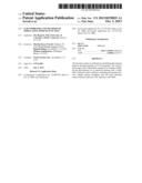 | 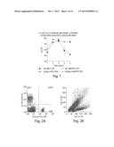 |
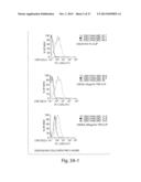 | 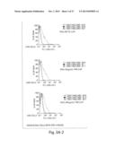 |
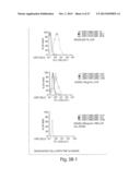 | 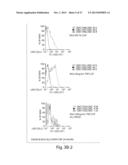 |
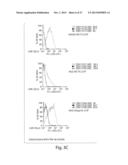 | 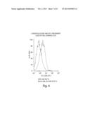 |
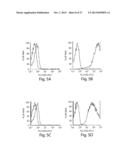 | 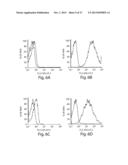 |
 |  |
 |  |
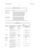 |  |
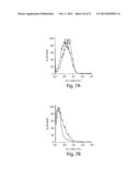 |  |
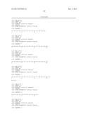 | 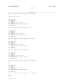 |
 |  |
 |  |
 |  |
 | 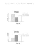 |
 |  |
 |  |
 |  |
 |  |
 |  |
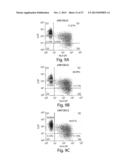 |  |
 |  |
 |  |
 |  |
 |  |
 | 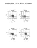 |
 |  |
 |  |
 |  |
 |  |
 |  |
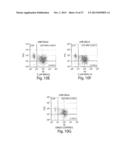 |  |
 |  |
 |  |
 |  |
 |  |
 |  |
 |  |
 |  |
 |  |
 | 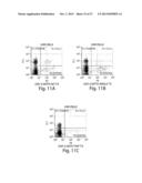 |
 |  |
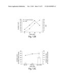 |  |
 |  |
 |  |
 |  |
 |  |
 |  |
 |  |
 |  |
 |  |
 | 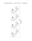 |
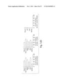 |  |
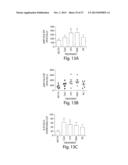 | 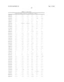 |
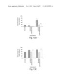 | 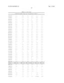 |
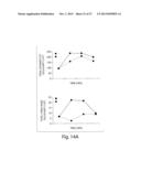 | 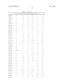 |
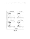 |  |
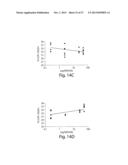 | 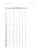 |
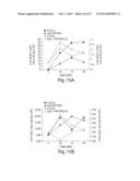 | 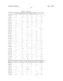 |
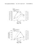 | 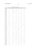 |
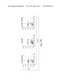 | 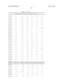 |
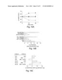 | 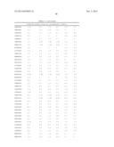 |
 | 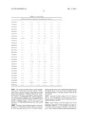 |
 | 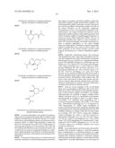 |
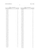 | 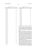 |
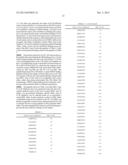 | 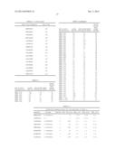 |
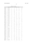 |  |
 | 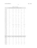 |
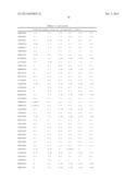 | 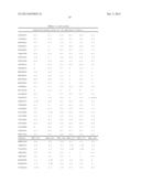 |
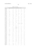 | 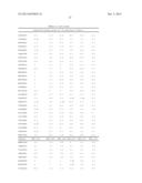 |
| Similar patent applications: | |
| Date | Title |
|---|---|
| 2013-10-31 | Compositions and methods to stimulate vascular structure formation |
| 2013-10-31 | Skin external preparation and method of producing same |
| 2013-10-31 | Biocomposites and methods of making the same |
| 2013-10-24 | Methods to modulate lysine variant distribution |
| New patent applications in this class: | |
| Date | Title |
|---|---|
| 2022-05-05 | Methods and systems for designing and/or characterizing soluble lipidated ligand agents |
| 2022-05-05 | Method of manufacturing mechanoreception stimulation fabric |
| 2019-05-16 | Methods of treating cancers with ct45 targeted therapies |
| 2019-05-16 | Compositions of muscle stimulated myokines and methods of use thereof |
| 2018-01-25 | Chimeric and humanized anti-histone antibodies |
| New patent applications from these inventors: | |
| Date | Title |
|---|---|
| 2022-09-01 | High yield production and use of enzymatic-exchangeable peptide major histocompatibility complex class i single chain trimer tetramer |
| 2015-08-20 | Competitive inhibitors of invariant chain expression and/or ectopic clip binding |
| 2015-04-23 | Methods and products for generating oils |
| 2014-10-23 | Methods and products for treating preeclampsia and modulating blood pressure |
| Top Inventors for class "Drug, bio-affecting and body treating compositions" | |
| Rank | Inventor's name |
|---|---|
| 1 | David M. Goldenberg |
| 2 | Hy Si Bui |
| 3 | Lowell L. Wood, Jr. |
| 4 | Roderick A. Hyde |
| 5 | Yat Sun Or |