Patent application title: ANTIBODY BINDING TO ABCA1 POLYPEPTIDE
Inventors:
Barbara Ecabert (Therwil, CH)
Hugues Matile (Basel, CH)
Hugues Matile (Basel, CH)
Everson Nogoceke (Basel, CH)
Bernhard Reis (Basel, CH)
Haiyan Wang (Allschwil, CH)
Haiyan Wang (Allschwil, CH)
Assignees:
Hoffmann-La Roche Inc.
IPC8 Class: AC07K1618FI
USPC Class:
435 721
Class name: Involving antigen-antibody binding, specific binding protein assay or specific ligand-receptor binding assay involving a micro-organism or cell membrane bound antigen or cell membrane bound receptor or cell membrane bound antibody or microbial lysate animal cell
Publication date: 2012-12-20
Patent application number: 20120322083
Abstract:
The invention relates to an isolated antibody that binds to native ABCA1
polypeptide and its uses for the detection of ABCA1 polypeptides in
tissue samples.Claims:
1. An isolated antibody that binds to ABCA1 polypeptide, wherein the
antibody binds to native ABCA1 polypeptide.
2. The isolated antibody of claim 1, wherein the ABCA1 polypeptide is human ABCA1 polypeptide.
3. The isolated antibody of claim 1, which is a monoclonal antibody.
4. The isolated antibody of claim 1, wherein the antibody has been produced by immunizing suitable animals with whole cells expressing the ABCA1 polypeptide.
5. The isolated antibody of claim 1, wherein the antibody comprises a CDR3 of a VH domain of an antibody obtainable from a hybridoma cell line selected from the group consisting of hybridoma cell line ABCA1-3/84 (DSM ACC3109), hybridoma cell line ABCA1-3/125 (DSM ACC3110) and hybridoma cell line ABCA1-4/18 (DSM ACC3111) and a CDR3 of a VL domain of an antibody obtainable from a hybridoma cell selected from the group consisting of hybridoma cell line ABCA1-3/84 (DSM ACC3109), hybridoma cell line ABCA1-3/125 (DSM ACC3110) and hybridoma cell line ABCA1-4/18 (DSM ACC3111).
6. The isolated antibody of claim 1, wherein the antibody comprises CDR1 to CDR3 of a VH domain of an antibody obtainable from a VH domain of an antibody obtainable from a hybridoma cell line selected from the group consisting of hybridoma cell line ABCA1-3/84 (DSM ACC3109), hybridoma cell line ABCA1-3/125 (DSM ACC3110) and hybridoma cell line ABCA1-4/18 (DSM ACC3111) and a CDR1 to CDR3 of a VL domain of an antibody obtainable from a hybridoma cell selected from the group consisting of hybridoma cell line ABCA1-3/84 (DSM ACC3109), hybridoma cell line ABCA1-3/125 (DSM ACC3110) and hybridoma cell line ABCA1-4/18 (DSM ACC3111).
7. The isolated antibody of claim 1, wherein the antibody comprises a VH domain and a VL domain of an antibody obtainable from hybridoma cell line selected from the group consisting of ABCA1-3/84 (DSM ACC3109), hybridoma cell line ABCA1-3/125 (DSM ACC3110) and hybridoma cell line ABCA1-4/18 (DSM ACC3111).
8. The isolated antibody of claim 1, wherein the antibody is produced by hybridoma cell line selected from the group consisting of hybridoma cell line ABCA1-3/84 (DSM ACC3109), hybridoma cell line ABCA1-3/125 (DSM ACC3110) and hybridoma cell line ABCA1-4/18 (DSM ACC3111).
9. A hybridoma cell line selected from the group consisting of hybridoma cell line ABCA1-3/84 (DSM ACC3109), hybridoma cell line ABCA1-3/125 (DSM ACC3110) and hybridoma cell line ABCA1-4/18 (DSM ACC3111).
10. An isolated nucleic acid comprising a sequence encoding: (i) a VH domain of an antibody obtainable from a hybridoma cell line selected from the group consisting of hybridoma cell line ABCA1-3/84 (DSM ACC3109), hybridoma cell line ABCA1-3/125 (DSM ACC3110) and hybridoma cell line ABCA1-4/18 (DSM ACC3111), (ii) a VL domain of an antibody obtainable from a hybridoma cell line selected from the group consisting of hybridoma cell line ABCA1-3/84 (DSM ACC3109), hybridoma cell line ABCA1-3/125 (DSM ACC3110) and hybridoma cell line ABCA1-4/18 (DSM ACC3111), or (iii) a VH and a VL domain of an antibody obtainable from a hybridoma cell line selected from the group consisting of hybridoma cell line ABCA1-3/84 (DSM ACC3109), hybridoma cell line ABCA1-3/125 (DSM ACC3110) and hybridoma cell line ABCA1-4/18 (DSM ACC3111).
11. The isolated nucleic acid of claim 10, comprising a sequence encoding an antibody produced by a hybridoma cell line selected from the group consisting of hybridoma cell line ABCA1-3/84 (DSM ACC3109), hybridoma cell line ABCA1-3/125 (DSM ACC3110) and hybridoma cell line ABCA1-4/18 (DSM ACC3111).
12. A vector comprising the nucleic acid of claim 10.
13. A host cell comprising the vector of claim 12.
14. A method of producing an antibody comprising culturing the host cell of claim 13 so that the antibody is produced.
15. A method for the detection of ABCA1 polypeptide in a tissue sample of an animal comprising: a) providing a tissue sample of the animal, b) detecting ABCA1 polypeptide in the sample of step a) using an antibody that binds to native ABCA1 polypeptide.
16. The method of claim 15, wherein the tissue sample is whole blood.
17. The method of claim 15, wherein the animal is a human subject.
18. The method of claim 15, wherein detection of ABCA1 polypeptide in step b) is done by Flow Cytometry.
Description:
RELATED APPLICATIONS
[0001] This application claims the benefit of EP Application No. 11174755.6, filed Jul. 20, 2011 and EP Application No. 11169924.5, filed Jun. 15, 2011. All the teachings of the above-referenced applications are incorporated herein by reference.
SEQUENCE LISTING
[0002] The instant application contains a Sequence Listing which has been submitted in ASCII format via EFS-Web and is hereby incorporated by reference in its entirety. Said ASCII copy, created on May 10, 2012, is named P4713SeqList.txt and is 20,220 bytes in size.
FIELD OF THE INVENTION
[0003] The present invention relates to antibodies binding to ABCA1 polypeptide and its uses in methods to detect ABCA1 polypeptide.
BACKGROUND
[0004] The ATP-binding cassette transporter Al (ABCA1) is an ATP dependent transporter mediating the efflux of cholesterol and phospholipids to extracellular lipid poor HDL particles. The amino acid sequence of human ABCA1 polypeptide is given in SEQ ID NO: 1. ABCA1 is essential for the assembly of nascent HDL particles by phospholipid and apolipoprotein. ABCA1 functions as a pivotal regulator of lipid efflux from cells to apolipoproteins and is thus involved in lowering the risk of atherosclerosis. ABCA1 is pivotal in influencing plasma HDL levels.
[0005] Active in liver and small intestine, generating most circulating HDL. Defects in the gene encoding for the ABCA1 were shown to be one of the genetic causes for familial hypoalphalipoproteinemia (FHA).
[0006] Commercially available anti ABCA1 antibodies are not suitable for the detection of native ABCA1 polypeptide in tissue samples.
[0007] Therefore, there is a need for anti ABCA1 antibodies capable of detecting native ABCA1 polypeptide in tissue samples.
SUMMARY OF THE INVENTION
[0008] In a first aspect the present invention relates to an isolated antibody that binds to native ABCA1 polypeptide.
[0009] In a particular embodiment, the ABCA1 polypeptide is human ABCA1 polypeptide.
[0010] In a further particular embodiment, the anti-ABCA1 antibody is a monoclonal antibody.
[0011] In a further particular embodiment, the antibody has been produced by immunizing suitable animals with whole cells expressing the ABCA1 polypeptide, preferably human ABCA1 polypeptide.
[0012] In a further particular embodiment, the antibody comprises a CDR3 of a VH domain of an antibody obtainable from a hybridoma cell line selected from the group consisting of hybridoma cell line ABCA1-3/84 (DSM ACC3109), hybridoma cell line ABCA1-3/125 (DSM ACC3110) and hybridoma cell line ABCA1-4/18 (DSM ACC3111) and a CDR3 of a VL domain of an antibody obtainable from a hybridoma cell selected from the group consisting of hybridoma cell line ABCA1-3/84 (DSM ACC3109), hybridoma cell line ABCA1-3/125 (DSM ACC3110) and hybridoma cell line ABCA1-4/18 (DSM ACC3111).
[0013] In another particular embodiment, the antibody comprises CDR1 to CDR3 of a VH domain of an antibody obtainable from a VH domain of an antibody obtainable from a hybridoma cell line selected from the group consisting of hybridoma cell line ABCA1-3/84 (DSM ACC3109), hybridoma cell line ABCA1-3/125 (DSM ACC3110) and hybridoma cell line ABCA1-4/18 (DSM ACC3111) and a CDR1 to CDR3 of a VL domain of an antibody obtainable from a hybridoma cell selected from the group consisting of hybridoma cell line ABCA1-3/84 (DSM ACC3109), hybridoma cell line ABCA1-3/125 (DSM ACC3110) and hybridoma cell line ABCA1-4/18 (DSM ACC3111).
[0014] In a further particular embodiment, the antibody comprises a VH domain and a VL domain of an antibody obtainable from hybridoma cell line selected from the group consisting of ABCA1-3/84 (DSM ACC3109), hybridoma cell line ABCA1-3/125 (DSM ACC3110) and hybridoma cell line ABCA1-4/18 (DSM ACC3111).
[0015] In a further particular embodiment, the antibody is produced by hybridoma cell line selected from the group consisting of hybridoma cell line ABCA1-3/84 (DSM ACC3109), hybridoma cell line ABCA1-3/125 (DSM ACC3110) and hybridoma cell line ABCA1-4/18 (DSM ACC3111).
[0016] In another aspect the present invention relates to a hybridoma cell line selected from the group consisting of hybridoma cell line ABCA1-3/84 (DSM ACC3109), hybridoma cell line ABCA1-3/125 (DSM ACC3110) and hybridoma cell line ABCA1-4/18 (DSM ACC3111).
[0017] In another aspect the present invention relates to an isolated nucleic acid comprising a sequence encoding a VH domain of an antibody obtainable from a hybridoma cell line selected from the group consisting of hybridoma cell line ABCA1-3/84 (DSM ACC3109), hybridoma cell line ABCA1-3/125 (DSM ACC3110) and hybridoma cell line ABCA1-4/18 (DSM ACC3111).
[0018] In another aspect the present invention provides an isolated nucleic acid comprising a sequence encoding a VL domain of an antibody obtainable from a hybridoma cell line selected from the group consisting of hybridoma cell line ABCA1-3/84 (DSM ACC3109), hybridoma cell line ABCA1-3/125 (DSM ACC3110) and hybridoma cell line ABCA1-4/18 (DSM ACC3111).
[0019] In another aspect the present invention provides an isolated nucleic acid comprising a sequence encoding an antibody produced by a hybridoma cell line selected from the group consisting of hybridoma cell line ABCA1-3/84 (DSM ACC3109), hybridoma cell line ABCA1-3/125 (DSM ACC3110) and hybridoma cell line ABCA1-4/18 (DSM ACC3111).
[0020] In another aspect the present invention provides a vector comprising a nucleic acid of the present invention and a host cell comprising a vector of the present invention.
[0021] In another aspect the present invention provides a method of producing an antibody comprising culturing a host cell of the present invention so that the antibody is produced.
[0022] In another aspect the present invention provides a use of the antibody of the present invention for the detection of ABCA1 polypeptide in a tissue sample of an animal.
[0023] In a particular embodiment of the use of the present invention the tissue sample is whole blood.
[0024] In a particular embodiment of the use of the present invention the animal is a human subject.
[0025] In another aspect the present invention provides a method for the detection of ABCA1 polypeptide in a tissue sample of an animal comprising: [0026] a) providing a tissue sample of the animal, [0027] b) detecting ABCA1 polypeptide in the sample of step a) using the antibody of the present invention.
[0028] In a particular embodiment, the tissue sample is whole blood.
[0029] In a particular embodiment, the animal is a human subject.
[0030] In a particular embodiment the detection of ABCA1 polypeptide in step b) is done by Flow Cytometry.
BRIEF DESCRIPTION OF THE FIGURES
[0031] The patent or application file contains at least one drawing executed in color. Copies of this patent or patent application publication with color drawing(s) will be provided by the U.S. Patent and Trademark Office upon request and payment of the necessary fee.
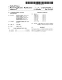
[0032] FIG. 1 is a visualization of the term "staining index". Blue: FLP293 cell line expressing ABCAl; White: parental cell line 293.
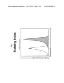
[0033] FIG. 2a: Detection of ABCA1 surface expression in a FLP293 cell line expressing ABCA1 (FLP293/ABCA1 cell line) using the commercial anti-ABCA1 antibody Novus DyLight 488 in FACS assay; Staining index is 1.54 (fluorescence FLP293ABCA1 cell line /fluorescence FLP293 parental cell line); Blue: FLP293 cell line expressing ABCA1; White: parental cell line 293. Antibody dilution: 1:200
[0034] FIG. 2b: Detection of ABCA1 surface expression in a FLP293 cell line expressing ABCA1 (FLP293/ABCA1 cell line) using the commercial anti-ABCA1 antibody ab81950 biotin in FACS assay; Staining index is 1.24 (fluorescence FLP293ABCA1 cell line /fluorescence FLP293 parental cell line); Blue: FLP293 cell line expressing ABCA1; White: parental cell line 293. Antibody dilution: 1:200
[0035] FIG. 2c: Detection of ABCA1 surface expression in a FLP293 cell line expressing ABCA1 (FLP293/ABCA1 cell line) using the commercial anti-ABCA1 antibody Novus biotin in FACS assay; Staining index is 1.28 (fluorescence FLP293ABCA1 cell line/fluorescence FLP293 parental cell line); Blue: FLP293 cell line expressing ABCAl; White: parental cell line 293. Antibody dilution: 1:200
[0036] FIG. 2d: Detection of ABCA1 surface expression in a FLP293 cell line expressing ABCA1 (FLP293/ABCA1 cell line) using the commercial anti-ABCA1 antibody ab18180 in FACS assay; Staining index is 1.41 (fluorescence FLP293ABCA1 cell line/fluorescence FLP293 parental cell line); Blue: FLP293 cell line expressing ABCAl; White: parental cell line 293. Antibody dilution: 1:200
[0037] FIG. 2e: Detection of ABCA1 surface expression in a FLP293 cell line expressing ABCA1 (FLP293/ABCA1 cell line) using the commercial anti-ABCA1 antibody ab66217 in FACS assay; Staining index is 1.84 (fluorescence FLP293ABCA1 cell line/fluorescence FLP293 parental cell line); Blue: FLP293 cell line expressing ABCA1; White: parental cell line 293. Antibody dilution: 1:200
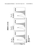
[0038] FIG. 3a: Detection of ABCA1 surface expression in a FLP293 cell line expressing ABCA1 (FLP293/ABCA1 cell line) using the inventive anti-ABCA1 antibody ABCA1-3/84 in FACS assay; Staining index is 8.9 (fluorescence FLP293ABCA1 cell line /fluorescence FLP293 parental cell line); Blue: FLP293 cell line expressing ABCAl; White: parental cell line 293. Antibody concentration: 0.1 μg/ml
[0039] FIG. 3b: Detection of ABCA1 surface expression in a FLP293 cell line expressing ABCA1 (FLP293/ABCA1 cell line) using the inventive anti-ABCA1 antibody ABCA1-3/84 in FACS assay; Staining index is 10.4 (fluorescence FLP293ABCA1 cell line/fluorescence FLP293 parental cell line); Blue: FLP293 cell line expressing ABCAl; White: parental cell line 293. Antibody concentration: 1 μg/ml
[0040] FIG. 3c: Detection of ABCA1 surface expression in a FLP293 cell line expressing ABCA1 (FLP293/ABCA1 cell line) using the inventive anti-ABCA1 antibody ABCA1-3/84 in FACS assay; Staining index is 10.5 (fluorescence FLP293ABCA1 cell line /fluorescence FLP293 parental cell line); Blue: FLP293 cell line expressing ABCA1; White: parental cell line 293. Antibody concentration: 10 μg/ml
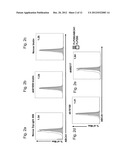
[0041] FIG. 4a: Detection of ABCA1 surface expression in a FLP293 cell line expressing ABCA1 (FLP293/ABCA1 cell line) using the inventive anti-ABCA1 antibody ABCA1-3/125 in FACS assay; Staining index is 49.7 (fluorescence FLP293ABCA1 cell line /fluorescence FLP293 parental cell line); Blue: FLP293 cell line expressing ABCAl; White: parental cell line 293. Antibody concentration: 0.1 μg/ml
[0042] FIG. 4b: Detection of ABCA1 surface expression in a FLP293 cell line expressing ABCA1 (FLP293/ABCA1 cell line) using the inventive anti-ABCA1 antibody ABCA1-3/125 in FACS assay; Staining index is 47.0 (fluorescence FLP293ABCA1 cell line /fluorescence FLP293 parental cell line); Blue: FLP293 cell line expressing ABCAl; White: parental cell line 293. Antibody concentration: 1 μg/ml
[0043] FIG. 4c: Detection of ABCA1 surface expression in a FLP293 cell line expressing ABCA1 (FLP293/ABCA1 cell line) using the inventive anti-ABCA1 antibody ABCA1-3/125 in FACS assay; Staining index is 46.9 (fluorescence FLP293ABCA1 cell line /fluorescence FLP293 parental cell line); Blue: FLP293 cell line expressing ABCAl; White: parental cell line 293. Antibody concentration: 10 μg/ml.
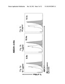
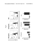
[0044] FIG. 5: Upregulation of ABCA1 on CD14+ monocytes with two different LXR agonists and LPS; Whole blood was incubated for 24h with LXR agonists or LPS. Detection of ABCA1 was done by FACS using anti-ABCA1 antibody ABCA1-3/125.
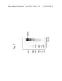
[0045] FIG. 6: Western Blot analysis of generated anti-ABCA1 monoclonal antibodies ABCA1-3/84, ABCA1-3/125 and ABCA1-4/18. The following cell lysates were used in the Western Blot to test the specificity of the generated anti-ABCA1 monoclonal antibodies:
[0046] Lane 1: stimulated THP1 cell-Lysate,
[0047] Lane 2: non stimulated THP1 cell-Lysate,
[0048] Lane 3: FLP293-hu-ABCA1 (FLP293 cell line expressing ABCA1),
[0049] Lane 4: FLP293 (negative control),
[0050] Lane 5: MWM (Molecular Weight Marker),
[0051] Lane 6: HEK-293-hu-ABCAl(DB272-in pANITA2)-6His (HEK cell line expressing human ABCA1 protein with a His tag).
[0052] FIG. 7: Western blot analysis of ABCA1 protein. Clone 4 expresses a large amount of ABCA1 indicated by detection of the full-length protein (approx. 240-kDA) and several smaller species. In comparison, no ABCA1 protein was detected in the untransfected FLP cell line.
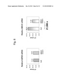
[0053] FIG. 8: qPCR analysis of ABCA1 mRNA expression: The Cp values for the housekeeping gene GAPDH remained constant between the ABCAl-overexpressing Clone 4 vs. the parental line (18.00+0.24 vs. 18.56+0.20). In contrast, the Cp values indicated extremely low expression of ABCA1 mRNA in the untransfected line (36.00+0.73) vs. high levels in the Clone4-ABCA1 line (21.64+0.48). Using the ddCp method to calculate the difference indicates that Clone 4 expresses 21,000 times more ABCA1 transcripts compared to the parental FLP cell line.
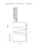
[0054] FIG. 9: Detection of ABCA1 surface expression in THP-1 cells using the inventive anti-ABCA1 antibody ABCA1-4/18 in a FACS assay. THP-1 cells were treated with LXR agonist overnight to induce ABCA1 expression or control treated with DMSO control. Cells were stained with 1 μg/ml ABCA1 specific antibody (clone 4/18) or isotype control followed by staining with secondary PE conjugated antibody against mouse IgG1. Anti-ABCA1 antibody ABCA1- 4/18 gives a good separation of basal ABCA1 signal from isotype control.
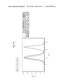
[0055] FIG. 10: Detection of ABCA1 surface expression in THP-1 cells using the inventive anti-ABCA1 antibody ABCA1-3/125 in a FACS assay. THP-1 cells were treated with LXR agonist overnight to induce ABCA1 expression or control treated with DMSO control. Cells were stained with 1 μg/ml ABCA1 specific antibody (clone 3/125) or isotype control followed by staining with secondary PE conjugated antibody against mouse IgG1.
[0056] FIG. 11: Human tissue distribution of ABCA1 proteins shown by Western Blot using mouse anti-ABCA1 mAb clone 3/84.
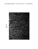
[0057] FIG. 12: Immunohistochemistry staining of ABCA1 in human liver with mouse anti-ABCA1 mAb clone 3/84. Green: ABCA1 protein localized in cell membranes of hepatocytes, and Blue: Nuclei DAPI stained.
DETAILED DESCRIPTION OF EMBODIMENTS OF THE INVENTION
[0058] The term "staining index" as used herein is defined as follows:
Staining index : Median fluorescence intensity ABCA 1 expressing cell line Median fluorescence intensity ABCA 1 - negative parental cell line ##EQU00001##
[0059] The term "antibody" encompasses the various forms of antibody structures including but not being limited to whole antibodies and antibody fragments. The antibody according to the invention can be a humanized antibody, chimeric antibody, or further genetically engineered antibody as long as the characteristic properties according to the invention are retained.
[0060] "Antibody fragments" comprise a portion of a full length antibody, preferably the variable domain thereof, or at least the antigen binding site thereof. Examples of antibody fragments include diabodies, single-chain antibody molecules, and multispecific antibodies formed from antibody fragments. scFv antibodies are, e.g. described in Houston, J. S., Methods in Enzymol. 203 (1991) 46-96). In addition, antibody fragments comprise single chain polypeptides having the characteristics of a VH domain, namely being able to assemble together with a VL domain, or of a VL domain binding to ABCA1, namely being able to assemble together with a VH domain to a functional antigen binding site and thereby providing the property.
[0061] The terms "monoclonal antibody" or "monoclonal antibody composition" as used herein refer to a preparation of antibody molecules of a single amino acid composition.
[0062] The term "chimeric antibody" refers to an antibody comprising a variable region, i.e., binding region, from one source or species and at least a portion of a constant region derived from a different source or species, usually prepared by recombinant DNA techniques. Chimeric antibodies comprising a murine variable region and a human constant region are preferred. Other preferred forms of "chimeric antibodies" encompassed by the present invention are those in which the constant region has been modified or changed from that of the original antibody to generate the properties according to the invention, especially in regard to C1q binding and/or Fc receptor (FcR) binding. Such chimeric antibodies are also referred to as "class-switched antibodies.". Chimeric antibodies are the product of expressed immunoglobulin genes comprising DNA segments encoding immunoglobulin variable regions and DNA segments encoding immunoglobulin constant regions. Methods for producing chimeric antibodies involve conventional recombinant DNA and gene transfection techniques are well known in the art. See e.g. Morrison, S. L., et al., Proc. Natl. Acad. Sci. USA 81 (1984) 6851-6855; U.S. Pat. Nos. 5,202,238 and 5,204,244.
[0063] The term "humanized antibody" refers to antibodies in which the framework or "complementarity determining regions" (CDR) have been modified to comprise the CDR of an immunoglobulin of different specificity as compared to that of the parent immunoglobulin. In a preferred embodiment, a murine CDR is grafted into the framework region of a human antibody to prepare the "humanized antibody." See e.g. Riechmann, L., et al., Nature 332 (1988) 323-327; and Neuberger, M. S., et al., Nature 314 (1985) 268-270. Particularly preferred CDRs correspond to those representing sequences recognizing the antigens noted above for chimeric antibodies. Other forms of "humanized antibodies" encompassed by the present invention are those in which the constant region has been additionally modified or changed from that of the original antibody to generate the properties according to the invention, especially in regard to C1q binding and/or Fc receptor (FcR) binding.
[0064] The term "human antibody", as used herein, is intended to include antibodies having variable and constant regions derived from human germ line immunoglobulin sequences. Human antibodies are well-known in the state of the art (van Dijk, M. A., and van de Winkel, J. G., Curr. Opin. Chem. Biol. 5 (2001) 368-374). Human antibodies can also be produced in transgenic animals (e.g., mice) that are capable, upon immunization, of producing a full repertoire or a selection of human antibodies in the absence of endogenous immunoglobulin production. Transfer of the human germ-line immunoglobulin gene array in such germ-line mutant mice will result in the production of human antibodies upon antigen challenge (see, e.g., Jakobovits, A., et al., Proc. Natl. Acad. Sci. USA 90 (1993) 2551-2555; Jakobovits, A., et al., Nature 362 (1993) 255-258; Bruggemann, M., et al., Year Immunol. 7 (1993) 33-40). Human antibodies can also be produced in phage display libraries (Hoogenboom, H. R., and Winter, G., J. Mol. Biol. 227 (1992) 381-388; Marks, J.D., et al., J. Mol. Biol. 222 (1991) 581-597). The techniques of Cole et al. and Boerner et al. are also available for the preparation of human monoclonal antibodies (Cole et al., Monoclonal Antibodies and Cancer Therapy, Alan R. Liss, p. 77 (1985); and Boerner, P., et al., J. Immunol. 147 (1991) 86-95). As already mentioned for chimeric and humanized antibodies according to the invention the term "human antibody" as used herein also comprises such antibodies which are modified in the constant region to generate the properties according to the invention, especially in regard to C1q binding and/or FcR binding, e.g. by "class switching" i.e. change or mutation of Fc parts (e.g. from IgG1 to IgG4 and/or IgG1/IgG4 mutation.).
[0065] The term "epitope" includes any polypeptide determinant capable of specific binding to an antibody. In certain embodiments, epitope determinant include chemically active surface groupings of molecules such as amino acids, sugar side chains, phosphoryl, or sulfonyl, and, in certain embodiments, may have specific three dimensional structural characteristics, and or specific charge characteristics. An epitope is a region of an antigen that is bound by an antibody.
[0066] The "variable domain" (variable domain of a light chain (VL), variable domain of a heavy chain (VH)) as used herein denotes each of the pair of light and heavy chain domains which are involved directly in binding the antibody to the antigen. The variable light and heavy chain domains have the same general structure and each domain comprises four framework (FR) regions whose sequences are widely conserved, connected by three "hypervariable regions" (or complementary determining regions, CDRs). The framework regions adopt a β-sheet conformation and the CDRs may form loops connecting the β-sheet structure. The CDRs in each chain are held in their three-dimensional structure by the framework regions and form together with the CDRs from the other chain the antigen binding site. The antibody's heavy and light chain CDR3 regions play a particularly important role in the binding specificity/affinity of the antibodies according to the invention and therefore provide a further object of the invention.
[0067] The term "antigen-binding portion of an antibody" when used herein refer to the amino acid residues of an antibody which are responsible for antigen-binding. The antigen-binding portion of an antibody comprises amino acid residues from the "complementary determining regions" or "CDRs". "Framework" or "FR" regions are those variable domain regions other than the hypervariable region residues as herein defined. Therefore, the light and heavy chain variable domains of an antibody comprise from N- to C-terminus the domains FR1, CDR1, FR2, CDR2, FR3, CDR3, and FR4. Especially, CDR3 of the heavy chain is the region which contributes most to antigen binding and defines the antibody's properties. CDR and FR regions are determined according to the standard definition of Kabat et al., Sequences of Proteins of Immunological Interest, 5th ed., Public Health Service, National Institutes of Health, Bethesda, Md. (1991) and/or those residues from a "hypervariable loop".
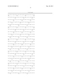
[0068] The term "ABCA1 polypeptide" is used herein to refer to native ABCA1 polypeptide from any animal, e.g. mammalian species, including humans, and ABCA1 variants. The ABCA1 polypeptides may be isolated from a variety of sources, including human tissue types or prepared by recombinant and/or synthetic methods. The amino acid sequence of human ABCA1 polypeptide is given in Seq. Id. No. 1.
[0069] An "isolated" antibody is one which has been separated from a component of its natural environment. In some embodiments, an antibody is purified to greater than 95% or 99% purity as determined by, for example, electrophoretic (e.g., SDS-PAGE, isoelectric focusing (IEF), capillary electrophoresis) or chromatographic (e.g., ion exchange or reverse phase HPLC). For review of methods for assessment of antibody purity, see, e.g., Flatman et al., J. Chromatogr. B 848:79-87 (2007).
[0070] An "isolated" nucleic acid refers to a nucleic acid molecule that has been separated from a component of its natural environment. An isolated nucleic acid includes a nucleic acid molecule contained in cells that ordinarily contain the nucleic acid molecule, but the nucleic acid molecule is present extrachromosomally or at a chromosomal location that is different from its natural chromosomal location.
[0071] "Isolated nucleic acid encoding an anti-ABCA1 antibody" refers to one or more nucleic acid molecules encoding antibody heavy and light chains (or fragments thereof), including such nucleic acid molecule(s) in a single vector or separate vectors, and such nucleic acid molecule(s) present at one or more locations in a host cell.
[0072] The term "vector," as used herein, refers to a nucleic acid molecule capable of propagating another nucleic acid to which it is linked. The term includes the vector as a self-replicating nucleic acid structure as well as the vector incorporated into the genome of a host cell into which it has been introduced. Certain vectors are capable of directing the expression of nucleic acids to which they are operatively linked. Such vectors are referred to herein as "expression vectors."
[0073] Antibodies may be produced using recombinant methods and compositions, e.g., as described in U.S. Pat. No. 4,816,567. In one embodiment, isolated nucleic acid encoding an anti-ABCA1 antibody described herein is provided. Such nucleic acid may encode an amino acid sequence comprising the VL and/or an amino acid sequence comprising the VH of the antibody (e.g., the light and/or heavy chains of the antibody). In a further embodiment, one or more vectors (e.g., expression vectors) comprising such nucleic acid are provided. In a further embodiment, a host cell comprising such nucleic acid is provided. In one such embodiment, a host cell comprises (e.g., has been transformed with): (1) a vector comprising a nucleic acid that encodes an amino acid sequence comprising the VL of the antibody and an amino acid sequence comprising the VH of the antibody, or (2) a first vector comprising a nucleic acid that encodes an amino acid sequence comprising the VL of the antibody and a second vector comprising a nucleic acid that encodes an amino acid sequence comprising the VH of the antibody. In one embodiment, the host cell is eukaryotic, e.g. a Chinese Hamster Ovary (CHO) cell or lymphoid cell (e.g., Y0, NS0, Sp20 cell). In one embodiment, a method of making an anti-ABCA1 antibody is provided, wherein the method comprises culturing a host cell comprising a nucleic acid encoding the antibody, as provided above, under conditions suitable for expression of the antibody, and optionally recovering the antibody from the host cell (or host cell culture medium).
[0074] For recombinant production of an antibody of the present invention, nucleic acid encoding an antibody, e.g., as described above, is isolated and inserted into one or more vectors for further cloning and/or expression in a host cell. Such nucleic acid may be readily isolated and sequenced using conventional procedures (e.g., by using oligonucleotide probes that are capable of binding specifically to genes encoding the heavy and light chains of the antibody).
[0075] Suitable host cells for cloning or expression of antibody-encoding vectors include prokaryotic or eukaryotic cells described herein. For example, antibodies may be produced in bacteria, in particular when glycosylation and Fc effector function are not needed. For expression of antibody fragments and polypeptides in bacteria, see, e.g., U.S. Pat. Nos. 5,648,237, 5,789,199, and 5,840,523. (See also Charlton, Methods in Molecular Biology, Vol. 248 (B.K.C. Lo, ed., Humana Press, Totowa, N.J., 2003), pp. 245-254, describing expression of antibody fragments in E. coli.). After expression, the antibody may be isolated from the bacterial cell paste in a soluble fraction and can be further purified.
[0076] Methods to clone antibody genes from hybridomas producing monoclonal antibodies are know to a person skilled in the art. For example, the genetic information for the variable heavy and light chain domains (VH and VL) can be amplified from hybridoma cells using polymerase chain reaction (PCR) with immunoglobulin-specific primers (Methods Mol Med. 2004; 94:447-58). The nucleic acid encoding the variable heavy and light chain domains (VH and VL) can then be cloned in a suitable vector for expression in host cells.
[0077] Methods for detection and/or measurement of polypeptides in biological samples are well known in the art and include, but are not limited to, Western-blotting, Flow cytometry, ELISAs or RIAs, or various proteomics techniques. For example, an antibody capable of binding to the denatured proteins, such as a polyclonal antibody, can be used to detect ABCA1 polypeptide in a Western Blot. An example for a method to measure a polypeptide is an ELISA. This type of protein quantitation is based on an antibody capable of capturing a specific antigen, and a second antibody capable of detecting the captured antigen.
[0078] A preferred method for the detection of native ABCA1 polypeptide is flow cytometry. Flow cytometry methods are described in Handbook of Flow Cytometry Method, J. Paul Robinson (Editor); Flow Cytometry--A Basic Introduction, Michael G Ormerod (2008) and Current Protocols in Cytometry (2010), Wiley.
EXAMPLES AND METHODS
[0079] Monoclonal Anti Human ABCA1 Antibodies of the Present Invention
[0080] The following three mouse hybridoma cell lines producing monoclonal antibodies against human ABCA1 have been deposited with Deutsche Sammlung von Mikroorganismen and Zellkulturen GmbH (DSMZ), Mascheroder Weg lb, D-38124 Branuschweig, Germany, under the provisions of the Budapest Treaty on January 20, 2011 in the name of F. Hoffmann-La Roche Ltd. and received the below listed deposit numbers: [0081] ABCA1-3/84=DSM ACC3109 [0082] ABCA1-3/125=DSM ACC3110 [0083] ABCA1-4/18=DSM ACC3111
[0084] Monoclonal Anti Human ABCA1 Antibody Generation
[0085] Expression of ABCA1 Protein on the Cell Surface of Mammalian Cells.
[0086] The human ABCA1 extracellular domain was expressed on the cell surface of HEK cells using the expression plasmid pANITA2. HEK-derived cell lines expressing human ABCA1 extracellular domain were established by stable transfection.
[0087] To obtain highly expressing cell lines, transfectants were separated into high-expressing cell-pools by fluorescent-activated-cell-sorting after surface staining with anti-FLAG antibodies. The mean fluorescence intensity of the cells gated for sorting into the high-expressing cell pool was 2.1-4.3 times higher than that of all transfectants.
[0088] Human ABCA1-expressing cell lines were tested for expression by Western blot analysis, showing a high level of expression of a protein with the expected molecular weight
[0089] Development of ABCA1 Specific Antibodies in Mice Immunised with Transfected HEK Cells
[0090] Our approach utilizes stably transfected mammalian cells (HEK293) expressing recombinant antigens on their cell surface. The transfected cells are also used for measuring seroconversion, hybridoma selection and antibody characterization. By presenting the antigen in its native conformation for immunization and hybridoma selection, this procedure promotes the generation of antibodies capable of binding to the endogenous protein.
[0091] The immunogen was obtained by immunopurifying the recombinant protein resulting in an immune complex and the use of these immune complexes as Immunizing Antigen. Immunepurification was confirmed by Western blot analysis using anti- His tag monoclonal antibodies.
[0092] Spleen cells of mice immunised with the immune complex were fused with PAI myeloma cells to generate B cell hybridoma. Fused cells were distributed in microtitre culture plate wells. To identify hybridoma cells that produce ABCA1 specific antibodies a two-step screening procedure was used that completely obviates the requirement for purified recombinant proteins. First all culture wells were tested for IgG production by ELISA. In a second step all wells positive for IgG production were screened for antibody binding to transfected cells by IFA. Transfected and non-transfected HEK cells spotted onto multiwell glass-slides were stained with individual hybridoma supernatants and analysed by fluorescence microscopy. Non-transfected HEK cells served as a negative control for each sample.
[0093] The specificity of generated monoclonal antibodies ABCA1-3/84, ABCA1-3/125 and ABCA1-4/18 was checked by Western Blot (FIG. 6). THP1=Human monocytic cell line.
[0094] Cell Culture
[0095] Human embryonic kidney cells (Flip-In 293 cells, Invitrogen), were maintained in Dulbecco's modified Eagle's medium (DMEM) supplemented with 10% fetal bovine serum, 2 mM L-glutamine, and antibiotics. Cells were transfected using Fugene-6 (Roche Biochemicals) as described by the manufacturer.
[0096] Human peripheral blood monocyte cells (THP-1, ATCC F-6430) were cultured in RPMI-1640 Glutamax (Invitrogen), supplemented with 0.05 mM 2-Mercaptoethanol (Invitrogen) and 10% heat inactivated FBS (Invitrogen). Cell concentrations did not exceed 1E06 cells/ml. Maximum passage number was 30.
[0097] Plasmids: A 6.5-kb DNA fragment encoding full-length human ABCA1 was subcloned into XhoI digested, Klenow filled plasmid PN721. After sequence verification, the 6.5-kb fragment was recovered and further subcloned into plasmid pCDNA5-FRT (Invitrogen).
[0098] Generation of an ABCA1 Stable Cell Line
[0099] A monoclonal stable cell line expressing human ABCA1 was obtained by co-transfecting pcDNA5-FRT-ABCA1 and pOG44 plasmids together (Invitrogen) using Fugene-6 (Roche Biochemicals) in growth medium in the absence of zeocin according to the manufacturer's recommendations. One day following transfection, hygromycin B was added to a final concentration of 50 μg per ml, and media changed every 3-4 days until hygromycin B-resistant colonies appeared. Individual colonies were selected and further propagated in the presence of hygromycin B. Individual clones were evaluated for ABCA1 expression by quantitative PCR and Western blot analysis. Based on these results, Clone 4 was selected as a high ABCA1-expressing cell line based.
[0100] Western blot analysis of ABCA1 protein expression: FLP or Clone 4 cells were cultured at 106 cells per well in 12-wells plate for 48 hours. Cells were lysed in Laemmli buffer/benzonase and the denatured samples applied to a 3-8% Tris-acetate gel and separated by one-dimensional gel electrophoresis. Separated proteins were transferred by electroblotting to a membrane.
[0101] ABCA1 was detected by incubation with a mouse monoclonal Ab ABCA1 (Neuromics) followed by a Goat anti-mouse IgG-HRP (Abcam # 20043) (FIG. 7).
[0102] Western blot analysis of human tissue: Human tissue lysates from Biochain Institute, Inc., Hayward, Calif. 94545, USA were used. The blotting was done the same way as described in the previous paragraph. ABCA1 was detected by incubation with mouse anti-ABCA1 mAb (clone 3/84) followed by Goat-anti-mouse IgG-HRP (FIG. 11).
[0103] Quantitative PCR analysis of ABCA1 mRNA expression: FLP or Clone4 cells were cultured at 5×10⁵ cells per well in 96 well plates for 48 hrs at 37°. Total RNA was isolated using an automated system according to manufacturer's instructions (Qiagen). Real-time quantitative RT-QPCR was performed on a Lightcycler 480 instrument (Roche) using a one-step reagent mix (Qiagen) and probe/primer sets (Applied Biosystems) to detect relative expression of ABCA1 and GAPDH mRNA's. Data are expressed as the mean Ct values (N=8 wells/condition) +/-SD (FIG. 8).
[0104] Antibodies and 2d Step Reagents
[0105] Commercially available primary monoclonal antibodies tested for specificity against ABCA1 were from Abcam® and Novus® (clone HJ1 and clone AB.H10). Rabbit polyclonal antibodies tested were from Abcam® (ab81950). Second step reagents Streptavidin PE and goat-α-mouse IgG PE were from Southern Biotechnology®.
[0106] In addition to the commercial antibodies, three murine anti-ABCA1 hybridoma supernatants, generated in-house, were evaluated for their performance in detecting ABCA1 in flow cytometry. Detection was performed by applying a secondary reagent (Streptavidin PE, goat-anti-mouse IgG PE and IgG1 PE from Southern Biotechnology®). Hybridoma supernatants of two clones (clone ABCA1-3/84 and ABCA1-3/125) were purified, retested and titrated. At a later time point, clone ABCA1-3/18 derived antibody was tested and compared to ABCA1-3/125 using THP-1 cells. The following antibodies were used for the identification of specific human blood subsets: CD3 Pacific blue, clone UCHT1, CD14 PerCP-Cy5.5, clone M5E2, CD15 APC, clone HI98, CD16 APC-Cy7, clone 3G8, CD19 PE-Cy7, clone SJ25CI and CD66b FITC, clone G10F5. All CD marker specific antibodies were obtained from Becton Dickinson®.
[0107] FACS Analysis of FLP 293, FLP 293 Derived Cells and THP-1 Cells
[0108] For FACS analysis, FLP 293 cells were rinsed with D-PBS without Calcium and Magnesium (Gibco®) and incubated with 0.02% EDTA (Sigma®). After harvesting, they were washed twice with cold D-PBS before proceeding with the antibody staining Briefly, 0.8-1×10⁶ cells/sample were resuspended in 100 μl BD stain buffer/2% FCS (Becton Dickinson®) containing the 1:200 diluted primary antibody. The incubation time was 45 minutes at +4° C. in the dark. The cells were washed once with cold BD stain buffer/2% FCS and 100 μl second step reagent diluted in BD stain buffer/2% FCS was added. After 45 minutes incubation at +4° C. in the dark and two washes with cold BD stain buffer/2% FCS the cells were analyzed. Non-adherent THP-1 cells were processed in the same way as described above for FLP 293 cells but omitting the incubation in EDTA containing buffer.
[0109] Cells were analyzed on a FACS Canto II (Becton Dickinson®). Evaluation was done with FlowJo software (Tree Star®).
[0110] ABCA1 Whole Blood Assay and FACS Staining
[0111] For whole blood FACS analysis, the anti-ABCA1 clone 3/125 generated in-house was used in all cases. Whole Na-Heparin blood was incubated with the indicated agonists for 24 hours at 37° C. After the incubation, primary anti-ABCA1 specific antibody was added to 100 μl of whole blood at a final dilution of 1:800 for 30 minutes on ice in the dark.
[0112] Red blood cells were lysed with lx red cell lysis buffer (Becton Dickinson®) and washed. Subsequently, 100 μl of a secondary goat-a-mouse IgG1 PE (Southern Biotechnology®) diluted 1:250 was added and incubated for 20 minutes at +4° C. followed by two washing steps.
[0113] Cell subset specific antibodies were added for 20 minutes at +4° C. in the dark followed by a washing step before analysis.
[0114] Cells were analyzed on a FACS Canto II (Becton Dickinson®). Evaluation was done with FlowJo software (Tree Star®).
[0115] Immuno Histochemistry Staining
[0116] Human liver FFPE sections (5 mm, Biochain) were dyhydrated, microwaved with citrate buffer (Thermo Scientific), and incubated with mouse anti-ABCA1 mAb (clone 3/84, 1 mg/ml) for 1 hour, followed by the secondary antibody ditection (1 mg/ml for 1 hour, Alexa Fluor® 488 donkey anti-mouse IgG (H+L), Invitrogen, Basel, Switzerland) and DAPI staining
[0117] Although the foregoing invention has been described in some detail by way of illustration and example for purposes of clarity of understanding, the descriptions and examples should not be construed as limiting the scope of the invention. The disclosures of all patent and scientific literature cited herein are expressly incorporated in their entirety by reference.
Sequence CWU
1
112261PRTHomo sapiens 1Met Ala Cys Trp Pro Gln Leu Arg Leu Leu Leu Trp Lys
Asn Leu Thr1 5 10 15Phe
Arg Arg Arg Gln Thr Cys Gln Leu Leu Leu Glu Val Ala Trp Pro 20
25 30Leu Phe Ile Phe Leu Ile Leu Ile
Ser Val Arg Leu Ser Tyr Pro Pro 35 40
45Tyr Glu Gln His Glu Cys His Phe Pro Asn Lys Ala Met Pro Ser Ala
50 55 60Gly Thr Leu Pro Trp Val Gln Gly
Ile Ile Cys Asn Ala Asn Asn Pro65 70 75
80Cys Phe Arg Tyr Pro Thr Pro Gly Glu Ala Pro Gly Val
Val Gly Asn 85 90 95Phe
Asn Lys Ser Ile Val Ala Arg Leu Phe Ser Asp Ala Arg Arg Leu
100 105 110Leu Leu Tyr Ser Gln Lys Asp
Thr Ser Met Lys Asp Met Arg Lys Val 115 120
125Leu Arg Thr Leu Gln Gln Ile Lys Lys Ser Ser Ser Asn Leu Lys
Leu 130 135 140Gln Asp Phe Leu Val Asp
Asn Glu Thr Phe Ser Gly Phe Leu Tyr His145 150
155 160Asn Leu Ser Leu Pro Lys Ser Thr Val Asp Lys
Met Leu Arg Ala Asp 165 170
175Val Ile Leu His Lys Val Phe Leu Gln Gly Tyr Gln Leu His Leu Thr
180 185 190Ser Leu Cys Asn Gly Ser
Lys Ser Glu Glu Met Ile Gln Leu Gly Asp 195 200
205Gln Glu Val Ser Glu Leu Cys Gly Leu Pro Arg Glu Lys Leu
Ala Ala 210 215 220Ala Glu Arg Val Leu
Arg Ser Asn Met Asp Ile Leu Lys Pro Ile Leu225 230
235 240Arg Thr Leu Asn Ser Thr Ser Pro Phe Pro
Ser Lys Glu Leu Ala Glu 245 250
255Ala Thr Lys Thr Leu Leu His Ser Leu Gly Thr Leu Ala Gln Glu Leu
260 265 270Phe Ser Met Arg Ser
Trp Ser Asp Met Arg Gln Glu Val Met Phe Leu 275
280 285Thr Asn Val Asn Ser Ser Ser Ser Ser Thr Gln Ile
Tyr Gln Ala Val 290 295 300Ser Arg Ile
Val Cys Gly His Pro Glu Gly Gly Gly Leu Lys Ile Lys305
310 315 320Ser Leu Asn Trp Tyr Glu Asp
Asn Asn Tyr Lys Ala Leu Phe Gly Gly 325
330 335Asn Gly Thr Glu Glu Asp Ala Glu Thr Phe Tyr Asp
Asn Ser Thr Thr 340 345 350Pro
Tyr Cys Asn Asp Leu Met Lys Asn Leu Glu Ser Ser Pro Leu Ser 355
360 365Arg Ile Ile Trp Lys Ala Leu Lys Pro
Leu Leu Val Gly Lys Ile Leu 370 375
380Tyr Thr Pro Asp Thr Pro Ala Thr Arg Gln Val Met Ala Glu Val Asn385
390 395 400Lys Thr Phe Gln
Glu Leu Ala Val Phe His Asp Leu Glu Gly Met Trp 405
410 415Glu Glu Leu Ser Pro Lys Ile Trp Thr Phe
Met Glu Asn Ser Gln Glu 420 425
430Met Asp Leu Val Arg Met Leu Leu Asp Ser Arg Asp Asn Asp His Phe
435 440 445Trp Glu Gln Gln Leu Asp Gly
Leu Asp Trp Thr Ala Gln Asp Ile Val 450 455
460Ala Phe Leu Ala Lys His Pro Glu Asp Val Gln Ser Ser Asn Gly
Ser465 470 475 480Val Tyr
Thr Trp Arg Glu Ala Phe Asn Glu Thr Asn Gln Ala Ile Arg
485 490 495Thr Ile Ser Arg Phe Met Glu
Cys Val Asn Leu Asn Lys Leu Glu Pro 500 505
510Ile Ala Thr Glu Val Trp Leu Ile Asn Lys Ser Met Glu Leu
Leu Asp 515 520 525Glu Arg Lys Phe
Trp Ala Gly Ile Val Phe Thr Gly Ile Thr Pro Gly 530
535 540Ser Ile Glu Leu Pro His His Val Lys Tyr Lys Ile
Arg Met Asp Ile545 550 555
560Asp Asn Val Glu Arg Thr Asn Lys Ile Lys Asp Gly Tyr Trp Asp Pro
565 570 575Gly Pro Arg Ala Asp
Pro Phe Glu Asp Met Arg Tyr Val Trp Gly Gly 580
585 590Phe Ala Tyr Leu Gln Asp Val Val Glu Gln Ala Ile
Ile Arg Val Leu 595 600 605Thr Gly
Thr Glu Lys Lys Thr Gly Val Tyr Met Gln Gln Met Pro Tyr 610
615 620Pro Cys Tyr Val Asp Asp Ile Phe Leu Arg Val
Met Ser Arg Ser Met625 630 635
640Pro Leu Phe Met Thr Leu Ala Trp Ile Tyr Ser Val Ala Val Ile Ile
645 650 655Lys Gly Ile Val
Tyr Glu Lys Glu Ala Arg Leu Lys Glu Thr Met Arg 660
665 670Ile Met Gly Leu Asp Asn Ser Ile Leu Trp Phe
Ser Trp Phe Ile Ser 675 680 685Ser
Leu Ile Pro Leu Leu Val Ser Ala Gly Leu Leu Val Val Ile Leu 690
695 700Lys Leu Gly Asn Leu Leu Pro Tyr Ser Asp
Pro Ser Val Val Phe Val705 710 715
720Phe Leu Ser Val Phe Ala Val Val Thr Ile Leu Gln Cys Phe Leu
Ile 725 730 735Ser Thr Leu
Phe Ser Arg Ala Asn Leu Ala Ala Ala Cys Gly Gly Ile 740
745 750Ile Tyr Phe Thr Leu Tyr Leu Pro Tyr Val
Leu Cys Val Ala Trp Gln 755 760
765Asp Tyr Val Gly Phe Thr Leu Lys Ile Phe Ala Ser Leu Leu Ser Pro 770
775 780Val Ala Phe Gly Phe Gly Cys Glu
Tyr Phe Ala Leu Phe Glu Glu Gln785 790
795 800Gly Ile Gly Val Gln Trp Asp Asn Leu Phe Glu Ser
Pro Val Glu Glu 805 810
815Asp Gly Phe Asn Leu Thr Thr Ser Val Ser Met Met Leu Phe Asp Thr
820 825 830Phe Leu Tyr Gly Val Met
Thr Trp Tyr Ile Glu Ala Val Phe Pro Gly 835 840
845Gln Tyr Gly Ile Pro Arg Pro Trp Tyr Phe Pro Cys Thr Lys
Ser Tyr 850 855 860Trp Phe Gly Glu Glu
Ser Asp Glu Lys Ser His Pro Gly Ser Asn Gln865 870
875 880Lys Arg Ile Ser Glu Ile Cys Met Glu Glu
Glu Pro Thr His Leu Lys 885 890
895Leu Gly Val Ser Ile Gln Asn Leu Val Lys Val Tyr Arg Asp Gly Met
900 905 910Lys Val Ala Val Asp
Gly Leu Ala Leu Asn Phe Tyr Glu Gly Gln Ile 915
920 925Thr Ser Phe Leu Gly His Asn Gly Ala Gly Lys Thr
Thr Thr Met Ser 930 935 940Ile Leu Thr
Gly Leu Phe Pro Pro Thr Ser Gly Thr Ala Tyr Ile Leu945
950 955 960Gly Lys Asp Ile Arg Ser Glu
Met Ser Thr Ile Arg Gln Asn Leu Gly 965
970 975Val Cys Pro Gln His Asn Val Leu Phe Asp Met Leu
Thr Val Glu Glu 980 985 990His
Ile Trp Phe Tyr Ala Arg Leu Lys Gly Leu Ser Glu Lys His Val 995
1000 1005Lys Ala Glu Met Glu Gln Met Ala
Leu Asp Val Gly Leu Pro Ser 1010 1015
1020Ser Lys Leu Lys Ser Lys Thr Ser Gln Leu Ser Gly Gly Met Gln
1025 1030 1035Arg Lys Leu Ser Val Ala
Leu Ala Phe Val Gly Gly Ser Lys Val 1040 1045
1050Val Ile Leu Asp Glu Pro Thr Ala Gly Val Asp Pro Tyr Ser
Arg 1055 1060 1065Arg Gly Ile Trp Glu
Leu Leu Leu Lys Tyr Arg Gln Gly Arg Thr 1070 1075
1080Ile Ile Leu Ser Thr His His Met Asp Glu Ala Asp Val
Leu Gly 1085 1090 1095Asp Arg Ile Ala
Ile Ile Ser His Gly Lys Leu Cys Cys Val Gly 1100
1105 1110Ser Ser Leu Phe Leu Lys Asn Gln Leu Gly Thr
Gly Tyr Tyr Leu 1115 1120 1125Thr Leu
Val Lys Lys Asp Val Glu Ser Ser Leu Ser Ser Cys Arg 1130
1135 1140Asn Ser Ser Ser Thr Val Ser Tyr Leu Lys
Lys Glu Asp Ser Val 1145 1150 1155Ser
Gln Ser Ser Ser Asp Ala Gly Leu Gly Ser Asp His Glu Ser 1160
1165 1170Asp Thr Leu Thr Ile Asp Val Ser Ala
Ile Ser Asn Leu Ile Arg 1175 1180
1185Lys His Val Ser Glu Ala Arg Leu Val Glu Asp Ile Gly His Glu
1190 1195 1200Leu Thr Tyr Val Leu Pro
Tyr Glu Ala Ala Lys Glu Gly Ala Phe 1205 1210
1215Val Glu Leu Phe His Glu Ile Asp Asp Arg Leu Ser Asp Leu
Gly 1220 1225 1230Ile Ser Ser Tyr Gly
Ile Ser Glu Thr Thr Leu Glu Glu Ile Phe 1235 1240
1245Leu Lys Val Ala Glu Glu Ser Gly Val Asp Ala Glu Thr
Ser Asp 1250 1255 1260Gly Thr Leu Pro
Ala Arg Arg Asn Arg Arg Ala Phe Gly Asp Lys 1265
1270 1275Gln Ser Cys Leu Arg Pro Phe Thr Glu Asp Asp
Ala Ala Asp Pro 1280 1285 1290Asn Asp
Ser Asp Ile Asp Pro Glu Ser Arg Glu Thr Asp Leu Leu 1295
1300 1305Ser Gly Met Asp Gly Lys Gly Ser Tyr Gln
Val Lys Gly Trp Lys 1310 1315 1320Leu
Thr Gln Gln Gln Phe Val Ala Leu Leu Trp Lys Arg Leu Leu 1325
1330 1335Ile Ala Arg Arg Ser Arg Lys Gly Phe
Phe Ala Gln Ile Val Leu 1340 1345
1350Pro Ala Val Phe Val Cys Ile Ala Leu Val Phe Ser Leu Ile Val
1355 1360 1365Pro Pro Phe Gly Lys Tyr
Pro Ser Leu Glu Leu Gln Pro Trp Met 1370 1375
1380Tyr Asn Glu Gln Tyr Thr Phe Val Ser Asn Asp Ala Pro Glu
Asp 1385 1390 1395Thr Gly Thr Leu Glu
Leu Leu Asn Ala Leu Thr Lys Asp Pro Gly 1400 1405
1410Phe Gly Thr Arg Cys Met Glu Gly Asn Pro Ile Pro Asp
Thr Pro 1415 1420 1425Cys Gln Ala Gly
Glu Glu Glu Trp Thr Thr Ala Pro Val Pro Gln 1430
1435 1440Thr Ile Met Asp Leu Phe Gln Asn Gly Asn Trp
Thr Met Gln Asn 1445 1450 1455Pro Ser
Pro Ala Cys Gln Cys Ser Ser Asp Lys Ile Lys Lys Met 1460
1465 1470Leu Pro Val Cys Pro Pro Gly Ala Gly Gly
Leu Pro Pro Pro Gln 1475 1480 1485Arg
Lys Gln Asn Thr Ala Asp Ile Leu Gln Asp Leu Thr Gly Arg 1490
1495 1500Asn Ile Ser Asp Tyr Leu Val Lys Thr
Tyr Val Gln Ile Ile Ala 1505 1510
1515Lys Ser Leu Lys Asn Lys Ile Trp Val Asn Glu Phe Arg Tyr Gly
1520 1525 1530Gly Phe Ser Leu Gly Val
Ser Asn Thr Gln Ala Leu Pro Pro Ser 1535 1540
1545Gln Glu Val Asn Asp Ala Ile Lys Gln Met Lys Lys His Leu
Lys 1550 1555 1560Leu Ala Lys Asp Ser
Ser Ala Asp Arg Phe Leu Asn Ser Leu Gly 1565 1570
1575Arg Phe Met Thr Gly Leu Asp Thr Lys Asn Asn Val Lys
Val Trp 1580 1585 1590Phe Asn Asn Lys
Gly Trp His Ala Ile Ser Ser Phe Leu Asn Val 1595
1600 1605Ile Asn Asn Ala Ile Leu Arg Ala Asn Leu Gln
Lys Gly Glu Asn 1610 1615 1620Pro Ser
His Tyr Gly Ile Thr Ala Phe Asn His Pro Leu Asn Leu 1625
1630 1635Thr Lys Gln Gln Leu Ser Glu Val Ala Leu
Met Thr Thr Ser Val 1640 1645 1650Asp
Val Leu Val Ser Ile Cys Val Ile Phe Ala Met Ser Phe Val 1655
1660 1665Pro Ala Ser Phe Val Val Phe Leu Ile
Gln Glu Arg Val Ser Lys 1670 1675
1680Ala Lys His Leu Gln Phe Ile Ser Gly Val Lys Pro Val Ile Tyr
1685 1690 1695Trp Leu Ser Asn Phe Val
Trp Asp Met Cys Asn Tyr Val Val Pro 1700 1705
1710Ala Thr Leu Val Ile Ile Ile Phe Ile Cys Phe Gln Gln Lys
Ser 1715 1720 1725Tyr Val Ser Ser Thr
Asn Leu Pro Val Leu Ala Leu Leu Leu Leu 1730 1735
1740Leu Tyr Gly Trp Ser Ile Thr Pro Leu Met Tyr Pro Ala
Ser Phe 1745 1750 1755Val Phe Lys Ile
Pro Ser Thr Ala Tyr Val Val Leu Thr Ser Val 1760
1765 1770Asn Leu Phe Ile Gly Ile Asn Gly Ser Val Ala
Thr Phe Val Leu 1775 1780 1785Glu Leu
Phe Thr Asp Asn Lys Leu Asn Asn Ile Asn Asp Ile Leu 1790
1795 1800Lys Ser Val Phe Leu Ile Phe Pro His Phe
Cys Leu Gly Arg Gly 1805 1810 1815Leu
Ile Asp Met Val Lys Asn Gln Ala Met Ala Asp Ala Leu Glu 1820
1825 1830Arg Phe Gly Glu Asn Arg Phe Val Ser
Pro Leu Ser Trp Asp Leu 1835 1840
1845Val Gly Arg Asn Leu Phe Ala Met Ala Val Glu Gly Val Val Phe
1850 1855 1860Phe Leu Ile Thr Val Leu
Ile Gln Tyr Arg Phe Phe Ile Arg Pro 1865 1870
1875Arg Pro Val Asn Ala Lys Leu Ser Pro Leu Asn Asp Glu Asp
Glu 1880 1885 1890Asp Val Arg Arg Glu
Arg Gln Arg Ile Leu Asp Gly Gly Gly Gln 1895 1900
1905Asn Asp Ile Leu Glu Ile Lys Glu Leu Thr Lys Ile Tyr
Arg Arg 1910 1915 1920Lys Arg Lys Pro
Ala Val Asp Arg Ile Cys Val Gly Ile Pro Pro 1925
1930 1935Gly Glu Cys Phe Gly Leu Leu Gly Val Asn Gly
Ala Gly Lys Ser 1940 1945 1950Ser Thr
Phe Lys Met Leu Thr Gly Asp Thr Thr Val Thr Arg Gly 1955
1960 1965Asp Ala Phe Leu Asn Lys Asn Ser Ile Leu
Ser Asn Ile His Glu 1970 1975 1980Val
His Gln Asn Met Gly Tyr Cys Pro Gln Phe Asp Ala Ile Thr 1985
1990 1995Glu Leu Leu Thr Gly Arg Glu His Val
Glu Phe Phe Ala Leu Leu 2000 2005
2010Arg Gly Val Pro Glu Lys Glu Val Gly Lys Val Gly Glu Trp Ala
2015 2020 2025Ile Arg Lys Leu Gly Leu
Val Lys Tyr Gly Glu Lys Tyr Ala Gly 2030 2035
2040Asn Tyr Ser Gly Gly Asn Lys Arg Lys Leu Ser Thr Ala Met
Ala 2045 2050 2055Leu Ile Gly Gly Pro
Pro Val Val Phe Leu Asp Glu Pro Thr Thr 2060 2065
2070Gly Met Asp Pro Lys Ala Arg Arg Phe Leu Trp Asn Cys
Ala Leu 2075 2080 2085Ser Val Val Lys
Glu Gly Arg Ser Val Val Leu Thr Ser His Ser 2090
2095 2100Met Glu Glu Cys Glu Ala Leu Cys Thr Arg Met
Ala Ile Met Val 2105 2110 2115Asn Gly
Arg Phe Arg Cys Leu Gly Ser Val Gln His Leu Lys Asn 2120
2125 2130Arg Phe Gly Asp Gly Tyr Thr Ile Val Val
Arg Ile Ala Gly Ser 2135 2140 2145Asn
Pro Asp Leu Lys Pro Val Gln Asp Phe Phe Gly Leu Ala Phe 2150
2155 2160Pro Gly Ser Val Leu Lys Glu Lys His
Arg Asn Met Leu Gln Tyr 2165 2170
2175Gln Leu Pro Ser Ser Leu Ser Ser Leu Ala Arg Ile Phe Ser Ile
2180 2185 2190Leu Ser Gln Ser Lys Lys
Arg Leu His Ile Glu Asp Tyr Ser Val 2195 2200
2205Ser Gln Thr Thr Leu Asp Gln Val Phe Val Asn Phe Ala Lys
Asp 2210 2215 2220Gln Ser Asp Asp Asp
His Leu Lys Asp Leu Ser Leu His Lys Asn 2225 2230
2235Gln Thr Val Val Asp Val Ala Val Leu Thr Ser Phe Leu
Gln Asp 2240 2245 2250Glu Lys Val Lys
Glu Ser Tyr Val 2255 2260
User Contributions:
Comment about this patent or add new information about this topic:
 |  |
 |  |
 |  |
 |  |
 |  |
 |  |
 |  |
 |  |
 |  |
| Similar patent applications: | |
| Date | Title |
|---|---|
| 2011-12-29 | Method for improving the yeild of a polypeptide |
| 2012-11-08 | Antibody specific to activated egfr |
| 2010-03-25 | Dna coding for polypeptide fusion |
| 2011-01-20 | Carbon nanotube binding peptides |
| 2011-01-20 | Carbon nanotube binding peptides |
| New patent applications in this class: | |
| Date | Title |
|---|---|
| 2017-08-17 | Integrated visual morphology and cell protein expression using resonance-light scattering |
| 2017-08-17 | Methods and test kits for determining male fertility status |
| 2016-07-07 | Antibody, kit and method for determination of amyloid peptides |
| 2016-07-07 | In vitro method for determining the stability of compositions comprising soluble fc gamma receptor(s) |
| 2016-07-07 | Method of agglutination immunoassay |
| New patent applications from these inventors: | |
| Date | Title |
|---|---|
| 2015-09-03 | Novel oxazolidinone compounds |
| 2014-11-13 | Expression vector |
| 2014-08-07 | Use of aminodihydrothiazines for the treatment or prevention of diabetes |
| 2014-07-10 | Heteroarylmethyl amides |
| 2013-05-09 | Expression vector |
| Top Inventors for class "Chemistry: molecular biology and microbiology" | |
| Rank | Inventor's name |
|---|---|
| 1 | Marshall Medoff |
| 2 | Anthony P. Burgard |
| 3 | Mark J. Burk |
| 4 | Robin E. Osterhout |
| 5 | Rangarajan Sampath |