Patent application title: FULL HUMAN ANTI-TNF-ALPHA MONOCLONAL ANTIBODY, PREPARATION METHOD AND USE THEREOF
Inventors:
Huaizu Guo (Shanghai, CN)
Chuan Li (Shanghai, CN)
Xin Tong (Shanghai, CN)
Assignees:
Shanghai Biomabs Pharmaceuticals Co., Ltd.
IPC8 Class: AA61K39395FI
USPC Class:
4241421
Class name: Immunoglobulin, antiserum, antibody, or antibody fragment, except conjugate or complex of the same with nonimmunoglobulin material monoclonal antibody or fragment thereof (i.e., produced by any cloning technology) human
Publication date: 2012-12-06
Patent application number: 20120308575
Abstract:
The present invention provides a full human anti-TNF-α monoclonal
antibody, the preparation method and use thereof. The antibody in the
present invention has an amino acid sequence of heavy chain variable
region as shown in SEQ ID NO: 6 and an amino acid sequence of light chain
variable region as shown in SEQ ID NO: 8. The antibody in the present
invention can be used to prepare medicines for the treatment of
autoimmune disorders.Claims:
1. A fully human anti-TNF-.alpha. monoclonal antibody, having an amino
acid sequence of heavy chain variable region as shown in SEQ ID NO: 6 and
an amino acid sequence of light chain variable region as shown in SEQ ID
NO: 8.
2. The fully human anti-TNF-.alpha. monoclonal antibody of claim 1, having an amino acid sequence of heavy chain as shown in SEQ ID NO: 10 and an amino acid sequence of light chain as shown in SEQ ID NO: 12.
3. A nucleotide encoding the fully human anti-TNF-.alpha. antibody, having a nucleotide sequence of heavy chain variable region as shown in SEQ ID NO: 5 and a nucleotide sequence of light chain variable region as shown in SEQ ID NO: 7.
4. The nucleotide of claim 3, having a nucleotide sequence of heavy chain as shown in SEQ ID NO: 9 and a nucleotide sequence of light chain as shown in SEQ ID NO: 11.
5. An expression vector containing the nucleotide of claim 3, being pcDNA3.1/ZEO(+) or pcDNA3.1(+).
6. A host cell containing the expression vector of claim 5, being CHO-K1 cell.
7. A method of preparing the fully human anti-TNF-.alpha. monoclonal antibody of claim 1, comprising four steps of: selecting human phage antibody library to obtain a fully human anti-TNF-.alpha. single-chain antibody with high affinity; constructing an eukaryotic expression vector of the complete molecular of the fully human anti-TNF-.alpha. antibody; expressing the complete molecular of the fully human anti-TNF-.alpha. antibody in CHO cells; and purifying the complete molecular of the fully human anti-TNF-.alpha. antibody.
8. Use of the fully human anti-TNF-.alpha. monoclonal antibody of claim 1 in preparing medicines for treatment of autoimmune diseases.
9. The use of claim 8, wherein said autoimmune disease is rheumatoid arthritis, ankylosing spondylitis or psoriasis.
10. An expression vector containing the nucleotide of claim 4, being pcDNA3.1/ZEO(+) or pcDNA3.1(+).
11. A host cell containing the expression vector of claim 10, being CHO-K1 cell.
12. Use of the fully human anti-TNF-.alpha. monoclonal antibody of claim 2 in preparing medicines for treatment of autoimmune diseases.
13. The use of claim 12, wherein said autoimmune disease is rheumatoid arthritis, ankylosing spondylitis or psoriasis.
Description:
FIELD OF THE INVENTION
[0001] The present invention relates to the field of biotechnology. In particular, the present invention relates to a fully human monoclonal antibody, the preparation method and use thereof.
BACKGROUND OF THE INVENTION
[0002] TNF-α is a multifunctional immunomodulatory molecule in vivo that can work by binding to the cytomembrane receptor, which always causes target cell death (where its name is derived from) or induces local aggregation of immune effector cells. TNF-α is a soluble homologous trimeric subunit having a molecular weight of 17 KD (Smith, et al., J. Biol. Chem. 262:6951-6954, 1987). A transmembrane binding precursor of TNF-α with a molecular weight of 26 KD has also been found (Kriegler, et al., Cell 53:45-53, 1988). Mononuclear macrophages can secrete TNF-α and TNF-β when simulated with endotoxin and other stimulus, and some other cells can also secrete TNF-α.
[0003] TNF-α plays a crucial role in the pathological process of rheumatoid arthritis, bacterial or viral infection, chronic inflammation, autoimmune diseases such as AIDS, malignant tumors and/or neurodegenerative diseases. TNF-α monoclonal antibody can neutralize TNF-α and negatively regulate the activity of TNF-α in vivo. Moreover, a large number of studies have shown that TNF-α is the main medium which may cause septic shock syndrome. The increase of TNF-α level in blood serum of patients suffering from septic shock syndrome indicates the increase of mortality rate and disability rate. The clinical use of TNF-α antibody or its receptor has a certain therapeutic effect on septic shock syndrome.
[0004] In addition, TNF-α is one of the main media for promoting asymptomatic HIV infection status into AIDS, and monoclonal antibodies against TNF-α can neutralize the activity of TNF-α, negatively regulate the activity of TNF-α in vivo, and may remove the inducement from asymptomatic infection status into AIDS and achieve a certain purpose of AIDS treatment. Combined use of a TNF-α monoclonal antibody and other AIDS drugs counteracts the side effect due to excessive TNF-α and will distinctly enhance the therapeutic effect.
[0005] Initially, the scientists prepared and obtained murine anti-TNF-α monoclonal antibodies which were used to neutralize TNF-α. However, studies have shown that the murine monoclonal antibodies have many disadvantages as drug for treatment, because when used in human body, the murine monoclonal antibodies have strong immunogenicity and fast elimination in vivo with a short half life, leading to limited clinical efficacy and considerable side effects. With the development of humanized monoclonal antibody technology, the disadvantages of the anti-TNF-α murine monoclonal antibody have been overcome. Thereamong, a human-mouse chimeric anti-TNF-α monoclonal antibody (Infliximab, Remicade®) has been prepared through upstream construction techniques of genetic engineering, the variable region of which is still derived from murine TNF-α monoclonal antibody, maintaining the specificity and affinity binding to soluble fragments and transmembrane domains of tumor necrosis factor (Ka=1010 M-1), and the constant region of which is replaced by the human IgG1 constant region, extending the in vivo half life considerably. Other TNF-α inhibitors that have been approved for marketing abroad include an antibody fusion protein (Enbrel, Amgen) and a fully human anti-tumor necrosis factor-α monoclonal antibody (Humira, Abbott).
[0006] From the standpoints of target and specificity of action, these drugs mentioned above have almost the same mechanism of action, but all the above antibodies and fusion protein have varying degrees of problems such as high immunogenicity, low specificity and deficient stability. Therefore, there is an urgent need to establish an anti-TNF-α antibody that not only can maintain or increase the affinity and specificity of the antibody but also can reduce or eliminate the antibody immunogenicity, thereby further improving the safety and efficiency in clinical application.
SUMMARY OF THE INVENTION
[0007] The present invention constructs a very large human natural phage antibody library and obtains a fully human anti-TNF-α antibody 4H16 by selecting therefrom.
[0008] More particularly, the present invention provides a fully human anti-TNF-α antibody, having an amino acid sequence of heavy chain variable region as shown in SEQ ID NO: 6, and an amino acid sequence of light chain variable region as shown in SEQ ID NO: 8.
[0009] The above fully human anti-TNF-α antibody according to the present invention has an amino acid sequence of heavy chain as shown in SEQ ID NO: 10, and an amino acid sequence of light chain as shown in SEQ ID NO: 12.
[0010] The present invention also provides an isolated nucleotide encoding the above fully human anti-TNF-α antibody.
[0011] The above nucleotide according to the present invention has a nucleotide sequence encoding heavy chain variable region of the fully human anti-TNF-α antibody as shown in SEQ ID NO: 5, and a nucleotide sequence encoding light chain variable region of the fully human anti-TNF-α antibody as shown in SEQ ID NO: 7.
[0012] The above nucleotide according to the present invention has a nucleotide sequence encoding heavy chain of the fully human anti-TNF-α antibody as shown in SEQ ID NO: 9, and a nucleotide sequence encoding light chain of the fully human anti-TNF-α antibody as shown in SEQ ID NO: 11.
[0013] The present invention also provides an expression vector containing the above nucleotide, which is pcDNA3.1/ZEO(+) or pcDNA3.1(+).
[0014] The present invention also provides a host cell transfected with the above expression vector, which is CHO-K1 cell.
[0015] The present invention further provides a method for preparing the above fully human antibody, comprising selecting human phage antibody library to obtain a fully human anti-TNF-α single-chain antibody with high affinity; constructing an eukaryotic expression vector of the complete molecular of the fully human anti-TNF-α antibody; expressing the complete molecular of fully human anti-TNF-α antibody in CHO cells; and purifying the complete molecular of the fully human anti-TNF-α antibody.
[0016] The present invention also provides a use of the above fully human anti-TNF-α antibody in preparing medicines for treatment of autoimmune diseases. The autoimmune diseases are selected from rheumatoid arthritis, ankylosing spondylitis or psoriasis.
[0017] The obtained antibodies are used to perform a series of experiments in the present invention and the experiment results show that compared to D2E7 (adalimumab monoclonal antibody, Abbott), and 7B3 disclosed in Chinese Patent Application No. 200310108440.0 entitled "Fully Human Tumor Necrosis Factor Antibody, Preparation Method and Pharmaceutical Composition Thereof" filed on Nov. 6, 2003, the antibodies obtained according to the present invention have higher antibody affinity and stronger TNF-α neutralizing capacity.
DESCRIPTION OF DRAWINGS
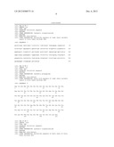
[0018] FIG. 1 shows the experiment results of blocking the binding of TNF-α to soluble P75 receptor by anti-TNF-α antibody 4H16;
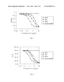
[0019] FIG. 2 shows the experiment results of blocking the binding of TNF-α to U-937 cell surface receptor by anti-TNF-α antibody 4H16;
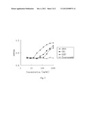
[0020] FIG. 3 shows the experiment results of the resistance to TNF-α-mediated killing in L929 cells by anti-TNF-α antibody 4H16.
DETAILED DESCRIPTION OF THE INVENTION
[0021] The following examples and experiment examples are used to further illustrate the present invention only and should not be construed to limit the present invention.
Example 1
Preparation of Antibody
(1) Cloning of Genes Encoding Human Antibody Light and Heavy Chain Constant Region
[0022] Healthy human lymphoma cells were isolated with lymphocyte separation medium (Dingguo Biotechnology Development Company, CHINA), and total RNA was extracted using Trizol reagent (Invitrogen). The genes encoding antibody heavy and light chain constant region were amplified by RT-PCR reaction, with the primers designed according to the sequences reported in the reference (Cell, 1980, 22: 197-207) and reference (Nucleic Acids Research, 1982, 10: 4071-4079), respectively. The PCR products were purified by agarose gel electrophoresis and recovered and cloned into pGEM-T vectors (Promega). Correct clones were obtained by sequencing verification. SEQ ID NO: 1 and SEQ ID NO: 2 show the nucleotide sequence and amino acid sequence of the heavy chain constant region (CH), respectively. SEQ ID NO: 3 and SEQ ID NO: 4 show the nucleotide sequence and amino acid sequence of the light chain constant region (CL), respectively. In this example, the correct clones were designated as pGEM-T/CH and pGEM-T/CL.
(2) Preparation of cDNA
[0023] 20 ml of peripheral blood was collected from each of 50 healthy people and mononuclear cells were isolated with lymphocyte separation medium after mixing the collected blood (Tianjin blood research Institute of Medical Science). Total cellular RNA was extracted from the isolated human peripheral blood lymphocytes using Trizol reagent (Invitrogen). cDNA was reverse transcribed using cDNA reverse transcription kit (Shanghai Biocolor Biotechnology Ltd.). The above procedures were performed according to the manufacturer's instructions.
(3) Design of Primers
[0024] VHBack, VHFor, VLBack and VLFor, the primers for cloning genes of human antibody heavy chain variable region (VH) and light chain variable region (VL), were designed and synthesized according to the reference (Immunotechnology, 1998, 3:271-278). Sequences of VHBack, VHFor, VLBack and VLFor were shown in Immunotechnology, 1998, 3:271-278. Wherein, VHBack primer was added with an Sfi I site-containing sequence: atg gcc cag ccg gcc atg gcc at the 5' end; VHFor primer was added with a sequence: gcc aga acc acc gcc gcc gga gcc acc acc gcc at the 5' end; VLBack primer was added with a sequence: tcc ggc ggc ggt ggt tct ggc gga ggc gga tct at the 5' end; and VLFor primer was added with a Not I site-containing sequence: atg cgg ccg c at the 5' end.
(4) Construction and Selection of Phage Antibody Library
[0025] Phage single-chain antibody library was constructed with the cDNA of (2) and the primers of (3) using recombinant Phage antibody system kit (Amersham Biosciences) and then selected with a specific antigen. The methods of constructing and selecting the antibody library were performed according to the instructions of recombinant phage antibody system kit. The specific antigen used for selecting "recombinant human TNF-α (rhTNF-α)" was purchased from R&D. An anti-TNF-α single-chain antibody 4H16ScFv was obtained after several times of selection, and its gene sequence was obtained by sequencing. SEQ ID NO: 5 and SEQ ID NO: 6 show the nucleotide sequence and amino acid sequence of the heavy chain variable region (VH) of 4H16ScFv, respectively. SEQ ID NO: 7 and SEQ ID NO: 8 show the nucleotide sequence and amino acid sequence of the light chain variable region (VL) of 4H16ScFv, respectively.
(5) Expression of Fully Human Antibody in Eukaryotic Cells
[0026] 4H16ScFv genes and pGEM-T/CH vectors were used as template to synthesize fully human antibody heavy chain genes by overlapping PCR. The reaction conditions were: 95° C. for 15 min; 94° C. for 50 sec, 58° C. for 50 sec, 72° C. for 50 sec, for 30 cycles; 72° C. for 10 min. Besides, the fully human antibody heavy chain genes were allowed to contain HindIII restriction enzyme sites and a signal peptide gene sequence at the 5' end and contain translation stop codens TAA and EcoRI restriction enzyme sites at the 3' end. The sequence of the signal peptide was: (ATGGATTTTCAGGTGCAGATTTTCAGCTTCCTGCTAATCAGTGCCTCAGTCATAATAT CCAGAGGA). Finally, PCR amplification products were separated by agarose gel electrophoresis and the band of interest was recovered and cloned into pGEM-T vectors (Promega) to select and sequence positive clones. Clones with the correct sequence were selected and digested with Hind III and EcoRI, and the fully human antibody heavy chain fragments 4H16VHCH were purified and recovered by agarose gel electrophoresis and ligated into the HindIII and EcoRI-digested plasmids pcDNA3.1(+) (Invitrogen) to construct fully human heavy chain eukaryotic expression vectors pcDNA3.1(+) (4H16VHCH).
[0027] 4H16ScFv genes and pGEM-T/CL vectors were used as template to synthesize fully human antibody light chain genes by overlapping PCR. The reaction conditions were: 95° C. for 15 min; 94° C. for 50 sec, 58° C. for 50 sec, 72° C. for 50 sec, for 30 cycles; 72° C. for 10 min. The obtained PCR products contained HindIII restriction enzyme sites and a signal peptide gene sequence at the 5' end and contained translation stop codens TAA and EcoRI restriction enzyme sites at the 3' end. The sequence of the signal peptide was: (ATGGATTTTCAGGTGCAGATTTTCAGCTTCCTGCTAATCAGTGCCTCAGTCATAAT ATCCAGAGGA). Clones with the correct sequences were selected and digested with Hind III and EcoRI, and the fully human antibody light chain fragments 4H16VLCL were purified and recovered by agarose gel electrophoresis and ligated into the HindIII and EcoRI-digested plasmids pcDNA3.1/ZEO(+) (Invitrogen) to construct fully human light chain eukaryotic expression vectors pcDNA3.1/ZEO(+) (4H16VLCL).
[0028] 3×10⁵ CHO-K1 cells (ATCC CRL-9618) were inoculated into 3.5 cm tissue culture dishes, and transfected when the cells were cultured to 90-95% confluence: 10 μg of plasmids (4 μg of plasmids pcDNA3.1(+) (4H16VHCH), 6 μg of plasmids pcDNA3.1/ZEO(+) (4H16VLCL)) and 20 μl of Lipofectamine2000 Reagent (Invitrogen) were taken to perform transfection according to the instructions of Lipofectamine2000 Reagent kit. After transfection for 24 hours, the cells were transferred to DMEM medium containing 600 μg/ml G418 (Invitrogen) and 250 μg/ml Zeocin (Invitrogen) to select resistant clones. Cell culture supernatants were taken to select high-expressing clones by ELISA: ELISA plates were coated with goat anti-human IgG (Fc) overnight at 4° C. and blocked with 2% BSA-PBS at 37° C. for 2 h; the culture supernatants of resistant clones to be tested or standard sample (Human myeloma IgG1, κ) (Sigma) were added and warm incubated at 37° C. for 2 h; HRP-goat anti-human IgG (κ) (Southern Biotechnology Associates) was added and warm incubated at 37° C. for 1 h for combining reaction, and chromogenic reagent TMB was added and reacted at 37° C. for 5 min, finally H2SO4 was used to stop the reaction and A450 value was measured. The high-expressing clones obtained by selection were enlarged cultured in serum-free medium, and fully human antibodies 4H16 were isolated and purified by Protein A affinity column (GE). The purified antibodies were dialyzed against PBS and finally quantified by UV absorbance. SEQ ID NO: 9 and SEQ ID NO: 10 show the nucleotide sequence and amino acid sequence of the heavy chain of fully human antibody 4H16, respectively. SEQ ID NO: 11 and SEQ ID NO: 12 show the nucleotide sequence and amino acid sequence of the light chain of fully human antibody 4H16, respectively.
Experimental Examples
[0029] 7B3 was prepared according to the method described in Chinese Patent Application No. 200310108440.0 entitled "Fully Human Tumor Necrosis Factor Antibody, Preparation Method and Pharmaceutical Composition Thereof" filed on Nov. 6, 2003.
Experimental Example 1
Affinity Detection of Anti-TNF-α Antibody
[0030] Affinity constant of TNFα antibody was detected by plasmon resonance of the surface Plasmon (SPR) using Biacore T100 system (Biacore AB, Uppsala, Sweden). Recombinant TNFα (R&D) was covalently linked to CM5 biological sensor chips (Biacore) by amino-coupling. fully human antibody 4H16; {circle around (2)} fully human antibody adalimumab (Humira, D2E7, commercial product); {circle around (3)} fully human anti-TNFα antibody 7B3 as positive control; {circle around (4)} antibody Trastuzumab as negative control were formulated with PBS/0.05% TWEEN-20 (ICI Americas) (an eradicator) into solutions with different concentrations (2-fold dilution) and passed through the chips at a flow rate of 50 μl/min. After each examination, they were washed with 5 μl of 50 mM hydrochloric acid aqueous solution at a flow rate of 3 μl/min so as to wash away the residual antibodies from the immobilized ligands. The binding curves were subjected to nonlinear regression analysis using BIAevalution software (T100 evalution version 2.0, Biacore). The results are shown in table 1. The KD value of fully human antibody 4H16 was significantly lower than that of fully human antibody adalimumab and fully human TNFα antibody 7B3, demonstrating that the affinity of fully human antibody 4H16 to TNFα was higher than that of adalimumab and fully human TNF-α antibody 7B3. The experimental results are shown in Table 1.
TABLE-US-00001 TABLE 1 Experiment results of affinity Antibody Kon (M-1S-1/105) Koff (105S-1) KD (nM) 4H16 1.58 5.7 0.36 adalimumab 1.35 8.5 0.63 7B3 1.32 9.5 0.72 Trastuzumab ND ND ND
Experimental Example 2
Experiment of Blocking the Binding of TNF-α to Soluble P75 by Anti-TNF-α Antibody 4H16
[0031] 10 μg/ml of P75 receptor-Fc fusion protein (which was prepared according to the method described in Chinese Patent Application No. 01132074.5 entitled "Recombinant genes, fusion genes and products of the soluble fragment of Tumor Necrosis Factor Receptor" filed on Oct. 31, 2001) was used to coat ELIAS plates and reacted at 37° C. for 2 h; 3% BSA-PBS was used to block the plate wells and reacted overnight at 4° C. Biotin-labeled TNF-α (Product 210-TA-050 of R&D, obtained using EZ-Link Sulfo-NHS-Biotinylation Kit 21425 of Pierce) was diluted into 10 ng/ml with PBS. Fully human monoclonal antibody 4H16 (the antibody of the present invention), D2E7 (adalimumab monoclonal antibody, Abbott), 7B3 and negative control antibody Trastuzumab (Genentech) were diluted into 10 μg/ml using the above diluted solution and subjected to serial 2-fold dilution. The diluted samples and control samples were added to washed ELIAS plates with 100 μl/well and reacted at 37° C. for 1 h; the ELIAS plates were washed; HRP-avidin (Zymed) was diluted at 1:1000 with PBS and added to the ELIAS plates with 100 μl/well and reacted at 37° C. for 1 h; the ELIAS plates were washed; equal volumes of A solution and B solution (Jingmei BioTech Co. Ltd.) of TMB substrate for HRP were mixed and added to the ELIAS plates with 100 μl/well and reacted at room temperature for 10 minutes in darkness; each well was added with 100 μl of 0.5M sulphuric acid to stop the reaction. Absorbance at 490 nm was measured using microplate reader. The concentration of samples was used as x-coordinate and absorbance was used as y-coordinate. The results are shown in Table 2 and FIG. 1.
TABLE-US-00002 TABLE 2 IC50 (μg/ml) No. 4H16 7B3 D2E7 1 0.25 1.19 2.30 2 0.31 1.42 2.15 3 0.27 1.06 2.59 Average 0.28 ± 0.03 1.22 ± 0.18 2.35 ± 0.22
[0032] The experiment results showed that fully human anti-TNF-α monoclonal antibody 4H16 of the present invention blocked the binding of TNF-α to P75 receptor with the smallest IC50, thus it had the highest affinity to TNF-α.
Experimental Example 3
Experiment of Blocking the Binding of TNF-α to U-937 Cell Surface Receptor by Anti-TNF-α Antibody 4H16
[0033] U937 cells (ATCC CRL1593) were cultured in RPMI-1640 medium (GIBCO) containing 10% fetal bovine serum (JRH). TNF-α receptors were expressed on the surface of the cells. The cells at logarithmic growth phase were counted and then centrifuged at 200 g for 5 min. Supernatant was removed and cell pellets were resuspended in PBS containing 1% fetal bovine serum. The cells were adjusted to a concentration 1×10⁶/ml, and then distributed into flow cytometry test tubes, with 100 μl/tube. Fluorescein isothiocyanate (FITC, Amresco)-labelled TNF-α (Product 210-TA-050, R&D) was diluted into 100 ng/ml using PBS. Fully human anti-TNF-α monoclonal antibody 4H16 (the antibody of the present invention), D2E7 (adalimumab monoclonal antibody, Abbott), 7B3 and negative control antibody Trastuzumab (Genentech) were diluted into 100 μg/ml using the above diluted solution, and subjected to serial 2-fold dilution. The diluted samples and control samples were added into flow cytometry test tubes, with 100 μl/tube, and reacted at 4° C. for 1 hour in darkness; the cells were washed twice with PBS containing 1% fetal bovine serum, and centrifuged at 200 g for 5 min each time. Supernatant was removed and cell pellets were resuspended in 300 μl of PBS containing 1% fetal bovine serum. Flow cytometry was used to measure the fluorescence intensity of each tube. Sample concentration was plotted on the x-coordinate and absorbance was plotted on the y-coordinate. The results were shown in Table 3 and FIG. 2.
TABLE-US-00003 TABLE 3 IC50 (μg/ml) Experiment No. 4H16 7B3 D2E7 1 0.89 3.42 6.62 2 1.03 3.06 8.03 3 0.97 4.15 7.44 Average 0.96 ± 0.07 3.54 ± 0.56 7.36 ± 0.71
[0034] The experiment results showed that fully human anti-TNF-α monoclonal antibody 4H16 of the present invention blocked the binding of TNF-α to U937 cell surface receptors with the smallest IC50, thus it had the highest affinity to TNF-α
Experimental Examples
Resistance to TNF-α-Mediated Killing of L929 Cells by Anti-TNF-α Antibody
[0035] L929 cells (ATCC CCL-1) were cultured in RPMI-1640 medium (GIBCO) containing 10% fetal bovine serum (JRH). The cells were digested and counted in logarithmic growth phase and then centrifuged at 200 g for 5 min. Supernatant was removed and cell pellets were resuspended in the above medium. The cells were adjusted to a concentration 1×10⁵/ml, then added in a 96-well culture plate with 100 μl/well and cultured overnight in a 5% CO2 incubator at 37° C. Next day, the culture solution was added with actinomycin D (Huamei Biotechnology Co., Ltd) until reaching a concentration of 20 μg/ml and TNF-α (Product 210-TA-050, R&D) until reaching a concentration of 4 ng/ml. Fully human anti-TNF-α monoclonal antibody 4H16 (the antibody of the present invention), D2E7 (adalimumab monoclonal antibody, Abbott), 7B3 and negative control antibody Trastuzumab (Genentech) were diluted into 1 μg/ml using the medium containing actinomycin D and TNF-α, and subjected to serial 2-fold dilution. The diluted samples and control samples were added in a 96-well culture plate that was incubated with L929 cells at 100 μl/well, and duplicate wells were set. They were cultured in a 5% CO2 incubator at 37° C. for 20 hours. Freshly prepared non-radioactive cell proliferation detection reagent (Promega), namely a mixture of MTS and PMS in a ratio of 20:1, was added to a 96-well culture plate at 20 μl/well and cultured for another 3 hours in the incubator. Absorbance at 490 nm was measured using microplate reader, with 630 nm as reference wavelength. Sample concentration was plotted on the x-coordinate and absorbance was plotted on the y-coordinate. The results are shown in Table 4 and FIG. 3.
TABLE-US-00004 TABLE 4 IC50 (ng/ml) Experiment No. 4H16 7B3 D2E7 1 59.2 224.3 296.3 2 45.9 247.1 335.2 3 66.4 210.2 317.8 Average 57.17 ± 10.40 227.20 ± 18.62 316.43 ± 19.49
[0036] The experiment results showed that fully human anti-TNF-α monoclonal antibody 4H16 of the present invention resisted the TNF-α-mediated killing effect in L929 cells with the smallest IC50, thus it had the highest affinity to TNF-α and the strongest ability to neutralize TNF-α.
Experimental Example 5
Resistance to TNF-α-Induced Death in Mice by Anti-TNF-α Antibody
[0037] The injection of recombinant human TNF-α may induce death in D-galactosamine-sensitized mice. Intraperitoneal injection of 1 μg of recombinant human TNF-α (Product 210-TA-050, R&D) and 20 mg of D-galactosamine (Amresco) may induce death in 80-90% of C57BL6 mice (Vital River Lab Animal Technology Inc., Beijing, China). In this experiment, first a certain amount of each antibody was administrated by intraperitoneal injection; after 30 minutes, 1 μg of recombinant human TNF-α and 20 mg of D-galactosamine were intraperitoneally injected. The preventive effect of each antibody was observed. The injection dosage and result of each group are shown below in table 5.
TABLE-US-00005 TABLE 5 Survival Status of Mice Injection Dosage 4H16 7B3 D2E7 Trastuzumab 10 μg 10/10 (100%) 8/10 (80%) 7/10 (70%) 0/10 (0%) 8 μg 10/10 (100%) 7/10 (70%) 6/10 (60%) 0/10 (0%) 6 μg 9/10 (90%) 7/10 (70%) 5/10 (50%) 1/10 (10%) 4 μg 8/10 (80%) 6/10 (60%) 4/10 (40%) 0/10 (0%) 2 μg 8/10 (80%) 5/10 (50%) 4/10 (40%) 1/10 (10%) 1 μg 7/10 (70%) 5/10 (50%) 2/10 (20%) 0/10 (0%) 0 0/10 (0%) 1/10 (10%) 1/10 (10%) 0/10 (0%) 10 μg (without TNF-α) 10/10 (100%) 10/10 (100%) 10/10 (100%) 10/10 (100%)
[0038] The above experiment results showed that fully human anti-TNF-α monoclonal antibody 4H16 had the greatest efficiency on resisting TNF-α-induced death in mice, thus the antibody had the most excellent ability to neutralize mouse TNF-α in vivo.
Experimental Example 6
Inhibition of TNF-α-Induced Fevering in Rabbit by Anti-TNF-α Antibody 4H16
[0039] New Zealand white rabbits were intravenously injected with recombinant human TNF-α to induce febrile reaction. 5 μg/kg of recombinant human TNF-α may induce a febrile reaction of about 0.5° C. in rabbits. In this experiment, New Zealand white rabbits were intravenously injected with a mixture of recombinant human TNF-α at 5 μg/kg body weight and each antibody at different dosages. Body temperatures of the tested animals were monitored before injection and at 60 minutes after injection to evaluate the ability of each antibody to neutralize the biological effect of recombinant human TNF-α. The injection dosage and result of each group are shown below in table 6.
TABLE-US-00006 TABLE 6 Increase in Body Temperature of Rabbits Injection Dosage 4H16 7B3 D2E7 Trastuzumab 500 μg/kg 0° C. 0° C. 0° C. 0.5° C. 100 μg/kg 0° C. 0° C. 0.2° C. 0.6° C. 20 μg/kg 0° C. 0.3° C. 0.3° C. 0.5° C. 4 μg/kg 0.2° C. 0.4° C. 0.4° C. 0.5° C. 0 0.5° C. 0.4° C. 0.4° C. 0.5° C. 500 μg/kg (without TNF-α) 0° C. 0° C. 0° C. 0° C.
[0040] The above experiment results showed that fully human anti-TNF-α monoclonal antibody 4H16 of the present invention had the best effect on inhibiting TNF-α induced fevering of rabbit, thus the antibody had the most excellent ability of neutralizing rabbit TNF-α in vivo.
Sequence CWU
1
121990DNAArtificial sequenceSynthetic oligonucleotide 1gctagcacca
agggcccatc ggtcttcccc ctggcaccct cctccaagag cacctctggg 60ggcacagcgg
ccctgggctg cctggtcaag gactacttcc ccgaaccggt gacggtgtcg 120tggaactcag
gcgccctgac cagcggcgtg cacaccttcc cggctgtcct acagtcctca 180ggactctact
ccctcagcag cgtggtgacc gtgccctcca gcagcttggg cacccagacc 240tacatctgca
acgtgaatca caagcccagc aacaccaagg tggacaagaa agttgagccc 300aaatcttgtg
acaaaactca cacatgccca ccgtgcccag cacctgaact cctgggggga 360ccgtcagtct
tcctcttccc cccaaaaccc aaggacaccc tcatgatctc ccggacccct 420gaggtcacat
gcgtggtggt ggacgtgagc cacgaagacc ctgaggtcaa gttcaactgg 480tacgtggacg
gcgtggaggt gcataatgcc aagacaaagc cgcgggaaga gcagtacaac 540agcacgtacc
gtgtggtcag cgtcctcacc gtcctgcacc aggactggct gaatggcaag 600gagtacaagt
gcaaggtctc caacaaagcc ctcccagccc ccatcgagaa aaccatctcc 660aaagccaaag
ggcagccccg agaaccacag gtgtacaccc tgcccccatc ccgggatgag 720ctgaccaaga
accaggtcag cctgacctgc ctggtcaaag gcttctatcc cagcgacatc 780gccgtggagt
gggagagcaa tgggcagccg gagaacaact acaagaccac gcctcccgtg 840ctggactccg
acggctcctt cttcctctac agcaagctca ccgtggacaa gagcaggtgg 900cagcagggga
acgtcttctc atgctccgtg atgcatgagg ctctgcacaa ccactacacg 960cagaagagcc
tctccctgtc tcccggtaaa
9902330PRTArtificial sequenceSynthetic polypeptide 2Ala Ser Thr Lys Gly
Pro Ser Val Phe Pro Leu Ala Pro Ser Ser Lys 1 5
10 15 Ser Thr Ser Gly Gly Thr Ala Ala Leu Gly
Cys Leu Val Lys Asp Tyr 20 25
30 Phe Pro Glu Pro Val Thr Val Ser Trp Asn Ser Gly Ala Leu Thr
Ser 35 40 45 Gly
Val His Thr Phe Pro Ala Val Leu Gln Ser Ser Gly Leu Tyr Ser 50
55 60 Leu Ser Ser Val Val Thr
Val Pro Ser Ser Ser Leu Gly Thr Gln Thr 65 70
75 80 Tyr Ile Cys Asn Val Asn His Lys Pro Ser Asn
Thr Lys Val Asp Lys 85 90
95 Lys Val Glu Pro Lys Ser Cys Asp Lys Thr His Thr Cys Pro Pro Cys
100 105 110 Pro Ala
Pro Glu Leu Leu Gly Gly Pro Ser Val Phe Leu Phe Pro Pro 115
120 125 Lys Pro Lys Asp Thr Leu Met
Ile Ser Arg Thr Pro Glu Val Thr Cys 130 135
140 Val Val Val Asp Val Ser His Glu Asp Pro Glu Val
Lys Phe Asn Trp 145 150 155
160 Tyr Val Asp Gly Val Glu Val His Asn Ala Lys Thr Lys Pro Arg Glu
165 170 175 Glu Gln Tyr
Asn Ser Thr Tyr Arg Val Val Ser Val Leu Thr Val Leu 180
185 190 His Gln Asp Trp Leu Asn Gly Lys
Glu Tyr Lys Cys Lys Val Ser Asn 195 200
205 Lys Ala Leu Pro Ala Pro Ile Glu Lys Thr Ile Ser Lys
Ala Lys Gly 210 215 220
Gln Pro Arg Glu Pro Gln Val Tyr Thr Leu Pro Pro Ser Arg Asp Glu 225
230 235 240 Leu Thr Lys Asn
Gln Val Ser Leu Thr Cys Leu Val Lys Gly Phe Tyr 245
250 255 Pro Ser Asp Ile Ala Val Glu Trp Glu
Ser Asn Gly Gln Pro Glu Asn 260 265
270 Asn Tyr Lys Thr Thr Pro Pro Val Leu Asp Ser Asp Gly Ser
Phe Phe 275 280 285
Leu Tyr Ser Lys Leu Thr Val Asp Lys Ser Arg Trp Gln Gln Gly Asn 290
295 300 Val Phe Ser Cys Ser
Val Met His Glu Ala Leu His Asn His Tyr Thr 305 310
315 320 Gln Lys Ser Leu Ser Leu Ser Pro Gly Lys
325 330 3318DNAArtificial
sequenceSynthetic oligonucleotide 3actgtggctg caccatctgt cttcatcttc
ccgccatctg atgagcagtt gaaatctgga 60actgcctctg ttgtgtgcct gctgaataac
ttctatccca gagaggccaa agtacagtgg 120aaggtggata acgccctcca atcgggtaac
tcccaggaga gtgtcacaga gcaggacagc 180aaggacagca cctacagcct cagcagcacc
ctgacgctga gcaaagcaga ctacgagaaa 240cacaaagtct acgcctgcga agtcacccat
cagggcctga gctcgcccgt cacaaagagc 300ttcaacaggg gagagtgt
3184106PRTArtificial sequenceSynthetic
polypeptide 4Thr Val Ala Ala Pro Ser Val Phe Ile Phe Pro Pro Ser Asp Glu
Gln 1 5 10 15 Leu
Lys Ser Gly Thr Ala Ser Val Val Cys Leu Leu Asn Asn Phe Tyr
20 25 30 Pro Arg Glu Ala Lys
Val Gln Trp Lys Val Asp Asn Ala Leu Gln Ser 35
40 45 Gly Asn Ser Gln Glu Ser Val Thr Glu
Gln Asp Ser Lys Asp Ser Thr 50 55
60 Tyr Ser Leu Ser Ser Thr Leu Thr Leu Ser Lys Ala Asp
Tyr Glu Lys 65 70 75
80 His Lys Val Tyr Ala Cys Glu Val Thr His Gln Gly Leu Ser Ser Pro
85 90 95 Val Thr Lys Ser
Phe Asn Arg Gly Glu Cys 100 105
5378DNAArtificial sequenceSynthetic oligonucleotide 5caggtcacct
tgagggagtc tggtcctgcg ctggtgaaac ccacacagac cctcacactg 60acctgcacct
tctctgggtt ctcactcagc actagtggaa tgtgtgtgag ctggatccgt 120cagcccccag
ggaaggccct ggagtggctt gcactcattg attgggatga tgataaatac 180tacagcacat
ctctgaagac caggctcacc atctccaagg acacctccaa aaaccaggtg 240gtccttacaa
tgaccaacat ggaccctgtg gacacagcca cgtattactg tgcacggata 300cttgtggata
tagtggctac gattacaaat gatgcttttg atgtctgggg ccaagggaca 360atggtcaccg
tctcttca
3786126PRTArtificial sequenceSynthetic polypeptide 6Gln Val Thr Leu Arg
Glu Ser Gly Pro Ala Leu Val Lys Pro Thr Gln 1 5
10 15 Thr Leu Thr Leu Thr Cys Thr Phe Ser Gly
Phe Ser Leu Ser Thr Ser 20 25
30 Gly Met Cys Val Ser Trp Ile Arg Gln Pro Pro Gly Lys Ala Leu
Glu 35 40 45 Trp
Leu Ala Leu Ile Asp Trp Asp Asp Asp Lys Tyr Tyr Ser Thr Ser 50
55 60 Leu Lys Thr Arg Leu Thr
Ile Ser Lys Asp Thr Ser Lys Asn Gln Val 65 70
75 80 Val Leu Thr Met Thr Asn Met Asp Pro Val Asp
Thr Ala Thr Tyr Tyr 85 90
95 Cys Ala Arg Ile Leu Val Asp Ile Val Ala Thr Ile Thr Asn Asp Ala
100 105 110 Phe Asp
Val Trp Gly Gln Gly Thr Met Val Thr Val Ser Ser 115
120 125 7330DNAArtificial sequenceSynthetic
oligonucleotide 7gacatccaga tgacccagtc tccatcctcc ctgtctgcat ctgtaggaga
cagagtcacc 60atcacttgcc aggcgagtca ggacattagc aactatttaa attggtatca
gcagaaacca 120gggaaagccc ctaagctcct gatctacgat gcatccaatt tggaaacagg
ggtcccatca 180aggttcagtg gaagtggatc tgggacagat tttactttca ccatcagcag
cctgcagcct 240gaagatattg caacatatta ctgtcaacag tatgataatc tccctccaga
gctcactttc 300ggcggaggga ccaaggtgga gatcaaacgt
3308110PRTArtificial sequenceSynthetic polypeptide 8Asp Ile
Gln Met Thr Gln Ser Pro Ser Ser Leu Ser Ala Ser Val Gly 1 5
10 15 Asp Arg Val Thr Ile Thr Cys
Gln Ala Ser Gln Asp Ile Ser Asn Tyr 20 25
30 Leu Asn Trp Tyr Gln Gln Lys Pro Gly Lys Ala Pro
Lys Leu Leu Ile 35 40 45
Tyr Asp Ala Ser Asn Leu Glu Thr Gly Val Pro Ser Arg Phe Ser Gly
50 55 60 Ser Gly Ser
Gly Thr Asp Phe Thr Phe Thr Ile Ser Ser Leu Gln Pro 65
70 75 80 Glu Asp Ile Ala Thr Tyr Tyr
Cys Gln Gln Tyr Asp Asn Leu Pro Pro 85
90 95 Glu Leu Thr Phe Gly Gly Gly Thr Lys Val Glu
Ile Lys Arg 100 105 110
91368DNAArtificial sequenceSynthetic oligonucleotide 9caggtcacct
tgagggagtc tggtcctgcg ctggtgaaac ccacacagac cctcacactg 60acctgcacct
tctctgggtt ctcactcagc actagtggaa tgtgtgtgag ctggatccgt 120cagcccccag
ggaaggccct ggagtggctt gcactcattg attgggatga tgataaatac 180tacagcacat
ctctgaagac caggctcacc atctccaagg acacctccaa aaaccaggtg 240gtccttacaa
tgaccaacat ggaccctgtg gacacagcca cgtattactg tgcacggata 300cttgtggata
tagtggctac gattacaaat gatgcttttg atgtctgggg ccaagggaca 360atggtcaccg
tctcttcagc tagcaccaag ggcccatcgg tcttccccct ggcaccctcc 420tccaagagca
cctctggggg cacagcggcc ctgggctgcc tggtcaagga ctacttcccc 480gaaccggtga
cggtgtcgtg gaactcaggc gccctgacca gcggcgtgca caccttcccg 540gctgtcctac
agtcctcagg actctactcc ctcagcagcg tggtgaccgt gccctccagc 600agcttgggca
cccagaccta catctgcaac gtgaatcaca agcccagcaa caccaaggtg 660gacaagagag
ttgagcccaa atcttgtgac aaaactcaca catgcccacc gtgcccagca 720cctgaactcc
tggggggacc gtcagtcttc ctcttccccc caaaacccaa ggacaccctc 780atgatctccc
ggacccctga ggtcacatgc gtggtggtgg acgtgagcca cgaagaccct 840gaggtcaagt
tcaactggta cgtggacggc gtggaggtgc ataatgccaa gacaaagccg 900cgggaggagc
agtacaacag cacgtaccgt gtggtcagcg tcctcaccgt cctgcaccag 960gactggctga
atggcaagga gtacaagtgc aaggtctcca acaaagccct cccagccccc 1020atcgagaaaa
ccatctccaa agccaaaggg cagccccgag aaccacaggt gtacaccctg 1080cccccatccc
gggaggagat gaccaagaac caggtcagcc tgacctgcct ggtcaaaggc 1140ttctatccca
gcgacatcgc cgtggagtgg gagagcaatg ggcagccgga gaacaactac 1200aagaccacgc
ctcccgtgct ggactccgac ggctccttct tcctctatag caagctcacc 1260gtggacaaga
gcaggtggca gcaggggaac gtcttctcat gctccgtgat gcatgaggct 1320ctgcacaacc
actacacgca gaagagcctc tccctgtccc cgggtaaa
136810456PRTArtificial sequenceSynthetic polypeptide 10Gln Val Thr Leu
Arg Glu Ser Gly Pro Ala Leu Val Lys Pro Thr Gln 1 5
10 15 Thr Leu Thr Leu Thr Cys Thr Phe Ser
Gly Phe Ser Leu Ser Thr Ser 20 25
30 Gly Met Cys Val Ser Trp Ile Arg Gln Pro Pro Gly Lys Ala
Leu Glu 35 40 45
Trp Leu Ala Leu Ile Asp Trp Asp Asp Asp Lys Tyr Tyr Ser Thr Ser 50
55 60 Leu Lys Thr Arg Leu
Thr Ile Ser Lys Asp Thr Ser Lys Asn Gln Val 65 70
75 80 Val Leu Thr Met Thr Asn Met Asp Pro Val
Asp Thr Ala Thr Tyr Tyr 85 90
95 Cys Ala Arg Ile Leu Val Asp Ile Val Ala Thr Ile Thr Asn Asp
Ala 100 105 110 Phe
Asp Val Trp Gly Gln Gly Thr Met Val Thr Val Ser Ser Ala Ser 115
120 125 Thr Lys Gly Pro Ser Val
Phe Pro Leu Ala Pro Ser Ser Lys Ser Thr 130 135
140 Ser Gly Gly Thr Ala Ala Leu Gly Cys Leu Val
Lys Asp Tyr Phe Pro 145 150 155
160 Glu Pro Val Thr Val Ser Trp Asn Ser Gly Ala Leu Thr Ser Gly Val
165 170 175 His Thr
Phe Pro Ala Val Leu Gln Ser Ser Gly Leu Tyr Ser Leu Ser 180
185 190 Ser Val Val Thr Val Pro Ser
Ser Ser Leu Gly Thr Gln Thr Tyr Ile 195 200
205 Cys Asn Val Asn His Lys Pro Ser Asn Thr Lys Val
Asp Lys Arg Val 210 215 220
Glu Pro Lys Ser Cys Asp Lys Thr His Thr Cys Pro Pro Cys Pro Ala 225
230 235 240 Pro Glu Leu
Leu Gly Gly Pro Ser Val Phe Leu Phe Pro Pro Lys Pro 245
250 255 Lys Asp Thr Leu Met Ile Ser Arg
Thr Pro Glu Val Thr Cys Val Val 260 265
270 Val Asp Val Ser His Glu Asp Pro Glu Val Lys Phe Asn
Trp Tyr Val 275 280 285
Asp Gly Val Glu Val His Asn Ala Lys Thr Lys Pro Arg Glu Glu Gln 290
295 300 Tyr Asn Ser Thr
Tyr Arg Val Val Ser Val Leu Thr Val Leu His Gln 305 310
315 320 Asp Trp Leu Asn Gly Lys Glu Tyr Lys
Cys Lys Val Ser Asn Lys Ala 325 330
335 Leu Pro Ala Pro Ile Glu Lys Thr Ile Ser Lys Ala Lys Gly
Gln Pro 340 345 350
Arg Glu Pro Gln Val Tyr Thr Leu Pro Pro Ser Arg Glu Glu Met Thr
355 360 365 Lys Asn Gln Val
Ser Leu Thr Cys Leu Val Lys Gly Phe Tyr Pro Ser 370
375 380 Asp Ile Ala Val Glu Trp Glu Ser
Asn Gly Gln Pro Glu Asn Asn Tyr 385 390
395 400 Lys Thr Thr Pro Pro Val Leu Asp Ser Asp Gly Ser
Phe Phe Leu Tyr 405 410
415 Ser Lys Leu Thr Val Asp Lys Ser Arg Trp Gln Gln Gly Asn Val Phe
420 425 430 Ser Cys Ser
Val Met His Glu Ala Leu His Asn His Tyr Thr Gln Lys 435
440 445 Ser Leu Ser Leu Ser Pro Gly Lys
450 455 11648DNAArtificial sequenceSynthetic
oligonucleotide 11gacatccaga tgacccagtc tccatcctcc ctgtctgcat ctgtaggaga
cagagtcacc 60atcacttgcc aggcgagtca ggacattagc aactatttaa attggtatca
gcagaaacca 120gggaaagccc ctaagctcct gatctacgat gcatccaatt tggaaacagg
ggtcccatca 180aggttcagtg gaagtggatc tgggacagat tttactttca ccatcagcag
cctgcagcct 240gaagatattg caacatatta ctgtcaacag tatgataatc tccctccaga
gctcactttc 300ggcggaggga ccaaggtgga gatcaaacgt actgtggctg caccatctgt
cttcatcttc 360ccgccatctg atgagcagtt gaaatctgga actgcctctg ttgtgtgcct
gctgaataac 420ttctatccca gagaggccaa agtacagtgg aaggtggata acgccctcca
atcgggtaac 480tcccaggaga gtgtcacaga gcaggacagc aaggacagca cctacagcct
cagcagcacc 540ctgacgctga gcaaagcaga ctacgagaaa cacaaagtct acgcctgcga
agtcacccat 600cagggcctga gctcgcccgt cacaaagagc ttcaacaggg gagagtgt
64812216PRTArtificial sequenceSynthetic polypeptide 12Asp Ile
Gln Met Thr Gln Ser Pro Ser Ser Leu Ser Ala Ser Val Gly 1 5
10 15 Asp Arg Val Thr Ile Thr Cys
Gln Ala Ser Gln Asp Ile Ser Asn Tyr 20 25
30 Leu Asn Trp Tyr Gln Gln Lys Pro Gly Lys Ala Pro
Lys Leu Leu Ile 35 40 45
Tyr Asp Ala Ser Asn Leu Glu Thr Gly Val Pro Ser Arg Phe Ser Gly
50 55 60 Ser Gly Ser
Gly Thr Asp Phe Thr Phe Thr Ile Ser Ser Leu Gln Pro 65
70 75 80 Glu Asp Ile Ala Thr Tyr Tyr
Cys Gln Gln Tyr Asp Asn Leu Pro Pro 85
90 95 Glu Leu Thr Phe Gly Gly Gly Thr Lys Val Glu
Ile Lys Arg Thr Val 100 105
110 Ala Ala Pro Ser Val Phe Ile Phe Pro Pro Ser Asp Glu Gln Leu
Lys 115 120 125 Ser
Gly Thr Ala Ser Val Val Cys Leu Leu Asn Asn Phe Tyr Pro Arg 130
135 140 Glu Ala Lys Val Gln Trp
Lys Val Asp Asn Ala Leu Gln Ser Gly Asn 145 150
155 160 Ser Gln Glu Ser Val Thr Glu Gln Asp Ser Lys
Asp Ser Thr Tyr Ser 165 170
175 Leu Ser Ser Thr Leu Thr Leu Ser Lys Ala Asp Tyr Glu Lys His Lys
180 185 190 Val Tyr
Ala Cys Glu Val Thr His Gln Gly Leu Ser Ser Pro Val Thr 195
200 205 Lys Ser Phe Asn Arg Gly Glu
Cys 210 215
User Contributions:
Comment about this patent or add new information about this topic:
 |  |
 |  |
 |  |
 |  |
 |  |
 |  |
| New patent applications in this class: | |
| Date | Title |
|---|---|
| 2022-05-05 | Anti-cd38 agents for desensitization and treatment of antibody-mediated rejection of organ transplants |
| 2022-05-05 | Treatment of pd-l1-negative melanoma using an anti-pd-1 antibody and an anti-ctla-4 antibody |
| 2022-05-05 | Methods for treating psoriasis using an anti-il-23 antibody |
| 2019-05-16 | Pd-1/pd-l1 inhibitors for cancer treatment |
| 2018-01-25 | Stable aqueous formulations of adalimumab |
| New patent applications from these inventors: | |
| Date | Title |
|---|---|
| 2022-01-06 | Cell handover method and apparatus in high-speed movement scenario |
| 2014-07-31 | Rebuilding a storage array |
| 2013-02-21 | Fully human monoclonal antibody to vegf, preparation method and use thereof |
| 2012-12-06 | Fully humanized anti-her2 antibody, preparation method and use thereof |
| Top Inventors for class "Drug, bio-affecting and body treating compositions" | |
| Rank | Inventor's name |
|---|---|
| 1 | David M. Goldenberg |
| 2 | Hy Si Bui |
| 3 | Lowell L. Wood, Jr. |
| 4 | Roderick A. Hyde |
| 5 | Yat Sun Or |