Patent application title: PROBE REAGENT FOR MEASUREMENT OF PROTEOLYTIC ACTIVITY
Inventors:
Atsushi Miyawaki (Saitama, JP)
Masahiko Hirano (Saitama, JP)
Assignees:
RIKEN
JAPAN SCIENCE AND TECHNOLOGY AGENCY
IPC8 Class: AG01N2176FI
USPC Class:
435 23
Class name: Measuring or testing process involving enzymes or micro-organisms; composition or test strip therefore; processes of forming such composition or test strip involving hydrolase involving proteinase
Publication date: 2012-11-15
Patent application number: 20120288883
Abstract:
This invention relates to a probe reagent comprising, in order from the
N-terminus to the C-terminus, the amino acid sequences of a fluorescent
protein I, a peptide capable of terminating protein degradation (i.e., a
degradation-terminating peptide), a spacer peptide, a fluorescent protein
II, and a protein to be degraded, wherein the protein to be degraded is a
protein degraded by the ubiquitin-proteasome system, and the probe
reagent is degraded from the C-terminus, but that the degradation of the
probe reagent is terminated at the degradation-terminating peptide, a
nucleic acid encoding the probe reagent, and use of the probe reagent or
the nucleic acid.Claims:
1. A probe reagent comprising, in order from the N-terminus to the
C-terminus, the amino acid sequences of a fluorescent protein I, a
peptide capable of terminating protein degradation (i.e., a
degradation-terminating peptide), a spacer peptide, a fluorescent protein
II, and a protein to be degraded, wherein the protein to be degraded is a
protein degraded by the ubiquitin-proteasome system, and the probe
reagent is degraded from the C-terminus but the degradation of the probe
reagent is terminated at the degradation-terminating peptide.
2. The probe reagent according to claim 1, wherein the fluorescent protein I and the fluorescent protein II differ in excitation wavelength or fluorescence wavelength, or both.
3. The probe reagent according to claim 1, wherein the fluorescent protein I and the fluorescent protein II are a donor and an acceptor, respectively, of fluorescence energy transfer (FRET).
4. The probe reagent according to claim 1, further comprising a nuclear localization signal or a nuclear export signal.
5. The probe reagent according to claim 1, wherein the probe reagent comprises one or more degradation-terminating peptides between the fluorescent protein I and the spacer.
6. The probe reagent according to claim 1, wherein the spacer peptide is a peptide of one or more amino acids for providing a separation between the degradation-terminating peptide and the fluorescent protein II.
7. A nucleic acid encoding a probe reagent according to claim 1.
8. A vector comprising a nucleic acid according to claim 7 in an expressible form.
9. A transformed cell comprising a vector according to claim 8.
10. The transformed cell according to claim 9, wherein the transformed cell is a diseased cell.
11. A method for screening for a therapeutic agent for a disease associated with abnormality in the ubiquitin-proteasome system, comprising using the probe reagent according to claim 1 to measure a proteolytic activity of the ubiquitin-proteasome system on the probe reagent in a cell in the presence of a candidate substance which controls proteasome activity.
12. The method according to claim 11, wherein the proteolytic activity on the probe reagent is measured on the basis of change in the ratio of fluorescence intensity between the fluorescent proteins I and II.
13. The method according to claim 11, wherein the cell is a diseased cell associated with abnormality in the ubiquitin-proteasome system.
14. A method for examining the relationship of an abnormality in the ubiquitin-proteasome system with a disease, comprising contacting the probe reagent according to claim 1 or the vector according to claim 8 with a cell or a cell extract from a patient with the disease, and measuring a proteolytic activity on the probe reagent.
15. The method according to claim 11, wherein the protein to be degraded in the probe reagent (i.e., a degron protein) is a protein associated with the disease.
Description:
TECHNICAL FIELD
[0001] The present invention relates to a fluorescent probe reagent for measuring a particular proteolytic activity dependent on the ubiquitin-proteasome system in a living cell.
BACKGROUND ART
[0002] The ubiquitin-proteasome system is well known as a proteolytic pathway possessed by cells. In this reaction system, linear chains comprising several molecules of a small protein called ubiquitin are attached to denatured proteins or abnormally folded proteins. The ubiquitin chains can mark the proteins for degradation, which are in turn recognized and destroyed by proteasome, a proteolytic machine. This system performs the removal of intracellular abnormal proteins. The ubiquitin-proteasome system, however, is not always a system intended only for the quality control of intracellular proteins. The system controls various cell functions by degrading even structurally or functionally normal proteins according to cell states of the moment and thereby suppressing their activities. The ubiquitin-proteasome system has been found so far to control the abundances of many proteins. These proteins have diverse functions, such as control of cell cycle, regulation of gene expression, stress response, and DNA repair. In this way, the ubiquitin-proteasome system controls many life phenomena exhibited by cells and is thus considered essential for the maintenance of normal cell activity. Therefore, the hypofunction of this proteolytic system has a critical impact on cells. The abnormal intracellular accumulation or aggregation of proteins is observed in neurodegenerative diseases such as Alzheimer's disease and Parkinson's disease. It has been suggested that the onset of these diseases is caused by the abnormal function of the ubiquitin-proteasome system. Since the ubiquitin-proteasome system is also involved in cell cycle, DNA repair, and the like, the disruption of this system is also known to induce the malignant transformation of cells. In this way, the ubiquitin-proteasome system is responsible for the control of many important life phenomena. Thus, the development of an approach of precisely and conveniently measuring this reaction system would make a significant contribution not only to the elucidation of mechanisms underlying life phenomena exhibited by cells but also to the development of therapy or drugs for diseases induced by abnormality therein.
[0003] Protein degradation by the intracellular ubiquitin-proteasome system has previously been measured using a biochemical approach such as Western blotting. In this approach, many cells are collectively destroyed, and their components are recovered. Protein analytes contained therein are electrophoretically separated according to molecular weights and further detected by staining using specific antibodies. As a result, the existing levels of the proteins or proteolytic activities on the proteins can be measured. Unfortunately, this approach requires complicated operation and much time and does not permit assay in individual living cells.
[0004] In recent years, great development has been brought about in techniques of applying luciferin and luciferase involved in the bioluminescent reaction seen in firefly or the like, or fluorescent proteins obtained from Aequorea victoria or the like, to probe reagents for monitoring intracellular molecular dynamics. Such techniques have been coupled with the advance of microscopic imaging techniques to thereby popularize approaches of spatiotemporally visualizing and measuring a particular physiological activity in cells. This approach has also allowed the proteolytic activity of the proteasome to be measured in living cells. In this approach, a protein analyte is fused with luciferase or a fluorescent protein and used as a probe reagent (Patent Literature 1). When this protein exists in cells, the luminescence or fluorescence which is a label is observed; however, when this protein is degraded by proteasome, the fused luciferase or fluorescent protein is degraded together with the protein, resulting in no observable luminescence or fluorescence. Thus, change in the intensity of this light can be monitored in order to measure a proteolytic activity on this protein. The degradation of proteins such as IκBα, p27, p53, or HIF-1α has been measured so far by this approach (Patent Literature 2 and Non Patent Literatures 1 to 3). Since one type of label, such as luciferase or a fluorescent protein, is used in these probe reagents, the analyte is measured at only one wavelength of luminescence or fluorescence. Therefore, the intensity of the light is influenced by factors independent of proteolytic activity, such as the nonuniform distribution or expression level of the probe reagent, cell or tissue morphology, quenching caused by fading or the like, or nonuniform illumination with excitation light, thereby causing difficulties in quantitative measurements. In addition, when light quantity is decreased by increase of proteolytic activity, the insufficient sensitivity of a detector or a reduced S/N ratio is disadvantageously caused, thereby making precise measurements difficult.
[0005] The undesired influence of protein degradation-independent factors in the photometric method using only one wavelength can be canceled by measuring luminescence or fluorescence at two wavelengths and determining the ratio of intensities. In the approach of Davis et al., a protein analyte IκBα fused to click beetle-derived luciferase (CBG68) exhibiting green luminescence was expressed in cells. At the same time, click beetle-derived luciferase (CBR) exhibiting red luminescence was also expressed in these cells. CBG68 exhibits increase or decrease in the amount of its luminescence depending on proteasomal degradation activities on IκBα. On the other hand, the luminescence of CBR is insusceptible to proteasomal degradation and, as such, was used as a control. Davis et al. measured luminescence in a cell group cultured in a multi-well plate, and measured an IκBα-proteolytic activity as a green/red ratio of luminescence intensities in order to correct the difference in the amount of luminescence derived from, for example, different numbers of cells among wells (Non Patent Literature 4). This approach improves quantitative performance compared with the 1-wavelength photometric method, but disadvantageously, can hardly equalize the expression level ratio between two probe molecules, i.e., luminescence intensity ratio, among cells because these two probe molecules are individually expressed. In addition, the luciferase or fluorescent protein used as a label might be nonuniformly distributed in cells, depending on its properties, and thus differ in its localization among the cells. These are responsible for the degraded accuracy of proteolytic activity assay particularly at a single-cell level.
PRIOR ART LITERATURE
Patent Literature
[0006] Patent Literature 1: JP Patent Publication (Kokai) No. 2007-209227A (2007) [0007] Patent Literature 2: JP Patent Publication (Kohyo) No. 2004-533224A (2004)
Non Patent Literature
[0007] [0008] Non Patent Literature 1: Li, X. et al., J. Biol. Chem., Vol. 274, p. 21244-21250, 1999 [0009] Non Patent Literature 2: Zhang, G.-J. et al., Nat. Med., Vol. 10, p. 643-648, 2004 [0010] Non Patent Literature 3: Rehemtulla, A. et al., Mol. Imaging, Vol. 3, p. 63-68, 2004 [0011] Non Patent Literature 4: Davis, R. E. et al., Assay and Drug Development. Technologies, Vol. 5, p. 85-103, 2007
SUMMARY OF INVENTION
Problem to be Solved by Invention
[0012] An object of the present invention is to provide a fluorescent probe reagent for measuring a particular proteolytic activity dependent on the ubiquitin-proteasome system in living cells.
[0013] Such a probe reagent can overcome the problems of the conventional methods as described in Background Art.
Means for Solution of Problem
[0014] In short, the present invention comprises the following characteristics:
[0015] (1) A probe reagent comprising, in order from the N-terminus to the C-terminus, the amino acid sequences of a fluorescent protein I, a peptide capable of terminating protein degradation (i.e., a degradation-terminating peptide), a spacer peptide, a fluorescent protein II, and a protein to be degraded, wherein the protein to be degraded is a protein degraded by the ubiquitin-proteasome system, and the probe reagent is degraded from the C-terminus but the degradation of the probe reagent is terminated at the degradation-terminating peptide.
[0016] (2) The probe reagent according to (1), wherein the fluorescent protein I and the fluorescent protein II differ in excitation wavelength or fluorescence wavelength, or both.
[0017] (3) The probe reagent according to (1) or (2), wherein the fluorescent protein I and the fluorescent protein II are a donor and an acceptor, respectively, of fluorescence energy transfer (FRET).
[0018] (4) The probe reagent according to any of (1) to (3), further comprising a nuclear localization signal or a nuclear export signal.
[0019] (5) The probe reagent according to any of (1) to (4), wherein the probe reagent comprises one or more degradation-terminating peptides between the fluorescent protein I and the spacer.
[0020] (6) The probe reagent according to any of (1) to (5), wherein the spacer peptide is a peptide of one or more amino acids for providing a separation between the degradation-terminating peptide and the fluorescent protein II.
[0021] (7) A nucleic acid encoding a probe reagent according to any of (1) to (6).
[0022] (8) A vector comprising a nucleic acid according to (7) in an expressible form.
[0023] (9) A transformed cell comprising a vector according to (8).
[0024] (10) The transformed cell according to (9), wherein the transformed cell is a diseased cell.
[0025] (11) A method for screening for a therapeutic agent for a disease associated with abnormality in the ubiquitin-proteasome system, comprising using the probe reagent according to any of (1) to (6), the vector according to (8), or the transformed cell according to (9) or (10) to measure a proteolytic activity of the ubiquitin-proteasome system on the probe reagent in a cell in the presence of a candidate substance which controls proteasome activity.
[0026] (12) The method according to (11), wherein the proteolytic activity on the probe reagent is measured on the basis of change in the ratio of fluorescence intensity between the fluorescent proteins I and II.
[0027] (13) The method according to (11) or (12), wherein the cell is a diseased cell associated with abnormality in the ubiquitin-proteasome system.
[0028] (14) A method for examining the relationship of an abnormality in the ubiquitin-proteasome system with a disease, comprising contacting the probe reagent according to any of (1) to (6) or the vector according to (8) with a cell or a cell extract from a patient with the disease, and measuring a proteolytic activity on the probe reagent.
[0029] (15) The method according to (11) or (14), wherein the protein to be degraded in the probe reagent (i.e., a degron protein) is a protein associated with the disease.
[0030] The present specification comprises the contents described in the specification and/or drawings of Japanese Patent Application No. 2010-012084 from which the present application claims the priority.
Advantageous Effect of Invention
[0031] The present invention provides a probe reagent capable of measuring in real time the proteolytic activity of the ubiquitin-proteasome system in a living cell (also called a "live cell"). This probe reagent has overcome the problems of the conventional probe reagents described in Background Art. By virtue of a degradation-terminating peptide placed between two fluorescent proteins, signals can be obtained from one of the fluorescent proteins even when the reagent is degraded by increased proteolytic activity. This achieves highly accurate measurement or assay even in the presence of increased proteolytic activity. These two fluorescent proteins are present in one molecule and are thus expressed at a quantitative ratio of 1:1 in every cell. As a result, change in their fluorescence intensities can be measured by the ratiometric method to thereby easily and quantitatively compare proteolytic activity among cells. Since the localization of the two fluorescent proteins is always consistent, the distribution of proteolytic activity in cells can be measured accurately. These properties have successfully improved the accuracy, quantitative performance, and convenience of the spatiotemporal assay of proteolytic activity.
BRIEF DESCRIPTION OF DRAWINGS
[0032] FIG. 1 shows the structure of a probe reagent of the present invention.
[0033] FIG. 2 shows the structure of a probe reagent for search for a degradation-terminating peptide.
[0034] FIG. 3 shows the sites of p105 used as a degradation-terminating peptide and a spacer peptide.
[0035] FIG. 4 shows fluorescent observation examples of the probe reagent for search for a degradation-terminating peptide.
[0036] FIG. 5 shows the results of FRET assay of the probe reagent for search for a degradation-terminating peptide. In this figure, * represents that fluorescence was observed not only in the nucleus but also in the cytoplasm.
[0037] FIG. 6 shows the time-dependent change in the fluorescence intensity ratio of a Geminin degron probe reagent in the nucleus and the cytoplasm of a cell.
[0038] FIG. 7 shows the time-lapse imaging of the Geminin degron probe reagent.
[0039] FIG. 8 shows the structure of an IκBα degron probe reagent.
[0040] FIG. 9 shows the time-lapse imaging of the IκBα degron probe reagent.
[0041] FIG. 10 shows the time-dependent change in the fluorescence intensity ratio of the IκBα degron probe reagent in cell Nos. 1 to 4.
MODE FOR CARRYING OUT THE INVENTION
[0042] A probe reagent of the present invention is constituted of proteins and structurally has a form in which five factors, i.e., a fluorescent protein I, a peptide capable of terminating protein degradation, a spacer peptide for providing a separation between this degradation-terminating peptide and a subsequent fluorescent protein II, the fluorescent protein II, and a protein to be degraded, are linked in this order from the N-terminus. This probe reagent is degraded from the C-terminus when proteolytic activity increases, but the degradation of the probe reagent is terminated at the degradation-terminating peptide. As a result, the fluorescent protein I remains intact, while the fluorescent protein II disappears by this degradation. Thus, change in their fluorescence intensities can be monitored in order to measure the proteolytic activity.
[0043] A characteristic of the probe reagent of the present invention is that this probe reagent can be used for measuring the proteolytic activity of the ubiquitin-proteasome system in a living cell.
[0044] The proteasome is a proteolytic machine possessed by cells. It has a barrel-like structure containing protease active sites in its lumen. The protein to be degraded by the proteasome is modified by polyubiquitination by the action of ubiquitin ligase or the like. The proteasome recognizes this marked protein and incorporates the protein into the lumen with its three-dimensional structure unfolded so that the protein is destroyed into peptides of several amino acids.
[0045] Not all proteins recognized by the proteasome are completely destroyed as described above. Some proteins are known to be degraded only at a limited site and escape the degradation of the whole molecule. For example, p105 protein, a component of the transcription factor NFκB, is distributed, with its transcriptional activity suppressed, in the cytoplasms of resting cells. Upon stimulation with activation signals, this protein is ubiquitinated. Approximately C-terminal half of its structure is destroyed by the proteasome, while the remaining N-terminal part is released as a protein called p50 without being degraded. This p50 then transfers to the nucleus and promotes gene transcriptional activity. Specifically, NFκB virtually utilizes proteasomal degradation activity for the ON/OFF control of its transcriptional activity.
[0046] Tian et al. (Nat. Struct. Mol. Biol., Vol. 12, p. 1045-1053, 2005) has reported that for terminating this proteasomal protein degradation in the middle thereof, it is structurally required that a sequence called simple sequence where identical amino acids are continuously arranged should precede a tightly folded domain, which is a strong three-dimensional structure, in the direction of proteasome movement. In the case of p105, a portion called glycine-rich region (GRR) where glycine appears with high frequency corresponds to the simple sequence, and a Rel homology domain corresponds to the tightly folded domain (Tian, L. et al., supra).
[0047] The probe reagent of the present invention utilizes such limited proteasomal degradation reaction which terminates protein degradation in the middle thereof. The present inventors combined a fluorescent protein having a β barrel structure, which would serve as the tightly folded domain, with a peptide having the simple sequence to prepare a probe reagent that terminated proteasomal degradation in the middle thereof. This probe reagent comprises five regions: two fluorescent proteins differing in excitation or fluorescence wavelength, or both, a peptide which halts degradation, a spacer peptide for providing a separation between the degradation-terminating peptide and the subsequent fluorescent protein, and a protein to be degraded by the proteasome (referred to as a "degron protein"). The probe reagent assumes a structure in which these five regions are linked in the form of one amino acid strand. These regions are arranged as follows: the fluorescent protein I, the degradation-terminating peptide, the spacer peptide, the fluorescent protein II, and the degron protein in this order from the N-terminus (FIG. 1). For example, when the fluorescent proteins I and II used in the probe reagent differ in fluorescence wavelengths, this reagent maintains its full length without degradation of the degron protein and can thus exhibit 2-wavelength fluorescence emission. Once the degron protein is degraded, the fluorescent protein II linked to the N-terminus thereof then disappears by this degradation. This degradation, however, is terminated by the degradation-terminating peptide located on the N-terminal side thereof. As a result, the fluorescent protein I remains intact without being degraded, so that only its fluorescence is observed. Thus, change in the fluorescence intensities of these 2 wavelengths can be determined to thereby assay the proteolytic activity on the degron protein.
[0048] Green fluorescent protein (GFP) obtained from Aequorea victoria or its variants, or other fluorescent proteins obtained from various organism species including coral or their variants, for example, fluorescent proteins known in the art such as GFP, EGFP, CFP, YFP, ECFP, YPet, CyPet, Venus, mCherry, Cerulean, mKeima, T-Sapphire, Midoriishi-Cyan, or Kusabira-Orange, can be used as fluorescent proteins without limitations (Current Protocols in Cell Biology, 2006; 21.5.1-21.5.33 (John Willy & Sons); J. Endocrinol. 2001; 170: 297-306; and Bioorganic & Medicinal Chemistry Letters 2009; 19: 3748-3751). The fluorescent proteins I and II used largely differ in excitation or fluorescence wavelength, or both so that the proteolytic activity on the degron protein can be measured on the basis of change in the fluorescence intensities of 2 wavelengths.
[0049] In the structure of this probe reagent, the fluorescent proteins I and II are positioned close enough to cause fluorescence energy transfer (FRET). Once the fluorescent protein II disappears along with the degradation of the degron protein, FRET is canceled. Thus, a pair that can serve as a donor and an acceptor in FRET can be used as the fluorescent proteins to determine the proteolytic activity also from change in the amount of FRET. Change in the amount of FRET also changes the fluorescence intensities of the donor and the acceptor at the time of donor excitation. Thus, for example, when the fluorescent proteins I and II are used as a donor and an acceptor, respectively, change in the amount of FRET can be measured on the basis of change in the ratio of fluorescence intensities between the fluorescent proteins I and II at the time of the excitation of the fluorescent protein I. It is also known that the fluorescence life of the donor is shorter in the presence of FRET than in the absence thereof Thus, the fluorescent protein I can be used as a donor to determine the proteolytic activity on the degron protein also from change in the fluorescence life. Examples of the fluorescent proteins serving as a donor-acceptor pair include: cyan fluorescent protein (CFP) and yellow fluorescent protein (YFP); and Midoriishi-Cyan and Kusabira-Orange.
[0050] Possible regions that could terminate the proteasomal degradation of p105, which is a protein to be degraded in a limited manner, were searched for peptides for terminating the degradation of the probe reagent (hereinafter, the amino acid Nos. of human p105 will be described according to Accession number NM--003998, human NFκB1 transcript variant 1; in addition, NM--001165412 (human NFκB1 transcript variant 2), NM--008689 and NM--001159394 (both, mouse homologs), and the like are known). The human p105 protein is composed of 969 amino acids. Upon activation, approximately C-terminal half of this protein, as described above, is degraded by the proteasome, while N-terminal 435 amino acids are released as a protein called p50 without being degraded. This degradation is terminated at a point called processing point. GRR (glycine-rich region), which corresponds to the simple sequence, is a region of amino acids 376-404 from the N-terminus and contains a cluster of 19 glycine residues (Orian, A. et al., Mol. Cell. Biol., Vol. 19, p. 3664-3673, 1999). Thus, the present inventors investigated whether this GRR and its neighboring sequence functioned as a degradation-terminating peptide by which degradation was terminated before reaching the fluorescent protein, and consequently found that: GRR alone failed to terminate the degradation; and a sequence positioned on the C-terminal side of GRR was further required for terminating the degradation. It was also found that the amino acid sequence flanking on the C-terminal side of GRR was not necessarily required to be derived from p105. These results demonstrated that a peptide sequence for providing a separation between the C-terminus of GRR and the fluorescent protein II was required for protecting this probe reagent from further proteasomal degradation. In this context, the GRR moiety was designated as a degradation-terminating peptide, while the sequence flanking on the C-terminal side thereof was designated as a spacer peptide. It was further found that the degradation was not terminated if 10 or more amino acids were inserted between the N-terminus of GRR and the fluorescent protein I.
[0051] Such peptides capable of terminating degradation may be any other proteins that are degraded by proteasome in a limited manner, as in p105. Examples of such proteins include, but are not limited to, p100 (transcription factor, NFκB component), cubitus interruptus (Drosophila transcription factor; e.g., NM--079878, NM--001081125 (mouse homolog), NM--005270 (human homolog)), EBNA-1 (Epstein-Barr virus protein), Spt23 (yeast transcription factor; e.g., NC--001143, NM--001179586, NC--006029, EU861367), and Mga2 (yeast transcription factor; e.g., NM--001179555, NC--001141, NC--006029, CP000499) (Rape, M. and Jentsch, S., Nat. Cell Biol., Vol. 4, E113-E116, 2002) (note: all accession numbers described herein are GenBank accession numbers). In addition, any of GRR-like peptides derived from these proteins may be used as the degradation-terminating peptide. Such a degradation-terminating peptide can have the following features (a) to (d): [0052] (a) The degradation-terminating peptide consists of a peptide containing 70% or more (in terms of component ratio) of amino acids having 0 to 3 carbon atoms in the side chain, such as glycine, alanine, serine, aspartic acid, and asparagine. [0053] (b) One or more degradation-terminating peptides may be arranged between the fluorescent protein I and the spacer. [0054] (c) The spacer peptide is a peptide for providing a separation between the degradation-terminating peptide and the fluorescent protein II and contains one or more amino acids, for example, consists of 1 to 200 amino acids, preferably, 2 to 100 amino acids, more preferably 5 to 50 amino acids. [0055] (d) The number of amino acids located between the fluorescent protein I and the degradation-terminating peptide is less than 10 amino acids, which may be zero (0).
[0056] The degron protein is a protein, as an analyte, that is degraded by the ubiquitin-proteasome system. Many proteins are known to be degraded by proteasome. This region can be replaced by the degron protein of interest, thereby allowing the probe reagent of the present invention to utilize in the assay of the proteolytic activity thereon with general versatility. Examples of the proteins degraded by the ubiquitin-proteasome system include, but are not limited to, proteins known in the art such as Cyclin (A, B, D, and E), p53, Aβ, p27, p21, p16, p15, p18, p19, p62, IκB, NF-κβ, c-fos/c-jun, c-myc, β-catenin, E2F-1, p130, cdc25, tyrosine amino transferase, Polo-like kinase, topoisomerase 1, Smad, Notch, Nrf2, HIF-1α, and Geminin (Adams, J. et al., Invest. New Drugs, 18, 109-121, 2000; and Nakano, T et al., Acta Neuropathol. (Berl), 107: 359-364, 2004). The full-length structure of the degron protein may be used. Alternatively, only a moiety essential for the degradation of the protein, for example, a moiety receiving ubiquitination, a moiety receiving modification such as phosphorylation necessary for the induction of ubiquitination, in each molecule may be used. Cells recognize the probe reagent as a foreign molecule. Therefore, for preventing the introduction of the probe reagent into a cell from causing unnecessary disturbance in the activity of the cell, it is preferred to comprise only the structure necessary for degradation as the degron protein and exclude the other active regions.
[0057] As described above, abnormality in the ubiquitin-proteasome system is associated with various diseases. For example, neurodegenerative diseases, cancers (or tumors), ischemic diseases (e.g., infarction), inflammatory diseases, and allergic diseases are known as such diseases. Of these diseases associated with such abnormality, the neurodegenerative diseases, such as Alzheimer's disease and Parkinson's disease, occur because the degron proteins are hardly susceptible to proteasomal degradation (JP Patent Publication (Kokai) No. 2009-149524A (2009), JP Patent Publication (Kokai) No. 2008-222603A (2008), etc.). Thus, proteasome activators probably serve as therapeutic agents therefor. On the other hand, cancer occurs as a result of proteasomal degradation of the degron protein (JP Patent Publication (Kokai) No. 2007-254320A (2007)). Thus, proteasome inhibitors probably serve as therapeutic agents therefor.
[0058] The probe reagent of the present invention can be used for measuring the proteolytic reaction of the ubiquitin-proteasome system possessed by cells. Since this degradation system is ubiquitously present in eukaryotic cells, all types of cells can be used as analytes. Such cells also include cells associated with abnormalities in the ubiquitin-proteasome system, for example, neurons, tumor cells, lymphocytes, skin cells, and joint synovial cells.
[0059] Since the probe reagent of the present invention is constituted only of proteins, a nucleic acid(s) (e.g., gene(s), DNA(s), or messenger RNA(s)) encoding its amino acid sequence(s) can be introduced into cells and used in assay or measurement after the expression of the probe reagent by the cells. The proteasome is known to be distributed in the nucleus and the cytoplasm. The probe reagent can be expressed in a form tagged with a signal sequence that causes expression in a manner localized only to the nucleus (nuclear localization signal), or a signal sequence that causes expression in a manner localized only to the cytoplasm (nuclear export signal), to thereby selectively measure the proteolytic activity on the degron protein in this site. Known nuclear localization signals and nuclear export signals as described in literatures, etc., can be used. The introduction of DNA or RNA into cells can be performed by a general approach such as lipofection, electroporation, or microinjecton.
[0060] The nucleic acid encoding the probe reagent of the present invention can be prepared, for example, by obtaining DNAs encoding the proteins and the peptides constituting the probe reagent by cloning or PCR known in the art and ligating these DNAs in order, followed by PCR amplification. For the cloning, the nucleic acid is inserted in an expressible form into an appropriate vector, which can in turn be cloned into cells such as E. coli, fungi, plant cells, or animal cells. The vectors are, for example, plasmids, phages, cosmids, and viruses. Various vectors and cloning systems according to purposes are commercially available from Takara Bio Inc., Invitrogen Corp., Applied Biosystems, Inc., and the like, and can be used conveniently. For the expression of the nucleic acid, an expression cassette containing ligation of regulatory sequences such as a promoter, an enhancer, a replication origin, a ribosome-binding site, a terminator, a polyadenylation site can be formed and inserted into the multicloning site of each vector. Techniques such as gene recombination, transformation, transfection, and PCR are described in, for example, Sambrook et al., Molecular Cloning: A Laboratory Manual, Cold Spring Harbor Laboratory Press, second ed. (1989); and Ausubel et al., Short Protocols in Molecular Biology, John Wiley & Sons (2002) and can be used for the present invention.
[0061] In addition to the approach of allowing cells to transiently express the probe reagent as described above, cells carrying the nucleic acid and stably expressing the probe reagent may be prepared and used in assay or measurement. Furthermore, transgenic non-human organisms (e.g., non-human animals) containing this nucleic acid introduced therein can be prepared in order to measure the proteolytic activity at a whole-body level. In this case, the nucleic acid may be ligated downstream of an appropriate promoter and selectively expressed in the organ or tissue of interest of a non-human animal for use in assay or measurement.
[0062] The non-human animals can be prepared according to an approach known in the art involving, for example, introducing the nucleic acid in an expressible form into animal-derived embryonic stem (ES) cells or induced pluripotent stem (iPS) cells, then introducing the ES or iPS cells into embryos at the blastocyst stage, and transplanting the embryos into the uteri of foster animals, which are in turn allowed to give birth to obtain chimeric non-human animals and further, their progeny.
[0063] When the fluorescent proteins I and II largely differ in excitation wavelength from each other and have almost equal fluorescence wavelengths, the fluorescence of this probe reagent is measured by a 2-wavelength excitation/1-wavelength fluorescence photometric method comprising switching the excitation wavelengths. Examples of such fluorescent proteins include T-Sapphire (excitation peak: 399 nm, fluorescence peak: 511 nm) and EGFP (excitation peak: 488 nm, fluorescence peak: 507 nm). Alternatively, when the fluorescent proteins I and II have almost equal excitation wavelengths and largely differ in fluorescence wavelength, a 1-wavelength excitation/2-wavelength fluorescence photometric method comprising switching the fluorescence wavelengths is performed for the measurement. Examples of such fluorescent proteins include Cerulean (excitation peak: 433 nm, fluorescence peak: 475 nm) and mKeima (excitation peak: 440 nm, fluorescence peak: 620 nm). Measurement using FRET also corresponds to this 1-wavelength excitation/2-wavelength fluorescence photometric method. Alternatively, when the fluorescent proteins I and II largely differ in both excitation and fluorescence wavelengths, a 2-wavelength excitation/2-wavelength fluorescence photometric method comprising switching these two excitation wavelengths and two fluorescence wavelengths is performed for the measurement. Examples of such fluorescent proteins include Venus (excitation peak: 515 nm, fluorescence peak: 528 nm) and mCherry (excitation peak: 587 nm, fluorescence peak: 610 nm).
[0064] In any case, fluorescence can be measured at two wavelengths. As a result, the proteolytic activity can be measured by the ratiometric method to determine change in their fluorescence intensities. The ratiometric method can cancel change in fluorescence intensity caused by protein degradation-independent factors such as different distribution of the probe reagent within cells, nonuniform illumination with excitation light, or fading of fluorescence and can thus achieve more quantitative measurement.
[0065] A microscopic imaging system comprising a fluorescence microscope connected to a detector such as a cooled CCD camera can be used for measurement at a single-cell level using the probe reagent of the present invention. According to measurement modes, a filter changer, a monochromator, or the like is connected behind a light source, for switching excitation wavelengths. Alternatively, a filter changer, a 2-wavelength spectrometer for imaging, or the like is connected before a detector, for monitoring fluorescence at 2 wavelengths. Also, dual-band fluorescence microscopic filters and dichroic mirrors suitable for the wavelength characteristics of the two fluorescent proteins used are used for monitoring fluorescence at 2 wavelengths and may be used in combination with a color camera. In this case, the proteolytic activity on the degron protein can be imaged as change in color and thus easily detected. In addition, a confocal laser scanning microscope, a multiphoton-excited microscope, or the like may be used as a microscopic imaging system. When it is desired to obtain data from many cells without the need of resolution at a single-cell level, the measurement may be performed using a fluorescence spectrophotometer, a plate reader, flow cytometry, etc. A macroscopic imaging apparatus using a black box can be used for measurement at a whole-body level.
[0066] With regard to the application of the probe reagent of the present invention, the reagent can be used in the medical field, for example, as follows:
[0067] The ubiquitin-proteasome system degrades many proteins. This means that the ubiquitin-proteasome system is deeply involved in diverse life activities. Abnormality in this system is responsible for many diseases. For example, adult T-cell leukemia, Crohn's disease, cancer of each organ, rheumatoid arthritis, xeroderma pigmentosum, Fanconi anemia, Cockayne syndrome, Alzheimer's disease, Parkinson's disease, amyotrophic lateral sclerosis, and Huntington's chorea are known as diseases associated allegedly with abnormality in the ubiquitin-proteasome system. The degron protein moiety can be replaced by any of these proteins to utilize the probe reagent of the present invention with general versatility. Moreover, the probe reagent of the present invention can be expected to contribute to the development of therapy or therapeutic agents for these diseases by measuring proteolytic activity on a protein causative of the disease or a protein associated with the disease as the degron protein in cells or animal bodies.
[0068] Thus, the present invention further provides a method for screening for a therapeutic agent for a disease associated with abnormality in the ubiquitin-proteasome system, comprising using the probe reagent, the vector, or the transformed cell to measure the proteolytic activity of the ubiquitin-proteasome system on the probe reagent in a cell in the presence of a candidate substance which controls a proteasome activity.
[0069] The disease associated with abnormality in the ubiquitin-proteasome system is, for example, neurodegenerative diseases, cancers (or tumors), ischemic diseases (e.g., infarction), inflammatory diseases, or allergic diseases, as exemplified above. Such abnormality accompanies the abnormal control of proteasome activity. For example, proteasome activators may serve as therapeutic agents for neurodegenerative diseases, while proteasome inhibitors may serve as therapeutic agents for cancers or ischemic diseases (e.g., infarction) (JP Patent Publication (Kohyo) No. 2002-541206A (2002), JP Patent Publication (Kohyo) No. 2001-511814A (2001), JP Patent Publication (Kohyo) No. 2008-525427A (2008), etc.).
[0070] In this test system, the probe reagent or the vector of the present invention is contacted with a cell or a transformed cell, particularly, a cell associated with abnormality in the ubiquitin-proteasome system, and a candidate substance to thereby select a substance controlling (i.e., increasing or suppressing (or inhibiting)) the proteolytic activity of the ubiquitin-proteasome system contained in the cell on the probe.
[0071] In this system, the proteolytic activity on the probe reagent can be measured on the basis of, for example, change in the ratio of fluorescence intensities between the fluorescent protein I and the fluorescent protein II.
[0072] The present invention further provides a method for examining the relationship of an abnormality in the ubiquitin-proteasome system with a disease, comprising contacting the probe reagent or the vector with a cell or a cell extract from a patient with the disease and measuring a proteolytic activity on the probe reagent.
[0073] In this method, a normal cell or a cell extract of the normal cell is used as a control. The abnormality in the ubiquitin-proteasome system can be associated with a disease by comparison with results of the control using the probe reagent or the vector of the present invention. Such a disease can be selected from the diseases exemplified above.
[0074] In this method, the degron protein in the probe reagent is a protein associated with the disease, for example, a protein known in the art such as Cyclin (A, B, D, or E), p53, Aft p27, p21, p16, p15, p18, p19, p62, IκB, NF-κβ, c-fos/c-jun, c-myc, β-catenin, E2F-1, p130, cdc25, Tyrosine amino transferase, Polo-like kinase, Topoisomerase 1, Smad, Notch, Nrf2, HIF-1α, or Geminin.
EXAMPLES
[0075] Hereinafter, the present invention will be described more specifically with reference to Examples. However, the scope of the present invention is not intended to be limited to these Examples.
Example 1
<Screening for Degradation-Terminating Peptide>
[0076] In order to develop a probe reagent of interest, p105 was screened for a peptide sequence capable of terminating degradation. CyPet as a fluorescent protein I, YPet as a fluorescent protein II, and Geminin as a degron protein were used to prepare an experimental probe reagent (FIG. 2). The fluorescent protein CyPet is derived from CFP and has an excitation wavelength peak of 435 nm and a fluorescence wavelength peak of 477 nm. The fluorescent protein YPet is derived from YFP and has an excitation wavelength peak of 517 nm and a fluorescence wavelength peak of 530 nm. This pair is known to efficiently cause FRET with CyPet as a donor and YPet as an acceptor. The protein Geminin is a factor controlling the progression of cell cycle and has the function of inhibiting the licensing of DNA replication (Cell 1998; 93 (11): 1043-1053; and Am. J. Pathol. 2002; 161 (1): 267-273). Its abundance is strictly controlled during cell cycle so that its expression level increases in the S/G2/M phases and this protein disappears due to the promoted degradation by the ubiquitin-proteasome system in the G1 phase. A region of N-terminal amino acids 1-110 (Geminin (1/110)) containing a moiety necessary for the degradation of the Geminin molecule was selected from the structure of Geminin (e.g., NM--015895 (human, SEQ ID NOs: 18 and 19), NM--020567 (mouse)) and used in this experiment.
[0077] The respective cDNAs of these molecules were amplified by PCR and ligated by insertion to each restriction enzyme site in the multicloning site of a cloning vector pBluescript II SK(+) (Stratagene) to prepare gene DNA of the probe reagent. A region of amino acids 366-440 in human-derived p105 (SEQ ID NOs: 16 and 17) was screened for a peptide sequence for terminating degradation. This region contains GRR and processing point.
[0078] FIG. 3 shows a schematic diagram thereof (where the term "p105 (m/n)" represents a peptide consisting of amino acids m to n from the N-terminus of p105). The prepared gene DNA of the probe reagent was then excised from the pBlueScript vector with restriction enzymes, and this fragment was inserted into the multicloning site of an expression vector CSII-EF-MCS to prepare a vector for expression in cultured cells.
[0079] HeLa cells were cultured in a Dulbecco's modified Eagle's medium containing 10% fetal bovine serum in a 35 mmφ glass-bottomed dish. The prepared vector plasmid was mixed with a transfection reagent FuGENE (Roche Applied Science) and added to the medium. The HeLa cells were cultured in a CO2 incubator for 2 days so that the cells expressed the probe reagent and experienced cell cycle.
[0080] As a result of cell observation under fluorescence microscope, bright fluorescence was observed in the nuclei of many cells. This is because the intramolecular nuclear localization signal of the Geminin protein, which functions in the nucleus, allowed the probe reagent to be selectively incorporated into the nucleus. The amount of Geminin changes periodically depending on the promoted or suppressed degradation by the ubiquitin-proteasome system in accordance with cell cycle. If the degradation of the probe reagent accompanying the degradation of Geminin is terminated by the degradation-terminating peptide, CyPet accumulates in the cell during every cell cycle without being degraded and thus reduces an YPet/CyPet fluorescence intensity ratio serving as an index value for FRET at the time of CyPet excitation. After the degradation of Geminin, the probe reagent lacks the nuclear localization signal. As a result, the remaining CyPet molecule also becomes distributed in the cytoplasm. The cells were fluorescently imaged from these viewpoints and screened for a peptide capable of terminating degradation.
[0081] An inverted microscope (IX70, Olympus Corp.) was used in cell observation. Fluorescence images were obtained using a 3-CCD cooled color camera (ORCA-3CCD, Hamamatsu Photonics K.K.). This camera can simultaneously capture images in three wavelength (red, green, and blue) regions and merge them to form observable color images. Also, it can individually measure the brightness of the image in each wavelength region. After excitation with a 440 nm band-pass filter, the fluorescence images of the cells were obtained through a 460 nm long-pass filter. This setting achieves the simultaneous observation of fluorescence images based on CyPet and YPet at the time of CyPet excitation. The fluorescence emission of CyPet is seen mainly in the blue region and partially in the green region according to its spectral characteristics. By contrast, the fluorescence emission of YPet is mostly seen in the green region. Since the brightness of images in the blue and green wavelength regions was derived from CyPet and YPet, respectively, the fluorescence intensity ratio therebetween was determined for FRET. Moreover, after excitation with a 490 nm band-pass filter, fluorescence images were obtained through a 510-560 nm band-pass filter. Since this setting achieves the observation of fluorescence in the green wavelength region at the time of direct excitation of YPet, the presence and localization of YPet were confirmed. The brightness of images was analyzed using an image analyzer AQUACOSMOS (Hamamatsu Photonics K.K.).
[0082] FIG. 4 shows observation examples of cells expressing the probe reagent containing p105 (376/440) or p105 (405/440) as a peptide for terminating degradation. For the p105 (405/440) probe reagent, fluorescence was distributed only in the nuclei of all observed cells. Only weak fluorescence was observed in the blue wavelength region at the time of CyPet excitation, whereas strong fluorescence was observed in the green wavelength region, indicating that strong FRET from CyPet to YPet occurred. The YPet/CyPet fluorescence intensity ratio at the time of CyPet excitation was 7.64±1.03 (mean±standard deviation). By contrast, for the p105 (376/440) probe reagent, relatively strong fluorescence was observed in some cells even in the blue wavelength region at the time of CyPet excitation. The fluorescence intensity ratio of these cells was 1.87±0.17, which was significantly lower than the value of the p105 (405/440) probe reagent, suggesting that FRET was canceled (FIG. 5). For the p105 (376/440) probe reagent, fluorescence attributed to CyPet excitation was also observed in the cytoplasm. Upon direct excitation of YPet, fluorescence was observed only in the nucleus. Thus, the fluorescence seen in the cytoplasm was probably derived from CyPet. These results demonstrated that the p105 (376/440) sequence was able to terminate the degradation of the probe reagent accompanying the degradation of the degron protein.
[0083] Similar results to those obtained in p105 (376/440) were also confirmed in the probe reagents containing p105 (382/440), p105 (392/440), p105 (376/434), p105 (376/420), or p105 (376/409) as a peptide for terminating degradation, demonstrating that some peptides comprising the whole or partial sequence of GRR combined with a sequence flanking on the C-terminal side thereof were capable of efficiently terminating protein degradation just before the fluorescent protein. Similar results were also obtained in the peptide p105 (376/404) +30 a.a. in which the amino acids flanking on the C-terminal side of GRR were replaced by a sequence of 30 amino acids near the C-terminus of CyPet, demonstrating that the amino acid strand added to GRR did not have to be derived from p105. In this context, the 30 a.a. is shown in SEQ ID NO: 15. These results demonstrated that GRR and the peptide sequence located on the C-terminal side thereof as a spacer for providing a separation between GRR and the fluorescent protein II were required for terminating the degradation of the probe reagent. Also, the results about p105 (366/440) demonstrated that the degradation was not terminated if 10 amino acids were inserted between the N-terminus of GRR and the fluorescent protein I.
[0084] Hereinafter, the amino acid sequence of each degradation-terminating peptide+spacer peptide confirmed by this experiment to be capable of terminating the degradation of the probe reagent will be shown together with the nucleotide sequence of its DNA.
TABLE-US-00001 p105 (376/440) Amino acid sequence of positions 376-440 in human p105 Degradation-terminating peptide: positions 376-404 (underlined), Spacer peptide: positions 405-440 (SEQ ID NO: 1) GGGSGAGAGGGGMFGSGGGGGGTGSTGPGYSFPHYGFPTYGGITFHPGTT KSNAGMKHGTMDTES Nucleotide sequence (SEQ ID NO: 2) GGCGGTGGTAGTGGTGCCGGAGCTGGAGGCGGAGGCATGTTTGGTAGTGG CGGTGGAGGAGGGGGCACTGGAAGTACAGGTCCAGGGTATAGCTTCCCAC ACTATGGATTTCCTACTTATGGTGGGATTACTTTCCATCCTGGAACTACT AAATCTAATGCTGGGATGAAGCATGGAACCATGGACACTGAATCT p105 (382/440) Amino acid sequence of positions 382-440 in human p105 Degradation-terminating peptide: positions 382-404 (underlined), Spacer peptide: positions 405-440 (SEQ ID NO: 3) GAGGGGMFGSGGGGGGTGSTGPGYSFPHYGFPTYGGITFHPGTTKSNAGM KHGTMDTES Nucleotide sequence (SEQ ID NO: 4) GGAGCTGGAGGCGGAGGCATGTTTGGTAGTGGCGGTGGAGGAGGGGGCAC TGGAAGTACAGGTCCAGGGTATAGCTTCCCACACTATGGATTTCCTACTT ATGGTGGGATTACTTTCCATCCTGGAACTACTAAATCTAATGCTGGGATG AAGCATGGAACCATGGACACTGAATCT p105 (392/440) Amino acid sequence of positions 392-440 in human p105 Degradation-terminating peptide: positions 392-404 (underlined), Spacer peptide: positions 405-440 (SEQ ID NO: 5) GGGGGGTGSTGPGYSFPHYGFPTYGGITFHPGTTKSNAGMKHGTMDTES Nucleotide sequence (SEQ ID NO: 6) GGCGGTGGAGGAGGGGGCACTGGAAGTACAGGTCCAGGGTATAGCTTCCC ACACTATGGATTTCCTACTTATGGTGGGATTACTTTCCATCCTGGAACTA CTAAATCTAATGCTGGGATGAAGCATGGAACCATGGACACTGAATCT p105 (376/434) Amino acid sequence of positions 376-434 in human p105 Degradation-terminating peptide: positions 376-404 (underlined), Spacer peptide: positions 405-434 (SEQ ID NO: 7) GGGSGAGAGGGGMFGSGGGGGGTGSTGPGYSFPHYGFPTYGGITFHPGTT KSNAGMKHG Nucleotide sequence (SEQ ID NO: 8) GGCGGTGGTAGTGGTGCCGGAGCTGGAGGCGGAGGCATGTTTGGTAGTGG CGGTGGAGGAGGGGGCACTGGAAGTACAGGTCCAGGGTATAGCTTCCCAC ACTATGGATTTCCTACTTATGGTGGGATTACTTTCCATCCTGGAACTACT AAATCTAATGCTGGGATGAAGCATGGA p105 (376/420) Amino acid sequence of positions 376-420 in human p105 Degradation-terminating peptide: positions 376-404 (underlined), Spacer peptide: positions 405-420 (SEQ ID NO: 9) GGGSGAGAGGGGMFGSGGGGGGTGSTGPGYSFPHYGFPTYGGITF Nucleotide sequence (SEQ ID NO: 10) GGCGGTGGTAGTGGTGCCGGAGCTGGAGGCGGAGGCATGTTTGGTAGTGG CGGTGGAGGAGGGGGCACTGGAAGTACAGGTCCAGGGTATAGCTTCCCAC ACTATGGATTTCCTACTTATGGTGGGATTACTTTC p105 (376/409) Amino acid sequence of positions 376-409 in human p105 Degradation-terminating peptide: positions 376-404 (underlined), Spacer peptide: positions 405-409 (SEQ ID NO: 11) GGGSGAGAGGGGMFGSGGGGGGTGSTGPGYSFPH Nucleotide sequence (SEQ ID NO: 12) GGCGGTGGTAGTGGTGCCGGAGCTGGAGGCGGAGGCATGTTTGGTAGTGG CGGTGGAGGAGGGGGCACTGGAAGTACAGGTCCAGGGTATAGCTTCCCAC AC p105 (376/404) + 30 a.a. Sequence comprising 30 amino acids near the C- terminus of CyPet added to the amino acid sequence of positions 376-404 in human p105 Degradation-terminating peptide: positions 376-404 (underlined) in p105, Spacer peptide: glutamic acid + phenylalanine + amino acids 211-238 of CyPet (SEQ ID NO: 13) GGGSGAGAGGGGMFGSGGGGGGTGSTGPGEFDPNEKRDHMVLLEFVTAAG ITLGMDELY Nucleotide sequence (SEQ ID NO: 14) GGCGGTGGTAGTGGTGCCGGAGCTGGAGGCGGAGGCATGTTTGGTAGTGG CGGTGGAGGAGGGGGCACTGGAAGTACAGGTCCAGGGGAATTCGACCCCA ACGAGAAGCGCGATCACATGGTCCTGCTGGAGTTCGTGACCGCCGCCGGG ATCACTCTCGGCATGGACGAGCTGTAC
[0085] The release of the fluorescent protein I to the cytoplasm along with the progression of cell cycle was also observed for the combinations of Venus and mCherry, AmCyan and mCherry, TurboGFP and TurboRFP, and mAzami-Green and mKusabira-Orange used as the fluorescent protein I and the fluorescent protein II, respectively, in the probe reagents containing Geminin (1/110) as a degron protein and p105 (376/440) as a degradation-terminating peptide+spacer peptide. This result suggests that many types of fluorescent proteins can be applied as components to this probe reagent.
Example 2
<Time-Lapse Imaging of Probe Reagent Comprising Geminin (1/110) as Degron Protein>
[0086] HeLa cells were allowed to express a probe reagent comprising the peptide p105 (376/440) (degradation-terminating peptide p105 (376/404)+spacer peptide p105 (405/440)) shown to be capable of terminating degradation, CyPet and YPet as fluorescent proteins I and II, respectively, and Geminin (1/110) as a degron protein, and time-lapse imaged to measure a change in fluorescence over time accompanying cell cycle.
[0087] The measurement was conducted using an incubator fluorescence microscope (LCV110, Olympus Corp.). After excitation with LED at 455 nm, the fluorescence images of CyPet and YPet were obtained with a cooled CCD camera through a 460-510 nm band-pass filter and a 515-560 nm band-pass filter, respectively. In addition, YPet was directly excited with LED 505 nm, and its images were obtained through a 528-555 nm band-pass filter. Each image was obtained every 30 minutes for 48 hours. The brightness was analyzed for each of the nucleus region and the other cytoplasm region to assay an YPet/CyPet fluorescence intensity ratio at the time of CyPet excitation.
[0088] The results are shown in FIG. 6. The value of the fluorescence intensity ratio was rapidly reduced in both the nucleus and the cytoplasm along with cell division. This value showed gradual recovery in the nucleus, but was kept low in the cytoplasm. As a result of observing the fluorescence images of CyPet and YPet by the direct excitation during cell division, the fluorescence of CyPet was constantly observed in the nucleus or the cytoplasm, whereas the fluorescence of YPet rapidly attenuated along with cell division and then showed gradual recovery in the nucleus (FIG. 7). These results demonstrated that the dynamics of Geminin during the process of cell cycle were captured as change in fluorescence associated with the terminated degradation of the probe reagent. Specifically, Geminin (1/110) in the probe reagent is degraded together with YPet in the G1 phase after cell division, while CyPet remains intact by virtue of the degradation-terminating peptide and thus reduces the value of the fluorescence intensity ratio. Then, the degradation of Geminin is suppressed in the S/G2/M phases, and newly expressed probe reagents accumulate in the nucleus. Thus, the value of the ratio shows recovery. On the other hand, CyPet formed by the limited degradation of the probe reagent is released to the cytoplasm and thus reduces the fluorescence intensity ratio according to cell division. However, new probe reagents do not accumulate therein. This seems to be the reason why the value of the ratio was kept low.
Example 3
<Probe Reagent Comprising IκBα as Degron Protein>
[0089] A probe reagent comprising IκBα as a degron protein was prepared for monitoring the activity of the transcription factor NFκB on the basis of proteolytic activity thereon. The protein IκBα binds to NFκB in the cytoplasm to inhibit its nuclear transfer and transcriptional activity. When cells receive signals activating NFκB, IκB kinase is activated to phosphorylate IκBα. The phosphorylated IκBα is ubiquitinated by ubiquitin ligase and degraded by proteasome. As a result, suppression against NFκB is canceled so that it transfers to the nucleus and activates gene transcription. The constant activation of NF-κB has been reported so far in malignant tumor cells or autoimmune disease. Thus, such an assay system can probably be applied to the development of diagnosis or therapeutic drugs for these diseases.
[0090] CyPet as a fluorescent protein I, YPet as a fluorescent protein II, full-length IκBα as a degron protein, p105 (376/404) as a degradation-terminating peptide, and p105 (405/440) as a spacer peptide were used as the components of a probe reagent. The degron protein Geminin (1/110) in the probe reagent for screening for a degradation-terminating peptide described above was replaced by human- or mouse-derived IκBα (human: NM--020529, NM--003340, or NM--003339, mouse: AF112979) to prepare a probe reagent (FIG. 8).
[0091] Cos7 cells were cultured in a Dulbecco's modified Eagle's medium containing 10% fetal bovine serum in a 35 mmφ glass-bottomed dish. In the same way as in the experiment to screen for a degradation-terminating peptide, the cells were transfected with the gene DNA of the probe reagent and imaged using an incubator microscope. Images were obtained every 10 minutes for 2 hours. Ten minutes after beginning of the imaging, TNF-α was added as an NFκB activator at a concentration of 20 ng/ml to the medium.
[0092] Of the obtained images, time-dependent change in the fluorescence image of CyPet from CyPet excitation and in the fluorescence image of YPet from YPet excitation is shown in FIG. 9. One (No. 1) out of four cells in view exhibited response, and fluorescence intensity was reduced only in YPet. Along with this, reduction in YPet/CyPet fluorescence intensity ratio at the time of CyPet excitation was observed (FIG. 10). This result demonstrated that this probe reagent was able to visualize the degradation process of IκBα mediated by TNF-═ stimulation as change in fluorescence.
INDUSTRIAL APPLICABILITY
[0093] The probe reagent of the present invention will be able to contribute to the development of therapies or drugs for diseases caused by abnormalities in the ubiquitin-proteasome system by measuring proteolytic activities on proteins associated with the disease in cells or animal individuals.
FREE TEXT FOR SEQUENCE LISTING
[0094] SEQ ID NO: 13: Synthetic peptide [0095] SEQ ID NO: 14: DNA encoding synthetic peptide [0096] SEQ ID NO: 15: CyPet-derived peptide
[0097] All publications, patents, and patent applications cited herein are incorporated herein by reference in their entirety.
Sequence CWU
1
19165PRTHomo sapiens 1Gly Gly Gly Ser Gly Ala Gly Ala Gly Gly Gly Gly Met
Phe Gly Ser1 5 10 15Gly
Gly Gly Gly Gly Gly Thr Gly Ser Thr Gly Pro Gly Tyr Ser Phe 20
25 30Pro His Tyr Gly Phe Pro Thr Tyr
Gly Gly Ile Thr Phe His Pro Gly 35 40
45Thr Thr Lys Ser Asn Ala Gly Met Lys His Gly Thr Met Asp Thr Glu
50 55 60Ser652195DNAHomo sapiens
2ggcggtggta gtggtgccgg agctggaggc ggaggcatgt ttggtagtgg cggtggagga
60gggggcactg gaagtacagg tccagggtat agcttcccac actatggatt tcctacttat
120ggtgggatta ctttccatcc tggaactact aaatctaatg ctgggatgaa gcatggaacc
180atggacactg aatct
195359PRTHomo sapiens 3Gly Ala Gly Gly Gly Gly Met Phe Gly Ser Gly Gly
Gly Gly Gly Gly1 5 10
15Thr Gly Ser Thr Gly Pro Gly Tyr Ser Phe Pro His Tyr Gly Phe Pro
20 25 30Thr Tyr Gly Gly Ile Thr Phe
His Pro Gly Thr Thr Lys Ser Asn Ala 35 40
45Gly Met Lys His Gly Thr Met Asp Thr Glu Ser 50
554177DNAHomo sapiens 4ggagctggag gcggaggcat gtttggtagt ggcggtggag
gagggggcac tggaagtaca 60ggtccagggt atagcttccc acactatgga tttcctactt
atggtgggat tactttccat 120cctggaacta ctaaatctaa tgctgggatg aagcatggaa
ccatggacac tgaatct 177549PRTHomo sapiens 5Gly Gly Gly Gly Gly Gly
Thr Gly Ser Thr Gly Pro Gly Tyr Ser Phe1 5
10 15Pro His Tyr Gly Phe Pro Thr Tyr Gly Gly Ile Thr
Phe His Pro Gly 20 25 30Thr
Thr Lys Ser Asn Ala Gly Met Lys His Gly Thr Met Asp Thr Glu 35
40 45Ser6147DNAHomo sapiens 6ggcggtggag
gagggggcac tggaagtaca ggtccagggt atagcttccc acactatgga 60tttcctactt
atggtgggat tactttccat cctggaacta ctaaatctaa tgctgggatg 120aagcatggaa
ccatggacac tgaatct 147759PRTHomo
sapiens 7Gly Gly Gly Ser Gly Ala Gly Ala Gly Gly Gly Gly Met Phe Gly Ser1
5 10 15Gly Gly Gly Gly
Gly Gly Thr Gly Ser Thr Gly Pro Gly Tyr Ser Phe 20
25 30Pro His Tyr Gly Phe Pro Thr Tyr Gly Gly Ile
Thr Phe His Pro Gly 35 40 45Thr
Thr Lys Ser Asn Ala Gly Met Lys His Gly 50
558177DNAHomo sapiens 8ggcggtggta gtggtgccgg agctggaggc ggaggcatgt
ttggtagtgg cggtggagga 60gggggcactg gaagtacagg tccagggtat agcttcccac
actatggatt tcctacttat 120ggtgggatta ctttccatcc tggaactact aaatctaatg
ctgggatgaa gcatgga 177945PRTHomo sapiens 9Gly Gly Gly Ser Gly Ala
Gly Ala Gly Gly Gly Gly Met Phe Gly Ser1 5
10 15Gly Gly Gly Gly Gly Gly Thr Gly Ser Thr Gly Pro
Gly Tyr Ser Phe 20 25 30Pro
His Tyr Gly Phe Pro Thr Tyr Gly Gly Ile Thr Phe 35
40 4510135DNAHomo sapiens 10ggcggtggta gtggtgccgg
agctggaggc ggaggcatgt ttggtagtgg cggtggagga 60gggggcactg gaagtacagg
tccagggtat agcttcccac actatggatt tcctacttat 120ggtgggatta ctttc
1351134PRTHomo sapiens 11Gly
Gly Gly Ser Gly Ala Gly Ala Gly Gly Gly Gly Met Phe Gly Ser1
5 10 15Gly Gly Gly Gly Gly Gly Thr
Gly Ser Thr Gly Pro Gly Tyr Ser Phe 20 25
30Pro His12102DNAHomo sapiens 12ggcggtggta gtggtgccgg
agctggaggc ggaggcatgt ttggtagtgg cggtggagga 60gggggcactg gaagtacagg
tccagggtat agcttcccac ac
1021359PRTArtificialsynthetic peptide 13Gly Gly Gly Ser Gly Ala Gly Ala
Gly Gly Gly Gly Met Phe Gly Ser1 5 10
15Gly Gly Gly Gly Gly Gly Thr Gly Ser Thr Gly Pro Gly Glu
Phe Asp 20 25 30Pro Asn Glu
Lys Arg Asp His Met Val Leu Leu Glu Phe Val Thr Ala 35
40 45Ala Gly Ile Thr Leu Gly Met Asp Glu Leu Tyr
50 5514177DNAArtificialsynthetic peptide coding DNA
14ggcggtggta gtggtgccgg agctggaggc ggaggcatgt ttggtagtgg cggtggagga
60gggggcactg gaagtacagg tccaggggaa ttcgacccca acgagaagcg cgatcacatg
120gtcctgctgg agttcgtgac cgccgccggg atcactctcg gcatggacga gctgtac
1771530PRTArtificialpeptide from CyPet 15Glu Phe Asp Pro Asn Glu Lys Arg
Asp His Met Val Leu Leu Glu Phe1 5 10
15Val Thr Ala Ala Gly Ile Thr Leu Gly Met Asp Glu Leu Tyr
20 25 30164093DNAHomo
sapiensCDS(468)..(3377) 16gtgagagagt gagcgagaca gaaagagaga gaagtgcacc
agcgagccgg ggcaggaaga 60ggaggtttcg ccaccggagc ggcccggcga cgcgctgaca
gcttcccctg cccttcccgt 120cggtcgggcc gccagccgcc gcagccctcg gcctgcacgc
agccaccggc cccgctcccg 180gagcccagcg ccgccgaggc cgcagccgcc cggccagtaa
ggcggcgccg ccgcccggcc 240accgcgcgcc ctgcgcttcc ctccgcccgc gctgcggcca
tggcgcggcg ctgactggcc 300tggcccggcc ccgccgcgct cccgctcgcc ccgacccgca
ctcgggcccg cccgggctcc 360ggcctgccgc cgcctcttcc ttctccagcc ggcaggcccg
cgccgcttag gagggagagc 420ccacccgcgc caggaggccg aacgcggact cgccacccgg
cttcaga atg gca gaa 476
Met Ala Glu 1gat
gat cca tat ttg gga agg cct gaa caa atg ttt cat ttg gat cct 524Asp
Asp Pro Tyr Leu Gly Arg Pro Glu Gln Met Phe His Leu Asp Pro 5
10 15tct ttg act cat aca ata ttt aat cca gaa
gta ttt caa cca cag atg 572Ser Leu Thr His Thr Ile Phe Asn Pro Glu
Val Phe Gln Pro Gln Met20 25 30
35gca ctg cca aca gca gat ggc cca tac ctt caa ata tta gag caa
cct 620Ala Leu Pro Thr Ala Asp Gly Pro Tyr Leu Gln Ile Leu Glu Gln
Pro 40 45 50aaa cag aga
gga ttt cgt ttc cgt tat gta tgt gaa ggc cca tcc cat 668Lys Gln Arg
Gly Phe Arg Phe Arg Tyr Val Cys Glu Gly Pro Ser His 55
60 65ggt gga cta cct ggt gcc tct agt gaa aag
aac aag aag tct tac cct 716Gly Gly Leu Pro Gly Ala Ser Ser Glu Lys
Asn Lys Lys Ser Tyr Pro 70 75
80cag gtc aaa atc tgc aac tat gtg gga cca gca aag gtt att gtt cag
764Gln Val Lys Ile Cys Asn Tyr Val Gly Pro Ala Lys Val Ile Val Gln 85
90 95ttg gtc aca aat gga aaa aat atc cac
ctg cat gcc cac agc ctg gtg 812Leu Val Thr Asn Gly Lys Asn Ile His
Leu His Ala His Ser Leu Val100 105 110
115gga aaa cac tgt gag gat ggg atc tgc act gta act gct gga
ccc aag 860Gly Lys His Cys Glu Asp Gly Ile Cys Thr Val Thr Ala Gly
Pro Lys 120 125 130gac atg
gtg gtc ggc ttc gca aac ctg ggt ata ctt cat gtg aca aag 908Asp Met
Val Val Gly Phe Ala Asn Leu Gly Ile Leu His Val Thr Lys 135
140 145aaa aaa gta ttt gaa aca ctg gaa gca
cga atg aca gag gcg tgt ata 956Lys Lys Val Phe Glu Thr Leu Glu Ala
Arg Met Thr Glu Ala Cys Ile 150 155
160agg ggc tat aat cct gga ctc ttg gtg cac cct gac ctt gcc tat ttg
1004Arg Gly Tyr Asn Pro Gly Leu Leu Val His Pro Asp Leu Ala Tyr Leu
165 170 175caa gca gaa ggt gga ggg gac
cgg cag ctg gga gat cgg gaa aaa gag 1052Gln Ala Glu Gly Gly Gly Asp
Arg Gln Leu Gly Asp Arg Glu Lys Glu180 185
190 195cta atc cgc caa gca gct ctg cag cag acc aag gag
atg gac ctc agc 1100Leu Ile Arg Gln Ala Ala Leu Gln Gln Thr Lys Glu
Met Asp Leu Ser 200 205
210gtg gtg cgg ctc atg ttt aca gct ttt ctt ccg gat agc act ggc agc
1148Val Val Arg Leu Met Phe Thr Ala Phe Leu Pro Asp Ser Thr Gly Ser
215 220 225ttc aca agg cgc ctg gaa
ccc gtg gta tca gac gcc atc tat gac agt 1196Phe Thr Arg Arg Leu Glu
Pro Val Val Ser Asp Ala Ile Tyr Asp Ser 230 235
240aaa gcc ccc aat gca tcc aac ttg aaa att gta aga atg gac
agg aca 1244Lys Ala Pro Asn Ala Ser Asn Leu Lys Ile Val Arg Met Asp
Arg Thr 245 250 255gct gga tgt gtg act
gga ggg gag gaa att tat ctt ctt tgt gac aaa 1292Ala Gly Cys Val Thr
Gly Gly Glu Glu Ile Tyr Leu Leu Cys Asp Lys260 265
270 275gtt cag aaa gat gac atc cag att cga ttt
tat gaa gag gaa gaa aat 1340Val Gln Lys Asp Asp Ile Gln Ile Arg Phe
Tyr Glu Glu Glu Glu Asn 280 285
290ggt gga gtc tgg gaa gga ttt gga gat ttt tcc ccc aca gat gtt cat
1388Gly Gly Val Trp Glu Gly Phe Gly Asp Phe Ser Pro Thr Asp Val His
295 300 305aga caa ttt gcc att gtc
ttc aaa act cca aag tat aaa gat att aat 1436Arg Gln Phe Ala Ile Val
Phe Lys Thr Pro Lys Tyr Lys Asp Ile Asn 310 315
320att aca aaa cca gcc tct gtg ttt gtc cag ctt cgg agg aaa
tct gac 1484Ile Thr Lys Pro Ala Ser Val Phe Val Gln Leu Arg Arg Lys
Ser Asp 325 330 335ttg gaa act agt gaa
cca aaa cct ttc ctc tac tat cct gaa atc aaa 1532Leu Glu Thr Ser Glu
Pro Lys Pro Phe Leu Tyr Tyr Pro Glu Ile Lys340 345
350 355gat aaa gaa gaa gtg cag agg aaa cgt cag
aag ctc atg ccc aat ttt 1580Asp Lys Glu Glu Val Gln Arg Lys Arg Gln
Lys Leu Met Pro Asn Phe 360 365
370tcg gat agt ttc ggc ggt ggt agt ggt gct gga gct gga ggc gga ggc
1628Ser Asp Ser Phe Gly Gly Gly Ser Gly Ala Gly Ala Gly Gly Gly Gly
375 380 385atg ttt ggt agt ggc ggt
gga gga ggg ggc act gga agt aca ggt cca 1676Met Phe Gly Ser Gly Gly
Gly Gly Gly Gly Thr Gly Ser Thr Gly Pro 390 395
400ggg tat agc ttc cca cac tat gga ttt cct act tat ggt ggg
att act 1724Gly Tyr Ser Phe Pro His Tyr Gly Phe Pro Thr Tyr Gly Gly
Ile Thr 405 410 415ttc cat cct gga act
act aaa tct aat gct ggg atg aag cat gga acc 1772Phe His Pro Gly Thr
Thr Lys Ser Asn Ala Gly Met Lys His Gly Thr420 425
430 435atg gac act gaa tct aaa aag gac cct gaa
ggt tgt gac aaa agt gat 1820Met Asp Thr Glu Ser Lys Lys Asp Pro Glu
Gly Cys Asp Lys Ser Asp 440 445
450gac aaa aac act gta aac ctc ttt ggg aaa gtt att gaa acc aca gag
1868Asp Lys Asn Thr Val Asn Leu Phe Gly Lys Val Ile Glu Thr Thr Glu
455 460 465caa gat cag gag ccc agc
gag gcc acc gtt ggg aat ggt gag gtc act 1916Gln Asp Gln Glu Pro Ser
Glu Ala Thr Val Gly Asn Gly Glu Val Thr 470 475
480cta acg tat gca aca gga aca aaa gaa gag agt gct gga gtt
cag gat 1964Leu Thr Tyr Ala Thr Gly Thr Lys Glu Glu Ser Ala Gly Val
Gln Asp 485 490 495aac ctc ttt cta gag
aag gct atg cag ctt gca aag agg cat gcc aat 2012Asn Leu Phe Leu Glu
Lys Ala Met Gln Leu Ala Lys Arg His Ala Asn500 505
510 515gcc ctt ttc gac tac gcg gtg aca gga gac
gtg aag atg ctg ctg gcc 2060Ala Leu Phe Asp Tyr Ala Val Thr Gly Asp
Val Lys Met Leu Leu Ala 520 525
530gtc cag cgc cat ctc act gct gtg cag gat gag aat ggg gac agt gtc
2108Val Gln Arg His Leu Thr Ala Val Gln Asp Glu Asn Gly Asp Ser Val
535 540 545tta cac tta gca atc atc
cac ctt cat tct caa ctt gtg agg gat cta 2156Leu His Leu Ala Ile Ile
His Leu His Ser Gln Leu Val Arg Asp Leu 550 555
560cta gaa gtc aca tct ggt ttg att tct gat gac att atc aac
atg aga 2204Leu Glu Val Thr Ser Gly Leu Ile Ser Asp Asp Ile Ile Asn
Met Arg 565 570 575aat gat ctg tac cag
acg ccc ttg cac ttg gca gtg atc act aag cag 2252Asn Asp Leu Tyr Gln
Thr Pro Leu His Leu Ala Val Ile Thr Lys Gln580 585
590 595gaa gat gtg gtg gag gat ttg ctg agg gct
ggg gcc gac ctg agc ctt 2300Glu Asp Val Val Glu Asp Leu Leu Arg Ala
Gly Ala Asp Leu Ser Leu 600 605
610ctg gac cgc ttg ggt aac tct gtt ttg cac cta gct gcc aaa gaa gga
2348Leu Asp Arg Leu Gly Asn Ser Val Leu His Leu Ala Ala Lys Glu Gly
615 620 625cat gat aaa gtt ctc agt
atc tta ctc aag cac aaa aag gca gca cta 2396His Asp Lys Val Leu Ser
Ile Leu Leu Lys His Lys Lys Ala Ala Leu 630 635
640ctt ctt gac cac ccc aac ggg gac ggt ctg aat gcc att cat
cta gcc 2444Leu Leu Asp His Pro Asn Gly Asp Gly Leu Asn Ala Ile His
Leu Ala 645 650 655atg atg agc aat agc
ctg cca tgt ttg ctg ctg ctg gtg gcc gct ggg 2492Met Met Ser Asn Ser
Leu Pro Cys Leu Leu Leu Leu Val Ala Ala Gly660 665
670 675gct gac gtc aat gct cag gag cag aag tcc
ggg cgc aca gca ctg cac 2540Ala Asp Val Asn Ala Gln Glu Gln Lys Ser
Gly Arg Thr Ala Leu His 680 685
690ctg gct gtg gag cac gac aac atc tca ttg gca ggc tgc ctg ctc ctg
2588Leu Ala Val Glu His Asp Asn Ile Ser Leu Ala Gly Cys Leu Leu Leu
695 700 705gag ggt gat gcc cat gtg
gac agt act acc tac gat gga acc aca ccc 2636Glu Gly Asp Ala His Val
Asp Ser Thr Thr Tyr Asp Gly Thr Thr Pro 710 715
720ctg cat ata gca gct ggg aga ggg tcc acc agg ctg gca gct
ctt ctc 2684Leu His Ile Ala Ala Gly Arg Gly Ser Thr Arg Leu Ala Ala
Leu Leu 725 730 735aaa gca gca gga gca
gat ccc ctg gtg gag aac ttt gag cct ctc tat 2732Lys Ala Ala Gly Ala
Asp Pro Leu Val Glu Asn Phe Glu Pro Leu Tyr740 745
750 755gac ctg gat gac tct tgg gaa aat gca gga
gag gat gaa gga gtt gtg 2780Asp Leu Asp Asp Ser Trp Glu Asn Ala Gly
Glu Asp Glu Gly Val Val 760 765
770cct gga acc acg cct cta gat atg gcc acc agc tgg cag gta ttt gac
2828Pro Gly Thr Thr Pro Leu Asp Met Ala Thr Ser Trp Gln Val Phe Asp
775 780 785ata tta aat ggg aaa cca
tat gag cca gag ttt aca tct gat gat tta 2876Ile Leu Asn Gly Lys Pro
Tyr Glu Pro Glu Phe Thr Ser Asp Asp Leu 790 795
800cta gca caa gga gac atg aaa cag ctg gct gaa gat gtg aag
ctg cag 2924Leu Ala Gln Gly Asp Met Lys Gln Leu Ala Glu Asp Val Lys
Leu Gln 805 810 815ctg tat aag tta cta
gaa att cct gat cca gac aaa aac tgg gct act 2972Leu Tyr Lys Leu Leu
Glu Ile Pro Asp Pro Asp Lys Asn Trp Ala Thr820 825
830 835ctg gcg cag aaa tta ggt ctg ggg ata ctt
aat aat gcc ttc cgg ctg 3020Leu Ala Gln Lys Leu Gly Leu Gly Ile Leu
Asn Asn Ala Phe Arg Leu 840 845
850agt cct gct cct tcc aaa aca ctt atg gac aac tat gag gtc tct ggg
3068Ser Pro Ala Pro Ser Lys Thr Leu Met Asp Asn Tyr Glu Val Ser Gly
855 860 865ggt aca gtc aga gag ctg
gtg gag gcc ctg aga caa atg ggc tac acc 3116Gly Thr Val Arg Glu Leu
Val Glu Ala Leu Arg Gln Met Gly Tyr Thr 870 875
880gaa gca att gaa gtg atc cag gca gcc tcc agc cca gtg aag
acc acc 3164Glu Ala Ile Glu Val Ile Gln Ala Ala Ser Ser Pro Val Lys
Thr Thr 885 890 895tct cag gcc cac tcg
ctg cct ctc tcg cct gcc tcc aca agg cag caa 3212Ser Gln Ala His Ser
Leu Pro Leu Ser Pro Ala Ser Thr Arg Gln Gln900 905
910 915ata gac gag ctc cga gac agt gac agt gtc
tgc gac agc ggc gtg gag 3260Ile Asp Glu Leu Arg Asp Ser Asp Ser Val
Cys Asp Ser Gly Val Glu 920 925
930aca tcc ttc cgc aaa ctc agc ttt acc gag tct ctg acc agt ggt gcc
3308Thr Ser Phe Arg Lys Leu Ser Phe Thr Glu Ser Leu Thr Ser Gly Ala
935 940 945tca ctg cta act ctc aac
aaa atg ccc cat gat tat ggg cag gaa gga 3356Ser Leu Leu Thr Leu Asn
Lys Met Pro His Asp Tyr Gly Gln Glu Gly 950 955
960cct cta gaa ggc aaa att tag cctgctgaca atttcccaca
ccgtgtaaac 3407Pro Leu Glu Gly Lys Ile 965caaagcccta aaattccact
gcgttgtcca caagacagaa gctgaagtgc atccaaaggt 3467gctcagagag ccggcccgcc
tgaatcattc tcgatttaac tcgagacctt ttcaacttgg 3527cttcctttct tggttcataa
atgaatttta gtttggttca cttacagata gtatctagca 3587atcacaacac tggctgagcg
gatgcatctg gggatgaggt tgcttactaa gctttgccag 3647ctgctgctgg atcacagctg
ctttctgttg tcattgctgt tgtccctctg ctacgttcct 3707attgtcatta aaggtatcac
ggtcgccacc tggcattcct tctgaccaca gcatcatttt 3767gcattcaaat taagggttaa
gaaaagagat attttaaaat gagagtcact tgatgtgcca 3827ttttaaaaaa aaaggcatat
tgctttttct aatgtggtta tttctctgat ttgcaaaaaa 3887aaaaaaaaaa aaaatacttg
tcaatattta aacatggtta caatcattgc tgaaaatggt 3947attttccccc ttttctgcat
tttgctattg taaatatgtt ttttagatca aatactttaa 4007aggaaaaaat gttggattta
taaatgctat tttttatttt acttttataa taaaaggaaa 4067agcaaattga tgacctcaaa
aaaaaa 409317969PRTHomo sapiens
17Met Ala Glu Asp Asp Pro Tyr Leu Gly Arg Pro Glu Gln Met Phe His1
5 10 15Leu Asp Pro Ser Leu Thr
His Thr Ile Phe Asn Pro Glu Val Phe Gln 20 25
30Pro Gln Met Ala Leu Pro Thr Ala Asp Gly Pro Tyr Leu
Gln Ile Leu 35 40 45Glu Gln Pro
Lys Gln Arg Gly Phe Arg Phe Arg Tyr Val Cys Glu Gly 50
55 60Pro Ser His Gly Gly Leu Pro Gly Ala Ser Ser Glu
Lys Asn Lys Lys65 70 75
80Ser Tyr Pro Gln Val Lys Ile Cys Asn Tyr Val Gly Pro Ala Lys Val
85 90 95Ile Val Gln Leu Val Thr
Asn Gly Lys Asn Ile His Leu His Ala His 100
105 110Ser Leu Val Gly Lys His Cys Glu Asp Gly Ile Cys
Thr Val Thr Ala 115 120 125Gly Pro
Lys Asp Met Val Val Gly Phe Ala Asn Leu Gly Ile Leu His 130
135 140Val Thr Lys Lys Lys Val Phe Glu Thr Leu Glu
Ala Arg Met Thr Glu145 150 155
160Ala Cys Ile Arg Gly Tyr Asn Pro Gly Leu Leu Val His Pro Asp Leu
165 170 175Ala Tyr Leu Gln
Ala Glu Gly Gly Gly Asp Arg Gln Leu Gly Asp Arg 180
185 190Glu Lys Glu Leu Ile Arg Gln Ala Ala Leu Gln
Gln Thr Lys Glu Met 195 200 205Asp
Leu Ser Val Val Arg Leu Met Phe Thr Ala Phe Leu Pro Asp Ser 210
215 220Thr Gly Ser Phe Thr Arg Arg Leu Glu Pro
Val Val Ser Asp Ala Ile225 230 235
240Tyr Asp Ser Lys Ala Pro Asn Ala Ser Asn Leu Lys Ile Val Arg
Met 245 250 255Asp Arg Thr
Ala Gly Cys Val Thr Gly Gly Glu Glu Ile Tyr Leu Leu 260
265 270Cys Asp Lys Val Gln Lys Asp Asp Ile Gln
Ile Arg Phe Tyr Glu Glu 275 280
285Glu Glu Asn Gly Gly Val Trp Glu Gly Phe Gly Asp Phe Ser Pro Thr 290
295 300Asp Val His Arg Gln Phe Ala Ile
Val Phe Lys Thr Pro Lys Tyr Lys305 310
315 320Asp Ile Asn Ile Thr Lys Pro Ala Ser Val Phe Val
Gln Leu Arg Arg 325 330
335Lys Ser Asp Leu Glu Thr Ser Glu Pro Lys Pro Phe Leu Tyr Tyr Pro
340 345 350Glu Ile Lys Asp Lys Glu
Glu Val Gln Arg Lys Arg Gln Lys Leu Met 355 360
365Pro Asn Phe Ser Asp Ser Phe Gly Gly Gly Ser Gly Ala Gly
Ala Gly 370 375 380Gly Gly Gly Met Phe
Gly Ser Gly Gly Gly Gly Gly Gly Thr Gly Ser385 390
395 400Thr Gly Pro Gly Tyr Ser Phe Pro His Tyr
Gly Phe Pro Thr Tyr Gly 405 410
415Gly Ile Thr Phe His Pro Gly Thr Thr Lys Ser Asn Ala Gly Met Lys
420 425 430His Gly Thr Met Asp
Thr Glu Ser Lys Lys Asp Pro Glu Gly Cys Asp 435
440 445Lys Ser Asp Asp Lys Asn Thr Val Asn Leu Phe Gly
Lys Val Ile Glu 450 455 460Thr Thr Glu
Gln Asp Gln Glu Pro Ser Glu Ala Thr Val Gly Asn Gly465
470 475 480Glu Val Thr Leu Thr Tyr Ala
Thr Gly Thr Lys Glu Glu Ser Ala Gly 485
490 495Val Gln Asp Asn Leu Phe Leu Glu Lys Ala Met Gln
Leu Ala Lys Arg 500 505 510His
Ala Asn Ala Leu Phe Asp Tyr Ala Val Thr Gly Asp Val Lys Met 515
520 525Leu Leu Ala Val Gln Arg His Leu Thr
Ala Val Gln Asp Glu Asn Gly 530 535
540Asp Ser Val Leu His Leu Ala Ile Ile His Leu His Ser Gln Leu Val545
550 555 560Arg Asp Leu Leu
Glu Val Thr Ser Gly Leu Ile Ser Asp Asp Ile Ile 565
570 575Asn Met Arg Asn Asp Leu Tyr Gln Thr Pro
Leu His Leu Ala Val Ile 580 585
590Thr Lys Gln Glu Asp Val Val Glu Asp Leu Leu Arg Ala Gly Ala Asp
595 600 605Leu Ser Leu Leu Asp Arg Leu
Gly Asn Ser Val Leu His Leu Ala Ala 610 615
620Lys Glu Gly His Asp Lys Val Leu Ser Ile Leu Leu Lys His Lys
Lys625 630 635 640Ala Ala
Leu Leu Leu Asp His Pro Asn Gly Asp Gly Leu Asn Ala Ile
645 650 655His Leu Ala Met Met Ser Asn
Ser Leu Pro Cys Leu Leu Leu Leu Val 660 665
670Ala Ala Gly Ala Asp Val Asn Ala Gln Glu Gln Lys Ser Gly
Arg Thr 675 680 685Ala Leu His Leu
Ala Val Glu His Asp Asn Ile Ser Leu Ala Gly Cys 690
695 700Leu Leu Leu Glu Gly Asp Ala His Val Asp Ser Thr
Thr Tyr Asp Gly705 710 715
720Thr Thr Pro Leu His Ile Ala Ala Gly Arg Gly Ser Thr Arg Leu Ala
725 730 735Ala Leu Leu Lys Ala
Ala Gly Ala Asp Pro Leu Val Glu Asn Phe Glu 740
745 750Pro Leu Tyr Asp Leu Asp Asp Ser Trp Glu Asn Ala
Gly Glu Asp Glu 755 760 765Gly Val
Val Pro Gly Thr Thr Pro Leu Asp Met Ala Thr Ser Trp Gln 770
775 780Val Phe Asp Ile Leu Asn Gly Lys Pro Tyr Glu
Pro Glu Phe Thr Ser785 790 795
800Asp Asp Leu Leu Ala Gln Gly Asp Met Lys Gln Leu Ala Glu Asp Val
805 810 815Lys Leu Gln Leu
Tyr Lys Leu Leu Glu Ile Pro Asp Pro Asp Lys Asn 820
825 830Trp Ala Thr Leu Ala Gln Lys Leu Gly Leu Gly
Ile Leu Asn Asn Ala 835 840 845Phe
Arg Leu Ser Pro Ala Pro Ser Lys Thr Leu Met Asp Asn Tyr Glu 850
855 860Val Ser Gly Gly Thr Val Arg Glu Leu Val
Glu Ala Leu Arg Gln Met865 870 875
880Gly Tyr Thr Glu Ala Ile Glu Val Ile Gln Ala Ala Ser Ser Pro
Val 885 890 895Lys Thr Thr
Ser Gln Ala His Ser Leu Pro Leu Ser Pro Ala Ser Thr 900
905 910Arg Gln Gln Ile Asp Glu Leu Arg Asp Ser
Asp Ser Val Cys Asp Ser 915 920
925Gly Val Glu Thr Ser Phe Arg Lys Leu Ser Phe Thr Glu Ser Leu Thr 930
935 940Ser Gly Ala Ser Leu Leu Thr Leu
Asn Lys Met Pro His Asp Tyr Gly945 950
955 960Gln Glu Gly Pro Leu Glu Gly Lys Ile
965181215DNAHomo sapiensCDS(335)..(964) 18cgtcagttgg tcacgtggtt
gttcggagcg ggcgagcgga gttagcaggg ctttactgca 60gagcgcgccg ggcactccag
cgaccgtggg gatcagcgta ggtgagctgt ggccttttgc 120gaggtgctgc agccatagct
acgtgcgttc gctacgagga ttgagcgtct ccacccagta 180agtgggcaag aggcggcagg
aagtgggtac gcaggggcgc aaggcgcaca gcctctagac 240gactcgcttt ccctccggcc
aacctctgaa gccgcgtcct actttgacag ctgcagggcc 300gcggcctggt cttctgtgct
tcaccatcta cata atg aat ccc agt atg aag cag 355
Met Asn Pro Ser Met Lys Gln
1 5aaa caa gaa gaa atc aaa gag aat ata aag aat agt tct
gtc cca aga 403Lys Gln Glu Glu Ile Lys Glu Asn Ile Lys Asn Ser Ser
Val Pro Arg 10 15 20aga act ctg
aag atg att cag cct tct gca tct gga tct ctt gtt gga 451Arg Thr Leu
Lys Met Ile Gln Pro Ser Ala Ser Gly Ser Leu Val Gly 25
30 35aga gaa aat gag ctg tcc gca ggc ttg tcc aaa agg
aaa cat cgg aat 499Arg Glu Asn Glu Leu Ser Ala Gly Leu Ser Lys Arg
Lys His Arg Asn40 45 50
55gac cac tta aca tct aca act tcc agc cct ggg gtt att gtc cca gaa
547Asp His Leu Thr Ser Thr Thr Ser Ser Pro Gly Val Ile Val Pro Glu
60 65 70tct agt gaa aat aaa aat
ctt gga gga gtc acc cag gag tca ttt gat 595Ser Ser Glu Asn Lys Asn
Leu Gly Gly Val Thr Gln Glu Ser Phe Asp 75 80
85ctt atg att aaa gaa aat cca tcc tct cag tat tgg aag
gaa gtg gca 643Leu Met Ile Lys Glu Asn Pro Ser Ser Gln Tyr Trp Lys
Glu Val Ala 90 95 100gaa aaa cgg
aga aag gcg ctg tat gaa gca ctt aag gaa aat gag aaa 691Glu Lys Arg
Arg Lys Ala Leu Tyr Glu Ala Leu Lys Glu Asn Glu Lys 105
110 115ctt cat aaa gaa att gaa caa aag gac aat gaa att
gcc cgc ctg aaa 739Leu His Lys Glu Ile Glu Gln Lys Asp Asn Glu Ile
Ala Arg Leu Lys120 125 130
135aag gag aat aaa gaa ctg gca gaa gta gca gaa cat gta cag tat atg
787Lys Glu Asn Lys Glu Leu Ala Glu Val Ala Glu His Val Gln Tyr Met
140 145 150gca gag cta ata gag
aga ctg aat ggt gaa cct ctg gat aat ttt gaa 835Ala Glu Leu Ile Glu
Arg Leu Asn Gly Glu Pro Leu Asp Asn Phe Glu 155
160 165tca ctg gat aat cag gaa ttt gat tct gaa gaa gaa
act gtt gag gat 883Ser Leu Asp Asn Gln Glu Phe Asp Ser Glu Glu Glu
Thr Val Glu Asp 170 175 180tct cta
gtg gaa gac tca gaa att ggc acg tgt gct gaa gga act gta 931Ser Leu
Val Glu Asp Ser Glu Ile Gly Thr Cys Ala Glu Gly Thr Val 185
190 195tct tcc tct acg gat gca aag cca tgt ata tga
aatgcattaa tatttgactg 984Ser Ser Ser Thr Asp Ala Lys Pro Cys Ile200
205ttgagaattt tactgccgaa gtttacctcc actagttctt tgtagcagag
tacataacta 1044cataatgcca actctggaat caaatttcct tgtttgaatc ctgggaccct
attgcattaa 1104agtacaaata ctatgtattt ttaatctatg atggtttatg tgaataggat
tttctcagtt 1164gtcagccatg acttatgttt attactaaat aaacttcaaa ctcctgttga a
121519209PRTHomo sapiens 19Met Asn Pro Ser Met Lys Gln Lys Gln
Glu Glu Ile Lys Glu Asn Ile1 5 10
15Lys Asn Ser Ser Val Pro Arg Arg Thr Leu Lys Met Ile Gln Pro
Ser 20 25 30Ala Ser Gly Ser
Leu Val Gly Arg Glu Asn Glu Leu Ser Ala Gly Leu 35
40 45Ser Lys Arg Lys His Arg Asn Asp His Leu Thr Ser
Thr Thr Ser Ser 50 55 60Pro Gly Val
Ile Val Pro Glu Ser Ser Glu Asn Lys Asn Leu Gly Gly65 70
75 80Val Thr Gln Glu Ser Phe Asp Leu
Met Ile Lys Glu Asn Pro Ser Ser 85 90
95Gln Tyr Trp Lys Glu Val Ala Glu Lys Arg Arg Lys Ala Leu
Tyr Glu 100 105 110Ala Leu Lys
Glu Asn Glu Lys Leu His Lys Glu Ile Glu Gln Lys Asp 115
120 125Asn Glu Ile Ala Arg Leu Lys Lys Glu Asn Lys
Glu Leu Ala Glu Val 130 135 140Ala Glu
His Val Gln Tyr Met Ala Glu Leu Ile Glu Arg Leu Asn Gly145
150 155 160Glu Pro Leu Asp Asn Phe Glu
Ser Leu Asp Asn Gln Glu Phe Asp Ser 165
170 175Glu Glu Glu Thr Val Glu Asp Ser Leu Val Glu Asp
Ser Glu Ile Gly 180 185 190Thr
Cys Ala Glu Gly Thr Val Ser Ser Ser Thr Asp Ala Lys Pro Cys 195
200 205Ile
User Contributions:
Comment about this patent or add new information about this topic:
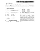 | 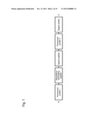 |
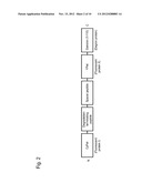 | 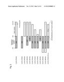 |
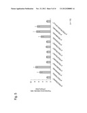 | 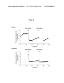 |
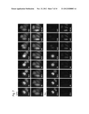 | 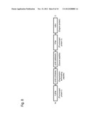 |
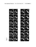 | 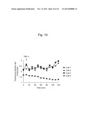 |
 |  |
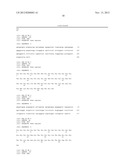 | 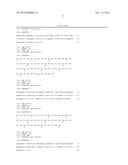 |
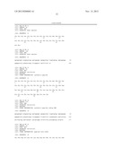 | 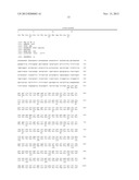 |
 |  |
 |  |
 |  |
 |  |
| New patent applications in this class: | |
| Date | Title |
|---|---|
| 2019-05-16 | Methods and uses for remotely triggered protease activity measurements |
| 2016-07-07 | Method for determining the cleaning performance of formulations |
| 2016-07-07 | Genetically encoded fret-based mmp-9 activity biosensor and use thereof |
| 2016-06-16 | Method for diagnosing cancer through detection of deglycosylation of glycoprotein |
| 2016-06-09 | Reagent for measuring endotoxin and method for measuring endotoxin |
| New patent applications from these inventors: | |
| Date | Title |
|---|---|
| 2015-12-03 | Antibody composition, kit for preparing antibody composition, and immunostaining method |
| 2012-07-12 | Method for measuring autophagy |
| 2011-11-03 | Fluorescent protein |
| 2011-09-15 | Fluorescent protein and chromoprotein |
| 2011-08-25 | Probe reagent for measuring oxidative stress |
| Top Inventors for class "Chemistry: molecular biology and microbiology" | |
| Rank | Inventor's name |
|---|---|
| 1 | Marshall Medoff |
| 2 | Anthony P. Burgard |
| 3 | Mark J. Burk |
| 4 | Robin E. Osterhout |
| 5 | Rangarajan Sampath |