Patent application title: Systems and Methods for Analyzing Nucleic Acid Sequences
Inventors:
Edward I. Ginns (Shrewsbury, MA, US)
Marzena Galdzicka (Shrewsbury, MA, US)
Assignees:
UNIVERSITY OF MASSACHUSETTS
IPC8 Class: AC12M134FI
USPC Class:
435 5
Class name: Chemistry: molecular biology and microbiology measuring or testing process involving enzymes or micro-organisms; composition or test strip therefore; processes of forming such composition or test strip involving virus or bacteriophage
Publication date: 2012-10-25
Patent application number: 20120270206
Abstract:
The invention relates to systems and methods for analyzing clinically
relevant nucleic acid sequences.Claims:
1. A system for performing an assay on a biological sample, comprising:
(a) a central controller programmed to: (i) exchange information about
the biological sample with an outside system or database; and (ii)
exchange information about the biological sample with one or more modules
of the system; (b) a sample transfer module for transferring a portion of
the sample to a first container; (c) a nucleic acid extraction module for
extracting nucleic acids from cells within the portion and for
transferring the portion from the first container to a second container;
(d) a nucleic acid measurement module for measuring the concentration of
nucleic acids in the portion; (e) a PCR preparation module for adding
polymerase chain reaction (PCR) reaction materials to the portion; (f) a
thermocyling module for amplifying a target sequence and extending a
primer in the portion; (g) a primer extension preparation module for
adding primer extension reaction materials to the portion; (h) a mass
spectrometry preparation module for removing a sample of the portion from
the second container to a support for analysis by mass spectrometry; and
(i) a mass spectrometry module for analyzing the sample.
2. The system of claim 1, further comprising linking software that enables the central controller to communicate with at least one other module in the system.
3. The system of claim 1, further comprising a plate editor module that provides sample information to the PCR preparation module.
4. The system of claim 1, further comprising: (j) a transport module comprising one or more robotic arms or tracks to transport a biological sample, or portion thereof, between at least two module of (a) to (i), and arranged to receive information from and transmit information to the central controller.
5. The system of claim 1, further comprising: (j) a detection module for detecting the presence of a sample and monitoring the progress of the sample through the system, and arranged to receive information from and transmit information to the central controller.
6. The system of claim 1, wherein the PCR preparation system includes a primer set selected from the group consisting of: SEQ ID NOS:1, 2, and 3; SEQ ID NOS:4, 5 and 6; SEQ ID NOS:7, 8, and 9; and SEQ ID NOS:10, 11, and 12; SEQ ID NOS:13, 14, and 15; SEQ ID NOS: 16, 17 and 18; SEQ ID NOS:19, 20, and 21; and SEQ ID NOS:22, 23 and 24; SEQ ID NOS:25, 26, and 27; SEQ ID NOS:28, 29, and 30; SEQ ID NOS:31, 32, and 33; SEQ ID NOS:34, 35 and 36; SEQ ID NOS:37, 38 and 39; SEQ ID NOS:40, 41 and 42; SEQ ID NOS:43, 44 and 45; SEQ ID NOS:46, 47 and 48; SEQ ID NOS:49, 50 and 51; SEQ ID NOS:52, 53 and 54; SEQ ID NOS: 55, 56 and 57; SEQ ID NOS:58, 59 and 60; SEQ ID NOS: 61, 62, and 63; SEQ ID NOS: 64, 65, and 66; SEQ ID NOS: 67, 68, and 69; SEQ ID NOS: 70, 71, and 72; SEQ ID NOS: 73, 74, and 75; SEQ ID NOS: 76, 77, and 78; SEQ ID NOS: 79, 80, and 81; SEQ ID NOS: 82, 83, and 84; SEQ ID NOS: 85, 86, and 87; SEQ ID NOS: 88, 89, and 90; SEQ ID NOS: 91, 92, and 93; SEQ ID NOS: 94, 95, and 96; SEQ ID NOS: 97, 98, and 99; SEQ ID NOS: 100, 101, and 102; SEQ ID NOS: 103, 104, and 105; SEQ ID NOS: 106, 107, and 108; SEQ ID NOS: 109, 110, and 111; SEQ ID NOS: 112, 113, and 114; SEQ ID NOS: 115, 116, and 117; SEQ ID NOS: 118, 119, and 120; SEQ ID NOS: 121, 122, and 123; SEQ ID NOS: 124, 125, and 126; SEQ ID NOS: 127, 128, and 129; SEQ ID NOS: 130, 131, and 132; SEQ ID NOS: 133, 134, and 135; SEQ ID NOS: 136, 137, and 138; SEQ ID NOS: 139, 140, and 141; SEQ ID NOS: 142, 143, and 144; SEQ ID NOS: 145, 146, and 147; SEQ ID NOS: 148, 149, and 150; SEQ ID NOS: 151, 152, and 153; SEQ ID NOS: 154, 155, and 156; SEQ ID NOS: 157, 158, and 159; SEQ ID NOS: 160, 161, and 162; SEQ ID NOS: 163, 164, and 165; SEQ ID NOS: 166, 167, and 168; SEQ ID NOS: 169, 170, and 171; SEQ ID NOS: 172, 173, and 174; SEQ ID NOS: 175, 176, and 177; SEQ ID NOS: 178, 179, and 180; SEQ ID NOS: 181, 182, and 183; SEQ ID NOS: 184, 185, and 186; SEQ ID NOS: 187, 188, and 189; SEQ ID NOS: 190, 191, and 192; SEQ ID NOS: 193, 194, and 195; SEQ ID NOS: 196, 197, and 198; SEQ ID NOS: 199, 200, and 201; SEQ ID NOS: 202, 203, and 204; SEQ ID NOS: 205, 206, and 207; SEQ ID NOS: 208, 209, and 210; SEQ ID NOS: 211, 212, and 213; SEQ ID NOS: 214, 215, and 216; SEQ ID NOS: 217, 218, and 219; SEQ ID NOS: 220, 221, and 222; SEQ ID NOS: 223, 224, and 225; SEQ ID NOS: 226, 227, and 228; SEQ ID NOS: 229, 230, and 231; SEQ ID NOS: 232, 233, and 234; SEQ ID NOS: 235, 236, and 237; SEQ ID NOS: 238, 239, and 240; SEQ ID NOS: 241, 242, and 243; SEQ ID NOS: 244, 245, and 246; SEQ ID NOS: 247, 248, and 249; SEQ ID NOS: 250, 251, and 252; SEQ ID NOS: 253, 254, and 255; SEQ ID NOS: 256, 257, and 258; SEQ ID NOS: 259, 260, and 261; SEQ ID NOS: 262, 263, and 264; SEQ ID NOS: 265, 266, and 267; SEQ ID NOS: 268, 269, and 270; SEQ ID NOS: 271, 272, and 273; SEQ ID NOS: 274, 275, and 276; SEQ ID NOS: 277, 278, and 279; SEQ ID NOS: 280, 281, and 282; SEQ ID NOS: 283, 284, and 285; SEQ ID NOS: 286, 287, and 288; SEQ ID NOS: 289, 290, and 291; SEQ ID NOS: 292, 293, and 294; SEQ ID NOS: 295, 296, and 297; SEQ ID NOS: 298, 299, and 300; SEQ ID NOS: 301, 302, and 303; SEQ ID NOS: 304, 305, and 306; SEQ ID NOS: 307, 308, and 309; SEQ ID NOS: 310, 311, and 312; SEQ ID NOS: 313, 314, and 315; SEQ ID NOS: 316, 317, and 318; SEQ ID NOS: 319, 320, and 321; SEQ ID NOS: 322, 323, and 324; SEQ ID NOS: 325, 326, and 327; SEQ ID NOS: 328, 329, and 330; SEQ ID NOS: 331, 332, and 333; SEQ ID NOS: 334, 335, and 336; SEQ ID NOS: 337, 338, and 339; SEQ ID NOS: 340, 341, and 342; SEQ ID NOS: 343, 344, and 345; SEQ ID NOS: 346, 347, and 348; SEQ ID NOS: 349, 350, and 351; SEQ ID NOS: 352, 353, and 354; SEQ ID NOS: 355, 356, and 357; SEQ ID NOS: 358, 359, and 360; SEQ ID NOS: 361, 362, and 363; SEQ ID NOS: 364, 365, and 366; SEQ ID NOS: 367, 368, and 369; SEQ ID NOS: 370, 371, and 372; SEQ ID NOS: 373, 374, and 375; SEQ ID NOS: 376, 377, and 378; SEQ ID NOS: 379, 380, and 381; SEQ ID NOS: 382, 383, and 384; SEQ ID NOS: 385, 386, and 387; SEQ ID NOS: 388, 389, and 390; SEQ ID NOS: 391, 392, and 393; SEQ ID NOS: 394, 395, and 396; SEQ ID NOS: 397, 398, and 399; SEQ ID NOS: 400, 401, and 402; SEQ ID NOS: 403, 404, and 405; SEQ ID NOS: 406, 407, and 408; SEQ ID NOS: 409, 410, and 411; SEQ ID NOS: 412, 413, and 414; SEQ ID NOS: 415, 416, and 417; SEQ ID NOS: 418, 419, and 420; SEQ ID NOS: 421, 422, and 423; SEQ ID NOS: 424, 425, and 426; SEQ ID NOS: 427, 428, and 429; SEQ ID NOS: 430, 431, and 432; SEQ ID NOS: 433, 434, and 435; SEQ ID NOS: 436, 437, and 438; SEQ ID NOS: 439, 440, and 441; SEQ ID NOS: 442, 443, and 444; SEQ ID NOS: 445, 446, and 447; SEQ ID NOS: 448, 449, and 450; SEQ ID NOS: 451, 452, and 453; SEQ ID NOS: 454, 455, and 456; SEQ ID NOS: 457, 458, and 459; SEQ ID NOS: 460, 461, and 462; SEQ ID NOS: 463, 464, and 465; SEQ ID NOS: 466, 467, and 468; SEQ ID NOS: 469, 470, and 471; SEQ ID NOS: 472, 473, and 474; SEQ ID NOS: 475, 476, and 477; SEQ ID NOS: 478, 479, and 480; SEQ ID NOS: 481, 482, and 483; SEQ ID NOS: 484, 485, and 486; SEQ ID NOS: 487, 488, and 489; SEQ ID NOS: 490, 491, and 492; SEQ ID NOS: 493, 494, and 495; SEQ ID NOS: 496, 497, and 498; SEQ ID NOS: 499, 500, and 501; and SEQ ID NOS: 502, 503, and 504, each primer set including two amplification primers and one detection extension primer.
7. The system of claim 1, wherein the central controller is a personal computer system.
8. The system of claim 1, wherein the central controller includes linking software.
9. The system of claim 1, wherein the sample transfer module includes a pipetting robot.
10. The system of claim 1, wherein the nucleic acids measurement module includes an ultraviolet light spectrophotometer or a fluorometer.
11. The system of claim 1, wherein the PCR preparation module includes a pipetting robot.
12. The system of claim 1, wherein the thermocycling module includes a thermocyler.
13. The system of claim 1, further comprising a computer-readable medium comprising one or more programs for instructing a given module.
14. The system of claim 1, wherein the support is a chip or microwell.
15-16. (canceled)
17. A method of performing a diagnostic assay on a biological sample, the method comprising: (a) receiving a biological sample, generating information about the biological sample, and transmitting the information to a central controller; (b) transferring a portion of the biological sample to a first container; (c) extracting nucleic acids from cells within the portion and transferring the portion to a second container; (d) measuring the concentration of extracted nucleic acids in the portion; (e) adding polymerase chain reaction (PCR) materials to the portion; (f) amplifying target nucleic acids in the portion; (g) adding primer extension reaction materials to the portion; (h) extending a detection extension primer in the portion; (i) transferring a sample of the portion from the second container to a support; (j) analyzing the sample and exporting data to the central controller using a mass spectrometry system; and (k) transmitting the data from the central controller to an output device, external system, or database.
18. The method of claim 17, wherein at steps (a) to (k) are performed automatically by an automated system.
19. The method of claim 18, wherein the automated system includes at least one component selected from the group consisting of: a central controller, a sample transfer module, a nucleic acid extraction module, a nucleic acid measurement module, a PCR preparation module, a thermocyling module, a primer extension preparation module, a mass spectrometry preparation module, and a mass spectrometry module.
20. The method of claim 17, wherein the diagnostic assay is an assay for detecting mutations in a gene.
21. The method of claim 20, wherein the gene is selected from the group consisting of: 5,10-Methylenetetrahydrofolate Reductase (MTFR); Coagulation Factor II; Coagulation Factor V; hemochromatosis (HFE); and a glucocerebrosidase (GC). fibroblast growth factor receptor 3; aspartoacylase; Glucocerebrosidase; Coagulation Factor VII; Fanconi Anemia, Complementation Group C (FANCC); inhibitor of kappa light polypeptide gene enhancer in b cells, kinase complex-associated protein; acid sphingomyelinase; hexosaminidase; angiotensin i-converting enzyme; adenylate cyclase 9; apolipoprotein A-1; apolipoprotein E; endothelial leukocyte adhesion molecule 1; fc fragment of IGG, low affinity IIa, receptor; fibrinogen beta chain; coagulation factor II, factor XIII; guanine nucleotide-binding protein beta-3; integrin, alpha-2, glycoprotein Ia/Iia; glycoprotein Ib, platelet, alpha polypeptide; intercellular adhesion molecule 1; glycoprotein Ia/IIa (a2), integrin, alpha-2; platelet glycoprotein Iib, integrin, alpha-2b; glycoprotein IIb/IIIa, integrin, beta-3,3-hydroxy-3-methylglutaryl-coa reductase; lymphocyte adhesion molecule 1; methylene tetrahydrofolate reductase; plasminogen activator inhibitor 1; platelet alpha-granule membrane protein; transforming growth factor-beta receptor, type III; thrombomodulin; tumor necrosis factor; vascular cell adhesion molecule; coagulation factor II receptor; glycoprotein VI, platelet; purinergic receptor P2Y, g protein-coupled, 1; purinergic receptor P2Y, G protein-coupled, 12; prostaglandin-endoperoxide synthase 1; prostaglandin-endoperoxide synthase 2; thromboxane A2 receptor, platelet; and thrombospondin I.
22. The method of claim 17, wherein the diagnostic assay is an assay for detecting a pathogen in the sample.
23. The method of claim 22, wherein the pathogen is a virus, bacterium, or fungus.
24. The method of claim 23, wherein the virus is a virus of the family Herpesviridae.
25. The method of claim 24, wherein the virus is of the genus cytomegalovirus (CMV).
26. An automated method for detecting mutations in a target gene, the method comprising: a) amplifying a target sequence using PCR and automatically performing a primer extension reaction using a set of three primers, each set of primers including two amplification primers and one detection extension primer; b) automatically transferring detection extension primers to a mass spectrometry device; and c) automatically determining the molecular weights of the detection extension primers by mass spectrometry following the primer extension reaction, wherein a change in the molecular weight of the extended primer, as compared to a control, indicates the presence of a mutation in the gene.
27. The method of claim 26, further comprising automatically transmitting information related to the presence of the mutation to a central controller.
28. The method of claim 26, wherein the gene is a 5,10-Methylenetetrahydrofolate Reductase (MTFR) gene, and the set of three primers is selected from the group consisting of: SEQ ID NOS: 1, 2, and 3; SEQ ID NOS: 4, 5 and 6; SEQ ID NOS: 7, 8, and 9; and SEQ ID NOS: 10, 11, and 12; each set of primers including two amplification primers and one detection extension primer.
29. The method of claim 26, wherein the gene is a Coagulation Factor II gene, and the set of three primers is selected from the group consisting of: SEQ ID NOS: 13, 14, and 15 and SEQ ID NOS: 16, 17 and 18; each primer set including two amplification primers and one detection extension primer.
30. The method of claim 26, wherein the gene is a Coagulation Factor Vgene, and the set of three primers is selected from the group consisting of: SEQ ID NOS: 19, 20, and 21 or SEQ ID NOS: 22, 23 and 24; each primer set including two amplification primers and one detection extension primer.
31. The method of claim 26, wherein the gene is a hemochromatosis (HFE) gene, and the set of three primers is selected from the group consisting of: SEQ ID NOS: 40, 41, and 42, SEQ ID NOS: 43, 44 and 45; SEQ ID NOS: 46, 47 and 48; SEQ ID NOS: 49, 50 and 51; SEQ ID NOS: 52, 53 and 54; or SEQ ID NOS: 55, 56 and 57; each set of primers including two amplification primers and one detection extension primer.
32. An automated method for detecting a pathogen in a biological sample, the method comprising: a) amplifying a target sequence using PCR and automatically performing a primer extension reaction using a set of three primers, each set of primers including two amplification primers and one detection extension primer; b) automatically transferring detection extension primers to a mass spectrometry device; and c) automatically determining the molecular weights of the detection extension primers by mass spectrometry following the primer extension reaction, wherein a change in the molecular weight of the extended primer, as compared to controls, indicates the presence of a pathogen in the sample.
33. The method of claim 32, wherein the controls include an internal control for determining the amount of the pathogen in the sample.
34. The method of claim 32, wherein the pathogen is cytomegalovirus (CMV), and the three primers are selected from the group consisting of: SEQ ID NOS: 25, 26, and 27; SEQ ID NOS: 28, 29 and 30; SEQ ID NOS: 31, 32, and 33; SEQ ID NOS: 34, 35, and 36; SEQ ID NOS: 37, 38, and 39; and SEQ ID NOS: 58, 59, and 60; each primer set including two amplification primers and one detection extension primer.
35-36. (canceled)
37. A computer readable medium comprising a program for instructing a central controller in an automated system for performing an assay on a biological sample to: (a) receive a biological sample, generate information about the biological sample, and transmit the information into a central controller; (b) transfer a portion of the biological sample to a first container; (c) extract nucleic acids from cells within the portion and transfer the portion to a second container; (d) measure the concentration of extracted nucleic acids in the portion; (e) add polymerase chain reaction (PCR) materials to the portion; (f) amplify target nucleic acids in the portion; (g) add primer extension reaction materials to the portion; (h) extend a detection extension primer in the portion; (i) transfer a sample of the portion from the second container to a support; (j) analyze the sample and exporting data to the central controller using a mass spectrometry system; and (k) transmit the data from the central controller to an output device, external system, or database.
Description:
CROSS-REFERENCE TO RELATED APPLICATIONS
[0001] This application claims priority to U.S. Provisional Application Nos. 60/493,238, filed on Aug. 6, 2003, and 60/568,958, filed on May 7, 2004. The contents of both of those provisional applications is incorporated herein by reference in its entirety.
TECHNICAL FIELD
[0002] This invention relates to systems and methods for analyzing clinically relevant nucleic acid sequences.
BACKGROUND
[0003] The healthcare delivery system has changed remarkably over the past several decades. Clinical laboratories are under increasing pressure to deliver low cost and highly accurate analytical services with the rapid turn-around time required by physicians and patients. Laboratory testing has changed and improved in recent years to meet the challenge. Robotics has been introduced to the laboratory to increase efficiency and reduce the need for human participation, and laboratory instruments have been designed to decrease the biological sample volumes needed to perform various assays. However, more improvement in the clinical laboratory area is required to meet the demands of the ever-changing healthcare system.
SUMMARY
[0004] The present invention provides novel automated systems and methods to perform assays on nucleic acid sequences (e.g., clinically relevant nucleic acid sequences). The system can provide assay results quickly, accurately, and in a format easily accessible by health care providers and/or third party payors (e.g., insurance companies). The invention also provides novel and highly accurate assays using mass spectrometry (e.g., matrix-assisted laser desorption/ionization (MALDI)).
[0005] In one aspect, the invention provides a system for performing a diagnostic assay on a biological sample. The system includes, as its main components (a) a central controller programmed to: (i) exchange information about the biological sample with an outside system or database; and (ii) exchange information about the biological sample with one or more modules of the system; (b) a sample transfer module for transferring a portion of the sample to a first container; (c) a nucleic acid extraction module for extracting nucleic acids from cells within the portion and for transferring the portion from the first container to a second container; (d) a nucleic acid measurement module for measuring the concentration of nucleic acids in the portion; (e) a PCR preparation module for adding polymerase chain reaction (PCR) reaction materials (e.g., individual nucleotides, primers, polymerase enzymes, and reagents) to the portion; (f) a thermocyling module for amplifying a target sequence and extending a primer in the portion; (g) a primer extension preparation module for adding primer extension reaction materials to the portion; (h) a mass spectrometry preparation module for removing a sample of the portion from the second container to a support (e.g., chip or microwell) for analysis by mass spectrometry; and (i) a mass spectrometry module for analyzing the sample.
[0006] The central controller can be a computer system, e.g., a commercially available personal computer system, and can include linking software that enables the central controller to communicate with at least one other module in the system. The system can also include a plate editor module that provides sample information to the PCR preparation module, a transport module comprising one or more robotic arms or tracks to transport a biological sample, or portion thereof, between at least two modules of (a) to (i), and arranged to receive information from and transmit information to the central controller. The system can also include a detection module for detecting the presence of a sample and monitoring the progress of the sample through the system, and arranged to receive information from and transmit information to the central controller. The nucleic acids measurement system can include an ultraviolet light spectrophotometer or a fluorometer. The PCR preparation module can include a pipetting robot, and the thermocycling system can include a thermocyler. The system can further include a computer-readable medium comprising one or more programs for instructing a given module.
[0007] The PCR preparation module can include PCR materials, e.g., at least one primer set described herein, e.g., a primer set selected from among SEQ ID NOS:1 to 504, each primer set including two amplification primers and one detection extension primer. The sample transfer system can include a pipetting robot.
[0008] In another aspect, the invention provides a method of performing a diagnostic assay on a biological sample. The method includes (a) performing on a biological sample an assay using a clinical assay system, wherein the assay comprises mass spectrometry analysis of a target nucleic acid; and (b) automatically reporting information about the assay from a central controller of the clinical assay system to an outside system or database accessible by at least one health care provider (e.g., at least 2, 10, or more than 10) or at least one third party payor (e.g., at least 2, 10, or more than 10). The clinical assay system can include at least one component selected from the group consisting of: a central controller, a sample transfer module, a nucleic acid extraction module, a nucleic acid measurement module, a PCR preparation module, a thermocyling module, a primer extension preparation module, a mass spectrometry preparation module, and a mass spectrometry module.
[0009] In another aspect, the invention provides a method of performing a diagnostic assay on a biological sample. The method includes (a) receiving a biological sample, generating information about the biological sample, and transmitting the information to a central controller; (b) transferring a portion of the biological sample to a first container; (c) extracting nucleic acids from cells within the portion and transferring the portion to a second container; (d) measuring the concentration of extracted nucleic acids in the portion; (e) adding polymerase chain reaction (PCR) materials to the portion; (f) amplifying target nucleic acids in the portion; (g) adding primer extension reaction materials to the portion; (h) extending a detection extension primer in the portion; (i) transferring a sample of the portion from the second container to a support; (j) analyzing the sample and exporting data to the central controller using a mass spectrometry system; and (k) transmitting the data from the central controller to an output device, external system, or database. In certain embodiments, steps (a) to (k) can be performed automatically by an automated system. The automated system can include at least one component selected from the group consisting of: a central controller, a sample transfer module, a nucleic acid extraction module, a nucleic acid measurement module, a PCR preparation module, a thermocyling module, a primer extension preparation module, a mass spectrometry preparation module, and a mass spectrometry module.
[0010] In certain embodiments, the diagnostic assay can be an assay for detecting mutations in a gene. The gene can be a gene selected from the group consisting of: 5,10-Methylenetetrahydrofolate Reductase (MTFR); Coagulation Factor II; Coagulation Factor V; hemochromatosis (HFE); and a glucocerebrosidase (GC). fibroblast growth factor receptor 3; aspartoacylase; Glucocerebrosidase; Coagulation Factor VII; Fanconi Anemia, Complementation Group C (FANCC); inhibitor of kappa light polypeptide gene enhancer in b cells, kinase complex-associated protein; acid sphingomyelinase; hexosaminidase; angiotensin i-converting enzyme; adenylate cyclase 9; apolipoprotein A-1; apolipoprotein E; endothelial leukocyte adhesion molecule 1; fc fragment of IGG, low affinity IIa, receptor; fibrinogen beta chain; coagulation factor II, factor XIII; guanine nucleotide-binding protein beta-3; integrin, alpha-2, glycoprotein Ia/Iia; glycoprotein Ib, platelet, alpha polypeptide; intercellular adhesion molecule 1; glycoprotein Ia/IIa (a2), integrin, alpha-2; platelet glycoprotein Iib, integrin, alpha-2b; glycoprotein integrin, beta-3,3-hydroxy-3-methylglutaryl-coa reductase; lymphocyte adhesion molecule 1; methylene tetrahydrofolate reductase; plasminogen activator inhibitor 1; platelet alpha-granule membrane protein; transforming growth factor-beta receptor, type III; thrombomodulin; tumor necrosis factor; vascular cell adhesion molecule; coagulation factor II receptor; glycoprotein VI, platelet; purinergic receptor P2Y, g protein-coupled, 1; purinergic receptor P2Y, G protein-coupled, 12; prostaglandin-endoperoxide synthase 1; prostaglandin-endoperoxide synthase 2; thromboxane A2 receptor, platelet; and thrombospondin I.
[0011] In other embodiments, the diagnostic assay is an assay for detecting a pathogen in the sample, e.g., a virus, bacterium, or fungus. The virus can be a virus of the family Herpesviridae, e.g., cytomegalovirus (CMV).
[0012] In another aspect, the invention provides an method, e.g., an automated method, for detecting mutations in a target gene. The method includes a) amplifying a target sequence using PCR and performing, e.g., automatically, a primer extension reaction using a set of three primers, each set of primers including two amplification primers and one detection extension primer; b) transferring, e.g., automatically, detection extension primers to a mass spectrometry device; and c) determining, e.g., automatically, the molecular weights of the detection extension primers by mass spectrometry following the primer extension reaction, wherein a change in the molecular weight of the extended primer, as compared to a control, indicates the presence of a mutation in the gene. The method can include automatically transmitting information related to the presence of the mutation to a central controller.
[0013] In certain embodiments, the gene is a 5,10-Methylenetetrahydrofolate Reductase (MTFR) gene, and the set of three primers is selected from the group consisting of: SEQ ID NOS: 1, 2, and 3; SEQ ID NOS: 4, 5 and 6; SEQ ID NOS: 7, 8, and 9; and SEQ ID NOS: 10, 11, and 12; each set of primers including two amplification primers and one detection extension primer.
[0014] In other embodiments, the gene is a Coagulation Factor II gene, and the set of three primers is selected from the group consisting of: SEQ ID NOS: 13, 14, and 15 and SEQ ID NOS: 16, 17 and 18; each primer set including two amplification primers and one detection extension primer.
[0015] In still other embodiments, the gene is a Coagulation Factor Vgene, and the set of three primers is selected from the group consisting of: SEQ ID NOS: 19, 20, and 21 or SEQ ID NOS: 22, 23 and 24; each primer set including two amplification primers and one detection extension primer.
[0016] In yet other embodiments, the gene is a hemochromatosis (HFE) gene, and the set of three primers is selected from the group consisting of: SEQ ID NOS: 40, 41, and 42, SEQ ID NOS: 43, 44 and 45; SEQ ID NOS: 46, 47 and 48; SEQ ID NOS: 49, 50 and 51; SEQ ID NOS: 52, 53 and 54; or SEQ ID NOS: 55, 56 and 57; each set of primers including two amplification primers and one detection extension primer.
[0017] In another aspect, the invention includes a method, e.g., an automated method, for detecting a pathogen in a biological sample. The method includes a) amplifying a target sequence using PCR and performing, e.g., automatically, a primer extension reaction using a set of three primers, each set of primers including two amplification primers and one detection extension primer; b) transferring, e.g., automatically, detection extension primers to a mass spectrometry device; and c) determining, e.g., automatically, the molecular weights of the detection extension primers by mass spectrometry following the primer extension reaction, wherein a change in the molecular weight of the extended primer, as compared to controls, indicates the presence of a pathogen in the sample. The controls can include an internal control for determining the amount of the pathogen in the sample.
[0018] In some embodiments, the pathogen is cytomegalovirus (CMV), and the three primers are selected from the group consisting of: SEQ ID NOS: 25, 26, and 27; SEQ ID NOS: 28, 29 and 30; SEQ ID NOS: 31, 32, and 33; SEQ ID NOS: 34, 35, and 36; SEQ ID NOS: 37, 38, and 39; and SEQ ID NOS: 58, 59, and 60; each primer set including two amplification primers and one detection extension primer.
[0019] In another aspect, the invention includes an isolated DNA selected from the group consisting of SEQ ID NOS:1 to 504.
[0020] In still another aspect, the invention includes a kit that includes at least one primer set described herein, e.g., a primer set selected from among SEQ ID NOS:1 to 504, each primer set including two amplification primers and one detection extension primer, and instructions for using the primer set to detect or analyze a target nucleic acid sequence in a biological sample. For example, instructions can be provided to describe how to use the primers to detect the presence of, or identify mutations in, a particular nucleic acid sequence or gene. As another example, the instructions can describe how to use the primers to detect the presence of a pathogen (e.g., a virus, bacterium, and/or fungus), the quantity of the pathogen, and/or the genotype of the pathogen.
[0021] In yet another aspect, the invention includes a computer readable medium that includes a program for instructing a central controller in an automated system for performing an assay on a biological sample to: (a) receive a biological sample, generate information about the biological sample, and transmit the information into a central controller; (b) transfer a portion of the biological sample to a first container; (c) extract nucleic acids from cells within the portion and transfer the portion to a second container; (d) measure the concentration of extracted nucleic acids in the portion; (e) add polymerase chain reaction (PCR) materials to the portion; (f) amplify target nucleic acids in the portion; (g) add primer extension reaction materials to the portion; (h) extend a detection extension primer in the portion; (i) transfer a sample of the portion from the second container to a support; (j) analyze the sample and exporting data to the central controller using a mass spectrometry system; and (k) transmit the data from the central controller to an output device, external system, or database.
[0022] Unless otherwise defined, all technical and scientific terms used herein have the same meaning as commonly understood by one of ordinary skill in the art to which this invention belongs. Although methods and equipment or software similar or equivalent to those described herein can be used in the practice of the present invention, suitable methods, equipment, and software are described below. All publications and other references mentioned herein are incorporated by reference in their entirety. In case of conflict, the present specification, including definitions, will control. In addition, the materials, methods, and examples are illustrative only and not intended to be limiting.
[0023] The details of one or more embodiments of the invention are set forth in the accompanying drawings and the description below. Other features and advantages of the invention will be apparent from the description and drawings, and from the claims.
DESCRIPTION OF DRAWINGS
[0024] FIG. 1 is a diagram illustrating the main components of a clinical assay system and the flow of biological samples and information through the system.
[0025] FIG. 2 is a flow diagram illustrating the steps of the clinical assay system.
[0026] FIG. 3 is a mass spectrum of a heterozygous "TC" allele (heterozygous positive) generated using a screen for a C677T mutation in the 5,10-Methylenetetrahydrofolate Reductase (MTFR) gene.
[0027] FIG. 4 is a mass spectrum of a heterozygous "GA" allele (heterozygous positive) generated using a screen for a G20210A mutation in the Coagulation Factor II (FII) gene.
[0028] FIG. 5 is a mass spectrum of a heterozygous "GA" allele (heterozygous positive) generated using a screen for a R506Q mutation in the Coagulation Factor V gene.
[0029] FIG. 6 is a mass spectrum of a heterozygous "GA" allele (heterozygous positive) generated using a screen for a R506Q mutation in the Coagulation Factor V gene.
[0030] FIG. 7 is a mass spectrum of heterozygous "GC" alleles (heterozygous positive) for H63D Histidine to Aspartic acid (C187G) mutation in the FM3-E assay.
[0031] FIG. 8 is a mass spectrum of heterozygous "GC" alleles (heterozygous positive) for H63D Histidine to Aspartic acid (C187G) mutation in the HFE-E3 assay.
[0032] FIG. 9 is a mass spectrum of heterozygous "GA" alleles (heterozygous positive) for S65C Serine to Cysteine (A193T) mutation in the HFE S65C E1 assay.
[0033] FIG. 10 is a mass spectrum of heterozygous "GA" alleles (heterozygous positive) for S65C Serine to Cysteine (A193T) mutation in the FIFE S65C E5 assay.
[0034] FIG. 11 is a mass spectrum of heterozygous "GA" alleles (heterozygous positive) for C282Y cysteine to tyrosine (G845A) mutation in the FM6-E assay.
[0035] FIG. 12 is a mass spectrum of heterozygous "GA" alleles (heterozygous positive) for C282Y cysteine to tyrosine (G845A) mutation in the HFE-E6 assay.
[0036] FIG. 13A1-13A3 is a set of mass spectra in a CMV quantitative assay on samples containing 400 CMV copies/ml.
[0037] FIG. 13B1-13B3 is a set of mass spectra in a CMV quantitative assay on samples containing 4000 CMV copies/ml.
[0038] FIG. 13C1-13C3 is a set of mass spectra in a CMV quantitative assay on samples containing 40,000 CMV copies/ml.
[0039] FIG. 13D-13D3 is a set of mass spectra in a CMV quantitative assay on samples containing 400,000 CMV copies/ml.
[0040] FIG. 13E1-13E3 is a set of mass spectra in a CMV quantitative assay on samples containing 4,000,000 CMV copies/ml.
[0041] FIG. 13F1-13F3 is a set of mass spectra in a CMV quantitative assay on samples containing 40,000,000 CMV copies/ml.
[0042] FIG. 13G1-13G3 is a set of mass spectra in a CMV quantitative assay on samples containing 400,000,000 CMV copies/ml.
[0043] FIG. 14 is a graph that plots CMV plasma samples versus internal standards. A CMV control (4×10⁹ copies per ml) was diluted down to 40 copies/ml in 10-fold increments, mixed with the Internal Standard of appropriate concentration, extracted (240 μl) on MDX (Qiagen), eluted in 75 μl of buffer and assayed (2 μl) by PCR, followed by SAP treatment, extension reaction and mass spectrometry analysis.
[0044] FIG. 15A-15GG is a table that lists a number of genetic targets for the assays of the invention, along with exemplary primers for those targets.
DETAILED DESCRIPTION
[0045] The invention provides a new highly automated system for performing clinical assays, optionally with automatic billing to third party providers such as insurance companies. The invention also provides novel assays using mass spectrometry (e.g., matrix-assisted laser desorption/ionization (MALDI). The assays are highly accurate and can detect, for example, sequence variations (e.g., mutations and/or polymorphisms) and foreign sequences (e.g., viral sequences) incorporated into a target gene. The assays are also useful for infectious disease/pathogen testing.
[0046] The entire process, or portions thereof, can be automated, i.e., performed by machine(s). Accordingly, the present invention also includes a high-throughput process for performing the assays described herein. Thus, the new system can perform dozens (e.g., 96, 128, 384) of different assays on dozens of different biological samples at the same time.
Clinical Assay System
[0047] Overview of System
[0048] FIG. 1 provides an overview of the clinical assay system 2 of the present invention. The clinical assay system includes, as its main components, the following modules. A central controller 4 for exchanging information about the biological sample with an outside system or database 8 and with one or more modules or systems within clinical assay system, and an input device 6; a sample transfer system 10 for transferring a portion of the sample to a first container; a nucleic acid extraction system 12 for extracting nucleic acids from the portion and for transferring the portion from the first container to a second container; a nucleic acid measurement system 14 for measuring the concentration of nucleic acids in the portion; a PCR preparation system 16 for adding polymerase chain reaction (PCR) reaction materials to the portion; a thermocycling system 18 for amplifying a target sequence and extending a primer in the portion; a primer extension preparation system 20 for adding primer extension reaction materials to the portion; a mass spectrometry preparation system 22 for removing a sample of the portion from the second container to a platform for analysis by mass spectrometry; and a mass spectrometry system 24 for analyzing the sample.
[0049] The central controller is capable of controlling one or more system modules, collecting and organizing data obtained from one or more of the system modules and an outside system or database, and of sending data to one or more of the system modules and an outside database (e.g., a database accessible by healthcare providers or third parties) or system (e.g., an outside computer through which health care providers or third parties can access the data). The input device 6 associated with the central controller can be a bar code reader. The system can optionally include a detection system 5 for detecting and tracking a sample as it progresses through the system. The system can also include a transport subsystem 25, e.g., a system of one or more robotic arms and/or tracks, for transporting samples between two or more modules within the system.
[0050] Central Controller
[0051] Typically, the central controller 4 is a computer system. The computer systems that can be used are commonly available personal computers having read-write memory, or industrial counterparts thereof. The central controller is provided with a suitable input device 6 such as a keyboard, touch screen, card reader, bar code scanner, or another computer (e.g., for inputting biological sample processing instructions and patient identification information).
[0052] The central controller 4 is run by linking software, which directs the central controller to receive information from, and/or transmit information to, each of the modules in the overall system. For example, the central controller can be configured to exchange information with one or more modules within the clinical assay system, and to relay that information to one or more other modules. Such information may include information about a biological sample, e.g., sample identification, information as to which assay(s) is to be/has been performed on a sample, and the location of a sample within the clinical assay system and within a given batch of samples being processed.
[0053] The central controller 4 is also configured to exchange information with outside systems and/or databases 8 (i.e., systems or databases not part of the clinical assay system). This configuration allows the central controller to report, e.g., the results of the clinical assays described herein, along with other data, e.g., billing amounts, patient identification, and other data to health-care providers (e.g., technicians, nurses, physicians) and/or third parties (e.g., insurance providers) at other sites. Reporting can occur automatically. Exemplary of outside systems are systems capable of interfacing directly with the central controller, or with a database accessible by both the outside system and the central controller. For example, Meditech® provides a laboratory application that allows multisite and/or multifacility specimen tracking, through which the central controller can exchange information with outside systems.
[0054] Sample Transfer Module
[0055] The sample transfer system 10 can be any system capable of receiving a biological sample, e.g., a blood sample, removing an aliquot of the sample, and placing the aliquot into one or more receptacles. Exemplary systems are pipetting robots, such as the Genesis® Freedom® Automated Workstation. The system is capable of scanning sample tube barcodes and multiwell (e.g., 96-well) plates, and creating a file that indicates where on the multiwell plate a sample is located following the transfer. The file can include information such as the barcodes of scanned sample tubes, the location of these samples on the multiwell plate, the volume transferred from the sample tube to the plate, and overall identifying information (e.g., a barcode) for of the multiwell plate (called DNA plate).
[0056] Nucleic Acid Extraction Module
[0057] The nucleic acid extraction system 12 can be any system capable of carrying out techniques, such as those described herein, for purifying nucleic acids (i.e., DNA and/or RNA) from one or more biological samples. An example of such a system is the BioRobot® MDx produced by Qiagen.
[0058] Nucleic Acids Measurement Module
[0059] The nucleic acids measurement system 14 can be any system capable of measuring the concentration of nucleic acids in a sample. For example, the system can be a commercially available ultraviolet (UV) light spectrophotometer, which is capable of determining the concentration of nucleic acids using optical density measurements. As another example, the system can be capable of measuring the UV-induced fluorescence of dye (e.g., ethidium bromide or Pico Green) intercalated into the nucleic acid, such as a fluorometer. The Genesis® Freedom® Automated Workstation produced by Tecan can include such a fluorometer. The nucleic acid measurement system can be associated with (e.g., a part of) the sample transfer system, or it can be a stand-alone module.
[0060] PCR Preparation Module
[0061] The PCR preparation system 16 can be any system capable of adding appropriate materials, e.g., enzymes (e.g., Taq polymerase), nucleic acid primers, individual nucleotides, and reagents, to an aliquot in preparation for amplifying a target sequence in the aliquot. The PCR preparation also prepares appropriate control reaction mixes. Examples of such systems are the Genesis® Automated Workstation and the Tecan TeMO® multi-pipetting module.
[0062] Overall, the PCR preparation system is capable of performing at least two steps. The first is to dispense appropriate assay mixes. Assay mixes can be prepared by an individual, e.g., a technician, or by a robot, according to typical laboratory procedures, and placed into holders. These PCR preparation system dispenses the mixes from the holders to a position on a second multiwell (e.g., 384) plate, according to instructions (e.g., sample identification and assays to be performed) it receives from a plate editor 7 (described in detail below). The second is to add samples to the appropriate assay mix. Using the file received by the PCR preparation system 16 from the sample transfer system 10, the PCR preparation system transfers samples from the first multiwell (e.g., 96 well) plate to the second multiwell (e.g., 384 well) plate. In this way, the PCR preparation system is able to transfer samples from a first plate to a second plate, while keeping track of the location of the samples, and to ensure that the appropriate assays are performed on each sample.
[0063] Thermocycling Module
[0064] The thermocycling system 18 can be any system capable of performing PCR reactions, e.g., PCR amplification and/or primer extension reactions, and is typically a commercially available thermocycler. Exemplary systems include the GeneAmp PCR System 9700 manufactured by Applied Biosystems, the Perkin Elmer 2000 PCR thermocycler, and the PTC-200 thermocycler manufactured by MJ Research.
[0065] Primer Extension Preparation Module
[0066] The primer extension preparation system 20 can be any system capable of adding appropriate materials, e.g., Shrimp Alkaline Phosphatase (SAP; to dephosphorylate unincorporated dNTPs), extension primers (e.g., the extension primers described herein), and appropriate mixtures of dNTPs and ddNTPs, to an aliquot in preparation for performing primer extension reactions. An exemplary system is the Multimek® manufactured by Beckman-Coulter
[0067] Mass Spectrometry Preparation Module
[0068] The mass spectrometry preparation system 22 can be any system capable of removing a sample of an aliquot and placing the sample on a support, e.g., a chip, for analysis by mass spectroscopy. The support can be composed of any material known to those skilled in the art to be usable in mass spectrometry, e.g., silicon, plastic, glass, and/or ceramic. A wide variety of chips are commercially available. Exemplary of chips is the Sequenom® SpectroCHIP®, which is supplied in 384 well format and are pre-spotted with a specially formulated matrix assisted laser desorption ionization (MALDI) matrix. The matrix can be of any composition known in the art of mass spectrometry, e.g., α-cyano-4-hydroxy cinnamic acid (CHCA), 2,4,6-trihydroxy acetophenone (THAP), or 3-dydroxypicolinic acid (3-HPA) in ammonium citrate, the choice of which will depend, e.g., on the mass spectrometry system used and the assay to be performed. Exemplary of the mass spectrometry preparation systems is the Sequenom® SpectroPOINT®, a nanoliter sample dispensing instrument.
[0069] Mass Spectrometry Module
[0070] The mass spectrometry system 24 can be any commercially available mass spectrometer. The clinical assay system of the present invention can be configured to utilize mass spectrometer formats including matrix assisted laser desorption ionization (MALDI), electrospray (ES), ion cyclotron resonance (ICR) and Fourier Transform. In one embodiment, maMALDI is utilized in the clinical assay system of the present invention. An exemplary mass spectrometry system is the Sequenom® Autoflex® Mass Spectrometer.
[0071] With MALDI mass spectrometry, various mass analyzers can be used, e.g., magnetic sector/magnetic deflection instruments in single or triple quadrupole mode (MS/MS), Fourier transform and time-of-flight (TOF) configurations as is known in the art of mass spectrometry. For the desorption/ionization process, numerous matrix/laser combinations can be used. Ion-trap and reflectron configurations can also be employed. In one embodiment of the present invention, MALDI-TOF is employed to analyze the biological samples.
[0072] Transport Subsystem
[0073] Optionally, the clinical assay system includes a transport system 25. The transport system can be capable of (i) transporting containers, e.g., containing biological samples, from a source (e.g., a separate site in which biological samples are obtained from a patient) to the clinical assay system; (ii) transporting containers between at least two modules within the clinical assay system; and/or (iii) transporting containers away from the clinical assay system to a predefined destination after completion of the assay. The transport system is capable of communicating with the central controller so the central controller can direct the transport system and/or receive information as to the location of sample within the clinical assay system. The transport system can comprise one or more robotic arms or tracks, or a combination of robotic arms and tracks.
[0074] Detection System
[0075] Optionally, the clinical assay system includes a detection system 5 for detecting the presence of a biological sample and monitoring the progress of the sample through the system. The detection system is capable of receiving information from and transmitting information to the central controller. The detection system can include a network of sensors, e.g., barcode readers, reed switches, weight systems (where the detector detects the presence of a sample by its weight); or optical interrupters, or combinations thereof, arranged throughout the clinical assay system, each of which are capable of transmitting information, directly or indirectly, to the central controller.
[0076] Software
[0077] The clinical assay system includes novel software for adapting one or more modules to be used in the system. For example, the central controller includes a novel software program to provide an interface between itself and an outside system or database (e.g., Meditech). In addition to causing the central controller to receive data from an outside source, the software causes the central controller to send information (e.g., automatically) about the clinical assays (e.g., the results and/or billing information) to at least one outside system (e.g., a computer not associated with the clinical assay system) or at least one outside database (e.g., a Meditech database). This configuration allows the clinical assay system to directly report results and billing information to at least one healthcare provider, at least one third party payor (e.g., an insurance company), or to both.
[0078] Optionally, the software can be written such that the central controller performs a final check of assay results before sending to the outside system or database, halting transmission of the data and/or alerting a technician of potential problems with the results. For example, where two samples from the same patient are submitted for duplicate analyses by the clinical assay system, the interface program can include a checking scheme to ensure that the results of the duplicate analyses agree with each other. If the duplicates do not agree (e.g., where both a positive and a negative result are reported to the central controller by the clinical assay system), the interface program can halt transmission of the results so that the mass spectroscopy data can be reanalyzed or to allow the assays to be performed again.
[0079] The central controller also includes a software program for linking at least one other module of the system to the central controller ("linking software"). The module(s) also include linking software allowing the module(s) to communicate with the central controller. For example, the linking software can allow the central controller to control the module (e.g., to instruct the module as to when and whether to execute a function), and to provide to the module(s) information about the biological sample (e.g., the tests to be performed on the sample). The linking software also allows the central controller to receive information from the module(s), e.g., assay results, information about the location of the sample, and the like.
[0080] Also included within the clinical assay system is plate editor software 7. The plate editor software can be loaded on the central controller and/or on a separate computer, e.g., a second computer system. The plate editor software can receive information from an outside system or database, such as patient information, tests to be performed, billing information, etc., and create a file that maps on a hypothetical 384-well plate where each assay for each sample will be located, and links this location to all data and information associated with each assay (one sample can have several assays, e.g., 1, 2, 3, or 4 assays). In this way, the plate editor software "maps out," in advance of processing a batch of samples through the clinical assay system, an arrangement of assays on a 384-well plate. This file can be used by other modules in the system, e.g., the sample PCR preparation system and the mass spectrometry system, to track the samples and their associated assay results as they move through the system.
[0081] Also included is a relational database 9, e.g., Oracle software. Like the plate editor software, the relational database can be loaded on the central controller and/or a separate controller, e.g., a second computer system. The relational database stores information from the plate editor and from an outside database or system (e.g., Meditech), and can be accessed by the outside system or database.
[0082] The sample transfer system 10 can include software causing it to receive at least one biological sample (e.g., a batch of samples), to obtain information about each sample (e.g., from a bar code associated with the sample), and to place an aliquot of the sample into a multiwell plate while keeping track of the location of the aliquot within the multiwell (e.g., a 96 well) plate. The software can instruct the sample transfer system to compile the information (e.g., identity of the sample and the location of the sample in the multiwell plate) into a file, which can then be transmitted to the central controller and/or other modules, e.g., the PCR preparation system.
[0083] An example of software that can be used, for example, with sample transfer system 10 is attached hereto as Appendix 1 and is described in detail below.
[0084] OutFileGenerator.exe
[0085] Laboratory software, e.g., Gemini® (Tecan) software, allows users to control robotic and liquid handling functions and to write specific application scripts based on the user's needs. OutFileGenerator.exe is part of the Gemini program that performs specimen transfer from original bar-coded tubes into a 96-well destination plate, prior to extraction. First, all racks, test tubes, and destination plates are scanned by the barcode scanner. The positions of all racks, tubes, and destination plates, are loaded to the file C:\Program Files\Gemini\Output\" created by Gemini the function. OutFileGenerator.exe creates another file, called "outputdestID.csv" (destID=barcode of the destination plate), by rewriting the original "Output" file such that it contains the following information in a consistent order: DNA ID, DestWellID (well number in the destination plate), Dest ID (ID of destination plate (as determined by the barcode of the plate), SourceWell ID (position of the tube in the rack), and Source ID (barcode of the rack).
[0086] Because samples are often transferred from one plate to another during the extraction process, both plates can be assigned the same barcode number. Alternatively, a script similar to "output file generator" can be used to rename the file and destination plate ID after extraction is complete.
[0087] The PCR preparation system can include software causing it to obtain information (e.g., sample identification and assays to be performed on each sample) about each sample from the sample transfer system and the plate editor, and causing it to add specific PCR materials (e.g., specific primers, nucleotides, etc.) to each sample. In this way, the PCR preparation system is instructed as to which test(s) is to be performed on a given sample, and the PCR preparation system adds the materials appropriate for performing that test(s).
[0088] In particular and as discussed above, the software can cause the PCR preparation system to (a) dispense appropriate assay mixes; and (b) to add samples to the appropriate assay mix. Examples of programs instructing functions (a) and (b) are attached hereto as Appendix 2 and 3, respectively, and are described in further detail below.
[0089] (a) Assay Transfer.gem
[0090] Assay Transfer.gem is an exemplary Gemini program (Appendix 2) for dispensing an aliquot 3 μl, e.g., of assay mixes to PCR plates, e.g., 384-well plates, (i.e., performing function (a) as discussed above). It uses a file called "input file," which is exported from Plate Editor MassARRAY (Sequenom). This file contains all information about the PCR plate, e.g., the location on a plate a particular assay will be run and into which a particular DNA sample should be placed, as well as the barcode of the plate. The following fields are exported from the Plate Editor and are contained in "input file": Plate ID--PCR plate barcode number; Group ID--name of the MassARRAY file; Assay ID--name of the assay; Sample ID--specimen's barcode; and Well Position--position in the plate. Hence, each position in the plate(s) is described by the fields described above, i.e., plate ID, Assay ID, and Sample ID. This file is used in both Assay Transfer.gem and DNA Transfer.gem, which is described in further detail below.
[0091] In this Gemini program are imbedded three executable scripts written in Visual Basic (VB): PCRMix Transfer.exe, Move File Assays.exe, and MoveFileProcessed.exe. Generally, in this program, Module 1 changes the name of a letter/number well (e.g., A1, A2) description, into a well number for a 96 well plate; Module 2 performs the same function as Module 1, but for a 384 well plate; Module 3 includes comments for module 4 execution; and Module 4 generates a worklist for making the transfer of assay mixes into a 384 well plate.
[0092] Specifically, the events as dictated by the program are as follows: [0093] 1) the barcode of the PCR plate is read by barcode reader (Gemini script); [0094] 2) the major part of the program is run by PCRMix Transfer.exe, VB script: [0095] a) the input file is opened and the column "Well Position" containing alphanumeric characters is divided into two columns: one contains alpha-characters and the second contains numerical characters; [0096] b) the file is sorted by column containing numerical characters, meaning that assay mixes will be dispensed later by columns. Hence, all 8 tips will be used at the same time in most cases, speeding up the process of dispensing PCR mixes; [0097] c) module 2 PCR Mix takes a combination letter/number well description and converts it to a well number for a plate (e.g., A1=1); [0098] d) the Gemini program loads "worklist," and Module 3 contains directions to read the .ini file which describes the location and names of: Input File=C:Gemini\Data\Assays\Input File; Worklist file=C:\Gemini\Data\Assays\assays.gwl; Source Type=Trough 1 column; Dest Plate=384 well Marsh; Volume=3 (volume of assay mix dispensed); and WashWorklist; and [0099] e) module 4: (1) opens the sorted Input file and parses it to determine the well into which a particular PCR mix goes into; (2) gives information about where to find the PCR Mix--the ID of the PCR Mix (assay) is written in Gemini file by naming the Trough with the PCR Mix ID; (3) directs when, where and how washes are performed. Each wash is carried out as follows: (i) aspiration of 290-400 μl of bleach; (ii) tips are immersed in the bleach for 10 seconds; (iii) bleach is dispensed into wash station; followed by (iv) three washes with water, for example: 10 ml, 5 ml, 5 ml each. Bleach washes of the tips are performed if the Mix ID is different. A full wash (both bleach and water) is always performed at the end of worklist. [0100] 3) the data from the run is exported into an output file by the "Export Data" command in the Gemini program; [0101] 4) the worklist is copied and saved under a barcode.txt file name (i.e., barcode of 384-well PCR plate) by Move File Assays.exe. Hence, the worklist file is emptied and ready for the next run; and [0102] 5) the above file is moved from the original directory into an Output Processed Directory by MoveFileProcessed.exe script.
[0103] In short, the following commands are contained in the Gemini program: number of plates to be run (in Gemini); reading of barcodes (instructions in Gemini); Execute: C:\programFiles\PCRMixTransfer\PCRMix Transfer.exe; Load Worklist; C;Gemini\Data\Assays\assays.gwl; Execute loaded worklist; Export Data; Execute: C:\ProgramFiles\\Project\Move File Assays.exe; and Execute: C:\ProgramFiles\MoveFileProcessed\MoveFileProcessed.exe.
[0104] (b) DNA Transfer.gem
[0105] The DNA Transfer.gem program (Appendix 3) is an exemplary Gemini program for obtaining an aliquot, e.g., 2 μl, of DNA aspirated from DNA plate, e.g., a 96-well plate, and dispensing it to PCR plate, e.g., a 384 well plate, which contains a particular PCR mix. There are four executable scripts written in VB in this program: DNATransfer.exe; MoveFile.exe; FinalOutputConverter.exe; and CompareFiles.exe.
[0106] Generally, the program uses two files. The first, "InputFile," is exported from the Plate Editor of MassARRAY software saved in csv format (which is the same file used in AssayTranfer.gem for dispensing the PCR mixes). This file dictates where the DNA will be dispensed in the PCR plate (e.g., 384-well plate) by giving for each specimen ID an exact destination, i.e., a specific plate and well. The second, "Output'barcode#.csv," (made on Freedom® (Tecan) by OutFileGenerator.exe) specifies the location of DNA samples in a particular 96-well DNA plate. Based on these two files, a worklist is prepared, which dictates where the DNA samples are dispensed to the PCR plate (destination plate).
[0107] In short, the following commands are contained in this Gemini program: How many DNA plates will be run (in Gemini)?; How many PCR plates will be run (in Gemini)?; Reading the plates barcodes (instructions in Gemini); Execute: C:\programFiles\DNATransfer\DNATransfer.exe; Loading worklist C:\Gemini\Data\DNAtransfer\DNA list gwl; Execute loaded worklist; Export Data; Execute: C:\programFiles\MoveExistFiles\MoveFile.exe; Execute: C:\programFiles\FinalOutputConverter\FinalOutputConverter.exe; and Execute: C:\programFiles\CompareFiles\CompareFiles.exe
[0108] The details of the program are provided below. [0109] 1) At the start of the program, there are instructions written in Gemini which ask: how many DNA plates will be run? How many PCR plates will be run? Instructions for reading the barcodes of each DNA plate and PCR plate are provided. [0110] 2) The major part of the DNA Transfer.gem is run by DNA Transfer.exe in VB script: [0111] Module 1: instructs that the `input file` is opened and the column "Well Position" containing alphanumeric characters is divided into two columns: one contain alpha-characters and second containing numerical characters; the file is sorted by column containing numerical characters first and then by column with alpha-characters. [0112] Module 2: instructs conversion of combination letter/number well descriptions to a well number for a 384 well plate. (for example A1=1). [0113] Module 3: contains instructions to read the .ini file which describes the location and names of: DataFile-=Input File; Worklist; Source Type=96 well plate Sarstedt; Destination Plate=384-well Marsh; and Volume=2 (volume of dispensed DNA). [0114] Module 4: (a) contains a loop that takes each DNA ID from the `inputfile` and goes through all files in the Output directory (containing Output'barcode#.csv" files) looking for the match of the ID to determine which well of the source plate from which a sample should be taken; and (b) creates a workfile containing all information needed for each DNA transfer, such as DNA ID, source well number, source plate type, source plate ID (that is a barcode of the 96-well DNA plate), volume to transfer, destination plate type, destination plate ID (barcode of 384-well PCR plate), destination well and tip number to be used; (c) CreatesTextFile("C:\Gemini\Logfiles\DNAIdLog.txt"); (d) instructs when, where and how the washes are performed. Each wash is carried out as follows: (i) aspiration of 290-400 μl of bleach; (ii) tips are immersed in the bleach for 10 seconds; (iii) bleach is dispensed into wash station; followed by (iv) three washes with water, for example: 10 ml, 5 ml, 5 ml each. Bleach washes of the tips are performed if the Mix ID is different. A full wash (both bleach and water) is always performed at the end of worklist; and e) instructs termination of the run if it cannot file DNA in the outputbarcode file .csv plate, giving the message:"DNA Ids missing! See DNAIdLog, fix files, and rerun"; (f) when all DNAs are found, the worklist is made and the files Output'barcode#.csv are moved from :\Gemini\Output directory to C:\Gemini\OutputProcessed. [0115] 3) Worklist is loaded to Gemini and executed by Gemini command. [0116] 4) Worklist is copied and saved under `outbarcode.txt` file name (barcode of 384-well PCR plate) by MoveFile.exe. Hence, the worklist file is emptied (cleaned out) and ready for the next run. [0117] 5) FinalOutputConverter.exe module 1 converts the original output file and puts it in a readable format containing: "DNA ID," "Destination Well ID," "Destination plate ID," "Source Well ID," and "Source Plate Id." Module 2 converts well number from integer (1) back to alphanumeric well number (A1). Module 3 scans the directory and looks for files and converts them by adding the date to the files. [0118] 6) In addition to checking DNA IDs between two files: `InputFile` and `BarcodeOutput file(s)` before the transfer starts (DNATransfer.exe module 4) CompareFiles.exe compares the "InputFile" with LogFile for correctness in transfer from source 96-well plate(s) to destination 384-well plate(s). It gives an error message if DNA is found to be missing.
[0119] The primer extension preparation system can include similar software, i.e., software that causes it to receive information about each sample and the assays to be performed, so that the primer extension preparation system adds the specific primer extension reaction materials (e.g., specific detection extension primers, termination mixes, etc.) appropriate for performing that test(s) on the sample.
Implementation
[0120] The methods and systems described herein can use a communications network to transmit information from the system (e.g., from the central controller) to healthcare providers and/or insurance companies. These communications networks can use either wired or wireless interfaces. For example, a communications network can be the internet, or a wire or optical cable.
[0121] The new methods can be carried out using various means of data storage. For example, the information or data relating to each sample can be stored on a computer-readable medium or in a computer memory. The information can be transferred physically on diskettes or electronically, e.g., on a dedicated intranet, or on the Internet. The data can be encrypted using standard encryption software from such companies as RSA Security (Bedford, Mass.) and Baltimore®. The data can be stored in various formats, e.g., spreadsheets or databases.
[0122] The invention can be implemented in hardware or software, or a combination of both. The invention can be implemented in computer programs using standard programming techniques following the method stern and figures disclosed herein. The programs (or scripts) should be designed to execute on the various modules or in the central controller. The output information is transmitted to one or more output devices such as a printer, or a CRT or other monitor, or a web page on a computer monitor with access to a website.
[0123] Each program used in the new methods can be implemented in a procedural or object oriented programming language to communicate with a computer system. However, the programs can be implemented in assembly or machine language, if desired. In any case, the language can be a compiled or interpreted language.
[0124] Each computer program can be stored on a storage medium or device (e.g., ROM or magnetic diskette) readable by a general or special purpose programmable computer, for configuring and operating the central controller when the storage media or device is read by the controller (or a given module) to perform the steps or procedures described herein. The system can also be considered to be implemented as a computer-readable medium, configured or encoded with a computer program, where the storage medium so configured causes a computer to operate in a specific and predefined manner to perform the functions described herein.
[0125] Although any communications network can be used, the Internet provides a useful choice to transmit data. In this method, files of data are transmitted from the system to a user in encrypted form, with each party privy to the decryption technique necessary to process the particular data, ending with the completely processed data being sent to the health care provider over the Internet in a similarly encrypted manner. In this method, the entire process can be performed in minutes once the biological sample has been obtained and processed.
Electronic Data Storage and Processing
[0126] The data is typically provided in digital form that can be transmitted and read electronically, e.g., via the Internet, on diskette, or any other mode of electronic or non-electronic communication.
[0127] As used herein, "sequence information" refers to any nucleotide and/or amino acid sequence information, including but not limited to full-length nucleotide and/or amino acid sequences, partial nucleotide and/or amino acid sequences, or mutated sequences. Moreover, information "related to" the sequence information includes detection of the presence or absence of a sequence (e.g., detection of a mutation or deletion), determination of the concentration of a sequence in the sample, and the like. These sequences can be read by electronic apparatus and can be stored on any suitable medium for storing, holding, or containing data or information that can be read and accessed by an electronic apparatus. Such media can include, but are not limited to: magnetic storage media, such as floppy discs, hard disc storage medium, and magnetic tape; optical storage media such as compact disc; electronic storage media such as RAM, ROM, EPROM, EEPROM and the like; general hard disks and hybrids of these categories such as magnetic/optical storage media. The medium is adapted or configured for having recorded thereon sequence information.
[0128] As used herein, the term "electronic apparatus" is intended to include any suitable computing or processing apparatus or other device configured or adapted for storing data or information. Examples of electronic apparatus suitable for use with the present invention include stand-alone computing apparatus; communications networks, including local area networks (LAN), wide area networks (WAN), Internet, Intranet, and Extranet; and local and distributed processing systems.
[0129] As used herein, "stored" refers to a process for encoding information on the electronic apparatus readable medium. Those skilled in the art can readily adopt any of the presently known methods for recording information on known media to generate manufactures comprising the sequence information.
[0130] A variety of software programs and formats can be used to store the sequence information on the electronic apparatus readable medium. Any number of data processor structuring formats (e.g., text file or database) can be employed to obtain or create a medium having recorded thereon the sequence information.
[0131] By providing sequence information in computer-readable form, one can routinely access the sequence information for a variety of purposes. For example, one skilled in the art can use the sequence information in readable form to compare a specific sequence with the sequence information stored within a database. Search means are used to identify fragments or regions of the sequences that match a particular sequence.
[0132] The present invention therefore provides a medium for holding instructions for performing a method for determining whether an individual has a specific disease or disorder or a pre-disposition, for a specific disease or disorder based on genetic information.
Methods of Using the Clinical Assay System
[0133] The new methods make use of the new clinical assay systems described herein. One method 26 of using an exemplary configuration of the clinical assay system is described below and illustrated in FIG. 2.
[0134] Biological samples to be processed (e.g., blood samples) typically arrive at the clinical assay system in multiple containers such as test tubes. Information about the samples (e.g., patient identification, tests to be performed, expected turn-around time) can accompany the sample and/or can be encoded within a barcode associated with the sample. Referring to FIG. 2, the information is first entered into the central controller (step 28) and/or the plate editor, e.g., using a keyboard or bar code scanner, and optionally coordinated (e.g., matched) with information about the sample received by the central controller from an outside system or database (step 30) (e.g., via a Meditech interface). Information obtained by the central controller can be transmitted to one or more modules of the clinical assay system immediately and/or at appropriate intervals during operation of the system.
[0135] The biological samples are deposited in holders in the sample transfer system. Aliquots of the biological samples are removed from the biological sample containers and deposited into multiwell plates by the sample transfer system (step 32). The sample transfer system can include a subsystem, e.g., a barcode scanner and appropriate software, capable of tracking where each aliquot of a biological sample is deposited within the multiwell plate. The information generated via the subsystem can be transmitted to the subsequent modules in the system and/or to the central controller.
[0136] The plate containing the aliquots is then transferred to the nucleic acid extraction system for DNA or total nucleic acids extraction (step 34). Following execution of the extraction process, the aliquots are transferred by the nucleic acid extraction system to a different multiwell plate.
[0137] The plate containing extracted nucleic acid samples is then transferred to the nucleic acid measurement system wherein the concentration of extracted nucleic acids in each aliquot is measured (step 36). The nucleic acid measurement system can include a subsystem, e.g., appropriate software, for assigning the data generated during the measurement process to each individual aliquot and transmitting this data to the central controller.
[0138] The plate is then transferred to the PCR preparation system. The PCR preparation system performs at least related two steps: (1) dispensing appropriate assay mixes, which include all components required for carrying out PCR reactions (e.g., buffers, dNTPs, primers, and polymerase enzyme(s)) (step 37); and (2) dispensing nucleic acid samples, e.g., control and patient DNA samples (step 38). The PCR preparation system can also include a subsystem, e.g., appropriate software, which allows the PCR preparation system to receive from the central controller information specifying which assay(s) is to be performed on a given aliquot, to locate the aliquot on the plate, and to dispense appropriate primers and nucleic acid samples based on those specifications into the aliquot.
[0139] The plate is then transferred to the thermocycling system for amplification of the target nucleic acid sequence (step 40). The PCR reactions can be performed using any appropriate thermocycling protocol, e.g., a protocol as described in the Examples section of the present application.
[0140] Following execution of the PCR reaction, the plate is transferred from the thermocycling system to the primer extension preparation system. The primer extension preparation system adds a dephosphorylating enzyme, e.g., Shrimp Alkaline Phosphatase (SAP), to each aliquot to dephosphorylate unincorporated dNTPs leftover from the PCR reaction (step 41). The plate is then optionally incubated. An extension mix, which includes the extension primer, appropriate enzymes, and a mixture of dNTPs and ddNTPs, is added to each aliquot by the primer extension preparation system (step 42). The primer extension preparation system can also include a subsystem, e.g., appropriate software, which allows the primer extension preparation system to receive from the plate editor information specifying which assay(s) is to be performed on a given aliquot, to locate the aliquot on the plate, and to dispense into each aliquot appropriate extension primers and mixtures of dNTPs and ddNTPs based to those specifications.
[0141] The plates are then transferred back to the thermocycling system, or to a second thermocycling system (e.g., a second thermocycling system contained within the primer extension preparation system), to allow the primer extension reaction to occur (step 44). After the primer extension reaction is complete, the plates are optionally transferred back to the primer extension preparation system, where resin is added by the system to the plates to remove salts, which could potentially interfere with MALDI-TOF analysis.
[0142] The plates are then transferred to the mass spectrometry preparation system. The system transfers a sample (e.g., nanoliter volume samples) of each aliquot on the plate to a chip (e.g., a silicon chip) for MAI DI-TOF analysis (step 46).
[0143] Chips containing the samples are transferred from the mass spectrometry preparation system to the mass spectrometry system. The mass spectrometry system analyzes the samples using MALDI-TOF mass spectrometry (step 48). Specifically, the mass spectrometry system analyzes extended detection extension primers on the basis of molecular weight. Software associated with the mass spectrometry system receives the raw mass spectrometry data, performs digital signal processing (e.g., de-noise, baseline determination, and data compression), analyzes the data, and calls the geneotype according to the resulting data (e.g., mass data). The software associated with the mass spectrometry system optionally receives information from the central controller and matches the data with information about the sample (e.g., patient identification and the assay performed). The data is exported from the mass spectrometry system to the relational database and/or central controller (step 49).
[0144] The finalized data, e.g., patient name, assays performed, and genotype, are exported from the relational database and/or the central controller to an outside system or database (e.g., a Meditech database) (step 50), e.g., via an interface program, e.g., automatically. The outside system or database is accessible by healthcare providers and/or third party payors. Optionally, the information exported from the central controller to the outside system or database can include billing information, e.g., billing codes for the test or tests performed, allowing direct billing of a third party payor for services rendered.
[0145] Optionally, the central controller can be programmed to include with the test results diagnosis information. Such information can be used to facilitate, or give a definitive, diagnosis. For example, with regard to infectious diseases, the diagnostic information can include an indication that a patient has been infected with a particular pathogen, the severity of the infection, the genotype of the organism (e.g., to facilitate the tailoring of a treatment, e.g., to treat a drug resistant organism with a drug to which it is sensitive), and a recommended regimen of treatment. As another example, with respect to genetic mutations that predispose an individual to genetic disease, the information can indicate whether the patient is hetero- or homozygous for the mutation, the likelihood that the patient will develop a disease or condition because of the mutation, and recommend a course of action such as a regimen of treatment or prevention. Such information can be associated with the patient data as necessary and exported from the central controller to the outside database or system.
Clinical Assays
[0146] In addition to the new system, the present invention provides novel diagnostic assays. The assays employ novel primer sequences and mass spectrometry (e.g., MALDI) to detect the presence of target nucleic acid sequences and/or sequence variations (e.g., insertions, mutations and/or polymorphisms) in biological samples. The assays can be categorized into two groups: genetic tests (e.g., to detect mutations) and infectious disease/pathogen testing (e.g., to detect the presence and/or amount of foreign, e.g., viral and/or bacterial, nucleic acids, and/or to genotype the pathogen). A general description of the assays is provided below, and several exemplary assays are described in the Examples.
[0147] Design of Primers and Specific Considerations
[0148] In one embodiment, the novel genetic assays of the present invention require primers for PCR amplification of a target sequence and detection extension primers for carrying out primer extension reactions.
[0149] A target sequence can be, e.g., a sequence that is part of a gene, or a mutation (e.g., insertions, deletions, transitions, or transversions), which is known, or suspected, to be associated with (e.g., cause, increase, or decrease the risk of) a genetic disease or disorder. A target sequence can also be a sequence that is part of a pathogen's (e.g., a virus, bacterium, or fungus) genome, which optionally is known or suspected to include variations (e.g., insertions, deletions, transitions, transversions, etc.) conferring special characteristics, e.g., possession of virulence factors and/or drug resistance, and/or which serve to differentiate among strains of the organism.
[0150] Generally, to generate amplification primers, a 100 bp stretch of nucleotides is first selected from either or both sides of a target sequence. A set of primers is then created manually and/or by using commercially available primer-generating software. An example of such a program is the Sequenom® Spectro Designer® program. This results in a first generation of amplification primers and appropriate detection extension primers.
[0151] The first generation of extension and/or amplification primers is analyzed using a second program, such as OLIGO® Primer Analysis Software (Macintosh, Molecular Biology Insights, Inc.), which evaluates each primer with regard to melting temperature of the primer (Tm), potential hairpins in each primer (judged by free energy (AG), number of nucleotides in the loop, and Tm of the hairpin), and potential for primer dimerization. This analysis results in a second generation of primers.
[0152] The second generation of amplification and/or extension primers is then compared to other known sequences, e.g., using a BLAST program (see, e.g., the National Institutes of Health's National Center for Biotechnology Information (NCBI) website on the World Wide Web at address ncbi.nlm.nih.gov). Primers that match with a sequence other than the target sequence are disregarded. Primers that match with only the target sequence are considered a third generation of amplification primers and are retained for further analysis.
[0153] All possible combinations of the third generation of amplification primers and the detection extension primers are run through the diagnostic genetic assay with positive and negative controls to identify sets of primers that are accurate in 100% of genotype calls. Amplification primers so identified are considered a fourth generation of amplification primers, and are suitable for use in the assays of the present invention.
[0154] The procedures discussed above can be modified for designing primers for a target gene when a homologous pseudogene(s) is known to be present, e.g., the glucocerebrosidase (GC) gene, which is involved in Gaucher Disease. Many commercially available software packages are not able to design specific primers when a homologous sequence to the target gene is present in the genome. Many design primers that amplify short stretches of DNA sequence (60 bp to 150 bp) because PCR is more efficient when short fragments of DNA are amplified. Where pseudogenes are present, however, the amplified fragments of DNA often must be as large as 1000 bp or more. In such cases, at least one of the amplification primer sequences directed against the functional gene must not be present in the pseudogene sequence. For example, based on a comparison of the target gene and pseudogene sequences, a primer can be designed, manually or using software, e.g., software other than SpectroDESIGNER®, that will prime the target gene, but not the pseudogene (homologous) sequence. The extension primer can then be designed using software, e.g., SpectroDESIGNER®. Each amplification and extend primer can then be analyzed using a program like BLAST for specificity (i.e. absence of repetitive or homologous sequences) of the sequence. At least one of the amplification primers should be specific.
[0155] In certain embodiments of the invention, duplicative confirming assays are used. In such cases, an assay is designed around a target using both the sense and antisense strand sequences, designing both forward and reverse detection extension primers. The results from both assays can be compared. Duplicative confirming assays can be beneficial, for example, if the region surrounding the target includes polymorphisms that prevent the PCR amplification and/or extension primers from working, which would cause erroneous results. They can also reduce the occurrence of mistaken calls, e.g., the calling of a heterozygote as a homozygote in an instance when one primer on one allele does not work because of a polymorphic sequence. Generally, when designing these types of confirming assays, two sets of amplification primers are prepared, each extend primer having its own pair of amplification primers. The second pair of amplification primers are designed either manually or using a program, e.g., a program other than SpectroDESIGNER®.
[0156] In certain other embodiments, primers for targets can be designed using more than one type of software, e.g., using SpectroDESIGNER with the input of another sequence aligning software, for additional security when manipulating sequences. For example, in one assay design protocol, the sequence having the deletion/insertion is duplicated. One of the sequences is used to design a forward extension primer, while the other sequence is used to design a reverse extension primer. SpectroDESIGNER® is used to design extension primers in the forward or reverse direction. To force SpectroDESIGNER® to design extend primers in the forward or reverse direction, the letter "N" can be introduced into the sequence at a position 3' to the deletion/insertion site when designing the forward extend primer, or 5' to the deletion/insertion site when designing a reverse extension primer.
[0157] The targets for SpectroDESIGNER® when designing primers for a deletion/insertion site is the first base in the deletion or insertion and the first base after the deletion or insertion. Often, these two nucleotide targets differ for the forward and reverse extension primers. The sequence following the target nucleotides for a forward extend primer can omit the first nucleotide after the deletion/insertion; and, similarly for the reverse extend primer, the nucleotide prior to the deletion/insertion can be omitted. For the purpose of designing the assay with a forward extension primer, in certain instances the sequence following a target should be the sequence that actually follows the deletion/insertion site in genomic DNA. In other cases, the target should be followed by the deleted/inserted sequence. Similar considerations are applicable to designing reverse primers. The choices of how to design the primers in view of these considerations can be dictated to SpectroDESIGNER® by a second type of software, e.g., software other than SpectroDESIGNER®. The validity of the extension primer designed by SpectroDESIGNER® is checked by aligning the extended primer sequences (Ext1 and Ex2) with the template sequence, with and without the insertion/deletion in order to check the validity of assay design.
[0158] Novel Primers
[0159] A number of novel primers were isolated for use in the assays of the present invention. Accordingly, the present invention includes the following nucleic acid sequences, in which the MTHFR primers are used to detect mutations in the 5,10-Methylenetetrahydrofolate Reductase gene, the FaII primers are used to detect mutations in the Coagulation Factor II (FII) gene, the FaV primers are used to detect mutations in the Coagulation Factor V (FaV) gene, and the HFE and FM primers are used to detect mutations in the HFE gene. All others are used to detect the presence and amount of cytomegalovirus (CMV) in a biological sample.
TABLE-US-00001 (SEQ ID NO: 1) MTHFR-F1 5'-ACGTTGGATGATGCCTTCACAAAGCGGAAG-3' (SEQ ID NO: 2) MTHFR-R2 5'-ACGTTGGATGCTTGAAGGAGAAGGTGTCTG-3' (SEQ ID NO: 3) MTHFR-E3 5'-TGCGTGATGATGAAATCG-3' (SEQ ID NO: 4) MTHFR-F17 5'-ACGTTGGATGAGTGATGCCCATGTCGGTG-3' (SEQ ID NO: 5) MTHFR-R18 5'-ACGTTGGATGCTGACCTGAAGCACTTGAAG-3' (SEQ ID NO: 6) MTHFR-E6 5'-GGAGAAGGTGTCTGCGGGAG-3' (SEQ ID NO: 7) MTHFR-F4 5'-ACGTTGGATGTCTACCTGAAGAGCAAGTCC-3' (SEQ ID NO: 8) MTHFR-R5 5'-ACGTTGGATGTCTCCCGAGAGGTAAAGAAC-3' (SEQ ID NO: 9) MTHFR-E11 5'-AACAAAGACTTCAAAGACACTT-3' (SEQ ID NO: 10) MTHFR-F7 5'-ACGTTGGATGACTACTACCTCTTCTACCTG-3' (SEQ ID NO: 11) MTHFR-R8 5'-ACGTTGGATGCTCCAGCATCACTCACTTTG-3' (SEQ ID NO: 12) MTHFR-E14 5'-GAGGAGCTGACCAGTGAAG-3' (SEQ ID NO: 13) FaII-F18 5'-ACGTTGGATGACTCATATTCTGGGCTCCTG-3' (SEQ ID NO: 14) FaII-R19 5'-ACGTTGGATGAGAGAGCTGCCCATGAATAG-3' (SEQ ID NO: 15) FaII-E16 5'-CACTGGGAGCATTGAGGCT-3' (SEQ ID NO: 16) FaII-F10 5'-ACGTTGGATGTGGAACCAATCCCGTGAAAG-3' (SEQ ID NO: 17) FaII-R11 5'-ACGTTGGATGAGAGAGCTGCCCATGAATAG-3' (SEQ ID NO: 18) FaII-E15 5'-CAATAAAAGTGACTCTCAGC-3' (SEQ ID NO: 19) FaV-F16: 5'-ACGTTGGATGAAGACCATACTACAGTGACG-3' (SEQ ID NO: 20) FaV-R17: 5'-ACGTTGGATGCATTATTTAGCCAGGAGACC-3' (SEQ ID NO: 21) FaV-E10: 5'-GACAAAATACCTGTATTCCT-3' (SEQ ID NO: 22) FaV-F18: 5'-ACGTTGGATGCTCTGGGCTAATAGGACTAC-3' (SEQ ID NO: 23) FaV-R19: 5'-ACGTTGGATGCTGAAAGGTTACTTCAAGGAC-3' (SEQ ID NO: 24) FaV-E9: 5'-GCAGATCCCTGGACAGGC-3' (SEQ ID NO: 25) 460 F1 5'-ACGTTGGATGGTCGTGTATGCCACTTTGAC-3' (SEQ ID NO: 26) 460 R2 5'-ACGTTGGATGTGAGGCTGTAATCGCACAGC-3' (SEQ ID NO: 27) 460 E3 5'-CCACTTTGACATTACACCC-3' (SEQ ID NO: 28) 520 F8 5'-ACGTTGGATGTCTTTCAGGAGACGGGTACG-3' (SEQ ID NO: 29) 520 R9 5'-ACGTTGGATGAGATGAGCAGCTTCTGCAGC-3' (SEQ ID NO: 30) 520 E10: 5'-TCTGCGCGAATGTTACCA-3' (SEQ ID NO: 31) 591 F12 5'-ACGTTGGATGAGGCGTTGCTCTTTAAGCAC-3' (SEQ ID NO: 32) 591 R13 5'-ACGTTGGATGAGTGCGTGAGCTTACCGTTC-3' (SEQ ID NO: 33) 591 E14 5'-TTAAGCACGCCGGCGCGG-3' (SEQ ID NO: 34) 607 F28 5'-ACGTTGGATGTCATTTGCGCCGCCAGAATG-3' (SEQ ID NO: 35) 607 R29 5'-ACGTTGGATGTTGCTCTTTAAGCACGCCGG-3' (SEQ ID NO: 36) 607 E30 5'-GCCGCCAGAATGAGCAGA-3' (SEQ ID NO: 37) CV-F31 5'-ACGTTGGATGGAGACGACGTGGACGGCAC-3' (SEQ ID NO: 38) CV-R32 5'-ACGTTGGATGCGTGCTTGGACACGCGACTT-3' (SEQ ID NO: 39) CV33 E 5'-CTCCTTGTCCGAAGCCGC-3' (SEQ ID NO: 40) HFE-F1 5'-ACGTTGGATGATGACCAGCTGTTCGTGTTC-3' (SEQ ID NO: 41) HFE-R2 5'-ACGTTGGATGTCTACTGGAAACCCATGGAG-3' (SEQ ID NO: 42) FM3-E 5' CTCCACACGGCGACTCTCAT-3' (SEQ ID NO: 43) HFE-F7 5'-ACGTTGGATGTTCATGGGTGCCTCAGAGC-3' (SEQ ID NO: 44) HFE-R8 5'-ACGTTGGATGCCACATCTGGCTTGAAATTC-3' (SEQ ID NO: 45) HFE-E3 5' CAGCTGTTCGTGTTCTATGAT 3' (SEQ ID NO: 46) HFE-F4 5'-ACGTTGGATGTGGATAACCTTGGCTGTACC-3' (SEQ ID NO: 47) HFE-R5 5'-ACGTTGGATGTATCACAATGAGGGGCTGATC-3' (SEQ ID NO: 48) FM6-E 5' GCCTGGGTGCTCCACCTGG 3' (SEQ ID NO: 49) HFE-F9 5'-ACGTTGGATGTAATGGGGATGGGACCTACC-3' (SEQ ID NO: 50) HFE-R10 5'-ACGTTGGATGTGCTCTCATCAGTCACATACC-3' (SEQ ID NO: 51) HFE-E6 5' GGGAAGAGCAGAGATATACGT 3' (SEQ ID NO: 52) HFE-F1 5'-ACGTTGGATGATGACCAGCTGTTCGTGTTC-3' (SEQ ID NO: 53) HFE-R2 5'-ACGTTGGATGTCTACTGGAAACCCATGGAG-3' (SEQ ID NO: 54) HFE S65C_E1 5' GGGCTCCACACGGCGAC 3' (SEQ ID NO: 55) HFE-F7 5'-ACGTTGGATGTTCATGGGTGCCTCAGAGC-3' (SEQ ID NO: 56) HFE-R8 5'-ACGTTGGATGCCACATCTGGCTTGAAATTC-3' (SEQ ID NO: 57) HFE S65C_E5 5' TTCGTGTTCTATGATCATGAG 3' (SEQ ID NO: 58) CMV560-S: 5'-ACGTTGGATGCGCTTCTACCACGAATGCTC-3' (SEQ ID NO: 59) CMV560-L: 5'-ACGTTGGATGATAAATACAGCCCGTCGCTC-3' (SEQ ID NO: 60) CMV560-E 5'-CTTTCTGACGTATTCGTGCAGCAT-3'
[0160] Additional primers are set forth in FIG. 15A-15GG, which includes a listing of the target gene, amplification primers and extension primers for that target, and the analytes.
[0161] Assays
[0162] In general, an assay of the present invention can be performed as follows. A biological sample is obtained. As used herein, a "biological sample" is material obtained from any living source (e.g. human, animal, plant, bacteria, fungi, protist, virus) that can be solid material (e.g. tissue, cell pellets, biopsies, fecal matter, nucleic acid samples) and/or fluids (e.g., urine, blood, saliva, sputum, amniotic fluid, mouth wash).
[0163] If necessary (i.e., when the starting material is an intact biological sample and not previously-extracted nucleic acids), nucleic acids are extracted from the biological sample. Extractions can be performed using any art-known method, the choice of which will depend, e.g., on the biological sample from which the nucleic acids are to be isolated. For example, freeze-thaw and alkaline lysis procedures can be useful for obtaining nucleic acid molecules from solid materials; heat and alkaline lysis procedures can be useful for obtaining nucleic acid molecules from urine; and proteinase K extraction can be used to obtain nucleic acid from blood (see, e.g., Rolff, A et al. PCR: Clinical Diagnostics and Research, Springer (1994)). There are numerous kits available for extracting nucleic acids from samples, e.g., the MagneSil® ONE, the Fixed Yield Blood Genomic System (Promega), and kits manufactured by Qiagen.
[0164] The target gene is then amplified via polymerase chain reaction (PCR) using fourth generation primers designed as described herein. Procedures for performing PCR are well known in the art, and are described in, e.g., Sambrook et al., Molecular Cloning: A Laboratory Manual, Cold Spring Harbor Laboratory Press, 1989. Where the nucleic acids to be analyzed are RNA (e.g., viral RNA), the assays of the present invention can be adapted to include reverse transcriptase PCR (RT-PCR) reactions, if necessary.
[0165] In certain embodiments of the invention, an oligo base extension method is then used to detect mutations in, or the presence of, a target nucleic acid. The method is based on the extension of a detection primer (i.e., a detection extension primer designed as described above) that anneals adjacent to a variable nucleotide position on the amplified target DNA, using a DNA polymerase, a mixture of dNTPs (e.g., 3 dNTPs), and the missing dideoxy nucleotides (e.g., one missing dideoxy nucleotide). Any art known primer extension method can be utilized.
[0166] The sample is then analyzed using mass spectrometry. Any mass spectrometer format can be used to analyze the samples, e.g., matrix assisted laser desorption ionization (MALDI), electrospray (ES), ion cyclotron resonance (ICR) and Fourier Transform. Various mass analyzers can be used, e.g., magnetic sector/magnetic deflection instruments in single or triple quadrupole mode (MS/MS), Fourier transform, and time-of-flight (TOF) configurations as is known in the art of mass spectrometry. In an embodiment of the present invention, samples are analyzed using MALDI-TOF mass spectrometry.
[0167] The molecular weight of extended primers is determined by mass spectrometry, and a change in the molecular weight of the extended primer, as compared to a control, indicates the presence of the target sequence and/or whether a mutation (e.g., insertion or deletion), is present in the target sequence. The results are analyzed using methods described in the Examples section below, and an analysis report is generated.
[0168] In other embodiments of the present invention, the assay described above is modified to include, e.g., homogenous MassCLEAVAGE® (hMC) reactions (Sequenom GmbH, Hamburg, Germany). During PCR amplification of the target region, a T7 promoter for in vitro transcription is introduced to the 5' end of one strand of the amplicon. One PCR is performed with a T7 promotor in the forward primer to mediate transcription of the forward DNA strand, and a second PCR is performed with the T7 promotor incorporated in the reverse primer to mediate transcription of the reverse DNA strand. Following the PCR reaction, SAP is added to each reaction to degrade any unincorporated nucleotides from the PCR reactions. For in vitro transcription production, each PCR reaction is split into two transcription reactions and performed. Next, base-specific cleavage using RNase A is conducted, yielding fragmented RNA molecules.
[0169] The final steps in the hMC reaction are sample conditioning and MALDI-TOF MS followed by spectra pattern analysis. The hMC reaction is a high throughput tool for sequence variation identification by comparative sequence analysis. The principle behind it is to generate signal patterns that are specific to a reference sequence in which changes, when compared to a sample sequence, can be tracked as a sequence variation in the form of SNPs or insertion/deletions.
Uses of Assays and the Clinical Assay System
[0170] The assays described above can be used to diagnose genetic diseases (or a patient's or subjects increased risk therefore), to identify foreign sequences (e.g., viral sequences) incorporated into a target gene, and for infectious disease/pathogen testing. The term "subject" as used herein, refers to an animal, or a human, and includes, but is not limited to, mammals, e.g., primates, pigs, rodents such as mice and rats, rabbits, guinea pigs, hamsters, cows, horses, cats, dogs, sheep, and goats. Agricultural analyses, e.g., analysis of biological samples from plants can also be made with the new systems and methods.
[0171] For example, the assays can be adapted to diagnose any of the more than 3000 genetic diseases or disorders currently known (e.g. hemophilias, Gaucher's disease, thalassemias, Duchenne Muscular Dystrophy (DMD), Huntington's Disease (HD), Alzheimer's Disease and Cystic Fibrosis (CF)). and lysosomal and metabolic diseases.
[0172] The assays can also be used to diagnose chromosomal abnormalities such as Trisomy 21 (Down's syndrome), Trisomy 13 (Platau Syndrome), Trisomy 18 (Edward's Syndrome), Monosomy X (Turner's Syndrome) and other sex chromosome aneuploidies such as Klienfelter's Syndrome (XXY), which result in birth defects, and to determine whether an individual has a genetic mutation(s) that predispose the individual to any of a number of disorders such as diabetes, arteriosclerosis, obesity, various autoimmune diseases, stroke, and cancer (e.g. colorectal, breast, ovarian, lung); chromosomal abnormality (either prenatally or postnatally); or a predisposition to a disease or condition (e.g. obesity, atherosclerosis, cancer, venous thromboembolism (VTE), neural tube defects (NTD), and the like) and lysosomal and metabolic diseases.
[0173] The assays can also be used in the detection of "DNA fingerprints," e.g., polymorphisms, such as "microsatellite sequences," which are useful for determining identity or heredity (e.g. paternity or maternity).
[0174] Further, the assays can be used to detect or quantify in a biological sample nucleic acid sequences that are specific to infectious organisms. Such assays are useful for diagnosing or monitoring infection. Examples of disease-causing viruses that infect humans and animals and that may be detected by the disclosed processes include: Retroviridae (e.g., human immunodeficiency viruses, such as HIV-1 (also referred to as HTLV-III, LAV or HTLV-III/LAV, see Ratner, L. et al., Nature, 313: 227-284 (1985); and Wain Hobson, S. et al., Cell, 40: 9-17 (1985)); HIV-2 (see Guyader et al., Nature, 328: 662-669 (1987); European Patent Publication No. 0 269 520; Chakraborti et al., Nature 328: 543-547 (1987); and European Patent Application No. 0 655 501); and other isolates, such as HIV-LP (International Publication No. WO 94/00562 entitled "A Novel Human Immunodeficiency Vitus"; Picornaviridae (e.g., polio viruses, hepatitis A virus, (Gust, I. D., et al., Intervirology, Vol. 20, pp. 1-7 (1983); entero viruses, human coxsackie viruses, rhinoviruses, echoviruses); Calciviridae (e.g., strains that cause gastroenteritis); Togaviridae (e.g., equine encephalitis viruses, rubella viruses); Flaviridae (e.g., dengue viruses, encephalitis viruses, yellow fever viruses); Coronaviridae (e.g., coronaviruses); Rhabdoviridae (e.g., vesicular stomatitis viruses, rabies viruses); Filoviridae (e.g., ebola viruses); Paramyxoviridae (e.g., parainfluenza viruses, mumps virus, measles virus, respiratory syncytial virus); Orthomyxoviridae (e.g., influenza viruses); Bungaviridae (e.g., Hantaan viruses, bunga viruses, phleboviruses and Nairo viruses); Arena viridae (hemorrhagic fever viruses); Reoviridae (e.g., reoviruses, orbiviurses and rotaviruses); Birnaviridae; Hepadnaviridae (Hepatitis B virus); Parvoviridae (parvoviruses); Papovaviridae (papilloma viruses, polyoma viruses); Adenoviridae (most adenoviruses); Herpesviridae (herpes simplex virus (HSV) 1 and 2, varicella zoster virus, cytomegalovirus (CMV), herpes viruses); Poxyiridae (variola viruses, vaccinia viruses, pox viruses); and Iridoviridae (e.g., African swine fever virus); and unclassified viruses (e.g., the etiological agents of Spongiform encephalopathies, the agent of delta hepatitis (thought to be a defective satellite of hepatitis B virus), the agents of non-A, non-B hepatitis (class 1=internally transmitted; class 2=parenterally transmitted (i.e., Hepatitis C); Norwalk and related viruses, and astroviruses), and Epstein-Barr virus (EBV).
[0175] Examples of infectious bacteria include: Helicobacter pyloris, Borelia burgdorferi, Legionella pneumophilia, Mycobacteria sps (e.g. M. tuberculosis, M. avium, M. intracellulare, M. kansaii, M. gordonae), Staphylococcus aureus, Neisseria gonorrhoeae, Neisseria meningitidis, Listeria monocytogenes, Streptococcus pyogenes (Group A Streptococcus), Streptococcus agalactiae (Group B Streptococcus), Streptococcus (viridans group), Streptococcus faecalis, Streptococcus bovis, Streptococcus (anaerobic sps.), Streptococcus pneumoniae, pathogenic Campylobacter sp., Enterococcus sp., Haemophilus influenzae, Bacillus anthracis, corynebacterium diphtheriae, corynebacterium sp., Erysipelothrix rhusiopathiae, Clostridium perfringers, Clostridium tetani, Enterobacter aerogenes, Klebsiella pneumoniae, Pasturella multocida, Bacteroides sp., Fusobacterium nucleatum, Streptobacillus moniliformis, Treponema pallidium, Treponema pertenue, Leptospira, and Actinomyces israelli.
[0176] Examples of infectious fungi include: Cryptococcus neoformans, Histoplasma capsulatum, Coccidioides immitis, Blastomyces dermatitidis, Chlamydia trachomatis, Candida albicans. Other infectious organisms (i.e., protists) include: Plasmodium falciparum and Toxoplasma gondii.
[0177] FIG. 15A-15GG describes a number of specific genetic targets that can be assayed according to the invention, as well as exemplary primers that can be used in those assays. For example, exemplary primers are provided for the following genetic targets: 5,10-Methylenetetrahydrofolate Reductase (MTFR); Coagulation Factor II; Coagulation Factor V; hemochromatosis (HFE); and a glucocerebrosidase (GC). fibroblast growth factor receptor 3; aspartoacylase; Glucocerebrosidase; Coagulation Factor VII; Fanconi Anemia, Complementation Group C (FANCC); inhibitor of kappa light polypeptide gene enhancer in b cells, kinase complex-associated protein; acid sphingomyelinase; hexosaminidase; angiotensin i-converting enzyme; adenylate cyclase 9; apolipoprotein A-1; apolipoprotein E; endothelial leukocyte adhesion molecule 1; fc fragment of IGG, low affinity IIa, receptor; fibrinogen beta chain; coagulation factor II, factor XIII; guanine nucleotide-binding protein beta-3; integrin, alpha-2, glycoprotein Ia/Iia; glycoprotein Ib, platelet, alpha polypeptide; intercellular adhesion molecule 1; glycoprotein Ia/IIa (a2), integrin, alpha-2; platelet glycoprotein Iib, integrin, alpha-2b; glycoprotein IIb/IIIa, integrin, beta-3,3-hydroxy-3-methylglutaryl-coa reductase; lymphocyte adhesion molecule 1; methylene tetrahydrofolate reductase; plasminogen activator inhibitor 1; platelet alpha-granule membrane protein; transforming growth factor-beta receptor, type III; thrombomodulin; tumor necrosis factor; vascular cell adhesion molecule; coagulation factor II receptor; glycoprotein VI, platelet; purinergic receptor P2Y, g protein-coupled, 1; purinergic receptor P2Y, G protein-coupled, 12; prostaglandin-endoperoxide synthase 1; prostaglandin-endoperoxide synthase 2; thromboxane A2 receptor, platelet; and thrombospondin I. The figure lists the disorder associated with the mutation, the gene information (e.g., gene number, symbol, and name), the target mutation, the termination mix that can be used in and assay, the amplification primers, the extension primers, the analytes, and the extended primers (Ext 1, 2, and 3, i.e., the extension primer as it would appear after extension).
[0178] The invention will be further described in the following examples, which do not limit the scope of the invention defined by the claims.
EXAMPLES
Example 1
Screen for 5,10-Methylenetetrahydrofolate Reductase (MTHFR) Mutations
Introduction
[0179] In one exemplary assay, a biological sample (e.g., a blood sample) is tested for the presence or absence of mutations in the MTHFR gene. MTHFR catalyzes the conversion of 5,10-methylenetetrahydrofolate to 5-methyltetrahydrofolate, a cosubstrate for homocysteine remethylation to methionine. MTHFR plays a key role in folate metabolism by channeling one-carbon units between nucleotide synthesis and methylation reactions. Severe enzyme deficiency leads to hyperhomocysteinemia and homocystinuria, with altered folate distribution and a phenotype that is characterized by damage to the nervous and vascular systems.
[0180] Two common mutations that can occur in MTHFR include MTHFR C677T (or Ala222Val) and MTHFR A1298C (or Glu429Ala). Assays to detect these mutations are described below. The C677T mutation is common in the general population, with a variable frequency depending on the subpopulation studied. In Caucasians of European descent, the frequency of homozygotes for this mutation ranges from approximately 5% (in Dutch and Finnish populations) to 12-15% (in French Canadian and other European populations). African Americans have a low frequency of 1.4% The A1298C mutation is also common in the general population with an allele frequency estimated at 0.33.
[0181] Individuals homozygous for the C677T mutation have significantly elevated plasma homocysteine levels. It has been suggested that the 677T variant of the MTHFR gene is a genetic risk factor for preeclampsia. From studies of the C677T mutation in cardiovascular patients and controls, investigators have concluded that homozygosity for this frequent mutation in the MTHFR gene is associated with a 3-fold increase in risk for premature cardiovascular disease. The association was stronger in homozygotes than in heterozygotes. Therefore, the C677T polymorphism may be a risk factor for coronary artery disease.
[0182] Currently, at least 22 other mutations in MTHFR gene are known and assays similar to those described herein can be designed to detect those mutations as well.
Gene Information
[0183] Gene Locus: 1p36.3.
[0184] Gene Symbol: MTHFR.
[0185] Gene Product: 5,10-METHYLENETETRAHYDROFOLATE REDUCTASE.
[0186] Mutations: MTHFR C677T, Ala222Val, FR A1298C, Glu429Ala.
[0187] Gene OMIM number: 607093
[0188] mRNA NCBI Acc Nos.: XM--030156 5,10-methylenetetrahydrofolate reductase mRNA; AJ237672 Homo sapiens mRNA for 5,10-methylenetetrahydrofolate reductase mRNA; NM--005957 Homo sapiens 5,10-methylenetetrahydrofolate reductase (NADPH) (MTHFR) mRNA; U09806 Synthetic construct methylenetetrahydrofolate reductase (MTHFR) mRNA, complete cds. (Goyette, 1994 Frosst 1995)
[0189] Genomic Acc No.: GA_x5L2HTU1V1E CELERA DataBase
Clinical Significance
[0190] The test can be used to screen for the MTHFR gene mutation before surgery, which may identify patients at an increased risk of graft thrombosis. The MTHFR mutation assay is also useful for patients with early-onset arteriosclerotic vascular disease or thrombosis, particularly those with hyperhomocysteinemia or significant family histories. The clinical scenario should be considered carefully, since additional genetic and non-genetic factors are important in the development of cardiovascular disease. MTHFR mutation analysis is also useful for patients with variable or ambiguous homocysteine levels, since plasma levels are dependent on nutritional status or sample handling before testing. MTHFR mutational analysis can also be offered for patients with a neural tube defect (NTD). The MTHFR mutation assay is also useful in the elderly and in patients with idiopathic venous thromboembolism (VTE) disease and arterial disease.
MTHFR-C677T E3 Assay
[0191] This section describes the amplification and detection extension primers for the MTHFR-C677T E3 Assay and provides the sequences of the primers. The sequences of the resulting extended primers are provided in Table 1, below.
TABLE-US-00002 ##STR00001## Amplification primers*: ##STR00002## Detection Extension Primer: ##STR00003## Resulting Amplicon length: 89 bp *amplification primers have the 10-mer tag specific for the homogeneous MassEXTEND ® assay (hME) method: acgttggatg. Termination mix: ddA/ddC/ddT
TABLE-US-00003 TABLE 1 Sequence, length and mass of extension primer, extended primer (G and A) and pause product for MTHFR-C677T Allele Sequence Length Mass MTHFR-E3 TGCGTGATGATGAAATCG 18 bp 5578.6 T mutant allele (ext A) TGCGTGATGATGAAATCGA 19 bp 5875.9 C wild type allele (ext G) TGCGTGATGATGAAATCGGC 20 bp 6181 Contaminant (pausing peak) 5907.9 (SEQ ID NOS.: 806, 807, and 808)
MTHFR C677T E6 Assay
[0192] This section describes the amplification and detection extension primers for the MTHFR C677T E6 Assay and provides the sequences of the primers. The sequences of the resulting extended primers are provided in Table 2, below.
TABLE-US-00004 ##STR00004## Amplification primers* ##STR00005## Detection Extension Primer: ##STR00006## Amplicon length: 122 bp *amplification primers have the 10-mer tag specific for the hME method: acgttggatg. Termination mix: ddA/ddC/ddG
TABLE-US-00005 TABLE 2 Sequence, length and mass of extension primer, extended primer (G and A) and pause product for MTHFR-C677T Allele Sequence Length Mass MTHFR-E6 GGAGAAGGTGTCTGCGGGAG 20 bp 6303.1 T mutant allele (ext A) GGAGAAGGTGTCTGCGGGAGTC 22 bp 6880.5 C wild type allele (ext G) GGAGAAGGTGTCTGCGGGAGC 21 bp 6576.3 Contaminant (pausing peak) 5907.9 [SEQ ID NOS: 811, 812, and 813)
MTHFR-A1298C Assay
[0193] This section describes the amplification and detection extension primers for the MTHFR-A1298C assay and provides the sequences of the primers. The sequences of the resulting extended primers are provided in Table 3, below.
TABLE-US-00006 ##STR00007## Amplification primers* ##STR00008## Detection Extension Primer: ##STR00009## Amplicon length: 109 bp *amplification primers have the 10-mer tag specific for the hME method: acgttggatg (SEQ ID No. 816) Termination mix: ddA/ddC/ddT
TABLE-US-00007 TABLE 3 Sequence, length and mass of extension primer, extended primers and pause product for MTHFR- A1298C E11: Allele Sequence Length Mass MTHFR-E11 AACAAAGACTTCAAAGACACTT 22 bp 6704.4 A mutant allele AACAAAGACTTCAAAGACACTTT 23 bp 6992.60 C wild type allele AACAAAGACTTCAAAGACACTTGC 24 bp 7306.80 Contaminant (pausing peak) 7033.60 (SEQ ID NOS: 817, 818, and 819)
MTHFR A1298C E14 Assay
[0194] This section describes the amplification and detection extension primers for the MTHFR A1298C E14 Assay and provides the sequences of the primers. The sequences of the resulting extended primers are provided in Table 4, below.
TABLE-US-00008 ##STR00010## Amplification primers* ##STR00011## Detection Extension Primer ##STR00012## Amplicon length: 159 bp *amplification primers have the 10-mer tag specific for the hME method: acgttggatg. Termination mix: ddC/ddG/ddT
TABLE-US-00009 TABLE 4 Sequence, length and mass of extension primer, extended primers and pause product for MTHFR- A1298C E14 Allele Sequence Length Mass MTHFR-E14 GAGGAGCTGACCAGTGAAG 19 bp 5926.90 A mutant allele GAGGAGCTGACCAGTGAAGAAAG 23 bp 7179.70 C wild type allele GAGGAGCTGACCAGTGAAGC 20 bp 6200.10 Contaminant (pausing peak) 6240.10 (SEQ ID NOS: 822, 823, and 824)
Testing Procedure
[0195] This section generally describes procedures useful in the MTHFR assays.
[0196] Specimens: Whole blood samples were collected in the same manner routinely used for any laboratory test. Exemplary test results, with specific numbers of samples, are provided below in Tables 12, 13, 14, and 15.
[0197] Specimen Storage and Stability [0198] 1. After arrival, blood was extracted on the same or next day. Specimen was not allowed to remain at +15° C. to +30° C. longer than 8 hours. If assays were not completed within 8 hours, specimen was stored at +2° C. to +8° C. [0199] 2. Tubes of blood should were kept closed at all times in a vertical, stopper-up position. [0200] 3. Blood specimen were not frozen.
[0201] Sample Volume
[0202] The minimum volume was 1.0 mL of sample.
[0203] Criteria for Unacceptable Specimens
[0204] Unlabeled specimens.
[0205] Insufficient quantity.
[0206] Clotted specimen.
[0207] Specimen Handling
[0208] Blood samples were received with the lab barcode identification numbers. Sample identification is ensured through out the process by the tracking system on Freedom, BioRobot MDx, Genesis, and MassARRAY systems.
[0209] Procedures Involved in Assays:
[0210] Nucleic acids extraction, nucleic acid concentration measurement, Uniplex and Multiplex PCR Procedure on Genesis® in conjunction with Freedom® and MassARRAY® software, Homogeneous MassEXTEND® Reaction using the MassARRAY® System.
[0211] Quality Control
[0212] PCR primers were analyzed ensure specificity for the prothrombin gene in the BLAST search and that the PCR product did not show allelic dropout. Validation of each assay was accomplished.
[0213] Four types of control material were analyzed with each PCR plate: (A) blank control (water), (B) DNA negative control (wild type), (C) heterozygous positive controls and (D) positive homozygous mutant. In each run at least two samples of each control were used if available. More frequent use of controls or the use of additional controls was left to the discretion of the user based on work load and work flow. Tables 5 and 6, below, provide a list of control DNAs used in the presently described assays.
TABLE-US-00010 TABLE 5 Controls used in MTHFR- C677T assay 39354 T MTHFR homozygous GM881 T MTHFR homozygous GM1278 C MTHFR negative- wild NA1464 C MTHFR negative- wild GM779 C MTHFR negative- wild NA1465 C MTHFR negative- wild GM972 CT MTHFR heterozygo NA0375 CT MTHFR heterozygo NA1359 CT MTHFR heterozygo NA1464 CT MTHFR heterozygo NA1470 CT MTHFR heterozygo NA1600 CT MTHFR heterozygo NA1602 CT MTHFR heterozygo
TABLE-US-00011 TABLE 6 Controls used in MTHFR-A1298C assay GM7798 C MTHFRA1298C homozygous mutant GM8810 A MTHFRA1298C negative- wild type NA16028 A MTHFRA1298C negative- wild type GM12783 AC MTHFRA1298C heterozygous NA14641 AC MTHFRA1298C heterozygous NA16000 AC MTHFRA1298C heterozygous
[0214] PCR Procedure
[0215] The master PCR mix was made, biannually or as necessary, in 11.954 ml total volume and aliquoted into 47 Sarstedt tubes, 250 μl each. The composition of the PCR master mix is provided in Table 7, below.
TABLE-US-00012 TABLE 7 Composition of master PCR mix Volumes used to Stock Final make master PCR Reagent Concentration Concentration mix Water N/A N/A 8,514 μl Buffer/MgCl2 15 mM (10x) 1.5 mM 2,150 μl MgCl2 25 mM (10x) 2.5 mM 860 μl dNTPs 10 mM (50x) 200 μM 430 μl Total volume 11,954 μl
[0216] The 5 μM primer mix was made by diluting 10 times 50 μM stock primers. The volumes used to make 2000 μl of 5 μM primer mix is shown in Table 8, below. The primer mix was aliquoted to 10 Sarstedt tubes, 200 μl each.
TABLE-US-00013 TABLE 8 Composition of 5 μM Primer Mix Volumes Stock Concentration used to make Reagent Concentration in the mix 5 μM mix Water N/A N/A 1,200 μl Forward 50 μM 5 μM 200 μl primer Reverse 50 μM 5 μM 200 μl primer Total Volume 2000 μl
[0217] The composition of the PCR reaction is shown in Table 9. To calculate the volumes per run, the volumes per reaction of PCR mix, Primer Mix, and HotStarTaq® need to be multiplied by number of reactions and then by 1.5. Table 9 shows an example of the calculations per 384 reactions. Increasing the final volume by 50% is to account for pipetting losses and `dead` volume when, using the robotics.
TABLE-US-00014 TABLE 9 Composition of single PCR reaction and PCR reaction for 384-well plate Volume per 384 Stock Final Reaction reactions Reagent Concentration Concentration Volume and × 1.5 PCR mix per 10x 1x 2.78 μl 1601.28 μl reaction Primer Mix 5 μM (40x) 200 nM 0.2 μl 115.2 μl (F + R) HotStarTaq 5 units/μl (50x) 0.1 unit per 0.02 μl 11.52 μl reaction TOTAL volume 3.00 μl 1728 μl
[0218] 3 μl of PCR mix were dispensed into each well of the 384-well plate. The protocol "PCR Procedure" (MD008) was utilized. 2 μl of DNA (2.5 ng/μl) were added to each well. The standard PCR program on the PCR machine was run:
TABLE-US-00015 95° C. 15 minutes 95° C. 20 seconds 56° C. 30 seconds {close oversize brace} 45 cycles 72° C. 1 minute 72° C. 3 minutes 4° C. forever (hold)
[0219] Dephosphorylation Procedure
[0220] SAP (Shrimp Alkaline Phosphatase) was diluted according to Table 10, multiplying the volumes by the number of the samples and then by 1.5.
TABLE-US-00016 TABLE 10 Composition of SAP mix Volume per Stock Final Reaction 384-well Reagent Concentration Concentration Volume plate Nanopure N/A N/A 1.53 μl 881.3 μl Water hME buffer 10x 1x * 0.17 μl 97.9 μl SAP 1 unit/μl 0.15 units/μl 0.30 μl 172.8 μl TOTALS: 2.00 μl 1152.00 μl
[0221] 2 μl of the diluted Shrimp Alkaline Phosphatase were dispensed to each well (Refer to MassARRAY procedure MD 009). The SAP PCR program was run on the PCR machine:
TABLE-US-00017 37° C. 20 minutes 85° C. 5 minutes, 4° C. forever (hold)
[0222] Homogeneous MassEXTEND (hME) Reaction Procedure:
[0223] The hME cocktail was prepared according to Table 11. Multiply one reaction volume by the number of samples and then by 1.38.
TABLE-US-00018 TABLE 11 Components of hME cocktail Volume per Stock Final Reaction 384-well Reagent Concentration Concentration Volume plate* Nanopure N/A N/A 1.674 μl 887.09 μl Water hME Mix 10x 1x 0.2 μl 105.98 μl (10x buffer w/2.25 mM d/ddNTPs). Extension 50 μM 2.7 μM 0.108 μl 57.23 μl primer Thermo 32 units/μl 0.063 unit/μl 0.018 μl 9.54 μl Sequenase.sup.& TOTALS 2.0 μl 1059.84 μl *Volumes are for a 384- well microplate and include a 38% overage to account for possible pipetting loss and dead volume in the 96-well microtiter plate used on the Multimek. .sup.&ThermoSequenase is stored at (-20)° C. until it is added to the reaction cocktail.
[0224] 2 μl of the hME cocktail mix were dispensed to the appropriate wells containing 7 μl dephosphorylated PCR product. Refer to MassARRAY procedure MD008. The Extension Reaction program was run on the PCR machine:
TABLE-US-00019 94° C. 2 minutes 94° C. 5 seconds 52° C. 5 seconds {close oversize brace} 45 cycles 72° C. 5 seconds 4° C. forever (hold)
[0225] SpectroClean Resin Clean Up Procedure
[0226] Using the SpectroClean plate, transfer resin into the 96-well Marsh plate. Run the hME cation Cleanup method on the Multimeck. Refer for details to MassARRAY protocol MD008.
[0227] Results Analysis
[0228] The Genotyper® Analyzer software reads the spectrum generated for each sample by the mass spectrometer, and uses a 3-parameter model to calculate the significance of each putative genotype: [0229] 1. Conservative: The most conservative set makes no error in reading the data. [0230] 2. Aggressive: The most aggressive set makes the most errors (still less than 1 percent). [0231] 3. Moderate: The moderate set is a compromise between the two extremes.
[0232] The interpretation of the results is as follows: If the results of two assays for the same mutation are called (i.e., interpreted) identically by conservative or moderate calls or conservative and aggressive calls, the result is acceptable. If the assays are called identically by moderate and aggressive calls, the test needs to be looked at by the director or the designee. If the assays are called only by aggressive calls, the test needs to be repeated. If there is a call on one but not the other assay, the "non call" assay needs to be repeated.
[0233] The non-calls can be further categorized into the following groups: (a) Low Probability: Implies that the spectrum in question contains peaks that fail any criteria of significance even for the aggressive parameter set; (b) Conflict: Implies that there is more that one read or spectrum corresponding to one sample, and these reads give rise to different and conflicting genotypes; (c) Bad spectrum: The spectrum for the sample doesn't exist above noise level; and (d) Bad Assay: results of analyst or operator input errors in defining the assays. The most common errors are mass values of analytes or contaminants that are out of range of the spectrum, or contaminants and analytes having the same mass; and (e) user call: The analyst or operator select a genotype in the table and performs a manual call.
[0234] In the presently described examples, each sample will have at least two different assays that together will confirm, e.g., the presence of a mutation. However, in many instances, accurate results can be obtained using one assay per sample and, therefore, performing two different assays is optional.
[0235] FIG. 3 is a mass spectrum of a heterozygous "TC" allele (heterozygous positive) generated using a screen for a C677T mutation in the 5,10-Methylenetetrahydrofolate Reductase (MTFR) gene. Exemplary test results are provided in Tables 12, 13, 14, and 15, below.
TABLE-US-00020 TABLE 12 MTHFR C677T E3 Low Source of DNA Conservative Moderate Aggressive Probability Associated 147 3 0 Regional and University Pathologists (ARUP) DNAs + Coriel Institute DNAs
TABLE-US-00021 TABLE 13 MTHFR C677T E6 Low Source of DNA Conservative Moderate Aggressive Probability ARUP DNAs + 150 2 0 Coriel DNAs
TABLE-US-00022 TABLE 14 MTHFR A1298C E11 Low Source of DNA Conservative Moderate Aggressive Probability ARUP DNAs + 125 26 1 Coriel DNAs
TABLE-US-00023 TABLE 15 MTHFR A1298C E14 Low Source of DNA Conservative Moderate Aggressive Probability ARUP DNAs + 89 53 6 Coriel DNAs
[0236] The results of the assays were 100% in agreement with results from Associated Regional and University Pathologists (ARUP). In both (E3 and E6) MTHFR-C677T assays, negative controls (homozygous wild type) GM12783, GM7798, NA14641 (Coriell) were homozygous CC, positive heterozygous controls NA13591, NA14646, NA14702, and NA16000 (Coriell) were TC, and, positive homozygous control NA8810 (Coriell) was TT. In both MTHFR-A1298C assays, negative control (homozygous wild type) GM7798 (Coriell) was homozygous CC, positive heterozygous controls NA14641 and NA16000 (Coriell) were both AC, and positive homozygous control NA16028 (Coriell) was AA. As indicated in Tables 12, 13, 14, and 15, above, the majority of geneotype calls (i.e., the genotype called by the software for each sample) for each assay were conservative calls, indicating a high level accuracy.
Example 2
Screen for Coagulation Factor II (FII) G20210A Mutations
[0237] Mutation G20210A in the prothrombin gene is associated with hyperprothrombinemia and increased risk of venous thromboembolism (VTE). It has been established that the FII G20210A mutation increases the risk of VTE 2-5 fold. The 20210A prothrombin allele also represents an inherited risk factor for acute coronary syndrome among patients who have limited extent of coronary disease at angiography or who lack major metabolic and acquired risk factors.
[0238] Mutation G20210A involves the last nucleotide of the transcribed prothrombin RNA. The mechanism by which prothrombin levels are altered is unknown. One explanation is that increased adenylation and increased mRNA stability are involved.
[0239] In southern Europe, the prevalence of the G20210A mutation is 3.0 percent (CI95 2.3 to 3.7%), nearly twice as high as the prevalence in northern Europe 1.7% (CI95 1.3 to 2.2%), the highest of 7.8% reported in a Greek-Cypriot population. In the United States, the overall prevalence estimates are between 1% and 2%, although this is highly dependent on race. The mutation is fairly uncommon in African Americans (˜0.2%), and is also rarely seen in Asians and Native Americans.
[0240] At least 28 missense mutations, one insertion, one deletion, and one substitution (in the regulatory region) are known to occur in the prothrombin gene. Accordingly, assays similar to those described herein can be designed to detect those mutations as well.
Gene Information
[0241] Gene Locus: 11p1-q12
[0242] Gene Symbol: F2, PRT
[0243] Gene Product: COAGULATION FACTOR II, prothrombin
[0244] Mutations: G20210A
[0245] Mutation Frequency The 20210G-A variation in the prothrombin gene is predominant only in the Caucasian population, among whom the prevalence of heterozygous carriers varies between 1% to 8%, and depends on the geographical location and ethnic background.
[0246] OMIM number: 176930
[0247] mRNA NCBI Acc No.: NM--000506 coagulation factor II mRNA; M17262. Human prothrombin mRNA
[0248] Genomic Acc No.: AF478696 H. coagulation factor II (thrombin) (F2) gene.
Clinical Significance
[0249] Prothrombin (G20210A) testing can be recommended, for example, in the following cases: when inherited thrombophilia is suspected; when the testing for factor V Leiden mutation is recommended; in patients with a history of recurrent VTE; in patients with a first episode of VTE before the age of 50 years; in patients with a history of an unprovoked VTE at any age; where thrombosis occurs in unusual anatomic sites, such as cerebral, mesenteric, portal, or hepatic veins; where there is a first-degree relative with VTE; in patients with a first VTE related to pregnancy, the puerperium, or oral contraceptive use; where there is a history of VTE related to pregnancy loss during the second or third trimesters; in young women smokers (age<50 years) with an MI; in older patients (age>50 years) with a first provoked VTE event in the absence of a cancer or an intravascular device; where there is a first VTE related to serum estrogen receptor modifier or tamoifen; in selected cases of women with unexplained severe preeclampsia, placental abruption, or intrauterine growth restriction; in asympthomatic adult family members of probands with known prothrombin G20210A mutations, especially those with a strong family history of thrombosis at young age; and asymptomatic female family members who are pregnant or are considering oral contraceptives or pregnancy.
Assay FaII-E16
[0250] This section describes the amplification and detection extension primers for the FaII-E16 Assay and provides the sequences of the primers. The sequences of the resulting extended primers are provided in Table 16, below.
TABLE-US-00024 ##STR00013## Amplification primers*: ##STR00014## Detection Extension Primer: ##STR00015## Termination mix: ddA/ddC/ddT *amplification primers have the 10-mer tag specific for the hME method: acgttggatg.
TABLE-US-00025 TABLE 16 Sequence, length and mass of extension primer, extended primer (G and A) and pause product) Allele Sequence Length Mass Fall-E16 (G20210A) CACTGGGAGCATTGAGGCT 19 bp 5868.8 Negative (wild type) CACTGGGAGCATTGAGGCTC 20 bp 6142 Homozygouse (mutant) CACTGGGAGCATTGAGGCTTG 21 bp 6486.2 Contaminant (pausing peak) 6173 (SEQ ID NOS: 825 826, and 827)
FaII-E15 ASSAY
[0251] This section describes the amplification and detection extension primers for the FaII-E15 Assay and provides the sequences of the primers. The sequences of the resulting extended primers are provided in Table 17, below:
TABLE-US-00026 ##STR00016## Amplification Primers*: ##STR00017## Detection Extension Primer: ##STR00018## Amplicon length: 112 bp *amplification primers have the 10-mer tag specific for the hME method: acgttggatg. Termination mix: ddA/ddC/ddT
TABLE-US-00027 TABLE 17 Sequence, length and mass of extension primer, extended primer (G and A) and pause product) Allele Sequence Length Mass Fall-E15 (G20210A) CAATAAAAGTGACTCTCAGC 20 bp 6094.00 Negative (wild type) CAATAAAAGTGACTCTCAGCGA 22 bp 6720.4 Homozygouse (mutant) CAATAAAAGTGACTCTCAGCA 21 bp 6391.2 Contaminant (pausing peak) 6423.20 (SEQ ID NOS: 830, 831, and 832)
Testing Procedure
[0252] Blood specimens and quality control procedures were performed according to protocols described in Example 1. Table 18, below, provides a list of controls used in the presently described assay.
TABLE-US-00028 TABLE 18 Controls used in Coagulation Factor II assay 64FaII030106 A Factor II G20210A homozygous mutant NA16000 A Factor II G20210A homozygous mutant 7740 G Factor II G20210A negative- wild type 7776 G Factor II G20210A negative- wild type GM14899 G Factor II G20210A negative- wild type GM7776 G Factor II G20210A negative- wild type GM7798 G Factor II G20210A negative- wild type GM8810 G Factor II G20210A negative- wild type GM9729 G Factor II G20210A negative- wild type NA14646 G Factor II G20210A negative- wild type NA14650 G Factor II G20210A negative- wild type NA14899 G Factor II G20210A negative- wild type NA16028 AG Factor II G20210A heterozygous
[0253] PCR, dephosphorylation, homogenous MASSEXTEND (HME), and spectroclean resin clean up procedures were carried out as described in Example 1, above.
[0254] Results Analysis
[0255] Results were analyzed as described in Example 1, above. FIG. 4 is a mass spectrum of a heterozygous "GA" allele (heterozygous positive) generated using a screen for a G20210A mutation in the Coagulation Factor II (FII) gene. Exemplary test results are provided in Tables 19 and 20, below.
TABLE-US-00029 TABLE 19 FaII-E15 Low Probability Source of DNA Conservative Moderate Aggressive (No results) ARUP DNAs 126 4 0 2 Coriell control 8 0 0 0 DNAs
[0256] Results of this assay were in 100% in agreement with results from ARUP. Negative controls (homozygous wild type) GM7798, GM8810, GM9729, GM14899, NA14646, NA14650 (Coriell Institute) were homozygous GG, and positive heterozygous control NA16028 (Coriell) was AG, and positive homozygous mutant control NA16000 (Coriell Institute) was AA. Due to blood clogging the DNA was not extracted and could not be re-extracted because of the too small volume of blood obtained (<0.5 ml). Two samples that had no DNA gave no signal. As indicated in Table 19, out of 140 samples tested, the majority of geneotype calls for this assay were conservative calls, indicating a high level of accuracy.
TABLE-US-00030 TABLE 20 FaII-E16 Low Probability Source of DNA Conservative Moderate Aggressive (No results) ARUP DNAs 128 0 1 3 Coriell control 8 0 0 0 DNAs
[0257] Results of this assay were in 100% in agreement with results from ARUP. Negative controls (homozygous wild type) GM7798, GM8810, GM9729, GM14899, NA14646, NA14650 (Coriell Institute) were homozygous GG, and positive heterozygous control NA16028 (Coriell) was AG, and positive homozygous mutant control NA16000 (Coriell Institute) was AA. Due to blood clogging the DNA was not extracted and could not be re-extracted because of the too small volume of blood obtained (<0.5 ml). Two samples that had no DNA gave no signal. As indicated in Table 20, out of 140 samples tested, the majority of geneotype calls for this assay were conservative calls, indicating a high level of accuracy.
Example 3
Screen for Coagulation Factor V R506Q Mutations
[0258] The Factor V mutation (Factor V Leiden) is the most common genetic cause of venous thrombosis. It is involved in 20-40% of cases and is present in 3% of the general population. In a population study of 180 individuals from Germany, the heterozygosity rate was 7.8% with a confidence interval 4 to 11%. The mutation causes resistance to activated protein C (APC) and induces a defect in the natural anticoagulation system. The other major genetic causes of venous thrombosis (deficiencies of protein C, protein S and antithrombin III) together account for only 5-10% of cases.
[0259] Presence of the factor V mutation increases the risk for venous thrombosis 7-fold in heterozygotes and 80-fold in homozygotes. This risk is increased still further when the factor V mutation is present along with situations such as pregnancy, oral contraceptive use, estrogen therapy, malignancy, diabetes mellitus, immobilization or surgery. Ten percent of heterozygotes and almost all homozygotes experience venous thrombosis in their lifetime. The discovery of the factor V mutation has revolutionized the diagnostic work-up of patients with hypercoagulability, and the ability to detect this mutation in asymptomatic relatives offers the opportunity to prevent venous thrombosis through special management of those at risk.
Gene Information
[0260] Gene Locus: 1q23
[0261] Gene Symbol: F5 (FV)
[0262] Gene Product Coagulation Factor V
[0263] Mutations: 1691 G-A; R506Q; Factor V Leiden
[0264] Mutation Frequency: 2-8% in most populations
[0265] OMIM number: 227400
[0266] mRNA NCBI Acc No.: XM 171908, XM 118651, XM 069970
[0267] Genomic Acc No.: NM--000130
Clinical Significance
[0268] Testing can be recommended, for example, in patients: having venous thrombosis or pulmonary embolism, transient ischemic attacks or premature stroke, peripheral vascular disease, particularly lower extremity occlusive disease, a history of a thrombotic event, family history of thrombosis or known factor V mutation in a relative; prior to major surgery, pregnancy, postpartum, oral contraceptive use or estrogen therapy if there is a personal or family history of thrombosis; or where there has been a previous finding of activated protein C resistance by laboratory analysis.
FaV-E10 Assay
[0269] This section describes the amplification and detection extension primers for the FaV-E10 Assay and provides the sequences of the primers. The sequences of the resulting extended primers are provided in Table 21, below.
TABLE-US-00031 ##STR00019## Amplification primers*: ##STR00020## Extension Primer: ##STR00021## *amplification primers have the 10-mer tag specific for hME method: acgttggatg. Amplicon length: 189 bp Termination mix: ddA/ddC/ddG
TABLE-US-00032 TABLE 21 Sequence, length and mass of extension primer, extended primer (C and T) and pause product Allele Sequence Length Mass FaV-E10 R506Q GACAAAATACCTGTATTCCT 20 bp 6060 C (R) wild type GACAAAATACCTGTATTCCTC 21 bp 6333.2 T (Q) mutant GACAAAATACCTGTATTCCTTG 22 bp 6677.4 Contaminant (pausing peak) 6364.2 (SEQ ID NOS: 835, 836, and 837)
FaV-E9 Assay
[0270] This section describes the amplification and detection extension primers for the FaV-E9 Assay and provides the sequences of the primers. The sequences of the resulting extended primers are provided in Table 22, below.
TABLE-US-00033 ##STR00022## Amplification Primers*: ##STR00023## Detection Extension Primers: ##STR00024## *amplification primers have the 10-mer tag specific for hME method: acgttggatg. Amplicon length: 92 bp Termination mix: ddA/ddC/ddT
TABLE-US-00034 TABLE 22 Sequence, length and mass of extension primer, extended primer (C and T) and pause product Allele Sequence Length Mass FaV-E9 R5060 GCAGATCCCTGGACAGGC 18 bp 5509.6 C (R) wild type GCAGATCCCTGGACAGGCGA 20 bp 6136 T (Q) mutant GCAGATCCCTGGACAGGCA 19 bp 5806.8 Contaminant (pausing peak) 5838.8 (SEQ ID NOS: 840, 841, and 842)
Testing Procedure
[0271] Blood specimens and quality control procedures were performed according to protocols described in Example 1. Table 23, below, provides a list of controls used in the presently described assay.
TABLE-US-00035 TABLE 23 Controls used in Coagulation Factor V assay 11313817 T Factor V R506Q homozygous mutant 319017 T Factor V R506Q homozygous mutant GM10701 C Factor V R506Q negative- wild type GM11053 C Factor V R506Q negative- wild type GM12784 C Factor V R506Q negative- wild type GM14646 C Factor V R506Q negative- wild type NA14646 C Factor V R506Q negative- wild type GM9730 C Factor V R506Q negative- wild type GM8811 C Factor V R506Q negative- wild type GM8914 CT Factor V R506Q heterozygous GM8915 CT Factor V R506Q heterozygous NA14641 CT Factor V R506Q heterozygous NA14650 CT Factor V R506Q heterozygous NA16028 CT Factor V R506Q heterozygous
[0272] PCR, dephosphorylation, homogenous MASSEXTEND (HME), and spectroclean resin clean up procedures were carried out as described in Example 1, above.
[0273] Results Analysis
[0274] Results were analyzed as described in Example 1, above. FIG. 5 is a mass spectrum of a heterozygous "GA" allele (heterozygous positive) generated using the FaV-E10 screen for a mutation in the Coagulation Factor V gene. FIG. 6 is a mass spectrum of a heterozygous "GA" allele (heterozygous positive) generated using the FaV-E9 screen for a mutation in the Coagulation Factor V gene. Exemplary test results are provided in Tables 24 and 25, below.
TABLE-US-00036 TABLE 24 FaV-E9 Low Probability Source of DNA Conservative Moderate Aggressive No Results 68 ARUP DNAs 65 2 0 1 71 ARUP DNAs 66 2 0 3 8 Coriell DNAs 8 0 0 0 2 UMMHC 2 0 0 0 Total # of 140 5 0 4 DNAs 149
TABLE-US-00037 TABLE 25 FaV-E10 Low Probability Source of DNA Conservative Moderate Aggressive No Results 68 ARUP DNAs 65 2 0 1 71 ARUP DNAs 66 2 0 3 8 Coriell DNAs 8 0 0 0 2 UMMHC 2 0 0 0 Total # of 140 5 0 4 DNAs 149
[0275] Due to blood clogging the DNA was not extracted and could not be re-extracted because of the too small volume of blood obtained (<0.5 ml). As indicated in Tables 24 and 25, above, the majority of geneotype calls for each assay were conservative calls, indicating a high level of accuracy.
Example 4
Cytomegalovirus (CMV) Testing
Clinical Significance
[0276] CMV is an opportunistic infection found throughout all geographic locations and socioeconomic groups, infecting about 50% of the general population of the United States by 40 years of age and 90% of people with HIV. CMV is transmitted from person to person by close personal contact. Infectious CMV may be shed in the bodily fluids of any previously infected person, and thus may be found in urine, saliva, blood, tears, semen, and breast milk. CMV can be sexually transmitted and can also be transmitted via breast milk, transplanted organs, and rarely from blood transfusions. In most healthy people, CMV remains in the body indefinitely without causing any harm. Therefore, CMV infection may not be a serious problem for the vast majority of infected individuals.
[0277] However, for those with a weakened immune system, including those infected with HIV, or who have undergone bone marrow transplantation or chemotherapy related to cancer, a CMV infection can cause serious and possibly fatal complications. Furthermore, generalized infection and infections of the central nervous system may occur and can be fatal. CMV is also frequently transmitted to a developing child before birth. CMV infection is widespread in developing countries and in areas of lower socioeconomic conditions. For most healthy individuals who acquire CMV after birth, there are few symptoms and no long-term health consequences. Some individuals with symptoms experience a mononucleosis-like syndrome with prolonged fever and a mild hepatitis. Once an individual becomes infected, the virus remains within that individual's body for life, often in a dormant state. Recurrent disease rarely occurs unless the individual's immune system is suppressed, e.g., due to therapeutic drugs or disease.
[0278] Infection with CMV is a major cause of disease and death in immunocompromised patients, including organ transplant recipients, patients undergoing hemodialysis, patients with cancer, patients receiving immunosuppressive drugs, and HIV-infected patients. Pneumonia, retinitis (an infection of the eyes), and gastrointestinal disease are the common manifestations of disease.
[0279] Because of this risk, exposing immunosuppressed patients to outside sources of CMV should be minimized. Whenever possible, patients without CMV infection should be given organs and/or blood products that are free of the virus.
[0280] The risk of developing CMV disease in patients is directly related to the presence and quantity of CMV DNA in blood. The ability to measure CMV viral load is a valuable tool in the management of HIV-1 infected patients. The infection is treatable with drugs. Optimum treatment depends on early and accurate detection.
Overview of Assay
[0281] A quantitative CMV assay combines competitive PCR with MassEXTEND® procedures and matrix assisted laser desorption/ionization time-of-flight mass spectrometry (MALDI-TOF MS). Following isolation of DNA from plasma, PCR amplification is performed on CMV DNA mixed with a known amount of internal standard (oligo--synthetic DNA molecule), which resembles the sequence of the targeted CMV DNA region in all positions except a single base which serves as an assay target. MassARRAY Typer Gene Expression Analysis® software allows calculating the ratio of internal standard and patient CMV on each spot and the viral concentration in the extracted sample, which is further recalculated into viral particles per ml of patient sample. The method can be used for sensitive and accurate detection of viral nucleic acid with a broad dynamic range (e.g., from 100 to 4×10⁸ viral copies/ml).
[0282] The CMV560 quantitative assay quantifies CMV DNA extracted from plasma of CMV infected individuals using PCR and mass spectrometry. The test can quantify CMV DNA over the range of 100-400,000,000 CMV copies/ml.
[0283] The assay can be used in conjunction with clinical presentation and other laboratory markers of disease status as an aid to management of individuals infected with CMV. CMV quantitative results from the assay can be used to assess prognosis of disease progression and to monitor the efficacy of antiretroviral therapy by measuring changes in CMV levels during the course of therapy.
[0284] This example generally describes amplification and detection extension primers for detecting the presence and amount of CMV in a biological sample. The sequences of the primers are provided below. The procedures described in Example 1, above, can be modified as necessary for handling and analyzing biological samples containing viruses.
460 E3 Assay
TABLE-US-00038 ##STR00025## [0285] Amplification Primers*: ##STR00026## Detection Extension Primer: ##STR00027## *amplification primers have the 10-mer tag specific for hME method: acgttggatg. Termination mix: ddA/ddC/ddT
[0286] The sequences of the detection extension primers as they would exist after the extension reaction are provided in Table 26, below.
TABLE-US-00039 TABLE 26 Sequence, length and mass of extension primer Allele Sequence Mass 460 E3 CCACTTTGACATTACACCC 5667.7 Da variant A CCACTTTGACATTACACCCA 5964.9 Da variant G CCACTTTGACATTACACCCGT 6285.1 Da (SEQ ID NOS: 845, 846, and 847)
520 E10 Assay
TABLE-US-00040 ##STR00028## [0287] Amplification Primers*: ##STR00029## Detection Extension Primer: ##STR00030## *amplification primers have the 10-mer tag specific for hME method: acgttggatg. Termination mix: ddA/ddC/ddT
[0288] The sequences of the detection extension primers as they would exist after the extension reaction are provided in Table 27, below.
TABLE-US-00041 TABLE 27 Sequence, length and mass of extension primer Allele Sequence Mass 520 E10: TCTGCGCGAATGTTACCA 5474.6 Da Variant C: TCTGCGCGAATGTTACCAC 5747.8 Da Variant G: TCTGCGCGAATGTTACCAGC 6077 Da (SEQ ID NOS: 850, 851, and 852)
591 E14 Assay
TABLE-US-00042 ##STR00031## [0289] Amplification Primers*: ##STR00032## Detection Extension Primer: ##STR00033## *amplification primers have the 10-mer tag specific for hME method: acgttggatg. Termination mix: ddA/ddC/ddG
[0290] The sequences of the detection extension primers as they would exist after the extension reaction are provided in Table 28, below.
TABLE-US-00043 TABLE 28 Sequence, length and mass of extension primer Allele Sequence Mass 591 E14 TTAAGCACGCCGGCGCGG 5525.6 Da Variant C: TTAAGCACGCCGGCGCGGC 5798.8 Da Variant T: TTAAGCACGCCGGCGCGGTC 6103 Da (SEQ ID NOS: 855, 856, and 857)
607 E30 Assay
TABLE-US-00044 ##STR00034## [0291] Amplification Primers*: ##STR00035## Detection Extension Primer: ##STR00036## *amplification primers have the 10-mer tag specific for hME method: acgttggatg. Termination mix: ddA/ddC/ddG
[0292] The sequences of the detection extension primers as they would exist after the extension reaction are provided in Table 29, below.
TABLE-US-00045 TABLE 29 Sequence, length and mass of extension primer Allele Sequence Mass 607 E30 GCCGCCAGAATGAGCAGA 5542.6 Da Variant A: GCCGCCAGAATGAGCAGATA 6144 Da Variant G: GCCGCCAGAATGAGCAGAC 5815.8 Da (SEQ ID NOS: 860, 861, and 862)
CV33-E Assay
TABLE-US-00046 ##STR00037## [0293] Amplification Primers*: ##STR00038## Detection Extension Primer: ##STR00039## *amplification primers have the 10-mer tag specific for hME method: acgttggatg. Termination mix: ddA/ddC/ddT
[0294] The sequences of the detection extension primers as they would exist after the extension reaction are provided in Table 30, below.
TABLE-US-00047 TABLE 30 Sequence, length and mass of extension primer Allele Sequence Mass CV33 E CTCCTTGTCCGAAGCCGC 5411.5 Da Variant C: CTCCTTGTCCGAAGCCGCGT 6028.9 Da Variant T: CTCCTTGTCCGAAGCCGCA 5708.7 Da (SEQ ID NOS: 865, 866, and 867)
CMV 560 Assay
[0295] For quantitative PCR the region chosen is situated in the human CMV UL97 region that codes for a viral serine/threonine protein kinase. A standardized quantitation panel was prepared with CMV strain AD169. The CMV stock of 4×10¹⁰ was diluted with human negative plasma (Accrometrix) to concentrations of 40 copies/ml to 4×10⁸ CMV copies per milliliter.
[0296] FIG. 13A1-13G3 illustrate typical results of the CMV assay. FIG. 13A1-13A3 are a set of mass spectra in a CMV quantitative assay on samples containing 400 CMV copies/ml compared to internal controls. FIG. 13B1-13B3 are a set of mass spectra in a CMV quantitative assay on samples containing 4000 CMV copies/ml compared to internal controls. FIG. 13C1-13C3 is a set of mass spectra in a CMV quantitative assay on samples containing 40,000 CMV copies/ml compared to internal controls. FIG. 13D-13D3 is a set of mass spectra in a CMV quantitative assay on samples containing 400,000 CMV copies/ml compared to internal controls. FIG. 13E1-13E3 are a set of mass spectra in a CMV quantitative assay on samples containing 4,000,000 CMV copies/ml compared to internal controls. FIG. 13F1-13F3 is a set of mass spectra in a CMV quantitative assay on samples containing 40,000,000 CMV copies/ml compared to internal controls. FIG. 13G1-13G3 is a set of mass spectra in a CMV quantitative assay on samples containing 400,000,000 CMV copies/ml compared to internal controls.
TABLE-US-00048 ##STR00040## Amplification Primers*: ##STR00041## Detection Extension Primer: ##STR00042## *amplification primers have the 10-mer tag specific for hME method: acgttggatg. Amplicon length: 94 bp Termination mix: ddA/ddC/ddT
[0297] The sequences of the detection extension primers as they would exist after the extension reaction are provided in Table 31, below.
TABLE-US-00049 TABLE 31 Sequence length and mass of extension primer Allele Sequence Length Mass CMV560Quant CTTTCTGACGTATTCGTGCAGCAT 24 bp 7309.8 C wild type CTTTCTGACGTATTCGTGCAGCAT 27 bp 8256.4 GGT T mutant CTTTCTGACGTATTCGTGCAGCA 25 bp 7607 TA Contaminant 7639 (1st pause) (SEQ ID NOS: 870, 871, and 872)
Testing Procedure
[0298] This section generally describes exemplary procedures used in CMV assays.
[0299] Type of Specimen
[0300] Blood was collected observing universal precautions for venipuncture.
[0301] For plasma samples, blood was collected in sterile tubes containing EDTA (K2) or ACD, and stored at room temperature. Plasma was removed from cells within 4 hours of collection. The samples were separated by centrifugation at 1000×g for 10 to 15 minutes and standard laboratory procedures were used to remove the plasma. Specimens were not clarified by filtration or further centrifugation.
[0302] Specimen Storage and Stability
[0303] Specimens were stored at -60° to -80° C. in sterile, screw-capped tubes. Specimens may also be stored at 2° to 8° C. for up to 48 hours or at -20° C. in a non-frost free freezer for up to 72 hours prior to freezing at -60° to -80° C. Repeated freeze/thaw of samples was avoided. Specimens were shipped frozen on dry ice and packaged and labeled in compliance with federal and international regulations covering the transport of clinical samples and etiologicalagents.
[0304] Sample Volume
[0305] This assay was performed with 500 μl of sample for a single determination. At least 1 ml of plasma was used in the tube.
[0306] Processing Whole Blood
[0307] Specimens were centrifuged at 1000×g for 10-15 minutes within 4 hours of blood collection. Plasma was aseptically removed and aliquoted (without disturbing the white cell layer or clot) into sterile unbreakable tubes (duplicate aliquots of at least 1 mL plasma are advised to avoid freeze-thaw of specimen in case of repeat testing). Plasma was immediately frozen at -60° C. to -80° C. within 30 minutes of separation. Specimens can be stored at -20° C. in a non-frost-free freezer for up to 72 hours.
[0308] Criteria for Unacceptable Specimens
[0309] Volume of plasma was below 1 ml.
[0310] Barcode of the tube was missing or not readable.
[0311] Inappropriate specimens, e.g., blood instead of plasma.
[0312] Specimen as received was already defrosted.
[0313] Specimen Handling
[0314] Blood samples were received with the lab barcode identification numbers. The sample identification was ensured through out the process by the tracking system on Freedom®, BioRobot MDx®, Genesis®, and MassARRAY® systems. Specimens were handled as if capable of transmitting infection. Specimen was stored immediately after receipt in the -80 C freezer were not processed immediately.
[0315] Materials and Equipment
[0316] I. Reagents used for PCR amplification: 10 mM dNTPs (Invitrogen), 10 μM primer mix (forward and reverse) (IDT), internal control--oligo (IDT), PCR kit (Qiagen), sterile water, 10% bleach. Each kit (Qiagen) contains the following reagents: 10×PCR/MgCl2 buffer; 25 mM MgCl2; and HotStar Taq DNA Polymerase.
[0317] Volumes that were used for the PCR reactions are provided in Table 32, below.
TABLE-US-00050 TABLE 32 Composition of PCR reaction Stock Final Reaction Reagent Concentration Concentration Volume Water N/A N/A 0.28 μl Buffer/MgCl2 15 mM (10x) 1.5 mM 2.0 μl MgCl2 25 mM (10x) 2.5 mM 0.8 μl dNTPs 10 mM (50x) 200 μM 0.16 μl Primer Mix 10 μM (x) 200 nM 0.4 μl Internal std Variable (see below) Variable (see below) 0.2 μl CMV560 C_T HotStarTag 5 units/μl (50x) 0. 1 unit per reaction 0.16 μl TOTAL volume 4.00 μl
[0318] Reagent Preparation
[0319] Oligo-standard (internal control or internal standard) stock solutions were made. The oligo-standard comes in dry-form. The 20 μM solution was made using the following equation for calculating the volume of water needed: μl water=nmol of oligo×1000 μl/20 nmol. The 20 μM solution of the oligo-standard oligo-standard was thawed and vortexed before use if frozen. A 1 μM stock of oligo-standard was made by diluting 20 μl of 20 μM oligo-standard solution in 380 μl of a 1 ng/μl carrier DNA. A series of working solutions was made in 10E-10M to 10E-20M in 10-fold increments by diluting the Oligo Standard (internal control) 1 μM stock solution using the Tecan Gemini program Diluting oligos 10×.gem on C:\Gemini\Data\CMV. 10× dilutions were made in 1 ng/μl carrier DNA.
[0320] Master mix was prepared with primers as follows: the 10×PCR Buffer, dNTP mix, 25 mM MgCl2, and stock primers were thawed and vortexed before use. The solutions were mixed completely before use to avoid localized salt concentrations. The PCR mixes w/primers were made according to Table 32, above, multiplying the reagent volumes by number of samples and factor which depends on number of samples run.
[0321] II. Reagents used for MassARRAY®: SpectroCHIP, 384-well microplates for PCR (Marsh #SP0401Sequen); 96-well polypropylene skirted microplates for resin addition (Marsh #AB-0800); 96-well polystyrene v-bottom microplates for use with liquid handler (Sarstedt #82.1583); Adhesive PCR foil seals for freezing plates (Marsh #AB-0626); Adhesive Plate seals for temporary sealing (Marsh #AB-0580); Adhesive PCR seals for use in thermocycling (Marsh #AB-0558 or ABI MicroAmp #4306311); 20 ul tips for the liquid handler (VWR #BK717254); tips for manual pipettors; tubes for mixing reagents
[0322] III. Other Materials: hME buffer; hME SAP enzyme (Shrimp Alkaline Phosphatase); ACT MassEXTEND® Mix; ThermoSequenase® enzyme; SpectroCLEAN® resin; 3 point calibrant; Autoclaved or sterile water>18.2 MegaOhm/cm resistivity (store in plastic); Deionized >18.2 MegaOhm/cm resistivity water for instrument wash stations; isopropanol for liquid handler cleaning; 50% ethanol for nanodispenser operation (stored in sealed, plastic container); oligonucleotide primers; Desiccant (silica gel with moisture indicator).
[0323] IV. Exemplary Hardware/Software
[0324] MassARRAY® Liquid handler (Beckman Multimek) automated 96-channel pipettor with controller PC; MassARRAY® Nanodispenser (RoboDesign pin tool) micro-arrayer with controller PC; 384-well Microplate Thermocycler; MassARRAY® Genotype Analyzer (Bruker Autoflex MALDI-TOF Mass Spectrometer controlled by SpectroACQUIRE software); MassARRAY® RT Workstation; MassARRAY® Assay Design Software; MassARRAY® Oracle Database; Plate centrifuge; universal plate holders; SpectroCLEAN® resin plate, spoon and scraper; dessiccator for chip storage; single and multi-channel pipettors; plate rotator for mixing resin after a plate has been stored frozen.
[0325] Reagent Preparation
[0326] Appropriate buffer, primers and EXTEND® mix were defrosted and vortexed to ensure suspension before using, except for enzymes which were mixed by inversion or pipetting. Enzymes were kept in the freezer or cooler at all times. As enzymes are in low viscosity buffer, they were pipetted slowly to make sure correct volume was applied. Autoclaved Type II water was used to make up solutions.
[0327] Reagent Volumes for SAP and hME Reactions
[0328] Dephosphorylation
[0329] Shrimp Alkaline Phosphatase (SAP) was prepared in a tube according to Table 33, multiplying the volumes by the number of the samples and then by 1.5 (volumes were for a 384-well microplate and included a 50% overage to account for possible pipetting loss and dead volume in the 96-well microtiter plate used on the Multimek).
[0330] Volumes that can be used for the SAP reactions are provided in Table 33, below.
TABLE-US-00051 TABLE 33 Composition of SAP mix Final Concentration Volume per Stock (with 5 ul PCR Reaction 384-well Reagent Concentration product) Volume plate Nanopure N/A N/A 1.53 μl 881.3 μl Autoclaved Water hME buffer 10x 1x 0.17 μl 97.9 μl SAP 1 unit/μl 0.04 units/μl 0.30 μl 172.8 μl Totals: 2.00 μl 1152.00 μl 2 μl of the diluted SAP was dispensed to each well (see testing procedure).
[0331] Homogeneous MassExtend (hME) Reaction
[0332] hME cocktail was prepared in a tube according to Table 34, below. One reaction volume was multiplied by the number of samples and then by 1.38 (volumes were for a 384-well microplate and included a 38% overage to account for possible pipetting loss and dead volume in the 96-well microtiter plate used on a Multimek).
[0333] Volumes that can be used for the hME reactions are provided in Table 34, below.
TABLE-US-00052 TABLE 34 Components of hME cocktail Final Concentration Volume per Stock (with 7 ul PCR/SAP Reaction 384-well Reagent Conc. volume in well) Volume plate* Nanopure N/A N/A 1.58 μl 837.3 μl Autoclaved Water hME Mix 10x 1x buffer (together 0.2 μl 106.0 μl (10xbuffer with PCR buffer) w/2.25 mM with 50 uM d/ddNTPs) d/ddNTPs each Extension 50 μM 600 nM 0.18 μl 95.4 μl primer Thermo 32 units/μl 0.063 unit/μl 0.04 μl 21.2 μl Sequenase.sup.& Totals: 2.0 μl 1059.9 μl .sup.&ThermoSequenase was kept at -20° C. until added to the reaction cocktail.
[0334] 2 μl of the hME cocktail mix was dispensed to the appropriate wells containing 7 μl dephosphorylated PCR product (see Testing Procedure).
[0335] SpectroCLEAN Resin Clean Up
[0336] Using the SpectroCLEAN® plate, resin was transferred into the 96-well Marsh plate. The SpectroCLEAN® plate controls the volume of resin added.
[0337] Acceptable Reagent Performance
[0338] The acceptability of a reagent was determined by ensuring that quality control results (negative, high, and low controls) were within acceptance criteria +/-0.5 log10 value when received from the manufacturer (Affimetrix).
[0339] Reagent Storage and Stability
[0340] hME buffer, hME Mix (10× buffer w/d/ddNTPs), SAP enzyme and ThermoSequenase® enzyme, calibrant and oligonucleotide primers were stored at -20° C. (tube of calibrant in use was stored at 4° C.). SpectroCLEAN® resin, water and alcohols were stored at room temperature. The resin was stored away from direct sunlight. SpectroCHIPS® were stored dessicated.
[0341] Calibration
[0342] Calibration of the Multimek and Autoflex was performed as daily maintenance. Calibration of the SpectroPOINT® was performed as monthly maintenance. Calibration of the ABI Thermocycler was performed as yearly maintenance. The high, low and negative external standards were run with appropriate internal standards with each run to ensure that oligo-standards were at appropriate concentrations. The eight internal standards (mutated oligos) were run with each patient sample (eight wells).
[0343] Standards
[0344] The high, low and negative external standards 0/500/5,000/50,000 copies/ml were made from human CMV purified virus, strain AD169 from Advanced Biotechnologies Inc., diluted in human CMV negative plasma (Acrometrix). Standards were diluted from the stock 4×10¹⁰ c/ml when needed, assayed concurrently with the previous batch before being used for the quantification of patient specimens. Diluted standards were stored in -80° C. Alternatively, CMV DNA 4 member panel: 0/500/5,000/50,000 copies/ml from Acrometrix (catalog number 94-2014) was used. Pre-assigned values for internal standards were: 1 pm, 100 fM, 10 fM, 1 fM, 100 aM, 10 aM, 1 aM. Standards were assayed with each run according to the plate 384 well map.
[0345] Interpreting Results
[0346] Procedures Involved in Assays
[0347] DNA extraction and concentration measurement, Uniplex PCR Procedure on Genesis with internal standard, and Homogeneous MassEXTEND Reaction using the MassARRAY System were performed using standard procedures.
[0348] Quality Control
[0349] To monitor assay performance, at least three levels of control material were included with every assay. The assay was considered valid if all the following conditions occurred: the values determined for the CMV positive controls were within +/-0.5 log10 specified range and the CMV negative control did not give a positive result. If any of the above conditions are not met: patient results from the affected run should not be reported, the cause for quality control failure should be determined, and the run should be repeated if needed. If the assay must be repeated, then do the following: review instructions to ensure that the assay is performed according to the procedure; verify that the standards and controls are in the appropriate location specified by the plate map; verify that the materials are not expired; verify that the liquid handling robotics are pipetting correct volumes (should be done before the original run).
[0350] Controls
[0351] All controls were diluted in HIV/HCV/CMV negative, human plasma. Pre-assigned values for controls appeared on the tubes: a) The negative control was non-reactive for CMV; b) the positive controls contained a specific CMV titer determined previously or stated by manufacturer (Acrometrix). All controls were be assayed in every plate run according to the order on the tray map. The performance of each control are discussed in the Results section.
[0352] Testing Procedures
[0353] I. CMV DNA Extraction
[0354] CMV DNA was extracted on MDX, Qiagen according to standard procedures. The external standards (240 μl) were diluted and pipetted to S-block under the biological hood.
[0355] II. PCR Procedure
[0356] The composition of a typical PCR reaction is shown in Table 35, below.
TABLE-US-00053 TABLE 35 Composition of single PCR reaction in 384-well plate Stock Final Reaction Reagent Concentration Concentration Volume PCR mix w/primers 10x 1x 4.0 μl & oligo-standard CMV DNA N/A N/A 16.0 μl TOTAL volume 20.00 μl
[0357] To calculate the volumes per run, the volumes of reagents per reaction in Table 32 were multiplied by the number of reactions and by multiplication factor which depended on the sample number run with each oligo-standard concentration (see Table 36). Increasing the final volume allowed for pipetting losses and `dead` volume when using robotics. The mix was stored at 4° C. until use.
TABLE-US-00054 TABLE 36 Multiplication factor in calculations reagent volumes for PCR reaction using Tecan AssayTransfer.gem program (8 wells of the same assay mix) Number of Wells w/the same Oligo-Standard Multiplication factor 1 56 2 28 3 19 4-6 14 7 8 8-9 7 10-11 6 12-13 5 14-18 4 19-22 3 23-26 2.5 27-37 2 >37 1.5
[0358] 4 μl of PCR mix were dispensed into each well of the 384-well plate. 16 μl of extracted CMV DNA was added to each well using TEMO (Tecan). The following PCR program was run on the thermocycler:
TABLE-US-00055 95° C. 15 minutes 95° C. 20 seconds 56° C. 30 seconds {close oversize brace} 65 cycles 72° C. 1 minute 72° C. 3 minutes 4° C. forever (hold)
[0359] III. Dephosphorylation Procedure:
[0360] Before running SAP procedure, 5 μl of the sample were transferred from each well of the original 20 μl 384-well plate to a new 384-well plate using Multimek (program: "PCR-5 μl to 1 384 plate"). If needed, 3 sister plates can be made this way using program "PCR-5 μl to 3 384 plate." SAP was diluted according to Table 36, above, multiplying the volumes by the number of the samples and then by 1.5. The composition of a typical SAP mix is provided in Table 37, below.
TABLE-US-00056 TABLE 37 Composition of SAP Mix Volume per Stock Final Reaction 384-well Reagent Concentration Concentration Volume plate Nanopure N/A N/A 1.53 μl 881.3 μl Water hME buffer 10x 1x * 0.17 μl 97.9 μl SAP 1 unit/μl 0.15 units/μl 0.30 μl 172.8 μl Total 2.00 μl 1152.00 μl
[0361] 2 μl of the diluted SAP were dispensed to each well. The SAP PCR program was run on the thermocycler as follows:
TABLE-US-00057 37° C. 20 minutes 85° C. 5 minutes, 4° C. forever (hold)
[0362] IV. Homogeneous MassExtend (hME) Reaction Procedure:
[0363] hME cocktail was prepared according to Table 37, above. One reaction volume was multiplied by the number of samples and then by 1.38. The composition of a typical hME preparation are provided in Table 38, below.
TABLE-US-00058 TABLE 38 Components of hME cocktail Volume per Stock Final Reaction 384-well Reagent Concentration Concentration Volume plate* Nanopure N/A N/A 1.58 μl 837.2 μl Water hME Mix 10x 1x 0.2 μl 106 μl (10xbuffer w/2.25 mM d/ddNTPs) Extension 50 μM 2.7 μM 0.18 μl 95.4 μl primer Thermo 32 units/μl 0.063 unit/μl 0.04 μl 21.2 μl Sequenase.sup.& TOTALS: 2.0 μl 1059.8 μl *Volumes are for a 384- well microplate and include a 38% overage to account for possible pipetting loss and dead volume in the 96-well microtiter plate used on the Multimek. .sup.&ThermoSequenase ® was kept at -20° C. in a portable cooler or freezer at all times.
[0364] 2 μl of the hME cocktail mix was dispensed to the appropriate wells containing 7 μl dephosphorylated PCR product. The Extension Reaction program was run on the PCR machine as follows:
TABLE-US-00059 94° C. 2 minutes 94° C. 5 seconds 52° C. 5 seconds {close oversize brace} 99 cycles 72° C. 5 seconds 4° C. forever (hold)
[0365] V. SpectroCLEAN Resin Clean Up Procedure:
[0366] Using a SpectroClean plate, resin was transferred into the 96-well Marsh plate. The hME cation Cleanup method was run on the Biomek.
[0367] VI. Dispensing Extended Product on a Chip Using Nanodispenser
[0368] Extended product was dispensed on chips according to the manufacturer's instructions.
[0369] VII. Reading the Chip on Autoflex.
[0370] Chips were read according to the manufacturer's instructions.
[0371] The specific issues in quantitative assays were:
[0372] (1) Sample Group in Plate Editor contained in SampleID column: name of the samples (barcodes) and standards, and Description column contained the concentrations of the standards as molar integers for example 1 aM, 10 aM, 1 fM etc.
[0373] (2) After the run was finished, "Gene Expression" report was used for calculating the molar concentration (M) of the CMV DNA in extracted sample.
[0374] (3) The molar concentrations needed to be recalculated into copies/ml by macro in excel file or written script.
[0375] Exemplary Test Results
[0376] FIGS. 13A-13G are spectra in CMV quantitative assays for samples containing 400 to 4×10⁸ copies/ml. The CMV target was read as "C" allele, and the internal standard (IS) as "T" allele. The areas under the signal peaks are were used by Sequenom Quantification software for quantitation of CMV concentration in the sample. Internal Standards (IS): IS5=10E-11M; IS6=10E-12M; IS7=10E-13M; IS8=10E-14M; IS9=10E-15M; IS10=16E-13M; IS11=10E-17M; IS12=10E-18M; IS13=10E-19M.
[0377] The data from the Quantitative Assay was measured as the area under the curve (spectrum) for standards, controls, and patient specimens. The concentration of the unknown samples was calculated based on known standards concentration automatically by the Sequenom software MassArray Typer® version 3.1.4. The external CMV controls were included in each run to ensure the quality of the results. The lower limit for the assay was 100 copies/mL. Specimens quantitating below this cut-off value can be reported as <100 copies/ml. Specimens with values greater than 1×10E8 copies/mL were above the upper limit of quantitation and can be reported as >1×10E8 copies/ml.
[0378] Quantitation of CMV required individual interpretation because of the lack of well-established standard levels and the different rates of progression of the infection to overt disease from the time of first detection in different illness (rapid in bone marrow transplant patients, less rapid in patients with HIV-AIDS). The most consistent marker of progression is a rising viral load in an individual patient, rather than an absolute value.
[0379] Standards
[0380] External standards were diluted CMV: High--4×10E6, Medium--4×10E4, Low--4×10E2 copies/ml. The original stock with 4×10E10 titer was purchased from ABI Tech, Inc. Internal standards were diluted oligos containing one mismatch at the site that was assayed. Concentration of the internal standards: 1 pM, 100 fM, 10 fM, 1 fM, 100 aM, 10 aM, 1 aM, 0 aM, 0.01 aM.
[0381] Negative Controls
[0382] 1. The area under the curve of the Negative Control should be less than the corresponding value of the cutoff (100 copies/mL).
[0383] 2. The assay should be flagged as "Invalid" if the above condition is not met.
[0384] Positive Controls
[0385] 1. The Positive Controls should quantitate within the range +/-0.5 log of the value on the standard label for home made standards or on the package insert for Acrometrix standard.
[0386] 2. If the Positive Controls quantitate outside the above range, the assay should be flagged "Invalid".
[0387] Reporting of Results
[0388] Results are reported in CMV copies/ml. Results can be saved into a Laboratory computer system and released to the Meditech System after run approval by a Director or designee through the Interface System.
[0389] Limitations
[0390] The following quantitation limits are defined for CMV QUANT assay: [0391] The Limit of Quantitation (LoQ) is 100 CMV DNA copies/ml. [0392] The Upper Quantitation Limit (UQL) for this assay has been determined to be 400,000,000 CMV copies/ml. [0393] The quantitation range of the assay is from 100 to 400,000,000 CMV copies/ml.
[0394] Multiple Freeze/Thaw Cycles
[0395] The effect of 1, 2, 3 and 4 freeze-thaw cycles frozen at -60° to -80° C. was tested on 40 CMV negative and 25 CMV-positive serum specimens. Up to 3 freeze-thaw cycles on CMV-negative or CMV-positive specimens had no effect on the CMV Quant assay performance.
[0396] SPECIFICITY of the CMV560 ASSAY--Herpes Virus GROUP
[0397] CMV (HSV type 5) is a member of the herpes virus group, which includes herpes simplex virus types 1 and 2, varicella-zoster virus, HSV type 4, (which causes chickenpox), and Epstein-Barr virus, HSV type 3, (which causes infectious mononucleosis). Specificity of the CMV560 assay was determined by testing amplification primers and extend probe against each of the four types, above mention, herpes virus reference standards using 103 DNA copies in each reaction. CMV560 assay was specific only for CMV virus. Also, positive specific tests were run for each of the other type viruses to ensure that there was specific DNA target.
[0398] Linearity
[0399] The Linearity of the CMV assay extends over seven logs. An aliquot of quantitated CMV strain AD169 (Advanced Biotechnologies, Columbia, Md.) serially diluted with CMV negative human plasma to concentrations of 4×10⁸ to 40 tcopies/ml was used for assessing the linearity of the assay. Sixteen tests were run for each viral concentration in 10-fold increments from 40 to 4×10⁸ copies/ml (4×4 replicates) and 12 tests for viral concentrations of 4×10⁷ and 4×10⁸ copies/ml (3×4 replicates). The linear range of the CMV assay in this experiment was from 400-40,000,000 copies/ml (FIG. 14). The standard deviation of values in this linear range (over 7 log10) was from 0.001-0.29 log 10, and viral load values agreed within 2-fold.
[0400] Sensitivity of the Assay
[0401] To increase sensitivity of the assay, total volume of the reaction was increased from 5 to 20 μl and the volume of CMV DNA in the reaction was increased 8 folds (from 2 to 16 μl). After this change the MassARRAY® assay reached a sensitivity of 100% (all samples called) when the input target amount was greater than 100 copies/ml.
Example 5
Screen for Hemochromatosis Mutations
[0402] Hereditary hemochromatosis is classically inherited as a recessive trait but is genetically heterogenous. Mutations in the HFE and the TFR2 (transferrin receptor2; Y250X) genes account for about 80% of patients, while a third locus on chromosome 1q is responsible for juvenile hemochromatosis. The nonclassical form of iron overload inherited as an autosomal dominant trait is caused by the mutation N144H in the SLC11A3 protein.
[0403] Hematochromatosis is a condition that causes the intestine to absorb too much iron. Over time (often several years) this excess iron is deposited in the cells of the liver, heart, pancreas, joints, and pituitary gland. If untreated, organ damage can result. Iron overload can cause, e.g., liver cancer, diabetes, cirrhosis of the liver, heart disease, arthritis, gray or bronze skin pigmentation, impotence, infertility, and amenorrhea.
[0404] The age of onset varies, but symptoms generally begin during middle age. Since hemochromatosis is a hereditary disease, the individuals in the family of a diagnosed individual are at higher risk for having gene mutations. Individuals having a mutation(s) should have iron overload screening since affected individuals are at higher risk for developing iron overload.
[0405] There are at least five types of hemochromatosis. Juvenile hemochromatosis or hemochromatosis type 2 (HFE2) is an autosomal recessive disorder. One form, which is designated HFE2A, maps to 1q21. A second form, designated HFE2B, is caused by mutation in the gene encoding hepcidin antimicrobial peptide and maps to 19q13. Hemochromatosis type 3 (HFE3) is an autosomal recessive disorder, caused by mutation in the gene encoding transferrin receptor-2 isoform (TFR2), which maps to 7q22. Hemochromatosis type 4 (HFE4), an autosomal dominant disorder, is caused by mutation N144H in the SLC11A3 gene (604653) that maps to 2q32 and encodes ferroportin 1/IREG1/MTP1--an intestinal iron transporter. Hemochromatosis type 5 is autosomal dominant and caused by mutation in H-ferritin--iron responsive element.
Gene Information
[0406] Gene Locus: 6p21.3
[0407] Gene Symbol: HFE
[0408] Gene Product: HFE protein
[0409] Mutations: H63D; C282Y; S65C
[0410] Mutation Frequency: The prevalence of C282Y homozygosity, H63D homozygosity, and C282Y/H63D compound heterozygosity is estimated to be 0.26%, 1.89% and 1.97%, respectively. The prevalence estimates for C282Y heterozygosity (i.e. C282Y/wild type) are 9.54% among non-Hispanic white population, 2.33% among non-Hispanic blacks, and 2.75% among Mexican-Americans. The prevalence estimates of the C282Y mutation in the US population and of the H63D mutation are 5.4% and 13.5%, respectively.
[0411] OMIM number: 235200
[0412] mRNA NCBI Acc No.: NM--000410: hemochromatosis mRNA (HFE); U60319: hemochromatosis protein (HLA-H); XM--030153: human hemochromatosis mRNA (HFE)
[0413] Genomic Acc No.: NG--001335 Homo sapiens genomic large histone family cluster (HFL@) on chromosome 6 (NCBI); NT--007592 Homo sapiens chromosome 6 genomic contig (NCBI); chr 6|GA_x5L2HTUTQ9V (Celera Database)
Clinical Significance
[0414] The test can indicate the presence or absence of three mutations that are associated with hereditary hemochromatosis: CYS282TYR (C282Y), HIS63ASP(H63D), and S65C.
[0415] CYS282TYR is a missense mutation caused by a G-to-A change at nucleotide position 845 in exon 4 that results in a cysteine to tyrosine transition at position 282 [Cys282Tyr (C282Y)] in the HFE protein. More than 80% of patients with hereditary hemochromatosis are homozygous for this mutation.
[0416] HIS63ASP is a mutation caused by a C-to-G change in exon 2 that results in a histidine to asparagine substitution at position 63 [His63Asp (H63D)] in the HFE protein. Heterozygotes for this mutation are highly prevalent in the general population. This mutation has been found in higher frequency in patients heterozygous for the C282Y substitution than in control individuals. In the homozygous state, His63Asp has been also been observed in a few patients with hereditary hemochromatosis. It is still unclear, however, whether this mutation is a neutral polymorphism or a mutation that in the presence of the C282Y mutation results in a more affected phenotype.
[0417] S65C is a mutation, A193T, resulting in a serine to cysteine change at amino acid 65 (S65C) that appears to be associated with milder forms of hereditary hemochromatosis.
[0418] The HFE screens described herein can be recommended for a patient, for example, when a member of the patient's family has been diagnosed with HFE and/or when evidence of iron overload exists.
ASSAY For H63D:
[0419] This section describes the amplification and detection extension primers for the H63D Assay and provides the sequences of the primers. The sequences of the resulting extended primers are provided in Tables 31 and 32, below.
HFE H63D (C187G) Mutation in Exon 2:
Assay: FM3-E
TABLE-US-00060 [0420] Overview: Reverse reaction: ##STR00043## Amplification by PCR*: ##STR00044## Extension: ##STR00045## Amplicon length: 74 bp *amplification primers have the 10-mer tag specific for hME method: acgttggatg. Terminator Bases Mix: ACT Genotype Calls: C: negative (wild type), G: homozygous (mutant), GC: heterozygous.
TABLE-US-00061 TABLE 39 Sequence, length and mass of extension primer, extended primers and pause product for HFE FM3-E assay, mutation H63D (C187G) Allele Sequence Length Mass FM3-E CTCCACACGGCGACTCTCAT 20 bp 5997.9 Da C (His) NEGATIVE CTCCACACGGCGACTCTCATGA 22 bp 6624.4 Da G (Asp) HOMOZYGOUS CTCCACACGGCGACTCTCATC 21 bp 6271.2 Da Pause CTCCACACGGCGACTCTCATG 21 bp 6327.2 Da (SEQ ID NOS: 878, 879 880)
Assay: HFE-E3
TABLE-US-00062 [0421] Overview: ##STR00046## Amplification by PCR*: ##STR00047## Extension: ##STR00048## Amplicon length: 153 bp *amplification primers have the 10-mer tag specific for hME method: acgttggatg. Terminator Bases Mix: ACT Genotype Calls: C negative (wild type), G homozygous (mutant), GC heterozygous.
TABLE-US-00063 TABLE 40 Sequence, length and mass of extension primer, extended primers and pause product for HFE-E3 assay, mutation H63D (C187G) Allele Sequence Length Mass HFE-E3 CAGCTGTTCGTGTTCTATGAT 21 bp 6418.2 Da C (His) NEGATIVE CAGCTGTTCGTGTTCTATGATC 22 bp 6691.4 Da G (Asp) HOMOZYGOUS CAGCTGTTCGTGTTCTATGATGA 23 bp 7044.6 Da Pause CAGCTGTTCGTGTTCTATGATG 22 bp (SEQ ID NOS: 885, 886 and 887)
HFE C282Y (G845A) Mutation in Exon 4:
[0422] This section describes the amplification and detection extension primers for the C282Y Assay and provides the sequences of the primers. The sequences of the resulting extended primers are provided in Tables 33 and 34, below.
Assay: FM6-E
TABLE-US-00064 [0423] Overview: Reverse reaction: ##STR00049## Amplification by PCR*: ##STR00050## Extension: ##STR00051## Amplicon length: 89 bp *amplification primers have the 10-mer tag specific for hME method: acgttggatg. Terminator Bases Mix: ACG Genotype Calls: G negative (wild type), A homozygous (mutant) GA heterozygous
TABLE-US-00065 TABLE 41 Sequence, length and mass of extension primer, extended primers and pause product for HFE FM6-E assay, mutation C282Y (G845A) Allele Sequence Length Mass FM6-E GCCTGGGTGCTCCACCTGG 19 bp 5796.8 Da G (Cyt) GCCTGGGTGCTCCACCTGGC 20 bp 6070.0 Da A (Tyr) GCCTGGGTGCTCCACCTGGTA 21 bp 6398.2 Da Pause GCCTGGGTGCTCCACCTGGT 20 bp 6101.0 Da (SEQ ID NOS: 893, 894 895, and 896)
Assay: HFE-E6
TABLE-US-00066 [0424] Overview: ##STR00052## Amplification by PCR*: ##STR00053## Extension: ##STR00054## Amplicon length: 137 bp *amplification primers have the 10-mer tag specific for hME method: acgttggatg. Genotype Calls: G negative (wild type) A homozygous (mutant) AG heterozygous
TABLE-US-00067 TABLE 42 Sequence, length and mass of extension primer, extended primers and pause product for HFE HFE-E6 assay, mutation C282Y (G845A) Allele Sequence Length Mass HFE-E6 GGGAAGAGCAGAGATATACGT 21 bp 6568.3 Da G (Cyt) GGGAAGAGCAGAGATATACGTGC 23 bp 7170.7 Da A (Tyr) GGGAAGAGCAGAGATATACGTA 22 bp 6865.5 Da Pause GGGAAGAGCAGAGATATACGTG 22 bp Da (SEQ ID NOS: 901, 902, 903, and 904)
HFE S65C (A193T) Mutation in Exon 2:
[0425] This section describes the amplification and detection extension primers for the A193T Assay and provides the sequences of the primers. The sequences of the resulting extended primers are provided in Tables 35 and 36, below.
Assay: HFE S65C_E1
TABLE-US-00068 [0426] Overview: ##STR00055## Amplification by PCR*: ##STR00056## Extension: ##STR00057## Amplicon length: 74 bp *amplification primers have the 10-mer tag specific for hME method: acgttggatg. Terminator Bases Mix: CGT Genotype Calls: A negative (wild type) T homozygous (mutant) AT heterozygous
TABLE-US-00069 TABLE 43 Sequence, length and mass of extension primer, extended primers and pause product for HFE S65C-E1 assay, mutation A193T Allele Sequence Length Mass HFE S65C_E1 GGGCTCCACACGGCGAC 17 bp 5181.4 Da A NEGATIVE GGGCTCCACACGGCGACT 18 bp 5469.6 Da T HOMOZYGOUS GGGCTCCACACGGCGACAC 19 bp 5767.8 Da Pause GGGCTCCACACGGCGACA 18 bp Da (SEQ 1D NOS: 910, 911 912, and 913)
Assay: HFE S65C_E5
TABLE-US-00070 [0427] Overview: ##STR00058## Amplification by PCR*: ##STR00059## Extension: ##STR00060## Amplicon length: 153 bp *amplification primers have the 10-mer tag specific for hME method: acgttggatg (SEQ ID NO: 916) Terminator Bases Mix: CGT Genotype Calls: A negative (wild type) T homozygous (mutant) AT heterozygous
TABLE-US-00071 TABLE 44 Sequence, length and mass of extension primer, extended primers and pause product for HFE S65C-E5 assay, mutation A193T tAllele Sequence Length Mass HFE S65C_E5 TTCGTGTTCTATGATCATGAG 21 bp 6442.2 Da A NEGATIVE TTCGTGTTCTATGATCATGAGAG 23 bp 7068.6 Da T HOMOZYGOUS TTCGTGTTCTATGATCATGAGT 22 bp 6730.4 Da Pause TTCGTGTTCTATGATCATGAGA 22 bp Da (SEQ ID NOS: 919, 920 and 921)
Testing Procedure
[0428] Blood specimens and quality control procedures were performed according to protocols described in Example 1. Table 45, below, provides a list of controls used in the presently described assay.
TABLE-US-00072 TABLE 45 Controls Used in the Assays HFE H63D NA13591 G HFE H63D homozygous mutant 393547 C HFE H63D negative- wild type GM7798 C HFE H63D negative- wild type GM8810 C HFE H63D negative- wild type GM9729 C HFE H63D negative- wild type NA14646 C HFE H63D negative- wild type NA14702 C HFE H63D negative- wild type NA16028 C HFE H63D negative- wild type GM12783 CG HFE H63D heterozygous NA14641 CG HFE H63D heterozygous NA14650 CG HFE H63D heterozygous NA16000 CG HFE H63D heterozygous HFE C282Y 393547 A HFE C282Y homozygous mutant NA14646 A HFE C282Y homozygous mutant GM5896 G HFE C282Y negative- wild type GM6023 G HFE C282Y negative- wild type GM7798 G HFE C282Y negative- wild type GM8810 G HFE C282Y negative- wild type NA13591 G HFE C282Y negative- wild type NA14702 G HFE C282Y negative- wild type NA16000 G HFE C282Y negative- wild type NA16028 G HFE C282Y negative- wild type NA14641 AG HFE C282Y heterozygous NA14650 AG HFE C282Y heterozygous HFE S65C 393547 A HFE S65C negative- wild type GM12783 A HFE S65C negative- wild type GM5896 A HFE S65C negative- wild type GM6023 A HFE S65C negative- wild type GM7798 A HFE S65C negative- wild type GM8810 A HFE S65C negative- wild type GM9729 A HFE S65C negative- wild type NA13591 A HFE S65C negative- wild type NA14641 A HFE S65C negative- wild type NA14646 A HFE S65C negative- wild type NA14650 A HFE S65C negative- wild type NA14702 A HFE S65C negative- wild type NA16000 A HFE S65C negative- wild type NA16028 AT HFE S65C heterozygous PCR, dephosphorylation, homogenous MASSEXTEND (HME), and spectroclean resin clean up procedures were carried out as described in Example 1, above.
[0429] Results Analysis
[0430] Results were analyzed as described in Example 1, above. FIG. 7 is a mass spectrum of heterozygous "GA" alleles (heterozygous positive) for H63D Histidine to Aspartic acid (C187G) mutation in the FM3-E assay. FIG. 8 is a mass spectrum of heterozygous "GC" alleles (heterozygous positive) for H63D Histidine to Aspartic acid (C187G) mutation in the HFE-E3 assay. FIG. 9 is a mass spectrum of heterozygous "GA" alleles (heterozygous positive) for S65C Serine to Cysteine (A193T) mutation in the HFE S65C E1 assay. FIG. 10 is a mass spectrum of heterozygous "GA" alleles (heterozygous positive) for S65C Serine to Cysteine (A193T) mutation in the HFE S65C E5 assay. FIG. 11 is a mass spectrum of heterozygous "GA" alleles (heterozygous positive) for C282Y cysteine to tyrosine (G845A) mutation in the FM6-E assay. FIG. 12 is a mass spectrum of heterozygous "GA" alleles (heterozygous positive) for C282Y cysteine to tyrosine (G845A) mutation in the HFE-E6 assay.
[0431] Exemplary test results are provided in Tables 46 to 48, below.
Validation Tests Results
TABLE-US-00073 [0432] TABLE 46 H63D mutation tested by HFE-E3_FM3-E assays Source of Low DNA/Assay Conservative Moderate Aggressive Probability ARUP 131 specimens FM3-E 130 1 0 0 HFE-E3 123 7 1 0
Distribution of Genotypes:
TABLE-US-00074 [0433] GG HOMOZYGOUS N = 5 CC NEGATIVE N = 68 GC HETEROZYGOUS N = 58
Summary:
[0434] a) FIFE assays, FM3-E and HFE-E3, were validated in two runs "DD33" and "Validation HFE-E3_FM3-E" for a total of 131 samples. [0435] b) The samples were obtained from ARUP and our results are compared to the results from ARUP. [0436] c) The results from 129 samples were in accordance with ARUP results for both assays. [0437] d) For two samples, 01hFE--021220 & 101hFE--021220, our results differ from those from ARUP. The sample: 101hFE--021220 is a duplicate (re-extracted and PCRed a second time) of the sample 01hFE--021220. All 4 of our results agreed. We suggest that our results are correct. [0438] e) Thirteen samples which did not give any result in one of the assays were re-run in subsequent runs and results were obtained for all the samples.
TABLE-US-00075 [0438] TABLE 47 C282Y mutation tested by FM6-E and HFE-E6 Assays Source of Low DNA/Assay Conservative Moderate Aggressive Probability ARUP 124 specimens FM6-E 124 0 0 0 HFE-E6 122 2 0 0
Distribution of Genotypes:
TABLE-US-00076 [0439] GG HOMOZYGOUS N = 32 CC NEGATIVE N = 47 GC HETEROZYGOUS N = 45
Summary:
[0440] a) HFE assays, FM6-E and HFE-E6, were validated in the run "VALIDATION HFE-FM6 HFE-E 041603" a total of 124 samples. [0441] b) The samples were obtained from ARUP and our results are compared to the results from ARUP. [0442] c) The results from 122 samples agreed with ARUP results for both assays. [0443] d) For two samples (04hFE--021220& 104hFE--021220) our results differ from those from ARUP. Sample 104hFE--021220 is a re-extracted second time sample 04hFE--021220, and all 4 of our results agree. Therefore, we suggest that our results are correct.
TABLE-US-00077 [0443] TABLE 48 S65C mutation tested by HFE S65C_E1 and HFE S65C_E5 assays Source of Low DNA/Assay Conservative Moderate Aggressive Probability ARUP 102 specimens HFE S65C_E1 102 0 0 0 HFE S65C_E5 99 3 0 0
Distribution of Genotypes:
TABLE-US-00078 [0444] TT HOMOZYGOUS N = 0 AA NEGATIVE N = 98 AT HETEROZYGOUS N = 4
Summary:
[0445] a) FIFE assays, FIFE S65C_E1 and HFE S65C_E5, were at total of 103 samples. [0446] b) The samples were obtained from ARUP and our results are compared to the results from ARUP. [0447] c) The results from 98 samples agree with ARUP results for both assays. [0448] d) For six samples (01hFE--021220 & 101hFE--021220, 04hFE--021220 & 104hFE--021220, 41hFE--021126 & 141hFE--021126) our results differ from those from ARUP. The samples: 101hFE--021220, 104hFE--021220, and 141hFE--021126 are duplicates (re-extracted and PCRed second time) of the samples 01hFE--021220, 04hFE--021220, and 41hFE--021126. All 4 of our results agree for each sample. [0449] e) In addition, since samples 01hFE--021220 and 04hFE--021220 had different calls for two mutations suggesting a mixup in the ARUP samples.
OTHER EMBODIMENTS
[0450] A number of embodiments of the invention have been described. Nevertheless, it will be understood that various modifications may be made without departing from the spirit and scope of the invention. Accordingly, other embodiments are within the scope of the following claims.
Sequence CWU
1
920130DNAArtificial SequencePrimer 1acgttggatg atgccttcac aaagcggaag
30230DNAArtificial SequencePrimer
2acgttggatg cttgaaggag aaggtgtctg
30318DNAArtificial SequencePrimer 3tgcgtgatga tgaaatcg
18429DNAArtificial SequencePrimer
4acgttggatg agtgatgccc atgtcggtg
29530DNAArtificial SequencePrimer 5acgttggatg ctgacctgaa gcacttgaag
30620DNAArtificial SequencePrimer
6ggagaaggtg tctgcgggag
20730DNAArtificial SequencePrimer 7acgttggatg tctacctgaa gagcaagtcc
30830DNAArtificial SequencePrimer
8acgttggatg tctcccgaga ggtaaagaac
30922DNAArtificial SequencePrimer 9aacaaagact tcaaagacac tt
221030DNAArtificial SequencePrimer
10acgttggatg actactacct cttctacctg
301130DNAArtificial SequencePrimer 11acgttggatg ctccagcatc actcactttg
301219DNAArtificial SequencePrimer
12gaggagctga ccagtgaag
191330DNAArtificial SequencePrimer 13acgttggatg actcatattc tgggctcctg
301430DNAArtificial SequencePrimer
14acgttggatg agagagctgc ccatgaatag
301519DNAArtificial SequencePrimer 15cactgggagc attgaggct
191630DNAArtificial SequencePrimer
16acgttggatg tggaaccaat cccgtgaaag
301730DNAArtificial SequencePrimer 17acgttggatg agagagctgc ccatgaatag
301820DNAArtificial SequencePrimer
18caataaaagt gactctcagc
201930DNAArtificial SequencePrimer 19acgttggatg aagaccatac tacagtgacg
302030DNAArtificial SequencePrimer
20acgttggatg cattatttag ccaggagacc
302120DNAArtificial SequencePrimer 21gacaaaatac ctgtattcct
202230DNAArtificial SequencePrimer
22acgttggatg ctctgggcta ataggactac
302331DNAArtificial SequencePrimer 23acgttggatg ctgaaaggtt acttcaagga c
312418DNAArtificial SequencePrimer
24gcagatccct ggacaggc
182530DNAArtificial SequencePrimer 25acgttggatg gtcgtgtatg ccactttgac
302630DNAArtificial SequencePrimer
26acgttggatg tgaggctgta atcgcacagc
302719DNAArtificial SequencePrimer 27ccactttgac attacaccc
192830DNAArtificial SequencePrimer
28acgttggatg tctttcagga gacgggtacg
302930DNAArtificial SequencePrimer 29acgttggatg agatgagcag cttctgcagc
303018DNAArtificial SequencePrimer
30tctgcgcgaa tgttacca
183130DNAArtificial SequencePrimer 31acgttggatg aggcgttgct ctttaagcac
303230DNAArtificial SequencePrimer
32acgttggatg agtgcgtgag cttaccgttc
303318DNAArtificial SequencePrimer 33ttaagcacgc cggcgcgg
183430DNAArtificial SequencePrimer
34acgttggatg tcatttgcgc cgccagaatg
303530DNAArtificial SequencePrimer 35acgttggatg ttgctcttta agcacgccgg
303618DNAArtificial SequencePrimer
36gccgccagaa tgagcaga
183729DNAArtificial SequencePrimer 37acgttggatg gagacgacgt ggacggcac
293830DNAArtificial SequencePrimer
38acgttggatg cgtgcttgga cacgcgactt
303918DNAArtificial SequencePrimer 39ctccttgtcc gaagccgc
184030DNAArtificial SequencePrimer
40acgttggatg atgaccagct gttcgtgttc
304130DNAArtificial SequencePrimer 41acgttggatg tctactggaa acccatggag
304220DNAArtificial SequencePrimer
42ctccacacgg cgactctcat
204329DNAArtificial SequencePrimer 43acgttggatg ttcatgggtg cctcagagc
294430DNAArtificial SequencePrimer
44acgttggatg ccacatctgg cttgaaattc
304521DNAArtificial SequencePrimer 45cagctgttcg tgttctatga t
214630DNAArtificial SequencePrimer
46acgttggatg tggataacct tggctgtacc
304731DNAArtificial SequencePrimer 47acgttggatg tatcacaatg aggggctgat c
314819DNAArtificial SequencePrimer
48gcctgggtgc tccacctgg
194930DNAArtificial SequencePrimer 49acgttggatg taatggggat gggacctacc
305031DNAArtificial SequencePrimer
50acgttggatg tgctctcatc agtcacatac c
315121DNAArtificial SequencePrimer 51gggaagagca gagatatacg t
215230DNAArtificial SequencePrimer
52acgttggatg atgaccagct gttcgtgttc
305330DNAArtificial SequencePrimer 53acgttggatg tctactggaa acccatggag
305417DNAArtificial SequencePrimer
54gggctccaca cggcgac
175529DNAArtificial SequencePrimer 55acgttggatg ttcatgggtg cctcagagc
295630DNAArtificial SequencePrimer
56acgttggatg ccacatctgg cttgaaattc
305721DNAArtificial SequencePrimer 57ttcgtgttct atgatcatga g
215830DNAArtificial SequencePrimer
58acgttggatg cgcttctacc acgaatgctc
305930DNAArtificial SequencePrimer 59acgttggatg ataaatacag cccgtcgctc
306024DNAArtificial SequencePrimer
60ctttctgacg tattcgtgca gcat
246130DNAArtificial SequencePrimer 61acgttggatg ccaggatgaa caggaagaag
306230DNAArtificial SequencePrimer
62acgttggatg acgaggcggg cagtgtgtat
306317DNAArtificial SequencePrimer 63caggaagaag cccaccc
176430DNAArtificial SequencePrimer
64acgttggatg taaacgtagc agggtagtgg
306531DNAArtificial SequencePrimer 65acgttggatg ctctgttgaa gcaaagagaa c
316617DNAArtificial SequencePrimer
66gtggagccag agaagtc
176730DNAArtificial SequencePrimer 67acgttggatg taccgtgtac cccgtgtttg
306830DNAArtificial SequencePrimer
68acgttggatg tgagcgttag tttagttgtc
306917DNAArtificial SequencePrimer 69ccccgtgttt gtgaatg
177030DNAArtificial SequencePrimer
70acgttggatg ccgtgtaaga tgtaagctgg
307130DNAArtificial SequencePrimer 71acgttggatg gaagcttttg caaagacaac
307218DNAArtificial SequencePrimer
72cagcagcgaa tacttttt
187330DNAArtificial SequencePrimer 73acgttggatg atttcctccc tgcgccattg
307430DNAArtificial SequencePrimer
74acgttggatg ttaggatgga tgatagcagc
307523DNAArtificial SequencePrimer 75aaaattatag agaaagttga tta
237630DNAArtificial SequencePrimer
76acgttggatg agcctttgag tagggtaagc
307730DNAArtificial SequencePrimer 77acgttggatg cccaggcaac agagtaagac
307818DNAArtificial SequencePrimer
78ggcagcctca caggattg
187930DNAArtificial SequencePrimer 79acgttggatg agtgcctcct tgagtatctg
308030DNAArtificial SequencePrimer
80acgttggatg ttgggtgcgt aactttgtcg
308117DNAArtificial SequencePrimer 81cacctacctg tggcgtc
178230DNAArtificial SequencePrimer
82acgttggatg aacgctgtct tcagcccact
308330DNAArtificial SequencePrimer 83acgttggatg ttgggtgcgt aactttgtcg
308418DNAArtificial SequencePrimer
84gttgtggtcg tgctaaac
188530DNAArtificial SequencePrimer 85acgttggatg ttctacttca ggcagtgtcg
308630DNAArtificial SequencePrimer
86acgttggatg cccaggcaac agagtaagac
308719DNAArtificial SequencePrimer 87cactgccttg actcactca
198830DNAArtificial SequencePrimer
88acgttggatg gctggttgcc agtcagaaga
308930DNAArtificial SequencePrimer 89acgttggatg tttagcacga ccacaacagc
309018DNAArtificial SequencePrimer
90ccagtcagaa gaacgacc
189130DNAArtificial SequencePrimer 91acgttggatg ctacaatgat gggactgtcg
309230DNAArtificial SequencePrimer
92acgttggatg actggaacct tgccctgaac
309319DNAArtificial SequencePrimer 93ctgtcgacaa agttacgca
199428DNAArtificial SequencePrimer
94acgttggatg tcggggttca gggcaagg
289530DNAArtificial SequencePrimer 95acgttggatg gttgagcctt tgtctctttg
309617DNAArtificial SequencePrimer
96ccacatggta caggagg
179730DNAArtificial SequencePrimer 97acgttggatg cgtaactttg tcgacagtcc
309829DNAArtificial SequencePrimer
98acgttggatg gccctccact cacctgaag
299922DNAArtificial SequencePrimer 99catcattgta gacatcacca ag
2210031DNAArtificial SequencePrimer
100acgttggatg ctgaaaggtt acttcaagga c
3110130DNAArtificial SequencePrimer 101acgttggatg ctctgggcta ataggactac
3010222DNAArtificial SequencePrimer
102ggacaaaata cctgtattcc tc
2210330DNAArtificial SequencePrimer 103acgttggatg ctctgggcta ataggactac
3010431DNAArtificial SequencePrimer
104acgttggatg ctgaaaggtt acttcaagga c
3110517DNAArtificial SequencePrimer 105cagatccctg gacaggc
1710630DNAArtificial SequencePrimer
106acgttggatg aaagttctct gcctccaagg
3010729DNAArtificial SequencePrimer 107acgttggatg ggccaggtgc agctctcag
2910817DNAArtificial SequencePrimer
108acacaggcgt cctctga
1710930DNAArtificial SequencePrimer 109acgttggatg gcaccaacac ttcaaatacg
3011030DNAArtificial SequencePrimer
110acgttggatg tgggacaagt tttcatctgc
3011122DNAArtificial SequencePrimer 111gatctagaat tccaaacccc ta
2211230DNAArtificial SequencePrimer
112acgttggatg tgacgatgcc cgtcaggtac
3011330DNAArtificial SequencePrimer 113acgttggatg tactcggatg gcagcaagga
3011417DNAArtificial SequencePrimer
114tcaggtacca cgtgccc
1711530DNAArtificial SequencePrimer 115acgttggatg tcaaagaagt gcagagcaag
3011630DNAArtificial SequencePrimer
116acgttggatg agaaccacag aattctggac
3011721DNAArtificial SequencePrimer 117tgcagagcaa gatttactct c
2111830DNAArtificial SequencePrimer
118acgttggatg ccacagaatt ctggacaatc
3011930DNAArtificial SequencePrimer 119acgttggatg aattcaaaga agtgcagagc
3012021DNAArtificial SequencePrimer
120cttaactcct ggatacaggt a
2112130DNAArtificial SequencePrimer 121acgttggatg tacaagcttt gtgtcctggg
3012230DNAArtificial SequencePrimer
122acgttggatg tgagcagcaa tcatgtgtcc
3012318DNAArtificial SequencePrimer 123ggcacaacag tgacaatc
1812430DNAArtificial SequencePrimer
124acgttggatg tcaagggtca tacccacatg
3012530DNAArtificial SequencePrimer 125acgttggatg cctgatagaa gtagaccctg
3012621DNAArtificial SequencePrimer
126tgctggcatt ctacatcagt a
2112730DNAArtificial SequencePrimer 127acgttggatg tgagcagcaa tcatgtgtcc
3012830DNAArtificial SequencePrimer
128acgttggatg tacaagcttt gtgtcctggg
3012917DNAArtificial SequencePrimer 129agtggagagg ggttcac
1713030DNAArtificial SequencePrimer
130acgttggatg ttacccacag cagggtacac
3013130DNAArtificial SequencePrimer 131acgttggatg agactcgtca ggaccaactg
3013218DNAArtificial SequencePrimer
132cccaggaact tcctcaca
1813330DNAArtificial SequencePrimer 133acgttggatg agttaccctt gctccttgcc
3013430DNAArtificial SequencePrimer
134acgttggatg ctcccggagt agtttccatc
3013522DNAArtificial SequencePrimer 135ccccacatcc ttgcaggtta cc
2213630DNAArtificial SequencePrimer
136acgttggatg agactcgtca ggaccaactg
3013730DNAArtificial SequencePrimer 137acgttggatg ttacccacag cagggtacac
3013817DNAArtificial SequencePrimer
138gaccaccgtc acagcac
1713930DNAArtificial SequencePrimer 139acgttggatg ctgtgggtaa ccatgaaagc
3014030DNAArtificial SequencePrimer
140acgttggatg ttggccatcg cttcatagag
3014118DNAArtificial SequencePrimer 141caatagcttc cctccccc
1814230DNAArtificial SequencePrimer
142acgttggatg ctcccggagt agtttccatc
3014330DNAArtificial SequencePrimer 143acgttggatg agttaccctt gctccttgcc
3014422DNAArtificial SequencePrimer
144gtttccatct atttggtaca ca
2214530DNAArtificial SequencePrimer 145acgttggatg atgaggaaga aaggcttcgg
3014629DNAArtificial SequencePrimer
146acgttggatg ccacccaccc tccttcctt
2914721DNAArtificial SequencePrimer 147ggagtccttc tacatccaga c
2114830DNAArtificial SequencePrimer
148acgttggatg acaggaggac ccccaaggga
3014930DNAArtificial SequencePrimer 149acgttggatg tcaagcagct ggagtccttc
3015018DNAArtificial SequencePrimer
150accctccttc cttcctca
1815130DNAArtificial SequencePrimer 151acgttggatg tgtggacaac acaaacctgg
3015230DNAArtificial SequencePrimer
152acgttggatg tctctaaggg agaactcctg
3015317DNAArtificial SequencePrimer 153ctggtcccca ggctctg
1715430DNAArtificial SequencePrimer
154acgttggatg tctctaaggg agaactcctg
3015530DNAArtificial SequencePrimer 155acgttggatg tgtggacaac acaaacctgg
3015617DNAArtificial SequencePrimer
156ccccccgaaa accctta
1715730DNAArtificial SequencePrimer 157acgttggatg cttgcagagt ttgacactcc
3015830DNAArtificial SequencePrimer
158acgttggatg cataacaagc agagtccctc
3015917DNAArtificial SequencePrimer 159ctttgtcctg gggacca
1716030DNAArtificial SequencePrimer
160acgttggatg cataacaagc agagtccctc
3016130DNAArtificial SequencePrimer 161acgttggatg cttgcagagt ttgacactcc
3016223DNAArtificial SequencePrimer
162ctggtcccag acatcattct tac
2316330DNAArtificial SequencePrimer 163acgttggatg acacagcaca ggatgtgaag
3016430DNAArtificial SequencePrimer
164acgttggatg ggagtgtcaa actctgcaag
3016521DNAArtificial SequencePrimer 165gtcattgaat acgcacggct c
2116630DNAArtificial SequencePrimer
166acgttggatg ggagtgtcaa actctgcaag
3016730DNAArtificial SequencePrimer 167acgttggatg acacagcaca ggatgtgaag
3016818DNAArtificial SequencePrimer
168caagcacacg gatacccc
1816930DNAArtificial SequencePrimer 169acgttggatg acacagcaca ggatgtgaag
3017030DNAArtificial SequencePrimer
170acgttggatg ggagtgtcaa actctgcaag
3017122DNAArtificial SequencePrimer 171gaaggaggtc attgaatacg ca
2217230DNAArtificial SequencePrimer
172acgttggatg ggagtgtcaa actctgcaag
3017330DNAArtificial SequencePrimer 173acgttggatg acacagcaca ggatgtgaag
3017417DNAArtificial SequencePrimer
174acggataccc cggagcc
1717530DNAArtificial SequencePrimer 175acgttggatg tatgaaggag ctggaactgg
3017630DNAArtificial SequencePrimer
176acgttggatg ggccatagga tatacggttc
3017717DNAArtificial SequencePrimer 177ccgggccctt ctctctg
1717830DNAArtificial SequencePrimer
178acgttggatg ggccatagga tatacggttc
3017930DNAArtificial SequencePrimer 179acgttggatg tatgaaggag ctggaactgg
3018017DNAArtificial SequencePrimer
180gttcaggtac caggggg
1718130DNAArtificial SequencePrimer 181acgttggatg attcgtcaga tctggtaggg
3018230DNAArtificial SequencePrimer
182acgttggatg attctctgag ctccccttac
3018317DNAArtificial SequencePrimer 183tggtaggggt ttgaatg
1718430DNAArtificial SequencePrimer
184acgttggatg atagatgtga cctcccttcc
3018530DNAArtificial SequencePrimer 185acgttggatg tggcgacgca atgaaaacag
3018620DNAArtificial SequencePrimer
186cacttgattt tttttctcct
2018730DNAArtificial SequencePrimer 187acgttggatg ttttttctcc cacaatgtag
3018830DNAArtificial SequencePrimer
188acgttggatg aactgggaat ttcatgaatc
3018924DNAArtificial SequencePrimer 189atgtagtaaa aatacatatg ccat
2419031DNAArtificial SequencePrimer
190acgttggatg ctgaaatcag gtaagacata g
3119130DNAArtificial SequencePrimer 191acgttggatg gcacataaag ccatggcata
3019223DNAArtificial SequencePrimer
192attataagaa ttattttttc tcc
2319330DNAArtificial SequencePrimer 193acgttggatg aggaagtcca aggagctgca
3019429DNAArtificial SequencePrimer
194acgttggatg tcgccgcggt actgcacca
2919517DNAArtificial SequencePrimer 195cggacatgga ggacgtg
1719630DNAArtificial SequencePrimer
196acgttggatg gagagctgag agaaactgtg
3019730DNAArtificial SequencePrimer 197acgttggatg cccaggaaag tatttcaagc
3019822DNAArtificial SequencePrimer
198tcatgacttc aagagttctt tt
2219930DNAArtificial SequencePrimer 199acgttggatg tccgtaagca tttccgaagc
3020030DNAArtificial SequencePrimer
200acgttggatg cttggtagct ggactttctg
3020117DNAArtificial SequencePrimer 201tttccgaagc cagagga
1720230DNAArtificial SequencePrimer
202acgttggatg gtcctcctca tcatgctttg
3020330DNAArtificial SequencePrimer 203acgttggatg gatggtctct acacattcac
3020420DNAArtificial SequencePrimer
204cctgtaccaa tacatcctgc
2020530DNAArtificial SequencePrimer 205acgttggatg tgtggtttgc ttgtgggatg
3020630DNAArtificial SequencePrimer
206acgttggatg ggtcaaggtc acattcttcc
3020718DNAArtificial SequencePrimer 207gagaaggtgg gatccaaa
1820830DNAArtificial SequencePrimer
208acgttggatg cacttgttgg ctgaaccatt
3020930DNAArtificial SequencePrimer 209acgttggatg tctaaactag accaacaaag
3021017DNAArtificial SequencePrimer
210aacatcttcc cagcaaa
1721130DNAArtificial SequencePrimer 211acgttggatg cacttgttgg ctgaaccatt
3021230DNAArtificial SequencePrimer
212acgttggatg tctaaactag accaacaaag
3021319DNAArtificial SequencePrimer 213gcaacatctt cccagcaaa
1921430DNAArtificial SequencePrimer
214acgttggatg gcttatgttt tctgacaatg
3021532DNAArtificial SequencePrimer 215acgttggatg gtctaaaaca aaagataaac
ac 3221619DNAArtificial SequencePrimer
216attctatttc aaaaggggc
1921730DNAArtificial SequencePrimer 217acgttggatg gcttatgttt tctgacaatg
3021832DNAArtificial SequencePrimer
218acgttggatg gtctaaaaca aaagataaac ac
3221920DNAArtificial SequencePrimer 219aattctattt caaaaggggc
2022030DNAArtificial SequencePrimer
220acgttggatg gctttatcct aaggcctctc
3022130DNAArtificial SequencePrimer 221acgttggatg ctgtaggttg cttactgttc
3022218DNAArtificial SequencePrimer
222tgtgcagttg gtctttct
1822330DNAArtificial SequencePrimer 223acgttggatg ttaaaggttg attctacttg
3022430DNAArtificial SequencePrimer
224acgttggatg aagacctaca agtagccgag
3022521DNAArtificial SequencePrimer 225ggttgattct acttggaatt t
2122630DNAArtificial SequencePrimer
226acgttggatg tggaaccaat cccgtgaaag
3022730DNAArtificial SequencePrimer 227acgttggatg tgaatagcac tgggagcatt
3022822DNAArtificial SequencePrimer
228cccaataaaa gtgactctca gc
2222930DNAArtificial SequencePrimer 229acgttggatg tggaaccaat cccgtgaaag
3023030DNAArtificial SequencePrimer
230acgttggatg tgaatagcac tgggagcatt
3023123DNAArtificial SequencePrimer 231tcccaataaa agtgactctc agc
2323230DNAArtificial SequencePrimer
232acgttggatg ctctgggcta ataggactac
3023331DNAArtificial SequencePrimer 233acgttggatg ctgaaaggtt acttcaagga c
3123417DNAArtificial SequencePrimer
234cagatccctg gacaggc
1723531DNAArtificial SequencePrimer 235acgttggatg ctgaaaggtt acttcaagga c
3123630DNAArtificial SequencePrimer
236acgttggatg ctctgggcta ataggactac
3023722DNAArtificial SequencePrimer 237aggacaaaat acctgtattc ct
2223830DNAArtificial SequencePrimer
238acgttggatg gtccagggat ctgctcttac
3023930DNAArtificial SequencePrimer 239acgttggatg catactacag tgacgtggac
3024017DNAArtificial SequencePrimer
240gcccagaggc gatgtct
1724130DNAArtificial SequencePrimer 241acgttggatg tgacgatgcc cgtcaggtac
3024230DNAArtificial SequencePrimer
242acgttggatg tactcggatg gcagcaagga
3024317DNAArtificial SequencePrimer 243tcaggtacca cgtgccc
1724430DNAArtificial SequencePrimer
244acgttggatg ctaatgcagc ggaagatgac
3024530DNAArtificial SequencePrimer 245acgttggatg tcataccttg caggttgacg
3024620DNAArtificial SequencePrimer
246ccacagtgga gcttcagggc
2024730DNAArtificial SequencePrimer 247acgttggatg tcgtagccag cgaatagtag
3024830DNAArtificial SequencePrimer
248acgttggatg tctcccacga gagcatcatc
3024920DNAArtificial SequencePrimer 249ccactgaggg agaaggccac
2025030DNAArtificial SequencePrimer
250acgttggatg acaggttcct gggacagctt
3025130DNAArtificial SequencePrimer 251acgttggatg tgttcactgc cttccacttc
3025221DNAArtificial SequencePrimer
252gggacagctt cctcaaaatc t
2125330DNAArtificial SequencePrimer 253acgttggatg tattcagcag cttctggtgg
3025430DNAArtificial SequencePrimer
254acgttggatg ccatcatgtg attcaccgtc
3025517DNAArtificial SequencePrimer 255gggcgacgaa gtgctac
1725631DNAArtificial SequencePrimer
256acgttggatg atccactcaa ggctcccttg c
3125730DNAArtificial SequencePrimer 257acgttggatg ttggcagcag gagcagcaag
3025817DNAArtificial SequencePrimer
258gctcccttgc ccacagg
1725930DNAArtificial SequencePrimer 259acgttggatg tgttagccag actgagcttc
3026030DNAArtificial SequencePrimer
260acgttggatg acctgaaagg caatgagctg
3026120DNAArtificial SequencePrimer 261tctccagctt gggtgtgggc
2026230DNAArtificial SequencePrimer
262acgttggatg actgtggggt tcaacctctg
3026329DNAArtificial SequencePrimer 263acgttggatg aggggaccgt ggtctgttc
2926417DNAArtificial SequencePrimer
264cgagactggg aacagcc
1726530DNAArtificial SequencePrimer 265acgttggatg tggcctatta gcaccaaaac
3026630DNAArtificial SequencePrimer
266acgttggatg tcccagacat cccaatatgg
3026719DNAArtificial SequencePrimer 267ttgcatattg aattgctcc
1926830DNAArtificial SequencePrimer
268acgttggatg tcccagacat cccaatatgg
3026930DNAArtificial SequencePrimer 269acgttggatg tggcctatta gcaccaaaac
3027019DNAArtificial SequencePrimer
270gggacctcac aaacacatt
1927130DNAArtificial SequencePrimer 271acgttggatg tccccctctt catcatcttc
3027230DNAArtificial SequencePrimer
272acgttggatg tctgtatacc ctgaccttgg
3027317DNAArtificial SequencePrimer 273ttccgcttga agaagcc
1727430DNAArtificial SequencePrimer
274acgttggatg atccaaactg gctgccattg
3027530DNAArtificial SequencePrimer 275acgttggatg tgctcttggg accttgtgct
3027617DNAArtificial SequencePrimer
276tggggcctgc atagaag
1727730DNAArtificial SequencePrimer 277acgttggatg ttgtgctcag cctcaatgtg
3027830DNAArtificial SequencePrimer
278acgttggatg acatgggtgt ctccatgcag
3027918DNAArtificial SequencePrimer 279cctcaatgtg tccctacc
1828030DNAArtificial SequencePrimer
280acgttggatg ctcttacctt gagagggttg
3028130DNAArtificial SequencePrimer 281acgttggatg acatcctgga tatacagccc
3028217DNAArtificial SequencePrimer
282ggctgtggga agcactg
1728330DNAArtificial SequencePrimer 283acgttggatg cacgcatggt tcaacgtgtc
3028430DNAArtificial SequencePrimer
284acgttggatg acactcacct gagcttcccc
3028519DNAArtificial SequencePrimer 285tcccctatgc ggtgccccc
1928630DNAArtificial SequencePrimer
286acgttggatg cacgcatggt tcaacgtgtc
3028730DNAArtificial SequencePrimer 287acgttggatg acactcacct gagcttcccc
3028821DNAArtificial SequencePrimer
288ttcaacgtgt cctccctccc c
2128930DNAArtificial SequencePrimer 289acgttggatg cagattctcc ttcaggtcac
3029030DNAArtificial SequencePrimer
290acgttggatg ttgctggact tctctttggg
3029120DNAArtificial SequencePrimer 291ggtcacagcg aggtgagccc
2029230DNAArtificial SequencePrimer
292acgttggatg ttgctggact tctctttggg
3029330DNAArtificial SequencePrimer 293acgttggatg agcagattct ccttcaggtc
3029420DNAArtificial SequencePrimer
294gccctgcctc tgggctcacc
2029530DNAArtificial SequencePrimer 295acgttggatg tgctcagagg aggactatcg
3029630DNAArtificial SequencePrimer
296acgttggatg attgaccaca gaggcactcg
3029717DNAArtificial SequencePrimer 297ccttcccagc aggacga
1729830DNAArtificial SequencePrimer
298acgttggatg actaccaaca tgacactgcc
3029930DNAArtificial SequencePrimer 299acgttggatg taccactgat gccaagactc
3030020DNAArtificial SequencePrimer
300acatgacact gcccgtcatt
2030130DNAArtificial SequencePrimer 301acgttggatg gaagccaatc cgcaggttac
3030230DNAArtificial SequencePrimer
302acgttggatg atctgtggag catccagaac
3030317DNAArtificial SequencePrimer 303ggttactggt gagcttt
1730430DNAArtificial SequencePrimer
304acgttggatg gagactatgt atcactcacc
3030530DNAArtificial SequencePrimer 305acgttggatg ggacacaatg gattaggctg
3030619DNAArtificial SequencePrimer
306ggtcttttcc aaactcttt
1930730DNAArtificial SequencePrimer 307acgttggatg gaagcaaaga aaggaaagag
3030830DNAArtificial SequencePrimer
308acgttggatg ttacacctgc aactgtgatg
3030921DNAArtificial SequencePrimer 309gaaaggaaag agacttacca a
2131030DNAArtificial SequencePrimer
310acgttggatg ttctagccct ttgccagttc
3031130DNAArtificial SequencePrimer 311acgttggatg tgcagatttc ctggcacatc
3031217DNAArtificial SequencePrimer
312ggtaagtcca gcagtcg
1731330DNAArtificial SequencePrimer 313acgttggatg tctcccgaga ggtaaagaac
3031430DNAArtificial SequencePrimer
314acgttggatg aggagctgct gaagatgtgg
3031523DNAArtificial SequencePrimer 315gaacaaagac ttcaaagaca ctt
2331630DNAArtificial SequencePrimer
316acgttggatg cttcacaaag cggaagaatg
3031730DNAArtificial SequencePrimer 317acgttggatg cttgaaggag aaggtgtctg
3031817DNAArtificial SequencePrimer
318gcgtgatgat gaaatcg
1731930DNAArtificial SequencePrimer 319acgttggatg cttgaaggag aaggtgtctg
3032030DNAArtificial SequencePrimer
320acgttggatg cttcacaaag cggaagaatg
3032117DNAArtificial SequencePrimer 321gaaggtgtct gcgggag
1732230DNAArtificial SequencePrimer
322acgttggatg cacagagaga gtctggacac
3032330DNAArtificial SequencePrimer 323acgttggatg ctcttggtct ttccctcatc
3032418DNAArtificial SequencePrimer
324gagtctggac acgtgggg
1832530DNAArtificial SequencePrimer 325acgttggatg ctccgatgat acacggctga
3032630DNAArtificial SequencePrimer
326acgttggatg gttgttgaca caagagagcc
3032717DNAArtificial SequencePrimer 327tacacggctg actcccc
1732830DNAArtificial SequencePrimer
328acgttggatg attgtacctt ggcaggttgg
3032930DNAArtificial SequencePrimer 329acgttggatg aatggctctg cacaaacagc
3033019DNAArtificial SequencePrimer
330tggcaggttg gcacggtag
1933130DNAArtificial SequencePrimer 331acgttggatg agttacagaa cccccacatc
3033230DNAArtificial SequencePrimer
332acgttggatg cagcaaatgc atctgttccc
3033317DNAArtificial SequencePrimer 333tccaaaactc gacagaa
1733430DNAArtificial SequencePrimer
334acgttggatg agtcacagtc ggtgccaatg
3033530DNAArtificial SequencePrimer 335acgttggatg taccttcgag tgcatctgcg
3033617DNAArtificial SequencePrimer
336cggtgccaat gtggcgg
1733730DNAArtificial SequencePrimer 337acgttggatg acgactgctt cgcgctctac
3033830DNAArtificial SequencePrimer
338acgttggatg agcgcactgt cattaggtgg
3033917DNAArtificial SequencePrimer 339cgctctaccc gggcccc
1734030DNAArtificial SequencePrimer
340acgttggatg gatttgtgtg taggaccctg
3034130DNAArtificial SequencePrimer 341acgttggatg gtccccaaaa gaaatggagg
3034217DNAArtificial SequencePrimer
342gaggctgaac cccgtcc
1734330DNAArtificial SequencePrimer 343acgttggatg ctaggaacct tgcagcttac
3034430DNAArtificial SequencePrimer
344acgttggatg ccctagagat ccagaaatcg
3034518DNAArtificial SequencePrimer 345ctcccattca cgaggcca
1834630DNAArtificial SequencePrimer
346acgttggatg caattcagac ccaaactgcc
3034730DNAArtificial SequencePrimer 347acgttggatg tgctgtttgt gtctgtgctc
3034817DNAArtificial SequencePrimer
348cccaaactgc caatcac
1734930DNAArtificial SequencePrimer 349acgttggatg gaaaccggtc aatgcttatg
3035030DNAArtificial SequencePrimer
350acgttggatg attgtgtcgc ttcgtcactg
3035117DNAArtificial SequencePrimer 351gagcaagata gaggcgt
1735230DNAArtificial SequencePrimer
352acgttggatg tctgatttcc caggaacctc
3035330DNAArtificial SequencePrimer 353acgttggatg atggaccctg cagaacctac
3035420DNAArtificial SequencePrimer
354taccaacaga accaccttcc
2035529DNAArtificial SequencePrimer 355acgttggatg tgttccccca ggacgaacc
2935630DNAArtificial SequencePrimer
356acgttggatg aagtcgagga ggagagaatg
3035719DNAArtificial SequencePrimer 357cccaggacga acccggacc
1935830DNAArtificial SequencePrimer
358acgttggatg aaatgcaagc cgtcgacaac
3035930DNAArtificial SequencePrimer 359acgttggatg agaggacctg ggtgattttg
3036020DNAArtificial SequencePrimer
360aacctcacct ctgcgcctgg
2036130DNAArtificial SequencePrimer 361acgttggatg tcaatttcac ttatctctgg
3036230DNAArtificial SequencePrimer
362acgttggatg tatggcatct acatcttggg
3036323DNAArtificial SequencePrimer 363tggtgaaata aaaagattac aaa
2336430DNAArtificial SequencePrimer
364acgttggatg ccaacaagaa atgcaagccg
3036530DNAArtificial SequencePrimer 365acgttggatg agaggacctg ggtgattttg
3036617DNAArtificial SequencePrimer
366atgcaagccg tcgacaa
1736730DNAArtificial SequencePrimer 367acgttggatg cagcagagtt ggaggaattg
3036830DNAArtificial SequencePrimer
368acgttggatg ggatggcact tttcaagaag
3036919DNAArtificial SequencePrimer 369tgtatggaga cattgatgc
1937030DNAArtificial SequencePrimer
370acgttggatg gaaggagtga gggctgaagg
3037130DNAArtificial SequencePrimer 371acgttggatg taccagtgtg actgcacccg
3037220DNAArtificial SequencePrimer
372aggcccagct cacggatggt
2037330DNAArtificial SequencePrimer 373acgttggatg tccttaaaga gccgcagttg
3037430DNAArtificial SequencePrimer
374acgttggatg aacccctctc tgtccacagg
3037517DNAArtificial SequencePrimer 375gtctccataa atgtggc
1737630DNAArtificial SequencePrimer
376acgttggatg tcctaggtac tcaccccatg
3037730DNAArtificial SequencePrimer 377acgttggatg acttcaccca ccagttcttc
3037817DNAArtificial SequencePrimer
378aagccaggac ccatctt
1737930DNAArtificial SequencePrimer 379acgttggatg gaaaaaggtg gacctggaag
3038030DNAArtificial SequencePrimer
380acgttggatg ctcctacaag gagataaggg
3038119DNAArtificial SequencePrimer 381aatggcatca tggatctga
1938230DNAArtificial SequencePrimer
382acgttggatg gatctgaact caggtctgtc
3038330DNAArtificial SequencePrimer 383acgttggatg tccattgtgg gtagaagcag
3038417DNAArtificial SequencePrimer
384cgtccacaca cttcgca
1738530DNAArtificial SequencePrimer 385acgttggatg accctcactc cctgcttctg
3038630DNAArtificial SequencePrimer
386acgttggatg acatgacaga gacagaaccg
3038719DNAArtificial SequencePrimer 387ccctgcttct gagttccat
1938830DNAArtificial SequencePrimer
388acgttggatg atccgtgagc tgggccttca
3038930DNAArtificial SequencePrimer 389acgttggatg ttagggtcta ggagaaaggg
3039017DNAArtificial SequencePrimer
390cagccctcac tccttcc
1739130DNAArtificial SequencePrimer 391acgttggatg actgctctgg acctaatttg
3039230DNAArtificial SequencePrimer
392acgttggatg tctacctgtg gacagagagg
3039318DNAArtificial SequencePrimer 393ggacctaatt tggcacgc
1839430DNAArtificial SequencePrimer
394acgttggatg agtctttgcc agggaagacc
3039530DNAArtificial SequencePrimer 395acgttggatg tgggaaactc aagtaccagg
3039617DNAArtificial SequencePrimer
396ccctctgccc taccccc
1739730DNAArtificial SequencePrimer 397acgttggatg aacatgaggt tggtgccttg
3039830DNAArtificial SequencePrimer
398acgttggatg agttgccaga tgcccagctc
3039917DNAArtificial SequencePrimer 399gggggtcagg tatgaac
1740030DNAArtificial SequencePrimer
400acgttggatg tgcaggaaat agccactcag
3040130DNAArtificial SequencePrimer 401acgttggatg ctatttccaa tcctgccctg
3040220DNAArtificial SequencePrimer
402cgtactcctc gatgacaatc
2040330DNAArtificial SequencePrimer 403acgttggatg ttcccatcct taaagagccg
3040430DNAArtificial SequencePrimer
404acgttggatg tctgtccaca ggtagacctc
3040517DNAArtificial SequencePrimer 405ttgatactga cgctcca
1740630DNAArtificial SequencePrimer
406acgttggatg tcatctctct cctctgcagg
3040728DNAArtificial SequencePrimer 407acgttggatg ctgggtccgc gagcagga
2840817DNAArtificial SequencePrimer
408gttcctgctc ctgctcc
1740930DNAArtificial SequencePrimer 409acgttggatg ggatggcact tttcaagaag
3041030DNAArtificial SequencePrimer
410acgttggatg cagcagagtt ggaggaattg
3041120DNAArtificial SequencePrimer 411cacttttcaa gaagcagtcc
2041230DNAArtificial SequencePrimer
412acgttggatg ttctcaccca cagtgaatcc
3041330DNAArtificial SequencePrimer 413acgttggatg gggtgcagtc acactggtag
3041418DNAArtificial SequencePrimer
414ttgttactat ccatgcca
1841530DNAArtificial SequencePrimer 415acgttggatg ctgttgttac tatccatgcc
3041630DNAArtificial SequencePrimer
416acgttggatg aatagcccgt gcgggtgcag
3041717DNAArtificial SequencePrimer 417ccgcttcggc cttgacc
1741830DNAArtificial SequencePrimer
418acgttggatg gagcaggagc aggaacagca
3041930DNAArtificial SequencePrimer 419acgttggatg tcatctctct cctctgcagg
3042017DNAArtificial SequencePrimer
420caggaacagc aagaacc
1742130DNAArtificial SequencePrimer 421acgttggatg cacccatctg cactcaaaac
3042230DNAArtificial SequencePrimer
422acgttggatg tctgattctg aggtgaaggc
3042317DNAArtificial SequencePrimer 423tgtgtggccc tggcact
1742430DNAArtificial SequencePrimer
424acgttggatg ccttcaatga gtaccgcaag
3042530DNAArtificial SequencePrimer 425acgttggatg ggaaacagct gctcacctac
3042620DNAArtificial SequencePrimer
426ggcatgaaac cctacacctc
2042730DNAArtificial SequencePrimer 427acgttggatg tgcactgata cctgtttttg
3042831DNAArtificial SequencePrimer
428acgttggatg gcatcttcca tgatgcatta g
3142922DNAArtificial SequencePrimer 429ttttgtttga tgacagaaaa at
2243030DNAArtificial SequencePrimer
430acgttggatg gagactgaat tgaggcagtg
3043130DNAArtificial SequencePrimer 431acgttggatg atatgttctc ctgcctactg
3043217DNAArtificial SequencePrimer
432tgaaaaccca cttctcc
1743330DNAArtificial SequencePrimer 433acgttggatg taaggaacac atttttaggg
3043430DNAArtificial SequencePrimer
434acgttggatg tcaggtatgc ttcctttgac
3043524DNAArtificial SequencePrimer 435gggattttaa aatatgggta taag
2443630DNAArtificial SequencePrimer
436acgttggatg agcactaccc atgatagatg
3043730DNAArtificial SequencePrimer 437acgttggatg tggaacatag ttggatgagg
3043822DNAArtificial SequencePrimer
438caaaagcaaa gatgaaattc ca
2243930DNAArtificial SequencePrimer 439acgttggatg ttcctgggtt tccgattttc
3044030DNAArtificial SequencePrimer
440acgttggatg aaaattgcgt aagcccggtg
3044122DNAArtificial SequencePrimer 441tgggtttccg attttctcat tt
2244230DNAArtificial SequencePrimer
442acgttggatg agggatcaga caggagagtg
3044330DNAArtificial SequencePrimer 443acgttggatg gagaaaatcg gaaacccagg
3044417DNAArtificial SequencePrimer
444ccccctctgc tcccaaa
1744530DNAArtificial SequencePrimer 445acgttggatg taaccttact cgccccagtc
3044630DNAArtificial SequencePrimer
446acgttggatg ccgtgtctgg tctgtacgtc
3044720DNAArtificial SequencePrimer 447ccgacgtgac ttcctcgacc
2044830DNAArtificial SequencePrimer
448acgttggatg acagggtaac tgcttaggac
3044930DNAArtificial SequencePrimer 449acgttggatg actgttctcc gtaccttcac
3045021DNAArtificial SequencePrimer
450tgaggagaat ttacctttcc c
2145130DNAArtificial SequencePrimer 451acgttggatg atttacggtg aaactctggc
3045230DNAArtificial SequencePrimer
452acgttggatg agtcaaagga agcatacctg
3045320DNAArtificial SequencePrimer 453tgaaactctg gctagacagc
2045430DNAArtificial SequencePrimer
454acgttggatg aggaaggttc tctcggttag
3045530DNAArtificial SequencePrimer 455acgttggatg tgctgaggag ttcctggacg
3045619DNAArtificial SequencePrimer
456ggttagcgac caattgtca
1945730DNAArtificial SequencePrimer 457acgttggatg attcccttcc ttcgaaatgc
3045830DNAArtificial SequencePrimer
458acgttggatg ctttgagaag gctaaaaacc
3045922DNAArtificial SequencePrimer 459gaaatgcaat tatgagttat gt
2246030DNAArtificial SequencePrimer
460acgttggatg cttctggtag aaaagcctcg
3046130DNAArtificial SequencePrimer 461acgttggatg ccataagtcc tttcaaggag
3046221DNAArtificial SequencePrimer
462ttggtgaaac catggtagaa g
2146330DNAArtificial SequencePrimer 463acgttggatg tgctgaggcg aggctggaga
3046431DNAArtificial SequencePrimer
464acgttggatg acctggaacc agatcctgga c
3146517DNAArtificial SequencePrimer 465gcgcggcgga acaggat
1746630DNAArtificial SequencePrimer
466acgttggatg ggtggtgcat acctgtaatc
3046730DNAArtificial SequencePrimer 467acgttggatg aaacatcact tcccctgtcg
3046817DNAArtificial SequencePrimer
468tacctgtaat cccagct
1746930DNAArtificial SequencePrimer 469acgttggatg ctgggacacc acgatggtac
3047030DNAArtificial SequencePrimer
470acgttggatg acgcgctcct ccttcctcac
3047118DNAArtificial SequencePrimer 471cggtgaggac gaggccgc
1847230DNAArtificial SequencePrimer
472acgttggatg taccatcgtg gtgtcccagc
3047330DNAArtificial SequencePrimer 473acgttggatg aagaagatca tgacgacgcc
3047419DNAArtificial SequencePrimer
474cagcacgccg cgctcttcg
1947530DNAArtificial SequencePrimer 475acgttggatg aagaagatca tgacgacgcc
3047630DNAArtificial SequencePrimer
476acgttggatg taccatcgtg gtgtcccagc
3047717DNAArtificial SequencePrimer 477cgacagagac ggcagcc
1747830DNAArtificial SequencePrimer
478acgttggatg agagcgctac ctgggtatca
3047929DNAArtificial SequencePrimer 479acgttggatg acaccagccc cacggtggc
2948017DNAArtificial SequencePrimer
480cgcccggcgg tcgcctc
1748130DNAArtificial SequencePrimer 481acgttggatg accaggaccc cgggtattgc
3048229DNAArtificial SequencePrimer
482acgttggatg caccgtgggg ctggtgtgg
2948317DNAArtificial SequencePrimer 483caggcccagc gccagcg
1748430DNAArtificial SequencePrimer
484acgttggatg agcgtcagga agcaccagga
3048529DNAArtificial SequencePrimer 485acgttggatg tgggcctgct gcccctgct
2948617DNAArtificial SequencePrimer
486aggaccccgg gtattgc
1748730DNAArtificial SequencePrimer 487acgttggatg ttcgggctgc tcttctccat
3048830DNAArtificial SequencePrimer
488acgttggatg tggtagacgt ggcacagggt
3048921DNAArtificial SequencePrimer 489ctgtccttcc tgctgaacac g
2149030DNAArtificial SequencePrimer
490acgttggatg agagccccta ctcaccagaa
3049130DNAArtificial SequencePrimer 491acgttggatg aggtggagat gatggctcag
3049218DNAArtificial SequencePrimer
492gggcagccaa cacacgct
1849330DNAArtificial SequencePrimer 493acgttggatg gtcaacccaa aaccctgctg
3049430DNAArtificial SequencePrimer
494acgttggatg tctgcctgtt ctgaggattc
3049517DNAArtificial SequencePrimer 495gctgctgatg cccactg
1749630DNAArtificial SequencePrimer
496acgttggatg gcagtggtaa gtcgcattgg
3049730DNAArtificial SequencePrimer 497acgttggatg tggcaatggc atcatctgcg
3049821DNAArtificial SequencePrimer
498gccacgcaca ccaggttctc a
2149930DNAArtificial SequencePrimer 499acgttggatg aactggtttc ctctagtggg
3050030DNAArtificial SequencePrimer
500acgttggatg aaacccaagt gccttcagag
3050120DNAArtificial SequencePrimer 501gggttagatg ttcatctctg
2050230DNAArtificial SequencePrimer
502acgttggatg tttctgcact aggtctgcac
3050330DNAArtificial SequencePrimer 503acgttggatg ttaacgcaga tcgagttggg
3050417DNAArtificial SequencePrimer
504atgagtgtcg aaatgga
1750518DNAArtificial SequencePrimer 505caggaagaag cccacccc
1850618DNAArtificial SequencePrimer
506caggaagaag cccaccct
1850720DNAArtificial SequencePrimer 507caggaagaag cccacccggt
2050818DNAArtificial SequencePrimer
508gtggagccag agaagtcc
1850919DNAArtificial SequencePrimer 509gtggagccag agaagtctg
1951018DNAArtificial SequencePrimer
510ccccgtgttt gtgaatgc
1851119DNAArtificial SequencePrimer 511ccccgtgttt gtgaatgag
1951222DNAArtificial SequencePrimer
512ttcgtgttct atgatcatga gt
2251322DNAArtificial SequencePrimer 513ttcgtgttct atgatcatga ga
2251419DNAArtificial SequencePrimer
514cagcagcgaa tactttttt
1951520DNAArtificial SequencePrimer 515cagcagcgaa tactttttgc
2051624DNAArtificial SequencePrimer
516aaaattatag agaaagttga ttac
2451725DNAArtificial SequencePrimer 517aaaattatag agaaagttga ttaac
2551819DNAArtificial SequencePrimer
518ggcagcctca caggattgc
1951920DNAArtificial SequencePrimer 519ggcagcctca caggattggc
2052018DNAArtificial SequencePrimer
520cacctacctg tggcgtca
1852119DNAArtificial SequencePrimer 521cacctacctg tggcgtcgc
1952219DNAArtificial SequencePrimer
522gttgtggtcg tgctaaacc
1952320DNAArtificial SequencePrimer 523gttgtggtcg tgctaaactg
2052420DNAArtificial SequencePrimer
524cactgccttg actcactcac
2052521DNAArtificial SequencePrimer 525cactgccttg actcactcat c
2152619DNAArtificial SequencePrimer
526ccagtcagaa gaacgaccc
1952720DNAArtificial SequencePrimer 527ccagtcagaa gaacgacctg
2052820DNAArtificial SequencePrimer
528ctgtcgacaa agttacgcac
2052921DNAArtificial SequencePrimer 529ctgtcgacaa agttacgcaa c
2153018DNAArtificial SequencePrimer
530ccacatggta caggaggc
1853120DNAArtificial SequencePrimer 531ccacatggta caggaggttc
2053223DNAArtificial SequencePrimer
532catcattgta gacatcacca agc
2353324DNAArtificial SequencePrimer 533catcattgta gacatcacca agga
2453423DNAArtificial SequencePrimer
534ggacaaaata cctgtattcc tca
2353524DNAArtificial SequencePrimer 535ggacaaaata cctgtattcc tcgc
2453618DNAArtificial SequencePrimer
536cagatccctg gacaggca
1853719DNAArtificial SequencePrimer 537cagatccctg gacaggcga
1953818DNAArtificial SequencePrimer
538acacaggcgt cctctgaa
1853919DNAArtificial SequencePrimer 539acacaggcgt cctctgagc
1954023DNAArtificial SequencePrimer
540gatctagaat tccaaacccc taa
2354124DNAArtificial SequencePrimer 541gatctagaat tccaaacccc tagt
2454218DNAArtificial SequencePrimer
542tcaggtacca cgtgcccc
1854319DNAArtificial SequencePrimer 543tcaggtacca cgtgccctg
1954422DNAArtificial SequencePrimer
544tgcagagcaa gatttactct ct
2254523DNAArtificial SequencePrimer 545tgcagagcaa gatttactct cat
2354622DNAArtificial SequencePrimer
546cttaactcct ggatacaggt at
2254723DNAArtificial SequencePrimer 547cttaactcct ggatacaggt aag
2354819DNAArtificial SequencePrimer
548ggcacaacag tgacaatcc
1954921DNAArtificial SequencePrimer 549ggcacaacag tgacaatcgg t
2155022DNAArtificial SequencePrimer
550tgctggcatt ctacatcagt aa
2255123DNAArtificial SequencePrimer 551tgctggcatt ctacatcagt agc
2355218DNAArtificial SequencePrimer
552agtggagagg ggttcacc
1855320DNAArtificial SequencePrimer 553agtggagagg ggttcacgga
2055419DNAArtificial SequencePrimer
554cccaggaact tcctcacaa
1955521DNAArtificial SequencePrimer 555cccaggaact tcctcacagg t
2155623DNAArtificial SequencePrimer
556ccccacatcc ttgcaggtta cct
2355724DNAArtificial SequencePrimer 557ccccacatcc ttgcaggtta ccgt
2455818DNAArtificial SequencePrimer
558gaccaccgtc acagcacc
1855920DNAArtificial SequencePrimer 559gaccaccgtc acagcacttg
2056019DNAArtificial SequencePrimer
560caatagcttc cctcccccc
1956121DNAArtificial SequencePrimer 561caatagcttc cctccccctt c
2156223DNAArtificial SequencePrimer
562gtttccatct atttggtaca cac
2356324DNAArtificial SequencePrimer 563gtttccatct atttggtaca caag
2456422DNAArtificial SequencePrimer
564ggagtccttc tacatccaga ca
2256523DNAArtificial SequencePrimer 565ggagtccttc tacatccaga cgt
2356619DNAArtificial SequencePrimer
566accctccttc cttcctcac
1956720DNAArtificial SequencePrimer 567accctccttc cttcctcatg
2056818DNAArtificial SequencePrimer
568ctggtcccca ggctctgc
1856919DNAArtificial SequencePrimer 569ctggtcccca ggctctggt
1957018DNAArtificial SequencePrimer
570ccccccgaaa acccttac
1857119DNAArtificial SequencePrimer 571ccccccgaaa acccttagc
1957218DNAArtificial SequencePrimer
572ctttgtcctg gggaccaa
1857320DNAArtificial SequencePrimer 573ctttgtcctg gggaccaggt
2057424DNAArtificial SequencePrimer
574ctggtcccag acatcattct tacc
2457526DNAArtificial SequencePrimer 575ctggtcccag acatcattct tacttg
2657622DNAArtificial SequencePrimer
576gtcattgaat acgcacggct cc
2257723DNAArtificial SequencePrimer 577gtcattgaat acgcacggct ctg
2357819DNAArtificial SequencePrimer
578caagcacacg gatacccca
1957921DNAArtificial SequencePrimer 579caagcacacg gataccccgg a
2158023DNAArtificial SequencePrimer
580gaaggaggtc attgaatacg cac
2358124DNAArtificial SequencePrimer 581gaaggaggtc attgaatacg catg
2458218DNAArtificial SequencePrimer
582acggataccc cggagcca
1858319DNAArtificial SequencePrimer 583acggataccc cggagccgt
1958419DNAArtificial SequencePrimer
584ccgggccctt ctctctgct
1958519DNAArtificial SequencePrimer 585ccgggccctt ctctctgta
1958618DNAArtificial SequencePrimer
586gttcaggtac cagggggc
1858719DNAArtificial SequencePrimer 587gttcaggtac cagggggga
1958818DNAArtificial SequencePrimer
588tggtaggggt ttgaatgc
1858919DNAArtificial SequencePrimer 589tggtaggggt ttgaatgtc
1959021DNAArtificial SequencePrimer
590cacttgattt tttttctcct c
2159122DNAArtificial SequencePrimer 591cacttgattt tttttctcct tg
2259225DNAArtificial SequencePrimer
592atgtagtaaa aatacatatg ccata
2559327DNAArtificial SequencePrimer 593atgtagtaaa aatacatatg ccatggc
2759424DNAArtificial SequencePrimer
594attataagaa ttattttttc tccc
2459525DNAArtificial SequencePrimer 595attataagaa ttattttttc tccta
2559618DNAArtificial SequencePrimer
596cggacatgga ggacgtgc
1859719DNAArtificial SequencePrimer 597cggacatgga ggacgtgtg
1959823DNAArtificial SequencePrimer
598tcatgacttc aagagttctt ttc
2359924DNAArtificial SequencePrimer 599tcatgacttc aagagttctt ttta
2460018DNAArtificial SequencePrimer
600tttccgaagc cagaggaa
1860119DNAArtificial SequencePrimer 601tttccgaagc cagaggaga
1960221DNAArtificial SequencePrimer
602cctgtaccaa tacatcctgc c
2160322DNAArtificial SequencePrimer 603cctgtaccaa tacatcctgc ag
2260419DNAArtificial SequencePrimer
604gagaaggtgg gatccaaac
1960520DNAArtificial SequencePrimer 605gagaaggtgg gatccaaatg
2060618DNAArtificial SequencePrimer
606aacatcttcc cagcaaaa
1860719DNAArtificial SequencePrimer 607aacatcttcc cagcaaagc
1960820DNAArtificial SequencePrimer
608gcaacatctt cccagcaaaa
2060921DNAArtificial SequencePrimer 609gcaacatctt cccagcaaag c
2161020DNAArtificial SequencePrimer
610attctatttc aaaaggggcc
2061121DNAArtificial SequencePrimer 611attctatttc aaaaggggct a
2161221DNAArtificial SequencePrimer
612aattctattt caaaaggggc c
2161322DNAArtificial SequencePrimer 613aattctattt caaaaggggc ta
2261419DNAArtificial SequencePrimer
614tgtgcagttg gtctttcta
1961520DNAArtificial SequencePrimer 615tgtgcagttg gtctttctgc
2061622DNAArtificial SequencePrimer
616ggttgattct acttggaatt tt
2261723DNAArtificial SequencePrimer 617ggttgattct acttggaatt tat
2361823DNAArtificial SequencePrimer
618cccaataaaa gtgactctca gca
2361924DNAArtificial SequencePrimer 619cccaataaaa gtgactctca gcga
2462024DNAArtificial SequencePrimer
620tcccaataaa agtgactctc agca
2462125DNAArtificial SequencePrimer 621tcccaataaa agtgactctc agcga
2562218DNAArtificial SequencePrimer
622cagatccctg gacaggca
1862319DNAArtificial SequencePrimer 623cagatccctg gacaggcga
1962423DNAArtificial SequencePrimer
624aggacaaaat acctgtattc ctc
2362524DNAArtificial SequencePrimer 625aggacaaaat acctgtattc cttg
2462618DNAArtificial SequencePrimer
626gcccagaggc gatgtctc
1862720DNAArtificial SequencePrimer 627gcccagaggc gatgtctttc
2062818DNAArtificial SequencePrimer
628tcaggtacca cgtgcccc
1862919DNAArtificial SequencePrimer 629tcaggtacca cgtgccctg
1963021DNAArtificial SequencePrimer
630ccacagtgga gcttcagggc t
2163122DNAArtificial SequencePrimer 631ccacagtgga gcttcagggc gt
2263221DNAArtificial SequencePrimer
632ccactgaggg agaaggccac a
2163323DNAArtificial SequencePrimer 633ccactgaggg agaaggccac gga
2363422DNAArtificial SequencePrimer
634gggacagctt cctcaaaatc tt
2263524DNAArtificial SequencePrimer 635gggacagctt cctcaaaatc tgga
2463618DNAArtificial SequencePrimer
636gggcgacgaa gtgctaca
1863719DNAArtificial SequencePrimer 637gggcgacgaa gtgctacga
1963818DNAArtificial SequencePrimer
638gctcccttgc ccacaggc
1863919DNAArtificial SequencePrimer 639gctcccttgc ccacaggtc
1964021DNAArtificial SequencePrimer
640tctccagctt gggtgtgggc a
2164122DNAArtificial SequencePrimer 641tctccagctt gggtgtgggc gt
2264218DNAArtificial SequencePrimer
642cgagactggg aacagccc
1864320DNAArtificial SequencePrimer 643cgagactggg aacagccggt
2064420DNAArtificial SequencePrimer
644ttgcatattg aattgctcca
2064521DNAArtificial SequencePrimer 645ttgcatattg aattgctccg a
2164620DNAArtificial SequencePrimer
646gggacctcac aaacacattc
2064721DNAArtificial SequencePrimer 647gggacctcac aaacacattt g
2164818DNAArtificial SequencePrimer
648ttccgcttga agaagcca
1864919DNAArtificial SequencePrimer 649ttccgcttga agaagccga
1965018DNAArtificial SequencePrimer
650tggggcctgc atagaaga
1865119DNAArtificial SequencePrimer 651tggggcctgc atagaaggt
1965219DNAArtificial SequencePrimer
652cctcaatgtg tccctacca
1965320DNAArtificial SequencePrimer 653cctcaatgtg tccctaccgc
2065418DNAArtificial SequencePrimer
654ggctgtggga agcactga
1865519DNAArtificial SequencePrimer 655ggctgtggga agcactgga
1965620DNAArtificial SequencePrimer
656tcccctatgc ggtgccccca
2065721DNAArtificial SequencePrimer 657tcccctatgc ggtgcccccg c
2165822DNAArtificial SequencePrimer
658ttcaacgtgt cctccctccc ct
2265924DNAArtificial SequencePrimer 659ttcaacgtgt cctccctccc caat
2466021DNAArtificial SequencePrimer
660ggtcacagcg aggtgagccc a
2166123DNAArtificial SequencePrimer 661ggtcacagcg aggtgagccc gga
2366221DNAArtificial SequencePrimer
662gccctgcctc tgggctcacc c
2166322DNAArtificial SequencePrimer 663gccctgcctc tgggctcacc tc
2266418DNAArtificial SequencePrimer
664ccttcccagc aggacgaa
1866519DNAArtificial SequencePrimer 665ccttcccagc aggacgagt
1966621DNAArtificial SequencePrimer
666acatgacact gcccgtcatt a
2166724DNAArtificial SequencePrimer 667acatgacact gcccgtcatt gggc
2466818DNAArtificial SequencePrimer
668ggttactggt gagctttc
1866919DNAArtificial SequencePrimer 669ggttactggt gagcttttg
1967020DNAArtificial SequencePrimer
670ggtcttttcc aaactctttt
2067122DNAArtificial SequencePrimer 671ggtcttttcc aaactctttg gt
2267222DNAArtificial SequencePrimer
672gaaaggaaag agacttacca aa
2267323DNAArtificial SequencePrimer 673gaaaggaaag agacttacca agc
2367418DNAArtificial SequencePrimer
674ggtaagtcca gcagtcgc
1867519DNAArtificial SequencePrimer 675ggtaagtcca gcagtcggt
1967624DNAArtificial SequencePrimer
676gaacaaagac ttcaaagaca cttt
2467725DNAArtificial SequencePrimer 677gaacaaagac ttcaaagaca cttgc
2567818DNAArtificial SequencePrimer
678gcgtgatgat gaaatcga
1867919DNAArtificial SequencePrimer 679gcgtgatgat gaaatcggc
1968018DNAArtificial SequencePrimer
680gaaggtgtct gcgggagc
1868119DNAArtificial SequencePrimer 681gaaggtgtct gcgggagtc
1968219DNAArtificial SequencePrimer
682gagtctggac acgtgggga
1968320DNAArtificial SequencePrimer 683gagtctggac acgtggggga
2068418DNAArtificial SequencePrimer
684tacacggctg actccccc
1868519DNAArtificial SequencePrimer 685tacacggctg actccccac
1968620DNAArtificial SequencePrimer
686tggcaggttg gcacggtagt
2068721DNAArtificial SequencePrimer 687tggcaggttg gcacggtagg t
2168818DNAArtificial SequencePrimer
688tccaaaactc gacagaac
1868919DNAArtificial SequencePrimer 689tccaaaactc gacagaatg
1969018DNAArtificial SequencePrimer
690cggtgccaat gtggcgga
1869119DNAArtificial SequencePrimer 691cggtgccaat gtggcgggc
1969218DNAArtificial SequencePrimer
692cgctctaccc gggcccca
1869319DNAArtificial SequencePrimer 693cgctctaccc gggccccgc
1969418DNAArtificial SequencePrimer
694gaggctgaac cccgtccc
1869519DNAArtificial SequencePrimer 695gaggctgaac cccgtcctc
1969619DNAArtificial SequencePrimer
696ctcccattca cgaggccac
1969720DNAArtificial SequencePrimer 697ctcccattca cgaggccagc
2069818DNAArtificial SequencePrimer
698cccaaactgc caatcacc
1869919DNAArtificial SequencePrimer 699cccaaactgc caatcactg
1970018DNAArtificial SequencePrimer
700gagcaagata gaggcgtt
1870119DNAArtificial SequencePrimer 701gagcaagata gaggcgtac
1970221DNAArtificial SequencePrimer
702taccaacaga accaccttcc c
2170322DNAArtificial SequencePrimer 703taccaacaga accaccttcc tc
2270420DNAArtificial SequencePrimer
704cccaggacga acccggacca
2070522DNAArtificial SequencePrimer 705cccaggacga acccggaccg gc
2270621DNAArtificial SequencePrimer
706aacctcacct ctgcgcctgg t
2170722DNAArtificial SequencePrimer 707aacctcacct ctgcgcctgg ga
2270824DNAArtificial SequencePrimer
708tggtgaaata aaaagattac aaac
2470925DNAArtificial SequencePrimer 709tggtgaaata aaaagattac aaatg
2571018DNAArtificial SequencePrimer
710atgcaagccg tcgacaac
1871119DNAArtificial SequencePrimer 711atgcaagccg tcgacaatc
1971220DNAArtificial SequencePrimer
712tgtatggaga cattgatgca
2071321DNAArtificial SequencePrimer 713tgtatggaga cattgatgcg t
2171421DNAArtificial SequencePrimer
714aggcccagct cacggatggt a
2171522DNAArtificial SequencePrimer 715aggcccagct cacggatggt gc
2271618DNAArtificial SequencePrimer
716gtctccataa atgtggcc
1871719DNAArtificial SequencePrimer 717gtctccataa atgtggctg
1971818DNAArtificial SequencePrimer
718aagccaggac ccatcttt
1871919DNAArtificial SequencePrimer 719aagccaggac ccatcttgc
1972020DNAArtificial SequencePrimer
720aatggcatca tggatctgac
2072121DNAArtificial SequencePrimer 721aatggcatca tggatctgat g
2172218DNAArtificial SequencePrimer
722cgtccacaca cttcgcaa
1872321DNAArtificial SequencePrimer 723cgtccacaca cttcgcaggg t
2172421DNAArtificial SequencePrimer
724ccctgcttct gagttccatg a
2172522DNAArtificial SequencePrimer 725ccctgcttct gagttccatg gt
2272618DNAArtificial SequencePrimer
726cagccctcac tccttcca
1872719DNAArtificial SequencePrimer 727cagccctcac tccttccgt
1972819DNAArtificial SequencePrimer
728ggacctaatt tggcacgca
1972920DNAArtificial SequencePrimer 729ggacctaatt tggcacgcgt
2073018DNAArtificial SequencePrimer
730ccctctgccc taccccct
1873120DNAArtificial SequencePrimer 731ccctctgccc tacccccggc
2073218DNAArtificial SequencePrimer
732gggggtcagg tatgaact
1873319DNAArtificial SequencePrimer 733gggggtcagg tatgaacgt
1973421DNAArtificial SequencePrimer
734cgtactcctc gatgacaatc c
2173523DNAArtificial SequencePrimer 735cgtactcctc gatgacaatc ttg
2373618DNAArtificial SequencePrimer
736ttgatactga cgctccat
1873719DNAArtificial SequencePrimer 737ttgatactga cgctccaga
1973818DNAArtificial SequencePrimer
738gttcctgctc ctgctccc
1873919DNAArtificial SequencePrimer 739gttcctgctc ctgctcctg
1974021DNAArtificial SequencePrimer
740cacttttcaa gaagcagtcc a
2174125DNAArtificial SequencePrimer 741cacttttcaa gaagcagtcc ggggt
2574219DNAArtificial SequencePrimer
742ttgttactat ccatgccaa
1974320DNAArtificial SequencePrimer 743ttgttactat ccatgccagc
2074418DNAArtificial SequencePrimer
744ccgcttcggc cttgacca
1874519DNAArtificial SequencePrimer 745ccgcttcggc cttgaccgc
1974618DNAArtificial SequencePrimer
746caggaacagc aagaacca
1874720DNAArtificial SequencePrimer 747caggaacagc aagaaccgga
2074818DNAArtificial SequencePrimer
748tgtgtggccc tggcacta
1874919DNAArtificial SequencePrimer 749tgtgtggccc tggcactga
1975021DNAArtificial SequencePrimer
750ggcatgaaac cctacacctc c
2175124DNAArtificial SequencePrimer 751ggcatgaaac cctacacctc tttc
2475223DNAArtificial SequencePrimer
752ttttgtttga tgacagaaaa ata
2375324DNAArtificial SequencePrimer 753ttttgtttga tgacagaaaa atga
2475418DNAArtificial SequencePrimer
754tgaaaaccca cttctccc
1875519DNAArtificial SequencePrimer 755tgaaaaccca cttctccac
1975625DNAArtificial SequencePrimer
756gggattttaa aatatgggta taagc
2575726DNAArtificial SequencePrimer 757gggattttaa aatatgggta taagtg
2675823DNAArtificial SequencePrimer
758caaaagcaaa gatgaaattc caa
2375924DNAArtificial SequencePrimer 759caaaagcaaa gatgaaattc cagc
2476024DNAArtificial SequencePrimer
760tgggtttccg attttctcat ttcg
2476124DNAArtificial SequencePrimer 761tgggtttccg attttctcat ttgc
2476218DNAArtificial SequencePrimer
762ccccctctgc tcccaaac
1876320DNAArtificial SequencePrimer 763ccccctctgc tcccaaattg
2076421DNAArtificial SequencePrimer
764ccgacgtgac ttcctcgacc c
2176522DNAArtificial SequencePrimer 765ccgacgtgac ttcctcgacc gt
2276622DNAArtificial SequencePrimer
766tgaggagaat ttacctttcc cc
2276723DNAArtificial SequencePrimer 767tgaggagaat ttacctttcc cgc
2376821DNAArtificial SequencePrimer
768tgaaactctg gctagacagc a
2176922DNAArtificial SequencePrimer 769tgaaactctg gctagacagc gt
2277020DNAArtificial SequencePrimer
770ggttagcgac caattgtcat
2077121DNAArtificial SequencePrimer 771ggttagcgac caattgtcag a
2177223DNAArtificial SequencePrimer
772gaaatgcaat tatgagttat gtc
2377324DNAArtificial SequencePrimer 773gaaatgcaat tatgagttat gtgt
2477422DNAArtificial SequencePrimer
774ttggtgaaac catggtagaa gc
2277524DNAArtificial SequencePrimer 775ttggtgaaac catggtagaa gttg
2477618DNAArtificial SequencePrimer
776gcgcggcgga acaggata
1877719DNAArtificial SequencePrimer 777gcgcggcgga acaggatgt
1977818DNAArtificial SequencePrimer
778tacctgtaat cccagcta
1877919DNAArtificial SequencePrimer 779tacctgtaat cccagctgc
1978019DNAArtificial SequencePrimer
780cggtgaggac gaggccgct
1978120DNAArtificial SequencePrimer 781cggtgaggac gaggccgcag
2078220DNAArtificial SequencePrimer
782cagcacgccg cgctcttcgt
2078321DNAArtificial SequencePrimer 783cagcacgccg cgctcttcga g
2178418DNAArtificial SequencePrimer
784cgacagagac ggcagcct
1878519DNAArtificial SequencePrimer 785cgacagagac ggcagccag
1978618DNAArtificial SequencePrimer
786cgcccggcgg tcgcctca
1878719DNAArtificial SequencePrimer 787cgcccggcgg tcgcctcgc
1978818DNAArtificial SequencePrimer
788caggcccagc gccagcgc
1878919DNAArtificial SequencePrimer 789caggcccagc gccagcgtg
1979018DNAArtificial SequencePrimer
790aggaccccgg gtattgct
1879119DNAArtificial SequencePrimer 791aggaccccgg gtattgcac
1979222DNAArtificial SequencePrimer
792ctgtccttcc tgctgaacac ga
2279323DNAArtificial SequencePrimer 793ctgtccttcc tgctgaacac ggt
2379419DNAArtificial SequencePrimer
794gggcagccaa cacacgctt
1979521DNAArtificial SequencePrimer 795gggcagccaa cacacgctgg c
2179618DNAArtificial SequencePrimer
796gctgctgatg cccactgc
1879719DNAArtificial SequencePrimer 797gctgctgatg cccactgtc
1979822DNAArtificial SequencePrimer
798gccacgcaca ccaggttctc ac
2279924DNAArtificial SequencePrimer 799gccacgcaca ccaggttctc attg
2480021DNAArtificial SequencePrimer
800gggttagatg ttcatctctg c
2180122DNAArtificial SequencePrimer 801gggttagatg ttcatctctg ag
2280218DNAArtificial SequencePrimer
802atgagtgtcg aaatggac
1880319DNAArtificial SequencePrimer 803atgagtgtcg aaatggagc
1980448DNAHomo sapiens 804aaagctgcgt
gatgatgaaa tcggctcccg cagacacctt ctccttca 4880548DNAHomo
sapiens 805aaagctgcgt gatgatgaaa tcgactcccg cagacacctt ctccttca
4880618DNAArtificial SequencePrimer 806tgcgtgatga tgaaatcg
1880719DNAArtificial
SequencePrimer 807tgcgtgatga tgaaatcga
1980820DNAArtificial SequencePrimer 808tgcgtgatga
tgaaatcggc 2080948DNAHomo
sapiens 809aaagctgcgt gatgatgaaa tcggctcccg cagacacctt ctccttca
4881048DNAHomo sapiens 810aaagctgcgt gatgatgaaa tcgactcccg
cagacacctt ctccttca 4881120DNAArtificial SequencePrimer
811ggagaaggtg tctgcgggag
2081222DNAArtificial SequencePrimer 812ggagaaggtg tctgcgggag tc
2281321DNAArtificial SequencePrimer
813ggagaaggtg tctgcgggag c
2181448DNAHomo sapiens 814gggaggagct gaccagtgaa gaaagtgtct ttgaagtctt
tgttcttt 4881548DNAHomo sapiens 815gggaggagct gaccagtgaa
gcaagtgtct ttgaagtctt tgttcttt 4881610DNAArtificial
SequencePrimer 816acgttggatg
1081722DNAArtificial SequencePrimer 817aacaaagact
tcaaagacac tt
2281823DNAArtificial SequencePrimer 818aacaaagact tcaaagacac ttt
2381924DNAArtificial SequencePrimer
819aacaaagact tcaaagacac ttgc
2482048DNAHomo sapiens 820gggaggagct gaccagtgaa gaaagtgtct ttgaagtctt
tgttcttt 4882148DNAHomo sapiens 821gggaggagct gaccagtgaa
gaacgtgtct ttgaagtctt tgttcttt 4882219DNAArtificial
SequencePrimer 822gaggagctga ccagtgaag
1982323DNAArtificial SequencePrimer 823gaggagctga
ccagtgaaga aag
2382420DNAArtificial SequencePrimer 824gaggagctga ccagtgaagc
2082519DNAArtificial SequencePrimer
825cactgggagc attgaggct
1982620DNAArtificial SequencePrimer 826cactgggagc attgaggctc
2082721DNAArtificial SequencePrimer
827cactgggagc attgaggctt g
2182838DNAHomo sapiens 828ccaataaaag tgactctcag cgagcctcaa tgctccca
3882938DNAHomo sapiens 829ccaataaaag tgactctcag
caagcctcaa tgctccca 3883020DNAArtificial
SequencePrimer 830caataaaagt gactctcagc
2083122DNAArtificial SequencePrimer 831caataaaagt
gactctcagc ga
2283221DNAArtificial SequencePrimer 832caataaaagt gactctcagc a
2183347DNAHomo sapiens 833cttcaaggac
aaaatacctg tattcctcgc ctgtccaggg atctgct 4783447DNAHomo
sapiens 834cttcaaggac aaaatacctg tattccttgc ctgtccaggg atctgct
4783520DNAArtificial SequencePrimer 835gacaaaatac ctgtattcct
2083621DNAArtificial
SequencePrimer 836gacaaaatac ctgtattcct c
2183722DNAArtificial SequencePrimer 837gacaaaatac
ctgtattcct tg 2283847DNAHomo
sapiens 838cttcaaggac aaaatacctg tattcctcgc ctgtccaggg atctgct
4783947DNAHomo sapiens 839cttcaaggac aaaatacctg tattccttgc
ctgtccaggg atctgct 4784018DNAArtificial SequencePrimer
840gcagatccct ggacaggc
1884120DNAArtificial SequencePrimer 841gcagatccct ggacaggcga
2084219DNAArtificial SequencePrimer
842gcagatccct ggacaggca
1984352DNAHomo sapiens 843tgtatgccac tttgacatta cacccatgaa cgtgctcatc
gacgtgaacc cg 5284452DNAHomo sapiens 844tgtatgccac tttgacatta
cacccgtgaa cgtgctcatc gacgtgaacc cg 5284519DNAArtificial
SequencePrimer 845ccactttgac attacaccc
1984620DNAArtificial SequencePrimer 846ccactttgac
attacaccca
2084721DNAArtificial SequencePrimer 847ccactttgac attacacccg t
2184844DNAHomo sapiens 848ctcgcaccgt
ctgcgcgaat gttaccaccc tgctttccga ccca 4484944DNAHomo
sapiens 849ctcgcaccgt ctgcgcgaat gttaccagcc tgctttccga ccca
4485018DNAArtificial SequencePrimer 850tctgcgcgaa tgttacca
1885119DNAArtificial
SequencePrimer 851tctgcgcgaa tgttaccac
1985220DNAArtificial SequencePrimer 852tctgcgcgaa
tgttaccagc 2085352DNAHomo
sapiens 853cgttgctctt taagcacgcc ggcgcggcct gccgcgcgtt ggagaacggt aa
5285452DNAHomo sapiens 854cgttgctctt taagcacgcc ggcgcggtct
gccgcgcgtt ggagaacggt aa 5285518DNAArtificial SequencePrimer
855ttaagcacgc cggcgcgg
1885619DNAArtificial SequencePrimer 856ttaagcacgc cggcgcggc
1985720DNAArtificial SequencePrimer
857ttaagcacgc cggcgcggtc
2085852DNAHomo sapiens 858aagctcacgc actgctccga cgcctatctg ctcattctgg
cggcgcaaat ga 5285952DNAHomo sapiens 859aagctcacgc actgctccga
cgcctatttg ctcattctgg cggcgcaaat ga 5286018DNAArtificial
SequencePrimer 860gccgccagaa tgagcaga
1886120DNAArtificial SequencePrimer 861gccgccagaa
tgagcagata
2086219DNAArtificial SequencePrimer 862gccgccagaa tgagcagac
1986351DNAHomo sapiens 863cgtgatgcgt
gacggagaaa aagaggacgc ggcttcggac aaggagaacc t 5186451DNAHomo
sapiens 864cgtgatgcgt gacggagaaa aagaggatgc ggcttcggac aaggagaacc t
5186518DNAArtificial SequencePrimer 865ctccttgtcc gaagccgc
1886620DNAArtificial
SequencePrimer 866ctccttgtcc gaagccgcgt
2086719DNAArtificial SequencePrimer 867ctccttgtcc gaagccgca
1986841DNAHomo sapiens
868ccacgaatgc tcgcagacca tgctgcacga atacgtcaga a
4186941DNAHomo sapiens 869ccacgaatgc tcgcagacta tgctgcacga atacgtcaga a
4187024DNAArtificial SequencePrimer 870ctttctgacg
tattcgtgca gcat
2487127DNAArtificial SequencePrimer 871ctttctgacg tattcgtgca gcatggt
2787225DNAArtificial SequencePrimer
872ctttctgacg tattcgtgca gcata
2587342DNAHomo sapiens 873ttcgtgttct atgatcatga gagtcgccgt gtggagcccc ga
4287442DNAHomo sapiens 874ttcgtgttct atgatgatga
gagtcgccgt gtggagcccc ga 4287520DNAArtificial
SequencePrimer 875atgagagtcg ccgtgtggag
2087622DNAHomo sapiens 876ctccacacgg cgactctcat ga
2287721DNAHomo sapiens 877ctccacacgg
cgactctcat c
2187820DNAArtificial SequencePrimer 878ctccacacgg cgactctcat
2087922DNAArtificial SequencePrimer
879ctccacacgg cgactctcat ga
2288021DNAArtificial SequencePrimer 880ctccacacgg cgactctcat c
2188155DNAHomo sapiens 881ggatgaccag
ctgttcgtgt tctatgatca tgagagtcgc cgtgtggagc cccga 5588254DNAHomo
sapiens 882ggatgaccag ctgttcgtgt tctatgatga tgagagtcgc cgtgtggagc cccg
5488322DNAHomo sapiens 883cagctgttcg tgttctatga tc
2288423DNAHomo sapiens 884cagctgttcg
tgttctatga tga
2388521DNAArtificial SequencePrimer 885cagctgttcg tgttctatga t
2188622DNAArtificial SequencePrimer
886cagctgttcg tgttctatga tc
2288723DNAArtificial SequencePrimer 887cagctgttcg tgttctatga tga
2388836DNAHomo sapiens 888gagcagagat
atacgtgcca ggtggagcac ccaggc 3688936DNAHomo
sapiens 889gagcagagat atacgtacca ggtggagcac ccaggc
3689019DNAArtificial SequencePrimer 890ccaggtggag cacccaggc
1989120DNAHomo sapiens
891gcctgggtgc tccacctggc
2089221DNAHomo sapiens 892gcctgggtgc tccacctggt a
2189319DNAArtificial SequencePrimer 893gcctgggtgc
tccacctgg
1989420DNAArtificial SequencePrimer 894gcctgggtgc tccacctggc
2089521DNAArtificial SequencePrimer
895gcctgggtgc tccacctggt a
2189620DNAArtificial SequencePrimer 896gcctgggtgc tccacctggt
2089743DNAHomo sapiens 897tggggaagag
cagagatata cgtgccaggt ggagcaccca ggc 4389843DNAHomo
sapiens 898tggggaagag cagagatata cgtaccaggt ggagcaccca ggc
4389922DNAHomo sapiens 899gggaagagca gagatatacg ta
2290023DNAHomo sapiens 900gggaagagca
gagatatacg tgc
2390121DNAArtificial SequencePrimer 901gggaagagca gagatatacg t
2190223DNAArtificial SequencePrimer
902gggaagagca gagatatacg tgc
2390322DNAArtificial SequencePrimer 903gggaagagca gagatatacg ta
2290422DNAArtificial SequencePrimer
904gggaagagca gagatatacg tg
2290550DNAHomo sapiens 905cagctgttcg tgttctatga tcatgagagt cgccgtgtgg
agccccgaac 5090650DNAHomo sapiens 906cagctgttcg tgttctatga
tcatgagtgt cgccgtgtgg agccccgaac 5090717DNAArtificial
SequencePrimer 907gtcgccgtgt ggagccc
1790818DNAArtificial SequencePrimer 908gggctccaca cggcgact
1890919DNAArtificial
SequencePrimer 909gggctccaca cggcgacac
1991017DNAArtificial SequencePrimer 910gggctccaca cggcgac
1791118DNAArtificial
SequencePrimer 911gggctccaca cggcgact
1891219DNAArtificial SequencePrimer 912gggctccaca cggcgacac
1991318DNAArtificial
SequencePrimer 913gggctccaca cggcgaca
1891450DNAHomo sapiens 914cagctgttcg tgttctatga tcatgagagt
cgccgtgtgg agccccgaac 5091550DNAHomo sapiens 915cagctgttcg
tgttctatga tcatgagtgt cgccgtgtgg agccccgaac
5091610DNAArtificial SequencePrimer 916acgttggatg
1091723DNAHomo sapiens 917ttcgtgttct
atgatcatga gag 2391822DNAHomo
sapiens 918ttcgtgttct atgatcatga gt
2291921DNAArtificial SequencePrimer 919ttcgtgttct atgatcatga g
2192023DNAArtificial
SequencePrimer 920ttcgtgttct atgatcatga gag
23
User Contributions:
Comment about this patent or add new information about this topic:
| People who visited this patent also read: | |
| Patent application number | Title |
|---|---|
| 20160325148 | GOLF BALL LAYER INCORPORATING INHOMOGENEOUS MATERIALS |
| 20160325147 | POLYAMIDE COMPOSITIONS CONTAINING PLASTICIZERS FOR USE IN MAKING GOLF BALLS |
| 20160325146 | VIRTUAL REALITY INDOOR BICYCLE EXERCISE SYSTEM USING MOBILE DEVICE |
| 20160325145 | SYNCHRONIZED EXERCISING AND SINGING |
| 20160325144 | Multi-Sensor Monitoring of Athletic Performance |
 |  |
 |  |
 | 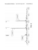 |
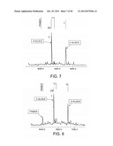 | 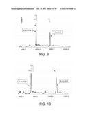 |
 |  |
 |  |
 |  |
 |  |
 |  |
 | 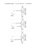 |
 |  |
 |  |
 |  |
 |  |
 |  |
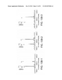 |  |
 |  |
 |  |
 |  |
 |  |
 |  |
 |  |
 |  |
 |  |
 |  |
 |  |
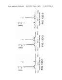 |  |
 |  |
 |  |
 |  |
 |  |
 | 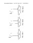 |
 |  |
 |  |
 |  |
 |  |
 |  |
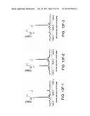 |  |
 |  |
 |  |
 |  |
 |  |
 | 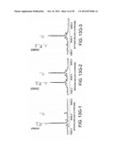 |
 |  |
 |  |
 |  |
 |  |
 |  |
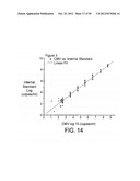 |  |
 |  |
 |  |
 |  |
 |  |
 |  |
 |  |
 |  |
 |  |
 |  |
 |  |
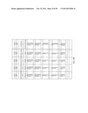 |  |
 |  |
 |  |
 |  |
 |  |
 | 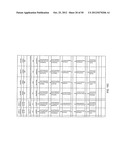 |
 |  |
 |  |
 |  |
 |  |
 |  |
 |  |
 |  |
 |  |
 |  |
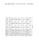 | 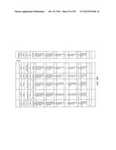 |
 | 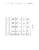 |
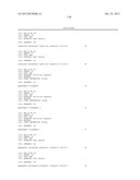 | 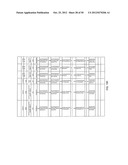 |
 | 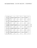 |
 |  |
 |  |
 | 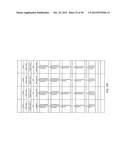 |
 | 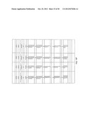 |
 | 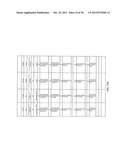 |
 | 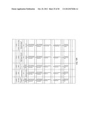 |
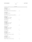 | 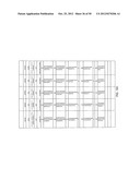 |
 |  |
 |  |
 |  |
 | 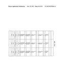 |
 |  |
 | 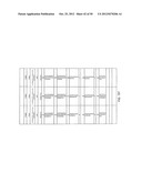 |
 |  |
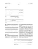 | 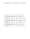 |
 |  |
 |  |
 |  |
 | 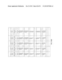 |
 | 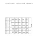 |
 | 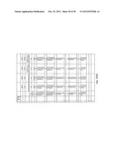 |
 |  |
 |  |
 |  |
 | 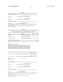 |
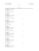 | 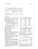 |
 |  |
 | 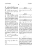 |
 |  |
 |  |
 |  |
| Similar patent applications: | |
| Date | Title |
|---|---|
| 2011-05-12 | Compositions and methods for nucleic acid delivery |
| 2011-05-12 | Stem cell aggregates and methods for making and using |
| 2010-07-08 | Method of analyzing nucleic acid |
| 2011-05-19 | Compositions, kits and related methods for the detection and/or monitoring of salmonella |
| 2011-04-28 | Method, agent, and kit for isolating nucleic acids |
| New patent applications in this class: | |
| Date | Title |
|---|---|
| 2022-05-05 | Method for diagnosing human t-cell leukemia virus type 1 (htlv-1) associated diseases |
| 2022-05-05 | Systems and methods for assay processing |
| 2022-05-05 | Rapid pathology/cytology without wash |
| 2019-05-16 | Biofluidic triggering system and method |
| 2019-05-16 | Method and system for detection of disease agents in blood |
| New patent applications from these inventors: | |
| Date | Title |
|---|---|
| 2014-11-27 | Compositions and their uses for gene therapy of bone conditions |
| 2013-03-14 | Compositions and their uses for gene therapy of bone conditions |
| 2012-02-16 | Novel therapy for lysosomal enzyme deficiencies |
| 2009-08-20 | Compositions and their uses for gene therapy of bone conditions |
| Top Inventors for class "Chemistry: molecular biology and microbiology" | |
| Rank | Inventor's name |
|---|---|
| 1 | Marshall Medoff |
| 2 | Anthony P. Burgard |
| 3 | Mark J. Burk |
| 4 | Robin E. Osterhout |
| 5 | Rangarajan Sampath |