Patent application title: ANTIBODIES THAT BIND TGF-ALPHA AND EPIREGULIN
Inventors:
Catherine Brautigam Beidler (Poway, CA, US)
Josef George Heuer (Carmel, IN, US)
Ramona Judita Petrovan (San Diego, CA, US)
Assignees:
ELI LILLY AND COMPANY
IPC8 Class: AA61K39395FI
USPC Class:
4241361
Class name: Immunoglobulin, antiserum, antibody, or antibody fragment, except conjugate or complex of the same with nonimmunoglobulin material structurally-modified antibody, immunoglobulin, or fragment thereof (e.g., chimeric, humanized, cdr-grafted, mutated, etc.) bispecific or bifunctional, or multispecific or multifunctional, antibody or fragment thereof
Publication date: 2012-10-11
Patent application number: 20120258109
Abstract:
The present invention provides antibodies that bind human TGF-alpha and
human Epiregulin and are characterized as having high affinity,
selective, and strong neutralizing properties. The antibodies are useful
in the treatment of diabetic nephropathy.Claims:
1. An antibody that binds TGF-alpha and Epiregulin, comprising a light
chain and a heavy chain, wherein the light chain comprises a light chain
variable region (LCVR) and the heavy chain comprises a heavy chain
variable region (HCVR), wherein the LCVR comprises amino acid sequences
LCDR1, LCDR2, and LCDR3, and the HCVR comprises amino acid sequences
HCDR1, HCDR2, and HCDR3, wherein LCDR1 is SEQ ID NO:4, LCDR2 is SEQ ID
NO:5, LCDR3 is SEQ ID NO:6, HCDR1 is SEQ ID NO:1, HCDR2 is SEQ ID NO:2,
and HCDR3 is SEQ ID NO:3.
2. The antibody of claim 1, wherein the amino acid sequence of the LCVR is SEQ ID NO:9 or SEQ ID NO:10.
3. The antibody of claim 1, wherein the amino acid sequence of the HCVR is SEQ ID NO:7.
4. The antibody of claim 1, wherein the amino acid sequence of the LCVR is SEQ ID NO:9 and the amino acid sequence of the HCVR is SEQ ID NO:7.
5. The antibody of claim 2, wherein the amino acid sequence of the light chain is SEQ ID NO:13 or SEQ ID NO:14.
6. The antibody of claim 3, wherein the amino acid sequence of the heavy chain is SEQ ID NO:12.
7. An antibody that binds TGF-alpha and Epiregulin, comprising two light chains wherein the amino acid sequence of each light chain is SEQ ID NO:13, and two heavy chains wherein the amino acid sequence of each heavy chain is SEQ ID NO:12.
8. An antibody that binds TGF-alpha and Epiregulin, comprising two light chains wherein the amino acid sequence of each light chain is SEQ ID NO:14, and two heavy chains wherein the amino acid sequence of each heavy chain is SEQ ID NO:12.
9. A pharmaceutical composition comprising the antibody of claim 1, and at least one pharmaceutically acceptable carrier, diluent, or excipient.
10. A method of treating diabetic nephropathy in a patient, comprising administering to the patient the antibody of claim 1.
11. A pharmaceutical composition comprising the antibody of claim 7, and at least one pharmaceutically acceptable carrier, diluent, or excipient.
12. A method of treating diabetic nephropathy in a patient, comprising administering to the patient the antibody of claim 7.
13. A pharmaceutical composition comprising the antibody of claim 8, and at least one pharmaceutically acceptable carrier, diluent, or excipient.
14. A method of treating diabetic nephropathy in a patient, comprising administering to the patient the antibody of claim 8.
Description:
[0001] The present invention relates to antibodies that bind human
TGF-alpha and Epiregulin and uses thereof.
[0002] TGF-alpha and Epiregulin are two of seven ligands of the Epidermal Growth Factor Receptor ("EGFR") that normally function in wound healing following injury. Diabetic nephropathy ("DN") is a major diabetic complication and is the leading cause of end stage renal disease ("ESRD"). Proteinuria is a clinical marker of renal functional decline accompanying DN and is associated with disease progression and increased cardiovascular risk, such as heart failure, vascular disease, dysrhythmia. The standard of care for DN includes ACE inhibitors and angiotensin receptor blockers ("ARBs") that only slow disease progression and leave considerable residual risk.
[0003] Blocking the EGFR attenuates not only proteinuria, but also renal pathology in preclinical animal models of renal disease. However, EGFR inhibitors, such as ERBITUX®, while approved for cancer, are associated with side effects such as a severe skin rash on the face and shoulders associated with target inhibition in the skin. Thus, there is still a need for alternative therapies for DN. In addition, there is a need for a more effective treatment therapy for DN.
[0004] Antibodies that bind TGF-alpha have been described (for example, see U.S. Pat. No. 5,190,858). In addition, antibodies that bind Epiregulin have been described (for example, see US 2009/0324491).
[0005] The present invention provides antibodies against TGF-alpha and Epiregulin for the treatment of DN. Furthermore, the present invention provides antibodies against TGF-alpha and Epiregulin that engage the target in vivo and subsequently cause a reduction in proteinuria with a concomitant reduction in disease progression and cardiovascular risk.
[0006] The present invention provides therapeutically useful antibodies that bind both TGF-alpha and Epiregulin that possess a number of desirable properties. The antibodies of the present invention have high affinity and are selective with full neutralizing activity against human TGF-alpha and human Epiregulin. When administered, the antibodies of the present invention also result in a decrease in albuminuria and in renal pathology for tubular protein, interstitial fibrosis, mesangial matrix expansion, and pelvic dilation in vivo. Furthermore, the preferred antibodies of the present invention cause no observed skin toxicity associated with complete EGFR inhibition.
[0007] The present invention provides an antibody that binds TGF-alpha and Epiregulin, comprising a light chain and a heavy chain, wherein the light chain comprises a light chain variable region (LCVR) and the heavy chain comprises a heavy chain variable region (HCVR), wherein the LCVR comprises amino acid sequences LCDR1, LCDR2, and LCDR3, and the HCVR comprises amino acid sequences HCDR1, HCDR2, and HCDR3, wherein LCDR1 is SEQ ID NO:4, LCDR2 is SEQ ID NO:5, LCDR3 is SEQ ID NO:6, HCDR1 is SEQ ID NO:1, HCDR2 is SEQ ID NO:2, and HCDR3 is SEQ ID NO:3.
[0008] The present invention also provides a pharmaceutical composition comprising an antibody of the present invention, as described herein, and at least one pharmaceutically acceptable carrier, diluent, or excipient.
[0009] The present invention provides an antibody of the present invention, as described herein, for use in the treatment of diabetic nephropathy.
[0010] Throughout this disclosure, an antibody of the present invention, as described herein, binds TGF-alpha and Epiregulin, and comprises a light chain and a heavy chain, wherein the light chain comprises a light chain variable region (LCVR) and the heavy chain comprises a heavy chain variable region (HCVR), wherein the LCVR comprises amino acid sequences LCDR1, LCDR2, and LCDR3, and the HCVR comprises amino acid sequences HCDR1, HCDR2, and HCDR3, wherein LCDR1 is SEQ ID NO:4, LCDR2 is SEQ ID NO:5, LCDR3 is SEQ ID NO:6, HCDR1 is SEQ ID NO:1, HCDR2 is SEQ ID NO:2, and HCDR3 is SEQ ID NO:3.
[0011] The present invention provides an antibody, as described herein, wherein the antibody is selective to human TGF-alpha and human Epiregulin. Further, the present invention provides an antibody, as described herein, wherein the antibody has full neutralizing activity to human TGF-alpha and human Epiregulin. Further preferred, the present invention provides an antibody, as described herein, wherein the antibody is selective and has full neutralizing activity to human TGF-alpha and human Epiregulin.
[0012] The present invention provides an antibody, as described herein, wherein the antibody has a dissociation equilibrium constant, Kd, between 0.01×10⁻⁹ M and 1.0×10⁻⁹ M for human TGF-alpha (SEQ ID NO: 18). Further preferred, an antibody of the present invention, as described herein, has a dissociation equilibrium constant, Kd, between 0.05×10⁻⁹ M and 0.8×10⁻⁹ M for human TGF-alpha. The Kd values are established by a binding equilibrium at 25° C. as described in Example 2.
[0013] The present invention also provides an antibody, as described herein, wherein the antibody has a dissociation equilibrium constant, Kd, between 0.1×10⁻⁹ M and 30×10-9 M for met-human Epiregulin (SEQ ID NO: 22). Further preferred, an antibody of the present invention, as described herein, has a dissociation equilibrium constant, Kd, between 0.5×10⁻⁹ M and 10×10-9 M for human Epiregulin. The Kd values are established by a binding equilibrium at 25° C. as described in Example 2.
[0014] The present invention provides an antibody, as described herein, wherein the antibody has a dissociation equilibrium constant, Kd, between 0.01×10⁻⁹ M and 1.0×10⁻⁹ M for human TGF-alpha (SEQ ID NO: 18) and a Kd between 0.1×10⁻⁹ M and 30×10-9 M for met-human Epiregulin (SEQ ID NO: 22). Further preferred, an antibody of the present invention, as described herein, has a dissociation equilibrium constant, Kd, between 0.05×10⁻⁹ M and 0.8×10⁻⁹ M for human TGF-alpha and a Kd between 0.5×10⁻⁹ M and 10×10-9 M for human Epiregulin. The Kd values are established by a binding equilibrium at 25° C. as described in Example 2.
[0015] The present invention provides antibodies which bind human TGF-alpha and Epiregulin, and cause dose-dependent decrease in albuminuria, reduction in serum creatinine and blood urea nitrogen ("BUN") in vivo in a mouse remnant kidney model and a mouse uninephrectomy db/db model as described in Example 5 and Example 6, respectively.
[0016] The present invention provides antibodies which bind human TGF-alpha and Epiregulin, and cause reduction in renal pathology for tubular protein and interstitial fibrosis and a decrease in mesangial matrix expansion and pelvic dilation in vivo in a mouse remnant kidney model and a mouse uninephrectomy db/db model as described in Example 5 and Example 6, respectively.
[0017] The present invention provides antibodies which bind human TGF-alpha and Epiregulin, and are believed to cause a reduction in proteinuria with a concomitant reduction in disease progression and cardiovascular risk in humans. Further, the present invention provides antibodies which bind human TGF-alpha and Epiregulin, and are believed to be effective in the treatment of diabetic nephropathy in humans.
[0018] The present invention provides antibodies which bind human TGF-alpha and Epiregulin, and cause no observed skin toxicity in a toxicity study in cynomolgus monkeys as described in Example 7.
[0019] The present invention provides an antibody that binds TGF-alpha and Epiregulin, comprising a light chain and a heavy chain, wherein the light chain comprises a light chain variable region (LCVR) and the heavy chain comprises a heavy chain variable region (HCVR), wherein the LCVR comprises amino acid sequences LCDR1, LCDR2, and LCDR3, and the HCVR comprises amino acid sequences HCDR1, HCDR2, and HCDR3, wherein LCDR1 is SEQ ID NO:4, LCDR2 is SEQ ID NO:5, LCDR3 is SEQ ID NO:6, HCDR1 is SEQ ID NO:1, HCDR2 is SEQ ID NO:2, and HCDR3 is SEQ ID NO:3.
[0020] Furthermore, the present invention provides an antibody that binds TGF-alpha and Epiregulin, comprising a light chain and a heavy chain, wherein the light chain comprises a light chain variable region (LCVR) and the heavy chain comprises a heavy chain variable region (HCVR), wherein the amino acid sequence of the LCVR is SEQ ID NO: 9 or SEQ ID NO: 10.
[0021] The present invention also provides an antibody that binds TGF-alpha and Epiregulin, comprising a light chain and a heavy chain, wherein the light chain comprises a light chain variable region (LCVR) and the heavy chain comprises a heavy chain variable region (HCVR), wherein the amino acid sequence of the HCVR is SEQ ID NO: 7.
[0022] The present invention also provides an antibody that binds TGF-alpha and Epiregulin, comprising a light chain and a heavy chain, wherein the light chain comprises a light chain variable region (LCVR) and the heavy chain comprises a heavy chain variable region (HCVR), wherein an amino acid sequence of the LCVR and an amino acid sequence of the HCVR is selected from the group consisting of: [0023] (i) the LCVR is SEQ ID NO: 9 and the HCVR is SEQ ID NO: 7; and [0024] (ii) the LCVR is SEQ ID NO: 10 and the HCVR is SEQ ID NO: 7.
[0025] The present invention provides an antibody that binds TGF-alpha and Epiregulin, comprising a light chain and a heavy chain, wherein the light chain comprises a light chain variable region (LCVR) and the heavy chain comprises a heavy chain variable region (HCVR), wherein the amino acid sequence of the LCVR is SEQ ID NO: 9 and the amino acid sequence of the HCVR is SEQ ID NO: 7.
[0026] The present invention provides an antibody that binds TGF-alpha and Epiregulin, comprising a light chain and a heavy chain, wherein the light chain comprises a light chain variable region (LCVR) and the heavy chain comprises a heavy chain variable region (HCVR), wherein the amino acid sequence of the LCVR is SEQ ID NO: 10 and the amino acid sequence of the HCVR is SEQ ID NO: 7.
[0027] Furthermore, the present invention provides an antibody that binds TGF-alpha and Epiregulin, comprising a light chain and a heavy chain, wherein the amino acid sequence of the light chain is SEQ ID NO: 13 or SEQ ID NO: 14.
[0028] The present invention also provides an antibody that binds TGF-alpha and Epiregulin, comprising a light chain and a heavy chain, wherein the amino acid sequence of the heavy chain is SEQ ID NO: 12.
[0029] Furthermore, the present invention provides an antibody that binds TGF-alpha and Epiregulin, comprising a light chain and a heavy chain, wherein an amino acid sequence of the heavy chain and an amino acid sequence of the light chain is selected from the group consisting of: [0030] the heavy chain is SEQ ID NO: 12 and the light chain is SEQ ID NO: 13, and [0031] (ii) the heavy chain is SEQ ID NO: 12 and the light chain is SEQ ID NO: 14.
[0032] The present invention provides an antibody that binds TGF-alpha and Epiregulin, comprising two light chains wherein the amino acid sequence of each light chain is SEQ ID NO: 13, and two heavy chains wherein the amino acid sequence of each heavy chain is SEQ ID NO: 12.
[0033] The present invention provides an antibody that binds TGF-alpha and Epiregulin, comprising two light chains wherein the amino acid sequence of each light chain is SEQ ID NO: 14, and two heavy chains wherein the amino acid sequence of each heavy chain is SEQ ID NO: 12.
[0034] Furthermore, the present invention provides an antigen-binding fragment of an antibody, as described herein.
[0035] The present invention also provides a pharmaceutical composition comprising the antibody of the present invention, as described herein, and at least one pharmaceutically acceptable carrier, diluent, or excipient.
[0036] Furthermore, the present invention provides a pharmaceutical composition comprising the antibody of the present invention, as described herein, together with at least one pharmaceutically acceptable carrier, diluent, or excipient, and optionally other therapeutic ingredients.
[0037] The present invention also provides a method of treating diabetic nephropathy in a patient comprising administering to the patient the antibody of the present invention, as described herein.
[0038] Furthermore, the present invention provides an antibody of the present invention, as described herein, for use in therapy. Preferably, the present invention provides an antibody of the present invention, as described herein, for use in the treatment of diabetic nephropathy.
[0039] Furthermore, the present invention provides the use of an antibody of the present invention, as described herein, in the manufacture of a medicament for the treatment of diabetic nephropathy.
[0040] The present invention also provides a method of treating diabetic nephropathy in a patient comprising administering to the patient the antibody of the present invention, as described herein, in simultaneous or sequential combination with a standard of care.
[0041] Furthermore, the present invention provides an antibody of the present invention, as described herein, for use in therapy, wherein the antibody is to be administered in simultaneous or sequential combination with a standard of care. Preferably, the present invention provides an antibody of the present invention, as described herein, for use in the treatment of diabetic nephropathy, wherein the antibody is to be administered in simultaneous or sequential combination with a standard of care.
[0042] Furthermore, the present invention provides the use of an antibody of the present invention, as described herein, in the manufacture of a medicament for the treatment of diabetic nephropathy, wherein the antibody is to be administered in simultaneous or sequential combination with a standard of care.
[0043] The present invention also provides a pharmaceutical composition comprising the antigen-binding fragment of an antibody of the present invention, as described herein, and at least one pharmaceutically acceptable carrier, diluent, or excipient.
[0044] Furthermore, the present invention provides a pharmaceutical composition comprising the antigen-binding fragment of an antibody of the present invention, as described herein, together with at least one pharmaceutically acceptable carrier, diluent, or excipient, and optionally other therapeutic ingredients.
[0045] The present invention also provides a method of treating diabetic nephropathy in a patient comprising administering to the patient the antigen-binding fragment of an antibody of the present invention, as described herein.
[0046] Furthermore, the present invention provides an antigen-binding fragment of an antibody of the present invention, as described herein, for use in therapy. Preferably, the present invention provides an antigen-binding fragment of an antibody of the present invention, as described herein, for use in the treatment of diabetic nephropathy.
[0047] Furthermore, the present invention provides the use of an antigen-binding fragment of an antibody of the present invention, as described herein, in the manufacture of a medicament for the treatment of diabetic nephropathy.
[0048] The present invention also provides a method of treating diabetic nephropathy in a patient comprising administering to the patient the antigen-binding fragment of an antibody of the present invention, as described herein, in simultaneous or sequential combination with a standard of care.
[0049] Furthermore, the present invention provides an antigen-binding fragment of an antibody of the present invention, as described herein, for use in therapy, wherein the antigen-binding fragment is to be administered in simultaneous or sequential combination with a standard of care. Preferably, the present invention provides an antigen-binding fragment of an antibody of the present invention, as described herein, for use in the treatment of diabetic nephropathy, wherein the antigen-binding fragment is to be administered in simultaneous or sequential combination with a standard of care.
[0050] Furthermore, the present invention provides the use of an antigen-binding fragment of an antibody of the present invention, as described herein, in the manufacture of a medicament for the treatment of diabetic nephropathy, wherein the antigen-binding fragment is to be administered in simultaneous or sequential combination with a standard of care.
[0051] The standard of care for DN includes, but is not limited to, ACE inhibitors and angiotensin receptor blockers (ARBs).
[0052] The general structure of an "antibody" is very well-known in the art. For an antibody of the IgG type, there are four amino acid chains (two "heavy" chains and two "light" chains) that are cross-linked via intra- and inter-chain disulfide bonds. When expressed in certain biological systems, antibodies having unmodified human Fc sequences are glycosylated in the Fc region. Antibodies may be glycosylated at other positions as well. The subunit structures and three-dimensional configurations of antibodies are well known in the art. Each heavy chain is comprised of an N-terminal heavy chain variable region ("HCVR") and a heavy chain constant region ("HCCR"). The heavy chain constant region is comprised of three domains (CH1, CH2, and CH3) for IgG, IgD, and IgA; and 4 domains (CH1, CH2, CH3, and CH4) for IgM and IgE. Each light chain is comprised of a light chain variable region ("LCVR") and a light chain constant region ("LCCR").
[0053] The variable regions of each light/heavy chain pair form the antibody binding site. The HCVR and LCVR regions can be further subdivided into regions of hypervariability, termed complementarity determining regions ("CDRs"), interspersed with regions that are more conserved, termed framework regions ("FR"). Each HCVR and LCVR are composed of three CDRs and four FRs, arranged from amino-terminus to carboxy-terminus in the following order: FR1, CDR1, FR2, CDR2, FR3, CDR3, FR4. Herein, the 3 CDRs of the heavy chain are referred to as "CDRH1, CDRH2, and CDRH3" and the 3 CDRs of the light chain are referred to as "CDRL1, CDRL2 and CDRL3." The CDRs contain most of the residues which form specific interactions with the antigen. The assignment of amino acids to each domain is in accordance with well-known conventions [e.g., Kabat, "Sequences of Proteins of Immunological Interest," National Institutes of Health, Bethesda, Md. (1991)].
[0054] An antibody of the present invention may have a heavy chain constant region selected from any of the immunoglobulin classes (IgA, IgD, IgG, IgM, and IgE). Furthermore, an antibody of the present invention contains an Fc portion which is derived from human IgG4 Fc region because of its reduced ability to bind complement factors as compared to other IgG sub-types.
[0055] An antibody may be derived from a single copy or clone, including e.g., any eukaryotic, prokaryotic, or phage clone. Preferably, an antibody of the present invention exists in a homogeneous or substantially homogeneous population of antibody molecules. An full-length antibody comprises full length or substantially full length constant regions, including the Fc region. An "antigen-binding fragment" of such an antibody is any shortened form of a full length antibody that comprises the antigen-binding portion and retains antigen-binding capability. Such shortened forms include, e.g., a Fab fragment, Fab' fragment or F(ab') 2 fragment that includes the CDRs or the variable regions of the antibodies disclosed. Furthermore, such shortened antibody forms can be a single chain Fv fragment that may be produced by joining the DNA encoding the LCVR and HCVR with a linker sequence. (See, Pluckthun, The Pharmacology of Monoclonal Antibodies, vol. 113, Rosenburg and Moore eds., Springer-Verlag, New York, pp 269-315, 1994). The term "antibody" does not include such fragments unless otherwise indicated. An antibody of the present invention can be produced using techniques well known in the art, e.g., recombinant technologies, phage display technologies, synthetic technologies or combinations of such technologies or other technologies readily known in the art.
[0056] An antibody of the present invention is an engineered antibody that has been designed to have frameworks, hinge regions, and constant regions of human origin that are identical with or substantially identical (substantially human) with frameworks and constant regions derived from human genomic sequences. Fully human frameworks, hinge regions, and constant regions are those human germline sequences as well as sequences with naturally-occurring somatic mutations and those with engineered mutations. An antibody of the present invention may comprise framework, hinge, or constant regions derived from a fully human framework, hinge, or constant region containing one or more amino acid substitutions, deletions, or additions therein. Further, an antibody of the present invention is substantially non-immunogenic in humans.
[0057] A variety of different human framework sequences may be used singly or in combination as a basis for an antibody of the present invention. Preferably, the framework regions of an antibody of the present invention are of human origin or substantially human (at least 95%, 97% or 99% of human origin.) The sequences of framework regions of human origin may be obtained from The Immunoglobulin Factsbook, by Marie-Paule Lafranc, Gerard Lefranc, Academic Press 2001, ISBN 012441351.
[0058] The framework sequence for an antibody of the present invention serves as the "donor" variable framework region and can be used to create additional antibodies with the same CDRs specified herein using methodology known in the art. Furthermore, the framework sequence for an antibody of the present invention can be compared to other known human framework sequences to generate additional antibodies. Thus, this information can be used to "back-mutate" another selected homologous human framework region to the donor amino acid residue at these positions. Further, any "rare" amino acids can be detected in additional human frameworks such that the consensus or donor amino acid residue can be used at the relevant position.
[0059] "TGF-alpha" or "human TGF-alpha" refers to human TGF-alpha protein (SEQ ID NO: 18).
[0060] "Epiregulin" or "human Epiregulin" refers to human Epiregulin protein (SEQ ID NO: 33). Met-human Epiregulin (SEQ ID NO: 22) is used in in vitro experiments herein. References to the ability of the antibodies of the present invention, as described herein, to bind or to neutralize human Epiregulin pertain also to their ability to bind and to neutralize human met-Epiregulin in in vitro experiments.
[0061] A "patient" is a mammal, preferably a human.
[0062] The term "treating" (or "treat" or "treatment") means slowing, stopping, reducing, or reversing the progression or severity of a symptom, disorder, condition, or disease.
[0063] The term "therapeutically effective amount" refers to the amount or dose of an antibody of this invention which, upon single or multiple dose administration to a patient, provides the desired treatment.
[0064] The following examples may be performed essentially as described below.
EXAMPLES
Example 1
Production of Antibodies
[0065] Antibodies I and II can be made and purified as follows. An appropriate host cell, such as HEK 293 or CHO, is either transiently or stably transfected with an expression system for secreting antibodies using an optimal predetermined HC:LC vector ratio or a single vector system encoding both HC, such as SEQ ID NO: 15, and LC, such as SEQ ID NO: 16 or SEQ ID NO: 17. Clarified media, into which the antibody has been secreted, is purified using any of many commonly-used techniques. For example, the medium may be conveniently applied to a Protein A or G column that has been equilibrated with a compatible buffer, such as phosphate buffered saline (pH 7.4). The column is washed to remove nonspecific binding components. The bound antibody is eluted, for example, by pH gradient (such as 0.1 M sodium phosphate buffer pH 6.8 to 0.1 M sodium citrate buffer pH 2.5). Antibody fractions are detected, such as by SDS-PAGE, and then are pooled. Further purification is optional, depending on the intended use. The antibody may be concentrated and/or sterile filtered using common techniques. Soluble aggregate and multimers may be effectively removed by common techniques, including size exclusion, hydrophobic interaction, ion exchange, or hydroxyapatite chromatography. The purity of the antibody after these chromatography steps is greater than 99%. The product may be immediately frozen at -70° C. or may be lyophilized. The amino acid sequences for these antibodies are provided below.
TABLE-US-00001 SEQ ID NOs Heavy Light Antibody Chain Chain HCVR LCVR I 12 13 7 9 II 12 14 7 10 III 31 32 8 11 Antibody HCDR1 HCDR2 HCDR3 LCDR1 LCDR2 LCDR3 I 1 2 3 4 5 6 II 1 2 3 4 5 6 III 1 2 3 4 5 6
Example 2
Affinity Binding Measurement by Surface Plasmon Reasonance (BIAcore) for Antibody I
[0066] Biacore T2000 instrument (BIAcore® AB, Upsala, Sweden), reagents and Biacore T2000 Evaluation Software Ver 4.1 are used for the Surface Plasmon Resonance analysis. A CM5 chip is prepared using manufacturer's EDC/NHS amine coupling method. The surfaces of all four flow cells are activated by injecting a 1:1 mixture of EDC/NHS for 7 minutes at 10 μL/min. Goat anti-human Fc γ specific antibody is diluted to 50 μg/ml in 10 mM acetate, pH 4.0 buffer and immobilized for approximately 10000 RU onto all four flow cells by 7 minute injection at a flow rate of 10 μL/min. Un-reacted sites are blocked with a 7 minute injection of ethanolamine at 10 μL/min. Injections of 3×20 seconds of glycine pH 1.5 at 30 μL/min are used to remove non-covalently associated protein. The running buffer is HBS-EP [10 mM HEPES, 150 mM Sodium Chloride, 3 mM EDTA, 0.005% Polysorbate 20].
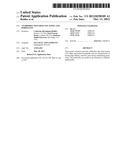
[0067] In study 1, Antibody I is diluted to 50 μg/mL in running buffer, and approximately 400-600 RU is captured in flowcell 2. Human TGF-alpha (SEQ ID NO: 18), rat TGF-alpha (SEQ ID NO: 20), met-human Epiregulin (SEQ ID NO: 22), and cynomolgus Epiregulin (SEQ ID NO: 24) are diluted from 100 μg/mL to 200 nM in running buffer and then two-fold serially diluted in running buffer to 6.25 nM. Mouse Epiregulin (SEQ ID NO: 23) is diluted from 100 μg/mL to 4 μM in running buffer and then two-fold serially diluted in running buffer to 125 nM. Duplicate injections of each ligand concentration are injected at 30 μL/min for 300 seconds followed by a dissociation phase. The dissociation phase is 1800 seconds for human and rat TGF-alpha, 1200 seconds for human and cynomolgus Epiregulin, and 120 seconds for mouse Epiregulin. Regeneration is performed by injecting 10 mM glycine pH 1.5 for 3×20 seconds at 30 μL/min over all flowcell.
[0068] In study 2, Antibody III is diluted to 100 mg/mL in running buffer, and approximately 400-600 RU is captured in flowcell 2. Mouse TGF-alpha (SEQ ID NO: 19), is diluted from 100 μg/mL to 200 nM in running buffer and then two-fold serially diluted in running buffer to 6.25 nM. Mouse Epiregulin (SEQ ID NO: 23) is diluted from 100 μg/mL to 4 μM in running buffer and then two-fold serially diluted in running buffer to 125 nM. Duplicate injections of each ligand concentration are injected at 30 μL/min for 300 seconds followed by a dissociation phase. The dissociation phase is 1800 seconds for mouse TGF-alpha, and 120 seconds for mouse Epiregulin. Regeneration is performed by injecting 10 mM glycine pH 1.5 for 30 seconds at 30 μL/min over all flowcell.
[0069] Reference-subtracted data are collected as Fc2-Fc1. The measurements are obtained at 25° C. The on-rate (kon) and off-rate (koff) for each ligand are evaluated using a "1:1 (Langmuir) Binding" binding model. The affinity (KD) is calculated from the binding kinetics according to the relationship: KD=koff/kon.
TABLE-US-00002 TABLE 1 Binding Parameters for Antibody I On Rate (kon) Off Rate (koff) Affinity (M-1s-1) (s-1) (KDa) Ligand Species (±SD) (±SD) (±SD) TGF-alpha Human 4.18 ± 0.28 × 10⁵ 4.09 ± 0.96 × 10⁻⁵ 97.6 ± 20.6 pM Rat 3.78 ± 0.39 × 10⁵ 2.66 ± 0.74 × 10⁻⁵ 70.5 ± 19.4 pM Epiregulin Human 4.91 ± 0.42 × 10⁵ 6.31 ± 0.55 × 10⁻⁴ 1.29 ± 0.03 nM Cynomolgus 6.73 ± 0.71 × 10⁵ 7.05 ± 0.23 × 10⁻⁴ 1.05 ± 0.09 nM Mouse 4.10 ± 1.15 × 10⁴ 1.33 ± 0.16 × 10⁻² 342 ± 136 nM aCalculated as KD = koff/kon
TABLE-US-00003 TABLE 2 Binding Parameters for Antibody III On Rate (kon) Off Rate (koff) Affinity (M-1s-1) (s-1) (KDa) Ligand (±SD) (±SD) (±SD) Mouse 5.41 ± 0.50 × 10⁵ 2.02 ± 0.54 × 10⁻⁵ 38.0 ± 13.6 pM TGF-alpha Mouse 6.55 ± 0.38 × 10⁴ 1.41 ± 0.09 × 10⁻² 215 ± 15 nM Epiregulin aCalculated as KD = koff/kon
[0070] Antibody I binds to human TGF-alpha and human Epiregulin with affinities of about 98 pM and 1.3 nM, respectively. Antibody I also binds to rat TGF-alpha and mouse Epiregulin with affinities of about 70 pM and 340 nM, respectively. Additionally, Antibody I binds to cynomolgus Epiregulin with an affinity of about 1 nM. Antibody III binds to mouse TGF-alpha and mouse Epiregulin with affinities of about 38 pM and 220 nM, respectively. Thus, Antibody I and Antibody III of the present invention have high affinity to human TGF-alpha and human Epiregulin.
Example 3
Internalization of EGF Target Ligands in the Human Colon Carcinoma Cell Line HT-29
Conjugation of Alexa Fluor® 488 to Antibodies
[0071] Alexa Fluor® 488 is conjugated to Antibody I and Control IgG according to the manufacturer's protocol. Protein is diluted to 2 mg/mL in PBS. To 0.5 mL of this 2 mg/mL solution, 50 μL of 1M sodium bicarbonate pH 9 is added. The protein solution is then transferred to a vial of dye and stirred at room temperature for 1 hour. The labeled protein is purified using the Bio-Rad BioGel P-30 resin included with the labeling kit.
In Vitro Internalization Assay
[0072] In study 1, 10,000 HT-29 cells, a colon adenocarcinoma cell line known to express TGF-alpha and Epiregulin, are seeded per well of a 96 well plate and allowed to incubate overnight in complete media [Dulbecco's Modified Eagle's Medium/F12 (Ham) Medium (1:1) ("DMEM/F12") containing L-glutamine, 10% heat-inactivated fetal bovine serum ("FBS"), 1× antibiotic, and 2.438 g/L sodium bicarbonate]. The next day, the cells are washed with PBS containing 0.1% BSA and then incubated with an Alexa Fluor® 488 conjugated Antibody I or Control IgG in PBS with 0.1% BSA at concentrations ranging from 0 to 88 ug/mL for 2 hours at 37° C. in a tissue culture incubator. Following the incubation period, the cells are washed in PBS with 0.1% BSA several times and then fixed with 4% formaldehyde for analysis. The quantitation of internalization is done as follows: 500 cells/well are collected with a Cellomics Arrayscan VTI (Thermo Scientific). Image analysis is performed with "Compartment al analysis" Bioapplications of the system. Cell nuclei are identified with a Hoechst stain (blue). Two regions of interest (ROI) are set to collect fluorescent signals from intracellular spots (red) and total green fluorescence (both red and blue) obtained from the masked image. The number, area and fluorescent intensity from each spot and cell are calculated. The mean spot total intensity of intracellular spots (red) is chosen for measuring Antibody I induced internalization.
[0073] In study 2, 10,000 HT-29 cells are prepared as previously described, and Alexa Fluor® 488 conjugated Antibody I or Control IgG in PBS containing 0.1% BSA is added to the cells at 40 ug/mL. Cells are incubated at 37° C. in a tissue culture incubator for various times ranging from 0-120 minutes, then washed with PBS containing 0.1% BSA several times and fixed with 4% formaldehyde for analysis. The quantification of signal is performed essentially as previously described.
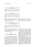
TABLE-US-00004 TABLE 3a Study 1 - Mean Ringspot Total Intensity of Fluorescence Dose (ug/ml) 88 44 22 11 5.5 Control IgG 2440 ± 199 1808 ± 207 1763 ± 68 1391 ± 76 1357 ± 63 Antibody I 24809 ± 4343 17451 ± 217 15135 ± 131 11516 ± 54 8474 ± 269 Mean ± SEM
TABLE-US-00005 TABLE 3b Study 1 - Mean Ringspot Total Intensity of Fluorescence Dose (ug/ml) 2.75 1.38 0.69 0.34 0 Control IgG 1570 ± 70 1473 ± 7 1483 ± 90 1407 ± 41 1630 ± 155 Antibody I 6503 ± 262 4349 ± 186 3440 ± 96 2432 ± 62 1460 ± 84 Mean ± SEM
[0074] The results from the imaging analysis of study 1 determined that the fluorescence signal was internalized into the cell and was dose dependent with Antibody I, but not with the Control IgG (Table 3a and Table 3b).
TABLE-US-00006 TABLE 4 Study 2 - Mean Ringspot Total Intensity of Fluorescence Time post addition (min) 120 60 30 15 5 0 Control IgG 177 ± 29 167 ± 23 124 ± 10 126 ± 18 116 ± 4 94 ± 11 Antibody I 4449 ± 866 4131 ± 1688 1494 ± 66 717 ± 72 261 ± 17 89 ± 1 Mean ± SEM
[0075] The results from study 2 demonstrated that Antibody I was internalized rapidly and the internalization was complete by 2 hours post addition to cells (Table 4). Antibody I induced internalization of target on HT-29 cells in vitro in a time dependent manner (Table 4).
Example 4
Measurement of Neutralization of EGFR Ligand Stimulated Cell Proliferation in a Myofibroblast Cell Line
[0076] A clonal mouse myofibroblast cell line ("MFc7") is used to test the ability of the antibodies of the present invention to block the proliferative activity of EGFR ligands. The seven ligands that can activate the EGFR are TGF-alpha (TGFA), Epiregulin (EREG), EGF, Heparin-Binding EGF (HB-EGF), Epigen (EPGN), Amphiregulin (AREG) and Betacellulin (BTC). The EGFR ligands share a structural motif, the EGF-like domain, characterized by three intramolecular disulfide bonds that are formed by six similarly spaced conserved cysteine residues. Proliferative activity is determined by Bromodeoxyuridine ("BrDU") incorporation and is measured with a colorimetric BrDU ELISA kit according to the manufacturer's instructions.
[0077] First, 2,000 MFc7 cells/well are plated in a tissue culture treated 96 well microplate in 0.1 mL of Dulbecco's Modified Eagle's Medium/F12 (Ham) Medium (1:1) ("DMEM/F12") containing L-glutamine, 10% heat-inactivated FBS, 1× antibiotic, and 2.438 g/L sodium bicarbonate. Cells are allowed to attach for 6 hours, and then the medium is removed and replaced with 0.1 mL of serum free DMEM/F12 containing 0.1% BSA for serum starvation overnight. The next day, serial dilutions of the EGFR ligands are made with serum free media containing 0.1% BSA in 96 well polypropylene plates in a volume of 0.12 mL/well from concentrations ranging from 0.001 to 3000 ng/mL. Following dilutions, medium is removed from serum starved cells and then stimulated with EGFR ligand for 24 hrs. Following stimulation, the cells are pulsed with BrDU for 4 hrs and then analyzed with a colorimetric BrDU ELISA kit according to the manufacturer's instructions.
[0078] In testing the specificity of Antibody I to EGFR ligands, serial dilutions of 2× or 3× of the antibody are made in 96 well polypropylene plates in a volume of 0.06 mL/well from concentrations ranging from 3000 nM to 0.059 nM. Following serial dilutions of the antibody, 0.06 mL of the EGFR ligand is added per well. The plate is then incubated at 37° C. in a humidified tissue culture incubator for 30 minutes. Following incubation, 0.1 mL of the solution is transferred per well to the cells. The cells are stimulated for 24 hours. Following stimulation, the cells are pulsed with BrDU for 4 hours and then analyzed with a colorimetric BrDU ELISA kit. Absorbance values (450 nM-690 nM) are generated on a SpectraMax 190 plate reader (Molecular Devices) and data are analyzed.
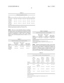
TABLE-US-00007 TABLE 5 MFc7 Assay EC50 Range IC50 (nM) IC50 (nM) EGFR Ligand (pM) Antibody I Antibody III Human TGF-alphaa 11-12 0.46 ± 0.03 0.52 ± 0.04 Human Epiregulin 78-282 3.15 ± 1.04 1.12 ± 0.36 Human Epigen 3797-18987 807 ± 577 ndb Human EGF 0.3-2.4 >2000 ndb Human HBEGF 30-39 >2000 ndb Human Betacellulin 1.8-3.2 >2000 ndb Human Amphiregulin 273-2727 >2000 ndb Rat TGF-alpha .sup. 13-13.8 0.19 ± 0.06 0.13 ± 0.01 Mouse Epiregulin 163-320 334 ± 41 214 ± 49 aHuman EGFR ligands were at a concentration of 0.5 nM when tested with Antibody I, except for Amphiregulin (60 nM) and Epigen (100 nM) Rat TGF-alpha and Mouse Epiregulin were used at 0.5 nM bnd, not determined
[0079] Mouse Epiregulin and rat TGF-alpha, as well as all of the human EGFR ligands except for Epigen and Amphiregulin were found to be potent stimulators of cell proliferation in the assay (Table 5). Antibody I and Antibody III have high affinity to human and rat TGF-alpha and human Epiregulin activity (Table 5).
[0080] Table 5 summarizes the calculated EC50 values for the EGFR ligands tested and the absolute IC50 values for the antibodies to those ligands. The calculated average IC50 for Antibody I was 0.46±0.03 nM to human TGF-alpha and 3.15±1.04 nM to human Epiregulin. The calculated IC50 average for Antibody III was 0.52±0.04 nM to human TGF-alpha and 1.12±0.36 nM to human Epiregulin. The calculated average IC50 value for Antibody III was 0.13±0.01 nM to rat TGF-alpha and 214±49 nM to mouse Epiregulin. Thus, Antibody I and Antibody III have high affinity and are selective with full neutralizing activity against human TGF-alpha and human Epiregulin.
Example 5
Renal Function and Pathology in a Mouse Remnant Kidney Model of Hypertensive Renal Disease
[0081] A mouse remnant kidney model involving surgical reduction of 75% of the total renal mass is used as a preclinical model of hypertensive renal disease. [Ma L J, Fogo A B. Kidney Int. 2003 July; 64(1):350-5] Surgical reduction of renal mass or sham surgery is done in male 129 Svev mice at 9-10 weeks of age. Randomization into groups of 12 mice is done at 2 weeks post surgery, by urine albumin/creatinine ratio ("ACR") and body weight. An isotype Control IgG (10 mg/kg) or Antibody III (1 and 10 mg/kg) are dosed subcutaneously following randomization and continued once weekly out to week 16 post surgery. The endpoints for the study are survival, systolic blood pressure, albuminuria, serum creatinine, serum BUN, urine TGF-alpha, urine MIP-2 and renal pathology.
[0082] At the end of the study, there were 3 deaths in the Control IgG group (25% mortality) with no deaths in the Antibody III treatment groups.
Measurement of Systolic Blood Pressure
[0083] Blood pressure is taken at 12 weeks post surgery by the tail cuff method. Selected mice from each group (N=3-4 per group) are acclimated to the restraint by placing them in the mouse holder with the tail cuff attached for 5 minutes daily, 3-5 days prior to the actual measurement. The equipment room temperature is increased to 24° C. to provide additional warmth during the blood pressure collection process. The mice are placed in a mouse restrainer and set on top of a warming pad unit (31-33° C.) to provide dilation to the tail vasculature. The tail is placed through the tail cuff and each mouse is restrained for an approximate time of 30 minutes, not to exceed 45 minutes. This time includes the initial warming and pressure measurements followed by immediate return to general housing. No anesthesia is used. The tail cuff is inflated, compressing the tail tightly enough to momentarily interrupt arterial blood flow, and then is gradually loosened by deflation to observe the return of the arterial pulse. On return of arterial pulse, the cuff is fully deflated.
Measurement of Albuminuria
[0084] Urine is collected every 4 weeks in Nalgene Metabolic cage units over a 24 hour time period. Each mouse (singly housed) receives food and water during the 24 hour collection process. At the end of the 24 hour period, the collected urine is placed on ice, centrifuged and subjected to albumin and creatinine analysis. Albuminuria is defined as the ratio of urine albumin to creatinine (ug/mg).
Serum Creatinine and BUN
[0085] At study termination, serum obtained by cardiac puncture is analyzed for BUN and creatinine.
TGF-Alpha and MIP-2 ELISA
[0086] Urine obtained by a 24 hour collection is concentrated 5-fold centrifugally using a 3K MW cutoff membrane spun at 14,000×g for 30 minutes. A sandwich-type enzyme-linked immunosorbent assay ("ELISA") for mouse TGF alpha is established. Rat TGF-alpha is used as the standard. Polystyrene 96-well plates are coated with 3 μg/mL of Antibody III overnight at 4° C. Plates are washed, blocked with blocking buffer, washed again, and then the concentrated urine samples are added. After 2 hours at room temperature, plates are washed, and then secondary biotinylated polyclonal anti-hTGF alpha is added. After 2 hours at room temperature, plates are washed and incubated with streptavidin-HRP for 30 minutes. Signal is generated with TMB substrate, and the reaction is stopped with 2 N H2SO4. A commercial Quantikine® sandwich ELISA kit for mouse macrophage inflammatory protein 2 (MIP-2, the equivalent of human IL-8) is used to detect urine MIP-2 according to the manufacturer's instructions. Absorbance data for both ELISA assays are obtained on a SpectraMax 190 plate reader (Molecular Devices) and data are analyzed.
Renal Pathology
[0087] Remnant kidneys are removed at study termination, fixed in formalin and processed for paraffin sectioning according to standard methodology. Sections of kidney are evaluated for renal lesions by a pathologist. Tubular protein, increased mesangial matrix and interstitial fibrosis, are semi-quantitatively scored using the following scale: none (0), minimal (1), slight (2), moderate (3), marked (4) and severe (5). Glomerular mesangial matrix expansion and basement membrane thickening are scored using hematoxylin and eosin ("H&E") and Periodic acid-Schiff ("PAS") stained sections. Masson's trichrome stained sections of kidney are evaluated to determine the degree of fibrosis (interstitial and glomerular).
Statistical Methods
[0088] All data are analyzed with JMP v.8.0 software (SAS Institute). Pathology scores are statistically evaluated by a contingency analysis and a Fishers exact test. All other data are evaluated by ANOVA with log transformed data and a Students unpaired t test. A P value of <0.05 is considered statistically significant.
TABLE-US-00008 TABLE 6 Albuminuria progression over time Weeks 2 4 8 12 16 Control IgG 1601 ± 269 3377 ± 860 5201 ± 907 .sup. 6144 ± 1654 4863 ± 2170 (10 mg/kg) Antibody III 1665 ± 305 3211 ± 343 3224 ± 518 3790 ± 857 5240 ± 2004 (1 mg/kg) Antibody III 1626 ± 273 2245 ± 334 2399 ± 261a 2749 ± 401a 3254 ± 654 (10 mg/kg) Arithmetic mean ± SEM for the urine albumin to creatinine ratio (ug/mg) aStatistically significant difference compared to the Control IgG (p < 0.05)
[0089] There was a dose dependent decrease in albuminuria relative to the Control IgG group with Antibody III (Table 6). Antibody III treatment at 10 mg/kg resulted in a significant reduction in albuminuria at weeks 8 and 12 post surgery relative to the Control IgG group, but not at weeks 2, 4, or 16 (Table 6).
TABLE-US-00009 TABLE 7 Systolic blood pressure, Serum Creatinine and BUN Week 12 Systolic Week 16 Serum Blood Pressure Creatinine Week 16 Serum Endpoint (mm Hg) (mg/dL) BUN (mg/dL) Sham nd 0.17 ± 0.01 31.5 ± 2.5 Control IgG 139.6 ± 4.0 0.31 ± 0.04a .sup. 64.0 ± 12.5a (10 mg/kg) Antibody III 147.5 ± 8.2 0.29 ± 0.01a 47.6 ± 1.7 (1 mg/kg) Antibody III 157.3 ± 4.5 0.23 ± 0.01b .sup. 44.8 ± 1.5b (10 mg/kg) Arithmetic mean ± SEM aStatistically significant relative to the sham group (p < 0.05) bStatistically significant difference compared to the Control IgG group (p < 0.05) nd, not determined
[0090] Antibody III demonstrated no effect on the systolic blood pressure, as all groups demonstrated hypertension at 12 weeks post surgery (Table 7). Furthermore, Antibody III treatment at 10 mg/kg resulted in improvements in renal function as shown by significant reductions in serum creatinine and BUN relative to the Control IgG group (Table 7).
TABLE-US-00010 TABLE 8 Urine TGF-alpha, Urine MIP-2 and renal pathology scores Week 8 Week 16 Week 16 Week 16 Urine TGF- Week 12 Pathology Pathology Pathology alpha to Urine MIP-2 Tubular Mesangial Interstitial Creatinine to Creatinine Protein Matrix Score Fibrosis Endpoint (pg/mg) (pg/mg) Score (1-5) (1-5) Score (1-5) Sham 115 ± 4 Not 0 ± 0 0 ± 0 0.25 ± 0.25 detectable Control IgG 102 ± 53 22.8 ± 7.4 2.55 ± 0.16 1.91 ± 0.28 2.09 ± 0.16 (10 mg/kg) Antibody III 74 ± 18 nd 2.17 ± 0.11 1.58 ± 0.15 1.83 ± 0.21 (1 mg/kg) Antibody III 19 ± 5a .sup. 5.6 ± 0.9a 2.08 ± 0.08a **1.42 ± 0.15 1.42 ± 0.15a (10 mg/kg) Arithmetic mean ± SEM aStatistically significant difference compared to the Control IgG group (p < 0.05) nd, not determined
[0091] There was a statistically significant reduction in urine TGF-alpha and urine MIP-2 at weeks 8 and 12 post surgery respectably with the 10 mg/kg Antibody III dose compared to the Control IgG group (Table 8). Furthermore, there were statistically significant reductions in renal pathology for tubular protein and interstitial fibrosis and a decrease in mesangial matrix expansion with the 10 mg/kg dose of Antibody III compared to the Control IgG (Table 8).
Example 6
Albuminuria and Renal Pathology in a Mouse Uninephrectomy db/db Model of Diabetic Renal Disease
[0092] The uninephrectomized db/db mouse model represents a model of diabetic nephropathy. [Ninichuk et al., Eur J Med Res. 2007 Aug. 16; 12(8):351-5] The uninephrectomized db/db model is used to determine the effects of Antibody III on renal disease parameters due to diabetes. The uninephrectomy ("UniNx") surgery on db/db mice on a C57BLKS/J background is performed at 4 weeks of age with removal of the right kidney. Randomization into groups of 12 mice is done at 8 weeks of age, by urine ACR, blood glucose and body weight. All the mice are hyperglycemic at the beginning of each study. An isotype Control IgG or Antibody III are dosed subcutaneously starting at 9 weeks of age and continued once weekly out to 25 weeks of age. Study 1 is conducted with doses of 0.3 and 10 mg/kg of Antibody III and a 10 mg/kg dose of isotype Control IgG. The endpoints for study 1 are survival, % HbA1c, albuminuria, urine TGF-alpha, kidney weight and renal pathology. Study 2 contains dose groups of 30, 10, 3 and 0.3 mg/kg of Antibody III with a 30 mg/kg dose of an isotype Control IgG. The endpoints for study 2 are survival and albuminuria.
[0093] There was only one death in the Control IgG group in study 1. There were no deaths in study 2.
Urine Collection and Measurement of Albuminuria
[0094] Urine is collected by a spot collection method to collect urine over a 2-4 hour time period. An individual mouse is placed on top of a 96 well polypropylene microplate and then covered by a Plexiglas chamber with holes for breathing but no access to food or water. At the end of the time period, the urine is removed from the plate with a micropipette and placed on ice, centrifuged and subjected to albumin and creatinine analysis. Albuminuria is defined as the ratio of urine albumin to creatinine (ug/mg).
Determination of % HbA1c
[0095] The % HbA1c is used as a measure of hyperglycemia at the end of the study. EDTA plasma is obtained at necropsy by cardiac puncture. Blood samples are spun at 2000 g for 20 minutes to remove blood cells and obtain plasma. Plasma samples are analyzed for Hemoglobin A1c and Total Hemoglobin. From these data, the % HbA1c as calculated.
Kidney Weight
[0096] Kidneys are removed at necropsy to determine their weight.
Determination of Urine TGF-Alpha by ELISA
[0097] Urine obtained by a spot collection is concentrated 5-fold with a 0.5 mL Amicon Ultra centrifugal filter containing an ultracel 3K MW cutoff membrane. The device is spun at 14,000×g for 30 minutes, and then the concentrated urine samples are collected. A sandwich-type ELISA for mouse TGF alpha is established. Rat TGF-alpha is used as the standard for the TGF-alpha ELISA. Polystyrene 96-well plates are coated with 3 μg/mL of Antibody III overnight at 4° C. Plates are washed, blocked with blocking buffer, washed again, and then the concentrated urine samples are added. After 2 hours at room temperature, plates are washed, and then secondary biotinylated polyclonal anti-hTGF-alpha is added. After 2 hours at room temperature, plates are washed and incubated with streptavidin-HRP for 30 minutes. Signal is generated with TMB substrate, and the reaction is stopped with 2 N H2SO4. Absorbance data are obtained on a SpectraMax 190 plate reader (Molecular Devices) and data are imported into Microsoft Excel 2007 and Sigmaplot v.9.01 for analysis.
Renal Pathology
[0098] Kidneys are removed at study termination, capsules removed and then fixed in formalin and processed for paraffin sectioning according to standard methodology. Sections of kidney are evaluated for renal lesions by a pathologist. Mesangial matrix, pelvic dilation and glomerular fibrosis, are semi-quantitatively scored using the following scale: none (0), minimal (1), slight (2), moderate (3), marked (4) and severe (5). Glomerular mesangial matrix expansion and basement membrane thickening are scored using H&E and PAS stained sections. Masson's trichrome stained sections of kidney are evaluated to determine the degree of fibrosis (glomerular).
Statistical Methods
[0099] All data are analyzed with JMP v.8.0 software (SAS Institute). Pathology scores are statistically evaluated by a contingency analysis and a Fishers exact test. Statistical analysis of albuminuria (ACR) is done by a Fit model with nontransformed data and the baseline ACR at week 8 as a covariate. ACR progression is analyzed by comparing the week 24 data with the week 16 data within each group by ANOVA and a Student's unpaired t test. The ACR change from week 16 to week 24 across groups is done by ANOVA and a student's unpaired t test. A P value of <0.05 is considered statistically significant. All other data are evaluated by ANOVA with log transformed data and a Students unpaired t test.
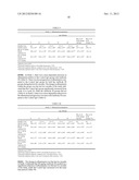
TABLE-US-00011 TABLE 9 Study 1 - Albuminuria progression Age (Weeks) Wk 16-24 Wk 16-24 ACR change ACR 8 12 16 20 24 (ug/mg) change (%) Healthy Lean nd 15 ± 2 19 ± 3 13 ± 3 12 ± 2 nd nd Db/db Control 273 ± 59a 903 ± 125a 1551 ± 180a 2384 ± 257a 3228 ± 488ac 1677 ± 419 108 ± 27 IgG @ 10 mg/kg Db/db 299 ± 63a 913 ± 174a 1573 ± 209a 1911 ± 222a 2248 ± 417ab 675 ± 332b 43 ± 21 Antibody III @ 0.3 mg/kg Db/db 291 ± 55a 1002 ± 107a 965 ± 141a 1433 ± 190ab 1426 ± 230ab 461 ± 219b 48 ± 23 Antibody III @ 10 mg/kg Arithmetic mean ± SEM aStatistically significant relative to the healthy lean group (p < 0.05) bStatistically significant difference compared to the Control IgG group (p < 0.05) cStatistically significant relative to the week 16 timepoint within that group (p < 0.05)
[0100] In Study 1, there was a dose dependent decrease in albuminuria relative to the Control IgG group with Antibody III (Table 9). There was less progression of albuminuria compared to the Control IgG group for both the Antibody III groups during the last two months. The change in albuminuria within the group over the last two months of the study indicated that the Control IgG group significantly increased from week 16 to week 24, while the Antibody III groups did not (Table 9). In Study 2, there was a dose dependent reduction in the albuminuria progression over time with Antibody III compared to the Control IgG (Table 9).
TABLE-US-00012 TABLE 10 Study 2 - Albuminuria progression Age (Weeks) Wk 16-24 Wk 16-24 ACR change ACR 8 12 16 20 24 (ug/mg) change (%) Healthy Lean nd 13 ± 0 15 ± 0 9 ± 0 9 ± 0 nd nd Db/db Control 358 ± 76a 1325 ± 271a 1621 ± 350a 2219 ± 320a 2397 ± 242a 776 ± 379 48 ± 23 IgG @ 30 mg/kg Db/db 356 ± 60a 1200 ± 213a 2410 ± 393a 2286 ± 416a 2086 ± 394a -323 ± 279b -13 ± 12b Antibody III @ 0.3 mg/kg Db/db 367 ± 77a 1122 ± 248a 1670 ± 193a 1427 ± 204a 1544 ± 264ab -126 ± 208b -8 ± 12b Antibody III @ 3 mg/kg Db/db 326 ± 77a 1107 ± 304a 1659 ± 286a 1202 ± 189ab 1171 ± 252ab -489 ± 275b -29 ± 17b Antibody III @ 10 mg/kg Db/db 308 ± 68a 1155 ± 179a 1669 ± 223a 1334 ± 237a 950 ± 132ac -719 ± 230b -43 ± 14b Antibody III @ 30 mg/kg Arithmetic mean ± SEM aStatistically significant difference relative to the healthy lean group (p < 0.05) bStatistically significant difference compared to the Control IgG group (p < 0.05) cStatistically significant relative to the week 16 timepoint within a group (p < 0.05)
[0101] The change in albuminuria over the last two months of Study 2 indicated that 30 mg/kg Antibody III resulted in a significant reduction of albuminuria over the last two months of the study, while the Control IgG increased over the same time period (Table 10).
TABLE-US-00013 TABLE 11 HbA1c, Kidney weight, urine TGF alpha and renal pathology scores Wk8 Wk24 Urine Urine Pathology Pathology Kidney TGF TGF Mesangial Pelvic HbA1c weight alpha alpha Matrix Score Dilation Endpoint (%) (mgs) (pg/mg) (pg/mg) (1-5) Score (1-5) Healthy 4.1 ± 0.0 138 ± 4 nd nd 0 ± 0 0 ± 0 Lean Db/db 11.1 ± 0.3a 396 ± 13a 215 ± 17 199 ± 18 1.92 ± 0.08a 1.67 ± 0.14a Control IgG @ 10 mg/kg Db/db 11.2 ± 0.4a 375 ± 14a 208 ± 17 145 ± 30b 1.64 ± 0.15a 0.45 ± 0.16ab Antibody III @ 0.3 mg/kg Db/db 10.7 ± 0.4a 359 ± 12ab 193 ± 16 3 ± 1b 1.17 ± 0.11ab 0.25 ± 0.13ab Antibody III @ 10 mg/kg Arithmetic mean ± SEM aStatistically significant difference relative to the healthy lean group (p < 0.05) bStatistically significant difference compared to the Control IgG group (p < 0.05)
[0102] Left Kidney weight was significantly lower in the 10 mg/kg Antibody III group relative to the 10 mg/kg Control IgG and 0.3 mg/kg Antibody III groups (Table 11). There was a significant decrease in urine TGF-alpha over the course of the study in the Antibody III 10 mg/kg dose group (Table 11). Furthermore, the % HbA1c for all the treatment groups were significantly elevated over the Control lean mice (Table 11). Antibody III treatment did not affect the % HbA1c compared to the Control IgG group (Table 11). Furthermore, there were significant reductions in renal pathology scores for mesangial matrix expansion and pelvic dilation with 10 mg/kg of Antibody III compared to the Control IgG (Table 11).
Example 7
Toxicity and Toxicokinetic Study in Cynomolgus Monkeys Given Weekly Intravenous Bolus Injections for 6 Weeks
[0103] A 6-week toxicology study is conducted in monkeys to evaluate whether inhibition of TGF-alpha and Epiregulin would lead to skin toxicity. Monkeys are dosed with vehicle, 10 or 100 mg/kg of Antibody I intravenous injection (IV) on a weekly basis for 6 weeks. The injection site is alternated between the right and left saphenous veins. Feed is provided twice daily (once in the morning and once in the afternoon). The morning food ration is provided soon after dosing on dosing days. Supplements and treats high in calcium are not offered during the study. A children's multivitamin is offered once weekly on Saturdays (after the 96-hour post-dose blood collections, where applicable).
[0104] Monkeys are housed in "divided pair" stainless steel slat/mesh cages throughout the study. During the first three weeks, the animals are individually housed. For the remainder of the study, the animals are pair-housed within treatment groups, beginning each afternoon and continuing until the following morning, in order to provide additional opportunity for socialization.
[0105] The No-Observed-Adverse-Effect Level ("NOAEL") for this study was 100 mg/kg of Antibody I. No skin changes were observed in treated animals. There were no other pathology changes observed.
Sequence CWU
1
33110PRTArtificial SequenceSynthetic Construct 1Gly Tyr Thr Phe Thr Asp
Ala Tyr Ile Asn1 5 10217PRTArtificial
SequenceSynthetic Construct 2Trp Ile Trp Pro Gly Pro Val Ile Thr Tyr Tyr
Asn Pro Lys Phe Lys1 5 10
15Gly39PRTArtificial SequenceSynthetic Construct 3Arg Glu Val Leu Ser
Pro Phe Ala Tyr1 5416PRTArtificial SequenceSynthetic
Construct 4Arg Ser Ser Gln Ser Ile Val His Ser Thr Gly Asn Thr Tyr Leu
Glu1 5 10
1557PRTArtificial SequenceSynthetic Construct 5Lys Val Ser Asn Arg Phe
Ser1 569PRTArtificial SequenceSynthetic Construct 6Phe His
Gly Thr His Val Pro Tyr Thr1 57118PRTArtificial
SequenceSynthetic Construct 7Gln Val Gln Leu Val Gln Ser Gly Ala Glu Val
Lys Lys Pro Gly Ser1 5 10
15Ser Val Lys Val Ser Cys Lys Ala Ser Gly Tyr Thr Phe Thr Asp Ala
20 25 30Tyr Ile Asn Trp Val Arg Gln
Ala Pro Gly Gln Gly Leu Glu Trp Met 35 40
45Gly Trp Ile Trp Pro Gly Pro Val Ile Thr Tyr Tyr Asn Pro Lys
Phe 50 55 60Lys Gly Arg Val Thr Ile
Thr Ala Asp Lys Ser Thr Ser Thr Ala Tyr65 70
75 80Met Glu Leu Ser Ser Leu Arg Ser Glu Asp Thr
Ala Val Tyr Tyr Cys 85 90
95Ala Arg Arg Glu Val Leu Ser Pro Phe Ala Tyr Trp Gly Gln Gly Thr
100 105 110Thr Val Thr Val Ser Ser
1158118PRTArtificial SequenceSynthetic Construct 8Gln Val Gln Leu Gln
Gln Ser Gly Pro Glu Leu Val Lys Pro Gly Ala1 5
10 15Ser Val Lys Ile Ser Cys Lys Ala Ser Gly Tyr
Thr Phe Thr Asp Ala 20 25
30Tyr Ile Asn Trp Val Lys Gln Arg Pro Gly Gln Gly Leu Glu Trp Ile
35 40 45Gly Trp Ile Trp Pro Gly Pro Val
Ile Thr Tyr Tyr Asn Pro Lys Phe 50 55
60Lys Gly Lys Ala Thr Leu Thr Val Asp Lys Ser Ser Ser Thr Ala Tyr65
70 75 80Met Leu Leu Ser Ser
Leu Thr Ser Glu Asp Ser Ala Phe Tyr Phe Cys 85
90 95Ala Arg Arg Glu Val Leu Ser Pro Phe Ala Tyr
Trp Gly Gln Gly Thr 100 105
110Leu Val Thr Val Ser Ala 1159112PRTArtificial SequenceSynthetic
Construct 9Asp Ile Val Met Thr Gln Ser Pro Asp Ser Leu Ala Val Ser Leu
Gly1 5 10 15Glu Arg Ala
Thr Ile Asn Cys Arg Ser Ser Gln Ser Ile Val His Ser 20
25 30Thr Gly Asn Thr Tyr Leu Glu Trp Tyr Gln
Gln Lys Pro Gly Gln Pro 35 40
45Pro Lys Leu Leu Ile Tyr Lys Val Ser Asn Arg Phe Ser Gly Val Pro 50
55 60Asp Arg Phe Ser Gly Ser Gly Ser Gly
Thr Asp Phe Thr Leu Thr Ile65 70 75
80Ser Ser Leu Gln Ala Glu Asp Val Ala Val Tyr Tyr Cys Phe
His Gly 85 90 95Thr His
Val Pro Tyr Thr Phe Gly Gly Gly Thr Lys Val Glu Ile Lys 100
105 11010112PRTArtificial SequenceSynthetic
Construct 10Asp Ile Gln Met Thr Gln Ser Pro Ser Ser Leu Ser Ala Ser Val
Gly1 5 10 15Asp Arg Val
Thr Ile Thr Cys Arg Ser Ser Gln Ser Ile Val His Ser 20
25 30Thr Gly Asn Thr Tyr Leu Glu Trp Tyr Gln
Gln Lys Pro Gly Lys Ala 35 40
45Pro Lys Leu Leu Ile Tyr Lys Val Ser Asn Arg Phe Ser Gly Val Pro 50
55 60Ser Arg Phe Ser Gly Ser Gly Ser Gly
Thr Asp Phe Thr Leu Thr Ile65 70 75
80Ser Ser Leu Gln Pro Glu Asp Phe Ala Thr Tyr Tyr Cys Phe
His Gly 85 90 95Thr His
Val Pro Tyr Thr Phe Gly Gly Gly Thr Lys Val Glu Ile Lys 100
105 11011112PRTArtificial SequenceSynthetic
Construct 11Asp Val Leu Met Thr Gln Thr Pro Leu Ser Leu Pro Val Ser Leu
Gly1 5 10 15Asp Gln Ala
Ser Ile Ser Cys Arg Ser Ser Gln Ser Ile Val His Ser 20
25 30Thr Gly Asn Thr Tyr Leu Glu Trp Tyr Leu
Gln Lys Pro Gly Gln Ser 35 40
45Pro Lys Leu Leu Ile Tyr Lys Val Ser Asn Arg Phe Ser Gly Val Pro 50
55 60Asp Arg Phe Ser Gly Ser Gly Ser Gly
Thr Asp Phe Thr Leu Lys Ile65 70 75
80Ser Arg Val Glu Ala Glu Asp Leu Gly Val Tyr Tyr Cys Phe
His Gly 85 90 95Thr His
Val Pro Tyr Thr Phe Gly Gly Gly Thr Lys Leu Glu Ile Lys 100
105 11012444PRTArtificial SequenceSynthetic
Construct 12Gln Val Gln Leu Val Gln Ser Gly Ala Glu Val Lys Lys Pro Gly
Ser1 5 10 15Ser Val Lys
Val Ser Cys Lys Ala Ser Gly Tyr Thr Phe Thr Asp Ala 20
25 30Tyr Ile Asn Trp Val Arg Gln Ala Pro Gly
Gln Gly Leu Glu Trp Met 35 40
45Gly Trp Ile Trp Pro Gly Pro Val Ile Thr Tyr Tyr Asn Pro Lys Phe 50
55 60Lys Gly Arg Val Thr Ile Thr Ala Asp
Lys Ser Thr Ser Thr Ala Tyr65 70 75
80Met Glu Leu Ser Ser Leu Arg Ser Glu Asp Thr Ala Val Tyr
Tyr Cys 85 90 95Ala Arg
Arg Glu Val Leu Ser Pro Phe Ala Tyr Trp Gly Gln Gly Thr 100
105 110Thr Val Thr Val Ser Ser Ala Ser Thr
Lys Gly Pro Ser Val Phe Pro 115 120
125Leu Ala Pro Cys Ser Arg Ser Thr Ser Glu Ser Thr Ala Ala Leu Gly
130 135 140Cys Leu Val Lys Asp Tyr Phe
Pro Glu Pro Val Thr Val Ser Trp Asn145 150
155 160Ser Gly Ala Leu Thr Ser Gly Val His Thr Phe Pro
Ala Val Leu Gln 165 170
175Ser Ser Gly Leu Tyr Ser Leu Ser Ser Val Val Thr Val Pro Ser Ser
180 185 190Ser Leu Gly Thr Lys Thr
Tyr Thr Cys Asn Val Asp His Lys Pro Ser 195 200
205Asn Thr Lys Val Asp Lys Arg Val Glu Ser Lys Tyr Gly Pro
Pro Cys 210 215 220Pro Pro Cys Pro Ala
Pro Glu Ala Ala Gly Gly Pro Ser Val Phe Leu225 230
235 240Phe Pro Pro Lys Pro Lys Asp Thr Leu Met
Ile Ser Arg Thr Pro Glu 245 250
255Val Thr Cys Val Val Val Asp Val Ser Gln Glu Asp Pro Glu Val Gln
260 265 270Phe Asn Trp Tyr Val
Asp Gly Val Glu Val His Asn Ala Lys Thr Lys 275
280 285Pro Arg Glu Glu Gln Phe Asn Ser Thr Tyr Arg Val
Val Ser Val Leu 290 295 300Thr Val Leu
His Gln Asp Trp Leu Asn Gly Lys Glu Tyr Lys Cys Lys305
310 315 320Val Ser Asn Lys Gly Leu Pro
Ser Ser Ile Glu Lys Thr Ile Ser Lys 325
330 335Ala Lys Gly Gln Pro Arg Glu Pro Gln Val Tyr Thr
Leu Pro Pro Ser 340 345 350Gln
Glu Glu Met Thr Lys Asn Gln Val Ser Leu Thr Cys Leu Val Lys 355
360 365Gly Phe Tyr Pro Ser Asp Ile Ala Val
Glu Trp Glu Ser Asn Gly Gln 370 375
380Pro Glu Asn Asn Tyr Lys Thr Thr Pro Pro Val Leu Asp Ser Asp Gly385
390 395 400Ser Phe Phe Leu
Tyr Ser Arg Leu Thr Val Asp Lys Ser Arg Trp Gln 405
410 415Glu Gly Asn Val Phe Ser Cys Ser Val Met
His Glu Ala Leu His Asn 420 425
430His Tyr Thr Gln Lys Ser Leu Ser Leu Ser Leu Gly 435
44013219PRTArtificial SequenceSynthetic Construct 13Asp Ile Val Met
Thr Gln Ser Pro Asp Ser Leu Ala Val Ser Leu Gly1 5
10 15Glu Arg Ala Thr Ile Asn Cys Arg Ser Ser
Gln Ser Ile Val His Ser 20 25
30Thr Gly Asn Thr Tyr Leu Glu Trp Tyr Gln Gln Lys Pro Gly Gln Pro
35 40 45Pro Lys Leu Leu Ile Tyr Lys Val
Ser Asn Arg Phe Ser Gly Val Pro 50 55
60Asp Arg Phe Ser Gly Ser Gly Ser Gly Thr Asp Phe Thr Leu Thr Ile65
70 75 80Ser Ser Leu Gln Ala
Glu Asp Val Ala Val Tyr Tyr Cys Phe His Gly 85
90 95Thr His Val Pro Tyr Thr Phe Gly Gly Gly Thr
Lys Val Glu Ile Lys 100 105
110Arg Thr Val Ala Ala Pro Ser Val Phe Ile Phe Pro Pro Ser Asp Glu
115 120 125Gln Leu Lys Ser Gly Thr Ala
Ser Val Val Cys Leu Leu Asn Asn Phe 130 135
140Tyr Pro Arg Glu Ala Lys Val Gln Trp Lys Val Asp Asn Ala Leu
Gln145 150 155 160Ser Gly
Asn Ser Gln Glu Ser Val Thr Glu Gln Asp Ser Lys Asp Ser
165 170 175Thr Tyr Ser Leu Ser Ser Thr
Leu Thr Leu Ser Lys Ala Asp Tyr Glu 180 185
190Lys His Lys Val Tyr Ala Cys Glu Val Thr His Gln Gly Leu
Ser Ser 195 200 205Pro Val Thr Lys
Ser Phe Asn Arg Gly Glu Cys 210 21514219PRTArtificial
SequenceSynthetic Construct 14Asp Ile Gln Met Thr Gln Ser Pro Ser Ser Leu
Ser Ala Ser Val Gly1 5 10
15Asp Arg Val Thr Ile Thr Cys Arg Ser Ser Gln Ser Ile Val His Ser
20 25 30Thr Gly Asn Thr Tyr Leu Glu
Trp Tyr Gln Gln Lys Pro Gly Lys Ala 35 40
45Pro Lys Leu Leu Ile Tyr Lys Val Ser Asn Arg Phe Ser Gly Val
Pro 50 55 60Ser Arg Phe Ser Gly Ser
Gly Ser Gly Thr Asp Phe Thr Leu Thr Ile65 70
75 80Ser Ser Leu Gln Pro Glu Asp Phe Ala Thr Tyr
Tyr Cys Phe His Gly 85 90
95Thr His Val Pro Tyr Thr Phe Gly Gly Gly Thr Lys Val Glu Ile Lys
100 105 110Arg Thr Val Ala Ala Pro
Ser Val Phe Ile Phe Pro Pro Ser Asp Glu 115 120
125Gln Leu Lys Ser Gly Thr Ala Ser Val Val Cys Leu Leu Asn
Asn Phe 130 135 140Tyr Pro Arg Glu Ala
Lys Val Gln Trp Lys Val Asp Asn Ala Leu Gln145 150
155 160Ser Gly Asn Ser Gln Glu Ser Val Thr Glu
Gln Asp Ser Lys Asp Ser 165 170
175Thr Tyr Ser Leu Ser Ser Thr Leu Thr Leu Ser Lys Ala Asp Tyr Glu
180 185 190Lys His Lys Val Tyr
Ala Cys Glu Val Thr His Gln Gly Leu Ser Ser 195
200 205Pro Val Thr Lys Ser Phe Asn Arg Gly Glu Cys 210
21515354DNAArtificial SequenceSynthetic Construct
15caggtgcagc tggtgcagtc tggggctgag gtgaagaagc ctgggtcctc agtgaaggtt
60tcctgcaagg catctggcta caccttcact gacgcgtata taaactgggt gcgacaggcc
120cctggacaag ggcttgagtg gatgggatgg atttggcctg gacccgttat tacttactac
180aatccgaagt tcaagggcag agtcaccatt accgcggaca aatccacgag cacagcctac
240atggagctga gcagcctgag atctgaggac acggccgtgt attactgtgc gagaagggaa
300gtactatccc cgtttgctta ctggggccaa ggaaccacgg tcaccgtctc ctca
35416336DNAArtificial SequenceSynthetic Construct 16gacatcgtga tgacccagtc
tccagactcc ctggctgtgt ctctgggcga gagggccacc 60atcaactgca gatctagtca
gagcattgta catagtactg gaaacaccta tttagaatgg 120taccagcaga aaccaggaca
gcctcctaag ctgctcattt acaaagtttc caaccgattt 180tctggggtcc ctgaccgatt
cagtggcagc gggtctggga cagatttcac tctcaccatc 240agcagcctgc aggctgaaga
tgtggcagtt tattactgtt ttcacggcac tcatgttccg 300tacacgttcg gcggagggac
caaggtggag atcaaa 33617336DNAArtificial
SequenceSynthetic Construct 17gacatccaga tgacccagtc tccatcctct ctgtctgcat
ctgtaggaga cagagtcacc 60atcacttgca gatctagtca gagcattgta catagtactg
gaaacaccta tttagaatgg 120tatcagcaga aaccagggaa agcccctaag ctcctgatct
ataaagtttc caaccgattt 180tctggggtcc catcaaggtt cagtggcagt ggatctggga
cagatttcac tctcaccatc 240agcagtctgc aacctgaaga ttttgcaact tactactgtt
ttcacggcac tcatgttccg 300tacacgttcg gcggagggac caaggtggag atcaaa
3361850PRTHomo sapiens 18Val Val Ser His Phe Asn
Asp Cys Pro Asp Ser His Thr Gln Phe Cys1 5
10 15Phe His Gly Thr Cys Arg Phe Leu Val Gln Glu Asp
Lys Pro Ala Cys 20 25 30Val
Cys His Ser Gly Tyr Val Gly Ala Arg Cys Glu His Ala Asp Leu 35
40 45Leu Ala 501950PRTMus musculus 19Val
Val Ser His Phe Asn Lys Cys Pro Asp Ser His Thr Gln Tyr Cys1
5 10 15Phe His Gly Thr Cys Arg Phe
Leu Val Gln Glu Glu Lys Pro Ala Cys 20 25
30Val Cys His Ser Gly Tyr Val Gly Val Arg Cys Glu His Ala
Asp Leu 35 40 45Leu Ala
502050PRTRattus norvegicus 20Val Val Ser His Phe Asn Lys Cys Pro Asp Ser
His Thr Gln Tyr Cys1 5 10
15Phe His Gly Thr Cys Arg Phe Leu Val Gln Glu Glu Lys Pro Ala Cys
20 25 30Val Cys His Ser Gly Tyr Val
Gly Val Arg Cys Glu His Ala Asp Leu 35 40
45Leu Ala 502150PRTMacaca fascicularis 21Val Val Ser His Phe
Asn Asp Cys Pro Asp Ser His Thr Gln Phe Cys1 5
10 15Phe His Gly Thr Cys Arg Phe Leu Val Gln Glu
Asp Lys Pro Ala Cys 20 25
30Val Cys His Ser Gly Tyr Val Gly Ala Arg Cys Glu His Ala Asp Leu
35 40 45Leu Ala 502247PRTArtificial
SequenceMature Human Epiregulin with addition of N-terminal
methionine 22Met Val Ser Ile Thr Lys Cys Ser Ser Asp Met Asn Gly Tyr Cys
Leu1 5 10 15His Gly Gln
Cys Ile Tyr Leu Val Asp Met Ser Gln Asn Tyr Cys Arg 20
25 30Cys Glu Val Gly Tyr Thr Gly Val Arg Cys
Glu His Phe Phe Leu 35 40
452347PRTArtificial SequenceMature Mouse (Mus musculus) Epiregulin with
addition of N-terminal methionine 23Met Val Gln Ile Thr Lys Cys Ser
Ser Asp Met Asp Gly Tyr Cys Leu1 5 10
15His Gly Gln Cys Ile Tyr Leu Val Asp Met Arg Glu Lys Phe
Cys Arg 20 25 30Cys Glu Val
Gly Tyr Thr Gly Leu Arg Cys Glu His Phe Phe Leu 35
40 452446PRTMacaca fascicularis 24Val Ser Ile Thr Lys
Cys Asn Ser Asp Met Asn Gly Tyr Cys Leu His1 5
10 15Gly Gln Cys Ile Tyr Leu Val Asp Met Ser Gln
Asn Tyr Cys Arg Cys 20 25
30Glu Val Gly Tyr Thr Gly Val Arg Cys Glu His Phe Tyr Leu 35
40 452572PRTHomo sapiens 25Ala Val Thr Val
Thr Pro Pro Ile Thr Ala Gln Gln Ala Asp Asn Ile1 5
10 15Glu Gly Pro Ile Ala Leu Lys Phe Ser His
Leu Cys Leu Glu Asp His 20 25
30Asn Ser Tyr Cys Ile Asn Gly Ala Cys Ala Phe His His Glu Leu Glu
35 40 45Lys Ala Ile Cys Arg Cys Phe Thr
Gly Tyr Thr Gly Glu Arg Cys Glu 50 55
60His Leu Thr Leu Thr Ser Tyr Ala65 702651PRTMus
musculus 26Leu Lys Phe Ser His Pro Cys Leu Glu Asp His Asn Ser Tyr Cys
Ile1 5 10 15Asn Gly Ala
Cys Ala Phe His His Glu Leu Lys Gln Ala Ile Cys Arg 20
25 30Cys Phe Thr Gly Tyr Thr Gly Gln Arg Cys
Glu His Leu Thr Leu Thr 35 40
45Ser Tyr Ala 502754PRTArtificial SequenceMature Human EGF with
addition of N-terminal methionine 27Met Asn Ser Asp Ser Glu Cys Pro
Leu Ser His Asp Gly Tyr Cys Leu1 5 10
15His Asp Gly Val Cys Met Tyr Ile Glu Ala Leu Asp Lys Tyr
Ala Cys 20 25 30Asn Cys Val
Val Gly Tyr Ile Gly Glu Arg Cys Gln Tyr Arg Asp Leu 35
40 45Lys Trp Trp Glu Leu Arg 502886PRTHomo
sapiens 28Asp Leu Gln Glu Ala Asp Leu Asp Leu Leu Arg Val Thr Leu Ser
Ser1 5 10 15Lys Pro Gln
Ala Leu Ala Thr Pro Asn Lys Glu Glu His Gly Lys Arg 20
25 30Lys Lys Lys Gly Lys Gly Leu Gly Lys Lys
Arg Asp Pro Cys Leu Arg 35 40
45Lys Tyr Lys Asp Phe Cys Ile His Gly Glu Cys Lys Tyr Val Lys Glu 50
55 60Leu Arg Ala Pro Ser Cys Ile Cys His
Pro Gly Tyr His Gly Glu Arg65 70 75
80Cys His Gly Leu Ser Leu 852980PRTHomo
sapiens 29Asp Gly Asn Ser Thr Arg Ser Pro Glu Thr Asn Gly Leu Leu Cys
Gly1 5 10 15Asp Pro Glu
Glu Asn Cys Ala Ala Thr Thr Thr Gln Ser Lys Arg Lys 20
25 30Gly His Phe Ser Arg Cys Pro Lys Gln Tyr
Lys His Tyr Cys Ile Lys 35 40
45Gly Arg Cys Arg Phe Val Val Ala Glu Gln Thr Pro Ser Cys Val Cys 50
55 60Asp Glu Gly Tyr Ile Gly Ala Arg Cys
Glu Arg Val Asp Leu Phe Tyr65 70 75
803098PRTHomo sapiens 30Ser Val Arg Val Glu Gln Val Val Lys
Pro Pro Gln Asn Lys Thr Glu1 5 10
15Ser Glu Asn Thr Ser Asp Lys Pro Lys Arg Lys Lys Lys Gly Gly
Lys 20 25 30Asn Gly Lys Asn
Arg Arg Asn Arg Lys Lys Lys Asn Pro Cys Asn Ala 35
40 45Glu Phe Gln Asn Phe Cys Ile His Gly Glu Cys Lys
Tyr Ile Glu His 50 55 60Leu Glu Ala
Val Thr Cys Lys Cys Gln Gln Glu Tyr Phe Gly Glu Arg65 70
75 80Cys Gly Glu Lys Ser Met Lys Thr
His Ser Met Ile Asp Ser Ser Leu 85 90
95Ser Lys31442PRTArtificial SequenceSynthetic Construct
31Gln Val Gln Leu Gln Gln Ser Gly Pro Glu Leu Val Lys Pro Gly Ala1
5 10 15Ser Val Lys Ile Ser Cys
Lys Ala Ser Gly Tyr Thr Phe Thr Asp Ala 20 25
30Tyr Ile Asn Trp Val Lys Gln Arg Pro Gly Gln Gly Leu
Glu Trp Ile 35 40 45Gly Trp Ile
Trp Pro Gly Pro Val Ile Thr Tyr Tyr Asn Pro Lys Phe 50
55 60Lys Gly Lys Ala Thr Leu Thr Val Asp Lys Ser Ser
Ser Thr Ala Tyr65 70 75
80Met Leu Leu Ser Ser Leu Thr Ser Glu Asp Ser Ala Phe Tyr Phe Cys
85 90 95Ala Arg Arg Glu Val Leu
Ser Pro Phe Ala Tyr Trp Gly Gln Gly Thr 100
105 110Leu Val Thr Val Ser Ala Ala Lys Thr Thr Pro Pro
Ser Val Tyr Pro 115 120 125Leu Ala
Pro Gly Ser Ala Ala Gln Thr Asn Ser Met Val Thr Leu Gly 130
135 140Cys Leu Val Lys Gly Tyr Phe Pro Glu Pro Val
Thr Val Thr Trp Asn145 150 155
160Ser Gly Ser Leu Ser Ser Gly Val His Thr Phe Pro Ala Val Leu Gln
165 170 175Ser Asp Leu Tyr
Thr Leu Ser Ser Ser Val Thr Val Pro Ser Ser Thr 180
185 190Trp Pro Ser Glu Thr Val Thr Cys Asn Val Ala
His Pro Ala Ser Ser 195 200 205Thr
Lys Val Asp Lys Lys Ile Val Pro Arg Asp Cys Gly Cys Lys Pro 210
215 220Cys Ile Cys Thr Val Pro Glu Val Ser Ser
Val Phe Ile Phe Pro Pro225 230 235
240Lys Pro Lys Asp Val Leu Thr Ile Thr Leu Thr Pro Lys Val Thr
Cys 245 250 255Val Val Val
Asp Ile Ser Lys Asp Asp Pro Glu Val Gln Phe Ser Trp 260
265 270Phe Val Asp Asp Val Glu Val His Thr Ala
Gln Thr Gln Pro Arg Glu 275 280
285Glu Gln Phe Asn Ser Thr Phe Arg Ser Val Ser Glu Leu Pro Ile Met 290
295 300His Gln Asp Trp Leu Asn Gly Lys
Glu Phe Lys Cys Arg Val Asn Ser305 310
315 320Ala Ala Phe Pro Ala Pro Ile Glu Lys Thr Ile Ser
Lys Thr Lys Gly 325 330
335Arg Pro Lys Ala Pro Gln Val Tyr Thr Ile Pro Pro Pro Lys Glu Gln
340 345 350Met Ala Lys Asp Lys Val
Ser Leu Thr Cys Met Ile Thr Asp Phe Phe 355 360
365Pro Glu Asp Ile Thr Val Glu Trp Gln Trp Asn Gly Gln Pro
Ala Glu 370 375 380Asn Tyr Lys Asn Thr
Gln Pro Ile Met Asp Thr Asp Gly Ser Tyr Phe385 390
395 400Val Tyr Ser Lys Leu Asn Val Gln Lys Ser
Asn Trp Glu Ala Gly Asn 405 410
415Thr Phe Thr Cys Ser Val Leu His Glu Gly Leu His Asn His His Thr
420 425 430Glu Lys Ser Leu Ser
His Ser Pro Gly Lys 435 44032219PRTArtificial
SequenceSynthetic Construct 32Asp Val Leu Met Thr Gln Thr Pro Leu Ser Leu
Pro Val Ser Leu Gly1 5 10
15Asp Gln Ala Ser Ile Ser Cys Arg Ser Ser Gln Ser Ile Val His Ser
20 25 30Thr Gly Asn Thr Tyr Leu Glu
Trp Tyr Leu Gln Lys Pro Gly Gln Ser 35 40
45Pro Lys Leu Leu Ile Tyr Lys Val Ser Asn Arg Phe Ser Gly Val
Pro 50 55 60Asp Arg Phe Ser Gly Ser
Gly Ser Gly Thr Asp Phe Thr Leu Lys Ile65 70
75 80Ser Arg Val Glu Ala Glu Asp Leu Gly Val Tyr
Tyr Cys Phe His Gly 85 90
95Thr His Val Pro Tyr Thr Phe Gly Gly Gly Thr Lys Leu Glu Ile Lys
100 105 110Arg Ala Asp Ala Ala Pro
Thr Val Ser Ile Phe Pro Pro Ser Ser Glu 115 120
125Gln Leu Thr Ser Gly Gly Ala Ser Val Val Cys Phe Leu Asn
Asn Phe 130 135 140Tyr Pro Lys Asp Ile
Asn Val Lys Trp Lys Ile Asp Gly Ser Glu Arg145 150
155 160Gln Asn Gly Val Leu Asn Ser Trp Thr Asp
Gln Asp Ser Lys Asp Ser 165 170
175Thr Tyr Ser Met Ser Ser Thr Leu Thr Leu Thr Lys Asp Glu Tyr Glu
180 185 190Arg His Asn Ser Tyr
Thr Cys Glu Ala Thr His Lys Thr Ser Thr Ser 195
200 205Pro Ile Val Lys Ser Phe Asn Arg Asn Glu Cys 210
2153346PRTHomo sapiens 33Val Ser Ile Thr Lys Cys Ser Ser
Asp Met Asn Gly Tyr Cys Leu His1 5 10
15Gly Gln Cys Ile Tyr Leu Val Asp Met Ser Gln Asn Tyr Cys
Arg Cys 20 25 30Glu Val Gly
Tyr Thr Gly Val Arg Cys Glu His Phe Phe Leu 35 40
45
User Contributions:
Comment about this patent or add new information about this topic:
 |  |
 |  |
 |  |
 |  |
 |  |
 |  |
 |  |
 |  |
 |  |
| Similar patent applications: | |
| Date | Title |
|---|---|
| 2012-07-19 | Antibody molecules which bind il-17a and il-17f |
| 2012-08-02 | Humanized antibodies against human interferon-alpha |
| 2011-06-30 | Ligands that bind il-4 and/or il-13 |
| 2011-06-23 | Antibodies to lymphotoxin-alpha |
| 2012-08-09 | Metal oxide particles coated with polyethylene glycol and their synthesis |
| New patent applications in this class: | |
| Date | Title |
|---|---|
| 2022-05-05 | Bispecific her2 ligands for cancer therapy |
| 2022-05-05 | Antibody that binds to human pd-l1 |
| 2022-05-05 | Methods and compositions to improve the safety and efficacy of cellular therapies |
| 2022-05-05 | Multivalent particles compositions and methods of use |
| 2019-05-16 | Anti-dr5 antibodies and methods of use thereof |
| New patent applications from these inventors: | |
| Date | Title |
|---|---|
| 2015-08-20 | Antibodies that bind il-23 |
| 2014-09-18 | Pan-elr+ cxc chemokine antibodies |
| 2014-09-11 | Antibodies that bind to il-23 |
| 2010-07-01 | Anti-egfr antibodies |
| 2009-09-24 | Anti-il-23 antibodies |
| Top Inventors for class "Drug, bio-affecting and body treating compositions" | |
| Rank | Inventor's name |
|---|---|
| 1 | David M. Goldenberg |
| 2 | Hy Si Bui |
| 3 | Lowell L. Wood, Jr. |
| 4 | Roderick A. Hyde |
| 5 | Yat Sun Or |