Patent application title: Encapsulation of Plasmid DNA (Lipogenes) and Therapeutic Agents with Nuclear Localization Signal/Fusogenic Peptide Conjugates into Targeted Liposome Complexes
Inventors:
Teni Boulikas (Mountain View, CA, US)
IPC8 Class: AA61K317088FI
USPC Class:
424450
Class name: Drug, bio-affecting and body treating compositions preparations characterized by special physical form liposomes
Publication date: 2012-07-19
Patent application number: 20120183596
Abstract:
A method is disclosed for encapsulating plasmids, oligonucleotides or
negatively-charged drugs into liposomes having a different lipid
composition between their inner and outer membrane bilayers and able to
reach primary tumors and their metastases after intravenous injection to
animals and humans. The formulation method includes complex formation
between DNA with cationic lipid molecules and fusogenic/NLS peptide
conjugates composed of a hydrophobic chain of about 10-20 amino acids and
also containing four or more histidine residues or NLS at their one end.
The encapsulated molecules display therapeutic efficacy in eradicating a
variety of solid human tumors including but not limited to breast
carcinoma and prostate carcinoma. Combination of the plasmids,
oligonucleotides or negatively-charged drugs with other anti-neoplastic
drugs (the positively-charged cis-platin, doxorubicin) encapsulated into
liposomes are of therapeutic value. Also of therapeutic value in cancer
eradication are combinations of encapsulated the plasmids,
oligonucleotides or negatively-charged drugs with HSV-tk plus
encapsulated ganciclovir.Claims:
1. A method for producing micelles with entrapped therapeutic agents,
comprising: a) combining an effective amount of a negatively charged
therapeutic agent with an effective amount of a cationic lipid in a ratio
where about 30% to about 90% the negatively charged atoms are neutralized
by positive charges on lipid molecules to form an electrostatic micelle
complex in about 20% to about 80% ethanol; and b) combining the micelle
complex of step a) with an effective amount of a fusogenic-karyophilic
peptide conjugates in a ratio range of about 0.0 to about 0.3, thereby
producing micelles with entrapped therapeutic agents.
2. The method of claim 1, wherein the negatively charged therapeutic agent is a therapeutic agent selected from the group consisting of a polynucleotide and a negatively charged drug.
3. The method of claim 2, wherein the polynucleotide is a DNA polynucleotide or an RNA polynucleotide.
4. The method of claim 2, wherein the polynucleotide is a DNA polynucleotide.
5. The method of claim 4, wherein the DNA polynucleotide comprises plasmid DNA.
6. The method of claim 1, further comprising combining an effective amount of an anionic lipid in step a).
7. The method of claim 6, wherein the anionic lipid is dipalmitoyl phosphatidyl glycerol (DDPG) or a derivative thereof.
8. The method of claim 4, further comprising combining an effective amount of a DNA condensing agent selected from the group consisting of spermine, spermidine, polylysine, polyarginine, polyhistidine, polyornithine and magnesium or a divalent metal ion.
9. The method of claim 5, wherein the plasmid DNA comprises a sequence encoding p53, HSV-tk, p21, Bax, Bad, IL-2, IL-12, GM-CSF, angiostatin, endostatin and oncostatin.
10. The method of claim 1, wherein the cationic lipids are selected from the group consisting of 3.beta.-(N--(N',N'-dimethylaminoethane)carbamoyl)cholesterol, dimethyldioctadecyl ammonium bromide (DDAB), N-[1-(2,3-dimyristyloxy)propyl]-N,N-dimethyl-N-(2-hydroxyethyl) ammonium bromide (DMRIE), 1,2-dimyristoyl-3-trimethylammonium propane (DMTAP), dioctadecylamidoglycylspermine (DOGS), N-(1-(2,3-dioleoyloxy)propyl)-N,N,N-trimethylammonium chloride (DOTMA), 1,2-dipalmitoyl-3-trimethylammonium propane (DPTAP), 1,2-disteroyl-3-trimethylammonium propane (DSTAP).
11. The method of claim 10, wherein the cationic lipids are combined with the fusogenic lipid DOPE in a molar ratio from about 1:1 to about 2:1.
12. The method of claim 11, wherein the cationic lipids are combined with the fusogenic lipid DOPE in a molar ratio of 1:1.
13. The method of claim 1, wherein the fusogenic-karyophilic peptide is an NLS peptide.
14. The method of claim 13, wherein the NLS peptide is a peptide selected from the group consisting of Seq. ID Nos. 20-622.
15. The method of claim 1, wherein the fusogenic-karyophilic peptide conjugate is a sole fusogenic peptide.
16. The method of claim 1, wherein the NLS peptide component of the fusogenic-karyophilic peptide conjugate is an NLS peptide selected from the group consisting of Seq. ID Nos. 20-622.
17. The method of claim 1, wherein the fusogenic/NLS peptide conjugates comprise amino acid sequences selected from the group consisting of (KAWLKAF)3 (SEQ ID NO:1), GLFKAAAKLLKSLWKLLLKA (SEQ ID NO:2), LLLKAFAKLLKSLWKLLLKA (SEQ ID NO:3) as well as all derivatives of the prototype (Hydrophobic3Karyophilic1Hydrophobic2Karyophilic- 1)2-3 where Hydrophobic is any of the A, I, L, V, P, G, W, F and Karyophilic is any of the K, R, or H, containing a positively-charged residue every 3rd or 4th amino acid, that form alpha helices and direct a net positive charge to the same direction of the helix.
18. The method of claim 1, wherein the fusogenic/NLS peptide conjugate comprise an amino acid sequence selected from the group consisting of GLFKAIAGFIKNGWKGMIDGGGYC (SEQ ID NO:4) from influenza virus hemagglutinin HA-2 and YGRKKRRQRRR (SEQ ID NO:5) from TAT of HIV.
19. The method of claim 1, wherein the fusogenic/NLS peptide conjugate comprise an amino acid sequence selected from the group consisting of MSGTFGGILAGLIGLL(K/R/H)1-6 (SEQ ID NO:6), derived from the N-terminal region of the S protein of duck hepatitis B virus but with the addition of one to six positively-charged lysine, arginine or histidine residues, and combinations of these, GAAIGLAWIPYFGPAA (SEQ ID NO:7) derived from the fusogenic peptide of the Ebola virus transmembrane protein; residues 53-70 (C-terminal helix) of apolipoprotein (apo) AII peptide, the 23-residue fusogenic N-terminal peptide of HIV-1 transmembrane glycoprotein gp41, the 29-42-residue fragment from Alzheimer's beta-amyloid peptide, the fusion peptide and N-terminal heptad repeat of Sendai virus, the 56-68 helical segment of lecithin cholesterol acyltransferase.
20. The method of any of claim 13 to 19, wherein the NLS peptide component in fusogenic/NLS peptide conjugates are synthetic peptides containing the above said NLS but further modified by additional K, R, H residues at the central part of the peptide or with P or G at the N- or C-terminus.
21. The method of claim 13, wherein the fusogenic peptide/NLS peptide conjugates are linked to each other with a short amino acid stretch representing an endogenous protease cleavage site.
22. The method of claim 1, wherein the structure of the preferred prototype fusogenic/NLS peptide conjugate used in this invention is: PKKRRGPSP(L/A/I)12-20 (SEQ ID NO:8) where (L/A/I)12-20 is a stretch of 12-20 hydrophobic amino acids containing A, L, I, Y, W, F and other hydrophobic amino acids.
23. The method of claim 1, wherein the fusogenic/NLS peptide conjugates are added to the mixture of DNA/cationic lipid and are incorporated into micelles.
24. The method of claim 1, further comprising combining an effective amount of an encapsulating lipid solution to step b).
25. The method of claim 24, wherein the encapsulating lipid is a lipid comprising cholesterol (40%), dioleoylphosphatidylethanolamine (DOPE) (20%), palmitoyloleoylphosphatidylcholine (POPC) (12%), hydrogenated soy phosphatidylcholine (HSPC) (10%), distearoylphosphatidylethanolamine (DSPE) (10%), sphingomyelin (SM) (5%), and derivatized vesicle-forming lipid M-PEG-DSPE (3%).
26. The method of claim 24, wherein the encapsulating lipid is a liposome.
27. The method of claim 26, wherein the liposomes comprises vesicle-forming lipids and between about 1 to about 7 mole percent of distearoylphosphatidyl ethanolamine (DSPE) derivatized with an effective amount of polyethyleneglycol.
28. The method of claim 27, wherein the liposomes have a selected average size of about 80 to about 160 nm.
29. The method of claim 27, wherein the polyethyleneglycol has a molecular weight from about 1,000 to about 5,000 daltons.
30. A micelle with an entrapped therapeutic agent produced by the method of claim 1.
31. A liposome encapsulated therapeutic agent produced by the method of claim 24.
32. The method of claim 31, wherein the therapeutic agent further comprises regulation by a liver, spleen or bone marrow regulatory DNA sequence.
33. The method of claim 32, wherein the regulatory DNA sequence is nuclear matrix DNA isolated from liver, spleen or bone marrow cells.
34. A method for delivering a therapeutic agent in vivo, comprising administration of an effective amount of the micelle of claim 30 to a subject.
35. The method of claim 34, wherein the therapeutic agent further comprises regulation by a tumor-specific regulatory DNA sequence.
36. The method of claim 35, wherein the tumor-specific regulatory sequence is nuclear matrix DNA isolated from specific tumor cells.
37. A method for delivering a therapeutic agent in vivo, comprising administration of an effective amount of the liposome encapsulated agent of claim 31 to the subject.
38. The method of claim 34 or 37, wherein the administration is intravenous administration or by injection.
39. A micelle with an entrapped DNA polynucleotide produced by the method of claim 9.
40. A method for reducing tumor size in a subject comprising administration of an effective amount of the micelle of claim 39 to the subject.
41. The method of claim 40, further comprising administration of an effective amount of a second therapeutic agent, wherein the agent is selected from the group consisting of ganciclovir, 5-fluorocytosine, an antisense oligonucleotides a ribozyme, and a triplex-forming oligonucleotide directed against genes that control the cell cycle or signaling pathways.
42. The method of claim 41, further comprising administration of an effective amount of a second therapeutic agent, wherein the second therapeutic agent is selected from the group consisting of adriamycin, angiostatin, azathioprine, bleomycin, busulfane, camptothecin, carboplatin, carmustine, chlorambucile, chlormethamine, chloroquinoxaline sulfonamide, cisplatin, cyclophosphamide, cycloplatam, cytarabine, dacarbazine, dactinomycin, daunorubicin, didox, doxorubicin, endostatin, enloplatin, estramustine, etoposide, extramustinephosphat, flucytosine, fluorodeoxyuridine, fluorouracil, gallium nitrate, hydroxyurea, idoxuridine, interferons, interleukins, leuprolide, lobaplatin, lomustine, mannomustine, mechlorethamine, mechlorethaminoxide, melphalan, mercaptopurine, methotrexate, mithramycin, mitobronitole, mitomycin, mycophenolic acid, nocodazole, oncostatin, oxaliplatin, paclitaxel, pentamustine, platinum-triamine complex, plicamycin, prednisolone, prednisone, procarbazine, protein kinase C inhibitors, puromycine, semustine, signal transduction inhibitors, spiroplatin, streptozotocine, stromelysin inhibitors, taxol, tegafur, telomerase inhibitors, teniposide, thalidomide, thiamiprine, thioguanine, thiotepa, tiamiprine, tretamine, triaziquone, trifosfamide, tyrosine kinase inhibitors, uramustine, vidarabine, vinblastine, vinca alcaloids, vincristine, vindesine, vorozole, zeniplatin, zeniplatin, and zinostatin.
Description:
CROSS-REFERENCE TO RELATED APPLICATIONS
[0001] This application claims priority under 35 U.S.C. §119(e) to U.S. Provisional Application Ser. No. 60/210,925 filed Jun. 9, 2000. The contents of this application is hereby incorporated by reference into the present disclosure.
FIELD OF THE INVENTION
[0002] The present invention relates to the field of gene therapy and is specifically directed toward methods for producing peptide-lipid-polynucleotide complexes suitable for delivery of polynucleotides to a subject. The peptide-lipid-polynucleotide complexes so produced are useful in a subject for inhibiting the progression of neoplastic disease.
BACKGROUND OF THE INVENTION
[0003] Throughout this application various publications, patents and published patent specifications are referenced by author and date or by an identifying patent number. Full bibliographical citations for the publications are provided immediately preceding the claims. The disclosures of these publications, patents and published patent specifications are hereby incorporated by reference into the present disclosure to more fully describe the state of the art to which this invention pertains.
[0004] Gene therapy is a newly emerging field of biomedical research that holds great promise for the treatment of both acute and chronic diseases and has the potential to bring a revolutionary era to molecular medicine. However, despite numerous preclinical and clinical studies, routine use of gene therapy for the treatment of human disease has not yet been perfected. It remains an important unmet need of gene therapy to create gene delivery systems that effectively target specific cells of interest in a subject while controlling harmful side effects.
[0005] Gene therapy is aimed at introducing therapeutically important genes into somatic cells of patients. Diseases already shown to be amenable to therapy with gene transfer in clinical trials include, cancer (melanoma, breast, lymphoma, head and neck, ovarian, colon, prostate, brain, chronic myelogenous leukemia, non-small cell lung, lung adenocarcinoma, colorectal, neuroblastoma, glioma, glioblastoma, astrocytoma, and others), AIDS, cystic fibrosis, adenosine deaminase deficiency, cardiovascular diseases (restenosis, familial hypercholesterolemia, peripheral artery disease), Gaucher disease, α1-antitrypsin deficiency, rheumatoid arthritis and others. Human diseases expected to be the object of clinical trials include hemophilia A and B, Parkinson's disease, ocular diseases, xeroderma pigmentosum, high blood pressure, obesity. ADA deficiency was the disease successfully treated by the first human "gene transfer" experiment conducted by Kenneth Culver in 1990. See, Culver, K. W. (1996) in: Gene Therapy: A Primer for Physicians, Second Ed., Mary Ann Liebert, Inc. Publ, New York, pp. 1-198.
[0006] The primary goals of gene therapy are to repair or replace mutated genes, regulate gene expression and signal transduction, manipulate the immune system, or target malignant and other cells for destruction. See, Anderson, W. F. (1992) Science 256:808-813; Lasic, D. (1997) in: Liposomes in Gene Delivery, CRC Press, pp. 1-295; Boulikas, T. (1998) Gene Ther. Mol. Biol. 1:1-172; Martin, F. and Boulikas, T. (1998) Gene Ther. Mol. Biol. 1:173-214; Ross, G. et al. (1996) Hum. Gene Ther. 7:1781-1790.
[0007] Human cancer presents a particular disease condition for which effective gene therapy methods would provide a particularly useful clinical benefit. Gene therapy concepts for treatment of such diseases include stimulation of immune responses as well as manipulation of a variety of alternative cellular functions that affect the malignant phenotype. Although many human tumors are non or weakly immunogenic, the immune system can be reinforced and instructed to eliminate cancer cells after transduction of a patient's cells ex vivo with the cytokine genes GM-CSF, IL-12, IL-2, IL-4, IL-7, IFN-γ, and TNF-α, followed by cell vaccination of the patient (e.g. intradermally) to potentiate T-lymphocyte-mediated antitumor effects (cancer immunotherapy). DNA vaccination with genes encoding tumor antigens and immunotherapy with synthetic tumor peptide vaccines are further developments that are currently being tested. The genes used for cancer gene therapy in human clinical trials include a number of tumor suppressor genes (p53, RB, BRCA1, E1A), antisense oncogenes (antisense c-fos, c-myc, K-ras), and suicide genes (HSV-tk, in combination with ganciclovir, cytosine deaminase in combination with 5-fluorocytosine). Other important genes that have been proposed for cancer gene therapy include bcl-2, MDR-1, p21, p16, bax, bcl-xs, E2F, IGF-I, VEGF, angiostatin, CFTR, LDL-R, TGF-β, and leptin. One major hurdle preventing successful implementation of these gene therapies is the difficulty of efficiently delivering an effective dose of polynucleotides to the site of the tumor. Thus, gene delivery systems with enhanced transfection capabilities would be highly advantageous.
[0008] A number of different vector technologies and gene delivery methods have been proposed and tested for delivering genes in vivo, including viral vectors and various nucleic acid encapsulation techniques. Alternative viral delivery vehicles for genes include murine retroviruses, recombinant adenoviral vectors, adeno-associated virus, HSV, EBV, HIV vectors, and baculovirus. Nonviral gene delivery methods use cationic or neutral liposomes, direct injection of plasmid DNA, and polymers. Various strategies to enhance efficiency of gene transfer have been tested such as fusogenic peptides in combination with liposomes or polymers to enhance the release of plasmid DNA from endosomes.
[0009] Each of the various gene delivery techniques has been found to possess different strengths and weaknesses. Recombinant retroviruses stably integrate into the chromosome but require host DNA synthesis to insert. Adenoviruses can infect non-dividing cells but cause immune reactions leading to the elimination of therapeutically transduced cells. Adeno-associated virus (AAV) is not pathogenic and does not elicit immune responses but new production strategies are required to obtain high AAV titers for preclinical and clinical studies. Wild-type AAVs integrate into chromosome 19, whereas recombinant AAVs are deprived of site-specific integration and may also persist episomally.
[0010] Herpes Simplex Virus (HSV) vectors can infect non-replicating cells, such as neuronal cells, and has a high payload capacity for foreign DNA but inflict cytotoxic effects. It seems that each delivery system will be developed independently of the others and that each will demonstrate strengths and weaknesses for certain applications. At present, retroviruses are most commonly used in human clinical trials, followed by adenoviruses, cationic liposomes and AAV.
[0011] As the challenges of perfecting gene therapy techniques have become apparent, a variety of additional delivery systems have been proposed to circumvent the difficulties observed with standard technologies. For example, cell-based gene delivery using polymer-encapsulated syngeneic or allogeneic cells implanted into a tissue of a patient can be used to secrete therapeutic proteins. This method is being tested in trials for amyotrophic lateral sclerosis using the ciliary neurotrophic factor gene, and may be extended to Factor VIII and IX for hemophilia, interleukin genes, dopamine-secreting cells to treat Parkinson's disease, nerve growth factor for Alzheimer's disease and other diseases. Other techniques under development include, vectors with the Cre-LoxP recombinase system to rid transfected cells of undesirable viral DNA sequences, use of tissue-specific promoters to express a gene in a particular cell type, or use of ligands recognizing cell surface molecules to direct gene vehicles to a particular cell type.
[0012] Additional methods that have been proposed for improving the efficacy of gene therapy technologies include designing p53 "gene bombs" that explode into tumor cells, exploiting the HIV-1 virus to engineer vectors for gene transfer, combining viruses with polymers or cationic lipids to improve gene transfer, the attachment of nuclear localization signal peptides to oligonucleotides to direct genes to nuclei, and the development of molecular switch systems allowing genes to be turned on or off at will. Nevertheless, because of the wide range of disease conditions for which gene therapies are required, and the complexities of developing treatments for such diseases, there remains a need for improved techniques for performing gene therapy. The present invention provides methods and compositions for addressing these issues.
DISCLOSURE OF THE INVENTION
[0013] A method is disclosed for encapsulating DNA and negatively charged drugs into liposomes having a different lipid composition between their inner and outer membrane bilayers. The liposomes are able to reach primary tumors and their metastases after intravenous injection to animals and humans. The method includes micelle formation between DNA with a mixture of cationic lipid and peptide molecules at molar ratios to nearly neutralization ratios in 10-90% ethanol; the cationic peptides specify nuclear localization and have a hydrophobic moiety endowed with membrane fusion to improve entrance across the cell membrane of the complex. These peptides insert with their cationic portion directed toward condensed DNA and their hydrophobic chain buried together with the hydrophobic chains of the lipids in the micelle membrane monolayer. The DNA/lipid/peptide micelles are converted into liposomes by mixing with pre-made liposomes or lipids followed by dilution in aqueous solutions and dialysis to remove the ethanol and allow liposome formation and extrusion through membranes to a diameter below 160 nm entrapping and encapsulating DNA with a very high yield. The encapsulated DNA has a high therapeutic efficacy in eradicating a variety of solid human tumors including, but not limited to, breast carcinoma and prostate carcinoma. A plasmid is constructed with DNA carrying anticancer genes including, but not limited to p53, RB, BRCA1, E1A, bcl-2, MDR-1, p21, p16, bax, bcl-xs, E2F, IGF-I VEGF, angiostatin, oncostatin, endostatin, GM-CSF, IL-12, IL-2, IL-4, IL-7, IFN-γ, TNF-α, HSV-tk (in combination with ganciclovir), E. coli cytosine deaminase (in combination with 5-fluorocytosine) and is combined with encapsulated cisplatin or with other similarly systemically delivered antineoplastic drugs to suppress cancer.
BRIEF DESCRIPTION OF THE DRAWINGS
[0014] FIG. 1 illustrates the structure of the cancer targeted liposome complex.
[0015] FIG. 2 illustrates the results of plasmid DNA condensation with various agents as well as various formulation of cationic liposomes in affecting the level of expression of the reporter beta-galactosidase gene after transfection of K562 human erythroleukemia cell cultures.
[0016] FIG. 3 illustrates tumor targeting in SCID mice. FIG. 3A shows a SCID mouse with a large and small human breast tumor before and after staining with X-Gal to test the expression of the transferred gene. Both tumors turn dark blue. The intensity of the blue color is proportional to the expression of the beta-galactosidase gene.
[0017] FIG. 3B shows that in the initial staining of the small tumor, the skin and the intestines at the injection area are the first organs to turn blue. FIG. 3C is a view of the back of the animal. The two tumors are clearly visible after removal of the skin (top). Dark staining of the small tumor and light blue staining of the large tumor is evident at an initial stage of staining (bottom). FIG. 3D is a view of the front side of the animal. The two tumors are clearly visible after removal of the skin. On the figure to the bottom the dark staining of both tumors is evident at a later stage during staining.
[0018] FIG. 3E shows the front (top) and rear (bottom) higher magnification view of the dark staining of both tumors at a later stage during staining. Staining of the vascular system around the small tumor can also be seen (bottom).
BRIEF DESCRIPTION OF THE TABLES
[0019] Table 1 is a list of molecules able to form micelles.
[0020] Table 2 lists several fusogenic peptides and describes their properties, along with a reference.
[0021] Table 3 lists simple Nuclear Localization Signal (NLS) peptides.
[0022] Table 4 shows a list of "bipartite" or "split" NLS peptides.
[0023] Table 5 lists "nonpositive NLS" peptides lacking clusters of arginines/lysines.
[0024] Table 6 lists peptides with nucleolar localization signals (NoLS).
[0025] Table 7 lists peptides having karyophilic clusters on non-membrane protein kinases.
[0026] Table 8 lists peptide nuclear localization signals on DNA repair proteins.
[0027] Table 9 lists NLS peptides in transcription factors.
[0028] Table 10 lists NLS peptides in other nuclear proteins.
MODES FOR CARRYING OUT THE INVENTION
Definitions
[0029] The practice of the present invention will employ, unless otherwise indicated, conventional techniques of immunology, molecular biology, microbiology, cell biology and recombinant DNA. These methods are described in the following publications. See, e.g., Sambrook, et al. MOLECULAR CLONING: A LABORATORY MANUAL, 2nd Edition (1989); CURRENT PROTOCOLS IN MOLECULAR BIOLOGY, F. M. Ausubel, et al. eds., (1987); the series METHODS IN ENZYMOLOGY (Academic Press, Inc.); PCR: A PRACTICAL APPROACH, M. MacPherson, et al., IRL Press at Oxford University Press (1991); PCR 2: A PRACTICAL APPROACH, MacPherson et al., eds. (1995); ANTIBODIES, A LABORATORY MANUAL, Harlow and Lane, eds. (1988); and ANIMAL CELL CULTURE, R. I. Freshney, ed. (1987).
[0030] As used in the specification and claims, the singular form "a," "an" and "the" include plural references unless the context clearly dictates otherwise. For example, the term "a cell" includes a plurality of cells, including mixtures thereof.
[0031] The term "comprising" is intended to mean that the compositions and methods include the recited elements, but not excluding others. "Consisting essentially of" when used to define compositions and methods, shall mean excluding other elements of any essential significance to the combination. Thus, a composition consisting essentially of the elements as defined herein would not exclude trace contaminants from the isolation and purification method and pharmaceutically acceptable carriers, such as phosphate buffered saline, preservatives, and the like. "Consisting of" shall mean excluding more than trace elements of other ingredients and substantial method steps for administering the compositions of this invention. Embodiments defined by each of these transition terms are within the scope of this invention.
[0032] The terms "polynucleotide" and "nucleic acid molecule" are used interchangeably to refer to polymeric forms of nucleotides of any length. The polynucleotides may contain deoxyribonucleotides, ribonucleotides, and/or their analogs. Nucleotides may have any three-dimensional structure, and may perform any function, known or unknown. The term "polynucleotide" includes, for example, single-, double-stranded and triple helical molecules, a gene or gene fragment, exons, introns, mRNA, tRNA, rRNA, ribozymes, cDNA, recombinant polynucleotides, branched polynucleotides, plasmids, vectors, isolated DNA of any sequence, isolated RNA of any sequence, nucleic acid probes, and primers. A nucleic acid molecule may also comprise modified nucleic acid molecules.
[0033] A "gene" refers to a polynucleotide containing at least one open reading frame that is capable of encoding a particular polypeptide or protein after being transcribed and translated.
[0034] A "gene product" refers to the amino acid (e.g., peptide or polypeptide) generated when a gene is transcribed and translated.
[0035] The following abbreviations are used herein: DDAB: dimethyldioctadecyl ammonium bromide (same as N,N-distearyl-N,N-dimethylammonium bromide); DODAC: N,N-dioleyl-N,N-dimethylammonium chloride; DODAP: 1,2-dioleoyl-3-dimethylammonium propane; DMRIE: N-[1'-(2,3-dimyristyloxy)propyl]-N,N-dimethyl-N-(2-hydroxyethyl) ammonium bromide; DMTAP: 1,2-dimyristoyl-3-trimethylammonium propane; DOGS: Dioctadecylamidoglycylspermine; DOTAP (same as DOTMA): N-(1-(2,3-dioleoyloxy)propyl)-N,N,N-trimethylammonium chloride; DOSPA: N-(1-(2,3-dioleyloxy)propyl)-N-(2-(sperminecarboxamido)ethyl)-N,N-dimethy- l ammonium trifluoroacetate; DPTAP: 1,2-dipalmitoyl-3-trimethylammonium propane; OSTAP: 1,2-disteroyl-3-trimethylammonium propane; DOPE, 1,2-sn-dioleoylphoshatidylethanolamine; DC-Chol, 3β-(N--(N',N'-dimethylaminoethane)carbamoyl)cholesterol. See, Gao et al., Biochem. Biophys. Res. Comm. 179:280-285 (1991).
[0036] As used herein, the term "pharmaceutically acceptable anion" refers to anions of organic and inorganic acids that provide non-toxic salts in pharmaceutical preparations. Examples of such anions include the halides anions, chloride, bromide, and iodide, inorganic anions such as sulfate, phosphate, and nitrate, and organic anions. Organic anions may be derived from simple organic acids, such as acetic acid, propionic acid, glycolic acid, pyruvic acid, oxalic acid, malic acid, malonic acid, succinic acid, maleic, acid, fumaric acid, tartaric acid, citric acid, benzoic acid, cinnamic acid, mandelic acid, methane sulfonic acid, ethane sulfonic acid, p-toluenesulfonic acid, and the like. The preparation of pharmaceutically acceptable salts is described in Berge, et al., J. Pharm. Sci. 66:1-19 (1977), incorporated herein by reference.
[0037] Physiologically acceptable carriers, excipients or stabilizers are nontoxic to recipients at the dosages and concentrations employed, and include buffers such as phosphate, citrate, and other organic acids; antioxidants including ascorbic acid; low molecular weight (less than about 10 residues) polypeptides; proteins, such as serum albumin, gelatin, or immunoglobulins; hydrophilic polymers such as polyvinylpyrrolidone; amino acids such as glycine, glutamine, asparagine, arginine or lysine; monosaccharides, disaccharides, and other carbohydrates including glucose, mannose, or dextrins; chelating agents such as EDTA: sugar alcohols such as mannitol or sorbitol; salt-forming counter ions such as sodium; and/or nonionic surfactants such as Tween, Pluronics or polyethylene glycol (PEG). PEG molecules also contain a fusogenic peptide with an attached Nuclear Localization Signal (NLS) covalently linked to the end of the PEG molecule.
[0038] The term "cationic lipid" refers to any of a number of lipid species that carry a net positive charge at physiological pH. Such lipids include, but are not limited to, DDAB, DMRIE, DODAC, DOGS, DOTAP, DOSPA and DC-Chol. Additionally, a number of commercial preparations of cationic lipids are available that can be used in the present invention. These include, for example, LIPOFECTIN (commercially available cationic liposomes comprising DOTMA and DOPE, from GIBCO/BRL, Grand Island, N.Y., USA); LIPOFECTAMINE (commercially available cationic liposomes comprising DOSPA and DOPE, from GIBCO/BRL); and TRANSFECTAM (commercially available cationic lipids comprising DOGS in ethanol from Promega Corp., Madison, Wis., USA).
[0039] This invention further provides a number of methods for producing micelles with entrapped therapeutic drugs. The method is particularly useful to produce micelles of drugs or compositions having a net overall negative charge, e.g., DNA, RNA or negatively charged small molecules. For example, the DNA can be comprised within a plasmid vector and encode for a therapeutic protein, e.g., wild-type p53, HSV-tk, p21, Bax, Bad, IL-2, IL-12, GM-CSF, angiostatin, endostatin and oncostatin. In one embodiment, the method requires combining an effective amount of the therapeutic agent with an effective amount of cationic lipids. Cationic lipids useful in the methods of this invention include, but are not limited to, DDAB, dimethyldioctadecyl ammonium bromide; DMRIE: N-[1-(2,3-dimyristyloxy)propyl]-N,N-dimethyl-N-(2-hydroxyethyl) ammonium bromide; DMTAP: 1,2-dimyristoyl-3-trimethylammonium propane; DOGS: Dioctadecylamidoglycylspermine; DOTAP (same as DOTMA): N-(1-(2,3-dioleoyloxy)propyl)-N,N,N-trimethylammonium chloride; DPTAP: 1,2-dipalmitoyl-3-trimethylammonium propane; DSTAP: 1,2-disteroyl-3-trimethylammonium propane.
[0040] In one aspect, a ratio of from about 30 to about 90% of phosphates contained within the negatively charged therapeutic agent are neutralized by positive charges on lipid molecules (negative charges are in excess) to form an electrostatic micelle complex in an effective concentration of ethanol. In one aspect, the ethanol solution is from about 20% to about 80% ethanol. In a further aspect, the ethanol concentration is about 30%. The ethanol/cationic lipid/therapeutic agent complex is then combined with an effective amount of a fusogenic-karyophilic peptide conjugate. In one aspect, an effective amount of the conjugate is a ratio range from about 0.0 to about 0.3 (positive charges on peptide to negative charges on phosphate groups) to neutralize the majority of the remaining negative charges on the phosphate groups of the therapeutic agents thereby leading to an almost complete neutralization of the complex. The optimal conditions give to the complex a slightly negative charge. However, when the positive charges on cationic lipids exceed the negative charges on the DNA, the excess of positive charges are neutralized by DPPG (dipalmitoyl phosphatidyl glycerol) and its derivatives, or by other anionic lipid molecules in the final micelle complex.
[0041] In an alternative embodiment, the above methods can be modified by addition of DNA condensing agents selected from spermine, spermidine, and magnesium or other divalent metal ions neutralizing a certain percentage (1-20%) of phosphate groups.
[0042] In a further embodiment, the cationic lipids are combined with an effective amount of fusogenic lipid DOPE at various molar ratios for example, in a molar ratio of from about 1:1 cationic lipid:DOPE. In an alternative embodiment, the cationic lipids are combined with an effective amount of a fusogenic/NLS peptide conjugate. Examples of fusogenic/NLS peptide conjugates include, but are not limited to (KAWLKAF)3 (SEQ ID NO:1), GLFKAAAKLLKSLWKLLLKA (SEQ ID NO:2), LLLKAFAKLLKSLWKLLLKA (SEQ ID NO:3), as well as all derivatives of the prototype (Hydrophobic3-Karyophilic1-Hydrophobic2-Karyophilic1)2-3 where Hydrophobic is any of the A, I, L, V, P, G, W, F and Karyophilic is any of the K, R, or H, containing a positively-charged residue every 3rd or 4th amino acid, which form alpha helices and direct a net positive charge to the same direction of the helix. Additional examples include but are not limited to GLFKAIAGFIKNGWKGMIDGGGYC (SEQ ID NO:4) from influenza virus hemagglutinin HA-2; YGRKKRRQRRR (SEQ ID NO:5) from TAT of HIV; MSGTFGGILAGLIGLL(K/R/H)1-6 (SEQ ID NO:6), derived from the N-terminal region of the S protein of duck hepatitis B virus, but with the addition of one to six positively-charged lysine, arginine or histidine residues, and combinations of these, able to interact directly with the phosphate groups of plasmid or oligonucleotide DNA, compensating for part of the positive charges provided by the cationic lipids. GAAIGLAWIPYFGPAA (SEQ ID NO:7) is derived from the fusogenic peptide of the Ebola virus transmembrane protein; residues 53-70 (C-terminal helix) of apolipoprotein (apo) AII peptide; the 23-residue fusogenic N-terminal peptide of HIV-1 transmembrane glycoprotein gp41; the 29-42-residue fragment from Alzheimer's β-amyloid peptide; the fusion peptide and N-terminal heptad repeat of Sendai virus; the 56-68 helical segment of lecithin cholesterol acyltransferase. Included within these embodiments are shorter versions of these peptides, that are known to induce fusion of unilamellar lipid vesicles or all that are similarly derivatized with the addition of one to six positively-charged lysine, arginine or histidine residues (K/R/H)1-6 able to interact directly with the phosphate groups of plasmid or oligonucleotide DNA, compensating for part of the positive charges provided by the cationic lipids. The fusogenic peptides in the fusogenic/NLS conjugates represent hydrophobic amino acid stretches, and smaller fragments of these peptide sequences, that include all signal peptide sequences used in membrane or secreted proteins that insert into the endoplasmic reticulum. Alternatively, the conjugates represent transmembrane domains and smaller fragments of these peptide sequences.
[0043] In one aspect of the invention, the NLS peptide component in fusogenic/NLS peptide conjugates is derived from the fusogenic hydrophobic peptides. However, there is an addition of 5-6 amino acid karyophilic Nuclear Localization Signals (NLS) derived from a number of known NLS peptides, as well as from searches of the nuclear protein databases, for stretches of five or more karyophilic amino acid stretches in proteins containing at least four positively-charged amino aids flanked by a proline (P) or glycine (G). Examples of NLS peptides are shown in Tables 1-8. The NLS peptide component in fusogenic/NLS peptide conjugates are synthetic peptides containing the above said NLS, but further modified by additional K, R, H residues at the central part of the peptide or with P or G at the N- or C-terminus.
[0044] In a further aspect, the fusogenic/NLS peptide conjugates are derived from the said fusogenic hydrophobic peptides but with the addition of a stretch of H4-6 (four to six histidine residues) in the place of NLS. Micelle formation takes place at pH 5-6 where histidyl residues are positively charged but lose their charge at the nearly neutral pH of the biological fluids, thus releasing the plasmid or oligonucleotide DNA from their electrostatic interaction.
[0045] The fusogenic peptide/NLS peptide conjugates are linked to each other with a short amino acid stretch representing an endogenous protease cleavage site.
[0046] In a preferred aspect of the invention, the structure of the preferred prototype fusogenic/NLS peptide conjugate used in this invention is: PKIGIRGPSP(L/A/I)12-20 (SEQ ID NO:8), where (L/A/I)12-20 is a stretch of 12-20 hydrophobic amino acids containing A, L, I, Y, W, F and other hydrophobic amino acids.
[0047] The micelles made by the above methods are further provided by this invention by conversion into liposomes. An effective amount of liposomes (diameter from about 80 to about 160 nm), or of a lipid solution composed of cholesterol (from about 10% to about 50%), neutral phospholipid such as hydrogenated soy phosphatidylcholine (HSPC) (from about 40% to about 90%), and the derivatized vesicle-forming lipid PEG-DSPE (distearoylphosphatidyl ethanolamine) from about 1- to about 7 mole percent, is added to the micelle solution.
[0048] In a specific embodiment, the liposomes are composed of vesicle-forming lipids and between from about 1 to about 7 mole percent of distearoylphosphatidyl ethanolamine (DSPE) derivatized with a polyethyleneglycol. The composition of claim 20, wherein the polyethyleneglycol has a molecular weight is between about 1,000 to 5,000 daltons. Micelles are converted into liposomes with a concomitant decrease of the ethanol concentration which can be accomplished by removal of the ethanol by dialysis of the liposome complexes through permeable membranes or reduced to a diameter of 80-160 nm by extrusion through membranes.
[0049] Liposome encapsulated therapeutic agents produced by the above methods are further provided by this invention.
[0050] Also provided herein is a method for delivering a therapeutic agent such as plasmid DNA or oligonucleotides to a tissue cell in vivo by intravenous, or other type of injection of the micelles or liposomes. This method specifically targets a primary tumor and the metastases by the long circulating time of the micelle or liposome complex because of the exposure of PEG chains on its surface, its small size (80-160 nm) and the decrease in hydrostatic pressure in the solid tumor from the center to its periphery supporting a preferential extravasation through the tumor vasculature to the extracellular space in tumors. A method for delivering plasmid or oligonucleotide DNA across the cell membrane barrier of the tumors using the micelle or liposome complexes described herein is capable because of the presence of the fusogenic peptides in the complex. In particular, a method for delivering plasmid or oligonucleotide DNA to the liver, spleen and bone marrow after intravenous injection of the complexes is provided. Further provided is a method for delivering therapeutic genes to the liver, spleen and bone marrow of cancer and noncancer patients including but not limited to, factor VIII or IX for the therapy of hemophilias, multidrug resistance, cytokine genes for cancer immunotherapy, genes for the alleviation of pain, genes for the alleviation of diabetes and genes that can be introduced to liver, spleen and bone marrow tissue, to produce a secreted form of a therapeutic protein.
[0051] The disclosed therapies also provide methods for reducing tumor size by combining the encapsulated plasmid DNA carrying one or more anticancer genes selected from the group consisting of p53, RB, BRCA1, E1A, bcl-2, MDR-1, p21, p16, bax, bcl-xs, E2F, IGF-I VEGF, angiostatin, oncostatin, endostatin, GM-CSF, IL-12, IL-2, IL-4, IL-7, IFN-γ, TNF-α, HSV-tk (in combination with ganciclovir), E. coli cytosine deaminase (in combination with 5-fluorocytosine) with encapsulated antisense oligonucleotides (antisense c-fos, c-myc, K-ras), ribozymes or triplex-forming oligonucleotides directed against genes that control the cell cycle or signaling pathways. These methods can be modified by combining the encapsulated plasmid DNA carrying one or more anticancer genes of with encapsulated or free antineoplastic drugs, consisting of the group of adriamycin, angiostatin, azathioprine, bleomycin, busulfane, camptothecin, carboplatin, carmustine, chlorambucile, chlormethamine, chloroquinoxaline sulfonamide, cisplatin, cyclophosphamide, cycloplatam, cytarabine, dacarbazine, dactinomycin, daunorubicin, didox, doxorubicin, endostatin, enloplatin, estramustine, etoposide, extramustinephosphat, flucytosine, fluorodeoxyuridine, fluorouracil, gallium nitrate, hydroxyurea, idoxuridine, interferons, interleukins, leuprolide, lobaplatin, lomustine, mannomustine, mechlorethamine, mechlorethaminoxide, melphalan, mercaptopurine, methotrexate, mithramycin, mitobronitole, mitomycin, mycophenolic acid, nocodazole, oncostatin, oxaliplatin, paclitaxel, pentamustine, platinum-triamine complex, plicamycin, prednisolone, prednisone, procarbazine, protein kinase C inhibitors, puromycine, semustine, signal transduction inhibitors, spiroplatin, streptozotocine, stromelysin inhibitors, taxol, tegafur, telomerase inhibitors, teniposide, thalidomide, thiamiprine, thioguanine, thiotepa, tiamiprine, tretamine, triaziquone, trifosfamide, tyrosine kinase inhibitors, uramustine, vidarabine, vinblastine, vinca alcaloids, vincristine, vindesine, vorozole, zeniplatin, zeniplatin, and zinostatin.
[0052] The following examples are intended to illustrate, but not limit the invention.
[0053] Liposome Composition
[0054] Liposomes are microscopic vesicles consisting of concentric lipid bilayers. Structurally, liposomes range in size and shape from long tubes to spheres, with dimensions from a few hundred Angstroms to fractions of a millimeter. Vesicle-forming lipids are selected to achieve a specified degree of fluidity or rigidity of the final complex providing the lipid composition of the outer layer. These are neutral (cholesterol) or bipolar and include phospholipids, such as phosphatidylcholine (PC), phosphatidylethanolamine (PE), phosphatidylinositol (PI), and sphingomyelin (SM) and other type of bipolar lipids including but not limited to dioleoylphosphatidylethanolamine (DOPE), with a hydrocarbon chain length in the range of 14-22, and saturated or with one or more double C═C bonds. Examples of lipids capable of producing a stable liposome, alone, or in combination with other lipid components are phospholipids, such as hydrogenated soy phosphatidylcholine (HSPC), lecithin, phosphatidylethanolamine, lysolecithin, lysophosphatidylethanolamine, phosphatidylserine, phosphatidylinositol, sphingomyelin, cephalin, cardiolipin, phosphatidic acid, cerebro sides, distearoylphosphatidylethanolamine (DSPE), dioleoylphosphatidylcholine (DOPC), dipalmitoylphosphatidylcholine (DPPC), palmitoyloleoylphosphatidylcholine (POPC), palmitoyloleoylphosphatidylethanolamine (POPE) and dioleoylphosphatidylethanolamine 4-(N-maleimido-methyl)cyclohexane-1-carboxylate (DOPE-mal). Additional non-phosphorous containing lipids that can become incorporated into liposomes include stearylamine, dodecylamine, hexadecylamine, isopropyl myristate, triethanolamine-lauryl sulfate, alkyl-aryl sulfate, acetyl palmitate, glycerol ricinoleate, hexadecyl stereate, amphoteric acrylic polymers, polyethyloxylated fatty acid amides, and the cationic lipids mentioned above (DDAB, DODAC, DMRIE, DMTAP, DOGS, DOTAP (DOTMA), DOSPA, DPTAP, DSTAP, DC-Chol). Negatively charged lipids include phosphatidic acid (PA), dipalmitoylphosphatidylglycerol (DPPG), dioleoylphosphatidylglycerol and (DOPG), dicetylphosphate that are able to form vesicles. Preferred lipids for use in the present invention are cholesterol, hydrogenated soy phosphatidylcholine (HSPC) and, the derivatized vesicle-forming lipid PEG-DSPE.
[0055] Typically, liposomes can be divided into three categories based on their overall size and the nature of the lamellar structure. The three classifications, as developed by the New York Academy Sciences Meeting, "Liposomes and Their Use in Biology and Medicine," December 1977, are multi-lamellar vesicles (MLVs), small uni-lamellar vesicles (SUVs) and large uni-lamellar vesicles (LUVs).
[0056] SUVs range in diameter from approximately 20 to 50 nm and consist of a single lipid bilayer surrounding an aqueous compartment. Unilamellar vesicles can also be prepared in sizes from about 50 nm to 600 nm in diameter. While unilamellar are single compartmental vesicles of fairly uniform size, MLVs vary greatly in size up to 10,000 nm, or thereabouts, are multi-compartmental in their structure and contain more than one bilayer. LUV liposomes are so named because of their large diameter that ranges from about 600 nm to 30,000 nm; they can contain more than one bilayer.
[0057] Liposomes may be prepared by a number of methods not all of which produce the three different types of liposomes. For example, ultrasonic dispersion by means of immersing a metal probe directly into a suspension of MLVs is a common way for preparing SUVs.
[0058] Preparing liposomes of the MLV class usually involves dissolving the lipids in an appropriate organic solvent and then removing the solvent under a gas or air stream. This leaves behind a thin film of dry lipid on the surface of the container. An aqueous solution is then introduced into the container with shaking, in order to free lipid material from the sides of the container. This process disperses the lipid, causing it to form into lipid aggregates or liposomes. Liposomes of the LUV variety may be made by slow hydration of a thin layer of lipid with distilled water or an aqueous solution of some sort. Alternatively, liposomes may be prepared by lyophilization. This process comprises drying a solution of lipids to a film under a stream of nitrogen. This film is then dissolved in a volatile solvent, frozen, and placed on a lyophilization apparatus to remove the solvent. To prepare a pharmaceutical formulation containing a drug, a solution of the drug is added to the lyophilized lipids, whereupon liposomes are formed.
[0059] Preparing Cationic Liposome/Cationic Peptide/Nucleic Acid Micelles
[0060] Cationic lipids, with the exception of sphingosine and some lipids in primitive life forms, do not occur in nature. The present invention uses single-chain amphiphiles which are chloride and bromide salts of the alkyltrimethylammonium surfactants including but not limited to C12 and C16 chains abbreviated DDAB (same as DODAB) or CTAB. The molecular geometry of these molecules determines the critical micelle concentration (ratio between free monomers in solution and molecules in micelles). Lipid exchange between the two states is a highly dynamic process; phospholipids have critical micelle concentration values below 10-8 M and are more stable in liposomes; however, single chain detergents, such as stearylamine, may emerge from the liposome membrane upon dilution or intravenous injection in milliseconds (Lasic, 1997).
[0061] Cationic lipids include, but are not limited to, DDAB: dimethyldioctadecyl ammonium bromide (same as N,N-distearyl-N,N-dimethylammonium bromide); DMRIE: N-[1-(2,3-dimyristyloxy)propyl]-N,N-dimethyl-N-(2-hydroxyethyl) ammonium bromide; DODAC: N,N-dioleyl-N,N-dimethylammonium chloride; DMTAP: 1,2-dimyristoyl-3-trimethylammonium propane; DODAP: 1,2-dioleoyl-3-dimethylammonium propane; DOGS: Dioctadecylamidoglycylspermine; DOTAP (same as DOTMA): N-(1-(2,3-dioleoyloxy)propyl)-N,N,N-trimethylammonium chloride; DOSPA: N-(1-(2,3-dioleyloxy)propyl)-N-(2-(sperminecarboxamido)ethyl)-N,N-dimethy- l ammonium trifluoroacetate; DPTAP: 1,2-dipalmitoyl-3-trimethylammonium propane; DSTAP: 1,2-disteroyl-3-trimethylammonium propane; DC-Chol, 3β-(N--(N',N'-dimethylaminoethane)carbamoyl)cholesterol.
[0062] Lipid-based vectors used in gene transfer have been formulated in one of two ways. In one method, the nucleic acid is introduced into preformed liposomes made of mixtures of cationic lipids and neutral lipids. The complexes thus formed have undefined and complicated structures and the transfection efficiency is severely reduced by the presence of serum. Preformed liposomes are commercially available as LIPOFECTIN and LIPOFECTAMINE. The second method involves the formation of DNA complexes with mono- or poly-cationic lipids without the presence of a neutral lipid. These complexes are prepared in the presence of ethanol and are not stable in water. Additionally, these complexes are adversely affected by serum (see, Behr, Acc. Chem. Res. 26:274-78 (1993)). An example of a commercially available poly-cationic lipid is TRANSFECTAM. Other efforts to encapsulate DNA in lipid-based formulations have not overcome these problems (see, Szoka et al., Ann. Rev. Biophys. Bioeng. 9:467 (1980); and Deamer, U.S. Pat. No. 4,515,736).
[0063] The nucleotide polymers can be single-stranded DNA or RNA, or double-stranded DNA or DNA-RNA hybrids. Examples of double-stranded DNA include structural genes, genes including control and termination regions, and self-replicating systems such as plasmid DNA. Particularly preferred nucleic acids are plasmids. Single-stranded nucleic acids include antisense oligonucleotides (complementary to DNA and RNA), ribozymes and triplex-forming oligonucleotides. In order to increase stability, some single-stranded nucleic acids will preferably have some or all of the nucleotide linkages substituted with stable, non-phosphodiester linkages, including, for example, phosphorothioate, phosphorodithioate, phosphoroselenate, methylphosphonate, or O-alkyl phosphotriester linkages.
[0064] Encapsulating Cationic Liposome/Cationic Peptide/Nucleic Acid Micelles into Neutral Liposomes
[0065] Cationic lipids used with fusogenic peptide/NLS conjugates to provide the inner layer of the particle can be any of a number of substances selected from the group of DDAB, DODAC, DMRIE, DMTAP, DOGS, DOTAP (DOTMA), DOSPA, DPTAP, DSTAP, DC-Chol. The cationic lipid is combined with DOPE. In one group of embodiments, the preferred cationic lipid is DDAB:DOPE 1:1.
[0066] Neutral lipids used herein to provide the outer layer of the particles can be any of a number of lipid species that exist either in an uncharged or neutral zwitterionic form at physiological pH. Such lipids are selected from a group consisting of diacylphosphatidylcholine, diacylphosphatidylethanolamine, ceramide, sphingomyelin, cephalin, and cerebrosides. In one group of embodiments, lipids containing saturated, mono-, or di-unsaturated fatty acids with carbon chain lengths in the range of C14 to C22 are preferred. In general, less saturated lipids are more easily sized, particularly when the liposomes must be sized below about 0.16 microns, for purposes of filter sterilization. Consideration of liposome size, rigidity and stability of the liposomes in the final preparation, its shelf life without leakage of the encapsulated DNA, and stability in the bloodstream generally guide the selection of neutral lipids for providing the outer coating of our gene vehicles. Lipids having a variety of acyl chain groups of varying chain length and degree of saturation are available or may be isolated or synthesized by well-known techniques. In another group of embodiments, lipids with carbon chain lengths in the range of C14 to C22 are used. Preferably, the neutral lipids used in the present invention are hydrogenated soy phosphatidylcholine (HSPC), cholesterol, and PEG-distearoylphosphatidyl ethanolamine (DSPE) or PEG-ceramide.
[0067] Methods for Preparing Liposomes
[0068] A variety of methods for preparing various liposome forms have been described in several issued patents, for example, U.S. Pat. Nos. 4,229,360; 4,224,179; 4,241,046; 4,737,323; 4,078,052; 4,235,871; 4,501,728; and 4,837,028, as well as in the articles Szoka et al., Ann. Rev. Biophys. Bioeng. 9:467 (1980) and Hope et al., Chem. Phys. Lip. 40:89 (1986). These methods do not produce all three different types of liposomes (MLVs, SUVs, LUVs). For example, ultrasonic dispersion by means of immersing a metal probe directly into a suspension of MLVs is a common way for preparing SUVs.
[0069] Preparing liposomes of the MLV class usually involves dissolving the lipids in an appropriate organic solvent and then removing the solvent under a gas or air stream. This leaves behind a thin film of dry lipid on the surface of the container. An aqueous solution is then introduced into the container with shaking, in order to free lipid material from the sides of the container. This process disperses the lipid, causing it to form into lipid aggregates or liposomes. Liposomes of the LUV variety may be made by slow hydration of a thin layer of lipid with distilled water or an aqueous solution of some sort. Alternatively, liposomes may be prepared by lyophilization. This process comprises drying a solution of lipids to a film under a stream of nitrogen. The film is then dissolved in a volatile solvent, frozen, and placed on a lyophilization apparatus to remove the solvent. To prepare a pharmaceutical formulation containing a drug, a solution of the drug is added to the lyophilized lipids, whereupon liposomes are formed.
[0070] Following liposome preparation, the liposomes may be sized to achieve a desired size range and relatively narrow distribution of liposome sizes. Preferably, the preformed liposomes are sized to a mean diameter of about 80 to 160 nm (the upper size limit for filter sterilization before in vivo administration). Several techniques are available for sizing liposomes to a desired size. Sonicating a liposome suspension either by bath or probe sonication produces a progressive size reduction down to small unilamellar vesicles less than about 0.05 microns (50 nm) in size. Extrusion of liposome through a small-pore polycarbonate is our preferred method for reducing liposome sizes to a relatively well-defined size distribution. The liposomes may be extruded through successively smaller-pore membranes, to achieve a gradual reduction in liposome size.
[0071] One way used to coat DNA with lipid is by controlled detergent depletion from a cationic lipid/DNA/detergent complex. This method can give complexes with stability in plasma. Hofland et al. (1996), have prepared such complexes by dialysis of a mixture of DOSPA/DOPE/DNA/octylglucoside.
[0072] Pharmaceutical compositions comprising the cationic liposome/nucleic acid complexes of the invention are prepared according to standard techniques and further comprise a pharmaceutically acceptable carrier. Generally, normal saline will be employed as the pharmaceutically acceptable carrier.
[0073] For in vivo administration, the pharmaceutical compositions are preferably administered parenterally, i.e., intravenously, intraperitoneally, subcutaneously, intrathecally, injection to the spinal cord, intramuscularly, intraarticularly, portal vein injection, or intratumorally. More preferably, the pharmaceutical compositions are administered intravenously or intratumorally by a bolus injection. In other methods, the pharmaceutical preparations may be contacted with the target tissue by direct application of the preparation to the tissue. The application may be made by topical "open" or "closed" procedures. The term "topical" means the direct application of the pharmaceutical preparation to a tissue exposed to the environment, such as the skin, to any surface of the body, nasopharynx, external auditory canal, ocular administration and administration to the surface of any body cavities, inhalation to the lung, genital mucosa and the like.
[0074] "Open" procedures are those procedures that include incising the skin of a patient and directly visualizing the underlying tissue to which the pharmaceutical preparations are applied. This is generally accomplished by a surgical procedure, such as a thoracotomy to access the lungs, abdominal laparotomy to access abdominal viscera, or other direct surgical approach to the target tissue.
[0075] "Closed" procedures are invasive procedures in which the internal target tissues are not directly visualized, but accessed via insertion of instruments through small wounds in the skin. For example, the preparations may be administered to the peritoneum by needle lavage. Likewise, the pharmaceutical preparations may be administered to the meninges or spinal cord by infusion during a lumbar puncture followed by appropriate positioning of the patient as commonly practiced for spinal anesthesia or metrazamide imaging of the spinal cord. Alternatively, the preparations may be administered through endoscopic devices.
EXAMPLES
Materials and Methods
[0076] DDAB, DOPE (dioleoylphosphatidylethanolamine) and most other lipids used here were purchased from Avanti Polar Lipids; PEG-DSPE was from Syngena.
[0077] Engineering of Plasmid pLF
[0078] The pGL3-C (Promega) was cut with XbaI and blunt-end ligated using the Klenow fragment of E. coli DNA polymerase. It was then cut with HindIII and the 1689-bp fragment, carrying the luciferase gene, was gel-purified. The pGFP-N1 plasmid (Clontech) was cut with SmaI and HindIII and the 4.7 kb fragment, isolated from an agarose gel, was ligated with the luciferase fragment. JM109 E. coli cells were transformed and 20 colonies were selected; about half of them showed the presence of inserts; 8 clones with inserts were cut with BamHI and XhoI to further confirm the presence of the luciferase gene; seven of them were positive.
[0079] Radiolabeled plasmid pLF was generated by culturing Escherichia coli in 3H-thymidine-5'-triphosphate or 32P inorganic phosphate (5 mCi) (Dupont/NEN, Boston, Mass.) and purified using standard techniques as described above.
[0080] DLS Measurements
[0081] A Coulter N4M light scattering instrument was used, at a 90° angle, set at a run time of 200 sec, using 4 to 25 microsec sample time. The scan of the particle size distribution was obtained in 1 ml sample volume using plastic cuvettes, at 20° C. and at 0.01 poise viscosity.
[0082] In one aspect, this invention provides a method for entrapping DNA into lipids that enhances the content of plasmid per volume unit, and reduces the toxicity of the cationic lipids used to trap plasmid or oligonucleotide DNA. The DNA becomes hidden in the inner membrane bilayer of the final complex. Furthermore, the gene transfer complex is endowed with long circulation time in body fluids and extravasates preferentially into solid tumors and their metastatic foci and nodules. The extravasation occurs through their vasculature at most sites of the human or animal body after intravenous injection of the gene-carrying vehicles. This occurs because of their small size (100-160 nm), their content in neutral to slightly negatively-charged lipids in their outer membrane bilayers, and their coating with PEG. These gene delivery vehicles are able to cross the cell membrane barrier after they reach the extracellular tumor space because of the presence of fusogenic peptides conjugated with karyophilic peptides. The vehicles assume a certain predefined orientation in the lipid membrane with their positive ends directed toward DNA and their hydrophobic tail buried inside the hydrophobic lipid bilayer. The labile NLS-fusogenic peptide linkage is cleaved after endocytosis and the remaining NLS peptide bound to plasmid DNA aids its nuclear uptake. This occurs especially when non-dividing cells are targeted, such as liver, spleen or bone marrow cells that represent the major sites for extravasation and concentration of these vehicles other than solid tumors.
[0083] Organic Solvent
[0084] A suitable solvent for preparing a micelle from the desired lipid components is ethanol, methanol, or other aliphatic alcohols such as propanol, isopropanol, butanol, tert-butanol, iso-butanol, pentanol and hexanol. Mixtures of two or more solvents may be used in the practice of the invention. It is also to be understood that any solvent that is miscible with an ethanol solution, even in small amounts, can be used to improve micelle formation and its subsequent conversion into liposomes, including chloroform, dichloromethane, di ethylether, cyclohexane, cyclopentane, benzene, and toluene.
[0085] Cationic Lipids
[0086] In a further embodiment, the liposome encapsulated DNA described herein further comprises an effective amount of cationic lipids. Cationic lipids have been widely used for gene transfer; a number of clinical trials (34 out of 220 total RAC-approved protocols as of December, 1997) use cationic lipids. Although many cell culture studies have been documented, systemic delivery of genes with cationic lipids in vivo has been very limited. All clinical protocols use subcutaneous, intradermal, intratumoral, and intracranial injection as well as intranasal, intrapleural, or aerosol administration but not I.V. delivery, because of the toxicity of the cationic lipids and DOPE (see, Martin and Boulikas, 1998). Liposomes formulated from DOPE and cationic lipids based on diacyltrimethylammonium propane (dioleoyl-, dimyristoyl-, dipalmitoyl-, disteroyl-trimethylammonium propane or DOTAP, DMTAP, DPTAP, DSTAP, respectively) or DDAB were highly toxic when incubated in vitro with phagocytic cells (macrophages and U937 cells), but not towards non-phagocytic T lymphocytes. The rank order of toxicity was DOPE/DDAB>DOPE/DOTAP>DOPE/DMTAP>DOPE/DPTAP>DOPE/DSTAP; and the toxicity was determined from the effect of the cationic liposomes on the synthesis of nitric oxide (NO) and TNF-α produced by activated macrophages (Filion and Phillips, 1997).
[0087] Another aspect to be considered before I.V. injection is undertaken, is that negatively charged serum proteins can interact and cause inactivation of cationic liposomes (Yang and Huang, 1997). Condensing agents used for plasmid delivery including polylysine, transferrin-polylysine, a fifth-generation poly(amidoamine) (PAMAM) dendrimer, poly(ethyleneimine), and several cationic lipids (DOTAP, DC-Chol/DOPE, DOGS/DOPE, and DOTMA/DOPE), were found to activate the complement system to varying extents. Strong complement activation was seen with long-chain polylysines, the dendrimer, poly(ethyleneimine), and DOGS. Modifying the surface of preformed DNA complexes with polyethyleneglycol (Plank et al., 1996) considerably reduced complement activation.
[0088] Cationic lipids increase the transfection efficiency by destabilizing the biological membranes, including plasma, endosomal, and lysosomal membranes. Incubation of isolated lysosomes with low concentrations of DOTAP caused a striking increase in free activity of β-galactosidase, and even a release of the enzyme into the medium. This demonstrates that the lysosomal membrane is deeply destabilized by the lipid. The mechanism of destabilization was thought to involve an interaction between cationic liposomes and anionic lipids of the lysosomal membrane, thus allowing a fusion between the lipid bilayers. The process was less pronounced at pH 5 than at pH 7.4, and anionic amphipathic lipids were able to prevent partially this membrane destabilization (Wattiaux et al., 1997).
[0089] In contrast to DOTAP and DMRIE that were 100% charged at pH 7.4, DC-CHOL was only about 50% charged as monitored by a pH-sensitive fluorophore.
[0090] This difference decreases the charge on the external surfaces of the liposomes, and was proposed to promote an easier dissociation of bilayers containing DC-CHOL from the plasmid DNA, and an increase in release of the DNA-lipid complex into the cytosol from the endosomes (Zuidam and Barenholz, 1997).
[0091] Although cationic lipids have been used widely for the delivery of genes, very few studies have used systemic I.V. injection of cationic liposome-plasmid complexes. This is because of the toxicity of the lipid component in animal models, not humans. Administration by I.V. injection of two types of cationic lipids of similar structure, DOTMA and DOTAP, shows that the transfection efficiency is determined mainly by the structure of the cationic lipid and the ratio of cationic lipid to DNA; the luciferase and GFP gene expression in different organs was transient, with a peak level between 4 and 24 hr, dropping to less than 1% of the peak level by day 4 (Song et al., 1997).
[0092] A number of different organs in vivo can be targeted after liposomal delivery of genes or oligonucleotides. Intravenous injection of cationic liposome-plasmid complexes by tail vein in mice, targeted mainly the lung and to a smaller extent the liver, spleen, heart, kidney and other organs (Zhu et al., 1993). Intraperitoneal injection of a plasmid-liposome complex expressing antisense K-ras RNA in nude mice inoculated i.p. with AsPC-1 pancreatic cancer cells harboring K-ras point mutations and PCR analysis indicated that the injected DNA was delivered to various organs except brain (Aoki et al., 1995).
[0093] A number of factors for DOTAP:cholesterol/DNA complex preparation including the DNA:liposome ratio, mild sonication, heating, and extrusion were found to be crucial for improved systemic delivery; maximal gene expression was obtained when a homogeneous population of DNA:liposome complexes between 200 to 450 nm in size were used. Cryo-electron microscopy showed that the DNA was condensed on the interior of invaginated liposomes between two lipid bilayers in these formulations, a factor that was thought to be responsible for the high transfection efficiency in vivo and for the broad tissue distribution (Templeton et al., 1997).
[0094] Steps to improve liposome-mediated gene delivery to somatic cells include, persistence of the plasmid in blood circulation, port of entry and transport across the cell membrane, release from endosomal compartments into the cytoplasm, nuclear import by docking through the pore complexes of the nuclear envelope, expression driven by the appropriate promoter/enhancer control elements, and persistence of the plasmid in the nucleus for long periods (Boulikas, 1998a).
[0095] Plasmid Condensation with Spermine
[0096] In a further embodiment, the liposome encapsulated DNA described herein is condensed with spermine and/or spermidine. DNA can be presented to cells in culture as a complex with polycations such as polylysine, or basic proteins such as protamine, total histones or specific histone fractions, protamine (Boulikas and Martin, 1997). The interaction of plasmid DNA with protamine sulfate, followed by the addition of DOTAP cationic liposomes, offered a better protection of plasmid DNA against enzymatic digestion. The method gave consistently higher gene expression in mice via tail vein injection as compared with DOTAP/DNA complexes. 50 μg of luciferase-plasmid per mouse gave 20 ng luciferase protein per mg extracted tissue protein in the lung, that was detected as early as 1 h after injection, peaked at 6 h and declined thereafter. Intraportal injection of protamine/DOTAP/DNA led to about a 100-fold decrease in gene expression in the lung as compared with I.V. injection. Endothelial cells were the primary locus of lacZ transgene expression (Li and Huang, 1997). Protamine sulfate enhanced plasmid delivery into several different types of cells in vitro, using the monovalent cationic liposomal formulations (DC-Chol and lipofectin). This effect was less pronounced with the multivalent cationic liposome formulation, lipofectamine (Sorgi et al., 1997).
[0097] Spermine is found to enhance the transfection efficiency of DNA-cationic liposome complexes in cell culture and in animal studies. This biogenic polyamine at high concentrations caused liposome fusion most likely promoted by the simultaneous interaction of one molecule of spermine (four positively charged amino groups) with the polar head groups of two or more molecules of lipids. At low concentrations (0.03-0.1 mM) it promoted anchorage of the liposome-DNA complex to the surface of cells and enhanced significantly transfection efficiency (Boulikas, unpublished).
[0098] The polycations polybrene, protamine, DEAE-dextran, and poly-L-lysine significantly increased the efficiency of adenovirus-mediated gene transfer in cell culture. This was thought to act by neutralizing the negative charges presented by membrane glycoproteins that reduce the efficiency of adenovirus-mediated gene transfer (Arcasoy et al., 1997).
[0099] Oligonucleotide Transfer
[0100] In a further embodiment, the liposome encapsulates oligonucleotide DNA. Encapsulation of oligonucleotides into liposomes increased their therapeutic index, prevented degradation in cultured cells, and in human serum and reduced toxicity to cells (Thierry and Dritschilo, 1992; Capaccioli et al., 1993; Lewis et al., 1996). However, most studies have been performed in cell culture, and very few in animals in vivo. There are still an important number of improvements needed before these approaches can move into clinical studies.
[0101] Zelphati and Szoka (1997), have found that complexes of fluorescently labeled oligonucleotides with DOTAP liposomes, entered the cell using an endocytic pathway mainly involving uncoated vesicles. Oligonucleotides were redistributed from punctate cytoplasmic regions into the nucleus. This process was independent of acidification of the endosomal vesicles. The nuclear uptake of oligonucleotides depended on several factors, such as charge of the particle, where positively charged complexes were required for enhanced nuclear uptake. DOTAP increased over 100 fold the antisense activity of a specific anti-luciferase oligonucleotide. Physicochemical studies of oligonucleotide-liposome complexes of different cationic lipid compositions indicated that either phosphatidylethanolamine or negative charges on other lipids in the cell membrane are required for efficient fusion with cationic liposome-oligonucleotide complexes to promote entry to the cell (Jaaskelainen et al., 1994).
[0102] Similar results were reported by Lappalainen et al. (1997). Digoxigenin-labeled oligodeoxynucleotides (ODNs) complexed with the polycationic DOSPA and the monocationic DDAB (with DOPE as a helper lipid) were taken up by CaSki cells in culture by endocytosis. The nuclear membrane was found to pose a barrier against nuclear import of ODNs that accumulated in the perinuclear area. Although DOSPA/DOPE liposomes could deliver ODNs into the cytosol, they were unable to mediate nuclear import of ODNs. On the contrary, oligonucleotide-DDAB/DOPE complexes with a net positive charge were released from vesicles into the cytoplasm. It was determined that DDAB/DOPE mediated nuclear import of the oligonucleotides.
[0103] DOPE-heme (ferric protoporphyrin IX) conjugates, inserted in cationic lipid particles with DOTAP, protected oligoribonucleotides from degradation in human serum and increased oligoribonucleotide uptake into 2.2.15 human hepatoma cells. The enhancing effect of heme was evident only at a net negative charge in the particles (Takle et al., 1997). Uptake of liposomes labeled with 111In and composed of DC-Chol and DOPE was primarily by liver, with some accumulation in spleen and skin and very little in the lung after I.V. tail injection. Preincubation of cationic liposomes with phosphorothioate oligonucleotide induced a dramatic, yet transient, accumulation of the lipid in lung that gradually redistributed to liver. The mechanism of lung uptake involved entrapment of large aggregates of oligonucleotides within pulmonary capillaries at 15 min post-injection via embolism. Labeled oligonucleotide was localized primarily to phagocytic vacuoles of Kupffer cells at 24 h post-injection. Nuclear uptake of oligonucleotides in vivo was not observed (Litzinger et al., 1996).
[0104] Polyethylene Glycol (PEG)-Coated Liposomes
[0105] In a further embodiment, the liposome encapsulated DNA described herein, further comprise coating of the final complex in step 2 (FIG. 1) with PEG. It is often desirable to conjugate a lipid to a polymer that confers extended half-life, such as polyethylene glycol (PEG). Derivatized lipids that are employed, include PEG-modified DSPE or PEG-ceramide. Addition of PEG components prevents complex aggregation, increases circulation lifetime of particles (liposomes, proteins, other complexes, drugs) and increases the delivery of lipid-nucleic acid complexes to the target tissues. See, Maxfield et al., Polymer 16:505-509 (1975); Bailey, F. E. et al., in: Nonionic Surfactants, Schick, M. J., ed., pp. 794-821 (1967); Abuchowski, A. et al., J. Biol. Chem. 252:3582-3586 (1977); Abuchowski, A. et al., Cancer Biochem. Biophys. 7:175-186 (1984); Katre, N. V. et al., Proc. Natl. Acad. Sci. USA 84:1487-1491 (1987); Goodson, R. et al. Bio Technology 8:343-346 (1990).
[0106] Conjugation to PEG is reported to have reduced immunogenicity and toxicity. See, Abuchowski et al., J. Biol. Chem. 252:3578-3581 (1977). The extent of enhancement of blood circulation time of liposomes, by coating with PEG is described in U.S. Pat. No. 5,013,556. Typically, the concentration of the PEG-modified phospholipids, or PEG-ceramide in the complex will be about 1-7%. In a particularly preferred embodiment, the PEG-modified lipid is a PEG-DSPE.
[0107] Coating the surface of liposomes with inert materials designed to camouflage the liposome from the body's host defense systems was shown to increase remarkably the plasma longevity of liposomes. The biological paradigm for this "surface modified" sub-branch was the erythrocyte, a cell that is coated with a dense layer of carbohydrate groups, and that manages to evade immune system detection and to circulate for several months (before being removed by the same type of cell responsible for removing liposomes).
[0108] The first breakthrough came in 1987 when a glycolipid (the brain tissue-derived ganglioside GM1), was identified that, when incorporated within the lipid matrix, allowed liposomes to circulate for many hours in the blood stream (Allen and Chonn, 1987). A second glycolipid, phosphatidylinositol, was also found to impart long plasma residence times to liposomes and, since it was extracted from soybeans, not brain tissue, was believed to be a more pharmaceutically acceptable excipient (Gabizon et al., 1989).
[0109] A major advance in the surface-modified sub-branch was the development of polymer-coated liposomes (Allen et al. 1991). Polyethylene glycol (PEG) modification had been used for many years to prolong the half-lives of biological proteins (such as enzymes and growth factors) and to reduce their immunogenicity (e.g. Beauchamp et al., 1983). It was reported in the early 1990s that PEG-coated liposomes circulated for remarkably long times after intravenous administration. Half-lives on the order of 24 h were seen in mice and rats, and over 30 hours in dogs. The term "stealth" was applied to these liposomes because of their ability of evade interception by the immune system. The PEG hydrophilic polymers form dense "conformational clouds" to prevent other macromolecules from interaction with the surface, even at low concentrations of the protecting polymer (Gabizon and Papahadjopoulos, 1988; Papahadjopoulos et al., 1991; reviewed by Torchilin, 1998). The increased hydrophilicity of the liposomes after their coating with the amphipathic PEG5000 leads to a reduction in nonspecific uptake by the reticuloendothelial system.
[0110] Whereas the half-life of antimyosin immunoliposomes was 40 min, by coating with PEG, they increased their half-life to 1000 min after intravenous injection to rabbits (Torchilin et al., 1992).
[0111] Micelles, Surfactants and Small Unilamellar Vesicles
[0112] In a further embodiment, the liposome encapsulated DNA described herein, further comprise an initial step of micelle formation between cationic lipids and condensed plasmid or oligonucleotide DNA in ethanol solutions. Micelles are small amphiphilic colloidal particles formed by certain kinds of lipid molecules, detergents or surfactants under defined conditions of concentration, solvent and temperature. They are composed of a single lipid layer. Micelles can have their hydrophilic head groups assembled exposing their hydrophobic tails to the solvent (for example in 30-60% aqueous ethanol solution) or can reverse their structures exposing their polar heads toward the solvent such as by lowering the concentration of the ethanol to below 10% (reverse micelles). Micelle systems are in thermodynamic equilibrium with the solvent molecules and environment. This results in constant phase changes, especially upon contact with biological materials, such as upon introduction to cell culture, injection to animals, dilution, contact with proteins or other macromolecules. These changes result in rapid micelle disassembly or flocculation. This is in contrast to the much higher stability of liposome bilayers.
[0113] Single-chain surfactants are able to form micelles (see Table 1, below). These include the anionic (sodium dodecyl sulfate, cholate or oleate) or cationic (cetyl-trimethylammonium bromide, CTAB) surfactants. CTAB, CTAC, and DOIC micelles yielded larger solubility gaps (lower concentration of colloidally suspended DNA) than corresponding SUV particles containing neutral lipid and CTAB (1:1) (Lasic, 1997).
TABLE-US-00001 TABLE 1 Molecules able to form micelles Molecule Reference CTAB, CTAC, DOIC Lasic, 1997 Detergent/phospholipid micelles Lusa et al., 1998 Dodecyl betaine de la Maza et al., 1998 (amphoteric surfactant) Dodecylphosphocholine cholate Lasic, 1997 Glycine-conjugated bile salt Leonard and Cohen, 1998 (anionic steroid detergent-like molecule) Lipid-dodecyl maltoside micelles Lambert et al., 1998 mixed micelles Lopez et al., 1998 (Triton X-100 & phosphatidylcholine) Octylglucoside Leonard and Cohen, 1998 (non-ionic straight chain detergent) Oleate Lasic, 1997 PEG- dialkylphosphatidic acid Tirosh et al., 1998 (dihexadecylphosphatidyl (DHP)- PEG2000) Phosphatidylcholine Schroeder et al., 1990 (neutral zwitterionic) Polyethyleneglycol (MW 5000)- Weissig et al., 1998 distearoyl phosphatidyl ethanolamine (PEG-DSPE) sodium dodecyl sulfate Leonard and Cohen, 1998 (anionic straight chain detergent) Sodium taurofusidate Leonard and Cohen, 1998 (conjugated fungal bile salt analog) Taurine- conjugated bile salts Leonard and Cohen, 1998 (anionic steroid detergent-like molecule) Triton X-100 surfactant Lasic, 1997
[0114] There is a critical detergent/phospholipid ratio at which lamellar-to-micellar transition occurs. For example, the vesicle-micelle transition was observed for dodecyl maltoside with large unilamellar liposomes. A striking feature of the solubilization process by dodecyl maltoside was the discovery of a new phase, consisting of a very viscous "gel-like" structure composed of long filamentous thread-like micelles, over 1 to 2 microns in length.
[0115] A long circulating complex needs to be slightly anionic. Therefore the liposomes used for the conversion of the micelles into liposomes contain bipolar lipids (PC, PE) and 1-30% negatively charged lipids (DPPG). The cationic lipids which are toxic, are hidden in the inner liposome membrane bilayer. Those reaching the solid tumor will exert their toxic effects causing apoptosis. Apoptosis will be caused by the delivery of the toxic drug or anti-neoplastic gene or oligonucleotide to the cancer cell but also by the nuclear localization of the cationic lipids (along with plasmid DNA) to the nucleus. Indeed, a number of studies suggest that plasmid DNA is imported to nuclei; its translocation docks cationic lipid molecules electrostatically attached to the DNA. These cationic lipid molecules exert their toxicity by interfering with the nucleosome and domain structure of the chromatin causing local destabilization. This disturbance or aberrant chromatin reorganization could be exerted at the level of the nuclear matrix where plasmid DNA is attached for transcription, autonomous replication, or integration via recombination.
[0116] Surfactants have found wide application in formulations such as emulsions (including microemulsions) and liposomes. The most common way of classifying and ranking the properties of the many different types of surfactants, both natural and synthetic, is by the use of the hydrophile/lipophile balance (HLB). The use of surfactants in drug products, formulations and in emulsions has been reviewed (Rieger, in: Pharmaceutical Dosage Forms, Marcel Dekker, Inc., New York, 1988, p. 285).
[0117] Nonionic surfactants find wide application in pharmaceutical and cosmetic products and are usable over a wide range of pH values. In general, their HLB values range from 2 to about 18, depending on their structure. Nonionic surfactants include, nonionic esters such as ethylene glycol esters, propylene glycol esters, glyceryl esters, polyglyceryl esters, sorbitan esters, sucrose esters, and ethoxylated esters. Nonionic alkanolamides and ethers, such as fatty alcohol ethoxylates, propoxylated alcohols, and ethoxylated/propoxylated, block polymers are also included in this class. The polyoxyethylene surfactants are the most popular members of the nonionic surfactant class.
[0118] Anionic surfactants include carboxylates such as soaps, acyl lactylates, acyl amides of amino acids, esters of sulfuric acid such as alkyl sulfates and ethoxylated alkyl sulfates, sulfonates such as alkyl benzene sulfonates, acyl isethionates, acyl taurates and sulfosuccinates, and phosphates. The most important members of the anionic surfactant class are the alkyl sulfates and the soaps.
[0119] Cationic surfactants include quaternary ammonium salts and ethoxylated amines. The quaternary ammonium salts are the most used members of this class. If the surfactant molecule has the ability to carry either a positive or negative charge, the surfactant is classified as amphoteric. Amphoteric surfactants include acrylic acid derivatives, substituted alkylamides, N-alkylbetaines and phosphatides.
[0120] Classical micelles may not be effective as gene transfer vehicles, but important intermediates in the formation of liposome complexes encapsulating drugs or nucleic acids. The stability of single chain surfactants-DNA-colloidal systems is lower than SUV particles containing neutral lipid and CTAB (1:1). However, second generation micelles are able to target tumors in vivo. Weissig and co-workers (1998) used the soybean trypsin inhibitor (STI) as a model protein to target tumors. STI was modified with a hydrophobic residue of N-glutaryl-phosphatidyl-ethanolamine (NGPE) and incorporated into both polyethyleneglycol (MW 5000)-distearoyl phosphatidyl ethanolamine (PEG-DSPE) micelles (<20 nm) and PEG-DSPE-modified long-circulating liposomes (ca. 100 nm). As determined from the protein label by using 111In attached to soybean trypsin inhibitor via protein-attached diethylene triamine pentaacetic acid, DTPA, PEG-lipid micelles accumulated better than the same protein anchored in long-circulating PEG-liposomes in subcutaneously established Lewis lung carcinoma in mice after tail vein injection.
[0121] Loading a liposomal dispersion with an amphiphilic drug may cause a phase transformation into a micellar solution. The transition from high ratios of phospholipid to drug (from 2:1 to 1:1 downwards) were accompanied by the conversion of liposomal dispersions of milky-white appearance (particle size 200 nm) to nearly transparent micelles (particle size below 25 nm). See, Schutze and Muller-Goymann (1998).
[0122] Fusogenic Peptides
[0123] In a further embodiment, the liposome encapsulated DNA described herein further comprises an effective amount of a fusogenic peptide. Fusogenic peptides belong to a class of helical amphipathic peptides characterized by a hydrophobicity gradient along the long helical axis. This hydrophobicity gradient causes the tilted insertion of the peptides in membranes, thus destabilizing the lipid core and, thereby, enhancing membrane fusion (Decout et al., 1999).
[0124] Hemagglutinin (HA) is a homotrimeric surface glycoprotein of the influenza virus. In infection, it induces membrane fusion between viral and endosomal membranes at low pH. Each monomer consists of the receptor-binding HA1 domain and the membrane-interacting HA2 domain. The NH2-terminal region of the HA2 domain (amino acids 1 to 127), the so-called "fusion peptide," inserts into the target membrane and plays a crucial role in triggering fusion between the viral and endosomal membranes. Based on the substitution of eight amino acids in region 5-14 with cysteines and spin-labeling electron paramagnetic resonance, it was concluded that the peptide forms an alphα-helix tilted approximately 25 degrees from the horizontal plane of the membrane with a maximum depth of 15 Å from the phosphate group (Macosko et al., 1997). Use of fusogenic peptides from influenza virus hemagglutinin HA-2 enhanced greatly the efficiency of transferrin-polylysine-DNA complex uptake by cells. The peptide was linked to polylysine and the complex was delivered by the transferrin receptor-mediated endocytosis (reviewed by Boulikas, 1998a). This peptide has the sequence: GLFEAIAGFI ENGWEGMIDG GGYC (SEQ ID NO:9) and is able to induce the release of the fluorescent dye calcein from liposomes prepared with egg yolk phosphatidylcholine, which was higher at acidic pH. This peptide was also able to increase up to 10-fold the anti-HIV potency of antisense oligonucleotides, at a concentration of 0.1-1 mM, using CEM-SS lymphocytes in culture. This peptide changes conformation at the slightly more acidic environment of the endosome, destabilizing and breaking the endosomal membrane (reviewed by Boulikas, 1998a).
[0125] The presence of negatively charged lipids in the membrane is important for the manifestation of the fusogenic properties of some peptides, but not of others. Whereas the fusogenic action of a peptide, representing a putative fusion domain of fertilin, a sperm surface protein involved in sperm-egg fusion, was dependent upon the presence of negatively charged lipids, that of the HIV2 peptide was not (Martin and Ruysschaert, 1997).
[0126] For example, to analyze the two domains on the fusogenic peptides of influenza virus hemagglutinin HA, HA-chimeras were designed in which the cytoplasmic tail and/or transmembrane domain of HA was replaced with the corresponding domains of the fusogenic glycoprotein F of Sendai virus. Constructs of HA were made in which the cytoplasmic tail was replaced by peptides of human neurofibromin type 1 (NF1) (residues 1441 to 1518) or c-Raf-1, (residues 51 to 131) and were expressed in CV-1 cells by using the vaccinia virus-T7 polymerase transient-expression system. Membrane fusion between CV-1 cells and bound human erythrocytes (RBCs) mediated by parental or chimeric HA proteins showed that, after the pH was lowered, a flow of the aqueous fluorophore calcein from preloaded RBCs into the cytoplasm of the protein-expressing CV-1 cells took place. This indicated that membrane fusion involves both leaflets of the lipid bilayers and leads to formation of an aqueous fusion pore (Schroth-Diaz et al., 1998).
[0127] A remarkable discovery was that the TAT protein of HIV is able to cross cell membranes (Green and Loewenstein, 1998) and that a 36-amino acid domain of TAT, when chemically cross-linked to heterologous proteins, conferred the ability to transduce into cells. The 11-amino acid fusogenic peptide of TAT (YGRKKRRQRRR (SEQ ID NO:10)) is a nucleolar localization signal (see Boulikas, 1998b).
[0128] Another protein of HIV, the glycoprotein gp41, contains fusogenic peptides. Linear peptides derived from the membrane proximal region of the gp41 ectodomain have potential applications as anti-HIV agents and inhibit infectivity by adopting a helical conformation (Judice et al., 1997). The 23 amino acid residue, N-terminal peptide of HIV-1 gp41 has the capacity to destabilize negatively charged large unilamellar vesicles. In the absence of cations, the main structure was a pore-forming alphα-helix, whereas in the presence of Ca2+ the conformation switched to a fusogenic, predominantly extended beta-type structure. The fusion activity of HIV(ala) (bearing the R22→A substitution) was reduced by 70%, whereas fusogenicity was completely abolished when a second substitution (V2→E) was included, arguing that it is not an alpha-helical but an extended structure adopted by the HIV-1 fusion peptide that actively destabilizes cholesterol-containing, electrically neutral membranes (Pereira et al., 1997).
[0129] The prion protein (PrP) is a glycoprotein of unknown function normally found at the surface of neurons and of glial cells. It is involved in diseases such as bovine spongiform encephalopathy, and Creutzfeldt-Jakob disease in humans, where PrP is converted into an altered form (termed PrPSc). According to computer modeling calculations, the 120 to 133 and 118 to 135 domains of PrP are tilted lipid-associating peptides inserting in a oblique way into a lipid bilayer and able to interact with liposomes to induce leakage of encapsulated calcein (Pillot et al., 1997b).
[0130] The C-terminal fragments of the Alzheimer amyloid peptide (amino acids 29-40 and 29-42) have properties related to those of the fusion peptides of viral proteins inducing fusion of liposomes in vitro. These properties could mediate a direct interaction of the amyloid peptide with cell membranes and account for part of the cytotoxicity of the amyloid peptide. In view of the epidemiologic and biochemical linkages between the pathology of Alzheimer's disease and apolipoprotein E (apoE) polymorphism, examination of the potential interaction between the three common apoE isoforms and the C-terminal fragments of the amyloid peptide showed that only apoE2 and apoE3, not apoE4, are potent inhibitors of the amyloid peptide fusogenic and aggregational properties. The protective effect of apoE against the formation of amyloid aggregates was thought to be mediated by the formation of stable apoE/amyloid peptide complexes (Pillot et al., 1997a; Lins et al., 1999).
[0131] The fusogenic properties of an amphipathic net-negative peptide (WAE 11), consisting of 11 amino acid residues were strongly promoted when the peptide was anchored to a liposomal membrane. The fusion activity of the peptide appeared to be independent of pH and membrane merging, and the target membranes required a positive charge that was provided by incorporating lysine-coupled phosphatidylethanolamine (PE-K). Whereas the coupled peptide could cause vesicle aggregation via nonspecific electrostatic interaction with PE-K, the free peptide failed to induce aggregation of PE-K vesicles (Pecheur et al., 1997).
[0132] A number of studies suggest that stabilization of an alpha-helical secondary structure of the peptide after insertion in lipid bilayers in membranes of cells or liposomes is responsible for the membrane fusion properties of peptides. Zn2+, enhances the fusogenic activity of peptides because it stabilizes the alpha-helical structure. For example, the HEXXH (SEQ ID NO:11) domain of the salivary antimicrobial peptide, located in the C-terminal functional domain of histatin-5, a recognized zinc-binding motif is in a helicoidal conformation (Martin et al., 1999; Melino et al., 1999; Curtain et al., 1999).
[0133] Fusion peptides have been formulated with DNA plasmids to create peptide-based gene delivery systems. A combination of the YKAKnWK (SEQ ID NO:12) peptide, used to condense plasmids into 40 to 200 nm nanoparticles, with the GLFEALLELLESLWELLLEA (SEQ ID NO:13) amphipathic peptide, that is a pH-sensitive lytic agent designed to facilitate release of the plasmid from endosomes enhanced expression systems containing the beta-galactosidase reporter gene (Duguid et al., 1998). See Table 2, below.
TABLE-US-00002 TABLE 2 Fusogenic peptides Fusogenic peptide Source Protein Properties Reference GLFEAIAGFIENGWEG Influenza virus Endowed with membrane Bongartz et al., 1994 MIDGGGYC (SEQ ID hemagglutinin fusion properties NO: 9) HA-2 YGRKKRRQRRR (SEQ TAT of HIV Endowed with membrane Green and ID NO: 5) fusion properties Loewenstein, 1988 23-residue fusogenic N- HIV-1 trans- Was able to insert as an Curtain et al., 1999 terminal peptide membrane alpha-helix into neutral glycoprotein gp41 phospholipid bilayers 70 residue peptide (SV- Fusion peptide Induced lipid mixing of egg Ghosh and Shai, 117) and N-terminal phosphatidylcholine- 1999 heptad repeat of phosphatidyiglycerol Sendai virus (PC/PG) large unilamellar vesicles (LUVs) 23 hydrophobic amino S protein of A high degree of similarity Rodriguez-Crespo et acids in the amino-terminal hepatitis B virus with known fusogenic al., 1994 region (HBV) peptides from other viruses. MSGTFGGILAGLIGLL N-terminal region Was inserted into the Rodriguez-Crespo et (SEQ ID NO: 6) of the S protein of hydrophobic core of the al., 1999 duck hepatitis B lipid bilayer and induced Virus (DHBV) leakage of internal aqueous contents from both neutral and negatively charged liposomes MSPSSLLGLLAGLQVV S protein of Was inserted into the Rodriguez-Crespo et (SEQ ID NO: 14) woodchuck hydrophobic core of the al., 1999 hepatitis B virus lipid bilayer and induced (WHV) leakage of internal aqueous contents from both neutral and negatively charged liposomes N-terminus of Nef Nef protein of Membrane-perturbing and Macreadie et al., human fusogenic activities in 1997 immunodeficiency artificial membranes; causes type 1 (HIV-1) cell killing in E. coli and yeast Amino-terminal sequence F1 polypeptide of Can be used as a carrier Partidos et al., 1996 F1 polypeptide measles virus system for CTL epitopes (MV) 19-27 amino acid segment Glycoprotein Adopts an amphiphilic Voneche et al., 1992 gp51 of bovine structure and plays a key leukemia virus role in the fusion events induced by bovine leukemia virus 120 to 133 and 118 to 135 Prion protein Tilted lipid-associating Pillot et al., 1997b domains peptide; interact with liposomes to induce leakage of encapsulated calcein 29-42-residue fragment Alzheimer's beta- Endowed with capacities Lins et al., 1999 amyloid peptide resembling those of the tilted fragment of viral fusion proteins Non-aggregated amyloid Alzheimer's beta- Induces apoptotic neuronal Pillot et al., 1999 beta-peptide (1-40) amyloid peptide cell death LCAT 56-68 helical Lecithin Forms stable beta-sheets in Peelman et al., 1999; segment cholesterol lipids Decout et al., 1999 acyltransferase (LCAT) Peptide sequence B18 Membrane- Triggers fusion between Ulrich et al., 1999 associated sea lipid vesicles; a histidine- urchin sperm rich motif for binding zinc protein binding is required for the fusogenic function 53-70 (C-terminal helix) Apolipoprotein Induces fusion of Lambert et al., 1998 (apo) AII unilamellar lipid vesicles and displaces apo AI from HDL and r-HDL Residues 90-111 PH-30 alpha (a Membrane-fusogenic Niidome et al., 1997 protein activity to acidic functioning in phospholipid bilayers sperm-egg fusion) Casein signal peptides Alpha s2- and Interact with Creuzenet et al., 1997 beta-casein dimyristoylphosphatidyl- glycerol and -choline liposomes; show both lytic and fusogenic activities Pardaxin Amphipathic Forms voltage-gated, Lelkes and polypeptide, cation-selective pores; Lazarovici, 1988 purified from the mediated the aggregation of gland secretion of liposomes composed of the Red Sea phosphatidylserine but not Moses sole of phosphatidylcholine flatfish Pardachirus marmoratus Histatin-5 Salivary Aggregates and fuses Melino et al., 1999 antimicrobial negatively charged small peptide unilamellar vesicles in the presence of Zn2+ Gramicidin (linear Antibiotic Induces aggregation and Massari and Colonna, hydrophobic polypeptide) fusion of vesicles 1986; Tournois et al., 1990 Amphipathic negatively Synthetic Forms an alpha-helix Martin et al., 1999 charged peptide consisting of inserted and anchored into 11 residues (WAE) the membrane (favored at 37° C.) oriented almost parallel to the lipid acyl chains; promotes fusion of large unilamellar liposomes (LUV) A polymer of polylysine Synthetic Histidyl residues become Midoux and (average 190) partially cationic upon protonation of Monsigny, 1999 substituted with histidyl the imidazole groups at pH residues below 6.0.; disrupt endosomal membranes GLFEALLELLESLWELL Synthetic Amphipathic peptide; a pH- Duguid et al., 1998 LEA (SEQ ID NO: 4) sensitive lytic agent to facilitate release of the plasmid from endosomes (LKKL)4 (SEQ ID NO: 15) Synthetic Amphiphilic fusogenic Gupta and Kothekar, peptide, able to interact with 1997 four molecules of DMPC Ac-(Leu-Ala-Arg-Leu)3- Synthetic; basic Caused a leakage of Suenaga et al., 1989; NHCH3 (SEQ ID NO: 16) amphipathic contents from small Lee et al., 1992 peptides unilamellar vesicles composed of egg yolk phosphatidylcholine and egg yolk phosphatidic acid (3:1) Amphiphilic anionic Synthetic Can mimic the fusogenic Murata et al., 1991 peptides E5 and E5L activity of influenza hemagglutinin (HA) 30-amino acid peptide with Synthetic; Becomes an amphipathic Parente et al., 1988 the major repeat unit Glu- designed to mimic alpha-helix as the pH is Ala-Leu-Ala (GALA)7 the behavior of lowered to 5.0; fusion of (SEQ ID NO: 17) the fusogenic phosphatidylcholine small sequences of viral unilamellar vesicles induced fusion proteins by GALA requires a peptide length greater than 16 amino acids Poly Glu-Aib-Leu-Aib Synthetic Amphiphilic structure upon Kono et al., 1993 (SEQ ID NO: 18) Aib the formation of alpha- represents 2- helix; caused fusion of aminoisobutyric acid EYPC liposomes and dipalmitoylphosphatidylcholine liposomes more strongly with decreasing pH
[0134] Fusogenic Lipids
[0135] DOPE is a fusogenic lipid; elastase cleavage of N-methoxy-succinyl-Ala-Ala-Pro-Val-DOPE (SEQ ID NO:19) converted this derivative to DOPE (overall positive charge) to deliver an encapsulated fluorescent probe, calcein, into the cell cytoplasm (Pak et al., 1999). An oligodeoxynucleic sequence of 30 bases complementary to a region of beta-endorphin mRNA elicited a concentration-dependent inhibition of beta-endorphin production in cell culture after it was encapsulated within small unilamellar vesicles (50 nm) containing dipalmitoyl-DL-alpha-phosphatidyl-L-serine endowed with fusogenic properties (Fresta et al., 1998).
[0136] Nuclear Localization Signals (NLS)
[0137] In a further embodiment, the liposome encapsulated plasmid or oligonucleotide DNA described herein further comprise an effective amount of nuclear localization signal (NLS) peptides. Trafficking of nuclear proteins from the site of their synthesis in the cytoplasm to the sites of function in the nucleus through pore complexes is mediated by NLSs on proteins to be imported into nuclei (Tables 3-10, below). Protein translocation from the cytoplasm to the nucleoplasm involves: (i) the formation of a complex of karyopherin a with NLS-protein; (ii) subsequent binding of karyopherin β; (iii) binding of the complex to FXFG peptide repeats on nucleoporins; (iv) docking of Ran-GDP to nucleoporin and to karyopherin heterodimer by p10; (v) a number of association-dissociation reactions on nucleoporins that dock the import substrate toward the nucleoplasmic side with a concomitant GDP-GTP exchange reaction transforming Ran-GDP into Ran-GTP and catalyzed by karyopherin α; and (vi) dissociation from karyopherin and release of the karyopherin α/NLS-protein by Ran-GTP to the nucleoplasm.
[0138] Karyophilic and acidic clusters were found in most non-membrane serine/threonine protein kinases whose primary structure has been examined (Table 6). These karyophilic clusters might mediate the anchoring of the kinase molecules to transporter proteins for their regulated nuclear import and might constitute the nuclear localization signals. In contrast to protein transcription factors that are exclusively nuclear possessing strong karyophilic peptides composed of at least four arginines, (R), and lysines, (K), within an hexapeptide flanked by proline and glycine helix-breakers, protein kinases often contain one histidine and three K+R residues (Boulikas, 1996). This was proposed to specify a weak NLS structure resulting in the nuclear import of a fraction of the total cytoplasmic kinase molecules, as well as in their weak retention in the different ionic strength nuclear environment. Putative NLS peptides in protein kinases may also contain hydrophobic or bulky aromatic amino acids proposed to further diminish their capacity to act as strong NLS.
[0139] Most mammalian proteins that participate in DNA repair pathways seem to possess strong karyophilic clusters containing at least four R+K over a stretch of six amino acids (Table 7).
[0140] Rules to Predict Nuclear Localization of an Unknown Protein
[0141] Several simple rules have been proposed for the prediction of the nuclear localization of a protein of an unknown function from its amino acid sequence:
[0142] (i) An NLS is defined as four arginines (R) plus lysines (K) within an hexapeptide; the presence of one or more histidines (H) in the tetrad of the karyophilic hexapeptide, often found in protein kinases that have a cytoplasmic and a nuclear function, may specify a weak NLS whose function might be regulated by phosphorylation or may specify proteins that function in both the cytoplasm and the nucleus (Boulikas, 1996);
[0143] (ii) The K/R clusters are flanked by the α-helix breakers G and P thus placing the NLS at a helix-turn-helix or end of a α-helix. Negatively-charged amino acids (D, E) are often found at the flank of the NLS and on some occasions may interrupt the positively-charged NLS cluster;
[0144] (iii) Bulky amino acids (W, F, Y) are not present within the NLS hexapeptide;
[0145] (iv) NLS signals may not be flanked by long stretches of hydrophobic amino acids (e.g. five); a mixture of charged and hydrophobic amino acids serves as a mitochondrial targeting signal;
[0146] (v) The higher the number of NLSs, the more readily a molecule is imported to the nucleus (Dworetzky et al., 1988). Even small proteins, for example histones (10-22 kDa), need to be actively imported to increase their import rates compared with the slow rate of diffusion of small molecules through pores;
[0147] (vi) Signal peptides are stronger determinants than NLSs for protein trafficking. Signal peptides direct proteins to the lumen of the endoplasmic reticulum for their secretion or insertion into cellular membranes (presence of transmembrane domains) (Boulikas, 1994);
[0148] (vii) Signals for the mitochondrial import of proteins (a mixture of hydrophobic and karyophilic amino acids) may antagonize nuclear import signals and proteins possessing both type of signals may be translocated to both mitochondria and nuclei;
[0149] (viii) Strong association of a protein with large cytoplasmic structures (membrane proteins, intermediate filaments) make such proteins unavailable for import even though they posses NLS-like peptides (Boulikas, 1994);
[0150] (ix). Transcription factors and other nuclear proteins posses a great different number of putative NLS stretches. Of the sixteen possible forms of putative NLS structures the most abundant types are the θθ×θθ, θθθ×θ, θθθθ, and θθ×θ×θ, where θ is R or K, together accounting for about 70% of all karyophilic clusters on transcription factors (Boulikas, 1994);
[0151] (x) A small number of nuclear proteins seem to be void of a typical karyophilic NLS. Either non karyophilic peptides function for their nuclear import, as such molecules possess bipartite NLSs, or these NLS-less proteins depend absolutely for import on their strong complexation in the cytoplasm with a nuclear protein partner able to be imported (Boulikas, 1994). This mechanism may ensure a certain stoichiometric ratio of the two molecules in the nucleus, and might be of physiological significance; and
[0152] (xi) A number of proteins may be imported via other mechanisms not dependent on classical NLS.
[0153] A number of processes have been found to be regulated by nuclear import including nuclear translocation of the transcription factors NF-κB, rNFIL-6, ISGF3, SRF, c-Fos, GR as well as human cyclins A and B1, casein kinase II, cAMP-dependent protein kinase II, protein kinase C, ERK1 and ERK2. Failure of cells to import specific proteins into nuclei can lead to carcinogenesis. For example, BRCA1 is mainly localized in the cytoplasm in breast and ovarian cancer cells, whereas in normal cells the protein is nuclear. mRNA is exported through the same route as a complex with nuclear proteins possessing nuclear export signals (NES). The majority of proteins with NES are RNA-binding proteins that bind to and escort RNAs to the cytoplasm. However, other proteins with NES function in the export of proteins; CRM1, that binds to the NES sequence on other proteins and interacts with the nuclear pore complex, is an essential mediator of the NES-dependent nuclear export of proteins in eukaryotic cells. Nuclear localization and export signals (NLS and NES) are found on a number of important molecules, including p53, v-Rel, the transcription factor NF-ATc, the c-Abl nonreceptor tyrosine kinase, and the fragile X syndrome mental retardation gene product. The deregulation of their normal import/export trafficking has important implications for human disease. Both nuclear import and export processes can be manipulated by conjugation of proteins with NLS or NES peptides. During gene therapy, the foreign DNA needs to enter nuclei for its transcription. A pathway is proposed involving the complexation of plasmids and oligonucleotides with nascent nuclear proteins possessing NLSs as a prerequisite for their nuclear import. Covalent linkage of NLS peptides to oligonucleotides and plasmids or formation of complexes of plasmids with proteins possessing multiple NLS peptides was proposed (Boulikas, 1998b) to increase their import rates and the efficiency of gene expression. Cancer cells were predicted to import more efficiently foreign DNA into nuclei, compared with terminally differentiated cells because of their increased rates of proliferation and protein import.
[0154] Antineoplastic Drugs
[0155] In a further embodiment, the liposome encapsulated plasmid or oligonucleotide DNA described herein, further comprises its use for reducing tumor size or restricting its growth with combination with encapsulated or free antineoplastic agents. Antineoplastic agents preferably are: (i) alkylating agents having the bis-(2-chloroethyl)-amine group such as chlormethine, chlorambucile, melphalan, uramustine, mannomustine, extramustinephosphat, mechlorethaminoxide, cyclophosphamide, ifosfamide, or trifosfamide; (ii) alkylating agents having a substituted aziridine group, for example tretamine, thiotepa, triaziquone, or mitomycine; (iii) alkylating agents of the methanesulfonic ester type such as busulfane; (iv) alkylating N-alkyl-N-nitrosourea derivatives, for example carmustine, lomustine, semustine, or streptozotocine; (v) alkylating agents of the mitobronitole, dacarbazine, or procarbazine type; (vi) complexing agents such as cis-platin; (vii) antimetabolites of the folic acid type, for example methotrexate; (viii) purine derivatives such as mercaptopurine, thioguanine, azathioprine, tiamiprine, vidarabine, or puromycine and purine nucleoside phosphorylase inhibitors; (ix) pyrimidine derivatives, for example fluorouracil, floxuridine, tegafur, cytarabine, idoxuridine, flucytosine; (x) antibiotics such as dactinomycin, daunorubicin, doxorubicin, mithramycin, bleomycin or etoposide; (xi) vinca alkaloids; (xii) inhibitors of proteins overexpressed in cancer cells such as telomerase inhibitors, glutathione inhibitors, proteasome inhibitors; (xiii) modulators or inhibitors of signal transduction pathways such as phosphatase inhibitors, protein kinase C inhibitors, casein kinase inhibitors, insulin-like growth factor-1 receptor inhibitor, ras inhibitors, ras-GAP inhibitor, protein tyrosine phosphatase inhibitors; (xiv) tumor angiogenesis inhibitors such as angiostatin, oncostatin, endostatin, thalidomide; (xv) modulators of the immune response and cytokines such as interferons, interleukins, TNF-alpha; (xvi) modulators of the extracellular matrix such as matrix metalloproteinase inhibitors, stromelysin inhibitors, plasminogen activator inhibitor; (xvii) hormone modulators for hormone-dependent cancers (breast cancer, prostate cancer) such as antiandrogen, estrogens; (xviii) apoptosis regulators; (xix) bFGF inhibitor; (xx) multiple drug resistance gene inhibitor; (xxi) monoclonal antibodies or antibody fragments against antigenes overexpressed in cancer cells (anti-Her2/neu for breast cancer); (xxii) anticancer genes whose expression will cause apoptosis, arrest the cell cycle, induce an immune response against cancer cells, inhibit tumor angiogenesis i.e. formation of blood vessels, tumor suppressor genes (p53, RB, BRCA1, E1A, bcl-2, MDR-1, p21, p16, bax, bcl-xs, E2F, IGF-I VEGF, angiostatin, oncostatin, endostatin, GM-CSF, IL-12, IL-2, IL-4, IL-7, IFN-γ, and TNF-α); and (xxiii) antisense oligonucleotides (antisense c-fos, c-myc, K-ras). Optionally these drugs are administered in combination with chlormethamine, prednisolone, prednisone, or procarbazine or combined with radiation therapy. Future new anticancer drugs added to the arsenal are expected to be ribozymes, triplex-forming oligonucleotides, gene inactivating oligonucleotides, a number of new genes directed against genes that control the cell proliferation or signaling pathways, and compounds that block signal transduction.
[0156] Anti-cancer drugs include: acivicin, aclarubicin, acodazole hydrochloride, acronine, adozelesin, adriamycin, aldesleukin, altretamine, ambomycin, ametantrone acetate, aminoglutethimide, amsacrine, anastrozole, anthramycin, asparaginase, asperlin, azacitidine, azetepa, azotomycin, batimastat, benzodepa, bicalutamide, bisantrene hydrochloride, bisnafide dimesylate, bizelesin, bleomycin sulfate, brequinar sodium, bropirimine, busulfan, cactinomycin, calusterone, caracemide, carbetimer, carboplatin, carmustine, carubicin hydrochloride, carzelesin, cedefingol, chlorambucil, cirolemycin, cisplatin, cladribine, crisnatol mesylate, cyclophosphamide, cytarabine, dacarbazine, dactinomycin, daunorubicin hydrochloride, decitabine, dexormaplatin, dezaguanine, dezaguanine mesylate, diaziquone, docetaxel, doxorubicin, doxorubicin hydrochloride, droloxifene, droloxifene citrate, dromostanolone propionate, duazomycin, edatrexate, eflornithine hydrochloride, elsamitrucin, enloplatin, enpromate, epipropidine, epirubicin hydrochloride, erbulozole, esorubicin hydrochloride, estramustine, estramustine phosphate sodium, etanidazole, etoposide, etoposide phosphate, etoprine, fadrozole hydrochloride, fazarabine, fenretinide, floxuridine, fludarabine phosphate, fluorouracil, fluorocitabine, fosquidone, fostriecin sodium, gemcitabine, gemcitabine hydrochloride, hydroxyurea, idarubicin hydrochloride, ifosfamide, ilmofosine, interferon alfa-2a, interferon α-2b, interferon α-n1, interferon α-n3, interferon β-i a, interferon γ-i b, iproplatin, irinotecan hydrochloride, lanreotide acetate, letrozole, leuprolide acetate, liarozole hydrochloride, lometrexol sodium, lomustine, losoxantrone hydrochloride, masoprocol, maytansine, mechlorethamine hydrochloride, megestrol acetate, melengestrol acetate, melphalan, menogaril, mercaptopurine, methotrexate, methotrexate sodium, metoprine, meturedepa, mitindomide, mitocarcin, mitocromin, mitogillin, mitomalcin, mitomycin, mitosper, mitotane, mitoxantrone hydrochloride, mycophenolic acid, nocodazole, nogalamycin, ormaplatin, oxisuran, paclitaxel, pegaspargase, peliomycin, pentamustine, peplomycin sulfate, perfosfamide, pipobroman, piposulfan, piroxantrone hydrochloride, plicamycin, plomestane, porfimer sodium, porfiromycin, prednimustine, prednisone, procarbazine hydrochloride, puromycin, puromycin hydrochloride, pyrazofurin, riboprine, rogletimide, safingol, safingol hydrochloride, semustine, simtrazene, sparfosate sodium, sparsomycin, spirogermanium hydrochloride, spiromustine, spiroplatin, streptonigrin, streptozocin, sulofenur, talisomycin, taxol, tecogalan sodium, tegafur, teloxantrone hydrochloride, temoporfin, teniposide, teroxirone, testolactone, thiamiprine, thioguanine, thiotepa, tiazofurin, tirapazamine, topotecan hydrochloride, toremifene citrate, trestolone acetate, triciribine phosphate, trimetrexate, trimetrexate glucuronate, triptorelin, tubulozole hydrochloride, uracil mustard, uredepa, vapreotide, verteporfin, vinblastine sulfate, vincristine sulfate, vindesine, vindesine sulfate, vinepidine sulfate, vinglycinate sulfate, vinleurosine sulfate, vinorelbine tartrate, vinzolidine sulfate, vinzolidine sulfate, vorozole, zeniplatin, zinostatin, zorubicin hydrochloride.
[0157] Other anti-cancer drugs include: 20-epi-1,25 dihydroxyvitamin D3, 5-ethynyluracil, abiraterone, aclarubicin, acylfulvene, adecypenol, adozelesin, aldesleukin, ALL-TK antagonists, altretamine, ambamustine, amidox, amifostine, aminolevulinic acid, amrubicin, amsacrine, anagrelide, anastrozole, andrographolide, angiogenesis inhibitors, antagonist D, antagonist G, antarelix, anti-dorsalizing morphogenetic protein-1, anti androgen, antiestrogen, antineoplaston, antisense oligonucleotides, aphidicolin glycinate, apoptosis gene modulators, apoptosis regulators, apurinic acid, ara-CDP-DL-PTBA, arginine deaminase, asulacrine, atamestane, atrimustine, axinastatin 1, axinastatin 2, axinastatin 3, azasetron, azatoxin, azatyrosine, baccatin III derivatives, balanol, batimastat, BCR/ABL antagonists, benzochlorins, benzoylstaurosporine, beta lactam derivatives, beta-alethine, betaclamycin B, betulinic acid, bFGF inhibitor, bicalutamide, bisantrene, bisaziridinylspermine, bisnafide, bistratene A, bizelesin, breflate, bropirimine, budotitane, buthionine sulfoximine, calcipotriol, calphostin C, camptothecin derivatives, canarypox IL-2, capecitabine, carboxamide-amino-triazole, carboxyamidotriazole, CaRest M3, CARN 700, cartilage derived inhibitor, carzelesin, casein kinase inhibitors (ICOS), castanospermine, cecropin B, cetrorelix, chlorins, chloroquinoxaline sulfonamide, cicaprost, cis-porphyrin, cladribine, clomifene analogues, clotrimazole, collismycin A, collismycin B, combretastatin A4, combretastatin analogue, conagenin, crambescidin 816, crisnatol, cryptophycin 8, cryptophycin A derivatives, curacin A, cyclopentanthraquinones, cycloplatam, cypemycin, cytarabine ocfosfate, cytolytic factor, cytostatin, dacliximab, decitabine, dehydrodidemnin B, deslorelin, dexifosfamide, dexrazoxane, dexverapamil, diaziquone, didemnin B, didox, diethylnorspermine, dihydro-5-azacytidine, dihydrotaxol, 9-dioxamycin, diphenyl spiromustine, docosanol, dolasetron, doxifluridine, droloxifene, dronabinol, duocarmycin SA, ebselen, ecomustine, edelfosine, edrecolomab, eflornithine, elemene, emitefur, epirubicin, epristeride, estramustine analogue, estrogen agonists, estrogen antagonists, etanidazole, etoposide phosphate, exemestane, fadrozole, fazarabine, fenretinide, filgrastim, finasteride, flavopiridol, flezelastine, fluasterone, fludarabine, fluorodaunorunicin hydrochloride, forfenimex, formestane, fostriecin, fotemustine, gadolinium gallium nitrate texaphyrin, galocitabine, ganirelix, gelatinase inhibitors, gemcitabine, glutathione inhibitors, hepsulfam, heregulin, hexamethylene bisacetamide, hypericin, ibandronic acid, idarubicin, idoxifene, idramantone, ilmofosine, ilomastat, imidazoacridones, imiquimod, immunostimulant peptides, insulin-like growth factor-1 receptor inhibitor, interferon agonists, interferons, interleukins, iobenguane, iododoxorubicin, ipomeanol, 4-, irinotecan, iroplact, irsogladine, isobengazole, isohomohalicondrin B, itasetron, jasplakinolide, kahalalide F, lamellarin-N triacetate, lanreotide, leinamycin, lenograstim, lentinan sulfate, leptolstatin, letrozole, leukemia inhibiting factor, leukocyte alpha interferon, leuprolide+estrogen+progesterone, leuprorelin, levamisole, liarozole, linear polyamine analogue, lipophilic disaccharide peptide, lipophilic platinum compounds, lissoclinamide 7, lobaplatin, lombricine, lometrexol, lonidamine, losoxantrone, lovastatin, loxoribine, lurtotecan, lutetium texaphyrin, lysofylline, lytic peptides, maitansine, mannostatin A, marimastat, masoprocol, maspin, matrilysin inhibitors, matrix metalloproteinase inhibitors, menogaril, merbarone, meterelin, methioninase, metoclopramide, MIF inhibitor, mifepristone, miltefosine, mirimostim, mismatched double stranded RNA, mitoguazone, mitolactol, mitomycin analogues, mitonafide, mitotoxin fibroblast growth factor-saporin, mitoxantrone, mofarotene, molgramostim, monoclonal antibody, human chorionic gonadotrophin, monophosphoryl lipid A+myobacterium cell wall sk, mopidamol, multiple drug resistance gene inhibitor, multiple tumor suppressor 1-based therapy, mustard anticancer agent, mycaperoxide B, mycobacterial cell wall extract, myriaporone, N-acetyldinaline, N-substituted benzamides, nafarelin, nagrestip, naloxone+pentazocine, napavin, naphterpin, nartograstim, nedaplatin, nemorubicin, neridronic acid, neutral endopeptidase, nilutamide, nisamycin, nitric oxide modulators, nitroxide antioxidant, nitrullyn, O6-benzylguanine, octreoti de, okicenone, oligonucleotides, onapristone, ondansetron, ondansetron, oracin, oral cytokine inducer, ormaplatin, osaterone, oxaliplatin, oxaunomycin, paclitaxel analogues, paclitaxel derivatives, palauamine, palmitoylrhizoxin, pamidronic acid, panaxytriol, panomifene, parabactin, pazelliptine, pegaspargase, peldesine, pentosan polysulfate sodium, pentostatin, pentrozole, perflubron, perfosfamide, perillyl alcohol, phenazinomycin, phenylacetate, phosphatase inhibitors, picibanil, pilocarpine hydrochloride, pirarubicin, piritrexim, placetin A, placetin B, plasminogen activator inhibitor, platinum complex, platinum compounds, platinum-triamine complex, porfimer sodium, porfiromycin, propyl bis-acridone, prostaglandin J2, proteasome inhibitors, protein A-based immune modulator, protein kinase C inhibitor, protein kinase C inhibitors, microalgal., protein tyrosine phosphatase inhibitors, purine nucleoside phosphorylase inhibitors, purpurins, pyrazoloacridine, pyridoxylated hemoglobin polyoxyethylene conjugate, raf antagonists, raltitrexed, ramosetron, ras farnesyl protein transferase inhibitors, ras inhibitors, ras-GAP inhibitor, retelliptine demethylated, rhenium Re 186 etidronate, rhizoxin, ribozymes, RH retinamide, rogletimide, rohitukine, romurtide, roquinimex, rubiginone B1, ruboxyl, safingol, saintopin, SarCNU, sarcophytol A, sargramostim, Sdi 1 mimetics, semustine, senescence derived inhibitor 1, sense oligonucleotides, signal transduction inhibitors, signal transduction modulators, single chain antigen binding protein, sizofuran, sobuzoxane, sodium borocaptate, sodium phenylacetate, solverol, somatomedin binding protein, sonermin, sparfosic acid, spicamycin D, spiromustine, splenopentin, spongistatin 1, squalamine, stem cell inhibitor, stem-cell division inhibitors, stipiamide, stromelysin inhibitors, sulfinosine, superactive vasoactive intestinal peptide antagonist, suradista, suramin, swainsonine, synthetic glycosaminoglycans, tallimustine, tamoxifen methiodide, tauromustine, tazarotene, tecogalan sodium, tegafur, tellurapyrylium, telomerase inhibitors, temoporfin, temozolomide, teniposide, tetrachlorodecaoxide, tetrazomine, thaliblastine, thalidomide, thiocoraline, thrombopoietin, thrombopoietin mimetic, thymalfasin, thymopoietin receptor agonist, thymotrinan, thyroid stimulating hormone, tin ethyl etiopurpurin, tirapazamine, titanocene dichloride, topotecan, topsentin, toremifene, totipotent stem cell factor, translation inhibitors, tretinoin, triacetyluridine, triciribine, trimetrexate, triptorelin, tropisetron, turosteride, tyrosine kinase inhibitors, tyrphostins, UBC inhibitors, ubenimex, urogenital sinus-derived growth inhibitory factor, urokinase receptor antagonists, vapreotide, variolin B, velaresol, veramine, verdins, verteporfin, vinorelbine, vinxaltine, vitaxin, vorozole, zanoterone, zeniplatin, zilascorb, zinostatin stimalamer.
[0158] pH-Sensitive Peptide-DNA Complexes
[0159] In a further embodiment of the invention, the genes in plasmid DNA are brought in interaction with fusogenic peptide/NLS conjugates. In a further embodiment the NLS moiety is a stretch of histidyl residues able to assume a net positive charge at a pH of about 5 to 6 and to show a reduction or loose completely this charge at pH above 7. The electrostatic interaction of these positively-charged peptides with the negatively-charged plasmid DNA molecules, established at pH 5-6 is weakened at physiological pH (pH-sensitive peptide-DNA complexes).
[0160] The first step of the present invention involves complex formation between the plasmid or oligonucleotide DNA with the histidyl/fusogenic peptide conjugate and lipid components in 10-90% ethanol at pH 5.0 to 6.0. The conditions must be where the histidyl residues have a net positive charge and can establish electrostatic interactions with plasmids, oligonucleotides or negatively-charged drugs. At the same time, the presence of the positively-charged lipid molecules promotes formation of micelles. At the second step, micelles are converted into liposomes by dilution with water and mixing with pre-made liposomes or lipids at pH 5-6. This is followed by dialysis against pH 7 and extrusion through membranes, entrapping and encapsulating plasmids or oligonucleotides to with a very high yield.
[0161] Whereas the composition of peptides and cationic lipids in the first step provides the lipids of the internal bilayer, the type of liposomes or lipids added at step 2 provide the external coating of the final liposome formulation (FIG. 1). Examples for the formulations of peptides include: HHHHHSPSL16 (SEQ ID NO:623), and HHHHHSPS(LAI)5 (SEQ ID NO:624).
[0162] These are added at a 1:0.5:0.5 molar ratio (negative charge on DNA: cationic liposome: histidine peptide). The peptide inserts in an alpha-helical conformation inside the lipid bilayer and not only carries out DNA condensation but also endows membrane fusion properties to the complex to improve entrance across the cell membrane. The type of hydrophobic amino acids (for example, content in aromatic amino acids), in the peptide chain is very important as is the length of the peptide chain in ensuring integrity and rigidity of the complexes. Coating the outer surface of the complexes with polyethyleneglycol, hyaluronic acids and other polymers conjugated to lipids gives the particles long circulation properties in body fluids and the ability to target solid tumors and their metastases after intravenous injection, and also the ability to cross the tumor cell membrane.
[0163] Protease-Sensitive Linkages in Peptides Between the NLS and Fusogenic Moieties
[0164] Conversion of Micelles into Liposomes
[0165] An important issue of the present invention is the conversion of micelles formed between the DNA and the cationic lipids, in the presence of ethanol, into liposomes. This is done by the direct addition of the micelle complex into an aqueous solution of preformed liposomes. The liposomes have an average size of 80-160 nm or vice versa, leading to a solution of a final ethanol concentration below 10%. A formulation suitable for pharmaceutical use and for injection into humans and animals will require that the liposomes are of neutral composition (such as cholesterol, PE, PC) coated with PEG.
[0166] However, another important aspect is the research application of the present invention, such as for transfection of cells in culture. The composition of the aqueous solution of liposomes is any type of liposomes containing cationic lipids and suitable therefore for transfection of cells in culture such as DDAB:DOPE 1:1. These liposomes are pre-formed and downsized by sonication or extrusion through membranes to a diameter of 80-160 nm. The ethanolic micelle preparations are then added to the aqueous solution of liposomes with a concomitant dilution of the ethanol solution to below 10%. This step will result in further condensation of DNA or interaction of the negatively-charged phosphate groups on DNA with positively charged groups on lipids. Care must be taken so as only part of the negative charges on DNA are neutralized by lipids in the micelle. The remaining charge neutralization of the DNA is to be provided by the cationic component of the preformed liposomes in the second step.
[0167] Regulatory DNA and Nuclear Matrix-Attached DNA
[0168] In a further embodiment of the present invention, the genes in plasmid DNA are driven by regulatory DNA sequences isolated from nuclear matrix-attached DNA using shotgun selection approaches.
[0169] The compact structural organization of chromatin and the proper spatial orientation of individual chromosomes within a cell are partially provided by the nuclear matrix. The nuclear matrix is composed of DNA, RNA and proteins and serves as the site of DNA replication, gene transcription, DNA repair, and chromosomal attachment in the nucleus. Diverse sets of DNA sequences have been found associated with nuclear matrices and is referred to as matrix attachment regions or MARs. The MARs serve many functions, acting as activators of gene transcription, silencers of gene expression, insulators of transcriptional activity, nuclear retention signals and origins of DNA replication. Current studies indicate that different subsets of MARs are found in different tissue types and may assist in regulating the specific functions of cells. The presence of this complex assortment of structural and regulatory molecules in the matrix, as well as the in situ localization of DNA replication and transcription complexes to the matrix strongly suggest that the nuclear matrix plays a fundamental, unique role in nuclear processes. The structuring of genomes into domains has a functional significance. The inclusion of specific MAR elements within gene transfer vectors could have utility in many experimental and gene therapy applications. Many gene therapy applications require specific expression of one or more genes in targeted cell types for prolonged time periods. MARs within vectors could enhance transcription of the introduced transgene, prolong the retention of that sequence within the nucleus or insulate expression of that transgene from the expression of a cotransduced gene (reviewed by Boulikas, 1995; Bode et al, 1996).
[0170] Various biochemical procedures have been used to identify regulatory regions within genes. Traditionally, identification and selection of regulatory DNA sequences depend on tedious procedures such as transcription factor footprinting in vitro or in vivo, or subcloning of smaller fragments from larger genomic DNA sequences upstream of reporter genes. These methods have been used primarily to identify regions proximal to the 5' end of genes. However, in many instances, regulatory regions are found at considerable distances from the proximal 5' end of the gene, and confer cell type- or developmental stage-specificity. For example, studies from the groups of Grosveld and Engel (Lakshmanan et al., 1999) have shown that over 625 kb of genomic sequences surrounding the GATA-3 locus are required for the correct developmental expression of the gene in transgenic mice. Extensive DNA stretches at distances 5-20 kb upstream of the gene were found to be responsible for the central nervous system-specificity of expression. The region between 20 to 130 kb upstream of the gene harbored regulatory regions for urogenital-specific expression of GATA-3, whereas sequences 90-180 kb downstream of the gene conferred endocardial-specific expression.
[0171] The presently disclosed method has the potential of rapidly identifying regulatory control regions. In cells, chromatin loops are formed and different attachment regions are used in different cell types or stages of development to modulate the expression of a gene. The presently disclosed method for isolating regulatory regions based on their attachment to the nuclear matrix can identify regulatory regions irrespective of their distance from the gene. Although the human genome project is expected to be almost complete by the year 2000, information on the location and nature of the vast majority of the estimated 500,000 regulatory regions will not be available.
Example 1
[0172] Plasmid DNA condenses with various agents, as well as various formulations of cationic liposomes. The condensation affects the level of expression of the reporter beta-galactosidase gene after transfection of K562 human erythroleukemia cell cultures. Liposome compositions are shown in the Table below and in FIG. 2. All lipids were from Avanti Polar Lipids (700 Industrial Park Drive, Alabaster, Ala. 35007). The optimal ratio of lipid to DNA was 7 nmoles total lipid/μg DNA. The transfection reagent (10 μg DNA mixed with 70 nmoles total lipid) was transferred to a small culture flask followed by the addition of 10 ml K562 cell culture (about 2 million cells total); mixing of cells with the transfection reagent was at 5-10 min after mixing DNA with liposomes. Cells were assayed for beta-galactosidase activity several times at 1-30 days post-transfection. The transfected cells were maintained in cell culture as normal cell cultures.
[0173] Best results were obtained when the cells used for transfection were at low number, not near confluence. In all experiments the transfection material was added directly in the presence of serum and antibiotics without removal of the transfection reagent or washings of the cells. This simplifies the transfection procedure and is suitable for lymphoid and other type of cell cultures that do not attach to the dish, but grow in suspension. All DNA condensing agents were purchased from Sigma. They were suspended at 0.1 mg/ml in water. Plasmid pCMVβ was purchased from Clontech and was purified using the Anaconda kit of Althea Technologies (San Diego, Calif.). PolyK is polylysine, mw 9,400. PolyR is polyarginine. PolyH is polyhistidine.
[0174] To 100 μl plasmid solution (10 μg total plasmid DNA) 20 μl or 50 μl of polyK, polyR, polyH, were added; the volume was adjusted to 250 μl with water followed by addition of about 70 μl liposomes (7 nmoles 4 μg DNA). After incubation for 10 min to 1 h at 20° C. the transfection mixture was brought in contact with the cell culture. The best DNA condensing reagent was polyhistidine compared with the popular polylysine. The best cationic lipid was DC-cholesterol (DC-CHOL: 3β[N--(N',N'-dimethylaminoethane)carbamoyl]cholesterol). SFV is Semliki Forest virus expressing beta-galactosidase. The results are shown in FIG. 2.
TABLE-US-00003 Liposome Molecular weight Composition Preparation L2 DDAB mw 631 DDAB 4.2 μmoles/ml 15 mg DDAB + 0.88 ml DOPE mw 744 DOPE 4.2 μmoles/ml 20 mg/ml DOPE L3 DOGS-NTA DOGS-NTA 1 μmole/ml 5 mg DOGS mw 1015.4 DOPE 1 μmole/ml 0.185 ml DOPE L4 DC-Chol (mw 537) DC-Chol 1 μmole/ml 0.106 ml DC-Chol DOPE (mw 744) DOPE 1 μmole/ml (25 mg/ml) + 0.185 ml DOPE (20 mg/ml) L5 DOTAP (mw 698) DOTAP 1.4 μmole/ml 0.5 ml 10 mg/ml DOTAP + DOPE (mw 744) DOPE 1.3 μmole/ml 0.25 ml DOPE (20 mg/ml) L6 DODAP (mw 648) DODAP 1.54 μmoles/ml 0.5 ml 10 mg/ml DODAP = DOPE 1.3 μmole/ml 5 mg = 7.72 μmoles + 0.25 ml DOPE (20 mg/ml)
Example 2
Targeting Genes to Tumors Using Gene Vehicles (Lipogenes)
[0175] As shown in FIG. 3, tumor targeting in SCID (severe combined immunodeficient) mice were implanted subcutaneously, at two sites, with human MCF-7 breast cancer cells. The cells were allowed to develop into large, measurable solid tumors at about 30 days post-inoculation. Mice were injected intraperitoneously with 0.2 mg plasmid pCMVβ DNA (size of the plasmid is ˜4 kb) per animal carrying the bacterial beta-galactosidase reporter gene. Plasmid DNA (200 μg, 2.0 mg/ml, 0.1 ml) was incubated for 5 min with 200 μl neutral liposomes of the composition 40% cholesterol, 20% dioleoylphosphatidylethanolamine(DOPE), 12% palmitoyloleoylphosphatidylcholine (POPC), 10% hydrogenated soy phosphatidylcholine (HSPC), 10% distearoylphosphatidylethanolamine (DSPE), 5% sphingomyelin (SM), and 3% derivatized vesicle-forming lipid M-PEG-DSPE.
[0176] At this stage, weak complexation of plasmid DNA with neutral (zwitterionic) liposomes takes place. This ensures homogeneous distribution of plasmid DNA to liposomes at the subsequent step of addition of cationic liposomes. After complexation of plasmid DNA with zwitterionic liposomes, 50 μl of cationic liposomes (DC-Chol 1 mmole/ml:DOPE 1.4 mmole/ml) were added and incubated at room temperature for 10 min. At this stage, a mixed liposome population is present and, most likely, formation of a type of liposome-DNA complexes containing lipids from the zwitterionic and cationic lipids takes place. The material was injected (0.35 ml total volume) to the intraperitoneal cavity of the animal. At 5 days post-injection the animal was sacrificed, the skin was removed and the carcass was incubated into X-gal staining solution for about 30 min at 37° C. The animal was incubated in fixative in X-gal staining for about 30 min (addition of 100 μl concentrated glutaraldehyde to 30 ml X-gal staining solution) and the incubation in staining solution continued. Photos were taken in a time course during the incubation period revealing the preferred organs where beta-galactosidase expression took place.
[0177] Because of the tumor vasculature targeting shown in FIG. 3E, the data imply that transfer of the genes of angiostatin, endostatin, or oncostatin to the tumors (whose gene products restrict vascular growth and inhibit blood supply to the tumor) is expected to be a rational approach for cancer treatment. Also, a combination therapy using anticancer lipogenes with encapsulated drugs into tumor targeting liposomes appears as a rational cancer therapy.
[0178] It is to be understood that while the invention has been described in conjunction with the above embodiments, that the foregoing description and the following examples are intended to illustrate and not limit the scope of the invention. Other aspects, advantages and modifications within the scope of the invention will be apparent to those skilled in the art to which the invention pertains.
TABLE-US-00004 TABLE 3 Simple NLS Signal oligopeptide Protein and features PKKKRKV (SEQ ID NO: 20) Wild-type SV40 large T protein A point mutation converting lysine-128 (double underlined) to threonine results in the retention of large T in the cytoplasm. Transfer of this peptide to the N-terminus of β-galactosidase or pyruvate kinase at the gene level and microinjection of plasmids into Vero cells showed nuclear location of chimeric proteins. PKKKRMV (SEQ ID NO: 21) SV40 large T with a K→M change. Site-directed mutagenesis only slightly impaired nuclear import of large T. PKKKRKVEDP (SEQ ID Synthetic NLS peptide from SV40 large T antigen crosslinked to BSA NO: 22) or IgG mediated their nuclear localization after microinjection in Xenopus oocytes. The PKKGSKKA from Xenopus H2B was ineffective and PKTKRKV was less effective. CGYGPKKKRKVGG (SEQ ID Synthetic peptide from SV40 large T antigen conjugated to various NO: 23) proteins and microinjected into the cytoplasm of TC-7 cells. Specified nuclear localization up to protein sizes of 465 kD (ferritin). IgM of 970 kD and with an estimated radius of 25-40 nm was retained in the cytoplasm. CYDDEATADSQHSTPPKKK SV40 large T protein long NLS. The long NLS but not the short NLS, RKVEDPKDFESELLS was able to localize the bulky IgM (970 kD) into the nucleus. (SEQ ID NO: 24) Mutagenesis at the four possible sites of phosphorylation (double underlined) impaired nuclear import. CGGPKKKRKVG SV40 large T protein. This synthetic peptide crosslinked to chicken (SEQ ID NO: 25) serum albumin and microinjected into HeLa cells caused nuclear localization. PKKKIKV (SEQ ID NO: 26) A mutated (R→I) version of SV40 large T NLS. Effective NLS. MKx11CRLKKLKCSKEKPKC Yeast GAL4 (99 kD). Fusions of the GAL4 gene portion encoding the AKCLKx5Rx3KTKR (SEQ ID 74 N-terminal amino acid with E. coli β-galactosidase introduced into NO: 27) yeast cells specify nuclear localization. 74 N-terminal amino acid MKx11CRLKKLKCSKEKPKC Yeast GAL4. Acted as an efficient nuclear localization sequence when A (SEQ ID NO: 28) fused to invertase but not to β-galactosidase introduced by 29 N-terminal amino acid transformation into yeast cells. PKKARED (SEQ ID NO: 29) Polyoma large T protein. Identified by fusion with pyruvate kinase VSRKRPR (SEQ ID NO: 30) cDNA and microinjection of Vero African green monkey cells. Mutually independent NLS. Can exert cooperative effects. CGYGVSRKRPRPG Polyoma virus large T protein. This synthetic peptide crosslinked to (SEQ ID NO: 31) chicken serum albumin and microinjected into HeLa cells caused nuclear localization. APTKRKGS SV40 VP1 capsid polypeptide (46 kD). NLS (N terminus) determined (SEQ ID NO: 32) by infection of monkey kidney cells with a fusion construct containing the 5' terminal portion of SV40 VP1 gene and the complete cDNA sequence of poliovirus capsid VP1 replacing the VP1 gene of SV40. APKRKSGVSKC (1-11) Polyoma virus major capsid protein VP1 (11 N-terminal amino acid). (SEQ ID NO: 33) Yeast expression vectors coding for 17 N-terminal amino acid of VP1 fused to β-galactosidase gave a protein that was transported to the nucleus in yeast cells. Subtractive constructs of VP1 lacking A1 to C11 were cytoplasmic. This, FITC-labeled, synthetic peptide crosslinked to BSA or IgG, caused nuclear import after microinjection into 3T6 cells. Replacement of K3 with T did not. PNKKKRK (SEQ ID NO: 34) SV40 VP2 capsid protein (39 kD). The 3' end of the SV40 VP2-VP3 (amino acid position 317-323) genes containing this peptide when fused to poliovirus VP1 capsid protein at the gene level resulted in nuclear import of the hybrid VP1 in simian cells infected with the hybrid SV40. EEDGPQKKKRRL (307-318) Polyoma virus capsid protein VP2. A construct having truncated VP2 (SEQ ID NO: 35) lacking the 307-318 peptide transfected into COS-7 cells showed cytoplasmic retention of VP2. The 307-318 peptide crosslinked to BSA or IgG specified nuclear import following their microinjection into NIH 3T6 cells. GKKRSKA (SEQ ID NO: 36) Yeast histone H2B. This peptide specified nuclear import when fused to β-galactosidase. KRPRP (SEQ ID NO: 37) Adenovirus E1a. This pentapeptide, when linked to the C-terminus of E. coli galactokinase, was sufficient to direct its nuclear accumulation after microinjection in Vero monkey cells. CGGLSSKRPRP (SEQ ID Adenovirus type 2/5 E1a. This synthetic peptide crosslinked to chicken NO: 38) bovine albumin and microinjected into HeLa cells caused nuclear localization. LVRKKRKTE3SP (NLS 1) Xenopus N1 (590 amino acid). Abundant in X. laevis oocytes, forming (SEQ ID NO: 39) complexes with histones H3, H4 via two acidic domains each LKDKDAKKSKQE (NLS2) containing 21 and 9 (D + E), respectively. The NLS1 is required but not (SEQ ID NO: 40) sufficient for nuclear accumulation of protein N1. NLS 1 and 2 are contiguous at the C-terminus. GNKAKRQRST v-Rel or p59v-rel the transforming protein, product of the v-rel (SEQ ID NO: 41) oncogene of the avian reticuloendotheliosis retrovirus strain T (Rev-T). v-Rel NLS added to the normally cytoplasmic β-galactosidase directed that protein to the nucleus. PFLDRLRRDQK NS1 protein of influenza A virus, that accumulates in nuclei of virus- (SEQ ID NO: 42) infected cells. Determined to be an NLS by deletion mutagenesis of PKQKRKMAR NS1 in recombinant SV40. The 1st NLS is conserved among all NS1 (SEQ ID NO: 43) proteins of influenza A viruses. SVTKKRKLE (SEQ ID NO: 44) Human lamin A. Dimerization of lamin A was proposed to give a complex with two NLSs that was transported more efficiently. SASKRRRLE Xenopus lamin A. NLS inferred from its similarity to human lamin A (SEQ ID NO: 45) NLS. TKGKRKRID Xenopus lamin LI. NLS inferred from its sequence similarity to (SEQ ID NO: 46) human lamin A NLS. CVRTTKGKRKRIDV Xenopus lamin LI. This synthetic peptide crosslinked to chicken (SEQ ID NO: 47) bovine albumin and microinjected into HeLa cells caused nuclear localization. ACIDKRVKLD Human c-myc oncoprotein. This synthetic peptide crosslinked to (SEQ ID NO: 48) chicken bovine albumin and microinjected into HeLa cells caused nuclear localization. ACIDKRVKLD Human c-myc oncoprotein. Conjugation of the M1 peptide to human (SEQ ID NO: 49) serum albumin and microinjection of Vero cells gives complete (M1, fully potent NLS) nuclear accumulation. M2 gave slower and only partial nuclear RQRRNELKRSP localization. (SEQ ID NO: 50) (M2, medium potency NLS) SALIKKKKKMAP Murine c-abl (IV) gene product. The p160gag/v-abl has a cytoplasmic (SEQ ID NO: 51) and plasma membrane localization, whereas the mouse type IV c-abl protein is largely nuclear. PPKKRMRRRIE Adenovirus 5 DBP (DNA-binding protein) found in nuclei of infected (SEQ ID NO: 52) cells and involved in virus replication and early and late gene PKKKKKRP (SEQ ID NO: 53) expression. Both NLS are needed, and disruption of either site impaired nuclear localization of the 529 amino acid protein. YRKCLQAGMNLEARKTKK Rat GR, glucocorticoid receptor (795 amino acid) NLS1 determined by KIKGIQQATA (497-524 amino fusion with β-galactosidase (116 kD). NLS1 is 100% conserved acid) between human, mouse and rat GR. Whereas the 407-615 amino acid (SEQ ID NO: 54) fragment of GR specifies nuclear location, the 407-740 amino acid fragment was cytoplasmic in the absence of hormone, indicating that sequence 615-740 may inhibit the nuclear location activity. A second (NLS2) is localized in an extensive 256 amino acid C-terminal domain. NLS 2 requires hormone binding for activity. RKDRRGGRMLKHKRQRDD Human ER (estrogen receptor, 595 amino acid) NLS. NLS is between GEGRGEVGSAGDMRAMIN the hormone-binding and DNA-binding regions; ER, in contrast with O ACIDNLWPSPLMIKRSKK GR, lacks a second NLS. Can direct a fusion product with β- (amino acid 256-303) galactosidase to the nucleus. (SEQ ID NO: 55) RKFKKFNK Rabbit PG (progesterone receptor). 100% homology in humans; F→L (SEQ ID NO: 56) change in chickens. When this sequence was deleted, the receptor became cytoplasmic but could be shifted into the nucleus by addition of hormone; in this case the hormone mediated the dimerization of a mutant PG with a wild type PG molecule. GKRKNKPK (SEQ ID NO: 57) Chicken Ets1 core NLS. Within a 77 amino acid C-terminal segment 90% homologous to Ets2. When deleted by deletion mutagenesis at the gene level the mutant Ets1 became cytoplasmic. PLLKKIKQ (SEQ ID NO: 58) c-myb gene product; directs puruvate kinase to the nucleus. PPQKKIKS (SEQ ID NO: 59) N-myc gene product; directs puruvate kinase to the nucleus. PQPKKKP (SEQ ID NO: 60) p53; directs puruvate kinase to the nucleus. SKRVAKRKL c-erb-A gene product; directs puruvate kinase to the nucleus. (SEQ ID NO: 61) CGGLSSKRPRP Adenovirus type2/5 E1a. This synthetic peptide conjugated with a (SEQ ID NO: 62) bifunctional crosslinker to chicken serum albumin (CSA) and microinjected into HeLa cells directed CSA to the nucleus. MTGSKTRKHRGSGA Yeast ribosomal protein L29. Double-stranded oligonucleotides (SEQ ID NO: 63) encoding the 7 amino acid peptides (underlined) and inserted at the N- MTGSKHRKHPGSGA terminus of the β-galactosidase gene resulted in nuclear import. (SEQ ID NO: 64)
RHRKHP (SEQ ID NO: 65) Mutated peptides derived from yeast L29 ribosomal protein NLS, KRRKHP (SEQ ID NO: 66) found to be efficient NLS. The last two are less effective NLS, KYRKHP (SEQ ID NO: 67) resulting in both nuclear and cytoplasmic location of β-galactosidase KHRRHP (SEQ ID NO: 68) fusion protein. KHKKHP (SEQ ID NO: 69) RHLKHP (SEQ ID NO: 70) KHRKYP (SEQ ID NO: 71) KHRQHP (SEQ ID NO: 72) PETTVVRRRGRSPRRRTPSP Double NLS of hepatitis B virus core antigen. The two underlined RRRRSPRRRRSQS (SEQ ID arginine clusters represent distinct and independent NLS. Mutagenesis NO: 73) showed that the antigen fails to accumulate in the nucleus only when (One sequence, C-terminus) both NLS are simultaneously deleted or mutated. ASKSRKRKL Viral Jun, a transcription factor of the AP-1 complex. Accumulates in (SEQ ID NO: 74) nuclei most rapidly during G2 and slowly during G1 and S. The cell cycle dependence of viral but not of cellular Jun is due to a C→S mutation in NLS of viral Jun. This NLS conjugated to rabbit IgG can mediate cell cycle-dependent translocation. GGLCSARLHRHALLAT Human T-cell leukemia virus Tax trans-activator protein. The most (SEQ ID NO: 75) basic region within the 48 N-terminal segment. Missense mutations in this domain result in its cytoplasmic retention. DTREKKKFLKRRLLRLDE Mouse nuclear Mx1 protein (72 kD), Induced by interferons (among (604-620) 20 other proteins). Selectively inhibits influenza virus mRNA (SEQ ID NO: 76) synthesis in the nucleus and virus multiplication. The cytoplasmic Mx2 has R→S and R→E changes in this region. CGYGPKKKRKV (SV40 large Synthetic peptides crosslinked to bovine serum albumin (BSA) and T) (SEQ ID NO: 77) introduced into MCF 7 or HeLa S3 cells with viral co-internalization CGYGDRNKKKKE (human method using adenovirus serotype 3B induced nuclear import of BSA. retinoic acid receptor) (SEQ ID NO: 78) CGYGARKTKKKIK (human glucocorticoid receptor) (SEQ ID NO: 79) CGYGIRKDRRGGR (human estrogen receptor) (SEQ ID NO: 80) CGYGARKLKKLGN (human androgen receptor) (SEQ ID NO: 81) RKRQRALMLRQAR Human XPAC (xeroderma pigmentosum group A complementing 30-42 protein) involved in DNA excision repair. By site-directed (SEQ ID NO: 82) mutagenesis and immunofluorescence. NLS is encoded by exon 1 which is not essential for DNA repair function. EYLSRKGKLEL (SEQ ID T-DNA -linked VirD2 endonuclease of the Agrobacterium NO: 83) tumefaciens tumor-inducing (Ti) plasmid. A fusion protein with β- (at the N-terminus) galactosidase is targeted to the nucleus. The T-plasmid integrates into plant nuclear DNA; VirD2 produces a site-specific nick for T integration. VirD2 also contains a bipartite NLS at its C-terminus (see Table 2). KKSKKKRC (SEQ ID NO: 84) Putative core NLS of yeast TRM1 (63 kD) that encodes the tRNA (95-102) modification enzyme N2,N2-dimethylguanosine-specific tRNA methyltransferase. Localizes at the nuclear periphery. The 70-213 amino acid segment of TRM1 causes nuclear localization of β- galactosidase fusion protein in yeast cells. Site-directed mutagenesis of the 95-102 peptide resulted in its cytoplasmic retention. TRM1 is both nuclear and mitochondrial. The 1-48 amino acid segment specifies mitochondrial import. PQSRKKLR (SEQ ID NO: 85) Max protein; specifically interacts with c-Myc protein. Fusion of 126-151 segment of Max to chicken pyruvate kinase (PK) gene, including this putative NLS, followed by transfection of COS-1 cells and indirect immunofluorescence with anti-PK showed nuclear targeting. QPQRYGGGRGRRW (SEQ ID Gag protein of human foamy retrovirus; a mutant that completely lacks NO: 86) this box exhibits very little nuclear localization; binds DNA and RNA in vitro.
TABLE-US-00005 TABLE 4 "Bipartite" or "split" NLS Signal Oligopeptide Protein and features C-terminus Xenopus nucleoplasmin. Deletion analysis demonstrated the presence of a signal responsible for nuclear location. TKKAGQAKKK (SEQ ID NO: 87) Xenopus nucleoplasmin TKKAGQAKKKKLD Xenopus nucleoplasmin. Whereas these 17 amino acids had NLS (SEQ ID NO: 88) activity, shorter versions of the 17 amino acid sequences were unable to locate pyruvate kinase to the nucleus. TKKAGQAKKK(KLD) Xenopus nucleoplasmin. This 14 amino acid segment was (SEQ ID NO: 89) identified as a minimal nuclear location sequence but was unable to locate puruvate kinase to the nucleus; three more amino acids at either end (shown in parenthesis) were needed. CGQAKKKKLD Xenopus nucleoplasmin-derived synthetic peptide; crosslinked to (SEQ ID NO: 90) chicken serum albumin and microinjected to HeLa cells specified nuclear localization. This suggests that nucleoplasmin may possess a simple NLS. KRPAMINO ACID Xenopus nucleoplasmin bipartite NLS. Two clusters of basic TKKAGQAKKKK (SEQ ID NO: 91) amino acids (underlined) separated by 10 amino acid are half NLS components. HRKYEAPRHx6PRKR (SEQ ID Yeast L3 ribosomal protein (387 amino acid) N-terminal 21 NO: 92) amino acid. Possible bipartite NLS. (Ribosomal proteins are transported to the nucleus to assemble with nascent rRNA). Fusion genes with β-galactosidase were used to transform yeast cells followed by fluorescence staining with b-gal antibody. The 373 amino acid of L3 fused to β-gal failed to localize to the nucleus, unless a 8 amino acid bridge containing a proline was inserted between L3 and β-gal. NKKKRKLSRGSSQKTKGTSASAK SV40 Vp3 structural protein. (35 amino acid C-terminus). By ARHKRRNRSSRS (one sequence) DEAE-dextran-mediated transfection of TC7 cells with mutated (SEQ ID NO: 93) constructs. RVTIRTVRVRRPPKGKHRK Simian sarcoma virus v-sis gene product (p28sis). The cellular (SEQ ID NO: 94) counterpart c-sis gene encodes a precursor of the PDGF B-chain (platelet-derived growth factor). The NLS is 100% conserved between v-sis gene product and PDGF. This protein is normally transported across the ER; introduction of a charged amino acid within the hydrophobic signal peptide results in a mutant protein that is translocated into the nucleus. Puruvate kinase-NLS fusion product is transported less efficiently than cytoplasmic v-sis mutant proteins to the nucleus. KRKIEEPEPEPKKAK Putative bipartite NLS of Xenopus laevis protein factor xnf7. (SEQ ID NO: 95) Inferred by similarity to the bipartite NLS of nucleoplasmin. During oocyte maturation xnf7 is cytoplasmic until mid-blastula- gastrula stage due to high phosphorylation. Partial dephosphorylation results in nuclear accumulation. KKYENVVIKRSPRKRGRPRKD Yeast SWI5 gene product, a transcription factor. Underlined (SEQ ID NO: 96) basic amino acid show similarity to bipartite NLS of Xenopus nucleoplasmin. The SWI5 gene is transcribed during S, G2 and M phases, during which the SWI5 protein remains cytoplasmic due to phosphorylation by CDC28-dependent histone H1 kinase at three serine residues two near and one (double underlined) in the NLS. Translocated at the end of anaphase/G1 due to dephosphorylation of NLS. NLS confers cell cycle-regulated nuclear import of SWI5-β-galactosidase fusion protein. MKRKRNS 735-741 Bipartite NLS of influenza virus polymerase basic protein 2 (SEQ ID NO: 97) (PB2). Mutational analysis of PB2 and transfection of BHK cells GIESIDNVMGMIGILPDMTPSTEM showed that both regions are involved in nuclear import. SMRGVRISKMGVDETSSAEKIV Deletion of 449-495 region gives perinuclear localization to the 449-495 (SEQ ID NO: 98) cytoplasmic side. AHRARRLH (SEQ ID NO: 99) "Tripartite" or "doubly bipartite" NLS of adenovirus DNA 6-13 (BSI) polymerase (AdPol). BSI and II functioned interdependently as PPRRRVRQQPP (SEQ ID NO: 100) an NLS for the nuclear targeting of AdPol, for which BSIII was 23-33 (BSII) dispensable. BSII-III was more efficient NLS than BSI-II. PARARRRRAP (SEQ ID NO: 101) 39-48 (BSIII) KRKx11KKKSKK 207-226 Human poly(ADP-ribose) polymerase (116 kD). The linear (SEQ ID NO: 102) distance between the two basic clusters is not crucial for NLS activity in this bipartite NLS. Lysine 222 (double underlined) is an essential NLS component. DNA binding and poly(ADP- ribosyl)ating active site are independent of NLS. GRKRAFHGDDPFGEGPPDKKGD Herpes simplex virus ICP8 protein (infected-cell protein). This (SEQ ID NO: 103) C-terminal portion of ICP8 introduced into pyruvate kinase (PK) caused nuclear targeting in transfected Vero cells. Inclusion of additional ICP8 regions to PK led to inhibition of nuclear localization. KRPREDDDGEPSERKRARDDR Bipartite NLS of VirD2 endonuclease of rhizogenes strains of (SEQ ID NO: 104) Agrobacterium tumefaciens. Within the C-terminal 34 amino acid. Each region (underlined) independently directs β- glucuronidase to the nucleus, but both motifs are necessary for maximum efficiency. VirD2 is tightly bound to the 5' end of the single stranded DNA transfer intermediate T-strand transferred from Agrobacterium to the plant cell genome.
TABLE-US-00006 TABLE 5 "Nonpositive NLS" lacking clusters of arginines/lysines Signal oligopeptide Protein and features QLVWMACNSAMINO Influenza virus nucleoprotein (NP). The underlined region ACIDFEDLRVLSFIRGTKVS (327-345) when fused to chimpanzee a1-globin at the cDNA level and PRG 327-356 microinjected into Xenopus oocytes specifies nuclear localization. (SEQ ID NO: 105) MNKIPIKDLLNPQ Yeast MAT a2 repressor protein, containing a homeodomain. (NLS1 at N-terminus) (SEQ ID The two NLS are distinct, each capable of targeting β-galactosidase to NO: 106) the nucleus. However, deletion of NLS2 results in a2 accumulation at VRILESWFAKNIEN the pores. NLS1 and 2 may act at different steps in a localization PYLDT (NLS2 at amino acid pathway. Part of the homeodomain mediates nuclear localization in 141-159, part of the addition to DNA binding. The core pentapeptide containing proline and homeodomain) two other hydrophobic amino acids flanked by lysines or arginines (SEQ ID NO: 107) (underlined) was suggested as one type of NLS core. Rx7Kx15KIPRx3HFY Drosophila HP1 (206 amino acids) that binds to EERLSWYSDNED (SEQ ID heterochromatin and is involved in gene silencing. NLS identified by β- NO: 108) galactosidase/HP1 fusion proteins introduced by P-element mediated 152-206 (C-terminal transformation into Drosophila embryos. segment) FVx7-20 Adenovirus type 5 E1A internal, developmentally-regulated MxSLxYMx4MF NLS. This NLS functions in Xenopus oocytes but not in somatic cells. This NLS can be utilized up to the early neurula stage.
TABLE-US-00007 TABLE 6 Nucleolar localization signals (NoLS) Signal oligopeptide Protein and features ##STR00001## Nucleolus localization signal in amino terminus of human p27X-III protein also called Rex) of T cell leukemia virus type I (HTLV-I). When this peptide is fused to N-terminus of β-galactosidase, directs it to the nucleolus. Deletion of residues 2-8 (underlined), 12-18 (double- underline) or substitution of the central RR (dotted-underlined) with TT abolish nucleolar localization. Other amino acids between positions 20-80 increase nucleolar localization efficiency. RLPVRRRRRRVP (SEQ ID NO: 110) Adenovirus pTP1 and pTP2 (preterminal proteins, 80 kD) between amino acid residues 362-373. The 140 kD DNA polymerase of adenovirus when it has lost its own NLS can enter the nucleus via its interaction with pTP. The staining was nuclear and nucleolar with some perinuclear staining as well. The NLS fused to the N-terminus of E. coli β-galactosidase was functional in nuclear targeting. GRKKRRQRRRP HIV (human immunodeficiency virus) Tat protein; localizes (SEQ ID NO: 111) pyruvate kinase to the nucleolus. Tat is constitutively nucleolar. RKKRRQRRR(AHQ) Tat positive trans-activator protein of HIV-1 (human Nucleolar localization signal immunodeficiency virus type 1). The 3 amino acids shown in (SEQ ID NO: 112) parenthesis are essential for the localization of the β- galactosidase to the nucleolus. The 9 amino acid basic region is able to localize β-gal to the nucleus but not to the nucleolus. KRVKLDQRRRP (SEQ ID NO: 113) Artificial sequence from c-Myc and HIV Tat NLSs that effectively localizes pyruvate kinase to the nucleolus. FKRKHKKDISQNKRAVRR Human HSP70 (heat shock protein of 70 kD); localizes pyruvate (SEQ ID NO: 114) kinase to the nucleus and nucleolus. HSP70 is physiologically cytoplasmic but with heat-shock HSP70 redistributes to the nucleoli, suggesting that the nucleolar targeting sequence is cryptic at physiological temperature and is revealed under heat- shock. RQARRNRRRRWRERQR (35-50) HIV-1 Rev protein (116 amino acid, nucleolar). Mutations in (SEQ ID NO: 115) either of the two regions of arginine clusters severely impair nuclear localization. β-galactosidase fused to R4W was targeted to the nucleus, and fused to the entire 35-50 region, was targeted to the nucleolus. RQARRNRRRRWRERQRQ (35-51) HIV-1 Rev protein. A fusion of this Rev peptide with β- (SEQ ID NO: 116) galactosidase became nuclear but not nucleolar. The 1-59 amino acid segment of Rev fused to β-galactosidase localized entirely within the nucleolus. Whereas the NRRRRW (bold) is responsible for nuclear targeting, the RR and WRERQRQ (double underlined) specify nucleolar localization. Rev may function to export HIV structural mRNAs from the nucleus to the cytoplasm.
TABLE-US-00008 TABLE 7 Karyophilic clusters on non-membrane protein kinases Non-membrane Karyophilic peptides protein kinase Species Features 73 FVVHKRCHE Protein kinase C (673 Bovine, human Known to translocate to the (SEQ ID NO: 117) aa) β type nucleus following treatment of 96 DDPRSKHKFKIH cells with mitogens. (SEQ ID NO: 118) 577 TKHPGKRLG (SEQ ID NO: 119) 71 FVVHRRCHEF Protein kinase C (697 bovine, human γ (SEQ ID NO: 120) aa) type 95 DDPRNKHKFRLH (SEQ ID NO: 121) 591 TKHPAKRLG (SEQ ID NO: 122) 72 FVVHKRCHE Protein kinase C (673 rabbit type α and β (SEQ ID NO: 123) aa) 96 DDPRSKHKFKIH (SEQ ID NO: 124) 577 TKHPGKRLG (SEQ ID NO: 125) 71 FVVHRRCHE PKC-I (701 aa) rat brain (SEQ ID NO: 126) 95 DDPRNKHKFRLH (SEQ ID NO: 127) 594 TKHPGKRLG (SEQ ID NO: 128) 22 GENKMKSRLRKG Protein kinase C Drosophila 14 exons, 20 kb; 3 transcripts in (not conserved) (639aa, 75 kDa) adult flies; not expressed in 0-3 h (SEQ ID NO: 129) Drosophila embryos; the 80SYVVHKRCHEYVT VVHKRCHE (SEQ ID (conserved) NO: 133)motif (or VVHRRCHE (SEQ ID NO: 130) (SEQ ID NO: 134)) is conserved 211PDDKDQSKKKTR among all PKC known. TIK (not conserved) (SEQ ID NO: 131) 614PPFKPKIKHRKMC P (not conserved) (SEQ ID NO: 132) 148 KKVLQDKRFK Glycogen synthase rat brain Phosphorylates glycogen synthase, NRELQIMRKLD (SEQ kinase 3 c-Jun, c-Myb; two isoforms ID NO: 135) GSK-3α encoded by discrete genes; highly (483 aa) expressed in brain; both α and β GSK-3β forms are cytosolic but also (420 aa) associated with the plasma membrane consistent with their role in signal transduction from the cell surface. LQDRRFKNRELQ Zw3 Drosophila Product of the segment polarity (SEQ ID NO: 136) zeste-white 3 gene zw3; the protein encoded has 34% homology to cdc2; mutations in zw3 give embryos that lack most of the ventral denticles, differentiated structures derived from the most anterior region of each segment. 289ECLKKFNARRKL Ca2+/calmodulin- rat brain Composed of nine 50 kDa α- KGAIL dependent protein subunits and three 60 kDa β- (SEQ ID NO: 137) kinase II (CaM kinase subunits; both are catalytic; II) β subunit (542aa, calmodulin- and ATP-binding 60.3 kDa) domains; highly expressed in forebrain neurons, concentrated in postsynaptic densities; acts as a Ca2+-triggered switch and could be involved in long-lasting changes in synapses. 290LKKFNARRKL CaM kinase II (478 rat brain This particular isoform is KGAILTTM (SEQ ID aa, 54 kDa) exclusively expressed in the brain; NO: 138) α-subunit high enzyme levels in specific 450EETRVWHRRDGK brain areas; might be involved in (SEQ ID NO: 139) short- and long-term responses to transient stimuli. 185 GFAKRVKGRT CADPK catalytic bovine (cardiac By Edman degradation of protein WTLCG subunit (349 aa, 40.6 kDa) muscle) fragments; mediates the action of (SEQ ID NO: 140) and is activated by cAMP; consists of two regulatory (R) and two catalytic (C) subunits; cAMP releases the C subunit from the inactive R2C2 cADPK; two cDNAs were cloned encoding two isoforms of the catalytic subunit of cADPK in mouse. 186 GFAKRVKGRTW CADPK bovine cDNA was isolated by screening a TLCG (catalytic subunit) bovine pituitary cDNA library; (SEQ ID NO: 141) (350 aa) 93% sequence similarity to known bovine cADPK; represents the second gene for the catalytic subunit of cADPK. 29 EEEIQELKRKLH CGDPK (SEQ ID bovine lung By protein sequencing; composed KCQSVLP (SEQ ID NO: 144) of two identical subunits activated NO: 142) (670 aa, 76.3 kDa) in an allosteric manner by binding 389 KILKKRHIVDTR of cGMP and not by dissociation (SEQ ID NO: 143) of catalytic subunit as in cADPK; sequence similar to cADPK 117 KTLKKHTIVK TPK3 S. cerevisiae cAMP-DPK is a tetrameric protein (SEQ ID NO: 145) (398 aa) with two catalytic and two cADPK regulatory subunits; cAMP activates the kinase by dissociating the catalytic subunits from the tetramer; all three TPK 1, 2, 3 are catalytic subunits. 16S2H13GHG2 SNF1 (633aa, 72 kDa) S. cerevisiae Ser/Thr kinase; 166 EYCHRHKIVHRD autophosphorylated; plays a LKP (SEQ ID NO: 146) central role is carbon catabolite 495 PLVTKKSKTRWH repression in yeast required for FG (SEQ ID NO: 147) expression of glucose-repressible genes; region 60-250 shows high sequence similarity to cAMP- dependent protein kinase (cADPK). 70 PVKKKKIKREIK Casein kinase II (α- Drosophila CKII is composed of α and β (SEQ ID NO: 148) subunit, catalytic) melanogaster subunits in a α2β2 130-150 kDa 269 DILQRHSRKRW (336aa) Drosophila protein; the α-subunit is the ERF (SEQ ID NO: 149) CKII (β-subunit, melanogaster catalytic and the β is 146 PKSSRHHHTDG regulatory) (215aa) autophosphorylated. (SEQ ID NO: 150) 142 PKSSRHHHTDG CKII (β-subunit, bovine (lung) (SEQ ID NO: 151) regulatory) (209aa, 24.2 kDa) 108 PKQRHRKSLG KIN1 (1064 aa, 117 kDa) S. cerevisiae 30% aa similarity to bovine (SEQ ID NO: 152) cADPK and 27% (KIN1) or 25% 129 GSMCKVKLAK (KIN2) aa similarity to v-Src HRYTNE within the kinase domain; the (SEQ ID NO: 153) catalytic domains of KIN1 and 506 DRKHAKIRNQ KIN2 are near the N-terminus and (SEQ ID NO: 154) are structural mosaics with features 638 GNIFRKLSQRR characteristic of both Tyr and KKTIEQ Ser/Thr kinases. (SEQ ID NO: 155) 773 PPLNVAKGRKL HP (SEQ ID NO: 156) 87 ELRQFHRRSLG KIN2 (1152 aa, 126 kDa) S. cerevisiae (SEQ ID NO: 157) 111 GKVKLVKHRQ TKE (SEQ ID NO: 158) 217 GSLKEHHARKF ARG (SEQ ID NO: 159) 807 LSVPKGRKLHP (SEQ ID NO: 160) 60FLRRGIKKKLTLD STE7 (515 aa) S. cerevisiae Implicated in the control of the (SEQ ID NO: 161) three cell types in yeast: (a, α, 472 PSKDDKFRHWC and a/α) of which a and α cells are RKIKSKIKEDKRIKRE haploid and are specialized for (SEQ ID NO: 162) mating whereas a/α cells are diploid and are specialized for meiosis and sporulation; with the exception of the mating type locus, MAT, all cells contain the same DNA sequences. STE7 gene produces insensitivity to cell- division arrest induced by the yeast mating hormone, α-factor. 722 QRRVKKLPSTTL S6KIIα (733aa) Xenopus (SEQ ID NO: 163) QRRVKKLPSITL S6KII α Xenopus (SEQ ID NO: 164) 742 QRRVKKLPSTTL S6KII (752 aa) Chicken (SEQ ID NO: 165) 713QRRVRKLPSTTL S6KII (724aa) Mouse (SEQ ID NO: 166) 16GVVYKGRHKTTG CDC2Hs Human Isolated by expressing a human (SEQ ID NO: 167) (297aa) cDNA library in S. pombe and 120 FCHSRRVLHRD p34.sup.cdc2 selecting for clones that LKP (SEQ ID NO: 168) complement a mutation in the cdc2 yeast gene; the human CDC2 gene can complement both the inviability of a null allele of S. cerevisiae CDC28 and cdc2 mutants of S. pombe; CDC2 mRNA appears after that of CDK2. GVVYKARHKLSGR cdc2 (297aa) S. pombe High homology to S. cerevisiae (SEQ ID NO: 169) CDC28. 119HSHRVLHRDLKP CDK2 (cell division Human The human CDK2 protein has 65% (SEQ ID NO: 170) kinase 2) (298 aa) sequence identity to human p34.sup.cdc2 and 89% sequence identity to Xenopus Eg1 kinase; human CDK2 was able to complement the inviability of a null allele of S. cerevisiae CDC28 but not cdc2 mutants in S. pombe. CDK2 mRNA appears in late G1/early S. 109 FCHSHRVLHRD Eg1 (297aa) Xenopus Cdk2-related LKP (SEQ ID NO: 171) 125 GIAYCHSHRILH CDC28 (298a) S. cerevisiae The homolog of S. pombe Cdc2 RDLKP (SEQ ID NO: 172) 119 HSHRVIHRDLKP cdk3 (305aa) Human (SEQ ID NO: 173) 56 KELKHKNIVR PSSALRE (291 aa) Human cdc2-related kinase. (SEQ ID NO: 174) (SEQ ID NO: 175) 1 MDRMKKIKRQ (N- PCTAIRE-1 (496 aa) Human cdc2-related kinase. terminus) (SEQ ID NO: 176) 141 DKPLSRRLRRV (SEQ ID NO: 177) 1 MKKFKRR PCTAIRE-2 (523 aa) Human cdc2 related kinase. (SEQ ID NO: 178) 129 RNRIHRRIS (SEQ ID NO: 179) 172 SRRSRRAS (SEQ ID NO: 180) 304 HRRKVLHR (SEQ ID NO: 181)
512 GHGKNRRQSM LF (SEQ ID NO: 182) 163 HTRKILHR PCTAIRE-3 Human cdc2 related kinase. (SEQ ID NO: 183) (380 aa) 369 PGRGKNRRQSIF (SEQ ID NO: 184) 69 EVFRRKRRLH KKIALRE (358 aa) Human cdc2-related kinase. (SEQ ID NO: 185) (SEQ ID NO: 187) 302 DKPTRKTLRKSR KHH (SEQ ID NO: 186) 1 MVKRHKNT nim1.sup.+ gene product S. pombe (SEQ ID NO: 188) (new inducer of 87 DGELFHYIRKHGP mitosis); protein (SEQ ID NO: 189) kinase (370 aa) 114 DAVAHCHRFRFR HRD (SEQ ID NO: 190) 295 KKSSSKKVVRRL QQRDD (SEQ ID NO: 191) 194 PAQKLRKKNNFD Wee1.sup.+ gene product S. pombe The Wee1.sup.+ gene functions as a (SEQ ID NO: 192) (877aa) dose-dependent inhibitor that 388 KQHRPRKNTNFT delays the initiation of mitosis PLPP (SEQ ID NO: 193) until the yeast cell has attained a 592 KYAVKKLKVKF certain size; Wee1 has a protein SGP (SEQ ID NO: 194) kinase consensus probably regulating cdc2 kinase. 266 PNETRRIKRAN CDC7 (497 aa) S. cerevisiae Required for mitotic but not RAG (SEQ ID NO: 195) meiotic DNA replication presumably to phosphorylate specific replication protein factors; implicated in DNA repair and meiotic recombination; some homology with CDC28 and oncogene protein kinases but differs in a large region within the phosphorylation receptor domain. 48YDHVRKTRVAIKK ERK1 (MAP kinase) Rat Known to translocate to the (SEQ ID NO: 196) (367 aa; 42 kDa) nucleus following their activation by phosphorylation at T-190, and Y-192 (T-183, Y-185 in ERK2). 59 ILKHFKHE FUS3 (353aa) S. cerevisiae MAP-(ERK1)-related. (SEQ ID NO: 197) 252 QIKSKRAKEY KSS1 (368 aa) S. cerevisiae MAP-(ERK1)-related. (SEQ ID NO: 198) ELVKHLVKHGSN SWI6 S. cerevisiae Activator of CACGA-box with (SEQ ID NO: 199) (803aa, 90 kDa) sequence similarity to cdc10; GKAKKIRSQLL required at START of cell cycle. (SEQ ID NO: 200) EQRLKRHRIDVSDED cdc10 S. pombe (SEQ ID NO: 201) SNIKSKCRRVV (SEQ ID NO: 202) 37 PPKRIRTD CTD kinase (528 aa) S. cerevisiae Consists of 3 subunits of 58, 38, (suggested by the 58 kDa subunit and 32 kDa; disruption of the 58 kDa authors) (SEQ ID (catalytic) gene gives cells that lack CTD NO: 203) kinase, grow slowly, are cold 492 KLARKQKRP sensitive, but have different (SEQ ID NO: 204) phosphorylated forms of RNA pol II. 29 GVSSVVRRCIHKP Phosphorylase kinase Rabbit (skeletal (SEQ ID NO: 205) (catalytic subunit) muscle) (386aa) 489 KKYMARRKW Myosin light chain Chicken gizzard Ca2+/calmodulin-activated; QKTGHAV kinase (MLCK) (669 phosphorylated by cADPK; first (SEQ ID NO: 206) aa) described as responsible for the phosphorylation of a specific class of myosin light chains; required for initiation of contraction in smooth muscle. 314 PWLNNLAEKAK Myosin light chain Rabbit (skeletal By protein sequencing. RCNRRLKSQ kinase (partial 368 muscle) (SEQ ID NO: 207) carboxy-terminal aa 334 ILLKKYLMKRR sequence) WKKNFIAVS (SEQ ID NO: 208) 28 GVSSVVRRCIHKP Phosphorylase kinase Mouse (muscle) Glycogenolytic regulatory enzyme; (SEQ ID NO: 209) (PhK) (catalytic γ undergoes complex regulation; subunit) (389 aa) composed of 16 subunits containing equimolar ratios of α, β, γ and δ subunits; high levels in skeletal muscle; isoforms in cardiac muscle and liver; cDNA probe does not hybridize to X chromosome in mice and is thus distinct from the mutant recessive PhK deficiency that results in glycogen storage disease.
TABLE-US-00009 TABLE 8 Nuclear localization signals on DNA repair proteins Gene Equivalent protein Putative NLS product in other species Features HIGHER EUKARYOTES None ERCC1 RAD10 297aa; DBD; interacts (N-terminus) strongly with ERCC4 (XPF) MDPGKDKEGvpqpsgppaRKKF to form an excision (bipartite NLS) endonuclease; unless the (SEQ ID NO: 210) KDKx11RKK is a bipartite NLS it may depend upon its binding with ERCC4 for its nuclear import. None ERCC2 RAD3 (S. cer) 760 aa; DNA helicase 681DKRFARGDKRGKLPR (XPD) component of TFIIH, (near the C-terminus) (four essential for cell viability; positive, one negative over a contains one nucleotide- heptapeptide stretch) binding, one DNA-binding, (SEQ ID NO: 211) and seven domains characteristic of helicases; 52% identity with S. cer RAD3 at the amino acid level. 8 DRDKKKSRKRHYEDEE ERCC3 SSL2 (S cer) 782 aa; helicase, component (SEQ ID NO: 212) (XPB) Haywire (Dros) of TFIIH essential for cell 522 YVAIKTKKRILLYTM viability; helix-turn-helix, (SEQ ID NO: 213) DNA-BD, and helicase (weak NLS if at all, hydrophobic domains environment) 769 PSKHVHPLFKRFRK (SEQ ID NO: 214) 84 KKQTLVKRRQRKD ERCC5 RAD2; 1186 aa in human, 1196 in X. laevis; (SEQ ID NO: 215) (XPG) Rad13 3' incision 210 EFTKRRRTL endonuclease; involved in (SEQ ID NO: 216) homologous recombination; 390 DESMIKDRKDRLP strongly nuclear (SEQ ID NO: 217) 1170 GKKRRKLRRARGRK RKT (SEQ ID NO: 218) 253PQKQEKKPRKIMLNEASG ERCC6 RAD26 1493aa; involved in the (SEQ ID NO: 219) CS-B preferential repair of active 314 PNKKARVLSKKEERLKK genes; nonessential for cell HIKKLQKR (SEQ ID NO: 220) viability 406 PLPKGGKRQKKVP (SEQ ID NO: 221) 455 DGDEDYYKQRLRRWNK LRLQDKEKRLKLEDDSEESD (SEQ ID NO: 222) 1028 DVQTPKCHLKRRIQP X8PKRKKFP (SEQ ID NO: 223) 1180 KHKSKTKHHSVAEEETL EKHLRPKQKPKX15PHLVKK RRY (SEQ ID NO: 224) 1324 PAGKKSRFGKKRN (SEQ ID NO: 225) 21 PASVRASIERKRQRALM XPA RAD14 273 aa; zinc finger domain; LRGAR (SEQ ID NO: 226) involved in lesion 160 PPLKFIVKKNPHHSQW recognition GD (weak) (SEQ ID NO: 227) 210 NREKMKQKKFDKKVKE (weak because of F) (SEQ ID NO: 228) 72 YLRRAMKRFN (weak) XPC RAD4 (23% identity, 823 aas, 92.9 kDa; very (SEQ ID NO: 229) 44% similarity) hydrophilic protein; might be 262 PSAKGKRNKGGRKKRSK involved in lesion PSSSEEDEGPG (SEQ ID recognition since XPC cells NO: 230) (40% of all XP cases) can 297 QRRPHGRERR (weak) repair active parts of the (SEQ ID NO: 231) genome whereas inactive and 368 RTHRGSHRKDP (weak) the nontranscribed strand of (SEQ ID NO: 232) active genes are not repaired 384 SSSSSSSKRGKKMCSDG (SEQ ID NO: 233) 531 ALKRHLLKYE (weak) (SEQ ID NO: 234) 594 SNRARKARLAEP (SEQ ID NO: 235) 660 PNLHRVARKLD (weak) (SEQ ID NO: 236) 716 ERKEKEKKEKR (SEQ ID NO: 237) 740 IRERLKRRYG (SEQ ID NO: 238) 801 GGPKKTKRERK (SEQ ID NO: 239) 20 KSKAKSKARREEEEED XPC 940 aa; the first 117 aa are (SEQ ID NO: 240) lacking in the Legerski and 54 GKRKRG (SEQ ID NO: 241) Peterson, (1992) XPC 69 GPAKKKVAKVTVK sequence (see above); the (SEQ ID NO: 242) following 823aa are 103 PSDLKKAHHLKRG identical. (SEQ ID NO: 243) 82 EIDRRKKRPLENDGPVKK Rep-3 Swi4 (S pom) 1137aa; mismatch repair KVKKVQQKE (SEQ ID (mouse) protein; Rep-3 is in the NO: 244) Duc-1 immediate 5' flanking region 375 KENVRDKKKG (HeLa) of DHFR gene (89 bp) but (SEQ ID NO: 245) transcribed from the opposite 571 FGRRKLKKWVT strand; a bidirectional (SEQ ID NO: 246) promoter is used for both 710 PLIKKRKDEIQG transcripts. (SEQ ID NO: 247) 1091 KELEGLINTKRKRLKYF AKLW (SEQ ID NO: 248) 422 EKHEGKHQKLL (weak) hMSH2 MSH2 (S cer) human mismatch repair (SEQ ID NO: 249) protein; homologous to S. cerevisiae MSH2; associated with the hereditary nonpolyposis colon cancer gene on chromosome 2p16. 397 PDIRRLTKKLNKRG MSH2 (SEQ ID NO: 250) (S cer) 547 DAKELRKHKKYIE (SEQ ID NO: 251) 869 VKMAKRKANE (SEQ ID NO: 252) 95 GELAKRSERRAEAE Human Rad2 Rad2 (S. pom) 400 aa; required for fidelity (SEQ ID NO: 253) of chromosome separation at 354 KRKEPEPKGSTKKKAK mitosis; limited similarity to TG (SEQ ID NO: 254) RAD2 (ssDNA nuclease), 394 GKFKRGK (SEQ ID rad13, and XPG (ERCC5). NO: 255) None mouse 339 aa; recombination-repair RAD51 protein; 83% homology to S cerevisiae RAD51 and 55% homology to E. coli RecA. None HHR23B/ RAD23 Subunit of XPC (125 kDa) p58 None HHR23A RAD23 Subunit of XPC (125 kDa) 32 PSQAEKKSRARAQ RPA (34 kDa RPA (70, 34, and 14 kDa (SEQ ID NO: 256) subunit) subunits) might stabilize the helicase-melted DNA around the lesion; antibodies against RPA 32 kDa subunit inhibit DNA replication. GAKKRKIDDA ATPase Q1 RecQ (E. coli) 649 aa; altered in XPC cells; (SEQ ID NO: 257) undetermined role in repair PKKPRGKM (SEQ ID NO: 258) HMG-1 Calf thymus HMG 1 EHKKKHP (SEQ ID NO: 259) (259 aa); involved in the ETKKKFKDP (SEQ ID NO: 260) recognition of cisplatin EKSKKKK(E/D)41 (SEQ ID lesions NO: 261) E3G2KKKKKFAK (SEQ ID NO: 262) 512 RDEKKRKQLKKAKAK SSRP1 ABF (S cer) 709 aa, 81 kDa, structure- MAKDRKSRKKP (SEQ ID specific recognition protein NO: 263) 1; involved in recognition of 619 GESSKRDKSKKKKKVKV cisplatin-induced lesions; KMEKK (SEQ ID NO: 264) also involved in Ig gene 674 GENKSKKKRRRSEDSEE recombination; one HMG- EE (SEQ ID NO: 265) box, similarity to SRY, MTFII, LEF-1, TCF-1a, and ABF2. 1 MPKRGKKG (SEQ ID Ref-1 Redox factor 1 from HeLa NO: 266) (HAP1) cells; 37 kDa, 318 aa; apurinic/apyrimidinic (AP) endonuclease for DNA repair but also of redox activity stimulating Jun/Fos DNA binding. 1 MPKRGKKG HAP1 ExoIII 323 aa; apurinic/apyrimidinic (SEQ ID NO: 267) (bovine) (E. coli) (AP)-endonuclease ExoA (S. pneumoniae) DROSOPHILA 1 MGPPKKSRKDRSGGDKF Haywire ERCC3 (XPB) helicase with 66% identity to GKKRRGQDE SSL2 (S cer) human ERCC3; flies (SEQ ID NO: 268) expressing marginal levels of EMSYSRKRQRFLVNQG Haywire display motor (weak) (SEQ ID NO: 269) defects and reduced life span YYEHRKKNIGSVHPLFK KFRG (bipartite) (SEQ ID NO: 270) 77 ARGKKKQPK (SEQ ID Rrp1 HAP1 Recombination repair protein NO: 271) 1); 679 aa; the 252 aa C- 98 KPKGRAKKA (SEQ ID terminal domain is NO: 272) homologous to AP- 157 QAKGRKKKELP (SEQ ID endonucleases, whereas the NO: 273) 1-426 aa domain is highly 179 EPPKQRARKE (SEQ ID charged, carries all of the NO: 274) putative NLSs. 241 PPKAASKRAKKGK (SEQ ID NO: 275) 282 PKKRAKKTT (SEQ ID NO: 276) 317 EPAPGKKQKKSAD (SEQ ID NO: 277) 336 EEEAKPSTETKPAKGR KKAP (SEQ ID NO: 278) 372 KPARGRKKA (SEQ ID NO: 279) 394 GSKTTKKAKKAE (SEQ ID NO: 280) S. CEREVISIAE 200 IEKRRKLYISGG RAD1 ERCC4 1100 aa; 30% sequence (SEQ ID NO: 281) (XPF) identity to Rad16; RAD1 515 NKKRGVRQVLLN (SEQ Rad16 interacts strongly with ID NO: 282) RAD10 565 KEQVTTKRRRTRG (conserved in Rad16) (SEQ ID NO: 283) 1024 NLRKKIKSFNKLQ (SEQ ID NO: 284) 89 RQRKERRQGKRE RAD2 XPGC 1031 aa, 117.8 kDa; ssDNA (SEQ ID NO: 285) Rad13 endonuclease; rad mutants 907 ENKFEKDLRKKLVNNE are defective in incision (SEQ ID NO: 286) 984 RDVNKRKKKGKQKRI (SEQ ID NO: 287) 1017 KRISTATGKLKKRKM (SEQ ID NO: 288) 672 GKDDYGVMVLADRRF RAD3 ERCC2 or XPD; 778 aa, 89,779 Da; 30% SRKRSQLP (contains the bulky (S. cer) Rad15 or Rhp3 sequence identity to rad16; F) (SEQ ID NO: 289) ATP-dependent DNA helicase; single-stranded DNA-dependent ATPase. 26 PLSRRRRVRRKNQPLPD RAD4 XPC 754 aa; mutations in RAD4 AKKKFKTG (SEQ ID NO: 290) that that inactivate the 134 NEERKRRKYFHMLYL excision repair function of (SEQ ID NO: 291) RAD4 result in truncated 160 EWINSKRLSRKLSNL proteins missing the C- (weak) (SEQ ID NO: 292) terminal one-third of RAD4. 254 EMSANNKRKFKTLKRSD
weak (SEQ ID NO: 293) 382 WMNSKVRKRRITKDDF GEK (SEQ ID NO: 294) 403 RKVITALHHRKRTKID DYED (SEQ ID NO: 295) 504 KTGSRCKKVIKRTVGRP (SEQ ID NO: 296) 150 FHPKRRRIYGFR (SEQ ID RAD5 1169 aa; helicase involved in NO: 297) postreplication-repair (RAD6 215 DSRGRKKASM (SEQ ID epistasis group); binds DNA NO: 298) with the seven helicase 297 DGESLMKRRRTEGGNK motifs and with zinc fingers; REK (SEQ ID NO: 299) increases the instability of 1152 DEDERRKRRIEE poly (GT) repeats in the yeast (SEQ ID NO: 300) genome. 1 MSTPARRRLMRDFKRM RAD6 RAD6 mediates the KEDAPP (SEQ ID NO: 301) ubiquitination of H2A and H2B histones 15 GVAKLRKEKSGAD RAD10 ERCC1 210 aa; forms an (SEQ ID NO: 302) endonuclease with RAD1; 76 DDYNRKRPFRSTRPGK the basic and tyrosine-rich (SEQ ID NO: 303) central domain was suggested to bind DNA by ionic interactions and tyrosine intercalation. 172 EGKAHRREKKYE RAD14 XPAC 247aa, 29.3 kDa; two zinc (SEQ ID NO: 304) fingers; involved in lesion 200 NRLREKKHGKAHIHH recognition; 27% sequence (SEQ ID NO: 305) identity and 54% sequence similarity (if conserved residues are grouped together) to human XPA; deletion of RAD14 gene generates high UV sensitivity. 345 ERRKQLKKQGPKRP Ixr1 591 aa; two consecutive (SEQ ID NO: 306) (S. cer) HMG boxes; involved in 479 ETYKKRIKEWESCYPDE recognition of 1,2-intrastrand (SEQ ID NO: 307) d(GpG) and d(ApG) cisplatin crosslinks. None RAD23 HHR23 483 LTCKKLKTHNRIILSG RAD26 ERCC6 1075aa; disruption of the weak (SEQ ID NO: 308) (yeast CS-B (hum) RAD26 gene gives viable 934 NALRKSRKKITKQYEIGT ERCC6) yeast cells unable to PX9GEIRKRDP preferentially repair the (SEQ ID NO: 309) actively transcribed strands; surprisingly, in contrast to human CS-B cells, disruption of the RAD26 in yeast does not cause sensitivity to UV, Cisplatin, or X-rays. 634 KPTSKPKRVRTATKKKIP MRE11 Rad32 (S pom) meiotic recombination (SEQ ID NO: 310) protein; functions in the 408 FYKKRSPVTRSKKSG same pathway with RAD51 (SEQ ID NO: 311) none; RAD51 RecA (E. coli) 402 aa; essential for repair of 361 GFKKGKGCQR DSBs and recombination; (SEQ ID NO: 312) associates strongly with RAD52; self associates; neither RAD51 nor RAD52 possess a typical simple NLS. none; RAD51 364 aa 328 GFKKGKGCQR (K. lactis) (SEQ ID NO: 313) none; RAD52 Rad22 504 aa; rad52 mutants are 155 ERAKKSAVTDALKRSLR defective in ionizing GFGX8DKDFLAKIDKVKFDP radiation, mitotic PD (tripartite) recombination, mating-type (SEQ ID NO: 314) switching, and repair of DSDs. 1 MARRRLPDRPP RAD54 898 aa; recombination-repair (SEQ ID NO: 315) protein; ATP-binding motif; 65 GGRSLRKRSA helicase domains; in the (SEQ ID NO: 316) same subfamily of helicases 99 QLTKRRKD with MOT1 and SNF2. (SEQ ID NO: 317) 269 DETVFVKSKRVKASSS RAD55 Similarity to RecA, and (extremely weak if at all NLS) lower similarity to RAD51, (SEQ ID NO: 318) RAD57, and DMC1 317 GEDRKREGRNLKR (SEQ ID NO: 319) 371 PISRQSKKRKFDYRVP RAD57 460 aa; nucleotide-binding (SEQ ID NO: 320) domain; limited similarity to RAD51 62 GLKKPRKKTKSSRH SSL2 ERCC3 (XPB) 843 aa; putative helicase that (SEQ ID NO: 321) seems to function in repair 688 GRILRAKRRNDEG but also in the removal of (SEQ ID NO: 322) secondary structures in the 5' 784 GRGSNGHKRFKS (weak) untranslated region of mRNA (SEQ ID NO: 323) to allow ribosome binding and scanning. 50 TRRHLCKIKGLSE (weak) DMC1 RecA 334 aa; yeast homolog of (SEQ ID NO: 324) RecA, meiosis-specific; 277 DGRKPIGGHX12RKGRG dmc1 mutants are defective DER (bipartite) (SEQ ID in reciprocal recombination NO: 325) and accumulate DSBs 11 ETEKRCKQKEQRY PMS1 904 aa, 103 kDa; mismatch- (SEQ ID NO: 326) repair protein; MutL (Salmonella) and HexB (Streptococcus) homolog None HRR25 Hhp1, Hhp1 (S pom) Mutations in HRR25 Ser/Thr 1 MDLRVGRKFRIGRKIG CKI (mamm protein kinase cause defects (SEQ ID NO: 327) in DNA repair and 139 GRRGX8GLSKKYRDFNT retardation in cell cycling HRHIP (Bipartite weak NLS) (SEQ ID NO: 328) 96 HELTKRSSRRVETEK YKL510 383 aa; structure-specific (SEQ ID NO: 329) endonuclease; two domains of about 100 aa with sequence similarity to N- and C-terminal regions of RAD2. 200 MLAMARRKKKMSAK MOT1 Modifier of transcription 1; (SEQ ID NO: 330) 1867 aa; DNA helicase of S. cerevisiae 617 EHYKVKHTEK (weak required for NLS) (SEQ ID NO: 331) viability; increases gene 670 LHPEKKRSISE (weak expression of several., but NLS) (SEQ ID NO: 332) not all, pheromone- responsive genes in the absence of STE12; the 1257 to 1825 aa domain (568 aa residues) has homology to SNF2 and RAD54 S. POMBE 60 SSIDEx5SIKRKRRI (SEQ ID Swi4 Duc-1 113 kDa; KCII sites are NO: 333) Rep-3 upstream of NLS like in SV40 large T; the homologous prokaryotic MutS and HexA lack NLS 96 GELAKRVARHQKARE Rad2 380 aa (weak NLS) (SEQ ID NO: 334) 362 GSAKRKRDS (SEQ ID NO: 335) 372 KGGESKKKR (SEQ ID NO: 336) None Rad9 -- 427 aa; no homology to other DNA repair proteins; rad9 fission yeast mutants are sensitive to both UV and ionizing radiation; may be involved in recombination- repair. None Rhp3 or ERCC2 772 aa; DNA helicase; 65% 681 DKRYGRSDKRTKLPK rad15 RAD3 identity to RAD3 and 55% (SEQ ID NO: 337) identity to ERCC2; essential for viability 464 PPSKRRRVRGG Rad 16 RAD1 Function in repair of UV (SEQ ID NO: 338) damage for both cyclobutane dimer and (6-4) photoproduct lesions; Rad16 interacts with Swi10. 431 DFKQAILRKRKNESPE Rad21 628 aa, 67.8 kDa, acidic EVEP (SEQ ID NO: 339) protein; a single base substitution in mutant rad21-45, changing an Ile into a Thr, is responsible for the low efficiency in repair of DSBs after g-radiation although capable of arresting at G2. 490 DKKAKKG (SEQ ID Rad22 RAD52 496 aa; functions in NO: 340) recombination-repair and mating-type switching. 394 DVVQFYLKKKYTRSKRN Rad32 MRE11 (S cer) 648 aa; meiotic DG (weak because of Y) (SEQ recombination protein; rad32 ID NO: 341) mutants are sensitive to g- 575 PSPALLKKTNKRRELP and UV radiation; functions (SEQ ID NO: 342) in the same pathway with Rhp51 (RAD51). Rad51 recombination-repair GLAKKYRDHKTHLHIP (weak Hhp1 CKI (mamm) Ser/Thr protein kinase; NLS because of Y and H) (SEQ HRR25 (S cer) mutation in this gene causes ID NO: 343) repair defects None Hhp2 CKI (mamm) Ser/Thr protein kinase; GLAKKYRD KTHVHIP (H in HRR25 (S cer) mutation in this gene causes Hhp1 is replaced by F in Hhp2) repair defects (SEQ ID NO: 344)
TABLE-US-00010 TABLE 9 NLS in Transcription factors NLS and Flanks Protein factor and features highly basic HR4QRTRK7R (SEQ ID NO: 345) Human GCF (GC-factor) LRRKSRP (SEQ ID NO: 346) SRRTKRRQ (SEQ ID NO: 347) GRKRKKRT (SEQ ID NO: 348) Oct-6 protein transcription factor from mouse cells GRRRKKRT (SEQ ID NO: 349) Mouse Oct-2 protein transcription factors (Oct-2.1 for Oct-2.6 isoforms) ARKRKRT (SEQ ID NO: 350) Oct-3 from mouse P19 embryonal carcinoma cells NRRQKGKRS (SEQ ID NO: 351) ECRRKKKE (SEQ ID NO: 352) Human ATF-1. In basic region/leucine zipper. ERKKRRRE (SEQ ID NO: 353) Human ATF-3 (in basic region that binds DNA) AKCRNKKKEKT (SEQ ID NO: 354) SKKKIRL (SEQ ID NO: 355) Mouse Pu.1 (Friend erythroleukemia cells). Related to ets oncogene QKGNRKKM (SEQ ID NO: 356) VKKVKKKL (SEQ ID NO: 357) VKRKKI (SEQ ID NO: 358) Human PRDII-BF1 that binds to IFN-β gene promoter. (The largest CRNRYRKLE (SEQ ID NO: 359) DNA-binding protein known, of 298 kD). IRKRRKMK (SEQ ID NO: 360) PKKKRLRL (SEQ ID NO: 361) GKKKKRKREKL Murine LEF-1 (397 aa). Lymphoid-specific with an HMG1-like box. (within the HMG-box) NLS is identical to that of human TCF- 1α. (SEQ ID NO: 362) GKKKKRKREKL Human TCF-lα (399 aa) (within the HMG-box) (T cell-specific transcription factor that activates the T cell (SEQ ID NO: 363) receptor Cα). Contains an HMG box. NLS core is identical to that of murine LEF-1. GKKKRRSREKH Human TCF-1 (within the HMG-box) (SEQ ID (uniquely T cell-specific). HMG box containing. NO: 364) PKKCRARF (SEQ ID NO: 365) FKQRRIKL (SEQ ID NO: 366) Xenopus laevis Oct-1(within POU-domain) NRRRKKRT (SEQ ID NO: 367) NRRQKEKRI (SEQ ID NO: 368) DKRSRKRKRSK (SEQ ID NO: 369) Drosophila Suvar (3) 7 gene product involved in position-effect RLRIDRKRN (SEQ ID NO: 370) variegation (932 aas). Five widely spaced zinc-fingers could help AKRSRRS (SEQ ID NO: 371) condensation of the chromatin fiber. IRKRRKMKSVGD2E2 (SEQ ID NO:372) Human MBP-1 (class I MHC enhancer binding protein 1) mw 200 (not suggested as NLS by the kD. Induced by phorbol esters and mitogens in Jurkat T cells. authors; between the 1st and 2nd zinc finger) PPKKKRLRLAE (suggested as NLS by the authors; just before 2nd zinc finger) (SEQ ID NO: 373) CRNRYRKLE (within 1st zinc finger) (SEQ ID NO: 374) PRRKRRV (SEQ ID NO: 375) rat TTF-1 (thyroid nuclear factor that binds to the promoter of HRYKMKRQ (SEQ ID NO: 376) thyroid-specific genes). An homeodomain protein. DGKRKRKN (SEQ ID NO: 377) Human thyroid hormone receptor α (c-erbA-1 gene). Belongs to the DDSKRVAKRKL (SEQ ID NO: 378) family of cytoplasmic proteins that are receptors of hydrophobic NRERRRKEE (SEQ ID NO: 379) ligands such as steroids, vitD, retinoic acid, thyroid hormones. WKQRRKF (SEQ ID NO: 380) The ligand binding may expose the NLS for nuclear import of the receptor-ligand complex. NRRKRKRS (SEQ ID NO: 381) Drosophila gcl (germ cell-less) gene product (569 aa, 65 kD), PKKKKL (SEQ ID NO: 382) located in nuclei, required for germ line formation. ##STR00002## C. elegans Sdc-3 protein (sex-determining protein) (2,150 aas). A zinc finger protein. LKKIRRKIKNKI (SEQ ID NO: 392) Drosophila BBF-2 (related to CREB/ATF) ESRRKKKE (SEQ ID NO: 393) Group 0000 DRNKKKKE (SEQ ID NO: 394) Xenopus RAR (retinoic acid receptor) ARRRRP (SEQ ID NO: 395) GRRRRA (SEQ ID NO: 396) Human ATF-2 (the 2nd and 3rd NLS are in basic region that binds DEKRRKV (SEQ ID NO: 397) DNA) CRQKRKV (SEQ ID NO: 398) ERKRRD (SEQ ID NO: 399) Myn (murine homolog of Max). Forms a specific DNA-binding SRKKLRME (SEQ ID NO: 400) complex with c-Myc oncoprotein through a helix-loop-helix/leucine zipper. EEKRKRTYE (SEQ ID NO: 401) human NFκB p65 (550 aa). Not binding DNA; complexed with p50 that binds DNA. NFκB p50 also contains a NLS (Table 3b). GRRRRA (SEQ ID NO: 402) Human HB16, a cAMP response element-binding protein DEKRRKF (SEQ ID NO: 403) SRCRQKRKV (SEQ ID NO: 404) SKKKKTKV (SEQ ID NO: 405) Human TFIIE-β (general transcription initiation protein factor; NRPDKKKI (SEQ ID NO: 406) forms tetramer α2β2 with TFIIE-α) QRRKKP (SEQ ID NO: 407) QKKRRFKT (SEQ ID NO: 408) SRKRKM (SEQ ID NO: 409) Human kup transcriptional activator (433 aas). Two distantly spaced zinc fingers. Expressed in hematopoietic cells and testis. ERKRLRNRLA (SEQ ID NO: 410) Mouse Jun-B homologue to avian sarcoma virus 17 oncogene v-jun ATKCRKRKL (SEQ ID NO: 411) product. One region is similar to yeast GCN4 and to Fos. (19 aa stretch) DKRx6ERKRRD (N-terminus) Max (specifically associates with c-Myc, N-Myc, L-Myc). The Max- (SEQ ID NO: 412) Myc complex binds to DNA; neither Max nor Myc alone exhibit QSRKKLRME (C-terminus) appreciable DNA binding. (SEQ ID NO: 413) ##STR00003## Chicken VBP (vitellogenin gene-binding protein). Leucine zipper. Related to rat DBP. ##STR00004## Xenopus borealis B1 factor. Closely related to the mammalian USF. Binds to CACGTG in TFIIIA promoter to developmentally regulate its expression. ##STR00005## Human USF (upstream stimulatory factor) activating the major late adenovirus promoter YRRYPRRRG (SEQ ID NO: 421) YB-1, a protein that binds to the MHC class II Y box. YB-1 QRRPYRRRRF (SEQ ID NO: 422) is a negative regulator. YRPRFRRG (SEQ ID NO: 423) QRRYRRN (SEQ ID NO: 424) YRRRRP (SEQ ID NO: 425) ##STR00006## Human TFEB Binds to IgH enhancer. ##STR00007## Human TFE3 (536 aa). Binds to μE3 enhancer of IgH genes. KTVALKRRKASSRL (SEQ ID Human Dr1 (176 aa, 19 kD). Interacts with TBP (TATA-binding NO: 431) protein) thus inhibiting association of TFIIA and/or TFIIB with TBP. TBP-Dr1 association is affected by Dr1 phosphorylation to repress activated and basal transcription. 1 LRRRGRQTY (SEQ ID NO: 432) Drosophila ultrabithorax protein (from the conserved 61 amino acid 27 LTRRRRIEM (SEQ ID NO: 433) homeodomain segment only). Conserved in the antenappedia 51 QNRRMKLKKEI (SEQ ID homeodomain protein. NO: 434) SNRRRPDHR (SEQ ID NO: 435) C. elegans sex-determining Tra-1 protein. Zinc finger. Peaks in VYRGRRRVRRE (SEQ ID the second larval stage. NO: 436) P7AP2RRRRSADNKD2 (SEQ ID NO: 437) PKKPRHQF (SEQ ID NO: 438) EKRKKERN (SEQ ID NO: 439) Yeast NPS1 transcription protein factor (1359 aa) involved in LLRRLKKEVE (SEQ ID cell growth control at G2 phase. Has a catalytic domain of NO: 440) protein kinases. EPLGRIRQKKRVY2D2 (SEQ ID NO: 441) (EDAIKKRREARERRRLRQ) (SEQ ID NO: 442) DKETTASRSKRRSSRKKRT (SEQ ID NO: 443) ESKKKKPKL (SEQ ID NO: 444) KKTAAKKTKTKS (SEQ ID NO: 445) QRKRQKL (SEQ ID NO: 446) Human 243 transcriptional activator (968 aas), induced by mitogens KAKKQK (SEQ ID NO: 447) in T cells. N-terminal half is homologous to oncoprotein Rel and LRRKRQK (SEQ ID NO: 448) Drosophila Dorsal protein involved in development. The C-terminal half contains repeats found in proteins involved in cell-cycle control of yeast and tissue differentiation in Drosophila. RDIRRRGKNKV (SEQ ID NO: 449) Mouse NF-E2 (45 kD), an erythroid transcription factor from mouse QNCRKRKLE (SEQ ID NO: 450) erythroleukemia (MEL) cells. Involved in globin gene regulation. Binds to AP-1-like sites. Homology to Jun B, GCN4, Fos, ATF1 and CREB in basic region/leucine zipper (see FIG. 2). Group 000x00 DKIRRKN (SEQ ID NO: 451) Human glucocorticoid receptor ARKTKKKI (SEQ ID NO: 452) 473 DKIRRKNCP (SEQ ID Mouse and human GR (glucocorticoid recptor) NO: 453) EARKTKKKIKGIQ (SEQ ID NO: 454) Group 000x0 ##STR00008## C/EBP (CCAAT/enhancer binding protein). Functions in liver-specific gene expression. DKIRRKN (SEQ ID NO: 458) Human mineralocorticoid receptor ARKSKKL (SEQ ID NO: 459) DKIRRKN (SEQ ID NO: 460) Human PR (progesterone receptor) GRKFKKF (SEQ ID NO: 461) EEVQRKRQKLMP (SEQ ID Human and mouse NFκB 105 kD precursor of p50 (968 aas) (first R NO: 462) is at 361 position). EEVQRKRQKL (SEQ ID Human NF-κB p50 (DNA-binding subunit). Identical to protein NO: 463) KBF1, homologous to rel oncogene product. NF-κB p65 also contains a NLS (Table 3a). GKTRTRKQ (SEQ ID NO: 464) Human TEF-1 (SV40 transcriptional enhancer factor 1). 426 aa. ARRKSRD (SEQ ID NO: 465) ##STR00009## Rat, mouse, human IRF-1 (interferon regulatory factor-1). Induced in lymphoma T cells by the pituitary peptide hormone prolactin. Regulates the growth-inhibitory interferon genes. GKCKKKN (SEQ ID NO: 468) Ehrlich ascites S-II transcription factor. A general factor that acts a tthe elongation step. ##STR00010## Tobacco TAF-1 transcriptional activator YKLDHMRRRIETDE (SEQ ID Drosophila TFIIEα (433 aa), a general transcription factor for NO: 472) RNA polymerase II. Composed of subunits α and β. ##STR00011## Human ER (estrogen receptor); 595 aa. EQRRHRIE (SEQ ID NO: 476) Yeast ADA2 (434 aa), a potential transcriptional adaptor required TTRAEKKRLL (SEQ ID for the function of certain acidic activation domains. NO: 477) IDKKRSKEAKE (SEQ ID NO: 478) EAALRRKIRTISK Yeast GCN5 gene product (439 aa), required for the function of (SEQ ID NO: 479) GCN4 transcriptional activator and for the activity of
the HAP2-3-4 complex. Group 00x00 NKKMRRNRF (SEQ ID NO: 480) Mouse LFB3 NRRKx4RQK (SEQ ID NO: 481) TKKGRRNRF (SEQ ID NO: 482) Mouse LFB1 NRRKx4RHK (SEQ ID NO: 483) NKKMRRNRFK (SEQ ID rat vHNF1-A NO: 484) NKKMRRNR (SEQ ID NO: 485) murine HNF-1β TKKGRRNRF (SEQ ID NO: 486) mouse HNF-1 NKKMRRNRF (SEQ ID NO: 487) human vHNF1 TKKGRRNRF (SEQ ID NO: 488) rat liver HNF1 LRRQKRFK (SEQ ID NO: 489) rat HNF-3β QQH3SH4Q (SEQ ID NO: 490) LRRQKRFK (SEQ ID NO: 491) rat HNF-3γ LRRQKRFK (SEQ ID NO: 492) rat HNF-3α ##STR00012## rat DBP a protein factor that binds to the D site of the albumin gene promoter ##STR00013## rat AT-BP1. Highly acidic domain. Two zinc fingers. Binds to the B-domain of αl-antitrypsin gene promoter and to the NF-κB site in the MHC gene enhancer. DRRVRKGKV (SEQ ID NO: 496) A 19 kD Drosophila melanogaster nonhistone associated with heterochromatin. SKHGRRARRLDP (SEQ ID murine EBF (early B-cell factor) of 591 aa. Regulates the pre-B NO: 497) and B lymphocyte-specific mb-1 gene. Expressed in pre-B and B-cell lines but not in plasmocytomas, T-cell and nonlymphoid cell lines. GRRTRRE (SEQ ID NO: 498) human Sp1 ##STR00014## yeast SNF2, a transcriptional regulator of many genes. Group 0x00x0 ##STR00015## mouse AGP/EBP (87% similarity to C/EBP), ubiquitously expressed ##STR00016## rat LAP, a 32-kD liver-enriched transcriptional activator, also present in lung, with 71% sequence similarity to C/EBP. Leucine zipper. Accumulates to maximal levels around birth. ##STR00017## Ig/EBP-1 (immunoglobulin gene enhancer-binding protein). Forms heterodimers with C/EBP. ##STR00018## mouse c-Myb DYYKVKRPKTD (SEQ ID NO: 512) Drosophila eyes absent protein (760 aa), a nuclear protein that GRARGRRHQ (SEQ ID NO: 513) functions in early development to prevent programmed cell death FRYRKIKDIY (SEQ ID NO: 514) and to allow the event that generate the eye to proceed. Mutations cause programmed cell death of eye progenitor cells. Group 0x0x00 AKAKAKKA (SEQ ID NO: 515) rat IL-6DBP interacting with interleukin-6 responsive elements. Has a leucine zipper domain. DKRQRNRC (SEQ ID NO: 516) mouse H-2RIIBP (MHC class I genes H-2 region II binding protein). FkrtirkD Member of the nuclear hormone receptor superfamily. FkrtirkD chicken RXR, related to RAR (retinoic acid receptor), a nuclear DKRQRNRC (SEQ ID NO: 517) protein factor from the thyroid/steroid hormone receptor family ##STR00019## human NF-IL6 (345 aa). Specifically binds to ILl-responsive element in the IL-6 gene. Leucine zipper. Homology to C/EBP. QKKNRNKC (SEQ ID NO: 521) mouse PPAR (peroxisome proliferator activated receptor) Group 000xx00 EQIRKLVKKHG (SEQ ID yeast RAP1 NO: 522) It binds regulatory sites at yeast mating type silencers. FRRSMKRKA (SEQ ID NO: 523) human vitamin D receptor (427 aa) Group 00xx00 LKRHQRRH (SEQ ID NO: 524) mouse WT1 (the murine homolog of human Wilms' tumor predisposition gene WT1) LKRHQRRH (SEQ ID NO: 525) human WT33 (Wilms' tumor predisposition) Group 000xx0 ##STR00020## yeast SWI3 99 kd), highly acidic protein. Global transcription activator. EVLKVQKRRIYD (SEQ ID human RBAP-1 (retinoblastoma-associated protein 1) factor (412 NO: 527) aa). A protein that binds to the pocket (functional domain) of the retinoblastoma (RB) protein involved in suppression of cell growth (tumor suppressor). The transcription factor E2F, implicated in cell growth, binds to the same pocket of RB.
TABLE-US-00011 TABLE 10 NLS in other nuclear proteins Putative NLS Protein YKSKKKA (SEQ ID NO: 528) Yeast L3 TKKLPRKT (SEQ ID NO: 529) TRKKGGRRGRRL (SEQ ID NO: 530) Yeast 59 ribosomal protein C-terminus ARATRRKRCKG (SEQ ID NO: 531) Yeast L16 ribosomal protein GKGKYRNRRW (SEQ ID NO: 532) yeast L2 ribosomal protein (homologous to Xenopus L1). Encoded by intronless genes. ##STR00021## Xenopus laevis L1 ribosomal protein (homologous to yeast L2) Encoded by intronless genes. ERKRKS (SEQ ID NO: 539) human S6 ribosomal protein (homologous to yeast GKRPRTKA (SEQ ID NO: 540) S10) HKRRRI (SEQ ID NO: 541) LKKQRTKKNKE (SEQ ID NO: 542) PKMRRRTYR (SEQ ID NO: 543) Rat L17 ribosomal protein (184 aas) KKKISQKKLKK (SEQ ID NO: 544) YMRRRTYRA (SEQ ID NO: 545) Podocoryne carnea (hydrozoan, Coelenteratum) EVKKVSKKKL (SEQ ID NO: 546) L17 ribosomal protein (184 aas) highly homologous to rat L17. ##STR00022## human, rat ribosomal S13 protein ERKRKS (SEQ ID NO: 548) yeast S10 ribosomal protein (homologous to human QRLQRKRH (SEQ ID NO: 549) S6) IRKRRA (SEQ ID NO: 550) GRRRKKHRSRSRSRERRSRSRDRGRG12GRER 35 kD subunit of U2 small nuclear DRRRSRDRER (SEQ ID NO: 551) ribonucleoprotein auxiliary factor (U2AF), an essential mammalian splicing factor. U2AF35 interacts with the 65 kD subunit (U2AF65). Both proteins are concentrated in a small number of subnuclear organelles, the coiled bodies. ##STR00023## human UsnRNP-associated 70 k protein (437 aas) that is phosphorylated at Arg/Ser-rich domains; involved in splicing QKRNNKKSKKKRCAE (SEQ ID NO: 558) yeast TRM1 enzyme for the N2,N2- EKLRKLKI (near C-terminus) (SEQ ID NO: 559) dimethylguanosine modification of both mitochondrial and cytoplasmic tRNAs. TRM1 is both nuclear and mitochondrial. The first motif is within a region (70-213 aa segment) known to cause nuclear localization of β-galactosidase. NKRKRV (SEQ ID NO: 560) Yeast nucleoporin NUP1 (1076 aa, 113 kD); an SLKNRSNRKRE (SEQ ID NO: 561) integral component of the pore complex. Involved EPKRKRRLP (SEQ ID NO: 562) in both binding and translocation steps of nuclear ARMRHSKR (C-terminus) (SEQ ID NO: 563) import. KAEKEx3KVD2E2 (SEQ ID NO: 564) Chicken, Xenopus No 38 nucleolar (38 kD); Kx3Kx5Kx3R (SEQ ID NO: 565) involved in intranuclear packaging of preribosomal particles. Shuttles between nucleus and cytoplasm. KTEREAEKALEEKx7R (SEQ ID NO: 566) Chicken, hamster nucleolin (92 kD). Binds Kx5Kx7Kx4RX3EDTTEETLR (SEQ ID NO: 567) preribosomal RNA. Shuttles between nucleus and RG2RG2RG3RG2FG2RG3RGFG2RG3FRG2RG4 cytoplasm. DHKPQGKKIKFE (SEQ ID NO: 568) (C-terminus) WYKHFKKTKD (SEQ ID NO: 569) human SATB1 (763 aa) which binds selectively to AT-rich MARs with mixed A, T, C on one strand excluding G. Binds to minor groove with little contact with bases. ##STR00024## yeast CBF5p, a centromere-binding protein (55kDa, 483aa). The KKE repeat at its C-terminus occurs in microtubule-binding domains; yeast cells containing only three copies of the KKE repeat of CBF5p delay at G2/M; depletion of CBE5p arrests cells at G1/S. TKKKSFKL (SEQ ID NO: 574) yeast CCE1, a cruciform cutting endonuclease KSERERMLRESLKEERRRF (SEQ ID NO: 575) rat nucleoporin 155 or Nup155 (1390 aas, 155 kDa), a protein of the nuclear pore complex; contains 46 consensus sites for various kinases; associated with both the nucleoplasmic and the cytoplasmic region of pores. PKKGSKKA (SEQ ID NO: 576) human H2B variant differentially expressed during DGKKRKRSRKES (SEQ ID NO: 577) the cell cycle GAKRHRKVLRD (SEQ ID NO: 578) Calf thymus histone H4 14-24 (102 aa) PAIRRLARRG (SEQ ID NO: 579) 32-41 EHARRKT (SEQ ID NO: 580) 74-80 ARRIRGERA 127-135 (SEQ ID NO: 581) Calf thymus H3 (135 aa) GSHHKAKGK 121-129 (SEQ ID NO: 582) Calf thymus H2A (129 aa) RGKSGKARTKAKSRSSR 3-19 (SEQ ID Sea urchin Psammechinus miliaris H2A (123 aa) NO: 583) PKKGSKKA 10-17 (SEQ ID NO: 584) Calf thymus H2B QKKDGKKRKRSRKES 22-36 (SEQ ID NO: 585) (125 aa) GGKKRHRKRKGSY (SEQ ID NO: 586) Sea urchin Psammechinus miliaris H2B (122 aa) 22-34 PRTDKKRRRKRKES 19-32 (SEQ ID NO: 587) Starfish H2B (121 aa) PAKAPKKKA 12-20 (SEQ ID NO: 588) Trout testis H1 EAKKPAKKA 104-112 (SEQ ID NO: 589) (194 aa) AKKPKKV 128-134 (SEQ ID NO: 590) AKKSPKKAKKP 142-152 (SEQ ID NO: 591) PKKVKKP 183-189 (SEQ ID NO: 592) PRRKAKRA 30-37 (SEQ ID NO: 593) Sea urchin Parechinus angulosus sperm H1 (248 PKKAKKT 119-125 (SEQ ID NO: 594) aa) AKAKKAKA 129-136 (SEQ ID NO: 595) AKKARKAKA 139-147 (SEQ ID NO: 596) AKKAKKPKKKA 171-181 (SEQ ID NO: 597) AKKAKKPAKK 182-191 (SEQ ID NO: 598) SPKKAKKP 192-199 (SEQ ID NO: 599) AKKSPKKKKAKRS 200-212 (SEQ ID NO: 600) PKKAKKA 213-219 (SEQ ID NO: 601) AKKAKKS 227-233 (SEQ ID NO: 602) PRKAGKRRSPKKARK 234-248 (SEQ ID NO: 603) ARRRKTA 1-7 (SEQ ID NO: 604) Annelid sperm H1a IRKFIRKA 55-61 (SEQ ID NO: 605) (119 aa) PKKKKA 83-88 (SEQ ID NO: 606) AKKPKAKKVKKP 89-100 (SEQ ID NO: 607) AKKKTNRARKPKTKKNR 104-120 (SEQ ID NO: 608) PKRKVSS 1-7 (SEQ ID NO: 609) Calf thymus HMG14 EEPKRRSARLS 14-24 (SEQ ID NO: 610) (100 aa) PKRKAEGDAK 1-10 (SEQ ID NO: 611) Calf thymus HMG17 PKGKKGKA 52-59 (SEQ ID NO: 612) (89aa; 9,247 D) PKKPRGKM (SEQ ID NO: 613) Calf thymus HMG 1 EHKKKHP (SEQ ID NO: 614) (259 aa) ETKKKFKDP (SEQ ID NO: 615) EKSKKKK(E/D)41 (SEQ ID NO: 616) E3G2KKKKKFAK (SEQ ID NO: 617) EHKKKHP (SEQ ID NO: 618) Calf thymus HMG 2 PKGDKKGKKKDP(SEQ ID NO: 619) (256 aa) E4G3KKKKKFAK(SEQ ID NO: 620) PKRKSATKGDEPARR 1-15 (SEQ ID NO: 621) Trout testis H6 (60 aa) KPKKAAAPKKA 30-34 (SEQ ID NO: 622)
REFERENCES
U.S. Patent Documents
[0179] U.S. Pat. No. 4,394,448 July, 1983 Szoka, Jr. et al. [0180] U.S. Pat. No. 4,598,051 July, 1986 Papahadjopoulos et al. [0181] U.S. Pat. No. 5,013,556 May, 1991 Woodle et al.
Journal Articles
[0181] [0182] Allen, T. M. and Chonn, A. (1987) "Large unilamellar liposomes with low uptake into the reticuloendothelial system" FEBS Lett. 223:42-46. [0183] Allen, T. M. et al. (1991) "Liposomes containing synthetic lipid derivatives of polyethylene glycol show prolonged circulation half-lives in vivo" Biochim. Biophys. Acta 1066:29-36. [0184] Anderson, W. F. (1992) "Human gene therapy" Science 256:808-813. [0185] Aoki, K. et al. (1995) "Liposome-mediated in vivo gene transfer of antisense K-ras construct inhibits pancreatic tumor dissemination in the murine peritoneal cavity" Cancer Res. 55:3810-3816. [0186] Arcasoy, S. M. et al. (1997) "Polycations increase the efficiency of adenovirus-mediated gene transfer to epithelial and endothelial cells in vitro" Gene Ther. 4:32-38. [0187] Beauchamp, C. O. et al. (1983) "A new procedure for the synthesis of polyethylene glycol-protein adducts; effects on function, receptor recognition, and clearance of superoxide dismutase, lactoferrin, and alpha 2-macroglobulin"Anal. Biochem. 131:25-33. [0188] Bongartz, J.-P. et al. (1994) "Improved biological activity of antisense oligonucleotides conjugated to a fusogenic peptide" Nucl. Acids Res. 22:4681-4688. [0189] Boulikas, T. (1993) "Nuclear localization signals (NLS)" Crit. Rev. Eukar. Gene Expression 3:193-227. [0190] Boulikas, T. (1994) "Putative nuclear localization signals (NLS) in protein transcription factors" J. Cell. Biochem. 55:32-58. [0191] Boulikas, T. (1996a) "Cancer gene therapy and immunotherapy" Intl. J. Oncol. 9:941-954. [0192] Boulikas, T. (1996b) "Gene therapy to human diseases: ex vivo and in vivo studies"Intl. J. Oncol. 9:1239-1251. [0193] Boulikas, T. (1996c) "Liposome DNA delivery and uptake by cells" Oncol. Rep. 3:989-995. [0194] Boulikas, T. (1996d) "Nuclear import of protein kinases and cyclins" J. Cell. Biochem. 60:61-82. [0195] Boulikas, T. (1997a) "Gene therapy of prostate cancer: p53, suicidal genes, and other targets" Anticancer Res. 17:1471-1506. [0196] Boulikas, T. (1997b) "Nuclear import of DNA repair proteins" Anticancer Res. 17:843-864. [0197] Boulikas, T. (1997c) "Nuclear localization signal peptides for the import of plasmid DNA in gene therapy" Int. J. Oncol. 10:301-309. [0198] Boulikas, T. (1998a) "Status of gene therapy in 1997: Molecular mechanisms, disease targets, and clinical applications" Gene Ther. Mol. Biol. 1:1-172. [0199] Boulikas, T. (1998b) "Nucleocytoplasmic trafficking: implications for the nuclear import of plasmid DNA during gene therapy" Gene Ther. Mol. Biol. 1:713-740. [0200] Boulikas, T. and Martin, F. (1997) "Histones, protamine, and polylysine but not poly(E:K) enhance transfection efficiency" Int. J. Oncol. 10:317-322. [0201] Capaccioli, S. et al. (1993) "Cationic lipids improve antisense oligonucleotide uptake and prevent degradation in cultured cells and in human serum" Biochem. Biophys. Res. Comm. 197:818-825. [0202] Creuzenet, C. et al. (1997) "Interaction of alpha s2- and beta-casein signal peptides with DMPC and DMPG liposomes" Peptides 18:463-472. [0203] Culver, K. W. (1996) in: Gene Therapy: A primer for physicians, Second Edition. Mary Ann Liebert, Inc. Publications, NY, pp. 1-198. [0204] Curtain, C. et al. (1999) "The interactions of the N-terminal fusogenic peptide of HIV-1 gp41 with neutral phospholipids" Eur. Biophys. J. 28:427-436. [0205] de la Maza, A. et al. (1998) "Solubilization of phosphatidylcholine liposomes by the amphoteric surfactant dodecyl betaine" Chem. Phys. Lipids 94:71-79. [0206] Decout, A. et al. (1999) "Contribution of the hydrophobicity gradient to the secondary structure and activity of fusogenic peptides" Mol. Membr. Biol. 16:237-246. [0207] Duguid, J. G. et al. (1998) "A physicochemical approach for predicting the effectiveness of peptide-based gene delivery systems for use in plasmid-based gene therapy" Biophys. J. 74:2802-2814. [0208] Filion, M. C. and Phillips, N. C. (1997) "Toxicity and immunomodulatory activity of liposomal vectors formulated with cationic lipids toward immune effector cells" Biochim. Biophys. Acta 1329:345-356. [0209] Fresta, M. et al. (1998) "Liposomal delivery of a 30-mer antisense oligodeoxynucleotide to inhibit proopiomelanocortin expression" J. Pharm. Sci. 87:616-625. [0210] Gabizon, A. and Papahadjopoulos, D. (1988) "Liposome formulations with prolonged circulation time in blood and enhanced uptake by tumors" Proc. Natl. Acad. Sci. USA 85:6949-6953. [0211] Gabizon, A. et al. (1989) "Pharmacokinetics and tissue localization of doxorubicin encapsulated in stable liposomes with long circulation times" J. Natl. Cancer Inst. 81:1484-1488. [0212] Ghosh, J. K. and Shai, Y. (1999) "Direct Evidence that the N-Terminal Heptad Repeat of Sendai Virus Fusion Protein Participates in Membrane Fusion" J. Mol. Biol. 292:531-546. [0213] Green, M. and Loewenstein, P. M. (1988) "Autonomous functional domains of chemically synthesized human immunodeficiency virus tat transactivator protein" Cell 55:1179-1188. [0214] Gupta, D. and Kothekar, V. (1997) "500 picosecond molecular dynamics simulation of amphiphilic polypeptide Ac(LKKL)4 NHEt with 1,2 di-mysristoyl-sn-glycero-3-phosphorylcholine (DMPC) molecules" Indian J. Biochem. Biophys. 34:501-511. [0215] Hofland, H. E. J. et al. (1996) "Formation of stable cationic lipid/DNA complexes for gene transfer" Proc. Natl. Acad. Sci. USA 93:7305-7309. [0216] Jaaskelainen, I. et al. (1994) "Oligonucleotide-cationic liposome interactions. A physicochemical study" Biochim. Biophys. Acta 1195:115-123. [0217] Judice, J. K. et al. (1997) "Inhibition of HIV type 1 infectivity by constrained alpha-helical peptides: implications for the viral fusion mechanism" Proc. Natl. Acad. Sci. USA 94:13426-13430. [0218] Kono, K. et al. (1993) "Fusion activity of an amphiphilic polypeptide having acidic amino acid residues: generation of fusion activity by alphα-helix formation and charge neutralization" Biochim. Biophys. Acta 1164:81-90. [0219] Lambert, G. et al. (1998) "The C-terminal helix of human apolipoprotein AII promotes the fusion of unilamellar liposomes and displaces apolipoprotein AI from high-density lipoproteins" Eur. J. Biochem. 253:328-338. [0220] Lambert, O. et al. (1998) "A new "gel-like" phase in dodecyl maltoside-lipid mixtures: implications in solubilization and reconstitution studies" Biophys. J. 74:918-930. [0221] Lappalainen, K. et al. (1997) "Intracellular distribution of oligonucleotides delivered by cationic liposomes: light and electron microscopic study" Histochem. Cytochem. 45:265-274. [0222] Lasic, D. (1997) in: Liposomes in Gene Delivery, CRC Press, pp. 1-295. [0223] Lee, S. et al. (1992) "Effect of amphipathic peptides with different alpha-helical contents on liposome-fusion" Biochim. Biophys. Acta 1103:157-162. [0224] Lelkes, P. I. and Lazarovici, P. (1988) "Pardaxin induces aggregation but not fusion of phosphatidylserine vesicles" FEBS Lett. 230:131-136. [0225] Leonard, A. N. and Cohen, D. E. (1998) "Submicellar bile salts stimulate phosphatidylcholine transfer activity of sterol carrier protein 2" J. Lipid Res. 39:1981-1988. [0226] Lewis, J. G. et al. (1996) "A serum-resistant cytofectin for cellular delivery of antisense oligodeoxynucleotides and plasmid DNA" Proc. Natl. Acad. Sci. USA 93:3176-3181. [0227] Li, S, and Huang, L. (1997) "In vivo gene transfer via intravenous administration of cationic lipid-protamine-DNA (LPD) complexes" Gene Ther. 4:891-900. [0228] Lins, L. et al. (1999) "Molecular determinants of the interaction between the C-terminal domain of Alzheimer's beta-amyloid peptide and apolipoprotein E alpha-helices" J. Neurochem. 73:758-769. [0229] Litzinger, D. C. et al. (1996) "Fate of cationic liposomes and their complex with oligonucleotide in vivo" Biochim. Biophys. Acta 1281:139-149. [0230] Lopez, O. et al. (1998) "Direct formation of mixed micelles in the solubilization of phospholipid liposomes by Triton X-100" FEBS Lett. 426:314-318. [0231] Lusa, S. et al. (1998) "Direct observation of lipoprotein cholesterol ester degradation in lysosomes" Biochem. J. 332:451-457. [0232] Macosko, J. C. et al. (1997) "The membrane topology of the fusion peptide region of influenza hemagglutinin determined by spin-labeling EPR" J. Mol. Biol. 267:1139-1148. [0233] Macreadie, I. G. et al. (1997) "Cytotoxicity resulting from addition of HIV-1 Nef N-terminal peptides to yeast and bacterial cells" Biochem. Biophys. Res. Commun. 232:707-711. [0234] Martin, F. and Boulikas, T. (1998) "The challenge of liposomes in gene therapy" Gene Ther. Mol. Biol. 1:173-214. [0235] Martin, I. et al. (1999) "Membrane fusion induced by a short fusogenic peptide is assessed by its insertion and orientation into target bilayers" Biochemistry 38:9337-9347. [0236] Martin, I. and Ruysschaert, J. M. (1997) "Comparison of lipid vesicle fusion induced by the putative fusion peptide of fertilin (a protein active in sperm-egg fusion) and the NH2-terminal domain of the HIV2 gp41" FEBS Lett. 405:351-355. [0237] Massari, S, and Colonna, R. (1986) "Gramicidin induced aggregation and size increase of phosphatidylcholine vesicles" Chem. Phys. Lipids 39:203-220. [0238] Melino, S. et al. (1999) "Zn(2+) ions selectively induce antimicrobial salivary peptide histatin-5 to fuse negatively charged vesicles. Identification and characterization of a zinc-binding motif present in the functional domain" Biochemistry 38:9626-9633. [0239] Midoux, P. and Monsigny, M. (1999) "Efficient gene transfer by histidylated polylysine/pDNA complexes" Bioconjug. Chem. 10:406-411. [0240] Murata, M. et al. (1991) "Modification of the N-terminus of membrane fusion-active peptides blocks the fusion activity" Biochem. Biophys. Res. Commun. 179:1050-1055. [0241] Niidome, T. et al. (1997) "Membrane interaction of synthetic peptides related to the putative fusogenic region of PH-30 alpha, a protein in sperm-egg fusion" J. Peptide Res. 49:563-569. [0242] Pak, C. C. et al. (1999) "Elastase activated liposomal delivery to nucleated cells" Biochim. Biophys. Acta 1419:111-126. [0243] Papahadjopoulos, D. et al. (1991) "Sterically stabilized liposomes: Improvements in pharmacokinetics and antitumor therapeutic efficacy" Proc. Natl. Acad. Sci. USA 88:11460-11464. [0244] Parente, R. A. et al. (1988) "pH-dependent fusion of phosphatidylcholine small vesicles. Induction by a synthetic amphipathic peptide" J. Biol. Chem. 263:4724-4730. [0245] Partidos, C. D. et al. (1996) "Priming of measles virus-specific CTL responses after immunization with a CTL epitope linked to a fusogenic peptide" Virology 215:107-110. [0246] Pecheur, E. I. et al. (1997) "Membrane anchorage brings about fusogenic properties in a short synthetic peptide" Biochemistry 36:3773-3781. [0247] Peelman, F. et al. (1999) "Characterization of functional residues in the interfacial recognition domain of lecithin cholesterol acyltransferase (LCAT)" Protein Eng. 12:71-78. [0248] Pereira, F. B. et al. (1997) "Permeabilization and fusion of uncharged lipid vesicles induced by the HIV-1 fusion peptide adopting an extended conformation: dose and sequence effects" Biophys. J. 73:1977-1986. [0249] Pillot, T. et al. (1999) "The nonfibrillar amyloid beta-peptide induces apoptotic neuronal cell death: involvement of its C-terminal fusogenic domain" Neurochem. 73:1626-1634. [0250] Pillot, T. et al. (1997) "Specific modulation of the fusogenic properties of the Alzheimer beta-amyloid peptide by apolipoprotein E isoforms" Eur. J. Biochem. 243:650-659. [0251] Pillot, T. et al. (1997) "The 118-135 peptide of the human prion protein forms amyloid fibrils and induces liposome fusion" J. Mol. Biol. 274:381-393. [0252] Plank, C. et al. (1996) "Activation of the complement system by synthetic DNA complexes: a potential barrier for intravenous gene delivery" Hum. Gene Ther. 7:1437-1446. [0253] Rodriguez-Crespo, I. et al. (1994) "Prediction of a putative fusion peptide in the S protein of hepatitis B virus" J. Gen. Virol. 75:637-639. [0254] Rodriguez-Crespo, I. et al. (1999) "Fusogenic activity of hepadenavirus peptides corresponding to sequences downstream of the putative cleavage site" Virology 261:133-142. [0255] Ross, G. et al. (1996) "Gene therapy in the United States: a five-year status report" Hum. Gene Ther. 7:1781-1790. [0256] Schroeder, F. et al. (1990) "Intermembrane cholesterol transfer: role of sterol carrier proteins and phosphatidylserine" Lipids 25:669-674. [0257] Schroth-Diez, B. et al. (1998) "Fusion activity of transmembrane and cytoplasmic domain chimeras of the influenza virus glycoprotein hemagglutinin" J. Virol. 72:133-141. [0258] Schutze, W. and Muller-Goymann, C. C. (1998) "Phase transformation of a liposomal dispersion into a micellar solution induced by drug-loading" Pharm. Res. 15:538-543. [0259] Song, Y. K. et al. (1997) "Characterization of cationic liposome-mediated gene transfer in vivo by intravenous administration" Hum. Gene Ther. 8:1585-1594. [0260] Sorgi, F. L. et al. (1997) "Protamine sulfate enhances lipid-mediated gene transfer" Gene Ther. 4:961-968. [0261] Suenaga, M. et al. (1989) "Basic amphipathic helical peptides induce destabilization and fusion of acidic and neutral liposomes" Biochim. Biophys. Acta 981:143-150. [0262] Takle, G. B. et al. (1997) "Delivery of oligoribonucleotides to human hepatoma cells using cationic lipid particles conjugated to ferric protoporphyrin IX (heme)" Antisense Nucleic Acid Drug Dev. 7:177-185. [0263] Templeton, N. S. et al. (1997) "Improved DNA: liposome complexes for increased systemic delivery and gene expression" Nature Biotechnol. 15:647-652. [0264] Thierry, A. R. and Dritschilo, A. (1992) "Intracellular availability of unmodified, phosphorothioated and liposomally encapsulated oligodeoxynucleotides for antisense activity" Nucl. Acids Res. 20:5691-5698. [0265] Tirosh, O. et al. (1998) "Hydration of polyethylene glycol-grafted liposomes" Biophys. J. 74:1371-1379. [0266] Torchilin, V. P. (1998) "Polymer-coated long-circulating microparticulate pharmaceuticals" J. Microencapsul. 15:1-19. [0267] Torchilin, V. P. et al. (1992) "Targeted accumulation of polyethylene glycol-coated immunoliposomes in infarcted rabbit myocardium" FASEB J. 6:2716-2719. [0268] Tournois, H. et al. (1990) "Gramicidin A induced fusion of large unilamellar dioleoylphosphatidylcholine vesicles and its relation to the induction of type II nonbilayer structures" Biochemistry 29:8297-8307. [0269] Ulrich, A. S. et al. (1999) "Ultrastructural characterization of peptide-induced membrane fusion and peptide self-assembly in the lipid bilayer" Biophys. J. 77:829-841. [0270] Voneche, V. et al. (1992) "The 19-27 amino acid segment of gp51 adopts an amphiphilic structure and plays a key role in the fusion events induced by bovine leukemia virus" J. Biol. Chem. 267:15193-15197. [0271] Wattiaux, R. et al. (1997) "Cationic lipids destabilize lysosomal membrane in vitro" FEBS Lett. 417:199-202. [0272] Weissig, V. et al. (1998) "Accumulation of protein-loaded long-circulating micelles and liposomes in subcutaneous Lewis lung carcinoma in mice
" Pharm. Res. 15:1552-1556. [0273] Zelphati, O. and Szoka, Jr., F. C. (1997) "Intracellular distribution and mechanism of delivery of oligonucleotides mediated by cationic lipids" Pharm. Res. 13:1367-1372. [0274] Zuidam, N. J. and Barenholz, Y. (1997) "Electrostatic parameters of cationic liposomes commonly used for gene delivery as determined by 4-heptadecyl-7-hydroxycoumarin"Biochim. Biophys. Acta 1329:211-222.
Sequence CWU
1
631121PRTArtificial SequenceDescription of Artificial Sequence Synthetic
peptide 1Lys Ala Trp Leu Lys Ala Phe Lys Ala Trp Leu Lys Ala Phe Lys
Ala1 5 10 15Trp Leu Lys
Ala Phe 20220PRTArtificial SequenceDescription of Artificial
Sequence Synthetic peptide 2Gly Leu Phe Lys Ala Ala Ala Lys Leu Leu
Lys Ser Leu Trp Lys Leu1 5 10
15Leu Leu Lys Ala 20320PRTArtificial SequenceDescription
of Artificial Sequence Synthetic peptide 3Leu Leu Leu Lys Ala Phe
Ala Lys Leu Leu Lys Ser Leu Trp Lys Leu1 5
10 15Leu Leu Lys Ala 20424PRTInfluenza A
virus 4Gly Leu Phe Lys Ala Ile Ala Gly Phe Ile Lys Asn Gly Trp Lys Gly1
5 10 15Met Ile Asp Gly Gly
Gly Tyr Cys 20511PRTHuman immunodeficiency virus 5Tyr Gly Arg
Lys Lys Arg Arg Gln Arg Arg Arg1 5
10622PRTArtificial SequenceDescription of Artificial Sequence Synthetic
peptide 6Met Ser Gly Thr Phe Gly Gly Ile Leu Ala Gly Leu Ile Gly Leu
Leu1 5 10 15Xaa Xaa Xaa
Xaa Xaa Xaa 20716PRTEbola virus 7Gly Ala Ala Ile Gly Leu Ala
Trp Ile Pro Tyr Phe Gly Pro Ala Ala1 5 10
15829PRTArtificial SequenceDescription of Artificial
Sequence Synthetic peptide 8Pro Lys Lys Arg Arg Gly Pro Ser Pro Xaa
Xaa Xaa Xaa Xaa Xaa Xaa1 5 10
15Xaa Xaa Xaa Xaa Xaa Xaa Xaa Xaa Xaa Xaa Xaa Xaa Xaa 20
25924PRTInfluenza A virus 9Gly Leu Phe Glu Ala Ile Ala
Gly Phe Ile Glu Asn Gly Trp Glu Gly1 5 10
15Met Ile Asp Gly Gly Gly Tyr Cys
201011PRTHuman immunodeficiency virus 10Tyr Gly Arg Lys Lys Arg Arg Gln
Arg Arg Arg1 5 10115PRTArtificial
SequenceDescription of Artificial Sequence Synthetic peptide 11His
Glu Xaa Xaa His1 5127PRTArtificial SequenceDescription of
Artificial Sequence Synthetic peptide 12Tyr Lys Ala Lys Asn Trp Lys1
51320PRTArtificial SequenceDescription of Artificial
Sequence Synthetic peptide 13Gly Leu Phe Glu Ala Leu Leu Glu Leu Leu
Glu Ser Leu Trp Glu Leu1 5 10
15Leu Leu Glu Ala 201416PRTHepatitis B virus 14Met Ser
Pro Ser Ser Leu Leu Gly Leu Leu Ala Gly Leu Gln Val Val1 5
10 151516PRTArtificial
SequenceDescription of Artificial Sequence Synthetic peptide 15Leu
Lys Lys Leu Leu Lys Lys Leu Leu Lys Lys Leu Leu Lys Lys Leu1
5 10 151612PRTArtificial
SequenceDescription of Artificial Sequence Synthetic peptide 16Leu
Ala Arg Leu Leu Ala Arg Leu Leu Ala Arg Leu1 5
101732PRTArtificial SequenceDescription of Artificial Sequence
Synthetic peptide 17Glu Ala Leu Ala Gly Ala Leu Ala Gly Ala Leu Ala
Gly Ala Leu1 5 10 15Ala
Gly Ala Leu Ala Gly Ala Leu Ala Gly Ala Leu Ala Gly Ala 20
25 30Leu Ala184PRTArtificial
SequenceDescription of Artificial Sequence Synthetic peptide 18Glu
Xaa Leu Xaa1194PRTArtificial SequenceDescription of Artificial Sequence
Synthetic peptide 19Ala Ala Pro Val1207PRTSimian virus 40 20Pro Lys
Lys Lys Arg Lys Val1 5217PRTSimian virus 40 21Pro Lys Lys
Lys Arg Met Val1 52210PRTArtificial SequenceDescription of
Artificial Sequence Synthetic peptide 22Pro Lys Lys Lys Arg Lys Val
Glu Asp Pro1 5 102313PRTArtificial
SequenceDescription of Artificial Sequence Synthetic peptide 23Cys
Gly Tyr Gly Pro Lys Lys Lys Arg Lys Val Gly Gly1 5
102434PRTSimian virus 40 24Cys Tyr Asp Asp Glu Ala Thr Ala Asp
Ser Gln His Ser Thr Pro Pro1 5 10
15Lys Lys Lys Arg Lys Val Glu Asp Pro Lys Asp Phe Glu Ser Glu
Leu 20 25 30Leu
Ser2511PRTArtificial SequenceDescription of Artificial Sequence Synthetic
peptide 25Cys Gly Gly Pro Lys Lys Lys Arg Lys Val Gly1
5 10267PRTSimian virus 40 26Pro Lys Lys Lys Ile Lys
Val1 52746PRTSaccharomyces cerevisiaeMOD_RES(3)..(13)Any
amino acid 27Met Lys Xaa Xaa Xaa Xaa Xaa Xaa Xaa Xaa Xaa Xaa Xaa Cys Arg
Leu1 5 10 15Lys Lys Leu
Lys Cys Ser Lys Glu Lys Pro Lys Cys Ala Lys Cys Leu 20
25 30Lys Xaa Xaa Xaa Xaa Xaa Arg Xaa Xaa Xaa
Lys Thr Lys Arg 35 40
452829PRTSaccharomyces cerevisiaeMOD_RES(3)..(13)Any amino acid 28Met Lys
Xaa Xaa Xaa Xaa Xaa Xaa Xaa Xaa Xaa Xaa Xaa Cys Arg Leu1 5
10 15Lys Lys Leu Lys Cys Ser Lys Glu
Lys Pro Lys Cys Ala 20 25297PRTPolyoma virus
29Pro Lys Lys Ala Arg Glu Asp1 5307PRTPolyoma virus 30Val
Ser Arg Lys Arg Pro Arg1 53113PRTArtificial
SequenceDescription of Artificial Sequence Synthetic peptide 31Cys
Gly Tyr Gly Val Ser Arg Lys Arg Pro Arg Pro Gly1 5
10328PRTSimian virus 40 32Ala Pro Thr Lys Arg Lys Gly Ser1
53311PRTPolyoma virus 33Ala Pro Lys Arg Lys Ser Gly Val Ser Lys
Cys347PRTSimian virus 40 34Pro Asn Lys Lys Lys Arg Lys1
53512PRTPolyoma virus 35Glu Glu Asp Gly Pro Gln Lys Lys Lys Arg Arg Leu1
5 10367PRTSaccharomyces cerevisiae 36Gly
Lys Lys Arg Ser Lys Ala1 5375PRTAdenovirus 37Lys Arg Pro
Arg Pro1 53811PRTArtificial SequenceDescription of
Artificial Sequence Synthetic peptide 38Cys Gly Gly Leu Ser Ser Lys
Arg Pro Arg Pro1 5 103913PRTXenopus
laevis 39Leu Val Arg Lys Lys Arg Lys Thr Glu Glu Glu Ser Pro1
5 104012PRTXenopus laevis 40Leu Lys Asp Lys Asp Ala
Lys Lys Ser Lys Gln Glu1 5
104110PRTUnknownDescription of Unknown V-Rel transforming peptide
41Gly Asn Lys Ala Lys Arg Gln Arg Ser Thr1 5
104211PRTInfluenza A virus 42Pro Phe Leu Asp Arg Leu Arg Arg Asp Gln
Lys1 5 10439PRTInfluenza A virus 43Pro
Lys Gln Lys Arg Lys Met Ala Arg1 5449PRTHomo sapiens 44Ser
Val Thr Lys Lys Arg Lys Leu Glu1 5459PRTXenopus laevis
45Ser Ala Ser Lys Arg Arg Arg Leu Glu1 5469PRTXenopus
laevis 46Thr Lys Gly Lys Arg Lys Arg Ile Asp1
54714PRTXenopus laevis 47Cys Val Arg Thr Thr Lys Gly Lys Arg Lys Arg Ile
Asp Val1 5 10488PRTArtificial
SequenceDescription of Artificial Sequence Synthetic peptide 48Ala
Ala Lys Arg Val Lys Leu Asp1 5498PRTArtificial
SequenceDescription of Artificial Sequence Synthetic peptide 49Ala
Ala Lys Arg Val Lys Leu Asp1 55011PRTArtificial
SequenceDescription of Artificial Sequence Synthetic peptide 50Arg
Gln Arg Arg Asn Glu Leu Lys Arg Ser Pro1 5
105112PRTMus sp. 51Ser Ala Leu Ile Lys Lys Lys Lys Lys Met Ala Pro1
5 105211PRTAdenovirus 52Pro Pro Lys Lys Arg
Met Arg Arg Arg Ile Glu1 5
10538PRTAdenovirus 53Pro Lys Lys Lys Lys Lys Arg Pro1
55428PRTRattus sp. 54Tyr Arg Lys Cys Leu Gln Ala Gly Met Asn Leu Glu Ala
Arg Lys Thr1 5 10 15Lys
Lys Lys Ile Lys Gly Ile Gln Gln Ala Thr Ala 20
255548PRTHomo sapiens 55Arg Lys Asp Arg Arg Gly Gly Arg Met Leu Lys His
Lys Arg Gln Arg1 5 10
15Asp Asp Gly Glu Gly Arg Gly Glu Val Gly Ser Ala Gly Asp Met Arg
20 25 30Ala Ala Asn Leu Trp Pro Ser
Pro Leu Met Ile Lys Arg Ser Lys Lys 35 40
45568PRTUnknownDescription of Unknown Rabbit PG peptide
56Arg Lys Phe Lys Lys Phe Asn Lys1 5578PRTGallus sp. 57Gly
Lys Arg Lys Asn Lys Pro Lys1 5588PRTUnknownDescription of
Unknown C-myb gene product peptide 58Pro Leu Leu Lys Lys Ile Lys
Gln1 5598PRTUnknownDescription of Unknown N-myc gene
product peptide 59Pro Pro Gln Lys Lys Ile Lys Ser1
5607PRTUnknownDescription of Unknown P53 peptide 60Pro Gln Pro Lys
Lys Lys Pro1 5619PRTUnknownDescription of Unknown C-erb-A
gene product peptide 61Ser Lys Arg Val Ala Lys Arg Lys Leu1
56211PRTArtificial SequenceDescription of Artificial Sequence
Synthetic peptide 62Cys Gly Gly Leu Ser Ser Lys Arg Pro Arg Pro1
5 106314PRTSaccharomyces cerevisiae 63Met Thr
Gly Ser Lys Thr Arg Lys His Arg Gly Ser Gly Ala1 5
106414PRTSaccharomyces cerevisiae 64Met Thr Gly Ser Lys His Arg
Lys His Pro Gly Ser Gly Ala1 5
10656PRTSaccharomyces cerevisiae 65Arg His Arg Lys His Pro1
5666PRTSaccharomyces cerevisiae 66Lys Arg Arg Lys His Pro1
5676PRTSaccharomyces cerevisiae 67Lys Tyr Arg Lys His Pro1
5686PRTSaccharomyces cerevisiae 68Lys His Arg Arg His Pro1
5696PRTSaccharomyces cerevisiae 69Lys His Lys Lys His Pro1
5706PRTSaccharomyces cerevisiae 70Arg His Leu Lys His Pro1
5716PRTSaccharomyces cerevisiae 71Lys His Arg Lys Tyr Pro1
5726PRTSaccharomyces cerevisiae 72Lys His Arg Gln His Pro1
57333PRTHepatitis B virus 73Pro Glu Thr Thr Val Val Arg Arg Arg Gly Arg
Ser Pro Arg Arg Arg1 5 10
15Thr Pro Ser Pro Arg Arg Arg Arg Ser Pro Arg Arg Arg Arg Ser Gln
20 25 30Ser749PRTUnknownDescription
of Unknown Viral Jun peptide 74Ala Ser Lys Ser Arg Lys Arg Lys Leu1
57516PRTLeukemia virus 75Gly Gly Leu Cys Ser Ala Arg Leu His
Arg His Ala Leu Leu Ala Thr1 5 10
157618PRTMus sp. 76Asp Thr Arg Glu Lys Lys Lys Phe Leu Lys Arg
Arg Leu Leu Arg Leu1 5 10
15Asp Glu7711PRTArtificial SequenceDescription of Artificial Sequence
Synthetic peptide 77Cys Gly Tyr Gly Pro Lys Lys Lys Arg Lys Val1
5 107812PRTArtificial SequenceDescription of
Artificial Sequence Synthetic peptide 78Cys Gly Tyr Gly Asp Arg Asn
Lys Lys Lys Lys Glu1 5
107913PRTArtificial SequenceDescription of Artificial Sequence Synthetic
peptide 79Cys Gly Tyr Gly Ala Arg Lys Thr Lys Lys Lys Ile Lys1
5 108013PRTArtificial SequenceDescription of
Artificial Sequence Synthetic peptide 80Cys Gly Tyr Gly Ile Arg Lys
Asp Arg Arg Gly Gly Arg1 5
108113PRTArtificial SequenceDescription of Artificial Sequence Synthetic
peptide 81Cys Gly Tyr Gly Ala Arg Lys Leu Lys Lys Leu Gly Asn1
5 108213PRTHomo sapiens 82Arg Lys Arg Gln Arg
Ala Leu Met Leu Arg Gln Ala Arg1 5
108311PRTAgrobacterium tumefaciens 83Glu Tyr Leu Ser Arg Lys Gly Lys Leu
Glu Leu1 5 10848PRTSaccharomyces
cerevisiae 84Lys Lys Ser Lys Lys Lys Arg Cys1
5858PRTUnknownDescription of Unknown Max peptide 85Pro Gln Ser Arg
Lys Lys Leu Arg1 58613PRTHuman foamy virus 86Gln Pro Gln
Arg Tyr Gly Gly Gly Arg Gly Arg Arg Trp1 5
108710PRTXenopus laevis 87Thr Lys Lys Ala Gly Gln Ala Lys Lys Lys1
5 108813PRTXenopus laevis 88Thr Lys Lys Ala Gly
Gln Ala Lys Lys Lys Lys Leu Asp1 5
108913PRTXenopus laevis 89Thr Lys Lys Ala Gly Gln Ala Lys Lys Lys Lys Leu
Asp1 5 109010PRTArtificial
SequenceDescription of Artificial Sequence Synthetic peptide 90Cys
Gly Gln Ala Lys Lys Lys Lys Leu Asp1 5
109116PRTXenopus laevis 91Lys Arg Pro Ala Ala Thr Lys Lys Ala Gly Gln Ala
Lys Lys Lys Lys1 5 10
159219PRTSaccharomyces cerevisiaeMOD_RES(10)..(15)Any amino acid 92His
Arg Lys Tyr Glu Ala Pro Arg His Xaa Xaa Xaa Xaa Xaa Xaa Pro1
5 10 15Arg Lys Arg9335PRTSimian virus
40 93Asn Lys Lys Lys Arg Lys Leu Ser Arg Gly Ser Ser Gln Lys Thr Lys1
5 10 15Gly Thr Ser Ala Ser
Ala Lys Ala Arg His Lys Arg Arg Asn Arg Ser 20
25 30Ser Arg Ser 359419PRTSimian sarcoma virus
94Arg Val Thr Ile Arg Thr Val Arg Val Arg Arg Pro Pro Lys Gly Lys1
5 10 15His Arg
Lys9515PRTXenopus laevis 95Lys Arg Lys Ile Glu Glu Pro Glu Pro Glu Pro
Lys Lys Ala Lys1 5 10
159621PRTSaccharomyces cerevisiae 96Lys Lys Tyr Glu Asn Val Val Ile Lys
Arg Ser Pro Arg Lys Arg Gly1 5 10
15Arg Pro Arg Lys Asp 20977PRTInfluenza A virus 97Met
Lys Arg Lys Arg Asn Ser1 59846PRTInfluenza A virus 98Gly
Ile Glu Ser Ile Asp Asn Val Met Gly Met Ile Gly Ile Leu Pro1
5 10 15Asp Met Thr Pro Ser Thr Glu
Met Ser Met Arg Gly Val Arg Ile Ser 20 25
30Lys Met Gly Val Asp Glu Thr Ser Ser Ala Glu Lys Ile Val
35 40 45998PRTAdenovirus 99Ala His
Arg Ala Arg Arg Leu His1 510011PRTAdenovirus 100Pro Pro Arg
Arg Arg Val Arg Gln Gln Pro Pro1 5
1010110PRTAdenovirus 101Pro Ala Arg Ala Arg Arg Arg Arg Ala Pro1
5 1010220PRTHomo sapiensMOD_RES(4)..(14)Any amino
acid 102Lys Arg Lys Xaa Xaa Xaa Xaa Xaa Xaa Xaa Xaa Xaa Xaa Xaa Lys Lys1
5 10 15Lys Ser Lys Lys
2010322PRTHerpes simplex virus 103Gly Arg Lys Arg Ala Phe His Gly
Asp Asp Pro Phe Gly Glu Gly Pro1 5 10
15Pro Asp Lys Lys Gly Asp 2010421PRTAgrobacterium
tumefaciens 104Lys Arg Pro Arg Glu Asp Asp Asp Gly Glu Pro Ser Glu Arg
Lys Arg1 5 10 15Ala Arg
Asp Asp Arg 2010530PRTInfluenza A virus 105Gln Leu Val Trp Met
Ala Cys Asn Ser Ala Ala Phe Glu Asp Leu Arg1 5
10 15Val Leu Ser Phe Ile Arg Gly Thr Lys Val Ser
Pro Arg Gly 20 25
3010613PRTSaccharomyces cerevisiae 106Met Asn Lys Ile Pro Ile Lys Asp Leu
Leu Asn Pro Gln1 5
1010719PRTSaccharomyces cerevisiae 107Val Arg Ile Leu Glu Ser Trp Phe Ala
Lys Asn Ile Glu Asn Pro Tyr1 5 10
15Leu Asp Thr10846PRTDrosophila melanogasterMOD_RES(2)..(8)Any
amino acid 108Arg Xaa Xaa Xaa Xaa Xaa Xaa Xaa Lys Xaa Xaa Xaa Xaa Xaa Xaa
Xaa1 5 10 15Xaa Xaa Xaa
Xaa Xaa Xaa Xaa Xaa Lys Ile Pro Arg Xaa Xaa Xaa His 20
25 30Phe Tyr Glu Glu Arg Leu Ser Trp Tyr Ser
Asp Asn Glu Asp 35 40
4510919PRTLeukemia virus 109Met Pro Lys Thr Arg Arg Arg Pro Arg Arg Ser
Gln Arg Lys Arg Pro1 5 10
15Pro Thr Pro11012PRTAdenovirus 110Arg Leu Pro Val Arg Arg Arg Arg Arg
Arg Val Pro1 5 1011111PRTHuman
immunodeficiency virus 111Gly Arg Lys Lys Arg Arg Gln Arg Arg Arg Pro1
5 1011212PRTHuman immunodeficiency virus
112Arg Lys Lys Arg Arg Gln Arg Arg Arg Ala His Gln1 5
1011311PRTArtificial SequenceDescription of Artificial
Sequence Synthetic peptide 113Lys Arg Val Lys Leu Asp Gln Arg Arg
Arg Pro1 5 1011418PRTHomo sapiens 114Phe
Lys Arg Lys His Lys Lys Asp Ile Ser Gln Asn Lys Arg Ala Val1
5 10 15Arg Arg11516PRTHuman
immunodeficiency virus 115Arg Gln Ala Arg Arg Asn Arg Arg Arg Arg Trp Arg
Glu Arg Gln Arg1 5 10
1511617PRTHuman immunodeficiency virus 116Arg Gln Ala Arg Arg Asn Arg Arg
Arg Arg Trp Arg Glu Arg Gln Arg1 5 10
15Gln1179PRTUnknownDescription of Unknown Bovine, human
beta type peptide 117Phe Val Val His Lys Arg Cys His Glu1
511812PRTUnknownDescription of Unknown Bovine, human beta type
peptide 118Asp Asp Pro Arg Ser Lys His Lys Phe Lys Ile His1
5 101199PRTUnknownDescription of Unknown Bovine, human
beta type peptide 119Thr Lys His Pro Gly Lys Arg Leu Gly1
512010PRTUnknownDescription of Unknown Bovine, human gamma type
peptide 120Phe Val Val His Arg Arg Cys His Glu Phe1 5
1012112PRTUnknownDescription of Unknown Bovine, human
gamma type peptide 121Asp Asp Pro Arg Asn Lys His Lys Phe Arg Leu His1
5 101229PRTUnknownDescription of Unknown
Bovine, human gamma type peptide 122Thr Lys His Pro Ala Lys Arg Leu
Gly1 51239PRTUnknownDescription of Unknown Rabbit type
alpha and beta peptide 123Phe Val Val His Lys Arg Cys His Glu1
512412PRTUnknownDescription of Unknown Rabbit type alpha and beta
peptide 124Asp Asp Pro Arg Ser Lys His Lys Phe Lys Ile His1
5 101259PRTUnknownDescription of Unknown Rabbit type
alpha and beta peptide 125Thr Lys His Pro Gly Lys Arg Leu Gly1
51269PRTRattus sp. 126Phe Val Val His Arg Arg Cys His Glu1
512712PRTRattus sp. 127Asp Asp Pro Arg Asn Lys His Lys Phe Arg Leu
His1 5 101289PRTRattus sp. 128Thr Lys His
Pro Gly Lys Arg Leu Gly1 512912PRTDrosophila melanogaster
129Gly Glu Asn Lys Met Lys Ser Arg Leu Arg Lys Gly1 5
1013013PRTDrosophila melanogaster 130Ser Tyr Val Val His Lys
Arg Cys His Glu Tyr Val Thr1 5
1013115PRTDrosophila melanogaster 131Pro Asp Asp Lys Asp Gln Ser Lys Lys
Lys Thr Arg Thr Ile Lys1 5 10
1513214PRTDrosophila melanogaster 132Pro Pro Phe Lys Pro Lys Ile Lys
His Arg Lys Met Cys Pro1 5
101338PRTDrosophila melanogaster 133Val Val His Lys Arg Cys His Glu1
51348PRTDrosophila melanogaster 134Val Val His Arg Arg Cys His
Glu1 513521PRTRattus sp. 135Lys Lys Val Leu Gln Asp Lys Arg
Phe Lys Asn Arg Glu Leu Gln Ile1 5 10
15Met Arg Lys Leu Asp 2013612PRTDrosophila
melanogaster 136Leu Gln Asp Arg Arg Phe Lys Asn Arg Glu Leu Gln1
5 1013717PRTRattus sp. 137Glu Cys Leu Lys Lys Phe
Asn Ala Arg Arg Lys Leu Lys Gly Ala Ile1 5
10 15Leu13818PRTRattus sp. 138Leu Lys Lys Phe Asn Ala
Arg Arg Lys Leu Lys Gly Ala Ile Leu Thr1 5
10 15Thr Met13912PRTRattus sp. 139Glu Glu Thr Arg Val
Trp His Arg Arg Asp Gly Lys1 5
1014015PRTBos sp. 140Gly Phe Ala Lys Arg Val Lys Gly Arg Thr Trp Thr Leu
Cys Gly1 5 10
1514115PRTBos sp. 141Gly Phe Ala Lys Arg Val Lys Gly Arg Thr Trp Thr Leu
Cys Gly1 5 10
1514219PRTBos sp. 142Glu Glu Glu Ile Gln Glu Leu Lys Arg Lys Leu His Lys
Cys Gln Ser1 5 10 15Val
Leu Pro14312PRTBos sp. 143Lys Ile Leu Lys Lys Arg His Ile Val Asp Thr
Arg1 5 101445PRTBos sp. 144Cys Gly Asp
Pro Lys1 514510PRTSaccharomyces cerevisiae 145Lys Thr Leu
Lys Lys His Thr Ile Val Lys1 5
1014615PRTSaccharomyces cerevisiae 146Glu Tyr Cys His Arg His Lys Ile Val
His Arg Asp Leu Lys Pro1 5 10
1514714PRTSaccharomyces cerevisiae 147Pro Leu Val Thr Lys Lys Ser
Lys Thr Arg Trp His Phe Gly1 5
1014812PRTDrosophila melanogaster 148Pro Val Lys Lys Lys Lys Ile Lys Arg
Glu Ile Lys1 5 1014914PRTDrosophila
melanogaster 149Asp Ile Leu Gln Arg His Ser Arg Lys Arg Trp Glu Arg Phe1
5 1015011PRTDrosophila melanogaster 150Pro
Lys Ser Ser Arg His His His Thr Asp Gly1 5
1015111PRTBos sp. 151Pro Lys Ser Ser Arg His His His Thr Asp Gly1
5 1015210PRTSaccharomyces cerevisiae 152Pro Lys
Gln Arg His Arg Lys Ser Leu Gly1 5
1015316PRTSaccharomyces cerevisiae 153Gly Ser Met Cys Lys Val Lys Leu Ala
Lys His Arg Tyr Thr Asn Glu1 5 10
1515410PRTSaccharomyces cerevisiae 154Asp Arg Lys His Ala Lys
Ile Arg Asn Gln1 5
1015517PRTSaccharomyces cerevisiae 155Gly Asn Ile Phe Arg Lys Leu Ser Gln
Arg Arg Lys Lys Thr Ile Glu1 5 10
15Gln15613PRTSaccharomyces cerevisiae 156Pro Pro Leu Asn Val Ala
Lys Gly Arg Lys Leu His Pro1 5
1015711PRTSaccharomyces cerevisiae 157Glu Leu Arg Gln Phe His Arg Arg Ser
Leu Gly1 5 1015813PRTSaccharomyces
cerevisiae 158Gly Lys Val Lys Leu Val Lys His Arg Gln Thr Lys Glu1
5 1015914PRTSaccharomyces cerevisiae 159Gly Ser
Leu Lys Glu His His Ala Arg Lys Phe Ala Arg Gly1 5
1016011PRTSaccharomyces cerevisiae 160Leu Ser Val Pro Lys Gly
Arg Lys Leu His Pro1 5
1016113PRTSaccharomyces cerevisiae 161Phe Leu Arg Arg Gly Ile Lys Lys Lys
Leu Thr Leu Asp1 5
1016227PRTSaccharomyces cerevisiae 162Pro Ser Lys Asp Asp Lys Phe Arg His
Trp Cys Arg Lys Ile Lys Ser1 5 10
15Lys Ile Lys Glu Asp Lys Arg Ile Lys Arg Glu 20
2516312PRTXenopus laevis 163Gln Arg Arg Val Lys Lys Leu Pro
Ser Thr Thr Leu1 5 1016412PRTXenopus
laevis 164Gln Arg Arg Val Lys Lys Leu Pro Ser Ile Thr Leu1
5 1016512PRTGallus sp. 165Gln Arg Arg Val Lys Lys Leu
Pro Ser Thr Thr Leu1 5 1016612PRTMus sp.
166Gln Arg Arg Val Arg Lys Leu Pro Ser Thr Thr Leu1 5
1016712PRTHomo sapiens 167Gly Val Val Tyr Lys Gly Arg His Lys
Thr Thr Gly1 5 1016814PRTHomo sapiens
168Phe Cys His Ser Arg Arg Val Leu His Arg Asp Leu Lys Pro1
5 1016913PRTSchizosaccharomyces pombe 169Gly Val Val
Tyr Lys Ala Arg His Lys Leu Ser Gly Arg1 5
1017012PRTHomo sapiens 170His Ser His Arg Val Leu His Arg Asp Leu Lys
Pro1 5 1017114PRTXenopus laevis 171Phe
Cys His Ser His Arg Val Leu His Arg Asp Leu Lys Pro1 5
1017217PRTSaccharomyces cerevisiae 172Gly Ile Ala Tyr Cys
His Ser His Arg Ile Leu His Arg Asp Leu Lys1 5
10 15Pro17312PRTHomo sapiens 173His Ser His Arg Val
Ile His Arg Asp Leu Lys Pro1 5
1017410PRTHomo sapiens 174Lys Glu Leu Lys His Lys Asn Ile Val Arg1
5 101757PRTHomo sapiens 175Pro Ser Ser Ala Leu
Arg Glu1 517610PRTHomo sapiens 176Met Asp Arg Met Lys Lys
Ile Lys Arg Gln1 5 1017711PRTHomo sapiens
177Asp Lys Pro Leu Ser Arg Arg Leu Arg Arg Val1 5
101787PRTHomo sapiens 178Met Lys Lys Phe Lys Arg Arg1
51799PRTHomo sapiens 179Arg Asn Arg Ile His Arg Arg Ile Ser1
51808PRTHomo sapiens 180Ser Arg Arg Ser Arg Arg Ala Ser1
51818PRTHomo sapiens 181His Arg Arg Lys Val Leu His Arg1
518212PRTHomo sapiens 182Gly His Gly Lys Asn Arg Arg Gln Ser Met Leu Phe1
5 101838PRTHomo sapiens 183His Thr Arg
Lys Ile Leu His Arg1 518412PRTHomo sapiens 184Pro Gly Arg
Gly Lys Asn Arg Arg Gln Ser Ile Phe1 5
1018510PRTHomo sapiens 185Glu Val Phe Arg Arg Lys Arg Arg Leu His1
5 1018615PRTHomo sapiens 186Asp Lys Pro Thr Arg
Lys Thr Leu Arg Lys Ser Arg Lys His His1 5
10 151877PRTHomo sapiens 187Lys Lys Ile Ala Leu Arg
Glu1 51888PRTSchizosaccharomyces pombe 188Met Val Lys Arg
His Lys Asn Thr1 518913PRTSchizosaccharomyces pombe 189Asp
Gly Glu Leu Phe His Tyr Ile Arg Lys His Gly Pro1 5
1019015PRTSchizosaccharomyces pombe 190Asp Ala Val Ala His Cys
His Arg Phe Arg Phe Arg His Arg Asp1 5 10
1519117PRTSchizosaccharomyces pombe 191Lys Lys Ser Ser
Ser Lys Lys Val Val Arg Arg Leu Gln Gln Arg Asp1 5
10 15Asp19212PRTSchizosaccharomyces pombe
192Pro Ala Gln Lys Leu Arg Lys Lys Asn Asn Phe Asp1 5
1019316PRTSchizosaccharomyces pombe 193Lys Gln His Arg Pro
Arg Lys Asn Thr Asn Phe Thr Pro Leu Pro Pro1 5
10 1519414PRTSchizosaccharomyces pombe 194Lys Tyr
Ala Val Lys Lys Leu Lys Val Lys Phe Ser Gly Pro1 5
1019514PRTSaccharomyces cerevisiae 195Pro Asn Glu Thr Arg Arg
Ile Lys Arg Ala Asn Arg Ala Gly1 5
1019613PRTRattus sp. 196Tyr Asp His Val Arg Lys Thr Arg Val Ala Ile Lys
Lys1 5 101978PRTSaccharomyces cerevisiae
197Ile Leu Lys His Phe Lys His Glu1 519810PRTSaccharomyces
cerevisiae 198Gln Ile Lys Ser Lys Arg Ala Lys Glu Tyr1 5
1019912PRTSaccharomyces cerevisiae 199Glu Leu Val Lys His
Leu Val Lys His Gly Ser Asn1 5
1020011PRTSaccharomyces cerevisiae 200Gly Lys Ala Lys Lys Ile Arg Ser Gln
Leu Leu1 5 1020115PRTSchizosaccharomyces
pombe 201Glu Gln Arg Leu Lys Arg His Arg Ile Asp Val Ser Asp Glu Asp1
5 10
1520211PRTSchizosaccharomyces pombe 202Ser Asn Ile Lys Ser Lys Cys Arg
Arg Val Val1 5 102038PRTSaccharomyces
cerevisiae 203Pro Pro Lys Arg Ile Arg Thr Asp1
52049PRTSaccharomyces cerevisiae 204Lys Leu Ala Arg Lys Gln Lys Arg Pro1
520513PRTUnknownDescription of Unknown Rabbit peptide
205Gly Val Ser Ser Val Val Arg Arg Cys Ile His Lys Pro1 5
1020616PRTGallus sp. 206Lys Lys Tyr Met Ala Arg Arg Lys
Trp Gln Lys Thr Gly His Ala Val1 5 10
1520720PRTUnknownDescription of Unknown Rabbit peptide
207Pro Trp Leu Asn Asn Leu Ala Glu Lys Ala Lys Arg Cys Asn Arg Arg1
5 10 15Leu Lys Ser Gln
2020820PRTUnknownDescription of Unknown Rabbit peptide 208Ile Leu
Leu Lys Lys Tyr Leu Met Lys Arg Arg Trp Lys Lys Asn Phe1 5
10 15Ile Ala Val Ser
2020913PRTMus sp. 209Gly Val Ser Ser Val Val Arg Arg Cys Ile His Lys Pro1
5 1021022PRTUnknownDescription of Unknown
DBD peptide 210Met Asp Pro Gly Lys Asp Lys Glu Gly Val Pro Gln Pro Ser
Gly Pro1 5 10 15Pro Ala
Arg Lys Lys Phe 2021115PRTUnknownDescription of Unknown DNA
helicase component of TFIIH, essential for cell viability
peptide 211Asp Lys Arg Phe Ala Arg Gly Asp Lys Arg Gly Lys Leu Pro Arg1
5 10
1521216PRTUnknownDescription of Unknown Helicase, component of TFIIH
essential for cell viability peptide 212Asp Arg Asp Lys Lys Lys Ser
Arg Lys Arg His Tyr Glu Asp Glu Glu1 5 10
1521315PRTUnknownDescription of Unknown Helicase,
component of TFIIH essential for cell viability peptide 213Tyr Val
Ala Ile Lys Thr Lys Lys Arg Ile Leu Leu Tyr Thr Met1 5
10 1521414PRTUnknownDescription of Unknown
Helicase, component of TFIIH essential for cell viability
peptide 214Pro Ser Lys His Val His Pro Leu Phe Lys Arg Phe Arg Lys1
5 1021513PRTUnknownDescription of Unknown
Human, Xenopus laevis peptide 215Lys Lys Gln Thr Leu Val Lys Arg Arg
Gln Arg Lys Asp1 5
102169PRTUnknownDescription of Unknown Human, Xenopus laevis peptide
216Glu Phe Thr Lys Arg Arg Arg Thr Leu1
521713PRTUnknownDescription of Unknown Human, Xenopus laevis peptide
217Asp Glu Ser Met Ile Lys Asp Arg Lys Asp Arg Leu Pro1 5
1021817PRTUnknownDescription of Unknown Human,
Xenopus laevis peptide 218Gly Lys Lys Arg Arg Lys Leu Arg Arg Ala Arg Gly
Arg Lys Arg Lys1 5 10
15Thr21918PRTUnknownDescription of Unknown Involved in the
preferential repair of active genes peptide 219Pro Gln Lys Gln Glu
Lys Lys Pro Arg Lys Ile Met Leu Asn Glu Ala1 5
10 15Ser Gly22025PRTUnknownDescription of Unknown
Involved in the preferential repair of active genes peptide
220Pro Asn Lys Lys Ala Arg Val Leu Ser Lys Lys Glu Glu Arg Leu Lys1
5 10 15Lys His Ile Lys Lys Leu
Gln Lys Arg 20 2522113PRTUnknownDescription
of Unknown Involved in the preferential repair of active genes
peptide 221Pro Leu Pro Lys Gly Gly Lys Arg Gln Lys Lys Val Pro1
5 1022236PRTUnknownDescription of Unknown Involved
in the preferential repair of active genes peptide 222Asp Gly
Asp Glu Asp Tyr Tyr Lys Gln Arg Leu Arg Arg Trp Asn Lys1 5
10 15Leu Arg Leu Gln Asp Lys Glu Lys
Arg Leu Lys Leu Glu Asp Asp Ser 20 25
30Glu Glu Ser Asp 3522330PRTUnknownDescription of Unknown
Involved in the preferential repair of active genes peptide
223Asp Val Gln Thr Pro Lys Cys His Leu Lys Arg Arg Ile Gln Pro Xaa1
5 10 15Xaa Xaa Xaa Xaa Xaa Xaa
Xaa Pro Lys Arg Lys Lys Phe Pro 20 25
3022452PRTUnknownDescription of Unknown Involved in the
preferential repair of active genes peptide 224Lys His Lys Ser Lys
Thr Lys His His Ser Val Ala Glu Glu Glu Thr1 5
10 15Leu Glu Lys His Leu Arg Pro Lys Gln Lys Pro
Lys Xaa Xaa Xaa Xaa 20 25
30Xaa Xaa Xaa Xaa Xaa Xaa Xaa Xaa Xaa Xaa Xaa Pro His Leu Val Lys
35 40 45Lys Arg Arg Tyr
5022513PRTUnknownDescription of Unknown Involved in the preferential
repair of active genes peptide 225Pro Ala Gly Lys Lys Ser Arg Phe
Gly Lys Lys Arg Asn1 5
1022622PRTUnknownDescription of Unknown Zinc finger domain peptide
226Pro Ala Ser Val Arg Ala Ser Ile Glu Arg Lys Arg Gln Arg Ala Leu1
5 10 15Met Leu Arg Gly Ala Arg
2022718PRTUnknownDescription of Unknown Zinc finger
domain peptide 227Pro Pro Leu Lys Phe Ile Val Lys Lys Asn Pro His His Ser
Gln Trp1 5 10 15Gly
Asp22816PRTUnknownDescription of Unknown Zinc finger domain peptide
228Asn Arg Glu Lys Met Lys Gln Lys Lys Phe Asp Lys Lys Val Lys Glu1
5 10
1522910PRTUnknownDescription of Unknown Hydrophilic peptide 229Tyr
Leu Arg Arg Ala Met Lys Arg Phe Asn1 5
1023028PRTUnknownDescription of Unknown Hydrophilic peptide 230Pro
Ser Ala Lys Gly Lys Arg Asn Lys Gly Gly Arg Lys Lys Arg Ser1
5 10 15Lys Pro Ser Ser Ser Glu Glu
Asp Glu Gly Pro Gly 20
2523110PRTUnknownDescription of Unknown Hydrophilic peptide 231Gln
Arg Arg Pro His Gly Arg Glu Arg Arg1 5
1023211PRTUnknownDescription of Unknown Hydrophilic peptide 232Arg
Thr His Arg Gly Ser His Arg Lys Asp Pro1 5
1023317PRTUnknownDescription of Unknown Hydrophilic peptide 233Ser
Ser Ser Ser Ser Ser Ser Lys Arg Gly Lys Lys Met Cys Ser Asp1
5 10 15Gly23410PRTUnknownDescription
of Unknown Hydrophilic peptide 234Ala Leu Lys Arg His Leu Leu Lys
Tyr Glu1 5 1023512PRTUnknownDescription
of Unknown Hydrophilic peptide 235Ser Asn Arg Ala Arg Lys Ala Arg
Leu Ala Glu Pro1 5
1023611PRTUnknownDescription of Unknown Hydrophilic peptide 236Pro
Asn Leu His Arg Val Ala Arg Lys Leu Asp1 5
1023711PRTUnknownDescription of Unknown Hydrophilic peptide 237Glu
Arg Lys Glu Lys Glu Lys Lys Glu Lys Arg1 5
1023810PRTUnknownDescription of Unknown Hydrophilic peptide 238Ile
Arg Glu Arg Leu Lys Arg Arg Tyr Gly1 5
1023911PRTUnknownDescription of Unknown Hydrophilic peptide 239Gly
Gly Pro Lys Lys Thr Lys Arg Glu Arg Lys1 5
1024016PRTUnknownDescription of Unknown XPC gene product peptide
240Lys Ser Lys Ala Lys Ser Lys Ala Arg Arg Glu Glu Glu Glu Glu Asp1
5 10
152416PRTUnknownDescription of Unknown XPC gene product peptide
241Gly Lys Arg Lys Arg Gly1 524213PRTUnknownDescription of
Unknown XPC gene product peptide 242Gly Pro Ala Lys Lys Lys Val Ala
Lys Val Thr Val Lys1 5
1024313PRTUnknownDescription of Unknown XPC gene product peptide
243Pro Ser Asp Leu Lys Lys Ala His His Leu Lys Arg Gly1 5
1024427PRTUnknownDescription of Unknown Mismatch
repair peptide 244Glu Ile Asp Arg Arg Lys Lys Arg Pro Leu Glu Asn Asp Gly
Pro Val1 5 10 15Lys Lys
Lys Val Lys Lys Val Gln Gln Lys Glu 20
2524510PRTUnknownDescription of Unknown Mismatch repair peptide
245Lys Glu Asn Val Arg Asp Lys Lys Lys Gly1 5
1024611PRTUnknownDescription of Unknown Mismatch repair peptide
246Phe Gly Arg Arg Lys Leu Lys Lys Trp Val Thr1 5
1024712PRTUnknownDescription of Unknown Mismatch repair
peptide 247Pro Leu Ile Lys Lys Arg Lys Asp Glu Ile Gln Gly1
5 1024821PRTUnknownDescription of Unknown Mismatch
repair peptide 248Lys Glu Leu Glu Gly Leu Ile Asn Thr Lys Arg Lys Arg Leu
Lys Tyr1 5 10 15Phe Ala
Lys Leu Trp 2024911PRTHomo sapiens 249Glu Lys His Glu Gly Lys
His Gln Lys Leu Leu1 5
1025014PRTSaccharomyces cerevisiae 250Pro Asp Ile Arg Arg Leu Thr Lys Lys
Leu Asn Lys Arg Gly1 5
1025113PRTSaccharomyces cerevisiae 251Asp Ala Lys Glu Leu Arg Lys His Lys
Lys Tyr Ile Glu1 5
1025210PRTSaccharomyces cerevisiae 252Val Lys Met Ala Lys Arg Lys Ala Asn
Glu1 5 1025314PRTUnknownDescription of
Unknown Human, Schizosaccharomyces pombe peptide 253Gly Glu Leu Ala
Lys Arg Ser Glu Arg Arg Ala Glu Ala Glu1 5
1025418PRTUnknownDescription of Unknown Human, Schizosaccharomyces
pombe peptide 254Lys Arg Lys Glu Pro Glu Pro Lys Gly Ser Thr Lys Lys Lys
Ala Lys1 5 10 15Thr
Gly2557PRTUnknownDescription of Unknown Human, Schizosaccharomyces
pombe peptide 255Gly Lys Phe Lys Arg Gly Lys1
525613PRTUnknownDescription of Unknown RPA peptide 256Pro Ser Gln
Ala Glu Lys Lys Ser Arg Ala Arg Ala Gln1 5
1025710PRTUnknownDescription of Unknown Altered in XPC cells
peptide 257Gly Ala Lys Lys Arg Lys Ile Asp Asp Ala1 5
102588PRTBos sp. 258Pro Lys Lys Pro Arg Gly Lys Met1
52597PRTBos sp. 259Glu His Lys Lys Lys His Pro1
52609PRTBos sp. 260Glu Thr Lys Lys Lys Phe Lys Asp Pro1
526148PRTBos sp.MOD_RES(8)..(48)Glu or Asp 261Glu Lys Ser Lys Lys Lys Lys
Xaa Xaa Xaa Xaa Xaa Xaa Xaa Xaa Xaa1 5 10
15Xaa Xaa Xaa Xaa Xaa Xaa Xaa Xaa Xaa Xaa Xaa Xaa Xaa
Xaa Xaa Xaa 20 25 30Xaa Xaa
Xaa Xaa Xaa Xaa Xaa Xaa Xaa Xaa Xaa Xaa Xaa Xaa Xaa Xaa 35
40 4526213PRTBos sp. 262Glu Glu Glu Gly Gly Lys
Lys Lys Lys Lys Phe Ala Lys1 5
1026326PRTUnknownDescription of Unknown Structure-specific
recognition 1 peptide 263Arg Asp Glu Lys Lys Arg Lys Gln Leu Lys Lys Ala
Lys Ala Lys Met1 5 10
15Ala Lys Asp Arg Lys Ser Arg Lys Lys Pro 20
2526422PRTUnknownDescription of Unknown Structure-specific
recognition 1 peptide 264Gly Glu Ser Ser Lys Arg Asp Lys Ser Lys Lys Lys
Lys Lys Val Lys1 5 10
15Val Lys Met Glu Lys Lys 2026519PRTUnknownDescription of
Unknown Structure-specific recognition 1 peptide 265Gly Glu Asn Lys
Ser Lys Lys Lys Arg Arg Arg Ser Glu Asp Ser Glu1 5
10 15Glu Glu Glu2668PRTUnknownDescription of
Unknown Redox factor 1 from HeLa cells peptide 266Met Pro Lys Arg
Gly Lys Lys Gly1 52678PRTUnknownDescription of Unknown
Apurinic/ apyrimidinic (AP)-endonuclease peptide 267Met Pro Lys
Arg Gly Lys Lys Gly1 526826PRTDrosophila melanogaster
268Met Gly Pro Pro Lys Lys Ser Arg Lys Asp Arg Ser Gly Gly Asp Lys1
5 10 15Phe Gly Lys Lys Arg Arg
Gly Gln Asp Glu 20 2526916PRTDrosophila
melanogaster 269Glu Met Ser Tyr Ser Arg Lys Arg Gln Arg Phe Leu Val Asn
Gln Gly1 5 10
1527021PRTDrosophila melanogaster 270Tyr Tyr Glu His Arg Lys Lys Asn Ile
Gly Ser Val His Pro Leu Phe1 5 10
15Lys Lys Phe Arg Gly 202719PRTDrosophila
melanogaster 271Ala Arg Gly Lys Lys Lys Gln Pro Lys1
52729PRTDrosophila melanogaster 272Lys Pro Lys Gly Arg Ala Lys Lys Ala1
527311PRTDrosophila melanogaster 273Gln Ala Lys Gly Arg Lys
Lys Lys Glu Leu Pro1 5
1027410PRTDrosophila melanogaster 274Glu Pro Pro Lys Gln Arg Ala Arg Lys
Glu1 5 1027513PRTDrosophila melanogaster
275Pro Pro Lys Ala Ala Ser Lys Arg Ala Lys Lys Gly Lys1 5
102769PRTDrosophila melanogaster 276Pro Lys Lys Arg Ala
Lys Lys Thr Thr1 527713PRTDrosophila melanogaster 277Glu
Pro Ala Pro Gly Lys Lys Gln Lys Lys Ser Ala Asp1 5
1027820PRTDrosophila melanogaster 278Glu Glu Glu Ala Lys Pro Ser
Thr Glu Thr Lys Pro Ala Lys Gly Arg1 5 10
15Lys Lys Ala Pro202799PRTDrosophila melanogaster 279Lys
Pro Ala Arg Gly Arg Lys Lys Ala1 528012PRTDrosophila
melanogaster 280Gly Ser Lys Thr Thr Lys Lys Ala Lys Lys Ala Glu1
5 1028112PRTSaccharomyces cerevisiae 281Ile Glu
Lys Arg Arg Lys Leu Tyr Ile Ser Gly Gly1 5
1028212PRTSaccharomyces cerevisiae 282Asn Lys Lys Arg Gly Val Arg Gln
Val Leu Leu Asn1 5
1028313PRTSaccharomyces cerevisiae 283Lys Glu Gln Val Thr Thr Lys Arg Arg
Arg Thr Arg Gly1 5
1028413PRTSaccharomyces cerevisiae 284Asn Leu Arg Lys Lys Ile Lys Ser Phe
Asn Lys Leu Gln1 5
1028512PRTSaccharomyces cerevisiae 285Arg Gln Arg Lys Glu Arg Arg Gln Gly
Lys Arg Glu1 5 1028616PRTSaccharomyces
cerevisiae 286Glu Asn Lys Phe Glu Lys Asp Leu Arg Lys Lys Leu Val Asn Asn
Glu1 5 10
1528715PRTSaccharomyces cerevisiae 287Arg Asp Val Asn Lys Arg Lys Lys Lys
Gly Lys Gln Lys Arg Ile1 5 10
1528815PRTSaccharomyces cerevisiae 288Lys Arg Ile Ser Thr Ala Thr
Gly Lys Leu Lys Lys Arg Lys Met1 5 10
1528923PRTSaccharomyces cerevisiae 289Gly Lys Asp Asp Tyr
Gly Val Met Val Leu Ala Asp Arg Arg Phe Ser1 5
10 15Arg Lys Arg Ser Gln Leu Pro
2029025PRTSaccharomyces cerevisiae 290Pro Leu Ser Arg Arg Arg Arg Val Arg
Arg Lys Asn Gln Pro Leu Pro1 5 10
15Asp Ala Lys Lys Lys Phe Lys Thr Gly 20
2529115PRTSaccharomyces cerevisiae 291Asn Glu Glu Arg Lys Arg Arg Lys
Tyr Phe His Met Leu Tyr Leu1 5 10
1529215PRTSaccharomyces cerevisiae 292Glu Trp Ile Asn Ser Lys
Arg Leu Ser Arg Lys Leu Ser Asn Leu1 5 10
1529317PRTSaccharomyces cerevisiae 293Glu Met Ser Ala
Asn Asn Lys Arg Lys Phe Lys Thr Leu Lys Arg Ser1 5
10 15Asp29419PRTSaccharomyces cerevisiae 294Trp
Met Asn Ser Lys Val Arg Lys Arg Arg Ile Thr Lys Asp Asp Phe1
5 10 15Gly Glu
Lys29520PRTSaccharomyces cerevisiae 295Arg Lys Val Ile Thr Ala Leu His
His Arg Lys Arg Thr Lys Ile Asp1 5 10
15Asp Tyr Glu Asp 2029617PRTSaccharomyces
cerevisiae 296Lys Thr Gly Ser Arg Cys Lys Lys Val Ile Lys Arg Thr Val Gly
Arg1 5 10
15Pro29712PRTSaccharomyces cerevisiae 297Phe His Pro Lys Arg Arg Arg Ile
Tyr Gly Phe Arg1 5
1029810PRTSaccharomyces cerevisiae 298Asp Ser Arg Gly Arg Lys Lys Ala Ser
Met1 5 1029919PRTSaccharomyces cerevisiae
299Asp Gly Glu Ser Leu Met Lys Arg Arg Arg Thr Glu Gly Gly Asn Lys1
5 10 15Arg Glu
Lys30012PRTSaccharomyces cerevisiae 300Asp Glu Asp Glu Arg Arg Lys Arg
Arg Ile Glu Glu1 5
1030122PRTSaccharomyces cerevisiae 301Met Ser Thr Pro Ala Arg Arg Arg Leu
Met Arg Asp Phe Lys Arg Met1 5 10
15Lys Glu Asp Ala Pro Pro 2030213PRTSaccharomyces
cerevisiae 302Gly Val Ala Lys Leu Arg Lys Glu Lys Ser Gly Ala Asp1
5 1030316PRTSaccharomyces cerevisiae 303Asp Asp
Tyr Asn Arg Lys Arg Pro Phe Arg Ser Thr Arg Pro Gly Lys1 5
10 1530412PRTSaccharomyces cerevisiae
304Glu Gly Lys Ala His Arg Arg Glu Lys Lys Tyr Glu1 5
1030515PRTSaccharomyces cerevisiae 305Asn Arg Leu Arg Glu Lys
Lys His Gly Lys Ala His Ile His His1 5 10
1530614PRTSaccharomyces cerevisiae 306Glu Arg Arg Lys
Gln Leu Lys Lys Gln Gly Pro Lys Arg Pro1 5
1030717PRTSaccharomyces cerevisiae 307Glu Thr Tyr Lys Lys Arg Ile Lys
Glu Trp Glu Ser Cys Tyr Pro Asp1 5 10
15Glu30816PRTSaccharomyces cerevisiae 308Leu Thr Cys Lys Lys
Leu Lys Thr His Asn Arg Ile Ile Leu Ser Gly1 5
10 1530936PRTSaccharomyces
cerevisiaeMOD_RES(20)..(28)Any amino acid 309Asn Ala Leu Arg Lys Ser Arg
Lys Lys Ile Thr Lys Gln Tyr Glu Ile1 5 10
15Gly Thr Pro Xaa Xaa Xaa Xaa Xaa Xaa Xaa Xaa Xaa Gly
Glu Ile Arg 20 25 30Lys Arg
Asp Pro 3531018PRTSaccharomyces cerevisiae 310Lys Pro Thr Ser Lys
Pro Lys Arg Val Arg Thr Ala Thr Lys Lys Lys1 5
10 15Ile Pro31115PRTSaccharomyces cerevisiae 311Phe
Tyr Lys Lys Arg Ser Pro Val Thr Arg Ser Lys Lys Ser Gly1 5
10 1531210PRTSaccharomyces cerevisiae
312Gly Phe Lys Lys Gly Lys Gly Cys Gln Arg1 5
1031310PRTSaccharomyces cerevisiae 313Gly Phe Lys Lys Gly Lys Gly Cys
Gln Arg1 5 1031445PRTSaccharomyces
cerevisiaeMOD_RES(21)..(28)Any amino acid 314Glu Arg Ala Lys Lys Ser Ala
Val Thr Asp Ala Leu Lys Arg Ser Leu1 5 10
15Arg Gly Phe Gly Xaa Xaa Xaa Xaa Xaa Xaa Xaa Xaa Asp
Lys Asp Phe 20 25 30Leu Ala
Lys Ile Asp Lys Val Lys Phe Asp Pro Pro Asp 35 40
4531511PRTSaccharomyces cerevisiae 315Met Ala Arg Arg
Arg Leu Pro Asp Arg Pro Pro1 5
1031610PRTSaccharomyces cerevisiae 316Gly Gly Arg Ser Leu Arg Lys Arg Ser
Ala1 5 103178PRTSaccharomyces cerevisiae
317Gln Leu Thr Lys Arg Arg Lys Asp1 531816PRTSaccharomyces
cerevisiae 318Asp Glu Thr Val Phe Val Lys Ser Lys Arg Val Lys Ala Ser Ser
Ser1 5 10
1531913PRTSaccharomyces cerevisiae 319Gly Glu Asp Arg Lys Arg Glu Gly Arg
Asn Leu Lys Arg1 5
1032016PRTSaccharomyces cerevisiae 320Pro Ile Ser Arg Gln Ser Lys Lys Arg
Lys Phe Asp Tyr Arg Val Pro1 5 10
1532114PRTSaccharomyces cerevisiae 321Gly Leu Lys Lys Pro Arg
Lys Lys Thr Lys Ser Ser Arg His1 5
1032213PRTSaccharomyces cerevisiae 322Gly Arg Ile Leu Arg Ala Lys Arg Arg
Asn Asp Glu Gly1 5
1032312PRTSaccharomyces cerevisiae 323Gly Arg Gly Ser Asn Gly His Lys Arg
Phe Lys Ser1 5 1032413PRTSaccharomyces
cerevisiae 324Thr Arg Arg His Leu Cys Lys Ile Lys Gly Leu Ser Glu1
5 1032529PRTSaccharomyces
cerevisiaeMOD_RES(10)..(21)Any amino acid 325Asp Gly Arg Lys Pro Ile Gly
Gly His Xaa Xaa Xaa Xaa Xaa Xaa Xaa1 5 10
15Xaa Xaa Xaa Xaa Xaa Arg Lys Gly Arg Gly Asp Glu Arg
20 2532613PRTSaccharomyces cerevisiae 326Glu Thr
Glu Lys Arg Cys Lys Gln Lys Glu Gln Arg Tyr1 5
1032716PRTSaccharomyces cerevisiae 327Met Asp Leu Arg Val Gly Arg
Lys Phe Arg Ile Gly Arg Lys Ile Gly1 5 10
1532828PRTSaccharomyces cerevisiaeMOD_RES(5)..(12)Any
amino acid 328Gly Arg Arg Gly Xaa Xaa Xaa Xaa Xaa Xaa Xaa Xaa Gly Leu Ser
Lys1 5 10 15Lys Tyr Arg
Asp Phe Asn Thr His Arg His Ile Pro 20
2532915PRTSaccharomyces cerevisiae 329His Glu Leu Thr Lys Arg Ser Ser Arg
Arg Val Glu Thr Glu Lys1 5 10
1533014PRTSaccharomyces cerevisiae 330Met Leu Ala Met Ala Arg Arg
Lys Lys Lys Met Ser Ala Lys1 5
1033110PRTSaccharomyces cerevisiae 331Glu His Tyr Lys Val Lys His Thr Glu
Lys1 5 1033211PRTSaccharomyces cerevisiae
332Leu His Pro Glu Lys Lys Arg Ser Ile Ser Glu1 5
1033318PRTSchizosaccharomyces pombeMOD_RES(6)..(10)Any amino acid
333Ser Ser Ile Asp Glu Xaa Xaa Xaa Xaa Xaa Ser Ile Lys Arg Lys Arg1
5 10 15Arg
Ile33415PRTSchizosaccharomyces pombe 334Gly Glu Leu Ala Lys Arg Val Ala
Arg His Gln Lys Ala Arg Glu1 5 10
153359PRTSchizosaccharomyces pombe 335Gly Ser Ala Lys Arg Lys
Arg Asp Ser1 53369PRTSchizosaccharomyces pombe 336Lys Gly
Gly Glu Ser Lys Lys Lys Arg1 533715PRTSchizosaccharomyces
pombe 337Asp Lys Arg Tyr Gly Arg Ser Asp Lys Arg Thr Lys Leu Pro Lys1
5 10
1533811PRTSchizosaccharomyces pombe 338Pro Pro Ser Lys Arg Arg Arg Val
Arg Gly Gly1 5
1033920PRTSchizosaccharomyces pombe 339Asp Phe Lys Gln Ala Ile Leu Arg
Lys Arg Lys Asn Glu Ser Pro Glu1 5 10
15Glu Val Glu Pro 203407PRTSchizosaccharomyces
pombe 340Asp Lys Lys Ala Lys Lys Gly1
534119PRTSchizosaccharomyces pombe 341Asp Val Val Gln Phe Tyr Leu Lys Lys
Lys Tyr Thr Arg Ser Lys Arg1 5 10
15Asn Asp Gly34216PRTSchizosaccharomyces pombe 342Pro Ser Pro
Ala Leu Leu Lys Lys Thr Asn Lys Arg Arg Glu Leu Pro1 5
10 1534316PRTSchizosaccharomyces pombe
343Gly Leu Ala Lys Lys Tyr Arg Asp His Lys Thr His Leu His Ile Pro1
5 10
1534416PRTSchizosaccharomyces pombe 344Gly Leu Ala Lys Lys Tyr Arg Asp
Phe Lys Thr His Val His Ile Pro1 5 10
1534517PRTHomo sapiens 345His Arg Arg Arg Arg Gln Arg Thr
Arg Lys Lys Lys Lys Lys Lys Lys1 5 10
15Arg3467PRTHomo sapiens 346Leu Arg Arg Lys Ser Arg Pro1
53478PRTHomo sapiens 347Ser Arg Arg Thr Lys Arg Arg Gln1
53488PRTMus sp. 348Gly Arg Lys Arg Lys Lys Arg Thr1
53498PRTMus sp. 349Gly Arg Arg Arg Lys Lys Arg Thr1
53507PRTMus sp. 350Ala Arg Lys Arg Lys Arg Thr1 53519PRTMus
sp. 351Asn Arg Arg Gln Lys Gly Lys Arg Ser1 53528PRTHomo
sapiens 352Glu Cys Arg Arg Lys Lys Lys Glu1 53538PRTHomo
sapiens 353Glu Arg Lys Lys Arg Arg Arg Glu1 535411PRTHomo
sapiens 354Ala Lys Cys Arg Asn Lys Lys Lys Glu Lys Thr1 5
103557PRTMus sp. 355Ser Lys Lys Lys Ile Arg Leu1
53568PRTMus sp. 356Gln Lys Gly Asn Arg Lys Lys Met1
53578PRTMus sp. 357Val Lys Lys Val Lys Lys Lys Leu1
53586PRTHomo sapiens 358Val Lys Arg Lys Lys Ile1
53599PRTHomo sapiens 359Cys Arg Asn Arg Tyr Arg Lys Leu Glu1
53608PRTHomo sapiens 360Ile Arg Lys Arg Arg Lys Met Lys1
53618PRTHomo sapiens 361Pro Lys Lys Lys Arg Leu Arg Leu1
536211PRTMus sp. 362Gly Lys Lys Lys Lys Arg Lys Arg Glu Lys Leu1
5 1036311PRTHomo sapiens 363Gly Lys Lys Lys Lys
Arg Lys Arg Glu Lys Leu1 5 1036411PRTHomo
sapiens 364Gly Lys Lys Lys Arg Arg Ser Arg Glu Lys His1 5
103658PRTHomo sapiens 365Pro Lys Lys Cys Arg Ala Arg Phe1
53668PRTXenopus laevis 366Phe Lys Gln Arg Arg Ile Lys Leu1
53678PRTXenopus laevis 367Asn Arg Arg Arg Lys Lys Arg Thr1
53689PRTXenopus laevis 368Asn Arg Arg Gln Lys Glu Lys Arg
Ile1 536911PRTDrosophila melanogaster 369Asp Lys Arg Ser
Arg Lys Arg Lys Arg Ser Lys1 5
103709PRTDrosophila melanogaster 370Arg Leu Arg Ile Asp Arg Lys Arg Asn1
53717PRTDrosophila melanogaster 371Ala Lys Arg Ser Arg Arg
Ser1 537215PRTHomo sapiens 372Ile Arg Lys Arg Arg Lys Met
Lys Ser Val Gly Asp Asp Glu Glu1 5 10
1537311PRTHomo sapiens 373Pro Pro Lys Lys Lys Arg Leu Arg
Leu Ala Glu1 5 103749PRTHomo sapiens
374Cys Arg Asn Arg Tyr Arg Lys Leu Glu1 53757PRTRattus sp.
375Pro Arg Arg Lys Arg Arg Val1 53768PRTRattus sp. 376His
Arg Tyr Lys Met Lys Arg Gln1 53778PRTHomo sapiens 377Asp
Gly Lys Arg Lys Arg Lys Asn1 537811PRTHomo sapiens 378Asp
Asp Ser Lys Arg Val Ala Lys Arg Lys Leu1 5
103799PRTHomo sapiens 379Asn Arg Glu Arg Arg Arg Lys Glu Glu1
53807PRTHomo sapiens 380Trp Lys Gln Arg Arg Lys Phe1
53818PRTDrosophila melanogaster 381Asn Arg Arg Lys Arg Lys Arg Ser1
53826PRTDrosophila melanogaster 382Pro Lys Lys Lys Lys Leu1
53838PRTCaenorhabditis elegans 383Ala Arg Arg Lys Arg Arg Arg
Leu1 53848PRTCaenorhabditis elegans 384Leu Lys Phe Lys Lys
Val Arg Asp1 53857PRTCaenorhabditis elegans 385Phe Lys Lys
Phe Arg Lys Phe1 53867PRTCaenorhabditis elegans 386Gly Lys
Gln Lys Arg Arg Phe1 538713PRTCaenorhabditis elegans 387Glu
Arg Leu Lys Arg Asp Lys Glu Lys Arg Glu Lys Glu1 5
1038810PRTCaenorhabditis elegans 388Thr Arg Gly Arg Pro Lys Lys
Val Lys Glu1 5 1038911PRTCaenorhabditis
elegans 389Ser Lys Lys Arg Gly Arg Arg Arg Lys Lys Thr1 5
103909PRTCaenorhabditis elegans 390Thr Arg Arg Gln Lys
Arg Ala Lys Val1 539110PRTCaenorhabditis elegans 391Ser Arg
Lys Ser Lys Lys Arg Leu Arg Ala1 5
1039212PRTDrosophila melanogaster 392Leu Lys Lys Ile Arg Arg Lys Ile Lys
Asn Lys Ile1 5 103938PRTDrosophila
melanogaster 393Glu Ser Arg Arg Lys Lys Lys Glu1
53948PRTXenopus laevis 394Asp Arg Asn Lys Lys Lys Lys Glu1
53956PRTXenopus laevis 395Ala Arg Arg Arg Arg Pro1
53966PRTHomo sapiens 396Gly Arg Arg Arg Arg Ala1
53977PRTHomo sapiens 397Asp Glu Lys Arg Arg Lys Val1
53987PRTHomo sapiens 398Cys Arg Gln Lys Arg Lys Val1
53996PRTMus sp. 399Glu Arg Lys Arg Arg Asp1 54008PRTMus sp.
400Ser Arg Lys Lys Leu Arg Met Glu1 54019PRTHomo sapiens
401Glu Glu Lys Arg Lys Arg Thr Tyr Glu1 54026PRTHomo
sapiens 402Gly Arg Arg Arg Arg Ala1 54037PRTHomo sapiens
403Asp Glu Lys Arg Arg Lys Phe1 54049PRTHomo sapiens 404Ser
Arg Cys Arg Gln Lys Arg Lys Val1 54058PRTHomo sapiens
405Ser Lys Lys Lys Lys Thr Lys Val1 54068PRTHomo sapiens
406Asn Arg Pro Asp Lys Lys Lys Ile1 54076PRTHomo sapiens
407Gln Arg Arg Lys Lys Pro1 54088PRTHomo sapiens 408Gln Lys
Lys Arg Arg Phe Lys Thr1 54096PRTHomo sapiens 409Ser Arg
Lys Arg Lys Met1 541010PRTMus sp. 410Glu Arg Lys Arg Leu
Arg Asn Arg Leu Ala1 5 104119PRTMus sp.
411Ala Thr Lys Cys Arg Lys Arg Lys Leu1
541215PRTUnknownDescription of Unknown Max peptide 412Asp Lys Arg
Xaa Xaa Xaa Xaa Xaa Xaa Glu Arg Lys Arg Arg Asp1 5
10 154139PRTUnknownDescription of Unknown Max
peptide 413Gln Ser Arg Lys Lys Leu Arg Met Glu1
541412PRTGallus sp. 414Asp Lys Glu Lys Lys Ile Lys Leu Glu Glu Asp Glu1
5 104157PRTGallus sp. 415Ile Lys Lys Ala
Lys Lys Val1 54166PRTGallus sp. 416Thr Arg Arg Lys Lys Asn1
54178PRTXenopus borealis 417Thr Arg Asp Asp Lys Arg Arg
Ala1 54189PRTXenopus borealis 418Glu Val Glu Arg Arg Arg
Arg Asp Lys1 54198PRTHomo sapiens 419Thr Arg Asp Glu Lys
Arg Arg Ala1 54209PRTHomo sapiens 420Glu Val Glu Arg Arg
Arg Arg Asp Lys1 54219PRTUnknownDescription of Unknown YB-1
peptide 421Tyr Arg Arg Tyr Pro Arg Arg Arg Gly1
542210PRTUnknownDescription of Unknown YB-1 peptide 422Gln Arg Arg
Pro Tyr Arg Arg Arg Arg Phe1 5
104238PRTUnknownDescription of Unknown YB-1 peptide 423Tyr Arg Pro
Arg Phe Arg Arg Gly1 54247PRTUnknownDescription of Unknown
YB-1 peptide 424Gln Arg Arg Tyr Arg Arg Asn1
54256PRTUnknownDescription of Unknown YB-1 peptide 425Tyr Arg Arg
Arg Arg Pro1 54268PRTHomo sapiens 426Ala Lys Glu Arg Gln
Lys Lys Asp1 54276PRTHomo sapiens 427Glu Arg Arg Arg Arg
Phe1 54288PRTHomo sapiens 428Leu Lys Glu Arg Gln Lys Lys
Asp1 54298PRTHomo sapiens 429Ile Glu Arg Arg Arg Arg Phe
Asn1 543010PRTHomo sapiens 430Tyr Phe Arg Arg Arg Arg Leu
Glu Lys Asp1 5 1043114PRTHomo sapiens
431Lys Thr Val Ala Leu Lys Arg Arg Lys Ala Ser Ser Arg Leu1
5 104329PRTDrosophila melanogaster 432Leu Arg Arg Arg
Gly Arg Gln Thr Tyr1 54339PRTDrosophila melanogaster 433Leu
Thr Arg Arg Arg Arg Ile Glu Met1 543411PRTDrosophila
melanogaster 434Gln Asn Arg Arg Met Lys Leu Lys Lys Glu Ile1
5 104359PRTCaenorhabditis elegans 435Ser Asn Arg Arg
Arg Pro Asp His Arg1 543611PRTCaenorhabditis elegans 436Val
Tyr Arg Gly Arg Arg Arg Val Arg Arg Glu1 5
1043721PRTCaenorhabditis elegans 437Pro Pro Pro Pro Pro Pro Pro Ala Pro
Pro Arg Arg Arg Arg Ser Ala1 5 10
15Asp Asn Lys Asp Asp 204388PRTCaenorhabditis elegans
438Pro Lys Lys Pro Arg His Gln Phe1 54398PRTSaccharomyces
cerevisiae 439Glu Lys Arg Lys Lys Glu Arg Asn1
544010PRTSaccharomyces cerevisiae 440Leu Leu Arg Arg Leu Lys Lys Glu Val
Glu1 5 1044116PRTSaccharomyces cerevisiae
441Glu Pro Leu Gly Arg Ile Arg Gln Lys Lys Arg Val Tyr Tyr Asp Asp1
5 10 1544218PRTSaccharomyces
cerevisiae 442Glu Asp Ala Ile Lys Lys Arg Arg Glu Ala Arg Glu Arg Arg Arg
Leu1 5 10 15Arg
Gln44319PRTSaccharomyces cerevisiae 443Asp Lys Glu Thr Thr Ala Ser Arg
Ser Lys Arg Arg Ser Ser Arg Lys1 5 10
15Lys Arg Thr4449PRTSaccharomyces cerevisiae 444Glu Ser Lys
Lys Lys Lys Pro Lys Leu1 544512PRTSaccharomyces cerevisiae
445Lys Lys Thr Ala Ala Lys Lys Thr Lys Thr Lys Ser1 5
104467PRTHomo sapiens 446Gln Arg Lys Arg Gln Lys Leu1
54476PRTHomo sapiens 447Lys Ala Lys Lys Gln Lys1
54487PRTHomo sapiens 448Leu Arg Arg Lys Arg Gln Lys1
544911PRTMus sp. 449Arg Asp Ile Arg Arg Arg Gly Lys Asn Lys Val1
5 104509PRTMus sp. 450Gln Asn Cys Arg Lys Arg Lys
Leu Glu1 54517PRTHomo sapiens 451Asp Lys Ile Arg Arg Lys
Asn1 54528PRTHomo sapiens 452Ala Arg Lys Thr Lys Lys Lys
Ile1 54539PRTUnknownDescription of Unknown Mouse and human
GR (glucocorticoid receptor) peptide 453Asp Lys Ile Arg Arg Lys Asn
Cys Pro1 545413PRTUnknownDescription of Unknown Mouse and
human GR (glucocorticoid receptor) peptide 454Glu Ala Arg Lys Thr
Lys Lys Lys Ile Lys Gly Ile Gln1 5
104558PRTUnknownDescription of Unknown C/EBP (CCAAT/ enhancer
binding protein) peptide 455Tyr Arg Val Arg Arg Glu Arg Asn1
54568PRTUnknownDescription of Unknown C/EBP (CCAAT/ enhancer
binding protein) peptide 456Val Arg Lys Ser Arg Asp Lys Ala1
54578PRTUnknownDescription of Unknown C/EBP (CCAAT/ enhancer
binding protein) peptide 457Asp Arg Leu Arg Lys Arg Val Glu1
54587PRTHomo sapiens 458Asp Lys Ile Arg Arg Lys Asn1
54597PRTHomo sapiens 459Ala Arg Lys Ser Lys Lys Leu1
54607PRTHomo sapiens 460Asp Lys Ile Arg Arg Lys Asn1
54617PRTHomo sapiens 461Gly Arg Lys Phe Lys Lys Phe1
546212PRTUnknownDescription of Unknown Human and mouse peptide
462Glu Glu Val Gln Arg Lys Arg Gln Lys Leu Met Pro1 5
1046310PRTHomo sapiens 463Glu Glu Val Gln Arg Lys Arg Gln Lys
Leu1 5 104648PRTHomo sapiens 464Gly Lys
Thr Arg Thr Arg Lys Gln1 54657PRTHomo sapiens 465Ala Arg
Arg Lys Ser Arg Asp1 54669PRTUnknownDescription of Unknown
Rat, mouse, human peptide 466Gln Arg Lys Glu Arg Lys Ser Lys Ser1
54679PRTUnknownDescription of Unknown Rat, mouse, human
peptide 467Thr Lys Ser Lys Thr Lys Arg Lys Leu1
54687PRTUnknownDescription of Unknown Ehrlich ascites S-II
transcription factor peptide 468Gly Lys Cys Lys Lys Lys Asn1
54699PRTNicotiana sp. 469Glu Arg Ser Lys Lys Arg Ser Arg Glu1
547011PRTNicotiana sp. 470Glu Arg Glu Leu Lys Arg Glu Lys Arg Lys Gln1
5 104719PRTNicotiana sp. 471Ala Arg Arg
Ser Arg Leu Arg Lys Gln1 547214PRTDrosophila sp. 472Tyr Lys
Leu Asp His Met Arg Arg Arg Ile Glu Thr Asp Glu1 5
104737PRTHomo sapiens 473Asp Lys Asn Arg Arg Lys Ser1
54747PRTHomo sapiens 474Ile Arg Lys Asp Arg Arg Gly1
54757PRTHomo sapiens 475Ile Lys Arg Ser Lys Lys Asn1
54768PRTSaccharomyces cerevisiae 476Glu Gln Arg Arg His Arg Ile Glu1
547710PRTSaccharomyces cerevisiae 477Thr Thr Arg Ala Glu Lys Lys
Arg Leu Leu1 5 1047811PRTSaccharomyces
cerevisiae 478Ile Asp Lys Lys Arg Ser Lys Glu Ala Lys Glu1
5 1047913PRTSaccharomyces cerevisiae 479Glu Ala Ala Leu
Arg Arg Lys Ile Arg Thr Ile Ser Lys1 5
104809PRTMus sp. 480Asn Lys Lys Met Arg Arg Asn Arg Phe1
548111PRTMus sp.MOD_RES(5)..(8)Any amino acid 481Asn Arg Arg Lys Xaa Xaa
Xaa Xaa Arg Gln Lys1 5 104829PRTMus sp.
482Thr Lys Lys Gly Arg Arg Asn Arg Phe1 548311PRTMus
sp.MOD_RES(5)..(8)Any amino acid 483Asn Arg Arg Lys Xaa Xaa Xaa Xaa Arg
His Lys1 5 1048410PRTRattus sp. 484Asn
Lys Lys Met Arg Arg Asn Arg Phe Lys1 5
104858PRTMus sp. 485Asn Lys Lys Met Arg Arg Asn Arg1
54869PRTMus sp. 486Thr Lys Lys Gly Arg Arg Asn Arg Phe1
54879PRTHomo sapiens 487Asn Lys Lys Met Arg Arg Asn Arg Phe1
54889PRTRattus sp. 488Thr Lys Lys Gly Arg Arg Asn Arg Phe1
54898PRTRattus sp. 489Leu Arg Arg Gln Lys Arg Phe Lys1
549011PRTRattus sp. 490Gln Gln His His His Ser His His His His Gln1
5 104918PRTRattus sp. 491Leu Arg Arg Gln Lys
Arg Phe Lys1 54928PRTRattus sp. 492Leu Arg Arg Gln Lys Arg
Phe Lys1 54938PRTRattus sp. 493Leu Lys Glu Lys Glu Arg Lys
Ala1 54947PRTRattus sp. 494Met Lys Lys Ala Arg Lys Val1
54957PRTRattus sp. 495Pro Arg Arg Glu Arg Arg Tyr1
54969PRTDrosophila melanogaster 496Asp Arg Arg Val Arg Lys Gly Lys Val1
549712PRTMus sp. 497Ser Lys His Gly Arg Arg Ala Arg Arg Leu
Asp Pro1 5 104987PRTHomo sapiens 498Gly
Arg Arg Thr Arg Arg Glu1 549912PRTSaccharomyces cerevisiae
499Asp Glu Gln Lys Arg Ala Glu Lys Lys Ala Lys Glu1 5
1050010PRTSaccharomyces cerevisiae 500Ile Arg Arg Ile His Lys
Val Ile Arg Pro1 5
1050110PRTSaccharomyces cerevisiae 501Leu Leu Arg Arg Leu Lys Lys Asp Val
Glu1 5 105028PRTMus sp. 502Ala Lys Ala
Lys Ala Lys Lys Ala1 55038PRTMus sp. 503Tyr Lys Met Arg Arg
Glu Arg Asn1 55048PRTMus sp. 504Val Arg Lys Ser Arg Asp Lys
Ala1 55058PRTRattus sp. 505Ala Lys Ala Lys Ala Lys Lys Ala1
55068PRTRattus sp. 506Tyr Lys Met Arg Arg Glu Arg Asn1
55078PRTRattus sp. 507Val Arg Lys Ser Arg Asp Lys Ala1
55087PRTUnknownDescription of Unknown Ig/EBP-1 peptide 508Tyr
Arg Gln Arg Arg Glu Arg1 550911PRTUnknownDescription of
Unknown Ig/EBP-1 peptide 509Val Lys Lys Ser Arg Leu Lys Ser Lys Gln
Lys1 5 1051013PRTMus sp. 510Glu Asp Pro
Glu Lys Glu Lys Arg Ile Lys Glu Leu Glu1 5
105115PRTMus sp. 511Met Arg Arg Lys Val1
551211PRTDrosophila melanogaster 512Asp Tyr Tyr Lys Val Lys Arg Pro Lys
Thr Asp1 5 105139PRTDrosophila
melanogaster 513Gly Arg Ala Arg Gly Arg Arg His Gln1
551410PRTDrosophila melanogaster 514Phe Arg Tyr Arg Lys Ile Lys Asp Ile
Tyr1 5 105158PRTRattus sp. 515Ala Lys Ala
Lys Ala Lys Lys Ala1 55168PRTMus sp. 516Asp Lys Arg Gln Arg
Asn Arg Cys1 55178PRTGallus sp. 517Asp Lys Arg Gln Arg Asn
Arg Cys1 55188PRTHomo sapiens 518Val Lys Ser Lys Ala Lys
Lys Thr1 55198PRTHomo sapiens 519Tyr Lys Ile Arg Arg Glu
Arg Asn1 55208PRTHomo sapiens 520Val Arg Lys Ser Arg Asp
Lys Ala1 55218PRTMus sp. 521Gln Lys Lys Asn Arg Asn Lys
Cys1 552211PRTSaccharomyces cerevisiae 522Glu Gln Ile Arg
Lys Leu Val Lys Lys His Gly1 5
105239PRTHomo sapiens 523Phe Arg Arg Ser Met Lys Arg Lys Ala1
55248PRTMus sp. 524Leu Lys Arg His Gln Arg Arg His1
55258PRTHomo sapiens 525Leu Lys Arg His Gln Arg Arg His1
552610PRTSaccharomyces cerevisiae 526Leu Lys Glu Ser Lys Arg Lys Tyr Asp
Glu1 5 1052712PRTHomo sapiens 527Glu Val
Leu Lys Val Gln Lys Arg Arg Ile Tyr Asp1 5
105287PRTSaccharomyces cerevisiae 528Tyr Lys Ser Lys Lys Lys Ala1
55298PRTSaccharomyces cerevisiae 529Thr Lys Lys Leu Pro Arg Lys
Thr1 553012PRTSaccharomyces cerevisiae 530Thr Arg Lys Lys
Gly Gly Arg Arg Gly Arg Arg Leu1 5
1053111PRTSaccharomyces cerevisiae 531Ala Arg Ala Thr Arg Arg Lys Arg Cys
Lys Gly1 5 1053210PRTSaccharomyces
cerevisiae 532Gly Lys Gly Lys Tyr Arg Asn Arg Arg Trp1 5
1053315PRTXenopus laevis 533Gly Lys Gly Lys Met Arg Asn
Arg Arg Arg Ile Gln Arg Arg Gly1 5 10
1553412PRTXenopus laevis 534Asn Lys Lys Val Lys Arg Arg Glu
Leu Lys Lys Asn1 5 105358PRTXenopus
laevis 535Ala Lys Thr Ala Arg Arg Lys Ala1 55368PRTXenopus
laevis 536Ile Lys Ala Lys Glu Lys Lys Pro1 55378PRTXenopus
laevis 537Gly Lys Pro Lys Ala Lys Lys Pro1 55387PRTXenopus
laevis 538Ala Lys Ala Lys Lys Arg Gln1 55396PRTHomo sapiens
539Glu Arg Lys Arg Lys Ser1 55408PRTHomo sapiens 540Gly Lys
Arg Pro Arg Thr Lys Ala1 55416PRTHomo sapiens 541His Lys
Arg Arg Arg Ile1 554211PRTHomo sapiens 542Leu Lys Lys Gln
Arg Thr Lys Lys Asn Lys Glu1 5
105439PRTRattus sp. 543Pro Lys Met Arg Arg Arg Thr Tyr Arg1
554411PRTRattus sp. 544Lys Lys Lys Ile Ser Gln Lys Lys Leu Lys Lys1
5 105459PRTPodocoryne carnea 545Tyr Met Arg Arg
Arg Thr Tyr Arg Ala1 554610PRTPodocoryne carnea 546Glu Val
Lys Lys Val Ser Lys Lys Lys Leu1 5
1054712PRTUnknownDescription of Unknown Human, rat ribosomal S13
peptide 547Glu Arg Asn Arg Lys Asp Lys Asp Ala Lys Phe Arg1
5 105486PRTSaccharomyces cerevisiae 548Glu Arg Lys Arg
Lys Ser1 55498PRTSaccharomyces cerevisiae 549Gln Arg Leu
Gln Arg Lys Arg His1 55506PRTSaccharomyces cerevisiae
550Ile Arg Lys Arg Arg Ala1 555151PRTUnknownDescription of
Unknown 35 kD subunit of U2 small nuclear ribonucleoprotein
auxiliary factor (U2AF) peptide 551Gly Arg Arg Arg Lys Lys His Arg
Ser Arg Ser Arg Ser Arg Glu Arg1 5 10
15Arg Ser Arg Ser Arg Asp Arg Gly Arg Gly Gly Gly Gly Gly
Gly Gly 20 25 30Gly Gly Gly
Gly Gly Gly Arg Glu Arg Asp Arg Arg Arg Ser Arg Asp 35
40 45Arg Glu Arg 505527PRTHomo sapiens 552Glu
Phe Glu Asp Pro Arg Asp1 55538PRTHomo sapiens 553Glu Thr
Arg Glu Glu Arg Met Glu1 55549PRTHomo sapiens 554Glu Ala
Gly Asp Ala Pro Pro Asp Pro1 555511PRTHomo sapiens 555Glu
Glu Arg Met Glu Arg Lys Arg Arg Glu Lys1 5
1055678PRTHomo sapiens 556His Arg Asp Arg Asp Arg Asp Arg Glu Arg Glu
Arg Arg Glu Ser Arg1 5 10
15Glu Arg Asp Lys Glu Arg Glu Arg Arg Arg Ser Arg Ser Arg Asp Arg
20 25 30Arg Arg Arg Ser Arg Ser Arg
Asp Lys Glu Glu Arg Arg Arg Ser Arg 35 40
45Glu Arg Ser Lys Asp Lys Asp Arg Asp Arg Lys Arg Arg Ser Ser
Arg 50 55 60Ser Arg Glu Arg Ala Arg
Arg Glu Arg Glu Arg Lys Glu Glu65 70
7555736PRTHomo sapiens 557Arg Asp Arg Asp Arg Glu Arg Arg Arg Ser His Arg
Ser Glu Arg Glu1 5 10
15Arg Arg Arg Asp Arg Asp Arg Asp Arg Asp Arg Asp Arg Glu His Lys
20 25 30Arg Gly Glu Arg
3555815PRTSaccharomyces cerevisiae 558Gln Lys Arg Asn Asn Lys Lys Ser Lys
Lys Lys Arg Cys Ala Glu1 5 10
155598PRTSaccharomyces cerevisiae 559Glu Lys Leu Arg Lys Leu Lys
Ile1 55606PRTSaccharomyces cerevisiae 560Asn Lys Arg Lys
Arg Val1 556111PRTSaccharomyces cerevisiae 561Ser Leu Lys
Asn Arg Ser Asn Arg Lys Arg Glu1 5
105629PRTSaccharomyces cerevisiae 562Glu Pro Lys Arg Lys Arg Arg Leu Pro1
55638PRTSaccharomyces cerevisiae 563Ala Arg Met Arg His Ser
Lys Arg1 556414PRTUnknownDescription of Unknown Chicken,
Xenopus No. 38 nucleolar (38 kD) peptide 564Lys Ala Glu Lys Glu Xaa
Xaa Xaa Lys Val Asp Asp Glu Glu1 5
1056515PRTUnknownDescription of Unknown Chicken, Xenopus No. 38
nucleolar (38 kD) peptide 565Lys Xaa Xaa Xaa Lys Xaa Xaa Xaa Xaa Xaa Lys
Xaa Xaa Xaa Arg1 5 10
1556621PRTUnknownDescription of Unknown Chicken, hamster nucleolin
(92 kD) peptide 566Lys Thr Glu Arg Glu Ala Glu Lys Ala Leu Glu Glu Lys
Xaa Xaa Xaa1 5 10 15Xaa
Xaa Xaa Xaa Arg 2056732PRTUnknownDescription of Unknown
Chicken, hamster nucleolin (92 kD) peptide 567Lys Xaa Xaa Xaa Xaa
Xaa Lys Xaa Xaa Xaa Xaa Xaa Xaa Xaa Lys Xaa1 5
10 15Xaa Xaa Xaa Arg Xaa Xaa Xaa Glu Asp Thr Thr
Glu Glu Thr Leu Arg 20 25
3056850PRTUnknownDescription of Unknown Chicken, hamster nucleolin
(92 kD) peptide 568Arg Gly Gly Arg Gly Gly Arg Gly Gly Gly Arg Gly Gly
Phe Gly Gly1 5 10 15Arg
Gly Gly Gly Arg Gly Phe Gly Gly Arg Gly Gly Gly Phe Arg Gly 20
25 30Gly Arg Gly Gly Gly Gly Asp His
Lys Pro Gln Gly Lys Lys Ile Lys 35 40
45Phe Glu 5056910PRTHomo sapiens 569Trp Tyr Lys His Phe Lys Lys
Thr Lys Asp1 5 105709PRTSaccharomyces
cerevisiae 570Gln Lys Lys Lys Gln Met Lys Ala Asp1
557130PRTSaccharomyces cerevisiae 571Lys Lys Glu Lys Lys Glu Lys Lys Glu
Lys Lys Glu Lys Lys Glu Lys1 5 10
15Lys Glu Lys Lys Glu Lys Lys Glu Lys Lys Glu Lys Lys Glu
20 25 3057210PRTSaccharomyces
cerevisiae 572Lys Lys Glu Lys Lys Arg Lys Ser Glu Asp1 5
1057310PRTSaccharomyces cerevisiae 573Glu Glu Lys Lys Ser
Lys Lys Ser Lys Lys1 5
105748PRTSaccharomyces cerevisiae 574Thr Lys Lys Lys Ser Phe Lys Leu1
557519PRTRattus sp. 575Lys Ser Glu Arg Glu Arg Met Leu Arg Glu
Ser Leu Lys Glu Glu Arg1 5 10
15Arg Arg Phe5768PRTArtificial SequenceDescription of Artificial
Sequence Synthetic peptide 576Pro Lys Lys Gly Ser Lys Lys Ala1
557712PRTHomo sapiens 577Asp Gly Lys Lys Arg Lys Arg Ser Arg Lys
Glu Ser1 5 1057811PRTBos sp. 578Gly Ala
Lys Arg His Arg Lys Val Leu Arg Asp1 5
1057910PRTBos sp. 579Pro Ala Ile Arg Arg Leu Ala Arg Arg Gly1
5 105807PRTBos sp. 580Glu His Ala Arg Arg Lys Thr1
55819PRTBos sp. 581Ala Arg Arg Ile Arg Gly Glu Arg Ala1
55829PRTBos sp. 582Gly Ser His His Lys Ala Lys Gly Lys1
558317PRTPsammechinus miliaris 583Arg Gly Lys Ser Gly Lys Ala Arg Thr
Lys Ala Lys Ser Arg Ser Ser1 5 10
15Arg5848PRTBos sp. 584Pro Lys Lys Gly Ser Lys Lys Ala1
558515PRTBos sp. 585Gln Lys Lys Asp Gly Lys Lys Arg Lys Arg Ser Arg
Lys Glu Ser1 5 10
1558613PRTPsammechinus miliaris 586Gly Gly Lys Lys Arg His Arg Lys Arg
Lys Gly Ser Tyr1 5
1058714PRTUnknownDescription of Unknown Starfish H2B peptide 587Pro
Arg Thr Asp Lys Lys Arg Arg Arg Lys Arg Lys Glu Ser1 5
105889PRTUnknownDescription of Unknown Trout testis H1
(194 aa) peptide 588Pro Ala Lys Ala Pro Lys Lys Lys Ala1
55899PRTUnknownDescription of Unknown Trout testis H1 (194 aa)
peptide 589Glu Ala Lys Lys Pro Ala Lys Lys Ala1
55907PRTUnknownDescription of Unknown Trout testis H1 (194 aa)
peptide 590Ala Lys Lys Pro Lys Lys Val1
559111PRTUnknownDescription of Unknown Trout testis H1 (194 aa)
peptide 591Ala Lys Lys Ser Pro Lys Lys Ala Lys Lys Pro1 5
105927PRTUnknownDescription of Unknown Trout testis
H1 (194 aa) peptide 592Pro Lys Lys Val Lys Lys Pro1
55938PRTParechinus angulosus 593Pro Arg Arg Lys Ala Lys Arg Ala1
55947PRTParechinus angulosus 594Pro Lys Lys Ala Lys Lys Thr1
55958PRTParechinus angulosus 595Ala Lys Ala Lys Lys Ala Lys Ala1
55969PRTParechinus angulosus 596Ala Lys Lys Ala Arg Lys Ala
Lys Ala1 559711PRTParechinus angulosus 597Ala Lys Lys Ala
Lys Lys Pro Lys Lys Lys Ala1 5
1059810PRTParechinus angulosus 598Ala Lys Lys Ala Lys Lys Pro Ala Lys
Lys1 5 105998PRTParechinus angulosus
599Ser Pro Lys Lys Ala Lys Lys Pro1 560013PRTParechinus
angulosus 600Ala Lys Lys Ser Pro Lys Lys Lys Lys Ala Lys Arg Ser1
5 106017PRTParechinus angulosus 601Pro Lys Lys
Ala Lys Lys Ala1 56027PRTParechinus angulosus 602Ala Lys
Lys Ala Lys Lys Ser1 560315PRTParechinus angulosus 603Pro
Arg Lys Ala Gly Lys Arg Arg Ser Pro Lys Lys Ala Arg Lys1 5
10 156047PRTUnknownDescription of
Unknown Annelid sperm H1a (119 aa) peptide 604Ala Arg Arg Arg Lys
Thr Ala1 56058PRTUnknownDescription of Unknown Annelid
sperm H1a (119 aa) peptide 605Ile Arg Lys Phe Ile Arg Lys Ala1
56066PRTUnknownDescription of Unknown Annelid sperm H1a (119
aa) peptide 606Pro Lys Lys Lys Lys Ala1
560712PRTUnknownDescription of Unknown Annelid sperm H1a (119 aa)
peptide 607Ala Lys Lys Pro Lys Ala Lys Lys Val Lys Lys Pro1
5 1060817PRTUnknownDescription of Unknown Annelid sperm
H1a (119 aa) peptide 608Ala Lys Lys Lys Thr Asn Arg Ala Arg Lys Pro
Lys Thr Lys Lys Asn1 5 10
15Arg6097PRTBos sp. 609Pro Lys Arg Lys Val Ser Ser1
561011PRTBos sp. 610Glu Glu Pro Lys Arg Arg Ser Ala Arg Leu Ser1
5 1061110PRTBos sp. 611Pro Lys Arg Lys Ala Glu Gly
Asp Ala Lys1 5 106128PRTBos sp. 612Pro
Lys Gly Lys Lys Gly Lys Ala1 56138PRTBos sp. 613Pro Lys Lys
Pro Arg Gly Lys Met1 56147PRTBos sp. 614Glu His Lys Lys Lys
His Pro1 56159PRTBos sp. 615Glu Thr Lys Lys Lys Phe Lys Asp
Pro1 561648PRTBos sp.MOD_RES(8)..(48)Glu or Asp 616Glu Lys
Ser Lys Lys Lys Lys Xaa Xaa Xaa Xaa Xaa Xaa Xaa Xaa Xaa1 5
10 15Xaa Xaa Xaa Xaa Xaa Xaa Xaa Xaa
Xaa Xaa Xaa Xaa Xaa Xaa Xaa Xaa 20 25
30Xaa Xaa Xaa Xaa Xaa Xaa Xaa Xaa Xaa Xaa Xaa Xaa Xaa Xaa Xaa
Xaa 35 40 4561713PRTBos sp.
617Glu Glu Glu Gly Gly Lys Lys Lys Lys Lys Phe Ala Lys1 5
106187PRTBos sp. 618Glu His Lys Lys Lys His Pro1
561912PRTBos sp. 619Pro Lys Gly Asp Lys Lys Gly Lys Lys Lys Asp Pro1
5 1062015PRTBos sp. 620Glu Glu Glu Glu
Gly Gly Gly Lys Lys Lys Lys Lys Phe Ala Lys1 5
10 1562115PRTUnknownDescription of Unknown Trout
testis H6 (60 aa) peptide 621Pro Lys Arg Lys Ser Ala Thr Lys Gly Asp
Glu Pro Ala Arg Arg1 5 10
1562211PRTUnknownDescription of Unknown Trout testis H6 (60 aa)
peptide 622Lys Pro Lys Lys Ala Ala Ala Pro Lys Lys Ala1 5
1062324PRTArtificial SequenceDescription of Artificial
Sequence Synthetic peptide 623His His His His His Ser Pro Ser Leu
Leu Leu Leu Leu Leu Leu Leu1 5 10
15Leu Leu Leu Leu Leu Leu Leu Leu
2062423PRTArtificial SequenceDescription of Artificial Sequence Synthetic
peptide 624His His His His His Ser Pro Ser Leu Ala Ile Leu Ala Ile
Leu Ala1 5 10 15Ile Leu
Ala Ile Leu Ala Ile 206256PRTArtificial SequenceDescription of
Artificial Sequence Synthetic His tag 625His His His His His His1
56267PRTArtificial SequenceDescription of Artificial Sequence
Synthetic peptide 626Pro Lys Thr Lys Arg Lys Val1
562735PRTAdenovirusMOD_RES(3)..(22)Any amino acid and this region may
encompass 7 to 20 residues 627Phe Val Xaa Xaa Xaa Xaa Xaa Xaa Xaa
Xaa Xaa Xaa Xaa Xaa Xaa Xaa1 5 10
15Xaa Xaa Xaa Xaa Xaa Xaa Met Xaa Ser Leu Xaa Tyr Met Xaa Xaa
Xaa 20 25 30Xaa Met Phe
356286PRTHuman immunodeficiency virus 628Asn Arg Arg Arg Arg Trp1
56297PRTHuman immunodeficiency virus 629Trp Arg Glu Arg Gln Arg
Gln1 563019PRTSaccharomyces cerevisiae 630Ser Ser His His
His His His His His His His His His His His Gly1 5
10 15His Gly Gly63117PRTUnknownDescription of
Unknown Bipartite NLS peptide 631Lys Asp Lys Xaa Xaa Xaa Xaa Xaa Xaa
Xaa Xaa Xaa Xaa Xaa Arg Lys1 5 10
15Lys
User Contributions:
Comment about this patent or add new information about this topic:
| People who visited this patent also read: | |
| Patent application number | Title |
|---|---|
| 20150060128 | DOUBLE-SIDED CIRCUIT BOARD AND METHOD FOR PREPARING THE SAME |
| 20150060127 | COMBINED PRINTED WIRING BOARD AND METHOD FOR MANUFACTURING THE SAME |
| 20150060126 | MODULES FOR INCREASING USEABLE SPACE ON CIRCUIT BOARDS |
| 20150060125 | TOUCH PANEL |
| 20150060124 | COMBINED PRINTED WIRING BOARD AND METHOD FOR MANUFACTURING THE SAME |
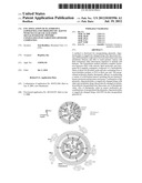 | 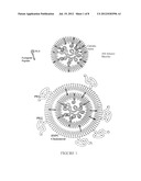 |
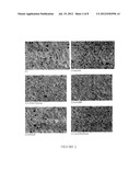 | 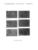 |
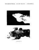 | 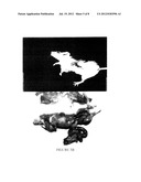 |
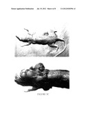 | 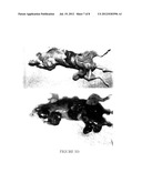 |
 |  |
 |  |
 |  |
 |  |
 |  |
 |  |
 |  |
 |  |
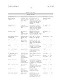 | 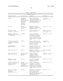 |
 |  |
 |  |
 |  |
 |  |
 |  |
 |  |
 |  |
 |  |
 |  |
 |  |
 |  |
 |  |
 |  |
 |  |
 |  |
 |  |
 |  |
 |  |
 |  |
 |  |
 |  |
 |  |
 |  |
 |  |
 | 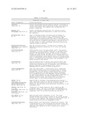 |
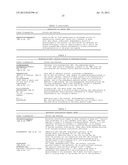 | 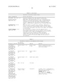 |
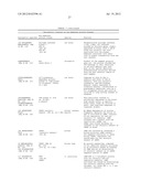 | 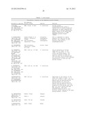 |
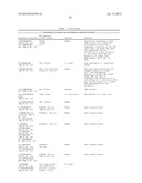 | 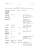 |
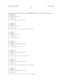 |  |
 |  |
 |  |
 |  |
 |  |
 |  |
 | 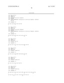 |
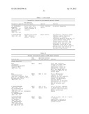 | 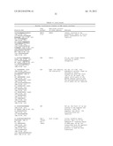 |
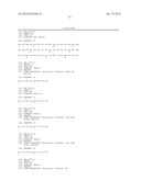 | 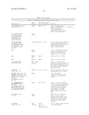 |
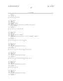 | 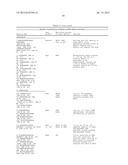 |
 | 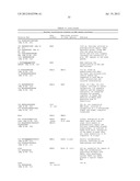 |
 | 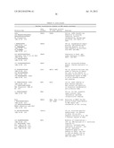 |
 | 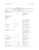 |
 |  |
 |  |
 |  |
 |  |
 |  |
 |  |
 |  |
 |  |
 |  |
 |  |
 |  |
 |  |
 |  |
 |  |
 |  |
 |  |
| New patent applications in this class: | |
| Date | Title |
|---|---|
| 2022-05-05 | Telomerase-containing exosomes for treatment of diseases associated with aging and age-related organ dysfunction |
| 2022-05-05 | Injectable formulations |
| 2022-05-05 | Process of preparing ice-based lipid nanoparticles |
| 2019-05-16 | Method for gene editing |
| 2019-05-16 | Cytosol-penetrating antibody and use thereof |
| Top Inventors for class "Drug, bio-affecting and body treating compositions" | |
| Rank | Inventor's name |
|---|---|
| 1 | David M. Goldenberg |
| 2 | Hy Si Bui |
| 3 | Lowell L. Wood, Jr. |
| 4 | Roderick A. Hyde |
| 5 | Yat Sun Or |