Patent application title: FUSION PROTEINS FOR HIV THERAPY
Inventors:
David Ho (New York, NY, US)
Yaoxing Huang (New York, NY, US)
Craig Pace (New York, NY, US)
Ruijiang Song (New York, NY, US)
Qing Fang (New York, NY, US)
Assignees:
THE ROCKEFELLER UNIVERSITY
IPC8 Class: AA61K3942FI
USPC Class:
4241361
Class name: Immunoglobulin, antiserum, antibody, or antibody fragment, except conjugate or complex of the same with nonimmunoglobulin material structurally-modified antibody, immunoglobulin, or fragment thereof (e.g., chimeric, humanized, cdr-grafted, mutated, etc.) bispecific or bifunctional, or multispecific or multifunctional, antibody or fragment thereof
Publication date: 2012-05-17
Patent application number: 20120121597
Abstract:
Disclosed herein are fusion antibodies created to provide both an
antigen-binding site that targets the CD4 receptor and an antigen-binding
site that targets the HIV envelope. The fusion antibodies disclosed
herein provide improved potency and breadth against HIV as compared to
monospecific antibodies, and additionally provide high barrier against
viral resistance. Also disclosed are pharmaceutical formulations and
therapeutic methods utilizing such fusion proteins.Claims:
1. A fusion antibody, comprising a first antigen-binding site which binds
to the CD4 receptor, conjugated to a second antigen-binding site which
binds to the HIV envelope.
2. The fusion antibody of claim 1, comprising an intact anti-CD4 antibody which provides said first antigen-binding site, conjugated to an antigen-binding fragment of an anti-HIV antibody which provides said second antigen-binding site.
3. The fusion antibody of claim 2, wherein said first antigen-binding site binds to the D2 domain of the CD4 receptor.
4. The fusion antibody of claim 3, wherein said anti-CD4 antibody is humanized or monkeynized.
5. The fusion antibody of claim 4, wherein said anti-CD4 antibody is ibalizumab or a monkeynized derivative thereof.
6. The fusion antibody of claim 2 wherein said anti-CD4 antibody is an affinity matured antibody that has a greater binding affinity to CD4 than ibalizumab.
7. The fusion antibody of claim 2, wherein said anti-HIV antibody is a broad and potent neutralizing antibody.
8. The fusion antibody of claim 7, wherein said anti-HIV antibody binds to an HIV envelope protein.
9. The fusion antibody of claim 8, wherein said anti-HIV antibody binds to a quaternary epitope on the HIV envelope trimer.
10. The fusion antibody of claim 9, wherein said quaternary epitope comprises the V1/V2 and V3 regions of the HIV envelope trimer.
11. The fusion antibody of claim 10, wherein said anti-HIV antibody is PG9.
12. The fusion antibody of claim 8, wherein said anti-HIV antibody binds to a CD4-induced epitope in the HIV gp120 protein.
13. The fusion antibody of claim 12, wherein said anti-HIV antibody is m36.
14. The fusion antibody of claim 8, wherein said anti-HIV antibody binds to an epitope in the HIV envelope involved in binding to CD4.
15. The fusion antibody of claim 14, wherein anti-HIV antibody is VRC01.
16. The fusion antibody of claim 1, wherein the Fc region is the Fc fragment of human IgG1 or IgG4 containing the LALA mutation.
17. The fusion antibody of claim 16, wherein the Fc region is additionally modified to include one or more mutations that improve recycling of the antibody.
18. A method of treating a subject infected with HIV comprising administering to the subject a therapeutically effective amount of the fusion protein according to any one of claims 1-17.
19. A method of preventing HIV infection in a subject in need thereof comprising administering to the subject a therapeutically effective amount of the fusion protein according to any one of claims 1-17.
20. A method of preventing a HIV-positive pregnant subject from transmitting the HIV virus to the child, comprising administering to the subject a therapeutically effective amount of the fusion protein according to any one of claims 1-17.
Description:
CROSS REFERENCE TO RELATED APPLICATION
[0001] This application claims the benefit of priority from U.S. Provisional Application No. 61/413,178, filed Nov. 12, 2010, the entire content of which is incorporated by reference.
FIELD OF THE DISCLOSURE
[0002] This disclosure generally relates to fusion proteins comprised of an antigen-binding site that binds to an epitope of the CD4 receptor and another antigen-binding site that binds an epitope of HIV. The fusion proteins are useful for treating HIV infection in a subject, decreasing the viral load of HIV in a patient, and/or preventing the transmission of HIV to subjects.
BACKGROUND ART
[0003] HIV-1 entry is triggered by interaction of the viral envelope (Env) glycoprotein gp120 with domain 1 (D1) of the T-cell receptor CD4. Binding of CD4 by gp120 induces extensive conformational changes in gp120 leading to formation and exposure of a structure called the co-receptor (coR) binding site, also known as the CD4-induced (CD4i) epitope, in the gp120 protein (Moore et al., Proc Natl Acad Sci USA 100(19): 10598-602, 2003). The bridging sheet of gp120 is a critical component of the coR binding site that is highly conserved across genetically diverse HIV-1 isolates from different clades (Huang et al., Proc Natl Acad Sci USA 101(9):2706-11, 2004). The coR binding site is typically unformed on free (non-CD4-bound) gp120. It forms after attachment of viruses to target cells through CD4 binding. The bridging sheet is highly immunogenic and elicits a class of antibodies known as CD4-induced (CD4i) antibodies in vivo. However, access of full-size Abs to the CD4i epitope (bridging sheet) is sterically restricted during viral entry into cells, most likely because the large size of an Ab cannot access the tight crypt within gp120 where the bridging sheet resides. Thus, most known full-size CD4i Abs do not have potent antiviral activity. Fragments of CD4i Abs that are smaller in size could potentially gain access to the CD4i epitope during viral entry. For example two Ab fragments, known as Fab and scFv, of known CD4i Abs have been shown to significantly inhibit HIV entry more potently than full-size Abs (Labrijn et al., J. Virol. 77: 10557-10565, 2003).
[0004] Ibalizumab (iMab) is a potent and broad HIV-1 neutralizing Ab (Jacobson et al., Antimicrob. Agents Chemother. 53:450-457, 2009; Kuritzkes et al., J. Infect. Dis. 189:286-291, 2004). iMab neutralizes HIV by binding mainly to domain 2 (D2) of the CD4 receptor on host T-cells, thus blocking the ability of HIV to use these CD4 receptors to gain entry into T-cells and produce infection (Burkly et al., J. Immunol. 149:1779-178, 1992). In a large panel of primary isolates (more than 100 viruses) tested recently, iMab neutralized 92% of all viruses as defined by 50% inhibition of infection, and 47.4% of viruses as defined by 90% inhibition of infection (FIG. 1). These data, while promising, indicate that there is still a need to further improve the potency and breadth of iMab.
[0005] Several antibodies have been reported that target epitopes on HIV Env. m36 polypeptide is a human heavy chain domain Ab fragment that targets the highly conserved, but sterically restricted, CD4i epitope on HIV Env. m36 is a potent and broad cross-reactive HIV-1 inhibitor with CD4i Ab activity in vitro, with a mean IC50 in the hundred nanomolar range (Chen et al., Proc Natl Acad Sci USA 105(44):17121-6, 2008). However, similar to other small-size antibody fragments, the m36 polypeptide is predicted to have a short half life in circulation. A long half life is an important indicator for long-lasting antiviral activity. PG9 is another potent and broad anti-HIV-1 antibody, targeting a quaternary epitope of the HIV envelope trimer (Walker et al., Science 326, 285-289 (2009)). VRC01 is also a potent and broad anti-HIV-1 antibody, targeting a CD4 binding site epitope on the HIV envelope.
SUMMARY OF THE DISCLOSURE
[0006] Disclosed herein are fusion antibodies created to provide both an antigen-binding site that targets the CD4 receptor and an antigen-binding site that targets the HIV envelope. The fusion antibodies disclosed herein provide improved potency and breadth against HIV as compared to monospecific antibodies, and additionally provide high barrier against viral resistance.
[0007] In some embodiments, the fusion proteins assume the configuration of an intact IgG molecule directed to a first antigen (which can be any isotype, i.e., IgG1, IgG2, IgG3, and IgG4), connected via a linker at either the C or N terminus of its heavy or light chain to an antigen-binding domain of a second antibody directed to a second antigen.
[0008] In specific embodiments, the fusion protein is composed of an anti-CD4 antibody conjugated to an antigen-binding domain of an anti-HIV antibody. Examples of such fusion antibodies include Ibalizumab conjugated to m36, PG9scFv, or VRC01scFv.
[0009] In additional embodiments, the anti-CD4 antibody portion of a bispecific fusion protein has been modified and fusion variants with improved affinity for CD4 have been selected by affinity maturation.
[0010] In other embodiments, the Fc region of a fusion protein has been engineered to provide a better PK profile, including improved stability (e.g., via introduced LALA mutations) and improved recycling capability.
[0011] Pharmaceutical formulations and therapeutic methods utilizing the bispecific fusion proteins disclosed herein are also provided.
BRIEF DESCRIPTION OF THE DRAWINGS
[0012] This patent or application file contains drawings executed in color. Copies of this patent or patent application publication with color drawings will be provided by the Office upon request and payment of the necessary fee.
[0013] FIG. 1. Ibalizumab neutralization profile on a diverse panel of Env-pseudotyped viruses.
[0014] FIG. 2. Schematic representation of the iMab-m36 fusion construct. Orange, Yellow, and Blue represent the Variable Heavy, Variable Light, and Constant chains of ibalizumab, respectively. Red represents the m36 polypeptide.
[0015] FIG. 3. SDS-PAGE showing expression of purified m36, iMab36 fusion, and iMab. Left-most lane contains a size marker.
[0016] FIG. 4. iMabm36 binds sCD4. X-axis indicates increasing levels of the unlabeled constructs for iMab36 (pink), iMab (black), or m36 (green). Y-axis indicates the competitive level of binding of HRP-labeled ibalizumab to sCD4.
[0017] FIG. 5. iMab36 was shown to be active against ibalizumab-sensitive and -resistant viruses. iMab-sensitive (left column) or iMab-resistant (right two columns) viruses were tested for their viral activity in the presence of the neutralizing constructs iMabm36 (pink), iMab (black), or m36 (green).
[0018] FIG. 6. iMabm36 was shown to have greater potency against iMab-resistant viruses as compared to iMab alone.
[0019] FIG. 7. Cell surface anchoring of m36 by fusing it to iMab was shown to improve its antiviral potency.
[0020] FIG. 8. Sensitivity of iMabm36 was determined by the iMab-resistant virus sensitivity to m36.
[0021] FIG. 9. Anti-HIV activity of iMabm36 was shown to be dependent on its CD4 binding ability.
[0022] FIG. 10. iMabm36 fusion was shown to be stable in vitro.
[0023] FIG. 11. Amino acid sequence of an iMabm36 fusion peptide. (A) SEQ ID NO: 1: composed of 1-473=MV1 heavy chain (SEQ ID NO: 2, the first 19 amino acid residues constituting a leader sequence), 474-489=linker (SEQ ID NO: 3), and 490-606=m36 variable heavy chain (SEQ ID NO: 4). (B) SEQ ID NO: 5: composed of residues 1-19=leader sequence (SEQ ID NO: 6), residues 20-131=MV1 variable light chain (SEQ ID NO: 7), and residues 132-238=MV1 constant light chain (SEQ ID NO: 8).
[0024] FIG. 12. Schematic representation of C-terminal and N-terminal ibalizumab-PG9 bispecific antibodies.
[0025] FIG. 13. Ibalizumab-PG9 fusion antibody was shown to bind to human CD4 and gp120 by ELISA.
[0026] FIG. 14. Ibalizumab-PG9 fusion antibody was found to be active against ibalizumab-sensitive and -resistant viruses. Neutralization curves of ibalizumab (left column) and ibalizumab-PG9 (right column) against ibalizumab-resistant (top) and ibalizumab-sensitive (bottom) pseudoviruses. Each colored line represents the neutralization profile of an individual virus. Dashed black line indicates 50% inhibition of infection. Red line indicates the lowest IC50 observed with ibalizumab to highlight the potency of the ibalizumab-PG9 fusion antibody.
[0027] FIG. 15. Amino acid sequence of a PG9-ibalizumab fusion peptide. (A) SEQ ID NO: 9: composed residues 1-19 (Purple)=Signal Peptide Sequence (SEQ ID NO: 10), residues 20-155 (Black)=PG9 variable heavy chain sequence (SEQ ID NO: 11), residues 156-175 (Green)=Linker (SEQ ID NO: 12), residues 176-285 (Red)=PG9 variable light chain sequence (SEQ ID NO: 13), residues 286-301 (Blue)=Linker (SEQ ID NO: 14), residues 302-423 (Underlined Black=Ibalizumab variable heavy chain sequence (SEQ ID NO: 15), and residues 424-756 (Orange)=Fc sequence (SEQ ID NO: 16). (B) SEQ ID NO: 17: composed of residues 1-20=signal peptide sequence (SEQ ID NO: 18), residues 21-133=ibalizumab variable light chain (SEQ ID NO: 19), and residues 134-236=ibalizumab constant light chain (SEQ ID NO: 20).
[0028] FIG. 16. Schematic representation of C-terminal and N-terminal ibalizumab-VRC01 bispecific antibodies.
[0029] FIG. 17. Ibalizumab-VRC01 fusion antibody was shown to bind to human CD4 and gp120 by ELISA.
[0030] FIG. 18. Ibalizumab-VRC01 fusion antibody was found to be active against ibalizumab-resistant viruses. Neutralization curves of ibalizumab (left column) and ibalizumab-VRC01 (right column) against ibalizumab-resistant pseudoviruses. Each colored line represents the neutralization profile of an individual virus. Dashed black line indicates 100% inhibition of infection.
[0031] FIG. 19. Amino acid sequence of a VRC01-ibalizumab fusion peptide. (A) SEQ ID NO: 21: composed of residues 1-121 (Black)=VRC01 variable heavy chain sequence (SEQ ID NO: 22), residues 122-141 (Green)=Linker (SEQ ID NO: 23), residues 142-245 (Red)=VRC01 variable light chain sequence (SEQ ID NO: 24), residues 246-255 (Blue)=Linker (SEQ ID NO: 23), residues 256-387 (Underlined Black)=Ibalizumab variable heavy chain sequence (SEQ ID NO: 15), and residues 388-720 (Orange)=Fc sequence (SEQ ID NO: 16). (B) SEQ ID NO: 17: composed of residues 1-20=signal peptide sequence (SEQ ID NO: 18), residues 21-133=ibalizumab variable light chain (SEQ ID NO: 19), and residues 134-236 (ibalizumab constant light chain) (SEQ ID NO: 20).
[0032] FIG. 20. HIV-neutralizing properties of ibalizumab and three bispecific fusion antibodies against a panel of 118 HIV isolates, as reflected by maximum percent inhibition (MPI in grey) and IC50 (in red).
[0033] FIG. 21. Comparison of antiviral breadth and potency of select bispecific constructs versus their parental mAbs.
[0034] FIG. 22. Enhanced potency of the PG9-iMab fusion construct in the right-most column. Red bars indicate median±interquartile IC50 range of all viruses tested.
[0035] FIG. 23. Stability of ibalizumab-G1LALA in monkeys was shown to be superior to the original ibalizumab.
[0036] FIG. 24. Improved PK profile for ibalizumab 428/434 variant in mice transgenic for human FcRn. Ab refers to IgG1-LALA version of ibalizumab; Tg signifies mice transgenic for human FcRn, whereas Wt signifies wild-type mice.
[0037] FIG. 25. (A) 5A8 (yellow), the murine progenitor of ibalizumab, was humanized with human-specific sequences (green) to create ibalizumab (iMab), which was then "monkeynized" with rhesus-specific sequences (blue) in two steps to yield RhiMab. (B) Using RhiMab, rhesus bispecific antibodies were constructed using (B) the scFv fusion approach. Note that the human residues in the additional antigen-binding Fv will also be replaced by their rhesus counterparts.
[0038] FIG. 26. (A) The amino acid sequences of the light chain (SEQ ID NO: 25) and heavy chain (SEQ ID NO: 26) of Rhesus Ibalizumab (RhiMab). (B) The amino acid sequence of Rhesus PG9 scFv (SEQ ID NO: 27): residues 1-137=heavy chain (SEQ ID NO: 28), residues 138-157=linker (SEQ ID NO: 12), and residues 158-267=light chain (SEQ ID NO: 29).
[0039] FIG. 27. Schematic summary of the method used to conduct "in vitro affinity maturation." Example shown here is for mutations directed to CDR-H1, but a similar approach was taken for other CDRs.
DETAILED DESCRIPTION
[0040] It has been identified in accordance with the present invention that fusion antibodies created to include both an antigen-binding site that targets the CD4 receptor (such as domain 2 of the CD4 receptor) and an antigen-binding site that targets the HIV envelope (such as glycoprotein gp120 or gp41) provide improved potency and breadth against HIV as compared to monospecific antibodies. Such fusion antibodies also provide high barrier against viral resistance. The basic structure of the fusion proteins disclosed herein is typically a whole IgG molecule directed to a first antigen, connected via a linker at either the C or N terminus of the heavy or light chain to an antigen-binding domain or a single-chain antibody (scFv) directed to a second antigen. The features and applications of these bispecific fusion antibodies are described in details below.
Definitions
[0041] An "antigen" refers to a molecule which contains one or more epitopes and which is capable of eliciting an immunological response. "Antigenic molecules" are also used in a general sense to refer to molecules that are binding targets of the fusion proteins disclosed herein.
[0042] An "epitope", also known as antigenic determinant, is the portion of an antigenic molecule or molecules that is recognized by the immune system, i.e., B cells, T cells or antibodies. An epitope can be a conformational epitope or a linear epitope. A conformational epitope is formed by discontinuous sections of an antigenic molecule, or formed by multiple molecules. In the case where the antigen is a protein, a conformational epitope can be formed by discontinuous amino acid residues of the same protein molecule, or by amino acid residues on different molecules of the protein (e.g., a quaternary epitope formed by a multimer of the protein). A linear epitope is formed by continuous sections of an antigen, e.g., a continuous sequence of amino acids of a protein antigen.
[0043] The term "antibody" is used herein broadly and encompasses intact antibody molecules, which include intact polyclonal, monoclonal, monospecific, polyspecific, chimeric, humanized, human, primatized, single-chain, single-domain, synthetic and recombinant antibodies, and antibody fragments that have a desired activity or function.
[0044] The term "chimeric antibody" refers to antibodies containing polypeptides from different sources, e.g., different species or different antibody class or subclass. Examples of chimeric antibodies include an antigen-binding portion of a murine monoclonal antibody fused an Fc fragment of a human immunoglobulin. Methods for making chimeric antibodies are known in the art; for example, methods described in patents by U.S. Pat. No. 4,816,397 to Boss et al. and U.S. Pat. No. 4,816,567 to Cabilly et al.
[0045] The term "humanized antibody" refers to antibodies that contain non-human sequence elements in a human immunoglobulin backbone or framework. Generally, humanized antibodies are human immunoglobulins (recipient antibody) in which residues from a hypervariable region (CDRs) of the recipient are replaced by residues from a hypervariable region of a non-human species (donor antibody) such as mouse, rat, rabbit or nonhuman primate having a desired specificity, affinity and capacity. In some instances, framework region (FR) residues of the human immunoglobulin are also replaced by non-human residues. Humanized antibodies may also, in some instances, contain residues that are not found in either the recipient antibody or the donor antibody and introduced to further refine antibody performance. In general, a humanized antibody contains substantially all of at least one, and typically two, variable domains, in which all or substantially all of the hypervariable regions correspond to those of a non-human immunoglobulin and all or substantially all of the framework regions are those of a human immunoglobulin sequence. A humanized antibody optionally also contains at least a portion of an immunoglobulin constant region (Fc), typically that of a human immunoglobulin. Methods for making humanized antibodies are documented in the art; see, for example, by U.S. Pat. No. 5,225,539 to Winter and U.S. Pat. No. 4,816,397 to Boss et al.
[0046] The term "primatized antibody" refers to antibodies that contain non-primate sequence elements in a primate immunoglobulin backbone or framework. For example, primatized antibodies can be made from a primate immunoglobulin (recipient antibody) by replacing residues in a hypervariable region (CDRs) of the recipient antibody with residues from a hypervariable region of a donor antibody from a non-primate species such as mouse, rat, rabbit or nonhuman primate having a desired specificity, affinity and capacity. Alternatively, primatized antibodies can be made suitable for administration to a desirable primate species by using a recipient immunoglobulin having non-primate sequences or sequences from a different primate species and introducing the Fc fragment, and/or residues, including particularly framework region residues, from the desirable primate, into the recipient immunoglobulin. Examples of primatized antibodies include "monkeynized" antibodies disclosed herein in the Examples section.
[0047] The term "monospecific antibody" refers to antibodies that recognize and bind to one epitope.
[0048] The term "polyspecific antibody" refers to antibodies formed from at least two separate antibodies and binding to multiple (i.e., two or more) separate epitopes.
[0049] As described above, the term "antibody" also includes fragments of an intact antibody, or "antibody fragments", including particularly antigen-binding fragments of an intact antibody. Examples of antigen-binding fragments include, but are not limited to, Fab fragments (consisting of the VL, VH, CL and CH1 domains), Fab' fragments (which differs from Fab fragments by having an additional few residues at the C-terminus of the CH1 domain including one or more cysteines from the antibody hinge region), (Fab')2 fragments (formed by two Fab' fragments linked by a disulphide bridge at the hinge region), Fd fragments (consisting of the VH and CH1 domains), Fv fragments (referring to a dimer of one heavy and one light chain variable domain in tight, non-covalent association which contains a complete antigen recognition and binding site), dAb fragments (consisting of a VH domain), single domain fragments (VH domain, VL domain, VHH domain, or VNAR domain), isolated CDR regions, scFv (or "single chain Fv", referring to a fusion of the VL and VH domains, linked together via a linker), and other antibody fragments that retain antigen-binding function.
[0050] The term "CDR" or "complementarity determining region" refers to the hypervariable regions within the variable domain of an antibody. There are 3 CDRs in each of the heavy chain and light chain variable domains, and are composed of amino acid residues responsible for antigen-binding. The term "framework region" or "FR" refers to the more conserved portions of the variable domains and is composed of residues other than the hypervariable region residues.
[0051] The term "antigen-binding site" of an antibody means a conformation and/or configuration formed by amino acids of the antibody to which an antigen binds. For example, the three CDRs of each of the VH and VL domains interact to define an antigen-binding site on the surface of the VH-VL dimer. Together, the six CDRs confer antigen-binding specificity to the antibody. It should be noted, however, a single variable domain (i.e., VH or VL) can also recognize and bind antigen, albeit often less effectively than the whole binding site with all six CDRs.
[0052] The term "neutralizing antibody" refers to an antibody that inhibits, reduces or completely prevents HIV-1 infection. Whether an antibody is a neutralizing antibody can be determined by in vitro assays described in the Examples section hereinbelow.
[0053] The term "potent neutralizing antibody" refers to an antibody which, when used at a low concentration, reduces HIV-1 infection by at least 50%, 60%, 70%, 80%, 90%, 95%, 99% or greater. Concentrations below 50 μg/ml, between 1 and 50 μg/ml, or even below 1 μg/ml, are considered "low concentrations". In some embodiments, low concentrations are concentrations in the picomolar range, such as 10-900 ng/ml, and include any concentration in that range, such as 800, 700, 600, 500, 400, 300, 200, 100, 75, 50, 25, 10 ng/ml, or even less than 10 ng/ml.
[0054] The term "broad neutralizing antibody" refers to an antibody which inhibits HIV-1 infection, as defined by a 50% inhibition of infection in vitro, in more than 50%, 60%, 70%, 80%, 90%, 95%, 99% or greater, of a large panel of (greater than 100) HIV-1 envelope pseudotyped viruses and viral isolates.
[0055] The term "fragment" as used herein refers to a physically contiguous portion of the primary structure of a biomolecule. In the case of proteins, a fragment may be defined by a contiguous portion of the amino acid sequence of a protein and may be at least 3-5 amino acids, at least 6-10 amino acids, at least 11-15 amino acids, at least 16-24 amino acids, at least 25-30 amino acids, at least 30-45 amino acids and up to the full length of the protein minus a few amino acids. For example, the m36 fragment is 117 amino acids long. In the case of polynucleotides, a fragment is defined by a contiguous portion of the nucleic acid sequence of a polynucleotide and may be at least 9-15 nucleotides, at least 15-30 nucleotides, at least 31-45 nucleotides, at least 46-74 nucleotides, at least 75-90 nucleotides, and at least 90-130 nucleotides. In some embodiments, fragments of biomolecules are immunogenic fragments.
[0056] A "fusion protein" refers to two or more peptides of different origins connected to each other via a linker or linkers. For example, a fusion protein can include a protein conjugated to an antibody. Other examples include, an antibody conjugated to a different antibody or an antibody conjugated to a Fab fragment. The Fab fragment can be conjugated to the N terminus or C terminus of the heavy or light chain of the antibody, or other regions within the antibody. The term fusion protein and fusion construct are used herein interchangeably.
[0057] A "peptide" is any compound formed by the linkage of two or more amino acids by amide (peptide) bonds, usually a polymer of alpha-amino acids in which the alpha-amino group of each amino acid residue (except the NH2 terminus) is linked to the alpha-carboxyl group of the next residue in a linear chain. The terms peptide, polypeptide and poly(amino acid) are used synonymously herein to refer to this class of compounds without restriction as to size, unless indicated to the contrary. Members of this class having a large size are also referred to as proteins and include antibodies.
[0058] The term "linker" refers to a chemical moiety that connects one peptide to another, e.g., one antibody to another. Linkers can also be used to attach antibodies to labels or solid substrates. A linker can include amino acids. Linkers can be straight or branched, saturated or unsaturated carbon chains. They can also include one or more heteroatoms within the chain or at the termini of the chains. By "heteroatoms" is meant atoms other than carbon which are chosen from the group comprising of oxygen, nitrogen, sulfur, phosphorus, boron and halogen. In specific embodiments, linkers are peptides. The use of a linker may or may not be advantageous or needed, depending on the specific antibody pairs. Methods and techniques for the attachment of a linker to an antibody are known in the art. For a treatise on this subject, the reader is referred to Bioconjugate Techniques, G. Hermanson, Academic Press, 1996.
[0059] Fusion Antibody Targeting Both CD4 and HIV
[0060] This disclosure is directed to fusion antibodies that contain an antigen-binding site that targets the CD4 receptor and another antigen-binding site that targets the HIV envelope. Fusion antibodies having specificities towards simply two different antigens are also referred to herein as bispecific antibodies. Bispecific fusion antibodies can be modified to include additional antigen binding sites to provide other polyspecific antibodies.
[0061] Generally speaking, the structure of the fusion proteins disclosed herein is composed of an intact IgG molecule directed to a first antigen, connected via a linker at either the C or N terminus of its heavy chain or light chain to an antigen-binding fragment of a second antibody directed to a second antigen. For example, a fusion antibody having dual specificities towards the CD4 receptor and HIV can be formed by conjugating an intact anti-CD4 antibody with an antigen-binding fragment of anti-HIV antibody. Conversely, a fusion antibody having dual specificities towards the CD4 receptor and HIV can be formed by conjugating an intact anti-HIV antibody with an antigen-binding fragment of anti-CD4 antibody. In either scenario, a bispecific fusion antibody contains an antigen-binding site that binds to an epitope on the CD4 receptor and another antigen-binding site that binds to an epitope on the HIV envelope.
[0062] Anti-CD4 antibodies have been described in the art and can also be readily generated as the protein sequence of the CD4 receptor is available to those skilled in the art. As a member of the immunoglobulin superfamily, CD4 has four immunoglobulin domains (D1 to D4) that are located on the extracellular surface of the cell. CD4 uses its D1 domain to interact with the β2-domain of MHC class II molecules.
[0063] In some embodiments, antibodies directed principally to the second immunoglobulin-like domain (D2) (amino acid positions 98-180) of the CD4 receptor are used in forming the bispecific fusion antibodies herein. Antibodies directed to the D2 domain of CD4 have the desirable property of blocking HIV infection without interfering with immune functions mediated by interaction of CD4 with the major histocompatibility complex (MHC) class II molecules. In specific embodiments, the anti-CD4 antibody used in forming a bispecific fusion binds to an epitope located in the BC-loop of D2 near the D1-D2 junction of the CD4 receptor (amino acids 121-127). In other embodiments, the anti-CD4 antibody binds to the FG-loop of D2 (amino acids 163-165) and part of D1 (amino acids 77-96). The anti-CD4 antibody used in forming a fusion protein can bind to one or more or all of these regions, i.e., D1, D1-D2 junction, D2, the BC or FG loop of D2, or any combination thereof. The above amino acid numbering corresponds to positions of the mature form of the receptor, not including the signal peptide. The amino acid sequence of the human CD4 receptor is available in GenBank under Accession No. AAA16069.1 and is also set forth in SEQ ID NO: 34, in which amino acids 1-25 represent a signal peptide, amino acids 26-122 constitute D1, and amino acids 123-205 constitute D2.
[0064] A specific example of an anti-CD4 antibody suitable for use in forming a bispecific fusion antibody is Ibalizumab (previously known as TNX-355, or hu5A8), which is a humanized, anti-CD4 monoclonal antibody. Ibalizumab potently blocks infection by a broad spectrum of HIV-1 isolates and targets an epitope located in the BC-loop of D2 near the D1-D2 junction of the CD4 receptor, without interfering with immune functions mediated by interaction of CD4 with the major histocompatibility complex (MHC) class II molecules.
[0065] Anti-HIV Env antibodies have also been described in the art and can also be readily generated by those skilled in the art. The env gene encodes a precursor protein, gp160. During HIV reproduction, the endogenous enzymes of the host cell cleave gp160 into gp120 and gp41. Three gp120 molecules form a trimer which is anchored to the viral membrane, or envelope, via non-covalent interactions with the transmembrane protein, gp41. Binding of CD4 by gp120 induces extensive conformational changes in gp120 leading to formation and exposure of a structure called the co-receptor (coR) binding site, also known as the CD4-induced (CD4i) epitope, in the gp120 protein (Moore et al., Proc Natl Acad Sci USA 100(19): 10598-602, 2003). The bridging sheet of gp120 is a critical component of the coR binding site that is highly conserved across genetically diverse HIV-1 isolates from different clades (Huang et al., Proc Natl Acad Sci USA 101(9):2706-11, 2004). The coR binding site is typically unformed on free (non-CD4-bound) gp120. It forms after attachment of viruses to target cells through CD4 binding. The bridging sheet is highly immunogenic and elicits a class of antibodies known as CD4-induced (CD4i) antibodies in vivo. However, access of full-size Abs to the CD4i epitope (bridging sheet) is sterically restricted during viral entry into cells, most likely because the large size of an Ab often cannot access the tight crypt within gp120 where the bridging sheet resides. Fragments of CD4i Abs that are smaller in size could potentially gain access to the CD4i epitope during viral entry. For example, two Ab fragments, known as Fab and scFv, of known CD4i Abs have been shown to significantly inhibit HIV entry more potently than full-size Abs (Labrijn et al., J. Virol. 77: 10557-10565, 2003), and such antibody fragments are suitable for use in forming a bispecific fusion antibody in accordance with this disclosure.
[0066] In specific embodiments, anti-HIV antibodies used herein in forming a bispecific fusion antibody are directed to an epitope on an envelope protein of HIV, e.g., gp120 monomer or trimer, or gp41. In more specific embodiments, the anti-HIV antibodies used in forming a bispecific fusion may bind to the CD4 binding site on the HIV envelope at amino acids 279-460, the V1/V2 and V3 regions of the HIV envelope trimer at amino acids 125-196 and 296-331, and/or the CD4 bridging sheet site on the HIV envelope at amino acids 332-444. Examples of anti-HIV antibodies suitable for use herein include, but are not limited to, m36, PG9, and VRC01. The amino acid sequence of HIV gp160 is available in GenBank under Accession No. AAB60578.1, and is also set forth in SEQ ID NO: 35, in which amino acids 1-30 represent a signal peptide, and amino acids 31-509 represent the mature gp120.
[0067] In certain embodiments, a fusion antibody having dual specificities towards the CD4 receptor and HIV is composed of an intact anti-CD4 antibody, conjugated with (i.e., covalently linked to) an antigen-binding fragment of anti-HIV antibody. As defined above, an antigen-binding fragment of anti-HIV antibody including Fab fragments, Fab' fragments, (Fab')2 fragments, Fd fragments, Fv fragments, dAb fragments, single domain fragments, isolated CDR regions, scFvs, and other antibody fragments that retain HIV-binding function of an anti-HIV antibody.
[0068] In a specific embodiment, a single domain antibody fragment of an anti-HIV antibody, e.g., m36, is fused to an intact anti-CD4 antibody. In some embodiments, the single domain antibody fragment is fused to the C-terminus of the heavy chain or light chain of the anti-CD4 antibody. In other embodiments, the single domain antibody fragment is fused to the N-terminus of the heavy or light chain of the anti-CD4 antibody.
[0069] In another specific embodiment, a single chain Fv fragment (i.e., scFv) of an anti-HIV antibody is fused to an intact anti-CD4 antibody. Similarly, the scFv fragment can be fused to the C-terminus or N-terminus of either the heavy chain or light chain of the anti-CD4 antibody. In some embodiments, the scFv fragment is a single chain of the VH domain linked to the N-terminus of the VL domain of a relevant anti-HIV antibody; while in other embodiments, the scFv fragment is a single chain of the VH domain linked to the C-terminus of the VL domain of a relevant anti-HIV antibody.
[0070] The linkage between the two antibody components, i.e., between the anti-CD4 antibody portion and the anti-HIV antibody portion, and also between the VH and VL domains of an scFv fragment, is achieved by a peptide linker. The length of a linker is generally in the range of 5 to 50 amino acids, and in specific embodiments, in the range of 9-25 amino acids, such as 9, 12, 16, 20 or 24 amino acids. The linkers can be synthetic or native human antibody-derived sequences, or a combination of both. Generally speaking, the linkers are principally composed of relatively small, neutral amino acids, such as Glycine, Serine, Alanine, and can include multiple copies of a sequence enriched in Glycine and Serine, such as multiple copies of GGGGS (SEQ ID NO: 33). Examples of linkers suitable for use herein include those set forth in SEQ ID NO: 3, 12, 14, 23 and 32.
[0071] The bispecific fusion antibodies disclosed herein can be produced by utilizing techniques available to those skilled in the art. For example, DNA molecules encoding a desirable bispecific fusion antibody can be constructed based on the coding sequence of the two antibody components of the fusion using molecular cloning techniques. The resulting DNAs can be placed into expression vectors which are then transfected into host cells such as E. coli cells, simian COS cells, Chinese Hamster Ovary (CHO) cells, human embryonic kidney 293 cells, or myeloma cells including murine myeloma cells that do not otherwise produce immunoglobulin protein, to obtain the synthesis of monoclonal antibodies in the recombinant host cells. For example, an expression vector encoding a heavy chain of an anti-CD4 antibody fused to an anti-HIV antibody fragment, and an expression vector encoding the light chain of the anti-CD4 antibody, can be co-transfected into a host cell for expression. Antibody-containing culture supernatants can be collected for purification of antibodies.
[0072] The produced fusion antibodies can be evaluated in in vitro assays to assess their functionality, e.g., binding to the CD4 receptor, binding to the HIV envelope, anti-HIV potency and breadth. In specific embodiments, this disclosure provides potent and broad bispecific fusion antibodies, with potency and breadth being defined as hereinabove. These antibodies are described in more details in the Examples section.
[0073] Modifications to Antibodies
[0074] Humanization and Primatization
[0075] In cases where the fusion antibody or the two antibodies forming the bispecific fusion antibody are non-human antibodies, the antibody can be "humanized" to reduce immunogenicity to a human recipient. Methods for humanizing non-human antibodies have been described in the art. See, e.g., Jones et al., Nature 321:522-525 (1986); Riechmann et al, Nature 332:323-327 (1988); Verhoeyen et al., Science 239:1534-1536 (1988), and U.S. Pat. No. 4,816,567. Generally, residues from the variable domain of a non-human antibody are "imported" into a human immunoglobulin molecule, resulting in antibodies in which some hypervariable region residues and possibly some FR residues of a human antibody are substituted by residues from analogous sites of non-human antibodies. It is important to humanize a non-human antibody while retaining high affinity for the antigen. To this end, three dimensional immunoglobulin models are commonly available and suitable for use in analyzing proposed humanized sequences in comparison to the parental non-human antibodies. Such analysis permits identification of residues likely involved in recognition and binding of the antigen, and therefore rational design of humanized sequences that retain the specificity and affinity for the antigen.
[0076] In specific embodiments, bispecific fusion antibodies are formed from an anti-CD4 antibody which has been humanized and an anti-HIV human or humanized antibody. For example, ibalizumab is an example of a humanized anti-CD4 antibody, and m36 is an example of the variable domain of a human anti-HIV antibody.
[0077] Similarly, a fusion antibody or the two antibodies forming the fusion can be "primatized" to reduce immunogenicity to another primate, non-human recipient, e.g., a rhesus recipient. Residues from the variable domain of a donor antibody (such as a non-primate antibody or an antibody of a primate species different from the recipient primate) are "imported" into a nonhuman primate recipient immunoglobulin molecule, resulting in antibodies in which some hypervariable region residues and possibly some FR residues of a nonhuman primate antibody are substituted by residues from analogous sites of donor antibodies. Alternatively, primatized antibodies can be made for use in a desirable primate species by using a recipient immunoglobulin having non-primate sequences or sequences from a different primate species by introducing the Fc fragment, and/or residues, including particularly framework region residues, from the desirable primate, into the recipient immunoglobulin. A "monkeynized" version of Ibalizumab, and a monkeynized version of anti-HIV antibody PG9 scFv, are disclosed in the Examples hereinbelow.
[0078] Affinity Maturation
[0079] One or more hypervariable region residues of an antibody can be substituted to select for variants that have improved biological properties relative to the parent antibody by employing, e.g., affinity maturation using phage or yeast display. For example, the Fab region of an anti-CD4 antibody or an anti-HIV antibody can be mutated at several sites selected based on available structural information to generate all possible amino substitutions at each site. The antibody variants thus generated are displayed in a monovalent fashion from phage particles or on the surface of yeast cells. The displayed variants are then screened for their biological activity (e.g. binding affinity). Examples of ibalizumab variants having high affinities are described herein below. These ibalizumab variants can be used in forming bispecific fusion antibodies.
[0080] Modifications to the Fc Region
[0081] The fusion antibody or the intact antibody used in forming the fusion, which can be of any IgG isotype from any primate species including human, can be modified to improve certain biological properties of the antibody, e.g., to improve stability, to enhance or reduce effector functions such as antigen-dependent cell-mediated cyotoxicity (ADCC) and/or complement dependent cytotoxicity (CDC) of the antibody, improved or decreased internalization and/or recycling, among others.
[0082] For example, the Fc fragment of ibalizumab (derived from human Ig4) can be replaced with human IgG1 carrying the so-called LALA mutations that reduce effector function mediated through FcRs (except FcRn). Such modification has been shown herein to improve the stability of the resulting antibody by about 5 fold. The LALA mutation can also be introduced into an IgG4 background. In another example, the IgG1 Fc fragment can be modified with the pair of mutations, M428L/N434S, to improve the recycling of the antibody via the antibody salvage pathway. This pair of mutations, introduced in the IgG1-LALA version of ibalizumab, has led to an additional about 3 fold improvement in PK of the resulting antibody.
[0083] Still another type of modification involves alteration of the glycosylation pattern of a parent antibody, including deletions of one or more carbohydrate moieties found in the parent antibody, or addition of one or more carbohydrates (via addition of one or more glycosylation sites) that are not present in the parent antibody
[0084] Pharmaceutical Formulations
[0085] Pharmaceutical formulations of the fusion antibody proteins disclosed can be prepared by mixing a fusion protein with optional pharmaceutically acceptable carriers. Pharmaceutically acceptable carriers include solvents, dispersion media, isotonic agents and the like. The carrier can be liquid, semi-solid, e.g. pastes, or solid carriers. Examples of carriers include water, saline solutions or other buffers (such as phosphate, citrate buffers), oil, alcohol, proteins (such as serum albumin, gelatin), carbohydrates (such as monosaccharides, disaccharides, and other carbohydrates including glucose, sucrose, trehalose, mannose, mannitol, sorbitol or dextrins), gel, lipids, liposomes, resins, porous matrices, binders, fillers, coatings, stabilizers, preservatives, antioxidants including ascorbic acid and methionine, chelating agents such as EDTA; salt forming counter-ions such as sodium; non-ionic surfactants such as TWEEN®, PLURONICS® or polyethylene glycol (PEG), or combinations thereof.
[0086] The formulation can contain more than one active compound, e.g., one or more fusion antibodies, in combination with one or more additional beneficial compound for preventing and treating HIV infections.
[0087] The active ingredients can be combined with the carrier in any convenient and practical manner, e.g., by admixture, solution, suspension, emulsification, encapsulation, absorption and the like, and can be made in formulations such as tablets, capsules, powder (including lyophilized powder), syrup, suspensions that are suitable for injections, ingestions, infusion, or the like. Sustained-release preparations can also be prepared.
[0088] Methods of Treatment and Prevention
[0089] In a further aspect, the bispecific fusion antibodies disclosed herein, optionally provided in pharmaceutically acceptable carrier, are employed for the treatment and prevention of HIV infection in a subject, as well as prevention of HIV transmission.
[0090] The term "treatment" of HIV infection refers to effective inhibition of the HIV infection so as to delay the onset, slow down the progression, reduce viral load, and/or ameliorate the symptoms caused by HIV infection.
[0091] The term "prevention" of HIV infection means the onset of HIV infection is delayed, and/or the incidence or likelihood of HIV infection is reduced or eliminated.
[0092] The term "prevention" of HIV transmission means the incidence or likelihood of HIV being transmitted from one individual to another (e.g., from an HIV-positive woman to the child during pregnancy, labor or delivery, or breastfeeding) is reduced or eliminated.
[0093] The term "subject" refers to any primate subject, including human and rhesus subjects.
[0094] To treat and/or prevent HIV infection, a therapeutic amount of a fusion antibody disclosed herein is administered to a subject in need.
[0095] The term "therapeutically effective amount" means the dose required to effect an inhibition of HIV infection so as to treat and/or prevent HIV infection. The dosage of a fusion antibody depends on the disease state and other clinical factors, such as weight and condition of the subject, the subject's response to the therapy, the type of formulations and the route of administration. The precise dosage to be therapeutically effective and non-detrimental can be determined by those skilled in the art. As a general rule, a suitable dose of a fusion antibody for the administration to adult humans parenterally is in the range of about 0.1 to 20 mg/kg of patient body weight per day, once a week, or even once a month, with the typical initial range used being in the range of about 2 to 10 mg/kg. Since the antibodies will eventually be cleared from the bloodstream, re-administration may be required. Alternatively, implantation or injection of the fusion antibodies provided in a controlled release matrix can be employed.
[0096] The fusion antibodies can be administered to the subject by standard routes, including the oral, transdermal, parenteral (e.g., intravenous, intraperitoneal, intradermal, subcutaneous or intramuscular). In addition, the antibodies can be introduced into the body, by injection or by surgical implantation or attachment such that a significant amount of a desirable antibody is able to enter blood stream in a controlled release fashion.
[0097] The description of some specific embodiments provides sufficient information that others can, by applying current knowledge, readily modify or adapt for various applications such specific embodiments without departing from the generic concept, and, therefore, such adaptations and modifications should and are intended to be comprehended within the meaning and range of equivalents of the disclosed embodiments. It is to be understood that the phraseology or terminology employed herein is for the purpose of description and not of limitation. In the drawings and the description, there have been disclosed exemplary embodiments and, although specific terms may have been employed, they are unless otherwise stated used in a generic and descriptive sense only and not for purposes of limitation, the scope of the claims therefore not being so limited. Moreover, one skilled in the art will appreciate that certain steps of the methods discussed herein may be sequenced in alternative order or steps may be combined. Therefore, it is intended that the appended claims not be limited to the particular embodiment disclosed herein.
[0098] The present description is further illustrated by the following examples, which should not be construed as limiting in any way. The contents of all cited references (including literature references, issued patents, and published patent applications as cited throughout this application) are hereby expressly incorporated by reference.
Example-1
Construction and Testing of iMabm36
[0099] This Example describes the construction and testing of a novel bispecific HIV-1 neutralizing antibody (Ab) for the prevention and treatment of HIV/AIDS which was named iMabm36. iMabm36 is composed of the anti-CD4 Ab ibalizumab (iMab) linked to two copies of the anti-CD4-induced Ab m36. iMabm36 is shown herein to have significantly increased antiviral potency and breadth over iMab and m36 alone. In particular, it can potently inhibit viral entry of all iMab-resistant viruses tested. Mechanistically, it is shown therein that iMabm36 activity requires CD4 binding and is m36 sensitivity dependent. The inter-dependency of this dual mechanism of action enables the high potency and breadth of iMabm36. As almost all HIV-1 isolates use CD4 as a primary entry receptor and the m36 targeting site is highly conserved across all HIV-1 isolates, targeting these two sites is believed to provide a high barrier against viral resistance. iMabm36 has been shown to be stable in vitro, and is believed to have improved pharmacokinetics as compared to m36 alone and is unlikely to be immunogenic.
[0100] Construction and expression of iMabm36 fusion Ab. A bi-specific fusion Ab was constructed based on a derivative IgG1 version of iMab called MV1 and m36. As shown in the schematic in FIG. 2 and the sequence in FIG. 11, m36 was linked to the C-terminus of the heavy chain of MV1 via a flexible (G4S)x3 linker peptide (GGGGSGGGGSGGGGSG, SEQ ID NO: 3). In brief, cDNA sequence of the fusion construct was generated by overlap PCR and subsequently cloned into the pVAX expression plasmid through unique restriction sites.
[0101] The iMabm36 fusion Ab was expressed by transient co-transfection of pVAX vectors expressing 1) heavy chain iMabm36 fusion and 2) light chain ibalizumab into human embryonic kidney 293 cells. Ab-containing culture supernatants were filtered and purified by affinity chromatography using a Protein A Sepharose column (FIG. 3). The average yield was 1-5 μg/ml of culture medium.
[0102] iMabm36 retains CD4 binding activity in vitro. Purified iMabm36 was assessed for soluble CD4 (sCD4) binding in a competition ELISA assay. In brief, a 96-well plate was coated with sCD4. A fixed concentration of HRP labeled iMab was then mixed with increasing concentrations of iMabm36 or unlabeled iMab and measured for sCD4 binding competition. As shown in FIG. 4, iMabm36 competed with iMab-HRP labeled Ab for CD4 binding with equal potency as compared to the parental unlabeled iMab, indicating the functional CD4 binding properties of the iMabm36 fusion protein. As expected, m36 alone did not bind to sCD4.
[0103] iMabm36 fusion Ab improves antiviral potency and breadth as compared to iMab or m36 alone. To test whether the fusion of m36 to the C-terminal of iMab could result in more potent antiviral activity, iMabm36, iMab and m36 were each examined in a TZM-b1 based HIV-1 Env pseudotyped neutralization assay against a panel of 6 viruses that included iMab sensitive and resistant viruses. As shown in FIG. 5, iMabm36 demonstrated a similar potency against iMab sensitive viruses (G19 and G23) as compared to iMab alone. While iMab can only achieve a maximal percent of inhibition (MPI) of 25% to 75% for iMab resistant viruses (G02, G09, G20, and B09), the iMabm36 fusion could neutralize all four of these viruses and achieved 100% MPI at low nanomolar concentrations. Consistent with previous publication, the 50% inhibitory activity of m36 is within the range of hundreds of nanomolar concentrations.
[0104] In a separate experiment, iMabm36 activity was further examined against all iMab-resistant viruses in the inventors' collection (FIG. 6). Among 13 iMab-resistant viruses tested (defined as a MPI of less than 100%), all could be neutralized by iMabm36 with 100% MPI, except for the virus G12 which was neutralized by up to 90% MPI. The mean concentration required to achieve 100% viral neutralization by iMabm36 is around 0.5 μg/ml (2.8 nM). Also, iMabm36 was active in a PMBC based neutralization assay (data not shown).
[0105] Increased antiviral potency of iMabm36 compared to iMab and m36 mixture. The results of FIG. 5 indicated that iMabm36 was more active than its individual components iMab and m36. To investigate if the high antiviral activity of iMabm36 could also be achieved by simply mixing iMab and m36, a neutralizing assay was performed using iMab and m36 at a molar ratio of 1:2 because one iMab carries two m36 peptides in the iMabm36 fusion. As shown in FIG. 7a, mixing iMab and m36 improved not only the antiviral activity of the respective components, but also achieved 100% neutralization of the iMab-resistant viruses tested. These results indicate that iMab and m36 contribute to the antiviral activity in an additive or synergistic manner. In comparison with the mixing of iMab and m36 components, the iMabm36 fusion Ab demonstrated even greater antiviral activity with at least 10-fold enhancement at the same molar concentrations tested.
[0106] To test the hypothesis that iMabm36 acts through increasing m36 local concentration, iMab was mixed with an excess of m36 at a molar ratio of 1:10. As shown in FIG. 7b, mixing iMab and m36 at a 1:10 ratio was more potent than that of the 1:2 ratio in FIG. 7a. Notably, even with a five times excess of m36, the mixture using individual components was still less potent than m36 fused to iMab.
[0107] Improved potency of the iMabm36 fusion against iMab-resistant viruses is conferred by viral sensitivity to m36. To better understand the contribution of m36 in the context of the iMab36 fusion, the inventors analyzed the correlation of ibalizumab-resistant virus sensitivity to m36 versus iMabm36. As shown in FIG. 8, a strong correlation was observed between the IC50 of m36 and the IC50 of iMabm36. This result supports the notion that, in the context of iMab-resistant viruses, the sensitivity of iMabm36 is determined by the virus sensitivity to m36.
[0108] Antiviral potency of iMabm36 is dependent on its CD4 binding. To understand the mechanism of antiviral activity of the iMab36 fusion, two contact amino acid residues in the iMab heavy chain variable CDR3 region were substituted with alanine residues. The CDR3 regions of the wt and mutant heavy chains of iMab are set forth in FIG. 9a and SEQ ID NOS: 35 and 36 respectively. The mutant fusion Ab (iMabΔm36) was purified and tested for its CD4 binding ability in vitro. As shown in FIG. 9a, iMabΔm36 could no longer compete with iMab-HRP for sCD4 binding, suggesting that these two mutations abolished the CD4 binding activity of iMabm36. In a virus neutralization assay, iMabΔm36 lost its virus neutralization activity (FIG. 9b). This result suggests that the antiviral activity of iMabm36 is dependent on its CD4 binding. In other words, anchoring of m36 via iMab binding to CD4 is critical for iMabm36 antiviral activity.
[0109] In vitro stability of iMabm36. To assess the stability of iMabm36, the inventors first generated high titers of rabbit anti-m36 immune serum. To do so, rabbits were immunized with purified m36 protein (300 μg/dose) in CFA at week 0 and subsequently boosted in IFA twice at weeks 4 and 8. Anti-m36 Ab titers were determined in the serum sample collected 4 weeks post last boost immunization. The in vitro stability of the iMabm36 fusion Ab was determined by incubation of the fusion Ab in 20% serum in PBS at 37° C. for up to 7 days. Aliquots of the untreated (day 0) and treated fusion Ab were taken at the indicated time points and stored at -20° C. The presence of intact iMabm36 was examined by the functional binding activity of iMabm36 to sCD4 and determined by anti-iMab Fc direct ELISA and anti-m36 sandwich ELISA, respectively.
[0110] Data from FIG. 10 demonstrates that, over time, there was no loss of CD4 binding as detected by a secondary Ab against iMab Fc, which indicates that the iMab portion of this fusion was still present. The m36 portion of this fusion was also still present as detected by an anti-m36 rabbit immune serum and a secondary Ab against rabbit serum Ig. These results suggest that iMabm36 was stable for up to at least 7 days in the condition tested.
Example-2
Construction and Testing of Ibalizumab-PG9
[0111] This Example describes the construction and testing of Ibalizumab-PG9 (or also referred to as "PG9-Ibalizumab"), a novel bispecific HIV-1 neutralizing antibody (NAb) for the prevention and treatment of HIV/AIDS. Ibalizumab-PG9 was created as a fusion of the anti-CD4 Ab ibalizumab fused to a single-chain Fv (scFv) version of the anti-HIV envelope NAb, PG9. Ibalizumab-PG9 showed significantly increased antiviral potency and breadth over ibalizumab or PG9 alone. In particular, it potently inhibited viral entry of all ibalizumab-resistant viruses tested. Since ibalizumab and PG9 inhibit HIV entry using two distinct mechanisms, the chance of emerging HIV resistance to the ibalizumab-PG9 fusion is diminished in comparison to either ibalizumab or PG9 alone.
[0112] Construction and expression of the ibalizumab-PG9 fusion antibodies. To improve ibalizumab's breadth and potency, bispecific ibalizumab-PG9 was created to simultaneously target both the CD4 receptor on host T-cells and the HIV envelope protein gp120. PG9 is currently one of the most potent and broadest anti-HIV-1 antibodies, targeting a quaternary epitope of the HIV envelope trimer (Walker et al., Science 326, 285-289 (2009)). To create the fusion antibody, the inventors fused the heavy chain variable domain of PG9 to the light chain variable domain of PG9 via a flexible linker (SEQ ID NO: 12) to create a PG9 single chain variable fragment (scFv). This PG9 scFv was then fused via a flexible linker (SEQ ID NO: 14) to the N-terminal and/or C-terminal of the ibalizumab heavy chain to create bispecific ibalizumab-PG9 (FIG. 12). The bispecific fusion antibody with PG9 scFv fused to the N-terminus of the ibalizumab heavy chain (FIG. 15) was used in the following experiments. The Fc region of the ibalizumab of this fusion construct also contained a LALA mutation. The LALA mutation is further described hereinbelow.
[0113] Ibalizumab-PG9 fusion antibody retains its binding functions. The ability of the fusion antibody to bind its cognate antigens was assessed using ELISA. As seen in FIG. 13 (left side), the fusion antibody was able to bind human CD4 comparably to ibalizumab. Biacore studies indicate that the addition of the PG9 scFv had no effect on the affinity at which the antibody binds CD4 (data not shown). To determine if the ibalizumab-PG9 fusion retained the inherent binding activity of PG9, the inventors demonstrated that the fusion antibody was able to bind gp120 comparably to the anti-gp120 antibody F105 (FIG. 13, right side). As expected, ibalizumab was unable to bind gp120, even when tested at concentrations 10-fold higher than that of the fusion antibody.
[0114] Ibalizumab-PG9 fusion antibody retains and improves virus neutralization activity as compared to ibalizumab alone. To assess the antiviral efficacy of the ibalizumab-PG9 fusion antibody, the inventors determined its HIV-1 neutralization capacity using the standardized single cycle TZM-b1 reporter assay. The ibalizumab-PG9 fusion antibody was able to neutralize 9 out of 9 ibalizumab-resistant viruses to greater than 95% inhibition (FIG. 14, top right). In addition to improving the breadth of ibalizumab, the fusion antibody exhibited significantly enhanced potency (p=0.002), up to 150-fold compared to ibalizumab.
Example-3
Construction and Testing of Ibalizumab-VRC01
[0115] This Example describes the generation and testing of Ibalizumab-VRC01 (also referred to as "VRC01-Ibalizumab"), a novel bispecific HIV-1 neutralizing antibody (NAb) for the prevention and treatment of HIV/AIDS. Ibalizumab-VRC01 was created as a fusion of a single-chain Fv (scFv) version of the anti-HIV envelope NAb, VRC01, fused to the anti-CD4 Ab ibalizumab. Ibalizumab-VRC01 demonstrated significantly increased antiviral breadth over ibalizumab or VRC01 alone. In particular, it potently inhibited viral entry of all ibalizumab-resistant viruses tested. In addition, complementary resistance to ibalizumab and VRC01 occur via a mutually exclusive genotype. The dominant pathway to ibalizumab resistance involves abrogation of potential N-linked glycosylation (PNG) of the V5 N-terminus. It has been previously demonstrated from a large panel of viruses and from site-directed mutants that the presence of a V5 N-terminal PNG site is sufficient to confer sensitivity to ibalizumab. In contrast, the presence of a V5 N-terminal PNG site substantially reduces (>20-fold) the sensitivity to VRC01. Therefore, combination therapy with ibalizumab and VRC01, either as a fusion antibody or co-administered, is believed to provide a substantially higher genetic barrier to resistance compared to such combinations of any other broadly NAbs against HIV described to date.
[0116] Construction and expression of the ibalizumab-VRC01 fusion antibodies. To improve the breadth of ibalizumab and VRC01, the inventors created bispecific ibalizumab-VRC01 to simultaneously target both the CD4 receptor on host T-cells and the HIV envelope protein gp120. VRC01 is currently one of the most potent and broadest anti-HIV-1 antibodies, targeting the CD4 binding site of the HIV envelope trimer. To create the fusion antibody, the inventors fused the heavy chain variable domain of VRC01 to the light chain variable domain of VRC01 via a flexible (GGGGS)4 (SEQ ID NO: 22) linker to create a VRC01 single chain variable fragment (scFv). This VRC01 scFv was then fused via the flexible (GGGGS)4 (SEQ ID NO: 22) linker to the N-terminal and/or C-terminal of the ibalizumab heavy chain to create bispecific ibalizumab-VRC01 (FIG. 16). The bispecific fusion antibody with VRC01 scFv fused to the N-terminus of the ibalizumab heavy chain (FIG. 19) was used in the following experiments. The Fc region of the ibalizumab of this fusion construct also contained a LALA mutation. The LALA mutation is further described hereinbelow.
[0117] Ibalizumab-VRC01 fusion antibody retains its binding functions. The ability of the fusion antibody to bind its cognate antigens was assessed using ELISA. As seen in FIG. 17 (left side), the fusion antibody was able to bind human CD4 comparably to ibalizumab. To determine if the ibalizumab-VRC01 fusion retained the inherent binding activity of VRC01, the inventors demonstrated that the fusion antibody was able to bind gp120 (FIG. 17, right side). As expected, ibalizumab alone was unable to bind gp120.
[0118] Ibalizumab-VRC01 fusion antibody retains and improves virus neutralization activity as compared to ibalizumab alone. To assess the antiviral efficacy of the ibalizumab-VRC01 fusion antibody, the inventors determined its HIV-1 neutralization capacity using the standardized single cycle TZM-b1 reporter assay. The ibalizumab-VRC01 fusion antibody was able to neutralize 10 out of 10 ibalizumab-resistant viruses to 100% inhibition (FIG. 18, right).
Example-4
Improvements of Bispecific Fusion Antibodies Over Ibalizumab In Breadth and Potency
[0119] The improvement of the fusion antibodies constructed herein as compared to ibalizumab is illustrated as follows.
[0120] The top-left panel of FIG. 20 recapitulates the in vitro neutralizing activity of unmodified ibalizumab (iMab) against a panel of 118 HIV isolates, illustrating the "hole" in its repertoire. An IC50 of ≧10 ug/mL was noted for a small fraction of the viruses, but high-level neutralization (MPI>80%) was not achieved for many more viruses. iMab-m36 (Example 1) demonstrated a modest increase in breadth (top-right, FIG. 20). VRC01-iMab (Example 3) showed even greater breadth and potency with IC50s consistently between 0.01 and 0.1 μg/mL (bottom-left). Most notable were the neutralizing activities exhibited by PG9-iMab (Example 2) (bottom-right), where breadth of 100% (as measured by ≧80% neutralization) was observed, as was a significant increase (>200-fold by IC90) in potency (median IC50, IC90 and IC99 of 0.005 μg/mL, 0.028 μg/mL, and 0.080 μg/mL, respectively).
[0121] The remarkable gain in anti-HIV activity of the fusion antibodies is even more evident when shown as percentage coverage of viruses neutralized over a range of concentrations (FIG. 21). In particular, 99% of the viruses were neutralized by PG9-iMab at concentrations <0.1 μg/mL.
[0122] The antiviral potency of the bispecific antibodies was also displayed by showing their IC50s on a scattered plot in comparison with results for ibalizumab (FIG. 22). The improvement is readily evident.
Example-5
PK Improvements on the Ibalizumab Backbone
[0123] Ibalizumab has a relatively short half-life in vivo (3.5 days for IV administration and ˜10 days for SC administration). mAbs humanized with IgG4 (such as ibalizumab) are known to have a strong tendency to switch their inter-chain disulfide bonds to intra-chain disulfide bonds, resulting in the dissociation of the antibody into monomers. In order to improve stability, the Fc region of the ibalizumab was modified to introduce the so-called "LALA" (Leu to Ala and Leu to Ala) mutations. The Fc regions of PG9-ibalizumab (FIG. 15) and VRC01-ibalizumab (FIG. 19) each contained a set of LALA mutations ((Leu to Ala and Leu to Ala). In an additional construct, the ibalizumab backbone was modified by replacing IgG4 with human IgG1 carrying the LALA mutations (L234A/L235A) (Hessell et al., Nature 449(7158): 101-4, 2007) that reduced effector function mediated through FcRs (with exception of FcRn, see below). The particular "LALA" modifications made in this additional construct are L239F, L240E (the numbering system does not account for the 19 amino acid leader sequence) and correspond to position 258 and 259 of SEQ ID NO: 30. When infused into rhesus monkeys in short-term experiments, this new variant was more stable by about 5-fold relative to ibalizumab (FIG. 23).
[0124] In addition, the inventors also modified the backbone of ibalizumab in order to increase the recycling of ibalizumab via the antibody salvage pathway. FcRn, the so-called neonatal FcR, has been shown to be involved in the transcytosis of IgG across epithelium, allowing transfer of maternal antibodies to the fetus (Ghetie et al., Immunol. Res. 25(2):97-113, 2002). Considerable evidence exists to support the additional role of FcRn in regulating levels of circulating IgG (Roopenian et al., Nat. Rev. Immunol. 7(9):715-25, 2007). Serum IgG is taken up by endothelial cells into their endosomes via fluid-phase pinocytosis. Typically, the Fc region of IgG binds FcRn only at pH 6.0-6.5 (endosomes), while dissociation of the IgG-FcRn complex is facilitated at pH 7.0-7.4 (cell surface). Therefore, upon acidification within endosomes, IgG binds to FcRn, which is then recycled to the cell surface and released from FcRn. IgG molecules that are not bound to FcRn within the endosomes are degraded. FcRn thus functions as a salvage receptor, rescuing IgG from the lysosomal degradation pathway. Therefore, manipulations to enhance the IgG-FcRn interaction could improve the PK of a mAb in vivo. Indeed, it has been reported that M428L/N434S mutations have beneficial effects on PK (Zalevsky et al., Nat. Biotechnol. 28(2):157-9, 2010). With this combination of mutations, affinity for FcRn at pH 6.0 increased by 11-fold, largely due to a slower off rate. When placed into the context of bevacizumab (Avastin, IgG1, anti-VEGF, approved for treatment of colon, lung, and breast cancer) or cetuximab (Erbitux, IgG1, anti-EGFR, approved for treatment of colon and head and neck cancers), the PK profile of both antibodies improved by 5-fold in human FcRn transgenic mice and by 3-fold in cynomolgus monkeys. The inventors made this set of mutations (M433L, N439S) in the IgG1-LALA version of ibalizumab (the numbering system does not account for the 19 amino acid leader sequence), which correspond to positions 452 and 458, respectively of SEQ ID NO: 30. This modification led to a ˜3-fold improvement in PK in human-FcRn-transgenic mice versus wild-type mice (FIG. 24).
[0125] SEQ ID NO: 30 sets forth the amino acid sequence of iMabm36 heavy chain with the modifications discussed above. Residues 1-19 of SEQ ID NO: 30 constitute a leader sequence (SEQ ID NO: 10); residues 20-471 represent the iMab heavy chain with IgG1-LALA and FcRn mutations (SEQ ID NO: 31); residues 472-489 represent a linker (SEQ ID NO: 32), and residues 490-606 represent m36 variable heavy chain (SEQ ID NO: 4).
Example-6
PK Evaluation, Immunogenicity Assessment, and SHIV Prevention Studies in Monkeys
[0126] Bispecific fusion antibody candidates with the desired antiviral and PK properties as determined by in vitro assessment of the binding affinity for both CD4 and gp120 are further assessed in vivo in rhesus macaques. However, to avoid the immunogenicity of the humanized antibody molecules in monkeys, rhesus equivalents of the bispecific humanized antibodies were generated. For example, for ibalizumab containing the PK enhancement mutations (G1-LALA and M428L/N434S), the CH1, CH2, and CH3 domains were first replaced with their rhesus counterparts, as depicted in FIG. 25A. Such a chimeric construct could still have a high probability of being immunogenic in monkeys because of the "human" framework residues in the Fv region. Thus, these framework residues were systematically replaced with rhesus amino acids, thereby creating a full "monkeynized" version of the antibody, which was called RhiMab (depicted in FIG. 25A). During this process, care was taken to avoid any loss in the antigen-binding property of the antibody. More specifically, based on the sequence alignment of iMab with GenBank available IgG of rhesus macaque sequences, a total of 8 amino acid residues (3 from heavy chain and 5 from light variable regions) of iMab were identified. All eight amino acid residues and combinations thereof were substituted with amino acids that were conserved in rhesus IgG. The amino acid sequences of the heavy and light chains of one of the resulting antibodies (RhiMab) are shown in FIG. 26 A. RhiMab was functionally characterized by a) sCD4 competition binding ELISA, 2) BiaCore binding, and 3) and a viral neutralization activity assay.
[0127] The PK profile of RhiMab in monkeys is determined as follows. A range of doses (from 1 mg/kg to 10 mg/kg) of this antibody is administered SC to macaques. Blood is drawn serially in the ensuing month to measure the serum level of the antibody over time. PBMCs are isolated to assess the percentage of CD4 receptors on T-cells bound by the antibody, and the resultant receptor occupancy values are correlated with serum antibody concentrations. Rectal and cervico-vaginal (if available) biopsies are obtained one week after RhiMab administration to quantify the extent of antibody coating of mucosal CD4 T-cells. It is crucially important to have adequate antibody penetration into these mucosal sites, through which HIV enters a new host. Three additional SC doses of RhiMab are given to each monkey at 1, 2, and 3 months following the initial injection. Again serial blood samples are drawn to determine whether there is accumulation of antibody over repeated dosing as well as to evaluate changes in the PK profile or receptor occupancy values during chronic administration. During this latter period, mucosal biopsies are only performed sparingly, as dictated by the PK findings.
[0128] An unusually low PK profile could hint at the development of an immunogenic response. An assay is currently under development to detect anti-RhiMab antibodies in macaque serum over time. A negative result would confirm that the RhiMab scaffold on which to build the bispecific constructs was indeed non-immunogenic. If immunogenicity is a frequent occurrence, attempts will be made to map the site(s) on RhiMab that is (are) responsible for inducing an antibody response. Once defined, substitutions at the site(s) on RhiMab would be made with the hope of eliminating the immunogenic potential.
[0129] PG9 scFv was selected for conversion into the rhesus equivalents. Specifically, amino acids in human PG9 scFv were identified based on the conserved amino acid usage in a panel of rhesus macaque antibody sequences found from GenBank. A total of 19 amino acid residues (14 from heavy chain and 5 from light chain) were identified and changed to generate rhesus PG9 scFv. The sequences of the heavy and light chains of the rhesus PG9 scFv are shown in FIG. 26B.
[0130] A bispecific fusion was then created with rhesus PG9 scFv and RhiMab, as shown in FIG. 25B. The bispecific antibody is tested in an initial PK study in monkeys similar to those already described. A dose is then chosen based on the maintenance for 2 months of serum levels above the IC90 and/or high receptor occupancy. At week 0, the antibody is administered SC to six rhesus macaques, while six additional animals serve as untreated controls. Each antibody is given again at the same dose SC at week 8 and week 16. Beginning on day 4, all monkeys are challenged intrarectally (IR) with low-dose SHIV. The IR inoculations are repeated weekly for 11 more times. All animals are monitored frequently over 6 months for seroconversion, plasma viremia, CD4 cell counts, and clinical manifestations of immunodeficiency. Protection conferred by the bispecific antibodies are scored by three different measures. First is complete protection against SHIV infection; second is the number of extra viral inoculations needed to achieve infection; and the third is an attenuation of infection as reflected by low viral loads, high CD4 cell counts, and lack of disease progression.
Example-7
Affinity Maturation to Alter the Fab Binding Recognition Region of Ibalizumab
[0131] Random mutations were generated in CDR residues chosen on the basis of the structure information from the ibalizumab-CD4 complex. A standard yeast display screening system was utilized to identify variants with higher or lower affinities. Guided by structural information, random mutations were introduced at specified positions within the CDRs to generate a single-chain Fv (scFv) library. Variants in the library were then transformed into yeast and subsequently expressed on the cell surface. Human soluble CD4 (sCD4) was used to bind cells expressing scFv in competition with unmodified scFv. CD4 binding was detected by staining with an anti-CD4 antibody followed by secondary detection reagents. The binding signal was measured by flow cytometry and the higher affinity CD4 binding clones were isolated by fluorescent-labeled cell sorting. The N-terminal c-myc epitope tag on scFv was also simultaneously stained by a second color with an anti-c-myc antibody in order to ensure that the full-length scFv expressed clones were properly separated by cell sorting. Selected clones were grown again and assessed for higher affinity CD4 binding in multiple rounds of selection to ensure enrichment of the highest affinity forms. After the final round of selection, the best yeast clones were isolated and their respective scFvs were subjected to DNA sequencing in order to identify the mutations present.
[0132] FIG. 27 is a schematic summary of the method used to conduct "in vitro affinity maturation". Example shown therein is for mutations directed to CDR-H1, but a similar approach was taken for other CDRs.
[0133] Higher affinity forms were generated, including one CDR-H1 variant of ibalizumab that has a 12.5-fold improvement in affinity (KD of 10 pM) (Table 1). iMab mutants tested by yeast platform demonstrated increased or decreased affinity to CD4 (Table 2). Variants with the highest and lowest affinity were confirmed by Biacore (Table 3).
TABLE-US-00001 TABLE 1 Affinity improvement after 2 rounds of screening - tested by yeast display platform Fold improvement in affinity CDR-H1 clone K compare to wt iMab scFv SEQ ID NOS 2nd round Library T S Y V I H 36 T S Q T I H 23 19.2 6.3 37 T S Q T L H 33 12 10.1 38 T A Q T I H 39 T A Q T L H 30 10.7 11.4 40 T E Q T L H 24, 35 9.7 (9.2; 10.2) 12.5 41 T D Q T L H 27, 31 15 (15.8; 14.3) 42 T D Q T M H 43 T G Q T L H 25, 37 17.3 (16; 18.7) 7 44 T N Q T L H 26, 36 40.6 (48.9; 38.2) 3 45 T N Q V I H 46 T D Y T F H 28 13.2 9.2 47 T E Y T F H 40 11.2 10.9 48 1st round clone T D Y T I H 22 34.6 3.5 49 control T S Y V I H wt control 36 wt, 28 121.6 (118.6; 124.6) indicates data missing or illegible when filed
TABLE-US-00002 TABLE 2 Affinity tested by yeast platform for increased or decreased iMab mutants Mutants sequence _ Yeast KD (pM) SEQ ID NOS M24 EQTLH 9.7 50 AQTLH 10.7 51 EYTFH 11.2 52 SQTLH 12 53 DYTFH 13.2 54 DQTLH 15 55 SQTIH 19.2 56 DYTIH 34.6 57 wt CDR-H1 SYVIH 121.6 58 ML1 SRVLS 2128 59 (wt CDR-H3) AREKDNYATGAWFA 60 ML28 AREKDNYAVPGWFA 1406 61 ML29 AREKDSLTTGAWFA 2230 62 ML11 VREKDSFATGAWFA 2468 63 ML26 ARQAANYATGAWFA 3643 64
TABLE-US-00003 TABLE 3 Affinity tested by Biacore for increased or decreased iMab mutants Mutants sequence Biacore (T100) KD(pM) Kon(1/Ms) Koff(1/s) SEQ ID NOS M24 EQTLH 4.2 0.68 × 10⁶ 0.028 × 10⁻⁴ 50 wt CDR-H1 SYVIH 150 1.37 × 10⁶ 2.06 × 10⁻⁴ 58 ML1 SRVLS 2142 0.35 × 10⁶ 7.5 × 10⁻⁴ By Biacore-T100 59 Mutants sequence BiacoreKD(pM) Kon (1/Ms) Koff (1/s) SEQ ID NOS M28 DYTFH 14 0.28 × 10⁶ 0.04 × 10⁻⁴ 54 M24 EQTLH 18 0.28 × 10⁶ 0.05 × 10⁻⁴ 50 M3 EYTIH 33 1 × 10⁶ 0.32 × 10⁻⁴ 65 M22 DYTIH 47 1.5 × 10⁶ 0.71 × 10⁻⁴ 57 wt (in-house) 430 0.28 × 10⁶ 1.2 × 10⁻⁴ By Biacore-3000 (wt CDR-H3) AREKDNYATGAWFA 150 1.37 × 10⁶ 2.06 × 10⁻⁴ 60 ML28 AREKDNYAVPGWFA 1759 0.734 × 10⁶ 13.18 × 10⁻⁴ 61 ML26 ARQAANYATGAWFA 4143 1.6 × 10⁶ 66.3 × 10⁻⁴ 64 ML29 AREKDSLTTGAWFA 4829 0.94 × 10⁶ 45.4 × 10⁻⁴ 62 ML11 VREKDSFATGAWFA 6722 0.839 × 10⁶ 56.4 × 10⁻⁴ By Biacore-T100 63 E95A ARAKDNYATGAWFA 7700 0.62 × 10⁶ 48 × 10-4 By Biacore-3000 66
Sequence CWU
1
661606PRTArtificial SequenceAn iMabm36 fusion protein 1Met Glu Trp Ser Gly
Val Phe Met Phe Leu Leu Ser Val Thr Ala Gly1 5
10 15Val His Ser Gln Val Gln Leu Gln Gln Ser Gly
Pro Glu Val Val Lys 20 25
30Pro Gly Ala Ser Val Lys Met Ser Cys Lys Ala Ser Gly Tyr Thr Phe
35 40 45Thr Ser Tyr Val Ile His Trp Val
Arg Gln Lys Pro Gly Gln Gly Leu 50 55
60Asp Trp Ile Gly Tyr Ile Asn Pro Tyr Asn Asp Gly Thr Asp Tyr Asp65
70 75 80Glu Lys Phe Lys Gly
Lys Ala Thr Leu Thr Ser Asp Thr Ser Thr Ser 85
90 95Thr Ala Tyr Met Glu Leu Ser Ser Leu Arg Ser
Glu Asp Thr Ala Val 100 105
110Tyr Tyr Cys Ala Arg Glu Lys Asp Asn Tyr Ala Thr Gly Ala Trp Phe
115 120 125Ala Tyr Trp Gly Gln Gly Thr
Leu Val Thr Val Ser Ser Ala Ser Thr 130 135
140Lys Gly Pro Ser Val Phe Pro Leu Ala Pro Ser Ser Lys Ser Thr
Ser145 150 155 160Gly Gly
Thr Ala Ala Leu Gly Cys Leu Val Lys Asp Tyr Phe Pro Glu
165 170 175Pro Val Thr Val Ser Trp Asn
Ser Gly Ala Leu Thr Ser Gly Val His 180 185
190Thr Phe Pro Ala Val Leu Gln Ser Ser Gly Leu Tyr Ser Leu
Ser Ser 195 200 205Val Val Thr Val
Pro Ser Ser Ser Leu Gly Thr Gln Thr Tyr Ile Cys 210
215 220Asn Val Asn His Lys Pro Ser Asn Thr Lys Val Asp
Lys Lys Val Glu225 230 235
240Pro Lys Ser Cys Asp Lys Thr His Thr Cys Pro Pro Cys Pro Ala Pro
245 250 255Glu Phe Glu Gly Gly
Pro Ser Val Phe Leu Phe Pro Pro Lys Pro Lys 260
265 270Asp Thr Leu Met Ile Ser Arg Thr Pro Glu Val Thr
Cys Val Val Val 275 280 285Asp Val
Ser His Glu Asp Pro Glu Val Lys Phe Asn Trp Tyr Val Asp 290
295 300Gly Val Glu Val His Asn Ala Lys Thr Lys Pro
Arg Glu Glu Gln Tyr305 310 315
320Asn Ser Thr Tyr Arg Val Val Ser Val Leu Thr Val Leu His Gln Asp
325 330 335Trp Leu Asn Gly
Lys Glu Tyr Lys Cys Lys Val Ser Asn Lys Ala Leu 340
345 350Pro Ala Ser Ile Glu Lys Thr Ile Ser Lys Ala
Lys Gly Gln Pro Arg 355 360 365Glu
Pro Gln Val Tyr Thr Leu Pro Pro Ser Arg Asp Glu Leu Thr Lys 370
375 380Asn Gln Val Ser Leu Thr Cys Leu Val Lys
Gly Phe Tyr Pro Ser Asp385 390 395
400Ile Ala Val Glu Trp Glu Ser Asn Gly Gln Pro Glu Asn Asn Tyr
Lys 405 410 415Thr Thr Pro
Pro Val Leu Asp Ser Asp Gly Ser Phe Phe Leu Tyr Ser 420
425 430Lys Leu Thr Val Asp Lys Ser Arg Trp Gln
Gln Gly Asn Val Phe Ser 435 440
445Cys Ser Val Leu His Glu Ala Leu His Ser His Tyr Thr Gln Lys Ser 450
455 460Leu Ser Leu Ser Pro Gly Lys Leu
Glu Gly Gly Gly Gly Ser Gly Gly465 470
475 480Gly Gly Ser Gly Gly Gly Gly Ser Gly Gln Val Gln
Leu Val Gln Ser 485 490
495Gly Gly Gly Leu Val Gln Pro Gly Gly Ser Leu Arg Leu Ser Cys Ala
500 505 510Ala Ser Ala Phe Asp Phe
Ser Asp Tyr Glu Met Ser Trp Val Arg Gln 515 520
525Ala Pro Gly Lys Gly Leu Glu Trp Ile Gly Glu Ile Asn Asp
Ser Gly 530 535 540Asn Thr Ile Tyr Asn
Pro Ser Leu Lys Ser Arg Val Thr Ile Ser Arg545 550
555 560Asp Asn Ser Lys Asn Thr Leu Tyr Leu Gln
Met Asn Thr Leu Arg Ala 565 570
575Glu Asp Thr Ala Ile Tyr Tyr Cys Ala Ile Tyr Gly Gly Asn Ser Gly
580 585 590Gly Glu Tyr Trp Gly
Gln Gly Thr Leu Val Thr Val Ser Ser 595 600
6052473PRTArtificial SequenceMV1 heavy chain 2Met Glu Trp Ser
Gly Val Phe Met Phe Leu Leu Ser Val Thr Ala Gly1 5
10 15Val His Ser Gln Val Gln Leu Gln Gln Ser
Gly Pro Glu Val Val Lys 20 25
30Pro Gly Ala Ser Val Lys Met Ser Cys Lys Ala Ser Gly Tyr Thr Phe
35 40 45Thr Ser Tyr Val Ile His Trp Val
Arg Gln Lys Pro Gly Gln Gly Leu 50 55
60Asp Trp Ile Gly Tyr Ile Asn Pro Tyr Asn Asp Gly Thr Asp Tyr Asp65
70 75 80Glu Lys Phe Lys Gly
Lys Ala Thr Leu Thr Ser Asp Thr Ser Thr Ser 85
90 95Thr Ala Tyr Met Glu Leu Ser Ser Leu Arg Ser
Glu Asp Thr Ala Val 100 105
110Tyr Tyr Cys Ala Arg Glu Lys Asp Asn Tyr Ala Thr Gly Ala Trp Phe
115 120 125Ala Tyr Trp Gly Gln Gly Thr
Leu Val Thr Val Ser Ser Ala Ser Thr 130 135
140Lys Gly Pro Ser Val Phe Pro Leu Ala Pro Ser Ser Lys Ser Thr
Ser145 150 155 160Gly Gly
Thr Ala Ala Leu Gly Cys Leu Val Lys Asp Tyr Phe Pro Glu
165 170 175Pro Val Thr Val Ser Trp Asn
Ser Gly Ala Leu Thr Ser Gly Val His 180 185
190Thr Phe Pro Ala Val Leu Gln Ser Ser Gly Leu Tyr Ser Leu
Ser Ser 195 200 205Val Val Thr Val
Pro Ser Ser Ser Leu Gly Thr Gln Thr Tyr Ile Cys 210
215 220Asn Val Asn His Lys Pro Ser Asn Thr Lys Val Asp
Lys Lys Val Glu225 230 235
240Pro Lys Ser Cys Asp Lys Thr His Thr Cys Pro Pro Cys Pro Ala Pro
245 250 255Glu Phe Glu Gly Gly
Pro Ser Val Phe Leu Phe Pro Pro Lys Pro Lys 260
265 270Asp Thr Leu Met Ile Ser Arg Thr Pro Glu Val Thr
Cys Val Val Val 275 280 285Asp Val
Ser His Glu Asp Pro Glu Val Lys Phe Asn Trp Tyr Val Asp 290
295 300Gly Val Glu Val His Asn Ala Lys Thr Lys Pro
Arg Glu Glu Gln Tyr305 310 315
320Asn Ser Thr Tyr Arg Val Val Ser Val Leu Thr Val Leu His Gln Asp
325 330 335Trp Leu Asn Gly
Lys Glu Tyr Lys Cys Lys Val Ser Asn Lys Ala Leu 340
345 350Pro Ala Ser Ile Glu Lys Thr Ile Ser Lys Ala
Lys Gly Gln Pro Arg 355 360 365Glu
Pro Gln Val Tyr Thr Leu Pro Pro Ser Arg Asp Glu Leu Thr Lys 370
375 380Asn Gln Val Ser Leu Thr Cys Leu Val Lys
Gly Phe Tyr Pro Ser Asp385 390 395
400Ile Ala Val Glu Trp Glu Ser Asn Gly Gln Pro Glu Asn Asn Tyr
Lys 405 410 415Thr Thr Pro
Pro Val Leu Asp Ser Asp Gly Ser Phe Phe Leu Tyr Ser 420
425 430Lys Leu Thr Val Asp Lys Ser Arg Trp Gln
Gln Gly Asn Val Phe Ser 435 440
445Cys Ser Val Leu His Glu Ala Leu His Ser His Tyr Thr Gln Lys Ser 450
455 460Leu Ser Leu Ser Pro Gly Lys Leu
Glu465 470316PRTArtificial SequenceSynthetic linker 3Gly
Gly Gly Gly Ser Gly Gly Gly Gly Ser Gly Gly Gly Gly Ser Gly1
5 10 154117PRTArtificial Sequencem36
variable heavy chain 4Gln Val Gln Leu Val Gln Ser Gly Gly Gly Leu Val Gln
Pro Gly Gly1 5 10 15Ser
Leu Arg Leu Ser Cys Ala Ala Ser Ala Phe Asp Phe Ser Asp Tyr 20
25 30Glu Met Ser Trp Val Arg Gln Ala
Pro Gly Lys Gly Leu Glu Trp Ile 35 40
45Gly Glu Ile Asn Asp Ser Gly Asn Thr Ile Tyr Asn Pro Ser Leu Lys
50 55 60Ser Arg Val Thr Ile Ser Arg Asp
Asn Ser Lys Asn Thr Leu Tyr Leu65 70 75
80Gln Met Asn Thr Leu Arg Ala Glu Asp Thr Ala Ile Tyr
Tyr Cys Ala 85 90 95Ile
Tyr Gly Gly Asn Ser Gly Gly Glu Tyr Trp Gly Gln Gly Thr Leu
100 105 110Val Thr Val Ser Ser
1155238PRTArtificial SequenceMV1 light chain 5Met Glu Trp Ser Gly Val Phe
Ile Phe Leu Leu Ser Val Thr Ala Gly1 5 10
15Val His Ser Asp Ile Val Met Thr Gln Ser Pro Asp Ser
Leu Ala Val 20 25 30Ser Leu
Gly Glu Arg Val Thr Met Asn Cys Lys Ser Ser Gln Ser Leu 35
40 45Leu Tyr Ser Thr Asn Gln Lys Asn Tyr Leu
Ala Trp Tyr Gln Gln Lys 50 55 60Pro
Gly Gln Ser Pro Lys Leu Leu Ile Tyr Trp Ala Ser Thr Arg Glu65
70 75 80Ser Gly Val Pro Asp Arg
Phe Ser Gly Ser Gly Ser Gly Thr Asp Phe 85
90 95Thr Leu Thr Ile Ser Ser Val Gln Ala Glu Asp Val
Ala Val Tyr Tyr 100 105 110Cys
Gln Gln Tyr Tyr Ser Tyr Arg Thr Phe Gly Gly Gly Thr Lys Leu 115
120 125Glu Ile Lys Arg Thr Val Ala Ala Pro
Ser Val Phe Ile Phe Pro Pro 130 135
140Ser Asp Glu Gln Leu Lys Ser Gly Thr Ala Ser Val Val Cys Leu Leu145
150 155 160Asn Asn Phe Tyr
Pro Arg Glu Ala Lys Val Gln Trp Lys Val Asp Asn 165
170 175Ala Leu Gln Ser Gly Asn Ser Gln Glu Ser
Val Thr Glu Gln Asp Ser 180 185
190Lys Asp Ser Thr Tyr Ser Leu Ser Ser Thr Leu Thr Leu Ser Lys Ala
195 200 205Asp Tyr Glu Lys His Lys Val
Tyr Ala Cys Glu Val Thr His Gln Gly 210 215
220Leu Ser Ser Pro Val Thr Lys Ser Phe Asn Arg Gly Glu Cys225
230 235619PRTArtificial SequenceLeader sequence
6Met Glu Trp Ser Gly Val Phe Ile Phe Leu Leu Ser Val Thr Ala Gly1
5 10 15Val His
Ser7112PRTArtificial SequenceMV1 variable light chain 7Asp Ile Val Met
Thr Gln Ser Pro Asp Ser Leu Ala Val Ser Leu Gly1 5
10 15Glu Arg Val Thr Met Asn Cys Lys Ser Ser
Gln Ser Leu Leu Tyr Ser 20 25
30Thr Asn Gln Lys Asn Tyr Leu Ala Trp Tyr Gln Gln Lys Pro Gly Gln
35 40 45Ser Pro Lys Leu Leu Ile Tyr Trp
Ala Ser Thr Arg Glu Ser Gly Val 50 55
60Pro Asp Arg Phe Ser Gly Ser Gly Ser Gly Thr Asp Phe Thr Leu Thr65
70 75 80Ile Ser Ser Val Gln
Ala Glu Asp Val Ala Val Tyr Tyr Cys Gln Gln 85
90 95Tyr Tyr Ser Tyr Arg Thr Phe Gly Gly Gly Thr
Lys Leu Glu Ile Lys 100 105
1108107PRTArtificial SequenceMV1 constant light chain 8Arg Thr Val Ala
Ala Pro Ser Val Phe Ile Phe Pro Pro Ser Asp Glu1 5
10 15Gln Leu Lys Ser Gly Thr Ala Ser Val Val
Cys Leu Leu Asn Asn Phe 20 25
30Tyr Pro Arg Glu Ala Lys Val Gln Trp Lys Val Asp Asn Ala Leu Gln
35 40 45Ser Gly Asn Ser Gln Glu Ser Val
Thr Glu Gln Asp Ser Lys Asp Ser 50 55
60Thr Tyr Ser Leu Ser Ser Thr Leu Thr Leu Ser Lys Ala Asp Tyr Glu65
70 75 80Lys His Lys Val Tyr
Ala Cys Glu Val Thr His Gln Gly Leu Ser Ser 85
90 95Pro Val Thr Lys Ser Phe Asn Arg Gly Glu Cys
100 1059756PRTArtificial Sequencea PG9-ibalizumab
fusion polypeptide 9Met Glu Trp Ser Gly Val Phe Met Phe Leu Leu Ser Val
Thr Ala Gly1 5 10 15Val
His Ser Gln Arg Leu Val Glu Ser Gly Gly Gly Val Val Gln Pro 20
25 30Gly Ser Ser Leu Arg Leu Ser Cys
Ala Ala Ser Gly Phe Asp Phe Ser 35 40
45Arg Gln Gly Met His Trp Val Arg Gln Ala Pro Gly Gln Gly Leu Glu
50 55 60Trp Val Ala Phe Ile Lys Tyr Asp
Gly Ser Glu Lys Tyr His Ala Asp65 70 75
80Ser Val Trp Gly Arg Leu Ser Ile Ser Arg Asp Asn Ser
Lys Asp Thr 85 90 95Leu
Tyr Leu Gln Met Asn Ser Leu Arg Val Glu Asp Thr Ala Thr Tyr
100 105 110Phe Cys Val Arg Glu Ala Gly
Gly Pro Asp Tyr Arg Asn Gly Tyr Asn 115 120
125Tyr Tyr Asp Phe Tyr Asp Gly Tyr Tyr Asn Tyr His Tyr Met Asp
Val 130 135 140Trp Gly Lys Gly Thr Thr
Val Thr Val Ser Ser Gly Ile Leu Gly Ser145 150
155 160Gly Gly Gly Gly Ser Gly Gly Gly Gly Ser Gly
Gly Gly Gly Ser Gln 165 170
175Ser Ala Leu Thr Gln Pro Ala Ser Val Ser Gly Ser Pro Gly Gln Ser
180 185 190Ile Thr Ile Ser Cys Asn
Gly Thr Ser Asn Asp Val Gly Gly Tyr Glu 195 200
205Ser Val Ser Trp Tyr Gln Gln His Pro Gly Lys Ala Pro Lys
Val Val 210 215 220Ile Tyr Asp Val Ser
Lys Arg Pro Ser Gly Val Ser Asn Arg Phe Ser225 230
235 240Gly Ser Lys Ser Gly Asn Thr Ala Ser Leu
Thr Ile Ser Gly Leu Gln 245 250
255Ala Glu Asp Glu Gly Asp Tyr Tyr Cys Lys Ser Leu Thr Ser Thr Arg
260 265 270Arg Arg Val Phe Gly
Thr Gly Thr Lys Leu Thr Val Leu Ser Gly Gly 275
280 285Gly Gly Ser Gly Gly Gly Gly Ser Gly Gly Gly Gly
Ser Gln Val Gln 290 295 300Leu Gln Gln
Ser Gly Pro Glu Val Val Lys Pro Gly Ala Ser Val Lys305
310 315 320Met Ser Cys Lys Ala Ser Gly
Tyr Thr Phe Thr Ser Tyr Val Ile His 325
330 335Trp Val Arg Gln Lys Pro Gly Gln Gly Leu Asp Trp
Ile Gly Tyr Ile 340 345 350Asn
Pro Tyr Asn Asp Gly Thr Asp Tyr Asp Glu Lys Phe Lys Gly Lys 355
360 365Ala Thr Leu Thr Ser Asp Thr Ser Thr
Ser Thr Ala Tyr Met Glu Leu 370 375
380Ser Ser Leu Arg Ser Glu Asp Thr Ala Val Tyr Tyr Cys Ala Arg Glu385
390 395 400Lys Asp Asn Tyr
Ala Thr Gly Ala Trp Phe Ala Tyr Trp Gly Gln Gly 405
410 415Thr Leu Val Thr Val Ser Ser Ala Ser Thr
Lys Gly Pro Ser Val Phe 420 425
430Pro Leu Ala Pro Ser Ser Arg Ser Thr Ser Glu Ser Thr Ala Ala Leu
435 440 445Gly Cys Leu Val Lys Asp Tyr
Phe Pro Glu Pro Val Thr Val Ser Trp 450 455
460Asn Ser Gly Ser Leu Thr Ser Gly Val His Thr Phe Pro Ala Val
Leu465 470 475 480Gln Ser
Ser Gly Leu Tyr Ser Leu Ser Ser Val Val Thr Val Pro Ser
485 490 495Ser Ser Leu Gly Thr Gln Thr
Tyr Val Cys Asn Val Asn His Lys Pro 500 505
510Ser Asn Thr Lys Val Asp Lys Arg Val Glu Ile Lys Thr Cys
Gly Gly 515 520 525Gly Ser Lys Pro
Pro Thr Cys Pro Pro Cys Pro Ala Pro Glu Ala Ala 530
535 540Gly Gly Pro Ser Val Phe Leu Phe Pro Pro Lys Pro
Lys Asp Thr Leu545 550 555
560Met Ile Ser Arg Thr Pro Glu Val Thr Cys Val Val Val Asp Val Ser
565 570 575Gln Glu Asp Pro Asp
Val Lys Phe Asn Trp Tyr Val Asn Gly Ala Glu 580
585 590Val His His Ala Gln Thr Lys Pro Arg Glu Thr Gln
Tyr Asn Ser Thr 595 600 605Tyr Arg
Val Val Ser Val Leu Thr Val Thr His Gln Asp Trp Leu Asn 610
615 620Gly Lys Glu Tyr Thr Cys Lys Val Ser Asn Lys
Ala Leu Pro Ala Ser625 630 635
640Ile Gln Lys Thr Ile Ser Lys Asp Lys Gly Gln Pro Arg Glu Pro Gln
645 650 655Val Tyr Thr Leu
Pro Pro Ser Arg Glu Glu Leu Thr Lys Asn Gln Val 660
665 670Ser Leu Thr Cys Pro Val Lys Gly Phe Tyr Pro
Ser Asp Ile Val Val 675 680 685Glu
Trp Glu Ser Ser Gly Gln Pro Glu Asn Thr Tyr Lys Thr Thr Pro 690
695 700Pro Val Leu Asp Ser Asp Gly Ser Tyr Phe
Leu Tyr Ser Lys Leu Thr705 710 715
720Val Asp Lys Ser Arg Trp Gln Gln Gly Asn Val Phe Ser Cys Ser
Val 725 730 735Met His Glu
Ala Leu His Asn His Tyr Thr Gln Lys Ser Leu Ser Val 740
745 750Ser Pro Gly Lys
7551019PRTArtificial SequenceSignal peptide 10Met Glu Trp Ser Gly Val Phe
Met Phe Leu Leu Ser Val Thr Ala Gly1 5 10
15Val His Ser11136PRTArtificial SequencePG9 variable
heavy chain 11Gln Arg Leu Val Glu Ser Gly Gly Gly Val Val Gln Pro Gly Ser
Ser1 5 10 15Leu Arg Leu
Ser Cys Ala Ala Ser Gly Phe Asp Phe Ser Arg Gln Gly 20
25 30Met His Trp Val Arg Gln Ala Pro Gly Gln
Gly Leu Glu Trp Val Ala 35 40
45Phe Ile Lys Tyr Asp Gly Ser Glu Lys Tyr His Ala Asp Ser Val Trp 50
55 60Gly Arg Leu Ser Ile Ser Arg Asp Asn
Ser Lys Asp Thr Leu Tyr Leu65 70 75
80Gln Met Asn Ser Leu Arg Val Glu Asp Thr Ala Thr Tyr Phe
Cys Val 85 90 95Arg Glu
Ala Gly Gly Pro Asp Tyr Arg Asn Gly Tyr Asn Tyr Tyr Asp 100
105 110Phe Tyr Asp Gly Tyr Tyr Asn Tyr His
Tyr Met Asp Val Trp Gly Lys 115 120
125Gly Thr Thr Val Thr Val Ser Ser 130
1351220PRTArtificial SequenceSynthetic linker 12Gly Ile Leu Gly Ser Gly
Gly Gly Gly Ser Gly Gly Gly Gly Ser Gly1 5
10 15Gly Gly Gly Ser 2013110PRTArtificial
SequencePG9 variable light chain 13Gln Ser Ala Leu Thr Gln Pro Ala Ser
Val Ser Gly Ser Pro Gly Gln1 5 10
15Ser Ile Thr Ile Ser Cys Asn Gly Thr Ser Asn Asp Val Gly Gly
Tyr 20 25 30Glu Ser Val Ser
Trp Tyr Gln Gln His Pro Gly Lys Ala Pro Lys Val 35
40 45Val Ile Tyr Asp Val Ser Lys Arg Pro Ser Gly Val
Ser Asn Arg Phe 50 55 60Ser Gly Ser
Lys Ser Gly Asn Thr Ala Ser Leu Thr Ile Ser Gly Leu65 70
75 80Gln Ala Glu Asp Glu Gly Asp Tyr
Tyr Cys Lys Ser Leu Thr Ser Thr 85 90
95Arg Arg Arg Val Phe Gly Thr Gly Thr Lys Leu Thr Val Leu
100 105 1101416PRTArtificial
SequenceSynthetic linker 14Ser Gly Gly Gly Gly Ser Gly Gly Gly Gly Ser
Gly Gly Gly Gly Ser1 5 10
1515122PRTArtificial SequenceIbalizumab variable heavy chain 15Gln Val
Gln Leu Gln Gln Ser Gly Pro Glu Val Val Lys Pro Gly Ala1 5
10 15Ser Val Lys Met Ser Cys Lys Ala
Ser Gly Tyr Thr Phe Thr Ser Tyr 20 25
30Val Ile His Trp Val Arg Gln Lys Pro Gly Gln Gly Leu Asp Trp
Ile 35 40 45Gly Tyr Ile Asn Pro
Tyr Asn Asp Gly Thr Asp Tyr Asp Glu Lys Phe 50 55
60Lys Gly Lys Ala Thr Leu Thr Ser Asp Thr Ser Thr Ser Thr
Ala Tyr65 70 75 80Met
Glu Leu Ser Ser Leu Arg Ser Glu Asp Thr Ala Val Tyr Tyr Cys
85 90 95Ala Arg Glu Lys Asp Asn Tyr
Ala Thr Gly Ala Trp Phe Ala Tyr Trp 100 105
110Gly Gln Gly Thr Leu Val Thr Val Ser Ser 115
12016333PRTArtificial SequenceIbalizumab Fc sequence 16Ala Ser
Thr Lys Gly Pro Ser Val Phe Pro Leu Ala Pro Ser Ser Arg1 5
10 15Ser Thr Ser Glu Ser Thr Ala Ala
Leu Gly Cys Leu Val Lys Asp Tyr 20 25
30Phe Pro Glu Pro Val Thr Val Ser Trp Asn Ser Gly Ser Leu Thr
Ser 35 40 45Gly Val His Thr Phe
Pro Ala Val Leu Gln Ser Ser Gly Leu Tyr Ser 50 55
60Leu Ser Ser Val Val Thr Val Pro Ser Ser Ser Leu Gly Thr
Gln Thr65 70 75 80Tyr
Val Cys Asn Val Asn His Lys Pro Ser Asn Thr Lys Val Asp Lys
85 90 95Arg Val Glu Ile Lys Thr Cys
Gly Gly Gly Ser Lys Pro Pro Thr Cys 100 105
110Pro Pro Cys Pro Ala Pro Glu Ala Ala Gly Gly Pro Ser Val
Phe Leu 115 120 125Phe Pro Pro Lys
Pro Lys Asp Thr Leu Met Ile Ser Arg Thr Pro Glu 130
135 140Val Thr Cys Val Val Val Asp Val Ser Gln Glu Asp
Pro Asp Val Lys145 150 155
160Phe Asn Trp Tyr Val Asn Gly Ala Glu Val His His Ala Gln Thr Lys165
170 175Pro Arg Glu Thr Gln Tyr Asn Ser Thr
Tyr Arg Val Val Ser Val Leu 180 185
190Thr Val Thr His Gln Asp Trp Leu Asn Gly Lys Glu Tyr Thr Cys Lys
195 200 205Val Ser Asn Lys Ala Leu
Pro Ala Ser Ile Gln Lys Thr Ile Ser Lys 210 215
220Asp Lys Gly Gln Pro Arg Glu Pro Gln Val Tyr Thr Leu Pro Pro
Ser225 230 235 240Arg Glu
Glu Leu Thr Lys Asn Gln Val Ser Leu Thr Cys Pro Val Lys
245 250 255Gly Phe Tyr Pro Ser Asp Ile
Val Val Glu Trp Glu Ser Ser Gly Gln 260 265
270Pro Glu Asn Thr Tyr Lys Thr Thr Pro Pro Val Leu Asp Ser
Asp Gly 275 280 285Ser Tyr Phe Leu
Tyr Ser Lys Leu Thr Val Asp Lys Ser Arg Trp Gln 290
295 300Gln Gly Asn Val Phe Ser Cys Ser Val Met His Glu
Ala Leu His Asn305 310 315
320His Tyr Thr Gln Lys Ser Leu Ser Val Ser Pro Gly Lys
325 33017236PRTArtificial SequenceIbalizumab light chain
17Met Arg Val Pro Ala Gln Leu Leu Gly Leu Leu Leu Leu Trp Leu Pro1
5 10 15Gly Ala Arg Gly Asp Ile
Val Met Thr Gln Ser Pro Asp Ser Leu Ala 20 25
30Val Ser Leu Gly Glu Arg Val Thr Met Asn Cys Lys Ser
Ser Gln Ser 35 40 45Leu Leu Tyr
Ser Thr Asn Gln Lys Asn Tyr Leu Ala Trp Tyr Gln Gln 50
55 60Lys Pro Gly Gln Ser Pro Lys Leu Leu Ile Tyr Trp
Ala Ser Thr Arg65 70 75
80Glu Ser Gly Val Pro Asp Arg Phe Ser Gly Ser Gly Ser Gly Thr Asp
85 90 95Phe Thr Leu Thr Ile Ser
Ser Val Gln Ala Glu Asp Val Ala Val Tyr 100
105 110Tyr Cys Gln Gln Tyr Tyr Ser Tyr Arg Thr Phe Gly
Gly Gly Thr Lys 115 120 125Leu Glu
Ile Lys Arg Ala Ala Pro Ser Val Phe Ile Phe Pro Pro Ser 130
135 140Asp Glu Gln Leu Lys Ser Gly Thr Ala Ser Val
Val Cys Leu Leu Asn145 150 155
160Asn Phe Tyr Pro Arg Glu Ala Lys Val Gln Trp Lys Val Asp Asn Ala
165 170 175Leu Gln Ser Gly
Asn Ser Gln Glu Ser Val Thr Glu Gln Asp Ser Lys 180
185 190Asp Ser Thr Tyr Ser Leu Ser Ser Thr Leu Thr
Leu Ser Lys Ala Asp 195 200 205Tyr
Glu Lys His Lys Val Tyr Ala Cys Glu Val Thr His Gln Gly Leu 210
215 220Ser Ser Pro Val Thr Lys Ser Phe Asn Arg
Gly Glu225 230 2351820PRTArtificial
SequenceSignal peptide 18Met Arg Val Pro Ala Gln Leu Leu Gly Leu Leu Leu
Leu Trp Leu Pro1 5 10
15Gly Ala Arg Gly 2019113PRTArtificial SequenceIbalizumab
variable light chain 19Asp Ile Val Met Thr Gln Ser Pro Asp Ser Leu Ala
Val Ser Leu Gly1 5 10
15Glu Arg Val Thr Met Asn Cys Lys Ser Ser Gln Ser Leu Leu Tyr Ser
20 25 30Thr Asn Gln Lys Asn Tyr Leu
Ala Trp Tyr Gln Gln Lys Pro Gly Gln 35 40
45Ser Pro Lys Leu Leu Ile Tyr Trp Ala Ser Thr Arg Glu Ser Gly
Val 50 55 60Pro Asp Arg Phe Ser Gly
Ser Gly Ser Gly Thr Asp Phe Thr Leu Thr65 70
75 80Ile Ser Ser Val Gln Ala Glu Asp Val Ala Val
Tyr Tyr Cys Gln Gln 85 90
95Tyr Tyr Ser Tyr Arg Thr Phe Gly Gly Gly Thr Lys Leu Glu Ile Lys
100 105 110Arg 20103PRTArtificial
SequenceIbalizumab constant light chain 20Ala Ala Pro Ser Val Phe Ile Phe
Pro Pro Ser Asp Glu Gln Leu Lys1 5 10
15Ser Gly Thr Ala Ser Val Val Cys Leu Leu Asn Asn Phe Tyr
Pro Arg 20 25 30Glu Ala Lys
Val Gln Trp Lys Val Asp Asn Ala Leu Gln Ser Gly Asn 35
40 45Ser Gln Glu Ser Val Thr Glu Gln Asp Ser Lys
Asp Ser Thr Tyr Ser 50 55 60Leu Ser
Ser Thr Leu Thr Leu Ser Lys Ala Asp Tyr Glu Lys His Lys65
70 75 80Val Tyr Ala Cys Glu Val Thr
His Gln Gly Leu Ser Ser Pro Val Thr 85 90
95Lys Ser Phe Asn Arg Gly Glu
10021720PRTArtificial SequenceA VRC01-ibalizumab fusion polypeptide 21Gln
Val Gln Leu Val Gln Ser Gly Gly Gln Met Lys Lys Pro Gly Glu1
5 10 15Ser Met Arg Ile Ser Cys Arg
Ala Ser Gly Tyr Glu Phe Ile Asp Cys 20 25
30Thr Leu Asn Trp Ile Arg Leu Ala Pro Gly Lys Arg Pro Glu
Trp Met 35 40 45Gly Trp Leu Lys
Pro Arg Gly Gly Ala Val Asn Tyr Ala Arg Pro Leu 50 55
60Gln Gly Arg Val Thr Met Thr Arg Asp Val Tyr Ser Asp
Thr Ala Phe65 70 75
80Leu Glu Leu Arg Ser Leu Thr Val Asp Asp Thr Ala Val Tyr Phe Cys
85 90 95Thr Arg Gly Lys Asn Cys
Asp Tyr Asn Trp Asp Phe Glu His Trp Gly 100
105 110Arg Gly Thr Pro Val Ile Val Ser Ser Gly Gly Gly
Gly Ser Gly Gly 115 120 125Gly Gly
Ser Gly Gly Gly Gly Ser Gly Gly Gly Gly Ser Glu Ile Val 130
135 140Leu Thr Gln Ser Pro Gly Thr Leu Ser Leu Ser
Pro Gly Glu Thr Ala145 150 155
160Ile Ile Ser Cys Arg Thr Ser Gln Tyr Gly Ser Leu Ala Trp Tyr Gln
165 170 175Gln Arg Pro Gly
Gln Ala Pro Arg Leu Val Ile Tyr Ser Gly Ser Thr 180
185 190Arg Ala Ala Gly Ile Pro Asp Arg Phe Ser Gly
Ser Arg Trp Gly Pro 195 200 205Asp
Tyr Asn Leu Thr Ile Ser Asn Leu Glu Ser Gly Asp Phe Gly Val 210
215 220Tyr Tyr Cys Gln Gln Tyr Glu Phe Phe Gly
Gln Gly Thr Lys Val Gln225 230 235
240Val Asp Ile Lys Arg Gly Gly Gly Gly Ser Gly Gly Gly Gly Ser
Gly 245 250 255Gly Gly Gly
Ser Gly Gly Gly Gly Ser Gln Val Gln Leu Gln Gln Ser 260
265 270Gly Pro Glu Val Val Lys Pro Gly Ala Ser
Val Lys Met Ser Cys Lys 275 280
285Ala Ser Gly Tyr Thr Phe Thr Ser Tyr Val Ile His Trp Val Arg Gln 290
295 300Lys Pro Gly Gln Gly Leu Asp Trp
Ile Gly Tyr Ile Asn Pro Tyr Asn305 310
315 320Asp Gly Thr Asp Tyr Asp Glu Lys Phe Lys Gly Lys
Ala Thr Leu Thr 325 330
335Ser Asp Thr Ser Thr Ser Thr Ala Tyr Met Glu Leu Ser Ser Leu Arg
340 345 350Ser Glu Asp Thr Ala Val
Tyr Tyr Cys Ala Arg Glu Lys Asp Asn Tyr 355 360
365Ala Thr Gly Ala Trp Phe Ala Tyr Trp Gly Gln Gly Thr Leu
Val Thr 370 375 380Val Ser Ser Ala Ser
Thr Lys Gly Pro Ser Val Phe Pro Leu Ala Pro385 390
395 400Ser Ser Arg Ser Thr Ser Glu Ser Thr Ala
Ala Leu Gly Cys Leu Val 405 410
415Lys Asp Tyr Phe Pro Glu Pro Val Thr Val Ser Trp Asn Ser Gly Ser
420 425 430Leu Thr Ser Gly Val
His Thr Phe Pro Ala Val Leu Gln Ser Ser Gly 435
440 445Leu Tyr Ser Leu Ser Ser Val Val Thr Val Pro Ser
Ser Ser Leu Gly 450 455 460Thr Gln Thr
Tyr Val Cys Asn Val Asn His Lys Pro Ser Asn Thr Lys465
470 475 480Val Asp Lys Arg Val Glu Ile
Lys Thr Cys Gly Gly Gly Ser Lys Pro 485
490 495Pro Thr Cys Pro Pro Cys Pro Ala Pro Glu Ala Ala
Gly Gly Pro Ser 500 505 510Val
Phe Leu Phe Pro Pro Lys Pro Lys Asp Thr Leu Met Ile Ser Arg 515
520 525Thr Pro Glu Val Thr Cys Val Val Val
Asp Val Ser Gln Glu Asp Pro 530 535
540Asp Val Lys Phe Asn Trp Tyr Val Asn Gly Ala Glu Val His His Ala545
550 555 560Gln Thr Lys Pro
Arg Glu Thr Gln Tyr Asn Ser Thr Tyr Arg Val Val 565
570 575Ser Val Leu Thr Val Thr His Gln Asp Trp
Leu Asn Gly Lys Glu Tyr 580 585
590Thr Cys Lys Val Ser Asn Lys Ala Leu Pro Ala Ser Ile Gln Lys Thr
595 600 605Ile Ser Lys Asp Lys Gly Gln
Pro Arg Glu Pro Gln Val Tyr Thr Leu 610 615
620Pro Pro Ser Arg Glu Glu Leu Thr Lys Asn Gln Val Ser Leu Thr
Cys625 630 635 640Pro Val
Lys Gly Phe Tyr Pro Ser Asp Ile Val Val Glu Trp Glu Ser
645 650 655Ser Gly Gln Pro Glu Asn Thr
Tyr Lys Thr Thr Pro Pro Val Leu Asp 660 665
670Ser Asp Gly Ser Tyr Phe Leu Tyr Ser Lys Leu Thr Val Asp
Lys Ser 675 680 685Arg Trp Gln Gln
Gly Asn Val Phe Ser Cys Ser Val Met His Glu Ala 690
695 700Leu His Asn His Tyr Thr Gln Lys Ser Leu Ser Val
Ser Pro Gly Lys705 710 715
72022121PRTArtificial SequenceVRC01 variable heavy chain 22Gln Val Gln
Leu Val Gln Ser Gly Gly Gln Met Lys Lys Pro Gly Glu1 5
10 15Ser Met Arg Ile Ser Cys Arg Ala Ser
Gly Tyr Glu Phe Ile Asp Cys 20 25
30Thr Leu Asn Trp Ile Arg Leu Ala Pro Gly Lys Arg Pro Glu Trp Met
35 40 45Gly Trp Leu Lys Pro Arg Gly
Gly Ala Val Asn Tyr Ala Arg Pro Leu 50 55
60Gln Gly Arg Val Thr Met Thr Arg Asp Val Tyr Ser Asp Thr Ala Phe65
70 75 80Leu Glu Leu Arg
Ser Leu Thr Val Asp Asp Thr Ala Val Tyr Phe Cys 85
90 95Thr Arg Gly Lys Asn Cys Asp Tyr Asn Trp
Asp Phe Glu His Trp Gly 100 105
110Arg Gly Thr Pro Val Ile Val Ser Ser 115
1202320PRTArtificial SequenceSynthetic linker 23Gly Gly Gly Gly Ser Gly
Gly Gly Gly Ser Gly Gly Gly Gly Ser Gly1 5
10 15Gly Gly Gly Ser 2024104PRTArtificial
SequenceVRC01 variable light chain 24Glu Ile Val Leu Thr Gln Ser Pro Gly
Thr Leu Ser Leu Ser Pro Gly1 5 10
15Glu Thr Ala Ile Ile Ser Cys Arg Thr Ser Gln Tyr Gly Ser Leu
Ala 20 25 30Trp Tyr Gln Gln
Arg Pro Gly Gln Ala Pro Arg Leu Val Ile Tyr Ser 35
40 45Gly Ser Thr Arg Ala Ala Gly Ile Pro Asp Arg Phe
Ser Gly Ser Arg 50 55 60Trp Gly Pro
Asp Tyr Asn Leu Thr Ile Ser Asn Leu Glu Ser Gly Asp65 70
75 80Phe Gly Val Tyr Tyr Cys Gln Gln
Tyr Glu Phe Phe Gly Gln Gly Thr 85 90
95Lys Val Gln Val Asp Ile Lys Arg
10025219PRTArtificial SequenceRhesus Ibalizumab light chain 25Asp Ile Val
Met Thr Gln Ser Pro Ser Ser Leu Ser Val Ser Leu Gly1 5
10 15Glu Arg Val Thr Met Thr Cys Lys Ser
Ser Gln Ser Leu Leu Tyr Ser 20 25
30Thr Asn Gln Lys Asn Tyr Leu Ala Trp Tyr Gln Gln Lys Pro Gly Gln
35 40 45Ser Pro Lys Leu Leu Ile Tyr
Trp Ala Ser Thr Arg Glu Ser Gly Val 50 55
60Pro Asp Arg Phe Ser Gly Ser Gly Ser Gly Thr Asp Phe Thr Leu Thr65
70 75 80Ile Ser Ser Val
Gln Pro Glu Asp Val Ala Val Tyr Tyr Cys Gln Gln 85
90 95Tyr Tyr Ser Tyr Arg Thr Phe Gly Gly Gly
Thr Lys Leu Glu Ile Lys 100 105
110Arg Ala Val Ala Ala Pro Ser Val Phe Ile Phe Pro Pro Ser Glu Asp
115 120 125Gln Val Lys Ser Gly Thr Val
Ser Val Val Cys Leu Leu Asn Asn Phe 130 135
140Tyr Pro Arg Glu Ala Ser Val Lys Trp Lys Val Asp Gly Val Leu
Lys145 150 155 160Thr Gly
Asn Ser Gln Glu Ser Val Thr Glu Gln Asp Ser Lys Asp Asn
165 170 175Thr Tyr Ser Leu Ser Ser Thr
Leu Thr Leu Ser Ser Thr Asp Tyr Gln 180 185
190Ser His Asn Val Tyr Ala Cys Glu Val Thr His Gln Gly Leu
Ser Ser 195 200 205Pro Val Thr Lys
Ser Phe Asn Arg Gly Glu Cys 210 21526455PRTArtificial
SequenceRhesus Ibalizumab heavy chain 26Glu Val Gln Leu Gln Gln Ser Gly
Ala Glu Val Val Lys Pro Gly Ala1 5 10
15Ser Val Lys Met Ser Cys Lys Ala Ser Gly Tyr Thr Phe Thr
Ser Tyr 20 25 30Val Ile His
Trp Val Arg Gln Lys Pro Gly Gln Gly Leu Asp Trp Ile 35
40 45Gly Tyr Ile Asn Pro Tyr Asn Asp Gly Thr Asp
Tyr Asp Glu Lys Phe 50 55 60Lys Gly
Lys Ala Thr Leu Thr Ser Asp Thr Ser Thr Ser Thr Ala Tyr65
70 75 80Met Glu Leu Ser Ser Leu Arg
Ser Glu Asp Thr Ala Val Tyr Tyr Cys 85 90
95Ala Arg Glu Lys Asp Asn Tyr Ala Thr Gly Ala Trp Phe
Ala Tyr Trp 100 105 110Gly Gln
Gly Val Leu Val Thr Val Ser Ser Ala Ser Thr Lys Gly Pro 115
120 125Ser Val Phe Pro Leu Ala Pro Ser Ser Arg
Ser Thr Ser Glu Ser Thr 130 135 140Ala
Ala Leu Gly Cys Leu Val Lys Asp Tyr Phe Pro Glu Pro Val Thr145
150 155 160Val Ser Trp Asn Ser Gly
Ser Leu Thr Ser Gly Val His Thr Phe Pro 165
170 175Ala Val Leu Gln Ser Ser Gly Leu Tyr Ser Leu Ser
Ser Val Val Thr 180 185 190Val
Pro Ser Ser Ser Leu Gly Thr Gln Thr Tyr Val Cys Asn Val Asn 195
200 205His Lys Pro Ser Asn Thr Lys Val Asp
Lys Arg Val Glu Ile Lys Thr 210 215
220Cys Gly Gly Gly Ser Lys Pro Pro Thr Cys Pro Pro Cys Pro Ala Pro225
230 235 240Glu Ala Ala Gly
Gly Pro Ser Val Phe Leu Phe Pro Pro Lys Pro Lys 245
250 255Asp Thr Leu Met Ile Ser Arg Thr Pro Glu
Val Thr Cys Val Val Val 260 265
270Asp Val Ser Gln Glu Asp Pro Asp Val Lys Phe Asn Trp Tyr Val Asn
275 280 285Gly Ala Glu Val His His Ala
Gln Thr Lys Pro Arg Glu Thr Gln Tyr 290 295
300Asn Ser Thr Tyr Arg Val Val Ser Val Leu Thr Val Thr His Gln
Asp305 310 315 320Trp Leu
Asn Gly Lys Glu Tyr Thr Cys Lys Val Ser Asn Lys Ala Leu
325 330 335Pro Ala Ser Ile Gln Lys Thr
Ile Ser Lys Asp Lys Gly Gln Pro Arg 340 345
350Glu Pro Gln Val Tyr Thr Leu Pro Pro Ser Arg Glu Glu Leu
Thr Lys 355 360 365Asn Gln Val Ser
Leu Thr Cys Leu Val Lys Gly Phe Tyr Pro Ser Asp 370
375 380Ile Val Val Glu Trp Glu Ser Ser Gly Gln Pro Glu
Asn Thr Tyr Lys385 390 395
400Thr Thr Pro Pro Val Leu Asp Ser Asp Gly Ser Tyr Phe Leu Tyr Ser
405 410 415Lys Leu Thr Val Asp
Lys Ser Arg Trp Gln Gln Gly Asn Val Phe Ser 420
425 430Cys Ser Val Leu His Glu Ala Leu His Ser His Tyr
Thr Gln Lys Ser 435 440 445Leu Ser
Val Ser Pro Gly Lys 450 45527267PRTArtificial
SequenceRhesus PG9 scFv 27Glu Val Gln Leu Val Glu Ser Gly Gly Gly Leu Val
Gln Pro Gly Gly1 5 10
15Ser Leu Arg Leu Ser Cys Ala Ala Ser Gly Phe Thr Phe Ser Arg Gln
20 25 30Gly Met His Trp Val Arg Gln
Ala Pro Gly Lys Gly Leu Glu Trp Val 35 40
45Ala Phe Ile Lys Tyr Asp Gly Ser Glu Lys Tyr His Ala Asp Ser
Val 50 55 60Trp Gly Arg Phe Thr Ile
Ser Arg Asp Asn Ser Lys Asn Thr Leu Tyr65 70
75 80Leu Gln Met Asn Ser Leu Arg Ala Glu Asp Thr
Ala Val Tyr Tyr Cys 85 90
95Ala Arg Glu Ala Gly Gly Pro Asp Tyr Arg Asn Gly Tyr Asn Tyr Tyr
100 105 110Asp Phe Tyr Asp Gly Tyr
Tyr Asn Tyr His Tyr Met Asp Val Trp Gly 115 120
125Lys Gly Thr Thr Val Thr Val Ser Ser Gly Ile Leu Gly Ser
Gly Gly 130 135 140Gly Gly Ser Gly Gly
Gly Gly Ser Gly Gly Gly Gly Ser Gln Ser Ala145 150
155 160Leu Thr Gln Pro Pro Ser Val Ser Gly Ser
Pro Gly Gln Ser Val Thr 165 170
175Ile Ser Cys Asn Gly Thr Ser Asn Asp Val Gly Gly Tyr Glu Ser Val
180 185 190Ser Trp Tyr Gln Gln
His Pro Gly Lys Ala Pro Lys Leu Val Ile Tyr 195
200 205Asp Val Ser Lys Arg Pro Ser Gly Val Ser Asp Arg
Phe Ser Gly Ser 210 215 220Lys Ser Gly
Asn Thr Ala Ser Leu Thr Ile Ser Gly Leu Gln Ala Glu225
230 235 240Asp Glu Ala Asp Tyr Tyr Cys
Lys Ser Leu Thr Ser Thr Arg Arg Arg 245
250 255Val Phe Gly Thr Gly Thr Lys Leu Thr Val Leu
260 26528137PRTArtificial SequenceHeavy chain of
Rhesus PG9 scFv 28Glu Val Gln Leu Val Glu Ser Gly Gly Gly Leu Val Gln Pro
Gly Gly1 5 10 15Ser Leu
Arg Leu Ser Cys Ala Ala Ser Gly Phe Thr Phe Ser Arg Gln 20
25 30Gly Met His Trp Val Arg Gln Ala Pro
Gly Lys Gly Leu Glu Trp Val 35 40
45Ala Phe Ile Lys Tyr Asp Gly Ser Glu Lys Tyr His Ala Asp Ser Val 50
55 60Trp Gly Arg Phe Thr Ile Ser Arg Asp
Asn Ser Lys Asn Thr Leu Tyr65 70 75
80Leu Gln Met Asn Ser Leu Arg Ala Glu Asp Thr Ala Val Tyr
Tyr Cys 85 90 95Ala Arg
Glu Ala Gly Gly Pro Asp Tyr Arg Asn Gly Tyr Asn Tyr Tyr 100
105 110Asp Phe Tyr Asp Gly Tyr Tyr Asn Tyr
His Tyr Met Asp Val Trp Gly 115 120
125Lys Gly Thr Thr Val Thr Val Ser Ser 130
13529110PRTArtificial SequenceLight chain of Rhesus PG9 scFv 29Gln Ser
Ala Leu Thr Gln Pro Pro Ser Val Ser Gly Ser Pro Gly Gln1 5
10 15Ser Val Thr Ile Ser Cys Asn Gly
Thr Ser Asn Asp Val Gly Gly Tyr 20 25
30Glu Ser Val Ser Trp Tyr Gln Gln His Pro Gly Lys Ala Pro Lys
Leu 35 40 45Val Ile Tyr Asp Val
Ser Lys Arg Pro Ser Gly Val Ser Asp Arg Phe 50 55
60Ser Gly Ser Lys Ser Gly Asn Thr Ala Ser Leu Thr Ile Ser
Gly Leu65 70 75 80Gln
Ala Glu Asp Glu Ala Asp Tyr Tyr Cys Lys Ser Leu Thr Ser Thr
85 90 95Arg Arg Arg Val Phe Gly Thr
Gly Thr Lys Leu Thr Val Leu 100 105
11030606PRTArtificial SequenceiMab-m36 heavy chain containing the
LALA+FcRn mutations 30Met Glu Trp Ser Gly Val Phe Met Phe Leu Leu
Ser Val Thr Ala Gly1 5 10
15Val His Ser Gln Val Gln Leu Gln Gln Ser Gly Pro Glu Val Val Lys
20 25 30Pro Gly Ala Ser Val Lys Met
Ser Cys Lys Ala Ser Gly Tyr Thr Phe 35 40
45Thr Ser Tyr Val Ile His Trp Val Arg Gln Lys Pro Gly Gln Gly
Leu 50 55 60Asp Trp Ile Gly Tyr Ile
Asn Pro Tyr Asn Asp Gly Thr Asp Tyr Asp65 70
75 80Glu Lys Phe Lys Gly Lys Ala Thr Leu Thr Ser
Asp Thr Ser Thr Ser 85 90
95Thr Ala Tyr Met Glu Leu Ser Ser Leu Arg Ser Glu Asp Thr Ala Val
100 105 110Tyr Tyr Cys Ala Arg Glu
Lys Asp Asn Tyr Ala Thr Gly Ala Trp Phe 115 120
125Ala Tyr Trp Gly Gln Gly Thr Leu Val Thr Val Ser Ser Ala
Ser Thr 130 135 140Lys Gly Pro Ser Val
Phe Pro Leu Ala Pro Ser Ser Lys Ser Thr Ser145 150
155 160Gly Gly Thr Ala Ala Leu Gly Cys Leu Val
Lys Asp Tyr Phe Pro Glu 165 170
175Pro Val Thr Val Ser Trp Asn Ser Gly Ala Leu Thr Ser Gly Val His
180 185 190Thr Phe Pro Ala Val
Leu Gln Ser Ser Gly Leu Tyr Ser Leu Ser Ser 195
200 205Val Val Thr Val Pro Ser Ser Ser Leu Gly Thr Gln
Thr Tyr Ile Cys 210 215 220Asn Val Asn
His Lys Pro Ser Asn Thr Lys Val Asp Lys Lys Val Glu225
230 235 240Pro Lys Ser Cys Asp Lys Thr
His Thr Cys Pro Pro Cys Pro Ala Pro 245
250 255Glu Phe Glu Gly Gly Pro Ser Val Phe Leu Phe Pro
Pro Lys Pro Lys 260 265 270Asp
Thr Leu Met Ile Ser Arg Thr Pro Glu Val Thr Cys Val Val Val 275
280 285Asp Val Ser His Glu Asp Pro Glu Val
Lys Phe Asn Trp Tyr Val Asp 290 295
300Gly Val Glu Val His Asn Ala Lys Thr Lys Pro Arg Glu Glu Gln Tyr305
310 315 320Asn Ser Thr Tyr
Arg Val Val Ser Val Leu Thr Val Leu His Gln Asp 325
330 335Trp Leu Asn Gly Lys Glu Tyr Lys Cys Lys
Val Ser Asn Lys Ala Leu 340 345
350Pro Ala Ser Ile Glu Lys Thr Ile Ser Lys Ala Lys Gly Gln Pro Arg
355 360 365Glu Pro Gln Val Tyr Thr Leu
Pro Pro Ser Arg Asp Glu Leu Thr Lys 370 375
380Asn Gln Val Ser Leu Thr Cys Leu Val Lys Gly Phe Tyr Pro Ser
Asp385 390 395 400Ile Ala
Val Glu Trp Glu Ser Asn Gly Gln Pro Glu Asn Asn Tyr Lys
405 410 415Thr Thr Pro Pro Val Leu Asp
Ser Asp Gly Ser Phe Phe Leu Tyr Ser 420 425
430Lys Leu Thr Val Asp Lys Ser Arg Trp Gln Gln Gly Asn Val
Phe Ser 435 440 445Cys Ser Val Leu
His Glu Ala Leu His Ser His Tyr Thr Gln Lys Ser 450
455 460Leu Ser Leu Ser Pro Gly Lys Leu Glu Gly Gly Gly
Gly Ser Gly Gly465 470 475
480Gly Gly Ser Gly Gly Gly Gly Ser Gly Gln Val Gln Leu Val Gln Ser
485 490 495Gly Gly Gly Leu Val
Gln Pro Gly Gly Ser Leu Arg Leu Ser Cys Ala 500
505 510Ala Ser Ala Phe Asp Phe Ser Asp Tyr Glu Met Ser
Trp Val Arg Gln 515 520 525Ala Pro
Gly Lys Gly Leu Glu Trp Ile Gly Glu Ile Asn Asp Ser Gly 530
535 540Asn Thr Ile Tyr Asn Pro Ser Leu Lys Ser Arg
Val Thr Ile Ser Arg545 550 555
560Asp Asn Ser Lys Asn Thr Leu Tyr Leu Gln Met Asn Thr Leu Arg Ala
565 570 575Glu Asp Thr Ala
Ile Tyr Tyr Cys Ala Ile Tyr Gly Gly Asn Ser Gly 580
585 590Gly Glu Tyr Trp Gly Gln Gly Thr Leu Val Thr
Val Ser Ser 595 600
60531452PRTArtificial SequenceiMab heavy chain with LALA and FcRn
mutations 31Gln Val Gln Leu Gln Gln Ser Gly Pro Glu Val Val Lys Pro Gly
Ala1 5 10 15Ser Val Lys
Met Ser Cys Lys Ala Ser Gly Tyr Thr Phe Thr Ser Tyr 20
25 30Val Ile His Trp Val Arg Gln Lys Pro Gly
Gln Gly Leu Asp Trp Ile 35 40
45Gly Tyr Ile Asn Pro Tyr Asn Asp Gly Thr Asp Tyr Asp Glu Lys Phe 50
55 60Lys Gly Lys Ala Thr Leu Thr Ser Asp
Thr Ser Thr Ser Thr Ala Tyr65 70 75
80Met Glu Leu Ser Ser Leu Arg Ser Glu Asp Thr Ala Val Tyr
Tyr Cys 85 90 95Ala Arg
Glu Lys Asp Asn Tyr Ala Thr Gly Ala Trp Phe Ala Tyr Trp 100
105 110Gly Gln Gly Thr Leu Val Thr Val Ser
Ser Ala Ser Thr Lys Gly Pro 115 120
125Ser Val Phe Pro Leu Ala Pro Ser Ser Lys Ser Thr Ser Gly Gly Thr
130 135 140Ala Ala Leu Gly Cys Leu Val
Lys Asp Tyr Phe Pro Glu Pro Val Thr145 150
155 160Val Ser Trp Asn Ser Gly Ala Leu Thr Ser Gly Val
His Thr Phe Pro 165 170
175Ala Val Leu Gln Ser Ser Gly Leu Tyr Ser Leu Ser Ser Val Val Thr
180 185 190Val Pro Ser Ser Ser Leu
Gly Thr Gln Thr Tyr Ile Cys Asn Val Asn 195 200
205His Lys Pro Ser Asn Thr Lys Val Asp Lys Lys Val Glu Pro
Lys Ser 210 215 220Cys Asp Lys Thr His
Thr Cys Pro Pro Cys Pro Ala Pro Glu Phe Glu225 230
235 240Gly Gly Pro Ser Val Phe Leu Phe Pro Pro
Lys Pro Lys Asp Thr Leu 245 250
255Met Ile Ser Arg Thr Pro Glu Val Thr Cys Val Val Val Asp Val Ser
260 265 270His Glu Asp Pro Glu
Val Lys Phe Asn Trp Tyr Val Asp Gly Val Glu 275
280 285Val His Asn Ala Lys Thr Lys Pro Arg Glu Glu Gln
Tyr Asn Ser Thr 290 295 300Tyr Arg Val
Val Ser Val Leu Thr Val Leu His Gln Asp Trp Leu Asn305
310 315 320Gly Lys Glu Tyr Lys Cys Lys
Val Ser Asn Lys Ala Leu Pro Ala Ser 325
330 335Ile Glu Lys Thr Ile Ser Lys Ala Lys Gly Gln Pro
Arg Glu Pro Gln 340 345 350Val
Tyr Thr Leu Pro Pro Ser Arg Asp Glu Leu Thr Lys Asn Gln Val 355
360 365Ser Leu Thr Cys Leu Val Lys Gly Phe
Tyr Pro Ser Asp Ile Ala Val 370 375
380Glu Trp Glu Ser Asn Gly Gln Pro Glu Asn Asn Tyr Lys Thr Thr Pro385
390 395 400Pro Val Leu Asp
Ser Asp Gly Ser Phe Phe Leu Tyr Ser Lys Leu Thr 405
410 415Val Asp Lys Ser Arg Trp Gln Gln Gly Asn
Val Phe Ser Cys Ser Val 420 425
430Leu His Glu Ala Leu His Ser His Tyr Thr Gln Lys Ser Leu Ser Leu
435 440 445Ser Pro Gly Lys
4503218PRTArtificial SequenceSynthetic linker 32Leu Glu Gly Gly Gly Gly
Ser Gly Gly Gly Gly Ser Gly Gly Gly Gly1 5
10 15Ser Gly335PRTArtificial SequenceSynthetic peptide
33Gly Gly Gly Gly Ser1 534458PRTHomo sapiens 34Met Asn Arg
Gly Val Pro Phe Arg His Leu Leu Leu Val Leu Gln Leu1 5
10 15Ala Leu Leu Pro Ala Ala Thr Gln Gly
Lys Lys Val Val Leu Gly Lys 20 25
30Lys Gly Asp Thr Val Glu Leu Thr Cys Thr Ala Ser Gln Lys Lys Ser
35 40 45Ile Gln Phe His Trp Lys Asn
Ser Asn Gln Ile Lys Ile Leu Gly Asn 50 55
60Gln Gly Ser Phe Leu Thr Lys Gly Pro Ser Lys Leu Asn Asp Arg Ala65
70 75 80Asp Ser Arg Arg
Ser Leu Trp Asp Gln Gly Asn Phe Pro Leu Ile Ile 85
90 95Lys Asn Leu Lys Ile Glu Asp Ser Asp Thr
Tyr Ile Cys Glu Val Glu 100 105
110Asp Gln Lys Glu Glu Val Gln Leu Leu Val Phe Gly Leu Thr Ala Asn
115 120 125Ser Asp Thr His Leu Leu Gln
Gly Gln Ser Leu Thr Leu Thr Leu Glu 130 135
140Ser Pro Pro Gly Ser Ser Pro Ser Val Gln Cys Arg Ser Pro Arg
Gly145 150 155 160Lys Asn
Ile Gln Gly Gly Lys Thr Leu Ser Val Ser Gln Leu Glu Leu
165 170 175Gln Asp Ser Gly Thr Trp Thr
Cys Thr Val Leu Gln Asn Gln Lys Lys 180 185
190Val Glu Phe Lys Ile Asp Ile Val Val Leu Ala Phe Gln Lys
Ala Ser 195 200 205Ser Ile Val Tyr
Lys Lys Glu Gly Glu Gln Val Glu Phe Ser Phe Pro 210
215 220Leu Ala Phe Thr Val Glu Lys Leu Thr Gly Ser Gly
Glu Leu Trp Trp225 230 235
240Gln Ala Glu Arg Ala Ser Ser Ser Lys Ser Trp Ile Thr Phe Asp Leu
245 250 255Lys Asn Lys Glu Val
Ser Val Lys Trp Val Thr Gln Asp Pro Lys Leu 260
265 270Gln Met Gly Lys Lys Leu Pro Leu His Leu Thr Leu
Pro Gln Ala Leu 275 280 285Pro Gln
Tyr Ala Gly Ser Gly Asn Leu Thr Leu Ala Leu Glu Ala Lys 290
295 300Thr Gly Lys Leu His Gln Glu Val Asn Leu Val
Val Met Arg Ala Thr305 310 315
320Gln Leu Gln Lys Asn Leu Thr Cys Glu Val Trp Gly Pro Thr Ser Pro
325 330 335Lys Leu Met Leu
Ser Leu Lys Leu Glu Asn Lys Glu Ala Lys Val Ser 340
345 350Lys Arg Glu Lys Ala Val Trp Val Leu Asn Pro
Glu Ala Gly Met Trp 355 360 365Gln
Cys Leu Leu Ser Asp Ser Gly Gln Val Leu Leu Glu Ser Asn Ile 370
375 380Lys Val Leu Pro Thr Trp Ser Thr Pro Val
Gln Pro Met Ala Leu Ile385 390 395
400Val Leu Gly Gly Val Ala Gly Leu Leu Leu Phe Ile Gly Leu Gly
Ile 405 410 415Phe Phe Cys
Val Arg Cys Arg His Arg Arg Arg Gln Ala Glu Arg Met 420
425 430Ser Gln Ile Lys Arg Leu Leu Ser Glu Lys
Lys Thr Cys Gln Cys Pro 435 440
445His Arg Phe Gln Lys Thr Cys Ser Pro Ile 450
45535854PRTHomo sapiens 35Met Arg Val Lys Glu Lys Tyr Gln His Leu Trp Arg
Trp Gly Trp Lys1 5 10
15Trp Gly Thr Met Leu Leu Gly Ile Leu Met Ile Cys Ser Ala Thr Glu
20 25 30Lys Leu Trp Val Thr Val Tyr
Tyr Gly Val Pro Val Trp Lys Glu Ala 35 40
45Thr Thr Thr Leu Phe Cys Ala Ser Asp Ala Lys Ala Tyr Asp Thr
Glu 50 55 60Val His Asn Val Trp Ala
Thr His Ala Cys Val Pro Thr Asp Pro Asn65 70
75 80Pro Gln Glu Val Val Leu Val Asn Val Thr Glu
Asn Phe Asn Met Trp 85 90
95Lys Asp Asp Met Val Glu Gln Met His Glu Asp Ile Ile Ser Leu Trp
100 105 110Asp Gln Ser Leu Lys Pro
Cys Val Lys Leu Thr Pro Leu Cys Val Ser 115 120
125Leu Lys Cys Thr Asp Leu Lys Asn Asp Thr Asn Thr Asn Ser
Ser Ser 130 135 140Gly Arg Met Ile Met
Glu Lys Gly Glu Ile Lys Asn Cys Ser Phe Asn145 150
155 160Ile Ser Thr Ser Ile Arg Asp Lys Val Gln
Lys Glu Tyr Ala Phe Phe 165 170
175Tyr Lys Leu Asp Ile Val Pro Ile Asp Asn Thr Ser Tyr Arg Leu Ile
180 185 190Ser Cys Asn Thr Ser
Val Ile Thr Gln Ala Cys Pro Lys Val Ser Phe 195
200 205Glu Pro Ile Pro Ile His Tyr Cys Ala Pro Ala Gly
Phe Ala Ile Leu 210 215 220Lys Cys Asn
Asn Lys Thr Phe Asn Gly Thr Gly Pro Cys Thr Asn Val225
230 235 240Ser Thr Val Gln Cys Thr His
Gly Ile Arg Pro Val Val Ser Thr Gln 245
250 255Leu Leu Leu Asn Gly Ser Leu Ala Glu Glu Asp Val
Val Ile Arg Ser 260 265 270Ala
Asn Phe Thr Asp Asn Ala Lys Thr Ile Ile Val Gln Leu Asn Thr 275
280 285Ser Val Glu Ile Asn Cys Thr Arg Pro
Asn Asn Asn Thr Arg Lys Ser 290 295
300Ile Arg Ile Gln Arg Gly Pro Gly Arg Ala Phe Val Thr Ile Gly Lys305
310 315 320Ile Gly Asn Met
Arg Gln Ala His Cys Asn Ile Ser Arg Ala Lys Trp 325
330 335Asn Ala Thr Leu Lys Gln Ile Ala Ser Lys
Leu Arg Glu Gln Phe Gly 340 345
350Asn Asn Lys Thr Ile Ile Phe Lys Gln Ser Ser Gly Gly Asp Pro Glu
355 360 365Ile Val Thr His Ser Phe Asn
Cys Gly Gly Glu Phe Phe Tyr Cys Asn 370 375
380Ser Thr Gln Leu Phe Asn Ser Thr Trp Phe Asn Ser Thr Trp Ser
Thr385 390 395 400Glu Gly
Ser Asn Asn Thr Glu Gly Ser Asp Thr Ile Thr Leu Pro Cys
405 410 415Arg Ile Lys Gln Phe Ile Asn
Met Trp Gln Glu Val Gly Lys Ala Met 420 425
430Tyr Ala Pro Pro Ile Ser Gly Gln Ile Arg Cys Ser Ser Asn
Ile Thr 435 440 445Gly Leu Leu Leu
Thr Arg Asp Gly Gly Asn Asn Asn Asn Gly Ser Glu 450
455 460Ile Phe Arg Pro Gly Gly Gly Asp Met Arg Asp Asn
Trp Arg Ser Glu465 470 475
480Leu Tyr Lys Tyr Lys Val Val Lys Ile Glu Pro Leu Gly Val Ala Pro
485 490 495Thr Lys Ala Lys Arg
Arg Val Val Gln Arg Glu Lys Arg Ala Val Gly 500
505 510Ile Gly Ala Leu Phe Leu Gly Phe Leu Gly Ala Ala
Gly Ser Thr Met 515 520 525Gly Ala
Ala Ser Met Thr Leu Thr Val Gln Ala Arg Gln Leu Leu Ser 530
535 540Asp Ile Val Gln Gln Gln Asn Asn Leu Leu Arg
Ala Ile Glu Ala Gln545 550 555
560Gln His Leu Leu Gln Leu Thr Val Trp Gly Ile Lys Gln Leu Gln Ala
565 570 575Arg Ile Leu Ala
Val Glu Arg Tyr Leu Lys Asp Gln Gln Leu Leu Gly 580
585 590Ile Trp Gly Cys Ser Gly Lys Leu Ile Cys Thr
Thr Ala Val Pro Trp 595 600 605Asn
Ala Ser Trp Ser Asn Lys Ser Leu Glu Gln Ile Trp Asn Asn Met 610
615 620Thr Trp Met Glu Trp Asp Arg Glu Ile Asn
Asn Tyr Thr Ser Leu Ile625 630 635
640His Ser Leu Ile Glu Glu Ser Gln Asn Gln Gln Glu Lys Asn Glu
Gln 645 650 655Glu Leu Leu
Glu Leu Asp Lys Trp Ala Ser Leu Trp Asn Trp Phe Asn 660
665 670Ile Thr Asn Trp Leu Trp Tyr Ile Lys Leu
Phe Ile Met Ile Val Gly 675 680
685Gly Leu Val Gly Leu Arg Ile Val Phe Ala Val Leu Ser Ile Val Asn 690
695 700Arg Val Arg Gln Gly Tyr Ser Pro
Leu Ser Phe Gln Thr His Leu Pro705 710
715 720Ile Pro Arg Gly Pro Asp Arg Pro Glu Gly Ile Glu
Glu Glu Gly Gly 725 730
735Glu Arg Gly Arg Asp Arg Ser Ile Arg Leu Val Asn Gly Ser Leu Ala
740 745 750Leu Ile Trp Asp Asp Leu
Arg Ser Leu Cys Leu Phe Ser Tyr His Arg 755 760
765Leu Arg Asp Leu Leu Leu Ile Val Thr Arg Ile Val Glu Leu
Leu Gly 770 775 780Arg Arg Gly Trp Glu
Ala Leu Lys Tyr Trp Trp Asn Leu Leu Gln Tyr785 790
795 800Trp Ser Gln Glu Leu Lys Asn Ser Ala Val
Asn Leu Leu Asn Ala Thr 805 810
815Ala Ile Ala Val Ala Glu Gly Thr Asp Arg Val Ile Glu Val Leu Gln
820 825 830Ala Ala Tyr Arg Ala
Ile Arg His Ile Pro Arg Arg Ile Arg Gln Gly 835
840 845Leu Glu Arg Ile Leu Leu 850366PRTArtificial
SequenceSynthetic peptide 36Thr Ser Tyr Val Ile His1
5376PRTArtificial SequencePeptide sequence from affinity maturation
variant 37Thr Ser Gln Thr Ile His1 5386PRTArtificial
SequencePeptide sequence from affinity maturation variant 38Thr Ser
Gln Thr Leu His1 5396PRTArtificial SequencePeptide sequence
from affinity maturation variant 39Thr Ala Gln Thr Ile His1
5406PRTArtificial SequencePeptide sequence from affinity maturation
variant 40Thr Ala Gln Thr Leu His1 5416PRTArtificial
SequencePeptide sequence from affinity maturation variant 41Thr Glu
Gln Thr Leu His1 5426PRTArtificial SequencePeptide sequence
from affinity maturation variant 42Thr Asp Gln Thr Leu His1
5436PRTArtificial SequencePeptide sequence from affinity maturation
variant 43Thr Asp Gln Thr Met His1 5446PRTArtificial
SequencePeptide sequence from affinity maturation variant 44Thr Gly
Gln Thr Leu His1 5456PRTArtificial SequencePeptide sequence
from affinity maturation variant 45Thr Asn Gln Thr Leu His1
5466PRTArtificial SequencePeptide sequence from affinity maturation
variant 46Thr Asn Gln Val Ile His1 5476PRTArtificial
SequencePeptide sequence from affinity maturation variant 47Thr Asp
Tyr Thr Phe His1 5486PRTArtificial SequencePeptide sequence
from affinity maturation variant 48Thr Glu Tyr Thr Phe His1
5496PRTArtificial SequencePeptide sequence from affinity maturation
variant 49Thr Asp Tyr Thr Ile His1 5505PRTArtificial
SequencePeptide sequence from affinity maturation variant 50Glu Gln
Thr Leu His1 5515PRTArtificial SequencePeptide sequence
from affinity maturation variant 51Ala Gln Thr Leu His1
5525PRTArtificial SequencePeptide sequence from affinity maturation
variant 52Glu Tyr Thr Phe His1 5535PRTArtificial
SequencePeptide sequence from affinity maturation variant 53Ser Gln
Thr Leu His1 5545PRTArtificial SequencePeptide sequence
from affinity maturation variant 54Asp Tyr Thr Phe His1
5555PRTArtificial SequencePeptide sequence from affinity maturation
variant 55Asp Gln Thr Leu His1 5565PRTArtificial
SequencePeptide sequence from affinity maturation variant 56Ser Gln
Thr Ile His1 5575PRTArtificial SequencePeptide sequence
from affinity maturation variant 57Asp Tyr Thr Ile His1
5585PRTArtificial SequencePeptide sequence of CDR-H1 of iMab 58Ser Tyr
Val Ile His1 5595PRTArtificial SequencePeptide sequence
from affinity maturation variant 59Ser Arg Val Leu Ser1
56014PRTArtificial SequencePeptide sequence of CDR-H3 of iMab 60Ala Arg
Glu Lys Asp Asn Tyr Ala Thr Gly Ala Trp Phe Ala1 5
106114PRTArtificial SequencePeptide sequence of CDR-H3 of iMab
61Ala Arg Glu Lys Asp Asn Tyr Ala Val Pro Gly Trp Phe Ala1
5 106214PRTArtificial SequencePeptide sequence from
affinity maturation variant 62Ala Arg Glu Lys Asp Ser Leu Thr Thr
Gly Ala Trp Phe Ala1 5
106314PRTArtificial SequencePeptide sequence from affinity maturation
variant 63Val Arg Glu Lys Asp Ser Phe Ala Thr Gly Ala Trp Phe Ala1
5 106414PRTArtificial SequencePeptide sequence
from affinity maturation variant 64Ala Arg Gln Ala Ala Asn Tyr Ala
Thr Gly Ala Trp Phe Ala1 5
10655PRTArtificial SequencePeptide sequence from affinity maturation
variant 65Glu Tyr Thr Ile His1 56614PRTArtificial
SequencePeptide sequence from affinity maturation variant 66Ala Arg
Ala Lys Asp Asn Tyr Ala Thr Gly Ala Trp Phe Ala1 5
10
User Contributions:
Comment about this patent or add new information about this topic:
| People who visited this patent also read: | |
| Patent application number | Title |
|---|---|
| 20120121165 | Method and apparatus for time of flight sensor 2-dimensional and 3-dimensional map generation |
| 20120121164 | IMAGE PROCESSING APPARATUS AND CONTROL METHOD THEREFOR |
| 20120121163 | 3D DISPLAY APPARATUS AND METHOD FOR EXTRACTING DEPTH OF 3D IMAGE THEREOF |
| 20120121162 | Filtering apparatus and method for high precision restoration of depth image |
| 20120121161 | SYSTEMS AND METHODS FOR VSLAM OPTIMIZATION |
 |  |
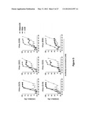 | 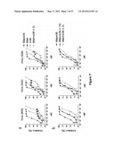 |
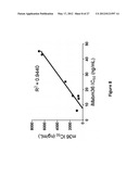 | 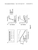 |
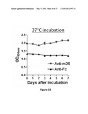 |  |
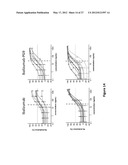 | 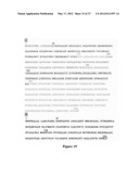 |
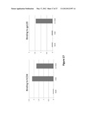 | 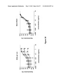 |
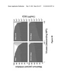 | 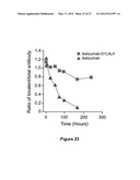 |
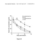 |  |
 | 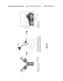 |
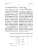 | 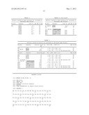 |
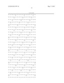 |  |
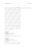 | 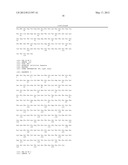 |
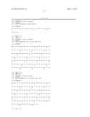 |  |
 |  |
 |  |
 |  |
 |  |
 |  |
 |  |
 |  |
 | 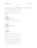 |
 | 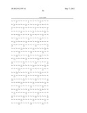 |
 | 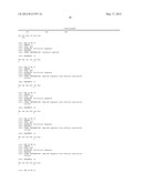 |
 | 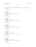 |
 | 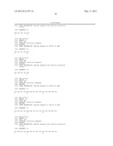 |
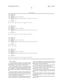 | 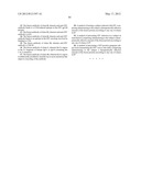 |
| Similar patent applications: | |
| Date | Title |
|---|---|
| 2010-01-21 | Fusion proteins of mycobacterium tuberculosis |
| 2010-07-22 | Fusion proteins of mycobacterium tuberculosis |
| 2010-07-22 | Fusion proteins of mycobacterium tuberculosis |
| 2010-06-17 | Fusion protein capable of binding vegf-a and tnf-alpha |
| 2009-05-28 | Ubiquitin-like protein isg15 for hiv inhibition |
| New patent applications in this class: | |
| Date | Title |
|---|---|
| 2022-05-05 | Bispecific her2 ligands for cancer therapy |
| 2022-05-05 | Antibody that binds to human pd-l1 |
| 2022-05-05 | Methods and compositions to improve the safety and efficacy of cellular therapies |
| 2022-05-05 | Multivalent particles compositions and methods of use |
| 2019-05-16 | Anti-dr5 antibodies and methods of use thereof |
| Top Inventors for class "Drug, bio-affecting and body treating compositions" | |
| Rank | Inventor's name |
|---|---|
| 1 | David M. Goldenberg |
| 2 | Hy Si Bui |
| 3 | Lowell L. Wood, Jr. |
| 4 | Roderick A. Hyde |
| 5 | Yat Sun Or |