Patent application title: METHODS AND COMPOSITIONS FOR MODULATING CARDIAC CONTRACTILITY
Inventors:
Gianluigi Condorelli (Rome, IT)
Deniele Catalucci (Milan, IT)
Assignees:
CONSIGLIO NAZIONALE DELLE RICERCHE
IPC8 Class: AA61K3817FI
USPC Class:
4241781
Class name: Drug, bio-affecting and body treating compositions conjugate or complex of monoclonal or polyclonal antibody, immunoglobulin, or fragment thereof with nonimmunoglobulin material
Publication date: 2012-03-22
Patent application number: 20120070451
Abstract:
Provide is a Cavβ2 peptide or variant thereof, or synthetic
molecules, or polynucleotides encoding said peptide or variant, for use
in the modulation of cardiac inotropism. Also provided are compositions
and methods of treatment comprising said Cavβ2 peptide,
polynucleotide or variants thereof.Claims:
1-23. (canceled)
24. A method of treatment or prophylaxis, comprising administering to a subject a Cavβ2 peptide or variant thereof, or a polynucleotide encoding the Cavβ2 peptide or variant thereof, wherein the subject is a subject in need of modulation of cardiac inotropism.
25. The method of claim 24, wherein the Cavβ2 peptide or variant thereof comprises an Akt consensus sequence, the consensus sequence comprising the sequence set forth as SEQ ID NO: 23 (N'-RTDRS-C').
26. The method of claim 24, wherein the Cavβ2 peptide or variant thereof comprises a "coiled-coil" region, the region comprising the sequence set forth as SEQ ID NO: 3 or 4.
27. The method of claim 24, wherein the Cavβ2 peptide or variant thereof comprises the full length Cavβ2 peptide sequence set forth as SEQ ID NO: 1 or SEQ ID NO: 2.
28. The method of claim 24, wherein the Cavβ2 peptide or variant thereof is a functional mimetic of the Cavβ2 peptide capable of modulation of cardiac inotropism.
29. The method of claim 28, wherein the mimetic mimics a phosphorylation of Cavβ2.
30. The method of claim 28, wherein the mimetic mimics Cavβ2 in its un-phosphorylated state.
31. The method of claim 28, wherein the mimetic is a synthetic molecule that mimics the effect of phosphorylated Cavβ2 or un-phosphorylated Cavβ2.
32. The method of claim 24, wherein the Cavβ2 peptide or variant thereof is capable of preventing proteolytic degradation of Cavα1.
33. The method of claim 32, wherein the Cavβ2 peptide or variant is capable of preventing proteolytic degradation of a PEST sequence in Cavα1.
34. The method of claim 24, wherein the Cavβ2 peptide or variant thereof is a variant containing a mutation of a phosphorylation site.
35. The method of claim 34, wherein the phosphorylation site is a Serine or Threonine residue.
36. The method of claim 35, wherein the variant contains the sequence set forth as SEQ ID NO: 1 or 2, with a mutation selected from S625A, S625E, and S625D.
37. The method of claim 24, wherein the Cavβ2 peptide, variant, or polynucleotide is a peptide or variant thereof, which is conjugated or coupled to a protein, antibody, or non-peptide synthetic molecule capable of directing the peptide or variant to a specific cell or tissue.
38. The method of claim 24, wherein the Cavβ2 peptide, variant, or polynucleotide comprises a polynucleotide, which polynucleotide comprises DNA, RNA, or a mixture thereof, and encodes the Cavβ2 peptide or variant, optionally under the control of a suitable promoter.
39. The method of claim 38, wherein the polynucleotide comprises or encodes an antisense polynucleotide, an RNAi polynucleotide, an siRNA polynucleotide, or a microRNA polynucleotide.
40. The method of claim 24, wherein the subject has dilated cardiomyopathy or cardiac hypertrophy and failure, which is primitive or has occurred after myocardial infarction.
41. A protein or polynucleotide, comprising a PEST-binding factor, which is capable of binding to a PEST sequence of a Cavα1 polypeptide and preventing the degradation of the Cavα1 polypeptide.
42. A polypeptide, comprising a variant of a Cavα1 polypeptide, in which either the I-II (Cavα1-.DELTA.P) or II-III (Cavα1-.DELTA.H) cytosolic linker region of the Cavα1 polypeptide has been mutated.
43. The polypeptide of claim 42, wherein the polypeptide does not contain the P sequence set forth as SEQ ID NO: 18, does not contain the P sequence set forth as SEQ ID NO: 19, or does not contain the H sequence set forth as SEQ ID NO: 20.
Description:
FIELD OF THE INVENTION
[0001] The present invention relates to a Cavβ2 peptide or functional variant thereof, or polynucleotides encoding said peptide or variant, for use in the modulation of cardiac inotropism or cardiac contractility.
BACKGROUND TO THE INVENTION
[0002] The insulin-like growth factor-1 (IGF-1)/phosphatidyl-inositol 3-kinase (PI3K)/Akt pathway plays a crucial role in a broad range of biological processes involved in the modulation of local responses as well as processes implicated in metabolism, cell proliferation, transcription, translation, apoptosis, and growth. In the heart, the IGF-1/PI3K/Akt pathway is involved in the regulation of contractile function and impairment of this signaling pathway is considered an important determinant of cardiac function (Catalucci and Condorelli, 2006; Ceci et al., 2004; Condorelli et al., 2002; McMullen et al., 2004; McMullen et al., 2003; Sun et al., 2006).
[0003] The Akt (also called PKB) family of serine/threonine kinases consists of 3 isoforms (Akt-1, -2, and -3) that are activated by IGF-1 and insulin through PI3K, a member of the lipid kinase family involved in the phosphorylation of membrane phosphoinositides (Ceci et al., 2004). The product of PI3K binds to the pleckstrin domain of Akt and induces its translocation from the cytosol to the plasma membrane where Akt becomes accessible for phosphorylation by phosphoinositide-dependent kinase-1 (PDK1), resulting in its activation (Bayascas et al., 2008; Ceci et al., 2004). The Ca2+ current (I.sub.Ca,L) in both cardiomyocytes and neuronal cells has been shown to be increased by Akt activation (Blair et al., 1999; Catalucci and Condorelli, 2006; Sun et al., 2006; Viard et al., 2004) and decreased by Akt inhibition (Catalucci and Condorelli, 2006; Sun et al., 2006; Viard et al., 2004), suggesting a pivotal role of Akt in regulating L-type Ca2+ channel complex (LTCC) function.
[0004] In cardiomyocytes, the LTCC is composed of different subunits: the pore-forming subunit Cavα1, and the accessory β, and α2δ subunits (Bourinet et al., 2004; Catterall, 2000). The opening of the LTCC is primarily regulated by the membrane potential and by other factors, including a variety of hormones, protein kinases, phosphatases, and accessory proteins (Bodi et al., 2005). In healthy cardiomyocytes, electrical excitation starting during the upstroke of the action potential leads to cytosolic Ca2+ influx through opening of the LTCC (Bers and Perez-Reyes, 1999; Richard et al., 2006). This triggers the calcium-induced calcium release of intracellular Ca2+ (CICR) from the sarcoplasmic reticulum (SR) through activation of the ryanodine receptor (Ryr), eventually leading to cardiomyocyte contraction (Bers, 2002).
[0005] The importance and ubiquity of Ca2+ as an intracellular signalling molecule suggests that altered channel function could give rise to widespread cellular and organ defects. Indeed, a variety of cardiovascular diseases, including atrial fibrillation, heart failure, ischemic heart disease, Timothy syndrome, and diabetic cardiomyopathy have been related to alterations in the density or function of the LTCC (Bodi et al., 2005; Mukherjee and Spinale, 1998; Pereira et al., 2006; Quignard et al., 2001). However, the molecular basis for dysregulation of LTCC function and the possible involvement of Akt in I.sub.Ca,L (current density of L-type Ca2+ currents) regulation remains unresolved.
[0006] Viard et al., 2004, showed that Akt-dependent phosphorylation of the Cavβ2 subunit is important in promoting the chaperoning of the Cav2.2 pore-forming unit to the plasma membrane. However, this study focused on neuronal cells and on a particular isoform of Cavβ2.
[0007] There is still a need in the art, therefore, for ways in which to modulate cardiac contractility. This is useful in treating conditions of where cardiac contractility is impaired such as dilated cardiomyopahty and, in general, cardiac hypertrophy and failure, both primitive and after myocardial infarction.
[0008] US 20087/0118438 A1 (Antzelevitch and Pollevick) discloses certain mutations that lead to a loss of function in Calcium Channel peptides, said mutations incurring "sudden cardiac death." WO 2008/060618 A1 (University of Florida Research Foundation) discloses a method of identifying a subject as having a propensity to have an adverse cardiovascular event by assessing mutations in a number of genes and proteins (alpha-adducin (ADD1) gene, calcium activated potassium channel (KCNMB1) gene, Betal-adrenergic receptor (ADRB1) gene, Beth7-adrenergic receptor (ADRB2) gene, leukotriene A4 hydrolase (LT A4H), arachidonate 5-lipoxygenase-activating protein (ALOX5AP), CACNAIC, CACNB2, and ALOX5 gene) relative to a wild-type reference sequence.
[0009] Surprisingly, we have identified a novel post-translational mechanism by which Akt modulates LTCC function under physiological conditions, highlighting the pivotal role of this kinase in cardiac function. In particular, we have found that the pore-forming channel subunit Cavα1 contains highly conserved PEST sequences that direct rapid protein degradation. Akt mediated phosphorylation of the Cavβ2 LTCC-chaperone subunit prevents PEST site recognition, thereby slowing or preventing Cavα1 degradation, thus regulating Ca2+ channel function and thus alteration of cardiomyocyte contractile function. Without being bound by theory, we believe that Akt-mediated phosphorylation of the Cavβ2 subunit (the LTCC chaperone) at its C-terminal region, in particular the "coiled coil" region of Cavβ2, induces a conformational shift in Cavβ2. This shift in turn stearically hinders protease access to the PEST sequences of Cavα1, which may occur via direct association of either the C-terminal portion of or the whole Cavβ2 with cytosolic loops on Cavα1 or indirectly through the intervention of known/unknown protein partners.
SUMMARY OF THE INVENTION
[0010] Thus, in a first aspect, the invention provides a Cavβ2 peptide or variant thereof, or polynucleotides encoding said peptide or variant, for use the modulation of cardiac inotropism.
[0011] The Cavβ2 peptide may comprise the full length Cavβ2 peptide, provided in SEQ ID NO: 1 (murine) but more preferably that provided in SEQ ID NO: 2, which is human. The Akt consensus site is underlined at positions 500-504 (murine) or 502-507 (human). The serine residue that is phosphorylated by Akt and mutated in preferred embodiments of the invention is highlighted in bold at the C' terminus of the consensus sequences.
TABLE-US-00001 SEQ ID NO: 1 (murine): MVQSDTSKSPPVAAVAQESQMELLESAAPAGALGAQSYGKGARRKNRFKG SDGSTSSDTTSNSFVRQGSADSYTSRPSDSDVSLEEDREAVRREAERQAQ AQLEKAKTKPVAFAVRTNVRYSAAQEDDVPVPGMAISFEAKDFLHVKEKF NNDWWIGRLVKEGCEIGFIPSPVKLENMRLQHEQRAKQGKFYSSKSGGNS SSSLGDIVPSSRKSTPPSSAIDIDATGLDAEENDIPANHRSPKPSANSVT SPHSKEKRMPFFKKTEHTPPYDVVPSMRPVVLVGPSLKGYEVTDMMQKAL FDFLKHRFEGRISITRVTADISLAKRSVLNNPSKHAIIERSNTRSSLAEV QSEIERIFELARTLQLVVLDADTINHPAQLSKTSLAPIIVYVKISSPKVL QRLIKSRGKSQAKHLNVQMVAADKLAQCPPQESFDVILDENQLEDACEHL ADYLEAYWKATHPPSGNLPNPLLSRTLASSTLPLSPTLASNSQGSQGDQR PDRSAPRSASQAEEEPCLEPVKKSQHRSSSATHQNHRSGTGRGLSRQETF DSETQESRDSAYVEPKEDYSHEHVDRYVPHREHNHREETHSSNGHRHRES RHRSRDMGRDQDHNECIKQRSRHKSKDRYCDKEGEVISKRRNEAGEWNRD VYIRQ SEQ ID NO: 2 Human: MVQRDMSKSPPTAAAAVAQEIQMELLENVAPAGALGAAAQSYGKGARRKN RFKGSDGSTSSDTTSNSFVRQGSADSYTSRPSDSDVSLEEDREAVRREAE RQAQAQLEKAKTKPVAFAVRTNVSYSAAHEDDVPVPGMAISFEAKDFLHV KEKFNNDWWIGRLVKEGCEIGFIPSPVKLENMRLQHEQRAKQGKFYSSKS GGNSSSSLGDIVPSSRKSTPPSSAIDIDATGLDAEENDIPANHRSPKPSA NSVTSPHSKEKRMPFFKKTEHTPPYDVVPSMRPVVLVGPSLKGYEVTDMM QKALFDFLKHRFEGRISITRVTADISLAKRSVLNNPSKHAIIERSNTRSS LAEVQSEIERIFELARTLQLVVLDADTINHPAQLSKTSLAPIIVYVKISS PKVLQRLIKSRGKSQAKHLNVQMVAADKLAQCPPELFDVILDENQLEDAC EHLADYLEAYWKATHPPSSSLPNPLLSRTLATSSLPLSPTLASNSQGSQG DQRTDRSAPIRSASQAEEEPSVEPVKKSQHRSSSSAPHHNHRSGTSRGLS RQETFDSETQESRDSAYVEPKEDYSHDHVDHYASHRDHNHRDETHGSSDH RHRESRHRSRDVDREQDHNECNKQRSRHKSKDRYCEKDGEVISKKRNEAG EWNRDVYIRQ
[0012] Polynucleotides encoding these protein sequences are also envisaged.
[0013] However, the Cavβ2 peptide preferably comprises the C-terminal portion of Cavβ2, for instance that provided as SEQ ID NO: 3 and 4, representing the "coiled-coil" region of the protein, which is particularly preferred.
TABLE-US-00002 SEQ ID NO: 3 (murine): ASSTLPLSPTLASNSQGSQGDQRPDRSAPRSASQAEEEPCLEPVKKSQHR SSSATHQNHRSGTGRGLSRQETFDSETQESRDSAYVEPKEDYSHEHVDRY VPHREHNHREETHSSNGHRHRESRHRSRDMGRDQDHNECIKQRSRHKSKD RYCDKEGEVISKRRNEAGEWNRDVYIRQ SEQ ID NO: 4 (human): ATSSLPLSPT LASNSQGSQG DQRTDRSAPI RSASQAEEEP SVEPVKKSQH RSSSSAPHHN HRSGTSRGLS RQETFDSETQ ESRDSAYVEP KEDYSHDHVD HYASHRDHNH RDETHGSSDH RHRESRHRSR DVDREQDHNE CNKQRSRHKS KDRYCEKDGE VISKKRNEAG EWNRDVYIRQ
[0014] It is particularly preferred however that the Cavβ2 peptide comprises merely the Akt-consensus sequence, provided as SEQ ID NO: 23 (N'-RTDRS-C'). Preferably, the Cavβ2 peptide comprises the coiled coil region of the Cavβ2, SEQ ID NO: 3 or 4.
[0015] The variant is preferably any mimetic of the Cavβ2 peptide that is a functional variant, i.e. capable of modulation of cardiac inotropism. This variant may mimic the phosphorylation of Cavβ2 or, alternatively, mimic Cavβ2 in its native, un-phosphorylated state. In this regard, the phosphorylated Cavβ2 mimic may be considered an agonist of phosphorylated Cavβ2, whilst the un-phosphorylated Cavβ2 mimic may be considered an antagonist of phosphorylated Cavβ2. It will be appreciated that the variant may not only be a peptide but also a synthetic molecule mimicking the effect of a peptide. It is also preferred that these variants may be designed and/or locked into a particular structural conformation to increase the specificity of binding to Cavα1, Cavβ2 or any other interacting partners.
[0016] It is particularly preferred that the peptide or variant is capable of preventing proteolytic degradation of Cavα1 and, in particular its PEST sequences. Suitable PEST sequences are provided in Table 1.
[0017] It will be appreciated that the terms modulation of cardiac inotropism and cardiac contractility can be interchanged. De-stabilisation of the calcium channel will lead to a reduced calcium flux therethrough and a resulting decrease in cardiac contractility. When Cavβ2 is phosphorylated, or a modulator (such as a synthetic molecule) mimicking Cavβ2 phosphorylation is provided, then the LTCC is stablised, thereby providing at least basal, and preferably enhanced, cardiac inotropism. Similarly, when the PEST sequences of Cavα1 are exposed to the cellular degradation machinery, for instance when Cavβ2 is not phosphorylated or a modulator mimicking Cavβ2 in its un-phosphorylation state is provided, then the LTCC is de-stablised, thereby providing at least reduced cardiac inotropism.
[0018] Suitable agonists will therefore increase cardiac inotropism, whilst a suitable antagonist will decrease cardiac inotropism.
[0019] The variant has preferably at least 70% sequence homology, more preferably at least 75% sequence homology, more preferably at least 80% sequence homology, more preferably at least 85% sequence homology, more preferably at least 90% sequence homology, more preferably at least 95% sequence homology, more preferably at least 99% sequence homology and most preferably at least 99.5% sequence homology with SEQ ID NOs 1 and 2. In each case, it is preferred that that the variant comprises the Akt consensus sequence (SEQ ID NO: 3). The same applies for any nucleotide sequences, although it will be appreciated that these may also be capable of hybridizing to the reference sequence under highly stringent conditions, such as washing in 6×SSC. Conservative substitutions are also envisaged in the peptide or nucleotide variants.
[0020] Particularly preferred variants are those where the putative phosphorylation site, preferably a Ser or Thr residue has been mutated. Suitable examples include S625A, S625E and: S625D (referring to the numbering in SEQ ID NO: 1) or positions corresponding thereto. Also preferred are variants where the Akt binding site has been mutated or is stearically hindered by the mutation to prevent Akt binding to, and therefore phosphorylating, Cavβ2.
[0021] The peptide or variants may be conjugated or coupled to another protein, antibody, or any other molecule capable of directing the peptide or variant to a specific cell type (tissue specificity), or non-peptide synthetic molecules mimicking the effects of these peptides. This also encompasses a fusion protein, which can be encoded with the present peptide or variant by the same polynucleotide.
[0022] The polynucleotides of the invention may be DNA or RNA or mixtures of both. Preferably, the polynucleotides encode the Cavβ2 peptide or variant under the control of a suitable promoter or a system such as the tet operon system that allows the user a degree of control over the expression of the system. Suitable promoters include a cardiac specific promoter (such as myosin heavy chain, Troponin I, for instance), which might be used to redirect the expression specifically to the myocardium.
[0023] The polynucleotides may comprise or encode antisense polynucleotides or RNAi, such as siRNA or microRNA. The microRNA may be specific for the 3'UTR of Akt or PDK1, but is most preferably specific for the 3'UTR of Cavβ2.
[0024] Delivery of the polynucleotides may be via a plasmid or suitable vector, such as an adeno-, retro- or lenti-viral vector. Thus, the invention also provides a vector comprising the polynucleotides encoding the Cavβ2 peptide or variant. The vector may be delivered by a "gene-gun," by electroporation or in the form of a pharmaceutically acceptable formulation. It may be administered to a mucosal lining, for instance orally, nasally or rectally or parentarally (i.e. not through the alimentary canal but rather by injection subcutaneously, intramuscularly, intraorbitally, intracapsularly, intraspinally, intrasternally, or intravenously).
[0025] Preferred levels of the peptide for administration and/or expression are in the region of 1-10 mg/kg to 1-10 μg/kg, although this will be readily determined by a physician.
[0026] In a further aspect, the invention also provides a pharmaceutical composition comprising or encoding a Cavβ2 peptide or polynucleotide, or functional variants thereof. Preferably, the Cavβ2 peptide, polynucleotide or variant comprises a mutation in the Akt consensus site, as discussed above, and most preferably corresponding to Ser625 in Cavβ2.
[0027] It will be appreciated that the Cavβ2 peptide or polynucleotide variant has cardiac inotropism/contractility modulating activity.
[0028] A further aspect of the invention is a cell, preferably a cardiomyocyte, comprising the present Cavβ2 peptide, polynucleotide or variants. The cell has preferably been transformed, by a means of delivery discussed above, to express the Cavβ2 peptide, polynucleotide or variants.
[0029] In a further aspect, the invention provides a PEST binding factor, such as a protein or polynucleotides, capable of binding to the PEST sequences of Cavα1 to prevent degradation thereof. Such a protein may have a large PEG molecule attached thereto or is a glycoprotein, the PEG or sugar unit(s) hindering the access of the PEST degraders.
BRIEF DESCRIPTION OF THE FIGURES
[0030] FIG. 1. Alteration of Ca2+ handling proteins in PDK1 KO cardiomyocytes. (A) Western blot and (B) densitometric analyses of ventricular homogenates along a time course of tamoxifen inductions (day 1 to 6 treatment is indicated by the bar) using various antibodies. A representative experiment is shown (n=3). (C) Total Akt activity in WT and KO cardiomyocyte lysates assayed using a GSK313/a Akt-specific substrate.
[0031] FIG. 2. Impaired intracellular Ca2+ handling and contractility in PDK1 KO cardiomyocytes. (A-B) Smaller Ca2+ current in KO cardiomyocytes: (A) Whole-cell representative I.sub.Ca,L currents normalized for difference in cell size. (B) I.sub.Ca,L I-V current/voltage relationships (n=12) (*P<0.05, **P<0.01). (C-D) Cardiomyocyte contraction and Ca2+ transients at different stimulation frequencies. (C) Cardiomyocyte shortening is decreased in KO compared to WT cardiomyocytes (*P<0.05 ANOVA). (D) Ca2+-frequency relationship indicates smaller peak systolic but not diastolic Ca2+ in KO compared to WT cells (*P<0.05, ANOVA).
[0032] FIG. 3. Akt mediates regulation of Cavα1 protein density at the plasma membrane. (A) RT-PCR analysis of Cavα1 mRNA expression from WT and KO ventricular extracts. GADPH serves as loading control. (B) Western blot analysis of whole lysate, membrane, and microsomal fractions from WT and KO ventricular extracts. (C) YFP-Cavα1 transfected COS-7 cells alone or in combination with Cavβ2-expression vector were serum-starved and treated with Akt inhibitor and 1 μM bafilomycin-A1, 25 μM MG132, or 25 μM calpeptin. 6 h post drug administration, cell lysates were prepared and subjected to Western blot analysis for YFP. GAPDH served as a loading control. (D-E) Cavα1 protein levels in KO cardiomyocytes infected with empty (mock) or active E40K-Akt (AdAkt) expressing adenoviral vector (D) and in whole lysates of WT and E40K-Akt (Tg Akt) hearts (E). Representative experiments are shown (n=4).
[0033] FIG. 4. Akt interacts with and phosphorylates Cavβ2. (A) Coimmunoprecipitation assay of Akt and Cavβ2. Ventricular homogenates from WT and HA-E40K-Akt transgenic mice (Tg Akt) immunoprecipitated with antibodies against HA and immunoblotted for Cavβ2 as well as HA as a control. (B) Examination of Cavβ2 phosphorylation by Akt. In vitro kinase assays were performed with immunoprecipitated Cavβ2 incubated with recombinant active Akt and 32P labeled ATP (left) or immunoprecipitated Cavβ2 from WT and KO cardiac extracts from mice treated or not with insulin (1 mU/g) using PAS (Phospho-Akt Substrate) antibody (right). (C) Back-phosphorylation assay of Cavβ2 from WT and KO hearts. Immunoprecipitated Cavβ2 from solubilized membranes was in vitro back-phosphorylated using recombinant active Akt and [γ32]ATP. Precipitate amounts were assayed for 32PCavβ2 and total Cavβ2. Representative experiments are shown (n=4).
[0034] FIG. 5. Akt phosphorylation of Cavβ2 protects Cavα1 from protein degradation. (A-C) YFP-Cavα1 co-transfected 293T cells with the indicated mutant variant of Cavβ2. Cells were serum-starved overnight and treated with (A) 100 μM insulin or (B-C) 5 μM Akt inhibitor as indicated. The expression of YFP-Cavα1 in lysates was monitored by Western blot analysis with anti-YFP antibody, and normalized based on transfection efficiency (Cav(32) and protein amount (Tubulin) (n=3). (D) Cavα1 and Cavβ2 co-transfected 293T cells were treated with siAkt expressing vector as indicated. 3 days post transfection, cell lysate was tested by Western blot analysis. Protein loading was normalized to GAPDH levels. Representative experiments are shown (n=3).
[0035] FIG. 6. Akt phosphorylation of Cavβ2 preserves Cavα1 currents. Ca2+ currents recorded in cotransfected tsA-201 cells with YFP-Cavα1 and either Cavβ2-WT, Cavβ2-SE, or Cavβ2-SA mutant, cultivated for 36 h in the presence or absence of fetal bovine serum (10%). Currents were recorded 1-2 minutes after the whole-cell configuration was achieved (i.e. after stabilization of the current) and were elicited by a 0 mV depolarization of 200 ms duration applied from a holding potential of -80 mV. Currents are normalized to cell capacitance (Current density, pA/pF). Representative current traces are shown. n>35 at each condition, (*P<0.05 compared to YFP-Cavα1, ANOVA).
[0036] FIG. 7. Rapid-protein-degradation PEST sequences determine Cavα1 protein instability. (A) Schematic representation of Cavα1 mapping the α1-interacting domain (AID) and PEST sequences in the I-II and II-III cytosolic loops. Deleted PEST sequences (P, H) are highlighted in red. (B) Western blot and immunofluorescence analyses showing relative levels of WT and PEST deleted mutants of YFP-Cavα1 (n=3). Bar represents 5 μm. (C) Wild-type Cavα1 subunit (alone or cotransfected with Cavβ2SE) and its in-frame ΔPEST mutants (Cavα1-ΔP and Cavα1-ΔH) half-lives were determined in COST cells. After overnight starvation, transfected cells were pulse-chased and analyzed along a time course (*P<0.001 compared to Cavα1, ANOVA; n=3). (D) Western blot and immunofluorescence analyses showing relative levels of WT GFP and N-terminal fusion PEST mutants (n=3). Bar represents 20 μm. (E) The Cavα1 C-terminus interacts with the Akt-phosphorylated-GST-Cavβ2 coiled coil region. Bacterially expressed GST or GST-C-Cavβ2 (Cavβ2: aa 480-655) fusion protein and glutathione sepharose beads were incubated with equal amounts of in vitro translated [35S] methionine-labeled C-Cavα1 (Cavα1: aa 1477-2169). Binding occurred only with Akt-phosphorylated GST-C-Cavβ2. Bound proteins were resolved by SDS-PAGE (4-12%). 10% of the input protein in each binding reaction is shown. Coomassie staining of SDS-PAGE is shown in the bottom panel. (F) Ca2+ currents recorded in tsA-201 cells cotransfected with Cavβ2-WT and either Cavα1-WT or Cavα1-ΔH and cultivated for 36 h in the presence or absence of fetal bovine serum (10%). Current densities (pA/pF) are normalized to the control condition. n>35 at each condition, (*P<0.05 compared to Cavα1, ANOVA).
[0037] FIG. 8. Proposed mechanism. Akt, followed by PDK1 activation, phopshorylates Cavβ2 at the C-terminal coiled-coil domain. The phosphorylation allows association of the C-terminal portion of Cavβ2 with the Cavα1 C-terminal domain. A conformation shift, in turns, prevents PEST sequence recognition, stabilizing Cavα1 protein levels. Blue and red ribbon in Cavα1 represent AID and PEST sequences, respectively.
[0038] FIG. 9. Characterization of mice lacking PDK1 expression. (A) PDK protein and RNA levels assessed by Western blot (upper) and RT-PCR (lower) analyses of atria, left (LV), and right (RV) ventricular cardiomyocytes from WT and KO mice. Protein and RNA loading was normalized to GAPDH levels, respectively. (B) Immunofluorescence staining of cardiomyocytes isolated from WT and KO hearts labeled with antibody against PDK1 (green) and counterstained with Hoechst nuclear stain (blue). Bar represent 15 μm. (C) Survival curve for mice lacking PDK1 (KO) in the heart. Mortality begins 5 days after tamoxifen injection and reaches 100% by day 10 after beginning of treatment. Time points of tamoxifen injections and echocardiography analysis (arrows) are shown (n=10). (D) Echocardiographic (M-mode) assessment of left ventricular size and function. Left ventricular diastolic internal dimensions LVIDd (red bar) and left ventricular systolic internal dimensions LVIDs (blue bar) were increased in KO mice. Heart rates were 486 and 511 bpm, respectively. (E) H&E-stained paraffin sections show severe dilatation and thinning of KO hearts. (F) Western blot analysis for caspase 3 activation in WT and KO heart homogenates. Basal apoptotic activation as a consequence of tamoxifen treatment was also observed in WT (as previously observed in supplementary ref (Zartman et al., 2004)). Amounts of loaded protein were verified with tubulin antibodies. (G) Representative Masson's trichrome staining of tissue sections from WT and KO hearts.
[0039] FIG. 10. Regulation of Ca2+ handling proteins by Akt. (A) Western blot analysis of ventricular homogenates from WT and KO mice. (B) Western blot analysis of ventricular homogenates along a time course of tamoxifen inductions (day 1 to 6 treatment is indicated by the bar) using various antibodies. A representative experiment is shown (n=3).
[0040] FIG. 11. Altered intracellular calcium handling in PDK1 KO cardiomyocytes. (A) Representative Ca2+ traces are shown for WT (upper) and KO (lower) cardiomyocytes. (B) Reduced averaged twitch Ca2+ transient amplitude is shown in KO compared to WT cardiomyocytes (left, unpaired t test, P<0.05). No difference was found in total SR Ca2+ analysis (right). WT and KO results are shown in blue and red respectively.
[0041] FIG. 12. Cavβ2 interacts with Akt isoforms and Akt affects Cavα1 protein stability. (A) Immunoprecipitated Akt isoforms from WT cardiac extracts from mice treated or not with insulin (1 mU/g) assayed for Cavβ2. Input protein in each co-immunoprecipitation is shown. (B) Cavα1, Cavα1-ΔP, or Cavα1-ΔH co-transfected 293T cells with either Cavβ2 or Cavβ2-SE were infected with indicated active (AdAkt) or dominant negative (AdAktDN) Akt expressing adenoviral vectors. Cells were serum-starved overnight as indicated. The expression of Cavα1 and phosphorylation of GSK in lysates was monitored by Western blot analysis. (C) Cavα1, Cavα1-ΔP, or Cavα1 co-transfected 293T cells with either Cavβ2 or Cavβ2-SE were treated with siAkt expressing vector as indicated. 3 days post transfection, cells were lysated for protein extraction. Protein loading was normalized to GAPDH levels. Representative experiments are shown (n=3).
[0042] FIG. 13. Serum deprivation and PEST-H deletion does not modify steady-state activation parameters. Ca2+ currents recorded in cotransfected tsA-201 cells with Cavβ2-WT and either Cavα1-WT or Cavα1-OH, and cultivated for 36 h in the presence or absence of fetal bovine serum (10%). IV curves are normalized to the maximal current. n>35 at each condition, (ANOVA).
DETAILED DESCRIPTION OF THE INVENTION
[0043] The present inventors have also shown through interaction experiments that phosphorylated--Cavβ2 binds only to Cavα1 C-terminal tail. Without being bound by theory, therefore, they hypothesize that the binding induces conformational changes in the alpha1 protein, thus covering PEST sequences. Therefore, the present invention encompasses any peptide or variant that binds the Cavα1 C-terminal tail region.
[0044] By degradation, it will be appreciated that this refers to proteolytic action, i.e. cleavage of the PEST sequences by cellular machinery, such as proteosomes.
[0045] It is particularly preferred that the variant is capable of preventing proteolytic degradation of Cavα1 and, in particular its PEST sequences. Suitable PEST sequences are any of provided in Table 1.
[0046] Also provided are the Cavβ2 peptide or variant or polynucleotides for use in therapy, and the Cavβ2 peptide or variant or polynucleotides for use in treating a condition associated with, or treatable by, cardiac contractility modulation. Methods of treatment or prophylaxis comprising administering the Cavβ2 peptide or variant or polynucleotides to a patient in need thereof are also provided. Preferred conditions are those associated with cardiac inotropism or cardiac contractility. Preferred examples include dilated cardiomyopahty and cardiac hypertrophy and failure, both primitive and after myocardial infarction. As mentioned above, it will be appreciated that this extends to synthetic molecules.
[0047] The invention also provides Cavα1 variants in which either the I-II (Cavα1-ΔP) or II-III (Cavα1-ΔH) cytosolic linker region of Cavα1 has been mutated, preferably by an in-frame deletion. These Cavα1 mutants at lack one or more, and preferably at least 50% and more preferably at least 75% or even all their PEST sequences. They may be delivered in the same manner as discussed above.
TABLE-US-00003 P sequence (mouse): KGYLDWITQAEDIDPENEDEGMDEDK (SEQ ID NO: 18) P sequence (human): KGYLDWITQAEDIDPENEDEGMDEEK (SEQ ID NO: 19) H sequence (mouse and human): GEEDEEEPEMPVGPR (SEQ ID NO: 20)
[0048] SEQ ID NO: 21 is the Cavα1-ΔH (mouse) sequence, wherein the H sequence (to be removed) is underlined and placed in bold (at positions 844-858 below):
TABLE-US-00004 MVNENTRMYVPEENHQGSNYGSPRPAHANMNANAAAGLAPEHIPTPGAAL SWQAAIDAARQAKLMGSAGNATISTVSSTQRKRQQYGKPKKQGGTTATRP PRALLCLTLKNPIRRACISIVEWKPFEIIILLTIFANCVALAIYIPFPED DSNATNSNLERVEYLFLIIFTVEAFLKVIAYGLLFHPNAYLRNGWNLLDF IIVVVGLFSAILEQATKADGANALGGKGAGFDVKALRAFRVLRPLRLVSG VPSLQVVLNSIIKAMVPLLHIALLVLFVIIIYAIIGLELFMGKMHKTCYN QEGIIDVPAEEDPSPCALETGHGRQCQNGTVCKPGWDGPKHGITNFDNFA FAMLTVFQCITMEGWTDVLYWMQDAMGYELPWVYFVSLVIFGSFFVLNLV LGVLSGEFSKEREKAKARGDFQKLREKQQLEEDLKGYLDWITQAEDIDPE NEDEGMDEDKPRNMSMPTSETESVNTENVAGGDIEGENCGARLAHRISKS KFSRYWRRWNRFCRRKCRAAVKSNVFYWLVIFLVFLNTLTIASEHYNQPH WLTEVQDTANKALLALFTAEMLLKMYSLGLQAYFVSLFNRFDCFIVCGGI LETILVETKIMSPLGISVLRCVRLLRIFKITRYWNSLSNLVASLLNSVRS IASLLLLLFLFIIIFSLLGMQLFGGKFNFDEMQTRRSTFDNFPQSLLTVF QILTGEDWNSVMYDGIMAYGGPSFPGMLVCIYFIILFICGNYILLNVFLA IAVDNLADAESLTSAQKEEEEEKERKKLARTASPEKKQEVMEKPAVEESK EEKIELKSITADGESPPTTKINMDDLQPSENEDKSPHSNPDTAGEEDEEE PEMPVGPRPRPLSELHLKEKAVPMPEASAFFIFSPNNRFRLQCHRIVNDT IFTNLILFFILLSSISLAAEDPVQHTSFRNHILGNADYVFTSIFTLEIIL KMTAYGAFLHKGSFCRNYFNILDLLVVSVSLISFGIQSSAINVVKILRVL RVLRPLRAINRAKGLKHVVQCVFVAIRTIGNIVIVTTLLQFMFACIGVQL FKGKLYTCSDSSKQTEAECKGNYITYKDGEVDHPIIQPRSWENSKFDFDN VLAAMMALFTVSTFEGWPELLYRSIDSHTEDKGPIYNYRVEISIFFIIYI IIIAFFMMNIFVGFVIVTFQEQGEQEYKNCELDKNQRQCVEYALKARPLR RYIPKNQHQYKVWYVVNSTYFEYLMFVLILLNTICLAMQHYGQSCLFKIA MNILNMLFTGLFTVEMILKLIAFKPKHYFCDAWNTFDALIVVGSIVDIAI TEVHPAEHTQCSPSMSAEENSRISITFFRLFRVMRLVKLLSRGEGIRTLL WTFIKSFQALPYVALLIVMLFFIYAVIGMQVFGKIALNDTTEINRNNNFQ TFPQAVLLLFRCATGEAWQDIMLACMPGKKCAPESEPSNSTEGETPCGSS FAVFYFISFYMLCAFLIINLFVAVIMDNFDYLTRDWSILGPHHLDEFKRI WAEYDPEAKGRIKHLDVVTLLRRIQPPLGFGKLCPHRVACKRLVSMNMPL NSDGTVMFNATLFALVRTALRIKTEGNLEQANEELRAIIKKIWKRTSMKL LDQVVPPAGDDEVTVGKFYATFLIQEYFRKFKKRKEQGLVGKPSQRNALS LQAGLRTLHDIGPEIRRAISGDLTAEEELDKAMKEAVSAASEDDIFRRAG GLFGNHVTYYQSDSRGNFPQTFATQRPLHINKTGNNQADTESPSHEKLVD STFTPSSYSSTGSNANINNANNTALGRFPHPAGYSSTVSTVEGHGPPLSP AVRVQEAAWKLSSKRCHSRESQGATVNQEIFPDETRSVRMSEEAEYCSEP SLLSTDMFSYQEDEHRQLTCPEEDKREIQPSPKRSFLRSASLGRRASFHL ECLKRQKDQGGDISQKTALPLHLVHHQALAVAGLSPLLQRSHSPTTFPRP CPTPPVTPGSRGRPLRPIPTLRLEGAESSEKLNSSFPSIHCSSWSEETTA CSGSSSMARRARPVSLTVPSQAGAPGRQFHGSASSLVEAVLISEGLGQFA QDPKFIEVTTQELADACDMTIEEMENAADNILSGGAQQSPNGTLLPFVNC RDPGQDRAVVPEDESCAYALGRGRSEEALADSRSYVSNL
[0049] SEQ ID NO: 22 is the Cavα1-ΔH (human) sequence, wherein the H sequence (to be removed) is underlined and placed in bold (at positions 841-855 below):
TABLE-US-00005 MVNENTRMYIPEENHQGSNYGSPRPAHANMNANAAAGLAPEHIPTPGAAL SWQAAIDAARQAKLMGSAGNATISTVSSTQRKRQQYGKPKKQGSTTATRP PRALLCLTLKNPIRRACISIVEWKPFEIIILLTIFANCVALAIYIPFPED DSNATNSNLERVEYLFLIIFTVEAFLKVIAYGLLFHPNAYLRNGWNLLDF IIVVVGLFSAILEQATKADGANALGGKGAGEDVKALRAFRVLRPLRLVSG VPSLQVVLNSIIKAMVPLLHIALLVLFVIIIYAIIGLELFMGKMHKTCYN QEGIAAEDDPSPCALETGHGRQCQNGTVCKPGWDGPKHGITNFDNFAFAM LTVFQCITMEGWTDVLYWVNDAVGRDWPWIYFVTLIIIGSFFVLNLVLGV LSGEFSKEREKAKARGDFQKLREKQQLEEDLKGYLDWITQAEDIDPENED EGMDEEKPRNMSMPTSETESVNTENVAGGDIEGENCGARLAHRISKSKFS RYWRRWNRFCRRKCRAAVKSNVFYWLVIFLVFLNTLTIASEHYNQPNWLT EVQDTANKALLALFTAEMLLKMYSLGLQAYFVSLFNRFDCFVVCGGILET ILVETKIMSPLGISVLRCVRLLRIFKITRYWNSLSNLVASLLNSVRSIAS LLLLLFLFIIIFSLLGMQLFGGKFNFDEMQTRRSTFDNFPQSLLTVFQIL TGEDWNSVMYDGIMAYGGPSFPGMLVCIYFIILFICGNYILLNVFLAIAV DNLADAESLTSAQKEEEEEKERKKLARTASPEKKQELVEKPAVGESKEEK IELKSITADGESPPATKINMDDLQPNENEDKSPYPNPETTGEEDEEEPEM PVGPRPRPLSELHLKEKAVPMPEASAFFIFSSNNRFRLQCHRIVNDTIFT NLILFFILLSSISLAAEDPVQHTSFRNHILFYFDIVFTTIFTIEIALKMT AYGAFLHKGSFCRNYFNILDLLVVSVSLISFGIQSSAINVVKILRVLRVL RPLRAINRAKGLKHVVQCVFVAIRTIGNIVIVTTLLQFMFACIGVQLFKG KLYTCSDSSKQTEAECKGNYITYKDGEVDHPIIQPRSWENSKFDFDNVLA AMMALFTVSTFEGWPELLYRSIDSHTEDKGPIYNYRVEISIFFIIYIIII AFFMMNIFVGFVIVTFQEQGEQEYKNCELDKNQRQCVEYALKARPLRRYI PKNQHQYKVWYVVNSTYFEYLMFVLILLNTICLAMQHYGQSCLFKIAMNI LNMLFTGLFTVEMILKLIAFKPKGYFSDPWNVFDFLIVIGSIIDVILSET NPAEHTQCSPSMNAEENSRISITFFRLFRVMRLVKLLSRGEGIRTLLWTF IKSFQALPYVVLLIVMLFFIYAVIGMQVFGKIALNDTTEINRNNNFQTFP QAVLLLFRCATGEAWQDIMLACMPGKKCAPESEPSNSTEGETPCGSSFAV FYFISFYMLCAFLIINLFVAVIMDNFDYLTRDWSILGPHHLDEFKRIWAE YDPEAKGRIKHLDVVTLLRRIQPPLGFGKLCPHRVACKRLVSMNMPLNSD GTVMFNATLFALVRTALRIKTEGNLEQANEELRAIIKKIWKRTSMKLLDQ VVPPAGDDEVTVGKFYATFLIQEYFRKFKKRKEQGLVGKPSQRNALSLQA GLRTLHDIGPEIRRAISGDLTAEEELDKAMKEAVSAASEDDIFRRAGGLF GNHVSYYQSDGRSAFPQTFTTQRPLHINKAGSSQGDTESPSHEKLVDSTF TPSSYSSTGSNANINNANNTALGRLPRPAGYPSTVSTVEGHGPPLSPAIR VQEVAWKLSSNRCHSRESQAAMAGQEETSQDETYEVKMNHDTEACSEPSL LSTEMLSYQDDENRQLTLPEEDKRDIRQSPKRGFLRSASLGRRASFHLEC LKRQKDRGGDISQKTVLPLHLVHHQALAVAGLSPLLQRSHSPASFPRPFA TPPATPGSRGWPPQPVPTLRLEGVESSEKLNSSFPSIHCGSWAETTPGGG GSSAARRVRPVSLMVPSQAGAPGRQFHGSASSLVEAVLISEGLGQFAQDP KFIEVTTQELADACDMTIEEMESAADNILSGGAPQSPNGALLPFVNCRDA GQDRAGGEEDAGCVRARGRPSEEELQDSRVYVSSL
[0050] Where reference is made herein to a particular position, it will be appreciated that this also refers to the equivalent position of such a feature or motif in a similar or variant sequence.
[0051] In short, the insulin IGF-1/PI3K/Akt signalling pathway has been suggested to improve cardiac inotropism and increase Ca2+ handling through the effects of the protein kinase Akt. However, to date the even the basic underlying molecular mechanisms behind the function of the myocyte Calcium channel remain largely unknown. However, we have found that Akt has an unanticipated regulatory function in controlling L-type Ca2+ channel (LTCC) protein density. Furthermore, we have surprisingly found that the pore-forming channel subunit Cavα1 contains highly conserved PEST sequences (signals for rapid protein degradation). In-frame deletion of these PEST sequences result in increased Cavα1 protein levels. Our findings show that Akt-dependent phosphorylation of Cavβ2, the LTCC chaperone for Cavα1, antagonizes Cavα1 protein degradation by preventing Cavα1-PEST sequence recognition. This leads to increased LTCC density and consequent modulation of Ca2+ channel function. This novel mechanism by which Akt modulates LTCC stability could profoundly influence cardiac myocyte Ca2+ entry, Ca2+ handling, and contractility.
[0052] Without being bound by theory, we believe that Akt-mediated phosphorylation of the Cavβ2 subunit, i.e. the LTCC chaperone, at its C-terminal region, in particular the "coiled coil" region of Cavβ2, induces a conformational shift in Cavβ2. This shift in turn stearically hinders protease access to the PEST sequences of Caval, which may occur via direct association of either the C-terminal portion of or the whole Cavβ2 with cytosolic loops on Cavα1 or indirectly through the intervention of protein partners.
[0053] This study reveals a mechanism through which the insulin IGF-1/PI3K/PDK1/Akt pathway can sustain or modulate Ca2+ entry in cardiac cells via the voltage-gated LTCC and eventually affect cardiac contractility. Using a mouse model with an inducible and cardiomyocyte-specific deletion of the upstream activator PDK1, we showed that Akt is of key importance for the structural organization and functionality of the LTCC complex at the plasma membrane. This regulation of LTCC activity is directly related to the Akt-mediated phosphorylation of the accessory subunit Cavβ2, which in turn results in increased protein density of the pore-forming Cavα1 subunit through protection of PEST sequences from the proteolytic degradation system. In the absence of phosphorylated Akt, the Ca2+ current is reduced, resulting in depressed Ca2+ transient and contractility. It is therefore tempting to speculate that the Akt-mediated phosphorylation of Cavβ2 and the consequent direct association of Cavβ2 C-terminal tail with the Cavα1 C-terminal coiled-coil region (FIG. 7E) may induce conformational changes that prevent PEST sequences to be recognized by the cell degradation system (FIG. 8).
[0054] The identified mechanism alone is unlikely to be responsible for the detrimental cardiac defects observed in the PDK1 KO mouse model. To assess whether a reduction in the Akt anti-apoptotic activity could lead to increased cell death, we measured caspase 3 activation (FIG. 9). However, consistent with previous evidence reported by Alessi's group (Mora et al., 2003), our results failed to prove any significant involvement of this mechanism in the PDK1 KO phenotype. Our PDK1 KO mouse model does not appear to progress through slow transitional states typical of heart failure but rather progresses directly to a dilated cardiac phenotype, which eventually leads to premature death (FIG. 9). Therefore, we hypothesize that the lethal phenotype is caused by activation of more complex systems that rapidly remodel the extracellular matrix and cell-to-cell contacts, and change the energy metabolism. Further studies are required to unravel the complex mechanisms that contribute to the establishment of the observed PDK1 KO mice heart phenotype.
[0055] Several findings have shown the importance of the insulin IGF-1/PI3K/Akt pathway in heart function. Our group has previously demonstrated that overexpression of an active form of Akt1 results in improved cardiac inotropism both in vivo (Condorelli et al., 2002) and in vitro (Kim et al., 2003), augmenting I.sub.Ca,L. Similar results were recently obtained in a mouse model with cardiac specific Akt1 nuclear-overexpression (Rota et al., 2005) and in mice deficient for PTEN (Phosphatase and TENsin homolog deleted on chromosome 10), an antagonizer of PI3K activity (Sun et al., 2006). In addition, short-term administration of IGF-1 in animal studies has also been reported to increase cardiac contractility (Duerr et al., 1995).
[0056] However, the mechanism through which the insulin IGF-1/PI3K/Akt pathway affects Ca2+ current has remained elusive. In an elegant in vitro study, Viard and coworkers (Viard et al., 2004) demonstrated that a region of the Cavβ2a subunit is involved in the PI3K-induced chaperoning of Cav2.2α in neurons. This PI3K-induced regulation was shown to be mediated by Akt phosphorylation of the Cavβ2a subunit, which in turn regulates Cav2.2α, trafficking from the ER to the plasma membrane.
[0057] Notably, the C-terminal region containing the putative Akt-phosphorylation consensus site is conserved in all variants of the Cavβ2 subunit both in neurons and heart (Viard et al., 2004), thus illustrating the importance of this site. Interestingly, two very short human cardiac splice isoforms, Cavβ2f and Cavβ2g with preserved Akt-site have been shown to be essential for modulating Ca2+ channel function and Cavα1 channel density (De Waard et al., 1994; Kobrinsky et al., 2005).
[0058] Strikingly, the same two Cavβ2 variants do not contain the protein kinase PKA phosphorylation site (Kamp and Hell, 2000), consistent with our data suggesting no PKA involvement in the modulation of LTCC density (FIG. 10B). As a corollary, the presence of this conserved C-terminal region in all Cavβ2 splice isoforms corroborates the relevance of identifying new functional motifs that may give important insights into LTCC modulation. Consistent with an important functional role of the conserved Cavβ2 C-terminal region, Soldatov and coworkers recently showed that, in the absence of the main Cavβ2 protein domain, the selected C-terminal essential determinant (CED) is sufficient for I.sub.Ca,L stimulation (Lao et al., 2008). All together, this evidence supports the notion that this region is a potential pharmacological target.
[0059] In conclusion, we show that the insulin IGF-1/PI3K/PDK1/Akt pathway regulates Cavβ2 chaperone activity through phosphorylation by Akt and suggest that this in turn controls Cavα1 channel density by protection of Cavα1 from PEST-dependent protein degradation (FIG. 8). This paradigm highlights an unanticipated regulatory function for Akt in modulating LTCC function and provides evidence for an essential role of Akt in the control of cardiomyocyte Ca2+ handling and contractility. Interestingly, the high level of conservation of PEST sequences in the Cavα1 subunit throughout evolution (Table 1) indicates that our proposed mechanism may play a universal role in regulating cell Ca2+ handling and survival. Since pathophysiological states are often accompanied by alterations in LTCC function (Mukherjee and Spinale, 1998), the elucidation of this novel regulatory pathway may open new therapeutic perspectives.
[0060] The invention will now be described in more detail with reference to the following examples.
EXAMPLES
[0061] To gain insight into the mechanism of action by which Akt regulates I.sub.Ca,L and Ca2+ handling in the heart, we studied a mouse line with tamoxifen-inducible (Sohal et al., 2001) and cardiac-specific deletion of PDK1, the upstream activator of all three Akt isoforms. Mice in which exon 3 and 4 of the pdk1 gene were flanked by loxP excision sequences (previously described by Lawlor (Lawlor et al., 2002)) were crossed with transgenic mice expressing an inducible and cardiac-specific MerCreMer α-MHC promoter driving the cre recombinase gene (Sohal et al., 2001), resulting in MerCreMer α-MHC PDK1 mice (KO).
[0062] As opposed to the previously described muscle creatine kinase-Cre PDK1 mouse model (Mora et al., 2003) where PDK1 is deleted embryonically in all striated muscles, this model allows for specific deletion of PDK1 in adult heart. A further advantage of this model is the inducible cardiac specific deletion that was necessary to circumvent the embryonic lethality we observed in a mouse model with constitutive α-MHC-Cre cardiac deletion of PDK1 (JHB unpublished data). Similar to the muscle creatine kinase-Cre PDK1 mouse model (Lawlor et al., 2002), PDK1 gene deletion in the adult mouse heart (KO) (FIG. 9A-B) resulted in a lethal phenotype with a mortality that reached 100% at 10 days after tamoxifen injection (FIG. 9C). Age-matched littermate control mice without cre (wildtype (WT)) were unaffected by tamoxifen treatment.
[0063] Consistent with findings from the previously reported analysis of the PDK1 KO mouse model (Lawlor et al., 2002), cardiac function evaluated by echocardiography at 7 days after tamoxifen injection, revealed dramatically impaired systolic function with severe dilated cardiomyopathy and an abrupt drop in fractional shortening in KO, but not in WT mice (FIG. 9D, Table 2, and data not shown). Histological examination substantiated the echocardiographic findings, revealing dilatation of both ventricles and atria (FIG. 9E) with apparently no evidence of significant apoptosis or interstitial fibrosis (FIG. 9F-G). These observations indicate that PDK1/Akt activity plays a major role in maintaining adult heart function.
Deficiency in Akt Activity Leads to a Reduction in the Cavα1 Protein Level
[0064] Using the cardiac specific PDK1 knockout mouse model, we investigated whether deficiency in Akt activity affects the expression or activation of signaling molecules that are implicated in Ca2+ handling and cardiac function. A time-course analysis of extracts from WT and KO mouse ventricle revealed striking changes in protein expression upon induction of the PDK1 knockout (FIG. 1A-B). Notably, KO mice had decreased protein levels of the pore-forming Ca2+ channel subunit (Cavα1), which progressed as PDK1 protein expression gradually declined. No change in the protein level of the regulatory Cavβ2 subunit was observed. As PDK1 expression decayed, levels of Akt activation also dramatically decreased (assessed by phosphorylation of Akt at the PDK1 phosphorylation site, Thr308), despite unaltered expression of total Akt protein (FIG. 1A-B). Furthermore, Akt activity (assessed using GSK-313 as a substrate) was virtually absent in KO hearts (FIG. 1C). Based on this evidence, we decided to perform further experiments by day 6 after the beginning of treatment.
[0065] Although the main physiological action of PDK1 is on Akt activation, PDK1 can potentially influence other members of the cAMP-dependent, cGMP-dependent, and protein kinase C (AGC) kinase protein family, such as PKC and PKA, which could also affect the cellular Ca2+ handling (Mora et al., 2004; Williams et al., 2000). PKC activity was, however, unchanged in KO mice (1.15±0.05 fold over WT, not statistically significant, assessed by an assay using a PKC specific peptide as substrate). There was no apparent effect of PDK1 deletion on SERCA2a (FIG. 10A) as well as PKA activity, since the phosphorylation of specific PKA regulatory sites in two SR Ca2+-regulatory proteins, ryanodine receptor (Ryr2-P2809) and phospholamban (PLN-P16), were unchanged in KO mice (FIG. 10B), although it cannot be excluded that typical changes associated with heart failure and secondary to adrenergic receptor hyperactivation may take place at subsequent time points. Taken together, these data suggest that an acute reduction in Akt activation affects expression of proteins involved in the Ca2+ influx into the cell.
Deficiency in Akt Activity Affects I.sub.Ca,L
[0066] Ca2+ handling and inotropism were examined in adult cardiomyocytes freshly isolated from WT and KO mice. Using the whole-cell voltage-clamp technique, we recorded and analyzed LTCC I.sub.Ca,L properties. No difference in cell size was observed between WT and KO cells as deduced from membrane capacitance (Mc) measurements. Mc was 116±6 pF in WT cells (n=18) and 115±6 pF in KO cells (n=18). However, the density of I.sub.Ca,L (pA/pF) was decreased in KO vs. WT (FIG. 2B). At 0 mV, the density of I.sub.Ca,L was -9.08±0.96 pA/pF in KO cells (n=12) vs. -16.26±0.96 pA/pF in WT cells (n=12; p<0.001).
[0067] In addition, there was no significant difference in either steady-state activation or inactivation curves (data not shown). Indeed, mean half activation occurred at -12.97±0.53 mV in WT cells vs. -15.07±0.66 mV in KO cells and mean half inactivation occurred at -31.11±0.48 mV in WT cells vs. -30.77±0.42 mV in KO cells. The absence of a shift in the voltage-dependence of these properties (FIG. 2B) was consistent with the absence of modification in gating properties of the LTCC, suggesting that a reduction in the number of functional LTCCs can account for the observed decrease in I.sub.Ca,L in KO mice.
[0068] Of note, the decay kinetics of I.sub.Ca,L was slower in KO cells compared to WT cells with a decrease in the early fast inactivating component (FIG. 2A). Consistent with previous observations by us and others regarding the role of Akt in cardiac function (Blair et al., 1999; Condorelli et al., 2002; Kim et al., 2003; Sun et al., 2006), both contraction (FIG. 2C) and systolic Ca2+ amplitudes (Ca2+ transients) (FIG. 2D and FIG. 11A) were significantly depressed (by ˜35% and 30%, respectively, P<0.05) in KO cardiomyocytes compared to WT littermates.
[0069] The observed reduction in Ca2+ transient amplitude and cardiac contractility could be explained by reduced Ca2+ entry into cells via the LTCC, but decreased intracellular Ca2+ release from the sarcoplasmic reticulum (SR) may also contribute. However, while the Ca2+ transient amplitude between the systolic and diastolic phase (twitch) was smaller in KO cardiomyocytes (FIG. 11B, left bars), no difference in total SR [Ca2+ ] content was found (FIG. 11B, right bars), suggesting that the decrease in Ca2+ transient amplitude is due only to reduced Ca2+ entry. This is consistent with the observed slowing of the early fast inactivation of I.sub.Ca,L (FIG. 2A), which is highly dependent on CICR-triggered SR Ca2+ release during the action potential (AP) (Richard et al., 2006). Therefore, we conclude that the reduced I.sub.Ca,L may contribute to the reduced contractility in KO hearts.
Akt Regulates the Cavα1 Protein Level at the Plasma Membrane
[0070] The properties of the Cavα1 subunit are known to be markedly affected by LTCC accessory subunits (Bourinet et al., 2004; Catterall, 2000). Among the LTCC accessory subunits expressed in the heart, Cavβ2 is known to act as a chaperone for the Cavα1 subunit, both as a positive modulator of channel opening probability and for its trafficking from the endoplasmic reticulum (ER) to the plasma membrane (Viard et al., 2004; Yamaguchi et al., 1998). Therefore, supported by previous results (Viard et al., 2004) as well as corroborated by unchanged Cavα1 mRNA levels in KO compared to WT hearts (FIG. 3A), we hypothesized that in the heart, an Akt-mediated phosphorylation of the LTCC accessory subunit would mainly affect trafficking of Cavα1 protein to the plasma membrane.
[0071] However, since the amount of Cavα1 was reduced in both microsomal and membrane fractions from KO extracts compared to WT (FIG. 3B), we hypothesized that the reduced Cavα1 level observed in KO mice was due to enhanced protein degradation in addition to impaired protein translocation to the plasma membrane. To assess the pathway involved in the Akt-dependent Cavα1 protein degradation, three sets of specific cell degradation system inhibitors were examined for their ability to prevent the decrease in Cavα1 protein elicited by Akt inhibition. Treatment of Cavα1 and Cavβ2 cotransfected cells with bafilomycin-A1, an inhibitor of the lysosomal degradation system responsible for the degradation of many membrane proteins (Dice, 1987), prevented the decrease in Cavα1 protein induced by Akt inhibition (FIG. 3C, upper panel). Conversely, an ubiquitin/proteasome inhibitor, MG132 failed to protect Cavα1 from protein degradation. Similar results were obtained by inhibiting calpain, the intracellular, Ca2+-dependent cysteine protease known to be involved in membrane protein degradation (Belles et al., 1988; Romanin et al., 1991). Intriguingly, the bafilomycin-A1-dependent protection effect was abolished in the absence of Cavβ2 cotransfection, a condition where Cavα1 is retained in the ER (FIG. 3C, lower panel). All together, these results confirm that Akt activity is regulating Cavα1 protein density and reveal that in the absence of Akt function, Cavα1 is susceptible to lysosome-mediated membrane protein degradation.
[0072] Since Cavβ2 is the only LTCC accessory subunit containing an Akt-phosphorylation consensus site (Viard et al., 2004), we hypothesized that Cavα1 protein degradation at the plasma membrane might result from loss of Cavβ2 chaperone activity in the absence of Akt-induced phosphorylation. In support of this hypothesis, forced expression of the active E40K-Akt mutant (AdAkt) restored Cavα1 protein levels in isolated cardiomyocytes from KO mice (FIG. 3D). Similarly, cardiomyocytes from transgenic mice expressing constitutively active HA-E40K-Akt (Tg Akt) (Condorelli et al., 2002) showed increased Cavα1 levels compared to WT controls (FIG. 3E).
Akt is Determinant for Cavα1 Protein Level Regulation by Direct Phosphorylation of the Cavβ2 Chaperone-Subunit
[0073] To assess whether Akt is directly involved in modulation of Cavβ2 chaperone activity in the heart, we first confirmed the interaction between Akt and Cavβ2. Ventricular homogenates derived from either WT or Tg Akt mice were immunoprecipitated with anti-HA antibody and assayed for Cavβ2, which revealed association of the Cavβ2 subunit with active Akt (FIG. 4A). Similarly, Cavβ2 was found to co-immunoprecipitate with insulin-stimulated endogenous Akts (FIG. 12A).
[0074] To determine whether Cavβ2 can be phosphorylated by Akt, Cavβ2-immunoprecipitates from cardiac homogenates were incubated with recombinant active Akt and [γ-32]ATP. A band corresponding to phosphorylated Cavβ2 was detected only in the presence of the kinase (FIG. 4B, left panel). To determine whether the Cavβ2 subunit was phosphorylated by Akt in vivo, we treated overnight-starved mice with 1 mU/g insulin to induce activation of Akt (Bayascas et al., 2008). 20 min post treatment, Cavβ2 was immunoprecipitated from ventricular homogenates, subjected to Western blot analysis, and probed for phosphorylated Akt consensus sites using PAS (Phospho-Akt Substrate) antibody.
[0075] This revealed insulin-stimulated phosphorylation of Cavβ2 in WT but not in KO hearts (FIG. 4B, right panel). Furthermore, a back-phosphorylation assay, used to assess the basal state of Cavβ2 phosphorylation, revealed a reduction of the basal phosphorylation level of Cavβ2 by 36% (p<0.05) in KO mouse ventricle compared to WT (FIG. 4C). Taken together, these data demonstrate that active Akt binds to and phosphorylates Cavβ2, the chaperone for Cavα1.
[0076] To directly assess whether Akt phosphorylation of Cavβ2 protects Cavα1 from protein degradation, we constructed a mutant of Cavβ2 in which the Serine 625, contained in the putative Akt--consensus site (R--X--X--R--S/T), was replaced by Glutamate (Cavβ2-SE) to mimic phosphorylation. Cotransfection of 293T cells with Cavα1 and Cavβ2-SE resulted in Cavα1 protein levels that were increased compared to those found when cotransfected with Cavβ2-WT (FIG. 5A). Similarly, Cavα1 expression was increased in insulin treated Cavβ2-WT cotransfected cells (FIG. 5A). Notably, the active phosphomimic Cavβ2-SE also counteracted the downregulation of Cavα1 induced by an Akt inhibitor, available from Calbiochem (FIG. 58).
[0077] Opposite results were obtained with a dominant-negative Cavβ2 mutant in which Serine was replaced by Alanine (Cavβ2-SA) to prevent Akt phosphorylation. Indeed, Cavα1 protein levels were reduced when coexpressed with Cavβ2-SA (FIG. 5C). In addition, insulin stimulation failed to increase Cavα1 in the presence of the dominant-negative Cavβ2-SA mutant (FIG. 5C). Consistent with the hypothesis that Cavα1 protein downregulation relies on Akt kinase activity, overexpression of a dominant negative form of Akt (AdAktDN) resulted in a significant reduction in Cavα1 protein levels while forced expression of AdAkt was sufficient to counteract Cavα1 reduction in a serum-free condition, where Akt is not phosphorylated (FIG. 12B). Furthermore, suppression of Akt expression in 293T cells by small interfering RNA (siRNA) (siAkt) resulted in reduction of the Cavα1 protein level (FIG. 5D).
[0078] To support the evidence that Akt-dependent phosphorylation of Cavβ2 is determinant for Cavα1 stability and functionality, we measured the effect of the Cavβ2 mutants on Ca2+ current. While cotransfection of cells with Cavα1 and Cavβ2-WT resulted in significant depressed I.sub.Ca,L in serum-free medium compared to serum-containing medium where Akt is phosphorylated (data not shown), cotransfection of Cavα1 and Cavβ2-SE mutant but not Cavβ2-SA mutant completely counteracted this reduction (FIG. 6).
Akt Regulates Cavα1 Protein Stability
[0079] PEST sequences have been suggested to serve as signals for rapid proteolytic degradation through the cell quality control system (Krappmann et al., 1996; Rechsteiner, 1990; Sandoval et al., 2006; Smith et al., 1993). Notably, PEST-mediated protein degradation has recently been suggested to play an essential role in modulating neuronal Ca2+ channel function through regulation of the Cavβ3 accessory subunit (Sandoval et al., 2006). Our findings raise the possibility that processing of the Cavα1 protein may be affected in a similar way.
[0080] To test this hypothesis, we used the web-based algorithm PESTFind (Rogers et al., 1986) in a search for potential Cavα1. PEST sequences and found several putative motifs (aa 435-460; 807-820; 847-858; 1732-1745; 1839-1865). Intriguingly, the highest-scored potential PEST sequences obtained are highly conserved among species (Table 1), with one located in the I-II linker of the Cavα1 subunit and overlapping with the α1-interacting domain (AID), the primary binding region for Cavβ2 (Bodi et al., 2005) (FIG. 7A). To determine whether these PEST sequences are involved in Cavα1 degradation control, we generated two in-frame deletion mutants encompassing either the I-II (Cavα1-ΔP) or II-III (Cavα1-ΔH) cytosolic linker region (FIG. 7A). Western blot and immunofluorescence analyses of serum-starved 293T cells transfected with these mutants revealed higher protein expression levels for both Cavα1-ΔP and Cavα1-ΔH mutants compared to Cavα1-WT, consistent with the hypothesis that these motifs determine Cavα1 protein stability (FIG. 7B).
[0081] Furthermore, a pulse-chase analysis, with a chase starting 36 h post-cell starvation, revealed markedly increased protein stability of Cavα1-ΔP and Cavα1-ΔH compared to Cavα1-WT (FIG. 7C). In particular, Cavα1-WT showed a short half-life typical of proteins containing PEST sequences (Dice, 1987) with a rapid and progressive degradation starting 4 h from the chase and reaching 50% of degradation 25 h after the chase. In contrast, Cavα1-ΔP and Cavα1-ΔH mutants were less sensitive to degradation and were degraded by only 23% and 15% after 25 h, respectively (P<0.001). Notably, cotransfection of Cavβ2-SE with Cavα1-WT resulted in a considerable increase in the half-life of Cavα1-WT (FIG. 7C).
[0082] In addition, transfection of 293T cells with Cavα1 PEST sequences fused in-frame with GFP resulted in marked instability of GFP, as shown by both Western blot and immunofluorescence analyses (FIG. 7D), providing further evidence that these motifs are determinants for Cavα1 protein stability. Consistent with the hypothesis that Akt-mediated protection of Cavα1 degradation acts through PEST sequences, overexpression of AdAktDN or siAkt had no significant effect on protein levels of either Cavα1-ΔP or Cavα1-ΔH mutants (FIG. 12B-C). To assess whether the observed PEST-mechanism is due to a direct Akt-dependent interaction between Cavβ2 and Cavα1, we performed in vitro binding assays using in vitro translated 35S-methionine-labeled Cavα1 cytosolic domains and GST-fused Cavβ1 C-terminal coiled coil region. Notably, direct interaction took place between the Akt-phosphorylated Cavβ2 C-terminal coiled coil region and the Cavα1 C-terminal domain (FIG. 7E). No interactions were found with other Cavα1 cytosolic domains (data not shown), although it cannot be excluded that other binding sites may exist.
[0083] To assess whether PEST-deleted Cavα1 channels are still functional, traffic appropriately to the membrane, and associate with the Cavβ2 subunit, we measured Ca2+ current in Cavα1-ΔH mutant transfected cells. No significant differences in I.sub.Ca,L were found in cells transfected with Cavα1-WT compared to Cavα1-ΔH (FIG. 7F). Conversely, while serum deprivation resulted in I.sub.Ca,L reduction in Cavα1-WT transfected cells, no significant changes were observed in Cavα1-OH mutant transfected cells (FIG. 7F). This confirms that PEST-deleted Cavα1-ΔH is resistant to rapid protein degradation and maintains its integrity and physiological function.
[0084] Furthermore, current-voltage analysis (IV curves) revealed that neither serum deprivation, nor PEST-H deletion modify steady-state activation parameters (FIG. 13). Also, all electrophysiological experiments were performed at a holding potential of -80 mV, a value far away from the potential for half steady-state inactivation (V0.5) of I.sub.Ca,L, indicating that a change in the macroscopic current properties of Cav1.2 is unlikely.
[0085] Taken together, our results suggest that Akt-mediated phosphorylation of Cavβ2 regulates Cavα1 density through protection of Cavα1 PEST motifs from the cell protein degradation machinery. Impairment of this mechanism is expected to result in dysregulation of cardiomyocyte contractile function.
Materials and Methods
Generation of Genetically Modified Mice
[0086] Cardiac-specific PDK1 inducible knockout mice (MerCreMer-α-MHC PDK1) were generated by breeding PDK1floxed/floxed transgenic mice (Williams et al., 2000), with mice expressing the cardiac-specific MerCreMer-α-MHC promoter-driven Cre recombinase gene (Sohal et al., 2001). The resulting background strain of the MerCreMer mice was C57BL/6-SV129 and was unchanged throughout all experiments. Control animals used in this study were PDK1floxed/floxed littermates, not expressing the Cre recombinase gene, and treated with the same Tamoxifen regiment. Tamoxifen dissolved in corn oil was injected intraperitoneally once a day at a dose of 75 mg/Kg body weight. Male animals, 7-8 weeks old were used. All animal procedures were performed in accordance with the Guide for the Care and Use of Laboratory Animals and approved by the Institutional Animal Care and Use Committee.
Reverse Transcription-PCR (RT-PCR) Analysis
[0087] Sequences of oligonucleotide primers to perform RT-PCR are available from the authors on request.
Culture and Treatment of Mouse Cardiomyocyte Cells
[0088] Isolation of ventricular myocytes was carried out as previously described (Care et al., 2007). Cells were infected with an adenovector expressing either no transgene (mock), HA-E40K-Akt (AdAkt), or Akt-K179M (AdAktDN) at m.o.i. 100 and harvested 48 h post infection. The viral vector was amplified and purified in 3% Sucrose/PBS by ViraQuest, Inc. (North Liberty, Iowa).
Cell Culture and cDNA Mutagenesis
[0089] Cell transfection was performed in serum-starved medium using LipoFectamine2000 (Invitrogen) according to the manufacturer's instructions. 5 μM Akt-XI inhibitor (Calbiochem), insulin (Sigma), 1 μM bafilomycin-A 1 (Sigma), 25 μM MG132 (Calbiochem), and 25 μM Calpeptin (Calbiochem) were used as described. Cacnb2 cDNA (complete cds, cDNA clone MGC:129335, IMAGE:40047531, ATCC #10959168) was cloned in the pcDNA3 vector. Site-directed mutagenesis was performed using the QuikChange Site-Directed Mutagenesis Kit (Stratagene, La Jolla, Calif.). Cavα1 PEST deletion mutants and GFP fusion proteins were generated by PCR. A lentivirus vector was generated and used as an expression vector for siRNA-mediated silencing of the AKT gene (siAKT). The used sequence (5'-tgcccttctacaaccaggatt-3') was chosen in a conserved region between rat, mouse, and human and has been validated for targeting Akt 1 and Akt2 (Katome et al., 2003). All constructs were confirmed by DNA sequencing. Primer sequences are available from the authors on request.
Ca2+ Current Measurement
[0090] Macroscopic Ice, was recorded at room temperature (˜22° C.) using the whole-cell patch clamp technique in native cells as previously described (Aimond et al., 2005; Maier et al., 2003). External recording solution contained (in mM): 136 TEA-Cl, 2 CaCl2, 1.8 MgCl2, 10 HEPES, 5 4-aminopyridine, and 10 glucose (pH 7.4 with TEA-OH). Pipette solution contained (in mM): 125 CsCl, 20 TEA-Cl, 10 EGTA, 10 HEPES, 5 phosphocreatine, 5 Mg2ATP, and 0.3 GTP (pH 7.2 with CsOH). Myocytes were held at -80 mV and 10 mV depolarizing steps from -50 mV to +50 mV for 300 ms were applied. Analysis was performed using a microscope nikon diaphot 200 (objective lenses nikon cfwn 10×/20). pclamp 9 (axon laboratory) was used as acquisition software. For electrophysiological recordings of recombinant Cavα1 currents, tsA-201 cells were transfected in OptiMEM with a DNA mix containing plasmids encoding YFP-Cavα1, Cavβ2 subunit (either Cavβ2-WT, Cavβ2-SE, or Cavβ2-SA), Cavα2δ1 subunit, and CD8 (in a ratio 1:2:0.5:0.1). After 24 h, cells were cultured in DMEM with or without serum for 36 h and electrophysiological recordings were performed on cells expressing both YFP-Cavα1 and CD8, which is identified using anti-CD8 coated beads (Dynabeads, Dynal). The extracellular solution contained (in mM): 135 NaCl, 20 TEAC1, 5 CaCl2, 1 MgCl2, and 10 HEPES (pH adjusted to 7.4 with KOH, ˜330 mOsM). Borosilicate glass pipettes have a typical resistance of 1.5-3 MW when filled with an internal solution containing (in mM): 140 CsCl, 10 EGTA, 10 HEPES, 3 Mg-ATP, 0.6 GTPNa and 2 CaCl2 (pH adjusted to 7.2 with KOH, ˜315 mOsM). Analysis was performed using a microscope Olympus x71. Data acquisition with software pclamp9.
Fluorescent Measurement of [Ca2+]i;
[0091] Isolated myocytes were loaded with 5 μM Fura-PE3 AM (TefLabs) and analyzed as previously described (Bassani et al., 1994; DeSantiago et al., 2002). Analysis was performed using a Nikon microscope. Data acquisition and analysis were performed using axon Pclamp software (clampex and clampfit v8.2).
Akt and PKC Kinase Assay
[0092] Myocardial tissue lysates were tested using the Akt Kinase Assay Kit (Cell Signaling) and PKC (Upstate Biotechnology) according to the manufacturer's instructions.
Western Blot Analysis and Antibodies
[0093] Proteins expression was evaluated in total lysates or cell fractions by Western blot analysis according to standard procedures. Antibodies against the following proteins were used: Cavα1 (Novus Biologicals), Cavα1, and Cavβ2 (kindly provided by Dr. Hannelore Haase, Max Delbruck Center for Molecular Medicine), Ryr, and Ryr2-P2809 (kindly provided by Dr. Andrew Marks, Columbia University), PDK1 (Calbiochem), Akt1, Akt2, Akt3, Akt, Akt-P308, and anti-phospho-(Ser/Thr)-Akt substrate (PAS) (Cell Signaling Technology), PLN, and PLN-P16 (Novus Biologicals), Calsequestrin (BD Transduction Laboratories), Caspase-3 (Cell Signaling), HA (Roche), GFP/YFP (GeneTex, Inc), tubulin (Novus Biologicals), GSK3r3 (Cell Signaling), and GAPDH (Cell Signaling Technology). ImageJ software (NIH) was used to perform densitometry analyses.
Tissue Preparation, Immunoprecipitation, and In Vitro Phosphorylation
[0094] When described, overnight fasted mice were injected i.p. with insulin (1 mU/g) or saline solution. 20 min after injection, the hearts were rapidly extracted, freeze clamped in liquid nitrogen, and homogenized to a powder in liquid nitrogen. In vitro phosphorylation assays on immunoprecipitates were performed as described elsewhere (Haase et al., 1999).
Cell Fractionation
[0095] Pulverized hearts were homogenized in ice-cold solution 1 (300 mM Sucrose, 10 mM Tris-HCl, pH 7.5, 1 mM EDTA, 1 mM EGTA, 50 mM NaF, 1 mM Na3VO4, and protease inhibitors) (1.5 ml/ventricle) by three bursts of 10 s in a Polytron homogenizer. Homogenates were then incubated for 15 min on ice (whole homogenates). Samples were spun at 1000×g for 10 min at 4° C. Pellets were washed in solution 1, spun at 1000×g for 10 min at 4° C., and supernatants were filtered through 4 layers of cheese clothes and centrifuged at 10000×g for 30 mM at 4° C. Supernatants were then centrifuged at 143000×g for 30 mM at 4° C. and pellets were resuspended in solution 3 (600 mM KCl, 30 mM Tris-HCl, pH 7.5, 300 mM Sucrose, 1 mM EDTA, 1 mM EGTA, 50 mM NaF, 1 mM Na3VO4, and protease inhibitors). Supernatants were saved as cytosolic fraction. Resuspended pellets from a further centrifugation at 143000×g for 45 mM at 4° C. were resuspended in solution 4 (100 mM KCl, 20 mM Tris-HCl, pH 7.5, 300 mM Sucrose, 1 mM EDTA, 1 mM EGTA, 50 mM NaF, 1 mM Na3VO4, and protease inhibitors) and saved as ER fraction. All aliquots were stored at -80° C.
Histology and Confocal Microscopy
[0096] Fixation, staining, and confocal analysis was performed as previously described (Care et al., 2007). Confocal microscopy was performed using a confocal microscope (Radiance 2000; Bio-Rad) with a 60× plan--Apochromat NA 1.4 objective (Carl Zeiss MicroImaging, Inc.). Individual images (1,024×1,024) were converted to tiff format and merged as pseudocolor RGB images using Imaris (Bitplane AG).
Pulse Chase and Immunoprecipitation Experiments
[0097] 36 h post transfection, 293T cells were starved for 30 min in methionine- and cysteine-free DMEM medium (Sigma) and were then labeled for 30 mM by adding 500 μCi [35S]-L-methionine and 2 mM L-cysteine. Radioactive media was eventually washed out with PBS (time 0 pulse) and replaced with normal DMEM. Time points were at 4, 10, and 25 h post pulse. Anti-GFP polyclonal IgG (GTX20290) was used for immunoprecipitation. Radioactivity was quantitated with ImageQuant 5.2 software (GE Amersham).
GST Pull-Down Assay
[0098] Affinity-purified GST-fusion proteins were generated using a pGEX system (Amersham) and phosphorylated as described below. GST-fusion protein bound to glutathione-Sepharose 4B beads (Amersham) was incubated with 25 μl of 35S labeled methionine protein with moderate shaking at 25° C. for 2 h in 200 μl of binding buffer containing 20 mM HEPES, pH 7.9, 1 mM EDTA, 10% glycerol, 0.15 M KCl, 0.05% Nonidet P-40, and 1 mM DTT. 35S labeled probes were generated from the C-terminal region of Cavα1 cDNA fragments under control of the T7 promoter using the TnT Quick Coupled Reticulocyte Lysate System (L1170, Promega) and washed three times with washing buffer (20 mM HEPES, pH 7.9, 1 mM EDTA, 10% glycerol, 250 m M KCl, 0.1% Nonidet P-40) and centrifuged. Bound proteins were eluted in SDS sample buffer, subjected to SDS-PAGE, and detected by autoradiography. Recombinant GST-Cavβ2 beads or GST beads were phosphorylated by incubation with recombinant Akt (Millipore). Briefly, 5 μg of GST-Cavβ2 or GST beads was incubated at 30° C. for 45 min in a solution (50 μl) containing 2 μg of activated Akt kinase, 10 mM Hepes-KOH at pH 7.5, 50 mM γ-glycerophosphate, 50 mM NaCl, 1 mM dithiothreitol, 10 mM MnCl2, and 1 mM ATP.
Statistical Analysis
[0099] Statistical comparison was carried out within at least 3 independent experiments by paired or unpaired Student t-test, while comparison between groups was analyzed by 1-way repeated-measures ANOVA combined with a Newman-Keuls post-test to compare different values using Prism 4.0 software (GraphPad Software, CA). Differences with P<0.05 were considered statistically significant.
TABLE-US-00006 TABLE 1 PEST sequences are highly conserved in Cava1. SEQ ID PestFind NO: Species Sequences score 5 Mouse Pest I 435 KGYLDWITQAEDIDPENEDEGMDEDK 460 +8.45 6 Rat Pest I 476 KGYLDWITQAEDIDPENEDEGMDEDK 501 +8.45 7 Human Pest I 446 KGYLDWITQAEDIDPENEDEGMDEEK 471 +8.66 8 Mouse Pest II 807 KSITADGESPPTTK 820 +9.45 9 Rat Pest II 848 KSITADGESPPTTK 861 +9.45 10 Mouse Pest 837 HSNPDTAGEEDEEEPEMPVGPR 858 +19.51 III 11 Rat Pest 878 HSNPDTAGEEDEEEPEMPVGPR 899 +19.51 III 12 Human Pest II 845 KSPYPNPETT GEEDEEEPEMPVGPR 869 +20.26 13 Mouse Pest 1732 KTGNNQADTESPSH 1745 +5.5 IV 14 Rat Pest 1772 KTGNNQADTESPSH 1785 +5.5 IV 15 Mouse Pest 1839 RMSEEAEYSEPSLLSTDMFSYQEDEH 1865 +5.86 V 16 Human Pest 1937 HDTEACSEPSLLSTEMLSYQDDENR 1961 +7.54 IV 17 Human Pest 2214 RGAPSEEELQDSR 2226 +7.71 V Occurrence of PEST sites within the amino acid sequence of Caval from mouse, rat, and human. Amino acid identity is highlighted in underline.
TABLE-US-00007 TABLE 2 Echocardiography analysis of WT and KO mice at day 7 following initiation of tamoxifen injections. Basal (n = 10) KO (n = 10) HR (bpm) 506 ± 5 492 ± 9 BW (g) 21 ± 4 21 ± 4 LVIDd/BW 0.16 ± 0.02 0.20 ± 0.03* IVSd 0.57 ± 0.02 0.51 ± 0.02** LVIDd 3.35 ± 0.33 4.24 ± 0.19** LVPWd 0.59 ± 0.06 0.52 ± 0.01 IVSs 0.96 ± 0.09 0.67 ± 0.08** LVIDs 1.80 ± 0.37 3.66 ± 0.28** LVPWs 1.16 ± 0.09 0.84 ± 0.07** % FS 46.5 ± 7.02 13.79 ± 5.39** EDD/PWD 5.73 ± 0.60 8.16 ± 0.25** VCF (circ/s) 8.85 ± 1.40 3.19 ± 1.19** LVM (d)(mg) 55.06 ± 12.86 73.89 ± 7.74* LVPWd/LVIDd 0.18 ± 0.02 0.12 ± 0.02* HW/BW 0.0054 ± 0.0010 0.0067 ± 0.0011* Values are expressed as mean ± SD. BW, body weight; HW, heart weight; LVIDd, left ventricular internal end-diastolic diameter; LVIDs, left ventricular internal end-systolic diameter; IVSd/s, interventricular septum thickness in diastole/systole; LVPWd/s, left ventricle posterior wall thickness in diastole/systole; FS, fractional shortening; VCF, velocity of circumferential fiber shortening calculated as FS divided by ejection time multiplied by the square root of the RR interval. *P < 0.05, **P < 0.01, ***P < 0.001.
[0100] FIG. 9 shows additional biochemical, histological, and echocardiographic analyses of mouse lacking PDK1 expression. FIG. 10 shows (A) SERCA 2 level and (B) phosphorylation of specific PKA regulatory sites in two SR Ca2+-regulatory proteins, ryanodine receptor (Ryr2-P2809) and phospholamban (PLN-P16). FIG. 11 shows (A) representative Ca2+ traces and (B) twitch Ca2+ transient amplitude in KO compared to WT cardiomyocytes. FIG. 12 shows (A) co-immunoprecipitation of Cavβ2 with insulin-activated Akt isoforms; effects of (B) dominant active and negative Akt as well as (C) siAkt on the Cavα1 protein level. FIG. 13 shows current-voltage analysis (IV curves) of cells transfected with Cavα1-WT or Cavα1-ΔH in normal or serum-free conditions. Table 2 shows echocardiography analysis values of WT and KO mice.
[0101] Abbreviation lists: AdAkt, Ad-HA-E40K-Akt; AdAktDN, Ad-AktK179M; AID, α1-interacting domain; Cavα1, pore-forming Ca2+ alpha1 channel subunit; Cavβ2, Ca2+ beta2 channel accessory subunit; CICR, calcium-induced calcium release; ICax, Ca2+ current; IGF-1, insulin-like growth factor-1; KO, MerCreMer α-MHC PDK1 mice; LTCC, L-type Ca2+ channel; PAS, Phospho-Akt Substrate; PEST, signals for rapid protein degradation; PI3K, phosphatidyl-inositol 3-kinase; PLN, phospholamban; Ryr, ryanodine receptor; Tg Akt, HA-E40K-Akt; WT, wildtype.
REFERENCES
[0102] All references cited herein are hereby incorporated by reference to the extent that they do not conflict with the present invention. [0103] Aimond, F., S. P. Kwak, K. J. Rhodes, and J. M. Nerbonne. 2005. Accessory Kvbeta 1 subunits differentially modulate the functional expression of voltage-gated K+ channels in mouse ventricular myocytes. Circ Res 96:451-8. [0104] Bassani, J. W., R. A. Bassani, and D. M. Bers. 1994. Relaxation in rabbit and rat cardiac cells: species-dependent differences in cellular mechanisms. J Physiol 476:279-93. [0105] Bayascas, J. R., S. Wullschleger, K. Sakamoto, J. M. Garcia-Martinez, C. Clacher, D. Komander, D. M. van Aalten, K. M. Boini, F. Lang, C. Lipina, L. Logie, C. Sutherland, J. A. Chudek, J. van Diepen, P. J. Voshol, J. M. Lucocq, and D. R. Alessi. 2008. Mutation of PDK1 PH domain inhibits PKB/Akt leading to small size and insulin-resistance. Mol Cell Biol 28:3258-72. [0106] Belles, B., J. Hescheler, W. Trautwein, K. Blomgren, and J. O. Karlsson. 1988. A possible physiological role of the Ca-dependent protease calpain and its inhibitor calpastatin on the Ca current in guinea pig myocytes. Pflugers Arch 412:554-6. [0107] Bers, D. M. 2002. Cardiac excitation-contraction coupling. Nature 415:198-205. [0108] Bers, D. M., and E. Perez-Reyes. 1999. Ca channels in cardiac myocytes: structure and function in Ca influx and intracellular Ca release. Cardiovasc Res 42:339-60. [0109] Blair, L. A., K. K. Bence-Hanulec, S. Mehta, T. Franke, D. Kaplan, and J. Marshall. 1999. Akt-dependent potentiation of L channels by insulin-like growth factor-1 is required for neuronal survival. J Neurosci 19:1940-51. [0110] Bodi, I., G. Mikala, S. E. Koch, S. A. Akhter, and A. Schwartz. 2005. The L-type calcium channel in the heart: the beat goes on. J Clin Invest 115:3306-17. [0111] Bourinet, E., M. E. Mangoni, and J. Nargeot. 2004. Dissecting the functional role of different isoforms of the L-type Ca2+ channel. J Clin Invest 113:1382-4. [0112] Care, A., D. Catalucci, F. Felicetti, D. Bonci, A. Addario, P. Gallo, M. L. Bang, P. Segnalini, Y. Gu, N. D. Dalton, L. Elia, M. V. Latronico, M. Hoydal, C. Autore, M. A. Russo, G. W. Dorn, 2nd, O. Ellingsen, P. Ruiz-Lozano, K. L. Peterson, C. M. Croce, C. Peschle, and G. Condorelli. 2007. MicroRNA-133 controls cardiac hypertrophy. Nat Med 13:613-8. [0113] Catalucci, D., and G. Condorelli. 2006. Effects of Akt on cardiac myocytes: location counts. Circ Res 99:339-41. [0114] Catterall, W. A. 2000. Structure and regulation of voltage-gated Ca2+ channels. Annu Rev Cell Dev Biol 16:521-55. [0115] Ceci, M., J. Ross, Jr., and G. Condorelli. 2004. Molecular determinants of the physiological adaptation to stress in the cardiomyocyte: a focus on AKT. J Mol Cell Cardiol 37:905-12. [0116] Condorelli, G., A. Drusco, G. Stassi, R. Roncarati, G. Iaccarino, M. A. Russo, Y. Gu, C. Chung, M. Latronico, C. Napoli, J. Sadoshima, C. M. Croce, and J. Ross, jr. 2002. Akt induces enhanced myocardial contractility and cell size in vivo in transgenic mice. Proc. Natl. Acad Sci. USA. [0117] De Waard, M., D. R. Witcher, and K. P. Campbell. 1994. Functional properties of the purified N-type Ca2+ channel from rabbit brain. J Biol Chem 269:6716-24. [0118] DeSantiago, J., L. S. Maier, and D. M. Bers. 2002. Frequency-dependent acceleration of relaxation in the heart depends on CaMKII, but not phospholamban. J Mol Cell Cardiol 34:975-84. [0119] Dice, J. F. 1987. Molecular determinants of protein half-lives in eukaryotic cells. Faseb J 1:349-57. [0120] Duerr, R. L., S. Huang, H. R. Miraliakbar, R. Clark, K. R. Chien, and J. Ross, Jr. 1995. Insulin-like growth factor-1 enhances ventricular hypertrophy and function during the onset of experimental cardiac failure. J Clin Invest 95:619-27. [0121] Haase, H., T. Podzuweit, G. Lutsch, A. Hohaus, S. Kostka, C. Lindschau, M. Kott, R. Kraft, and I. Morano. 1999. Signaling from beta-adrenoceptor to L-type calcium channel: identification of a novel cardiac protein kinase A target possessing similarities to AHNAK. Faseb J 13:2161-72. [0122] Kamp, T. J., and J. W. Hell. 2000. Regulation of cardiac L-type calcium channels by protein kinase A and protein kinase C. Circ Res 87:1095-102. [0123] Katome, T., T. Obata, R. Matsushima, N. Masuyama, L. C. Cantley, Y. Gotoh, K. Kishi, H. Shiota, and Y. Ebina. 2003. Use of RNA interference-mediated gene silencing and adenoviral overexpression to elucidate the roles of AKT/protein kinase B isoforms in insulin actions. J Biol Chem 278:28312-23. [0124] Kim, Y. K., S. J. Kim, A. Yatani, Y. Huang, G. Castelli, D. E. Vatner, J. Liu, Q. Zhang, G. Diaz, R. Zieba, J. Thaisz, A. Drusco, C. Croce, J. Sadoshima, G. Condorelli, and S. F. Vatner. 2003. Mechanism of enhanced cardiac function in mice with hypertrophy induced by overexpressed Akt. J Biol Chem 278:47622-8. [0125] Kobrinsky, E., S. Tiwari, V. A. Maltsev, J. B. Harry, E. Lakatta, D. R. Abernethy, and N. M. Soldatov. 2005. Differential role of the alpha1C subunit tails in regulation of the Cav1.2 channel by membrane potential, beta subunits, and Ca2+ ions. J Biol Chem 280:12474-85. [0126] Krappmann, D., F. G. Wulczyn, and C. Scheidereit. 1996. Different mechanisms control signal-induced degradation and basal turnover of the NF-kappaB inhibitor IkappaB alpha in vivo. Embo J 15:6716-26. [0127] Lao, Q. Z., E. Kobrinsky, J. B. Harry, A. Ravindran, and N. M. Soldatov. 2008. New Determinant for the CaVbeta2 subunit modulation of the CaV1.2 calcium channel. J Biol Chem 283:15577-88. [0128] Lawlor, M. A., A. Mora, P. R. Ashby, M. R. Williams, V. Murray-Tait, L. Malone, A. R. Prescott, J. M. Lucocq, and D. R. Alessi. 2002. Essential role of PDK1 in regulating cell size and development in mice. Embo J21:3728-38. [0129] Maier, L. S., T. Zhang, L. Chen, J. DeSantiago, J. H. Brown, and D. M. Bers. 2003. Transgenic CaMKIIdeltaC overexpression uniquely alters cardiac myocyte Ca2+ handling: reduced SR Ca2+ load and activated SR Ca2+ release. Circ Res 92:904-11. [0130] McMullen, J. R., T. Shioi, W. Y. Huang, L. Zhang, O. Tamayski, E. Bisping, M. Schinke, S. Kong, M. C. Sherwood, J. Brown, L. Riggi, P. M. Kang, and S. Izumo. 2004. The insulin-like growth factor 1 receptor induces physiological heart growth via the phosphoinositide 3-kinase(p110alpha) pathway. J Biol Chem 279:4782-93. [0131] McMullen, J. R., T. Shioi, L. Zhang, O. Tarnayski, M. C. Sherwood, P. M. Kang, and S. [0132] Izumo. 2003. Phosphoinositide 3-kinase(p110alpha) plays a critical role for the induction of physiological, but not pathological, cardiac hypertrophy. Proc Natl Acad Sci USA 100:12355-60. [0133] Mora, A., A. M. Davies, L. Bertrand, I. Sharif, G. R. Budas, S. Jovanovic, V. Mouton, C. R. Kahn, J. M. Lucocq, G. A. Gray, A. Jovanovic, and D. R. Alessi. 2003. Deficiency of PDK1 in cardiac muscle results in heart failure and increased sensitivity to hypoxia. Embo J 22:4666-76. [0134] Mora, A., D. Komander, D. M. van Aalten, and D. R. Alessi. 2004. PDK1, the master regulator of AGC kinase signal transduction. Semin Cell Dev Biol 15:161-70. [0135] Mukherjee, R., and F. G. Spinale. 1998. L-type calcium channel abundance and function with cardiac hypertrophy and failure: a review. J Mol Cell Cardiol 30:1899-916. [0136] Pereira, L., J. Matthes, I. Schuster, H. H. Valdivia, S. Herzig, S. Richard, and A. M. Gomez. 2006. Mechanisms of [Ca2+]i Transient Decrease in Cardiomyopathy of db/db Type 2 Diabetic Mice. Diabetes 55:608-15. [0137] Quignard, J. F., M. C. Harricane, C. Menard, P. Lory, J. Nargeot, L. Capron, D. Mornet, and S. Richard. 2001. Transient down-regulation of L-type Ca(2+) channel and dystrophin expression after balloon injury in rat aortic cells. Cardiovasc Res 49:177-88. [0138] Rechsteiner, M. 1990. PEST sequences are signals for rapid intracellular proteolysis. Semin Cell Biol 1:433-40. [0139] Richard, S., E. Perrier, J. Fauconnier, R. Perrier, L. Pereira, A. M. Gomez, and J. P. Benitah. 2006. `Ca(2+)-induced Ca(2+) entry` or how the L-type Ca(2+) channel remodels its own signalling pathway in cardiac cells. Prog Biophys Mol Biol 90:118-35. [0140] Rogers, S., R. Wells, and M. Rechsteiner. 1986. Amino acid sequences common to rapidly degraded proteins: the PEST hypothesis. Science 234:364-8. [0141] Romanin, C., P. Grosswagen, and H. Schindler. 1991. Calpastatin and nucleotides stabilize cardiac calcium channel activity in excised patches. Pflugers Arch 418:86-92. [0142] Rota, M., A. Boni, K. Urbanek, E. Padin-Iruegas, T. J. Kajstura, G. Fiore, H. Kubo, E. H. Sonnenblick, E. Musso, S. R. Houser, A. Leri, M. A. Sussman, and P. Anversa. 2005. Nuclear Targeting of Akt Enhances Ventricular Function and Myocyte Contractility. Circ Res 97:1332-41. [0143] Sandoval, A., N. Oviedo, A. Tadmouri, T. Avila, M. De Waard, and R. Felix. 2006. Two PEST-like motifs regulate Ca2+/calpain-mediated cleavage of the CaVbeta3 subunit and provide important determinants for neuronal Ca2+ channel activity. Eur J Neurosci 23:2311-20. [0144] Smith, L. K., M. Bradshaw, D. E. Croall, and C. W. Garner. 1993. The insulin receptor substrate (IRS-1) is a PEST protein that is susceptible to calpain degradation in vitro. Biochem Biophys Res Commun 196:767-72. [0145] Sohal, D. S., M. Nghiem, M. A. Crackower, S. A. Witt, T. R. Kimball, K. M. Tymitz, J. M. Penninger, and J. D. Molkentin. 2001. Temporally regulated and tissue-specific gene manipulations in the adult and embryonic heart using a tamoxifen-inducible Cre protein. Circ Res 89:20-5. [0146] Sun, H., B. G. Kerfant, D. Zhao, M. G. Trivieri, G. Y. Oudit, J. M. Penninger, and P. H. Backx. 2006. Insulin-like growth factor-1 and PTEN deletion enhance cardiac L-type Ca2+ currents via increased PI3Kalpha/PKB signaling. Circ Res 98:1390-7. [0147] Viard, P., A. J. Butcher, G. Halet, A. Davies, B. Nurnberg, F. Heblich, and A. C. Dolphin. 2004. PI3K promotes voltage-dependent calcium channel trafficking to the plasma membrane. Nat Neurosci 7:939-46. [0148] Williams, M. R., J. S. Arthur, A. Balendran, J. van der Kaay, V. Poli, P. Cohen, and D. R. Alessi. 2000. The role of 3-phosphoinositide-dependent protein kinase 1 in activating AGC kinases defined in embryonic stem cells. Curr Biol 10:439-48. [0149] Yamaguchi, H., M. Hara, M. Strobeck, K. Fukasawa, A. Schwartz, and G. Varadi. 1998. Multiple modulation pathways of calcium channel activity by a beta subunit. Direct evidence of beta subunit participation in membrane trafficking of the alpha1C subunit. J Biol Chem 273:19348-56. [0150] Catalucci et al (J. Cell Biol., Vol 184, 23 Mar. 2009, pp 923-933) is a post-published, per-reviewed paper by the present inventors validating the work behind the present invention.
Sequence CWU
1
231655PRTMus sp. 1Met Val Gln Ser Asp Thr Ser Lys Ser Pro Pro Val Ala Ala
Val Ala1 5 10 15Gln Glu
Ser Gln Met Glu Leu Leu Glu Ser Ala Ala Pro Ala Gly Ala 20
25 30Leu Gly Ala Gln Ser Tyr Gly Lys Gly
Ala Arg Arg Lys Asn Arg Phe 35 40
45Lys Gly Ser Asp Gly Ser Thr Ser Ser Asp Thr Thr Ser Asn Ser Phe 50
55 60Val Arg Gln Gly Ser Ala Asp Ser Tyr
Thr Ser Arg Pro Ser Asp Ser65 70 75
80Asp Val Ser Leu Glu Glu Asp Arg Glu Ala Val Arg Arg Glu
Ala Glu 85 90 95Arg Gln
Ala Gln Ala Gln Leu Glu Lys Ala Lys Thr Lys Pro Val Ala 100
105 110Phe Ala Val Arg Thr Asn Val Arg Tyr
Ser Ala Ala Gln Glu Asp Asp 115 120
125Val Pro Val Pro Gly Met Ala Ile Ser Phe Glu Ala Lys Asp Phe Leu
130 135 140His Val Lys Glu Lys Phe Asn
Asn Asp Trp Trp Ile Gly Arg Leu Val145 150
155 160Lys Glu Gly Cys Glu Ile Gly Phe Ile Pro Ser Pro
Val Lys Leu Glu 165 170
175Asn Met Arg Leu Gln His Glu Gln Arg Ala Lys Gln Gly Lys Phe Tyr
180 185 190Ser Ser Lys Ser Gly Gly
Asn Ser Ser Ser Ser Leu Gly Asp Ile Val 195 200
205Pro Ser Ser Arg Lys Ser Thr Pro Pro Ser Ser Ala Ile Asp
Ile Asp 210 215 220Ala Thr Gly Leu Asp
Ala Glu Glu Asn Asp Ile Pro Ala Asn His Arg225 230
235 240Ser Pro Lys Pro Ser Ala Asn Ser Val Thr
Ser Pro His Ser Lys Glu 245 250
255Lys Arg Met Pro Phe Phe Lys Lys Thr Glu His Thr Pro Pro Tyr Asp
260 265 270Val Val Pro Ser Met
Arg Pro Val Val Leu Val Gly Pro Ser Leu Lys 275
280 285Gly Tyr Glu Val Thr Asp Met Met Gln Lys Ala Leu
Phe Asp Phe Leu 290 295 300Lys His Arg
Phe Glu Gly Arg Ile Ser Ile Thr Arg Val Thr Ala Asp305
310 315 320Ile Ser Leu Ala Lys Arg Ser
Val Leu Asn Asn Pro Ser Lys His Ala 325
330 335Ile Ile Glu Arg Ser Asn Thr Arg Ser Ser Leu Ala
Glu Val Gln Ser 340 345 350Glu
Ile Glu Arg Ile Phe Glu Leu Ala Arg Thr Leu Gln Leu Val Val 355
360 365Leu Asp Ala Asp Thr Ile Asn His Pro
Ala Gln Leu Ser Lys Thr Ser 370 375
380Leu Ala Pro Ile Ile Val Tyr Val Lys Ile Ser Ser Pro Lys Val Leu385
390 395 400Gln Arg Leu Ile
Lys Ser Arg Gly Lys Ser Gln Ala Lys His Leu Asn 405
410 415Val Gln Met Val Ala Ala Asp Lys Leu Ala
Gln Cys Pro Pro Gln Glu 420 425
430Ser Phe Asp Val Ile Leu Asp Glu Asn Gln Leu Glu Asp Ala Cys Glu
435 440 445His Leu Ala Asp Tyr Leu Glu
Ala Tyr Trp Lys Ala Thr His Pro Pro 450 455
460Ser Gly Asn Leu Pro Asn Pro Leu Leu Ser Arg Thr Leu Ala Ser
Ser465 470 475 480Thr Leu
Pro Leu Ser Pro Thr Leu Ala Ser Asn Ser Gln Gly Ser Gln
485 490 495Gly Asp Gln Arg Pro Asp Arg
Ser Ala Pro Arg Ser Ala Ser Gln Ala 500 505
510Glu Glu Glu Pro Cys Leu Glu Pro Val Lys Lys Ser Gln His
Arg Ser 515 520 525Ser Ser Ala Thr
His Gln Asn His Arg Ser Gly Thr Gly Arg Gly Leu 530
535 540Ser Arg Gln Glu Thr Phe Asp Ser Glu Thr Gln Glu
Ser Arg Asp Ser545 550 555
560Ala Tyr Val Glu Pro Lys Glu Asp Tyr Ser His Glu His Val Asp Arg
565 570 575Tyr Val Pro His Arg
Glu His Asn His Arg Glu Glu Thr His Ser Ser 580
585 590Asn Gly His Arg His Arg Glu Ser Arg His Arg Ser
Arg Asp Met Gly 595 600 605Arg Asp
Gln Asp His Asn Glu Cys Ile Lys Gln Arg Ser Arg His Lys 610
615 620Ser Lys Asp Arg Tyr Cys Asp Lys Glu Gly Glu
Val Ile Ser Lys Arg625 630 635
640Arg Asn Glu Ala Gly Glu Trp Asn Arg Asp Val Tyr Ile Arg Gln
645 650 6552660PRTHomo sapiens
2Met Val Gln Arg Asp Met Ser Lys Ser Pro Pro Thr Ala Ala Ala Ala1
5 10 15Val Ala Gln Glu Ile Gln
Met Glu Leu Leu Glu Asn Val Ala Pro Ala 20 25
30Gly Ala Leu Gly Ala Ala Ala Gln Ser Tyr Gly Lys Gly
Ala Arg Arg 35 40 45Lys Asn Arg
Phe Lys Gly Ser Asp Gly Ser Thr Ser Ser Asp Thr Thr 50
55 60Ser Asn Ser Phe Val Arg Gln Gly Ser Ala Asp Ser
Tyr Thr Ser Arg65 70 75
80Pro Ser Asp Ser Asp Val Ser Leu Glu Glu Asp Arg Glu Ala Val Arg
85 90 95Arg Glu Ala Glu Arg Gln
Ala Gln Ala Gln Leu Glu Lys Ala Lys Thr 100
105 110Lys Pro Val Ala Phe Ala Val Arg Thr Asn Val Ser
Tyr Ser Ala Ala 115 120 125His Glu
Asp Asp Val Pro Val Pro Gly Met Ala Ile Ser Phe Glu Ala 130
135 140Lys Asp Phe Leu His Val Lys Glu Lys Phe Asn
Asn Asp Trp Trp Ile145 150 155
160Gly Arg Leu Val Lys Glu Gly Cys Glu Ile Gly Phe Ile Pro Ser Pro
165 170 175Val Lys Leu Glu
Asn Met Arg Leu Gln His Glu Gln Arg Ala Lys Gln 180
185 190Gly Lys Phe Tyr Ser Ser Lys Ser Gly Gly Asn
Ser Ser Ser Ser Leu 195 200 205Gly
Asp Ile Val Pro Ser Ser Arg Lys Ser Thr Pro Pro Ser Ser Ala 210
215 220Ile Asp Ile Asp Ala Thr Gly Leu Asp Ala
Glu Glu Asn Asp Ile Pro225 230 235
240Ala Asn His Arg Ser Pro Lys Pro Ser Ala Asn Ser Val Thr Ser
Pro 245 250 255His Ser Lys
Glu Lys Arg Met Pro Phe Phe Lys Lys Thr Glu His Thr 260
265 270Pro Pro Tyr Asp Val Val Pro Ser Met Arg
Pro Val Val Leu Val Gly 275 280
285Pro Ser Leu Lys Gly Tyr Glu Val Thr Asp Met Met Gln Lys Ala Leu 290
295 300Phe Asp Phe Leu Lys His Arg Phe
Glu Gly Arg Ile Ser Ile Thr Arg305 310
315 320Val Thr Ala Asp Ile Ser Leu Ala Lys Arg Ser Val
Leu Asn Asn Pro 325 330
335Ser Lys His Ala Ile Ile Glu Arg Ser Asn Thr Arg Ser Ser Leu Ala
340 345 350Glu Val Gln Ser Glu Ile
Glu Arg Ile Phe Glu Leu Ala Arg Thr Leu 355 360
365Gln Leu Val Val Leu Asp Ala Asp Thr Ile Asn His Pro Ala
Gln Leu 370 375 380Ser Lys Thr Ser Leu
Ala Pro Ile Ile Val Tyr Val Lys Ile Ser Ser385 390
395 400Pro Lys Val Leu Gln Arg Leu Ile Lys Ser
Arg Gly Lys Ser Gln Ala 405 410
415Lys His Leu Asn Val Gln Met Val Ala Ala Asp Lys Leu Ala Gln Cys
420 425 430Pro Pro Glu Leu Phe
Asp Val Ile Leu Asp Glu Asn Gln Leu Glu Asp 435
440 445Ala Cys Glu His Leu Ala Asp Tyr Leu Glu Ala Tyr
Trp Lys Ala Thr 450 455 460His Pro Pro
Ser Ser Ser Leu Pro Asn Pro Leu Leu Ser Arg Thr Leu465
470 475 480Ala Thr Ser Ser Leu Pro Leu
Ser Pro Thr Leu Ala Ser Asn Ser Gln 485
490 495Gly Ser Gln Gly Asp Gln Arg Thr Asp Arg Ser Ala
Pro Ile Arg Ser 500 505 510Ala
Ser Gln Ala Glu Glu Glu Pro Ser Val Glu Pro Val Lys Lys Ser 515
520 525Gln His Arg Ser Ser Ser Ser Ala Pro
His His Asn His Arg Ser Gly 530 535
540Thr Ser Arg Gly Leu Ser Arg Gln Glu Thr Phe Asp Ser Glu Thr Gln545
550 555 560Glu Ser Arg Asp
Ser Ala Tyr Val Glu Pro Lys Glu Asp Tyr Ser His 565
570 575Asp His Val Asp His Tyr Ala Ser His Arg
Asp His Asn His Arg Asp 580 585
590Glu Thr His Gly Ser Ser Asp His Arg His Arg Glu Ser Arg His Arg
595 600 605Ser Arg Asp Val Asp Arg Glu
Gln Asp His Asn Glu Cys Asn Lys Gln 610 615
620Arg Ser Arg His Lys Ser Lys Asp Arg Tyr Cys Glu Lys Asp Gly
Glu625 630 635 640Val Ile
Ser Lys Lys Arg Asn Glu Ala Gly Glu Trp Asn Arg Asp Val
645 650 655Tyr Ile Arg Gln
6603178PRTMus sp. 3Ala Ser Ser Thr Leu Pro Leu Ser Pro Thr Leu Ala Ser
Asn Ser Gln1 5 10 15Gly
Ser Gln Gly Asp Gln Arg Pro Asp Arg Ser Ala Pro Arg Ser Ala 20
25 30Ser Gln Ala Glu Glu Glu Pro Cys
Leu Glu Pro Val Lys Lys Ser Gln 35 40
45His Arg Ser Ser Ser Ala Thr His Gln Asn His Arg Ser Gly Thr Gly
50 55 60Arg Gly Leu Ser Arg Gln Glu Thr
Phe Asp Ser Glu Thr Gln Glu Ser65 70 75
80Arg Asp Ser Ala Tyr Val Glu Pro Lys Glu Asp Tyr Ser
His Glu His 85 90 95Val
Asp Arg Tyr Val Pro His Arg Glu His Asn His Arg Glu Glu Thr
100 105 110His Ser Ser Asn Gly His Arg
His Arg Glu Ser Arg His Arg Ser Arg 115 120
125Asp Met Gly Arg Asp Gln Asp His Asn Glu Cys Ile Lys Gln Arg
Ser 130 135 140Arg His Lys Ser Lys Asp
Arg Tyr Cys Asp Lys Glu Gly Glu Val Ile145 150
155 160Ser Lys Arg Arg Asn Glu Ala Gly Glu Trp Asn
Arg Asp Val Tyr Ile 165 170
175Arg Gln4180PRTHomo sapiens 4Ala Thr Ser Ser Leu Pro Leu Ser Pro Thr
Leu Ala Ser Asn Ser Gln1 5 10
15Gly Ser Gln Gly Asp Gln Arg Thr Asp Arg Ser Ala Pro Ile Arg Ser
20 25 30Ala Ser Gln Ala Glu Glu
Glu Pro Ser Val Glu Pro Val Lys Lys Ser 35 40
45Gln His Arg Ser Ser Ser Ser Ala Pro His His Asn His Arg
Ser Gly 50 55 60Thr Ser Arg Gly Leu
Ser Arg Gln Glu Thr Phe Asp Ser Glu Thr Gln65 70
75 80Glu Ser Arg Asp Ser Ala Tyr Val Glu Pro
Lys Glu Asp Tyr Ser His 85 90
95Asp His Val Asp His Tyr Ala Ser His Arg Asp His Asn His Arg Asp
100 105 110Glu Thr His Gly Ser
Ser Asp His Arg His Arg Glu Ser Arg His Arg 115
120 125Ser Arg Asp Val Asp Arg Glu Gln Asp His Asn Glu
Cys Asn Lys Gln 130 135 140Arg Ser Arg
His Lys Ser Lys Asp Arg Tyr Cys Glu Lys Asp Gly Glu145
150 155 160Val Ile Ser Lys Lys Arg Asn
Glu Ala Gly Glu Trp Asn Arg Asp Val 165
170 175Tyr Ile Arg Gln 180526PRTMus sp. 5Lys
Gly Tyr Leu Asp Trp Ile Thr Gln Ala Glu Asp Ile Asp Pro Glu1
5 10 15Asn Glu Asp Glu Gly Met Asp
Glu Asp Lys 20 25626PRTRattus sp. 6Lys Gly
Tyr Leu Asp Trp Ile Thr Gln Ala Glu Asp Ile Asp Pro Glu1 5
10 15Asn Glu Asp Glu Gly Met Asp Glu
Asp Lys 20 25726PRTHomo sapiens 7Lys Gly Tyr
Leu Asp Trp Ile Thr Gln Ala Glu Asp Ile Asp Pro Glu1 5
10 15Asn Glu Asp Glu Gly Met Asp Glu Glu
Lys 20 25814PRTMus sp. 8Lys Ser Ile Thr Ala
Asp Gly Glu Ser Pro Pro Thr Thr Lys1 5
10914PRTRattus sp. 9Lys Ser Ile Thr Ala Asp Gly Glu Ser Pro Pro Thr Thr
Lys1 5 101022PRTMus sp. 10His Ser Asn Pro
Asp Thr Ala Gly Glu Glu Asp Glu Glu Glu Pro Glu1 5
10 15Met Pro Val Gly Pro Arg
201122PRTRattus sp. 11His Ser Asn Pro Asp Thr Ala Gly Glu Glu Asp Glu Glu
Glu Pro Glu1 5 10 15Met
Pro Val Gly Pro Arg 201225PRTHomo sapiens 12Lys Ser Pro Tyr
Pro Asn Pro Glu Thr Thr Gly Glu Glu Asp Glu Glu1 5
10 15Glu Pro Glu Met Pro Val Gly Pro Arg
20 251314PRTMus sp. 13Lys Thr Gly Asn Asn Gln Ala
Asp Thr Glu Ser Pro Ser His1 5
101414PRTRattus sp. 14Lys Thr Gly Asn Asn Gln Ala Asp Thr Glu Ser Pro Ser
His1 5 101526PRTMus sp. 15Arg Met Ser Glu
Glu Ala Glu Tyr Ser Glu Pro Ser Leu Leu Ser Thr1 5
10 15Asp Met Phe Ser Tyr Gln Glu Asp Glu His
20 251625PRTHomo sapiens 16His Asp Thr Glu Ala
Cys Ser Glu Pro Ser Leu Leu Ser Thr Glu Met1 5
10 15Leu Ser Tyr Gln Asp Asp Glu Asn Arg
20 251713PRTHomo sapiens 17Arg Gly Ala Pro Ser Glu Glu
Glu Leu Gln Asp Ser Arg1 5 101826PRTMus
sp. 18Lys Gly Tyr Leu Asp Trp Ile Thr Gln Ala Glu Asp Ile Asp Pro Glu1
5 10 15Asn Glu Asp Glu Gly
Met Asp Glu Asp Lys 20 251926PRTHomo sapiens
19Lys Gly Tyr Leu Asp Trp Ile Thr Gln Ala Glu Asp Ile Asp Pro Glu1
5 10 15Asn Glu Asp Glu Gly Met
Asp Glu Glu Lys 20
252015PRTArtificialsynthetically constructed H sequence (mouse and
human) 20Gly Glu Glu Asp Glu Glu Glu Pro Glu Met Pro Val Gly Pro Arg1
5 10 15212139PRTMus sp.
21Met Val Asn Glu Asn Thr Arg Met Tyr Val Pro Glu Glu Asn His Gln1
5 10 15Gly Ser Asn Tyr Gly Ser
Pro Arg Pro Ala His Ala Asn Met Asn Ala 20 25
30Asn Ala Ala Ala Gly Leu Ala Pro Glu His Ile Pro Thr
Pro Gly Ala 35 40 45Ala Leu Ser
Trp Gln Ala Ala Ile Asp Ala Ala Arg Gln Ala Lys Leu 50
55 60Met Gly Ser Ala Gly Asn Ala Thr Ile Ser Thr Val
Ser Ser Thr Gln65 70 75
80Arg Lys Arg Gln Gln Tyr Gly Lys Pro Lys Lys Gln Gly Gly Thr Thr
85 90 95Ala Thr Arg Pro Pro Arg
Ala Leu Leu Cys Leu Thr Leu Lys Asn Pro 100
105 110Ile Arg Arg Ala Cys Ile Ser Ile Val Glu Trp Lys
Pro Phe Glu Ile 115 120 125Ile Ile
Leu Leu Thr Ile Phe Ala Asn Cys Val Ala Leu Ala Ile Tyr 130
135 140Ile Pro Phe Pro Glu Asp Asp Ser Asn Ala Thr
Asn Ser Asn Leu Glu145 150 155
160Arg Val Glu Tyr Leu Phe Leu Ile Ile Phe Thr Val Glu Ala Phe Leu
165 170 175Lys Val Ile Ala
Tyr Gly Leu Leu Phe His Pro Asn Ala Tyr Leu Arg 180
185 190Asn Gly Trp Asn Leu Leu Asp Phe Ile Ile Val
Val Val Gly Leu Phe 195 200 205Ser
Ala Ile Leu Glu Gln Ala Thr Lys Ala Asp Gly Ala Asn Ala Leu 210
215 220Gly Gly Lys Gly Ala Gly Phe Asp Val Lys
Ala Leu Arg Ala Phe Arg225 230 235
240Val Leu Arg Pro Leu Arg Leu Val Ser Gly Val Pro Ser Leu Gln
Val 245 250 255Val Leu Asn
Ser Ile Ile Lys Ala Met Val Pro Leu Leu His Ile Ala 260
265 270Leu Leu Val Leu Phe Val Ile Ile Ile Tyr
Ala Ile Ile Gly Leu Glu 275 280
285Leu Phe Met Gly Lys Met His Lys Thr Cys Tyr Asn Gln Glu Gly Ile 290
295 300Ile Asp Val Pro Ala Glu Glu Asp
Pro Ser Pro Cys Ala Leu Glu Thr305 310
315 320Gly His Gly Arg Gln Cys Gln Asn Gly Thr Val Cys
Lys Pro Gly Trp 325 330
335Asp Gly Pro Lys His Gly Ile Thr Asn Phe Asp Asn Phe Ala Phe Ala
340 345 350Met Leu Thr Val Phe Gln
Cys Ile Thr Met Glu Gly Trp Thr Asp Val 355 360
365Leu Tyr Trp Met Gln Asp Ala Met Gly Tyr Glu Leu Pro Trp
Val Tyr 370 375 380Phe Val Ser Leu Val
Ile Phe Gly Ser Phe Phe Val Leu Asn Leu Val385 390
395 400Leu Gly Val Leu Ser Gly Glu Phe Ser Lys
Glu Arg Glu Lys Ala Lys 405 410
415Ala Arg Gly Asp Phe Gln Lys Leu Arg Glu Lys Gln Gln Leu Glu Glu
420 425 430Asp Leu Lys Gly Tyr
Leu Asp Trp Ile Thr Gln Ala Glu Asp Ile Asp 435
440 445Pro Glu Asn Glu Asp Glu Gly Met Asp Glu Asp Lys
Pro Arg Asn Met 450 455 460Ser Met Pro
Thr Ser Glu Thr Glu Ser Val Asn Thr Glu Asn Val Ala465
470 475 480Gly Gly Asp Ile Glu Gly Glu
Asn Cys Gly Ala Arg Leu Ala His Arg 485
490 495Ile Ser Lys Ser Lys Phe Ser Arg Tyr Trp Arg Arg
Trp Asn Arg Phe 500 505 510Cys
Arg Arg Lys Cys Arg Ala Ala Val Lys Ser Asn Val Phe Tyr Trp 515
520 525Leu Val Ile Phe Leu Val Phe Leu Asn
Thr Leu Thr Ile Ala Ser Glu 530 535
540His Tyr Asn Gln Pro His Trp Leu Thr Glu Val Gln Asp Thr Ala Asn545
550 555 560Lys Ala Leu Leu
Ala Leu Phe Thr Ala Glu Met Leu Leu Lys Met Tyr 565
570 575Ser Leu Gly Leu Gln Ala Tyr Phe Val Ser
Leu Phe Asn Arg Phe Asp 580 585
590Cys Phe Ile Val Cys Gly Gly Ile Leu Glu Thr Ile Leu Val Glu Thr
595 600 605Lys Ile Met Ser Pro Leu Gly
Ile Ser Val Leu Arg Cys Val Arg Leu 610 615
620Leu Arg Ile Phe Lys Ile Thr Arg Tyr Trp Asn Ser Leu Ser Asn
Leu625 630 635 640Val Ala
Ser Leu Leu Asn Ser Val Arg Ser Ile Ala Ser Leu Leu Leu
645 650 655Leu Leu Phe Leu Phe Ile Ile
Ile Phe Ser Leu Leu Gly Met Gln Leu 660 665
670Phe Gly Gly Lys Phe Asn Phe Asp Glu Met Gln Thr Arg Arg
Ser Thr 675 680 685Phe Asp Asn Phe
Pro Gln Ser Leu Leu Thr Val Phe Gln Ile Leu Thr 690
695 700Gly Glu Asp Trp Asn Ser Val Met Tyr Asp Gly Ile
Met Ala Tyr Gly705 710 715
720Gly Pro Ser Phe Pro Gly Met Leu Val Cys Ile Tyr Phe Ile Ile Leu
725 730 735Phe Ile Cys Gly Asn
Tyr Ile Leu Leu Asn Val Phe Leu Ala Ile Ala 740
745 750Val Asp Asn Leu Ala Asp Ala Glu Ser Leu Thr Ser
Ala Gln Lys Glu 755 760 765Glu Glu
Glu Glu Lys Glu Arg Lys Lys Leu Ala Arg Thr Ala Ser Pro 770
775 780Glu Lys Lys Gln Glu Val Met Glu Lys Pro Ala
Val Glu Glu Ser Lys785 790 795
800Glu Glu Lys Ile Glu Leu Lys Ser Ile Thr Ala Asp Gly Glu Ser Pro
805 810 815Pro Thr Thr Lys
Ile Asn Met Asp Asp Leu Gln Pro Ser Glu Asn Glu 820
825 830Asp Lys Ser Pro His Ser Asn Pro Asp Thr Ala
Gly Glu Glu Asp Glu 835 840 845Glu
Glu Pro Glu Met Pro Val Gly Pro Arg Pro Arg Pro Leu Ser Glu 850
855 860Leu His Leu Lys Glu Lys Ala Val Pro Met
Pro Glu Ala Ser Ala Phe865 870 875
880Phe Ile Phe Ser Pro Asn Asn Arg Phe Arg Leu Gln Cys His Arg
Ile 885 890 895Val Asn Asp
Thr Ile Phe Thr Asn Leu Ile Leu Phe Phe Ile Leu Leu 900
905 910Ser Ser Ile Ser Leu Ala Ala Glu Asp Pro
Val Gln His Thr Ser Phe 915 920
925Arg Asn His Ile Leu Gly Asn Ala Asp Tyr Val Phe Thr Ser Ile Phe 930
935 940Thr Leu Glu Ile Ile Leu Lys Met
Thr Ala Tyr Gly Ala Phe Leu His945 950
955 960Lys Gly Ser Phe Cys Arg Asn Tyr Phe Asn Ile Leu
Asp Leu Leu Val 965 970
975Val Ser Val Ser Leu Ile Ser Phe Gly Ile Gln Ser Ser Ala Ile Asn
980 985 990Val Val Lys Ile Leu Arg
Val Leu Arg Val Leu Arg Pro Leu Arg Ala 995 1000
1005Ile Asn Arg Ala Lys Gly Leu Lys His Val Val Gln
Cys Val Phe 1010 1015 1020Val Ala Ile
Arg Thr Ile Gly Asn Ile Val Ile Val Thr Thr Leu 1025
1030 1035Leu Gln Phe Met Phe Ala Cys Ile Gly Val Gln
Leu Phe Lys Gly 1040 1045 1050Lys Leu
Tyr Thr Cys Ser Asp Ser Ser Lys Gln Thr Glu Ala Glu 1055
1060 1065Cys Lys Gly Asn Tyr Ile Thr Tyr Lys Asp
Gly Glu Val Asp His 1070 1075 1080Pro
Ile Ile Gln Pro Arg Ser Trp Glu Asn Ser Lys Phe Asp Phe 1085
1090 1095Asp Asn Val Leu Ala Ala Met Met Ala
Leu Phe Thr Val Ser Thr 1100 1105
1110Phe Glu Gly Trp Pro Glu Leu Leu Tyr Arg Ser Ile Asp Ser His
1115 1120 1125Thr Glu Asp Lys Gly Pro
Ile Tyr Asn Tyr Arg Val Glu Ile Ser 1130 1135
1140Ile Phe Phe Ile Ile Tyr Ile Ile Ile Ile Ala Phe Phe Met
Met 1145 1150 1155Asn Ile Phe Val Gly
Phe Val Ile Val Thr Phe Gln Glu Gln Gly 1160 1165
1170Glu Gln Glu Tyr Lys Asn Cys Glu Leu Asp Lys Asn Gln
Arg Gln 1175 1180 1185Cys Val Glu Tyr
Ala Leu Lys Ala Arg Pro Leu Arg Arg Tyr Ile 1190
1195 1200Pro Lys Asn Gln His Gln Tyr Lys Val Trp Tyr
Val Val Asn Ser 1205 1210 1215Thr Tyr
Phe Glu Tyr Leu Met Phe Val Leu Ile Leu Leu Asn Thr 1220
1225 1230Ile Cys Leu Ala Met Gln His Tyr Gly Gln
Ser Cys Leu Phe Lys 1235 1240 1245Ile
Ala Met Asn Ile Leu Asn Met Leu Phe Thr Gly Leu Phe Thr 1250
1255 1260Val Glu Met Ile Leu Lys Leu Ile Ala
Phe Lys Pro Lys His Tyr 1265 1270
1275Phe Cys Asp Ala Trp Asn Thr Phe Asp Ala Leu Ile Val Val Gly
1280 1285 1290Ser Ile Val Asp Ile Ala
Ile Thr Glu Val His Pro Ala Glu His 1295 1300
1305Thr Gln Cys Ser Pro Ser Met Ser Ala Glu Glu Asn Ser Arg
Ile 1310 1315 1320Ser Ile Thr Phe Phe
Arg Leu Phe Arg Val Met Arg Leu Val Lys 1325 1330
1335Leu Leu Ser Arg Gly Glu Gly Ile Arg Thr Leu Leu Trp
Thr Phe 1340 1345 1350Ile Lys Ser Phe
Gln Ala Leu Pro Tyr Val Ala Leu Leu Ile Val 1355
1360 1365Met Leu Phe Phe Ile Tyr Ala Val Ile Gly Met
Gln Val Phe Gly 1370 1375 1380Lys Ile
Ala Leu Asn Asp Thr Thr Glu Ile Asn Arg Asn Asn Asn 1385
1390 1395Phe Gln Thr Phe Pro Gln Ala Val Leu Leu
Leu Phe Arg Cys Ala 1400 1405 1410Thr
Gly Glu Ala Trp Gln Asp Ile Met Leu Ala Cys Met Pro Gly 1415
1420 1425Lys Lys Cys Ala Pro Glu Ser Glu Pro
Ser Asn Ser Thr Glu Gly 1430 1435
1440Glu Thr Pro Cys Gly Ser Ser Phe Ala Val Phe Tyr Phe Ile Ser
1445 1450 1455Phe Tyr Met Leu Cys Ala
Phe Leu Ile Ile Asn Leu Phe Val Ala 1460 1465
1470Val Ile Met Asp Asn Phe Asp Tyr Leu Thr Arg Asp Trp Ser
Ile 1475 1480 1485Leu Gly Pro His His
Leu Asp Glu Phe Lys Arg Ile Trp Ala Glu 1490 1495
1500Tyr Asp Pro Glu Ala Lys Gly Arg Ile Lys His Leu Asp
Val Val 1505 1510 1515Thr Leu Leu Arg
Arg Ile Gln Pro Pro Leu Gly Phe Gly Lys Leu 1520
1525 1530Cys Pro His Arg Val Ala Cys Lys Arg Leu Val
Ser Met Asn Met 1535 1540 1545Pro Leu
Asn Ser Asp Gly Thr Val Met Phe Asn Ala Thr Leu Phe 1550
1555 1560Ala Leu Val Arg Thr Ala Leu Arg Ile Lys
Thr Glu Gly Asn Leu 1565 1570 1575Glu
Gln Ala Asn Glu Glu Leu Arg Ala Ile Ile Lys Lys Ile Trp 1580
1585 1590Lys Arg Thr Ser Met Lys Leu Leu Asp
Gln Val Val Pro Pro Ala 1595 1600
1605Gly Asp Asp Glu Val Thr Val Gly Lys Phe Tyr Ala Thr Phe Leu
1610 1615 1620Ile Gln Glu Tyr Phe Arg
Lys Phe Lys Lys Arg Lys Glu Gln Gly 1625 1630
1635Leu Val Gly Lys Pro Ser Gln Arg Asn Ala Leu Ser Leu Gln
Ala 1640 1645 1650Gly Leu Arg Thr Leu
His Asp Ile Gly Pro Glu Ile Arg Arg Ala 1655 1660
1665Ile Ser Gly Asp Leu Thr Ala Glu Glu Glu Leu Asp Lys
Ala Met 1670 1675 1680Lys Glu Ala Val
Ser Ala Ala Ser Glu Asp Asp Ile Phe Arg Arg 1685
1690 1695Ala Gly Gly Leu Phe Gly Asn His Val Thr Tyr
Tyr Gln Ser Asp 1700 1705 1710Ser Arg
Gly Asn Phe Pro Gln Thr Phe Ala Thr Gln Arg Pro Leu 1715
1720 1725His Ile Asn Lys Thr Gly Asn Asn Gln Ala
Asp Thr Glu Ser Pro 1730 1735 1740Ser
His Glu Lys Leu Val Asp Ser Thr Phe Thr Pro Ser Ser Tyr 1745
1750 1755Ser Ser Thr Gly Ser Asn Ala Asn Ile
Asn Asn Ala Asn Asn Thr 1760 1765
1770Ala Leu Gly Arg Phe Pro His Pro Ala Gly Tyr Ser Ser Thr Val
1775 1780 1785Ser Thr Val Glu Gly His
Gly Pro Pro Leu Ser Pro Ala Val Arg 1790 1795
1800Val Gln Glu Ala Ala Trp Lys Leu Ser Ser Lys Arg Cys His
Ser 1805 1810 1815Arg Glu Ser Gln Gly
Ala Thr Val Asn Gln Glu Ile Phe Pro Asp 1820 1825
1830Glu Thr Arg Ser Val Arg Met Ser Glu Glu Ala Glu Tyr
Cys Ser 1835 1840 1845Glu Pro Ser Leu
Leu Ser Thr Asp Met Phe Ser Tyr Gln Glu Asp 1850
1855 1860Glu His Arg Gln Leu Thr Cys Pro Glu Glu Asp
Lys Arg Glu Ile 1865 1870 1875Gln Pro
Ser Pro Lys Arg Ser Phe Leu Arg Ser Ala Ser Leu Gly 1880
1885 1890Arg Arg Ala Ser Phe His Leu Glu Cys Leu
Lys Arg Gln Lys Asp 1895 1900 1905Gln
Gly Gly Asp Ile Ser Gln Lys Thr Ala Leu Pro Leu His Leu 1910
1915 1920Val His His Gln Ala Leu Ala Val Ala
Gly Leu Ser Pro Leu Leu 1925 1930
1935Gln Arg Ser His Ser Pro Thr Thr Phe Pro Arg Pro Cys Pro Thr
1940 1945 1950Pro Pro Val Thr Pro Gly
Ser Arg Gly Arg Pro Leu Arg Pro Ile 1955 1960
1965Pro Thr Leu Arg Leu Glu Gly Ala Glu Ser Ser Glu Lys Leu
Asn 1970 1975 1980Ser Ser Phe Pro Ser
Ile His Cys Ser Ser Trp Ser Glu Glu Thr 1985 1990
1995Thr Ala Cys Ser Gly Ser Ser Ser Met Ala Arg Arg Ala
Arg Pro 2000 2005 2010Val Ser Leu Thr
Val Pro Ser Gln Ala Gly Ala Pro Gly Arg Gln 2015
2020 2025Phe His Gly Ser Ala Ser Ser Leu Val Glu Ala
Val Leu Ile Ser 2030 2035 2040Glu Gly
Leu Gly Gln Phe Ala Gln Asp Pro Lys Phe Ile Glu Val 2045
2050 2055Thr Thr Gln Glu Leu Ala Asp Ala Cys Asp
Met Thr Ile Glu Glu 2060 2065 2070Met
Glu Asn Ala Ala Asp Asn Ile Leu Ser Gly Gly Ala Gln Gln 2075
2080 2085Ser Pro Asn Gly Thr Leu Leu Pro Phe
Val Asn Cys Arg Asp Pro 2090 2095
2100Gly Gln Asp Arg Ala Val Val Pro Glu Asp Glu Ser Cys Ala Tyr
2105 2110 2115Ala Leu Gly Arg Gly Arg
Ser Glu Glu Ala Leu Ala Asp Ser Arg 2120 2125
2130Ser Tyr Val Ser Asn Leu 2135222135PRTHomo sapiens 22Met
Val Asn Glu Asn Thr Arg Met Tyr Ile Pro Glu Glu Asn His Gln1
5 10 15Gly Ser Asn Tyr Gly Ser Pro
Arg Pro Ala His Ala Asn Met Asn Ala 20 25
30Asn Ala Ala Ala Gly Leu Ala Pro Glu His Ile Pro Thr Pro
Gly Ala 35 40 45Ala Leu Ser Trp
Gln Ala Ala Ile Asp Ala Ala Arg Gln Ala Lys Leu 50 55
60Met Gly Ser Ala Gly Asn Ala Thr Ile Ser Thr Val Ser
Ser Thr Gln65 70 75
80Arg Lys Arg Gln Gln Tyr Gly Lys Pro Lys Lys Gln Gly Ser Thr Thr
85 90 95Ala Thr Arg Pro Pro Arg
Ala Leu Leu Cys Leu Thr Leu Lys Asn Pro 100
105 110Ile Arg Arg Ala Cys Ile Ser Ile Val Glu Trp Lys
Pro Phe Glu Ile 115 120 125Ile Ile
Leu Leu Thr Ile Phe Ala Asn Cys Val Ala Leu Ala Ile Tyr 130
135 140Ile Pro Phe Pro Glu Asp Asp Ser Asn Ala Thr
Asn Ser Asn Leu Glu145 150 155
160Arg Val Glu Tyr Leu Phe Leu Ile Ile Phe Thr Val Glu Ala Phe Leu
165 170 175Lys Val Ile Ala
Tyr Gly Leu Leu Phe His Pro Asn Ala Tyr Leu Arg 180
185 190Asn Gly Trp Asn Leu Leu Asp Phe Ile Ile Val
Val Val Gly Leu Phe 195 200 205Ser
Ala Ile Leu Glu Gln Ala Thr Lys Ala Asp Gly Ala Asn Ala Leu 210
215 220Gly Gly Lys Gly Ala Gly Phe Asp Val Lys
Ala Leu Arg Ala Phe Arg225 230 235
240Val Leu Arg Pro Leu Arg Leu Val Ser Gly Val Pro Ser Leu Gln
Val 245 250 255Val Leu Asn
Ser Ile Ile Lys Ala Met Val Pro Leu Leu His Ile Ala 260
265 270Leu Leu Val Leu Phe Val Ile Ile Ile Tyr
Ala Ile Ile Gly Leu Glu 275 280
285Leu Phe Met Gly Lys Met His Lys Thr Cys Tyr Asn Gln Glu Gly Ile 290
295 300Ala Ala Glu Asp Asp Pro Ser Pro
Cys Ala Leu Glu Thr Gly His Gly305 310
315 320Arg Gln Cys Gln Asn Gly Thr Val Cys Lys Pro Gly
Trp Asp Gly Pro 325 330
335Lys His Gly Ile Thr Asn Phe Asp Asn Phe Ala Phe Ala Met Leu Thr
340 345 350Val Phe Gln Cys Ile Thr
Met Glu Gly Trp Thr Asp Val Leu Tyr Trp 355 360
365Val Asn Asp Ala Val Gly Arg Asp Trp Pro Trp Ile Tyr Phe
Val Thr 370 375 380Leu Ile Ile Ile Gly
Ser Phe Phe Val Leu Asn Leu Val Leu Gly Val385 390
395 400Leu Ser Gly Glu Phe Ser Lys Glu Arg Glu
Lys Ala Lys Ala Arg Gly 405 410
415Asp Phe Gln Lys Leu Arg Glu Lys Gln Gln Leu Glu Glu Asp Leu Lys
420 425 430Gly Tyr Leu Asp Trp
Ile Thr Gln Ala Glu Asp Ile Asp Pro Glu Asn 435
440 445Glu Asp Glu Gly Met Asp Glu Glu Lys Pro Arg Asn
Met Ser Met Pro 450 455 460Thr Ser Glu
Thr Glu Ser Val Asn Thr Glu Asn Val Ala Gly Gly Asp465
470 475 480Ile Glu Gly Glu Asn Cys Gly
Ala Arg Leu Ala His Arg Ile Ser Lys 485
490 495Ser Lys Phe Ser Arg Tyr Trp Arg Arg Trp Asn Arg
Phe Cys Arg Arg 500 505 510Lys
Cys Arg Ala Ala Val Lys Ser Asn Val Phe Tyr Trp Leu Val Ile 515
520 525Phe Leu Val Phe Leu Asn Thr Leu Thr
Ile Ala Ser Glu His Tyr Asn 530 535
540Gln Pro Asn Trp Leu Thr Glu Val Gln Asp Thr Ala Asn Lys Ala Leu545
550 555 560Leu Ala Leu Phe
Thr Ala Glu Met Leu Leu Lys Met Tyr Ser Leu Gly 565
570 575Leu Gln Ala Tyr Phe Val Ser Leu Phe Asn
Arg Phe Asp Cys Phe Val 580 585
590Val Cys Gly Gly Ile Leu Glu Thr Ile Leu Val Glu Thr Lys Ile Met
595 600 605Ser Pro Leu Gly Ile Ser Val
Leu Arg Cys Val Arg Leu Leu Arg Ile 610 615
620Phe Lys Ile Thr Arg Tyr Trp Asn Ser Leu Ser Asn Leu Val Ala
Ser625 630 635 640Leu Leu
Asn Ser Val Arg Ser Ile Ala Ser Leu Leu Leu Leu Leu Phe
645 650 655Leu Phe Ile Ile Ile Phe Ser
Leu Leu Gly Met Gln Leu Phe Gly Gly 660 665
670Lys Phe Asn Phe Asp Glu Met Gln Thr Arg Arg Ser Thr Phe
Asp Asn 675 680 685Phe Pro Gln Ser
Leu Leu Thr Val Phe Gln Ile Leu Thr Gly Glu Asp 690
695 700Trp Asn Ser Val Met Tyr Asp Gly Ile Met Ala Tyr
Gly Gly Pro Ser705 710 715
720Phe Pro Gly Met Leu Val Cys Ile Tyr Phe Ile Ile Leu Phe Ile Cys
725 730 735Gly Asn Tyr Ile Leu
Leu Asn Val Phe Leu Ala Ile Ala Val Asp Asn 740
745 750Leu Ala Asp Ala Glu Ser Leu Thr Ser Ala Gln Lys
Glu Glu Glu Glu 755 760 765Glu Lys
Glu Arg Lys Lys Leu Ala Arg Thr Ala Ser Pro Glu Lys Lys 770
775 780Gln Glu Leu Val Glu Lys Pro Ala Val Gly Glu
Ser Lys Glu Glu Lys785 790 795
800Ile Glu Leu Lys Ser Ile Thr Ala Asp Gly Glu Ser Pro Pro Ala Thr
805 810 815Lys Ile Asn Met
Asp Asp Leu Gln Pro Asn Glu Asn Glu Asp Lys Ser 820
825 830Pro Tyr Pro Asn Pro Glu Thr Thr Gly Glu Glu
Asp Glu Glu Glu Pro 835 840 845Glu
Met Pro Val Gly Pro Arg Pro Arg Pro Leu Ser Glu Leu His Leu 850
855 860Lys Glu Lys Ala Val Pro Met Pro Glu Ala
Ser Ala Phe Phe Ile Phe865 870 875
880Ser Ser Asn Asn Arg Phe Arg Leu Gln Cys His Arg Ile Val Asn
Asp 885 890 895Thr Ile Phe
Thr Asn Leu Ile Leu Phe Phe Ile Leu Leu Ser Ser Ile 900
905 910Ser Leu Ala Ala Glu Asp Pro Val Gln His
Thr Ser Phe Arg Asn His 915 920
925Ile Leu Phe Tyr Phe Asp Ile Val Phe Thr Thr Ile Phe Thr Ile Glu 930
935 940Ile Ala Leu Lys Met Thr Ala Tyr
Gly Ala Phe Leu His Lys Gly Ser945 950
955 960Phe Cys Arg Asn Tyr Phe Asn Ile Leu Asp Leu Leu
Val Val Ser Val 965 970
975Ser Leu Ile Ser Phe Gly Ile Gln Ser Ser Ala Ile Asn Val Val Lys
980 985 990Ile Leu Arg Val Leu Arg
Val Leu Arg Pro Leu Arg Ala Ile Asn Arg 995 1000
1005Ala Lys Gly Leu Lys His Val Val Gln Cys Val Phe
Val Ala Ile 1010 1015 1020Arg Thr Ile
Gly Asn Ile Val Ile Val Thr Thr Leu Leu Gln Phe 1025
1030 1035Met Phe Ala Cys Ile Gly Val Gln Leu Phe Lys
Gly Lys Leu Tyr 1040 1045 1050Thr Cys
Ser Asp Ser Ser Lys Gln Thr Glu Ala Glu Cys Lys Gly 1055
1060 1065Asn Tyr Ile Thr Tyr Lys Asp Gly Glu Val
Asp His Pro Ile Ile 1070 1075 1080Gln
Pro Arg Ser Trp Glu Asn Ser Lys Phe Asp Phe Asp Asn Val 1085
1090 1095Leu Ala Ala Met Met Ala Leu Phe Thr
Val Ser Thr Phe Glu Gly 1100 1105
1110Trp Pro Glu Leu Leu Tyr Arg Ser Ile Asp Ser His Thr Glu Asp
1115 1120 1125Lys Gly Pro Ile Tyr Asn
Tyr Arg Val Glu Ile Ser Ile Phe Phe 1130 1135
1140Ile Ile Tyr Ile Ile Ile Ile Ala Phe Phe Met Met Asn Ile
Phe 1145 1150 1155Val Gly Phe Val Ile
Val Thr Phe Gln Glu Gln Gly Glu Gln Glu 1160 1165
1170Tyr Lys Asn Cys Glu Leu Asp Lys Asn Gln Arg Gln Cys
Val Glu 1175 1180 1185Tyr Ala Leu Lys
Ala Arg Pro Leu Arg Arg Tyr Ile Pro Lys Asn 1190
1195 1200Gln His Gln Tyr Lys Val Trp Tyr Val Val Asn
Ser Thr Tyr Phe 1205 1210 1215Glu Tyr
Leu Met Phe Val Leu Ile Leu Leu Asn Thr Ile Cys Leu 1220
1225 1230Ala Met Gln His Tyr Gly Gln Ser Cys Leu
Phe Lys Ile Ala Met 1235 1240 1245Asn
Ile Leu Asn Met Leu Phe Thr Gly Leu Phe Thr Val Glu Met 1250
1255 1260Ile Leu Lys Leu Ile Ala Phe Lys Pro
Lys Gly Tyr Phe Ser Asp 1265 1270
1275Pro Trp Asn Val Phe Asp Phe Leu Ile Val Ile Gly Ser Ile Ile
1280 1285 1290Asp Val Ile Leu Ser Glu
Thr Asn Pro Ala Glu His Thr Gln Cys 1295 1300
1305Ser Pro Ser Met Asn Ala Glu Glu Asn Ser Arg Ile Ser Ile
Thr 1310 1315 1320Phe Phe Arg Leu Phe
Arg Val Met Arg Leu Val Lys Leu Leu Ser 1325 1330
1335Arg Gly Glu Gly Ile Arg Thr Leu Leu Trp Thr Phe Ile
Lys Ser 1340 1345 1350Phe Gln Ala Leu
Pro Tyr Val Val Leu Leu Ile Val Met Leu Phe 1355
1360 1365Phe Ile Tyr Ala Val Ile Gly Met Gln Val Phe
Gly Lys Ile Ala 1370 1375 1380Leu Asn
Asp Thr Thr Glu Ile Asn Arg Asn Asn Asn Phe Gln Thr 1385
1390 1395Phe Pro Gln Ala Val Leu Leu Leu Phe Arg
Cys Ala Thr Gly Glu 1400 1405 1410Ala
Trp Gln Asp Ile Met Leu Ala Cys Met Pro Gly Lys Lys Cys 1415
1420 1425Ala Pro Glu Ser Glu Pro Ser Asn Ser
Thr Glu Gly Glu Thr Pro 1430 1435
1440Cys Gly Ser Ser Phe Ala Val Phe Tyr Phe Ile Ser Phe Tyr Met
1445 1450 1455Leu Cys Ala Phe Leu Ile
Ile Asn Leu Phe Val Ala Val Ile Met 1460 1465
1470Asp Asn Phe Asp Tyr Leu Thr Arg Asp Trp Ser Ile Leu Gly
Pro 1475 1480 1485His His Leu Asp Glu
Phe Lys Arg Ile Trp Ala Glu Tyr Asp Pro 1490 1495
1500Glu Ala Lys Gly Arg Ile Lys His Leu Asp Val Val Thr
Leu Leu 1505 1510 1515Arg Arg Ile Gln
Pro Pro Leu Gly Phe Gly Lys Leu Cys Pro His 1520
1525 1530Arg Val Ala Cys Lys Arg Leu Val Ser Met Asn
Met Pro Leu Asn 1535 1540 1545Ser Asp
Gly Thr Val Met Phe Asn Ala Thr Leu Phe Ala Leu Val 1550
1555 1560Arg Thr Ala Leu Arg Ile Lys Thr Glu Gly
Asn Leu Glu Gln Ala 1565 1570 1575Asn
Glu Glu Leu Arg Ala Ile Ile Lys Lys Ile Trp Lys Arg Thr 1580
1585 1590Ser Met Lys Leu Leu Asp Gln Val Val
Pro Pro Ala Gly Asp Asp 1595 1600
1605Glu Val Thr Val Gly Lys Phe Tyr Ala Thr Phe Leu Ile Gln Glu
1610 1615 1620Tyr Phe Arg Lys Phe Lys
Lys Arg Lys Glu Gln Gly Leu Val Gly 1625 1630
1635Lys Pro Ser Gln Arg Asn Ala Leu Ser Leu Gln Ala Gly Leu
Arg 1640 1645 1650Thr Leu His Asp Ile
Gly Pro Glu Ile Arg Arg Ala Ile Ser Gly 1655 1660
1665Asp Leu Thr Ala Glu Glu Glu Leu Asp Lys Ala Met Lys
Glu Ala 1670 1675 1680Val Ser Ala Ala
Ser Glu Asp Asp Ile Phe Arg Arg Ala Gly Gly 1685
1690 1695Leu Phe Gly Asn His Val Ser Tyr Tyr Gln Ser
Asp Gly Arg Ser 1700 1705 1710Ala Phe
Pro Gln Thr Phe Thr Thr Gln Arg Pro Leu His Ile Asn 1715
1720 1725Lys Ala Gly Ser Ser Gln Gly Asp Thr Glu
Ser Pro Ser His Glu 1730 1735 1740Lys
Leu Val Asp Ser Thr Phe Thr Pro Ser Ser Tyr Ser Ser Thr 1745
1750 1755Gly Ser Asn Ala Asn Ile Asn Asn Ala
Asn Asn Thr Ala Leu Gly 1760 1765
1770Arg Leu Pro Arg Pro Ala Gly Tyr Pro Ser Thr Val Ser Thr Val
1775 1780 1785Glu Gly His Gly Pro Pro
Leu Ser Pro Ala Ile Arg Val Gln Glu 1790 1795
1800Val Ala Trp Lys Leu Ser Ser Asn Arg Cys His Ser Arg Glu
Ser 1805 1810 1815Gln Ala Ala Met Ala
Gly Gln Glu Glu Thr Ser Gln Asp Glu Thr 1820 1825
1830Tyr Glu Val Lys Met Asn His Asp Thr Glu Ala Cys Ser
Glu Pro 1835 1840 1845Ser Leu Leu Ser
Thr Glu Met Leu Ser Tyr Gln Asp Asp Glu Asn 1850
1855 1860Arg Gln Leu Thr Leu Pro Glu Glu Asp Lys Arg
Asp Ile Arg Gln 1865 1870 1875Ser Pro
Lys Arg Gly Phe Leu Arg Ser Ala Ser Leu Gly Arg Arg 1880
1885 1890Ala Ser Phe His Leu Glu Cys Leu Lys Arg
Gln Lys Asp Arg Gly 1895 1900 1905Gly
Asp Ile Ser Gln Lys Thr Val Leu Pro Leu His Leu Val His 1910
1915 1920His Gln Ala Leu Ala Val Ala Gly Leu
Ser Pro Leu Leu Gln Arg 1925 1930
1935Ser His Ser Pro Ala Ser Phe Pro Arg Pro Phe Ala Thr Pro Pro
1940 1945 1950Ala Thr Pro Gly Ser Arg
Gly Trp Pro Pro Gln Pro Val Pro Thr 1955 1960
1965Leu Arg Leu Glu Gly Val Glu Ser Ser Glu Lys Leu Asn Ser
Ser 1970 1975 1980Phe Pro Ser Ile His
Cys Gly Ser Trp Ala Glu Thr Thr Pro Gly 1985 1990
1995Gly Gly Gly Ser Ser Ala Ala Arg Arg Val Arg Pro Val
Ser Leu 2000 2005 2010Met Val Pro Ser
Gln Ala Gly Ala Pro Gly Arg Gln Phe His Gly 2015
2020 2025Ser Ala Ser Ser Leu Val Glu Ala Val Leu Ile
Ser Glu Gly Leu 2030 2035 2040Gly Gln
Phe Ala Gln Asp Pro Lys Phe Ile Glu Val Thr Thr Gln 2045
2050 2055Glu Leu Ala Asp Ala Cys Asp Met Thr Ile
Glu Glu Met Glu Ser 2060 2065 2070Ala
Ala Asp Asn Ile Leu Ser Gly Gly Ala Pro Gln Ser Pro Asn 2075
2080 2085Gly Ala Leu Leu Pro Phe Val Asn Cys
Arg Asp Ala Gly Gln Asp 2090 2095
2100Arg Ala Gly Gly Glu Glu Asp Ala Gly Cys Val Arg Ala Arg Gly
2105 2110 2115Arg Pro Ser Glu Glu Glu
Leu Gln Asp Ser Arg Val Tyr Val Ser 2120 2125
2130Ser Leu 2135235PRTArtificialsynthetically constructed Akt
consensus sequence 23Arg Thr Asp Arg Ser1 5
User Contributions:
Comment about this patent or add new information about this topic:
 | 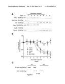 |
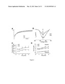 | 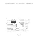 |
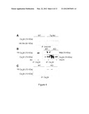 | 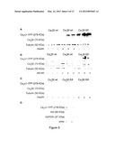 |
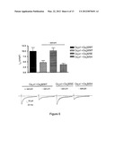 | 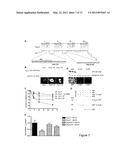 |
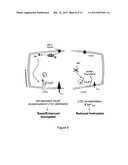 |  |
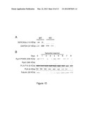 | 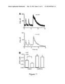 |
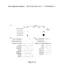 | 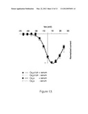 |
 | 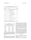 |
 |  |
 |  |
 |  |
 |  |
 |  |
 |  |
 |  |
 |  |
 |  |
 |  |
 |
| Similar patent applications: | |
| Date | Title |
|---|---|
| 2011-02-17 | Methods and compositions for identifying lung cancer or a humoral immune response against lung cancer |
| 2011-02-03 | Methods and agents modulating upa/upar activity |
| 2011-02-17 | Titania fine-particle composite and compositions containing the titania fine-particle composite |
| 2011-02-17 | Compositions and methods for treating bipolar disorder |
| 2011-02-10 | Methods related to immunostimulatory nucleic acid-induced interferon |
| New patent applications in this class: | |
| Date | Title |
|---|---|
| 2022-05-05 | Conjugates for treating inflammatory disease and identification of patients likely to benefit from such treatment |
| 2019-05-16 | Asymmetric conjugate compounds |
| 2019-05-16 | Functionalized nanoparticles and compositions for cancer treatment and methods |
| 2019-05-16 | Methods |
| 2018-01-25 | Bispecific antibody capable of being combined with immune cells to enhance tumor killing capability, and preparation method therefor and application thereof |
| New patent applications from these inventors: | |
| Date | Title |
|---|---|
| 2011-07-28 | Uses and compositions comprising mirnas |
| Top Inventors for class "Drug, bio-affecting and body treating compositions" | |
| Rank | Inventor's name |
|---|---|
| 1 | David M. Goldenberg |
| 2 | Hy Si Bui |
| 3 | Lowell L. Wood, Jr. |
| 4 | Roderick A. Hyde |
| 5 | Yat Sun Or |