Patent application title: DEVICE FOR SEROLOGICALLY DETECTING YERSINIA INFECTIONS AND/OR SECONDARY DISEASES THEREOF AND USE OF THE PROTEINS MyfA AND PsaA OF Y. ENTEROCOLITICA AND Y. PSEUDOTUBERCULOSIS AS RECOMBINANT ANTIGENS
Inventors:
Erwin Soutschek (Berg, DE)
Assignees:
MIKROGEN GmbH
IPC8 Class: AG01N33569FI
USPC Class:
506 9
Class name: Combinatorial chemistry technology: method, library, apparatus method of screening a library by measuring the ability to specifically bind a target molecule (e.g., antibody-antigen binding, receptor-ligand binding, etc.)
Publication date: 2011-12-15
Patent application number: 20110306515
Abstract:
Devices are disclosed for serologically detecting an infection with
human-pathogenic Yersinia ssp, wherein said device comprises at least one
antigen selected from the group of antigens consisting of the following
group: YopD, YopH, YopM, YopE, V-AG and YopN or a fragment of one of said
antigens having at least eight consecutive amino acids and furthermore
one of two proteins selected from MyfA and PsaA or fragments of one of
said two proteins having at least eight consecutive amino acids.Claims:
1. A device for serologically differentiating an infection with Yersinia
enterocolitica from an infection with Yersinia pseudotuberculosis,
wherein said device comprises (a) at least one antigen selected from the
group consisting of: YopD, YopH, YopM, YopE, V-AG, YopN and fragments
thereof having at least eight consecutive amino acids, and (b) least one
proteins selected from the group consisting of MyfA, PsaA, and fragments
thereof having at least eight consecutive amino acids and at least one
diagnostically relevant epitope, wherein each individual antigens is
spatially separately from the other.
2. Device according to claim 1, further wherein said device comprises both the MyfA protein or fragment thereof, and the PsaA protein or fragment thereof, wherein said fragments comprise at least eight consecutive amino acids and at least one diagnostically relevant epitope as set forth in claim 1.
3. Device according to claim 1, further wherein said device comprises the YopD antigen or a fragment thereof having at least eight consecutive amino acids as set forth in claim 1.
4. The device according to claim 1, further wherein the fragments of the proteins used as antigens comprise at least 12 consecutive amino acids of the respective antigen.
5. The device according to claim 1, further wherein the fragments of the proteins used as antigens comprise at least 20 consecutive amino acids of the respective antigen.
6. The device according to claim 1, further wherein the fragments of the proteins used as antigen comprise at least 30 amino acids of the respective antigen.
7. The device according to claim 1, wherein said device comprises a diagnostic kit.
8. The device according to claim 7, wherein said kit comprises an ELISA assay.
9. The device according to claim 7, wherein said kit comprises a line-test.
10. The device according to claim 7, wherein said kit comprises immunoblots, bead-based assays or microarrays.
11. (canceled)
12. (canceled)
13. (canceled)
14. A method of serologically differentiating an infection caused by Yersinia enterocolitica from an infection caused by Yersinia pseudotuberculosis, comprising the steps of: a. providing an antigenic protein selected from the group consisting of MyfA, PsaA and fragments thereof comprising at least 8 consecutive amino acids; and b. reacting said antigenic protein with at least one antigen selected from the group consisting of Yop D, Yop H, Yop M, Yop E, V-AG and Yop N and fragments thereof having at least 8 consecutive amino acids and at least one diagnostically relevant epitope, wherein the antigens are spatially separated from one another.
Description:
[0001] The genus Yersinia comprises three human-pathogenic Yersinia spp.,
Y. pestis, Y. pseudotuberculosis and Y. enterocolitica and eight further
species namely Y. aldovae, Y. bercovieri, Y. frederiksenii, Y.
intermedia, Y. kristensenii, Y. moolaretti, Y. rohdei and Y. ruckeri,
which rather play a role in humans as opportunistic pathogens in the case
of wounds and sepsis. Y. pestis causes plague and can be transmitted by
fleas from the natural rodent reservoir to humans. Y. enterocolitica and
Y. pseudotuberculosis occur in wild and domesticated animals in the
temperate to subtropical zones throughout the world. In Germany, the
intestinal disease caused by Y. enterocolitica and Y. pseudotuberculosis,
called yersiniosis, is, after salmonellosis (Salmonella enterica) and
campylobacter enteritis (Campylobacter jejuni), the third most common
bacterial enteritis disease notified annually to the Robert Koch
Institute (RKI) in Berlin. In Germany, 4987 cases of diseases caused by
Yersinia according to the RKI disease classification were reported to the
Robert Koch Institute in 2007, and 5162 in the year 2006. The highest
age-specific incidence was observed in young children in the age range
from 1 to 4 years (RKI). However, the actual frequency of these diseases
is not known exactly--many who become ill do not see a doctor if the
disease course is subclinical or mild and brief, many diseases are not
clarified aetiologically and not all diagnosed cases are reported. It is
estimated that up to 43% of blood donors in Germany and 31% of blood
donors in Finland have specific antibodies to Yersinia (tested by enzyme
immunoassays or immunoblots). This result suggests an unexpectedly high
prevalence of yersinioses experienced in industrial countries with high
standards of hygiene.
[0002] The enteropathogenic Y. enterocolitica and Y. pseudotuberculosis pathogens occur in pigs, sheep, bovines and poultry and enter food (e.g. raw pork) and drinking water mainly via the slaughtering process. Furthermore, cases of transmission to pets have been described. It has also been found that up to 100% of wild boar carry Yersinia enterocolitica in the nasopharyngeal cavity. Yersinia are able to multiply at low temperatures (>4° C.). In the industrial countries, multiplication in refrigerated foods represents the epidemiologically most important form of distribution of Y. enterocolitica and Y. pseudotuberculosis.
[0003] The exact infective dose is not known (presumably >10000 bacteria). The incubation time is two to five days after infection. Enteropathogenic Y. enterocolitica and Y. pseudotuberculosis strains generally colonise the intestine-associated lymphatic tissue of the terminal ileum, in the so-called Peyer's patches (continuous aggregation of lymphatic follicles). Following colonisation, the Yersinia overcome, via so-called M cells in the follicle-associated epithelial cell layer of the Peyer's patches, the gastrointestinal barrier of the organism and spread via the draining lymphatic vessels in the mesenteric lymph nodes. Y. enterocolitica then attacks and damages further regions of the intestine, which explains the typical tissue damage of the small intestine and colon in yersiniosis patients. Y. pseudotuberculosis shows a course of infection similar to that of Y. enterocolitica, except that mesenteric lymph nodes, liver and spleen are more often affected and enteritis symptoms and excretion via the intestine are much less than with Y. enterocolitica.
[0004] The enteritic form of Yersinia infections, also called yersiniosis, is limited, in uncomplicated cases, to the gastrointestinal tract and is characterised by watery diarrhoea for several days (called acute Yersinia gastroenteritis). However, depending on patients' age, immune status, histocompatibility type (HLA-B27) and sex, yersinioses can display different courses and symptoms, for example fever, nausea, colic-like lower abdominal pains (pseudoappendicitis), muscular pains and headaches and inflammations in the pharyngeal region (pharyngitis), in the mesenteric lymph nodes (acute mesenteric lymphadenitis) and in the ileum and colon (Yersinia ileitis and colitis). In uncomplicated cases, the disease abates after a few days or at the latest after two weeks.
[0005] In patients with underlying diseases, for example diabetes mellitus, liver cirrhosis, iron storage diseases (dialysis patients, thalassaemia) or immunosuppression, extraintestinal infections may occur (e.g. septic forms). Not uncommonly, septic shock with high lethality (approx. 75%) occurs after blood transfusion with Yersinia-contaminated banked blood.
[0006] Apart from the intestinal and extraintestinal forms of yersiniosis, in which the causative agent can generally be isolated, in up to 30% of cases there are also concomitant or secondary diseases, generally with an aseptic course. The most common secondary disease is HLA-B27-associated reactive arthritis [abbreviation: ReA] (about 60-80% of ReA cases are HLA-B27-positive). ReA starts approx. 1-4 weeks after onset of the intestinal symptoms (but often without preceding intestinal symptoms). Most often it is the joints of the lower extremities that are affected. In most cases the symptoms abate after approx. 3-12 months, but a chronic or relapsing course may also develop. Other complications that often occur after yersiniosis are inflammations of the thyroid (thyroiditis), of the heart muscle (myocarditis) and of the kidneys (glomerulonephritis), reddening of the skin (erythema) of the lower extremities (Erythema nodosum) and acute or chronic enlargement of the lymph nodes (lymphadenopathies) or of the spleen (splenomegaly).
[0007] In yersinioses without further immunological complications, for example reactive arthritis, the symptoms generally abate after a short time. Therefore in most cases it is unnecessary to treat a Yersinia infection with antibiotics. In the case of a septic course or persistent symptoms, differential treatment with antibiotics is necessary. Owing to the frequently occurring beta-lactamases with Yersinia, infections with Y. enterocolitica or Y. pseudotuberculosis are treated pathogen-specifically with gyrase inhibitors, tetracycline, trimethoprim/sulfamethoxazole etc.
[0008] Related Yersinia species (e.g. Y. enterocolitica and Y. pseudotuberculosis) are differentiated biochemically. Y. pseudotuberculosis forms a biochemically uniform group, which is divided into eight serogroups, of which the serovars I, II and III are of importance in human medicine in Europe (Bottone, [1997] Clin. Microbiol. Rev., 10, 257-276). In contrast, Y. enterocolitica is an extremely heterogeneous species both biochemically and serologically, with pathogenic and non-pathogenic, geographically separate subgroups. Y. enterocolitica is divided into six biovars (BV)/biotypes with different biochemical effects (1A, 1B, 2, 3, 4, 5) and approx. 60 serovars (with different O and H antigens) (Wauters et al., [1987] Contrib. Microbiol. Immunol., 9, 14-21). Whereas biovar 1A Yersinia mainly occur in the environment, the known human-pathogenic Y. enterocolitica strains belong to BV 1B (serotypes O:8, O:4, O:13, O:18, O:20, O:21, occurring mainly in the USA), BV 2 (Europe [O:9], USA, Japan [O:5, 27]), BV 3 (O:9 and O:5, 27) and BV 4 (Europe and USA [O:3]). (Bucher et al., [2008] Foodborne Pathog. Dis., 5, 273-280). Approximately 80-90% of reported cases of Yersinia gastroenteritis in Germany are caused by Y. enterocolitica serotype O:3 (RKI).
[0009] The virulence of the enteropathogenic Yersinia (Y. enterocolitica biovars 1B, 2, 3, 4 and 5, Y. pseudotuberculosis) and of the plague-causing Y. pestis is due to a highly conserved virulence plasmid pYV with a size of 65-70 kb (in Y. pestis pCDI). These virulence factors enable pathogenic Yersinia to survive in the host's lymphoid tissue. The Y. enterocolitica biovar 1A does not carry a pYV plasmid and is therefore regarded as non-pathogenic.
[0010] The pYV plasmid codes for the membrane protein YadA (Yersinia adhesin) and a number of so-called Yersinia outer proteins (Yops) and the associated secretory apparatus (T3SS), which is called Ysc (Yop secretion). As substrate of T3SS, the Yops are injected as anti-host effectors into eukaryotic cells (e.g. in macrophages). The protein complex of T3SS consists of 27 proteins with known or unknown functions, for example a regulatory function (YopQ, YopN), translocation function (YopB, YopD) and effector function (YopH, YopE, YopT, YopP, YopO). The adhesin YadA is also encoded on the virulence plasmid. It mediates the adhesion of the pathogens on host cells and enables the Yops to inject into the target cells. The so-called V antigen (LcrV) is also encoded on the pYV plasmid. This antigen forms the tip of the Ysc injectisome, regulates Yop secretion and modulation of the host's immune system.
[0011] The virulence of the Yersinia is also influenced by chromosomally encoded factors, in addition to the plasmid-encoded virulence factors. Adhesion of Yersinia on epithelial cells of the intestine requires among other things the presence of the invasin Inv and of the adhesin Ail (the "attachment invasion locus" of Y. enterocolitica), which can be expressed by all enteropathogenic Yersinia.
[0012] Other known chromosomally encoded pathogenicity factors are e.g. the secreted and thermally stable enterotoxin Yst, the so-called "mucoid Yersinia factor" MyfA of Y. enterocolitica, or the homologous pH 6 antigen PsaA of Y. pseudotuberculosis and Y. pestis, the yersiniabactin-siderophore system encoded on the so-called high-pathogenicity island (HPI) (FyuA receptor with Irp1-9), the Ysa Type-III secretory apparatus, the lipopolysaccharide LPS that is characteristic of all Gram-negative bacteria, the enzyme urease and the toxin complex TC that is active against insects (tcbA, tcaC, tccC).
[0013] The MyF fibril system consists of the three subunits MyfA, MyfB and MyfC. The fibril is constructed from MyfA subunits, whereas MyfB and MyfC form the transport and structuring apparatus. The MyfA surface antigen with a size of approx. 17 kDa is expressed in vitro during the early stationary growth phase, and full expression only takes place at 37° C. in an acid environment (pH 6). The myfA gene is present in human-pathogenic Y. enterocolitica strains (e.g. O:3, O:4, O:8; O:9) and in approx. 16% of non-human-pathogenic Y. enterocolitica BV 1A strains and shows a significant link to virulence. Leiva et al. showed, in coagglutination experiments with sera from rabbits that had been immunised intravenously with live Y. enterocolitica or Y. pseudotuberculosis strains, that the antisera obtained against MyfA (Y. enterocolitica) or PsaA (Y. pseudotuberculosis) allow bacteriological differentiation between Y. enterocolitica (MyfA) and Y. pseudotuberculosis (PsaA) strains (Leiva et al., Contrib. Microbiol. Immunol., 13, 158-164). These immunisation experiments also show that MyfA or PsaA are produced under laboratory conditions, i.e. under controlled nutrient conditions (growth medium) and temperature and pH conditions, and induce the production of the serum antibodies in intravenously immunised rabbits. However, a serodiagnostic application in humans is not under consideration. In particular, as the natural function of the antigens is unknown, it can also not be expected automatically that the MyfA or PsaA antigens are expressed during natural infection in the human gastrointestinal tract by pathogenic Yersinia. Additionally it has to be borne in mind that data from animal experiments can seldom be applied to the diagnostic situation in humans, because as is well known, the production of antibodies in animals can differ from that in humans.
[0014] The Psa-antigen complex (pH6 antigen) of enteropathogenic Y. pseudotuberculosis was originally characterised in the plague-causing Y. pestis as fimbriated structure with a diameter of 3-5 nm. The PsaA subunits are arranged on the bacteria surface by the translocation and structuring apparatus, which consists of PsaB and PsaC. The antigen domain PsaA, with a size of approx. 17 kDa, shows approx. 44-47% amino acid sequence homology with the Y. enterocolitica-homologous MyfA. As with MyfA, the production of PsaA in Yersinia is induced under laboratory conditions by temperature (37° C.) and slightly acid pH (pH 6). The PsaA deletion mutant strains of Y. pestis show a significantly reduced virulence in vitro and in vivo.
[0015] The observations that MyfA and PsaA antigens are expressed under laboratory conditions only at 37° C. and at acid pH, and that an immune response can be produced in laboratory mice and rabbits after infection with precultured (in vitro) Yersinia strains, leads to the presumption that these proteins might have a function during infection or might be expressed in the acidic environment of the intestinal tract. However, the exact functions of Myf and Psa during Yersinia infection, and therefore the importance of these antigens for Yersinia pathogenesis, are still unknown.
[0016] The classical diagnosis of the acute yersiniosis diseases is based primarily on detection of the pathogen in the stool, e.g. by means of cold enrichment, selective culture medium (e.g. so-called cefsulodin-Irgasan®-novobiocin agar culture medium [CIN-agar]), biochemical properties (e.g. so-called API E20 test; bioMerieux, Paris, France) and the detection of pathogen-specific nucleic acids (DNA) by polymerase chain reaction (PCR). Detection of the microbe in the stool may be possible for a period of 2 to 12 weeks. After the diarrhoea symptoms have subsided, Yersinia are typically no longer detectable in the stool.
[0017] Serology, i.e. detection of the individual serum antibody response to Yersinia-specific O and H antigens (so-called Widal and passive haemagglutination tests), virulence-associated proteins (e.g. by enzyme-linked immunosorbent assay [ELISA] and immunoblotting) or bacterial ultrasonicate (by means of complement-fixation reaction [CFR]), is suitable according to current standards for supplementary diagnosis of acute infection.
[0018] However, serodiagnostics is essential for clarifying secondary diseases, for example reactive arthritis, because direct detection of the pathogen, mostly after the acute infection has subsided, is not possible. The present invention relates to diagnostic devices, by means of which an infection with Yersinia enterocolitica can be differentiated serologically from an infection with Yersinia pseudotuberculosis.
[0019] In an acute Yersinia infection, typically the Yersinia-specific immunoglobulin (Ig) classes IgM, IgA and generally also IgG are detectable. In the course of infection, the specific IgM and IgA response is attenuated within 3-6 months (persistence of IgM: approx. 1-3 months and IgA: approx. 2-4 months), whereas Yersinia-specific IgG antibodies can persist for several years, possibly even life-long (in 80% of patients after a Yersinia infection). In chronic Yersinia infection and Yersinia-induced reactive arthritis, persistent IgA antibodies can be detected for years, along with Yersinia-specific antibodies of the IgG class. The Yersinia-specific antibodies of the IgM class are mostly no longer detectable in secondary diseases.
[0020] The conventional serological methods of detection, such as the Widal and complement-fixation reaction directed against whole cell lysates, only possess low diagnostic sensitivity and specificity owing to cross-reactivity with a large number of human pathogens (for example Bartonella henselae, Borrelia burgdorferi, Chlamydia pneumoniae, Rickettsia rickettsii, Escherichia coli, Brucella spp., Salmonella spp.). Therefore enzyme immunoassays (ELISA) and immunoblots are currently preferred for the detection of IgG, IgA and IgM antibodies to recombinantly produced virulence-associated Yersinia-specific antigens (e.g. Yops and V-AG).
[0021] Existing Yersinia serodiagnostics is based mainly on the reaction for detecting the IgG and IgA (conditionally also IgM) response to virulence plasmid pYV secreted Yop proteins, for example YopD, YopH, YopM, YopE, V-AG and YopN. The specificity and sensitivity of these Yop antigens is, however, in need of improvement and is supplemented according to the invention with additional Yersinia-specific antigens.
[0022] In addition, owing to the similarity of the aetiology and of the process of infection, a subclinical course of infection or a nonspecific symptomatology and a high infection rate, it is difficult to differentiate between Y. enterocolitica and Y. pseudotuberculosis infections or secondary diseases caused by these pathogens (for example ReA or post-enteritic arthritis, myocarditis, glomerulonephritis, lymphadenopathies, splenomegaly, erythema nodosum) with the conventional diagnostic test methods. However, a method for specific serological detection (i.e. in particular differentiating between the most common human-pathogenic Yersinia species Y. enterocolitica and Y. pseudotuberculosis) is necessary, so as to be able to provide early and effective treatment of yersinioses or prevention of secondary diseases (i.e. adequate antibiotic therapy). The different proteins of Y. enterocolitica (MyfA) and Y. pseudotuberculosis (PsaA) can make a contribution to this.
[0023] Heesemann et al. (Microbial Pathogenesis [1988], p. 437-447) describe the immune response of orally infected rabbits to virulent (pYV plasmid) and non-virulent (no pYV plasmid) serotype O:3 strains of Yersinia enterocolitica, which had been precultured overnight in neutral growth medium (BHI). It is unlikely that the MyfA antigen was expressed under these conditions owing to neutral pH and the growth phase. In addition the presence of the PsaA antigen is ruled out (the authors only used Y. enterocolitica strains). The antigens were separated using SDS-electrophoresis and were investigated further by Western blotting. No further purification was carried out. Moreover, the authors stated that this method is rather unsuitable as such for diagnostic purposes, owing to high cross-reactivity with intestinally pathogenic Escherichia coli and Salmonella strains.
[0024] Tomaso et al. (European Journal of Epidemiology [2006], 21: 77-81) and Stolck-Engelaar et al. (Scand. J. Infect. Dis. [1996], p. 571-575) describe the seroprevalence of anti-Yersinia antibodies in healthy Austrians and in Dutch yersiniosis patients. Determination is carried out with a commercial Western Blot assay with the antigens Yop M, Yop H, V-Ag, Yop D and Yop E. The antigens MyfA or PsaA are not used.
[0025] The only method based on the MyfA and PsaA antigens proposed to date was described by Leiva et al., 1995. However, this related to a bacteriological detection method for identifying pathogenic Yersinia strains after growing the cells on an agar culture medium.
[0026] The present invention relates to a device in the broader sense for serological differentiation of an infection with Yersinia enterocolitica from an infection with Yersinia pseudotuberculosis. "Serological differentiation" in the sense of the present application means that, on the basis of a sample obtained from blood (serum, plasma), it is possible to determine by means of an immunological assay whether it is an infection caused by a strain of Yersinia enterocolitica or Yersinia pseudotuberculosis. Said device contains at least one antigen selected from a group of antigens that can be classed with the outer surface proteins or secreted proteins of Yersinia. At least one of these antigens must be used in the device, and it is not absolutely essential to use the complete protein--it may be perfectly sufficient to use protein fragments that have a diagnostically relevant epitope.
[0027] According to the invention, the antigens are used in essentially pure form, and this is preferably achieved by producing the antigens recombinantly rather than isolating them from cell lysate.
[0028] A fragment of one of the antigens listed below, which has at least eight consecutive amino acids, preferably at least 12, more preferably at least 20, even more preferably at least 30 consecutive amino acids and quite particularly preferably at least 50 consecutive amino acids, is sufficient. In a preferred embodiment the peptides have 10 to 30 consecutive amino acids. Each peptide/fragment has at least one diagnostically relevant epitope.
[0029] When selecting the fragments, a region is selected that contains at least one diagnostically relevant epitope. The epitope regions can be localised by standard methods known by a person skilled in the art. It is possible to determine the hydrophilicity/hydrophobicity of the protein using suitable computer programs. Hydrophilic regions are as a rule predestined to carry suitable epitopes, because in the folded protein the hydrophilic regions end up on the surface. Hydrophobic regions are more likely to be localised in the interior of the folded protein and are therefore unlikely to be involved in diagnostically relevant epitopes. The epitopes are preferably linear epitopes, but conformation epitopes can also be used advantageously.
[0030] When suitable regions have been identified, these can either be synthesised by chemical synthesis or produced by recombinant methods. These proteins or peptides can then be reacted with suitable blood, serum or plasma samples, whose aetiology has been determined with other, medical parameters. In this way, a person skilled in the art can localise suitable epitopes.
[0031] The devices according to the invention thus contain at least one antigen or a fragment of one of these antigens selected from the group consisting of antigens listed hereunder. These are the following antigens:
TABLE-US-00001 YopD (Seq ID No. 1) MTINIKTDSPIITTGSQIDAITTETVGQSGEVKKTEDTRHEAQAIKSSEASLSRSQVPELIKPSQ GINVALLSKSQGDLNGTLSILLLLLELARKAREMGLQQRDIENKAAITAQKEQVAEMVSGAKL MIAMAVVSGIMAATSTVASAFSIAKEVKIVKQEQILNSNIAGRDQLIDTKLQQMSNTSDKAVS REDIGRIWKPEQVADQNKLALLDKEFRMTDSKANAFNAATQPLGQMANSAIQVHRGYSQA EVKEKEVNASIAANEKQKAEEAMNYNDNFMKDVLRLIEQYVSSHTHAMKAAFGVV. YopH (Seq ID No. 2) MNLSLSDLHRQVSRLVQQESGDCTGKLRGNVAANKETTFQGLTIASGARESEKVFAQTVLS HVANIVLTQEDTAKLLQSTVKHNLNNYELRSVGNGNSVLVSLRSDQMTLQDAKVLLEAALR QESGARGHVSSHSHSVLHAPGTPVREGLRSHLDPRTPPLPPRERPHTSGHHGAGEARAT APSTVSPYGPEARAELSSRLTTLRNTLAPATNDPRYLQACGGEKLNRFRDIQCCRQTAVRA DLNANYIQVGNTRTIACQYPLQSQLESHFRMLAENRTPVLAVLASSSEIANQRFGMPDYFR QSGTYGSITVESKMTQQVGLGDGIMADMYTLTIREAGQKTISVPVVHVGNWPDQTAVSSEV TKALASLVDQTAETKRNMYESKGSSAVADDSKLRPVIHCRAGVGRTAQLIGAMCMNDSRN SQLSVEDMVSQMRVQRNGIMVQKDEQLDVLIKLAEGQGRPLLNS. YopN (Seq ID No. 3) MTTLHNISYGNTTLRNEHPETASSQIVNQTLGQFRGESVQIVSGTLQSIADMAEEVTFVFSE RKELSLDKRKLSDSQARVSDVEEQVNQYLSKVPELEQKQNVSELLSLLSNSPNISLSQLKAY LEGKSEEPSEQFKMLCGLRDALKGRPELAHLSHLVEQALVSMAEEQGEAIVLGARITPEAY RESQSSVNPLQPLRDTYRDAVMGYQGIYAIWSDLQKRFPNGDIDSVILFLQKALSADLQSQ QSGSGREKLGIVISDLQKLKEFGSVSDQVKGFWQFFSEGKTNGVRPF. YopE (Seq ID No. 4) MPKISSFISTSLPLPTSVSGSSSVGEMSGRSVSQQKSEQYANNLAGRTESPQGSSLASRIT EKLSSMARSAIEFIKRMFSEGSHKPVVTPAPTPAQMPSPTSFSDSIKQLAAETLPKYIQQLSS LDAETLQKNHDQFATGSGPLRGSITQCQGLMQFCGGELQAEASAILNTPVCGIPFSQWGTI GGAASAYVASGVDLTQAANELKGLAQQMHQLLSLM. YopM (Seq ID No. 5) MFINPRNVSNTFLQEPLRHSSDLTEIPVEAENVKSKTEYYNAWSEWERNAPPGNGEQREM AVSRLRDCLDRQAHELELNNLGLSSLPELPPHLERLVASCNSLTELPELPQSLKSLEVYENN LKALPDLPPLLVDLRVFNNQLEELPELQNLPFLTEIYANNNSLKTLPDLPPSLVDLNVRENYL TALPELPQSLIFLDISDNILSGLSELPPNLSCLDASRNGIRSLCDLPPSLVYLDVRDNQLIELPA LPSGLERLIASFNHLAELPELPPNLYYLDASRNEISSLCDLPPSLVDLNVRKNQLIELPALPPD LERLIASFNHLAELPELPPNLSYLDASRNEISSLCDLPPSLVDLNVRKNQLIELPALPPDLERLI ASFNHLAELPELPPNLSYLDASRNEISSLCDLPPSLVELDVRDNQLIELPALPPHLERLIASLN HLAEVPELPQNLKQLHVEHNALREFPDIPESVEDLRMDSERVIDPYEFAHETIDKLEDDVFE. V-AG (also called LCRV) (Seq ID No. 6) MIRAYEQNPQHFIEDLEKVRVEQLTGHGSSVLEELVQLVKDKKIDISIKYDPKKDSEVFAERV ITDDIELLKKILAYFLPEDAILKGGHYDNQLQNGIKRVKEFLESSPNTQWELRAFMAVMHFSL TADRIDDDILKVIVDSMNHHGDARSKLREELAELTAELKIYSVIQAEINKHLSSSGTINIHEKSI NLMDKNLYGYTDEEIFKASAEYKILKKMPQTTIKDDELHEVGVIAGAEKQIVSIKNFLESENKR TGALGNLKDSYSYNKDNNELSHFATACSDKSRPLNDLVSQKTTQLSDITSRFNSAIEALNRFI QKYDSVMQRLLDDTR.
[0032] In addition to the antigen from the first group of antigens, the device according to the invention also has at least one of two further proteins, namely either the protein MyfA and/or the protein PsaA or fragments of one of these two proteins. Once again the fragments have a minimum size of at least 8 consecutive amino acids, preferably at least 12 consecutive amino acids, more preferably at least 20, particularly preferably at least 30 and quite particularly preferably at least 50 consecutive amino acids of one of the proteins MyfA and/or PsaA.
[0033] It is preferable for the device to have the two complete proteins MyfA and PsaA or fragments thereof together, wherein the individual antigens are spatially separate from one another. The amino acid sequences of the two proteins MyfA and PsaA (without leader sequence) are shown below.
TABLE-US-00002 MyfA (132-AA) (Seq ID No. 7) MEPTVINSKDISATKTVKEGGSFSVEFKATENEIVSGKLDADTPAFHL VMSDSGEHKGWNVRPTGASEGGQMVSADGTRVDLHTNELSWDNDHWWI DDGSERVEATFFLAAGDEVKAGEYQFTGRVEEYVE. PsaA (134-AA) (Seq ID No. 8) MSTVINSKDVSGEVTVKQGNTFHVDFAPNTGEIFAGKQPGDVTMFTLT MGDTAPHGGWRLIPTGDSKGGYMISADGDYVGLYSYMMSWVGIDNNW YINDDSPKDIKDHLYVKAGTVLKPTTYKFTGRVEEYVF.
[0034] The proteins MyfA or PsaA are encoded by the nucleotide sequences MyfA or PsaA shown below and can be produced recombinantly using suitable vectors and host cells.
TABLE-US-00003 myfA (Seq ID No. 9) atggaaccgactgttattaatagtaaagacatctctgcaacaaaaactgttaaagagggaggttcgttctcagt- tgaattcaaggc cactgaaaacgagattgtgtcaggcaaattggatgcagatacacctgccttccatctggtaatgtcggactcag- gggaacataaa ggttggaatgttcggcctaccggtgcatctgagggaggacagatggtttctgcagatggtaccagagttgactt- acatacaaatga gctatcgtgggataacgaccactggtggatagatgacggttctgagcgtgtggaagcgactttctttcttgctg- ctggcgacgaggtt aaagcaggtgaatatcagttcactgggcgtgttgaggaatatgtcgagtaa psaA (Seq ID No. 10) atgtctactgtcattaactccaaggatgtttctggtgaggtgactgtcaagcagggaaacacattccacgtcga- ttttgcgcctaaca caggagagatttttgcgggtaaacagccgggtgatgtcactatgtttacgctaactatgggtgatactgcacca- cacggtggttggc gtttgattccaacaggggactcaaaaggtggatatatgatcagcgccgatggtgactatgttggtttatacagt- tatatgatgtcat gggtaggtatagataataactggtatataaatgatgactctcctaaagatataaaagatcatctgtacgttaag- gcagggactgtcc ttaaaccaacgacttataaattcacggggcgtgttgaagagtatgtattttaa
[0035] The individual antigens are, according to the invention, arranged spatially separately from one another in the test device or the test kit. In the case of Western Blots, for example, the antigens can be applied in the form of bands on the carrier material, wherein the individual antigens are in each case present in a particular, well-defined band. This spatial separation of the individual antigens can also be achieved if the antigens are applied spatially separately from one another in a line-assay. However, it is also possible to apply the individual antigens on a microtitre plate, so that only one antigen is present in each well. In an alternative embodiment, in each case an antigen is applied on a type of carrier (for example spheres), so that only one antigen is bound to each carrier. As an alternative, the antigens can be applied to assay plates (microchips), wherein the individual antigens are fixed on specified points on said chips.
[0036] According to the invention, first it is determined, by reaction with at least one, or even a plurality of antigens selected from the group Yop D, Yop H, Yop M, Yop E, V-AG and/or Yop N, whether it is a Yersinia infection. If the result of this test is positive, it is determined, using the antigen MyfA and/or PsaA, whether the infection is caused by Yersinia enterocolitica or Y. pseudotuberculosis. The individual detection steps can be carried out either simultaneously or successively.
[0037] A device for serologically detecting an infection by Yersinia species is, according to the invention, in a preferred embodiment a diagnostic kit. This is to be understood as a device that is used by diagnostic laboratories for serological diagnosis. In a preferred embodiment the antigens are bound spatially separately from one another on a carrier matrix, for example wells of a microtitre plate, spheres, nitrocellulose or nylon. The antibodies (mainly of classes IgG, IgM and IgA) present in a sample from a patient (e.g. blood, serum, plasma, saliva) react with the bound antigens and so are immobilised. In a preferred embodiment these are diagnostic kits, wherein ELISA assays represent a preferred embodiment. In the case of ELISA assay kits, usually the antigen is bound to the wells of a microtitre plate. The specific antibodies present in the samples can react with the antigens. The antibodies from serum or plasma, which have bound specifically to the antigens present in the wells, are as a rule detected with anti-antibodies, which carry a marker, preferably an enzyme marker.
[0038] Another preferred embodiment of the device according to the invention comprises so-called line-tests. In this case, a plurality of antigens are applied on the test strips according to a predetermined pattern. The blood samples to be investigated (sera or plasmas) are reacted with the test strips and antigen-antibody reactions are detected by enzyme-labelled antibodies and subsequent colour reaction. A conclusion can be drawn about the infection or infective agent from the specific pattern of the reactivities or colour signals.
[0039] The device according to the invention can also be an immunoblot or Western blot. In this case, the diagnostically relevant proteins are first separated according to size for example by diffusion, capillary action or electrophoresis and transferred to a carrier material, for example a nylon membrane or a nitrocellulose membrane, and fixed there. This carrier material with the proteins or protein fragments bound thereto is reacted with the patient's blood samples (serum or plasma).
[0040] The immobilised specific antibodies can for example be detected by reaction with an anti-antibody. Preferably various anti-antibodies can be used, which react either with IgG, IgM or IgA. This makes further differentiation of the immune response possible. As a rule these anti-antibodies carry a marker. This can be an enzyme, which catalyses a colour reaction, but it can also be fluorescent residues or radioactive residues. What is important is that antibodies bound to antigens can be detected with the anti-antibodies.
[0041] In another embodiment, the device according to the invention is a bead-based assay. A known commercial application of these bead-based microarrays is the Luminex-XMAB technology from Luminex Corporation (Austin, USA). This system uses microspheres (so-called beads) and evaluation is based on flow cytometry. In the case of fluorescence-labelled embodiments, the antigens according to the invention can be fixed on beads and the binding of the antibodies to the antigens is visualised with suitable labelling, e.g. fluorescence.
[0042] Another preferred embodiment of the device according to the invention relates to protein microarrays. In this case various antigens are fixed in a narrow space on a surface. As it is known which antigen is present in which place, after visualisation of the antigen-antibody reaction for example by means of a colour reaction or fluorescence labelling, it is also possible to state which antigen has reacted with the antibodies in the serum.
[0043] Usually the individual antigens are fixed in the device in such a way that after the reaction it is possible to establish with which particular antigen the antibodies present in the sample have reacted. In an ELISA assay, for example, the individual antigens are put in different cavities of the microtitre plate. In the line-assay, the antigens are sprayed on different strips of the carrier material and in the case of bead and planar microarrays the respective antigens are always applied at a defined place on the carrier or defined beads. The devices according to the invention are used for detecting human-pathogenic Yersinia species or subspecies. By combining different antigens, on the one hand it is possible to perform sensitive and specific detection of the pathogens and on the other hand, in a preferred embodiment a differential-diagnostic detection is also possible, wherein it is possible to distinguish between an infection caused by Yersinia enterocolitica and an infection with Yersinia pseudotuberculosis.
[0044] Another essential aspect of the present invention is also that serological detection or serological differentiation is possible when the acute Yersinia infection has already abated. For treating these secondary diseases it is essential to be able to determine which bacterium (Yersinia enterocolitica or Yersinia pseudotuberculosis) caused the original infection.
[0045] The device according to the invention thus has at least two different antigens, wherein one antigen is selected from the group consisting of the antigens YopD, YopH, YopM, YopE, V-AG and YopN described in more detail above. The other group comprises the antigens MyfA and PsaA.
[0046] In a particularly preferred embodiment the device comprises the antigen YopD and the antigen YopH together with either MyfA or PsaA.
[0047] In a still further preferred embodiment the device according to the invention comprises the antigens YopD, YopH, YopM and either MyfA or PsaA, the combined use of MyfA and PsaA being quite particularly preferred, however.
[0048] In a particularly preferred embodiment the device according to the invention thus has the proteins MyfA and/or PsaA or fragments thereof, as defined above, together with the following protein or protein combinations, wherein these proteins can also be in the form of fragments, as defined above. The device according to the invention has at least the protein YopD, which is preferably used together with YopH and more preferably together with YopM combined with MyfA and/or PsaA.
[0049] Another preferred embodiment comprises the proteins or fragments of YopD, YopH, YopM and YopE together with MyfA and/or PsaA.
[0050] Another preferred embodiment comprises the proteins and/or fragments of YopD, YopH, YopM, YopE, V-AG together with MyfA and/or PsaA.
[0051] Another preferred embodiment comprises a combination of proteins and/or fragments with the following designations: YopD, YopH, YopM, YopE, V-AG, YopN, and MyfA and/or PsaA.
[0052] The stated antigens can also be used in the form of fragments thereof, wherein the fragments have a serologically relevant epitope. When, according to the invention, fragments of the antigens are used, in the case of the antigen MyfA, on the one hand the N-terminal fragment with the amino acids 30-41 and on the other hand the C-terminal fragment with the amino acids 148-159 is particularly preferred. In the PsaA protein, the fragments that comprise the amino acids 27-37 (N-terminus) and 144-155 (C-terminus) are particularly preferred. In this case fragments are used which comprise this amino acid sequence and which have the sizes defined in more detail above.
[0053] The present invention thus relates to the use of an antigenic protein MyfA or a fragment thereof, as defined in more detail above, either alone or together with the antigenic protein PsaA or a fragment thereof, as defined above, for serologically detecting an infection caused by Yersinia or an infection caused by Y. enterocolitica or Y. pseudotuberculosis. This differentiation is clinically important, because an infection caused by Yersinia enterocolitica can be treated differently from an infection caused by Yersinia pseudotuberculosis.
[0054] With the device according to the invention, it is possible to detect Yersinia, which are of great importance, as they are pathogenic for humans. The Yersinia occur in farm animals, for example pigs and poultry, and on infection can cause a disease in humans. It can therefore be of considerable medical and epidemiological importance to detect, by means of a simple and relatively inexpensive test kit, the course of the chains of infection. This allows conclusions to be drawn about the sources of the pathogens and these can be identified and possibly be made harmless. It is thus possible to detect and differentiate the pathogens of yersinioses.
[0055] The results that can be achieved with the detection according to the invention are surprising, because it could not automatically be assumed that the antigens MyfA and/or PsaA are also expressed in sufficient amounts for antibody formation in the human body under real infection conditions. Furthermore, it was not known that specific antibodies of the IgG, IgM and IgA class against MyfA and PsaA antigens are formed in humans.
[0056] Based on the relatively high protein homology (44%) between MyfA and PsaA antigens and the already described interspecies homology or interspecies cross-reactivity between non-pathogenic Yersinia strains and other enterobacteria, for example E. coli and Salmonella species, it was not to be expected that the MyfA or the PsaA antigen can be used for differentiating the infective agent. The detection or differentiation is preferably performed with human blood, in particular serum or plasma. Determination is, however, also possible with cerebrospinal fluid or saliva.
[0057] In a quite particularly preferred embodiment the sample to be investigated is on the one hand reacted with an antigen selected from the group Yop D, Yop H, Yop M, Yop E, V-AG and Yop-N, quite particularly preferably with Yop D. If this does not result in a positive reaction and otherwise there is no special further suspicion, this can conclude the diagnosis.
[0058] Conversely, if the sample to be investigated reacts positively with this antigen, in particular Yop D, in a further diagnostic step the sample to be investigated can be reacted with the antigen PsaA and/or MyfA, preferably with both antigens. Furthermore, it is useful to differentiate the antibodies found, as to whether they are IgG or IgA antibodies. If the IgM or IgA finding is negative, but the IgG finding is positive, presumably an infection has come to an end.
[0059] If the IgM or IgA finding, preferably IgA finding, is positive, there may be an acute infection or a secondary disease. The infection is caused by Yersinia enterocolitica if the IgG, IgM or IgA reaction, preferably IgG finding, is positive with MyfA. If there is a positive reaction with PsaA, it is an infection with Yersinia pseudotuberculosis.
[0060] The figures clarify preferred embodiments of the present invention and explain the results obtained in the examples.
[0061] FIG. 1 shows the homologous amino acid sequence regions of the MyfA and PsaA antigens. The homologous regions (consensus) are marked in black. A line is drawn round the N- and C-terminal homologous regions.
[0062] FIG. 2 explains the in silico determination of the antigenic domains of MyfA and PsaA. The antigenicity index was calculated on the basis of the Jameson-Wolf algorithm and the hydrophilicity of the antigen on the basis of the Kyte-Doolittle algorithm. The so-called leader sequence is marked with a black arrow.
[0063] FIG. 3 shows a schematic representation of the DNA sequences of the four His-Tag-MyfA fusion proteins used. The primer sequences used for amplification are marked with arrows (I, II, III, IV). The leader sequences are shown in light-grey and the regions homologous with PsaA are shown in white.
[0064] FIG. 4 shows the chromatographic separation (SDS-polyacrylamide-gel electrophoresis) of the MyfA partial fragments myfA 1-441, myfA 121-441 and myfA 121-447 (FIG. 3) with subsequent Coomassie Blue staining (left) and immunoblot (myfA, myfA 1-441, myfA 121-441 and myfA 121-447; FIG. 3) with anti-Y. enterocolitica (O:3 or O:9) and anti-MyfA sera from rabbit (right).
[0065] FIG. 5 shows verification of the serological reactivity of the purified MyfA total protein (MyfA) and the purified MyfA partial fragments MyfA 1-441, MyfA 121-441 and MyfA 121-447 (FIG. 1; FIG. 3) by means of line-assays. The test strips were incubated with four different anti-Y. enterocolitica O:3, O:8 or O:9 sera from rabbit (1-4) before (*) and after infection.
[0066] FIG. 6 shows the serological reactivity of human anti-Y. enterocolitica or anti-Y. pseudotuberculosis serum with YopM, YopH, V-AG, YopD, YopN, YopE, MyfA and PsaA in the line-assay.
[0067] FIG. 7 shows the IgG response to Yop, MyfA-MIK and PsaA-MIK antigens with the tested Bavarian blood donor sera (n=40).
[0068] FIG. 8 explains the IgG response to Yop, MyfA-MIK and PsaA-MIK antigens with the tested yersiniosis patient sera from Finland (n=18).
[0069] FIG. 9 shows the IgG response to Yop, MyfA-MIK and PsaA-MIK antigens with the tested yersiniosis patient sera from Germany (n=23). With the surface protein, YopD is particularly suitable for use in diagnostic testing devices.
EXAMPLE 1
In Silico Determination of the Immunogenic Domains of the MyfA and PsaA Antigens
[0070] Determination of the homologous antigen regions that might be responsible for the immunogenic reactivity or cross-reactivity of the MyfA and PsaA antigens, was carried out in silico by means of direct amino acid sequence comparison (see FIG. 1). The homologous regions are mainly located in the N- and C-terminus of the MyfA (AA 32-40 and 150-159) and PsaA (AA 29-38 and 149-158) antigen.
[0071] The antigenicity index and the hydrophilic character of the antigen regions was determined in silico with the algorithms of Jameson and Wolf (Comput. Appl. Biosci. (1988) p. 181-186) or of Kyte and Doolittle (Kyte and Doolittle, (1982) J. Mol. Biol., 157, p. 105-132) (FIG. 2). The eight >5 AA immunogenic domains of the PsaA antigen are probably located in AA 30-48, 52-56, 59-67, 74-83, 87-96, 122-133, 144-150 and 151-158. The seven putative >5 AA immunogenic domains of the MyfA antigen were localised in AA 35-42, 43-54, 55-73, 79-100, 101-133, 139-148 and 152-159. According to the invention, these fragments are preferably used in the diagnostic tests. However, as the predicative Kyte-Doolittle algorithm is only informative conditionally, the immunogenic epitopes of the new antigens were determined experimentally (Example 2).
Complete Total Protein Sequences:
TABLE-US-00004 [0072] (Seq ID No. 11) MyfA (159-AA) AA 1-29: Leader Sequence AA 30-41: N-terminal fragment (Example 2) AA 148-159: C-terminal fragment (Example 2) MNMKKFVKKPLAIAVLMLASGGMVNMVHAEPTVINSKDISATKTVKEGGSFSVEFKATENEI VSGKLDADTPAFHLVMSDSGEHKGWNVRPTGASEGGQMVSADGTRVDLHTNELSWDND HWWIDDGSERVEATFFLAAGDEVKAGEYQFTGRVEEYVE (Seq ID No. 12) PsaA (158-AA) AA 1-26: Leader Sequence MKMKCFAKNALAVTTLMIAACGMANASTVINSKDVSGEVTVKQGNTFHVDFAPNTGEIFAG KQPGDVTMFTLTMGDTAPHGGWRLIPTGDSKGGYMISADGDYVGLYSYMMSWVGIDNNW YINDDSPKDIKDHLYVKAGTVLKPTTYKFTGRVEEYVF
EXAMPLE 2
Experimental Determination of the Immunogenic Domains of the myfA Antigens
[0073] Preparation of the myfA Partial Fragments
[0074] Four fragments were prepared starting from the complete reading frame of the MyfA antigen (Example 1). Both the complete reading frame including the leader peptide and N-, C-, and N- and C-terminally shortened partial fragments are shown (FIG. 1; FIG. 3).
[0075] Specific amplification of the myfA partial fragments was carried out by PCR with chromosomal DNA from Y. enterocolitica serotype O:3/4 (Y. enterocolitica subsp. palearctica strain Y-11; DMSZ 13030) as templates.
[0076] The resulting amplificates were cleaved enzymatically with restriction endonucleases and ligated into a suitable vector, pET21-b, which had been cut with the same restriction endonucleases as the amplificates. After transformation of the ligation preparation into a suitable E. coli strain, e.g. BL21 pLys, individual clones were tested for the presence of myfA and psaA by agarose-gel electrophoresis of enzymatically cleaved plasmid DNA and then sequenced. In addition, the expression of the partial fragments was identified and characterised by analysis of the expression products by SDS-polyacrylamide-gel electrophoresis with subsequent Coomassie Blue staining or subsequent transfer to nitrocellulose followed by immunological detection. The fusion proteins were purified by Ni-NTA column chromatography according to the manufacturer's information (the company Qiagen).
[0077] In addition to the complete reading frame of MyfA (MyfA; FIG. 3), three partial fragments of the antigen were cloned as His-Tag fusion proteins into pET21-b vector. The first fragment (MyfA 1-441) was cloned without the C-terminal region homologous to PsaA (Example 1; FIG. 1) and comprises the AA 1-147 (nucleotide [NT] 1-441). The second fragment (MyfA 121-441) codes for the AA 40-147 (NT 121-441) and therefore does not contain the N- and C-terminal regions homologous to PsaA (Example 1; FIG. 1). The third fragment (MyfA 121-477) codes for the AA 40-159 (NT 121-477) and does not contain the N-terminal sequence of MyfA (Example 1; FIG. 3; FIG. 1).
[0078] The following oligonucleotide primers were used for preparing the sequences.
TABLE-US-00005 Primer 1 (I): (Seq ID No. 13) myfA-f: GTA ATT CCA TAT GAA TAT GAA AAA ATT TGT Primer 2 (II): (Seq ID No. 14) myfA_121f: GTA ATC CCA TAT GGC AAC AAA AAC TGT Primer 3 (III): (Seq ID No. 15) myfA_441rev: TTA CTC GAG TTC ACC TGC TTT AAC Primer 4 (IV): (Seq ID No 16) myfA_rev: ATC TAC TCG AGC TCG ACA TAT TCC TCA A
[0079] The following primer combinations were used:
myfA: primer 1 and primer 4 myfA 1-441: primer 1 and primer 3 myfA 121-441: primer 2 and primer 3 myfA 121-477: primer 2 and primer 4
EXAMPLE 3
Verification of Antigenicity
[0080] The immunological reactivity/antigenicity of the MyfA fusion proteins (Example 2) was tested by immunoblot (FIG. 4) and line-assay (FIG. 5)
[0081] FIG. 4, left, shows in each case cell lysates (ZL) of MyfA total protein (MyfA, including leader sequence; Example 1; FIG. 1; Example 2; FIG. 3) and/or E. coli BL21 pLys cells expressing one of the three MyfA partial fragments (MyfA 1-441, MyfA 121-441 and MyfA 121-447) and the His-tagged recombinant MyfA proteins (MyfA 1-441, MyfA 121-441 and MyfA 121-447) purified by Ni-NTA column chromatography.
[0082] FIG. 4, right, shows an immunoblot with two anti-Y. enterocolitica O:3 or O:9 sera and an anti-MyfA serum from rabbit. The MyfA-protein shortened on the N- and C-terminus (MyfA 121-441; Example 1; FIG. 1; Example 2; FIG. 3) does not show any reaction with the sera used. The protein shortened on the C-terminus MyfA 1-441 (FIG. 1; FIG. 2) shows a weaker reaction than the protein shortened on the N-terminus MyfA 121-447 (FIG. 1; FIG. 2). Therefore the C-terminus appears to contain a particularly important immunogenic region, but the N-terminus is also diagnostically important.
[0083] The proteins could be detected by detecting the His-tags with a nickel-NTA conjugate from rabbit (very weak reaction, no double bands). The immunoblot with an anti-Y. pseudotuberculosis serum from rabbit did not show any reaction.
[0084] The immunological reactivity/antigenicity of the purified MyfA total protein (MyfA) and of the purified MyfA partial fragments MyfA 1-441, MyfA 121-441 and MyfA 121-447 (Example 1, FIG. 1; Example 2; FIG. 3) was verified by means of line-assays (FIG. 5). The test strips were incubated with four different anti-Y. enterocolitica sera from rabbit (serum 1 and 4: serotype O:3; serum 2: serum O:9, serum 3: O:8) before (*) and after experimental peroral infection with precultured Yersinia. Recombinantly produced YopD and YopH antigens were incorporated into the assay as immunisation or assay control. Additionally, so as to be able to evaluate the cross-reactivity of the MyfA and PsaA antigens, recombinantly produced PsaA antigen was also included in the assay (FIG. 5). The reactivity of the antigens was in addition verified with an anti-Y. pseudotuberculosis serum from rabbit.
[0085] The blood samples taken before infection showed no reactivity with the antigens of the assay (i.e. YopD, YopH, MyfA, MyfA 1-441, MyfA 121-441, MyfA 121-447, PsaA). After the experimental infection, all four samples reacted with the Yersinia-specific antigens YopD and YopH (FIG. 5). As already shown in the immunoblot (FIG. 4), the C- (Δ C-terminus) or the N- and C- (Δ N- and C-terminus) terminally shortened MyfA protein showed markedly reduced reactivity and the N-terminally shortened protein (Δ N-terminus) reduced reactivity in comparison with the MyfA total protein (FIG. 5). Interestingly, sera No. 2 (very weakly), 3 (weakly) and 4 (positively) also reacted with the applied PsaA antigen. The anti-Y. pseudotuberculosis serum from rabbit reacted very strongly with YopD and PsaA, but showed no reactivity with the total antigen MyfA or its partial fragments. The cross-reactivity of the anti-Y. enterocolitica sera with PsaA occurring in the assay is possibly caused by semi-optimum production conditions (i.e. purification via His-Taq, buffer conditions or antigen concentration too high). In addition, cross-reactivities occurring between MyfA and PsaA or other Yersinia surface proteins (so-called RPs) are known in rabbit sera immunised with precultured Yersinia (Leiva et al., Heesemann et al.). For the subsequent experiments, the MyfA and PsaA antigens were recloned (without His-Taq and leader sequence), purified and the assay conditions were optimised (Examples 3-5).
EXAMPLE 4
Preparation and Purification of the Recombinant MyfA and PsaA Antigens
[0086] MyfA (pmyfA MIK) and PsaA (ppsaA-MIK) expression clones were prepared starting from the complete reading frames (Example 1) of the two antigens. The following oligonucleotide primers were used for preparation of the sequences. The proteins were prepared without leader sequence (MyfA AA 1-29 and PsaA AA 1-26; Example 1), as preliminary experiments had shown that the leader peptide causes reduced expression.
TABLE-US-00006 pmyfA-MIK Primer 5 (Seq ID No. 17): myfA-F-Ndel: CAC ATA TGG AAC CGA CTG TTA TTA ATA GTA AAG ACA TC Primer 6 (Seq ID No. 18): myfA-R-Baml: ATG GAT CCT TAC TCG ACA TAT TCC TCA ACA CG ppsaA-MIK Primer 7 (Seq ID No. 19): psaA-F-Ndel: GCC ATA TGT CTA CTG TCA TTA ACT CCA AGG ATG Primer 8 (Seq ID No. 20): psaA-R-Baml: CAG GAT CCT TAA AAT ACA TAC TCT TCA ACA CGC C
[0087] Specific amplification of the myfA fragment was performed by PCR with chromosomal DNA from Y. enterocolitica serotype O:3/4 (Y. enterocolitica subsp. palearctica strain Y-11; DMSZ 13030) as template. The psaA fragment was amplified with chromosomal DNA from Y. pseudotuberculosis serotype 1A.
[0088] The resulting amplificates were enzymatically cleaved with restriction endonucleases Nde I and Bam HI and were ligated into a suitable vector, pET3c (New England Biolabs). After transformation of the ligation preparation into the E. coli strain UT 5600 (Elish et al. [1998] J. Gen. Microbiol., 134, p. 1355-1364) the clones were tested for the presence of the myfA and psaA fragments by agarose-gel electrophoresis of enzymatically cleaved plasmid DNA and by DNA sequencing (see below). In addition, the expression of the antigens was identified and characterised by analysis of the expression products MyfA-MIK and PsaA-MIK by SDS-polyacrylamide-gel electrophoresis followed by Coomassie Blue staining or subsequent transfer to nitrocellulose followed by immunological detection.
[0089] The recombinant proteins MyfA-MIK and PsaA-MIK were purified by anion-exchange and cation-exchange column chromatography. An anion exchange (Q-Sepharose Fast Flow; GE Healthcare, Munich, Germany) was performed in the first step, a cation exchange (S-Source 15; GE Healthcare) in the second step and an anion exchange (Q-Source 30; GE Healthcare) in the third step. The individual purification steps and/or the purified protein were verified by SDS-polyacrylamide-gel electrophoresis with subsequent Coomassie Blue staining or subsequent transfer to nitrocellulose followed by immunological detection and were characterised with respect to the degree of purity, possible protein cleavage and immunological reactivity.
EXAMPLE 5
DNA Sequencing of the pmyfA-MIK and ppsaA-MIK Expression Clones and Resultant Protein Sequences (AA) MyfA-MIK and PsaA-MIK
[0090] The START (atg) and STOP (uaa) codons are marked in black.
DNA Sequences
TABLE-US-00007 [0091] of pmyfA-MIK (396 bp) (Seq ID No. 21): atggaaccgactgttattaatagtaaagacatctctgcaacaaaaactgttaaagagggaggttcgttctcagt- tgaattcaaggc cactgaaaacgagattgtgtcaggcaaattggatgcagatacacctgccttccatctggtaatgtcggactcag- gggaacataaa ggttggaatgttcggcctaccggtgcatctgagggaggacagatggtttctgcagatggtaccagagttgactt- acatacaaatga gctatcgtgggataacgaccactggtggatagatgacggttctgagcgtgtggaagcgactttctttcttgctg- ctggcgacgagg ttaaagcaggtgaatatcagttcactgggcgtgttgaggaatatgtcgagtaa ppsaA-MIK (402 bp) (Seq ID No. 22): atgtctactgtcattaactccaaggatgtttctggtgaggtgactgtcaagcagggaaacacattccacgtcga- ttttgcgcctaa cacaggagagatttttgcgggtaaacagccgggtgatgtcactatgtttacgctaactatgggtgatactgcac- cacacggtggtt ggcgtttgattccaacaggggactcaaaaggtggatatatgatcagcgccgatggtgactatgttggtttatac- agttatatgatg tcatgggtaggtatagataataactggtatataaatgatgactctcctaaagatataaaagatcatctgtacgt- taaggcagggac tgtccttaaaccaacgacttataaattcacggggcgtgttgaagagtatgtattttaa
Amino acid sequences (AA)
TABLE-US-00008 MyfA-MIK (132-AA) (Seq ID No. 23) MEPTVINSKDISATKTVKEGGSFSVEFKATENEIVSGKLDADTPAFHL VMSDSGEHKGWNVRPTGASEGGQMVSADGTRVDLHTNELSWDNDHWWI DDGSERVEATFFLAAGDEVKAGEYQFTGRVEEYVE. PsaA-MIK (134-AA) (Seq ID No. 24) MSTVINSKDVSGEVTVKQGNTFHVDFAPNTGEIFAGKQPGDVTMFTLT MGDTAPHGGWRLIPTGDSKGGYMISADGDYVGLYSYMMSWVGIDNNW YINDDSPKDIKDHLYVKAGTVLKPTTYKFTGRVEEYVF.
EXAMPLE 6
Serological Differentiation of Yersinioses Caused by Y. enterocolitica or Y. pseudotuberculosis
[0092] Two human serum samples were defined by Widal reaction as anti-Y. enterocolitica serotype O:3 (LYE16)- or as anti-Y. pseudotuberculosis (LYE01)-IgG-positive.
[0093] Comparison of the serological IgG reactivity of Yop, MyfA-MIK and PsaA-MIK antigens (Example 3) by means of line-assays showed that the Y. pseudotuberculosis-positive serum sample reacted very strongly with YopD and PsaA. In contrast, the anti-Y. enterocolitica-IgG-positive sample reacted with the antigens YopM, YopH, V-AG, YopD, YopN, YopE and MyfA. This serum sample also showed very weak reactivity with PsaA.
[0094] There is thus an indication that the antigens MyfA-MIK and PsaA-MIK make possible the serological differentiation of Y. enterocolitica and Y. pseudotuberculosis infections.
EXAMPLE 7
Comparison of the Serological Prevalence of the Anti-Yop, Anti-MyfA and Anti-PsaA Antibodies of the IgG, IgM and IgA Class in Serum Samples from Bavarian Blood Donors or in Serum Samples from Finnish and German Yersiniosis Patients
[0095] The IgG, IgM and IgA response to recombinantly produced YopM, YopH, V-AG, YopD, YopN, YopE, MyfA-MIK and PsaA-MIK (Example 4 and 5) antigens with sera from three different sera collections (i.e. Bavarian blood donors n=40; Bavaria; Germany, yersiniosis patients from Finland [KTL, n=18] and yersiniosis patients from Germany [YeD, n=23]) were investigated by line-assays. The recomLine Yersinia from the company Microgen GmbH was used as reference test.
Blood Donor Sera
[0096] 48% (n=19) of the tested blood donor sera from Bavaria (n=40) showed IgG reactivity with YopD (FIG. 7; Table 1). Eight serum samples had an IgG response, which was not directed against YopD but exclusively against YopH (n=4), YopH and YopN (n=1), YopH and V-AG (n=1), YopM (n=1) and PsaA-MIK (n=1). Three of the samples reacted with the two new Yersinia antigens MyfA-MIK (n=4) and PsaA-MIK (n=9; Table 1).
[0097] As the sera were obtained from healthy donors, IgM antibodies to YopD could not be detected in any of the sera tested (Table 1). Three Yop-IgG-positive sera (n=19) had an isolated IgM titre directed against YopH (n=2; 11%; serum No. 1087: high IgG titre against YopD and PsaA; serum No. 1099: high IgG titre against YopD) or PsaA (n=1; 5%, serum No. 1083: high IgG titre against YopD, MyfA-MIK and PsaA-MIK). None of the samples tested reacted with MyfA-MIK (Table 1).
[0098] Seven YopD-IgG-positive serum samples from blood donors (n=19) showed an IgA titre against YopD (Table 1). Three of the sera tested reacted with PsaA-MIK. Interestingly, the Yop-IgG-negative serum sample No. 1080 showed a very weak isolated reactivity with PsaA. The YopD and PsaA-MIK IgG-high positive serum samples No. 1096 and No. 1110 also had a strong (serum No. 1096) or weak (serum No. 1110) IgA response to PsaA-MIK (Table 1). None of the samples tested reacted with MyfA-MIK (Table 1). Based on the parallel occurrence of anti-IgA-reactivity to the YopD, Yop-N and PsaA antigens in the samples No. 1110, it is to be expected that this is a diagnostically relevant serological finding, for example recent Y. pseudotuberculosis infection or occurrence of reactive arthritis.
Sera of Yersiniosis Patients from Finland
[0099] 17 (90%) of the sera tested from patients with suspected Yersinia-induced reactive arthritis from Finland (n=18) showed an IgG response to YopD (FIG. 8; Table 2). Eleven of the samples reacted with MyfA-MIK (65%) and four with PsaA-MIK (24%) (FIG. 8; Table 2).
[0100] 15 (88%) of the sera tested had IgM antibodies to YopD (Table 2). One serum (No. 52) also showed a strong IgM response to PsaA-MIK (Table 2).
[0101] 17 serum samples from yersiniosis patients (n=18) had an IgA titre against YopD (Table 2). Three of the sera tested also reacted with PsaA-MIK (No. 23, 24 and 40; Table 2).
Sera of Yersiniosis Patients from Germany
[0102] All (n=23) sera tested of yersiniosis patients from Germany showed an IgG reactivity with YopD (FIG. 9; Table 3). 15 serum samples (65%) additionally had an IgG response to PsaA-MIK. Four (17%) of the samples reacted with the antigen MyfA-MIK (FIG. 9; Table 3). Interestingly, two of the serum samples (serum 1038 and 1025) reacted with the two antigens MyfA-MIK and PsaA-MIK. However, the IgG response to MyfA-MIK was much weaker compared with PsaA-MIK (Table 3).
[0103] A low IgM response to YopD was only detected in two (9%) of the sera tested (n=23), and to YopE in three sera (Table 3). One serum, No. 986, reacted very strongly with PsaA-MIK (Table 3).
[0104] All sera tested from yersiniosis patients had an IgA response to YopD (Table 3). One of the samples also reacted with MyfA-MIK (serum No. 976) and another with PsaA-MIK (serum No. 1025). These sera also showed a strong IgG response to MyfA-MIK or PsaA-MIK (Table 3).
[0105] Interestingly, the two tested collections of samples from yersiniosis patients from Finland and Germany differ in anti-MyfA- and anti-PsaA-IgG prevalence. Owing to low isolation rate or technically difficult cultivation and isolation of the Y. pseudotuberculosis strains, at present no reliable epidemiological data are available.
TABLE-US-00009 TABLE 1 The IgG, IgM and IgA response to recombinantly produced YopM, YopH, V-AG, YopD, YopN, YopE, MyfA-MIK and PsaA-MIK antigens among the tested blood donor sera from Bavaria (n = 40). Serum YopM YopH V-AG YopD YopN YopE MyfA PsaA IgG response 1075 1 4 4 2 4 4 1076 1 1 1077 2 4 4 2 1078 1079 1080 1081 1082 1 3 1083 4 1 4 2 1084 1085 3 1 1086 3 3 1087 4 4 1088 1089 1090 2 1091 2 1092 1093 2 1 4 1094 2 1095 1096 3 3 4 1097 1 3 1098 1 1 1099 3 1100 1101 1 3 2 1102 3 2 3 1103 3 1104 1 1105 1106 1107 1 1108 1 3 1109 2 2 2 1110 2 3 1111 1112 2 4 2 1113 2 2 1114 4 IgM response 1075 1076 1077 1078 1079 1080 1081 1082 1083 2 1084 1085 1086 1087 1 1088 1089 1090 1091 1092 1093 1094 1095 1096 1097 1098 1099 4 1100 1101 1102 1103 1104 1105 1106 1107 1108 1109 1110 1111 1112 1113 1114 IgA response 1075 1076 1077 1 1078 1079 1080 1 1081 1082 1 1083 1084 1085 1086 1087 4 1088 1089 1090 1091 1092 1093 1 1094 1095 1096 3 1097 3 1098 1099 1100 1101 1102 1103 1104 1105 1106 1107 1108 1109 1110 3 2 1 1111 1112 1113 1114 1 The reactivity was assessed semi-quantitatively with the following assessment scheme: 1 = very weak reactivity, 2 = weak reactivity, 3 and 4 = strong reactivity.
TABLE-US-00010 TABLE 2 The IgG, IgM and IgA response to recombinantly produced YopM, YopH, V-AG, YopD, YopN, YopE, MyfA-MIK and PsaA-MIK antigens among the tested yersiniosis patient sera from Finland (n = 18). V- YopM YopH AG YopD YopN YopE MyfA PsaA IgG response 1 1 3 3 4 2 3 1 5 2 4 1 4 1 3 6 1 1 4 11 3 1 3 2 1 16 2 2 3 19 3 4 2 23 2 3 3 24 2 4 3 4 3 1 4 34 3 3 1 2 35 2 3 3 4 1 36 2 2 2 4 1 1 40 3 3 3 4 1 2 3 1 44 3 1 3 1 47 50 2 3 4 3 1 2 51 3 4 3 4 3 3 1 52 1 4 2 4 2 3 1 53 3 4 2 4 1 IgM response 1 1 4 5 3 6 2 11 1 16 1 19 3 23 24 3 34 35 4 36 3 40 1 44 2 47 50 1 51 3 52 2 3 53 4 IgA response 1 2 5 2 6 4 11 1 16 2 19 2 23 2 4 24 1 4 3 34 2 35 3 36 3 40 1 2 1 4 2 2 1 44 1 4 47 50 3 51 1 2 52 2 1 53 3 4 The reactivity was assessed semi-quantitatively with the following assessment scheme: 1 = very weak reactivity, 2 = weak reactivity, 3 and 4 = strong reactivity.
TABLE-US-00011 TABLE 3 The IgG, IgM and IgA response to recombinantly produced YopM, YopH, V-AG, YopD, YopN, YopE, MyfA-MIK and PsaA-MIK antigens among the tested yersiniosis patient sera from Germany (n = 23). YopM YopH V-AG YopD YopN YopE MyfA PsaA IgG response 262 2 2 327 4 2 4 3 2 2 1534 1 3 3 4 1 4 3 976 4 4 4 2 1038 2 3 2 3 1 2 158 2 3 1 4 2 3 58 2 3 4 321 4 2 326 4 2 4 2 2 2 333 1 4 2 1025 3 4 1 4 1547 1 111 4 2 1 3 4 3 4 4 3 252 1 4 2 4 2 4 262 3 3 113 4 2 4 3 986 4 4 320 2 2 1748 1 1 4 3 1176 3 3 3 1801 4 3 1810 4 3 IgM response 262 327 1534 976 1038 1 158 1 58 1 321 1 326 333 1 1025 1547 111 1 252 262 113 986 4 320 1748 1176 1801 1810 IgA response 262 2 327 2 1534 2 976 2 1 1038 2 158 3 58 2 321 3 326 2 333 3 1025 2 3 1547 1 111 3 1 3 3 3 252 2 262 3 113 3 986 2 320 2 1 1748 3 2 1176 4 1801 4 1810 4 The reactivity was assessed semi-quantitatively with the following assessment scheme: 1 = very weak reactivity, 2 = weak reactivity, 3 and 4 = strong reactivity.
EXAMPLE 8
Diagnostic Relevance of the PsaA-MIK and MyfA-MIK Antigens in the Diagnosis of Acute Y. pseudotuberculosis Infections
[0106] The abbreviation "MIK" indicates that the antigens are produced recombinantly by the applicant.
[0107] The PsaA-MIK and MyfA-MIK antigens were used in a bead-based assay system (Luminex) for detecting IgG, IgM and IgA reactivity to the PsaA-MIK or the MyfA-MIK antigen. A collection of serum samples from patients who had acute yersiniosis caused by Yersinia pseudotuberculosis (K. Jalava, P. Nuorti: "Porkkanaraasteesta laaja Yersinia pseudotuberculosis-epidemia, Kansanterveyslehti, 2003) was used. Eight Y. enterocolitica IgG-positive (determined with so-called Widal haemagglutination test and a blot assay) and 19 Y. enterocolitica-negative routine samples were used as negative comparison group. The results are presented in Table 4.
[0108] 96.9% (63 samples from 65 samples tested) of the tested Y. pseudotuberculosis patient samples showed high IgG reactivity to the PsaA-MIK antigen. Only three of the samples tested reacted in parallel to both antigens (MyfA-MIK and PsaA-MIK). 44.6% of the samples had anti-PsaA-MIK-IgM reactivity and 58.5% of the samples had anti-PsaA-MIK-IgA reactivity. None of the samples tested reacted with the MyfA-MIK antigen when antibodies of the IgM and IgA classes were investigated.
[0109] 75.0% of the Y. enterocolitica WIDAL-positive and blot-positive samples were IgG-positive for the MyfA-MIK antigen. None of these samples reacted with the PsaA-MIK antigen. None of these samples showed IgM or IgA reactivity to the PsaA-MIK or MyfA-MIK antigens.
[0110] None of the Yersinia IgG-negative samples (n=19) reacted with the PsaA-MIK antigen. Three of the samples showed isolated anti-MyfA-MIK-IgG reactivity and two of the samples showed isolated anti-PsaA-MIK-IgM reactivity.
TABLE-US-00012 TABLE 4 Investigation of anti-PsaA-MIK and -MyfA-MIK IgG, IgM and IgA reactivity in patients with yersiniosis caused by Y. pseudotuberculosis and in Y. enterocolitica-IgG - positive or Yersinia-IgG - negative samples. Sample IgG IgM IgA collection PsaA MyfA PsaA MyfA PsaA MyfA Y. 63 3 29 0 38 0 pseudo- (96.9%) (4.6%) (44.6%) (58.5%) tuberculosis- patients' samples n = 65 Y. 0 6 0 0 0 0 enterocolitica (75.0%) IgG - positive n = 8 Yersinia IgG - 0 3 2 0 0 0 negative (15.8%) (10.5%) n = 19
[0111] The present example provides evidence that, surprisingly, the antigens PsaA or MyfA make it possible to differentiate infections with Y. pseudotuberculosis from infections with Y. enterocolitica, although there are relatively high homologies between the two antigens, which can also lead to cross-reactivities. Cross-reactivities to MyfA-like or PsaA-like antigens, which occur in other enterobacteria, can be ruled out through the use of the other antigens described here, in particular Yop D.
[0112] Based on these results it is to be assumed that the device described here could also be used for serologically detecting an infection caused by Y. pestis. However, because a Y. pestis infection--in contrast to a Y. pseudotuberculosis infection--is typically associated with a black, rapidly progressing, symptomatology with a different course (so-called pneumonic plague, bubonic plague), serodiagnosis supported by clinical symptomatology permits reliable differentiation of the two types of infection.
Sequence CWU
1
241306PRTYersinia 1Met Thr Ile Asn Ile Lys Thr Asp Ser Pro Ile Ile Thr Thr
Gly Ser1 5 10 15Gln Ile
Asp Ala Ile Thr Thr Glu Thr Val Gly Gln Ser Gly Glu Val 20
25 30Lys Lys Thr Glu Asp Thr Arg His Glu
Ala Gln Ala Ile Lys Ser Ser 35 40
45Glu Ala Ser Leu Ser Arg Ser Gln Val Pro Glu Leu Ile Lys Pro Ser 50
55 60Gln Gly Ile Asn Val Ala Leu Leu Ser
Lys Ser Gln Gly Asp Leu Asn65 70 75
80Gly Thr Leu Ser Ile Leu Leu Leu Leu Leu Glu Leu Ala Arg
Lys Ala 85 90 95Arg Glu
Met Gly Leu Gln Gln Arg Asp Ile Glu Asn Lys Ala Ala Ile 100
105 110Thr Ala Gln Lys Glu Gln Val Ala Glu
Met Val Ser Gly Ala Lys Leu 115 120
125Met Ile Ala Met Ala Val Val Ser Gly Ile Met Ala Ala Thr Ser Thr
130 135 140Val Ala Ser Ala Phe Ser Ile
Ala Lys Glu Val Lys Ile Val Lys Gln145 150
155 160Glu Gln Ile Leu Asn Ser Asn Ile Ala Gly Arg Asp
Gln Leu Ile Asp 165 170
175Thr Lys Leu Gln Gln Met Ser Asn Thr Ser Asp Lys Ala Val Ser Arg
180 185 190Glu Asp Ile Gly Arg Ile
Trp Lys Pro Glu Gln Val Ala Asp Gln Asn 195 200
205Lys Leu Ala Leu Leu Asp Lys Glu Phe Arg Met Thr Asp Ser
Lys Ala 210 215 220Asn Ala Phe Asn Ala
Ala Thr Gln Pro Leu Gly Gln Met Ala Asn Ser225 230
235 240Ala Ile Gln Val His Arg Gly Tyr Ser Gln
Ala Glu Val Lys Glu Lys 245 250
255Glu Val Asn Ala Ser Ile Ala Ala Asn Glu Lys Gln Lys Ala Glu Glu
260 265 270Ala Met Asn Tyr Asn
Asp Asn Phe Met Lys Asp Val Leu Arg Leu Ile 275
280 285Glu Gln Tyr Val Ser Ser His Thr His Ala Met Lys
Ala Ala Phe Gly 290 295 300Val
Val3052468PRTYersinia 2Met Asn Leu Ser Leu Ser Asp Leu His Arg Gln Val
Ser Arg Leu Val1 5 10
15Gln Gln Glu Ser Gly Asp Cys Thr Gly Lys Leu Arg Gly Asn Val Ala
20 25 30Ala Asn Lys Glu Thr Thr Phe
Gln Gly Leu Thr Ile Ala Ser Gly Ala 35 40
45Arg Glu Ser Glu Lys Val Phe Ala Gln Thr Val Leu Ser His Val
Ala 50 55 60Asn Ile Val Leu Thr Gln
Glu Asp Thr Ala Lys Leu Leu Gln Ser Thr65 70
75 80Val Lys His Asn Leu Asn Asn Tyr Glu Leu Arg
Ser Val Gly Asn Gly 85 90
95Asn Ser Val Leu Val Ser Leu Arg Ser Asp Gln Met Thr Leu Gln Asp
100 105 110Ala Lys Val Leu Leu Glu
Ala Ala Leu Arg Gln Glu Ser Gly Ala Arg 115 120
125Gly His Val Ser Ser His Ser His Ser Val Leu His Ala Pro
Gly Thr 130 135 140Pro Val Arg Glu Gly
Leu Arg Ser His Leu Asp Pro Arg Thr Pro Pro145 150
155 160Leu Pro Pro Arg Glu Arg Pro His Thr Ser
Gly His His Gly Ala Gly 165 170
175Glu Ala Arg Ala Thr Ala Pro Ser Thr Val Ser Pro Tyr Gly Pro Glu
180 185 190Ala Arg Ala Glu Leu
Ser Ser Arg Leu Thr Thr Leu Arg Asn Thr Leu 195
200 205Ala Pro Ala Thr Asn Asp Pro Arg Tyr Leu Gln Ala
Cys Gly Gly Glu 210 215 220Lys Leu Asn
Arg Phe Arg Asp Ile Gln Cys Cys Arg Gln Thr Ala Val225
230 235 240Arg Ala Asp Leu Asn Ala Asn
Tyr Ile Gln Val Gly Asn Thr Arg Thr 245
250 255Ile Ala Cys Gln Tyr Pro Leu Gln Ser Gln Leu Glu
Ser His Phe Arg 260 265 270Met
Leu Ala Glu Asn Arg Thr Pro Val Leu Ala Val Leu Ala Ser Ser 275
280 285Ser Glu Ile Ala Asn Gln Arg Phe Gly
Met Pro Asp Tyr Phe Arg Gln 290 295
300Ser Gly Thr Tyr Gly Ser Ile Thr Val Glu Ser Lys Met Thr Gln Gln305
310 315 320Val Gly Leu Gly
Asp Gly Ile Met Ala Asp Met Tyr Thr Leu Thr Ile 325
330 335Arg Glu Ala Gly Gln Lys Thr Ile Ser Val
Pro Val Val His Val Gly 340 345
350Asn Trp Pro Asp Gln Thr Ala Val Ser Ser Glu Val Thr Lys Ala Leu
355 360 365Ala Ser Leu Val Asp Gln Thr
Ala Glu Thr Lys Arg Asn Met Tyr Glu 370 375
380Ser Lys Gly Ser Ser Ala Val Ala Asp Asp Ser Lys Leu Arg Pro
Val385 390 395 400Ile His
Cys Arg Ala Gly Val Gly Arg Thr Ala Gln Leu Ile Gly Ala
405 410 415Met Cys Met Asn Asp Ser Arg
Asn Ser Gln Leu Ser Val Glu Asp Met 420 425
430Val Ser Gln Met Arg Val Gln Arg Asn Gly Ile Met Val Gln
Lys Asp 435 440 445Glu Gln Leu Asp
Val Leu Ile Lys Leu Ala Glu Gly Gln Gly Arg Pro 450
455 460Leu Leu Asn Ser4653293PRTYersinia 3Met Thr Thr Leu
His Asn Ile Ser Tyr Gly Asn Thr Thr Leu Arg Asn1 5
10 15Glu His Pro Glu Thr Ala Ser Ser Gln Ile
Val Asn Gln Thr Leu Gly 20 25
30Gln Phe Arg Gly Glu Ser Val Gln Ile Val Ser Gly Thr Leu Gln Ser
35 40 45Ile Ala Asp Met Ala Glu Glu Val
Thr Phe Val Phe Ser Glu Arg Lys 50 55
60Glu Leu Ser Leu Asp Lys Arg Lys Leu Ser Asp Ser Gln Ala Arg Val65
70 75 80Ser Asp Val Glu Glu
Gln Val Asn Gln Tyr Leu Ser Lys Val Pro Glu 85
90 95Leu Glu Gln Lys Gln Asn Val Ser Glu Leu Leu
Ser Leu Leu Ser Asn 100 105
110Ser Pro Asn Ile Ser Leu Ser Gln Leu Lys Ala Tyr Leu Glu Gly Lys
115 120 125Ser Glu Glu Pro Ser Glu Gln
Phe Lys Met Leu Cys Gly Leu Arg Asp 130 135
140Ala Leu Lys Gly Arg Pro Glu Leu Ala His Leu Ser His Leu Val
Glu145 150 155 160Gln Ala
Leu Val Ser Met Ala Glu Glu Gln Gly Glu Ala Ile Val Leu
165 170 175Gly Ala Arg Ile Thr Pro Glu
Ala Tyr Arg Glu Ser Gln Ser Ser Val 180 185
190Asn Pro Leu Gln Pro Leu Arg Asp Thr Tyr Arg Asp Ala Val
Met Gly 195 200 205Tyr Gln Gly Ile
Tyr Ala Ile Trp Ser Asp Leu Gln Lys Arg Phe Pro 210
215 220Asn Gly Asp Ile Asp Ser Val Ile Leu Phe Leu Gln
Lys Ala Leu Ser225 230 235
240Ala Asp Leu Gln Ser Gln Gln Ser Gly Ser Gly Arg Glu Lys Leu Gly
245 250 255Ile Val Ile Ser Asp
Leu Gln Lys Leu Lys Glu Phe Gly Ser Val Ser 260
265 270Asp Gln Val Lys Gly Phe Trp Gln Phe Phe Ser Glu
Gly Lys Thr Asn 275 280 285Gly Val
Arg Pro Phe 2904220PRTYersinia 4Met Pro Lys Ile Ser Ser Phe Ile Ser
Thr Ser Leu Pro Leu Pro Thr1 5 10
15Ser Val Ser Gly Ser Ser Ser Val Gly Glu Met Ser Gly Arg Ser
Val 20 25 30Ser Gln Gln Lys
Ser Glu Gln Tyr Ala Asn Asn Leu Ala Gly Arg Thr 35
40 45Glu Ser Pro Gln Gly Ser Ser Leu Ala Ser Arg Ile
Thr Glu Lys Leu 50 55 60Ser Ser Met
Ala Arg Ser Ala Ile Glu Phe Ile Lys Arg Met Phe Ser65 70
75 80Glu Gly Ser His Lys Pro Val Val
Thr Pro Ala Pro Thr Pro Ala Gln 85 90
95Met Pro Ser Pro Thr Ser Phe Ser Asp Ser Ile Lys Gln Leu
Ala Ala 100 105 110Glu Thr Leu
Pro Lys Tyr Ile Gln Gln Leu Ser Ser Leu Asp Ala Glu 115
120 125Thr Leu Gln Lys Asn His Asp Gln Phe Ala Thr
Gly Ser Gly Pro Leu 130 135 140Arg Gly
Ser Ile Thr Gln Cys Gln Gly Leu Met Gln Phe Cys Gly Gly145
150 155 160Glu Leu Gln Ala Glu Ala Ser
Ala Ile Leu Asn Thr Pro Val Cys Gly 165
170 175Ile Pro Phe Ser Gln Trp Gly Thr Ile Gly Gly Ala
Ala Ser Ala Tyr 180 185 190Val
Ala Ser Gly Val Asp Leu Thr Gln Ala Ala Asn Glu Leu Lys Gly 195
200 205Leu Ala Gln Gln Met His Gln Leu Leu
Ser Leu Met 210 215 2205505PRTYersinia
5Met Phe Ile Asn Pro Arg Asn Val Ser Asn Thr Phe Leu Gln Glu Pro1
5 10 15Leu Arg His Ser Ser Asp
Leu Thr Glu Ile Pro Val Glu Ala Glu Asn 20 25
30Val Lys Ser Lys Thr Glu Tyr Tyr Asn Ala Trp Ser Glu
Trp Glu Arg 35 40 45Asn Ala Pro
Pro Gly Asn Gly Glu Gln Arg Glu Met Ala Val Ser Arg 50
55 60Leu Arg Asp Cys Leu Asp Arg Gln Ala His Glu Leu
Glu Leu Asn Asn65 70 75
80Leu Gly Leu Ser Ser Leu Pro Glu Leu Pro Pro His Leu Glu Arg Leu
85 90 95Val Ala Ser Cys Asn Ser
Leu Thr Glu Leu Pro Glu Leu Pro Gln Ser 100
105 110Leu Lys Ser Leu Glu Val Tyr Glu Asn Asn Leu Lys
Ala Leu Pro Asp 115 120 125Leu Pro
Pro Leu Leu Val Asp Leu Arg Val Phe Asn Asn Gln Leu Glu 130
135 140Glu Leu Pro Glu Leu Gln Asn Leu Pro Phe Leu
Thr Glu Ile Tyr Ala145 150 155
160Asn Asn Asn Ser Leu Lys Thr Leu Pro Asp Leu Pro Pro Ser Leu Val
165 170 175Asp Leu Asn Val
Arg Glu Asn Tyr Leu Thr Ala Leu Pro Glu Leu Pro 180
185 190Gln Ser Leu Ile Phe Leu Asp Ile Ser Asp Asn
Ile Leu Ser Gly Leu 195 200 205Ser
Glu Leu Pro Pro Asn Leu Ser Cys Leu Asp Ala Ser Arg Asn Gly 210
215 220Ile Arg Ser Leu Cys Asp Leu Pro Pro Ser
Leu Val Tyr Leu Asp Val225 230 235
240Arg Asp Asn Gln Leu Ile Glu Leu Pro Ala Leu Pro Ser Gly Leu
Glu 245 250 255Arg Leu Ile
Ala Ser Phe Asn His Leu Ala Glu Leu Pro Glu Leu Pro 260
265 270Pro Asn Leu Tyr Tyr Leu Asp Ala Ser Arg
Asn Glu Ile Ser Ser Leu 275 280
285Cys Asp Leu Pro Pro Ser Leu Val Asp Leu Asn Val Arg Lys Asn Gln 290
295 300Leu Ile Glu Leu Pro Ala Leu Pro
Pro Asp Leu Glu Arg Leu Ile Ala305 310
315 320Ser Phe Asn His Leu Ala Glu Leu Pro Glu Leu Pro
Pro Asn Leu Ser 325 330
335Tyr Leu Asp Ala Ser Arg Asn Glu Ile Ser Ser Leu Cys Asp Leu Pro
340 345 350Pro Ser Leu Val Asp Leu
Asn Val Arg Lys Asn Gln Leu Ile Glu Leu 355 360
365Pro Ala Leu Pro Pro Asp Leu Glu Arg Leu Ile Ala Ser Phe
Asn His 370 375 380Leu Ala Glu Leu Pro
Glu Leu Pro Pro Asn Leu Ser Tyr Leu Asp Ala385 390
395 400Ser Arg Asn Glu Ile Ser Ser Leu Cys Asp
Leu Pro Pro Ser Leu Val 405 410
415Glu Leu Asp Val Arg Asp Asn Gln Leu Ile Glu Leu Pro Ala Leu Pro
420 425 430Pro His Leu Glu Arg
Leu Ile Ala Ser Leu Asn His Leu Ala Glu Val 435
440 445Pro Glu Leu Pro Gln Asn Leu Lys Gln Leu His Val
Glu His Asn Ala 450 455 460Leu Arg Glu
Phe Pro Asp Ile Pro Glu Ser Val Glu Asp Leu Arg Met465
470 475 480Asp Ser Glu Arg Val Ile Asp
Pro Tyr Glu Phe Ala His Glu Thr Ile 485
490 495Asp Lys Leu Glu Asp Asp Val Phe Glu 500
5056333PRTYersinia 6Met Ile Arg Ala Tyr Glu Gln Asn Pro
Gln His Phe Ile Glu Asp Leu1 5 10
15Glu Lys Val Arg Val Glu Gln Leu Thr Gly His Gly Ser Ser Val
Leu 20 25 30Glu Glu Leu Val
Gln Leu Val Lys Asp Lys Lys Ile Asp Ile Ser Ile 35
40 45Lys Tyr Asp Pro Lys Lys Asp Ser Glu Val Phe Ala
Glu Arg Val Ile 50 55 60Thr Asp Asp
Ile Glu Leu Leu Lys Lys Ile Leu Ala Tyr Phe Leu Pro65 70
75 80Glu Asp Ala Ile Leu Lys Gly Gly
His Tyr Asp Asn Gln Leu Gln Asn 85 90
95Gly Ile Lys Arg Val Lys Glu Phe Leu Glu Ser Ser Pro Asn
Thr Gln 100 105 110Trp Glu Leu
Arg Ala Phe Met Ala Val Met His Phe Ser Leu Thr Ala 115
120 125Asp Arg Ile Asp Asp Asp Ile Leu Lys Val Ile
Val Asp Ser Met Asn 130 135 140His His
Gly Asp Ala Arg Ser Lys Leu Arg Glu Glu Leu Ala Glu Leu145
150 155 160Thr Ala Glu Leu Lys Ile Tyr
Ser Val Ile Gln Ala Glu Ile Asn Lys 165
170 175His Leu Ser Ser Ser Gly Thr Ile Asn Ile His Glu
Lys Ser Ile Asn 180 185 190Leu
Met Asp Lys Asn Leu Tyr Gly Tyr Thr Asp Glu Glu Ile Phe Lys 195
200 205Ala Ser Ala Glu Tyr Lys Ile Leu Lys
Lys Met Pro Gln Thr Thr Ile 210 215
220Lys Asp Asp Glu Leu His Glu Val Gly Val Ile Ala Gly Ala Glu Lys225
230 235 240Gln Ile Val Ser
Ile Lys Asn Phe Leu Glu Ser Glu Asn Lys Arg Thr 245
250 255Gly Ala Leu Gly Asn Leu Lys Asp Ser Tyr
Ser Tyr Asn Lys Asp Asn 260 265
270Asn Glu Leu Ser His Phe Ala Thr Ala Cys Ser Asp Lys Ser Arg Pro
275 280 285Leu Asn Asp Leu Val Ser Gln
Lys Thr Thr Gln Leu Ser Asp Ile Thr 290 295
300Ser Arg Phe Asn Ser Ala Ile Glu Ala Leu Asn Arg Phe Ile Gln
Lys305 310 315 320Tyr Asp
Ser Val Met Gln Arg Leu Leu Asp Asp Thr Arg 325
3307131PRTYersinia 7Met Glu Pro Thr Val Ile Asn Ser Lys Asp Ile Ser
Ala Thr Lys Thr1 5 10
15Val Lys Glu Gly Gly Ser Phe Ser Val Glu Phe Lys Ala Thr Glu Asn
20 25 30Glu Ile Val Ser Gly Lys Leu
Asp Ala Asp Thr Pro Ala Phe His Leu 35 40
45Val Met Ser Asp Ser Gly Glu His Lys Gly Trp Asn Val Arg Pro
Thr 50 55 60Gly Ala Ser Glu Gly Gly
Gln Met Val Ser Ala Asp Gly Thr Arg Val65 70
75 80Asp Leu His Thr Asn Glu Leu Ser Trp Asp Asn
Asp His Trp Trp Ile 85 90
95Asp Asp Gly Ser Glu Arg Val Glu Ala Thr Phe Phe Leu Ala Ala Gly
100 105 110Asp Glu Val Lys Ala Gly
Glu Tyr Gln Phe Thr Gly Arg Val Glu Glu 115 120
125Tyr Val Glu 1308133PRTYersinia 8Met Ser Thr Val Ile
Asn Ser Lys Asp Val Ser Gly Glu Val Thr Val1 5
10 15Lys Gln Gly Asn Thr Phe His Val Asp Phe Ala
Pro Asn Thr Gly Glu 20 25
30Ile Phe Ala Gly Lys Gln Pro Gly Asp Val Thr Met Phe Thr Leu Thr
35 40 45Met Gly Asp Thr Ala Pro His Gly
Gly Trp Arg Leu Ile Pro Thr Gly 50 55
60Asp Ser Lys Gly Gly Tyr Met Ile Ser Ala Asp Gly Asp Tyr Val Gly65
70 75 80Leu Tyr Ser Tyr Met
Met Ser Trp Val Gly Ile Asp Asn Asn Trp Tyr 85
90 95Ile Asn Asp Asp Ser Pro Lys Asp Ile Lys Asp
His Leu Tyr Val Lys 100 105
110Ala Gly Thr Val Leu Lys Pro Thr Thr Tyr Lys Phe Thr Gly Arg Val
115 120 125Glu Glu Tyr Val Phe
1309396DNAYersinia 9atggaaccga ctgttattaa tagtaaagac atctctgcaa
caaaaactgt taaagaggga 60ggttcgttct cagttgaatt caaggccact gaaaacgaga
ttgtgtcagg caaattggat 120gcagatacac ctgccttcca tctggtaatg tcggactcag
gggaacataa aggttggaat 180gttcggccta ccggtgcatc tgagggagga cagatggttt
ctgcagatgg taccagagtt 240gacttacata caaatgagct atcgtgggat aacgaccact
ggtggataga tgacggttct 300gagcgtgtgg aagcgacttt ctttcttgct gctggcgacg
aggttaaagc aggtgaatat 360cagttcactg ggcgtgttga ggaatatgtc gagtaa
39610402DNAYersinia 10atgtctactg tcattaactc
caaggatgtt tctggtgagg tgactgtcaa gcagggaaac 60acattccacg tcgattttgc
gcctaacaca ggagagattt ttgcgggtaa acagccgggt 120gatgtcacta tgtttacgct
aactatgggt gatactgcac cacacggtgg ttggcgtttg 180attccaacag gggactcaaa
aggtggatat atgatcagcg ccgatggtga ctatgttggt 240ttatacagtt atatgatgtc
atgggtaggt atagataata actggtatat aaatgatgac 300tctcctaaag atataaaaga
tcatctgtac gttaaggcag ggactgtcct taaaccaacg 360acttataaat tcacggggcg
tgttgaagag tatgtatttt aa 40211159PRTYersinia 11Met
Asn Met Lys Lys Phe Val Lys Lys Pro Leu Ala Ile Ala Val Leu1
5 10 15Met Leu Ala Ser Gly Gly Met
Val Asn Met Val His Ala Glu Pro Thr 20 25
30Val Ile Asn Ser Lys Asp Ile Ser Ala Thr Lys Thr Val Lys
Glu Gly 35 40 45Gly Ser Phe Ser
Val Glu Phe Lys Ala Thr Glu Asn Glu Ile Val Ser 50 55
60Gly Lys Leu Asp Ala Asp Thr Pro Ala Phe His Leu Val
Met Ser Asp65 70 75
80Ser Gly Glu His Lys Gly Trp Asn Val Arg Pro Thr Gly Ala Ser Glu
85 90 95Gly Gly Gln Met Val Ser
Ala Asp Gly Thr Arg Val Asp Leu His Thr 100
105 110Asn Glu Leu Ser Trp Asp Asn Asp His Trp Trp Ile
Asp Asp Gly Ser 115 120 125Glu Arg
Val Glu Ala Thr Phe Phe Leu Ala Ala Gly Asp Glu Val Lys 130
135 140Ala Gly Glu Tyr Gln Phe Thr Gly Arg Val Glu
Glu Tyr Val Glu145 150
15512158PRTYersinia 12Met Lys Met Lys Cys Phe Ala Lys Asn Ala Leu Ala Val
Thr Thr Leu1 5 10 15Met
Ile Ala Ala Cys Gly Met Ala Asn Ala Ser Thr Val Ile Asn Ser 20
25 30Lys Asp Val Ser Gly Glu Val Thr
Val Lys Gln Gly Asn Thr Phe His 35 40
45Val Asp Phe Ala Pro Asn Thr Gly Glu Ile Phe Ala Gly Lys Gln Pro
50 55 60Gly Asp Val Thr Met Phe Thr Leu
Thr Met Gly Asp Thr Ala Pro His65 70 75
80Gly Gly Trp Arg Leu Ile Pro Thr Gly Asp Ser Lys Gly
Gly Tyr Met 85 90 95Ile
Ser Ala Asp Gly Asp Tyr Val Gly Leu Tyr Ser Tyr Met Met Ser
100 105 110Trp Val Gly Ile Asp Asn Asn
Trp Tyr Ile Asn Asp Asp Ser Pro Lys 115 120
125Asp Ile Lys Asp His Leu Tyr Val Lys Ala Gly Thr Val Leu Lys
Pro 130 135 140Thr Thr Tyr Lys Phe Thr
Gly Arg Val Glu Glu Tyr Val Phe145 150
1551330DNAArtificial Sequenceprimer 13gtaattccat atgaatatga aaaaatttgt
301427DNAArtificial Sequenceprimer
14gtaatcccat atggcaacaa aaactgt
271524DNAArtificial Sequenceprimer 15ttactcgagt tcacctgctt taac
241628DNAArtificial Sequenceprimer
16atctactcga gctcgacata ttcctcaa
281738PRTArtificial Sequenceprimer 17Cys Ala Cys Ala Thr Ala Thr Gly Gly
Ala Ala Cys Cys Gly Ala Cys1 5 10
15Thr Gly Thr Thr Ala Thr Thr Ala Ala Thr Ala Gly Thr Ala Ala
Ala 20 25 30Gly Ala Cys Ala
Thr Cys 351832DNAArtificial Sequenceprimer 18atggatcctt actcgacata
ttcctcaaca cg 321933DNAArtificial
Sequenceprimer 19gccatatgtc tactgtcatt aactccaagg atg
332034DNAArtificial Sequenceprimer 20caggatcctt aaaatacata
ctcttcaaca cgcc 3421396DNAYersinia
21atggaaccga ctgttattaa tagtaaagac atctctgcaa caaaaactgt taaagaggga
60ggttcgttct cagttgaatt caaggccact gaaaacgaga ttgtgtcagg caaattggat
120gcagatacac ctgccttcca tctggtaatg tcggactcag gggaacataa aggttggaat
180gttcggccta ccggtgcatc tgagggagga cagatggttt ctgcagatgg taccagagtt
240gacttacata caaatgagct atcgtgggat aacgaccact ggtggataga tgacggttct
300gagcgtgtgg aagcgacttt ctttcttgct gctggcgacg aggttaaagc aggtgaatat
360cagttcactg ggcgtgttga ggaatatgtc gagtaa
39622402DNAYersinia 22atgtctactg tcattaactc caaggatgtt tctggtgagg
tgactgtcaa gcagggaaac 60acattccacg tcgattttgc gcctaacaca ggagagattt
ttgcgggtaa acagccgggt 120gatgtcacta tgtttacgct aactatgggt gatactgcac
cacacggtgg ttggcgtttg 180attccaacag gggactcaaa aggtggatat atgatcagcg
ccgatggtga ctatgttggt 240ttatacagtt atatgatgtc atgggtaggt atagataata
actggtatat aaatgatgac 300tctcctaaag atataaaaga tcatctgtac gttaaggcag
ggactgtcct taaaccaacg 360acttataaat tcacggggcg tgttgaagag tatgtatttt
aa 40223131PRTYersinia 23Met Glu Pro Thr Val Ile
Asn Ser Lys Asp Ile Ser Ala Thr Lys Thr1 5
10 15Val Lys Glu Gly Gly Ser Phe Ser Val Glu Phe Lys
Ala Thr Glu Asn 20 25 30Glu
Ile Val Ser Gly Lys Leu Asp Ala Asp Thr Pro Ala Phe His Leu 35
40 45Val Met Ser Asp Ser Gly Glu His Lys
Gly Trp Asn Val Arg Pro Thr 50 55
60Gly Ala Ser Glu Gly Gly Gln Met Val Ser Ala Asp Gly Thr Arg Val65
70 75 80Asp Leu His Thr Asn
Glu Leu Ser Trp Asp Asn Asp His Trp Trp Ile 85
90 95Asp Asp Gly Ser Glu Arg Val Glu Ala Thr Phe
Phe Leu Ala Ala Gly 100 105
110Asp Glu Val Lys Ala Gly Glu Tyr Gln Phe Thr Gly Arg Val Glu Glu
115 120 125Tyr Val Glu
13024133PRTYersinia 24Met Ser Thr Val Ile Asn Ser Lys Asp Val Ser Gly Glu
Val Thr Val1 5 10 15Lys
Gln Gly Asn Thr Phe His Val Asp Phe Ala Pro Asn Thr Gly Glu 20
25 30Ile Phe Ala Gly Lys Gln Pro Gly
Asp Val Thr Met Phe Thr Leu Thr 35 40
45Met Gly Asp Thr Ala Pro His Gly Gly Trp Arg Leu Ile Pro Thr Gly
50 55 60Asp Ser Lys Gly Gly Tyr Met Ile
Ser Ala Asp Gly Asp Tyr Val Gly65 70 75
80Leu Tyr Ser Tyr Met Met Ser Trp Val Gly Ile Asp Asn
Asn Trp Tyr 85 90 95Ile
Asn Asp Asp Ser Pro Lys Asp Ile Lys Asp His Leu Tyr Val Lys
100 105 110Ala Gly Thr Val Leu Lys Pro
Thr Thr Tyr Lys Phe Thr Gly Arg Val 115 120
125Glu Glu Tyr Val Phe 130
User Contributions:
Comment about this patent or add new information about this topic:
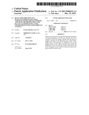 | 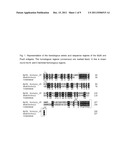 |
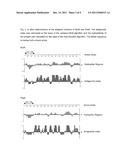 | 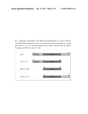 |
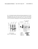 | 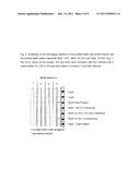 |
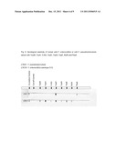 | 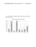 |
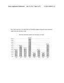 |  |
 |  |
 | 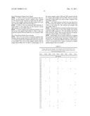 |
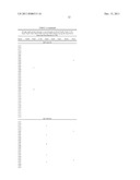 | 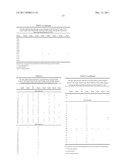 |
 |  |
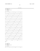 | 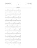 |
 |  |
 |  |
 |  |
 |  |
 |  |
| New patent applications in this class: | |
| Date | Title |
|---|---|
| 2022-05-05 | Microfluidic system for amplifying and detecting polynucleotides in parallel |
| 2019-05-16 | Reagents and methods for detecting protein lysine 2-hydroxyisobutyrylation |
| 2019-05-16 | Lateral flow analyte detection |
| 2019-05-16 | Mutations in the bcr-abl tyrosine kinase associated with resistance to sti-571 |
| 2019-05-16 | Enhanced methods of ribonucleic acid hybridization |
| Top Inventors for class "Combinatorial chemistry technology: method, library, apparatus" | |
| Rank | Inventor's name |
|---|---|
| 1 | Mehdi Azimi |
| 2 | Kia Silverbrook |
| 3 | Geoffrey Richard Facer |
| 4 | Alireza Moini |
| 5 | William Marshall |