Patent application title: Multiplex amplification reaction method for determination of Campylobacter jejuni Penner/capsule type
Inventors:
Frederic Poly (Silver Spring, MD, US)
Patricia Guerry (Silver Spring, MD, US)
Patricia Guerry (Silver Spring, MD, US)
Carl Mason
Oralak Serichantalergs
IPC8 Class: AC12Q168FI
USPC Class:
435 612
Class name: Measuring or testing process involving enzymes or micro-organisms; composition or test strip therefore; processes of forming such composition or test strip involving nucleic acid with significant amplification step (e.g., polymerase chain reaction (pcr), etc.)
Publication date: 2011-08-25
Patent application number: 20110207138
Abstract:
The inventive method and associated reagents relate to a molecular
approach to determining Campylobacter jejuni capsule/Penner types. The
invention also relates to a method of identifying Campylobacter jejuni
types using the inventive primers in a multiplex PCR assay.Claims:
1. A method of identifying Campylobacter jejuni strains in a sample
suspected of containing Campylobacter jejuni DNA by polymerase chain
reaction, comprising: (a) subjecting DNA from said sample to a PCR
amplification reaction using one or more PCR primer pairs targeting one
or more regions of the O-methyl phosphoramidate synthesis region, heptose
synthesis and hyper-variable region of the polysaccharide capsule loci of
Camplylobacter jejuni; (b) analyzing amplification products resulting
from said amplification reaction.
2. The method of claim 1, wherein said polysaccharide capsule loci is derived from Campylobacter jejuni strains selected from HS2; HS3; HS6; HS10; HS15C; HS41; HS53; HS1; HS23; HS42; HS44; HS17 and strain 8486.
3. The method of claim 1, wherein said amplification products are analyzed by size determination.
4. The method of claim 1, wherein said PCR primer pairs contain sequences selected from the group consisting of: SEQ ID NO: 1 and SEQ ID NO: 2; SEQ ID NO: 3 and SEQ ID NO: 4; SEQ ID NO: 5 and SEQ ID NO: 6; SEQ ID NO: 7 and SEQ ID NO: 8; SEQ ID NO: 9 and SEQ ID NO: 10; SEQ ID NO: 11 and SEQ ID NO: 12; SEQ ID NO: 13 and SEQ ID NO: 14; SEQ ID NO: 15 and SEQ ID NO: 16; SEQ ID NO: 17 and SEQ ID NO: 18; SEQ ID NO: 19 and SEQ ID NO: 20; SEQ ID NO: 21 and SEQ ID NO: 22; SEQ ID NO: 23 and SEQ ID NO: 24; SEQ ID NO: 25 and SEQ ID NO: 26; SEQ ID NO 27 and SEQ ID NO: 28.
5. The method of claim 1, wherein said PCR reaction is multiplex amplification reaction.
6. The method of claim 1, wherein said primers are grouped in an alpha mix and a beta mix with the alpha and beta mixes that are separately added to an unknown DNA sample in order to discriminate product sizes.
7. The method of claim 1, wherein said sample is a clinical sample.
8. The method of claim 1, wherein said sample is collected from a matrix selected from the group consisting of a bacterial culture, a blood, a tissue, and fecal material.
9. The method of claim 1, wherein the primers have about 18-30 nucleotides, a G/C content of 20-50%, and a melting temperature between about 57.degree. C. and 63.degree. C.
10. The method of claim 1, wherein said amplification reaction yields one or more of amplification products selected from the group consisting of SEQ ID NO: 29; SEQ ID NO: 30; SEQ ID NO: 31; SEQ ID NO: 32; SEQ ID NO: 33; SEQ ID NO: 34; SEQ ID NO: 35; SEQ ID NO: 36; SEQ ID NO: 37; SEQ ID NO: 38; SEQ ID NO: 39; SEQ ID NO: 40; SEQ ID NO: 41; and SEQ ID NO: 42.
11. The method of claim 2, wherein said HS2 PCR primers recognize HS2 Penner type; HS3 PCR primers recognize H3 Penner type; HS4 PCR primers recognize HS4 A Penner complex; HS6 PCR primers recognize HS6 Penner type; HS10 PCR primers recognize HS10 Penner type; HS15C PCR primers recognize HS15 and HS31 Penner types; HS41 PCR primers recognize HS41 Penner type; HS53 PCR primers recognize HS53 Penner type; HS1 D PCR primers recognize HS1 complex Penner type; HS17 PCR primers recognize HS8 and HS17 Penner type; 8486 PCR primers recognize HS4B Penner type; HS23 PCR primers recognize HS23 Penner complex; HS42E PCR primers recognize HS42 Penner type; HS44 PCR primers recognize HS44 Penner type.
12. The method of claim 3, wherein the amplification of products are analyzed by agarose gel electrophoresis.
13. The method of claim 5, wherein said PCR primer pairs are grouped into an alpha mix comprising one or more sequence pairs selected from the group consisting of: SEQ ID NO: 1 and SEQ ID NO: 2; SEQ ID NO: 3 and SEQ ID NO: 4; SEQ ID NO: 5 and SEQ ID NO: 6; SEQ ID NO: 7 and SEQ ID NO: 8; SEQ ID NO: 9 and SEQ ID NO: 10; SEQ ID NO: 11 and SEQ ID NO: 12; SEQ ID NO: 13 and SEQ ID NO: 14; SEQ ID NO: 15 and SEQ ID NO: 16, and a beta mix comprising one or more sequence pairs selected from the group consisting of: SEQ ID NO: 17 and SEQ ID NO: 18; SEQ ID NO: 19 and SEQ ID NO: 20; SEQ ID NO: 21 and SEQ ID NO: 22; SEQ ID NO: 23 and SEQ ID NO: 24; SEQ ID NO: 25 and SEQ ID NO: 26; SEQ ID NO 27 and SEQ ID NO: 28.
14. A kit for typing Campylobacter jejuni strains, wherein the kit comprises one or more PCR primer pairs of claim 1.
15. The kit of claim 14, wherein said polysaccharide capsule loci is derived from Campylobacter jejuni strains selected from HS2; HS3; HS6; HS10; HS15C; HS41; HS53; HS1; HS23; HS42; HS44; HS17 and strain 8486.
16. The kit of claim 14, wherein said PCR primer pairs contain sequences selected from the group consisting of: SEQ ID NO: 1 and SEQ ID NO: 2; SEQ ID NO: 3 and SEQ ID NO: 4; SEQ ID NO: 5 and SEQ ID NO: 6; SEQ ID NO: 7 and SEQ ID NO: 8; SEQ ID NO: 9 and SEQ ID NO: 10; SEQ ID NO: 11 and SEQ ID NO: 12; SEQ ID NO: 13 and SEQ ID NO: 14; SEQ ID NO: 15 and SEQ ID NO: 16; SEQ ID NO: 17 and SEQ ID NO: 18; SEQ ID NO: 19 and SEQ ID NO: 20; SEQ ID NO: 21 and SEQ ID NO: 22; SEQ ID NO: 23 and SEQ ID NO: 24; SEQ ID NO: 25 and SEQ ID NO: 26; and SEQ ID NO 27 and SEQ ID NO: 28.
17. The kit of claim 14, wherein said PCR primer pairs are grouped into an alpha mix comprising one or more sequence pairs selected from the group consisting of: SEQ ID NO: 1 and SEQ ID NO: 2; SEQ ID NO: 3 and SEQ ID NO: 4; SEQ ID NO: 5 and SEQ ID NO: 6; SEQ ID NO: 7 and SEQ ID NO: 8; SEQ ID NO: 9 and SEQ ID NO: 10; SEQ ID NO: 11 and SEQ ID NO: 12; SEQ ID NO: 13 and SEQ ID NO: 14; SEQ ID NO: 15 and SEQ ID NO: 16, and a beta mix comprising one or more sequence pairs selected from the group consisting of: SEQ ID NO: 17 and SEQ ID NO: 18; SEQ ID NO: 19 and SEQ ID NO: 20; SEQ ID NO: 21 and SEQ ID NO: 22; SEQ ID NO: 23 and SEQ ID NO: 24; SEQ ID NO: 25 and SEQ ID NO: 26; SEQ ID NO 27 and SEQ ID NO: 28.
18. The kit of claim 14, further comprising a buffer, diluents and/or excipient.
19. The kit of claim 14, further comprising a DNA polymerase.
20. The kit of claim 14, wherein the primers have about 18-30 nucleotides, a G/C content of 20-50%, and a melting temperature between about 57.degree. C. and 63.degree. C.
Description:
CROSS-REFERENCES TO RELATED APPLICATIONS
[0001] This application claims the benefit of U.S. Provisional Application No. 61/307,632, filed 24 Feb. 2010, which is incorporated by reference, herein.
BACKGROUND OF INVENTION
[0002] 1. Field of Invention
[0003] The inventive subject matter relates to a molecular method for determining Campylobacter jejuni capsule/Penner types.
[0004] 2. Background
[0005] Campylobacter is a major cause of human bacterial diarrheal disease worldwide, with C. jejuni, and to a lesser extent C. coli, the most important pathogenic Campylobacter species. Campylobacteriosis symptoms range from asymptomatic infection to bloody diarrhea associated with abdominal pain and fever. The major source of human infection is through consumption of uncooked poultry, which is commonly colonized by C. jejuni. Post infectious sequelae associated with C. jejuni include reactive arthritis, Guillain-Barre syndrome and irritable bowel syndrome.
[0006] The molecular pathogenesis of C. jejuni is not well understood, but a polysaccharide capsule (CPS) is one of the few recognized virulence determinants of this pathogen. The capsular polysaccharide undergoes a reversible phase variation in expression (Bacon, et al., Mol. Microbiol. 40:769-777 (2001)). The capsule contributes to serum resistance of C. jejuni, the ability of C. jejuni to invade intestinal epithelial cells in vitro, and, in a ferret model, is required for virulence (Bacon, et al., Mol. Microbiol. 40:769-777 (2001)). More recently, polysaccharide capsule conjugated to a protein carrier has been shown to protect non-human primates against diarrheal disease Monteiro, et al., Infect Imm. 77(3): 1128-36 (2009). Differentiation of Campylobacter jejuni strains is typically conducted through the use of Penner serotyping.
[0007] The Penner or "heat stable" serotyping scheme is a passive slide hemaglutination assay for both C. jejuni and C. coli that includes 47 C. jejuni serotypes. Rabbit polyclonal antibodies are generated against whole cells of each of the 47 type strains. Antigens are extracted from C. jejuni strains to be tested by heating bacterial suspensions in saline at 100° C. These "heat-stable" antigens are used to sensitize sheep erythrocytes, which are used in a passive slide hemagglutination assay with the specific polyclonal antisera. Genetic studies indicate that CPS is the major serodeterminant of the Penner scheme. Thus, mutation of genes required for CPS biogenesis rendered many strains untypable in the Penner scheme.
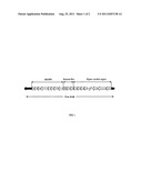
[0008] However, other surface heat stable surface structures such as lipooligosaccharides (LOS) may also contribute to serospecificity of some Penner types. The capsular polysaccharides of C. jejuni are known to be structurally diverse (Karlyshev et al., Molecular Microbiology 55:90-103). This structural diversity is consistent with the variability observed in the genes encoding the capsule in C. jejuni. The capsule locus of C. jejuni includes both highly conserved genes involved in capsule synthesis and highly variable loci that encode genes involved in synthesis of specific sugars and specific glycosyl transferases required to link the sugars together. The variable CPS locus located between two conserved genes, kpsC and kpsF, and the variable genes can range from 15 to 34 kb (FIG. 1). Variable genes also encode synthesis and transfer of modifications to the sugars, such as methyl phosphormidate (MeOPN) (Karlyshev et al., Molecular Microbiology 55:90-103).
[0009] Penner serotyping is technically difficult to perform and expensive to produce the type antisera. As a result, only a handful of reference laboratories routinely perform Penner typing. Moreover, many serotypes fall into Penner "complexes". The significance of these complexes is not totally understood in most cases, but they appear to include capsules with related structures (Aspinall et al. Carbohydr Res. 231:13-30 (1992).
[0010] Others have tried to replace the laborious Penner serotyping using a molecular typing approach involving restriction fragment length polymorphism (RFLP) analysis of PCR amplified lipooligosaccharide (LOS) loci (Shi et al. J Clin Microbiol. 40(5):1791-7 (2002); Nakari et al., J Clin Microbiol. 43(3):1166-70) (2005). However, these RFLP methods have not been widely used and have not replaced Penner serotyping as the typing method of choice. This may be due in part to the RFLP method requiring amplification of a 9.6 kb fragment. Using PCR to generate such large amplicons is difficult and can place special requirements on the PCR conditions and reagents used, as demonstrated by Nakari et al., who were unable to generate amplified fragments using the amplification conditions described by Shi et al. These RFLP methods are also limited because they are based on the amplification of the LOS locus. At the time of the Shi et al. study, it was known that both the LOS and CPS structure were part of the Heat Stable antigen (HS) recognized through the Penner serotyping method. However, in 2005, CPS was demonstrated to be the major serodeterminant of the Penner method (Karylshev, et al., Mol. Micro. 55: 90-103 (2005). This helps explain why Shi et al. and Nakari et al. found only partial correlation between the Penner serotypes and RFLP groups. Penner serotyping distinguishes strains that cannot be distinguished by this RFLP method. For example, the most common RFLP type, Hh1Dd1, contained strains belonging to several HS serotypes, including HS 6,7, HS12, HS 27, HS 55, HS 21, HS10, HS 57, HS 6, HS 15, HS 23,36,53, and HS 27+HS 31 (Nakari et al., J Clin Microbiol. 43(3):1166-70 (2005)). And some serotypes, such as HS 2, HS 3, HS 4 complex, HS 8, HS10, HS11, HS12, HS15, HS19, HS 31, HS 32, HS 41, HS 57, and HS 23,36,53 include more than one RFLP (Nakari et al., J Clin Microbiol. 43(3):1166-70 (2005).
SUMMARY OF THE INVENTION
[0011] The current invention relates to reagents and method to identify Campylobacter jejuni Capsule/Penner types via molecular, rather than serological, methods.
[0012] Therefore, an object of the invention is a panel of multiplex DNA primers for identification of C. jejuni Capsule/Penner types by polymerase chain reaction (PCR).
[0013] Several important advantages of amplification reactions over serological determination are evident. First, it is technically difficult to perform and expensive to produce type antisera. As a result, few reference laboratories are capable of routine Penner typing. Additionally, many serotypes fall into Penner "complexes."
[0014] Amplification methods, unlike typing sera methods, are relatively available to research and reference laboratories. Furthermore, no expression of capsule is needed. Therefore, there are no affects due to phase variation in capsule expression, as is possible with serotyping. Multiplexing reduces the number of reactions to be performed per samples. Additionally, amplification reactions do not suffer from CPS being shut down or modified thru slipstrand mutations. The instant invention can identify 14 CPS types, that include 17 serotypes.
[0015] The multiplex amplification technique amplifies a fragment less than 1 kb that can be routinely performed in any molecular biology lab worldwide.
BRIEF DESCRIPTION OF DRAWINGS
[0016] FIG. 1. Schematic of the general organization of the capsule loci of C. jejuni. The region between kpsC and kpsF (black arrows) encodes the genes for synthesis of distinct capsule structures. If present, genes for heptose and MeOPN synthesis are highly conserved. The region to the right is the hyper-variable region containing sugar transferases and sugar biosynthetic genes.
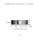
[0017] FIG. 2. Graphic depiction illustrating cumulative distribution of C. jejuni serotypes in Thailand (103 strains). Serotypes were determined using Penner serotying. The numbers in the boxes refer to Penner serotypes.
DETAILED DESCRIPTION OF PREFERRED EMBODIMENTS
[0018] The following terms are defined:
[0019] "Amplification reaction" refers to a method of detecting target nucleic acid by in vitro amplification of DNA or RNA.
[0020] "Polymerase chain reaction (PCR)" refers to the amplification of a specific DNA sequence, termed target or template sequence, that is present in a mixture, by adding two or more short oligonucleotides, also called primers, that are specific for the terminal or outer limits of the template sequence. The template-primers mixture is subjected to repeated cycles of heating to separate (melt) the double-stranded DNA and cooling in the presence of nucleotides and DNA polymerase such that the template sequence is copied at each cycle.
[0021] "Primer" refers to DNA oligonucleotides complementary to a region of DNA and serves as the initiation of amplification reaction from the 5' to 3' direction.
[0022] "Primer pair refers to the forward and reverse primers in an amplification reaction leading to amplification of a double-stranded DNA region of the target.
[0023] "Target" refers to a nucleic acid region bound by a primer pair that is amplified through an amplification reaction.
[0024] The term "multiplex amplification reaction" herein refers the detection of more than one template in a mixture by the addition of more than one set of oligonucleotide primers.
[0025] The term "capsule" herein refers to the structure lying outside the cell wall of bacteria, such as Campylobacter jejuni.
[0026] Utilizing genomic and capsule loci sequences, a molecular method for determining Penner and capsule type was developed. This method is simpler than Penner typing. The inventive method is more easily standardized than Penner serotyping, since molecular reagents (i.e., primers) can be produced and standardized resulting in lower cost. Additionally, the method does not require that the capsule be expressed. Therefore, it is not affected by phase variation in capsule expression, unlike the typing system.
[0027] In one embodiment, the current invention provides a method to specifically recognize Capsule/Penner types thru PCR amplification of type specific sequences. The inventive method and reagents permit identification of Campylobacter jejuni Penner types without the potential for capsule shutdown or modification due to slipstrand mutations.
Example 1
Design of Primer Sequences Correlating with Penner Serotype
[0028] The capsule locus of C. jejuni includes both highly conserved genes involved in capsule synthesis and highly variable loci that encode genes involved in synthesis of specific sugars and specific glycosyl transferases required to link the sugars together. The variable CPS locus, located between two conserved genes, kpsC and kpsF, and the variable genes range from 15 to 34 kb (FIG. 1). Variable genes also encode synthesis and transfer of modifications to the sugars, such as methyl phosphormidate (Karlyshev, A. et al., Mol. Microbiol. 55:90-103 (2005)). In a preferred embodiment, based on the DNA sequences, unique DNA sequences from the capsule loci of C. jejuni, for each Penner type, were identified. The selected genes were further compared to the whole genome sequences of C. jejuni in order to eliminate potential similarities with genes outside the CPS region.
[0029] Selection of genes unique to a particular serotype was performed using a local BLAST program. Each single gene of the variable capsule region (between kpsC and kpsF) was compared with a database containing the nucleotides sequences of all the available capsule loci of C. jejuni. The selected genes were further compared to the whole genome sequences of C. jejuni sequenced genomes to eliminate potential similarities with genes outside CPS region.
[0030] Penner serotyping was performed on a subset of C. jejuni clinical strains obtained from Thailand. Serotype distribution is shown in FIG. 2. As illustrated in FIG. 2, HS1, 2, 3, 4, 5, 15, 23, 42 and 53 serotypes are the most common serotypes observed in Thailand. These results are in accordance with other Penner serotyping surveys demonstrating that HS1, 2, 3, and especially 4, represent a large percentage of clinical isolates in the world (Asrat, et al. Epidemiol. Infect. 118:222-226 (1997); Jones, et al. J. Infect. 9:51-58 (1984); Karmali et al. J. Infect. Dis. 147:243-246 (1984); Owen, R. J., and J. R. Gibson. PHLS Microbiol. Dig. 12:2-6 (1995); Penner et al., Eur. J. Clin. Microbiol. 2:378-383 (1983)). In addition, some serotypes appear to be more "regional" like HS8 and HS10.
[0031] Based on these observations, sequencing of capsule loci of common worldwide and regional Penner serotypes, for which no sequences were available, was performed. CPS locus sequences were obtained by cloning the kpsC-hddA and KpsF-dmhA PCR fragments into a pCR4-TOPO® vector (Invitrogen, Carlsbad, Calif.) in order to create a representative genomic library. Following purification, clones were sequenced. Assembly was performed using Sequencher® 4.8 (Gene Codes Corporation, Ann Arbor, Mich.). PCR primers were designed to correct errors and close gaps. Development of unique Campylobacter jejuni PCR primer sequences were undertaken by sequencing DNA of capsule loci of the strains of HS3; HS3/13/50; HS4; HS8; HS10: HS15; HS17; and HS42, plus HS1/44 and HS44. A summary of the data is presented in Table 1.
TABLE-US-00001 TABLE 1 CPS No. structure Penner Size Accession GC No. MeOPN Deoxy available type (bp) number (%) Genes TF's Heptose Heptose (reference) HS1 15,180 BX545859a 26.8 11 1 No No (g) HS2 34,180 AL139078b 26.5 28 2 Yes No (a) HS3 26,371 HQ343268f 27.3 23 1 Yes Yes (h) HS3/13/50 26,371 HQ343267f 27.3 23 1 Yes Yes -- HS4 22,836 HQ343269f 28.0 18 2 Yes Yes -- HS4/13/64 23,423 AASY01000000c 28.0 18 2 Yes Yes (i) HS6 26,729 NC_009839.d 27.6 21 0 No No (j) HS8 22,063 HQ343270f 27.1 18 0 Yes Yes -- HS10 27,307 HQ343271f 27.1 25 1 Yes Yes -- HS15 23,868 HQ343272f 28.3 22 1 Yes Yes -- HS17 22,064 HQ343273f 27.1 18 0 Yes Yes -- HS19 16,727 BX545860a 26.1 13 1 No No (k) HS23 24,627 AY332625a 27.0 21 1 Yes Yes (l; a) HS36 24,625 AY332624a 26.9 21 1 Yes Yes (l; a) HS23/36 24,625 BX545858a 27.1 21 1 Yes Yes (l) HS41 34,118 BX545857a 27.2 30 0 Yes Yes n HS42 23,268 HQ343274f 26.9 21 0 Yes Yes -- HS53 18,272 CP000025.1e 27.0 15 0 Yes Yes (m) aKarlyshev, et al., Mol. Microbiol., 55: 90-103 (2005) bParkhill, et al., Nature 403: 665-668 (2000) cPoly, et al., Infect. Immun., 75: 3425-3433 (2007) dPearson, et al., J. Bacteriol., 189: 8402-8403 (2007) eFouts, et al., PLoS. Biol., 3: e15 (2005) fCurrent study gMcNall, et al., FEBS J. 272: 4407-4422 (2005) hAspinall, et al., Eur. J. Biochem. 231: 570-578 (1995) iChen, et al., Carbohydr. Res. 343: 1034-1040 (2008) jMuldoon, et al., Carbohydr. Res. 337: 2223-2229 (2002) kMcNally, et al., FEBS J. 273: 3975-3989 (2006) lAspinall, et al., Carbohydr. Res. 231: 13-30 (1992) mGilbert, et al., Chembiochem. 8: 625-631 (2007) nHannify, et al., Carbohydr. Res. 319: 124-132 (1999)
[0032] The capsule loci sequences obtained were then compared to Penner serotyping results. The results of this comparison led to corroboration of complexes in the Penner serotyping system. For example, Karlyshev et al., Mol. Microbiology, 55(1): 90-103, determined that strains in the HS23/36 complex (i.e. HS23, HS36 and HS23/36) have the same CPS loci. Mu_HS23 primers identify strain that belong to the HS23/36 complex (including: HS23, HS36 and HS23/36).
[0033] HS1 is often found in complex with HS44. However, no differences in capsule sequence were observed between the HS1 type strain and a clinical isolate that typed as HS1/44. Interestingly, the sequence of the HS44 type strain appeared to be drastically different than HS1 or HS1/44 strains. For these reasons primers Mu_HS1 D recognize HS1 as well as HS1/44 (HS1 complex) strains, but not HS44 strains. A primer set Mu_HS44 was specifically designed to recognize this serotype. HS8 and HS17 are also often associated. No difference of capsule sequence was identified between these serotypes.
[0034] Furthermore, HS8 and HS17 are often associated. Mu_HS17 primers identify both HS8, HS17 and HS8/17 strains (defined as the HS8 complex). Similarly, the HS4 serotype is often associated with HS13, HS43, HS50, HS64 or HS65. Sequencing of three strains belonging to this complex (HS4, HS13 and HS4/13/64) allowed discrimination of these strains into two groups based on their capsule loci. Primer sets were designed to distinguish these groups: Mu_HS4B that recognized HS4, HS13 type strains, as well as some strains typed as HS4/13/64. Un--8486 primers identified strains belonging to HS4/13/64 complexes as well as the HS64 type strain.
[0035] A database containing all available CPS loci was created to identify unique regions of each serotype. This data set included a partial sequence of the type strain of HS44, which forms a complex with HS1. In one embodiment, two primer sets were designed for the HS4 complex. These are based on differences in MeOPN transferases among the sequenced strains in this complex. The embodiment is based on an assumption that the differences among the HS4 complex are due to differences in the position of MeOPN on the polysaccharide. These primer sets, named Mu_HS4 and Mu--8486, respectively, were designed in HS4.07 and Cj8486--1475, both putative MeOPN transferases. Since some HS4 strains contain two MeOPN transferases, a strain can theoretically be positive with both Mu--8486 and Mu_HS4. Since the CPS loci of HS8 and HS17 are so similar, a single primer set was designed for this complex. Although CPS has been shown not to be the serodeterminant of the HS6 serotype, the CPS genes in this strain do not match any of the other published sequences so a primer set was designed for this CPS type as well.
Example 2
Design of Multiplex PCR Assay to Penner Serotypes
[0036] In a preferred embodiment, PCR primers were designed in regions that were found unique to each particular C. jejuni serotype. In one embodiment, primer sets are grouped into two `mixes` based on the sizes of the products (Table 2). The alpha mix contains primers that distinguish HS2, the HS3 complex, HS6, HS10, HS15, HS41, HS53 and part of the HS4 complex (HS4 and HS13, termed HS4A). The beta mix contains primers that distinguish the HS1 complex (including HS1 and HS1/44), the HS23/36 complex, the HS8 complex (HS8 and HS17), HS42, HS44 and part of the HS4 complex (HS4/13/64 or CG8486-like, termed HS4B).
[0037] In a preferred embodiment, the PCR primers were designed to permit multiplex PCR. Multiplex PCR significantly reduces the number of reactions needed for strain identification. Design of the multiplex primers was conducted utilizing the online software MuPlex® (Boston University, Boston, Mass.) (described in Rachlin, et al., Nucleic Acid Research 33 (Web Server Issue): W544-W547) (2005).
[0038] Primers were designed with the following parameters: length between 18 and 30 residues, 20 to 50% GC, Tm ranging from 57° to 63° C. with a minimum product difference of 20 bp. The primer sequences were verified for absence of dimerization or hairpin formation using AutoDimer® (Vallone and Butler, Biotechniques 37(2): 226-231 (2004)).
[0039] Primers were evaluated for their ability to enable efficient amplification of predicted product and for not interfering with other primers, capable of amplifying Campylobacter jejuni DNA, included in the reaction. The primer sets were designed to produce amplicons that differ by at least 20 bp from the other amplicons in the same mix. Primer sets were judged satisfactory if they produced the expected size PCR product on their Penner serotype DNA template or related complexes and were negative for other tested serotypes. Shown in Table 2 are the strains identified by each primer pair, and the product size, as well as the associated SEQ ID Number.
TABLE-US-00002 TABLE 2 Alpha mix PCR multiplex primers Forward Reverse Product sequence sequence Product Size Penner type Designed in (SEQ ID (SEQ ID (SEQ ID (bp) recognized Gene No.) No.) No.) Mix Alpha Mu_HS2 62 HS2 Cj1437c 1 2 29 Mu_HS3 149 HS3 HS3.17 3 4 30 Mu_HS4 370 HS4A HS4.07 5 6 31 Mu_HS6 185 HS6 C8J_1331 7 8 32 Mu_HS10 229 HS10 HS10.08 9 10 33 Mu_HS15C 325 HS15 and HS15.12 11 12 34 HS31 Mu_HS41 279 HS41 HS41.22c 13 14 35 Mu_HS53 251 HS53 CJE1602 15 16 36 Mix Beta Mu_HS1D 607 HS1 complex HS1.08 17 18 37 Mu_HS17 342 HS8 and HS17.16 19 20 38 HS17 Mu_8486 652 HS4 B Cj8486_1475 21 22 39 Mu_HS23 161 HS23/36 CJJ81176_1435 23 24 40 Mu_HS42E 441 HS42 HS42.14 25 26 41 Mu_HS44 148 HS44 Not annotated 27 28 42
[0040] Although other potential parameters are possible, multiplex PCR amplification conditions are standardized for primers in the alpha and beta mixes. A preferred embodiment of the PCR identification method comprises the following steps: [0041] a. Obtain a sample suspected of containing Campylobacter jejuni DNA; [0042] b. Subject sample containing said DNA to one or more primer pairs listed in Table 2, or a primer pair with capable of amplifying the same product shown in Table 2. In a preferred embodiment, the primers are 18-30 nucleotides, have a G/C content of 20-50%, and a melting temperature between about 57° C. and 63° C.; [0043] c. Amplify target DNA under the following parameters: 94° C. for 30'', 56° C. for 30'', 72° C. for 45'' for a total of 29 cycles; [0044] d. Subsequent to PCR amplification compare PCR product size.
[0045] Amplifying DNA from an unknown C. jejuni sample, using the primers in Table 2, and comparing the size of the ensuing amplification products permits identification of 17 C. jejuni Penner serotypes (14 CPS types). In a preferred embodiment, the amplified DNA is separated and sized through an agarose gel (2%), run in 0.5×TBE buffer. The sizes of the PCR products and corresponding serotype are determined by comparison with 100 bp molecular size standards. Although agarose gel electrophoresis is a preferred method, other methods to analyze PCR product size are contemplated.
[0046] In other embodiments, methods are carried out, at least in part, using a solid support. A variety of different supports can be used. In some embodiments, the solid support is a single solid support, such as a chip or wafer, or the interior or exterior surface of a tube, cone, plastic plate or other article. In some embodiments, the solid support is a particulate support, also referred to as a microsphere, bead or particle. Typically, the particles form groups in which particles within each group have a particular characteristic. Examples of suitable characteristics include, but are not limited to, color, fluorescence frequency, density, size, or shape. The selection of characteristics will depend on multiple criteria including the ability to distinguish or separate target-bound particles from particles of other groups. Particles can be separated by a number of methods. In a preferred embodiment, the particles can be separated using techniques, such as, for example, flow cytometry.
[0047] The particles can be fabricated from virtually any insoluble or solid material. For example, the particles can be fabricated from silica gel, glass, nylon, resins, Sephadex®, Sepharose®, cellulose, magnetic material, a metal (e.g., steel, gold, silver, aluminum, copper, or an alloy) or metal-coated material, a plastic material (e.g., polyethylene, polypropylene, polyimide, polyester, polyvinylidenefluoride (PVDF)) and the like, and combinations thereof. Examples of suitable micro-beads are described, for example, in U.S. Pat. Nos. 5,736,330, 6,046,807 and 6,057,107, all of which are incorporated herein by reference in their entirety.
[0048] Thus, in one embodiment, the multiplex method described herein is performed using microspheres conjugated to unique capture oligonucleotides, permitting the analysis of many different nucleic acids in a single reaction. Each unique capture oligonucleotide is complementary to a unique tag sequence within one of the amplicons to be detected. In this embodiment, the microsphere mix consists of a number of microspheres equal to the number of serotypes that can be detected in the assay. Each of the microspheres contains a different fluorescent dye mix and is coupled to a unique capture oligonucleotide sequence complementary to a unique tag sequence within the amplicon of each serotype of interest. The hybridization of the capture oligonucleotide and the tag sequence of an amplicon results in the coupling of the amplicon to the solid support. The unique capture oligonucleotide and its complementary tag sequence are, thus, associated with a single, specific Penner serotype. The capture oligonucleotides are designed so there is no cross-hybridization between the capture oligonucleotides and the amplicons from more than one serotype under the hybridization conditions used.
[0049] In this method, the multiplex Alpha and Beta primer sets are used to amplify regions of interest in a C. jejuni DNA sample in the presence of a biotinylated dNTP mixture. Instead of running the amplified PCR fragments on an agarose gel to estimate their size, the amplified PCR fragments are incubated with microspheres conjugated to capture oligonucleotides specific for the serotypes of interest and streptavidin conjugated to a dye, such as phycoerythrin, and analyzed using an appropriate detection system.
Example 3
Comparison/Validation of Multiplex PCR Method to Penner Serotyping
[0050] From the correlations observed between Penner serotyping data and capsule loci sequence information, PCR primers were developed that were specific to available CPS strains. The primers were then validated against species of DNA of from C. jejuni strains of known Penner serotypes.
[0051] Validation of the inventive multiplex PCR assay method was conducted on 244 strains from Thailand and Egypt that had been Penner serotyped in reference laboratories. The result of this study is illustrated in Table 3.
[0052] In summary, the multiplex method had a specificity and accuracy of >97% and a sensitivity of >89%. The method detected 100% of strains of HS2 (30/30), HS8/17 (10/10), HS15 (19/19), HS23/36 (13/13, HS41 (2/2), HS53 (16/16) and HS6 (1/1). There were two false negatives with the HS1/44 primers (2/25), the HS3 complex primers (2/25), and the HS4 complex primers (2/20). The HS10 primers resulted in one false negative (1/14) and two false positives (one HS1/44 and one HS44 serotype). The HS6 primers picked up five strains, two of which belonged to the HS3 complex, and the HS15 primers picked up 6 false positives, all of which were HS31. Since there is no information about the CPS locus or structure of HS31, this may suggest a relationship between HS15 and HS31, although they are not part of a complex. Included within the 244 strains were 37 strains that belonged to 15 serotypes not included in the multiplex. A total of 27 of these 37 strains were negative with the multiplex primers, but six HS31 strains reacted with the HS15 primers, as discussed above, individual strains that typed as HS32, HS35 and HS59 reacted with the HS6 primers, and one HS37 strain reacted with the HS3 primers. There were also 23 strains that were not able to be typed in the Penner scheme, and 8 of these reacted with the HS6 primers, two reacted with the HS15 primers, and one each reacted with HS2 and HS10 primers. These data suggest that some strains are not able to be typed in the Penner scheme because CPS is not expressed.
TABLE-US-00003 TABLE 3 # True False False True Primer set strains positive Positive negative Negative % Accuracy1 % Sensitivity2 % Specificity3 HS1/44 25 23 0 2 219 99.18 92.00 100.00 Complex HS2 30 30 0 0 214 100.00 100.00 100.00 HS3 Complex 26 24 1 2 217 98.77 92.31 99.54 HS4 Complex 20 18 0 2 224 99.18 90.00 100.00 HS6 1 1 5 0 238 97.95 100.00 97.94 HS8/17 10 10 0 0 234 100 100.00 100 complex HS10 14 13 2 1 228 98.77 92.86 99.13 HS15 19 19 6 0 219 97.54 100.00 97.33 HS23/36 13 13 1 0 230 99.59 100.00 99.57 complex HS41 2 2 0 0 242 100.00 100.00 100.00 HS42 8 8 0 0 236 100.00 100.00 100.00 HS53 16 16 1 0 227 99.59 100.00 99.56 1Accuracy = (true positive + true negative)/true positive + true negative + false positive + false negative) 2Sensitivity = true positive/(true positive + false negative) 3Specificity = true negative/(true negative + false positive)
[0053] Having described the invention, one of skill in the art will appreciate in the appended claims that many modifications and variations of the present invention are possible in light of the above teachings. It is therefore, to be understood that, within the scope of the appended claims, the invention may be practices otherwise than as specifically described.
Sequence CWU
1
42125DNACampylobacter jejuni 1cagcattgga ggatttacaa tatat
25222DNACampylobacter jejuni 2catcctagca
caactcactt ca
22324DNACampylobacter jejuni 3ggtaaggttg attctgggtt taat
24422DNACampylobacter jejuni 4agattaggcc
aagcaatgat aa
22520DNACampylobacter jejuni 5tatatttggt tagggatcca
20623DNACampylobacter jejuni 6cctaacatat
catacactac ggt
23726DNACampylobacter jejuni 7catacatttg ctttcagatt ctttac
26821DNACampylobacter jejuni 8acacgcctat
tgttgttgtt c
21919DNACampylobacter jejuni 9tcttatgcag cacgctgat
191022DNACampylobacter jejuni 10caaattcaat
cgactagcca ct
221124DNACampylobacter jejuni 11acaggtaata aaatgtgcga gttt
241220DNACampylobacter jejuni 12atgcatctgc
aacatcatcc
201327DNACampylobacter jejuni 13cttacatatg ctggtagaga tgatatg
271420DNACampylobacter jejuni 14tgcaatctct
aaagcccaag
201519DNACampylobacter jejuni 15aggcaagcag gaattgttt
191622DNACampylobacter jejuni 16ttaattgctc
tttggcaatc tt
221721DNACampylobacter jejuni 17ttggcggtaa gtttttgaag a
211821DNACampylobacter jejuni 18gcaagagaaa
catctcgcct a
211920DNACampylobacter jejuni 19ttcacgtgga ggattattgg
202025DNACampylobacter jejuni 20ttgaacattt
catgtgtatt cccta
252120DNACampylobacter jejuni 21gtggacatgg aactgggact
202223DNACampylobacter jejuni 22aaaacgttta
aagtcagtgg aaa
232324DNACampylobacter jejuni 23gcttgggaga tgaatttacc ttta
242427DNACampylobacter jejuni 24gctttatatc
tatccagtcc attatca
272520DNACampylobacter jejuni 25atggtaaaac cggcatttca
202620DNACampylobacter jejuni 26atgcttcagt
tccacccaaa
202721DNACampylobacter jejuni 27agaagatgca ctaggctcta g
212820DNACampylobacter jejuni 28gctatctaat
tccatccctg
202962DNACampylobacter jejuni 29cagcattgga ggatttacaa tatatgagag
attatgtaga tgaagtgagt tgtgctagga 60tg
6230149DNACampylobacter jejuni
30agattaggcc aagcaatgat aataaattct aaaaattttt taggttatat atttttaccg
60tatattttat taagtattgt tatactatat aaacaagagc aaaaaaatta taaacataaa
120attaaattaa acccagaatc aaccttacc
14931370DNACampylobacter jejuni 31tatatttggt tagggatcca atttcgagat
taaaaacggg tttaaatcat attaatttaa 60aggcgaatag acttgatcgt tttgatttgg
atacacctat agaaagagtt ttagatagag 120agacatatta tttcgaatct cctttgccaa
cctgtgatca tataaaaact tattggattt 180atgcggaaag cttttttaga ttaaattttt
taacacaatt ttttaaaata gaaaaaatta 240cttacttaga catggctagt attaaacctg
aatatgctta tcatactttc agtcaattaa 300atgctttata ccattttaga caaatatcaa
aaaatttatt ccataatacc gtagtgtatg 360atatgttagg
37032185DNACampylobacter jejuni
32acacgcctat tgttgttgtt cctactagct ttaatgaaat caaagccagt gatttggcta
60catatggagc taatataatt atatatgcta atcatatgct tcgtgcttct tttgttgcaa
120tgcaaaatgt ggcaaaagaa attttggaaa atgatagaag taaagaatct gaaagcaaat
180gtatg
18533229DNACampylobacter jejuni 33tcttatgcag cacgctgata ttttatatgg
ctccaatgaa tctatgttta gagcattggc 60tgcaaatatt tcttatcgaa atattcagaa
taaccttgtt gatcatgttt ttgatttgca 120atctcaatat gaaataatta gctcaaatat
tgataattat aatattgata atttatataa 180aagtgcatct tgtatttatt tatttatagt
ggctagtcga ttgaatttg 22934325DNACampylobacter jejuni
34acaggtaata aaatgtgcga gttttataac cagcaatttg gcacaaattt tattacttta
60gttccaacaa gtatttatgg acctggggat aattttaatt tagccacagc tcatgttttt
120cctgcaatat ttgcaaaaat ttatttagga aaattgctaa atgagcaaaa gcatcaagag
180ttgtttaata gcttgagatt ggataatata caagatgttt taaaatattt aagtcaattt
240gatattgatg agaataaggt tacattgctt ggaagtggta accctagaag agaatttatt
300tacgtggatg atgttgcaga tgcat
32535279DNACampylobacter jejuni 35cttacatatg ctggtagaga tgatatgtta
ttaaaaattg aagaagttgt tttaaaacat 60ccaaaactgt ataaatataa aaattttata
aaaaaatttt atcaaaaaga gggtttttat 120ccttttaata tgtttattat gaaaaaaaat
cttttttttg aatatgcgga atttatattt 180actattttta atgaaatatg gagtgatgat
attgagaata gtttaaaatt aagaggttta 240cattatcaaa gagaattagc ttgggcttta
gagattgca 27936251DNACampylobacter jejuni
36aggcaagcag gaattgtttt atattaaaga aggttttgat ctaagatatt atttattgca
60aaagatgtat gatattaata ccagtggttc taaaatatat catagtttat ttattgatga
120actacaagat caattaaaaa atggaaatat aaaaagtgag gtaaaatttg caatttgtat
180gtatggtatg ttgcgtggtg attggaaggg aacgttagaa aaaaatataa agattgccaa
240agagcaatta a
25137607DNACampylobacter jejuni 37ttggcggtaa gtttttgaag aaatgccctg
atgagttaaa aactctttct tttttaaagc 60agaaatttcc aaatacttat agcaaattaa
atatagagca aggtgttcaa aaaataaacc 120aaaaatatat tcaggatata gtaaagtgct
ctaatattca aattcaaaat gaagaagaat 180ttattagctc tttgtattta aatatgacat
ctaaaagaga aacaaatcga ataaaaatgt 240tttatggaat ttataaatct atacagatgg
cgttagaata tgaaaaaata aataaattta 300ggtatgatta tatttttaga gtaaggccag
atatcggttt aatagggaat attgagataa 360aagatttaaa taagcttaaa aataatgagc
ttgctgtaga ttttttttct tatggtgtgc 420aagatcaatt tttttatgca catagaaatg
ttatgattga agtcgcaaaa atatgggaat 480attgttatga aaaaaatgat atttttttaa
gaagttttga tagttctcat tatttattat 540tcatctattt aactttgaga aatattttga
cagttaaacc taattttagg cgagatgttt 600ctcttgc
60738342DNACampylobacter jejuni
38ttcacgtgga ggattattgg taaatcctga tattgatagt ttgtcaaaag ctatgataaa
60atatttaaaa gaatctattc aaacttgtaa ttttgacttt gtcgagtata ataatattat
120tttagaaaaa tttaattatc tttttggagt tagtttcgaa gggtataaaa atgacaggtg
180tatattgtct ccatgtatca ttatggcaaa gcctgatggc tttggtatga gactgtttgc
240gatgatggca gggttattgt tatccgaaaa aactaatttg ccattttatt ttaaatgggg
300aaagattgaa gatgtgatag ggaatacaca tgaaatgttc aa
34239652DNACampylobacter jejuni 39gtggacatgg aactgggact gaagcattaa
aagtattttt atcttacaat aaaattatta 60ttccggataa tttttttaat tatgaaacag
gattacaaag atataaatat gctcttaata 120tcttattaaa tgatattgat catataaaag
gcatacgctt aaaagattat cattttaatg 180attttgaaaa attttgtaaa ttgattcaaa
aaaaatgtaa atttattttt caagtaagag 240attattttga aatatttact tgctatataa
atcatagaac tagaaaatct gacgcaatta 300tgaattttga tctacaaact aatttaagtg
atgttttcga tcgtttttat tatttttcga 360gcggagaaaa tcatccaatt agattaaatc
taaaaaattt tttatcttgg cctgcattac 420atcaagaaat gggatttaga acttgtgtaa
tggagtattc gatgctacaa aattttgata 480atattttaga tgttctttat attgatataa
aagatattat aggagtagac acaaaaaaca 540caattcaaaa aatatgtaat tttataaata
tatcttataa tcaagaatat aattattctg 600aaaatattat aggagattta aaaattatat
ttccactgac tttaaacgtt tt 65240161DNACampylobacter jejuni
40gcttgggaga tgaatttacc tttacctgat gggtataagt ttgttttaat aggaggccat
60ggaacaggag agaaagcttt tcaagaaatg ctttccagat gtaatgttaa aattttagaa
120aaaaatattt ggtatgataa tggactggat agatataaag c
16141441DNACampylobacter jejuni 41atgcttcagt tccacccaaa ttaatccatt
ctttatggtt gtcataccat tcatcatacg 60cacttcctag ataaactttc caaggtttat
tatatcccaa taaatgaatt aattttgcat 120tatttttatt ttcataagag aatacatgat
atatatcttt tggaaaaata tcaactttta 180aatcaaattt ctcaatcatc atttgcaata
taccttgctc agcatatttt agttttgaag 240aatattgttc taatttttga tatagatatt
ttcttatttc caaaggattc tttaatgcat 300cagagaatat aattaaacct gaagcataag
cagtggcatt catatcgatt tcatcgattt 360cgtcgataaa aaaatctctt atagagtgat
tggtcggata aggatataga tctaaagaca 420ttgaaatgcc ggttttacca t
44142148DNACampylobacter jejuni
42agaagatgca ctaggctcta gaatgtatgc ttttgttaat ggaatggtta ttgcaagaaa
60aataggtttt gattttggat atgtttggaa agagattaat catgattttc aaaaaaatga
120tgatttagca gggatggaat tagatagc
148
User Contributions:
Comment about this patent or add new information about this topic:
 |  |
| New patent applications in this class: | |
| Date | Title |
|---|---|
| 2022-05-05 | Isolation and detection of exosome-associated microbiome for diagnostic and therapeutic purposes |
| 2022-05-05 | New biomarkers and biotargets in renal cell carcinoma |
| 2022-05-05 | Method of predicting survival rates for cancer patients |
| 2022-05-05 | Biomarkers for autism spectrum disorders |
| 2022-05-05 | Method for preimplantation genetic screening of embryos for detection of structural rearrangements |
| New patent applications from these inventors: | |
| Date | Title |
|---|---|
| 2016-05-26 | Synthetic antigen constructs against campylobacter jejuni |
| 2015-10-01 | Immunogenic composition against campylobacter jejuni |
| 2015-09-17 | Combined enteropathogen recombinant construct |
| 2015-02-12 | Multiplex amplification reaction method for determination of campylobacter jejuni penner/capsule type |
| Top Inventors for class "Chemistry: molecular biology and microbiology" | |
| Rank | Inventor's name |
|---|---|
| 1 | Marshall Medoff |
| 2 | Anthony P. Burgard |
| 3 | Mark J. Burk |
| 4 | Robin E. Osterhout |
| 5 | Rangarajan Sampath |