Patent application title: FREE CIRCULATING DNA BIO-MARKERS AND THEIR APPLICATIONS
Inventors:
Lurong Zhang (Rochester, NY, US)
Paul Okunieff (Rochester, NY, US)
Assignees:
DIACARTA LLC
IPC8 Class: AC12Q168FI
USPC Class:
435 611
Class name: Measuring or testing process involving enzymes or micro-organisms; composition or test strip therefore; processes of forming such composition or test strip involving nucleic acid nucleic acid based assay involving a hybridization step with a nucleic acid probe, involving a single nucleotide polymorphism (snp), involving pharmacogenetics, involving genotyping, involving haplotyping, or involving detection of dna methylation gene expression
Publication date: 2011-07-28
Patent application number: 20110183326
Abstract:
This invention relates generally to methods for detecting cell damage as
a consequence of pathophysiological or traumatic insults such as in a
nuclear accident, bioterror attack, tumorigenesis, infections or in
individuals with cardiovascular disease.Claims:
1. A method for detecting cell damage related to a pathophysiological
insult in a subject comprising detecting the presence or absence of a
free circulating generic biomarker in a biological sample of the subject,
wherein the detection is without nucleic acid amplification and wherein
the presence of the free circulating generic biomarker is indicative of
cell damage related to a pathophysiological insult in the subject.
2. The method of claim 1, wherein the biomarker is selected from the group consisting of Alu, 18S/28S, and telomere.
3. The method of claim 1, wherein the detection is carried out using method selected from the group consisting of QuantiGene® method, real-time PCR, quantitative PCR, fluorescent PCR, RT-MSP (RT methylation specific polymerase chain reaction), PicoGreen® detection of DNA, radioimmunoassay, and direct radio-labeling of DNA.
4. The method of claim 1, wherein the biological sample is a blood sample, serum sample, or a plasma sample.
5. The method of claim 1, wherein the cell damage is related to necrosis or programmed cell death.
6. The method of claim 1, wherein the cell damage is related to cancer, radiation, autoimmune diseases, infection, or cardiac infarction.
7. The method of claim 1, wherein the biomarker is Alu, wherein the detection is carried out using a probe set designed according to a region of Alu as shown in SEQ ID NO. 1 or SEQ ID NO. 2.
8. The method of claim 1, wherein the biomarker is Alu, wherein the detection is carried out using a probe set for QuantiGene® detection and wherein the probe set comprises at least two capture extenders selected from the group consisting of SEQ ID NO. 7, 8, 9, 10, 11, 12, 13, 14, 15, 16, 17, 18 and 19, and at least one label extender selected from the group consisting of SEQ ID NO. 32, 33, 34, 35, 36, 37, 38, 39, 40, 41, 42, 43 and 44.
9. The method of claim 1, wherein the biomarker is Alu, wherein the detection is carried out using a probe set for QuantiGene® detection and wherein the probe set comprises a capture extender, a label extender and a blocking label.
10. A method for detecting cell damage related to a pathophysiological insult in a subject comprising detecting the presence or absence of a free circulating generic biomarker in a biological sample of the subject, wherein the presence of the free circulating generic biomarker is indicative of cell damage related to a pathophysiological insult in the subject and wherein the pathophysiological insult is not cancer.
11. The method of claim 10, wherein the pathophysiological insult is selected from the group consisting of radiation, autoimmune diseases, infection and cardiac infarction.
12. The method of claim 10, wherein the pathophysiological insult is necrosis or cell death.
13. The method of claim 10, wherein the biomarker is selected from the group consisting of Alu, 18S/28S, and telomere.
14. The method of claim 10, wherein the detection is without nucleic acid amplification.
15. The method of claim 10, wherein the detection is carried out using a method selected from the group consisting of QuantiGene® method, real-time PCR, quantitative PCR, fluorescent PCR, RT-MSP (RT methylation specific polymerase chain reaction), PicoGreen®, radioimmunoassay, and direct radio-labeling of DNA.
16. The method of claim 10, wherein the biological sample is a blood sample, serum sample, or a plasma sample.
17. The method of claim 10, wherein the biomarker is Alu, wherein the detection is carried out using QuantiGene® and a probe set for QuantiGene® detection is designed according to a region of Alu as shown in SEQ ID NO. 1 and SEQ ID NO. 2
18. The method of claim 10, wherein the biomarker is Alu, wherein the detection is carried out using QuantiGene® and a probe set for QuantiGene® detection comprises a capture extender and a label extender, and wherein the capture extender is selected from the group consisting of SEQ ID NO. 7, 8, 9, 10, and 11, and the label extender is selected from the group consisting of SEQ ID NO. 32, 33, 34, and 35.
19. The method of claim 10, wherein the biomarker is Alu, wherein the detection is carried out using QuantiGene® and a probe set for QuantiGene® detection comprises a capture extender and a label extender, and wherein the capture extender is selected from the group consisting of SEQ ID NO. 12, 13, 14, 15, 16, 17, 18 and 19, and the label extender is selected from the group consisting of SEQ ID NO. 36, 37, 38, 39, 40, 41, 42, 43 and 44.
20. The method of claim 10, wherein the biomarker is Alu, wherein the detection is carried out using QuantiGene® and a probe set for QuantiGene® detection comprises a capture extender and a label extender, and wherein the capture extender is selected from the group consisting of SEQ ID NO. 7, 8, 9, 10, 11, 12, 13, 14, 15, 16, 17, 18 and 19, and the label extender is selected from the group consisting of SEQ ID NO. 32, 33, 34, 35, 36, 37, 38, 39, 40, 41, 42, 43 and 44.
21. The method of claim 10, wherein the biomarker is Alu, wherein the detection is carried out using QuantiGene® and a probe set for QuantiGene® detection comprises a capture extender, a label extender and a blocking label.
22. The method of claim 10, wherein the biomarker is 18S ribosomal RNA, wherein the detection is carried out using QuantiGene® and a probe set for QuantiGene® detection is designed according to a probe design region of 18S ribosomal RNA as shown in SEQ ID NO. 3.
23. The method of claim 10, wherein the biomarker is 18S ribosomal RNA, wherein the detection is carried out using QuantiGene® and a probe set for QuantiGene® detection comprises a capture extender and a label extender, and wherein the capture extender is selected from the group consisting of SEQ ID NO. 20, 21, 22, 23, 24 and 25, and the label extender is selected from the group consisting of SEQ ID NO. 45, 46, 47, 48, 49, 50, 51, 52, 53, 54, 55 and 56.
24. The method of claim 10, wherein the biomarker is 28S ribosomal RNA, wherein the detection is carried out using QuantiGene® and a probe set for QuantiGene® detection is designed according to a probe design region of 28S ribosomal RNA as shown in SEQ ID NO. 4.
25. The method of claim 10, wherein the biomarker is 28S ribosomal RNA, wherein the detection is carried out using QuantiGene® and a probe set for QuantiGene® detection comprises a capture extender and a label extender, and wherein the capture extender is selected from the group consisting of SEQ ID NO. 26, 27, 28, 29, 30 and 31, and the label extender is selected from the group consisting of SEQ ID NO.57, 58, 59, 60, 61, 62, 63, 64, 65, 66, 67 and 68.
26. A method for detecting cell damage related to a pathophysiological insult in a subject comprising determining an expression profile of a free circulating generic biomarker in a biological sample of the subject, wherein the expression profile is indicative of cell damage related to a particular pathophysiological insult in the subject among a group of possible pathophysiological insults consisting of cancer, radiation, autoimmune diseases, infection, and cardiac infarction.
27. The method of claim 26, wherein the expression profile comprises at least a characteristic selected from the group consisting of concentration, timeline, rate of increase, rate of resolution to baseline, peak level, differential expression of different generic markers, and time dependant changes in the differential expression of different generic markers.
28. The method of claim 26, wherein the biomarker is selected from the group consisting of Alu, 18S/28S, and telomere.
29. The method of claim 26, wherein the determination of the expression profile is without nucleic acid amplification.
30. The method of claim 26, wherein the determination is carried out using QuantiGene® method. In one embodiment, the method of detection can be, but not limited to real-time PCR, quantitative PCR, fluorescent PCR, RT-MSP (RT methylation specific polymerase chain reaction), PicoGreen® (Molecular Probes, Eugene, Oreg.) detection of DNA, radioimmunoassay, direct radio-labeling of DNA, etc.
31. The method of claim 26, wherein the biological sample is a blood sample, serum sample, or a plasma sample.
32. The method of claim 26, wherein the biomarker is Alu, wherein the determination is carried out using QuantiGene® and a probe set for QuantiGene® detection is designed according to a probe design region of Alu as shown in SEQ ID NO. 1 and SEQ ID NO. 2.
33. The method of claim 26, wherein the biomarker is Alu, wherein the detection is carried out using QuantiGene® and a probe set for QuantiGene® detection comprises a capture extender and a label extender, and wherein the capture extender is selected from the group consisting of SEQ ID NO. 7, 8, 9, 10, and 11, and the label extender is selected from the group consisting of SEQ ID NO. 32, 33, 34, and 35.
34. The method of claim 26, wherein the biomarker is Alu, wherein the detection is carried out using QuantiGene® and a probe set for QuantiGene® detection comprises a capture extender and a label extender, and wherein the capture extender is selected from the group consisting of SEQ ID NO. 12, 13, 14, 15, 16, 17, 18 and 19, and the label extender is selected from the group consisting of SEQ ID NO. 36, 37, 38, 39, 40, 41, 42, 43 and 44.
35. The method of claim 26, wherein the biomarker is Alu, wherein the determination is carried out using QuantiGene® and a probe set for QuantiGene® detection comprises a capture extender and a label extender, and wherein the capture extender is selected from the group consisting of SEQ ID NO. 7, 8, 9, 10, 11, 12, 13, 14, 15, 16, 17, 18 and 19, and the label extender is selected from the group consisting of SEQ ID NO.32, 33, 34, 35, 36, 37, 38, 39, 40, 41, 42, 43 and 44.
36. The method of claim 26, wherein the biomarker is Alu, wherein the determination is carried out using QuantiGene® and a probe set for QuantiGene® detection comprises a capture extender, a label extender and a blocking label.
37. The method of claim 26, wherein the biomarker is 18S ribosomal RNA, wherein the detection is carried out using QuantiGene® and a probe set for QuantiGene® detection is designed according to a probe design region of 18S ribosomal RNA as shown in SEQ ID NO. 3.
38. The method of claim 26, wherein the biomarker is 18S ribosomal RNA, wherein the detection is carried out using QuantiGene® and a probe set for QuantiGene® detection comprises a capture extender and a label extender, and wherein the capture extender is selected from the group consisting of SEQ ID NO. 20, 21, 22, 23, 24 and 25, and the label extender is selected from the group consisting of SEQ ID NO. 45, 46, 47, 48, 49, 50, 51, 52, 53, 54, 55 and 56.
39. The method of claim 26, wherein the biomarker is 28S ribosomal RNA, wherein the detection is carried out using QuantiGene® and a probe set for QuantiGene® detection is designed according to a probe design region of 28S ribosomal RNA as shown in SEQ ID NO. 4.
40. The method of claim 26, wherein the biomarker is 28S ribosomal RNA, wherein the detection is carried out using QuantiGene® and a probe set for QuantiGene® detection comprises a capture extender and a label extender, and wherein the capture extender is selected from the group consisting of SEQ ID NO. 26, 27, 28, 29, 30 and 31, and the label extender is selected from the group consisting of SEQ ID NO. 57, 58, 59, 60, 61, 62, 63, 64, 65, 66, 67 and 68.
41. A kit comprising a probe set useful for detection of Alu biomarker using QuantiGene® method, wherein the probe set comprises a capture extender selected from the group consisting of SEQ ID NO. 7, 8, 9, 10, 11, 12, 13, 14, 15, 16, 17, 18 and 19, and the label extender is selected from the group consisting of SEQ ID NO. 32, 33, 34, 35, 36, 37, 38, 39, 40, 41, 42, 43 and 44.
42. The kit of claim 41, wherein the probe set comprises a capture extender selected from the group consisting of SEQ ID NO. 7, 8, 9, 10, and 11, and the label extender is selected from the group consisting of SEQ ID NO. 32, 33, 34 and 35.
43. The kit of claim 41, wherein the probe set comprises a capture extender selected from the group consisting of SEQ ID NO. 12, 13, 14, 15, 16, 17, 18 and 19, and the label extender is selected from the group consisting of SEQ ID NO. 36, 37, 38, 39, 40, 41, 42, 43 and 44.
44. The kit of claim 41, further comprising a blocking label having the sequence of SEQ ID NO. 69, 70 and 71.
45. A kit comprising a probe set useful for detection of 18S ribosomal RNA biomarker using QuantiGene® method, wherein the probe set comprises a capture extender selected from the group consisting of SEQ ID NO. 20, 21, 22, 23, 24 and 25, and the label extender is selected from the group consisting of SEQ ID NO. 45, 46, 47, 48, 49, 50, 51, 52, 53, 54, 55 and 56.
46. The kit of claim 45, further comprising a blocking label having the sequence of SEQ ID NO. 72 and 73.
47. A kit comprising a probe set useful for detection of 28S ribosomal RNA biomarker using QuantiGene® method, wherein the probe set comprises a capture extender selected from the group consisting of SEQ ID NO. 26, 27, 28, 29, 30 and 31, and the label extender is selected from the group consisting of SEQ ID NO. 57, 58, 59, 60, 61, 62, 63, 64, 65, 66, 67 and 68.
48. The kit of claim 47, further comprising a blocking label having the sequence of SEQ ID NO. 74, 75, 76 and 77.
Description:
FIELD OF THE INVENTION
[0001] This invention relates generally to detecting and/or measuring DNA markers related to cell damage, e.g., as a consequence of pathophysiological or traumatic insults, especially in a nuclear accident, bioterror attack, tumorigenesis, infections or in individuals with cardiovascular disease(s). In particular, this invention relates to using one or more generic biomarkers to detect and/or measure cell damages in a subject.
BACKGROUND OF THE INVENTION
[0002] Genomic DNA in healthy cells is normally confined within the nucleus of the cells. However, genomic DNA is released into the circulation during cell aging where cells undergo programmed cell death or apoptosis, or during early disease states. These circulating DNAs are often not detectable in healthy individuals because of the homeostatic ability to efficiently eliminate them from the circulation. Likewise, circulating DNA in individuals during early disease stage, such as during early tumorigenesis, do not express tumor specific markers in amounts high enough for early detection using currently available nucleic acid detection methods. Although circulating DNA from individuals in the early stage of tumor development may be higher than normal healthy individuals, there are currently no biomarkers that would discriminate between circulating DNA released as a result of tumorigenesis and normal cell aging in individuals. In addition, most if not all of the free circulating DNA is eliminated from the circulation using the same cell machinery as in healthy individuals.
[0003] Furthermore, in circumstances where individuals have been exposed to high levels of radiation, for example in a nuclear accident, or high levels of radiation and/or infectious agent in a bioterror attack, massive cell damage ensues resulting in high levels of genomic DNA being released into circulation. However, there is not currently available a method for an efficient, reliable and inexpensive means for detecting these individuals for the purpose of treating and quarantining these individuals from cross-contaminating individuals who have not been exposed to such agents.
[0004] Accordingly, there is a need in the art for methods and/or biomarkers useful for detecting and/or measuring the state or condition of a subject, especially based on genomic DNA released into circulation upon pathophysiological insults.
SUMMARY OF THE INVENTION
[0005] The present invention is based, in part, on the discovery that certain nucleotides, e.g., genomic DNA in free circulation of a subject can be used as a biomarker to indicate the state of the subject, e.g., subject's exposure to any pathophysiological insult. Accordingly, the present invention provides methods, kits, and related biomarkers and reagents useful for detecting cell damage associated with a physiopathologica insult.
[0006] In one aspect of the invention, it provides a method for detecting cell damage related to a pathophysiological insult in a subject. The method includes detecting the presence or absence of a free circulating generic biomarker in a biological sample of the subject, wherein the presence of the free circulating generic biomarker is indicative of cell damage related to a pathophysiological insult in the subject.
[0007] The pathophysiological insults can be a physical insult including without any limitation exposure to high levels of irradiation, for example, in a nuclear accident or a bomb attack. In one embodiment, the pathophysiological insult can be due to infectious agents, such as but not limited to infections by viruses, bacteria, and or parasite, either naturally or as a result of bioterrorism. In another embodiment, the pathophysiological insult can be due to the development and progression of a disease state, such as in tumorigenesis, heart, liver, lung or kidney disease. In yet another embodiment, the pathophysiological insult can be a chemical insult such as chemotherapy and other hematotoxic agents, carbon tetrachloride and other hepatotoxic agents, oxidizing agents and acids and other topically or ingested necrotizing agents. In another embodiment, the pathophysiological insult can be a traumatic physical insult such as in head injuries or burns resulting from accidents.
[0008] The free circulating generic biomarker of the present invention comprises nucleotides that are present as free circulating nucleotides or free plasma nucleotides in a subject and are not associated or located within any cell. Examples of generic biomarkers include sequences selected from those having Alu sequence, or sequences derived from telomeres, and genes encoding 18S/28S ribosomal RNA.
[0009] In another aspect, the present invention provides a method for detecting cell damage resulting from a pathophysiological insult comprising determining the level of a free circulating generic biomarker in a biological sample of a subject. In one embodiment, the level of the free circulating generic biomarker relates to a physical insult such as but not limited to exposure to radiation, for example, in a nuclear accident or a dirty bomb attack.
[0010] In another embodiment, the level of the free circulating generic biomarker relates to a pathophysiological insult due to infectious agents such as but not limited to infections by viruses, bacteria, and or parasite, either naturally or as a result of bioterrorism. In another embodiment the level of the free circulating generic biomarker relates to a pathophysiological insult as a result of the development and progression of a disease state, such as in tumorigenesis, heart, liver, lung or kidney disease.
[0011] In yet another embodiment, the level of the free circulating generic biomarker relates to a pathophysiological insult resulting from chemical exposure such as but not limited to an exposures to aerosols from chemical fires and industrial toxin exposure In another embodiment, the level of the free circulating generic biomarker relates to a pathophysiological insult due to a traumatic physical insult such as physical injuries, e.g., head injuries resulting from accidents, sports-related injuries and excessive exercise.
[0012] The present invention also provides a kit for detecting circulating nucleotides, e.g., DNAs due to damage to cells as a result of pathophysiological insults in individuals. In one embodiment, the kit comprises a probe set used for detecting free circulating nucleotides. In one embodiment the probe set comprises sequences that hybridize to the generic biomarkers that are present in the free circulating nucleotides or free plasma nucleotides in a biological samples, such as plasma or other body fluids. Examples of generic biomarkers include sequences selected from those having Alu sequence, sequences derived from telomeres, 18S/28S ribosomal RNA and other genomic DNA sequences.
[0013] In another embodiment, the probe set comprises a capture extender having sequences that hybridize to the generic biomarkers such as but not limited to Alu sequences, telemore sequences or 18S/28S ribosomal RNA sequences present in the free circulating nucleotides or free plasma nucleotides present in a biological sample obtained from a subject. In yet another embodiment, the probe set further comprises a label extender having sequences that hybridize to the generic biomarkers such as but not limited to Alu sequences, telemore sequences or 18S/28S ribosomal RNA sequences that are present in the free circulating nucleotides or free plasma nucleotides present in a biological sample obtained from a subject.
BRIEF DESCRIPTION OF THE DRAWINGS
[0014] FIG. 1 shows the schematic where DNA is released into the circulation following insults from various pathophysiological processes.
[0015] FIG. 2 shows the schematic for detecting DNA released into the circulation as a result of pathophysiological insults.
[0016] FIG. 3 shows the correlation of relative light units (RLU) emitted from streptavidin-conjugated phycoerythrin labeled probe bound to the generic marker, Alu sequences.
[0017] FIG. 4A shows DNA released into the circulation after total body irradiation of 10 Gy. over a 24-hour period.
[0018] FIG. 4B shows ethidium bromide stained DNA of samples FIG. 3A above.
[0019] FIG. 5 shows the correlation between DNA released into the circulation and radiation dose.
[0020] FIG. 6 shows DNA released into the circulation after total body irradiation of 2 and 5 Gy over a 21-day period.
[0021] FIG. 7 shows DNA released into the circulation for various disease states.
[0022] FIG. 8 shows the correlation between DNA released into the circulation and the presence of creatine-kinase 2 (CK-MB) in a patient with myocardia infarction.
[0023] FIG. 9 shows the effect of D68, a radio-protective agent used for protecting/treating a subject from the effects of radiation.
DETAILED DESCRIPTION OF THE INVENTION
[0024] The present invention is based, in part, on the discovery that certain nucleotides, e.g., genomic DNA in free circulation of a subject can be used as a biomarker to indicate the state of the subject, e.g., subject's exposure to any pathophysiological insult. Accordingly, the present invention provides methods, kits, and related biomarkers and reagents used for detecting cell damage associated with a pathophysiological insult.
[0025] In one aspect of the invention, it provides methods for detecting cell damage as a result of or associated with pathophysiological insults in individuals via detecting the presence or absence of one or more free circulating generic biomarkers in a biological sample obtained from a subject of interest.
[0026] The free circulating generic biomarker of the present invention can be any generic biomaker, e.g., DNA or RNA marker that is released into vascular system, present in circulation, e.g. blood or plasma, present in body fluid, e.g., plasma, serum, urine, or pleural effusion or is extracellular, e.g., outside of (not associated or located within) any cell, bound or unbound to the cell surface. The free circulating nucleotides, e.g., containing generic biomarker(s), as used herein can be used interchangeably with the term "cell free nucleotide", "cell free circulating nucleotide" or "free circulating nucleotide", "plasma nucleotide" or "cell free plasma nucleotide". According to the present invention, free circulating nucleotides, e.g., containing generic biomarkers can be obtained from a biological sample such as but not limited to blood sample, serum sample, plasma sample, urine sample, or a pleural effusion sample or a combination thereof.
[0027] The term "generic biomarkers" as used in the present invention are nucleotides (DNA or RNA) or other biological entities that function as markers associated with cellular release of genetic contents. In general, generic biomarkers are nucleotides, e.g., biomarkers present extracellularly, and optionally whose sequences per se or expressions or activities within cells and/or on cell surfaces does not constitute any significant part of a biomarker, e.g., are not specifically associated with any particular disease or condition. In particular, generic biomarkers of the present invention relies primarily on their extracellularly presence or activity to function as a biomarker.
[0028] In one embodiment, generic biomarkers are markers associated with cell death or cellular release of e.g., genetic contents such as DNA as a result of neoplasia such as cancer. In another embodiment, generic biomarkers are markers associated with cell death or cellular release of, e.g., genetic contents such as DNA as a result of exogenous insult, treatment, or traumatic impact to a subject, e.g., pathogen attack, damaging exposure, physical trauma, etc. In yet another embodiment, generic biomarkers are markers associated with cell death or cellular release of, e.g., genetic content such as DNA as a result of internal trauma or insult in a subject, e.g. cardiac infarction, autoimmune diseases, etc. In still another embodiment, generic biomarkers are markers associated with cell death or cellular release of, e.g., genetic content such as DNA as a result of a pathophysiological insult, but not normal physiological process in a subject, e.g., aging.
[0029] In still yet another embodiment, generic biomarkers are repetitive sequences or elements, e.g., tandem repeats, etc., house keeping genes or elements, or any other sequences that are present in certain abundance in mammalian, e.g., human genomes. For instance, generic biomarkers can be repetitive sequences or any sequence element that constitutes at least 1%, 2%, 3%, 4%, 5%, 6%, 7%, 8%, 9%, or 10% of the human genome.
[0030] In one exemplary embodiment, the generic biomarkers are markers having partial or whole Alu sequences. The Alu sequence is a short stretch of DNA originally characterized by the action of Alu restriction endonuclease, which recognizes the sequence 5' AG/CT 3'. The Alu sequence belongs to a family of repetitive elements present in mammalian genome such as the human genome. There are roughly over 300,000 Alu family members in the haploid genome. The Alu sequences are about 300 base pair long and there are over one million of these sequences interspersed throughout the human genome. It is estimated that about 10% of the human genome consists of Alu sequences.
[0031] Any Alu sequence, partial or whole can be used as generic biomarkers. In one embodiment, Alu sequences that are most abundant or have substantial abundance in the human genome are used as generic biomarkers. In another embodiment, Alu sequences that are uniquely associated with stress or insults to a subject are used as generic biomarkers.
[0032] Examples of Alu sequences that can be used as generic biomarkers include but are not limited to the sequences represented in SEQ ID NOS: 1 or 2 or fragments thereof. In one embodiment, generic biomarkers of the present invention are about 50%, 55%, 60%, 65%, 70%, 75%, 80%, 85%, 90%, 95%, 96%, 97%, 98%, 99% identical to SEQ ID NOS: 1 or 2.
[0033] In a further embodiment, the Alu sequence biomarkers can be detected using a probe set designed based on Alu sequences used as markers, e.g., according to QuantiGene® methods. In general, a probe set includes a Capture Extender (CE), e.g., at least two, three, four, or five CEs, each containing one of sequences of SEQ ID NOS: 7-19 (See, Tables 1-3 below), a Label Extender (LE), e.g., at least one, two, three, four, or five LEs, each containing one of sequences of SEQ ID NOS: 32-44 (See, Table 6-8 below) and optionally a Blocking Label (BL), e.g., at least one, two, or three BLs each containing one of sequences of SEQ ID NOS: 69-71 (See, Table 11 below).
[0034] The length of CE, LE and BL can be from about 10 nucleotides to about 20 nucleotides in length, from about 20 nucleotide to about 30 nucleotides in length, or from about 30 nucleotides to 50 nucleotides in length. In one embodiment, the probe set includes CEs each containing one of sequences of SEQ ID NOS: 7-11, LEs each containing one of sequences of SEQ ID NOS: 32-35 and optionally a BL containing a sequence of SEQ ID NO: 69. In another embodiment, the probe set includes CEs each containing one of sequences of SEQ ID NOS: 12-15, LEs each containing one of sequences of SEQ ID NOS: 36-38. In yet another embodiment, the probe set includes CEs each containing one of sequences of SEQ ID NOS: 16-19, LEs each containing one of sequences of SEQ ID NOS: 39-44 and optionally BLs each containing one of sequences of SEQ ID NOS: 70 and 71.
[0035] In another exemplary embodiment, the generic biomarkers are markers having partial or whole sequences derived from genes encoding 18S/28S ribosomal RNA. Ribosomal RNA genes are organized in tandem repeats in mammalian genomes. In humans, there are about 300-400 such repeats organized in five clusters. Examples of 18S and 28S sequences that can be used as generic biomarkers include but are not limited to those represented in SEQ ID NOS: 3 or 4 or fragments thereof. In one embodiment, generic biomarkers of the present invention are about 50%, 55%, 60%, 65%, 70%, 75%, 80%, 85%, 90%, 95%, 96%, 97%, 98%, 99% identical to SEQ ID NOS: 3 or 4.
[0036] In a further embodiment, the 18S/28S rRNA sequence biomarkers can be detected using a probe set designed based on 18S/28S rRNA sequences used as markers, e.g., according to QuantiGene® methods. In general, a probe set includes a Capture Extender (CE), e.g., at least two, three, four, or five CEs each containing one of sequences of SEQ ID NOS: 20-31 (See, Tables 4 and 5 below), a Label Extender (LE), e.g., at least one, two, three, four, or five LEs each containing one of sequences of SEQ ID NOS: 45-68 (See, Tables 9 and 10 below) and optionally a Blocking Label (BL), e.g., at least one, two, three, four, or five BLs each containing one of sequences of SEQ ID NOS: 72-77 (See, Table 11 below).
[0037] The length of CE LE and BL can be from about 10 nucleotide to about 20 nucleotide in length, from about 20 nucleotide to about 30 nucleotide in length, or from about 30 nucleotide to 50 nucleotide in length.
[0038] In a further embodiment, the generic biomarkers are markers having partial or whole sequences derived from telomeres. Telomere is involved in the replication and stability of the chromosome. It includes a region of repetitive DNA sequences of about six nucleotide bases at the end of the chromosome. The telomeric sequences can vary between approximately 300 to approximately 600 by in length in yeast to many kilobases in humans. The sequences typically comprise an array of about 6 to about 8 by of G-rich repeats, or less, such as TTAGGG, TTGGG, TTTTGGGG, etc. Examples of sequences useful for being used as generic biomarkers include but are not limited to SEQ ID NOS: 5 and 6 and fragments thereof. In one embodiment, generic biomarkers of the present invention are about 50%, 55%, 60%, 65%, 70%, 75%, 80%, 85%, 90%, 95%, 96%, 97%, 98%, or 99% identical to SEQ ID NOS: 5 or 6.
[0039] In a further embodiment, the telomeric sequence biomarkers can be detected using a probe set designed based on telomeric sequences used as markers, e.g., according to QuantiGene® methods. In general, a probe set includes a Capture Extender (CE), e.g., at least two, three, four, or five CEs, a Label Extender (LE) e.g., at least one, two, three, four, or five LEs, and optionally a Blocking Label (BL), e.g., at least one, two, three, four, or five BLs. The length of CE, LE and BL can be from about 10 nucleotide to about 20 nucleotide in length, from about 20 nucleotide to about 30 nucleotide in length, or from about 30 nucleotide to 50 nucleotide in length.
[0040] The term "pathophysiological insult" as used herein means any physical or pathological impact or trauma, e.g., associated with an exogenous or internal event or source. In one embodiment, the pathophysiological insult is associated with an exogenous stress, impact or treatment to a subject. For example, it can be an instance of pathogen infection, exposure to hazardous material, physical injury, or any external event that is traumatic to a subject's system. In another embodiment, the pathophysiological insult is associated with an internal stress, impact or pathological event to a subject. For example, it can be any pathological event associated with cell death or programmed cell death, necrosis, cellular degradation, etc.
[0041] In one exemplary embodiment, the pathophysiological insult is an exposure to radiation or any other material or energy source that causes DNA damage. In one embodiment, the pathophysiological insult is an exposure to radiation in association with a nuclear incident, attack of nuclear weapon, etc.
[0042] In another embodiment, the pathophysiological insult is a chemical insult such as but not limited to caustic aerosols from industrial accidents or explosions, such as halogenated hydrocarbons or acids. Evaluating the severity of exposure to chemical weapon detonation is another example.
[0043] In another exemplary embodiment, the pathophysiological insult is due to infectious agents, such as but not limited to infections by viruses, bacteria, and or parasite, either by natural exposure to an infectious agent or as a result of bioterrorism.
[0044] Examples of viral families that can result in pathophysiological damage in cells include: Paramyxoviridae (e.g., parainfluenza, mumps, measles); Orthomyxoviridae (e.g., influenza); Hepdnaviridae (e.g., hepatitis); Adenoviridae (e.g., acute respiratory disease); Poxviridae (e.g., small pox); Herpesviridae (e.g., herpes, Karposi sarcoma); Papillomaviridae (e.g., HPV); Polyomaviridae (e.g., cystitis or mild or acute respiratory diseases); Parvoviridae; Rhabdoviridae (e.g., rabies); Filoviridae (e.g., hemorrhagic fever caused by Ebola virus and Marburg virus); Bunyaviridae (e.g., encephalitis, Hantavirus respiratory syndrome, Rift Valley fever); Arenaviridae (e.g., aseptic meningitis, encephalitis, meningoencephalitis, Lassa fever); Coronaviridae (e.g., severe acute respiratoroy syndrome or SARS); Flaviviradae (e.g., Dengue hemorrhagic fever); Togaviridae; Picornaviridae (e.g.,); Caliciviridae (e.g., winter vomiting disease); Astroviridae (e.g., gastroenteritis); Retroviridae (e.g., HIV, HTLV) and Reoviridae (e.g., Colorado Tick fever).
[0045] Non-limiting examples of bacteria that can cause pathophysiological damage in infected cells are: B. pertussis; Leptospira Pomona; S. paratyphi A and B; C. diphtheriae, C. tetani, C. botulinum, C. perfringens, C. feseri and other gas gangrene bacteria; B. anthracis; P. pestis; P. multocida; Neisseria meningitides; N. gonorrheae; Hemophilus influenzae; Actinomyces (e.g., Norcardia); Acinetobacter; Bacillaceae (e.g., Bacillus anthrasis); Bacteroides (e.g., Bacteroides fragilis); Blastomycosis; Bordetella (Bordetella pertusi); Borrelia (e.g., Borrelia burgdorferi); Brucella; Campylobacter; Chlamydia; Coccidioides; Corynebacterium (e.g., Corynebacterium diptheriae); E. coli (e.g., Enterotoxigenic E. coli and Enterohemorrhagic E. coli); Enterobacter (e.g. Enterobacter aerogenes); Enterobacteriaceae (Klebsiella, Salmonella (e.g., Salmonella typhi, Salmonella enteritidis, Serratia, Yersinia, Shigella); Erysipelothrix; Haemophilus (e.g., Haemophilus influenza type B); Helicobacter; Legionella (e.g., Legionella pneumophila); Leptospira; Listeria (e.g., Listeria monocytogenes); Mycoplasma; Mycobacterium (e.g., Mycobacterium leprae, Mycobacterium tuberculosis and Mycobacterium bovis); Vibrio (e.g., Vibrio cholerae); Pasteurellacea (Pasteurella haemolytica); Proteus; Pseudomonas (e.g., Pseudomonas aeruginosa); Rickettsiaceae; Spirochetes (e.g., Treponema spp., Leptospira spp., Borrelia spp.); Shigella spp.; Meningiococcus; Pneumococcus and Streptococcus (e.g., Streptococcus pneumoniae and Groups A, B, and C Streptococci); Ureaplasmas; Treponema pollidum; and Staphylococcus (Staphylococcus aureus and Staphylococcus epidermidis).
[0046] Non-limiting examples of parasites or protozoa which can cause pathophysiological damage in infected cells include: leishmaniasis (Leishmania tropica mexicana, Leishmania tropica, Leishmania major, Leishmania aethiopica, Leishmania braziliensis, Leishmania donovani, Leishmania infantum, Leishmania chagasi); trypanosomiasis (Trypanosoma brucei gambiense, Trypanosoma brucei rhodesiense); toxoplasmosis (Toxoplasma gondii); schistosomiasis (Schistosoma haematobium, Schistosoma japonicum, Schistosoma mansoni, Schistosoma mekongi, Schistosoma intercalatum); malaria (Plasmodium virax, Plasmodium falciparium, Plasmodium malariae and Plasmodium ovate); Amebiasis (Entamoeba histolytica); Babesiosis (Babesiosis microti); Cryptosporidiosis (Cryptosporidium parvum); Dientamoebiasis (Dientamoeba fragilis); Giardiasis (Giardia lamblia); Helminthiasis and Trichomonas (Trichomonas vaginalis). The above lists are meant to be illustrative and by no means are meant to limit the invention to those particular bacterial, viral or parasitic organisms.
[0047] In another embodiment, the pathophysiological insult can be due to the development and progression of a disease state, e.g., associated with cell death (programmed cell death), cell damage, necrosis, etc. In one embodiment, the pathophysiological insult is tumorigenesis or neoplasia in a subject. In another embodiment, the pathophysiological insult is substantial tumorigenesis or neoplasia at more than one location in a subject, for example, metastasis in lymph nodes etc. Examples of diseases resulting from tumorigenesis or neoplasia, include but not limited to leukemia, breast cancer, prostate cancer, liver cancer, stomach cancer, colon cancer, melanoma, lymphoma, lung cancer, pancreatic cancer, brain tumor, oral cancer etc.
[0048] In yet another embodiment, the pathophysiological insult is an autoimmune disease. Examples of autoimmune disease include vertiligo, scleroderma, rheumatoid arthritis, Chagas disease, diabetes mellitus type 1, Hashimoto disease, ankylosing spondylitis, Grave's disease, Guillain-Barre Syndrome, etc.
[0049] In yet another embodiment, the pathophysiological insult is a cardiovascular disease. Examples of cardiovascular disease include acute vascular obstruction, such as pulmonary embolism and cardiac infarction resulting in apoptosis of heart cells which in turn causes the release of cell free nucleotides, e.g., DNA into circulation. In yet another embodiment, the pathophysiological insult is hepatic disease, lung disease or kidney disease, etc., especially conditions associated with cell death and necrosis.
[0050] In still another embodiment, the pathophysiological insult is a traumatic physical insult such as head injuries resulting from accidents. In still another embodiment, the pathophysiological insult can be from sports injury, such as in boxing, football or strenuous exercise, etc.
[0051] According to the present invention, the method for detecting cell damage related to a pathophysiological insult in a subject can include qualitative and/or quantitative detection of the cell damage. In one embodiment, the method of the present invention includes detection of presence or absence of one or more free circulating generic biomarkers in a biological sample obtained from a subject, which is exposed to or suspected of being exposed to a pathophysiological insult. In another embodiment, the method of the present invention includes detection of the level of one or more free circulating generic biomarkers in a biological sample obtained from a subject to monitor the progression, the extent or level of, or the effect of the treatment for a pathophysiological insult.
[0052] The method of detection of the present invention can be carried out with or without amplification of the free circulating generic biomarker. In one embodiment, the method of detection can be, but not limited to real-time PCR, quantitative PCR, fluorescent PCR, RT-MSP (RT methylation specific polymerase chain reaction), PicoGreen® (Molecular Probes, Eugene, Oreg.) detection of DNA, radioimmunoassay, direct radio-labeling of DNA, etc. In another embodiment, the method of detection of the present invention can be carried out without relying on amplification, e.g., without generating any copy or duplication of a target sequence, without involvement of any polymerase, or without the need for any thermal cycling. In yet another embodiment, the method of detection of the present invention is carried out using the principles set forth in the QuantiGene® method described in U.S. application Ser. No. 11/471,025, filed Jun. 19, 2006, and is incorporated herein by reference.
[0053] The QuantiGene® method uses a branched DNA technology in a series of hybridization reactions without the need for thermal cycling for amplification of a signal. In principle, it uses a set of primary probes to hybridize to a target sequence and the presence of such hybridization is intensified via additional probes hybridizing to part of these primary probes. In other words, the method intensifies the signal of hybridization by multiple layers of probe hybridization instead of any actual nucleotide sequence amplification.
[0054] In one exemplary embodiment for the branched DNA technology, about 30 or more different oligonucleotide probes are used to bind specifically to a target DNA or RNA. Briefly, a nucleic acid such as free circulating DNA (linear or circular) is captured to a solid support by hybridizing to a set of probes, e.g., Capture Extenders (CEs), which in turn hybridizing to a set of probes, e.g., Capture Probes (CPs) attached to the solid support, e.g., beads, etc. Subsequently another set of probes, e.g., Label Extenders (LEs) can be used to further hybridize to the target nucleotide captured on the solid support. The signal of such hybridization can be intensified either by directly detecting the multiple hybridization of LEs to the target nucleotide on the solid support, or alternatively by further hybridization of one or more set of probes, e.g., pre-Amplifier probe, Amplifier probe, etc. to the hybridized LEs, or both. In one particular embodiment, the detection of such hybridization is carried out using a detectable entity conjugated with streptavidin while the corresponding probes are biotinylated. (See, FIG. 3)
[0055] In one embodiment, the probe set comprises the Capture Extender and the Label Extender having sequences that recognize and bind to Alu sequences. In another embodiment, the Capture Extender and the Label Extender are designed to recognize and bind to telomeric sequences. In another embodiment, Capture Extender and the Label Extender are designed to recognize and bind to 18S or 28 ribosomal RNA sequences.
[0056] In another aspect of the invention, the method for detecting cell damage resulting from a pathophysiological insult comprises determining the expression profile of a free circulating generic biomarker in a biological sample of a subject. In general, the expression profile of the free circulating generic biomarker of the present invention includes any parameter or data or qualitative or quantitative description associated with the presence or absence of the free circulating generic biomarker.
[0057] In one embodiment, the expression profile of the free circulating generic biomarker includes the level or concentration of one or more free circulating generic biomarkers. In another embodiment, the expression profile of the free circulating generic biomarker includes the presence or absence of a group or combination of free circulating generic biomarkers. In yet another embodiment, the expression profile of the free circulating generic biomarkers includes the level or concentration of one or more free circulating generic biomarkers in relation to a predetermined timeline or a timeline in association with the occurrence of a pathophysiological insult. In yet another embodiment, the expression profile of the free circulating generic biomarkers includes a combination of parameters, e.g., concentration, presence or absence with respect to a pre-determined timeline, extent of cell damage, the nuclear acid release kinetics, the balance between the gene production/fragmentation and the rate of body clearance, etc. In still another embodiment, the expression profile of the free circulating generic biomarkers includes one or more factors such as concentration, timeline, rate of increase, rate of resolution to baseline, peak level, differential expression of different generic markers, and time dependant changes in the differential expression of different generic markers.
[0058] According to another aspect of the present invention, it provides a kit for detecting free circulating generic biomarker due to cell damage as a result of pathophysiological insults in a subject. In one embodiment, the kit comprises a probe set useful for detecting free circulating generic biomarkers. In one embodiment the probe set comprises sequences that hybridize to the free circulating generic biomarkers.
[0059] In one exemplary embodiment, the probe set comprises a capture extender (CE) having sequences that hybridize to the generic biomarkers. In one embodiment, the probe set comprises a capture extender of probe set 1 derived from the Alu sequence of SEQ ID NO: 1. Examples of capture extenders derived from SEQ ID NO: 1 include SEQ ID NOS: 7, 8, 9, 10 and 11. (See, Table 1)
TABLE-US-00001 TABLE 1 Capture Extender of Probe Set 1 derived from the Alu sequence of SEQ ID NO: 1. SEQ ID CAPTURE EXTENDER NUCLEOTIDE SEQUENCE OF NO: PROBE SET 1 7 atttttagtagagacggggtttcaTTTTTctcttggaaagaaagt 8 cgcccggctaattttttgtTTTTTctcttggaaagaaagt 9 cgcctcccgggttcacgTTTTTctcttggaaagaaagt 10 ggagtgcagtggcgcgaTTTTTctcttggaaagaaagt 11 cgctctgtcgcccaggctTTTTTctcttggaaagaaagt
[0060] In another embodiment, the probe set comprises a capture extender of probe set 2 derived from the Alu sequence of SEQ ID NO: 1. Examples of capture extender derived from SEQ ID NO: 1 include SEQ ID NOS: 12, 13, 14, 15 (See, Table 2).
TABLE-US-00002 TABLE 2 Capture Extender of Probe Set 2 derived from the Alu sequence of SEQ ID NO: 1 SEQ ID NO: CAPTURE EXTENDER NUCLEOTIDE SEQUENCE OF PROBE SET 2 12 cgcccggctaattttttgtatttttagtagagacTTTTTctcttggaaagaaagt 13 tctcctgcctcagcctcccgagtagctTTTTTctcttggaaagaaagt 14 cgcctcccgggttcacgccatTTTTTctcttggaaagaaagt 15 gtcgcccaggctggagtgcagtggTTTTTctcttggaaagaaagt
[0061] In another embodiment, the probe set comprises a capture extender derived from the Alu sequence of SEQ ID NO: 2. Examples of capture extender derived from SEQ ID NO: 2 include SEQ ID NOS: 16, 17, 18 and 19. (See, Table 3).
TABLE-US-00003 TABLE 3 Capture Extender of Probe Set derived from the Alu sequence of SEQ ID NO: 2. SEQ ID CAPTURE EXTENDER NUCLEOTIDE SEQUENCE OF NO: ALU SEQ ID NO: 2 16 caaagtgctgggattacaggcTTTTTctcttggaaagaaagt 17 tttcattatattggtcaggctggtTTTTTctcttggaaagaaagt 18 gctgggattacaggcacccTTTTTctcttggaaagaaagt 19 cgctctgtcgcccaggctTTTTTctcttggaaagaaagt
[0062] In another embodiment, the probe set comprises a capture extender, which is designed based on the 18S sequence of SEQ ID NO: 3. Examples of capture extenders based on 18S SEQ ID NO: 3 include SEQ ID NOS: 20, 21, 22, 23, 24 and 25. (See, Table 4)
TABLE-US-00004 TABLE 4 Capture Extender of Probe Set derived from the 18S sequence of SEQ ID NO: 3. SEQ ID NO: CAPTURE EXTENDER NUCLEOTIDE SEQUENCE OF 18S 20 catggccgttcttagttggtgTTTTTctcttggaaagaaagt 21 ggcccggacacggacagTTTTTctcttggaaagaaagt 22 tgaaacttaaaggaattgacggaaTTTTTctcttggaaagaaagt 23 gggcagcttccgggaaaTTTTTctcttggaaagaaagt 24 gttattcccatgacccgccTTTTTctcttggaaagaaagt 25 cgaaagtcggaggttcgaagaTTTTTctcttggaaagaaagt
[0063] In another embodiment the probe set comprises a capture extender which is designed based on the 28S sequence of SEQ ID NO: 4. Examples of capture extenders based on 18S SEQ ID NO: 4 include SEQ ID NOS: 26, 27, 28, 29, 30 and 31. (See, Table 5)
TABLE-US-00005 TABLE 5 Capture Extender of Probe Set derived from the 28S sequence of SEQ ID NO: 4. SEQ ID NO: CAPTURE EXTENDER NUCLEOTIDE SEQUENCE OF 28S 26 ggtgtatgtgcttggctgaggaTTTTTctcttggaaagaaagt 27 ggaacgtgagctgggtttagaTTTTTctcttggaaagaaagt 28 cgacgtcgctttttgatccttTTTTTctcttggaaagaaagt 29 gcggccaagcgttcatagTTTTTctcttggaaagaaagt 30 tccttctgaccttttgggttttTTTTTctcttggaaagaaagt 31 tcccgtggagcagaagggTTTTTctcttggaaagaaagt
[0064] In another embodiment, the probe set further comprises a label extender having sequences that hybridize to the generic biomarkers. In one embodiment, the probe set comprises a label extender of probe set 1 derived from the Alu sequence of SEQ ID NO: 1. Examples of label extenders derived from SEQ ID NO: 1 include SEQ ID NOS: 32, 33, 35 and 35. (See, Table 6).
TABLE-US-00006 TABLE 6 Label Extender of Probe Set 1 derived from the Alu sequence of SEQ ID NO: 1. SEQ ID NO: LABEL EXTENDER NUCLEOTIDE SEQUENCE OF PROBE SET 1 32 ccgtgttagccaggatggtctTTTTTctgagtcaaagcatgaagttac 33 tcccgagtagctgggactacaTTTTTctgagtcaaagcatgaagttac 34 ccattctcctgcctcagccTTTTTctgagtcaaagcatgaagttac 35 tctcggctcactgcaagctcTTTTTctgagtcaaagcatgaagttac
[0065] In another embodiment the probe set comprises a label extender of probe set 2 derived from the Alu sequence of SEQ ID NO: 1. Examples of label extender of probe set 2 derived from SEQ ID NO: 1 include SEQ ID NOS: 36, 37, and 38. (See, Table 7)
TABLE-US-00007 TABLE 7 Label Extender of Probe Set 2 derived from the Alu sequence of SEQ ID NO: 1. SEQ ID NO: LABEL EXTENDER NUCLEOTIDE SEQUENCE OF PROBE SET 2 36 ggggtttcaccgtgttagccaggatggtctTTTTTctgagtcaaagcatgaagttac 37 gggactacaggcgcccgccaccaTTTTTctgagtcaaagcatgaagttac 38 cgcgatctcggctcactgcaagctcTTTTTctgagtcaaagcatgaagttac
[0066] In another embodiment, the probe set comprises a label extender derived from the Alu sequence of SEQ ID NO: 2. Examples of label extender derived from SEQ ID NO: 2 include SEQ ID NOS: 39, 40, 41, 42, 43 and 44. (See, Table 8).
TABLE-US-00008 TABLE 8 Label Extender of Probe Set 2 derived from the Alu sequence of SEQ ID NO: 2. SEQ ID NO: LABEL EXTENDER NUCLEOTIDE SEQUENCE OF SEQ ID NO: 2 39 ccaccagcttcggcctccTTTTTctgagtcaaagcatgaagttac 40 ctcaaactcctgacctcaagtgatTTTTTctgagtcaaagcatgaagttac 41 tttttgtatttttagtagagatggggTTTTTctgagtcaaagcatgaagttac 42 gccaccacgcccagctaaTTTTTctgagtcaaagcatgaagttac 43 ctgcctcagcctcccaagtaTTTTTctgagtcaaagcatgaagttac 44 cccaggttcaagcgattctcTTTTTctgagtcaaagcatgaagttac
[0067] In another embodiment, the probe set comprises a label extender, which is designed based on the 18S sequence of SEQ ID NO: 3. Examples of label extenders based on 18S SEQ ID NO: 3 include SEQ ID NOS: 45, 46, 47, 48, 49, 50, 51, 52, 53, 54, 55 and 56. (See, Table 9)
TABLE-US-00009 TABLE 9 Label Extender of Probe Set derived from the 18S sequence of SEQ ID NO: 3. SEQ ID NO: LABEL EXTENDER NUCLEOTIDE SEQUENCE OF 18S 45 gataacgaacgagactctggcatTTTTTgaagttaccgtttt 46 gagcgatttgtctggttaattccTTTTTctgagtcaaagcat 47 gattccgtgggtggtggtgTTTTTgaagttaccgtttt 48 gattgacagattgatagctctttctcTTTTTctgagtcaaagcat 49 caacacgggaaacctcacccTTTTTgaagttaccgtttt 50 gcctgcggcttaatttgactTTTTTctgagtcaaagcat 51 ggggagtatggttgcaaagcTTTTTgaagttaccgtttt 52 ccaaagtctttgggttccggTTTTTctgagtcaaagcat 53 gaccataaacgatgccgaccTTTTTgaagttaccgtttt 54 cgatcagataccgtcgtagttccTTTTTctgagtcaaagcat 55 ccaagaatgttttcattaatcaagaaTTTTTgaagttaccgtttt 56 ggaccagagcgaaagcatttgTTTTTctgagtcaaagcat
[0068] In another embodiment the probe set comprises a label extender which is designed based on the 28S sequence of SEQ ID NO: 4. Examples of label extenders based on 18S SEQ ID NO: 4 include SEQ ID NOS: 57, 58, 59, 60, 61, 62, 63, 64, 65, 66, 67 and 68. (See, Table 10)
TABLE-US-00010 TABLE 10 Label Extender of Probe Set derived from the 28S sequence of SEQ ID NO: 4. SEQ ID NO: LABEL EXTENDER NUCLEOTIDE SEQUENCE OF 28S 57 catctgtgggattatgactgaacgTTTTTgaagttaccgtttt 58 gccaatggggcgaagctacTTTTTctgagtcaaagcat 59 gaaccgcaggttcagacatttTTTTTgaagttaccgtttt 60 tggtaatcctgctcagtacgagagTTTTTctgagtcaaagcat 61 cctactgatgatgtgttgttgccaTTTTTgaagttaccgtttt 62 ccgtcgtgagacaggttagttttacTTTTTctgagtcaaagcat 63 cgttggattgttcacccactaatagTTTTTgaagttaccgtttt 64 tgtgaagcagaattcgccaagTTTTTctgagtcaaagcat 65 tttcagtacgaatacagaccgtgaTTTTTgaagttaccgtttt 66 caaaagctcgcttgatcttgatTTTTTctgagtcaaagcat 67 agctcagggaggacagaaaccTTTTTgaagttaccgtttt 68 cgcaggtgtcctaaggcgTTTTTctgagtcaaagcat
[0069] In another embodiment, the probe set further comprises a blocking label having sequences that hybridize to the generic biomarkers. Examples of blocking label derived from SEQ ID NO: 1 include SEQ ID NO: 69. Examples of blocking label of probe set 2 derived from the Alu sequence of SEQ ID NO: 2 include SEQ ID NOS: 70 and 71. In another embodiment, the probe set comprises a blocking label, which is designed based on the 18S sequence of SEQ ID NO: 3. Examples of blocking label based on 18S SEQ ID NO: 3 include SEQ ID NOS: 72 and 73. In another embodiment the probe set comprises a blocking label is designed based on the 28S sequence of SEQ ID NO: 4. Examples of blocking label based on 28S SEQ ID NO: 4 include SEQ ID NOS: 74, 75, 76 and 77.
TABLE-US-00011 TABLE 11 Blocking Label Sequence of Alu Sequences of SEQ ID NOs: 1 and 2, 18S and 28S Sequences. SEQ ID NO: BLOCKING LABEL NUCLEOTIDE SEQUENCES 69 ggcgcccgccacca 70 gctcactgcaacctccacct 71 ggagtgcagtggcatgatcttg 72 gggcaccaccaggagtgga 73 ggcgatgcggcggc 74 cgatgtcggctcttcctatcat 75 ccacagggataactggcttgtg 76 aagcaggaggtgtcagaaaagtta 77 aagcggggcctcacga
EXAMPLES
Example 1
[0070] As shown in FIG. 1, the flow diagram shows the various mechanisms that cause DNA release into the plasma as a result of pathophysiological insults resulting from chemical, radiological or nuclear, biological and explosive exposures.
Example 2
Nucleotide Sequences from which Probe Sets are Designed
[0071] Alu Sequence used for Probe Set Design (SEQ ID NO: 1)
TABLE-US-00012 AGACCATCCTGGCTAACACGGTGAAACCCCGTCTCTACTAAAAATACAAA AAATTAGCCGGGCGTGGTGGCGGGCGCCTGTAGTCCCAGCTACTCGGGAG GCTGAGGCAGGAGAATGGCGTGAACCCGGGAGGCGGAGCTTGCAGTGAGC CGAGATCGCGCCACTGCACTCCAGCCTGGG CGACAGAGCGAGACTCCGT CT
[0072] Alu Sequence used for Probe Set Design (SEQ ID NO: 2)
TABLE-US-00013 AGACCATCCTGGCTAACACGGTGAAACCCCGTCTCTACTAAAAATACAAA AAATTAGCCGGGCGTGGTGGCGGGCGCCTGTAGTCCCAGCTACTCGGGAG GCTGAGGCAGGAGAATGGCGTGAACCCGGGAGGCGGAGCTTGCAGTGAGC CGAGATCGCGCCACTGCACTCCAGCCTGGGCGACAGAGCGAGACTCCGTC T
[0073] 18S Sequence used for Probe Set Design (SEQ ID NO: 3)
TABLE-US-00014 ATGCCAGAGTCTCGTTCGTTATCGGAATTAACCAGACAAATCGCTCCACC AACTAAGAACGGCCATGCACCACCACCCACGGAATCGAGAAAGAGCTATC AATCTGTCAATCCTGTCCGTGTCCGGGCCGGGTGAGGTTTCCCGTGTTGA GTCAAATTAAGCCGCAGGCTCCACTCCTGGTGGTGCCCTTCCGTCAATTC CTTTAAGTTTCAGCTTTGCAACCATACTCCCCCCGGAACCCAAAGACTTT GGTTTCCCGGAAGCTGCCCGGCGGGTCATGGGAATAACGCCGCCGCATCG CCGGTCGGCATCGTTTATGGTCGGAACTACGACGGTATCTGATCGTCTTC GAACCTCCGACTTTCGTTCTTGATTAATGAAAACATTCTTGGCAAATGCT TTCGCTCTGGTCC
[0074] 28S Sequence used for Probe Set Design (SEQ ID NO: 4)
TABLE-US-00015 CGTTCAGTCATAATCCCACAGATGGTAGCTTCGCCCCATTGGCTCCTCAG CCAAGCACATACACCAAATGTCTGAACCTGCGGTTCCTCTCGTACTGAGC AGGATTACCATGGCAACAACACATCATCAGTAGGGTAAAACTAACCTGTC TCACGACGGTCTAAACCCAGCTCACGTTCCCTATTAGTGGGTGAACAATC CAACGCTTGGCGAATTCTGCTTCACAATGATAGGAAGAGCCGACATCGAA GGATCAAAAAGCGACGTCGCTATGAACGCTTGGCCGCCACAAGCCAGTTA TCCCTGTGGTAACTTTTCTGACACCTCCTGCTTAAAACCCAAAAGGTCAG AAGGATCGTGAGGCCCCGCTTTCACGGTCTGTATTCGTACTGAAAATCAA GATCAAGCGAGCTTTTGCCCTTCTGCTCCACGGGAGGTTTCTGTCCTCCC TGAGCTCGCCTTAGGACACCTGCG
[0075] Telomeric Sequence used for Probe Set Design
TABLE-US-00016 TTAGGG (SEQ ID NO: 5) TTTTGGGG (SEQ ID NO: 6)
Example 3
Protocol for QuantiGene® Detection Method
[0076] As shown in FIG. 2, the schematic shows the method of detection being carried out using the QuantiGene® method. Briefly, the method involves capturing target circulating DNA from a plasma sample by mixing probe set comprising both the capture extender and label extender with the plasma sample under hybridizing conditions at 55° C. for 30 minutes in 3×SSC, 10% dextransulfate, 0.2% casein, 10 ug/ml polyA and 100 ug/ml denatured salmon sperm DNA. Signal amplification is then carried out by sequentially hybridizing pre-Amplifier, Amplifier and Label Probe at 55° C., 55° C. and 50° C. respectively for 10 minutes each in 3×SSC, 10% dextransulfate, 0.2% casein, 10 ug/ml polyA and 100 ug/ml denatured salmon sperm DNA and with wash buffer: 20 mmol/L Tris-HCL, 400 mmol/L lithium chloride, 1 mL/L Tween 20. The Label Probe may be biotinylated.
Example 4
Correlation of Relative Light Units (RLU) with Alu Sequence Concentration
[0077] Samples containing various concentrations of Alu sequences were incubated with Capture Probe-coated plates together with Alu-specific Capture Extender (CE), Label Extender (LE) and Blocking Label (BL) for 1, 2, 3, 4 h and overnight.
Example 5
Detection of Plasma DNA Over a 24-h Period Following Total Body Irradiation
[0078] As shown in FIG. 4, an exemplary experiment showing the detection of plasma DNA following total body irradiation in mice. Mice were irradiated with 10 Gy of radiation with Ce137 irradiator at dose rate of 1.83 Gy/min. At 0, 3, 6, 9, 12 and 24 hours following irradiation, plasma samples were taken from the mice and diluted at 1:10 with distilled water. Two, five, ten and twenty μL of the diluted samples were taken for analysis following the protocol described in Example 2 above. Free plasma DNA released from the cells as a result of damage due to radiation exposure was measured using probe set containing Alu sequences:
Mouse Probeset:
TABLE-US-00017 [0079] B4gaInt2_alu.29.45.CE CEtgcctcccgagtgctggTTTTTctcttggaaagaaagt B4gaInt2_alu.46.65.CE CEctcagaaatccgcctgcctcTTTTTctcttggaaagaaagt B4gaInt2_alu.66.84.CE CEagaccaggctggcctcgaaTTTTTctcttggaaagaaagt B4gaInt2_alu.108.129.CE CEagacagggtttctctgtagcccTTTTTctcttggaaagaaagt B4gaInt2_alu.8.28.LE LE gattaaaggcatgcaccaccaTTTTTctgagtcaaagcatgaagttac B4gaInt2_alu.85.107.LE LE tggtgtcctggaactcactctgaTTTTTctgagtcaaagcatgaagttac
Mouse Seq:
TABLE-US-00018 [0080]>B4gaInt2_alu CCGGGCATGGTGGTGCATGCCTTTAATCCCAGCACTCGGGAGGCAGAGGC AGGCGGATTTCTGAGTTCGAGGCCAGCCTGGTCTTCAGAGTGAGTTCCAG GACACCAGGGCTACAGAGAAACCCTGTCT
[0081] The results in FIG. 4A showed an increase in plasma DNA from a 5 μL of 1:10 diluted samples over time using the method described in the present invention. The amount of free plasma DNA peaked at 9 h post irradiation.
[0082] FIG. 4B confirmed the results obtained by running the samples in a 2% agarose gel followed by staining with ethidium bromide.
Example 6
Free DNA Released into Plasma Increased with Radiation Dose
[0083] The radiation dose response measuring the amount of free circulating generic biomarker, using the Alu-like sequence in mice (the B1 sequence) and the appropriate sequence probe set and the QuantiGene® method of detection described in Example 2 above is shown in FIG. 5. Plasma samples were obtained from mice 9 h post total body irradiation with gamma radiation generated by a 137Cs source at 0, 2, 4, 6, 8, and 10 Gy. The amount of free circulating Alu biomarker released into the plasma were measured using the QuantiGene® detection method described in Example 2 above with Alu sequence probe set
Mouse Probeset:
TABLE-US-00019 [0084] B4gaInt2_alu.29.45.CE CEtgcctcccgagtgctggTTTTTctcttggaaagaaagt B4gaInt2_alu.46.65.CE CEctcagaaatccgcctgcctcTTTTTctcttggaaagaaagt B4gaInt2_alu.66.84.CE CEagaccaggctggcctcgaaTTTTTctcttggaaagaaagt B4gaInt2_alu.108.129.CE CEagacagggtttctctgtagcccTTTTTctcttggaaagaaagt B4gaInt2_alu.8.28.LE LE gattaaaggcatgcaccaccaTTTTTctgagtcaaagcatgaagttac B4gaInt2_alu.85.107.LE LE tggtgtcctggaactcactctgaTTTTTctgagtcaaagcatgaagttac
Mouse Seq:
TABLE-US-00020 [0085]>B4gaInt2_alu CCGGGCATGGTGGTGCATGCCTTTAATCCCAGCACTCGGGAGGCAGAGGC AGGCGGATTTCTGAGTTCGAGGCCAGCCTGGTCTTCAGAGTGAGTTCCAG GACACCAGGGCTACAGAGAAACCCTGTCT
[0086] The results showed that the amount of free plasma DNA measured using the Alu sequences, released into circulation increased with increase radiation dose. Statistical analysis showed that even at lower doses of 2, 4, and 6 Gy, a significant difference (P<0.05) can be detected between the different radiation dose and background DNA levels in un-irradiated mice (healthy mice) can be distinguished. The results also show that the generic biomarker used in this assay was sufficiently sensitive for detection of radiation exposure within 9 h of exposure.
Example 7
Sub-Acute and Latent Effect is Dependent on Radiation Dose
[0087] The time course for free plasma DNA in the circulation was shown in FIG. 6 to illustrate the sub-acute and latent effects of two different radiation doses measured over time following total body irradiation of mice (2 and 5 Gy using gamma radiation from a 137Cs source Free plasma DNA was measured using the QuantiGene® method of detection described in Example 2
Mouse Probeset:
TABLE-US-00021 [0088] B4gaInt2_alu.29.45.CE CEtgcctcccgagtgctggTTTTTctcttggaaagaaagt B4gaInt2_alu.46.65.CE CEctcagaaatccgcctgcctcTTTTTctcttggaaagaaagt B4gaInt2_alu.66.84.CE CEagaccaggctggcctcgaaTTTTTctcttggaaagaaagt B4gaInt2_alu.108.129.CE CEagacagggtttctctgtagcccTTTTTctcttggaaagaaagt B4gaInt2_alu.8.28.LE LE gattaaaggcatgcaccaccaTTTTTctgagtcaaagcatgaagttac B4gaInt2_alu.85.107.LE LE tggtgtcctggaactcactctgaTTTTTctgagtcaaagcatgaagttac
Mouse Seq:
TABLE-US-00022 [0089]>B4gaInt2_alu CCGGGCATGGTGGTGCATGCCTTTAATCCCAGCACTCGGGAGGCAGAGGC AGGCGGATTTCTGAGTTCGAGGCCAGCCTGGTCTTCAGAGTGAGTTCCAG GACACCAGGGCTACAGAGAAACCCTGTCT
[0090] As shown in FIG. 6A, in plasma of mice exposed to 2 Gy radiation, the amount of free DNA detected in the circulation decreased two-fold 21-days after irradiation. On the other hand in FIG. 6B, mice exposed to 5 Gy irradiation continued to increase in the amount of free plasma DNA in the circulation 21-days after irradiation. The results show that the levels of free plasma DNA can be used to predict the level of damage to the cells following radiation exposure and the method described in the present invention can be used to monitor the course of the damage resulting from pathophysiological insults such as that caused by radiation.
Example 8
Increased Free Plasma DNA Levels Detected in Different Disease States
[0091] The plasma was taken from the same plasma tube of patients who was diagnosized as MI according to the elevated plasma CK-MB, and then tested with human Alu kit. A close correlation was observed.
[0092] Human plasma samples were collected and 10 ul of plasma was used for each test. Probe sets are list below.
TABLE-US-00023 SEQ ID CAPTURE EXTENDER NUCLEOTIDE SEQUENCE NO: OF PROBE SET 1 7 atttttagtagagacggggtttcaTTTTTctcttggaaagaaagt 8 cgcccggctaattttttgtTTTTTctcttggaaagaaagt 9 cgcctcccgggttcacgTTTTTctcttggaaagaaagt 10 ggagtgcagtggcgcgaTTTTTctcttggaaagaaagt 11 cgctctgtcgcccaggctTTTTTctcttggaaagaaagt
TABLE-US-00024 SEQ ID NO: LABEL EXTENDER NUCLEOTIDE SEQUENCE OF PROBE SET 1 32 ccgtgttagccaggatggtctTTTTTctgagtcaaagcatgaagttac 33 tcccgagtagctgggactacaTTTTTctgagtcaaagcatgaagttac 34 ccattctcctgcctcagccTTTTTctgagtcaaagcatgaagttac 35 tctcggctcactgcaagctcTTTTTctgagtcaaagcatgaagttac
TABLE-US-00025 SEQ ID NO: BLOCKING LABEL NUCLEOTIDE SEQUENCES 69 ggcgcccgccacca 70 gctcactgcaacctccacct 71 ggagtgcagtggcatgatcttg 72 gggcaccaccaggagtgga 73 ggcgatgcggcggc 74 cgatgtcggctcttcctatcat 75 ccacagggataactggcttgtg 76 aagcaggaggtgtcagaaaagtta 77 aagcggggcctcacga
[0093] As shown in FIG. 7, the levels of free plasma DNA was shown to be higher in samples obtained from human subjects with leukemia (LM), diabetes mellitus (DM), cancer (CA), myocardia infarction (M), acute pancreatitis (AP), HBV (hepatitis B), and HCV (hepatitis C) as compare to the level of free plasma DNA in samples from healthy individuals (N).
Example 9
Free Plasma DNA Levels Correlates with Levels of Creatine Kinase 2 (CK-MB) in Patient with Myocardia Infarction
[0094] The plasma was taken from the same plasma tube of patients who was diagnosized as MI according to the elevated plasma CK-MB, and then tested with human Alu kit. A close correlation was observed.
[0095] Human plasma samples were collected and 10 ul of plasma was used for each test. Probe sets are list below.
TABLE-US-00026 SEQ ID CAPTURE EXTENDER NUCLEOTIDE SEQUENCE OF NO: PROBE SET 1 7 atttttagtagagacggggtttcaTTTTTctcttggaaagaaagt 8 cgcccggctaattttttgtTTTTTctcttggaaagaaagt 9 cgcctcccgggttcacgTTTTTctcttggaaagaaagt 10 ggagtgcagtggcgcgaTTTTTctcttggaaagaaagt 11 cgctctgtcgcccaggctTTTTTctcttggaaagaaagt
TABLE-US-00027 SEQ ID NO: LABEL EXTENDER NUCLEOTIDE SEQUENCE OF PROBE SET 1 32 ccgtgttagccaggatggtctTTTTTctgagtcaaagcatgaagttac 33 tcccgagtagctgggactacaTTTTTctgagtcaaagcatgaagttac 34 ccattctcctgcctcagccTTTTTctgagtcaaagcatgaagttac 35 tctcggctcactgcaagctcTTTTTctgagtcaaagcatgaagttac
TABLE-US-00028 SEQ ID NO: BLOCKING LABEL NUCLEOTIDE SEQUENCES 69 ggcgcccgccacca 70 gctcactgcaacctccacct 71 ggagtgcagtggcatgatcttg 72 gggcaccaccaggagtgga 73 ggcgatgcggcggc 74 cgatgtcggctcttcctatcat 75 ccacagggataactggcttgtg 76 aagcaggaggtgtcagaaaagtta 77 aagcggggcctcacga
[0096] The results in FIG. 8 showed that the amount of free plasma DNA released into the circulation correlated with the presence of creatine kinase 2 (CK-MB) in patient with myocardia infarction. Furthermore, the results also show that that the assay is just as reliable as the well-established assay using CK-MB for detection of myocardia infarction.
Example 10
Treatment with D68, a Radio-Protective Agent Reduces Free Plasma DNA in the Circulation of Irradiated Mice
[0097] BALB/c mice (5 per group) exposed to total body irradiation of 10 Gy (gamma irradiation from a 137Cs source) were treated with saline (control) and 350 mg/kg of D68, a radio-protective agent (University of Rochester, N.Y., Department of Radiation Oncology). International Patent Application No. PCT/US2008/064872. Based on U.S. Provisional Patent Application No. 60/940,396, the patent application is herein incorporated by reference. Plasma samples from the mice were collected 9-h post-irradiation and the amount of free plasma DNA present in circulation is measured using the QuantiGene® method of detection described in Example 2 using the Alu-like B1 sequence derived probe set of human Alu probe set.
[0098] FIG. 9 showed the effect of D68, a radio-protective agent on the levels of free plasma DNA in the circulation of irradiated mice treated with D68 as compared to saline-treated control mice (NS). Irradiated mice treated with D68 had reduced levels of free plasma Alu DNA in the circulation compared to irradiated mice treated with saline, consistent with their lowered sensitivity to irradiation due to the agent.
[0099] All publications, patents and patent applications herein are incorporated by reference to the same extent as if each individual publication or patent application was specifically and individually indicated to be incorporated by reference.
[0100] The foregoing detailed description has been given for clearness of understanding only and no unnecessary limitations should be understood therefrom as modifications will be obvious to those skilled in the art. It is not an admission that any of the information provided herein is prior art or relevant to the presently claimed inventions, or that any publication specifically or implicitly referenced is prior art.
[0101] Unless defined otherwise, all technical and scientific terms used herein have the same meaning as commonly understood by one of ordinary skill in the art to which this invention belongs.
[0102] While the invention has been described in connection with specific embodiments thereof, it will be understood that it is capable of further modifications and this application is intended to cover any variations, uses, or adaptations of the invention following, in general, the principles of the invention and including such departures from the present disclosure as come within known or customary practice within the art to which the invention pertains and as may be applied to the essential features hereinbefore set forth and as follows in the scope of the appended claims.
Sequence CWU
1
841201DNAHomo sapiens 1agaccatcct ggctaacacg gtgaaacccc gtctctacta
aaaatacaaa aaattagccg 60ggcgtggtgg cgggcgcctg tagtcccagc tactcgggag
gctgaggcag gagaatggcg 120tgaacccggg aggcggagct tgcagtgagc cgagatcgcg
ccactgcact ccagcctggg 180cgacagagcg agactccgtc t
2012201DNAHomo sapiens 2agaccatcct ggctaacacg
gtgaaacccc gtctctacta aaaatacaaa aaattagccg 60ggcgtggtgg cgggcgcctg
tagtcccagc tactcgggag gctgaggcag gagaatggcg 120tgaacccggg aggcggagct
tgcagtgagc cgagatcgcg ccactgcact ccagcctggg 180cgacagagcg agactccgtc t
2013413DNAHomo sapiens
3atgccagagt ctcgttcgtt atcggaatta accagacaaa tcgctccacc aactaagaac
60ggccatgcac caccacccac ggaatcgaga aagagctatc aatctgtcaa tcctgtccgt
120gtccgggccg ggtgaggttt cccgtgttga gtcaaattaa gccgcaggct ccactcctgg
180tggtgccctt ccgtcaattc ctttaagttt cagctttgca accatactcc ccccggaacc
240caaagacttt ggtttcccgg aagctgcccg gcgggtcatg ggaataacgc cgccgcatcg
300ccggtcggca tcgtttatgg tcggaactac gacggtatct gatcgtcttc gaacctccga
360ctttcgttct tgattaatga aaacattctt ggcaaatgct ttcgctctgg tcc
4134474DNAHomo sapiens 4cgttcagtca taatcccaca gatggtagct tcgccccatt
ggctcctcag ccaagcacat 60acaccaaatg tctgaacctg cggttcctct cgtactgagc
aggattacca tggcaacaac 120acatcatcag tagggtaaaa ctaacctgtc tcacgacggt
ctaaacccag ctcacgttcc 180ctattagtgg gtgaacaatc caacgcttgg cgaattctgc
ttcacaatga taggaagagc 240cgacatcgaa ggatcaaaaa gcgacgtcgc tatgaacgct
tggccgccac aagccagtta 300tccctgtggt aacttttctg acacctcctg cttaaaaccc
aaaaggtcag aaggatcgtg 360aggccccgct ttcacggtct gtattcgtac tgaaaatcaa
gatcaagcga gcttttgccc 420ttctgctcca cgggaggttt ctgtcctccc tgagctcgcc
ttaggacacc tgcg 47456DNAHomo sapiens 5ttaggg
668DNAHomo sapiens 6ttttgggg
8745DNAArtificial SequenceCapture extender sequence 7atttttagta
gagacggggt ttcatttttc tcttggaaag aaagt
45840DNAArtificial SequenceCapture extender sequence 8cgcccggcta
attttttgtt ttttctcttg gaaagaaagt
40938DNAArtificial SequenceCapture extender sequence 9cgcctcccgg
gttcacgttt ttctcttgga aagaaagt
381038DNAArtificial SequenceCapture extender sequence 10ggagtgcagt
ggcgcgattt ttctcttgga aagaaagt
381139DNAArtificial SequenceCapture extender sequence 11cgctctgtcg
cccaggcttt tttctcttgg aaagaaagt
391255DNAArtificial SequenceCapture extender sequence 12cgcccggcta
attttttgta tttttagtag agactttttc tcttggaaag aaagt
551348DNAArtificial SequenceCapture extender sequence 13tctcctgcct
cagcctcccg agtagctttt ttctcttgga aagaaagt
481442DNAArtificial SequenceCapture extender sequence 14cgcctcccgg
gttcacgcca ttttttctct tggaaagaaa gt
421545DNAArtificial SequenceCapture extender sequence 15gtcgcccagg
ctggagtgca gtggtttttc tcttggaaag aaagt
451642DNAArtificial SequenceCapture extender sequence 16caaagtgctg
ggattacagg ctttttctct tggaaagaaa gt
421745DNAArtificial SequenceCapture extender sequence 17tttcattata
ttggtcaggc tggttttttc tcttggaaag aaagt
451840DNAArtificial SequenceCapture extender sequence 18gctgggatta
caggcaccct ttttctcttg gaaagaaagt
401939DNAArtificial SequenceCapture extender sequence 19cgctctgtcg
cccaggcttt tttctcttgg aaagaaagt
392042DNAArtificial SequenceCapture extender sequence 20catggccgtt
cttagttggt gtttttctct tggaaagaaa gt
422138DNAArtificial SequenceCapture extender sequence 21ggcccggaca
cggacagttt ttctcttgga aagaaagt
382245DNAArtificial SequenceCapture extender sequence 22tgaaacttaa
aggaattgac ggaatttttc tcttggaaag aaagt
452338DNAArtificial SequenceCapture extender sequence 23gggcagcttc
cgggaaattt ttctcttgga aagaaagt
382440DNAArtificial SequenceCapture extender sequence 24gttattccca
tgacccgcct ttttctcttg gaaagaaagt
402542DNAArtificial SequenceCapture extender sequence 25cgaaagtcgg
aggttcgaag atttttctct tggaaagaaa gt
422643DNAArtificial SequenceCapture extender sequence 26ggtgtatgtg
cttggctgag gatttttctc ttggaaagaa agt
432742DNAArtificial SequenceCapture extender sequence 27ggaacgtgag
ctgggtttag atttttctct tggaaagaaa gt
422842DNAArtificial SequenceCapture extender sequence 28cgacgtcgct
ttttgatcct ttttttctct tggaaagaaa gt
422939DNAArtificial SequenceCapture extender sequence 29gcggccaagc
gttcatagtt tttctcttgg aaagaaagt
393043DNAArtificial SequenceCapture extender sequence 30tccttctgac
cttttgggtt tttttttctc ttggaaagaa agt
433139DNAArtificial SequenceCapture extender sequence 31tcccgtggag
cagaagggtt tttctcttgg aaagaaagt
393248DNAArtificial SequenceLabel extender sequence 32ccgtgttagc
caggatggtc ttttttctga gtcaaagcat gaagttac
483348DNAArtificial SequenceLabel extender sequence 33tcccgagtag
ctgggactac atttttctga gtcaaagcat gaagttac
483446DNAArtificial SequenceLabel extender sequence 34ccattctcct
gcctcagcct ttttctgagt caaagcatga agttac
463547DNAArtificial SequenceLabel extender sequence 35tctcggctca
ctgcaagctc tttttctgag tcaaagcatg aagttac
473657DNAArtificial SequenceLabel extender sequence 36ggggtttcac
cgtgttagcc aggatggtct tttttctgag tcaaagcatg aagttac
573750DNAArtificial SequenceLabel extender sequence 37gggactacag
gcgcccgcca ccatttttct gagtcaaagc atgaagttac
503852DNAArtificial SequenceLabel extender sequence 38cgcgatctcg
gctcactgca agctcttttt ctgagtcaaa gcatgaagtt ac
523945DNAArtificial SequenceLabel extender sequence 39ccaccagctt
cggcctcctt tttctgagtc aaagcatgaa gttac
454051DNAArtificial SequenceLabel extender sequence 40ctcaaactcc
tgacctcaag tgattttttc tgagtcaaag catgaagtta c
514153DNAArtificial SequenceLabel extender sequence 41tttttgtatt
tttagtagag atggggtttt tctgagtcaa agcatgaagt tac
534245DNAArtificial SequenceLabel extender sequence 42gccaccacgc
ccagctaatt tttctgagtc aaagcatgaa gttac
454347DNAArtificial SequenceLabel extender sequence 43ctgcctcagc
ctcccaagta tttttctgag tcaaagcatg aagttac
474447DNAArtificial SequenceLabel extender sequence 44cccaggttca
agcgattctc tttttctgag tcaaagcatg aagttac
474542DNAArtificial SequenceLabel extender sequence 45gataacgaac
gagactctgg cattttttga agttaccgtt tt
424642DNAArtificial SequenceLabel extender sequence 46gagcgatttg
tctggttaat tcctttttct gagtcaaagc at
424738DNAArtificial SequenceLabel extender sequence 47gattccgtgg
gtggtggtgt ttttgaagtt accgtttt
384845DNAArtificial SequenceLabel extender sequence 48gattgacaga
ttgatagctc tttctctttt tctgagtcaa agcat
454939DNAArtificial SequenceLabel extender sequence 49caacacggga
aacctcaccc tttttgaagt taccgtttt
395039DNAArtificial SequenceLabel extender sequence 50gcctgcggct
taatttgact tttttctgag tcaaagcat
395139DNAArtificial SequenceLabel extender sequence 51ggggagtatg
gttgcaaagc tttttgaagt taccgtttt
395239DNAArtificial SequenceLabel extender sequence 52ccaaagtctt
tgggttccgg tttttctgag tcaaagcat
395339DNAArtificial SequenceLabel extender sequence 53gaccataaac
gatgccgacc tttttgaagt taccgtttt
395442DNAArtificial SequenceLabel extender sequence 54cgatcagata
ccgtcgtagt tcctttttct gagtcaaagc at
425545DNAArtificial SequenceLabel extender sequence 55ccaagaatgt
tttcattaat caagaatttt tgaagttacc gtttt
455640DNAArtificial SequenceLabel extender sequence 56ggaccagagc
gaaagcattt gtttttctga gtcaaagcat
405743DNAArtificial SequenceLabel extender sequence 57catctgtggg
attatgactg aacgtttttg aagttaccgt ttt
435838DNAArtificial SequenceLabel extender sequence 58gccaatgggg
cgaagctact ttttctgagt caaagcat
385940DNAArtificial SequenceLabel extender sequence 59gaaccgcagg
ttcagacatt ttttttgaag ttaccgtttt
406043DNAArtificial SequenceLabel extender sequence 60tggtaatcct
gctcagtacg agagtttttc tgagtcaaag cat
436143DNAArtificial SequenceLabel extender sequence 61cctactgatg
atgtgttgtt gccatttttg aagttaccgt ttt
436244DNAArtificial SequenceLabel extender sequence 62ccgtcgtgag
acaggttagt tttacttttt ctgagtcaaa gcat
446344DNAArtificial SequenceLabel extender sequence 63cgttggattg
ttcacccact aatagttttt gaagttaccg tttt
446440DNAArtificial SequenceLabel extender sequence 64tgtgaagcag
aattcgccaa gtttttctga gtcaaagcat
406543DNAArtificial SequenceLabel extender sequence 65tttcagtacg
aatacagacc gtgatttttg aagttaccgt ttt
436641DNAArtificial SequenceLabel extender sequence 66caaaagctcg
cttgatcttg attttttctg agtcaaagca t
416740DNAArtificial SequenceLabel extender sequence 67agctcaggga
ggacagaaac ctttttgaag ttaccgtttt
406837DNAArtificial SequenceLabel extender sequence 68cgcaggtgtc
ctaaggcgtt tttctgagtc aaagcat
376914DNAArtificial SequenceBlocking label sequence 69ggcgcccgcc acca
147020DNAArtificial
SequenceBlocking label sequence 70gctcactgca acctccacct
207122DNAArtificial SequenceBlocking label
sequence 71ggagtgcagt ggcatgatct tg
227219DNAArtificial SequenceBlocking label sequence 72gggcaccacc
aggagtgga
197314DNAArtificial SequenceBlocking label sequence 73ggcgatgcgg cggc
147422DNAArtificial
SequenceBlocking label sequence 74cgatgtcggc tcttcctatc at
227522DNAArtificial SequenceBlocking label
sequence 75ccacagggat aactggcttg tg
227624DNAArtificial SequenceBlocking label sequence 76aagcaggagg
tgtcagaaaa gtta
247716DNAArtificial SequenceBlocking label sequence 77aagcggggcc tcacga
167838DNAMus sp.
78tgcctcccga gtgctggttt ttctcttgga aagaaagt
387941DNAMus sp. 79ctcagaaatc cgcctgcctc tttttctctt ggaaagaaag t
418040DNAMus sp. 80agaccaggct ggcctcgaat ttttctcttg
gaaagaaagt 408143DNAMus sp. 81agacagggtt
tctctgtagc cctttttctc ttggaaagaa agt 438248DNAMus
sp. 82gattaaaggc atgcaccacc atttttctga gtcaaagcat gaagttac
488350DNAMus sp. 83tggtgtcctg gaactcactc tgatttttct gagtcaaagc
atgaagttac 5084129DNAMus sp. 84ccgggcatgg tggtgcatgc
ctttaatccc agcactcggg aggcagaggc aggcggattt 60ctgagttcga ggccagcctg
gtcttcagag tgagttccag gacaccaggg ctacagagaa 120accctgtct
129
User Contributions:
Comment about this patent or add new information about this topic:
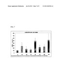 | 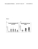 |
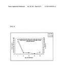 | 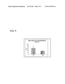 |
 |  |
 |  |
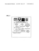 |
| Similar patent applications: | |
| Date | Title |
|---|---|
| 2009-05-14 | Method for fabricating aerogels and their application in biocomposites |
| 2008-10-16 | Fluorescent ion indicators and their applications |
| 2009-07-23 | Immuno-pcr method for detecting nasopharyngeal carcinoma markers and kit thereof |
| 2009-08-20 | Method of identifying cancer biomarkers and cancer progression |
| 2009-08-27 | Method of identifying cancer biomarkers and cancer progression |
| New patent applications in this class: | |
| Date | Title |
|---|---|
| 2022-05-05 | Photocleavable mass-tags for multiplexed mass spectrometric imaging of tissues using biomolecular probes |
| 2022-05-05 | Macrophage expression in breast cancer |
| 2022-05-05 | Characterizing methylated dna, rna, and proteins in the detection of lung neoplasia |
| 2022-05-05 | Methods for identifying and improving t cell multipotency |
| 2022-05-05 | Sequence analysis using meta-stable nucleic acid molecules |
| New patent applications from these inventors: | |
| Date | Title |
|---|---|
| 2009-05-07 | Methods and compositions related to esculentoside a |
| Top Inventors for class "Chemistry: molecular biology and microbiology" | |
| Rank | Inventor's name |
|---|---|
| 1 | Marshall Medoff |
| 2 | Anthony P. Burgard |
| 3 | Mark J. Burk |
| 4 | Robin E. Osterhout |
| 5 | Rangarajan Sampath |