Patent application title: GENOMIC FINGERPRINT OF BREAST CANCER
Inventors:
Ramonón Garcia Escudero (Madrid, ES)
Jesús Maria Paramio González (Madrid, ES)
Jesús Maria Paramio González (Madrid, ES)
Ana Belén Martínez Cruz (Madrid, ES)
Mirentxu Santos Lafuente (Madrid, ES)
IPC8 Class: AC40B3004FI
USPC Class:
506 9
Class name: Combinatorial chemistry technology: method, library, apparatus method of screening a library by measuring the ability to specifically bind a target molecule (e.g., antibody-antigen binding, receptor-ligand binding, etc.)
Publication date: 2011-06-23
Patent application number: 20110152113
Abstract:
The present invention relates to in vitro methods for determining the
prognosis of a subject diagnosed with breast cancer developing metastasis
or for selecting suitable treatment for said subject. Particularly, the
invention relates to a signature of genes the expression of which is
correlated with the prognosis of a subject who has been diagnosed with
breast cancer.Claims:
1. An in vitro method for determining the prognosis of a subject
diagnosed with breast cancer or for selecting the treatment of a subject
diagnosed with breast cancer which comprises determining the expression
levels of the genes identified in Table 1 and in Table 2 in a tumor
tissue sample from said subject, wherein an increase of the expression of
the genes identified in Table 1 and a decrease of the expression of the
genes identified in Table 2 with respect to a reference value is
indicative of a worse prognosis or of said subject having to be treated
with chemotherapy.
2-11. (canceled)
12. A reagent capable of detecting the expression levels of the genes identified in Tables 1 and 2.
13-20. (canceled)
21. A method for selecting genetic markers for predicting the tendency to develop metastasis of a primary tumor comprising the following steps: i) determining the genes the expression of which is altered with respect to a reference value in a tumor sample from a genetically modified non-human animal showing a tendency to develop tumors spontaneously; ii) identifying the homologous genes in humans corresponding to the genes identified in step i); and iii) selecting those genes identified in step ii) the expression of which in primary tumor samples from patients who develop metastasis from said primary tumor is altered with respect to the expression of said genes in primary tumors of patients who do not develop metastasis.
22-33. (canceled)
34. The method according to claim 1, wherein the determining of the expression levels of the genes identified in Tables 1 and 2 comprises the quantification of the messenger RNA (mRNA) of said genes, or a fragment of said mRNA, the complementary DNA (cDNA) of said genes, or a fragment of said cDNA, or mixtures thereof.
35. The method according to claim 1, wherein the determining of the expression levels of the genes identified in Tables 1 and 2 is performed by means of a quantitative multiplex polymerase chain reaction (PCR) or a DNA or RNA array.
36. The method according to claim 1, wherein the determining of the expression levels of the genes identified in Table 1 and Table 2 is performed by means of a DNA array comprising the probes identified in Tables 3 and 4.
37. The method according to claim 1, wherein the determination of said prognosis comprises a proportional hazards regression analysis of said prognosis depending on the expression levels of the genes identified in Table 1 and in Table 2.
38. The method according to claim 37, wherein said proportional hazards regression analysis is a Cox-type analysis.
39. The method according to claim 38, wherein distant metastasis is established in said Cox-type analysis as a prognostic variable.
40. The method according to claim 39, wherein said distant metastasis is distant metastasis at 5 or 10 years.
41. The method according to claim 38, wherein the determination of said prognosis is carried out by applying the following formula: i = 1 40 s i x i + 39.2 ##EQU00004## wherein xi is the value of the expression level in log2 of each of said genes identified in Tables 1 and 2; and si is the value of the Wald statistic of the Cox-type regression analysis of each of said genes identified in Tables 1 and 2 according to claims 6 to 8, wherein if said value is greater than zero, then it is indicative of said patient presenting a worse prognosis or of said patient having to be treated with chemotherapy, and wherein if said value is less than zero, it is indicative of said patient presenting a good prognosis or of said patient not having to be treated with chemotherapy.
42. The method according to claim 1, wherein the quantification of the expression levels of the genes identified in Tables 1 and 2 comprises the quantification of the levels of protein encoded by said genes or of a variant thereof.
43. The reagent according to claim 12, wherein the reagent comprises (i) a set of nucleic acids comprising the nucleotide sequences of the probes identified in Tables 1 and 2 or the products of their transcription, or (ii) a set of antibodies or a fragment thereof capable of detecting an antigen, consisting of each antibody or fragment being capable of binding specifically to one of the proteins encoded by the genes the nucleotide sequences of which hybridize with the probes identified in Tables 1 and 2.
44. The reagent according to claim 43, wherein the nucleic acids are DNA, cDNA or RNA probes and/or primers.
45. The method according to claim 21, wherein said non-human animal is an animal in which the gene expression of the Tp53 gene is inhibited.
46. The method according to claim 45, wherein said animal further presents inhibited gene expression of the pRb gene.
47. The method according to claim 21, wherein the sample obtained in step (i) is an epidermal carcinoma sample.
48. The method according to any of claim 21, wherein said step iii) is carried out by means of a proportional hazards regression analysis.
49. The method according to any of claim 21, wherein said primary tumors analyzed in step iii) are breast cancer or glioblastoma tumors.
50. The method according to any of claim 21, wherein the quantification of the expression levels of the genes according to step iii) comprises the quantification of the levels of protein encoded by said genes.
Description:
FIELD OF THE INVENTION
[0001] The present invention relates to in vitro methods for determining the prognosis of a subject diagnosed with breast cancer for developing metastasis or for selecting suitable treatment for said subject.
BACKGROUND OF THE INVENTION
[0002] Breast cancer is the second most common type of cancer worldwide (10.4%, after lung cancer) and the fifth most common cause of cancer-induced death (after lung cancer, stomach cancer, liver cancer, and colon cancer). Breast cancer is the most common cause of cancer-induced death among women worldwide. In 2005, breast cancer caused 502,000 deaths all over the world (7% of cancer-induced deaths; almost 1% of all deaths). The number of cases worldwide has significantly increased since the 1970s, a phenomenon which can partially be attributed to modern Western lifestyles. Women in North America have the highest incidence of breast cancer in the world.
[0003] Since the breast is made up of identical tissues in men and women, breast cancer also occurs in men. The incidence of breast cancer in men is approximately 100 times less than in women, but it is considered that men with breast cancer statistically have the same survival rates as women.
[0004] Breast cancer is staged according to the TNM system. The prognosis is closely related to the results of the staging, and the staging is also used to assign patients to treatments both in clinical trials and in medical practice. The information for staging is as follows: [0005] TX: The primary tumor cannot be evaluated. T0: There is no evidence of tumor. Tis: Carcinoma in situ, no invasion. T1: The tumor is 2 cm or less across. T2: The tumor is more than 2 cm but less than 5 cm across. T3: The tumor is more than 5 cm across. T4: Tumor of any size growing into the chest wall or skin, or inflammatory breast cancer. [0006] NX: The nearby lymph nodes cannot be evaluated. NO: The cancer has not spread to regional lymph nodes. Ni: The cancer has spread to 1 to 3 underarm lymph nodes or to an internal mammary lymph node. N2: The cancer has spread to 4 to 9 underarm lymph nodes or to multiple internal mammary lymph nodes. N3: Any of the following: [0007] The cancer has spread to 10 or more underarm lymph nodes, or the cancer has spread to the lymph nodes under the clavicle, or the cancer has spread to the lymph nodes above the clavicle or the cancer affects underarm lymph nodes and has spread to internal mammary lymph nodes, or the cancer affects 4 or more underarm lymph nodes, and small amounts of cancer are found in the internal mammary lymph nodes or in sentinel lymph node biopsy. [0008] MX: The presence of distant spread (metastasis) cannot be evaluated. M0: No distant spread is found. M1: Spread to distant organs is present, these organs not including the lymph node above the clavicle.
[0009] The principal pillar of breast cancer treatment is surgery when the tumor is localized, with possible adjuvant hormone therapy (with tamoxifen or an aromatase inhibitor), chemotherapy, and/or radiotherapy. Current recommendations for treatment after surgery (adjuvant therapy) follow a pattern. This pattern is subject to change, because every two years, a world conference is held in St. Gallen, Switzerland to discuss the actual results of multicenter studies conducted worldwide. Likewise, said pattern is also reviewed according to the consensual criterion of the National Institute of Health (NIH). Based on these criteria, over 85-90% of the patients who do not present metastasis in lymph nodes would be candidates for receiving adjuvant systemic therapy.
[0010] Today no set of predictors of a satisfactory prognosis based solely on clinical information has been identified. Over the last 30 years oncologists have focused on optimizing the outcome of the cancer patients and it is only now that the new available technologies allow investigating polymorphisms, expression levels of genes and gene mutations for the purpose of predicting the impact of a determined therapy on different groups of cancer patients in order to design customized chemotherapies. PCR assays such as Oncotype DX or microarray assays such as MammaPrint can predict the risk of breast cancer relapse based on gene expression. In February 2007, the MammaPrint assay became the first breast cancer indicator to receive official authorization from the Food and Drug Administration.
[0011] Document WO02103320 describes a method for predicting the prognosis of breast cancer patients by means of analyzing the expression of a group of genetic markers, particularly 70 genes (Table 6, page 89). D1 also describes a microarray for determining the prognosis of breast cancer from a sample from a patient comprising probes for detecting the gene expression of said genes.
[0012] In addition, document WO2005/083429 describes a method for selecting a genetic marker signature for the prognosis of breast cancer. Said document also describes a method for determining the prognosis of patients with breast cancer by means of analyzing the expression of a group of genes selected from said method. Specifically, said document relates to the use of genetic markers for predicting the prognosis of breast cancer from a characteristic signature consisting of 76 genetic markers. Said document also describes a kit, such as a microarray, for determining the prognosis of breast cancer from a sample from a patient comprising probes for detecting the gene expression of said 76 genes.
[0013] Therefore, there is a need to develop new methods which allow identifying the most relevant genes involved in metastasis such that more reliable signatures can be obtained and these signatures can be based on a smaller number of genes. Said signatures will allow predicting the prognosis of a patient suffering breast cancer more efficiently than the methods described in the state of the art. The identification of new prognosis factors will serve as a guideline in selecting the most suitable treatments.
SUMMARY OF THE INVENTION
[0014] In a first aspect, the present invention relates to an in vitro method for determining the prognosis of a subject diagnosed with breast cancer or for selecting the treatment of a subject diagnosed with breast cancer which comprises determining the expression levels of the genes identified in Table 1 and in Table 2 in a tumor tissue sample from said subject, wherein an increase of the expression of the genes identified in Table 1 and a decrease of the expression of the genes identified in Table 2 with respect to a reference value is indicative of a worse prognosis or of said subject having to be treated with chemotherapy.
[0015] In a second aspect, the invention relates to a reagent capable of detecting the expression levels of the genes of Tables 1 and 2.
[0016] In another aspect, the invention relates to a kit comprising at least one reagent according to the invention.
[0017] In another aspect, the invention relates to the use of a kit according to the invention for the prognosis of patients diagnosed with breast cancer or for selecting the treatment of a subject diagnosed with breast cancer.
[0018] The invention also relates in another aspect to a method for selecting genetic markers for predicting the tendency to develop metastasis of a primary tumor comprising the following steps: [0019] i) determining the genes the expression of which is altered with respect to a reference value in a tumor sample from a genetically modified non-human animal showing a tendency to develop tumors spontaneously; [0020] ii) identifying the homologous genes in humans corresponding to the genes identified in step i); and [0021] iii) selecting those genes identified in step ii) the expression of which in primary tumor samples from patients who develop metastasis from said primary tumor is altered with respect to the expression of said genes in primary tumors of patients who do not develop metastasis.
BRIEF DESCRIPTION OF THE DRAWINGS
[0022] FIG. 1 shows Receiver Operating Curves (ROC) of predicting the genetic signature of the invention, in the complete Loi dataset (n=191), or in the Loi dataset excluding grade 3 ER- samples (n=142). Panels A and C show the prediction of distant metastasis (DM) at 5 years, and panel E shows the prediction of overall survival at 5 years. Panels B and D show the prediction of distant metastasis (DM) at 10 years, and panel E shows the prediction of overall survival at 10 years. The RV of the point of maximum Specificity (40.1%) and Sensitivity (100%) of panel A was chosen as the threshold for separating into samples of good or poor prognosis (RV=-39.2) (circle).
[0023] FIG. 2 shows survival curves or prognostic prediction of patients not treated with tamoxifen, and N- within the Loi dataset (86 tumors, diameter≦5 cm).
[0024] FIG. 3 shows survival curves or prognostic prediction of patients treated with tamoxifen, and N+ within the Loi dataset (108 tumors, diameter≦5 cm).
[0025] FIG. 4 shows the genetic signature of the invention in the Desmedt dataset. The left panel shows an expression map of the genes the sequences of which hybridize with the probes of the signature of the invention in the Desmedt tumors, excluding the grade 3 ER- (142 samples). The columns represent genes, and the rows represent samples. The genes are arranged from left to right according to decreasing values of the Wald statistic value in accordance with Table 2. The expression values are shown as log2 (mean=0, standard deviation=1).
[0026] The right panel shows the prediction results using the signature of genes of the invention, or using the criteria of St. Gallen (SG), NIH, Nottingham Prognostic Index (NPI), Adjuvant Online (AOL), and the 76-gene genomic signature of Veridex. Samples in light gray indicate a good prognosis; samples in dark gray indicate a poor prognosis. The presence of 37 probes, the expression of which is higher in the poor prognosis samples, can be observed. The ER status (ER+ in black, ER- in white), as well as the existence of DM at 5 or 10 years (presence of metastasis in white, absence in black), are shown. The genes are represented both by the Affymetrix probe identifiers, and by the NCBI gene symbols.
[0027] FIG. 5 shows the genetic signature of the invention in the Loi dataset. A) The left panel shows an expression map of the genes the sequences of which hybridize with the probes in the Loi tumors, which were N- and were not treated with tamoxifen, excluding grade 3 ER- (86 samples). The columns represent genes and the rows represent samples. The genes are arranged from left to right according to decreasing values of the Wald statistic value in accordance with Table 2. The expression values are shown as log2 (mean=0, standard deviation=1).
[0028] The right panel shows the prediction results using the signature of genes of the invention, or the criteria of St. Gallen (SG) and NIH. Samples in light gray indicate a good prognosis; samples in dark gray indicate a poor prognosis. The presence of 37 probes, the expression of which is greater the poor prognosis samples, can be observed. The ER status (ER+ in black, ER- in white), as well as the existence of DM at 5 or 10 years (presence of metastasis in white, absence in black), are shown. The genes are represented both by the Affymetrix probe identifiers and by the NCBI gene symbols.
[0029] B) This panel is similar to panel A, but it is for the group of 108 patients of the Loi dataset who were N+ and received hormone therapy.
[0030] FIG. 6 shows the ranking of the genes selected in the comparison between murine tumors p53- and p53-; pRb- and normal skin. The genes which are overexpressed in mouse tumors arranged according to the magnitude of the relative change in gene expression (from 80.3 up to 1.2 times, panel A), or arranged by the P value (panel C), computed according to the Student's t-Test. The black color in the left column indicates genes increased more than 2 times. The dark gray in the central column indicates genes which also met the differential expression criteria of the SAM test. The position within the range of the equivalent mouse genes of the signature of genes of the invention in humans is indicated in the right column. Genes the name of which appears in dark gray are overexpressed in malignant human breast tumors; genes in light gray are decreased in malignant tumors
[0031] Genes having a reduced expression in the mouse tumors arranged according to the magnitude of the relative change in gene expression (from 375.8 up to 1.2 times, panel B), or arranged by the P value (panel D), computed according to the Student's t-Test. The black color of the left column indicates genes regulated negatively by more than 2 times. The dark gray in the column central indicates genes which also met the differential expression criteria of the SAM test. The position within the range of the equivalent mouse genes of the signature of genes in humans is indicated in the right column. Genes the name of which appears in dark gray are overexpressed in malignant human breast tumors; genes in light gray are decreased in malignant tumors.
DETAILED DESCRIPTION OF THE INVENTION
[0032] The authors of the present invention have selected a signature of genes which hybridize with probes and the expression of which is correlated with the prognosis of a subject who has been diagnosed with breast cancer. Said signature can also be used for selecting the most suitable treatment for said subject diagnosed with breast cancer.
[0033] As hereinbefore mentioned, the criteria of St. Gallen and of the NIH classify patients as high risk or low risk based on several histological and clinical characteristics. The authors of the present invention have demonstrated that said prognostic signature of the invention assigns more patients to the low risk (or good prognosis) group than the traditional methods do. In fact, the inventors have shown that said clinical criteria mistakenly classify a clinically significant number of patients in the poor prognosis group, so in current clinical practice many patients are receiving chemotherapy unnecessarily.
[0034] Therefore, in a first aspect the invention relates to an in vitro method for determining the prognosis of a subject diagnosed with breast cancer or for selecting the treatment of a subject diagnosed with breast cancer which comprises determining the expression levels of the genes identified in Table 1 and in Table 2 in a tumor tissue sample from said subject, wherein an increase of the expression of the genes identified in Table 1 and a decrease of the expression of the genes identified in Table 2 with respect to a reference value is indicative of a worse prognosis or of said subject having to be treated with chemotherapy. In a particular embodiment of the invention, said tumor tissue sample is a primary tumor sample, particularly, said tumor is breast cancer. Thus, by way of illustration said tumor tissue sample can be a biopsy sample obtained, for example, by surgical resection.
[0035] In a particular embodiment of the method of the invention, said genes are the genes the nucleotide sequences of which hybridize with the probes identified in Table 1 and Table 2.
TABLE-US-00001 TABLE 1 Gene symbol TOP2A TOMM70A PLK1 CCNB2 UBEC2C SPAG5 CDC2 MAD2L1 BUB1B TRIP13 AURKA KIF11 BRCA1 HMMR CIAPIN1 LRP8 AURKB CDKN3 HSP90AA1 NUSAP1 ERO1L MLF1IP DCC1 C21orf45 PBK ATAD5 MCM10 CDCA3 RACGAP1
TABLE-US-00002 TABLE 2 Gene symbol ELOVL5 PARP3 CBX7
TABLE-US-00003 TABLE 3 Symbol SEQ ID NO TOP2A SEQ ID NO: 1-SEQ ID NO: 11 TOP2A-2 SEQ ID NO: 12-SEQ ID NO: 22 TOMM70A SEQ ID NO: 23-SEQ ID NO: 33 PLK1 SEQ ID NO: 34-SEQ ID NO: 44 CCNB2 SEQ ID NO: 45-SEQ ID NO: 55 UBEC2C SEQ ID NO: 56-SEQ ID NO: 66 SPAG5 SEQ ID NO: 67-SEQ ID NO: 77 CDC2 SEQ ID NO: 78-SEQ ID NO: 88 CDC2-2 SEQ ID NO: 89-SEQ ID NO: 99 MAD2L1 SEQ ID NO: 100-SEQ ID NO: 110 BUB1B SEQ ID NO: 111-SEQ ID NO: 121 TRIP13 SEQ ID NO: 122-SEQ ID NO: 132 AURKA SEQ ID NO: 133-SEQ ID NO: 143 KIF11 SEQ ID NO: 144-SEQ ID NO: 154 BRCA1 SEQ ID NO: 155-SEQ ID NO: 165 HMMR SEQ ID NO: 166-SEQ ID NO: 176 CIAPIN1 SEQ ID NO: 177-SEQ ID NO: 187 LRP8 SEQ ID NO: 188-SEQ ID NO: 198 CIAPIN1-2 SEQ ID NO: 199-SEQ ID NO: 209 AURKB SEQ ID NO: 210-SEQ ID NO: 220 HMMR-2 SEQ ID NO: 221-SEQ ID NO: 231 CDKN3 SEQ ID NO: 232-SEQ ID NO: 242 HSP90AA1 SEQ ID NO: 243-SEQ ID NO: 253 BRCA1-2 SEQ ID NO: 254-SEQ ID NO: 264 HSP90AA1-2 SEQ ID NO: 265-SEQ ID NO: 275 HSP90AA1-3 SEQ ID NO: 276-SEQ ID NO: 286 HSP90AA1-4 SEQ ID NO: 287-SEQ ID NO: 297 NUSAP1 SEQ ID NO: 298-SEQ ID NO: 308 ERO1L SEQ ID NO: 309-SEQ ID NO: 319 MLF1IP SEQ ID NO: 320-SEQ ID NO: 330 DCC1 SEQ ID NO: 331-SEQ ID NO: 341 C21orf45 SEQ ID NO: 342-SEQ ID NO: 352 PBK SEQ ID NO: 353-SEQ ID NO: 363 ATAD5 SEQ ID NO: 364-SEQ ID NO: 374 MCM10 SEQ ID NO: 375-SEQ ID NO: 385 CDCA3 SEQ ID NO: 386-SEQ ID NO: 396 RACGAP1 SEQ ID NO: 397-SEQ ID NO: 407
TABLE-US-00004 TABLE 4 Symbol SEQ ID NO ELOVL5 SEQ ID NO: 408-SEQ ID NO: 418 PARP3 SEQ ID NO: 419-SEQ ID NO: 429 CBX7 SEQ ID NO: 430-SEQ ID NO: 440
[0036] The quantification of the expression levels of the genes identified in Table 1 and in Table 2 can be performed from the
[0037] RNA resulting from the transcription of said genes (mRNA) or, alternatively, from the complementary DNA (cDNA) of said genes. Therefore, in a particular embodiment, the quantification of the expression levels of the genes identified in Table 1 and in Table 2 comprises the quantification of the messenger RNA (mRNA) of said genes, or a fragment of said mRNA, the complementary DNA (cDNA) of said genes, or a fragment of said cDNA, or mixtures thereof.
[0038] Additionally, the method of the invention can include performing an extraction step for the purpose of obtaining the total RNA, which can be performed by means of conventional techniques (Chomczynski at al., Anal. Biochem., 1987, 162:156; Chomczynski P., Biotechniques, 1993, 15:532).
[0039] Virtually any conventional method can be used within the invention for detecting and quantifying the levels of mRNA encoded by the genes the nucleotide sequences of which hybridize with the probes of Tables 1 and 2 or of the corresponding cDNA thereof. By way of non-limiting illustration, the levels of mRNA encoded by said genes can be quantified by means of using conventional methods, for example, methods comprising the amplification of the mRNA and the quantification of the said mRNA amplification product, such as electrophoresis and staining, or alternatively, by means of Northern blot and using suitable probes, Northern blot and using probes specific for mRNA of the genes of interest or of the corresponding cDNA thereof, mapping with the S1 nuclease, RT-PCR, hybridization, microarrays, etc. Similarly, the levels of the cDNA corresponding to said mRNA encoded by the genes of Tables 1 and 2 can also be quantified by means of using conventional techniques; in this case, the method of the invention includes a step of synthesizing the corresponding cDNA by means of reverse transcription (RT) of the corresponding mRNA followed by amplification and quantification of the said cDNA amplification product. Conventional methods of quantifying expression levels can be found, for example, in Sambrook et al., 2001 "Molecular cloning: a Laboratory Manual", 3rd ed., Cold Spring Harbor Laboratory Press, N.Y., Vol. 1-3.
[0040] In a particular embodiment of the invention, the quantification of the expression levels of the genes identified in Tables 1 and 2 is performed by means of a quantitative multiplex polymerase chain reaction (PCR) or a DNA or RNA array.
[0041] In another particular embodiment of the invention, the determination of the expression levels of the genes identified in Table 1 and Table 2 is performed by means of a DNA array comprising the probes identified in Tables 3 and 4. In a more particular embodiment of the invention, said array comprises at least one set of 11 probes for determining the expression levels of each of the genes of Tables 1 and 2. Thus, for determining the expression levels of each of said genes, a mean of the signal of said 11 probes used for detecting the expression of said gene is calculated.
[0042] Thus, said method comprises determining the expression levels of said genes of Tables 1 and 2 with respect to a reference value. In a particular embodiment of the invention, said reference value is the gene expression value of said genes of Tables 1 and 2 in a primary tumor sample from patients who do not develop metastasis. Preferably, it will be considered that the genes present increased expression when the expression ratio of a gene is at least 1.5 times with respect to a reference value, preferably greater than 2 times, more preferably greater than 3, 4, 5 and 10 times. Likewise, in a particular embodiment of the invention, it will be considered that the genes present decreased expression with respect to a reference value when the expression ratio of a gene is at least 1.5 times less than the reference value.
[0043] In a particular embodiment of the invention, the method for determining the better or worse prognosis of a subject who has been diagnosed with breast cancer comprises performing a proportional hazards regression analysis depending on said expression values of the genes identified in Tables 1 and 2. According to the data shown in Example 2, the authors have demonstrated that in this manner, said prognosis is performed with an effectiveness which, according to the ROC curves, would have a sensitivity of 100% as well as maximum specificity. Therefore, in a particular embodiment of the invention, the determination of said prognosis comprises a proportional hazards regression analysis of said prognosis depending on the expression levels of the genes identified in Table 1 and in Table 2.
[0044] As is described in Example 2 enclosed in the present description, the inventors have used a Cox-type proportional hazards regression analysis for determining the prognosis of a subject diagnosed with breast cancer. Said Cox analysis assigns a regression coefficient for each gene, such that the gene the expression of which is directly correlated with the prognostic variable, for example with the onset of metastasis, is >0, and if its expression is inversely related to said variable, it is <0. Therefore, in a particular embodiment of the invention, said proportional hazards regression analysis is a Cox-type analysis. In a more particular embodiment, distant metastasis is established in said Cox-type analysis as a prognostic variable. In a preferred embodiment, said distant metastasis is distant metastasis at 5 or 10 years.
[0045] The inventors have demonstrated that by means of the method of the invention it is possible to determine the prognosis of a patient with high sensitivity and specificity. Thus, from the gene expression values for the genes of Tables 1 and 2 as has been hereinbefore described, and from the value of the Wald statistic of the proportional hazards regression analysis, the inventors have demonstrated that it is possible to determine said prognosis by applying the following formula:
i = 1 40 s i x i + 39.2 ##EQU00001##
[0046] wherein xi is the value of the expression level in log2 of each of said genes identified in Tables 1 and 2; and si is the value of the Wald statistic of the Cox-type regression analysis for each of said genes identified in Tables 1 and 2,
wherein if the value obtained is greater than zero, then it is indicative of said patient presenting a worse prognosis or of said patient having to be treated with chemotherapy, and wherein if said value is less than zero, it is indicative of said patient presenting a good prognosis or of said patient not having to be treated with chemotherapy.
[0047] The value of the Wald statistic is a value commonly used by the person skilled in the art to known whether or not the variables which are introduced in the statistical analysis are relevant. Said value can be calculated as described in Wald A. (1943) (Transactions of the American Mathematical Society. 1943; 54:426-482) and Silvey (1959) (Silvey S D. Annals of Mathematical Statistics. 1959; 30:389-407).
[0048] In addition, besides quantifying the expression levels of the genes identified in Tables 1 and 2, the expression level of the proteins encoded by said genes can also be quantified for putting the invention into practice. Thus, in a Particular embodiment the quantification of the levels of the proteins encoded by the genes identified in Tables 1 and 2 comprises the quantification of said proteins or variables thereof.
[0049] As it is used herein, the term "protein" relates to a molecular chain of amino acids attached by covalent or non-covalent bonds. The term further includes all the physiologically relevant forms of post-translational chemical modifications, for example, glycosylation, phosphorylation or acetylation, etc.
[0050] In the present invention "variant" is understood as a protein the amino acid sequence of which is substantially homologous to the amino acid sequence of a specific protein. An amino acid sequence is substantially homologous to a determined amino acid sequence when it presents at least a 70% degree of identity, advantageously at least 75%, typically at least 80%, preferably at least 85%, more preferably at least 90%, still more preferably at least 95%, 97%, 98% or 99%, with respect to said determined amino acid sequence. The degree of identity between two amino acid sequences can be determined by conventional methods, for example, by means of standard sequence alignment algorithms known in the state of the art, such as BLAST [Altschul S. F. et al. Basic local alignment search tool. J Mol Biol. 1990 Oct. 5; 215(3):403-10], for example.
[0051] The person skilled in the art understands that the mutations in the nucleotide sequence of the genes which give rise to conservative substitutions of amino acids in non-critical positions for protein functionality are mutations with a neutral evolution which do not affect its overall structure or its functionality. Said variants fall within the scope of the present invention.
[0052] Therefore, as it is used herein, the term "variant" also includes any fragment of one of the proteins described in the present invention. The term "fragment" relates to a peptide comprising a portion of a protein.
[0053] The expression level of the proteins encoded by the genes identified in Tables 1 and 2 can be quantified by means of any conventional method which allows detecting and quantifying said proteins in a sample from a subject. By way of non-limiting illustration, the levels of said proteins can be quantified, for example, by means of using antibodies with the capacity to bind to said proteins (or to fragments thereof containing an antigenic determinant) and the subsequent quantification of the formed complexes. The antibodies which are used in these assays can be labeled or not. Illustrative examples of markers which can be used include radioactive isotopes, enzymes, flourophores, chemiluminescent reagents, enzyme substrates or cofactors, enzyme inhibitors, particles, dyes, etc. There is a wide range of known assays that can be used in the present invention which use non-labeled antibodies (primary antibody) and labeled antibodies (secondary antibody); these techniques include Western blot, ELISA (Enzyme-linked Immunosorbent Assay), RIA (Radioimmunoassay), competitive EIA (Competitive Enzyme Immunoassay), DAS-ELISA (Double Antibody Sandwich ELISA), immunocytochemical and immunohistochemical techniques, techniques based on using protein biochips or microarrays which include specific antibodies or assays based on colloidal precipitation in formats such as dipsticks. Other ways of detecting and quantifying said proteins include affinity chromatography techniques, ligand binding assays, etc.
[0054] In a particular embodiment, the quantification of the levels of protein encoded by the genes identified in Tables 1 and 2 is performed by means of Western blot, ELISA, immunohistochemistry or a protein array.
[0055] As hereinbefore mentioned, the in vitro method of the invention can be used for selecting the treatment of a subject diagnosed with breast cancer, wherein an increase of the expression of the genes identified in Table 1 and a decrease of the expression of the genes identified in Table 2 with respect to a reference value is indicative of said subject having to be treated with chemotherapy.
[0056] The suitable chemotherapy agents include but are not limited to alkylating agents such as, for example, cyclophosphamide, carmustine, daunorubicin, mechlorethamine, chlorambucil, nimustine, melphalan and the like; anthracylines, such as, for example, daunorubicin, doxorubicin, epirubicin, idarubicin, mitoxantrone, valrubicin and the like; taxane compounds, such as, for example, paclitaxel, docetaxel and the like; topoisomerase inhibitors such as, for example, etoposide, teniposide, irinotecan, tuliposide and the like; nucleotide analogues such as, for example, azacitidine, azathioprine, capecitabine, cytarabine, doxifluridine, fluorouracil, gemcitabine, mercaptopurine, methotrexate, thioguanine ftorafur and the like; platinum-based agents such as, for example, carboplatin, cisplatin, oxaliplatin and the like; anti-neoplastic agents such as, for example, vincristine, leucovorin, lomustine, procarbazine and the like; hormone modulators such as, for example, tamoxifen, finasteride, 5-α-reductase inhibitors and the like; vinca alkaloids such as, for example, vinblastine, vincristine, vindesine, vinorelbine and the like. The suitable chemotherapy agents are described in detail in the literature, such as in The Merck Index in CD-ROM, 13th edition.
[0057] In the present invention, "antitumor agent" is understood as that chemical, physical or biological agent or compound with antiproliferative, antioncogenic and/or carcinostatic properties which can be used to inhibit tumor growth, proliferation and/or development. Examples of antitumor agents which can be used in the present invention are (i) antimetabolites, such as antifolates and purine analogues; (ii) natural products, such as antitumor antibiotics and mitotic inhibitors; (iii) hormones and antagonist thereof, such as androgens and corticosteroids; and (iv) biological agents, such as viral vectors. A list of compounds which can be used as antitumor agents is described in patent application WO2005/112973.
[0058] In another aspect, the present invention relates to a reagent, hereinafter reagent of the invention, capable of detecting the expression levels of the genes of Tables 1 and 2.
[0059] In a particular embodiment, said reagent of the invention comprises [0060] (i) a set of nucleic acids comprising the nucleotide sequences of the probes identified in Tables 3 and 4 or the products of their transcription, or [0061] (ii) a set of antibodies, or a fragment thereof, capable of detecting an antigen, consisting of each antibody or fragment being capable of binding specifically to one of the proteins encoded by the genes identified in Tables 1 and 2.
[0062] In a particular embodiment of the invention, said nucleic acids are DNA, cDNA or RNA probes and/or primers. Said nucleic acids can be obtained by conventional techniques known by the person skilled in the art from the nucleotide sequences of the genes of Tables 1 and 2. Said probes and/or primers can generally be obtained from companies by chemical synthesis. In addition, said sequences of said genes are well described in the literature and are therefore known.
[0063] In another aspect, the present invention relates to a kit comprising at least one reagent according to the invention. In a particular embodiment of the invention, said kit is a DNA or RNA array comprising a set of nucleic acids, wherein said set of nucleic acids comprises the nucleotide sequences of the probes of Tables 3 and 4, or a fragment thereof, or the products of their transcription. In a more particular embodiment, said kit further comprises a nucleic acid molecule of one or several constitutive expression genes.
[0064] In the present invention "genes which are expressed constitutively" or "constitutive expression genes" is understood as those genes that are always active or which are constantly transcribed. Examples of genes which are expressed constitutively are 2-myoglobin, ubiquitin, 18S ribosomal protein, cyclophilin A, transferrin receptor, actin, GAPDH, tyrosine 3-monooxygenase/tryptophan 5-monooxygenase activation protein (YWHAZ), ubiquitin, beta-actin and β-2-microglobulin.
[0065] In another particular embodiment of the invention, the kit of the invention comprises a set of antibodies, wherein said set of antibodies consists of antibodies or fragments thereof capable of binding specifically with the proteins encoded by the genes identified in Tables 1 and 2 or any variant of said proteins. In a particular embodiment, said kit further comprises antibodies or fragments thereof capable of binding specifically with the proteins encoded by one or several constitutive expression genes.
[0066] The term genetic signature or signature of genes of the invention as it is herein used relates to the genes identified in Tables 1 and 2. According to the data shown by the inventors (see Example 4), the signature of genes or genetic signature of the invention assigns more patients to the low risk (or good prognosis) group than traditional methods do. Thus, the inventors have demonstrated that the patients classified as poor prognosis patients according to the method of the present invention tend to have a greater proportion of metastasis than the poor prognosis patients according to the clinical criteria of St Gallen and NIH. Therefore, in another aspect, the invention relates to the use of a kit according to the invention for the prognosis of patients diagnosed with breast cancer or for selecting the treatment of a subject diagnosed with breast cancer.
[0067] In another aspect, the invention relates to a method for selecting genetic markers for predicting the tendency to develop metastasis of a primary tumor comprising the following steps: [0068] i) determining the genes the expression of which is altered with respect to a reference value in a tumor sample from a genetically modified non-human animal showing a tendency to develop tumors spontaneously; [0069] ii) identifying the homologous genes in humans corresponding to the genes identified in step i); and [0070] iii) selecting those genes identified in step ii) the expression of which in primary tumor samples from patients who develop metastasis from said primary tumor is altered with respect to the expression of said genes in primary tumors of patients who do not develop metastasis.
[0071] For determining the genes the expression of which is altered according to step i), first a sample is obtained from said animal, preferably said sample is a tumor tissue sample. Example 1 of the present invention describes a particular embodiment of the method of the invention. Thus, in a particular embodiment, the total RNA is extracted from said tumor tissue sample of said animal and said RNA is analyzed to determine the genes the expression of which is altered in said tumor sample with respect to a reference value. In a particular embodiment, said reference value is the gene expression value in a non-tumor tissue sample from said animal. Said expression value can be obtained, for example, from the values resulting from the gene expression signal in a gene expression array of said non-human animal model as is explained in Example 1 of the present description. Thus, said values correspond with the values in the CEL (CEL format) type files according to the GCOS (GeneChip® Operating Software) software of Affymetrix.
[0072] In a particular embodiment of the method for selecting genetic markers of the invention, said non-human animal is an animal in which the gene expression of the Tp53 gene is inhibited. In another particular embodiment, the gene expression of the pRb gene is further inhibited in said animal. The Tp53 and Rb1 genes respectively encode tumor suppressors p53 and pRb. Said animal models, with a deficiency of the p53 and pRb genes (p53- and pRb-), spontaneously develop highly invasive epidermal carcinomas. Therefore, in a particular embodiment, said animal is a non-human animal which spontaneously develops epidermal carcinomas.
[0073] To carry out step ii) of the method for selecting genetic markers for predicting the tendency to develop metastasis of a primary tumor, the homologous genes in humans corresponding to the genes identified in step i) are identified. To that end, conventional techniques for mapping homologous genes known by the person skilled in the art are used. Particularly, the inventors have mapped the Affymetrix probe identifiers of the non-human animal with human gene symbols through the search for identifiers in U133plus 2.0 and U133A by means of using the AILUN (Array Information Library Universal Navigator) web utility (Chen R, et al. Nat Methods 2007; 4(11):879).
[0074] In a final step of said method, those genes identified in step ii) the expression of which in primary tumor samples from patients who develop metastasis from said primary tumor is altered with respect to the expression of said genes in primary tumors of patients who do not develop metastasis are selected.
[0075] In a particular embodiment of the invention, step iii) is carried out by means of a proportional hazards regression analysis as hereinbefore mentioned. In a more particular embodiment, said regression analysis is a Cox-type analysis. In a preferred embodiment, said Cox-type method establishes distant metastasis as a prognostic variable. In an even more preferred embodiment of the invention, said distant metastasis is distant metastasis at 5 or 10 years.
[0076] The inventors have further applied the Wald test (Wald A. Transactions of the American Mathematical Society 1943; 54:426-482; Silvey S D. Annals of Mathematical Statistics 1959; 30389-407) to analyze the null hypothesis that the coefficient is 0 (not related to the prognostic variable), a Wald statistic value, the corresponding P value, and P value corrected by the FDR (false discovery rate) method as explained in Example 2 of the present description, being assigned to each gene. FDR control is a statistical method known by the person skilled in the art used in multiple hypothesis testing for correcting multiple comparisons.
[0077] In a particular embodiment of the invention, said primary tumors according to step iii) are breast cancer tumors.
[0078] In a particular embodiment, the determination of the expression of said genes according to step iii) comprises the quantification of the messenger RNA (mRNA) of said genes, or a fragment of said mRNA, the complementary DNA (cDNA), or a fragment of said cDNA, or mixtures thereof.
[0079] In a more particular embodiment, the quantification of the expression levels of the genes according to step iii) is performed by means of a quantitative multiplex polymerase chain reaction (PCR) or a DNA or RNA array.
[0080] In a particular embodiment, the quantification of the expression levels of the genes according to step iii) comprises the quantification of the levels of protein encoded by said genes. In a more particular embodiment, the quantification of the levels of protein is performed by means of Western blot, ELISA or a protein array.
[0081] The following Examples illustrate the invention and must not be interpreted as limiting of the scope thereof.
EXAMPLE 1
Analysis of Mouse Epidermal Tumors
[0082] The animal models used in the present invention are K14Cre mice (they express Cre recombinase in the basal layer of stratified epithelia) crossed with mice with essential exons flanked by loxP sequences in the alleles of the Tp53 genes (p53- model), or in the Tp53 and Rb1 alleles simultaneously (p53- model; pRb- model) (Martinez-Cruz A B. et al. Cancer Res 2008; 68(3):683-692). Tp53 and Rb1 respectively encode tumor suppressors p53 and pRb. Therefore they are gene deletion models in stratified epithelia. Both models spontaneously develop highly invasive poorly differentiated or undifferentiated type epidermal squamous cell carcinomas.
[0083] RNA from frozen epidermal carcinomas which occurred in mice deficient in p53 (p53-) (7 tumors) and deficient in p53 and pRb (p53-/pRb-) (8 tumors) was purified. RNA from normal skin preserved in RNAlater from adult animals (8 weeks old, 5 control samples) was obtained as controls. The integrity of the RNA populations was checked by means of using the Bioanalyzer system (Agilent). All the RNA samples met the quality criteria for microarray analysis (RIN number (RNA integrity number) above 6). Hybridization to the Affymetrix GeneChip, Mouse Gene Expression MOE430 2.0, was performed in the Genomic Department of the Cancer Research Center of Salamanca, using standard Affymetrix protocols. The expression values were extracted from the CEL files (resulting from the fluorescence scanning according to the Affymetrix GCOS software (GeneChip® Operating Software) by means of the RMA (Robust Multichip Average) method (Boistad B M, at al. Bioinformatics 2003; 19(2):185-193; Irizarry R A, et al. Biostatistics 2003; 4(2):249-264). All the hybridizations met the quality criteria included in the RMAExpress computer program using RLE (Relative Log Expression) and MUSE (Normalized Unscaled Standard Error) graphics.
[0084] The analysis of differential gene expression of the mouse tumors compared with normal tissue were performed by means of the Student's t-Test (T-test) and SAM (Significant Analysis of Microarrays) (Tusher V G, et al. Proc Natl Acad Sci U S A 2001; 98(9):5116-5121) in the free Multiexperiment Viewer 4.0 software (MeV 4) (Saeed A I, et al. Biotechniques 2003; 34(2):374-378). The probes were selected if they met two criteria: i) T-test analysis with probability P value, corrected by the False Discovery Rate method (Benjamini Y, Hochberg Y. Journal of the Royal Statistical Society B 1995; 57:289-300) or FDR<3×10⁻⁷; and ii) SAM analysis with FDR<1×10⁻³. A total of 682 probes were selected as differentially expressed, 371 being overexpressed and 311 being negatively regulated in the tumors compared to normal tissue. The Affymetrix identifiers of the chip used (MOE430 2.0) were mapped to the homologous human gene symbol using the Ailun web utility (Chen R, et al. Nat Methods 2007; 4(11):879), which resulted in 427 human genes.
EXAMPLE 2
Two-Step Extraction of a Breast Cancer Metastasis Predictor Based on a p53 Signature.
[0085] 2.1 Selection of Genes with the p53 Signature Related to Metastasis.
[0086] The raw data on the hybridization to microarrays of human primary breast tumors and their corresponding clinical data, obtained with the versions of Affymetrix Human Gene Expression U133A or U133Plus 2.0 GeneChips were downloaded from the Gene Expression Omnibus (GEO) web page database of the NCBI, with the identifiers GSE7390 (Desmedt C. et al. Clin Cancer Res 2007; 13(11):3207-3214) (study hereinafter referred to as Desmedt dataset) and GSE6532 (Loi S. at al. J Clin Oncol 2007; 25(10):1239-1246) (study hereinafter referred to as Loi dataset). The CEL files were taken to extract the signal intensity values using the RMAExpress program. The RLE and NUSE graphics allowed identifying some tumor microarrays which did not meet the optimal normalization criteria with RMA. The corresponding CEL files were removed from subsequent analyses.
[0087] The Desmedt dataset was used as a training set which, after removing the low-quality arrays, contained 191 tumors with healthy lymph nodes (N-), including both samples which express estrogen receptor and samples which do not (ER+ or ER-, respectively) from patients who have not received adjuvant systemic therapy (Table 5).
TABLE-US-00005 TABLE 5 Clinical and pathological characteristics of the patients of the Desmedt dataset Desmedt dataset (n = 191) Age (years) Mean 46 years <45 78 (41%) 45-65 113 (59%) >65 0 (0%) Size (cm) <1 8 (4%) 1-2 90 (47%) >2-5 93 (49%) Degree of tumor differentiation Poor 81 (42%) Moderate 80 (42%) Good 28 (15%) Unknown 2 (1%) ER status Positive 128 (67%) Negative 63 (33%) Distant metastasis after 5 years Yes 34 (18%) No 149 (78%) Censored 8 (4%) The data are numbers of patients, or percentages of patients. TM = tamoxifen
[0088] A Cox-type proportional hazards regression analysis was performed using distant metastasis (DM) at 5 years for the 707 U133A probes (and also present in U133Plus 2.0) corresponding to the 427 genes humans mapped from the analysis of differential expression of the mouse tumors, using the survival utility implemented on the GEPAS web page (www.gepas.org) (Vaquerizas J M. et al. Nucleic Acids Res 2005; 33 (Web Server issue):W616-620).
[0089] Briefly, the Cox analysis assigns a Cox regression coefficient for each probe, such that a probe the expression of which is directly correlated with the occurrence of DM is >0, and if its expression is inversely related to DM it is <0. Furthermore the Wald test (Wald A. Transactions of the American Mathematical Society 1943; 54:426-482; Silvey S D. Annals of Mathematical Statistics 1959; 30:389-407) is applied to analyze the null hypothesis that the coefficient is 0 (not related to DM), a Wald statistic value, the corresponding P value, and P value corrected by the FDR method, being assigned to each probe. The probes with Wald statistic values >3 or <-3 were chosen for subsequent analyses. The purpose of these analyses is to check the DM prediction capacity DM at 5 and 10 years of human breast cancer.
2.2 Development of a Mathematical Model for the Prediction of Metastasis.
[0090] A formula was obtained to calculate a "risk value" (RV) of each tumor based on the described genes:
Risk value ( R V ) = i = 1 40 s i x i ##EQU00002##
wherein si is the Wald statistic of the Cox-type regression analysis; and xi is the expression value in log2 of the Affymetrix probe (mean=1; standard deviation=1) Table 6 indicates the genes of the signature of the invention and the corresponding values of si
[0091] Said formula assigns a numerical value to each sample (RV) based on the sum of the products of the expression values of each gene and the values of the Wald statistic of each gene according to the Cox model hereinbefore explained (see Table 7).
[0092] The RV values of the 191 tumors of the Desmedt dataset were obtained according to the RV formula described above. Receiver Operating Curves (ROC) were computed for the RV using the variable of DM at 5 years as the censored dependent variable. As is shown in FIG. 1, the Rye based on the signature of the genes identified in Table 1 and Table 2 allows predicting the absence of metastatic events at 5 years with a success rate of 100% (100% sensitivity) in a group of tumors (referred to hereinafter as the good prognosis group), and the presence of metastasis with a success rate of 40.1% (40.1% specificity) in the remaining tumors (referred to as the poor prognosis group).
[0093] A detailed analysis of the characteristics of the Desmedt tumors showed that those which were grade 3 ER- (46 samples) were not correctly predicted (data not shown). ROC curves of the remaining tumors showed that the signature of genes of the invention maintained sensitivity values of 100%, but it substantially improved the specificity up to 51.6% (FIG. 1, Table 7).
TABLE-US-00006 TABLE 7 Parameters of the analysis of ROC curves Desmedt dataset (142 tumors) DM at 5 AUC 0.846 years RV -39.2 Threshold Sensitivity Specificity 100.0 51.6 DM at 10 AUC 0.819 years RV -39.2 Threshold Sensitivity Specificity 96.0 53.0 AUC = Area under the ROC curve
[0094] The results obtained demonstrate that the genetic signature of the invention could be used as an optimal predictor of DM at 5 years in human breast cancer.
[0095] Thus, from the formula hereinbefore described, the inventors have been able to determine whether a patient would belong to the good prognosis group or to the poor prognosis group. This method is based on the formula for calculating the risk value (RV) and on the ROC curve of DM at 5 years of the of Desmedt tumor group of 142 samples (FIG. 1, panel C). According to the ROC curves, the predictor would have a sensitivity of 100 and maximum specificity (RV=-39,2).
Si i = 1 40 s i x i + 39.2 > 0 → Poor prognosis ##EQU00003## Si i = 1 40 s i x i + 39.2 < 0 → Good prognosis ##EQU00003.2##
EXAMPLE 3
Validation of the Predictor in an External Tumor Group
[0096] The Loi tumor group or Loi dataset was used as an external tumor group for validation or testing dataset. The Loi dataset contains: i) tumors from patients treated or not treated with tamoxifen; ii) tumors from patients who had lymph node metastasis (N+) or not (N-) at the time of the operation; and iii) ER- or ER+ tumors. The Loi tumor group contains a more varied range of breast cancer samples than the Desmedt dataset (only N-, and not treated with tamoxifen). The Loi dataset originally has 327 samples analyzed using both the Affymetrix U133A GeneChip and the U133B GeneChip. It also contains 87 tumors analyzed with the U133Plus 2.0 chip. Since the genomic signature for prediction contains probes (Table 6) which are within the U133A GeneChip and not the U133B GeneChip, the analyses performed with the U133B GeneChip were discarded. However, the 87 samples analyzed with the U133Plus 2.0 chip will be processed because this chip contains all the U133A probes. After normalization with RMA, 400 tumors met the quality criteria hereinbefore explained (NUSE and RLE graphics).
[0097] The expression values were extracted in log2 scale for the genes of the signature of all the tumors. The risk value (RV) was calculated according to the formula hereinbefore described, the expression values of the new tumors and the Wald statistic values computed in the Cox analysis of the Desmedt dataset (Table 6) being used. The tumors for which there are no data on hormone treatment, tumor grade, presence or absence of ER, presence or absence of distant metastasis with follow-up over time, or the lymph node status, were discarded (94 samples). Out of the remaining 306 tumors, those tumors for which the genomic predictor did not work in the Desmedt dataset, i.e., grade 3 ER- tumors (19 samples), were eliminated. In the remaining 287 tumors, the precision of the genomic predictor was analyzed by groups of patients with similar characteristics.
3.1. The Genetic Signature of the Invention is a Genomic Predictor of Distant Metastasis in Breast Cancer Patients with Healthy Lymph Nodes and Who Did Not Receive Hormone Therapy.
[0098] First the tumors with characteristics similar to those of the tumors of the Desmedt study, i.e., patients not treated with tamoxifen, N- nodes, and tumor diameter ≦5 cm, were analyzed but this analysis was independent of the age of the patient (86 samples, see Table 8 with clinical characteristics).
TABLE-US-00007 TABLE 8 Clinical and pathological characteristics of the patients of the Loi dataset Loi Dataset N- No TM N+ (n = 86) TM (n = 108) Age (years) Mean 52 years 64 years <45 19 (22%) 1 (1%) 45-65 65 (76%) 62 (57%) >65 2 (2%) 45 (42%) Size (cm) <1 3 (4%) 1 (1%) 1-2 51 (59%) 35 (32%) >2-5 32 (37%) 72 (67%) Degree of tumor differentiation Poor 12 (14%) 20 (19%) Moderate 47 (55%) 65 (60%) Good 27 (31%) 23 (21%) Unknown ER status Positive 69 (80%) 107 (99%) Negative 17 (20%) 1 (1%) Distant metastasis after 5 years Yes 15 (18%) 23 (21%) No 57 (66%) 71 (66%) Censored 14 (16%) 14 (13%) The data are numbers of patients, or percentages of patients. TM = tamoxifen
[0099] Based on the computation of the genomic risk according to the rules of the formulas for a good or poor prognosis hereinbefore described, the signature of genes of the invention is a good predictor of DM at 5 and at 10 years. To that end, the relative risk (RR) between the patients with a good prognosis profile and the patients with a poor prognosis profile was calculated by means of a Cox proportional hazards analysis (Table 9).
TABLE-US-00008 TABLE 9 Univariate and multivariate Cox proportional hazards analysis in Loi dataset Dataset Loi Dataset Loi (86 tumors) 1, 3 (108 tumors) 2, 3 RR 4 CI 95% 4 P 4 RR 4 CI 95% 4 P 4 DM Univariate 19.9 2.6 to 154.4 4.0E-03 4.2 1.2 to 14.6 2.1E-02 at 5 analysis years Multivariate 14.9 1.8 to 123.7 1.2E-02 3.9 1.1 to 14.7 4.2E-02 analysis DM Univariate 8.2 2.3 to 28.7 1.0E-03 4.7 1.6 to 13.5 4.5E-03 at 10 analysis years Multivariate 7.3 1.9 to 28.5 4.0E-03 4.2 1.3 to 13 1.3E-02 analysis 1 Only tumors with diameter ≦5 cm, without adjuvant treatment, N-, ER+ (all grades) and ER- (grades 1 and 2) 2 Only tumors with diameter ≦5 cm, with adjuvant treatment, N+ 3 Stratified by hospital 4 RR = relative risk; CI = Confidence Interval; P = probability
[0100] According to Univariate analysis, the RR of developing DM between both patient groups is 19.9 at 5 years (confidence interval CI 95% 2.6-154.4, P=0.004) or 8.2 at 10 years (CI 95% 2.3-28.7, P=0.001). If the hazards analysis is multivariate, i.e., including the clinical data (age of the patient, tumor size, tumor grade, ER status) in the model, a RR of 14.9 at 5 years (CI 95% 1.8-123.7, P=0.012) and of 7.3 at 10 years (CI 95% 1.9-28.5, P=0,004) is obtained. It is important to point out that these RR are statistically significant in multivariate analysis, which demonstrates that the signature of the genes identified in Tables 1 and 2 is a genomic predictor independent of other clinical parameters (Table 9 and Table 10).
TABLE-US-00009 TABLE 10 Multivariate proportional hazards analysis, DM at 5 years in Loi dataset N-, No Tamoxifen (n = 86) N+, Tamoxifen (n = 108) VARIABLE RELATIVE RISK CI* 95% P VALUE RELATIVE RISK CI* 95% P VALUE Signature of poor 14.9 1.8 to 123.7 0.012 3.9 1.1 to 14.6 0.042 prognosis (vs. good prognosis) Age (<45, 45 to 1.3 0.4 to 3.6 0.678 4.7 0.5 to 44.4 0.173 65, >65) Degree of tumor 1.0 0.4 to 2.6 0.962 1.3 0.6 to 3.1 0.499 differentiation Tumor size (in cm) 2.0 1.0 to 4.0 0.063 1.0 0.5 to 2.0 0.918 ER status 0.9 0.3 to 3.0 0.881 1.2 0.7 to 1.9 0.548 *CI denotes confidence interval
[0101] The probability of survival was also calculated in both patient groups (Table 11). Thus, the good prognosis group has a probability of survival at 5 years of 96.1% (±2.2), and 92.3% (±4.3) at 10 years. The poor prognosis group has a probability of 70.1% (±6.2) at 5 years and 49.2% (±8.7) at 10 years. The differences of survival between both groups are considerable, being 26% at 5 years and 43% at 10 years.
TABLE-US-00010 TABLE 11 Probabilities of survival of prognosis subgroups in Loi dataset Probability of Probability Dataset Genomic group DM at 5 years DM at 10 years Dataset Lai Good prognosis 96.1 ± 2.2 92.3 ± 4.3 (86 tumors)1 Poor prognosis 70.1 ± 6.2 49.2 ± 8.7 Dataset Loi Good prognosis 94.2 ± 2.8 88.8 ± 5.3 (108 tumors)2 Poor prognosis 76.3 ± 4 58.2 ± 6.1 1Only tumors with diameter ≦ 5 cm, without adjuvant treatment, N-, ER+ (all grades) and ER- (grades 1 and 2) 2Only tumors diameter ≦ 5 cm, with adjuvant treatment, N+
3.2. The Genetic Signature of the Invention is a Genomic Predictor of Distant Metastasis in Breast Cancer Patients with Lymph Node Metastasis and Who Received Hormone Therapy with Tamoxifen.
[0102] Then it was checked whether the predictive signature of the invention was valid for patients who, at the time of extracting the tumor, had lymph node metastasis (N+), and received hormone therapy, with a tumor diameter of ≦5 cm, but independently of the age of the patient (108 samples, see Table 8 with clinical characteristics). Similarly to what has been described in section 3.1, the patients were divided into two risk groups: poor prognosis and good prognosis. Univariate and multivariate Cox analysis was performed to check the relative risks of developing DM at 5 or at 10 years between both groups (Tables 9 and 10). The results show that the RR is 4.2 at 5 years (CI 95% 1.2-14.6, P=0.021), or 4.7 at 10 years (CI 95% 1.6-13.5, P=0.004) in Univariate analysis. When clinical parameters were included in the Cox model (age of the patient, tumor size, tumor grade), the genomic predictor proved to be independent of these parameters, maintaining the RR between the two prognosis groups, being 3.9 at 5 years (CI 95% 1.1-14.7, P=0.042) and 4.2 at 10 years (CI 95% 1.3-13, P=0.013) (Tables 9 and 10).
[0103] The probability of survival was also calculated in both patient groups (Table 11). Thus, the good prognosis group has a probability of survival at 5 years of 94.2% (±2.8) and 88.8% (±5.3) at 10 years. The poor prognosis group has a probability of 76.3% (±4) at 5 years and 58.2% (±6.1) at 10 years. The differences of survival between both groups are the 17.9% at 5 years and 30.6% at 10 years, which are also significant.
[0104] Finally, the prediction capacity of the genomic signature in the patient subgroup within the Loi study who, in the absence of local metastasis at the time of the extraction of the tumor, were treated with hormone therapy, with tumor diameter ≦5 cm, independently of the age of the patient (89 samples, all ER+) was analyzed. The results showed that despite the fact that the two groups defined by the genomic risk had a RR in Univariate analysis of about 2.5 at 5 years or 1.9 at 10 years, the differences were not statistically significant (data not shown).
[0105] Overall, the results show that the genetic signature of the invention is a good predictor of the risk of DM at 5 and 10 years in tumors smaller than 5 cm, ER+ tumors, ER- tumors (grade 1 and 2), in N- patients without hormone treatment, and in N+ patients treated with tamoxifen. Furthermore, in these patients the predictor is independent of the age of the patient, ER status, tumor grade, and tumor size.
EXAMPLE 4
[0106] Comparison of the Genetic Signature of the Invention with Clinical Predictors
[0107] By means of Kaplan-Meier curves, survivals free of DM of the good or poor prognosis patient groups (FIG. 2, 86 N- patients and not treated with tamoxifen; FIG. 3, 108 N+ patients and treated with tamoxifen) were compared according to the criteria based on the genetic signature of the invention (FIGS. 2A and 3A), or according to the consensual clinical prediction criteria of St. Gallen (Goldhirsch A, at al. J Clin Oncol 2001; 19(18):3817-3827) (FIGS. 2B and 3B), or of the NIH (National Institutes of Health, USA) (Eifel P, at al. J Natl Cancer Inst 2001; 93(13):979-989) (FIGS. 2D and 3D) (see Table 12).
TABLE-US-00011 TABLE 12 Clinical criteria for receiving adjuvant chemotherapy St. Gallen tumor ≧ 2 cm any of these criteria ER- Grade 2 or 3 patient < 35 years NIH tumor > 1 cm
[0108] The criteria of St. Gallen and of the NIH classify the patients as high risk or low risk based on several histological and clinical characteristics. This comparison shows that the prognostic signature of the invention assigns more patients to the low risk (or good prognosis) group than traditional methods do (56%, compared with 21% according to criteria of St. Gallen and 13% according to criteria of the NIH for patients not treated with tamoxifen and N-; 32%, compared with 10% according to criteria of St. Gallen and 2% according to criteria of the NIH for patients treated with tamoxifen and N+). The poor prognosis patients according to the genomic signature tend to have a higher proportion of DM than the poor prognosis patients according to the clinical criteria of St Gallen and NIH. This result indicates that both groups of clinical criteria used today mistakenly classify a clinically significant number of patients in the poor prognosis group. Furthermore, the poor prognosis group defined according to criteria of St. Gallen includes many patients who had a genomic signature of good prognosis and a good result (FIG. 2C and FIG. 3C). Similar subgroups were identified within the high risk group identified according to criteria of the NIH (FIG. 2E and FIG. 3E).
[0109] Given that both the St. Gallen and the NIH subgroups include patients that are poorly classified within the poor prognosis group among the patients not treated with tamoxifen and N-(subgroup of 86 patients), there would be patients that would be over-treated in current clinical practice. In addition, since all the N+ patients received hormone therapy (subgroup of 108 patients), it is not possible to determine how necessary genomic prediction would be in the absence of hormone treatment. However, the results show that the genetic signature of the invention is a good predictor of the response to treatment with tamoxifen in patients with lymph node metastasis.
Sequence CWU
1
440125DNAArtificialProbe 1 for determining the expression of the
TOP2A gene 1gagacttttt tgaactcaga cttaa
25225DNAArtificialProbe 2 for determining the expression of the
TOP2A gene 2tggctcctag gaatgcttgg tgctg
25325DNAArtificialProbe 3 for determining the expression of
the TOP2A gene 3cttggtgctg aatctgctaa actga
25425DNAArtificialProbe 4 for determining the expression
of the TOP2A gene 4gaataatcag gctcgcttta tctta
25525DNAArtificialProbe 5 for determining the
expression of the TOP2A gene 5gatatgattc ggatcctgtg aaggc
25625DNAArtificialProbe 6 for determining
the expression of the TOP2A gene 6actccgtaac agattctgga ccaac
25725DNAArtificialProbe 7 for
determining the expression of the TOP2A gene 7gaccaacctt caactatctt
cttga 25825DNAArtificialProbe 8
for determining the expression of the TOP2A gene 8gaaagatgaa
ctctgcaggc taaga
25925DNAArtificialProbe 9 for determining the expression of the
TOP2A gene 9aagaacaaga gctggacaca ttaaa
251025DNAArtificialProbe 10 for determining the expression of the
TOP2A gene 10aaagaaagag tccatcagat ttgtg
251125DNAArtificialProbe 11 for determining the expression
of the TOP2A gene 11acaagatgaa caagtcggac ttcct
251225DNAArtificialProbe 1 for determining the
expression of the TOP2A-2 gene 12tacagatact ctactacact cagcc
251325DNAArtificialProbe 2 for
determining the expression of the TOP2A-2 gene 13ctactacact
cagcctctta tgtgc
251425DNAArtificialProbe 3 for determining the expression of the
TOP2A-2 gene 14gcctcttatg tgccaagttt ttctt
251525DNAArtificialProbe 4 for determining the expression of
the TOP2A-2 gene 15tcatcttctc aaatcatcag aggcc
251625DNAArtificialProbe 5 for determining the
expression of the TOP2A-2 gene 16actttggctg tgtctataac ttgac
251725DNAArtificialProbe 6 for
determining the expression of the TOP2A-2 gene 17agtagttatg
tgattatttc agctc
251825DNAArtificialProbe 7 for determining the expression of the
TOP2A-2 gene 18actggattgc agaagactcg gggac
251925DNAArtificialProbe 8 for determining the expression of
the TOP2A-2 gene 19gactcgggga caacatttga tccaa
252025DNAArtificialProbe 9 for determining the
expression of the TOP2A-2 gene 20ttatattgat aaccatgctc agcaa
252125DNAArtificialProbe 10 for
determining the expression of the TOP2A-2 gene 21attcattttg
ggaaatctcc ataat
252225DNAArtificialProbe 11 for determining the expression of the
TOP2A-2 gene 22taagacctgt ctacattgtt atatg
252325DNAArtificialProbe 1 for determining the expression of
the TOMM70A gene 23taagggtgct taattctgtt gaaac
252425DNAArtificialProbe 2 for determining the
expression of the TOMM70A gene 24ttgtctacaa atgctatctt tttta
252525DNAArtificialProbe 3 for
determining the expression of the TOMM70A gene 25ggaagtttga
gaccgctgca ttttg
252625DNAArtificialProbe 4 for determining the expression of the
TOMM70A gene 26aaattatgca ccttctgata acccc
252725DNAArtificialProbe 5 for determining the expression of
the TOMM70A gene 27aatgttctac atctctgaat gacct
252825DNAArtificialProbe 6 for determining the
expression of the TOMM70A gene 28tctctgaatg acctctgact ttaaa
252925DNAArtificialProbe 7 for
determining the expression of the TOMM70A gene 29tctcccttct
ttcatcttgg ggttg
253025DNAArtificialProbe 8 for determining the expression of the
TOMM70A gene 30gaagcatgtg ccattctata ctgtc
253125DNAArtificialProbe 9 for determining the expression of
the TOMM70A gene 31gtgccattct atactgtcat tccaa
253225DNAArtificialProbe 10 for determining the
expression of the TOMM70A gene 32aattctcatg gactattgcc tgttg
253325DNAArtificialProbe 11 for
determining the expression of the TOMM70A gene 33ctgaaagctg
catctgtctg tatct
253425DNAArtificialProbe 1 for determining the expression of the
PLK1 gene 34acgccgcgcg aaggtgatga gctcg
253525DNAArtificialProbe 2 for determining the expression of the
PLK1 gene 35acctcagcaa cggcagcgtg cagat
253625DNAArtificialProbe 3 for determining the expression of
the PLK1 gene 36atcaacttct tccaggatca cacca
253725DNAArtificialProbe 4 for determining the
expression of the PLK1 gene 37gatcacacca agctcatctt gtgcc
253825DNAArtificialProbe 5 for determining
the expression of the PLK1 gene 38cactgatggc agccgtgacc tacat
253925DNAArtificialProbe 6 for
determining the expression of the PLK1 gene 39gagaagcggg acttccgcac
atacc 254025DNAArtificialProbe
7 for determining the expression of the PLK1 gene 40accgcctgag
tctcctggag gagta
254125DNAArtificialProbe 8 for determining the expression of the
PLK1 gene 41tacgcccgca ctatggtgga caagc
254225DNAArtificialProbe 9 for determining the expression of the
PLK1 gene 42gtctcaaggc ctcctaatag ctgcc
254325DNAArtificialProbe 10 for determining the expression of
the PLK1 gene 43gtggctgggc agagctgcat catcc
254425DNAArtificialProbe 11 for determining the
expression of the PLK1 gene 44gtgtgggttc tacagacttg tcccc
254525DNAArtificialProbe 1 for determining
the expression of the CCNB2 gene 45gccactacac ttcttaaggc gagca
254625DNAArtificialProbe 2 for
determining the expression of the CCNB2 gene 46gcgagcatca aaagccgggg
aggtt 254725DNAArtificialProbe
3 for determining the expression of the CCNB2 gene 47atggagctga
ctctcatcga ctatg
254825DNAArtificialProbe 4 for determining the expression of the
CCNB2 gene 48atatggtgca ttatcatcct tctaa
254925DNAArtificialProbe 5 for determining the expression of the
CCNB2 gene 49atccttctaa ggtagcagca gctgc
255025DNAArtificialProbe 6 for determining the expression
of the CCNB2 gene 50acttaactaa attcatcgcc atcaa
255125DNAArtificialProbe 7 for determining the
expression of the CCNB2 gene 51caaaagccgt caaagacctt gcctc
255225DNAArtificialProbe 8 for
determining the expression of the CCNB2 gene 52cttgcctccc cactgatagg
aaggt 255325DNAArtificialProbe
9 for determining the expression of the CCNB2 gene 53gataggaagg
tcctaggctg ccgtg
255425DNAArtificialProbe 10 for determining the expression of the
CCNB2 gene 54gattttgtac atagtcctct ggtct
255525DNAArtificialProbe 11 for determining the expression of
the CCNB2 gene 55agtcctctgg tctatctcat gaaac
255625DNAArtificialProbe 1 for determining the
expression of the UBEC2C gene 56gccttccctg aatcagacaa ccttt
255725DNAArtificialProbe 2 for
determining the expression of the UBEC2C gene 57gggtagggac
catccatgga gcagc
255825DNAArtificialProbe 3 for determining the expression of the
UBEC2C gene 58tataagctct cgctagagtt cccca
255925DNAArtificialProbe 4 for determining the expression of
the UBEC2C gene 59caatgcgccc acagtgaagt tcctc
256025DNAArtificialProbe 5 for determining the
expression of the UBEC2C gene 60gggtaacata tgcctggaca tcctg
256125DNAArtificialProbe 6 for
determining the expression of the UBEC2C gene 61ggaaaagtgg
tctgccctgt atgat
256225DNAArtificialProbe 7 for determining the expression of the
UBEC2C gene 62atgatgtcag gaccattctg ctctc
256325DNAArtificialProbe 8 for determining the expression of
the UBEC2C gene 63tccatccaga gccttctagg agaac
256425DNAArtificialProbe 9 for determining the
expression of the UBEC2C gene 64tgatagtccc ttgaacacac atgct
256525DNAArtificialProbe 10 for
determining the expression of the UBEC2C gene 65gagctctgga
aaaaccccac agctt
256625DNAArtificialProbe 11 for determining the expression of the
UBEC2C gene 66agatggtctg tcctttttgt gattt
256725DNAArtificialProbe 1 for determining the expression of
the SPAG5 gene 67gagtggcgag ctcataagcc ttaga
256825DNAArtificialProbe 2 for determining the
expression of the SPAG5 gene 68tagagaggag gtgacccacc ttacc
256925DNAArtificialProbe 3 for
determining the expression of the SPAG5 gene 69ctcacttcgg cgtgcggaga
cagag 257025DNAArtificialProbe
4 for determining the expression of the SPAG5 gene 70agagaccaaa
gtgctccagg aggcc
257125DNAArtificialProbe 5 for determining the expression of the
SPAG5 gene 71gcctatggcc accaattgga tccag
257225DNAArtificialProbe 6 for determining the expression of the
SPAG5 gene 72tcctagagga gaaccttcgg cgctc
257325DNAArtificialProbe 7 for determining the expression
of the SPAG5 gene 73aaccttcggc gctctgacaa ggagt
257425DNAArtificialProbe 8 for determining the
expression of the SPAG5 gene 74atttataaga ccctgctctc tattc
257525DNAArtificialProbe 9 for
determining the expression of the SPAG5 gene 75ccctgctctc tattccagag
gtggt 257625DNAArtificialProbe
10 for determining the expression of the SPAG5 gene 76gaaagccaga
atttgtttca cctct
257725DNAArtificialProbe 11 for determining the expression of the
SPAG5 gene 77taccccaata ccaagaccaa ctggc
257825DNAArtificialProbe 1 for determining the expression of the
CDC2 gene 78tgctaagttc aagtttcgta atgct
257925DNAArtificialProbe 2 for determining the expression of
the CDC2 gene 79tgaagtattt ttatgctctg aatgt
258025DNAArtificialProbe 3 for determining the
expression of the CDC2 gene 80aaatgttctc atcagtttct tgcca
258125DNAArtificialProbe 4 for determining
the expression of the CDC2 gene 81tgttaactat acaacctggc taaag
258225DNAArtificialProbe 5 for
determining the expression of the CDC2 gene 82gatgaatatt tttctactgg
tattt 258325DNAArtificialProbe
6 for determining the expression of the CDC2 gene 83caaagatcaa
gggctgtccg caaca
258425DNAArtificialProbe 7 for determining the expression of the
CDC2 gene 84aagggctgtc cgcaacaggg aagaa
258525DNAArtificialProbe 8 for determining the expression of the
CDC2 gene 85gaaagctttt tgtctaagtg aattc
258625DNAArtificialProbe 9 for determining the expression of
the CDC2 gene 86gtgaattctt atgccttggt cagag
258725DNAArtificialProbe 10 for determining the
expression of the CDC2 gene 87cttatcttgg ctttcgagtc tgagt
258825DNAArtificialProbe 11 for
determining the expression of the CDC2 gene 88gacatagtgt ttattagcag
ccatc 258925DNAArtificialProbe
1 for determining the expression of the CDC2-2 gene 89ggggattgtg
ttttgtcact ctaga
259025DNAArtificialProbe 2 for determining the expression of the
CDC2-2 gene 90taaactggct gattttggcc ttgcc
259125DNAArtificialProbe 3 for determining the expression of
the CDC2-2 gene 91ttggccttgc cagagctttt ggaat
259225DNAArtificialProbe 4 for determining the
expression of the CDC2-2 gene 92gtaacactct ggtacagatc tccag
259325DNAArtificialProbe 5 for
determining the expression of the CDC2-2 gene 93gtattgctgg
ggtcagctcg ttact
259425DNAArtificialProbe 6 for determining the expression of the
CDC2-2 gene 94agctcgttac tcaactccag ttgac
259525DNAArtificialProbe 7 for determining the expression of
the CDC2-2 gene 95taggcaccat atttgctgaa ctagc
259625DNAArtificialProbe 8 for determining the
expression of the CDC2-2 gene 96gaaaccactt ttccatgggg attca
259725DNAArtificialProbe 9 for
determining the expression of the CDC2-2 gene 97gattttcaga
gctttgggca ctccc
259825DNAArtificialProbe 10 for determining the expression of the
CDC2-2 gene 98aaaccaggaa gcctagcatc ccatg
259925DNAArtificialProbe 11 for determining the expression of
the CDC2-2 gene 99aatggcttgg atttgctctc gaaaa
2510025DNAArtificialProbe 1 for determining the
expression of the MAD2L1 gene 100aaatgatact tactgaactg tgtgt
2510125DNAArtificialProbe 2 for
determining the expression of the MAD2L1 gene 101gtacctattt
gacttaccat ggagt
2510225DNAArtificialProbe 3 for determining the expression of the
MAD2L1 gene 102ggaggttttt ttgtcaacat tgtga
2510325DNAArtificialProbe 4 for determining the expression of
the MAD2L1 gene 103aagctagatg ctttcctaaa tcaga
2510425DNAArtificialProbe 5 for determining the
expression of the MAD2L1 gene 104cagaatcttt gttaaggtcc tgaaa
2510525DNAArtificialProbe 6 for
determining the expression of the MAD2L1 gene 105aggtcctgaa
agtaactcat aatct
2510625DNAArtificialProbe 7 for determining the expression of the
MAD2L1 gene 106attgctgtat agctcctttt gacct
2510725DNAArtificialProbe 8 for determining the expression of
the MAD2L1 gene 107ctccttttga ccttcatttc atgta
2510825DNAArtificialProbe 9 for determining the
expression of the MAD2L1 gene 108atttcatgta tagttttccc tattg
2510925DNAArtificialProbe 10 for
determining the expression of the MAD2L1 gene 109gttttcccta
ttgaatcagt ttcca
2511025DNAArtificialProbe 11 for determining the expression of the
MAD2L1 gene 110atttgtactg tttaatgttc tgtga
2511125DNAArtificialProbe 1 for determining the expression of
the BUB1B gene 111ttctttgtgc ggattctgaa tgcca
2511225DNAArtificialProbe 2 for determining the
expression of the BUB1B gene 112tggggttttt gacactacat tccaa
2511325DNAArtificialProbe 3 for
determining the expression of the BUB1B gene 113gttaactagt
cctggggctt tgctc
2511425DNAArtificialProbe 4 for determining the expression of the
BUB1B gene 114ggggctttgc tctttcagtg agcta
2511525DNAArtificialProbe 5 for determining the expression of
the BUB1B gene 115gagctaggca atcaagtctc acaga
2511625DNAArtificialProbe 6 for determining the
expression of the BUB1B gene 116gtctcacaga ttgctgcctc agagc
2511725DNAArtificialProbe 7 for
determining the expression of the BUB1B gene 117ggacacattt
agatgcacta ccatt
2511825DNAArtificialProbe 8 for determining the expression of the
BUB1B gene 118cactaccatt gctgttctac ttttt
2511925DNAArtificialProbe 9 for determining the expression of
the BUB1B gene 119ggtacaggta tattttgacg tcact
2512025DNAArtificialProbe 10 for determining the
expression of the BUB1B gene 120ggccttgtct aacttttgtg aagaa
2512125DNAArtificialProbe 11 for
determining the expression of the BUB1B gene 121gttctcttat
gatcaccatg tattt
2512225DNAArtificialProbe 1 for determining the expression of the
TRIP13 gene 122gaagaaccat cgaaacctgt ttgtt
2512325DNAArtificialProbe 2 for determining the expression of
the TRIP13 gene 123aaatgcacac attactccag gtgga
2512425DNAArtificialProbe 3 for determining the
expression of the TRIP13 gene 124ggtggcaatt gctttctgat atcag
2512525DNAArtificialProbe 4 for
determining the expression of the TRIP13 gene 125atcaagacat
ggtcccattt gcagg
2512625DNAArtificialProbe 5 for determining the expression of the
TRIP13 gene 126gtgcagactc tgagtgttcc aggga
2512725DNAArtificialProbe 6 for determining the expression of
the TRIP13 gene 127gaaacacatg ctggacatcc cttgt
2512825DNAArtificialProbe 7 for determining the
expression of the TRIP13 gene 128catcccttgt aacccggtat gggcg
2512925DNAArtificialProbe 8 for
determining the expression of the TRIP13 gene 129ctgcattgct
gggatgtttc tgccc
2513025DNAArtificialProbe 9 for determining the expression of the
TRIP13 gene 130ctgcccacgg ttttgtttgt gcaat
2513125DNAArtificialProbe 10 for determining the expression of
the TRIP13 gene 131ataggtcagt tactggtctc tttct
2513225DNAArtificialProbe 11 for determining the
expression of the TRIP13 gene 132ggtctctttc tgccgaatgt tatgt
2513325DNAArtificialProbe 1 for
determining the expression of the AURKA gene 133tgccctgacc
ccgatcagtt aagga
2513425DNAArtificialProbe 2 for determining the expression of the
AURKA gene 134gaccccgatc agttaaggag ctgtg
2513525DNAArtificialProbe 3 for determining the expression of
the AURKA gene 135gagctgtgca ataaccttcc tagta
2513625DNAArtificialProbe 4 for determining the
expression of the AURKA gene 136gctgtgcaat aaccttccta gtacc
2513725DNAArtificialProbe 5 for
determining the expression of the AURKA gene 137aaagctgttg
gaatgagtat gtgat
2513825DNAArtificialProbe 6 for determining the expression of the
AURKA gene 138ttgtattttt tctctggtgg cattc
2513925DNAArtificialProbe 7 for determining the expression of
the AURKA gene 139ttttttctct ggtggcattc cttta
2514025DNAArtificialProbe 8 for determining the
expression of the AURKA gene 140ttctctggtg gcattccttt aggaa
2514125DNAArtificialProbe 9 for
determining the expression of the AURKA gene 141attcctttag
gaatgctgtg tgtct
2514225DNAArtificialProbe 10 for determining the expression of the
AURKA gene 142ttaaccactt atctcccata tgaga
2514325DNAArtificialProbe 11 for determining the expression of
the AURKA gene 143cacttatctc ccatatgaga gtgtg
2514425DNAArtificialProbe 1 for determining the
expression of the KIF11 gene 144aagcccactt tagagtatac attgc
2514525DNAArtificialProbe 2 for
determining the expression of the KIF11 gene 145acattgctat
tatgggagac caccc
2514625DNAArtificialProbe 3 for determining the expression of the
KIF11 gene 146acccagacat ctgactaatg gctct
2514725DNAArtificialProbe 4 for determining the expression of
the KIF11 gene 147tgactaatgg ctctgtgcca cactc
2514825DNAArtificialProbe 5 for determining the
expression of the KIF11 gene 148actccaagac ctgtgccttt tagag
2514925DNAArtificialProbe 6 for
determining the expression of the KIF11 gene 149gtatcttttt
ctcgattcaa atctt
2515025DNAArtificialProbe 7 for determining the expression of the
KIF11 gene 150ttcaaatctt aacccttagg actct
2515125DNAArtificialProbe 8 for determining the expression of
the KIF11 gene 151aggactctgg tatttttgat ctggc
2515225DNAArtificialProbe 9 for determining the
expression of the KIF11 gene 152ttttgatctg gcaaccatat ttctg
2515325DNAArtificialProbe 10 for
determining the expression of the KIF11 gene 153gaataaattt
tctgctcacg atgag
2515425DNAArtificialProbe 11 for determining the expression of the
KIF11 gene 154gagacatctg actttgatag ctaaa
2515525DNAArtificialProbe 1 for determining the expression of
the BRCA1 gene 155ttcaagaacc ggtttccaaa gacag
2515625DNAArtificialProbe 2 for determining the
expression of the BRCA1 gene 156atgtttattg ttgtagctct ggtat
2515725DNAArtificialProbe 3 for
determining the expression of the BRCA1 gene 157gctctggtat
ataatccatt cctct
2515825DNAArtificialProbe 4 for determining the expression of the
BRCA1 gene 158taagacctct ggcatgaata tttca
2515925DNAArtificialProbe 5 for determining the expression of
the BRCA1 gene 159tgacagatcc caccaggaag gaagc
2516025DNAArtificialProbe 6 for determining the
expression of the BRCA1 gene 160tgctccctgt tgctgaaacc ataca
2516125DNAArtificialProbe 7 for
determining the expression of the BRCA1 gene 161aaaccataca
gcttcataaa taatt
2516225DNAArtificialProbe 8 for determining the expression of the
BRCA1 gene 162cataaaccca ttatccagga ctgtt
2516325DNAArtificialProbe 9 for determining the expression of
the BRCA1 gene 163aggactgttt atagctgttg gaagg
2516425DNAArtificialProbe 10 for determining the
expression of the BRCA1 gene 164tggaaggact aggtcttccc tagcc
2516525DNAArtificialProbe 11 for
determining the expression of the BRCA1 gene 165agggcagtga
agacttgatt gtaca
2516625DNAArtificialProbe 1 for determining the expression of the
HMMR 166atttacaggt tcttaggctc catcc
2516725DNAArtificialProbe 2 for determining the expression of the
HMMR 167aggctccatc ctgtttgtat gaaat
2516825DNAArtificialProbe 3 for determining the expression of the
HMMR 168taatctgtgg attggccttt aagcc
2516925DNAArtificialProbe 4 for determining the expression of the
HMMR 169gattggcctt taagcctgca ttctt
2517025DNAArtificialProbe 5 for determining the expression of the
HMMR 170gcctgcattc ttaacaaact cttca
2517125DNAArtificialProbe 6 for determining the expression of the
HMMR 171actctacatg taactatttc ttcag
2517225DNAArtificialProbe 7 for determining the expression of
the HMMR 172actatttctt cagagtttgt catat
2517325DNAArtificialProbe 8 for determining the expression
of the HMMR 173gtttgtcata tactgcttgt catct
2517425DNAArtificialProbe 9 for determining the
expression of the HMMR 174ctgcttgtca tctgcatgtc tactc
2517525DNAArtificialProbe 10 for determining
the expression of the HMMR 175atctgcatgt ctactcagca tttga
2517625DNAArtificialProbe 11 for
determining the expression of the HMMR 176tcagcatttg attaacattt
gtgta 2517725DNAArtificialProbe
1 for determining the expression of the CIAPIN1 177gctgcatatc
ttgacatatc ttgag
2517825DNAArtificialProbe 2 for determining the expression of the
CIAPIN1 178atcttgagat tctgcatgtc ttgta
2517925DNAArtificialProbe 3 for determining the expression of the
CIAPIN1 179gatgttggat agtcatccac gctca
2518025DNAArtificialProbe 4 for determining the expression of
the CIAPIN1 180catccacgct cagtttggac cattg
2518125DNAArtificialProbe 5 for determining the
expression of the CIAPIN1 181ggaggaactt agtgtcacgc acaaa
2518225DNAArtificialProbe 6 for determining
the expression of the CIAPIN1 182caaatggggc tattcctacg cttag
2518325DNAArtificialProbe 7 for
determining the expression of the CIAPIN1 183tagaataggg cttgtctgcc
cactt 2518425DNAArtificialProbe
8 for determining the expression of the CIAPIN1 184gtctgcccac
tttagaagag tccag
2518525DNAArtificialProbe 9 for determining the expression of the
CIAPIN1 185gaacgacaat acgtctctct gagca
2518625DNAArtificialProbe 10 for determining the expression of the
CIAPIN1 186gttcttgtta tccacccata tggac
2518725DNAArtificialProbe 11 for determining the expression
of the CIAPIN1 187tatggacttg gaatcaatct tgcca
2518825DNAArtificialProbe 1 for determining the
expression of the LRP8 188gaattttgac aacccagtct acagg
2518925DNAArtificialProbe 2 for determining
the expression of the LRP8 189tgctcagatt ggccatgtct atcct
2519025DNAArtificialProbe 3 for
determining the expression of the LRP8 190agatgatgga ctaccctgag
gatgg 2519125DNAArtificialProbe
4 for determining the expression of the LRP8 191ccttcgtgcc
tcatggaatt cagtc
2519225DNAArtificialProbe 5 for determining the expression of the
LRP8 192tggaattcag tcccatgcac tacac
2519325DNAArtificialProbe 6 for determining the expression of the
LRP8 193gggtttctat atatgggtct gtgtg
2519425DNAArtificialProbe 7 for determining the expression of the
LRP8 194taactggttg cactacccat gagga
2519525DNAArtificialProbe 8 for determining the expression of the
LRP8 195ggaattcgtg gaatggctac tgctg
2519625DNAArtificialProbe 9 for determining the expression of the
LRP8 196gatgcacata accaaatggg ggcca
2519725DNAArtificialProbe 10 for determining the expression of
the LRP8 197atgggggcca atggcacagt acctt
2519825DNAArtificialProbe 11 for determining the expression
of the LRP8 198ggcacagtac cttactcatc attta
2519925DNAArtificialProbe 1 for determining the
expression of the CIAPIN1-2 199aatgggatgg gtttcttcac ctcat
2520025DNAArtificialProbe 2 for
determining the expression of the CIAPIN1-2 200aatgctgacc agaacgctct
tgagc 2520125DNAArtificialProbe
3 for determining the expression of the CIAPIN1-2 201tgagcccagg
catcgttgag catta
2520225DNAArtificialProbe 4 for determining the expression of the
CIAPIN1-2 202gtctcatctc agcaatgctg ccacc
2520325DNAArtificialProbe 5 for determining the expression of
the CIAPIN1-2 203gtttactcca ttctttgtga cacga
2520425DNAArtificialProbe 6 for determining the
expression of the CIAPIN1-2 204cacgagtcaa gtggctcaca acctc
2520525DNAArtificialProbe 7 for
determining the expression of the CIAPIN1-2 205ggactcactc actggttgct
gtgat 2520625DNAArtificialProbe
8 for determining the expression of the CIAPIN1-2 206tgtgatgata
tccagtgtcc ctctg
2520725DNAArtificialProbe 9 for determining the expression of the
CIAPIN1-2 207atccccaacc acatttgact gtagc
2520825DNAArtificialProbe 10 for determining the expression of
the CIAPIN1-2 208gactgtagca ttgcatctgt gtcct
2520925DNAArtificialProbe 11 for determining the
expression of the CIAPIN1-2 209atctgtgtcc tgttgtcatt tatgt
2521025DNAArtificialProbe 1 for
determining the expression of the AURKB 210gaagagctgc acatttgacg
agcag 2521125DNAArtificialProbe
2 for determining the expression of the AURKB 211tgacgagcag
cgaacagcca cgatc
2521225DNAArtificialProbe 3 for determining the expression of the
AURKB 212gatgctctaa tgtactgcca tggga
2521325DNAArtificialProbe 4 for determining the expression of the
AURKB 213gccagaaaat ctgctcttag ggctc
2521425DNAArtificialProbe 5 for determining the expression of the
AURKB 214gaagacaatg tgtggcaccc tggac
2521525DNAArtificialProbe 6 for determining the expression of
the AURKB 215gaggggcgca tcgacaatga gaagg
2521625DNAArtificialProbe 7 for determining the expression
of the AURKB 216agctgctggt ggggaaccca tttga
2521725DNAArtificialProbe 8 for determining the
expression of the AURKB 217gaacccattt gagagtgcat cacac
2521825DNAArtificialProbe 9 for determining
the expression of the AURKB 218gcatcacaca acgagaccta tcgcc
2521925DNAArtificialProbe 10 for
determining the expression of the AURKB 219ctcatctcca aactgctcag
gcata 2522025DNAArtificialProbe
11 for determining the expression of the AURKB 220cattcactcg
ggtgcgtgtg tttgt
2522125DNAArtificialProbe 1 for determining the expression of the
HMMR-2 221ttgccctgaa gaccccatta aaaga
2522225DNAArtificialProbe 2 for determining the expression of the
HMMR-2 222tacaaactgt taccgagctc ctatg
2522325DNAArtificialProbe 3 for determining the expression of
the HMMR-2 223gattatttca ttcgtcttgt tgtta
2522425DNAArtificialProbe 4 for determining the expression
of the HMMR-2 224cctttcgctg gctttccagc ttaga
2522525DNAArtificialProbe 5 for determining the
expression of the HMMR-2 225ccagcttaga atgcatctca tcaac
2522625DNAArtificialProbe 6 for determining
the expression of the HMMR-2 226catattatta tcctcctgtt ctgaa
2522725DNAArtificialProbe 7 for
determining the expression of the HMMR-2 227ccccagattc ttcagcttga
tcctg 2522825DNAArtificialProbe
8 for determining the expression of the HMMR-2 228cttttctagt
ctgagcttct ttagc
2522925DNAArtificialProbe 9 for determining the expression of the
HMMR-2 229agctaggcta aaacaccttg gcttg
2523025DNAArtificialProbe 10 for determining the expression of the
HMMR-2 230accttggctt gttattgcct ctact
2523125DNAArtificialProbe 11 for determining the expression of
the HMMR-2 231tcacttggtc ctacctatta tcctt
2523225DNAArtificialProbe 1 for determining the expression
of the CDKN3 232tttctcggtt tatgtgctct tccag
2523325DNAArtificialProbe 2 for determining the
expression of the CDKN3 233tagagtccca aaccttctgg atctc
2523425DNAArtificialProbe 3 for determining
the expression of the CDKN3 234ggatctctac cagcaatgtg gaatt
2523525DNAArtificialProbe 4 for
determining the expression of the CDKN3 235acccatcatc atccaatcgc
agatg 2523625DNAArtificialProbe
5 for determining the expression of the CDKN3 236ctcctgacat
agccagctgc tgtga
2523725DNAArtificialProbe 6 for determining the expression of the
CDKN3 237tggaagagct tacaacctgc cttaa
2523825DNAArtificialProbe 7 for determining the expression of the
CDKN3 238ggaggacttg ggagatcttg tcttg
2523925DNAArtificialProbe 8 for determining the expression of the
CDKN3 239gacacaatat caccagagca agcca
2524025DNAArtificialProbe 9 for determining the expression of
the CDKN3 240aagccataga cagcctgcga gacct
2524125DNAArtificialProbe 10 for determining the expression
of the CDKN3 241gaggatccgg ggcaatacag accat
2524225DNAArtificialProbe 11 for determining the
expression of the CDKN3 242attagctgca catctatcat caaga
2524325DNAArtificialProbe 1 for determining
the expression of the HSP90AA1 243tctgtatggc atgacaacta cttta
2524425DNAArtificialProbe 2 for
determining the expression of the HSP90AA1 244gatttctgtc tactaagtga
tgctg 2524525DNAArtificialProbe
3 for determining the expression of the HSP90AA1 245gtgatgctgt
gataccttag gcact
2524625DNAArtificialProbe 4 for determining the expression of the
HSP90AA1 246gataccttag gcactaaagc agagc
2524725DNAArtificialProbe 5 for determining the expression of the
HSP90AA1 247aatgcttttt gagtttcatg ttggt
2524825DNAArtificialProbe 6 for determining the expression
of the HSP90AA1 248gattggggta acgtgcactg taaga
2524925DNAArtificialProbe 7 for determining the
expression of the HSP90AA1 249gtttagctgt caagccggat gccta
2525025DNAArtificialProbe 8 for
determining the expression of the HSP90AA1 250gccggatgcc taagtagacc
aaatc 2525125DNAArtificialProbe
9 for determining the expression of the HSP90AA1 251tgaagtgttc
tgagctgtat cttga
2525225DNAArtificialProbe 10 for determining the expression of the
HSP90AA1 252gtattcgtta catcttgtag gatct
2525325DNAArtificialProbe 11 for determining the expression of
the HSP90AA1 253aggatctact ttttgaactt ttcat
2525425DNAArtificialProbe 1 for determining the
expression of the BRCA1-2 254aacaccacat cactttaact aatct
2525525DNAArtificialProbe 2 for determining
the expression of the BRCA1-2 gene 255gtagttagct atttctgggt gaccc
2525625DNAArtificialProbe 3 for
determining the expression of the BRCA1-2 gene 256accaacatgc
ccacagatca actgg
2525725DNAArtificialProbe 4 for determining the expression of the
BRCA1-2 gene 257ggtacagctg tgtggtgctt ctgtg
2525825DNAArtificialProbe 5 for determining the expression of
the BRCA1-2 gene 258gaaggagctt tcatcattca ccctt
2525925DNAArtificialProbe 6 for determining the
expression of the BRCA1-2 gene 259attcaccctt ggcacaggtg tccac
2526025DNAArtificialProbe 7 for
determining the expression of the BRCA1-2 gene 260ggtgtccacc
caattgtggt tgtgc
2526125DNAArtificialProbe 8 for determining the expression of the
BRCA1-2 gene 261ggttgtgcag ccagatgcct ggaca
2526225DNAArtificialProbe 9 for determining the expression of
the BRCA1-2 gene 262aggcacctgt ggtgacccga gagtg
2526325DNAArtificialProbe 10 for determining the
expression of the BRCA1-2 gene 263gtagcactct accagtgcca ggagc
2526425DNAArtificialProbe 11 for
determining the expression of the BRCA1-2 gene 264tgccaggagc
tggacaccta cctga
2526525DNAArtificialProbe 1 for determining the expression of the
HSP90AA1-2 gene 265gtacgcagag ttttcatcat ggata
2526625DNAArtificialProbe 2 for determining the expression
of the HSP90AA1-2 gene 266ggagctaatc cctgaatatc tgaac
2526725DNAArtificialProbe 3 for determining
the expression of the HSP90AA1-2 gene 267ggtggtagac tcggaggatc tccct
2526825DNAArtificialProbe 4 for
determining the expression of the HSP90AA1-2 gene 268tctccctcta
aacatatccc gtgag
2526925DNAArtificialProbe 5 for determining the expression of the
HSP90AA1-2 gene 269tgttaaggta ctacacatct gcctc
2527025DNAArtificialProbe 6 for determining the expression
of the HSP90AA1-2 gene 270gtttctctca aggactactg cacca
2527125DNAArtificialProbe 7 for determining
the expression of the HSP90AA1-2 gene 271ggaccaggta gctaactcag ccttt
2527225DNAArtificialProbe 8 for
determining the expression of the HSP90AA1-2 gene 272tcagcctttg
tggaacgtct tcgga
2527325DNAArtificialProbe 9 for determining the expression of the
HSP90AA1-2 gene 273gaacgtcttc ggaaacatgg cttag
2527425DNAArtificialProbe 10 for determining the
expression of the HSP90AA1-2 gene 274gattgagccc attgatgagt actgt
2527525DNAArtificialProbe 11 for
determining the expression of the HSP90AA1-2 gene 275ggaagacttt
agtgtcagtc accaa
2527625DNAArtificialProbe 1 for determining the expression of the
HSP90AA1-3 gene 276gagctgcata ttaaccttat accga
2527725DNAArtificialProbe 2 for determining the expression
of the HSP90AA1-3 gene 277aagatcgaac tctcactatt gtgga
2527825DNAArtificialProbe 3 for determining
the expression of the HSP90AA1-3 gene 278ggtactatcg ccaagtctgg gacca
2527925DNAArtificialProbe 4 for
determining the expression of the HSP90AA1-3 gene 279ggaagctttg
caggctggtg cagat
2528025DNAArtificialProbe 5 for determining the expression of the
HSP90AA1-3 gene 280atctctatga ttggccagtt cggtg
2528125DNAArtificialProbe 6 for determining the expression
of the HSP90AA1-3 gene 281gttcggtgtt ggtttttatt ctgct
2528225DNAArtificialProbe 7 for determining
the expression of the HSP90AA1-3 gene 282atgagcagta cgcttgggag tcctc
2528325DNAArtificialProbe 8 for
determining the expression of the HSP90AA1-3 gene 283cctcagcagg
gggatcattc acagt
2528425DNAArtificialProbe 9 for determining the expression of the
HSP90AA1-3 gene 284cacaggtgaa cctatgggtc gtgga
2528525DNAArtificialProbe 10 for determining the
expression of the HSP90AA1-3 gene 285gaacaaaagt tatcctacac ctgaa
2528625DNAArtificialProbe 11 for
determining the expression of the HSP90AA1-3 gene 286tggatatccc
attactcttt ttgtg
2528725DNAArtificialProbe 1 for determining the expression of the
HSP90AA1-4 gene 287ccctgtagtt gacaattctg catgt
2528825DNAArtificialProbe 2 for determining the expression
of the HSP90AA1-4 gene 288aattctgcat gtactagtcc tctag
2528925DNAArtificialProbe 3 for determining
the expression of the HSP90AA1-4 gene 289gatggaagga tctctccaca gggct
2529025DNAArtificialProbe 4 for
determining the expression of the HSP90AA1-4 gene 290tctccacagg
gcttgttttc caaag
2529125DNAArtificialProbe 5 for determining the expression of the
HSP90AA1-4 gene 291gagcaaagtt aaaagcctac ctaag
2529225DNAArtificialProbe 6 for determining the expression
of the HSP90AA1-4 gene 292aaagcctacc taagcatatc gtaaa
2529325DNAArtificialProbe 7 for determining
the expression of the HSP90AA1-4 gene 293gcatatcgta aagctgttca aaaat
2529425DNAArtificialProbe 8 for
determining the expression of the HSP90AA1-4 gene 294caaaaataac
tcagacccag tcttg
2529525DNAArtificialProbe 9 for determining the expression of the
HSP90AA1-4 gene 295tcagacccag tcttgtggat ggaaa
2529625DNAArtificialProbe 10 for determining the
expression of the HSP90AA1-4 gene 296ggatggaaat gtagtgctcg agtca
2529725DNAArtificialProbe 11 for
determining the expression of the HSP90AA1-4 gene 297tgctcgagtc
acattctgct taaag
2529825DNAArtificialProbe 1 for determining the expression of the
NUSAP1 gene 298aactgcagtc ttctgctagc caata
2529925DNAArtificialProbe 2 for determining the expression of
the NUSAP1 gene 299gggatagaaa ggccacctct tcact
2530025DNAArtificialProbe 3 for determining the
expression of the NUSAP1 gene 300cacctcttca ctctctatag aatat
2530125DNAArtificialProbe 4 for
determining the expression of the NUSAP1 gene 301tgtaccttcg
ttcaaatatc ctcat
2530225DNAArtificialProbe 5 for determining the expression of the
NUSAP1 gene 302tcctcatgta attgccatct gtcac
2530325DNAArtificialProbe 6 for determining the expression of
the NUSAP1 gene 303catctgtcac tcactatatt cacaa
2530425DNAArtificialProbe 7 for determining the
expression of the NUSAP1 gene 304actcattcta acattgctta cttaa
2530525DNAArtificialProbe 8 for
determining the expression of the NUSAP1 gene 305gctacatagc
cctatcgaaa tgcga
2530625DNAArtificialProbe 9 for determining the expression of the
NUSAP1 gene 306ggctttgctt agtatcatgt ccatg
2530725DNAArtificialProbe 10 for determining the expression of
the NUSAP1 gene 307ccttcacctc agtggagctt ctgag
2530825DNAArtificialProbe 11 for determining the
expression of the NUSAP1 gene 308gttttatact gctcaagatc gtcat
2530925DNAArtificialProbe 1 for
determining the expression of the ERO1L gene 309tcaagttcca
atctaaagtt ctttt
2531025DNAArtificialProbe 2 for determining the expression of the
ERO1L gene 310gagttttgtt gcccgtttta tgctt
2531125DNAArtificialProbe 3 for determining the expression of
the ERO1L gene 311gttgcccgtt ttatgcttga tgtgt
2531225DNAArtificialProbe 4 for determining the
expression of the ERO1L gene 312ctggaacttg aacgactggg ctgaa
2531325DNAArtificialProbe 5 for
determining the expression of the ERO1L gene 313tacccgaagt
tcatttcctt tgtct
2531425DNAArtificialProbe 6 for determining the expression of the
ERO1L gene 314tcctttgtct ccctaaaact gaact
2531525DNAArtificialProbe 7 for determining the expression of
the ERO1L gene 315ggttttcatt agtggaagct cttca
2531625DNAArtificialProbe 8 for determining the
expression of the ERO1L gene 316ggggtttagg aatttatatc acatg
2531725DNAArtificialProbe 9 for
determining the expression of the ERO1L gene 317ctatatacct
caaaatcgtg ccctc
2531825DNAArtificialProbe 10 for determining the expression of the
ERO1L gene 318gtgccctctt tacatatgtc ttatc
2531925DNAArtificialProbe 11 for determining the expression of
the ERO1L gene 319atgtcagttt acactgctgt atact
2532025DNAArtificialProbe 1 for determining the
expression of the MLF1IP gene 320aaacgtatga ttcatccagc cttcc
2532125DNAArtificialProbe 2 for
determining the expression of the MLF1IP gene 321aagaacactt
ctgggagccg aaagc
2532225DNAArtificialProbe 3 for determining the expression of the
MLF1IP gene 322ggagccgaaa gccatctgcg aaata
2532325DNAArtificialProbe 4 for determining the expression of
the MLF1IP gene 323gccatctgcg aaatatcaac catca
2532425DNAArtificialProbe 5 for determining the
expression of the MLF1IP gene 324caaccatcag ttagagaagc tcctt
2532525DNAArtificialProbe 6 for
determining the expression of the MLF1IP gene 325gaagctcctt
gaccagggat gagaa
2532625DNAArtificialProbe 7 for determining the expression of the
MLF1IP gene 326gtgcctatag gaagactagt ctcat
2532725DNAArtificialProbe 8 for determining the expression of
the MLF1IP gene 327aaatagcatc agtttgtcca atagt
2532825DNAArtificialProbe 9 for determining the
expression of the MLF1IP gene 328aggccataat catcttttct ggtta
2532925DNAArtificialProbe 10 for
determining the expression of the MLF1IP gene 329atgttgacac
cttaatcggt cccag
2533025DNAArtificialProbe 11 for determining the expression of the
MLF1IP gene 330atcggtccca ggtatgagct ataat
2533125DNAArtificialProbe 1 for determining the expression of
the DCC1 gene 331tcaagtgagt gagttcccct ctact
2533225DNAArtificialProbe 2 for determining the
expression of the DCC1 gene 332gccttccacc caaactggaa gcctc
2533325DNAArtificialProbe 3 for
determining the expression of the DCC1 gene 333ggaagcctct aggtgctatc
aatta 2533425DNAArtificialProbe
4 for determining the expression of the DCC1 gene 334attggctgaa
taattactcc tctgc
2533525DNAArtificialProbe 5 for determining the expression of the
DCC1 gene 335gaatactggc acaggcaatg ctcac
2533625DNAArtificialProbe 6 for determining the expression of
the DCC1 gene 336tggcacaggc aatgctcact cgaaa
2533725DNAArtificialProbe 7 for determining the
expression of the DCC1 gene 337tctagttggt tttggaatgc ttgat
2533825DNAArtificialProbe 8 for
determining the expression of the DCC1 gene 338agctaatgaa ctcatcacca
ggaca 2533925DNAArtificialProbe
9 for determining the expression of the DCC1 gene 339ggacagttgg
agggggtagg ccgag
2534025DNAArtificialProbe 10 for determining the expression of the
DCC1 gene 340gtaggccgag gttaaatggt ccacg
2534125DNAArtificialProbe 11 for determining the expression of
the DCC1 gene 341tggtccacgt ttcaaaaatg ttaat
2534225DNAArtificialProbe 1 for determining the
expression of the C21orf45 gene 342agccaggagg acaccaactg catcc
2534325DNAArtificialProbe 2 for
determining the expression of the C21orf45 gene 343gtgtttcctg
taatgtttct gtgga
2534425DNAArtificialProbe 3 for determining the expression of the
C21orf45 gene 344gcgtccttga gactttgtgc tgcgc
2534525DNAArtificialProbe 4 for determining the expression
of the C21orf45 gene 345tgctcactca atcttggcta cgtgt
2534625DNAArtificialProbe 5 for determining the
expression of the C21orf45 gene 346ggctacgtgt acagatgcac gccca
2534725DNAArtificialProbe 6 for
determining the expression of the C21orf45 gene 347cagatgcacg
cccaagaatc ttgat
2534825DNAArtificialProbe 7 for determining the expression of the
C21orf45 gene 348gagagacttg ttttgcctca gtgtt
2534925DNAArtificialProbe 8 for determining the expression
of the C21orf45 gene 349gttttgcctc agtgttgaag ccatt
2535025DNAArtificialProbe 9 for determining the
expression of the C21orf45 gene 350gaaagttatg ttttagggtc ctctg
2535125DNAArtificialProbe 10 for
determining the expression of the C21orf45 gene 351gggaggccga
atccaaattg tcctt
2535225DNAArtificialProbe 11 for determining the expression of the
C21orf45 gene 352aagctgaact ctagtctgtg tcctc
2535325DNAArtificialProbe 1 for determining the expression
of the PBK gene 353agcatactat gcagcgttgg gaact
2535425DNAArtificialProbe 2 for determining the
expression of the PBK gene 354cagcgttggg aactaggcca cctat
2535525DNAArtificialProbe 3 for
determining the expression of the PBK gene 355tgaactcttc tctgtatgca
ctaat 2535625DNAArtificialProbe
4 for determining the expression of the PBK gene 356agaccctaaa
gatcgtcctt ctgct
2535725DNAArtificialProbe 5 for determining the expression of the
PBK gene 357atgtctagtg atcatctcag ctgaa
2535825DNAArtificialProbe 6 for determining the expression of the
PBK gene 358gtgtggcttg cgtaaataac tgttt
2535925DNAArtificialProbe 7 for determining the expression
of the PBK gene 359gaggaccata gtttcttgtt aacat
2536025DNAArtificialProbe 8 for determining the
expression of the PBK gene 360aagcacttgg aattgtactg ggttt
2536125DNAArtificialProbe 9 for
determining the expression of the PBK gene 361gtactttgat actgctcatg
ctgac 2536225DNAArtificialProbe
10 for determining the expression of the PBK gene 362tgctcatgct
gacttaaaac actag
2536325DNAArtificialProbe 11 for determining the expression of the
PBK gene 363ggatctactg acattagcac tttgt
2536425DNAArtificialProbe 1 for determining the expression of the
ATAD5 gene 364gggataaagc tagattcttc caaag
2536525DNAArtificialProbe 2 for determining the expression
of the ATAD5 gene 365caacctcaga ctgccagtga actta
2536625DNAArtificialProbe 3 for determining the
expression of the ATAD5 gene 366agatttctcg ggtggcatag acttt
2536725DNAArtificialProbe 4 for
determining the expression of the ATAD5 gene 367gagagtcgtc
tttgcaatac tgtcc
2536825DNAArtificialProbe 5 for determining the expression of the
ATAD5 gene 368atactgtcct tataacaggg ccaac
2536925DNAArtificialProbe 6 for determining the expression of
the ATAD5 gene 369tgctgcagtg tatgcttgtg cccag
2537025DNAArtificialProbe 7 for determining the
expression of the ATAD5 gene 370tttgaagtga atgcctcttc ccagc
2537125DNAArtificialProbe 8 for
determining the expression of the ATAD5 gene 371ctcttcccag
cgcagtggta gacaa
2537225DNAArtificialProbe 9 for determining the expression of the
ATAD5 gene 372gaagctactc agtcccatca agtag
2537325DNAArtificialProbe 10 for determining the expression of
the ATAD5 gene 373gctactacat aggcaagtca ccaag
2537425DNAArtificialProbe 11 for determining the
expression of the ATAD5 gene 374aactattttc cttaagccat aatgt
2537525DNAArtificialProbe 1 for
determining the expression of the MCM10 gene 375cagccttaaa
taacccgaac ttcag
2537625DNAArtificialProbe 2 for determining the expression of the
MCM10 gene 376ggattggctg tgtattgtcc attga
2537725DNAArtificialProbe 3 for determining the expression of
the MCM10 gene 377tccattgatt cctgattgac gccgt
2537825DNAArtificialProbe 4 for determining the
expression of the MCM10 gene 378gttaagccca taagctttgc ctgct
2537925DNAArtificialProbe 5 for
determining the expression of the MCM10 gene 379taagctttgc
ctgcttactt tctgc
2538025DNAArtificialProbe 6 for determining the expression of the
MCM10 gene 380gcttactttc tgccattggg ttggt
2538125DNAArtificialProbe 7 for determining the expression of
the MCM10 gene 381aaccaagtta tcattgtctt ttcta
2538225DNAArtificialProbe 8 for determining the
expression of the MCM10 gene 382gtcttttcta agctcagtgt ggatg
2538325DNAArtificialProbe 9 for
determining the expression of the MCM10 gene 383gtttatacga
acacccagag gcaaa
2538425DNAArtificialProbe 10 for determining the expression of the
MCM10 gene 384atttggctta attctcactc caggt
2538525DNAArtificialProbe 11 for determining the expression of
the MCM10 gene 385agtagcttaa cttctgggct tcagt
2538625DNAArtificialProbe 1 for determining the
expression of the CDCA3 gene 386gcacggacac ctatgaagac cagca
2538725DNAArtificialProbe 2 for
determining the expression of the CDCA3 gene 387ccccaagccc
actggtgaaa cagct
2538825DNAArtificialProbe 3 for determining the expression of the
CDCA3 gene 388ccagaggcac ctttatcttc tgaat
2538925DNAArtificialProbe 4 for determining the expression of
the CDCA3 gene 389ctgaattgga cttgcctctg ggtac
2539025DNAArtificialProbe 5 for determining the
expression of the CDCA3 gene 390ccagatcttc aggttctatg cgcaa
2539125DNAArtificialProbe 6 for
determining the expression of the CDCA3 gene 391gcaaggtact
agggagatcc cccct
2539225DNAArtificialProbe 7 for determining the expression of the
CDCA3 gene 392tcctgcagga tgacaactcc cctgg
2539325DNAArtificialProbe 8 for determining the expression of
the CDCA3 gene 393tacgacaggg taagcggcct tcacc
2539425DNAArtificialProbe 9 for determining the
expression of the CDCA3 gene 394ggagccattc ttggaactgg acgac
2539525DNAArtificialProbe 10 for
determining the expression of the CDCA3 gene 395gagcaaggcc
aggaccatga caagg
2539625DNAArtificialProbe 11 for determining the expression of the
CDCA3 gene 396aaaatcagca ctttcccttg gtgga
2539725DNAArtificialProbe 1 for determining the expression of
the RACGAP1 gene 397gtacaactcg tatttatctc tgatg
2539825DNAArtificialProbe 2 for determining the
expression of the RACGAP1 gene 398caatatatca tcctttggca tccca
2539925DNAArtificialProbe 3 for
determining the expression of the RACGAP1 gene 399agctacagtc
attttttctt tgcac
2540025DNAArtificialProbe 4 for determining the expression of the
RACGAP1 gene 400ttttctttgc actttggatg ctgaa
2540125DNAArtificialProbe 5 for determining the expression of
the RACGAP1 gene 401ggatgctgaa atttttccca tggaa
2540225DNAArtificialProbe 6 for determining the
expression of the RACGAP1 gene 402ttcccatgga acatagccac atcta
2540325DNAArtificialProbe 7 for
determining the expression of the RACGAP1 gene 403tctagataga
tgtgagcttt ttctt
2540425DNAArtificialProbe 8 for determining the expression of the
RACGAP1 gene 404aaacgatttt cttctgtaga atgtt
2540525DNAArtificialProbe 9 for determining the expression of
the RACGAP1 gene 405gaatgtttga cttcgtattg accct
2540625DNAArtificialProbe 10 for determining the
expression of the RACGAP1 gene 406acttcgtatt gacccttatc tgtaa
2540725DNAArtificialProbe 11 for
determining the expression of the RACGAP1 gene 407atctgtaaaa
cacctatttg ggata
2540825DNAArtificialProbe 1 for determining the expression of the
ELOVL5 gene 408agatgtgttt agaacctctt gttta
2540925DNAArtificialProbe 2 for determining the expression of
the ELOVL5 gene 409tatcataaaa tcacatctca cacat
2541025DNAArtificialProbe 3 for determining the
expression of the ELOVL5 gene 410aatttagcct ctgaatacct tctcc
2541125DNAArtificialProbe 4 for
determining the expression of the ELOVL5 gene 411ttcttggaac
cactcatgac atatc
2541225DNAArtificialProbe 5 for determining the expression of the
ELOVL5 gene 412gcacattcgt actataggga gccta
2541325DNAArtificialProbe 6 for determining the expression of
the ELOVL5 gene 413gggagcctat tggttctcta ttagt
2541425DNAArtificialProbe 7 for determining the
expression of the ELOVL5 gene 414ctctattagt cttgtgggtt ttctg
2541525DNAArtificialProbe 8 for
determining the expression of the ELOVL5 gene 415ggagtcatgg
catctgttta cattt
2541625DNAArtificialProbe 9 for determining the expression of the
ELOVL5 gene 416gtatgtcttc attgctaggt actaa
2541725DNAArtificialProbe 10 for determining the expression of
the ELOVL5 gene 417ggtactaatt tgcagatgtc tttac
2541825DNAArtificialProbe 11 for determining the
expression of the ELOVL5 gene 418agctcacctg gatataccta cattg
2541925DNAArtificialProbe 1 for
determining the expression of the PARP3 gene 419tggcaccaac
atggccgtgg tggcc
2542025DNAArtificialProbe 2 for determining the expression of the
PARP3 gene 420tctggtgggc gtgttggcaa gggca
2542125DNAArtificialProbe 3 for determining the expression of
the PARP3 gene 421gcaagggcat ctactttgcc tcaga
2542225DNAArtificialProbe 4 for determining the
expression of the PARP3 gene 422tcggctacat gttcctgggt gaggt
2542325DNAArtificialProbe 5 for
determining the expression of the PARP3 gene 423gggcagagag
caccatatca acacg
2542425DNAArtificialProbe 6 for determining the expression of the
PARP3 gene 424tggcttcgac agtgtcattg cccga
2542525DNAArtificialProbe 7 for determining the expression of
the PARP3 gene 425atccgaccca ggacactgag ttgga
2542625DNAArtificialProbe 8 for determining the
expression of the PARP3 gene 426tctaccagga gagccagtgt cgcct
2542725DNAArtificialProbe 9 for
determining the expression of the PARP3 gene 427gggtcctgca
aggctggact gtgat
2542825DNAArtificialProbe 10 for determining the expression of the
PARP3 gene 428gactgtgatc ttcaatcatc ctgcc
2542925DNAArtificialProbe 11 for determining the expression of
the PARP3 gene 429ccctatatca ctcctttttt tcaag
2543025DNAArtificialProbe 1 for determining the
expression of the CBX7 gene 430ccccacccgc tttgaatgta gagac
2543125DNAArtificialProbe 2 for
determining the expression of the CBX7 gene 431tgtagagacc cgtgggcact
tttcc 2543225DNAArtificialProbe
3 for determining the expression of the CBX7 gene 432cacagccgcc
tggaatgcag gactg
2543325DNAArtificialProbe 4 for determining the expression of the
CBX7 gene 433cactgctgtt cgggtgatga cctcg
2543425DNAArtificialProbe 5 for determining the expression of
the CBX7 gene 434gctgtgcttc tgtgaggtgg tttag
2543525DNAArtificialProbe 6 for determining the
expression of the CBX7 gene 435gctttcgaag tggccagctg cggcc
2543625DNAArtificialProbe 7 for
determining the expression of the CBX7 gene 436ccaggtctca gcacaagagc
gcttc 2543725DNAArtificialProbe
8 for determining the expression of the CBX7 gene 437agcgcttcct
ttgcacagaa tgagc
2543825DNAArtificialProbe 9 for determining the expression of the
CBX7 gene 438agcttcgagc tttgttcaga ctaaa
2543925DNAArtificialProbe 10 for determining the expression of
the CBX7 gene 439gtcgggggca cgagttgatt ccaag
2544025DNAArtificialProbe 11 for determining the
expression of the CBX7 gene 440gattccaagc acatgccttt gctga
25
User Contributions:
Comment about this patent or add new information about this topic:
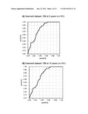 | 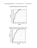 |
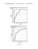 | 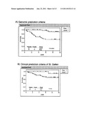 |
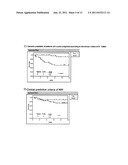 | 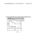 |
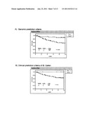 | 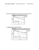 |
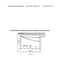 | 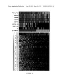 |
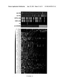 |  |
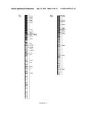 |
| Similar patent applications: | |
| Date | Title |
|---|---|
| 2010-05-06 | Method for predicting the occurrence of metastasis in breast cancer patients |
| 2009-04-16 | Genomic screen for epigenetically silenced genes associated with cancer |
| 2011-09-15 | Gene expression signature of genomic instability in breast cancer |
| 2011-10-27 | Method for examining prognosis of breast cancer |
| 2011-12-01 | Genetic variants for breast cancer risk assessment |
| New patent applications in this class: | |
| Date | Title |
|---|---|
| 2022-05-05 | Microfluidic system for amplifying and detecting polynucleotides in parallel |
| 2019-05-16 | Reagents and methods for detecting protein lysine 2-hydroxyisobutyrylation |
| 2019-05-16 | Lateral flow analyte detection |
| 2019-05-16 | Mutations in the bcr-abl tyrosine kinase associated with resistance to sti-571 |
| 2019-05-16 | Enhanced methods of ribonucleic acid hybridization |
| Top Inventors for class "Combinatorial chemistry technology: method, library, apparatus" | |
| Rank | Inventor's name |
|---|---|
| 1 | Mehdi Azimi |
| 2 | Kia Silverbrook |
| 3 | Geoffrey Richard Facer |
| 4 | Alireza Moini |
| 5 | William Marshall |