Patent application title: NOVEL MEMBERS OF THE CAPSAICIN/VANILLOID RECEPTOR FAMILY OF PROTEINS AND USES THEREOF
Inventors:
Rory A. J. Curtis (Southborough, MA, US)
IPC8 Class: AC12P2102FI
USPC Class:
435 691
Class name: Chemistry: molecular biology and microbiology micro-organism, tissue cell culture or enzyme using process to synthesize a desired chemical compound or composition recombinant dna technique included in method of making a protein or polypeptide
Publication date: 2011-02-10
Patent application number: 20110033892
Claims:
1. An isolated nucleic acid molecule selected from the group consisting
of:(a) a nucleic acid molecule comprising the nucleotide sequence set
forth in SEQ ID NO:1, 3, 4, 6, 7, 9, 10, or 12 or a complement
thereof;(b) a nucleic acid molecule consisting of the nucleotide sequence
set forth in SEQ ID NO:1, 3, 4, 6, 7, 9, 10, or 12 or a complement
thereof;(c) a nucleic acid molecule which encodes a polypeptide selected
from the group consisting of (1) a polypeptide comprising the amino acid
sequence set forth in SEQ ID NO:2, 5, 8, or 11; and (2) a polypeptide
consisting of the amino acid sequence set forth in SEQ ID NO:2, 5, 8, or
11;(d) a nucleic acid comprising a nucleotide sequence which is at least
83% identical to the nucleotide sequence of SEQ ID NO: 1, 3, 4, 6, 7, 9,
10, or 12 or to a complement thereof;(e) a nucleic acid molecule
comprising a fragment of at least 20 nucleotides of a nucleic acid
comprising the nucleotide sequence of SEQ ID NO:1, 3, 4, 6, 7, 9, 10, or
12, or a complement thereof;(f) a nucleic acid molecule which encodes a
polypeptide comprising an amino acid sequence at least about 87%
identical to the amino acid sequence of SEQ ID NO:2, 5, 8, or 11; and(g)
a nucleic acid molecule which encodes a fragment of a polypeptide
comprising the amino acid sequence of SEQ ID NO:2, 5, 8, or 11, wherein
the fragment comprises at least 15 contiguous amino acid residues of the
amino acid sequence of SEQ ID NO:2, 5, 8, or 11.
2. The isolated nucleic acid molecule of claim 1 further comprising a nucleotide sequence encoding a heterologous polypeptide.
3. The isolated nucleic acid molecule of claim 1 which is a vector.
4. The isolated nucleic acid molecule of claim 3 which is an expression vector.
5. A host cell transfected with the expression vector of claim 4.
6. A method of expressing a polypeptide comprising culturing the host cell of claim 5 in an appropriate culture medium, thereby expressing the polypeptide.
7. An isolated polypeptide selected from the group consisting of:(a) a fragment of a polypeptide comprising the amino acid sequence of SEQ ID NO:2, 5, 8, or 11, wherein the fragment comprises at least 15 contiguous amino acids of SEQ ID NO:2, 5, 8, or 11;(b) a naturally occurring allelic variant of a polypeptide comprising the amino acid sequence of SEQ ID NO:2, 5, 8, or 11, wherein the polypeptide is encoded by a nucleic acid molecule which hybridizes to a nucleic acid molecule consisting of SEQ ID NO:1, 3, 4, 6, 7, 9, 10, or 12 under stringent conditions;(c) a polypeptide which is encoded by a nucleic acid molecule comprising a nucleotide sequence which is at least 83% identical to a nucleic acid comprising the nucleotide sequence of SEQ ID NO:1, 3, 4, 6, 7, 9, 10, or 12; and(d) a polypeptide comprising an amino acid sequence which is at least 87% identical to the amino acid sequence of SEQ ID NO:2, 5, 8, or 11.
8. The isolated polypeptide of claim 7 comprising the amino acid sequence of SEQ ID NO:2, 5, 8, or 11.
9. The isolated polypeptide of claim 7 further comprising a heterologous amino acid sequence.
10. An isolated antibody which selectively binds to a polypeptide of claim 7.
11. A method for detecting the presence of a polypeptide of claim 7 in a sample comprising:contacting the sample with a compound which selectively binds to the polypeptide; anddetermining whether the compound binds to the polypeptide in the sample, thereby detecting the polypeptide in the sample.
12. A method for detecting the presence of a nucleic acid molecule of claim 1 in a sample comprising:contacting the sample with a nucleic acid probe or primer which selectively hybridizes to the nucleic acid molecule; anddetermining whether the nucleic acid probe or primer binds to a nucleic acid molecule in the sample, thereby detecting the presence of the nucleic acid molecule in the sample.
13. The method of claim 12 wherein the sample comprises mRNA molecules and is contacted with a nucleic acid probe.
Description:
[0001]This application is a division of Ser. No. 11/967,558 filed Dec. 31,
2007, which is a continuation of Ser. No. 11/013,090 filed Dec. 15, 2004,
now U.S. Pat. No. 7,323,314, which is a division of Ser. No. 09/587,111
filed Jun. 2, 2000, now U.S. Pat. No. 7,063,951, which is a division of
Ser. No. 09/439,165 filed Nov. 12, 1999, now abandoned, which is a
continuation-in-part of Ser. No. 09/421,134 filed Oct. 19, 1999, now
abandoned, which is a continuation-in-part of Ser. No. 09/258,633 filed
Feb. 26, 1999, now abandoned, which claims priority to Ser. No.
60/108,322 filed Nov. 13, 1998 and Ser. No. 60/114,078 filed Dec. 28,
1998. Each of these applications is incorporated by reference herein in
its entirety.
[0002]This application incorporates by reference a 144 kb text file created on Sep. 29, 2010 and named "11967558DIVsequencelisting.txt," which is the sequence listing for this application.
BACKGROUND OF THE INVENTION
[0003]Pain is initiated when the peripheral terminals of a subgroup of sensory neurons are activated by noxious chemical, mechanical or thermal stimuli. These neurons, called nociceptors, transmit information regarding tissue damage to pain-processing centres in the spinal chord and brain (Fields, H. L. Pain, McGraw-Hill, New York, 1987). Nociceptors are characterized in part, by their sensitivity to capsaicin, a vanilloid-containing compound, and a natural product of capsicum peppers that is the active ingredient of many "hot" and spicy foods. In mammals, exposure of nociceptor terminals to capsaicin leads initially to excitation of the neuron and the consequent perception of pain and local release of inflammatory mediators. With prolonged exposure, nociceptor terminals become insensitive to capsaicin, as well as to other noxious stimuli (Szolcsanyi, J. in Capsaicin in the Study of Pain (ed. Wood, J.) 1-26 (Academic, London, 1993). This latter phenomenon of nociceptor desensitization underlies the seemingly paradoxical use of capsaicin as an analgesic agent in the treatment of painful disorders ranging from viral and diabetic neuropathies to rheumatoid arthritis (Campbell, E. in Capsaicin and the Study of Pain (ed. Wood, J.) 255-272 (Academic, London, 1993); Szallasi, A. et al. (1996) Pain 68, 195-208). Some of this decreased sensitivity to noxious stimuli may result from reversible changes in the nociceptor, but the long-term loss of responsiveness can be explained by death of the nociceptor or destruction of its peripheral terminals following exposure to capsaicin (Jancso, G. et al. (1977) Nature 270, 741-743).
[0004]The cellular specificity of capsaicin action and its ability to evoke the sensation of burning pain have led to speculation that the target of capsaicin action plays an important physiological role in the detection of painful stimuli. Indeed, capsaicin may elicit the perception of pain by mimicking the actions of a physiological stimulus or an endogenous ligand produced during tissue injury (James, I. F., Kinkina, N. N. & Wood, J. N. in Capsaicin in the Study of Pain (ed. Wood, J. N.) 83-104 (Academic, London, 1993).
[0005]Caterina M. J. et al. have recently determined the molecular basis underlying this phenomenon by characterizing a functional cDNA that encodes a vanilloid receptor (VR-1) in rat sensory ganglia (Caterina M. J. et al., (1997) Nature 389:816-824). VR-1 is a vanilloid-gated, nonselective cation channel that resembles members of the transient receptor potential (TRP) channel family, first identified as components of the Drosophila phototransduction pathway (Montell et al. (1989) Neuron 2:1313-1323).
SUMMARY OF THE INVENTION
[0006]The present invention is based, at least in part, on the discovery of novel members of the Capsaicin/Vanilloid family of receptors. Described herein is the isolation of the human orthologue of rat VR-1 (rVR-1), referred to herein as hVR-1, as well as another previously unknown member of the VR family of receptors, referred herein as VR-2, and specifically as human VR-2 (hVR-2, including an alternate form which contains a deletion) and rat VR-2 (rVR-2) nucleic acid and protein molecules. The hVR-1, hVR-2, and rVR-2 molecules of the present invention are useful as targets for developing modulating agents to regulate a variety of cellular processes, e.g., cellular processes involved in pain. Accordingly, in one aspect, this invention provides isolated nucleic acid molecules encoding hVR-1, hVR-2, and rVR-2 proteins and fragments thereof, as well as nucleic acid fragments suitable as primers or hybridization probes for the detection of hVR-1, hVR-2, and rVR-2-encoding nucleic acids.
[0007]In one embodiment, an hVR-1, hVR-2, or rVR-2 nucleic acid molecule of the invention is at least 60%, 65%, 70%, 75%, 80%, 83%, 85%, 86%, 87%, 88%, 89%, 90%, 91%, 92%, 93%, 94%, 95%, 96%, 97%, 98%, 99% or more identical to the nucleotide sequence (e.g., to the entire length of the nucleotide sequence) shown in SEQ ID NO: 1, 3, 4, 6, 7, 9, 10, or 12 or the nucleotide sequence of the DNA insert of the plasmid deposited with ATCC as Accession Number ______, or a complement thereof.
[0008]In another embodiment, the isolated nucleic acid molecule includes the nucleotide sequence shown SEQ ID NO:1, 3, 4, 6, 7, 9, 10, or 12, or a complement thereof. In another embodiment, the nucleic acid molecule includes at least 10, 15, 20, or more contiguous nucleotides of SEQ ID NO:1, 3, 4, 6, 7, 9, 10, or 12.
[0009]In another embodiment, an hVR-1, hVR-2, and rVR-2 nucleic acid molecule includes a nucleotide sequence encoding a protein having an amino acid sequence sufficiently homologous to the amino acid sequence of SEQ ID NO:2, 5, 8, or 11. In one embodiment, an hVR-1, hVR-2, and rVR-2 nucleic acid molecule includes a nucleotide sequence encoding a protein having an amino acid sequence at least 60%, 65%, 70%, 75%, 80%, 85%, 87%, 90%, 95%, 98% or more identical to the entire length of the amino acid sequence of SEQ ID NO:2, 5, 8, or 11.
[0010]Another embodiment of the invention features nucleic acid molecules, preferably hVR-1, hVR-2, and rVR-2 nucleic acid molecules, which specifically detect hVR-1, hVR-2, and rVR-2 nucleic acid molecules relative to nucleic acid molecules encoding non-hVR-1, non-hVR-2, and non-hVR-2 proteins. For example, in one embodiment, such a nucleic acid molecule is at least 100-150, 1150-200, 200-250, 250-300, 300-350, 350-400, 400-450, 450-500, 500-550, 550-600, 600-700, 700-800, 800-900, 900-1000, 1088, or more nucleotides in length and hybridizes under stringent conditions to a nucleic acid molecule comprising the nucleotide sequence shown in SEQ ID NO:1, 3, 4, 6, 7, 9, 10, or 12. In preferred embodiments, the nucleic acid molecules are at least 15 (e.g., contiguous) nucleotides in length and hybridize under stringent conditions to nucleotides 1-17, 3696-3863, or 3901-3909 of SEQ ID NO:1. In other preferred embodiments, the nucleic acid molecules comprise nucleotides 1-17, 3696-3863, or 3901-3909 of SEQ ID NO:1. In yet other preferred embodiments, the nucleic acid molecules consist of nucleotides 1-17, 3696-3863, or 3901-3909 of SEQ ID NO:1. In preferred embodiments, the nucleic acid molecules are at least 15 (e.g., contiguous) nucleotides in length and hybridize under stringent conditions to nucleotides 1944-2003 of SEQ ID NO:4. In other preferred embodiments, the nucleic acid molecules comprise nucleotides 1944-2003 of SEQ ID NO:4. In yet other preferred embodiments, the nucleic acid molecules consist of nucleotides 1944-2003 of SEQ ID NO:4.
[0011]In other embodiments, the nucleic acid molecule encodes a naturally occurring allelic variant of a polypeptide comprising the amino acid sequence of SEQ ID NO:2, 5, 8, or 11, wherein the nucleic acid molecule hybridizes to a nucleic acid molecule consisting of SEQ ID NO:1, 3, 4, 6, 7, 9, 10, or 12 under stringent conditions and is encoded by the same locus as hVR-1, hVR-2 or rVR-2.
[0012]Another embodiment of the invention provides a nucleic acid molecule that encodes a naturally occurring orthologue of a polypeptide comprising the amino acid sequence of SEQ ID NO:2, 5, 8, or 11, wherein the nucleic acid molecule hybridizes to a nucleic acid molecule consisting of SEQ ID NO:1, 3, 4, 6, 7, 9, 10, or 12 under stringent conditions.
[0013]Another embodiment of the invention provides an isolated nucleic acid molecule which is antisense to an hVR-1, hVR-2, and rVR-2 nucleic acid molecule, e.g., the coding strand of an hVR-1, hVR-2, and rVR-2 nucleic acid molecule.
[0014]Since the hVR2 (the alternate form) and rVR2 sequences represent fragments of the entire coding regions of these genes, another embodiment of the invention provides the complete gene sequences. A skilled artisan can readily isolate such molecule using the sequences disclosed herein.
[0015]Another aspect of the invention provides a vector comprising an hVR-1, an hVR-2, or a rVR-2 nucleic acid molecule. In certain embodiments, the vector is a recombinant expression vector. In another embodiment, the invention provides a host cell containing a vector of the invention. In yet another embodiment, the invention provides a host cell containing a nucleic acid molecule of the invention. The invention also provides a method for producing a protein, preferably an hVR-1, hVR-2, and rVR-2 protein, by culturing in a suitable medium, a host cell, e.g., a mammalian host cell such as a non-human mammalian cell, of the invention containing a recombinant expression vector, such that the protein is produced.
[0016]Another aspect of this invention features isolated or recombinant hVR-1, hVR-2, and rVR-2 proteins and polypeptides. In one embodiment, the isolated protein, preferably an hVR-1, hVR-2, or rVR-2 protein, includes at least one transmembrane domain. In another embodiment, the isolated protein, preferably an hVR-1, hVR-2, or rVR-2 protein, includes at least one transmembrane domain and at least one proline rich domain. In yet another embodiment, the isolated protein, preferably an hVR-1, hVR-2, or rVR-2 protein, includes at least one transmembrane domain, at least one proline rich domain, and at least one ankyrin repeat domain. In yet another embodiment, the protein, preferably an hVR-1, hVR-2, or rVR-2 protein, includes at least one transmembrane domain, at least one proline rich domain, and at least one ankyrin repeat domain and has an amino acid sequence at least about 60%, 65%, 70%, 75%, 80%, 85%, 87%, 90%, 95%, 98% or more homologous to the amino acid sequence of SEQ ID NO:2, 5, 8, or 11. In another embodiment, the protein, preferably an hVR-1, hVR-2, or rVR-2 protein, includes at least one transmembrane domain, at least one proline rich domain, and at least one ankyrin repeat domain and plays a role in the development and regulation of pain. In yet another embodiment, the protein, preferably an hVR-1, hVR-2, and rVR-2 protein, includes at least one transmembrane domain, at least one proline rich domain, and at least one ankyrin repeat domain and is encoded by a nucleic acid molecule having a nucleotide sequence which hybridizes under stringent hybridization conditions to a nucleic acid molecule comprising the nucleotide sequence of SEQ ID NO:1, 3, 4, 6, 7, 9, 10, or 12.
[0017]In another embodiment, the invention features fragments of the protein having the amino acid sequence of SEQ ID NO:2, 5, 8, or 11, wherein the fragment comprises at least 15, 30, 40, 50, 60, 70, 80, 90, or 100 amino acids (e.g., contiguous amino acids).
[0018]In another embodiment, the invention features an isolated protein, preferably an hVR-1, hVR-2, and rVR-2 protein, which is encoded by a nucleic acid molecule consisting of a nucleotide sequence at least about 60%, 65%, 70%, 75%, 80%, 83%, 85%, 86%, 87%, 88%, 89%, 90%, 91%, 92%, 93%, 94%, 95%, 96%, 97%, 98%, 99% or more homologous to a nucleotide sequence of SEQ ID NO:1, 3, 4, 6, 7, 9, 10, or 12, or a complement thereof. This invention further features an isolated protein, preferably an hVR-1, hVR-2, or rVR-2 protein, which is encoded by a nucleic acid molecule consisting of a nucleotide sequence which hybridizes under stringent hybridization conditions to a nucleic acid molecule consisting of the nucleotide sequence of SEQ ID NO:1, 3, 4, 6, 7, 9, 10, or 12, or a complement thereof.
[0019]The proteins of the present invention or portions thereof, e.g., biologically active portions thereof, can be operatively linked to a non-hVR-1, non-hVR-2, or non-rVR-2 polypeptide (e.g., heterologous amino acid sequences) to form fusion proteins. The invention further features antibodies, such as monoclonal or polyclonal antibodies, that specifically bind proteins of the invention, preferably hVR-1, hVR-2, and rVR-2 proteins. In addition, the hVR-1, hVR-2, and rVR-2 proteins or biologically active portions thereof can be incorporated into pharmaceutical compositions, which optionally include pharmaceutically acceptable carriers.
[0020]In another aspect, the present invention provides a method for detecting the presence of an hVR-1, hVR-2, and rVR-2 nucleic acid molecule, protein or polypeptide in a biological sample by contacting the biological sample with an agent capable of detecting an hVR-1, hVR-2, and rVR-2 nucleic acid molecule, protein or polypeptide such that the presence of an hVR-1, hVR-2, and rVR-2 nucleic acid molecule, protein or polypeptide is detected in the biological sample.
[0021]In another aspect, the present invention provides a method for detecting the presence of hVR-1, hVR-2, and rVR-2 activity in a biological sample by contacting the biological sample with an agent capable of detecting an indicator of hVR-1, hVR-2, and rVR-2 activity such that the presence of hVR-1, hVR-2, and rVR-2 activity is detected in the biological sample.
[0022]In another aspect, the invention provides a method for modulating hVR-1, hVR-2, and rVR-2 activity comprising contacting a cell capable of expressing hVR-1, hVR-2, and rVR-2 with an agent that modulates hVR-1, hVR-2, and rVR-2 activity such that hVR-1, hVR-2, and rVR-2 activity in the cell is modulated. In one embodiment, the agent inhibits hVR-1, hVR-2, and rVR-2 activity. In another embodiment, the agent stimulates hVR-1, hVR-2, and rVR-2 activity. In one embodiment, the agent is an antibody that specifically binds to an hVR-1, hVR-2, and rVR-2 protein. In another embodiment, the agent modulates expression of hVR-1, hVR-2, and rVR-2 by modulating transcription of an hVR-1, hVR-2, and rVR-2 gene or translation of an hVR-1, hVR-2, and rVR-2 mRNA. In yet another embodiment, the agent is a nucleic acid molecule having a nucleotide sequence that is antisense to the coding strand of an hVR-1, hVR-2, and rVR-2 mRNA or an hVR-1, hVR-2, and rVR-2 gene.
[0023]In one embodiment, the methods of the present invention are used to treat a subject having a disorder characterized by aberrant hVR-1, hVR-2, and rVR-2 protein or nucleic acid expression or activity by administering an agent which is an hVR-1, hVR-2, and rVR-2 modulator to the subject. In one embodiment, the hVR-1, hVR-2, and rVR-2 modulator is an hVR-1, hVR-2, and rVR-2 protein. In another embodiment the hVR-1, hVR-2, and rVR-2 modulator is an hVR-1, hVR-2, and rVR-2 nucleic acid molecule. In yet another embodiment, the hVR-1, hVR-2, and rVR-2 modulator is a peptide, peptidomimetic, or other small molecule. In a further embodiment, the disorder characterized by aberrant hVR-1, hVR-2, and rVR-2 protein or nucleic acid expression is a pain disorder, e.g., hyperalgesia.
[0024]The present invention also provides a diagnostic assay for identifying the presence or absence of a genetic alteration characterized by at least one of (i) aberrant modification or mutation of a gene encoding an hVR-1, hVR-2, and rVR-2 protein; (ii) mis-regulation of the gene; and (iii) aberrant post-translational modification of an hVR-1, hVR-2, and rVR-2 protein, wherein a wild-type form of the gene encodes a protein with an hVR-1, hVR-2, and rVR-2 activity (as described herein).
[0025]In another aspect the invention provides a method for identifying a compound that binds to or modulates the activity of an hVR-1, hVR-2, and rVR-2 protein, by providing an indicator composition comprising an hVR-1, hVR-2, and rVR-2 protein having hVR-1, hVR-2, and rVR-2 activity, contacting the indicator composition with a test compound, and determining the effect of the test compound on hVR-1, hVR-2, and rVR-2 activity in the indicator composition to identify a compound that modulates the activity of an hVR-1, hVR-2, and rVR-2 protein.
[0026]Other features and advantages of the invention will be apparent from the following detailed description and claims.
BRIEF DESCRIPTION OF THE DRAWINGS
[0027]FIGS. 1A-1D depict the full length cDNA sequence and predicted amino acid sequence of human VR-1 (hVR-1). The nucleotide sequence corresponds to nucleic acids 1 to 3909 of SEQ ID NO:1. The amino acid sequence corresponds to amino acids 1 to 839 of SEQ ID NO:2. The coding region without the 5' and 3' untranslated regions of the human VR-1 (hVR-1) gene is shown in SEQ ID NO:3.
[0028]FIGS. 2A-2C depict the full length cDNA sequence and predicted amino acid sequence of human VR-2 (hVR-2). The nucleotide sequence corresponds to nucleic acids 1 to 2809 of SEQ ID NO:4. The amino acid sequence corresponds to amino acids 1 to 764 of SEQ ID NO:5. The coding region without the 5' and 3' untranslated regions of the human VR-2 (hVR-2) gene is shown in SEQ ID NO:6.
[0029]FIGS. 3A-3B depict the partial cDNA sequence and partial predicted amino acid sequence of an alternate form of human VR-2 (hVR-2). The nucleotide sequence corresponds to nucleic acids 1 to 1489 of SEQ ID NO:7. The amino acid sequence corresponds to amino acids 1 to 436 of SEQ ID NO:8. The coding region without the 5' and 3' untranslated regions of the alternate form of human VR-2 (hVR-2) gene is shown in SEQ ID NO:9.
[0030]FIGS. 4A-4B depict the partial cDNA sequence and partial predicted amino acid sequence of rat VR-2 (rVR-2). The nucleotide sequence corresponds to nucleic acids 1 to 1794 of SEQ ID NO:10. The amino acid sequence corresponds to amino acids 1 to 554 of SEQ ID NO:11. The coding region without the 5' and 3' untranslated regions of the rat VR-2 (rVR-2) gene is shown in SEQ ID NO:12.
[0031]FIGS. 5A-5B depict an alignment of the hVR-1 protein (SEQ ID NO:2) with the human VR-2 protein (SEQ ID NO:5) using the GAP program in the GCG software package (Blosum 62 matrix) and a gap weight of 12 and a length weight of 4.
[0032]FIGS. 6A-6F depict an alignment of the hVR-1 nucleotide sequence (SEQ ID NO:1) with the human VR-2 nucleotide sequence (SEQ ID NO:4) using the GAP program in the GCG software package (nwsgapdna matrix) and a gap weight of 50 and a length weight of 3.
[0033]FIG. 7 depicts an alignment of the hVR-2 protein (SEQ ID NO:5) with the rat VR-2 protein (SEQ ID NO:11) using the CLUSTAL W (1.74) multiple sequence alignment program.
[0034]FIGS. 8A-8B depict an alignment of the hVR-2 protein (SEQ ID NO:5) with the rat VR-2 protein (SEQ ID NO:11) using the GAP program in the GCG software package (Blosum 62 matrix) and a gap weight of 12 and a length weight of 4.
[0035]FIGS. 9A-9F depict an alignment of the hVR-1 nucleotide sequence (SEQ ID NO:1) with the rat VR-1 nucleotide sequence (Accession Number: AF029310, SEQ ID NO:21) using the GAP program in the GCG software package (nwsgapdna matrix) and a gap weight of 50 and a length weight of 3.
[0036]FIGS. 10A-10B depict an alignment of the hVR-1 protein (SEQ ID NO:2) with the rat VR-1 protein (Accession Number: AF029310, SEQ ID NO:22) using the GAP program in the GCG software package (Blosum 62 matrix) and a gap weight of 12 and a length weight of 4.
[0037]FIG. 11 depicts an alignment of the hVR-2 protein (SEQ ID NO:5) with the human VR-2 protein (alternate form) (SEQ ID NO:8) using the CLUSTAL W (1.74) multiple sequence alignment program.
[0038]FIG. 12 depicts a structural, hydrophobicity, and antigenicity analysis of the hVR-1 protein.
[0039]FIG. 13 depicts an alignment between the amino acid sequence of the hVR-1 protein (SEQ ID NO:2) and Ank repeat domains (SEQ ID NO:23) identified while performing a search using the amino acid sequence of the hVR-1 protein against the HMM database.
[0040]FIG. 14 depicts a structural, hydrophobicity, and antigenicity analysis of the hVR-2 protein.
[0041]FIG. 15 depicts an alignment between the amino acid sequence of the hVR-2 protein (SEQ ID NO:5) and Ank repeat domains (SEQ ID NO:23) identified while performing a search using the amino acid sequence of the hVR-2 protein against the HMM database.
[0042]FIG. 16 depicts the predicted full length amino acid sequence of the human VR-2 protein (alternate form) (SEQ ID NO:20).
[0043]FIG. 17 depicts an alignment of the hVR-2 protein (SEQ ID NO:5) with the predicted full length human VR-2 protein (alternate form) (SEQ ID NO:20) using the CLUSTAL W (1.74) multiple sequence alignment program.
DETAILED DESCRIPTION OF THE INVENTION
[0044]The present invention is based, at least in part, on the discovery of nucleic acid and amino acid molecules which are novel members of the Capsaicin/Vanilloid family of receptors. Described herein is the isolation of the human orthologue of rat VR-1 (rVR-1), referred to herein as hVR-1, as well as another previously unknown member of the VR family of receptors, referred herein as VR-2, and specifically as human VR-2 (hVR-2) and rat VR-2 (rVR-2) nucleic acid and protein molecules. The hVR-1, hVR-2, and rVR-2 molecules were identified based on their sequence similarity to the known rat vanilloid receptor (VR-1). VR-1 is a vanilloid gated, non-selective cation channel which resembles members of the transient receptor potential (TRP) ion channel family (described in Montell et al. (1989) Neuron 2:1313-1323) that mediate the influx of extracellular calcium in response to depletion of intracellular calcium stores. The rat VR-1 cDNA contains an open reading frame of 2514 nucleotides that encodes a protein of 838 amino acids. Hydrophilicity analysis has indicated that rat VR-1 contains six transmembrane domains (predicted to be mostly α-helices) with an additional short hydrophobic stretch between transmembrane regions 5 and 6. The amino terminal hydrophilic segment contains a relatively proline rich region followed by three ankyrin repeat domains. The rat VR-1 is expressed in small diameter neurons within sensory ganglia. The present hVR-1 sequence is the human orthologue of rVR-1. As described in further detail infra, the human VR-1 is expressed in nodose, trigeminal sensory neurons, as well as in some, but not all, small dorsal root ganglion (DRG) neurons and in a few medium sized DRG neurons.
[0045]The hVR-1, hVR-2, and rVR-2 molecules of the present invention play a role in pain signaling mechanisms. As used herein, the term "pain signaling mechanisms" includes the cellular mechanisms involved in the development and regulation of pain, e.g., pain elicited by noxious chemical, mechanical, or thermal stimuli, in a subject, e.g., a mammal such as a human. In mammals, the initial detection of noxious chemical, mechanical, or thermal stimuli, a process referred to as "nociception", occurs predominantly at the peripheral terminals of specialized, small diameter primary afferent neurons, called polymodal nociceptors. These afferent neurons transmit the information to the central nervous system, evoking a perception of pain or discomfort and initiating appropriate protective reflexes. Capsaicin/Vanilloid receptors, e.g., the hVR-1, hVR-2, and rVR-2 molecules of the present invention, present on these afferent neurons, are involved in detecting these noxious chemical, mechanical, or thermal stimuli and transducing this information into membrane depolarization events. Thus, the hVR-1, hVR-2, and rVR-2 molecules by participating in pain signaling mechanisms, can modulate pain elicitation and provide novel diagnostic targets and therapeutic agents to control pain.
[0046]The hVR-1, hVR-2, and rVR-2 molecules provide novel diagnostic targets and therapeutic agents to control pain in a variety of disorders, diseases, or conditions which are characterized by a deregulated, e.g., upregulated or downregulated, pain response. For example, the hVR-1, hVR-2, and rVR-2 molecules provide novel diagnostic targets and therapeutic agents to control the exaggerated pain response elicited during various forms of tissue injury, e.g., inflammation, infection, and ischemia, usually referred to as hyperalgesia (described in, for example, Fields, H. L. (1987) Pain, New York: McGraw-Hill). Moreover, the hVR-1, hVR-2, and rVR-2 molecules provide novel diagnostic targets and therapeutic agents to control pain associated with muscoloskeletal disorders, e.g., joint pain; tooth pain; headaches; pain associated with surgery, or neuropathic pain.
[0047]As the hVR-1 gene maps to a region of human chromosome 17 between WI-5436 (7.7cR) and WI-6584 (18.9cR) (Example 6), which has been associated with myasthenia gravis, Smith-Magenis syndrome, CORDS, Cone-rod dysrtophy, and breast cancer, the hVR-1 molecule may provide novel diagnostic targets and therapeutic agents to treat, diagnose, or prognose these disorders or other disorders linked to this chromosomal region. Similarly, as the hVR-2 gene maps to a region of human chromosome 17 between AFMA043ZB5 (23.3 cR) and D175721 (29.3cR) (Example 6) which has been associated with myasthenia gravis, Smith-Magenis syndrome, CORDS, Cone-rod dysrtophy, choroidal dystrophy, central areolar, and retinal cone dystrophy, the hVR-2 molecule may provide novel diagnostic targets and therapeutic agents to treat, diagnose, or prognose these disorders or other disorders linked to this chromosomal region.
[0048]The term "family" when referring to the protein and nucleic acid molecules of the invention is intended to mean two or more proteins or nucleic acid molecules having a common structural domain or motif and having sufficient amino acid or nucleotide sequence homology as defined herein. Such family members can be naturally or non-naturally occurring and can be from either the same or different species. For example, a family can contain a first protein of human origin, as well as other, distinct proteins of human origin or alternatively, can contain homologues of non-human origin. Members of a family may also have common functional characteristics.
[0049]For example, the family of hVR-1, hVR-2, and rVR-2 proteins comprise at least one, and preferably six "transmembrane domains." As used herein, the term "transmembrane domain" includes an amino acid sequence of about 15 amino acid residues in length which spans the plasma membrane. More preferably, a transmembrane domain includes about at least 20, 25, 30, 35, 40, or 45 amino acid residues and spans the plasma membrane. Transmembrane domains are rich in hydrophobic residues, and typically have a helical structure. In a embodiment, at least 50%, 60%, 70%, 80%, 90%, 95% or more of the amino acid residues of a transmembrane domain are hydrophobic, e.g., leucines, isoleucines, tyrosines, or tryptophans. Transmembrane domains are described in, for example, Zagotta W. N. et al, (1996) Annual Rev. Neurosci. 19: 235-63, the contents of which are incorporated herein by reference Amino acid residues 434-455, 480-495, (509-531; based on homology to the rat VR-1) or 514-531, (543-569; based on homology to the rat VR-1) or 538-555, (577-596; based on homology to the rat VR-1) or 580-599, and (656-683; based on homology to the rat VR-1) or 658-682 of hVR-1 (SEQ ID NO:2) and amino acid residues 391-410, 431-448, 459-476, 486-508, 538-556, and 621-645 of hVR-2 (SEQ ID NO:5) comprise transmembrane domains.
[0050]In another embodiment, an hVR-1, hVR-2, and rVR-2 of the present invention is identified based on the presence of a "proline rich domain" in the protein or corresponding nucleic acid molecule. As used herein, the term "proline rich domain" includes an amino acid sequence of about 4-6 amino acid residues in length having the general sequence X-Pro-X-X-Pro-X (where X can be any amino acid). Proline rich domains are usually located in a helical structure and bind through hydrophobic interactions to SH3 domains. SH3 domains recognize proline rich domains in both forward and reverse orientations. Proline rich domains are described in, for example, Sattler M. et al. (1998) Leukemia 12:637-644, the contents of which are incorporated herein by reference.
[0051]In another embodiment, an hVR-1, hVR-2, and rVR-2 of the present invention is identified based on the presence of an "ankyrin repeat domain" in the protein or corresponding nucleic acid molecule. As used herein, the term "ankyrin repeat domain" includes a protein domain having an amino acid sequence of about 30-50 amino acid residues and having a bit score for the alignment of the sequence to the ankyrin repeat domain (HMM) of at least 6. Preferably, an ankyrin repeat domain includes at least about 30-45, more preferably about 30-40 amino acid residues, or about 30-35 amino acids and has a bit score for the alignment of the sequence to the ankyrin repeat domain (HMM) of at least 3-10, more preferably 10-30, more preferably 30-50, even more preferably 50-75, 75-100, 100-200 or greater. The ankyrin repeat domain HMM has been assigned the PFAM Accession PF00023 (genome.wustl.edu/Pfam/.html). Ankyrin repeats are involved in protein-protein interactions and are described in, for example, Ketchum K. A et al. (1996) FEBS Letters 378:19-26, the contents of which are incorporated herein by reference.
[0052]To identify the presence of an ankyrin repeat domain in an hVR-1, hVR-2, and rVR-2 protein and make the determination that a protein of interest has a particular profile, the amino acid sequence of the protein is searched against a database of HMMs (e.g., the Pfam database, release 2.1) using the default parameters (www.sanger.ac.uk/Software/Pfam/HMM_search). A description of the Pfam database can be found in Sonhammer et al. (1997) Proteins 28(3)405-420 and a detailed description of HMMs can be found, for example, in Gribskov et al. (1990) Meth. Enzymol. 183:146-159; Gribskov et al. (1987) Proc. Natl. Acad. Sci. USA 84:4355-4358; Krogh et al. (1994) J. Mol. Biol. 235:1501-1531; and Stultz et al. (1993) Protein Sci. 2:305-314, the contents of which are incorporated herein by reference. A search was performed against the HMM database resulting in the identification of three ankyrin repeat domains in the amino acid sequence of SEQ ID NO:2 (at about residues 201-233, 248-283, and 333-361) and SEQ ID NO:5 (at about residues 162-194, 208-243, and 293-328). The results of the searches are set forth in FIGS. 13 and 15.
[0053]Isolated proteins of the present invention, preferably hVR-1, hVR-2, and rVR-2 proteins, have an amino acid sequence sufficiently identical to the amino acid sequence of SEQ ID NO:2, 5, 8, or 11 or are encoded by a nucleotide sequence sufficiently identical to SEQ ID NO:1, 3, 4, 6, 7, 9, 10, or 12. As used herein, the term "sufficiently identical" refers to a first amino acid or nucleotide sequence which contains a sufficient or minimum number of identical or equivalent (e.g., an amino acid residue which has a similar side chain) amino acid residues or nucleotides to a second amino acid or nucleotide sequence such that the first and second amino acid or nucleotide sequences share common structural domains or motifs and/or a common functional activity. For example, amino acid or nucleotide sequences which share common structural domains have at least 30%, 40%, or 50% identity, preferably 60% identity, more preferably 70%-80%, and even more preferably 90-95% identity across the amino acid sequences of the domains and contain at least one and preferably two structural domains or motifs, are defined herein as sufficiently identical. Furthermore, amino acid or nucleotide sequences which share at least 30%, 40%, or 50%, preferably 60%, more preferably 70-80%, or 90-95% identity and share a common functional activity are defined herein as sufficiently identical.
[0054]As used interchangeably herein, an "hVR-1, hVR-2, and rVR-2 activity", "biological activity of hVR-1, hVR-2, and rVR-2" or "functional activity of hVR-1, hVR-2, and rVR-2", refers to an activity exerted by an hVR-1, hVR-2, and rVR-2 protein, polypeptide or nucleic acid molecule on an hVR-1, hVR-2, and rVR-2 responsive cell or on an hVR-1, hVR-2, and rVR-2 protein substrate, as determined in vivo, or in vitro, according to standard techniques. In one embodiment, an hVR-1, hVR-2, and rVR-2 activity is a direct activity, such as an association with an hVR-1, hVR-2, and rVR-2-target molecule. As used herein, a "target molecule" or "binding partner" is a molecule with which an hVR-1, hVR-2, and rVR-2 protein binds or interacts in nature, such that hVR-1, hVR-2, and rVR-2-mediated function is achieved. An hVR-1, hVR-2, and rVR-2 target molecule can be a non-hVR-1, non-hVR-2, and non-rVR-2 molecule or an hVR-1, hVR-2, and rVR-2 protein or polypeptide of the present invention. In an exemplary embodiment, an hVR-1, hVR-2, and rVR-2 target molecule is an hVR-1, hVR-2, and rVR-2 ligand, e.g., capsaicin. Alternatively, an hVR-1, hVR-2, and rVR-2 activity is an indirect activity, such as a cellular signaling activity mediated by interaction of the hVR-1, hVR-2, and rVR-2 protein with an hVR-1, hVR-2, and rVR-2 ligand.
[0055]Accordingly, another embodiment of the invention features isolated hVR-1, hVR-2, and rVR-2 proteins and polypeptides having an hVR-1, hVR-2, and rVR-2 activity. Other proteins of the invention are hVR-1, hVR-2, and rVR-2 proteins having at least one, and preferably six, transmembrane domains and, preferably, an hVR-1, hVR-2, and rVR-2 activity. Yet other proteins of the invention are hVR-1, hVR-2, and rVR-2 proteins having at least one transmembrane domain, at least one proline rich domain and, preferably, an hVR-1, hVR-2, and rVR-2 activity. Other proteins of the invention are hVR-1, hVR-2, and rVR-2 proteins having at least one transmembrane domain, at least one proline rich domain, at least one ankyrin repeat domain and, preferably, an hVR-1, hVR-2, and rVR-2 activity. Additional proteins of the invention have at least one transmembrane domain, at least one proline rich domain, at least one ankyrin repeat domain, and are, preferably, encoded by a nucleic acid molecule having a nucleotide sequence which hybridizes under stringent hybridization conditions to a nucleic acid molecule comprising the nucleotide sequence of SEQ ID NO:1, 3, 4, 6, 7, 9, 10, or 12.
[0056]The nucleotide sequence of the full length hVR-1 cDNA and the predicted amino acid sequence of the hVR-1 polypeptide are shown in FIGS. 1A-1D and in SEQ ID NOS:1 and 2, respectively.
[0057]The nucleotide sequence of the full length hVR-2 cDNA and the predicted amino acid sequence of the hVR-2 polypeptide are shown in FIGS. 2A-2B and in SEQ ID NOS:4 and 5, respectively.
[0058]The nucleotide sequence of the partial hVR-2 (alternate form) cDNA and the predicted amino acid sequence of the hVR-2 (alternate form) polypeptide are shown in FIGS. 3A-3B and in SEQ ID NOS:7 and 8, respectively.
[0059]The nucleotide sequence of the partial rVR-2 cDNA and the predicted amino acid sequence of the rVR-2 polypeptide are shown in FIGS. 4A-4B and in SEQ ID NOS:10 and 11, respectively.
[0060]Various aspects of the invention are described in further detail in the following subsections:
I. Isolated Nucleic Acid Molecules
[0061]One aspect of the invention pertains to isolated nucleic acid molecules that encode hVR-1, hVR-2, and rVR-2 proteins or biologically active portions thereof, as well as nucleic acid fragments sufficient for use as hybridization probes to identify hVR-1, hVR-2, and rVR-2-encoding nucleic acid molecules (e.g., hVR-1, hVR-2, and rVR-2 mRNA) and fragments for use as PCR primers for the amplification or mutation of hVR-1, hVR-2, and rVR-2 nucleic acid molecules. As used herein, the term "nucleic acid molecule" is intended to include DNA molecules (e.g., cDNA or genomic DNA) and RNA molecules (e.g., mRNA) and analogs of the DNA or RNA generated using nucleotide analogs. The nucleic acid molecule can be single-stranded or double-stranded, but preferably is double-stranded DNA.
[0062]The term "isolated nucleic acid molecule" includes nucleic acid molecules which are separated from other nucleic acid molecules which are present in the natural source of the nucleic acid. For example, with regards to genomic DNA, the term "isolated" includes nucleic acid molecules which are separated from the chromosome with which the genomic DNA is naturally associated. Preferably, an "isolated" nucleic acid is free of sequences which naturally flank the nucleic acid (i.e., sequences located at the 5' and 3' ends of the nucleic acid) in the genomic DNA of the organism from which the nucleic acid is derived. For example, in various embodiments, the isolated hVR-1, hVR-2, and rVR-2 nucleic acid molecule can contain less than about 5 kb, 4 kb, 3 kb, 2 kb, 1 kb, 0.5 kb or 0.1 kb of nucleotide sequences which naturally flank the nucleic acid molecule in genomic DNA of the cell from which the nucleic acid is derived. Moreover, an "isolated" nucleic acid molecule, such as a cDNA molecule, can be substantially free of other cellular material, or culture medium when produced by recombinant techniques, or substantially free of chemical precursors or other chemicals when chemically synthesized.
[0063]A nucleic acid molecule of the present invention, e.g., a nucleic acid molecule having the nucleotide sequence of SEQ ID NO:1, 3, 4, 6, 7, 9, 10, or 12. Using all or portion of the nucleic acid sequence of SEQ ID NO:1, 3, 4, 6, 7, 9, 10, or 12, as a hybridization probe, hVR-1, hVR-2, and rVR-2 nucleic acid molecules can be isolated using standard hybridization and cloning techniques (e.g., as described in Sambrook, J., Fritsh, E. F., and Maniatis, T. Molecular Cloning: A Laboratory Manual. 2nd, ed., Cold Spring Harbor Laboratory, Cold Spring Harbor Laboratory Press, Cold Spring Harbor, N.Y., 1989).
[0064]Moreover, a nucleic acid molecule encompassing all or a portion of SEQ ID NO:1, 3, 4, 6, 7, 9, 10, or 12, can be isolated by the polymerase chain reaction (PCR) using synthetic oligonucleotide primers designed based upon the sequence of SEQ ID NO:1, 3, 4, 6, 7, 9, 10, or 12.
[0065]A nucleic acid of the invention can be amplified using cDNA, mRNA or alternatively, genomic DNA, as a template and appropriate oligonucleotide primers according to standard PCR amplification techniques. The nucleic acid so amplified can be cloned into an appropriate vector and characterized by DNA sequence analysis. Furthermore, oligonucleotides corresponding to hVR-1, hVR-2, and rVR-2 nucleotide sequences can be prepared by standard synthetic techniques, e.g., using an automated DNA synthesizer.
[0066]In one embodiment, an isolated nucleic acid molecule of the invention comprises the nucleotide sequence shown in SEQ ID NO:1. The sequence of SEQ ID NO:1 corresponds to the full length hVR-1 encoding cDNA.
[0067]In another embodiment, an isolated nucleic acid molecule of the invention comprises the nucleotide sequence shown in SEQ ID NO:4. The sequence of SEQ ID NO:4 corresponds to the full length hVR-2 encoding cDNA.
[0068]In another embodiment, an isolated nucleic acid molecule of the invention comprises the nucleotide sequence shown in SEQ ID NO:7. The sequence of SEQ ID NO:7 corresponds to a fragment of the hVR-2 (alternate form) encoding cDNA.
[0069]In another embodiment, an isolated nucleic acid molecule of the invention comprises the nucleotide sequence shown in SEQ ID NO:10. The sequence of SEQ ID NO:10 corresponds to a fragment of the rVR-2 cDNA.
[0070]In another embodiment, an isolated nucleic acid molecule of the invention comprises a nucleic acid molecule which is a complement of the nucleotide sequence shown in SEQ ID NO:1, 3, 4, 6, 7, 9, 10, or 12, or a portion of any of these nucleotide sequences. A nucleic acid molecule which is complementary to the nucleotide sequence shown in SEQ ID NO:1, 3, 4, 6, 7, 9, 10, or 12, is one which is sufficiently complementary to the nucleotide sequence shown in SEQ ID NO:1, 3, 4, 6, 7, 9, 10, or 12, such that it can hybridize to the nucleotide sequence shown in SEQ ID NO:1, 3, 4, 6, 7, 9, 10, or 12 thereby forming a stable duplex.
[0071]In still another embodiment, an isolated nucleic acid molecule of the present invention comprises a nucleotide sequence which is at least about 60%, 65%, 70%, 75%, 80%, 83%, 85%, 86%, 87%, 88%, 89%, 90%, 91%, 92%, 93%, 94%, 95%, 96%, 97%, 98%, 99% or more homologous to the entire length of the nucleotide sequence shown in SEQ ID NO:1, 3, 4, 6, 7, 9, 10, or 12, or a portion of any of these nucleotide sequences.
[0072]Moreover, the nucleic acid molecule of the invention can comprise only a portion of the nucleic acid sequence of SEQ ID NO:1, 3, 4, 6, 7, 9, 10, or 12, for example, a fragment which can be used as a probe or primer or a fragment encoding a portion of an hVR-1, hVR-2, and rVR-2 protein, e.g., a biologically active portion of an hVR-1, hVR-2, and rVR-2 protein. The nucleotide sequence determined from the cloning of the hVR-1, hVR-2, and rVR-2 gene allows for the generation of probes and primers designed for use in identifying and/or cloning other hVR-1, hVR-2, and rVR-2 family members, as well as hVR-1, hVR-2, and rVR-2 homologues from other species. The probe/primer typically comprises a substantially purified oligonucleotide. The oligonucleotide typically comprises a region of nucleotide sequence that hybridizes under stringent conditions to at least about 12 or 15, preferably about 20 or 25, more preferably about 30, 35, 40, 45, 50, 55, 60, 65, 75, or 100 consecutive nucleotides of a sense sequence of SEQ ID NO:1, 3, 4, 6, 7, 9, 10, or 12, of an anti-sense sequence of SEQ ID NO:1, 3, 4, 6, 7, 9, 10, or 12, or of a naturally occurring allelic variant or mutant of SEQ ID NO:1, 3, 4, 6, 7, 9, 10, or 12. In an exemplary embodiment, a nucleic acid molecule of the present invention comprises a nucleotide sequence which is greater than 100-150, 150-200, 200-250, 250-300, 300-350, 350-400, 400-450, 450-500, 500-550, 550-600, 600-650, 650-700, 700-750, 750-800, 800-850, 850-900, 900-950, 950-1000, 1088, or more nucleotides in length and hybridizes under stringent hybridization conditions to a nucleic acid molecule of SEQ ID NO:1, 3, 4, 6, 7, 9, 10, or 12.
[0073]Probes based on the hVR-1, hVR-2, and rVR-2 nucleotide sequences can be used to detect transcripts or genomic sequences encoding the same or homologous proteins. In preferred embodiments, the probe further comprises a label group attached thereto, e.g., the label group can be a radioisotope, a fluorescent compound, an enzyme, or an enzyme co-factor. Such probes can be used as a part of a diagnostic test kit for identifying cells or tissue which misexpress an hVR-1, hVR-2, and rVR-2 protein, such as by measuring a level of an hVR-1, hVR-2, and rVR-2-encoding nucleic acid in a sample of cells from a subject e.g., detecting hVR-1, hVR-2, and rVR-2 mRNA levels or determining whether a genomic hVR-1, hVR-2, and rVR-2 gene has been mutated or deleted.
[0074]A nucleic acid fragment encoding a "biologically active portion of an hVR-1, hVR-2, and rVR-2 protein" can be prepared by isolating a portion of the nucleotide sequence of SEQ ID NO:1, 3, 4, 6, 7, 9, 10, or 12, which encodes a polypeptide having an hVR-1, hVR-2, and rVR-2 biological activity (the biological activities of the hVR-1, hVR-2, and rVR-2 proteins are described herein), expressing the encoded portion of the hVR-1, hVR-2, and rVR-2 protein (e.g., by recombinant expression in vitro) and assessing the activity of the encoded portion of the hVR-1, hVR-2, and rVR-2 protein.
[0075]The invention further encompasses nucleic acid molecules that differ from the nucleotide sequence shown in SEQ ID NO:1, 3, 4, 6, 7, 9, 10, or 12, due to degeneracy of the genetic code and thus encode the same hVR-1, hVR-2, and rVR-2 proteins as those encoded by the nucleotide sequence shown in SEQ ID NO:1, 3, 4, 6, 7, 9, 10, or 12. In another embodiment, an isolated nucleic acid molecule of the invention has a nucleotide sequence encoding a protein having an amino acid sequence shown in SEQ ID NO:2, 5, 8, or 11.
[0076]In addition to the hVR-1, hVR-2, and rVR-2 nucleotide sequences shown in SEQ ID NO:1, 3, 4, 6, 7, 9, 10, or 12, it will be appreciated by those skilled in the art that DNA sequence polymorphisms that lead to changes in the amino acid sequences of the hVR-1, hVR-2, and rVR-2 proteins may exist within a population (e.g., the human population). Such genetic polymorphism in the hVR-1, hVR-2, and rVR-2 genes may exist among individuals within a population due to natural allelic variation. As used herein, the terms "gene" and "recombinant gene" refer to nucleic acid molecules which include an open reading frame encoding an hVR-1, hVR-2, and rVR-2 protein, preferably a mammalian hVR-1, hVR-2, and rVR-2 protein, and can further include non-coding regulatory sequences, and introns.
[0077]Allelic variants of hVR-1, hVR-2, and rVR-2 include both functional and non-functional hVR-1, hVR-2, and rVR-2 proteins. Functional allelic variants are naturally occurring amino acid sequence variants of the hVR-1, hVR-2, and rVR-2 protein that maintain the ability to bind an hVR-1, hVR-2, and rVR-2 ligand and/or modulate a pain signaling mechanism. Functional allelic variants will typically contain only conservative substitution of one or more amino acids of SEQ ID NO:2, 5, 8, or 11, or substitution, deletion or insertion of non-critical residues in non-critical regions of the protein.
[0078]Non-functional allelic variants are naturally occurring amino acid sequence variants of the hVR-1, hVR-2, and rVR-2 protein that do not have the ability to either bind an hVR-1, hVR-2, and rVR-2 ligand and/or modulate a pain signaling mechanism. Non-functional allelic variants will typically contain a non-conservative substitution, a deletion, or insertion or premature truncation of the amino acid sequence of SEQ ID NO:2, 5, 8, or 11, or a substitution, insertion or deletion in critical residues or critical regions.
[0079]The present invention further provides non-human orthologues of the hVR-2 and rVR-2 protein. Orthologues of the hVR-2 and rVR-2 protein are proteins that are isolated from non-human and non-rat organisms and possess the same hVR-2 and rVR-2 ligand binding and/or modulation of pain signaling mechanism capabilities of the hVR-2 and rVR-2 proteins. Orthologues of the hVR-2 and rVR-2 proteins can readily be identified as comprising an amino acid sequence that is substantially homologous to SEQ ID NO: 4, 6, 8 or 10.
[0080]Moreover, nucleic acid molecules encoding other hVR-1, hVR-2, and rVR-2 family members and, thus, which have a nucleotide sequence which differs from the hVR-1, hVR-2, and rVR-2 sequences of SEQ ID NO:1, 3, 4, 6, 7, 9, 10, or 12, are intended to be within the scope of the invention. For example, another hVR-1, hVR-2, and rVR-2 cDNA can be identified based on the nucleotide sequence of hVR-1, hVR-2, and rVR-2. Moreover, nucleic acid molecules encoding VR-2 proteins from different species, and which, thus, have a nucleotide sequence which differs from the hVR-2 and rVR-2 sequences of SEQ ID NO:4, 6, 8, or 10 are intended to be within the scope of the invention. For example, a mouse hVR-2 cDNA can be identified based on the nucleotide sequence of the human VR-2 (hVR-2) or the rat VR-2 (rVR-2).
[0081]Nucleic acid molecules corresponding to natural allelic variants and homologues of the hVR-1, hVR-2, and rVR-2 cDNAs of the invention can be isolated based on their homology to the hVR-1, hVR-2, and rVR-2 nucleic acids disclosed herein using the cDNAs disclosed herein, or a portion thereof, as a hybridization probe according to standard hybridization techniques under stringent hybridization conditions. Nucleic acid molecules corresponding to natural allelic variants and homologues of the hVR-1, hVR-2, and rVR-2 cDNAs of the invention can further be isolated by mapping to the same chromosome or locus as the hVR-1, hVR-2, and rVR-2 gene.
[0082]Accordingly, in another embodiment, an isolated nucleic acid molecule of the invention is at least 15, 20, 25, 30 or more nucleotides in length and hybridizes under stringent conditions to the nucleic acid molecule comprising the nucleotide sequence of SEQ ID NO:1, 3, 4, 6, 7, 9, 10, or 12. In other embodiment, the nucleic acid is at least 30, 50, 100, 150, 200, 250, 300, 350, 400, 450, 500, 550, 600, 650, 700, 750, 800, 850, 900, or 950 nucleotides in length. As used herein, the term "hybridizes under stringent conditions" is intended to describe conditions for hybridization and washing under which nucleotide sequences at least 60% identical to each other typically remain hybridized to each other. Preferably, the conditions are such that sequences at least about 70%, more preferably at least about 80%, even more preferably at least about 85% or 90% identical to each other typically remain hybridized to each other. Such stringent conditions are known to those skilled in the art and can be found in Current Protocols in Molecular Biology, John Wiley & Sons, N.Y. (1989), 6.3.1-6.3.6. A preferred, non-limiting example of stringent hybridization conditions are hybridization in 6× sodium chloride/sodium citrate (SSC) at about 45° C., followed by one or more washes in 0.2×SSC, 0.1% SDS at 50° C., preferably at 55° C., more preferably at 60° C., and even more preferably at 65° C. Preferably, an isolated nucleic acid molecule of the invention that hybridizes under stringent conditions to the sequence of SEQ ID NO:1, 3, 4, 6, 7, 9, 10, or 12 corresponds to a naturally-occurring nucleic acid molecule. As used herein, a "naturally-occurring" nucleic acid molecule refers to an RNA or DNA molecule having a nucleotide sequence that occurs in nature (e.g., encodes a natural protein).
[0083]In addition to naturally-occurring allelic variants of the hVR-1, hVR-2, and rVR-2 sequences that may exist in the population, the skilled artisan will further appreciate that changes can be introduced by mutation into the nucleotide sequences of SEQ ID NO:1, 3, 4, 6, 7, 9, 10, or 12, thereby leading to changes in the amino acid sequence of the encoded hVR-1, hVR-2, and rVR-2 proteins, without altering the functional ability of the hVR-1, hVR-2, and rVR-2 proteins. For example, nucleotide substitutions leading to amino acid substitutions at "non-essential" amino acid residues can be made in the sequence of SEQ ID NO:1, 3, 4, 6, 7, 9, 10, or 12. A "non-essential" amino acid residue is a residue that can be altered from the wild-type sequence of hVR-1, hVR-2, and rVR-2 (e.g., the sequence of SEQ ID NO:2, 5, 8, or 11) without altering the biological activity, whereas an "essential" amino acid residue is required for biological activity. For example, amino acid residues that are conserved among the hVR-1, hVR-2, and rVR-2 proteins of the present invention, are predicted to be particularly unamenable to alteration. Furthermore, additional amino acid residues that are conserved between the hVR-1, hVR-2, and rVR-2 proteins of the present invention and other members of the Capsaicin/Vanilloid receptor family are not likely to be amenable to alteration.
[0084]Accordingly, another aspect of the invention pertains to nucleic acid molecules encoding hVR-1, hVR-2, and rVR-2 proteins that contain changes in amino acid residues that are not essential for activity. Such hVR-1, hVR-2, and rVR-2 proteins differ in amino acid sequence from SEQ ID NO:2, 5, 8, or 11, yet retain biological activity. In one embodiment, the isolated nucleic acid molecule comprises a nucleotide sequence encoding a protein, wherein the protein comprises an amino acid sequence at least about 60%, 65%, 70%, 75%, 80%, 85%, 87%, 90%, 95%, 98% or more homologous to SEQ ID NO:2, 5, 8, or 11.
[0085]An isolated nucleic acid molecule encoding an hVR-1, hVR-2, and rVR-2 protein homologous to the protein of SEQ ID NO:2, 5, 8, or 11 can be created by introducing one or more nucleotide substitutions, additions or deletions into the nucleotide sequence of SEQ ID NO:1, 3, 4, 6, 7, 9, 10, or 12, such that one or more amino acid substitutions, additions or deletions are introduced into the encoded protein. Mutations can be introduced into SEQ ID NO:1, 3, 4, 6, 7, 9, 10, or 12, by standard techniques, such as site-directed mutagenesis and PCR-mediated mutagenesis. Preferably, conservative amino acid substitutions are made at one or more predicted non-essential amino acid residues. A "conservative amino acid substitution" is one in which the amino acid residue is replaced with an amino acid residue having a similar side chain. Families of amino acid residues having similar side chains have been defined in the art. These families include amino acids with basic side chains (e.g., lysine, arginine, histidine), acidic side chains (e.g., aspartic acid, glutamic acid), uncharged polar side chains (e.g., glycine, asparagine, glutamine, serine, threonine, tyrosine, cysteine), nonpolar side chains (e.g., alanine, valine, leucine, isoleucine, proline, phenylalanine, methionine, tryptophan), beta-branched side chains (e.g., threonine, valine, isoleucine) and aromatic side chains (e.g., tyrosine, phenylalanine, tryptophan, histidine). Thus, a predicted nonessential amino acid residue in an hVR-1, hVR-2, and rVR-2 protein is preferably replaced with another amino acid residue from the same side chain family. Alternatively, in another embodiment, mutations can be introduced randomly along all or part of an hVR-1, hVR-2, and rVR-2 coding sequence, such as by saturation mutagenesis, and the resultant mutants can be screened for hVR-1, hVR-2, and rVR-2 biological activity to identify mutants that retain activity. Following mutagenesis of SEQ ID NO:1, 3, 4, 6, 7, 9, 10, or 12.
[0086]In a embodiment, a mutant hVR-1, hVR-2, and rVR-2 protein can be assayed for the ability to (1) interact with a non-hVR-1, non-hVR-2, or non-rVR-2 protein molecule, e.g., a vanilloid compound such as capsaicin; (2) modulate intracellular calcium concentration; (3) activate an hVR-1, hVR-2, and rVR-2-dependent signal transduction pathway; or (4) modulate a pain signaling mechanism.
[0087]In addition to the nucleic acid molecules encoding hVR-1, hVR-2, and rVR-2 proteins described above, another aspect of the invention pertains to isolated nucleic acid molecules which are antisense thereto. An "antisense" nucleic acid comprises a nucleotide sequence which is complementary to a "sense" nucleic acid encoding a protein, e.g., complementary to the coding strand of a double-stranded cDNA molecule or complementary to an mRNA sequence. Accordingly, an antisense nucleic acid can hydrogen bond to a sense nucleic acid. The antisense nucleic acid can be complementary to an entire hVR-1, hVR-2, and rVR-2 coding strand, or to only a portion thereof. In one embodiment, an antisense nucleic acid molecule is antisense to a "coding region" of the coding strand of a nucleotide sequence encoding hVR-1, hVR-2, and rVR-2. The term "coding region" refers to the region of the nucleotide sequence comprising codons which are translated into amino acid residues (e.g., the coding region of hVR-1, hVR-2, and rVR-2). In another embodiment, the antisense nucleic acid molecule is antisense to a "noncoding region" of the coding strand of a nucleotide sequence encoding hVR-1, hVR-2, and rVR-2. The term "noncoding region" refers to 5' and 3' sequences which flank the coding region that are not translated into amino acids (i.e., also referred to as 5' and 3' untranslated regions).
[0088]Given the coding strand sequences encoding hVR-1, hVR-2, and rVR-2 disclosed herein, antisense nucleic acids of the invention can be designed according to the rules of Watson and Crick base pairing. The antisense nucleic acid molecule can be complementary to the entire coding region of hVR-1, hVR-2, and rVR-2 mRNA, but more preferably is an oligonucleotide which is antisense to only a portion of the coding or noncoding region of hVR-1, hVR-2, and rVR-2 mRNA. For example, the antisense oligonucleotide can be complementary to the region surrounding the translation start site of hVR-1, hVR-2, and rVR-2 mRNA. An antisense oligonucleotide can be, for example, about 5, 10, 15, 20, 25, 30, 35, 40, 45 or 50 nucleotides in length. An antisense nucleic acid of the invention can be constructed using chemical synthesis and enzymatic ligation reactions using procedures known in the art. For example, an antisense nucleic acid (e.g., an antisense oligonucleotide) can be chemically synthesized using naturally occurring nucleotides or variously modified nucleotides designed to increase the biological stability of the molecules or to increase the physical stability of the duplex formed between the antisense and sense nucleic acids, e.g., phosphorothioate derivatives and acridine substituted nucleotides can be used. Examples of modified nucleotides which can be used to generate the antisense nucleic acid include 5-fluorouracil, 5-bromouracil, 5-chlorouracil, 5-iodouracil, hypoxanthine, xantine, 4-acetylcytosine, 5-(carboxyhydroxylmethyl) uracil, 5-carboxymethylaminomethyl-2-thiouridine, 5-carboxymethylaminomethyluracil, dihydrouracil, beta-D-galactosylqueosine, inosine, N6-isopentenyladenine, 1-methylguanine, 1-methylinosine, 2,2-dimethylguanine, 2-methyladenine, 2-methylguanine, 3-methylcytosine, 5-methylcytosine, N6-adenine, 7-methylguanine, 5-methylaminomethyluracil, 5-methoxyaminomethyl-2-thiouracil, beta-D-mannosylqueosine, 5'-methoxycarboxymethyluracil, 5-methoxyuracil, 2-methylthio-N6-isopentenyladenine, uracil-5-oxyacetic acid (v), wybutoxosine, pseudouracil, queosine, 2-thiocytosine, 5-methyl-2-thiouracil, 2-thiouracil, 4-thiouracil, 5-methyluracil, uracil-5-oxyacetic acid methylester, uracil-5-oxyacetic acid (v), 5-methyl-2-thiouracil, 3-(3-amino-3-N-2-carboxypropyl) uracil, (acp3)w, and 2,6-diaminopurine. Alternatively, the antisense nucleic acid can be produced biologically using an expression vector into which a nucleic acid has been subcloned in an antisense orientation (i.e., RNA transcribed from the inserted nucleic acid will be of an antisense orientation to a target nucleic acid of interest, described further in the following subsection).
[0089]The antisense nucleic acid molecules of the invention are typically administered to a subject or generated in situ such that they hybridize with or bind to cellular mRNA and/or genomic DNA encoding an hVR-1, hVR-2, and rVR-2 protein to thereby inhibit expression of the protein, e.g., by inhibiting transcription and/or translation. The hybridization can be by conventional nucleotide complementarity to form a stable duplex, or, for example, in the case of an antisense nucleic acid molecule which binds to DNA duplexes, through specific interactions in the major groove of the double helix. An example of a route of administration of antisense nucleic acid molecules of the invention include direct injection at a tissue site. Alternatively, antisense nucleic acid molecules can be modified to target selected cells and then administered systemically. For example, for systemic administration, antisense molecules can be modified such that they specifically bind to receptors or antigens expressed on a selected cell surface, e.g., by linking the antisense nucleic acid molecules to peptides or antibodies which bind to cell surface receptors or antigens. The antisense nucleic acid molecules can also be delivered to cells using the vectors described herein. To achieve sufficient intracellular concentrations of the antisense molecules, vector constructs in which the antisense nucleic acid molecule is placed under the control of a strong pol II or pol III promoter are preferred.
[0090]In yet another embodiment, the antisense nucleic acid molecule of the invention is an -anomeric nucleic acid molecule. An α-anomeric nucleic acid molecule forms specific double-stranded hybrids with complementary RNA in which, contrary to the usual-units, the strands run parallel to each other (Gaultier et al. (1987) Nucleic Acids. Res. 15:6625-6641). The antisense nucleic acid molecule can also comprise a 2'-o-methylribonucleotide (Inoue et al. (1987) Nucleic Acids Res. 15:6131-6148) or a chimeric RNA-DNA analogue (Inoue et al. (1987) FEBS Lett. 215:327-330).
[0091]In still another embodiment, an antisense nucleic acid of the invention is a ribozyme. Ribozymes are catalytic RNA molecules with ribonuclease activity which are capable of cleaving a single-stranded nucleic acid, such as an mRNA, to which they have a complementary region. Thus, ribozymes (e.g., hammerhead ribozymes (described in Haselhoff and Gerlach (1988) Nature 334:585-591)) can be used to catalytically cleave hVR-1, hVR-2, and rVR-2 mRNA transcripts to thereby inhibit translation of hVR-1, hVR-2, and rVR-2 mRNA. A ribozyme having specificity for an hVR-1, hVR-2, and rVR-2-encoding nucleic acid can be designed based upon the nucleotide sequence of an hVR-1, hVR-2, and rVR-2 cDNA disclosed herein (i.e., SEQ ID NO:1, 3, 4, 6, 7, 9, 10, or 12). For example, a derivative of a Tetrahymena L-19 IVS RNA can be constructed in which the nucleotide sequence of the active site is complementary to the nucleotide sequence to be cleaved in an hVR-1, hVR-2, and rVR-2-encoding mRNA. See, e.g., Cech et al. U.S. Pat. No. 4,987,071; and Cech et al. U.S. Pat. No. 5,116,742. Alternatively, hVR-1, hVR-2, and rVR-2 mRNA can be used to select a catalytic RNA having a specific ribonuclease activity from a pool of RNA molecules. See, for example, Bartel, D. and Szostak, J. W. (1993) Science 261:1411-1418.
[0092]Alternatively, hVR-1, hVR-2, and rVR-2 gene expression can be inhibited by targeting nucleotide sequences complementary to the regulatory region of the hVR-1, hVR-2, and rVR-2 (e.g., the hVR-1, hVR-2, and rVR-2 promoter and/or enhancers) to form triple helical structures that prevent transcription of the hVR-1, hVR-2, and rVR-2 gene in target cells. See generally, Helene, C. (1991) Anticancer Drug Des. 6(6):569-84; Helene, C. et al. (1992) Ann. N.Y. Acad. Sci. 660:27-36; and Maher, L. J. (1992) Bioassays 14(12):807-15.
[0093]In yet another embodiment, the hVR-1, hVR-2, and rVR-2 nucleic acid molecules of the present invention can be modified at the base moiety, sugar moiety or phosphate backbone to improve, e.g., the stability, hybridization, or solubility of the molecule. For example, the deoxyribose phosphate backbone of the nucleic acid molecules can be modified to generate peptide nucleic acids (see Hyrup B. et al. (1996) Bioorganic & Medicinal Chemistry 4 (1): 5-23). As used herein, the terms "peptide nucleic acids" or "PNAs" refer to nucleic acid mimics, e.g., DNA mimics, in which the deoxyribose phosphate backbone is replaced by a pseudopeptide backbone and only the four natural nucleobases are retained. The neutral backbone of PNAs has been shown to allow for specific hybridization to DNA and RNA under conditions of low ionic strength. The synthesis of PNA oligomers can be performed using standard solid phase peptide synthesis protocols as described in Hyrup B. et al. (1996) supra; Perry-O'Keefe et al. Proc. Natl. Acad. Sci. 93: 14670-675.
[0094]PNAs of hVR-1, hVR-2, and rVR-2 nucleic acid molecules can be used in therapeutic and diagnostic applications. For example, PNAs can be used as antisense or antigene agents for sequence-specific modulation of gene expression by, for example, inducing transcription or translation arrest or inhibiting replication. PNAs of hVR-1, hVR-2, and rVR-2 nucleic acid molecules can also be used in the analysis of single base pair mutations in a gene, (e.g., by PNA-directed PCR clamping); as `artificial restriction enzymes` when used in combination with other enzymes, (e.g., S1 nucleases (Hyrup B. (1996) supra)); or as probes or primers for DNA sequencing or hybridization (Hyrup B. et al. (1996) supra; Perry-O'Keefe supra).
[0095]In another embodiment, PNAs of hVR-1, hVR-2, and rVR-2 can be modified, (e.g., to enhance their stability or cellular uptake), by attaching lipophilic or other helper groups to PNA, by the formation of PNA-DNA chimeras, or by the use of liposomes or other techniques of drug delivery known in the art. For example, PNA-DNA chimeras of hVR-1, hVR-2, and rVR-2 nucleic acid molecules can be generated which may combine the advantageous properties of PNA and DNA. Such chimeras allow DNA recognition enzymes, (e.g., RNAse H and DNA polymerases), to interact with the DNA portion while the PNA portion would provide high binding affinity and specificity. PNA-DNA chimeras can be linked using linkers of appropriate lengths selected in terms of base stacking, number of bonds between the nucleobases, and orientation (Hyrup B. (1996) supra). The synthesis of PNA-DNA chimeras can be performed as described in Hyrup B. (1996) supra and Finn P. J. et al. (1996) Nucleic Acids Res. 24 (17): 3357-63. For example, a DNA chain can be synthesized on a solid support using standard phosphoramidite coupling chemistry and modified nucleoside analogs, e.g., 5'-(4-methoxytrityl)amino-5'-deoxy-thymidine phosphoramidite, can be used as a between the PNA and the 5' end of DNA (Mag, M. et al. (1989) Nucleic Acid Res. 17: 5973-88). PNA monomers are then coupled in a stepwise manner to produce a chimeric molecule with a 5' PNA segment and a 3' DNA segment (Finn P. J. et al. (1996) supra). Alternatively, chimeric molecules can be synthesized with a 5' DNA segment and a 3' PNA segment (Peterser, K. H. et al. (1975) Bioorganic Med. Chem. Lett. 5: 1119-11124).
[0096]In other embodiments, the oligonucleotide may include other appended groups such as peptides (e.g., for targeting host cell receptors in vivo), or agents facilitating transport across the cell membrane (see, e.g., Letsinger et al. (1989) Proc. Natl. Acad. Sci. USA 86:6553-6556; Lemaitre et al. (1987) Proc. Natl. Acad. Sci. USA 84:648-652; PCT Publication No. W088/09810) or the blood-brain barrier (see, e.g., PCT Publication No. W089/10134). In addition, oligonucleotides can be modified with hybridization-triggered cleavage agents (See, e.g., Krol et al. (1988) Bio-Techniques 6:958-976) or intercalating agents. (See, e.g., Zon (1988) Pharm. Res. 5:539-549). To this end, the oligonucleotide may be conjugated to another molecule, (e.g., a peptide, hybridization triggered cross-linking agent, transport agent, or hybridization-triggered cleavage agent).
II. Isolated hVR-1, hVR-2, and rVR-2 Proteins and Anti-hVR-1, Anti-hVR-2, and Anti-rVR-2 Antibodies
[0097]One aspect of the invention pertains to isolated hVR-1, hVR-2, and rVR-2 proteins, and biologically active portions thereof, as well as polypeptide fragments suitable for use as immunogens to raise anti-hVR-2, anti-hVR-2, and anti-rVR-2 antibodies. In one embodiment, native hVR-1, hVR-2, and rVR-2 proteins can be isolated from cells or tissue sources by an appropriate purification scheme using standard protein purification techniques. In another embodiment, hVR-1, hVR-2, and rVR-2 proteins are produced by recombinant DNA techniques. Alternative to recombinant expression, an hVR-1, hVR-2, and rVR-2 protein or polypeptide can be synthesized chemically using standard peptide synthesis techniques.
[0098]An "isolated" or "purified" protein or biologically active portion thereof is substantially free of cellular material or other contaminating proteins from the cell or tissue source from which the hVR-1, hVR-2, and rVR-2 protein is derived, or substantially free from chemical precursors or other chemicals when chemically synthesized. The language "substantially free of cellular material" includes preparations of hVR-1, hVR-2, and rVR-2 protein in which the protein is separated from cellular components of the cells from which it is isolated or recombinantly produced. In one embodiment, the language "substantially free of cellular material" includes preparations of hVR-1, hVR-2, and rVR-2 protein having less than about 30% (by dry weight) of non-hVR-1, hVR-2, and rVR-2 protein (also referred to herein as a "contaminating protein"), more preferably less than about 20% of non-hVR-1, hVR-2, and rVR-2 protein, still more preferably less than about 10% of non-hVR-1, hVR-2, and rVR-2 protein, and most preferably less than about 5% non-hVR-1, non-hVR-2, and non-rVR-2 protein. When the hVR-1, hVR-2, and rVR-2 protein or biologically active portion thereof is recombinantly produced, it is also preferably substantially free of culture medium, i.e., culture medium represents less than about 20%, more preferably less than about 10%, and most preferably less than about 5% of the volume of the protein preparation.
[0099]The language "substantially free of chemical precursors or other chemicals" includes preparations of hVR-1, hVR-2, and rVR-2 protein in which the protein is separated from chemical precursors or other chemicals which are involved in the synthesis of the protein. In one embodiment, the language "substantially free of chemical precursors or other chemicals" includes preparations of hVR-1, hVR-2, and rVR-2 protein having less than about 30% (by dry weight) of chemical precursors or non-hVR-1, hVR-2, and rVR-2 chemicals, more preferably less than about 20% chemical precursors or non-hVR-1, hVR-2, and rVR-2 chemicals, still more preferably less than about 10% chemical precursors or non-hVR-1, hVR-2, and rVR-2 chemicals, and most preferably less than about 5% chemical precursors or non-hVR-1, hVR-2, and rVR-2 chemicals.
[0100]As used herein, a "biologically active portion" of an hVR-1, hVR-2, and rVR-2 protein includes a fragment of an hVR-1, hVR-2, and rVR-2 protein which participates in an interaction between an hVR-1, hVR-2, and rVR-2 molecule and a non-hVR-1, non-hVR-2, and non-rVR-2 molecule, respectively. Biologically active portions of an hVR-1, hVR-2, and rVR-2 protein include peptides comprising amino acid sequences sufficiently homologous to or derived from the amino acid sequence of the hVR-1, hVR-2, and rVR-2 protein, e.g., the amino acid sequence shown in SEQ ID NO:2, 5, 8, or 11, which include less amino acids than the full length hVR-1, hVR-2, and rVR-2 proteins, and exhibit at least one activity of an hVR-1, hVR-2, and rVR-2 protein. Typically, biologically active portions comprise a domain or motif with at least one activity of the hVR-1, hVR-2, and rVR-2 protein, e.g., binding of an hVR-1, hVR-2, and rVR-2 ligand such as a vanilloid compound, e.g., Capsaicin. A biologically active portion of an hVR-1, hVR-2, and rVR-2 protein can be a polypeptide which is, for example, 10, 20, 30, 40, 50, 60, 70, 80, 90, 100, 200 or more amino acids in length. Biologically active portions of an hVR-1, hVR-2, and rVR-2 protein can be used as targets for developing agents which modulate an hVR-1, hVR-2, and rVR-2 mediated activity, e.g., a pain signaling mechanism.
[0101]In one embodiment, a biologically active portion of an hVR-1, hVR-2, and rVR-2 protein comprises at least one transmembrane domain, and/or at least one proline rich domain, and/or at least one ankyrin repeat domain. It is to be understood that a biologically active portion of an hVR-1, hVR-2, and rVR-2 protein of the present invention may contain at least one of the above-identified structural domains. A more biologically active portion of an hVR-1, hVR-2, and rVR-2 protein may contain at least two of the above-identified structural domains. Moreover, other biologically active portions, in which other regions of the protein are deleted, can be prepared by recombinant techniques and evaluated for one or more of the functional activities of a native hVR-1, hVR-2, and rVR-2 protein.
[0102]In a embodiment, the hVR-1, hVR-2, and rVR-2 protein has an amino acid sequence shown in SEQ ID NO:2, 5, 8, or 11. In other embodiments, the hVR-1, hVR-2, and rVR-2 protein is substantially homologous to SEQ ID NO:2, 5, 8, or 11, and retains the functional activity of the protein of SEQ ID NO:2, 5, 8, or 11, yet differs in amino acid sequence due to natural allelic variation or mutagenesis, as described in detail in subsection I above. Accordingly, in another embodiment, the hVR-1, hVR-2, and rVR-2 protein is a protein which comprises an amino acid sequence at least about 60%, 65%, 70%, 75%, 80%, 85%, 87%, 90%, 95%, 98% or more homologous to SEQ ID NO:2, 5, 8, or 11.
[0103]To determine the percent identity of two amino acid sequences or of two nucleic acid sequences, the sequences are aligned for optimal comparison purposes (e.g., gaps can be introduced in one or both of a first and a second amino acid or nucleic acid sequence for optimal alignment and non-homologous sequences can be disregarded for comparison purposes). In a embodiment, the length of a reference sequence aligned for comparison purposes is at least 30%, preferably at least 40%, more preferably at least 50%, even more preferably at least 60%, and even more preferably at least 70%, 80%, or 90% of the length of the reference sequence (e.g., when aligning a second sequence to the hVR-1, hVR-2, and rVR-2 amino acid sequence of SEQ ID NO:2, 5, 8, or 11, having 177 amino acid residues, at least 80, preferably at least 100, more preferably at least 120, even more preferably at least 140, and even more preferably at least 150, 160 or 170 amino acid residues are aligned). The amino acid residues or nucleotides at corresponding amino acid positions or nucleotide positions are then compared. When a position in the first sequence is occupied by the same amino acid residue or nucleotide as the corresponding position in the second sequence, then the molecules are identical at that position (as used herein amino acid or nucleic acid "identity" is equivalent to amino acid or nucleic acid "homology"). The percent identity between the two sequences is a function of the number of identical positions shared by the sequences, taking into account the number of gaps, and the length of each gap, which need to be introduced for optimal alignment of the two sequences.
[0104]The comparison of sequences and determination of percent identity between two sequences can be accomplished using a mathematical algorithm. In a embodiment, the percent identity between two amino acid sequences is determined using the Needleman and Wunsch (J. Mol. Biol. (48):444-453 (1970)) algorithm which has been incorporated into the GAP program in the GCG software package (available at www.gcg.com), using either a Blossum 62 matrix or a PAM250 matrix, and a gap weight of 16, 14, 12, 10, 8, 6, or 4 and a length weight of 1, 2, 3, 4, 5, or 6. In yet another embodiment, the percent identity between two nucleotide sequences is determined using the GAP program in the GCG software package (available at www.gcg.com), using a NWSgapdna.CMP matrix and a gap weight of 40, 50, 60, 70, or 80 and a length weight of 1, 2, 3, 4, 5, or 6. In another embodiment, the percent identity between two amino acid or nucleotide sequences is determined using the algorithm of E. Meyers and W. Miller (CABIOS, 4:11-17 (1989)) which has been incorporated into the ALIGN program (version 2.0), using a PAM120 weight residue table, a gap length penalty of 12 and a gap penalty of 4.
[0105]The nucleic acid and protein sequences of the present invention can further be used as a "query sequence" to perform a search against public databases to, for example, identify other family members or related sequences. Such searches can be performed using the NBLAST and XBLAST programs (version 2.0) of Altschul, et al. (1990) J. Mol. Biol. 215:403-10. BLAST nucleotide searches can be performed with the NBLAST program, score=100, wordlength=12 to obtain nucleotide sequences homologous to hVR-1, hVR-2, and rVR-2 nucleic acid molecules of the invention. BLAST protein searches can be performed with the XBLAST program, score=50, wordlength=3 to obtain amino acid sequences homologous to hVR-1, hVR-2, and rVR-2 protein molecules of the invention. To obtain gapped alignments for comparison purposes, Gapped BLAST can be utilized as described in Altschul et al., (1997) Nucleic Acids Res. 25(17):3389-3402. When utilizing BLAST and Gapped BLAST programs, the default parameters of the respective programs (e.g., XBLAST and NBLAST) can be used. See www.ncbi.nlm.nih.gov.
[0106]The invention also provides hVR-1, hVR-2, and rVR-2 chimeric or fusion proteins. As used herein, an hVR-1, hVR-2, and rVR-2 "chimeric protein" or "fusion protein" comprises an hVR-1, hVR-2, and rVR-2 polypeptide operatively linked to a non-hVR-1, hVR-2, and rVR-2 polypeptide. An "hVR-1, hVR-2, and rVR-2 polypeptide" refers to a polypeptide having an amino acid sequence corresponding to hVR-1, hVR-2, and rVR-2, whereas a "non-hVR-1, non-hVR-2, and non-rVR-2 polypeptide" refers to a polypeptide having an amino acid sequence corresponding to a protein which is not substantially homologous to the hVR-1, hVR-2, and rVR-2 protein, e.g., a protein which is different from the hVR-1, hVR-2, and rVR-2 protein and which is derived from the same or a different organism. Within an hVR-1, hVR-2, and rVR-2 fusion protein the hVR-1, hVR-2, and rVR-2 polypeptide can correspond to all or a portion of an hVR-1, hVR-2, and rVR-2 protein. In a embodiment, an hVR-1, hVR-2, and rVR-2 fusion protein comprises at least one biologically active portion of an hVR-1, hVR-2, and rVR-2 protein. In another embodiment, an hVR-1, hVR-2, and rVR-2 fusion protein comprises at least two biologically active portions of an hVR-1, hVR-2, and rVR-2 protein. Within the fusion protein, the term "operatively linked" is intended to indicate that the hVR-1, hVR-2, and rVR-2 polypeptide and the non-hVR-1, non-hVR-2, and non-rVR-2 polypeptide are fused in-frame to each other. The non-hVR-1, hVR-2, and rVR-2 polypeptide can be fused to the N-terminus or C-terminus of the hVR-1, hVR-2, and rVR-2 polypeptide.
[0107]For example, in one embodiment, the fusion protein is a GST-hVR-1, GST-hVR-2, and GST-rVR-2 fusion protein in which the hVR-1, hVR-2, and rVR-2 sequences are fused to the C-terminus of the GST sequences. Such fusion proteins can facilitate the purification of recombinant hVR-1, hVR-2, and rVR-2.
[0108]In another embodiment, the fusion protein is an hVR-1, hVR-2, and rVR-2 protein containing a heterologous signal sequence at its N-terminus. In certain host cells (e.g., mammalian host cells), expression and/or secretion of hVR-1, hVR-2, and rVR-2 can be increased through use of a heterologous signal sequence.
[0109]The hVR-1, hVR-2, and rVR-2 fusion proteins of the invention can be incorporated into pharmaceutical compositions and administered to a subject in vivo. The hVR-1, hVR-2, and rVR-2 fusion proteins can be used to affect the bioavailability of an hVR-1, hVR-2, and rVR-2 substrate. Use of hVR-1, hVR-2, and rVR-2 fusion proteins may be useful therapeutically for the treatment of disorders caused by, for example, (i) aberrant modification or mutation of a gene encoding an hVR-1, hVR-2, and rVR-2 protein; (ii) mis-regulation of the hVR-1, hVR-2, and rVR-2 gene; and (iii) aberrant post-translational modification of an hVR-1, hVR-2, and rVR-2 protein.
[0110]Moreover, the hVR-1, hVR-2, and rVR-2-fusion proteins of the invention can be used as immunogens to produce anti-hVR-1, anti-hVR-2, and anti-rVR-2 antibodies in a subject, to purify hVR-1, hVR-2, and rVR-2 ligands and in screening assays to identify molecules which inhibit the interaction of hVR-1, hVR-2, and rVR-2 with an hVR-1, hVR-2, and rVR-2 substrate.
[0111]Preferably, an hVR-1, hVR-2, and rVR-2 chimeric or fusion protein of the invention is produced by standard recombinant DNA techniques. For example, DNA fragments coding for the different polypeptide sequences are ligated together in-frame in accordance with conventional techniques, for example by employing blunt-ended or stagger-ended termini for ligation, restriction enzyme digestion to provide for appropriate termini, filling-in of cohesive ends as appropriate, alkaline phosphatase treatment to avoid undesirable joining, and enzymatic ligation. In another embodiment, the fusion gene can be synthesized by conventional techniques including automated DNA synthesizers. Alternatively, PCR amplification of gene fragments can be carried out using anchor primers which give rise to complementary overhangs between two consecutive gene fragments which can subsequently be annealed and reamplified to generate a chimeric gene sequence (see, for example, Current Protocols in Molecular Biology, eds. Ausubel et al. John Wiley & Sons: 1992). Moreover, many expression vectors are commercially available that already encode a fusion moiety (e.g., a GST polypeptide). An hVR-1, hVR-2, and rVR-2-encoding nucleic acid can be cloned into such an expression vector such that the fusion moiety is linked in-frame to the hVR-1, hVR-2, and rVR-2 protein.
[0112]The present invention also pertains to variants of the hVR-1, hVR-2, and rVR-2 proteins which function as either hVR-1, hVR-2, and rVR-2 agonists (mimetics) or as hVR-1, hVR-2, and rVR-2 antagonists. Variants of the hVR-1, hVR-2, and rVR-2 proteins can be generated by mutagenesis, e.g., discrete point mutation or truncation of an hVR-1, hVR-2, and rVR-2 protein. An agonist of the hVR-1, hVR-2, and rVR-2 proteins can retain substantially the same, or a subset, of the biological activities of the naturally occurring form of an hVR-1, hVR-2, and rVR-2 protein. An antagonist of an hVR-1, hVR-2, and rVR-2 protein can inhibit one or more of the activities of the naturally occurring form of the hVR-1, hVR-2, and rVR-2 protein by, for example, competitively modulating an hVR-1, hVR-2, and rVR-2-mediated activity of an hVR-1, hVR-2, and rVR-2 protein. Thus, specific biological effects can be elicited by treatment with a variant of limited function. In one embodiment, treatment of a subject with a variant having a subset of the biological activities of the naturally occurring form of the protein has fewer side effects in a subject relative to treatment with the naturally occurring form of the hVR-1, hVR-2, and rVR-2 protein.
[0113]In one embodiment, variants of an hVR-1, hVR-2, and rVR-2 protein which function as either hVR-1, hVR-2, and rVR-2 agonists (mimetics) or as hVR-1, hVR-2, and rVR-2 antagonists can be identified by screening combinatorial libraries of mutants, e.g., truncation mutants, of an hVR-1, hVR-2, and rVR-2 protein for hVR-1, hVR-2, and rVR-2 protein agonist or antagonist activity. In one embodiment, a variegated library of hVR-1, hVR-2, and rVR-2 variants is generated by combinatorial mutagenesis at the nucleic acid level and is encoded by a variegated gene library. A variegated library of hVR-1, hVR-2, and rVR-2 variants can be produced by, for example, enzymatically ligating a mixture of synthetic oligonucleotides into gene sequences such that a degenerate set of potential hVR-1, hVR-2, and rVR-2 sequences is expressible as individual polypeptides, or alternatively, as a set of larger fusion proteins (e.g., for phage display) containing the set of hVR-1, hVR-2, and rVR-2 sequences therein. There are a variety of methods which can be used to produce libraries of potential hVR-1, hVR-2, and rVR-2 variants from a degenerate oligonucleotide sequence. Chemical synthesis of a degenerate gene sequence can be performed in an automatic DNA synthesizer, and the synthetic gene then ligated into an appropriate expression vector. Use of a degenerate set of genes allows for the provision, in one mixture, of all of the sequences encoding the desired set of potential hVR-1, hVR-2, and rVR-2 sequences. Methods for synthesizing degenerate oligonucleotides are known in the art (see, e.g., Narang, S. A. (1983) Tetrahedron 39:3; Itakura et al. (1984) Annu. Rev. Biochem. 53:323; Itakura et al. (1984) Science 198:1056; Ike et al. (1983) Nucleic Acid Res. 11:477.
[0114]In addition, libraries of fragments of an hVR-1, hVR-2, and rVR-2 protein coding sequence can be used to generate a variegated population of hVR-1, hVR-2, and rVR-2 fragments for screening and subsequent selection of variants of an hVR-1, hVR-2, and rVR-2 protein. In one embodiment, a library of coding sequence fragments can be generated by treating a double stranded PCR fragment of an hVR-1, hVR-2, and rVR-2 coding sequence with a nuclease under conditions wherein nicking occurs only about once per molecule, denaturing the double stranded DNA, renaturing the DNA to form double stranded DNA which can include sense/antisense pairs from different nicked products, removing single stranded portions from reformed duplexes by treatment with S1 nuclease, and ligating the resulting fragment library into an expression vector. By this method, an expression library can be derived which encodes N-terminal, C-terminal and internal fragments of various sizes of the hVR-1, hVR-2, and rVR-2 protein.
[0115]Several techniques are known in the art for screening gene products of combinatorial libraries made by point mutations or truncation, and for screening cDNA libraries for gene products having a selected property. Such techniques are adaptable for rapid screening of the gene libraries generated by the combinatorial mutagenesis of hVR-1, hVR-2, and rVR-2 proteins. The most widely used techniques, which are amenable to high through-put analysis, for screening large gene libraries typically include cloning the gene library into replicable expression vectors, transforming appropriate cells with the resulting library of vectors, and expressing the combinatorial genes under conditions in which detection of a desired activity facilitates isolation of the vector encoding the gene whose product was detected. Recrusive ensemble mutagenesis (REM), a new technique which enhances the frequency of functional mutants in the libraries, can be used in combination with the screening assays to identify hVR-1, hVR-2, and rVR-2 variants (Arkin and Yourvan (1992) Proc. Natl. Acad. Sci. USA 89:7811-7815; Delgrave et al. (1993) Protein Engineering 6(3):327-331).
[0116]In one embodiment, cell based assays can be exploited to analyze a variegated hVR-1, hVR-2, and rVR-2 library. For example, a library of expression vectors can be transfected into a cell line, e.g., a neuronal cell line, which ordinarily responds to a particular ligand in an hVR-1, hVR-2, and rVR-2-dependent manner. The transfected cells are then contacted with the ligand and the effect of expression of the mutant on signaling by the ligand can be detected, e.g., by measuring intracellular calcium concentration, neuronal membrane depolarization, or the activity of an hVR-1, hVR-2, and rVR-2-regulated transcription factor. Plasmid DNA can then be recovered from the cells which score for inhibition, or alternatively, potentiation of signaling by the ligand, and the individual clones further characterized.
[0117]An isolated hVR-1, hVR-2, and rVR-2 protein, or a portion or fragment thereof, can be used as an immunogen to generate antibodies that bind hVR-1, hVR-2, and rVR-2 using standard techniques for polyclonal and monoclonal antibody preparation. A full-length hVR-1, hVR-2, and rVR-2 protein can be used or, alternatively, the invention provides antigenic peptide fragments of hVR-1, hVR-2, and rVR-2 for use as immunogens. The antigenic peptide of hVR-1, hVR-2, and rVR-2 comprises at least 8 amino acid residues of the amino acid sequence shown in SEQ ID NO:2, 5, 8, or 11 and encompasses an epitope of hVR-1, hVR-2, and rVR-2 such that an antibody raised against the peptide forms a specific immune complex with hVR-1, hVR-2, and rVR-2. Preferably, the antigenic peptide comprises at least 10 amino acid residues, more preferably at least 15 amino acid residues, even more preferably at least 20 amino acid residues, and most preferably at least 30 amino acid residues.
[0118]Epitopes encompassed by the antigenic peptide are regions of hVR-1, hVR-2, and rVR-2 that are located on the surface of the protein, e.g., hydrophilic regions, as well as regions with high antigenicity (see, for example, FIGS. 12 and 14).
[0119]An hVR-1, hVR-2, and rVR-2 immunogen typically is used to prepare antibodies by immunizing a suitable subject, (e.g., rabbit, goat, mouse or other mammal) with the immunogen. An appropriate immunogenic preparation can contain, for example, recombinantly expressed hVR-1, hVR-2, and rVR-2 protein or a chemically synthesized hVR-1, hVR-2, and rVR-2 polypeptide. The preparation can further include an adjuvant, such as Freund's complete or incomplete adjuvant, or similar immunostimulatory agent. Immunization of a suitable subject with an immunogenic hVR-1, hVR-2, and rVR-2 preparation induces a polyclonal anti-hVR-1, anti-hVR-2, and anti-rVR-2 antibody response.
[0120]Accordingly, another aspect of the invention pertains to anti-hVR-1, anti-hVR-2, and anti-rVR-2 antibodies. The term "antibody" as used herein refers to immunoglobulin molecules and immunologically active portions of immunoglobulin molecules, i.e., molecules that contain an antigen binding site which specifically binds (immunoreacts with) an antigen, such as hVR-1, hVR-2, and rVR-2. Examples of immunologically active portions of immunoglobulin molecules include F(ab) and F(ab')2 fragments which can be generated by treating the antibody with an enzyme such as pepsin. The invention provides polyclonal and monoclonal antibodies that bind hVR-1, hVR-2, and rVR-2. The term "monoclonal antibody" or "monoclonal antibody composition", as used herein, refers to a population of antibody molecules that contain only one species of an antigen binding site capable of immunoreacting with a particular epitope of hVR-1, hVR-2, and rVR-2. A monoclonal antibody composition thus typically displays a single binding affinity for a particular hVR-1, hVR-2, and rVR-2 protein with which it immunoreacts.
[0121]Polyclonal anti-hVR-1, anti-hVR-2, and anti-rVR-2 antibodies can be prepared as described above by immunizing a suitable subject with an hVR-1, hVR-2, and rVR-2 immunogen. The anti-hVR-1, anti-hVR-2, and anti-rVR-2 antibody titer in the immunized subject can be monitored over time by standard techniques, such as with an enzyme linked immunosorbent assay (ELISA) using immobilized hVR-1, hVR-2, and rVR-2. If desired, the antibody molecules directed against hVR-1, hVR-2, and rVR-2 can be isolated from the mammal (e.g., from the blood) and further purified by well known techniques, such as protein A chromatography to obtain the IgG fraction. At an appropriate time after immunization, e.g., when the anti-hVR-1, anti-hVR-2, and anti-rVR-2 antibody titers are highest, antibody-producing cells can be obtained from the subject and used to prepare monoclonal antibodies by standard techniques, such as the hybridoma technique originally described by Kohler and Milstein (1975) Nature 256:495-497) (see also, Brown et al. (1981) J. Immunol. 127:539-46; Brown et al. (1980)J. Biol. Chem. 0.255:4980-83; Yeh et al. (1976) Proc. Natl. Acad. Sci. USA 76:2927-31; and Yeh et al. (1982) Int. J. Cancer 29:269-75), the more recent human B cell hybridoma technique (Kozbor et al. (1983) Immunol Today 4:72), the EBV-hybridoma technique (Cole et al. (1985), Monoclonal Antibodies and Cancer Therapy, Alan R. Liss, Inc., pp. 77-96) or trioma techniques. The technology for producing monoclonal antibody hybridomas is well known (see generally R. H. Kenneth, in Monoclonal Antibodies: A New Dimension In Biological Analyses, Plenum Publishing Corp., New York, N.Y. (1980); E. A. Lerner (1981) Yale J. Biol. Med., 54:387402; M. L. Gefter et al. (1977) Somatic Cell Genet. 3:23136). Briefly, an immortal cell line (typically a myeloma) is fused to lymphocytes (typically splenocytes) from a mammal immunized with an hVR-1, hVR-2, and rVR-2 immunogen as described above, and the culture supernatants of the resulting hybridoma cells are screened to identify a hybridoma producing a monoclonal antibody that binds hVR-1, hVR-2, and rVR-2.
[0122]Any of the many well known protocols used for fusing lymphocytes and immortalized cell lines can be applied for the purpose of generating an anti-hVR-1, anti-hVR-2, and anti-rVR-2 monoclonal antibodies (see, e.g., G. Galfre et al. (1977) Nature 266:55052; Gefter et al. Somatic Cell Genet., cited supra; Lerner, Yale J. Biol. Med., cited supra; Kenneth, Monoclonal Antibodies, cited supra). Moreover, the ordinarily skilled worker will appreciate that there are many variations of such methods which also would be useful. Typically, the immortal cell line (e.g., a myeloma cell line) is derived from the same mammalian species as the lymphocytes. For example, murine hybridomas can be made by fusing lymphocytes from a mouse immunized with an immunogenic preparation of the present invention with an immortalized mouse cell line immortal cell lines are mouse myeloma cell lines that are sensitive to culture medium containing hypoxanthine, aminopterin and thymidine ("HAT medium"). Any of a number of myeloma cell lines can be used as a fusion partner according to standard techniques, e.g., the P3-NS1/1-Ag4-1, P3-x63-Ag8.653 or Sp2/O--Ag14 myeloma lines. These myeloma lines are available from ATCC. Typically, HAT-sensitive mouse myeloma cells are fused to mouse splenocytes using polyethylene glycol ("PEG"). Hybridoma cells resulting from the fusion are then selected using HAT medium, which kills unfused and unproductively fused myeloma cells (unfused splenocytes die after several days because they are not transformed). Hybridoma cells producing a monoclonal antibody of the invention are detected by screening the hybridoma culture supernatants for antibodies that bind hVR-1, hVR-2, and rVR-2, e.g., using a standard ELISA assay.
[0123]Alternative to preparing monoclonal antibody-secreting hybridomas, a monoclonal anti-hVR-1, anti-hVR-2, and anti-rVR-2 antibody can be identified and isolated by screening a recombinant combinatorial immunoglobulin library (e.g., an antibody phage display library) with hVR-1, hVR-2, and rVR-2 to thereby isolate immunoglobulin library members that bind hVR-1, hVR-2, and rVR-2. Kits for generating and screening phage display libraries are commercially available (e.g., the Pharmacia Recombinant Phage Antibody System, Catalog No. 27-9400-01; and the Stratagene SurfZAP® Phage Display Kit, Catalog No. 240612). Additionally, examples of methods and reagents particularly amenable for use in generating and screening antibody display library can be found in, for example, Ladner et al. U.S. Pat. No. 5,223,409; Kang et al. PCT International Publication No. WO 92/18619; Dower et al. PCT International Publication No. WO 91/17271; Winter et al. PCT International Publication WO 92/20791; Markland et al. PCT International Publication No. WO 92/15679; Breitling et al. PCT International Publication WO 93/01288; McCafferty et al. PCT International Publication No. WO 92/01047; Garrard et al. PCT International Publication No. WO 92/09690; Ladner et al. PCT International Publication No. WO 90/02809; Fuchs et al. (1991) Bio/Technology 9:1370-1372; Hay et al. (1992) Hum. Antibod. Hybridomas 3:81-85; Huse et al. (1989) Science 246:1275-1281; Griffiths et al. (1993) EMBO J. 12:725-734; Hawkins et al. (1992) J. Mol. Biol. 226:889-896; Clarkson et al. (1991) Nature 352:624-628; Gram et al. (1992) Proc. Natl. Acad. Sci. USA 89:3576-3580; Garrad et al. (1991) Bio/Technology 9:1373-1377; Hoogenboom et al. (1991) Nuc. Acid Res. 19:4133-4137; Barbas et al. (1991) Proc. Natl. Acad. Sci. USA 88:7978-7982; and McCafferty et al. Nature (1990) 348:552-554.
[0124]Additionally, recombinant anti-hVR-1, anti-hVR-2, and anti-rVR-2 antibodies, such as chimeric and humanized monoclonal antibodies, comprising both human and non-human portions, which can be made using standard recombinant DNA techniques, are within the scope of the invention. Such chimeric and humanized monoclonal antibodies can be produced by recombinant DNA techniques known in the art, for example using methods described in Robinson et al. International Application No. PCT/US86/02269; Akira, et al. European Patent Application 184,187; Taniguchi, M., European Patent Application 171,496; Morrison et al. European Patent Application 173,494; Neuberger et al. PCT International Publication No. WO 86/01533; Cabilly et al. U.S. Pat. No. 4,816,567; Cabilly et al. European Patent Application 125,023; Better et al. (1988) Science 240:1041-1043; Liu et al. (1987) Proc. Natl. Acad. Sci. USA 84:3439-3443; Liu et al. (1987) J. Immunol. 139:3521-3526; Sun et al. (1987) Proc. Natl. Acad. Sci. USA 84:214-218; Nishimura et al. (1987) Canc. Res. 47:999-1005; Wood et al. (1985) Nature 314:446-449; and Shaw et al. (1988)J. Natl. Cancer Inst. 80:1553-1559); Morrison, S. L. (1985) Science 229:1202-1207; Oi et al., (1986) BioTechniques 4:214; Winter U.S. Pat. No. 5,225,539; Jones et al. (1986) Nature 321:552-525; Verhoeyan et al. (1988) Science 239:1534; and Beidler et al. (1988) J. Immunol. 141:4053-4060.
[0125]An anti-hVR-1, anti-hVR-2, and anti-rVR-2 antibody (e.g., monoclonal antibody) can be used to isolate hVR-1, hVR-2, and rVR-2 by standard techniques, such as affinity chromatography or immunoprecipitation. An anti-hVR-1, anti-hVR-2, and anti-rVR-2 antibody can facilitate the purification of natural hVR-1, hVR-2, and rVR-2 from cells and of recombinantly produced hVR-1, hVR-2, and rVR-2 expressed in host cells. Moreover, an anti-hVR-1, anti-hVR-2, and anti-rVR-2 antibody can be used to detect hVR-1, hVR-2, and rVR-2 protein (e.g., in a cellular lysate or cell supernatant) in order to evaluate the abundance and pattern of expression of the hVR-1, hVR-2, and rVR-2 protein. Anti-hVR-1, anti-hVR-2, and anti-rVR-2 antibodies can be used diagnostically to monitor protein levels in tissue as part of a clinical testing procedure, e.g., to, for example, determine the efficacy of a given treatment regimen. Detection can be facilitated by coupling (i.e., physically linking) the antibody to a detectable substance. Examples of detectable substances include various enzymes, prosthetic groups, fluorescent materials, luminescent materials, bioluminescent materials, and radioactive materials. Examples of suitable enzymes include horseradish peroxidase, alkaline phosphatase, -galactosidase, or acetylcholinesterase; examples of suitable prosthetic group complexes include streptavidin/biotin and avidin/biotin; examples of suitable fluorescent materials include umbelliferone, fluorescein, fluorescein isothiocyanate, rhodamine, dichlorotriazinylamine fluorescein, dansyl chloride or phycoerythrin; an example of a luminescent material includes luminol; examples of bioluminescent materials include luciferase, luciferin, and aequorin, and examples of suitable radioactive material include 125I, 131I, 35S or 3H.
III. Recombinant Expression Vectors and Host Cells
[0126]Another aspect of the invention pertains to vectors, preferably expression vectors, containing a nucleic acid encoding an hVR-1, hVR-2, and rVR-2 protein (or a portion thereof). As used herein, the term "vector" refers to a nucleic acid molecule capable of transporting another nucleic acid to which it has been linked. One type of vector is a "plasmid", which refers to a circular double stranded DNA loop into which additional DNA segments can be ligated. Another type of vector is a viral vector, wherein additional DNA segments can be ligated into the viral genome. Certain vectors are capable of autonomous replication in a host cell into which they are introduced (e.g., bacterial vectors having a bacterial origin of replication and episomal mammalian vectors). Other vectors (e.g., non-episomal mammalian vectors) are integrated into the genome of a host cell upon introduction into the host cell, and thereby are replicated along with the host genome. Moreover, certain vectors are capable of directing the expression of genes to which they are operatively linked. Such vectors are referred to herein as "expression vectors". In general, expression vectors of utility in recombinant DNA techniques are often in the form of plasmids. In the present specification, "plasmid" and "vector" can be used interchangeably as the plasmid is the most commonly used form of vector. However, the invention is intended to include such other forms of expression vectors, such as viral vectors (e.g., replication defective retroviruses, adenoviruses and adeno-associated viruses), which serve equivalent functions.
[0127]The recombinant expression vectors of the invention comprise a nucleic acid of the invention in a form suitable for expression of the nucleic acid in a host cell, which means that the recombinant expression vectors include one or more regulatory sequences, selected on the basis of the host cells to be used for expression, which is operatively linked to the nucleic acid sequence to be expressed. Within a recombinant expression vector, "operably linked" is intended to mean that the nucleotide sequence of interest is linked to the regulatory sequence(s) in a manner which allows for expression of the nucleotide sequence (e.g., in an in vitro transcription/translation system or in a host cell when the vector is introduced into the host cell). The term "regulatory sequence" is intended to include promoters, enhancers and other expression control elements (e.g., polyadenylation signals). Such regulatory sequences are described, for example, in Goeddel; Gene Expression Technology: Methods in Enzymology 185, Academic Press, San Diego, Calif. (1990). Regulatory sequences include those which direct constitutive expression of a nucleotide sequence in many types of host cells and those which direct expression of the nucleotide sequence only in certain host cells (e.g., tissue-specific regulatory sequences). It will be appreciated by those skilled in the art that the design of the expression vector can depend on such factors as the choice of the host cell to be transformed, the level of expression of protein desired, and the like. The expression vectors of the invention can be introduced into host cells to thereby produce proteins or peptides, including fusion proteins or peptides, encoded by nucleic acids as described herein (e.g., hVR-1, hVR-2, and rVR-2 proteins, mutant forms of hVR-1, hVR-2, and rVR-2 proteins, fusion proteins, and the like).
[0128]The recombinant expression vectors of the invention can be designed for expression of hVR-1, hVR-2, and rVR-2 proteins in prokaryotic or eukaryotic cells. For example, hVR-1, hVR-2, and rVR-2 proteins can be expressed in bacterial cells such as E. coli, insect cells (using baculovirus expression vectors) yeast cells or mammalian cells. Suitable host cells are discussed further in Goeddel, Gene Expression Technology: Methods in Enzymology 185, Academic Press, San Diego, Calif. (1990). Alternatively, the recombinant expression vector can be transcribed and translated in vitro, for example using T7 promoter regulatory sequences and T7 polymerase.
[0129]Expression of proteins in prokaryotes is most often carried out in E. coli with vectors containing constitutive or inducible promoters directing the expression of either fusion or non-fusion proteins. Fusion vectors add a number of amino acids to a protein encoded therein, usually to the amino terminus of the recombinant protein. Such fusion vectors typically serve three purposes: 1) to increase expression of recombinant protein; 2) to increase the solubility of the recombinant protein; and 3) to aid in the purification of the recombinant protein by acting as a ligand in affinity purification. Often, in fusion expression vectors, a proteolytic cleavage site is introduced at the junction of the fusion moiety and the recombinant protein to enable separation of the recombinant protein from the fusion moiety subsequent to purification of the fusion protein. Such enzymes, and their cognate recognition sequences, include Factor Xa, thrombin and enterokinase. Typical fusion expression vectors include pGEX (Pharmacia Biotech Inc; Smith, D. B. and Johnson, K. S. (1988) Gene 67:31-40), pMAL (New England Biolabs, Beverly, Mass.) and pRIT5 (Pharmacia, Piscataway, N.J.) which fuse glutathione S-transferase (GST), maltose E binding protein, or protein A, respectively, to the target recombinant protein.
[0130]Purified fusion proteins can be utilized in hVR-1, hVR-2, and rVR-2 activity assays, (e.g., direct assays or competitive assays described in detail below), or to, for example, generate antibodies specific for hVR-1, hVR-2, and rVR-2 proteins. In a embodiment, an hVR-1, hVR-2, and rVR-2 fusion protein expressed in a retroviral expression vector of the present invention can be utilized to infect bone marrow cells which are subsequently transplanted into irradiated recipients. The pathology of the subject recipient is then examined after sufficient time has passed (e.g., six (6) weeks).
[0131]Examples of suitable inducible non-fusion E. coli expression vectors include pTrc (Amann et al., (1988) Gene 69:301-315) and pET 11d (Studier et al., Gene Expression Technology: Methods in Enzymology 185, Academic Press, San Diego, Calif. (1990) 60-89). Target gene expression from the pTrc vector relies on host RNA polymerase transcription from a hybrid trp-lac fusion promoter. Target gene expression from the pET 11d vector relies on transcription from a T7 gn10-lac fusion promoter mediated by a coexpressed viral RNA polymerase (T7 gn1). This viral polymerase is supplied by host strains BL21(DE3) or HMS174(DE3) from a resident prophage harboring a T7 gn1 gene under the transcriptional control of the lacUV 5 promoter.
[0132]One strategy to maximize recombinant protein expression in E. coli is to express the protein in a host bacteria with an impaired capacity to proteolytically cleave the recombinant protein (Gottesman, S., Gene Expression Technology: Methods in Enzymology 185, Academic Press, San Diego, Calif. (1990) 119-128). Another strategy is to alter the nucleic acid sequence of the nucleic acid to be inserted into an expression vector so that the individual codons for each amino acid are those preferentially utilized in E. coli (Wada et al., (1992) Nucleic Acids Res. 20:2111-2118). Such alteration of nucleic acid sequences of the invention can be carried out by standard DNA synthesis techniques.
[0133]In another embodiment, the hVR-1, hVR-2, and rVR-2 expression vector is a yeast expression vector. Examples of vectors for expression in yeast S. cerivisae include pYepSec1 (Baldari, et al., (1987) Embo J. 6:229-234), pMFa (Kurjan and Herskowitz, (1982) Cell 30:933-943), pJRY88 (Schultz et al., (1987) Gene 54:113-123), pYES2 (Invitrogen Corporation, San Diego, Calif.), and picZ (InVitrogen Corp, San Diego, Calif.).
[0134]Alternatively, hVR-1, hVR-2, and rVR-2 proteins can be expressed in insect cells using baculovirus expression vectors. Baculovirus vectors available for expression of proteins in cultured insect cells (e.g., Sf 9 cells) include the pAc series (Smith et al. (1983) Mol. Cell. Biol. 3:2156-2165) and the pVL series (Lucklow and Summers (1989) Virology 170:31-39).
[0135]In yet another embodiment, a nucleic acid of the invention is expressed in mammalian cells using a mammalian expression vector. Examples of mammalian expression vectors include pCDM8 (Seed, B. (1987) Nature 329:840) and pMT2PC (Kaufman et al. (1987) EMBO J. 6:187-195). When used in mammalian cells, the expression vector's control functions are often provided by viral regulatory elements. For example, commonly used promoters are derived from polyoma, Adenovirus 2, cytomegalovirus and Simian Virus 40. For other suitable expression systems for both prokaryotic and eukaryotic cells see chapters 16 and 17 of Sambrook, J., Fritsh, E. F., and Maniatis, T. Molecular Cloning: A Laboratory Manual. 2nd, ed., Cold Spring Harbor Laboratory, Cold Spring Harbor Laboratory Press, Cold Spring Harbor, N.Y., 1989.
[0136]In another embodiment, the recombinant mammalian expression vector is capable of directing expression of the nucleic acid preferentially in a particular cell type (e.g., tissue-specific regulatory elements are used to express the nucleic acid). Tissue-specific regulatory elements are known in the art. Non-limiting examples of suitable tissue-specific promoters include the albumin promoter (liver-specific; Pinkert et al. (1987) Genes Dev. 1:268-277), lymphoid-specific promoters (Calame and Eaton (1988) Adv. Immunol. 43:235-275), in particular promoters of T cell receptors (Winoto and Baltimore (1989) EMBO J. 8:729-733) and immunoglobulins (Banerji et al. (1983) Cell 33:729-740; Queen and Baltimore (1983) Cell 33:741-748), neuron-specific promoters (e.g., the neurofilament promoter; Byrne and Ruddle (1989) Proc. Natl. Acad. Sci. USA 86:5473-5477), pancreas-specific promoters (Edlund et al. (1985) Science 230:912-916), and mammary gland-specific promoters (e.g., milk whey promoter; U.S. Pat. No. 4,873,316 and European Application Publication No. 264,166). Developmentally-regulated promoters are also encompassed, for example the murine hox promoters (Kessel and Gruss (1990) Science 249:374-379) and the α-fetoprotein promoter (Campes and Tilghman (1989) Genes Dev. 3:537-546).
[0137]The expression characteristics of an endogenous hVR-1, hVR-2, and rVR-2 gene within a cell line or microorganism may be modified by inserting a heterologous DNA regulatory element into the genome of a stable cell line or cloned microorganism such that the inserted regulatory element is operatively linked with the endogenous hVR-1, hVR-2, and rVR-2 gene. For example, an endogenous hVR-1, hVR-2, and rVR-2 gene which is normally "trancriptionally silent", i.e., a hVR-1, hVR-2, and rVR-2 gene which is normally not expressed, or is expressed only at very low levels in a cell line or microorganism, may be activated by inserting a regulatory element which is capable of promoting the expression of a normally expressed gene product in that cell line or microorganism. Alternatively, a transcriptionally silent, endogenous hVR-1, hVR-2, and rVR-2 gene, may be activated by insertion of a promiscuous regulatory element that works across cell types.
[0138]A heterologous regulatory element may be inserted into a stable cell line or cloned microorganism, such that it is operatively linked with an endogenous hVR-1, hVR-2, and rVR-2 gene, using techniques, such as targeted homologous recombination, which are well known to those of skill in the art, and described e.g., in Chappel, U.S. Pat. No. 5,272,071; PCT publication No. WO 91/06667, published May 16, 1991.
[0139]The invention further provides a recombinant expression vector comprising a DNA molecule of the invention cloned into the expression vector in an antisense orientation. That is, the DNA molecule is operatively linked to a regulatory sequence in a manner which allows for expression (by transcription of the DNA molecule) of an RNA molecule which is antisense to hVR-1, hVR-2, and rVR-2 mRNA. Regulatory sequences operatively linked to a nucleic acid cloned in the antisense orientation can be chosen which direct the continuous expression of the antisense RNA molecule in a variety of cell types, for instance viral promoters and/or enhancers, or regulatory sequences can be chosen which direct constitutive, tissue specific or cell type specific expression of antisense RNA. The antisense expression vector can be in the form of a recombinant plasmid, phagemid or attenuated virus in which antisense nucleic acids are produced under the control of a high efficiency regulatory region, the activity of which can be determined by the cell type into which the vector is introduced. For a discussion of the regulation of gene expression using antisense genes see Weintraub, H. et al., Antisense RNA as a molecular tool for genetic analysis, Reviews--Trends in Genetics, Vol. 1(1) 1986.
[0140]Another aspect of the invention pertains to host cells into which an hVR-1, hVR-2, and rVR-2 nucleic acid molecule of the invention is introduced, e.g., an hVR-1, hVR-2, and rVR-2 nucleic acid molecule within a recombinant expression vector or an hVR-1, hVR-2, and rVR-2 nucleic acid molecule containing sequences which allow it to homologously recombine into a specific site of the host cell's genome. The terms "host cell" and "recombinant host cell" are used interchangeably herein. It is understood that such terms refer not only to the particular subject cell but to the progeny or potential progeny of such a cell. Because certain modifications may occur in succeeding generations due to either mutation or environmental influences, such progeny may not, in fact, be identical to the parent cell, but are still included within the scope of the term as used herein.
[0141]A host cell can be any prokaryotic or eukaryotic cell. For example, an hVR-1, hVR-2, and rVR-2 protein can be expressed in bacterial cells such as E. coli, insect cells, yeast or mammalian cells (such as Chinese hamster ovary cells (CHO) or COS cells). Other suitable host cells are known to those skilled in the art.
[0142]Vector DNA can be introduced into prokaryotic or eukaryotic cells via conventional transformation or transfection techniques. As used herein, the terms "transformation" and "transfection" are intended to refer to a variety of art-recognized techniques for introducing foreign nucleic acid (e.g., DNA) into a host cell, including calcium phosphate or calcium chloride co-precipitation, DEAE-dextran-mediated transfection, lipofection, or electroporation. Suitable methods for transforming or transfecting host cells can be found in Sambrook, et al. (Molecular Cloning: A Laboratory Manual. 2nd, ed., Cold Spring Harbor Laboratory, Cold Spring Harbor Laboratory Press, Cold Spring Harbor, N.Y., 1989), and other laboratory manuals.
[0143]For stable transfection of mammalian cells, it is known that, depending upon the expression vector and transfection technique used, only a small fraction of cells may integrate the foreign DNA into their genome. In order to identify and select these integrants, a gene that encodes a selectable marker (e.g., resistance to antibiotics) is generally introduced into the host cells along with the gene of interest. selectable markers include those which confer resistance to drugs, such as G418, hygromycin and methotrexate. Nucleic acid encoding a selectable marker can be introduced into a host cell on the same vector as that encoding an hVR-1, hVR-2, and rVR-2 protein or can be introduced on a separate vector. Cells stably transfected with the introduced nucleic acid can be identified by drug selection (e.g., cells that have incorporated the selectable marker gene will survive, while the other cells die).
[0144]A host cell of the invention, such as a prokaryotic or eukaryotic host cell in culture, can be used to produce (i.e., express) an hVR-1, hVR-2, and rVR-2 protein. Accordingly, the invention further provides methods for producing an hVR-1, hVR-2, and rVR-2 protein using the host cells of the invention. In one embodiment, the method comprises culturing the host cell of invention (into which a recombinant expression vector encoding an hVR-1, hVR-2, and rVR-2 protein has been introduced) in a suitable medium such that an hVR-1, hVR-2, and rVR-2 protein is produced. In another embodiment, the method further comprises isolating an hVR-1, hVR-2, and rVR-2 protein from the medium or the host cell.
[0145]The host cells of the invention can also be used to produce non-human transgenic animals. For example, in one embodiment, a host cell of the invention is a fertilized oocyte or an embryonic stem cell into which hVR-1, hVR-2, and rVR-2-coding sequences have been introduced. Such host cells can then be used to create non-human transgenic animals in which exogenous hVR-1, hVR-2, and rVR-2 sequences have been introduced into their genome or homologous recombinant animals in which endogenous hVR-1, hVR-2, and rVR-2 sequences have been altered. Such animals are useful for studying the function and/or activity of an hVR-1, hVR-2, and rVR-2 and for identifying and/or evaluating modulators of hVR-1, hVR-2, and rVR-2 activity. As used herein, a "transgenic animal" is a non-human animal, preferably a mammal, more preferably a rodent such as a rat or mouse, in which one or more of the cells of the animal includes a transgene. Other examples of transgenic animals include non-human primates, sheep, dogs, cows, goats, chickens, amphibians, and the like. A transgene is exogenous DNA which is integrated into the genome of a cell from which a transgenic animal develops and which remains in the genome of the mature animal, thereby directing the expression of an encoded gene product in one or more cell types or tissues of the transgenic animal. As used herein, a "homologous recombinant animal" is a non-human animal, preferably a mammal, more preferably a mouse, in which an endogenous hVR-1, hVR-2, and rVR-2 gene has been altered by homologous recombination between the endogenous gene and an exogenous DNA molecule introduced into a cell of the animal, e.g., an embryonic cell of the animal, prior to development of the animal.
[0146]A transgenic animal of the invention can be created by introducing an hVR-1, hVR-2, and rVR-2-encoding nucleic acid into the male pronuclei of a fertilized oocyte, e.g., by microinjection, retroviral infection, and allowing the oocyte to develop in a pseudopregnant female foster animal. The hVR-1, hVR-2, and rVR-2 cDNA sequence of SEQ ID NO:1, 3, 5, 7 or 9 can be introduced as a transgene into the genome of a non-human animal. Alternatively, a nonhuman homologue of a hVR-2 gene, such as a mouse or rat hVR-2, e.g., the rVR-2 gene, can be used as a transgene. Alternatively, an hVR-1, hVR-2, and rVR-2 gene homologue, such as another member of the Capsaicin/Vanilloid family, can be isolated based on hybridization to the hVR-1, hVR-2, and rVR-2 cDNA sequences of SEQ ID NO:1, 3, 4, 6, 7, 9, 10, or 12, (described further in subsection I above) and used as a transgene. Intronic sequences and polyadenylation signals can also be included in the transgene to increase the efficiency of expression of the transgene. A tissue-specific regulatory sequence(s) can be operably linked to an hVR-1, hVR-2, and rVR-2 transgene to direct expression of an hVR-1, hVR-2, and rVR-2 protein to particular cells. Methods for generating transgenic animals via embryo manipulation and microinjection, particularly animals such as mice, have become conventional in the art and are described, for example, in U.S. Pat. Nos. 4,736,866 and 4,870,009, both by Leder et al., U.S. Pat. No. 4,873,191 by Wagner et al. and in Hogan, B., Manipulating the Mouse Embryo, (Cold Spring Harbor Laboratory Press, Cold Spring Harbor, N.Y., 1986). Similar methods are used for production of other transgenic animals. A transgenic founder animal can be identified based upon the presence of an hVR-1, hVR-2, and rVR-2 transgene in its genome and/or expression of hVR-1, hVR-2, and rVR-2 mRNA in tissues or cells of the animals. A transgenic founder animal can then be used to breed additional animals carrying the transgene. Moreover, transgenic animals carrying a transgene encoding an hVR-1, hVR-2, and rVR-2 protein can further be bred to other transgenic animals carrying other transgenes.
[0147]To create a homologous recombinant animal, a vector is prepared which contains at least a portion of an hVR-1, hVR-2, and rVR-2 gene into which a deletion, addition or substitution has been introduced to thereby alter, e.g., functionally disrupt, the hVR-1, hVR-2, and rVR-2 gene. The VR-1 or VR-2 gene can be a human gene (e.g., the cDNA of SEQ ID NO:1, 3, 5, 4, 6, 7, or 9), but more preferably, is a non-human homologue of a hVR-1 and hVR-2 gene (e.g., the cDNA of SEQ ID NO:10 or 12, or a cDNA isolated by stringent hybridization with the nucleotide sequence of SEQ ID NO: 1, 3, 5, 4, 6, 7, or 9). For example, a mouse VR-2 gene can be used to construct a homologous recombination nucleic acid molecule, e.g., a vector, suitable for altering an endogenous VR-2 gene in the mouse genome. In a embodiment, the homologous recombination nucleic acid molecule is designed such that, upon homologous recombination, the endogenous hVR-1, hVR-2, and rVR-2 gene is functionally disrupted (i.e., no longer encodes a functional protein; also referred to as a "knock out" vector). Alternatively, the homologous recombination nucleic acid molecule can be designed such that, upon homologous recombination, the endogenous hVR-1, hVR-2, and rVR-2 gene is mutated or otherwise altered but still encodes functional protein (e.g., the upstream regulatory region can be altered to thereby alter the expression of the endogenous hVR-1, hVR-2, and rVR-2 protein). In the homologous recombination nucleic acid molecule, the altered portion of the hVR-1, hVR-2, and rVR-2 gene is flanked at its 5' and 3' ends by additional nucleic acid sequence of the hVR-1, hVR-2, and rVR-2 gene to allow for homologous recombination to occur between the exogenous hVR-1, hVR-2, and rVR-2 gene carried by the homologous recombination nucleic acid molecule and an endogenous hVR-1, hVR-2, and rVR-2 gene in a cell, e.g., an embryonic stem cell. The additional flanking hVR-1, hVR-2, and rVR-2 nucleic acid sequence is of sufficient length for successful homologous recombination with the endogenous gene. Typically, several kilobases of flanking DNA (both at the 5' and 3' ends) are included in the homologous recombination nucleic acid molecule (see, e.g., Thomas, K. R. and Capecchi, M. R. (1987) Cell 51:503 for a description of homologous recombination vectors). The homologous recombination nucleic acid molecule is introduced into a cell, e.g., an embryonic stem cell line (e.g., by electroporation) and cells in which the introduced hVR-1, hVR-2, and rVR-2 gene has homologously recombined with the endogenous hVR-1, hVR-2, and rVR-2 gene are selected (see e.g., Li, E. et al. (1992) Cell 69:915). The selected cells can then injected into a blastocyst of an animal (e.g., a mouse) to form aggregation chimeras (see e.g., Bradley, A. in Teratocarcinomas and Embryonic Stem Cells: A Practical Approach, E. J. Robertson, ed. (IRL, Oxford, 1987) pp. 113-152). A chimeric embryo can then be implanted into a suitable pseudopregnant female foster animal and the embryo brought to term. Progeny harboring the homologously recombined DNA in their germ cells can be used to breed animals in which all cells of the animal contain the homologously recombined DNA by germline transmission of the transgene. Methods for constructing homologous recombination nucleic acid molecules, e.g., vectors, or homologous recombinant animals are described further in Bradley, A. (1991) Current Opinion in Biotechnology 2:823-829 and in PCT International Publication Nos.: WO 90/11354 by Le Mouellec et al.; WO 91/01140 by Smithies et al.; WO 92/0968 by Zijlstra et al.; and WO 93/04169 by Berns et al.
[0148]In another embodiment, transgenic non-humans animals can be produced which contain selected systems which allow for regulated expression of the transgene. One example of such a system is the cre/loxP recombinase system of bacteriophage P1. For a description of the cre/loxP recombinase system, see, e.g., Lakso et al. (1992) Proc. Natl. Acad. Sci. USA 89:6232-6236. Another example of a recombinase system is the FLP recombinase system of Saccharomyces cerevisiae (O'Gorman et al. (1991) Science 251:1351-1355. If a cre/loxP recombinase system is used to regulate expression of the transgene, animals containing transgenes encoding both the Cre recombinase and a selected protein are required. Such animals can be provided through the construction of "double" transgenic animals, e.g., by mating two transgenic animals, one containing a transgene encoding a selected protein and the other containing a transgene encoding a recombinase.
[0149]Clones of the non-human transgenic animals described herein can also be produced according to the methods described in Wilmut, I. et al. (1997) Nature 385:810-813 and PCT International Publication Nos. WO 97/07668 and WO 97/07669. In brief, a cell, e.g., a somatic cell, from the transgenic animal can be isolated and induced to exit the growth cycle and enter Go phase. The quiescent cell can then be fused, e.g., through the use of electrical pulses, to an enucleated oocyte from an animal of the same species from which the quiescent cell is isolated. The recontructed oocyte is then cultured such that it develops to morula or blastocyte and then transferred to pseudopregnant female foster animal. The offspring borne of this female foster animal will be a clone of the animal from which the cell, e.g., the somatic cell, is isolated.
IV. Pharmaceutical Compositions
[0150]The hVR-1, hVR-2, and rVR-2 nucleic acid molecules, fragments of hVR-1, hVR-2, and rVR-2 proteins, and anti-hVR-1, anti-hVR-2, and anti-rVR-2 antibodies (also referred to herein as "active compounds") of the invention can be incorporated into pharmaceutical compositions suitable for administration. Such compositions typically comprise the nucleic acid molecule, protein, or antibody and a pharmaceutically acceptable carrier. As used herein the language "pharmaceutically acceptable carrier" is intended to include any and all solvents, dispersion media, coatings, antibacterial and antifungal agents, isotonic and absorption delaying agents, and the like, compatible with pharmaceutical administration. The use of such media and agents for pharmaceutically active substances is well known in the art. Except insofar as any conventional media or agent is incompatible with the active compound, use thereof in the compositions is contemplated. Supplementary active compounds can also be incorporated into the compositions.
[0151]A pharmaceutical composition of the invention is formulated to be compatible with its intended route of administration. Examples of routes of administration include parenteral, e.g., intravenous, intradermal, subcutaneous, oral (e.g., inhalation), transdermal (topical), transmucosal, and rectal administration. Solutions or suspensions used for parenteral, intradermal, or subcutaneous application can include the following components: a sterile diluent such as water for injection, saline solution, fixed oils, polyethylene glycols, glycerine, propylene glycol or other synthetic solvents; antibacterial agents such as benzyl alcohol or methyl parabens; antioxidants such as ascorbic acid or sodium bisulfate; chelating agents such as ethylenediaminetetraacetic acid; buffers such as acetates, citrates or phosphates and agents for the adjustment of tonicity such as sodium chloride or dextrose. pH can be adjusted with acids or bases, such as hydrochloric acid or sodium hydroxide. The parenteral preparation can be enclosed in ampoules, disposable syringes or multiple dose vials made of glass or plastic.
[0152]Pharmaceutical compositions suitable for injectable use include sterile aqueous solutions (where water soluble) or dispersions and sterile powders for the extemporaneous preparation of sterile injectable solutions or dispersion. For intravenous administration, suitable carriers include physiological saline, bacteriostatic water, Cremophor EL® (BASF, Parsippany, N.J.) or phosphate buffered saline (PBS). In all cases, the composition must be sterile and should be fluid to the extent that easy syringability exists. It must be stable under the conditions of manufacture and storage and must be preserved against the contaminating action of microorganisms such as bacteria and fungi. The carrier can be a solvent or dispersion medium containing, for example, water, ethanol, polyol (for example, glycerol, propylene glycol, and liquid polyetheylene glycol, and the like), and suitable mixtures thereof. The proper fluidity can be maintained, for example, by the use of a coating such as lecithin, by the maintenance of the required particle size in the case of dispersion and by the use of surfactants. Prevention of the action of microorganisms can be achieved by various antibacterial and antifungal agents, for example, parabens, chlorobutanol, phenol, ascorbic acid, thimerosal, and the like. In many cases, it will be preferable to include isotonic agents, for example, sugars, polyalcohols such as manitol, sorbitol, sodium chloride in the composition. Prolonged absorption of the injectable compositions can be brought about by including in the composition an agent which delays absorption, for example, aluminum monostearate and gelatin.
[0153]Sterile injectable solutions can be prepared by incorporating the active compound (e.g., a fragment of an hVR-1, hVR-2, and rVR-2 protein or an anti-hVR-1, anti-hVR-2, and anti-rVR-2 antibody) in the required amount in an appropriate solvent with one or a combination of ingredients enumerated above, as required, followed by filtered sterilization. Generally, dispersions are prepared by incorporating the active compound into a sterile vehicle which contains a basic dispersion medium and the required other ingredients from those enumerated above. In the case of sterile powders for the preparation of sterile injectable solutions, the methods of preparation are vacuum drying and freeze-drying which yields a powder of the active ingredient plus any additional desired ingredient from a previously sterile-filtered solution thereof.
[0154]Oral compositions generally include an inert diluent or an edible carrier. They can be enclosed in gelatin capsules or compressed into tablets. For the purpose of oral therapeutic administration, the active compound can be incorporated with excipients and used in the form of tablets, troches, or capsules. oral compositions can also be prepared using a fluid carrier for use as a mouthwash, wherein the compound in the fluid carrier is applied orally and swished and expectorated or swallowed. Pharmaceutically compatible binding agents, and/or adjuvant materials can be included as part of the composition. The tablets, pills, capsules, troches and the like can contain any of the following ingredients, or compounds of a similar nature: a binder such as microcrystalline cellulose, gum tragacanth or gelatin; an excipient such as starch or lactose, a disintegrating agent such as alginic acid, Primogel, or corn starch; a lubricant such as magnesium stearate or Sterotes; a glidant such as colloidal silicon dioxide; a sweetening agent such as sucrose or saccharin; or a flavoring agent such as peppermint, methyl salicylate, or orange flavoring.
[0155]For administration by inhalation, the compounds are delivered in the form of an aerosol spray from pressured container or dispenser which contains a suitable propellant, e.g., a gas such as carbon dioxide, or a nebulizer.
[0156]Systemic administration can also be by transmucosal or transdermal means. For transmucosal or transdermal administration, penetrants appropriate to the barrier to be permeated are used in the formulation. Such penetrants are generally known in the art, and include, for example, for transmucosal administration, detergents, bile salts, and fusidic acid derivatives. Transmucosal administration can be accomplished through the use of nasal sprays or suppositories. For transdermal administration, the active compounds are formulated into ointments, salves, gels, or creams as generally known in the art.
[0157]The compounds can also be prepared in the form of suppositories (e.g., with conventional suppository bases such as cocoa butter and other glycerides) or retention enemas for rectal delivery.
[0158]In one embodiment, the active compounds are prepared with carriers that will protect the compound against rapid elimination from the body, such as a controlled release formulation, including implants and microencapsulated delivery systems. Biodegradable, biocompatible polymers can be used, such as ethylene vinyl acetate, polyanhydrides, polyglycolic acid, collagen, polyorthoesters, and polylactic acid. Methods for preparation of such formulations will be apparent to those skilled in the art. The materials can also be obtained commercially from Alza Corporation and Nova Pharmaceuticals, Inc. Liposomal suspensions (including liposomes targeted to infected cells with monoclonal antibodies to viral antigens) can also be used as pharmaceutically acceptable carriers. These can be prepared according to methods known to those skilled in the art, for example, as described in U.S. Pat. No. 4,522,811.
[0159]It is especially advantageous to formulate oral or parenteral compositions in dosage unit form for ease of administration and uniformity of dosage. Dosage unit form as used herein refers to physically discrete units suited as unitary dosages for the subject to be treated; each unit containing a predetermined quantity of active compound calculated to produce the desired therapeutic effect in association with the required pharmaceutical carrier. The specification for the dosage unit forms of the invention are dictated by and directly dependent on the unique characteristics of the active compound and the particular therapeutic effect to be achieved, and the limitations inherent in the art of compounding such an active compound for the treatment of individuals.
[0160]Toxicity and therapeutic efficacy of such compounds can be determined by standard pharmaceutical procedures in cell cultures or experimental animals, e.g., for determining the LD50 (the dose lethal to 50% of the population) and the ED50 (the dose therapeutically effective in 50% of the population). The dose ratio between toxic and therapeutic effects is the therapeutic index and it can be expressed as the ratio LD50/ED50. Compounds which exhibit large therapeutic indices are preferred. While compounds that exhibit toxic side effects may be used, care should be taken to design a delivery system that targets such compounds to the site of affected tissue in order to minimize potential damage to uninfected cells and, thereby, reduce side effects.
[0161]The data obtained from the cell culture assays and animal studies can be used in formulating a range of dosage for use in humans. The dosage of such compounds lies preferably within a range of circulating concentrations that include the ED50 with little or no toxicity. The dosage may vary within this range depending upon the dosage form employed and the route of administration utilized. For any compound used in the method of the invention, the therapeutically effective dose can be estimated initially from cell culture assays. A dose may be formulated in animal models to achieve a circulating plasma concentration range that includes the IC50 (i.e., the concentration of the test compound which achieves a half-maximal inhibition of symptoms) as determined in cell culture. Such information can be used to more accurately determine useful doses in humans. Levels in plasma may be measured, for example, by high performance liquid chromatography.
[0162]As defined herein, a therapeutically effective amount of protein or polypeptide (i.e., an effective dosage) ranges from about 0.001 to 30 mg/kg body weight, preferably about 0.01 to 25 mg/kg body weight, more preferably about 0.1 to 20 mg/kg body weight, and even more preferably about 1 to 10 mg/kg, 2 to 9 mg/kg, 3 to 8 mg/kg, 4 to 7 mg/kg, or 5 to 6 mg/kg body weight. The skilled artisan will appreciate that certain factors may influence the dosage required to effectively treat a subject, including but not limited to the severity of the disease or disorder, previous treatments, the general health and/or age of the subject, and other diseases present. Moreover, treatment of a subject with a therapeutically effective amount of a protein, polypeptide, or antibody can include a single treatment or, preferably, can include a series of treatments. In a preferred example, a subject is treated with antibody, protein, or polypeptide in the range of between about 0.1 to 20 mg/kg body weight, one time per week for between about 1 to 10 weeks, preferably between 2 to 8 weeks, more preferably between about 3 to 7 weeks, and even more preferably for about 4, 5, or 6 weeks. It will also be appreciated that the effective dosage of antibody, protein, or polypeptide used for treatment may increase or decrease over the course of a particular treatment. Changes in dosage may result and become apparent from the results of diagnostic assays as described herein.
[0163]The present invention encompasses agents which modulate expression or activity. An agent may, for example, be a small molecule. For example, such small molecules include, but are not limited to, peptides, peptidomimetics, amino acids, amino acid analogs, polynucleotides, polynucleotide analogs, nucleotides, nucleotide analogs, organic or inorganic compounds (i.e., including heteroorganic and organometallic compounds) having a molecular weight less than about 10,000 grams per mole, organic or inorganic compounds having a molecular weight less than about 5,000 grams per mole, organic or inorganic compounds having a molecular weight less than about 1,000 grams per mole, organic or inorganic compounds having a molecular weight less than about 500 grams per mole, and salts, esters, and other pharmaceutically acceptable forms of such compounds.
[0164]It is understood that appropriate doses of small molecule agents depends upon a number of factors within the ken of the ordinarily skilled physician, veterinarian, or researcher. The dose(s) of the small molecule will vary, for example, depending upon the identity, size, and condition of the subject or sample being treated, further depending upon the route by which the composition is to be administered, if applicable, and the effect which the practitioner desires the small molecule to have upon the nucleic acid or polypeptide of the invention.
[0165]Exemplary doses include milligram or microgram amounts of the small molecule per kilogram of subject or sample weight (e.g., about 1 microgram per kilogram to about 500 milligrams per kilogram, about 100 micrograms per kilogram to about 5 milligrams per kilogram, or about 1 microgram per kilogram to about 50 micrograms per kilogram. It is furthermore understood that appropriate doses of a small molecule depend upon the potency of the small molecule with respect to the expression or activity to be modulated. Such appropriate doses may be determined using the assays described herein.
[0166]When one or more of these small molecules is to be administered to an animal (e.g., a human) in order to modulate expression or activity of a polypeptide or nucleic acid of the invention, a physician, veterinarian, or researcher may, for example, prescribe a relatively low dose at first, subsequently increasing the dose until an appropriate response is obtained. In addition, it is understood that the specific dose level for any particular animal subject will depend upon a variety of factors including the activity of the specific compound employed, the age, body weight, general health, gender, and diet of the subject, the time of administration, the route of administration, the rate of excretion, any drug combination, and the degree of expression or activity to be modulated.
[0167]The nucleic acid molecules of the invention can be inserted into vectors and used as gene therapy vectors. Gene therapy vectors can be delivered to a subject by, for example, intravenous injection, local administration (see U.S. Pat. No. 5,328,470) or by stereotactic injection (see e.g., Chen et al. (1994) Proc. Natl. Acad. Sci. USA 91:3054-3057). The pharmaceutical preparation of the gene therapy vector can include the gene therapy vector in an acceptable diluent, or can comprise a slow release matrix in which the gene delivery vehicle is imbedded. Alternatively, where the complete gene delivery vector can be produced intact from recombinant cells, e.g., retroviral vectors, the pharmaceutical preparation can include one or more cells which produce the gene delivery system.
[0168]The pharmaceutical compositions can be included in a container, pack, or dispenser together with instructions for administration.
V. Uses and Methods of the Invention
[0169]The nucleic acid molecules, proteins, protein homologues, and antibodies described herein can be used in one or more of the following methods: a) screening assays; b) predictive medicine (e.g., diagnostic assays, prognostic assays, monitoring clinical trials, and pharmacogenetics); and c) methods of treatment (e.g., therapeutic and prophylactic). As described herein, an hVR-1, hVR-2, and rVR-2 protein of the invention has one or more of the following activities: (1) it interacts with a non-hVR-1, non-hVR-2, and non-rVR-2 protein molecule, e.g., a vanilloid compound such as capsaicin; (2) it modulates intracellular calcium concentration; (3) it activates an hVR-1, hVR-2, and rVR-2-dependent signal transduction pathway; and (4) it modulates a pain signaling mechanism, and, thus, can be used to, for example, (1) modulate the interaction with a non-hVR-1, non-hVR-2, and non-rVR-2 protein molecule; (2) modulate intracellular calcium concentration; (3) activate an hVR-1, hVR-2, and rVR-2-dependent signal transduction pathway; and (4) modulate a pain signaling mechanism.
[0170]The isolated nucleic acid molecules of the invention can be used, for example, to express hVR-1, hVR-2, and rVR-2 protein (e.g., via a recombinant expression vector in a host cell in gene therapy applications), to detect hVR-1, hVR-2, and rVR-2 mRNA (e.g., in a biological sample) or a genetic alteration in an hVR-1, hVR-2, and rVR-2 gene, and to modulate hVR-1, hVR-2, and rVR-2 activity, as described further below. The hVR-1, hVR-2, and rVR-2 proteins can be used to screen for naturally occurring hVR-1, hVR-2, and rVR-2 substrates, to screen for drugs or compounds which modulate hVR-1, hVR-2, and rVR-2 activity, as well as to treat disorders characterized by insufficient or excessive production of hVR-1, hVR-2, and rVR-2 protein or production of hVR-1, hVR-2, and rVR-2 protein forms which have decreased or aberrant activity compared to hVR-1, hVR-2, and rVR-2 wild type protein (e.g., pain disorders). Moreover, the anti-hVR-1, anti-hVR-2, and anti-rVR-2 antibodies of the invention can be used to detect and isolate hVR-1, hVR-2, and rVR-2 proteins, regulate the bioavailability of hVR-1, hVR-2, and rVR-2 proteins, and modulate hVR-1, hVR-2, and rVR-2 activity.
[0171]A. Screening Assays:
[0172]The invention provides a method (also referred to herein as a "screening assay") for identifying modulators, i.e., candidate or test compounds or agents (e.g., peptides, peptidomimetics, small molecules or other drugs) which bind to hVR-1, hVR-2, and rVR-2 proteins, have a stimulatory or inhibitory effect on, for example, hVR-1, hVR-2, and rVR-2 expression or hVR-1, hVR-2, and rVR-2 activity, or have a stimulatory or inhibitory effect on, for example, the expression or activity of hVR-1, hVR-2, and rVR-2 substrate.
[0173]In one embodiment, the invention provides assays for screening candidate or test compounds which are substrates of an hVR-1, hVR-2, and rVR-2 protein or polypeptide or biologically active portion thereof. In another embodiment, the invention provides assays for screening candidate or test compounds which bind to or modulate the activity of an hVR-1, hVR-2, and rVR-2 protein or polypeptide or biologically active portion thereof. The test compounds of the present invention can be obtained using any of the numerous approaches in combinatorial library methods known in the art, including: biological libraries; spatially addressable parallel solid phase or solution phase libraries; synthetic library methods requiring deconvolution; the `one-bead one-compound` library method; and synthetic library methods using affinity chromatography selection. The biological library approach is limited to peptide libraries, while the other four approaches are applicable to peptide, non-peptide oligomer or small molecule libraries of compounds (Lam, K. S. (1997) Anticancer Drug Des. 12:145).
[0174]Examples of methods for the synthesis of molecular libraries can be found in the art, for example in: DeWitt et al. (1993) Proc. Natl. Acad. Sci. U.S.A. 90:6909; Erb et al. (1994) Proc. Natl. Acad. Sci. USA 91:11422; Zuckermann et al. (1994). J. Med. Chem. 37:2678; Cho et al. (1993) Science 261:1303; Carrell et al. (1994) Angew. Chem. Int. Ed. Engl. 33:2059; Carell et al. (1994) Angew. Chem. Int. Ed. Engl. 33:2061; and in Gallop et al. (1994) J. Med. Chem. 37:1233.
[0175]Libraries of compounds may be presented in solution (e.g., Houghten (1992) Biotechniques 13:412-421), or on beads (Lam (1991) Nature 354:82-84), chips (Fodor (1993) Nature 364:555-556), bacteria (Ladner U.S. Pat. No. 5,223,409), spores (Ladner U.S. Pat. No. '409), plasmids (Cull et al. (1992) Proc Natl Acad Sci USA 89:1865-1869) or on phage (Scott and Smith (1990) Science 249:386-390); (Devlin (1990) Science 249:404-406); (Cwirla et al. (1990) Proc. Natl. Acad. Sci. 87:6378-6382); (Felici (1991) J. Mol. Biol. 222:301-310); (Ladner supra.).
[0176]In one embodiment, an assay is a cell-based assay in which a cell, e.g., a neuronal cell, which expresses an hVR-1, hVR-2, and rVR-2 protein or biologically active portion thereof is contacted with a test compound and the ability of the test compound to modulate hVR-1, hVR-2, and rVR-2 activity is determined Determining the ability of the test compound to modulate hVR-1, hVR-2, and rVR-2 activity can be accomplished by monitoring, for example, intracellular calcium concentration or membrane depolarization by, e.g., patch-clamp recordings in whole-cell, inside-out, and outside-out configurations (as described in, for example, Tominaga M. et al. (1998) Neuron 21:531-543). Determining the ability of the test compound to modulate hVR-1, hVR-2, and rVR-2 activity can further be accomplished by monitoring the activity of an hVR-1, hVR-2, and rVR-2-regulated transcription factor. The cell, for example, can be of mammalian origin, e.g., a neuronal cell.
[0177]The ability of the test compound to modulate hVR-1, hVR-2, and rVR-2 binding to a substrate or to bind to hVR-1, hVR-2, and rVR-2 can also be determined Determining the ability of the test compound to modulate hVR-1, hVR-2, and rVR-2 binding to a substrate can be accomplished, for example, by coupling the hVR-1, hVR-2, and rVR-2 substrate with a radioisotope or enzymatic label such that binding of the hVR-1, hVR-2, and rVR-2 substrate to hVR-1, hVR-2, and rVR-2 can be determined by detecting the labeled hVR-1, hVR-2, and rVR-2 substrate in a complex. Determining the ability of the test compound to bind hVR-1, hVR-2, and rVR-2 can be accomplished, for example, by coupling the compound with a radioisotope or enzymatic label such that binding of the compound to hVR-1, hVR-2, and rVR-2 can be determined by detecting the labeled hVR-1, hVR-2, and rVR-2 compound in a complex. For example, compounds (e.g., hVR-1, hVR-2, and rVR-2 substrates) can be labeled with 125I, 35S, 14C, or 3H, either directly or indirectly, and the radioisotope detected by direct counting of radioemmission or by scintillation counting. Alternatively, compounds can be enzymatically labeled with, for example, horseradish peroxidase, alkaline phosphatase, or luciferase, and the enzymatic label detected by determination of conversion of an appropriate substrate to product.
[0178]It is also within the scope of this invention to determine the ability of a compound (e.g., an hVR-1, hVR-2, and rVR-2 substrate) to interact with hVR-1, hVR-2, and rVR-2 without the labeling of any of the interactants. For example, a microphysiometer can be used to detect the interaction of a compound with hVR-1, hVR-2, and rVR-2 without the labeling of either the compound or the hVR-1, hVR-2, and rVR-2. McConnell, H. M. et al. (1992) Science 257:1906-1912. As used herein, a "microphysiometer" (e.g., Cytosensor) is an analytical instrument that measures the rate at which a cell acidifies its environment using a light-addressable potentiometric sensor (LAPS). Changes in this acidification rate can be used as an indicator of the interaction between a compound and hVR-1, hVR-2, and rVR-2.
[0179]In yet another embodiment, an assay of the present invention is a cell-free assay in which an hVR-1, hVR-2, and rVR-2 protein or biologically active portion thereof is contacted with a test compound and the ability of the test compound to bind to the hVR-1, hVR-2, and rVR-2 protein or biologically active portion thereof is determined biologically active portions of the hVR-1, hVR-2, and rVR-2 proteins to be used in assays of the present invention include fragments which participate in interactions with non-hVR-1, non-hVR-2, and non-rVR-2 molecules, e.g., fragments with high surface probability scores. Binding of the test compound to the hVR-1, hVR-2, and rVR-2 protein can be determined either directly or indirectly as described above. In a embodiment, the assay includes contacting the hVR-1, hVR-2, and rVR-2 protein or biologically active portion thereof with a known compound which binds hVR-1, hVR-2, and rVR-2 to form an assay mixture, contacting the assay mixture with a test compound, and determining the ability of the test compound to interact with an hVR-1, hVR-2, and rVR-2 protein, wherein determining the ability of the test compound to interact with an hVR-1, hVR-2, and rVR-2 protein comprises determining the ability of the test compound to preferentially bind to hVR-1, hVR-2, and rVR-2 or biologically active portion thereof as compared to the known compound.
[0180]In another embodiment, the assay is a cell-free assay in which an hVR-1, hVR-2, and rVR-2 protein or biologically active portion thereof is contacted with a test compound and the ability of the test compound to modulate (e.g., stimulate or inhibit) the activity of the hVR-1, hVR-2, and rVR-2 protein or biologically active portion thereof is determined Determining the ability of the test compound to modulate the activity of an hVR-1, hVR-2, and rVR-2 protein can be accomplished, for example, by determining the ability of the hVR-1, hVR-2, and rVR-2 protein to bind to an hVR-1, hVR-2, and rVR-2 target molecule, e.g., a vanilloid compound such as capsaicin, by one of the methods described above for determining direct binding. Determining the ability of the hVR-1, hVR-2, and rVR-2 protein to bind to an hVR-1, hVR-2, and rVR-2 target molecule can also be accomplished using a technology such as real-time Biomolecular Interaction Analysis (BIA). Sjolander, S, and Urbaniczky, C. (1991) Anal. Chem. 63:2338-2345 and Szabo et al. (1995) Curr. Opin. Struct. Biol. 5:699-705. As used herein, "BIA" is a technology for studying biospecific interactions in real time, without labeling any of the interactants (e.g., BIAcore). Changes in the optical phenomenon of surface plasmon resonance (SPR) can be used as an indication of real-time reactions between biological molecules.
[0181]In an alternative embodiment, determining the ability of the test compound to modulate the activity of an hVR-1, hVR-2, and rVR-2 protein can be accomplished by determining the ability of the hVR-1, hVR-2, and rVR-2 protein to further modulate the activity of a downstream effector of an hVR-1, hVR-2, and rVR-2 target molecule. For example, the activity of the effector molecule on an appropriate target can be determined or the binding of the effector to an appropriate target can be determined as previously described.
[0182]In yet another embodiment, the cell-free assay involves contacting an hVR-1, hVR-2, and rVR-2 protein or biologically active portion thereof with a known compound which binds the hVR-1, hVR-2, and rVR-2 protein to form an assay mixture, contacting the assay mixture with a test compound, and determining the ability of the test compound to interact with the hVR-1, hVR-2, and rVR-2 protein, wherein determining the ability of the test compound to interact with the hVR-1, hVR-2, and rVR-2 protein comprises determining the ability of the hVR-1, hVR-2, and rVR-2 protein to preferentially bind to or modulate the activity of an hVR-1, hVR-2, and rVR-2 target molecule.
[0183]The cell-free assays of the present invention are amenable to use of both soluble and/or membrane-bound forms of isolated proteins (e.g., hVR-1, hVR-2, and rVR-2 proteins or biologically active portions thereof). In the case of cell-free assays in which a membrane-bound form of an isolated protein is used it may be desirable to utilize a solubilizing agent such that the membrane-bound form of the isolated protein is maintained in solution. Examples of such solubilizing agents include non-ionic detergents such as n-octylglucoside, n-dodecylglucoside, n-dodecylmaltoside, octanoyl-N-methylglucamide, decanoyl-N-methylglucamide, Triton® X-100, Triton® X-114, Thesit®, Isotridecypoly(ethylene glycol ether)n, 3-[(3-cholamidopropyl)dimethylamminio]-1-propane sulfonate (CHAPS), 3-[(3-cholamidopropyl)dimethylamminio]-2-hydroxy-1-propane sulfonate (CHAPSO), or N-dodecyl=N,N-dimethyl-3-ammonio-1-propane sulfonate.
[0184]In more than one embodiment of the above assay methods of the present invention, it may be desirable to immobilize either hVR-1, hVR-2, and rVR-2 or its target molecule to facilitate separation of complexed from uncomplexed forms of one or both of the proteins, as well as to accommodate automation of the assay. Binding of a test compound to an hVR-1, hVR-2, and rVR-2 protein, or interaction of an hVR-1, hVR-2, and rVR-2 protein with a target molecule in the presence and absence of a candidate compound, can be accomplished in any vessel suitable for containing the reactants. Examples of such vessels include microtitre plates, test tubes, and micro-centrifuge tubes. In one embodiment, a fusion protein can be provided which adds a domain that allows one or both of the proteins to be bound to a matrix. For example, glutathione-S-transferase/hVR-1, hVR-2, and rVR-2 fusion proteins or glutathione-S-transferase/target fusion proteins can be adsorbed onto glutathione sepharose beads (Sigma Chemical, St. Louis, Mo.) or glutathione derivatized microtitre plates, which are then combined with the test compound or the test compound and either the non-adsorbed target protein or hVR-1, hVR-2, and rVR-2 protein, and the mixture incubated under conditions conducive to complex formation (e.g., at physiological conditions for salt and pH). Following incubation, the beads or microtitre plate wells are washed to remove any unbound components, the matrix immobilized in the case of beads, complex determined either directly or indirectly, for example, as described above. Alternatively, the complexes can be dissociated from the matrix, and the level of hVR-1, hVR-2, and rVR-2 binding or activity determined using standard techniques.
[0185]Other techniques for immobilizing proteins on matrices can also be used in the screening assays of the invention. For example, either an hVR-1, hVR-2, and rVR-2 protein or an hVR-1, hVR-2, and rVR-2 target molecule can be immobilized utilizing conjugation of biotin and streptavidin. Biotinylated hVR-1, hVR-2, and rVR-2 protein or target molecules can be prepared from biotin-NHS(N-hydroxy-succinimide) using techniques known in the art (e.g., biotinylation kit, Pierce Chemicals, Rockford, Ill.), and immobilized in the wells of streptavidin-coated 96 well plates (Pierce Chemical). Alternatively, antibodies reactive with hVR-1, hVR-2, and rVR-2 protein or target molecules but which do not interfere with binding of the hVR-1, hVR-2, and rVR-2 protein to its target molecule can be derivatized to the wells of the plate, and unbound target or hVR-1, hVR-2, and rVR-2 protein trapped in the wells by antibody conjugation. Methods for detecting such complexes, in addition to those described above for the GST-immobilized complexes, include immunodetection of complexes using antibodies reactive with the hVR-1, hVR-2, and rVR-2 protein or target molecule, as well as enzyme-linked assays which rely on detecting an enzymatic activity associated with the hVR-1, hVR-2, and rVR-2 protein or target molecule.
[0186]In another embodiment, modulators of hVR-1, hVR-2, and rVR-2 expression are identified in a method wherein a cell is contacted with a candidate compound and the expression of hVR-1, hVR-2, and rVR-2 mRNA or protein in the cell is determined The level of expression of hVR-1, hVR-2, and rVR-2 mRNA or protein in the presence of the candidate compound is compared to the level of expression of hVR-1, hVR-2, and rVR-2 mRNA or protein in the absence of the candidate compound. The candidate compound can then be identified as a modulator of hVR-1, hVR-2, and rVR-2 expression based on this comparison. For example, when expression of hVR-1, hVR-2, and rVR-2 mRNA or protein is greater (statistically significantly greater) in the presence of the candidate compound than in its absence, the candidate compound is identified as a stimulator of hVR-1, hVR-2, and rVR-2 mRNA or protein expression. Alternatively, when expression of hVR-1, hVR-2, and rVR-2 mRNA or protein is less (statistically significantly less) in the presence of the candidate compound than in its absence, the candidate compound is identified as an inhibitor of hVR-1, hVR-2, and rVR-2 mRNA or protein expression. The level of hVR-1, hVR-2, and rVR-2 mRNA or protein expression in the cells can be determined by methods described herein for detecting hVR-1, hVR-2, and rVR-2 mRNA or protein.
[0187]In yet another aspect of the invention, the hVR-1, hVR-2, and rVR-2 proteins can be used as "bait proteins" in a two-hybrid assay or three-hybrid assay (see, e.g., U.S. Pat. No. 5,283,317; Zervos et al. (1993) Cell 72:223-232; Madura et al., (1993) J. Biol. Chem. 268:12046-12054; Bartel et al. (1993) Biotechniques 14:920-924; Iwabuchi et al. (1993) Oncogene 8:1693-1696; and Brent WO94/10300), to identify other proteins, which bind to or interact with hVR-1, hVR-2, and rVR-2 ("hVR-1-binding proteins", "hVR-2-binding proteins", and "rVR-2-binding proteins" or "hVR-1-bp", "hVR-2-bp", and "rVR-2-bp") and are involved in hVR-1, hVR-2, and rVR-2 activity. Such hVR-1, hVR-2, and rVR-2-binding proteins are also likely to be involved in the propagation of signals by the hVR-1, hVR-2, and rVR-2 proteins or hVR-1, hVR-2, and rVR-2 targets as, for example, downstream elements of an hVR-1, hVR-2, and rVR-2-mediated signaling pathway, e.g., a pain signaling pathway. Alternatively, such hVR-1, hVR-2, and rVR-2-binding proteins are likely to be hVR-1, hVR-2, and rVR-2 inhibitors.
[0188]The two-hybrid system is based on the modular nature of most transcription factors, which consist of separable DNA-binding and activation domains. Briefly, the assay utilizes two different DNA constructs. In one construct, the gene that codes for an hVR-1, hVR-2, and rVR-2 protein is fused to a gene encoding the DNA binding domain of a known transcription factor (e.g., GAL-4). In the other construct, a DNA sequence, from a library of DNA sequences, that encodes an unidentified protein ("prey" or "sample") is fused to a gene that codes for the activation domain of the known transcription factor. If the "bait" and the "prey" proteins are able to interact, in vivo, forming an hVR-1, hVR-2, and rVR-2-dependent complex, the DNA-binding and activation domains of the transcription factor are brought into close proximity. This proximity allows transcription of a reporter gene (e.g., LacZ) which is operably linked to a transcriptional regulatory site responsive to the transcription factor. Expression of the reporter gene can be detected and cell colonies containing the functional transcription factor can be isolated and used to obtain the cloned gene which encodes the protein which interacts with the hVR-1, hVR-2, and rVR-2 protein.
[0189]This invention further pertains to novel agents identified by the above-described screening assays. Accordingly, it is within the scope of this invention to further use an agent identified as described herein in an appropriate animal model. For example, an agent identified as described herein (e.g., an hVR-1, hVR-2, and rVR-2 modulating agent, an antisense hVR-1, hVR-2, and rVR-2 nucleic acid molecule, an hVR-1, hVR-2, and rVR-2-specific antibody, or an hVR-1, hVR-2, and rVR-2-binding partner) can be used in an animal model to determine the efficacy, toxicity, or side effects of treatment with such an agent. Alternatively, an agent identified as described herein can be used in an animal model to determine the mechanism of action of such an agent. Furthermore, this invention pertains to uses of novel agents identified by the above-described screening assays for treatments as described herein.
[0190]B. Detection Assays
[0191]Portions or fragments of the cDNA sequences identified herein (and the corresponding complete gene sequences) can be used in numerous ways as polynucleotide reagents. For example, these sequences can be used to: (i) map their respective genes on a chromosome; and, thus, locate gene regions associated with genetic disease; (ii) identify an individual from a minute biological sample (tissue typing); and (iii) aid in forensic identification of a biological sample. These applications are described in the subsections below.
[0192]1. Chromosome Mapping
[0193]Once the sequence (or a portion of the sequence) of a gene has been isolated, this sequence can be used to map the location of the gene on a chromosome. This process is called chromosome mapping. Accordingly, portions or fragments of the hVR-1, hVR-2, and rVR-2 nucleotide sequences, described herein, can be used to map the location of the hVR-1, hVR-2, and rVR-2 genes on a chromosome. The mapping of the hVR-1, hVR-2, and rVR-2 sequences to chromosomes is an important first step in correlating these sequences with genes associated with disease.
[0194]Briefly, hVR-1, hVR-2, and rVR-2 genes can be mapped to chromosomes by preparing PCR primers (preferably 15-25 by in length) from the hVR-1, hVR-2, and rVR-2 nucleotide sequences. Computer analysis of the hVR-1, hVR-2, and rVR-2 sequences can be used to predict primers that do not span more than one exon in the genomic DNA, thus complicating the amplification process. These primers can then be used for PCR screening of somatic cell hybrids containing individual human chromosomes. Only those hybrids containing the human gene corresponding to the hVR-1, hVR-2, and rVR-2 sequences will yield an amplified fragment.
[0195]Somatic cell hybrids are prepared by fusing somatic cells from different mammals (e.g., human and mouse cells). As hybrids of human and mouse cells grow and divide, they gradually lose human chromosomes in random order, but retain the mouse chromosomes. By using media in which mouse cells cannot grow, because they lack a particular enzyme, but human cells can, the one human chromosome that contains the gene encoding the needed enzyme, will be retained. By using various media, panels of hybrid cell lines can be established. Each cell line in a panel contains either a single human chromosome or a small number of human chromosomes, and a full set of mouse chromosomes, allowing easy mapping of individual genes to specific human chromosomes. (D'Eustachio P. et al. (1983) Science 220:919-924). Somatic cell hybrids containing only fragments of human chromosomes can also be produced by using human chromosomes with translocations and deletions.
[0196]PCR mapping of somatic cell hybrids is a rapid procedure for assigning a particular sequence to a particular chromosome. Three or more sequences can be assigned per day using a single thermal cycler. Using the hVR-1, hVR-2, and rVR-2 nucleotide sequences to design oligonucleotide primers, sublocalization can be achieved with panels of fragments from specific chromosomes. Other mapping strategies which can similarly be used to map an hVR-1, hVR-2, and rVR-2 sequence to its chromosome include in situ hybridization (described in Fan, Y. et al. (1990) Proc. Natl. Acad. Sci. USA, 87:6223-27), pre-screening with labeled flow-sorted chromosomes, and pre-selection by hybridization to chromosome specific cDNA libraries.
[0197]Fluorescence in situ hybridization (FISH) of a DNA sequence to a metaphase chromosomal spread can further be used to provide a precise chromosomal location in one step. Chromosome spreads can be made using cells whose division has been blocked in metaphase by a chemical such as colcemid that disrupts the mitotic spindle. The chromosomes can be treated briefly with trypsin, and then stained with Giemsa. A pattern of light and dark bands develops on each chromosome, so that the chromosomes can be identified individually. The FISH technique can be used with a DNA sequence as short as 500 or 600 bases. However, clones larger than 1,000 bases have a higher likelihood of binding to a unique chromosomal location with sufficient signal intensity for simple detection. Preferably 1,000 bases, and more preferably 2,000 bases will suffice to get good results at a reasonable amount of time. For a review of this technique, see Verma et al., Human Chromosomes: A Manual of Basic Techniques (Pergamon Press, New York 1988).
[0198]Reagents for chromosome mapping can be used individually to mark a single chromosome or a single site on that chromosome, or panels of reagents can be used for marking multiple sites and/or multiple chromosomes. Reagents corresponding to noncoding regions of the genes actually are for mapping purposes. Coding sequences are more likely to be conserved within gene families, thus increasing the chance of cross hybridizations during chromosomal mapping.
[0199]Once a sequence has been mapped to a precise chromosomal location, the physical position of the sequence on the chromosome can be correlated with genetic map data. (Such data are found, for example, in V. McKusick, Mendelian Inheritance in Man, available on-line through Johns Hopkins University Welch Medical Library). The relationship between a gene and a disease, mapped to the same chromosomal region, can then be identified through linkage analysis (co-inheritance of physically adjacent genes), described in, for example, Egeland, J. et al. (1987) Nature, 325:783-787.
[0200]Moreover, differences in the DNA sequences between individuals affected and unaffected with a disease associated with the hVR-1, hVR-2, and rVR-2 gene, can be determined. If a mutation is observed in some or all of the affected individuals but not in any unaffected individuals, then the mutation is likely to be the causative agent of the particular disease. Comparison of affected and unaffected individuals generally involves first looking for structural alterations in the chromosomes, such as deletions or translocations that are visible from chromosome spreads or detectable using PCR based on that DNA sequence. Ultimately, complete sequencing of genes from several individuals can be performed to confirm the presence of a mutation and to distinguish mutations from polymorphisms.
[0201]2. Tissue Typing
[0202]The hVR-1, hVR-2, and rVR-2 sequences of the present invention can also be used to identify individuals from minute biological samples. The United States military, for example, is considering the use of restriction fragment length polymorphism (RFLP) for identification of its personnel. In this technique, an individual's genomic DNA is digested with one or more restriction enzymes, and probed on a Southern blot to yield unique bands for identification. This method does not suffer from the current limitations of "Dog Tags" which can be lost, switched, or stolen, making positive identification difficult. The sequences of the present invention are useful as additional DNA markers for RFLP (described in U.S. Pat. No. 5,272,057).
[0203]Furthermore, the sequences of the present invention can be used to provide an alternative technique which determines the actual base-by-base DNA sequence of selected portions of an individual's genome. Thus, the hVR-1, hVR-2, and rVR-2 nucleotide sequences described herein can be used to prepare two PCR primers from the 5' and 3' ends of the sequences. These primers can then be used to amplify an individual's DNA and subsequently sequence it.
[0204]Panels of corresponding DNA sequences from individuals, prepared in this manner, can provide unique individual identifications, as each individual will have a unique set of such DNA sequences due to allelic differences. The sequences of the present invention can be used to obtain such identification sequences from individuals and from tissue. The hVR-1, hVR-2, and rVR-2 nucleotide sequences of the invention uniquely represent portions of the human genome. Allelic variation occurs to some degree in the coding regions of these sequences, and to a greater degree in the noncoding regions. It is estimated that allelic variation between individual humans occurs with a frequency of about once per each 500 bases. Each of the sequences described herein can, to some degree, be used as a standard against which DNA from an individual can be compared for identification purposes. Because greater numbers of polymorphisms occur in the noncoding regions, fewer sequences are necessary to differentiate individuals.
[0205]If a panel of reagents from hVR-1, hVR-2, and rVR-2 nucleotide sequences described herein is used to generate a unique identification database for an individual, those same reagents can later be used to identify tissue from that individual. Using the unique identification database, positive identification of the individual, living or dead, can be made from extremely small tissue samples.
[0206]3. Use of Partial hVR-1, hVR-2, and rVR-2 Sequences in Forensic Biology
[0207]DNA-based identification techniques can also be used in forensic biology. Forensic biology is a scientific field employing genetic typing of biological evidence found at a crime scene as a means for positively identifying, for example, a perpetrator of a crime. To make such an identification, PCR technology can be used to amplify DNA sequences taken from very small biological samples such as tissues, e.g., hair or skin, or body fluids, e.g., blood, saliva, or semen found at a crime scene. The amplified sequence can then be compared to a standard, thereby allowing identification of the origin of the biological sample.
[0208]The sequences of the present invention can be used to provide polynucleotide reagents, e.g., PCR primers, targeted to specific loci in the human genome, which can enhance the reliability of DNA-based forensic identifications by, for example, providing another "identification marker" (i.e. another DNA sequence that is unique to a particular individual). As mentioned above, actual base sequence information can be used for identification as an accurate alternative to patterns formed by restriction enzyme generated fragments. Examples of polynucleotide reagents include the hVR-1, hVR-2, and rVR-2 nucleotide sequences or portions thereof, e.g., fragments derived from SEQ ID NO:1, 3, 4, 6, 7, 9, 10, or 11 having a length of at least 20 bases, preferably at least 30 bases.
[0209]The hVR-1, hVR-2, and rVR-2 nucleotide sequences described herein can further be used to provide polynucleotide reagents, e.g., labeled or labelable probes which can be used in, for example, an in situ hybridization technique, to identify a specific tissue, e.g., brain tissue. This can be very useful in cases where a forensic pathologist is presented with a tissue of unknown origin. Panels of such hVR-1, hVR-2, and rVR-2 probes can be used to identify tissue by species and/or by organ type.
[0210]In a similar fashion, these reagents, e.g., hVR-1, hVR-2, and rVR-2 primers or probes can be used to screen tissue culture for contamination (i.e. screen for the presence of a mixture of different types of cells in a culture).
[0211]C. Predictive Medicine:
[0212]The present invention also pertains to the field of predictive medicine in which diagnostic assays, prognostic assays, and monitoring clinical trials are used for prognostic (predictive) purposes to thereby treat an individual prophylactically. Accordingly, one aspect of the present invention relates to diagnostic assays for determining hVR-1, hVR-2, and rVR-2 protein and/or nucleic acid expression as well as hVR-1, hVR-2, and rVR-2 activity, in the context of a biological sample (e.g., blood, serum, cells, tissue) to thereby determine whether an individual is afflicted with a disease or disorder, or is at risk of developing a disorder, associated with aberrant hVR-1, hVR-2, and rVR-2 expression or activity. The invention also provides for prognostic (or predictive) assays for determining whether an individual is at risk of developing a disorder associated with hVR-1, hVR-2, and rVR-2 protein, nucleic acid expression or activity. For example, mutations in an hVR-1, hVR-2, and rVR-2 gene can be assayed in a biological sample. Such assays can be used for prognostic or predictive purpose to thereby phophylactically treat an individual prior to the onset of a disorder characterized by or associated with hVR-1, hVR-2, and rVR-2 protein, nucleic acid expression or activity.
[0213]Another aspect of the invention pertains to monitoring the influence of agents (e.g., drugs, compounds) on the expression or activity of hVR-1, hVR-2, and rVR-2 in clinical trials.
[0214]These and other agents are described in further detail in the following sections.
[0215]1. Diagnostic Assays
[0216]An exemplary method for detecting the presence or absence of hVR-1, hVR-2, and rVR-2 protein or nucleic acid in a biological sample involves obtaining a biological sample from a test subject and contacting the biological sample with a compound or an agent capable of detecting hVR-1, hVR-2, and rVR-2 protein or nucleic acid (e.g., mRNA, genomic DNA) that encodes hVR-1, hVR-2, and rVR-2 protein such that the presence of hVR-1, hVR-2, and rVR-2 protein or nucleic acid is detected in the biological sample. A agent for detecting hVR-1, hVR-2, and rVR-2 mRNA or genomic DNA is a labeled nucleic acid probe capable of hybridizing to hVR-1, hVR-2, and rVR-2 mRNA or genomic DNA. The nucleic acid probe can be, for example, a full-length hVR-1, hVR-2, and rVR-2 nucleic acid, such as the nucleic acid of SEQ ID NO:1, 3, 4, 6, 7, 9, 10, or 12, or a portion thereof, such as an oligonucleotide of at least 15, 30, 50, 100, 250 or 500 nucleotides in length and sufficient to specifically hybridize under stringent conditions to hVR-1, hVR-2, and rVR-2 mRNA or genomic DNA. Other suitable probes for use in the diagnostic assays of the invention are described herein.
[0217]An agent for detecting hVR-1, hVR-2, and rVR-2 protein is an antibody capable of binding to hVR-1, hVR-2, and rVR-2 protein, preferably an antibody with a detectable label. Antibodies can be polyclonal, or more preferably, monoclonal. An intact antibody, or a fragment thereof (e.g., Fab or F(ab')2) can be used. The term "labeled", with regard to the probe or antibody, is intended to encompass direct labeling of the probe or antibody by coupling (i.e., physically linking) a detectable substance to the probe or antibody, as well as indirect labeling of the probe or antibody by reactivity with another reagent that is directly labeled. Examples of indirect labeling include detection of a primary antibody using a fluorescently labeled secondary antibody and end-labeling of a DNA probe with biotin such that it can be detected with fluorescently labeled streptavidin. The term "biological sample" is intended to include tissues, cells and biological fluids isolated from a subject, as well as tissues, cells and fluids present within a subject. That is, the detection method of the invention can be used to detect hVR-1, hVR-2, and rVR-2 mRNA, protein, or genomic DNA in a biological sample in vitro as well as in vivo. For example, in vitro techniques for detection of hVR-1, hVR-2, and rVR-2 mRNA include Northern hybridizations and in situ hybridizations. In vitro techniques for detection of hVR-1, hVR-2, and rVR-2 protein include enzyme linked immunosorbent assays (ELISAs), Western blots, immunoprecipitations and immunofluorescence. In vitro techniques for detection of hVR-1, hVR-2, and rVR-2 genomic DNA include Southern hybridizations. Furthermore, in vivo techniques for detection of hVR-1, hVR-2, and rVR-2 protein include introducing into a subject a labeled anti-hVR-1, hVR-2, and rVR-2 antibody. For example, the antibody can be labeled with a radioactive marker whose presence and location in a subject can be detected by standard imaging techniques.
[0218]In one embodiment, the biological sample contains protein molecules from the test subject. Alternatively, the biological sample can contain mRNA molecules from the test subject or genomic DNA molecules from the test subject. A biological sample is a serum sample isolated by conventional means from a subject.
[0219]In another embodiment, the methods further involve obtaining a control biological sample from a control subject, contacting the control sample with a compound or agent capable of detecting hVR-1, hVR-2, and rVR-2 protein, mRNA, or genomic DNA, such that the presence of hVR-1, hVR-2, and rVR-2 protein, mRNA or genomic DNA is detected in the biological sample, and comparing the presence of hVR-1, hVR-2, and rVR-2 protein, mRNA or genomic DNA in the control sample with the presence of hVR-1, hVR-2, and rVR-2 protein, mRNA or genomic DNA in the test sample.
[0220]The invention also encompasses kits for detecting the presence of hVR-1, hVR-2, and rVR-2 in a biological sample. For example, the kit can comprise a labeled compound or agent capable of detecting hVR-1, hVR-2, and rVR-2 protein or mRNA in a biological sample; means for determining the amount of hVR-1, hVR-2, and rVR-2 in the sample; and means for comparing the amount of hVR-1, hVR-2, and rVR-2 in the sample with a standard. The compound or agent can be packaged in a suitable container. The kit can further comprise instructions for using the kit to detect hVR-1, hVR-2, and rVR-2 protein or nucleic acid.
[0221]2. Prognostic Assays
[0222]The diagnostic methods described herein can furthermore be utilized to identify subjects having or at risk of developing a disease or disorder associated with aberrant hVR-1, hVR-2, and rVR-2 expression or activity. As used herein, the term "aberrant" includes an hVR-1, hVR-2, and rVR-2 expression or activity which deviates from the wild type hVR-1, hVR-2, and rVR-2 expression or activity. Aberrant expression or activity includes increased or decreased expression or activity, as well as expression or activity which does not follow the wild type developmental pattern of expression or the subcellular pattern of expression. For example, aberrant hVR-1, hVR-2, and rVR-2 expression or activity is intended to include the cases in which a mutation in the hVR-1, hVR-2, and rVR-2 gene causes the hVR-1, hVR-2, and rVR-2 gene to be under-expressed or over-expressed and situations in which such mutations result in a non-functional hVR-1, hVR-2, and rVR-2 protein or a protein which does not function in a wild-type fashion, e.g., a protein which does not interact with an hVR-1, hVR-2, and rVR-2 ligand or one which interacts with a non-hVR-1, non-hVR-2, and non-rVR-2 ligand.
[0223]The assays described herein, such as the preceding diagnostic assays or the following assays, can be utilized to identify a subject having or at risk of developing a disorder associated with a misregulation in hVR-1, hVR-2, and rVR-2 protein activity or nucleic acid expression, such as a pain disorder. Alternatively, the prognostic assays can be utilized to identify a subject having or at risk for developing a disorder associated with a misregulation in hVR-1, hVR-2, and rVR-2 protein activity or nucleic acid expression, such as a pain disorder. Thus, the present invention provides a method for identifying a disease or disorder associated with aberrant hVR-1, hVR-2, and rVR-2 expression or activity in which a test sample is obtained from a subject and hVR-1, hVR-2, and rVR-2 protein or nucleic acid (e.g., mRNA or genomic DNA) is detected, wherein the presence of hVR-1, hVR-2, and rVR-2 protein or nucleic acid is diagnostic for a subject having or at risk of developing a disease or disorder associated with aberrant hVR-1, hVR-2, and rVR-2 expression or activity. As used herein, a "test sample" refers to a biological sample obtained from a subject of interest. For example, a test sample can be a biological fluid (e.g., serum), cell sample, or tissue.
[0224]Furthermore, the prognostic assays described herein can be used to determine whether a subject can be administered an agent (e.g., an agonist, antagonist, peptidomimetic, protein, peptide, nucleic acid, small molecule, or other drug candidate) to treat a disease or disorder associated with aberrant hVR-1, hVR-2, and rVR-2 expression or activity. For example, such methods can be used to determine whether a subject can be effectively treated with an agent for a pain disorder. Thus, the present invention provides methods for determining whether a subject can be effectively treated with an agent for a disorder associated with aberrant hVR-1, hVR-2, and rVR-2 expression or activity in which a test sample is obtained and hVR-1, hVR-2, and rVR-2 protein or nucleic acid expression or activity is detected (e.g., wherein the abundance of hVR-1, hVR-2, and rVR-2 protein or nucleic acid expression or activity is diagnostic for a subject that can be administered the agent to treat a disorder associated with aberrant hVR-1, hVR-2, and rVR-2 expression or activity).
[0225]The methods of the invention can also be used to detect genetic alterations in an hVR-1, hVR-2, and rVR-2 gene, thereby determining if a subject with the altered gene is at risk for a disorder characterized by misregulation in hVR-1, hVR-2, and rVR-2 protein activity or nucleic acid expression, such as a neurodegenerative disorder. In embodiments, the methods include detecting, in a sample of cells from the subject, the presence or absence of a genetic alteration characterized by at least one of an alteration affecting the integrity of a gene encoding an hVR-1, hVR-2, and rVR-2-protein, or the mis-expression of the hVR-1, hVR-2, and rVR-2 gene. For example, such genetic alterations can be detected by ascertaining the existence of at least one of 1) a deletion of one or more nucleotides from an hVR-1, hVR-2, and rVR-2 gene; 2) an addition of one or more nucleotides to an hVR-1, hVR-2, and rVR-2 gene; 3) a substitution of one or more nucleotides of an hVR-1, hVR-2, and rVR-2 gene, 4) a chromosomal rearrangement of an hVR-1, hVR-2, and rVR-2 gene; 5) an alteration in the level of a messenger RNA transcript of an hVR-1, hVR-2, and rVR-2 gene, 6) aberrant modification of an hVR-1, hVR-2, and rVR-2 gene, such as of the methylation pattern of the genomic DNA, 7) the presence of a non-wild type splicing pattern of a messenger RNA transcript of an hVR-1, hVR-2, and rVR-2 gene, 8) a non-wild type level of an hVR-1, hVR-2, and rVR-2-protein, 9) allelic loss of an hVR-1, hVR-2, and rVR-2 gene, and 10) inappropriate post-translational modification of an hVR-1, hVR-2, and rVR-2-protein. As described herein, there are a large number of assays known in the art which can be used for detecting alterations in an hVR-1, hVR-2, and rVR-2 gene. A biological sample is a tissue or serum sample isolated by conventional means from a subject.
[0226]In certain embodiments, detection of the alteration involves the use of a probe/primer in a polymerase chain reaction (PCR) (see, e.g., U.S. Pat. Nos. 4,683,195 and 4,683,202), such as anchor PCR or RACE PCR, or, alternatively, in a ligation chain reaction (LCR) (see, e.g., Landegran et al. (1988) Science 241:1077-1080; and Nakazawa et al. (1994) Proc. Natl. Acad. Sci. USA 91:360-364), the latter of which can be particularly useful for detecting point mutations in the hVR-1, hVR-2, and rVR-2-gene (see Abravaya et al. (1995) Nucleic Acids Res. 23:675-682). This method can include the steps of collecting a sample of cells from a subject, isolating nucleic acid (e.g., genomic, mRNA or both) from the cells of the sample, contacting the nucleic acid sample with one or more primers which specifically hybridize to an hVR-1, hVR-2, and rVR-2 gene under conditions such that hybridization and amplification of the hVR-1, hVR-2, and rVR-2-gene (if present) occurs, and detecting the presence or absence of an amplification product, or detecting the size of the amplification product and comparing the length to a control sample. It is anticipated that PCR and/or LCR may be desirable to use as a preliminary amplification step in conjunction with any of the techniques used for detecting mutations described herein.
[0227]Alternative amplification methods include: self sustained sequence replication (Guatelli, J. C. et al., (1990) Proc. Natl. Acad. Sci. USA 87:1874-1878), transcriptional amplification system (Kwoh, D. Y. et al., (1989) Proc. Natl. Acad. Sci. USA 86:1173-1177), Q-Beta Replicase (Lizardi, P. M. et al. (1988) Bio-Technology 6:1197), or any other nucleic acid amplification method, followed by the detection of the amplified molecules using techniques well known to those of skill in the art. These detection schemes are especially useful for the detection of nucleic acid molecules if such molecules are present in very low numbers.
[0228]In an alternative embodiment, mutations in an hVR-1, hVR-2, and rVR-2 gene from a sample cell can be identified by alterations in restriction enzyme cleavage patterns. For example, sample and control DNA is isolated, amplified (optionally), digested with one or more restriction endonucleases, and fragment length sizes are determined by gel electrophoresis and compared. Differences in fragment length sizes between sample and control DNA indicates mutations in the sample DNA. Moreover, the use of sequence specific ribozymes (see, for example, U.S. Pat. No. 5,498,531) can be used to score for the presence of specific mutations by development or loss of a ribozyme cleavage site.
[0229]In other embodiments, genetic mutations in hVR-1, hVR-2, and rVR-2 can be identified by hybridizing a sample and control nucleic acids, e.g., DNA or RNA, to high density arrays containing hundreds or thousands of oligonucleotides probes (Cronin, M. T. et al. (1996) Human Mutation 7: 244-255; Kozal, M. J. et al. (1996) Nature Medicine 2: 753-759). For example, genetic mutations in hVR-1, hVR-2, and rVR-2 can be identified in two dimensional arrays containing light-generated DNA probes as described in Cronin, M. T. et al. supra. Briefly, a first hybridization array of probes can be used to scan through long stretches of DNA in a sample and control to identify base changes between the sequences by making linear arrays of sequential overlapping probes. This step allows the identification of point mutations. This step is followed by a second hybridization array that allows the characterization of specific mutations by using smaller, specialized probe arrays complementary to all variants or mutations detected. Each mutation array is composed of parallel probe sets, one complementary to the wild-type gene and the other complementary to the mutant gene.
[0230]In yet another embodiment, any of a variety of sequencing reactions known in the art can be used to directly sequence the hVR-1, hVR-2, and rVR-2 gene and detect mutations by comparing the sequence of the sample hVR-1, hVR-2, and rVR-2 with the corresponding wild-type (control) sequence. Examples of sequencing reactions include those based on techniques developed by Maxam and Gilbert ((1977) Proc. Natl. Acad. Sci. USA 74:560) or Sanger ((1977) Proc. Natl. Acad. Sci. USA 74:5463). It is also contemplated that any of a variety of automated sequencing procedures can be utilized when performing the diagnostic assays ((1995) Biotechniques 19:448), including sequencing by mass spectrometry (see, e.g., PCT International Publication No. WO 94/16101; Cohen et al. (1996) Adv. Chromatogr. 36:127-162; and Griffin et al. (1993) Appl. Biochem. Biotechnol. 38:147-159).
[0231]Other methods for detecting mutations in the hVR-1, hVR-2, and rVR-2 gene include methods in which protection from cleavage agents is used to detect mismatched bases in RNA/RNA or RNA/DNA heteroduplexes (Myers et al. (1985) Science 230:1242). In general, the art technique of "mismatch cleavage" starts by providing heteroduplexes of formed by hybridizing (labeled) RNA or DNA containing the wild-type hVR-1, hVR-2, and rVR-2 sequence with potentially mutant RNA or DNA obtained from a tissue sample. The double-stranded duplexes are treated with an agent which cleaves single-stranded regions of the duplex such as which will exist due to basepair mismatches between the control and sample strands. For instance, RNA/DNA duplexes can be treated with RNase and DNA/DNA hybrids treated with S1 nuclease to enzymatically digesting the mismatched regions. In other embodiments, either DNA/DNA or RNA/DNA duplexes can be treated with hydroxylamine or osmium tetroxide and with piperidine in order to digest mismatched regions. After digestion of the mismatched regions, the resulting material is then separated by size on denaturing polyacrylamide gels to determine the site of mutation. See, for example, Cotton et al. (1988) Proc. Natl. Acad Sci USA 85:4397; Saleeba et al., (1992) Methods Enzymol. 217:286-295. In a embodiment, the control DNA or RNA can be labeled for detection.
[0232]In still another embodiment, the mismatch cleavage reaction employs one or more proteins that recognize mismatched base pairs in double-stranded DNA (so called "DNA mismatch repair" enzymes) in defined systems for detecting and mapping point mutations in hVR-1, hVR-2, and rVR-2 cDNAs obtained from samples of cells. For example, the mutY enzyme of E. coli cleaves A at G/A mismatches and the thymidine DNA glycosylase from HeLa cells cleaves T at G/T mismatches (Hsu et al. (1994) Carcinogenesis 15:1657-1662). According to an exemplary embodiment, a probe based on an hVR-1, hVR-2, and rVR-2 sequence, e.g., a wild-type hVR-1, hVR-2, and rVR-2 sequence, is hybridized to a cDNA or other DNA product from a test cell(s). The duplex is treated with a DNA mismatch repair enzyme, and the cleavage products, if any, can be detected from electrophoresis protocols or the like. See, for example, U.S. Pat. No. 5,459,039.
[0233]In other embodiments, alterations in electrophoretic mobility will be used to identify mutations in hVR-1, hVR-2, and rVR-2 genes. For example, single strand conformation polymorphism (SSCP) may be used to detect differences in electrophoretic mobility between mutant and wild type nucleic acids (orita et al. (1989) Proc Natl. Acad. Sci. USA: 86:2766, see also Cotton (1993) Mutat. Res. 285:125-144; and Hayashi (1992) Genet. Anal. Tech. Appl. 9:73-79). Single-stranded DNA fragments of sample and control hVR-1, hVR-2, and rVR-2 nucleic acids will be denatured and allowed to renature. The secondary structure of single-stranded nucleic acids varies according to sequence, the resulting alteration in electrophoretic mobility enables the detection of even a single base change. The DNA fragments may be labeled or detected with labeled probes. The sensitivity of the assay may be enhanced by using RNA (rather than DNA), in which the secondary structure is more sensitive to a change in sequence. In a embodiment, the subject method utilizes heteroduplex analysis to separate double stranded heteroduplex molecules on the basis of changes in electrophoretic mobility (Keen et al. (1991) Trends Genet 7:5).
[0234]In yet another embodiment the movement of mutant or wild-type fragments in polyacrylamide gels containing a gradient of denaturant is assayed using denaturing gradient gel electrophoresis (DGGE) (Myers et al. (1985) Nature 313:495). When DGGE is used as the method of analysis, DNA will be modified to insure that it does not completely denature, for example by adding a GC clamp of approximately 40 by of high-melting GC-rich DNA by PCR. In a further embodiment, a temperature gradient is used in place of a denaturing gradient to identify differences in the mobility of control and sample DNA (Rosenbaum and Reissner (1987) Biophys Chem 265:12753).
[0235]Examples of other techniques for detecting point mutations include, but are not limited to, selective oligonucleotide hybridization, selective amplification, or selective primer extension. For example, oligonucleotide primers may be prepared in which the known mutation is placed centrally and then hybridized to target DNA under conditions which permit hybridization only if a perfect match is found (Saiki et al. (1986) Nature 324:163); Saiki et al. (1989) Proc. Natl Acad. Sci. USA 86:6230). Such allele specific oligonucleotides are hybridized to PCR amplified target DNA or a number of different mutations when the oligonucleotides are attached to the hybridizing membrane and hybridized with labeled target DNA.
[0236]Alternatively, allele specific amplification technology which depends on selective PCR amplification may be used in conjunction with the instant invention. Oligonucleotides used as primers for specific amplification may carry the mutation of interest in the center of the molecule (so that amplification depends on differential hybridization) (Gibbs et al. (1989) Nucleic Acids Res. 17:2437-2448) or at the extreme 3' end of one primer where, under appropriate conditions, mismatch can prevent, or reduce polymerase extension (Prossner (1993) Tibtech 11:238). In addition it may be desirable to introduce a novel restriction site in the region of the mutation to create cleavage-based detection (Gasparini et al. (1992) Mol. Cell Probes 6:1). It is anticipated that in certain embodiments amplification may also be performed using Taq ligase for amplification (Barany (1991) Proc. Natl. Acad. Sci USA 88:189). In such cases, ligation will occur only if there is a perfect match at the 3' end of the 5' sequence making it possible to detect the presence of a known mutation at a specific site by looking for the presence or absence of amplification.
[0237]The methods described herein may be performed, for example, by utilizing pre-packaged diagnostic kits comprising at least one probe nucleic acid or antibody reagent described herein, which may be conveniently used, e.g., in clinical settings to diagnose patients exhibiting symptoms or family history of a disease or illness involving an hVR-1, hVR-2, and rVR-2 gene.
[0238]Furthermore, any cell type or tissue in which hVR-1, hVR-2, and rVR-2 is expressed may be utilized in the prognostic assays described herein.
[0239]3. Monitoring of Effects During Clinical Trials
[0240]Monitoring the influence of agents (e.g., drugs) on the expression or activity of an hVR-1, hVR-2, and rVR-2 protein can be applied not only in basic drug screening, but also in clinical trials. For example, the effectiveness of an agent determined by a screening assay as described herein to increase hVR-1, hVR-2, and rVR-2 gene expression, protein levels, or upregulate hVR-1, hVR-2, and rVR-2 activity, can be monitored in clinical trials of subjects exhibiting decreased hVR-1, hVR-2, and rVR-2 gene expression, protein levels, or downregulated hVR-1, hVR-2, and rVR-2 activity. Alternatively, the effectiveness of an agent determined by a screening assay to decrease hVR-1, hVR-2, and rVR-2 gene expression, protein levels, or downregulate hVR-1, hVR-2, and rVR-2 activity, can be monitored in clinical trials of subjects exhibiting increased hVR-1, hVR-2, and rVR-2 gene expression, protein levels, or upregulated hVR-1, hVR-2, and rVR-2 activity. In such clinical trials, the expression or activity of an hVR-1, hVR-2, and rVR-2 gene, and preferably, other genes that have been implicated in, for example, an hVR-1, hVR-2, and rVR-2-associated disorder can be used as a "read out" or markers of the phenotype of a particular cell.
[0241]For example, and not by way of limitation, genes, including hVR-1, hVR-2, and rVR-2, that are modulated in cells by treatment with an agent (e.g., compound, drug or small molecule) which modulates hVR-1, hVR-2, and rVR-2 activity (e.g., identified in a screening assay as described herein) can be identified. Thus, to study the effect of agents on hVR-1, hVR-2, and rVR-2-associated disorders (e.g., pain disorders), for example, in a clinical trial, cells can be isolated and RNA prepared and analyzed for the levels of expression of hVR-1, hVR-2, and rVR-2 and other genes implicated in the hVR-1, hVR-2, and rVR-2-associated disorder, respectively. The levels of gene expression (e.g., a gene expression pattern) can be quantified by northern blot analysis or RT-PCR, as described herein, or alternatively by measuring the amount of protein produced, by one of the methods as described herein, or by measuring the levels of activity of hVR-1, hVR-2, and rVR-2 or other genes. In this way, the gene expression pattern can serve as a marker, indicative of the physiological response of the cells to the agent. Accordingly, this response state may be determined before, and at various points during treatment of the individual with the agent.
[0242]In a embodiment, the present invention provides a method for monitoring the effectiveness of treatment of a subject with an agent (e.g., an agonist, antagonist, peptidomimetic, protein, peptide, nucleic acid, small molecule, or other drug candidate identified by the screening assays described herein) including the steps of (i) obtaining a pre-administration sample from a subject prior to administration of the agent; (ii) detecting the level of expression of an hVR-1, hVR-2, and rVR-2 protein, mRNA, or genomic DNA in the preadministration sample; (iii) obtaining one or more post-administration samples from the subject; (iv) detecting the level of expression or activity of the hVR-1, hVR-2, and rVR-2 protein, mRNA, or genomic DNA in the post-administration samples; (v) comparing the level of expression or activity of the hVR-1, hVR-2, and rVR-2 protein, mRNA, or genomic DNA in the pre-administration sample with the hVR-1, hVR-2, and rVR-2 protein, mRNA, or genomic DNA in the post administration sample or samples; and (vi) altering the administration of the agent to the subject accordingly. For example, increased administration of the agent may be desirable to increase the expression or activity of hVR-1, hVR-2, and rVR-2 to higher levels than detected, i.e., to increase the effectiveness of the agent. Alternatively, decreased administration of the agent may be desirable to decrease expression or activity of hVR-1, hVR-2, and rVR-2 to lower levels than detected, i.e. to decrease the effectiveness of the agent. According to such an embodiment, hVR-1, hVR-2, and rVR-2 expression or activity may be used as an indicator of the effectiveness of an agent, even in the absence of an observable phenotypic response.
[0243]D. Methods of Treatment:
[0244]The present invention provides for both prophylactic and therapeutic methods of treating a subject at risk of (or susceptible to) a disorder or having a disorder associated with aberrant hVR-1, hVR-2, and rVR-2 expression or activity. With regards to both prophylactic and therapeutic methods of treatment, such treatments may be specifically tailored or modified, based on knowledge obtained from the field of pharmacogenomics. "Pharmacogenomics", as used herein, refers to the application of genomics technologies such as gene sequencing, statistical genetics, and gene expression analysis to drugs in clinical development and on the market. More specifically, the term refers the study of how a patient's genes determine his or her response to a drug (e.g., a patient's "drug response phenotype", or "drug response genotype".) Thus, another aspect of the invention provides methods for tailoring an individual's prophylactic or therapeutic treatment with either the hVR-1, hVR-2, and rVR-2 molecules of the present invention or hVR-1, hVR-2, and rVR-2 modulators according to that individual's drug response genotype. Pharmacogenomics allows a clinician or physician to target prophylactic or therapeutic treatments to patients who will most benefit from the treatment and to avoid treatment of patients who will experience toxic drug-related side effects.
[0245]1. Prophylactic Methods
[0246]In one aspect, the invention provides a method for preventing in a subject, a disease or condition associated with an aberrant hVR-1, hVR-2, and rVR-2 expression or activity, by administering to the subject an hVR-1, hVR-2, and rVR-2 or an agent which modulates hVR-1, hVR-2, and rVR-2 expression or at least one hVR-1, hVR-2, and rVR-2 activity. Subjects at risk for a disease which is caused or contributed to by aberrant hVR-1, hVR-2, and rVR-2 expression or activity can be identified by, for example, any or a combination of diagnostic or prognostic assays as described herein. Administration of a prophylactic agent can occur prior to the manifestation of symptoms characteristic of the hVR-1, hVR-2, and rVR-2 aberrancy, such that a disease or disorder is prevented or, alternatively, delayed in its progression. Depending on the type of hVR-1, hVR-2, and rVR-2 aberrancy, for example, an hVR-1, hVR-2, and rVR-2, hVR-1, hVR-2, and rVR-2 agonist or hVR-1, hVR-2, and rVR-2 antagonist agent can be used for treating the subject. The appropriate agent can be determined based on screening assays described herein.
[0247]2. Therapeutic Methods
[0248]Another aspect of the invention pertains to methods of modulating hVR-1, hVR-2, and rVR-2 expression or activity for therapeutic purposes. Accordingly, in an exemplary embodiment, the modulatory method of the invention involves contacting a cell with an hVR-1, hVR-2, and rVR-2 or agent that modulates one or more of the activities of hVR-1, hVR-2, and rVR-2 protein activity associated with the cell. An agent that modulates hVR-1, hVR-2, and rVR-2 protein activity can be an agent as described herein, such as a nucleic acid or a protein, a naturally-occurring target molecule of an hVR-1, hVR-2, and rVR-2 protein (e.g., an hVR-1, hVR-2, and rVR-2 substrate), an hVR-1, hVR-2, and rVR-2 antibody, an hVR-1, hVR-2, and rVR-2 agonist or antagonist, a peptidomimetic of an hVR-1, hVR-2, and rVR-2 agonist or antagonist, or other small molecule. In one embodiment, the agent stimulates one or more hVR-1, hVR-2, and rVR-2 activities. Examples of such stimulatory agents include active hVR-1, hVR-2, and rVR-2 protein and a nucleic acid molecule encoding hVR-1, hVR-2, and rVR-2 that has been introduced into the cell. In another embodiment, the agent inhibits one or more hVR-1, hVR-2, and rVR-2 activities. Examples of such inhibitory agents include antisense hVR-1, hVR-2, and rVR-2 nucleic acid molecules, anti-hVR-1, hVR-2, and rVR-2 antibodies, and hVR-1, hVR-2, and rVR-2 inhibitors. These modulatory methods can be performed in vitro (e.g., by culturing the cell with the agent) or, alternatively, in vivo (e.g., by administering the agent to a subject). As such, the present invention provides methods of treating an individual afflicted with a disease or disorder characterized by aberrant expression or activity of an hVR-1, hVR-2, and rVR-2 protein or nucleic acid molecule. In one embodiment, the method involves administering an agent (e.g., an agent identified by a screening assay described herein), or combination of agents that modulates (e.g., upregulates or downregulates) hVR-1, hVR-2, and rVR-2 expression or activity. In another embodiment, the method involves administering an hVR-1, hVR-2, and rVR-2 protein or nucleic acid molecule as therapy to compensate for reduced or aberrant hVR-1, hVR-2, and rVR-2 expression or activity.
[0249]Stimulation of hVR-1, hVR-2, and rVR-2 activity is desirable in situations in which hVR-1, hVR-2, and rVR-2 is abnormally downregulated and/or in which increased hVR-1, hVR-2, and rVR-2 activity is likely to have a beneficial effect. For example, stimulation of hVR-1, hVR-2, and rVR-2 activity is desirable in situations in which an hVR-1, hVR-2, and rVR-2 is downregulated and/or in which increased hVR-1, hVR-2, and rVR-2 activity is likely to have a beneficial effect. Likewise, inhibition of hVR-1, hVR-2, and rVR-2 activity is desirable in situations in which hVR-1, hVR-2, and rVR-2 is abnormally upregulated and/or in which decreased hVR-1, hVR-2, and rVR-2 activity is likely to have a beneficial effect.
[0250]3. Pharmacogenomics
[0251]The hVR-1, hVR-2, and rVR-2 molecules of the present invention, as well as agents, or modulators which have a stimulatory or inhibitory effect on hVR-1, hVR-2, and rVR-2 activity (e.g., hVR-1, hVR-2, and rVR-2 gene expression) as identified by a screening assay described herein can be administered to individuals to treat (prophylactically or therapeutically) hVR-1, hVR-2, and rVR-2-associated disorders (e.g., pain disorders) associated with aberrant hVR-1, hVR-2, and rVR-2 activity. In conjunction with such treatment, pharmacogenomics (i.e., the study of the relationship between an individual's genotype and that individual's response to a foreign compound or drug) may be considered. Differences in metabolism of therapeutics can lead to severe toxicity or therapeutic failure by altering the relation between dose and blood concentration of the pharmacologically active drug. Thus, a physician or clinician may consider applying knowledge obtained in relevant pharmacogenomics studies in determining whether to administer an hVR-1, hVR-2, and rVR-2 molecule or hVR-1, hVR-2, and rVR-2 modulator as well as tailoring the dosage and/or therapeutic regimen of treatment with an hVR-1, hVR-2, and rVR-2 molecule or hVR-1, hVR-2, and rVR-2 modulator.
[0252]Pharmacogenomics deals with clinically significant hereditary variations in the response to drugs due to altered drug disposition and abnormal action in affected persons. See, for example, Eichelbaum, M. et al. (1996) Clin. Exp. Pharmacol. Physiol. 23(10-11):983-985 and Linder, M. W. et al. (1997) Clin. Chem. 43(2):254-266. In general, two types of pharmacogenetic conditions can be differentiated. Genetic conditions transmitted as a single factor altering the way drugs act on the body (altered drug action) or genetic conditions transmitted as single factors altering the way the body acts on drugs (altered drug metabolism). These pharmacogenetic conditions can occur either as rare genetic defects or as naturally-occurring polymorphisms. For example, glucose-6-phosphate dehydrogenase deficiency (G6PD) is a common inherited enzymopathy in which the main clinical complication is haemolysis after ingestion of oxidant drugs (anti-malarials, sulfonamides, analgesics, nitrofurans) and consumption of fava beans.
[0253]One pharmacogenomics approach to identifying genes that predict drug response, known as "a genome-wide association", relies primarily on a high-resolution map of the human genome consisting of already known gene-related markers (e.g., a "bi-allelic" gene marker map which consists of 60,000-100,000 polymorphic or variable sites on the human genome, each of which has two variants.) Such a high-resolution genetic map can be compared to a map of the genome of each of a statistically significant number of patients taking part in a Phase II/III drug trial to identify markers associated with a particular observed drug response or side effect. Alternatively, such a high resolution map can be generated from a combination of some ten-million known single nucleotide polymorphisms (SNPs) in the human genome. As used herein, a "SNP" is a common alteration that occurs in a single nucleotide base in a stretch of DNA. For example, a SNP may occur once per every 1000 bases of DNA. A SNP may be involved in a disease process, however, the vast majority may not be disease-associated. Given a genetic map based on the occurrence of such SNPs, individuals can be grouped into genetic categories depending on a particular pattern of SNPs in their individual genome. In such a manner, treatment regimens can be tailored to groups of genetically similar individuals, taking into account traits that may be common among such genetically similar individuals.
[0254]Alternatively, a method termed the "candidate gene approach", can be utilized to identify genes that predict drug response. According to this method, if a gene that encodes a drugs target is known (e.g., an hVR-1, hVR-2, and rVR-2 protein of the present invention), all common variants of that gene can be fairly easily identified in the population and it can be determined if having one version of the gene versus another is associated with a particular drug response.
[0255]As an illustrative embodiment, the activity of drug metabolizing enzymes is a major determinant of both the intensity and duration of drug action. The discovery of genetic polymorphisms of drug metabolizing enzymes (e.g., N-acetyltransferase 2 (NAT 2) and cytochrome P450 enzymes CYP2D6 and CYP2C19) has provided an explanation as to why some patients do not obtain the expected drug effects or show exaggerated drug response and serious toxicity after taking the standard and safe dose of a drug. These polymorphisms are expressed in two phenotypes in the population, the extensive metabolizer (EM) and poor metabolizer (PM). The prevalence of PM is different among different populations. For example, the gene coding for CYP2D6 is highly polymorphic and several mutations have been identified in PM, which all lead to the absence of functional CYP2D6. Poor metabolizers of CYP2D6 and CYP2C19 quite frequently experience exaggerated drug response and side effects when they receive standard doses. If a metabolite is the active therapeutic moiety, PM show no therapeutic response, as demonstrated for the analgesic effect of codeine mediated by its CYP2D6-formed metabolite morphine. The other extreme are the so called ultra-rapid metabolizers who do not respond to standard doses. Recently, the molecular basis of ultra-rapid metabolism has been identified to be due to CYP2D6 gene amplification.
[0256]Alternatively, a method termed the "gene expression profiling", can be utilized to identify genes that predict drug response. For example, the gene expression of an animal dosed with a drug (e.g., an hVR-1, hVR-2, and rVR-2 molecule or hVR-1, hVR-2, and rVR-2 modulator of the present invention) can give an indication whether gene pathways related to toxicity have been turned on.
[0257]Information generated from more than one of the above pharmacogenomics approaches can be used to determine appropriate dosage and treatment regimens for prophylactic or therapeutic treatment an individual. This knowledge, when applied to dosing or drug selection, can avoid adverse reactions or therapeutic failure and thus enhance therapeutic or prophylactic efficiency when treating a subject with an hVR-1, hVR-2, and rVR-2 molecule or hVR-1, hVR-2, and rVR-2 modulator, such as a modulator identified by one of the exemplary screening assays described herein.
[0258]This invention is further illustrated by the following examples which should not be construed as limiting. The contents of all references, patents and published patent applications cited throughout this application, as well as the Figures and the Sequence Listing are incorporated herein by reference.
EXAMPLES
Example 1
Identification and Characterization of hVR-1, hVR-2, and rVR-2 cDNA
[0259]In this example, the identification and characterization of the genes encoding hVR-1 (clone Fchrb87a6), hVR-2 (clone flh21e11), hVR-2 alternate form (clone frhob12c4), and rVR-2 (clone flrxb147g11) are described.
Isolation of the hVR-1, hVR-2, and the rVR-2 cDNA
[0260]The invention is based, at least in part, on the discovery of two human genes and one rat gene encoding novel members of the Capsaicin/Vanilloid receptor family, referred to herein as hVR-1, hVR-2, and rVR-2, respectively. These clones were identified from a human heart library and a rat dorsal root ganglion (DRG) library, based on sequence homology to the known rat VR-1 (Accession Number AF029310). The sequence of the two human clones and the rat clone was determined and found to contain open reading frames.
[0261]The nucleotide sequence of the full length hVR-1 cDNA and the predicted amino acid sequence of the hVR-1 polypeptide are shown in FIGS. 1A-1D and in SEQ ID NOS:1 and 2, respectively.
[0262]The nucleotide sequence of the full length hVR-2 cDNA and the predicted amino acid sequence of the hVR-2 polypeptide are shown in FIGS. 2A-2B and in SEQ ID NOS:4 and 5, respectively.
[0263]The nucleotide sequence of the partial hVR-2 (alternate form) cDNA and the predicted amino acid sequence of the hVR-2 (alternate form) polypeptide are shown in FIGS. 3A-3B and in SEQ ID NOS:7 and 8, respectively.
[0264]The amino acid sequence of the predicted full length human VR-2 protein (alternate form) is shown in FIG. 16 and in SEQ ID NO:20.
[0265]The nucleotide sequence of the partial rVR-2 cDNA and the predicted amino acid sequence of the rVR-2 polypeptide are shown in FIGS. 4A-4B and in SEQ ID NOS:10 and 11, respectively.
Analysis of the hVR-1, hVR-2, and rVR-2 Molecules
[0266]The hVR-1 protein (SEQ ID NO:2) was aligned with the human VR-2 protein (SEQ ID NO:5) using the GAP program in the GCG software package (Blosum 62 matrix) and a gap weight of 12 and a length weight of 4. The results showed a 46.348% identity and 55.378% similarity between the two sequences (see FIGS. 5A-5B).
[0267]The hVR-1 nucleotide sequence (SEQ ID NO:1) was aligned with the human VR-2 nucleotide sequence (SEQ ID NO:4) using the GAP program in the GCG software package (nwsgapdna matrix) and a gap weight of 50 and a length weight of 3. The results showed a 55.316% identity and 55.316% similarity between the two sequences (see FIGS. 6A-6F).
[0268]The hVR-2 protein (SEQ ID NO:5) was aligned with the rat VR-2 protein (SEQ ID NO:11) using the CLUSTAL W (1.74) multiple sequence alignment program (FIG. 7), as well as using the GAP program in the GCG software package (Blosum 62 matrix) and a gap weight of 12 and a length weight of 4. The results showed a 79.167% identity and 81.703% similarity between the two sequences (see FIGS. 8A-8B).
[0269]The hVR-1 nucleotide sequence (SEQ ID NO:1) was aligned with the rat VR-1 nucleotide sequence (Accession Number: AF029310, SEQ ID NO:21) using the GAP program in the GCG software package (nwsgapdna matrix) and a gap weight of 50 and a length weight of 3. The results showed a 82.125% identity and 82.125% similarity between the two sequences (see FIGS. 9A-9F).
[0270]The hVR-1 protein (SEQ ID NO:2) was aligned with the rat VR-1 protein (Accession Number: AF029310, SEQ ID NO:22) using the GAP program in the GCG software package (Blosum 62 matrix) and a gap weight of 12 and a length weight of 4. The results showed a 86.022% identity and 89.247% similarity between the two sequences (see FIGS. 10A-10B).
[0271]The hVR-2 protein (SEQ ID NO:5) was aligned with the human VR-2 protein (alternate form) (SEQ ID NO:8) using the CLUSTAL W (1.74) multiple sequence alignment program (FIG. 11).
[0272]Finally, the hVR-2 protein (SEQ ID NO:5) was aligned with the predicted full length human VR-2 protein (alternate form) (SEQ ID NO:20) using the CLUSTAL W (1.74) multiple sequence alignment program (FIG. 17).
[0273]A search was performed against the HMM database resulting in the identification of three ankyrin repeat domains in the amino acid sequence of hVR-1 (SEQ ID NO:2) at about residues 201-233, 248-283, and 333-361, and in the amino acid sequence of hVR-2 (SEQ ID NO:5) at about residues 162-194, 208-243, and 293-328. The results of the searches are set forth in FIGS. 13 and 15, respectively.
[0274]Hydropathy plots have identified 6 transmembrane domains in the hVR-1 and the hVR-2 proteins (see FIGS. 12 and 14, respectively).
[0275]A series of searches have revealed that the hVR-1 protein matches the ProDom entry 141801 for the vanilloid receptor subtype and the ProDom entry 145518 for the vanilloid receptor subtype.
[0276]Moreover, a search was performed against the Prosite database resulting in the identification of four N-glycosylation sites in the amino acid sequence of SEQ ID NO:5 (at about residues 171-174, 192-195, 604-607, and 749-752), three cGMP-dependent protein kinase phosphorylation sites in the amino acid sequence of SEQ ID NO:5 (at about residues 2-5, 368-371, and 499-502), a series of protein kinase C and Casein kinase II phosphorylation sites in the amino acid sequence of SEQ ID NO:5, two tyrosine kinase phosphorylation sites in the amino acid sequence of SEQ ID NO:5 (at about residues 368-375 and 622-628), and two myristoylation sites in the amino acid sequence of SEQ ID NO:5 (at about residues 169-174 and 765-770).
Tissue Distribution of hVR-1 and hVR-2 mRNA
[0277]This Example describes the tissue distribution of hVR-1 and hVR-2 mRNA as determined by in situ hybridization.
[0278]For in situ analysis, tissues, such as brain regions and whole brain, obtained from human and monkey were first frozen on dry ice. Ten-micrometer-thick coronal sections of the tissues were postfixed with 4% formaldehyde in DEPC treated 1× phosphate-buffered saline at room temperature for 10 minutes before being rinsed twice in DEPC 1× phosphate-buffered saline and once in 0.1 M triethanolamine-HCl (pH 8.0). Following incubation in 0.25% acetic anhydride-0.1 M triethanolamine-HCl for 10 minutes, sections were rinsed in DEPC 2×SSC (1×SSC is 0.15M NaCl plus 0.015M sodium citrate). Tissue was then dehydrated through a series of ethanol washes, incubated in 100% chloroform for 5 minutes, and then rinsed in 100% ethanol for 1 minute and 95% ethanol for 1 minute and allowed to air dry.
[0279]Hybridizations were performed with 35S-radiolabeled (5×10⁷ cpm/ml) cRNA probes. Probes were incubated in the presence of a solution containing 600 mM NaCl, 10 mM Tris (pH 7.5), 1 mM EDTA, 0.01% sheared salmon sperm DNA, 0.01% yeast tRNA, 0.05% yeast total RNA type X1, 1×Denhardt's solution, 50% formamide, 10% dextran sulfate, 100 mM dithiothreitol, 0.1% sodium dodecyl sulfate (SDS), and 0.1% sodium thiosulfate for 18 hours at 55° C.
[0280]After hybridization, slides were washed with 2×SSC. Sections were then sequentially incubated at 37° C. in TNE (a solution containing 10 mM Tris-HCl (pH 7.6), 500 mM NaCl, and 1 mM EDTA), for 10 minutes, in TNE with 10 μg of RNase A per ml for 30 minutes, and finally in TNE for 10 minutes. Slides were then rinsed with 2×SSC at room temperature, washed with 2×SSC at 50° C. for 1 hour, washed with 0.2×SSC at 55° C. for 1 hour, and 0.2×SSC at 60° C. for 1 hour. Sections were then dehydrated rapidly through serial ethanol-0.3 M sodium acetate concentrations before being air dried and exposed to Kodak Biomax MR scientific imaging film for 24 hours and subsequently dipped in NB-2 photoemulsion and exposed at 4° C. for 7 days before being developed and counter stained.
[0281]The data indicate that the hVR-1 molecule is not expressed in human nor monkey brain. The hVR-1 molecule is expressed in nodose, trigeminal sensory neurons, but is not expressed in sympathetic neurons. Within the nodose sensory neurons and trigeminal sensory neurons, expression was seen in distinct sub-populations. Moreover, hVR1 is expressed in some, but not all, small dorsal root ganglion (DRG) neurons and in a few medium sized DRG neurons. The hVR-1 molecule is partially co-expressed with the neuropeptide CGRP and with substance P which are present in nociceptive neurons.
[0282]The data further indicate that the VR-2 molecule is expressed in both human and monkey brain, primarily in cortical neurons. The VR2 molecule is also expressed in other brain regions, for example, the thalamus, striatum, hippocampus, hypothalamus, midbrain, medula and brain stem. In addition, the VR-2 molecule is expressed in parasympathetic neurons of the monkey heart (atrium), nodose sensory neurons, trigeminal (TRG) sensory neurons, dorsal root ganglion sensory neurons, sympathetic neurons, and motor neurons of the spinal cord. The VR2 molecule is widely expressed in TRG and DRG neurons, being present in most small and medium sized neurons and also in a few of the large neurons. VR2, like VR-1, partially co-localizes with CGRP and substance P.
[0283]Trigeminal sensory neurons are recognized pain centers while sympathetic neurons have been shown to be involved in neuropathic pain.
Example 2
Expression of Recombinant hVR-1, hVR-2, and rVR-2 Protein In Bacterial Cells
[0284]In this example, hVR-1, hVR-2, and rVR-2 is expressed as a recombinant glutathione-S-transferase (GST) fusion polypeptide in E. coli and the fusion polypeptide is isolated and characterized. Specifically, hVR-1, hVR-2, and rVR-2 is fused to GST and this fusion polypeptide is expressed in E. coli, e.g., strain PEB199. Expression of the GST-hVR-1, GST-hVR-2, and GST-rVR-2 fusion protein in PEB199 is induced with IPTG. The recombinant fusion polypeptide is purified from crude bacterial lysates of the induced PEB199 strain by affinity chromatography on glutathione beads. Using polyacrylamide gel electrophoretic analysis of the polypeptide purified from the bacterial lysates, the molecular weight of the resultant fusion polypeptide is determined.
Example 3
Expression of Recombinant hVR-1, hVR-2, and
[0285]To express the hVR-1, hVR-2, and rVR-2 gene in COS cells, the pcDNA/Amp vector from Invitrogen Corporation (San Diego, Calif.) is used. This vector contains an SV40 origin of replication, an ampicillin resistance gene, an E. coli replication origin, a CMV promoter followed by a polylinker region, and an SV40 intron and polyadenylation site. A DNA fragment encoding the entire hVR-1, hVR-2, and rVR-2 protein and an HA tag (Wilson et al. (1984) Cell 37:767) or a FLAG tag fused in-frame to its 3' end of the fragment is cloned into the polylinker region of the vector, thereby placing the expression of the recombinant protein under the control of the CMV promoter.
[0286]To construct the plasmid, the hVR-1, hVR-2, and rVR-2 DNA sequence is amplified by PCR using two primers. The 5' primer contains the restriction site of interest followed by approximately twenty nucleotides of the hVR-1, hVR-2, and rVR-2 coding sequence starting from the initiation codon; the 3' end sequence contains complementary sequences to the other restriction site of interest, a translation stop codon, the HA tag or FLAG tag and the last 20 nucleotides of the hVR-1, hVR-2, and rVR-2 coding sequence. The PCR amplified fragment and the pcDNA/Amp vector are digested with the appropriate restriction enzymes and the vector is dephosphorylated using the CIAP enzyme (New England Biolabs, Beverly, Mass.). Preferably the two restriction sites chosen are different so that the hVR-1, hVR-2, and rVR-2 gene is inserted in the correct orientation. The ligation mixture is transformed into E. coli cells (strains HB101, DH5a, SURE, available from Stratagene Cloning Systems, La Jolla, Calif., can be used), the transformed culture is plated on ampicillin media plates, and resistant colonies are selected. Plasmid DNA is isolated from transformants and examined by restriction analysis for the presence of the correct fragment.
[0287]COS cells are subsequently transfected with the hVR-1, hVR-2, and rVR-2-pcDNA/Amp plasmid DNA using the calcium phosphate or calcium chloride co-precipitation methods, DEAE-dextran-mediated transfection, lipofection, or electroporation. Other suitable methods for transfecting host cells can be found in Sambrook, J., Fritsh, E. F., and Maniatis, T. Molecular Cloning: A Laboratory Manual. 2nd, ed., Cold Spring Harbor Laboratory, Cold Spring Harbor Laboratory Press, Cold Spring Harbor, N.Y., 1989. The expression of the hVR-1, hVR-2, and rVR-2 polypeptide is detected by radiolabelling (35S-methionine or 35S-cysteine available from NEN, Boston, Mass., can be used) and immunoprecipitation (Harlow, E. and Lane, D. Antibodies: A Laboratory Manual, Cold Spring Harbor Laboratory Press, Cold Spring Harbor, N.Y., 1988) using an HA specific monoclonal antibody. Briefly, the cells are labelled for 8 hours with 35S-methionine (or 35S-cysteine). The culture media are then collected and the cells are lysed using detergents (RIPA buffer, 150 mM NaCl, 1% NP-40, 0.1% SDS, 0.5% DOC, 50 mM Tris, pH 7.5). Both the cell lysate and the culture media are precipitated with an HA specific monoclonal antibody. Precipitated polypeptides are then analyzed by SDS-PAGE.
[0288]Alternatively, DNA containing the hVR-1, hVR-2, and rVR-2 coding sequence is cloned directly into the polylinker of the pCDNA/Amp vector using the appropriate restriction sites. The resulting plasmid is transfected into COS cells in the manner described above, and the expression of the hVR-1, hVR-2, and rVR-2 polypeptide is detected by radiolabelling and immunoprecipitation using an hVR-1, hVR-2, and rVR-2 specific monoclonal antibody.
Example 4
Electrophysiological Studies of VR2
[0289]Human VR2 was functionally characterized in both HEK293 cells and Xenopus oocytes using electrophysiological methods. VR2 (in the pcDNA3.1 vector purchased by Invitrogen) was transiently expressed in HEK293 cells (ATCC) and recordings were performed 48 hours after transfection of cells using the whole-cell patch-clamp method (described in Bertil Hille, Ionin Channels of excitable membranes, 1992; Hammill et al. (1981) Pluger Arch. 391:85-100). The results indicate that heat stimulation (>50° C.) induces a rapid inactivating inward current (1-2 nA). Heat-evoked currents of VR2 displayed profound desensitization and could be reversibly blocked by the VR1 inhibitor capsazepin (at a 10 μM concentration). In contrast to rat VR1, Capsaicin (at a 1-10 μM concentration), resiniferatoxin (at a 0.1-3 μM concentration), and low pH (5.0-6.0) do not induce any currents from VR2. Binding studies of [3H]-resiniferatoxin (NEN) to both human VR1 and VR2 in membranes isolated from HEK293 cell homogenates also indicate that resiniferatoxin (at a 0.1-10 nM concentration) has no specific binding to VR2 while it binds to human VR1 with high affinities.
[0290]For the oocyte studies, human VR2 was subcloned into an oocyte expression vector containing 5'- and 3'-UTR of Xenopus β-globin (Chiara et al. (1999) Biochemistry 38(20)6689-6698). In vitro transcription was carried out as described in Chiara et al. (supra) and cRNA (10-100 ng) was then injected into the oocytes. VR2 function was characterized in the oocytes 48 hours after cRNA injection using a standard two-electrode voltage-clamp. Consistent with the data from the HEK293 studies, VR2 can only be activated by heat stimulation (48-50° C.) but not by vanilloid receptor agonists, capsaicin, or resiniferatoxin. The vanilloid receptor antagonist capsazepine (at a 1-10 μM concentration) blocks the heat response of VR2 reversibly.
Example 5
Generation of Anti-hVR-2 Antibodies and hVR-2 Protein Localization by Immunostaining
[0291]Polyclonal antisera were raised in rabbits against the following three peptides derived from the human VR2 amino acid sequence, using the techniques described in Ed Harlow and David Lane (1988) "Antibodies; A Laboratory Manual" Cold Spring harbor Laboratory Press.
TABLE-US-00001 Antibody PEPTIDE 1: AFHCKSPHRHRMVVLE (SEQ ID NO: 13) Antibody PEPTIDE 2: RPEAPTGPNATESVQPMEGQEDEGN (SEQ ID NO: 14) Antibody PEPTIDE 3: SVLEMENGYWWCRKKQRAG (SEQ ID NO: 15)
Antisera were subsequently affinity purified using the peptide immunogen.
[0292]The polyclonal antisera were tested for immunostaining of both monkey and rat dorsal root ganglion sensory neurons. Peptides 1 and 3 gave specific staining of subpopulations of sensory neurons that was competed with the corresponding peptide. This pattern of expression was very similar to the one observed using a VR-2 riboprobe.
Example 6
Chromosomal Localization of hVR-1 and hVR-2
[0293]To chromosomally map the hVR-1 gene, primers were designed based on the sequence of hVR-1 (clone Fchrb87a6) (amplifying a 177 by product from a human control cell line DNA and multiple faint larger products from a control Hamster cell line DNA by PCR). These primers were used to amplify 93 DNAs in duplicate from the Genebridge 4 Radiation Hybrid Panel (Research Genetics, Inc., Huntsville, Ala.).
[0294]The hVR-1 primers used in the PCR mapping studies were: forward--TAGGAGACCCCGTTGCCACG (SEQ ID NO:16) and reverse--GATTCACTTGGGGACAGTGACG (SEQ ID NO:17) and the PCR reactions were performed as follows: 5 μl Template DNA (10 ng/μl), 1.5 μl 10× Perkin Elmer PCR Buffer, 1.2 μl Pharmacia dNTP mix 2.5 mM, 1.15 μl Forward primer 6.6 μM, 1.15 μl Reverse primer 6.6 μM, 5 μl Gibco/BRL Platinum Taq 0.05 U/μl (Hot Start), using an amplification profile of: 95° C. for 10 minutes followed by 35 Cycles of 94° C. for 40 seconds, 55° C. for 40 seconds, 72° C. for 40 seconds, and 72° C. for 5 minutes. The PCR products were run on 2% agarose gels, post-stained with SYBR Gold (1:10,000 dilution in 1×TBE), and scanned on a Molecular Dynamics 595 Fluorimager.
[0295]The following is the vector data for the 93 Genebridge4 hybrid DNAs. These are in order 1-93. A "1" is a positive result, a "-" is a negative result, a "?" is an ambiguous result.
hVR1 1 - - 1 ? - 1 - 1 - 1 1 - - - 1 - - 1 - 1 1 - - 1 - 1 - 1 - - 1 1 - 1 - - - - - - - 1 - 1 1 1 - - - 1 - - - 1 1 - - - - 1 - - - - - 1 - 1 - - 1 1 1 - 1 - 1 - - - 1 1 - 1 - - - - - - - 1RH linkage analysis was performed using the Map Manager QTb28 software package.
[0296]hVR1 was found to map to the p arm of human chromosome 17, 18.9 cR3000 telomeric to the Whitehead Institute framework marker WI-6584, and 7.7 cR3000 centromeric of the Whitehead framework marker WI-5436. LOD scores for linkage were 14.5 for WI-6584 and 19.3 for WI-5436. This region corresponds to the cytogenetic location 17p12-13. This region is syntenic to mouse chromosome 11.
[0297]To chromosomally map the hVR-2 gene, primers were designed from 5' UTR sequence of human VR2 (clone Flh21e11) (amplifying a 166 by product from a human control cell line DNA and 2 much larger faint bands from a control Hamster cell line DNA by PCR). These primers were used to amplify 93 DNAs in duplicate from the Genebridge 4 Radiation Hybrid Panel (Research Genetics, Inc., Huntsville, Ala.).
[0298]The hVR-2 primers used in the PCR mapping studies were: forward--TTAAGCTCCCGTTCTCACCG (SEQ ID NO:18) and reverse--GCTGCGGGAGGAAGTGAAGC (SEQ ID NO:19) and the PCR reactions were performed as follows: 5 μl Template DNA (10 ng/μl), 1.5 μl 10× Perkin Elmer PCR Buffer, 1.2 μl Pharmacia dNTP mix 2.5 mM, 1.15 μl Forward primer 6.6 μM, 1.15 μl Reverse primer 6.6 μM, 5 μl Gibco/BRL Platinum Taq 0.05 U/μl (Hot Start), using an amplification profile of 95° C. for 10 minutes, followed by 35 Cycles of 94° C. for 40 seconds, 55° C. for 40 seconds, 72° C. for 40 seconds, and 72° C. for 5 minutes. The PCR products were run on 2% agarose gels, post-stained with SYBR Gold (1:10,000 dilution in 1×TBE), and scanned on a Molecular Dynamics 595 Fluorimager.
[0299]The following is the vector data for the 93 Genebridge4 hybrid DNAs. These are in order 1-93. A "1" is a positive result, a "-" is a negative result, a "?" is an ambiguous result.
hVR2 1 - - 1 1 - ? 1 1 - - - - - - 1 - - 1 - 1 1 - - - - 1 - 1 1 1 1 - - 1 - - - - - - 1 1 - 1 1 1 - - 1 1 - - - 1 1 - - - - 1 1 1 - - - 1 - 1 1 - 1 - 1 1 1 1 1 - - - 1 1 - 1 - 1 - ? - - - ?RH linkage analysis was performed using the Map Manager QTb28 software package.
[0300]hVR2 was found to map to the p arm of human chromosome 17, 29.3cR cR3000 telomeric to the Whitehead Institute framework marker D17S721, and 23.3 cR3000 centromeric of the Whitehead framework marker AFMA043ZB5. LOD scores for linkage were 11.9 for D17S721 and 13.6 for AFMA043ZB5. This region corresponds to the cytogenetic location 17p11-12. This region is syntenic to mouse chromosome 11.
EQUIVALENTS
[0301]Those skilled in the art will recognize, or be able to ascertain using no more than routine experimentation, many equivalents to the specific embodiments of the invention described herein. Such equivalents are intended to be encompassed by the following claims.
Sequence CWU
1
2013909DNAHomo sapiensCDS(1113)..(3629) 1gtgagcgcaa cgcactgcgg gcagtgagcg
caacgcactg cgggcagtga gcgcaacgca 60ctgcgggcag tgagcgcaac gcactgcggg
cagtgagcgc aacgcactgc gggcagtgag 120cgcaacgcac tgcgggcagt gagcgcaacg
cactgcgggc agtgagcgca acgcacttgc 180gggcagtgag cgcaacgcac tgcgggcagt
gagcgcaacg cactgcgggc agtgagcgca 240acgcactgcg ggcagtgagc gcaacgcact
gcgggcagtg agcgcaacgc actgcgggca 300gtgagcgcaa cgcactgcgg gcagtgagcg
caacgcactg cgggcagtga gcgcaacgca 360ctgcgggcag tgagcgcaac gcactgcggg
cagtgagcgc aacgcactgc gggcagtgag 420cgcaacgcac ttaatgtgag ttagctcact
cattaggcac cccaggcttt acactttatg 480cttccggctc gtatgttgtg tggaattgtg
agcggataac aatttcacac aggaaacagc 540tatgaccatg attacgccaa gctctaatac
gactcactat agggaaagct ggtacgcctg 600caggtaccgg tccggaattc ccgggtcgac
ccacgcgtcc gaaaacacac ctctctgctg 660tgggaagact gtgcaatggc acagccgcag
agcttggttt gggaggttga agtgctctgg 720ggagaattcg tagatcatcc tcagaaaagc
cttgccctgg tgttctacca gaaaaacgtc 780tcccaatcac ccagaaaagc tgtccacagt
agtcccccct tatccacggg tgtcactttc 840catgggttca gttatttgcg gtcaaccacg
gtctgccaat attaaatgga aaattcttca 900aacagttccc aagttttccc ttgtgcattg
ttctgagcag tgtgatgaag agtctctgcc 960gtgccatctg ggatgcaaac cgtccctgtg
tcccccacgt ccaggccgta gatgctcccc 1020gccggtcagt cacttagtcg tcagatcgcc
cgtcctggta tcacagtgct tctgttcagg 1080ttgcacactg ggccacagag gatccagcaa
gg atg aag aaa tgg agc agc aca 1133
Met Lys Lys Trp Ser Ser Thr 1
5gac ttg ggg aca gct gcg gac cca ctc caa aag gac acc tgc cca gac
1181Asp Leu Gly Thr Ala Ala Asp Pro Leu Gln Lys Asp Thr Cys Pro Asp
10 15 20ccc ctg gat gga gac cct
aac tcc agg cca cct cca gcc aag ccc cag 1229Pro Leu Asp Gly Asp Pro
Asn Ser Arg Pro Pro Pro Ala Lys Pro Gln 25 30
35ctc ccc acg gcc aag agc cgc acc cgg ctc ttt ggg aag ggt gac
tcg 1277Leu Pro Thr Ala Lys Ser Arg Thr Arg Leu Phe Gly Lys Gly Asp
Ser 40 45 50 55gag gag
gct ttc ccg gtg gat tgc ccc cac gag gaa ggt gag ttg gac 1325Glu Glu
Ala Phe Pro Val Asp Cys Pro His Glu Glu Gly Glu Leu Asp
60 65 70tcc tgc ccg acc atc aca gtc agc
cct gtt atc acc atc cag agg cca 1373Ser Cys Pro Thr Ile Thr Val Ser
Pro Val Ile Thr Ile Gln Arg Pro 75 80
85gga gac ggc ccc acc ggt gcc agg ctg ctg tcc cag gac tct gtc
gcc 1421Gly Asp Gly Pro Thr Gly Ala Arg Leu Leu Ser Gln Asp Ser Val
Ala 90 95 100gcc agc acc gag aag
acc ctc agg ctc tat gat cgc agg agt atc ttt 1469Ala Ser Thr Glu Lys
Thr Leu Arg Leu Tyr Asp Arg Arg Ser Ile Phe 105 110
115gaa gcc gtt gct cag aat aac tgc cag gat ctg gag agc ctg
ctg ctc 1517Glu Ala Val Ala Gln Asn Asn Cys Gln Asp Leu Glu Ser Leu
Leu Leu120 125 130 135ttc
ctg cag aag agc aag aag cac ctc aca gac aac gag ttc aaa gac 1565Phe
Leu Gln Lys Ser Lys Lys His Leu Thr Asp Asn Glu Phe Lys Asp
140 145 150cct gag aca ggg aag acc tgt
ctg ctg aaa gcc atg ctc aac ctg cac 1613Pro Glu Thr Gly Lys Thr Cys
Leu Leu Lys Ala Met Leu Asn Leu His 155 160
165gac gga cag aac acc acc atc ccc ctg ctc ctg gag atc gcg
cgg caa 1661Asp Gly Gln Asn Thr Thr Ile Pro Leu Leu Leu Glu Ile Ala
Arg Gln 170 175 180acg gac agc ctg
aag gag ctt gtc aac gcc agc tac acg gac agc tac 1709Thr Asp Ser Leu
Lys Glu Leu Val Asn Ala Ser Tyr Thr Asp Ser Tyr 185
190 195tac aag ggc cag aca gca ctg cac atc gcc atc gag
aga cgc aac atg 1757Tyr Lys Gly Gln Thr Ala Leu His Ile Ala Ile Glu
Arg Arg Asn Met200 205 210
215gcc ctg gtg acc ctc ctg gtg gag aac gga gca gac gtc cag gct gcg
1805Ala Leu Val Thr Leu Leu Val Glu Asn Gly Ala Asp Val Gln Ala Ala
220 225 230gcc cat ggg gac ttc
ttt aag aaa acc aaa ggg cgg cct gga ttc tac 1853Ala His Gly Asp Phe
Phe Lys Lys Thr Lys Gly Arg Pro Gly Phe Tyr 235
240 245ttc ggt gaa ctg ccc ctg tcc ctg gcc gcg tgc acc
aac cag ctg ggc 1901Phe Gly Glu Leu Pro Leu Ser Leu Ala Ala Cys Thr
Asn Gln Leu Gly 250 255 260atc gtg
aag ttc ctg ctg cag aac tcc tgg cag acg gcc gac atc agc 1949Ile Val
Lys Phe Leu Leu Gln Asn Ser Trp Gln Thr Ala Asp Ile Ser 265
270 275gcc agg gac tcg gtg ggc aac acg gtg ctg cac
gcc ctg gtg gag gtg 1997Ala Arg Asp Ser Val Gly Asn Thr Val Leu His
Ala Leu Val Glu Val280 285 290
295gcc gac aac acg gcc gac aac acg aag ttt gtg acg agc atg tac aat
2045Ala Asp Asn Thr Ala Asp Asn Thr Lys Phe Val Thr Ser Met Tyr Asn
300 305 310gag att ctg atg ctg
ggg gcc aaa ctg cac ccg acg ctg aag ctg gag 2093Glu Ile Leu Met Leu
Gly Ala Lys Leu His Pro Thr Leu Lys Leu Glu 315
320 325gag ctc acc aac aag aag gga atg acg ccg ctg gct
ctg gca gct ggg 2141Glu Leu Thr Asn Lys Lys Gly Met Thr Pro Leu Ala
Leu Ala Ala Gly 330 335 340acc ggg
aag atc ggg gtc ttg gcc tat att ctc cag cgg gag atc cag 2189Thr Gly
Lys Ile Gly Val Leu Ala Tyr Ile Leu Gln Arg Glu Ile Gln 345
350 355gag ccc gag tgc agg cac ctg tcc agg aag ttc
acc gag tgg gcc tac 2237Glu Pro Glu Cys Arg His Leu Ser Arg Lys Phe
Thr Glu Trp Ala Tyr360 365 370
375ggg ccc gtg cac tcc tcg ctg tac gac ctg tcc tgc atc gac acc tgc
2285Gly Pro Val His Ser Ser Leu Tyr Asp Leu Ser Cys Ile Asp Thr Cys
380 385 390gag aag aac tcg gtg
ctg gag gtg atc gcc tac agc agc agc gag acc 2333Glu Lys Asn Ser Val
Leu Glu Val Ile Ala Tyr Ser Ser Ser Glu Thr 395
400 405cct aat cgc cac gac atg ctc ttg gtg gag ccg ctg
aac cga ctc ctg 2381Pro Asn Arg His Asp Met Leu Leu Val Glu Pro Leu
Asn Arg Leu Leu 410 415 420cag gac
aag tgg gac aga ttc gtc aag cgc atc ttc tac ttc aac ttc 2429Gln Asp
Lys Trp Asp Arg Phe Val Lys Arg Ile Phe Tyr Phe Asn Phe 425
430 435ctg gtc tac tgc ctg tac atg atc atc ttc acc
atg gct gcc tac tac 2477Leu Val Tyr Cys Leu Tyr Met Ile Ile Phe Thr
Met Ala Ala Tyr Tyr440 445 450
455agg ccc gtg gat ggc ttg cct ccc ttt aag atg gaa aaa att gga gac
2525Arg Pro Val Asp Gly Leu Pro Pro Phe Lys Met Glu Lys Ile Gly Asp
460 465 470tat ttc cga gtt act
gga gag atc ctg tct gtg tta gga gga gtc tac 2573Tyr Phe Arg Val Thr
Gly Glu Ile Leu Ser Val Leu Gly Gly Val Tyr 475
480 485ttc ttt ttc cga ggg att cag tat ttc ctg cag agg
cgg ccg tcg atg 2621Phe Phe Phe Arg Gly Ile Gln Tyr Phe Leu Gln Arg
Arg Pro Ser Met 490 495 500aag acc
ctg ttt gtg gac agc tac agt gag atg ctt ttc ttt ctg cag 2669Lys Thr
Leu Phe Val Asp Ser Tyr Ser Glu Met Leu Phe Phe Leu Gln 505
510 515tca ctg ttc atg ctg gcc acc gtg gtg ctg tac
ttc agc cac ctc aag 2717Ser Leu Phe Met Leu Ala Thr Val Val Leu Tyr
Phe Ser His Leu Lys520 525 530
535gag tat gtg gct tcc atg gta ttc tcc ctg gcc ttg ggc tgg acc aac
2765Glu Tyr Val Ala Ser Met Val Phe Ser Leu Ala Leu Gly Trp Thr Asn
540 545 550atg ctc tac tac acc
cgc ggt ttc cag cag atg ggc atc tat gcc gtc 2813Met Leu Tyr Tyr Thr
Arg Gly Phe Gln Gln Met Gly Ile Tyr Ala Val 555
560 565atg ata gag aag atg atc ctg aga gac ctg tgc cgt
ttc atg ttt gtc 2861Met Ile Glu Lys Met Ile Leu Arg Asp Leu Cys Arg
Phe Met Phe Val 570 575 580tac atc
gtc ttc ttg ttc ggg ttt tcc aca gcg gtg gtg acg ctg att 2909Tyr Ile
Val Phe Leu Phe Gly Phe Ser Thr Ala Val Val Thr Leu Ile 585
590 595gaa gac ggg aag aat gac tcc ctg ccg tct gag
tcc acg tcg cac agg 2957Glu Asp Gly Lys Asn Asp Ser Leu Pro Ser Glu
Ser Thr Ser His Arg600 605 610
615tgg cgg ggg cct gcc tgc agg ccc ccc gat agc tcc tac aac agc ctg
3005Trp Arg Gly Pro Ala Cys Arg Pro Pro Asp Ser Ser Tyr Asn Ser Leu
620 625 630tac tcc acc tgc ctg
gag ctg ttc aag ttc acc atc ggc atg ggc gac 3053Tyr Ser Thr Cys Leu
Glu Leu Phe Lys Phe Thr Ile Gly Met Gly Asp 635
640 645ctg gag ttc act gag aac tat gac ttc aag gct gtc
ttc atc atc ctg 3101Leu Glu Phe Thr Glu Asn Tyr Asp Phe Lys Ala Val
Phe Ile Ile Leu 650 655 660ctg ctg
gcc tat gta att ctc acc tac atc ctc ctg ctc aac atg ctc 3149Leu Leu
Ala Tyr Val Ile Leu Thr Tyr Ile Leu Leu Leu Asn Met Leu 665
670 675atc gcc ctc atg ggt gag act gtc aac aag atc
gca cag gag agc aag 3197Ile Ala Leu Met Gly Glu Thr Val Asn Lys Ile
Ala Gln Glu Ser Lys680 685 690
695aac atc tgg aag ctg cag aga gcc atc acc atc ctg gac acg gag aag
3245Asn Ile Trp Lys Leu Gln Arg Ala Ile Thr Ile Leu Asp Thr Glu Lys
700 705 710agc ttc ctt aag tgc
atg agg aag gcc ttc cgc tca ggc aag ctg ctg 3293Ser Phe Leu Lys Cys
Met Arg Lys Ala Phe Arg Ser Gly Lys Leu Leu 715
720 725cag gtg ggg tac aca cct gat ggc aag gac gac tac
cgg tgg tgc ttc 3341Gln Val Gly Tyr Thr Pro Asp Gly Lys Asp Asp Tyr
Arg Trp Cys Phe 730 735 740agg gtg
gac gag gtg aac tgg acc acc tgg aac acc aac gtg ggc atc 3389Arg Val
Asp Glu Val Asn Trp Thr Thr Trp Asn Thr Asn Val Gly Ile 745
750 755atc aac gaa gac ccg ggc aac tgt gag ggc gtc
aag cgc acc ctg agc 3437Ile Asn Glu Asp Pro Gly Asn Cys Glu Gly Val
Lys Arg Thr Leu Ser760 765 770
775ttc tcc ctg cgg tca agc aga gtt tca ggc aga cac tgg aag aac ttt
3485Phe Ser Leu Arg Ser Ser Arg Val Ser Gly Arg His Trp Lys Asn Phe
780 785 790gcc ctg gtc ccc ctt
tta aga gag gca agt gct cga gat agg cag tct 3533Ala Leu Val Pro Leu
Leu Arg Glu Ala Ser Ala Arg Asp Arg Gln Ser 795
800 805gct cag ccc gag gaa gtt tat ctg cga cag ttt tca
ggg tct ctg aag 3581Ala Gln Pro Glu Glu Val Tyr Leu Arg Gln Phe Ser
Gly Ser Leu Lys 810 815 820cca gag
gac gct gag gtc ttc aag agt cct gcc gct tcc ggg gag aag 3629Pro Glu
Asp Ala Glu Val Phe Lys Ser Pro Ala Ala Ser Gly Glu Lys 825
830 835tgaggacgtc acgcagacag cactgtcaac actgggcctt
aggagacccc gttgccacgg 3689ggggctgctg agggaacacc agtgctctgt cagcagcctg
gcctggtctg tgcctgccca 3749gcatgttccc aaatctgtgc tggacaagct gtgggaagcg
ttcttggaag catggggagt 3809gatgtacatc caaccgtcac tgtccccaag tgaatctcct
aacagacttt caggttttta 3869ctcactttac taaaaaaaaa aaaaaaaggg cggccgctta
39092839PRTHomo sapiens 2Met Lys Lys Trp Ser Ser
Thr Asp Leu Gly Thr Ala Ala Asp Pro Leu 1 5
10 15Gln Lys Asp Thr Cys Pro Asp Pro Leu Asp Gly Asp
Pro Asn Ser Arg 20 25 30Pro
Pro Pro Ala Lys Pro Gln Leu Pro Thr Ala Lys Ser Arg Thr Arg 35
40 45Leu Phe Gly Lys Gly Asp Ser Glu Glu
Ala Phe Pro Val Asp Cys Pro 50 55
60His Glu Glu Gly Glu Leu Asp Ser Cys Pro Thr Ile Thr Val Ser Pro 65
70 75 80Val Ile Thr Ile Gln
Arg Pro Gly Asp Gly Pro Thr Gly Ala Arg Leu 85
90 95Leu Ser Gln Asp Ser Val Ala Ala Ser Thr Glu
Lys Thr Leu Arg Leu 100 105
110Tyr Asp Arg Arg Ser Ile Phe Glu Ala Val Ala Gln Asn Asn Cys Gln
115 120 125Asp Leu Glu Ser Leu Leu Leu
Phe Leu Gln Lys Ser Lys Lys His Leu 130 135
140Thr Asp Asn Glu Phe Lys Asp Pro Glu Thr Gly Lys Thr Cys Leu
Leu145 150 155 160Lys Ala
Met Leu Asn Leu His Asp Gly Gln Asn Thr Thr Ile Pro Leu
165 170 175Leu Leu Glu Ile Ala Arg Gln
Thr Asp Ser Leu Lys Glu Leu Val Asn 180 185
190Ala Ser Tyr Thr Asp Ser Tyr Tyr Lys Gly Gln Thr Ala Leu
His Ile 195 200 205Ala Ile Glu Arg
Arg Asn Met Ala Leu Val Thr Leu Leu Val Glu Asn 210
215 220Gly Ala Asp Val Gln Ala Ala Ala His Gly Asp Phe
Phe Lys Lys Thr225 230 235
240Lys Gly Arg Pro Gly Phe Tyr Phe Gly Glu Leu Pro Leu Ser Leu Ala
245 250 255Ala Cys Thr Asn Gln
Leu Gly Ile Val Lys Phe Leu Leu Gln Asn Ser 260
265 270Trp Gln Thr Ala Asp Ile Ser Ala Arg Asp Ser Val
Gly Asn Thr Val 275 280 285Leu His
Ala Leu Val Glu Val Ala Asp Asn Thr Ala Asp Asn Thr Lys 290
295 300Phe Val Thr Ser Met Tyr Asn Glu Ile Leu Met
Leu Gly Ala Lys Leu305 310 315
320His Pro Thr Leu Lys Leu Glu Glu Leu Thr Asn Lys Lys Gly Met Thr
325 330 335Pro Leu Ala Leu
Ala Ala Gly Thr Gly Lys Ile Gly Val Leu Ala Tyr 340
345 350Ile Leu Gln Arg Glu Ile Gln Glu Pro Glu Cys
Arg His Leu Ser Arg 355 360 365Lys
Phe Thr Glu Trp Ala Tyr Gly Pro Val His Ser Ser Leu Tyr Asp 370
375 380Leu Ser Cys Ile Asp Thr Cys Glu Lys Asn
Ser Val Leu Glu Val Ile385 390 395
400Ala Tyr Ser Ser Ser Glu Thr Pro Asn Arg His Asp Met Leu Leu
Val 405 410 415Glu Pro Leu
Asn Arg Leu Leu Gln Asp Lys Trp Asp Arg Phe Val Lys 420
425 430Arg Ile Phe Tyr Phe Asn Phe Leu Val Tyr
Cys Leu Tyr Met Ile Ile 435 440
445Phe Thr Met Ala Ala Tyr Tyr Arg Pro Val Asp Gly Leu Pro Pro Phe 450
455 460Lys Met Glu Lys Ile Gly Asp Tyr
Phe Arg Val Thr Gly Glu Ile Leu465 470
475 480Ser Val Leu Gly Gly Val Tyr Phe Phe Phe Arg Gly
Ile Gln Tyr Phe 485 490
495Leu Gln Arg Arg Pro Ser Met Lys Thr Leu Phe Val Asp Ser Tyr Ser
500 505 510Glu Met Leu Phe Phe Leu
Gln Ser Leu Phe Met Leu Ala Thr Val Val 515 520
525Leu Tyr Phe Ser His Leu Lys Glu Tyr Val Ala Ser Met Val
Phe Ser 530 535 540Leu Ala Leu Gly Trp
Thr Asn Met Leu Tyr Tyr Thr Arg Gly Phe Gln545 550
555 560Gln Met Gly Ile Tyr Ala Val Met Ile Glu
Lys Met Ile Leu Arg Asp 565 570
575Leu Cys Arg Phe Met Phe Val Tyr Ile Val Phe Leu Phe Gly Phe Ser
580 585 590Thr Ala Val Val Thr
Leu Ile Glu Asp Gly Lys Asn Asp Ser Leu Pro 595
600 605Ser Glu Ser Thr Ser His Arg Trp Arg Gly Pro Ala
Cys Arg Pro Pro 610 615 620Asp Ser Ser
Tyr Asn Ser Leu Tyr Ser Thr Cys Leu Glu Leu Phe Lys625
630 635 640Phe Thr Ile Gly Met Gly Asp
Leu Glu Phe Thr Glu Asn Tyr Asp Phe 645
650 655Lys Ala Val Phe Ile Ile Leu Leu Leu Ala Tyr Val
Ile Leu Thr Tyr 660 665 670Ile
Leu Leu Leu Asn Met Leu Ile Ala Leu Met Gly Glu Thr Val Asn 675
680 685Lys Ile Ala Gln Glu Ser Lys Asn Ile
Trp Lys Leu Gln Arg Ala Ile 690 695
700Thr Ile Leu Asp Thr Glu Lys Ser Phe Leu Lys Cys Met Arg Lys Ala705
710 715 720Phe Arg Ser Gly
Lys Leu Leu Gln Val Gly Tyr Thr Pro Asp Gly Lys 725
730 735Asp Asp Tyr Arg Trp Cys Phe Arg Val Asp
Glu Val Asn Trp Thr Thr 740 745
750Trp Asn Thr Asn Val Gly Ile Ile Asn Glu Asp Pro Gly Asn Cys Glu
755 760 765Gly Val Lys Arg Thr Leu Ser
Phe Ser Leu Arg Ser Ser Arg Val Ser 770 775
780Gly Arg His Trp Lys Asn Phe Ala Leu Val Pro Leu Leu Arg Glu
Ala785 790 795 800Ser Ala
Arg Asp Arg Gln Ser Ala Gln Pro Glu Glu Val Tyr Leu Arg
805 810 815Gln Phe Ser Gly Ser Leu Lys
Pro Glu Asp Ala Glu Val Phe Lys Ser 820 825
830Pro Ala Ala Ser Gly Glu Lys 83532517DNAHomo
sapiensCDS(1)..(2517) 3atg aag aaa tgg agc agc aca gac ttg ggg aca gct
gcg gac cca ctc 48Met Lys Lys Trp Ser Ser Thr Asp Leu Gly Thr Ala
Ala Asp Pro Leu 1 5 10
15caa aag gac acc tgc cca gac ccc ctg gat gga gac cct aac tcc agg
96Gln Lys Asp Thr Cys Pro Asp Pro Leu Asp Gly Asp Pro Asn Ser Arg
20 25 30cca cct cca gcc aag ccc cag
ctc ccc acg gcc aag agc cgc acc cgg 144Pro Pro Pro Ala Lys Pro Gln
Leu Pro Thr Ala Lys Ser Arg Thr Arg 35 40
45ctc ttt ggg aag ggt gac tcg gag gag gct ttc ccg gtg gat tgc
ccc 192Leu Phe Gly Lys Gly Asp Ser Glu Glu Ala Phe Pro Val Asp Cys
Pro 50 55 60cac gag gaa ggt gag ttg
gac tcc tgc ccg acc atc aca gtc agc cct 240His Glu Glu Gly Glu Leu
Asp Ser Cys Pro Thr Ile Thr Val Ser Pro 65 70
75 80gtt atc acc atc cag agg cca gga gac ggc ccc
acc ggt gcc agg ctg 288Val Ile Thr Ile Gln Arg Pro Gly Asp Gly Pro
Thr Gly Ala Arg Leu 85 90
95ctg tcc cag gac tct gtc gcc gcc agc acc gag aag acc ctc agg ctc
336Leu Ser Gln Asp Ser Val Ala Ala Ser Thr Glu Lys Thr Leu Arg Leu
100 105 110tat gat cgc agg agt atc
ttt gaa gcc gtt gct cag aat aac tgc cag 384Tyr Asp Arg Arg Ser Ile
Phe Glu Ala Val Ala Gln Asn Asn Cys Gln 115 120
125gat ctg gag agc ctg ctg ctc ttc ctg cag aag agc aag aag
cac ctc 432Asp Leu Glu Ser Leu Leu Leu Phe Leu Gln Lys Ser Lys Lys
His Leu 130 135 140aca gac aac gag ttc
aaa gac cct gag aca ggg aag acc tgt ctg ctg 480Thr Asp Asn Glu Phe
Lys Asp Pro Glu Thr Gly Lys Thr Cys Leu Leu145 150
155 160aaa gcc atg ctc aac ctg cac gac gga cag
aac acc acc atc ccc ctg 528Lys Ala Met Leu Asn Leu His Asp Gly Gln
Asn Thr Thr Ile Pro Leu 165 170
175ctc ctg gag atc gcg cgg caa acg gac agc ctg aag gag ctt gtc aac
576Leu Leu Glu Ile Ala Arg Gln Thr Asp Ser Leu Lys Glu Leu Val Asn
180 185 190gcc agc tac acg gac agc
tac tac aag ggc cag aca gca ctg cac atc 624Ala Ser Tyr Thr Asp Ser
Tyr Tyr Lys Gly Gln Thr Ala Leu His Ile 195 200
205gcc atc gag aga cgc aac atg gcc ctg gtg acc ctc ctg gtg
gag aac 672Ala Ile Glu Arg Arg Asn Met Ala Leu Val Thr Leu Leu Val
Glu Asn 210 215 220gga gca gac gtc cag
gct gcg gcc cat ggg gac ttc ttt aag aaa acc 720Gly Ala Asp Val Gln
Ala Ala Ala His Gly Asp Phe Phe Lys Lys Thr225 230
235 240aaa ggg cgg cct gga ttc tac ttc ggt gaa
ctg ccc ctg tcc ctg gcc 768Lys Gly Arg Pro Gly Phe Tyr Phe Gly Glu
Leu Pro Leu Ser Leu Ala 245 250
255gcg tgc acc aac cag ctg ggc atc gtg aag ttc ctg ctg cag aac tcc
816Ala Cys Thr Asn Gln Leu Gly Ile Val Lys Phe Leu Leu Gln Asn Ser
260 265 270tgg cag acg gcc gac atc
agc gcc agg gac tcg gtg ggc aac acg gtg 864Trp Gln Thr Ala Asp Ile
Ser Ala Arg Asp Ser Val Gly Asn Thr Val 275 280
285ctg cac gcc ctg gtg gag gtg gcc gac aac acg gcc gac aac
acg aag 912Leu His Ala Leu Val Glu Val Ala Asp Asn Thr Ala Asp Asn
Thr Lys 290 295 300ttt gtg acg agc atg
tac aat gag att ctg atg ctg ggg gcc aaa ctg 960Phe Val Thr Ser Met
Tyr Asn Glu Ile Leu Met Leu Gly Ala Lys Leu305 310
315 320cac ccg acg ctg aag ctg gag gag ctc acc
aac aag aag gga atg acg 1008His Pro Thr Leu Lys Leu Glu Glu Leu Thr
Asn Lys Lys Gly Met Thr 325 330
335ccg ctg gct ctg gca gct ggg acc ggg aag atc ggg gtc ttg gcc tat
1056Pro Leu Ala Leu Ala Ala Gly Thr Gly Lys Ile Gly Val Leu Ala Tyr
340 345 350att ctc cag cgg gag atc
cag gag ccc gag tgc agg cac ctg tcc agg 1104Ile Leu Gln Arg Glu Ile
Gln Glu Pro Glu Cys Arg His Leu Ser Arg 355 360
365aag ttc acc gag tgg gcc tac ggg ccc gtg cac tcc tcg ctg
tac gac 1152Lys Phe Thr Glu Trp Ala Tyr Gly Pro Val His Ser Ser Leu
Tyr Asp 370 375 380ctg tcc tgc atc gac
acc tgc gag aag aac tcg gtg ctg gag gtg atc 1200Leu Ser Cys Ile Asp
Thr Cys Glu Lys Asn Ser Val Leu Glu Val Ile385 390
395 400gcc tac agc agc agc gag acc cct aat cgc
cac gac atg ctc ttg gtg 1248Ala Tyr Ser Ser Ser Glu Thr Pro Asn Arg
His Asp Met Leu Leu Val 405 410
415gag ccg ctg aac cga ctc ctg cag gac aag tgg gac aga ttc gtc aag
1296Glu Pro Leu Asn Arg Leu Leu Gln Asp Lys Trp Asp Arg Phe Val Lys
420 425 430cgc atc ttc tac ttc aac
ttc ctg gtc tac tgc ctg tac atg atc atc 1344Arg Ile Phe Tyr Phe Asn
Phe Leu Val Tyr Cys Leu Tyr Met Ile Ile 435 440
445ttc acc atg gct gcc tac tac agg ccc gtg gat ggc ttg cct
ccc ttt 1392Phe Thr Met Ala Ala Tyr Tyr Arg Pro Val Asp Gly Leu Pro
Pro Phe 450 455 460aag atg gaa aaa att
gga gac tat ttc cga gtt act gga gag atc ctg 1440Lys Met Glu Lys Ile
Gly Asp Tyr Phe Arg Val Thr Gly Glu Ile Leu465 470
475 480tct gtg tta gga gga gtc tac ttc ttt ttc
cga ggg att cag tat ttc 1488Ser Val Leu Gly Gly Val Tyr Phe Phe Phe
Arg Gly Ile Gln Tyr Phe 485 490
495ctg cag agg cgg ccg tcg atg aag acc ctg ttt gtg gac agc tac agt
1536Leu Gln Arg Arg Pro Ser Met Lys Thr Leu Phe Val Asp Ser Tyr Ser
500 505 510gag atg ctt ttc ttt ctg
cag tca ctg ttc atg ctg gcc acc gtg gtg 1584Glu Met Leu Phe Phe Leu
Gln Ser Leu Phe Met Leu Ala Thr Val Val 515 520
525ctg tac ttc agc cac ctc aag gag tat gtg gct tcc atg gta
ttc tcc 1632Leu Tyr Phe Ser His Leu Lys Glu Tyr Val Ala Ser Met Val
Phe Ser 530 535 540ctg gcc ttg ggc tgg
acc aac atg ctc tac tac acc cgc ggt ttc cag 1680Leu Ala Leu Gly Trp
Thr Asn Met Leu Tyr Tyr Thr Arg Gly Phe Gln545 550
555 560cag atg ggc atc tat gcc gtc atg ata gag
aag atg atc ctg aga gac 1728Gln Met Gly Ile Tyr Ala Val Met Ile Glu
Lys Met Ile Leu Arg Asp 565 570
575ctg tgc cgt ttc atg ttt gtc tac atc gtc ttc ttg ttc ggg ttt tcc
1776Leu Cys Arg Phe Met Phe Val Tyr Ile Val Phe Leu Phe Gly Phe Ser
580 585 590aca gcg gtg gtg acg ctg
att gaa gac ggg aag aat gac tcc ctg ccg 1824Thr Ala Val Val Thr Leu
Ile Glu Asp Gly Lys Asn Asp Ser Leu Pro 595 600
605tct gag tcc acg tcg cac agg tgg cgg ggg cct gcc tgc agg
ccc ccc 1872Ser Glu Ser Thr Ser His Arg Trp Arg Gly Pro Ala Cys Arg
Pro Pro 610 615 620gat agc tcc tac aac
agc ctg tac tcc acc tgc ctg gag ctg ttc aag 1920Asp Ser Ser Tyr Asn
Ser Leu Tyr Ser Thr Cys Leu Glu Leu Phe Lys625 630
635 640ttc acc atc ggc atg ggc gac ctg gag ttc
act gag aac tat gac ttc 1968Phe Thr Ile Gly Met Gly Asp Leu Glu Phe
Thr Glu Asn Tyr Asp Phe 645 650
655aag gct gtc ttc atc atc ctg ctg ctg gcc tat gta att ctc acc tac
2016Lys Ala Val Phe Ile Ile Leu Leu Leu Ala Tyr Val Ile Leu Thr Tyr
660 665 670atc ctc ctg ctc aac atg
ctc atc gcc ctc atg ggt gag act gtc aac 2064Ile Leu Leu Leu Asn Met
Leu Ile Ala Leu Met Gly Glu Thr Val Asn 675 680
685aag atc gca cag gag agc aag aac atc tgg aag ctg cag aga
gcc atc 2112Lys Ile Ala Gln Glu Ser Lys Asn Ile Trp Lys Leu Gln Arg
Ala Ile 690 695 700acc atc ctg gac acg
gag aag agc ttc ctt aag tgc atg agg aag gcc 2160Thr Ile Leu Asp Thr
Glu Lys Ser Phe Leu Lys Cys Met Arg Lys Ala705 710
715 720ttc cgc tca ggc aag ctg ctg cag gtg ggg
tac aca cct gat ggc aag 2208Phe Arg Ser Gly Lys Leu Leu Gln Val Gly
Tyr Thr Pro Asp Gly Lys 725 730
735gac gac tac cgg tgg tgc ttc agg gtg gac gag gtg aac tgg acc acc
2256Asp Asp Tyr Arg Trp Cys Phe Arg Val Asp Glu Val Asn Trp Thr Thr
740 745 750tgg aac acc aac gtg ggc
atc atc aac gaa gac ccg ggc aac tgt gag 2304Trp Asn Thr Asn Val Gly
Ile Ile Asn Glu Asp Pro Gly Asn Cys Glu 755 760
765ggc gtc aag cgc acc ctg agc ttc tcc ctg cgg tca agc aga
gtt tca 2352Gly Val Lys Arg Thr Leu Ser Phe Ser Leu Arg Ser Ser Arg
Val Ser 770 775 780ggc aga cac tgg aag
aac ttt gcc ctg gtc ccc ctt tta aga gag gca 2400Gly Arg His Trp Lys
Asn Phe Ala Leu Val Pro Leu Leu Arg Glu Ala785 790
795 800agt gct cga gat agg cag tct gct cag ccc
gag gaa gtt tat ctg cga 2448Ser Ala Arg Asp Arg Gln Ser Ala Gln Pro
Glu Glu Val Tyr Leu Arg 805 810
815cag ttt tca ggg tct ctg aag cca gag gac gct gag gtc ttc aag agt
2496Gln Phe Ser Gly Ser Leu Lys Pro Glu Asp Ala Glu Val Phe Lys Ser
820 825 830cct gcc gct tcc ggg gag
aag 2517Pro Ala Ala Ser Gly Glu
Lys 83542809DNAHomo sapiensCDS(361)..(2652) 4ggctagcctg tcctgacagg
ggagagttaa gctcccgttc tccaccgtgc cggctggcca 60ggtgggctga gggtgaccga
gagaccagaa cctgcttgct ggagcttagt gctcagagct 120ggggagggag gttccgccgc
tcctctgctg tcagcgccgg cagcccctcc cggcttcact 180tcctcccgca gcccctgcta
ctgagaagct ccgggatccc agcagccgcc acgccctggc 240ctcagcctgc ggggctccag
tcaggccaac accgacgcgc agctgggagg aagacaggac 300ccttgacatc tccatctgca
cagaggtcct ggctggaccg agcagcctcc tcctcctagg 360atg acc tca ccc tcc agc
tct cca gtt ttc agg ttg gag aca tta gat 408Met Thr Ser Pro Ser Ser
Ser Pro Val Phe Arg Leu Glu Thr Leu Asp 1 5
10 15gga ggc caa gaa gat ggc tct gag gcg gac aga gga
aag ctg gat ttt 456Gly Gly Gln Glu Asp Gly Ser Glu Ala Asp Arg Gly
Lys Leu Asp Phe 20 25 30ggg
agc ggg ctg cct ccc atg gag tca cag ttc cag ggc gag gac cgg 504Gly
Ser Gly Leu Pro Pro Met Glu Ser Gln Phe Gln Gly Glu Asp Arg 35
40 45aaa ttc gcc cct cag ata aga gtc aac
ctc aac tac cga aag gga aca 552Lys Phe Ala Pro Gln Ile Arg Val Asn
Leu Asn Tyr Arg Lys Gly Thr 50 55
60ggt gcc agt cag ccg gat cca aac cga ttt gac cga gat cgg ctc ttc
600Gly Ala Ser Gln Pro Asp Pro Asn Arg Phe Asp Arg Asp Arg Leu Phe65
70 75 80aat gcg gtc tcc cgg
ggt gtc ccc gag gat ctg gct gga ctt cca gag 648Asn Ala Val Ser Arg
Gly Val Pro Glu Asp Leu Ala Gly Leu Pro Glu 85
90 95tac ctg agc aag acc agc aag tac ctc acc gac
tcg gaa tac aca gag 696Tyr Leu Ser Lys Thr Ser Lys Tyr Leu Thr Asp
Ser Glu Tyr Thr Glu 100 105
110ggc tcc aca ggt aag acg tgc ctg atg aag gct gtg ctg aac ctt aag
744Gly Ser Thr Gly Lys Thr Cys Leu Met Lys Ala Val Leu Asn Leu Lys
115 120 125gac gga gtc aat gcc tgc att
ctg cca ctg ctg cag atc gac agg gac 792Asp Gly Val Asn Ala Cys Ile
Leu Pro Leu Leu Gln Ile Asp Arg Asp 130 135
140tct ggc aat cct cag ccc ctg gta aat gcc cag tgc aca gat gac tat
840Ser Gly Asn Pro Gln Pro Leu Val Asn Ala Gln Cys Thr Asp Asp Tyr145
150 155 160tac cga ggc cac
agc gct ctg cac atc gcc att gag aag agg agt ctg 888Tyr Arg Gly His
Ser Ala Leu His Ile Ala Ile Glu Lys Arg Ser Leu 165
170 175cag tgt gtg aag ctc ctg gtg gag aat ggg
gcc aat gtg cat gcc cgg 936Gln Cys Val Lys Leu Leu Val Glu Asn Gly
Ala Asn Val His Ala Arg 180 185
190gcc tgc ggc cgc ttc ttc cag aag ggc caa ggg act tgc ttt tat ttc
984Ala Cys Gly Arg Phe Phe Gln Lys Gly Gln Gly Thr Cys Phe Tyr Phe
195 200 205ggt gag cta ccc ctc tct ttg
gcc gct tgc acc aag cag tgg gat gtg 1032Gly Glu Leu Pro Leu Ser Leu
Ala Ala Cys Thr Lys Gln Trp Asp Val 210 215
220gta agc tac ctc ctg gag aac cca cac cag ccc gcc agc ctg cag gcc
1080Val Ser Tyr Leu Leu Glu Asn Pro His Gln Pro Ala Ser Leu Gln Ala225
230 235 240act gac tcc cag
ggc aac aca gtc ctg cat gcc cta gtg atg atc tcg 1128Thr Asp Ser Gln
Gly Asn Thr Val Leu His Ala Leu Val Met Ile Ser 245
250 255gac aac tca gct gag aac att gca ctg gtg
acc agc atg tat gat ggg 1176Asp Asn Ser Ala Glu Asn Ile Ala Leu Val
Thr Ser Met Tyr Asp Gly 260 265
270ctc ctc caa gct ggg gcc cgc ctc tgc cct acc gtg cag ctt gag gac
1224Leu Leu Gln Ala Gly Ala Arg Leu Cys Pro Thr Val Gln Leu Glu Asp
275 280 285atc cgc aac ctg cag gat ctc
acg cct ctg aag ctg gcc gcc aag gag 1272Ile Arg Asn Leu Gln Asp Leu
Thr Pro Leu Lys Leu Ala Ala Lys Glu 290 295
300ggc aag atc gag att ttc agg cac atc ctg cag cgg gag ttt tca gga
1320Gly Lys Ile Glu Ile Phe Arg His Ile Leu Gln Arg Glu Phe Ser Gly305
310 315 320ctg agc cac ctt
tcc cga aag ttc acc gag tgg tgc tat ggg cct gtc 1368Leu Ser His Leu
Ser Arg Lys Phe Thr Glu Trp Cys Tyr Gly Pro Val 325
330 335cgg gtg tcg ctg tat gac ctg gct tct gtg
gac agc tgt gag gag aac 1416Arg Val Ser Leu Tyr Asp Leu Ala Ser Val
Asp Ser Cys Glu Glu Asn 340 345
350tca gtg ctg gag atc att gcc ttt cat tgc aag agc ccg cac cga cac
1464Ser Val Leu Glu Ile Ile Ala Phe His Cys Lys Ser Pro His Arg His
355 360 365cga atg gtc gtt ttg gag ccc
ctg aac aaa ctg ctg cag gcg aaa tgg 1512Arg Met Val Val Leu Glu Pro
Leu Asn Lys Leu Leu Gln Ala Lys Trp 370 375
380gat ctg ctc atc ccc aag ttc ttc tta aac ttc ctg tgt aat ctg atc
1560Asp Leu Leu Ile Pro Lys Phe Phe Leu Asn Phe Leu Cys Asn Leu Ile385
390 395 400tac atg ttc atc
ttc acc gct gtt gcc tac cat cag cct acc ctg aag 1608Tyr Met Phe Ile
Phe Thr Ala Val Ala Tyr His Gln Pro Thr Leu Lys 405
410 415aag cag gcc gcc cct cac ctg aaa gcg gag
gtt gga aac tcc atg ctg 1656Lys Gln Ala Ala Pro His Leu Lys Ala Glu
Val Gly Asn Ser Met Leu 420 425
430ctg acg ggc cac atc ctt atc ctg cta ggg ggg atc tac ctc ctc gtg
1704Leu Thr Gly His Ile Leu Ile Leu Leu Gly Gly Ile Tyr Leu Leu Val
435 440 445ggc cag ctg tgg tac ttc tgg
cgg cgc cac gtg ttc atc tgg atc tcg 1752Gly Gln Leu Trp Tyr Phe Trp
Arg Arg His Val Phe Ile Trp Ile Ser 450 455
460ttc ata gac agc tac ttt gaa atc ctc ttc ctg ttc cag gcc ctg ctc
1800Phe Ile Asp Ser Tyr Phe Glu Ile Leu Phe Leu Phe Gln Ala Leu Leu465
470 475 480aca gtg gtg tcc
cag gtg ctg tgt ttc ctg gcc atc gag tgg tac ctg 1848Thr Val Val Ser
Gln Val Leu Cys Phe Leu Ala Ile Glu Trp Tyr Leu 485
490 495ccc ctg ctt gtg tct gcg ctg gtg ctg ggc
tgg ctg aac ctg ctt tac 1896Pro Leu Leu Val Ser Ala Leu Val Leu Gly
Trp Leu Asn Leu Leu Tyr 500 505
510tat aca cgt ggc ttc cag cac aca ggc atc tac agt gtc atg atc cag
1944Tyr Thr Arg Gly Phe Gln His Thr Gly Ile Tyr Ser Val Met Ile Gln
515 520 525aag gtc atc ctg cgg gac ctg
ctg cgc ttc ctt ctg atc tac tta gtc 1992Lys Val Ile Leu Arg Asp Leu
Leu Arg Phe Leu Leu Ile Tyr Leu Val 530 535
540ttc ctt ttc ggc ttc gct gta gcc ctg gtg agc ctg agc cag gag gct
2040Phe Leu Phe Gly Phe Ala Val Ala Leu Val Ser Leu Ser Gln Glu Ala545
550 555 560tgg cgc ccc gaa
gct cct aca ggc ccc aat gcc aca gag tca gtg cag 2088Trp Arg Pro Glu
Ala Pro Thr Gly Pro Asn Ala Thr Glu Ser Val Gln 565
570 575ccc atg gag gga cag gag gac gag ggc aac
ggg gcc cag tac agg ggt 2136Pro Met Glu Gly Gln Glu Asp Glu Gly Asn
Gly Ala Gln Tyr Arg Gly 580 585
590atc ctg gaa gcc tcc ttg gag ctc ttc aaa ttc acc atc ggc atg ggc
2184Ile Leu Glu Ala Ser Leu Glu Leu Phe Lys Phe Thr Ile Gly Met Gly
595 600 605gag ctg gcc ttc cag gag cag
ctg cac ttc cgc ggc atg gtg ctg ctg 2232Glu Leu Ala Phe Gln Glu Gln
Leu His Phe Arg Gly Met Val Leu Leu 610 615
620ctg ctg ctg gcc tac gtg ctg ctc acc tac atc ctg ctg ctc aac atg
2280Leu Leu Leu Ala Tyr Val Leu Leu Thr Tyr Ile Leu Leu Leu Asn Met625
630 635 640ctc atc gcc ctc
atg agc gag acc gtc aac agt gtc gcc act gac agc 2328Leu Ile Ala Leu
Met Ser Glu Thr Val Asn Ser Val Ala Thr Asp Ser 645
650 655tgg agc atc tgg aag ctg cag aaa gcc atc
tct gtc ctg gag atg gag 2376Trp Ser Ile Trp Lys Leu Gln Lys Ala Ile
Ser Val Leu Glu Met Glu 660 665
670aat ggc tat tgg tgg tgc agg aag aag cag cgg gca ggt gtg atg ctg
2424Asn Gly Tyr Trp Trp Cys Arg Lys Lys Gln Arg Ala Gly Val Met Leu
675 680 685acc gtt ggc act aag cca gat
ggc agc ccg gat gag cgc tgg tgc ttc 2472Thr Val Gly Thr Lys Pro Asp
Gly Ser Pro Asp Glu Arg Trp Cys Phe 690 695
700agg gtg gag gag gtg aac tgg gct tca tgg gag cag acg ctg cct acg
2520Arg Val Glu Glu Val Asn Trp Ala Ser Trp Glu Gln Thr Leu Pro Thr705
710 715 720ctg tgt gag gac
ccg tca ggg gca ggt gtc cct cga act ctc gag aac 2568Leu Cys Glu Asp
Pro Ser Gly Ala Gly Val Pro Arg Thr Leu Glu Asn 725
730 735cct gtc ctg gct tcc cct ccc aag gag gat
gag gat ggt gcc tct gag 2616Pro Val Leu Ala Ser Pro Pro Lys Glu Asp
Glu Asp Gly Ala Ser Glu 740 745
750gaa aac tat gtg ccc gtc cag ctc ctc cag tcc aac tgatggccca
2662Glu Asn Tyr Val Pro Val Gln Leu Leu Gln Ser Asn 755
760gatgcagcag gaggccagag gacagagcag aggatctttc caaccacatc tgctggctct
2722ggggtcccag tgaattctgg tggcaaatat atattttcac taactcaaaa aaaaaaaaaa
2782aaaaaaaaaa aaaaaaaaaa aaaaaaa
28095764PRTHomo sapiens 5Met Thr Ser Pro Ser Ser Ser Pro Val Phe Arg Leu
Glu Thr Leu Asp 1 5 10
15Gly Gly Gln Glu Asp Gly Ser Glu Ala Asp Arg Gly Lys Leu Asp Phe
20 25 30Gly Ser Gly Leu Pro Pro Met
Glu Ser Gln Phe Gln Gly Glu Asp Arg 35 40
45Lys Phe Ala Pro Gln Ile Arg Val Asn Leu Asn Tyr Arg Lys Gly
Thr 50 55 60Gly Ala Ser Gln Pro Asp
Pro Asn Arg Phe Asp Arg Asp Arg Leu Phe 65 70
75 80Asn Ala Val Ser Arg Gly Val Pro Glu Asp Leu
Ala Gly Leu Pro Glu 85 90
95Tyr Leu Ser Lys Thr Ser Lys Tyr Leu Thr Asp Ser Glu Tyr Thr Glu
100 105 110Gly Ser Thr Gly Lys Thr
Cys Leu Met Lys Ala Val Leu Asn Leu Lys 115 120
125Asp Gly Val Asn Ala Cys Ile Leu Pro Leu Leu Gln Ile Asp
Arg Asp 130 135 140Ser Gly Asn Pro Gln
Pro Leu Val Asn Ala Gln Cys Thr Asp Asp Tyr145 150
155 160Tyr Arg Gly His Ser Ala Leu His Ile Ala
Ile Glu Lys Arg Ser Leu 165 170
175Gln Cys Val Lys Leu Leu Val Glu Asn Gly Ala Asn Val His Ala Arg
180 185 190Ala Cys Gly Arg Phe
Phe Gln Lys Gly Gln Gly Thr Cys Phe Tyr Phe 195
200 205Gly Glu Leu Pro Leu Ser Leu Ala Ala Cys Thr Lys
Gln Trp Asp Val 210 215 220Val Ser Tyr
Leu Leu Glu Asn Pro His Gln Pro Ala Ser Leu Gln Ala225
230 235 240Thr Asp Ser Gln Gly Asn Thr
Val Leu His Ala Leu Val Met Ile Ser 245
250 255Asp Asn Ser Ala Glu Asn Ile Ala Leu Val Thr Ser
Met Tyr Asp Gly 260 265 270Leu
Leu Gln Ala Gly Ala Arg Leu Cys Pro Thr Val Gln Leu Glu Asp 275
280 285Ile Arg Asn Leu Gln Asp Leu Thr Pro
Leu Lys Leu Ala Ala Lys Glu 290 295
300Gly Lys Ile Glu Ile Phe Arg His Ile Leu Gln Arg Glu Phe Ser Gly305
310 315 320Leu Ser His Leu
Ser Arg Lys Phe Thr Glu Trp Cys Tyr Gly Pro Val 325
330 335Arg Val Ser Leu Tyr Asp Leu Ala Ser Val
Asp Ser Cys Glu Glu Asn 340 345
350Ser Val Leu Glu Ile Ile Ala Phe His Cys Lys Ser Pro His Arg His
355 360 365Arg Met Val Val Leu Glu Pro
Leu Asn Lys Leu Leu Gln Ala Lys Trp 370 375
380Asp Leu Leu Ile Pro Lys Phe Phe Leu Asn Phe Leu Cys Asn Leu
Ile385 390 395 400Tyr Met
Phe Ile Phe Thr Ala Val Ala Tyr His Gln Pro Thr Leu Lys
405 410 415Lys Gln Ala Ala Pro His Leu
Lys Ala Glu Val Gly Asn Ser Met Leu 420 425
430Leu Thr Gly His Ile Leu Ile Leu Leu Gly Gly Ile Tyr Leu
Leu Val 435 440 445Gly Gln Leu Trp
Tyr Phe Trp Arg Arg His Val Phe Ile Trp Ile Ser 450
455 460Phe Ile Asp Ser Tyr Phe Glu Ile Leu Phe Leu Phe
Gln Ala Leu Leu465 470 475
480Thr Val Val Ser Gln Val Leu Cys Phe Leu Ala Ile Glu Trp Tyr Leu
485 490 495Pro Leu Leu Val Ser
Ala Leu Val Leu Gly Trp Leu Asn Leu Leu Tyr 500
505 510Tyr Thr Arg Gly Phe Gln His Thr Gly Ile Tyr Ser
Val Met Ile Gln 515 520 525Lys Val
Ile Leu Arg Asp Leu Leu Arg Phe Leu Leu Ile Tyr Leu Val 530
535 540Phe Leu Phe Gly Phe Ala Val Ala Leu Val Ser
Leu Ser Gln Glu Ala545 550 555
560Trp Arg Pro Glu Ala Pro Thr Gly Pro Asn Ala Thr Glu Ser Val Gln
565 570 575Pro Met Glu Gly
Gln Glu Asp Glu Gly Asn Gly Ala Gln Tyr Arg Gly 580
585 590Ile Leu Glu Ala Ser Leu Glu Leu Phe Lys Phe
Thr Ile Gly Met Gly 595 600 605Glu
Leu Ala Phe Gln Glu Gln Leu His Phe Arg Gly Met Val Leu Leu 610
615 620Leu Leu Leu Ala Tyr Val Leu Leu Thr Tyr
Ile Leu Leu Leu Asn Met625 630 635
640Leu Ile Ala Leu Met Ser Glu Thr Val Asn Ser Val Ala Thr Asp
Ser 645 650 655Trp Ser Ile
Trp Lys Leu Gln Lys Ala Ile Ser Val Leu Glu Met Glu 660
665 670Asn Gly Tyr Trp Trp Cys Arg Lys Lys Gln
Arg Ala Gly Val Met Leu 675 680
685Thr Val Gly Thr Lys Pro Asp Gly Ser Pro Asp Glu Arg Trp Cys Phe 690
695 700Arg Val Glu Glu Val Asn Trp Ala
Ser Trp Glu Gln Thr Leu Pro Thr705 710
715 720Leu Cys Glu Asp Pro Ser Gly Ala Gly Val Pro Arg
Thr Leu Glu Asn 725 730
735Pro Val Leu Ala Ser Pro Pro Lys Glu Asp Glu Asp Gly Ala Ser Glu
740 745 750Glu Asn Tyr Val Pro Val
Gln Leu Leu Gln Ser Asn 755 76062292DNAHomo
sapiensCDS(1)..(2292) 6atg acc tca ccc tcc agc tct cca gtt ttc agg ttg
gag aca tta gat 48Met Thr Ser Pro Ser Ser Ser Pro Val Phe Arg Leu
Glu Thr Leu Asp 1 5 10
15gga ggc caa gaa gat ggc tct gag gcg gac aga gga aag ctg gat ttt
96Gly Gly Gln Glu Asp Gly Ser Glu Ala Asp Arg Gly Lys Leu Asp Phe
20 25 30ggg agc ggg ctg cct ccc atg
gag tca cag ttc cag ggc gag gac cgg 144Gly Ser Gly Leu Pro Pro Met
Glu Ser Gln Phe Gln Gly Glu Asp Arg 35 40
45aaa ttc gcc cct cag ata aga gtc aac ctc aac tac cga aag gga
aca 192Lys Phe Ala Pro Gln Ile Arg Val Asn Leu Asn Tyr Arg Lys Gly
Thr 50 55 60ggt gcc agt cag ccg gat
cca aac cga ttt gac cga gat cgg ctc ttc 240Gly Ala Ser Gln Pro Asp
Pro Asn Arg Phe Asp Arg Asp Arg Leu Phe 65 70
75 80aat gcg gtc tcc cgg ggt gtc ccc gag gat ctg
gct gga ctt cca gag 288Asn Ala Val Ser Arg Gly Val Pro Glu Asp Leu
Ala Gly Leu Pro Glu 85 90
95tac ctg agc aag acc agc aag tac ctc acc gac tcg gaa tac aca gag
336Tyr Leu Ser Lys Thr Ser Lys Tyr Leu Thr Asp Ser Glu Tyr Thr Glu
100 105 110ggc tcc aca ggt aag acg
tgc ctg atg aag gct gtg ctg aac ctt aag 384Gly Ser Thr Gly Lys Thr
Cys Leu Met Lys Ala Val Leu Asn Leu Lys 115 120
125gac gga gtc aat gcc tgc att ctg cca ctg ctg cag atc gac
agg gac 432Asp Gly Val Asn Ala Cys Ile Leu Pro Leu Leu Gln Ile Asp
Arg Asp 130 135 140tct ggc aat cct cag
ccc ctg gta aat gcc cag tgc aca gat gac tat 480Ser Gly Asn Pro Gln
Pro Leu Val Asn Ala Gln Cys Thr Asp Asp Tyr145 150
155 160tac cga ggc cac agc gct ctg cac atc gcc
att gag aag agg agt ctg 528Tyr Arg Gly His Ser Ala Leu His Ile Ala
Ile Glu Lys Arg Ser Leu 165 170
175cag tgt gtg aag ctc ctg gtg gag aat ggg gcc aat gtg cat gcc cgg
576Gln Cys Val Lys Leu Leu Val Glu Asn Gly Ala Asn Val His Ala Arg
180 185 190gcc tgc ggc cgc ttc ttc
cag aag ggc caa ggg act tgc ttt tat ttc 624Ala Cys Gly Arg Phe Phe
Gln Lys Gly Gln Gly Thr Cys Phe Tyr Phe 195 200
205ggt gag cta ccc ctc tct ttg gcc gct tgc acc aag cag tgg
gat gtg 672Gly Glu Leu Pro Leu Ser Leu Ala Ala Cys Thr Lys Gln Trp
Asp Val 210 215 220gta agc tac ctc ctg
gag aac cca cac cag ccc gcc agc ctg cag gcc 720Val Ser Tyr Leu Leu
Glu Asn Pro His Gln Pro Ala Ser Leu Gln Ala225 230
235 240act gac tcc cag ggc aac aca gtc ctg cat
gcc cta gtg atg atc tcg 768Thr Asp Ser Gln Gly Asn Thr Val Leu His
Ala Leu Val Met Ile Ser 245 250
255gac aac tca gct gag aac att gca ctg gtg acc agc atg tat gat ggg
816Asp Asn Ser Ala Glu Asn Ile Ala Leu Val Thr Ser Met Tyr Asp Gly
260 265 270ctc ctc caa gct ggg gcc
cgc ctc tgc cct acc gtg cag ctt gag gac 864Leu Leu Gln Ala Gly Ala
Arg Leu Cys Pro Thr Val Gln Leu Glu Asp 275 280
285atc cgc aac ctg cag gat ctc acg cct ctg aag ctg gcc gcc
aag gag 912Ile Arg Asn Leu Gln Asp Leu Thr Pro Leu Lys Leu Ala Ala
Lys Glu 290 295 300ggc aag atc gag att
ttc agg cac atc ctg cag cgg gag ttt tca gga 960Gly Lys Ile Glu Ile
Phe Arg His Ile Leu Gln Arg Glu Phe Ser Gly305 310
315 320ctg agc cac ctt tcc cga aag ttc acc gag
tgg tgc tat ggg cct gtc 1008Leu Ser His Leu Ser Arg Lys Phe Thr Glu
Trp Cys Tyr Gly Pro Val 325 330
335cgg gtg tcg ctg tat gac ctg gct tct gtg gac agc tgt gag gag aac
1056Arg Val Ser Leu Tyr Asp Leu Ala Ser Val Asp Ser Cys Glu Glu Asn
340 345 350tca gtg ctg gag atc att
gcc ttt cat tgc aag agc ccg cac cga cac 1104Ser Val Leu Glu Ile Ile
Ala Phe His Cys Lys Ser Pro His Arg His 355 360
365cga atg gtc gtt ttg gag ccc ctg aac aaa ctg ctg cag gcg
aaa tgg 1152Arg Met Val Val Leu Glu Pro Leu Asn Lys Leu Leu Gln Ala
Lys Trp 370 375 380gat ctg ctc atc ccc
aag ttc ttc tta aac ttc ctg tgt aat ctg atc 1200Asp Leu Leu Ile Pro
Lys Phe Phe Leu Asn Phe Leu Cys Asn Leu Ile385 390
395 400tac atg ttc atc ttc acc gct gtt gcc tac
cat cag cct acc ctg aag 1248Tyr Met Phe Ile Phe Thr Ala Val Ala Tyr
His Gln Pro Thr Leu Lys 405 410
415aag cag gcc gcc cct cac ctg aaa gcg gag gtt gga aac tcc atg ctg
1296Lys Gln Ala Ala Pro His Leu Lys Ala Glu Val Gly Asn Ser Met Leu
420 425 430ctg acg ggc cac atc ctt
atc ctg cta ggg ggg atc tac ctc ctc gtg 1344Leu Thr Gly His Ile Leu
Ile Leu Leu Gly Gly Ile Tyr Leu Leu Val 435 440
445ggc cag ctg tgg tac ttc tgg cgg cgc cac gtg ttc atc tgg
atc tcg 1392Gly Gln Leu Trp Tyr Phe Trp Arg Arg His Val Phe Ile Trp
Ile Ser 450 455 460ttc ata gac agc tac
ttt gaa atc ctc ttc ctg ttc cag gcc ctg ctc 1440Phe Ile Asp Ser Tyr
Phe Glu Ile Leu Phe Leu Phe Gln Ala Leu Leu465 470
475 480aca gtg gtg tcc cag gtg ctg tgt ttc ctg
gcc atc gag tgg tac ctg 1488Thr Val Val Ser Gln Val Leu Cys Phe Leu
Ala Ile Glu Trp Tyr Leu 485 490
495ccc ctg ctt gtg tct gcg ctg gtg ctg ggc tgg ctg aac ctg ctt tac
1536Pro Leu Leu Val Ser Ala Leu Val Leu Gly Trp Leu Asn Leu Leu Tyr
500 505 510tat aca cgt ggc ttc cag
cac aca ggc atc tac agt gtc atg atc cag 1584Tyr Thr Arg Gly Phe Gln
His Thr Gly Ile Tyr Ser Val Met Ile Gln 515 520
525aag gtc atc ctg cgg gac ctg ctg cgc ttc ctt ctg atc tac
tta gtc 1632Lys Val Ile Leu Arg Asp Leu Leu Arg Phe Leu Leu Ile Tyr
Leu Val 530 535 540ttc ctt ttc ggc ttc
gct gta gcc ctg gtg agc ctg agc cag gag gct 1680Phe Leu Phe Gly Phe
Ala Val Ala Leu Val Ser Leu Ser Gln Glu Ala545 550
555 560tgg cgc ccc gaa gct cct aca ggc ccc aat
gcc aca gag tca gtg cag 1728Trp Arg Pro Glu Ala Pro Thr Gly Pro Asn
Ala Thr Glu Ser Val Gln 565 570
575ccc atg gag gga cag gag gac gag ggc aac ggg gcc cag tac agg ggt
1776Pro Met Glu Gly Gln Glu Asp Glu Gly Asn Gly Ala Gln Tyr Arg Gly
580 585 590atc ctg gaa gcc tcc ttg
gag ctc ttc aaa ttc acc atc ggc atg ggc 1824Ile Leu Glu Ala Ser Leu
Glu Leu Phe Lys Phe Thr Ile Gly Met Gly 595 600
605gag ctg gcc ttc cag gag cag ctg cac ttc cgc ggc atg gtg
ctg ctg 1872Glu Leu Ala Phe Gln Glu Gln Leu His Phe Arg Gly Met Val
Leu Leu 610 615 620ctg ctg ctg gcc tac
gtg ctg ctc acc tac atc ctg ctg ctc aac atg 1920Leu Leu Leu Ala Tyr
Val Leu Leu Thr Tyr Ile Leu Leu Leu Asn Met625 630
635 640ctc atc gcc ctc atg agc gag acc gtc aac
agt gtc gcc act gac agc 1968Leu Ile Ala Leu Met Ser Glu Thr Val Asn
Ser Val Ala Thr Asp Ser 645 650
655tgg agc atc tgg aag ctg cag aaa gcc atc tct gtc ctg gag atg gag
2016Trp Ser Ile Trp Lys Leu Gln Lys Ala Ile Ser Val Leu Glu Met Glu
660 665 670aat ggc tat tgg tgg tgc
agg aag aag cag cgg gca ggt gtg atg ctg 2064Asn Gly Tyr Trp Trp Cys
Arg Lys Lys Gln Arg Ala Gly Val Met Leu 675 680
685acc gtt ggc act aag cca gat ggc agc ccg gat gag cgc tgg
tgc ttc 2112Thr Val Gly Thr Lys Pro Asp Gly Ser Pro Asp Glu Arg Trp
Cys Phe 690 695 700agg gtg gag gag gtg
aac tgg gct tca tgg gag cag acg ctg cct acg 2160Arg Val Glu Glu Val
Asn Trp Ala Ser Trp Glu Gln Thr Leu Pro Thr705 710
715 720ctg tgt gag gac ccg tca ggg gca ggt gtc
cct cga act ctc gag aac 2208Leu Cys Glu Asp Pro Ser Gly Ala Gly Val
Pro Arg Thr Leu Glu Asn 725 730
735cct gtc ctg gct tcc cct ccc aag gag gat gag gat ggt gcc tct gag
2256Pro Val Leu Ala Ser Pro Pro Lys Glu Asp Glu Asp Gly Ala Ser Glu
740 745 750gaa aac tat gtg ccc gtc
cag ctc ctc cag tcc aac 2292Glu Asn Tyr Val Pro Val
Gln Leu Leu Gln Ser Asn 755 760 71489DNAHomo
sapiensCDS(3)..(1310) 7gc ggc cgc ttc ttc cag aag ggc caa ggg act tgc ttt
tat ttc ggt 47 Gly Arg Phe Phe Gln Lys Gly Gln Gly Thr Cys Phe
Tyr Phe Gly 1 5 10
15gag cta ccc ctc tct ttg gcc gct tgc acc aag cag tgg gat gtg gta
95Glu Leu Pro Leu Ser Leu Ala Ala Cys Thr Lys Gln Trp Asp Val Val
20 25 30agc tac ctc ctg gag aac
cca cac cag ccc gcc agc ctg cag gcc act 143Ser Tyr Leu Leu Glu Asn
Pro His Gln Pro Ala Ser Leu Gln Ala Thr 35 40
45gac tcc cag ggc aac aca gtc ctg cat gcc cta gtg atg
atc tcg gac 191Asp Ser Gln Gly Asn Thr Val Leu His Ala Leu Val Met
Ile Ser Asp 50 55 60aac tca gct
gag aac att gca ctg gtg acc agc atg tat gat ggg ctc 239Asn Ser Ala
Glu Asn Ile Ala Leu Val Thr Ser Met Tyr Asp Gly Leu 65
70 75ctc caa gct ggg gcc cgc ctc tgc cct acc gtg cag
ctt gag gac atc 287Leu Gln Ala Gly Ala Arg Leu Cys Pro Thr Val Gln
Leu Glu Asp Ile80 85 90
95cgc aac ctg cag gat ctc acg cct ctg aag ctg gcc gcc aag gag ggc
335Arg Asn Leu Gln Asp Leu Thr Pro Leu Lys Leu Ala Ala Lys Glu Gly
100 105 110aag atc gag att ttc
agg cac atc ctg cag cgg gag ttt tca gga ctg 383Lys Ile Glu Ile Phe
Arg His Ile Leu Gln Arg Glu Phe Ser Gly Leu 115
120 125agc cac ctt tcc cga aag ttc acc gag tgg tgc tat
ggg cct gtc cgg 431Ser His Leu Ser Arg Lys Phe Thr Glu Trp Cys Tyr
Gly Pro Val Arg 130 135 140gtg tcg
ctg tat gac ctg gct tct gtg gac agc tgt gag gag aac tca 479Val Ser
Leu Tyr Asp Leu Ala Ser Val Asp Ser Cys Glu Glu Asn Ser 145
150 155gtg ctg gag atc att gcc ttt cat tgc aag agc
ccg cac cga cac cga 527Val Leu Glu Ile Ile Ala Phe His Cys Lys Ser
Pro His Arg His Arg160 165 170
175atg gtc gtt ttg gag ccc ctg aac aaa ctg ctg cag gcg aaa tgg gat
575Met Val Val Leu Glu Pro Leu Asn Lys Leu Leu Gln Ala Lys Trp Asp
180 185 190ctg ctc atc ccc aag
ttc ttc tta aac ttc ctg tgt aat ctg atc tac 623Leu Leu Ile Pro Lys
Phe Phe Leu Asn Phe Leu Cys Asn Leu Ile Tyr 195
200 205atg ttc atc ttc acc gct gtt gcc tac cat cag cct
acc ctg aag aag 671Met Phe Ile Phe Thr Ala Val Ala Tyr His Gln Pro
Thr Leu Lys Lys 210 215 220cag gcc
gcc cct cac ctg aaa gcg gag gtt gga aac tcc atg ctg ctg 719Gln Ala
Ala Pro His Leu Lys Ala Glu Val Gly Asn Ser Met Leu Leu 225
230 235acg ggc cac atc ctt atc ctg cta ggg ggg atc
tac ctc ctc gtg ggc 767Thr Gly His Ile Leu Ile Leu Leu Gly Gly Ile
Tyr Leu Leu Val Gly240 245 250
255cag ctg tgg tac ttc tgg cgg cgc cac gtg ttc atc tgg atc tcg ttc
815Gln Leu Trp Tyr Phe Trp Arg Arg His Val Phe Ile Trp Ile Ser Phe
260 265 270ata gac agc tac ttt
gaa atc ctc ttc ctg ttc cag gcc ctg ctc aca 863Ile Asp Ser Tyr Phe
Glu Ile Leu Phe Leu Phe Gln Ala Leu Leu Thr 275
280 285gtg gtg tcc cag gtg ctg tgt ttc ctg gcc atc gag
tgg tac ctg ccc 911Val Val Ser Gln Val Leu Cys Phe Leu Ala Ile Glu
Trp Tyr Leu Pro 290 295 300ctg ctt
gtg tct gcg ctg gtg ctg ggc tgg ctg aac ctg ctt tac tat 959Leu Leu
Val Ser Ala Leu Val Leu Gly Trp Leu Asn Leu Leu Tyr Tyr 305
310 315aca cgt ggc ttc cag cac aca ggc atc tac agt
gtc atg atc cag aag 1007Thr Arg Gly Phe Gln His Thr Gly Ile Tyr Ser
Val Met Ile Gln Lys320 325 330
335aaa gcc atc tct gtc ctg gag atg gag aat ggc tat tgg tgg tgc agg
1055Lys Ala Ile Ser Val Leu Glu Met Glu Asn Gly Tyr Trp Trp Cys Arg
340 345 350aag aag cag cgg gca
ggt gtg atg ctg acc gtt ggc act aag cca gat 1103Lys Lys Gln Arg Ala
Gly Val Met Leu Thr Val Gly Thr Lys Pro Asp 355
360 365ggc agc ccg gat gag cgc tgg tgc ttc agg gtg gag
gag gtg aac tgg 1151Gly Ser Pro Asp Glu Arg Trp Cys Phe Arg Val Glu
Glu Val Asn Trp 370 375 380gct tca
tgg gag cag acg ctg cct acg ctg tgt gag gac ccg tca ggg 1199Ala Ser
Trp Glu Gln Thr Leu Pro Thr Leu Cys Glu Asp Pro Ser Gly 385
390 395gca ggt gtc cct cga act ctc gag aac cct gtc
ctg gct tcc cct ccc 1247Ala Gly Val Pro Arg Thr Leu Glu Asn Pro Val
Leu Ala Ser Pro Pro400 405 410
415aag gag gat gag gat ggt gcc tct gag gaa aac tat gtg ccc gtc cag
1295Lys Glu Asp Glu Asp Gly Ala Ser Glu Glu Asn Tyr Val Pro Val Gln
420 425 430ctc ctc cag tcc aac
tgatggccca gatgcagcag gaggccagag gacagagcag 1350Leu Leu Gln Ser Asn
435aggatctttc caaccacatc tgctggctct ggggtcccag tgaattctgg
tggcaaatat 1410atattttcac taactcaaaa aaaaaaaaaa aaaaaaaaaa aaaaaaaaaa
aaaaaaaagg 1470agcggacgcg tgggtcgac
1489 8436PRTHomo sapiens 8Gly Arg Phe Phe Gln Lys Gly Gln Gly
Thr Cys Phe Tyr Phe Gly Glu 1 5 10
15Leu Pro Leu Ser Leu Ala Ala Cys Thr Lys Gln Trp Asp Val Val
Ser 20 25 30Tyr Leu Leu Glu
Asn Pro His Gln Pro Ala Ser Leu Gln Ala Thr Asp 35
40 45Ser Gln Gly Asn Thr Val Leu His Ala Leu Val Met
Ile Ser Asp Asn 50 55 60Ser Ala Glu
Asn Ile Ala Leu Val Thr Ser Met Tyr Asp Gly Leu Leu 65
70 75 80Gln Ala Gly Ala Arg Leu Cys Pro
Thr Val Gln Leu Glu Asp Ile Arg 85 90
95Asn Leu Gln Asp Leu Thr Pro Leu Lys Leu Ala Ala Lys Glu
Gly Lys 100 105 110Ile Glu Ile
Phe Arg His Ile Leu Gln Arg Glu Phe Ser Gly Leu Ser 115
120 125His Leu Ser Arg Lys Phe Thr Glu Trp Cys Tyr
Gly Pro Val Arg Val 130 135 140Ser Leu
Tyr Asp Leu Ala Ser Val Asp Ser Cys Glu Glu Asn Ser Val145
150 155 160Leu Glu Ile Ile Ala Phe His
Cys Lys Ser Pro His Arg His Arg Met 165
170 175Val Val Leu Glu Pro Leu Asn Lys Leu Leu Gln Ala
Lys Trp Asp Leu 180 185 190Leu
Ile Pro Lys Phe Phe Leu Asn Phe Leu Cys Asn Leu Ile Tyr Met 195
200 205Phe Ile Phe Thr Ala Val Ala Tyr His
Gln Pro Thr Leu Lys Lys Gln 210 215
220Ala Ala Pro His Leu Lys Ala Glu Val Gly Asn Ser Met Leu Leu Thr225
230 235 240Gly His Ile Leu
Ile Leu Leu Gly Gly Ile Tyr Leu Leu Val Gly Gln 245
250 255Leu Trp Tyr Phe Trp Arg Arg His Val Phe
Ile Trp Ile Ser Phe Ile 260 265
270Asp Ser Tyr Phe Glu Ile Leu Phe Leu Phe Gln Ala Leu Leu Thr Val
275 280 285Val Ser Gln Val Leu Cys Phe
Leu Ala Ile Glu Trp Tyr Leu Pro Leu 290 295
300Leu Val Ser Ala Leu Val Leu Gly Trp Leu Asn Leu Leu Tyr Tyr
Thr305 310 315 320Arg Gly
Phe Gln His Thr Gly Ile Tyr Ser Val Met Ile Gln Lys Lys
325 330 335Ala Ile Ser Val Leu Glu Met
Glu Asn Gly Tyr Trp Trp Cys Arg Lys 340 345
350Lys Gln Arg Ala Gly Val Met Leu Thr Val Gly Thr Lys Pro
Asp Gly 355 360 365Ser Pro Asp Glu
Arg Trp Cys Phe Arg Val Glu Glu Val Asn Trp Ala 370
375 380Ser Trp Glu Gln Thr Leu Pro Thr Leu Cys Glu Asp
Pro Ser Gly Ala385 390 395
400Gly Val Pro Arg Thr Leu Glu Asn Pro Val Leu Ala Ser Pro Pro Lys
405 410 415Glu Asp Glu Asp Gly
Ala Ser Glu Glu Asn Tyr Val Pro Val Gln Leu 420
425 430Leu Gln Ser Asn 43591308DNAHomo
sapiensCDS(1)..(1308) 9ggc cgc ttc ttc cag aag ggc caa ggg act tgc ttt
tat ttc ggt gag 48Gly Arg Phe Phe Gln Lys Gly Gln Gly Thr Cys Phe
Tyr Phe Gly Glu 1 5 10
15cta ccc ctc tct ttg gcc gct tgc acc aag cag tgg gat gtg gta agc
96Leu Pro Leu Ser Leu Ala Ala Cys Thr Lys Gln Trp Asp Val Val Ser
20 25 30tac ctc ctg gag aac cca cac
cag ccc gcc agc ctg cag gcc act gac 144Tyr Leu Leu Glu Asn Pro His
Gln Pro Ala Ser Leu Gln Ala Thr Asp 35 40
45tcc cag ggc aac aca gtc ctg cat gcc cta gtg atg atc tcg gac
aac 192Ser Gln Gly Asn Thr Val Leu His Ala Leu Val Met Ile Ser Asp
Asn 50 55 60tca gct gag aac att gca
ctg gtg acc agc atg tat gat ggg ctc ctc 240Ser Ala Glu Asn Ile Ala
Leu Val Thr Ser Met Tyr Asp Gly Leu Leu 65 70
75 80caa gct ggg gcc cgc ctc tgc cct acc gtg cag
ctt gag gac atc cgc 288Gln Ala Gly Ala Arg Leu Cys Pro Thr Val Gln
Leu Glu Asp Ile Arg 85 90
95aac ctg cag gat ctc acg cct ctg aag ctg gcc gcc aag gag ggc aag
336Asn Leu Gln Asp Leu Thr Pro Leu Lys Leu Ala Ala Lys Glu Gly Lys
100 105 110atc gag att ttc agg cac
atc ctg cag cgg gag ttt tca gga ctg agc 384Ile Glu Ile Phe Arg His
Ile Leu Gln Arg Glu Phe Ser Gly Leu Ser 115 120
125cac ctt tcc cga aag ttc acc gag tgg tgc tat ggg cct gtc
cgg gtg 432His Leu Ser Arg Lys Phe Thr Glu Trp Cys Tyr Gly Pro Val
Arg Val 130 135 140tcg ctg tat gac ctg
gct tct gtg gac agc tgt gag gag aac tca gtg 480Ser Leu Tyr Asp Leu
Ala Ser Val Asp Ser Cys Glu Glu Asn Ser Val145 150
155 160ctg gag atc att gcc ttt cat tgc aag agc
ccg cac cga cac cga atg 528Leu Glu Ile Ile Ala Phe His Cys Lys Ser
Pro His Arg His Arg Met 165 170
175gtc gtt ttg gag ccc ctg aac aaa ctg ctg cag gcg aaa tgg gat ctg
576Val Val Leu Glu Pro Leu Asn Lys Leu Leu Gln Ala Lys Trp Asp Leu
180 185 190ctc atc ccc aag ttc ttc
tta aac ttc ctg tgt aat ctg atc tac atg 624Leu Ile Pro Lys Phe Phe
Leu Asn Phe Leu Cys Asn Leu Ile Tyr Met 195 200
205ttc atc ttc acc gct gtt gcc tac cat cag cct acc ctg aag
aag cag 672Phe Ile Phe Thr Ala Val Ala Tyr His Gln Pro Thr Leu Lys
Lys Gln 210 215 220gcc gcc cct cac ctg
aaa gcg gag gtt gga aac tcc atg ctg ctg acg 720Ala Ala Pro His Leu
Lys Ala Glu Val Gly Asn Ser Met Leu Leu Thr225 230
235 240ggc cac atc ctt atc ctg cta ggg ggg atc
tac ctc ctc gtg ggc cag 768Gly His Ile Leu Ile Leu Leu Gly Gly Ile
Tyr Leu Leu Val Gly Gln 245 250
255ctg tgg tac ttc tgg cgg cgc cac gtg ttc atc tgg atc tcg ttc ata
816Leu Trp Tyr Phe Trp Arg Arg His Val Phe Ile Trp Ile Ser Phe Ile
260 265 270gac agc tac ttt gaa atc
ctc ttc ctg ttc cag gcc ctg ctc aca gtg 864Asp Ser Tyr Phe Glu Ile
Leu Phe Leu Phe Gln Ala Leu Leu Thr Val 275 280
285gtg tcc cag gtg ctg tgt ttc ctg gcc atc gag tgg tac ctg
ccc ctg 912Val Ser Gln Val Leu Cys Phe Leu Ala Ile Glu Trp Tyr Leu
Pro Leu 290 295 300ctt gtg tct gcg ctg
gtg ctg ggc tgg ctg aac ctg ctt tac tat aca 960Leu Val Ser Ala Leu
Val Leu Gly Trp Leu Asn Leu Leu Tyr Tyr Thr305 310
315 320cgt ggc ttc cag cac aca ggc atc tac agt
gtc atg atc cag aag aaa 1008Arg Gly Phe Gln His Thr Gly Ile Tyr Ser
Val Met Ile Gln Lys Lys 325 330
335gcc atc tct gtc ctg gag atg gag aat ggc tat tgg tgg tgc agg aag
1056Ala Ile Ser Val Leu Glu Met Glu Asn Gly Tyr Trp Trp Cys Arg Lys
340 345 350aag cag cgg gca ggt gtg
atg ctg acc gtt ggc act aag cca gat ggc 1104Lys Gln Arg Ala Gly Val
Met Leu Thr Val Gly Thr Lys Pro Asp Gly 355 360
365agc ccg gat gag cgc tgg tgc ttc agg gtg gag gag gtg aac
tgg gct 1152Ser Pro Asp Glu Arg Trp Cys Phe Arg Val Glu Glu Val Asn
Trp Ala 370 375 380tca tgg gag cag acg
ctg cct acg ctg tgt gag gac ccg tca ggg gca 1200Ser Trp Glu Gln Thr
Leu Pro Thr Leu Cys Glu Asp Pro Ser Gly Ala385 390
395 400ggt gtc cct cga act ctc gag aac cct gtc
ctg gct tcc cct ccc aag 1248Gly Val Pro Arg Thr Leu Glu Asn Pro Val
Leu Ala Ser Pro Pro Lys 405 410
415gag gat gag gat ggt gcc tct gag gaa aac tat gtg ccc gtc cag ctc
1296Glu Asp Glu Asp Gly Ala Ser Glu Glu Asn Tyr Val Pro Val Gln Leu
420 425 430ctc cag tcc aac
1308Leu Gln Ser Asn
435101794DNARattus sp.CDS(2)..(1663) 10g tcg acc cac gcg tcc gct ctt tct
ctg gct gcg tgc acc aag cag tgg 49 Ser Thr His Ala Ser Ala Leu Ser
Leu Ala Ala Cys Thr Lys Gln Trp 1 5 10
15gat gtg gtg acc tac ctc ctg gag aac cca cac cag ccg gcc
agc ctg 97Asp Val Val Thr Tyr Leu Leu Glu Asn Pro His Gln Pro Ala
Ser Leu 20 25 30gag gcc acc
gac tcc ctg ggc aac aca gtc ctg cat gct ctg gta atg 145Glu Ala Thr
Asp Ser Leu Gly Asn Thr Val Leu His Ala Leu Val Met 35
40 45att gca gat aac tcg cct gag aac agt gcc ctg
gtg atc cac atg tac 193Ile Ala Asp Asn Ser Pro Glu Asn Ser Ala Leu
Val Ile His Met Tyr 50 55 60gac ggg
ctt cta caa atg ggg gcg cgc ctc tgc ccc act gtg cag ctt 241Asp Gly
Leu Leu Gln Met Gly Ala Arg Leu Cys Pro Thr Val Gln Leu 65
70 75 80gag gaa atc tcc aac cac caa
ggc ctc aca ccc ctg aaa cta gcc gcc 289Glu Glu Ile Ser Asn His Gln
Gly Leu Thr Pro Leu Lys Leu Ala Ala 85
90 95aag gaa ggc aaa atc gag att ttc agg cac att ctg cag
cgg gaa ttc 337Lys Glu Gly Lys Ile Glu Ile Phe Arg His Ile Leu Gln
Arg Glu Phe 100 105 110tca gga
ccg tac cag ccc ctt tcc cga aag ttt act gag tgg tgt tac 385Ser Gly
Pro Tyr Gln Pro Leu Ser Arg Lys Phe Thr Glu Trp Cys Tyr 115
120 125ggt cct gtg cgg gta tcg ctg tac gac ctg
tcc tct gtg gac agc tgg 433Gly Pro Val Arg Val Ser Leu Tyr Asp Leu
Ser Ser Val Asp Ser Trp 130 135 140gaa
aag aac tcg gtg ctg gag atc atc gct ttt cat tgc aag agc ccg 481Glu
Lys Asn Ser Val Leu Glu Ile Ile Ala Phe His Cys Lys Ser Pro145
150 155 160aac cgg cac cgc atg gtg
gtt tta gaa cca ctg aac aag ctt ctg cag 529Asn Arg His Arg Met Val
Val Leu Glu Pro Leu Asn Lys Leu Leu Gln 165
170 175gag aaa tgg gat cgg ctc gtc tca aga ttc ttc ttc
aac ttc gcc tgc 577Glu Lys Trp Asp Arg Leu Val Ser Arg Phe Phe Phe
Asn Phe Ala Cys 180 185 190tac
ttg gtc tac atg ttc atc ttc acc gtc gtt gcc tac cac cag cct 625Tyr
Leu Val Tyr Met Phe Ile Phe Thr Val Val Ala Tyr His Gln Pro 195
200 205tcc ctg gat cag cca gcc atc ccc tca
tca aaa gcg act ttt ggg gaa 673Ser Leu Asp Gln Pro Ala Ile Pro Ser
Ser Lys Ala Thr Phe Gly Glu 210 215
220tcc atg ctg ctg ctg ggc cac att ctg atc ctg ctt ggg ggt att tac
721Ser Met Leu Leu Leu Gly His Ile Leu Ile Leu Leu Gly Gly Ile Tyr225
230 235 240ctc tta ctg ggc
cag ctg tgg tac ttt tgg cgg cgg cgc ctg ttt atc 769Leu Leu Leu Gly
Gln Leu Trp Tyr Phe Trp Arg Arg Arg Leu Phe Ile 245
250 255tgg atc tca ttc atg gac agc tac ttt gaa
atc ctc ttt ctc ctt cag 817Trp Ile Ser Phe Met Asp Ser Tyr Phe Glu
Ile Leu Phe Leu Leu Gln 260 265
270gct ctg ctc aca gtg ctg tcc cag gtg ctg cgc ttc atg gag act gaa
865Ala Leu Leu Thr Val Leu Ser Gln Val Leu Arg Phe Met Glu Thr Glu
275 280 285tgg tac cta ccc ctg cta gtg
tta tcc cta gtg ctg ggc tgg ctg aac 913Trp Tyr Leu Pro Leu Leu Val
Leu Ser Leu Val Leu Gly Trp Leu Asn 290 295
300ctg ctt tac tac aca cgg ggc ttt cag cac aca ggc atc tac agt gtc
961Leu Leu Tyr Tyr Thr Arg Gly Phe Gln His Thr Gly Ile Tyr Ser Val305
310 315 320atg atc cag aag
gtc atc ctt cga gac ctg ctc cgt ttc ctg ctg gtc 1009Met Ile Gln Lys
Val Ile Leu Arg Asp Leu Leu Arg Phe Leu Leu Val 325
330 335tac ctg gtc ttc ctt ttc ggc ttt gct gta
gcc cta gta agc ttg agc 1057Tyr Leu Val Phe Leu Phe Gly Phe Ala Val
Ala Leu Val Ser Leu Ser 340 345
350aga gag gcc cga agt ccc aaa gcc cct gaa gat aac aac tcc aca gtg
1105Arg Glu Ala Arg Ser Pro Lys Ala Pro Glu Asp Asn Asn Ser Thr Val
355 360 365acg gaa cag ccc acg gtg ggc
cag gag gag gag cca gct cca tat cgg 1153Thr Glu Gln Pro Thr Val Gly
Gln Glu Glu Glu Pro Ala Pro Tyr Arg 370 375
380agc att ctg gat gcc tcc cta gag ctg ttc aag ttc acc att ggt atg
1201Ser Ile Leu Asp Ala Ser Leu Glu Leu Phe Lys Phe Thr Ile Gly Met385
390 395 400ggg gag ctg gct
ttc cag gaa cag ctg cgt ttt cgt ggg gtg gtc ctg 1249Gly Glu Leu Ala
Phe Gln Glu Gln Leu Arg Phe Arg Gly Val Val Leu 405
410 415ctg ttg ctg ttg gcc tac gtc ctt ctc acc
tac gtc ctg ctg ctc aac 1297Leu Leu Leu Leu Ala Tyr Val Leu Leu Thr
Tyr Val Leu Leu Leu Asn 420 425
430atg ctc att gct ctc atg agc gaa act gtc aac cac gtt gct gac aac
1345Met Leu Ile Ala Leu Met Ser Glu Thr Val Asn His Val Ala Asp Asn
435 440 445agc tgg agc atc tgg aag ttg
cag aaa gcc atc tct gtc ttg gag atg 1393Ser Trp Ser Ile Trp Lys Leu
Gln Lys Ala Ile Ser Val Leu Glu Met 450 455
460gag aat ggt tac tgg tgg tgc cgg agg aag aaa cat cgt gaa ggg agg
1441Glu Asn Gly Tyr Trp Trp Cys Arg Arg Lys Lys His Arg Glu Gly Arg465
470 475 480ctg ctg aaa gtc
ggc acc agg ggg gat ggt acc cct gat gag cgc tgg 1489Leu Leu Lys Val
Gly Thr Arg Gly Asp Gly Thr Pro Asp Glu Arg Trp 485
490 495tgc ttc agg gtg gag gaa gta aat tgg gct
gct tgg gag aag act ctt 1537Cys Phe Arg Val Glu Glu Val Asn Trp Ala
Ala Trp Glu Lys Thr Leu 500 505
510ccc acc tta tct gag gat cca tca ggg cca ggc atc act ggt aat aaa
1585Pro Thr Leu Ser Glu Asp Pro Ser Gly Pro Gly Ile Thr Gly Asn Lys
515 520 525aag aac cca acc tct aaa ccg
ggg aag aac agt gcc tca gag gaa gac 1633Lys Asn Pro Thr Ser Lys Pro
Gly Lys Asn Ser Ala Ser Glu Glu Asp 530 535
540cat ctg ccc ctt cag gtc ctc cag tcc ccc tgatggccca gatgcagcag
1683His Leu Pro Leu Gln Val Leu Gln Ser Pro545
550caggctggca ggatggagta gggaatcttc ccagccacac cagaggctac tgaattttgg
1743tggaaatata aatatttttt ttgcataaaa aaaaaaaaaa agggcggccg c
179411554PRTRattus sp. 11Ser Thr His Ala Ser Ala Leu Ser Leu Ala Ala Cys
Thr Lys Gln Trp 1 5 10
15Asp Val Val Thr Tyr Leu Leu Glu Asn Pro His Gln Pro Ala Ser Leu
20 25 30Glu Ala Thr Asp Ser Leu Gly
Asn Thr Val Leu His Ala Leu Val Met 35 40
45Ile Ala Asp Asn Ser Pro Glu Asn Ser Ala Leu Val Ile His Met
Tyr 50 55 60Asp Gly Leu Leu Gln Met
Gly Ala Arg Leu Cys Pro Thr Val Gln Leu 65 70
75 80Glu Glu Ile Ser Asn His Gln Gly Leu Thr Pro
Leu Lys Leu Ala Ala 85 90
95Lys Glu Gly Lys Ile Glu Ile Phe Arg His Ile Leu Gln Arg Glu Phe
100 105 110Ser Gly Pro Tyr Gln Pro
Leu Ser Arg Lys Phe Thr Glu Trp Cys Tyr 115 120
125Gly Pro Val Arg Val Ser Leu Tyr Asp Leu Ser Ser Val Asp
Ser Trp 130 135 140Glu Lys Asn Ser Val
Leu Glu Ile Ile Ala Phe His Cys Lys Ser Pro145 150
155 160Asn Arg His Arg Met Val Val Leu Glu Pro
Leu Asn Lys Leu Leu Gln 165 170
175Glu Lys Trp Asp Arg Leu Val Ser Arg Phe Phe Phe Asn Phe Ala Cys
180 185 190Tyr Leu Val Tyr Met
Phe Ile Phe Thr Val Val Ala Tyr His Gln Pro 195
200 205Ser Leu Asp Gln Pro Ala Ile Pro Ser Ser Lys Ala
Thr Phe Gly Glu 210 215 220Ser Met Leu
Leu Leu Gly His Ile Leu Ile Leu Leu Gly Gly Ile Tyr225
230 235 240Leu Leu Leu Gly Gln Leu Trp
Tyr Phe Trp Arg Arg Arg Leu Phe Ile 245
250 255Trp Ile Ser Phe Met Asp Ser Tyr Phe Glu Ile Leu
Phe Leu Leu Gln 260 265 270Ala
Leu Leu Thr Val Leu Ser Gln Val Leu Arg Phe Met Glu Thr Glu 275
280 285Trp Tyr Leu Pro Leu Leu Val Leu Ser
Leu Val Leu Gly Trp Leu Asn 290 295
300Leu Leu Tyr Tyr Thr Arg Gly Phe Gln His Thr Gly Ile Tyr Ser Val305
310 315 320Met Ile Gln Lys
Val Ile Leu Arg Asp Leu Leu Arg Phe Leu Leu Val 325
330 335Tyr Leu Val Phe Leu Phe Gly Phe Ala Val
Ala Leu Val Ser Leu Ser 340 345
350Arg Glu Ala Arg Ser Pro Lys Ala Pro Glu Asp Asn Asn Ser Thr Val
355 360 365Thr Glu Gln Pro Thr Val Gly
Gln Glu Glu Glu Pro Ala Pro Tyr Arg 370 375
380Ser Ile Leu Asp Ala Ser Leu Glu Leu Phe Lys Phe Thr Ile Gly
Met385 390 395 400Gly Glu
Leu Ala Phe Gln Glu Gln Leu Arg Phe Arg Gly Val Val Leu
405 410 415Leu Leu Leu Leu Ala Tyr Val
Leu Leu Thr Tyr Val Leu Leu Leu Asn 420 425
430Met Leu Ile Ala Leu Met Ser Glu Thr Val Asn His Val Ala
Asp Asn 435 440 445Ser Trp Ser Ile
Trp Lys Leu Gln Lys Ala Ile Ser Val Leu Glu Met 450
455 460Glu Asn Gly Tyr Trp Trp Cys Arg Arg Lys Lys His
Arg Glu Gly Arg465 470 475
480Leu Leu Lys Val Gly Thr Arg Gly Asp Gly Thr Pro Asp Glu Arg Trp
485 490 495Cys Phe Arg Val Glu
Glu Val Asn Trp Ala Ala Trp Glu Lys Thr Leu 500
505 510Pro Thr Leu Ser Glu Asp Pro Ser Gly Pro Gly Ile
Thr Gly Asn Lys 515 520 525Lys Asn
Pro Thr Ser Lys Pro Gly Lys Asn Ser Ala Ser Glu Glu Asp 530
535 540His Leu Pro Leu Gln Val Leu Gln Ser Pro545
550121662DNARattus sp.CDS(1)..(1662) 12tcg acc cac gcg tcc
gct ctt tct ctg gct gcg tgc acc aag cag tgg 48Ser Thr His Ala Ser
Ala Leu Ser Leu Ala Ala Cys Thr Lys Gln Trp 1 5
10 15gat gtg gtg acc tac ctc ctg gag aac cca cac
cag ccg gcc agc ctg 96Asp Val Val Thr Tyr Leu Leu Glu Asn Pro His
Gln Pro Ala Ser Leu 20 25
30gag gcc acc gac tcc ctg ggc aac aca gtc ctg cat gct ctg gta atg
144Glu Ala Thr Asp Ser Leu Gly Asn Thr Val Leu His Ala Leu Val Met
35 40 45att gca gat aac tcg cct gag
aac agt gcc ctg gtg atc cac atg tac 192Ile Ala Asp Asn Ser Pro Glu
Asn Ser Ala Leu Val Ile His Met Tyr 50 55
60gac ggg ctt cta caa atg ggg gcg cgc ctc tgc ccc act gtg cag ctt
240Asp Gly Leu Leu Gln Met Gly Ala Arg Leu Cys Pro Thr Val Gln Leu 65
70 75 80gag gaa atc tcc
aac cac caa ggc ctc aca ccc ctg aaa cta gcc gcc 288Glu Glu Ile Ser
Asn His Gln Gly Leu Thr Pro Leu Lys Leu Ala Ala 85
90 95aag gaa ggc aaa atc gag att ttc agg cac
att ctg cag cgg gaa ttc 336Lys Glu Gly Lys Ile Glu Ile Phe Arg His
Ile Leu Gln Arg Glu Phe 100 105
110tca gga ccg tac cag ccc ctt tcc cga aag ttt act gag tgg tgt tac
384Ser Gly Pro Tyr Gln Pro Leu Ser Arg Lys Phe Thr Glu Trp Cys Tyr
115 120 125ggt cct gtg cgg gta tcg ctg
tac gac ctg tcc tct gtg gac agc tgg 432Gly Pro Val Arg Val Ser Leu
Tyr Asp Leu Ser Ser Val Asp Ser Trp 130 135
140gaa aag aac tcg gtg ctg gag atc atc gct ttt cat tgc aag agc ccg
480Glu Lys Asn Ser Val Leu Glu Ile Ile Ala Phe His Cys Lys Ser Pro145
150 155 160aac cgg cac cgc
atg gtg gtt tta gaa cca ctg aac aag ctt ctg cag 528Asn Arg His Arg
Met Val Val Leu Glu Pro Leu Asn Lys Leu Leu Gln 165
170 175gag aaa tgg gat cgg ctc gtc tca aga ttc
ttc ttc aac ttc gcc tgc 576Glu Lys Trp Asp Arg Leu Val Ser Arg Phe
Phe Phe Asn Phe Ala Cys 180 185
190tac ttg gtc tac atg ttc atc ttc acc gtc gtt gcc tac cac cag cct
624Tyr Leu Val Tyr Met Phe Ile Phe Thr Val Val Ala Tyr His Gln Pro
195 200 205tcc ctg gat cag cca gcc atc
ccc tca tca aaa gcg act ttt ggg gaa 672Ser Leu Asp Gln Pro Ala Ile
Pro Ser Ser Lys Ala Thr Phe Gly Glu 210 215
220tcc atg ctg ctg ctg ggc cac att ctg atc ctg ctt ggg ggt att tac
720Ser Met Leu Leu Leu Gly His Ile Leu Ile Leu Leu Gly Gly Ile Tyr225
230 235 240ctc tta ctg ggc
cag ctg tgg tac ttt tgg cgg cgg cgc ctg ttt atc 768Leu Leu Leu Gly
Gln Leu Trp Tyr Phe Trp Arg Arg Arg Leu Phe Ile 245
250 255tgg atc tca ttc atg gac agc tac ttt gaa
atc ctc ttt ctc ctt cag 816Trp Ile Ser Phe Met Asp Ser Tyr Phe Glu
Ile Leu Phe Leu Leu Gln 260 265
270gct ctg ctc aca gtg ctg tcc cag gtg ctg cgc ttc atg gag act gaa
864Ala Leu Leu Thr Val Leu Ser Gln Val Leu Arg Phe Met Glu Thr Glu
275 280 285tgg tac cta ccc ctg cta gtg
tta tcc cta gtg ctg ggc tgg ctg aac 912Trp Tyr Leu Pro Leu Leu Val
Leu Ser Leu Val Leu Gly Trp Leu Asn 290 295
300ctg ctt tac tac aca cgg ggc ttt cag cac aca ggc atc tac agt gtc
960Leu Leu Tyr Tyr Thr Arg Gly Phe Gln His Thr Gly Ile Tyr Ser Val305
310 315 320atg atc cag aag
gtc atc ctt cga gac ctg ctc cgt ttc ctg ctg gtc 1008Met Ile Gln Lys
Val Ile Leu Arg Asp Leu Leu Arg Phe Leu Leu Val 325
330 335tac ctg gtc ttc ctt ttc ggc ttt gct gta
gcc cta gta agc ttg agc 1056Tyr Leu Val Phe Leu Phe Gly Phe Ala Val
Ala Leu Val Ser Leu Ser 340 345
350aga gag gcc cga agt ccc aaa gcc cct gaa gat aac aac tcc aca gtg
1104Arg Glu Ala Arg Ser Pro Lys Ala Pro Glu Asp Asn Asn Ser Thr Val
355 360 365acg gaa cag ccc acg gtg ggc
cag gag gag gag cca gct cca tat cgg 1152Thr Glu Gln Pro Thr Val Gly
Gln Glu Glu Glu Pro Ala Pro Tyr Arg 370 375
380agc att ctg gat gcc tcc cta gag ctg ttc aag ttc acc att ggt atg
1200Ser Ile Leu Asp Ala Ser Leu Glu Leu Phe Lys Phe Thr Ile Gly Met385
390 395 400ggg gag ctg gct
ttc cag gaa cag ctg cgt ttt cgt ggg gtg gtc ctg 1248Gly Glu Leu Ala
Phe Gln Glu Gln Leu Arg Phe Arg Gly Val Val Leu 405
410 415ctg ttg ctg ttg gcc tac gtc ctt ctc acc
tac gtc ctg ctg ctc aac 1296Leu Leu Leu Leu Ala Tyr Val Leu Leu Thr
Tyr Val Leu Leu Leu Asn 420 425
430atg ctc att gct ctc atg agc gaa act gtc aac cac gtt gct gac aac
1344Met Leu Ile Ala Leu Met Ser Glu Thr Val Asn His Val Ala Asp Asn
435 440 445agc tgg agc atc tgg aag ttg
cag aaa gcc atc tct gtc ttg gag atg 1392Ser Trp Ser Ile Trp Lys Leu
Gln Lys Ala Ile Ser Val Leu Glu Met 450 455
460gag aat ggt tac tgg tgg tgc cgg agg aag aaa cat cgt gaa ggg agg
1440Glu Asn Gly Tyr Trp Trp Cys Arg Arg Lys Lys His Arg Glu Gly Arg465
470 475 480ctg ctg aaa gtc
ggc acc agg ggg gat ggt acc cct gat gag cgc tgg 1488Leu Leu Lys Val
Gly Thr Arg Gly Asp Gly Thr Pro Asp Glu Arg Trp 485
490 495tgc ttc agg gtg gag gaa gta aat tgg gct
gct tgg gag aag act ctt 1536Cys Phe Arg Val Glu Glu Val Asn Trp Ala
Ala Trp Glu Lys Thr Leu 500 505
510ccc acc tta tct gag gat cca tca ggg cca ggc atc act ggt aat aaa
1584Pro Thr Leu Ser Glu Asp Pro Ser Gly Pro Gly Ile Thr Gly Asn Lys
515 520 525aag aac cca acc tct aaa ccg
ggg aag aac agt gcc tca gag gaa gac 1632Lys Asn Pro Thr Ser Lys Pro
Gly Lys Asn Ser Ala Ser Glu Glu Asp 530 535
540cat ctg ccc ctt cag gtc ctc cag tcc ccc
1662His Leu Pro Leu Gln Val Leu Gln Ser Pro545
5501316PRTArtificial SequenceDescription of Artificial Sequence synthetic
peptide 13Ala Phe His Cys Lys Ser Pro His Arg His Arg Met Val Val
Leu Glu 1 5 10
151425PRTArtificial SequenceDescription of Artificial Sequence synthetic
peptide 14Arg Pro Glu Ala Pro Thr Gly Pro Asn Ala Thr Glu Ser Val Gln
Pro 1 5 10 15Met Glu Gly
Gln Glu Asp Glu Gly Asn 20
251519PRTArtificial SequenceDescription of Artificial Sequence synthetic
peptide 15Ser Val Leu Glu Met Glu Asn Gly Tyr Trp Trp Cys Arg Lys Lys
Gln 1 5 10 15Arg Ala
Gly1620DNAArtificial SequenceDescription of Artificial Sequence primer
16taggagaccc cgttgccacg
201722DNAArtificial SequenceDescription of Artificial Sequenceprimer
17gattcacttg gggacagtga cg
221821DNAArtificial SequenceDescription of Artificial Sequenceprimer
18ttaagctccc gttctccacc g
211920DNAArtificial SequenceDescription of Artificial Sequenceprimer
19gctgcgggag gaagtgaagc
2020630PRTHomo sapiens 20Met Thr Ser Pro Ser Ser Ser Pro Val Phe Arg Leu
Glu Thr Leu Asp 1 5 10
15Gly Gly Gln Glu Asp Gly Ser Glu Ala Asp Arg Gly Lys Leu Asp Phe
20 25 30Gly Ser Gly Leu Pro Pro Met
Glu Ser Gln Phe Gln Gly Glu Asp Arg 35 40
45Lys Phe Ala Pro Gln Ile Arg Val Asn Leu Asn Tyr Arg Lys Gly
Thr 50 55 60Gly Ala Ser Gln Pro Asp
Pro Asn Arg Phe Asp Arg Asp Arg Leu Phe 65 70
75 80Asn Ala Val Ser Arg Gly Val Pro Glu Asp Leu
Ala Gly Leu Pro Glu 85 90
95Tyr Leu Ser Lys Thr Ser Lys Tyr Leu Thr Asp Ser Glu Tyr Thr Glu
100 105 110Gly Ser Thr Gly Lys Thr
Cys Leu Met Lys Ala Val Leu Asn Leu Lys 115 120
125Asp Gly Val Asn Ala Cys Ile Leu Pro Leu Leu Gln Ile Asp
Arg Asp 130 135 140Ser Gly Asn Pro Gln
Pro Leu Val Asn Ala Gln Cys Thr Asp Asp Tyr145 150
155 160Tyr Arg Gly His Ser Ala Leu His Ile Ala
Ile Glu Lys Arg Ser Leu 165 170
175Gln Cys Val Lys Leu Leu Val Glu Asn Gly Ala Asn Val His Ala Arg
180 185 190Ala Cys Gly Arg Phe
Phe Gln Lys Gly Gln Gly Thr Cys Phe Tyr Phe 195
200 205Gly Glu Leu Pro Leu Ser Leu Ala Ala Cys Thr Lys
Gln Trp Asp Val 210 215 220Val Ser Tyr
Leu Leu Glu Asn Pro His Gln Pro Ala Ser Leu Gln Ala225
230 235 240Thr Asp Ser Gln Gly Asn Thr
Val Leu His Ala Leu Val Met Ile Ser 245
250 255Asp Asn Ser Ala Glu Asn Ile Ala Leu Val Thr Ser
Met Tyr Asp Gly 260 265 270Leu
Leu Gln Ala Gly Ala Arg Leu Cys Pro Thr Val Gln Leu Glu Asp 275
280 285Ile Arg Asn Leu Gln Asp Leu Thr Pro
Leu Lys Leu Ala Ala Lys Glu 290 295
300Gly Lys Ile Glu Ile Phe Arg His Ile Leu Gln Arg Glu Phe Ser Gly305
310 315 320Leu Ser His Leu
Ser Arg Lys Phe Thr Glu Trp Cys Tyr Gly Pro Val 325
330 335Arg Val Ser Leu Tyr Asp Leu Ala Ser Val
Asp Ser Cys Glu Glu Asn 340 345
350Ser Val Leu Glu Ile Ile Ala Phe His Cys Lys Ser Pro His Arg His
355 360 365Arg Met Val Val Leu Glu Pro
Leu Asn Lys Leu Leu Gln Ala Lys Trp 370 375
380Asp Leu Leu Ile Pro Lys Phe Phe Leu Asn Phe Leu Cys Asn Leu
Ile385 390 395 400Tyr Met
Phe Ile Phe Thr Ala Val Ala Tyr His Gln Pro Thr Leu Lys
405 410 415Lys Gln Ala Ala Pro His Leu
Lys Ala Glu Val Gly Asn Ser Met Leu 420 425
430Leu Thr Gly His Ile Leu Ile Leu Leu Gly Gly Ile Tyr Leu
Leu Val 435 440 445Gly Gln Leu Trp
Tyr Phe Trp Arg Arg His Val Phe Ile Trp Ile Ser 450
455 460Phe Ile Asp Ser Tyr Phe Glu Ile Leu Phe Leu Phe
Gln Ala Leu Leu465 470 475
480Thr Val Val Ser Gln Val Leu Cys Phe Leu Ala Ile Glu Trp Tyr Leu
485 490 495Pro Leu Leu Val Ser
Ala Leu Val Leu Gly Trp Leu Asn Leu Leu Tyr 500
505 510Tyr Thr Arg Gly Phe Gln His Thr Gly Ile Tyr Ser
Val Met Ile Gln 515 520 525Lys Lys
Ala Ile Ser Val Leu Glu Met Glu Asn Gly Tyr Trp Trp Cys 530
535 540Arg Lys Lys Gln Arg Ala Gly Val Met Leu Thr
Val Gly Thr Lys Pro545 550 555
560Asp Gly Ser Pro Asp Glu Arg Trp Cys Phe Arg Val Glu Glu Val Asn
565 570 575Trp Ala Ser Trp
Glu Gln Thr Leu Pro Thr Leu Cys Glu Asp Pro Ser 580
585 590Gly Ala Gly Val Pro Arg Thr Leu Glu Asn Pro
Val Leu Ala Ser Pro 595 600 605Pro
Lys Glu Asp Glu Asp Gly Ala Ser Glu Glu Asn Tyr Val Pro Val 610
615 620Gln Leu Leu Gln Ser Asn625
630
User Contributions:
Comment about this patent or add new information about this topic:
 |  |
 |  |
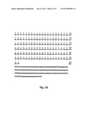 | 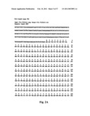 |
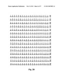 | 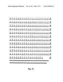 |
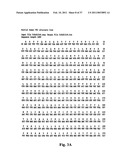 | 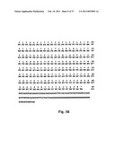 |
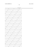 |  |
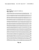 | 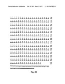 |
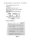 | 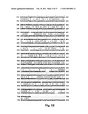 |
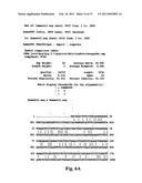 | 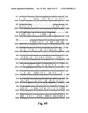 |
 | 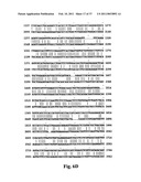 |
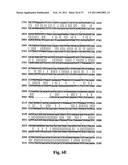 | 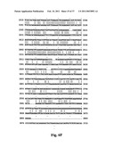 |
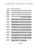 |  |
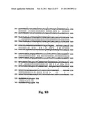 | 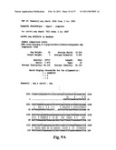 |
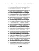 | 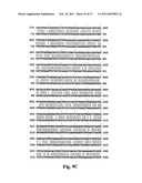 |
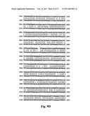 | 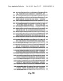 |
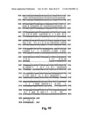 | 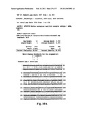 |
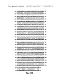 |  |
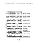 | 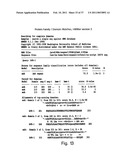 |
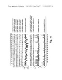 | 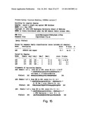 |
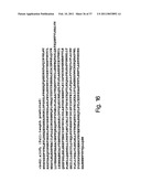 | 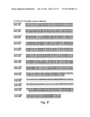 |
 |  |
 |  |
 |  |
 |  |
 |  |
 |  |
 |  |
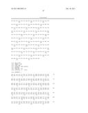 | 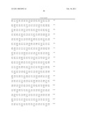 |
 | 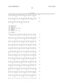 |
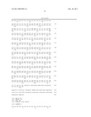 | 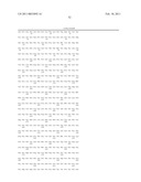 |
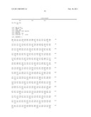 | 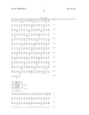 |
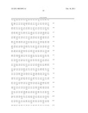 | 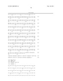 |
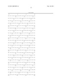 | 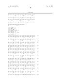 |
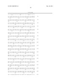 |  |
 |
| Similar patent applications: | |
| Date | Title |
|---|---|
| 2011-12-08 | Novel members of the capsaicin/vanilloid receptor family of proteins and uses thereof |
| 2010-09-23 | Novel hemopoietin receptor protein, nr10 |
| 2009-10-29 | B7-h2 molecules, novel members of the b7 family and uses thereof |
| 2010-11-25 | Novel use of liver x receptor agonists |
| 2011-12-22 | Methods of identifying modulators of the bitter taste receptor tas2r44 |
| New patent applications in this class: | |
| Date | Title |
|---|---|
| 2022-05-05 | Engineered cd47 extracellular domain for bioconjugation |
| 2019-05-16 | High cell density anaerobic fermentation for protein expression |
| 2019-05-16 | Polynucleotide encoding fusion of anchoring motif and dehalogenase, host cell including the polynucleotide, and use thereof |
| 2019-05-16 | Cell culture method, medium, and medium kit |
| 2018-01-25 | Protein expression strains |
| New patent applications from these inventors: | |
| Date | Title |
|---|---|
| 2012-06-28 | Novel members of the capsaicin/vanilloid receptor family of proteins and uses thereof |
| Top Inventors for class "Chemistry: molecular biology and microbiology" | |
| Rank | Inventor's name |
|---|---|
| 1 | Marshall Medoff |
| 2 | Anthony P. Burgard |
| 3 | Mark J. Burk |
| 4 | Robin E. Osterhout |
| 5 | Rangarajan Sampath |