Patent application title: REAGENTS AND METHODS FOR USE IN CANCER DIAGNOSIS, CLASSIFICATION AND THERAPY
Inventors:
Brian Z. Ring (Foster City, CA, US)
Brian Z. Ring (Foster City, CA, US)
Douglas T. Ross (Burlingame, CA, US)
Douglas T. Ross (Burlingame, CA, US)
Robert S. Seitz (Hampton Cove, AL, US)
IPC8 Class: AC40B3004FI
USPC Class:
506 9
Class name: Combinatorial chemistry technology: method, library, apparatus method of screening a library by measuring the ability to specifically bind a target molecule (e.g., antibody-antigen binding, receptor-ligand binding, etc.)
Publication date: 2011-01-06
Patent application number: 20110003709
Claims:
1. A method of assessing a prognosis of a patient having a lung tumor of
the squamous cell carcinoma subclass, the method comprising steps
of:obtaining a tumor sample from a patient with a lung tumor of the
squamous cell carcinoma subclass;contacting the tumor sample with a panel
of antibodies that includes at least two antibodies selected from the
group consisting of antibodies directed to biomarkers CYP4Z1, SLC7A5,
DDH1 and ABCG2; andassessing the patient's likely prognosis based upon a
pattern of binding or lack of binding of the panel to the tumor sample,
wherein across a population of patients with lung tumors of the squamous
cell carcinoma subclass, a lower level of binding of the at least two
antibodies selected from the group consisting of antibodies directed to
biomarkers CYP4Z1, SLC7A5, DDH1 and ABCG2 correlates with a higher
likelihood that the patient will die from lung cancer or have a
recurrence.
2. The method of claim 1, wherein the panel of antibodies includes an antibody directed to biomarker CYP4Z1 and at least one antibody directed to a biomarker selected from the group consisting of SLC7A5, DDH1 and ABCG2.
3. The method of claim 2, wherein the panel of antibodies includes an antibody directed to biomarker CYP4Z1 and an antibody directed to biomarker SLC7A5.
4. The method of claim 2, wherein the panel of antibodies includes an antibody directed to biomarker CYP4Z1 and antibodies directed to biomarkers SLC7A5 and DDH1.
5. The method of claim 2, wherein the panel of antibodies includes an antibody directed to biomarker CYP4Z1 and antibodies directed to biomarkers SLC7A5, DDH1 and ABCG2.
6. The method of claim 1, wherein the step of assessing comprises assessing the likelihood that the patient will die from lung cancer.
7. The method of claim 1, wherein the step of assessing comprises assessing the likelihood that the patient will have a recurrence.
8. A method of assessing prognosis of a patient having a lung tumor of the adenocarcinoma subclass, the method comprising steps of:obtaining a tumor sample from a patient with a lung tumor of the adenocarcinoma subclass;contacting the tumor sample with a panel of antibodies that includes at least two antibodies selected from the group consisting of antibodies directed to biomarkers ERBB2, HTF9C, NDRG1 and SLC7A5; andassessing the patient's likely prognosis based upon a pattern of binding or lack of binding of the panel to the tumor sample, wherein across a population of patients with lung tumors of the adenocarcinoma subclass, a higher level of binding of the at least two antibodies selected from the group consisting of antibodies directed to biomarkers ERBB2, HTF9C, NDRG1 and SLC7A5 correlates with a higher likelihood that the patient will die from lung cancer or have a recurrence.
9. The method of claim 8, wherein the panel of antibodies includes an antibody directed to biomarker ERBB2 and an antibody directed to biomarker HTF9C.
10. The method of claim 8, wherein the panel of antibodies includes an antibody directed to biomarker ERBB2 and antibodies directed to biomarkers HTF9C and NDRG1.
11. The method of claim 8, wherein the panel of antibodies includes an antibody directed to biomarker ERBB2 and antibodies directed to biomarkers HTF9C, NDRG1 and SLC7A5.
12. The method of claim 8, wherein the step of assessing comprises assessing the likelihood that the patient will die from lung cancer.
13. The method of claim 8, wherein the step of assessing comprises assessing the likelihood that the patient will have a recurrence.
14. A kit for assessing prognosis of a patient with a lung tumor of the squamous cell carcinoma subclass comprising a panel of antibodies that includes an antibody directed to biomarker CYP4Z1 and at least one antibody directed to a biomarker selected from the group consisting of SLC7A5, DDH1 and ABCG2.
15. The kit of claim 14, wherein the panel of antibodies includes an antibody directed to biomarker CYP4Z1 and an antibody directed to biomarker SLC7A5.
16. The kit of claim 14, wherein the panel of antibodies includes an antibody directed to biomarker CYP4Z1 and antibodies directed to biomarkers SLC7A5 and DDH1.
17. The kit of claim 14, wherein the panel of antibodies includes an antibody directed to biomarker CYP4Z1 and antibodies directed to biomarkers SLC7A5, DDH1 and ABCG2.
18. A kit for assessing prognosis of a patient with a lung tumor of the adenocarcinoma subclass comprising a panel of antibodies that includes at least two antibodies selected from the group consisting of antibodies directed to biomarkers ERBB2, HTF9C, NDRG1 and SLC7A5.
19. The kit of claim 18, wherein the panel of antibodies includes an antibody directed to biomarker ERBB2 and at least one antibody directed to a biomarker selected from the group consisting of HTF9C, NDRG1 and SLC7A5.
20. The kit of claim 19, wherein the panel of antibodies includes an antibody directed to biomarker ERBB2 and an antibody directed to biomarker HTF9C.
21. The kit of claim 19, wherein the panel of antibodies includes an antibody directed to biomarker ERBB2 and antibodies directed to biomarkers HTF9C and NDRG1.
22. The kit of claim 19, wherein the panel of antibodies includes an antibody directed to biomarker ERBB2 and antibodies directed to biomarkers HTF9C, NDRG1 and SLC7A5.
Description:
PRIORITY INFORMATION
[0001]This application is a divisional application of U.S. Ser. No. 12/013,739 filed Jan. 14, 2008, which is a divisional application of U.S. Ser. No. 11/061,067 filed Feb. 18, 2005, which is a continuation-in-part of U.S. Ser. No. 10/915,059 filed Aug. 10, 2004 which claimed priority to U.S. Ser. No. 60/494,334 filed Aug. 11, 2003 and U.S. Ser. No. 60/570,206 filed May 12, 2004. The entire contents of each of these applications are hereby incorporated by reference.
SEQUENCE LISTING
[0002]In accordance with 37 CFR 1.52(e)(5), a Sequence Listing in the form of a text file (entitled "Sequence Listing.txt," created on Sep. 7, 2010, and 93 kilobytes) is incorporated herein by reference in its entirety.
BACKGROUND OF THE INVENTION
[0003]A major challenge of cancer treatment is the selection of therapeutic regimens that maximize efficacy and minimize toxicity for a given patient. A related challenge lies in the attempt to provide accurate diagnostic, prognostic and predictive information. At present, tumors are generally classified under the tumor-node-metastasis (TNM) system. This system, which uses the size of the tumor, the presence or absence of tumor in regional lymph nodes, and the presence or absence of distant metastases, to assign a stage to the tumor is described in the AJCC Cancer Staging Manual, Lippincott, 5th ed., pp. 171-180 (1997). The assigned stage is used as a basis for selection of appropriate therapy and for prognostic purposes. In addition to the TNM parameters, morphologic appearance is used to further classify tumors into tumor types and thereby aid in selection of appropriate therapy. However, this approach has serious limitations. Tumors with similar histopathologic appearance can exhibit significant variability in terms of clinical course and response to therapy. For example, some tumors are rapidly progressive while others are not. Some tumors respond readily to hormonal therapy or chemotherapy while others are resistant.
[0004]Assays for cell surface markers, e.g., using immunohistochemistry, have provided means for dividing certain tumor types into subclasses. For example, one factor considered in prognosis and treatment decisions for breast cancer is the presence or absence of the estrogen receptor (ER) in tumor samples. ER-positive breast cancers typically respond much more readily to hormonal therapies such as tamoxifen, which acts as an anti-estrogen in breast tissue, than ER-negative tumors. Though useful, these analyses only in part predict the clinical behavior of breast tumors. There is phenotypic diversity present in cancers that current diagnostic tools fail to detect. As a consequence, there is still much controversy over how to stratify patients amongst potential treatments in order to optimize outcome (e.g., for breast cancer see "NIH Consensus Development Conference Statement: Adjuvant Therapy for Breast Cancer, Nov. 1-3, 2000", J. Nat. Cancer Inst. Monographs, 30:5-15, 2001 and Di Leo et al., Int. J. Clin. Oncol. 7:245-253, 2002).
[0005]There clearly exists a need for improved methods and reagents for classifying tumors. Once these methods and reagents are available, clinical studies can be performed that will allow the identification of classes or subclasses of patients having different prognosis and/or responses to therapy. Such prognostic tools will allow more rationally based choices governing the aggressiveness of therapeutic interventions; such predictive tools will also be useful for directing patients into appropriate treatment protocols.
SUMMARY OF THE INVENTION
[0006]The invention encompasses the realization that particular panels of tumor sample binding agents ("interaction partners") can be used to provide new insights into the biology of cancer. Among other things, the present invention provides methods and reagents for classifying tumors and for identifying new tumor classes and subclasses. The invention further provides methods for correlating tumor class or subclass with therapeutic regimen or outcome, for identifying appropriate (new or known) therapies for particular classes or subclasses, and for predicting outcomes based on class or subclass. The invention further provides new therapeutic agents and methods for the treatment of cancer.
[0007]For example, the present invention provides methods for identifying suitable panels of interaction partners (e.g., without limitation antibodies) whose binding is correlated with any of a variety of desirable aspects such as tumor class or subclass, tumor source (e.g., primary tumor versus metastases), likely prognosis, responsiveness to therapy, etc. Specifically, collections of interaction partners are selected and their activity in binding to a variety of different tumors, normal tissues and/or cell lines is assessed. Data are collected for multiple interaction partners to multiple samples and correlations with interesting or desirable features are assessed. As described herein, the detection of individual interaction partners or panels thereof that bind differentially with different tumors provides new methods of use in cancer prognosis and treatment selection. In addition, these interaction partners provide new therapies for treating cancer.
[0008]As described in further detail below, the invention employs methods for grouping interaction partners within a panel into subsets by determining their binding patterns across a collection of samples obtained from different tumor tissues, normal tissues and/or cell lines. The invention also groups the tumor samples into classes or subclasses based on similarities in their binding to a panel of interaction partners. This two-dimensional grouping approach permits the association of particular classes of tumors with particular subsets of interaction partners that, for example, show relatively high binding to tumors within that class. Correlation with clinical information indicates that the tumor classes have clinical significance in terms of prognosis or response to chemotherapy.
BRIEF DESCRIPTION OF APPENDICES A-F
[0009]This patent application refers to material comprising tables and data presented as appendices.
[0010]Appendix A is a table that lists the antibodies included in the breast, lung and/or colon panels that are discussed in the Examples. The table includes the antibody ID, parent gene name, NCBI LocusLink ID, UniGene ID, known aliases for the parent gene, peptides that were used in preparing antibodies, antibody titer and a link to any relevant IHC images of Appendix B. Using the parent gene name, NCBI LocusLink ID, UniGene ID, and/or known aliases for the parent gene, a skilled person can readily obtain the nucleotide (and corresponding amino acid) sequences for each and every one of the parent genes that are listed in Appendix A from a public database (e.g., GenBank, Swiss-Prot or any future derivative of these). The nucleotide and corresponding amino acid sequences for each and every one of the parent genes that are listed in Appendix A are hereby incorporated by reference from these public databases. Antibodies with AGI IDs that begin with s5 or s6 were obtained from commercial sources as indicated. The third and fourth columns of Appendix A indicate whether the antibodies of the breast cancer classification panel were identified by staining with the Russian breast cohort (Example 2) and/or the HH breast cohort (Example 3). The fifth and sixth columns indicate whether the antibodies of the lung cancer classification panel were identified by staining with the Russian lung cohort (Example 4) and/or the HH lung cohort (Example 5). The seventh column indicates the antibodies of the colon cancer classification panel. These were all identified by staining with the Russian colon cohort (Example 6).
[0011]Appendix B includes breast IHC images, colon IHC images and lung IHC images. The IHC images of Appendix B are referenced in the right hand column of Appendix A. An actual copy of Appendix B is not included with this continuation-in-part but can be found in parent case U.S. Ser. No. 10/915,059 filed Aug. 10, 2004 (published as US 2005/0112662 on May 26, 2005), the entire contents of which are hereby incorporated by reference.
[0012]Appendix C is a table that lists exemplary antibodies whose binding patterns have been shown to correlate with tumor prognosis in breast cancer patients. The results are grouped into four categories that have been clinically recognized to be of significance: all patients, ER+patients, ER- patients, and ER+/lymph node metastases negative (ER+/node-) patients. Scoring methods 1-3 use the following schemes: method 1 (0=negative; 1=weak; 2=strong); method 2 (0=negative; 1=weak or strong); and method 3 (0=negative or weak; 1=strong). This table was prepared using samples from the HH breast cohort as described in Example 10.
[0013]Appendix D is a table that lists exemplary antibodies whose binding patterns have been shown to correlate with tumor prognosis in lung cancer patients. The results are grouped into three categories that have been clinically recognized to be of significance: all patients, adenocarcinoma patients, and squamous cell carcinoma patients. Scoring methods 1-3 use the following schemes: method 1 (0=negative; 1=weak; 2=strong); method 2 (0=negative; 1=weak or strong); and method 3 (0=negative or weak; 1=strong).
[0014]Appendix E is a table that lists exemplary antibodies whose binding patterns have been shown to correlate with tumor prognosis in breast cancer patients. The results are grouped into four categories that have been clinically recognized to be of significance: all patients, ER+patients, ER- patients, and ER+/lymph node metastases negative (ER+/node-) patients. Scoring methods 1-3 use the following schemes: method 1 (0=negative; 1=weak; 2=strong); method 2 (0=negative; 1=weak or strong); and method 3 (0=negative or weak; 1=strong). This table was prepared using samples from the HH breast cohort as described in Example 12. Appendix E differs from Appendix C because of further analysis.
[0015]Appendix F is a table that lists exemplary antibodies whose binding patterns have been shown to correlate with tumor prognosis in lung cancer patients. The results are grouped into two categories that have been clinically recognized to be of significance: all patients and adenocarcinoma patients. Scoring methods 1-3 use the following schemes: method 1 (0=negative; 1=weak; 2=strong); method 2 (0=negative; 1=weak or strong); and method 3 (0=negative or weak; 1=strong). This table was prepared using samples from the HH and UAB lung cohorts as described in Example 13. The p-values and hazard ratios that were obtained with each cohort are shown. The antibodies listed have a prognostic p-value of less than 0.2 in both cohorts.
BRIEF DESCRIPTION OF THE DRAWING
[0016]FIG. 1 depicts semi-quantitative immunohistochemistry (IHC) scoring of a 298 breast cancer patient cohort with an inventive breast cancer classification panel. The panel was prepared as described in Example 2--antibodies were used as interaction partners. The patients (rows) were classified using k-means clustering while the antibodies (columns) were organized using hierarchical clustering. Dark gray represents strong positive staining, black represents weak positive staining, while light gray represents the absence of staining and medium gray represents a lack of data. As illustrated in the Figure, nine groups of patients were identified by their consensus pattern of staining with the panel of antibodies.
[0017]FIG. 2 depicts semi-quantitative immunohistochemistry (IHC) scoring of a 387 lung cancer patient cohort with an inventive lung cancer classification panel. The panel was prepared as described in Example 4--antibodies were used as interaction partners. The patients (rows) were classified using k-means clustering while the antibodies (columns) were organized using hierarchical clustering. Dark gray represents strong positive staining, black represents weak positive staining, while light gray represents the absence of staining and medium gray represents a lack of data. As illustrated in the Figure, eight groups of patients were identified by their consensus pattern of staining with the panel of antibodies.
[0018]FIG. 3 depicts semi-quantitative immunohistochemistry (IHC) scoring of a 359 colon cancer patient cohort with an inventive colon cancer classification panel. The panel was prepared as described in Example 6--antibodies were used as interaction partners. The patients (rows) were classified using k-means clustering while the antibodies (columns) were organized using hierarchical clustering. Dark gray represents strong positive staining, black represents weak positive staining, while light gray represents the absence of staining and medium gray represents a lack of data. As illustrated in the Figure, seven groups of patients were identified by their consensus pattern of staining with the panel of antibodies.
[0019]FIG. 4 shows Kaplan-Meier curves that were generated for ER+patients after prognostic classification based on (A) staining with a prognostic panel of antibodies from Appendix C and (B) the Nottingham Prognostic Index (NPI). In each case the patients were placed into one of three prognostic groups, namely "poor" (bottom curve), "moderate" (middle curve) and "good" (top curve).
[0020]FIG. 5 shows Kaplan-Meier curves that were generated for ER+/node- patients after prognostic classification based on (A) staining with a prognostic panel of antibodies from Appendix C and (B) the Nottingham Prognostic Index (NPI). In each case the patients were placed into one of three prognostic groups, namely "poor" (bottom curve), "moderate" (middle curve) and "good" (top curve). Note that under the NPI scheme ER+/node- patients are never categorized as having a "poor" prognosis. For this reason, FIG. 5B only includes curves for patients with a "moderate" or "good" prognosis.
[0021]FIG. 6 shows Kaplan-Meier curves that were generated for ER+/node- patients after prognostic classification based on staining with the exemplary prognostic panel of antibodies from Table 5. In each case the patients were placed into one of three prognostic groups, namely "bad" (bottom curve), "moderate" (middle curve) and "good" (top curve).
[0022]FIG. 7 shows Kaplan-Meier curves that were generated for ER- patients after prognostic classification based on staining with the exemplary prognostic panel of antibodies from Table 6. In each case the patients were placed into one of three prognostic groups, namely "bad" (bottom curve), "moderate" (middle curve) and "good" (top curve).
[0023]FIG. 8 shows Kaplan-Meier curves that were generated for ER- patients after prognostic classification based on staining with the exemplary prognostic panel of antibodies from Table 7. In each case the patients were placed into one of three prognostic groups, namely "bad" (bottom curve), "moderate" (middle curve) and "good" (top curve).
[0024]FIG. 9 shows a dendrogram for the tree panel of Table 8 that may be used for the prognostic classification of ER+/node- patients. If a patient is positive for staining at a given node his or her prognosis decision tree follows the branch marked with a "+". Conversely if a patient is negative for staining at a given node his or her prognosis decision tree follows the branch marked "-". This is done until a terminus is reached.
[0025]FIG. 10 shows Kaplan-Meier curves that were generated for ER+/node- patients after prognostic classification based on staining with the exemplary prognostic panel of antibodies from Table 8. In each case the patients were placed into one of three prognostic groups, namely "bad" (bottom curve), "moderate" (middle curve) and "good" (top curve).
[0026]FIG. 11 shows a dendrogram for the tree panels of Table 9 that may be used for the prognostic classification of ER+ and ER- patients. If a patient is positive for staining at a given node his or her prognosis decision tree follows the branch marked with a "+". Conversely if a patient is negative for staining at a given node his or her prognosis decision tree follows the branch marked "-". This is done until a terminus is reached.
[0027]FIG. 12 shows Kaplan-Meier curves that were generated for combined lung cancer patients (HH cohort) after prognostic classification with the exemplary prognostic panels of antibodies from Tables 10 and 11. In each case the patients were placed into one of three prognostic groups, namely "bad" (bottom curve), "moderate" (middle curve) and "good" (top curve).
[0028]FIG. 13 shows the curves that were obtained when patients in the "moderate" and "bad" groups of FIG. 12 were combined into a single "bad" prognostic group.
[0029]FIG. 14 shows Kaplan-Meier curves that were generated for combined lung cancer patients (UAB cohort) after prognostic classification with the exemplary prognostic panels of antibodies from Tables 10 and 11. In each case the patients were placed into one of three prognostic groups, namely "bad" (bottom curve), "moderate" (middle curve) and "good" (top curve).
[0030]FIG. 15 shows the curves that were obtained when the patients in the "moderate" and "bad" groups of FIG. 14 were combined into a single "bad" prognostic group.
[0031]FIG. 16 shows Kaplan-Meier curves that were generated for adenocarcinoma patients (UAB cohort) after prognostic classification with the exemplary prognostic panels of antibodies from Table 11. In each case the patients were placed into one of three prognostic groups, namely "bad" (bottom curve), "moderate" (middle curve) and "good" (top curve).
[0032]FIG. 17 shows Kaplan-Meier curves that were generated for squamous cell carcinoma patients (UAB cohort) after prognostic classification with the exemplary prognostic panels of antibodies from Table 10. In each case the patients were placed into one of three prognostic groups, namely "bad" (bottom curve), "moderate" (middle curve) and "good" (top curve).
[0033]FIG. 18 shows the relative proportions of different lung cancer morphologies that were identified in seven sub-classes of patients in the HH lung cohort.
DEFINITIONS
[0034]Associated--When an interaction partner and a tumor marker are physically "associated" with one another as described herein, they are linked by direct non-covalent interactions. Desirable non-covalent interactions include those of the type which occur between an immunoglobulin molecule and an antigen for which the immunoglobulin is specific, for example, ionic interactions, hydrogen bonds, van der Waals interactions, hydrophobic interactions, etc. The strength, or affinity of the physical association can be expressed in terms of the dissociation constant (Kd) of the interaction, wherein a smaller Kd represents a greater affinity. The association properties of selected interaction partners and tumor markers can be quantified using methods well known in the art (e.g., see Davies et al., Annual Rev. Biochem. 59:439, 1990).
[0035]Classification panel--A "classification panel" of interaction partners is a set of interaction partners whose collective pattern of binding or lack of binding to a tumor sample, when taken together, is sufficient to classify the tumor sample as a member of a particular class or subclass of tumor, or as not a member of a particular class or subclass of tumor.
[0036]Correlation--"Correlation" refers to the degree to which one variable can be predicted from another variable, e.g., the degree to which a patient's therapeutic response can be predicted from the pattern of binding between a set of interaction partners and a tumor sample taken from that patient. A variety of statistical methods may be used to measure correlation between two variables, e.g., without limitation the student t-test, the Fisher exact test, the Pearson correlation coefficient, the Spearman correlation coefficient, the Chi squared test, etc. Results are traditionally given as a measured correlation coefficient with a p-value that provides a measure of the likelihood that the correlation arose by chance. A correlation with a p-value that is less than 0.05 is generally considered to be statistically significant. Preferred correlations have p-values that are less than 0.01, especially less than 0.001.
[0037]Interaction partner--An "interaction partner" is an entity that physically associates with a tumor marker. For example and without limitation, an interaction partner may be an antibody or a fragment thereof that physically associates with a tumor marker. In general, an interaction partner is said to "associate specifically" with a tumor marker if it associates at a detectable level with the tumor marker and does not associate detectably with unrelated molecular entities (e.g., other tumor markers) under similar conditions. Specific association between a tumor marker and an interaction partner will typically be dependent upon the presence of a particular structural feature of the target tumor marker such as an antigenic determinant or epitope recognized by the interaction partner. Generally, if an interaction partner is specific for epitope A, the presence of a molecular entity (e.g., a protein) containing epitope A or the presence of free unlabeled A in a reaction containing both free labeled A and the interaction partner thereto, will reduce the amount of labeled A that binds to the interaction partner. In general, it is to be understood that specificity need not be absolute. For example, it is well known in the art that antibodies frequently cross-react with other epitopes in addition to the target epitope. Such cross-reactivity may be acceptable depending upon the application for which the interaction partner is to be used. Thus the degree of specificity of an interaction partner will depend on the context in which it is being used. In general, an interaction partner exhibits specificity for a particular tumor marker if it favors binding with that partner above binding with other potential partners, e.g., other tumor markers. One of ordinary skill in the art will be able to select interaction partners having a sufficient degree of specificity to perform appropriately in any given application (e.g., for detection of a target tumor marker, for therapeutic purposes, etc.). It is also to be understood that specificity may be evaluated in the context of additional factors such as the affinity of the interaction partner for the target tumor marker versus the affinity of the interaction partner for other potential partners, e.g., other tumor markers. If an interaction partner exhibits a high affinity for a target tumor marker and low affinity for non-target molecules, the interaction partner will likely be an acceptable reagent for diagnostic purposes even if it lacks specificity. It will be appreciated that once the specificity of an interaction partner is established in one or more contexts, it may be employed in other, preferably similar, contexts without necessarily re-evaluating its specificity.
[0038]Predictive panel--A "predictive panel" of interaction partners is a set of interaction partners whose collective pattern of binding or lack of binding to a tumor sample, when taken together, has sufficient correlation to classify the tumor sample as being from a patient who is likely (or not) to respond to a given therapeutic regimen.
[0039]Prognostic panel--A "prognostic panel" of interaction partners is a set of interaction partners whose collective pattern of binding or lack of binding to a tumor sample, when taken together, has sufficient correlation to classify the tumor sample as being from a patient who is likely to have a given outcome. Generally, "outcome" may include, but is not limited to, the average life expectancy of the patient, the likelihood that the patient will survive for a given amount of time (e.g., 6 months, 1 year, 5 years, etc.), the likelihood of recurrence, the likelihood that the patient will be disease-free for a specified prolonged period of time, or the likelihood that the patient will be cured of the disease.
[0040]Response--The "response" of a tumor or a cancer to therapy may represent any detectable change, for example at the molecular, cellular, organellar, or organismal level. For instance, tumor size, patient life expectancy, recurrence, or the length of time the patient survives, etc., are all responses. Responses can be measured in any of a variety of ways, including for example non-invasive measuring of tumor size (e.g., CT scan, image-enhanced visualization, etc.), invasive measuring of tumor size (e.g., residual tumor resection, etc.), surrogate marker measurement (e.g., serum PSA, etc.), clinical course variance (e.g., measurement of patient quality of life, time to relapse, survival time, etc.).
[0041]Small molecule--A "small molecule" is a non-polymeric molecule. A small molecule can be synthesized in a laboratory (e.g., by combinatorial synthesis) or found in nature (e.g., a natural product). A small molecule is typically characterized in that it contains several carbon-carbon bonds and has a molecular weight of less than about 1500 Da, although this characterization is not intended to be limiting for the purposes of the present invention.
[0042]Tumor markers--"Tumor markers" are molecular entities that are detectable in tumor samples. Generally, tumor markers will be proteins that are present within the tumor sample, e.g., within the cytoplasm or membranes of tumor cells and/or secreted from such cells. According to the present invention, sets of tumor markers that correlate with tumor class or subclass are identified. Thus, subsequent tumor samples may be classified or subclassified based on the presence of these sets of tumor markers.
[0043]Tumor sample--As used herein the term "tumor sample" is taken broadly to include cell or tissue samples removed from a tumor, cells (or their progeny) derived from a tumor that may be located elsewhere in the body (e.g., cells in the bloodstream or at a site of metastasis), or any material derived by processing such a sample. Derived tumor samples may include, for example, nucleic acids or proteins extracted from the sample.
DETAILED DESCRIPTION OF CERTAIN PREFERRED EMBODIMENTS OF THE INVENTION
[0044]As noted above, the present invention provides techniques and reagents for the classification and subclassification, of tumors. Such classification (or subclassification) has many beneficial applications. For example, a particular tumor class or subclass may correlate with prognosis and/or susceptibility to a particular therapeutic regimen. As such, the classification or subclassification may be used as the basis for a prognostic or predictive kit and may also be used as the basis for identifying previously unappreciated therapies. Therapies that are effective against only a particular class or subclass of tumor may have been lost in studies whose data were not stratified by subclass; the present invention allows such data to be re-stratified, and allows additional studies to be performed, so that class- or subclass-specific therapies may be identified and/or implemented. Alternatively or additionally, the present invention allows identification and/or implementation of therapies that are targeted to genes identified as class- or subclass-specific.
Classification and Subclassification of Tumors
[0045]In general, according to the present invention, tumors are classified or subclassified on the basis of tumor markers whose presence is correlated with a particular class or subclass. In preferred embodiments, the tumor markers are detected via their physical association with an interaction partner. Included in the present invention are kits comprising sets of interaction partners that together can be used to identify or classify a particular tumor sample; such sets are generally referred to as "classification panels".
[0046]The present invention provides systems of identifying classification panels. In general, tumor samples are contacted with individual interaction partners, and binding between the interaction partners and their cognate tumor markers is detected. For example, panels of interaction partners that identify a particular class or subclass of tumor within tumor samples of a selected tissue of origin may be defined by contacting individual interaction partners with a variety of different tumor samples (e.g., from different patients) all of the same tissue of origin. Individual interaction partners may be selected for inclusion in the ultimate classification panel based on their binding to only a subset of the tumor samples (e.g., see Examples 1-4). Those of ordinary skill in the art, however, will appreciate that all that is required for a collection of interaction partners to operate effectively as a classification panel is that the combined binding characteristics of member interaction partners together are sufficient to classify a particular tumor sample.
[0047]The inventive process of identifying useful panels of interaction partners as described herein may itself result in the identification of new tumor classes or subclasses. That is, through the process of analyzing interaction partner binding patterns, investigators will often discover new tumor classes or subclasses to which sets of interaction partners bind. Thus, the processes (a) of defining classification panels of interaction partners for given tumor classes or subclasses; and (b) identifying new tumor classes or subclasses may well be experimentally interrelated. In general, the greater the number of tumor samples tested, the greater the likelihood that new classes or subclasses will be defined.
[0048]Often, when identifying sets of interaction partners that can act as a classification (or subclassification) panel, it will be desirable to obtain the largest set of tumor samples possible, and also to collect the largest amount of information possible about the individual samples. For example, the origin of the tumor, the gender of the patient, the age of the patient, the staging of the tumor (e.g., according to the TNM system), any microscopic or submicroscopic characteristics of the tumor that may have been determined, may be recorded. Those of ordinary skill in the art will appreciate that the more information that is known about a tumor sample, the more aspects of that sample are available for correlation with interaction partner binding.
[0049]The systems of the present invention have particular utility in classifying or subclassifying tumor samples that are not otherwise distinguishable from one another. Thus, in some embodiments, it will be desirable to analyze the largest collection of tumor samples that are most similar to one another.
[0050]When obtaining tumor samples for testing according to the present invention, it is generally preferred that the samples represent or reflect characteristics of a population of patients or samples. It may also be useful to handle and process the samples under conditions and according to techniques common to clinical laboratories. Although the present invention is not intended to be limited to the strategies used for processing tumor samples, we note that, in the field of pathology, it is often common to fix samples in buffered formalin, and then to dehydrate them by immersion in increasing concentrations of ethanol followed by xylene. Samples are then embedded into paraffin, which is then molded into a "paraffin block" that is a standard intermediate in histologic processing of tissue samples. The present inventors have found that many useful interaction partners display comparable binding regardless of the method of preparation of tumor samples; those of ordinary skill in the art can readily adjust observations to account for differences in preparation procedure.
[0051]In preferred embodiments of the invention, large numbers of tissue samples are analyzed simultaneously. In some embodiments, a tissue array is prepared. Tissue arrays may be constructed according to a variety of techniques. According to one procedure, a commercially-available mechanical device (e.g., the manual tissue arrayer MTA1 from Beecher Instruments of Sun Prairie, Wis.) is used to remove an 0.6-micron-diameter, full thickness "core" from a paraffin block (the donor block) prepared from each patient, and to insert the core into a separate paraffin block (the recipient block) in a designated location on a grid. In preferred embodiments, cores from as many as about 400 patients can be inserted into a single recipient block; preferably, core-to-core spacing is approximately 1 mm. The resulting tissue array may be processed into thin sections for staining with interaction partners according to standard methods applicable to paraffin embedded material. Depending upon the thickness of the donor blocks, as well as the dimensions of the clinical material, a single tissue array can yield about 50-150 slides containing >75% relevant tumor material for assessment with interaction partners. Construction of two or more parallel tissue arrays of cores from the same cohort of patient samples can provide relevant tumor material from the same set of patients in duplicate or more. Of course, in some cases, additional samples will be present in one array and not another.
[0052]The present inventors have found that it is often desirable to evaluate some aspects of the binding characteristics of potential interaction partners before or while assessing the desirability of including them in an interaction panel. For example, the inventors have found that it is often desirable to perform a titration study in which different concentrations of the interaction partner are contacted with a diverse set of tissue samples derived from a variety of different tissues (e.g., normal and/or tumor) in order to identify a concentration or titer at which differential binding is observed. This titer is referred to herein as a "discriminating titer". Such differential staining may be observed between different tissue samples and/or between different cell types within a given tissue sample.
[0053]In general, any tissue sample may be used for this purpose (e.g., samples obtained from the epididymis, esophagus, gall bladder, kidneys, liver, lungs, lymph nodes, muscles, ovaries, pancreas, parathyroid glands, placenta, prostate, saliva, skin, spleen, stomach, testis, thymus, thyroid, tonsils, uterus, etc.). For such titration studies, greater diversity among samples is often preferred. Without intending to limit the present invention, the inventors observe that useful titers for particular interaction partners can typically be defined in a study of approximately 40-70 different tissue samples from about 20-40 different tissues.
[0054]Binding studies (for titration, for assessment of inclusion in a panel, or during use of a panel) may be performed in any format that allows specific interaction to be detected. Where large numbers of samples are to be handled, it may be desirable to utilize arrayed and/or automated formats. Particularly preferred formats include tissue arrays as discussed above. The staining of large numbers of samples derived from a variety of tumors in a tissue array format allows excellent comparative assessment of differential staining between or among samples under identical conditions. According to the present invention, staining patterns that identify at least about 10% of samples as binding with a particular interaction partner, or at least about 20, 30, 40, 50% or more of samples, are likely to represent "real" differential staining patterns (i.e., real variations in binding with interaction partner and not experimental variations, for example, due to sample processing or day to day variation in staining techniques).
[0055]Any available technique may be used to detect binding between an interaction partner and a tumor sample. One powerful and commonly used technique is to have a detectable label associated (directly or indirectly) with the interaction partner. For example, commonly-used labels that often are associated with antibodies used in binding studies include fluorochromes, enzymes, gold, iodine, etc. Tissue staining by bound interaction partners is then assessed, preferably by a trained pathologist or cytotechnologist. For example, a scoring system may be utilized to designate whether the interaction partner does or does not bind to (e.g., stain) the sample, whether it stains the sample strongly or weakly and/or whether useful information could not be obtained (e.g., because the sample was lost, there was no tumor in the sample or the result was otherwise ambiguous). Those of ordinary skill in the art will recognize that the precise characteristics of the scoring system are not critical to the invention. For example, staining may be assessed qualitatively or quantitatively; more or less subtle gradations of staining may be defined; etc.
[0056]Whatever the format, and whatever the detection strategy, identification of a discriminating titer can simplify binding studies to assess the desirability of including a given interaction partner in a panel. In such studies, the interaction partner is contacted with a plurality of different tumor samples that preferably have at least one common trait (e.g., tissue of origin), and often have multiple common traits (e.g., tissue of origin, stage, microscopic characteristics, etc.). In some cases, it will be desirable to select a group of samples with at least one common trait and at least one different trait, so that a panel of interaction partners is defined that distinguishes the different trait. In other cases, it will be desirable to select a group of samples with no detectable different traits, so that a panel of interaction partners is defined that distinguishes among previously indistinguishable samples. Those of ordinary skill in the art will understand, however, that the present invention often will allow both of these goals to be accomplished even in studies of sample collections with varying degrees of similarity and difference.
[0057]According to the present invention, interaction partners that bind to tumor samples may be characterized by their ability to discriminate among tumor samples. Any of a variety of techniques may be used to identify discriminating interaction partners. To give but one example, the present inventors have found it useful to define a "consensus panel" of tissue samples for tumors of a particular tissue of origin (see Examples 2-6). Those of ordinary skill in the art will again appreciate that the precise parameters used to designate a particular sample as interpretable and reproducible are not critical to the invention. Interaction partners may then be classified based on their ability to discriminate among tissue samples in the consensus panel (see Examples 2-6).
Assessing Prognosis or Therapeutic Regimen
[0058]The present invention further provides systems for identifying panels of interaction partners whose binding correlates with factors beyond tumor class or subclass, such as likelihood of a particular favorable or unfavorable outcome, susceptibility (or lack thereof) to a particular therapeutic regimen, etc.
[0059]As mentioned in the background, current approaches to assigning prognostic probabilities and/or selecting appropriate therapeutic regimens for particular tumors generally utilize the tumor-node-metastasis (TNM) system. This system uses the size of the tumor, the presence or absence of tumor in regional lymph nodes and the presence or absence of distant metastases, to assign a stage to the tumor. The assigned stage is used as a basis for selection of appropriate therapy and for prognostic purposes.
[0060]The present invention provides new methods and systems for evaluating tumor prognosis and/or recommended therapeutic approaches. In particular, the present invention provides systems for identifying panels of interaction partners whose binding correlates with tumor prognosis or therapeutic outcome.
[0061]For example, interaction partners whose binding correlates with prognosis can be identified by evaluating their binding to a collection of tumor samples for which prognosis is known or knowable. That is, the strategies of the invention may be employed either to identify collections of interaction partners whose binding correlates with a known outcome, or may be employed to identify a differential staining pattern that is then correlated with outcome (which outcome may either be known in advance or determined over time).
[0062]In general, it is preferred that inventive binding analyses be performed on human tumor samples. However, it is not necessary that the human tumors grow in a human host. Particularly for studies in which long-term outcome data are of interest (especially prognostic or predictive studies), it can be particularly useful to analyze samples grown in vitro (e.g., cell lines) or, more preferably, in a non-human host (e.g., a rodent, a dog, a sheep, a pig, or other animal). For instance, Example 9 provides a description of an assay in which inventive techniques employing human tumor cells growing in a non-human host are employed to define and/or utilize a panel of interaction partners whose binding to tumor samples correlates with prognosis and/or responsiveness to therapy.
[0063]It will often be desirable, when identifying interaction partners whose binding correlates with prognosis, to collect information about treatment regimens that may have been applied to the tumor whose sample is being assessed, in order to control for effects attributable to tumor therapy. Prognostic panel binding may correlate with outcome independent of treatment (Hayes et al., J. Mamm. Gland Bio. Neo. 6:375, 2001). Many prognostic markers, however, have both prognostic and predictive character (e.g., Her2/Neu status). Many of the individual interaction partners that comprise a prognostic panel may likewise have predictive capability and/or be members of a predictive panel.
[0064]Those of ordinary skill in the art will appreciate that prognostic panels (or individual interaction partners) have greater clinical utility if their binding/lack thereof correlates with positive/negative outcomes that are well separated statistically.
[0065]The inventive strategies may also be applied to the identification of predictive panels of interaction partners (i.e., panels whose binding correlates with susceptibility to a particular therapy). As noted above, some prognostic panels may also have predictive capabilities.
[0066]Interaction partners to be included in predictive panels are identified in binding studies performed on tumor samples that do or do not respond to a particular therapy. As with the prognostic panels, predictive panels may be assembled based on tests of tumor samples whose responsiveness is already known, or on samples whose responsiveness is not known in advance. As with the prognostic studies discussed above, the source of the tumor samples is not essential and can include, for example, tumor cell lines whose responsiveness to particular chemical agents has been determined, tumor samples from animal models in which tumors have been artificially introduced and therapeutic responsiveness has been determined and/or samples from naturally-occurring (human or other animal) tumors for which outcome data (e.g., time of survival, responsiveness to therapy, etc.) are available. Panels of interaction partners whose binding to tumor samples correlates with any prognostic or therapeutic trend can be defined and utilized as described herein.
[0067]Once correlations between interaction partner binding and tumor behavior have been established, the defined prognostic or predictive panels can be used to evaluate and classify tumor samples from patients and can be relied upon, for example to guide selection of an effective therapeutic regimen. As with the tumor classification studies described above, the process of identifying interaction partner panels whose binding correlates with outcome may itself identify particular outcomes not previously appreciated as distinct.
[0068]Those of ordinary skill in the art will appreciate that it is likely that, in at least some instances, tumor class or subclass identity will itself correlate with prognosis or responsiveness. In such circumstances, it is possible that the same set of interaction partners can act as both a classification panel and a prognosis or predictive panel.
Tumor Elements Bound By Interaction Partners
[0069]The inventive strategies for identifying and utilizing interaction partner panels in classifying or analyzing tumor samples do not rely on any assumptions about the identity or characteristics of the tumor components bound by the interaction partners. So long as interaction partner binding within the relevant panel correlates with some feature of interest, the inventive teachings apply. In many if not most, cases, however, it is expected that binding will be with a protein expressed by tumor cells.
[0070]In some preferred embodiments of the invention, interaction partners bind to tumor markers that (a) are differentially expressed in tumor cells; (b) are members of protein families whose activities contribute to relevant biological events (e.g., gene families that have been implicated in cancer such as oncogenes, tumor suppressor genes, and genes that regulate apoptosis; gene families that have been implicated in drug resistance; etc.); (c) are present on or in the plasma membrane of the tumor cells; and/or (d) are the products of degradation of tumor components, which degradation products might be detectable in patient serum.
[0071]In fact, according to the present invention, interaction partners for analysis and use in inventive panels may sometimes be identified by first identifying a tumor-associated protein of interest, and then finding a potential interaction partner that binds with the protein. Binding by this potential interaction partner to tumor samples may then be assessed and utilized as described herein.
[0072]For example, as described in the Examples, the present inventors have successfully assembled classification panels comprised of antibodies that bind to tumor protein antigens. Candidate antigens were identified both through literature reviews of proteins that play a biological role in tumor initiation or progression, or that are known to be differentially expressed in tumors, and through gene expression studies that identified additional differentially expressed proteins.
[0073]Work by the present inventors, as well as by others, has already demonstrated that studies of gene expression patterns in large tumor cohorts can identify novel tumor classes (see, for example, Perou et al., Nature 406:747, 2000; Sorlie et al., Proc Natl Acad. Sci. USA 98:10869, 2001; van't Veer et al., Nature 415:530, 2002; West et al., Proc Natl. Acad. Sci. USA 98:11462, 2001; Hedenfalk et al., N. Engl. J. Med. 344:539, 2001; Gruvberger et al., Cancer Res. 61:5979, 2001; MacDonald et al., Nature Genet. 29:143, 2001; Pomeroy et al., Nature 415:436, 2002; Jazaeri et al., J. Natl Cancer Inst 94:990, 2002; Welsh et al., Proc. Natl. Acad. Sci. USA 98:1176, 2001; Wang et al., Gene 229:101, 1999; Beer et al., Nature Med. 8:816, 2002; Garber et al., Proc Natl Acad Sci USA 98:13784, 2001; Bhattacharjee et al., Proc Natl Acad Sci USA 98:13790, 2001; Zou et al., Oncogene 21:4855, 2002; Lin et al., Oncogene 21:4120, 2002; Alon et al., Proc Natl Acad Sci USA 96:6745, 1999; Takahashi et al., Proc Natl Acad Sci USA 98:9754, 2001; Singh et al., Cancer Cell 1:203, 2002; LaTulippe et al., Cancer Res. 62:4499, 2002; Welsh et al., Cancer Res. 61:5974, 2001; Dhanasekaran et al., Nature 412:822, 2001; Hippo et al., Cancer Res. 62:233, 2002; Yeoh et al., Cancer Cell 1:133, 2002; Hofmann et al., Lancet 359:481, 2002; Ferrando et al., Cancer Cell 1:75, 2002; Shipp et al., Nature Med 8:68, 2002; Rosenwald et al., N. Engl. J. Med. 346:1937, 2002; and Alizadeh et al., Nature 403:503, 2000, each of which is incorporated herein by reference).
[0074]The gene sets described in these publications are promising candidates for genes that are likely to encode tumor markers whose interaction partners are useful in tumor classification and subclassification according to the present invention. Of particular interest are gene sets differentially expressed in solid tumors.
[0075]Furthermore, in general, given that differentially expressed genes are likely to be responsible for the different phenotypic characteristics of tumors, the present invention recognizes that such genes will often encode tumor markers for which a useful interaction partner, that discriminates among tumor classes or subclasses, can likely be prepared. A differentially expressed gene is a gene whose transcript abundance varies between different samples, e.g., between different tumor samples, between normal versus tumor samples, etc. In general, the amount by which the expression varies and the number of samples in which the expression varies by that amount will depend upon the number of samples and the particular characteristics of the samples. One skilled in the art will be able to determine, based on knowledge of the samples, what constitutes a significant degree of differential expression. Such genes can be identified by any of a variety of techniques including, for instance, in situ hybridization, Northern blot, nucleic acid amplification techniques (e.g., PCR, quantitative PCR, the ligase chain reaction, etc.), and, most commonly, microarray analysis.
[0076]Furthermore, those of ordinary skill in the art will readily appreciate, reading the present disclosure, that the inventive processes described herein of identifying and/or using sets of interaction partners whose binding (or lack thereof) correlates with an interesting tumor feature (e.g., tumor type or subtype, patient outcome, responsiveness of tumor or patient to therapy, etc.) inherently identifies both interaction partners of interest and the tumor markers to which they bind. Thus, one important aspect of the present invention is the identification of tumor markers whose ability (or lack thereof) to associate with an interaction partner correlates with a tumor characteristic of interest. Such tumor markers are useful as targets for identification of new therapeutic reagents, as well as of additional interaction partners useful in the practice of the present invention. Thus, it is to be understood that discussions of interaction partners presented herein are typically not limited to a particular interaction partner compound or entity, but may be generalized to include any compound or entity that binds to the relevant tumor marker(s) with requisite specificity and affinity.
Preparation of Interaction Partners
[0077]In general, interaction partners are entities that physically associate with selected tumor markers. Thus, any entity that binds detectably to a tumor marker may be utilized as an interaction partner in accordance with the present invention, so long as it binds with an appropriate combination of affinity and specificity.
[0078]Particularly preferred interaction partners are antibodies, or fragments (e.g., F(ab) fragments, F(ab')2 fragments, Fv fragments, or sFv fragments, etc.; see, for example, Inbar et al., Proc. Nat. Acad. Sci. USA 69:2659, 1972; Hochman et al., Biochem. 15:2706, 1976; and Ehrlich et al., Biochem. 19:4091, 1980; Huston et al., Proc. Nat. Acad. Sci. USA 85:5879, 1998; U.S. Pat. Nos. 5,091,513 and 5,132,405 to Huston et al.; and U.S. Pat. No. 4,946,778 to Ladner et al., each of which is incorporated herein by reference). In certain embodiments, interaction partners may be selected from libraries of mutant antibodies (or fragments thereof). For example, collections of antibodies that each include different point mutations may be screened for their association with a tumor marker of interest. Yet further, chimeric antibodies may be used as interaction partners, e.g., "humanized" or "veneered" antibodies as described in greater detail below.
[0079]It is to be understood that the present invention is not limited to using antibodies or antibody fragments as interaction partners of inventive tumor markers. In particular, the present invention also encompasses the use of synthetic interaction partners that mimic the functions of antibodies. Several approaches to designing and/or identifying antibody mimics have been proposed and demonstrated (e.g., see the reviews by Hsieh-Wilson et al., Acc. Chem. Res. 29:164, 2000 and Peczuh and Hamilton, Chem. Rev. 100:2479, 2000). For example, small molecules that bind protein surfaces in a fashion similar to that of natural proteins have been identified by screening synthetic libraries of small molecules or natural product isolates (e.g., see Gallop et al., J. Med. Chem. 37:1233, 1994; Gordon et al., J. Med. Chem. 37:1385, 1994; DeWitt et al., Proc. Natl. Acad. Sci. U.S.A. 90:6909, 1993; Bunin et al., Proc. Natl. Acad. Sci. U.S.A. 91:4708, 1994; Virgilio and Ellman, J. Am. Chem. Soc. 116:11580, 1994; Wang et al., J. Med. Chem. 38:2995, 1995; and Kick and Ellman, J. Med. Chem. 38:1427, 1995). Similarly, combinatorial approaches have been successfully applied to screen libraries of peptides and polypeptides for their ability to bind a range of proteins (e.g., see Cull et al., Proc. Natl. Acad. Sci. U.S.A. 89:1865, 1992; Mattheakis et al., Proc. Natl. Acad. Sci. U.S.A. 91:9022, 1994; Scott and Smith, Science 249:386, 1990; Devlin et al., Science 249:404, 1990; Corey et al., Gene 128:129, 1993; Bray et al., Tetrahedron Lett. 31:5811, 1990; Fodor et al., Science 251:767, 1991; Houghten et al., Nature 354:84, 1991; Lam et al., Nature 354:82, 1991; Blake and Litzi-Davis, Bioconjugate Chem. 3:510, 1992; Needels et al., Proc. Natl. Acad. Sci. U.S.A. 90:10700, 1993; and Ohlmeyer et al., Proc. Natl. Acad. Sci. U.S.A. 90:10922, 1993). Similar approaches have also been used to study carbohydrate-protein interactions (e.g., see Oldenburg et al., Proc. Natl. Acad. Sci. U.S.A. 89:5393, 1992) and polynucleotide-protein interactions (e.g., see Ellington and Szostak, Nature 346:818, 1990 and Tuerk and Gold, Science 249:505, 1990). These approaches have also been extended to study interactions between proteins and unnatural biopolymers such as oligocarbamates, oligoureas, oligosulfones, etc. (e.g., see Zuckermann et al., J. Am. Chem. Soc. 114:10646, 1992; Simon et al., Proc. Natl. Acad. Sci. U.S.A. 89:9367, 1992; Zuckermann et al., J. Med. Chem. 37:2678, 1994; Burgess et al., Angew. Chem., Int. Ed. Engl. 34:907, 1995; and Cho et al., Science 261:1303, 1993). Yet further, alternative protein scaffolds that are loosely based around the basic fold of antibody molecules have been suggested and may be used in the preparation of inventive interaction partners (e.g., see Ku and Schultz Proc. Natl. Acad. Sci. U.S.A. 92:6552, 1995). Antibody mimics comprising a scaffold of a small molecule such as 3-aminomethylbenzoic acid and a substituent consisting of a single peptide loop have also been constructed. The peptide loop performs the binding function in these mimics (e.g., see Smythe et al., J. Am. Chem. Soc. 116:2725, 1994). A synthetic antibody mimic comprising multiple peptide loops built around a calixarene unit has also been described (e.g., see U.S. Pat. No. 5,770,380 to Hamilton et al.).
Detecting Association of Interaction Partners and Tumor Markers
[0080]Any available strategy or system may be utilized to detect association between an interaction partner and its cognate tumor marker. In certain embodiments, association can be detected by adding a detectable label to the interaction partner. In other embodiments, association can be detected by using a labeled secondary interaction partner that associates specifically with the primary interaction partner, e.g., as is well known in the art of antigen/antibody detection. The detectable label may be directly detectable or indirectly detectable, e.g., through combined action with one or more additional members of a signal producing system. Examples of directly detectable labels include radioactive, paramagnetic, fluorescent, light scattering, absorptive and colorimetric labels. Examples of indirectly detectable include chemiluminescent labels, e.g., enzymes that are capable of converting a substrate to a chromogenic product such as alkaline phosphatase, horseradish peroxidase and the like.
[0081]Once a labeled interaction partner has bound a tumor marker, the complex may be visualized or detected in a variety of ways, with the particular manner of detection being chosen based on the particular detectable label, where representative detection means include, e.g., scintillation counting, autoradiography, measurement of paramagnetism, fluorescence measurement, light absorption measurement, measurement of light scattering and the like.
[0082]In general, association between an interaction partner and its cognate tumor marker may be assayed by contacting the interaction partner with a tumor sample that includes the marker. Depending upon the nature of the sample, appropriate methods include, but are not limited to, immunohistochemistry (IHC), radioimmunoassay, ELISA, immunoblotting and fluorescence activates cell sorting (FACS). In the case where the polypeptide is to be detected in a tissue sample, e.g., a biopsy sample, IHC is a particularly appropriate detection method. Techniques for obtaining tissue and cell samples and performing IHC and FACS are well known in the art.
[0083]The inventive strategies for classifying and/or subclassifying tumor samples may be applied to samples of any type and of any tissue of origin. In certain preferred embodiments of the invention, the strategies are applied to solid tumors. Historically, researchers have encountered difficulty in defining solid tumor subtypes, given the challenges associated with defining their molecular characteristics. As demonstrated in the Examples, the present invention is particularly beneficial in this area. Particularly preferred solid tumors include, for example, breast, lung, colon, and ovarian tumors. The invention also encompasses the recognition that tumor markers that are secreted from the cells in which they are produced may be present in serum, enabling their detection through a blood test rather than requiring a biopsy specimen. An interaction partner that binds to such tumor markers represents a particularly preferred embodiment of the invention.
[0084]In general, the results of such an assay can be presented in any of a variety of formats. The results can be presented in a qualitative fashion. For example, the test report may indicate only whether or not a particular tumor marker was detected, perhaps also with an indication of the limits of detection. Additionally the test report may indicate the subcellular location of binding, e.g., nuclear versus cytoplasmic and/or the relative levels of binding in these different subcellular locations. The results may be presented in a semi-quantitative fashion. For example, various ranges may be defined and the ranges may be assigned a score (e.g., 0 to 5) that provides a certain degree of quantitative information. Such a score may reflect various factors, e.g., the number of cells in which the tumor marker is detected, the intensity of the signal (which may indicate the level of expression of the tumor marker), etc. The results may be presented in a quantitative fashion, e.g., as a percentage of cells in which the tumor marker is detected, as a concentration, etc. As will be appreciated by one of ordinary skill in the art, the type of output provided by a test will vary depending upon the technical limitations of the test and the biological significance associated with detection of the tumor marker. For example, in the case of certain tumor markers a purely qualitative output (e.g., whether or not the tumor marker is detected at a certain detection level) provides significant information. In other cases a more quantitative output (e.g., a ratio of the level of expression of the tumor marker in two samples) is necessary.
Identification of Novel Therapies
[0085]Predictive panels of interaction agents are useful according to the present invention not only to classify tumor samples obtained from cancer sufferers with respect to their likely responsiveness to known therapies, but also to identify potential new therapies or therapeutic agents that could be useful in the treatment of cancer.
[0086]For example, as noted above, the process of identifying or using inventive panels according to the present invention simultaneously identifies and/or characterizes tumor markers in or on the tumor cells that correlate with one or more selected tumor characteristics (e.g., tumor type or subtype, patient prognosis, and/or responsiveness of tumor or patient to therapy). Such tumor markers are attractive candidates for identification of new therapeutic agents (e.g., via screens to detect compounds or entities that bind to the tumor markers, preferably with at least a specified affinity and/or specificity, and/or via screens to detect compounds or entities that modulate (i.e., increase or decrease) expression, localization, modification, or activity of the tumor markers. In many instances, interaction partners themselves may prove to be useful therapeutics.
[0087]Thus the present invention provides interaction partners that are themselves useful therapeutic agents. For example, binding by an interaction partner, or a collection of interaction partners, to a cancer cell, might inhibit growth of that cell. Alternatively or additionally, interaction partners defined or prepared according to the present invention could be used to deliver a therapeutic agent to a cancer cell. In particular, interaction partners may be coupled to one or more therapeutic agents. Suitable agents in this regard include radionuclides and drugs. Preferred radionuclides include 90Y, 123I, 125I, 131I, 186Re, 211At and 212Bi. Preferred drugs include chlorambucil, ifosphamide, meclorethamine, cyclophosphamide, carboplatin, cisplatin, procarbazine, decarbazine, carmustine, cytarabine, hydroxyurea, mercaptopurine, methotrexate, thioguanine, 5-fluorouracil, actinomycin D, bleomycin, daunorubicin, doxorubicin, etoposide, vinblastine, vincristine, L-asparginase, adrenocorticosteroids, canciclovir triphosphate, adenine arabinonucleoside triphosphate, 5-aziridinyl-4-hydroxylamino-2-nitrobenzamide, acrolein, phosphoramide mustard, 6-methylpurine, etoposide, methotrexate, benzoic acid mustard, cyanide and nitrogen mustard.
[0088]According to such embodiments, the therapeutic agent may be coupled with an interaction partner by direct or indirect covalent or non-covalent interactions. A direct interaction between a therapeutic agent and an interaction partner is possible when each possesses a substituent capable of reacting with the other. For example, a nucleophilic group, such as an amino or sulfhydryl group, on one may be capable of reacting with a carbonyl-containing group, such as an anhydride or an acid halide, or with an alkyl group containing a good leaving group (e.g., a halide) on the other. Indirect interactions might involve a linker group that is itself associated with both the therapeutic agent and the interaction partner. A linker group can function as a spacer to distance an interaction partner from an agent in order to avoid interference with association capabilities. A linker group can also serve to increase the chemical reactivity of a substituent on an agent or an interaction partner and thus increase the coupling efficiency. An increase in chemical reactivity may also facilitate the use of agents, or functional groups on agents, which otherwise would not be possible.
[0089]It will be evident to those skilled in the art that a variety of bifunctional or polyfunctional reagents, both homo- and hetero-functional (such as those described in the catalog of the Pierce Chemical Co., Rockford, Ill.), may be employed as the linker group. Coupling may be effected, for example, through amino groups, carboxyl groups, sulfydryl groups or oxidized carbohydrate residues. There are numerous references describing such methodology, e.g., U.S. Pat. No. 4,671,958, to Rodwell et al. It will further be appreciated that a therapeutic agent and an interaction partner may be coupled via non-covalent interactions, e.g., ligand/receptor type interactions. Any ligand/receptor pair with a sufficient stability and specificity to operate in the context of the invention may be employed to couple a therapeutic agent and an interaction partner. To give but an example, a therapeutic agent may be covalently linked with biotin and an interaction partner with avidin. The strong non-covalent binding of biotin to avidin would then allow for coupling of the therapeutic agent and the interaction partner. Typical ligand/receptor pairs include protein/co-factor and enzyme/substrate pairs. Besides the commonly used biotin/avidin pair, these include without limitation, biotin/streptavidin, digoxigenin/anti-digoxigenin, FK506/FK506-binding protein (FKBP), rapamycin/FKBP, cyclophilin/cyclosporin and glutathione/glutathione transferase pairs. Other suitable ligand/receptor pairs would be recognized by those skilled in the art, e.g., monoclonal antibodies paired with a epitope tag such as, without limitation, glutathione-S-transferase (GST), c-myc, FLAG® and maltose binding protein (MBP) and further those described in Kessler pp. 105-152 of Advances in Mutagenesis" Ed. by Kessler, Springer-Verlag, 1990; "Affinity Chromatography: Methods and Protocols (Methods in Molecular Biology)" Ed. by Pascal Baillon, Humana Press, 2000; and "Immobilized Affinity Ligand Techniques" by Hermanson et al., Academic Press, 1992.
[0090]Where a therapeutic agent is more potent when free from the interaction partner, it may be desirable to use a linker group which is cleavable during or upon internalization into a cell. A number of different cleavable linker groups have been described. The mechanisms for the intracellular release of an agent from these linker groups include cleavage by reduction of a disulfide bond (e.g., U.S. Pat. No. 4,489,710 to Spitler), by irradiation of a photolabile bond (e.g., U.S. Pat. No. 4,625,014 to Senter et al.), by hydrolysis of derivatized amino acid side chains (e.g., U.S. Pat. No. 4,638,045 to Kohn et al.), by serum complement-mediated hydrolysis (e.g., U.S. Pat. No. 4,671,958 to Rodwell et al.) and by acid-catalyzed hydrolysis (e.g., U.S. Pat. No. 4,569,789 to Blattler et al.).
[0091]In certain embodiments, it may be desirable to couple more than one therapeutic agent to an interaction partner. In one embodiment, multiple molecules of an agent are coupled to one interaction partner molecule. In another embodiment, more than one type of therapeutic agent may be coupled to one interaction partner molecule. Regardless of the particular embodiment, preparations with more than one agent may be prepared in a variety of ways. For example, more than one agent may be coupled directly to an interaction partner molecule, or linkers that provide multiple sites for attachment can be used.
[0092]Alternatively, a carrier can be used. A carrier may bear the agents in a variety of ways, including covalent bonding either directly or via a linker group. Suitable carriers include proteins such as albumins (e.g., U.S. Pat. No. 4,507,234 to Kato et al.), peptides, and polysaccharides such as aminodextran (e.g., U.S. Pat. No. 4,699,784 to Shih et al.). A carrier may also bear an agent by non-covalent bonding or by encapsulation, such as within a liposome vesicle (e.g., U.S. Pat. Nos. 4,429,008 to Martin et al. and 4,873,088 to Mayhew et al.). Carriers specific for radionuclide agents include radiohalogenated small molecules and chelating compounds. For example, U.S. Pat. No. 4,735,792 to Srivastava discloses representative radiohalogenated small molecules and their synthesis. A radionuclide chelate may be formed from chelating compounds that include those containing nitrogen and sulfur atoms as the donor atoms for binding the metal, or metal oxide, radionuclide. For example, U.S. Pat. No. 4,673,562 to Davison et al. discloses representative chelating compounds and their synthesis.
[0093]When interaction partners are themselves therapeutics, it will be understood that, in many cases, any interaction partner that binds with the same tumor marker may be so used.
[0094]In one preferred embodiment of the invention, the therapeutic agents (whether interaction partners or otherwise) are antibodies. As is well known in the art, when using an antibody or fragment thereof for therapeutic purposes it may prove advantageous to use a "humanized" or "veneered" version of an antibody of interest to reduce any potential immunogenic reaction. In general, "humanized" or "veneered" antibody molecules and fragments thereof minimize unwanted immunological responses toward antihuman antibody molecules which can limit the duration and effectiveness of therapeutic applications of those moieties in human recipients.
[0095]A number of "humanized" antibody molecules comprising an antigen binding portion derived from a non-human immunoglobulin have been described in the art, including chimeric antibodies having rodent variable regions and their associated complementarity-determining regions (CDRs) fused to human constant domains (e.g., see Winter et al., Nature 349:293, 1991; Lobuglio et al., Proc. Nat. Acad. Sci. USA 86:4220, 1989; Shaw et al., J. Immunol. 138:4534, 1987; and Brown et al., Cancer Res. 47:3577, 1987), rodent CDRs grafted into a human supporting framework region (FR) prior to fusion with an appropriate human antibody constant domain (e.g., see Riechmann et al., Nature 332:323, 1988; Verhoeyen et al., Science 239:1534, 1988; and Jones et al. Nature 321:522, 1986) and rodent CDRs supported by recombinantly veneered rodent FRs (e.g., see European Patent Publication No. 519,596, published Dec. 23, 1992). It is to be understood that the invention also encompasses "fully human" antibodies produced using the XenoMouse® technology (AbGenix Corp., Fremont, Calif.) according to the techniques described in U.S. Pat. No. 6,075,181.
[0096]Yet further, so-called "veneered" antibodies may be used that include "veneered FRs". The process of veneering involves selectively replacing FR residues from, e.g., a murine heavy or light chain variable region, with human FR residues in order to provide a xenogeneic molecule comprising an antigen binding portion which retains substantially all of the native FR polypeptide folding structure. Veneering techniques are based on the understanding that the antigen binding characteristics of an antigen binding portion are determined primarily by the structure and relative disposition of the heavy and light chain CDR sets within the antigen-association surface (e.g., see Davies et al., Ann. Rev. Biochem. 59:439, 1990). Thus, antigen association specificity can be preserved in a humanized antibody only wherein the CDR structures, their interaction with each other and their interaction with the rest of the variable region domains are carefully maintained. By using veneering techniques, exterior (e.g., solvent-accessible) FR residues which are readily encountered by the immune system are selectively replaced with human residues to provide a hybrid molecule that comprises either a weakly immunogenic, or substantially non-immunogenic veneered surface.
[0097]Preferably, interaction partners suitable for use as therapeutics (or therapeutic agent carriers) exhibit high specificity for the target tumor marker and low background binding to other tumor markers. In certain embodiments, monoclonal antibodies are preferred for therapeutic purposes.
[0098]Tumor markers that are expressed on the cell surface represent preferred targets for the development of therapeutic agents, particularly therapeutic antibodies. For example, cell surface proteins can be tentatively identified using sequence analysis based on the presence of a predicted transmembrane domain. Their presence on the cell surface can ultimately be confirmed using IHC.
Kits
[0099]Useful sets or panels of interaction partners according to the present invention may be prepared and packaged together in kits for use in classifying, diagnosing, or otherwise characterizing tumor samples, or for inhibiting tumor cell growth or otherwise treating cancer.
[0100]Any available technique may be utilized in the preparation of individual interaction partners for inclusion in kits. For example, protein or polypeptide interaction partners may be produced by cells (e.g., recombinantly or otherwise), may be chemically synthesized, or may be otherwise generated in vitro (e.g., via in vitro transcription and/or translation). Non-protein or polypeptide interaction partners (e.g., small molecules, etc.) may be synthesized, may be isolated from within or around cells that produce them, or may be otherwise generated.
[0101]When antibodies are used as interaction partners, these may be prepared by any of a variety of techniques known to those of ordinary skill in the art (e.g., see Harlow and Lane, Antibodies: A Laboratory Manual, Cold Spring Harbor Laboratory, 1988). For example, antibodies can be produced by cell culture techniques, including the generation of monoclonal antibodies, or via transfection of antibody genes into suitable bacterial or mammalian cell hosts, in order to allow for the production of recombinant antibodies. In one technique, an "immunogen" comprising an antigenic portion of a tumor marker of interest (or the tumor marker itself) is initially injected into any of a wide variety of mammals (e.g., mice, rats, rabbits, sheep or goats). In this step, a tumor marker (or an antigenic portion thereof) may serve as the immunogen without modification. Alternatively, particularly for relatively short tumor markers, a superior immune response may be elicited if the tumor marker is joined to a carrier protein, such as bovine serum albumin or keyhole limpet hemocyanin (KLH). The immunogen is injected into the animal host, preferably according to a predetermined schedule incorporating one or more booster immunizations and the animals are bled periodically. Polyclonal antibodies specific for the tumor marker may then be purified from such antisera by, for example, affinity chromatography using the tumor marker (or an antigenic portion thereof) coupled to a suitable solid support. An exemplary method is described in Example 7.
[0102]If desired for diagnostic or therapeutic kits, monoclonal antibodies specific for a tumor marker of interest may be prepared, for example, using the technique of Kohler and Milstein, Eur. J. Immunol. 6:511, 1976 and improvements thereto. Briefly, these methods involve the preparation of immortal cell lines capable of producing antibodies having the desired specificity (i.e., reactivity with the tumor marker of interest). Such cell lines may be produced, for example, from spleen cells obtained from an animal immunized as described above. The spleen cells are then immortalized by, for example, fusion with a myeloma cell fusion partner, preferably one that is syngeneic with the immunized animal. A variety of fusion techniques may be employed. For example, the spleen cells and myeloma cells may be combined with a nonionic detergent for a few minutes and then plated at low density on a selective medium that supports the growth of hybrid cells, but not myeloma cells. A preferred selection technique uses HAT (hypoxanthine, aminopterin, thymidine) selection. After a sufficient time, usually about 1 to 2 weeks, colonies of hybrids are observed. Single colonies are selected and their culture supernatants tested for binding activity against the tumor marker. Hybridomas having high reactivity and specificity are preferred.
[0103]Monoclonal antibodies may be isolated from the supernatants of growing hybridoma colonies. In addition, various techniques may be employed to enhance the yield, such as injection of the hybridoma cell line into the peritoneal cavity of a suitable vertebrate host, such as a mouse. Monoclonal antibodies may then be harvested from the ascites fluid or the blood. Contaminants may be removed from the antibodies by conventional techniques, such as chromatography, gel filtration, precipitation and extraction. The tumor marker of interest may be used in the purification process in, for example, an affinity chromatography step.
[0104]In addition to inventive interaction partners, preferred kits for use in accordance with the present invention may include, a reference sample, instructions for processing samples, performing the test, instructions for interpreting the results, buffers and/or other reagents necessary for performing the test. In certain embodiments the kit can comprise a panel of antibodies.
Pharmaceutical Compositions
[0105]As mentioned above, the present invention provides new therapies and methods for identifying these. In certain embodiments, an interaction partner may be a useful therapeutic agent. Alternatively or additionally, interaction partners defined or prepared according to the present invention bind to tumor markers that serve as targets for therapeutic agents. Also, inventive interaction partners may be used to deliver a therapeutic agent to a cancer cell. For example, interaction partners provided in accordance with the present invention may be coupled to one or more therapeutic agents.
[0106]In addition, as mentioned above, to the extent that a particular predictive panel correlates with responsiveness to a particular therapy because it detects changes that reflect inhibition (or inhibitability) of cancer cell growth, that panel could be used to evaluate therapeutic candidates (e.g., small molecule drugs) for their ability to induce the same or similar changes in different cells. In particular, binding by the panel could be assessed on cancer cells before and after exposure to candidate therapeutics; those candidates that induce expression of the tumor markers to which the panel binds are then identified.
[0107]The invention includes pharmaceutical compositions comprising these inventive therapeutic agents. In general, a pharmaceutical composition will include a therapeutic agent in addition to one or more inactive agents such as a sterile, biocompatible carrier including, but not limited to, sterile water, saline, buffered saline, or dextrose solution. The pharmaceutical compositions may be administered either alone or in combination with other therapeutic agents including other chemotherapeutic agents, hormones, vaccines and/or radiation therapy. By "in combination with", it is not intended to imply that the agents must be administered at the same time or formulated for delivery together, although these methods of delivery are within the scope of the invention. In general, each agent will be administered at a dose and on a time schedule determined for that agent. Additionally, the invention encompasses the delivery of the inventive pharmaceutical compositions in combination with agents that may improve their bioavailability, reduce or modify their metabolism, inhibit their excretion, or modify their distribution within the body. The invention encompasses treating cancer by administering the pharmaceutical compositions of the invention. Although the pharmaceutical compositions of the present invention can be used for treatment of any subject (e.g., any animal) in need thereof, they are most preferably used in the treatment of humans.
[0108]The pharmaceutical compositions of this invention can be administered to humans and other animals by a variety of routes including oral, intravenous, intramuscular, intra-arterial, subcutaneous, intraventricular, transdermal, rectal, intravaginal, intraperitoneal, topical (as by powders, ointments, or drops), bucal, or as an oral or nasal spray or aerosol. In general the most appropriate route of administration will depend upon a variety of factors including the nature of the agent (e.g., its stability in the environment of the gastrointestinal tract), the condition of the patient (e.g., whether the patient is able to tolerate oral administration), etc. At present the intravenous route is most commonly used to deliver therapeutic antibodies. However, the invention encompasses the delivery of the inventive pharmaceutical composition by any appropriate route taking into consideration likely advances in the sciences of drug delivery.
[0109]General considerations in the formulation and manufacture of pharmaceutical agents may be found, for example, in Remington's Pharmaceutical Sciences, 19th ed., Mack Publishing Co., Easton, Pa., 1995.
[0110]According to the methods of treatment of the present invention, cancer is treated or prevented in a patient such as a human or other mammal by administering to the patient a therapeutically effective amount of a therapeutic agent of the invention, in such amounts and for such time as is necessary to achieve the desired result. By a "therapeutically effective amount" of a therapeutic agent of the invention is meant a sufficient amount of the therapeutic agent to treat (e.g., to ameliorate the symptoms of, delay progression of, prevent recurrence of, cure, etc.) cancer at a reasonable benefit/risk ratio, which involves a balancing of the efficacy and toxicity of the therapeutic agent. In general, therapeutic efficacy and toxicity may be determined by standard pharmacological procedures in cell cultures or with experimental animals, e.g., by calculating the ED50 (the dose that is therapeutically effective in 50% of the treated subjects) and the LD50 (the dose that is lethal to 50% of treated subjects). The ED50/LD50 represents the therapeutic index of the agent. Although in general therapeutic agents having a large therapeutic index are preferred, as is well known in the art, a smaller therapeutic index may be acceptable in the case of a serious disease, particularly in the absence of alternative therapeutic options. Ultimate selection of an appropriate range of doses for administration to humans is determined in the course of clinical trials.
[0111]It will be understood that the total daily usage of the therapeutic agents and compositions of the present invention for any given patient will be decided by the attending physician within the scope of sound medical judgment. The specific therapeutically effective dose level for any particular patient will depend upon a variety of factors including the disorder being treated and the severity of the disorder; the activity of the specific therapeutic agent employed; the specific composition employed; the age, body weight, general health, sex and diet of the patient; the time of administration, route of administration and rate of excretion of the specific therapeutic agent employed; the duration of the treatment; drugs used in combination or coincidental with the specific therapeutic agent employed; and like factors well known in the medical arts.
[0112]The total daily dose of the therapeutic agents of this invention administered to a human or other mammal in single or in divided doses can be in amounts, for example, from 0.01 to 50 mg/kg body weight or more usually from 0.1 to 25 mg/kg body weight. Single dose compositions may contain such amounts or submultiples thereof to make up the daily dose. In general, treatment regimens according to the present invention comprise administration to a patient in need of such treatment from about 0.1 μg to about 2000 mg of the therapeutic agent(s) of the invention per day in single or multiple doses.
EXEMPLIFICATION
Example 1
Selection of Candidate Genes and Identification of Potential Interaction Partners for Tumor Classification Panels
[0113]The present inventors identified a collection of candidate genes that (a) were differentially expressed across a set of tumor samples in a manner that suggested they distinguish biologically distinct classes of tumors; (b) were members of a gene functional class that has been linked to cellular pathways implicated in tumor prognosis or drug resistance; (c) were known or thought to display an expression, localization, modification, or activity pattern that correlates with a relevant tumor feature; etc.
[0114]For example, differentially expressed genes were identified using microarrays as described in co-pending U.S. patent application Ser. No. 09/916,722, filed Jul. 26, 2001 entitled "REAGENTS AND METHODS FOR USE IN MANAGING BREAST CANCER", the entire contents of which are incorporated herein by reference. Other genes were typically selected on the basis of published data suggesting their possible implication in drug resistance, cancer prognosis, etc. A total of 730 candidate genes were identified as encoding proteins against which antibodies should be raised.
[0115]Rabbit polyclonal affinity-purified antibodies were then raised against 661 of these proteins as described in Example 7. Each antibody was initially tested over a range of dilutions on tissue arrays that included a set of normal tissues, tumor tissues and cell lines, so that, for each antibody, a discriminating titer was established at which differential staining across the diverse set was observed. The preparation and staining of tissue arrays is described in greater detail in Example 8. Of the 661 antibodies subjected to this analysis, 460 showed differential staining and were considered of sufficient interest for further analysis.
Example 2
Breast Cancer Classification Panel (Russian Breast Cohort)
[0116]The present inventors prepared an exemplary panel of antibodies for use in classifying breast tumors. 272 of the 460 differentially staining antibodies of Example 1 exhibited a reproducibly robust staining pattern on tissues relevant for this application. These antibodies were therefore applied (at appropriate titers) to a tissue array comprised of approximately 400 independent breast tumor samples from a cohort of breast cancer patients (the Russian breast cohort). Stained tissue samples were scored by a trained cytotechnologist or pathologist on a semi-quantitative scale in which 0=no stain on tumor cells; 1=no information; 2=weak staining of tumor cells; and 3=strong staining of tumor cells. Antibodies were included in a breast cancer classification panel if they stained greater than 10% and less than 90% of a defined "consensus panel" of the breast tumor tissue samples on at least two independent tissue arrays.
[0117]A given tissue sample was included in this "consensus panel" if at least 80% of the antibodies tested gave interpretable scores (i.e., a non-zero score) with that sample. Of the 400 breast tumor samples in the tissue array about 320 were included in the consensus panel. Also, in scoring antibody binding to the consensus panel, all scores represented a consensus score of replicate tissue arrays comprised of independent samples from the same sources. The consensus score was determined by computing the median (rounded down to an integer, where applicable) of all scores associated with a given antibody applied under identical conditions to the particular patient sample. In cases where the variance of the scores was greater than 2, the score was changed to 1 (i.e., no information). The data for each antibody was stored in an Oracle-based database that contained the semi-quantitative scores of tumor tissue staining and also contained links to both patient clinical information and stored images of the stained patient samples.
[0118]Through this analysis 90 of the 272 tested antibodies were selected for inclusion in an exemplary breast cancer classification panel (see Appendix A, e.g., S0021, S0022, S0039, etc.). It is to be understood that any sub-combination of these 90 antibodies may be used in constructing an inventive breast cancer classification panel. It will also be appreciated that additional antibodies may be added to or removed from an inventive breast cancer classification panel as more tumor markers are identified and/or more samples are tested (e.g., see Example 3).
[0119]FIG. 1 shows the pattern of reactivity observed with certain members of this panel of antibodies across samples from the Russian breast cohort. Dark gray represents strong positive staining, black represents weak positive staining, while light gray represents the absence of staining and medium gray represents a lack of data. Images of stained samples can be found in Appendix B (see right hand column of Appendix A for cross-references to corresponding antibodies).
[0120]The patients (rows) were classified using k-means clustering (as described, for example, in MacQueen in Proceedings of the Fifth Berkeley Symposium on Mathematical Statistics and Probability (Le Cam et al., Eds.; University of California Press, Berkeley, Calif.) 1:281, 1967; Heyer et al., Genome Res. 9:1106, 1999, each of which is incorporated herein by reference) while the antibodies (columns) were organized using hierarchical clustering (as described in, for example, Sokal et al., Principles of Numerical Tazonomy (Freeman & Co., San Francisco, Calif.), 1963; Eisen et al., Proc. Natl. Acad. Sci. USA 95:14863, 1998, each of which is incorporated herein by reference). As shown in FIG. 1, nine sub-classes of breast cancer patients were identified by their consensus pattern of staining with this breast cancer classification panel.
Example 3
Breast Cancer Classification Panel (HH Breast Cohort)
[0121]In order to refine and expand the breast cancer classification panel of Example 2, the present inventors tested 109 of the 460 differentially staining antibodies of Example 1 on samples from a new cohort of 550 breast cancer patients (the Huntsville Hospital breast cohort or "HH breast" cohort, the characteristics of which are described in Example 10).
[0122]Antibodies were included in an updated breast cancer classification panel if they stained more than 10% and less than 90% of the particular consensus panel of tissue samples tested. Through this analysis 87 of the 109 tested antibodies were selected (see Appendix A, e.g., S0011, S0018, S0020, etc.).
Example 4
Lung Cancer Classification Panel (Russian Lung Cohort)
[0123]The present inventors also prepared an exemplary panel of antibodies for use in classifying lung tumors. 417 of the 460 differentially staining antibodies of Example 1 exhibited a reproducibly robust staining pattern on tissues relevant for this application. These antibodies were therefore applied (at the titers determined in Example 1) to a tissue array comprised of approximately 400 independent lung tumor tissues from a cohort of lung cancer patients (the Russian lung cohort). Stained tissue samples were scored by a trained cytotechnologist or pathologist as before and again antibodies were included in the classification panel if they stained greater than 10% and less than 90% of a defined "consensus panel" of tissue samples on at least two independent tissue arrays.
[0124]Through this analysis an exemplary lung cancer classification panel was generated that was made up of 106 of the 417 tested antibodies (see Appendix A, e.g., s0021, s0022, s0024, etc.). It is to be understood that any sub-combination of these 106 antibodies may be used in constructing an inventive lung cancer classification panel. It will also be appreciated that additional antibodies may be added to or removed from an inventive lung cancer classification panel as more tumor markers are identified and/or more samples are tested (e.g., see Example 5).
[0125]FIG. 2 shows the pattern of reactivity observed with certain members of this panel of antibodies across samples from the Russian lung cohort. Dark gray represents strong positive staining, black represents weak positive staining, while light gray represents the absence of staining and medium gray represents a lack of data. Images of stained samples can be found in Appendix B (see right hand column of Appendix A for cross-references to corresponding antibodies).
[0126]The patients (rows) were again classified using k-means clustering while the antibodies (columns) were organized using hierarchical clustering. As shown in FIG. 2, eight sub-classes of lung cancer patients were identified by their consensus pattern of staining with this lung cancer classification panel.
Example 5
Lung Cancer Classification Panel (HH Lung Cohort)
[0127]In order to refine and expand the lung cancer classification panel of Example 4, the present inventors tested 54 of the 460 differentially staining antibodies of Example 1 on samples from a new cohort of 379 lung cancer patients (the Huntsville Hospital lung cohort or "HH lung" cohort, the characteristics of which are described in Example 11).
[0128]Antibodies were included in an updated colon cancer classification panel if they stained more than 10% and less than 90% of the particular consensus panel of tissue samples tested. Through this analysis 39 of the 54 tested antibodies were selected (see Appendix A, e.g., S0021, S0022, S0046, etc.).
Example 6
Colon Cancer Classification Panel (Russian Colon Cohort)
[0129]The present inventors also prepared an exemplary panel of antibodies for use in classifying colon tumors. 382 of the 460 differentially staining antibodies of Example 1 exhibited a reproducibly robust staining pattern on tissues relevant for this application. These antibodies were therefore applied (at the titers determined in Example 1) to a tissue array comprised of approximately 400 independent colon tumor tissues from a cohort of colon cancer patients (the Russian colon cohort). Stained tissue samples were scored by a trained cytotechnologist or pathologist as before and again antibodies were included in the classification panel if they stained greater than 10% and less than 90% of a defined "consensus panel" of tissue samples on at least two independent tissue arrays.
[0130]Through this analysis a colon antibody classification panel was generated that was made up of 86 of the 382 tested antibodies (see Appendix A, e.g., S0022, S0036, S0039, etc.). It will be appreciated that any sub-combination of these 86 antibodies may be used in constructing an inventive colon cancer classification panel. It will also be appreciated that additional antibodies may be added to or removed from an inventive colon cancer classification panel as more tumor markers are identified and/or more samples are tested.
[0131]FIG. 3 shows the pattern of reactivity observed with certain members of this panel of antibodies across samples from the Russian colon cohort. Dark gray represents strong positive staining, black represents weak positive staining, while light gray represents the absence of staining and medium gray represents a lack of data. Images of the stained samples can be found in Appendix B (see right hand column of Appendix A for cross-references to corresponding antibodies).
[0132]The patients (rows) were again classified using k-means clustering while the antibodies (columns) were organized using hierarchical clustering. As shown in FIG. 3, seven sub-classes of patients were identified by their consensus pattern of staining with this exemplary colon cancer classification panel.
Example 7
Raising Antibodies
[0133]This example describes a method that was employed to generate the majority of the antibodies that were used in Examples 1-6. Similar methods may be used to generate an antibody that binds to any polypeptide of interest (e.g., to polypeptides that are or are derived from other tumor markers). In some cases, antibodies may be obtained from commercial sources (e.g., Chemicon, Dako, Oncogene Research Products, NeoMarkers, etc.) or other publicly available sources (e.g., Imperial Cancer Research Technology, etc.).
Materials and Solutions
[0134]Anisole (Cat. No. A4405, Sigma, St. Louis, Mo.) [0135]2,2'-azino-di-(3-ethyl-benzthiazoline-sulfonic acid) (ABTS) (Cat. No. A6499, Molecular Probes, Eugene, Oreg.) [0136]Activated maleimide Keyhole Limpet Hemocyanin (Cat. No. 77106, Pierce, Rockford, Ill.) [0137]Keyhole Limpet Hemocyanin (Cat. No. 77600, Pierce, Rockford, Ill.) [0138]Phosphoric Acid (H3PO4) (Cat. No. P6560, Sigma) [0139]Glacial Acetic Acid (Cat No. BP1185-500, Fisher) [0140]EDC (EDAC) (Cat No. 341006, Calbiochem) [0141]25% Glutaraldehyde (Cat No. G-5882, Sigma) [0142]Glycine (Cat No. G-8898, Sigma) [0143]Biotin (Cat. No. B2643, Sigma) [0144]Boric acid (Cat. No. B0252, Sigma) [0145]Sepharose 4B (Cat. No. 17-0120-01, LKB/Pharmacia, Uppsala, Sweden) [0146]Bovine Serum Albumin (LP) (Cat. No. 100 350, Boehringer Mannheim, Indianapolis, Ind.) [0147]Cyanogen bromide (Cat. No. C6388, Sigma) [0148]Dialysis tubing Spectra/Por Membrane MWCO: 6-8,000 (Cat. No. 132 665, Spectrum Industries, Laguna Hills, Calif.) [0149]Dimethyl formamide (DMF) (Cat. No. 22705-6, Aldrich, Milwaukee, Wis.) [0150]DIC (Cat. No. BP 592-500, Fisher) [0151]Ethanedithiol (Cat. No. 39, 802-0, Aldrich) [0152]Ether (Cat. No. TX 1275-3, EM Sciences) [0153]Ethylenediaminetetraacetatic acid (EDTA) (Cat. No. BP 120-1, Fisher, Springfield, N.J.) [0154]1-ethyl-3-(3' dimethylaminopropyl)-carbodiimide, HCL (EDC) (Cat. no. 341-006, Calbiochem, San Diego, Calif.) [0155]Freund's Adjuvant, complete (Cat. No. M-0638-50B, Lee Laboratories, Grayson, Ga.) [0156]Freund's Adjuvant, incomplete (Cat. No. M-0639-50B, Lee Laboratories) [0157]Fritted chromatography columns (Column part No. 12131011; Frit Part No. 12131029, Varian Sample Preparation Products, Harbor City, Calif.) [0158]Gelatin from Bovine Skin (Cat. No. G9382, Sigma) [0159]Goat anti-rabbit IgG, biotinylated (Cat. No. A 0418, Sigma) [0160]HOBt (Cat. No. 01-62-0008, Calbiochem) [0161]Horseradish peroxidase (HRP) (Cat. No. 814 393, Boehringer Mannheim) [0162]HRP-Streptavidin (Cat. No. S 5512, Sigma) [0163]Hydrochloric Acid (Cat. No. 71445-500, Fisher) [0164]Hydrogen Peroxide 30% w/w (Cat. No. H1009, Sigma) [0165]Methanol (Cat. No. A412-20, Fisher) [0166]Microtiter plates, 96 well (Cat. No. 2595, Corning-Costar, Pleasanton, Calif.) [0167]N-α-Fmoc protected amino acids from Calbiochem. See '97-'98 Catalog pp. 1-45. [0168]N-α-Fmoc protected amino acids attached to Wang Resin from Calbiochem. See '97-'98 Catalog pp. 161-164. [0169]NMP (Cat. No. CAS 872-50-4, Burdick and Jackson, Muskegon, Mich.) [0170]Peptide (Synthesized by Research Genetics. Details given below) [0171]Piperidine (Cat. No. 80640, Fluka, available through Sigma) [0172]Sodium Bicarbonate (Cat. No. BP328-1, Fisher) [0173]Sodium Borate (Cat. No. B9876, Sigma) [0174]Sodium Carbonate (Cat. No. BP357-1, Fisher) [0175]Sodium Chloride (Cat. No. BP 358-10, Fisher) [0176]Sodium Hydroxide (Cat. No. SS 255-1, Fisher) [0177]Streptavidin (Cat. No. 1 520, Boehringer Mannheim) [0178]Thioanisole (Cat. No. T-2765, Sigma) [0179]Trifluoroacetic acid (Cat. No. TX 1275-3, EM Sciences) [0180]Tween-20 (Cat. No. BP 337-500, Fisher) [0181]Wetbox (Rectangular Servin' Saver® Part No. 3862, Rubbermaid, Wooster, Ohio) [0182]BBS--Borate Buffered Saline with EDTA dissolved in distilled water (pH 8.2 to 8.4 with HCl or NaOH), 25 mM Sodium borate (Borax), 100 mM Boric Acid, 75 mM NaCl and 5 mM EDTA. [0183]0.1 N HCl in saline as follows: concentrated HCl (8.3 ml/0.917 liter distilled water) and 0.154 M NaCl [0184]Glycine (pH 2.0 and pH 3.0) dissolved in distilled water and adjusted to the desired pH, 0.1 M glycine and 0.154 M NaCl. [0185]5× Borate 1× Sodium Chloride dissolved in distilled water, 0.11 M NaCl, 60 mM Sodium Borate and 250 mM Boric Acid. [0186]Substrate Buffer in distilled water adjusted to pH 4.0 with sodium hydroxide, 50 to 100 mM Citric Acid. [0187]AA solution: HOBt is dissolved in NMP (8.8 grams HOBt to 1 liter NMP). Fmoc-N-α-amino at a concentration at 0.53 M. [0188]DIC solution: 1 part DIC to 3 parts NMP. [0189]Deprotecting solution: 1 part Piperidine to 3 parts DMF. [0190]Reagent R: 2 parts anisole, 3 parts ethanedithiol, 5 parts thioanisole and 90 parts trifluoroacetic acid.
Equipment
[0190] [0191]MRX Plate Reader (Dynatech, Chantilly, Va.) [0192]Hamilton Eclipse (Hamilton Instruments, Reno, Nev.) [0193]Beckman TJ-6 Centrifuge (Model No. TJ-6, Beckman Instruments, Fullerton, Calif.) [0194]Chart Recorder (Recorder 1 Part No. 18-1001-40, Pharmacia LKB Biotechnology) [0195]UV Monitor (Uvicord SII Part No. 18-1004-50, Pharmacia LKB Biotechnology) [0196]Amicon Stirred Cell Concentrator (Model 8400, Amicon, Beverly, Mass.) [0197]30 kD MW cut-off filter (Cat. No. YM-30 Membranes Cat. No. 13742, Amicon) [0198]Multi-channel Automated Pipettor (Cat. No. 4880, Corning Costar, Cambridge, Mass.) [0199]pH Meter Corning 240 (Corning Science Products, Corning Glassworks, Corning, N.Y.) [0200]ACT396 peptide synthesizer (Advanced ChemTech, Louisville, Ky.) [0201]Vacuum dryer (Box from Labconco, Kansas City, Mo. and Pump from Alcatel, Laurel, Md.). [0202]Lyophilizer (Unitop 600 sl in tandem with Freezemobile 12, both from Virtis, Gardiner, N.Y.)
Peptide Selection
[0203]Peptides against which antibodies would be raised were selected from within the polypeptide sequence of interest using a program that uses the Hopp/Woods method (described in Hopp and Woods, Mol. Immunol. 20:483, 1983 and Hopp and Woods, Proc. Nat. Acad. Sci. U.S.A. 78:3824, 1981). The program uses a scanning window that identifies peptide sequences of 15-20 amino acids containing several putative antigenic epitopes as predicted by low solvent accessibility. This is in contrast to most implementations of the Hopp/Woods method, which identify single short (˜6 amino acids) presumptive antigenic epitopes. Occasionally the predicted solvent accessibility was further assessed by PHD prediction of loop structures (described in Rost and Sander, Proteins 20:216, 1994). Preferred peptide sequences display minimal similarity with additional known human proteins. Similarity was determined by performing BLASTP alignments, using a wordsize of 2 (described in Altschul et al., J. Mol. Biol. 215:403, 1990). All alignments given an EXPECT value less than 1000 were examined and alignments with similarities of greater than 60% or more than four residues in an exact contiguous non-gapped alignment forced those peptides to be rejected. When it was desired to target regions of proteins exposed outside the cell membrane, extracellular regions of the protein of interest were determined from the literature or as defined by predicted transmembrane domains using a hidden Markov model (described in Krogh et al., J. Mol. Biol. 305:567, 2001). When the peptide sequence was in an extracellular domain, peptides were rejected if they contained N-linked glycosylation sites. As shown in Appendix A, one to three peptide sequences were selected for each polypeptide using this procedure.
Peptide Synthesis
[0204]The sequence of the desired peptide was provided to the peptide synthesizer. The C-terminal residue was determined and the appropriate Wang Resin was attached to the reaction vessel. The peptides were synthesized C-terminus to N-terminus by adding one amino acid at a time using a synthesis cycle. Which amino acid is added was controlled by the peptide synthesizer, which looks to the sequence of the peptide that was entered into its database. The synthesis steps were performed as follows:
[0205]Step 1--Resin Swelling: Added 2 ml DMF, incubated 30 minutes, drained DMF.
[0206]Step 2--Synthesis cycle (repeated over the length of the peptide) [0207]2a--Deprotection: 1 ml deprotecting solution was added to the reaction vessel and incubated for 20 minutes. [0208]2b--Wash Cycle [0209]2c--Coupling: 750 ml of amino acid solution (changed as the sequence listed in the peptide synthesizer dictated) and 250 ml of DIC solution were added to the reaction vessel. The reaction vessel was incubated for thirty minutes and washed once. The coupling step was repeated once. [0210]2d--Wash Cycle
[0211]Step 3--Final Deprotection: Steps 2a and 2b were performed one last time.
[0212]Resins were deswelled in methanol (rinsed twice in 5 ml methanol, incubated 5 minutes in 5 ml methanol, rinsed in 5 ml methanol) and then vacuum dried.
[0213]Peptide was removed from the resin by incubating 2 hours in reagent R and then precipitated into ether. Peptide was washed in ether and then vacuum dried. Peptide was resolubilized in diH2O, frozen and lyophilized overnight.
Conjugation of Peptide with Keyhole Limpet Hemocyanin
[0214]Peptide (6 mg) was conjugated with Keyhole Limpet Hemocyanin (KLH). When the selected peptide included at least one cysteine, three aliquots (2 mg) were dissolved in PBS (2 ml) and coupled to KLH via glutaraldehyde, EDC or maleimide activated KLH (2 mg) in 2 ml of PBS for a total volume of 4 ml. When the peptide lacked cysteine, two aliquots (3 mg) were coupled via glutaraldehyde and EDC methods.
[0215]Maleimide coupling is accomplished by mixing 2 mg of peptide with 2 mg of maleimide-activated KLH dissolved in PBS (4 ml) and incubating 4 hr.
[0216]EDC coupling is accomplished by mixing 2 mg of peptide, 2 mg unmodified KLH, and 20 mg of EDC in 4 ml PBS (lowered to pH 5 by the addition of phosphoric acid), and incubating for 4 hours. The reaction is stopped by the slow addition of 1.33 ml acetic acid (pH 4.2). When using EDC to couple 3 mg of peptide, the amounts listed above are increased by a factor of 1.5.
[0217]Glutaraldehyde coupling occurs when 2 mg of peptide are mixed with 2 mg of KLH in 0.9 ml of PBS. 0.9 ml of 0.2% glutaraldehyde in PBS is added and mixed for one hour. 0.46 ml of 1 M glycine in PBS is added and mixed for one hour. When using glutaraldehyde to couple 3 mg of peptide, the above amounts are increased by a factor of 1.5.
[0218]The conjugated aliquots were subsequently repooled, mixed for two hours, dialyzed in 1 liter PBS and lyophilized.
Immunization of Rabbits
[0219]Two New Zealand White Rabbits were injected with 250 μg (total) KLH conjugated peptide in an equal volume of complete Freund's adjuvant and saline in a total volume of 1 ml. 100 μg KLH conjugated peptide in an equal volume of incomplete Freund's Adjuvant and saline were then injected into three to four subcutaneous dorsal sites for a total volume of 1 ml two, six, eight and twelve weeks after the first immunization. The immunization schedule was as follows:
TABLE-US-00001 Day 0 Pre-immune bleed, primary immunization Day 15 1st boost Day 27 1st bleed Day 44 2nd boost Day 57 2nd bleed and 3rd boost Day 69 3rd bleed Day 84 4th boost Day 98 4th bleed
Collection of Rabbit Serum
[0220]The rabbits were bled (30 to 50 ml) from the auricular artery. The blood was allowed to clot at room temperature for 15 minutes and the serum was separated from the clot using an IEC DPR-6000 centrifuge at 5000 g. Cell-free serum was decanted gently into a clean test tube and stored at -20° C. for affinity purification.
Determination of Antibody Titer
[0221]All solutions with the exception of wash solution were added by the Hamilton Eclipse, a liquid handling dispenser. The antibody titer was determined in the rabbits using an ELISA assay with peptide on the solid phase. Flexible high binding ELISA plates were passively coated with peptide diluted in BBS (100 μl, 1 μg/well) and the plate was incubated at 4° C. in a wetbox overnight (air-tight container with moistened cotton balls). The plates were emptied and then washed three times with BBS containing 0.1% Tween-20 (BBS-TW) by repeated filling and emptying using a semi-automated plate washer. The plates were blocked by completely filling each well with BBS-TW containing 1% BSA and 0.1% gelatin (BBS-TW-BG) and incubating for 2 hours at room temperature. The plates were emptied and sera of both pre- and post-immune serum were added to wells. The first well contained sera at 1:50 in BBS. The sera were then serially titrated eleven more times across the plate at a ratio of 1:1 for a final (twelfth) dilution of 1:204,800. The plates were incubated overnight at 4° C. The plates were emptied and washed three times as described.
[0222]Biotinylated goat anti-rabbit IgG (100 μl) was added to each microtiter plate test well and incubated for four hours at room temperature. The plates were emptied and washed three times. Horseradish peroxidase-conjugated Streptavidin (100 μl diluted 1:10,000 in BBS-TW-BG) was added to each well and incubated for two hours at room temperature. The plates were emptied and washed three times. The ABTS was prepared fresh from stock by combining 10 ml of citrate buffer (0.1 M at pH 4.0), 0.2 ml of the stock solution (15 mg/ml in water) and 10 μl of 30% hydrogen peroxide. The ABTS solution (100 μl) was added to each well and incubated at room temperature. The plates were read at 414 nm, 20 minutes following the addition of substrate.
Preparation of Peptide Affinity Purification Column:
[0223]The affinity column was prepared by conjugating 5 mg of peptide to 10 ml of cyanogen bromide-activated Sepharose 4B and 5 mg of peptide to hydrazine-Sepharose 4B. Briefly, 100 μl of DMF was added to peptide (5 mg) and the mixture was vortexed until the contents were completely wetted. Water was then added (900 μl) and the contents were vortexed until the peptide dissolved. Half of the dissolved peptide (500 μl) was added to separate tubes containing 10 ml of cyanogen-bromide activated Sepharose 4B in 0.1 ml of borate buffered saline at pH 8.4 (BBS) and 10 ml of hydrazine-Sepharose 4B in 0.1 M carbonate buffer adjusted to pH 4.5 using excess EDC in citrate buffer pH 6.0. The conjugation reactions were allowed to proceed overnight at room temperature. The conjugated Sepharose was pooled and loaded onto fritted columns, washed with 10 ml of BBS, blocked with 10 ml of 1 M glycine and washed with 10 ml 0.1 M glycine adjusted to pH 2.5 with HCl and re-neutralized in BBS. The column was washed with enough volume for the optical density at 280 nm to reach baseline.
Affinity Purification of Antibodies
[0224]The peptide affinity column was attached to a UV monitor and chart recorder. The titered rabbit antiserum was thawed and pooled. The serum was diluted with one volume of BBS and allowed to flow through the columns at 10 ml per minute. The non-peptide immunoglobulins and other proteins were washed from the column with excess BBS until the optical density at 280 nm reached baseline. The columns were disconnected and the affinity purified column was eluted using a stepwise pH gradient from pH 7.0 to 1.0. The elution was monitored at 280 nm and fractions containing antibody (pH 3.0 to 1.0) were collected directly into excess 0.5 M BBS. Excess buffer (0.5 M BBS) in the collection tubes served to neutralize the antibodies collected in the acidic fractions of the pH gradient.
[0225]The entire procedure was repeated with "depleted" serum to ensure maximal recovery of antibodies. The eluted material was concentrated using a stirred cell apparatus and a membrane with a molecular weight cutoff of 30 kD. The concentration of the final preparation was determined using an optical density reading at 280 nm. The concentration was determined using the following formula: mg/ml=OD280/1.4.
[0226]It will be appreciated that in certain embodiments, additional steps may be used to purify antibodies of the invention. In particular, it may prove advantageous to repurify antibodies, e.g., against one of the peptides that was used in generating the antibodies. It is to be understood that the present invention encompasses antibodies that have been prepared with such additional purification or repurification steps. It will also be appreciated that the purification process may affect the binding between samples and the inventive antibodies.
Example 8
Preparing and Staining Tissue Arrays
[0227]This example describes a method that was employed to prepare the tissue arrays that were used in Examples 1-6. This example also describes how the antibody staining was performed.
[0228]Tissue arrays were prepared by inserting full-thickness cores from a large number of paraffin blocks (donor blocks) that contain fragments of tissue derived from many different patients and/or different tissues or fragments of tissues from a single patient, into a virgin paraffin block (recipient block) in a grid pattern at designated locations in a grid. A standard slide of the paraffin embedded tissue (donor block) was then made which contained a thin section of the specimen amenable to H & E staining A trained pathologist, or the equivalent versed in evaluating tumor and normal tissue, designated the region of interest for sampling on the tissue array (e.g., a tumor area as opposed to stroma). A commercially available tissue arrayer from Beecher Instruments was then used to remove a core from the donor block which was then inserted into the recipient block at a designated location. The process was repeated until all donor blocks had been inserted into the recipient block. The recipient block was then thin-sectioned to yield 50-300 slides containing cores from all cases inserted into the block.
[0229]The selected antibodies were then used to perform immunohistochemical staining using the DAKO Envision+, Peroxidase IHC kit (DAKO Corp., Carpenteria, Calif.) with DAB substrate according to the manufacturer's instructions.
Example 9
Correlating Interaction Partner Binding with Outcome/Responsiveness of Xenograft Tumors
[0230]According to the present invention, panels of useful interaction partners may be defined through analysis of human tumor cells grown in a non-human host. In particular, such analyses may define interaction partner panels whose binding correlates with prognosis and/or with responsiveness to therapy.
[0231]Cells derived from human tumors may be transplanted into a host animal (e.g., a mouse), preferably into an immunocompromised host animal. In preferred embodiments of the invention, cells (e.g., cell lines, tumor samples obtained from human patients, etc.) from a variety of different human tumors (e.g., at least 10, 20, 30, 40, 50, 60 or more different tumors) are transplanted into host animals. The animals are then treated with different (e.g., increasing) concentrations of a chemical compound known or thought to be selectively toxic to tumors with a predetermined common characteristic (e.g., class or subclass). Relative growth or regression of the tumors may then be assessed using standard techniques.
[0232]In certain embodiments of the invention, a dataset of sensitivity of the transplanted cells to a given compound or set of compounds may optionally be created. For example, a dataset might consist of the concentration of compound administered to the host animal that inhibited tumor growth 50% at 96 hr (i.e., the LD50) for each of the cell samples or cell lines tested. Such a dataset, for example across at least 10, 20, 30, 40, 50, 60 or more cell lines, could then be correlated with the relative staining of the binding partners across the same cell lines. Those binding partners whose interaction (or lack thereof) with cells was highly correlated with either sensitivity to or resistance to a given compound would be useful members of a predictive panel.
Example 10
Correlating Interaction Partner Binding with Clinical Prognostic Data in Breast Cancer
[0233]According to the present invention, panels of useful interaction partners may be defined through analysis of correlations between binding patterns and clinical prognostic data. In particular, such analyses may define interaction partner panels whose binding correlates with prognosis.
[0234]The following describes the identification of exemplary panels of antibodies whose binding has been shown to correlate with the prognosis of breast cancer patients. The data was obtained using samples from the Huntsville Hospital breast cohort (the "HH breast" cohort) that was referred to in Example 3.
[0235]The HH breast cohort was generated from 1082 breast cancer patients that were treated by the Comprehensive Cancer Institute (Huntsville, Ala.) between 1990 and 2000. This larger group was filtered to a study group of 550 patients by eliminating patients according to the following criteria: 249 that had no chart which could be found; 103 that had no clinical follow up; and 180 that did not have sufficient clinical material in the paraffin block to sample. For the remaining 550 patients, clinical data through Dec. 31, 2002 was available. Every patient in the cohort therefore had between 2 and 13 years of follow-up. The average time of follow-up among patients who did not recur was 5.6 years. Of the 550 patients, 140 had a recurrence of cancer within the study period; 353 patients were estrogen receptor positive (ER+); 154 were estrogen receptor negative (ER-); and 43 were undetermined. Some patients within these groups received adjuvant hormone therapy as shown in Table 1:
TABLE-US-00002 TABLE 1 Total Hormone No hormone Unknown ER+ 353 278 68 7 ER- 154 70 83 1 Undetermined 43 28 15 0
[0236]In addition, 263 patients received chemotherapy. Up to 16 different regimens were used, however, most were variants of cyclophosphamide, doxorubicin (with and without 5-fluorouracil and/or cyclophosphamide), methotrexate and 5-fluorouracil. Finally, 333 of the patients received radiation. Clinical information regarding age, stage, node status, tumor size, and grade was obtained.
[0237]The clinical information for the patients in the cohort is summarized in Table 2.
TABLE-US-00003 TABLE 2 All (550) ER+ (353) ER- (154) Stage = 1 236 162 49 Stage = 2 269 167 87 Stage = 3 44 23 18 Undetermined 1 0 0 Mean Age @ Dx 58 59 55 Tumor status = 0 1 0 1 Tumor status = 1 295 203 63 Tumor status = 2 195 122 62 Tumor status = 3 26 14 11 Tumor status = 4 14 6 8 Undetermined 21 8 9 Node status = 0 326 215 76 Node status = 1 205 127 71 Node status = 2 10 6 3 Undetermined 10 5 4 Metastasis = 0 527 338 147 Metastasis = 1 5 4 1 Undetermined 19 11 6
[0238]Where each category is defined in Table 3. These rules are not fixed and staging is typically done by an oncologist based on TNM status and other factors. These definitions for staging will not necessarily match with the stage that each patient was actually given. Node status is the primary tool for staging purposes.
TABLE-US-00004 TABLE 3 Tumor status = 0 No evidence of tumor Tumor status = 1 <2 cm Tumor status = 2 2-5 cm Tumor status = 3 >5 cm Tumor status = 4 Any size but extends to chest wall Node status = 0 No regional LN metastasis Node status = 1 Ancillary LN metastasis but nodes still moveable Node status = 2 Ancillary LN metastasis with nodes fixed to each other OR internal mammary node metastasis Metastasis = 0 No distant metastasis Metastasis = 1 Distant metastasis Stage = 1 T1, N0, M0 Stage = 2 T0, N1, M0 T1, N1, M0 T2, N0, M0 T2, N1, M0 T3, N0, M0 Stage = 3 T(0-3), N2, M0 T3, N1, M0 T4, NX, M0 Stage = 4 TX, NX, M1
[0239]Samples from patients in the cohort were stained with antibodies from the breast cancer classification panel identified in Appendix A (as previously described in Examples 2 and 3). The stained samples were then scored in a semi-quantitative fashion, with 0=negative, 1=weak staining, and 2=strong staining. When appropriate, alternative scoring systems were used (i.e., 0=negative, 1=weak or strong; or 0=negative or weak and 1=strong staining) For each antibody, the scoring system used was selected to produce the most significant prognostication of the patients, as determined by a log-rank test (e.g., see Mantel and Haenszel, Journal of the National Cancer Institute 22:719-748, 1959). The results are presented in Appendix C and are grouped into four categories that have been clinically recognized to be of significance: all patients, ER+patients, ER- patients, and ER+/node- patients. As shown, the antibodies were found to have differing significances for each of these categories of breast cancer patients.
[0240]It is to be understood that exclusion of a particular antibody from any prognostic panel based on these experiments is not determinative. Indeed, it is anticipated that additional data with other samples may lead to the identification of other antibodies (from Appendix A and beyond) that may have prognostic value for these and other classes of patients.
[0241]The expected relationship between the staining of patient samples with each antibody and the recurrence of tumors was measured using the Kaplan-Meier estimate of expected recurrence (e.g., see Kaplan and Meier, J. Am. Stat. Assn. 53:457-81, 1958). The log-rank test was used to determine the significance of different expected recurrences for each antibody (e.g., see Mantel and Haenszel, Journal of the National Cancer Institute, 22:719-748, 1959). This produces the p-value that is listed for each antibody in Appendix C. Preferred antibodies are those that produce a p-value of less than 0.10.
[0242]The degree to which these antibodies predicted recurrence was determined using a Cox univariate proportional hazard model (e.g., see Cox and Oakes, "Analysis of Survival Data", Chapman & Hall, 1984). The "hazard ratio" listed in Appendix C for each antibody reflects the predicted increase in risk of recurrence for each increase in the staining score. Scores greater than 1.0 indicate that staining predicts an increased risk of recurrence compared to an average individual, scores less than 1.0 indicate that staining predicts a decreased risk.
[0243]It will be appreciated that these antibodies can be used alone or in combinations to predict recurrence (e.g., in combinations of 2, 3, 4, 5, 6, 7, 8, 9, 10 or more antibodies). It will also be appreciated that while a given antibody may not predict recurrence when used alone the same antibody may predict recurrence when used in combination with others. It will also be understood that while a given antibody or combination of antibodies may not predict recurrence in a given set of patients (e.g., ER+patients), the same antibody or combination of antibodies may predict recurrence in a different set of patients (e.g., ER- patients). Similarly, it is to be understood that while a given antibody or combination of antibodies may not predict recurrence in a given set of patients (e.g., ER+patients), the same antibody or combination of antibodies may predict recurrence in a subset of these patients (e.g., ER+/node negative patients).
[0244]These prognostic panels could be constructed using any method. Without limitation these include simple empirically derived rules, Cox multivariate proportional hazard models (e.g., see Cox and Oakes, "Analysis of Survival Data", Chapman & Hall, 1984), regression trees (e.g., see Segal and Bloch, Stat. Med. 8:539-50, 1989), and/or neural networks (e.g., see Ravdin et al., Breast Cancer Res. Treat. 21:47-53, 1992). In certain embodiments a prognostic panel might include between 2-10 antibodies, for example 3-9 or 5-7 antibodies. It will be appreciated that these ranges are exemplary and non-limiting.
[0245]The prognostic value of exemplary panels of antibodies were also assessed by generating Kaplan-Meier recurrence curves for ER+ and ER+/node- patients and then comparing these with curves produced for these same patients with the standard Nottingham Prognostic Index (NPI).
[0246]In order to generate Kaplan-Meier curves based on antibody panels, Cox univariate proportional hazard regression models were first run with all antibodies from Appendix C utilizing all three scoring procedures. The antibodies and scoring systems best able to predict recurrence were then used in a regression tree model and pruned to maintain predictive power while reducing complexity. Patients whom the panel predicted as being strongly likely to recur were placed in the "poor" prognosis group. Patients whom the panel predicted as being strongly unlikely to recur were given the prediction of "good". Patients whom the panel predicted as neither being strongly likely to recur or not recur were placed in the "moderate" prognosis group. Kaplan-Meier curves were then calculated based on recurrence data for patients within each group. FIG. 4A show the curves that were obtained for ER+patients in each of these prognostic groups. FIG. 5A show the curves that were obtained for ER+/node- patients in each of these prognostic groups.
[0247]The antibodies from Appendix C that were used to predict recurrence for ER+patients (FIG. 4A) were: s0296P1 (1:225 dilution, scoring method 3), s6006 (1:1 dilution, scoring method 2), s0545 (1:900 dilution, scoring method 2), s0063 (1:300 dilution, scoring method 2), s6002 (1:1 dilution, scoring method 3), s0081 (1:20 dilution, scoring method 2), s0255 (1:1000 dilution, scoring method 3), and s0039 (1:100 dilution, scoring method 2).
[0248]The antibodies from Appendix C that were used to predict recurrence for ER+/node-patients (FIG. 5A) were: s0143P3 (1:630 dilution, scoring method 1), s0137 (1:2500 dilution, scoring method 2), s0260 (1:5400 dilution, scoring method 2), s0702 (1:178200 dilution, scoring method 2), s0545 (1:900 dilution, scoring method 2), s6002 (1:1 dilution, scoring method 1), s6007 (1:1 dilution, scoring method 1).
[0249]Kaplan-Meier recurrence curves were then generated for the same patients based on their standard NPI scores. NPI scores were calculated for patients according to the standard formula NPI=(0.2×tumor diameter in cm)+lymph node stage+tumor grade. As is well known in the art, lymph node stage is either 1 (if there are no nodes affected), 2 (if 1-3 glands are affected) or 3 (if more than 3 glands are affected). The tumor grade was scored according to the Bloom-Richardson Grade system (Bloom and Richardson, Br. J. Cancer 11:359-377, 1957). According to this system, tumors were examined histologically and given a score for the frequency of cell mitosis (rate of cell division), tubule formation (percentage of cancer composed of tubular structures), and nuclear pleomorphism (change in cell size and uniformity). Each of these features was assigned a score ranging from 1 to 3 as shown in Table 4. The scores for each feature were then added together for a final sum that ranged between 3 to 9. A tumor with a final sum of 3, 4, or 5 was considered a Grade 1 tumor (less aggressive appearance); a sum of 6 or 7 a Grade 2 tumor (intermediate appearance); and a sum of 8 or 9 a Grade 3 tumor (more aggressive appearance).
TABLE-US-00005 TABLE 4 Tubule formation (% of carcinoma composed of tubular structures) Score >75% 1 10-75% 2 <10% 3 Nuclear pleomorphism (Change in Cells) Score Small, uniform cells 1 Moderate increase in size and 2 variation Marked variation 3 Mitosis Count (Cell Division) Score Up to 7 1 8 to 14 2 15 or more 3
[0250]Patients with tumors having an overall NPI score of less than 3.4 were placed in the "good" prognosis group. Those with an NPI score of between 3.4 and 5.4 were placed in the "moderate" prognosis group and patients with an NPI score of more than 5.4 were placed in the "poor" prognosis group. Kaplan-Meier curves were then calculated based on recurrence data for patients within each group. FIG. 4B show the curves that were obtained for ER+patients in each of these NPI prognostic groups. FIG. 5B show the curves that were obtained for ER+/node- patients in each of these NPI prognostic groups. By definition ER+/node- patients have an NPI score that is less than 5.4. This explains why there is no "poor" prognosis curve in FIG. 5B. Example 12 describes other exemplary prognostic panels for breast cancer patients.
Example 11
Correlating Interaction Partner Binding with Clinical Prognostic Data in Lung Cancer
[0251]This Example describes the identification of exemplary panels of antibodies whose binding has been shown to correlate with the prognosis of lung cancer patients. The data was obtained using samples from the Huntsville Hospital lung cohort (the "HH lung" cohort) that was referred to in Example 5.
[0252]The HH lung cohort was generated from 544 lung cancer patients that were treated by the Comprehensive Cancer Institute (Huntsville, Ala.) between 1987 and 2002. This larger group was filtered to a study group of 379 patients by eliminating patients that had insufficient clinical follow up or that did not have sufficient clinical material in the paraffin block to sample. For the remaining patients, clinical data through Sep. 30, 2003 was available. This set of patients consisted of 232 males and 147 females. The average time of follow-up among patients who did not recur was 3.5 years. Of the 379 patients, 103 had a recurrence of cancer within the study period. All patients in this study were diagnosed at a pathological stage of 1 or 2, with 305 patients at stage 1, 1A, or 1B, and 74 patients at stage 2, 2A, or 2B.
[0253]Samples from patients in the cohort were stained with antibodies from the lung cancer classification panel identified in Appendix A (as previously described in Examples 4 and 5). The stained samples were then scored in a semi-quantitative fashion; scoring methods 1-3 use the following schemes: method 1 (0=negative; 1=weak; 2=strong); method 2 (0=negative; 1=weak or strong); and method 3 (0=negative or weak; 1=strong). For each antibody, the scoring system used was selected to produce the most significant prognostication of the patients, as determined by a log-rank test (e.g., see Mantel and Haenszel, Journal of the National Cancer Institute 22:719-748, 1959). The results are presented in Appendix D and are grouped into three categories that have been clinically recognized to be of significance: all patients, adenocarcinoma patients, and squamous cell carcinoma patients. As shown, the antibodies were found to have differing significances for each of these categories of lung cancer patients.
[0254]It is to be understood that exclusion of a particular antibody from any prognostic panel based on these experiments is not determinative. Indeed, it is anticipated that additional data with other samples may lead to the identification of other antibodies (from Appendix A and beyond) that may have prognostic value for these and other classes of patients.
[0255]As for the breast study of Example 10, the expected relationship between the staining of patient samples with each antibody and the recurrence of tumors was measured using the Kaplan-Meier estimate of expected recurrence and a log-rank test was used to determine the significance of different expected recurrences. This produces the p-value that is listed for each antibody in Appendix D. Preferred antibodies are those that produce a p-value of less than 0.10.
[0256]The degree to which these antibodies predicted recurrence was determined using a Cox univariate proportional hazard model. The "hazard ratio" listed in Appendix D for each antibody reflects the predicted increase in risk of recurrence for each increase in the staining score. Scores greater than 1.0 indicate that staining predicts an increased risk of recurrence compared to an average individual, scores less than 1.0 indicate that staining predicts a decreased risk.
[0257]As a number of patients had information regarding whether or not the cancer recurred but lacked information on time to recurrence, a chi-square test was also performed. This standard statistical test shows the degree of divergence between observed and expected frequencies and does not employ time to recurrence, as does the log-rank test. Preferred antibodies are those that produce a p-value of less than 0.10.
[0258]It will be appreciated that these prognostic antibodies can be used alone or in combinations to predict recurrence (e.g., in combinations of 2, 3, 4, 5, 6, 7, 8, 9, 10 or more antibodies). It will also be appreciated that while a given antibody may not predict recurrence when used alone, the same antibody may predict recurrence when used in combination with others. It will also be understood that while a given antibody or combination of antibodies may not predict recurrence in a given set of patients (e.g., adenocarcinoma patients), the same antibody or combination of antibodies may predict recurrence in a different set of patients (e.g., squamous cell carcinoma patients).
[0259]As for the breast study of Example 10, these prognostic panels could be constructed using any method. Without limitation these include simple empirically derived rules, Cox multivariate proportional hazard models, regression trees, and/or neural networks. In certain embodiments a prognostic panel might include between 2-10 antibodies, for example 3-9 or 5-7 antibodies. It will be appreciated that these ranges are exemplary and non-limiting. The construction of exemplary prognostic panels for lung cancer patients is described in Example 13.
Example 12
Prognostic Breast Cancer Panels
[0260]This Example builds on the results of Example 10 and describes the identification of additional exemplary panels of antibodies whose binding has been shown to correlate with the prognosis of breast cancer patients.
[0261]First, the individual prognostic ability of the antibodies of Appendix C was refined using samples from the HH breast cohort that was described in Example 2. In particular, certain antibodies were excluded based on subjective assessment of specificity and scoreability. The methodology paralleled that used in Example 10 and the updated antibody data is presented in Appendix E.
[0262]Second, prognostic panels in two currently identified clinically important subclasses of breast cancer patients were generated, namely ER+/node- patients and ER- patients. To minimize the chance of identifying spurious associations, only those antibodies from Appendix E that showed sufficient significance (p-value of less than 0.10) in either the ER+ or ER+/node- patient classes were used in creating prognostic panels for the ER+/node- patients, and only the similarly significant markers from the ER- patient set for creating a prognostic panel for the ER- patients. Using Cox proportional hazard analysis and regression tree analysis (as described in Example 10) candidate panels (and dendrograms for regression tree analysis) were derived for prediction of early recurrence. For both ER+/node- patients and ER- patients, panels and dendrograms were chosen that identified patients with significantly increased risks of recurrence.
Prognostic Panels Generated by Cox Analysis
[0263]Cox proportional hazard analysis treats the component antibodies of a panel as additive risk factors. The panels for the specified patient classes were created by initially using all applicable antibodies, and then iteratively removing antibodies from the panel. If the removal of an antibody increased or did not affect the significance and prognostic ability of the panel as a whole, it was excluded, otherwise it was retained. In this manner preferred panels with minimal numbers of antibodies were created. The preferred panels for ER+/node- and ER- patients are presented in Tables 5 and 6, respectively. Antibodies within the preferred panels are ranked based on their relative contributions to the overall prediction function.
TABLE-US-00006 TABLE 5 Panel Analysis P value1 Hazard ratio2 Breast ER+/node- Cox 8.17E-05 5.68 AGI ID Rank P value3 Terms4 S0702/s0296P1 1 0.00015 -0.213, 1.330 s6006 2 0.00660 -0.325, 0.799 s0404 3 0.06200 -0.099, 0.958 s0545 4 0.10000 -0.112, 0.604 s0235 5 0.25000 -0.114, 0.390 1P value of overall panel 2Hazard ratio of overall panel 3P value of the contribution of a given antibody to the overall panel 4Contribution of given antibody to overall panel prediction function depending on IHC score (e.g., scores of 0 or 1 for s6006 which uses scoring method 2 [see Appendix E] result in its term in the model equaling -0.325 or 0.799, respectively).
TABLE-US-00007 TABLE 6 Panel Analysis P value1 Hazard ratio2 Breast ER- Cox 3.10E-03 2.25 AGI ID Rank P value3 Terms4 s0691 1 0.04700 -0.163, 0.436, 0.640 s0545 2 0.08900 -0.339, 0.259 s0330x1 3 0.57000 0.510, -5.560 1,2,3,4See Table 5
[0264]The prognostic value of these exemplary panels were assessed by generating Kaplan-Meier recurrence curves for ER+/node- and ER- patients. Patients whom the panels predicted as being strongly likely to recur were placed in the "bad" prognosis group. Patients whom the panels predicted as being strongly unlikely to recur were given the prediction of "good". Patients whom the panels predicted as neither being strongly likely to recur or not recur were placed in the "moderate" prognosis group. Kaplan-Meier curves were then calculated based on recurrence data for patients within each group. FIG. 6 shows the curves that were obtained for ER+/node- patients in each of these prognostic groups. FIG. 7 shows the curves that were obtained for ER- patients in each of these prognostic groups.
[0265]When lymph node status was included as an additional variable for the ER- patient set the preferred panel was as shown in Table 7.
TABLE-US-00008 TABLE 7 Panel Type P value1 Hazard ratio2 Breast ER- Cox plus node 3.70E-05 3.93 AGI ID Rank P value3 Terms4 s6007 1 0.05000 -0.460, 0.280 s0545 2 0.06400 -0.400, 0.290 s0068 3 0.18000 -0.350, 0.160 s0330x1 4 0.62000 -5.820, 0.450 1,2,3,4See Table 5
[0266]The prognostic value of this exemplary panel was also assessed by generating Kaplan-Meier recurrence curves for ER- patients. Patients whom the panel predicted as being strongly likely to recur were placed in the "bad" prognosis group. Patients whom the model predicted as being strongly unlikely to recur were given the prediction of "good". Patients whom the model predicted as neither being strongly likely to recur or not recur were placed in the "moderate" prognosis group. Kaplan-Meier curves were then calculated based on recurrence data for patients within each group. FIG. 8 shows the curves that were obtained for ER-patients in each of these prognostic groups.
[0267]While the preferred Cox panels of the invention for ER+/node- and ER- patients include each of the listed antibodies, it is to be understood that other related panels are encompassed by the present invention. In particular, it will be appreciated that the present invention is in no way limited to the specific antibodies listed. For example, other antibodies directed to the same biomarker(s) may be used (e.g., taking the Cox ER+/node- panel, it can be readily seen from Appendix A that antibodies s0702 or s0296P1 can be replaced with other antibodies directed to biomarker Hs.184601; antibody s6006 can be replaced with other antibodies directed to biomarker Hs.1846, etc.). As noted, addition of certain antibodies from Appendix E had no effect on the significance and prognostic ability of the panel as a whole. Thus, antibodies may be added to any given panel without necessarily diminishing the utility of a panel for patient prognosis. The inclusion of antibodies beyond those listed in Appendix E is also within the scope of the invention. In certain embodiments less than all of the listed antibodies may be used in a prognostic panel.
[0268]Generally, a Cox panel for ER+/node- patients will include at least two antibodies selected from the group consisting of antibodies directed to biomarkers Hs.184601, Hs.1846, Hs.75789, Hs.63609 and Hs.220529 (e.g., s0702 and/or s0296P1, s6006, s0404, s0545 and s0235, see Table 5 and Appendix A). Preferably, the panel will include an antibody directed to biomarker Hs.184601 and at least one antibody directed to a biomarker selected from the group consisting of Hs.1846, Hs.75789, Hs.63609 and Hs.220529. All permutations of these antibodies are encompassed. In one embodiment an antibody to biomarker Hs.184601 (e.g., s0702 and/or s0296P1) is used with an antibody to biomarker Hs.1846 (e.g., s6006). In another embodiment an antibody to biomarker Hs.184601 is used with antibodies to biomarkers Hs.1846 and Hs.75789 (e.g., s6006 and s0404). In other embodiments an antibody to biomarker Hs.184601 is used with antibodies to biomarkers Hs.1846, Hs.75789, Hs.63609 and optionally Hs.220529 (e.g., s6006, s0404, s0545 and optionally s0235). In preferred embodiments an antibody to Hs.184601 is used with antibodies to biomarkers Hs.1846, Hs.75789, Hs.63609 and Hs.220529.
[0269]Similarly, a Cox panel for ER- patients will include at least two antibodies selected from the group consisting of antibodies directed to biomarkers Hs.6682, Hs.63609 and Hs.306098 (e.g., s0691, s0545 and s0330x1, see Table 6 and Appendix A). Preferably, the panel will include an antibody directed to biomarker Hs.6682 and antibodies to one or both of biomarkers Hs.63609 and Hs.306098. In preferred embodiments an antibody to biomarker Hs.6682 is used with antibodies to biomarkers Hs.63609 and Hs.306098.
[0270]When lymph node status is used as an additional variable, an inventive prognostic Cox panel for ER- patients will include at least two antibodies selected from the group consisting of antibodies directed to biomarkers Hs.80976, Hs.63609, Hs.416854 and Hs.306098 (e.g., s6007, s0545, s0068 and s0330x1, see Table 7 and Appendix A). Preferably, the panel will include an antibody directed to biomarker Hs.80976 and antibodies to one or more of biomarkers Hs.63609, Hs.416854 and Hs.306098. All permutations of these antibodies are encompassed. In one embodiment an antibody to biomarker Hs.80976 is used with an antibody to biomarker Hs.63609. In another embodiment an antibody to biomarker Hs.80976 is used with antibodies to biomarkers Hs.63609 and Hs.416854 and optionally with a biomarker to Hs.306098. In preferred embodiments an antibody to biomarker Hs.80976 is used with antibodies to biomarkers Hs.63609, Hs.416854 and Hs.306098.
[0271]The present invention also encompasses methods of assessing the prognosis of a patient having a breast tumor using these exemplary panels. After obtaining a tumor sample from a patient with unknown prognosis the sample is contacted with two or more antibodies from the panels of Tables 5, 6 and/or 7. The patient's likely prognosis is then assessed based upon the pattern of positive and negative binding of the two or more antibodies to the tumor sample.
Prognostic Panels Generated by Regression Tree Analysis
[0272]Regression trees classify the patients into a number of subclasses each defined by their pattern of binding to a unique set of antibodies from within a panel. An exemplary tree (or "dendrogram") for ER+/node- patients is shown in FIG. 9 which is discussed below. Regression trees were initially created with all applicable antibodies, and then "pruned" to a minimal complexity (least number of terminal nodes without losing too much prognostic ability) using a cross validation procedure. This cross validation procedure involved building panels and dendrograms using a series of patient groups that were picked from the total patient set using a series of increasingly pruned trees. The results over the tested groups were summed and the minimally complex least error-prone panel and dendrogram were chosen. The resulting dendrogram was further simplified by placing nodes with a range of response values into the classes "good" or "poor" (or alternatively "good", "moderate" or "poor"). Table 8 lists the antibodies of an exemplary ER+/node- tree panel that was constructed as described above. The dendrograms for this panel is illustrated in FIG. 9.
TABLE-US-00009 TABLE 8 Panel Analysis P value1 Hazard ratio2 Breast ER+/node- Tree 2.82E-05 6.06 AGI ID Rank s0702/s0296P1 1 s0081 2 s6006 2 s6007 3 s0545 4 s6002 4 1P value of overall panel 2Hazard ratio of overall panel
[0273]As illustrated in FIG. 9, if a patient is positive for staining at a given node his or her prognosis decision tree follows the branch marked with a "+". Conversely if a patient is negative for staining at a given node his or her prognosis decision tree follows the branch marked "-". This is done until a terminus is reached.
[0274]For example, if patient A is positive for staining with s0702 and negative for staining with s0081 then, based on the dendrogram, his or her prognosis is "bad". In contrast, if patient B is negative for staining with s0702, negative for staining with s6006, positive for staining with s6007 and negative for staining with s0545 then his or her prognosis is "good". It will be appreciated from the foregoing and FIG. 9 that the number of stains required in order to yield a prognosis will vary from patient to patient. However, from a practical standpoint (and without limitation), it may prove advantageous to complete all the stains for a given panel in one sitting rather than adopting an iterative approach with each individual antibody.
[0275]The prognostic value of the exemplary panel of Table 8 was also assessed by generating Kaplan-Meier recurrence curves for ER+/node- patients. Patients whom the panel predicted as being strongly likely to recur were placed in the "bad" prognosis group. Patients whom the panel predicted as being strongly unlikely to recur were given the prediction of "good". Patients whom the panel predicted as neither being strongly likely to recur or not recur were placed in the "moderate" prognosis group. Kaplan-Meier curves were then calculated based on recurrence data for patients within each group. FIG. 10 shows the curves that were obtained for ER+/node- patients in each of these prognostic groups.
[0276]Generally, a tree panel for ER+/node- patients will include an antibody to biomarker Hs.184601 (e.g., s0702 or s0296P1) with antibodies to one or both of biomarkers Hs.155956 and Hs.1846 (e.g., s0081 and s6006, see Table 8 and Appendix A). In certain embodiments an antibody to biomarker Hs.80976 (e.g., s6007) may be added, optionally with antibodies to one or both of biomarkers Hs.63609 and Hs.2905 (e.g., s0545 and s6002). In preferred embodiments, the tree panel includes an antibody to biomarker Hs.184601 and antibodies to biomarkers Hs.155956, Hs.1846, Hs.80976, Hs.63609 and Hs.2905.
[0277]Table 9 lists the antibodies of exemplary ER+ and ER- tree panels that were constructed as described above for the ER+/node- tree panel of Table 8. The dendrograms for theses panels are illustrated in FIG. 11.
TABLE-US-00010 TABLE 9 Panel Analysis Panel Analysis Breast ER+ Tree Breast ER- Tree AGI ID Rank AGI ID Rank s0702/s0296P1 1 s6007 1 s0137 2 s0303 2 s6007 2 s0398 2 s5076 3 s0063 3 s0143 3 s0545 4 s6007 4 s0702/s0296P1 4 s0545 4 s0068 5
[0278]The present invention also encompasses methods of assessing the prognosis of a patient having a breast tumor using an inventive tree panel. For example, after obtaining a tumor sample from a patient with unknown prognosis the sample is contacted with two or more antibodies from the panel of Table 8 (or one of the panels in Table 9). The patient's likely prognosis is then assessed based upon the positive or negative binding of the two or more antibodies to the tumor sample using the dendrogram of FIG. 9 (or FIG. 11). Taking the ER+/node- panel of Table 8 as an example, the method generally includes a step of contacting the tumor sample with an antibody to biomarker Hs.184601 (e.g., s0702 or s0296P1) and antibodies to one or both of biomarkers Hs.155956 and Hs.1846 (e.g., s0081 and/or s6006). In other embodiments the tumor sample is further contacted with an antibody to biomarker Hs.80976 (e.g., s6007) and optionally with antibodies to biomarkers Hs.63609 and/or Hs.2905 (e.g., s0545 and/or s6002). As mentioned above, the tumor sample may be contacted with these antibodies in a single sitting or sequentially based on the binding results of a previous stain. Obviously the tumor sample may be divided and different antibodies contacted with different fractions. Alternatively different original tumor samples may be contacted with different antibodies.
[0279]Whether created by Cox or regression tree analysis, the exemplary prognostic panels were determined to be independent of age, stage, and grade. To ensure that the panels were not identifying classes of patients unlikely to be found to be significant in an independent cohort, cross validation was used to estimate the error inherent in each panel. Ten-fold cross-validation was performed by sequentially "leaving-out" 10% of patients and building panels on the remaining patients ten times such that all patients were ultimately classified. This included re-determining the set of antibodies sufficiently significant to be employed in the panel building process (p-value <0.10). Cross validated p-values reflect the confidence calculated for the sum of the ten independent panels and confirmed the statistical significance of these panels. For the ER+/node- patient set, both the Cox (Table 5) and regression tree (Table 8) panels showed significance after cross-validation (p-value/hazard ratio of 1.12E-02/2.36 and 3.40E-03/2.91, respectively). For the ER- patient set, the Cox panels (Tables 6-7) were also shown to be able to retain significance (p-value/hazard ratios of 6.40E-02/1.37 and 1.80E-03/1.79 for the panels of Table 6 and 7, respectively).
[0280]It is to be understood that each of the exemplary Cox and tree panels described herein may be used alone, in combination with one another (e.g., the Cox panel of Table 5 and the tree panel of Table 8 for ER+/node- patients) or in conjunction with other panels and/or independent prognostic factors.
Example 13
Prognostic Lung Cancer Panels
[0281]This Example builds on the results of Example 11 and describes the identification of exemplary panels of antibodies whose binding has been shown to correlate with the prognosis of lung cancer patients.
[0282]Prognostic panels in two currently identified clinically important subclasses of lung cancer patients were generated, namely adenocarcinoma and squamous cell carcinoma patients. Consistent with the known clinical significance of diagnoses of these two subclasses of lung cancer patients it was found that the most robust models were derived when patients were first classified in this manner, and then the separate patient groups modeled independently. It will be appreciated that this approach is non-limiting and that models could be generated using all lung cancer patients or using other subclasses of patients. To minimize the chance of identifying spurious associations, only those antibodies from Appendix D that showed sufficient significance (p-value of less than 0.10) in the adenocarcinoma patient class were used in creating prognostic panels for the adenocarcinoma patients, and only the similarly significant markers from the squamous cell carcinoma patient class for creating a prognostic panel for the squamous cell carcinoma patients. Using Cox proportional hazard analysis (as described in Example 10) candidate panels were derived for prediction of early recurrence. For both adenocarcinoma and squamous cell carcinoma patients, panels were chosen that identified patients with significantly increased risks of recurrence.
[0283]As previously noted, Cox proportional hazard analysis treats the component antibodies of a panel as additive risk factors. The panels for the specified patient classes were created by initially using all applicable antibodies, and then iteratively removing antibodies from the panel. If the removal of an antibody increased or did not affect the significance and prognostic ability of the panel as a whole, it was excluded, otherwise it was retained. In this manner preferred panels with minimal numbers of antibodies were created. The preferred panels for squamous cell carcinoma and adenocarcinoma patients are presented in Tables 10 and 11, respectively. Antibodies within the preferred panels are ranked based on their relative contributions to the overall prediction function.
TABLE-US-00011 TABLE 10 Panel Analysis P value1 Hazard ratio2 Lung squamous Cox 3.20E-05 6.88 AGI ID Rank P value3 Terms4 s0022 1 0.00620 0.880, -1.240 s0702/s0296P1 2 0.12000 0.980, -0.150 s0330 3 0.13000 0.870, -0.034 s0586 4 0.16000 0.680, -0.250 1P value of overall panel 2Hazard ratio of overall panel 3P value of the contribution of a given antibody to the overall panel 4Contribution of given antibody to overall panel prediction function depending on IHC score (e.g., scores of 0 or 1 for s0022 which uses scoring method 2 [see Appendix D] result in its term in the model equaling 0.880 or -1.240, respectively).
TABLE-US-00012 TABLE 11 Panel Analysis P value1 Hazard ratio2 Lung Cox 1.30E-05 3.23 adenocarcinoma AGI ID Rank P value3 Terms4 s6013 1 0.02000 -0.430, 0.520 s0545 2 0.03500 -0.070, 1.150 s0404 3 0.04000 -0.270, 0.550 s0702/s0296P1 4 0.08800 -0.230, 0.450 1,2,3,4See Table 10
[0284]The prognostic value of these exemplary panels were assessed by generating Kaplan-Meier recurrence curves for the combined lung cancer patients of the HH lung cohort. Patients were initially classified as adenocarcinoma or squamous cell carcinoma patients. For each patient the pattern of antibody staining with the applicable panel (i.e., Table 10 or 11) was then assessed. Patients whom the panels predicted as being strongly likely to recur were placed in the "bad" prognosis group. Patients whom the panels predicted as being strongly unlikely to recur were given the prediction of "good". Patients whom the panels predicted as neither being strongly likely to recur or not recur were placed in the "moderate" prognosis group. Kaplan-Meier curves were then calculated based on five year recurrence data for patients within each group. FIG. 12 shows the curves that were obtained when the combined lung cancer patients were placed in "good", "moderate" or "bad" prognosis groups. FIG. 13 shows the curves that were obtained when patients in the "moderate" and "bad" groups were combined into a single "bad" prognostic group.
[0285]To ensure that the panels were not identifying classes of patients unlikely to be found to be significant in an independent cohort, cross validation was used to estimate the error inherent in each panel. Ten-fold cross-validation was performed by sequentially "leaving-out" 10% of patients and building panels on the remaining patients ten times such that all patients were ultimately classified. This included re-determining the set of antibodies sufficiently significant to be employed in the panel building process (p-value<0.10). Cross validated p-values reflect the confidence calculated for the sum of the ten independent panels and confirmed the statistical significance of these panels. The panels showed significance after cross-validation with the combined lung patients (p-value/hazard ratio of 2.20E-02/1.48 when a "good", "moderate" and "bad" scheme was used and 1.80E-02/2.07 when a "good" and "bad" scheme was used).
[0286]While preferred Cox panels of the invention for lung cancer patients include each of the listed antibodies, it is to be understood that other related panels are encompassed by the present invention. In particular, it will be appreciated that the present invention is in no way limited to the specific antibodies listed. For example, other antibodies directed to the same biomarker(s) may be used (e.g., taking the squamous cell carcinoma panel, it can be readily seen from Appendix A that antibody s0022 can be replaced with other antibodies directed to biomarker Hs.176588; s0702 or s0296P1 can be replaced with other antibodies directed to biomarker Hs.184601, etc.). Other antibodies from Appendix D may be added to any given panel without necessarily diminishing the utility of a panel for patient prognosis. The inclusion of antibodies beyond those listed in Appendix D is also within the scope of the invention. In certain embodiments less than all of the listed antibodies may be used in a prognostic panel.
[0287]Generally, a Cox panel for squamous cell carcinoma patients will include at least two antibodies selected from the group consisting of antibodies directed to biomarkers Hs.176588, Hs.184601, Hs.306098 and Hs.194720 (e.g., s0022, s0702 or s0296P1, s0330 and s0586, see Table 10 and Appendix A). Preferably, the panel will include an antibody directed to biomarker Hs.176588 and at least one antibody directed to a biomarker selected from the group consisting of Hs.184601, Hs.306098 and Hs.194720. All permutations of these antibodies are encompassed. In one embodiment an antibody to biomarker Hs.176588 (e.g., s0022) is used with an antibody to biomarker Hs.184601 (e.g., s0702 and/or s0296P1). In another embodiment an antibody to biomarker Hs.176588 is used with antibodies to biomarkers Hs.184601 and Hs.306098 (e.g., s0702 or s0296P1 and s0330). In preferred embodiments an antibody to biomarker Hs.176588 is used with antibodies to biomarkers Hs.184601, Hs.306098 and Hs.194720.
[0288]Similarly, a Cox panel for adenocarcinoma patients will include at least two antibodies selected from the group consisting of antibodies directed to biomarkers Hs.323910, Hs.63609, Hs.75789 and Hs.184601 (e.g., s6013, s0545, s0404 and s0702 or s0296P1, see Table 11 and Appendix A). Preferably, the panel will include an antibody directed to biomarker Hs.323910 and at least one antibody directed to a biomarker selected from the group consisting of Hs.63609, Hs.75789 and Hs.184601. All permutations of these antibodies are encompassed. In one embodiment an antibody to biomarker Hs.323910 (e.g., s6013) is used with an antibody to biomarker Hs.63609 (e.g., s0545). In another embodiment an antibody to biomarker Hs.323910 is used with antibodies to biomarkers Hs.63609 and Hs.75789 (e.g., s0545 and s0404). In preferred embodiments an antibody to biomarker Hs.323910 is used with antibodies to biomarkers Hs.63609, Hs.75789 and Hs.184601.
[0289]It is to be understood that these exemplary Cox panels may be used alone, in combination with one another or in conjunction with other panels and/or independent prognostic factors.
[0290]The present invention also encompasses methods of assessing the prognosis of a patient having a lung tumor using these exemplary panels. After obtaining a tumor sample from a patient with unknown prognosis the sample is contacted with two or more antibodies from the panels of Table 10 and/or 11. The patient's likely prognosis is then assessed based upon the positive or negative binding of the two or more antibodies to the tumor sample.
Example 14
Use of Prognostic Lung Cancer Panels with an Independent Cohort
[0291]This Example builds on the results of Example 13 by describing how the exemplary prognostic lung cancer panels were used to predict recurrence in an independent cohort of lung cancer patients.
[0292]A cohort of 119 lung cancer patients from the University of Alabama-Birmingham (UAB) was used for this purpose. Relatively limited clinical data was available for these patients, in most cases only survival time was available. The average time of follow-up among patients who did not die of disease was 28 months. Of the 119 patients, 54 were noted to have had a recurrence of cancer within the study period, and 74 died of disease. This cohort differed significantly from the HH lung cohort (see Example 11) in that it was not limited to early stage tumors, and therefore the cohort had a greater incidence of death due to disease. Since recurrence data for this cohort was limited, the prognostic panels of Example 13 (designed to predict recurrence) were used to predict survival in this independent cohort. Specifically, the prognostic value of the panels were assessed by generating Kaplan-Meier survival curves for the combined lung cancer patients of the UAB cohort. As in Example 13, patients were initially classified as adenocarcinoma or squamous cell carcinoma patients. For each patient the pattern of antibody staining with the applicable panel (i.e., Table 10 or 11) was then assessed. Patients were placed in "bad", "moderate" and "good" prognosis groups based on their binding patterns with these antibodies. Kaplan-Meier curves were then calculated based on survival data for patients within each group. FIG. 14 shows the curves that were obtained when the combined lung cancer patients were placed in "good", "moderate" or "bad" prognosis groups (p-value/hazard ratio of 5.20E-02/1.98). FIG. 15 shows the curves that were obtained when the patients in the "moderate" and "bad" groups were combined into a single "bad" prognostic group (p-value/hazard ratio of 2.50E-02/3.03). FIG. 16 shows the curves that were obtained when the subclass of adenocarcinoma patients were placed in "good", "moderate" or "bad" prognosis groups (no patients fell within the "bad" group hence there are only two curves, p-value/hazard ratio of 4.00E-02/2.19). FIG. 17 shows the curves that were obtained when the subclass of squamous cell carcinoma patients were placed in "good", "moderate" or "bad" prognosis groups (p-value/hazard ratio of 2.50E-02/3.03).
[0293]The prognostic significance of individual antibodies identified in the HH lung cohort (i.e., those listed in Appendix D) were also reassessed using the five year survival data from the UAB cohort. The methodology was as described in Example 11. The prognostic significance of these same antibodies was also recalculated using five year recurrence data from the HH lung cohort (instead of the complete follow-up period as in Example 11 where patients who did not die of disease were followed for a period of up to ten years). Based on these calculations, several antibodies from Appendix D were found to have a relatively significant individual prognostic value (p-value less than 0.2) in both the HH and UAB lung cohorts. These antibodies are presented in Appendix F.
Example 15
Use of a Lung Cancer Classification Panel with an Independent Cohort
[0294]The pattern of reactivity with the lung cancer classification panel of Example 5 (see Appendix A) was determined using samples from the HH lung cohort (data not shown). As in Example 4, patients were classified using k-means clustering. Seven sub-classes of lung cancer patients were chosen by their consensus pattern of staining
[0295]The morphology of the lung cancers within each sub-class were determined and are shown graphically in FIG. 18. Interestingly, the sub-classes were found to comprise patients with lung cancers having similar morphological characteristics (i.e., sub-classes 1, 2, 3 and 7 were composed of a majority of patients with adenocarcinomas; sub-classes 4 and 5 were composed of a majority of patients with squamous cell carcinomas and sub-class 6 was composed of a majority of patients with large cell carcinomas). These results suggest that the antibodies in the classification panel are recognizing biological and clinical diversity in lung cancers.
[0296]Out of interest, the prognostic value of these seven sub-classes was also assessed. (Note that these sub-classes were constructed based on sample staining patterns across the entire classification panel of Appendix A. This differs from the approach that was described in Example 14 where specific antibodies with predetermined prognostic value were combined into prognostic panels that were then used to classify patients). The average probability of recurrence for the overall HH cohort was first calculated and found to level out at about 38% after six years. Average probabilities within each of the seven HH sub-classes were then calculated and compared with the overall average. Sub-classes with an average probability of recurrence after six years that was greater than 48% (i.e., more than 10% worse than the overall population) were classified as having a "bad" prognosis. Sub-classes with an average probability of recurrence after six years that was less than 28% (i.e., more than 10% better than the overall population) were classified as having a "good" prognosis. Sub-classes with an average probability of recurrence after six years of 28 to 48% were classified as having a "moderate" prognosis. Based on this analysis, HH sub-classes 1, 6 and 7 were classified as "bad"; HH sub-classes 2 and 4 as "moderate"; and HH sub-classes 3 and 5 as "good". When the recurrence data for patients in the "bad", "moderate" and "good" classes were combined and plotted as Kaplan-Meier curves the different outcomes for the three prognostic groups were statistically significant (p-value<0.02, data not shown).
[0297]In order to assess whether the sub-classes of FIG. 18 would correlate across lung cancers in general, the k-means clustering criteria that were used in classifying the HH lung cohort were "forced" onto samples from an independent lung cohort (namely the UAB lung cohort that was described in Example 14). Note that while the HH lung cohort was composed of Stage I/II patients, the UAB lung cohort was composed of Stage III/IV patients. Thus, overall the prognosis of UAB patients was worse than the prognosis of HH patients. First, the mean values from the HH k-means analysis were calculated for each of the seven HH sub-classes of FIG. 18. Antibody staining results for each UAB sample were then compared with all seven means and samples were assigned to one of the seven "HH sub-classes" based on the closest match. The seven UAB clusters were then classified as having a "bad", "moderate" and "good" prognosis based simply on the prognoses that had been previously determined for the corresponding seven HH sub-classes (see above). When the recurrence data for patients in the "bad", "moderate" and "good" classes were combined and plotted as Kaplan-Meier curves the different outcomes for the three prognostic groups were again statistically significant (p-value<0.02, data not shown). Examination of the curves and subsequent analysis showed that "good" and "moderate" gave similar outcomes relative to each other while "bad" was clearly divergent from these two.
Other Embodiments
[0298]Other embodiments of the invention will be apparent to those skilled in the art from a consideration of the specification or practice of the invention disclosed herein. It is intended that the specification and examples be considered as exemplary only, with the true scope of the invention being indicated by the following claims.
TABLE-US-00013 APPENDIX A BREAST LUNG COLON NCBI PANEL? PANEL? PANEL? LocusLink UniGene AGI ID GENE NAME Russ. HH Russ. HH Russ. ID ID ALIASES S0011 VAV-3 protein X 10451 Hs.267659 VAV3; VAV3 ONCOGENE; ONCOGENE VAV3; vav 3 oncogene S0018 mammoglobin X 4250 Hs.46452 UGB2; MGB1; mammaglobin 1; MAMMAGLOBIN A PRECURSOR S0020 RB18A (P53 binding) X 5469 Hs.15589 RB18A; TRIP2; PPARGBP; PBP; CRSP1; PPARBP; CRSP200; DRIP230; PPAR-BINDING PROTEIN; PPARG binding protein; PPAR binding protein; CRSP, 200-KD SUBUNIT; PEROXISOME PROLIFERATOR-ACTIVATED RECEPTOR-BINDING PROTEIN; THYROID HORMONE RECEPTOR INTERACTOR 2; RECOGN S0021 Putative cadherin-like X X X X 222256 Hs.12496 FLJ23834 protein S0022 Sim to Cyto p450 X X X X X 199974 Hs.176588 cytochrome P450 4Z1 S0024 Chuck Ras X 85004 Hs.21594 RERG; RAS-like, estrogen-regulated, growth-inhibitor S0032 MDGI X 2170 Hs.49881 MDGI; O-FABP; FABP3; FABP11; H- FABP; FATTY ACID-BINDING PROTEIN, SKELETAL MUSCLE; Fatty acid-binding protein 3, muscle; fatty acid binding protein 11; FATTY ACID-BINDING PROTEIN, MUSCLE AND HEART; fatty acid binding protein 3, muscle and heart (mammary-de S0036 Human GABA-A receptor X 2568 Hs.70725 GABRP; GAMMA-AMINOBUTYRIC pi subunit ACID RECEPTOR, PI; GABA-A RECEPTOR, PI POLYPEPTIDE; gamma-aminobutyric acid (GABA) A receptor, pi S0037 Annexin VIII X 244 Hs.87268 ANX8; ANXA8; annexin VIII; annexin A8 S0039 hypothetical UBCc X X X X Hs.351357 containing protein (Ubiquitin-conjugating enzyme E2, catalytic domain homologues) S0040 MDR1 X X X X 5243 Hs.21330 GP170; PGY1; P-gp; ABCB1; ABC20; MDR1; DOXORUBICIN RESISTANCE; P-GLYCOPROTEIN 1; multidrug resistance 1; P- glycoprotein-1/multiple drug resistance-1; MULTIDRUG RESISTANCE PROTEIN 1; ATP- BINDING CASSETTE, SUBFAMILY B, MEMBER 1; P glycoprotein 1/multiple dr S0041 MDR3 X 5244 Hs.73812 MDR2; MDR3; PGY3; PFIC-3; ABCB4; ABC21; MDR2/3; P- GLYCOPROTEIN 3; MULTIDRUG RESISTANCE 3; P-glycoprotein- 3/multiple drug resistance-3; MULTIDRUG RESISTANCE PROTEIN 3; P glycoprotein 3/multiple drug resistance 3; ATP- binding cassette, sub-family B (MDR/TAP S0042 MRP-1 X X X X 4363 Hs.89433 MRP; MRP1; GS-X; ABC29; ABCC1; ABCC; multidrug resistance protein; multiple drug resistance-associated protein; MULTIDRUG RESISTANCE- ASSOCIATED PROTEIN 1; multiple drug resistance protein 1; ATP- BINDING CASSETTE, SUBFAMILY C, MEMBER 1; ATP-binding cassett S0043 MRP2/CMOAT X X X X 1244 Hs.193852 MRP2; cMRP; CMOAT; ABCC2; ABC30; ABC#; CMOAT1; DJS; MULTIDRUG RESISTANCE- ASSOCIATED PROTEIN 2; CANALICULAR MULTIDRUG RESISTANCE PROTEIN; MULTISPECIFIC ORGANIC ANION TRANSPORTER, CANALICULAR; CANALICULAR MULTISPECIFIC ORGANIC ANION TRANSPORTER 1; ATP-BINDI S0044 MRP4 X X X X 10257 Hs.139336 MOAT-B; MRP4; ABCC4; MOATB; EST170205; MRP/CMOAT- RELATED ABC TRANSPORTER; MULTIDRUG RESISTANCE- ASSOCIATED PROTEIN 4; MULTISPECIFIC ORGANIC ANION TRANSPORTER B; ATP-binding cassette, sub-family C, member 4; ATP-BINDING CASSETTE, SUBFAMILY C, MEMBER 4; ATP- S0045 MRP3/CMOAT2 X 8714 Hs.90786 MOAT-D; MRP3; ABCC3; MLP2; ABC31; EST90757; CMOAT2; MULTIDRUG RESISTANCE- ASSOCIATED PROTEIN 3; canicular multispecific organic anion transporter; ATP-BINDING CASSETTE, SUBFAMILY C, MEMBER 3; CANALICULAR MULTISPECIFIC ORGANIC ANION TRANSPORTER 2; ATP-bindi S0046 MRP5/MOAT-C X X X X 10057 Hs.108660 ABCC5; MRP5; EST277145; ABC33; SMRP; pABC11; MOATC; ABC TRANSPORTER MOAT-C; MULTIDRUG RESISTANCE- ASSOCIATED PROTEIN 5; canalicular multispecific organic anion transporter C; ATP-binding cassette, sub-family C, member 5; ATP-BINDING CASSETTE, SUBFAMILY C, S0047 MRP6 X X X 368 Hs.274260 MOAT-E; MRP6; ARA; EST349056; ABCC6; MOATE; PXE; MLP1; ABC34; pseudoxanthoma elasticum; ANTHRACYCLINE RESISTANCE- ASSOCIATED PROTEIN; MULTIDRUG RESISTANCE- ASSOCIATED PROTEIN 6; ATP- BINDING CASSETTE, SUBFAMILY C, MEMBER 6; ATP-binding cassette, sub-family C S0048 BSEP X 8647 Hs.158316 BSEP; ABCB11; PFIC-2; SPGP; PGY4; PFIC2; ABC16; SISTER OF P-GLYCOPROTEIN; bile salt export pump; progressive familial intrahepatic cholestasis 2; ABC member 16, MDR/TAP subfamily; ATP-BINDING CASSETTE, SUBFAMILY B, MEMBER 11; ATP- binding cassette, sub-fam S0049 ATP-binding cassette, X X 23456 Hs.1710 MTABC2; EST20237; MABC2; M- sub-family B, member 10 ABC2; ABCB10; MITOCHONDRIAL ABC PROTEIN 2; ATP-BINDING CASSETTE, SUBFAMILY B, MEMBER 10; ATP-binding cassette, sub-family B, member 10; ATP- binding cassette, sub-family B (MDR/TAP), member 10 S0050 TAP1 X X 6890 Hs.352018 S0052 SUR1 X X X 6833 Hs.54470 SUR1; MRP8; ABC36; SUR; HI; ABCC8; HRINS; PHHI; sulfonylurea receptor (hyperinsulinemia); SULFONYLUREA RECEPTOR 1; SULFONYLUREA RECEPTOR, BETA-CELL HIGH-AFFINITY; ATP- binding cassette, sub-family C, member 8; ATP-BINDING CASSETTE, SUBFAMILY C, MEMBER 8; A S0053 SUR2 X X 10060 Hs.248960 SUR2; ABC37; ABCC9; sulfonylurea receptor 2A; ATP-BINDING CASSETTE, SUBFAMILY C, MEMBER 9; ATP-binding cassette, sub-family C (CFTR/MRP), member 9; ATP-binding cassette, sub-family C, member 9, isoform SUR2A-delta- 14; ATP-binding cassette, sub-family C, m S0055 INTEGRAL MEMBRANE X 9445 Hs.239625 E25B; ABRI; E3-16; FBD; BRI2; PROTEIN 2B BRICD2B; ITM2B; BRI GENE; (TRANSMEMBRANE BRICHOS domain containing 2B; PROTEIN BRI) integral membrane protein 2B S0057 ANK-3 X 288 Hs.75893 ankyrin-G; ANK3; ankyrin-3, node of Ranvier; ankyrin 3 isoform 1; ankyrin 3 isoform 2; ankyrin 3, node of Ranvier (ankyrin G) S0058 hypothetical protein X 80004 Hs.282093 FLJ21918; hypothetical protein FLJ21918 (RNA FLJ21918 recognition motif (RRM) containing protein) S0059 ataxia-telangiectasia X X X X 23650 Hs.82237 ATDC; TRIM29; tripartite motif- group D-associated containing 29; ataxia-telangiectasia protein group D-associated protein; tripartite motif protein TRIM29 isoform alpha; tripartite motif protein TRIM29 isoform beta S0059P2 ataxia-telangiectasia X X 23650 Hs.82237 ATDC; TRIM29; tripartite motif- group D-associated containing 29; ataxia-telangiectasia protein group D-associated protein; tripartite motif protein TRIM29 isoform alpha; tripartite motif protein TRIM29 isoform beta S0063 iroquois related X X X X 79191 Hs.3321 homeobox 3 S0068 Chuck Ras C-term X 85004 Hs.416854 RERG; hypothetical protein MGC15754; RAS-like, estrogen- regulated, growth-inhibitor S0070 putative G protein- X 26996 Hs.97101 GPCR150; GPR160; putative G coupled receptor protein-coupled receptor; G protein- (NP_055188.1|) coupled receptor 160 S0072 Calgranulin A X X 6279 Hs.100000 MRP-8; MIF; S100A8; MRP8; CFAG; L1Ag; 60B8AG; P8; CGLA; CAGA; NIF; MA387; CP-10; CYSTIC FIBROSIS ANTIGEN; MIGRATION INHIBITORY FACTOR-RELATED PROTEIN 8; LEUKOCYTE L1 COMPLEX LIGHT CHAIN; S100 calcium-binding protein A8 (calgranulin A) S0073 hepatocyte nuclear X 3169 Hs.70604 HNF3A; MGC33105; TCF3A; protein 3, alpha FOXA1; forkhead box A1; HEPATOCYTE NUCLEAR FACTOR 3-ALPHA; hepatocyte nuclear factor 3, alpha S0073P2 hepatocyte nuclear X X 3169 Hs.70604 HNF3A; MGC33105; TCF3A; protein 3, alpha FOXA1; forkhead box A1; HEPATOCYTE NUCLEAR FACTOR 3-ALPHA; hepatocyte nuclear factor 3, alpha S0074 intestinal trefoil precursor X X X X 7033 Hs.82961 TFF3; trefoil factor 3 (intestinal); trefoil factor 3, HITF, human intestinal trefoil factor S0074P3 intestinal trefoil precursor X 7033 Hs.82961 TFF3; trefoil factor 3 (intestinal); trefoil factor 3, HITF, human intestinal trefoil factor S0076x1 Keratin 17 X 3872 Hs.2785 PCHC1; PC; PC2; 39.1; KRT17; K17; CYTOKERATIN 17; VERSION 1; CK 17; KERATIN, TYPE I CYTOSKELETAL 17 S0079 LIV-1 X X 25800 Hs.79136 LIV-1; LIV-1 protein, estrogen regulated S0081 NAT1 X X 9 Hs.155956 MNAT; NAT-1; AAC1; NAT1; ACETYL-CoA:ARYLAMINE N- ACETYLTRANSFERASE; EC 2.3.1.5; arylamine N- acetyltransferase-1; N- ACETYLTRANSFERASE TYPE 1; ARYLAMINE N- ACETYLTRANSFERASE, MONOMORPHIC; ARYLAMINE N- ACETYLTRANSFERASE 1; N- acetyltransferase 1 (arylamine N-ace S0086 x-box binding X 7494 Hs.149923 XBP2; TREB5; XBP1; X-box-binding protein-1; X BOX-BINDING PROTEIN 1; X BOX-BINDING PROTEIN 2; X-box binding protein 1 S0096 ATPase, H+ transporting, X X 525 Hs.64173 ATP6B1; VATB; 3.6.1.34; VPP3;
lysosomal (vacuolar RTA1B; EC 3.6.3.14; V-ATPASE B1 proton pump), beta SUBUNIT; VACUOLAR PROTON polypeptide, 56/58 kD, PUMP B ISOFORM 1; isoform 1R7340 ENDOMEMBRANE PROTON PUMP 58 KDA SUBUNIT; VACUOLAR ATP SYNTHASE SUBUNIT B, KIDNEY ISOFORM; ATPase, H+ TRANSPORTING, LYSOSOMAL, BETA SUBUNIT, ISOFORM 1; S0110 hypothetical protein X X X 84259 Hs.74284 hypothetical protein MGC2714 MGC2714 (in part) S0117 Reproduction 8 X 7993 Hs.153678 D8S2298E; REP8; reproduction 8; Reproduction/chromosome 8 S0119 slit1 X 6585 Hs.133466 SLIT3; MEGF4; SLIL1; Slit-1; SLIT1; slit homolog 1 (Drosophila); SLIT, DROSOPHILA, HOMOLOG OF, 1; MULTIPLE EPIDERMAL GROWTH FACTOR-LIKE DOMAINS 4 S0132 sry-box9 X X X 6662 Hs.2316 SRA1; CMD1; SOX9; CMPD1; CMPD1 SRY-BOX 9; transcription factor SOX9; TRANSCRIPTION FACTOR SOX-9; ACAMPOMELIC CAMPOMELIC DYSPLASIA; SRY- RELATED HMG-BOX GENE 9; SEX REVERSAL, AUTOSOMAL, 1; SRY (sex-determining region Y)-box 9 protein; SRY (sex-determining r S0137 EGF-Like Domain, X X X X X 1952 Hs.57652 EGFL2; CDHF10; KIAA0279; Multiple 2 CELSR2; MEGF3; EGF-like-domain, multiple 2; epidermal growth factor- like 2; multiple epidermal growth factor-like domains 3; cadherin EGF LAG seven-pass G-type receptor 2; similar to cadherin-related tumor suppressor hFat protein; S0139 Gamma-glytamyl X X 8836 Hs.78619 GGH; GH; EC 3.4.19.9; GAMMA- hydrolase GLU-X CARBOXYPEPTIDASE; GAMMA-GLUTAMYL HYDROLASE PRECURSOR; gamma-glutamyl hydrolase (conjugase, folylpolygammaglutamyl hydrolase) precursor S0140 Bullous Pemphigoid X X X X 667 Hs.198689 BP240; FLJ13425; FLJ32235; Antigen 1 FLJ21489; FLJ30627; CATX-15; KIAA0728; BPAG1; dystonin; hemidesmosomal plaque protein; bullous pemphigoid antigen 1, 230/240 kDa; bullous pemphigoid antigen 1 (230/240 kD); bullous pemphigoid antigen 1 isoform 1eA precursor; bulb S0140P1 Bullous Pemphigoid X 667 Hs.198689 BP240; FLJ13425; FLJ32235; Antigen 1 FLJ21489; FLJ30627; CATX-15; KIAA0728; BPAG1; dystonin; hemidesmosomal plaque protein; bullous pemphigoid antigen 1, 230/240 kDa; bullous pemphigoid antigen 1 (230/240 kD); bullous pemphigoid antigen 1 isoform 1eA precursor; bulb S0143 fasn X X X 2194 Hs.83190 FASN; FAS; EC 2.3.1.85; EC 4.2.1.61; EC 1.3.1.10; EC 1.1.1.100; EC 3.1.2.14; EC 2.3.1.41; EC 2.3.1.38; EC 2.3.1.39; fatty acid synthase S0143P3 fasn X X 2194 Hs.83190 FASN; FAS; EC 2.3.1.85; EC 4.2.1.61; EC 1.3.1.10; EC 1.1.1.100; EC 3.1.2.14; EC 2.3.1.41; EC 2.3.1.38; EC 2.3.1.39; fatty acid synthase S0144 Matrix Metalloproteinase X X 4323 Hs.2399 MMP-14; MMP-X1; MT1MMP; 14 MMP14; MTMMP1; MT1-MMP; EC 3.4.24.--; MT-MMP 1; membrane- type-1 matrix metalloproteinase; MATRIX METALLOPROTEINASE-14 PRECURSOR; matrix metalloproteinase 14 (membrane- inserted); membrane-type matrix metalloproteinase 1; MATRIX METAL S0149 ABP/ZF X 55503 Hs.302740 TRPV6; ECAC2; CAT1; CATL; CALCIUM TRANSPORTER 1; CALCIUM TRANSPORTER-LIKE PROTEIN; EPITHELIAL CALCIUM CHANNEL 2; transient receptor potential cation channel, subfamily V, member 6 S0156 fatty acid binding protein X X X 2173 Hs.26770 B-FABP; FABP7; FABPB; MRG; 7, FABP7 mammary-derived growth inhibitor- related; FATTY ACID-BINDING PROTEIN 7; FATTY ACID-BINDING PROTEIN, BRAIN; fatty acid binding protein 7, brain S0158 Cadherin 3 X X X X 1001 Hs.2877 CDHP; HJMD; PLACENTAL- CADHERIN; PCAD; CDH3; placental cadherin; CADHERIN-3 PRECURSOR; CADHERIN, PLACENTAL; calcium-dependent adhesion protein, placental; cadherin 3, type 1 preproprotein; cadherin 3, type 1, P-cadherin (placental); cadherin 3, P-cadherin S0165 GRO1 Oncogene X X 2919 Hs.789 GRO1; CXCL1; MGSA; SCYB1; GROA; GRO PROTEIN, ALPHA; GRO1 oncogene (melanoma growth- stimulating activity); MELANOMA GROWTH STIMULATORY ACTIVITY, ALPHA; GRO1 oncogene (melanoma growth stimulating activity, alpha); SMALL INDUCIBLE CYTOKINE SUBFAMILY B, MEMBE S0171 Effector cell protease X N/A Hs.1578 BIRC5; baculoviral IAP repeat- receptor 1 containing 5 (survivin) S0193 PLOD2 X 5352 Hs.41270 PLOD2; LYSYL HYDROXYLASE 2; LYSINE HYDROXYLASE 2; PROCOLLAGEN-LYSINE, 2- OXOGLUTARATE 5- DIOXYGENASE 2; procollagen- lysine, 2-oxoglutarate 5-dioxygenase (lysine hydroxylase) 2; procollagen- lysine, 2-oxoglutarate 5-dioxygenase (lysine hydroxylase) 2 isoform S0211 Cytochrome p450, X X 1549 Hs.250615 CYPIIA7; P450-IIA4; CPAD; CPA7; subfamily IIa CYP2A7; EC 1.14.14.1; CYTOCHROME P450 2A7; cytochrome P450, subfamily IIA (phenobarbital-inducible), polypeptide 7, isoform 2; cytochrome P450, subfamily IIA (phenobarbital- inducible), polypeptide 7, isoform 1 S0218 Putative nucleoside X 222962 Hs.4302 ENT4 transporter-like protein S0221 Solute Carrier Family 28 X X 9153 Hs.193665 HCNT2; SLC28A2; CNT2; SPNT1; (sodium-coupled SPNT; CNT 2; nucleoside transporter), SODIUM/NUCLEOSIDE member 2'' COTRANSPORTER 2; NA(+)/NUCLEOSIDE COTRANSPORTER 2; SODIUM/PURINE NUCLEOSIDE CO-TRANSPORTER; SODIUM- COUPLED NUCLEOSIDE TRANSPORTER 2; CONCENTRATIVE NUCLEOSIDE TRANSPORTER 2; SODIUM- DEPENDENT PURIN S0223 Hepatic angiopoietin- X X X 51129 Hs.9613 HFARP; FIAF; ANGPTL4; PGAR; related protein angiopoietin-like 4; FASTING- INDUCED ADIPOSE FACTOR; PPARG ANGIOPOIETIN-RELATED PROTEIN; HEPATIC FIBRINOGEN/ANGIOPOIETIN- RELATED PROTEIN; PPAR(gamma) angiopoietin related protein; PPAR(gamma) angiopoietin related gene; angiopoi S0235 Carcinoembryonic X 1048 Hs.220529 CEA; CEACAM5; CD66e; antigen-related cell carcinoembryonic antigen-related cell adhesion molecule 5 adhesion molecule 5 S0237 podocalyxin-like protein 1 X 5420 Hs.16426 PCLP; PODXL; PODOCALYXIN- LIKE PROTEIN S0241 glycyl-tRNA synthetase X X X 2617 Hs.293885 S0251 LBP protein 32 X 29841 Hs.321264 LBP-32; LBP protein 32 S0253 Putative Integral X X 55353 Hs.296398 LAPTM4beta; LC27; putative integral Membrane Transporter membrane transporter; lysosomal- (LC27) associated transmembrane protein 4 beta S0255 Cyclin E2 X X X X 9134 Hs.408658 CYCE2; CCNE2; G1/S-SPECIFIC CYCLIN E2 S0260 KIAA0253 X X X 23385 Hs.4788 KIAA0253; nicastrin; NCSTN S0265 FXYD domain-containing X X 5349 Hs.301350 MAT-8; MAT8; PLML; FXYD3; ion transport regulator 3 phospholemman-like protein; MAMMARY TUMOR, 8-KD; FXYD domain-containing ion transport regulator 3; FXYD domain containing ion transport regulator 3; FXYD domain containing ion transport regulator 3 isoform 2 precursor; FXYD domai S0267 Immunoglobulin X 3321 Hs.81234 V8; IGSF3; immunoglobin Superfamily, Member 3 superfamily, member 3; immunoglobulin superfamily, member 3 S0270 STAM2 X 10254 Hs.17200 STAM2; DKFZp564C047; Hbp; hypothetical protein; SIGNAL- TRANSDUCING ADAPTOR MOLECULE 2; STAM-like protein containing SH3 and ITAM domains 2; signal transducing adaptor molecule (SH3 domain and ITAM motif) 2 S0273 Dickkopf Homolog 1 X 22943 Hs.40499 DKK1; DKK-1; SK; dickkopf-1 like; dickkopf (Xenopus laevis) homolog 1; dickkopf homolog 1 (Xenopus laevis); DICKKOPF, XENOPUS, HOMOLOG OF, 1 S0280 Putative Anion X 65010 Hs.298476 SLC26A6; DKFZP586E1422; solute Transporter 1 carrier family 26, member 6 S0286 Wnt Inhibitory Factor 1 X 11197 Hs.284122 WIF1; WIF-1; Wnt inhibitory factor-1; WNT INHIBITORY FACTOR 1 S0288 PRAME X 23532 Hs.30743 MAPE; PRAME; OPA- INTERACTING PROTEIN 4; Opa- interacting protein OIP4; preferentially expressed antigen in melanoma; melanoma antigen preferentially expressed in tumors S0295 Prostaglandin E X 9536 Hs.146688 PGES; TP53I12; MGST1L1; PP1294; Synthase PP102; PTGES; MGC10317; PIG12; MGST1-L1; MGST-IV; MGST1-like 1; p53-INDUCED GENE 12; prostaglandin E synthase; p53- induced apoptosis protein 12; prostaglandin E synthase isoform 2; prostaglandin E synthase isoform 1; micros S0296 Solute Carrier Family 7, X X X X 8140 Hs.184601 SLC7A5; MPE16; D16S469E; E16; member 5/LAT1 protein 4F2LC; CD98; HLAT1; LAT1; CD98LC; 4F2 LC; 4F2 light chain; CD98 LIGHT CHAIN; INTEGRAL MEMBRANE PROTEIN E16; L-TYPE AMINO ACID TRANSPORTER 1; Solute carrier family 7, member 5; LARGE NEUTRAL AMINO ACIDS TRANSPORTER SMALL SUBUN S0296P1 Solute Carrier Family 7, X X 8140 Hs.184601 SLC7A5; MPE16; D16S469E; E16; member 5/LAT1 protein 4F2LC; CD98; HLAT1; LAT1; CD98LC; 4F2 LC; 4F2 light chain; CD98 LIGHT CHAIN; INTEGRAL MEMBRANE PROTEIN E16; L-TYPE AMINO ACID TRANSPORTER 1; Solute carrier family 7, member 5; LARGE NEUTRAL AMINO ACIDS TRANSPORTER SMALL SUBUN S0297 v-maf X 7975 Hs.131953 FLJ32205; NFE2U; MAFK; NFE2, musculoaponeurotic 18-KD SUBUNIT; nuclear factor fibrosarcoma (avian) erythroid-2, ubiquitous (p18); oncogene family, protein NUCLEAR FACTOR ERYTHROID 2, K or NF-E2p18'' UBIQUITOUS SUBUNIT; v-maf
musculoaponeurotic fibrosarcoma oncogene homolog K (avian); v-maf avian musculoaponeurotic fibrosarcoma oncogen S0301 CEGP1 X 57758 Hs.222399 SCUBE2; signal peptide, CUB domain, EGF-like 2 S0303 GABRE gamma- X X X X 2564 Hs.22785 GABRE; GABA(A) RECEPTOR; aminobutyric acid GABA-A RECEPTOR, EPSILON (GABA) A receptor, POLYPEPTIDE; GAMMA- epsilon AMINOBUTYRIC ACID RECEPTOR, EPSILON; GAMMA- AMINOBUTYRIC-ACID RECEPTOR EPSILON SUBUNIT PRECURSOR; gamma-aminobutyric acid (GABA) A receptor, epsilon, isoform 3; gamma- aminobutyric acid (G S0305 S100 calcium-binding X X X 6281 Hs.400250 ANX2L; p10; GP11; S100A10; 42C; protein A10 ANX2LG; CAL1L; P11; CLP11; Ca[1]; P10 PROTEIN; CALPACTIN I LIGHT CHAIN; CALPACTIN I, p11 SUBUNIT; CALPACTIN I, LIGHT CHAIN; CELLULAR LIGAND OF ANNEXIN II; S100 calcium-binding protein A10 (annexin II ligand, calpactin I, li S0311 v-myb avian X X X 4605 Hs.179718 MYBL2; MGC15600; MYB-RELATED myeloblastosis viral GENE BMYB; MYB-related protein B; oncogene homolog-like 2 v-myb myeloblastosis viral oncogene homolog (avian)-like 2; V-MYB AVIAN MYELOBLASTOSIS VIRAL ONCOGENE HOMOLOG-LIKE 2 S0312 nucleoside X 4860 Hs.75514 PNP; NP; EC 2.4.2.1; INOSINE phosphorylase PHOSPHORYLASE; PURINE- NUCLEOSIDE:ORTHOPHOSPHATE RIBOSYLTRANSFERASE; purine nucleoside phosphorylase; PNP NUCLEOSIDE PHOSPHORYLASE DEFICIENCY; ATAXIA WITH DEFICIENT CELLULAR IMMUNITY S0314 aldo-keto reductase X X 22948 Hs.1600 KIAA0098; CCT5; chaperonin family 1, member C4 containing TCP1, subunit 5 (epsilon) S0315 Non-metastatic cells 1, X X X X 4830 Hs.118638 NM23H1; NDPKA; NM23; NM23-H1; protein (NM23A) NME1; NDK A; EC 2.7.4.6; NUCLEOSIDE DIPHOSPHATE KINASE-A; NDP KINASE A; METASTASIS INHIBITION FACTOR NM23; NUCLEOSIDE DIPHOSPHATE KINASE A; non- metastatic cells 1 protein; TUMOR METASTATIC PROCESS- ASSOCIATED PROTEIN; NONMETAS S0316 Squalene epoxidase X X X 6713 Hs.71465 SQLE; SE; ERG1; EC 1.14.99.7; squalene epoxidase; squalene monooxygenase S0319 Pregnancy-induced X 29948 Hs.31773 OKL38; pregnancy-induced growth growth inhibitor inhibitor S0326 Putative 4TM Mal-like X X X 114569 Hs.76550 MAL2; MAL proteolipid protein 2; protein mal, T-cell differentiation protein 2 S0330 aldo-keto reductase X X X 1645 Hs.306098 DDH1; 2-ALPHA-HSD; MBAB; 20- family 1, member C1/C2 ALPHA-HSD; H-37; DD1; DD2; HBAB; AKR1C1; HAKRC; DDH; EC 1.3.1.20; 20 ALPHA- HYDROXYSTEROID DEHYDROGENASE; ALDO-KETO REDUCTASE C; DIHYDRODIOL DEHYDROGENASE 2; DIHYDRODIOL DEHYDROGENASE, TYPE I; CHLORDECONE REDUCTASE HOMOLOG HAKRC S0330x1 aldo-keto reductase X X 1645 Hs.306098 DDH1; 2-ALPHA-HSD; MBAB; 20- family 1, member C1/C2 ALPHA-HSD; H-37; DD1; DD2; HBAB; AKR1C1; HAKRC; DDH; EC 1.3.1.20; 20 ALPHA- HYDROXYSTEROID DEHYDROGENASE; ALDO-KETO REDUCTASE C; DIHYDRODIOL DEHYDROGENASE 2; DIHYDRODIOL DEHYDROGENASE, TYPE I; CHLORDECONE REDUCTASE HOMOLOG HAKRC S0331 aldo-keto reductase X X 8644 Hs.78183 HA1753; DD3; PGFS; HAKRB; family 1, member C3 KIAA0119; 3ALPHA-HSD; DDH1; AKR1C3; EC 1.3.1.20; 3-ALPHA- HYDROXYSTEROID DEHYDROGENASE; PROBABLE TRANS-1,2-DIHYDROBENZENE- 1,2-DIOL DEHYDROGENASE; ALDO-KETO REDUCTASE B; DIHYDRODIOL DEHYDROGENASE 3; PROSTAGLANDIN F SYNTHASE; 3-@ALP S0331x1 aldo-keto reductase X X 8644 Hs.78183 HA1753; DD3; PGFS; HAKRB; family 1, member C3 KIAA0119; 3ALPHA-HSD; DDH1; AKR1C3; EC 1.3.1.20; 3-ALPHA- HYDROXYSTEROID DEHYDROGENASE; PROBABLE TRANS-1,2-DIHYDROBENZENE- 1,2-DIOL DEHYDROGENASE; ALDO-KETO REDUCTASE B; DIHYDRODIOL DEHYDROGENASE 3; PROSTAGLANDIN F SYNTHASE; 3-@ALP S0332 aldo-keto reductase X X 1645 Hs.306098 1.1.1.213; 2-ALPHA-HSD; 1.3.1.20; family 1, member C4 20-ALPHA-HSD; MGC8954; H-37; HAKRC; MBAB; C9; DDH1; AKR1C1; trans-1,2-dihydrobenzene- 1,2-diol dehydrogenase; chlordecone reductase homolog; aldo-keto reductase C; 20 alpha-hydroxysteroid dehydrogenase; hepatic dihydrodiol S0332x1 aldo-keto reductase X X 1645 Hs.306098 1.1.1.213; 2-ALPHA-HSD; 1.3.1.20; family 1, member C4 20-ALPHA-HSD; MGC8954; H-37; HAKRC; MBAB; C9; DDH1; AKR1C1; trans-1,2-dihydrobenzene- 1,2-diol dehydrogenase; chlordecone reductase homolog; aldo-keto reductase C; 20 alpha-hydroxysteroid dehydrogenase; hepatic dihydrodiol S0336 Putative ParB-like X 140809 Hs.135056 nuclease domain containing protein S0342 Putative glucose X X X X 154091 Hs.26691 GLUT8; GLUT12 transporter-like protein S0343 Putative glucose X X X 154091 Hs.26691 GLUT8; GLUT12 transporter-like protein S0374 chloride intracellular X X X 53405 Hs.283021 CLIC5; chloride intracellular channel 5 channel 5 (CLIC5), S0380 hypothetical protein X Hs.59509 (AX119005 Sequence 169 from Patent WO0129221) S0384 FERM, RhoGEF X 10160 Hs.183738 CDEP; FARP1; chondrocyte-derived (ARHGEF) and pleckstrin ezrin-like protein; domain protein 1 CHRONDROCYTE-DERIVED (chondrocyte-derived) EZRIN-LIKE PROTEIN; FERM, (FARP1) RhoGEF, and pleckstrin domain protein 1; FERM, ARHGEF, AND PLECKSTRIN DOMAIN- CONTAINING PROTEIN 1; FERM, RhoGEF (ARHGEF) and pleckstrin domain protein 1 (c S0388 zinc finger transcription X 7227 Hs.26102 TRPS1; factor TRPS1 TRICHORHINOPHALANGEAL (trichorhinophalangeal SYNDROME GENE; syndrome I gene) trichorhinophalangeal syndrome I gene; ZINC FINGER TRANSCRIPTION FACTOR TRPS1 S0398 FAT tumor suppressor X X X 2195 Hs.166994 ME5; CDHF7; FAT; cadherin ME5; (Drosophila) homolog FAT PROTEIN HOMOLOG; FAT (FAT) tumor suppressor precursor; FAT tumor suppressor (Drosophila) homolog; cadherin-related tumor suppressor homolog precursor; cadherin family member 7 precursor; homolog of Drosophila Fat protein precu S0401 granulin (GRN) X X 2896 Hs.180577 ACROGRANIN; PROEPITHELIN; PROGRANULIN; PEPI; PCDGF; granulin; GRN; EPITHELIN PRECURSOR S0404 N-myc downstream X X X X X 10397 Hs.75789 NDR1; DRG1; RTP; NDRG1; RIT42; regulated (NDRG1) CAP43; NORG1 PROTEIN; DIFFERENTIATION-RELATED GENE 1 PROTEIN; NICKEL- SPECIFIC INDUCTION PROTEIN CAP43; NMYC DOWNSTREAM- REGULATED GENE 1; REDUCING AGENTS AND TUNICAMYCIN- RESPONSIVE PROTEIN; N-MYC DOWNSTREAM REGULATED GENE 1 P S0411 fatty acid binding protein X 2171 Hs.380942 PAFABP; EFABP; E-FABP; FABP5; 5 (psoriasis- PA-FABP; FATTY ACID-BINDING associated)(FABP5) PROTEIN, EPIDERMAL; FATTY ACID-BINDING PROTEIN 5; FATTY ACID-BINDING PROTEIN, PSORIASIS-ASSOCIATED; fatty acid binding protein 5 (psoriasis- associated) S0413 cyclin-dependent kinase X 1028 Hs.106070 WBS; p57(KIP2); BWCR; CDKN1C; inhibitor 1C (p57, BWS; Beckwith-Wiedemann Kip2)(CDKN1C) syndrome; cyclin-dependent kinase inhibitor 1C (p57, Kip2) S0414 alpha-methylacyl-CoA X 23600 Hs.128749 AMACR; 5.1.99.4; ALPHA- racemase(AMACR) METHYLACYL-CoA RACEMASE; AMACR DEFICIENCY; AMACR ALPHA-METHYLACYL-CoA RACEMASE DEFICIENCY; alpha- methylacyl-CoA racemase isoform 1; alpha-methylacyl-CoA racemase isoform 2 S0415 gamma-aminobutyric X X 2562 Hs.1440 MGC9051; GABRB3; GABA-A acid (GABA) A receptor, RECEPTOR, BETA-3 beta 3(GABRB3) POLYPEPTIDE; GAMMA- AMINOBUTYRIC ACID RECEPTOR, BETA-3; gamma-aminobutyric acid (GABA) A receptor, beta 3; gamma- aminobutyric acid (GABA) A receptor, beta 3 isoform 2 precursor; gamma-aminobutyric acid (GABA) A rece S0417 KIAA0872 protein X X 22879 Hs.436089 KIAA0872 protein (KIAA0872) S0425 tumor necrosis factor X 27242 Hs.159651 TNFRSF21; DR6; BM-018; TNFR- receptor superfamily, related death receptor 6; tumor member 21 (TNFRSF21) necrosis factor receptor superfamily, member 21; tumor necrosis factor receptor superfamily, member 21 precursor S0429 KIAA1380 (thyroid X 221037 Hs.381298 JMJD1C; TRIP8; jumonji domain hormone receptor containing 1C; THYROID interactor 8)(TRIP8) HORMONE RECEPTOR INTERACTOR 8 S0432 BSTP5 X X X X Hs.19322 S0440 cell division cycle 25B X X 994 Hs.153752 CDC25HU2; EC 3.1.3.48; M-PHASE (cdc25b) INDUCER PHOSPHATASE 2; DUAL SPECIFICITY PHOSPHATASE CDC25B; cell division cycle 25B, isoform 4; cell division cycle 25B, isoform 1; cell division cycle 25B, isoform 2; cell division cycle 25B, isoform 3 S0445 laminin, beta 1 (lamb1) X 3912 Hs.82124 LAMB1; LAMININ, BETA-1; CUTIS LAXA-MARFANOID SYNDROME; laminin, beta 1; laminin, beta 1 precursor; LAMB1 NEONATAL CUTIS LAXA WITH MARFANOID PHENOTYPE S0447 papillary renal cell X 5546 Hs.9629 TPRC; MGC17178; MGC4723;
carcinoma (translocation- PRCC; proline-rich protein PRCC; associated) (prcc) RCCP1 PRCC/TFE3 FUSION GENE; papillary renal cell carcinoma (translocation-associated); RENAL CELL CARCINOMA, PAPILLARY, 1 GENE; papillary renal cell carcinoma translocation-associated gene product S0455 tumor necrosis factor X 8743 Hs.83429 APO2L; TL2; Apo-2L; TNFSF10; (ligand) superfamily, Apo-2 ligand; APO2 LIGAND; TNF- member 10 (TNFSF10) RELATED APOPTOSIS-INDUCING LIGAND; TNF-related apoptosis inducing ligand TRAIL; tumor necrosis factor (ligand) superfamily, member 10; TUMOR NECROSIS FACTOR LIGAND SUPERFAMILY, MEMBER 10 S0459 titin X X 7273 Hs.434384 titin; CMH9; TTN; TTN CARDIOMYOPATHY, FAMILIAL HYPERTROPHIC, 9 S0469 DNA fragmentation X 1676 Hs.105658 DFF45; DFF1; DFFA; ICAD; DFF-45; factor, 45 kD, alpha INHIBITOR OF CASPASE- polypeptide ACTIVATED DNase; DNA FRAGMENTATION FACTOR, 45-KD, ALPHA SUBUNIT; DNA fragmentation factor, 45 kDa, alpha polypeptide; DNA fragmentation factor, 45 kD, alpha subunit; DNA fragmentation factor, 45 kD, alp S0494 Caspase 2 X 835 Hs.108131 CASP-2; ICH-1L/1S; CASP2; ICH1; NEDD2; CASPASE-2 PRECURSOR; ICH-1 protease; EC 3.4.22.--; NEDD2 apoptosis regulatory gene; caspase 2, isoform 3; caspase 2, isoform 4; caspase 2, isoform 1 preproprotein; caspase 2, isoform 2 precursor; NEURAL PRECURSOR CELL S0501 G1 to S phase transition X X X 2935 Hs.2707 GSPT1; eRF3a; ETF3A; GST1, 1 (GSPT1) YEAST, HOMOLOG OF; PEPTIDE CHAIN RELEASE FACTOR 3A; G1- TO S-PHASE TRANSITION 1; G1 to S phase transition 1 S0502 GCN5 general control of X X X 2648 Hs.101067 hGCN5; GCN5; GCN5L2; HSGCN5; amino-acid synthesis 5- EC 2.3.1.--; HISTONE like 2 (yeast) (GCN5L2) ACETYLTRANSFERASE GCN5; GENERAL CONTROL OF AMINO ACID SYNTHESIS PROTEIN 5-LIKE 2; GCN5 (general control of amino- acid synthesis, yeast, homolog)-like 2; General control of amino acid synthesis, yeast, homol S0507 Guanylate-binding X 64225 Hs.27099 ARL6IP2; ADP-ribosylation factor-like protein (FLJ23293) 6 interacting protein 2 S0511 HSPC037 protein X 51659 Hs.433180 Pfs2; DNA replication complex GINS protein PSF2 S0524 Hypothetical protein X 55608 Hs.172572 ANKRD10; ankyrin repeat domain 10 (FLJ20093) S0527 Hypothetical protein X N/A Hs.4935 KCTD2; potassium channel (KIAA0176) tetramerisation domain containing 2 S0528 Hypothetical protein X X 23312 Hs.13264 RC3; KIAA0856; rabconnectin-3 (KIAA0856) S0544 RhoGEF containing X 84904 Hs.245342 hypothetical protein FLJ14642 hypothetical protein (FLJ14642) S0545 RNA methyltransferase X X X 27037 Hs.63609 D22S1733E; HTF9C; Hpall tiny (Hpall tiny fragments fragments locus 9C; Hpall tiny locus 9C) fragments locus 9C S0546 Serine rich hypothetical X 157313 Hs.373547 CDCA2; cell division cycle protein associated 2 S0553 Similar to mitotic X X Hs.180591 phosphoprotein 44 [Xenopus laevis] S0557 SMC4 (structural X 10051 Hs.50758 CAP-C; SMC4L1; CAPC; HCAP-C; maintenance of chromosome-associated polypeptide chromosomes 4, yeast)- C; SMC4 (structural maintenance of like 1; SMC4L1; CAP-C chromosomes 4, yeast)-like 1; structural maintenance of chromosomes (SMC) family member, chromosome-associated protein C S0564 phosphatidylserine X X X X 9791 Hs.77329 KIAA0024; PSSA; PTDSS1; EC synthase 1 2.7.8.--; phosphatidylserine synthase 1; PHOSPHATIDYLSERINE SYNTHASE I (SERINE-EXCHANGE ENZYME I) (EC 2.7.8.--) S0565 polo (Drosophia)-like X X 5347 Hs.433619 PLK1; PLK; PLK-1; STPK13; POLO- kinase LIKE KINASE; EC 2.7.1.--; polo (Drosophia)-like kinase; SERINE/THREONINE-PROTEIN KINASE PLK; SERINE-THREONINE PROTEIN KINASE 13; SERINE THREONINE PROTEIN KINASE 13 S0567 Pirin X X 8544 Hs.424966 Pirin; PIR S0578 ATP-binding cassette, X 21 Hs.26630 ABCA3; ABC3; LBM180; ABC-C; sub-family A (ABC1), EST111653; ABC transporter 3; member 3 (ABCA3) ATP-binding cassette 3; ATP- BINDING CASSETTE TRANSPORTER 3; ATP-BINDING CASSETTE, SUBFAMILY A, MEMBER 3; ATP-binding cassette, sub-family A member 3; ATP-binding cassette, sub-family A (ABC1), memb S0579 ATP-binding cassette, X X 10347 Hs.134514 ABCX; ABCA7; ABCA-SSN; sub-family A (ABC1), autoantigen SS-N; macrophage ABC member 7 (ABCA7) transporter; ATP-BINDING CASSETTE, SUBFAMILY A, MEMBER 7; ATP-binding cassette, sub-family A (ABC1), member 7; ATP-binding cassette, sub-family A, member 7, isoform a; ATP-binding cassette, sub-famil S0581 ATP-binding cassette, X X X 22 Hs.125856 ABCB7; Atm1p; ASAT; ABC7; sub-family B (MDR/TAP), EST140535; ABC TRANSPORTER member 7 (ABCB7) 7; ATP-binding cassette 7; ATP- BINDING CASSETTE TRANSPORTER 7; Anemia, sideroblastic, with spinocerebellar ataxia; ATP-BINDING CASSETTE, SUBFAMILY B, MEMBER 7; ATP- binding cassette, sub-family B, member S0585 ATP-binding cassette, X X 94160 Hs.343663 MRP9; ABCC12; ATP-binding sub-family C cassette, sub-family C, member 12; (CFTR/MRP), member ATP-binding cassette, sub-family C 12 (ABCC12) (CFTR/MRP), member 12 S0586 ATP-binding cassette, X X 9429 Hs.194720 ABC15; MXR1; ABCP; EST157481; sub-family G (WHITE), MXR; ABCG2; ABC1; BCRP; member 2 (ABCG2) mitoxantrone resistance protein; breast cancer resistance protein; placenta specific MDR protein; ATP- BINDING CASSETTE TRANSPORTER, PLACENTA- SPECIFIC; ATP-BINDING CASSETTE, SUBFAMILY G, MEMBER 2; ATP-b S0593 solute carrier family 21 X X X X 28234 Hs.274981 SLC21A8; OATP8; ORGANIC (organic anion ANION TRANSPORTER 8; SOLUTE transporter), member 8 CARRIER FAMILY 21, MEMBER 8; (SLC21A8) solute carrier family 21 (organic anion transporter), member 8 S0597 solute carrier family 22 X 9356 Hs.23965 ROAT1; MGC45260; HOAT1; PAHT; (organic anion SLC22A6; PAH TRANSPORTER; transporter), member 6 para-aminohippurate transporter; (SLC22A6) renal organic anion transporter 1; solute carrier family 22 member 6 isoform b; solute carrier family 22 member 6 isoform c; solute carrier family 22 member 6 isoform S0604 solute carrier family 35 X X X 7355 Hs.21899 UGT2; UGTL; UGAT; SLC35A2; (UDP-galactose UGT1; UDP-galactose translocator; transporter), member 2 UDP-GALACTOSE (SLC35A2) TRANSPORTER, ISOFORM 2; UGALT UDP-GALACTOSE TRANSPORTER, ISOFORM 1; solute carrier family 35 (UDP- galactose transporter), member A2; solute carrier family 35 (UDP- galactose transpo S0607 cdc25b isoforms 1,3 X 994 Hs.153752 CDC25HU2; EC 3.1.3.48; M-PHASE INDUCER PHOSPHATASE 2; DUAL SPECIFICITY PHOSPHATASE CDC25B; cell division cycle 25B, isoform 4; cell division cycle 25B, isoform 1; cell division cycle 25B, isoform 2; cell division cycle 25B, isoform 3 S0609 SCD stearoyl-CoA X 6319 Hs.119597 SCD; DELTA(9)-DESATURASE; desaturase delta-9- ACYL-COA DESATURASE; EC desaturase 1.14.99.5; stearoyl-CoA desaturase (delta-9-desaturase); FATTY ACID DESATURASE S0611 MAPK12 mitogen- X X 6300 Hs.55039 SAPK3; p38gamma; SAPK-3; ERK6; activated protein kinase MAPK12; p38-GAMMA; PRKM12; 12 ERK-6; ERK3; ERK5; EC 2.7.1.--; STRESS-ACTIVATED PROTEIN KINASE-3; mitogen-activated protein kinase 3; EXTRACELLULAR SIGNAL-REGULATED KINASE 6; MAP KINASE P38 GAMMA; stress- activated protein kinase S0612 NFKB2 nuclear factor of X X 4791 Hs.73090 LYT-10; LYT10; NFKB2; kappa light 2 (p49/p100) ONCOGENE LYT 10; TRANSCRIPTION FACTOR NFKB2; NFKB, p52/p100 SUBUNIT; LYMPHOCYTE TRANSLOCATION CHROMOSOME 10; NUCLEAR FACTOR KAPPA-B, SUBUNIT 2; Nuclear factor of kappa light chain gene enhancer in B-cells 2; nuclear factor of kappa I S0613 TNFRSF5: tumor X X 958 Hs.25648 Bp50; TNFRSF5; MGC9013; necrosis factor receptor CDW40; CD40 antigen; CD40L superfamily, member 5 receptor; B CELL-ASSOCIATED (CD40) MOLECULE CD40; CD40 type II isoform; B cell surface antigen CD40; nerve growth factor receptor-related B-lymphocyte activation molecule; tumor necrosis factor receptor superfam S0614 EBI3 Epstein-Barr virus X X X 10148 Hs.185705 EBI3; EPSTEIN-BARR VIRUS- induced gene 3 INDUCED GENE 3; Epstein-Barr virus induced gene 3 precursor S0616 ZNF339: zinc finger X 58495 Hs.71935 ZNF339; zinc finger protein 339 protein 339 S0617 DAB2IP: DAB2 X 153090 Hs.238465 DAB2IP; DAB2 interacting protein interacting protein S0618 PPFIA1: protein tyrosine X 8500 Hs.183648 MGC26800; LIP1; PPFIA1; LIP.1; phosphatase, receptor LAR-interacting protein 1; PTPRF type, f polypeptide interacting protein alpha 1 isoform a; (PTPRF), interacting PTPRF interacting protein alpha 1 protein (liprin), alpha 1 isoform b; protein tyrosine phosphatase, receptor type, f polypeptide (PTPRF), interacting protein (liprin), alpha 1 S0631 hypothetical protein X 56963 Hs.271277 RGMA; REPULSIVE GUIDANCE CAB66760 MOLECULE; RGM domain family, member A S0633 novel hypothetical protein X X 144347 Hs.432901 LOC144347; hypothetical protein LOC144347 S0645 frizzled homolog 7 FZD7 X 8324 Hs.173859 FzE3; FZD7; frizzled 7; frizzled homolog 7 (Drosophila); Frizzled, drosophila, homolog of, 7 S0646 SLC3A2: solute carrier X X 6520 Hs.79748 MDU1; 4T2HC; SLC3A2; NACAE; family 3, member 2 4F2HC; 4F2 HEAVY CHAIN; CD98
HEAVY CHAIN; CD98 MONOCLONAL ANTIBODY 44D7; ANTIGEN DEFINED BY MONOCLONAL ANTIBODY 4F2, HEAVY CHAIN; antigen identified by monoclonal antibodies 4F2, TRA1.10, TROP4, and T43; SOLUTE CARRIER FAMILY 3 S0651 PLA2R1 phospholipase X 22925 Hs.171945 PLA2IR; PLA2-R; PLA2R1; A2 receptor 1 PLA2G1R; PHOSPHOLIPASE A2 RECEPTOR, 180-KD; phospholipase A2 receptor 1, 180 kDa S0654 KIAA0182 protein X 23199 Hs.222171 KIAA0182; KIAA0182 protein (KIAA0182) S0659 thymidine kinase 2, X 7084 Hs.274701 TK2; THYMIDINE KINASE, mitochondrial MITOCHONDRIAL; thymidine kinase 2, mitochondrial S0663 hypothetical protein X X X 64430 Hs.413671 FLJ12799; hypothetical protein FLJ12799 (FLJ12799) FLJ12799 S0665 KIAA1007 protein X X X 23019 Hs.279949 KIAA1007; KIAA1007 protein; (KIAA1007) adrenal gland protein AD-005; KIAA1007 protein isoform a; KIAA1007 protein isoform b S0670 DKFZP566O1646 protein X 25936 Hs.24427 DC8; DKFZP566O1646 protein (DC8) S0673 ART-4 protein (ART-4) X 28987 Hs.3566 ART-4; NOB1P; adenocarcinoma antigen recognized by T lymphocytes 4; likely ortholog of mouse nin one binding protein S0676 guanine nucleotide X 2768 Hs.182874 RMP; NNX3; GNA12; G alpha 12; binding protein (G Guanine nucleotide-binding protein, protein) alpha 12 alpha-12 subunit; guanine nucleotide (GNA12) binding protein (G protein) alpha 12 S0677 GrpE-like protein X 80273 Hs.151903 HMGE; GrpE-like protein cochaperone (HMGE) cochaperone; HUMAN MITOCHONDRIAL GrpE PROTEIN; GrpE, E. COLI, HOMOLOG OF S0684 FLJ34922 X 91607 Hs.235709 S0687 hypothetical protein X 54942 Hs.29276 FLJ20457; hypothetical protein FLJ20457 FLJ20457 S0691 solute carrier family 7, X 23657 Hs.6682 CCBR1; SLC7A11; xCT; (cationic amino acid cystine/glutamate transporter; transporter, y+ system) SYSTEM Xc(--) TRANSPORTER- member 11 RELATED PROTEIN; SOLUTE CARRIER FAMILY 7, MEMBER 11; solute carrier family 7, (cationic amino acid transporter, y+ system) member 11 S0695 Integrin, beta 4 X 3691 Hs.85266 GP150; ITGB4; CD104 antigen; INTEGRIN, BETA-4; Integrin beta-4 precursor; integrin, beta 4 S0702 Solute Carrier Family 7, X X 8140 Hs.184601 SLC7A5; MPE16; D16S469E; E16; member 5/LAT1 protein 4F2LC; CD98; HLAT1; LAT1; CD98LC; 4F2 LC; 4F2 light chain; CD98 LIGHT CHAIN; INTEGRAL MEMBRANE PROTEIN E16; L-TYPE AMINO ACID TRANSPORTER 1; Solute carrier family 7, member 5; LARGE NEUTRAL AMINO ACIDS TRANSPORTER SMALL SUBUN S5002 cytokeratin 14 X X X 3861 Hs.355214 CK; KRT14; K14; EBS4; EBS3; cytokeratin 14; CK 14; KERATIN, TYPE I CYTOSKELETAL 14; keratin 14 (epidermolysis bullosa simplex, Dowling-Meara, Koebner) S5003 cytokeratin 17 X 3872 Hs.514738 PCHC1; PC; PC2; 39.1; KRT17; K17; CYTOKERATIN 17; VERSION 1; CK 17; KERATIN, TYPE I CYTOSKELETAL 17 S5004 cytokeratin 18 X 3875 Hs.406013 K18; CYK18; KRT18; CYTOKERATIN 18; CK 18; KERATIN, TYPE I CYTOSKELETAL 18 S5005 CAM5.2 (cytokeratin X X X 3875 Hs.65114 K18; CYK18; KRT18; 8/18) CYTOKERATIN 18; CK 18; KERATIN, TYPE I CYTOSKELETAL 18 S5012 EpCAM (Ab-1), mono X X X 4072 Hs.692 TROP1; LY74; Ep-CAM; GA733-2; EGP40; MK-1; CO17-1A; EPCAM; M4S1; KSA; TACSTD1; EGP; MK-1 antigen; EPITHELIAL CELLULAR ADHESION MOLECULE; GASTROINTESTINAL TUMOR- ASSOCIATED ANTIGEN 2, 35-KD GLYCOPROTEIN; tumor-associated calcium signal transducer 1 precurso S5014 Estrogen Receptor Beta X 2100 Hs.103504 ER-BETA; ESR-BETA; ESR2; Erb; (Ab-2), mono ESRB; NR3A2; ESTROGEN RECEPTOR, BETA; estrogen receptor 2 (ER beta) S5038 milk fat globule protein X X X 4582 Hs.89603 PEMT; MUC1; episialin; EMA; PUM; (C-Mu1) H23AG; CD227; PEM; CARCINOMA-ASSOCIATED MUCIN; H23 antigen; TUMOR- ASSOCIATED MUCIN; DF3 antigen; peanut-reactive urinary mucin; mucin 1, transmembrane; polymorphic epithelial mucin; MUCIN 1, URINARY; MUCIN, TUMOR- ASSOCIATE S5044 CD 71 MAB-3 X X X 7037 Hs.77356 P90; TR; TFRC; TFR; CD71; T9; TRFR; ANTIGEN CD71; TRANSFERRIN RECEPTOR PROTEIN; transferrin receptor (p90, CD71) S5045 c-ErbB2/Her-2 X X 2064 Hs.323910 HER-2; ERBB2; NGL; P185ERBB2; HER2; C-ERBB-2; NEU; MLN 19; EC 2.7.1.112; TKR1 HERSTATIN; NEU PROTO-ONCOGENE; ONCOGENE ERBB2; RECEPTOR PROTEIN- TYROSINE KINASE ERBB-2 PRECURSOR; ONCOGENE NGL, NEUROBLASTOMA- OR GLIOBLASTOMA-DERIVED; TYROSINE KINASE-TYPE CELL S5047 LRP/MVP MAB-2 X X X 9961 Hs.80680 MVP; LRP; VAULT1; LUNG RESISTANCE-RELATED PROTEIN; MAJOR VAULT PROTEIN, RAT, HOMOLOG OF S5064 TP63 X 8626 Hs.137569 LMS; TP73L; KET; SHFM4; p73H; EEC3; TP63; p51; TUMOR PROTEIN p63; TUMOR PROTEIN p73-LIKE; p53-RELATED PROTEIN p63; tumor protein 63 kDa with strong homology to p53 S5065 estrogen receptor 1 X 2099 Hs.1657 ER; NR3A1; ESR1; Era; ESR; ER- ALPHA; ESRA; ESTRADIOL RECEPTOR; ESTROGEN RECEPTOR, ALPHA; estrogen receptor 1 (alpha) S5066 c-ErbB2/Her-2 X X 2064 Hs.446352 HER-2; ERBB2; NGL; P185ERBB2; HER2; C-ERBB-2; NEU; MLN 19; EC 2.7.1.112; TKR1 HERSTATIN; NEU PROTO-ONCOGENE; ONCOGENE ERBB2; RECEPTOR PROTEIN- TYROSINE KINASE ERBB-2 PRECURSOR; ONCOGENE NGL, NEUROBLASTOMA- OR GLIOBLASTOMA-DERIVED; TYROSINE KINASE-TYPE CELL S5067 Cathepsin D X X X 1509 Hs.343475 CTSD; MGC2311; CPSD; EC 3.4.23.5; cathepsin D preproprotein; Cathepsin D precursor; cathepsin D (lysosomal aspartyl protease); S5069 CA 125 X 94025 Hs.432676 S5070 CA 15-3 X X X N/A N/A S5071 CA 19-9 X X X N/A N/A S5072 c-myc X X 4609 Hs.79070 c-Myc; MYC; ONCOGENE MYC; Myc proto-oncogene protein; PROTOONCOGENE HOMOLOGOUS TO MYELOCYTOMATOSIS VIRUS; v- myc myelocytomatosis viral oncogene homolog (avian); v-myc avian myelocytomatosis viral oncogene homolog; Avian myelocytomatosis viral (v-myc) onco S5073 e-cadherin X X X 999 Hs.194657 CDH1; Cadherin-1; Arc-1; ECAD; CDHE; Uvomorulin; LCAM; Epithelial-cadherin precursor; cell- CAM 120/80; CADHERIN, EPITHELIAL; calcium-dependent adhesion protein, epithelial; cadherin 1, E-cadherin (epithelial); cadherin 1, type 1 preproprotein; cadherin 1, S5074 gst-pi X X 2950 Hs.226795 GSTP1; DFN7; GSTP1-1; GST3; GSTPP; GST class-pi; glutathione transferase; EC 2.5.1.18; glutathione S-transferase pi; GST, CLASS PI; deafness, X-linked 7; GLUTATHIONE S-TRANSFERASE 3; GLUTATHIONE S- TRANSFERASE, PI; FAEES3 GLUTATHIONE S-TRANSFERASE PI PSEUD S5075 p53 X X 7157 Hs.1846 p53; TP53; TRP53; PHOSPHOPROTEIN P53; TRANSFORMATION-RELATED PROTEIN 53; TUMOR SUPPRESSOR P53; CELLULAR TUMOR ANTIGEN P53; tumor protein p53 (Li-Fraumeni syndrome) S5076 progesterone receptor X X 5241 Hs.2905 NR3C3; PR; PGR; PROGESTERONE RESISTANCE; PSEUDOCORPUS LUTEUM INSUFFICIENCY PROGESTERONE RECEPTOR S5077 ps2 MAB-1 X 7031 Hs.350470 S5079 NSE X X X 2026 Hs.511915 NSE; ENO2; 2-phospho-D-glycerate hydro-lyase; ENOLASE, GAMMA; neurone-specific enolase; ENOLASE, NEURON-SPECIFIC; 2- phospho-D-glycerate hydrolyase; EC 4.2.1.11; Neural enolase; enolase-2, gamma, neuronal; neuron specific gamma enolase; enolase 2, (gamma, S5080 bcl-2 X 596 Hs.79241 BCL2; FOLLICULAR LYMPHOMA; APOPTOSIS REGULATOR BCL-2; B-cell CLL/lymphoma 2; B-cell lymphoma protein 2 alpha; B-cell lymphoma protein 2 beta; ONCOGENE B-CELL LEUKEMIA 2 LEUKEMIA, CHRONIC LYMPHATIC, TYPE 2 S5081 RB X X 5925 Hs.75770 p105-Rb; PP110; Retinoblastoma-1; RB; RB1; RETINOBLASTOMA- ASSOCIATED PROTEIN; RB OSTEOSARCOMA, RETINOBLASTOMA-RELATED; retinoblastoma 1 (including osteosarcoma) S5082 Synaptophysin X X 6855 Hs.75667 SYP; Synaptophysin; Major synaptic vesicle protein P38 S5083 bax X 581 Hs.159428 BAX; BCL2-associated X protein; APOPTOSIS REGULATOR BAX, MEMBRANE ISOFORM ALPHA S6001 estrogen receptor X 2099 Hs.1657 ER; NR3A1; ESR1; Era; ESR; ER- ALPHA; ESRA; ESTRADIOL RECEPTOR; ESTROGEN RECEPTOR, ALPHA; estrogen receptor 1 (alpha) S6002 progesterone receptor X 5241 Hs.2905 NR3C3; PR; PGR; PROGESTERONE RESISTANCE; PSEUDOCORPUS LUTEUM INSUFFICIENCY PROGESTERONE RECEPTOR S6003 c-ErbB2/Her-2 X 2064 Hs.323910 HER-2; ERBB2; NGL; P185ERBB2; HER2; C-ERBB-2; NEU; MLN 19; EC 2.7.1.112; TKR1 HERSTATIN; NEU PROTO-ONCOGENE; ONCOGENE ERBB2; RECEPTOR PROTEIN- TYROSINE KINASE ERBB-2 PRECURSOR; ONCOGENE NGL, NEUROBLASTOMA- OR GLIOBLASTOMA-DERIVED; TYROSINE KINASE-TYPE CELL S6004 bcl-2 X 596 Hs.79241 BCL2; FOLLICULAR LYMPHOMA; APOPTOSIS REGULATOR BCL-2;
B-cell CLL/lymphoma 2; B-cell lymphoma protein 2 alpha; B-cell lymphoma protein 2 beta; ONCOGENE B-CELL LEUKEMIA 2 LEUKEMIA, CHRONIC LYMPHATIC, TYPE 2 S6005 cytokeratin 5/6 X X 3852 Hs.433845 KRT5; EBS2; Keratin-5; K5; CYTOKERATIN 5; CK 5; 58 KDA CYTOKERATIN; KERATIN, TYPE II CYTOSKELETAL 5; keratin 5 (epidermolysis bullosa simplex, Dowling-Meara/Kobner/Weber- Cockayne types) S6006 p53 X X 7157 Hs.1846 p53; TP53; TRP53; PHOSPHOPROTEIN P53; TRANSFORMATION-RELATED PROTEIN 53; TUMOR SUPPRESSOR P53; CELLULAR TUMOR ANTIGEN P53; tumor protein p53 (Li-Fraumeni syndrome) S6007 ki67 X X 4288 Hs.80976 ki67; MKI67 S6008 EGFR 1 X X 1956 Hs.77432 S7; EGFR; 2.7.1.112; ERBB; ONCOGENE ERBB; ERBB1 SPECIES ANTIGEN 7; V-ERB-B AVIAN ERYTHROBLASTIC LEUKEMIA VIRAL ONCOGENE HOMOLOG; epidermal growth factor receptor (avian erythroblastic leukemia viral (v-erb-b) oncogene homolog) S6011 NSE X 2026 Hs.146580 NSE; ENO2; 2-phospho-D-glycerate hydro-lyase; ENOLASE, GAMMA; neurone-specific enolase; ENOLASE, NEURON-SPECIFIC; 2- phospho-D-glycerate hydrolyase; EC 4.2.1.11; Neural enolase; enolase-2, gamma, neuronal; neuron specific gamma enolase; enolase 2, (gamma, S6012 Thyroid Transcription X 7080 Hs.94367 benign chorea; chorea, hereditary Factor-1 benign; NK-2 (Drosophila) homolog A (thyroid nuclear factor); Thyroid transcription factor 1 (NK-2, Drosophila, homolog of, A); BCH; BHC; TEBP; TTF1; NKX2A; TTF-1; NKX2.1 S6013 c-ErbB2/Her-2 X 2064 Hs.323910 HER-2; ERBB2; NGL; P185ERBB2; HER2; C-ERBB-2; NEU; MLN 19; EC 2.7.1.112; TKR1 HERSTATIN; NEU PROTO-ONCOGENE; ONCOGENE ERBB2; RECEPTOR PROTEIN- TYROSINE KINASE ERBB-2 PRECURSOR; ONCOGENE NGL, NEUROBLASTOMA- OR GLIOBLASTOMA-DERIVED; TYROSINE KINASE-TYPE CELL
TABLE-US-00014 Antibody Generation IHC Images Peptide 1 Peptide 3 (SEQ ID (Appendix B) AGI ID (SEQ ID NO:) Peptide 2 (SEQ ID NO:) NO:) TITER Breast IHC Lung IHC Colon IHC S0011 DYISKSKEDVK EKRTNGLRRTPKQVD TEESINDEDIYKGL 1:90-1:100 LK (1) (2) PDLIDE (3) S0018 SKTINPQVSKT DDNATTNAIDELKEC (5) NQTDETLSNVEVF 1:300 EYKELLQE (4) MQ (6) S0020 SSDDGIRPLP NKTKKKKSSRLPPEK DGKSKDKPPKRKK 1:100 EYSTEKHKK (8) ADTE (9) (7) S0021 KNKEPLTKKG KLTCTDLDSSPRSFRYS EVDYENPSNLAAG 1:200-1:2500 2_25_2_5_66_21_58 ETKTAERD (11) NKYT (12) (10) S0022 KTLQVFNPLR QHFAIIECKVAVALT (14) RKFLAPDHSRPPQ 1:50-1:500 1_25_12_6_67_22_73 1_26_5_2_72_22_24 1_32_14_2_89_22_15 FSRENSEKIH PVRQ (15) (13) S0024 VLPLKNILDEIK YELCREVRRRRMVQG MAKSAEVKLAIFG 1:900-1:1000 KPKN (16) KT (17) RAGVGK (18) S0032 KNTEISFKLGV HLQKWDGQETTLVRE TKPTTIIEKNGDILT 1:225 EFDE (19) (20) LKTH (21) S0036 LQQMAAKDR KRKISFASIEISSDNVDY DGNDVEFTWLRG 1:250-1:500 GTTKEVEEVS SD (23) NDSVRGLEH (24) (22) S0037 QRQQIAKSFK REIMKAYEEDYGSSLEE EEYEKIANKSIEDSI 1:30-1:40 AQFGKDLTE DIQ (26) KSE (27) (25) S0039 EGGSLVPAAR RKAGKSKKSFSRKEAE KTHEKYGWVTPPV 1:50-1:30000 3_18_4_3_108_39_26 3_26_6_4_67_39_51 QQHCTQVRS (29) SDG (30) RR (28) S0040 MDLEGDRNG NLEDLMSNITNRSDIND RGSQAQDRKLSTK 1:200-1:400 3_18_4_6_63_40_63 2_26_3_5_129_40_59 1_32_6_3_105_40_34 GAKKKN (31) TG (32) EA (33) S0041 MDLEAAKNGT NFSFPVNFSLSLLNPGK KNSQMCQKSLDV 1:60-1:300 2_18_7_4_102_41_54 AWRPTSAE (35) ETDG (36) (34) S0042 MALRGFCSAD KNWKKECAKTRKQPVK DSIERRPVKDGGG 1:40-1:500 1_18_11_3_65_42_33 2_26_3_5_176_42_59 4_32_8_6_170_42_77 GSD (37) (38) TNS (39) S0043 MLEKFCNSTF SILCGTFQFQTLIRT (41) ENNESSNNPSSIA 1:50-1:333 1_18_11_3_66_43_33 3_26_12_8_177_43_101 4_32_8_6_171_43_77 WNSSFLDSPE S (42) (40) S0044 QEVKPNPLQD DEISQRNRQLPSDGKK VQDFTAFWDKASE 1:20-1:100 2_18_7_10_67_44_144 2_26_8_3_169_44_36 4_32_8_6_167_44_77 ANICSR (43) (44) TPTLQ (45) S0045 MDALCGSGEL RKQEKQTARHKASAA DPQSVERKTISPG 1:2000 GSKFWDSN (47) (48) (46) S0046 MKDIDIGKEYII RDREDSKFRRTRPLEC SKHESSDVNCRRL 1:100-1:300 1_18_5_5_68_46_49 1_26_14_8_134_46_99 4_32_8_6_120_46_77 PSPGYRS (49) QD (50) ER (51) S0047 MAAPAEPCAG DPGVVDSSSSGSAAGK HTLVAENAMNAEK 1:50 3_25_5_3_118_47_33 3_26_10_4_173_47_47 4_32_8_6_169_47_77 QGVWNQTEP D (53) (54) E (52) S0048 QEVLSKIQHG TNSSLNQNMTNGTR MSDSVILRSIKKFG 1:600 HTIIS (55) (56) EEND (57) S0049 GADDPSSVTA NAVASPEFPPRFNT KPNGIYRKLMNKQ 1:10-1:25 2_18_3_8_71_49_118 EEIQR (58) (59) SFISA (60) S0050 MASSRCPAPR QGGSGNPVRR (62) EFVGDGIYNNTMG 1:80 3_18_11_7_103_50_77 GCR (61) HVHS (63) S0052 MPLAFCGSEN DHLGKENDVFQPKTQF EIREEQCAPHEPT 1:25-1:150 1_18_2_8_74_52_87 HSAAYR (64) LG (65) PQG (66) S0053 MSLSFCGNNI QRVNETQNGTNNTTGI DEIGDDSWRTGES 1:25-1:50 3_18_3_7_75_53_69 SS (67) SE (68) SLPFES (69) S0055 MVKVTFNSAL QTIEENIKIFEEEEVE HDKETYKLQRRET 1:450-1:500 AQKEAKKDEP (71) IKGIQKRE (72) K (70) S0057 HKKETESDQD EGFKVKTKKEIRHVEKK MAHAASQLKKNR 1:750 DEIEKTDRRQ SHS (74) DLEINAEE (75) (73) S0058 ERALAAAQRC TAGMKDLLSVFQAYQ DPPRTVLQAPKEW 1:20 HKKVMKER (77) VCL (78) (76) S0059 MEAADASRSN ELHLKPHLEGAAFRDH EGEGLGQSLGNFK 1:50-1:3000 2_18_2_5_41_59_62 2_26_13_1_128_59_13 4_32_8_6_166_59_77 GSSPEARDAR Q (80) DDLLN (81) (79) S0059P2 ELHLKPHLEG N/A N/A 1:90 AAFRDHQ (82) S0063 GSEERGAGR KIWSLAETATSPDNPRR KKLLKTAFQPVPR 1:200-1:1200 GSSGGREE S (84) RPQNHLD (85) (83) S0068 RRSSTTHVKQ N/A N/A 1:500-1:40000 AINKMLTKISS (86) S0070 MRRKNTCQNF NETILYFPFSSHSSYTV KVQIPAYIEMNIPL 1:10-1:100 MEYFCISLAF RSKK (88) VILCQ (89) (87) S0072 MLTELEKALN RDDLKKLLETECPQYIR KMGVAAHKKSHE 1:6500-1:10000 SIIDVYHK (90) KKGAD (91) ESHKE (92) S0073 PESRKDPSGA HGLAPHESQLHLKGD EQQHKLDFKAYEQ 1:100-1:2700 SNPSADS (93) (94) ALQYS (95) S0073P2 PESRKDPSGA HGLAPHESQLHLKGD EQQHKLDFKAYEQ 1:50-1:450 SNPSADS (96) (97) ALQYS (98) S0074 EEYVGLSANQ RVDCGYPHVTPKECN VPWCFKPLQEAEC 1:2500-1:30000 1_25_8_4_98_74_49 2_26_4_3_68_74_32 CAVPAKDRVD (100) TF (101) (99) S0074P3 VPWCFKPLQE N/A N/A 1:810 AECTF (102) S0076x1 KKEPVTTRQV QDGKVISSREQVHQTT SSSIKGSSGLGGG 1:200 RTIVEE (103) R (104) SS (105) S0079 DHNHAASGKN EEPAMEMKRGPLFSHL QRYSREELKDAGV 1:400-1:800 KRKALCPDHD SSQNI (107) ATL (108) (106) S0081 MDIEAYLERIG QMWQPLELISGKDQPQ FNISLQRKLVPKH 1:10-1:60 1_18_5_5_55_81_49 YKKSRNKLDL VPCVFR (110) GDRFFTI (111) E (109) S0086 QPPFLCQWG EKTHGLVVENQELRQR RQRLTHLSPEEKA 1:180-1:400 RHQPSWKPL LGMD (113) LRRKLKNR (114) MN (112) S0096 REHMQAVTRN KKSKAVLDYHDDN DEFYSREGRLQDL 1:100-1:800 YITHPR (115) (116) APDTAL (117) S0110 RYAFDFARDK SVFYQYLEQSKYRVMN EDGAWPVLLDEFV 1:500-1:2500 1_25_14_7_92_110_98 1_26_4_7_207_110_88 4_32_8_6_207_110_77 DQRSLDID KDQ (119) EWQKVRQTS (118) (120) S0117 SFKSPQVYLK RKKQQEAQGEKASRYI EDIGITVDTVLILEE 1:200 EEEEKNEKR E (122) KEQTN (123) (121) S0119 DFRCEEGQEE DGTSFAEEVEKPTKCG KAFRGATDLKNLR 1:900 GGCLPRPQ CALCA (125) LDKNQ (126) (124) S0132 MNLLDPFMKM NTFPKGEPDLKKESEE KNGQAEAEEATEQ 1:100-1:500 4_25_6_1_95_132_6 1_26_4_8_127_132_109 TDEQEKGLS DK (128) THISPN (129) (127) S0137 QASSLRLEPG ELKGFAERLQRNESGL RSGKSQPSYIPFLL 1:2000-1:5000 4_25_14_3_51_137_42 1_26_3_1_65_137_3 1_32_5_80_137_33 RANDGDWH DSGR (131) REE (132) (130) S0139 RRSDYAKVAK KNFTMNEKLKKFFNVLT EFFVNEARKNNHH 1:2500-1:30000 IFYNLSIQSFD TN (134) FKSESEE (135) D (133) S0140 KNTQAAEALV QENQPENSKTLATQLN KQMEKDLAFQKQ 1:250-1:20000 2_25_5_5_70_140_61 KLYETKLCE Q (137) VAEKQLK (138) (136) S0140P1 KNTQAAEALV N/A N/A 1:270 KLYETKLCE (136) S0143 EFVEQLRKEG DRHPQALEAAQAELQQ REVRQLTLRKLQE 1:5000-1:30000 2_25_12_8_64_143_101 VFAKEVR HD (140) LSSKADE (141) (139) S0143P3 EFVEQLRKEG DRHPQALEAAQAELQQ REVRQLTLRKLQE 1:300-1:630 VFAKEVR HD (140) LSSKADE (141) (139) S0144 AYIREGHEKQ DEASLEPGYPKHIKELG RGSFMGSDEVFTY 1:500-1:2000 2_25_13_7_54_144_97 2_26_12_7_208_144_96 ADIMIFFAE R (143) FYK (144) (142) S0149 RQEHCMSEHF QGHKWGESPSQGTQA RACGKRVSEGDR 1:400-1:20000 KNRPACLGAR GAGK (146) NGSGGGKWG (145) (147) S0156 MVEAFCATWK QVGNVTKPTVIISQE KVVIRTLSTFKNTE 1:100-1:20000 LTNSQN (148) (149) (150) S0158 RAVFREAEVT QEPALFSTDNDDFTVR QKYEAHVPENAVG 1:150-1:500 1_18_9_10_8_158_102 1_26_3_8_69_158_110 1_32_2_1_94_158_2 LEAGGAEQE N (152) HE (153) (151) S0165 KKIIEKMLNSD N/A N/A 1:100-1:500 KSN (154) S0171 GKPGNQNSK QAEAPLVPLSRQNK NCFLTERKAQPDE 1:22500-1:30000 NEPPKKRERE (156) (157) R (155) S0193 KQVDLENVWL EFDTVDLSAVDVHPN NKEVYHEKDIKVFF 1:20000 DFIRE (158) (159) DKAK (160) S0211 KRGIEERIQEE DRVIGKNRQPKFEDRT NPQHFLDDKGQFK 1:500-1:2500 SGFLIE (161) K (162) KSD (163) S0218 RHCILGEWLPI KQRELAGNTMTVSYMS RNAHGSCLHASTA 1:20-1:50 4_25_8_5_73_218_64 LIMAVFN (164) (165) NGSILAGL (166) S0221 ELMEKEVEPE KARSFCKTHARLFKK KNKRLSGMEEWIE 1:500-1:1200 GSKRTD (167) (168) GEK (169) S0223 EGSTDLPLAP KVAQQQRHLEKQHLR DHKHLDHEVAKPA 1:30-1:10000 1_32_8_3_135_223_36
ESRVDPE (171) RRKRLPE (172) (170) S0235 KLTIESTPFNV KSDLVNEEATGQFRVY KPVEDKDAVAFTC 1:500-1:4500 AEGKEC (173) PELPK (174) EPEAQ (175) S0237 DEKLISLICRA KDKWDELKEAGVSDM DSWIVPLDNLTKD 1:1000 VKATFNPAQD KLGD (177) DLDEEEDTHL K (176) (178) S0241 RKRVLEAKEL RHGVSHKVDDSSGSIG EARYPLFEGQETG 1:500-1:7500 1_26_7_3_133_241_35 4_32_8_6_168_241_77 ALQPKDDIVD RRYAR (180) KKETIEE (181) (179) S0251 EALYPQRRSY DYYKVPRERRSSTAKP DKYDVPHDKIGKIF 1:5400 2_32_5_6_134_251_80 TSEDEAWK EVE (183) KKCKK (184) (182) S0253 DPDQYNFSSS EYIRQLPPNFPYRDD DTTVLLPPYDDAT 1:500-1:2000 ELGGDFEFMD (186) VNGAAKE (187) D (185) S0255 RREEVTKKHQ KESRYVHDKHFEVLHS DFFDRFMLTQKDI 1:1000-1:2000 YEIR (188) DLE (189) NK (190) S0260 ESKHFTRDLM ETDRLPRCVRSTARLA ESRWKDIRARIFLI 1:3600-1:5400 EKLKGRTSR R (192) ASKELE (193) (191) S0265 SEWRSSGEQ KCKCKFGQKSGHHPG KVTLGLLVFLAGFP 1:400 AGR (194) E (195) VLDANDLED (196) S0267 KVAKESDSVF EREKTVTGEFIDKESKR KRAEDTAGQTALT 1:200-1:250 2_26_9_1_205_267_9 VLKIYHLRQED PK (198) VMRPD (199) (197) S0270 KVARKVRALY ETEVAAVDKLNVIDDDV EIKKSEPEPVYIDE 1:1000-1:9000 2_26_8_3_200_270_36 DFEAVEDNE E (201) DKMDR (202) (200) S0273 DEECGTDEYC RGEIEETITESFGNDHS N/A 1:400-1:500 ASPTRGGD TLD (204) (203) S0280 MDLRRRDYH DTDIYRDVAEYSEAKE EFYSDALKQRCGV 1:1800-1:2400 2_25_6_1_108_280_6 MERPLLNQEH (206) DVDFLISQKKK LEE (205) (207) S0286 DAHQARVLIG ERRICECPDGFHGPHC N/A 1:90 FEEDILIVSE EK (209) (208) S0288 DIKMILKMVQL KRKVDGLSTEAEQPFIP KEGACDELFSYLIE 1:1200 DSIEDLE (210) VE (211) KVKRKK (212) S0295 RLRKKAFANP RSDPDVERCLRAHRND RVAHTVAYLGKLR 1:100-1:2400 EDALR (213) (214) APIR (215) S0296 KRRALAAPAA EAREKMLAAKSADGSA MIWLRHRKPELER 1:300-1:5000 1_25_8_4_56_296_49 3_26_13_6_54_296_72 4_32_8_6_90_296_77 EEKEEAR PAGE (217) PIK (218) (216) S0296P1 KRRALAAPAA EAREKMLAAKSADGSA MIWLRHRKPELER 1:225-1:1350 EEKEEAR PAGE (217) PIK (218) (216) S0297 KPNKALKVKK KRVTQKEELERQRVEL RLELDALRSKYE 1:333-1:800 EAGE (219) QQEVEK (220) (221) S0301 KMHTDGRSCL KKGFKLLTDEKSCQDV KRTEKRLRKAIRTL 1:3500-1:5400 EREDTVLEVT DE (223) RKAVHRE (224) E (222) S0303 RVEGPQTESK EETKSTETETGSRVGKL N/A 1:300 NEASSRD PE (226) (225) S0305 DKGYLTKEDL KDPLAVDKIMKDLDQC N/A 1:8300-1:10000 RVLMEKE RDGK (228) (227) S0311 EEDLKEVLRS MSRRTRCEDLDELHYQ RRSPIKKVRKSLAL 1:750-1:5000 EAGIELIIEDDI DTDSD (230) DIVDED (231) R (229) S0312 EDYKNTAEWL DERFGDRFPAMSDAYD KVIMDYESLEKAN 1:1000-1:3600 LSHTKHR RTMRQR (233) HEE (234) (232) S0314 DQDRKSRLM KGVIVDKDFSHPQMPK RMILKIDDIRKPGE 1:6000-1:30000 GLEALKSHIMA KVED (236) SEE (237) AK (235) S0315 RLQPEFKPKQ KFMQASEDLLKEHYVD DSVESAEKEIGLW 1:9000-1:18000 1_18_2_4_16_315_43 2_26_8_3_206_315_36 LEGTMANCER LKDR (239) FHPEELVD (240) (238) S0316 KSPPESENKE RDGRKVTVIERDLKEPD DHLKEPFLEATDN 1:1000-1:10000 1_25_12_1_85_316_12 2_26_3_3_75_316_31 QLEARRRR R (242) SHLR (243) (241) S0319 DLEVKDWMQ EYHKVHQMMREQSILS RHQLLCFKEDCQA 1:900 2_26_6_6_203_319_79 KKRRGLRNSR PSPYEGYR (245) VFQDLEGVEK (244) (246) S0326 GPDILRTYSGA CSLGLALRRWRP (248) N/A 1:120-1:1200 2_25_8_1_42_326_8 3_32_12_4_115_326_45 FVCLE (247) S0330 RYLTLDIFAGP N/A N/A 1:2500-1:75000 4_27_11_1_140_330_11 3_32_7_2_78_330_22 PNYPFSDEY (249) S0330x1 RYLTLDIFAGP N/A N/A 1:600 PNYPFSDEY (249) S0331 HYFNSDSFAS N/A N/A 1:300-1:320 2_26_5_5_59_331_29 HPNYPYSDEY (250) S0331x1 HYFNSDSFAS N/A N/A 1:150 HPNYPYSDEY (250) S0332 RYVVMDFLMD N/A N/A 1:400 2_26_5_5_60_332_61 HPDYPFSDEY (251) S0332x1 RYVVMDFLMD N/A N/A 1:100-1:150 HPDYPFSDEY (251) S0336 DPAKVQSLVD RETIPAKLVQSTLSDLR N/A 1:1600 TIREDPD (252) (253) S0342 SDTTEELTVIK N/A N/A 1:400-1:1250 3_25_12_8_111_342_101 3_26_8_3_132_342_36 SSLKDE (254) S0343 HSRSSLMPLR N/A N/A 1:50-1:125 2_18_13_6_30_343_78 NDVDKR (255) S0374 DANTCGEDKG N/A N/A 1:5000-1:9000 2_25_2_4_109_374_55 SRRKFLDGDE (256) S0380 QLEEMTELES N/A N/A 1:2000-1:9000 2_26_8_7_195_380_92 PKCKRQENEQ (257) S0384 QADGAASAPT N/A N/A 1:100 EEEEEVVKDR (258) S0388 SGDSLETKED N/A N/A 1:600 QKMSPKATEE (259) S0398 KIRLPEREKPD N/A N/A 1:200 RERNARREP (260) S0401 RGTKCLRREA N/A N/A 1:600-1:2000 PRWDAPLRDP (261) S0404 GTRSRSHTSE N/A N/A 1:100-1:900 4_18_8_4_9_404_53 2_26_5_5_66_404_61 4_32_8_6_91_404_77 GTRSRSHTSE (262) S0411 EETTADGRKT N/A N/A 1:1800 QTVCNFTD (263) S0413 AKRKRSAPEK N/A N/A 1:2700 SSGDVP (264) S0414 RVDRPGSRYD N/A N/A 1:100 VSRLGRGKRS (265) S0415 ETVDKLLKGY N/A N/A 1:600-1:1800 DIRLRPD (266) S0417 APGGAEDLED N/A N/A 1:9000 TQFPSEEARE (267) S0425 RKSSRTLKKG N/A N/A 1:9000 PRQDPSAIVE (268) S0429 GSESGDSDES 1:1200 ESKSEQRTKR (269) S0432 EADSGDARRL N/A N/A 1:90-1:100 2_25_11_6_50_432_74 4_26_1_3_209_432_29 2_32_7_1_209_432_7 PRARGERRR H (270) S0440 RLERPQDRDT N/A N/A 1:350-1:1200 1_18_11_6_249_440_56 PVQNKRRRS (271) S0445 DRVEDVMME N/A N/A 1:600 RESQFKEKQE (272) S0447 DEAFKRLQGK N/A N/A 1:6000 RNRGREE (273) S0455 RFQEEIKENTK N/A N/A 1:900 NDKQ (274) S0459 KRDKEGVRW N/A N/A 1:2700-1:8100 TKCNKKTLTD (275) S0469 KEGSLLSKQE 1:600 ESKAAFGEE (276) S0494 ESDAGKEKLP N/A N/A 1:2000 KMRLPTRSD (277) S0501 ERDKGKTVEV N/A N/A 1:15000 GRAYFETEK (278) S0502 EKFRVEKDKL N/A N/A 1:9000 VPEKR (279) S0507 ENYEDDDLVN N/A N/A 1:9000 SDEVMKKP (280) S0511 PKADEIRTLVK N/A N/A 1:2000
DMWDTR (281) S0524 RKRCLEDSED N/A N/A 1:4500 FGVKKARTE (282) S0527 EPKSFLCRLC N/A N/A 1:1500 CQEDPELDS (283) S0528 EEYDRESKSS N/A N/A 1:350-1:1200 DDVDYRGS (284) S0544 EQRARWERK N/A N/A 1:40 RACTARE (285) S0545 ERKQLECEQV N/A N/A 1:900-1:5400 LQKLAKE (286) S0546 RNSETKVRRS N/A N/A 1:1200 TRLQKDLEN (287) S0553 SDYQVISDRQ N/A N/A 1:5400 TPKKDE (288) S0557 DIEGKLPQTE N/A N/A 1:200 QELKE (289) S0564 DDVNYKMHFR N/A N/A 1:1000-1:8000 3_25_5_3_45_564_33 MINEQQVED (290) S0565 ENPLPERPRE N/A N/A 1:10-1:100 2_18_15_5_99_565_75 KEEPVVR (291) S0567 REQSEGVGA N/A N/A 1:240 RVRRSIGRPE (292) S0578 PRAVAGKEEE 1:1500 DSDPEKALR (293) S0579 EKADTDMEGS N/A N/A 1:400 VDTRQEK (294) S0581 RVQNHDNPK N/A N/A 1:4000-1:10000 WEAKKENISK (295) S0585 RSPPAKGATG N/A N/A 1:500 PEEQSDSLK (296) S0586 REEDFKATEII N/A N/A 1:333-1:400 EPSKQDKP (297) S0593 DKTCMKWST N/A N/A 1:500-1:2400 NSCGAQ (298) S0597 DANLSKNGGL N/A N/A 1:3000 EVWL (299) S0604 EPFLPKLLTK N/A N/A 1:2400 (300) S0607 RKSEAGSGAA N/A N/A 1:1800 SSSGEDKEN (301) S0609 DDIYDPTYKD N/A N/A 1:2000-1:5000 KEGPSPKVE (302) S0611 QSDEAKNNM N/A N/A 1:100 KGLPELEKKD (303) S0612 SRPQGLTEAE N/A N/A 1:4500 QRELEQEAK (304) S0613 RVQQKGTSET N/A N/A 1:250-1:270 DTIC (305) S0614 VRLSPLAERQ N/A N/A 1:1200-1:3000 LQVQWE (306) S0616 RRSLGVSVRS N/A N/A 1:2500 WDELPDEKR (307) S0617 DEGLGPDPPH N/A N/A 1:600 RDRLRSK (308) S0618 SGKRSSDGSL N/A N/A 1:150 SHEEDLAK (309) S0631 SQERSDSPEI N/A N/A 1:600 CHYEKSFHK (310) S0633 KVNPEPTHEIR N/A N/A 1:100-1:200 CNSEVK (311) S0645 SDGRGRPAFP N/A N/A 1:900 FSCPRQ (312) S0646 GSKEDFDSLL N/A N/A 1:3600-1:5400 QSAKK (313) S0651 QKEEKTWHEA N/A N/A 1:3600 LRSCQADN (314) S0654 EKAEEGPRKR N/A N/A 1:400 EPAPLDK (315) S0659 EQNRDRILTPE 1:300 NRK (316) S0663 RDWYIGLVSD N/A N/A 1:900 EKWK (317) S0665 DSYLKTRSPV 1:1500-1:3000 TFLSDLR (318) S0670 KCRGETVAKE N/A N/A 1:900 ISEAMKS (319) S0673 KPPQETEKGH 1:50 SACEPEN (320) S0676 ERRAGSGAR N/A N/A 1:1200 DAERE (321) S0677 SEQKADPPAT N/A N/A 1:500-1:1000 EKTLLE (322) S0684 EAEWSQGVQ N/A N/A 1:8100 GTLRIKKYLT (323) S0687 EESKSITEGLL 1:1260 TQKQYE (324) S0691 QNFKDAFSGR N/A N/A 1:1575-1:2000 DSSITR (325) S0695 TEDVDEFRNK N/A N/A 1:4050 LQGER (326) S0702 KGDVSNLDPN N/A N/A 1:133650-1:178200 FSFEGTKLDV (327)
TABLE-US-00015 APPENDIX C BREAST PROGNOSIS All ER Pos ER Pos/Node Neg ER neg (HH COHORT) Log rank Log rank Log rank Log rank Dilution Scoring Hazard Hazard Hazard Hazard AGI ID (1:X) method P value ratio P value ratio P value ratio P value ratio S0021 500 3 0.0001 2.8378 0.0014 3.6147 0.0003 4.9455 0.0588 2.0089 S0022 100 2 >0.10 0.9363 >0.10 0.6376 >0.10 0.8219 >0.10 1.4664 S0039 100 1 >0.10 1.1066 0.0625 1.1982 0.0551 1.2810 >0.10 1.0798 S0040 200 3 0.0389 1.7357 >0.10 1.2649 0.0983 2.2353 0.0449 2.0825 S0059 300 3 0.0469 1.9686 0.0468 3.7769 0.0099 5.4146 >0.10 1.3547 S0063 300 2 0.0037 1.7351 0.0418 1.6450 >0.10 1.5656 >0.10 1.5060 S0068 700 2 0.0218 0.6445 >0.10 0.8501 >0.10 0.7793 0.0982 0.5632 S0072 6500 2 0.0627 1.4824 0.0069 2.2083 >0.10 1.0843 >0.10 0.7685 S0073P2 450 2 0.0023 0.5703 >0.10 0.7329 >0.10 0.6297 0.0987 0.4909 s0076x1 200 2 0.0807 1.2392 >0.10 0.6988 >0.10 0.9070 >0.10 1.1187 S0079I 400 2 0.0007 1.9503 0.0002 2.5357 >0.10 1.3644 >0.10 1.1705 S0081 60 2 0.0026 0.5093 0.0384 0.5774 >0.10 0.8913 >0.10 0.5235 S0137 2500 2 0.0322 1.4856 0.0745 1.5241 0.0872 1.8527 >0.10 1.2568 S0143P3 630 1 0.0932 1.0806 0.0342 1.1450 >0.10 1.1183 >0.10 1.0291 S0143P3 630 3 0.0294 1.7362 0.0103 2.1681 >0.10 1.7919 >0.10 1.2595 S0235 4500 2 0.0174 1.6960 0.0284 1.8866 >0.10 1.4827 >0.10 1.4227 S0237 1000 3 >0.10 1.3805 >0.10 1.9314 0.0431 3.2504 >0.10 0.9726 s0255n 1000 2 >0.10 0.7361 0.0933 0.6593 >0.10 0.6550 >0.10 0.9813 S0260 5400 2 >0.10 1.0896 0.0695 0.2945 >0.10 0.6016 >0.10 1.2113 S0296P1 225 2 0.0038 1.7491 0.0002 2.5560 0.0466 2.3419 >0.10 0.8519 S0303 300 2 0.0860 1.4072 >0.10 1.1960 >0.10 1.5662 >0.10 1.3788 S0305 8332 2 0.0809 1.2267 >0.10 1.1590 >0.10 0.9719 >0.10 1.2343 s0330x1 600 2 >0.10 0.9730 0.0134 3.3569 0.0632 5.4988 0.0555 0.0021 S0343 125 2 >0.10 0.7487 0.0795 0.6256 >0.10 0.5594 >0.10 1.1414 S0398 200 2 0.0125 0.4725 >0.10 0.6070 >0.10 0.8846 0.0725 0.1956 S0398 200 3 0.0551 0.3428 0.0790 0.3049 >0.10 0.4646 >0.10 0.6364 S0404 150 1 0.0321 1.1427 >0.10 1.1160 >0.10 1.1811 >0.10 1.0783 S0404 150 3 0.0087 1.8696 >0.10 1.7755 0.0727 2.3524 >0.10 1.4714 S0459 2700 2 >0.10 1.3287 >0.10 1.4538 >0.10 1.2304 >0.10 0.9768 S0545 900 2 0.0000 2.2547 0.0048 2.1037 >0.10 1.7913 0.0300 2.0266 S0654 400 3 0.0050 2.8738 >0.10 1.5890 >0.10 1.2356 0.0119 3.1822 S0670 900 2 >0.10 0.7709 0.0715 0.5411 0.0652 0.2800 >0.10 0.9506 S0676 1200 1 0.0088 1.1968 >0.10 1.0888 >0.10 1.2110 0.0130 1.2678 S0677 500 2 0.0041 1.7183 0.0289 1.7290 >0.10 1.1095 >0.10 1.3276 S0691NM 1575 2 0.0280 1.8399 >0.10 1.0211 >0.10 1.6936 0.0761 1.8725 S0702 178200 2 0.0005 1.9066 0.0000 2.8624 0.0120 2.6656 >0.10 0.9739 S6001 na 1 0.0292 0.8715 na na na na na na S6002 na 1 0.0027 0.8341 0.0214 0.8319 >0.10 0.8850 >0.10 0.9325 S6003 na 3 0.0176 1.9477 0.0051 3.1766 >0.10 1.3355 >0.10 1.2017 S6006 na 2 0.0194 1.5374 0.0958 1.5021 0.0135 2.3697 >0.10 1.2597 S6007 na 1 0.0756 1.1144 0.0107 1.2055 >0.10 1.0747 0.0342 0.9164
TABLE-US-00016 APPENDIX D LUNG PROGNOSIS All Adenocarcinoma Squamous cell (HH COHORT) Log rank Chi Log rank Chi Log rank Chi Dilution Scoring Hazard square Hazard square Hazard square AGI ID (1:X) method P value ratio P value P value ratio P value P value ratio P value S0021 1500 3 0.1288 1.8320 0.0240 0.2188 1.6167 0.0860 0.0005 0.0657 0.0100 S0022 250 2 0.0116 0.3747 0.1000 >0.10 nd >0.10 >0.10 nd >0.10 S0039 400 2 0.3532 1.5469 0.0080 >0.10 nd >0.10 >0.10 nd >0.10 S0046 300 2 0.0145 0.5165 0.0270 >0.10 nd >0.10 0.0529 0.3302 0.0850 S0063 1200 2 >0.10 nd >0.10 0.0831 1.8631 0.0550 >0.10 nd >0.10 S0072 6500 2 0.2633 1.3570 0.0680 >0.10 nd >0.10 >0.10 nd >0.10 S0073P2 50 2 0.0935 0.4640 0.0430 >0.10 nd >0.10 >0.10 nd >0.10 S0074P3 810 3 0.0723 0.0022 0.0530 >0.10 nd >0.10 >0.10 nd >0.10 S0137 5000 2 0.0610 1.6429 0.1010 0.2271 1.5312 0.0660 >0.10 nd >0.10 S0143P3 300 3 >0.10 nd >0.10 0.0008 4.5211 0.0270 >0.10 nd >0.10 S0296P1 1350 2 0.0783 1.6148 0.0460 0.0237 2.1849 0.0180 0.1042 0.3968 0.0840 S0303 300 2 >0.10 nd >0.10 0.0469 2.0494 0.5360 >0.10 nd >0.10 S0330 15000 3 >0.10 nd >0.10 >0.10 nd >0.10 0.0124 0.2278 0.0460 S0330 45000 3 0.0880 0.5248 0.0440 >0.10 nd >0.10 0.0188 0.1270 0.0130 s0330x1 600 3 >0.10 nd >0.10 >0.10 nd >0.10 0.0455 0.1631 0.0080 S0331 300 3 0.2157 0.6603 0.0850 >0.10 nd >0.10 0.0404 0.2350 0.0050 s0331x1 300 3 >0.10 nd >0.10 >0.10 nd >0.10 0.0705 0.2744 0.0360 S0332 400 3 0.1496 0.5639 0.0680 >0.10 nd >0.10 0.0621 0.1785 0.0160 s0332x1 150 2 >0.10 nd >0.10 >0.10 nd >0.10 0.0321 0.1466 0.1290 S0398 200 2 0.1253 0.5775 0.0870 >0.10 nd >0.10 0.3348 0.6094 0.0640 S0404 900 3 0.1273 1.7817 0.0420 >0.10 nd >0.10 >0.10 nd >0.10 S0545 2700 2 0.1246 1.8432 0.0150 0.0191 3.2839 0.0180 >0.10 nd >0.10 S0586 400 2 0.0204 0.4659 0.4380 0.1322 0.2457 0.0960 >0.10 nd >0.10 S0691 1575 3 >0.10 nd >0.10 >0.10 nd >0.10 0.0608 3.1820 0.0420 S0702 178200 1 >0.10 nd >0.10 >0.10 nd >0.10 0.0463 0.6944 0.5360 S6006 1 2 >0.10 nd >0.10 0.1259 1.7720 0.0550 >0.10 nd >0.10 S6007 1 2 >0.10 nd >0.10 0.0316 3.4266 0.0110 >0.10 nd >0.10 S6008 1 2 >0.10 nd >0.10 >0.10 nd >0.10 0.2388 1312.0118 0.0230 S6013 1 2 >0.10 nd >0.10 0.0154 2.5755 0.0540 >0.10 nd >0.10 S0614 3000 2 0.0930 1.5785 0.0860 >0.10 nd >0.10 >0.10 nd >0.10
TABLE-US-00017 APPENDIX E BREAST PROGNOSIS All ER Pos ER Pos/Node Neg ER neg (HH COHORT) Log rank Log rank Log rank Log rank Dilution Scoring Hazard Hazard Hazard Hazard AGI ID (1:X) method P value ratio P value ratio P value ratio P value ratio S0022 100 2 >0.10 0.8660 0.0633 0.5679 >0.10 0.8810 >0.10 1.3696 S0053 30 2 0.0496 0.6276 >0.10 0.7265 >0.10 1.0372 >0.10 0.5002 S0059P2 90 2 0.0088 1.9934 >0.10 1.7964 >0.10 1.8087 >0.10 1.4654 S0063 300 2 0.0031 1.7208 0.0955 1.5202 >0.10 1.5656 >0.10 1.7249 S0063 600 2 0.0006 1.9517 0.0154 2.0148 0.0660 2.4507 >0.10 1.4408 S0068 700 2 0.0312 0.6537 >0.10 0.8644 >0.10 0.7793 0.0936 0.5597 S0072 6500 2 >0.10 1.4087 0.0307 2.0193 >0.10 1.3344 >0.10 0.7426 s0073p2 450 2 0.0060 0.6051 >0.10 0.8755 >0.10 1.0495 >0.10 0.4614 s0076x1 200 2 0.0471 1.6431 >0.10 0.5060 >0.10 0.8227 >0.10 1.3384 S0079 400 2 0.0006 1.8382 0.0050 2.0469 >0.10 1.0820 >0.10 1.2420 S0081 60 2 0.0006 0.5518 0.0621 0.6335 >0.10 0.7897 >0.10 0.4758 S0140 500 2 0.0146 1.7628 >0.10 1.7553 >0.10 0.6881 >0.10 1.3817 S0235 4500 2 0.0002 1.9769 0.0017 2.1999 >0.10 1.5655 >0.10 1.5469 S0253 2000 2 0.0579 1.5740 >0.10 1.5128 0.0784 2.1386 >0.10 1.4763 S0253 500 2 >0.10 1.1127 >0.10 0.7030 >0.10 0.8017 0.0377 2.1389 S0255 1000 2 0.0610 0.6995 0.0603 0.6193 >0.10 0.6550 >0.10 0.8992 S0296P1 225 1 0.0028 1.1896 0.0001 1.3344 0.0008 1.3554 >0.10 0.9879 S0296P1 225 2 0.0052 1.7183 0.0014 2.3378 0.0466 2.3419 >0.10 0.8994 S0305 8332 2 0.0945 1.5035 >0.10 1.2945 >0.10 0.9447 >0.10 1.4199 s0330x1 600 2 >0.10 0.7456 0.0945 2.6186 0.0632 5.4988 0.0525 0.0021 S0404 150 2 0.0265 1.6475 >0.10 1.3534 >0.10 1.8109 >0.10 1.4264 S0404 150 3 0.0027 2.0719 0.0706 1.8942 0.0623 2.4347 >0.10 1.6912 S0440 1200 2 0.0581 0.6270 >0.10 0.8110 >0.10 0.6788 >0.10 0.2904 S0545 2700 2 0.0000 2.4278 >0.10 1.7002 >0.10 0.7788 0.0024 2.4284 S0545 900 2 0.0000 2.2447 0.0055 2.1160 >0.10 1.7913 0.0412 1.9465 S0654 400 2 >0.10 0.8150 0.0676 0.4627 >0.10 0.6037 >0.10 1.7718 S0670 900 2 >0.10 0.7040 0.0258 0.4379 0.0652 0.2800 >0.10 0.8549 S0676 1200 1 0.0113 1.1614 >0.10 1.0623 >0.10 1.2110 0.0212 1.2442 S0676 1200 2 0.0392 1.5231 >0.10 1.2602 >0.10 1.9314 0.0875 1.8335 S0677 1000 1 0.0017 1.1649 0.0076 1.2173 >0.10 1.0408 >0.10 1.0892 S0677 1000 2 0.0123 1.5683 0.0148 1.8170 >0.10 1.0567 >0.10 1.2487 S0687 1260 2 0.0830 0.6973 0.0519 0.5427 >0.10 1.1017 >0.10 0.8919 S0691 1575 1 0.0001 1.2824 >0.10 1.0463 >0.10 1.1225 0.0058 1.2902 S0691 1575 2 0.0020 2.1106 >0.10 1.1418 >0.10 1.6936 0.0217 2.1925 S0695 2700 2 0.0459 1.4465 >0.10 1.0295 >0.10 0.9663 >0.10 1.2102 S0702 178200 2 0.0001 2.0291 0.0000 3.1207 0.0010 3.3919 >0.10 1.0431 S6001 na 2 0.0009 0.5721 >0.10 1.0000 >0.10 1.0000 >0.10 1.0000 S6002 na 2 0.0004 0.5236 0.0083 0.5255 >0.10 0.6236 >0.10 0.9192 S6006 na 1 0.0413 1.1246 0.0591 1.1129 0.0334 1.3081 >0.10 1.0566 S6006 na 2 0.0399 1.4388 >0.10 1.3185 0.0095 2.4996 >0.10 1.2815 S6007 na 1 >0.10 1.1140 0.0086 1.1848 >0.10 1.0585 0.0284 0.9403 S6007 na 2 0.0358 1.4803 0.0537 1.6016 >0.10 1.0875 >0.10 0.9724 S6007 na 3 >0.10 1.1979 0.0032 2.4860 >0.10 2.1836 0.0232 0.4636
TABLE-US-00018 APPENDIX F LUNG PROGNOSIS HH 5 yr UAB 5 yr (HH & UAB COHORTS) recurrence survival Dilution Scoring Hazard Hazard AGI ID (1:X) method P value ratio P value ratio S0073P2 50 2 0.129 0.497 0.192 0.660 All S0074P3 810 3 0.091 0.002 0.096 0.002 S0586 400 2 0.007 0.385 0.148 0.619 S6007 1 2 0.081 2.420 0.112 2.099 S0074P3 810 3 0.166 0.002 0.076 0.002 Adenocarcinoma S0143P3 300 3 0.001 4.521 0.023 4.450 S0296P1 1350 2 0.024 2.185 0.021 2.214 S0303 300 2 0.047 2.049 0.007 3.358 S6006 1 2 0.126 1.772 0.164 1.650 S6007 1 2 0.032 3.427 0.033 2.734
Sequence CWU
1
327113PRTArtificial SequencePeptide from VAV-3 protein 1Asp Tyr Ile Ser
Lys Ser Lys Glu Asp Val Lys Leu Lys1 5
10215PRTArtificial SequencePeptide from VAV-3 protein 2Glu Lys Arg Thr
Asn Gly Leu Arg Arg Thr Pro Lys Gln Val Asp1 5
10 15320PRTArtificial SequencePeptide from VAV-3
protein 3Thr Glu Glu Ser Ile Asn Asp Glu Asp Ile Tyr Lys Gly Leu Pro Asp1
5 10 15Leu Ile Asp Glu
20419PRTArtificial SequencePeptide from mammoglobin 4Ser Lys Thr
Ile Asn Pro Gln Val Ser Lys Thr Glu Tyr Lys Glu Leu1 5
10 15Leu Gln Glu515PRTArtificial
SequencePeptide from mammoglobin 5Asp Asp Asn Ala Thr Thr Asn Ala Ile Asp
Glu Leu Lys Glu Cys1 5 10
15615PRTArtificial SequencePeptide from mammoglobin 6Asn Gln Thr Asp Glu
Thr Leu Ser Asn Val Glu Val Phe Met Gln1 5
10 15719PRTArtificial SequencePeptide from RB18A (P53
binding) 7Ser Ser Asp Asp Gly Ile Arg Pro Leu Pro Glu Tyr Ser Thr Glu
Lys1 5 10 15His Lys
Lys815PRTArtificial SequencePeptide from RB18A (P53 binding) 8Asn Lys Thr
Lys Lys Lys Lys Ser Ser Arg Leu Pro Pro Glu Lys1 5
10 15917PRTArtificial SequencePeptide from
RB18A (P53 binding) 9Asp Gly Lys Ser Lys Asp Lys Pro Pro Lys Arg Lys Lys
Ala Asp Thr1 5 10
15Glu1018PRTArtificial SequencePeptide from Putative cadherin-like
protein 10Lys Asn Lys Glu Pro Leu Thr Lys Lys Gly Glu Thr Lys Thr Ala
Glu1 5 10 15Arg
Asp1117PRTArtificial SequencePeptide from Putative cadherin-like protein
11Lys Leu Thr Cys Thr Asp Leu Asp Ser Ser Pro Arg Ser Phe Arg Tyr1
5 10 15Ser1217PRTArtificial
SequencePeptide from Putative cadherin-like protein 12Glu Val Asp Tyr Glu
Asn Pro Ser Asn Leu Ala Ala Gly Asn Lys Tyr1 5
10 15Thr1320PRTArtificial SequencePeptide from Sim
to Cyto p450 13Lys Thr Leu Gln Val Phe Asn Pro Leu Arg Phe Ser Arg Glu
Asn Ser1 5 10 15Glu Lys
Ile His 201415PRTArtificial SequencePeptide from Sim to Cyto
p450 14Gln His Phe Ala Ile Ile Glu Cys Lys Val Ala Val Ala Leu Thr1
5 10 151517PRTArtificial
SequencePeptide from Sim to Cyto p450 15Arg Lys Phe Leu Ala Pro Asp His
Ser Arg Pro Pro Gln Pro Val Arg1 5 10
15Gln1616PRTArtificial SequencePeptide from Chuck Ras 16Val
Leu Pro Leu Lys Asn Ile Leu Asp Glu Ile Lys Lys Pro Lys Asn1
5 10 151717PRTArtificial
SequencePeptide from Chuck Ras 17Tyr Glu Leu Cys Arg Glu Val Arg Arg Arg
Arg Met Val Gln Gly Lys1 5 10
15Thr1819PRTArtificial SequencePeptide from Chuck Ras 18Met Ala Lys
Ser Ala Glu Val Lys Leu Ala Ile Phe Gly Arg Ala Gly1 5
10 15Val Gly Lys1915PRTArtificial
SequencePeptide from MDGI 19Lys Asn Thr Glu Ile Ser Phe Lys Leu Gly Val
Glu Phe Asp Glu1 5 10
152015PRTArtificial SequencePeptide from MDGI 20His Leu Gln Lys Trp Asp
Gly Gln Glu Thr Thr Leu Val Arg Glu1 5 10
152119PRTArtificial SequencePeptide from MDGI 21Thr Lys
Pro Thr Thr Ile Ile Glu Lys Asn Gly Asp Ile Leu Thr Leu1 5
10 15Lys Thr His2219PRTArtificial
SequencePeptide from Human GABA-A receptor pi subunit 22Leu Gln Gln Met
Ala Ala Lys Asp Arg Gly Thr Thr Lys Glu Val Glu1 5
10 15Glu Val Ser2320PRTArtificial
SequencePeptide from Human GABA-A receptor pi subunit 23Lys Arg Lys Ile
Ser Phe Ala Ser Ile Glu Ile Ser Ser Asp Asn Val1 5
10 15Asp Tyr Ser Asp
202421PRTArtificial SequencePeptide from Human GABA-A receptor pi subunit
24Asp Gly Asn Asp Val Glu Phe Thr Trp Leu Arg Gly Asn Asp Ser Val1
5 10 15Arg Gly Leu Glu His
202519PRTArtificial SequencePeptide from Annexin VIII 25Gln Arg Gln
Gln Ile Ala Lys Ser Phe Lys Ala Gln Phe Gly Lys Asp1 5
10 15Leu Thr Glu2620PRTArtificial
SequencePeptide from Annexin VIII 26Arg Glu Ile Met Lys Ala Tyr Glu Glu
Asp Tyr Gly Ser Ser Leu Glu1 5 10
15Glu Asp Ile Gln 202718PRTArtificial SequencePeptide
from Annexin VIII 27Glu Glu Tyr Glu Lys Ile Ala Asn Lys Ser Ile Glu Asp
Ser Ile Lys1 5 10 15Ser
Glu2821PRTArtificial SequencePeptide from hypothetical UBCc containing
protein 28Glu Gly Gly Ser Leu Val Pro Ala Ala Arg Gln Gln His Cys Thr
Gln1 5 10 15Val Arg Ser
Arg Arg 202916PRTArtificial SequencePeptide from hypothetical
UBCc containing protein 29Arg Lys Ala Gly Lys Ser Lys Lys Ser Phe
Ser Arg Lys Glu Ala Glu1 5 10
153016PRTArtificial SequencePeptide from hypothetical UBCc
containing protein 30Lys Thr His Glu Lys Tyr Gly Trp Val Thr Pro Pro
Val Ser Asp Gly1 5 10
153115PRTArtificial SequencePeptide from MDR1 31Met Asp Leu Glu Gly Asp
Arg Asn Gly Gly Ala Lys Lys Lys Asn1 5 10
153219PRTArtificial SequencePeptide from MDR1 32Asn Leu
Glu Asp Leu Met Ser Asn Ile Thr Asn Arg Ser Asp Ile Asn1 5
10 15Asp Thr Gly3315PRTArtificial
SequencePeptide from MDR1 33Arg Gly Ser Gln Ala Gln Asp Arg Lys Leu Ser
Thr Lys Glu Ala1 5 10
153418PRTArtificial SequencePeptide from MDR3 34Met Asp Leu Glu Ala Ala
Lys Asn Gly Thr Ala Trp Arg Pro Thr Ser1 5
10 15Ala Glu3517PRTArtificial SequencePeptide from MDR3
35Asn Phe Ser Phe Pro Val Asn Phe Ser Leu Ser Leu Leu Asn Pro Gly1
5 10 15Lys3616PRTArtificial
SequencePeptide from MDR3 36Lys Asn Ser Gln Met Cys Gln Lys Ser Leu Asp
Val Glu Thr Asp Gly1 5 10
153713PRTArtificial SequencePeptide from MRP-1 37Met Ala Leu Arg Gly Phe
Cys Ser Ala Asp Gly Ser Asp1 5
103816PRTArtificial SequencePeptide from MRP-1 38Lys Asn Trp Lys Lys Glu
Cys Ala Lys Thr Arg Lys Gln Pro Val Lys1 5
10 153916PRTArtificial SequencePeptide from MRP-1 39Asp
Ser Ile Glu Arg Arg Pro Val Lys Asp Gly Gly Gly Thr Asn Ser1
5 10 154020PRTArtificial
SequencePeptide from MRP2/CMOAT 40Met Leu Glu Lys Phe Cys Asn Ser Thr Phe
Trp Asn Ser Ser Phe Leu1 5 10
15Asp Ser Pro Glu 204115PRTArtificial SequencePeptide
from MRP2/CMOAT 41Ser Ile Leu Cys Gly Thr Phe Gln Phe Gln Thr Leu Ile Arg
Thr1 5 10
154214PRTArtificial SequencePeptide from MRP2/CMOAT 42Glu Asn Asn Glu Ser
Ser Asn Asn Pro Ser Ser Ile Ala Ser1 5
104316PRTArtificial SequencePeptide from MRP4 43Gln Glu Val Lys Pro Asn
Pro Leu Gln Asp Ala Asn Ile Cys Ser Arg1 5
10 154416PRTArtificial SequencePeptide from MRP4 44Asp
Glu Ile Ser Gln Arg Asn Arg Gln Leu Pro Ser Asp Gly Lys Lys1
5 10 154518PRTArtificial
SequencePeptide from MRP4 45Val Gln Asp Phe Thr Ala Phe Trp Asp Lys Ala
Ser Glu Thr Pro Thr1 5 10
15Leu Gln4618PRTArtificial SequencePeptide from MRP3/ CMOAT2 46Met Asp
Ala Leu Cys Gly Ser Gly Glu Leu Gly Ser Lys Phe Trp Asp1 5
10 15Ser Asn4715PRTArtificial
SequencePeptide from MRP3/ CMOAT2 47Arg Lys Gln Glu Lys Gln Thr Ala Arg
His Lys Ala Ser Ala Ala1 5 10
154813PRTArtificial SequencePeptide from MRP3/ CMOAT2 48Asp Pro Gln
Ser Val Glu Arg Lys Thr Ile Ser Pro Gly1 5
104919PRTArtificial SequencePeptide from MRP5/ MOAT-C 49Met Lys Asp Ile
Asp Ile Gly Lys Glu Tyr Ile Ile Pro Ser Pro Gly1 5
10 15Tyr Arg Ser5018PRTArtificial
SequencePeptide from MRP5/ MOAT-C 50Arg Asp Arg Glu Asp Ser Lys Phe Arg
Arg Thr Arg Pro Leu Glu Cys1 5 10
15Gln Asp5115PRTArtificial SequencePeptide from MRP5/ MOAT-C
51Ser Lys His Glu Ser Ser Asp Val Asn Cys Arg Arg Leu Glu Arg1
5 10 155220PRTArtificial
SequencePeptide from MRP6 52Met Ala Ala Pro Ala Glu Pro Cys Ala Gly Gln
Gly Val Trp Asn Gln1 5 10
15Thr Glu Pro Glu 205317PRTArtificial SequencePeptide from
MRP6 53Asp Pro Gly Val Val Asp Ser Ser Ser Ser Gly Ser Ala Ala Gly Lys1
5 10
15Asp5413PRTArtificial SequencePeptide from MRP6 54His Thr Leu Val Ala
Glu Asn Ala Met Asn Ala Glu Lys1 5
105515PRTArtificial SequencePeptide from BSEP 55Gln Glu Val Leu Ser Lys
Ile Gln His Gly His Thr Ile Ile Ser1 5 10
155614PRTArtificial SequencePeptide from BSEP 56Thr Asn
Ser Ser Leu Asn Gln Asn Met Thr Asn Gly Thr Arg1 5
105718PRTArtificial SequencePeptide from BSEP 57Met Ser Asp Ser
Val Ile Leu Arg Ser Ile Lys Lys Phe Gly Glu Glu1 5
10 15Asn Asp5815PRTArtificial SequencePeptide
from ATP-binding cassette, sub-family B, member 10 58Gly Ala Asp Asp
Pro Ser Ser Val Thr Ala Glu Glu Ile Gln Arg1 5
10 155914PRTArtificial SequencePeptide from
ATP-binding cassette, sub-family B, member 10 59Asn Ala Val Ala Ser
Pro Glu Phe Pro Pro Arg Phe Asn Thr1 5
106018PRTArtificial SequencePeptide from ATP-binding cassette, sub-family
B, member 10 60Lys Pro Asn Gly Ile Tyr Arg Lys Leu Met Asn Lys Gln
Ser Phe Ile1 5 10 15Ser
Ala6113PRTArtificial SequencePeptide from TAP1 61Met Ala Ser Ser Arg Cys
Pro Ala Pro Arg Gly Cys Arg1 5
106210PRTArtificial SequencePeptide from TAP1 62Gln Gly Gly Ser Gly Asn
Pro Val Arg Arg1 5 106317PRTArtificial
SequencePeptide from TAP1 63Glu Phe Val Gly Asp Gly Ile Tyr Asn Asn Thr
Met Gly His Val His1 5 10
15Ser6416PRTArtificial SequencePeptide from SUR1 64Met Pro Leu Ala Phe
Cys Gly Ser Glu Asn His Ser Ala Ala Tyr Arg1 5
10 156518PRTArtificial SequencePeptide from SUR1
65Asp His Leu Gly Lys Glu Asn Asp Val Phe Gln Pro Lys Thr Gln Phe1
5 10 15Leu Gly6616PRTArtificial
SequencePeptide from SUR1 66Glu Ile Arg Glu Glu Gln Cys Ala Pro His Glu
Pro Thr Pro Gln Gly1 5 10
156712PRTArtificial SequencePeptide from SUR2 67Met Ser Leu Ser Phe Cys
Gly Asn Asn Ile Ser Ser1 5
106818PRTArtificial SequencePeptide from SUR2 68Gln Arg Val Asn Glu Thr
Gln Asn Gly Thr Asn Asn Thr Thr Gly Ile1 5
10 15Ser Glu6919PRTArtificial SequencePeptide from SUR2
69Asp Glu Ile Gly Asp Asp Ser Trp Arg Thr Gly Glu Ser Ser Leu Pro1
5 10 15Phe Glu
Ser7021PRTArtificial SequencePeptide from INTEGRAL MEMBRANE PROTEIN 2B
70Met Val Lys Val Thr Phe Asn Ser Ala Leu Ala Gln Lys Glu Ala Lys1
5 10 15Lys Asp Glu Pro Lys
207116PRTArtificial SequencePeptide from INTEGRAL MEMBRANE PROTEIN
2B 71Gln Thr Ile Glu Glu Asn Ile Lys Ile Phe Glu Glu Glu Glu Val Glu1
5 10 157221PRTArtificial
SequencePeptide from INTEGRAL MEMBRANE PROTEIN 2B 72His Asp Lys Glu Thr
Tyr Lys Leu Gln Arg Arg Glu Thr Ile Lys Gly1 5
10 15Ile Gln Lys Arg Glu
207320PRTArtificial SequencePeptide from ANK-3 73His Lys Lys Glu Thr Glu
Ser Asp Gln Asp Asp Glu Ile Glu Lys Thr1 5
10 15Asp Arg Arg Gln 207420PRTArtificial
SequencePeptide from ANK-3 74Glu Gly Phe Lys Val Lys Thr Lys Lys Glu Ile
Arg His Val Glu Lys1 5 10
15Lys Ser His Ser207520PRTArtificial SequencePeptide from ANK-3 75Met
Ala His Ala Ala Ser Gln Leu Lys Lys Asn Arg Asp Leu Glu Ile1
5 10 15Asn Ala Glu Glu
207618PRTArtificial SequencePeptide from hypothetical protein FLJ21918
76Glu Arg Ala Leu Ala Ala Ala Gln Arg Cys His Lys Lys Val Met Lys1
5 10 15Glu Arg7715PRTArtificial
SequencePeptide from hypothetical protein FLJ21918 77Thr Ala Gly Met Lys
Asp Leu Leu Ser Val Phe Gln Ala Tyr Gln1 5
10 157816PRTArtificial SequencePeptide from
hypothetical protein FLJ21918 78Asp Pro Pro Arg Thr Val Leu Gln Ala Pro
Lys Glu Trp Val Cys Leu1 5 10
157920PRTArtificial SequencePeptide from ataxia-telangiectasia group
D-associated protein 79Met Glu Ala Ala Asp Ala Ser Arg Ser Asn Gly
Ser Ser Pro Glu Ala1 5 10
15Arg Asp Ala Arg 208017PRTArtificial SequencePeptide from
ataxia-telangiectasia group D-associated protein 80Glu Leu His Leu
Lys Pro His Leu Glu Gly Ala Ala Phe Arg Asp His1 5
10 15Gln8118PRTArtificial SequencePeptide from
ataxia-telangiectasia group D-associated protein 81Glu Gly Glu Gly
Leu Gly Gln Ser Leu Gly Asn Phe Lys Asp Asp Leu1 5
10 15Leu Asn8217PRTArtificial SequencePeptide
from ataxia-telangiectasia group D-associated protein 82Glu Leu His
Leu Lys Pro His Leu Glu Gly Ala Ala Phe Arg Asp His1 5
10 15Gln8317PRTArtificial SequencePeptide
from iroquois related homeobox 3 83Gly Ser Glu Glu Arg Gly Ala Gly Arg
Gly Ser Ser Gly Gly Arg Glu1 5 10
15Glu8418PRTArtificial SequencePeptide from iroquois related
homeobox 3 84Lys Ile Trp Ser Leu Ala Glu Thr Ala Thr Ser Pro Asp Asn Pro
Arg1 5 10 15Arg
Ser8520PRTArtificial SequencePeptide from iroquois related homeobox 3
85Lys Lys Leu Leu Lys Thr Ala Phe Gln Pro Val Pro Arg Arg Pro Gln1
5 10 15Asn His Leu Asp
208621PRTArtificial SequencePeptide from Chuck Ras C-term 86Arg Arg Ser
Ser Thr Thr His Val Lys Gln Ala Ile Asn Lys Met Leu1 5
10 15Thr Lys Ile Ser Ser
208720PRTArtificial SequencePeptide from putative G protein-coupled
receptor (NP_055188.1|) 87Met Arg Arg Lys Asn Thr Cys Gln Asn Phe Met Glu
Tyr Phe Cys Ile1 5 10
15Ser Leu Ala Phe 208821PRTArtificial SequencePeptide from
putative G protein-coupled receptor (NP_055188.1|) 88Asn Glu Thr Ile
Leu Tyr Phe Pro Phe Ser Ser His Ser Ser Tyr Thr1 5
10 15Val Arg Ser Lys Lys
208919PRTArtificial SequencePeptide from putative G protein-coupled
receptor (NP_055188.1|) 89Lys Val Gln Ile Pro Ala Tyr Ile Glu Met Asn Ile
Pro Leu Val Ile1 5 10
15Leu Cys Gln9018PRTArtificial SequencePeptide from Calgranulin A 90Met
Leu Thr Glu Leu Glu Lys Ala Leu Asn Ser Ile Ile Asp Val Tyr1
5 10 15His Lys9122PRTArtificial
SequencePeptide from Calgranulin A 91Arg Asp Asp Leu Lys Lys Leu Leu Glu
Thr Glu Cys Pro Gln Tyr Ile1 5 10
15Arg Lys Lys Gly Ala Asp 209217PRTArtificial
SequencePeptide from Calgranulin A 92Lys Met Gly Val Ala Ala His Lys Lys
Ser His Glu Glu Ser His Lys1 5 10
15Glu9317PRTArtificial SequencePeptide from hepatocyte nuclear
protein 3, alpha 93Pro Glu Ser Arg Lys Asp Pro Ser Gly Ala Ser Asn
Pro Ser Ala Asp1 5 10
15Ser9415PRTArtificial SequencePeptide from hepatocyte nuclear protein 3,
alpha 94His Gly Leu Ala Pro His Glu Ser Gln Leu His Leu Lys Gly Asp1
5 10 159518PRTArtificial
SequencePeptide from hepatocyte nuclear protein 3, alpha 95Glu Gln
Gln His Lys Leu Asp Phe Lys Ala Tyr Glu Gln Ala Leu Gln1 5
10 15Tyr Ser9617PRTArtificial
SequencePeptide from hepatocyte nuclear protein 3, alpha 96Pro Glu
Ser Arg Lys Asp Pro Ser Gly Ala Ser Asn Pro Ser Ala Asp1 5
10 15Ser9715PRTArtificial
SequencePeptide from hepatocyte nuclear protein 3, alpha 97His Gly
Leu Ala Pro His Glu Ser Gln Leu His Leu Lys Gly Asp1 5
10 159818PRTArtificial SequencePeptide from
hepatocyte nuclear protein 3, alpha 98Glu Gln Gln His Lys Leu Asp
Phe Lys Ala Tyr Glu Gln Ala Leu Gln1 5 10
15Tyr Ser9920PRTArtificial SequencePeptide from
intestinal trefoil precursor 99Glu Glu Tyr Val Gly Leu Ser Ala Asn Gln
Cys Ala Val Pro Ala Lys1 5 10
15Asp Arg Val Asp2010015PRTArtificial SequencePeptide from
intestinal trefoil precursor 100Arg Val Asp Cys Gly Tyr Pro His Val Thr
Pro Lys Glu Cys Asn1 5 10
1510115PRTArtificial SequencePeptide from intestinal trefoil precursor
101Val Pro Trp Cys Phe Lys Pro Leu Gln Glu Ala Glu Cys Thr Phe1
5 10 1510215PRTArtificial
SequencePeptide from intestinal trefoil precursor 102Val Pro Trp Cys Phe
Lys Pro Leu Gln Glu Ala Glu Cys Thr Phe1 5
10 1510316PRTArtificial SequencePeptide from Keratin 17
103Lys Lys Glu Pro Val Thr Thr Arg Gln Val Arg Thr Ile Val Glu Glu1
5 10 1510417PRTArtificial
SequencePeptide from Keratin 17 104Gln Asp Gly Lys Val Ile Ser Ser Arg
Glu Gln Val His Gln Thr Thr1 5 10
15Arg10515PRTArtificial SequencePeptide from Keratin 17 105Ser
Ser Ser Ile Lys Gly Ser Ser Gly Leu Gly Gly Gly Ser Ser1 5
10 1510620PRTArtificial SequencePeptide
from LIV-1 106Asp His Asn His Ala Ala Ser Gly Lys Asn Lys Arg Lys Ala Leu
Cys1 5 10 15Pro Asp His
Asp 2010721PRTArtificial SequencePeptide from LIV-1 107Glu Glu
Pro Ala Met Glu Met Lys Arg Gly Pro Leu Phe Ser His Leu1 5
10 15Ser Ser Gln Asn Ile
2010816PRTArtificial SequencePeptide from LIV-1 108Gln Arg Tyr Ser Arg
Glu Glu Leu Lys Asp Ala Gly Val Ala Thr Leu1 5
10 1510922PRTArtificial SequencePeptide from NAT1
109Met Asp Ile Glu Ala Tyr Leu Glu Arg Ile Gly Tyr Lys Lys Ser Arg1
5 10 15Asn Lys Leu Asp Leu Glu
2011022PRTArtificial SequencePeptide from NAT1 110Gln Met Trp
Gln Pro Leu Glu Leu Ile Ser Gly Lys Asp Gln Pro Gln1 5
10 15Val Pro Cys Val Phe Arg
2011120PRTArtificial SequencePeptide from NAT1 111Phe Asn Ile Ser Leu Gln
Arg Lys Leu Val Pro Lys His Gly Asp Arg1 5
10 15Phe Phe Thr Ile 2011220PRTArtificial
SequencePeptide from x-box binding 112Gln Pro Pro Phe Leu Cys Gln Trp Gly
Arg His Gln Pro Ser Trp Lys1 5 10
15Pro Leu Met Asn 2011320PRTArtificial
SequencePeptide from x-box binding 113Glu Lys Thr His Gly Leu Val Val Glu
Asn Gln Glu Leu Arg Gln Arg1 5 10
15Leu Gly Met Asp 2011421PRTArtificial
SequencePeptide from x-box binding 114Arg Gln Arg Leu Thr His Leu Ser Pro
Glu Glu Lys Ala Leu Arg Arg1 5 10
15Lys Leu Lys Asn Arg 2011516PRTArtificial
SequencePeptide from ATPase, H+ transporting, lysosomal (vacuolar
proton pump), beta polypeptide, 56/58kD, isoform 1R7340 115Arg Glu His
Met Gln Ala Val Thr Arg Asn Tyr Ile Thr His Pro Arg1 5
10 1511613PRTArtificial SequencePeptide
from ATPase, H+ transporting, lysosomal (vacuolar proton pump), beta
polypeptide, 56/58kD, isoform 1R7340 116Lys Lys Ser Lys Ala Val Leu Asp
Tyr His Asp Asp Asn1 5
1011719PRTArtificial SequencePeptide from ATPase, H+ transporting,
lysosomal (vacuolar proton pump), beta polypeptide, 56/58kD, isoform
1R7340 117Asp Glu Phe Tyr Ser Arg Glu Gly Arg Leu Gln Asp Leu Ala Pro
Asp1 5 10 15Thr Ala
Leu11818PRTArtificial SequencePeptide from hypothetical protein MGC2714
(in part) 118Arg Tyr Ala Phe Asp Phe Ala Arg Asp Lys Asp Gln Arg Ser
Leu Asp1 5 10 15Ile
Asp11919PRTArtificial SequencePeptide from hypothetical protein MGC2714
(in part) 119Ser Val Phe Tyr Gln Tyr Leu Glu Gln Ser Lys Tyr Arg Val
Met Asn1 5 10 15Lys Asp
Gln12022PRTArtificial SequencePeptide from hypothetical protein MGC2714
(in part) 120Glu Asp Gly Ala Trp Pro Val Leu Leu Asp Glu Phe Val Glu
Trp Gln1 5 10 15Lys Val
Arg Gln Thr Ser 2012119PRTArtificial SequencePeptide from
Reproduction 8 121Ser Phe Lys Ser Pro Gln Val Tyr Leu Lys Glu Glu Glu Glu
Lys Asn1 5 10 15Glu Lys
Arg12217PRTArtificial SequencePeptide from Reproduction 8 122Arg Lys Lys
Gln Gln Glu Ala Gln Gly Glu Lys Ala Ser Arg Tyr Ile1 5
10 15Glu12320PRTArtificial SequencePeptide
from Reproduction 8 123Glu Asp Ile Gly Ile Thr Val Asp Thr Val Leu Ile
Leu Glu Glu Lys1 5 10
15Glu Gln Thr Asn 2012418PRTArtificial SequencePeptide from
slit1 124Asp Phe Arg Cys Glu Glu Gly Gln Glu Glu Gly Gly Cys Leu Pro Arg1
5 10 15Pro
Gln12521PRTArtificial SequencePeptide from slit1 125Asp Gly Thr Ser Phe
Ala Glu Glu Val Glu Lys Pro Thr Lys Cys Gly1 5
10 15Cys Ala Leu Cys Ala
2012618PRTArtificial SequencePeptide from slit1 126Lys Ala Phe Arg Gly
Ala Thr Asp Leu Lys Asn Leu Arg Leu Asp Lys1 5
10 15Asn Gln12719PRTArtificial SequencePeptide from
sry-box9 127Met Asn Leu Leu Asp Pro Phe Met Lys Met Thr Asp Glu Gln Glu
Lys1 5 10 15Gly Leu
Ser12818PRTArtificial SequencePeptide from sry-box9 128Asn Thr Phe Pro
Lys Gly Glu Pro Asp Leu Lys Lys Glu Ser Glu Glu1 5
10 15Asp Lys12919PRTArtificial SequencePeptide
from sry-box9 129Lys Asn Gly Gln Ala Glu Ala Glu Glu Ala Thr Glu Gln Thr
His Ile1 5 10 15Ser Pro
Asn13018PRTArtificial SequencePeptide from EGF-Like Domain, Multiple 2
130Gln Ala Ser Ser Leu Arg Leu Glu Pro Gly Arg Ala Asn Asp Gly Asp1
5 10 15Trp
His13120PRTArtificial SequencePeptide from EGF-Like Domain, Multiple 2
131Glu Leu Lys Gly Phe Ala Glu Arg Leu Gln Arg Asn Glu Ser Gly Leu1
5 10 15Asp Ser Gly Arg
2013217PRTArtificial SequencePeptide from EGF-Like Domain, Multiple 2
132Arg Ser Gly Lys Ser Gln Pro Ser Tyr Ile Pro Phe Leu Leu Arg Glu1
5 10 15Glu13322PRTArtificial
SequencePeptide from Gamma-glytamyl hydrolase 133Arg Arg Ser Asp Tyr Ala
Lys Val Ala Lys Ile Phe Tyr Asn Leu Ser1 5
10 15Ile Gln Ser Phe Asp Asp
2013419PRTArtificial SequencePeptide from Gamma-glytamyl hydrolase 134Lys
Asn Phe Thr Met Asn Glu Lys Leu Lys Lys Phe Phe Asn Val Leu1
5 10 15Thr Thr Asn13520PRTArtificial
SequencePeptide from Gamma-glytamyl hydrolase 135Glu Phe Phe Val Asn Glu
Ala Arg Lys Asn Asn His His Phe Lys Ser1 5
10 15Glu Ser Glu Glu 2013619PRTArtificial
SequencePeptide from Bullous Pemphigoid Antigen 1 136Lys Asn Thr Gln Ala
Ala Glu Ala Leu Val Lys Leu Tyr Glu Thr Lys1 5
10 15Leu Cys Glu13717PRTArtificial SequencePeptide
from Bullous Pemphigoid Antigen 1 137Gln Glu Asn Gln Pro Glu Asn Ser Lys
Thr Leu Ala Thr Gln Leu Asn1 5 10
15Gln13819PRTArtificial SequencePeptide from Bullous Pemphigoid
Antigen 1 138Lys Gln Met Glu Lys Asp Leu Ala Phe Gln Lys Gln Val Ala Glu
Lys1 5 10 15Gln Leu
Lys13917PRTArtificial SequencePeptide from fasn 139Glu Phe Val Glu Gln
Leu Arg Lys Glu Gly Val Phe Ala Lys Glu Val1 5
10 15Arg14018PRTArtificial SequencePeptide from
fasn 140Asp Arg His Pro Gln Ala Leu Glu Ala Ala Gln Ala Glu Leu Gln Gln1
5 10 15His
Asp14120PRTArtificial SequencePeptide from fasn 141Arg Glu Val Arg Gln
Leu Thr Leu Arg Lys Leu Gln Glu Leu Ser Ser1 5
10 15Lys Ala Asp Glu
2014219PRTArtificial SequencePeptide from Matrix Metalloproteinase 14
142Ala Tyr Ile Arg Glu Gly His Glu Lys Gln Ala Asp Ile Met Ile Phe1
5 10 15Phe Ala
Glu14318PRTArtificial SequencePeptide from Matrix Metalloproteinase 14
143Asp Glu Ala Ser Leu Glu Pro Gly Tyr Pro Lys His Ile Lys Glu Leu1
5 10 15Gly
Arg14416PRTArtificial SequencePeptide from Matrix Metalloproteinase 14
144Arg Gly Ser Phe Met Gly Ser Asp Glu Val Phe Thr Tyr Phe Tyr Lys1
5 10 1514520PRTArtificial
SequencePeptide from ABP/ZF 145Arg Gln Glu His Cys Met Ser Glu His Phe
Lys Asn Arg Pro Ala Cys1 5 10
15Leu Gly Ala Arg 2014619PRTArtificial SequencePeptide
from ABP/ZF 146Gln Gly His Lys Trp Gly Glu Ser Pro Ser Gln Gly Thr Gln
Ala Gly1 5 10 15Ala Gly
Lys14721PRTArtificial SequencePeptide from ABP/ZF 147Arg Ala Cys Gly Lys
Arg Val Ser Glu Gly Asp Arg Asn Gly Ser Gly1 5
10 15Gly Gly Lys Trp Gly
2014816PRTArtificial SequencePeptide from fatty acid binding protein 7,
FABP7 148Met Val Glu Ala Phe Cys Ala Thr Trp Lys Leu Thr Asn Ser Gln
Asn1 5 10
1514915PRTArtificial SequencePeptide from fatty acid binding protein 7,
FABP7 149Gln Val Gly Asn Val Thr Lys Pro Thr Val Ile Ile Ser Gln Glu1
5 10 1515014PRTArtificial
SequencePeptide from fatty acid binding protein 7, FABP7 150Lys Val
Val Ile Arg Thr Leu Ser Thr Phe Lys Asn Thr Glu1 5
1015119PRTArtificial SequencePeptide from Cadherin 3 151Arg Ala
Val Phe Arg Glu Ala Glu Val Thr Leu Glu Ala Gly Gly Ala1 5
10 15Glu Gln Glu15217PRTArtificial
SequencePeptide from Cadherin 3 152Gln Glu Pro Ala Leu Phe Ser Thr Asp
Asn Asp Asp Phe Thr Val Arg1 5 10
15Asn15315PRTArtificial SequencePeptide from Cadherin 3 153Gln
Lys Tyr Glu Ala His Val Pro Glu Asn Ala Val Gly His Glu1 5
10 1515414PRTArtificial SequencePeptide
from GRO1 Oncogene 154Lys Lys Ile Ile Glu Lys Met Leu Asn Ser Asp Lys Ser
Asn1 5 1015520PRTArtificial
SequencePeptide from Effector cell protease receptor 1 155Gly Lys Pro Gly
Asn Gln Asn Ser Lys Asn Glu Pro Pro Lys Lys Arg1 5
10 15Glu Arg Glu Arg
2015614PRTArtificial SequencePeptide from Effector cell protease receptor
1 156Gln Ala Glu Ala Pro Leu Val Pro Leu Ser Arg Gln Asn Lys1
5 1015713PRTArtificial SequencePeptide from Effector
cell protease receptor 1 157Asn Cys Phe Leu Thr Glu Arg Lys Ala Gln Pro
Asp Glu1 5 1015815PRTArtificial
SequencePeptide from PLOD2 158Lys Gln Val Asp Leu Glu Asn Val Trp Leu Asp
Phe Ile Arg Glu1 5 10
1515915PRTArtificial SequencePeptide from PLOD2 159Glu Phe Asp Thr Val
Asp Leu Ser Ala Val Asp Val His Pro Asn1 5
10 1516018PRTArtificial SequencePeptide from PLOD2
160Asn Lys Glu Val Tyr His Glu Lys Asp Ile Lys Val Phe Phe Asp Lys1
5 10 15Ala
Lys16117PRTArtificial SequencePeptide from Cytochrome p450, subfamily IIa
161Lys Arg Gly Ile Glu Glu Arg Ile Gln Glu Glu Ser Gly Phe Leu Ile1
5 10 15Glu16217PRTArtificial
SequencePeptide from Cytochrome p450, subfamily IIa 162Asp Arg Val Ile
Gly Lys Asn Arg Gln Pro Lys Phe Glu Asp Arg Thr1 5
10 15Lys16316PRTArtificial SequencePeptide from
Cytochrome p450, subfamily IIa 163Asn Pro Gln His Phe Leu Asp Asp Lys Gly
Gln Phe Lys Lys Ser Asp1 5 10
1516418PRTArtificial SequencePeptide from Putative nucleoside
transporter-like protein 164Arg His Cys Ile Leu Gly Glu Trp Leu Pro Ile
Leu Ile Met Ala Val1 5 10
15Phe Asn16516PRTArtificial SequencePeptide from Putative nucleoside
transporter-like protein 165Lys Gln Arg Glu Leu Ala Gly Asn Thr Met Thr
Val Ser Tyr Met Ser1 5 10
1516621PRTArtificial SequencePeptide from Putative nucleoside
transporter-like protein 166Arg Asn Ala His Gly Ser Cys Leu His Ala Ser
Thr Ala Asn Gly Ser1 5 10
15Ile Leu Ala Gly Leu 2016716PRTArtificial SequencePeptide
from Solute Carrier Family 28 (sodium-coupled nucleoside
transporter), member 2" 167Glu Leu Met Glu Lys Glu Val Glu Pro Glu Gly
Ser Lys Arg Thr Asp1 5 10
1516815PRTArtificial SequencePeptide from Solute Carrier Family 28
(sodium-coupled nucleoside transporter), member 2" 168Lys Ala Arg Ser Phe
Cys Lys Thr His Ala Arg Leu Phe Lys Lys1 5
10 1516916PRTArtificial SequencePeptide from Solute
Carrier Family 28 (sodium-coupled nucleoside transporter), member 2
169Lys Asn Lys Arg Leu Ser Gly Met Glu Glu Trp Ile Glu Gly Glu Lys1
5 10 1517017PRTArtificial
SequencePeptide from Hepatic angiopoietin-related protein 170Glu Gly
Ser Thr Asp Leu Pro Leu Ala Pro Glu Ser Arg Val Asp Pro1 5
10 15Glu17115PRTArtificial
SequencePeptide from Hepatic angiopoietin-related protein 171Lys Val
Ala Gln Gln Gln Arg His Leu Glu Lys Gln His Leu Arg1 5
10 1517220PRTArtificial SequencePeptide
from Hepatic angiopoietin-related protein 172Asp His Lys His Leu Asp
His Glu Val Ala Lys Pro Ala Arg Arg Lys1 5
10 15Arg Leu Pro Glu 2017317PRTArtificial
SequencePeptide from Carcinoembryonic antigen-related cell adhesion
molecule 5 173Lys Leu Thr Ile Glu Ser Thr Pro Phe Asn Val Ala Glu Gly Lys
Glu1 5 10
15Cys17421PRTArtificial SequencePeptide from Carcinoembryonic
antigen-related cell adhesion molecule 5 174Lys Ser Asp Leu Val Asn
Glu Glu Ala Thr Gly Gln Phe Arg Val Tyr1 5
10 15Pro Glu Leu Pro Lys
2017518PRTArtificial SequencePeptide from Carcinoembryonic
antigen-related cell adhesion molecule 5 175Lys Pro Val Glu Asp Lys
Asp Ala Val Ala Phe Thr Cys Glu Pro Glu1 5
10 15Ala Gln17622PRTArtificial SequencePeptide from
podocalyxin-like protein 1 176Asp Glu Lys Leu Ile Ser Leu Ile Cys Arg Ala
Val Lys Ala Thr Phe1 5 10
15Asn Pro Ala Gln Asp Lys 2017719PRTArtificial
SequencePeptide from podocalyxin-like protein 1 177Lys Asp Lys Trp Asp
Glu Leu Lys Glu Ala Gly Val Ser Asp Met Lys1 5
10 15Leu Gly Asp17823PRTArtificial SequencePeptide
from podocalyxin-like protein 1 178Asp Ser Trp Ile Val Pro Leu Asp Asn
Leu Thr Lys Asp Asp Leu Asp1 5 10
15Glu Glu Glu Asp Thr His Leu 2017920PRTArtificial
SequencePeptide from glycyl-tRNA synthetase 179Arg Lys Arg Val Leu Glu
Ala Lys Glu Leu Ala Leu Gln Pro Lys Asp1 5
10 15Asp Ile Val Asp 2018021PRTArtificial
SequencePeptide from glycyl-tRNA synthetase 180Arg His Gly Val Ser His
Lys Val Asp Asp Ser Ser Gly Ser Ile Gly1 5
10 15Arg Arg Tyr Ala Arg
2018120PRTArtificial SequencePeptide from glycyl-tRNA synthetase 181Glu
Ala Arg Tyr Pro Leu Phe Glu Gly Gln Glu Thr Gly Lys Lys Glu1
5 10 15Thr Ile Glu Glu
2018218PRTArtificial SequencePeptide from LBP protein 32 182Glu Ala Leu
Tyr Pro Gln Arg Arg Ser Tyr Thr Ser Glu Asp Glu Ala1 5
10 15Trp Lys18319PRTArtificial
SequencePeptide from LBP protein 32 183Asp Tyr Tyr Lys Val Pro Arg Glu
Arg Arg Ser Ser Thr Ala Lys Pro1 5 10
15Glu Val Glu18419PRTArtificial SequencePeptide from LBP
protein 32 184Asp Lys Tyr Asp Val Pro His Asp Lys Ile Gly Lys Ile Phe Lys
Lys1 5 10 15Cys Lys
Lys18521PRTArtificial SequencePeptide from Putative Integral Membrane
Transporter (LC27) 185Asp Pro Asp Gln Tyr Asn Phe Ser Ser Ser Glu Leu
Gly Gly Asp Phe1 5 10
15Glu Phe Met Asp Asp 2018615PRTArtificial SequencePeptide
from Putative Integral Membrane Transporter (LC27) 186Glu Tyr Ile
Arg Gln Leu Pro Pro Asn Phe Pro Tyr Arg Asp Asp1 5
10 1518720PRTArtificial SequencePeptide from
Putative Integral Membrane Transporter (LC27) 187Asp Thr Thr Val Leu
Leu Pro Pro Tyr Asp Asp Ala Thr Val Asn Gly1 5
10 15Ala Ala Lys Glu
2018814PRTArtificial SequencePeptide from Cyclin E2 188Arg Arg Glu Glu
Val Thr Lys Lys His Gln Tyr Glu Ile Arg1 5
1018919PRTArtificial SequencePeptide from Cyclin E2 189Lys Glu Ser Arg
Tyr Val His Asp Lys His Phe Glu Val Leu His Ser1 5
10 15Asp Leu Glu19015PRTArtificial
SequencePeptide from Cyclin E2 190Asp Phe Phe Asp Arg Phe Met Leu Thr Gln
Lys Asp Ile Asn Lys1 5 10
1519119PRTArtificial SequencePeptide from KIAA0253 191Glu Ser Lys His
Phe Thr Arg Asp Leu Met Glu Lys Leu Lys Gly Arg1 5
10 15Thr Ser Arg19217PRTArtificial
SequencePeptide from KIAA0253 192Glu Thr Asp Arg Leu Pro Arg Cys Val Arg
Ser Thr Ala Arg Leu Ala1 5 10
15Arg19320PRTArtificial SequencePeptide from KIAA0253 193Glu Ser Arg
Trp Lys Asp Ile Arg Ala Arg Ile Phe Leu Ile Ala Ser1 5
10 15Lys Glu Leu Glu
2019412PRTArtificial SequencePeptide from FXYD domain-containing ion
transport regulator 3 194Ser Glu Trp Arg Ser Ser Gly Glu Gln Ala Gly Arg1
5 1019516PRTArtificial SequencePeptide
from FXYD domain-containing ion transport regulator 3 195Lys Cys Lys
Cys Lys Phe Gly Gln Lys Ser Gly His His Pro Gly Glu1 5
10 1519623PRTArtificial SequencePeptide
from FXYD domain-containing ion transport regulator 3 196Lys Val Thr
Leu Gly Leu Leu Val Phe Leu Ala Gly Phe Pro Val Leu1 5
10 15Asp Ala Asn Asp Leu Glu Asp
2019721PRTArtificial SequencePeptide from Immunoglobulin Superfamily,
Member 3 197Lys Val Ala Lys Glu Ser Asp Ser Val Phe Val Leu Lys Ile
Tyr His1 5 10 15Leu Arg
Gln Glu Asp 2019819PRTArtificial SequencePeptide from
Immunoglobulin Superfamily, Member 3 198Glu Arg Glu Lys Thr Val Thr
Gly Glu Phe Ile Asp Lys Glu Ser Lys1 5 10
15Arg Pro Lys19918PRTArtificial SequencePeptide from
Immunoglobulin Superfamily, Member 3 199Lys Arg Ala Glu Asp Thr Ala
Gly Gln Thr Ala Leu Thr Val Met Arg1 5 10
15Pro Asp20019PRTArtificial SequencePeptide from STAM2
200Lys Val Ala Arg Lys Val Arg Ala Leu Tyr Asp Phe Glu Ala Val Glu1
5 10 15Asp Asn
Glu20118PRTArtificial SequencePeptide from STAM2 201Glu Thr Glu Val Ala
Ala Val Asp Lys Leu Asn Val Ile Asp Asp Asp1 5
10 15Val Glu20219PRTArtificial SequencePeptide from
STAM2 202Glu Ile Lys Lys Ser Glu Pro Glu Pro Val Tyr Ile Asp Glu Asp Lys1
5 10 15Met Asp
Arg20318PRTArtificial SequencePeptide from Dickkopf Homolog 1 203Asp Glu
Glu Cys Gly Thr Asp Glu Tyr Cys Ala Ser Pro Thr Arg Gly1 5
10 15Gly Asp20420PRTArtificial
SequencePeptide from Dickkopf Homolog 1 204Arg Gly Glu Ile Glu Glu Thr
Ile Thr Glu Ser Phe Gly Asn Asp His1 5 10
15Ser Thr Leu Asp 2020522PRTArtificial
SequencePeptide from Putative Anion Transporter 1 205Met Asp Leu Arg Arg
Arg Asp Tyr His Met Glu Arg Pro Leu Leu Asn1 5
10 15Gln Glu His Leu Glu Glu
2020616PRTArtificial SequencePeptide from Putative Anion Transporter 1
206Asp Thr Asp Ile Tyr Arg Asp Val Ala Glu Tyr Ser Glu Ala Lys Glu1
5 10 1520724PRTArtificial
SequencePeptide from Putative Anion Transporter 1 207Glu Phe Tyr Ser Asp
Ala Leu Lys Gln Arg Cys Gly Val Asp Val Asp1 5
10 15Phe Leu Ile Ser Gln Lys Lys Lys
2020820PRTArtificial SequencePeptide from Wnt Inhibitory Factor 1 208Asp
Ala His Gln Ala Arg Val Leu Ile Gly Phe Glu Glu Asp Ile Leu1
5 10 15Ile Val Ser Glu
2020918PRTArtificial SequencePeptide from Wnt Inhibitory Factor 1 209Glu
Arg Arg Ile Cys Glu Cys Pro Asp Gly Phe His Gly Pro His Cys1
5 10 15Glu Lys21018PRTArtificial
SequencePeptide from PRAME 210Asp Ile Lys Met Ile Leu Lys Met Val Gln Leu
Asp Ser Ile Glu Asp1 5 10
15Leu Glu21119PRTArtificial SequencePeptide from PRAME 211Lys Arg Lys
Val Asp Gly Leu Ser Thr Glu Ala Glu Gln Pro Phe Ile1 5
10 15Pro Val Glu21220PRTArtificial
SequencePeptide from PRAME 212Lys Glu Gly Ala Cys Asp Glu Leu Phe Ser Tyr
Leu Ile Glu Lys Val1 5 10
15Lys Arg Lys Lys 2021315PRTArtificial SequencePeptide from
Prostaglandin E Synthase 213Arg Leu Arg Lys Lys Ala Phe Ala Asn Pro Glu
Asp Ala Leu Arg1 5 10
1521416PRTArtificial SequencePeptide from Prostaglandin E Synthase 214Arg
Ser Asp Pro Asp Val Glu Arg Cys Leu Arg Ala His Arg Asn Asp1
5 10 1521517PRTArtificial
SequencePeptide from Prostaglandin E Synthase 215Arg Val Ala His Thr Val
Ala Tyr Leu Gly Lys Leu Arg Ala Pro Ile1 5
10 15Arg21617PRTArtificial SequencePeptide from Solute
Carrier Family 7, member 5/LAT1 protein 216Lys Arg Arg Ala Leu Ala
Ala Pro Ala Ala Glu Glu Lys Glu Glu Ala1 5
10 15Arg21720PRTArtificial SequencePeptide from Solute
Carrier Family 7, member 5/LAT1 protein 217Glu Ala Arg Glu Lys Met
Leu Ala Ala Lys Ser Ala Asp Gly Ser Ala1 5
10 15Pro Ala Gly Glu 2021816PRTArtificial
SequencePeptide from Solute Carrier Family 7, member 5/LAT1 protein
218Met Ile Trp Leu Arg His Arg Lys Pro Glu Leu Glu Arg Pro Ile Lys1
5 10 1521914PRTArtificial
SequencePeptide from v-maf musculoaponeurotic fibrosarcoma (avian)
oncogene family, protein K or NF-E2p18" 219Lys Pro Asn Lys Ala Leu Lys
Val Lys Lys Glu Ala Gly Glu1 5
1022022PRTArtificial SequencePeptide from v-maf musculoaponeurotic
fibrosarcoma (avian) oncogene family, protein K or NF-E2p18" 220Lys Arg
Val Thr Gln Lys Glu Glu Leu Glu Arg Gln Arg Val Glu Leu1 5
10 15Gln Gln Glu Val Glu Lys
2022112PRTArtificial SequencePeptide from v-maf musculoaponeurotic
fibrosarcoma (avian) oncogene family, protein K or NF-E2p18" 221Arg Leu
Glu Leu Asp Ala Leu Arg Ser Lys Tyr Glu1 5
1022221PRTArtificial SequencePeptide from CEGP1 222Lys Met His Thr Asp
Gly Arg Ser Cys Leu Glu Arg Glu Asp Thr Val1 5
10 15Leu Glu Val Thr Glu
2022318PRTArtificial SequencePeptide from CEGP1 223Lys Lys Gly Phe Lys
Leu Leu Thr Asp Glu Lys Ser Cys Gln Asp Val1 5
10 15Asp Glu22421PRTArtificial SequencePeptide from
CEGP1 224Lys Arg Thr Glu Lys Arg Leu Arg Lys Ala Ile Arg Thr Leu Arg Lys1
5 10 15Ala Val His Arg
Glu 2022517PRTArtificial SequencePeptide from GABRE
gamma-aminobutyric acid (GABA) A receptor, epsilon 225Arg Val Glu
Gly Pro Gln Thr Glu Ser Lys Asn Glu Ala Ser Ser Arg1 5
10 15Asp22619PRTArtificial SequencePeptide
from GABRE gamma-aminobutyric acid (GABA) A receptor, epsilon 226Glu
Glu Thr Lys Ser Thr Glu Thr Glu Thr Gly Ser Arg Val Gly Lys1
5 10 15Leu Pro Glu22717PRTArtificial
SequencePeptide from S100 calcium-binding protein A10 227Asp Lys Gly Tyr
Leu Thr Lys Glu Asp Leu Arg Val Leu Met Glu Lys1 5
10 15Glu22820PRTArtificial SequencePeptide from
S100 calcium-binding protein A10 228Lys Asp Pro Leu Ala Val Asp Lys Ile
Met Lys Asp Leu Asp Gln Cys1 5 10
15Arg Asp Gly Lys 2022923PRTArtificial
SequencePeptide from v-myb avian myeloblastosis viral oncogene
homolog-like 2 229Glu Glu Asp Leu Lys Glu Val Leu Arg Ser Glu Ala Gly Ile
Glu Leu1 5 10 15Ile Ile
Glu Asp Asp Ile Arg 2023021PRTArtificial SequencePeptide from
v-myb avian myeloblastosis viral oncogene homolog-like 2 230Met Ser
Arg Arg Thr Arg Cys Glu Asp Leu Asp Glu Leu His Tyr Gln1 5
10 15Asp Thr Asp Ser Asp
2023120PRTArtificial SequencePeptide from v-myb avian myeloblastosis
viral oncogene homolog-like 2 231Arg Arg Ser Pro Ile Lys Lys Val Arg
Lys Ser Leu Ala Leu Asp Ile1 5 10
15Val Asp Glu Asp 2023217PRTArtificial
SequencePeptide from nucleoside phosphorylase 232Glu Asp Tyr Lys Asn Thr
Ala Glu Trp Leu Leu Ser His Thr Lys His1 5
10 15Arg23322PRTArtificial SequencePeptide from
nucleoside phosphorylase 233Asp Glu Arg Phe Gly Asp Arg Phe Pro Ala Met
Ser Asp Ala Tyr Asp1 5 10
15Arg Thr Met Arg Gln Arg 2023416PRTArtificial
SequencePeptide from nucleoside phosphorylase 234Lys Val Ile Met Asp Tyr
Glu Ser Leu Glu Lys Ala Asn His Glu Glu1 5
10 1523522PRTArtificial SequencePeptide from aldo-keto
reductase family 1, member C4 235Asp Gln Asp Arg Lys Ser Arg Leu Met
Gly Leu Glu Ala Leu Lys Ser1 5 10
15His Ile Met Ala Ala Lys 2023620PRTArtificial
SequencePeptide from aldo-keto reductase family 1, member C4 236Lys
Gly Val Ile Val Asp Lys Asp Phe Ser His Pro Gln Met Pro Lys1
5 10 15Lys Val Glu Asp
2023717PRTArtificial SequencePeptide from aldo-keto reductase family 1,
member C4 237Arg Met Ile Leu Lys Ile Asp Asp Ile Arg Lys Pro Gly Glu
Ser Glu1 5 10
15Glu23820PRTArtificial SequencePeptide from Non-metastatic cells 1,
protein (NM23A) 238Arg Leu Gln Pro Glu Phe Lys Pro Lys Gln Leu Glu
Gly Thr Met Ala1 5 10
15Asn Cys Glu Arg 2023920PRTArtificial SequencePeptide from
Non-metastatic cells 1, protein (NM23A) 239Lys Phe Met Gln Ala Ser
Glu Asp Leu Leu Lys Glu His Tyr Val Asp1 5
10 15Leu Lys Asp Arg 2024021PRTArtificial
SequencePeptide from Non-metastatic cells 1, protein (NM23A) 240Asp
Ser Val Glu Ser Ala Glu Lys Glu Ile Gly Leu Trp Phe His Pro1
5 10 15Glu Glu Leu Val Asp
2024118PRTArtificial SequencePeptide from Squalene epoxidase 241Lys Ser
Pro Pro Glu Ser Glu Asn Lys Glu Gln Leu Glu Ala Arg Arg1 5
10 15Arg Arg24218PRTArtificial
SequencePeptide from Squalene epoxidase 242Arg Asp Gly Arg Lys Val Thr
Val Ile Glu Arg Asp Leu Lys Glu Pro1 5 10
15Asp Arg24317PRTArtificial SequencePeptide from
Squalene epoxidase 243Asp His Leu Lys Glu Pro Phe Leu Glu Ala Thr Asp Asn
Ser His Leu1 5 10
15Arg24419PRTArtificial SequencePeptide from Pregnancy-induced growth
inhibitor 244Asp Leu Glu Val Lys Asp Trp Met Gln Lys Lys Arg Arg Gly Leu
Arg1 5 10 15Asn Ser
Arg24524PRTArtificial SequencePeptide from Pregnancy-induced growth
inhibitor 245Glu Tyr His Lys Val His Gln Met Met Arg Glu Gln Ser Ile Leu
Ser1 5 10 15Pro Ser Pro
Tyr Glu Gly Tyr Arg 2024623PRTArtificial SequencePeptide from
Pregnancy-induced growth inhibitor 246Arg His Gln Leu Leu Cys Phe Lys Glu
Asp Cys Gln Ala Val Phe Gln1 5 10
15Asp Leu Glu Gly Val Glu Lys 2024716PRTArtificial
SequencePeptide from Putative 4TM Mal-like protein 247Gly Pro Asp Ile Leu
Arg Thr Tyr Ser Gly Ala Phe Val Cys Leu Glu1 5
10 1524812PRTArtificial SequencePeptide from
Putative 4TM Mal-like protein 248Cys Ser Leu Gly Leu Ala Leu Arg Arg Trp
Arg Pro1 5 1024920PRTArtificial
SequencePeptide from aldo-keto reductase family 1, member C1/C2
249Arg Tyr Leu Thr Leu Asp Ile Phe Ala Gly Pro Pro Asn Tyr Pro Phe1
5 10 15Ser Asp Glu Tyr
2025020PRTArtificial SequencePeptide from aldo-keto reductase family
1, member C4 250His Tyr Phe Asn Ser Asp Ser Phe Ala Ser His Pro Asn
Tyr Pro Tyr1 5 10 15Ser
Asp Glu Tyr 2025120PRTArtificial SequencePeptide from
aldo-keto reductase family 1, member C4 251Arg Tyr Val Val Met Asp
Phe Leu Met Asp His Pro Asp Tyr Pro Phe1 5
10 15Ser Asp Glu Tyr 2025217PRTArtificial
SequencePeptide from Putative ParB-like nuclease domain containing
protein 252Asp Pro Ala Lys Val Gln Ser Leu Val Asp Thr Ile Arg Glu Asp
Pro1 5 10
15Asp25317PRTArtificial SequencePeptide from Putative ParB-like nuclease
domain containing protein 253Arg Glu Thr Ile Pro Ala Lys Leu Val Gln
Ser Thr Leu Ser Asp Leu1 5 10
15Arg25417PRTArtificial SequencePeptide from Putative glucose
transporter-like protein 254Ser Asp Thr Thr Glu Glu Leu Thr Val Ile
Lys Ser Ser Leu Lys Asp1 5 10
15Glu25516PRTArtificial SequencePeptide from Putative glucose
transporter-like protein 255His Ser Arg Ser Ser Leu Met Pro Leu Arg
Asn Asp Val Asp Lys Arg1 5 10
1525620PRTArtificial SequencePeptide from CLIC5 256Asp Ala Asn Thr
Cys Gly Glu Asp Lys Gly Ser Arg Arg Lys Phe Leu1 5
10 15Asp Gly Asp Glu
2025720PRTArtificial SequencePeptide from AX119005 257Gln Leu Glu Glu Met
Thr Glu Leu Glu Ser Pro Lys Cys Lys Arg Gln1 5
10 15Glu Asn Glu Gln
2025820PRTArtificial SequencePeptide from FARP1 258Gln Ala Asp Gly Ala
Ala Ser Ala Pro Thr Glu Glu Glu Glu Glu Val1 5
10 15Val Lys Asp Arg
2025920PRTArtificial SequencePeptide from TRPS1 259Ser Gly Asp Ser Leu
Glu Thr Lys Glu Asp Gln Lys Met Ser Pro Lys1 5
10 15Ala Thr Glu Glu
2026020PRTArtificial SequencePeptide from FAT 260Lys Ile Arg Leu Pro Glu
Arg Glu Lys Pro Asp Arg Glu Arg Asn Ala1 5
10 15Arg Arg Glu Pro 2026120PRTArtificial
SequencePeptide from GRN 261Arg Gly Thr Lys Cys Leu Arg Arg Glu Ala Pro
Arg Trp Asp Ala Pro1 5 10
15Leu Arg Asp Pro 2026220PRTArtificial SequencePeptide from
NDRG1 262Gly Thr Arg Ser Arg Ser His Thr Ser Glu Gly Thr Arg Ser Arg Ser1
5 10 15His Thr Ser Glu
2026318PRTArtificial SequencePeptide from FABP5 263Glu Glu Thr
Thr Ala Asp Gly Arg Lys Thr Gln Thr Val Cys Asn Phe1 5
10 15Thr Asp26416PRTArtificial
SequencePeptide from CDKN1C 264Ala Lys Arg Lys Arg Ser Ala Pro Glu Lys
Ser Ser Gly Asp Val Pro1 5 10
1526520PRTArtificial SequencePeptide from AMACR 265Arg Val Asp Arg
Pro Gly Ser Arg Tyr Asp Val Ser Arg Leu Gly Arg1 5
10 15Gly Lys Arg Ser
2026617PRTArtificial SequencePeptide from GABRB3 266Glu Thr Val Asp Lys
Leu Leu Lys Gly Tyr Asp Ile Arg Leu Arg Pro1 5
10 15Asp26720PRTArtificial SequencePeptide from
KIAA0872 protein 267Ala Pro Gly Gly Ala Glu Asp Leu Glu Asp Thr Gln Phe
Pro Ser Glu1 5 10 15Glu
Ala Arg Glu 2026820PRTArtificial SequencePeptide from TNFRSF21
268Arg Lys Ser Ser Arg Thr Leu Lys Lys Gly Pro Arg Gln Asp Pro Ser1
5 10 15Ala Ile Val Glu
2026920PRTArtificial SequencePeptide from TRIP8 269Gly Ser Glu Ser Gly
Asp Ser Asp Glu Ser Glu Ser Lys Ser Glu Gln1 5
10 15Arg Thr Lys Arg
2027020PRTArtificial SequencePeptide from BSTP5 270Glu Ala Asp Ser Gly
Asp Ala Arg Arg Leu Pro Arg Ala Arg Gly Glu1 5
10 15Arg Arg Arg His
2027119PRTArtificial SequencePeptide from cdc25b 271Arg Leu Glu Arg Pro
Gln Asp Arg Asp Thr Pro Val Gln Asn Lys Arg1 5
10 15Arg Arg Ser27219PRTArtificial SequencePeptide
from laminin, beta 1 (lamb1) 272Asp Arg Val Glu Asp Val Met Met Glu Arg
Glu Ser Gln Phe Lys Glu1 5 10
15Lys Gln Glu27317PRTArtificial SequencePeptide from papillary renal
cell carcinoma (translocation-associated) (prcc) 273Asp Glu Ala Phe
Lys Arg Leu Gln Gly Lys Arg Asn Arg Gly Arg Glu1 5
10 15Glu27415PRTArtificial SequencePeptide from
TNFSF10 274Arg Phe Gln Glu Glu Ile Lys Glu Asn Thr Lys Asn Asp Lys Gln1
5 10 1527519PRTArtificial
SequencePeptide from titin 275Lys Arg Asp Lys Glu Gly Val Arg Trp Thr Lys
Cys Asn Lys Lys Thr1 5 10
15Leu Thr Asp27619PRTArtificial SequencePeptide from DNA fragmentation
factor, 45 kD, alpha polypeptide 276Lys Glu Gly Ser Leu Leu Ser Lys
Gln Glu Glu Ser Lys Ala Ala Phe1 5 10
15Gly Glu Glu27719PRTArtificial SequencePeptide from Caspase
2 277Glu Ser Asp Ala Gly Lys Glu Lys Leu Pro Lys Met Arg Leu Pro Thr1
5 10 15Arg Ser
Asp27819PRTArtificial SequencePeptide from GSPT1 278Glu Arg Asp Lys Gly
Lys Thr Val Glu Val Gly Arg Ala Tyr Phe Glu1 5
10 15Thr Glu Lys27915PRTArtificial SequencePeptide
from GCN5L2 279Glu Lys Phe Arg Val Glu Lys Asp Lys Leu Val Pro Glu Lys
Arg1 5 10
1528018PRTArtificial SequencePeptide from FLJ23293 280Glu Asn Tyr Glu Asp
Asp Asp Leu Val Asn Ser Asp Glu Val Met Lys1 5
10 15Lys Pro28117PRTArtificial SequencePeptide from
HSPC037 protein 281Pro Lys Ala Asp Glu Ile Arg Thr Leu Val Lys Asp Met
Trp Asp Thr1 5 10
15Arg28219PRTArtificial SequencePeptide from FLJ20093 282Arg Lys Arg Cys
Leu Glu Asp Ser Glu Asp Phe Gly Val Lys Lys Ala1 5
10 15Arg Thr Glu28319PRTArtificial
SequencePeptide from KIAA0176 283Glu Pro Lys Ser Phe Leu Cys Arg Leu Cys
Cys Gln Glu Asp Pro Glu1 5 10
15Leu Asp Ser28418PRTArtificial SequencePeptide from KIAA0856 284Glu
Glu Tyr Asp Arg Glu Ser Lys Ser Ser Asp Asp Val Asp Tyr Arg1
5 10 15Gly Ser28516PRTArtificial
SequencePeptide from FLJ14642 285Glu Gln Arg Ala Arg Trp Glu Arg Lys Arg
Ala Cys Thr Ala Arg Glu1 5 10
1528617PRTArtificial SequencePeptide from RNA methyltransferase
(HpaII tiny fragments locus 9C) 286Glu Arg Lys Gln Leu Glu Cys Glu
Gln Val Leu Gln Lys Leu Ala Lys1 5 10
15Glu28719PRTArtificial SequencePeptide from Serine rich
hypothetical protein 287Arg Asn Ser Glu Thr Lys Val Arg Arg Ser Thr Arg
Leu Gln Lys Asp1 5 10
15Leu Glu Asn28816PRTArtificial SequencePeptide from Similar to mitotic
phosphoprotein 44 [Xenopus laevis] 288Ser Asp Tyr Gln Val Ile Ser
Asp Arg Gln Thr Pro Lys Lys Asp Glu1 5 10
1528915PRTArtificial SequencePeptide from SMC4 289Asp
Ile Glu Gly Lys Leu Pro Gln Thr Glu Gln Glu Leu Lys Glu1 5
10 1529019PRTArtificial SequencePeptide
from phosphatidylserine synthase 290Asp Asp Val Asn Tyr Lys Met His Phe
Arg Met Ile Asn Glu Gln Gln1 5 10
15Val Glu Asp29117PRTArtificial SequencePeptide from polo
(Drosophia)-like kinase 291Glu Asn Pro Leu Pro Glu Arg Pro Arg Glu Lys
Glu Glu Pro Val Val1 5 10
15Arg29219PRTArtificial SequencePeptide from Pirin 292Arg Glu Gln Ser
Glu Gly Val Gly Ala Arg Val Arg Arg Ser Ile Gly1 5
10 15Arg Pro Glu29319PRTArtificial
SequencePeptide from ABCA3 293Pro Arg Ala Val Ala Gly Lys Glu Glu Glu Asp
Ser Asp Pro Glu Lys1 5 10
15Ala Leu Arg29417PRTArtificial SequencePeptide from ABCA7 294Glu Lys
Ala Asp Thr Asp Met Glu Gly Ser Val Asp Thr Arg Gln Glu1 5
10 15Lys29519PRTArtificial
SequencePeptide from ABCB7 295Arg Val Gln Asn His Asp Asn Pro Lys Trp Glu
Ala Lys Lys Glu Asn1 5 10
15Ile Ser Lys29619PRTArtificial SequencePeptide from ABCC12 296Arg Ser
Pro Pro Ala Lys Gly Ala Thr Gly Pro Glu Glu Gln Ser Asp1 5
10 15Ser Leu Lys29719PRTArtificial
SequencePeptide from ABCG2 297Arg Glu Glu Asp Phe Lys Ala Thr Glu Ile Ile
Glu Pro Ser Lys Gln1 5 10
15Asp Lys Pro29815PRTArtificial SequencePeptide from SLC21A8 298Asp Lys
Thr Cys Met Lys Trp Ser Thr Asn Ser Cys Gly Ala Gln1 5
10 1529914PRTArtificial SequencePeptide
from SLC22A6 299Asp Ala Asn Leu Ser Lys Asn Gly Gly Leu Glu Val Trp Leu1
5 1030010PRTArtificial SequencePeptide
from SLC35A2 300Glu Pro Phe Leu Pro Lys Leu Leu Thr Lys1 5
1030119PRTArtificial SequencePeptide from cdc25b
isoforms 1,3 301Arg Lys Ser Glu Ala Gly Ser Gly Ala Ala Ser Ser Ser Gly
Glu Asp1 5 10 15Lys Glu
Asn30219PRTArtificial SequencePeptide from SCD stearoyl-CoA desaturase
delta-9-desaturase 302Asp Asp Ile Tyr Asp Pro Thr Tyr Lys Asp Lys Glu
Gly Pro Ser Pro1 5 10
15Lys Val Glu30319PRTArtificial SequencePeptide from MAPK12
mitogen-activated protein kinase 12 303Gln Ser Asp Glu Ala Lys Asn
Asn Met Lys Gly Leu Pro Glu Leu Glu1 5 10
15Lys Lys Asp30419PRTArtificial SequencePeptide from
NFKB2 nuclear factor of kappa light 2 (p49/p100) 304Ser Arg Pro Gln
Gly Leu Thr Glu Ala Glu Gln Arg Glu Leu Glu Gln1 5
10 15Glu Ala Lys30514PRTArtificial
SequencePeptide from TNFRSF5 305Arg Val Gln Gln Lys Gly Thr Ser Glu Thr
Asp Thr Ile Cys1 5 1030616PRTArtificial
SequencePeptide from EBI3 Epstein-Barr virus induced gene 3 306Val
Arg Leu Ser Pro Leu Ala Glu Arg Gln Leu Gln Val Gln Trp Glu1
5 10 1530719PRTArtificial
SequencePeptide from ZNF339 307Arg Arg Ser Leu Gly Val Ser Val Arg Ser
Trp Asp Glu Leu Pro Asp1 5 10
15Glu Lys Arg30817PRTArtificial SequencePeptide from DAB2IP 308Asp
Glu Gly Leu Gly Pro Asp Pro Pro His Arg Asp Arg Leu Arg Ser1
5 10 15Lys30918PRTArtificial
SequencePeptide from PPFIA1 309Ser Gly Lys Arg Ser Ser Asp Gly Ser Leu
Ser His Glu Glu Asp Leu1 5 10
15Ala Lys31019PRTArtificial SequencePeptide from CAB66760 310Ser Gln
Glu Arg Ser Asp Ser Pro Glu Ile Cys His Tyr Glu Lys Ser1 5
10 15Phe His Lys31117PRTArtificial
SequencePeptide from novel hypothetical protein 311Lys Val Asn Pro Glu
Pro Thr His Glu Ile Arg Cys Asn Ser Glu Val1 5
10 15Lys31216PRTArtificial SequencePeptide from
frizzled homolog 7 FZD7 312Ser Asp Gly Arg Gly Arg Pro Ala Phe Pro Phe
Ser Cys Pro Arg Gln1 5 10
1531315PRTArtificial SequencePeptide from SLC3A2 313Gly Ser Lys Glu Asp
Phe Asp Ser Leu Leu Gln Ser Ala Lys Lys1 5
10 1531418PRTArtificial SequencePeptide from PLA2R1
phospholipase A2 receptor 1 314Gln Lys Glu Glu Lys Thr Trp His Glu Ala
Leu Arg Ser Cys Gln Ala1 5 10
15Asp Asn31517PRTArtificial SequencePeptide from KIAA0182 315Glu Lys
Ala Glu Glu Gly Pro Arg Lys Arg Glu Pro Ala Pro Leu Asp1 5
10 15Lys31614PRTArtificial
SequencePeptide from thymidine kinase 2, mitochondrial 316Glu Gln Asn Arg
Asp Arg Ile Leu Thr Pro Glu Asn Arg Lys1 5
1031714PRTArtificial SequencePeptide from FLJ12799 317Arg Asp Trp Tyr
Ile Gly Leu Val Ser Asp Glu Lys Trp Lys1 5
1031817PRTArtificial SequencePeptide from KIAA1007 318Asp Ser Tyr Leu
Lys Thr Arg Ser Pro Val Thr Phe Leu Ser Asp Leu1 5
10 15Arg31917PRTArtificial SequencePeptide from
DKFZP566O1646 319Lys Cys Arg Gly Glu Thr Val Ala Lys Glu Ile Ser Glu Ala
Met Lys1 5 10
15Ser32017PRTArtificial SequencePeptide from ART-4 320Lys Pro Pro Gln Glu
Thr Glu Lys Gly His Ser Ala Cys Glu Pro Glu1 5
10 15Asn32114PRTArtificial SequencePeptide from
GNA12 321Glu Arg Arg Ala Gly Ser Gly Ala Arg Asp Ala Glu Arg Glu1
5 1032216PRTArtificial SequencePeptide from
GrpE-like protein cochaperone (HMGE) 322Ser Glu Gln Lys Ala Asp Pro
Pro Ala Thr Glu Lys Thr Leu Leu Glu1 5 10
1532319PRTArtificial SequencePeptide from FLJ34922
323Glu Ala Glu Trp Ser Gln Gly Val Gln Gly Thr Leu Arg Ile Lys Lys1
5 10 15Tyr Leu
Thr32417PRTArtificial SequencePeptide from FLJ20457 324Glu Glu Ser Lys
Ser Ile Thr Glu Gly Leu Leu Thr Gln Lys Gln Tyr1 5
10 15Glu32516PRTArtificial SequencePeptide from
solute carrier family 7, (cationic amino acid transporter, y+
system) member 11 325Gln Asn Phe Lys Asp Ala Phe Ser Gly Arg Asp Ser Ser
Ile Thr Arg1 5 10
1532615PRTArtificial SequencePeptide from Integrin, beta 4 326Thr Glu Asp
Val Asp Glu Phe Arg Asn Lys Leu Gln Gly Glu Arg1 5
10 1532720PRTArtificial SequencePeptide from
Solute Carrier Family 7, member 5/LAT1 protein 327Lys Gly Asp Val
Ser Asn Leu Asp Pro Asn Phe Ser Phe Glu Gly Thr1 5
10 15Lys Leu Asp Val 20
User Contributions:
Comment about this patent or add new information about this topic:
 | 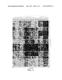 |
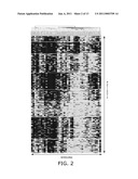 | 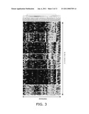 |
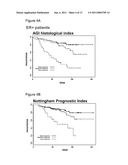 | 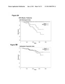 |
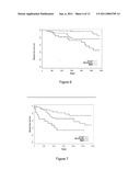 |  |
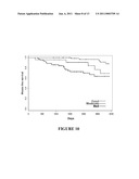 | 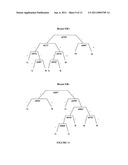 |
 |  |
 |  |
 |  |
 |  |
 |  |
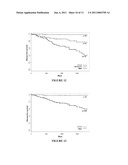 |  |
 |  |
 |  |
 |  |
 |  |
 | 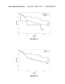 |
 |  |
 |  |
 |  |
 |  |
 |  |
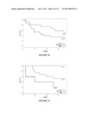 |  |
 |  |
 |  |
 |  |
 |  |
 | 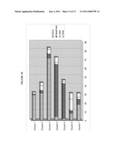 |
 |  |
 |  |
 |  |
 |  |
 |  |
 |  |
 | 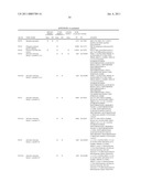 |
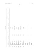 | 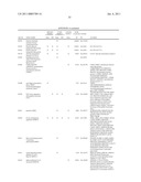 |
 |  |
 |  |
 |  |
 |  |
 |  |
 |  |
 |  |
 |  |
 |  |
 |  |
 | 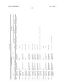 |
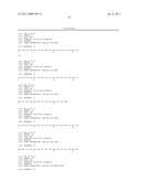 | 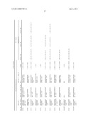 |
 |  |
 |  |
 |  |
 |  |
 |  |
 |  |
 |  |
 |  |
 |  |
 |  |
 |  |
| New patent applications in this class: | |
| Date | Title |
|---|---|
| 2022-05-05 | Microfluidic system for amplifying and detecting polynucleotides in parallel |
| 2019-05-16 | Reagents and methods for detecting protein lysine 2-hydroxyisobutyrylation |
| 2019-05-16 | Lateral flow analyte detection |
| 2019-05-16 | Mutations in the bcr-abl tyrosine kinase associated with resistance to sti-571 |
| 2019-05-16 | Enhanced methods of ribonucleic acid hybridization |
| New patent applications from these inventors: | |
| Date | Title |
|---|---|
| 2015-07-16 | Tle3 as a marker for chemotherapy |
| 2013-08-15 | Reagents and methods for use in cancer diagnosis, classification and therapy |
| Top Inventors for class "Combinatorial chemistry technology: method, library, apparatus" | |
| Rank | Inventor's name |
|---|---|
| 1 | Mehdi Azimi |
| 2 | Kia Silverbrook |
| 3 | Geoffrey Richard Facer |
| 4 | Alireza Moini |
| 5 | William Marshall |