Patent application title: METHOD FOR SCREENING AN INHIBITORY AGENT OF HBV PROLIFERATION BY USING THE INTERACTION BETWEEN HBV CAPSID AND SURFACE PROTEINS BASED ON CELLULAR IMAGING
Inventors:
Hyesung Jeon (Seongnam-Si, KR)
Soo Jin Oh (Seoul, KR)
Yeon Gyu Yu (Seoul, KR)
Yun-Kyoung Kim (Bucheon-Si, KR)
Assignees:
Korea Institute of Science and Technology
IPC8 Class: AC12Q170FI
USPC Class:
435 5
Class name: Chemistry: molecular biology and microbiology measuring or testing process involving enzymes or micro-organisms; composition or test strip therefore; processes of forming such composition or test strip involving virus or bacteriophage
Publication date: 2010-12-16
Patent application number: 20100316989
Inventors list |
Agents list |
Assignees list |
List by place |
Classification tree browser |
Top 100 Inventors |
Top 100 Agents |
Top 100 Assignees |
Usenet FAQ Index |
Documents |
Other FAQs |
Patent application title: METHOD FOR SCREENING AN INHIBITORY AGENT OF HBV PROLIFERATION BY USING THE INTERACTION BETWEEN HBV CAPSID AND SURFACE PROTEINS BASED ON CELLULAR IMAGING
Inventors:
Hyesung Jeon
Soo Jin Oh
Yeon Gyu Yu
Yun-Kyoung Kim
Agents:
EDWARDS ANGELL PALMER & DODGE LLP
Assignees:
Origin: BOSTON, MA US
IPC8 Class: AC12Q170FI
USPC Class:
Publication date: 12/16/2010
Patent application number: 20100316989
Abstract:
The present invention relates to a method for screening an inhibitory
agent of HBV proliferation by measuring the interaction (binding
strength) between capsid protein and surface protein, necessary for the
proliferation of HBV, by using cellular imaging, more precisely a method
for measuring changes on cellular imaging caused by the interaction
between a fusion protein containing PreS domain of HBV surface protein
and PH (Pleckstrin homology) domain sequence and a fusion protein
containing capsid protein and fluorescence protein (GFP) interacting with
the said fusion protein. The method of the present invention detecting
the interaction between proteins necessary for HBV proliferation at
cellular level can be effectively used for the screening of a novel
inhibitory agent of HBV proliferation at cellular level.Claims:
1. A expression vector comprising expression vector 1 containing a first
polynucleotide encoding a first fusion protein in which PreS domain of
HBV surface protein is linked to certain protein domain functioning for
cell membrane targeting a HBV capsid protein domain is linked to a
fluorescence protein and expression vector 2 containing a polynucleotide
encoding a second fusion protein in which a PreS domain of HBV surface
protein is linked to a protein domain functioning for cell membrane
targeting.
2. The expression vector according to claim 1, wherein the HBV capsid protein domain has the amino acid sequence represented by SEQ. ID. NO: 19 of HBV capsid protein except pro sequence (amino acids nos. 30-214).
3. The expression vector according to claim 1, wherein the fluorescence protein is selected from the group consisting of green fluorescence protein (GFP), red fluorescence protein (RFP), blue fluorescence protein (BFP), yellow fluorescence protein (YFP), cyan fluorescence protein (CFP) and enhanced green fluorescence protein (EGFP).
4. The expression vector according to claim 1, wherein the PreS domain of the surface protein has the amino acid sequence represented by SEQ. ID. NO: 20.
5. The expression vector according to claim 1, wherein the PreS domain of the surface protein is deficient in the part ranging from amino acid no. 93 to amino acid no. 117 of the sequence represented by SEQ. ID. NO: 20.
6. The expression vector according to claim 1, wherein the cell membrane targeting protein domain is selected from the group consisting of PH (Pleckstrin homology) domain of PLC-.delta. (phospholipase C delta) (Genebank ID: 241276, amino acid nos. 2-175), FYVE domain of EEA1 (early endosome antigene1) (Genebank ID: L40157, amino acid nos. 1352-1410), PHD (Prolyl-hydroxylase) domain of ING2 (Inhibitor of growth2) (Genebank ID: NM--001564, amino acid nos. 212-261), C2 (calcium/lipid-binding) domain of protein kinase C (Genebank ID: NM002737, amino acid nos. 172-260) and SEC14 (S. cerevisiae phosphatidylinositol transfer protein homology) domain of guanine nucleotide exchange factor DBS (Genebank ID: AB--116074, amino acid nos. 90-236).
7. The expression vector according to claim 1, wherein the first fusion protein has the amino acid sequence represented by SEQ. ID. NO: 4.
8. The expression vector according to claim 1, wherein the second fusion protein has the amino acid sequence represented by SEQ. ID. NO: 2.
9. The expression vector according to claim 1, wherein the second fusion protein's PreS domain is deficient in the part ranging from amino acid no. 93 to amino acid no. 117 and the second fusion protein has the amino acid sequence represented by SEQ. ID. NO: 6.
10. An animal cell transfected with the expression vector of claim 1, wherein said expression vector 1 comprises a first polynucleotide encoding a first fusion protein in which a HBV capsid protein domain is linked to a fluorescence protein and said expression vector 2 comprises a polynucleotide encoding a second fusion protein in which a PreS domain of HBV surface protein is linked to a protein domain functioning for cell membrane targeting.
11. The animal cell according to claim 10, wherein the cell is selected from the group consisting of HEK293T, COS7, HeLa and CHO.
12. A method for screening an inhibitory agent of HBV proliferation comprising the following steps:1) treating candidates with the animal cell of claim 10 during culture;2) taking fluorescence image of the fluorescence protein expressed in step 1) using fluorescent microscope; and3) selecting candidates locating fluorescence image in cytoplasm.
13. A screening kit of an inhibitory agent of HBV proliferation containing the animal cell of claim 10.
Description:
TECHNICAL FIELD
[0001]The present invention relates to a method for screening an inhibitory agent of HBV proliferation by measuring the interaction (binding strength) between capsid protein and surface protein, necessary for the proliferation of HBV, by using cellular imaging, more precisely a method for measuring changes on cellular imaging caused by the interaction between a fusion protein containing PreS domain of HBV surface protein and PH (Pleckstrin homology) domain sequence and a fusion protein containing capsid protein and fluorescence protein (GFP) interacting with the said fusion protein.
BACKGROUND ART
[0002]HBV (hepatitis B virus) is a member of Hepadnaviridae family which causes hepatitis B. Approximately two hundred million people are HBV carriers over the world. HBV vaccine has already been developed and widely used, but HBV treatment agents for those infected are limited to lamivudine and interferon. Lamivudine inhibits the activity of HBV DNA polymerase but it can produce resistant virus when it is administered for a long term, suggesting that the use thereof is limited. Therefore, it is required to develop diverse HBV treatment agents.
[0003]As an inhibitory agent of HBV, interferon, nucleic acid derivative or immune regulators have been developed, but the effect is in doubt. So, studies are undergoing to find out an inhibitory agent of HBV functioning to interrupt virus-receptor binding or to inhibit active proteins or polymerase thereof. Attempts have been made to develop an agent to inhibit diverse activities of HBV proteins. In HBV infected cells, the interaction of HBV capsid protein and surface protein is essential for the assembly of HBV. HBV capsid protein binds to HBV nucleic acid to form a nucleocapsid particle of 30 nm in diameter. As HBV surface proteins, three proteins, L, M, and S proteins, are biosynthesized from one gene (S gene), which are expressed in ER lipid membrane in cells and then bind specifically to the said nucleocapsid. At the same time, ER lipid membrane wraps the HBV nucleocapsid, resulting in a complete HBV particle (Volker B & Don G, PNAS USA 88:1059-1063, 1991). During while, capsid protein on the HBV nucleocapsid and the domain of amino acid residues 1-163 at N-terminal of surface protein L, particularly named as PreS, are selectively bound each other to form a HBV particle. The interaction between the two proteins can be a target of the development of a HBV proliferation inhibitory agent. To screen the interaction between HBV capsid protein and surface protein, an immuno assay using a recombinant protein has been developed (Asif-Ullah M et al., Antiviral Res 70; 85-90. 2006). This method is characterized by using a recombinant protein expressed in E. coli.
[0004]In addition to the method measuring the activity of a purified target protein, as a method measuring the bioactivity of a compound, a method to measure the activity of a target protein using a cell is actively tried during the development of a new drug. The method measuring the bioactivity of a compound in targeting cells has advantages of simultaneous detection of cellular permeability and toxicity of a target compound, making it an efficient screening method.
[0005]In HBV infected cells, HBV capsid protein and a nucleocapsid particle comprising HBV nucleic acid are specifically bound to HBV surface proteins expressed in ER membrane and as a result, active HBV particles are formed. The binding of HBV proteins is determined by the selective interaction between HBV capsid protein and PreS domain of surface protein. Therefore, if the interaction of HBV capsid protein and PreS domain of surface protein can be screened at cellular level, a compound capable of inhibiting the interaction between those proteins will be screened. So, the compound screened thereby is capable of inhibiting HBV proliferation and thus can be used as a treatment agent for HBV mediated hepatitis.
[0006]The present invention relates to a method for measuring the changes of cellular distribution of fluorescence signal generated by the interaction between a fusion protein containing PreS domain of HBV surface protein and PH sequence functioning for membrane targeting and a fusion protein containing capsid protein and fluorescence protein (GFP) interacting with the said fusion protein under fluorescent microscope. The method of the present invention is a screening method of the interaction between proteins necessary for HBV proliferation at cellular level, so that it can be effectively used for the screening of a novel inhibitory agent of HBV proliferation.
[0007]The present inventors developed a method for measuring the interaction of HBV capsid protein and surface protein at cellular level and further completed this invention by confirming that the method could be effectively used for the screening of a compound capable of inhibiting HBV proliferation by interrupting the interaction between HBV capsid protein and surface protein.
DISCLOSURE
Technical Problem
[0008]It is an object of the present invention to provide a method for screening an inhibitory agent of HBV proliferation efficiently by measuring the interaction between HBV capsid protein and surface protein at cellular level.
Technical Solution
[0009]To achieve the above object, the present invention provides an expression vector containing polynucleotide encoding the fusion protein in which HBV capsid protein is linked to fluorescence protein and the other polynucleotide encoding the fusion protein in which PreS domain of HBV surface protein is linked to certain protein domain functioning for cell membrane targeting.
[0010]The present invention also provides animal cells transfected with both expression vector 1 containing polynucleotide encoding the fusion protein in which PreS domain of HBV surface protein is linked to certain protein domain functioning for cell membrane targeting and expression vector 2 containing polynucleotide encoding the fusion protein in which HBV capsid protein interacting with the said fusion protein is linked to fluorescence protein.
[0011]The present invention also provides a method for screening an inhibitory agent of HBV proliferation comprising the following steps:
[0012]1) treating candidates to the animal cells during culture;
[0013]2) taking fluorescence image of the fluorescence protein expressed in step 1) using fluorescent microscope; and
[0014]3) selecting candidates locating fluorescence image in cytoplasm.
[0015]The present invention also provides a screening kit of an inhibitory agent of HBV proliferation containing the said animal cells.
ADVANTAGEOUS EFFECT
[0016]The method for screening the interaction of proteins necessary for HBV proliferation of the present invention can be effectively used for the screening of a novel inhibitory agent of HBV proliferation at cellular level.
DESCRIPTION OF DRAWINGS
[0017]The application of the preferred embodiments of the present invention is best understood with reference to the accompanying drawings, wherein:
[0018]FIG. 1 is a schematic diagram illustrating the structures of the fusion protein (PreS-PH) in which PreS domain of HBV (Hepatitis B Virus) surface protein is linked to N-terminal of PH domain targeting cell membrane and the fusion protein (Capsid-GFP) in which capsid protein (HBcAg) is linked to N-terminal of green fluorescence protein (GFP).
[0019]FIG. 2 is a schematic diagram illustrating the cleavage map of the expression vector expressing PreS-PH, Capsid-GFP, and mutant PreS-PH simultaneously:
[0020]a: pHBsPH-HBcGFP;
[0021]b: pHBc-GFP; and,
[0022]c: pΔHBsPH-HBcGFP.
[0023]FIG. 3 is a schematic diagram illustrating the migration of Capsid-GFP protein from cytoplasm to cell membrane by the interaction with PreS-PH protein.
[0024]FIG. 4 is a diagram illustrating that fluorescence image of Capsid-GFP protein expressed in the cytoplasm of cell is moved to cell membrane by the interaction with PreS protein when the PreS-PH fusion proteins is co-expressed:
[0025]a: pHBc-GFP;
[0026]b: pHBsPH-HBcGFP; and,
[0027]c: pΔHBsPH-HBcGFP.
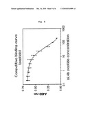
[0028]FIG. 5 is a diagram illustrating the inhibition of the interaction of capsid protein and PreS protein by PreS derived peptide.
[0029]FIG. 6 is a diagram illustrating that fluorescence image of Capsid-GFP co-expressed with PreS-PH in cells is observed in cytoplasm, resulted from the action of PreS-peptide.
BEST MODE
[0030]Hereinafter, the present invention is described in detail.
[0031]The present invention provides an expression vector containing polynucleotide encoding the fusion protein in which HBV capsid protein is linked to fluorescence protein and the other polynucleotide encoding the fusion protein in which PreS domain of HBV surface protein is linked to certain protein domain functioning for cell membrane targeting.
[0032]The said expression vector can be prepared by inserting a polynucleotide encoding the fusion protein comprising HBV capsid protein and fluorescence protein and a polynucleotide encoding the fusion protein comprising PreS domain of HBV surface protein and cell membrane targeting protein domain into the basic vector. The basic vector used in this invention can be any vector applicable in animal cell transfection which is preferably selected from the group consisting of pBud-CE 4.1, pcDNA3.1, pcDNA4 and pEF6p, but not always limited thereto. In a preferred embodiment of the present invention, pBud-CE4.1 was used.
[0033]The capsid protein herein can be full length capsid protein or a fragment capable of interacting with the surface protein. In a preferred embodiment of the invention, HBV capsid protein having the amino acid sequence without pro sequence, represented by SEQ. ID. NO: 19 (amino acids 30-214), was used.
[0034]The fluorescence protein herein is exemplified by green fluorescence protein (GFP), red fluorescence protein (RFP), blue fluorescence protein (BFP), yellow fluorescence protein (YFP), cyan fluorescence protein (CFP) and enhanced green fluorescence protein (EGFP), but not always limited thereto and in a preferred embodiment of the present invention, GFP was used.
[0035]The surface protein herein can be full length PreS domain or a fragment of PreS domain capable of interacting with capsid protein or PreS domain of HBV surface protein excluding the domain ranging from amino acid residue #93 to amino acid residue #117 capable of interacting core part in capsid protein. In a preferred embodiment of the present invention, PreS domain having the amino acid sequence of HBV surface protein capable of interacting with capsid protein, represented by SEQ. ID. NO: 20, was used. In another preferred embodiment of the present invention, PreS domain excluding the domain from amino acid #93 to amino acid #117 interacting with core of capsid protein was used.
[0036]The cell membrane targeting protein domain in step 1) is exemplified by PH (Pleckstrin homology) domain of PLC-6 (phospholipase C delta) (Genebank ID: 241276, amino acids 2-175), FYVE (No full name) domain of EEA1 (early endosome antigene1) (Genebank ID: L40157, amino acids 1352-1410), PHD (Prolyl-hydroxylase) domain of ING2 (Inhibitor of growth2) (Genebank ID: NM--001564, amino acids 212-261), C2 (calcium/lipid-binding) domain of protein kinase C (Genebank ID: NM002737, amino acids 172-260) and SEC14 (S. cerevisiae phosphatidylinositol transfer protein homology) domain of guanine nucleotide exchange factor DBS (Genebank ID: AB 116074, amino acids 90-236), but not always limited thereto. In a preferred embodiment of the present invention, PH domain of PLC-δ was used.
[0037]The capsid-GFP fusion protein has the amino acid sequence represented by SEQ. ID. NO: 4 (see FIG. 2b). The amino acid sequence represented by SEQ. ID. NO: 4 is characteristically coded by the nucleotide sequence represented by SEQ. ID. NO: 3.
[0038]The fusion protein comprising PreS domain of HBV surface protein and cell membrane targeting protein domain has the amino acid sequence represented by SEQ. ID. NO: 2 (see FIG. 2a). The amino acid sequence represented by SEQ. ID. NO: 2 is characteristically coded by the nucleotide sequence represented by SEQ. ID. NO: 1.
[0039]In another preferred embodiment of the present invention, the PreS domain of HBV surface protein can be substituted with another PreS domain excluding the domain ranging from amino acid #93 to amino acid #117 interacting core in capsid protein. That is, the domain from amino acid #93 to amino acid #117 interacting core region is eliminated from PreS domain to which cell membrane targeting protein domain is conjugated, resulting in the fusion protein having the amino acid sequence represented by SEQ. ID. NO: 6 (see FIG. 2c). The amino acid sequence represented by SEQ. ID. NO: 6 is characteristically coded by the nucleotide sequence represented by SEQ. ID. NO: 5.
[0040]The interaction of capsid protein and surface protein can be clearly detected when the expression vector (pΔHBsPH-HBcGFP, see FIG. 4c) co-expressing the fusion protein in which HBV capsid protein is linked to fluorescence protein and the fusion protein in which PreS domain excluding the amino acid domain from amino acid #93 to amino acid #117 interacting with core of capsid protein was used, compared to when the expression vector (pHBsPH-HBcGFP, see FIG. 4b) co-expressing the fusion protein in which HBV capsid protein is linked to fluorescence protein and the fusion protein in which PreS domain of HBV surface protein is linked to cell membrane targeting protein domain was used, because the interaction of core in capsid protein with surface protein is eliminated when the former was used (see FIG. 4).
[0041]The capsid protein of the present invention can be conjugated with fluorescence protein and PreS domain of surface protein can be conjugated with cell membrane targeting protein (see FIG. 1). The interaction of fluorescence protein conjugated capsid protein and cell membrane targeting protein conjugated PreS domain makes the fluorescence protein conjugated capsid protein to move from cytoplasm to cell membrane (see FIG. 3). The present inventors confirmed that the interaction of capsid protein and PreS domain was inhibited by PreS derived peptide and fluorescence image of the fusion protein comprising capsid protein-fluorescence protein was located not in cell membrane but in cytoplasm (see FIGS. 5 and 6). Accordingly, the present inventors confirmed that fluorescence protein conjugated capsid protein specifically interacted with cell membrane targeting protein conjugated PreS domain of HBV surface protein.
[0042]The present invention also provides animal cells transfected with the above expression vector.
[0043]The animal cells herein can be exemplified by HEK293T, COS7, HeLa and CHO cells, but not always limited thereto and in a preferred embodiment of the present invention HEK293T cells were used.
[0044]The present invention further provides animal cells transfected with both expression vector 1 containing polynucleotide encoding the fusion protein (PreS-PH) in which PreS domain of HBV surface protein is linked to certain protein domain functioning for cell membrane targeting and expression vector 2 containing polynucleotide encoding the fusion protein (Capsid-GFP) in which HBV capsid protein interacting with the said fusion protein is linked to fluorescence protein.
[0045]In a preferred embodiment of the present invention, PreS-PH and Capsid-GFP are cloned into different vectors having different origins but co-expressed in animal cells together by co-transfection.
[0046]The present invention also provides a screening method of an inhibitory agent of HBV proliferation comprising the following steps:
[0047]1) treating candidates to the animal cells during culture;
[0048]2) taking fluorescence image of the fluorescence protein expressed in step 1) using fluorescent microscope; and
[0049]3) selecting candidates locating fluorescence image in cytoplasm.
[0050]In addition, the present invention provides a screening kit of an inhibitory agent of HBV proliferation containing the said animal cells.
MODE FOR INVENTION
[0051]Practical and presently preferred embodiments of the present invention are illustrative as shown in the following Examples.
[0052]However, it will be appreciated that those skilled in the art, on consideration of this disclosure, may make modifications and improvements within the spirit and scope of the present invention.
Example 1
Construction of Expression Vector
[0053]<1-1>Construction of pCapsid-GFP
[0054]PCR was performed using pHBcAg (Choi K J et al., Biochem Biophys Res Commun 319:959-66, 2004) containing HBV capsid protein excluding pro sequence (amino acids 30-214, SEQ. ID. NO: 19) as a template with the primers represented by SEQ. ID. NO: 7 and NO: 8 as follows: at 95° C. for 1 minute, at 55° C. for 30 seconds, and at 72° C. for 1 minute (25 cycles). The amplified product was digested with SalI and KpnI, and then inserted into pEGFP--N1 vector (Clontech) digested with the same restriction enzymes by using T4 DNA ligase. As a result, pCapsid-GFP containing capsid-GFP was constructed. PCR was performed using the above DNA as a template with the primers represented by SEQ. ID. NO: 9 and NO: 10 to produce capsid-GFP DNA fragments containing the DNA restriction enzymes, NotI and XhoI. The synthesized capsid-GFP and pBud-CE 4.1 (Stratagene, USA) were digested with NotI and XhoI, followed by ligation using T4 DNA to construct pHBc-GFP (FIG. 2b). The DNA was sequenced to confirm whether the capsid-GFP DNA represented by SEQ. ID. NO: 3 encoding the amino acid sequence represented by SEQ. ID. NO: 4 was successfully inserted.
<1-2>Construction of PreS-PH
[0055]PCR was performed using rat cDNA as a template with the primers represented by SEQ. ID. NO: 11 and NO: 12 to synthesize PH domain (Genebank ID: 241276, amino acids 2-175) of PLC-δ (phospholipase C delta) as follows: at 95° C. for 1 minute, at 55° C. for 30 seconds, and at 72° C. for 1 minute (25 cycles). PCR was also performed using pTrx-PreS (Choi K J et al., Biochem Biophys Res Commun 319:959-66, 2004) as a template with the primers represented by SEQ. ID. NO: 13 and NO: 14 to synthesize PreS domain (amino acids 1-163, SEQ. ID. NO: 20) of HBV surface protein as follows: at 95° C. for 1 minute, at 55° C. for 30 seconds, and at 72° C. for 1 minute (25 cycles). PCR was also performed using the DNA encoding PreS and the DNA encoding PH as templates with the primers represented by SEQ. ID. NO: 15 and NO: 16 to synthesize PreS-PH DNA as follows: at 95° C. for 1 minute, at 55° C. for 30 seconds, and at 72° C. for 1 minute (25 cycles). At this time, the two templates were overlapped. The amplified DNA was digested with HindIII and XbaI, followed by ligation to pHBc-GFP digested with the same restriction enzymes by using T4 DNA. As a result, pHBsPH-HBcGFP (FIG. 2b) containing DNA having the nucleotide sequence represented by SEQ. ID. NO: 1 encoding the amino acid sequence represented by SEQ. ID. NO: 2 was constructed. The DNA was sequenced to confirm whether the amplified DNA was successfully inserted.
[0056]PCR was also performed using pHBsPH-HBcGFP without the domain ranging from amino acid #93 to amino acid #117 interacting with core protein as a template with the primers represented by SEQ. ID. NO: 17 and NO: 18 to synthesize DNA having the nucleotide sequence represented by SEQ. ID. NO: 5 encoding the amino acid sequence represented by SEQ. ID. NO: 6 as follows: at 95° C. for 30 seconds, at 55° C. for 1 minute, and at 68° C. for 7 minutes (18 cycles). The domain other than non-methylated DNA was eliminated by treating DpnI, the restriction enzyme recognizing methylated adenine. pΔHBsPH-HBcGFP (FIG. 2c) was confirmed by nucleotide sequencing.
Example 2
Animal Cell Transformation
<2-1>Animal Cell Culture
[0057]HEK293T cells were cultured in DMEM (Dulbecco's modified Eagle's medium; Gibco, USA) supplemented with 10% FBS (fetal bovine serum) in a 5% CO2, 37° C. incubator. The cells were sub-cultured when the density reached 90% and thus the density was adjusted to 25%, which was maintained until transformation.
<2-2>Animal Cell Transformation
[0058]The HEK293T cells cultured in Example <2-1> were sub-cultured, followed by further culture in a 6 well plate with cover glass. The cells were cultured until the density reached 40-60%. Then, the cells were transfected with pHBc-GFP, pHBsPH-HBcGFP or pΔHBsPH-HBcGFP by using Lipofectamine (Invitrogen, USA) and PLUS reagent (Invitrogen, USA). The transfected cells were cultured for 48 hours to produce protein.
Example 3
Inhibition of Interaction Between Capsid Protein and PreS Protein by PreS Derived Peptide
[0059]It was investigated whether PreS derived peptide (ΔL4b peptide; SEQ. ID. NO: 21: RQPTPISPPLRDSHPQAMQWNS; Peptron, Inc., Korea) could inhibit the interaction between PreS protein and capsid protein.
[0060]Thioredoxin conjugated PreS protein purified from E. coli transfected with pTrx-PreS (Choi K J et al., Biochem Biophys Res Commun 319:959-66, 2004) was dissolved in buffer-A (50 mM sodium phosphate, 0.15M NaCl, pH 8.0) at the concentration of 10 g/ml. 100 μl of the mixed solution was loaded in each well of a 96 well plate (CoStar, USA), followed by incubation for one hour at room temperature for fixation. The plate was blocked with buffer-A containing 5% (w/v) skim milk, to which different concentrations of serially diluted ΔL4b peptide and capsid protein purified from E. coli transfected with pHBcAg (Choi K J et al., Biochem Biophys Res Commun 319:959-66, 2004) were added (final conc.: 0.4 mM). Incubation was continued for 60 minutes at room temperature, followed by washing 6 times with PBS-T buffer (50 mM sodium phosphate, 0.15 M NaCl, pH 7.4 and 0.1% Tween-20) (300 a/well). The plate was incubated with anti-HBcAg antibody (1:2000, Cat. No. K0112162, KOMA biotechnology, Korea) for one hour (100 μl/well), to which HRP labeled anti-rabbit secondary antibody (1:2000, Sigma, USA) was added, followed by further incubation for one more hour. The plate was washed 6 times with PBS-T buffer, followed by color development using OPD solution (100 μl/well, dissolved in peroxide substrate buffer the concentration of 1 mg/m). The reaction was terminated by adding 2.5 M sulfuric acid (100 pt/well). Then, OD490 was measured by using multiplate reader (Spectra Max340 spectrometer, Molecular Devices Corp., USA).
[0061]As a result, it was confirmed that the PreS derived peptide inhibited the interaction between capsid protein and PreS protein dose-dependently (FIG. 5).
Example 4
Cell Imaging Using Fluorescent Microscope
<4-1>Measurement of Interaction
[0062]Cover glass was recovered from the plate where the HEK293T cells transfected with pHBc-GFP, pHBsPH-HBcGFP or pΔHBsPH-HBcGFP were growing, and washed with ice-cold PBS. The cover glass was attached onto slide glass for observation under microscope. The cells were observed through filter (490±20/528±38 nm, excitation/emission) to observe GFP under fluorescent microscope.
[0063]As a result, GFP fluorescence image was observed in cytoplasm in the cells transfected with pHBc-GFP or pΔHBsPH-HBcGFP, while GFP fluorescence image was observed in cell membrane in the cells transfected with pHBsPH-HBcGFP (FIG. 4). The fluorescence image in the cells transfected with pΔHBsPH-HBcGFP confirmed the importance of interaction between the core in capsid protein and the surface protein.
<4-2>Measurement of Inhibition of Interaction
[0064]When the HEK293T cells transfected with pHBc-GFP, pHBsPH-HBcGFP or pΔHBsPH-HBcGFP were reached 60% of confluence, PreS derived peptide (ΔL4b peptide; SEQ. ID. NO: 21) inhibiting the interaction between PreS and capsid protein was treated at the concentration of 50 μM thereto. Cover glass was recovered from the plate where the transfected HEK293T cells were growing, and washed with ice-cold PBS. The cover glass was attached onto slide glass for observation under microscope. The cells were observed through filter (490±20/528±38 nm, excitation/emission) to observe GFP under fluorescent microscope.
[0065]As a result, it was confirmed that the fluorescence image of fusion protein conjugated with GFP was located not in cell membrane but in cytoplasm of cells, suggesting that the interaction was inhibited (FIG. 6).
[0066]Those skilled in the art will appreciate that the conceptions and specific embodiments disclosed in the foregoing description may be readily utilized as a basis for modifying or designing other embodiments for carrying out the same purposes of the present invention. Those skilled in the art will also appreciate that such equivalent embodiments do not depart from the spirit and scope of the invention as set forth in the appended claims.
Sequence CWU
1
2111044DNAArtificial SequencePreS-PH nucleotide sequence 1atggggacga
atctttctgt tcccaatcct ctgggattct ttcccgatca ccagttggac 60cctgcgttcg
gagccaactc aaacaatcca gattgggact tcaaccccaa caaggatcac 120tggccagagg
cgaatcaggt aggagcggga gcattcgggc cagggttcac cccaccacac 180ggcggtcttt
tggggtggag ccctcaggct cagggcatat tgacagcagt gccagcagcg 240cctcctcctg
cctccaccaa tcggcagtca ggaagacagc ctactcccat ctctccacct 300ctaagagaca
gtcatcctca ggccatgcag tggaattcca caacattcca ccaagctctg 360ctagatccca
gagtgagggg cctatatttt cctgctggtg gctccagttc cggaacagta 420aaccctgttc
cgactactgc ctctcccata tcgtcaatct tctcgaggac tggggaccct 480gcaccgaacc
gtggacaggg aaacagcgat gcatctgtgg actcgggtag ggacttcctg 540accctgcacg
ggctccagga tgacccggac cttcaggccc ttctgaaggg cagccagctt 600ctgaaggtga
agtccagctc gtggcgtagg gaacgcttct acaagctaca ggaggactgc 660aagaccatct
ggcaggaatc tcgaaaggtc atgaggtccc cggagtcgca gctgttctcc 720atcgaggaca
ttcaggaggt acggatggga caccgcacag aaggcctgga gaagtttgcc 780cgagacatcc
ccgaggatcg atgcttctcc attgtcttca aggaccagcg caacacccta 840gacctcattg
ccccatcacc agctgacgct cagcactggg tgcagggcct gcgcaagatc 900atccaccact
ccggctccat ggaccagcgg cagaagctgc agcactggat tcactcctgc 960ttgcgaaagg
ctgataaaaa caaggcaaac aagatgaact tcaaggagct gaaggacttc 1020ctgaaggagc
tcaacatcca gtaa
10442347PRTArtificial SequencePreS-PH amino acid sequence 2Met Gly Thr
Asn Leu Ser Val Pro Asn Pro Leu Gly Phe Phe Pro Asp1 5
10 15His Gln Leu Asp Pro Ala Phe Gly Ala
Asn Ser Asn Asn Pro Asp Trp 20 25
30Asp Phe Asn Pro Asn Lys Asp His Trp Pro Glu Ala Asn Gln Val Gly
35 40 45Ala Gly Ala Phe Gly Pro Gly
Phe Thr Pro Pro His Gly Gly Leu Leu 50 55
60Gly Trp Ser Pro Gln Ala Gln Gly Ile Leu Thr Ala Val Pro Ala Ala65
70 75 80Pro Pro Pro Ala
Ser Thr Asn Arg Gln Ser Gly Arg Gln Pro Thr Pro 85
90 95Ile Ser Pro Pro Leu Arg Asp Ser His Pro
Gln Ala Met Gln Trp Asn 100 105
110Ser Thr Thr Phe His Gln Ala Leu Leu Asp Pro Arg Val Arg Gly Leu
115 120 125Tyr Phe Pro Ala Gly Gly Ser
Ser Ser Gly Thr Val Asn Pro Val Pro 130 135
140Thr Thr Ala Ser Pro Ile Ser Ser Ile Phe Ser Arg Thr Gly Asp
Pro145 150 155 160Ala Pro
Asn Arg Gly Gln Gly Asn Ser Asp Ala Ser Val Asp Ser Gly
165 170 175Arg Asp Phe Leu Thr Leu His
Gly Leu Gln Asp Asp Pro Asp Leu Gln 180 185
190Ala Leu Leu Lys Gly Ser Gln Leu Leu Lys Val Lys Ser Ser
Ser Trp 195 200 205Arg Arg Glu Arg
Phe Tyr Lys Leu Gln Glu Asp Cys Lys Thr Ile Trp 210
215 220Gln Glu Ser Arg Lys Val Met Arg Ser Pro Glu Ser
Gln Leu Phe Ser225 230 235
240Ile Glu Asp Ile Gln Glu Val Arg Met Gly His Arg Thr Glu Gly Leu
245 250 255Glu Lys Phe Ala Arg
Asp Ile Pro Glu Asp Arg Cys Phe Ser Ile Val 260
265 270Phe Lys Asp Gln Arg Asn Thr Leu Asp Leu Ile Ala
Pro Ser Pro Ala 275 280 285Asp Ala
Gln His Trp Val Gln Gly Leu Arg Lys Ile Ile His His Ser 290
295 300Gly Ser Met Asp Gln Arg Gln Lys Leu Gln His
Trp Ile His Ser Cys305 310 315
320Leu Arg Lys Ala Asp Lys Asn Lys Ala Asn Lys Met Asn Phe Lys Glu
325 330 335Leu Lys Asp Phe
Leu Lys Glu Leu Asn Ile Gln 340
34531399DNAArtificial SequenceCapsid-GFP nucleotide sequence 3atgcaacttt
ttcacctctg cctaatcatc tcttgtacat gtcccactgt tcaagcctcc 60aagctgtgcc
ttgggtggct ttggggcatg gacattgacc cttataaaga atttggagct 120actgtggagt
tactctcgtt tttgccttct gacttttttc cttccgtcag agatctccta 180gacaccgcct
cagctctgta tcgggaagcc ttagagtctc ctgagcattg ctcacctcac 240catactgcac
tcaggcaagc aattctctgc tggggggaat tgatgactct agctacctgg 300gtgggtaata
atttggaaga tccagcatcc agggatctag tagtcaatta tgttaatact 360aacatgggtt
taaagatcag gcaactattg tggtttcata tatcttgcct tacttttgga 420agagagactg
tacttgaata tttggtctct ttcggagtgt ggattcgcac tcctccagcc 480tatagaccac
caaatgcccc tatcttatca acacttccgg aaactactgt tgttagacga 540cgggaccgag
gcaggtcccc tagaagaaga actccctcgc ctcgcagacg cagatctcaa 600tcgccgcgtc
gcagaagatc tcaatctcgg gaatctcaat gtacggtacc gcgggcccgg 660gatcccaccg
gtcgccacca tggtgagcaa gggcgaggag ctgttcaccg gggtggtgcc 720catcctggtc
gagctggacg gcgacgtaaa cggccacaag ttcagcgtgt ccggcgaggg 780cgagggcgat
gccacctacg gcaagctgac cctgaagttc atctgcacca ccggcaagct 840gcccgtgccc
tggcccaccc tcgtgaccac cctgacctac ggcgtgcagt gcttcagccg 900ctaccccgac
cacatgaagc agcacgactt cttcaagtcc gccatgcccg aaggctacgt 960ccaggagcgc
accatcttct tcaaggacga cggcaactac aagacccgcg ccgaggtgaa 1020gttcgagggc
gacaccctgg tgaaccgcat cgagctgaag ggcatcgact tcaaggagga 1080cggcaacatc
ctggggcaca agctggagta caactacaac agccacaacg tctatatcat 1140ggccgacaag
cagaagaacg gcatcaaggt gaacttcaag atccgccaca acatcgagga 1200cggcagcgtg
cagctcgccg accactacca gcagaacacc cccatcggcg acggccccgt 1260gctgctgccc
gacaaccact acctgagcac ccagtccgcc ctgagcaaag accccaacga 1320gaagcgcgat
cacatggtcc tgctggagtt cgtgaccgcc gccgggatca ctctcggcat 1380ggacgagctg
tacaagtaa
13994466PRTArtificial SequenceCapsid-GFP amino acid sequence 4Met Gln Leu
Phe His Leu Cys Leu Ile Ile Ser Cys Thr Cys Pro Thr1 5
10 15Val Gln Ala Ser Lys Leu Cys Leu Gly
Trp Leu Trp Gly Met Asp Ile 20 25
30Asp Pro Tyr Lys Glu Phe Gly Ala Thr Val Glu Leu Leu Ser Phe Leu
35 40 45Pro Ser Asp Phe Phe Pro Ser
Val Arg Asp Leu Leu Asp Thr Ala Ser 50 55
60Ala Leu Tyr Arg Glu Ala Leu Glu Ser Pro Glu His Cys Ser Pro His65
70 75 80His Thr Ala Leu
Arg Gln Ala Ile Leu Cys Trp Gly Glu Leu Met Thr 85
90 95Leu Ala Thr Trp Val Gly Asn Asn Leu Glu
Asp Pro Ala Ser Arg Asp 100 105
110Leu Val Val Asn Tyr Val Asn Thr Asn Met Gly Leu Lys Ile Arg Gln
115 120 125Leu Leu Trp Phe His Ile Ser
Cys Leu Thr Phe Gly Arg Glu Thr Val 130 135
140Leu Glu Tyr Leu Val Ser Phe Gly Val Trp Ile Arg Thr Pro Pro
Ala145 150 155 160Tyr Arg
Pro Pro Asn Ala Pro Ile Leu Ser Thr Leu Pro Glu Thr Thr
165 170 175Val Val Arg Arg Arg Asp Arg
Gly Arg Ser Pro Arg Arg Arg Thr Pro 180 185
190Ser Pro Arg Arg Arg Arg Ser Gln Ser Pro Arg Arg Arg Arg
Ser Gln 195 200 205Ser Arg Glu Ser
Gln Cys Thr Val Pro Arg Ala Arg Asp Pro Thr Gly 210
215 220Arg His His Gly Glu Gln Gly Arg Gly Ala Val His
Arg Gly Gly Ala225 230 235
240His Pro Gly Arg Ala Gly Arg Arg Arg Lys Arg Pro Gln Val Gln Arg
245 250 255Val Arg Arg Gly Arg
Gly Arg Cys His Leu Arg Gln Ala Asp Pro Glu 260
265 270Val His Leu His His Arg Gln Ala Ala Arg Ala Leu
Ala His Pro Arg 275 280 285Asp His
Pro Asp Leu Arg Arg Ala Val Leu Gln Pro Leu Pro Arg Pro 290
295 300His Glu Ala Ala Arg Leu Leu Gln Val Arg His
Ala Arg Arg Leu Arg305 310 315
320Pro Gly Ala His His Leu Leu Gln Gly Arg Arg Gln Leu Gln Asp Pro
325 330 335Arg Arg Gly Glu
Val Arg Gly Arg His Pro Gly Glu Pro His Arg Ala 340
345 350Glu Gly His Arg Leu Gln Gly Gly Arg Gln His
Pro Gly Ala Gln Ala 355 360 365Gly
Val Gln Leu Gln Gln Pro Gln Arg Leu Tyr His Gly Arg Gln Ala 370
375 380Glu Glu Arg His Gln Gly Glu Leu Gln Asp
Pro Pro Gln His Arg Gly385 390 395
400Arg Gln Arg Ala Ala Arg Arg Pro Leu Pro Ala Glu His Pro His
Arg 405 410 415Arg Arg Pro
Arg Ala Ala Ala Arg Gln Pro Leu Pro Glu His Pro Val 420
425 430Arg Pro Glu Gln Arg Pro Gln Arg Glu Ala
Arg Ser His Gly Pro Ala 435 440
445Gly Val Arg Asp Arg Arg Arg Asp His Ser Arg His Gly Arg Ala Val 450
455 460Gln Val4655969DNAArtificial
Sequencedeleted PreS-PH nucleotide sequence 5atggggacga atctttctgt
tcccaatcct ctgggattct ttcccgatca ccagttggac 60cctgcgttcg gagccaactc
aaacaatcca gattgggact tcaaccccaa caaggatcac 120tggccagagg cgaatcaggt
aggagcggga gcattcgggc cagggttcac cccaccacac 180ggcggtcttt tggggtggag
ccctcaggct cagggcatat tgacagcagt gccagcagcg 240cctcctcctg cctccaccaa
tcggcagtca ggaagacaag ctctgctaga tcccagagtg 300aggggcctat attttcctgc
tggtggctcc agttccggaa cagtaaaccc tgttccgact 360actgcctctc ccatatcgtc
aatcttctcg aggactgggg accctgcacc gaaccgtgga 420cagggaaaca gcgatgcatc
tgtggactcg ggtagggact tcctgaccct gcacgggctc 480caggatgacc cggaccttca
ggcccttctg aagggcagcc agcttctgaa ggtgaagtcc 540agctcgtggc gtagggaacg
cttctacaag ctacaggagg actgcaagac catctggcag 600gaatctcgaa aggtcatgag
gtccccggag tcgcagctgt tctccatcga ggacattcag 660gaggtacgga tgggacaccg
cacagaaggc ctggagaagt ttgcccgaga catccccgag 720gatcgatgct tctccattgt
cttcaaggac cagcgcaaca ccctagacct cattgcccca 780tcaccagctg acgctcagca
ctgggtgcag ggcctgcgca agatcatcca ccactccggc 840tccatggacc agcggcagaa
gctgcagcac tggattcact cctgcttgcg aaaggctgat 900aaaaacaagg caaacaagat
gaacttcaag gagctgaagg acttcctgaa ggagctcaac 960atccagtaa
9696322PRTArtificial
Sequencedeleted PreS-PH amino acid sequence 6Met Gly Thr Asn Leu Ser Val
Pro Asn Pro Leu Gly Phe Phe Pro Asp1 5 10
15His Gln Leu Asp Pro Ala Phe Gly Ala Asn Ser Asn Asn
Pro Asp Trp 20 25 30Asp Phe
Asn Pro Asn Lys Asp His Trp Pro Glu Ala Asn Gln Val Gly 35
40 45Ala Gly Ala Phe Gly Pro Gly Phe Thr Pro
Pro His Gly Gly Leu Leu 50 55 60Gly
Trp Ser Pro Gln Ala Gln Gly Ile Leu Thr Ala Val Pro Ala Ala65
70 75 80Pro Pro Pro Ala Ser Thr
Asn Arg Gln Ser Gly Arg Gln Ala Leu Leu 85
90 95Asp Pro Arg Val Arg Gly Leu Tyr Phe Pro Ala Gly
Gly Ser Ser Ser 100 105 110Gly
Thr Val Asn Pro Val Pro Thr Thr Ala Ser Pro Ile Ser Ser Ile 115
120 125Phe Ser Arg Thr Gly Asp Pro Ala Pro
Asn Arg Gly Gln Gly Asn Ser 130 135
140Asp Ala Ser Val Asp Ser Gly Arg Asp Phe Leu Thr Leu His Gly Leu145
150 155 160Gln Asp Asp Pro
Asp Leu Gln Ala Leu Leu Lys Gly Ser Gln Leu Leu 165
170 175Lys Val Lys Ser Ser Ser Trp Arg Arg Glu
Arg Phe Tyr Lys Leu Gln 180 185
190Glu Asp Cys Lys Thr Ile Trp Gln Glu Ser Arg Lys Val Met Arg Ser
195 200 205Pro Glu Ser Gln Leu Phe Ser
Ile Glu Asp Ile Gln Glu Val Arg Met 210 215
220Gly His Arg Thr Glu Gly Leu Glu Lys Phe Ala Arg Asp Ile Pro
Glu225 230 235 240Asp Arg
Cys Phe Ser Ile Val Phe Lys Asp Gln Arg Asn Thr Leu Asp
245 250 255Leu Ile Ala Pro Ser Pro Ala
Asp Ala Gln His Trp Val Gln Gly Leu 260 265
270Arg Lys Ile Ile His His Ser Gly Ser Met Asp Gln Arg Gln
Lys Leu 275 280 285Gln His Trp Ile
His Ser Cys Leu Arg Lys Ala Asp Lys Asn Lys Ala 290
295 300Asn Lys Met Asn Phe Lys Glu Leu Lys Asp Phe Leu
Lys Glu Leu Asn305 310 315
320Ile Gln733DNAArtificial SequenceHBV Capsid forward primer 7gccgagctcg
ccaccatgca actttttcac ctc
33828DNAArtificial SequenceHBV Capsid reverse primer 8ggggtaccgt
ataacattga gattcccg
28947DNAArtificial SequenceCapsid-GFP forward primer 9aaggaaaaag
cggccgcgcg ctcgccacca tgcaactttt tcacctc
471033DNAArtificial SequenceCapsid-GFP reverse primer 10ccgctcgagt
tacttgtaca gctcgtccca tga
331128DNAArtificial SequenceMouse PLC delta forward primer 11gactcgggta
gggacttcct gaccctgc
281228DNAArtificial SequenceMouse PLC delta reverse primer 12ttactggatg
ttgagctcct tcaggaag
281334DNAArtificial SequenceHBV PreS forward primer 13gccgagctcg
ccaccatggg gacgaatctt tctg
341435DNAArtificial SequenceHBV PreS reverse primer 14aaggaaaaag
cggccgctta ctggatgttg agctc
351540DNAArtificial SequencePreS-PH forward primer 15cccaagcttt
gcgctcgcca ccatgcaact ttttcacctc
401626DNAArtificial SequencePreS-PH reverse primer 16gctctagatt
actggatgtt gagctc
261730DNAArtificial Sequencedeleted PreS-PH foward primer 17ggcagtcagg
aagacaagct ctgctagatc
301830DNAArtificial Sequencedeleted PreS-PH reverse primer 18gatctagcag
agcttgtctt cctgactgcc
3019214PRTArtificial SequenceHBV capsid protein amino acid sequence 19Met
Gln Leu Phe His Leu Cys Leu Ile Ile Ser Cys Thr Cys Pro Thr1
5 10 15Val Gln Ala Ser Lys Leu Cys
Leu Gly Trp Leu Trp Gly Met Asp Ile 20 25
30Asp Pro Tyr Lys Glu Phe Gly Ala Thr Val Glu Leu Leu Ser
Phe Leu 35 40 45Pro Ser Asp Phe
Phe Pro Ser Val Arg Asp Leu Leu Asp Thr Ala Ser 50 55
60Ala Leu Tyr Arg Glu Ala Leu Glu Ser Pro Glu His Cys
Ser Pro His65 70 75
80His Thr Ala Leu Arg Gln Ala Ile Leu Cys Trp Gly Glu Leu Met Thr
85 90 95Leu Ala Thr Trp Val Gly
Asn Asn Leu Glu Asp Pro Ala Ser Arg Asp 100
105 110Leu Val Val Asn Tyr Val Asn Thr Asn Met Gly Leu
Lys Ile Arg Gln 115 120 125Leu Leu
Trp Phe His Ile Ser Cys Leu Thr Phe Gly Arg Glu Thr Val 130
135 140Leu Glu Tyr Leu Val Ser Phe Gly Val Trp Ile
Arg Thr Pro Pro Ala145 150 155
160Tyr Arg Pro Pro Asn Ala Pro Ile Leu Ser Thr Leu Pro Glu Thr Thr
165 170 175Val Val Arg Arg
Arg Asp Arg Gly Arg Ser Pro Arg Arg Arg Thr Pro 180
185 190Ser Pro Arg Arg Arg Arg Ser Gln Ser Pro Arg
Arg Arg Arg Ser Gln 195 200 205Ser
Arg Glu Ser Gln Cys 21020163PRTArtificial SequenceHBV PreS protein
amino acid sequence 20Met Gly Thr Asn Leu Ser Val Pro Asn Pro Leu Gly Phe
Phe Pro Asp1 5 10 15His
Gln Leu Asp Pro Ala Phe Gly Ala Asn Ser Asn Asn Pro Asp Trp 20
25 30Asp Phe Asn Pro Asn Lys Asp His
Trp Pro Glu Ala Asn Gln Val Gly 35 40
45Ala Gly Ala Phe Gly Pro Gly Phe Thr Pro Pro His Gly Gly Leu Leu
50 55 60Gly Trp Ser Pro Gln Ala Gln Gly
Ile Leu Thr Ala Val Pro Ala Ala65 70 75
80Pro Pro Pro Ala Ser Thr Asn Arg Gln Ser Gly Arg Gln
Pro Thr Pro 85 90 95Ile
Ser Pro Pro Leu Arg Asp Ser His Pro Gln Ala Met Gln Trp Asn
100 105 110Ser Thr Thr Phe His Gln Ala
Leu Leu Asp Pro Arg Val Arg Gly Leu 115 120
125Tyr Phe Pro Ala Gly Gly Ser Ser Ser Gly Thr Val Asn Pro Val
Pro 130 135 140Thr Thr Ala Ser Pro Ile
Ser Ser Ile Phe Ser Arg Thr Gly Asp Pro145 150
155 160Ala Pro Asn2122PRTArtificial Sequencedeleted
L4b peptide 21Arg Gln Pro Thr Pro Ile Ser Pro Pro Leu Arg Asp Ser His Pro
Gln1 5 10 15Ala Met Gln
Trp Asn Ser 20
User Contributions:
comments("1"); ?> comment_form("1"); ?>Inventors list |
Agents list |
Assignees list |
List by place |
Classification tree browser |
Top 100 Inventors |
Top 100 Agents |
Top 100 Assignees |
Usenet FAQ Index |
Documents |
Other FAQs |
User Contributions:
Comment about this patent or add new information about this topic:
 |  |
 |  |
 |  |
 |  |
 |  |
 |  |
 |  |
 |  |
 |
| Similar patent applications: | |
| Date | Title |
|---|---|
| 2013-01-31 | Genetic inhibition by double-stranded rna |
| 2014-02-27 | Jak/stat inhibitors and mapk/erk inhibitors for rsv infection |
| 2013-06-27 | Method and system for diagnosis of lawsonia intracellularis |
| 2013-10-03 | Store-operated calcium cellular assay |
| 2014-01-16 | Method for assessing risk of hepatocellular carcinoma |
| New patent applications in this class: | |
| Date | Title |
|---|---|
| 2022-05-05 | Method for diagnosing human t-cell leukemia virus type 1 (htlv-1) associated diseases |
| 2022-05-05 | Systems and methods for assay processing |
| 2022-05-05 | Rapid pathology/cytology without wash |
| 2019-05-16 | Biofluidic triggering system and method |
| 2019-05-16 | Method and system for detection of disease agents in blood |
| New patent applications from these inventors: | |
| Date | Title |
|---|---|
| 2021-12-30 | Composition for preventing and treatment of stroke |
| 2021-02-04 | Composition for preventing and treatment of spinal cord injury |
| 2016-03-03 | Amphiphilic polymer |
| 2015-05-28 | Novel compounds as chloride channel blocking agent |
| 2014-04-03 | Glutamate release from astrocyte |
| Top Inventors for class "Chemistry: molecular biology and microbiology" | |
| Rank | Inventor's name |
|---|---|
| 1 | Marshall Medoff |
| 2 | Anthony P. Burgard |
| 3 | Mark J. Burk |
| 4 | Robin E. Osterhout |
| 5 | Rangarajan Sampath |