Patent application title: Methods and compositions for identifying a fetal cell
Inventors:
David Xingfei Deng (Mountain View, CA, US)
Yun Bao (Fremont, CA, US)
Yue-Jen Chuu (Cupertino, CA, US)
Daniel Shoemaker (San Diego, CA, US)
Daniel Shoemaker (San Diego, CA, US)
David L. Robbins (Temecula, CA, US)
IPC8 Class: AC40B3000FI
USPC Class:
506 7
Class name: Combinatorial chemistry technology: method, library, apparatus method of screening a library
Publication date: 2010-12-02
Patent application number: 20100304978
Claims:
1. A method for identifying a fnRBC comprising detecting transcript or
protein expression of a HBE, AFP, AHSG, or J42-4-d gene.
2. The method of claim 1, wherein said detecting comprises using at least two primers and at least one probe that anneals to a cDNA generated from a transcript expressed by said HBE, AFP, AHSG, or J42-4-d gene.
3. A method for identifying a trophoblast comprising detecting transcript or protein expression of a KISS1, LOC90625, AFP, hPL, beta-hCG, or FN1 gene.
4. The method of claim 3, wherein said detecting comprises using at least two primers and at least one probe that anneals to a cDNA generated from a transcript expressed by said KISS1, LOC90625, AFP, hPL, beta-hCG, or FN1 gene.
5. A method for identifying a fetal cell in a maternal sample comprising detecting transcript or protein expression by a cell of one or more of the KISS1, LOC90625, FN1, or AHSG genes to distinguish said fetal cell from a maternal cell.
6. A method for identifying a fetal cell in a maternal sample comprising detecting transcript or protein expression by a cell of three or more of the hPL, KISS1, LOC90625, FN1, PSG9, HBE, AFP, beta-hCG, AHSG or J42-4-d genes to distinguish said fetal cell from a maternal cell.
7. The method of claim 5 or 6, wherein the maternal sample is a maternal blood sample, amniocentesis sample, or cervical swab.
8. The method of claim 5 or 6, wherein said fetal cell is a fetal nucleated RBC or a placental cell.
9. The method of claim 7, wherein said sample is taken in the 1.sup.st or early 2.sup.nd trimester.
10. The method of claim 7, wherein said sample is taken in the 2.sup.nd trimester.
11. The method of claim 5, wherein said fetal cell is a fetal nucleated red blood cell and said gene is AHSG.
12. The method of claim 5, wherein said fetal cell is a trophoblast and said gene is FN1.
13. The method of claim 5 or 6, wherein said detecting comprises RNA FISH, RNA-FISH with a molecule beacon probe, RT-PCR, Q-PCR, digital mRNA profiling, Northern blotting, ribonuclease protection assay, or RNA expression profiling using microarrays.
14. The method of claim 5 or 6, wherein said detecting comprises binding a protein with one or more binding moieties.
15. The method of claim 14, wherein said one or more binding moieties is an antibody, Fab fragment, Fc fragment, scFv fragment, peptidomimetic, or peptoid.
16. A method for identifying a fetal cell in a maternal sample, comprising:a. enriching a fetal cell, andb. detecting protein or transcript expression of one or more genes by said fetal cell, wherein said expression of said one or more genes distinguishes said fetal cell from a maternal cell, wherein said one or more genes is hPL, CHS2, KISS1, GDF15, CRH, TFP12, CGB, LOC90625, FN1, COL1A2, PSG9, PSG1, AFP, APOC3, SERPINC1, AMBP, CPB2, ITIH1, APOH, HPX, beta-hCG, AHSG, APOB, or J42-4-d.
17. The method of claim 16, wherein the step of enriching a fetal cell comprises one or more steps of density centrifugation, size based separation, affinity separation, magnetic separation, microfluidic fluorescent cell sorting, dielectrophoretic enrichment, or antibody separation.
18. The method of claim 16, wherein the sample is a maternal blood sample, amniocentesis sample, or cervical swab.
19. The method of claim 16, wherein said cell is a fetal nucleated RBC or a placental cell.
20. The method of claim 16, further comprising enriching a fetal nucleated RBC by magnetic enrichment.
21. The method of claim 16, further comprising enriching one or more fetal nucleated RBCs by anti-CD71 or anti-GLA selection.
22. The method of claim 16, further comprising enriching one or more trophoblasts by anti-HLA-G or anti-EGFR selection.
23. The method of claim 16, wherein said cell is a fetal nucleated RBC and said one or more genes is AFP, AHSG, or J42-4-d.
24. The method of claim 16, wherein said cell is a trophoblast and said one or more genes is KISS1, LOC90625, AFP, hPL, beta-hCG, or FN1.
25. The method of claim 16, wherein said detecting is by RNA FISH, RNA-FISH with a molecule beacon probe, RT-PCR, Q-PCR, digital mRNA profiling, Northern blotting, ribonuclease protection assay, or RNA expression profiling using microarrays.
26. The method of claim 16, wherein said fetal cell is from a maternal sample obtained in the 1.sup.st trimester or 2.sup.nd trimester of pregnancy.
27. The method of claim 16, wherein said detecting protein expression comprises binding a protein with a binding moiety.
28. The method of claim 27, wherein said binding moiety is an antibody, Fab fragment, Fc fragment, scFv fragment, peptidomimetic, or peptoid.
29. A method for identifying a fetal cell specific transcript comprisinga. isolating a transcript from a sample containing a fetal cell and a transcript from a sample lacking fetal cells;b. producing cDNAs of said transcripts;c. performing quantitative PCR on said cDNAs; andd. comparing results of said quantitative PCR between samples to identify a marker transcript with higher expression in a fetal cell relative to a non-fetal cell.
30. The method of claim 29, wherein said fetal cell is first enriched from a maternal sample by size based separation.
31. The method of claim 29, further comprising a verifying step comprising detecting a marker transcript by quantitative PCR.
Description:
CROSS-REFERENCE
[0001]This application claims the benefit of U.S. Patent Application Ser. No. 61/147,456, filed Jan. 26, 2009, which is incorporated herein by reference in its' entirety.
BACKGROUND OF THE INVENTION
[0002]Circulating fetal cells (CFCs) are present in maternal blood during pregnancy. Successful isolation and enrichment of one or more CFCs from maternal peripheral blood can be used to perform noninvasive genetic diagnosis of fetal well being. However, the number of CFCs in circulating maternal blood is relatively low, with approximately one fetal cell per one ml of whole blood. Owing to their low numbers, it is technically challenging to enrich and purify a fetal cell from maternal blood samples.
[0003]Fetal call identification (FCID) using fetal cell-type specific markers (FCMs) can play a role in fetal cell enrichment, enumeration, and genetic analysis. FCID markers can be DNA, RNA or proteins. DNA markers, such as loci on the Y-chromosome or other chromosomes, can be used to distinguish a maternal and fetal cell. A fetal cell can be identified using techniques such as by RNA fluorescent in situ hybridization (FISH) or immunocytochemical (ICC) staining for one or more protein markers. Cell surface protein markers can also be used for both cell selection and identification.
[0004]A gene expression panel that can be used to identify a circulating fetal cell such as a fetal nucleated red blood cell (fnRBC) or a trophoblast would be useful for the enrichment, enumeration, purification or analysis of these cells. Currently, available fetal cell markers have some drawbacks and are not specific for the various fetal cell types present in maternal samples in the first and second trimesters.
[0005]Specific FCMs are useful in identification, enrichment, purification, and enumeration of a fetal cell. Identification of one or more genes whose expression is specific for a fetal cell can be used to identify a fetal cell, such as through RNA fluorescent in situ hybridization (FISH), and/or isolate a target fetal cell to high purity such as by immunocytometry. The corresponding protein markers of these genes can also be used in ICC for FCID.
SUMMARY OF THE INVENTION
[0006]In one aspect, a method for identifying a fnRBC comprising detecting transcript or protein expression of a HBE, AFP, AHSG, or J42-4-d gene is provided. In one embodiment, said detecting comprises using at least two primers and at least one probe that anneals to a cDNA generated from a transcript expressed by said HBE, AFP, AHSG, or J42-4-d gene.
[0007]In another aspect, a method for identifying a trophoblast comprising detecting transcript or protein expression of a KISS1, LOC90625, AFP, hPL, beta-hCG, or FN1 gene is provided. In one embodiment, said detecting comprises using at least two primers and at least one probe that anneals to a cDNA generated from a transcript expressed by said KISS1, LOC90625, AFP, hPL, beta-hCG, or FN1 gene.
[0008]In another aspect, a method for identifying a fetal cell in a maternal sample is provided comprising detecting transcript or protein expression by a cell of one or more of the KISS1, LOC90625, FN1, or AHSG genes to distinguish said fetal cell from a maternal cell.
[0009]In another aspect, a method for identifying a fetal cell in a maternal sample is provided comprising detecting transcript or protein expression by a cell of three or more of the hPL, KISS1, LOC90625, FN1, PSG9, HBE, AFP, beta-hCG, AHSG or J42-4-d genes to distinguish said fetal cell from a maternal cell.
[0010]In one embodiment, the maternal sample is a maternal blood sample, amniocentesis sample, or cervical swab. In another embodiment, said fetal cell is a fetal nucleated RBC or a placental cell. In another embodiment, said sample is taken in the 1st or early 2nd trimester. In another embodiment, said sample is taken in the 2nd trimester. In another embodiment, said fetal cell is a fetal nucleated red blood cell and said gene is AHSG. In another embodiment, said fetal cell is a trophoblast and said gene is FN1. In another embodiment, said detecting comprises RNA FISH, RNA-FISH with a molecule beacon probe, RT-PCR, Q-PCR, digital mRNA profiling, Northern blotting, ribonuclease protection assay, or RNA expression profiling using microarrays. In another embodiment, said detecting comprises binding a protein with one or more binding moieties. In another embodiment, said one or more binding moieties is an antibody, Fab fragment, Fc fragment, scFv fragment, peptidomimetic, or peptoid.
[0011]In another aspect, a method for identifying a fetal cell in a maternal sample is provided comprising: enriching a fetal cell and detecting protein or transcript expression of one or more genes by said fetal cell, wherein said expression of said one or more genes distinguishes said fetal cell from a maternal cell, wherein said one or more genes is hPL, CHS2, KISS1, GDF15, CRH, TFP12, CGB, LOC90625, FN1, COL1A2, PSG9, PSG1, AFP, APOC3, SERPINC1, AMBP, CPB2, ITIH1, APOH, HPX, beta-hCG, AHSG, APOB, or J42-4-d. In one embodiment, the step of enriching a fetal cell comprises one or more steps of density centrifugation, size based separation, affinity separation, magnetic separation, microfluidic fluorescent cell sorting, dielectrophoretic enrichment, or antibody separation. In another embodiment, the sample is a maternal blood sample, amniocentesis sample, or cervical swab. In another embodiment, said cell is a fetal nucleated RBC or a placental cell. In another embodiment, the method further comprises enriching a fetal nucleated RBC by magnetic enrichment. In another embodiment, the method further comprises enriching one or more fetal nucleated RBCs by anti-CD71 or anti-GLA selection. In another embodiment, the method further comprises enriching one or more trophoblasts by anti-HLA-G or anti-EGFR selection. In another embodiment, said cell is a fetal nucleated RBC and said one or more genes is AFP, AHSG, or J42-4-d. In another embodiment, said cell is a trophoblast and said one or more genes is KISS1, LOC90625, AFP, hPL, beta-hCG, or FN1. In another embodiment, said detecting is by RNA FISH, RNA-FISH with a molecule beacon probe, RT-PCR, Q-PCR, digital mRNA profiling, Northern blotting, ribonuclease protection assay, or RNA expression profiling using microarrays. In another embodiment, said fetal cell is from a maternal sample obtained in the 1st trimester or 2nd trimester of pregnancy. In another embodiment, said detecting protein expression comprises binding a protein with a binding moiety. In another embodiment, said binding moiety is an antibody, Fab fragment, Fc fragment, scFv fragment, peptidomimetic, or peptoid.
[0012]In another aspect, a method for identifying a fetal cell specific transcript is provided comprising isolating a transcript from a sample containing a fetal cell and a transcript from a sample lacking fetal cells; producing cDNAs of said transcripts; performing quantitative PCR on said cDNAs; and comparing results of said quantitative PCR between samples to identify a marker transcript with higher expression in a fetal cell relative to a non-fetal cell. In one embodiment, said fetal cell is first enriched from a maternal sample by size based separation. In another embodiment, the method further comprises a verifying step comprising detecting a marker transcript by quantitative PCR.
INCORPORATION BY REFERENCE
[0013]All publications, patents, and patent applications mentioned in this specification are herein incorporated by reference to the same extent as if each individual publication, patent, or patent application was specifically and individually indicated to be incorporated by reference.
BRIEF DESCRIPTION OF THE DRAWINGS
[0014]The novel features of the invention are set forth with particularity in the appended claims. A better understanding of the features and advantages of the present invention will be obtained by reference to the following detailed description that sets forth illustrative embodiments, in which the principles of the invention are utilized, and the accompanying drawings of which:
[0015]FIGS. 1A-1D illustrate embodiments of a size-based separation module.
[0016]FIG. 2A illustrates cells flowing through an array of obstacles.
[0017]FIG. 2B illustrates antibody coated posts.
[0018]FIG. 2C illustrates one embodiment of an affinity separation module.
[0019]FIG. 3 illustrates one embodiment of a magnetic separation module.
[0020]FIG. 4 illustrates one embodiment of a multiplex enrichment module of the present invention.
[0021]FIG. 5 illustrates exemplary genes that can be analyzed from enriched cells, such as epithelial cells, endothelial cells, circulating tumor cells, progenitor cells, etc.
[0022]FIG. 6 illustrates one embodiment for genotyping rare cell(s) or rare DNA using, e.g., Affymetrix DNA microarrays.
[0023]FIG. 7 illustrates one embodiment for genotyping rare cell(s) or rare DNA using, e.g., Illumina bead arrays.
[0024]FIG. 8 illustrates one embodiment for determining gene expression of rare cell(s) or rare DNA using, e.g., Affymetrix expression chips.
[0025]FIG. 9 illustrates one embodiment for determining gene expression of rare cell(s) or rare DNA using, e.g., Illumina bead arrays.
[0026]FIG. 10 illustrates one embodiment for high-throughput sequencing of rare cell(s) or rare DNA using, e.g., single molecule sequence by synthesis methods (e.g., Helicos BioSciences Corporation).
[0027]FIG. 11 illustrates one embodiment for high-throughput sequencing of rare cell(s) or rare DNA using, e.g., amplification of nucleic acid molecules on a bead (e.g., 454 Lifesciences).
[0028]FIG. 12 illustrates one embodiment for high-throughput sequencing of rare cell(s) or rare DNA using, e.g., clonal single molecule arrays technology (e.g., Solexa, Inc.).
[0029]FIG. 13 illustrates one embodiment for high-throughput sequencing of rare cell(s) or rare DNA using, e.g., single base polymerization using enhanced nucleotide fluorescence (e.g., Genovoxx GmbH).
[0030]FIG. 14 illustrates methods of fetal diagnostic assays. A fetal cell is isolated by CSM-HE enrichment of target cells from blood. The designation of a cell as a fetal cell can be confirmed using techniques comprising FISH staining (using slides or membranes and optionally an automated detector), FACS, and/or binning Binning can comprise distribution of enriched cells across wells in a plate (such as a 96 or 384 well plate), microencapsulation of cells in droplets that are separated in an emulsion, or by introduction of cells into microarrays of nanofluidic bins. A fetal cell is then identified using methods that can comprise the use of biomarkers (such as fetal (gamma) hemoglobin), allele-specific SNP panels that could detect fetal genome DNA, detection of differentially expressed maternal and fetal transcripts (such as Affymetrix chips), or primers and probes directed to fetal specific loci (such as the multi-repeat DYZ locus on the Y-chromosome). Binning sites that contain a fetal cell are then be analyzed for aneuploidy and/or other genetic defects using a technique such as CGH array detection, ultra deep sequencing (such as Solexa, 454, or mass spectrometry), STR analysis, or SNP detection.
[0031]FIG. 15 illustrates methods of fetal diagnostic assays, further comprising the step of whole genome amplification prior to analysis of aneuploidy and/or other genetic defects.
[0032]FIGS. 16A-D illustrate various embodiments of a size-based separation module.
[0033]FIGS. 17A and B illustrate cell smears of the product and waste fractions.
[0034]FIG. 18 illustrates an initial screening strategy for identifying fetal cell markers.
[0035]FIG. 19 illustrates an experimental setup for identification of fetal specific RNAs.
[0036]FIG. 20 illustrates a strategy for screening for fetal specific markers with a Fluidigm Chip.
[0037]FIG. 21 illustrates a strategy for verifying fetal specific markers.
[0038]FIG. 22 depicts an experimental protocol for verifying fetal specific markers.
[0039]FIG. 23 illustrates RNA FISH using cDNA probes.
[0040]FIG. 24 illustrates validation of gene labeling specificity by single cell analysis.
[0041]FIG. 25 illustrates a summary of a fetal cell marker screening.
[0042]FIG. 26A depicts 12 placental (trophoblast) specific markers.
[0043]FIG. 26B depicts 12 fetal liver (fnRBC) specific markers.
[0044]FIG. 27A depicts 13 fnRBC markers selected for further verification by RT-PCR.
[0045]FIG. 27B depicts 7 trophoblast markers selected for further verification by RT-PCR.
[0046]FIG. 28 displays the expression levels of gene markers for fnRBC in different tissues and isolated cells.
[0047]FIG. 29 displays the expression levels of gene markers for trophoblasts in different tissues and isolated cells.
[0048]FIG. 30 displays relative gene expression results and cell type specificity for RNA markers.
[0049]FIG. 31 illustrates RNA FISH in cultured cell-lines.
[0050]FIG. 32 illustrates RNA FISH in cord blood and non-pregnant samples.
[0051]FIG. 33 illustrates RNA FISH staining of fnRBC in pre-termination pregnant blood samples.
[0052]FIG. 34 illustrates preliminary results of RNA FISH staining in pre-term and post-term blood samples.
[0053]FIG. 35 illustrates detection of AFP expression in LCM isolated fnRBCs.
[0054]FIG. 36 illustrates that AFP is expressed in HBE antibody-stained positive cells, but not in negative cells.
[0055]FIG. 37 illustrates a strategy for enriching a fetal cell from maternal blood.
[0056]FIG. 38 illustrates a strategy for direct gene expression profiling from fetal cell enriched products.
[0057]FIG. 39 illustrates results that 35 HBE positive cell counts (one count/well).
[0058]FIG. 40 illustrates fetal trophoblast cell type and count.
[0059]FIG. 41 shows a comparison between fetal cell marker results with Y chromosome genotyping results using 10 ml whole blood.
[0060]FIG. 42 lists sequences of transcripts that can be fetal cell markers.
[0061]FIG. 43 lists sequences of proteins that can be fetal cell markers
[0062]FIG. 44 illustrates an overview for diagnosing, prognosing, or monitoring a prenatal condition in a fetus.
[0063]FIG. 45A-C illustrates one embodiment of a sample splitting apparatus.
[0064]FIG. 46 illustrates the detection of single copies of a fetal cell genome by qPCR.
[0065]FIG. 47 illustrates detection of single fetal cells in binned samples by SNP analysis.
[0066]FIG. 48 illustrates fetal cell enumeration by PCR analysis.
[0067]FIG. 49 illustrates a method for fetal cell identification and verification.
[0068]FIG. 50 illustrates expression of hPL, Beta-hCG and AFP in fetal trophoblasts.
[0069]FIGS. 51A-F illustrate isolated fetal cells confirmed by the reliable presence of male Y chromosome.
[0070]FIG. 52 illustrates trisomy 21 pathology in an isolated fetal nucleated red blood cell.
DETAILED DESCRIPTION OF THE INVENTION
[0071]In general, methods and compositions for identifying a fetal cell by detecting expression of one or more genes are provided. Detection of expression of fetal cell-specific markers can be used to distinguish a fetal cell from a reference cell (e.g., maternal cell), distinguish between types of fetal cells, purify and/or enrich a fetal cell, and enumerate a fetal cell.
[0072]I. Sample Collection/Preparation
[0073]Sample Type
[0074]Samples containing one or more rare cells (e.g., one or more fetal cells) can be obtained from any animal in need of a diagnosis or prognosis or from an animal pregnant with a fetus in need of a diagnosis or prognosis. In one embodiment, a sample can be obtained from an animal suspected of being pregnant, pregnant, or that has been pregnant to detect the presence of a fetus or fetal abnormality. When the animal is a human, the sample can be taken during the first trimester (about the first three months of pregnancy), the 2nd trimester (about months 4-6 of pregnancy), or the third trimester (about months 7-9 of pregnancy). An animal of the present invention can be a human or a domesticated animal such as a cow, chicken, pig, horse, rabbit, dogs, cat, or goat. Samples derived from an animal, e.g., a human, can include, e.g., whole blood, sweat, tears, ear flow, sputum, lymph, bone marrow suspension, lymph, urine, saliva, semen, vaginal flow, cerebrospinal fluid, brain fluid, ascites, milk, secretions of the respiratory, intestinal or genitourinary tracts fluid. The sample can include. a sample of amniotic fluid (via amniocentesis), a biopsy of the placenta (e.g., by chorionic villi sampling, CVS), a maternal blood sample, an umbilical cord blood sample, or cervical swab.
[0075]Samples, including reference samples, can be collected for the purpose of identifying fetal cell-specific markers. Samples can include cord blood, peripheral blood cells from a non-pregnant woman (NP-PBC), adult bone marrow (ABM), fetal liver, or placenta. Fetal liver contains fnRBCs, and placenta contains trophoblasts and connective tissue. When the sample is taken from a pregnant woman, or a woman suspected of being pregnant, the sample can be taken in the 1st, 2nd, or 3rd trimester.
[0076]To obtain a blood sample, a device known in the art can be used, e.g., a syringe or other vacuum suction device.
[0077]A maternal sample can contain one or more different types of fetal cells. A fetal cell can be any cell derived from a zygote, blastocyst, or embryo. A fetal cell can include, for example, T cells, B cells, natural-killer (NK) cells, antigen-presenting cells, erythroblasts, nucleated erythrocytes, leukocytes, pregnancy-associated progenitor cells (PAPCs), fetal mesenchymal stem cells, CD34+ cells (hematopoietic stem cells; HSCs); CD34+CD38+ cells, epithelial cells, endometrial cells, and placental cells. Placental cells can include trophoblasts, e.g., syncytiotrophoblasts (cells of the outer syncytial layer of the trophoblast) and cytotrophoblasts (cells of the inner layer of the trophoblast).
[0078]When obtaining a sample from an animal (e.g., blood sample), the amount of sample can vary depending upon animal size, its gestation period, and the condition being screened. In one embodiment, up to 50, 40, 30, 20, 10, 9, 8, 7, 6, 5, 4, 3, 2, or 1 mL of a sample is obtained. In one embodiment, 1-50, 2-40, 3-30, or 4-20 mL of sample is obtained. In one embodiment, more than 5, 10, 15, 20, 25, 30, 35, 40, 45, 50, 55, 60, 65, 70, 75, 80, 85, 90, 95, or 100 mL of a sample is obtained. In one embodiment between about 10-20 ml of a peripheral blood sample is obtained from a pregnant female.
[0079]To detect one or more fetal abnormalities, a blood sample can be obtained from a pregnant animal or human within 36, 24, 22, 20, 18, 16, 14, 12, 10, 8, 6, or 4 weeks of conception or even after a pregnancy has terminated.
[0080]In one embodiment, the sample is a maternal blood sample taken in the 1st trimester or 2nd trimester.
[0081]Pre-Treatment of a Sample
[0082]A blood sample can be optionally pre-treated or processed prior to enrichment. In one embodiment a pre-treatment step includes the addition of one or more reagents including, but not limited to, a membrane stabilizer, a preservative, a fixative, a lysing reagent, a diluent, an anti-apoptotic reagent, an anti-coagulation reagent, an anti-thrombotic reagent, magnetic property regulating reagent, a buffering reagent, an osmolality regulating reagent, a pH regulating reagent, and/or a cross-linking reagent. In one embodiment the fixative used is formaldehyde, paraformaldehyde, glutaraldehyde, acrolein, glyoxal, malonaldehyde, diacetyl, polyaldehydes, carbodiimides, diisocyanates, diazonium compounds, diimido esters, diethylpyrocarbonate, maleimides, benzoquinone, and metallic ions, Dinitrobenzaldehyde, Dinitrobenzene sulfonic acids, or Dinitrobenzoic acids. In another embodiment the fixative is a Dinitrophenols, 3,5-Dinitrosalicylic acid, 2,4-Dinitrobenzoic acid, 5-Sulfosalicylic acid, 2,5-Dihydroxy-1,4-benzene disulfonic acid, 3,5-Dinitrobenzoic acid, 8-Hydroxyquinoline-5-sulfonic acid, 4-Nitrophenol, 3,5-Dinitrosalicylaldehyde, 3,5-Dinitroaniline, Paratoluene sulfonic acid, 2-Mesitylene sulfonic acid, 2-(Trifluoromethyl)benzoic acid, 3,5-Dinitrobenzonitrile, and 2,4-Dinitrobenzene sulfonic acid, 3,5-Dinitrobenzoic acid, 2,4-Dinitrobenzoic acid, 2,4-Dinitrobenzene sulfonic acid, 2,6-Dinitrobenzene sulfonic acid, 3,5-Dinitrobenzene sulfonic acid, or 2,4-Dinitrophenol. Fixatives are described in U.S. Pat. No. 5,422,277, issued Jun. 6, 1995, which is herein incorporated by reference. In one embodiment the cell membrane stabilizer used is potassium dichromate, a monosaccaride (e.g., glucose, fructose), a sugar alcohol (e.g., sorbitol, inositol), a disaccharide (e.g., sucrose, trehalose, lactose, maltose), a trisaccharide (e.g., raffinose), a oligosaccharide (e.g., cycloinulohexaose), a polysaccharide (e.g., ficoll, or dextran), or a polymer (e.g., poly-vinyl-pyrrolidone, polyethyleneglycol). In one embodiment the molecule that can change the magnetic property of, e.g., red blood cells' hemoglobin, is CO2, N2, or NaNO2.
[0083]When a blood sample is obtained, a preservative such an anti-coagulation agent and/or a stabilizer can be added to the sample prior to enrichment. This addition allows for an extended time for analysis/detection. Thus, a sample, such as a blood sample, can be enriched and/or analyzed under any of the methods and systems herein within 1 week, 6 days, 5 days, 4 days, 3 days, 2 days, 1 day, 12 hrs, 6 hrs, 3 hrs, 2 hrs, or 1 hr from the time the sample is obtained.
[0084]II. Enrichment/Purification
[0085]Concentration
[0086]A sample (e.g., blood sample) can be enriched for one or more rare analytes or rare cells (e.g. one or more fetal cells or epithelial cells) using one or more any methods known in the art (e.g. Guetta, E M et al. Stem Cells Dev, 13(1):93-9 (2004), which is herein incorporated by reference in its entirety) or described herein. The enrichment increases the concentration of one or more rare cells or the ratio of one or more rare cells to non-rare cells in the sample. For example, enrichment can increase the concentration of an analyte of interest such as a fetal cell or epithelial cell by a factor of at least 2, 4, 6, 8, 10, 20, 50, 100, 200, 500, 1,000, 2,000, 5,000, 10,000, 20,000, 50,000, 100,000, 200,000, 500,000, 1,000,000, 2,000,000, 5,000,000, 10,000,000, 20,000,000, 50,000,000, 100,000,000, 200,000,000, 500,000,000, 1,000,000,000, 2,000,000,000, or 5,000,000,000 fold over its concentration in the original sample. In particular, when enriching one or more fetal cells from a maternal peripheral venous blood sample, the initial concentration of the one or more fetal cells in a sample can be about 1:50,000,000 and it can be increased to at least 1:5,000 or 1:500. Rare cells can also be enriched in a sample by the removal of fluid. A fluid sample (e.g., a blood sample) of greater than 10, 15, 20, 50, or 100 mL total volume can comprise rare components of interest, and it can be concentrated such that the rare component of interest is concentrated into a concentrated solution of less than 0.5, 1, 2, 3, 5, or mL total volume.
[0087]Density Gradient Centrifugation
[0088]Density gradient centrifugation is a method of separating cells based on the different densities of cell types in a mixture. The method can be used in a single step to separate cells into two compartments which contain cells that are either lighter or heavier than a specific density of the gradient material used. Density gradient centrifugation can be carried out through repetitive steps based on a series of different density gradients or in combination with affinity separation, cell panning, cell sorting, and the like. Alternatively, density gradient centrifugation can be performed using multiple layers of the different gradient densities. This method allows cells of different densities to form zones or bands at their corresponding densities after centrifugation. The cells in the different zones are then collected by placing a pipette at the appropriate location. Methods for enriching specific cell-types by density gradient centrifugation are described in U.S. Pat. No. 5,840,502, which is herein incorporated by reference in its entirety.
[0089]Methods of identifying fetal cells in a specimen using density gradient centrifugation utilize density gradient medium. The density gradient medium can be colloidal polyvinylpyrrolidone-coated silica (e.g. PercolD, Nycodenz, a nonionic polysucrose (Ficoll) either alone or with sodium diatrizoate (e.g. Ficoll-Paque or Histopaque), or mixtures thereof. The density of the reagent employed is selected to separate the fetal cells of interest from other blood components.
[0090]Enrichment can occur using one or more types of separation modules. Several different modules are described herein, all of which can be fluidly coupled with one another in series for enhanced performance.
[0091]Enrichment by Lysis
[0092]In one embodiment, enrichment occurs by selective lysis. In one embodiment, a blood sample can be combined with an agent that selectively lyses one or more cells or components in a blood sample. For example, one or more fetal cells can be selectively lysed and their nuclei released when a blood sample including one or more fetal cells is combined with deionized water. Such selective lysis allows for the subsequent enrichment of fetal nuclei using, e.g., size or affinity based separation. In another example platelets and/or enucleated red blood cells are selectively lysed to generate a sample enriched in nucleated cells, such as fetal nucleated red blood cells (fnRBC's), maternal nucleated blood cells (mnBC), or epithelial cells. fnRBCs can be subsequently separated from mnBC's using, e.g., antigen-i affinity or differences in. hemoglobin.
[0093]Size-Based Enrichment
[0094]In one embodiment, enrichment of rare cells occurs using one or more size-based separation modules. Examples of size-based separation modules include filtration modules, sieves, matrixes, etc. Examples of size-based separation modules contemplated by the present invention include those disclosed in International Publication No. WO 2004/113877, which is herein incorporated by reference in its entirety. Other size based separation modules are disclosed in International Publication No. WO 2004/0144651 and U.S. Patent Application Publication Nos. US20080138809A1 and US20080220422A1, which are herein incorporated by reference in their entirety.
[0095]In one embodiment, a size-based separation module comprises one or more arrays of obstacles forming a network of gaps. The obstacles are configured to direct particles as they flow through the array/network of gaps into different directions or outlets based on the particle's hydrodynamic size. For example, as a blood sample flows through an array of obstacles, nucleated cells or cells having a hydrodynamic size larger than a predetermined size, e.g., 8 microns, are directed to a first outlet located on the opposite side of the array of obstacles from the fluid flow inlet, while the enucleated cells or cells having a hydrodynamic size smaller than a predetermined size, e.g., 8 microns, are directed to a second outlet also located on the opposite side of the array of obstacles from the fluid flow inlet.
[0096]An array can be configured to separate cells smaller or larger than a predetermined size by adjusting the size of the gaps, obstacles, and offset in the period between each successive row of obstacles. For example, in one embodiment, obstacles or gaps between obstacles can be up to 10, 20, 50, 70, 100, 120, 150, 170, or 200 microns in length or about 2, 4, 6, 8 or 10 microns in length. In one embodiment, an array for size-based separation includes more than 100, 500, 1,000, 5,000, 10,000, 50,000 or 100,000 obstacles that are arranged into more than 10, 20, 50, 100, 200, 500, or 1000 rows. In one embodiment, obstacles in a first row of obstacles are offset from a previous (upstream) row of obstacles by up to 50% the period of the previous row of obstacles. In one embodiment, obstacles in a first row of obstacles are offset from a previous row of obstacles by up to 45, 40, 35, 30, 25, 20, 15 or 10% the period of the previous row of obstacles. Furthermore, the distance between a first row of obstacles and a second row of obstacles can be up to 10, 20, 50, 70, 100, 120, 150, 170 or 200 microns. A particular offset can be continuous (repeating for multiple rows) or non-continuous. In one embodiment, a separation module includes multiple discrete arrays of obstacles fluidly coupled such that they are in series with one another. Each array of obstacles has a continuous offset. But each subsequent (downstream) array of obstacles has an offset that is different from the previous (upstream) offset. In one embodiment, each subsequent array of obstacles has a smaller offset that the previous array of obstacles. This arrangement allows for a refinement in the separation process as cells migrate through the array of obstacles. Thus, a plurality of arrays can be fluidly coupled in series or in parallel, (e.g., more than 2, 4, 6, 8, 10, 20, 30, 40, 50). Fluidly coupling separation modules (e.g., arrays) in parallel allows for high-throughput analysis of the sample, such that at least 1, 2, 5, 10, 20, 50, 100, 200, or 500 mL per hour flows through the enrichment modules or at least 1, 5, 10, or 50 million cells per hour are sorted or flow through the device.
[0097]FIG. 1A illustrates an example of a size-based separation module. In one embodiment, a fetal cell can be labeled by (which can be of any shape) are coupled to a flat substrate to form an array of gaps. A transparent cover or lid can be used to cover the array. The obstacles form a two-dimensional array with each successive row shifted horizontally with respect to the previous row of obstacles, where the array of obstacles directs one or more components having a hydrodynamic size smaller than a predetermined size in a first direction and one or more components having a hydrodynamic size larger that a predetermined size in a second direction. For enriching epithelial cells from enucleated cells, the predetermined size of gaps in an array of obstacles can be 6-12 μm or 6-8 μm. For enriching one or more fetal cells from a mixed sample (e.g., maternal blood sample) the predetermined size of gaps in an array of obstacles can be between 4-10 μm or 6-8 μm. The flow of sample into the array of obstacles can be aligned at a small angle (flow angle) with respect to a line-of-sight of the array. Optionally, the array is coupled to an infusion pump to perfuse the sample through the obstacles. The flow conditions of the size-based separation module described herein are such that cells are sorted by the array with minimal damage. This allows for downstream analysis of intact cells and intact nuclei to be more efficient and reliable.
[0098]In one embodiment, a size-based separation module comprises an array of obstacles configured to direct cells larger than a predetermined size to migrate along a line-of-sight within the array (e.g., towards a first outlet or bypass channel leading to a first outlet), while directing cells and analytes smaller than a predetermined size to migrate through the array of obstacles in a different direction than the larger cells (e.g., towards a second outlet). Such embodiments are illustrated in part in FIGS. 1B-1D.
[0099]A variety of enrichment protocols can be utilized. In one embodiment the cells are handled gently to reduce mechanical damage to the cells or their DNA. This gentle handling can serve to preserve the small number of one or more fetal cells in the sample. Integrity of the nucleic acid being evaluated is an important feature to permit the distinction between the genomic material from the one or more fetal cells and other cells in the sample. In particular, the enrichment and separation of one or more fetal cells using the arrays of obstacles provides gentle treatment which minimizes cellular damage. Moreover, this gentle treatment maximizes nucleic acid integrity, permits exceptional levels of separation, and allows for the ability to subsequently utilize various formats to analyze the genome of the cells.
[0100]Affinity-Based Enrichment
[0101]In one embodiment, enrichment of one or more rare cells (e.g., one or more fetal cells or epithelial cells) occurs using one or more capture modules that selectively inhibit the mobility of one or more cells of interest. In one embodiment, a capture module is fluidly coupled downstream to a size-based separation module. Capture modules can include a substrate having multiple obstacles that restrict the movement of cells or analytes greater than a predetermined size. Examples of capture modules that inhibit the migration of cells based on size are disclosed in U.S. Pat. Nos. 5,837,115 and 6,692,952, which are herein incorporated by reference in their entirety.
[0102]In one embodiment, a capture module includes a two dimensional array of obstacles that selectively filters or captures cells or analytes having a hydrodynamic size greater than a particular gap size (predetermined size), International Publication No: WO 2004/113877, which is herein incorporated by reference in its entirety.
[0103]In one embodiment a capture module captures analytes (e.g., cells of interest or not of interest) based on their affinity for a binding moiety. For example, an affinity-based separation module that can capture cells or analytes can include an array of obstacles adapted for permitting sample flow through, but for the fact that the obstacles are covered with binding moieties that selectively bind one or more analytes (e.g., cell populations) of interest (e.g., one or more red blood cells, fetal cells, epithelial cells or nucleated cells) or analytes not-of-interest (e.g., white blood cells). Arrays of obstacles adapted for separation by capture can include obstacles having one or more shapes and can be arranged in a uniform or non-uniform order. In one embodiment, a two-dimensional array of obstacles is staggered such that each subsequent row of obstacles is offset from the previous row of obstacles to increase the number of interactions between the analytes being sorted (separated) and the obstacles. Other types of binding modules can be used.
[0104]Binding moieties coupled to the obstacles can include e.g., proteins (e.g., ligands/receptors), nucleic acids having complementary counterparts in retained analytes, antibodies, etc. In one embodiment, an affinity-based separation module comprises a two-dimensional array of obstacles covered with one or more antibodies that are: anti-CD71, anti-CD235a, anti-CD36, anti-carbohydrates, anti-selectin, anti-CD45, anti-GPA, anti-antigen-i, anti-EpCAM, anti-E-cadherin, anti-Muc-1, anti-hPL, anti-CHS2, anti-KISS1, anti-GDF15, anti-CRH, anti-TFP12, anti-CGB, anti-LOC90625, anti-FN1, anti-COL1A2, anti-PSG9, anti-PSG1, anti-HBE, anti-AFP, anti-APOC3, anti-SERPINC1, anti-AMBP, anti-CPB2, anti-ITIH1, anti-APOH, anti-HPX, anti-beta-hCG, anti-AHSG, anti-APOB, or anti-J42-4-d.
[0105]In one embodiment, a fnRBC is enriched using anti-CD71 or anti-GLA selection. In another embodiment, a trophoblast is enriched using anti-HLA-G or anti-EGFR selection. In another embodiment, a fnRBC is enriched using one or more antibodies or antibody fragments that can bind a protein expressed from the genes HBE, AFP, APOC3, SERPINC1, AMBP, CPB2, ITIH1, APOH, HPX, beta-hCG, AHSG, APOB, or J42-4-d. In another embodiment, a trophoblast is enriched using one or more antibodies or antibody fragments that can bind a protein expressed from the genes hPL, CHS2, KISS1, GDF15, CRH, TFP12, CGB, LOC90625, FN1, COL1A2, PSG9, or PSG1.
[0106]The binding moiety can be a single moiety, e.g., a polypeptide or protein, or it can include two or more moieties, e.g., a pair of polypeptides such as a pair of single chain antibody domains. Methods of generating antibodies are well know to those skilled in the art, e.g., by immunization strategies for the generation of monoclonal or polyclonal antibodies or in vitro methods for generating alternative binding members. Polyclonal antibodies can include, e.g., sheep, goat, rabbit, or rat polyclonal antibody. In addition any suitable molecule capable of high affinity binding can be used including antibody fragments such as single chain antibodies (scFv), Fab and scFv antibodies which can be obtained by phage-display or single domain antibodies (VHH) or chimeric antibodies. The binding moiety can be derived from a naturally occurring protein or polypeptide; it can be designed de novo, or it can be selected from a library. For example, the binding moiety can be or be derived from an antibody, a single chain antibody (scFv), a single domain antibody (VHH), a lipocalin, a single chain MHC molecule, an Anticalin® (Pieris), an Affibody®, a nanobody (Ablynx) or a Trinectin® (Phylos). Methods of generating binding members of various types are well known in the art.
[0107]Antibodies
[0108]A binding member can according to the invention be an antibody, such as any suitable antibody known in the art including other immunologically active fragments of antibodies or single chain antibodies. Antibody molecules are typically Y-shaped molecules whose basic unit consist of four polypeptides, two identical heavy chains and two identical light chains, which are covalently linked together by disulfide bonds. Each of these chains is folded in discrete domains. The C-terminal regions of both heavy and light chains are conserved in sequence and are called the constant regions, also known as C-domains. The N-terminal regions, also known as V-domains, are variable in sequence and are responsible for the antibody specificity. The antibody specifically recognizes and binds to an antigen mainly through six `short complementarity-determining regions located in their V-domains.
[0109]Antibody Fragments
[0110]In one embodiment of the invention the binding member is a fragment of an antibody, e.g., an antigen binding fragment or a variable region. Examples of antibody fragments useful with the present invention include Fab, Fab', F(ab') 2 and Fv fragments. Papain digestion of antibodies produces two identical antigen binding fragments, called the Fab fragment, each with a single antigen binding site, and a residual "Fc" fragment, so-called for its ability to crystallize readily. Pepsin treatment yields an F(ab') 2 fragment that has two antigen binding fragments which are capable of cross-linking antigen, and a residual other fragment (which is termed pFc').
[0111]Additional fragments can include diabodies, linear antibodies, single-chain antibody molecules, and multispecific antibodies formed from antibody fragments.
[0112]The antibody fragments Fab, Fv and scFv differ from whole antibodies in that the antibody fragments carry only a single antigen-binding site. Recombinant fragments with two binding sites have been made in several ways, for example, by chemical cross-linking of cysteine residues introduced at the C-terminus of the VH of an Fv (Cumber et al., 1992 which is herein incorporated by reference in its entirety), or at the C-terminus of the VL of an scFv (Pack and Pluckthun, 1992, which is herein incorporated by reference in its entirety), or through the hinge cysteine residues of Fab's (Carter et al., 1992, which is herein incorporated by reference in its entirety).
[0113]Antibody fragments retain some or essentially all the ability of an antibody to selectively bind with its antigen or receptor. Examples of antibody fragments include the following:
[0114]Fab is the fragment that contains a monovalent antigen-binding fragment of an antibody molecule. A Fab fragment can be produced by digestion of whole antibody with the enzyme papain to yield an intact light chain and a portion of one heavy chain.
[0115]Fab' is the fragment of an antibody molecule and can be obtained by treating whole antibody with pepsin, followed by reduction, to yield an intact light chain and a portion of the heavy chain. Two Fab' fragments are obtained per antibody molecule. Fab 1 fragments differ from Fab fragments by the addition of a few residues at the carboxyl terminus of the heavy chain CH 1 domain including one or more cysteines from the antibody hinge region.
[0116](Fab')2 is the fragment of an antibody that can be obtained by treating whole antibody with the enzyme pepsin without subsequent reduction. F(ab')2 is a dimer of two Fab' fragments held together by two disulfide bonds.
[0117]Fv is the minimum antibody fragment that contains a complete antigen recognition and binding site. This region consists of a dimer of one heavy and one light chain variable domain in a tight, non-covalent association (VH-V L dimer). It is in this configuration that the three CDRs of each variable domain interact to define an antigen binding site on the surface of the VH-V L dimer. Collectively, the six CDRs confer antigen binding specificity to the antibody. However, even a single variable domain (or half of an Fv comprising only three CDRs specific for an antigen) has the ability to recognize and bind antigen, although at a lower affinity than the entire binding site.
[0118]The antibody can be a single chain antibody ("SCA"), defined as a genetically engineered molecule containing the variable region of the light chain, the variable region of the heavy chain, linked by a suitable polypeptide linker as a genetically fused single chain molecule. Such single chain anti-bodies are also referred to as "single-chain Fv" or "sFv" antibody fragments. Generally, the Fv polypeptide further comprises a polypeptide linker between the VH and VL domains that enables the sFv to form the desired structure for antigen binding.
[0119]The antibody fragments according to the invention can be produced in any suitable manner known to the person skilled in the art. Several microbial expression systems have already been developed for producing active antibody fragments, e.g., the production of Fab in various hosts, such as E. coli, yeast, and the filamentous fungus Trichoderma reesei are known in the art. The recombinant protein yields in these alternative systems can be relatively high (1-2 g/l for Fab secreted to the periplasmic space of E. coli in high cell density fermentation or at a lower level, e.g. about 0.1 mg/l for Fab in yeast in fermenters, and 150 mg/l for a fusion protein CBHI-Fab and 1 mg/l for Fab in Trichoderma in fermenters and such production is very cheap compared to whole antibody production in mammalian cells (hybridoma, myeloma, CHO).
[0120]The fragments can be produced as Fab's or as Fv's, but additionally it has been shown that a VH and a VL can be genetically linked in either order by a flexible polypeptide linker, which combination is known as an scFv.
[0121]Natural Single Domain Antibodies
[0122]Heavy-chain antibodies (HCAbs) are naturally produced by camelids (camels, dromedaries and llamas). HCAbs are homodimers of heavy chains only, devoid of light chains and the first constant domain (Hamers-Casterman et al., 1993, which is herein incorporated by reference in its entirety). The possibility to immunize these animals allows for the cloning, selection and production of an antigen binding unit consisting of a single-domain only. Furthermore these minimal-sized antigen binding fragments are well expressed in bacteria, interact with the antigen with high affinity and are very stable.
[0123]New or Nurse Shark Antigen Receptor (NAR) protein exists as a dimer of two heavy chains with no associated light chains. Each chain is composed of one variable (V) and five constant domains. The NAR proteins constitute a single immunoglobulin variable-like domain (Greenberg et al) which is much lighter than an antibody molecule.
[0124]According to the invention natural single domain antibodies can be considered an antibody fragment. The proteins can be produced and purified by any suitable method know by a person skilled in the art as described above.
[0125]In a further embodiment the binding member is active fragments of antibodies selected from Fab, Fab', F(ab)2, Fv, HCAbs and NARs.
[0126]In one embodiment of the methods and compositions of the provided invention, one or more antibodies are used that can bind one or more proteins expressed by a fetal cell from a hPL, CHS2, KISS1, GDF15, CRH, TFP12, CGB, LOC90625, FN1, COL1A2, PSG9, PSG1, HBE, AFP, APOC3, SERPINC1, AMBP, CPB2, ITIH1, APOH, HPX, beta-hCG, AHSG, APOB, or J42-4-d gene.
[0127]In one embodiment of the methods and compositions of the provided invention, one or more antibodies are used to bind one or more proteins expressed by an fnRBC from a HBE, AFP, AHSG, or J42-4-d gen.
[0128]In one embodiment of the methods and composition of the provided invention, one or more antibodies are used to bind one or more proteins expressed by a trophoblast from an hPL, beta-hCG, FN1, KISS1, or LOC90625 gene.
[0129]FIG. 2A illustrates a path of a first analyte through an array of posts wherein an analyte that does not specifically bind to a post continues to migrate through the array, while an analyte that does bind a post is captured by the array. FIG. 2B is a picture of antibody coated posts. FIG. 2C illustrates an embodiment of antibodies coupled to a substrate (e.g., obstacles, side walls, etc.) as contemplated by the present invention. Examples of such affinity-based separation modules are described in International Publication No. WO 2004/029221, which is herein incorporated by reference in its entirety.
[0130]Magnetic-Based Enrichment
[0131]In one embodiment, a capture module utilizes a magnetic field to separate and/or enrich one or more analytes (cells) based on a magnetic property or magnetic potential in such analyte of interest or an analyte not of interest. For example, red blood cells which are slightly diamagnetic (repelled by magnetic field) in physiological conditions can be made paramagnetic (attributed by magnetic field) by deoxygenation of the hemoglobin into methemoglobin. This magnetic property can be achieved through physical or chemical treatment of the red blood cells. Thus, a sample containing one or more red blood cells and one or more white blood cells can be enriched for the red blood cells by first inducing a magnetic property in the red blood cells and then separating the red blood cells from the white blood cells by flowing the sample through a magnetic field (uniform or non-uniform).
[0132]For example, a maternal blood sample can flow first through a size-based separation module to remove enucleated cells and cellular components (e.g., analytes having a hydrodynamic size less than 6 gins) based on size. Subsequently, the enriched nucleated cells (e.g., analytes having a hydrodynamic size greater than 6 μms) white blood cells and nucleated red blood cells are treated with a reagent, such as CO2, N2, or NaNO2, that changes the magnetic property of the red blood cells' hemoglobin. Other means of rendering cells magnetic include by adsorption of magnetic cations. Paramagnetic cations include, for example, Cr+3, Co+2, Mn+2, Ni+2, Fe+3, Fe+2, La+3, Cu+2, GD+3, Ce+3, Tb+3, Pr+3, Dy+3, Nd+3, Ho+3, Pm+3, Er+3, Sm+3, Tm+3, Eu+3, Yb+3, and Lu+3 (U.S. Patent Application Publication No. 20060078502, which is herein incorporated by reference in its entirety). For example, red blood cells can be rendered paramagnetic with chromium by contacting cells with an aqueous solution of chromate ions (Eisenberg et al. U.S. Pat. No. 4,669,481, which is herein incorporated by reference in its entirety).
[0133]The treated sample then flows through a magnetic field (e.g., a column coupled to an external magnet), such that the paramagnetic analytes (e.g., red blood cells) will be captured by the magnetic field while the white blood cells and any other non-red blood cells will flow through the device to result in a sample enriched in nucleated red blood cells (including fetal nucleated red blood cells or fnRBC's). Additional examples of magnetic separation modules are described in U.S. application Ser. No. 11/323,971, filed Dec. 29, 2005, entitled "Devices and Methods for Magnetic Enrichment of Cells and Other Particles" and U.S. application Ser. No. 11/227,904, filed Sep. 15, 2005, entitled "Devices and Methods for Enrichment and Alteration of Cells and Other Particles", which are herein incorporated by reference in their entirety.
[0134]In one embodiment, where the analyte desired to be separated (e.g., red blood cells nucleated red blood cells, placental cells (e.g., trophoblasts) or white blood cells) can be coupled to a magnetic particle (e.g., a bead) or compound (e.g., Fe3+) to give the analyte a magnetic property. In one embodiment, a bead can be coupled to an antibody that selectively binds to an analyte of interest, such as a fetal cell. In one embodiment the bead is couple to an antibody or fragment of an antibody that is an anti CD71, anti-CD75, anti-hPL, anti-CHS2, anti-KISS1, anti-GDF15, anti-CRH, anti-TFP12, anti-CGB, anti-LOC90625, anti-FN1, anti-COL1A2, anti-PSG9, anti-PSG1, anti-HBE, anti-AFP, anti-APOC3, anti-SERPINC1, anti-AMBP, anti-CPB2, anti-ITIH1, anti-APOH, anti-HPX, anti-beta-hCG, anti-AHSG, anti-APOB, or anti-J42-4-d antibody or fragment of an antibody. In one embodiment a magnetic compound, such as Fe3+, can be coupled to an antibody such as those described above. The magnetic particles or magnetic antibodies herein can be coupled to any one or more of the devices herein prior to contact with a sample or can be mixed with the sample prior to delivery of the sample to the device(s). Magnetic particles can also be used to decorate one or more analytes (cells of interest or not of interest) to increase the size prior to performing size-based separation.
[0135]A magnetic field used to separate analytes/cells in any of the embodiments herein can be uniform or non-uniform as well as external or internal to the device(s) herein. An external magnetic field is one whose source is outside a device herein (e.g., container, channel, obstacles). An internal magnetic field is one whose source is within a device contemplated herein. An example of an internal magnetic field is one where magnetic particles can be attached to obstacles present in the device (or manipulated to create obstacles) to increase surface area for analytes to interact with to increase the likelihood of binding. Analytes captured by a magnetic field can be released by demagnetizing the magnetic regions retaining the magnetic particles. For selective release of analytes from regions, the demagnetization can be limited to selected obstacles or regions. For example, the magnetic field can be designed to be electromagnetic, enabling turn-on and turn-off off the magnetic fields for each individual region or obstacle at will.
[0136]FIG. 3 illustrates an embodiment of a device configured for capture and isolation of cells expressing the transferrin receptor from a complex mixture. Monoclonal antibodies to CD71 receptor can be covalently coupled to magnetic materials, such as a particle including but not limited to ferrous doped polystyrene, ferroparticles or ferro-colloids (e.g., from Miltenyi and Dynal). In one embodiment the anti CD71 bound to magnetic particles is flowed into the device. The antibody coated particles are drawn to the floor, walls or obstacles (e.g., posts) and are retained by the strength of the magnetic field interaction between the particles and the magnetic field. In one embodiment loosely retained particles can be removed by a wash solution.
[0137]Enrichment by Flow Cytometry
[0138]In one embodiment, one or more rare cells (e.g., one or more fnRBCs, placental cells, etc.) can be enriched or purified using flow cytometry, fluorescent activated cell sorting (FACS) or microfluidic fluorescent cell sorting (e.g. the Cellula platform). In one embodiment one or more molecules (e.g., nucleic acids, proteins) in a rare cell of interest (e.g., fnRBC, placental cell, etc.) can be fluorescently labeled. For binding proteins, a fluorescent molecule can be attached a binding moiety, e.g., an antibody or antibody-based fragment. For enriching cells based on binding nucleic acids, a fluorescent label can be attached to a nucleic acid, e.g., a DNA or RNA probe. Techniques can include RNA-FISH. Expression products (e.g. transcripts or proteins) of any of the genes hPL, CHS2, KISS1, GDF15, CRH, TFP12, CGB, LOC90625, FN1, COL1A2, PSG9, PSG1, HBE, AFP, APOC3, SERPINC1, AMBP, CPB2, ITIH1, APOH, HPX, beta-hCG, AHSG, APOB, or J42-4-d can be bound with any of the probes mentioned above and used to enrich or purify a cell by flow cytometry (e.g., FACS).
[0139]The probe can be a molecular beacon probe, in which the probe can anneal to form a hairpin that juxtaposes a fluorescent molecule attached to one end of the probe with a quenching moiety attached to the other end of the probe. In the hairpin formation, the probe is unable to fluoresce. In the presence of the target molecule for the probe, the probe hybridizes to the target, forcing the fluorescent molecule and the quenching moiety apart, and allowing fluorescence. A molecular beacon probe can be 25 nucleotides long. The five nucleotides at the 5' and 3' ends of the probe can be complementary to each other but not anneal to the target DNA, and the internal 15 nucleotides can anneal to the target DNA. One or more molecular beacon probes can designed to hybridize to one or more transcripts expressed from the genes hPL, CHS2, KISS1, GDF15, CRH, TFP12, CGB, LOC90625, FN1, COL1A2, PSG9, PSG1, HBE, AFP, APOC3, SERPINC1, AMBP, CPB2, ITIH1, APOH, HPX, beta-hCG, AHSG, APOB, or J42-4-d. These probes can be used, for example, to identify, enrich, purify, or enumerate one or more fetal cells.
[0140]Subsequent enrichment steps can be used to separate the rare cells (e.g., fnRBC's or placental cells) from non-rare cells, e.g., maternal nucleated red blood cells. In one embodiment, a sample enriched by size-based separation followed by affinity/magnetic separation is further enriched for rare cells using fluorescence activated cell sorting (FACS) or selective lysis of a subset of the cells.
[0141]Dielectrophoretic Enrichment
[0142]In one embodiment an electric field exert forces on a neutral but polarisable particle, such as cell, suspended in a liquid. According to this particular electrokinetic principle, which is called dielectrophoresis (DEP), a neutral particle, when subject to non-uniform electric fields, experiences a net force directed towards locations with increasing (positive dielectrophoresis--pDEP) or decreasing (negative dielectrophoresis--nDEP) field intensities. More specifically, a particle can be subject to pDEP or nDEP according to the (frequency-dependent) electrical properties of the particle and its suspending medium, the particle dimension and the gradient of the electric field. In one embodiment, the electric field is generated by a silicon chip directly interfaced to a microchamber containing living or non-living particles in liquid suspension. The microchamber is confined between the chip surface and a conductive transparent lid spaced tens of microns apart. The chip surface implements a two dimensional array of microlocations, each consisting of a surface electrode, embedded sensors and logic. The electrodes induce suitable closed nDEP cages in the spatial region above selected microsites, within which single particles may be trapped and levitated individually. Step by step, DEP potential cages can be moved around the device plane concurrently and independently, thus grabbing and dragging single cells and/or microbeads to or from any microchamber location. Separation of heterogeneous populations can be performed by either exploiting DEP spectrum characterisation (i.e. using the frequency-dependent DEP force changing from positive to negative or vice versa) or by using labelling techniques based on functionalised microbeads or fluorescent dyes.
[0143]In another embodiment an apparatus can be used to enrich a particle such as a fetal cell by establishing closed dielectrophoretic potential cages and precise displacement thereof. The apparatus can comprise a first array of selectively addressable electrodes, lying on a substantially planar substrate and facing toward a second array comprising one electrode. The arrays define the upper and lower bounds of a micro-chamber where particles are placed in liquid suspension. By applying in-phase and counter-phase periodic signals to electrodes, one or more independent potential cages can be established which cause particles to be attracted to or repelled from cages according to signal frequency and the dielectric characteristics of the particles and suspending medium. By properly applying voltage signal patterns into arrays, cages may trap one or more particles, thus permitting them to levitate steadily and/or move. In one embodiment, an array can be integrated on a semiconductor substrate, displacement of particles can be monitored by embedded sensors.
[0144]Enrichment by Apoptosis
[0145]In one embodiment, enrichment involves detection and/or isolation of one or more rare cells or rare DNA (e.g. one or more fetal cells or fetal DNA) by selectively initiating apoptosis in the one or more rare cells. This enrichment can be accomplished, for example, by subjecting a sample that includes rare cells (e.g. a mixed sample) to hyperbaric pressure (increased levels of CO2; e.g. 4% CO2). This process will selectively initiate condensation and/or apoptosis in the one or more rare or fragile cells in the sample (e.g., one or more fetal cells). Once the one or more rare cells (e.g., one or more fetal cells) begin apoptosis, their nuclei will condense and optionally be ejected from the rare cells. At that point, the one or more rare cells or nuclei can be detected using any technique known in the art to detect condensed nuclei, including DNA gel electrophoresis, in situ labeling fluorescence labeling, and in situ labeling of DNA nicks using terminal deoxynucleotidyl transferase (TdT)-mediated dUTP in situ nick labeling (TUNEL) (Gavrieli, Y., et al. J. Cell Biol. 119:493-501 (1992), which is herein incorporated by reference in its entirety), and ligation of DNA strand breaks having one or two-base 3' overhangs (Taq polymerase-based in situ ligation; Didenko V., et al. J. Cell Biol. 135:1369-76 (1996), which is herein incorporated by reference in its entirety).
[0146]In one embodiment ejected nuclei can further be detected using a size based separation module adapted to selectively enrich nuclei and other analytes smaller than a predetermined size (e.g. 6 microns) and isolate them from cells and analytes having a hydrodynamic diameter larger than 6 microns. Thus, in one embodiment, the present invention contemplated detecting one or more fetal cells/fetal DNA and optionally using such fetal DNA to diagnose or prognose a condition in a fetus. Such detection and diagnosis can occur by obtaining a blood sample from the female pregnant with the fetus, enriching the sample for cells and analytes larger than 8 microns using, for example, an array of obstacles adapted for size-base separation where the predetermined size of the separation is 8 microns (e.g. the gap between obstacles is up to 8 microns). Then, the enriched product is further enriched for red blood cells (RBC's) by oxidizing the sample to make the hemoglobin paramagnetic and flowing the sample through one or more magnetic regions. This selectively captures the RBC's and removes other cells (e.g. white blood cells) from the sample. Subsequently, the fnRBC's can be enriched from mnRBC's in the second enriched product by subjecting the second enriched product to hyperbaric or hypobaric pressure or other stimulus that selectively causes the one or more fetal cells to begin apoptosis and condense/eject their nuclei. Such condensed nuclei are then identified/isolated using, e.g., laser capture microdissection or a size based separation module that separates components smaller than 3, 4, 5 or 6 microns from a sample. Such fetal nuclei can then by analyzed using any method known in the art or described herein.
[0147]In one embodiment, a fluid sample such as a blood sample is first flowed through one or more size-base separation module. Such modules can be fluidly connected in series and/or in parallel. FIG. 4 illustrates one embodiment of three size-based enrichment modules that are fluidly coupled in parallel. The waste (e.g., cells having hydrodynamic size less than 4 microns) are directed into a first outlet and the product (e.g., cells having hydrodynamic size greater than 4 microns) are directed to a second outlet. The product is subsequently enriched using the inherent magnetic property of hemoglobin. The product is modified (e.g., by addition of one or more reagents) such that the hemoglobin in the red blood cells becomes paramagnetic. Subsequently, the product is flowed through one or more magnetic fields. The cells that are trapped by the magnetic field are subsequently analyzed using the one or more methods herein.
[0148]One or more of the enrichment modules herein (e.g., size-based separation module(s) and capture module(s)) can be fluidly coupled in series or in parallel with one another. For example a first outlet from a separation module can be fluidly coupled to a capture module. In one embodiment, the separation module and capture module are integrated such that a plurality of obstacles acts both to deflect certain analytes according to size and direct them in a path different than the direction of analyte(s) of interest, and also as a capture module to capture, retain, or bind certain analytes based on size, affinity, magnetism or other physical property.
[0149]Efficiency of Enrichment
[0150]In any of the embodiments herein, the enrichment steps performed have a specificity and/or sensitivity greater than 50, 60, 70, 80, 90, 95, 96, 97, 98, 99, 99.1, 99.2, 99.3, 99.4, 99.5, 99.6, 99.7, 99.8, 99.9, or 99.95% The retention rate of the enrichment module(s) herein is such that 60, 70, 80, 90, 91, 92, 93, 94, 95, 96, 97, 98, 99, or 99.9% of the analytes or cells of interest (e.g., nucleated cells or nucleated red blood cells or nucleated from red blood cells) are retained. Simultaneously, the enrichment modules are configured to remove ≧50, 60, 70, 80, 85, 90, 91, 92, 93, 94, 95, 96, 97, 98, 99, or 99.9% of all unwanted analytes (e.g., red blood-platelet enriched cells) from a sample.
[0151]For example, in one embodiment the analytes of interest are retained in an enriched solution that is less than 50, 40, 30, 20, 10, 9.0, 8.0, 7.0, 6.0, 5.0, 4.5, 4.0, 3.5, 3.0, 2.5, 2.0, 1.5, 1.0, or 0.5 fold diluted from the original sample. In one embodiment, any or all of the enrichment steps increase the concentration of the analyte of interest (fetal cell), for example, by transferring them from the fluid sample to an enriched fluid sample (sometimes in a new fluid medium, such as a buffer).
[0152]III. Fetal Biomarkers
[0153]In one embodiment fetal biomarkers can be used to detect and/or isolate one or more fetal cells. For example, this can be performed by distinguishing between fetal and maternal nRBCs based on relative expression of a gene (e.g., DYS1, DYZ, CD-71, .di-elect cons.- and ζ-globin) that is differentially expressed during fetal development. In one embodiment of the provided invention, detection of transcript or protein expression of one or more genes including, hPL, CHS2, KISS1, GDF15, CRH, TFP12, CGB, LOC90625, FN1, COL1A2, PSG9, PSG1, HBE, AFP, APOC3, SERPINC1, AMBP, CPB2, ITIH1, APOH, HPX, beta-hCG, AHSG, APOB, or J42-4-d, is used to enrich, purify, enumerate, identify detect or distinguish a fetal cell. The expression can include a transcript expressed from these genes (FIG. 42) or a protein (FIG. 43). In one embodiment of the provided invention, expression of one or more genes including HBE, AFP, AHSG, or J42-4-d is used to identify, purify, enrich, or enumerate an fnRBC. In another embodiment of the provided invention, transcript or protein expression of one or more genes including hPL, beta-hCG, FN1, KISS1, or LOC90625 is used to identify, purify, enrich, or enumerate a trophoblast.
[0154]In one embodiment samples can be taken at different times during the pregnancy of a mother (e.g., trimester, early 2nd trimester, 2nd trimester, or 3rd trimester) and expression of genes (e.g., transcript or protein) in cells from the samples can be used to detect, distinguish, identify, purify, enrich, or enumerate a fetal cell (e.g., a fnRBC or trophoblast). In one embodiment, a maternal sample is taken in the 1st or early 2nd trimester, and expression of HBE is used to detect, distinguish, identify, purify, enrich, or enumerate a fnRBC. In another embodiment, a maternal sample is taken in the early 2nd trimester, and detection of transcript or protein expression of AFP, AHSG, or J42-4-d is used to detect, distinguish, identify, purify, enrich, or enumerate an fnRBC. In another embodiment, a maternal sample is taken in the 1st or early 2nd trimester, and detection of transcript or protein expression of hPL, beta-hCG, or FN1 is used to detect, distinguish, identify, purify, enrich, or enumerate a trophoblast.
[0155]Genes
[0156]hPL (also known as CH1; CSA; CSMT; and FLJ75407) encodes a protein that is a member of the somatotropin/prolactin family of hormones. This protein plays a role in growth control. The gene is located at the growth hormone locus on chromosome 17 along with four other related genes in the same transcriptional orientation. Although the five genes share a remarkably high degree of sequence identity, they are expressed selectively in different tissues. Alternative splicing generates additional isoforms of each of the five growth hormones, leading to further diversity and potential for specialization. This particular family member is expressed mainly in the placenta and utilizes multiple transcription initiation sites. Expression of the identical mature proteins for chorionic somatomammotropin hormones 1 and 2 is up regulated during development, although the ratio of 1 to 2 increases by term. Mutations in this gene result in placental lactogen deficiency and Silver-Russell syndrome.
[0157]CSH2 (also know as CSB; CS-2; and hCS-B) encodes a protein that is a member of the somatotropin/prolactin family of hormones and plays a role in growth control. The gene is located at the growth hormone locus on chromosome 17 along with four other related genes in the same transcriptional orientation; an arrangement which is thought to have evolved by a series of gene duplications. Although the five genes share a remarkably high degree of sequence identity, they are expressed selectively in different tissues. Alternative splicing generates additional isoforms of each of the five growth hormones. This particular family member is expressed mainly in the placenta and utilizes multiple transcription initiation sites. Expression of the identical mature proteins for chorionic somatomammotropin hormones 1 and 2 is up regulated during development, while the ratio of 1 to 2 increases by term. Structural and expression differences provide avenues for developmental regulation and tissue specificity.
[0158]KISS1 (also known as KiSS-1; METASTIN; and MGC39258) is a metastasis suppressor gene that suppresses metastases of melanomas and breast carcinomas without affecting tumorigenicity. The encoded protein may function to inhibit chemotaxis and invasion, attenuating metastasis in malignant melanomas. Studies suggest a putative role in the regulation of events downstream of cell-matrix adhesion, perhaps involving cytoskeletal reorganization. A polymorphism in the terminal exon of this mRNA results in two protein isoforms. An adenosine present at the polymorphic site represents the third position in a stop codon. When the adenosine is absent, a downstream stop codon is utilized and the encoded protein extends for an additional seven amino acid residues.
[0159]GDF15 (also known as PDF; MIC1; PLAB; MIC-1; NAG-1; PTGFB; and GDF-15) is a member of the transforming growth factor-beta superfamily and regulates tissue differentiation and maintenance. It is synthesized as a precursor molecule that is processed at a dibasic cleavage site to release a C-terminal domain containing a characteristic motif of 7 conserved cysteines in the mature protein.
[0160]CRH (also known as Corticotropin-releasing hormone; and CRF) is a 41-amino acid peptide derived from a 191-amino acid preprohormone. CRH is secreted by the paraventricular nucleus (PVN) of the hypothalamus in response to stress. Marked reduction in CRH has been observed in association with Alzheimer disease and autosomal recessive hypothalamic corticotropin deficiency has multiple and potentially fatal metabolic consequences including hypoglycemia and hepatitis. In addition to production in the hypothalamus, CRH is also synthesized in peripheral tissues, such as T lymphocytes and is highly expressed in the placenta. In the placenta CRH is a marker that determines the length of gestation and the timing of parturition and delivery. A rapid increase in circulating levels of CRH occurs at the onset of parturition, suggesting that, in addition to its metabolic functions, CRH may act as a trigger for parturition.
[0161]TFPI2 (also known as tissue factor pathway inhibitor 2; PPS; REF1; TFPI-2; and FLJ21164) weakly inhibits the coagulation proteins factor Xa and factor VIIa/TF complex. Targets of TFPI-2 include serine proteases, e.g., kallikrein, trypsin, chymotrypsin, and plasmin. TFPI-2 expressed by endothelial cells of various origins localizes within the ECM. TFPI-2 can limit the enzymatic activity of matrix metalloproteinases (MMPs).
[0162]Beta-hCG (also know as b-hCG, HCG, CGB, CGB3 and hCGB) is a member of the glycoprotein hormone beta chain family and encodes the beta 3 subunit of chorionic gonadotropin (CG). Glycoprotein hormones are heterodimers consisting of a common alpha subunit and an unique beta subunit which confers biological specificity. CG is produced by the trophoblastic cells of the placenta and stimulates the ovaries to synthesize the steroids that are essential for the maintenance of pregnancy. The beta subunit of CG is encoded by 6 genes which are arranged in tandem and inverted pairs on chromosome 19q13.3 and contiguous with the luteinizing hormone beta subunit gene.
[0163]LOC90625 (also known as chromosome 21 open reading frame 105; C21orf105) is expressed in the placenta and is overexpressed in trisomy 21 placentas.
[0164]FN1 (also known as FN; CIG; FNZ; MSF; ED-B; FINC; GFND; LETS; GFND2; DKFZp686H0342; DKFZp6861I1370; DKFZp686F10164; and DKFZp686O13149) encodes fibronectin, a glycoprotein present in a soluble dimeric form in plasma, and in a dimeric or multimeric form at the cell surface and in extracellular matrix. Fibronectin is involved in cell adhesion and migration processes including embryogenesis, wound healing, blood coagulation, host defense, and metastasis. The gene has three regions subject to alternative splicing, with the potential to produce 20 different transcript variants.
[0165]COL1A2 (also known as collagen, type I, alpha 2; OI4) encodes the pro-alpha2 chain of type I collagen whose triple helix comprises two alpha1 chains and one alpha2 chain. Type I is a fibril-forming collagen found in most connective tissues and is abundant in bone, cornea, dermis and tendon. Mutations in this gene are associated with osteogenesis imperfecta types I-IV, Ehlers-Danlos syndrome type VIIB, recessive Ehlers-Danlos syndrome Classical type, idiopathic osteoporosis, and atypical Marfan syndrome. Symptoms associated with mutations in this gene, however, tend to be less severe than mutations in the gene for the alpha1 chain of type I collagen (COL1A21) reflecting the different role of alpha2 chains in matrix integrity. Three transcripts, resulting from the use of alternate polyadenylation signals, have been identified for this gene.
[0166]PSG9 (also known as pregnancy specific beta-1-glycoprotein 9; PSG11; and PSGII) is a member of the carcinoembryonic antigen (CEA)/PSG family. PSG9 is produced at high levels during pregnancy, mainly by syncytiotrophoblasts.
[0167]PSG1 (also known as pregnancy specific beta-1-glycoprotein 1; SP1; B1G1; PBG1; CD66f; PSBG1; PSGGA; DHFRP2; PSGIIA; FLJ90598; and FLJ90654) is a pregnancy associated protein produced by the human placenta. PSG1 shares sequence similarity with carcinoembryonic antigen (CEA) family members, and is structurally similar to immunoglobulins (Igs).
[0168]HBE (also know as hemoglobin, epsilon 1, HBE1) is normally expressed in the embryonic yolk sac: two epsilon chains together with two zeta chains (an alpha-like globin) constitute the embryonic hemoglobin Hb Gower I; two epsilon chains together with two alpha chains form the embryonic Hb Gower II. Both of these embryonic hemoglobins are normally supplanted by fetal, and later, adult hemoglobin. The five beta-like globin genes are found within a 45 kb cluster on chromosome 11 in the following order: 5'-epsilon-G-gamma-A-gamma-delta-beta-3'.
[0169]AFP (also known as alpha-fetoprotein; FETA; and HPAFP) encodes alpha-fetoprotein, a major plasma protein produced by the yolk sac and the liver during fetal life. Alpha-fetoprotein expression in adults is often associated with hepatoma or teratoma. However, hereditary persistance of alpha-fetoprotein may also be found in individuals with no obvious pathology. The protein is thought to be the fetal counterpart of serum albumin, and the alpha-fetoprotein and albumin genes are present in tandem in the same transcriptional orientation on chromosome 4. Alpha-fetoprotein is found in monomeric as well as dimeric and trimeric forms, and binds copper, nickel, fatty acids and bilirubin. The level of alpha-fetoprotein in amniotic fluid is used to measure renal loss of protein to screen for spina bifida and anencephaly.
[0170]GC (also known as group-specific component (vitamin D binding protein); DBP; VDBG; VDBP; and DBP/GC) encodes a protein that belongs to the albumin gene family. It is a multifunctional protein found in plasma, ascitic fluid, or cerebrospinal fluid and on the surface of many cell types. It binds to vitamin D and its plasma metabolites and transports them to target tissues.
[0171]APOC3 (also known as apolipoprotein C-III; APOCIII; and MGC150353) encodes a very low density lipoprotein (VLDL) protein. APOC3 inhibits lipoprotein lipase and hepatic lipase; it is thought to delay catabolism of triglyceride-rich particles. The APOA1, APOC3 and APOA4 genes are closely linked in both rat and human genomes. The A-I and A-IV genes are transcribed from the same strand, while the A-1 and C-III genes are convergently transcribed. An increase in apoC-III levels induces the development of hypertriglyceridemia.
[0172]SERPINC1 (also known as serpin peptidase inhibitor, Glade C (antithrombin), member 1; AT3; ATIII; and MGC22579) is a glycoprotein that can inactivate several enzymes of the coagulation system. SERPINC1 produced by the liver and consists of 432 amino acids. It contains three disulfide bonds and four possible glycosylation sites. The dominant form of antithrombin found in blood plasma is α-antithrombin. α-antithrombin has an oligosaccharide occupying each of its four glycosylation sites. In the minor form of antithrombin, β-antithrombin, a single glycosylation site remains consistently un-occupied.
[0173]APOB (also known as apolipoprotein B (including Ag(x) antigen) and FLDB) is the main apolipoprotein of chylomicrons and low density lipoproteins. It occurs in plasma as two main isoforms, apoB-48 and apoB-100: the former is synthesized exclusively in the gut and the latter in the liver. The intestinal and the hepatic forms of apoB are encoded by a single gene from a single, very long mRNA. The two isoforms share a common N-terminal sequence. The shorter apoB-48 protein is produced after RNA editing of the apoB-100 transcript at residue 2180 (CAA->UAA), resulting in the creation of a stop codon, and early translation termination. Mutations in this gene or its regulatory region cause hypobetalipoproteinemia, normotriglyceridemic hypobetalipoproteinemia, and hypercholesterolemia due to ligand-defective apoB, diseases affecting plasma cholesterol and apoB levels.
[0174]AHSG (also known as alpha-2-HS-glycoprotein; AHS; A2HS; HSGA; and FETUA) is a glycoprotein present in the serum and can be synthesized by hepatocytes. The AHSG molecule consists of two polypeptide chains, which are both cleaved from a proprotein encoded from a single mRNA. It is involved in several functions, such as endocytosis, brain development and the formation of bone tissue. The protein is commonly present in the cortical plate of the immature cerebral cortex and bone marrow hemopoietic matrix, and it has therefore been postulated that it participates in the development of the tissues.
[0175]HPX (also known as hemopexin) can bind heme. It can protect the body from the oxidative damage that can be caused by free heme by scavenging the heme released or lost by the turnover of heme proteins such as hemoglobin. To preserve the body's iron, upon interacting with a specific receptor situated on the surface of liver cells, hemopexin can release its bound ligand for internalisation.
[0176]CPB2 (also known as carboxypeptidase B2 (plasma); CPU; PCPB; and TAFI) is an enzyme that can hydrolyze C-terminal peptide bonds. The carboxypeptidase family includes metallo-, serine, and cysteine carboxypeptidases. According to their substrate specificity, these enzymes are referred to as carboxypeptidase A (cleaving aliphatic residues) or carboxypeptidase B (cleaving basic amino residues). The protein encoded by this gene is activated by trypsin and acts on carboxypeptidase B substrates. After thrombin activation, the mature protein downregulates fibrinolysis. Polymorphisms have been described for this gene and its promoter region. Available sequence data analyses indicate splice variants that encode different isoforms.
[0177]ITIH1 (also known as inter-alpha (globulin) inhibitor H1; H1P; ITIH; LATIH; and MGC126415) is a serine protease inhibitor family member. It is assembled from two precursor proteins: a light chain and either one or two heavy chains. ITIH1 can increase cell attachment in vitro.
[0178]APOH (also known as apolipoprotein H (beta-2-glycoprotein I); BG; and B2G1) has been implicated in a variety of physiologic pathways including lipoprotein metabolism, coagulation, and the production of antiphospholipid autoantibodies. APOH may be a required cofactor for anionic phospholipid binding by the antiphospholipid autoantibodies found in sera of many patients with lupus and primary antiphospholipid syndrome.
[0179]AMBP (also known as alpha-1-microglobulin/bikunin precursor; HCP; ITI; UTI; EDC1; HI30; ITIL; IATIL; and ITILC) encodes a complex glycoprotein secreted in plasma. The precursor is proteolytically processed into distinct functioning proteins: alpha-1-microglobulin, which belongs to the superfamily of lipocalin transport proteins and may play a role in the regulation of inflammatory processes, and bikunin, which is a urinary trypsin inhibitor belonging to the superfamily of Kunitz-type protease inhibitors and plays an important role in many physiological and pathological processes. This gene is located on chromosome 9 in a cluster of lipocalin genes.
[0180]J42-4-d is also known as t-complex 11 (mouse)-like 2; MGC40368 and TCP11L2.
[0181]In one embodiment, biomarker genes are differentially expressed in the first and/or second trimester.
[0182]"Differentially expressed," as applied to nucleotide sequences or polypeptide sequences in a cell or cell nuclei, refers to differences in over/under-expression of that sequence when compared to the level of expression of the same sequence in another sample, a control or a reference sample. In one embodiment, expression differences can be temporal and/or cell-specific. For example, for cell-specific expression of biomarkers, differential expression of one or more biomarkers in the cell(s) of interest can be higher or lower relative to background cell populations. Detection of such a difference in expression of the biomarker can indicate the presence of a rare cell (e.g., fnRBC or a trophoblast) versus other cells in a mixed sample (e.g., background cell populations). In other embodiments, a ratio of two or more such biomarkers that are differentially expressed can be measured and used to detect rare cells.
[0183]Threshold of Expression Difference
[0184]In one embodiment transcript or protein expression of a gene in a fetal cell can be used as a marker to enrich, enumerate, purify, detect or identify the fetal cell if the expression of the gene is higher or lower in the fetal cell than in a reference sample, e.g., in a maternal cell. In one embodiment, a gene can be a fetal cell marker if the level of its expression (in the form of a transcript or protein) is at least about 10%, 11%, 12%, 13%, 14%, 15%, 20%, 25%, 30%, 35%, 40%, 45%, 50%, 55%, 60%, 65%, 70%, 75%, 80%, 85%, 90%, 95%, 100%, 150%, 200%, 250%, 300%, 350%, 400%, 450%, 500%, 750%, 1000%, 2000%, 3000%, 4000%, 5000% or 10,000% higher or lower than the level of expression of the gene (in the form of a transcript or protein) in a reference sample (e.g., a maternal cell). In one embodiment a gene has a higher level of protein or transcript expression in comparison to a reference sample (e.g., a maternal cell). In another embodiment a gene can be a marker of a fetal cell if the ratio of the expression of a of protein or transcript of the gene in a fetal cell compared to the expression of the gene in a reference sample (e.g., a maternal cell) is at least about 11:10, 6:5, 13:10, 7:5, 3:2, 8:5, 17:10, 9:5, 2:1, 3:1, 4:1, 5:1, or 10:1, 20:1, 25:1, 30:1, 35:1, 40:1, 45:1, 50:1, 55:1, 60:1, 65:1, 70:1, 75:1, 80:1, 85:1, 90:1, 100:1, 150:1, 200:1, 250:1, 300:1, 350:1, 400:1, 450:1, 500:1, 550:1, 600:1, 650:1, 700:1, 750:1, 800:1, 850:1, 900:1, 950:1, or 1000:1. In another embodiment a gene can be a marker of a fetal cell if the expression of a of protein or transcript of the gene in a fetal cell is at least about 1.1-, 1.2-, 1.3-, 1.4-, 1.5-, 1.6-, 1.7-, 1.8-, 1.9-, 2-, 3-, 4-, 5-, 10-, 15-, 20-, 25-, 30-, 35-, 40-, 45-, 50-, 55-, 60-, 65-, 70-, 75-, 80-, 85-, 90-, 95, or 100-fold higher or lower than expression of the transcript in a reference sample (e.g., a maternal cell). Levels of transcript or protein expression can be normalized to expression levels of other transcripts, or proteins, respectively.
[0185]Hemoglobins
[0186]In one embodiment, fetal biomarkers comprise differentially expressed hemoglobins. Erythroblasts (nRBCs) are abundant in the early fetal circulation and virtually absent in normal adult blood having a short finite lifespan, there is no risk of obtaining a fnRBC which can persist from a previous pregnancy. Furthermore, unlike trophoblast cells, fetal erythroblasts are not prone to mosaic characteristics.
[0187]Yolk sac erythroblasts synthesize .di-elect cons.-, ζ-, γ- and α-globins, these combine to form the embryonic hemoglobins. Between six and eight weeks, the primary site of erythropoiesis shifts from the yolk sac to the liver, the three embryonic hemoglobins are replaced by fetal hemoglobin (HbF) as the predominant oxygen transport system, and .di-elect cons.- and ζ-globin production gives way to γ-, α- and β-globin production within definitive erythrocytes (Peschle et al., 1985). HbF remains the principal hemoglobin until birth, when the second globin switch occurs and β-globin production accelerates.
[0188]Hemoglobin (Hb) is a heterodimer composed of two identical a globin chains and two copies of a second globin. Due to differential gene expression during fetal development, the composition of the second chain changes from .di-elect cons. globin during early embryonic development (1 to 4 weeks of gestation) to γ globin during fetal development (6 to 8 weeks of gestation) to β globin in neonates and adults as illustrated in (Table 1).
TABLE-US-00001 TABLE 1 Relative expression of ε, γ and β in maternal and fetal RBCs. ε γ β 1st trimester Fetal ++ ++ - Maternal - +/- ++ 2nd trimester Fetal - ++ +/- Maternal - +/- ++
[0189]In the late-first trimester, the earliest time that a fetal cell can be sampled by CVS, a fnRBC contains, in addition to α globin, primarily .di-elect cons. and γ globin. In the early to mid second trimester, when amniocentesis is typically performed, a fnRBC contains primarily γ globin with some adult β globin. Maternal cells contain almost exclusively α and β globin, with traces of γ detectable in some samples. Therefore, by measuring the relative expression of the .di-elect cons., γ and β genes in one or more RBCs purified from maternal blood samples, the presence of one or more fetal cells in the sample can be determined. Furthermore, positive controls can be utilized to assess the FISH analysis.
[0190]In one embodiment, a fetal cell is distinguished from a maternal cell based on the differential expression of hemoglobins β, γ or .di-elect cons.. Expression levels or RNA levels can be determined in the cytoplasm or in the nucleus of a cell. Thus in one embodiment, the methods herein involve determining levels of messenger RNA (mRNA), ribosomal RNA (rRNA), or nuclear RNA (nRNA).
[0191]In one embodiment, identification of an fnRBC can be achieved by measuring the levels of at least two hemoglobins in the cytoplasm or nucleus of a cell. In various embodiments, identification and assay is from 1, 2, 3, 4, 5, 6, 7, 8, 9, 10, 15, or 20 fetal nuclei. Furthermore, total nuclei arrayed on one or more slides can number from about 100, 200, 300, 400, 500, 700, 800, 5000, 10,000, 100,000, 1,000,000, 2,000,000 to about 3,000,000. In one embodiment, a ratio for γ/β or .di-elect cons./β is used to determine the presence of one or more fetal cells, where a number less than one indicates that a fnRBC(s) is not present. In one embodiment, the relative expression of γ/β or .di-elect cons./β provides an fnRBC index ("FNI"), as measured by γ or .di-elect cons. relative to β. In one embodiment, a FNI for γ/β greater than 5, 10, 15, 20, 25, 30, 35, 40, 45, 90, 180, 360, 720, 975, 1020, 1024, 1250 to about 1250, indicate that an fnRBC(s) is present. In yet another embodiment, an FNI for γ/β of less than about 1 indicates that an fnRBC(s) is not present. The above FNI can be determined from a sample obtained during a first trimester. However, similar ratios can be used during second trimester and third trimester.
[0192]Detecting Expression of a Marker
[0193]Expression of gene expression can be determined by, for example, detecting transcripts or protein expressed from a gene. Expression of a transcript from a gene can be detected by, for example, RNA chromogenic in situ hybridization (CISH), RNA FISH, RNA-FISH using a molecular beacon probe, Q-PCR, RT-PCR, Taqman RT-PCR, Northern blotting, ribonuclease protection assay, or RNA expression profiling using microarrays.
[0194]Protein expression can be detected by, e.g., immunohistochemistry, immunocytochemistry, Western blotting, mass spectrometry, ELISA, gel electrophoresis followed by Coomassie staining or silver staining, flow cytometry, FACS, or microfluidic fluorescent cell sorting. The expressed protein can be a cell surface or an internal expressed protein. The cell surface protein can be recognized by a binding moiety, e.g., an antibody based moiety. The binding moieties used in detection can be an antibody, Fab fragment, Fc fragment, scFv fragment, peptidomimetic, or peptoid.
[0195]In one embodiment, the expression levels are determined by measuring nuclear RNA transcripts including, nascent or unprocessed transcripts. In another embodiment, expression levels are determined by measuring mRNA, including ribosomal RNA. There are many methods known in the art for imaging (e.g., measuring) nucleic acids or RNA including, but not limited to, using expression arrays from Affymetrix, Inc. or Illumina, Inc.
[0196]Primers and Probes
[0197]RT-PCR primers can be designed by targeting the globin variable regions, selecting the amplicon size, and adjusting the primers annealing temperature to achieve equal PCR amplification efficiency. Thus TaqMan probes can be designed for each of the amplicons with well-separated fluorescent dyes, Alexa Fluor®-355 for .di-elect cons., Alexa Fluor®-488 for γ, and Alexa Fluor-555 for β. The specificity of these primers can be first verified using .di-elect cons., γ, and β cDNA as templates. The primer sets that give the best specificity can be selected for further assay development. As an alternative, the primers can be selected from two exons spanning an intron sequence to amplify only the mRNA to eliminate the genomic DNA contamination.
[0198]The primers selected can be tested first in a duplex format to verify their specificity, limit of detection, and amplification efficiency using target cDNA templates. The best combinations of primers can be further tested in a triplex format for its amplification efficiency, detection dynamic range, and limit of detection.
[0199]Various commercially available reagents are available for RT-PCR, such as One-step RT-PCR reagents, including Qiagen One-Step RT-PCR Kit and Applied Biosystems TaqMan One-Step RT-PCR Master Mix Reagents kit. Such reagents can be used to establish the expression ratio of .di-elect cons., γ, and β using purified RNA from enriched samples. Forward primers can be labeled for each of the targets, using Alexa fluor-355 for .di-elect cons., Alexa fluor-488 for γ, and Alexa fluor-555 for β. Enriched cells can be deposited by cytospinning onto glass slides. Additionally, cytospinning the enriched cells can be performed after in situ RT-PCR. Thereafter, the presence of the fluorescent-labeled amplicons can be visualized by fluorescence microscopy. The reverse transcription time and PCR cycles can be optimized to maximize the amplicon signal:background ratio to have maximal separation of fetal over maternal signature. In one embodiment, signal:background ratio is greater than 5, 10, 50, or 100 and the overall cell loss during the process is less than 50, 10 or 5%.
[0200]Examples of other fluorescent molecules or dyes that can be used with the nucleic acid, antibody or antibody-based fragment probes of the present invention include Alexa Fluor 350, AMCA, Alexa Fluor 488, Fluorescein isothiocyanate (FITC), GFP, RFP, YFP, BFP, CFSE, CFDA-SE, DyLight 288, SpectrumGreen, Alexa Fluor 532, Rhodamine, Rhodamine 6G, Alexa Fluor 546, Cy3 dye, tetramethylrhodamine (TRITC), SpectrumOrange, Alexa Fluor 555, Alexa Fluor 568, Lissamine rhodamine B dye, Alexa Fluor 594, Texas Red dye, SpectrumRed, Alexa Fluor 647, Cy5 dye, Alexa Fluor 660, Cy5.5 dye, Alexa Fluor 680, Phycoerythrin (PE), Propidium iodide (PI), Peridinin chlorophyll protein (PerCP), PE-Alexa Fluor 700, PE-Cy5 (TRI-COLOR), PE-Alexa Fluor 750, PE-Cy7, APC, APC-Cy7, Draq-5, Pacific Orange, Amine Aqua, Pacific Blue, Alexa Fluor 405, Alexa Fluor 430, Alexa Fluor 500, Alexa Fluor 514, Alexa Fluor-555, Alexa fluor-568, Alexa Fluor-610, Alexa Fluor-633, DyLight 405, DyLight 488, DyLight 549, DyLight 594, DyLight 633, DyLight 649, DyLight 680, DyLight 750, or DyLight 800.
[0201]Primers and probes that can be used in the methods and compositions of the provided invention include those listed in Table 2.
TABLE-US-00002 TABLE 2 Primers and probes for detecting gene expression Gene Symbol Type Name Sequence AFP Forward Primer AFP_317/318_F CCCACTGGAGATGAACAGTCTTC Reverse Primer AFP_317/318_R TGGCAAAGTTCTTCCAGAAAGG Probe AFP_317/318_P TGTTTAGAAAACCAGCTACCT Forward Primer AFP_1238/1239_F CCAAGATAAAGGAGAAGAAGAATTACAGAA Reverse Primer AFP_1238/1239_R AGCAACGAGAAACGCATTTTG Probe AFP_1238/1239_P CATCCAGGAGAGCCAAG Forward Primer AFP_1475/1476_F GAGGGAGCGGCTGACATTATT Reverse Primer AFP_1475/1476_R ACACCAGGGTTTACTGGAGTCATT Probe AFP_1475/1476_P TCGGACACTTATGTATCAGACA HPX Forward Primer HPX_83/84_F CCCCTCTTCCTCCGACTAGTG Reverse Primer HPX_83/84_R CGTCTGGGTCTGGCTTGGT Probe HPX_83/84_P CATGGGAATGTTGCTGAA Forward Primer HPX_142/143_F TGACTGAACGCTGCTCAGATG Reverse Primer HPX_142/143_R CCCCTTTAAAAAACAGCATGGT Probe HPX_142/143_P CTGGAGCTTTGATGCTA Forward Primer HPX_490/491_F CACCGTGGAGAATGTCAAGCT Reverse Primer HPX_490/491_R CCGTAGCCAAGTCCCAGAAC Probe HPX_490/491_P CTCTTCTTCCAAGGTGACC AMBP Forward Primer AMBP_600/601_F CACAAATCCAAATGGAACATAACC Reverse Primer AMBP_600/601_R GGTCAGGAAAATGGCATACTCAT Probe AMBP_600/601_P TGGAGTCCTATGTGGTCC Forward Primer AMBP_819/820_F TCCCTGAGGACTCCATCTTTA Reverse Primer AMBP_819/820_R GGGATTAAGATGGGCTCTGGTT Probe AMBP_819/820_P CTGACCGAGGTGAATGT GC Forward Primer GC_211/212_F TGTGGCATTTGGACATGCTT Reverse Primer GC_211/212_R ATGGGAGAATTCCTTGCAGACTT Probe GC_211/212_P AGAGAGGCCGGGATT Forward Primer GC_1548/1549_F TGTTCCATAAACTCACCTCCTCTTT Reverse Primer GC_1548/1549_R TGCTTCAGGACTACAGGATATTCTTC Probe GC_1548/1549_P TGTGATTCAGAGATTGATGC AHSG Forward Primer AHSG 654/655_F TCCAATTTTAGCTGGAGGAA Reverse Primer AHSG 654/655_R CAGACACTGTAAACTCCACATAGGTAGA Probe AHSG 654/655_P TCAGCTTGTGCCCCTC Forward Primer AHSG 756/757_F GCTGGCAGAAAAGCAATATGG Reverse Primer AHSG 756/757_R CAACCTCTGCCCCACCAA Probe AHSG 756/757_P AGGCAACACTCAGTGAGA ITIH1 Forward Primer ITIH1_141/142_F GGCTACAGGCAGGTCCAAGA Reverse Primer ITIH1_141/142_R CGAGAGGTGACTTTGCAGTTGA Probe ITIH1_141/142_P CAGCGAGAAGCGAC Forward Primer ITIH1_141/142_F AGGATTCTCCGCCTTTGGA Reverse Primer ITIH1_1948/1949_R TGGAGCTGGAATGAGTAGGAGAAG Probe ITIH1_1948/1949_P CCAGAAGGACGTTCGTG CPB2 Forward Primer CPB2 342/343_F CGGAATTCCATGCAGTGTCTT Reverse Primer CPB2 342/343_R TGTCGTTGGAAATCTGCTGTTG Probe CPB2 342/343_P CAGATGTGGAAGATCT Forward Primer CPB2 553/554_F GATATGCTTACAAAAATCCACATTGG Reverse Primer CPB2 553/554_R GCATTTTTGGCTGCTTGTTCT Probe CPB2 553/554_P CACTCTATGTTTTAAAGGTTTCT APOH Forward Primer APOH_397/398_F TGAATATCCCAACACGATCAGTTT Reverse Primer APOH_397/398_R GGCAGAATCAGCGCCATT Probe APOH_397/398_P TCTTGTAACACTGGGTTTTA Forward Primer APOH 1041/1042_F GGCACTATCGAAGTCCCCAAA Reverse Primer APOH 1041/1042_R GATGCATCAGTTTTCCAAAAAGC Probe APOH 1041/1042_P CTTCAAGGAACACAGTTC APOC3 Forward Primer APOC_3101/102_F CAGCCCCGGGTACTCCTT Reverse Primer APOC_3101/102_R TTGGTGGCGTGCTTCATGTA Probe APOC_3101/102_P CTCTGCCCGAGCTT APOB Forward Primer APOB 249/250_F AGAGGAAATGCTGGAAAATGTCA Reverse Primer APOB 249/250_R CCGGAGGTGCTTGAATCG Probe APOB 249/250_P TCTGTCCAAAAGATGCG Forward Primer APOB 3636/3637_F TCCACAGTTTCCAAGAGGGTG Reverse Primer APOB 3636/3637_R GCCTGTGTTCCATTCAAATTCA Probe APOB 3636/3637_P GGCATTATGATGAAGAGAA SER Forward Primer SERF1 CCAAGCTGGGTGCCTGTAA Reverse Primer SERR1 GTTTGGCAAAGAAGAAGTGGATCT Probe SERPl TGATGGAGGTATTTAAGTTT HBE Forward Primer HBE(GHC) TGGAAGAGGCTGGAGGTGAA Reverse Primer HBE(GHC) AGACGACAGGTTTCCAAAGCTG Probe HBE(GHC) CAGACTCCTCGTTGTTT Forward Primer HBE-1(AHI) GCTGCATGTGGATCCTGAGA Reverse Primer HBE-1(AHI) TGAGTAGCCAGAATAATCACCATCA Probe HBE-1(AHI) CTTCAAGCTCCTGGGTAA Forward Primer HBE-3(AHD CTAGCCTGTGGAGCAAGATGAA Reverse Primer HBE-3(AHI) GACAGGTTTCCAAAGCTGTCAA Probe HBE-3(AHI) AGGCTGGAGGTGAAGC J42-4d Forward Primer J42_4d_68_F CAAGGCCTGGCCAACTATGT Reverse Primer J42_4d_685_R CGCACGGGAGCACACA Probe J42_4d_685_P ATCAGTACGATGGGAAAG Forward Primer J42_4d_139_F GACCCGGTGCTACCTTTTTACC Reverse Primer J42_4d_139_R CACTGCTTCTCGCCATTGAA Probe J42_4d_139_P TTAAGTGACGCAAAATG Forward Primer J42_4d_809_F GGTGCTGAGACAAATATTCCATGT Reverse Primer J42_4d_809_R TGCGGTCTGAGACTCATAATTGTAA Probe J42_4d_809_P TGCAAATGGACATGGC Forward Primer J42_4d_1316_F GAAGGCATGAACAAAGAGACCTTT Reverse Primer J42_4d_1316_R CCTCAACACAAGTCTGAATACCAATAG Probe J42_4d_1316_P CTTGAAGGAAGTCCTGAAT hPL Forward Primer hPL GCACCAGCTGGCCATTG Reverse Primer TGAATACTTCTGGTCCTTTGGGATA Probe AGGAGTTTGAAGAAACCT Forward Primer CGB CACCATCTGTGCCGGCTACT CGB Reverse Primer GCGCACATCGCGGTAGTT Probe CCCACCATGACCCG KISS1 Forward Primer KISS1 TCTGTGCCACCCACTTTGG Reverse Primer AGGAGGCCCAGGGATTCTAG Probe ACCCACAGGCCAGCA CRH Forward Primer CRH CCGGCTCACCTGCGAA Reverse Primer CGGCAGCCGCATGTTAG Probe CTGGGAAGCGAGTGC LOC90625 Forward Primer LOC90625 TGCACATCGGTCACTGATCTC Reverse Primer GGGTCAGTTTGGCCGATAAA Probe CCTACTGGCACAGACG FN1 Forward Primer FN1 GAAGACATACCACGTAGGAGAACA Reverse Primer AGGTCTGCGGCAGTTGTC Probe Roche Universal Probe #29 PSG9 Forward Primer PSG9_163_164_F GCTCACAGCATCACTTTTAAACTTCT Reverse Primer PSG9_163_164_R CTGGGCTTCAATCGTGACTTC Probe PSG9_163_164_P CCCGCCCACCACT Forward Primer PSG9_1087/1088_F TGGTGGCCTCCGCAGTAA Reverse Primer PSG9_1087/1088_R GGTAATAGGTGAATGAAGGGTAAATTCT Probe PSG9_1087/1088_P CTAAATGTCCTCTATGGTCCAG
[0202]Fetal Cell Detection
[0203]In one embodiment the presence of or transcript expression of one or more genes in a fetal cell can be detected using one or more primer/probe sets. For example, at least 1, 2, 3, 4, 5, 6 or more primer/probe sets can be used to detect expression of one or more genes in a fetal cell (e.g., a fnRBC or trophoblast). In one embodiment a primer/probe set comprises two primers and one probe and optionally a quencher. In one embodiment a multiplex primer/probe combination comprises one or more primer/probe sets. In one embodiment a primer/probe set or a multiplex primer/probe combination is combined with a sample for q-PCR. In one embodiment a primer/probe set or a multiplex primer/probe combination is combined with a sample for Real Time-PCR. In one embodiment a multiplex primer/probe combination can be designed so as to balance the amounts of the primers and probes for each set so that a detectable signal is produced for each primer/probe set, if a target sequence is present in a sample. In one embodiment optimum annealing temperatures and thermocycling profiles can be designed so that multiple primer/probe combination can function in the same reaction chamber to detect the presence of a target sequence in sample. In one embodiment the probes are labeled with different fluorescent dyes. The dye labeled probes can be optimized so that each probe from a particular primer/probe set in a multiplex reaction, is labeled with a different dye that fluoresces at a peak wavelength sufficiently different from the other dye labeled probes so as to allow identification of the fluorescence from each sets probe. In one embodiment a probe is labeled with Alexa Fluor 350, AMCA, Alexa Fluor 488, Fluorescein isothiocyanate (FITC), GFP, RFP, YFP, BFP, CFSE, CFDA-SE, DyLight 288, SpectrumGreen, Alexa Fluor 532, Rhodamine, Rhodamine 6G, Alexa Fluor 546, Cy3 dye, tetramethylrhodamine (TRITC), SpectrumOrange, Alexa Fluor 555, Alexa Fluor 568, Lissamine rhodamine B dye, Alexa Fluor 594, Texas Red dye, SpectrumRed, Alexa Fluor 647, Cy5 dye, Alexa Fluor 660, Cy5.5 dye, Alexa Fluor 680, Phycoerythrin (PE), Propidium iodide (PI), Peridinin chlorophyll protein (PerCP), PE-Alexa Fluor 700, PE-Cy5 (TRI-COLOR), PE-Alexa Fluor 750, PE-Cy7, APC, APC-Cy7, Draq-5, Pacific Orange, Amine Aqua, Pacific Blue, Alexa Fluor 405, Alexa Fluor 430, Alexa Fluor 500, Alexa Fluor 514, Alexa Fluor-555, Alexa fluor-568, Alexa Fluor-610, Alexa Fluor-633, DyLight 405, DyLight 488, DyLight 549, DyLight 594, DyLight 633, DyLight 649, DyLight 680, DyLight 750, or DyLight 800. In one embodiment, a multiplex primer/probe combination comprises one or more primer/probe sets that anneal to a genomic DNA, of the hPL, CHS2, KISS1, GDF15, CRH, TFP12, CGB, LOC90625, FN1, COL1A2, PSG9, PSG1, HBE, AFP, APOC3, SERPINC1, AMBP, CPB2, ITIH1, APOH, HPX, beta-hCG, AHSG, APOB, or J42-4-d genes. In another embodiment, a multiplex primer/probe combination comprises one or more primer/probe sets that anneal to a RNA expressed by, or a cDNA of an RNA expressed by the hPL, CHS2, KISS1, GDF15, CRH, TFP12, CGB, LOC90625, FN1, COL1A2, PSG9, PSG1, HBE, AFP, APOC3, SERPINC1, AMBP, CPB2, ITIH1, APOH, HPX, beta-hCG, AHSG, APOB, or J42-4-d genes. In one embodiment, a fnRBC is enriched, enumerated, purified, detected or identified using a multiplex primer/probe combination comprising one or more primer/probe sets that anneal to a genomic DNA, a RNA expressed by, or a cDNA of an RNA expressed by the HBE, AFP, APOC3, SERPINC1, AMBP, CPB2, ITIH1, APOH, HPX, beta-hCG, AHSG, APOB, or J42-4-d genes. In another embodiment, a trophoblast is enriched, enumerated, purified, detected or identified using a multiplex primer/probe combination comprising one or more primer/probe sets that anneal to a genomic DNA, a RNA expressed by, or a cDNA of an RNA expressed by the hPL, CHS2, KISS1, GDF15, CRH, TFP12, CGB, LOC90625, FN1, COL1A2, PSG9, or PSG1 genes. In one embodiment a multiplex primer/probe combination comprises at least three primer/probe sets that anneal to a genomic DNA, a RNA expressed by, or a cDNA of an RNA expressed by the HBE, hPL, or AFP genes. In another embodiment a multiplex primer/probe combination comprises at least three primer/probe sets that anneal to a genomic DNA, a RNA expressed by, or a cDNA of an RNA expressed by the FN1, beta-hCG, or AHSG genes.
[0204]In another embodiment at least 1, 2, 3, 4, 5, 6 or more sets of primers can be used to detect the presence of or transcript expression of one or more genes in a fetal cell (e.g., a fnRBC or trophoblast). In one embodiment a primer set comprises two primers. In one embodiment two or more primer sets are included in a multiplex reaction with a sample, comprising a target sequence. In one embodiment a multiplex primer combination can be designed so as to balance the amounts of the primers for each set so that a detectable amplified product is produced for each primer set, if a target sequence is present in a sample. In one embodiment optimum annealing temperatures and thermocycling profiles can be designed so that multiple primer sets can be combined to function in the same reaction chamber to amplify the presence of a target sequence in sample. In one embodiment, a primer set anneals to a genomic DNA, of the hPL, CHS2, KISS1, GDF15, CRH, TFP12, CGB, LOC90625, FN1, COL1A2, PSG9, PSG1, HBE, AFP, APOC3, SERPINC1, AMBP, CPB2, ITIH1, APOH, HPX, beta-hCGbeta-hCG, AHSG, APOB, or J42-4-d genes. In another embodiment, a primer set anneals to an RNA expressed by, or a cDNA of an RNA expressed by the hPL, CHS2, KISS1, GDF15, CRH, TFP12, CGB, LOC90625, FN1, COL1A2, PSG9, PSG1, HBE, AFP, APOC3, SERPINC1, AMBP, CPB2, ITIH1, APOH, HPX, beta-hCG, AHSG, APOB, or J42-4-d genes. In one embodiment, a fnRBC is enriched, enumerated, purified, detected or identified using a primer set that anneals to a genomic DNA, a RNA expressed by, or a cDNA of an RNA expressed by the HBE, AFP, APOC3, SERPINC1, AMBP, CPB2, ITIH1, APOH, HPX, beta-hCG, AHSG, APOB, or J42-4-d genes. In another embodiment, a trophoblast is enriched, enumerated, purified, detected or identified using a primer set that anneals to a genomic DNA, a RNA expressed by, or a cDNA of an RNA expressed by the hPL, CHS2, KISS1, GDF15, CRH, TFP12, CGB, LOC90625, FN1, COL1A2, PSG9, or PSG1 genes.
[0205]In another embodiment at least 1, 2, 3, 4, 5, 6 or more probes can be used to detect transcript expression of one or more genes by a fetal cell (e.g., a fnRBC or trophoblast). In one embodiment two or more probes are detectably labeled and can bind to an RNA sequence. In one embodiment two or more probes are used to detect more than one RNA sequence expressed by a fetal cell. In one embodiment the two or more probes are used in a method of fluorescent in-situ hybridization. In one embodiment the method of fluorescent in-situ hybridization is RNA-FISH. In one embodiment the probes are nucleic acid probes. In another embodiment the probe is a peptide nucleic acid (PNA). In another embodiment a probe comprises one or more modified nucleic acids, such as an amide modified nucleic acid, a phosphoramidate modified nucleic acid, a boranophosphate modified nucleic acid, a methylphosophonate modified nucleic acid, a deoxyribonucleic guanidine (DNG) modified nucleic acid or a morpholino modified nucleic acid.
[0206]In one embodiment two or more probes are labeled with a detectable tag, such as biotin or streptavidin, which can bind to a labeled conjugate. In another embodiment the probe is labeled with an enzyme (such as alkaline phosphatase) that can convert a substrate (such as Fast Red) into a detectable label. In one embodiment the enzyme is alkaline phosphatase, horseradish peroxidase, beta-galactosidase, or glucose oxidase.
[0207]Alkaline phosphatase substrates include, but are not limited to, AP-Blue substrate (blue precipitate, Zymed); AP-Orange substrate (orange, precipitate, Zymed), AP-Red substrate (red, red precipitate, Zymed), 5-bromo, 4-chloro, 3-indolyphosphate (BCIP substrate, turquoise precipitate), 5-bromo, 4-chloro, 3-indolyl phosphate/nitroblue tetrazolium/iodonitrotetrazolium (BCIP/INT substrate, yellow-brown precipitate, Biomeda), 5-bromo, 4-chloro, 3-indolyphosphate/nitroblue tetrazolium (BCIP/NBT substrate, blue/purple), 5-bromo, 4-chloro, 3-indolyl phosphate/nitroblue tetrazolium/iodonitrotetrazolium (BCIP/NBT/INT, brown precipitate, DAKO, Fast Red (Red), Magenta-phos (magenta), Naphthol AS-BI-phosphate (NABP)/Fast Red, TR (Red), Naphthol AS-BI-phosphate (NABP)/New Fuchsin (Red), Naphthol AS-MX-phosphate (NAMP)/New Fuchsin (Red), New Fuchsin AP substrate (red), p-Nitrophenyl phosphate (PNPP, Yellow, water soluble), VECTOR.Black (black), VECTOR. Blue (blue), VECTOR. Red (red), or Vega Red (raspberry red color).
[0208]Horseradish Peroxidase (HRP, sometimes abbreviated PO) substrates include, but are not limited to, 2,2' Azino-di-3-ethylbenz-thiazoline sulfonate (ABTS, green, water soluble), aminoethyl carbazole, 3-amino, 9-ethylcarbazole AEC (3A9EC, red). Alpha-naphthol pyronin (red), 4-chloro-1-naphthol (4C1N, blue, blue-black), 3,3'-diaminobenzidine tetrahydrochloride (DAB, brown), ortho-dianisidine (green), o-phenylene diamine (OPD, brown, water soluble), TACS Blue (blue), TACS Red (red), 3,3',5,5' Tetramethylbenzidine (TMB, green or green/blue), TRUE BLUE. (blue), VECTOR.VIP (purple), VECTOR. SG (smoky blue-gray), or Zymed Blue HRP substrate (vivid blue).
[0209]Glucose Oxidase (GO) substrates, include, but are not limited to, nitroblue tetrazolium (NBT, purple precipitate), tetranitroblue tetrazolium (TNBT, black precipitate), 2-(4-iodophenyl)-5-(4-nitorphenyl)-3-phenyltetrazolium chloride (INT, red or orange precipitate), Tetrazolium blue (blue), Nitrotetrazolium violet (violet), or 3-(4,5-dimethylthiazol-2-yl)-2,5-diphenyltetrazolium bromide (MTT, purple). Tetrazolium substrates generally require glucose as a co-substrate. The glucose is oxidized and the tetrazolium salt are reduced and form an insoluble formazan which forms the color precipitate.
[0210]Beta-Galactosidase substrates, include, but are not limited to, 5-bromo-4-chloro-3-indoyl beta-D-galactopyranoside (X-gal, blue precipitate).
[0211]In one embodiment the conjugate is labeled with a fluorescent dye. In another embodiment, two or more probes are detectably labeled by fluorescent labeling. In one embodiment two or more probes are labeled with the same fluorescent label. In one embodiment two or more probes are labeled with different fluorescent labels. The fluorescently labeled probes can be optimized so that each probe from is labeled with a different label that fluoresces at a peak wavelength sufficiently different from the other fluorescently labeled probe so as to allow identification of the fluorescence from each probe. In one embodiment a probe is directly labeled with, or can bind to a conjugate labeled with: Alexa Fluor 350, AMCA, Alexa Fluor 488, Fluorescein isothiocyanate (FITC), GFP, RFP, YFP, BFP, CFSE, CFDA-SE, DyLight 288, SpectrumGreen, Alexa Fluor 532, Rhodamine, Rhodamine 6G, Alexa Fluor 546, Cy3 dye, tetramethylrhodamine (TRITC), SpectrumOrange, Alexa Fluor 555, Alexa Fluor 568, Lissamine rhodamine B dye, Alexa Fluor 594, Texas Red dye, SpectrumRed, Alexa Fluor 647, Cy5 dye, Alexa Fluor 660, Cy5.5 dye, Alexa Fluor 680, Phycoerythrin (PE), Propidium iodide (PI), Peridinin chlorophyll protein (PerCP), PE-Alexa Fluor 700, PE-Cy5 (TRI-COLOR), PE-Alexa Fluor 750, PE-Cy7, APC, APC-Cy7, Draq-5, Pacific Orange, Amine Aqua, Pacific Blue, Alexa Fluor 405, Alexa Fluor 430, Alexa Fluor 500, Alexa Fluor 514, Alexa Fluor-555, Alexa fluor-568, Alexa Fluor-610, Alexa Fluor-633, DyLight 405, DyLight 488, DyLight 549, DyLight 594, DyLight 633, DyLight 649, DyLight 680, DyLight 750, or DyLight 800.
[0212]In one embodiment, one or more detectably labeled probes anneal to an RNA sequence expressed by an hPL, CHS2, KISS1, GDF15, CRH, TFP12, CGB, LOC90625, FN1, COL1A2, PSG9, PSG1, HBE, AFP, APOC3, SERPINC1, AMBP, CPB2, ITIH1, APOH, HPX, beta-hCG, AHSG, APOB, or J42-4-d gene. In one embodiment, an fnRBC is enriched, enumerated, purified, detected or identified using one or more detectably labeled probes that anneal to an RNA sequence expressed by one or more of the HBE, AFP, APOC3, SERPINC1, AMBP, CPB2, ITIH1, APOH, HPX, beta-hCG, AHSG, APOB, or J42-4-d genes. In another embodiment, a trophoblast is enriched, enumerated, purified, detected or identified using one or more detectably labeled probes that anneal to an RNA sequence expressed by two or more of the hPL, CHS2, KISS1, GDF15, CRH, TFP12, CGB, LOC90625, FN1, COL1A2, PSG9, or PSG1 genes. In one embodiment a fetal cell is enriched, enumerated, purified, detected or identified using detectably labeled probes that anneal to an RNA sequence expressed by the HBE, hPL, or AFP genes. In another embodiment a fetal cell is enriched, enumerated, purified, detected or identified using detectably labeled probes that anneal to an RNA sequence expressed by the FN1, beta-hCG, or AHSG genes. In another embodiment a fetal cell is enriched, enumerated, purified, detected or identified using detectably labeled probes that anneal to an RNA sequence expressed by the HBE, AFP, hPL, or FN1 genes. In another embodiment a fetal cell is enriched, enumerated, purified, detected or identified using detectably labeled probes that anneal to an RNA sequence expressed by the HBE, AFP, hPL, or beta-hCG genes. In another embodiment a fetal cell is enriched, enumerated, purified, detected or identified using detectably labeled probes that anneal to an RNA sequence expressed by the HBE, AHSG, AFP, hPL, or beta-hCG genes. In another embodiment a fetal cell is enriched, enumerated, purified, detected or identified using detectably labeled probes that anneal to an RNA sequence expressed by one of the HBE, AHSG, AFP, hPL, or FN1 genes. In another embodiment a fetal cell is enriched, enumerated, purified, detected or identified detectably labeled probes that anneal to an RNA sequence expressed by the HBE, AHSG, AFP, hPL, beta-hCG, or FN1 genes.
[0213]In another embodiment at least 1, 2, 3, 4, 5, 6 or more antibodies or antibody-based fragments can be used to detect expression of one or more proteins in a fetal cell (e.g., a fnRBC or trophoblast).
[0214]In one embodiment an antibody or antibody-based fragment is labeled with a detectable tag, such as biotin or streptavidin, which can bind to a labeled conjugate. In another embodiment an antibody or antibody-based fragment is labeled with an enzyme (such as alkaline phosphatase) that can convert a substrate (such as Fast Red) into a detectable label. In one embodiment the enzyme is alkaline phosphatase, horseradish peroxidase, beta.-galactosidase, or glucose oxidase.
[0215]Alkaline phosphatase substrates include, but are not limited to, AP-Blue substrate (blue precipitate, Zymed); AP-Orange substrate (orange, precipitate, Zymed), AP-Red substrate (red, red precipitate, Zymed), 5-bromo, 4-chloro, 3-indolyphosphate (BCIP substrate, turquoise precipitate), 5-bromo, 4-chloro, 3-indolyl phosphate/nitroblue tetrazolium/iodonitrotetrazolium (BCIP/INT substrate, yellow-brown precipitate, Biomeda), 5-bromo, 4-chloro, 3-indolyphosphate/nitroblue tetrazolium (BCIP/NBT substrate, blue/purple), 5-bromo, 4-chloro, 3-indolyl phosphate/nitroblue tetrazolium/iodonitrotetrazolium (BCIP/NBT/INT, brown precipitate, DAKO, Fast Red (Red), Magenta-phos (magenta), Naphthol AS-BI-phosphate (NABP)/Fast Red, TR (Red), Naphthol AS-BI-phosphate (NABP)/New Fuchsin (Red), Naphthol AS-MX-phosphate (NAMP)/New Fuchsin (Red), New Fuchsin AP substrate (red), p-Nitrophenyl phosphate (PNPP, Yellow, water soluble), VECTOR.Black (black), VECTOR. Blue (blue), VECTOR. Red (red), or Vega Red (raspberry red color).
[0216]Horseradish Peroxidase (HRP, sometimes abbreviated PO) substrates include, but are not limited to, 2,2' Azino-di-3-ethylbenz-thiazoline sulfonate (ABTS, green, water soluble), aminoethyl carbazole, 3-amino, 9-ethylcarbazole AEC (3A9EC, red). Alpha-naphthol pyronin (red), 4-chloro-1-naphthol (4C1N, blue, blue-black), 3,3'-diaminobenzidine tetrahydrochloride (DAB, brown), ortho-dianisidine (green), o-phenylene diamine (OPD, brown, water soluble), TACS Blue (blue), TACS Red (red), 3,3',5,5' Tetramethylbenzidine (TMB, green or green/blue), TRUE BLUE. (blue), VECTOR.VIP (purple), VECTOR. SG (smoky blue-gray), or Zymed Blue HRP substrate (vivid blue).
[0217]Glucose Oxidase (GO) substrates, include, but are not limited to, nitroblue tetrazolium (NBT, purple precipitate), tetranitroblue tetrazolium (TNBT, black precipitate), 2-(4-iodophenyl)-5-(4-nitorphenyl)-3-phenyltetrazolium chloride (INT, red or orange precipitate), Tetrazolium blue (blue), Nitrotetrazolium violet (violet), or 3-(4,5-dimethylthiazol-2-yl)-2,5-diphenyltetrazolium bromide (MTT, purple). Tetrazolium substrates generally require glucose as a co-substrate. The glucose is oxidized and the tetrazolium salt are reduced and form an insoluble formazan which forms the color precipitate.
[0218]Beta-Galactosidase substrates, include, but are not limited to, 5-bromo-4-chloro-3-indoyl beta-D-galactopyranoside (X-gal, blue precipitate).
[0219]In one embodiment an antibody or antibody fragment binds to a fetal cell marker protein. In one embodiment an antibody or antibody fragment is labeled with a fluorescent dye. In another embodiment an antibody or antibody fragment binds to an antibody or antibody fragment labeled with a fluorescent dye. In one embodiment more than one antibody or antibody fragments is labeled with the same fluorescent dye. In one embodiment each antibody or antibody fragment is labeled with a different fluorescent dye. The dye labeled antibody or antibody-based fragment can be optimized so that each antibody or antibody-based fragment is labeled with a different dye that fluoresces at a peak wavelength sufficiently different from another dye labeled antibody or antibody-based fragment so as to allow identification of the fluorescence from each antibody or antibody-based fragment. In one embodiment two or more dye labeled antibodies are bound to a fetal cell for detection by FACS or microfluidic fluorescent cell sorting. In one embodiment an antibody that binds to a fetal marker is labeled with Alexa Fluor 350, AMCA, Alexa Fluor 488, Fluorescein isothiocyanate (FITC), GFP, RFP, YFP, BFP, CFSE, CFDA-SE, DyLight 288, SpectrumGreen, Alexa Fluor 532, Rhodamine, Rhodamine 6G, Alexa Fluor 546, Cy3 dye, tetramethylrhodamine (TRITC), SpectrumOrange, Alexa Fluor 555, Alexa Fluor 568, Lissamine rhodamine B dye, Alexa Fluor 594, Texas Red dye, SpectrumRed, Alexa Fluor 647, Cy5 dye, Alexa Fluor 660, Cy5.5 dye, Alexa Fluor 680, Phycoerythrin (PE), Propidium iodide (PI), Peridinin chlorophyll protein (PerCP), PE-Alexa Fluor 700, PE-Cy5 (TRI-COLOR), PE-Alexa Fluor 750, PE-Cy7, APC, APC-Cy7, Draq-5, Pacific Orange, Amine Aqua, Pacific Blue, Alexa Fluor 405, Alexa Fluor 430, Alexa Fluor 500, Alexa Fluor 514, Alexa Fluor-555, Alexa fluor-568, Alexa Fluor-610, Alexa Fluor-633, DyLight 405, DyLight 488, DyLight 549, DyLight 594, DyLight 633, DyLight 649, DyLight 680, DyLight 750, or DyLight 800.
[0220]In one embodiment at least 1, 2, 3, 4, 5, 6 or more anti-hPL, anti-CHS2, anti-KISS1, anti-GDF15, anti-CRH, anti-TFP12, anti-CGB, anti-LOC90625, anti-FN1, anti-COL1A2, anti-PSG9, anti-PSG1, anti-HBE, anti-AFP, anti-APOC3, anti-SERPINC1, anti-AMBP, anti-CPB2, anti-ITIH1, anti-APOH, anti-HPX, anti-beta-hCG, anti-AHSG, anti-APOB, or anti-J42-4-d antibodies or antibody-based fragments are used to detect expression of one or more proteins by a fetal cell. In one embodiment an antibody or antibody-based fragment binds to a protein within a fetal cell. In another embodiment an antibody, or antibody-based fragment binds to a protein expressed on the surface of a fetal cell. In one embodiment, an fnRBC is enriched, enumerated, purified, detected or identified using one or more antibodies or antibody fragments that can bind proteins expressed from the HBE, AFP, APOC3, SERPINC1, AMBP, CPB2, ITIH1, APOH, HPX, beta-hCG, AHSG, APOB, or J42-4-d genes. In another embodiment, a trophoblast is enriched, enumerated, purified, detected or identified using one or more antibodies or antibody fragments that can bind proteins expressed from the hPL, CHS2, KISS1, GDF15, CRH, TFP12, CGB, LOC90625, FN1, COL1A2, PSG9, or PSG1 genes. In one embodiment a fetal cell is enriched, enumerated, purified, detected or identified using antibodies or antibody fragments that bind to proteins expressed by the HBE, hPL, or AFP genes. In another embodiment a fetal cell is enriched, enumerated, purified, detected or identified using antibodies or antibody fragments that bind to proteins expressed by the FN1, beta-hCG, or AHSG genes. In another embodiment a fetal cell is enriched, enumerated, purified, detected or identified using antibodies or antibody fragments that bind to proteins expressed by the HBE, AFP, hPL, or FN1 genes. In another embodiment a fetal cell is enriched, enumerated, purified, detected or identified using antibodies or antibody fragments that bind to proteins expressed by the HBE, AFP, hPL, or beta-hCG genes. In another embodiment a fetal cell is enriched, enumerated, purified, detected or identified using antibodies or antibody fragments that bind to proteins expressed by the HBE, AHSG, AFP, hPL, or FN1 genes. In another embodiment a fetal cell is enriched, enumerated, purified, detected or identified using antibodies or antibody fragments that bind to proteins expressed by the HBE, AHSG, AFP, hPL, beta-hCG, or FN1 genes.
[0221]Applications of Fetal Cell Markers
[0222]The detection of protein or transcript expression by specific genes can be used to distinguish a fetal cell from a reference cell, e.g., a maternal cell, distinguish between fetal cell types, identify a fetal cell, purify or enrich one or more fetal cells, or for enumeration of one or more fetal cells.
[0223]In one embodiment, cell type specific FCMs can be used to identify the fetal cell types by an RT-PCR approach.
[0224]In one embodiment, a fetal cell can be labeled by RNA FISH. In one embodiment a fetal cell can be labeled with a molecular beacon. In one embodiment a fetal cell labeled with a molecular beacon can be identified, purified, enriched or enumerated by FACS or microfluidic fluorescent cell sorting.
[0225]In one embodiment, by combining RT-PCR and digital PCR, fetal cell types can be identified and the fetal cell numbers counted.
[0226]In one embodiment, a fetal cell can be labeled by an antibody or antibody-based fragment that binds to a protein expressed by a FCM gene. In one embodiment a fetal cell labeled with an antibody or antibody-based fragment can be identified, purified, enriched or enumerated by FACS or microfluidic fluorescent cell sorting.
[0227]IV. Fetal Cell Analysis
[0228]Fetal conditions that can be determined based on the methods and systems herein include the presence of a fetus and/or a condition of the fetus such as fetal aneuploidy e.g., trisomy 13, trisomy 18, trisomy 21 (Down Syndrome), Klinefelter Syndrome (XXY) and other irregular number of sex or autosomal chromosomes, including monosomy of one or more chromosomes (X chromosome monosomy, also known as Turner's syndrome), trisomy of one or more chromosomes (13, 18, 21, and X), tetrasomy and pentasomy of one or more chromosomes (which in humans is most commonly observed in the sex chromosomes, e.g., XXXX, XXYY, XXXY, XYYY, XXXXX, XXXXY, XXXYY, XYYYY and XXYYY), monoploidy, triploidy (three of every chromosome, e.g., 69 chromosomes in humans), tetraploidy (four of every chromosome, e.g., 92 chromosomes in humans), pentaploidy and multiploidy. Other fetal conditions that can be detected using the methods herein include segmental aneuploidy, such as 1p36 duplication, dup(17)(p11.2p11.2) syndrome, Down syndrome, Pre-eclampsia, Pre-term labor, Edometriosis, Pelizaeus-Merzbacher disease, dup(22)(q11.2q11.2) syndrome, Cat eye syndrome. In one embodiment, the fetal abnormality to be detected is due to one or more deletions in sex or autosomal chromosomes, including Cri-du-chat syndrome, Wolf-Hirschhorn syndrome, Williams-Beuren syndrome, Charcot-Marie-Tooth disease, Hereditary neuropathy with liability to pressure palsies, Smith-Magenis syndrome, Neurofibromatosis, Alagille syndrome, Velocardiofacial syndrome, DiGeorge syndrome, steroid sulfatase deficiency, Kallmann syndrome, Microphthalmia with linear skin defects, Adrenal hypoplasia, Glycerol kinase deficiency, Pelizaeus-Merzbacher disease, testis-determining factor on Y, Azospermia (factor a), Azospermia (factor b), Azospermia (factor c) and 1p36 deletion. In one embodiment, the fetal abnormality is an abnormal decrease in chromosomal number, such as XO syndrome.
[0229]In one embodiment, sample analysis involves performing one or more genetic analyses or detection steps on nucleic acids from the enriched product (e.g., enriched cells or nuclei). Nucleic acids from enriched cells or enriched nuclei that can be analyzed by the methods herein include: double-stranded DNA, single-stranded DNA, single-stranded DNA hairpins, DNA/RNA hybrids, RNA (e.g. mRNA) and RNA hairpins. Examples of genetic analyses that can be performed on enriched cells or nucleic acids include, e.g., SNP detection, STR detection, and RNA expression analysis.
[0230]In one embodiment, less than 1 μg, 500 ng, 200 ng, 100 ng, 50 ng, 40 ng, 30 ng, 20 ng, 10 ng, 5 ng, 1 ng, 500 pg, 200 pg, 100 pg, 50 pg, 40 pg, 30 pg, 20 pg, 10 pg, 5 pg, or 1 pg of nucleic acids are obtained from a sample or an enriched sample for further genetic analysis. In one embodiment, about 1-5 μg, 5-10 or 10-100 μg of nucleic acids are obtained from the enriched sample for further genetic analysis.
[0231]When analyzing, for example, a sample such as a blood sample from a patient to diagnose a condition such as cancer, the genetic analyses can be performed on one or more genes encoding or regulating a polypeptide listed in FIG. 5. In one embodiment, a diagnosis is made by comparing results from such genetic analyses with results from similar analyses from a reference sample (one without one or more fetal cells). For example, a maternal blood sample enriched for one or more fetal cells can be analyzed to determine the presence of one or more fetal cells and/or a condition in such cells by comparing the ratio of maternal to paternal genomic DNA (or alleles) in control and test samples.
[0232]In one embodiment, target nucleic acids from a test sample are amplified and optionally results are compared with amplification of similar target nucleic acids from a non-rare cell population (reference sample). Amplification of target nucleic acids can be performed by any means known in the art. In one embodiment, target nucleic acids are amplified by polymerase chain reaction (PCR). Examples of PCR techniques that can be used include, but are not limited to, digital PCR, reverse transcription PCR, quantitative PCR, quantitative fluorescent PCR (QF-PCR), multiplex fluorescent PCR (MF-PCR), real time PCR (RT-PCR), single cell PCR, restriction fragment length polymorphism PCR (PCR-RFLP), PCR-RFLP/RT-PCR-RFLP, hot start PCR, nested PCR, in situ polony PCR, in situ rolling circle amplification (RCA), bridge PCR, picotiter PCR and emulsion PCR. Other suitable amplification methods include the ligase chain reaction (LCR), transcription amplification, self-sustained sequence replication, selective amplification of target polynucleotide sequences, consensus sequence primed polymerase chain reaction (CP-PCR), arbitrarily primed polymerase chain reaction (AP-PCR), degenerate oligonucleotide-primed PCR (DOP-PCR) and nucleic acid based sequence amplification (NABSA). Other amplification methods that can be used herein include those described in U.S. Pat. Nos. 5,242,794; 5,494,810; 4,988,617; and 6,582,938, which are herein incorporated by reference in their entirety
[0233]In any of the embodiments, amplification of target nucleic acids can occur on a bead. In any of the embodiments herein, target nucleic acids can be obtained from a single cell.
[0234]In any of the embodiments herein, the nucleic acid(s) of interest can be pre-amplified prior to the amplification step (e.g., PCR). In one embodiment, a nucleic acid sample can be pre-amplified to increase the overall abundance of genetic material to be analyzed (e.g., DNA). Pre-amplification can therefore include whole genome amplification such as multiple displacement amplification (MDA) or amplifications with outer primers in a nested PCR approach.
[0235]In one embodiment amplified nucleic acid(s) are quantified. Methods for quantifying nucleic acids are known in the art and include, but are not limited to, gas chromatography, supercritical fluid chromatography, liquid chromatography (including partition, chromatography, adsorption chromatography, ion exchange chromatography, size-exclusion chromatography, thin-layer chromatography, and affinity chromatography), electrophoresis (including capillary electrophoresis, capillary zone electrophoresis, capillary isoelectric focusing, capillary electrochromatography, micellar electrokinetic capillary chromatography, isotachophoresis, transient isotachophoresis and capillary gel electrophoresis), comparative genomic hybridization (CGH), microarrays, bead arrays, and high-throughput genotyping such as with the use of molecular inversion probe (MIP).
[0236]Quantification of amplified target nucleic acid can be used to determine gene/or allele copy number, gene or exon-level expression, methylation-state analysis, or detect a novel transcript in order to diagnose a condition, e.g., fetal abnormality.
[0237]In one embodiment, analysis involves detecting one or more mutations or SNPs in DNA from e.g., enriched rare cells or enriched rare DNA. Such detection can be performed using, for example, DNA microarrays. Examples of DNA microarrays include those commercially available from Affymetrix, Inc. (Santa Clara, Calif.), including the GeneChip® Mapping Arrays including Mapping 100K Set, Mapping 10K 2.0 Array, Mapping 10K Array, Mapping 500K Array Set, and GeneChip® Human Mitochondrial Resequencing Array 2.0. The Mapping 10K array, Mapping 100K array set, and Mapping 500K array set analyze more than 10,000, 100,000 and 500,000 different human SNPs, respectively. SNP detection and analysis using GeneChip® Mapping Arrays is described in part in Kennedy, G. C., et al., Nature Biotechnology 21, 1233-1237, 2003; Liu, W. M., Bioinformatics 19, 2397-2403, 2003; Matsuzaki, H., Genome Research 3, 414-25, 2004; and Matsuzaki, H., Nature Methods, 1, 109-111, 2004 as well as in U.S. Pat. Nos. 5,445,934; 5,744,305; 6,261,776; 6,291,183; 5,799,637; 5,945,334; 6,346,413; 6,399,365; and 6,610,482, and EP 619 321; 373 203, which are herein incorporated by reference in their entirety. In one embodiment, a microarray is used to detect at least 5, 10, 20, 50, 100, 200, 500, 1,000, 2,000, 5,000 10,000, 20,000, 50,000, 100,000, 200,000, or 500,000 different nucleic acid target(s) (e.g., SNPs, mutations or STRs) in a sample.
[0238]Methods for analyzing chromosomal copy number using mapping arrays are disclosed, for example, in Bignell et al., Genome Res. 14:287-95 (2004), Lieberfarb, et al., Cancer Res. 63:4781-4785 (2003), Zhao et al., Cancer Res. 64:3060-71 (2004), Nannya et al., Cancer Res. 65:6071-6079 (2005) and Ishikawa et al., Biochem. and Biophys. Res. Comm., 333:1309-1314 (2005), which are herein incorporated by reference in their entirety. Computer implemented methods for estimation of copy number based on hybridization intensity are disclosed in U.S. Publication Application Nos. 20040157243; 20050064476; and 20050130217, which are herein incorporated by reference in their entirety.
[0239]In another aspect, mapping analysis using fixed content arrays, for example, 10K, 100K or 500K arrays, identifies one or more regions that show linkage or association with the phenotype of interest. These linked regions can then be analyzed to identify and genotype polymorphisms within the identified region or regions, for example, by designing a panel of MIPs targeting polymorphisms or mutations in the identified region. The targeted regions can be amplified by hybridization of a target specific primer and extension of the primer by a highly processive strand displacing polymerase, such as phi29 and then analyzed, for example, by genotyping.
[0240]An overview for the process of using a SNP detection microarray (such as the Mapping 100K Set) is illustrated in FIG. 6. First, in step 600 a sample comprising one or more rare cells (e.g., fetal cells) and non-rare cells (e.g., RBC's) is obtained from an animal such as a human. In step 601, rare cells or rare DNA (e.g., rare nuclei) are enriched using one or more methods disclosed herein or known in the art. In one embodiment, rare cells are enriched by flowing the sample through an array of obstacles that selectively directs particles or cells of different hydrodynamic sizes into different outlets. In one embodiment, cDNA is obtained from both rare and non-rare cells enriched by the methods herein.
[0241]In step 602, genomic DNA is obtained from the rare cell(s) or nuclei and optionally one or more non-rare cells remaining in the enriched mixture. In step 603, the genomic DNA obtained from the enriched sample is digested with a restriction enzyme, such as XbaI or Hind III. Other DNA microarrays can be designed for use with other restriction enzymes, e.g., Sty I or NspI. In step 604 all fragments resulting from the digestion are ligated on both ends with an adapter sequence that recognizes the overhangs from the restriction digest. In step 605, the DNA fragments are diluted. Subsequently, in step 606 fragments having the adapter sequence at both ends are amplified using a generic primer that recognizes the adapter sequence. The PCR conditions used for amplification preferentially amplify fragments that have a unique length, e.g., between 250 and 2,000 base pairs in length. In step 607, amplified DNA sequences are fragmented, labeled and hybridized with the DNA microarray (e.g., 100K Set Array or other array). Hybridization is followed by a step 608 of washing and staining.
[0242]In step 609 results are visualized using a scanner that enables the viewing of intensity of data collected and a software "calls" the bases present at each of the SNP positions analyzed. Computer implemented methods for determining genotype using data from mapping arrays are disclosed, for example, in Liu, et al., Bioinformatics 19:2397-2403, 2003; and Di et al., Bioinformatics 21:1958-63, 2005. Computer implemented methods for linkage analysis using mapping array data are disclosed, for example, in Ruschendorf and Nurnberg, Bioinformatics 21:2123-5, 2005; and Leykin et al., BMC Genet. 6:7, 2005; and in U.S. Pat. No. 5,733,729, which are herein incorporated by reference in their entirety.
[0243]In one embodiment, genotyping microarrays that are used to detect SNPs can be used in combination with molecular inversion probes (MIPs) as described in Hardenbol et al., Genome Res. 15(2):269-275, 2005, Hardenbol, P. et al. Nature Biotechnology 21(6), 673-8, 2003; Faham M, et al. Hum Mol. Genet. August 1; 10(16):1657-64, 2001; Maneesh Jain, Ph.D., et all. Genetic Engineering News V24: No. 18, 2004; and Fakhrai-Rad H, et al. Genome Res. July; 14(7):1404-12, 2004; and in U.S. Pat. No. 6,858,412, which are herein incorporated by reference in their entireties. Universal tag arrays and reagent kits for performing such locus specific genotyping using panels of custom MIPs are available from Affymetrix and ParAllele. MIP technology involves the use enzymological reactions that can score up to 10,000; 20,000, 50,000; 100,000; 200,000; 500,000; 1,000,000; 2,000,000 or 5,000,000 SNPs (target nucleic acids) in a single assay. The enzymological reactions are insensitive to cross-reactivity among multiple probe molecules and there is no need for pre-amplification prior to hybridization of the probe with the genomic DNA. In any of the embodiments, the target nucleic acid(s) or SNPs are obtained from a single cell.
[0244]Thus, the present invention contemplates obtaining a sample enriched for one or more fetal cells (such as a fnRBC or a placental cell), and analyzing such enriched sample using the MIP technology or oligonucleotide probes that are precircle probes i.e., probes that form a substantially complete circle when they hybridize to a SNP. The precircle probes comprise a first targeting domain that hybridizes upstream to a SNP position, a second targeting domain that hybridizes downstream of a SNP position, at least a first universal priming site, and a cleavage site. Once the probes are allowed to contact genomic DNA regions of interest (comprising SNPs to be assayed), a hybridization complex forms with a precircle probe and a gap at a SNP position region. Subsequently, ligase is used to "fill in" the gap or complete the circle. The enzymatic "gap fill" process occurs in an allele-specific manner. The nucleotide added to the probe to fill the gap is complementary to the nucleotide base at the SNP position. Once the probe is circular, it can be separated from cross-reacted or unreacted probes by a simple exonuclease reaction. The circular probe is then cleaved at the cleavage site such that it becomes linear again. The cleavage site can be any site in the probe other than the SNP site. Linearization of the circular probe results in the placement of universal primer region at one end of the probe. The universal primer region can be coupled to a tag region. The tag can be detected using amplification techniques known in the art. The SNP analyzed can subsequently be detected by amplifying the cleaved (linearized) probe to detect the presence of the target sequence in said sample or the presence of the tag.
[0245]Another method contemplated by the present invention to detect SNPs involves the use of bead arrays (e.g., such as one commercially available by Illumina, Inc.) as described in U.S. Pat. Nos. 7,040,959; 7,035,740; 7033,754; 7,025,935, 6,998,274; 6,942,968; 6,913,884; 6,890,764; 6,890,741; 6,858,394; 6,846,460; 6,812,005; 6,770,441; 6,663,832; 6,620,584; 6,544,732; 6,429,027; 6,396,995; 6,355,431 and US Publication Application Nos. 20060019258; 20050266432; 20050244870; 20050216207; 20050181394; 20050164246; 20040224353; 20040185482; 20030198573; 20030175773; 20030003490; 20020187515; and 20020177141; as well as Shen, R., et al. Mutation Research 573: 70-82 (2005), which are herein incorporated by reference in their entirety.
[0246]FIG. 7 illustrates an overview of one embodiment of detecting mutations or SNPs using bead arrays. In this embodiment, a sample comprising one or more rare cells (e.g., fnRBC cell or placental cell) and non-rare cells (e.g., RBC's) is obtained from an animal such as a human. Rare cells or rare DNA (e.g., rare nuclei) are enriched using one or more methods disclosed herein or known in the art. In one embodiment, rare cells are enriched by flowing the sample through an array of obstacles that selectively directs particles or cells of different hydrodynamic sizes into different outlets.
[0247]In step 701, genomic DNA is obtained from the rare cell(s) or nuclei and, optionally, from the one or more non-rare cells remaining in the enriched mixture. The assays in this embodiment require very little genomic DNA starting material, e.g., between 250 ng-2 μg. Depending on the multiplex level, the activation step can require only 160 pg of DNA per SNP genotype call. In step 702, the genomic DNA is activated such that it can bind paramagnetic particles. In step 703 assay oligonucleotides, hybridization buffer, and paramagnetic particles are combined with the activated DNA and allowed to hybridize (hybridization step). In one embodiment, three oligonucleotides are added for each SNP to be detected. Two of the three oligos are specific for each of the two alleles at a SNP position and are referred to as Allele-Specific Oligos (ASOs). A third oligo hybridizes several bases downstream from the SNP site and is referred to as the Locus-Specific Oligo (LSO). All three oligos contain regions of genomic complementarity (C1, C2, and C3) and universal PCR primer sites (P1, P2 and P3). The LSO also contains a unique address sequence (Address) that targets a particular bead type. (Up to 1,536 SNPs can be assayed in this manner using GoldenGate® Assay available by Illumina, Inc. (San Diego, Calif.).) During the primer hybridization process, the assay oligonucleotides hybridize to the genomic DNA sample bound to paramagnetic particles. Because hybridization occurs prior to any amplification steps, no amplification bias is introduced into the assay.
[0248]In step 704, following the hybridization step, several wash steps are performed reducing noise by removing excess and mis-hybridized oligonucleotides. Extension of the appropriate ASO and ligation of the extended product to the LSO joins information about the genotype present at the SNP site to the address sequence on the LSO. In step 705, the joined, full-length products provide a template for performing PCR reactions using universal PCR primers P1, P2, and P3. Universal primers P1 and P2 are labeled with two different labels (e.g., Cy3 and Cy5). Other labels that can be used include, chromophores, fluorescent moieties, enzymes, antigens, heavy metal, magnetic probes, dyes, phosphorescent groups, radioactive materials, chemiluminescent moieties, scattering or fluorescent nanoparticles, Raman signal generating moieties, or electrochemical detection moieties.
[0249]In step 706, the single-stranded, labeled DNAs are eluted and prepared for hybridization. In step 707, the single-stranded, labeled DNAs are hybridized to their complement bead type through their unique address sequence. Hybridization of the GoldenGate Assay® products onto the Array Matrix® of Beadchip® allows for separation of the assay products in solution, onto a solid surface for individual SNP genotype readout.
[0250]In step 708, the array is washed and dried. In step 709, a reader such as the BeadArray Reader® is used to analyze signals from the label. For example, when the labels are dye labels such as Cy3 and Cy5, the reader can analyze the fluorescence signal on the Sentrix Array Matrix or BeadChip.
[0251]In step 710, a computer program comprising a computer readable medium having a computer executable logic is used to automate genotyping clusters and callings.
[0252]In any of the embodiments herein, more than 1000, 5,000, 10,000, 50,000, 100,000, 500,000, or 1,000,000 SNPs can be assayed in parallel.
[0253]In one embodiment, analysis involves detecting levels of expression of one or more genes or exons in e.g., enriched rare cells or enriched rare mRNA. Such detection can be performed using, for example, expression microarrays. Thus, the present invention contemplates a method comprising the steps of: enriching rare cells from a sample as described herein, isolating nucleic acids from the rare cells, contacting a microarray under conditions such that the nucleic acids specifically hybridize to the genetic probes on the microarray, and determining the binding specificity (and amount of binding) of the nucleic acid from the enriched sample to the probes. The results from these steps can be used to obtain a binding pattern that would reflect the nucleic acid abundance and establish a gene expression profile. In one embodiment, the gene expression or copy number results from the enriched cell population is compared with gene expression or copy number of a non-rare cell population to diagnose a disease or a condition.
[0254]Examples of expression microarrays include those commercially available from Affymetrix, Inc. (Santa Clara, Calif.), such as the exon arrays (e.g., Human Exon ST Array); tiling arrays (e.g., Chromosome 21/22 1.0 Array Set, ENCODE01 1.0 Array, or Human Genome Arrays +); and 3' eukaryotic gene expression arrays (e.g., Human Genome Array +, etc.). Examples of human genome arrays include HuGene FL Genome Array, Human Cancer G110 ARray, Human Exon 1.0 ST, Human Genome Focus Array, Human Genome U133 Plus 2.0, Human Genome U133 Set, Human Genome U133A 2.0, Human Promoter U95 SetX, Human Tiling 1.0R Array Set, Human Tiling 2.0R Array Set, and Human X3P Array.
[0255]Expression detection and analysis using microarrays is described in part in Valk, P. J. et al. New England Journal of Medicine 350(16), 1617-28, 2004; Modlich, O. et al. Clinical Cancer Research 10(10), 3410-21, 2004; Onken, Michael D. et al. Cancer Res. 64(20), 7205-7209, 2004; Gardian, et al. J. Biol. Chem. 280(1), 556-563, 2005; Becker, M. et al. Mol. Cancer. Ther. 4(1), 151-170, 2005; and Flechner, S M et al. Am J Transplant 4(9), 1475-89, 2004; as well as in U.S. Pat. Nos. 5,445,934; 5,700,637; 5,744,305; 5,945,334; 6,054,270; 6,140,044; 6,261,776; 6,291,183; 6,346,413; 6,399,365; 6,420,169; 6,551,817; 6,610,482; 6,733,977; and EP 619321; 323 203, which are herein incorporated by reference in their entirety.
[0256]An overview of a protocol that can be used to detect RNA expression (e.g., using Human Genome U133A Set) is illustrated in FIG. 8. In step 800 a sample comprising one or more rare cells (e.g., a fnRBC cell or a placental cell) and non-rare cells (e.g., RBC's) is obtained from an animal, such as a human. In step 801, rare cells or rare DNA (e.g., rare nuclei) are enriched using one or more methods disclosed herein or known in the art. In one embodiment, rare cells are enriched by flowing the sample through an array of obstacles that selectively directs particles or cells of different hydrodynamic sizes into different outlets such that rare cells and cells larger than rare cells are directed into a first outlet and one or more cells or particles smaller than the rare cells are directed into a second outlet.
[0257]In step 802 total RNA or poly-A mRNA is obtained from enriched cell(s) (e.g., a fnRBC cell or a placental cell) using purification techniques known in the art. Generally, about 1 μg-2 μg of total RNA is sufficient. In step 803, a first-strand complementary DNA (cDNA) is synthesized using reverse transcriptase and a single T7-oligo(dT) primer. In step 804, a second-strand cDNA is synthesized using DNA ligase, DNA polymerase, and RNase enzyme. In step 805, the double stranded cDNA (ds-cDNA) is purified. In step 806, the ds-cDNA serves as a template for in vitro transcription reaction. The in vitro transcription reaction is carried out in the presence of T7 RNA polymerase and a biotinylated nucleotide analog/ribonucleotide mix. This generates roughly ten times as many complementary RNA (cRNA) transcripts.
[0258]In step 807, biotinylated cRNAs are cleaned up, and subsequently in step 808, they are fragmented randomly. Finally, in step 809 the expression microarray (e.g., Human Genome U133 Set) is washed with the fragmented, biotin-labeled cRNAs and subsequently stained with streptavidin phycoerythrin (SAPE). And in step 810, after final washing, the microarray is scanned to detect hybridization of cRNA to probe pairs.
[0259]In step 811 a computer program product comprising a computer executable logic analyzes images generated from the scanner to determine gene expression. Such methods are disclosed in part in U.S. Pat. No. 6,505,125, which is herein incorporated by reference in its entirety.
[0260]Another method contemplated by the present invention to detect and quantify gene expression involves the use of bead as is commercially available by Illumina, Inc. (San Diego) and as described in U.S. Pat. Nos. 7,035,740; 7033,754; 7,025,935, 6,998,274; 6, 942,968; 6,913,884; 6,890,764; 6,890,741; 6,858,394; 6,812,005; 6,770,441; 6,620,584; 6,544,732; 6,429,027; 6,396,995; 6,355,431 and U.S. Publication Application Nos. 20060019258; 20050266432; 20050244870; 20050216207; 20050181394; 20050164246; 20040224353; 20040185482; 20030198573; 20030175773; 20030003490; 20020187515; and 20020177141; and in B. E. Stranger, et al., Public Library of Science--Genetics, 1 (6), December 2005; Jingli Cai, et al., Stem Cells, published online Nov. 17, 2005; C. M. Schwartz, et al., Stem Cells and Development, 14, 517-534, 2005; Barnes, M., J. et al., Nucleic Acids Research, 33 (18), 5914-5923, October 2005; and Bibikova M, et al. Clinical Chemistry, Volume 50, No. 12, 2384-2386, December 2004, which are herein incorporated by reference in their entirety.
[0261]FIG. 9 illustrates an overview of one embodiment of detecting mutations or SNPs using bead arrays. In step 900 a sample comprising one or more rare cells (e.g., a fnRBC cell or a placental cell) and non-rare cells (e.g., RBC's) is obtained from an animal, such as a human. In step 901, rare cells or rare DNA (e.g., rare nuclei) are enriched using one or more methods disclosed herein or known in the art. In one embodiment, rare cells are enriched by flowing the sample through an array of obstacles that selectively directs particles or cells of different hydrodynamic sizes into different outlets such that rare cells and cells larger than rare cells are directed into a first outlet and one or more cells or particles smaller than the rare cells are directed into a second outlet.
[0262]In step 902, total RNA is extracted from one or more enriched cells (e.g., one or more fnRBC cells or placental cells). In step 903, two one-quarter scale Message Amp II reactions (Ambion, Austin, Tex.) are performed for each RNA extraction using 200 ng of total RNA. MessageAmp is a procedure based on antisense RNA (aRNA) amplification, and involves a series of enzymatic reactions resulting in linear amplification of exceedingly small amounts of RNA for use in array analysis Unlike exponential RNA amplification methods, such as NASBA and RT-PCR, aRNA amplification maintains representation of the starting mRNA population. The procedure begins with total or poly(A) RNA that is reverse transcribed using a primer containing both oligo(dT) and a T7 RNA polymerase promoter sequence. After first-strand synthesis, the reaction is treated with RNase H to cleave the mRNA into small fragments. These small RNA fragments serve as primers during a second-strand synthesis reaction that produces a double-stranded cDNA template for transcription. Contaminating rRNA, mRNA fragments and primers are removed and the cDNA template is then used in a large scale in vitro transcription reaction to produce linearly amplified aRNA. The aRNA can be labeled with biotin rNTPS or amino allyl-UTP during transcription.
[0263]In step 904, biotin-16-UTP (Perkin Elmer, Wellesley, Calif.) is added such that half of the UTP is used in the in vitro transcription reaction. In step 905, cRNA yields are quantified using RiboGreen (Invitrogen, Carlsbad, Calif.). In step 906, 1 μg of cRNA is hybridized to a bead array (e.g.; Illumina Bead Array). In step 907, one or more washing steps is performed on the array. In step 908, after final washing, the microarray is scanned to detect hybridization of cRNA. In step 908, a computer program product comprising an executable program analyzes images generated from the scanner to determine gene expression.
[0264]Additional description for preparing RNA for bead arrays is described in Kacharmina J E, et al., Methods Enzymol 303: 3-18, 1999; Pabon C, et al., Biotechniques 31(4): 874-9, 2001; Van Gelder R N, et al., Proc Natl Acad Sci USA 87: 1663-7 (1990); and Murray, S S. BMC Genetics 6(Suppl I):S85 (2005), which are herein incorporated by reference in their entirety.
[0265]In one embodiment, more than 1000, 5,000, 10,000, 50,000, 100,000, 500,000, or 1,000,000 transcripts can be assayed in parallel.
[0266]In any of the embodiments herein, genotyping (e.g., SNP detection) and/or expression analysis (e.g., RNA transcript quantification) of genetic content from enriched rare cells or enriched rare cell nuclei can be accomplished by sequencing. Sequencing be accomplished through classic Sanger sequencing methods which are well known in the art. Sequence can also be accomplished using high-throughput systems some of which allow detection of a sequenced nucleotide immediately after or upon its incorporation into a growing strand, i.e., detection of sequence in real time or substantially real time. In one embodiment, high throughput sequencing generates at least 1,000, at least 5,000, at least 10,000, at least 20,000, at least 30,000, at least 40,000, at least 50,000, at least 100,000 or at least 500,000 sequence reads per hour; with each read being at least 50, at least 60, at least 70, at least 80, at least 90, at least 100, at least 120 or at least 150 bases per read. Sequencing can be preformed using genomic DNA or cDNA derived from RNA transcripts as a template.
[0267]In one embodiment, high-throughput sequencing involves the use of technology available by Helicos BioSciences Corporation (Cambridge, Mass.) such as the Single Molecule Sequencing by Synthesis (SMSS) method. SMSS is unique because it allows for sequencing the entire human genome in up to 24 hours. This fast sequencing method also allows for detection of a SNP/nucleotide in a sequence in substantially real time or real time. Finally, SMSS is powerful because, like the MIP technology, it does not require a preamplification step prior to hybridization. In fact, SMSS does not require any amplification. SMSS is described in part in U.S. Publication Application Nos. 2006002471.1; 20060024678; 20060012793; 20060012784; and 20050100932, which are herein incorporated by reference in their entirety.
[0268]An overview the use of SMSS for analysis of enriched cells/nucleic acids (e.g., one or more fnRBC cells or placental cells) is outlined in FIG. 10.
[0269]First, in step 1000 a sample comprising one or more rare cells (e.g., fnRBC cells or placental cells) and one or more non-rare cells (e.g., RBC's) is obtained from an animal, such as a human. In step 1002, rare cells or rare DNA (e.g., rare nuclei) are enriched using one or more methods disclosed herein or known in the art. In one embodiment, rare cells are enriched by flowing the sample through an array of obstacles that selectively directs particles or cells of different hydrodynamic sizes into different outlets. In step 1004, genomic DNA is obtained from the rare cell(s) or nuclei and optionally one or more non-rare cells remaining in the enriched mixture.
[0270]In step 1006 the genomic DNA is purified and optionally fragmented. In step 1008, a universal priming sequence is generated at the end of each strand. In step 1010, the strands are labeled with a fluorescent nucleotide. These strands will serve as templates in the sequencing reactions.
[0271]In step 1012 universal primers are immobilized on a substrate (e.g., glass surface) inside a flow cell.
[0272]In step 1014, the labeled DNA strands are hybridized to the immobilized primers on the substrate.
[0273]In step 1016, the hybridized DNA strands are visualized by illuminating the surface of the substrate with a laser and imaging the labeled DNA with a digital TV camera connected to a microscope. In this step, the position of all hybridization duplexes on the surface is recorded.
[0274]In step 1018, DNA polymerase is flowed into the flow cell. The polymerase catalyzes the addition of the labeled nucleotides to the correct primers.
[0275]In step 1020, the polymerase and unincorporated nucleotides are washed away in one or more washing procedures.
[0276]In step 1022, the incorporated nucleotides are visualized by illuminating the surface with a laser and imaging the incorporated nucleotides with a camera. In this step, recordation is made of the positions of the incorporated nucleotides.
[0277]In step 1024, the fluorescent labels on each nucleotide are removed.
[0278]Steps 1018-1024 are repeated with the next nucleotide such that the steps are repeated for A, G, T, and C. This sequence of events is repeated until the desired read length is achieved.
[0279]SMSS can be used, e.g., to sequence DNA from one or more enriched fetal cells to identify one or more genetic mutations (e.g., SNPs) in DNA, or to profile gene expression of one or more mRNA transcripts of such one or more cells or other cells (e.g., one or more fetal cells). SMSS can also be used to identify one or more genes in a fetal cell that are methylated ("turned off") and develop cancer diagnostics based on such methylation. Finally, one or more enriched cells/DNA can be analyzed using SMSS to detect minute levels of DNA from pathogens such as viruses, bacteria or fungi. Such DNA analysis can further be used for serotyping to detect, e.g., drug resistance or susceptibility to disease. Furthermore, one or more enriched stem cells can be analyzed using SMSS to determine if various expression profiles and differentiation pathways are turned "on" or "off". This allows for a determination to be made of the enriched stem cells are prior to or post differentiation.
[0280]In one embodiment, high-throughput sequencing involves the use of technology available by 454 Lifesciences, Inc. (Branford, Conn.) such as the PicoTiterPlate device which includes a fiber optic plate that transmits chemilluminescent signal generated by the sequencing reaction to be recorded by a CCD camera in the instrument. This use of fiber optics allows for the detection of a minimum of 20 million base pairs in 4.5 hours.
[0281]Methods for using bead amplification followed by fiber optics detection are described in Marguiles, M., et al. "Genome sequencing in microfabricated high-density pricolitre reactors", Nature, doi:10.1038/nature03959; and well as in U.S. Publication Application Nos. 20020012930; 20030068629; 20030100102; 20030148344; 20040248161; 20050079510, 20050124022; and 20060078909, which are herein incorporated by reference in their entirety.
[0282]An overview of this embodiment is illustrated in FIG. 11.
[0283]First, in step 1100 a sample comprising one or more rare cells (e.g., fnRBC cells or placental cells) and one or more non-rare cells (e.g., RBC's) is obtained from an animal, such as a human. In step 1102, rare cells or rare DNA (e.g., rare nuclei) are enriched using one or more methods disclosed herein or known in the art. In one embodiment, rare cells are enriched by flowing the sample through an array of obstacles that selectively directs particles or cells of different hydrodynamic sizes into different outlets. In step 1104, genomic DNA is obtained from the rare cell(s) or nuclei and optionally one or more non-rare cells remaining in the enriched mixture.
[0284]In step 1112, the enriched genomic DNA is fragmented to generate a library of hundreds of DNA fragments for sequencing runs. Genomic DNA (gDNA) is fractionated into smaller fragments (300-500 base pairs) that are subsequently polished (blunted). In step 1113, short adaptors (e.g., A and B) are ligated onto the ends of the fragments. These adaptors provide priming sequences for both amplification and sequencing of the sample-library fragments. One of the adaptors (e.g., Adaptor B) contains a 5'-biotin tag or other tag that enables immobilization of the library onto beads (e.g., streptavidin coated beads). In step 1114, only gDNA fragments that include both Adaptor A and B are selected using avidin-blotting purification. The sstDNA library is assessed for its quality and the optimal amount (DNA copies per bead) needed for subsequent amplification is determined by titration. In step 1115, the sstDNA library is annealed and immobilized onto an excess of capture beads (e.g., streptavidin coated beads). The latter occurs under conditions that favor each bead to carry only a single sstDNA molecule. In step 1116, each bead is captured in its own microreactor, such as a well, which can optionally be addressable, or a picolitre-sized well. In step 1117, the bead-bound library is amplified using, e.g., emPCR. This can be accomplished by capturing each bead within a droplet of a PC-reaction-mixture-in-oil-emulsion. Thus, the bead-bound library can be emulsified with the amplification reagents in a water-in-oil mixture. EmPCR enables the amplification of a DNA fragment immobilized on a bead from a single fragment to 10 million identical copies. This amplification step generates sufficient identical DNA fragments to obtain a strong signal in the subsequent sequencing step. The amplification step results in bead-immobilized, clonally amplified DNA fragments. The amplification on the bead results can result in each bead carrying at least one million, at least 5 million, or at least 10 million copies of the unique target nucleic acid.
[0285]The emulsion droplets can then be broken, genomic material on each bead can be denatured, and single-stranded nucleic acids clones can be deposited into wells, such as picolitre-sized wells, for further analysis including, but are not limited to quantifying said amplified nucleic acid, gene and exon-level expression analysis, methylation-state analysis, novel transcript discovery, sequencing, genotyping or resequencing. In step 1118, the sstDNA library beads are added to a DNA bead incubation mix (containing DNA polymerase) and are layered with enzyme beads (containing sulfurylase and luciferase as is described in U.S. Pat. Nos. 6,956,114 and 6,902,921) onto a fiber optic plate such as the PicoTiterPlate device. The fiber optic plate is centrifuged to deposit the beads into wells (˜up to 50 or 45 microns in diameter). The layer of enzyme beads ensures that the DNA beads remain positioned in the wells during the sequencing reaction. The bead-deposition process maximizes the number of wells that contain a single amplified library bead (avoiding more than one sstDNA library bead per well). In one embodiment, each well contains a single amplified library bead. In step 1119, the loaded fiber optic plate (e.g., PicoTiterPlate device) is then placed into a sequencing apparatus (e.g., the Genome Sequencer 20 Instrument). Fluidics subsystems flow sequencing reagents (containing buffers and nucleotides) across the wells of the plate. Nucleotides are flowed sequentially in a fixed order across the fiber optic plate during a sequencing run. In step 1120, each of the hundreds of thousands of beads with millions of copies of DNA is sequenced in parallel during the nucleotide flow. If a nucleotide complementary to the template strand is flowed into a well, the polymerase extends the existing DNA strand by adding nucleotide(s) which transmits a chemilluminescent signal. In step 1122, the addition of one (or more) nucleotide(s) results in a reaction that generates a chemilluminescent signal that is recorded by a digital camera or CCD camera in the instrument. The signal strength of the chemilluminescent signal is proportional to the number of nucleotides added. Finally, in step 1124, a computer program product comprising an executable logic processes the chemilluminescent signal produced by the sequencing reaction. Such logic enables whole genome sequencing for de novo or resequencing projects.
[0286]In one embodiment, high-throughput sequencing is performed using Clonal Single Molecule Array (Solexa, Inc.) or sequencing-by-synthesis (SBS) utilizing reversible terminator chemistry. These technologies are described in part in U.S. Pat. Nos. 6,969,488; 6,897,023; 6,833,246; 6,787,308; and US Publication Application Nos. 20040106110; 20030064398; 20030022207; and Constans, A., The Scientist 2003, 17(13):36, which are herein incorporated by reference in their entirety.
[0287]FIG. 12 illustrates a first embodiment using the SBS approach described above.
[0288]First, in step 1200 a sample comprising one or more rare cells (e.g., fnRBC cells or placental cells) and one or more non-rare cells (e.g., RBC's) is obtained from an animal, such as a human. In step 1202, rare cells, rare DNA (e.g., rare nuclei), or raremRNA is enriched using one or more methods disclosed herein or known in the art. In one embodiment, rare cells are enriched by flowing the sample through an array of obstacles that selectively directs particles or cells of different hydrodynamic sizes into different outlets.
[0289]In step 1204, enriched genetic material e.g., gDNA is obtained using methods known in the art or disclosed herein. In step 1206, the genetic material e.g., gDNA is randomly fragmented. In step 1222, the randomly fragmented gDNA is ligated with adapters on both ends. In step 1223, the genetic material, e.g., ssDNA are bound randomly to inside surface of a flow cell channels. In step 1224, unlabeled nucleotides and enzymes are added to initiate solid phase bridge amplification. The above step results in genetic material fragments becoming double stranded and bound at either end to the substrate. In step 1225, the double stranded bridge is denatured to create to immobilized single stranded genomic DNA (e.g., ssDNA) sequencing complementary to one another. The above bridge amplification and denaturation steps are repeated multiple times (e.g., at least 10, 50, 100, 500, 1,000, 5,000, 10,000, 50,000, 100,000, 500,000, 1,000,000, 5,000,000 times) such that several million dense clusters of dsDNA (or immobilized ssDNA pairs complementary to one another) are generated in each channel of the flow cell. In step 1226, the first sequencing cycle is initiated by adding all four labeled reversible terminators, primers, and DNA polymerase enzyme to the flow cell. This sequencing-by-synthesis (SBS) method utilizes four fluorescently labeled modified nucleotides that are especially created to posses a reversible termination property, which allow each cycle of the sequencing reaction to occur simultaneously in the presence of all four nucleotides (A, C, T, G). In the presence of all four nucleotides, the polymerase is able to select the correct base to incorporate, with the natural competition between all four alternatives leading to higher accuracy than methods where only one nucleotide is present in the reaction mix at a time which require the enzyme to reject an incorrect nucleotide. In step 1227, all unincorporated labeled terminators are then washed off. In step 1228, laser is applied to the flow cell. Laser excitation captures an image of emitted fluorescence from each cluster on the flow cell. In step 1229, a computer program product comprising a computer executable logic records the identity of the first base for each cluster. In step 1230, before initiated the next sequencing step, the 3' terminus and the fluorescence from each incorporated base are removed.
[0290]Subsequently, a second sequencing cycle is initiated, just as the first was by adding all four labeled reversible terminators, primers, and DNA polymerase enzyme to the flow cell. A second sequencing read occurs by applying a laser to the flow cell to capture emitted fluorescence from each cluster on the flow cell which is read and analyzed by a computer program product that comprises a computer executable logic to identify the first base for each cluster. The above sequencing steps are repeated as necessary to sequence the entire gDNA fragment. In one embodiment, the above steps are repeated at least 5, 10, 50, 100, 500, 1,000, 5,000, to 10,000 times.
[0291]In one embodiment, high-throughput sequencing of mRNA or gDNA can take place using AnyDot.chips (Genovoxx, Germany), which allows for the monitoring of biological processes (e.g., mRNA expression or allele variability (SNP detection). In particular, the AnyDot.chips allow for 10×-50× enhancement of nucleotide fluorescence signal detection. AnyDot.chips and methods for using them are described in part in International Publication Application Nos. WO 02088382, WO 03020968, WO 03031947, WO 2005044836, PCT/EP 05/05657, PCT/EP 05/05655; and German Patent Application Nos. DE 101 49 786, DE 102 14 395, DE 103 56 837, DE 10 2004 009 704, DE 10 2004 025 696, DE 10 2004 025 746, DE 10 2004 025 694, DE 10 2004 025 695, DE 10 2004 025 744, DE 10 2004 025 745, and DE 10 2005 012 301, which are herein incorporated by reference in their entirety. An overview of one embodiment of the present invention is illustrated in FIG. 13.
[0292]First, in step 1300 a sample comprising one or more rare cells (e.g., fnRBC cells or placental cells) and one or more non-rare cells (e.g., RBC's) is obtained from an animal, such as a human. In step 1302, rare cells or rare genetic material (e.g., gDNA or RNA) is enriched using one or more methods disclosed herein or known in the art. In one embodiment, rare cells are enriched by flowing the sample through an array of obstacles that selectively directs particles or cells of different hydrodynamic sizes into different outlets. In step 1304, genetic material is obtained from the enriched sample. In step 1306, the genetic material (e.g., gDNA) is fragmented into millions of individual nucleic acid molecules and in step 1308, a universal primer binding site is added to each fragment (nucleic acid molecule). In step 1332, the fragments are randomly distributed, fixed and primed on a surface of a substrate, such as an AnyDot.chip. Distance between neighboring molecules averages 0.1-10 μm or about 1 μM. A sample is applied by simple liquid exchange within a microfluidic system. Each mm2 contains 1 million single DNA molecules ready for sequencing. In step 1334, unbound DNA fragments are removed from the substrate; and in step 1336, a solution containing polymerase and labeled nucleotide analogs having a reversible terminator that limits extension to a single base, such as AnyBase.nucleotides are applied to the substrate. When incorporated into the primer-DNA hybrid, such nucleotide analogs cause a reversible stop of the primer-extension (terminating property of nucleotides). This step represents a single-base extension. During the stop, incorporated bases, which include a fluorescence label, can be detected on the surface of the substrate.
[0293]In step 1338, fluorescent dots are detected by a single-molecule fluorescence detection system (e.g., fluorescent microscope). In one embodiment, a single fluorescence signal (300 nm in diameter) can be properly tracked over the complete sequencing cycles (see below). After detection of the single-base, in step 1340, the terminating property and fluorescent label of the incorporated nucleotide analogs (e.g., AnyBase.nucleotides) are removed. The nucleotides are now extendable similarly to native nucleotides. Thus, steps 1336-1340 are thus repeated, e.g., at least 2, 10, 20, 100, 200, 1,000, 2,000 times. For generating sequence data that can be compared with a reference database (for example human mRNA database of the NCBI), length of the sequence snippets has to exceed 15-20 nucleotides. Therefore, steps 1 to 3 are repeated until the majority of all single molecules reach the required length. This will take, on average, 2 offers of nucleotide incorporations per base.
[0294]Other high-throughput sequencing systems include those disclosed in Venter, J., et al. Science 16 Feb. 2001; Adams, M. et al. Science 24 Mar. 2000; and M. J. Levene, et al. Science 299:682-686, January 2003; as well as U.S. Publication Application No. 20030044781 and 2006/0078937, which are herein incorporated by reference in their entirety. Overall such system involve sequencing a target nucleic acid molecule having a plurality of bases by the temporal addition of bases via a polymerization reaction that is measured on a molecule of nucleic acid, i.e. the activity of a nucleic acid polymerizing enzyme on the template nucleic acid molecule to be sequenced is followed in real time. Sequence can then be deduced by identifying which base is being incorporated into the growing complementary strand of the target nucleic acid by the catalytic activity of the nucleic acid polymerizing enzyme at each step in the sequence of base additions. A polymerase on the target nucleic acid molecule complex is provided in a position suitable to move along the target nucleic acid molecule and extend the oligonucleotide primer at an active site. A plurality of labeled types of nucleotide analogs are provided proximate to the active site, with each distinguishable type of nucleotide analog being complementary to a different nucleotide in the target nucleic acid sequence. The growing nucleic acid strand is extended by using the polymerase to add a nucleotide analog to the nucleic acid strand at the active site, where the nucleotide analog being added is complementary to the nucleotide of the target nucleic acid at the active site. The nucleotide analog added to the oligonucleotide primer as a result of the polymerizing step is identified. The steps of providing labeled nucleotide analogs, polymerizing the growing nucleic acid strand, and identifying the added nucleotide analog are repeated so that the nucleic acid strand is further extended and the sequence of the target nucleic acid is determined.
[0295]In one embodiment, cDNAs, which are reverse transcribed from mRNAs obtained from fetal or maternal cells, are analyzed (e.g., SNP analysis or sequencing) by the methods disclosed herein. The type and abundance of the cDNAs can be used to determine whether a cell is a fetal cell (such as by the presence of Y chromosome specific transcripts) or whether the fetal cell has a genetic abnormality (such as aneuploidy, abundance or type of alternative transcripts or problems with DNA methylation or imprinting).
[0296]In one embodiment, one or more fetal or maternal cells are enriched using one or more methods disclosed herein. In one embodiment, one or more fetal cells are enriched by flowing the sample through an array of obstacles that selectively directs particles or cells of different hydrodynamic sizes into different outlets such that one or more fetal cells and cells larger than a fetal cell are directed into a first outlet and one or more cells or particles smaller than a rare cell (e.g., a fetal cell) are directed into a second outlet.
[0297]Total RNA or poly-A mRNA is then obtained from enriched cell(s) (fetal or maternal cells) using purification techniques known in the art. Generally, about 1 μg-2 μg of total RNA is sufficient. Next, a first-strand complementary DNA (cDNA) is synthesized using reverse transcriptase and a single T7-oligo(dT) primer. Next, a second-strand cDNA is synthesized using DNA ligase, DNA polymerase, and RNase enzyme. Next, the double stranded cDNA (ds-cDNA) is purified.
[0298]Analyzing one or more rare cells to determine the existence of a condition or disease can also include detecting mitochondrial DNA, telomerase, or a nuclear matrix protein in the enriched rare cell sample; detecting the presence or absence of perinuclear compartments in a cell of the enriched sample; or performing gene expression analysis, determining nucleic acid copy number, in-cell PCR, or fluorescence in-situ hybridization of the enriched sample.
[0299]In one embodiment, PCR-amplified single-strand nucleic acid is hybridized to a primer and incubated with a polymerase, ATP sulfurylase, luciferase, apyrase, and the substrates luciferin and adenosine 5' phosphosulfate. Next, deoxynucleotide triphosphates corresponding to the bases A, C, G, and T (U) are added sequentially. Each base incorporation step is accompanied by release of pyrophosphate, converted to ATP by sulfurylase, which drives synthesis of oxyluciferin and the release of visible light. Since pyrophosphate release is equimolar with the number of incorporated bases, the light given off is proportional to the number of nucleotides adding in any one step. The process repeats until the entire sequence is determined. In one embodiment, pyrosequencing analyzes DNA methylations, mutation and SNPs. In another embodiment, pyrosequencing also maps surrounding sequences as an internal quality control. Pyrosequencing analysis methods are known in the art.
[0300]In one embodiment, sequence analysis of the rare cell's genetic material can include a four-color sequencing by ligation scheme (degenerate ligation), which involves hybridizing an anchor primer to one of four positions. Then an enzymatic ligation reaction of the anchor primer to a population of degenerate nonamers that are labeled with fluorescent dyes is performed. At any given cycle, the population of nonamers that is used is structure such that the identity of one of its positions is correlated with the identity of the fluorophore attached to that nonamer. To the extent that the ligase discriminates for complementarity at that queried position, the fluorescent signal allows the inference of the identity of the base. After performing the ligation and four-color imaging, the anchor primer: nonamer complexes are stripped and a new cycle begins. Methods to image sequence information after performing ligation are known in the art.
[0301]Another embodiment includes kits for performing some or all of the steps of the invention. The kits can include devices and reagents in any combination to perform any or all of the steps. For example, the kits can include the arrays for the size-based separation or enrichment, the device and reagents for magnetic separation and the reagents needed for the genetic analysis. In one embodiment, the methods herein are used for detecting the presence or conditions of one or more rare cells that are present in a mixed sample at a concentration of less than or equal to 90%, 80%, 70%, 60%, 50%, 40%, 30%, 20%, 10%, 5%, 1%, 1×10⁻¹%, 1×10⁻²%, 1×10⁻³%, 1×10⁻⁴%, 1×10⁻⁵%, 1×10⁻⁶%, 1×10⁻⁷%, 1×10⁻⁸%, or 1×10⁻⁹% of all cells in the mixed sample. In another embodiment, the methods herein are used for detecting the presence or conditions of one or more rare cells that are present in a mixed sample at a concentration of less than or equal to 1:2, 1:4, 1:10, 1:50, 1:100, 1:200, 1:500, 1:1000, 1:2000, 1:5000, 1:10,000, 1:20,000, 1:50,000, 1:100,000, 1:200,000, 1:1,000,000, 1:2,000,000, 1:5,000,000, 1:10,000,000, 1:20,000,000, 1:50,000,000 or 1:100,000,000 of all cells in the sample. In another embodiment, the methods herein are used for detecting the presence or conditions of one or more rare cells that are present in a mixed sample at a concentration of less than 1×10⁻³, 1×10⁻⁴, 1×10⁻⁵, 1×10⁻⁶, or 1×10⁻⁷ cells/μL of a fluid sample. In one embodiments, the mixed sample has a total of less than or equal to 2, 3, 4, 5, 6, 7, 8, 9, 10, 15, 20, 30, 40, 50, or 100 rare cells.
[0302]A rare cell can be, for example, a fetal cell derived from a maternal sample (e.g., blood sample).
[0303]One or more enriched target cells (e.g., fnRBC) can be "binned" prior to analysis of the one or more enriched cells (FIGS. 14 and 15). Binning is any process which results in the reduction of complexity and/or total cell number of the enriched cell output. Binning can be performed by any method known in the art or described herein. One method of binning the enriched cells is by serial dilution. Such dilution can be carried out using any appropriate platform (e.g., PCR wells, microtiter plates). Other methods include nanofluidic systems which separate samples into droplets (e.g., BioTrove, Raindance, Fluidigm). Such nanofluidic systems can result in the presence of a single cell present in a nanodroplet.
[0304]Binning can be preceded by positive selection for target cells including, but not limited to affinity binding (e.g., using anti-CD71 antibodies). Alternately, negative selection of non-target cells can precede binning. For example, output from the size-based separation module can be passed through a magnetic hemoglobin enrichment module (MHEM) which selectively removes WBCs from the enriched sample.
[0305]For example, the possible cellular content of output from enriched maternal blood which has been passed through a size-based separation module (with or without further enrichment by passing the enriched sample through a MHEM) can consist of: 1) approximately 20 fnRBC; 2) 1,500 nmRBC; 3) 4,000-40,000 WBC; 4) 15×106 RBC. If this sample is separated into 100 bins (PCR wells or other acceptable binning platform), each bin would be expected to contain: 1) 80 negative bins and 20 bins positive for one fnRBC; 2) 150 nmRBC; 3) 400-4,000 WBC; 4) 15×104 RBC. If separated into 10,000 bins, each bin would be expected to contain: 1) 9,980 negative bins and 20 bins positive for one fnRBC; 2) 8,500 negative bins and 1,500 bins positive for one mnRBC; 3) <1-4 WBC; 4) 15×102 RBC. One of skill in the art will recognize that the number of bins can be increased depending on experimental design and/or the platform used for binning. The reduced complexity of the binned cell populations can facilitate further genetic and cellular analysis of the target cells.
[0306]Analysis can be performed on individual bins to confirm the presence of target cells (e.g. fnRBC) in the individual bin. Such analysis can consist of any method known in the art, including, but not limited to, FISH, PCR, STR detection, SNP analysis, biomarker detection, and sequence analysis (FIGS. 14 and 15).
STR Analysis
[0307]FIG. 44 illustrates an overview of one embodiment of the present invention.
[0308]Aneuploidy means the condition of having less than or more than the normal diploid number of chromosomes. In other words, it is any deviation from euploidy. Aneuploidy includes conditions such as monosomy (the presence of only one chromosome of a pair in a cell's nucleus), trisomy (having three chromosomes of a particular type in a cell's nucleus), tetrasomy (having four chromosomes of a particular type in a cell's nucleus), pentasomy (having five chromosomes of a particular type in a cell's nucleus), triploidy (having three of every chromosome in a cell's nucleus), and tetraploidy (having four of every chromosome in a cell's nucleus). Birth of a live triploid is extraordinarily rare and such individuals are quite abnormal, however triploidy occurs in about 2-3% of all human pregnancies and appears to be a factor in about 15% of all miscarriages. Tetraploidy occurs in approximately 8% of all miscarriages. (http://www.emedicine.com/med/topic3241.htm).
[0309]In step 4400, a sample is obtained from an animal, such as a human. In one embodiment, an animal or human is pregnant, suspected of being pregnant, or may have been pregnant, and, the systems and methods herein are used to diagnose pregnancy and/or conditions of the fetus (e.g. aneuploidy). In some embodiments, the animal or human is suspected of having a condition, has a condition, or had a condition (e.g., cancer) and, the systems and methods herein are used to diagnose the condition, determine appropriate therapy, and/or monitor for recurrence.
[0310]In both scenarios a sample obtained from the animal can be a blood sample e.g., of up to 50, 40, 30, 20, or 15 mL. In some cases multiple samples are obtained from the same animal at different points in time (e.g. before therapy, during therapy, and after therapy, or during 1st trimester, 2nd trimester, and 3rd trimester of pregnancy).
[0311]In optional step 4402, one or more rare cells (e.g., one or more fetal cells or epithelial cells) or DNA of such rare cells are enriched using one or more methods known in the art or described herein. For example, to enrich one or more fetal cells from a maternal blood sample, the sample can be applied to a size-base separation module (e.g., two-dimensional array of obstacles) configured to direct cells or particles in the sample greater than 8 microns to a first outlet and cells or particles in the sample smaller than 8 microns to a second outlet. The fetal cells can subsequently be further enriched from maternal white blood cells (which are also greater than 8 microns) based on their potential magnetic property. For example, N2 or anti-CD71 coated magnetic beads are added to the first enriched product to make the hemoglobin in the red blood cells (maternal and fetal) paramagnetic. The enriched sample is then flowed through a column coupled to an external magnet. This captures both the fnRBC's and mnRBC's creating a second enriched product. The sample can then be subjected to hyperbaric pressure or other stimulus to initiate apoptosis in the fetal cells. Fetal cells/nuclei can then be enriched using microdissection, for example. It should be noted that even an enriched product can be dominated (>50%) by cells not of interest (e.g., maternal red blood cells). In some cases an enriched sample has one or more of the rare cells (or rare genomes) consisting of up to 0.01, 0.02, 0.05, 0.1, 0.2, 0.5, 1, 2, 5, 10, 20, or 50% of all cells (or genomes) in the enriched sample. For example, using the systems herein, a maternal blood sample of 20 mL from a pregnant human can be enriched for one or more fetal cells such that the enriched sample has a total of about 500 cells, 2% of which are fetal and the rest are maternal.
[0312]In step 4404, the enriched product is split between two or more discrete locations. In one embodiment, a sample is split into at least 2, 10, 20, 50, 100, 200, 300, 400, 500, 600, 700, 800, 900, 1000, 2000, 3,000, 4,000, 5000, or 10,000 total different discrete sites or about 100, 200, 500, 1000, 1200, 1500 sites. In one embodiment, output from an enrichment module is serially divided into wells of a 1536 microwell plate (FIG. 45A). This can result in one cell or genome per location or 0 or 1 cell or genome per location. In one embodiment, cell splitting results in more than 1, 2, 3, 4, 5, 6, 7, 8, 9, 10, 20, 50, 100, 200, 500, 1000, 2000, 5000, 10,000, 20,000, 50,000, 100,000, 200,000, or 500,000 cells or genomes per location. When splitting a sample enriched for fnRBC cells or placental cells the load at each discrete location (e.g., well) can include several leukocytes, while one only some of the loads includes one or more fnRBC cells or placental cells. When splitting a sample enriched for fetal cells, preferably each site includes 0 or 1 fetal cells.
[0313]Examples of discrete locations which could be used as addressable locations include, but are not limited to, wells, bins, sieves, pores, geometric sites, slides, matrixes, membranes, electric traps, gaps, obstacles, or in-situ within a cell or nuclear membrane. In one embodiment, the discrete cells are addressable such that one can correlate a cell or cell sample with a particular location.
[0314]Examples of methods for splitting a sample into discrete addressable locations include, but are not limited to, microfluidic fluorescent cell sorting or fluorescent activated cell sorting (FACS) (Sherlock, J V et al. Ann. Hum. Genet. 62 (Pt. 1): 9-23 (1998)), micromanipulation (Samura, 0., et al Hum. Genet. 107(1):28-32 (2000)) and dilution strategies (Findlay, I. et al. Mol. Cell. Endocrinol. 183 Suppl 1: S5-12 (2001)), each of which are herein incorporated by reference in their entireties. Other methods for sample splitting cell sorting and splitting methods known in the art can be used. For example, samples can be split by affinity sorting techniques using affinity agents (e.g., antibodies) bound to any immobilized or mobilized substrate (Samura O., et al., Hum. Genet. 107(1):28-32 (2000), which is herein incorporated by reference in its entirety). Such affinity agents can be specific to a cell type e.g., RBC's fetal cells epithelial cells including those specifically binding EpCAM, antigen-i, or CD-71.
[0315]In one embodiment, a sample or enriched sample is transferred to a cell sorting device that includes an array of discrete locations for capturing cells traveling along a fluid flow. The discrete locations can be arranged in a defined pattern across a surface such that the discrete sites are also addressable. In one embodiment, the sorting device is coupled to any of the enrichment devices known in the art or disclosed herein. Examples of cell sorting devices included are described in International Publication No. WO 01/35071, which is herein incorporated by reference in its entirety. Examples of surfaces that can be used for creating arrays of cells in discrete addressable sites include, but are not limited to, cellulose, cellulose acetate, nitrocellulose, glass, quartz or other crystalline substrates such as gallium arsenide, silicones, metals, semiconductors, various plastics and plastic copolymers, cyclo-olefin polymers, various membranes and gels, microspheres, beads and paramagnetic or supramagnetic microparticles.
[0316]In one embodiment, a sorting device comprises an array of wells or discrete locations wherein each well or discrete location is configured to hold up to 1 cell. Each well or discrete addressable location can have a capture mechanism adapted for retention of such cell (e.g., gravity, suction, etc.) and optionally a release mechanism for selectively releasing a cell of interest from a specific well or site (e.g., bubble actuation). FIG. 45B illustrates such an embodiment.
[0317]In step 4406, nucleic acids of interest from each cell or nuclei arrayed are tagged by amplification. In one embodiment, the amplified/tagged nucleic acids include at least 1, 2, 3, 4, 5, 6, 7, 8, 9, 10, 20, 30, 40, 50, 60, 70, 90, 90 or 100 polymorphic genomic DNA regions such as short tandem repeats (STRs) or variable number of tandem repeats ("VNTR"). When the amplified DNA regions include one or more STR/s/, the STR/s/ are selected for high heterozygosity (variety of alleles) such that the paternal allele of any fetal cell is more likely to be distinct in length from the maternal allele. This results in improved power to detect the presence of fetal cells in a mixed sample and any potential of fetal abnormalities in such cells. In some embodiment, STR(s) amplified are selected for their association with a particular condition. For example, to determine fetal abnormality an STR sequence comprising a mutation associated with fetal abnormality or condition is amplified. Examples of STRs that can be amplified/analyzed by the methods herein include, but are not limited to D21S1414, D21S1411, D21S1412, D21S11 MBP, D13S634, D13S631, D18S535, AmgXY and XHPRT. Additional STRs that can be amplified/analyzed by the methods herein include, but are not limited to, those at locus F13B (1:q31-q32); TPDX (2:p23-2pter); FIBRA (FGA) (4:q28); CSFIPO (5:q33.3-q34); FI3A (6:p24-p25); THOI (11:p15-15.5); VWA (12:p12-pter); CDU (12p12-pter); D14S1434 (14:q32.13); CYAR04 (p450) (15:q21.1) D21 S11 (21:q11-q21) and D22S1045 (22:q12.3). In some cases, STR loci are chosen on a chromosome suspected of trisomy and on a control chromosome. Examples of chromosomes that are often trisomic include chromosomes 21, 18, 13, and X. In some cases, 1 or more than 1, 2, 3, 4, 5, 6, 7, 8, 9, 10, 15, or 20 STRs are amplified per chromosome tested (Samura, O. et al., Clin. Chem. 47(9):1622-6 (2001), which is herein incorporated by reference in its entirety). For example amplification can be used to generate amplicons of up to 20, up to 30, up to 40, up to 50, up to 60, up to 70, up to 80, up to 90, up to 100, up to 150, up to 200, up to 300, up to 400, up to 500 or up to 1000 nucleotides in length. Di-, tri-, tetra-, or penta-nucleotide repeat STR loci can be used in the methods described herein.
[0318]To amplify and tag genomic DNA region(s) of interest, PCR primers can include: (i) a primer element, (ii) a sequencing element, and (iii) a locator element.
[0319]The primer element is configured to amplify the genomic DNA region of interest (e.g. STR). The primer element includes, when necessary, the upstream and downstream primers for the amplification reactions. Primer elements can be chosen which are multiplexible with other primer pairs from other tags in the same amplification reaction (e.g., fairly uniform melting temperature, absence of cross-priming on the human genome, and absence of primer-primer interaction based on sequence analysis). The primer element can have at least 4, 5, 6, 7, 8, 9, 10, 15, 20, 25, 30, 40 or 50 nucleotide bases, which are designed to specifically hybridize with and amplify the genomic DNA region of interest.
[0320]The sequencing element can be located on the 5' end of each primer element or nucleic acid tag. The sequencing element is adapted to cloning and/or sequencing of the amplicons (Marguiles, M, Nature 437 (7057): 376-80, which is herein incorporated by reference in its entirety). The sequencing element can be about 4, 6, 8, 10, 18, 20, 28, 36, 46 or 50 nucleotide bases in length.
[0321]The locator element (also known as a unique tag sequence), which is often incorporated into the middle part of the upstream primer, can include a short DNA or nucleic acid sequence between 4-20 by in length (e.g., about 4, 6, 8, 10, or 20 nucleotide bases). The locator element makes it possible to pool the amplicons from all discrete addressable locations following the amplification step and analyze the amplicons in parallel. In one embodiment each locator element is specific for a single addressable location.
[0322]Tags are added to the cells/DNA at each discrete location using an amplification reaction. Amplification can be performed using PCR or by a variety of methods including, but not limited to, singleplex PCR, quantitative PCR, quantitative fluorescent PCR (QF-PCR), multiplex fluorescent PCR (MF-PCR), real time PCR(RT-PCR), single cell PCR, restriction fragment length polymorphism PCR(PCR-RFLP), PCR-RFLP/RT-PCR-RFLP, hot start PCR, nested PCR, in situ polony PCR, in situ rolling circle amplification (RCA), bridge PCR, picotiter PCR, multiple strand displacement amplification (MDA), and emulsion PCR. Other suitable amplification methods include the ligase chain reaction (LCR), transcription amplification, self-sustained sequence replication, selective amplification of target polynucleotide sequences, consensus sequence primed polymerase chain reaction (CP-PCR), arbitrarily primed polymerase chain reaction (AP-PCR), degenerate oligonucleotide-primed PCR (DOP-PCR) and nucleic acid based sequence amplification (NABSA). Additional examples of amplification techniques using PCR primers are described in, U.S. Pat. Nos. 5,242,794, 5,494,810, 4,988,617 and 6,582,938, which are herein incorporated by reference in their entirety.
[0323]In some embodiments, a further PCR amplification is performed using nested primers for the one or more genomic DNA regions of interest to ensure optimal performance of the multiplex amplification. The nested PCR amplification generates sufficient genomic DNA starting material for further analysis such as in the parallel sequencing procedures below.
[0324]In step 4408, genomic DNA regions tagged/amplified are pooled and purified prior to further processing. Methods for pooling and purifying genomic DNA are known in the art.
[0325]In step 4410, pooled genomic DNA/amplicons are analyzed to measure, e.g., allele abundance of genomic DNA regions (e.g. STRs amplified). In one embodiment such analysis involves the use of capillary gel electrophoresis (CGE). In another embodiment, such analysis involves sequencing or ultra deep sequencing.
[0326]Sequencing can be performed using the classic Sanger sequencing method or any other method known in the art.
[0327]For example, sequencing can occur by sequencing-by-synthesis, which involves inferring the sequence of the template by synthesizing a strand complementary to the target nucleic acid sequence. Sequence-by-synthesis can be initiated using sequencing primers complementary to the sequencing element on the nucleic acid tags. The method involves detecting the identity of each nucleotide immediately after (substantially real-time) or upon (real-time) the incorporation of a labeled nucleotide or nucleotide analog into a growing strand of a complementary nucleic acid sequence in a polymerase reaction. After the successful incorporation of a label nucleotide, a signal is measured and then nulled by methods known in the art. Examples of sequence-by-synthesis methods are described in U.S. Application Publication Nos. 2003/0044781, 2006/0024711, 2006/0024678 and 2005/0100932, which are herein incorporated by reference in their entirety. Examples of labels that can be used to label nucleotide or nucleotide analogs for sequencing-by-synthesis include, but are not limited to, chromophores, fluorescent moieties, enzymes, antigens, heavy metal, magnetic probes, dyes, phosphorescent groups, radioactive materials, chemiluminescent moieties, scattering or fluorescent nanoparticles, Raman signal generating moieties, and electrochemical detection moieties. Sequencing-by-synthesis can generate at least 1,000, at least 5,000, at least 10,000, at least 20,000, 30,000, at least 40,000, at least 50,000, at least 100,000 or at least 500,000 reads per hour. Such reads can have at least 50, at least 60, at least 70, at least 80, at least 90, at least 100, at least 120 or at least 150 bases per read.
[0328]Another sequencing method involves hybridizing the amplified genomic region of interest to a primer complementary to it. This hybridization complex is incubated with a polymerase, ATP sulfurylase, luciferase, apyrase, and the substrates luciferin and adenosine 5' phosphosulfate. Next, deoxynucleotide triphosphates corresponding to the bases A, C, G, and T (U) are added sequentially. Each base incorporation is accompanied by release of pyrophosphate, converted to ATP by sulfurylase, which drives synthesis of oxyluciferin and the release of visible light. Since pyrophosphate release is equimolar with the number of incorporated bases, the light given off is proportional to the number of nucleotides adding in any one step. The process is repeated until the entire sequence is determined.
[0329]Yet another sequencing method involves a four-color sequencing by ligation scheme (degenerate ligation), which involves hybridizing an anchor primer to one of four positions. Then an enzymatic ligation reaction of the anchor primer to a population of degenerate nonamers that are labeled with fluorescent dyes is performed. At any given cycle, the population of nonamers that is used is structure such that the identity of one of its positions is correlated with the identity of the fluorophore attached to that nonamer. To the extent that the ligase discriminates for complementarily at that queried position, the fluorescent signal allows the inference of the identity of the base. After performing the ligation and four-color imaging, the anchor primer: nonamer complexes are stripped and a new cycle begins. Methods to image sequence information after performing ligation are known in the art.
[0330]In one embodiment, analysis involves the use of ultra-deep sequencing, such as described in Marguiles et al., Nature 437 (7057): 376-80 (2005), which is herein incorporated by reference in its entirety. Briefly, the amplicons are diluted and mixed with beads such that each bead captures a single molecule of the amplified material. The DNA molecule on each bead is then amplified to generate millions of copies of the sequence which all remain bound to the bead. Such amplification can occur by PCR. Each bead can be placed in a separate well, which can be a (optionally addressable) picolitre-sized well. In one embodiment, each bead is captured within a droplet of a PCR-reaction-mixture-in-oil-emulsion and PCR amplification occurs within each droplet. The amplification on the bead results in each bead carrying at least one million, at least 5 million, or at least 10 million copies of the original amplicon coupled to it. Finally, the beads are placed into a highly parallel sequencing by synthesis machine which generates over 400,000 reads (˜100 bp per read) in a single 4 hour run.
[0331]Other methods for ultra-deep sequencing that can be used are described in Hong, S. et al. Nat. Biotechnol. 22(4):435-9 (2004); Bennett, B. et al. Pharmacogenomics 6(4):373-82 (2005); Shendure, P. et al. Science 309 (5741):1728-32 (2005), which are herein incorporated by reference in their entirety.
[0332]The role of the ultra-deep sequencing is to provide an accurate and quantitative way to measure the allele abundances for each of the STRs. The total required number of reads for each of the aliquot wells is determined by the number of STRs, the error rates of the multiplex PCR, and the Poisson sampling statistics associated with the sequencing procedures.
[0333]In one example, the enrichment output from step 4402 results in approximately 500 cells of which 98% are maternal cells and 2% are fetal cells. Such enriched cells are subsequently split into 500 discrete locations (e.g., wells) in a microtiter plate such that each well contains 1 cell. PCR is used to amplify STR's (˜3-10 STR loci) on each chromosome of interest. Based on the above example, as the fetal/maternal ratio goes down, the aneuploidy signal becomes diluted and more loci are needed to average out measurement errors associated with variable DNA amplification efficiencies from locus to locus. The sample division into wells containing ˜1 cell proposed in the methods described herein achieves pure or highly enriched fetal/maternal ratios in some wells, alleviating the requirements for averaging of PCR errors over many loci.
[0334]In one example, let `f` be the fetal/maternal DNA copy ratio in a particular PCR reaction. Trisomy increases the ratio of maternal to paternal alleles by a factor 1+f/2. PCR efficiencies vary from allele to allele within a locus by a mean square error in the logarithm given by σallele2, and vary from locus to locus by σlocus2, where this second variance is apt to be larger due to differences in primer efficiency. Nc is the loci per suspected aneuploid chromosome and Nc is the control loci. If the mean of the two maternal allele strengths at any locus is `m` and the paternal allele strength is `p,` then the squared error expected is the mean of the ln(ratio(m/p)), where this mean is taken over N loci is given by 2(σallele2)/N. When taking the difference of this mean of ln(ratio(m/p)) between a suspected aneuploidy region and a control region, the error in the difference is given by
σdiff2=2(σallele2)/Na+2(σa- llele2)/Nc (1)
[0335]A robust detection of aneuploidy requires
3σdiff<f/2.
[0336]For simplicity, assuming Na=Nc=N in Equation 1, this gives the requirement
6σallele/N1/2<f/2, (3)
or a minimum N of
N=144(σallele/f)2 (4)
[0337]In the context of trisomy detection, the suspected aneuploidy region is usually the entire chromosome and N denotes the number of loci per chromosome. For reference, Equation 3 is evaluated for N in the following Table 3 for various values of σallele and f.
TABLE-US-00003 TABLE 3 Required number of loci per chromosome as a function of σallele and f. f σallele 0.1 0.3 1.0 0.1 144 16 1 0.3 1296 144 13 1.0 14400 1600 144
Since sample splitting decreases the number of starting genome copies which increases σallele at the same time that it increases the value of fin some wells, the methods herein are based on the assumption that the overall effect of splitting is favorable; i.e., that the PCR errors do not increase too fast with decreasing starting number of genome copies to offset the benefit of having some wells with large f. The required number of loci can be somewhat larger because for many loci the paternal allele is not distinct from the maternal alleles, and this incidence depends on the heterozygosity of the loci. In the case of highly polymorphic STRs, this amounts to an approximate doubling of N.
[0338]The role of the sequencing is to measure the allele abundances output from the amplification step. It is desirable to do this without adding significantly more error due to the Poisson statistics of selecting only a finite number of amplicons for sequencing. The rms error in the ln(abundance) due to Poisson statistics is approximately (Nreads)-1/2. It is desirable to keep this value less than or equal to the PCR error a σallele. Thus, a typical paternal allele needs to be allocated at least (σallele)-2 reads. The maternal alleles, being more abundant, do not add appreciably to this error when forming the ratio estimate for m/p. The mixture input to sequencing contains amplicons from Nloci loci of which roughly an abundance fraction f/2 are paternal alleles. Thus, the total required number of reads for each of the aliquot wells is given approximately by 2Nloci/(f σallele2). Combining this result with Equation 4, it is found a total number of reads over all the wells given approximately by
Nreads=288 Nwells f3. (5)
[0339]When performing sample splitting, a rough approximation is to stipulate that the sample splitting causes f to approach unity in at least a few wells. If the sample splitting is to have advantages, then it must be these wells which dominate the information content in the final result. Therefore, Equation (5) with f=1 is adopted, which suggests a minimum of about 300 reads per well. For 500 wells, this gives a minimum requirement for 150,000 sequence reads. Allowing for the limited heterozygosity of the loci tends to increase the requirements (by a factor of ˜2 in the case of STRs), while the effect of reinforcement of data from multiple wells tends to relax the requirements with respect to this result (in the baseline case examined above it is assumed that ˜10 wells have a pure fetal cell). Thus the required total number of reads per patient is expected to be in the range 100,000-300,000.
[0340]In step 4412, wells with rare cells/alleles (e.g., fetal alleles) are identified. The locator elements of each tag can be used to sort the reads (˜200,000 sequence reads) into `bins` which correspond to the individual wells of the microtiter plates (˜500 bins). The sequence reads from each of the bins (˜400 reads per bin) are then separated into the different genomic DNA region groups, (e.g. STR loci,) using standard sequence alignment algorithms. The aligned sequences from each of the bins are used to identify rare (e.g., non-maternal) alleles. It is estimated that on average a 15 ml blood sample from a pregnant human will result in ˜10 bins having a single fetal cell each.
[0341]The following are two examples by which rare alleles can be identified. In a first approach, an independent blood sample fraction known to contain only maternal cells can be analyzed as described above in order to obtain maternal alleles. This sample can be a white blood cell fraction or simply a dilution of the original sample before enrichment. In a second approach, the sequences or genotypes for all the wells can be similarity-clustered to identify the dominant pattern associated with maternal cells. In either approach, the detection of non-maternal alleles determines which discrete location (e.g. well) contained fetal cells. Determining the number of bins with non-maternal alleles relative to the total number of bins provides an estimate of the number of fetal cells that were present in the original cell population or enriched sample. Bins containing fetal cells are identified with high levels of confidence because the non-maternal alleles are detected by multiple independent polymorphic DNA regions, e.g. STR loci.
[0342]In step 4414, condition of one or more rare cells or DNA is determined. This determination can be accomplished by determining abundance of selected alleles (polymorphic genomic DNA regions) in bin(s) with rare cells/DNA. In one embodiment, allele abundance is used to determine aneuploidy, e.g., chromosomes 13, 18 and 21. Abundance of alleles can be determined by comparing the ratio of maternal to paternal alleles for each genomic region amplified (e.g., ˜12 STR's). For example, if 12 STRs are analyzed, for each bin there are 33 sequence reads for each of the STRs. In a normal fetus, a given STR will have 1:1 ratio of the maternal to paternal alleles with approximately 16 sequence reads corresponding to each allele (normal diallelic). In a trisomic fetus, three doses of an STR marker will be detected either as three alleles with a 1:1:1 ratio (trisomic triallelic) or two alleles with a ratio of 2:1 (trisomic diallelic) (Adinolfi, P. et al., Prenat. Diagn, 17(13):1299-311 (1997), which is herein incorporated by reference in its entirety). In rare instances all three alleles can coincide and the locus will not be informative for that individual patient. In one embodiment, the information from the different DNA regions on each chromosome are combined to increase the confidence of a given aneuploidy call. In one embodiment, the information from the independent bins containing fetal cells can also be combined to further increase the confidence of the call.
[0343]In one embodiment allele abundance is used to determine segmental aneuploidy. Normal diploid cells have two copies of each chromosome and thus two alleles of each gene or loci. Changes in the allele abundance for a particular chromosomal region can be indicative of a chromosomal rearrangement, such as a deletion, duplication or translocation event. In some embodiments, the information from the different DNA regions on each chromosome are combined to increase the confidence of a given segmental aneuploidy call. In some embodiments, the information from the independent bins containing fetal cells can also be combined to further increase the confidence of the call.
[0344]The determination of fetal trisomy can be used to diagnose conditions such as abnormal fetal genotypes, including, trisomy 13, trisomy 18, trisomy 21 (Down syndrome) and Klinefelter Syndrome (XXY). Other examples of abnormal fetal genotypes include, but are not limited to, aneuploidy such as, monosomy of one or more chromosomes (X chromosome monosomy, also known as Turner's syndrome), trisomy of one or more chromosomes (13, 18, 21, and X), tetrasomy and pentasomy of one or more chromosomes (which in humans is most commonly observed in the sex chromosomes, e.g. XXXX, XXYY, XXXY, XYYY, XXXXX, XXXXY, XXXYY, XYYYY and XXYYY), triploidy (three of every chromosome, e.g. 69 chromosomes in humans), tetraploidy (four of every chromosome, e.g. 92 chromosomes in humans) and multiploidy. In some embodiments, an abnormal fetal genotype is a segmental aneuploidy. Examples of segmental aneuploidy include, but are not limited to, 1p36 duplication, dup(17)(p11.2p11.2) syndrome, Down syndrome, Pelizaeus-Merzbacher disease, dup(22)(q11.2q11.2) syndrome, and cat-eye syndrome. In some cases, an abnormal fetal genotype is due to one or more deletions of sex or autosomal chromosomes, which can result in a condition such as Cri-du-chat syndrome, Wolf-Hirschhorn, Williams-Beuren syndrome, Charcot-Marie-Tooth disease, Hereditary neuropathy with liability to pressure palsies, Smith-Magenis syndrome, Neurofibromatosis, Alagille syndrome, Velocardiofacial syndrome, DiGeorge syndrome, Steroid sulfatase deficiency, Kallmann syndrome, Microphthalmia with linear skin defects, Adrenal hypoplasia, Glycerol kinase deficiency, Pelizaeus-Merzbacher disease, Testis-determining factor on Y, Azospermia (factor, a), Azospermia (factor b), Azospermia (factor c), or 1p36 deletion. In some embodiments, a decrease in chromosomal number results in an XO syndrome.
[0345]In one embodiment, the methods of the invention allow for the determination of maternal or paternal trisomy. In some embodiments, the methods of the invention allow for the determination of trisomy or other conditions in fetal cells in a mixed maternal sample arising from more than one fetus.
[0346]In another aspect of the invention, standard quantitative genotyping technology is used to declare the presence of fetal cells and to determine the copy numbers (ploidies) of the fetal chromosomes. Several groups have demonstrated that quantitative genotyping approaches can be used to detect copy number changes (Wang, Moorhead et al. 2005, which is herein incorporated by reference in its entirety). However, these approaches do not perform well on mixtures of cells and typically require a relatively large number of input cells
EXAMPLES
Example 1
Screening Method for Fetal Cell Markers
[0347]The process of identifying fetal cell markers includes the initial screening of pre-selected gene candidates by a Fluidigm PCR array approach followed by verification by Quantitative RT-PCR and further validation in clinical samples (FIGS. 18 and 49).
[0348]Model Tissues/Cell Systems
[0349]Several types of model tissues/cell systems were used to screen for fetal cell markers (FIG. 19). These include cord blood, which contains fetal blood cells, and non-pregnant peripheral blood cells (NP-PBC), which are normal adult blood cells. A fetal cell marker (FCM) is anticipated to be highly expressed in cord blood cells and at no or low expression level in NP-PBC. Another tissue source was bone marrow, which contains immature blood cells. ABM is used to distinguish the genes expressed in immature cells from ones expressed only in fetus. A FCM is expected to be at a low expression level in ABM. Finally, other cell sources include fetal liver and placenta. Fetal liver is a major organ for the generation of fetal blood cells in early fetal development. RNA from fetal liver contains abundant fetal nucleated red blood cells (fnRBC). Placenta contains different types of trophoblasts (TBC) and other cells of connective tissues.
[0350]Screening Process
[0351]The gene candidates for initial screening were selected from various sources, including data from microarray experiments, public databases, and scientific literature. Approximately 400 genes were selected in the initial screening process. Initial screening was performed using a Fluidigm Chip (FIG. 20). Universal probe/primer sets from commercially available sources for selected genes were used in the initial Fluidigm PCR array screening. Total RNA (10 pg, 50 pg and 100 pg) from each of the selected tissues/cells was first pre-amplified by multiplex primer sets for all genes before being loaded for PCR in the Fluidigm chip. The pre-amplification step selectively amplified target genes. In each chip run, 3-6 repeats were done for each condition.
[0352]Verification and Further Selection
[0353]The verification and further selection of the genes markers were done by Taqman RT-PCR using custom probe/primer designs and testing in more defined target tissues and cells (FIG. 21). Probe and primers for each gene were custom designed. Several criteria were used to guide probe and primer design. First, common sequence regions for genes with multiple variants were selected that cover genes with multiple variants. Second, undesirable sequence regions were avoided, including regions with SNPs, the junctions of alternative variants, and areas with low complexity DNA sequences. Third, probe/primer sets that are specific for a target gene by BLAST analysis were selected.
[0354]Defined tissues and cells were used to test for cell type specific verification (FIG. 22). Whole tissues included placenta and fetal liver. Additional material included cord blood, bone marrow, and non-pregnant peripheral blood cells. Anti-CD71 and/or anti-GLA purified fetal cells from cord blood and fetal liver were used (CD71 and GLA are relatively specific for fnRBC). CD71 also exists on the surface of trophoblasts. Also used were primary cultures of trophoblasts, which include cytotrophoblasts, the trophoblast type that most likely exists in maternal blood. The gestational ages of the samples from fetal liver, placenta and CD71-purified cells cover both 1st trimester and early 2nd trimester.
[0355]Method for Selection of Subset of Candidate Genes
[0356]The Ct value represents the gene expression level. In order to compare gene expression levels among different tissues/cells and different experimental sample lots, the Ct value for each gene and experiment was normalized by GAPDH, a constitutively active gene using the formula below:
ΔCt=Ct.sub.GAPDH-Ct.sub.Target Gene
[0357]By this expression, the positive ΔCt indicates a higher expression level of a target gene than GAPDH. A negative ΔCt indicates a lowered expression of a target gene than GAPDH.
[0358]The selection of final gene set is based on several criteria. First, expression of the gene should be tissue and cell-type specific. There gene should display a) no expression in non-pregnant samples, b) no expression or extremely low expression in bone marrow, and c) a high level of expression in target cell types and/or tissues, such as cold blood samples. Second, the overall gene panel should cover both 1st and early 2nd trimesters
[0359]RNA FISH in model cell systems and pregnant blood samples can be used to verify and validate the markers (FIG. 23). In addition, single cell analysis can be used to validated gene labeling specificity (FIG. 24). One of the approaches to verify if expression of the selected genes is specific for a fetal cell is by identifying and micro-dissecting a fetal cell for further molecular analysis. For examples, if AFP or FN1 gene expression is detected in a micro-dissected fnRBC or trophoblast, then the results indicate that AFP and FN1 are expressed for these target cells.
Example 2
Summary of Screening Results
[0360]Approximately 400 pre-selected candidate genes were screened by PCR array using the Fluidigm Biomark Genetic Analysis platform. A summary of final screening results is shown in the (FIG. 25). 12 genes displaying specific expression in trophoblast and 12 genes displaying specific expression in fnRBC were identified. All trophoblast marker genes were not detected in non-pregnant samples and ABM (not shown), but strongly expressed in placental tissues and cord blood samples. Two genes were also expressed in fetal liver. All fnRBC marker genes are not detectable in non-pregnant, placenta and ABM (not shown), but are strongly expressed in cord blood and fetal liver. FIGS. 26A and 26B list the selected gene symbols and accession numbers.
[0361]Selection of FCM for Validation
[0362]Twelve genes for fnRBC from the screening results and another gene called J42-4-d, a putative candidate gene of fnRBC, were selected for further testing and verification. Seven genes for trophoblast from screening results were selected based on prior experimental information or knowledge from literature. FIGS. 27A and 27B list the gene symbol and probe location for each gene for further verification.
[0363]Summary of Verification Results
[0364]The expression data of putative fetal nRBC markers and trophoblast markers are presented in FIGS. 28 and 29. Gene Markers expressed in the 1st and early 2nd trimester samples indicate that:
[0365]HBE is the highest fnRBC expression marker in cord blood and CD71+ and GlyA+ selected cells. HBE expression is not detected in non-pregnant and preterm whole blood and bone marrow.
[0366]AFP is an fnRBC expression marker, the most abundant expressed in the fetal liver, and is also moderately expressed in primary trophoblast and placenta samples. The AFP expression level is increased in the early 2nd trimester samples. AFP expression is not detected in non-pregnant and preterm whole blood and bone marrow.
[0367]AHSG is an expression marker for early 2nd trimester fnRBC. The expression level is significantly increased in the early 2'' trimester cord blood and CD71+ and GlyA+ selected cells, although AHSG is relative low expressed in the 1st trimester samples. AHSG expression is not detected in non-pregnant and preterm whole blood but is expressed at a low level in the bone marrow.
[0368]J42-4-d is a potential expression marker for early 2nd trimester fnRBC, and it shows some expression in the preterm and bone marrow.
[0369]For trophoblasts, gene markers expressed in the 1st and early 2nd trimester samples include the following:
[0370]hPL displays high expression in the placenta, and is abundantly expressed in primary trophoblasts, cord blood, and CD71+ selected cells. hPL expression is not detected in non-pregnant and bone marrow, and displays very low expression in preterm whole blood.
[0371]β-hCG is a good expression trophoblast marker. It is moderately expressed in primary trophoblasts and placenta and relatively abundantly expressed in cord blood. β-hCG expression is not detected in non-pregnant peripheral blood cells and preterm whole blood and bone marrow.
[0372]FN1 is the highest expression marker in primary trophoblast. FN1 is abundantly expressed in placenta. FN1 is also expressed in the fetal liver shown in the FIG. 25.
[0373]Selection of Fetal Gene Markers
[0374]A subset of gene markers from the screening experiments based on the verification by quantitative RT-PCR was selected. The relative gene expression results and cell type selectivity are listed in FIG. 30.
[0375]Other candidate gene marker include J42-4-d for fnRBC and KISS1 and LOC90625 for trophoblasts.
[0376]Results by RNA FISH
[0377]Transcripts from fetal marker genes have been detected by RNA FISH in model cell systems and blood samples (FIGS. 31, 32, 33, and 34). The results show that fnRBC and trophoblast specific gene markers specifically stain fetal nucleated RBC and trophoblasts, respectively, in the tested samples.
[0378]Example of Validation by Single Cell Analysis--Preliminary Demonstration
[0379]Primary liver cells, isolated from fetal liver, spiked into non-pregnant maternal cells samples were fixed onto the glass slides and used for immunocytochemical staining with hemoglobin .di-elect cons. antibody. Groups of 5 positive and 5 negative antibody staining cells were micro-dissected by PALM, separately. AFP gene expression of the group cells was analyzed directly using cell lysate and pre-amplification protocols as outlined in FIG. 35. AFP is expressed in the HBE antibody-stained positive cells, but not in negative cells (FIG. 36). The results suggest that AFP is expressed in fnRBC. The data indicate that the AFP gene was expressed in the HBE-.di-elect cons. antibody positive, but not the negative, staining cells isolated by LCM/PALM.
Example 3
Simultaneous Detection and Enumeration of Fetal Cell Types
[0380]Fetal cells were partially enriched from maternal blood, as illustrated in FIG. 37. Next, direct gene expression profiling was performed on the fetal cell enriched products. Gene expression was analyzed by a Cell-to-Ct protocol using multiplex and pre-amplification steps with HBE (hemoglobin) and hPL gene specific primers and probes, as illustrated in FIG. 38.
(A) Fetal nRBC cell type and count: As shown in FIG. 39, 35 HBE positive cell counts (one count/well) were detected. A HBE positive cell (well) count is defined as a well with a Ct value less than 37. Total 35 positive counts and 9 negative counts is equivalent to 35 fnRBC counts in 5 ml of whole blood. The data were converted to 70 fnRBC counts in 10 ml whole blood.
[0381]The hPL positive cell (well) count is defined as Ct value less than 37. There was a total of 1 positive count, which is equivalent to one fetal trophoblast in 5 ml whole blood (FIG. 40). Data was converted into 2 fetal trophoblast counts in 10 ml whole blood.
[0382]The data indicate that approximately 2 trophoblasts and 70 fetal nRBCs were present in 10 ml whole blood sample, which is similar to the 69 fetal cell counts obtained from Y chromosome genotyping (FIG. 41 and data not shown). Thus, using fetal cell enriched products purified from post-term whole blood, the preliminary data indicated that the identified fetal cell markers can be used to simultaneously detect different fetal cell types and enumerate these cell types.
Example 4
Separation of Fetal Cord Blood
[0383]FIGS. 16A-160 shows a schematic of the device used to separate nucleated cells from fetal cord blood.
[0384]Dimensions: 100 mm×28 mm×1 mm
[0385]Array design: 3 stages, gap size=18, 12 and 8 μm for the first, second and third stage, respectively.
[0386]Device fabrication: The arrays and channels were fabricated in silicon using standard photolithography and deep silicon reactive etching techniques. The etch depth is 140 μm. Through holes for fluid access are made using KOH wet etching. The silicon substrate was sealed on the etched face to form enclosed fluidic channels using a blood compatible pressure sensitive adhesive (9795, 3M, St Paul, Minn.).
[0387]Device packaging: The device was mechanically mated to a plastic manifold with external fluidic reservoirs to deliver blood and buffer to the device and extract the generated fractions.
[0388]Device operation: An external pressure source was used to apply a pressure of 2.0 PSI to the buffer and blood reservoirs to modulate fluidic delivery and extraction from the packaged device.
[0389]Experimental conditions: Human fetal cord blood was drawn into phosphate buffered saline containing Acid Citrate Dextrose anticoagulants. 1 mL of blood was processed at 3 mL/hr using the device described above at room temperature and within 48 hrs of draw. Nucleated cells from the blood were separated from enucleated cells (red blood cells and platelets), and plasma delivered into a buffer stream of calcium and magnesium-free Dulbecco's Phosphate Buffered Saline (14190-144, Invitrogen, Carlsbad, Calif.) containing 1% Bovine Serum Albumin (BSA) (A8412-100 ML, Sigma-Aldrich, St Louis, Mo.) and 2 mM EDTA (15575-020, Invitrogen, Carlsbad, Calif.).
[0390]Measurement techniques: Cell smears of the product and waste fractions (FIG. 17A-17B) were prepared and stained with modified Wright-Giemsa (WG16, Sigma Aldrich, St. Louis, Mo.).
[0391]Performance: Fetal nucleated red blood cells were observed in the product fraction (FIG. 17A) and absent from the waste fraction (FIG. 17B).
Example 5
Isolation of Fetal Cells from Maternal Blood
[0392]The device and process described in detail in Example 4 were used in combination with immunomagnetic affinity enrichment techniques to demonstrate the feasibility of isolating fetal cells from maternal blood.
[0393]Experimental conditions: blood from consenting maternal donors carrying male fetuses was collected into K2EDTA vacutainers (366643, Becton Dickinson, Franklin Lakes, N.J.) immediately following elective termination of pregnancy. The undiluted blood was processed using the device described in Example 1 at room temperature and within 9 hrs of draw. Nucleated cells from the blood were separated from enucleated cells (red blood cells and platelets), and plasma delivered into a buffer stream of calcium and magnesium-free Dulbecco's Phosphate Buffered Saline (14190-144, Invitrogen, Carlsbad, Calif.) containing 1% Bovine Serum Albumin (BSA) (A8412-100 mL, Sigma-Aldrich, St Louis, Mo.). Subsequently, the nucleated cell fraction was labeled with anti-CD71 microbeads (130-046-201, Miltenyi Biotech Inc., Auburn, Calif.) and enriched using the MiniMACS® MS column (130-042-201, Miltenyi Biotech Inc., Auburn, Calif.) according to the manufacturer's specifications. Finally, the CD71-positive fraction was spotted onto glass slides.
[0394]Measurement techniques: Spotted slides were stained using fluorescence in situ hybridization (FISH) techniques according to the manufacturer's specifications using Vysis probes (Abbott Laboratories, Downer's Grove, Ill.). Samples were stained from the presence of X and Y chromosomes. In one case, a sample prepared from a known Trisomy 21 pregnancy was also stained for chromosome 21.
[0395]Performance: Isolation of fetal cells was confirmed by the reliable presence of male cells in the CD71-positive population prepared from the nucleated cell fractions (FIG. 51). In the single abnormal case tested, the trisomy 21 pathology was also identified (FIG. 52).
Example 6
RT-PCR Protocol
[0396]RNA was extracted from blood cells using Qiagen's RNeasy Midi Kit following manufacturer's blood protocol. Briefly, for nucleated cells <3×10⁷, 2 ml of buffer RLT (with β-ME added) to lyse the cells, then 2 ml of 70% ethanol was added to the lysate, and mixed thoroughly by shaking vigorously. Sample was applied to the RNeasy midi column and centrifuged for 5 min at 3000×g. DNA digestion was performed on column at this step to remove genomic DNA. The column was then washed sequentially with RW1 and PRE buffers and eluted with 150 ul RNase-free water. RNA was quantified by NanoDrop®.
[0397]Primers and Taqman probe (Applied Biosystems) were specifically designed for each gene of interest. Quantitative RT-PCR was performed using TaqMan One-Step RT-PCR Kit (Applied Biosystems) on ABI 7300 or 7500 Real-time PCR System (Applied Biosystems). Each reaction contained 1× TaqMan One-Step RT-PCR Master Mix without UNG, 1× MultiScribe and RNase Inhibitor Mix, 400 nM of each primer (Integrated DNA Technologies), 250 nM of the corresponding Taqman each probe (Applied Biosystems) and 10 ng of the RNA sample. The RT-PCR was performed at 42° C. for 30 min for RT process, followed by 95° C. for 10 min and 45 cycles of 95° C. for 15 s and 60° C. for 1 min.
Example 7
Fetal Diagnosis with CGH
[0398]Fetal cells or nuclei can be isolated as described in the enrichment section or as described in Examples 4 or 5. Comparative genomic hybridization (CGH) can be used to determine copy numbers of genes and chromosomes. DNA extracted from the enriched fetal cells will be hybridized to immobilized reference DNA which can be in the form of bacterial artificial chromosome (BAC) clones, or PCR products, or synthesized DNA oligos representing specific genomic sequence tags. Comparing the strength of hybridization fetal cells and maternal control cells to the immobilized DNA segments gives a copy number ratio between the two samples. To perform CGH effectively starting with small numbers of cells, the DNA from the enriched fetal cells can be amplified according to the methods described in the amplification section.
[0399]A ratio-preserving amplification of the DNA would be done to minimize these errors; i.e. this amplification method would be chosen to produce as close as possible the same amplification factor for all target regions of the genome. Appropriate methods would include multiple displacement amplification, the two-stage PCR, and linear amplification methods such as in vitro transcription.
[0400]To the extent the amplification errors are random their effect can be reduced by averaging the copy number or copy number ratios determined at different loci over a genomic region in which aneuploidy is suspected. For example, a microarray with 1000 oligo probes per chromosome could provide a chromosome copy number with error bars ˜sqrt(1000) times smaller than those from the determination based on a single probe. It is also important to perform the probe averaging over the specific genomic region(s) suspected for aneuploidy. For example, a common known segmental aneuploidy would be tested for by averaging the probe data only over that known chromosome region rather than the entire chromosome. Segmental aneuploidies can be caused by a chromosomal rearrangement, such as a deletion, duplication or translocation event. Random errors could be reduced by a very large factor using DNA microarrays such as Affymetrix arrays that could have a million or more probes per chromosome.
[0401]In practice other biases will dominate when the random amplification errors have been averaged down to a certain level, and these biases in the CGH experimental technique must be carefully controlled. For example, when the two biological samples being compared are hybridized to the same array, it is helpful to repeat the experiment with the two different labels reversed and to average the two results--this technique of reducing the dye bias is called a `fluor reversed pair`. To some extent the use of long `clone` segments, such as BAC clones, as the immobilized probes provides an analog averaging of these kinds of errors; however, a larger number of shorter oligo probes should be superior because errors associated with the creation of the probe features are better averaged out.
[0402]Differences in amplification and hybridization efficiency from sequence region to sequence region can be systematically related to DNA sequence. These differences can be minimized by constraining the choices of probes so that they have similar melting temperatures and avoid sequences that tend to produce secondary structure. Also, although these effects are not truly `random`, they will be averaged out by averaging the results from a large number of array probes. However, these effects can result in a systematic tendency for certain regions or chromosomes to have slightly larger signals than others, after probe averaging, which can mimic aneuploidy. When these particular biases are in common between the two samples being compared, they divide out if the results are normalized so that control genomic regions believed to have the same copy number in both samples yield a unity ratio.
[0403]After performing CGH analysis trisomy can be diagnosed by comparing the strength of hybridization fetal cells and maternal control cells to the immobilized DNA segments which would give a copy number ratio between the two samples.
[0404]In one method, DNA samples will be obtained from the genomic DNA from enriched fetal cells and a maternal control sample. These samples are digested with the Alu I restriction enzyme (Promega, catalog #R6281) in order to introduce nicks into the genomic DNA (e.g. 10 minutes at 55° C. followed by immediately cooling to ˜32° C.). The partially digested sample is then boiled and transferred to ice. This is followed by Terminal Deoxynucleotidyl (TdT) tailing with dTTP at 37° C. for 30 minutes. The sample is boiled again after completion of the tailing reaction, followed by a ligation reaction wherein capture sequences, complementary to the poly T tail and labeled with a fluorescent dye, such as Cy3/green and Cy5/red, are ligated onto the strands. If fetal DNA is labeled with Cy3 then the maternal DNA is labeled with FITC, or vice versa. The ligation reaction is allowed to proceed for 30 minutes at room temperature before it is stopped by the addition of 0.5M EDTA. Labeled DNAs are then purified from the reaction components using a cleanup kit, such as the Zymo DNA Clean and Concentration kit. Purified tagged DNAs are resuspended in a mixture containing 2× hybridization buffer, which contains LNA dT blocker, calf thymus DNA, and nuclease free water. The mixture is vortexed at 14,000 RPM for one minute after the tagged DNA is added, then it is incubated at 95° C.-100° C. for 10 minutes. The tagged DNA hybridization mixture containing both labeled DNAs is then incubated on a glass hybridization slide, which has been prepared with human bacterial artificial chromosomes (BAC), such as the 32K array set. BAC clones covering at least 98% of the human genome are available from BACPAC Resources, Oakland Calif.
[0405]The slide is then incubated overnight (˜16 hours) in a dark humidified chamber at 52° C. The slide is then washed using multiple post hybridization washed. The BAC microarray is then imaged using an epifluorescence microscope and a CCD camera interfaced to a computer. Analysis of the microarray images is performed using the GenePix Pro 4.0 software (Axon Instruments, Foster City Calif.). For each spot the median pixel intensity minus the median local background for both dyes is used to obtain a test over reference gene copy number ratio. Data normalization is performed per array sub-grid using lowest curve fitting with a smoothing factor of 0.33. To identify imbalances the MATLAB toolbox CGH plotter is applied, using moving mean average over three clones and limits of log 2>o.2. Classification as gain or loss is based on (1) identification as such by the CGH plotter and (2) visual inspection of the log 2 ratios. In general, log 2 ratios >0.5 in at least four adjacent clones will be considered to be deviating. Ratios of 0.5-1.0 will be classified as duplications/hemizygous deletions; whereas, ratios >1 will be classified as amplifications/homozygous deletions. All normalizations and analyses are carried out using analysis software, such as the BioArray Software Environment database. Regions of the genome that are either gained or lost in the fetal cells are indicated by the fluorescence intensity ratio profiles. Thus, in a single hybridization it is possible to screen the vast majority of chromosomal sites that can contain genes that are either deleted or amplified in the fetal cells
[0406]The sensitivity of CGH in detecting gains and losses of DNA sequences is approximately 0.2-20 Mb. For example, a loss of a 200 kb region should be detectable under optimal hybridization conditions. Prior to CGH hybridization, DNA can be universally amplified using degenerate oligonucleotide-primed PCR (DOP-PCR), which allows the analysis of, for example, rare fetal cell samples. The latter technique requires a PCR pre-amplification step.
[0407]Primers used for DOP-PCR have defined sequences at the 5' end and at the 3' end, but have a random hexamer sequence between the two defined ends. The random hexamer sequence displays all possible combinations of the natural nucleotides A, G, C, and T. DOP-PCR primers are annealed at low stringency to the denatured template DNA and hybridize statistically to primer binding sites. The distance between primer binding sites can be controlled by the length of the defined sequence at the 3' end and the stringency of the annealing conditions. The first five cycles of the DOP-PCR thermal cycle consist of low stringency annealing, followed by a slow temperature increase to the elongation temperature, and primer elongation. The next thirty-five cycles use a more stringent (higher) annealing temperature. Under the more stringent conditions the material which was generated in the first five cycles is amplified preferentially, since the complete primer sequence created at the amplicon termini is required for annealing. DOP-PCR amplification ideally results in a smear of DNA fragments that are visible on an agarose gel stained with ethidium bromide. These fragments can be directly labelled by ligating capture sequences, complementary to the primer sequences and labeled with a fluorescent dye, such as Cy3/green and Cy5/red. Alternatively the primers can be labelled with a florescent dye, in a manner that minimizes steric hindrance, prior to the amplification step.
Example 8
Confirmation of the Presence of Male Fetal Cells in Enriched Samples
[0408]Confirmation of the presence of a male fetal cell in an enriched sample is performed using qPCR with primers specific for DYZ, a marker repeated in high copy number on the Y chromosome. After enrichment of fnRBC by any of the methods described herein, the resulting enriched fnRBC are binned by dividing the sample into 100 PCR wells. Prior to binning, enriched samples can be screened by FISH to determine the presence of any fnRBC containing an aneuploidy of interest. Because of the low number of fnRBC in maternal blood, only a portion of the wells will contain a single fnRBC (the other wells are expected to be negative for fnRBC). The cells are fixed in 2% Paraformaldehyde and stored at 4° C. Cells in each bin are pelleted and resuspended in 5 μl PBS plus 1 μA 20 mg/ml Proteinase K (Sigma #P-2308). Cells are lysed by incubation at 65° C. for 60 minutes followed by inactivation of the Proteinase K by incubation for 15 minutes at 95° C. For each reaction, primer sets (DYZ forward primer TCGAGTGCATTCCATTCCG; DYZ reverse primer ATGGAATGGCATCAAACGGAA; and DYZ Taqman Probe 6FAM-TGGCTGTCCATTCCA-MGBNFQ), TaqMan Universal PCR master mix, No AmpErase and water are added. The samples are run and analysis is performed on an ABI 7300: 2 minutes at 50° C., 10 minutes 95° C. followed by 40 cycles of 95° C. (15 seconds) and 60° C. (1 minute). Following confirmation of the presence of male fetal cells, further analysis of bins containing fnRBC is performed. Positive bins can be pooled prior to further analysis.
[0409]FIG. 46 shows the results expected from such an experiment. The data in FIG. 46 was collected by the following protocol. Nucleated red blood cells were enriched from cord cell blood of a male fetus by Sucrose gradient two Heme Extractions (HE). The cells were fixed in 2% paraformaldehyde and stored at 4° C. Approximately 10×1000 cells were pelleted and resuspended each in 5 μl PBS plus 1 μl 20 mg/ml Proteinase K (Sigma #P-2308). Cells were lysed by incubation at 65° C. for 60 minutes followed by a inactivation of the Proteinase K by 15 minute at 95° C. Cells were combined and serially diluted 10-fold in PBS for 100, 10 and 1 cell per 6 μl final concentration were obtained. Six μl of each dilution was assayed in quadruplicate in 96 well format. For each reaction, primer sets (DYZ forward primer TCGAGTGCATTCCATTCCG; 0.9 uM DYZ reverse primer ATGGAATGGCATCAAACGGAA; and 0.5 uM DYZ TaqMan Probe 6FAM-TGGCTGTCCATTCCA-MGBNFQ), TaqMan Universal PCR master mix, No AmpErase and water were added to a final volume of 25 μl per reaction. Plates were run and analyzed on an ABI 7300: 2 minutes at 50° C., 10 minutes 95° C. followed by 40 cycles of 95° C. (15 seconds) and 60° C. (1 minute). These results show that detection of a single fnRBC in a bin is possible using this method.
Example 9
Confirmation of the Presence of Fetal Cells in Enriched Samples by STR Analysis
[0410]Maternal blood is processed through a size-based separation module, with or without subsequent MHEM enhancement of fnRBCs. The enhanced sample is then subjected to FISH analysis using probes specific to the aneuploidy of interest (e.g., triploidy 13, triploidy 18, and XYY). Individual positive cells are isolated by "plucking" individual positive cells from the enhanced sample using standard micromanipulation techniques. Using a nested PCR protocol, STR marker sets are amplified and analyzed to confirm that the FISH-positive aneuploid cell(s) are of fetal origin. For this analysis, comparison to the maternal genotype is typical. An example of a potential resulting data set is shown in Table 4. Non-maternal alleles can be proven to be paternal alleles by paternal genotyping or genotyping of known fetal tissue samples. As can be seen, the presence of paternal alleles in the resulting cells, demonstrates that the cell is of fetal origin (cells # 1, 2, 9, and 10). Positive cells can be pooled for further analysis to diagnose aneuploidy of the fetus, or can be further analyzed individually.
TABLE-US-00004 TABLE 4 STR locus alleles in maternal and fetal cells STR STR STR STR STR locus locus locus locus locus DNA Source D14S D16S D8S F13B vWA Maternal alleles 14, 17 11, 12 12, 14 9, 9 16, 17 Cell #1 alleles 8 19 Cell #2 alleles 17 15 Cell #3 alleles 14 Cell #4 alleles Cell #5 alleles 17 12 9 Cell #6 alleles Cell #7 alleles 19 Cell #8 alleles Cell #9 alleles 17 14 7, 9 17, 19 Cell #10 alleles 15
Example 10
Confirmation of the Presence of Fetal Cells in Enriched Samples by SNP Analysis
[0411]Maternal blood is processed through a size-based separation module, with or without subsequent MHEM enhancement of fnRBCs. The enhanced sample is then subjected to FISH analysis using probes specific to the aneuploidy of interest (e.g., triploidy 13, triploidy 18, and XYY). Samples testing positive with FISH analysis are then binned into 96 microtiter wells, each well containing 15 μl of the enhanced sample. Of the 96 wells, 5-10 are expected to contain a single fnRBC and each well should contain approximately. 1000 nucleated maternal cells (both WBC and mnRBC). Cells are pelleted and resuspended in 50 PBS plus 1 μl 20 mg/ml Proteinase K (Sigma #P-2308). Cells are lysed by incubation at 65° C. for 60 minutes followed by a inactivation of the Proteinase K by 15 minute at 95° C.
[0412]In this example, the maternal genotype (BB) and fetal genotype (AB) for a particular set of SNPs is known. The genotypes A and B encompass all three SNPs and differ from each other at all three SNPs. The following sequence from chromosome 7 contains these three SNPs (rs7795605, rs7795611 and rs7795233 indicated in brackets, respectively) ATGCAGCAAGGCACAGACTAA[G/A]CAAGGAGA[G/C]GCAAAATTTTC[A/G]TAGGGGAGAGAAA TGGGTCATT).
[0413]In the first round of PCR, genomic DNA from binned enriched cells is amplified using primers specific to the outer portion of the fetal-specific allele A and which flank the interior SNP (forward primer ATGCAGCAAGGCACAGACTACG; reverse primer AGAGGGGAGAGAAATGGGTCATT). In the second round of PCR, amplification using real time SYBR Green PCR is performed with primers specific to the inner portion of allele A and which encompass the interior SNP (forward primer CAAGGCACAGACTAAGCAAGGAGAG; reverse primer GGCAAAATTTTCATAGGGGAGAGAAATGGGTCATT).
[0414]Expected results are shown in FIG. 47. Here, six of the 96 wells test positive for allele A, confirming the presence of cells of fetal origin, because the maternal genotype (BB) is known and cannot be positive for allele A. DNA from positive wells can be pooled for further analysis or analyzed individually.
Example 11
Amplification and Sequencing of STRs for Fetal Diagnosis
[0415]Fetal cells or nuclei can be isolated as describe in the enrichment section or as described in Examples 4 or 5. DNA from the fetal cells or isolated nuclei from fetal cells can be obtained using any methods known in the art. STR loci can be chosen on the suspected trisomic chromosomes (X, 13, 18, or 21) and on other control chromosomes. These would be selected for high heterozygosity (variety of alleles) so that the paternal allele of the fetal cells is more likely to be distinct in length from the maternal alleles, with resulting improved power to detect. Di-, tri-, or tetra-nucleotide repeat loci can be used. The STR loci can then be amplified according the methods described in the amplification section.
[0416]For instance, the genomic DNA from the enriched fetal cells and a maternal control sample can be fragmented, and separated into single strands. The single strands of the target nucleic acids would be bound to beads under conditions that favor each single strand molecule of DNA to bind a different bead. Each bead would then be captured within a droplet of a PCR-reaction-mixture-in-oil-emulsion and PCR amplification occurs within each droplet. The amplification on the bead could results in each bead carrying at least one 10 million copies of the unique single stranded target nucleic acid. The emulsion would be broken, the DNA is denatured and the beads carrying single-stranded nucleic acids clones would be deposited into a picolitre-sized well for further analysis.
[0417]The beads can then be placed into a highly parallel sequencing by synthesis machine which can generate over 400,000 reads (˜100 bp per read) in a single 4 hour run. Sequence by synthesis involves inferring the sequence of the template by synthesizing a strand complementary to the target nucleic acid sequence. The identity of each nucleotide would be detected after the incorporation of a labeled nucleotide or nucleotide analog into a growing strand of a complementary nucleic acid sequence in a polymerase reaction. After the successful incorporation of a label nucleotide, a signal would be measured and then nulled and the incorporation process would be repeated until the sequence of the target nucleic acid is identified. The allele abundances for each of the STRs loci can then be determined. The presence of trisomy would be determined by comparing abundance for each of the STR loci in the fetal cells with the abundance for each of the SRTs loci in a maternal control sample. The enrichment, amplification and sequencing methods described in this example allow for the analysis of rare alleles from fetal cells, even in circumstances where fetal cells are in a mixed sample comprising other maternal cells, and even in circumstances where other maternal cells dominate the mixture.
Example 12
Analysis of STR's Using Quantitative Fluorescence
[0418]Genomic DNA from enriched fetal cells and a maternal control sample will be genotyped for specific STR loci in order to assess the presence of chromosomal abnormalities, such as trisomy. Due to the small number of fetal cells typically isolated from maternal blood it is advantageous to perform a pre-amplification step prior to analysis, using a protocol such as improved primer extension pre-amplification (IPEP) PCR. Cell lysis is carried out in 10 ul High Fidelity buffer (50 mM Tris-HCL, 22 mM (NH4)2 SO4 2.5 mM MgCl2, pH 8.9) which also contained 4 mg/ml proteinase K and 0.5 vol % Tween 20 (Merck) for 12 hours at 48° C. The enzyme is then inactivated for 15 minutes at 94° C. Lysis is performed in parallel batches in 5 ul, 200 mM KOH, 50 mM dithiothreitol for 10 minutes at 65° The batches are then neutralized with 5 ul 900 mM TrisHCl pH 8.3, 300 mM KCl. Preamplication is then carried out for each sample using completely randomized 15-mer primers (16 uM) and dNTP (100 uM) with 5 units of a mixture of Taq polymerase (Boehringer Mannheim) and Pwo polymerase (Boehringer Mannheim) in a ratio of 10:1 under standard PCR buffer conditions (50 mM Tris-HCL, 22 mM (NH.4)2 SO4, 2.5 mM Mg2, pH 8.9, also containing 5% by vol. of DMSO) in a total volume of 60 ul with the following 50 thermal cycles: Step Temperature Time (1) 92° C. 1 Min 30 Sec; (2) 92° C. 40 Min (3) 37° C. 2 Min; (4) ramp: 0.1° C./sec to 55° C. (5) 55° C. 4 Min (6)68° C. 30 Sec (7) go to step 2, 49 times (8) 8° C. 15 Min.
[0419]Dye labeled primers are then chosen from Table 5 based on STR loci on chromosomes of interest, such as 13, 18, 21 or X. The primers are designed so that one primer of each pair contains a fluorescent dye, such as ROX, HEX, JOE, NED, FAM, TAMARA or LIZ. The primers are placed into multiplex mixes based on expected product size, fluorescent tag compatibility and melting temperature. This allows multiple STR loci to be assayed at once and yet still conserves the amount of initial starting material required. All primers are initially diluted to a working dilution of 10 μM. The primers are then combined in a cocktail that has a final volume of 40 ul. Final primer concentration is determined by reaction optimization. Additional PCR grade water is added if the primer mix is below 40 ul. A reaction mix containing 6 ul of Sigma PCR grade water, 1.25 ul of Perkin Elmer Goldamp PCR buffer, 0.5 ul of dNTPs, 8 ul of the primer cocktail, 0.12 ul of Perkin Elmer Taq Gold Polymerase and 1.25 ul of Mg (25 mM) is mixed for each sample. To this a 1 ul sample containing preamplified DNA from enriched fetal cells or maternal control genomic DNA is added.
[0420]The reaction mix is amplified in a DNA thermocycler, (PTC-200; MJ Research) using an amplification cycle optimized for the melting temperature of the primers and the amount of sample DNA.
[0421]The amplification product will then analyzed using an automated DNA sequencer system, such as the ABI 310, 377, 3100, 3130, 3700 or 3730, or the L1-Cor 4000, 4100, 4200 or 4300. For example when the amplification products are prepared for analysis on a ABI 377 sequencer, 6 ul of products will be removed and combined with 1.6 ul of loading buffer mix. The master loading buffer mix contains 90 ul deionized formamide combined with 25 ul Perkin Elmer loading dye and 10 ul of a size standard, such as the ROX 350 size standard. Various other standards can be used interchangeably depending on the sizes of the labeled PCR products. The loading buffer and sample are then heat denatured at 95° C. for 3 minutes followed by flash cooling on ice. 2 ul of the product/buffer mix is then electrophoresed on a 12 inch 6% (19:1) polyacrylamide gel on an ABI 377 sequencer.
[0422]The results are then analyzed using ABI Genotyper software. The incorporation of a fluorochrome during amplification allows product quantification for each chromosome specific STR, with 2 fluorescent peaks observed in a normal heterozygous individual with an approximate ratio of 1:1. By comparison in trisomic samples, either 3 fluorescent peaks with a ratio of 1:1:1 (trialleleic) or 2 peaks with a ratio of around 2:1(diallelic) are observed. Using this method, screening can be carried out for common trisomies and sex chromosome aneuploidy in a single reaction.
TABLE-US-00005 TABLE 5 Primer Sets for STRs on Chromosomes 13, 18, 21 and X Ch. STR Marker Primer 1 Primer 2 13 D135317 5ACAGAAGTCTGGGATGTGGA GCCCAAAAAGACAGACAGAA D1351493 ACCTGTTGTATGGCAGCAGT AGTTGACTCTTTCCCCAACTA D1351807 TTTGGTAAGAAAAACATCTCCC GGCTGCAGTTAGCTGTCATT D135256 CCTGGGCAACAAGAGCAAA AGCAGAGAGACATAATTGTG D135258 ACCTGCCAAATTTTACCAGG GACAGAGAGAGGGAATAAACC D135285 ATATATGCACATCCATCCATG GGCCAAAGATAGATAGCAAGGTA D135303 ACATCGCTCCTTACCCCATC TGTACCCATTAACCATCCCCA D135317 ACAGAAGTCTGGGATGTGGA GCCCAAAAAGACAGACAGAA D135779 AGAGTGAGATTCTGTCTCAATTAA GGCCCTGTGTAGAAGCTGTA D135787 ATCAGGATTCCAGGAGGAAA ACCTGGGAGGCGGAGCTC D135793 GGCATAAAAATAGTACAGCAAGC ATTTGAACAGAGGCATGTAC D135796 CATGGATGCAGAATTCACAG TCATCTCCCTGTTTGGTAGC D135800 AGGGATCTTCAGAGAAACAGG TGACACTATCAGCTCTCTGGC D135894 GGTGCTTGCTGTAAATATAATTG CACTACAGCAGATTGCACCA 18 D18551 CAAACCCGACTACCAGCAAC GAGCCATGTTCATGCCACTG D1851002 CAAAGAGTGAATGCTGTACAAACAGC CAAGATGTGAGTGTGCTTTTCAGGAG D18S1357 ATCCCACAGGATGCCTATTT ACGGGAGCTTTTGAGAAGTT D18S1364 TCAAATTTTTAAGTCTCACCAGG GCCTGTAGAAAGCAACAACC D18S1370 GGTGACAGAGCAAGACCTTG GCCTCTTGTCATCCCAAGTA D18S1371 CTCTCTTCATCCACCATTGG GCTGTAAGAGACCTGTGTTG D18S1376 TGGAACCACTTCATTCTTGG ATTTCAGACCAAGATAGGC D18S1390 CCTATTTAAGTTTCTGTAAGG ATGGTGTAGACCCTGTGGAA D18S499 CTGCACAACATAGTGAGACCTG AGATTACCCAGAAATGAGATCAGC D18S535 TCATGTGACAAAAGCCACAC AGACAGAAATATAGATGAGAATGCA D18S535 TCATGTGACAAAAGCCACAC AGACAGAAATATAGATGAGAATGCA D18S542 TTTCCAGTGGAAACCAAACT TCCAGCAACAACAAGAGACA D18S843 GTCCTCATCCTGTAAAACGGG CCACTAACTAGTTTGTGACTTTGG D18S851 CTGTCCTCTAGGCTCATTTAGC TTATGAAGCAGTGATGCCAA D18S858 AGCTGGAGAGGGATAGCATT TGCATTGCATGAAAGTAGGA D18S877 GATGATAGAGATGGCACATGA TCTTCATACATGCTTTATCATGC 21 D21S11 GTGAGTCAATTCCCCAAG GTTGTATTAGTCAATGTTCTCC D21S1411 ATGATGAATGCATAGATGGATG AATGTGTGTCCTTCCAGGC D21S1413 TTGCAGGGAAACCACAGTT TCCTTGGAATAAATTCCCGG D21S1432 CTTAGAGGGACAGAACTAATAGGC AGCCTATTGTGGGTTTGTGA D21S1437 ATGTACATGTGTCTGGGAAGG TTCTCTACATATTTACTGCCAACA D21S1440 GAGTTTGAAAATAAAGTGTTCTGC CCCCACCCCTTTTAGTTTTA D21S1446 ATGTACGATACGTAATACTTGACAA GTCCCAAAGGACCTGCTC D21S2052 GCACCCCTTTATACTTGGGTG TAGTACTCTACCATCCATCTATCCC D21S2055 AACAGAACCAATAGGCTATCTATC TACAGTAAATCACTTGGTAGGAGA X SBMA TCCGCGAAGTGAAGAAC CTTGGGGAGAACCATCCTCA DXS1047 CCGGCTACAAGTGATGTCTA CCTAGGTAACATAGTGAGACCTTG DXS1068 CCTCTAAAGCATAGGGTCCA CCCATCTGAGAACACGCTG DXS1283E AGTTTAGGAGATTATCAAGCTGG GTTCCCATAATAGATGTATCCAG DXS6789 TTGGTACTTAATAAACCCTCTTTT CTAGAGGGACAGAACCAATAGG DXS6795 TGTCTGCTAATGAATGATTTGG CCATCCCCTAAACCTCTCAT DXS6800 GTGGGACCTTGTGATTGTGT CTGGCTGACACTTAGGGAAA DXS6810 ACAGAAAACCTTTTGGGACC CCCAGCCCTGAATATTATCA DXS7127 TGCACTTAATATCTGGTGATGG ATTTCTTTCCCTCTGCAACC DXS7132 AGCCCATTTTCATAATAAATCC AATCAGTGCTTTCTGTACTATTGG DXS8377 CACTTCATGGCTTACCACAG GACCTTTGGAAAGCTAGTGT DXS9893 TGTCACGTTTACCCTGGAAC TATTCTTCTATCCAACCAACAGC DXS9895 TTGGGTGGGGACACAGAG CCTGGCTCAAGGAATTACAA DXS9896 CCAGCCTGGCTGTTAGAGTA ATATTCTTATATTCCATATGGCACA DXS9902 TGGAGTCTCTGGGTGAAGAG CAGGAGTATGGGATCACCAG DXS998 CAGCAATTTTTCAAAGGC AGATCATTCATATAACCTCAAAAGA
Example 13
Enumeration of Fetal Cells in Maternal Blood
[0423]Methods were developed to accurately enumerate circulating fetal cells in early pregnancy, using the Y-chromosome as the fetal cell marker, in order to study the relationship between fetal cell numbers and gestational age (FIG. 48). Fetal DNA markers specific for the Y chromosome were used to count fetal cells in maternal whole blood using PCR analysis PCR analysis has been developed to differentiate between large DNA fragments from intact cells and small fragment from circulating cell-free fetal DNA. Fetal cells are detected in all pregnant samples tested. Fetal cell number is independent of gestational age in 1st & 2nd trimesters.
[0424]Blood samples were analyzed from pregnant women with gestational ages ranging from 6 to 19 weeks. The first fetal cell enumeration methods were able to distinguish between DNA from intact fetal cells and that from fragmented cell free fetal nucleic acids. The average number of fetal cells was 9.6±7.2 cells/10 ml whole blood, and ranged from 2-41 cells/10 ml blood.
[0425]In the future a method of enumeration will be developed to accurately enumerate circulating fetal cells in early pregnancy, using detection of an hPL, CHS2, KISS1, GDF15, CRH, TFP12, CGB, LOC90625, FN1, COL1A2, PSG9, PSG1, HBE, AFP, APOC3, SERPINC1, AMBP, CPB2, ITIH1, APOH, HPX, beta-hCG, AHSG, APOB, or J42-4-d transcript or protein. The enumeration of fetal cells from a maternal blood sample will be used to study the relationship between fetal cell numbers and a fetal abnormal condition.
[0426]While preferred embodiments of the present invention have been shown and described herein, it will be obvious to those skilled in the art that such embodiments are provided by way of example only. Numerous variations, changes, and substitutions will now occur to those skilled in the art without departing from the invention. It should be understood that various alternatives to the embodiments of the invention described herein can be employed in practicing the invention. It is intended that the following claims define the scope of the invention and that methods and structures within the scope of these claims and their equivalents be covered thereby.
Sequence CWU
1
281123DNAArtificial SequenceDescription of Artificial Sequence Synthetic
primer 1cccactggag atgaacagtc ttc
23222DNAArtificial SequenceDescription of Artificial Sequence
Synthetic primer 2tggcaaagtt cttccagaaa gg
22321DNAArtificial SequenceDescription of Artificial
Sequence Synthetic probe 3tgtttagaaa accagctacc t
21430DNAArtificial SequenceDescription of
Artificial Sequence Synthetic primer 4ccaagataaa ggagaagaag
aattacagaa 30521DNAArtificial
SequenceDescription of Artificial Sequence Synthetic primer
5agcaacgaga aacgcatttt g
21617DNAArtificial SequenceDescription of Artificial Sequence Synthetic
probe 6catccaggag agccaag
17721DNAArtificial SequenceDescription of Artificial Sequence
Synthetic primer 7gagggagcgg ctgacattat t
21824DNAArtificial SequenceDescription of Artificial
Sequence Synthetic primer 8acaccagggt ttactggagt catt
24922DNAArtificial SequenceDescription of
Artificial Sequence Synthetic probe 9tcggacactt atgtatcaga ca
221021DNAArtificial
SequenceDescription of Artificial Sequence Synthetic primer
10cccctcttcc tccgactagt g
211119DNAArtificial SequenceDescription of Artificial Sequence Synthetic
primer 11cgtctgggtc tggcttggt
191218DNAArtificial SequenceDescription of Artificial Sequence
Synthetic probe 12catgggaatg ttgctgaa
181321DNAArtificial SequenceDescription of Artificial
Sequence Synthetic primer 13tgactgaacg ctgctcagat g
211422DNAArtificial SequenceDescription of
Artificial Sequence Synthetic primer 14cccctttaaa aaacagcatg gt
221517DNAArtificial
SequenceDescription of Artificial Sequence Synthetic probe
15ctggagcttt gatgcta
171621DNAArtificial SequenceDescription of Artificial Sequence Synthetic
primer 16caccgtggag aatgtcaagc t
211720DNAArtificial SequenceDescription of Artificial Sequence
Synthetic primer 17ccgtagccaa gtcccagaac
201819DNAArtificial SequenceDescription of Artificial
Sequence Synthetic probe 18ctcttcttcc aaggtgacc
191924DNAArtificial SequenceDescription of
Artificial Sequence Synthetic primer 19cacaaatcca aatggaacat aacc
242023DNAArtificial
SequenceDescription of Artificial Sequence Synthetic primer
20ggtcaggaaa atggcatact cat
232118DNAArtificial SequenceDescription of Artificial Sequence Synthetic
probe 21tggagtccta tgtggtcc
182221DNAArtificial SequenceDescription of Artificial Sequence
Synthetic primer 22tccctgagga ctccatcttc a
212322DNAArtificial SequenceDescription of Artificial
Sequence Synthetic primer 23gggattaaga tgggctctgg tt
222417DNAArtificial SequenceDescription of
Artificial Sequence Synthetic probe 24ctgaccgagg tgaatgt
172520DNAArtificial
SequenceDescription of Artificial Sequence Synthetic primer
25tgtggcattt ggacatgctt
202623DNAArtificial SequenceDescription of Artificial Sequence Synthetic
primer 26atgggagaat tccttgcaga ctt
232715DNAArtificial SequenceDescription of Artificial Sequence
Synthetic probe 27agagaggccg ggatt
152825DNAArtificial SequenceDescription of Artificial
Sequence Synthetic primer 28tgttccataa actcacctcc tcttt
252926DNAArtificial SequenceDescription of
Artificial Sequence Synthetic primer 29tgcttcagga ctacaggata ttcttc
263020DNAArtificial
SequenceDescription of Artificial Sequence Synthetic probe
30tgtgattcag agattgatgc
203121DNAArtificial SequenceDescription of Artificial Sequence Synthetic
primer 31tccaattttc agctggagga a
213228DNAArtificial SequenceDescription of Artificial Sequence
Synthetic primer 32cagacactgt aaactccaca taggtaga
283316DNAArtificial SequenceDescription of Artificial
Sequence Synthetic probe 33tcagcttgtg cccctc
163421DNAArtificial SequenceDescription of
Artificial Sequence Synthetic primer 34gctggcagaa aagcaatatg g
213518DNAArtificial
SequenceDescription of Artificial Sequence Synthetic primer
35caacctctgc cccaccaa
183618DNAArtificial SequenceDescription of Artificial Sequence Synthetic
probe 36aggcaacact cagtgaga
183720DNAArtificial SequenceDescription of Artificial Sequence
Synthetic primer 37ggctacaggc aggtccaaga
203822DNAArtificial SequenceDescription of Artificial
Sequence Synthetic primer 38cgagaggtga ctttgcagtt ga
223914DNAArtificial SequenceDescription of
Artificial Sequence Synthetic probe 39cagcgagaag cgac
144019DNAArtificial
SequenceDescription of Artificial Sequence Synthetic primer
40aggattctcc gcctttgga
194124DNAArtificial SequenceDescription of Artificial Sequence Synthetic
primer 41tggagctgga atgagtagga gaag
244217DNAArtificial SequenceDescription of Artificial Sequence
Synthetic probe 42ccagaaggac gttcgtg
174321DNAArtificial SequenceDescription of Artificial
Sequence Synthetic primer 43cggaattcca tgcagtgtct t
214422DNAArtificial SequenceDescription of
Artificial Sequence Synthetic primer 44tgtcgttgga aatctgctgt tg
224516DNAArtificial
SequenceDescription of Artificial Sequence Synthetic probe
45cagatgtgga agatct
164626DNAArtificial SequenceDescription of Artificial Sequence Synthetic
primer 46gatatgctta caaaaatcca cattgg
264721DNAArtificial SequenceDescription of Artificial Sequence
Synthetic primer 47gcatttttgg ctgcttgttc t
214823DNAArtificial SequenceDescription of Artificial
Sequence Synthetic probe 48cactctatgt tttaaaggtt tct
234924DNAArtificial SequenceDescription of
Artificial Sequence Synthetic primer 49tgaatatccc aacacgatca gttt
245018DNAArtificial
SequenceDescription of Artificial Sequence Synthetic primer
50ggcagaatca gcgccatt
185120DNAArtificial SequenceDescription of Artificial Sequence Synthetic
probe 51tcttgtaaca ctgggtttta
205221DNAArtificial SequenceDescription of Artificial Sequence
Synthetic primer 52ggcactatcg aagtccccaa a
215323DNAArtificial SequenceDescription of Artificial
Sequence Synthetic primer 53gatgcatcag ttttccaaaa agc
235418DNAArtificial SequenceDescription of
Artificial Sequence Synthetic probe 54cttcaaggaa cacagttc
185518DNAArtificial
SequenceDescription of Artificial Sequence Synthetic primer
55cagccccggg tactcctt
185620DNAArtificial SequenceDescription of Artificial Sequence Synthetic
primer 56ttggtggcgt gcttcatgta
205714DNAArtificial SequenceDescription of Artificial Sequence
Synthetic probe 57ctctgcccga gctt
145823DNAArtificial SequenceDescription of Artificial
Sequence Synthetic primer 58agaggaaatg ctggaaaatg tca
235918DNAArtificial SequenceDescription of
Artificial Sequence Synthetic primer 59ccggaggtgc ttgaatcg
186017DNAArtificial
SequenceDescription of Artificial Sequence Synthetic probe
60tctgtccaaa agatgcg
176121DNAArtificial SequenceDescription of Artificial Sequence Synthetic
primer 61tccacagttt ccaagagggt g
216222DNAArtificial SequenceDescription of Artificial Sequence
Synthetic primer 62gcctgtgttc cattcaaatt ca
226319DNAArtificial SequenceDescription of Artificial
Sequence Synthetic probe 63ggcattatga tgaagagaa
196419DNAArtificial SequenceDescription of
Artificial Sequence Synthetic primer 64ccaagctggg tgcctgtaa
196524DNAArtificial
SequenceDescription of Artificial Sequence Synthetic primer
65gtttggcaaa gaagaagtgg atct
246620DNAArtificial SequenceDescription of Artificial Sequence Synthetic
probe 66tgatggaggt atttaagttt
206720DNAArtificial SequenceDescription of Artificial Sequence
Synthetic primer 67tggaagaggc tggaggtgaa
206822DNAArtificial SequenceDescription of Artificial
Sequence Synthetic primer 68agacgacagg tttccaaagc tg
226917DNAArtificial SequenceDescription of
Artificial Sequence Synthetic probe 69cagactcctc gttgttt
177020DNAArtificial
SequenceDescription of Artificial Sequence Synthetic primer
70gctgcatgtg gatcctgaga
207125DNAArtificial SequenceDescription of Artificial Sequence Synthetic
primer 71tgagtagcca gaataatcac catca
257218DNAArtificial SequenceDescription of Artificial Sequence
Synthetic probe 72cttcaagctc ctgggtaa
187322DNAArtificial SequenceDescription of Artificial
Sequence Synthetic primer 73ctagcctgtg gagcaagatg aa
227422DNAArtificial SequenceDescription of
Artificial Sequence Synthetic primer 74gacaggtttc caaagctgtc aa
227516DNAArtificial
SequenceDescription of Artificial Sequence Synthetic probe
75aggctggagg tgaagc
167620DNAArtificial SequenceDescription of Artificial Sequence Synthetic
primer 76caaggcctgg ccaactatgt
207716DNAArtificial SequenceDescription of Artificial Sequence
Synthetic primer 77cgcacgggag cacaca
167818DNAArtificial SequenceDescription of Artificial
Sequence Synthetic probe 78atcagtacga tgggaaag
187922DNAArtificial SequenceDescription of
Artificial Sequence Synthetic primer 79gacccggtgc taccttttta cc
228020DNAArtificial
SequenceDescription of Artificial Sequence Synthetic primer
80cactgcttct cgccattgaa
208117DNAArtificial SequenceDescription of Artificial Sequence Synthetic
probe 81ttaagtgacg caaaatg
178224DNAArtificial SequenceDescription of Artificial Sequence
Synthetic primer 82ggtgctgaga caaatattcc atgt
248325DNAArtificial SequenceDescription of Artificial
Sequence Synthetic primer 83tgcggtctga gactcataat tgtaa
258416DNAArtificial SequenceDescription of
Artificial Sequence Synthetic probe 84tgcaaatgga catggc
168524DNAArtificial
SequenceDescription of Artificial Sequence Synthetic primer
85gaaggcatga acaaagagac cttt
248627DNAArtificial SequenceDescription of Artificial Sequence Synthetic
primer 86cctcaacaca agtctgaata ccaatag
278719DNAArtificial SequenceDescription of Artificial Sequence
Synthetic probe 87cttgaaggaa gtcctgaat
198817DNAArtificial SequenceDescription of Artificial
Sequence Synthetic primer 88gcaccagctg gccattg
178925DNAArtificial SequenceDescription of
Artificial Sequence Synthetic primer 89tgaatacttc tggtcctttg ggata
259018DNAArtificial
SequenceDescription of Artificial Sequence Synthetic probe
90aggagtttga agaaacct
189120DNAArtificial SequenceDescription of Artificial Sequence Synthetic
primer 91caccatctgt gccggctact
209218DNAArtificial SequenceDescription of Artificial Sequence
Synthetic primer 92gcgcacatcg cggtagtt
189314DNAArtificial SequenceDescription of Artificial
Sequence Synthetic probe 93cccaccatga cccg
149419DNAArtificial SequenceDescription of
Artificial Sequence Synthetic primer 94tctgtgccac ccactttgg
199520DNAArtificial
SequenceDescription of Artificial Sequence Synthetic primer
95aggaggccca gggattctag
209615DNAArtificial SequenceDescription of Artificial Sequence Synthetic
probe 96acccacaggc cagca
159716DNAArtificial SequenceDescription of Artificial Sequence
Synthetic primer 97ccggctcacc tgcgaa
169817DNAArtificial SequenceDescription of Artificial
Sequence Synthetic primer 98cggcagccgc atgttag
179915DNAArtificial SequenceDescription of
Artificial Sequence Synthetic probe 99ctgggaagcg agtgc
1510021DNAArtificial
SequenceDescription of Artificial Sequence Synthetic primer
100tgcacatcgg tcactgatct c
2110120DNAArtificial SequenceDescription of Artificial Sequence Synthetic
primer 101gggtcagttt ggccgataaa
2010216DNAArtificial SequenceDescription of Artificial
Sequence Synthetic probe 102cctactggca cagacg
1610324DNAArtificial SequenceDescription of
Artificial Sequence Synthetic primer 103gaagacatac cacgtaggag aaca
2410418DNAArtificial
SequenceDescription of Artificial Sequence Synthetic primer
104aggtctgcgg cagttgtc
1810526DNAArtificial SequenceDescription of Artificial Sequence Synthetic
primer 105gctcacagca tcacttttaa acttct
2610621DNAArtificial SequenceDescription of Artificial
Sequence Synthetic primer 106ctgggcttca atcgtgactt c
2110713DNAArtificial SequenceDescription of
Artificial Sequence Synthetic probe 107cccgcccacc act
1310818DNAArtificial
SequenceDescription of Artificial Sequence Synthetic primer
108tggtggcctc cgcagtaa
1810928DNAArtificial SequenceDescription of Artificial Sequence Synthetic
primer 109ggtaataggt gaatgaaggg taaattct
2811022DNAArtificial SequenceDescription of Artificial
Sequence Synthetic probe 110ctaaatgtcc tctatggtcc ag
2211119DNAArtificial SequenceDescription of
Artificial Sequence Synthetic primer 111tcgagtgcat tccattccg
1911221DNAArtificial
SequenceDescription of Artificial Sequence Synthetic primer
112atggaatggc atcaaacgga a
2111315DNAArtificial SequenceDescription of Artificial Sequence Synthetic
probe 113tggctgtcca ttcca
1511419DNAArtificial SequenceDescription of Artificial Sequence
Synthetic primer 114tcgagtgcat tccattccg
1911521DNAArtificial SequenceDescription of
Artificial Sequence Synthetic primer 115atggaatggc atcaaacgga a
2111615DNAArtificial
SequenceDescription of Artificial Sequence Synthetic probe
116tggctgtcca ttcca
1511765DNAArtificial SequenceDescription of Artificial Sequence Synthetic
oligonucleotide 117atgcagcaag gcacagacta arcaaggaga sgcaaaattt
tcrtagggga gagaaatggg 60tcatt
6511822DNAArtificial SequenceDescription of
Artificial Sequence Synthetic primer 118atgcagcaag gcacagacta cg
2211923DNAArtificial
SequenceDescription of Artificial Sequence Synthetic primer
119agaggggaga gaaatgggtc att
2312025DNAArtificial SequenceDescription of Artificial Sequence Synthetic
primer 120caaggcacag actaagcaag gagag
2512135DNAArtificial SequenceDescription of Artificial
Sequence Synthetic primer 121ggcaaaattt tcatagggga gagaaatggg tcatt
3512220DNAArtificial SequenceDescription of
Artificial Sequence Synthetic primer 122acagaagtct gggatgtgga
2012320DNAArtificial
SequenceDescription of Artificial Sequence Synthetic primer
123gcccaaaaag acagacagaa
2012420DNAArtificial SequenceDescription of Artificial Sequence Synthetic
primer 124acctgttgta tggcagcagt
2012521DNAArtificial SequenceDescription of Artificial
Sequence Synthetic primer 125agttgactct ttccccaact a
2112622DNAArtificial SequenceDescription of
Artificial Sequence Synthetic primer 126tttggtaaga aaaacatctc cc
2212720DNAArtificial
SequenceDescription of Artificial Sequence Synthetic primer
127ggctgcagtt agctgtcatt
2012819DNAArtificial SequenceDescription of Artificial Sequence Synthetic
primer 128cctgggcaac aagagcaaa
1912920DNAArtificial SequenceDescription of Artificial
Sequence Synthetic primer 129agcagagaga cataattgtg
2013020DNAArtificial SequenceDescription of
Artificial Sequence Synthetic primer 130acctgccaaa ttttaccagg
2013121DNAArtificial
SequenceDescription of Artificial Sequence Synthetic primer
131gacagagaga gggaataaac c
2113221DNAArtificial SequenceDescription of Artificial Sequence Synthetic
primer 132atatatgcac atccatccat g
2113323DNAArtificial SequenceDescription of Artificial
Sequence Synthetic primer 133ggccaaagat agatagcaag gta
2313420DNAArtificial SequenceDescription of
Artificial Sequence Synthetic primer 134acatcgctcc ttaccccatc
2013521DNAArtificial
SequenceDescription of Artificial Sequence Synthetic primer
135tgtacccatt aaccatcccc a
2113620DNAArtificial SequenceDescription of Artificial Sequence Synthetic
primer 136acagaagtct gggatgtgga
2013720DNAArtificial SequenceDescription of Artificial
Sequence Synthetic primer 137gcccaaaaag acagacagaa
2013824DNAArtificial SequenceDescription of
Artificial Sequence Synthetic primer 138agagtgagat tctgtctcaa ttaa
2413920DNAArtificial
SequenceDescription of Artificial Sequence Synthetic primer
139ggccctgtgt agaagctgta
2014020DNAArtificial SequenceDescription of Artificial Sequence Synthetic
primer 140atcaggattc caggaggaaa
2014118DNAArtificial SequenceDescription of Artificial
Sequence Synthetic primer 141acctgggagg cggagctc
1814223DNAArtificial SequenceDescription of
Artificial Sequence Synthetic primer 142ggcataaaaa tagtacagca agc
2314320DNAArtificial
SequenceDescription of Artificial Sequence Synthetic primer
143atttgaacag aggcatgtac
2014420DNAArtificial SequenceDescription of Artificial Sequence Synthetic
primer 144catggatgca gaattcacag
2014520DNAArtificial SequenceDescription of Artificial
Sequence Synthetic primer 145tcatctccct gtttggtagc
2014621DNAArtificial SequenceDescription of
Artificial Sequence Synthetic primer 146agggatcttc agagaaacag g
2114721DNAArtificial
SequenceDescription of Artificial Sequence Synthetic primer
147tgacactatc agctctctgg c
2114823DNAArtificial SequenceDescription of Artificial Sequence Synthetic
primer 148ggtgcttgct gtaaatataa ttg
2314920DNAArtificial SequenceDescription of Artificial
Sequence Synthetic primer 149cactacagca gattgcacca
2015020DNAArtificial SequenceDescription of
Artificial Sequence Synthetic primer 150caaacccgac taccagcaac
2015120DNAArtificial
SequenceDescription of Artificial Sequence Synthetic primer
151gagccatgtt catgccactg
2015226DNAArtificial SequenceDescription of Artificial Sequence Synthetic
primer 152caaagagtga atgctgtaca aacagc
2615326DNAArtificial SequenceDescription of Artificial
Sequence Synthetic primer 153caagatgtga gtgtgctttt caggag
2615420DNAArtificial SequenceDescription of
Artificial Sequence Synthetic primer 154atcccacagg atgcctattt
2015520DNAArtificial
SequenceDescription of Artificial Sequence Synthetic primer
155acgggagctt ttgagaagtt
2015623DNAArtificial SequenceDescription of Artificial Sequence Synthetic
primer 156tcaaattttt aagtctcacc agg
2315720DNAArtificial SequenceDescription of Artificial
Sequence Synthetic primer 157gcctgtagaa agcaacaacc
2015820DNAArtificial SequenceDescription of
Artificial Sequence Synthetic primer 158ggtgacagag caagaccttg
2015920DNAArtificial
SequenceDescription of Artificial Sequence Synthetic primer
159gcctcttgtc atcccaagta
2016020DNAArtificial SequenceDescription of Artificial Sequence Synthetic
primer 160ctctcttcat ccaccattgg
2016120DNAArtificial SequenceDescription of Artificial
Sequence Synthetic primer 161gctgtaagag acctgtgttg
2016220DNAArtificial SequenceDescription of
Artificial Sequence Synthetic primer 162tggaaccact tcattcttgg
2016319DNAArtificial
SequenceDescription of Artificial Sequence Synthetic primer
163atttcagacc aagataggc
1916421DNAArtificial SequenceDescription of Artificial Sequence Synthetic
primer 164cctatttaag tttctgtaag g
2116520DNAArtificial SequenceDescription of Artificial
Sequence Synthetic primer 165atggtgtaga ccctgtggaa
2016622DNAArtificial SequenceDescription of
Artificial Sequence Synthetic primer 166ctgcacaaca tagtgagacc tg
2216724DNAArtificial
SequenceDescription of Artificial Sequence Synthetic primer
167agattaccca gaaatgagat cagc
2416820DNAArtificial SequenceDescription of Artificial Sequence Synthetic
primer 168tcatgtgaca aaagccacac
2016925DNAArtificial SequenceDescription of Artificial
Sequence Synthetic primer 169agacagaaat atagatgaga atgca
2517020DNAArtificial SequenceDescription of
Artificial Sequence Synthetic primer 170tcatgtgaca aaagccacac
2017125DNAArtificial
SequenceDescription of Artificial Sequence Synthetic primer
171agacagaaat atagatgaga atgca
2517220DNAArtificial SequenceDescription of Artificial Sequence Synthetic
primer 172tttccagtgg aaaccaaact
2017320DNAArtificial SequenceDescription of Artificial
Sequence Synthetic primer 173tccagcaaca acaagagaca
2017421DNAArtificial SequenceDescription of
Artificial Sequence Synthetic primer 174gtcctcatcc tgtaaaacgg g
2117524DNAArtificial
SequenceDescription of Artificial Sequence Synthetic primer
175ccactaacta gtttgtgact ttgg
2417622DNAArtificial SequenceDescription of Artificial Sequence Synthetic
primer 176ctgtcctcta ggctcattta gc
2217720DNAArtificial SequenceDescription of Artificial
Sequence Synthetic primer 177ttatgaagca gtgatgccaa
2017820DNAArtificial SequenceDescription of
Artificial Sequence Synthetic primer 178agctggagag ggatagcatt
2017920DNAArtificial
SequenceDescription of Artificial Sequence Synthetic primer
179tgcattgcat gaaagtagga
2018021DNAArtificial SequenceDescription of Artificial Sequence Synthetic
primer 180gatgatagag atggcacatg a
2118123DNAArtificial SequenceDescription of Artificial
Sequence Synthetic primer 181tcttcataca tgctttatca tgc
2318218DNAArtificial SequenceDescription of
Artificial Sequence Synthetic primer 182gtgagtcaat tccccaag
1818322DNAArtificial
SequenceDescription of Artificial Sequence Synthetic primer
183gttgtattag tcaatgttct cc
2218422DNAArtificial SequenceDescription of Artificial Sequence Synthetic
primer 184atgatgaatg catagatgga tg
2218519DNAArtificial SequenceDescription of Artificial
Sequence Synthetic primer 185aatgtgtgtc cttccaggc
1918619DNAArtificial SequenceDescription of
Artificial Sequence Synthetic primer 186ttgcagggaa accacagtt
1918720DNAArtificial
SequenceDescription of Artificial Sequence Synthetic primer
187tccttggaat aaattcccgg
2018824DNAArtificial SequenceDescription of Artificial Sequence Synthetic
primer 188cttagaggga cagaactaat aggc
2418920DNAArtificial SequenceDescription of Artificial
Sequence Synthetic primer 189agcctattgt gggtttgtga
2019021DNAArtificial SequenceDescription of
Artificial Sequence Synthetic primer 190atgtacatgt gtctgggaag g
2119124DNAArtificial
SequenceDescription of Artificial Sequence Synthetic primer
191ttctctacat atttactgcc aaca
2419224DNAArtificial SequenceDescription of Artificial Sequence Synthetic
primer 192gagtttgaaa ataaagtgtt ctgc
2419320DNAArtificial SequenceDescription of Artificial
Sequence Synthetic primer 193ccccacccct tttagtttta
2019425DNAArtificial SequenceDescription of
Artificial Sequence Synthetic primer 194atgtacgata cgtaatactt gacaa
2519518DNAArtificial
SequenceDescription of Artificial Sequence Synthetic primer
195gtcccaaagg acctgctc
1819621DNAArtificial SequenceDescription of Artificial Sequence Synthetic
primer 196gcaccccttt atacttgggt g
2119725DNAArtificial SequenceDescription of Artificial
Sequence Synthetic primer 197tagtactcta ccatccatct atccc
2519824DNAArtificial SequenceDescription of
Artificial Sequence Synthetic primer 198aacagaacca ataggctatc tatc
2419924DNAArtificial
SequenceDescription of Artificial Sequence Synthetic primer
199tacagtaaat cacttggtag gaga
2420017DNAArtificial SequenceDescription of Artificial Sequence Synthetic
primer 200tccgcgaagt gaagaac
1720120DNAArtificial SequenceDescription of Artificial
Sequence Synthetic primer 201cttggggaga accatcctca
2020220DNAArtificial SequenceDescription of
Artificial Sequence Synthetic primer 202ccggctacaa gtgatgtcta
2020324DNAArtificial
SequenceDescription of Artificial Sequence Synthetic primer
203cctaggtaac atagtgagac cttg
2420420DNAArtificial SequenceDescription of Artificial Sequence Synthetic
primer 204cctctaaagc atagggtcca
2020519DNAArtificial SequenceDescription of Artificial
Sequence Synthetic primer 205cccatctgag aacacgctg
1920623DNAArtificial SequenceDescription of
Artificial Sequence Synthetic primer 206agtttaggag attatcaagc tgg
2320723DNAArtificial
SequenceDescription of Artificial Sequence Synthetic primer
207gttcccataa tagatgtatc cag
2320824DNAArtificial SequenceDescription of Artificial Sequence Synthetic
primer 208ttggtactta ataaaccctc tttt
2420922DNAArtificial SequenceDescription of Artificial
Sequence Synthetic primer 209ctagagggac agaaccaata gg
2221022DNAArtificial SequenceDescription of
Artificial Sequence Synthetic primer 210tgtctgctaa tgaatgattt gg
2221120DNAArtificial
SequenceDescription of Artificial Sequence Synthetic primer
211ccatccccta aacctctcat
2021220DNAArtificial SequenceDescription of Artificial Sequence Synthetic
primer 212gtgggacctt gtgattgtgt
2021320DNAArtificial SequenceDescription of Artificial
Sequence Synthetic primer 213ctggctgaca cttagggaaa
2021420DNAArtificial SequenceDescription of
Artificial Sequence Synthetic primer 214acagaaaacc ttttgggacc
2021520DNAArtificial
SequenceDescription of Artificial Sequence Synthetic primer
215cccagccctg aatattatca
2021622DNAArtificial SequenceDescription of Artificial Sequence Synthetic
primer 216tgcacttaat atctggtgat gg
2221720DNAArtificial SequenceDescription of Artificial
Sequence Synthetic primer 217atttctttcc ctctgcaacc
2021822DNAArtificial SequenceDescription of
Artificial Sequence Synthetic primer 218agcccatttt cataataaat cc
2221924DNAArtificial
SequenceDescription of Artificial Sequence Synthetic primer
219aatcagtgct ttctgtacta ttgg
2422020DNAArtificial SequenceDescription of Artificial Sequence Synthetic
primer 220cacttcatgg cttaccacag
2022120DNAArtificial SequenceDescription of Artificial
Sequence Synthetic primer 221gacctttgga aagctagtgt
2022220DNAArtificial SequenceDescription of
Artificial Sequence Synthetic primer 222tgtcacgttt accctggaac
2022323DNAArtificial
SequenceDescription of Artificial Sequence Synthetic primer
223tattcttcta tccaaccaac agc
2322418DNAArtificial SequenceDescription of Artificial Sequence Synthetic
primer 224ttgggtgggg acacagag
1822520DNAArtificial SequenceDescription of Artificial
Sequence Synthetic primer 225cctggctcaa ggaattacaa
2022620DNAArtificial SequenceDescription of
Artificial Sequence Synthetic primer 226ccagcctggc tgttagagta
2022725DNAArtificial
SequenceDescription of Artificial Sequence Synthetic primer
227atattcttat attccatatg gcaca
2522820DNAArtificial SequenceDescription of Artificial Sequence Synthetic
primer 228tggagtctct gggtgaagag
2022920DNAArtificial SequenceDescription of Artificial
Sequence Synthetic primer 229caggagtatg ggatcaccag
2023018DNAArtificial SequenceDescription of
Artificial Sequence Synthetic primer 230cagcaatttt tcaaaggc
1823125DNAArtificial
SequenceDescription of Artificial Sequence Synthetic primer
231agatcattca tataacctca aaaga
25232879DNAHomo sapiens 232gcagggagag agaactggcc agggtataaa aagggcccac
aagagaccgg ctctaggatc 60ccaaggccca actccccgaa ccactcaggg tcctgtggac
agctcaccta gtggcaatgg 120ctccaggctc ccggacgtcc ctgctcctgg cttttgccct
gctctgcctg ccctggcttc 180aagaggctgg tgccgtccaa accgttccgt tatccaggct
ttttgaccac gctatgctcc 240aagcccatcg cgcgcaccag ctggccattg acacctacca
ggagtttgaa gaaacctata 300tcccaaagga ccagaagtat tcattcctgc atgactccca
gacctccttc tgcttctcag 360actctattcc gacaccctcc aacatggagg aaacgcaaca
gaaatccaat ctagagctgc 420tccgcatctc cctgctgctc atcgagtcgt ggctggagcc
cgtgcggttc ctcaggagta 480tgttcgccaa caacctggtg tatgacacct cggacagcga
tgactatcac ctcctaaagg 540acctagagga aggcatccaa acgctgatgg ggaggctgga
agacggcagc cgccggactg 600ggcagatcct caagcagacc tacagcaagt ttgacacaaa
ctcgcacaac catgacgcac 660tgctcaagaa ctacgggctg ctctactgct tcaggaagga
catggacaag gtcgagacat 720tcctgcgcat ggtgcagtgc cgctctgtgg agggcagctg
tggcttctag gtgcccgagt 780agcatcctgt gacccctccc cagtgcctct cctggccctg
aaggtgccac tccagtgccc 840accagccttg tcctaataaa attaagttgt atcatttca
879233731DNAHomo sapiens 233gagaactctt gagaccggga
gcccagctgc ccaccctctg gacattcacc cagccaggtg 60gtctcgtcac ctcagaggct
ccgccagact cctgcccagg ccaggactga ggcaagcctc 120aaggcacttc taggacctgc
ctcttctcac caagatgaac tcactggttt cttggcagct 180actgcttttc ctctgtgcca
cccactttgg ggagccatta gaaaaggtgg cctctgtggg 240gaattctaga cccacaggcc
agcagctaga atccctgggc ctcctggccc ccggggagca 300gagcctgccg tgcaccgaga
ggaagccagc tgctactgcc aggctgagcc gtcgggggac 360ctcgctgtcc ccgccccccg
agagctccgg gagcccccag cagccgggcc tgtccgcccc 420ccacagccgc cagatccccg
caccccaggg cgcggtgctg gtgcagcggg agaaggacct 480gccgaactac aactggaact
ccttcggcct gcgcttcggc aagcgggagg cggcaccagg 540gaaccacggc agaagcgctg
ggcggggctg agggcgcagg tgcggggcag tgaacttcag 600accccaaagg agtcagagca
tgcggggcgg gggcgggggg cggggacgta gggctaaggg 660agggggcgct ggagcttcca
acccgaggca ataaaagaaa tgttgcgtaa ctcaaaaaaa 720aaaaaaaaaa a
7312341279DNAHomo sapiens
234agaaactcag agaccaagtc cattgagaga ctgaggggaa agagaggaga gaaagaaaaa
60gagagtggga acagtaaaga gaaaggaaga caacctccag agaaagcccc cggagacgtc
120tctctgcaga gaggcggcag cacccggctc acctgcgaag cgcctgggaa gcgagtgccc
180ctaacatgcg gctgccgctg cttgtgtccg cgggagtcct gctggtggct ctcctgccct
240gcccgccatg cagggcgctc ctgagccgcg ggccggtccc gggagctcgg caggcgccgc
300agcaccctca gcccttggat ttcttccagc cgccgccgca gtccgagcag ccccagcagc
360cgcaggctcg gccggtcctg ctccgcatgg gagaggagta cttcctccgc ctggggaacc
420tcaacaagag cccggccgct cccctttcgc ccgcctcctc gctcctcgcc ggaggcagcg
480gcagccgccc ttcgccggaa caggcgaccg ccaacttttt ccgcgtgttg ctgcagcagc
540tgctgctgcc tcggcgctcg ctcgacagcc ccgcggctct cgcggagcgc ggcgctagga
600atgccctcgg cggccaccag gaggcaccgg agagagaaag gcggtccgag gagcctccca
660tctccctgga tctcaccttc cacctcctcc gggaagtctt ggaaatggcc agggccgagc
720agttagcaca gcaagctcac agcaacagga aactcatgga gattattggg aaataaaacg
780gtgcgtttgg ccaaaaagaa tctgcattta gcacaaaaaa aatttaaaaa aatacagtat
840tctgtaccat agcgctgctc ttatgccatt tgtttatttt tatatagctt gaaacataga
900gggagagagg gagagagcct atacccctta cttagcatgc acaaagtgta ttcacgtgca
960gcagcaacac aatgttattc gttttgtcta cgtttagttt ccgtttccag gtgtttatag
1020tggtgtttta aagagaatgt agacctgtga gaaaacgttt tgtttgaaaa agcagacaga
1080agtcactcaa ttgtttttgt tgtggtctga gccaaagaga atgccattct cttgggtggg
1140taagactaaa tctgtaagct ctttgaaaca actttctctt gtaaacgttt cagtaataaa
1200acatctttcc agtccttggt cagtttggtt gtgtaagaga atgttgaata cttatatttt
1260taataaaagt tgcaaaggt
1279235879DNAHomo sapiens 235tccagcacct ttctcgggtc acggcctcct cctggctccc
aggaccccac cataggcaga 60ggcaggcctt cctacaccct actccctgtg cctccagcct
cgactagtcc ctagcactcg 120acgactgagt ctctgaggtc acttcaccgt ggtctccgcc
tcacccttgg cgctggacca 180gtgagaggag agggctgggg cgctccgctg agccactcct
gcgcccccct ggccttgtct 240acctcttgcc ccccgagggg ttagtgtcga gctcacccca
gcatcctatc acctcctggt 300ggccttgccg cccccacaac cccgaggtat aaagccaggt
acacgaggca ggggacgcac 360caaggatgga gatgttccag gggctgctgc tgttgctgct
gctgagcatg ggcgggacat 420gggcatccaa ggagccgctt cggccacggt gccgccccat
caatgccacc ctggctgtgg 480agaaggaggg ctgccccgtg tgcatcaccg tcaacaccac
catctgtgcc ggctactgcc 540ccaccatgac ccgcgtgctg cagggggtcc tgccggccct
gcctcaggtg gtgtgcaact 600accgcgatgt gcgcttcgag tccatccggc tccctggctg
cccgcgcggc gtgaaccccg 660tggtctccta cgccgtggct ctcagctgtc aatgtgcact
ctgccgccgc agcaccactg 720actgcggggg tcccaaggac caccccttga cctgtgatga
cccccgcttc caggactcct 780cttcctcaaa ggcccctccc cccagccttc caagcccatc
ccgactcccg gggccctcgg 840acaccccgat cctcccacaa taaaggcttc tcaatccgc
8792362067DNAHomo sapiens 236cgcttttttt tttttttttt
atgaataaat tgtatgtgtt tctagctgaa actactcacc 60aatatgcatg ttaaattcaa
tcctctctta cctattcttt gatataactc cttgaagttt 120tcccattctc ctacatcatc
tcccccaccc ccactgagtc atttctataa tacaaacata 180ttatcttttt attaaattaa
aaatctacct gtacaccttg aatgacaatg ctcggagctc 240ccctgtggtc atcgacgcct
ccactgccat tgatgcacca tccaacctgc gtttcctggc 300caccacaccc aattccttgc
tggtatcatg gcagccgcca cgtgccagga ttaccggcta 360catcatcaag tatgagaagc
ctgggtctcc tcccagagaa gtggtccctc ggccccgccc 420tggtgtcaca gaggctacta
ttactggcct ggaaccggga accgaatata caatttatgt 480cattgccctg aagaataatc
agaagagcga gcccctgatt ggaaggaaaa agacaggaca 540agaagctctc tctcagacaa
ccatctcatg ggccccattc caggacactt ctgagtacat 600catttcatgt catcctgttg
gcactgatga agaaccctta cagttcaggg ttcctggaac 660ttctaccagt gccactctga
caggcctcac cagaggtgcc acctacaaca tcatagtgga 720ggcactgaaa gaccagcaga
ggcataaggt tcgggaagag gttgttaccg tgggcaactc 780tgtcaacgaa ggcttgaacc
aacctacgga tgactcgtgc tttgacccct acacagtttc 840ccattatgcc gttggagatg
agtgggaacg aatgtctgaa tcaggcttta aactgttgtg 900ccagtgctta ggctttggaa
gtggtcattt cagatgtgat tcatctagat ggtgccatga 960caatggtgtg aactacaaga
ttggagagaa gtgggaccgt cagggagaaa atggccagat 1020gatgagctgc acatgtcttg
ggaacggaaa aggagaattc aagtgtgacc ctcatgaggc 1080aacgtgttat gatgatggga
agacatacca cgtaggagaa cagtggcaga aggaatatct 1140cggtgccatt tgctcctgca
catgctttgg aggccagcgg ggctggcgct gtgacaactg 1200ccgcagacct gggggtgaac
ccagtcccga aggcactact ggccagtcct acaaccagta 1260ttctcagaga taccatcaga
gaacaaacac taatgttaat tgcccaattg agtgcttcat 1320gcctttagat gtacaggctg
acagagaaga ttcccgagag taaatcatct ttccaatcca 1380gaggaacaag catgtctctc
tgccaagatc catctaaact ggagtgatgt tagcagaccc 1440agcttagagt tcttctttct
ttcttaagcc ctttgctctg gaggaagttc tccagcttca 1500gctcaactca cagcttctcc
aagcatcacc ctgggagttt cctgagggtt ttctcataaa 1560tgagggctgc acattgcctg
ttctgcttcg aagtattcaa taccgctcag tattttaaat 1620gaagtgattc taagatttgg
tttgggatca ataggaaagc atatgcagcc aaccaagatg 1680caaatgtttt gaaatgatat
gaccaaaatt ttaagtagga aagtcaccca aacacttctg 1740ctttcactta agtgtctggc
ccgcaatact gtaggaacaa gcatgatctt gttactgtga 1800tattttaaat atccacagta
ctcacttttt ccaaatgatc ctagtaattg cctagaaata 1860tctttctctt acctgttatt
tatcaatttt tcccagtatt tttatacgga aaaaattgta 1920ttgaaaacac ttagtatgca
gttgataaga ggaatttggt ataattatgg tgggtgatta 1980ttttttatac tgtatgtgcc
aaagctttac tactgtggaa agacaactgt tttaataaaa 2040gatttacatt ccaaaaaaaa
aaaaaaa 20672371706DNAHomo sapiens
237acagaaggag gaaggacagc acagctgaca gccgtgctca gacagcttct ggatcccagg
60ctcatctcca cagaggagaa cacacaggca gcagagacca tggggcccct cccagcccct
120tcctgcacac agcgcatcac ctggaagggg ctcctgctca cagcatcact tttaaacttc
180tggaacccgc ccaccactgc cgaagtcacg attgaagccc agccacccaa agtttctgag
240gggaaggatg ttcttctact tgtccacaat ttgccccaga atcttcctgg ctacttctgg
300tacaaagggg aaatgacgga cctctaccat tacattatat cgtatatagt tgatggtaaa
360ataattatat atgggcctgc atacagtgga agagaaacag tatattccaa cgcatccctg
420ctgatccaga atgtcacccg gaaggatgca ggaacctaca ccttacacat cataaagcga
480ggtgatgaga ctagagaaga aattcgacat ttcaccttca ccttatactt ggagactccc
540aagccctaca tctccagcag caacttaaac cccagggagg ccatggaggc tgtgcgctta
600atctgtgatc ctgagactct ggacgcaagc tacctatggt ggatgaatgg tcagagcctc
660cctgtgactc acaggttgca gctgtccaaa accaacagga ccctctatct atttggtgtc
720acaaagtata ttgcaggacc ctatgaatgt gaaatacgga acccagtgag tgccagtcgc
780agtgacccag tcaccctgaa tctcctcccg aagctgccca tcccctacat caccatcaac
840aacttaaacc ccagggagaa taaggatgtc ttagccttca cctgtgaacc taagagtgag
900aactacacct acatttggtg gctaaacggt cagagcctcc ccgtcagtcc cggggtaaag
960cgacccattg aaaacaggat actcattcta cccagtgtca cgagaaatga aacaggaccc
1020tatcaatgtg aaatacggga ccgatatggt ggcctccgca gtaacccagt catcctaaat
1080gtcctctatg gtccagacct ccccagaatt tacccttcat tcacctatta ccgttcagga
1140gaaaacctcg acttgtcctg cttcacggaa tctaacccac cggcagagta tttttggaca
1200attaatggga agtttcagca atcaggacaa aagctcttta tcccccaaat tactagaaat
1260catagcgggc tctatgcttg ctctgttcat aactcagcca ctggcaagga aatctccaaa
1320tccatgacag tcaaagtctc tggtccctgc catggagacc tgacagagtc tcagtcatga
1380ctgcaacaac tgagacactg agaaaaagaa caggctgata ccttcatgaa attcaagaca
1440aagaagaaaa aaactcaatg ttattggact aaataatcaa aaggataatg ttttcataat
1500tttttattgg aaaatgtgct gattctttga atgttttatt ctccagattt atgaactttt
1560tttcttcagc aattggtaaa gtatactttt gtaaacaaaa attgaaatat ttgcttttgc
1620tgtctatctg aatgccccag aattgtgaaa ctattcatga gtattcatag gtttatggta
1680ataaagttat ttgcacatgt tccgta
1706238883DNAHomo sapiens 238cagggagaga gaactggcca gggtataaaa agggcccaca
agagaccggc tctaggatcc 60caaggcccaa ctccccgaac cactcagggt cctgtggaca
gctcacctag cggcaatggc 120tgcaggctcc cggacgtccc tgctcctggc ttttgccctg
ctctgcctgc cctggcttca 180agaggctggt gccgtccaaa ccgttccgtt atccaggctt
tttgaccacg ctatgctcca 240agcccatcgc gcgcaccagc tggccattga cacctaccag
gagtttgaag aaacctatat 300cccaaaggac cagaagtatt cattcctgca tgactcccag
acctccttct gcttctcaga 360ctctattccg acaccctcca acatggagga aacgcaacag
aaatccaatc tagagctgct 420ccgcatctcc ctgctgctca tcgagtcgtg gctggagccc
gtgcggttcc tcaggagtat 480gttcgccaac aacctggtgt atgacacctc ggacagcgat
gactatcacc tcctaaagga 540cctagaggaa ggcatccaaa cgctgatggg gaggctggaa
gacggcagcc gccggactgg 600gcagatcctc aagcagacct acagcaagtt tgacacaaac
tcacacaacc atgacgcact 660gctcaagaac tacgggctgc tctactgctt caggaaggac
atggacaagg tcgagacatt 720cctgcgcatg gtgcagtgcc gctctgtaga gggtagctgt
ggcttctagg tgcccgcgtg 780gcatcctgtg accgacccct ccccagtgcc tctcctggcc
ctggaaggtg ccactccagt 840gcccatcagc cttgtcctaa taaaattaag ttgtatcatt
tca 883239992DNAHomo sapiens 239agcgtttaaa
cttaagcttg gagttatttc caccatgccc gggcaagaac tcaggacgct 60gaatggctct
cagatgctcc tggtgttgct ggtgctctcg tggctgccgc atgggggcgc 120cctgtctctg
gccgaggcga gccgcgcaag tttcccggga ccctcagagt tgcactccga 180agactccaga
ttccgagagt tgcggaaacg ctacgaggac ctgctaacca ggctgcgggc 240caaccagagc
tgggaagatt cgaacaccga cctcgtcccg gcccctgcag tccggatact 300cacgccagaa
gtgcggctgg gatccggcgg ccacctgcac ctgcgtatct ctcgggccgc 360ccttcctgag
gggctccccg aggcctcccg ccttcaccgg gctctgttcc ggctgtcccc 420gacggcgtca
aggtcgtggg acgtgacacg accgctgcgg cgtcagctca gccttgcaag 480accccaggcg
cccgcgctgc acctgcgact gtcgccgccg ccgtcgcagt cggaccaact 540gctggcagaa
tcttcgtccg cacggcccca gctggagttg cacttgcggc cgcaagccgc 600cagggggcgc
cgcagagcgc gtgcgcgcaa cggggaccac tgtccgctcg ggcccgggcg 660ttgctgccgt
ctgcacacgg tccgcgcgtc gctggaagac ctgggctggg ccgattgggt 720gctgtcgcca
cgggaggtgc aagtgaccat gtgcatcggc gcgtgcccga gccagttccg 780ggcggcaaac
atgcacgcgc agatcaagac gagcctgcac cgcctgaagc ccgacacggt 840gccagcgccc
tgctgcgtgc ccgccagcta caatcccatg gtgctcattc aaaagaccga 900caccggggtg
tcgctccaga cctatgatga cttgttagcc aaagactgcc actgcatatg 960aactagtact
aagccgaatt ctgcagatat cc
992240979DNAHomo sapiens 240ggacgccttg cccagcgggc cgcccgaccc cctgcaccat
ggaccccgct cgccccctgg 60ggctgtcgat tctgctgctt ttcctgacgg aggctgcact
gggcgatgct gctcaggagc 120caacaggaaa taacgcggag atctgtctcc tgcccctaga
ctacggaccc tgccgggccc 180tacttctccg ttactactac gacaggtaca cgcagagctg
ccgccagttc ctgtacgggg 240gctgcgaggg caacgccaac aatttctaca cctgggaggc
ttgcgacgat gcttgctgga 300ggatagaaaa agttcccaaa gtttgccggc tgcaagtgag
tgtggacgac cagtgtgagg 360ggtccacaga aaagtatttc tttaatctaa gttccatgac
atgtgaaaaa ttcttttccg 420gtgggtgtca ccggaaccgg attgagaaca ggtttccaga
tgaagctact tgtatgggct 480tctgcgcacc aaagaaaatt ccatcatttt gctacagtcc
aaaagatgag ggactgtgct 540ctgccaatgt gactcgctat tattttaatc caagatacag
aacctgtgat gctttcacct 600atactggctg tggagggaat gacaataact ttgttagcag
ggaggattgc aaacgtgcat 660gtgcaaaagc tttgaaaaag aaaaagaaga tgccaaagct
tcgctttgcc agtagaatcc 720ggaaaattcg gaagaagcaa ttttaaacat tcttaatatg
tcatcttgtt tgtctttatg 780gcttatttgc ctttatggtt gtatctgaag aataatatga
cagcatgagg aaacaaatca 840ttggtgattt attcaccagt ttttattaat acaagtcact
ttttcaaaaa tttggatttt 900tttatatata actagctgct attcaaatgt gagtctacca
tttttaattt atggttcaac 960tgtttgtgag actgaattc
9792412779DNAHomo sapiens 241ctaggcctca gtctgtctgc
atccaggtgc ttattaaaac agtgtgttgc tccacaccgc 60ctcgtgttgt ctgttggcgc
gctctccggg ttccaaccaa tgcaagagcc ttggggctgg 120ccctgaaacc tgcgaggggc
ttccgtccac gtccccagtg gacctaccac ccctccatct 180gggaaagcag gccacagcag
ccggacaaag gaagctcctc agcctctagt cgcctctctg 240tgcatgcaca tcggtcactg
atctcgccta ctggcacaga cgtgtttatc ggccaaactg 300accctcacaa aaagctacca
ccgaagtgga caggccccta cactgtgata ctcagcacac 360caactgcagt gagagtccga
ggactcccca actggatcca tcgcaccagg gtcaagctca 420cccccaaggc agcttcttcc
tccaaaacat taacagctaa gtgtttgtct gggccaattt 480ctcctaccaa gtttaaatta
accaacattt ttttcttaaa accaaaacac aaggaagact 540aaccacgtgc ttccaggaat
ggcctgtatc tacccaacca ctttctatac ctctcttcca 600accaaaagtc ttaatatggg
aatatccctc accacgatcc taatactgtc agtagctgtc 660ctgctgtcca cagcagcccc
tccgagctgc cgtgagtgtt atcagtcttt gcactacaga 720ggggagatgc aacaatactt
tacttaccat actcatatag aaagatcctg ttatggaaac 780ttaatcgagg aatgtgttga
atcaggaaag agttattata aagtaaagaa tctaggagta 840tgtggcagtc gtaatggggc
tatttgcccc agagggaagc agtggctttg cttcaccaaa 900attggacaat ggggagtaaa
cactcaggtg cttgaggaca taaagagaga acagattata 960gccaaagcca aagcctcaaa
accaacaact ccccctgaaa atcgcccgcg gcatttccat 1020tcctttatac aaaaactata
agcagatgca tcccttccta agccaggaaa aaatctgttt 1080gtagatctag gagaaccatt
gtgcttacca tgaatgtgtc caattgttgg gtatgcgggg 1140gagctttatg agtgaacagt
ggctgtggga cgggatagac attccccctt acttacaggc 1200atcccaaaac cccagactca
ctttcactcc tcaggaatgc ccgcagtcct ggacacttac 1260caacccagta tgagggacgg
tgtgcatatc ccgcaagtgg actgataaaa cccatcgcgc 1320cgtaggtgaa aacccgtcac
caaaccctaa cagtcaatgc ctccatagct gagtggtggc 1380caaggttacc ccctggagcc
tggtctcctt ctaacttaag ctacctcaat tgtgtcttgt 1440caaaaaaggc ctggtactgt
acgaacacca ctaaccctta tgccgcatac ctccgcctaa 1500gtgtactatg cgacaatcct
aggaacacca gctgacaatg gactgccact gacggattcc 1560tgtggatatg gggaacccag
gcttactcac agctacctta tcactggcaa ggtacttgct 1620tcctaggcac aattcaacct
ggattctttt tacttccgaa gcaggcgggc aacaccctca 1680gagtccctgt gtatgataac
cagagaaaaa tgatccttgg aggtaggagg gagccaaaga 1740ttgtgagagg acgagtggcc
tctgcaacgg atcattgaat actatggtcc tgccacttgg 1800gcagaggatg gttcatgggg
ttatcgcact cccatatata tgccaaatag agcgattaga 1860ctacaagctg ttctagagat
aatcactaac caaactgcct cagccctaga aatgctcgcg 1920caacaacaaa accaaatgcg
cgcggcaatt tatcaaaaca ggctggccct agactactta 1980ttagcagaag agggtgcggg
ctgtggtaag tttaacatct ccaattgctg tcttaacata 2040ggcaataatg gagaagaggt
tctggaaatc gcttcaaaca tcagaaaagt agcccgtgta 2100ccagtccaaa cctgggaggg
atgggaccca gcaaaccttc taggagggtg gttctctaat 2160ttaggaggat ttaaaatgct
ggtggggaca gtcattttca tcactggggt cctcctgttt 2220ctcccctgtg gtatcccatt
aaaactcttg ttgaaactac agttaacctc ctgacaatcc 2280agatgatgct cctgctacag
cggcacgatg gataccaacc cgtctctcaa gaatacccca 2340aaaattaagt ttttcttttt
ccaaggtgcc cacgccaccc ctatgtcacg cctgaagtag 2400ttattgagaa agtcgtccct
ttcccctttt ctataaccaa atagacagga atggaagatt 2460ctcctcgggg cctgaaagct
tgcgggatga ataactcctc ctcctcaggc ccagtcccaa 2520ggtacaaact tgcaccagca
gcaagatagc agaggcagga agagagctgg ctggaagaca 2580cgtaccccct gaagatcaag
agggaggtcg ccctggtact acatagcagt cacgttaggc 2640tgggacaatt cctgtttaca
gaggactata aaacccctgc cccatcctca cttggggctg 2700atgccatttt aggcctcagc
ctgtctgcat gcaggcgctc attaaaacag catgttgctc 2760caaaaaaaaa aaaaaaaaa
27792425411DNAHomo sapiens
242gtgtcccata gtgtttccaa acttggaaag ggcgggggag ggcgggagga tgcggagggc
60ggaggtatgc agacaacgag tcagagtttc cccttgaaag cctcaaaagt gtccacgtcc
120tcaaaaagaa tggaaccaat ttaagaagcc agccccgtgg ccacgtccct tcccccattc
180gctccctcct ctgcgccccc gcaggctcct cccagctgtg gctgcccggg cccccagccc
240cagccctccc attggtggag gcccttttgg aggcacccta gggccaggga aacttttgcc
300gtataaatag ggcagatccg ggctttatta ttttagcacc acggcagcag gaggtttcgg
360ctaagttgga ggtactggcc acgactgcat gcccgcgccc gccaggtgat acctccgccg
420gtgacccagg ggctctgcga cacaaggagt ctgcatgtct aagtgctaga catgctcagc
480tttgtggata cgcggacttt gttgctgctt gcagtaacct tatgcctagc aacatgccaa
540tctttacaag aggaaactgt aagaaagggc ccagccggag atagaggacc acgtggagaa
600aggggtccac caggcccccc aggcagagat ggtgaagatg gtcccacagg ccctcctggt
660ccacctggtc ctcctggccc ccctggtctc ggtgggaact ttgctgctca gtatgatgga
720aaaggagttg gacttggccc tggaccaatg ggcttaatgg gacctagagg cccacctggt
780gcagctggag ccccaggccc tcaaggtttc caaggacctg ctggtgagcc tggtgaacct
840ggtcaaactg gtcctgcagg tgctcgtggt ccagctggcc ctcctggcaa ggctggtgaa
900gatggtcacc ctggaaaacc cggacgacct ggtgagagag gagttgttgg accacagggt
960gctcgtggtt tccctggaac tcctggactt cctggcttca aaggcattag gggacacaat
1020ggtctggatg gattgaaggg acagcccggt gctcctggtg tgaagggtga acctggtgcc
1080cctggtgaaa atggaactcc aggtcaaaca ggagcccgtg ggcttcctgg tgagagagga
1140cgtgttggtg cccctggccc agctggtgcc cgtggcagtg atggaagtgt gggtcccgtg
1200ggtcctgctg gtcccattgg gtctgctggc cctccaggct tcccaggtgc ccctggcccc
1260aagggtgaaa ttggagctgt tggtaacgct ggtcctgctg gtcccgccgg tccccgtggt
1320gaagtgggtc ttccaggcct ctccggcccc gttggacctc ctggtaatcc tggagcaaac
1380ggccttactg gtgccaaggg tgctgctggc cttcccggcg ttgctggggc tcccggcctc
1440cctggacccc gcggtattcc tggccctgtt ggtgctgccg gtgctactgg tgccagagga
1500cttgttggtg agcctggtcc agctggctcc aaaggagaga gcggtaacaa gggtgagccc
1560ggctctgctg ggccccaagg tcctcctggt cccagtggtg aagaaggaaa gagaggccct
1620aatggggaag ctggatctgc cggccctcca ggacctcctg ggctgagagg tagtcctggt
1680tctcgtggtc ttcctggagc tgatggcaga gctggcgtca tgggccctcc tggtagtcgt
1740ggtgcaagtg gccctgctgg agtccgagga cctaatggag atgctggtcg ccctggggag
1800cctggtctca tgggacccag aggtcttcct ggttcccctg gaaatatcgg ccccgctgga
1860aaagaaggtc ctgtcggcct ccctggcatc gacggcaggc ctggcccaat tggcccagct
1920ggagcaagag gagagcctgg caacattgga ttccctggac ccaaaggccc cactggtgat
1980cctggcaaaa acggtgataa aggtcatgct ggtcttgctg gtgctcgggg tgctccaggt
2040cctgatggaa acaatggtgc tcagggacct cctggaccac agggtgttca aggtggaaaa
2100ggtgaacagg gtccccctgg tcctccaggc ttccagggtc tgcctggccc ctcaggtccc
2160gctggtgaag ttggcaaacc aggagaaagg ggtctccatg gtgagtttgg tctccctggt
2220cctgctggtc caagagggga acgcggtccc ccaggtgaga gtggtgctgc cggtcctact
2280ggtcctattg gaagccgagg tccttctgga cccccagggc ctgatggaaa caagggtgaa
2340cctggtgtgg ttggtgctgt gggcactgct ggtccatctg gtcctagtgg actcccagga
2400gagaggggtg ctgctggcat acctggaggc aagggagaaa agggtgaacc tggtctcaga
2460ggtgaaattg gtaaccctgg cagagatggt gctcgtggtg ctcctggtgc tgtaggtgcc
2520cctggtcctg ctggagccac aggtgaccgg ggcgaagctg gggctgctgg tcctgctggt
2580cctgctggtc ctcggggaag ccctggtgaa cgtggtgagg tcggtcctgc tggccccaat
2640ggatttgctg gtcctgctgg tgctgctggt caacctggtg ctaaaggaga aagaggagcc
2700aaagggccta agggtgaaaa cggtgttgtt ggtcccacag gccccgttgg agctgctggc
2760ccagctggtc caaatggtcc ccccggtcct gctggaagtc gtggtgatgg aggcccccct
2820ggtatgactg gtttccctgg tgctgctgga cggactggtc ccccaggacc ctctggtatt
2880tctggccctc ctggtccccc tggtcctgct gggaaagaag ggcttcgtgg tcctcgtggt
2940gaccaaggtc cagttggccg aactggagaa gtaggtgcag ttggtccccc tggcttcgct
3000ggtgagaagg gtccctctgg agaggctggt actgctggac ctcctggcac tccaggtcct
3060cagggtcttc ttggtgctcc tggtattctg ggtctccctg gctcgagagg tgaacgtggt
3120ctaccaggtg ttgctggtgc tgtgggtgaa cctggtcctc ttggcattgc cggccctcct
3180ggggcccgtg gtcctcctgg tgctgtgggt agtcctggag tcaacggtgc tcctggtgaa
3240gctggtcgtg atggcaaccc tgggaacgat ggtcccccag gtcgcgatgg tcaacccgga
3300cacaagggag agcgcggtta ccctggcaat attggtcccg ttggtgctgc aggtgcacct
3360ggtcctcatg gccccgtggg tcctgctggc aaacatggaa accgtggtga aactggtcct
3420tctggtcctg ttggtcctgc tggtgctgtt ggcccaagag gtcctagtgg cccacaaggc
3480attcgtggcg ataagggaga gcccggtgaa aaggggccca gaggtcttcc tggcttaaag
3540ggacacaatg gattgcaagg tctgcctggt atcgctggtc accatggtga tcaaggtgct
3600cctggctccg tgggtcctgc tggtcctagg ggccctgctg gtccttctgg ccctgctgga
3660aaagatggtc gcactggaca tcctggtaca gttggacctg ctggcattcg aggccctcag
3720ggtcaccaag gccctgctgg cccccctggt ccccctggcc ctcctggacc tccaggtgta
3780agcggtggtg gttatgactt tggttacgat ggagacttct acagggctga ccagcctcgc
3840tcagcacctt ctctcagacc caaggactat gaagttgatg ctactctgaa gtctctcaac
3900aaccagattg agacccttct tactcctgaa ggctctagaa agaacccagc tcgcacatgc
3960cgtgacttga gactcagcca cccagagtgg agcagtggtt actactggat tgaccctaac
4020caaggatgca ctatggatgc tatcaaagta tactgtgatt tctctactgg cgaaacctgt
4080atccgggccc aacctgaaaa catcccagcc aagaactggt ataggagctc caaggacaag
4140aaacacgtct ggctaggaga aactatcaat gctggcagcc agtttgaata taatgtagaa
4200ggagtgactt ccaaggaaat ggctacccaa cttgccttca tgcgcctgct ggccaactat
4260gcctctcaga acatcaccta ccactgcaag aacagcattg catacatgga tgaggagact
4320ggcaacctga aaaaggctgt cattctacag ggctctaatg atgttgaact tgttgctgag
4380ggcaacagca ggttcactta cactgttctt gtagatggct gctctaaaaa gacaaatgaa
4440tggggaaaga caatcattga atacaaaaca aataagccat cacgcctgcc cttccttgat
4500attgcacctt tggacatcgg tggtgctgac caggaattct ttgtggacat tggcccagtc
4560tgtttcaaat aaatgaactc aatctaaatt aaaaaagaaa gaaatttgaa aaaactttct
4620ctttgccatt tcttcttctt cttttttaac tgaaagctga atccttccat ttcttctgca
4680catctacttg cttaaattgt gggcaaaaga gaaaaagaag gattgatcag agcattgtgc
4740aatacagttt cattaactcc ttcccccgct cccccaaaaa tttgaatttt tttttcaaca
4800ctcttacacc tgttatggaa aatgtcaacc tttgtaagaa aaccaaaata aaaattgaaa
4860aataaaaacc ataaacattt gcaccacttg tggcttttga atatcttcca cagagggaag
4920tttaaaaccc aaacttccaa aggtttaaac tacctcaaaa cactttccca tgagtgtgat
4980ccacattgtt aggtgctgac ctagacagag atgaactgag gtccttgttt tgttttgttc
5040ataatacaaa ggtgctaatt aatagtattt cagatacttg aagaatgttg atggtgctag
5100aagaatttga gaagaaatac tcctgtattg agttgtatcg tgtggtgtat tttttaaaaa
5160atttgattta gcattcatat tttccatctt attcccaatt aaaagtatgc agattatttg
5220cccaaatctt cttcagattc agcatttgtt ctttgccagt ctcattttca tcttcttcca
5280tggttccaca gaagctttgt ttcttgggca agcagaaaaa ttaaattgta cctattttgt
5340atatgtgaga tgtttaaata aattgtgaaa aaaatgaaat aaagcatgtt tggttttcca
5400aaagaacata t
54112432306DNAHomo sapiens 243gagcttgaga attgctcctg ccctgggaag aggctcagca
cagaaagagg aaggacagca 60cagctgacag ccgtgctcag agagtttctg gatcctaggc
ttatctccac agaggagaac 120acacaagcag cagagaccat gggaaccctc tcagcccctc
cctgcacaca gcgcatcaaa 180tggaaggggc tcctgctcac agcatcactt ttaaacttct
ggaacctgcc caccactgcc 240caagtcacga ttgaagccga gccaaccaaa gtttccgagg
ggaaggatgt tcttctactt 300gtccacaatt tgccccagaa tcttaccggc tacatctggt
acaaagggca aatgagggac 360ctctaccatt acattacatc atatgtagta gacggtgaaa
taattatata tgggcctgca 420tatagtggac gagaaacagc atattccaat gcatccctgc
tgatccagaa tgtcacccgg 480gaggacgcag gatcctacac cttacacatc ataaagggag
atgatgggac tagaggagta 540actggacgtt tcaccttcac cttacacctg gagactccta
agccctccat ctccagcagc 600aacttaaatc ccagggagac catggaggct gtgagcttaa
cctgtgaccc tgagactcca 660gacgcaagct acctgtggtg gatgaatggt cagagcctcc
ctatgactca cagcttgaag 720ctgtccgaaa ccaacaggac cctctttcta ttgggtgtca
caaagtatac tgcaggaccc 780tatgaatgtg aaatacggaa cccagtgagt gccagccgca
gtgacccagt caccctgaat 840ctcctcccga agctgcccaa gccctacatc accatcaaca
acttaaaccc cagggagaat 900aaggatgtct taaacttcac ctgtgaacct aagagtgaga
actacaccta catttggtgg 960ctaaatggtc agagcctccc ggtcagtccc agggtaaagc
gacccattga aaacaggatc 1020ctcattctac ccagtgtcac gagaaatgaa acaggaccct
atcaatgtga aatacgggac 1080cgatatggtg gcatccgcag tgacccagtc accctgaatg
tcctctatgg tccagacctc 1140cccagaattt acccttcatt cacctattac cgttcaggag
aagtcctcta cttgtcctgt 1200tctgcggact ctaacccacc ggcacagtat tcttggacaa
ttaatgaaaa gtttcagcta 1260ccaggacaaa agctctttat ccgccatatt actacaaagc
atagcgggct ctatgtttgc 1320tctgttcgta actcagccac tggcaaggaa agctccaaat
ccatgacagt cgaagtctct 1380ggtaagtgga tcccagcatc gttggcaata gggttttagg
tggagtctat ctggcattca 1440gagaagagtc aggaaaacaa ttgtattccc agcctgtgtc
ccatgggcac aagcaaatcc 1500caaattctcc tcctgaaccc tccaaatttg tctaagaact
tcgaaaactt taacaaacag 1560gctgatatct tcataatatt cccagcctag accaagcagg
aagaacattg atttcattga 1620aataattgat aataatgaag ataatgtttt tatgattttt
atttgaaaat ttgctgattc 1680tttaaatggt ttgttttcta cattgatgga atttttctct
tttaatctat ctacagctta 1740tagcagttca ataaactata cttctgggaa ccgtaattga
aacatttact tttgctttct 1800acctgactgc cccagaattg ggcaactatt catgagaatt
gatatgttta tggtaataca 1860catatttgca caagtacagt aacaatctgc tttctttgta
acatgacaca tttgaaatca 1920ttggttatat taccaatgct ttgattcgga tgttatatta
aaaacataga tagaatgaac 1980caatatgaac tgcaggcaaa gtctgaagtc agccttggtt
tggcttccta ttctcaagag 2040gtttgtgaag atttaatctc agattcctta taaaaactta
gagaaaagaa aattttagaa 2100gacagcctac atggtccatt gctactcttg ctgcacttat
gtaaacaatc agaccacatt 2160tgaagaaact ccacctattt tgcaaacaaa cttattctac
tgaaattatc attggtaaaa 2220gtagagatgc ccatagaggg aaaaattatg tggaaaataa
aaactgtagt atacctaaaa 2280aaaaaaaaaa aaaaaaaaaa aaaaaa
2306244816DNAHomo sapiens 244caacaaaaaa gagcctcagg
atccagcaca cattatcaca aacttagtgt ccatccatca 60ctgctgaccc tctccggacc
tgactccacc cctgagggac acaggtcagc cttgaccaat 120gacttttaag taccatggag
aacagggggc cagaacttcg gcagtaaaga ataaaaggcc 180agacagagag gcagcagcac
atatctgctt ccgacacagc tgcaatcact agcaagctct 240caggcctggc atcatggtgc
attttactgc tgaggagaag gctgccgtca ctagcctgtg 300gagcaagatg aatgtggaag
aggctggagg tgaagccttg ggcagactcc tcgttgttta 360cccctggacc cagagatttt
ttgacagctt tggaaacctg tcgtctccct ctgccatcct 420gggcaacccc aaggtcaagg
cccatggcaa gaaggtgctg acttcctttg gagatgctat 480taaaaacatg gacaacctca
agcccgcctt tgctaagctg agtgagctgc actgtgacaa 540gctgcatgtg gatcctgaga
acttcaagct cctgggtaac gtgatggtga ttattctggc 600tactcacttt ggcaaggagt
tcacccctga agtgcaggct gcctggcaga agctggtgtc 660tgctgtcgcc attgccctgg
cccataagta ccactgagtt ctcttccagt ttgcaggtgt 720tcctgtgacc ctgacaccct
ccttctgcac atggggactg ggcttggcct tgagagaaag 780ccttctgttt aataaagtac
attttcttca gtaatc 8162451801DNAHomo sapiens
245tttaataata attctgtgtt gcttctgaga ttaataattg attaattcat agtcaggaat
60ctttgtaaaa aggaaaccaa ttacttttgg ctaccacttt tacatggtca cctacaggag
120agaggaggtg ctgcaagact ctctggtaga aaaatgaaga gggtcctggt actactgctt
180gctgtggcat ttggacatgc tttagagaga ggccgggatt atgaaaagaa taaagtctgc
240aaggaattct cccatctggg aaaggaggac ttcacatctc tgtcactagt cctgtacagt
300agaaaatttc ccagtggcac gtttgaacag gtcagccaac ttgtgaagga agttgtctcc
360ttgaccgaag cctgctgtgc ggaaggggct gaccctgact gctatgacac caggacctca
420gcactgtctg ccaagtcctg tgaaagtaat tctccattcc ccgttcaccc aggcactgct
480gagtgctgca ccaaagaggg cctggaacga aagctctgca tggctgctct gaaacaccag
540ccacaggaat tccctaccta cgtggaaccc acaaatgatg aaatctgtga ggcgttcagg
600aaagatccaa aggaatatgc taatcaattt atgtgggaat attccactaa ttacggacaa
660gctcctctgt cacttttagt cagttacacc aagagttatc tttctatggt agggtcctgc
720tgtacctctg caagcccaac tgtatgcttt ttgaaagaga gactccagct taaacattta
780tcacttctca ccactctgtc aaatagagtc tgctcacaat atgctgctta tggggagaag
840aaatcaaggc tcagcaatct cataaagtta gcccaaaaag tgcctactgc tgatctggag
900gatgttttgc cactagctga agatattact aacatcctct ccaaatgctg tgagtctgcc
960tctgaagatt gcatggccaa agagctgcct gaacacacag taaaactctg tgacaattta
1020tccacaaaga attctaagtt tgaagactgt tgtcaagaaa aaacagccat ggacgttttt
1080gtgtgcactt acttcatgcc agctgcccaa ctccccgagc ttccagatgt agagttgccc
1140acaaacaaag atgtgtgtga tccaggaaac accaaagtca tggataagta tacatttgaa
1200ctaagcagaa ggactcatct tccggaagta ttcctcagta aggtacttga gccaacccta
1260aaaagccttg gtgaatgctg tgatgttgaa gactcaacta cctgttttaa tgctaagggc
1320cctctactaa agaaggaact atcttctttc attgacaagg gacaagaact atgtgcagat
1380tattcagaaa atacatttac tgagtacaag aaaaaactgg cagagcgact aaaagcaaaa
1440ttgcctgatg ccacacccac ggaactggca aagctggtta acaagcactc agactttgcc
1500tccaactgct gttccataaa ctcacctcct ctttactgtg attcagagat tgatgctgaa
1560ttgaagaata tcctgtagtc ctgaagcatg tttattaact ttgaccagag ttggagccac
1620ccaggggaat gatctctgat gacctaacct aagcaaaacc actgagcttc tgggaagaca
1680actaggatac tttctacttt ttctagctac aatatcttca tacaatgaca agtatgatga
1740tttgctatca aaataaattg aaatataatg caaaccataa aaaaaaaaaa aaaaaaaaaa
1800a
18012461559DNAHomo sapiens 246aaagaaccag ttttcaggcg gattgcctca gatcacacta
tctccacttg cccagccctg 60tggaagatta gcggccatgt attccaatgt gataggaact
gtaacctctg gaaaaaggaa 120ggtttatctt ttgtccttgc tgctcattgg cttctgggac
tgcgtgacct gtcacgggag 180ccctgtggac atctgcacag ccaagccgcg ggacattccc
atgaatccca tgtgcattta 240ccgctccccg gagaagaagg caactgagga tgagggctca
gaacagaaga tcccggaggc 300caccaaccgg cgtgtctggg aactgtccaa ggccaattcc
cgctttgcta ccactttcta 360tcagcacctg gcagattcca agaatgacaa tgataacatt
ttcctgtcac ccctgagtat 420ctccacggct tttgctatga ccaagctggg tgcctgtaat
gacaccctcc agcaactgat 480ggaggtattt aagtttgaca ccatatctga gaaaacatct
gatcagatcc acttcttctt 540tgccaaactg aactgccgac tctatcgaaa agccaacaaa
tcctccaagt tagtatcagc 600caatcgcctt tttggagaca aatcccttac cttcaatgag
acctaccagg acatcagtga 660gttggtatat ggagccaagc tccagcccct ggacttcaag
gaaaatgcag agcaatccag 720agcggccatc aacaaatggg tgtccaataa gaccgaaggc
cgaatcaccg atgtcattcc 780ctcggaagcc atcaatgagc tcactgttct ggtgctggtt
aacaccattt acttcaaggg 840cctgtggaag tcaaagttca gccctgagaa cacaaggaag
gaactgttct acaaggctga 900tggagagtcg tgttcagcat ctatgatgta ccaggaaggc
aagttccgtt atcggcgcgt 960ggctgaaggc acccaggtgc ttgagttgcc cttcaaaggt
gatgacatca ccatggtcct 1020catcttgccc aagcctgaga agagcctggc caaggtagag
aaggaactca ccccagaggt 1080gctgcaagag tggctggatg aattggagga gatgatgctg
gtggtccaca tgccccgctt 1140ccgcattgag gacggcttca gtttgaagga gcagctgcaa
gacatgggcc ttgtcgatct 1200gttcagccct gaaaagtcca aactcccagg tattgttgca
gaaggccgag atgacctcta 1260tgtctcagat gcattccata aggcatttct tgaggtaaat
gaagaaggca gtgaagcagc 1320tgcaagtacc gctgttgtga ttgctggccg ttcgctaaac
cccaacaggg tgactttcaa 1380ggccaacagg cctttcctgg tttttataag agaagttcct
ctgaacacta ttatcttcat 1440gggcagagta gccaaccctt gtgttaagta aaatgttctt
attctttgca cctcttccta 1500tttttggttt gtgaacagaa gtaaaaataa atacaaacta
cttccatctc acattaaaa 15592471594DNAHomo sapiens 247tacctttccc
agcagagcac ctgggttggt cccgaagcct ccaaccacct gcacgcctgc 60cagggcctct
ctggggcagc catgaagtcc ctcgtcctgc tcctttgtct tgctcagctc 120tggggctgcc
actcagcccc acatggccca gggctgattt atagacaacc gaactgcgat 180gatccagaaa
ctgaggaagc agctctggtg gctatagact acatcaatca aaaccttcct 240tggggataca
aacacacctt gaaccagatt gatgaagtaa aggtgtggcc tcagcagccc 300tccggagagc
tgtttgagat tgaaatagac accctggaaa ccacctgcca tgtgctggac 360cccacccctg
tggcaagatg cagcgtgagg cagctgaagg agcatgctgt cgaaggagac 420tgtgatttcc
agctgttgaa actagatggc aagttttccg tggtatacgc aaaatgtgat 480tccagtccag
actcagccga ggacgtgcgc aaggtgtgcc aagactgccc cctgctggcc 540ccgctgaacg
acaccagggt ggtgcacgcc gcgaaagctg ccctggccgc cttcaacgct 600cagaacaacg
gctccaattt tcagctggag gaaatttccc gggctcagct tgtgcccctc 660ccaccttcta
cctatgtgga gtttacagtg tctggcactg actgtgttgc taaagaggcc 720acagaggcag
ccaagtgtaa cctgctggca gaaaagcaat atggcttttg taaggcaaca 780ctcagtgaga
agcttggtgg ggcagaggtt gcagtgacct gcatggtgtt ccaaacacag 840cccgtgagct
cacagcccca accagaaggt gccaatgaag cagtccccac acccgtggtg 900gacccagatg
cacctccgtc ccctccactt ggcgcacctg gactccctcc agctggctca 960cccccagact
cccatgtgtt actggcagct cctccaggac accagttgca ccgggcgcac 1020tacgacctgc
gccacacctt catgggtgtg gtctcattgg ggtcaccctc aggagaagtg 1080tcgcaccccc
ggaaaacacg cacagtggtg cagcctagtg ttggtgctgc tgctgggcca 1140gtggttcctc
catgtccggg gaggatcaga cacttcaagg tctaggctag acatggcaga 1200gatgaggagg
tttggcacag aaaacatagc caccattttg tccaagcctg ggcatgggtg 1260gggggccttg
tctgctggcc acgcaagtgt cacatgcgat ctacattaat atcaagtctt 1320gactccctac
ttcccgtcat tcctcacagg acagaagcag agtgggtggt ggttatgttt 1380gacagaaggc
attaggttga caacttgtca tgattttgac ggtaagccac catgattgtg 1440ttctctgcct
ctggttgacc ttacaaaaac cattggaact gtgactttga aaggtgctct 1500tgctaagctt
atatgtgcct gttaatgaaa gtgcctgaaa gaccttcctt aataaagaag 1560gttctaagct
gaaaaaaaaa aaaaaaaaaa aaaa
15942481766DNAHomo sapiens 248gttatgcaat caatgatctg ggtttttctc ttcagagaaa
tttgttgtac agaaaattgc 60tgttgggatg aagctttgca gccttgcagt ccttgtaccc
attgttctct tctgtgagca 120gcatgtcttc gcgtttcaga gtggccaagt tctagctgct
cttcctagaa cctctaggca 180agttcaagtt ctacagaatc ttactacaac atatgagatt
gttctctggc agccggtaac 240agctgacctt attgtgaaga aaaaacaagt ccattttttt
gtaaatgcat ctgatgtcga 300caatgtgaaa gcccatttaa atgtgagcgg aattccatgc
agtgtcttgc tggcagatgt 360ggaagatctt attcaacagc agatttccaa cgacacagtc
agcccccgag cctccgcatc 420gtactatgaa cagtatcact cactaaatga aatctattct
tggatagaat ttataactga 480gaggcatcct gatatgctta caaaaatcca cattggatcc
tcatttgaga agtacccact 540ctatgtttta aaggtttctg gaaaagaaca agcagccaaa
aatgccatat ggattgactg 600tggaatccat gccagagaat ggatctctcc tgctttctgc
ttgtggttca taggccatat 660aactcaattc tatgggataa tagggcaata taccaatctc
ctgaggcttg tggatttcta 720tgttatgcca gtggttaatg tggatggtta tgactactca
tggaaaaaga atcgaatgtg 780gagaaagaac cgttctttct atgcgaacaa tcattgcatc
ggaacagacc tgaataggaa 840ctttgcttcc aaacactggt gtgaggaagg tgcatccagt
tcctcatgct cggaaaccta 900ctgtggactt tatcctgagt cagaaccaga agtgaaggca
gtggctagtt tcttgagaag 960aaatatcaac cagattaaag catacatcag catgcattca
tactcccagc atatagtgtt 1020tccatattcc tatacacgaa gtaaaagcaa agaccatgag
gaactgtctc tagtagccag 1080tgaagcagtt cgtgctattg agaaaactag taaaaatacc
aggtatacac atggccatgg 1140ctcagaaacc ttatacctag ctcctggagg tggggacgat
tggatctatg atttgggcat 1200caaatattcg tttacaattg aacttcgaga tacgggcaca
tacggattct tgctgccgga 1260gcgttacatc aaacccacct gtagagaagc ttttgccgct
gtctctaaaa tagcttggca 1320tgtcattagg aatgtttaat gcccctgatt ttatcattct
gcttccgtat tttaatttac 1380tgattccagc aagaccaaat cattgtatca aattattttt
aagttttatc cgtagttttg 1440ataaaagatt ttcctattcc ttggttctgt cagagaacct
aataagtgct actttgccat 1500taaggcagac tagggttcat gtctttttac cctttaaaaa
aaattgtaaa agtctagtta 1560cctacttttt ctttgatttt cgacgtttga ctagccatct
caagcaagtt tcgacgtttg 1620actagccatc tcaagcaagt ttaatcaatg atcatctcac
gctgatcatt ggatcctact 1680caacaaaagg aagggtggtc agaagtacat taaagatttc
tgctccaaat tttcaataaa 1740tttctgcttg tgcctttaga aataca
17662491216DNAHomo sapiens 249ggctctgtct ttttagcaga
cgaaaaccac tttggtagtg ccagtgtgac tcatccacaa 60tgatttctcc agtgctcatc
ttgttctcga gttttctctg ccatgttgct attgcaggac 120ggacctgtcc caagccagat
gatttaccat tttccacagt ggtcccgtta aaaacattct 180atgagccagg agaagagatt
acgtattcct gcaagccggg ctatgtgtcc cgaggaggga 240tgagaaagtt tatctgccct
ctcacaggac tgtggcccat caacactctg aaatgtacac 300ccagagtatg tccttttgct
ggaatcttag aaaatggagc cgtacgctat acgacttttg 360aatatcccaa cacgatcagt
ttttcttgta acactgggtt ttatctgaat ggcgctgatt 420ctgccaagtg cactgaggaa
ggaaaatgga gcccggagct tcctgtctgt gctcccatca 480tctgccctcc accatccata
cctacgtttg caacacttcg tgtttataag ccatcagctg 540gaaacaattc cctctatcgg
gacacagcag tttttgaatg tttgccacaa catgcgatgt 600ttggaaatga tacaattacc
tgcacgacac atggaaattg gactaaatta ccagaatgca 660gggaagtaaa atgcccattc
ccatcaagac cagacaatgg atttgtgaac tatcctgcaa 720aaccaacact ttattacaag
gataaagcca catttggctg ccatgatgga tattctctgg 780atggcccgga agaaatagaa
tgtaccaaac tgggaaactg gtctgccatg ccaagttgta 840aagcatcttg taaagtacct
gtgaaaaaag ccactgtggt gtaccaagga gagagagtaa 900agattcagga aaaatttaag
aatggaatgc tacatggtga taaagtttct ttcttctgca 960aaaataagga aaagaagtgt
agctatacag aggatgctca gtgtatagat ggcactatcg 1020aagtccccaa atgcttcaag
gaacacagtt ctctggcttt ttggaaaact gatgcatccg 1080atgtaaagcc atgctaaggt
ggttttcaga ttccacacaa aatgtcacac ttgtttcttg 1140ttcatccaag gaacctaatt
gaaatttaaa aataaagcta ctgaatttat tgccgcaccc 1200aaaaaaaaaa aaaaaa
12162502032DNAHomo sapiens
250tccatattgt gcttccacca ctgccaataa caaaataact agcaaccatg aagtgggtgg
60aatcaatttt tttaattttc ctactaaatt ttactgaatc cagaacactg catagaaatg
120aatatggaat agcttccata ttggattctt accaatgtac tgcagagata agtttagctg
180acctggctac catatttttt gcccagtttg ttcaagaagc cacttacaag gaagtaagca
240aaatggtgaa agatgcattg actgcaattg agaaacccac tggagatgaa cagtcttcag
300ggtgtttaga aaaccagcta cctgcctttc tggaagaact ttgccatgag aaagaaattt
360tggagaagta cggacattca gactgctgca gccaaagtga agagggaaga cataactgtt
420ttcttgcaca caaaaagccc actccagcat cgatcccact tttccaagtt ccagaacctg
480tcacaagctg tgaagcatat gaagaagaca gggagacatt catgaacaaa ttcatttatg
540agatagcaag aaggcatccc ttcctgtatg cacctacaat tcttctttgg gctgctcgct
600atgacaaaat aattccatct tgctgcaaag ctgaaaatgc agttgaatgc ttccaaacaa
660aggcagcaac agttacaaaa gaattaagag aaagcagctt gttaaatcaa catgcatgtg
720cagtaatgaa aaattttggg acccgaactt tccaagccat aactgttact aaactgagtc
780agaagtttac caaagttaat tttactgaaa tccagaaact agtcctggat gtggcccatg
840tacatgagca ctgttgcaga ggagatgtgc tggattgtct gcaggatggg gaaaaaatca
900tgtcctacat atgttctcaa caagacactc tgtcaaacaa aataacagaa tgctgcaaac
960tgaccacgct ggaacgtggt caatgtataa ttcatgcaga aaatgatgaa aaacctgaag
1020gtctatctcc aaatctaaac aggtttttag gagatagaga ttttaaccaa ttttcttcag
1080gggaaaaaaa tatcttcttg gcaagttttg ttcatgaata ttcaagaaga catcctcagc
1140ttgctgtctc agtaattcta agagttgcta aaggatacca ggagttattg gagaagtgtt
1200tccagactga aaaccctctt gaatgccaag ataaaggaga agaagaatta cagaaataca
1260tccaggagag ccaagcattg gcaaagcgaa gctgcggcct cttccagaaa ctaggagaat
1320attacttaca aaatgcgttt ctcgttgctt acacaaagaa agccccccag ctgacctcgt
1380cggagctgat ggccatcacc agaaaaatgg cagccacagc agccacttgt tgccaactca
1440gtgaggacaa actattggcc tgtggcgagg gagcggctga cattattatc ggacacttat
1500gtatcagaca tgaaatgact ccagtaaacc ctggtgttgg ccagtgctgc acttcttcat
1560atgccaacag gaggccatgc ttcagcagct tggtggtgga tgaaacatat gtccctcctg
1620cattctctga tgacaagttc attttccata aggatctgtg ccaagctcag ggtgtagcgc
1680tgcaaacgat gaagcaagag tttctcatta accttgtgaa gcaaaagcca caaataacag
1740aggaacaact tgaggctgtc attgcagatt tctcaggcct gttggagaaa tgctgccaag
1800gccaggaaca ggaagtctgc tttgctgaag agggacaaaa actgatttca aaaactcgtg
1860ctgctttggg agtttaaatt acttcagggg aagagaagac aaaacgagtc tttcattcgg
1920tgtgaacttt tctctttaat tttaactgat ttaacacttt ttgtgaatta atgaaatgat
1980aaagactttt atgtgagatt tccttatcac agaaataaaa tatctccaaa tg
2032251533DNAHomo sapiens 251tgctcagttc atccctagag gcagctgctc caggaacaga
ggtgccatgc agccccgggt 60actccttgtt gttgccctcc tggcgctcct ggcctctgcc
cgagcttcag aggccgagga 120tgcctccctt ctcagcttca tgcagggtta catgaagcac
gccaccaaga ccgccaagga 180tgcactgagc agcgtgcagg agtcccaggt ggcccagcag
gccaggggct gggtgaccga 240tggcttcagt tccctgaaag actactggag caccgttaag
gacaagttct ctgagttctg 300ggatttggac cctgaggtca gaccaacttc agccgtggct
gcctgagacc tcaatacccc 360aagtccacct gcctatccat cctgcgagct ccttgggtcc
tgcaatctcc agggctgccc 420ctgtaggttg cttaaaaggg acagtattct cagtgctctc
ctaccccacc tcatgcctgg 480cccccctcca ggcatgctgg cctcccaata aagctggaca
agaagctgct atg 53325214121DNAHomo sapiens 252attcccaccg
ggacctgcgg ggctgagtgc ccttctcggt tgctgccgct gaggagcccg 60cccagccagc
cagggccgcg aggccgaggc caggccgcag cccaggagcc gccccaccgc 120agctggcgat
ggacccgccg aggcccgcgc tgctggcgct gctggcgctg cctgcgctgc 180tgctgctgct
gctggcgggc gccagggccg aagaggaaat gctggaaaat gtcagcctgg 240tctgtccaaa
agatgcgacc cgattcaagc acctccggaa gtacacatac aactatgagg 300ctgagagttc
cagtggagtc cctgggactg ctgattcaag aagtgccacc aggatcaact 360gcaaggttga
gctggaggtt ccccagctct gcagcttcat cctgaagacc agccagtgca 420ccctgaaaga
ggtgtatggc ttcaaccctg agggcaaagc cttgctgaag aaaaccaaga 480actctgagga
gtttgctgca gccatgtcca ggtatgagct caagctggcc attccagaag 540ggaagcaggt
tttcctttac ccggagaaag atgaacctac ttacatcctg aacatcaaga 600ggggcatcat
ttctgccctc ctggttcccc cagagacaga agaagccaag caagtgttgt 660ttctggatac
cgtgtatgga aactgctcca ctcactttac cgtcaagacg aggaagggca 720atgtggcaac
agaaatatcc actgaaagag acctggggca gtgtgatcgc ttcaagccca 780tccgcacagg
catcagccca cttgctctca tcaaaggcat gacccgcccc ttgtcaactc 840tgatcagcag
cagccagtcc tgtcagtaca cactggacgc taagaggaag catgtggcag 900aagccatctg
caaggagcaa cacctcttcc tgcctttctc ctacaagaat aagtatggga 960tggtagcaca
agtgacacag actttgaaac ttgaagacac accaaagatc aacagccgct 1020tctttggtga
aggtactaag aagatgggcc tcgcatttga gagcaccaaa tccacatcac 1080ctccaaagca
ggccgaagct gttttgaaga ctctccagga actgaaaaaa ctaaccatct 1140ctgagcaaaa
tatccagaga gctaatctct tcaataagct ggttactgag ctgagaggcc 1200tcagtgatga
agcagtcaca tctctcttgc cacagctgat tgaggtgtcc agccccatca 1260ctttacaagc
cttggttcag tgtggacagc ctcagtgctc cactcacatc ctccagtggc 1320tgaaacgtgt
gcatgccaac ccccttctga tagatgtggt cacctacctg gtggccctga 1380tccccgagcc
ctcagcacag cagctgcgag agatcttcaa catggcgagg gatcagcgca 1440gccgagccac
cttgtatgcg ctgagccacg cggtcaacaa ctatcataag acaaacccta 1500cagggaccca
ggagctgctg gacattgcta attacctgat ggaacagatt caagatgact 1560gcactgggga
tgaagattac acctatttga ttctgcgggt cattggaaat atgggccaaa 1620ccatggagca
gttaactcca gaactcaagt cttcaatcct gaaatgtgtc caaagtacaa 1680agccatcact
gatgatccag aaagctgcca tccaggctct gcggaaaatg gagcctaaag 1740acaaggacca
ggaggttctt cttcagactt tccttgatga tgcttctccg ggagataagc 1800gactggctgc
ctatcttatg ttgatgagga gtccttcaca ggcagatatt aacaaaattg 1860tccaaattct
accatgggaa cagaatgagc aagtgaagaa ctttgtggct tcccatattg 1920ccaatatctt
gaactcagaa gaattggata tccaagatct gaaaaagtta gtgaaagaag 1980ctctgaaaga
atctcaactt ccaactgtca tggacttcag aaaattctct cggaactatc 2040aactctacaa
atctgtttct cttccatcac ttgacccagc ctcagccaaa atagaaggga 2100atcttatatt
tgatccaaat aactaccttc ctaaagaaag catgctgaaa actaccctca 2160ctgcctttgg
atttgcttca gctgacctca tcgagattgg cttggaagga aaaggctttg 2220agccaacatt
ggaagctctt tttgggaagc aaggattttt cccagacagt gtcaacaaag 2280ctttgtactg
ggttaatggt caagttcctg atggtgtctc taaggtctta gtggaccact 2340ttggctatac
caaagatgat aaacatgagc aggatatggt aaatggaata atgctcagtg 2400ttgagaagct
gattaaagat ttgaaatcca aagaagtccc ggaagccaga gcctacctcc 2460gcatcttggg
agaggagctt ggttttgcca gtctccatga cctccagctc ctgggaaagc 2520tgcttctgat
gggtgcccgc actctgcagg ggatccccca gatgattgga gaggtcatca 2580ggaagggctc
aaagaatgac ttttttcttc actacatctt catggagaat gcctttgaac 2640tccccactgg
agctggatta cagttgcaaa tatcttcatc tggagtcatt gctcccggag 2700ccaaggctgg
agtaaaactg gaagtagcca acatgcaggc tgaactggtg gcaaaaccct 2760ccgtgtctgt
ggagtttgtg acaaatatgg gcatcatcat tccggacttc gctaggagtg 2820gggtccagat
gaacaccaac ttcttccacg agtcgggtct ggaggctcat gttgccctaa 2880aagctgggaa
gctgaagttt atcattcctt ccccaaagag accagtcaag ctgctcagtg 2940gaggcaacac
attacatttg gtctctacca ccaaaacgga ggtgatccca cctctcattg 3000agaacaggca
gtcctggtca gtttgcaagc aagtctttcc tggcctgaat tactgcacct 3060caggcgctta
ctccaacgcc agctccacag actccgcctc ctactatccg ctgaccgggg 3120acaccagatt
agagctggaa ctgaggccta caggagagat tgagcagtat tctgtcagcg 3180caacctatga
gctccagaga gaggacagag ccttggtgga taccctgaag tttgtaactc 3240aagcagaagg
tgcgaagcag actgaggcta ccatgacatt caaatataat cggcagagta 3300tgaccttgtc
cagtgaagtc caaattccgg attttgatgt tgacctcgga acaatcctca 3360gagttaatga
tgaatctact gagggcaaaa cgtcttacag actcaccctg gacattcaga 3420acaagaaaat
tactgaggtc gccctcatgg gccacctaag ttgtgacaca aaggaagaaa 3480gaaaaatcaa
gggtgttatt tccatacccc gtttgcaagc agaagccaga agtgagatcc 3540tcgcccactg
gtcgcctgcc aaactgcttc tccaaatgga ctcatctgct acagcttatg 3600gctccacagt
ttccaagagg gtggcatggc attatgatga agagaagatt gaatttgaat 3660ggaacacagg
caccaatgta gataccaaaa aaatgacttc caatttccct gtggatctct 3720ccgattatcc
taagagcttg catatgtatg ctaatagact cctggatcac agagtccctc 3780aaacagacat
gactttccgg cacgtgggtt ccaaattaat agttgcaatg agctcatggc 3840ttcagaaggc
atctgggagt cttccttata cccagacttt gcaagaccac ctcaatagcc 3900tgaaggagtt
caacctccag aacatgggat tgccagactt ccacatccca gaaaacctct 3960tcttaaaaag
cgatggccgg gtcaaatata ccttgaacaa gaacagtttg aaaattgaga 4020ttcctttgcc
ttttggtggc aaatcctcca gagatctaaa gatgttagag actgttagga 4080caccagccct
ccacttcaag tctgtgggat tccatctgcc atctcgagag ttccaagtcc 4140ctacttttac
cattcccaag ttgtatcaac tgcaagtgcc tctcctgggt gttctagacc 4200tctccacgaa
tgtctacagc aacttgtaca actggtccgc ctcctacagt ggtggcaaca 4260ccagcacaga
ccatttcagc cttcgggctc gttaccacat gaaggctgac tctgtggttg 4320acctgctttc
ctacaatgtg caaggatctg gagaaacaac atatgaccac aagaatacgt 4380tcacactatc
atgtgatggg tctctacgcc acaaatttct agattcgaat atcaaattca 4440gtcatgtaga
aaaacttgga aacaacccag tctcaaaagg tttactaata ttcgatgcat 4500ctagttcctg
gggaccacag atgtctgctt cagttcattt ggactccaaa aagaaacagc 4560atttgtttgt
caaagaagtc aagattgatg ggcagttcag agtctcttcg ttctatgcta 4620aaggcacata
tggcctgtct tgtcagaggg atcctaacac tggccggctc aatggagagt 4680ccaacctgag
gtttaactcc tcctacctcc aaggcaccaa ccagataaca ggaagatatg 4740aagatggaac
cctctccctc acctccacct ctgatctgca aagtggcatc attaaaaata 4800ctgcttccct
aaagtatgag aactacgagc tgactttaaa atctgacacc aatgggaagt 4860ataagaactt
tgccacttct aacaagatgg atatgacctt ctctaagcaa aatgcactgc 4920tgcgttctga
atatcaggct gattacgagt cattgaggtt cttcagcctg ctttctggat 4980cactaaattc
ccatggtctt gagttaaatg ctgacatctt aggcactgac aaaattaata 5040gtggtgctca
caaggcgaca ctaaggattg gccaagatgg aatatctacc agtgcaacga 5100ccaacttgaa
gtgtagtctc ctggtgctgg agaatgagct gaatgcagag cttggcctct 5160ctggggcatc
tatgaaatta acaacaaatg gccgcttcag ggaacacaat gcaaaattca 5220gtctggatgg
gaaagccgcc ctcacagagc tatcactggg aagtgcttat caggccatga 5280ttctgggtgt
cgacagcaaa aacattttca acttcaaggt cagtcaagaa ggacttaagc 5340tctcaaatga
catgatgggc tcatatgctg aaatgaaatt tgaccacaca aacagtctga 5400acattgcagg
cttatcactg gacttctctt caaaacttga caacatttac agctctgaca 5460agttttataa
gcaaactgtt aatttacagc tacagcccta ttctctggta actactttaa 5520acagtgacct
gaaatacaat gctctggatc tcaccaacaa tgggaaacta cggctagaac 5580ccctgaagct
gcatgtggct ggtaacctaa aaggagccta ccaaaataat gaaataaaac 5640acatctatgc
catctcttct gctgccttat cagcaagcta taaagcagac actgttgcta 5700aggttcaggg
tgtggagttt agccatcggc tcaacacaga catcgctggg ctggcttcag 5760ccattgacat
gagcacaaac tataattcag actcactgca tttcagcaat gtcttccgtt 5820ctgtaatggc
cccgtttacc atgaccatcg atgcacatac aaatggcaat gggaaactcg 5880ctctctgggg
agaacatact gggcagctgt atagcaaatt cctgttgaaa gcagaacctc 5940tggcatttac
tttctctcat gattacaaag gctccacaag tcatcatctc gtgtctagga 6000aaagcatcag
tgcagctctt gaacacaaag tcagtgccct gcttactcca gctgagcaga 6060caggcacctg
gaaactcaag acccaattta acaacaatga atacagccag gacttggatg 6120cttacaacac
taaagataaa attggcgtgg agcttactgg acgaactctg gctgacctaa 6180ctctactaga
ctccccaatt aaagtgccac ttttactcag tgagcccatc aatatcattg 6240atgctttaga
gatgagagat gccgttgaga agccccaaga atttacaatt gttgcttttg 6300taaagtatga
taaaaaccaa gatgttcact ccattaacct cccatttttt gagaccttgc 6360aagaatattt
tgagaggaat cgacaaacca ttatagttgt actggaaaac gtacagagaa 6420acctgaagca
catcaatatt gatcaatttg taagaaaata cagagcagcc ctgggaaaac 6480tcccacagca
agctaatgat tatctgaatt cattcaattg ggagagacaa gtttcacatg 6540ccaaggagaa
actgactgct ctcacaaaaa agtatagaat tacagaaaat gatatacaaa 6600ttgcattaga
tgatgccaaa atcaacttta atgaaaaact atctcaactg cagacatata 6660tgatacaatt
tgatcagtat attaaagata gttatgattt acatgatttg aaaatagcta 6720ttgctaatat
tattgatgaa atcattgaaa aattaaaaag tcttgatgag cactatcata 6780tccgtgtaaa
tttagtaaaa acaatccatg atctacattt gtttattgaa aatattgatt 6840ttaacaaaag
tggaagtagt actgcatcct ggattcaaaa tgtggatact aagtaccaaa 6900tcagaatcca
gatacaagaa aaactgcagc agcttaagag acacatacag aatatagaca 6960tccagcacct
agctggaaag ttaaaacaac acattgaggc tattgatgtt agagtgcttt 7020tagatcaatt
gggaactaca atttcatttg aaagaataaa tgacgttctt gagcatgtca 7080aacactttgt
tataaatctt attggggatt ttgaagtagc tgagaaaatc aatgccttca 7140gagccaaagt
ccatgagtta atcgagaggt atgaagtaga ccaacaaatc caggttttaa 7200tggataaatt
agtagagttg gcccaccaat acaagttgaa ggagactatt cagaagctaa 7260gcaatgtcct
acaacaagtt aagataaaag attactttga gaaattggtt ggatttattg 7320atgatgctgt
caagaagctt aatgaattat cttttaaaac attcattgaa gatgttaaca 7380aattccttga
catgttgata aagaaattaa agtcatttga ttaccaccag tttgtagatg 7440aaaccaatga
caaaatccgt gaggtgactc agagactcaa tggtgaaatt caggctctgg 7500aactaccaca
aaaagctgaa gcattaaaac tgtttttaga ggaaaccaag gccacagttg 7560cagtgtatct
ggaaagccta caggacacca aaataacctt aatcatcaat tggttacagg 7620aggctttaag
ttcagcatct ttggctcaca tgaaggccaa attccgagag accctagaag 7680atacacgaga
ccgaatgtat caaatggaca ttcagcagga acttcaacga tacctgtctc 7740tggtaggcca
ggtttatagc acacttgtca cctacatttc tgattggtgg actcttgctg 7800ctaagaacct
tactgacttt gcagagcaat attctatcca agattgggct aaacgtatga 7860aagcattggt
agagcaaggg ttcactgttc ctgaaatcaa gaccatcctt gggaccatgc 7920ctgcctttga
agtcagtctt caggctcttc agaaagctac cttccagaca cctgatttta 7980tagtccccct
aacagatttg aggattccat cagttcagat aaacttcaaa gacttaaaaa 8040atataaaaat
cccatccagg ttttccacac cagaatttac catccttaac accttccaca 8100ttccttcctt
tacaattgac tttgtagaaa tgaaagtaaa gatcatcaga accattgacc 8160agatgctgaa
cagtgagctg cagtggcccg ttccagatat atatctcagg gatctgaagg 8220tggaggacat
tcctctagcg agaatcaccc tgccagactt ccgtttacca gaaatcgcaa 8280ttccagaatt
cataatccca actctcaacc ttaatgattt tcaagttcct gaccttcaca 8340taccagaatt
ccagcttccc cacatctcac acacaattga agtacctact tttggcaagc 8400tatacagtat
tctgaaaatc caatctcctc ttttcacatt agatgcaaat gctgacatag 8460ggaatggaac
cacctcagca aacgaagcag gtatcgcagc ttccatcact gccaaaggag 8520agtccaaatt
agaagttctc aattttgatt ttcaagcaaa tgcacaactc tcaaacccta 8580agattaatcc
gctggctctg aaggagtcag tgaagttctc cagcaagtac ctgagaacgg 8640agcatgggag
tgaaatgctg ttttttggaa atgctattga gggaaaatca aacacagtgg 8700caagtttaca
cacagaaaaa aatacactgg agcttagtaa tggagtgatt gtcaagataa 8760acaatcagct
taccctggat agcaacacta aatacttcca caaattgaac atccccaaac 8820tggacttctc
tagtcaggct gacctgcgca acgagatcaa gacactgttg aaagctggcc 8880acatagcatg
gacttcttct ggaaaagggt catggaaatg ggcctgcccc agattctcag 8940atgagggaac
acatgaatca caaattagtt tcaccataga aggacccctc acttcctttg 9000gactgtccaa
taagatcaat agcaaacacc taagagtaaa ccaaaacttg gtttatgaat 9060ctggctccct
caacttttct aaacttgaaa ttcaatcaca agtcgattcc cagcatgtgg 9120gccacagtgt
tctaactgct aaaggcatgg cactgtttgg agaagggaag gcagagttta 9180ctgggaggca
tgatgctcat ttaaatggaa aggttattgg aactttgaaa aattctcttt 9240tcttttcagc
ccagccattt gagatcacgg catccacaaa caatgaaggg aatttgaaag 9300ttcgttttcc
attaaggtta acagggaaga tagacttcct gaataactat gcactgtttc 9360tgagtcccag
tgcccagcaa gcaagttggc aagtaagtgc taggttcaat cagtataagt 9420acaaccaaaa
tttctctgct ggaaacaacg agaacattat ggaggcccat gtaggaataa 9480atggagaagc
aaatctggat ttcttaaaca ttcctttaac aattcctgaa atgcgtctac 9540cttacacaat
aatcacaact cctccactga aagatttctc tctatgggaa aaaacaggct 9600tgaaggaatt
cttgaaaacg acaaagcaat catttgattt aagtgtaaaa gctcagtata 9660agaaaaacaa
acacaggcat tccatcacaa atcctttggc tgtgctttgt gagtttatca 9720gtcagagcat
caaatccttt gacaggcatt ttgaaaaaaa cagaaacaat gcattagatt 9780ttgtcaccaa
atcctataat gaaacaaaaa ttaagtttga taagtacaaa gctgaaaaat 9840ctcacgacga
gctccccagg acctttcaaa ttcctggata cactgttcca gttgtcaatg 9900ttgaagtgtc
tccattcacc atagagatgt cggcattcgg ctatgtgttc ccaaaagcag 9960tcagcatgcc
tagtttctcc atcctaggtt ctgacgtccg tgtgccttca tacacattaa 10020tcctgccatc
attagagctg ccagtccttc atgtccctag aaatctcaag ctttctcttc 10080cagatttcaa
ggaattgtgt accataagcc atatttttat tcctgccatg ggcaatatta 10140cctatgattt
ctcctttaaa tcaagtgtca tcacactgaa taccaatgct gaacttttta 10200accagtcaga
tattgttgct catctccttt cttcatcttc atctgtcatt gatgcactgc 10260agtacaaatt
agagggcacc acaagattga caagaaaaag gggattgaag ttagccacag 10320ctctgtctct
gagcaacaaa tttgtggagg gtagtcataa cagtactgtg agcttaacca 10380cgaaaaatat
ggaagtgtca gtggcaacaa ccacaaaagc ccaaattcca attttgagaa 10440tgaatttcaa
gcaagaactt aatggaaata ccaagtcaaa acctactgtc tcttcctcca 10500tggaatttaa
gtatgatttc aattcttcaa tgctgtactc taccgctaaa ggagcagttg 10560accacaagct
tagcttggaa agcctcacct cttacttttc cattgagtca tctaccaaag 10620gagatgtcaa
gggttcggtt ctttctcggg aatattcagg aactattgct agtgaggcca 10680acacttactt
gaattccaag agcacacggt cttcagtgaa gctgcagggc acttccaaaa 10740ttgatgatat
ctggaacctt gaagtaaaag aaaattttgc tggagaagcc acactccaac 10800gcatatattc
cctctgggag cacagtacga aaaaccactt acagctagag ggcctctttt 10860tcaccaacgg
agaacataca agcaaagcca ccctggaact ctctccatgg caaatgtcag 10920ctcttgttca
ggtccatgca agtcagccca gttccttcca tgatttccct gaccttggcc 10980aggaagtggc
cctgaatgct aacactaaga accagaagat cagatggaaa aatgaagtcc 11040ggattcattc
tgggtctttc cagagccagg tcgagctttc caatgaccaa gaaaaggcac 11100accttgacat
tgcaggatcc ttagaaggac acctaaggtt cctcaaaaat atcatcctac 11160cagtctatga
caagagctta tgggatttcc taaagctgga tgtaaccacc agcattggta 11220ggagacagca
tcttcgtgtt tcaactgcct ttgtgtacac caaaaacccc aatggctatt 11280cattctccat
ccctgtaaaa gttttggctg ataaattcat tattcctggg ctgaaactaa 11340atgatctaaa
ttcagttctt gtcatgccta cgttccatgt cccatttaca gatcttcagg 11400ttccatcgtg
caaacttgac ttcagagaaa tacaaatcta taagaagctg agaacttcat 11460catttgccct
caacctacca acactccccg aggtaaaatt ccctgaagtt gatgtgttaa 11520caaaatattc
tcaaccagaa gactccttga ttcccttttt tgagataacc gtgcctgaat 11580ctcagttaac
tgtgtcccag ttcacgcttc caaaaagtgt ttcagatggc attgctgctt 11640tggatctaaa
tgcagtagcc aacaagatcg cagactttga gttgcccacc atcatcgtgc 11700ctgagcagac
cattgagatt ccctccatta agttctctgt acctgctgga attgtcattc 11760cttcctttca
agcactgact gcacgctttg aggtagactc tcccgtgtat aatgccactt 11820ggagtgccag
tttgaaaaac aaagcagatt atgttgaaac agtcctggat tccacatgca 11880gctcaaccgt
acagttccta gaatatgaac taaatgtttt gggaacacac aaaatcgaag 11940atggtacgtt
agcctctaag actaaaggaa catttgcaca ccgtgacttc agtgcagaat 12000atgaagaaga
tggcaaatat gaaggacttc aggaatggga aggaaaagcg cacctcaata 12060tcaaaagccc
agcgttcacc gatctccatc tgcgctacca gaaagacaag aaaggcatct 12120ccacctcagc
agcctcccca gccgtaggca ccgtgggcat ggatatggat gaagatgacg 12180acttttctaa
atggaacttc tactacagcc ctcagtcctc tccagataaa aaactcacca 12240tattcaaaac
tgagttgagg gtccgggaat ctgatgagga aactcagatc aaagttaatt 12300gggaagaaga
ggcagcttct ggcttgctaa cctctctgaa agacaacgtg cccaaggcca 12360caggggtcct
ttatgattat gtcaacaagt accactggga acacacaggg ctcaccctga 12420gagaagtgtc
ttcaaagctg agaagaaatc tgcagaacaa tgctgagtgg gtttatcaag 12480gggccattag
gcaaattgat gatatcgacg tgaggttcca gaaagcagcc agtggcacca 12540ctgggaccta
ccaagagtgg aaggacaagg cccagaatct gtaccaggaa ctgttgactc 12600aggaaggcca
agccagtttc cagggactca aggataacgt gtttgatggc ttggtacgag 12660ttactcaaga
attccatatg aaagtcaagc atctgattga ctcactcatt gattttctga 12720acttccccag
attccagttt ccggggaaac ctgggatata cactagggag gaactttgca 12780ctatgttcat
aagggaggta gggacggtac tgtcccaggt atattcgaaa gtccataatg 12840gttcagaaat
actgttttcc tatttccaag acctagtgat tacacttcct ttcgagttaa 12900ggaaacataa
actaatagat gtaatctcga tgtataggga actgttgaaa gatttatcaa 12960aagaagccca
agaggtattt aaagccattc agtctctcaa gaccacagag gtgctacgta 13020atcttcagga
ccttttacaa ttcattttcc aactaataga agataacatt aaacagctga 13080aagagatgaa
atttacttat cttattaatt atatccaaga tgagatcaac acaatcttca 13140gtgattatat
cccatatgtt tttaaattgt tgaaagaaaa cctatgcctt aatcttcata 13200agttcaatga
atttattcaa aacgagcttc aggaagcttc tcaagagtta cagcagatcc 13260atcaatacat
tatggccctt cgtgaagaat attttgatcc aagtatagtt ggctggacag 13320tgaaatatta
tgaacttgaa gaaaagatag tcagtctgat caagaacctg ttagttgctc 13380ttaaggactt
ccattctgaa tatattgtca gtgcctctaa ctttacttcc caactctcaa 13440gtcaagttga
gcaatttctg cacagaaata ttcaggaata tcttagcatc cttaccgatc 13500cagatggaaa
agggaaagag aagattgcag agctttctgc cactgctcag gaaataatta 13560aaagccaggc
cattgcgacg aagaaaataa tttctgatta ccaccagcag tttagatata 13620aactgcaaga
tttttcagac caactctctg attactatga aaaatttatt gctgaatcca 13680aaagattgat
tgacctgtcc attcaaaact accacacatt tctgatatac atcacggagt 13740tactgaaaaa
gctgcaatca accacagtca tgaaccccta catgaagctt gctccaggag 13800aacttactat
catcctctaa ttttttaaaa gaaatcttca tttattcttc ttttccaatt 13860gaactttcac
atagcacaga aaaaattcaa actgcctata ttgataaaac catacagtga 13920gccagccttg
cagtaggcag tagactataa gcagaagcac atatgaactg gacctgcacc 13980aaagctggca
ccagggctcg gaaggtctct gaactcagaa ggatggcatt ttttgcaagt 14040taaagaaaat
caggatctga gttattttgc taaacttggg ggaggaggaa caaataaatg 14100gagtctttat
tgtgtatcat a
141212531389DNAHomo sapiens 253atggctaggg tactgggagc acccgttgca
ctggggttgt ggagcctatg ctggtctctg 60gccattgcca cccctcttcc tccgactagt
gcccatggga atgttgctga aggcgagacc 120aagccagacc cagacgtgac tgaacgctgc
tcagatggct ggagctttga tgctaccacc 180ctggatgaca atggaaccat gctgtttttt
aaaggggagt ttgtgtggaa gagtcacaaa 240tgggaccggg agttaatctc agagagatgg
aagaatttcc ccagccctgt ggatgctgca 300ttccgtcaag gtcacaacag tgtctttctg
atcaaggggg acaaagtctg ggtataccct 360cctgaaaaga aggagaaagg atacccaaag
ttgctccaag atgaatttcc tggaatccca 420tccccactgg atgcagctgt ggaatgtcac
cgtggagaat gtcaagctga aggcgtcctc 480ttcttccaag gtgaccgcga gtggttctgg
gacttggcta cgggaaccat gaaggagcgt 540tcctggccag ctgttgggaa ctgctcctct
gccctgagat ggctgggccg ctactactgc 600ttccagggta accaattcct gcgcttcgac
cctgtcaggg gagaggtgcc tcccaggtac 660ccgcgggatg tccgagacta cttcatgccc
tgccctggca gaggccatgg acacaggaat 720gggactggcc atgggaacag tacccaccat
ggccctgagt atatgcgctg tagcccacat 780ctagtcttgt ctgcactgac gtctgacaac
catggtgcca cctatgcctt cagtgggacc 840cactactggc gtctggacac cagccgggat
ggctggcata gctggcccat tgctcatcag 900tggccccagg gtccttcagc agtggatgct
gccttttcct gggaagaaaa actctatctg 960gtccagggca cccaggtata tgtcttcctg
acaaagggag gctataccct agtaagcggt 1020tatccgaagc ggctggagaa ggaagtcggg
acccctcatg ggattatcct ggactctgtg 1080gatgcggcct ttatctgccc tgggtcttct
cggctccata tcatggcagg acggcggctg 1140tggtggctgg acctgaagtc aggagcccaa
gccacgtgga cagagcttcc ttggccccat 1200gagaaggtag acggagcctt gtgtatggaa
aagtcccttg gccctaactc atgttccgcc 1260aatggtcccg gcttgtacct catccatggt
cccaatttgt actgctacag tgatgtggag 1320aaactgaatg cagccaaggc ccttccgcaa
ccccagaatg tgaccagtct cctgggctgc 1380actcactga
13892542910DNAHomo sapiens 254acagggcagc
aggagcctta gagcatggac ggtgccatgg ggcctcgggg gctgctgttg 60tgcatgtacc
tggtatctct cctcatcctg caggccatgc ctgccctggg ctcggctaca 120ggcaggtcca
agagcagcga gaagcgacag gctgtggaca ccgctgtcga tggcgtgttc 180atccggagtt
tgaaagtcaa ctgcaaagtc acctctcgct tcgcccacta tgttgtcacc 240agccaagtgg
tcaacactgc caatgaagcc agggaagtgg ccttcgacct ggaaatcccc 300aagacagcat
tcatcagtga ctttgccgtt acagcagatg gaaacgcatt tatcggagac 360ataaaggaca
aggtgactgc atggaagcag taccggaaag cagctatctc aggagagaat 420gccggccttg
tcagggcctc ggggagaact atggagcaat tcaccatcca cctcaccgtc 480aatccccaga
gcaaggtcac gtttcagctg acttatgagg aagtgctgaa gagaaaccat 540atgcagtatg
aaattgtcat caaagtcaag cccaagcagc tggtgcatca ttttgagatt 600gatgtggaca
tcttcgagcc ccaggggatc agcaagctgg atgcccaggc ctctttcctg 660ccgaaggaac
tggcagccca aactatcaag aagtccttct caggaaaaaa gggtcatgtg 720ctgttccgtc
ccaccgtgag ccagcagcag tcctgcccca catgctctac atccttactg 780aacgggcact
tcaaggtgac ctacgatgtc agtcgagaca agatctgcga cctcctggtg 840gccaataacc
actttgccca cttctttgcc ccccaaaacc tgacaaacat gaacaagaac 900gtggtttttg
tgattgacat cagtggctcc atgagaggcc agaaagtgaa gcagaccaag 960gaggcactcc
ttaaaattct gggggacatg cagccagggg actactttga cctggttctt 1020tttgggactc
gagtacaatc gtggaagggc tcgctggtgc aagcatctga ggccaaccta 1080caagcagctc
aagactttgt gcggggcttt tccctggatg aggccacaaa cctgaatgga 1140ggtttgctcc
ggggaattga gatcttgaac caagttcagg aaagcctccc agaactcagc 1200aaccatgcct
caatactcat catgttgaca gatggcgatc ccacagaggg ggtgacggac 1260cgttcccaaa
tcctcaagaa cgtccgcaac gccatccggg gcaggttccc gctctacaac 1320ctgggtttcg
gccacaatgt ggactttaac tttctggagg tcatgtccat ggagaacaac 1380ggacgggccc
agagaatcta cgaggaccat gatgccaccc agcagctgca gggtttctac 1440agccaggtag
ccaaacccct gctggtggat gtggatttgc agtaccccca ggatgctgtc 1500ttggccctga
cccagaacca ccataaacag tactacgaag gctcagagat tgtggtggcc 1560gggcgcattg
ctgacaacaa acagagcagc ttcaaggctg atgtgcaggc ccatggggag 1620ggacaagaat
tcagtataac ctgcctagtg gatgaggagg agatgaagaa actgctccga 1680gagcgtggcc
acatgctgga gaaccacgtc gagcgcctct gggcctacct caccatccag 1740gagctgctgg
ccaagcggat gaaggtggac agggaggaga gggccaacct gtcatcccag 1800gccctgcaga
tgtcgctgga ctatgggttt gtgaccccac tgacctccat gagcatcagg 1860ggcatggcgg
accaggacgg cctgaagccc accatcgaca agccctcaga ggattctccg 1920cctttggaga
tgctgggacc cagaaggacg ttcgtgctgt cagccttgca gccttctcct 1980actcattcca
gctccaatac ccagcggctg ccagaccgag tgaccggcgt ggacacagac 2040cctcacttca
tcatccacgt gccccagaaa gaggacaccc tgtgcttcaa catcaatgag 2100gagcctggtg
ttatcctgag cctggtacag gaccccaaca caggcttctc agtgaatgga 2160cagctcattg
gcaacaaggc caggagccct gggcagcatg acggcacgta cttcgggcgg 2220ctgggaatcg
caaaccctgc cacggacttt cagttggaag tgactcctca gaacattacg 2280ctgaaccccg
gctttggtgg gcctgtgttt tcctggaggg accaagctgt gctgcggcag 2340gacggggtgg
tggtgaccat caacaagaag aggaacctgg tggtgtctgt ggacgacggt 2400ggcacctttg
aggttgtttt gcaccgagtg tggaagggga gctcggtcca ccaggacttc 2460ctgggcttct
atgtgctgga cagtcatcgg atgtcagccc ggacgcacgg gctgctgggg 2520caatttttcc
accccatcgg ttttgaagtg tctgacatcc acccaggctc tgaccccaca 2580aagccagatg
ccacgatggt ggtgaggaac cgccggctca cggtcaccag gggtttgcaa 2640aaagactaca
gcaaggaccc gtggcatggg gccgaggtgt cctgctggtt cattcacaac 2700aatggggctg
gactcatcga tggtgcctac actgattata tcgtccccga catcttctga 2760gccctctggc
cagcacgcct gtcctccccc ggggccaagg cagaggagga ggacgacatc 2820ctgacctgct
gctgaggctg tacctccttg actaagctgg ttccttgtgt caaagcacct 2880catgccttcc
attaaagaga ggccgtgtcc
29102551450DNAHomo sapiens 255attgtgcttg gccaatgcct cttctgaagc agccatcccg
gcctcttggt actgctgacc 60ccagccaggc tacagggatc gattggagct gtccttgggg
ctgtaattgg ccccagctga 120gcagggcaaa cactgaggtc aactacaagc cacaggcccc
ttccccagcc tcagttcaca 180gctgccctgt tgcagggagg cggtggccct tctgttgcta
gaccgagcct gtgggatata 240ccaaggcaga ggagcccata gccatgagga gcctcggggc
cctgctcttg ctgctgagcg 300cctgcctggc ggtgagcgct ggccctgtgc caacgccgcc
cgacaacatc caagtgcagg 360aaaacttcaa tatctctcgg atctatggga agtggtacaa
cctggccatc ggttccacct 420gcccctggct gaagaagatc atggacagga tgacagtgag
cacgctggtg ctgggagagg 480gcgctacaga ggcggagatc agcatgacca gcactcgttg
gcggaaaggt gtctgtgagg 540agacgtctgg agcttatgag aaaacagata ctgatgggaa
gtttctctat cacaaatcca 600aatggaacat aaccatggag tcctatgtgg tccacaccaa
ctatgatgag tatgccattt 660tcctgaccaa gaaattcagc cgccatcatg gacccaccat
tactgccaag ctctacgggc 720gggcgccgca gctgagggaa actctcctgc aggacttcag
agtggttgcc cagggtgtgg 780gcatccctga ggactccatc ttcaccatgg ctgaccgagg
tgaatgtgtc cctggggagc 840aggaaccaga gcccatctta atcccgagag tccggagggc
tgtgctaccc caagaagagg 900aaggatcagg gggtgggcaa ctggtaactg aagtcaccaa
gaaagaagat tcctgccagc 960tgggctactc ggccggtccc tgcatgggaa tgaccagcag
gtatttctat aatggtacat 1020ccatggcctg tgagactttc cagtacggcg gctgcatggg
caacggtaac aacttcgtca 1080cagaaaagga gtgtctgcag acctgccgaa ctgtggcggc
ctgcaatctc cccatagtcc 1140ggggcccctg ccgagccttc atccagctct gggcatttga
tgctgtcaag gggaagtgcg 1200tcctcttccc ctacgggggc tgccagggca acgggaacaa
gttctactca gagaaggagt 1260gcagagagta ctgcggtgtc cctggtgatg gtgatgagga
gctgctgcgc ttctccaact 1320gacaactggc cggtctgcaa gtcagaggat ggccagtgtc
tgtcccgggg tcctgtggca 1380ggcagcgcca agcaacctgg gtccaaataa aaactaaatt
gtaaactcct gaaaaaaaaa 1440aaaaaaaaaa
14502562243DNAHomo sapiens 256gggacactgg ggctgggggg
gtttgttggg ctggtgagtt ttcgaggtga ctgtgctgtg 60ctcggtgagg ggacgcccag
agagccctga cccggggtca ctccgtcgcc gttctcctct 120tgtctacgtg ctggacccgg
tgctaccttt ttacccacac ttaagtgacg caaaatgccc 180ttcaatggcg agaagcagtg
tgtgggagag gaccagccaa gcgattctga ttcttcccgg 240ttttccgaaa gcatggcttc
gctcagtgac tatgaatgct ccaggcagag ctttgcaagt 300gactcctcca gcaaatccag
ctctcctgct tcaacaagcc ctccaagggt tgtaacattt 360gatgaagtga tggctacagc
aaggaactta tcaaacttga ctcttgctca tgagattgct 420gtaaatgaga actttcaatt
gaaacaagag gctctcccag aaaagagttt ggctggtcga 480gtgaagcaca ttgttcacca
ggccttctgg gacgtcttgg attcagaact aaatgctgac 540cctcctgagt ttgaacatgc
catcaaactg tttgaagaaa tcagagagat tcttctctct 600tttctcactc ccggtggcaa
ccggcttcgc aaccaaatct gtgaagtttt ggacacagac 660ctcattaggc agcaggctga
gcacagtgct gttgacatcc aaggcctggc caactatgtc 720atcagtacga tgggaaagct
gtgtgctccc gtgcgagata atgatatcag agagttaaag 780gctactggca acatcgtgga
ggtgctgaga caaatattcc atgtcctgga cctcatgcaa 840atggacatgg ccaattttac
aattatgagt ctcagaccgc accttcaacg ccagttggtg 900gaatatgaga gaaccaagtt
ccaggaaatt ttggaagaaa ctccaagtgc tcttgatcag 960actacagaat ggataaaaga
atctgtaaat gaagaattat tttctctttc tgagagtgct 1020ttaactcctg gggccgaaaa
tacctccaag ccaagcctga gccctacttt ggtgctaaat 1080aatagttact tgaaactgtt
acagtgggat tatcagaaaa aagaattacc agagacactt 1140atgacagatg gagcacgtct
tcaggaacta acagaaaagc tgaatcaatt gaaaattatt 1200gcctgcctgt ccctaattac
caacaacatg gtgggtgcta ttacaggagg cctgcctgag 1260cttgcaagca ggttaacaag
gatttcagct gttctacttg aaggcatgaa caaagagacc 1320tttaacttga aggaagtcct
gaattctatt ggtattcaga cttgtgttga ggttaacaag 1380accctgatgg aaagaggttt
acccacttta aatgctgaga ttcaagctaa tcttataggt 1440caattttcaa gcattgaaga
ggaggacaat cctatctggt ccttgattga taaacgaatt 1500aagctttaca tgagaaggct
actttgtctt ccaagccctc aaaaatgcat gcctcctatg 1560ccaggaggcc tagctgtcat
tcagcaggag ctagaagccc taggctctca atatgcaaac 1620attgtgaatc tcaacaaaca
agtgtatgga ccattttatg caaatatact tcgaaagctg 1680ctcttcaatg aggaagccat
ggggaaggta gatgcttcac ctcctactaa ctaaagaaga 1740actgacattg gacgagagat
tggaaatcca gtactttggt atccagtcca cttccattga 1800tggcattaga gatccagcac
attctcagta ctgtggtgca gtattagccc aaatctgtgt 1860aatgggtaat attagcatta
cagaagacac acacatcaca tagaccctca gaagacgtaa 1920acatcacata gaccctattt
gtgcatcatt ttcaagttta aaacagatat ttgtaatgaa 1980cagaaaacaa tttgtaatta
attatattac ctatataata cttgtaaatg ttttcttaac 2040catttatatt tggcttatga
catttaaccc ctaaggagtt gtttttctca cttgttatta 2100tcaaacctaa tggtttttaa
ttttggtaca actccttaaa gggttgaagg ttgtgacaat 2160aactgaggga actgatgttc
tgaataaatg atgtgaagta aacacaattg tatttgaaaa 2220aaaaaaaaaa aaaaaaaaaa
aaa 2243257217PRTHomo sapiens
257Met Ala Pro Gly Ser Arg Thr Ser Leu Leu Leu Ala Phe Ala Leu Leu1
5 10 15Cys Leu Pro Trp Leu Gln
Glu Ala Gly Ala Val Gln Thr Val Pro Leu 20 25
30Ser Arg Leu Phe Asp His Ala Met Leu Gln Ala His Arg
Ala His Gln 35 40 45Leu Ala Ile
Asp Thr Tyr Gln Glu Phe Glu Glu Thr Tyr Ile Pro Lys 50
55 60Asp Gln Lys Tyr Ser Phe Leu His Asp Ser Gln Thr
Ser Phe Cys Phe65 70 75
80Ser Asp Ser Ile Pro Thr Pro Ser Asn Met Glu Glu Thr Gln Gln Lys
85 90 95Ser Asn Leu Glu Leu Leu
Arg Ile Ser Leu Leu Leu Ile Glu Ser Trp 100
105 110Leu Glu Pro Val Arg Phe Leu Arg Ser Met Phe Ala
Asn Asn Leu Val 115 120 125Tyr Asp
Thr Ser Asp Ser Asp Asp Tyr His Leu Leu Lys Asp Leu Glu 130
135 140Glu Gly Ile Gln Thr Leu Met Gly Arg Leu Glu
Asp Gly Ser Arg Arg145 150 155
160Thr Gly Gln Ile Leu Lys Gln Thr Tyr Ser Lys Phe Asp Thr Asn Ser
165 170 175His Asn His Asp
Ala Leu Leu Lys Asn Tyr Gly Leu Leu Tyr Cys Phe 180
185 190Arg Lys Asp Met Asp Lys Val Glu Thr Phe Leu
Arg Met Val Gln Cys 195 200 205Arg
Ser Val Glu Gly Ser Cys Gly Phe 210 215258138PRTHomo
sapiens 258Met Asn Ser Leu Val Ser Trp Gln Leu Leu Leu Phe Leu Cys Ala
Thr1 5 10 15His Phe Gly
Glu Pro Leu Glu Lys Val Ala Ser Val Gly Asn Ser Arg 20
25 30Pro Thr Gly Gln Gln Leu Glu Ser Leu Gly
Leu Leu Ala Pro Gly Glu 35 40
45Gln Ser Leu Pro Cys Thr Glu Arg Lys Pro Ala Ala Thr Ala Arg Leu 50
55 60Ser Arg Arg Gly Thr Ser Leu Ser Pro
Pro Pro Glu Ser Ser Gly Ser65 70 75
80Pro Gln Gln Pro Gly Leu Ser Ala Pro His Ser Arg Gln Ile
Pro Ala 85 90 95Pro Gln
Gly Ala Val Leu Val Gln Arg Glu Lys Asp Leu Pro Asn Tyr 100
105 110Asn Trp Asn Ser Phe Gly Leu Arg Phe
Gly Lys Arg Glu Ala Ala Pro 115 120
125Gly Asn His Gly Arg Ser Ala Gly Arg Gly 130
135259196PRTHomo sapiens 259Met Arg Leu Pro Leu Leu Val Ser Ala Gly Val
Leu Leu Val Ala Leu1 5 10
15Leu Pro Cys Pro Pro Cys Arg Ala Leu Leu Ser Arg Gly Pro Val Pro
20 25 30Gly Ala Arg Gln Ala Pro Gln
His Pro Gln Pro Leu Asp Phe Phe Gln 35 40
45Pro Pro Pro Gln Ser Glu Gln Pro Gln Gln Pro Gln Ala Arg Pro
Val 50 55 60Leu Leu Arg Met Gly Glu
Glu Tyr Phe Leu Arg Leu Gly Asn Leu Asn65 70
75 80Lys Ser Pro Ala Ala Pro Leu Ser Pro Ala Ser
Ser Leu Leu Ala Gly 85 90
95Gly Ser Gly Ser Arg Pro Ser Pro Glu Gln Ala Thr Ala Asn Phe Phe
100 105 110Arg Val Leu Leu Gln Gln
Leu Leu Leu Pro Arg Arg Ser Leu Asp Ser 115 120
125Pro Ala Ala Leu Ala Glu Arg Gly Ala Arg Asn Ala Leu Gly
Gly His 130 135 140Gln Glu Ala Pro Glu
Arg Glu Arg Arg Ser Glu Glu Pro Pro Ile Ser145 150
155 160Leu Asp Leu Thr Phe His Leu Leu Arg Glu
Val Leu Glu Met Ala Arg 165 170
175Ala Glu Gln Leu Ala Gln Gln Ala His Ser Asn Arg Lys Leu Met Glu
180 185 190Ile Ile Gly Lys
195260165PRTHomo sapiens 260Met Glu Met Phe Gln Gly Leu Leu Leu Leu Leu
Leu Leu Ser Met Gly1 5 10
15Gly Thr Trp Ala Ser Lys Glu Pro Leu Arg Pro Arg Cys Arg Pro Ile
20 25 30Asn Ala Thr Leu Ala Val Glu
Lys Glu Gly Cys Pro Val Cys Ile Thr 35 40
45Val Asn Thr Thr Ile Cys Ala Gly Tyr Cys Pro Thr Met Thr Arg
Val 50 55 60Leu Gln Gly Val Leu Pro
Ala Leu Pro Gln Val Val Cys Asn Tyr Arg65 70
75 80Asp Val Arg Phe Glu Ser Ile Arg Leu Pro Gly
Cys Pro Arg Gly Val 85 90
95Asn Pro Val Val Ser Tyr Ala Val Ala Leu Ser Cys Gln Cys Ala Leu
100 105 110Cys Arg Arg Ser Thr Thr
Asp Cys Gly Gly Pro Lys Asp His Pro Leu 115 120
125Thr Cys Asp Asp Pro Arg Phe Gln Asp Ser Ser Ser Ser Lys
Ala Pro 130 135 140Pro Pro Ser Leu Pro
Ser Pro Ser Arg Leu Pro Gly Pro Ser Asp Thr145 150
155 160Pro Ile Leu Pro Gln
165261163PRTHomo sapiens 261Met Ser Glu Ser Gly Phe Lys Leu Leu Cys Gln
Cys Leu Gly Phe Gly1 5 10
15Ser Gly His Phe Arg Cys Asp Ser Ser Arg Trp Cys His Asp Asn Gly
20 25 30Val Asn Tyr Lys Ile Gly Glu
Lys Trp Asp Arg Gln Gly Glu Asn Gly 35 40
45Gln Met Met Ser Cys Thr Cys Leu Gly Asn Gly Lys Gly Glu Phe
Lys 50 55 60Cys Asp Pro His Glu Ala
Thr Cys Tyr Asp Asp Gly Lys Thr Tyr His65 70
75 80Val Gly Glu Gln Trp Gln Lys Glu Tyr Leu Gly
Ala Ile Cys Ser Cys 85 90
95Thr Cys Phe Gly Gly Gln Arg Gly Trp Arg Cys Asp Asn Cys Arg Arg
100 105 110Pro Gly Gly Glu Pro Ser
Pro Glu Gly Thr Thr Gly Gln Ser Tyr Asn 115 120
125Gln Tyr Ser Gln Arg Tyr His Gln Arg Thr Asn Thr Asn Val
Asn Cys 130 135 140Pro Ile Glu Cys Phe
Met Pro Leu Asp Val Gln Ala Asp Arg Glu Asp145 150
155 160Ser Arg Glu262426PRTHomo sapiens 262Met
Gly Pro Leu Pro Ala Pro Ser Cys Thr Gln Arg Ile Thr Trp Lys1
5 10 15Gly Leu Leu Leu Thr Ala Ser
Leu Leu Asn Phe Trp Asn Pro Pro Thr 20 25
30Thr Ala Glu Val Thr Ile Glu Ala Gln Pro Pro Lys Val Ser
Glu Gly 35 40 45Lys Asp Val Leu
Leu Leu Val His Asn Leu Pro Gln Asn Leu Pro Gly 50 55
60Tyr Phe Trp Tyr Lys Gly Glu Met Thr Asp Leu Tyr His
Tyr Ile Ile65 70 75
80Ser Tyr Ile Val Asp Gly Lys Ile Ile Ile Tyr Gly Pro Ala Tyr Ser
85 90 95Gly Arg Glu Thr Val Tyr
Ser Asn Ala Ser Leu Leu Ile Gln Asn Val 100
105 110Thr Arg Lys Asp Ala Gly Thr Tyr Thr Leu His Ile
Ile Lys Arg Gly 115 120 125Asp Glu
Thr Arg Glu Glu Ile Arg His Phe Thr Phe Thr Leu Tyr Leu 130
135 140Glu Thr Pro Lys Pro Tyr Ile Ser Ser Ser Asn
Leu Asn Pro Arg Glu145 150 155
160Ala Met Glu Ala Val Arg Leu Ile Cys Asp Pro Glu Thr Leu Asp Ala
165 170 175Ser Tyr Leu Trp
Trp Met Asn Gly Gln Ser Leu Pro Val Thr His Arg 180
185 190Leu Gln Leu Ser Lys Thr Asn Arg Thr Leu Tyr
Leu Phe Gly Val Thr 195 200 205Lys
Tyr Ile Ala Gly Pro Tyr Glu Cys Glu Ile Arg Asn Pro Val Ser 210
215 220Ala Ser Arg Ser Asp Pro Val Thr Leu Asn
Leu Leu Pro Lys Leu Pro225 230 235
240Ile Pro Tyr Ile Thr Ile Asn Asn Leu Asn Pro Arg Glu Asn Lys
Asp 245 250 255Val Leu Ala
Phe Thr Cys Glu Pro Lys Ser Glu Asn Tyr Thr Tyr Ile 260
265 270Trp Trp Leu Asn Gly Gln Ser Leu Pro Val
Ser Pro Gly Val Lys Arg 275 280
285Pro Ile Glu Asn Arg Ile Leu Ile Leu Pro Ser Val Thr Arg Asn Glu 290
295 300Thr Gly Pro Tyr Gln Cys Glu Ile
Arg Asp Arg Tyr Gly Gly Leu Arg305 310
315 320Ser Asn Pro Val Ile Leu Asn Val Leu Tyr Gly Pro
Asp Leu Pro Arg 325 330
335Ile Tyr Pro Ser Phe Thr Tyr Tyr Arg Ser Gly Glu Asn Leu Asp Leu
340 345 350Ser Cys Phe Thr Glu Ser
Asn Pro Pro Ala Glu Tyr Phe Trp Thr Ile 355 360
365Asn Gly Lys Phe Gln Gln Ser Gly Gln Lys Leu Phe Ile Pro
Gln Ile 370 375 380Thr Arg Asn His Ser
Gly Leu Tyr Ala Cys Ser Val His Asn Ser Ala385 390
395 400Thr Gly Lys Glu Ile Ser Lys Ser Met Thr
Val Lys Val Ser Gly Pro 405 410
415Cys His Gly Asp Leu Thr Glu Ser Gln Ser 420
425263217PRTHomo sapiens 263Met Ala Ala Gly Ser Arg Thr Ser Leu Leu
Leu Ala Phe Ala Leu Leu1 5 10
15Cys Leu Pro Trp Leu Gln Glu Ala Gly Ala Val Gln Thr Val Pro Leu
20 25 30Ser Arg Leu Phe Asp His
Ala Met Leu Gln Ala His Arg Ala His Gln 35 40
45Leu Ala Ile Asp Thr Tyr Gln Glu Phe Glu Glu Thr Tyr Ile
Pro Lys 50 55 60Asp Gln Lys Tyr Ser
Phe Leu His Asp Ser Gln Thr Ser Phe Cys Phe65 70
75 80Ser Asp Ser Ile Pro Thr Pro Ser Asn Met
Glu Glu Thr Gln Gln Lys 85 90
95Ser Asn Leu Glu Leu Leu Arg Ile Ser Leu Leu Leu Ile Glu Ser Trp
100 105 110Leu Glu Pro Val Arg
Phe Leu Arg Ser Met Phe Ala Asn Asn Leu Val 115
120 125Tyr Asp Thr Ser Asp Ser Asp Asp Tyr His Leu Leu
Lys Asp Leu Glu 130 135 140Glu Gly Ile
Gln Thr Leu Met Gly Arg Leu Glu Asp Gly Ser Arg Arg145
150 155 160Thr Gly Gln Ile Leu Lys Gln
Thr Tyr Ser Lys Phe Asp Thr Asn Ser 165
170 175His Asn His Asp Ala Leu Leu Lys Asn Tyr Gly Leu
Leu Tyr Cys Phe 180 185 190Arg
Lys Asp Met Asp Lys Val Glu Thr Phe Leu Arg Met Val Gln Cys 195
200 205Arg Ser Val Glu Gly Ser Cys Gly Phe
210 215264308PRTHomo sapiens 264Met Pro Gly Gln Glu Leu
Arg Thr Leu Asn Gly Ser Gln Met Leu Leu1 5
10 15Val Leu Leu Val Leu Ser Trp Leu Pro His Gly Gly
Ala Leu Ser Leu 20 25 30Ala
Glu Ala Ser Arg Ala Ser Phe Pro Gly Pro Ser Glu Leu His Ser 35
40 45Glu Asp Ser Arg Phe Arg Glu Leu Arg
Lys Arg Tyr Glu Asp Leu Leu 50 55
60Thr Arg Leu Arg Ala Asn Gln Ser Trp Glu Asp Ser Asn Thr Asp Leu65
70 75 80Val Pro Ala Pro Ala
Val Arg Ile Leu Thr Pro Glu Val Arg Leu Gly 85
90 95Ser Gly Gly His Leu His Leu Arg Ile Ser Arg
Ala Ala Leu Pro Glu 100 105
110Gly Leu Pro Glu Ala Ser Arg Leu His Arg Ala Leu Phe Arg Leu Ser
115 120 125Pro Thr Ala Ser Arg Ser Trp
Asp Val Thr Arg Pro Leu Arg Arg Gln 130 135
140Leu Ser Leu Ala Arg Pro Gln Ala Pro Ala Leu His Leu Arg Leu
Ser145 150 155 160Pro Pro
Pro Ser Gln Ser Asp Gln Leu Leu Ala Glu Ser Ser Ser Ala
165 170 175Arg Pro Gln Leu Glu Leu His
Leu Arg Pro Gln Ala Ala Arg Gly Arg 180 185
190Arg Arg Ala Arg Ala Arg Asn Gly Asp His Cys Pro Leu Gly
Pro Gly 195 200 205Arg Cys Cys Arg
Leu His Thr Val Arg Ala Ser Leu Glu Asp Leu Gly 210
215 220Trp Ala Asp Trp Val Leu Ser Pro Arg Glu Val Gln
Val Thr Met Cys225 230 235
240Ile Gly Ala Cys Pro Ser Gln Phe Arg Ala Ala Asn Met His Ala Gln
245 250 255Ile Lys Thr Ser Leu
His Arg Leu Lys Pro Asp Thr Val Pro Ala Pro 260
265 270Cys Cys Val Pro Ala Ser Tyr Asn Pro Met Val Leu
Ile Gln Lys Thr 275 280 285Asp Thr
Gly Val Ser Leu Gln Thr Tyr Asp Asp Leu Leu Ala Lys Asp 290
295 300Cys His Cys Ile305265235PRTHomo sapiens
265Met Asp Pro Ala Arg Pro Leu Gly Leu Ser Ile Leu Leu Leu Phe Leu1
5 10 15Thr Glu Ala Ala Leu Gly
Asp Ala Ala Gln Glu Pro Thr Gly Asn Asn 20 25
30Ala Glu Ile Cys Leu Leu Pro Leu Asp Tyr Gly Pro Cys
Arg Ala Leu 35 40 45Leu Leu Arg
Tyr Tyr Tyr Asp Arg Tyr Thr Gln Ser Cys Arg Gln Phe 50
55 60Leu Tyr Gly Gly Cys Glu Gly Asn Ala Asn Asn Phe
Tyr Thr Trp Glu65 70 75
80Ala Cys Asp Asp Ala Cys Trp Arg Ile Glu Lys Val Pro Lys Val Cys
85 90 95Arg Leu Gln Val Ser Val
Asp Asp Gln Cys Glu Gly Ser Thr Glu Lys 100
105 110Tyr Phe Phe Asn Leu Ser Ser Met Thr Cys Glu Lys
Phe Phe Ser Gly 115 120 125Gly Cys
His Arg Asn Arg Ile Glu Asn Arg Phe Pro Asp Glu Ala Thr 130
135 140Cys Met Gly Phe Cys Ala Pro Lys Lys Ile Pro
Ser Phe Cys Tyr Ser145 150 155
160Pro Lys Asp Glu Gly Leu Cys Ser Ala Asn Val Thr Arg Tyr Tyr Phe
165 170 175Asn Pro Arg Tyr
Arg Thr Cys Asp Ala Phe Thr Tyr Thr Gly Cys Gly 180
185 190Gly Asn Asp Asn Asn Phe Val Ser Arg Glu Asp
Cys Lys Arg Ala Cys 195 200 205Ala
Lys Ala Leu Lys Lys Lys Lys Lys Met Pro Lys Leu Arg Phe Ala 210
215 220Ser Arg Ile Arg Lys Ile Arg Lys Lys Gln
Phe225 230 235266179PRTHomo sapiens
266Arg Pro Gln Ser Val Cys Ile Gln Val Leu Ile Lys Thr Val Cys Cys1
5 10 15Ser Thr Pro Pro Arg Val
Val Cys Trp Arg Ala Leu Arg Val Pro Thr 20 25
30Asn Ala Arg Ala Leu Gly Leu Ala Leu Lys Pro Ala Arg
Gly Phe Arg 35 40 45Pro Arg Pro
Gln Trp Thr Tyr His Pro Ser Ile Trp Glu Ser Arg Pro 50
55 60Gln Gln Pro Asp Lys Gly Ser Ser Ser Ala Ser Ser
Arg Leu Ser Val65 70 75
80His Ala His Arg Ser Leu Ile Ser Pro Thr Gly Thr Asp Val Phe Ile
85 90 95Gly Gln Thr Asp Pro His
Lys Lys Leu Pro Pro Lys Trp Thr Gly Pro 100
105 110Tyr Thr Val Ile Leu Ser Thr Pro Thr Ala Val Arg
Val Arg Gly Leu 115 120 125Pro Asn
Trp Ile His Arg Thr Arg Val Lys Leu Thr Pro Lys Ala Ala 130
135 140Ser Ser Ser Lys Thr Leu Thr Ala Lys Cys Leu
Ser Gly Pro Ile Ser145 150 155
160Pro Thr Lys Phe Lys Leu Thr Asn Ile Phe Phe Leu Lys Pro Lys His
165 170 175Lys Glu Asp
2671366PRTHomo sapiens 267Met Leu Ser Phe Val Asp Thr Arg Thr Leu Leu Leu
Leu Ala Val Thr1 5 10
15Leu Cys Leu Ala Thr Cys Gln Ser Leu Gln Glu Glu Thr Val Arg Lys
20 25 30Gly Pro Ala Gly Asp Arg Gly
Pro Arg Gly Glu Arg Gly Pro Pro Gly 35 40
45Pro Pro Gly Arg Asp Gly Glu Asp Gly Pro Thr Gly Pro Pro Gly
Pro 50 55 60Pro Gly Pro Pro Gly Pro
Pro Gly Leu Gly Gly Asn Phe Ala Ala Gln65 70
75 80Tyr Asp Gly Lys Gly Val Gly Leu Gly Pro Gly
Pro Met Gly Leu Met 85 90
95Gly Pro Arg Gly Pro Pro Gly Ala Ala Gly Ala Pro Gly Pro Gln Gly
100 105 110Phe Gln Gly Pro Ala Gly
Glu Pro Gly Glu Pro Gly Gln Thr Gly Pro 115 120
125Ala Gly Ala Arg Gly Pro Ala Gly Pro Pro Gly Lys Ala Gly
Glu Asp 130 135 140Gly His Pro Gly Lys
Pro Gly Arg Pro Gly Glu Arg Gly Val Val Gly145 150
155 160Pro Gln Gly Ala Arg Gly Phe Pro Gly Thr
Pro Gly Leu Pro Gly Phe 165 170
175Lys Gly Ile Arg Gly His Asn Gly Leu Asp Gly Leu Lys Gly Gln Pro
180 185 190Gly Ala Pro Gly Val
Lys Gly Glu Pro Gly Ala Pro Gly Glu Asn Gly 195
200 205Thr Pro Gly Gln Thr Gly Ala Arg Gly Leu Pro Gly
Glu Arg Gly Arg 210 215 220Val Gly Ala
Pro Gly Pro Ala Gly Ala Arg Gly Ser Asp Gly Ser Val225
230 235 240Gly Pro Val Gly Pro Ala Gly
Pro Ile Gly Ser Ala Gly Pro Pro Gly 245
250 255Phe Pro Gly Ala Pro Gly Pro Lys Gly Glu Ile Gly
Ala Val Gly Asn 260 265 270Ala
Gly Pro Ala Gly Pro Ala Gly Pro Arg Gly Glu Val Gly Leu Pro 275
280 285Gly Leu Ser Gly Pro Val Gly Pro Pro
Gly Asn Pro Gly Ala Asn Gly 290 295
300Leu Thr Gly Ala Lys Gly Ala Ala Gly Leu Pro Gly Val Ala Gly Ala305
310 315 320Pro Gly Leu Pro
Gly Pro Arg Gly Ile Pro Gly Pro Val Gly Ala Ala 325
330 335Gly Ala Thr Gly Ala Arg Gly Leu Val Gly
Glu Pro Gly Pro Ala Gly 340 345
350Ser Lys Gly Glu Ser Gly Asn Lys Gly Glu Pro Gly Ser Ala Gly Pro
355 360 365Gln Gly Pro Pro Gly Pro Ser
Gly Glu Glu Gly Lys Arg Gly Pro Asn 370 375
380Gly Glu Ala Gly Ser Ala Gly Pro Pro Gly Pro Pro Gly Leu Arg
Gly385 390 395 400Ser Pro
Gly Ser Arg Gly Leu Pro Gly Ala Asp Gly Arg Ala Gly Val
405 410 415Met Gly Pro Pro Gly Ser Arg
Gly Ala Ser Gly Pro Ala Gly Val Arg 420 425
430Gly Pro Asn Gly Asp Ala Gly Arg Pro Gly Glu Pro Gly Leu
Met Gly 435 440 445Pro Arg Gly Leu
Pro Gly Ser Pro Gly Asn Ile Gly Pro Ala Gly Lys 450
455 460Glu Gly Pro Val Gly Leu Pro Gly Ile Asp Gly Arg
Pro Gly Pro Ile465 470 475
480Gly Pro Ala Gly Ala Arg Gly Glu Pro Gly Asn Ile Gly Phe Pro Gly
485 490 495Pro Lys Gly Pro Thr
Gly Asp Pro Gly Lys Asn Gly Asp Lys Gly His 500
505 510Ala Gly Leu Ala Gly Ala Arg Gly Ala Pro Gly Pro
Asp Gly Asn Asn 515 520 525Gly Ala
Gln Gly Pro Pro Gly Pro Gln Gly Val Gln Gly Gly Lys Gly 530
535 540Glu Gln Gly Pro Pro Gly Pro Pro Gly Phe Gln
Gly Leu Pro Gly Pro545 550 555
560Ser Gly Pro Ala Gly Glu Val Gly Lys Pro Gly Glu Arg Gly Leu His
565 570 575Gly Glu Phe Gly
Leu Pro Gly Pro Ala Gly Pro Arg Gly Glu Arg Gly 580
585 590Pro Pro Gly Glu Ser Gly Ala Ala Gly Pro Thr
Gly Pro Ile Gly Ser 595 600 605Arg
Gly Pro Ser Gly Pro Pro Gly Pro Asp Gly Asn Lys Gly Glu Pro 610
615 620Gly Val Val Gly Ala Val Gly Thr Ala Gly
Pro Ser Gly Pro Ser Gly625 630 635
640Leu Pro Gly Glu Arg Gly Ala Ala Gly Ile Pro Gly Gly Lys Gly
Glu 645 650 655Lys Gly Glu
Pro Gly Leu Arg Gly Glu Ile Gly Asn Pro Gly Arg Asp 660
665 670Gly Ala Arg Gly Ala Pro Gly Ala Val Gly
Ala Pro Gly Pro Ala Gly 675 680
685Ala Thr Gly Asp Arg Gly Glu Ala Gly Ala Ala Gly Pro Ala Gly Pro 690
695 700Ala Gly Pro Arg Gly Ser Pro Gly
Glu Arg Gly Glu Val Gly Pro Ala705 710
715 720Gly Pro Asn Gly Phe Ala Gly Pro Ala Gly Ala Ala
Gly Gln Pro Gly 725 730
735Ala Lys Gly Glu Arg Gly Ala Lys Gly Pro Lys Gly Glu Asn Gly Val
740 745 750Val Gly Pro Thr Gly Pro
Val Gly Ala Ala Gly Pro Ala Gly Pro Asn 755 760
765Gly Pro Pro Gly Pro Ala Gly Ser Arg Gly Asp Gly Gly Pro
Pro Gly 770 775 780Met Thr Gly Phe Pro
Gly Ala Ala Gly Arg Thr Gly Pro Pro Gly Pro785 790
795 800Ser Gly Ile Ser Gly Pro Pro Gly Pro Pro
Gly Pro Ala Gly Lys Glu 805 810
815Gly Leu Arg Gly Pro Arg Gly Asp Gln Gly Pro Val Gly Arg Thr Gly
820 825 830Glu Val Gly Ala Val
Gly Pro Pro Gly Phe Ala Gly Glu Lys Gly Pro 835
840 845Ser Gly Glu Ala Gly Thr Ala Gly Pro Pro Gly Thr
Pro Gly Pro Gln 850 855 860Gly Leu Leu
Gly Ala Pro Gly Ile Leu Gly Leu Pro Gly Ser Arg Gly865
870 875 880Glu Arg Gly Leu Pro Gly Val
Ala Gly Ala Val Gly Glu Pro Gly Pro 885
890 895Leu Gly Ile Ala Gly Pro Pro Gly Ala Arg Gly Pro
Pro Gly Ala Val 900 905 910Gly
Ser Pro Gly Val Asn Gly Ala Pro Gly Glu Ala Gly Arg Asp Gly 915
920 925Asn Pro Gly Asn Asp Gly Pro Pro Gly
Arg Asp Gly Gln Pro Gly His 930 935
940Lys Gly Glu Arg Gly Tyr Pro Gly Asn Ile Gly Pro Val Gly Ala Ala945
950 955 960Gly Ala Pro Gly
Pro His Gly Pro Val Gly Pro Ala Gly Lys His Gly 965
970 975Asn Arg Gly Glu Thr Gly Pro Ser Gly Pro
Val Gly Pro Ala Gly Ala 980 985
990Val Gly Pro Arg Gly Pro Ser Gly Pro Gln Gly Ile Arg Gly Asp Lys
995 1000 1005Gly Glu Pro Gly Glu Lys
Gly Pro Arg Gly Leu Pro Gly Leu Lys 1010 1015
1020Gly His Asn Gly Leu Gln Gly Leu Pro Gly Ile Ala Gly His
His 1025 1030 1035Gly Asp Gln Gly Ala
Pro Gly Ser Val Gly Pro Ala Gly Pro Arg 1040 1045
1050Gly Pro Ala Gly Pro Ser Gly Pro Ala Gly Lys Asp Gly
Arg Thr 1055 1060 1065Gly His Pro Gly
Thr Val Gly Pro Ala Gly Ile Arg Gly Pro Gln 1070
1075 1080Gly His Gln Gly Pro Ala Gly Pro Pro Gly Pro
Pro Gly Pro Pro 1085 1090 1095Gly Pro
Pro Gly Val Ser Gly Gly Gly Tyr Asp Phe Gly Tyr Asp 1100
1105 1110Gly Asp Phe Tyr Arg Ala Asp Gln Pro Arg
Ser Ala Pro Ser Leu 1115 1120 1125Arg
Pro Lys Asp Tyr Glu Val Asp Ala Thr Leu Lys Ser Leu Asn 1130
1135 1140Asn Gln Ile Glu Thr Leu Leu Thr Pro
Glu Gly Ser Arg Lys Asn 1145 1150
1155Pro Ala Arg Thr Cys Arg Asp Leu Arg Leu Ser His Pro Glu Trp
1160 1165 1170Ser Ser Gly Tyr Tyr Trp
Ile Asp Pro Asn Gln Gly Cys Thr Met 1175 1180
1185Asp Ala Ile Lys Val Tyr Cys Asp Phe Ser Thr Gly Glu Thr
Cys 1190 1195 1200Ile Arg Ala Gln Pro
Glu Asn Ile Pro Ala Lys Asn Trp Tyr Arg 1205 1210
1215Ser Ser Lys Asp Lys Lys His Val Trp Leu Gly Glu Thr
Ile Asn 1220 1225 1230Ala Gly Ser Gln
Phe Glu Tyr Asn Val Glu Gly Val Thr Ser Lys 1235
1240 1245Glu Met Ala Thr Gln Leu Ala Phe Met Arg Leu
Leu Ala Asn Tyr 1250 1255 1260Ala Ser
Gln Asn Ile Thr Tyr His Cys Lys Asn Ser Ile Ala Tyr 1265
1270 1275Met Asp Glu Glu Thr Gly Asn Leu Lys Lys
Ala Val Ile Leu Gln 1280 1285 1290Gly
Ser Asn Asp Val Glu Leu Val Ala Glu Gly Asn Ser Arg Phe 1295
1300 1305Thr Tyr Thr Val Leu Val Asp Gly Cys
Ser Lys Lys Thr Asn Glu 1310 1315
1320Trp Gly Lys Thr Ile Ile Glu Tyr Lys Thr Asn Lys Pro Ser Arg
1325 1330 1335Leu Pro Phe Leu Asp Ile
Ala Pro Leu Asp Ile Gly Gly Ala Asp 1340 1345
1350Gln Glu Phe Phe Val Asp Ile Gly Pro Val Cys Phe Lys
1355 1360 1365268426PRTHomo sapiens
268Met Gly Thr Leu Ser Ala Pro Pro Cys Thr Gln Arg Ile Lys Trp Lys1
5 10 15Gly Leu Leu Leu Thr Ala
Ser Leu Leu Asn Phe Trp Asn Leu Pro Thr 20 25
30Thr Ala Gln Val Thr Ile Glu Ala Glu Pro Thr Lys Val
Ser Glu Gly 35 40 45Lys Asp Val
Leu Leu Leu Val His Asn Leu Pro Gln Asn Leu Thr Gly 50
55 60Tyr Ile Trp Tyr Lys Gly Gln Met Arg Asp Leu Tyr
His Tyr Ile Thr65 70 75
80Ser Tyr Val Val Asp Gly Glu Ile Ile Ile Tyr Gly Pro Ala Tyr Ser
85 90 95Gly Arg Glu Thr Ala Tyr
Ser Asn Ala Ser Leu Leu Ile Gln Asn Val 100
105 110Thr Arg Glu Asp Ala Gly Ser Tyr Thr Leu His Ile
Ile Lys Gly Asp 115 120 125Asp Gly
Thr Arg Gly Val Thr Gly Arg Phe Thr Phe Thr Leu His Leu 130
135 140Glu Thr Pro Lys Pro Ser Ile Ser Ser Ser Asn
Leu Asn Pro Arg Glu145 150 155
160Thr Met Glu Ala Val Ser Leu Thr Cys Asp Pro Glu Thr Pro Asp Ala
165 170 175Ser Tyr Leu Trp
Trp Met Asn Gly Gln Ser Leu Pro Met Thr His Ser 180
185 190Leu Lys Leu Ser Glu Thr Asn Arg Thr Leu Phe
Leu Leu Gly Val Thr 195 200 205Lys
Tyr Thr Ala Gly Pro Tyr Glu Cys Glu Ile Arg Asn Pro Val Ser 210
215 220Ala Ser Arg Ser Asp Pro Val Thr Leu Asn
Leu Leu Pro Lys Leu Pro225 230 235
240Lys Pro Tyr Ile Thr Ile Asn Asn Leu Asn Pro Arg Glu Asn Lys
Asp 245 250 255Val Leu Asn
Phe Thr Cys Glu Pro Lys Ser Glu Asn Tyr Thr Tyr Ile 260
265 270Trp Trp Leu Asn Gly Gln Ser Leu Pro Val
Ser Pro Arg Val Lys Arg 275 280
285Pro Ile Glu Asn Arg Ile Leu Ile Leu Pro Ser Val Thr Arg Asn Glu 290
295 300Thr Gly Pro Tyr Gln Cys Glu Ile
Arg Asp Arg Tyr Gly Gly Ile Arg305 310
315 320Ser Asp Pro Val Thr Leu Asn Val Leu Tyr Gly Pro
Asp Leu Pro Arg 325 330
335Ile Tyr Pro Ser Phe Thr Tyr Tyr Arg Ser Gly Glu Val Leu Tyr Leu
340 345 350Ser Cys Ser Ala Asp Ser
Asn Pro Pro Ala Gln Tyr Ser Trp Thr Ile 355 360
365Asn Glu Lys Phe Gln Leu Pro Gly Gln Lys Leu Phe Ile Arg
His Ile 370 375 380Thr Thr Lys His Ser
Gly Leu Tyr Val Cys Ser Val Arg Asn Ser Ala385 390
395 400Thr Gly Lys Glu Ser Ser Lys Ser Met Thr
Val Glu Val Ser Gly Lys 405 410
415Trp Ile Pro Ala Ser Leu Ala Ile Gly Phe 420
425269147PRTHomo sapiens 269Met Val His Phe Thr Ala Glu Glu Lys Ala
Ala Val Thr Ser Leu Trp1 5 10
15Ser Lys Met Asn Val Glu Glu Ala Gly Gly Glu Ala Leu Gly Arg Leu
20 25 30Leu Val Val Tyr Pro Trp
Thr Gln Arg Phe Phe Asp Ser Phe Gly Asn 35 40
45Leu Ser Ser Pro Ser Ala Ile Leu Gly Asn Pro Lys Val Lys
Ala His 50 55 60Gly Lys Lys Val Leu
Thr Ser Phe Gly Asp Ala Ile Lys Asn Met Asp65 70
75 80Asn Leu Lys Pro Ala Phe Ala Lys Leu Ser
Glu Leu His Cys Asp Lys 85 90
95Leu His Val Asp Pro Glu Asn Phe Lys Leu Leu Gly Asn Val Met Val
100 105 110Ile Ile Leu Ala Thr
His Phe Gly Lys Glu Phe Thr Pro Glu Val Gln 115
120 125Ala Ala Trp Gln Lys Leu Val Ser Ala Val Ala Ile
Ala Leu Ala His 130 135 140Lys Tyr
His145270474PRTHomo sapiens 270Met Lys Arg Val Leu Val Leu Leu Leu Ala
Val Ala Phe Gly His Ala1 5 10
15Leu Glu Arg Gly Arg Asp Tyr Glu Lys Asn Lys Val Cys Lys Glu Phe
20 25 30Ser His Leu Gly Lys Glu
Asp Phe Thr Ser Leu Ser Leu Val Leu Tyr 35 40
45Ser Arg Lys Phe Pro Ser Gly Thr Phe Glu Gln Val Ser Gln
Leu Val 50 55 60Lys Glu Val Val Ser
Leu Thr Glu Ala Cys Cys Ala Glu Gly Ala Asp65 70
75 80Pro Asp Cys Tyr Asp Thr Arg Thr Ser Ala
Leu Ser Ala Lys Ser Cys 85 90
95Glu Ser Asn Ser Pro Phe Pro Val His Pro Gly Thr Ala Glu Cys Cys
100 105 110Thr Lys Glu Gly Leu
Glu Arg Lys Leu Cys Met Ala Ala Leu Lys His 115
120 125Gln Pro Gln Glu Phe Pro Thr Tyr Val Glu Pro Thr
Asn Asp Glu Ile 130 135 140Cys Glu Ala
Phe Arg Lys Asp Pro Lys Glu Tyr Ala Asn Gln Phe Met145
150 155 160Trp Glu Tyr Ser Thr Asn Tyr
Gly Gln Ala Pro Leu Ser Leu Leu Val 165
170 175Ser Tyr Thr Lys Ser Tyr Leu Ser Met Val Gly Ser
Cys Cys Thr Ser 180 185 190Ala
Ser Pro Thr Val Cys Phe Leu Lys Glu Arg Leu Gln Leu Lys His 195
200 205Leu Ser Leu Leu Thr Thr Leu Ser Asn
Arg Val Cys Ser Gln Tyr Ala 210 215
220Ala Tyr Gly Glu Lys Lys Ser Arg Leu Ser Asn Leu Ile Lys Leu Ala225
230 235 240Gln Lys Val Pro
Thr Ala Asp Leu Glu Asp Val Leu Pro Leu Ala Glu 245
250 255Asp Ile Thr Asn Ile Leu Ser Lys Cys Cys
Glu Ser Ala Ser Glu Asp 260 265
270Cys Met Ala Lys Glu Leu Pro Glu His Thr Val Lys Leu Cys Asp Asn
275 280 285Leu Ser Thr Lys Asn Ser Lys
Phe Glu Asp Cys Cys Gln Glu Lys Thr 290 295
300Ala Met Asp Val Phe Val Cys Thr Tyr Phe Met Pro Ala Ala Gln
Leu305 310 315 320Pro Glu
Leu Pro Asp Val Glu Leu Pro Thr Asn Lys Asp Val Cys Asp
325 330 335Pro Gly Asn Thr Lys Val Met
Asp Lys Tyr Thr Phe Glu Leu Ser Arg 340 345
350Arg Thr His Leu Pro Glu Val Phe Leu Ser Lys Val Leu Glu
Pro Thr 355 360 365Leu Lys Ser Leu
Gly Glu Cys Cys Asp Val Glu Asp Ser Thr Thr Cys 370
375 380Phe Asn Ala Lys Gly Pro Leu Leu Lys Lys Glu Leu
Ser Ser Phe Ile385 390 395
400Asp Lys Gly Gln Glu Leu Cys Ala Asp Tyr Ser Glu Asn Thr Phe Thr
405 410 415Glu Tyr Lys Lys Lys
Leu Ala Glu Arg Leu Lys Ala Lys Leu Pro Asp 420
425 430Ala Thr Pro Thr Glu Leu Ala Lys Leu Val Asn Lys
His Ser Asp Phe 435 440 445Ala Ser
Asn Cys Cys Ser Ile Asn Ser Pro Pro Leu Tyr Cys Asp Ser 450
455 460Glu Ile Asp Ala Glu Leu Lys Asn Ile Leu465
470271464PRTHomo sapiens 271Met Tyr Ser Asn Val Ile Gly Thr
Val Thr Ser Gly Lys Arg Lys Val1 5 10
15Tyr Leu Leu Ser Leu Leu Leu Ile Gly Phe Trp Asp Cys Val
Thr Cys 20 25 30His Gly Ser
Pro Val Asp Ile Cys Thr Ala Lys Pro Arg Asp Ile Pro 35
40 45Met Asn Pro Met Cys Ile Tyr Arg Ser Pro Glu
Lys Lys Ala Thr Glu 50 55 60Asp Glu
Gly Ser Glu Gln Lys Ile Pro Glu Ala Thr Asn Arg Arg Val65
70 75 80Trp Glu Leu Ser Lys Ala Asn
Ser Arg Phe Ala Thr Thr Phe Tyr Gln 85 90
95His Leu Ala Asp Ser Lys Asn Asp Asn Asp Asn Ile Phe
Leu Ser Pro 100 105 110Leu Ser
Ile Ser Thr Ala Phe Ala Met Thr Lys Leu Gly Ala Cys Asn 115
120 125Asp Thr Leu Gln Gln Leu Met Glu Val Phe
Lys Phe Asp Thr Ile Ser 130 135 140Glu
Lys Thr Ser Asp Gln Ile His Phe Phe Phe Ala Lys Leu Asn Cys145
150 155 160Arg Leu Tyr Arg Lys Ala
Asn Lys Ser Ser Lys Leu Val Ser Ala Asn 165
170 175Arg Leu Phe Gly Asp Lys Ser Leu Thr Phe Asn Glu
Thr Tyr Gln Asp 180 185 190Ile
Ser Glu Leu Val Tyr Gly Ala Lys Leu Gln Pro Leu Asp Phe Lys 195
200 205Glu Asn Ala Glu Gln Ser Arg Ala Ala
Ile Asn Lys Trp Val Ser Asn 210 215
220Lys Thr Glu Gly Arg Ile Thr Asp Val Ile Pro Ser Glu Ala Ile Asn225
230 235 240Glu Leu Thr Val
Leu Val Leu Val Asn Thr Ile Tyr Phe Lys Gly Leu 245
250 255Trp Lys Ser Lys Phe Ser Pro Glu Asn Thr
Arg Lys Glu Leu Phe Tyr 260 265
270Lys Ala Asp Gly Glu Ser Cys Ser Ala Ser Met Met Tyr Gln Glu Gly
275 280 285Lys Phe Arg Tyr Arg Arg Val
Ala Glu Gly Thr Gln Val Leu Glu Leu 290 295
300Pro Phe Lys Gly Asp Asp Ile Thr Met Val Leu Ile Leu Pro Lys
Pro305 310 315 320Glu Lys
Ser Leu Ala Lys Val Glu Lys Glu Leu Thr Pro Glu Val Leu
325 330 335Gln Glu Trp Leu Asp Glu Leu
Glu Glu Met Met Leu Val Val His Met 340 345
350Pro Arg Phe Arg Ile Glu Asp Gly Phe Ser Leu Lys Glu Gln
Leu Gln 355 360 365Asp Met Gly Leu
Val Asp Leu Phe Ser Pro Glu Lys Ser Lys Leu Pro 370
375 380Gly Ile Val Ala Glu Gly Arg Asp Asp Leu Tyr Val
Ser Asp Ala Phe385 390 395
400His Lys Ala Phe Leu Glu Val Asn Glu Glu Gly Ser Glu Ala Ala Ala
405 410 415Ser Thr Ala Val Val
Ile Ala Gly Arg Ser Leu Asn Pro Asn Arg Val 420
425 430Thr Phe Lys Ala Asn Arg Pro Phe Leu Val Phe Ile
Arg Glu Val Pro 435 440 445Leu Asn
Thr Ile Ile Phe Met Gly Arg Val Ala Asn Pro Cys Val Lys 450
455 460272367PRTHomo sapiens 272Met Lys Ser Leu Val
Leu Leu Leu Cys Leu Ala Gln Leu Trp Gly Cys1 5
10 15His Ser Ala Pro His Gly Pro Gly Leu Ile Tyr
Arg Gln Pro Asn Cys 20 25
30Asp Asp Pro Glu Thr Glu Glu Ala Ala Leu Val Ala Ile Asp Tyr Ile
35 40 45Asn Gln Asn Leu Pro Trp Gly Tyr
Lys His Thr Leu Asn Gln Ile Asp 50 55
60Glu Val Lys Val Trp Pro Gln Gln Pro Ser Gly Glu Leu Phe Glu Ile65
70 75 80Glu Ile Asp Thr Leu
Glu Thr Thr Cys His Val Leu Asp Pro Thr Pro 85
90 95Val Ala Arg Cys Ser Val Arg Gln Leu Lys Glu
His Ala Val Glu Gly 100 105
110Asp Cys Asp Phe Gln Leu Leu Lys Leu Asp Gly Lys Phe Ser Val Val
115 120 125Tyr Ala Lys Cys Asp Ser Ser
Pro Asp Ser Ala Glu Asp Val Arg Lys 130 135
140Val Cys Gln Asp Cys Pro Leu Leu Ala Pro Leu Asn Asp Thr Arg
Val145 150 155 160Val His
Ala Ala Lys Ala Ala Leu Ala Ala Phe Asn Ala Gln Asn Asn
165 170 175Gly Ser Asn Phe Gln Leu Glu
Glu Ile Ser Arg Ala Gln Leu Val Pro 180 185
190Leu Pro Pro Ser Thr Tyr Val Glu Phe Thr Val Ser Gly Thr
Asp Cys 195 200 205Val Ala Lys Glu
Ala Thr Glu Ala Ala Lys Cys Asn Leu Leu Ala Glu 210
215 220Lys Gln Tyr Gly Phe Cys Lys Ala Thr Leu Ser Glu
Lys Leu Gly Gly225 230 235
240Ala Glu Val Ala Val Thr Cys Met Val Phe Gln Thr Gln Pro Val Ser
245 250 255Ser Gln Pro Gln Pro
Glu Gly Ala Asn Glu Ala Val Pro Thr Pro Val 260
265 270Val Asp Pro Asp Ala Pro Pro Ser Pro Pro Leu Gly
Ala Pro Gly Leu 275 280 285Pro Pro
Ala Gly Ser Pro Pro Asp Ser His Val Leu Leu Ala Ala Pro 290
295 300Pro Gly His Gln Leu His Arg Ala His Tyr Asp
Leu Arg His Thr Phe305 310 315
320Met Gly Val Val Ser Leu Gly Ser Pro Ser Gly Glu Val Ser His Pro
325 330 335Arg Lys Thr Arg
Thr Val Val Gln Pro Ser Val Gly Ala Ala Ala Gly 340
345 350Pro Val Val Pro Pro Cys Pro Gly Arg Ile Arg
His Phe Lys Val 355 360
365273423PRTHomo sapiens 273Met Lys Leu Cys Ser Leu Ala Val Leu Val Pro
Ile Val Leu Phe Cys1 5 10
15Glu Gln His Val Phe Ala Phe Gln Ser Gly Gln Val Leu Ala Ala Leu
20 25 30Pro Arg Thr Ser Arg Gln Val
Gln Val Leu Gln Asn Leu Thr Thr Thr 35 40
45Tyr Glu Ile Val Leu Trp Gln Pro Val Thr Ala Asp Leu Ile Val
Lys 50 55 60Lys Lys Gln Val His Phe
Phe Val Asn Ala Ser Asp Val Asp Asn Val65 70
75 80Lys Ala His Leu Asn Val Ser Gly Ile Pro Cys
Ser Val Leu Leu Ala 85 90
95Asp Val Glu Asp Leu Ile Gln Gln Gln Ile Ser Asn Asp Thr Val Ser
100 105 110Pro Arg Ala Ser Ala Ser
Tyr Tyr Glu Gln Tyr His Ser Leu Asn Glu 115 120
125Ile Tyr Ser Trp Ile Glu Phe Ile Thr Glu Arg His Pro Asp
Met Leu 130 135 140Thr Lys Ile His Ile
Gly Ser Ser Phe Glu Lys Tyr Pro Leu Tyr Val145 150
155 160Leu Lys Val Ser Gly Lys Glu Gln Ala Ala
Lys Asn Ala Ile Trp Ile 165 170
175Asp Cys Gly Ile His Ala Arg Glu Trp Ile Ser Pro Ala Phe Cys Leu
180 185 190Trp Phe Ile Gly His
Ile Thr Gln Phe Tyr Gly Ile Ile Gly Gln Tyr 195
200 205Thr Asn Leu Leu Arg Leu Val Asp Phe Tyr Val Met
Pro Val Val Asn 210 215 220Val Asp Gly
Tyr Asp Tyr Ser Trp Lys Lys Asn Arg Met Trp Arg Lys225
230 235 240Asn Arg Ser Phe Tyr Ala Asn
Asn His Cys Ile Gly Thr Asp Leu Asn 245
250 255Arg Asn Phe Ala Ser Lys His Trp Cys Glu Glu Gly
Ala Ser Ser Ser 260 265 270Ser
Cys Ser Glu Thr Tyr Cys Gly Leu Tyr Pro Glu Ser Glu Pro Glu 275
280 285Val Lys Ala Val Ala Ser Phe Leu Arg
Arg Asn Ile Asn Gln Ile Lys 290 295
300Ala Tyr Ile Ser Met His Ser Tyr Ser Gln His Ile Val Phe Pro Tyr305
310 315 320Ser Tyr Thr Arg
Ser Lys Ser Lys Asp His Glu Glu Leu Ser Leu Val 325
330 335Ala Ser Glu Ala Val Arg Ala Ile Glu Lys
Thr Ser Lys Asn Thr Arg 340 345
350Tyr Thr His Gly His Gly Ser Glu Thr Leu Tyr Leu Ala Pro Gly Gly
355 360 365Gly Asp Asp Trp Ile Tyr Asp
Leu Gly Ile Lys Tyr Ser Phe Thr Ile 370 375
380Glu Leu Arg Asp Thr Gly Thr Tyr Gly Phe Leu Leu Pro Glu Arg
Tyr385 390 395 400Ile Lys
Pro Thr Cys Arg Glu Ala Phe Ala Ala Val Ser Lys Ile Ala
405 410 415Trp His Val Ile Arg Asn Val
420274345PRTHomo sapiens 274Met Ile Ser Pro Val Leu Ile Leu Phe
Ser Ser Phe Leu Cys His Val1 5 10
15Ala Ile Ala Gly Arg Thr Cys Pro Lys Pro Asp Asp Leu Pro Phe
Ser 20 25 30Thr Val Val Pro
Leu Lys Thr Phe Tyr Glu Pro Gly Glu Glu Ile Thr 35
40 45Tyr Ser Cys Lys Pro Gly Tyr Val Ser Arg Gly Gly
Met Arg Lys Phe 50 55 60Ile Cys Pro
Leu Thr Gly Leu Trp Pro Ile Asn Thr Leu Lys Cys Thr65 70
75 80Pro Arg Val Cys Pro Phe Ala Gly
Ile Leu Glu Asn Gly Ala Val Arg 85 90
95Tyr Thr Thr Phe Glu Tyr Pro Asn Thr Ile Ser Phe Ser Cys
Asn Thr 100 105 110Gly Phe Tyr
Leu Asn Gly Ala Asp Ser Ala Lys Cys Thr Glu Glu Gly 115
120 125Lys Trp Ser Pro Glu Leu Pro Val Cys Ala Pro
Ile Ile Cys Pro Pro 130 135 140Pro Ser
Ile Pro Thr Phe Ala Thr Leu Arg Val Tyr Lys Pro Ser Ala145
150 155 160Gly Asn Asn Ser Leu Tyr Arg
Asp Thr Ala Val Phe Glu Cys Leu Pro 165
170 175Gln His Ala Met Phe Gly Asn Asp Thr Ile Thr Cys
Thr Thr His Gly 180 185 190Asn
Trp Thr Lys Leu Pro Glu Cys Arg Glu Val Lys Cys Pro Phe Pro 195
200 205Ser Arg Pro Asp Asn Gly Phe Val Asn
Tyr Pro Ala Lys Pro Thr Leu 210 215
220Tyr Tyr Lys Asp Lys Ala Thr Phe Gly Cys His Asp Gly Tyr Ser Leu225
230 235 240Asp Gly Pro Glu
Glu Ile Glu Cys Thr Lys Leu Gly Asn Trp Ser Ala 245
250 255Met Pro Ser Cys Lys Ala Ser Cys Lys Val
Pro Val Lys Lys Ala Thr 260 265
270Val Val Tyr Gln Gly Glu Arg Val Lys Ile Gln Glu Lys Phe Lys Asn
275 280 285Gly Met Leu His Gly Asp Lys
Val Ser Phe Phe Cys Lys Asn Lys Glu 290 295
300Lys Lys Cys Ser Tyr Thr Glu Asp Ala Gln Cys Ile Asp Gly Thr
Ile305 310 315 320Glu Val
Pro Lys Cys Phe Lys Glu His Ser Ser Leu Ala Phe Trp Lys
325 330 335Thr Asp Ala Ser Asp Val Lys
Pro Cys 340 345275609PRTHomo sapiens 275Met
Lys Trp Val Glu Ser Ile Phe Leu Ile Phe Leu Leu Asn Phe Thr1
5 10 15Glu Ser Arg Thr Leu His Arg
Asn Glu Tyr Gly Ile Ala Ser Ile Leu 20 25
30Asp Ser Tyr Gln Cys Thr Ala Glu Ile Ser Leu Ala Asp Leu
Ala Thr 35 40 45Ile Phe Phe Ala
Gln Phe Val Gln Glu Ala Thr Tyr Lys Glu Val Ser 50 55
60Lys Met Val Lys Asp Ala Leu Thr Ala Ile Glu Lys Pro
Thr Gly Asp65 70 75
80Glu Gln Ser Ser Gly Cys Leu Glu Asn Gln Leu Pro Ala Phe Leu Glu
85 90 95Glu Leu Cys His Glu Lys
Glu Ile Leu Glu Lys Tyr Gly His Ser Asp 100
105 110Cys Cys Ser Gln Ser Glu Glu Gly Arg His Asn Cys
Phe Leu Ala His 115 120 125Lys Lys
Pro Thr Pro Ala Ser Ile Pro Leu Phe Gln Val Pro Glu Pro 130
135 140Val Thr Ser Cys Glu Ala Tyr Glu Glu Asp Arg
Glu Thr Phe Met Asn145 150 155
160Lys Phe Ile Tyr Glu Ile Ala Arg Arg His Pro Phe Leu Tyr Ala Pro
165 170 175Thr Ile Leu Leu
Trp Ala Ala Arg Tyr Asp Lys Ile Ile Pro Ser Cys 180
185 190Cys Lys Ala Glu Asn Ala Val Glu Cys Phe Gln
Thr Lys Ala Ala Thr 195 200 205Val
Thr Lys Glu Leu Arg Glu Ser Ser Leu Leu Asn Gln His Ala Cys 210
215 220Ala Val Met Lys Asn Phe Gly Thr Arg Thr
Phe Gln Ala Ile Thr Val225 230 235
240Thr Lys Leu Ser Gln Lys Phe Thr Lys Val Asn Phe Thr Glu Ile
Gln 245 250 255Lys Leu Val
Leu Asp Val Ala His Val His Glu His Cys Cys Arg Gly 260
265 270Asp Val Leu Asp Cys Leu Gln Asp Gly Glu
Lys Ile Met Ser Tyr Ile 275 280
285Cys Ser Gln Gln Asp Thr Leu Ser Asn Lys Ile Thr Glu Cys Cys Lys 290
295 300Leu Thr Thr Leu Glu Arg Gly Gln
Cys Ile Ile His Ala Glu Asn Asp305 310
315 320Glu Lys Pro Glu Gly Leu Ser Pro Asn Leu Asn Arg
Phe Leu Gly Asp 325 330
335Arg Asp Phe Asn Gln Phe Ser Ser Gly Glu Lys Asn Ile Phe Leu Ala
340 345 350Ser Phe Val His Glu Tyr
Ser Arg Arg His Pro Gln Leu Ala Val Ser 355 360
365Val Ile Leu Arg Val Ala Lys Gly Tyr Gln Glu Leu Leu Glu
Lys Cys 370 375 380Phe Gln Thr Glu Asn
Pro Leu Glu Cys Gln Asp Lys Gly Glu Glu Glu385 390
395 400Leu Gln Lys Tyr Ile Gln Glu Ser Gln Ala
Leu Ala Lys Arg Ser Cys 405 410
415Gly Leu Phe Gln Lys Leu Gly Glu Tyr Tyr Leu Gln Asn Ala Phe Leu
420 425 430Val Ala Tyr Thr Lys
Lys Ala Pro Gln Leu Thr Ser Ser Glu Leu Met 435
440 445Ala Ile Thr Arg Lys Met Ala Ala Thr Ala Ala Thr
Cys Cys Gln Leu 450 455 460Ser Glu Asp
Lys Leu Leu Ala Cys Gly Glu Gly Ala Ala Asp Ile Ile465
470 475 480Ile Gly His Leu Cys Ile Arg
His Glu Met Thr Pro Val Asn Pro Gly 485
490 495Val Gly Gln Cys Cys Thr Ser Ser Tyr Ala Asn Arg
Arg Pro Cys Phe 500 505 510Ser
Ser Leu Val Val Asp Glu Thr Tyr Val Pro Pro Ala Phe Ser Asp 515
520 525Asp Lys Phe Ile Phe His Lys Asp Leu
Cys Gln Ala Gln Gly Val Ala 530 535
540Leu Gln Thr Met Lys Gln Glu Phe Leu Ile Asn Leu Val Lys Gln Lys545
550 555 560Pro Gln Ile Thr
Glu Glu Gln Leu Glu Ala Val Ile Ala Asp Phe Ser 565
570 575Gly Leu Leu Glu Lys Cys Cys Gln Gly Gln
Glu Gln Glu Val Cys Phe 580 585
590Ala Glu Glu Gly Gln Lys Leu Ile Ser Lys Thr Arg Ala Ala Leu Gly
595 600 605Val 27699PRTHomo sapiens
276Met Gln Pro Arg Val Leu Leu Val Val Ala Leu Leu Ala Leu Leu Ala1
5 10 15Ser Ala Arg Ala Ser Glu
Ala Glu Asp Ala Ser Leu Leu Ser Phe Met 20 25
30Gln Gly Tyr Met Lys His Ala Thr Lys Thr Ala Lys Asp
Ala Leu Ser 35 40 45Ser Val Gln
Glu Ser Gln Val Ala Gln Gln Ala Arg Gly Trp Val Thr 50
55 60Asp Gly Phe Ser Ser Leu Lys Asp Tyr Trp Ser Thr
Val Lys Asp Lys65 70 75
80Phe Ser Glu Phe Trp Asp Leu Asp Pro Glu Val Arg Pro Thr Ser Ala
85 90 95Val Ala
Ala2774563PRTHomo sapiens 277Met Asp Pro Pro Arg Pro Ala Leu Leu Ala Leu
Leu Ala Leu Pro Ala1 5 10
15Leu Leu Leu Leu Leu Leu Ala Gly Ala Arg Ala Glu Glu Glu Met Leu
20 25 30Glu Asn Val Ser Leu Val Cys
Pro Lys Asp Ala Thr Arg Phe Lys His 35 40
45Leu Arg Lys Tyr Thr Tyr Asn Tyr Glu Ala Glu Ser Ser Ser Gly
Val 50 55 60Pro Gly Thr Ala Asp Ser
Arg Ser Ala Thr Arg Ile Asn Cys Lys Val65 70
75 80Glu Leu Glu Val Pro Gln Leu Cys Ser Phe Ile
Leu Lys Thr Ser Gln 85 90
95Cys Thr Leu Lys Glu Val Tyr Gly Phe Asn Pro Glu Gly Lys Ala Leu
100 105 110Leu Lys Lys Thr Lys Asn
Ser Glu Glu Phe Ala Ala Ala Met Ser Arg 115 120
125Tyr Glu Leu Lys Leu Ala Ile Pro Glu Gly Lys Gln Val Phe
Leu Tyr 130 135 140Pro Glu Lys Asp Glu
Pro Thr Tyr Ile Leu Asn Ile Lys Arg Gly Ile145 150
155 160Ile Ser Ala Leu Leu Val Pro Pro Glu Thr
Glu Glu Ala Lys Gln Val 165 170
175Leu Phe Leu Asp Thr Val Tyr Gly Asn Cys Ser Thr His Phe Thr Val
180 185 190Lys Thr Arg Lys Gly
Asn Val Ala Thr Glu Ile Ser Thr Glu Arg Asp 195
200 205Leu Gly Gln Cys Asp Arg Phe Lys Pro Ile Arg Thr
Gly Ile Ser Pro 210 215 220Leu Ala Leu
Ile Lys Gly Met Thr Arg Pro Leu Ser Thr Leu Ile Ser225
230 235 240Ser Ser Gln Ser Cys Gln Tyr
Thr Leu Asp Ala Lys Arg Lys His Val 245
250 255Ala Glu Ala Ile Cys Lys Glu Gln His Leu Phe Leu
Pro Phe Ser Tyr 260 265 270Lys
Asn Lys Tyr Gly Met Val Ala Gln Val Thr Gln Thr Leu Lys Leu 275
280 285Glu Asp Thr Pro Lys Ile Asn Ser Arg
Phe Phe Gly Glu Gly Thr Lys 290 295
300Lys Met Gly Leu Ala Phe Glu Ser Thr Lys Ser Thr Ser Pro Pro Lys305
310 315 320Gln Ala Glu Ala
Val Leu Lys Thr Leu Gln Glu Leu Lys Lys Leu Thr 325
330 335Ile Ser Glu Gln Asn Ile Gln Arg Ala Asn
Leu Phe Asn Lys Leu Val 340 345
350Thr Glu Leu Arg Gly Leu Ser Asp Glu Ala Val Thr Ser Leu Leu Pro
355 360 365Gln Leu Ile Glu Val Ser Ser
Pro Ile Thr Leu Gln Ala Leu Val Gln 370 375
380Cys Gly Gln Pro Gln Cys Ser Thr His Ile Leu Gln Trp Leu Lys
Arg385 390 395 400Val His
Ala Asn Pro Leu Leu Ile Asp Val Val Thr Tyr Leu Val Ala
405 410 415Leu Ile Pro Glu Pro Ser Ala
Gln Gln Leu Arg Glu Ile Phe Asn Met 420 425
430Ala Arg Asp Gln Arg Ser Arg Ala Thr Leu Tyr Ala Leu Ser
His Ala 435 440 445Val Asn Asn Tyr
His Lys Thr Asn Pro Thr Gly Thr Gln Glu Leu Leu 450
455 460Asp Ile Ala Asn Tyr Leu Met Glu Gln Ile Gln Asp
Asp Cys Thr Gly465 470 475
480Asp Glu Asp Tyr Thr Tyr Leu Ile Leu Arg Val Ile Gly Asn Met Gly
485 490 495Gln Thr Met Glu Gln
Leu Thr Pro Glu Leu Lys Ser Ser Ile Leu Lys 500
505 510Cys Val Gln Ser Thr Lys Pro Ser Leu Met Ile Gln
Lys Ala Ala Ile 515 520 525Gln Ala
Leu Arg Lys Met Glu Pro Lys Asp Lys Asp Gln Glu Val Leu 530
535 540Leu Gln Thr Phe Leu Asp Asp Ala Ser Pro Gly
Asp Lys Arg Leu Ala545 550 555
560Ala Tyr Leu Met Leu Met Arg Ser Pro Ser Gln Ala Asp Ile Asn Lys
565 570 575Ile Val Gln Ile
Leu Pro Trp Glu Gln Asn Glu Gln Val Lys Asn Phe 580
585 590Val Ala Ser His Ile Ala Asn Ile Leu Asn Ser
Glu Glu Leu Asp Ile 595 600 605Gln
Asp Leu Lys Lys Leu Val Lys Glu Ala Leu Lys Glu Ser Gln Leu 610
615 620Pro Thr Val Met Asp Phe Arg Lys Phe Ser
Arg Asn Tyr Gln Leu Tyr625 630 635
640Lys Ser Val Ser Leu Pro Ser Leu Asp Pro Ala Ser Ala Lys Ile
Glu 645 650 655Gly Asn Leu
Ile Phe Asp Pro Asn Asn Tyr Leu Pro Lys Glu Ser Met 660
665 670Leu Lys Thr Thr Leu Thr Ala Phe Gly Phe
Ala Ser Ala Asp Leu Ile 675 680
685Glu Ile Gly Leu Glu Gly Lys Gly Phe Glu Pro Thr Leu Glu Ala Leu 690
695 700Phe Gly Lys Gln Gly Phe Phe Pro
Asp Ser Val Asn Lys Ala Leu Tyr705 710
715 720Trp Val Asn Gly Gln Val Pro Asp Gly Val Ser Lys
Val Leu Val Asp 725 730
735His Phe Gly Tyr Thr Lys Asp Asp Lys His Glu Gln Asp Met Val Asn
740 745 750Gly Ile Met Leu Ser Val
Glu Lys Leu Ile Lys Asp Leu Lys Ser Lys 755 760
765Glu Val Pro Glu Ala Arg Ala Tyr Leu Arg Ile Leu Gly Glu
Glu Leu 770 775 780Gly Phe Ala Ser Leu
His Asp Leu Gln Leu Leu Gly Lys Leu Leu Leu785 790
795 800Met Gly Ala Arg Thr Leu Gln Gly Ile Pro
Gln Met Ile Gly Glu Val 805 810
815Ile Arg Lys Gly Ser Lys Asn Asp Phe Phe Leu His Tyr Ile Phe Met
820 825 830Glu Asn Ala Phe Glu
Leu Pro Thr Gly Ala Gly Leu Gln Leu Gln Ile 835
840 845Ser Ser Ser Gly Val Ile Ala Pro Gly Ala Lys Ala
Gly Val Lys Leu 850 855 860Glu Val Ala
Asn Met Gln Ala Glu Leu Val Ala Lys Pro Ser Val Ser865
870 875 880Val Glu Phe Val Thr Asn Met
Gly Ile Ile Ile Pro Asp Phe Ala Arg 885
890 895Ser Gly Val Gln Met Asn Thr Asn Phe Phe His Glu
Ser Gly Leu Glu 900 905 910Ala
His Val Ala Leu Lys Ala Gly Lys Leu Lys Phe Ile Ile Pro Ser 915
920 925Pro Lys Arg Pro Val Lys Leu Leu Ser
Gly Gly Asn Thr Leu His Leu 930 935
940Val Ser Thr Thr Lys Thr Glu Val Ile Pro Pro Leu Ile Glu Asn Arg945
950 955 960Gln Ser Trp Ser
Val Cys Lys Gln Val Phe Pro Gly Leu Asn Tyr Cys 965
970 975Thr Ser Gly Ala Tyr Ser Asn Ala Ser Ser
Thr Asp Ser Ala Ser Tyr 980 985
990Tyr Pro Leu Thr Gly Asp Thr Arg Leu Glu Leu Glu Leu Arg Pro Thr
995 1000 1005Gly Glu Ile Glu Gln Tyr
Ser Val Ser Ala Thr Tyr Glu Leu Gln 1010 1015
1020Arg Glu Asp Arg Ala Leu Val Asp Thr Leu Lys Phe Val Thr
Gln 1025 1030 1035Ala Glu Gly Ala Lys
Gln Thr Glu Ala Thr Met Thr Phe Lys Tyr 1040 1045
1050Asn Arg Gln Ser Met Thr Leu Ser Ser Glu Val Gln Ile
Pro Asp 1055 1060 1065Phe Asp Val Asp
Leu Gly Thr Ile Leu Arg Val Asn Asp Glu Ser 1070
1075 1080Thr Glu Gly Lys Thr Ser Tyr Arg Leu Thr Leu
Asp Ile Gln Asn 1085 1090 1095Lys Lys
Ile Thr Glu Val Ala Leu Met Gly His Leu Ser Cys Asp 1100
1105 1110Thr Lys Glu Glu Arg Lys Ile Lys Gly Val
Ile Ser Ile Pro Arg 1115 1120 1125Leu
Gln Ala Glu Ala Arg Ser Glu Ile Leu Ala His Trp Ser Pro 1130
1135 1140Ala Lys Leu Leu Leu Gln Met Asp Ser
Ser Ala Thr Ala Tyr Gly 1145 1150
1155Ser Thr Val Ser Lys Arg Val Ala Trp His Tyr Asp Glu Glu Lys
1160 1165 1170Ile Glu Phe Glu Trp Asn
Thr Gly Thr Asn Val Asp Thr Lys Lys 1175 1180
1185Met Thr Ser Asn Phe Pro Val Asp Leu Ser Asp Tyr Pro Lys
Ser 1190 1195 1200Leu His Met Tyr Ala
Asn Arg Leu Leu Asp His Arg Val Pro Gln 1205 1210
1215Thr Asp Met Thr Phe Arg His Val Gly Ser Lys Leu Ile
Val Ala 1220 1225 1230Met Ser Ser Trp
Leu Gln Lys Ala Ser Gly Ser Leu Pro Tyr Thr 1235
1240 1245Gln Thr Leu Gln Asp His Leu Asn Ser Leu Lys
Glu Phe Asn Leu 1250 1255 1260Gln Asn
Met Gly Leu Pro Asp Phe His Ile Pro Glu Asn Leu Phe 1265
1270 1275Leu Lys Ser Asp Gly Arg Val Lys Tyr Thr
Leu Asn Lys Asn Ser 1280 1285 1290Leu
Lys Ile Glu Ile Pro Leu Pro Phe Gly Gly Lys Ser Ser Arg 1295
1300 1305Asp Leu Lys Met Leu Glu Thr Val Arg
Thr Pro Ala Leu His Phe 1310 1315
1320Lys Ser Val Gly Phe His Leu Pro Ser Arg Glu Phe Gln Val Pro
1325 1330 1335Thr Phe Thr Ile Pro Lys
Leu Tyr Gln Leu Gln Val Pro Leu Leu 1340 1345
1350Gly Val Leu Asp Leu Ser Thr Asn Val Tyr Ser Asn Leu Tyr
Asn 1355 1360 1365Trp Ser Ala Ser Tyr
Ser Gly Gly Asn Thr Ser Thr Asp His Phe 1370 1375
1380Ser Leu Arg Ala Arg Tyr His Met Lys Ala Asp Ser Val
Val Asp 1385 1390 1395Leu Leu Ser Tyr
Asn Val Gln Gly Ser Gly Glu Thr Thr Tyr Asp 1400
1405 1410His Lys Asn Thr Phe Thr Leu Ser Cys Asp Gly
Ser Leu Arg His 1415 1420 1425Lys Phe
Leu Asp Ser Asn Ile Lys Phe Ser His Val Glu Lys Leu 1430
1435 1440Gly Asn Asn Pro Val Ser Lys Gly Leu Leu
Ile Phe Asp Ala Ser 1445 1450 1455Ser
Ser Trp Gly Pro Gln Met Ser Ala Ser Val His Leu Asp Ser 1460
1465 1470Lys Lys Lys Gln His Leu Phe Val Lys
Glu Val Lys Ile Asp Gly 1475 1480
1485Gln Phe Arg Val Ser Ser Phe Tyr Ala Lys Gly Thr Tyr Gly Leu
1490 1495 1500Ser Cys Gln Arg Asp Pro
Asn Thr Gly Arg Leu Asn Gly Glu Ser 1505 1510
1515Asn Leu Arg Phe Asn Ser Ser Tyr Leu Gln Gly Thr Asn Gln
Ile 1520 1525 1530Thr Gly Arg Tyr Glu
Asp Gly Thr Leu Ser Leu Thr Ser Thr Ser 1535 1540
1545Asp Leu Gln Ser Gly Ile Ile Lys Asn Thr Ala Ser Leu
Lys Tyr 1550 1555 1560Glu Asn Tyr Glu
Leu Thr Leu Lys Ser Asp Thr Asn Gly Lys Tyr 1565
1570 1575Lys Asn Phe Ala Thr Ser Asn Lys Met Asp Met
Thr Phe Ser Lys 1580 1585 1590Gln Asn
Ala Leu Leu Arg Ser Glu Tyr Gln Ala Asp Tyr Glu Ser 1595
1600 1605Leu Arg Phe Phe Ser Leu Leu Ser Gly Ser
Leu Asn Ser His Gly 1610 1615 1620Leu
Glu Leu Asn Ala Asp Ile Leu Gly Thr Asp Lys Ile Asn Ser 1625
1630 1635Gly Ala His Lys Ala Thr Leu Arg Ile
Gly Gln Asp Gly Ile Ser 1640 1645
1650Thr Ser Ala Thr Thr Asn Leu Lys Cys Ser Leu Leu Val Leu Glu
1655 1660 1665Asn Glu Leu Asn Ala Glu
Leu Gly Leu Ser Gly Ala Ser Met Lys 1670 1675
1680Leu Thr Thr Asn Gly Arg Phe Arg Glu His Asn Ala Lys Phe
Ser 1685 1690 1695Leu Asp Gly Lys Ala
Ala Leu Thr Glu Leu Ser Leu Gly Ser Ala 1700 1705
1710Tyr Gln Ala Met Ile Leu Gly Val Asp Ser Lys Asn Ile
Phe Asn 1715 1720 1725Phe Lys Val Ser
Gln Glu Gly Leu Lys Leu Ser Asn Asp Met Met 1730
1735 1740Gly Ser Tyr Ala Glu Met Lys Phe Asp His Thr
Asn Ser Leu Asn 1745 1750 1755Ile Ala
Gly Leu Ser Leu Asp Phe Ser Ser Lys Leu Asp Asn Ile 1760
1765 1770Tyr Ser Ser Asp Lys Phe Tyr Lys Gln Thr
Val Asn Leu Gln Leu 1775 1780 1785Gln
Pro Tyr Ser Leu Val Thr Thr Leu Asn Ser Asp Leu Lys Tyr 1790
1795 1800Asn Ala Leu Asp Leu Thr Asn Asn Gly
Lys Leu Arg Leu Glu Pro 1805 1810
1815Leu Lys Leu His Val Ala Gly Asn Leu Lys Gly Ala Tyr Gln Asn
1820 1825 1830Asn Glu Ile Lys His Ile
Tyr Ala Ile Ser Ser Ala Ala Leu Ser 1835 1840
1845Ala Ser Tyr Lys Ala Asp Thr Val Ala Lys Val Gln Gly Val
Glu 1850 1855 1860Phe Ser His Arg Leu
Asn Thr Asp Ile Ala Gly Leu Ala Ser Ala 1865 1870
1875Ile Asp Met Ser Thr Asn Tyr Asn Ser Asp Ser Leu His
Phe Ser 1880 1885 1890Asn Val Phe Arg
Ser Val Met Ala Pro Phe Thr Met Thr Ile Asp 1895
1900 1905Ala His Thr Asn Gly Asn Gly Lys Leu Ala Leu
Trp Gly Glu His 1910 1915 1920Thr Gly
Gln Leu Tyr Ser Lys Phe Leu Leu Lys Ala Glu Pro Leu 1925
1930 1935Ala Phe Thr Phe Ser His Asp Tyr Lys Gly
Ser Thr Ser His His 1940 1945 1950Leu
Val Ser Arg Lys Ser Ile Ser Ala Ala Leu Glu His Lys Val 1955
1960 1965Ser Ala Leu Leu Thr Pro Ala Glu Gln
Thr Gly Thr Trp Lys Leu 1970 1975
1980Lys Thr Gln Phe Asn Asn Asn Glu Tyr Ser Gln Asp Leu Asp Ala
1985 1990 1995Tyr Asn Thr Lys Asp Lys
Ile Gly Val Glu Leu Thr Gly Arg Thr 2000 2005
2010Leu Ala Asp Leu Thr Leu Leu Asp Ser Pro Ile Lys Val Pro
Leu 2015 2020 2025Leu Leu Ser Glu Pro
Ile Asn Ile Ile Asp Ala Leu Glu Met Arg 2030 2035
2040Asp Ala Val Glu Lys Pro Gln Glu Phe Thr Ile Val Ala
Phe Val 2045 2050 2055Lys Tyr Asp Lys
Asn Gln Asp Val His Ser Ile Asn Leu Pro Phe 2060
2065 2070Phe Glu Thr Leu Gln Glu Tyr Phe Glu Arg Asn
Arg Gln Thr Ile 2075 2080 2085Ile Val
Val Leu Glu Asn Val Gln Arg Asn Leu Lys His Ile Asn 2090
2095 2100Ile Asp Gln Phe Val Arg Lys Tyr Arg Ala
Ala Leu Gly Lys Leu 2105 2110 2115Pro
Gln Gln Ala Asn Asp Tyr Leu Asn Ser Phe Asn Trp Glu Arg 2120
2125 2130Gln Val Ser His Ala Lys Glu Lys Leu
Thr Ala Leu Thr Lys Lys 2135 2140
2145Tyr Arg Ile Thr Glu Asn Asp Ile Gln Ile Ala Leu Asp Asp Ala
2150 2155 2160Lys Ile Asn Phe Asn Glu
Lys Leu Ser Gln Leu Gln Thr Tyr Met 2165 2170
2175Ile Gln Phe Asp Gln Tyr Ile Lys Asp Ser Tyr Asp Leu His
Asp 2180 2185 2190Leu Lys Ile Ala Ile
Ala Asn Ile Ile Asp Glu Ile Ile Glu Lys 2195 2200
2205Leu Lys Ser Leu Asp Glu His Tyr His Ile Arg Val Asn
Leu Val 2210 2215 2220Lys Thr Ile His
Asp Leu His Leu Phe Ile Glu Asn Ile Asp Phe 2225
2230 2235Asn Lys Ser Gly Ser Ser Thr Ala Ser Trp Ile
Gln Asn Val Asp 2240 2245 2250Thr Lys
Tyr Gln Ile Arg Ile Gln Ile Gln Glu Lys Leu Gln Gln 2255
2260 2265Leu Lys Arg His Ile Gln Asn Ile Asp Ile
Gln His Leu Ala Gly 2270 2275 2280Lys
Leu Lys Gln His Ile Glu Ala Ile Asp Val Arg Val Leu Leu 2285
2290 2295Asp Gln Leu Gly Thr Thr Ile Ser Phe
Glu Arg Ile Asn Asp Val 2300 2305
2310Leu Glu His Val Lys His Phe Val Ile Asn Leu Ile Gly Asp Phe
2315 2320 2325Glu Val Ala Glu Lys Ile
Asn Ala Phe Arg Ala Lys Val His Glu 2330 2335
2340Leu Ile Glu Arg Tyr Glu Val Asp Gln Gln Ile Gln Val Leu
Met 2345 2350 2355Asp Lys Leu Val Glu
Leu Ala His Gln Tyr Lys Leu Lys Glu Thr 2360 2365
2370Ile Gln Lys Leu Ser Asn Val Leu Gln Gln Val Lys Ile
Lys Asp 2375 2380 2385Tyr Phe Glu Lys
Leu Val Gly Phe Ile Asp Asp Ala Val Lys Lys 2390
2395 2400Leu Asn Glu Leu Ser Phe Lys Thr Phe Ile Glu
Asp Val Asn Lys 2405 2410 2415Phe Leu
Asp Met Leu Ile Lys Lys Leu Lys Ser Phe Asp Tyr His 2420
2425 2430Gln Phe Val Asp Glu Thr Asn Asp Lys Ile
Arg Glu Val Thr Gln 2435 2440 2445Arg
Leu Asn Gly Glu Ile Gln Ala Leu Glu Leu Pro Gln Lys Ala 2450
2455 2460Glu Ala Leu Lys Leu Phe Leu Glu Glu
Thr Lys Ala Thr Val Ala 2465 2470
2475Val Tyr Leu Glu Ser Leu Gln Asp Thr Lys Ile Thr Leu Ile Ile
2480 2485 2490Asn Trp Leu Gln Glu Ala
Leu Ser Ser Ala Ser Leu Ala His Met 2495 2500
2505Lys Ala Lys Phe Arg Glu Thr Leu Glu Asp Thr Arg Asp Arg
Met 2510 2515 2520Tyr Gln Met Asp Ile
Gln Gln Glu Leu Gln Arg Tyr Leu Ser Leu 2525 2530
2535Val Gly Gln Val Tyr Ser Thr Leu Val Thr Tyr Ile Ser
Asp Trp 2540 2545 2550Trp Thr Leu Ala
Ala Lys Asn Leu Thr Asp Phe Ala Glu Gln Tyr 2555
2560 2565Ser Ile Gln Asp Trp Ala Lys Arg Met Lys Ala
Leu Val Glu Gln 2570 2575 2580Gly Phe
Thr Val Pro Glu Ile Lys Thr Ile Leu Gly Thr Met Pro 2585
2590 2595Ala Phe Glu Val Ser Leu Gln Ala Leu Gln
Lys Ala Thr Phe Gln 2600 2605 2610Thr
Pro Asp Phe Ile Val Pro Leu Thr Asp Leu Arg Ile Pro Ser 2615
2620 2625Val Gln Ile Asn Phe Lys Asp Leu Lys
Asn Ile Lys Ile Pro Ser 2630 2635
2640Arg Phe Ser Thr Pro Glu Phe Thr Ile Leu Asn Thr Phe His Ile
2645 2650 2655Pro Ser Phe Thr Ile Asp
Phe Val Glu Met Lys Val Lys Ile Ile 2660 2665
2670Arg Thr Ile Asp Gln Met Leu Asn Ser Glu Leu Gln Trp Pro
Val 2675 2680 2685Pro Asp Ile Tyr Leu
Arg Asp Leu Lys Val Glu Asp Ile Pro Leu 2690 2695
2700Ala Arg Ile Thr Leu Pro Asp Phe Arg Leu Pro Glu Ile
Ala Ile 2705 2710 2715Pro Glu Phe Ile
Ile Pro Thr Leu Asn Leu Asn Asp Phe Gln Val 2720
2725 2730Pro Asp Leu His Ile Pro Glu Phe Gln Leu Pro
His Ile Ser His 2735 2740 2745Thr Ile
Glu Val Pro Thr Phe Gly Lys Leu Tyr Ser Ile Leu Lys 2750
2755 2760Ile Gln Ser Pro Leu Phe Thr Leu Asp Ala
Asn Ala Asp Ile Gly 2765 2770 2775Asn
Gly Thr Thr Ser Ala Asn Glu Ala Gly Ile Ala Ala Ser Ile 2780
2785 2790Thr Ala Lys Gly Glu Ser Lys Leu Glu
Val Leu Asn Phe Asp Phe 2795 2800
2805Gln Ala Asn Ala Gln Leu Ser Asn Pro Lys Ile Asn Pro Leu Ala
2810 2815 2820Leu Lys Glu Ser Val Lys
Phe Ser Ser Lys Tyr Leu Arg Thr Glu 2825 2830
2835His Gly Ser Glu Met Leu Phe Phe Gly Asn Ala Ile Glu Gly
Lys 2840 2845 2850Ser Asn Thr Val Ala
Ser Leu His Thr Glu Lys Asn Thr Leu Glu 2855 2860
2865Leu Ser Asn Gly Val Ile Val Lys Ile Asn Asn Gln Leu
Thr Leu 2870 2875 2880Asp Ser Asn Thr
Lys Tyr Phe His Lys Leu Asn Ile Pro Lys Leu 2885
2890 2895Asp Phe Ser Ser Gln Ala Asp Leu Arg Asn Glu
Ile Lys Thr Leu 2900 2905 2910Leu Lys
Ala Gly His Ile Ala Trp Thr Ser Ser Gly Lys Gly Ser 2915
2920 2925Trp Lys Trp Ala Cys Pro Arg Phe Ser Asp
Glu Gly Thr His Glu 2930 2935 2940Ser
Gln Ile Ser Phe Thr Ile Glu Gly Pro Leu Thr Ser Phe Gly 2945
2950 2955Leu Ser Asn Lys Ile Asn Ser Lys His
Leu Arg Val Asn Gln Asn 2960 2965
2970Leu Val Tyr Glu Ser Gly Ser Leu Asn Phe Ser Lys Leu Glu Ile
2975 2980 2985Gln Ser Gln Val Asp Ser
Gln His Val Gly His Ser Val Leu Thr 2990 2995
3000Ala Lys Gly Met Ala Leu Phe Gly Glu Gly Lys Ala Glu Phe
Thr 3005 3010 3015Gly Arg His Asp Ala
His Leu Asn Gly Lys Val Ile Gly Thr Leu 3020 3025
3030Lys Asn Ser Leu Phe Phe Ser Ala Gln Pro Phe Glu Ile
Thr Ala 3035 3040 3045Ser Thr Asn Asn
Glu Gly Asn Leu Lys Val Arg Phe Pro Leu Arg 3050
3055 3060Leu Thr Gly Lys Ile Asp Phe Leu Asn Asn Tyr
Ala Leu Phe Leu 3065 3070 3075Ser Pro
Ser Ala Gln Gln Ala Ser Trp Gln Val Ser Ala Arg Phe 3080
3085 3090Asn Gln Tyr Lys Tyr Asn Gln Asn Phe Ser
Ala Gly Asn Asn Glu 3095 3100 3105Asn
Ile Met Glu Ala His Val Gly Ile Asn Gly Glu Ala Asn Leu 3110
3115 3120Asp Phe Leu Asn Ile Pro Leu Thr Ile
Pro Glu Met Arg Leu Pro 3125 3130
3135Tyr Thr Ile Ile Thr Thr Pro Pro Leu Lys Asp Phe Ser Leu Trp
3140 3145 3150Glu Lys Thr Gly Leu Lys
Glu Phe Leu Lys Thr Thr Lys Gln Ser 3155 3160
3165Phe Asp Leu Ser Val Lys Ala Gln Tyr Lys Lys Asn Lys His
Arg 3170 3175 3180His Ser Ile Thr Asn
Pro Leu Ala Val Leu Cys Glu Phe Ile Ser 3185 3190
3195Gln Ser Ile Lys Ser Phe Asp Arg His Phe Glu Lys Asn
Arg Asn 3200 3205 3210Asn Ala Leu Asp
Phe Val Thr Lys Ser Tyr Asn Glu Thr Lys Ile 3215
3220 3225Lys Phe Asp Lys Tyr Lys Ala Glu Lys Ser His
Asp Glu Leu Pro 3230 3235 3240Arg Thr
Phe Gln Ile Pro Gly Tyr Thr Val Pro Val Val Asn Val 3245
3250 3255Glu Val Ser Pro Phe Thr Ile Glu Met Ser
Ala Phe Gly Tyr Val 3260 3265 3270Phe
Pro Lys Ala Val Ser Met Pro Ser Phe Ser Ile Leu Gly Ser 3275
3280 3285Asp Val Arg Val Pro Ser Tyr Thr Leu
Ile Leu Pro Ser Leu Glu 3290 3295
3300Leu Pro Val Leu His Val Pro Arg Asn Leu Lys Leu Ser Leu Pro
3305 3310 3315Asp Phe Lys Glu Leu Cys
Thr Ile Ser His Ile Phe Ile Pro Ala 3320 3325
3330Met Gly Asn Ile Thr Tyr Asp Phe Ser Phe Lys Ser Ser Val
Ile 3335 3340 3345Thr Leu Asn Thr Asn
Ala Glu Leu Phe Asn Gln Ser Asp Ile Val 3350 3355
3360Ala His Leu Leu Ser Ser Ser Ser Ser Val Ile Asp Ala
Leu Gln 3365 3370 3375Tyr Lys Leu Glu
Gly Thr Thr Arg Leu Thr Arg Lys Arg Gly Leu 3380
3385 3390Lys Leu Ala Thr Ala Leu Ser Leu Ser Asn Lys
Phe Val Glu Gly 3395 3400 3405Ser His
Asn Ser Thr Val Ser Leu Thr Thr Lys Asn Met Glu Val 3410
3415 3420Ser Val Ala Thr Thr Thr Lys Ala Gln Ile
Pro Ile Leu Arg Met 3425 3430 3435Asn
Phe Lys Gln Glu Leu Asn Gly Asn Thr Lys Ser Lys Pro Thr 3440
3445 3450Val Ser Ser Ser Met Glu Phe Lys Tyr
Asp Phe Asn Ser Ser Met 3455 3460
3465Leu Tyr Ser Thr Ala Lys Gly Ala Val Asp His Lys Leu Ser Leu
3470 3475 3480Glu Ser Leu Thr Ser Tyr
Phe Ser Ile Glu Ser Ser Thr Lys Gly 3485 3490
3495Asp Val Lys Gly Ser Val Leu Ser Arg Glu Tyr Ser Gly Thr
Ile 3500 3505 3510Ala Ser Glu Ala Asn
Thr Tyr Leu Asn Ser Lys Ser Thr Arg Ser 3515 3520
3525Ser Val Lys Leu Gln Gly Thr Ser Lys Ile Asp Asp Ile
Trp Asn 3530 3535 3540Leu Glu Val Lys
Glu Asn Phe Ala Gly Glu Ala Thr Leu Gln Arg 3545
3550 3555Ile Tyr Ser Leu Trp Glu His Ser Thr Lys Asn
His Leu Gln Leu 3560 3565 3570Glu Gly
Leu Phe Phe Thr Asn Gly Glu His Thr Ser Lys Ala Thr 3575
3580 3585Leu Glu Leu Ser Pro Trp Gln Met Ser Ala
Leu Val Gln Val His 3590 3595 3600Ala
Ser Gln Pro Ser Ser Phe His Asp Phe Pro Asp Leu Gly Gln 3605
3610 3615Glu Val Ala Leu Asn Ala Asn Thr Lys
Asn Gln Lys Ile Arg Trp 3620 3625
3630Lys Asn Glu Val Arg Ile His Ser Gly Ser Phe Gln Ser Gln Val
3635 3640 3645Glu Leu Ser Asn Asp Gln
Glu Lys Ala His Leu Asp Ile Ala Gly 3650 3655
3660Ser Leu Glu Gly His Leu Arg Phe Leu Lys Asn Ile Ile Leu
Pro 3665 3670 3675Val Tyr Asp Lys Ser
Leu Trp Asp Phe Leu Lys Leu Asp Val Thr 3680 3685
3690Thr Ser Ile Gly Arg Arg Gln His Leu Arg Val Ser Thr
Ala Phe 3695 3700 3705Val Tyr Thr Lys
Asn Pro Asn Gly Tyr Ser Phe Ser Ile Pro Val 3710
3715 3720Lys Val Leu Ala Asp Lys Phe Ile Ile Pro Gly
Leu Lys Leu Asn 3725 3730 3735Asp Leu
Asn Ser Val Leu Val Met Pro Thr Phe His Val Pro Phe 3740
3745 3750Thr Asp Leu Gln Val Pro Ser Cys Lys Leu
Asp Phe Arg Glu Ile 3755 3760 3765Gln
Ile Tyr Lys Lys Leu Arg Thr Ser Ser Phe Ala Leu Asn Leu 3770
3775 3780Pro Thr Leu Pro Glu Val Lys Phe Pro
Glu Val Asp Val Leu Thr 3785 3790
3795Lys Tyr Ser Gln Pro Glu Asp Ser Leu Ile Pro Phe Phe Glu Ile
3800 3805 3810Thr Val Pro Glu Ser Gln
Leu Thr Val Ser Gln Phe Thr Leu Pro 3815 3820
3825Lys Ser Val Ser Asp Gly Ile Ala Ala Leu Asp Leu Asn Ala
Val 3830 3835 3840Ala Asn Lys Ile Ala
Asp Phe Glu Leu Pro Thr Ile Ile Val Pro 3845 3850
3855Glu Gln Thr Ile Glu Ile Pro Ser Ile Lys Phe Ser Val
Pro Ala 3860 3865 3870Gly Ile Val Ile
Pro Ser Phe Gln Ala Leu Thr Ala Arg Phe Glu 3875
3880 3885Val Asp Ser Pro Val Tyr Asn Ala Thr Trp Ser
Ala Ser Leu Lys 3890 3895 3900Asn Lys
Ala Asp Tyr Val Glu Thr Val Leu Asp Ser Thr Cys Ser 3905
3910 3915Ser Thr Val Gln Phe Leu Glu Tyr Glu Leu
Asn Val Leu Gly Thr 3920 3925 3930His
Lys Ile Glu Asp Gly Thr Leu Ala Ser Lys Thr Lys Gly Thr 3935
3940 3945Phe Ala His Arg Asp Phe Ser Ala Glu
Tyr Glu Glu Asp Gly Lys 3950 3955
3960Tyr Glu Gly Leu Gln Glu Trp Glu Gly Lys Ala His Leu Asn Ile
3965 3970 3975Lys Ser Pro Ala Phe Thr
Asp Leu His Leu Arg Tyr Gln Lys Asp 3980 3985
3990Lys Lys Gly Ile Ser Thr Ser Ala Ala Ser Pro Ala Val Gly
Thr 3995 4000 4005Val Gly Met Asp Met
Asp Glu Asp Asp Asp Phe Ser Lys Trp Asn 4010 4015
4020Phe Tyr Tyr Ser Pro Gln Ser Ser Pro Asp Lys Lys Leu
Thr Ile 4025 4030 4035Phe Lys Thr Glu
Leu Arg Val Arg Glu Ser Asp Glu Glu Thr Gln 4040
4045 4050Ile Lys Val Asn Trp Glu Glu Glu Ala Ala Ser
Gly Leu Leu Thr 4055 4060 4065Ser Leu
Lys Asp Asn Val Pro Lys Ala Thr Gly Val Leu Tyr Asp 4070
4075 4080Tyr Val Asn Lys Tyr His Trp Glu His Thr
Gly Leu Thr Leu Arg 4085 4090 4095Glu
Val Ser Ser Lys Leu Arg Arg Asn Leu Gln Asn Asn Ala Glu 4100
4105 4110Trp Val Tyr Gln Gly Ala Ile Arg Gln
Ile Asp Asp Ile Asp Val 4115 4120
4125Arg Phe Gln Lys Ala Ala Ser Gly Thr Thr Gly Thr Tyr Gln Glu
4130 4135 4140Trp Lys Asp Lys Ala Gln
Asn Leu Tyr Gln Glu Leu Leu Thr Gln 4145 4150
4155Glu Gly Gln Ala Ser Phe Gln Gly Leu Lys Asp Asn Val Phe
Asp 4160 4165 4170Gly Leu Val Arg Val
Thr Gln Glu Phe His Met Lys Val Lys His 4175 4180
4185Leu Ile Asp Ser Leu Ile Asp Phe Leu Asn Phe Pro Arg
Phe Gln 4190 4195 4200Phe Pro Gly Lys
Pro Gly Ile Tyr Thr Arg Glu Glu Leu Cys Thr 4205
4210 4215Met Phe Ile Arg Glu Val Gly Thr Val Leu Ser
Gln Val Tyr Ser 4220 4225 4230Lys Val
His Asn Gly Ser Glu Ile Leu Phe Ser Tyr Phe Gln Asp 4235
4240 4245Leu Val Ile Thr Leu Pro Phe Glu Leu Arg
Lys His Lys Leu Ile 4250 4255 4260Asp
Val Ile Ser Met Tyr Arg Glu Leu Leu Lys Asp Leu Ser Lys 4265
4270 4275Glu Ala Gln Glu Val Phe Lys Ala Ile
Gln Ser Leu Lys Thr Thr 4280 4285
4290Glu Val Leu Arg Asn Leu Gln Asp Leu Leu Gln Phe Ile Phe Gln
4295 4300 4305Leu Ile Glu Asp Asn Ile
Lys Gln Leu Lys Glu Met Lys Phe Thr 4310 4315
4320Tyr Leu Ile Asn Tyr Ile Gln Asp Glu Ile Asn Thr Ile Phe
Ser 4325 4330 4335Asp Tyr Ile Pro Tyr
Val Phe Lys Leu Leu Lys Glu Asn Leu Cys 4340 4345
4350Leu Asn Leu His Lys Phe Asn Glu Phe Ile Gln Asn Glu
Leu Gln 4355 4360 4365Glu Ala Ser Gln
Glu Leu Gln Gln Ile His Gln Tyr Ile Met Ala 4370
4375 4380Leu Arg Glu Glu Tyr Phe Asp Pro Ser Ile Val
Gly Trp Thr Val 4385 4390 4395Lys Tyr
Tyr Glu Leu Glu Glu Lys Ile Val Ser Leu Ile Lys Asn 4400
4405 4410Leu Leu Val Ala Leu Lys Asp Phe His Ser
Glu Tyr Ile Val Ser 4415 4420 4425Ala
Ser Asn Phe Thr Ser Gln Leu Ser Ser Gln Val Glu Gln Phe 4430
4435 4440Leu His Arg Asn Ile Gln Glu Tyr Leu
Ser Ile Leu Thr Asp Pro 4445 4450
4455Asp Gly Lys Gly Lys Glu Lys Ile Ala Glu Leu Ser Ala Thr Ala
4460 4465 4470Gln Glu Ile Ile Lys Ser
Gln Ala Ile Ala Thr Lys Lys Ile Ile 4475 4480
4485Ser Asp Tyr His Gln Gln Phe Arg Tyr Lys Leu Gln Asp Phe
Ser 4490 4495 4500Asp Gln Leu Ser Asp
Tyr Tyr Glu Lys Phe Ile Ala Glu Ser Lys 4505 4510
4515Arg Leu Ile Asp Leu Ser Ile Gln Asn Tyr His Thr Phe
Leu Ile 4520 4525 4530Tyr Ile Thr Glu
Leu Leu Lys Lys Leu Gln Ser Thr Thr Val Met 4535
4540 4545Asn Pro Tyr Met Lys Leu Ala Pro Gly Glu Leu
Thr Ile Ile Leu 4550 4555
4560278462PRTHomo sapiens 278Met Ala Arg Val Leu Gly Ala Pro Val Ala Leu
Gly Leu Trp Ser Leu1 5 10
15Cys Trp Ser Leu Ala Ile Ala Thr Pro Leu Pro Pro Thr Ser Ala His
20 25 30Gly Asn Val Ala Glu Gly Glu
Thr Lys Pro Asp Pro Asp Val Thr Glu 35 40
45Arg Cys Ser Asp Gly Trp Ser Phe Asp Ala Thr Thr Leu Asp Asp
Asn 50 55 60Gly Thr Met Leu Phe Phe
Lys Gly Glu Phe Val Trp Lys Ser His Lys65 70
75 80Trp Asp Arg Glu Leu Ile Ser Glu Arg Trp Lys
Asn Phe Pro Ser Pro 85 90
95Val Asp Ala Ala Phe Arg Gln Gly His Asn Ser Val Phe Leu Ile Lys
100 105 110Gly Asp Lys Val Trp Val
Tyr Pro Pro Glu Lys Lys Glu Lys Gly Tyr 115 120
125Pro Lys Leu Leu Gln Asp Glu Phe Pro Gly Ile Pro Ser Pro
Leu Asp 130 135 140Ala Ala Val Glu Cys
His Arg Gly Glu Cys Gln Ala Glu Gly Val Leu145 150
155 160Phe Phe Gln Gly Asp Arg Glu Trp Phe Trp
Asp Leu Ala Thr Gly Thr 165 170
175Met Lys Glu Arg Ser Trp Pro Ala Val Gly Asn Cys Ser Ser Ala Leu
180 185 190Arg Trp Leu Gly Arg
Tyr Tyr Cys Phe Gln Gly Asn Gln Phe Leu Arg 195
200 205Phe Asp Pro Val Arg Gly Glu Val Pro Pro Arg Tyr
Pro Arg Asp Val 210 215 220Arg Asp Tyr
Phe Met Pro Cys Pro Gly Arg Gly His Gly His Arg Asn225
230 235 240Gly Thr Gly His Gly Asn Ser
Thr His His Gly Pro Glu Tyr Met Arg 245
250 255Cys Ser Pro His Leu Val Leu Ser Ala Leu Thr Ser
Asp Asn His Gly 260 265 270Ala
Thr Tyr Ala Phe Ser Gly Thr His Tyr Trp Arg Leu Asp Thr Ser 275
280 285Arg Asp Gly Trp His Ser Trp Pro Ile
Ala His Gln Trp Pro Gln Gly 290 295
300Pro Ser Ala Val Asp Ala Ala Phe Ser Trp Glu Glu Lys Leu Tyr Leu305
310 315 320Val Gln Gly Thr
Gln Val Tyr Val Phe Leu Thr Lys Gly Gly Tyr Thr 325
330 335Leu Val Ser Gly Tyr Pro Lys Arg Leu Glu
Lys Glu Val Gly Thr Pro 340 345
350His Gly Ile Ile Leu Asp Ser Val Asp Ala Ala Phe Ile Cys Pro Gly
355 360 365Ser Ser Arg Leu His Ile Met
Ala Gly Arg Arg Leu Trp Trp Leu Asp 370 375
380Leu Lys Ser Gly Ala Gln Ala Thr Trp Thr Glu Leu Pro Trp Pro
His385 390 395 400Glu Lys
Val Asp Gly Ala Leu Cys Met Glu Lys Ser Leu Gly Pro Asn
405 410 415Ser Cys Ser Ala Asn Gly Pro
Gly Leu Tyr Leu Ile His Gly Pro Asn 420 425
430Leu Tyr Cys Tyr Ser Asp Val Glu Lys Leu Asn Ala Ala Lys
Ala Leu 435 440 445Pro Gln Pro Gln
Asn Val Thr Ser Leu Leu Gly Cys Thr His 450 455
460279911PRTHomo sapiens 279Met Asp Gly Ala Met Gly Pro Arg Gly
Leu Leu Leu Cys Met Tyr Leu1 5 10
15Val Ser Leu Leu Ile Leu Gln Ala Met Pro Ala Leu Gly Ser Ala
Thr 20 25 30Gly Arg Ser Lys
Ser Ser Glu Lys Arg Gln Ala Val Asp Thr Ala Val 35
40 45Asp Gly Val Phe Ile Arg Ser Leu Lys Val Asn Cys
Lys Val Thr Ser 50 55 60Arg Phe Ala
His Tyr Val Val Thr Ser Gln Val Val Asn Thr Ala Asn65 70
75 80Glu Ala Arg Glu Val Ala Phe Asp
Leu Glu Ile Pro Lys Thr Ala Phe 85 90
95Ile Ser Asp Phe Ala Val Thr Ala Asp Gly Asn Ala Phe Ile
Gly Asp 100 105 110Ile Lys Asp
Lys Val Thr Ala Trp Lys Gln Tyr Arg Lys Ala Ala Ile 115
120 125Ser Gly Glu Asn Ala Gly Leu Val Arg Ala Ser
Gly Arg Thr Met Glu 130 135 140Gln Phe
Thr Ile His Leu Thr Val Asn Pro Gln Ser Lys Val Thr Phe145
150 155 160Gln Leu Thr Tyr Glu Glu Val
Leu Lys Arg Asn His Met Gln Tyr Glu 165
170 175Ile Val Ile Lys Val Lys Pro Lys Gln Leu Val His
His Phe Glu Ile 180 185 190Asp
Val Asp Ile Phe Glu Pro Gln Gly Ile Ser Lys Leu Asp Ala Gln 195
200 205Ala Ser Phe Leu Pro Lys Glu Leu Ala
Ala Gln Thr Ile Lys Lys Ser 210 215
220Phe Ser Gly Lys Lys Gly His Val Leu Phe Arg Pro Thr Val Ser Gln225
230 235 240Gln Gln Ser Cys
Pro Thr Cys Ser Thr Ser Leu Leu Asn Gly His Phe 245
250 255Lys Val Thr Tyr Asp Val Ser Arg Asp Lys
Ile Cys Asp Leu Leu Val 260 265
270Ala Asn Asn His Phe Ala His Phe Phe Ala Pro Gln Asn Leu Thr Asn
275 280 285Met Asn Lys Asn Val Val Phe
Val Ile Asp Ile Ser Gly Ser Met Arg 290 295
300Gly Gln Lys Val Lys Gln Thr Lys Glu Ala Leu Leu Lys Ile Leu
Gly305 310 315 320Asp Met
Gln Pro Gly Asp Tyr Phe Asp Leu Val Leu Phe Gly Thr Arg
325 330 335Val Gln Ser Trp Lys Gly Ser
Leu Val Gln Ala Ser Glu Ala Asn Leu 340 345
350Gln Ala Ala Gln Asp Phe Val Arg Gly Phe Ser Leu Asp Glu
Ala Thr 355 360 365Asn Leu Asn Gly
Gly Leu Leu Arg Gly Ile Glu Ile Leu Asn Gln Val 370
375 380Gln Glu Ser Leu Pro Glu Leu Ser Asn His Ala Ser
Ile Leu Ile Met385 390 395
400Leu Thr Asp Gly Asp Pro Thr Glu Gly Val Thr Asp Arg Ser Gln Ile
405 410 415Leu Lys Asn Val Arg
Asn Ala Ile Arg Gly Arg Phe Pro Leu Tyr Asn 420
425 430Leu Gly Phe Gly His Asn Val Asp Phe Asn Phe Leu
Glu Val Met Ser 435 440 445Met Glu
Asn Asn Gly Arg Ala Gln Arg Ile Tyr Glu Asp His Asp Ala 450
455 460Thr Gln Gln Leu Gln Gly Phe Tyr Ser Gln Val
Ala Lys Pro Leu Leu465 470 475
480Val Asp Val Asp Leu Gln Tyr Pro Gln Asp Ala Val Leu Ala Leu Thr
485 490 495Gln Asn His His
Lys Gln Tyr Tyr Glu Gly Ser Glu Ile Val Val Ala 500
505 510Gly Arg Ile Ala Asp Asn Lys Gln Ser Ser Phe
Lys Ala Asp Val Gln 515 520 525Ala
His Gly Glu Gly Gln Glu Phe Ser Ile Thr Cys Leu Val Asp Glu 530
535 540Glu Glu Met Lys Lys Leu Leu Arg Glu Arg
Gly His Met Leu Glu Asn545 550 555
560His Val Glu Arg Leu Trp Ala Tyr Leu Thr Ile Gln Glu Leu Leu
Ala 565 570 575Lys Arg Met
Lys Val Asp Arg Glu Glu Arg Ala Asn Leu Ser Ser Gln 580
585 590Ala Leu Gln Met Ser Leu Asp Tyr Gly Phe
Val Thr Pro Leu Thr Ser 595 600
605Met Ser Ile Arg Gly Met Ala Asp Gln Asp Gly Leu Lys Pro Thr Ile 610
615 620Asp Lys Pro Ser Glu Asp Ser Pro
Pro Leu Glu Met Leu Gly Pro Arg625 630
635 640Arg Thr Phe Val Leu Ser Ala Leu Gln Pro Ser Pro
Thr His Ser Ser 645 650
655Ser Asn Thr Gln Arg Leu Pro Asp Arg Val Thr Gly Val Asp Thr Asp
660 665 670Pro His Phe Ile Ile His
Val Pro Gln Lys Glu Asp Thr Leu Cys Phe 675 680
685Asn Ile Asn Glu Glu Pro Gly Val Ile Leu Ser Leu Val Gln
Asp Pro 690 695 700Asn Thr Gly Phe Ser
Val Asn Gly Gln Leu Ile Gly Asn Lys Ala Arg705 710
715 720Ser Pro Gly Gln His Asp Gly Thr Tyr Phe
Gly Arg Leu Gly Ile Ala 725 730
735Asn Pro Ala Thr Asp Phe Gln Leu Glu Val Thr Pro Gln Asn Ile Thr
740 745 750Leu Asn Pro Gly Phe
Gly Gly Pro Val Phe Ser Trp Arg Asp Gln Ala 755
760 765Val Leu Arg Gln Asp Gly Val Val Val Thr Ile Asn
Lys Lys Arg Asn 770 775 780Leu Val Val
Ser Val Asp Asp Gly Gly Thr Phe Glu Val Val Leu His785
790 795 800Arg Val Trp Lys Gly Ser Ser
Val His Gln Asp Phe Leu Gly Phe Tyr 805
810 815Val Leu Asp Ser His Arg Met Ser Ala Arg Thr His
Gly Leu Leu Gly 820 825 830Gln
Phe Phe His Pro Ile Gly Phe Glu Val Ser Asp Ile His Pro Gly 835
840 845Ser Asp Pro Thr Lys Pro Asp Ala Thr
Met Val Val Arg Asn Arg Arg 850 855
860Leu Thr Val Thr Arg Gly Leu Gln Lys Asp Tyr Ser Lys Asp Pro Trp865
870 875 880His Gly Ala Glu
Val Ser Cys Trp Phe Ile His Asn Asn Gly Ala Gly 885
890 895Leu Ile Asp Gly Ala Tyr Thr Asp Tyr Ile
Val Pro Asp Ile Phe 900 905
910280352PRTHomo sapiens 280Met Arg Ser Leu Gly Ala Leu Leu Leu Leu Leu
Ser Ala Cys Leu Ala1 5 10
15Val Ser Ala Gly Pro Val Pro Thr Pro Pro Asp Asn Ile Gln Val Gln
20 25 30Glu Asn Phe Asn Ile Ser Arg
Ile Tyr Gly Lys Trp Tyr Asn Leu Ala 35 40
45Ile Gly Ser Thr Cys Pro Trp Leu Lys Lys Ile Met Asp Arg Met
Thr 50 55 60Val Ser Thr Leu Val Leu
Gly Glu Gly Ala Thr Glu Ala Glu Ile Ser65 70
75 80Met Thr Ser Thr Arg Trp Arg Lys Gly Val Cys
Glu Glu Thr Ser Gly 85 90
95Ala Tyr Glu Lys Thr Asp Thr Asp Gly Lys Phe Leu Tyr His Lys Ser
100 105 110Lys Trp Asn Ile Thr Met
Glu Ser Tyr Val Val His Thr Asn Tyr Asp 115 120
125Glu Tyr Ala Ile Phe Leu Thr Lys Lys Phe Ser Arg His His
Gly Pro 130 135 140Thr Ile Thr Ala Lys
Leu Tyr Gly Arg Ala Pro Gln Leu Arg Glu Thr145 150
155 160Leu Leu Gln Asp Phe Arg Val Val Ala Gln
Gly Val Gly Ile Pro Glu 165 170
175Asp Ser Ile Phe Thr Met Ala Asp Arg Gly Glu Cys Val Pro Gly Glu
180 185 190Gln Glu Pro Glu Pro
Ile Leu Ile Pro Arg Val Arg Arg Ala Val Leu 195
200 205Pro Gln Glu Glu Glu Gly Ser Gly Gly Gly Gln Leu
Val Thr Glu Val 210 215 220Thr Lys Lys
Glu Asp Ser Cys Gln Leu Gly Tyr Ser Ala Gly Pro Cys225
230 235 240Met Gly Met Thr Ser Arg Tyr
Phe Tyr Asn Gly Thr Ser Met Ala Cys 245
250 255Glu Thr Phe Gln Tyr Gly Gly Cys Met Gly Asn Gly
Asn Asn Phe Val 260 265 270Thr
Glu Lys Glu Cys Leu Gln Thr Cys Arg Thr Val Ala Ala Cys Asn 275
280 285Leu Pro Ile Val Arg Gly Pro Cys Arg
Ala Phe Ile Gln Leu Trp Ala 290 295
300Phe Asp Ala Val Lys Gly Lys Cys Val Leu Phe Pro Tyr Gly Gly Cys305
310 315 320Gln Gly Asn Gly
Asn Lys Phe Tyr Ser Glu Lys Glu Cys Arg Glu Tyr 325
330 335Cys Gly Val Pro Gly Asp Gly Asp Glu Glu
Leu Leu Arg Phe Ser Asn 340 345
350281519PRTHomo sapiens 281Met Pro Phe Asn Gly Glu Lys Gln Cys Val Gly
Glu Asp Gln Pro Ser1 5 10
15Asp Ser Asp Ser Ser Arg Phe Ser Glu Ser Met Ala Ser Leu Ser Asp
20 25 30Tyr Glu Cys Ser Arg Gln Ser
Phe Ala Ser Asp Ser Ser Ser Lys Ser 35 40
45Ser Ser Pro Ala Ser Thr Ser Pro Pro Arg Val Val Thr Phe Asp
Glu 50 55 60Val Met Ala Thr Ala Arg
Asn Leu Ser Asn Leu Thr Leu Ala His Glu65 70
75 80Ile Ala Val Asn Glu Asn Phe Gln Leu Lys Gln
Glu Ala Leu Pro Glu 85 90
95Lys Ser Leu Ala Gly Arg Val Lys His Ile Val His Gln Ala Phe Trp
100 105 110Asp Val Leu Asp Ser Glu
Leu Asn Ala Asp Pro Pro Glu Phe Glu His 115 120
125Ala Ile Lys Leu Phe Glu Glu Ile Arg Glu Ile Leu Leu Ser
Phe Leu 130 135 140Thr Pro Gly Gly Asn
Arg Leu Arg Asn Gln Ile Cys Glu Val Leu Asp145 150
155 160Thr Asp Leu Ile Arg Gln Gln Ala Glu His
Ser Ala Val Asp Ile Gln 165 170
175Gly Leu Ala Asn Tyr Val Ile Ser Thr Met Gly Lys Leu Cys Ala Pro
180 185 190Val Arg Asp Asn Asp
Ile Arg Glu Leu Lys Ala Thr Gly Asn Ile Val 195
200 205Glu Val Leu Arg Gln Ile Phe His Val Leu Asp Leu
Met Gln Met Asp 210 215 220Met Ala Asn
Phe Thr Ile Met Ser Leu Arg Pro His Leu Gln Arg Gln225
230 235 240Leu Val Glu Tyr Glu Arg Thr
Lys Phe Gln Glu Ile Leu Glu Glu Thr 245
250 255Pro Ser Ala Leu Asp Gln Thr Thr Glu Trp Ile Lys
Glu Ser Val Asn 260 265 270Glu
Glu Leu Phe Ser Leu Ser Glu Ser Ala Leu Thr Pro Gly Ala Glu 275
280 285Asn Thr Ser Lys Pro Ser Leu Ser Pro
Thr Leu Val Leu Asn Asn Ser 290 295
300Tyr Leu Lys Leu Leu Gln Trp Asp Tyr Gln Lys Lys Glu Leu Pro Glu305
310 315 320Thr Leu Met Thr
Asp Gly Ala Arg Leu Gln Glu Leu Thr Glu Lys Leu 325
330 335Asn Gln Leu Lys Ile Ile Ala Cys Leu Ser
Leu Ile Thr Asn Asn Met 340 345
350Val Gly Ala Ile Thr Gly Gly Leu Pro Glu Leu Ala Ser Arg Leu Thr
355 360 365Arg Ile Ser Ala Val Leu Leu
Glu Gly Met Asn Lys Glu Thr Phe Asn 370 375
380Leu Lys Glu Val Leu Asn Ser Ile Gly Ile Gln Thr Cys Val Glu
Val385 390 395 400Asn Lys
Thr Leu Met Glu Arg Gly Leu Pro Thr Leu Asn Ala Glu Ile
405 410 415Gln Ala Asn Leu Ile Gly Gln
Phe Ser Ser Ile Glu Glu Glu Asp Asn 420 425
430Pro Ile Trp Ser Leu Ile Asp Lys Arg Ile Lys Leu Tyr Met
Arg Arg 435 440 445Leu Leu Cys Leu
Pro Ser Pro Gln Lys Cys Met Pro Pro Met Pro Gly 450
455 460Gly Leu Ala Val Ile Gln Gln Glu Leu Glu Ala Leu
Gly Ser Gln Tyr465 470 475
480Ala Asn Ile Val Asn Leu Asn Lys Gln Val Tyr Gly Pro Phe Tyr Ala
485 490 495Asn Ile Leu Arg Lys
Leu Leu Phe Asn Glu Glu Ala Met Gly Lys Val 500
505 510Asp Ala Ser Pro Pro Thr Asn 515
User Contributions:
Comment about this patent or add new information about this topic:
| People who visited this patent also read: | |
| Patent application number | Title |
|---|---|
| 20220105255 | METHOD FOR PROVIDING NEGATIVE PRESSURE TO A NEGATIVE PRESSURE WOUND THERAPY BANDAGE |
| 20220105254 | DUAL LUMEN CANNULA |
| 20220105253 | Device for Establishing the Venous Inflow to a Blood Reservoir of an Extracorporeal Blood Circulation System |
| 20220105252 | METHOD FOR FILLING A MEMBRANE |
| 20220105251 | REDUCED LEACHING OF A LIGAND |
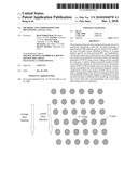 | 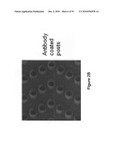 |
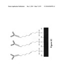 | 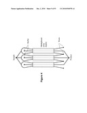 |
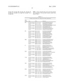 | 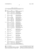 |
 | 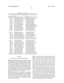 |
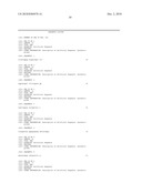 |  |
 |  |
 |  |
 |  |
 |  |
 |  |
 |  |
 |  |
 |  |
 |  |
 |  |
 |  |
 |  |
 | 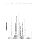 |
 |  |
 |  |
 |  |
 |  |
 |  |
 |  |
 |  |
 |  |
 |  |
 |  |
 | 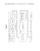 |
 |  |
 |  |
 |  |
 |  |
 |  |
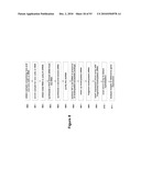 |  |
 |  |
 |  |
 |  |
 |  |
 | 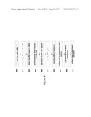 |
 |  |
 |  |
 |  |
 |  |
 | 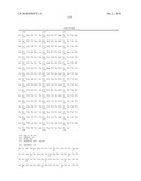 |
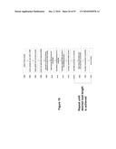 | 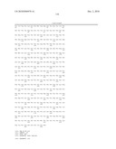 |
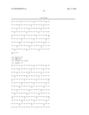 |  |
 |  |
 |  |
 |  |
 |  |
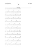 |  |
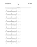 | 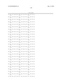 |
 |  |
 |  |
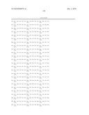 | 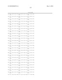 |
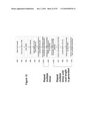 | 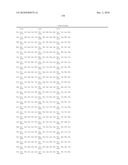 |
 |  |
 |  |
 | 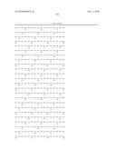 |
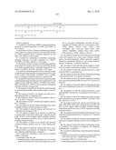 |  |
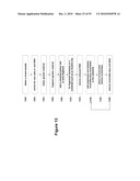 | 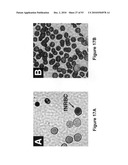 |
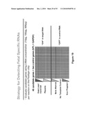 | 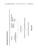 |
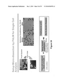 | 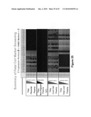 |
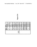 | 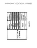 |
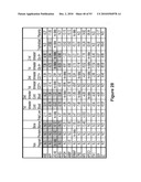 | 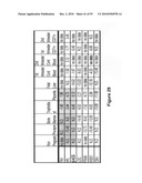 |
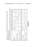 | 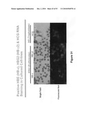 |
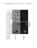 | 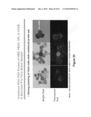 |
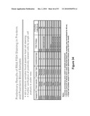 | 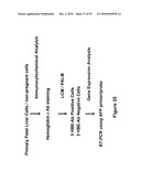 |
 | 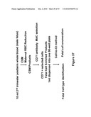 |
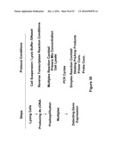 | 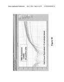 |
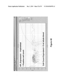 | 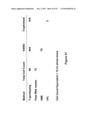 |
 | 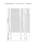 |
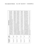 | 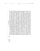 |
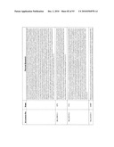 | 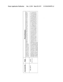 |
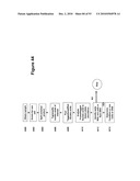 | 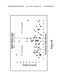 |
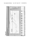 | 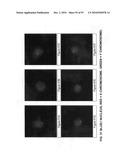 |
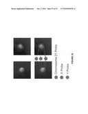 |
| Similar patent applications: | |
| Date | Title |
|---|---|
| 2012-08-16 | Methods and compositions for diagnosis of acute myocardial infarction (ami) |
| 2011-01-13 | Methods and compositions for increasing membrane permeability |
| 2012-06-28 | Methods and compositions for evaluating genetic markers |
| 2012-07-05 | Methods and compositions for diagnosing heart failure |
| 2012-06-28 | Methods and compositions for assessment of pulmonary function and disorders |
| New patent applications in this class: | |
| Date | Title |
|---|---|
| 2016-12-29 | Prediction of acute kidney injury from a post-surgical metabolic blood panel |
| 2016-09-01 | Microreactor system |
| 2016-06-30 | Sheath fluid systems and methods for particle analysis in blood samples |
| 2016-06-16 | Biomarkers of autism spectrum disorder |
| 2016-06-16 | Rigid mask for protecting selective portions of a chip, and use of the rigid mask |
| New patent applications from these inventors: | |
| Date | Title |
|---|---|
| 2021-12-09 | Apparatus and method for high throughput parallel nucleic acid sequencing on surfaces of microbeads |
| 2021-11-18 | Methods for targeted nucleic acid library formation |
| 2021-11-04 | Cell potency assay for therapeutic potential |
| 2021-01-14 | Diagnosis of fetal abnormalities using polymorphisms including short tandem repeats |
| Top Inventors for class "Combinatorial chemistry technology: method, library, apparatus" | |
| Rank | Inventor's name |
|---|---|
| 1 | Mehdi Azimi |
| 2 | Kia Silverbrook |
| 3 | Geoffrey Richard Facer |
| 4 | Alireza Moini |
| 5 | William Marshall |