Patent application title: MODIFIED GLOBIN PROTEINS WITH ALTERED ELECTRON TRANSPORT PATHWAY
Inventors:
Christopher Eric Cooper (Colchester, GB)
Michael Thomas Wilson (Colchester, GB)
Brandon Jon Reeder (Colchester, GB)
IPC8 Class: AA61K3842FI
USPC Class:
514 6
Class name: Designated organic active ingredient containing (doai) peptide containing (e.g., protein, peptones, fibrinogen, etc.) doai heavy metal containing (e.g., hemoglobin, etc.)
Publication date: 2010-06-03
Patent application number: 20100137189
Claims:
1. A modified porphyrin-based oxygen-carrying protein, said protein in an
unmodified state comprising a low affinity site of electron transfer, and
a high affinity electron transfer between a reductant and ferryl haem
iron via one or more protein amino acids, wherein said protein comprises
a modification to enhance or introduce a high affinity electron transfer
pathway to the iron ion.
2. The protein of claim 1 which is a mammalian haemoglobin chain subunit.
3. The protein of claim 1 which is a beta chain subunit or member of the beta chain superfamily.
4. The protein of claim 3 which is a human or beta, gamma or delta chain.
5. The protein of claim 1 which is modified by substitution of Phe41.
6. The protein of claim 5 wherein the modification is Phe41>Tyr.
7. The protein of claim 1 which is modified by insertion of an amino acid.
8. The protein of claim 7 wherein said insertion is of a redox-active amino acid into the C-helix of a haemoglobin.
9. The protein of claim 1 which comprises non-wild-type residue which reduces NO binding.
10. A protein comprising a dimer, tetramer or multimer of the protein of claim 1.
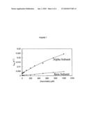
11. The protein of claim 10 wherein the dimer, tetramer or multimer is cross-linked.
12. A protein comprising two alpha globin subunits and two subunits of the protein of claim 1.
13. The protein of claim 1 conjugated to a protecting group.
14. The protein of claim 13 wherein the protecting group is an anti-oxidant enzyme.
15. The protein of claim 13 wherein the protecting group is a polyalkylene oxide.
16. A composition comprising the protein of claim 1 in a physiologically acceptable carrier.
17. A method of treatment of a human subject comprising administering to a subject in need of treatment an effective amount of the composition of claim 16.
18. A nucleic acid encoding the protein of claim 1.
19. An expression vector comprising the nucleic acid of claim 18 operably linked to a promoter.
20. A host cell comprising the expression vector of claim 19.
21. A method of making the protein of claim 1 which comprises expressing a protein in a host cell expressing a vector comprising a nucleic acid encoding the protein operably linked to a promoter and recovering the protein and optionally modifying the protein to provide said protein.
Description:
FIELD OF THE INVENTION
[0001]The present invention relates to modified oxygen-carrying compounds such as haemoglobin and their use.
BACKGROUND TO THE INVENTION
[0002]Transfusion of a patient with donated blood has a number of disadvantages. Firstly, there may be a shortage of a patient's blood type. Secondly, there is a danger that the donated blood may be contaminated with infectious agents such as hepatitis viruses and HIV. Thirdly, donated blood has a limited shelf life. In addition, there are some situations where blood may not be readily available, such as in a battlefield or civil emergencies.
[0003]An alternative to transfusion involves the use of a blood substitute. A blood substitute is an oxygen carrying solution that also provides the oncotic pressure necessary to maintain blood volume. Two types of substitutes have recently been studied, fluorocarbon emulsions and haemoglobin solutions.
[0004]Haemoglobin as it exists within the red blood cell is composed of two alpha globin chains and two beta globin chains, each carrying a haem molecule. One alpha-like globin chain and one beta-like globin chain combine to form a dimer which is very stable. Alpha-like and beta-like globin genes belong to a family of related globin genes which are expressed at different stages of development and regulated by oxygen tension, pH, and the development from embryo to fetus to newborn. Two dimers then line up in antiparallel fashion to form tetramers. The binding of dimers to form the tetramers is not as strong as in the case of monomers binding to associate into dimers. The tetramers, therefore, have a tendency to fall apart to form dimers and there is always an equilibrium between tetramers, dimers, and monomers. At high concentrations of globin, the predominant form is the tetramer; with dilution, the dimer becomes the predominant form. This equilibrium is also affected by solvent, salts, pH and other factors as the binding forces are partly electrostatic.
[0005]There are obstacles however to using native haemoglobin as a blood substitute. Firstly, large dosages are required, requiring large scale production of protein, either by recombinant means or from donated human or recovered non-human blood. Secondly, it is important to obtain haemoglobin that is free from infectious agents and toxic substances. Thirdly, although haemoglobin is normally a tetramer of 68,000 molecular weight, it can dissociate to form alpha-beta dimers. The dimers are rapidly cleared by the kidneys and the residence time is much too short for cell-free haemoglobin to be useful as a blood substitute.
[0006]Several approaches have been taken to circumvent these difficulties. These include the expression of haemoglobin via recombinant DNA systems, chemical modification of haemoglobin, and the production of haemoglobin variants. Haemoglobin and variants of it have been expressed in various cellular systems, including E. coli, yeast and mammalian cells such as CHO cells.
[0007]A number of naturally-occurring variants of haemoglobin are known. Variants include: variants which autopolymerize, variants which prevent the dissociation of the tetramer, and variants that are stable in alkali. There are also over 30 naturally occurring haemoglobin variants which exhibit lowered oxygen affinity. Several examples of such variants are disclosed in WO 88/091799.
[0008]Another approach to improving the use of haemoglobin is the modification of this protein by the addition of further polymers to improve the stability of the protein in the blood. For example, U.S. Pat. No. 5,900,402 describes the use of non antigenic polymers, preferably polyalkylene oxide or polyethylene glycol.
[0009]Because haemoglobins (and indeed myoglobins or other oxygen-carrying proteins) are involved in oxygen transport and storage they are, as a consequence of this function (because of the redox properties of the iron ion present in the porphyrin ring of protein), responsible for the generation of reactive oxygen species. Autoxidation of the oxy derivative (Fe(II)) leads to non-functional ferric haem (Fe(III)) and superoxide ion (O2.sup.•-), which subsequently dismutates to generate H2O2. These species can ultimately damage the protein and/or the haem group. An essential intermediate in the pathway leading to this damage is the ferryl haem (Fe(IV)=O2-), itself formed through the reaction of the haem with H2O2 and lipid peroxides. A protein/porphyrin-based radical cation (P+•) accompanies the formation of the ferryl haem from ferric haem and peroxide as set out in equation (1):
P--Fe(III)+H2O2→P.sup.•+Fe(IV)=O2-+H2O (1)
[0010]Ferryl haem and the radical can also be extremely toxic, notwithstanding their transient existence. These oxidative cascades can be damaging because: (i) peroxide is a powerful oxidant known to produce cellular damage, (ii) both the ferryl haem and protein-based radicals can initiate oxidation of lipids, nucleic and amino acids by abstraction of hydrogen atoms, and (iii) haem modification can lead to highly toxic haem to protein-cross-linked species and to the loss of haem and the release of the `free` iron.
[0011]The potential for haemoglobin-mediated peroxidative damage exists especially whenever the protein is removed from the protective environment of the erythrocyte. This would occur, for example, during spontaneous erythrocyte haemolysis or in haemolytic anaemias (e.g. sickle-cell anaemia). It has been shown that myoglobin induces kidney damage following crush injury (rhabdomyolysis) by exactly this peroxidative mechanism, rather than by free-iron catalysed Fenton chemistry as was thought previously (Holt et al, (1999) Increased lipid peroxidation in patients with rhabdomyolysis. Lancet 353, 1241; Moore, et al (1998) A causative role for redox) cycling of myoglobin and its inhibition by alkalinization in the pathogenesis and treatment of rhabdomyolysis-induced renal failure. J. Biol. Chem. 273, 31731-31737).
[0012]It has also been shown recently that haemoglobin can cause similar damage in vivo when it is released from the erythrocyte in subarachnoid haemorrhage (Reeder, et al (2002) Toxicity of myoglobin and haemoglobin: oxidative stress in patients with rhabdomyolysis and subarachnoid haemorrhage. Biochem. Soc. Trans. 30, 745-748). Furthermore, uncontrolled haem-mediated oxidative reactions of cell-free haemoglobin (developed as a blood substitute) have emerged as an important potential pathway of toxicity, either directly or via interactions with cell signalling pathways (Alayash, A. I. (2004) Oxygen therapeutics: can we tame haemoglobin? Nat. Rev. Drug Discovery 3, 152-159). The toxicity of ferryl haemoglobin has been demonstrated in an endothelial cell culture model system of ischaemia/reperfusion [McLeod, L. L. and Alayash, A. I. (1999) Detection of a ferryl-haemoglobin intermediate in an endothelial cell model after hypoxia-reoxygenation. Am. J. Physiol. 277, H92H99] and in cells that lack their antioxidant mechanisms such as glutathione (D'Agnillo & Alayash (2000) Interactions of haemoglobin with hydrogen peroxide alters thiol levels and course of endothelial cell death. Am. J. Physiol. Heart Circ. Physiol. 279, H1880-H1889).
[0013]Ferryl haemoglobin can cause cell injury, including apoptotic and necrotic cell death. Perfusion of rat intestine with chemically modified haemoglobin has been shown to cause localized oxidative stress, leading to leakage of the mesentery of radiolabelled albumin (Baldwin et al (2002) Comparison of effects of two haemoglobin-based O2 carriers on intestinal integrity and co microvascular leakage. Am. J. Physiol. Heart Circ. Physiol. 283, H1292-H1301). Importantly, the cyanomet derivative of this haemoglobin, in which the haem iron is blocked with cyanide and is unavailable to enter a redox reaction, produced no cellular changes.
[0014]U.S. Pat. No. 5,606,025 describes the conjugation of haemoglobin to superoxide dismutase and/or catalase as one approach to reduce reperfusion injuries and other free-radical mediated processes associated with haemoglobin blood substitutes.
DISCLOSURE OF THE INVENTION
[0015]Our studies have investigated in further detail the mechanisms by which the ferryl (IV) species is generated in haemoglobin (Hb) and myoglobin (Mb), and the mechanisms by which this ion is responsible for the generation of oxidative stress.
[0016]We have discovered that myoglobin and haemoglobin from certain species (including human) show two distinct pathways of electron transfer from exogenous reductants to the ferryl haem iron. A low affinity pathway (typically >5 mM) represents direct electron transfer from the reductant to the ferryl haem iron in a hydrophobic pocket within the protein. A second high affinity pathway (typically <100 μM but often <10 μM) involves electron transfer between the reductant and ferryl haem iron via one or more protein amino acids. This high affinity through-protein pathway is present in native human myoglobin and haemoglobin alpha subunit, but absent in human haemoglobin beta subunits and Aplysia myoglobin. An example of an amino acid that allows this electron transfer is Tyr103 and Tyr42 in myoglobin and haemoglobin alpha chain respectively.
[0017]Our finding is based on observing commonalities in the profiles for the reduction of ferryl Mb by many iron chelators, which we have observed to have anti-oxidant properties. The effect of these reducing agents on the rate constant for ferryl decay plotted as a function of reductant concentration exhibits a complex curve that can be expressed as a double rectangular hyperbola function. We have also now found that more classical reducing agents such as ascorbate also show this double rectangular hyperbola concentration dependence. The two hyperbolae represent two binding sites for the reductant having differing affinities. Through the use of kinetic model, simulations and use of selected native and engineered proteins, we have interpreted this concentration dependence to represent two distinct electron transfer pathways from the reductant to the haem iron.
[0018]This high affinity pathway, though present in the alpha chain of haemoglobin, and in myoglobin, does not appear to be as active in other porphyrin-based oxygen-carrying proteins which lack a tyrosine residue at the equivalent to position 42. This is particularly the case with beta-haemoglobin.
[0019]According to the present invention, we propose the introduction of this pathway into such proteins by introduction of an amino acid equivalent to Tyr42, or any other residue able to constitute a high affinity electron transfer pathway to the ferryl ion. Such a modification will increase the ability of the ferryl ion to be reduced via this pathway. Effectively, such modifications expose the ferryl ion such that this ion may be more rapidly reduced and thus lessen its ability to damage surrounding tissues or substrates, such as lipids.
[0020]Thus the present invention provides a modified porphyrin-based oxygen-carrying protein, wherein said protein comprises a modification to enhance or introduce a high affinity electron transfer pathway to the ferryl ion.
[0021]The protein is preferably a haemoglobin beta chain (HO). The modification may be to Phe41, particularly wherein Phe41 is substituted by Tyr.
[0022]The invention also provides nucleic acids encoding these proteins, means for their production and the use of the proteins in methods of treatment. These and other aspects of the invention are described further herein below.
BRIEF DESCRIPTION OF THE DRAWINGS
[0023]FIG. 1: Concentration dependence ferryl reduction of different myoglobin species with reducing agent deferiprone. Horse myoglobin ( ), but not Aplysia myoglobin (.box-solid.), shows a double rectangular hyperbola concentration dependence.
[0024]FIG. 2: Concentration dependence of recombinant sperm whale myoglobin with reducing agent deferiprone. Wild type sperm whale myoglobin (.diamond-solid.), but not the Tyr103>Phe mutant of sperm whale myoglobin (.tangle-solidup.), shows a double rectangular hyperbola concentration dependence.
[0025]FIG. 3: The proposed two site model for reduction of a haemoprotein possessing a high affinity through protein electron transfer pathway. Reductant (xH) binds to two possible sites on myoglobin in its ferryl oxidation state (P--[Fe(IV)=O2-]2+, where P denotes protein) with affinities KD1 and KD2. Only from these two sites can electron transfer from the reductant to the ferryl iron take place. The high affinity binding site is situated at, or close to, tyrosine 103 (PxH--[Fe(IV)=O2-]), allowing the transfer of an electron (kmax≈0.01 s-1) through the protein to the ferryl haem iron to generate the ferric protein (P--Fe(III)). The low affinity site is situated in the haem pocket allowing electron transfer directly between the reductant and the ferryl haem (P--[Fe(IV)=O2-]xH). This model also allows both sites being filled by reductant (PxH--[Fe(IV)=O2-]xH). For the kinetic simulations it is assumed that the binding on one site will not affect the affinity of binding to the second site. The model also incorporates a step in which the oxidized reductant may be regenerated.
[0026]FIG. 4: Position of residues that can introduce or eliminate through-protein electron transfer in alpha human haemoglobin (A), beta human haemoglobin (B) with comparison of known electron conduit residue Tyr103 of horse myoglobin (C) from crystal structures. Tyr103 of horse myoglobin is close to the haem and is surface exposed making it ideal to act as an electron conduit from exogenous reductants to the ferryl haem iron. Human haemoglobin alpha subunit has a tyrosine in approximately the same spatial environment (Tyr42), however this residue in human haemoglobin beta is a redox-inactive phenylalanine.
[0027]FIG. 5: Concentration dependence of wild type ferryl human haemoglobin reduction by ascorbate. Ferryl myoglobin (10 μM) was reacted with ascorbate in sodium phosphate pH 7.4. Ferryl reduction rate constants for alpha subunit ( ) and beta (◯) subunit calculated by fitting to a double exponential function. The alpha subunit, but not the beta subunit shows double rectangular hyperbola concentration dependence.
[0028]FIG. 6: Concentration dependence of recombinant α-Tyr42>Val ferryl human haemoglobin reduction by ascorbate. Ferryl myoglobin (10 μM) was reacted with ascorbate in sodium phosphate pH 7.4. Ferryl reduction rate constants for alpha subunit ( ) and beta (◯) subunit calculated by fitting to a double exponential function. Both the alpha subunit and beta subunit shows single rectangular hyperbola concentration dependencies.
[0029]FIG. 7: Concentration dependence of recombinant α-Tyr42>Trp ferryl human haemoglobin reduction by ascorbate. Ferryl myoglobin (10 μM) was reacted with ascorbate in sodium phosphate pH 7.4. Ferryl reduction rate constants for alpha subunit ( ) and beta (◯) subunit calculated by fitting to a double exponential function. Both the alpha subunit and beta subunit shows single rectangular hyperbola concentration dependencies.
DETAILED DESCRIPTION OF THE INVENTION
Porphyrin-Based Oxygen-Carrying Protein
[0030]A porphyrin-based oxygen carrying protein refers to any polypeptide chain which in its native form carries a porphyrin molecule and which polypeptide, either alone or in a complex, carries and releases oxygen bound to the porphyrin molecule. Variants of such proteins, e.g. naturally occurring or synthetic mutants of wild-type porphyrin-based oxygen-carrying proteins are also contemplated by the invention.
[0031]The porphyrin-based oxygen-carrying protein will, in its native state, lack the high affinity oxygen transfer pathway present in the haemoglobin alpha subunit, e.g. the protein of SEQ ID NO:1 or its mammalian homologues. This pathway is mediated via an electron transfer pathway which includes a tyrosine residue at position 7 of the C helix. (As is known in the art, haemoglobin subunit proteins are also numbered by reference to the residues of individual helices or inter-helix resides, as set out in Table 1 below (based on U.S. Pat. No. 5,028,588 the contents of which are incorporated herein by reference). Tyr42 of human haemoglobin alpha chain is thus also identified in the art as residue C7. Accordingly, the equivalent residue in other haemoglobin alpha chains will also be in the C7 position.) The pathway is an electron transfer) between the reductant and ferryl haem iron via one or more protein amino acids. This high affinity through-protein pathway is present in native human myoglobin and haemoglobin alpha subunit, but absent in human haemoglobin beta subunits.
[0032]The oxygen-carrying proteins to be modified include mammalian haemoglobin subunits but may include non-mammalian haemoproteins and other genetically engineered proteins where the protein is altered to carry oxygen. These proteins will be recombinant, having altered sequences (substitution of amino acid residues, but may also include insertion of residues) to introduce a high affinity through-protein electron pathway from reductants in the bulk solution to the haem ferryl iron.
[0033]It is expected that by introducing the high affinity pathway in proteins such as human haemoglobin beta subunit we will be able to decrease the toxicity of blood substitutes by allowing reductants that are administered with the blood substitute such as ascorbate, urate or deferiprone to more rapidly reduce the highly toxic ferryl oxidation state of these haemoproteins and thus limiting oxidation of substrates such as lipids and DNA.
[0034]Thus the invention is applicable to any haemoglobin subunit which in its natural state does not have the high-affinity pathway. In one aspect, the protein is human haemoglobin beta chain, whose sequence is set out as SEQ ID NO:2 below. Other haemoglobin subunits which may be used are those which are vertebrate or non-vertebrate haemoglobin subunits that do not have a tyrosine residue at a position equivalent to residue 42/C7.
[0035]Vertebrate haemoglobins include mammalian haemoglobins. Mammalian haemoglobins are particularly highly conserved. Examples of homologues to the human beta chain include members of the beta globin superfamily, such as gamma or delta haemoglobin found in haemoglobin F (HbF) or haemoglobin A2 (HbA2) respectively. These and other non-limiting examples of such homologues include the mammalian species homologues of Table 2, all of which also have a Phe42 residue. The sequences may be obtained from on-line databases including via the Research Collaboratory for Structural Bioinformatics protein databank (pdb). These pdb references provide sequences for the haemoglobin beta-family subunit proteins and, where applicable, a corresponding alpha subunit protein. The beta-family subunit sequence may be used either with its corresponding alpha subunit or alone, or in combination with another beta-family subunit protein.
TABLE-US-00001 TABLE 2 Species PDB Code Human (Homo sapiens) HbF 1FDH Human (Homo sapiens) HbA2 1SI4 Bovine (Bos taurus) 1G09 Pig (Sus scrofa) 2PGH Horse (Equus caballus) 1NS6 Maned Wolf (Chrysocyon brachyurus) 1FHJ Deer (Odocoileus virginianus virginianus) 1HDS Donkey (Equus asinus) 1S0H Crab Eating Fox (Cerdocyon thous) 2B7H
[0036]Other vertebrate haemoglobins alpha subunit homologues include avian, reptile and fish haemoglobins having a residue equivalent to Phe42. Non-limiting examples of such subunits include those given in Table 3, which indicates in column 3 the position of the amino acid residue homologous to Phe42 of mammalian beta chain subunits.
TABLE-US-00002 TABLE 3 Phe 42 Equivalent Species PDB Code Residue Avian: Bar Headed Goose (Anser indicus) 1A4F Phe41 Graylag Goose (Anser anser) 1FAW Phe41 Chicken (Gallus gallus) 1HBR Phe41 Reptile: Giant Tortoise (Geochelone gigantean) 1V75 Phe41 Fish: Akaei (Dasyatis akajei) 1CG5 Phe42 Antarctic fish (Pagothenia bernacchii) 1PBX Phe42
[0037]Other non-vertebrate eukaryote beta haemoglobin homologues may be identified in arthropods or other multicellular organisms (e.g. molluscs, nematode worms and non-nematode worms) and those of unicellular organisms.
Modification of the Oxygen-Carrying Protein
[0038]The oxygen carrying protein may be modified by substitution or insertion to introduce a high-affinity electron transfer pathway.
[0039]In particular and as illustrated in FIG. 5, the phenylalanine of the beta subunit of haemoglobin is located close to the haem and is surface exposed making it ideal to act as an electron conduit from exogenous reductants to the ferryl haem iron were it to be a redox-active group. Thus the invention provides an insertion or substitution to provide a redox-active residue at a similar position.
[0040]The redox-active amino acid may be, for example, tyrosine, tryptophan or histidine. Tyrosine is particularly preferred.
[0041]Thus in one aspect the invention provides a mammalian beta globin subunit modified to position a redox-active residue in the protein such that protein exhibits a high affinity electron transfer pathway. The presence of this pathway may be observed by showing that the protein has a double rectangular hyperbola concentration dependence to reducing agents.
[0042]In one aspect, the C7 position is substituted to introduce a redox-active amino acid, particularly those mentioned above. The beta globin subunit alternatively may include an insertion to introduce this residue into the protein, e.g. into the C helix, e.g. between the C6 and C7 residues, or between the C7 and CD1 residues. Such an insertion may be combined with a further modification of the C helix of the protein, e.g. a deletion or substitution of a residue in the C1 to C6 positions.
[0043]Molecular modelling methods may be used to identify other positions in the beta chain of mammalian haemoglobins which are also located near the ferryl haem iron and which may be substituted to introduce a redox-active amino acid.
Other Modifications
[0044]In one embodiment, in addition to the attenuating modification of a wild-type oxygen-carrying protein, the protein may comprise one or more (for example from one to five, such as one or two) additional substitutions, or a deletion or insertion of from one to five, such as one, two or three amino acids (which may be contiguous or non-contiguous). These may be variations which affect a further property of the protein, such as its oxygen affinity or cooperativity, enhancements in stability and assembly rates, decreased heme loss rates or autoxidation rates, or resistance to proteolytic degradation and aggregation, its binding to nitric oxide or its ability to be produced in a soluble form by recombinant means. Such modifications are known in the art per se and may be incorporated into the proteins of the present invention.
[0045]In a preferred aspect, the modification is one which reduces the binding of nitric oxide (NO). A number of haemoglobin variants which limit NO binding while still permitting oxygen transport are known. A number of variants of haemoglobin beta chains which have reduced rates of reaction with nitric oxide are disclosed in U.S. Pat. No. 6,455,676, the contents of which are incorporated herein by reference.
[0046]In particular, the following changes may be included in the oxygen carrying protein in addition to the attenuating modification:
[0047]B13(Leu>Phe or Trp); G12(Leu>Phe or Trp); B10(Leu>Phe) and E4(Val>Leu); B10(Leu>Trp) and E4(Val>Leu); B14(Leu>Phe or Trp); G8(Leu>Phe) and G12(Leu>Trp); E11(Val>Leu) and G8(Leu>Trp); E11(Val>Trp) and G8(Leu>Met); E11(Val>Leu) and G8(Leu>Phe); E11(Val>Leu) and G8(Leu>Met); E11(Val>Phe) and G8(Leu>Ile); E11(Val>Phe) and G8(Leu>Phe); E11(Val>Phe) and G8(Leu>Trp); E11(Val>Phe) and G8(Leu>Met); E11(Val>Met) and G8(Leu>Trp); E11(Val>Met) and G8(Leu>Trp) and E7(His>Gln); E11 (Val>Trp) and G8(Leu>Ile); E7(His>Gln) and E11(Val>Trp); E7(His>Gln) and E11(Val>Leu); E7(His>Gln) and E11(Val>Phe); E7(His>Gln) and E11(Val>Phe) and G8(Leu>Phe or Trp); E7(His>Gln) and E11(Val>Leu or Trp) and G8(Leu>Phe or Trp); E11(Val>Trp or Phe) and G12(Leu>Trp or Met); E11(Val>Trp or Phe) and B13(Leu>Trp or Met); B10(Leu>Trp) and B13(Leu>Trp or Met); B10(Leu>Phe) and B13(Leu>Trp); B10(Leu>Trp or Phe) and G12(Leu>Trp); B10(Leu>Phe) and G12(Leu>Met); G8(Leu>Trp) and G12(Leu>Trp or Met); or G8(Leu>Trp) and B13(Leu>Trp or Met).
[0048]The numbering used above is based on helix chain numbering, which can be cross-referenced to the primary sequence numbering of Table 1 for the human beta chain. These modifications at equivalent positions in other oxygen carrying proteins may also be made.
Protein Multimers
[0049]In one embodiment, oxygen-carrying proteins are present in multimeric forms. Such forms may prolong life of the protein in circulation, improve oxygen-carrying capacity or reduce side-effects.
[0050]In the case of haemoglobin beta subunit proteins such forms include a tetrameric haemoglobin protein. In this form two beta chains may form a tetramer with two alpha chains. Optionally, two or more of the subunits may be covalently linked to each other, e.g. via chemical cross-linking or as a result of recombinant expression.
[0051]The alpha chains may be wild type alpha chains, e.g. a human alpha chain of SEQ ID NO:1 or a homologous vertebrate or non-vertebrate alpha chain. Vertebrate alpha chains include mammalian, avian, reptile and fish alpha chains. Such alpha chains include those found with their associated with the beta chains referred to above in Tables 2 to 4 and whose sequences are obtainable from the pdb entries.
[0052]The alpha chains may comprise one or more (for example from one to five, such as one or two) additional substitutions, or a deletion or insertion of from one to five, such as one, two or three amino acids (which may be contiguous or non-contiguous). These may be variations which affect a further property of the protein, such as its interaction with other proteins, its binding to nitric oxide or to facilitate its production by recombinant means.
[0053]In one aspect, the alpha chain may be modified to remove tyrosine residue at position 42, e.g. by deletion or by substitution with a redox-inactive residue. Amino acids which are contemplated here as redox-inactive residues include alanine, arginine, asparagine, aspartic acid, cysteine, glutamic acid, glutamine, glycine, isoleucine, leucine, lysine, methionine, phenylalanine, proline, serine, threonine and valine
[0054]Particular changes to the alpha-chain contemplated which modify binding to nitric oxide include E11(Val>Leu) and E7(His>Gln); E11(Val>Phe or Trp) and E7(His>Gln); E11(Val>Phe or Trp or Leu) and E7(His>Gln) and G8(Leu>Phe or Trp); B10(Leu>Phe) and E4(Val>Leu); B10(Leu>Trp) and E4(Val>Leu); B10(Leu>Trp) and E7(His>Gln); B10(Leu>Trp) and E11(Val>Phe); B10(Leu>Trp) and E11 (Val>Trp); B10(Leu>Trp) and E11(Val>Leu) and G8(Leu>Trp); B10(Leu>Trp) and E11(Val>Leu) and G8(Leu>Phe); B10(Leu>Trp) and E11(Val>Phe) and G8(Leu>Trp); B10(Leu>Trp) and E11(Val>Phe) and G8(Leu>Ilc); B10(Leu>Trp) and E7(His>Gln) and E11(Val>Leu) and G8(Leu>Trp); B10(Leu>Trp) and E11(Val>Trp) and G8(Leu>Trp); E11(Val>Leu) and G8(Leu>Phe); E11(Val>Leu) and G8(Leu>Trp); B13(Met>Phe or Trp); G12(Leu>Phe or Trp); or B14(Phe>Trp)).
[0055]The numbering used above is based on helix chain numbering, which can be cross-referenced to the primary sequence numbering of Table 1 for the human alpha chain. These modifications at equivalent positions in other oxygen carrying proteins may also be made.
[0056]Other higher order forms, either covalently or non-covalently associated with each other and/or with other oxygen-carrying proteins may also be provided. For example, polymerized haemoglobin subunit chains or cross-linked chains are known in the art per se and these approaches may be applied to the present invention.
Protecting Groups
[0057]In another aspect, the oxygen-carrying proteins of the invention, whether in monomeric or multimeric form, may be conjugated to a protecting group. Various types of protecting groups are known as such in the art and may be used in the present invention. Where the protecting group is a protein, this protecting group may be produced as a fusion, e.g. at the N- or C-terminus of the oxygen-carrying protein. Alternatively, the protein may be co-expressed with the oxygen-carrying protein or expressed separately, and the two proteins joined by chemical) means using a cross-linker.
[0058]For example, one class of protecting groups are enzymatic anti-oxidant proteins. These include catalase and superoxide dismutase (SOD). Any suitable catalase or SOD may be used, though preferably these are human enzymes. The enzymes may be produced recombinantly or by any other means conventional in the art.
[0059]U.S. Pat. No. 5,606,025, the contents of which are incorporated herein by reference, describes the conjugation of such enzymes to a haemoglobin and such methods may be used in the present invention. Thus any suitable inert cross-linking reagent previously reported as suitable for preparing cross-linked haemoglobin for use as an oxygen-carrying resuscitative fluid can be used, for example glutaraldehyde, diasprin derivatives, polyaldehydes including those derived from oxidative ring-opening of oligosaccharides, diphosphate esters, triphosphate esters, etc. The enzymes of interest have chemical groups similar to those on the globin chains of haemoglobin so that they will appropriately chemically bind to the haemoglobin as it cross-links by reaction with the cross-linking reagent.
[0060]Relative amounts of the oxygen-carrying protein and the enzymatic anti-oxidant protein can vary over wide limits, with the oxygen-carrying protein constituting the major component. The total weight of the enzyme(s) is suitably in the approximate range of 0.1-10% based on the weight of the oxygen-carrying protein, and preferably in the approximate range 0.5-2.5%. When, as in one embodiment, both SOD and catalase are chemically bound to the polyhaemoglobin, the weight ratio of SOD to catalase is suitably from about 1:1 to 5:1 and preferably from about 1.5:1 to 2.5:1.
[0061]Another class of protecting group, which may be used as well as the above-described enzymatic groups, or in the alternative, is a non-antigenic polymeric group such as a polyalkylene oxide protecting group. Such groups may also be used on monomeric oxygen-carrying proteins or these proteins when in dimeric or higher form.
[0062]For example, U.S. Pat. No. 5,900,402, the contents of which are incorporated herein by reference, describes the conjugation of polyalkylene oxides, most preferably polyethylene glycol (PEG) to oxygen-carrying proteins.
[0063]The conjugate is preferably formed by covalently bonding a hydroxyl terminal of the polyalkylene oxide and the free amino groups of lysine residues of the oxygen-carrying protein. See, for example, U.S. Pat. No. 5,234,903, which discloses mPEG-succinimidyl carbonate-Hb conjugates. Other methods for conjugating the polymers with oxygen-carrying proteins are known in the art as such, such as by via an amide or ester linkage, are also suitable for use with the present invention. While epsilon amino group modifications of haemoglobin lysines are preferred, other conjugation methods are also contemplated. Covalent linkage by any atom between the haemoglobin and polymer is possible. Moreover, non-covalent conjugation such as lipophilic or hydrophilic interactions are also contemplated.
[0064]Additional examples of activated polymers which are suitable for covalently conjugating the oxygen carrying proteins are described in U.S. Pat. Nos. 5,349,001; 5,321,095; 5,324,844 and 5,605,976 as well as PCT Publication Numbers WO95/11924 and WO96/00080, the disclosure of each of which is incorporated herein by reference.
[0065]The conjugates preferably include polyethylene glycol (PEG) as the polyalkylene oxide. The polyalkylene oxides include monomethoxy-polyethylene glycol, polypropylene glycol, block copolymers of polyethylene glycol and polypropylene glycol and the like. The polymers can also be distally capped with C2-4, alkyls instead of monomethoxy groups.
[0066]To be suitable for use herein, the polyalkylene oxides must be soluble in water at room temperature. Polyalkylene oxide strands having a (number average) molecular weight of from about 200 to about 100,000 Daltons can be used. For example, preferable PAOs have molecular weights of from about 1,000 to about 30,000 while PAOs having a molecular weight of from about 2,000 to about 25,000 are more preferred. Some particularly preferred conjugates of the present invention include polyalkylene oxide strands having a molecular weight of about 5,000 Daltons.
[0067]The ratio of the number of strands of the non-antigenic polymeric group to the oxygen-carrying protein may be from about 1:1 to about 20:1, preferably from about 5:1 to 15:1, for example about 10:1. The strands may be of the size ranges specified above.
[0068]Overall, the molecular weight of a monomer of an oxygen-carrying protein prior to conjugation is about 17,000 Da. Where such a protein is conjugated to a non-antigenic polymeric group as described above, the conjugate will be from about 30% to 60%, such as about 45% to 55% by weight of protein (i.e. the oxygen-carrying protein or a conjugate of this protein and an enzymatic group), the remainder being the non-antigenic polymeric group.
[0069]An exemplary embodiment of the invention is thus a conjugate of an oxygen-carrying protein of to the invention and 45% to 55% by weight of polyalkylene oxide having a molecular weight of from about 2,000 to about 25,000. In one aspect of this embodiment, the oxygen-carrying protein may be a haemoglobin beta chain in which the modification is at Phe41. In another aspect of this embodiment, the polyalkylene oxide is PEG. In a further aspect, the oxygen-carrying protein is a haemoglobin alpha chain in which the attenuating modification is at Phe41 and the polyalkylene oxide is PEG.
[0070]In the above embodiments, the oxygen-carrying protein may be in the form of a monomer or a polymer of two or more units.
Compositions
[0071]The oxygen-carrying proteins of the invention are desirably formulated as a composition comprising a physiologically acceptable carrier, suitable for administration to a mammal, particularly a human. Generally, such a carrier will be a sterile solution which comprises buffers and preservatives used to keep the solution at physiological pH and stable during storage. The carriers may be such physiologically compatible buffers as Hank's or Ringer's solution, physiological saline, a mixture consisting of saline and glucose, and heparinized sodium-citrate-citric acid-dextrose solution. The oxygen-carrying proteins of the present invention can be mixed with colloidal-like plasma substitutes and plasma expanders such as linear polysaccharides (e.g. dextran), hydroxyethyl starch, balanced fluid gelatin, and other plasma proteins. Additionally, the oxygen-carrying proteins may be mixed with water soluble, physiologically acceptable, polymeric plasma substitutes, examples of which include polyvinyl alcohol, poly(ethylene oxide), polyvinylpyrrolidone, and ethylene oxide-polypropylene glycol condensates.
[0072]Compositions of the invention may further include one or more compounds with anti-oxidant properties. These compounds may include ascorbate and urate. The anti-oxidant may be included at any suitable concentration, which may vary according to intended use and the nature of the anti-oxidant. For example, a suitable concentration of urate may be in the range of from 50 to 400 micromolar, and for ascorbate of from 50 to 200 micromolar, though lower or higher amounts may be used if need be.
[0073]The compositions may also include iron chelating agents which may play a role in sequestering iron released by the breakdown of the oxygen-carrying protein. Examples of such iron chelating agents include desferrioxamine and deferiprone. The iron chelating agent, or mixture thereof, may be present at a concentration of, for example, 10-5000 micromolar.
Administration of Oxygen-Carrying Proteins
[0074]Proteins of the invention may be used as blood substitutes. There are numerous conditions in which it will be useful for restoration, maintenance or replacement of oxygen levels is required. These include trauma; ischemia (such as ischemia induced by heart attack, stroke, or cerebrovascular trauma); haemodilution, where a blood substitute is required to replace blood that is removed pre-operatively; septic shock; cancer (e.g. to deliver oxygen to the hypoxic inner core of a tumour mass); chronic anaemia; sickle cell anaemia; cardioplegia; and hypoxia. Thus the oxygen-carrying proteins, and compositions thereof, of the present invention may be used in methods for the treatment of the above-mentioned conditions.
[0075]The oxygen-carrying proteins, and compositions thereof, of the present invention may also be used ex vivo in organ perfusion. Blood substitutes may be particularly useful in the organ perfusion, where maintaining oxygen content in an organ ex vivo prior to transplantation is required to sustain the organ in an acceptable condition. Organs include heart, liver, lung, kidneys.
[0076]The concentration and amount of oxygen-carrying protein of the invention used in any of the above-mentioned methods will be at the discretion of the physician, taking account of the nature of the condition of the patient and the treatment. Typically, the oxygen-carrying protein may be used at a concentration of from 0.1 to 6 g/dl, e.g. from 0.1 to 4 g/dl. The oxygen-carrying protein will usually be administered intravenously.
[0077]Co-administration of an innocuous reagent to enhance nitric oxide production (e.g. arginine) is also envisaged.
Nucleic Acids
[0078]The invention also provides nucleic acids encoding the modified oxygen-carrying proteins of the invention. The nucleic acid may be DNA or RNA. The DNA may be single- or double-stranded.
[0079]The nucleic acid of the invention may in isolated and/or purified form, or free or substantially free of material with which it is naturally associated, such as free or substantially free of nucleic acid flanking the gene in the human genome, except possibly one or more regulatory sequence(s) for expression.
[0080]Generally, nucleic acids of the invention may be obtained by modification of wild-type sequences encoding the oxygen-carrying protein. The nucleic acid sequences of wild-type haemoglobins and other oxygen carrying proteins are known in the art and widely available. Generally, recombinant techniques such as site-directed mutagenesis may be used to modify a known wild-type sequence such that the sequence encodes a modified oxygen-carrying protein of the invention.
[0081]The wild-type sequence of a mammalian nucleic acid may also be modified to optimize codon usage for expression in a heterologous system, e.g. in bacterial or yeast cells.
[0082]A nucleic acid of the invention may be incorporated into a recombinant replicable vector. The vector may be used to replicate the nucleic acid in a compatible host cell. Thus in a further embodiment, the invention provides a method of making a nucleic acid of the invention by introducing a nucleic acid of the invention into a replicable vector, introducing the vector into a compatible host cell, and growing the host cell under conditions which bring about replication of the vector.
[0083]Preferably, a nucleic acid of the invention in a vector is operably linked to a control sequence which is capable of providing for the expression of the coding sequence by the host cell, i.e. the vector is an expression vector.
[0084]The term "operably linked" refers to a juxtaposition wherein the components described are in a relationship permitting them to function in their intended manner. A control sequence "operably linked" to a coding sequence is ligated in such a way that expression of the coding sequence is achieved under condition compatible with the control sequences.
[0085]Suitable vectors can be chosen or constructed, containing appropriate regulatory sequences, including promoter sequences, terminator fragments, polyadenylation sequences, enhancer sequences, marker genes and other sequences as appropriate. Vectors may be plasmids, viral e.g. `phage phagemid or baculoviral, cosmids, YACs, BACs, or PACs as appropriate. Vectors include gene therapy vectors, for example vectors based on adenovirus, adeno-associated virus, retrovirus (such as HIV or MLV) or alpha virus vectors.
[0086]The vectors may be provided with an origin of replication, optionally a promoter for the expression of the oxygen-carrying protein and optionally a regulator of the promoter. The vectors may contain one or more selectable marker genes, for example an ampicillin resistance gene in the case of a bacterial plasmid or a neomycin resistance gene for a mammalian vector. Vectors may be used in vitro, for example for the production of RNA or used to transfect or transform a host cell. Systems for cloning and expression of a polypeptide in a variety of different host cells are well known. Suitable host cells include bacteria, eukaryotic cells such as mammalian and yeast, and baculovirus systems. Mammalian cell lines available in the art for expression of a heterologous polypeptide include Chinese hamster ovary cells, HeLa cells, baby hamster kidney cells, COS cells and many others.
[0087]Promoters and other expression regulation signals may be selected to be compatible with the host cell for which the expression vector is designed. For example, yeast promoters include S. cerevisiae GAL4 and ADH promoters, S. pombe nmt1 and adh promoter. Mammalian promoters include the metallothionein promoter which is can be induced in response to heavy metals such as cadmium. Viral promoters such as the SV40 large T antigen promoter or adenovirus promoters may also be used. All these promoters are readily available in the art.
[0088]The vectors may include other sequences such as promoters or enhancers to drive the expression of the inserted nucleic acid, nucleic acid sequences so that the oxygen-carrying protein is produced as a fusion and/or nucleic acid encoding secretion signals so that the polypeptide produced in the host cell is secreted from the cell.
[0089]Vectors for production of polypeptides of the invention for use in gene therapy include vectors which carry a mini-gene sequence of the invention.
Host Cells and Production of Oxygen-Carrying Proteins
[0090]Host cells according to the invention such as those mentioned herein above may be cultured under conditions to bring about expression of the oxygen-carrying protein, followed by recovery of the protein to provide the protein in substantially isolated form.
[0091]The protein may be produced with a source of haem or may be mixed with a suitable source of haem such as ferro-protoporphyrin or ferri-protoporphyrin (hemin) during or after recovery in order to provide a functional oxygen-carrying protein.
[0092]For example, U.S. Pat. No. 5,801,019, the contents of which are incorporated herein by reference, describes expression and recovery of various modified haemoglobins, including multimers of haemoglobin subunits, in yeast cells. Such methods may be used in the present invention for the production of the oxygen-carrying proteins.
[0093]Where the oxygen-carrying protein is a haemoglobin beta chain subunit, it may be co-expressed with complementary subunits, e.g. a alpha chain subunit. The co-expressed protein may be in the form of a separate protein or a fusion with the beta chain subunit.
[0094]Following expression, the proteins are recovered using standard methods including but not limited to chromatography (e.g., ion exchange, affinity, and sizing column chromatography), centrifugation, differential solubility, or by any other standard technique for the purification of proteins.
EXAMPLES
[0095]The present invention is illustrated further by the following examples.
Example 1
[0096]Myoglobin from Horse, but not Aplysia, possesses a high affinity through-protein electron transfer pathway as measured by ferryl reduction kinetics
[0097]In this example the kinetics of the reduction of ferryl haem from myoglobin from different species to ferric haem protein were examined kinetically to determine the presence or absence of a high affinity-through protein pathway. Following the procedure, ferric horse myoglobin from Sigma-Aldrich (Poole, Dorset, UK, further purified by gel filtration), or recombinant ferric Aplysia myoglobin Mb (20 μM) in 5 mM sodium phosphate pH 7.4 was reacted with H2O2 (20 μM) at 25° C. for 15 min. At this time conversion of ferric myoglobin to ferryl myoglobin was greater than 95% as measured by addition of sodium sulfide (1 mM). Catalase (10 nM) was added to remove unreacted H2O2 and was left to react for a further 1 min. At the pH used the ferryl haem protein is stable for several hours. Reductant was then added in 0.1M sodium phosphate pH 7.4 in a 1:1 volume ratio so that final concentration of ferryl myoglobin was 10 μM. The pH may be `jumped` to other values where the ferryl haem is unstable using other strong buffers (e.g. 0.1M sodium acetate, pH 5). The optical spectrum was followed until reaction was complete. The time course (425 nm-408 nm) was fitted to a single exponential function using the least squares method. These rate constants were then plotted as a function of reductant concentration and this profile fitted (least squares method) to a double rectangular hyperbola (FIG. 1):
k obs = k a [ S ] [ S ] + K D 1 + k b [ S ] [ S ] + K D 2 + A R ##EQU00001##
[0098]Where ka and kb are the maximum rates for each hyperbola and KD1 and KD2 are the dissociation constants, S is the concentration of the reductant and AR is the rate constant for ferryl auto-reduction.
[0099]The dependence of the concentration of reductant on the changes in the observed rate constant for ferryl myoglobin reduction from horse shows a double rectangular hyperbola dependence that is not observed in Aplysia myoglobin. Myoglobin from horse has two tyrosine residues at positions 103 and 146, while Aplysia myoglobin has no tyrosine residues.
Example 2
[0100]A tyrosine residue close to the haem is key to the high affinity through-protein electron transfer pathway.
[0101]Wild-type and recombinant sperm whale myoglobin and recombinant Tyr103>Phe sperm whale myoglobin were reacted with peroxide and the kinetics of ferryl haem reduction by deferiprone determined as described in example 1. The through-protein electron transfer pathway, evident in wild type sperm whale myoglobin, is not observed in the Tyr103>Phe mutant (FIG. 2). This demonstrates that the presence of a redox active tyrosine, interfacing the haem and the external environment, is a key component for the high affinity pathway.
[0102]The data from FIGS. 1 and 2 can be rationalised by a two site model where a reductant (xH) binds to two possible sites on myoglobin in its ferryl oxidation state (P--[Fe(IV)=O2-]2+, where P denotes protein, FIG. 3) with affinities KD1 and KD2. Only from these two sites can electron transfer from the reductant to the ferryl iron take place. The high affinity binding site is situated at, or close to, Tyr103 (PxH--[Fe(IV)=O2-]), allowing the transfer of an electron (kmax≈0.01 s-1) through the protein to the ferryl haem iron to generate the ferric protein (P--Fe(III)). The low affinity site is situated in the haem pocket allowing electron transfer directly between the reductant and the ferryl haem (P--[Fe(IV)=O2-]xH). This model also allows both sites being filled by reductant (PxH--[Fe(IV)=O2-]xH). For the kinetic simulations it is assumed that the binding on one site will not affect the affinity of binding to the second site. The model incorporates a step in which the oxidized reductant may be regenerated. This model also allows predictions on other haem proteins based on crystal structures. The structure of alpha subunit of human haemoglobin shows a tyrosine in a similar spatial position (Tyr42) compared to myoglobin, close to the haem and is surface exposed making it ideal to act as an electron conduit from exogenous reductants to the ferryl haem iron (FIG. 4). The corresponding residue in human haemoglobin beta is a redox-inactive phenylalanine. Thus the model predicts that human haemoglobin alpha subunit, but not the beta subunit, will possess the high affinity through-protein electron transfer pathway.
Example 3
[0103]The heterogeneous subunits of human haemoglobin exhibit different mechanisms of ferryl reduction.
[0104]Recombinant human haemoglobin was reacted with peroxide and the kinetics of ferryl haem reduction by ascorbate determined as described in example 1. The time course of ferryl haem reduction is not single exponential, as observed with myoglobin, but can be described using a double exponential function that generates two rate constants, one representing the observed rate constant for ferryl reduction of the alpha subunit and one representing the observed rate constant for ferryl reduction of the beta subunit. The kinetics of ferryl reduction (FIG. 5) shows that the subunits behave very differently towards reductants with only one of the haemoglobin subunits, assigned to the alpha subunit, exhibiting a high affinity electron transfer pathway.
Example 4
[0105]Site directed mutagenesis of α-Tyr42 eliminates the high affinity through-protein electron transfer pathway decreasing the rate of ferryl haem reduction.
[0106]Mutant variants of the alpha genes were created using site-directed mutagenesis. Primer sequences can be found in Table 5 (below). A high-fidelity enzyme, either Phusion® (Finnzymes) or Pfu Ultra® (Stratagene), was used according to the suppliers' specifications in the PCR reactions with the following PCR-program: 95° C. 2 min; 95° C. 30 s 55° C. 1 min 72° C. 5 min 16 cycles; 72° C. 10 min. The template DNA was then digested using Dpnl (Fermentas) and mutated plasmid transformed into Escherichia coli BL21 DE3 using standard procedures. Resulting clones were sequenced with BigDye® terminator v.3.0 (Applied Biosystems) to confirm correct sequence.
[0107]Escherichia coli harbouring a plasmid encoding the modified alpha chains were grown and the modified alpha chains recovered using standard methods, essentially as described in Example 5 below.
TABLE-US-00003 TABLE 4 Primer sequences used for site-directed mutagenesis. Name Sequence (5'-3') aY42V for CCTTCCCAACCACCAAAACCGTGTTCCCACACTTTGATCTG (SEQ ID NO: 3) aY42V rev CAGATCAAAGTGTGGGAACACGGTTTTGGTGGTTGGGAAGG (SEQ ID NO: 4) aY42random for CCTTCCCAACCACCAAAACCNNKTTCCCACACTTTGATCTG (SEQ ID NO: 5) aY42random rev CAGATCAAAGTGTGGGAAMNNGGTTTTGGTGGTTGGGAAGG (SEQ ID NO: 6)
[0108]Recombinant human haemoglobin with α-Tyr42>Val (FIG. 6) or α-Tyr42>Trp (FIG. 7) mutation was reacted with peroxide and the kinetics of ferryl haem reduction by ascorbate determined as described in example 1. In both mutants the effect on the rate of ferryl reduction of the alpha subunit is dramatic with loss of the high affinity through-protein pathway as shown by the absence of the first hyperbola. Thus mutation of α-Tyr42 to a partially redox active tryptophan decreases the rate of ferryl reduction of the alpha subunit 2 fold at 10 μM ascorbate concentration and mutation of α-Tyr42 to a redox inactive valine decreases the rate of ferryl reduction of the alpha 12 fold at 10 μM ascorbate concentration.
Example 5
[0109]Site directed mutagenesis of Hb beta chain.
[0110]Using the methods described in example 4 above, a beta chain mutant of the human haemoglobin beta chain was made using the following primer pair to introduce a β-Phe41>Tyr mutation:
TABLE-US-00004 Name Sequence (5'-3') bF41Y for CCGTGGACCCAGCGTTACTTTGAATCCTTCGGTG (SEQ ID NO: 7) bF41Y rev CACCGAAGGATTCAAAGTAACGCTGGGTCCACGG (SEQ ID NO: 8)
[0111]The genes for human haemoglobin alpha- and beta-chains were optimised for expression in Escherichia coli and cloned into the vector pETDuet resulting in HbpETDuet. Mutant variants of the alpha and beta genes were created using site-directed mutagenesis, as described above. A high-fidelity enzyme, either Phusion (Finnzymes) or Pfu Ultra (Stratagene), was used according to the suppliers' specifications in the PCR reactions with the following PCR-program: 95° C. 2 min; 95° C. 30 s 55° C. 1 min 72° C. 5 min 16 cycles; 72° C. 10 min. The template DNA was then digested using Dpnl (Fermentas) and mutated plasmid transformed into Escherichia coli BL21 DE3 using standard procedures. Resulting clones were sequenced with BigDye terminator v.3.0 (Applied Biosystems) to confirm correct sequence.
[0112]Escherichia coli BL21 DE3 harbouring the plasmid HbpETDuet was grown in 2 L flasks containing 1 L TB-medium with 100 μg/ml carbenicillin at 37° C. and 120 rpm until OD620≧1. Expression of haemoglobin was then induced by adding 0.1 mM IPTG. Also 0.3 mM δ-aminolevulinic acid and CO-gas was added to improve protein yield. Culture conditions were altered after induction to 22° C. and 60 rpm. Cells were harvested and resuspended in 10 mM NaP buffer pH 6.0 before sonication to break the cells. Haemoglobin was purified using ion-exchange chromatography with CM Sepharose FF (GE Healthcare). After the sample was applied to the column it was washed with 10 mM NaP buffer pH 6.0 until the absorbance returned to baseline. The haemoglobin was eluted with 70 mM NaP-buffer pH 7.2 and concentrated using VivaSpin columns (Vivascience). The concentrated sample was then applied to a Sephacryl S-200 gel filtration column (GE Healthcare) using elution-buffer. Haemoglobin containing fractions were concentrated as above and stored at -80° C. until needed.
[0113]Recombinant haemoglobin comprising the mutant alpha and beta chains was tested to demonstrate transfer of the high affinity electron transfer pathway from the alpha chain to the beta chain.
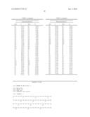
TABLE-US-00005 TABLE 1 Amino Acid Sequence and Helical Residue Notation for Human Haemoglobin Ao Helix α Helix β NA1 1 Val NA1 1 Val -- -- NA2 2 His NA2 2 Leu NA3 3 Leu A1 3 Ser AI 4 Thr A2 4 Pro A2 5 Pro A3 5 Ala A3 6 Glu A4 6 Asp A4 7 Glu A5 7 Lys A5 8 Lys A6 8 Thr A6 9 Ser A7 9 Asn A7 10 Ala A8 10 Val A8 11 Val A9 11 Lys A9 12 Thr A10 12 Ala A10 13 Ala A11 13 Ala A11 14 Leu A12 14 Trp A12 15 Trp A13 15 Gly A13 16 Gly A14 16 Lys A14 17 Lys A15 17 Val A15 18 Val A16 18 Gly -- -- AB1 19 Ala -- -- B1 20 His B1 19 Asn B2 21 Ala B2 20 Val B3 22 Gly B3 21 Asp B4 23 Glu B4 22 Glu B5 24 Tyr B5 23 Val B6 25 Gly B6 24 Gly B7 26 Ala B7 25 Gly B8 27 Glu B8 26 Glu B9 28 Ala B9 27 Ala B10 29 Leu B10 28 Leu B11 30 Glu B11 29 Gly B12 31 Arg B12 30 Arg B13 32 Met B13 31 Leu B14 33 Phe B14 32 Leu B15 34 Leu B15 33 Val B16 35 Ser B16 34 Val C1 36 Phe C1 35 Tyr C2 37 Pro C2 36 Pro C3 38 Thr C3 37 Trp C4 39 Thr C4 38 Thr C5 40 Lys C5 39 Gln C6 41 Thr C6 40 Arg C7 42 Tyr C7 41 Phe CE1 43 Phe CD1 42 Phe CE2 44 Pro CD2 43 Glu CE3 45 His CD3 44 Ser CE4 46 Phe CD4 45 Phe -- -- CD5 46 Gly CE5 47 Asp CD6 47 Asp CE6 48 Leu CD7 48 Leu CE7 49 Ser CD8 49 Ser CE8 50 His D1 50 Thr -- -- D2 51 Pro -- -- D3 52 Asp -- -- D4 53 Ala -- -- D5 54 Val -- -- D6 55 Met CE9 51 Gly D7 56 Gly E1 52 Ser E1 57 Asn E2 53 Ala E2 58 Pro E3 54 Gln E3 59 Lys E4 55 Val E4 60 Val E5 56 Lys E5 61 Lys E6 57 Gly E6 62 Ala E7 58 His E7 63 His E8 59 Gly E8 64 Gly E9 60 Lys E9 65 Lys E10 61 Lys E10 66 Lys E11 62 Val E11 67 Val E12 63 Ala E12 68 Leu E13 64 Asp E13 69 Gly E14 65 Ala E14 70 Ala E15 66 Leu E15 71 Phe E16 67 Thr E16 72 Ser E17 68 Asn E17 73 Asp E18 69 Ala E18 74 Gly E19 70 Val E19 75 Leu E20 71 Ala E20 76 Ala EF1 72 His EF1 77 His EF2 73 Val EF2 78 Leu EF3 74 Asp EF3 79 Asp EF4 75 Asp EF4 80 Asn EF5 76 Met EF5 81 Leu EF6 77 Pro EF6 82 Lys EF7 78 Asn EF7 83 Gly EF8 79 Ala EF8 84 Thr F1 80 Leu F1 85 Phe F2 81 Ser F2 86 Ala F3 82 Ala F3 87 Thr F4 83 Leu F4 88 Leu F5 84 Ser F5 89 Ser F6 85 Asp F6 90 Glu F7 86 Leu F7 91 Leu F8 87 His F8 92 His F9 88 Ala F9 93 Cys FG1 89 His FG1 94 Asp FG2 90 Lys FG2 95 Lys FG3 91 Leu FG3 96 Leu FG4 92 Arg FG4 97 His FG5 93 Val FG5 98 Val G1 94 Asp G1 99 Asp G2 95 Pro G2 100 Pro G3 96 Val G3 101 Glu G4 97 Asn G4 102 Asn G5 98 Phe G5 103 Phe G6 99 Lys G6 104 Arg G7 100 Leu G7 105 Leu G8 101 Leu G8 106 Leu G9 102 Ser G9 107 Gly G10 103 His G10 108 Asn G11 104 Cys G11 109 Val G12 105 Leu G12 110 Leu G13 106 Leu G13 111 Val G14 107 Val G14 112 Cys G15 108 Thr G15 113 Val G16 109 Leu G16 114 Leu G17 110 Ala G17 115 Ala G18 111 Ala G18 116 His G19 112 His GI9 117 His GH1 113 Leu GH1 118 Phe GH2 114 Pro GH2 119 Gly GH3 115 Ala GH3 120 Lys GH4 116 Glu GH4 121 Glu GH5 117 Phe GH5 122 Phe H1 118 Thr H1 123 Thr H2 119 Pro H2 124 Pro H3 120 Ala H3 125 Pro H4 121 Val H4 126 Val H5 122 His H5 127 Gln H6 123 Ala H6 128 Ala H7 124 Ser H7 129 Ala H8 125 Leu H8 130 Tyr H9 126 Asp H9 131 Gln H10 127 Lys H10 132 Lys H11 128 Phe H11 133 Val H12 129 Leu H12 134 Val H13 130 Ala H13 135 Ala H14 131 Ser H14 136 Gly H15 132 Val H15 137 Val H16 133 Ser H16 138 Ala H17 134 Thr H17 139 Asn H18 135 Val H18 140 Ala H19 136 Leu H19 141 Leu H20 137 Thr H20 142 Ala H21 138 Ser H21 143 His HC1 139 Lys HC1 144 Lys HC2 140 Tyr HC2 145 Tyr HC3 141 Arg HC3 146 His
Sequence CWU
1
81141PRTHomo sapiens 1Val Leu Ser Pro Ala Asp Lys Thr Asn Val Lys Ala Ala
Trp Gly Lys1 5 10 15Val
Gly Ala His Ala Gly Glu Tyr Gly Ala Glu Ala Leu Glu Arg Met 20
25 30Phe Leu Ser Phe Pro Thr Thr Lys
Thr Tyr Phe Pro His Phe Asp Leu 35 40
45Ser His Gly Ser Ala Gln Val Lys Gly His Gly Lys Lys Val Ala Asp
50 55 60Ala Leu Thr Asn Ala Val Ala His
Val Asp Asp Met Pro Asn Ala Leu65 70 75
80Ser Ala Leu Ser Asp Leu His Ala His Lys Leu Arg Val
Asp Pro Val 85 90 95Asn
Phe Lys Leu Leu Ser His Cys Leu Leu Val Thr Leu Ala Ala His
100 105 110Leu Pro Ala Glu Phe Thr Pro
Ala Val His Ala Ser Leu Asp Lys Phe 115 120
125Leu Ala Ser Val Ser Thr Val Leu Thr Ser Lys Tyr Arg 130
135 1402146PRTHomo sapiens 2Val His Leu Thr
Pro Glu Glu Lys Ser Ala Val Thr Ala Leu Trp Gly1 5
10 15Lys Val Asn Val Asp Glu Val Gly Gly Glu
Ala Leu Gly Arg Leu Leu 20 25
30Val Val Tyr Pro Trp Thr Gln Arg Phe Phe Glu Ser Phe Gly Asp Leu
35 40 45Ser Thr Pro Asp Ala Val Met Gly
Asn Pro Lys Val Lys Ala His Gly 50 55
60Lys Lys Val Leu Gly Ala Phe Ser Asp Gly Leu Ala His Leu Asp Asn65
70 75 80Leu Lys Gly Thr Phe
Ala Thr Leu Ser Glu Leu His Cys Asp Lys Leu 85
90 95His Val Asp Pro Glu Asn Phe Arg Leu Leu Gly
Asn Val Leu Val Cys 100 105
110Val Leu Ala His His Phe Gly Lys Glu Phe Thr Pro Pro Val Gln Ala
115 120 125Ala Tyr Gln Lys Val Val Ala
Gly Val Ala Asn Ala Leu Ala His Lys 130 135
140Tyr His145341DNAArtificial sequenceSynthetic sequence Primer
aY42V for 3ccttcccaac caccaaaacc gtgttcccac actttgatct g
41441DNAArtificial sequenceSynthetic sequence Primer aY42V rev
4cagatcaaag tgtgggaaca cggttttggt ggttgggaag g
41541DNAArtificial sequenceSynthetic sequence Primer aY42random for
5ccttcccaac caccaaaacc nnkttcccac actttgatct g
41641DNAArtificial sequenceSynthetic sequence Primer aY42random rev
6cagatcaaag tgtgggaamn nggttttggt ggttgggaag g
41734DNAArtificial sequenceSynthetic sequence Primer bF41Y for
7ccgtggaccc agcgttactt tgaatccttc ggtg
34834DNAArtificial sequenceSynthetic sequence Primer bF41Y rev
8caccgaagga ttcaaagtaa cgctgggtcc acgg 34
User Contributions:
Comment about this patent or add new information about this topic:
| People who visited this patent also read: | |
| Patent application number | Title |
|---|---|
| 20160000895 | COMPOSITIONS AND METHODS OF ENHANCING IMMUNE RESPONSES TO EIMERIA OR LIMITING EIMERIA INFECTION |
| 20160000894 | IN VIVO EFFICACY OF NY-ESO-1 PLUS ADJUVANT |
| 20160000893 | TARGET PEPTIDES FOR OVARIAN CANCER THERAPY AND DIAGNOSTICS |
| 20160000892 | REGULATORY T CELL EPITOPES, COMPOSITIONS AND USES THEREOF |
| 20160000891 | IMMUNOGENIC PRODUCTS BASED ON MUTEIN AMYLOID beta (Abeta) AMINO ACID SEQUENCES AND USES THEREOF |
 |  |
 |  |
 |  |
 |  |
 |  |
| Similar patent applications: | |
| Date | Title |
|---|---|
| 2010-10-28 | Milk protein hydrolyzates with reduced immunogenic potential |
| 2010-11-04 | Composition enriched in diglyceride with conjugated linoleic acid |
| 2010-11-11 | Composition having a high osmotic activity and exhibiting antimicrobial, anti-inflammatory, and regenerative effects |
| 2010-09-23 | Soluble protein and utilization of the same |
| 2010-11-11 | Azetidinyl g-protein coupled receptor agonists |
| Top Inventors for class "Drug, bio-affecting and body treating compositions" | |
| Rank | Inventor's name |
|---|---|
| 1 | Anthony W. Czarnik |
| 2 | Ulrike Wachendorff-Neumann |
| 3 | Ken Chow |
| 4 | John E. Donello |
| 5 | Rajinder Singh |