Patent application title: WSTF REGULATES THE DNA DAMAGE RESPONSE OF H2A.X VIA NOVEL TYROSINE KINASE ACTIVITY
Inventors:
C. David Allis (Princeton, NJ, US)
Andrew Xiao (New York, NY, US)
Assignees:
THE ROCKEFELLER UNIVERSITY
IPC8 Class: AA61K39395FI
USPC Class:
4241301
Class name: Drug, bio-affecting and body treating compositions immunoglobulin, antiserum, antibody, or antibody fragment, except conjugate or complex of the same with nonimmunoglobulin material
Publication date: 2010-04-01
Patent application number: 20100080799
Inventors list |
Agents list |
Assignees list |
List by place |
Classification tree browser |
Top 100 Inventors |
Top 100 Agents |
Top 100 Assignees |
Usenet FAQ Index |
Documents |
Other FAQs |
Patent application title: WSTF REGULATES THE DNA DAMAGE RESPONSE OF H2A.X VIA NOVEL TYROSINE KINASE ACTIVITY
Inventors:
C. David Allis
Andrew Xiao
Agents:
NIXON PEABODY LLP - PATENT GROUP
Assignees:
THE ROCKEFELLER UNIVERSITY
Origin: ROCHESTER, NY US
IPC8 Class: AA61K39395FI
USPC Class:
4241301
Patent application number: 20100080799
Abstract:
The present invention relates to methods of detecting and regulating
cellular DNA damage as well as methods of screening compounds suitable
for modulating cellular DNA damage. The methods of the present invention
utilize antibodies that selectively bind to a phosphorylated tyrosine
residue in a SQEY tyrosine phosphorylation motif sequence or a
phosphorylated serine residue in a KENSSQ phosphorylation motif sequence,
where the phosphorylated serine residue is closest to the glutamine
residue. Also encompassed by the present invention are methods of
treating a subject having cancer. These methods involve the
administration of an agent that modulates the phosphorylation of the
tyrosine residue in a SQEY motif. Suitable agents for modulating the
phosphorylation of the tyrosine residue of the SQEY motif are also
disclosed.Claims:
1. An antibody or antigen-binding fragment thereof that selectively binds
to a phosphorylated tyrosine residue, wherein the phosphorylated tyrosine
residue is located in an SQEY tyrosine phosphorylation motif sequence
(SEQ ID NO: 2).
2. The antibody or antigen-binding fragment thereof according to claim 1, wherein the phosphorylated tyrosine residue of the SQEY motif is at amino acid position 142 of SEQ ID NO:1.
3. The antibody according to claim 1, wherein the antibody is monoclonal or polyclonal.
4. The antibody according to claim 1, wherein the antibody is multivalent or monovalent.
5. The antibody according to claim 1, wherein the antibody is humanized.
6. An antibody or antigen-binding fragment thereof that selectively binds to a phosphorylated serine residue, wherein the phosphorylated serine residue is closest to the glutamine residue in an KENSSQ phosphorylation motif sequence (SEQ ID NO:4).
7. The antibody or antigen-binding fragment thereof according to claim 6, wherein the phosphorylated serine residue of the KENSSQ motif is at amino acid position 167 of SEQ ID NO:3.
8. The antibody according to claim 6, wherein the antibody is monoclonal or polyclonal.
9. The antibody according to claim 6, wherein the antibody is multivalent or monovalent.
10. The antibody according to claim 6, wherein the antibody is humanized.
11. An isolated polypeptide molecule comprising an amino acid sequence of SEQ ID NO:6 or SEQ ID NO:7.
12. An isolated nucleic acid molecule encoding the polypeptide of claim 11.
13. The isolated nucleic acid molecule of claim 12, wherein the nucleic acid molecule has a nucleotide sequence that has at least 80% nucleic acid sequence identity to the nucleotide sequence of SEQ ID NO: 5.
14. The isolated nucleic acid molecule of claim 12, wherein the nucleic acid molecule has a nucleotide sequence that has at least 90% nucleic acid sequence identity to the nucleotide sequence of SEQ ID NO:5.
15. The isolated nucleic acid molecule of claim 12, wherein the nucleic acid molecule has a nucleotide sequence that has at least 95% nucleic acid sequence identity to the nucleotide sequence of SEQ ID NO:5.
16. An expression vector comprising the isolated nucleic acid molecule of claim 12.
17. A host cell containing the expression vector of claim 16.
18. A method of identifying cellular DNA damage in a sample, said method comprising:providing a cell sample;detecting the presence or absence of a phosphorylated tyrosine residue in an SQEY tyrosine phosphorylation motif (SEQ ID NO:2); andidentifying cellular DNA damage in the cell sample based on said detecting.
19. The method according to claim 18, wherein the phosphorylated tyrosine residue of the SQEY motif is at amino acid position 142 of SEQ ID NO:1.
20. The method according to claim 18, wherein said detecting comprises contacting the sample with an antibody or antigen-binding fragment thereof that selectively binds to the phosphorylated tyrosine residue in an SQEY tyrosine phosphorylation motif (SEQ ID NO:2).
21. The method according to claim 20, wherein said antibody or antigen-binding fragment thereof selectively binds to the phosphorylated tyrosine residue of the SQEY motif at amino acid position 142 of SEQ ID NO:1.
22. The method according to claim 18, wherein the absence of a phosphorylated tyrosine residue in the SQEY motif indicates the presence of cellular DNA damage.
23. The method according to claim 18, wherein the presence of a phosphorylated tyrosine residue in the SQEY motif indicates the absence of cellular DNA damage.
24. A method of identifying cellular DNA damage in a sample, said method comprising:providing a cell sample;detecting the presence or absence of a phosphorylated serine residue at the serine closest to the glutamine residue in an KENSSQ phosphorylation motif sequence (SEQ ID NO:4); andidentifying cellular DNA damage in the cell sample based on said detecting.
25. The method according to claim 24, wherein the phosphorylated serine closest to the glutamine residue of the KENSSQ motif is at amino acid position 167 of SEQ ID NO:3.
26. The method according to claim 24, wherein said detecting comprises contacting the sample with an antibody or antigen-binding fragment thereof that selectively binds to the phosphorylated serine closest to the glutamine residue in an KENSSQ phosphorylation motif sequence (SEQ ID NO:4).
27. The method according to claim 26, wherein said antibody or antigen-binding fragment thereof selectively binds to the phosphorylated serine closest to the glutamine residue in the KENSSQ motif at amino acid position 167 of SEQ ID NO:3
28. The method according to claim 24, wherein the presence of a phosphorylated serine residue at the serine closest to the glutamine residue of the KENSSQ motif indicates the presence of cellular DNA damage.
29. The method according to claim 24, wherein the absence of a phosphorylated serine residue at the serine closest to the glutamine residue of the KENSSQ motif indicates the absence of cellular DNA damage.
30. A method of screening candidate compounds useful for modulating WSTF kinase activity, said method comprising:providing a candidate compound;providing a sample, wherein said sample comprises a WSTF protein or polypeptide having kinase activity, a protein substrate having an SQEY motif sequence, manganese, and ATP;contacting the candidate compound with the sample;detecting the presence or absence of a phosphorylated tyrosine residue in the SQEY tyrosine phosphorylation motif (SEQ ID NO:2); andidentifying compounds useful for modulating WSTF kinase activity based on said detecting.
31. The method according to claim 30, wherein the WSTF protein or polypeptide having kinase activity comprises SEQ ID NO: 6 or SEQ ID NO:7.
32. The method according to claim 30, wherein the phosphorylated tyrosine residue in the SQEY motif is at amino acid position 142 of SEQ ID NO:1 in the sample.
33. The method according to claim 30, wherein the sample is a cell sample.
34. The method according to claim 30, wherein detecting the absence of a phosphorylated tyrosine residue in the SQEY motif identifies a compound useful for inhibiting WSTF kinase activity.
35. A method of screening candidate compounds useful for modulating cellular DNA damage repair, said method comprising;providing a candidate compound;providing a cell sample;contacting the candidate compound with the cell sample;detecting the presence or absence of a phosphorylated tyrosine residue in a SQEY tyrosine phosphorylation motif (SEQ ID NO:2) in the cell sample; andidentifying candidate compounds useful for modulating cellular DNA damage repair based on said detecting.
36. The method according to claim 35, wherein the phosphorylated tyrosine residue in the SQEY motif is at amino acid position 142 of SEQ ID NO:1 in the cell sample.
37. The method according to claim 35, wherein the cell sample comprises one or more genetically modified yeast cells, wherein the genetically modified yeast cells contain a leucine to tyrosine mutation in their H2A.X SQEL motif.
38. The method according to claim 35, wherein said contacting occurs in the presence of a DNA damaging agent.
39. A method of screening candidate compounds useful for treating cancer, said method comprising;providing a candidate compound;providing a cell sample;contacting the candidate compound with the cell sample;detecting the presence or absence of a phosphorylated tyrosine residue in an SQEY tyrosine phosphorylation motif (SEQ ID NO:2) in the cell sample; andidentifying candidate compounds useful for treating cancer based on said detecting.
40. The method according to claim 39, wherein the phosphorylated tyrosine residue in the SQEY motif is at amino acid position 142 of SEQ ID NO:1 in the cell sample.
41. The method according to claim 39, wherein the cell sample comprises one or more genetically modified yeast cells, wherein the genetically modified yeast cells contain a leucine to tyrosine mutation in their H2A.X SQEL motif.
42. A method of modulating cellular DNA damage repair, said method comprising:administering to a cell an agent which modulates tyrosine phosphorylation of an SQEY tyrosine phosphorylation motif (SEQ ID NO:2) under conditions effective to modulate cellular DNA damage repair.
43. The method according to claim 42, wherein said agent modulates tyrosine phosphorylation of the tyrosine residue in the SQEY motif at amino acid position 142 of SEQ ID NO:1 in the cell.
44. The method according to claim 42, wherein an agent that reduces tyrosine phosphorylation of the SQEY motif enhances cellular DNA damage repair.
45. The method according to claim 44, wherein the agent is a WSTF kinase domain inhibitor.
46. The method according to claim 45, wherein the WSTF kinase domain inhibitor is an siRNA molecule comprising SEQ ID NO: 21.
47. The method according to claim 44, wherein the agent is a recombinant EYA phosphatase protein.
48. The method according to claim 42, wherein an agent that induces tyrosine phosphorylation of the SQEY motif suppresses cellular DNA damage repair.
49. The method according to claim 48, wherein the agent is a nucleic acid molecule encoding a WSTF kinase domain comprising the nucleotide sequence of SEQ ID NO: 5 or the encoded polypeptide comprising an amino acid sequence of SEQ ID NO: 6 or SEQ ID NO:7.
50. The method according to claim 48, wherein the agent is an EYA phosphatase protein inhibitor.
51. A method of treating a subject having cancer, said method comprising:administering to said subject having cancer, an agent that modulates tyrosine phosphorylation of a SQEY tyrosine phosphorylation motif (SEQ ID NO:2) under conditions effective to treat the subject having cancer.
52. The method according to claim 51, wherein said agent modulates tyrosine phosphorylation of the tyrosine residue in the SQEY motif at amino acid position 142 of SEQ ID NO:1.
53. The method according to claim 51, wherein the agent reduces tyrosine phosphorylation of the SQEY motif.
54. The method according to claim 53, wherein the agent is a WSTF kinase domain inhibitor.
55. The method according to claim 54, wherein the WSTF kinase domain inhibitor is an siRNA molecule comprising SEQ ID NO: 21.
56. The method according to claim 53, wherein the agent is a recombinant EYA phosphatase protein.
57. The method according to claim 51, wherein the agent induces tyrosine phosphorylation of the SQEY motif.
58. The method according to claim 57, wherein the agent is a nucleic acid molecule encoding a WSTF kinase domain comprising the nucleotide sequence of SEQ ID NO: 5 or the encoded polypeptide comprising an amino acid sequence of SEQ ID NO: 6 or SEQ ID NO:7.
59. The method according to claim 57, wherein the agent is an EYA phosphatase protein inhibitor.
Description:
[0001]This application claims the benefit of U.S. Provisional Patent
Application Ser. No. 61/097,672, filed Sep. 17, 2008, which is hereby
incorporated in its entirety.
FIELD OF THE INVENTION
[0003]The present invention describes methods of identifying and regulating cellular DNA damage repair and methods of treating a subject having cancer. These methods involve modulating tyrosine phosphorylation at a newly defined tyrosine phosphorylation motif. The invention further describes a novel tyrosine kinase involved in regulating DNA damage repair.
BACKGROUND OF THE INVENTION
[0004]One hallmark of the mammalian DNA double-strand break (DSB) response is the formation of Ionizing Radiation Induced Foci (IRIF), which are composed of compacted chromatin and numerous DNA repair and checkpoint proteins (Khanna et al., "DNA Double-Strand Breaks: Signaling, Repair and the Cancer Connection," Nat Genet. 27:247-254 (2001) and Zhou et al., The DNA Damage Response Putting Checkpoints in Perspective," Nature 408:433-439 (2000)). Despite considerable progress in understanding signaling pathways leading to checkpoint control and DNA repair, the nature of the specialized chromatin structures at IRIF is not well understood. One of the earliest events occurring at IRIF is the phosphorylation of H2A.X, a specialized histone H2A variant, at Ser139 (referred to as γ-H2A.X) by the ATM and ATR kinases (Redon et al., "Histone H2A Variants H2AX and H2AZ," Curr Opin Genet Dev 12:162-169 (2002)). H2A.X-deficient mouse embryonic fibroblasts (MEFs), B and T cells display pronounced levels of genomic instability, including broken and dicentric chromosomes and random translocations (Celeste et al., "Genomic Instability in Mice Lacking Histone H2AX," Science 296:922-927 (2002)). Class switch recombination and spermatogenesis are also defective in H2A.X deficient mice, further implying its involvement in DNA damage repair under physiological conditions (Celeste et al., "Genomic Instability in Mice Lacking Histone H2AX," Science 296:922-927 (2002); Celeste et al., "H2AX Haploinsufficiency Modifies Genomic Stability and Tumor Susceptibility," Cell 114:371-383 (2003); and Reina-San-Martin et al., "H2AX is Required for Recombination Between Immunoglobulin Switch Regions but Not for Intra-Switch Region Recombination or Somatic Hypermutation," J Exp Med 197:1767-1778 (2003)). Moreover, H2A.X-deficiency accelerates B and T cell lymphoma development in p53-deficient mice (Celeste et al., "H2AX Haploinsufficiency Modifies Genomic Stability and Tumor Susceptibility," Cell 114:371-383 (2003); [0005]Reina-San-Martin et al., "H2AX is Required for Recombination Between Immunoglobulin Switch Regions but Not for Intra-Switch Region Recombination or Somatic Hypermutation," J Exp Med 197:1767-1778 (2003); and Bassing et al., "Histone H2AX: A Dosage-dependent Suppressor of Oncogenic Translocations and Tumors," Cell 114:359-370 (2003)). Consistent with these functions in mammalian cells, phosphorylation of the equivalent site on yeast H2A (Ser129) is found at DSB sites and spreads to ±50 Kb of the flanking regions (Unal et al., "DNA Damage Response Pathway Uses Histone Modification to Assemble a Double-Strand Break-Specific Cohesin Domain," Mol Cell 16:991-1002 (2004)). In mammals this phosphorylation event directly recruits Mdc1 and sets in motion the recruitment of additional factors such as 53BP1, RNF8, and the Brca1 A complex (Harper et al., "The DNA Damage Response: Ten Years After," Mol Cell 28:739-745 (2007)). Furthermore, several recent studies have also indicated that ATP-dependent chromatin remodeling complexes are engaged in DNA repair pathways at these sites. The yeast NuA4 and INO80 complexes, for example, are recruited to the damaged chromatin via γ-H2A.X (Morrison et al., "INO80 and Gamma-H2AX Interaction Links ATP-dependent Chromatin Remodeling to DNA Damage Repair," Cell 119:767-775 (2004); Downs et al., "Binding of Chromatin-Modifying Activities to Phosphorylated Histone H2A at DNA Damage Sites," Mol Cell 16:979-990 (2004); and van Attikum et al., "Recruitment of the INO80 Complex by H2A Phosphorylation Links ATP-Dependent Chromatin Remodeling With DNA Double-Strand Break Repair," Cell 119:777-788 (2004)).
[0006]Mammalian H2A.X bears significant differences with lower eukaryotes. For example, H2A.X is a minor H2A variant in mammalian cells (1-10%) while the major yeast form of H2A is most similar to H2A.X, because it contains the signature C-terminal sequence of mammalian H2A.X (Redon et al., "Histone H2A Variants H2AX and H2AZ," Curr Opin Genet Dev 12:162-169 (2002)). A Drosophila H2A variant, known as H2A.V, is a "hybrid" protein containing signature sequences of mammalian H2A.X and H2A.Z (Redon et al., "Histone H2A Variants H2AX and H2AZ," Curr Opin Genet Dev 12:162-169 (2002)). The Drosophila Tip60 complex exchanges phosphorylated H2A.V with unmodified H2A.V in vitro and regulates H2A.V phosphorylation levels in vivo (Kusch et al., "Acetylation by Tip60 is Required for Selective Histone Variant Exchange at DNA Lesions," Science 306:2084-2087 (2004)). In mammalian cells, the loss of SWI/SNF chromatin remodeling complex expression leads to defects in the H2A.X DNA damage response. However, it is unclear whether these defects are due to the lack of direct regulation by the SWI/SNF complex or other indirect pathways, since the SWI/SNF complex is involved in a variety of important biological processes (Park et al., "Mammalian SWI/SNF Complexes Facilitate DNA Double-Strand Break Repair by Promoting Gamma-H2AX Induction," EMBO J 25:3986-3997 (2006)).
[0007]Given the unique features of H2A.X in mammalian cells, factors that are directly involved in regulating H2A.X function need to be identified.
SUMMARY OF THE INVENTION
[0008]A first aspect of the present invention relates to an antibody or antigen-binding fragment thereof that selectively binds to a phosphorylated tyrosine residue in an SQEY tyrosine phosphorylation motif sequence (SEQ ID NO: 2).
[0009]A second aspect of the present invention relates to an antibody or antigen-binding fragment thereof that selectively binds to the phosphorylated serine residue closest to the glutamine residue in an KENSSQ phosphorylation motif sequence (SEQ ID NO:4).
[0010]Other aspects of the present invention relate to an isolated polypeptide molecule having an amino acid sequence of SEQ ID NO: 6 or SEQ ID NO: 7, and an isolated nucleic acid molecule encoding the polypeptide.
[0011]Another aspect of the present invention relates to a method of identifying cellular DNA damage in a sample. This method involves providing a cell sample and detecting the presence or absence of a phosphorylated tyrosine residue in an SQEY tyrosine phosphorylation motif (SEQ ID NO:2). Cellular DNA damage in the cell sample is identified based on detecting the presence or absence of tyrosine phosphorylation.
[0012]Another aspect of the present invention relates to a method of identifying cellular DNA damage in a sample. This method involves providing a cell sample and detecting the presence or absence of a phosphorylated serine residue at the serine closest to the glutamine residue in an KENSSQ phosphorylation motif sequence (SEQ ID NO:4). Cellular DNA damage in the cell sample is identified based on detecting the presence or absence of serine phosphorylation.
[0013]Another aspect of the present invention relates to a method of screening candidate compounds useful for modulating WSTF kinase activity. This method involves providing a candidate compound and a sample, where the sample contains a WSTF protein or polypeptide having kinase activity, a protein substrate having a SQEY motif sequence, manganese, and ATP. Contacting the candidate compound with the sample and detecting the presence or absence of a phosphorylated tyrosine residue in a SQEY tyrosine phosphorylation motif (SEQ ID NO:2) identifies compounds useful for modulating WSTF kinase activity.
[0014]The present invention is also directed to a method of screening candidate compounds useful for modulating cellular DNA damage repair. This method involves providing a candidate compound and a cell sample and contacting the candidate compound with the cell sample. The presence or absence of a phosphorylated tyrosine residue in a SQEY tyrosine phosphorylation motif (SEQ ID NO:2) in the cell sample is detected, and candidate compounds useful for modulating cellular DNA damage repair are identified based on the presence or absence of tyrosine phosphorylation.
[0015]Another aspect of the present invention relates to a method of screening candidate compounds useful for treating cancer. This method involves providing a candidate compound and a cell sample and contacting the candidate compound with the cell sample. The presence or absence of a phosphorylated tyrosine residue in a SQEY tyrosine phosphorylation motif (SEQ ID NO:2) in the cell sample is detected and candidate compounds useful for treating cancer are identified based on the presence or absence of tyrosine phosphorylation.
[0016]The present invention is also directed to a method of modulating cellular DNA damage repair. This method involves administering to a cell an agent which modulates tyrosine phosphorylation of a SQEY tyrosine phosphorylation motif (SEQ ID NO:2) under conditions effective to modulate cellular DNA damage repair.
[0017]Another aspect of the present invention is directed to a method of treating a subject having cancer. This method involves administering to the subject having cancer, an agent that modulates tyrosine phosphorylation of a SQEY tyrosine phosphorylation motif (SEQ ID NO:2) under conditions effective to treat the subject having cancer.
[0018]The studies described herein define a novel DNA damage response pathway regulating H2A.X function in mammals that is mediated through the WSTF-SNF2H chromatin remodeling complex and involves the phosphorylation of H2A.X at tyrosine residue 142 ("Tyr142" or "Y142"). Unexpectedly, it was determined that the amino-terminal domain of WSTF, including its WAC domain, exhibits tyrosine kinase activity towards Tyr142 of H2A.X, a novel phosphorylation mark that plays a role in γ-H2A.X maintenance and related downstream chromatin remodeling events. Both the novel tyrosine kinase activity of WSTF and the phosphorylation of H2A.X at Tyr142 are required for eliciting an appropriate downstream DNA damage response in mammalian cells. These findings support an emerging view that the regulatory pathways of chromatin structure brought about by histone covalent modification, chromatin remodeling by ATP-dependent complexes, and the introduction of histone variants are closely interconnected, affecting many DNA-templated processes including the repair of double-strand breaks.
BRIEF DESCRIPTION OF THE DRAWINGS
[0019]FIGS. 1A-1B show Tyr142, a novel phosphorylation mark of H2A.X, is regulated by DNA damage signals. FIG. 1A is a comparison of extreme C-terminal sequences of H2A.X of various species, demonstrating that Tyr142 is conserved in "multicellular" (mammals and fruit flies) but not in "single-cellular" eukaryotes. As shown in FIG. 1A, the SQEY motif of SEQ ID NO:2 is conserved between H. sapiens and M. musculus; D. melanogaster has an SQAY motif (SEQ ID NO: 18); X. laevis has an SQEY/F motif (SEQ ID NO: 19); and S. cerevisiae has a SQEL motif (SEQ ID NO: 20). In FIG. 1B, primary MEF cells were treated with 10 Gy of ionizing radiation (IR) and recovered for the period of time indicated. Acid-extracted histones were separated by SDS-PAGE and subjected to immunoblotting. H2A.X Tyr142 phosphorylation levels in MEFs gradually declined, reaching a minimum at 8 hr. As expected, γ-H2A.X signal was initiated upon damage and maintained up to 16 hours post IR.
[0020]FIGS. 2A-2D demonstrate the specific association between the WSTF-SNF2H chromatin remodeling complex and H2A.X nucleosomes in vivo. FIG. 2A shows the purification scheme of H2A.X-containing mononucleosomes. MEFs reconstituted with Flag-H2A.X (WT or Y142F mutant) were treated with or without ionizing radiation. After hypotonic buffer extraction, nuclear pellets (P1) were further extracted and fractionated into soluble nuclear extracts (S2) and insoluble nuclear pellets (P2) containing primarily chromatin and associated proteins. After Mnase digestion, mononucleosomes were immunoprecipitated with α-Flag to enrich for H2A.X-containing nucleosomes and associated proteins. This approach efficiently and specifically isolated H2A.X nucleosomes (See FIG. 6). The immunoprecipitated complexes were separated by SDS-PAGE and silver stained (FIG. 2B upper panels). Two polypeptides migrating at 145 and 171 KDa were consistently associated with undamaged WT (FIG. 2B, upper left panel), but not the Y142F mutant (FIG. 2B, upper right panel) H2A.X mononucleosomes. Mass spectrometry (MS) analyses demonstrated that these polypeptides, WSTF (171 KDa) and SNF2H (145 KDa), constitute the mammalian WICH complex (See FIG. 7). The third band at 60 KDa was identified as β-actin in the MS analyses; the significance of β-actin is not known. An aliquot of the complex immunoprecipitated from the same sample was separated by SDS-PAGE and silver stained for lower molecular weight bands (<20 KDa) (FIG. 2B, lower panel). Staining revealed similar levels of H2A.X and other core histones identified by MS analysis. In FIG. 2C the association between the WICH complex and undamaged WT H2A.X mononucleosomes was confirmed by IP-western experiments. Note that the 5139 phosphorylation level was greatly reduced in the Y142F mutant cells (FIG. 2C; also see FIG. 4). WSTF knock-down cell lines (WSTF RNAi) were generated by stably expressing short hairpin RNAi constructs specifically targeting the WSTF gene in NIH3T3 cells. FIG. 4D shows SDS-PAGE and immunoblot of nuclear extracts from the WSTF RNAi and control 3T3 cells. In WSTF RNAi cells, the expression level of WSTF was significantly diminished and H2A.X Y142 phosphorylation level was significantly reduced.
[0021]FIGS. 3A-3G illustrate the characterization of the novel kinase domain of WSTF that phosphorylates Tyr142 of H2A.X. FIG. 3A is a schematic representation of the domain architecture of the human WSTF protein and a series of recombinant proteins representing portions of WSTF (i.e., the 1-345N construct represents N-terminal amino acids 1 to 345 of WSTF). These constructs were extensively purified through affinity (GST pull down), ion exchange (SP sepharose) and sizing (gel filtration) chromatography. No other proteins at significant level (>2.5%) were co-purified with them, as confirmed by MS analysis. In FIGS. 3B-F, recombinant WSTF proteins were generated in insect cells and the N- and C-terminal motifs (FIG. 3G) were generated in E. coli. As shown in FIG. 3B, Y142 was phosphorylated by recombinant full length WSTF proteins. WT and Y142F mutant H2A.X nucleosomes were reconstituted with recombinant free histones and DNA molecules in vitro. Following in vitro kinase assays, reaction mixtures were separated by SDS-PAGE and exposed for radioautography. In FIG. 3C, the specific phosphorylation of H2A.X at Tyr142 was also detected by immunoblotting with an α-H2A.X Y142ph antibody. In FIGS. 3D-G, free H2A.X proteins were used as substrates in in vitro kinase assays. The recombinant N-terminal (N-WSTF) and C-terminal portion (C-WSTF) of WSTF were generated from insect cells and tested for kinase activity. Only N-WSTF had kinase activity as shown in FIG. 3D. A series of truncated WSTF proteins were generated from insect cells (see FIG. 3A) and the activity of the 1-340 amino acid construct was much reduced (<50 fold) in comparison to the other constructs (FIG. 3E). The C338A point mutant was derived from the WT 1-359 construct and its kinase activity was much reduced compared to WT (FIG. 3F). The N-motif and C-motif of WSTF kinase domain were generated either individually or in combination from E. coli. The co-expressed N-motif and C-motif had significant kinase activity towards H2A.X, while the N-motif alone had a minimal kinase activity (FIG. 3G). The C-motif construct, when expressed alone, was unstable and partially degraded from its C-terminus, however, it was protected when co-expressed with the N-motif.
[0022]FIGS. 4A-4J demonstrate how critical WSTF is for the maintenance of γ-H2A.X phosphorylation after DNA damage. In WSTF RNAi cells (as described in FIG. 2), γ-H2A.X level maintenance was defective after DNA damage treatment as illustrated in FIG. 4A. Western blotting experiments were performed on the histone samples isolated from control and WSTF RNAi cells after 10 Gy of IR treatment. The time points labeled indicate the recovery time following the IR treatment. The maintenance of γ-H2A.X foci is also defective in WSTF RNAi cells as shown in FIG. 4B. Immunofluorescent staining experiments were performed on control and WSTF RNAi cells fixed at different time points (as labeled) following 10 Gy of IR treatment. As shown in FIGS. 4C and 4D, phos-ATM and Mdc1 recruitment is also defective in WSTF deficient cells. Immunofluorescent staining experiments were performed using anti-ATM S1981 phos (FIG. 4C) and anti-Mdc1 (FIG. 4D) antibodies on control or WSTF RNAi cells 8-hours post 10 Gy of IR treatment. FIGS. 4E and 4F demonstrates that WSTF kinase activity is also critical for γ-H2A.X and phos-ATM foci maintenance. FIGS. 4E and 4F are immunofluorescent images of WSTF RNAi cells complemented with wildtype or mutant (C338A) kinase domain constructs of WSTF (amino acids 1-359, myc epitope-tagged at N-terminus). Eight hours following treatment with 10 Gy of IR, cells were fixed and co-stained with anti-myc (red) and γ-H2A.X (green, FIG. 4E) or anti-ATM S1981 phos (green, FIG. 4F) antibodies. As shown in FIGS. 4G and 4H, the level of γ-H2A.X protein is reduced in Tyr142 mutants. H2A.X null MEFs were reconstituted with WT or Y142H2A.X mutants (Y to L or Y to F mutants). Histone samples were separated by SDS-PAGE and subjected to Western blotting ("V" is vector only) (FIG. 4G). Immunostaining experiments with these same cells demonstrated that γ-H2A.X foci (FIG. 4H, upper left panel) were observed in WT cells upon DNA damage while they were severely diminished in Tyr142 mutants (upper middle and right panels). Cells were fixed after DNA damage treatment (10 Gy 2 hr) and stained with γ-H2A.X antibodies. In peptide pull-down experiments (FIG. 4I), Ser139(ph) peptides specifically interacted with endogenous MDC1, but not BRCA1 from 293T cell nuclear extracts. Either pre-phosphorylation of Y142 (i.e., a doubly phosphorylated (S139(ph) & Y142(ph)) peptide) or replacement of the C-terminal tyrosine to leucine (L) or phenylalanine (F) diminished MDC1 binding significantly in this assay. A recombinant MDC1 BRCT domain specifically bound to Ser139(ph) peptides as demonstrated in FIG. 4J. Similarly, pre-phosphorylation or replacement of Y142 diminished binding significantly.
[0023]FIGS. 5A-5F show the modulation of Y142 phosphorylation level during DNA damage response and the characterization of the α-H2A.X Y142ph antibody. In FIG. 5A, α-H2A.X Y142ph antibodies were reacted with Xenopus H2A.X in Xenopus egg extracts. After incubating with double-strand break (DSB) plasmid DNA (for the time span as indicated), the Y142 phosphorylation significantly decreased while γ-H2A.X increased, reminiscent of mammalian cells. Yeast H2A L132Y mutant strongly reacted with antibodies to mammalian H2A.X Y142 phosphorylation (FIG. 5B). As expected, WT yeast cells did not have affinity for the anti-H2A.X Y142ph antibody. In contrast, the anti-H2A.X Y142ph antibody reacted with nuclear extracts from untreated H2A L132Y cultures, but did not react with extracts of cells treated with the DNA-damaging agent, MMS. γ-H2A.X (S139ph) antibody reacted strongly with WT and H2A L132Y cells following DNA damage. The equal loading of histone proteins was judged by α-H3 antibodies. In ELISA, α-H2A.X-Y142ph antibody reacted with H2A.X C-terminal peptides (129-142) that contained phosphorylated Tyr142 regardless of the phosphorylation status of Ser139 (FIG. 5C, compare (.tangle-solidup.) and (x) curves), but not with similar peptides where Tyr142 was not phosphorylated (compare (.diamond-solid.) and (.box-solid.) curves). In western blot analyses, α-H2A.X Y142ph antibody recognized a single band at the expected molecular weight of H2A.X (˜17 KDa in U2OS cells (FIG. 5D, left lane)); this band was sensitive to protein tyrosine phosphatase b (FIG. 5D, right lane). The signals could be competed away only by H2A.X Y142ph peptides, but not unmodified peptides (FIG. 5E). In western blot analyses, this antibody only reacted with reconstituted WT H2A.X cells, but not cells expressing a Y142F point mutant (FIG. 5F).
[0024]FIG. 6 shows H2A.X-containing nucleosomes enriched in immunocomplexes precipitated with α-Flag. Flag-H2A.X-containing mononucleosomes isolated from reconstituted MEFs (see scheme in FIG. 2A) were separated by SDS-PAGE before being subjected to immunoblot analyses or Coomassie blue staining. In western blot analyses (upper panel), α-Flag antibodies recognized a single band at 20 KDa (there was no endogenous H2A.X in these cells and the MW of Flag-tagged H2A.X was larger than H3). Coomassie staining of the immunoprecipitated complexes (lower panel) revealed that Flag-H2A.X (MW 20 KDa) was associated with other core histones at similar stoichiometrical level whereas it was undetectable in input (5%) samples.
[0025]FIG. 7 is an identification of WSTF-SNF2H complex subunits by mass spectrometry. Protein bands (at 171 and 140 KDa, FIG. 2B) were isolated from SDS-PAGE gel and subjected to trypsin digestion. Peptide mass fingerprinting (PMF) demonstrated that in the protein band at 171 KDa, 19 peptides (SEQ ID NOs: 38-56; selected out of 29 for PMF) were from the mouse WSTF protein, which cover 248 out of 1479 amino acids (16.8%). In the protein band at 140 KDa, 10 peptides (SEQ ID NOs: 57-66; out of 35 selected for PMF) were from the mouse SNF2H protein, which cover 109 out of 1052 amino acids (10.4%). The protein band at 60 KDa was identified as β-actin.
[0026]FIG. 8 demonstrates that alteration of C-terminal Tyr142 in H2A.X does not change the affinity of γ-H2A.X antibodies. In ELISA, γ-H2A.X antibodies reacted with H2A.X C-terminal peptides (amino acid residues 129-142) that contain Phos-Ser139 regardless of the phosphorylation status of Tyr142 or Y142L replacement.
[0027]FIGS. 9A-9B show that phosphorylation of H2A.X at Ser139 by the ATM/R kinases upon DNA damage does not require Tyr142 and was not affected by phosphorylation at this site. H2A.X C-terminal peptides (listed in FIG. 9B) were incubated with Xenopus egg extracts in the presence of DSB DNA (+) or control DNA(-) and γ-32P-ATP (reflective of ATM kinase; see Khanna et al., "DNA Double-Strand Breaks Signaling, Repair and the Cancer Connection," Nat Genet 27:247-254 (2001), which hereby incorporated by reference in its entirely). Incorporation of radioactive ATP was measured by scintillation counting in standard P81 filter paper assays (Khanna et al., "DNA Double-Strand Breaks: Signaling, Repair and the Cancer Connection," Nat Genet 27:247-254 (2001), which hereby incorporated by reference in its entirely). FIG. 9A is a bar graph showing ATM kinase activity (Ser139 phosphorylation). Ser139 of H2A.X was specifically phosphorylated in the presence of DSB DNA (compare unmodified peptide (UN) in the presence of DSB DNA (+) as compared to control DNA (-) or with pre-phosphorylated Ser139(ph) peptide (DSB+). The status of Tyr142, either in its pre-phosphorylated state (Tyr142(ph)) or a Tyr-to-Leu replacement (Leu142) failed to significantly affect Ser139 phosphorylation in the presence of DSB.
[0028]FIGS. 10A-10B demonstrate that the in vitro kinase activity of WSTF is dependent on Mn2+ but not Mg2+. Full length recombinant WSTF proteins isolated from insect cells phosphorylated free H2A.X protein in the presence of Mn2+ in the in vitro kinase assays (FIG. 10A). The linear range of the kinase activity was observed from 50-500 uM. On the other hand, no kinase activity was observed in the presence of Mg2+. Further increase of Mg2+ concentration up to 10 mM did not stimulate the kinase activity. Similar Mn2+ dependence was observed with recombinant kinase domain (1-359) (FIG. 10B).
[0029]FIGS. 11A-11B show WSTF kinase domain mediated phosphorylation of H2A.X Y142 in vitro. Recombinant WSTF kinase domain phosphorylated WT H2A.X histone proteins in an in vitro kinase assay, while its activity towards a Y142F mutant was much reduced (FIG. 11A). In vitro, WSTF kinase domain activity towards H2A.X was much higher than other core histones (FIG. 11B).
[0030]FIG. 12 shows that the mutant (C338A) WSTF kinase domain has a similar protein fold to its WT counterpart. WT and C338A mutant recombinant proteins were diluted to 10 uM and their circular dichroism (CD) spectra (260 to 200 nm) were collected at 25° C. every 5 seconds. Spectra shown were averages of three independent measurements.
[0031]FIGS. 13A-13C show the two WSTF kinase domain motifs. The recombinant WSTF kinase domain was partially cleaved into several defined polypeptides (FIG. 13B). The polypeptides were transferred to a PVDF membrane and analyzed by Edman degradation. FIG. 13B shows the sequencing results, which revealed that the recombinant WSTF protein kinase domain consisting of amino acids 1-391 (SEQ ID NO:6) was cleaved into two motifs. Band "a" (SEQ ID NO:8, shown in the bottom of FIG. 13B), represents the WSTF kinase domain plus an N-terminal linker sequence and a C-terminal affinity tag. The N-terminal WSTF kinase motif, band "b", consists of amino acid residues G1-C344 of SEQ ID NO:8. The C-terminal motif, band "d", consisting of residues K213-H489 of SEQ ID NO: 8 and band "e" consisting of amino acid residues K213-H477 of SEQ ID NO:8, starts from residue K207 of the WSTF kinase domain (SEQ ID NO:6) (band "e" is a further truncated form of band "d"). Band "c" is the co-purified GST affinity tag. The start of N-terminal motif and C-terminal motif of the WSTF kinase domain are underlined in the WSTF kinase domain amino acid sequence of SEQ ID NO:8 shown in FIG. 13B. FIG. 13C is an alignment of multiple putative kinase regions of WSTF proteins (SEQ ID NOS: 10 to 18) from various vertebrates and invertebrates. Residues conserved in at least 50% of the sequences are shown on black background, and invariant residues are shown on grey background. Sequence abbreviations: hs, human (SEQ ID NO: 9); mm, mouse (SEQ ID NO: 10); gg, chicken (SEQ ID NO: 11); xl, xenopus laevis (SEQ ID NO: 12); dr, zebrafish (SEQ ID NO: 13); cs, ciona savignyi (SEQ ID NO: 14); ac, aplysia californica (SEQ ID NO: 15); lg, lottia gigantea (SEQ ID NO: 16); and pl, paracentrotus lividus (SEQ ID NO: 17).
DETAILED DESCRIPTION OF THE INVENTION
[0032]A first aspect of the present invention relates to an antibody or antigen-binding fragment thereof that selectively binds to a phosphorylated tyrosine residue in an SQEY tyrosine phosphorylation motif sequence (SEQ ID NO: 2). The novel SQEY phosphorylation motif sequence was identified in the histone 2A variant, H2A.X. The full-length amino acid sequence of human H2A.X is provided below as SEQ ID NO:1.
TABLE-US-00001 Ser Gly Arg Gly Lys Thr Gly Gly Lys Ala Arg Ala Lys Ala Lys 1 5 10 15 Ser Arg Ser Ser Arg Ala Gly Leu Gln Phe Pro Val Gly Arg Val His 20 25 30 Arg Leu Leu Arg Lys Gly His Tyr Ala Glu Arg Val Gly Ala Gly Ala 35 40 45 Pro Val Tyr Leu Ala Ala Val Leu Glu Tyr Leu Thr Ala Glu Ile Leu 50 55 60 Glu Leu Ala Gly Asn Ala Ala Arg Asp Asn Lys Lys Thr Arg Ile Ile 65 70 75 Pro Arg His Leu Gln Leu Ala Ile Arg Asn Asp Glu Glu Leu Asn Lys 80 85 90 95 Leu Leu Gly Gly Val Thr Ile Ala Gln Gly Gly Val Leu Pro Asn Ile 100 105 110 Gln Ala Val Leu Leu Pro Lys Lys Thr Ser Ala Thr Val Gly Pro Lys 115 120 125 Ala Pro Ser Gly Gly Lys Lys Ala Thr Gln Ala Ser Gln Glu Tyr 130 135 140
In a preferred embodiment of the present invention, the antibody or antigen binding fragment thereof selectively binds to the phosphorylated tyrosine residue of the SQEY motif at amino acid position 142 of SEQ ID NO:1.
[0033]A second aspect of the present invention relates to an antibody or antigen-binding fragment thereof that selectively binds to the phosphorylated serine residue closest to the glutamine residue in an KENSSQ phosphorylation motif sequence (SEQ ID NO:4). The novel KENSSQ phosphorylation motif sequence was identified in the N-terminal region of the WSTF protein. The full-length amino acid sequence of the human WSTF protein is provided below as SEQ ID NO:3.
TABLE-US-00002 Met Ala Pro Leu Leu Gly Arg Lys Pro Phe Pro Leu Val Lys Pro Leu 1 5 10 15 Pro Gly Glu Glu Pro Leu Phe Thr Ile Pro His Thr Gln Glu Ala Phe 20 25 30 Arg Thr Arg Glu Glu Tyr Glu Ala Arg Leu Glu Arg Tyr Ser Glu Arg 35 40 45 Ile Trp Thr Cys Lys Ser Thr Gly Ser Ser Gln Leu Thr His Lys Glu 50 55 60 Ala Trp Glu Glu Glu Gln Glu Val Ala Glu Leu Leu Lys Glu Glu Phe 65 70 75 80 Pro Ala Trp Tyr Glu Lys Leu Val Leu Glu Met Val His His Asn Thr 85 90 95 Ala Ser Leu Glu Lys Leu Val Asp Thr Ala Trp Leu Glu Ile Met Thr 100 105 110 Lys Tyr Ala Val Gly Glu Glu Cys Asp Phe Glu Val Gly Lys Glu Lys 115 120 125 Met Leu Lys Val Lys Ile Val Lys Ile His Pro Leu Glu Lys Val Asp 130 135 140 Glu Glu Ala Thr Glu Lys Lys Ser Asp Gly Ala Cys Asp Ser Pro Ser 145 150 155 160 Ser Asp Lys Glu Asn Ser Ser Gln Ile Ala Gln Asp His Gln Lys Lys 165 170 175 Glu Thr Val Val Lys Glu Asp Glu Gly Arg Arg Glu Ser Ile Asn Asp 180 185 190 Arg Ala Arg Arg Ser Pro Arg Lys Leu Pro Thr Ser Leu Lys Lys Gly 195 200 205 Glu Arg Lys Trp Ala Pro Pro Lys Phe Leu Pro His Lys Tyr Asp Val 210 215 220 Lys Leu Gln Asn Glu Asp Lys Ile Ile Ser Asn Val Pro Ala Asp Ser 225 230 235 240 Leu Ile Arg Thr Glu Arg Pro Pro Asn Lys Glu Ile Val Arg Tyr Phe 245 250 255 Ile Arg His Asn Ala Leu Arg Ala Gly Thr Gly Glu Asn Ala Pro Trp 260 265 270 Val Val Glu Asp Glu Leu Val Lys Lys Tyr Ser Leu Pro Ser Lys Phe 275 280 285 Ser Asp Phe Leu Leu Asp Pro Tyr Lys Tyr Met Thr Leu Asn Pro Ser 290 295 300 Thr Lys Arg Lys Asn Thr Gly Ser Pro Asp Arg Lys Pro Ser Lys Lys 305 310 315 320 Ser Lys Thr Asp Asn Ser Ser Leu Ser Ser Pro Leu Asn Pro Lys Leu 325 330 335 Trp Cys His Val His Leu Lys Lys Ser Leu Ser Gly Ser Pro Leu Lys 340 345 350 Val Lys Asn Ser Lys Asn Ser Lys Ser Pro Glu Glu His Leu Glu Glu 355 360 365 Met Met Lys Met Met Ser Pro Asn Lys Leu His Thr Asn Phe His Ile 370 375 380 Pro Lys Lys Gly Pro Pro Ala Lys Lys Pro Gly Lys His Ser Asp Lys 385 390 395 400 Pro Leu Lys Ala Lys Gly Arg Ser Lys Gly Ile Leu Asn Gly Gln Lys 405 410 415 Ser Thr Gly Asn Ser Lys Ser Pro Lys Lys Gly Leu Lys Thr Pro Lys 420 425 430 Thr Lys Met Lys Gln Met Thr Leu Leu Asp Met Ala Lys Gly Thr Gln 435 440 445 Lys Met Thr Arg Ala Pro Arg Asn Ser Gly Gly Thr Pro Arg Thr Ser 450 455 460 Ser Lys Pro His Lys His Leu Pro Pro Ala Ala Leu His Leu Ile Ala 465 470 475 480 Tyr Tyr Lys Glu Asn Lys Asp Arg Glu Asp Lys Arg Ser Ala Leu Ser 485 490 495 Cys Val Ile Ser Lys Thr Ala Arg Leu Leu Ser Ser Glu Asp Arg Ala 500 505 510 Arg Leu Pro Glu Glu Leu Arg Ser Leu Val Gln Lys Arg Tyr Glu Leu 515 520 525 Leu Glu His Lys Lys Arg Trp Ala Ser Met Ser Glu Glu Gln Arg Lys 530 535 540 Glu Tyr Leu Lys Lys Lys Arg Glu Glu Leu Lys Lys Lys Leu Lys Glu 545 550 555 560 Lys Ala Lys Glu Arg Arg Glu Lys Glu Met Leu Glu Arg Leu Glu Lys 565 570 575 Gln Lys Arg Tyr Glu Asp Gln Glu Leu Thr Gly Lys Asn Leu Pro Ala 580 585 590 Phe Arg Leu Val Asp Thr Pro Glu Gly Leu Pro Asn Thr Leu Phe Gly 595 600 605 Asp Val Ala Met Val Val Glu Phe Leu Ser Cys Tyr Ser Gly Leu Leu 610 615 620 Leu Pro Asp Ala Gln Tyr Pro Ile Thr Ala Val Ser Leu Met Glu Ala 625 630 635 640 Leu Ser Ala Asp Lys Gly Gly Phe Leu Tyr Leu Asn Arg Val Leu Val 645 650 655 Ile Leu Leu Gln Thr Leu Leu Gln Asp Glu Ile Ala Glu Asp Tyr Gly 660 665 670 Glu Leu Gly Met Lys Leu Ser Glu Ile Pro Leu Thr Leu His Ser Val 675 680 685 Ser Glu Leu Val Arg Leu Cys Leu Arg Arg Ser Asp Val Gln Glu Glu 690 695 700 Ser Glu Gly Ser Asp Thr Asp Asp Asn Lys Asp Ser Ala Ala Phe Glu 705 710 715 720 Asp Asn Glu Val Gln Asp Glu Phe Leu Glu Lys Leu Glu Thr Ser Glu 725 730 735 Phe Phe Glu Leu Thr Ser Glu Glu Lys Leu Gln Ile Leu Thr Ala Leu 740 745 750 Cys His Arg Ile Leu Met Thr Tyr Ser Val Gln Asp His Met Glu Thr 755 760 765 Arg Gln Gln Met Ser Ala Glu Leu Trp Lys Glu Arg Leu Ala Val Leu 770 775 780 Lys Glu Glu Asn Asp Lys Lys Arg Ala Glu Lys Gln Lys Arg Lys Glu 785 790 795 800 Met Glu Ala Lys Asn Lys Glu Asn Gly Lys Val Glu Asn Gly Leu Gly 805 810 815 Lys Thr Asp Arg Lys Lys Glu Ile Val Lys Phe Glu Pro Gln Val Asp 820 825 830 Thr Glu Ala Glu Asp Met Ile Ser Ala Val Lys Ser Arg Arg Leu Leu 835 840 845 Ala Ile Gln Ala Lys Lys Glu Arg Glu Ile Gln Glu Arg Glu Met Lys 850 855 860 Val Lys Leu Glu Arg Gln Ala Glu Glu Glu Arg Ile Arg Lys His Lys 865 870 875 880 Ala Ala Ala Glu Lys Ala Phe Gln Glu Gly Ile Ala Lys Ala Lys Leu 885 890 895 Val Met Arg Arg Thr Pro Ile Gly Thr Asp Arg Asn His Asn Arg Tyr 900 905 910 Trp Leu Phe Ser Asp Glu Val Pro Gly Leu Phe Ile Glu Lys Gly Trp 915 920 925 Val His Asp Ser Ile Asp Tyr Arg Phe Asn His His Cys Lys Asp His 930 935 940 Thr Val Ser Gly Asp Glu Asp Tyr Cys Pro Arg Ser Lys Lys Ala Asn 945 950 955 960 Leu Gly Lys Asn Ala Ser Met Asn Thr Gln His Gly Thr Ala Thr Glu 965 970 975 Val Ala Val Glu Thr Thr Thr Pro Lys Gln Gly Gln Asn Leu Trp Phe 980 985 990 Leu Cys Asp Ser Gln Lys Glu Leu Asp Glu Leu Leu Asn Cys Leu His 995 1000 1005 Pro Gln Gly Ile Arg Glu Ser Gln Leu Lys Glu Arg Leu Glu Lys 1010 1015 1020 Arg Tyr Gln Asp Ile Ile His Ser Ile His Leu Ala Arg Lys Pro 1025 1030 1035 Asn Leu Gly Leu Lys Ser Cys Asp Gly Asn Gln Glu Leu Leu Asn 1040 1045 1050 Phe Leu Arg Ser Asp Leu Ile Glu Val Ala Thr Arg Leu Gln Lys 1055 1060 1065 Gly Gly Leu Gly Tyr Val Glu Glu Thr Ser Glu Phe Glu Ala Arg 1070 1075 1080 Val Ile Ser Leu Glu Lys Leu Lys Asp Phe Gly Glu Cys Val Ile 1085 1090 1095 Ala Leu Gln Ala Ser Val Ile Lys Lys Phe Leu Gln Gly Phe Met 1100 1105 1110 Ala Pro Lys Gln Lys Arg Arg Lys Leu Gln Ser Glu Asp Ser Ala 1115 1120 1125 Lys Thr Glu Glu Val Asp Glu Glu Lys Lys Met Val Glu Glu Ala 1130 1135 1140 Lys Val Ala Ser Ala Leu Glu Lys Trp Lys Thr Ala Ile Arg Glu 1145 1150 1155 Ala Gln Thr Phe Ser Arg Met His Val Leu Leu Gly Met Leu Asp 1160 1165 1170 Ala Cys Ile Lys Trp Asp Met Ser Ala Glu Asn Ala Arg Cys Lys 1175 1180 1185 Val Cys Arg Lys Lys Gly Glu Asp Asp Lys Leu Ile Leu Cys Asp 1190 1195 1200 Glu Cys Asn Lys Ala Phe His Leu Phe Cys Leu Arg Pro Ala Leu 1205 1210 1215 Tyr Glu Val Pro Asp Gly Glu Trp Gln Cys Pro Ala Cys Gln Pro 1220 1225 1230 Ala Thr Ala Arg Arg Asn Ser Arg Gly Arg Asn Tyr Thr Glu Glu 1235 1240 1245 Ser Ala Ser Glu Asp Ser Glu Asp Asp Glu Ser Asp Glu Glu Glu 1250 1255 1260 Glu Glu Glu Glu Glu Glu Glu Glu Glu Glu Asp Tyr Glu Val Ala 1265 1270 1275 Gly Leu Arg Leu Arg Pro Arg Lys Thr Ile Arg Gly Lys His Ser 1280 1285 1290 Val Ile Pro Pro Ala Ala Arg Ser Gly Arg Arg Pro Gly Lys Lys 1295 1300 1305 Pro His Ser Thr Arg Arg Ser Gln Pro Lys Ala Pro Pro Val Asp
1310 1315 1320 Asp Ala Glu Val Asp Glu Leu Val Leu Gln Thr Lys Arg Ser Ser 1325 1330 1335 Arg Arg Gln Ser Leu Glu Leu Gln Lys Cys Glu Glu Ile Leu His 1340 1345 1350 Lys Ile Val Lys Tyr Arg Phe Ser Trp Pro Phe Arg Glu Pro Val 1355 1360 1365 Thr Arg Asp Glu Ala Glu Asp Tyr Tyr Asp Val Ile Thr His Pro 1370 1375 1380 Met Asp Phe Gln Thr Val Gln Asn Lys Cys Ser Cys Gly Ser Tyr 1385 1390 1395 Arg Ser Val Gln Glu Phe Leu Thr Asp Met Lys Gln Val Phe Thr 1400 1405 1410 Asn Ala Glu Val Tyr Asn Cys Arg Gly Ser His Val Leu Ser Cys 1415 1420 1425 Met Val Lys Thr Glu Gln Cys Leu Val Ala Leu Leu His Lys His 1430 1435 1440 Leu Pro Gly His Pro Tyr Val Arg Arg Lys Arg Lys Lys Phe Pro 1445 1450 1455 Asp Arg Leu Ala Glu Asp Glu Gly Asp Ser Glu Pro Glu Ala Val 1460 1465 1470 Gly Gln Ser Arg Gly Arg Arg Gln Lys Lys 1475 1480
[0034]In a preferred embodiment of the present invention, the antibody or antigen binding fragment thereof selectively binds to the phosphorylated serine closest to the glutamine residue in the KENSSQ phosphorylation motif at amino acid position 167 of SEQ ID NO: 3.
[0035]The antibodies of the present invention can be monoclonal or polyclonal.
[0036]Procedures for raising polyclonal antibodies are well known in the art. Typically, such antibodies are raised by administering a peptide containing the epitope of interest, i.e., a tyrosine phosphorylated SQEY peptide or serine phosphorylated KENSSQ peptide, subcutaneously to New Zealand white rabbits which have first been bled to obtain pre-immune serum. The antigens can be injected at a total volume of 100 μl per site at six different sites. Injected material will contain synthetic surfactant adjuvant pluronic polyols, or pulverized acrylamide gel containing the protein or polypeptide after SDS-polyacrylamide gel electrophoresis. The rabbits are bled approximately every two weeks after the first injection and periodically boosted with the same antigen three times every six weeks. A sample of serum is collected 10 days after each boost and polyclonal antibodies are recovered by affinity chromatography using the corresponding antigen to capture the antibody. This and other procedures for raising polyclonal antibodies are disclosed in ANTIBODIES: A LABORATORY MANUAL (Harlow et al. eds., 1988), which is hereby incorporated by reference in its entirety.
[0037]A monoclonal antibody is obtained from a substantially homogeneous population of antibodies, i.e., the individual antibodies comprising the population are identical except for possible naturally occurring mutations that may be present in minor amounts. The monoclonal antibodies herein specifically include "chimeric" antibodies in which a portion of the heavy and/or light chain is identical with or homologous to corresponding sequences in antibodies derived from a particular species or belonging to a particular antibody class or subclass, while the remainder of the chain(s) is identical with or homologous to corresponding sequences in antibodies derived from another species or belonging to another antibody class or subclass, as well as fragments of such antibodies, so long as they exhibit the desired activity (See U.S. Pat. No. 4,816,567 to Cabilly et al., and Morrison et al., "Chimeric Human Antibody Molecules Mouse Antigen-Binding Domains with Human Constant Region Domains," Proc. Natl. Acad. Sci. USA, 81:6851-6855 (1984) which are hereby incorporated by reference in their entirety).
[0038]Monoclonal antibodies may be prepared using hybridoma methods, such as those described by Kohler et al., "Continuous Cultures of Fused Cells Secreting Antibody of Predefined Specificity," Nature 256:495-7 (1975) or ANTIBODIES: A LABORATORY MANUAL (Harlow et al. eds., 1988), which are hereby incorporated by reference in their entirety. In a hybridoma method, a mouse or other appropriate host animal, is immunized with an immunizing agent to elicit lymphocytes to produce antibodies that will specifically bind to the immunizing agent. Alternatively, the lymphocytes may be immunized in vitro. In accordance with the present invention, the immunizing agent comprises a tyrosine phosphorylated SQEY peptide sequence or a serine phosphorylated KENSSQ sequence, where the serine closest to the glutamine residue is phosphorylated.
[0039]In addition to the traditional approaches for generating monoclonal antibodies, which depend on the availability of purified protein or peptide for use as the immunogen, more recently developed DNA based immunizations are also suitable for generating the antibodies of the present invention. In this approach, DNA-based immunization can be used, where DNA encoding the SQEY or KENSSQ motifs are expressed as a fusion protein with human IgG1 and injected into the host animal according to methods known in the art (e.g., Kilpatrick et al., "Gene Gun Delivered DNA-Based Immunizations Mediate Rapid Production of Murine Monoclonal Antibodies to the Flt-3 Receptor," Hybridoma 17(6):569-76 (1998) and Kilpatrick et al., "High-Affinity Monoclonal Antibodies to PED/PEA-15 Generated Using 5 Micrograms of DNA," Hybridoma 19(4):297-302 (2000), which are hereby incorporated by reference in their entirety. Alternatively, the nucleic acid sequences encoding the SQEY or KENSSQ motifs be can expressed in a baculovirus expression system. The advantages to this system include ease of generation, high levels of expression, and post-translational modifications that are highly similar to those seen in mammalian systems.
[0040]Generally, peripheral blood lymphocytes are used in methods of producing monoclonal antibodies if cells of human origin are desired, or spleen cells or lymph node cells are used if non-human mammalian sources are desired. The lymphocytes are then fused with an immortalized cell line using a suitable fusing agent, such as polyethylene glycol, to form a hybridoma cell (JAMES W. GODING, MONOCLONAL ANTIBODIES: PRINCIPLES AND PRACTICE (Academic Press 1986), which is hereby incorporated by reference in its entirety). Immortalized cell lines are usually transformed mammalian cells, including myeloma cells of rodent, bovine, equine, and human origin. The hybridoma cells are cultured in a suitable culture medium that preferably contains one or more substances that inhibit the growth or survival of the unfused, immortalized cells. Preferred immortalized cell lines (e.g., murine myeloma lines) are those that fuse efficiently and support stable high level expression of antibody by the selected antibody-producing cells. Human myeloma and mouse-human heteromyeloma cell lines have also been described for the production of human monoclonal antibodies (Kozbor et al., "A Human Hybrid Myeloma for Production of Human Monoclonal Antibodies," J. Immunol. 133:3001-5 (1984) and MONOCLONAL ANTIBODY PRODUCTION TECHNIQUES AND APPLICATIONS (L. B. Shook ed., 1987), which are hereby incorporated by reference in their entirety). The culture medium in which the hybridoma cells are cultured can be assayed for the presence of monoclonal antibodies directed against the phosphotyrosine of the SQEY motif or the phosphoserine of the KENSSQ motif. Preferably, the binding specificity of monoclonal antibodies produced by the hybridoma cells is determined by immunoprecipitation or by an in vitro binding assay, such as radioimmunoassay (RIA) or enzyme-linked immunoabsorbent assay (ELISA), or chemiluminescence assays. Such techniques and assays are known in the art, and are fully described in ANTIBODIES: A LABORATORY MANUAL (Harlow et al. eds., 1988), which is hereby incorporated by reference in its entirety.
[0041]After the desired hybridoma cells are identified, the clones may be subcloned by limiting dilution or FACS sorting procedures and grown by standard methods. Suitable culture media for this purpose include, for example, Dulbecco's Modified Eagle's Medium and RPMI-1640 medium. Alternatively, the hybridoma cells may be grown in vivo as ascites in a mammal.
[0042]The monoclonal antibodies secreted by the subclones may be isolated or purified from the culture medium or ascites fluid by conventional immunoglobulin purification procedures such as, for example, protein A-Sepharose, protein G, hydroxylapatite chromatography, gel electrophoresis, dialysis, or affinity chromatography.
[0043]The monoclonal antibodies of the present invention may also be made by recombinant DNA methods, such as those described in U.S. Pat. No. 4,816,567 to Cabilly et al, which is hereby incorporated by reference in its entirety. DNA encoding the monoclonal antibodies can be readily isolated and sequenced using conventional procedures (e.g., by using oligonucleotide probes that are capable of binding specifically to genes encoding the heavy and light chains of murine antibodies). The hybridoma cells serve as a preferred source of such DNA. Once isolated, the DNA may be placed into expression vectors, which are then transfected into host cells such as simian COS cells, Chinese hamster ovary (CHO) cells, plasmacytoma cells, or myeloma cells that do not otherwise produce immunoglobulin protein, to obtain the synthesis of monoclonal antibodies in the recombinant host cells, using the appropriate vectors described herein. The DNA also may be modified, for example, by substituting the coding sequence for human heavy and light chain constant domains in place of the homologous murine sequences or by covalently joining to the immunoglobulin coding sequence all or part of the coding sequence for a non-immunoglobulin polypeptide (See U.S. Pat. No. 4,816,567 to Cabilly et al, which is hereby incorporated by reference in its entirety). Optionally, such a non-immunoglobulin polypeptide is substituted for the constant domains of an antibody or substituted for the variable domains of one antigen-combining site of an antibody to create a chimeric bivalent antibody comprising one antigen-combining site having specificity for either the phosphotyrosine of the SQEY motif or the phosphoserine of the KENSSQ motif and another antigen-combining site having specificity for a different antigen.
[0044]The antibodies of the present invention may be whole immunoglobulin (i.e., an intact antibody) of any class. Native antibodies are usually heterotetrameric glycoproteins, composed of two identical light (L) chains and two identical heavy (H) chains. Typically, each light chain is linked to a heavy chain by one covalent disulfide bond, while the number of disulfide linkages varies between the heavy chains of different immunoglobulin isotypes. Each heavy and light chain also has regularly spaced intrachain disulfide bridges. Each heavy chain has at one end a variable domain (V(H)) followed by a number of constant domains. Each light chain typically has a variable domain at one end (V(L)) and a constant domain at its other end. Depending on the amino acid sequence of the constant domain of their heavy chains, immunoglobulins can be assigned to different classes. There are five major classes of human immunoglobulins: IgA, IgD, IgE, IgG and IgM, and several of these may be further divided into subclasses (isotypes), e.g., IgG-1, IgG-2, IgG-3, and IgG-4; IgA-1 and IgA-2.
[0045]The variable regions of the heavy and light chains form a cleft which comprises the antigen binding domain of antibody. Antibodies of the present invention can be mono-, bi-, or multivalent (i.e., having one, two, or multiple antigen binding domains).
[0046]In addition to whole antibodies, the present invention encompasses chimeric antibodies, hybrid antibodies, and fragments, such as scFv, sFv, F(ab')2, Fab', Fab and the like, including hybrid fragments. Thus, fragments of the antibodies that retain phosphotyrosine or phosphoserine binding activity are included within the meaning of antibody or antigen binding fragment thereof. Such antibodies and fragments can be made and screened for specificity and activity by techniques known in the art (see ANTIBODIES: A LABORATORY MANUAL (Harlow et al. eds., 1988), which is hereby incorporated by reference in its entirety).
[0047]Monovalent antibodies can be generated by in vitro digestion of whole antibodies to produce fragments, i.e., Fab fragments, using routine techniques known in the art. For instance, digestion can be performed using papain as described in WO94/29348 to Landon, and ANTIBODIES: A LABORATORY MANUAL (Harlow et al. eds., 1988), which are hereby incorporated by reference in their entirety. Papain digestion of antibodies typically produces two identical antigen binding fragments, called Fab fragments, each with a single antigen binding site, and a residual Fc fragment. Pepsin treatment yields a fragment, called the F(ab')2 fragment, that has two antigen combining sites and is still capable of cross-linking antigen. Methods for generating stable monovalent antibody fragments for therapeutic utility are further described in WO/2005063816 to Huang et al., which is hereby incorporated by reference in its entirety.
[0048]The Fab fragments produced by antibody digestion contain the constant domains of the light chain and the first constant domain of the heavy chain. Fab' fragments differ from Fab fragments by the addition of a few residues at the carboxy terminus of the heavy chain domain including one or more cysteines from the antibody hinge region. The F(ab')2 fragment is a bivalent fragment comprising two Fab' fragments linked by a disulfide bridge at the hinge region. Other chemical couplings of antibody fragments are also known.
[0049]An isolated immunogenically specific paratope or fragment of the antibody is also provided. A specific immunogenic epitope of the antibody can be isolated from the whole antibody by chemical or mechanical disruption of the molecule. The purified fragments thus obtained are tested to determine their immunogenicity and specificity. Immunoreactive paratopes of the antibody, optionally, are synthesized directly. An immunoreactive fragment is defined as an amino acid sequence of at least about two to five consecutive amino acids derived from the antibody amino acid sequence.
[0050]The antibodies of the present invention can be generated in a non-human species and "humanized" for administration in humans. Humanized forms of non-human (e.g., murine) antibodies are chimeric immunoglobulins, immunoglobulin chains or fragments thereof which contain minimal sequence derived from non-human immunoglobulin. Humanized antibodies include human immunoglobulins in which residues of the complementary determining region (CDR) are replaced by residues from a CDR of a non-human species such as mouse, rat, or rabbit having the desired specificity, affinity, and capacity. In some instances, Fv framework residues of the human immunoglobulin are replaced by corresponding non-human residues. Methods for humanizing non-human antibodies are well known in the art as described in U.S. Pat. No. 4,816,567 to Cabilly et al.; Jones et al., "Replacing the Complementarity-Determining Regions in a Human Antibody with those From a Mouse," Nature 321:522-525 (1986); Riechmann et al., "Reshaping Human Antibodies for Therapy," Nature 332:323-327 (1988); and Verhoeyen et al., "Reshaping Human Antibodies: Grafting an Antilysozyme Activity," Science 239:1534-1536 (1988), which are hereby incorporated by reference in their entirety.
[0051]Another aspect of the present invention relates to applicants' discovery of a novel kinase domain within the N-terminal region of the WSTF protein. As described herein, WSTF is a component of the WICH ATP-dependent chromatin remodeling complex. The full-length amino acid sequence of the human WSTF protein is set forth supra in SEQ ID NO:3. The novel kinase domain of the WSTF is found within amino acids 1-391 of the full length WSTF protein sequence of SEQ ID NO:3 and comprises two motifs, a conserved N-terminal WAC domain and a more divergent C-terminal domain. These two motifs are linked by a long loop region where the ATM phosphorylation site resides (S167 in KENSSQ). Accordingly, another aspect of the present invention relates to an isolated polypeptide having an amino acid sequence of the novel WSTF kinase domain as set forth below as SEQ ID NO:6.
TABLE-US-00003 Met Ala Pro Leu Leu Gly Arg Lys Pro Phe Pro Leu Val Lys Pro Leu 1 5 10 15 Pro Gly Glu Glu Pro Leu Phe Thr Ile Pro His Thr Gln Glu Ala Phe 20 25 30 Arg Thr Arg Glu Glu Tyr Glu Ala Arg Leu Glu Arg Tyr Ser Glu Arg 35 40 45 Ile Trp Thr Cys Lys Ser Thr Gly Ser Ser Gln Leu Thr His Lys Glu 50 55 60 Ala Trp Glu Glu Glu Gln Glu Val Ala Glu Leu Leu Lys Glu Glu Phe 65 70 75 80 Pro Ala Trp Tyr Glu Lys Leu Val Leu Glu Met Val His His Asn Thr 85 90 95 Ala Ser Leu Glu Lys Leu Val Asp Thr Ala Trp Leu Glu Ile Met Thr 100 105 110 Lys Tyr Ala Val Gly Glu Glu Cys Asp Phe Glu Val Gly Lys Glu Lys 115 120 125 Met Leu Lys Val Lys Ile Val Lys Ile His Pro Leu Glu Lys Val Asp 130 135 140 Glu Glu Ala Thr Glu Lys Lys Ser Asp Gly Ala Cys Asp Ser Pro Ser 145 150 155 160 Ser Asp Lys Glu Asn Ser Ser Gln Ile Ala Gln Asp His Gln Lys Lys 165 170 175 Glu Thr Val Val Lys Glu Asp Glu Gly Arg Arg Glu Ser Ile Asn Asp 180 185 190 Arg Ala Arg Arg Ser Pro Arg Lys Leu Pro Thr Ser Leu Lys Lys Gly 195 200 205 Glu Arg Lys Trp Ala Pro Pro Lys Phe Leu Pro His Lys Tyr Asp Val 210 215 220 Lys Leu Gln Asn Glu Asp Lys Ile Ile Ser Asn Val Pro Ala Asp Ser 225 230 235 240 Leu Ile Arg Thr Glu Arg Pro Pro Asn Lys Glu Ile Val Arg Tyr Phe 245 250 255 Ile Arg His Asn Ala Leu Arg Ala Gly Thr Gly Glu Asn Ala Pro Trp 260 265 270 Val Val Glu Asp Glu Leu Val Lys Lys Tyr Ser Leu Pro Ser Lys Phe 275 280 285 Ser Asp Phe Leu Leu Asp Pro Tyr Lys Tyr Met Thr Leu Asn Pro Ser 290 295 300 Thr Lys Arg Lys Asn Thr Gly Ser Pro Asp Arg Lys Pro Ser Lys Lys 305 310 315 320 Ser Lys Thr Asp Asn Ser Ser Leu Ser Ser Pro Leu Asn Pro Lys Leu 325 330 335 Trp Cys His Val His Leu Lys Lys Ser Leu Ser Gly Ser Pro Leu Lys 340 345 350 Val Lys Asn Ser Lys Asn Ser Lys Ser Pro Glu Glu His Leu Glu Glu 355 360 365 Met Met Lys Met Met Ser Pro Asn Lys Leu His Thr Asn Phe His Ile 370 375 380 Pro Lys Lys Gly Pro Pro Ala 385 390
[0052]The kinase domain of the human WSTF protein of SEQ ID NO:6 is encoded by the nucleic acid sequence set forth below as SEQ ID NO:5.
TABLE-US-00004 atggcgccgc tcctgggccg caagcccttc ccgctggtga agccgttgcc cggagaggag 60 ccgctcttca ccatcccgca cactcaggag gccttccgca cccgggaaga gtatgaagcc 120 cgcttggaaa ggtacagtga gcgcatttgg acgtgcaaga gtactggaag cagtcagcta 180 acacacaagg aagcctggga ggaagaacag gaagttgctg agcttttgaa ggaggagttt 240 cctgcctggt atgagaagct tgttctggaa atggttcacc ataacacagc ctccttagag 300 aagttagtag atactgcttg gttggagatc atgaccaaat atgctgtggg agaagagtgt 360 gacttcgagg ttgggaagga gaaaatgctc aaggtgaaga ttgtgaagat tcatcctttg 420 gagaaagtgg atgaagaggc cactgagaag aaatctgatg gtgcctgtga ttctccatca 480 agtgacaaag agaactccag tcagattgct caggaccatc agaagaagga gacagttgtg 540 aaagaggatg aaggaaggag agagagtatt aatgacagag cacgtagatc gccacgaaaa 600 cttcctactt cattaaaaaa aggagaaagg aaatgggctc ctccaaaatt tctgcctcac 660 aaatatgatg tgaaactaca aaatgaagat aagatcatca gtaacgtgcc agcagacagc 720 ttgattcgta cagagcgccc accaaataag gagatagttc gatactttat acggcataat 780 gcattacgag ctggtactgg tgaaaatgca ccttgggtcg tagaagatga attggtgaag 840 aaatactctc tgcccagcaa gttcagtgac tttttacttg atccatacaa gtatatgact 900 ctcaaccctt ctactaagag gaagaatact ggatccccag acaggaagcc ctcaaagaaa 960 tccaagacag acaactcttc tcttagttca ccactaaatc ctaagttatg gtgtcacgta 1020 cacttgaaga agtcattgag tggctcgcca ctcaaagtga agaactcaaa gaattccaaa 1080 tctcctgaag aacatctaga agaaatgatg aagatgatgt cgcccaataa gctgcacact 1140 aactttcaca ttcctaaaaa aggcccacct gcc 1173
[0053]Accordingly, the present invention also relates to an isolated nucleic acid molecule having a nucleotide sequence of SEQ ID NO:5. In a preferred embodiment, the isolated nucleic acid molecule of the present invention has at least 80% nucleic acid sequence identity to the nucleotide sequence of SEQ ID NO: 5. More preferably, the isolated nucleic acid molecule of the present invention has at least 90% nucleic acid sequence identity to the nucleotide sequence of SEQ ID NO:5. Most preferably, the isolated nucleic acid molecule of the present invention has at least 95% nucleic acid sequence identity to the nucleotide sequence of SEQ ID NO:5.
[0054]In an alternative embodiment, the isolated polypeptide molecule of the present invention is derived from a WSTF kinase domain consensus sequence. A WSTF kinase domain consensus sequence was generated by aligning the putative kinase regions of WSTF proteins from various vertebrates and invertebrates as shown in FIG. 13. The resulting amino acid sequence of the WSTF consensus sequence is set forth below as SEQ ID NO: 7, where X is any amino acid.
TABLE-US-00005 Xaa Xaa Xaa Xaa Xaa Xaa Xaa Lys Xaa Xaa Xaa Xaa Xaa Xaa Xaa Xaa 1 5 10 15 Xaa Xaa Xaa Xaa Xaa Xaa Phe Xaa Xaa Xaa Xaa Xaa Xaa Xaa Xaa Xaa 20 25 30 Xaa Xaa Xaa Xaa Glu Glu Tyr Glu Xaa Arg Leu Glu Arg Tyr Xaa Glu 35 40 45 Arg Ile Trp Thr Cys Lys Ser Thr Gly Ser Ser Gln Leu Thr His Xaa 50 55 60 Xaa Xaa Xaa Xaa Xaa Glu Xaa Glu Val Xaa Glu Leu Leu Lys Glu Glu 65 70 75 80 Phe Pro Xaa Trp Xaa Glu Lys Leu Val Leu Glu Xaa Val His His Asn 85 90 95 Thr Xaa Ser Leu Glu Lys Leu Val Asp Xaa Ala Trp Xaa Glu Ile Xaa 100 105 110 Thr Lys Xaa Ala Val Gly Glu Xaa Cys Asp Phe Xaa Val Gly Xaa Xaa 115 120 125 Lys Xaa Leu Xaa Xaa Lys Ile Val Lys Xaa His Pro Leu Xaa Xaa Xaa 130 135 140 Xaa Xaa Xaa Xaa Xaa Xaa Xaa Xaa Xaa Xaa Xaa Xaa Xaa Xaa Xaa Xaa 145 150 155 160 Xaa Xaa Lys Glu Asn Ser Ser Gln Xaa Xaa Xaa Xaa Xaa Xaa Xaa Xaa 165 170 175 Xaa Xaa Xaa Xaa Xaa Xaa Xaa Xaa Xaa Xaa Xaa Xaa Xaa Xaa Xaa Xaa 180 185 190 Xaa Xaa Xaa Xaa Xaa Xaa Xaa Xaa Xaa Xaa Xaa Xaa Xaa Lys Lys Xaa 195 200 205 Xaa Xaa Lys Trp Xaa Pro Pro Lys Phe Leu Pro His Lys Tyr Asp Val 210 215 220 Lys Leu Xaa Asn Glu Asp Lys Ile Ile Ser Xaa Val Pro Ala Asp Xaa 225 230 235 240 Leu Xaa Arg Thr Glu Arg Pro Pro Asn Lys Glu Ile Xaa Arg Tyr Phe 245 250 255 Ile Arg His Asn Ala Leu Arg Ala Gly Xaa Gly Glu Xaa Xaa Pro Trp 260 265 270 Val Val Glu Asp Glu Leu Val Lys Lys Tyr Xaa Leu Pro Ser Lys Phe 275 280 285 Ser Asp Phe Leu Leu Asp Pro Xaa Lys Xaa Xaa Xaa Xaa Asn Pro Ser 290 295 300 Thr Lys Arg Lys Xaa Xaa Gly Ser Pro Xaa Xaa Lys Pro Ser Lys Lys 305 310 315 320 Xaa Lys Xaa Xaa Xaa Xaa Ser Xaa Xaa Xaa Xaa Xaa Xaa Xaa Xaa Xaa 325 330 335 Trp Xaa Xaa Xaa Xaa Xaa Xaa Xaa Xaa Xaa Xaa Xaa Xaa Xaa Xaa Xaa 340 345 350 Xaa Xaa Xaa Xaa Xaa Xaa Xaa Xaa Xaa Xaa Xaa Xaa Xaa Xaa Xaa 355 360 365
[0055]Using standard recombinant cloning technology and techniques well known in the art, and described in the Examples below, the isolated nucleic acid molecules encoding the WSTF kinase domain of the present invention can be inserted into an expression vector and transformed into a host cell to facilitate, inter alias, WSTF kinase domain expression and peptide purification.
[0056]An isolated nucleic acid molecule of the present invention is inserted into an expression system to which the molecule is heterologous. The heterologous nucleic acid molecule is inserted into the expression system or vector in proper sense (5'→3') orientation relative to the promoter and any other 5' regulatory molecules. The preparation of the nucleic acid constructs can be carried out using standard cloning methods well known in the art as described by SAMBROOK AND RUSSELL, MOLECULAR CLONING: A LABORATORY MANUAL (Cold Springs Laboratory Press, 1989), which is hereby incorporated by reference in its entirety. U.S. Pat. No. 4,237,224 to Cohen and Boyer, which is hereby incorporated by reference in its entirety, also describes the production of expression systems in the form of recombinant plasmids using restriction enzyme cleavage and ligation with DNA ligase.
[0057]Suitable expression vectors include those which contain replicon and control sequences that are derived from species compatible with a chosen host cell. For example, if E. coli is used as a host cell, plasmids such as pUC19, pUC18 or pBR322 may be used. As described herein, full length WSTF protein was expressed and purified using a baculovirus system. Therefore, appropriate transfer vectors compatible with insect host cells include, pVL1392, pVL1393, pAcGP67 and pAcSecG2T, which incorporate a secretory signal fused to the desired protein, and pAcGHLT and pAcHLT, which contain GST and 6×His tags (BD Biosciences, Franklin Lakes, N.J.). Other suitable expression vectors are described in SAMBROOK AND RUSSELL, MOLECULAR CLONING: A LABORATORY MANUAL (Cold Springs Laboratory Press, 1989), which is hereby incorporated by reference in its entirety. Many known techniques and protocols for manipulation of nucleic acids, for example in preparation of nucleic acid constructs, mutagenesis, sequencing, introduction of DNA into cells and gene expression, and analysis of proteins, are described in detail in CURRENT PROTOCOLS IN MOLECULAR BIOLOGY (Fred M. Ausubel et al. eds., 1992), which is hereby incorporated by reference in its entirety.
[0058]Different genetic signals and processing events control many levels of gene expression (e.g., DNA transcription and messenger RNA ("mRNA") translation). Transcription of DNA is dependent upon the presence of a promoter, which is a DNA sequence that directs the binding of RNA polymerase, and thereby promotes mRNA synthesis. Promoters vary in their "strength" (i.e., their ability to promote transcription). For the purposes of expressing a cloned gene, it is desirable to use strong promoters to obtain a high level of transcription and, hence, expression and kinase activity. Therefore, depending upon the host system utilized, any one of a number of suitable promoters may also be incorporated into the expression vector carrying the nucleic acid molecules of the present invention. For instance, when using E. coli, its bacteriophages, or plasmids, promoters such as the T7 phage promoter, lac promoter, trp promoter, recA promoter, ribosomal RNA promoter, the PR and PL promoters of coliphage lambda and others, including but not limited, to lacUV5, ompF, bla, lpp, and the like, may be used to direct high levels of transcription of adjacent DNA segments. Additionally, a hybrid trp-lacUV5 (tac) promoter or other E. coli promoters produced by recombinant DNA or other synthetic DNA techniques may be used to provide for transcription of the inserted gene. When using insect cells, suitable baculovirus promoters include late promoters, such as 39K protein promoter or basic protein promoter, and very late promoters, such as the p10 and polyhedron promoters. In some cases it may be desirable to use transfer vectors containing multiple baculoviral promoters.
[0059]Translation of mRNA in prokaryotes depends upon the presence of the proper prokaryotic signals, which differ from those of eukaryotes. Efficient translation of mRNA in prokaryotes requires a ribosome binding site called the Shine-Dalgarno ("SD") sequence on the mRNA. This sequence is a short nucleotide sequence of mRNA that is located before the start codon, usually AUG, which encodes the amino-terminal methionine of the protein. The SD sequences are complementary to the 3'-end of the 16S rRNA (ribosomal RNA) and probably promote binding of mRNA to ribosomes by duplexing with the rRNA to allow correct positioning of the ribosome. For a review on maximizing gene expression, see Roberts and Lauer, Methods in Enzymology, 68:473 (1979), which is hereby incorporated by reference in its entirety.
[0060]Host cells suitable for expressing or propagating the nucleic acid construct encoding the WSTF kinase domain include any one of the more commonly available gram negative bacteria. Suitable microorganisms include Pseudomonas aeruginosa, Escherichia coli, Salmonella gastroenteritis (typhimirium), S. typhi, S. enteriditis, Shigella flexneri, S. sonnie, S. dysenteriae, Neisseria gonorrhoeae, N. meningitides, Haemophilus influenzae, H. pleuropneumoniae, Pasteurella haemolytica, P. multilocida, Legionella pneumophila, Treponema pallidum, T. denticola, T. orales, Borrelia burgdorferi, Borrelia spp., Leptospira interrogans, Klebsiella pneumoniae, Proteus vulgaris, P. morganii, P. mirabilis, Rickettsia prowazeki, R. typhi, R. richettsii, Porphyromonas (Bacteriodes) gingivalis, Chlamydia psittaci, C. pneumoniae, C. trachomatis, Campylobacter jejuni, C. intermedis, C. fetus, Helicobacter pylori, Francisella tularenisis, Vibrio cholerae, Vibrio parahaemolyticus, Bordetella pertussis, Burkholderie pseudomallei, Brucella abortus, B. susi, B. melitensis, B. canis, Spirillum minus, Pseudomonas mallei, Aeromonas hydrophile, A. salmonicida, and Yersinia pestis.
[0061]In addition to bacteria cells, eukaryotic cells such as mammalian, insect, and yeast systems are also suitable host cells for transfection/transformation of the expression vector carrying an isolated nucleic acid molecule of the present invention. Mammalian cell lines available in the art for expression of a heterologous polypeptide include Chinese hamster ovary cells, HeLa cells, baby hamster kidney cells, COS cells and many others. Suitable insect cell lines include those susceptible to baculoviral infection, including Sf9 and Sf21 cells. Methods for transforming/transfecting host cells with expression vectors are well-known in the art and depend on the host system selected, as described in SAMBROOK AND RUSSELL, MOLECULAR CLONING: A LABORATORY MANUAL (Cold Springs Laboratory Press, 1989), which is hereby incorporated by reference in its entirety. For bacterial cells, suitable techniques may include calcium chloride transformation, electroporation, and transfection using bacteriophage For eukaryotic cells, suitable techniques may include calcium phosphate transfection, DEAE-Dextran, electroporation, liposome-mediated transfection and transduction using retrovirus or other virus, e.g., vaccinia. For insect cells, the transfer vector containing the nucleic acid construct encoding the WSTF kinase domain is co-transfected with baculovirus DNA, such as AcNPV, to facilitate the production of a recombinant virus resulting from homologous recombination between the WSTF construct in the transfer vector and baculovirus DNA. Subsequent recombinant viral infection of Sf cells results in a high rate of recombinant protein production that can be readily purified using standard purification methods known in the art and described in the Examples below.
[0062]Another aspect of the present invention relates to a method of identifying cellular DNA damage in a sample. This method involves providing a cell sample and detecting the presence or absence of a phosphorylated tyrosine residue in an SQEY tyrosine phosphorylation motif (SEQ ID NO:2) in the sample. Cellular DNA damage in the cell sample is identified based on detecting the presence or absence of tyrosine phosphorylation.
[0063]In a preferred embodiment of the invention, the presence or absence of a phosphotyrosine residue in the SQEY motif at amino acid position 142 of SEQ ID NO:1 is detected.
[0064]The presence or absence of the phosphotyrosine residue, in accordance with this aspect of the present invention, can be detected using the antibody or antigen-binding fragment thereof of the present invention that selectively binds to the phosphorylated tyrosine in the SQEY sequence as described supra. Other pan-antibodies capable of binding phosphorylated tyrosine residues are well known in the art and commercially available from a number of companies (e.g., Millipore, Billerica, Ma.; Abcam, Cambridge, Mass.; Santa Cruz, Santa Cruz, Calif.). In a preferred embodiment, the antibody or antigen binding fragment used for detecting the phosphotyrosine residue selectively binds to the phosphorylated tyrosine residue of the SQEY at amino acid position 142 of SEQ ID NO:1 as described herein.
[0065]Although the use of a phosphotyrosine specific antibody for detecting a phosphorylated tyrosine residue is a convenient method of detection, alternative methods for detecting a phosphorylated tyrosine of the SQEY motif are known in the art and are suitable for use in this aspect of the invention. For example, phosphorylated amino acids can be detected using in situ labeling techniques with [32P]orthophosphate as described in POST-TRANSLATIONAL PROCESSING: A PRACTICAL APPROACH (Steve Higgens et al., eds., 1999), which is hereby incorporated by reference in its entirety. Alternatively, mass spectrometry based techniques, e.g., liquid chromatography/mass spectrometry/mass spectrometry (LC/MS/MS) can be employed to detect a phosphotyrosine residue in a SQEY motif.
[0066]Upon initiation of the cellular response to DNA damage, the tyrosine residue of the SQEY sequence is dephosphorylated. Therefore, in accordance with this aspect of the present invention, detecting an absence of a phosphorylated tyrosine residue in the SQEY motif indicates the presence of cellular DNA damage and detecting the presence of a phosphorylated tyrosine residue in the SQEY motif indicates a lack of cellular DNA damage in the cell sample.
[0067]Another aspect of the present invention is directed to a method of identifying cellular DNA damage in a sample. This method involves providing a cell sample and detecting the presence or absence of a phosphorylated serine residue at the serine closest to the glutamine residue in an KENSSQ phosphorylation motif sequence (SEQ ID NO:4). Cellular DNA damage in the cell sample is identified based on detecting the presence or absence of serine phosphorylation.
[0068]In a preferred embodiment of the invention, the presence or absence of a phosphorylated serine closest to the glutamine residue of the KENSSQ motif at amino acid position 167 of SEQ ID NO:3 is detected.
[0069]Detecting the phosphorylated serine in the KENSSQ motif can be carried out using an antibody or antigen-binding fragment thereof of the present invention that binds to the phosphorylated serine closest to the glutamine residue in an KENSSQ phosphorylation motif sequence (SEQ ID NO:4), as described supra. In a preferred embodiment, an antibody or antigen-binding fragment thereof that selectively binds to the phosphorylated serine closest to the glutamine residue in the KENSSQ motif at amino acid position 167 of SEQ ID NO:3 is used. Non-specific phosphoserine antibodies are also suitable for detecting the phosphorylated serine closest to the glutamine residue in the KENSSQ motif. Any of the alternative methods of detecting phosphorylated amino acid residues that are known in the art, and described supra, are also suitable.
[0070]Upon the initiation of DNA damage in a cell, the DNA damage-induced ATM kinase phosphorylates the serine closest to the glutamine residue of the KENSSQ motif in the WSTF amino-terminus. Therefore, in accordance with this aspect of the present invention, detecting the presence of a phosphorylated serine residue at the serine closest to the glutamine residue of the KENSSQ motif indicates the presence of cellular DNA damage, whereas the absence of a phosphorylated serine residue at the serine closest to the glutamine residue of the KENSSQ motif indicates a lack cellular DNA damage.
[0071]Another aspect of the present invention relates to a method of screening candidate compounds useful for modulating WSTF kinase activity. This method involves providing a candidate compound and a sample, where the sample contains a WSTF protein or polypeptide having kinase activity, a protein substrate having an SQEY motif sequence, manganese, and ATP. Contacting the candidate compound with the sample and detecting the presence or absence of a phosphorylated tyrosine residue in the SQEY tyrosine phosphorylation motif (SEQ ID NO:2) identifies compounds useful for modulating WSTF kinase activity.
[0072]In accordance with this aspect of the invention, detecting the presence or absence of a phosphorylated tyrosine residue in the SQEY motif can be carried out using any of the aforementioned techniques and antibodies. In addition, phosphorylation can also be detected indirectly by measuring the depletion of ATP from the reaction as described by Munagala et al., "Identification of Small Molecule Ceramide Kinase Inhibitors Using a Homogeneous Chemiluminescence High Throughput Assay," Assay Drug Dev Tech 5:65-73 (2007), which is hereby incorporated by reference in its entirety. Alternatively, the incorporation of labeled ATP (i.e., 32P-ATP) into the protein substrate, as described herein, can also be measured.
[0073]In a preferred embodiment, the sample used in the method of screening candidate compounds useful for modulating WSTF kinase activity contains an isolated WSTF polypeptide of SEQ ID NO:6 or SEQ ID NO:7. In addition, the sample preferably contains an isolated H2A.X protein having the amino acid sequence of SEQ ID NO:1, and phosphorylation of the tyrosine residue in the SQEY motif at amino acid position 142 of SEQ ID NO:1 is detected.
[0074]The above method of screening candidate compounds useful for modulating WSTF activity can be carried out using a whole cell sample. Any eukaryotic or prokaryotic cell sample expressing a SQEY motif sequence is suitable for use, including mammalian, insect (e.g., D. melangaster), amphibian (e.g., x. laevis), fungi (e.g., yeast), or bacteria cells. In a preferred embodiment, the cell sample comprises one or more yeast cells containing a genetically modified H2A.X gene. As described herein, a genetically modified S. cerevisiae strain containing a leucine to tyrosine mutation in its H2A.X SQEL motif (FIG. 1A; SEQ ID NO: 20) is an exemplary cell sample for use in this screening method.
[0075]Detecting the absence of a phosphorylated tyrosine residue in the SQEY motif identifies a compound useful for inhibiting WSTF kinase activity. To identify candidate compounds that mimic WSTF kinase activity, the WSTF protein or polypeptide having kinase activity is excluded from the sample. Alternatively, a cell sample which does not express endogenous WSTF or a cell sample in which endogenous WSTF expression is knocked-down using an siRNA or other biomolecular approach is used. When using a sample void of WSTF kinase activity, detecting the presence of a phosphorylated tyrosine residue in the SQEY motif identifies a compound that mimics WSTF kinase activity.
[0076]The present invention is also directed to a method of screening candidate compounds useful for modulating cellular DNA damage repair. This method involves providing a candidate compound and a cell sample and contacting the candidate compound with the cell sample. The presence or absence of a phosphorylated tyrosine residue in a SQEY tyrosine phosphorylation motif (SEQ ID NO:2) in the cell sample is detected, and candidate compounds useful for modulating cellular DNA damage repair are identified based on the presence or absence of tyrosine phosphorylation.
[0077]Methods for detecting the presence or absence of a phosphorylated tyrosine residue in a SQEY motif are described supra. In a preferred embodiment, phosphorylation of the tyrosine residue in the SQEY motif at amino acid position 142 of SEQ ID NO:1 is detected.
[0078]In accordance with this aspect of the present invention, detecting the absence of a phosphotyrosine residue in the SQEY motif identifies a candidate compound that induces DNA damage repair. Alternatively, a compound that inhibits DNA damage repair is identified by contacting the candidate compound and cell sample in the presence of a known DNA damaging agent. Any known DNA damaging agent is suitable for use in this method (CASARETT & DOULL'S TOXICOLOGY THE BASIC SCIENCE OF POISONS (Curtis D. Klaassen ed., 1996) which is hereby incorporated by reference in its entirety). Following exposure of the cell sample to a known DNA damaging agent, the detection of a phosphorylated tyrosine residue in the SQEY motif identifies a candidate compound that inhibits the cellular DNA damage repair pathway.
[0079]As described supra, any number of cell samples can be utilized in this method (mammalian, insect, yeast, etc.). In a preferred embodiment the genetically modified S. cerevisiae H2A L132Y mutant strain is utilized.
[0080]Another aspect of the present invention relates to a method of screening candidate compounds useful for treating cancer. This method involves providing a candidate compound and a cell sample and contacting the candidate compound with the cell sample. The presence or absence of a phosphorylated tyrosine residue in an SQEY tyrosine phosphorylation motif (SEQ ID NO:2) in the sample is detected and candidate compounds useful for treating cancer are identified based on the presence or absence of tyrosine phosphorylation.
[0081]Methods for detecting the presence of absence of a phosphorylated tyrosine residue in an SQEY motif are described supra. In a preferred embodiment, phosphorylation of the tyrosine residue in the SQEY motif at amino acid position 142 of SEQ ID NO:1 is detected.
[0082]Preferably, the cell sample is a cancer cell sample. The cancer cell sample can be derived directly from a cancerous tissue (i.e., primary cancer cell) or can be one of the many established cancer cell lines that are well known in the art. Preferably, the cancer cell sample, is a human cancer cell sample, however, any mammalian cancer cell is suitable.
[0083]Detecting a phosphorylated tyrosine residue in the SQEY motif in the presence of a candidate compound, but not in its absence identifies a compound that is suitable for treating cancer. Such identified compounds are particularly suitable as adjunct therapies to conventional radiotherapy and chemotherapeutic approaches to cancer as described in more detail below.
[0084]In an alternative embodiment, detecting a dephosphorylated tyrosine residue in the SQEY motif in the presence of a candidate compound but not in its absence also identifies compounds that are suitable for treating cancer. Identified compounds are suitable for administration following radiotherapy and chemotherapeutic treatments to promote DNA repair in non-cancerous cells exposed to such therapy. In addition, identified compounds are appropriate for subjects having cancer who are not receiving radiotherapy or chemotherapeutic treatments.
[0085]The present invention is also directed to a method of modulating cellular DNA damage repair. This method involves administering to a cell an agent which modulates tyrosine phosphorylation of a SQEY tyrosine phosphorylation motif (SEQ ID NO:2) under conditions effective to modulate cellular DNA damage repair.
[0086]In a preferred embodiment of this aspect of the present invention, the agent modulates tyrosine phosphorylation of the tyrosine residue in the SQEY motif at amino acid position 142 of SEQ ID NO:1 in the cell. An agent that reduces tyrosine phosphorylation of the SQEY is an agent that enhances DNA damage repair. Agents useful for enhancing DNA damage repair include WSTF kinase domain inhibitors and recombinant phosphatase proteins or peptides.
[0087]WSTF kinase domain inhibitors that are suitable for carrying out the present invention include nucleic acid, protein or peptide, or small molecule inhibitors. Nucleic acid inhibitors of the WSTF kinase domain include antisense RNA molecules, short hairpin RNA molecules (shRNA), or small interfering RNA (siRNA) molecules.
[0088]siRNA are double stranded synthetic RNA molecules approximately 20-25 nucleotides in length with short 2-3 nucleotide 3' overhangs on both ends. The double stranded siRNA molecule represents the sense and anti-sense strand of a portion of the target mRNA molecule, in this case a portion of the WSTF mRNA sequence. siRNA molecules are typically designed to a region of the mRNA target approximately 50-100 nucleotides downstream from the start codon. Upon introduction into a cell, the siRNA complex triggers the endogenous RNA interference (RNAi) pathway, resulting in the cleavage and degradation of the target mRNA molecule. Various improvements of siRNA compositions, such as the incorporation of modified nucleosides or motifs into one or both strands of the siRNA molecule to enhance stability, specificity, and efficacy have been described and are suitable for use in accordance with this aspect of the invention (see e.g., WO2004015107 to Giese et al., WO2003070918 to McSwiggen et al., WO199839352 to Imanishi et al., U.S. Patent Application Publication No. 2002/0068708 to Jesper et al.; U.S. Patent Application Publication No. 2002/0147332 to Kaneko et al; and U.S. Patent Application Publication No. 2008/0119427 to Bhat et al., which are all hereby incorporated by reference in their entirety).
[0089]Short or small hairpin RNA molecules are longer RNA sequences that make a tight hairpin turn. Like siRNA, they silence gene expression via the cellular RNA interference pathway. shRNA is cleaved by cellular machinery into siRNA. A suitable shRNA molecule that can be used in accordance with the methods of the present invention is provided as SEQ ID NO: 21 below.
TABLE-US-00006 tgctgttgac agtgagcgcg gagatacttc gatactttat tagtgaagcc acagatgtaa 60 taaagtatcg aagtatctcc ttgcctactg cctcgga 97
[0090]Alternatively, the WSTF kinase domain inhibitor can be a peptide or protein inhibitor, such as an antibody. In a preferred embodiment, the protein inhibitor of the WSTF kinase domain is an intrabody, which selectively recognizes and binds to the intracellular WSTF kinase domain, comprising amino acids 1-391 of SEQ ID NO:3. A preferred intrabody for use in accordance with the present invention would selectively recognize and bind to an epitope of the WSTF kinase domain comprising amino acids 330-345 of SEQ ID NO:3.
[0091]Intrabodies are generally obtained by selecting a single variable domain from variable regions of an antibody having two variable domains (i.e., a heterodimer of a heavy chain variable domain and a light chain variable domain). Methods for obtaining heavy chain-light chain heterodimers are described by Kohler et al., "Continuous Cultures of Fused Cells Secreting Antibody of Predefined Specificity," Nature 256:495-7 (1975); Campbell et al., Monoclonal Antibody Technology The Production and Characterization of Rodent and Human Hybridomas, in, LABORATORY TECHNIQUES IN BIOCHEMISTRY AND MOLECULAR BIOLOGY (Burdon et al., eds., 1985), which are hereby incorporated by reference in their entirety. Single chain Fv fragments which are the minimal recombinant antigen-binding fragments of antibodies are the most commonly used format for intrabody development. However, other suitable formats include Fab fragments, ScFv-Ck fusion proteins, single chain diabodies, VH-CH1 fragments, and even whole IgG molecules (see Kontermann R. E., "Intrabodies as Therapeutic Agents," Methods 34:163-70 (2004), which is here by incorporated by reference in its entirety).
[0092]Intrabodies can be bi- or multi-functional. In addition to the antigen binding region, intrabodies can comprise other polypeptide regions defining a bioactive or function domain. Suitable functional domains include an enzyme, e.g., a protease, which can lead to the proteolysis of the WSTF protein. In another embodiment, the intrabody comprises a targeting signal, e.g., ubiquitin, which can target the WSTF protein to a proteasome for subsequent destruction. In yet another embodiment, the intrabody comprises a targeting signal that is capable of retargeting the intrabody bound WSTF protein to another cellular locale. Such a locale may be cytoplasmic, nuclear, lysosomal, plasma membrane-associated, endoplasmic reticulum-associated, peroxisomal, or proteasomal. In addition, the intrabodies or binding molecules of the invention may encompass any art recognized targeting signal for altering the cellular location of a heterologous polypeptide.
[0093]The intrabodies can be obtained from phage display, yeast surface display, or ribosome surface display. Methods for producing libraries of intrabodies and isolating intrabodies of interest are further described in U.S. Published Patent Application No. 20030104402 to Zauderer and U.S. Published Patent Application No. 20050276800 to Rabbitts, which are hereby incorporated by reference in their entirety. Methods for improving the stability and affinity binding characteristics of intrabodies are described in WO/2008/070363 to Zhenping and Contreras-Martinez et al., "Intracellular Ribosome Display via SecM Translation Arrest as a Selection for Antibodies with Enhanced Cytosolic Stability," J. Mol. Biol. 372(2):513-24 (2007), which are hereby incorporated by reference in their entirety.
[0094]Alternatively, the WSTF kinase domain inhibitor can be a small molecule. ATP analogs, such as ATP-γ-S, ATP-γ-N, AP.PNP, or any of those described in U.S. Pat. No. 5,955,447 to Ingall et al. and Bagshaw C., "ATP Analogues at a Glance," J Cell Science 114:459-460 (2001) which are hereby incorporated by reference in their entirety, are suitable for use in accordance with this aspect of the invention.
[0095]In an alternative embodiment of this aspect of the present invention, the agent that reduces tyrosine phosphorylation of the SQEY motif is a recombinant phosphatase protein, an active phosphatase polypeptide fragment thereof, or a nucleic acid molecule encoding the recombinant phosphatase protein or active phosphatase polypeptide fragment thereof. An exemplary recombinant phosphatase protein for reducing tyrosine phosphorylation of an SQEY motif is any phosphatase in the family of Eyes Absent (EYA) protein phosphatases, including mammalian EYA-1, EYA-2, EYA-3, and EYA-4, and Drosophila EYA proteins. In a preferred embodiment of the present invention, tyrosine phosphorylation of the SQEY motif of SEQ ID NO:1 (H2A.X) is reduced using recombinant EYA-2 or EYA-3 phosphatase proteins as described by Krishnan et al., "Dephosphorylation of the C-terminal Tyrosyl Residue of the DNA Damage-Related Histone H2A.X is Mediated by the Protein Phosphatase Eyes Absent," J Biol Chem 284(24):16066-16070), which is hereby incorporated by reference in its entirety. The nucleic acid and amino acid sequences of human EYA protein phosphatases are well known in the art (see NCBI Genbank Accession Nos. NG 011735 for EYA1, NG 011673 for EYA-2, and NG 011596 for EYA-4, and NCBI Reference Sequence Nos. NM 001990 and NP 001981 for EYA-3, which are hereby incorporated by reference in their entirety). Methods of making recombinant proteins or peptides are described supra.
[0096]A related aspect of the invention involves a method of treating a subject having cancer. This method involves administering to the subject having cancer, an agent that modulates tyrosine phosphorylation of an SQEY tyrosine phosphorylation motif (SEQ ID NO:2) under conditions effective to treat the subject having cancer. In a preferred embodiment, the agent modulates tyrosine phosphorylation of the tyrosine residue in the SQEY motif at amino acid position 142 of SEQ ID NO:1.
[0097]In accordance with this aspect of the invention, administration of agents that modulate tyrosine phosphorylation of the SQEY motif are selected based on the type and stage of cancer the subject has, and other forms of therapy available to the subject. In one embodiment of this aspect of the invention, it may be desirable to administer agents which reduce tyrosine phosphorylation of the SQEY motif, thereby promoting cellular DNA damage repair mechanisms. Such treatments are suitable following radiation or chemotherapeutic treatments to facilitate DNA repair in non-cancerous cells exposed to such therapy. In addition, agents which promote DNA damage repair are suitable for subjects having cancer who are not receiving radiation or chemotherapy treatment. Agents of the present invention which promote DNA damage repair include WSTF kinase domain inhibitors and recombinant phosphatase proteins or polypeptides that dephosphorylate tyrosine residues of a SQEY motif. Accordingly, any of the WSTF kinase domain inhibitors described supra, including WSTF siRNA molecules, intrabodies, or other peptide-based binding molecules or small molecules, are suitable for administration to a subject having cancer. Likewise, a recombinant EYA phosphatase protein or polypeptide, particularly a recombinant EYA-2 or EYA-3 phosphatase protein or polypeptide, as described supra are also suitable for administration to a subject having cancer. Pharmaceutical compositions containing WSTF inhibitors or recombinant phosphatase proteins and modes of delivery and administration of such pharmaceutical compositions for the treatment of cancer are described infra.
[0098]In another embodiment of this aspect of the invention, it may be desirable to suppress DNA damage repair. Treatments for a wide variety of human solid malignancies and leukemia include the administration of ionizing radiation and chemotherapy. These treatments induce DNA double-strand breaks with the goal of causing cell, in particular, tumor cell lethality. In this context, it is desirable to suppress, rather than enhance, DNA repair mechanisms to avoid therapeutic resistance to such treatments. Agents that impair or suppress DNA damage repair will enhance the efficacy of radiation and chemotherapeutic treatments. Therefore, any agent that induces tyrosine phosphorylation of the SQEY motif is suitable for suppressing DNA damage repair and can be administered to a subject having cancer in combination with the above treatments. One exemplary agent useful for suppressing DNA damage repair is any agent that mimics WSTF kinase domain activity or induces endogenous WSTF kinase domain activity. Other agents suitable for suppressing DNA damage repair are EYA phosphatase inhibitors.
[0099]Agents mimicking WSTF kinase domain activity include recombinant proteins or peptides and the nucleic acids encoding such recombinant proteins or peptides. A WSTF kinase domain peptide having an amino acid sequence of SEQ ID NO:6 or SEQ ID NO:7 can be synthesized chemically or produced recombinantly in a host cell using the isolated nucleic acid molecule of SEQ ID NO:5 as described supra, in a suitable expression vector.
[0100]Agents that inhibit EYA phosphatase proteins, e.g., mammalian EYA-1, EYA-2, EYA-3, or EYA-4, include inhibitory nucleic acid molecules, peptides, and small molecules. Any EYA phosphatase inhibitors known in the art are suitable for use in the methods of the present invention. Exemplary EYA siRNA inhibitor molecules that are useful for inhibiting EYA mediated dephosphorylation of SQEY tyrosine phosphorylation are known in the art (see Krishnan et al., "Dephosphorylation of the C-terminal Tyrosyl Residue of the DNA Damage-Related Histone H2A.X is Mediated by the Protein Phosphatase Eyes Absent," J Biol Chem 284(24):16066-16070 and Cook et al., "Tyrosine Dephosphorylation of H2AX Modulates Apoptosis and Survival Decisions," Nature 458(2):591-596 (2009), which are hereby incorporated by reference in their entirety). These siRNA molecules are suitable agents for inhibiting DNA damage repair and promoting apoptosis of cancerous cells in a subject.
[0101]Methods of delivering a suitable agent to a subject having cancer, whether it be a WSTF kinase inhibitor, EYA phosphatase, an agent that induces WSTF kinase activity, or an EYA phosphatase inhibitor, will vary depending on the type of agent.
[0102]Intrabodies directed to WSTF or recombinant WSTF polypeptides can be administered via gene therapy. The general approach involves the introduction of a nucleic acid molecule encoding an intrabody or a nucleic acid encoding the WSTF kinase domain (SEQ ID NO:5) into cells such that one or more gene products encoded by the introduced genetic material are produced in the cells to inhibit or mimic WSTF kinase activity.
[0103]Methods of introducing nucleic acid molecule encoding the polypeptide or intrabody of interest into vectors suitable for gene therapy delivery are described supra. Gene therapy vectors can be delivered to a subject by, for example, intravenous injection, local administration (see U.S. Pat. No. 5,328,470 to Nabel et al., which is hereby incorporated by reference in its entirety) or by stereotactic injection (see e.g., Chen et al. "Gene Therapy for Brain Tumors: Regression of Experimental Gliomas by Adenovirus Mediated Gene Transfer In Vivo," PNAS 91:3054-3057 (1994), which is hereby incorporated by reference in its entirety). The pharmaceutical preparation of the gene therapy vector can include the gene therapy vector in an acceptable diluent, or can comprise a slow release matrix in which the gene delivery vehicle is imbedded. Alternatively, where the complete gene delivery vector can be produced intact from recombinant cells, e.g., retroviral vectors, the pharmaceutical preparation can include one or more cells which produce the gene delivery system. Gene therapy vectors typically utilize constitutive regulatory elements which are responsive to endogenous transcriptions factors. In a preferred embodiment, the gene therapy vectors encoding the intrabody or polypeptide is an expression vector derived from a virus that is an adenovirus, adeno-associated virus, retrovirus, lentivirus, or herpes virus. Adenoviral viral vector gene delivery vehicles can be readily prepared and utilized given the disclosure provided in Berkner, "Development of Adenovirus Vectors for the Expression of Heterologous Genes," Biotechniques 6:616-627 (1988) and Rosenfeld et al., "Adenovirus-Mediated Transfer of a Recombinant Alpha 1-Antitrypsin Gene to the Lung Epithelium In Vivo," Science 252:431-434 (1991), WO 93/07283 to Curiel et al., WO 93/06223 to Perricaudet et al., and WO 93/07282 to Curiel et al., which are hereby incorporated by reference in their entirety. Adeno-associated viral gene delivery vehicles can be constructed and used to deliver a gene to cells as described in Chatterjee et al., "Dual-Target Inhibition of HIV-1 In Vitro by Means of an Adeno-Associated Virus Antisense Vector," Science 258:1485-1488 (1992); Walsh et al., "Regulated High Level Expression of a Human Gamma-Globin Gene Introduced Into Erythroid Cells by an Adeno-Associated Virus Vector," Proc. Nat'l. Acad. Sci. 89:7257-7261 (1992); Walsh et al., "Phenotypic Correction of Fanconi Anemia in Human Hematopoietic Cells With a Recombinant Adeno-Associated Virus Vector," J. Clin Invest. 94:1440-1448 (1994); Flotte et al., "Expression of the Cystic Fibrosis Transmembrane Conductance Regulator From a Novel Adeno-Associated Virus Promoter," J. Biol. Chem. 268:3781-3790 (1993); Ponnazhagan et al., "Suppression of Human Alpha-Globin Gene Expression Mediated by the Recombinant Adeno-Associated Virus 2-Based Antisense Vectors," J. Exp. Med. 179:733-738 (1994); and Zhou et al., "Adeno-associated Virus 2-Mediated Transduction and Erythroid Cell-Specific Expression of a Human Beta-Globin Gene," Gene Ther. 3:223-229 (1996), which are hereby incorporated by reference in their entirety. In vivo use of these vehicles is described in Flotte et al., "Stable in Vivo Expression of the Cystic Fibrosis Transmembrane Conductance Regulator With an Adeno-Associated Virus Vector," Proc. Nat'l Acad. Sci. 90:10613-10617 (1993); and Kaplitt et al., "Long-Term Gene Expression and Phenotypic Correction Using Adeno-Associated Virus Vectors in the Mammalian Brain," Nature Genet. 8:148-153 (1994), which are hereby incorporated by reference in their entirety. Additional types of adenovirus vectors are described in U.S. Pat. No. 6,057,155 to Wickham et al.; U.S. Pat. No. 6,033,908 to Bout et al.; U.S. Pat. No. 6,001,557 to Wilson et al.; U.S. Pat. No. 5,994,132 to Chamberlain et al.; U.S. Pat. No. 5,981,225 to Kochanek et al.; and U.S. Pat. No. 5,885,808 to Spooner et al.; and U.S. Pat. No. 5,871,727 to Curiel, which are hereby incorporated by reference in their entirety.
[0104]Retroviral vectors which have been modified to form infective transformation systems can also be used to deliver nucleic acid encoding a desired protein or polypeptide into a target cell. One such type of retroviral vector is disclosed in U.S. Pat. No. 5,849,586 to Kriegler et al., which is hereby incorporated by reference.
[0105]Regardless of the type of infective transformation system employed, it should be targeted for delivery of the nucleic acid to a specific cell type. For example, for delivery of the nucleic acid into tumor cells, a high titer of the infective transformation system can be injected directly within the tumor site so as to enhance the likelihood of tumor cell infection. The infected cells will then express the desired protein product, for example a WSTF kinase domain, to immolate the cancer cell.
[0106]Non-viral gene delivery vehicles are also a means to effect cell-specific delivery of the therapeutic plasmids for the present invention. These are traditionally antibodies or single-chain Fv antibodies that are coupled or fused to DNA complexing agents (see Uherek et al., "Long-Term Gene Expression and Phenotypic Correction Using Adeno-Associated Virus Vectors in the Mammalian Brain," J. Biol. Chem. 273:8835-8841 (1998); Foster et al., "HER2-Targeted Gene Transfer," Human Gene Ther. 8:719-727 (1997); Chen et al., "Design of a Genetic Immunotoxin to Eliminate Toxin Immunogenicity," Gene Ther. 2:116-123 (1995), which are hereby incorporated by reference in their entirety). This class of gene delivery vehicles also includes antibodies or their fragments coupled to liposomes (U.S. Pat. Nos. 4,925,661, 4,957,735, and 6,008,202 to Huang, which are hereby incorporated by reference in their entirety).
[0107]Another approach for delivering recombinant proteins or peptides as well as siRNA molecules of the present invention directly into cells involves the use of liposomes. Liposomes are vesicles comprised of one or more concentrically ordered lipid bilayers which encapsulate an aqueous phase. They are normally not leaky, but become leaky if a hole or pore occurs in the membrane, if the membrane is dissolved or degrades, or if the membrane temperature is increased to the phase transition temperature. Current methods of drug delivery via liposomes require that the liposome carrier ultimately become permeable and release the encapsulated drug, in this case recombinant WSTF kinase polypeptide or siRNA molecule, at the target site. This can be accomplished, for example, in a passive manner wherein the liposome bilayer degrades over time through the action of various agents in the body. Every liposome composition will have a characteristic half-life in the circulation or at other sites in the body and, thus, by controlling the half-life of the liposome composition, the rate at which the bilayer degrades can be regulated.
[0108]In contrast to passive drug release, active drug release involves using an agent to induce a permeability change in the liposome vesicle. Liposome membranes can be constructed so that they become destabilized when the environment becomes acidic near the liposome membrane (see e.g., Wang et al., "pH-Sensitive Immunoliposomes Mediate Target-Cell-Specific Delivery and controlled Expression of a Foreign Gene in Mouse," Proc. Natl. Acad. Sci. USA 84:7851 (1987), which is hereby incorporated by reference). When liposomes are endocytosed by a target cell, for example, they can be routed to acidic endosomes which will destabilize the liposome and result in drug release. Alternatively, the liposome membrane can be chemically modified such that an enzyme is placed as a coating on the membrane which slowly destabilizes the liposome. A liposome delivery system can also be made to accumulate at a target organ, tissue, or cell via active targeting (e.g., by incorporating an antibody or hormone on the surface of the liposomal vehicle). This can be achieved according to known methods.
[0109]Different types of liposomes can be prepared according to Bangham et al., "Diffusion of Univalent Ions Across the Lamellae of Swollen Phospholipids," J. Mol. Biol. 13:238-252 (1965); U.S. Pat. No. 5,653,996 to Hsu et al.; U.S. Pat. No. 5,643,599 to Lee et al.; U.S. Pat. No. 5,885,613 to Holland et al.; U.S. Pat. No. 5,631,237 to Dzau et al.; and U.S. Pat. No. 5,059,421 to Loughrey et al., which are hereby incorporated by reference in their entirety.
[0110]An alternative approach for delivery of proteins or polypeptides involves the conjugation of the desired protein or polypeptide to a polymer that is stabilized to avoid enzymatic degradation of the conjugated protein or polypeptide. Conjugated proteins or polypeptides of this type are described in U.S. Pat. No. 5,681,811 to Ekwuribe, which is hereby incorporated by reference in its entirety.
[0111]Yet another approach for delivery of proteins or polypeptides involves preparation of chimeric proteins according to U.S. Pat. No. 5,817,789 to Heartlein et al., which is hereby incorporated by reference in its entirety. A chimeric protein suitable for use in the methods of the present invention contains a ligand binding domain and the kinase domain of WSTF (SEQ ID NO:6 or SEQ ID NO:7) or a fragment of variant thereof. The ligand binding domain is specific for cell surface receptors located on a target cell. Thus, when the chimeric protein is delivered intravenously or otherwise introduced into blood or lymph, the chimeric protein will be selectively taken up and internalized by the target cell.
[0112]In practicing the method of the present invention, agents suitable for treating a subject having cancer can be administered using any method standard in the art. The agents, in their appropriate delivery form, can be administered orally, intradermally, intramuscularly, intraperitoneally, intravenously, subcutaneously, or intranasally. The compositions of the present invention may be administered alone or with suitable pharmaceutical carriers, and can be in solid or liquid form, such as tablets, capsules, powders, solutions, suspensions, or emulsions.
[0113]The agents of the present invention may be orally administered, for example, with an inert diluent, or with an assimilable edible carrier, or it may be enclosed in hard or soft shell capsules, or it may be compressed into tablets, or they may be incorporated directly with the food of the diet. Agents of the present invention may also be administered in a time release manner incorporated within such devices as time-release capsules or nanotubes. Such devices afford flexibility relative to time and dosage. For oral therapeutic administration, the agents of the present invention may be incorporated with excipients and used in the form of tablets, capsules, elixirs, suspensions, syrups, and the like. Such compositions and preparations should contain at least 0.1% of the agent, although lower concentrations may be effective and indeed optimal. The percentage of the agent in these compositions may, of course, be varied and may conveniently be between about 2% to about 60% of the weight of the unit. The amount of an agent of the present invention in such therapeutically useful compositions is such that a suitable dosage will be obtained.
[0114]Also specifically contemplated are oral dosage forms of the agents of the present invention. The agents may be chemically modified so that oral delivery of the derivative is efficacious. Generally, the chemical modification contemplated is the attachment of at least one moiety to the component molecule itself, where said moiety permits (a) inhibition of proteolysis; and (b) uptake into the blood stream from the stomach or intestine. Also desired is the increase in overall stability of the component or components and increase in circulation time in the body. Examples of such moieties include: polyethylene glycol, copolymers of ethylene glycol and propylene glycol, carboxymethyl cellulose, dextran, polyvinyl alcohol, polyvinyl pyrrolidone and polyproline (A. Abuchowski, Soluble Polymer-Enzyme Adducts, in ENZYMES AS DRUGS 367-383 (Holcenberg et al., eds., 1981), which are hereby incorporated by reference in their entirety). Other polymers that could be used are poly-1,3-dioxolane and poly-1,3,6-tioxocane. Preferred for pharmaceutical usage, as indicated above, are polyethylene glycol moieties.
[0115]The tablets, capsules, and the like may also contain a binder such as gum tragacanth, acacia, corn starch, or gelatin; excipients such as dicalcium phosphate; a disintegrating agent such as corn starch, potato starch, alginic acid; a lubricant such as magnesium stearate; and a sweetening agent such as sucrose, lactose, sucrulose, or saccharin. When the dosage unit form is a capsule, it may contain, in addition to materials of the above type, a liquid carrier such as a fatty oil.
[0116]Various other materials may be present as coatings or to modify the physical form of the dosage unit. For instance, tablets may be coated with shellac, sugar, or both. A syrup may contain, in addition to active ingredient, sucrose as a sweetening agent, methyl and propylparabens as preservatives, a dye, and flavoring such as cherry or orange flavor.
[0117]The agents of the present invention may also be administered parenterally. Solutions or suspensions of the agent can be prepared in water suitably mixed with a surfactant such as hydroxypropylcellulose. Dispersions can also be prepared in glycerol, liquid polyethylene glycols, and mixtures thereof in oils. Illustrative oils are those of petroleum, animal, vegetable, or synthetic origin, for example, peanut oil, soybean oil, or mineral oil. In general, water, saline, aqueous dextrose and related sugar solution, and glycols, such as propylene glycol or polyethylene glycol, are preferred liquid carriers, particularly for injectable solutions. Under ordinary conditions of storage and use, these preparations contain a preservative to prevent the growth of microorganisms.
[0118]Pharmaceutical formulations suitable for injectable use include sterile aqueous solutions or dispersions and sterile powders for the extemporaneous preparation of sterile injectable solutions or dispersions. In all cases, the form must be sterile and must be fluid to the extent that easy syringability exists. It must be stable under the conditions of manufacture and storage and must be preserved against the contaminating action of microorganisms, such as bacteria and fungi. The carrier can be a solvent or dispersion medium containing, for example, water, ethanol, polyol (e.g., glycerol, propylene glycol, and liquid polyethylene glycol), suitable mixtures thereof, and vegetable oils.
[0119]When it is desirable to deliver the agents of the present invention systemically, they may be formulated for parenteral administration by injection, e.g., by bolus injection or continuous infusion. Formulations for injection may be presented in unit dosage form, e.g., in ampoules or in multi-dose containers, with an added preservative. The compositions may take such forms as suspensions, solutions or emulsions in oily or aqueous vehicles, and may contain formulatory agents such as suspending, stabilizing and/or dispersing agents.
[0120]Intraperitoneal or intrathecal administration of the agents of the present invention can also be achieved using infusion pump devices such as those described by Medtronic, Northridge, Calif. Such devices allow continuous infusion of desired compounds avoiding multiple injections and multiple manipulations.
[0121]In addition to the formulations described previously, the agents may also be formulated as a depot preparation. Such long acting formulations may be formulated with suitable polymeric or hydrophobic materials (for example as an emulsion in an acceptable oil) or ion exchange resins, or as sparingly soluble derivatives, for example, as a sparingly soluble salt
[0122]The agents of the present invention may also be administered directly to the airways in the form of an aerosol. This form of administration is particularly suited for siRNA delivery. For use as aerosols, the agent of the present invention in solution or suspension may be packaged in a pressurized aerosol container together with suitable propellants, for example, hydrocarbon propellants like propane, butane, or isobutane with conventional adjuvants. The agent of the present invention also may be administered in a non-pressurized form such as in a nebulizer or atomizer.
[0123]Effective doses of the compositions of the present invention, for the treatment of cancer vary depending upon many different factors, including type and stage of cancer, means of administration, target site, physiological state of the patient, other medications or therapies administered, and physical state of the patient relative to other medical complications. Treatment dosages need to be titrated to optimize safety and efficacy.
EXAMPLES
[0124]The Examples set forth below are for illustrative purposes only and are not intended to limit, in any way, the scope of the present invention.
Example 1
Plasmids and Cell Culture
[0125]Tyr142 mutant constructs were derived from WT human H2A.X plasmids (Open Biosystems) using Quickchange kit (Stratagene) and verified by DNA sequencing.
TABLE-US-00007 Primers: Y142L forward: (SEQ ID NO: 22) 5'-caagaaggccacccaggcctcccaggagctctaag-3'; Y142L Reverse: (SEQ ID NO: 23) 5'-cttagagctcctgggaggcctgggtggccttcttg-3'; Y142F forward: (SEQ ID NO: 24) 5'-caagaaggccacccaggcctcccaggagttctaag-3'; Y142L Reverse: (SEQ ID NO: 25) 5'-cttagaactcctgggaggcctgggtggccttcttg-3'.
These plasmids were amplified by PCR and cloned into pTOPO vectors (Invitrogen).
TABLE-US-00008 Primers: Forward: (SEQ ID NO: 26) 5'-cacctcgggccgcggcaagactg-3' Reverse: WT: (SEQ ID NO: 27) 5'-cttagtactcctgggag-3'; Y142L: (SEQ ID NO: 28) 5'-cttagagctcctgggag-3'; Y142F: (SEQ ID NO: 29) 5'-cttagaactcctgggag-3'.
[0126]For generating N-terminal Flag tagged constructs, the WT or mutant H2A.X coding region in the pTOPO vectors were cloned in-frame between the BamH1 and Xho1 sites of the pCMV-Tag2A vectors (Stratagene). Tagged or untagged constructs were cloned into MSCV-puro vectors (gifts from Dr. Herr's lab, University of Zurich, Switzerland). To generate constructs for recombinant protein expression in E. Coli, the coding regions of the WT or Y142F H2A.X of the untagged vectors (above) were cloned into pET28 vectors (Novagen), in-frame with the N-terminal 6×His tag.
[0127]MSCV virus production and infection followed standard protocols (Zhou et al., The DNA Damage Response: Putting Checkpoints in Perspective," Nature 408:433-439 (2000), which is hereby incorporated by reference in its entirety). In brief, MSCV viruses were packaged in Phoenix cells and used to infect H2A.X -/- MEFs (gifts from Dr. A. Nussenzweig's Lab, NIH, Bethesda, Md.), followed by puromycin selection. For each construct, three independent lines were derived. The expression levels of H2A.X were checked by immunoblot and immunofluorescence.
[0128]The full-length WSTF (human) construct was a gift from Dr. Varga-Wewasz's lab (Babraham Institute, Cambridge, UK). To generate a series of truncated WSTF constructs for insect cell expression, the PCR fragments of the corresponding regions of the WSTF gene were cloned into a modified pFASTBac vector (Invitrogen), which contains an N-terminal GST and a C-terminal His-tag sequence. The C338A mutant (1-359) was derived from the WT 1-359 construct, using the QuickChange site-directed mutagenesis kit (Stratagene) and was verified by DNA sequencing. To generate constructs for protein expression in E coli, the PCR product of the N-motif (1-205) was cloned into pGex6p vector (GE Healthcare) in-frame with the N-terminal GST tag, and the PCR product of the C-motif (208-345) was cloned into a modified pRSFDuet-1 (Novagen) vector in-frame with the N-terminal MBP tag. To generate constructs for mammalian cell expression, the PCR products (the forward primer contains a BamH1 and reverse contains a Xho1 site sequence) of the corresponding fragments of the human WSTF gene were cloned in between the BamH1 and Xho1 sites of pTAG3A vectors (Stratagene), in-frame with the N-terminal Myc epitope tag. The tagged constructs were cloned into MSCV-puro vectors.
[0129]WSTF RNAi cell lines were generated using recombinant pShag-2 vectors containing shRNA constructs targeting the mouse WSTF gene (Clone ID: V2MM--17087, Open Biosystems). Three independent lines were generated by infecting virus carrying these vectors into NIH3T3 cells and propagated under puromycin. In FIG. 4, the WSTF RNAi cells were infected with recombinant MSCV viruses carrying the WT (1-359) or the mutant (C338A) constructs.
Example 2
Purification of H2A.X Containing Mononucleosome and its Associated Protein Factors
[0130]MEF fractionation and chromatin pellet isolation (FIG. 2) were performed as described (Redon et al., "Histone H2A Variants H2AX and H2AZ," Curr Opin Genet Dev 12:162-169 (2002), which is hereby incorporated by reference in its entirety). Chromatin pellets were briefly digested with Mnase (Sigma) and the generation of mononucleosomes was monitored by electrophoresis (Celeste et al., "Genomic Instability in Mice Lacking Histone H2AX," Science 296:922-927 (2002), which is hereby incorporated by reference in its entirety). Fifty micro-liter FLAG-M2 agarose beads were used for immunoprecipitating the total mononucleosomes isolated from 2×10⁸ cells. The immunocomplex was resolved on 4-12% gradient gels (Invitrogen) and then silver- or coomassie blue stained. The gel bands were isolated and subjected to mass spectrometry.
Example 3
Protein Identification by Mass Spectrometry
[0131]Gel-resolved proteins were digested with trypsin, batch purified on a reversed-phase micro-tip, and resulting peptide pools individually analyzed by matrix-assisted laser desorption/ionization reflectron time-of-flight (MALDI-reTOF) mass spectrometry (MS) (UltraFlex TOF/TOF; BRUKER; Bremen, Germany) for peptide mass fingerprinting (PMF), as described (Celeste et al., "H2AX Haploinsufficiency Modifies Genomic Stability and Tumor Susceptibility," Cell 114:371-383 (2003), which is hereby incorporated by reference in its entirety). Selected peptide ions (m/z) were taken to search a "non-redundant" protein database (NR; 3,245,378 entries on 28 Jan. 2006; National Center for Biotechnology Information; Bethesda, Md.) utilizing the PeptideSearch algorithm (Matthias Mann, Max-Planck Institute for Biochemistry, Martinsried, Germany; an updated version of this program is currently available as `PepSea` from Applied Biosystems/MDS Sciex; Foster City, Calif.). A molecular mass range up to twice the apparent molecular weight (as estimated from electrophoretic relative mobility) was covered, with a mass accuracy restriction of less than 40 ppm, and maximum one missed cleavage site allowed per peptide. To confirm PMF results, mass spectrometric sequencing of selected peptides was done by MALDI-TOF/TOF (MS/MS) analysis on the same prepared samples, using the UltraFlex instrument in `LIFT` mode. Fragment ion spectra were taken to search NR using the MASCOT MS/MS Ion Search program (Reina-San-Martin et al., "H2AX is Required for Recombination Between Immunoglobulin Switch Regions but Not for Intra-Switch Region Recombination or Somatic Hypermutation," J Exp Med 197:1767-1778 (2003), which is hereby incorporated by reference in its entirety), version 2.0.04 for Windows (Matrix Science Ltd., London, UK). Any tentative confirmation of a PMF result thus obtained was verified by comparing the computer-generated fragment ion series of the predicted tryptic peptide with the experimental MS/MS data.
Example 4
Immunoblotting and Immunofluorescence
[0132]Cells at 70-80% confluence were exposed to an ionizing radiation source (Cs) to introduce DNA damage (at 10 Gy). Samples were collected at indicated time points post irradiation. Histone extraction, cell fractionation, high salt nuclear extraction and western blotting followed standard protocols (Bassing et al., "Histone H2AX: A Dosage-dependent Suppressor of Oncogenic Translocations and Tumors," Cell 114:359-370 (2003) and Unal et al., "DNA Damage Response Pathway Uses Histone Modification to Assemble a Double-Strand Break-Specific Cohesin Domain," Mol Cell 16:991-1002 (2004), which is hereby incorporated by reference in its entirety). For immunofluorescence, cells were grown on cover slips and fixed with 3% paraformaldehyde. Subsequent indirect immunofluorescent staining was achieved following standard protocols (Unal et al., "DNA Damage Response Pathway Uses Histone Modification to Assemble a Double-Strand Break-Specific Cohesin Domain," Mol Cell 16:991-1002 (2004), which is hereby incorporated by reference in its entirety). For WSTF staining, cells were extracted with a high salt buffer for 2 minutes at 4° C. before fixing, as described (Harper et al., "The DNA Damage Response: Ten Years After," Mol Cell 28:739-745 (2007), which is hereby incorporated by reference in its entirety). Antibodies: γ-H2A.X, α-general H2A.X and α-BRCA1 (Millipore/Upstate); α-WSTF and α-Myc (Sigma); α-BAF53 and α-SNF2H (Rockefeller University, NY, and New York University, NY, respectively).
Example 5
Recombinant MDC1 Protein Expression and Peptide Pull-Down Assay
[0133]GST-MDC1-BRCT protein expression followed manufacturer's protocols (Stratagene). Peptides (unmodified "UN"):
TABLE-US-00009 N-CPSGGKKATQASQEY-C; (SEQ ID NO: 30) S139(ph): N-CPSGGKKATQApSQEY-C; (SEQ ID NO: 31) Y142(ph): N-CPSGGKKATQASQEpY-C; (SEQ ID NO: 32) S139(ph) & Y142(ph): N-CPSGGKKATQApSQEpY-C; (SEQ ID NO: 33) Y142F: N-CPSGGKKATQASQEF-C; (SEQ ID NO: 34) Y142L: N-CPSGGKKATQASQEL-C; (SEQ ID NO: 35) S139(ph), Y142L: N-CPSGGKKATQApSQEL-C; (SEQ ID NO: 36) S139(ph), Y142F: N-CPSGGKKATQApSQEF-C (SEQ ID NO: 37))
were conjugated to agarose bead with Sulfolink kit (Pierce). Peptide pull-down experiments were performed as described previously (Morrison et al., "INO80 and Gamma-H2AX Interaction Links ATP-dependent Chromatin Remodeling to DNA Damage Repair," Cell 119:767-775 (2004), which is hereby incorporated by reference in its entirety).
Example 6
Circular Dichroism Measurement
[0134]Circular Dichroism (CD) experiments were performed on an Aviv 62A/DS (Aviv Associates, Lakewood, N.J.) spectropolarimeter. Spectra (260 nm to 200 nm) were collected with a cell path length of 0.1 cm and bandwidth of 1 nm. Samples were diluted to 10 uM with 20 mM Tris-HCl, pH 7.5, 100 mM KCl, 2 mM DTT. Protein concentrations were determined with their 280 nm absorbance.
Example 7
Automated Chemical (`Edman`) Protein Sequencing
[0135]Protein samples were analyzed with a Procise 494 instrument from Applied Biosystems (AB) as described (Downs et al., "Binding of Chromatin-Modifying Activities to Phosphorylated Histone H2A at DNA Damage Sites," Mol Cell 16:979-990 (2004), which is hereby incorporated by reference in its entirety). Stepwise liberated PTH-amino acids were identified using an "on-line" HPLC system (AB) equipped with a PTH C18 (2.1×220 mm; 5 micron particle size) column (AB).
Example 8
Preparation of Recombinant WSTF Protein from Insect Cells
[0136]Recombinant baculovirus carrying the full length WSTF (Flag-epitope-tagged at C terminus) was produced using the BaculoGold-BEVS Kit (BD Biosciences). The full length WSTF proteins were purified using a published protocol (van Attikum et al., "Recruitment of the INO80 Complex by H2A Phosphorylation Links ATP-Dependent Chromatin Remodeling With DNA Double-Strand Break Repair," Cell 119:777-788 (2004), which is hereby incorporated by reference in its entirety). The viruses that express truncated WSTF constructs were produced with the Bac-to-Bac (Invitrogen) method, all of which were tagged with an N-terminal GST tag and a C-terminal 6×His tag. Baculoviruses were produced in Sf9 cells, and proteins were expressed in Hi5 suspension cells. For truncated WSTF protein purification, harvested Hi5 cells were suspended in 0.4 M KCl, 20 mM Tris, pH 7.5, supplemented with the EDTA-free Complete® Protease Inhibitor Cocktail tablet (Roche). Whole cell lysates were purified through GSTrap columns (GE Healthcare). After removal of the GST tags by PreScission Protease digestion (GE Healthcare), the protein mixtures were purified by HiTrap SP (GE Healthcare). The recombinant WSTF proteins (peaks form at ˜200 mM KCL, 20 mM Tris pH 7.5) were further purified with Superdex 75 (GE Healthcare) in the elution buffer (50 mM KCl, 20 mM Tris, pH 7.5). The monomeric peaks (˜60 KDa) were collected and concentrated for further use. The C338A mutant protein was expressed and purified as the WT protein described above.
Example 9
Protein Preparation in E. coli
[0137]For co-expression of the N- and C-motifs of WSTF, the recombinant plasmid vectors carrying the N- and C-motif constructs (above) were transformed into E. coli host strain Rosetta2 (DE3) (Novagen) for protein expression under triple antibiotic selection (ampicillin, kanamycin, and chloramphenicol) in LB medium. After overnight induction by 0.4 mM isopropyl β-D-thiogalactoside at 25° C., cells were harvested in buffer 0.4 M KCl, 20 mM Tris, pH 7.5. Co-expressed proteins were affinity purified using Amylose resin (New England Biolabs, MA) against the MBP tag. Maltose-eluted samples were purified through a Superdex 200 column (GE Healthcare) in the elution buffer (50 mM KCL, 20 mM Tris, pH 7.5). Most proteins were eluted in a single peak that contains GST-tagged WAC N- and MBP-tagged WAC C-motif with a stoichiometry of 1:1.
[0138]The N- or C-motif were also expressed separately in Rosetta2 (DE3) (Novagen) cells in parallel. The N-motif was purified with GSTrap column and Superdex 200 (GE Healthcare) and C-motif was purified with Amylose resin (New England Biolabs, MA) and Superdex 200 (GE Healthcare). Most proteins were eluted in a monomeric peak (˜50 KDa).
Example 10
Histone Protein Purification and In Vitro Assembly of Histone Octamers and Mononucleosomes
[0139]Individual free histone proteins (H2A.X, H2B, H3 and H4) were expressed and purified using a standard protocol (Kusch et al., "Acetylation by Tip60 is Required for Selective Histone Variant Exchange at DNA Lesions," Science 306:2084-2087 (2004), which is hereby incorporated by reference in its entirety). N-terminal 6×His-epitope-tagged WT and F142H2A.X proteins were purified with Ni Sepharose6 columns (GE Healthcare). The assembly of the histone octamers followed standard protocols (Kusch et al., "Acetylation by Tip60 is Required for Selective Histone Variant Exchange at DNA Lesions," Science 306:2084-2087 (2004), which is hereby incorporated by reference in its entirety). Four recombinant histone proteins were denatured in 8M Guanidine-HCl solutions and dialyzed to 2M NaCl. The crude histone octamers were further purified through a Superdex 75 column (GE Healthcare). Mononucleosomes were assembled as described (Park et al., "Mammalian SWI/SNF Complexes Facilitate DNA Double-Strand Break Repair by Promoting Gamma-H2AX Induction," EMBO J. 25:3986-3997 (2006), which is hereby incorporated by reference in its entirety). The PCR products of the DNA template (nucleosome positioning sequence (clone 601) (Stewart et al., "MDC1 is a Mediator of the Mammalian DNA Damage Checkpoint," Nature 421:961-966 (2003), which is hereby incorporated by reference in its entirety) was initially incubated in a reaction buffer (Park et al., "Mammalian SWI/SNF Complexes Facilitate DNA Double-Strand Break Repair by Promoting Gamma-H2AX Induction," EMBO J. 25:3986-3997 (2006), which is hereby incorporated by reference in its entirety) supplemented with 2M NaCl and diluted in the same reaction buffer stepwise until the NaCl concentration reached 200 mM. The assembly of mononucleosomes was checked by electrophoresis on 5% native PAGE gels.
Example 11
In Vitro Kinase Assays
[0140]Recombinant full-length (50 ng-100 ng) or truncated WSTF proteins (0.1-0.5 ug) were incubated with free histone proteins (1-4 μg), histone octamers or nucleosomes assembled in vitro (500 ng-1 μg) in 20 mM Tris pH 7.4 and 150 mM NaCl supplemented with γ-32P-ATP (NEN) and 1 mM MnCl2 for 45 minutes. In FIG. 10, 0.1-1 mM of MnCl2 or MgCl2 was supplemented as indicated. The reaction was stopped by adding 2 mM EDTA. The reaction mixtures were separated by SDS-PAGE, stained with Coomassie blue and dried. The dried gels were exposed against phosphorimager screens (FujiFilm). The autoradiography images were scanned in phosphorimager (FujiFilm) and analyzed by Image Reader FLA-5000.
Example 12
Phosphorylation of H2A.X at Tyr142 is a Novel Phosphorylation Mark that is Regulated in Response to DNA Damage
[0141]A role in the DNA damage response for mammalian H2A.X (γ-H2A.X) is well documented, although its regulation and underlying mechanism of action is only partially understood (reviewed in van Attikum et al., "Recruitment of the INO80 Complex by H2A Phosphorylation Links ATP-Dependent Chromatin Remodeling With DNA Double-Strand Break Repair," Cell 119:777-788 (2004), which is hereby incorporated by reference in its entirety). Phosphorylation of S139 in the C-terminal tail of H2A.X is responsible for the recruitment of many proteins to sites of DNA damage and directly recruits Mdc1 (Stewart et al., "MDC1 is a Mediator of the Mammalian DNA Damage Checkpoint," Nature 421:961-966 (2003); Goldberg et al., "MDC1 is Required for the Intra-S-Phase DNA Damage Checkpoint," Nature 421:952-956 (2003); Lou et al., "MDC1 is Coupled to Activated CHK2 in Mammalian DNA Damage Response Pathways," Nature 421:957-961 (2003); Stucki et al., "MDC1 Directly Binds Phosphorylated Histone H2AX to Regulate Cellular Responses to DNA Double-Strand Breaks," Cell 123:1213-1226 (2005); Lou et al., "MDC1 Maintains Genomic Stability by Participating in the Amplification of ATM-Dependent DNA Damage Signals," Mol Cell 21:187-200 (2006); and Lee et al., "Structure of the BRCT Repeat Domain of MDC1 and its Specificity for the Free COOH-terminal End of the Gamma-H2AX Histone Tail," J Biol Chem 280:32053-32056 (2005), which are hereby incorporated by reference in their entirety), a critical mediator of DNA damage response. Further inspection of the C-terminus of H2A.X revealed a tyrosine (Tyr142 in mammals) that exists in metazoans, such as human, rodents, and Drosophila, but is absent in unicellular eukaryotes such as yeast (FIG. 1A). Interestingly, two forms of H2A.X in the Xenopus genome, which are different at this residue (the "F" and "Y" form, FIG. 1A), are differentially expressed during development. Although recent studies suggest a role for Tyr142 in recruiting Mdc1 (Stewart et al., "MDC1 is a Mediator of the Mammalian DNA Damage Checkpoint," Nature 421:961-966 (2003); Goldberg et al., "MDC1 is Required for the Intra-S-Phase DNA Damage Checkpoint," Nature 421:952-956 (2003); Lou et al., "MDC1 is Coupled to Activated CHK2 in Mammalian DNA Damage Response Pathways," Nature 421:957-961 (2003); Stucki et al., "MDC1 Directly Binds Phosphorylated Histone H2AX to Regulate Cellular Responses to DNA Double-Strand Breaks," Cell 123:1213-1226 (2005); Lou et al., "MDC1 Maintains Genomic Stability by Participating in the Amplification of ATM-Dependent DNA Damage Signals," Mol Cell 21:187-200 (2006); and Lee et al., "Structure of the BRCT Repeat Domain of MDC1 and its Specificity for the Free COOH-terminal End of the Gamma-H2AX Histone Tail," J Biol Chem 280:32053-32056 (2005), which are hereby incorporated by reference in their entirety) the in vivo function of Tyr142, especially in the regulation of γ-H2A.X, remains unclear (Stucki et al., "MDC1 Directly Binds Phosphorylated Histone H2AX to Regulate Cellular Responses to DNA Double-Strand Breaks," Cell 123:1213-1226 (2005), which is hereby incorporated by reference in its entirety).
[0142]Whether Tyr142 is phosphorylated in H2A.X under certain physiological conditions was investigated (FIG. 1B). Preliminary studies using a pan anti-phosphotyrosine antibody indicated that H2A.X was phosphorylated prior to DNA damage. To investigate further whether Tyr142 is indeed phosphorylated, an antibody raised against an extreme C-terminal peptide containing phosphorylated Tyr142 of H2A.X was generated and shown to be highly selective for H2A.X-Tyr142ph (hereafter, α-H2A.X-Y142ph; See FIGS. 5C-F for characterization of antibody). In MEF cells, Tyr142 is constitutively phosphorylated under normal growth conditions and becomes gradually dephosphorylated during the DNA damage response, while γ-H2A.X increases (FIG. 1B). Whether this observation is conserved in other organisms was then investigated. It has been reported that H2A.X is highly enriched in Xenopus oocytes and eggs (Dimitrov et al., "Remodeling Sperm Chromatin in Xenopus laevis Egg Extracts: The Role of Core Histone Phosphorylation and Linker Histone B4 in Chromatin Assembly," J Cell Biol 126:591-601 (1994) and Kleinschmidt et al., "DNA-Dependent Phosphorylation of Histone H2A.X During Nucleosome Assembly in Xenopus laevis Oocytes: Involvement of Protein Phosphorylation in Nucleosome Spacing," EMBO J 10:3043-3050 (1991), which are hereby incorporated by reference in their entirety). Thus, whether its Tyr142 phosphorylation status would be altered in response to DNA damage treatment, in the context of Xenopus early embryonic development was examined. Similar to mammalian cells, Tyr142 phosphorylation levels are greatly decreased in response to DNA damage (FIG. 5A).
[0143]Since the major form of H2A in yeast carries the "SQEL" motif (FIG. 1A and Redon et al., "Histone H2A Variants H2AX and H2AZ," Curr Opin Genet Dev 12:162-169 (2002), which is hereby incorporated by reference in its entirety), whether a point mutant mimicking the mammalian "SQEY" motif would constitute a new phospho-acceptor was investigated. Similar to the mammalian cells, this mutant yeast strain (L132Y) is strongly reactive with the α-H2A.X-Y142ph antibody, with reactivity greatly diminished following DNA damage treatment (FIG. 5B). Collectively, these unexpected results raise the intriguing possibility that a conserved phosphotyrosine pathway may regulate H2A.X phosphorylation in mammalian, Xenopus, and yeast cells, even though yeast H2A lacks a C-terminal tyrosine naturally. These data provide an early indication that Tyr142 phosphorylation may play a role in regulating the DNA damage response of H2A.X in a wide range of organisms by previously unknown mechanisms.
Example 13
The WSTF-SNF2H Chromatin Remodeling Complex Interacts with H2A.X
[0144]To investigate novel mechanisms that regulate H2A.X function, protein complexes directly associated with H2A.X in vivo were isolated. Primary H2A.X -/- MEF cells were reconstituted with H2A.X constructs (WT or Y142F mutant), with or without an N-terminal FLAG epitope tag. The FLAG tag did not interfere with typical DNA damage response pathways, including γ-H2A.X. Since the expression of H2A.X at physiological levels in the reconstituted lines is important to study its function (Stucki et al., "MDC1 Directly Binds Phosphorylated Histone H2AX to Regulate Cellular Responses to DNA Double-Strand Breaks," Cell 123:1213-1226 (2005), which is hereby incorporated by reference in its entirety), three independent cell lines (WT and mutants) were constructed in which the expression levels were comparable (50-80%) to the control primary MEFs (with two intact endogenous H2A.X alleles) and the majority of the reconstituted cells (˜90%) had similar expression levels. To enrich for proteins or complexes that may associate with H2A.X mononucleosomes, an approach that has previously been used to successfully purify such chromatin particles for subsequent biochemical purifications was adapted (Mendez et al, "Chromatin Association of Human Origin Recognition Complex, cdc6, and Minichromosome Maintenance Proteins During the Cell Cycle: Assembly of Prereplication Complexes in Late Mitosis," Mol Cell Biol 20:8602-8612 (2000), which is hereby incorporated by reference in its entirety). This method, followed by immunoprecipitation with α-FLAG antibodies, significantly enriched H2A.X containing mononucleosomes and associated proteins (FIGS. 2A and 6). As shown by silver staining, a small number of polypeptides besides histones were associated with the wild type H2A.X mononucleosomes at a stoichiometric level, the amount of which was decreased after cells were treated with ionizing radiation (FIG. 2B). Subsequent mass spectrometry (MS) analysis demonstrated that two of the more prominent proteins are SNF2H (140 KDa), a mammalian homolog of the ISWI ATPase (Fyodorov et al., "The Many Faces of Chromatin Remodeling: SWItching Beyond Transcription," Cell 106:523-525 (2001) and Becker et al., "ATP-Dependent Nucleosome Remodeling," Annu Rev Biochem 71:247-273 (2002), which are hereby incorporated by reference in their entirety), and WSTF (William-Beuren syndrome Transcription Factor) (171 KDa), a.k.a. BAZ1B, a member of the BAZ/WAL family chromatin remodeling factors (FIG. 7) (Ito et al., "ACF Consists of Two Subunits, Acf1 and ISWI, That Function Cooperatively in the ATP-Dependent Catalysis of Chromatin Assembly," Genes Dev 13:1529-1539 (1999); Poot et al., "HuCHRAC, a Human ISWI Chromatin Remodeling Complex Contains hACF1 and Two Novel Histone-Fold Proteins," EMBO J. 19:3377-3387 (2000); Jones et al., "A Novel Family of Bromodomain Genes," Genomics 63:40-45 (2000), which are hereby incorporated by reference in their entirety). These proteins constitute the WICH complex (WSTF-ISWI ATP-dependent chromatin-remodeling complex), which mobilizes nucleosomes in vitro and is suggested to be involved in the regulation of DNA replication (Poot et al., "The Williams Syndrome Transcription Factor Interacts with PCNA to Target Chromatin Remodeling by ISWI to Replication Foci," Nat Cell Biol 6:1236-1244 (2004) and Bozhenok et al., "WSTF-ISWI Chromatin Remodeling Complex Targets Heterochromatic Replication Foci," EMBO J. 21:2231-2241 (2002), which are hereby incorporated by reference in their entirety).
[0145]Whether other protein factors present in ISWI-containing chromatin remodeling complex, such as CHRAC15 and 17 (Kukimoto et al., "The Histone-Fold Protein Complex CHRAC-15/17 Enhances Nucleosome Sliding and Assembly Mediated by ACF," Mol Cell 13:265-277 (2004), which is hereby incorporated by reference in its entirety), co-purified with H2A.X-containing mononucleosomes at sub-stoichiometric levels was investigated. However, none of these components have been identified by either MS or immunoblotting approaches, leading to the tentative conclusion that only the WICH complex co-purifies with H2A.X-containing mononucleosomes at significant levels in vivo. This association was then confirmed by immunoblot analyses of the complexes co-immunoprecipitated with H2A.X-containing nucleosomes. Consistent with the MS results, WSTF and SNF2H were enriched in these complexes (FIG. 2C). Interestingly, a significant decrease of γ-H2A.X phosphorylation levels in Y142F cells was observed (FIG. 2C and FIG. 4). Because of the close proximity between Ser139 and Tyr142 in the C-tail of H2A.X (FIG. 1A), the possibility that the mutation of Tyr142 or Tyr142 phosphorylation would influence Ser139 phosphorylation simply due to epitope disruption or impaired ATM/R kinase activity at Ser139 of H2A.X was investigated (See FIGS. 8 and 9). In addition, the interaction of the WICH complex is greatly reduced with Y142F mutant H2A.X mononucleosomes (FIGS. 2B and 2C). These results indicate that Y142 phosphorylation plays an important role in regulating H2A.X function during DNA damage.
[0146]To investigate the roles of WSTF in regulating the function of H2A.X, several independent WSTF knock-down NIH3T3 cell lines (WSTF RNAi cells) were generated by infection with a short hairpin WSTF RNAi construct, reducing WSTF protein expression significantly (FIG. 2D). Additionally, knockdown of SNF2H using a similar approach was attempted, but SNF2H knockdown resulted in S-phase defects and cell death as described in SNF2H knock-out mice studies (Stopka et al., "The ISWI ATPase Snf2h is Required for Early Mouse Development," Proc Natl Acad Sci USA 100:14097-14102 (2003), which is hereby incorporated by reference in its entirety). Presumably, SNF2H functions in additional chromatin remodeling complexes that are involved in a variety of vital cell functions (Becker et al., "ATP-Dependent Nucleosome Remodeling," Annu Rev Biochem 71:247-273 (2002), which is hereby incorporated by reference in its entirety). The specificity of the RNAi knock-down approach was demonstrated by rescuing WSTF expression with the human WSTF gene (the sequences in the WSTF mRNA targeted by the RNAi construct are different between human and mouse) (FIGS. 4E and 4F). Most interestingly, WSTF deficiency leads to a decrease in Y142 phosphorylation (FIG. 2D), indicating that WSTF may also be directly involved in regulating Y142 phosphorylation in vivo (see below). These data suggest a novel regulatory mechanisms of H2A.X involves WICH complex and Tyr142 phosphorylation.
Example 14
WSTF is a Novel Tyrosine Kinase that Phosphorylates Tyr142 of H2A.X
[0147]To further investigate the function of WSTF, purified recombinant, full-length human WSTF proteins were isolated from insect cells (FIG. 3A). A single protein band migrating at the expected molecular weight (171 KDa) was detected with silver staining (FIG. 3B). MS analysis on the purified samples confirmed that this protein was WSTF and did not detect any other proteins except trace amounts of heat shock proteins. The recombinant full length WSTF protein phosphorylated H2A.X-containing nucleosomes (FIG. 3B) that were reconstituted in vitro using ATP and divalent manganese ion Mn2+ (but not Mg2+, see FIG. 10A) as cofactors. To address whether WSTF can specifically phosphorylate Y142 of H2A.X, Y142F mutant nucleosomes were also reconstituted. The Y142F mutant does not interfere with histone octamer or nucleosome formation during reconstitution. In in vitro kinase assays, WSTF phosphorylated the WT but not Y142F H2A.X, indicating that Y142 is the major site of phosphorylation (FIG. 3B). The in vitro kinase activity of WSTF towards Y142 of H2A.X was confirmed by immunoblotting using the α-H2A.X Y142ph antibody (FIG. 3C).
[0148]Since no detectable proteins other than trace amounts of heat shock proteins were co-purified with WSTF from insect cells (FIG. 3B), it was hypothesized that WSTF has an intrinsic kinase activity. However, WSTF does not share homology with known kinase domains. Therefore truncated recombinant WSTF proteins were generated (FIG. 3A) to investigate which portion of the protein is required for this novel kinase activity. Using this approach, it was determined that the N-terminal portion of WSTF is necessary and sufficient for kinase activity, strongly indicating that the kinase motif resides in its N-terminal portion (FIG. 3D). To map the putative kinase domain, a series of protein constructs within the N-terminal regions of WSTF were generated. All these protein constructs were extensively purified (FIG. 3A, right), and MS analysis did not identify any contaminating protein kinases. Strikingly, the 1-345 amino acid construct of WSTF had much more potent kinase activity (>50 fold) than the 1-340 construct (FIG. 3E). Furthermore, the substrate specificity of the 1-345 construct is similar to the full length WSTF (FIG. 11). These results suggest that the amino acid residues in the surrounding regions of 340-345 are critical for the kinase activity of WSTF. Consistent with these results, the activity of a point mutant at C338 (C338A) was greatly diminished (<50 fold, FIG. 3F). The CD spectrum of this mutant is identical to the WT counterpart (FIG. 12), indicating this mutation did not alter the global structure of the protein.
[0149]To further exclude the possibility that this activity might come from an undetected kinase co-purified at extremely low levels with WSTF (beyond the detection limit of silver stain and MS), recombinant WSTF proteins were generated in E coli since protein kinases, especially tyrosine kinases, are rare in the E. coli genome. The bioinformatical analysis of the kinase domain of WSTF reveals two well-structured motifs (N-motif, containing the conserved WAC domain (Jones et al., "A Novel Family of Bromodomain Genes," Genomics 63:40-45 (2000), which is hereby incorporated by reference in its entirety) and a previously unreported C-motif, FIGS. 3A and 13). The polypeptides generated in the partial cleavage of the recombinant WSTF kinase domain also support this predication (FIG. 13). Based on these findings, recombinant protein constructs representing these two motifs from E. coli were generated. Co-expression of both motifs restores the kinase activity (which is perhaps related to the observation that these motifs are strongly associated) while the N-terminal motif alone had much reduced activity. These results strongly indicate that the N- and C-motif are required for the optimal kinase activity. Collectively, these results demonstrate that WSTF has a previously unidentified intrinsic kinase activity via a novel kinase domain.
Example 15
WSTF Plays a Critical Role in the DNA Damage Foci Formation
[0150]As the functions of the WICH complex, especially WSTF, in the DNA damage response have not been explored, the role of WSTF in this response was further investigated. Upon DNA damage treatment, the initial Ser139 phosphorylation of H2A.X and its foci formation in WSTF RNAi cells (as described in FIG. 2) were similar to control cells (up to 1-hour post IR, FIG. 4A). However, in control cells, the γ-H2A.X level remained unchanged until 16-hour post IR. In contrast, the level of γ-H2A.X phosphorylation rapidly declined in WSTF RNAi cells 4 hours after IR treatment (FIG. 4A). It is well-established that the number and morphology of γ-H2A.X foci undergo significant changes during the DNA damage response. Initially, a number of small foci are formed, while at late stage of the DNA damage response, only a few large foci are usually observed (Aten et al., "Dynamics of DNA Double-Strand Breaks Revealed by Clustering of Damaged Chromosome Domains," Science 303:92-95 (2004), which is hereby incorporated by reference in its entirety). As expected, large γ-H2A.X foci were formed in control cells starting 4-hours post IR (FIG. 4B, upper panels) and persisting until 12-hours after IR, while the overall level of γ-H2A.X phosphorylation remains relatively constant. However, this morphological progression was not observed in WSTF RNAi cells despite the initial formation of small γ-H2A.X speckles as in the controls (FIG. 4B, lower panels). Instead, the amount and intensity of the γ-H2A.X foci were significantly less than in controls (only 16% of the WSTF RNAi retain γ-H2A.X foci 4 hours post IR), and no large foci were observed in WSTF RNAi cells during the entire DNA repair process, even after 12 hours post ionizing radiation (FIG. 4B). Since the ATM/R kinases are the major kinase for γ-H2A.X phosphorylation and γ-H2A.X foci maintenance is dependent on sustained recruitment of active ATM to the damage foci via Mdc1, (Stucki et al., "MDC1 Directly Binds Phosphorylated Histone H2AX to Regulate Cellular Responses to DNA Double-Strand Breaks," Cell 123:1213-1226 (2005), which is hereby incorporated by reference in its entirety) whether the re-localization of ATM and Mdc1 is defective in the WSTF deficient cells was examined. ATM activation during the DNA damage response is reflected by its autophosphorylation of a serine 1981 (referred to as phos-ATM hereafter) (Bakkenist et al, "DNA Damage Activates ATM Through Intermolecular Autophosphorylation and Dimer Dissociation," Nature 421:499-506 (2003), which is hereby incorporated by reference in its entirety), although the functional significance of this mark is unclear (Lee et al., "ATM Activation by DNA Double-Strand Breaks Through the Mre11-Rad50-Nbs1 Complex," Science 308:551-554 (2005), which is hereby incorporated by reference in its entirety). In control cells, large phos-ATM foci were observed at late stage of DNA damage response (8-hour post 10 Gy of IR) (FIG. 4C), while similar foci were not observed in the WSTF deficient cells, indicating that the recruitment and/or the activation of ATM is defective at late stage of DNA damage response in these cells. Similarly, Mdc1foci formation was also greatly impaired in WSTF deficient cells (FIG. 4D). These data strongly suggest that WSTF plays a critical role in the recruitment of active ATM and Mdc1 to the damage sites, both of which are critical for γ-H2A.X foci formation.
[0151]Whether the kinase activity of WSTF is required for its function during the DNA damage response, namely, the maintenance of phos-ATM and γ-H2A.X foci, was next investigated. Since the WSTF mRNA sequences targeted by the short hairpin RNAi constructs are different between human and mouse (see above), WSTF RNAi cells (derived from mouse 3T3 cells) were complemented with wildtype or C338A mutant kinase domain constructs of human WSTF. This approach successfully restored the expression of kinase domain of WSTF (FIGS. 4E and 4F). The expression of the wildtype WSTF kinase domain rescued the γ-H2A.X and phos-ATM foci formation defects in the WSTF RNAi cells (FIGS. 4E and 4F). In nearly 100% of the cells complemented with the wildtype construct, γ-H2A.X and phos-ATM foci were observed at least 8 hours after DNA damage treatment (FIGS. 4E and F). The kinetics and morphology of these foci are indistinguishable from the control 3T3 cells. On the other hand, the phos-ATM and γ-H2A.X foci formation were as defective in cells expressing the C338A mutant as in WSTF RNAi cells containing vector alone (FIGS. 4E and F). These results demonstrate that WSTF has additional roles during the DNA damage response via its novel kinase activity in addition to regulating H2A.X Y142 phosphorylation.
[0152]To explore the in vivo role of Tyr142, whether the H2A.X Y142F and Y142L mutations would affect phosphorylation on Ser139 and foci formation were examined. γ-H2A.X levels and foci formation were greatly reduced upon DNA damage in both mutant cells (FIGS. 4G and 4H), despite similar expression levels of the wildtype and mutant H2A.X proteins. Because of the close proximity between Ser139 and Tyr142 in the C-terminus of H2A.X, the possibility that the mutation of Tyr142 or Tyr142 phosphorylation would influence Ser139 phosphorylation simply due to epitope disruption of the γ-H2A.X antibodies or impaired ATM/R kinase activity at Ser139 of H2A.X was investigated and ruled out (FIGS. 8 and 9A). These results are consistent with the DNA damage defects observed in the WSTF deficient cells. As recent studies have demonstrated that Tyr142 is critical for the interaction between Mdc1 and the H2A.X phosphorylated at Ser139 (Stewart et al., "MDC1 is a Mediator of the Mammalian DNA Damage Checkpoint," Nature 421:961-966 (2003); Stucki et al., "MDC1 Directly Binds Phosphorylated Histone H2AX to Regulate Cellular Responses to DNA Double-Strand Breaks," Cell 123:1213-1226 (2005); and Lee et al., "Structure of the BRCT Repeat Domain of MDC1 and its Specificity for the Free COOH-terminal End of the Gamma-H2AX Histone Tail," J Biol Chem 280:32053-32056 (2005), which are hereby incorporated by reference in their entirety), whether mutations on Tyr142 would affect this interaction in vitro was tested. Only H2A.X peptides singly phosphorylated on Ser139 interacted with significant levels of Mdc1 from nuclear extracts, while phosphorylation or mutation of Tyr142 in these peptides greatly reduced this interaction (FIG. 4I). The recombinant Mdc1BRCT domain specifically bound to Ser139(ph) peptides, but not to unphosphorylated peptides and either phosphorylation or replacement of Tyr142 greatly reduced the binding (FIG. 4J), consistent with the results from nuclear extract pull-downs. Therefore, the observed γ-H2A.X defects in the Tyr142 mutant cells could be due to the impairment of Mdc1 binding to the damage sites. Taken together, these data suggest that WSTF plays a critical role in regulating DNA damage foci formation and H2A.X Tyr142 may have multiple functions during the DNA damage response.
Discussion of Examples 1-15
[0153]These studies call attention to a new regulatory mechanism controlling histone H2A.X function mediated by the WSTF-SNF2H (WICH) chromatin remodeling complex. A novel phosphorylation site on H2A.X, Y142, was found that plays a vital role in the DNA damage response. It was also discovered that the amino-terminal domain of WSTF has a previously unrecognized tyrosine kinase activity for this site. WSTF, a gene frequently deleted in the human William Syndrome (WS), has previously been shown to be the regulatory component of the WICH chromatin remodeling complex (Ito et al., "ACF Consists of Two Subunits, Acf1 and ISWI, That Function Cooperatively in the ATP-Dependent Catalysis of Chromatin Assembly," Genes Dev 13:1529-1539 (1999) and Bozhenok et al., "WSTF-ISWI Chromatin Remodeling Complex Targets Heterochromatic Replication Foci," EMBO J. 21:2231-2241 (2002), which are hereby incorporated by reference in their entirety). The findings herein, therefore, have identified an unexpected link between a histone minor variant, H2A.X, a histone covalent modification, and an ATP-dependent chromatin remodeling mechanisms that control the mammalian DNA damage response.
[0154]The kinase domain of WSTF is composed of two putative motifs. The N-motif contains the highly conserved WAC domain, which is found in many eukaryotic proteins, while the C-motif is much more divergent. Structural and biochemical studies are underway to determine the molecular basis for the catalytic reaction of WSTF. Eukaryotic kinases often have several downstream targets and important factors in a signaling network may be modified by multiple kinases. Therefore, the outcome of a sophisticated signaling network can be controlled at multiple levels by crosstalk mechanisms (Hunter T., "Signaling--2000 and Beyond," Cell 100:113-127 (2000), which is hereby incorporated by reference in its entirety). In light of this notion, WSTF may have other downstream targets given its multiple roles in DNA damage repair, replication, and transcription. William-Beuren syndrome is a well-documented genetic disorder characterized by developmental defects and clinical behavioral phenotypes (Francke U., "Williams-Beuren Syndrome: Genes and Mechanisms," Hum Mol Genet 8:1947-1954 (1999), which is hereby incorporated by reference in its entirety), but the molecular mechanisms leading to the development of this disease is unclear. The link between the biochemical function of WSTF, including its intrinsic ability to bring about tyrosine phosphorylation of H2A.X and ensuing chromatin remodeling, and the clinical manifestation of WS remains a challenge for future studies.
[0155]The data demonstrate that WSTF also plays a critical role during the DNA damage response. In WSTF deficient cells, the recruitment of phos-ATM and Mdc1 to the DNA damage foci is compromised and the maintenance of γ-H2A.X foci is also defective. Since ATM and Mdc1 are critical for γ-H2A.X foci formation (Stucki et al., "MDC1 Directly Binds Phosphorylated Histone H2AX to Regulate Cellular Responses to DNA Double-Strand Breaks," Cell 123:1213-1226 (2005), which is hereby incorporated by reference in its entirety) it is conceivable that observed γ-H2A.X foci maintenance defects in WSTF RNAi cells may be due to the defective phos-ATM and Mdc1 foci. The kinase activity of WSTF is also required for the foci formation of phos-ATM and in turn, γ-H2A.X, although the underlying mechanisms remain unclear. γ-H2A.X foci formation is defective in Y142 mutant cell lines. This result seems to suggest a critical role for Y142 phosphorylation in H2A.X DNA damage response. On the other hand, it was also found that mutation of Y142 to F or L reduces Mdc1 binding to H2A.X in vitro. Therefore, it is possible that H2A.X Y142F or Y142L mutants less efficiently recruit Mdc1 and ATM to the DNA damage sites, which causes γ-H2A.X foci formation defects.
[0156]A clue to how WSTF activity is regulated during the DNA damage response comes from the recent observation that the kinase domain of WSTF is hyper-phosphorylated in response to DNA damage, presumably by the ATM/R kinases (Matsuoka et al., "ATM and ATR Substrate Analysis Reveals Extensive Protein Networks Responsive to DNA Damage," Science 316:1160-1166 (2007), which is hereby incorporated by reference in its entirety). This phosphorylation event could modulate the activity of WSTF and provide a mechanism to retain ATM at the damage foci. For example, phosphorylated WSTF may modify yet unknown histone or non-histone substrates, some, if not all, of which may facilitate the retention of ATM. Alternatively, phosphorylated WSTF, together with SNF2H, may remodel the chromatin structure to recruit and stimulate ATM activity as several studies have already implicated chromatin structure in the regulation of ATM activity (Bakkenist et al, "DNA Damage Activates ATM Through Intermolecular Autophosphorylation and Dimer Dissociation," Nature 421:499-506 (2003) and You et al., "Rapid Activation of ATM on DNA Flanking Double-Strand Breaks," Nat Cell Biol 9:1311-1318 (2007), which are hereby incorporated by reference in their entirety). In summary, although the underlying mechanism remains to be explored, WSTF plays a critical role in regulating the DNA damage response. The novel kinase activity may contribute to DNA damage foci formation via multiple pathways, some of which may be independent of H2A.X Y142.
[0157]A number of outstanding issues can be resolved to provide a more complete understanding of the H2A.X-mediated DNA repair pathway. First, the DNA damage induced dephosphorylation of Y142 coincides with S139 (γ-H2A.X foci) phosphorylation, which raises several interesting questions. For example, is Y142 dephosphorylation an obligatory step for γ-H2AX phosphorylation or does γ-H2A.X phosphorylation trigger a Y142 dephosphorylation in response to DNA damage? Identifying and characterizing the function of protein phosphatase(s) for H2A.X Y142 phosphorylation will provide valuable insights. Furthermore, are Ser139 and Y142 simultaneously phosphorylated on the same H2A.X molecule under certain physiological conditions? It is quite possible that the Y142-phosphorylated and doubly phosphorylated (Y142 and S139) H2A.X C-terminus, if indeed it exists in vivo, may recruit distinct, yet unknown binding effectors to mediate H2A.X function. Thus, identifying and characterizing these factors will be required to delineate the pathways connecting S139 and Y142 phosphorylation. Third, how Y142 phosphorylation levels are reduced in response to damage remains unclear. Are H2A.X nucleosomes that contain phosphorylated Y142 evicted from sites of damaged chromatin, or are these nucleosomes dephosphorylated in situ by a yet unknown phosphatase? Identification of signaling pathways utilizing H2A.X Tyr142 phosphorylation or other physiologically-relevant phosphorylation events regulated by WICH will be of great interest in future studies.
[0158]Although preferred embodiments have been depicted and described in detail herein, it will be apparent to those skilled in the relevant art that various modifications, additions, substitutions, and the like can be made without departing from the spirit of the invention and these are therefore considered to be within the scope of the invention as defined in the claims which follow.
Sequence CWU
1
661142PRTHomo sapiens 1Ser Gly Arg Gly Lys Thr Gly Gly Lys Ala Arg Ala Lys
Ala Lys Ser1 5 10 15Arg
Ser Ser Arg Ala Gly Leu Gln Phe Pro Val Gly Arg Val His Arg 20
25 30Leu Leu Arg Lys Gly His Tyr Ala
Glu Arg Val Gly Ala Gly Ala Pro 35 40
45Val Tyr Leu Ala Ala Val Leu Glu Tyr Leu Thr Ala Glu Ile Leu Glu
50 55 60Leu Ala Gly Asn Ala Ala Arg Asp
Asn Lys Lys Thr Arg Ile Ile Pro65 70 75
80Arg His Leu Gln Leu Ala Ile Arg Asn Asp Glu Glu Leu
Asn Lys Leu 85 90 95Leu
Gly Gly Val Thr Ile Ala Gln Gly Gly Val Leu Pro Asn Ile Gln
100 105 110Ala Val Leu Leu Pro Lys Lys
Thr Ser Ala Thr Val Gly Pro Lys Ala 115 120
125Pro Ser Gly Gly Lys Lys Ala Thr Gln Ala Ser Gln Glu Tyr
130 135 14024PRTArtificialSQEY motif 2Ser
Gln Glu Tyr131483PRTHomo sapiens 3Met Ala Pro Leu Leu Gly Arg Lys Pro Phe
Pro Leu Val Lys Pro Leu1 5 10
15Pro Gly Glu Glu Pro Leu Phe Thr Ile Pro His Thr Gln Glu Ala Phe
20 25 30Arg Thr Arg Glu Glu Tyr
Glu Ala Arg Leu Glu Arg Tyr Ser Glu Arg 35 40
45Ile Trp Thr Cys Lys Ser Thr Gly Ser Ser Gln Leu Thr His
Lys Glu 50 55 60Ala Trp Glu Glu Glu
Gln Glu Val Ala Glu Leu Leu Lys Glu Glu Phe65 70
75 80Pro Ala Trp Tyr Glu Lys Leu Val Leu Glu
Met Val His His Asn Thr 85 90
95Ala Ser Leu Glu Lys Leu Val Asp Thr Ala Trp Leu Glu Ile Met Thr
100 105 110Lys Tyr Ala Val Gly
Glu Glu Cys Asp Phe Glu Val Gly Lys Glu Lys 115
120 125Met Leu Lys Val Lys Ile Val Lys Ile His Pro Leu
Glu Lys Val Asp 130 135 140Glu Glu Ala
Thr Glu Lys Lys Ser Asp Gly Ala Cys Asp Ser Pro Ser145
150 155 160Ser Asp Lys Glu Asn Ser Ser
Gln Ile Ala Gln Asp His Gln Lys Lys 165
170 175Glu Thr Val Val Lys Glu Asp Glu Gly Arg Arg Glu
Ser Ile Asn Asp 180 185 190Arg
Ala Arg Arg Ser Pro Arg Lys Leu Pro Thr Ser Leu Lys Lys Gly 195
200 205Glu Arg Lys Trp Ala Pro Pro Lys Phe
Leu Pro His Lys Tyr Asp Val 210 215
220Lys Leu Gln Asn Glu Asp Lys Ile Ile Ser Asn Val Pro Ala Asp Ser225
230 235 240Leu Ile Arg Thr
Glu Arg Pro Pro Asn Lys Glu Ile Val Arg Tyr Phe 245
250 255Ile Arg His Asn Ala Leu Arg Ala Gly Thr
Gly Glu Asn Ala Pro Trp 260 265
270Val Val Glu Asp Glu Leu Val Lys Lys Tyr Ser Leu Pro Ser Lys Phe
275 280 285Ser Asp Phe Leu Leu Asp Pro
Tyr Lys Tyr Met Thr Leu Asn Pro Ser 290 295
300Thr Lys Arg Lys Asn Thr Gly Ser Pro Asp Arg Lys Pro Ser Lys
Lys305 310 315 320Ser Lys
Thr Asp Asn Ser Ser Leu Ser Ser Pro Leu Asn Pro Lys Leu
325 330 335Trp Cys His Val His Leu Lys
Lys Ser Leu Ser Gly Ser Pro Leu Lys 340 345
350Val Lys Asn Ser Lys Asn Ser Lys Ser Pro Glu Glu His Leu
Glu Glu 355 360 365Met Met Lys Met
Met Ser Pro Asn Lys Leu His Thr Asn Phe His Ile 370
375 380Pro Lys Lys Gly Pro Pro Ala Lys Lys Pro Gly Lys
His Ser Asp Lys385 390 395
400Pro Leu Lys Ala Lys Gly Arg Ser Lys Gly Ile Leu Asn Gly Gln Lys
405 410 415Ser Thr Gly Asn Ser
Lys Ser Pro Lys Lys Gly Leu Lys Thr Pro Lys 420
425 430Thr Lys Met Lys Gln Met Thr Leu Leu Asp Met Ala
Lys Gly Thr Gln 435 440 445Lys Met
Thr Arg Ala Pro Arg Asn Ser Gly Gly Thr Pro Arg Thr Ser 450
455 460Ser Lys Pro His Lys His Leu Pro Pro Ala Ala
Leu His Leu Ile Ala465 470 475
480Tyr Tyr Lys Glu Asn Lys Asp Arg Glu Asp Lys Arg Ser Ala Leu Ser
485 490 495Cys Val Ile Ser
Lys Thr Ala Arg Leu Leu Ser Ser Glu Asp Arg Ala 500
505 510Arg Leu Pro Glu Glu Leu Arg Ser Leu Val Gln
Lys Arg Tyr Glu Leu 515 520 525Leu
Glu His Lys Lys Arg Trp Ala Ser Met Ser Glu Glu Gln Arg Lys 530
535 540Glu Tyr Leu Lys Lys Lys Arg Glu Glu Leu
Lys Lys Lys Leu Lys Glu545 550 555
560Lys Ala Lys Glu Arg Arg Glu Lys Glu Met Leu Glu Arg Leu Glu
Lys 565 570 575Gln Lys Arg
Tyr Glu Asp Gln Glu Leu Thr Gly Lys Asn Leu Pro Ala 580
585 590Phe Arg Leu Val Asp Thr Pro Glu Gly Leu
Pro Asn Thr Leu Phe Gly 595 600
605Asp Val Ala Met Val Val Glu Phe Leu Ser Cys Tyr Ser Gly Leu Leu 610
615 620Leu Pro Asp Ala Gln Tyr Pro Ile
Thr Ala Val Ser Leu Met Glu Ala625 630
635 640Leu Ser Ala Asp Lys Gly Gly Phe Leu Tyr Leu Asn
Arg Val Leu Val 645 650
655Ile Leu Leu Gln Thr Leu Leu Gln Asp Glu Ile Ala Glu Asp Tyr Gly
660 665 670Glu Leu Gly Met Lys Leu
Ser Glu Ile Pro Leu Thr Leu His Ser Val 675 680
685Ser Glu Leu Val Arg Leu Cys Leu Arg Arg Ser Asp Val Gln
Glu Glu 690 695 700Ser Glu Gly Ser Asp
Thr Asp Asp Asn Lys Asp Ser Ala Ala Phe Glu705 710
715 720Asp Asn Glu Val Gln Asp Glu Phe Leu Glu
Lys Leu Glu Thr Ser Glu 725 730
735Phe Phe Glu Leu Thr Ser Glu Glu Lys Leu Gln Ile Leu Thr Ala Leu
740 745 750Cys His Arg Ile Leu
Met Thr Tyr Ser Val Gln Asp His Met Glu Thr 755
760 765Arg Gln Gln Met Ser Ala Glu Leu Trp Lys Glu Arg
Leu Ala Val Leu 770 775 780Lys Glu Glu
Asn Asp Lys Lys Arg Ala Glu Lys Gln Lys Arg Lys Glu785
790 795 800Met Glu Ala Lys Asn Lys Glu
Asn Gly Lys Val Glu Asn Gly Leu Gly 805
810 815Lys Thr Asp Arg Lys Lys Glu Ile Val Lys Phe Glu
Pro Gln Val Asp 820 825 830Thr
Glu Ala Glu Asp Met Ile Ser Ala Val Lys Ser Arg Arg Leu Leu 835
840 845Ala Ile Gln Ala Lys Lys Glu Arg Glu
Ile Gln Glu Arg Glu Met Lys 850 855
860Val Lys Leu Glu Arg Gln Ala Glu Glu Glu Arg Ile Arg Lys His Lys865
870 875 880Ala Ala Ala Glu
Lys Ala Phe Gln Glu Gly Ile Ala Lys Ala Lys Leu 885
890 895Val Met Arg Arg Thr Pro Ile Gly Thr Asp
Arg Asn His Asn Arg Tyr 900 905
910Trp Leu Phe Ser Asp Glu Val Pro Gly Leu Phe Ile Glu Lys Gly Trp
915 920 925Val His Asp Ser Ile Asp Tyr
Arg Phe Asn His His Cys Lys Asp His 930 935
940Thr Val Ser Gly Asp Glu Asp Tyr Cys Pro Arg Ser Lys Lys Ala
Asn945 950 955 960Leu Gly
Lys Asn Ala Ser Met Asn Thr Gln His Gly Thr Ala Thr Glu
965 970 975Val Ala Val Glu Thr Thr Thr
Pro Lys Gln Gly Gln Asn Leu Trp Phe 980 985
990Leu Cys Asp Ser Gln Lys Glu Leu Asp Glu Leu Leu Asn Cys
Leu His 995 1000 1005Pro Gln Gly
Ile Arg Glu Ser Gln Leu Lys Glu Arg Leu Glu Lys 1010
1015 1020Arg Tyr Gln Asp Ile Ile His Ser Ile His Leu
Ala Arg Lys Pro1025 1030 1035Asn Leu
Gly Leu Lys Ser Cys Asp Gly Asn Gln Glu Leu Leu Asn1040
1045 1050Phe Leu Arg Ser Asp Leu Ile Glu Val Ala Thr
Arg Leu Gln Lys1055 1060 1065Gly Gly
Leu Gly Tyr Val Glu Glu Thr Ser Glu Phe Glu Ala Arg1070
1075 1080Val Ile Ser Leu Glu Lys Leu Lys Asp Phe Gly
Glu Cys Val Ile1085 1090 1095Ala Leu
Gln Ala Ser Val Ile Lys Lys Phe Leu Gln Gly Phe Met1100
1105 1110Ala Pro Lys Gln Lys Arg Arg Lys Leu Gln Ser
Glu Asp Ser Ala1115 1120 1125Lys Thr
Glu Glu Val Asp Glu Glu Lys Lys Met Val Glu Glu Ala1130
1135 1140Lys Val Ala Ser Ala Leu Glu Lys Trp Lys Thr
Ala Ile Arg Glu1145 1150 1155Ala Gln
Thr Phe Ser Arg Met His Val Leu Leu Gly Met Leu Asp1160
1165 1170Ala Cys Ile Lys Trp Asp Met Ser Ala Glu Asn
Ala Arg Cys Lys1175 1180 1185Val Cys
Arg Lys Lys Gly Glu Asp Asp Lys Leu Ile Leu Cys Asp1190
1195 1200Glu Cys Asn Lys Ala Phe His Leu Phe Cys Leu
Arg Pro Ala Leu1205 1210 1215Tyr Glu
Val Pro Asp Gly Glu Trp Gln Cys Pro Ala Cys Gln Pro1220
1225 1230Ala Thr Ala Arg Arg Asn Ser Arg Gly Arg Asn
Tyr Thr Glu Glu1235 1240 1245Ser Ala
Ser Glu Asp Ser Glu Asp Asp Glu Ser Asp Glu Glu Glu1250
1255 1260Glu Glu Glu Glu Glu Glu Glu Glu Glu Glu Asp
Tyr Glu Val Ala1265 1270 1275Gly Leu
Arg Leu Arg Pro Arg Lys Thr Ile Arg Gly Lys His Ser1280
1285 1290Val Ile Pro Pro Ala Ala Arg Ser Gly Arg Arg
Pro Gly Lys Lys1295 1300 1305Pro His
Ser Thr Arg Arg Ser Gln Pro Lys Ala Pro Pro Val Asp1310
1315 1320Asp Ala Glu Val Asp Glu Leu Val Leu Gln Thr
Lys Arg Ser Ser1325 1330 1335Arg Arg
Gln Ser Leu Glu Leu Gln Lys Cys Glu Glu Ile Leu His1340
1345 1350Lys Ile Val Lys Tyr Arg Phe Ser Trp Pro Phe
Arg Glu Pro Val1355 1360 1365Thr Arg
Asp Glu Ala Glu Asp Tyr Tyr Asp Val Ile Thr His Pro1370
1375 1380Met Asp Phe Gln Thr Val Gln Asn Lys Cys Ser
Cys Gly Ser Tyr1385 1390 1395Arg Ser
Val Gln Glu Phe Leu Thr Asp Met Lys Gln Val Phe Thr1400
1405 1410Asn Ala Glu Val Tyr Asn Cys Arg Gly Ser His
Val Leu Ser Cys1415 1420 1425Met Val
Lys Thr Glu Gln Cys Leu Val Ala Leu Leu His Lys His1430
1435 1440Leu Pro Gly His Pro Tyr Val Arg Arg Lys Arg
Lys Lys Phe Pro1445 1450 1455Asp Arg
Leu Ala Glu Asp Glu Gly Asp Ser Glu Pro Glu Ala Val1460
1465 1470Gly Gln Ser Arg Gly Arg Arg Gln Lys Lys1475
148046PRTArtificialKENSSQ motif 4Lys Glu Asn Ser Ser Gln1
551173DNAArtificialWSTF kinase domain 5atggcgccgc tcctgggccg
caagcccttc ccgctggtga agccgttgcc cggagaggag 60ccgctcttca ccatcccgca
cactcaggag gccttccgca cccgggaaga gtatgaagcc 120cgcttggaaa ggtacagtga
gcgcatttgg acgtgcaaga gtactggaag cagtcagcta 180acacacaagg aagcctggga
ggaagaacag gaagttgctg agcttttgaa ggaggagttt 240cctgcctggt atgagaagct
tgttctggaa atggttcacc ataacacagc ctccttagag 300aagttagtag atactgcttg
gttggagatc atgaccaaat atgctgtggg agaagagtgt 360gacttcgagg ttgggaagga
gaaaatgctc aaggtgaaga ttgtgaagat tcatcctttg 420gagaaagtgg atgaagaggc
cactgagaag aaatctgatg gtgcctgtga ttctccatca 480agtgacaaag agaactccag
tcagattgct caggaccatc agaagaagga gacagttgtg 540aaagaggatg aaggaaggag
agagagtatt aatgacagag cacgtagatc gccacgaaaa 600cttcctactt cattaaaaaa
aggagaaagg aaatgggctc ctccaaaatt tctgcctcac 660aaatatgatg tgaaactaca
aaatgaagat aagatcatca gtaacgtgcc agcagacagc 720ttgattcgta cagagcgccc
accaaataag gagatagttc gatactttat acggcataat 780gcattacgag ctggtactgg
tgaaaatgca ccttgggtcg tagaagatga attggtgaag 840aaatactctc tgcccagcaa
gttcagtgac tttttacttg atccatacaa gtatatgact 900ctcaaccctt ctactaagag
gaagaatact ggatccccag acaggaagcc ctcaaagaaa 960tccaagacag acaactcttc
tcttagttca ccactaaatc ctaagttatg gtgtcacgta 1020cacttgaaga agtcattgag
tggctcgcca ctcaaagtga agaactcaaa gaattccaaa 1080tctcctgaag aacatctaga
agaaatgatg aagatgatgt cgcccaataa gctgcacact 1140aactttcaca ttcctaaaaa
aggcccacct gcc 11736391PRTArtificialWSTF
kinase domain 6Met Ala Pro Leu Leu Gly Arg Lys Pro Phe Pro Leu Val Lys
Pro Leu1 5 10 15Pro Gly
Glu Glu Pro Leu Phe Thr Ile Pro His Thr Gln Glu Ala Phe 20
25 30Arg Thr Arg Glu Glu Tyr Glu Ala Arg
Leu Glu Arg Tyr Ser Glu Arg 35 40
45Ile Trp Thr Cys Lys Ser Thr Gly Ser Ser Gln Leu Thr His Lys Glu 50
55 60Ala Trp Glu Glu Glu Gln Glu Val Ala
Glu Leu Leu Lys Glu Glu Phe65 70 75
80Pro Ala Trp Tyr Glu Lys Leu Val Leu Glu Met Val His His
Asn Thr 85 90 95Ala Ser
Leu Glu Lys Leu Val Asp Thr Ala Trp Leu Glu Ile Met Thr 100
105 110Lys Tyr Ala Val Gly Glu Glu Cys Asp
Phe Glu Val Gly Lys Glu Lys 115 120
125Met Leu Lys Val Lys Ile Val Lys Ile His Pro Leu Glu Lys Val Asp
130 135 140Glu Glu Ala Thr Glu Lys Lys
Ser Asp Gly Ala Cys Asp Ser Pro Ser145 150
155 160Ser Asp Lys Glu Asn Ser Ser Gln Ile Ala Gln Asp
His Gln Lys Lys 165 170
175Glu Thr Val Val Lys Glu Asp Glu Gly Arg Arg Glu Ser Ile Asn Asp
180 185 190Arg Ala Arg Arg Ser Pro
Arg Lys Leu Pro Thr Ser Leu Lys Lys Gly 195 200
205Glu Arg Lys Trp Ala Pro Pro Lys Phe Leu Pro His Lys Tyr
Asp Val 210 215 220Lys Leu Gln Asn Glu
Asp Lys Ile Ile Ser Asn Val Pro Ala Asp Ser225 230
235 240Leu Ile Arg Thr Glu Arg Pro Pro Asn Lys
Glu Ile Val Arg Tyr Phe 245 250
255Ile Arg His Asn Ala Leu Arg Ala Gly Thr Gly Glu Asn Ala Pro Trp
260 265 270Val Val Glu Asp Glu
Leu Val Lys Lys Tyr Ser Leu Pro Ser Lys Phe 275
280 285Ser Asp Phe Leu Leu Asp Pro Tyr Lys Tyr Met Thr
Leu Asn Pro Ser 290 295 300Thr Lys Arg
Lys Asn Thr Gly Ser Pro Asp Arg Lys Pro Ser Lys Lys305
310 315 320Ser Lys Thr Asp Asn Ser Ser
Leu Ser Ser Pro Leu Asn Pro Lys Leu 325
330 335Trp Cys His Val His Leu Lys Lys Ser Leu Ser Gly
Ser Pro Leu Lys 340 345 350Val
Lys Asn Ser Lys Asn Ser Lys Ser Pro Glu Glu His Leu Glu Glu 355
360 365Met Met Lys Met Met Ser Pro Asn Lys
Leu His Thr Asn Phe His Ile 370 375
380Pro Lys Lys Gly Pro Pro Ala385
3907367PRTArtificialWSTF kinase domain consensus sequence 7Xaa Xaa Xaa
Xaa Xaa Xaa Xaa Lys Xaa Xaa Xaa Xaa Xaa Xaa Xaa Xaa1 5
10 15Xaa Xaa Xaa Xaa Xaa Xaa Phe Xaa Xaa
Xaa Xaa Xaa Xaa Xaa Xaa Xaa 20 25
30Xaa Xaa Xaa Xaa Glu Glu Tyr Glu Xaa Arg Leu Glu Arg Tyr Xaa Glu
35 40 45Arg Ile Trp Thr Cys Lys Ser
Thr Gly Ser Ser Gln Leu Thr His Xaa 50 55
60Xaa Xaa Xaa Xaa Xaa Glu Xaa Glu Val Xaa Glu Leu Leu Lys Glu Glu65
70 75 80Phe Pro Xaa Trp
Xaa Glu Lys Leu Val Leu Glu Xaa Val His His Asn 85
90 95Thr Xaa Ser Leu Glu Lys Leu Val Asp Xaa
Ala Trp Xaa Glu Ile Xaa 100 105
110Thr Lys Xaa Ala Val Gly Glu Xaa Cys Asp Phe Xaa Val Gly Xaa Xaa
115 120 125Lys Xaa Leu Xaa Xaa Lys Ile
Val Lys Xaa His Pro Leu Xaa Xaa Xaa 130 135
140Xaa Xaa Xaa Xaa Xaa Xaa Xaa Xaa Xaa Xaa Xaa Xaa Xaa Xaa Xaa
Xaa145 150 155 160Xaa Xaa
Lys Glu Asn Ser Ser Gln Xaa Xaa Xaa Xaa Xaa Xaa Xaa Xaa
165 170 175Xaa Xaa Xaa Xaa Xaa Xaa Xaa
Xaa Xaa Xaa Xaa Xaa Xaa Xaa Xaa Xaa 180 185
190Xaa Xaa Xaa Xaa Xaa Xaa Xaa Xaa Xaa Xaa Xaa Xaa Xaa Lys
Lys Xaa 195 200 205Xaa Xaa Lys Trp
Xaa Pro Pro Lys Phe Leu Pro His Lys Tyr Asp Val 210
215 220Lys Leu Xaa Asn Glu Asp Lys Ile Ile Ser Xaa Val
Pro Ala Asp Xaa225 230 235
240Leu Xaa Arg Thr Glu Arg Pro Pro Asn Lys Glu Ile Xaa Arg Tyr Phe
245 250 255Ile Arg His Asn Ala
Leu Arg Ala Gly Xaa Gly Glu Xaa Xaa Pro Trp 260
265 270Val Val Glu Asp Glu Leu Val Lys Lys Tyr Xaa Leu
Pro Ser Lys Phe 275 280 285Ser Asp
Phe Leu Leu Asp Pro Xaa Lys Xaa Xaa Xaa Xaa Asn Pro Ser 290
295 300Thr Lys Arg Lys Xaa Xaa Gly Ser Pro Xaa Xaa
Lys Pro Ser Lys Lys305 310 315
320Xaa Lys Xaa Xaa Xaa Xaa Ser Xaa Xaa Xaa Xaa Xaa Xaa Xaa Xaa Xaa
325 330 335Trp Xaa Xaa Xaa
Xaa Xaa Xaa Xaa Xaa Xaa Xaa Xaa Xaa Xaa Xaa Xaa 340
345 350Xaa Xaa Xaa Xaa Xaa Xaa Xaa Xaa Xaa Xaa Xaa
Xaa Xaa Xaa Xaa 355 360
3658405PRTArtificialWSTF kinase domain 8Gly Pro Leu Gly Ser His Met Ala
Pro Leu Leu Gly Arg Lys Pro Phe1 5 10
15Pro Leu Val Lys Pro Leu Pro Gly Glu Glu Pro Leu Phe Thr
Ile Pro 20 25 30His Thr Gln
Glu Ala Phe Arg Thr Arg Glu Glu Tyr Glu Ala Arg Leu 35
40 45Glu Arg Tyr Ser Glu Arg Ile Trp Thr Cys Lys
Ser Thr Gly Ser Ser 50 55 60Gln Leu
Thr His Lys Glu Ala Trp Glu Glu Glu Gln Glu Val Ala Glu65
70 75 80Leu Leu Lys Glu Glu Phe Pro
Ala Trp Tyr Glu Lys Leu Val Leu Glu 85 90
95Met Val His His Asn Thr Ala Ser Leu Glu Lys Leu Val
Asp Thr Ala 100 105 110Trp Leu
Glu Ile Met Thr Lys Tyr Ala Val Gly Glu Glu Cys Asp Phe 115
120 125Glu Val Gly Lys Glu Lys Met Leu Lys Val
Lys Ile Val Lys Ile His 130 135 140Pro
Leu Glu Lys Val Asp Glu Glu Ala Thr Glu Lys Lys Ser Asp Gly145
150 155 160Ala Cys Asp Ser Pro Ser
Ser Asp Lys Glu Asn Ser Ser Gln Ile Ala 165
170 175Gln Asp His Gln Lys Lys Glu Thr Val Val Lys Glu
Asp Glu Gly Arg 180 185 190Arg
Glu Ser Ile Asn Asp Arg Ala Arg Arg Ser Pro Arg Lys Leu Pro 195
200 205Thr Ser Leu Lys Lys Gly Glu Arg Lys
Trp Ala Pro Pro Lys Phe Leu 210 215
220Pro His Lys Tyr Asp Val Lys Leu Gln Asn Glu Asp Lys Ile Ile Ser225
230 235 240Asn Val Pro Ala
Asp Ser Leu Ile Arg Thr Glu Arg Pro Pro Asn Lys 245
250 255Glu Ile Val Arg Tyr Phe Ile Arg His Asn
Ala Leu Arg Ala Gly Thr 260 265
270Gly Glu Asn Ala Pro Trp Val Val Glu Asp Glu Leu Val Lys Lys Tyr
275 280 285Ser Leu Pro Ser Lys Phe Ser
Asp Phe Leu Leu Asp Pro Tyr Lys Tyr 290 295
300Met Thr Leu Asn Pro Ser Thr Lys Arg Lys Asn Thr Gly Ser Pro
Asp305 310 315 320Arg Lys
Pro Ser Lys Lys Ser Lys Thr Asp Asn Ser Ser Leu Ser Ser
325 330 335Pro Leu Asn Pro Lys Leu Trp
Cys His Val His Leu Lys Lys Ser Leu 340 345
350Ser Gly Ser Pro Leu Lys Val Lys Asn Ser Lys Asn Ser Lys
Ser Pro 355 360 365Glu Glu His Leu
Glu Glu Met Met Lys Met Met Ser Pro Asn Lys Leu 370
375 380His Thr Asn Phe His Ile Pro Lys Lys Gly Pro Pro
Ala Leu Glu His385 390 395
400His His His His His 4059367PRTArtificialHuman WSTF
kinase domain 9Met Ala Pro Leu Leu Gly Arg Lys Pro Phe Pro Leu Val Lys
Pro Leu1 5 10 15Pro Gly
Glu Glu Pro Leu Phe Thr Ile Pro His Thr Gln Glu Ala Phe 20
25 30Arg Thr Arg Glu Glu Tyr Glu Ala Arg
Leu Glu Arg Tyr Ser Glu Arg 35 40
45Ile Trp Thr Cys Lys Ser Thr Gly Ser Ser Gln Leu Thr His Lys Glu 50
55 60Ala Trp Glu Glu Glu Gln Glu Val Ala
Glu Leu Leu Lys Glu Glu Phe65 70 75
80Pro Ala Trp Tyr Glu Lys Leu Val Leu Glu Met Val His His
Asn Thr 85 90 95Ala Ser
Leu Glu Lys Leu Val Asp Thr Ala Trp Leu Glu Ile Met Thr 100
105 110Lys Tyr Ala Val Gly Glu Glu Cys Asp
Phe Glu Val Gly Lys Glu Lys 115 120
125Met Leu Lys Val Lys Ile Val Lys Ile His Pro Leu Glu Lys Val Asp
130 135 140Glu Glu Ala Thr Glu Lys Lys
Ser Asp Gly Ala Cys Asp Ser Pro Ser145 150
155 160Ser Asp Lys Glu Asn Ser Ser Gln Ile Ala Gln Asp
His Gln Lys Lys 165 170
175Glu Thr Val Val Lys Glu Asp Glu Gly Arg Arg Glu Ser Ile Asn Asp
180 185 190Arg Ala Arg Arg Ser Pro
Arg Lys Leu Pro Thr Ser Leu Lys Lys Gly 195 200
205Glu Arg Lys Trp Ala Pro Pro Lys Phe Leu Pro His Lys Tyr
Asp Val 210 215 220Lys Leu Gln Asn Glu
Asp Lys Ile Ile Ser Asn Val Pro Ala Asp Ser225 230
235 240Leu Ile Arg Thr Glu Arg Pro Pro Asn Lys
Glu Ile Val Arg Tyr Phe 245 250
255Ile Arg His Asn Ala Leu Arg Ala Gly Thr Gly Glu Asn Ala Pro Trp
260 265 270Val Val Glu Asp Glu
Leu Val Lys Lys Tyr Ser Leu Pro Ser Lys Phe 275
280 285Ser Asp Phe Leu Leu Asp Pro Tyr Lys Tyr Met Thr
Leu Asn Pro Ser 290 295 300Thr Lys Arg
Lys Asn Thr Gly Ser Pro Asp Arg Lys Pro Ser Lys Lys305
310 315 320Ser Lys Thr Asp Asn Ser Ser
Leu Ser Ser Pro Leu Asn Pro Lys Leu 325
330 335Trp Cys His Val His Leu Lys Lys Ser Leu Ser Gly
Ser Pro Leu Lys 340 345 350Val
Lys Asn Ser Lys Asn Ser Lys Ser Pro Glu Glu His Leu Glu 355
360 36510367PRTArtificialmouse WSTF kinase
domain 10Met Ala Pro Leu Leu Gly Arg Lys Pro Phe Pro Leu Val Lys Pro Leu1
5 10 15Pro Gly Glu Glu
Pro Leu Phe Thr Ile Pro His Thr Gln Glu Ala Phe 20
25 30Arg Thr Arg Glu Glu Tyr Glu Ala Arg Leu Glu
Arg Tyr Ser Glu Arg 35 40 45Ile
Trp Thr Cys Lys Ser Thr Gly Ser Ser Gln Leu Thr His Lys Glu 50
55 60Ala Trp Glu Glu Glu Gln Glu Val Ala Glu
Leu Leu Lys Glu Glu Phe65 70 75
80Pro Asn Trp Tyr Glu Lys Leu Val Leu Glu Met Val His His Asn
Thr 85 90 95Ala Ser Leu
Glu Lys Leu Val Asp Ser Ala Trp Leu Glu Ile Met Thr 100
105 110Lys Tyr Ala Val Gly Glu Glu Cys Asp Phe
Glu Val Gly Lys Glu Lys 115 120
125Met Leu Lys Val Lys Ile Val Lys Ile His Pro Leu Glu Lys Val Asp 130
135 140Glu Glu Ala Val Glu Lys Lys Ser
Asp Gly Ala Cys Asp Ser Pro Ser145 150
155 160Ser Asp Lys Glu Asn Ser Ser Gln Met Ala Gln Asp
Leu Gln Lys Lys 165 170
175Glu Thr Val Val Lys Glu Asp Glu Gly Arg Arg Glu Ser Ile Asn Asp
180 185 190Arg Ala Arg Arg Ser Pro
Arg Lys Leu Pro Thr Ser Leu Lys Lys Gly 195 200
205Glu Arg Lys Trp Ala Pro Pro Lys Phe Leu Pro His Lys Tyr
Asp Val 210 215 220Lys Leu Gln Asn Glu
Asp Lys Ile Ile Ser Asn Val Pro Ala Asp Ser225 230
235 240Leu Ile Arg Thr Glu Arg Pro Pro Asn Lys
Glu Ile Leu Arg Tyr Phe 245 250
255Ile Arg His Asn Ala Leu Arg Ala Gly Thr Gly Glu Asn Ala Pro Trp
260 265 270Val Val Glu Asp Glu
Leu Val Lys Lys Tyr Ser Leu Pro Ser Lys Phe 275
280 285Ser Asp Phe Leu Leu Asp Pro Tyr Lys Tyr Met Thr
Leu Asn Pro Ser 290 295 300Thr Lys Arg
Arg Asn Thr Gly Ser Pro Asp Arg Lys Pro Ser Lys Lys305
310 315 320Pro Lys Arg Asp Ser Ser Ser
Leu Ser Ser Pro Leu Asn Pro Lys Leu 325
330 335Trp Cys His Val His Leu Glu Lys Ser Leu Asn Gly
Pro Pro Leu Lys 340 345 350Val
Lys Asn Ser Lys Asn Ser Lys Ser Pro Glu Glu His Leu Glu 355
360 36511372PRTArtificialchicken WSTF kinase
domain 11Met Ala Pro Leu Leu Gly Arg Lys Pro Phe Pro Leu Ala Lys Pro Leu1
5 10 15Pro Pro Gly Glu
Pro Gly Glu Arg Phe Ile Ile Pro His Thr Gln Glu 20
25 30Ala Phe Arg Thr Thr Cys Cys Cys Tyr Arg Glu
Tyr Glu Ala Arg Leu 35 40 45Glu
Arg Tyr Ser Glu Arg Ile Trp Thr Cys Lys Ser Thr Gly Ser Ser 50
55 60Gln Leu Thr His Lys Glu Ala Trp Glu Glu
Glu Gln Glu Val Ala Glu65 70 75
80Leu Leu Lys Glu Glu Phe Pro Ile Trp Tyr Glu Lys Pro Val Leu
Glu 85 90 95Ile Val His
His Asn Thr Val Ser Leu Glu Lys Leu Val Asp Ala Ala 100
105 110Trp Val Glu Ile Met Thr Lys Phe Ala Val
Gly Glu Glu Cys Asp Phe 115 120
125Glu Val Gly Lys Glu Lys Met Leu Pro Val Lys Val Val Lys Ile His 130
135 140Pro Leu Glu Lys Val Asp Glu Glu
Ala Ser Glu Lys Lys Ser Glu Gly145 150
155 160Ala Cys Asp Ser Pro Ser Ser Asp Lys Glu Asn Ser
Ser Gln Ala Ala 165 170
175Gln Asp Asn Gln Lys Glu Ser Val Leu Lys Glu Glu Asp Ser Arg Arg
180 185 190Glu Ser Met Asn Asp Arg
Ala Arg Arg Ser Pro Arg Lys Leu Pro Thr 195 200
205Ser Leu Lys Lys Glu Glu Arg Lys Trp Val Pro Pro Lys Phe
Leu Pro 210 215 220His Lys Tyr Asp Val
Lys Leu Lys Asn Glu Asp Lys Ile Ile Ser Asn225 230
235 240Val Pro Ala Asp Ser Leu Val Arg Thr Glu
Arg Pro Pro Asn Lys Glu 245 250
255Ile Leu Arg Tyr Phe Ile Arg His Asn Ala Leu Arg Ala Gly Ile Gly
260 265 270Glu Asn Ala Pro Trp
Val Val Glu Asp Glu Leu Val Lys Lys Tyr Ser 275
280 285Leu Pro Ser Lys Phe Ser Asp Phe Leu Leu Asp Pro
His Lys Tyr Met 290 295 300Thr Leu Asn
Pro Ser Thr Lys Arg Lys Ser Ser Gly Ser Pro Asp Arg305
310 315 320Lys Pro Ser Lys Lys Ser Lys
Thr Asp Gly Ser Ser Leu Ser Gln Pro 325
330 335Leu Ser Pro Thr Leu Trp Cys His Val His Leu Glu
Lys Ser Ile Ile 340 345 350Gly
Ser Pro Leu Lys Met Lys Asn Ser Lys Ile Ser Lys Cys Pro Lys 355
360 365Glu Glu Leu Glu
37012381PRTArtificialxenopus WSTF kinase domain 12Met Ala Pro Leu Leu Gly
Arg Arg Pro Phe Pro Leu Val Lys Pro Leu1 5
10 15Ser Glu Ala Ala Thr Gly Glu Gly Glu Glu Glu Val
Tyr Met Ile Glu 20 25 30His
Ser Lys Glu Ala Phe Arg Ser Arg Glu Glu Tyr Glu Ser Arg Leu 35
40 45Glu Arg Tyr Ala Glu Arg Ile Trp Thr
Cys Lys Ser Thr Gly Ser Ser 50 55
60Gln Leu Thr His Lys Glu Ala Trp Asp Glu Glu Gln Glu Val Ala Glu65
70 75 80Leu Leu Lys Glu Glu
Phe Pro Val Trp Tyr Glu Lys Gln Val Leu Glu 85
90 95Met Val His His Asn Thr Ile Ser Leu Asp Lys
Leu Val Asp Gln Ser 100 105
110Trp Met Glu Ile Met Thr Lys Tyr Ala Asp Gly Glu Glu Cys Asp Phe
115 120 125Glu Val Gly Pro Glu Lys Tyr
Leu Arg Ala Lys Ile Val Lys Val His 130 135
140Pro Leu Glu Lys Glu Glu Gln Ala Ser Glu Lys Lys Ser Glu Gly
Ser145 150 155 160Cys Asp
Ser Pro Ser Ser Asp Lys Glu Asn Ser Asn Lys Val Ala Gln
165 170 175Asp Ile Gln Leu Lys Glu Glu
Ser Asn Arg Leu Glu Ser Leu Ser Ser 180 185
190Leu Arg Glu Ser Asp Arg Ala Arg Arg Ser Pro Arg Lys Leu
Pro Thr 195 200 205Ser Leu Lys Lys
Glu Glu Lys Lys Trp Val Pro Pro Lys Phe Leu Pro 210
215 220His Lys Tyr Asp Val Lys Leu Leu Asn Glu Asp Lys
Val Ile Ser Phe225 230 235
240Val Pro Val Asp Ser Leu Tyr Arg Ser Glu Arg Pro Pro Asn Lys Glu
245 250 255Ile Leu Arg Tyr Phe
Ile Arg His Asn Ala Leu Arg Ile Gly Thr Gly 260
265 270Glu Asn Pro Pro Trp Val Val Glu Asp Glu Leu Val
Lys Lys Tyr Thr 275 280 285Leu Pro
Ser Lys Phe Ser Asp Phe Leu Leu Asp Pro His Lys Tyr Met 290
295 300Thr Leu Asn Pro Ser Ser Ala Thr Lys Arg Lys
Ser Leu Gly Ser Pro305 310 315
320Asp Gln Lys Pro Ala Lys Lys Ser Lys Lys Ser Pro Leu Ser Pro Ser
325 330 335Ser Trp Ser Leu
Ala Asn Leu Lys Lys Thr Ala Val Asn Ser Ser Ser 340
345 350Ser Glu Glu Glu Met Gln Leu Met Ile Gly Ala
Asn Leu Asn Lys Lys 355 360 365Gly
Ser Ile Gly Lys Lys Ser Asp Lys Lys Lys Pro Lys 370
375 38013383PRTArtificialzebrafish WSTF kinase domain
13Met Ala Pro Leu Leu Gly Arg Lys Pro Tyr Pro Leu Val Lys Pro Leu1
5 10 15Ser Glu Pro Pro Gly Pro
Gly Glu Glu Val Tyr Thr Ile Glu His Thr 20 25
30Lys Glu Ala Phe Arg Asn Lys Glu Glu Tyr Glu Ala Arg
Leu Arg Arg 35 40 45Tyr Gly Glu
Arg Ile Trp Thr Cys Lys Ser Thr Gly Ser Ser Gln Leu 50
55 60Thr His Lys Glu Ala Trp Glu Glu Glu Gln Glu Val
Thr Glu Leu Leu65 70 75
80Gln Glu Glu Tyr Pro Val Trp Phe Glu Lys Pro Val Leu Glu Ile Val
85 90 95His His Asn Thr Val Pro
Leu Asp Lys Leu Val Asp Gln Val Trp Val 100
105 110Glu Ile Leu Thr Lys Tyr Ala Val Gly Glu Lys Cys
Asp Leu Met Val 115 120 125Gly Asn
Asp Lys Thr Leu Ser Val Glu Val Val Lys Ile His Pro Leu 130
135 140Glu Asn Pro Pro Glu Glu Asn Ala Glu Lys Lys
Met Glu Gly Ala Cys145 150 155
160Asp Ser Pro Ser Ser Asp Lys Glu Asn Ala Ser Gln Glu Asn Leu Lys
165 170 175Lys Glu Pro Gln
Ser Lys Glu Glu Glu Ser Arg Arg Glu Ser Leu Ser 180
185 190Asp Arg Ala Arg Arg Ser Pro Arg Lys Leu Pro
Thr Thr Met Lys Glu 195 200 205Glu
Lys Lys Lys Trp Val Met Pro Lys Phe Leu Pro His Lys Tyr Asp 210
215 220Val Lys Leu Val Asn Glu Asp Lys Val Ile
Ser Asp Val Pro Ala Asp225 230 235
240Asn Leu Phe Arg Thr Glu Arg Pro Pro Asn Lys Glu Ile Met Arg
Tyr 245 250 255Phe Ile Arg
His Tyr Ala Leu Arg Leu Gly Ser Gly Glu Ser Ala Pro 260
265 270Trp Val Val Glu Asp Glu Leu Val Lys Lys
Phe Ser Leu Pro Ser Lys 275 280
285Phe Ser Asp Phe Leu Leu Asp Pro His Lys Phe Leu Ala Glu Asn Pro 290
295 300Ser Thr Lys Arg Lys Ser Leu Ser
Ser Pro Glu Gly Lys Pro Arg Lys305 310
315 320Arg Leu Lys Asn Val Glu Thr Gly Thr Gly Gly Glu
Gly Ala Lys Gly 325 330
335Asp Lys Lys Lys Asn Lys Asp Ser Gln Asn Ile Pro Leu Ser Pro Thr
340 345 350Ile Trp Ser His Met Gln
Val Lys Lys Val Asn Gly Ser Pro Leu Lys 355 360
365Met Lys Asn Ser Gly Thr Ser Lys Lys Ser Asp Glu Glu Asn
Val 370 375 38014431PRTArtificialCiona
savignyi WSTF kinase domain 14Met Pro Met Leu Gly Arg Lys Thr Phe Asp Gly
Gly Ser Pro Ser Leu1 5 10
15Gly Gly Ile Glu Ser Lys Phe Val Ile Pro His Thr Lys Glu Ala Phe
20 25 30Gly Asp Glu Glu Glu Tyr Thr
Glu Arg Met Asn Leu Tyr Asp Lys Lys 35 40
45Ile Trp Thr Cys Gln Cys Thr Gly His Ser Asn His Thr His Leu
Gly 50 55 60Ala Thr Leu Ser Glu Ala
Ser Cys Arg Glu Lys Leu Ala Gln Thr Tyr65 70
75 80Asp Glu Ile Trp Glu Glu Pro Val Leu Lys Leu
Val His His Ser Phe 85 90
95Ser Pro Leu Glu Ser Ile Val Val Gln Ala Asn Asn Thr Leu Leu Thr
100 105 110Thr Leu Val His Gly Glu
Thr Val Lys Phe Thr Ala Lys Pro Gly Val 115 120
125Thr Phe Leu Ala Lys Ile Gln Glu Lys His Val Asp Val Asp
Val Pro 130 135 140Glu Thr Ser Thr Asp
Pro Asp Thr Pro Asp Val Ile Asp Lys Lys Glu145 150
155 160Asp Val Ala Glu Asn Gly Asp Lys Thr Asp
Glu Thr Asn His Asp Gly 165 170
175Lys Glu Asp Ser Val Lys Ser Glu Val Gly Glu Asn Ser Ser Asp Ala
180 185 190Lys Lys Ile Leu Asn
Ser Val Ser Pro Ser Ser Asp Lys Glu Asn Lys 195
200 205Arg Gln Ser Leu Ile Thr Asp Ile Phe Lys Thr Asp
Ile Arg Arg Ser 210 215 220Ala Arg Ser
Pro Ser Thr Ile Val Arg Ser Gly Val Ser Leu Lys Gly225
230 235 240Lys Arg Pro Lys Ile Ile Pro
Ala Lys Tyr Asn Ile Leu Leu Val Glu 245
250 255Glu Asn Lys Ile Val Ser Lys Val Pro Ser Glu His
Leu Thr Arg Thr 260 265 270Met
Lys Pro Pro Asn Arg Asp Gln Ile Arg Leu Phe Ile Arg Ser Asn 275
280 285Ser Ile Arg Pro Leu Ser Ser Lys Lys
Lys Val Pro Trp Met Val Asn 290 295
300Glu Asp Leu Val Phe Arg Tyr Lys Leu Pro Ser Arg Val Pro Glu Glu305
310 315 320Glu Ile Val Asp
Ile Leu Gln Leu Cys Ser Val Thr Thr Gln Gln Tyr 325
330 335Leu His Gly Ser Asp Thr Ser Ile Ser Asn
Gly His Cys Asn Gly Asp 340 345
350Val Ile Asp Val Asp Asp Cys Asp Glu Thr Ser Lys Lys Arg Lys Gly
355 360 365Glu Lys Leu Gln Ser Gln Ser
Lys Lys Ala Lys Arg Asp Ser Leu Leu 370 375
380Leu Lys Glu Thr Lys Ser Ser Leu Asp Ala Phe Leu Lys Lys Thr
Thr385 390 395 400Lys Leu
Phe Ser Glu Lys Ile Gly Glu Ile Asp Gly Lys Ser Ala Arg
405 410 415Gln Gln Gly Ser Pro Arg Lys
Ser Thr Ser Pro Lys Lys Lys Lys 420 425
43015352PRTArtificialAplysia californica WSTF kinase domain
15Met Pro Leu Leu Gly Lys Lys Val Phe Ala Leu Gly Ala Gln Ser Pro1
5 10 15Asn Asn Glu Gln Glu Asp
Ala Pro Tyr Leu Ile Pro His Thr Lys Glu 20 25
30Arg Phe Gln Ser Lys Thr Glu Tyr Glu Lys Arg Val Asn
Leu Tyr Ala 35 40 45Gln Leu Ile
Trp Thr Cys Arg Ala Thr Gly His Thr Cys Leu Thr His 50
55 60Glu Glu Ala Cys Lys Ser Glu Gln Val Val Thr Asp
Gln Leu Lys Ser65 70 75
80Gln Phe Pro Lys Cys Phe Glu Lys Glu Val Leu Ser Gln Val His His
85 90 95Ser Ile Leu Ser Leu Asp
Ser Leu Val Asp Lys Thr Trp Trp Thr Leu 100
105 110His Gln Gln Phe Val Val Gly Glu Lys Val Ser Leu
Lys Val Lys Val 115 120 125Ala Asp
Lys Leu Ile Pro Ala Lys Ile Val Gly Val Asp Ser Ser Ser 130
135 140Asn Glu Ala Lys Ser Gly Ala Thr Cys Asn Ser
Pro Ser Ser Asp Lys145 150 155
160Glu Asn Ser Ser Glu Glu Lys Asp Gly Asn Lys Ser Pro Lys Lys Trp
165 170 175Val Pro Pro Lys
Phe Leu Pro Tyr Thr Tyr Ser Ile Glu Leu Glu Asn 180
185 190Glu Ala Lys Ile Ile His Ser Val Pro Ser Ala
Asp Leu Met Arg Asp 195 200 205Glu
Lys Ala Pro Ser Lys Asp Leu Leu Arg Leu Tyr Ile Arg Val Ser 210
215 220Ala Ile Arg Ser Gly Thr Leu Ser Thr Ser
Pro Trp Val Ala Asn Ala225 230 235
240Asp Leu Val Lys Gln Tyr Lys Leu Ile Ser Lys Phe Ala Asp Phe
Phe 245 250 255Leu Ser Pro
Met Lys Met Ala Asp Ala Phe Lys Lys Ala Glu Glu Thr 260
265 270Gly Lys Lys Arg Lys Leu Ser Asn Gly Met
Val Gly Ser Val Ala Lys 275 280
285Lys Leu Lys Leu Glu Lys Ser Ser Glu Lys Pro Lys Lys Thr Lys Asp 290
295 300Lys Ser Lys Thr Pro Gln Gly Ser
Asn Lys Ala Asp Lys Asn Pro Ser305 310
315 320Ser Lys Lys Lys Lys Asn Leu Asn Lys Asp Ser Asn
Lys Ser Ser Ala 325 330
335Lys Lys Thr Ser Pro Ser Lys Lys Ala Ala Thr Thr Gly Lys Lys Ala
340 345 35016351PRTArtificialLottia
gigantean WSTF kinase domain 16Met Pro Leu Leu Gly Lys Lys Ile Tyr Ser
Pro Ser Lys Pro Pro Lys1 5 10
15Asp Ile Leu Pro Asn Glu Lys Val Phe Thr Ile Pro Phe Thr Gly Glu
20 25 30Arg Phe Arg Ser Lys Ser
Glu Tyr Asp Lys Arg Cys Ser Leu Tyr Arg 35 40
45Glu Lys Ile Trp Thr Cys Gln Cys Thr Gly His Val Asn Leu
Ser Tyr 50 55 60Glu Glu Ala His Lys
Ser Glu Lys Ala Gly His Lys Thr Ile Lys Asp65 70
75 80Gln Phe Asp Glu Cys Phe Glu Arg Pro Thr
Leu Glu Val Val His His 85 90
95Ala Thr Val Ser Leu Asp Asn Leu Val Asp Gln Ala Trp Thr Lys Ile
100 105 110Gln Gln Val Leu Leu
Val Gly Glu Asn Val Ala Leu Lys Val Lys Ala 115
120 125Ala Gly Arg Thr Ile Lys Gly Thr Ile Val Ser Val
Asp Asn Cys Gly 130 135 140Met Asp Val
Asn Pro Thr Ser Asn Cys Ser Ser Pro Ser Ser Asp Lys145
150 155 160Glu Asn Asn Asn Ser Lys Asp
Gln Pro Lys Lys Trp Gln Ala Pro Lys 165
170 175Leu Leu Pro Tyr Lys Tyr Ser Met Arg Leu Glu Gly
Glu Glu Lys Val 180 185 190Ile
Asn Ala Ile Pro Ala Ser Asp Leu Ala Arg Cys Gly Arg Pro Pro 195
200 205Ser Lys Asp Leu Ile Arg Leu Phe Ile
Arg Ala His Thr Val Arg Ala 210 215
220Gly Gln Ser Ala Thr Ser Pro Trp Val Val Asp Asp Lys Met Val His225
230 235 240Lys Tyr Lys Leu
Pro Lys Lys Phe Thr Asp Phe Leu Leu Ser Pro Ala 245
250 255Lys Met Thr Arg Ile Thr Pro Gln Met Val
Glu Glu Ile Ser Lys Lys 260 265
270Arg Lys Met Glu Val Glu Val Val Glu Ser Ser Ser Asp Glu Asp Glu
275 280 285Ile Val Leu Ser Glu Val Ile
Arg Lys Ser Ile Gly Lys Ser Asp Ala 290 295
300Ser Ala Asn Asn Asp Ser Asp Ser Asp Val Pro Leu Val Lys Leu
Lys305 310 315 320Ser Lys
Asn Asn Ser Asn Gly Ser Asp Ser Glu Asp Glu Pro Leu Ser
325 330 335Lys Leu Gly Ser Gly Lys Lys
Ser Pro Lys Lys Lys Ser Pro Thr 340 345
35017272PRTArtificialParacentrotus lividus WSTF kinase domain
17Met Pro Leu Leu Gly Arg Lys Leu Tyr Ile Pro Lys Val Leu Ser Lys1
5 10 15Thr Val Thr Leu Ala Glu
Pro His Phe Leu Ile Pro Phe Thr Asn Glu 20 25
30Val Phe Ser Ser Lys Ile Asp Tyr Asp Asn Gln Leu Glu
Val Tyr Ala 35 40 45Lys Ala Leu
Trp Thr Cys Gln Cys Thr Gly Gln Thr Gly Leu Thr Phe 50
55 60Glu Glu Ala Gln Arg Ser Glu Val Asn Ala Arg Gln
Arg Leu Gln Asp65 70 75
80Phe Pro Ala His Phe Glu Lys Pro Ile Leu Gln His Val His His Asn
85 90 95Ala Ser Thr Met Glu Ile
Leu Leu Gln Glu Thr Thr Asp Ser Leu Ile 100
105 110Ser Ser Phe Val Gln Gly Glu Lys Val Glu Leu Lys
Val Lys Val Lys 115 120 125Gly Lys
Leu Phe Arg Gly Lys Ile Val Lys Lys Val Glu Ala Asp Ser 130
135 140Thr Ser Glu Val Pro Gly Val Ser Gly Ser Pro
Ser Ser Asp Lys Glu145 150 155
160Asn His Gly Asn Ser Ser Asp Glu Gly Leu Ser Ser Lys Thr Lys Lys
165 170 175Lys Asn Gln Ser
Ser Ser Tyr Arg Tyr Asn Ile Gln Leu Glu Asn Glu 180
185 190Glu Lys Ile Ile Ser Ser Val Pro Phe Ser Asp
Leu Ile Arg Cys Asn 195 200 205Pro
Leu Pro Ser Arg Asp Ile Leu Glu Ile Phe Ile Arg Cys Ser Ala 210
215 220Leu Gln Ala Gly Asn Lys Pro Ala Ser Pro
Trp Ile Val His Asp Asp225 230 235
240Leu Val Lys Lys His Ala Leu Ser Gly Arg Leu Ile Pro Ser Leu
Leu 245 250 255Ser Pro Thr
Arg Ile Ser Ala Thr Gly Leu Lys Leu Ala Val Lys Arg 260
265 270184PRTArtificialD. melanogaster SQAY
motif 18Ser Gln Ala Tyr1194PRTArtificialX. laevis SQEY/F motif 19Ser Gln
Glu Xaa1204PRTArtificialS. cerevisia SQEL 20Ser Gln Glu
Leu12197DNAArtificialWSTF kinase domain shRNA 21tgctgttgac agtgagcgcg
gagatacttc gatactttat tagtgaagcc acagatgtaa 60taaagtatcg aagtatctcc
ttgcctactg cctcgga 972235DNAArtificialprimer
22caagaaggcc acccaggcct cccaggagct ctaag
352335DNAArtificialprimer 23cttagagctc ctgggaggcc tgggtggcct tcttg
352435DNAArtificialprimer 24caagaaggcc acccaggcct
cccaggagtt ctaag 352535DNAArtificialprimer
25cttagaactc ctgggaggcc tgggtggcct tcttg
352623DNAArtificialprimer 26cacctcgggc cgcggcaaga ctg
232717DNAArtificialprimer 27cttagtactc ctgggag
172817DNAArtificialprimer
28cttagagctc ctgggag
172917DNAArtificialprimer 29cttagaactc ctgggag
173015PRTArtificialunmodified peptide 30Cys Pro
Ser Gly Gly Lys Lys Ala Thr Gln Ala Ser Gln Glu Tyr1 5
10 153115PRTArtificialS139(ph) 31Cys Pro
Ser Gly Gly Lys Lys Ala Thr Gln Ala Ser Gln Glu Tyr1 5
10 153215PRTArtificialY142(ph) 32Cys Pro
Ser Gly Gly Lys Lys Ala Thr Gln Ala Ser Gln Glu Tyr1 5
10 153315PRTArtificialS139(ph) and Y142(ph)
33Cys Pro Ser Gly Gly Lys Lys Ala Thr Gln Ala Ser Gln Glu Tyr1
5 10 153415PRTArtificialF142 34Cys
Pro Ser Gly Gly Lys Lys Ala Thr Gln Ala Ser Gln Glu Phe1 5
10 153515PRTArtificialL142 35Cys Pro
Ser Gly Gly Lys Lys Ala Thr Gln Ala Ser Gln Glu Leu1 5
10 153615PRTArtificialS139(ph), L142 36Cys
Pro Ser Gly Gly Lys Lys Ala Thr Gln Ala Ser Gln Glu Leu1 5
10 153715PRTArtificialS139(ph), F142
37Cys Pro Ser Gly Gly Lys Lys Ala Thr Gln Ala Ser Gln Glu Phe1
5 10 15388PRTArtificialWSTF peptide
38Ile Leu Thr Ala Leu Cys His Arg1 5398PRTArtificialWSTF
peptide 39Ala Arg Leu Pro Glu Glu Leu Arg1
5409PRTArtificialWSTF peptide 40Ser Asp Leu Ile Glu Val Ala Thr Arg1
5419PRTArtificialWSTF peptide 41His Leu Pro Gly His Pro Tyr Val
Arg1 54210PRTArtificialWSTF peptide 42Gly Ser His Val Leu
Ser Cys Met Glu Lys1 5
104310PRTArtificialWSTF peptide 43Gly Trp Val His Asn Ser Ile Asp Tyr
Arg1 5 104412PRTArtificialWSTF peptide
44Ile Ile Ser Asn Val Pro Ala Asp Ser Leu Ile Arg1 5
104513PRTArtificialWSTF peptide 45Arg Asp Ser Ser Ser Leu Ser
Ser Pro Leu Asn Pro Lys1 5
104613PRTArtificialWSTF peptide 46Ser Thr Gly Asn Ser Lys Ser Pro Ser Lys
Cys Val Lys1 5 104711PRTArtificialWSTF
peptide 47Lys Arg Trp Ala Ser Met Ser Glu Glu Gln Arg1 5
104811PRTArtificialWSTF peptide 48Phe Ser Trp Pro Phe Arg
Glu Pro Val Thr Arg1 5
104914PRTArtificialWSTF peptide 49His Leu Pro Pro Ala Ala Leu His Leu Ile
Ala Tyr Tyr Lys1 5
105016PRTArtificialWSTF peptide 50Gly Pro Ala Ala Lys Lys Pro Gly Lys His
Ser Asp Lys Pro Leu Lys1 5 10
155116PRTArtificialWSTF peptide 51Trp Asp Met Ser Ala Glu Asn Ala
Arg Cys Lys Val Cys Arg Lys Lys1 5 10
155217PRTArtificialWSTF peptide 52Ser Arg Pro Lys Asp Asp
Pro Glu Val Asp Asp Leu Val Leu Gln Thr1 5
10 15Lys5318PRTArtificialWSTF peptide 53Leu Gln Asn Glu
Asp Lys Ile Ile Ser Asn Val Pro Ala Asp Ser Leu1 5
10 15Ile Arg5421PRTArtificialWSTF peptide 54Asn
Ala Ser Val Asn Ala His His Gly Pro Ala Leu Glu Ala Val Glu1
5 10 15Thr Thr Val Pro Lys
205522PRTArtificialWSTF peptide 55Ser Val Gln Glu Phe Leu Thr Asp Met Lys
Gln Val Phe Ala Asn Ala1 5 10
15Glu Leu Tyr Asn Cys Arg 205624PRTArtificialWSTF peptide
56Leu Gln Ile Leu Thr Ala Leu Cys His Arg Ile Leu Met Thr Tyr Ser1
5 10 15Val Gln Asp His Met Glu
Thr Arg 20579PRTArtificialSMARC peptide 57Tyr Leu Val Ile Asp
Glu Ala His Arg1 5589PRTArtificialSMARC peptide 58Ala Asn
Arg Phe Glu Tyr Leu Leu Lys1 5599PRTArtificialSMARC peptide
59Tyr Lys Ala Pro Phe His Gln Leu Arg1
56010PRTArtificialSMARC peptide 60Phe Ile Thr Asp Asn Thr Val Glu Glu
Arg1 5 106111PRTArtificialSMARC peptide
61Lys Ala Asn Tyr Ala Val Asp Ala Tyr Phe Arg1 5
106211PRTArtificialSMARC peptide 62Ile Gly Lys Asp Glu Met Leu Gln
Met Ile Arg1 5 106312PRTArtificialSMARC
peptide 63Leu Arg Leu Asp Ser Ile Val Ile Gln Gln Gly Arg1
5 106413PRTArtificialSMARC peptide 64Asp Ile Asp Ile
Leu Asn Ser Ala Gly Lys Met Asp Lys1 5
106512PRTArtificialSMARC peptide 65Ile Ala Phe Thr Glu Trp Ile Glu Pro
Pro Lys Arg1 5 106613PRTArtificialSMARC
peptide 66Gln Pro Asn Val Gln Asp Phe Gln Phe Phe Pro Pro Arg1
5 10
User Contributions:
comments("1"); ?> comment_form("1"); ?>Inventors list |
Agents list |
Assignees list |
List by place |
Classification tree browser |
Top 100 Inventors |
Top 100 Agents |
Top 100 Assignees |
Usenet FAQ Index |
Documents |
Other FAQs |
User Contributions:
Comment about this patent or add new information about this topic:
| People who visited this patent also read: | |
| Patent application number | Title |
|---|---|
| 20100093688 | Porphyrin catalysts and methods of use thereof |
| 20100093687 | Method Of Treating Disorder Related To High Cholesterol Concentration |
| 20100093686 | COATINGS WITH CRYSTALLIZED ACTIVE AGENT(S) AND METHODS |
| 20100093685 | NITRIC OXIDE RELEASING STEROIDS |
| 20100093684 | SYNERGISTIC INTERACTION OF NOTCH-1 INHIBITORS WITH GLUCOCORTICOIDS |
 |  |
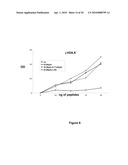 | 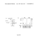 |
 |  |
 | 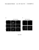 |
 |  |
 | 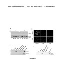 |
 | 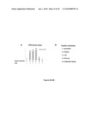 |
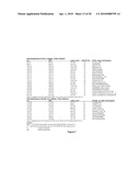 | 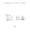 |
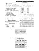 |  |
 | 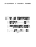 |
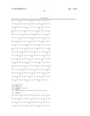 | 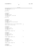 |
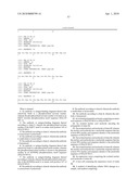 | 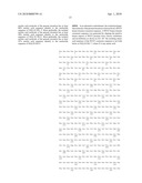 |
 |  |
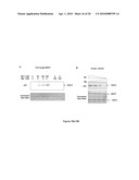 |  |
 |  |
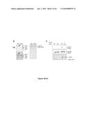 |  |
 |  |
 | 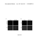 |
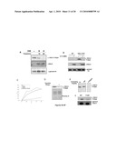 |  |
 |  |
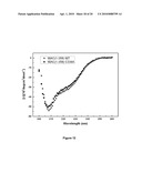 | 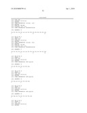 |
 | 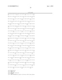 |
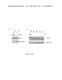 |  |
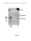 |  |
 | 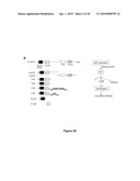 |
 | 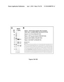 |
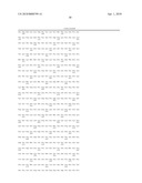 | 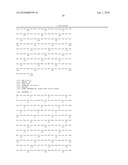 |
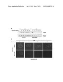 | 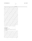 |
| Similar patent applications: | |
| Date | Title |
|---|---|
| 2011-04-28 | Devices, methods, and compositions for controlling infections |
| 2011-04-28 | System for the expression of peptides on the bacterial surface |
| 2011-04-21 | Single domain antibodies capable of modulating bace activity |
| 2011-04-28 | Skin patches and sustained-release formulations comprising lofexidine for transdermal and oral delivery |
| 2011-01-13 | Further use of protein kinase n beta |
| New patent applications from these inventors: | |
| Date | Title |
|---|---|
| 2014-04-17 | Polycomb repressive complex 2 (prc2) inhibitors and uses thereof |
| 2011-12-22 | Cathepsin l proteolytically processes histone h3 during mouse embryonic stem cell differentiation |
| Top Inventors for class "Drug, bio-affecting and body treating compositions" | |
| Rank | Inventor's name |
|---|---|
| 1 | David M. Goldenberg |
| 2 | Hy Si Bui |
| 3 | Lowell L. Wood, Jr. |
| 4 | Roderick A. Hyde |
| 5 | Yat Sun Or |