Patent application title: Amelioration of Inflammatory Arthritis By Targeting the Pre-ligand Assembly Domain (Plad) of Tumor Necrosis Factor Receptors
Inventors:
Michael Lenardo (Potomac, MD, US)
Guo-Min Deng (Bethesda, MD, US)
Francis Ka-Ming Chan (Silver Spring, MD, US)
Lixen Zheng (Rockville, MD, US)
IPC8 Class: AA61K3816FI
USPC Class:
514 12
Class name: Designated organic active ingredient containing (doai) peptide containing (e.g., protein, peptones, fibrinogen, etc.) doai 25 or more peptide repeating units in known peptide chain structure
Publication date: 2010-02-18
Patent application number: 20100041596
Inventors list |
Agents list |
Assignees list |
List by place |
Classification tree browser |
Top 100 Inventors |
Top 100 Agents |
Top 100 Assignees |
Usenet FAQ Index |
Documents |
Other FAQs |
Patent application title: Amelioration of Inflammatory Arthritis By Targeting the Pre-ligand Assembly Domain (Plad) of Tumor Necrosis Factor Receptors
Inventors:
Michael Lenardo
Guo-Min Deng
Francis Ka-Ming Chan
Lixen Zheng
Agents:
NATIONAL INSTITUTE OF HEALTH;C/O Ballard Spahr LLP
Assignees:
Origin: ATLANTA, GA US
IPC8 Class: AA61K3816FI
USPC Class:
514 12
Patent application number: 20100041596
Abstract:
The present invention provides a polypeptide comprising the isolated amino
acid sequence of a pre-ligand assembly domain (PLAD) of a TNF
receptor-like receptor. Also provided by this invention is a polypeptide
comprising the isolated amino acid sequence of a pre-ligand assembly
domain (PLAD), wherein the PLAD is selected from the group consisting of:
the PLAD of a TNF-R, the PLAD of p60, the PLAD of p80, the PLAD of Fas
(CD95/APO-1), the PLAD of TRAIL receptors, the PLAD of LTyR, the PLAD of
CD40, the PLAD of CD30, the PLAD of CD27, the PLAD of HVEM, the PLAD of
OX40 and the PLAD of DR4. TNF-R, p60, p80, Fas, TRAIL receptor, LTyR,
CD40, CD30, CD27, HVEM, OX40, DR4, TROY, EDAR, XEDAR, DCR3, AITR, 4-1BB,
DR3, RANK, TACI, BCMA, DR6, DPG, DR5, DCR1 AND DCR2 are all members of
the TNF receptor superfamily or the TNF-like receptor family. The
invention also provides the PLAD for other members of the TNF receptor
superfamily. The polypeptides of the present invention can be utilized to
inhibit oligomerization of members of the TNF receptor superfamily. These
polypeptides can also be utilized to inhibit ligand binding to members of
the TNF receptor superfamily. The present invention also provides a
composition comprising an inhibitor of TNF receptor oligomerization.
Further provided by this invention are members of the TNF receptor
superfamily that are lacking a PLAD.Claims:
1. A polypeptide of 38 to 125 amino acids, comprising the isolated amino
acid sequence of a pre-ligand assembly domain (PLAD) of a TNF
receptor-like receptor.
2. The polypeptide of claim 1, wherein the PLAD is selected from the group consisting of: the PLAD of TNF-R, the PLAD of p60, the PLAD of p80, the PLAD of Fas (CD95/APO-1), the PLAD of TRAIL, the PLAD of LTyR, the PLAD of CD40, the PLAD of CD30, the PLAD of CD27, the PLAD of HVEM, the PLAD of OX40, the PLAD of DR4, the PLAD of NGFR, the PLAD of Troy, the PLAD of EDAR, the PLAD of XEDAR, the PLAD of DcR3, the PLAD of AITR, the PLAD of 4-1BB, the PLAD of DR3, the PLAD of RANK, the PLAD of TACI, the PLAD of BCMA, the PLAD of DR6, the PLAD of OPG, the PLAD of DRS, the PLAD of DcR1, and the PLAD of DcR2.
3. A polypeptide consisting of the amino acid sequence of a pre-ligand assembly domain of a TNF receptor-like receptor.
4. A polypeptide comprising the isolated amino acid sequence of a pre-ligand assembly domain (PLAD) of a TNF receptor-like receptor, wherein the polypeptide is R1-TNF receptor-like receptor PLAD-R2, wherein R1 or R2 comprise an amino acid sequence that does not flank the TNF receptor-like receptor PLAD in a naturally occurring TNF receptor-like receptor.
5. The polypeptide of claim 1, wherein the polypeptide is R1-TNF receptor-like receptor PLAD-R2, wherein R1 is H, acyl, NH2, an amino acid or a peptide, and R2 is H, acyl, NH2, an amino acid or a peptide.
6. The polypeptide of claim 1, wherein the polypeptide is R1-amino acids 1-54 of p60-R2 (SEQ ID NO: 1), wherein R1 is H, acyl, NH2, an amino acid or a peptide, and R2 is H, acyl, NH2, an amino acid or a peptide.
7. The polypeptide of claim 1, wherein the polypeptide is R1-amino acids 10-54 of p80-R2 (SEQ ID NO: 2), wherein R1 is H, acyl, NH2, an amino acid or a peptide, and R2 is H, acyl, NH2, an amino acid or a peptide.
8. The polypeptide of claim 1, wherein the polypeptide is R1-amino acids 1-43 of Fas-R2 (SEQ ID NO: 3), wherein R1 is H, acyl, NH2, an amino acid or a peptide, and R2 is H, acyl, NH2, an amino acid or a peptide.
9. The polypeptide of claim 1, wherein the polypeptide is R1-amino acids 1-66 of Fas-R2 (SEQ ID NO: 4), wherein R1 is H, acyl, NH2, an amino acid or a peptide, and R2 is H, acyl, NH2, an amino acid or a peptide.
10. The polypeptide of claim 1, wherein the polypeptide is R-amino acids 13-50 of Lt R-R2 (SEQ ID NO:5), wherein R1 is H, acyl, NH2, an amino acid or a peptide, and R2 is H, acyl, NH2, an amino acid or a peptide.
11. The polypeptide of claim 1, wherein the polypeptide is R1-amino acids 6-39 of CD40-R2 (SEQ ID NO:6), wherein R1 is H, acyl, NH2, an amino acid or a peptide, and R2 is H, acyl, NH2, an amino acid or a peptide.
12. The polypeptide of claim 1, wherein the polypeptide is R1-amino acids 11-51 of CD30-R2 (SEQ ID NO: 7), wherein R1 is H, acyl, NH2, an amino acid or a peptide, and R2 is H, acyl, NH2, an amino acid or a peptide.
13. The polypeptide of claim 1, wherein the polypeptide is R-amino acids 7-42 of CD27-R2 (SEQ ID NO: 8), wherein R1 is H, acyl, NH2, an amino acid or a peptide, and R2 is H, acyl, NH2, an amino acid or a peptide.
14. The polypeptide of claim 1, wherein the polypeptide is R1-amino acids 6-37 of HVEM-R2 (SEQ ID NO: 9), wherein R1 is H, acyl, NH2, an amino acid or a peptide, and R2 is H, acyl, NH2, an amino acid or a peptide.
15. The polypeptide of claim 1, wherein the polypeptide is R-amino acids 3-36 of OX40-R2 (SEQ ID NO: 10), wherein R1 is H, acyl, NH2, an amino acid or a peptide, and R2 is H, acyl, NH2, an amino acid or a peptide.
16. The polypeptide of claim 1, wherein the polypeptide is R-amino acids 109-138 of DR4-R2 (SEQ ID NO: 11), wherein R1 is H, acyl, NH2, an amino acid or a peptide, and R2 is H, acyl, NH2, an amino acid or a peptide.
17. The polypeptide of claim 3, wherein the PLAD is selected from the group consisting of: the PLAD of TNF-R, the PLAD of Fas (CD95/APO-1) and the PLAD of TRAIL.
18. An isolated nucleic acid encoding the polypeptide of claim 1.
19. The isolated nucleic acid of claim 18 in a vector.
20. The vector of claim 19 in a host suitable for expressing the nucleic acid.
21. A method of inhibiting TNF receptor oligomerization in a cell by administering an effective amount of the polypeptide of claim 1.
22. A method of inhibiting Fas oligomerization in a cell by administering an effective amount of the polypeptide of claim 1.
23. A method of inhibiting ligand binding to a TNF receptor-like receptor by administering an effective amount of the polypeptide of claim 1.
24. A method of inhibiting ligand binding to Fas by administering an effective amount of the polypeptide of claim 1.
25. A method of treating inflammation in a subject by administering an effective amount of the polypeptide of claim 1.
26. The method of claim 25, wherein the inflammation is associated with an autoimmune disorder.
27. The method of claim 25, wherein the inflammation is associated with rheumatoid arthritis, osteoarthritis or septic arthritis.
28. A composition comprising an inhibitor of PLAD association.
29. The method of claim 25, wherein the inhibitor is an antibody that specifically binds to the PLAD of a TNF receptor-like receptor.
30. The method of claim 25, wherein the inhibitor is a PLAD of a TNF receptor-like receptor.
31. The method of claim 30, wherein the soluble PLAD is PLAD of p60.
32. A method of screening for an inhibitor of PLAD-association comprising:a) transfecting a cell with a plasmid containing a nucleic acid comprising a nucleic acid sequence encoding an isolated PLAD functionally linked to a fluorescence donor and a plasmid comprising a nucleic acid sequence encoding an isolated PLAD functionally linked to a fluorescence acceptor;b) contacting the cell with a putative inhibitor; andc) measuring FRET, wherein a decrease in FRET as compared to FRET measurement in a cell that was not contacted with the putative inhibitor indicates the presence of an inhibitor of PLAD-association.
33. A method of screening for an inhibitor of PLAD association comprising:a) transfecting a cell with a plasmid containing a nucleic acid comprising a nucleic acid sequence encoding an isolated PLAD and a plasmid comprising a nucleic acid sequence encoding a second isolated PLAD;b) contacting the cell with a putative inhibitor and;c) measuring PLAD self association, wherein a decrease in PLAD association in the cell of step b) as compared to PLAD association in a cell that was not contacted with the putative inhibitor indicates the presence of an inhibitor of PLAD-association.
Description:
[0001]This application claims priority to U.S. Provisional Application No.
60/694,015, filed on Jun. 24, 2005, and U.S. Provisional Application No.
60/717,589, filed on Sep. 16, 2005, hereby incorporated by reference in
their entirety.
BACKGROUND OF THE INVENTION
[0002]1. Field of the Invention
[0003]This invention provides a novel function for a conserved domain in the extracellular region of the members of the TNF receptor (TNFR) superfamily in mediating specific ligand-independent assembly of receptor oligomers.
[0004]2. Background Art
[0005]The members of the TNFR superfamily typically contain one to six cysteine rich domains (CRDs) in their extracellular regions, a single transmembrane domain and variably sized intracytoplasmic domains. The members of this receptor family typically bind to ligands of the TNF cytokine family that are defined by structural, functional and sequence similarities. These receptors form trimers in their active liganded state and several members contain a cytoplasmic domain referred to as a death domain.
[0006]According to the present invention, the extracellular region of these receptors is further characterized by a novel self-association or homotypic association function that is mediated via a pre-ligand receptor assembly domain (PLAD) that contains at least one cysteine rich domain. More specifically, members of the TNFR superfamily, including TRAIL receptor 1, CD40, 60 kDa TNFR and 80 kDa TNFR show this homotypic association. Other members of the TNFR superfamily, including Fas, LTβR, CD40, CD30, CD27, HVEM, RANK, OX40 and DR4 contain this PLAD. The PLAD is necessary for ligand binding and receptor function. Thus, members of the TNFR superfamily appear to signal through distinct pre-formed complexes rather than through ligand-induced cross-linking of individual receptor subunits. Therefore, PLAD can be targeted by pharmaceutical agents in order to block the formation of these preformed complexes and thus block receptor function.
[0007]It is well established that many microbes have evolved effective strategies for countering the immune response directed against them (79). More specifically, several viruses and bacteria express homologs of cellular proteins designed to modulate or directly block the action of immune effector molecules, including cytokines and chemokines (80). Viral homologs of TNF receptor-like receptors (vTNFRs) have been identified for several large DNA viruses, including several poxviruses and human cytomegalovirus. The existence of or any role for PLAD-mediated self-association of vTNFRs or heterologous association with TNF family receptors has not been elucidated.
[0008]Two major arthritides are rheumatoid arthritis (RA) and septic arthritis (SA). RA is a common human autoimmune disease with chronic joint inflammation and progressive bone destruction (48). Although the etiology and pathogenesis of RA are not yet fully understood, cytokines such as TNF-α, IL-1, IL-6 and receptor activator of NF-κB ligand (RANKL), are involved in disease progression (56-60). Nuclear factor-kappa B (NF-κB) is a critical regulator of these cytokines (56-60). TNF-α plays a key role in the pathogenesis of RA and its antagonists such as etanercept (also known as Enbrel), a TNFR II immunoglobulin Fc fusion protein, and infliximab (also known as Remicade), an anti-TNF-α monoclonal antibody, can improve the clinical course of RA (48). SA is a rapidly progressive and highly destructive joint disease induced by bacterial infection in which TNF-α also plays an important role (49). Experimental mouse models of arthritis induced by TNF-α (59), lipopolysaccharide (LPS), CpG-DNA (50, 61), and collagen (62) have been useful for testing new treatments. These agents induce synovitis, pannus formation, and bone and cartilage destruction as well as other features observed in human RA and SA.
[0009]According to the invention, in vitro and in vivo data indicate that TNFR PLAD proteins can potently inhibit TNF-α and its consequences in experimental inflammatory arthritis.
SUMMARY OF THE INVENTION
[0010]The present invention provides a polypeptide comprising the isolated amino acid sequence of a pre-ligand assembly domain (PLAD) of a TNF receptor-like receptor.
[0011]Also provided by this invention is a polypeptide comprising the isolated amino acid sequence of a pre-ligand assembly domain (PLAD), wherein the PLAD is selected from the group consisting of: the PLAD of TNF-R, the PLAD of p60, the PLAD of p80, the PLAD of Fas (CD95/APO-1), the PLAD of TRAIL receptors, the PLAD of LTβR, the PLAD of CD40, the PLAD of CD30, the PLAD of CD27, the PLAD of HVEM, the PLAD of OX40, the PLAD of DR4, the PLAD of NGFR, the PLAD of Troy, the PLAD of EDAR, the PLAD of XEDAR, the PLAD of DcR3, the PLAD of AITR, the PLAD of 4-1BB, the PLAD of DR3, the PLAD of RANK, the PLAD of TACI, the PLAD of BCMA, the PLAD of DR6, the PLAD of OPG, the PLAD of DRS, the PLAD of DcR1, and the PLAD of DcR2. TNF-R, p60 TNFR, p80 TNFR, Fas, TRAIL receptors, LTβR, CD40, CD30, CD27, HVEM, OX40, DR4, NGFR, Troy, EDAR, XEDAR, DcR3, AITR, 4-1BB, DR3, RANK, TACI, BCMA, DR6, OPG, DRS, DcR1, and DcR2 are all members of the TNF receptor superfamily also referred to herein as the TNF receptor-like receptor family. The invention also provides the PLAD for other members of the TNF receptor superfamily and how it can be identified by one of skill in the art.
[0012]The polypeptides of the present invention can be utilized to inhibit PLAD self-association as well as oligomerization of members of the TNF receptor superfamily. These polypeptides can also be utilized to inhibit ligand binding to members of the TNF receptor superfamily.
[0013]The present invention also provides a composition comprising an inhibitor of TNF receptor oligomerization. Further provided by this invention are members of the TNF receptor superfamily that are lacking a PLAD.
BRIEF DESCRIPTION OF THE DRAWINGS
[0014]FIG. 1A illustrates TNFR oligomers in the absence of ligand. H9 T cell lymphoma, treated or untreated with TNFα, were subjected to crosslinking with DTSSP (7). Total cell lysates were electrophoresed under non-reducing (lanes 1-4, 9-12) or reducing (lanes 5-8, 13-16) conditions as indicated and blotted for p60 or p80 TNFRs. The brackets indicate the position of trimers (T) and monomers (M). The circles indicate a non-specific protein cross-reacting with the anti-p80 antibody. The results represent three independent experiments.
[0015]FIG. 1B illustrates specific p60 TNFR self-association. 293T cells were transfected with p60ΔCD-GFP-HA (lanes 1-3) or pEGFP-N1 (lanes 4-6) and either pcDNA3 (lanes 1, 4), p60ΔCD-HA (lanes 2, 5) or HVEMΔCD-HA (lanes 3, 6). Immunoprecipitation was carried out with anti-GFP antibody (GFP IP in the top 2 panels) and blotted with anti-HA antibody (HA WB) or anti-GFP antibody (GFP WB) as indicated. The top and middle panels show the precipitated p60ΔCD-GFP-HA (or GFP) and p60ΔCD-HA respectively. The bottom panels show the p60ΔCD-HA and HVEMΔCD-HA proteins in cell lysates. Results represent five experiments.
[0016]FIG. 1C illustrates specific p80 self-association and the definition of the Pre-Ligand Assembly Domain (PLAD). 293T cells were transfected with the plasmids indicated at the top. Immunoprecipitation was performed with a C-terminal-specific anti-p80 antibody (p80 IP) that recognizes only the full-length p80 (top and middle panels). The expression of the truncated p80 or p60 proteins in the lysates is shown in the bottom panel. Western blots were performed with anti-HA antibody (top and bottom panels) and the C-terminal specific anti-p80 antibody (middle panel). The open circles represent the glycosylated and unglycosylated forms of p80. The closed circle denotes the Ig heavy chain.
[0017]FIG. 1D illustrates that PLAD is sufficient for receptor self-association. 293T cells were transfected with p80ΔCD-GFP-HA (lanes 1-5) together with the plasmids indicated at the top of each lane. Immunoprecipitation was performed with anti-GFP antibody and Western blots with anti-HA antibody. The co-precipitated DCD proteins and their expression in total cell lysates are shown in the middle and bottom panels respectively. The top panel shows the precipitated p60ΔCD-GFP-HA protein.
[0018]FIG. 1E illustrates the PLAD is essential for TNFα binding. Histograms show the expression of transfected receptors (by anti-HA staining) and their binding to TNFα in 293T cells transfected with the indicated constructs (25). The x-axis shows the intensity of fluorescence and the y-axis shows the cell number. The numbers shown are percentages of positive population compared to the vector-transfected control.
[0019]FIG. 2A illustrates that replacement of residues in the PLAD prevents self-association. 293T cells were transfected with the indicated plasmids. Immunoprecipitation was performed as in FIG. 1 with anti-GFP antibody. Western blots were performed with anti-HA antibody. The top and middle panels show the precipitated p60ΔCD-GFP-HA (open circle) and p60ΔCD-HA mutant proteins (bracket) respectively. The bottom panel shows the expression of p60ΔCD-HA mutants (bracket) and HVEMΔCD-HA (filled circle) in cell lysates.
[0020]FIG. 2B illustrates homotypic self-association of p60 and p80 TNFRs as demonstrated by fluorescence resonance energy transfer (FRET). Histograms of flow cytometric analysis of 293T cells transfected with the indicated CFP (top) and YFP (bottom) plasmid pairs. The dashed line represents the CFP transfected alone control, the solid line represents FRET without TNFα and the thick line represents FRET with TNFα. The x-axis and y-axis show the FRET fluorescence intensity and cell number respectively. FRET was analyzed in the CFP positive population in which all cells were YFP positive as well. FRET is defined as fluorescence emission of YFP due to excitation of CFP. The results are representative of four independent experiments.
[0021]FIG. 3A shows a sequence alignment of CRD1 for representatives (CRD1 of p60 (SEQ ID NO: 22), CRD1 of p80 (SEQ ID NO: 23), CRD1 of LTβR (SEQ ID NO: 24), CRD1 of CD40 (SEQ ID NO: 25), CRD1 of HVEM (SEQ ID NO: 26) and CRD1 of CD30 (SEQ ID NO: 27) of the TNFR superfamily (26). This figure illustrates the highly conserved positions of cysteines that for disulfide bonds and define the cysteine-rich domain which confers membership in the TNFR superfamily.
[0022]FIG. 3B illustrates receptor self-association in other TNFR superfamily members. 293T cells were transfected with either DR4ΔCD-GFP-HA (lanes 1-4) or CD40ΔCD-GFP-HA (lanes 5, 6) together with p80ΔCD-HA (lanes 1,6), p60ΔCD-HA (lane 2), HVEMΔCD-HA (lane 3), DR4ΔCD-HA (lane 4) or CD40ΔCD-HA (lane 5). Immunoprecipitations and Western blots were performed with anti-GFP and anti-HA antibodies respectively. The top panels show the precipitated proteins in the immune complexes and the bottom panels show the expression of the DCD proteins in the cell lysates. The filled circles denote the GFP fusion proteins and the arrows indicate the DCD protein in the immune complexes.
[0023]FIG. 3C shows flow cytometric analysis of specific receptor association of DR4 and CD40 as demonstrated by FRET. Transfections with the indicated CFP (top) and YFP (bottom) plasmid pairs were performed as in FIG. 2B. The dashed lines represent background FRET with CFP alone and the thick lines represent FRET in the presence of both CFP and YFP fusion proteins. For each group, the x-axis is the FRET intensity and the y-axis is the cell number.
[0024]FIG. 3D illustrates the two models of TNFR signaling based on pre-associated trimer complexes. For the pre-assembly chain rearrangement model (left), the ovals represent CRDs (CRDs are numbered 1-4 going from membrane distal to membrane proximal) and stippled boxes indicate the cytoplasmic domains. The receptors are viewed perpendicular to the plasma membrane. The Roman numerals represent the chains in the trimer complex. For the trimer clustering model (right), the gray symbols indicate pre-assembled TNFR trimers on the cell surface and the encircled triangles represent the trimeric TNFα. The numbers 1-3 represent the three chains of receptor in the pre-assembled trimer complex. The receptors are viewed top down to the plasma membrane.
[0025]FIG. 4A snows that a pathogenic Fas mutation causes dominant-interference in the absence of ligand binding. Surface expression and binding characteristics of wild-type (WT), Pt 2 (del 52-96), and Pt 6 (A241D) Fas molecules. The left column shows surface expression 24 hours after transfection into 293T cells using staining for the AU-1 epitope tag present at the N-terminus of each receptor protein. The middle column shows the same cells stained with 10 μg/ml of the anti-Fas agonistic antibody APO-1 (Kamiya). The right column shows binding of FasL engineered to trimerize through a modified leucine zipper and visualized by staining with an anti-leucine zipper mAb (FasL stain). Antibody binding was visualized with phycoerythrin-conjugated anti-mouse antibodies. The brackets indicate the percentage of cells strongly positive for staining when compared to the non-transfected controls. In each plot the thick and thin lines represent the signals from the transfected and non-transfected cell preparations, respectively. All histograms represent 10,000 events plotted on a 4 decade logarithmic fluorescence scale (X axis) vs. cell count (Y axis). Data was collected on a FACScalibur flow cytometer using Flowjo software (Treestar software).
[0026]FIG. 4B shows dominant inhibition by mutant Fas molecules co-transfected with WT Fas. Ten μg of the indicated expression vectors or pCI vector alone were electroporated into BW cells lacking human Fas as previously described (15), with 5 μg of pEGFP-N1 (Clontech) to mark transfected cells with GFP. Twenty-four hours later, the indicated amounts of anti-Fas mAb APO-1 were added along with 1/20 volume soluble protein A (Sigma) for maximal apoptosis induction. Apoptosis was quantitated by enumerating GFP-positive viable cells by flow cytometry and calculating cell loss (15).
[0027]FIG. 4C illustrates self-association of Fas molecules. An expression vector encoding HA-tagged Fas with the C-terminal death domain replaced by the Green Fluorescent Protein (HAFas 1-210:GFP) was co-transfected with wild-type (WT) Fas and the EC mutant Pt 2 Fas (del 52-96). Control cells were co-transfected with WT Fas and an HA-tagged cytoplasmic truncated version of the TNF-receptor family member Herpesvirus Entry Mediator (HVEM or HveA) fused to GFP (HA HVEMΔCD:GFP). Cell lysates were lysed, immunoprecipitated with anti-GFP and electrophoresed as described in the Examples. Blots of precipitated proteins were probed with a polyclonal antiserum against the Fas C-terminal (C20, Santa Cruz biotechnology) (Anti-Fas CT) to reveal full-length Fas molecules co-precipitating with the GFP tagged proteins. Cell lysates were also probed in western blots (WB) with anti-HA-HRP (Roche Molecular Biochemicals) and anti-Fas C20 to quantitate the total amount of these proteins. The open circle indicates the IgG heavy chain in the immunoprecipitates, the closed circle indicates WT Fas, and the arrow indicates the truncated Pt 2 Fas protein. The upper band in some lanes blotted with anti-Fas C20 represents glycosylated Fas.
[0028]FIG. 5A shows the expression and function of Fas mutants lacking the PLAD or ligand binding. Binding of APO-1 and FasL by N-terminal Fas mutants. Staining of the indicated HA-tagged Fas mutants, the R86S Fas mutant, and control transfections with a C-terminal truncated HA-tagged TNFR2 was performed as in FIG. 1A except that anti-HA was used instead of anti-AU1 to show total expression of each mutant on the cell surface.
[0029]FIG. 5B shows the interaction of Fas extracellular domains is dependent on a domain in the N-terminal region of the protein. In lanes 14, 293T cells were co-transfected with an AU-1 tagged Fas 1-210 lacking the death domain and the indicated HA-tagged Fas mutants or control TNFR2 protein (HA TNFR2ΔCD). Lysates were immunoprecipitated with anti-AU1, and probed with anti-HA to reveal co-precipitated proteins. Control blots with an antibody against the N-terminal of Fas (WB anti-FasN) are shown to quantitate the amount of the AU-1 Fas 1-210 protein in the lysates. The results are representative of three independent transfections. Lanes 5-7 show co-precipitation of WT Pas and the FasR86S mutant by HAFas1-210:GFP with the same procedure used in FIG. 1C. The open circle indicates the Ig heavy chain of the immunoprecipitating antibody, and the closed circle indicates the position of immunoprecipitated Fas.
[0030]FIG. 5C illustrates the induction of apoptosis is lost in Fas molecules lacking the self-association domain. BW5147 murine thymoma cells were transfected with 10 μg of expression vectors for indicated Fas molecules. Apoptosis was induced with 500 μg/ml soluble APO-1 and quantitated as in FIG. 1B.
[0031]FIG. 5D illustrates the induction and inhibition of apoptosis by the non-ligand binding R86S Fas mutant. BW cells were transfected with 10 μg of each Fas expression vector and 5 μg of GFP plasmid. Apoptosis induction and quantitation was performed as in FIG. 2C, except that APO-1 was used to induce apoptosis in samples shown with open bars, and 5% v/vol FasL supernatant was added to the samples with filled bars.
[0032]FIG. 6A shows Fluorescence Resonance Energy Transfer between Pas molecules. Dot plots showing the relationships between CFP, YFP and FRET signals in the indicated co-transfectants. CFP and YFP fusion proteins were constructed, transfected into 293T cells and analyzed on a FACS vantage cytometer. Numbers are the percentage of cells positive for CFP or YFP with FRET signal (top right quadrant).
[0033]FIG. 6B is a comparison of FRET signals between full-length and N-terminal deleted Fas receptors. Histograms of FRET signals were generated in cells gated for CFP fluorescence. YFP fluorescence was comparable between all transfectants. The thick line is the signal from co-transfected cells and the thin line is the signal from the CFP construct alone of each pair.
[0034]FIG. 6C shows FRET efficiency for the indicated CFP and YFP pairs as determined by microscopic photobleaching of YFP on individual cells (Five readings of 4-7 cell regions). The numbers represent the average E % and standard error for each plasmid pair.
[0035]FIG. 7A illustrates pre-association of endogenous Fas receptor chains. 1×10⁷ H9 lymphoma cells were treated with the crosslinker DTSSP (Pierce, 10 mM for 30 minutes at 4° C., followed by quenching with 10 mM Tris-Cl pH8 for 15 min), and/or stimulated with 1 μg of the agonistic antibody APO-1 or FasL for 15 minutes under the indicated conditions. For anti-Fas immunoblotting, cell lysates were treated with N-glycanase-F (Roche Molecular Biochemicals) before electrophoresis and probed with the anti-Fas C terminal mAb B10 (Santa Cruz Biotechnology) and anti-mouse IgG1-HRP (Southern Biotechnology).
[0036]FIG. 7B After treatment with the indicated reagents, cells were lysed, immunoprecipitated and blotted for FADD and caspase-8 as previously described (11). The positions of the two isoforms of procaspase-8 (p54/52) and the caspase-8 cleavage products after proteolysis of the p11 caspase subunit (p43/41) are shown with arrows.
[0037]FIG. 7C shows PARP cleavage. Aliquots of cells used in (A) and (B) were cultured at 37° C. for an additional 4 hrs and cell lysates were blotted with anti-PARP mAb (Research Diagnostics Inc). The upper band is the 115 kD full-length PARP and the lower band is the signature 85 kD caspase cleavage fragment. The results are representative of at least three independent experiments for each condition.
[0038]FIG. 8A illustrates that dominant interference depends on the N-terminal PLAD. Alignment of selected ALPS patient Fas mutations from families studied at the NIH. "X" symbols indicate the location of point mutations. Capacity to associate with wildtype Fas as tested by co-precipitation (SA) and dominant inhibition of Fas-induced apoptosis in co-transfection studies (DI) are indicated as shown. Sequences encoding dominant-negative PLAD containing polypeptides encoded by mutations from patients #1 and #20 are shown. Numbering begins with the first amino acid after the signal peptide. Italics denote extra amino acids added by frameshift mutations.
[0039]FIG. 8B shows that dominant interference is lost without the PLAD. Fas-sensitive Jurkat T lymphoma cells were transfected with the 10 μg of the indicated constructs and 2.5 μg of the GFP reporter plasmid. Eighteen hours after transfection, the indicated amounts of Apo-1 were added for 6 hours and apoptosis was quantitated by staining with Annexin V-PE (Pharmingen). Percentages are the percent of GFP(+) cells staining positive for Annexin V. These results are representative of three independent transfections.
[0040]FIG. 9 shows the analysis of immunoprecipitates for the presence of p80 chimeric receptors or truncations. 293T cells were transfected with the indicated plasmids and harvested for co-immunoprecipitation using an anti-p80 COOH-terminal specific antibody. The immunoprecipitates were analyzed for the presence of p80 chimeric receptors or truncations using anti-HA antibody in Western blot analysis (top panel). The bottom panel shows the expression of the HA-tagged proteins in whole cell lysates.
[0041]FIG. 10 shows expression of recombinant bacterial PLAD proteins. (a) A model of how the PLAD contributes to receptor trimer assembly and competence for ligand binding. A soluble PLAD protein could associate with individual receptor chains, prevent trimeric receptor assembly, and thereby block ligand-induced signaling. (b) Gel electrophoresis of purified GST, P60 PLAD-GST (P60), and P80 PLAD-GST (P80). Molecular weight markers and sizes in kilodaltons are shown on the left. (c) Western blot analysis using monoclonal antibodies (MAb) against the P60 PLAD (top panels) or the P80 PAD (bottom panels). Titrations in μg protein used in the original gel electrophoresis for P60 PLAD (left panels) or P80 PLAD (right panels) are indicated.
[0042]FIG. 11 shows the effects of PLAD proteins in TNF-α-induced cell death. (a) Cell death assessed by flow cytometry after L929 cells were treated with: medium; human TNF-α (hTNF) (2 ng); hTNF-α (2 ng)+P60 PLAD (P60) (40 μg). Inset shows phase contrast photomicrographs. In the lower right is given the percent of gated live cells. The Y axis is propidium iodide (PI) staining and the X axis is FSC (forward scatter profile). Dead cells exhibit increased PI staining and reduced FSC. (b) Cell loss induced by mouse TNF-α (mTNF) (2 ng) or hTNF (2 ng) with different doses of PLAD P60 (P60) protein. (c) Loss of L929 cells induced by mTNF (2 ng) or hTNF (2 ng) with different doses of PLAD CD40 (CD40) protein. (d) Loss of 42.3 Jurkat cells after treatment with TNF-α or anti-Fas with and without P60 PLAD (P60), P80 PLAD (P80) and GST for 12 h. TNF-α (3 ng), P60L (low; 4.5 μg), P60H (high; 15 μg), P80L (low; 4.5 μg), P80H (high; 15 μg), GST (15 μg), anti-Fas (10 ng). (e) Caspase-8 activity in L929 cells measured by optical density (OD) of substrate conversion treated with mTNF-α (4 ng) with or without P60 PLAD (100 μg), etanercept (25 μg), infliximab (20 μg).
[0043]FIG. 12 shows the effects of P60 and P80 PLAD proteins on arthritis induced by intra-articular injection of TNF-α in BALB/c mice and bacterial CpG DNA in C3H/HeJ mice. (a) Representative photomicrographs of H&E-stained tissue sections of knee joints showing: PBS; 45 ng TNF-α; P60 PLAD (100 μg) and 45 ng TNF-α; P80 PLAD (100 μg) and 45 ng TNF-α; CpG DNA (1 nmole); CpG DNA (1 nmole) and P60 PLAD (100 μg). Arrows indicate foci of inflammation. Labels are: C (cartilage), JC (joint cavity), ST (synovial tissue), B (bone), and the arrowhead indicates the lining layer of synovial tissue. (b, c) Quantitation of the histological analysis of synovitis, pannus, and erosion of bone and cartilage of experimental groups (n=5) treated with TNF-α alone (TNF) or TNF-α plus the P60 PLAD protein (P60) or TNF-α plus P80 PLAD protein (P80) as indicated. (d) Quantitation of histological analysis of synovitis, pannus, erosion of bone and cartilage of each experimental group (n=5). These analyses were repeated at least two times. CpG DNA alone (CpG), CpG DNA plus P60 PLAD (P60). Values are mean±standard deviation (s.d.) **P<0.01 for treated versus control group. Mice were sacrificed 3 d after intra-articular inoculation for histopathological examination. This is representative of three experiments.
[0044]FIG. 13 shows the effects of P60 and P80 PLAD proteins on CIA in DBA/1J mice. A masked experiment (a-f). (a) Photographs of the paws of CIA mice treated with PBS or P60 PLAD. (b) H&E stained section of a CIA joint 75 d after primary immunization treated with PBS or 4 weeks of intraperitoneal injection of P60 PLAD protein (100 μg three times per week). (c) Severity of arthritis; paw thickness measurement; and weight in CIA mice treated with PBS (n=13 mice) (square), P60 PLAD protein (P60) (n=12 mice) (diamond), and P80 PLAD protein (P80) (n=13 mice) (oval). (d) Incidence of arthritis in PBS, P60, and P80 PLAD treatment groups. (e) Evaluation of synovitis; pannus; erosion of bone and cartilage in joint sections in CIA treated for 4 weeks as indicated. (f) IL-1 and IL-6 level in sera. *=P<0.05 versus control PBS group. (g) Severity of arthritis in CIA mice receiving 2 weeks' intraperitoneal injection of PBS (n=10 mice) (square), P60 PLAD protein (P60) (n=10 mice) (diamond), and etanercept (n=10 mice) (oval). *=P<0.05 versus control PBS group. (h) The effect of P60 PLAD protein in established CIA. Severity of arthritis in established CIA mice receiving 2 weeks' intraperitoneal injection of PBS (n=9 mice) (square), P60 PLAD protein (P60, 400 μg every other day) (n=10 mice) (diamond). *=P<0.05 versus control PBS group.
[0045]FIG. 14 shows TNFR expression in arthritic joints and PLAD protein inhibition of TNF-α binding and NF-κB activation. (a) Immunohistochemistry for TNFR1 and TNFR2 in an arthritic joint from DBA/1J mice sacrificed 75 d with CIA treated with PBS or P60 PLAD. Brown color indicates TNFR expressing cells. (b) Flow cytometry following staining with: 50 ng biotinylated human TNF-α (Bt-TNF-α) and different doses of P60 PLAD protein pretreatment. Curves represent Bt-TNFα alone, Bt-TNF-α plus 3 μg P60 PLAD, Bt-TNF-α plus 15 μg P60 PLAD, Bt-TNF-α plus 30 μg P60 PLAD, human TNF-α, or negative control. (c) Gel electrophoresis of 50 ng human TNF-α immunoprecipitated (IP) with etanercept (1 μg), P60 (1 μg) or P80 (1 μg) PLAD protein as indicated (test protein). Western blot using antibody (Ab) against TNF-α. (d) Electrophoretic mobility shift assay of nuclear extracts prepared from A3 Jurkat cells treated with TNF-α in the presence (+) or absence (-) of P60 PLAD protein using radiolabelled oligonucleotide probes for NF-κB (left arrow) or OCT1 (right arrow). Quantitation of NF-κB p65 nuclear translocation in mononuclear cells isolated from spleen from TNFR1(e) or TNFR2 (f) knockout (-/-) mice after these cells were treated with mTNF-α (2 ng) with or without P60 (100 μg) or P80 (100 μg) PLAD protein in vitro. *P<0.05 versus control group; **=P<0.01 versus control group. The difference between the P60 and P80 treatment groups is significantly different in each genetic background (P<0.01).
[0046]FIG. 15 shows that P60 PLAD protein inhibits osteoclastogenesis and RANK and RANK ligand (RANKL) expression. (a) Representative photomicrographs of immunohistochemistry of calcitonin receptor in an arthritic joint from DBA/1J mice sacrificed 75 d after primary immunization with collagen treated with PBS or P60 PLAD protein Light shaded arrow indicates positive staining (brown in original color slide). (b) Effects of PLAD protein in TNF-α-induced osteoclastogenesis in vitro. Photomicrographs of TRAP-positive osteoclasts in bone marrow macrophages (MM) cultured with: M-CSF and mouse TNF-α (10 μg); mouse TNF-α (10 μg) plus 32 μg P60 PLAD; or PBS. Light shaded arrows indicate tartrate-resistant acid phosphatase (TRAP)-positive osteoclasts. Quantitation of TRAP-positive cells in each well of BMM cultures after 7 d treatment with different doses of P60 PLAD protein as indicated, determined microscopically. (c) Photomicrographs of immunohistochemistry of RANK and RANKL staining in an arthritic joint from DBA/1J mice sacrificed 75 d after primary immunization with collagen and then treated with PBS, with P60 PLAD protein. Dark (brown in its original color slide) staining indicates positive staining cells.
[0047]FIG. 16 shows the amino acid sequences of human PLAD-GST fusion proteins in standard single letter code. (a) Shown bold and underline (amino acid residues 230-282) is the P60 PLAD peptide sequence (SEQ ID NO:59). (b) Shown bold and underline (amino acid residues 230-274) is the P80 PLAD peptide sequence (SEQ ID NO:60). Shown in plain text in both parts is the GST protein sequence.
[0048]FIG. 17 shows the effects of P60 PLAD protein on TNF-α-induced cell death in L929 cells. Phase contrast photomicrographs of cells treated for 19 hours with: (a) medium alone; (b) human TNF-α (2 ng); (c) P60 PLAD (3 μg)+hTNF-α (2 ng); (d) P60 PLAD (30 μg)+hTNF-α (2 ng); (e) infliximab (1 μg)+hTNF-α (2 ng); (f) etanercept (1 μg)+hTNF-α (2 ng). (g) P80 PLAD (30 μg)+hTNF-α (2 ng). Magnification 400×. (h) Loss of L929 cells induced by hTNF (2 ng) with or without P60 PLAD (50 μg), etanercept (1 μg) and infliximab (1 μg).
[0049]FIG. 18 shows the effects of GST protein and P80 PLAD protein on inflammatory arthritis. (a) Quantitation of the histological analysis of synovitis, pannus and erosion of bone and cartilage in experimental groups (n=5) treated with TNF-α (45 ng) alone (TNF) or TNF-α (45 ng) plus the GST protein (GST, 100 μg) in BALB/c mice; (b) with CpG DNA (1 mmole) alone (CpG) or CpG DNA (1 mmole) plus P80 PLAD protein (P80, 100 μg) in C3H/HeJ mice. Values are mean±s.d. Mice were sacrificed 3 d after intra-articular inoculation for histopathological examination. This is representative of three experiments. (c) Photomicrographs of H&E stained section showing inflammation and destruction of a CIA joint 75 d after primary immunization with 4 weeks' intraperitoneal injection of P80 PLAD protein (100 μg). Magnification 200×. (d) Quantitation of the histological analysis of synovitis, pannus, and erosion of bone and cartilage in CIA in DBA/1J mice treated with P80 PLAD protein for 4 weeks. P>0.05 compared with control group.
[0050]FIG. 19 shows photomicrographs of immunohistochemistry. (a) TNFR1 and TNFR2 in an arthritic joint from a TNF-α transgenic mouse sacrificed at 6 months. Arrow in TNFR1 panel shows dark (brown in its original color slide) staining indicating receptor expression. Arrow in TNFR2 panel indicates high TNFR2 expression by chondrocytes deep within cartilage. (b) calcitonin receptor in: an arthritic joint from a TNF-o transgenic mouse sacrificed at 6 months of age, and a healthy joint from a control C57BL/6 mouse. (c) RANK and RANKL staining in an arthritic joint from TNF-α transgenic mice sacrificed at age of 6 months, and in a healthy joint from normal control C57BL/6 mouse. Dark (brown in it's original color slide) staining indicates a positive histochemical reaction. Magnification 300×.
[0051]FIG. 20 shows the immunogenicity and half-life of the P60 PLAD protein. (a) Anti-PLAD P60 antibody level compared to a standard curve based on purified PLAD protein in sera from DBA/1 mice sacrificed 75 d after primary immunization with collagen and then treated with PBS, P60 or P80 PLAD proteins. GST+means that GST was added to remove GST antibody from sera. (b) Half-life of P60 PLAD protein. (c) Half-life of P80 PLAD protein.
[0052]FIG. 21 shows that PLAD proteins inhibit TNF-α-induced IkBα degradation. Western Blot shows that IkBα degradation in mononuclear cells isolated from spleen from TNFR1 or TNFR2 knockout (-\-) mice after these cells were treated with mTNF-α (2 ng) with or without P60 (10, 50, 100 μg) or P80 (10, 50, 100 μg) for 5 min (b), 15 min (a) or GST (10, 50, 100 μg) for 1 h (c) in vitro.
[0053]FIG. 22 shows gel electrophoresis of 50 ng of human TNF-α immunoprecipitated (IP) with different amounts of etanercept and P60 PLAD protein. (a) etanercept (10 μg) or P60 PLAD protein (100 μg) as indicated (testing protein). (b) etanercept. (0.1, 1, 10 μg) or P60 PLAD protein (0.1, 1, 10 μg) as indicated (testing protein). Arrowheads indicate the position of the TNF protein on the gel. Western blotting was carried out with antibody (Ab) against TNF-α.
[0054]FIG. 23 shows the dimerized-PLAD portion from the crystal structure of TNFR1 (PDB ID: 1NCF). Dark and light grey illustrate individual peptide chains of a PLAD dimer. The mirror imidazole rings of His-34 from each peptide are depicted within the circle. These two histidines lock up each other in an inter-chain pocket.
[0055]FIG. 24 shows immunoprecipitation (IP) and Western Blot (WB) of P80-PLAD protein mixed with etanercept.
[0056]FIG. 25 shows that P60-PLAD protein inhibited MW expression in CIA. Note that the dark (brown in its original color slide) staining in the left panel indicates MMP expression.
[0057]FIG. 26 shows that P60-PLAD protein inhibits iNOS expression in CIA. Note that the dark (brown in its original color slide) staining in the left panel indicate iNOS expression.
DETAILED DESCRIPTION OF THE INVENTION
[0058]The present invention may be understood more readily by reference to the following detailed description of the preferred embodiments of the invention and the Examples included therein and to the Figures and their previous and following description.
[0059]As used in the specification and in the claims, "a" can mean one or more, depending upon the context in which it is used. Thus, for example, reference to "a nucleic acid" means that at least one nucleic acid is utilized.
Polypeptides
[0060]The present invention provides a polypeptide comprising the isolated amino acid sequence of a pre-ligand assembly domain (PLAD). The present invention also provides a polypeptide consisting of the amino acid sequence of a pre-ligand assembly domain. The PLAD of the present invention can be the PLAD of a TNF-R, the PLAD of p60, the PLAD of p80, the PLAD of Fas (CD95/APO-1), the PLAD of TRAIL, the PLAD of LTβR, the PLAD of CD40, the PLAD of CD30, the PLAD of CD27, the PLAD of HVEM, the PLAD of OX40, the PLAD of DR4 or any other PLAD domain from a member of the TNFR superfamily. Since the PLAD domain is highly conserved among members of the TNFR superfamily, one skilled in the art could identify the PLAD domain of any TNF receptor by searching available databases for the conserved motif that characterizes the PLAD domain. Identification of these regions in TNF receptor-like receptors is made routine by the provision of exemplary PLAD sequences herein and their comparison to published sequences of other members of the family (see FIG. 3, for example). Furthermore, one skilled in the art would also be able to identify a PLAD by performing functional assays, such as those provided in the Examples. In one embodiment the functional PLAD is not the PLAD of Fas/CD59 (83). In a further embodiment, the functional PLAD is not the amino-terminal 49 amino acids of the Fas/CD59 receptor (83).
[0061]The PLADs provided herein can comprise as few as 38 amino acids of the N-terminus of a mature TNF receptor-like receptor. A mature TNF receptor-like receptor is a TNF receptor-like receptor that does not include a signal sequence. Examples of PLADs are disclosed in the sequence listing, which includes amino acid sequences of examples of TNF receptor-like receptors including their signal sequences. The residues of the signal sequences of the respective receptors can be found by reference to the GenBank accession numbers for these TNF receptor-like receptors listed in Table 1. Thus, the sequences of the mature TNF receptor-like receptors and their corresponding PLADs are disclosed in the provided sequences. Table 3 provides additional information about the TNF receptor-like receptors and receptor ligands disclosed herein. It also provides information regarding the uses for the isolated PLADs and polypeptides containing the isolated PLADs disclosed herein. The PLADs can be used to study the implications of interfering with a signal transduction pathway mediated by a receptor of the TNFR superfamily. For example, if signaling via a receptor of the TNFR superfamily is known or shown to be associated with a disease pathway, the inhibition of receptor pre-ligand assembly by the present polypeptides, can treat or prevent the disease. For example, diseases that can be treated include cancer, heart disease and inflammatory diseases. Modifications of PLAD can also change the affinity of ligand/receptor interactions, which can be used in in vitro studies such as measuring ligand and receptor binding, receptor signals etc. Fluorescence-tagged PLAD proteins may also be utilized as reagents for determining relative expression of specific TNFRs on the surface of cells via flow cytometry or fluorescence microscopy.
[0062]The present invention also provides a polypeptide of 38 to 125 amino acids comprising an isolated PLAD. For example, the polypeptide can be from 50 to 125 amino acids comprising an isolated PLAD. In a further example, the polypeptide can comprise the subsequence R1-TNF receptor-like receptor PLAD-R2, wherein R1 and R2 are optional and when present can be H, acyl, NH2, an amino acid or a peptide. When present, R1 and/or R2 can be any amino acid. When R1 and/or R2 is a peptide, this peptide can vary in length. For example, R1 and/or R2 can be 1, 2, 3, 4, 5, 6, 7, 8, 9, 10, 11, 12, 13, 14, 15, 16, 17, 18, 19, 20, 21, 22, 23, 24, 25 or more amino acids in length as long as the entire polypeptide comprising the isolated TNF-like PLAD is no more than 125 amino acid residues, and can be 38, 39, 40, 41, 42, 43, 44, 45, 46, 47, 48, 49, 50, 51, 52, 53, 54, 55, 56, 57, 58, 59, 60, 61, 62, 63, 64, 65, 66, 67, 68, 69, 70, 71, 72, 73, 74, 75, 76, 77, 78, 79, 80, 81, 82, 83, 84, 85, 86, 87, 89, 90, 91, 92, 93, 94, 95, 96, 97, 98, 99, 100, 101, 102, 103, 104, 105, 106, 107, 108, 109, 110, 111, 112, 113, 114, 115, 116, 117, 118, 119, 120, 121, 122, 123, 124 or 125 amino acids in length. R1 and R2 can also be sequences of the TNF receptor-like receptor that normally flank the TNF-like PLAD in a naturally occurring TNF receptor-like receptor, wherein the polypeptide comprising the TNF-like receptor PLAD is not the entire extracellular domain of a TNF receptor-like receptor.
[0063]Further provided by this invention is a polypeptide of any size, comprising the isolated amino acid sequence of a pre-ligand assembly domain (PLAD) of a TNF receptor-like receptor, wherein the polypeptide is R1-TNF receptor-like receptor PLAD-R2, wherein R1 or R2 comprise an amino acid sequence that does not flank the TNF receptor-like receptor PLAD in a naturally occurring TNF receptor-like receptor. R1 or R2, but not both can be full or partial sequences of the TNF receptor-like receptor that normally flank the TNF-like PLAD in a naturally occurring TNF receptor-like receptor. For example, the PLAD can be from a TNF receptor-like receptor and R1 or R2, can be amino acid sequences that are not present in the TNF receptor-like receptor from which the TNF-like PLAD of the polypeptide was derived or any other TNF receptor-like receptor. R1 or R2 can be any amino acid sequence as long as R1-TNF-like PLAD-R2 is not a naturally occurring full-length TNF receptor-like receptor. In another example, the PLAD can be from one TNF receptor-like receptor and R1 or R2 or both, if present, can be peptide sequences from another TNF receptor-like receptor. Therefore, one skilled in the art can combine the PLAD of one TNF receptor-like receptor with R1 or R2 sequences from a different TNF receptor-like receptor to obtain this polypeptide. Since the sequences of known TNF receptor-like receptors are publicly available, the structure of R1 and R2 of the present polypeptide are numerous but well known and contemplated herein. Alternatively, R1 or R2 can be peptide sequences that are not related to any of the TNF receptor-like receptor sequences. In one embodiment the polypeptide comprising an isolated PLAD is not the 124 amino acid sequence of the mature (lacking the signal sequence) TNF1 receptor disclosed in U.S. Pat. No. 5,633,145 (Feldman et al.) and shown in SEQ ID NO:40.
[0064]Examples of polypeptides comprising the above-mentioned subsequence include: R1-amino acids 1-38, 1-39, 1-40, 141, 142, 143, 1-44, 1-45, 1-46, 1-47, 1-48, 1-49, 1-50, 1-51, 1-52, 1-53, or 1-54 of mature p60-R2 (e.g., SEQ ID NO: 1); R1-amino acids 10-48, 10-49, 10-50, 10-51, 10-52, 10-53, or 10-54 of mature p80-R2 (SEQ ID NO: 2); R1-amino acids 1-38, 1-39, 1-40, 1-41, 142, or 1-43 of mature Fas-R2 (SEQ ID NO: 3); R1-amino acids 1-38, 1-39, 1-40, 1-41, 1-42, 1-43, 1-44, 145, 1-46, 1-47, 1-48, 1-49, 1-50, 1-51, 1-52, 1-53, 1-54, 1-55, 1-56, 1-57, 1-58, 1-59, 1-60, 1-61, 1-62, 1-63, 1-64, 1-65, or 1-66 of mature Fas-R2 (SEQ ID NO: 4); R1-amino acids 13-50 of mature LtβR-R2 (SEQ ID NO: 5); R1-amino acids 6-39 of mature CD40-R2 (SEQ ID NO: 6); R1-amino acids 11-49, 11-50, or 11-51 of mature CD30-R2 (SEQ ID NO: 7); R1-amino acids 7-42 of mature CD27-R2 (SEQ ID NO: 8), R1-amino acids 6-37 of mature HVEM-R2 (SEQ ID NO: 9); R1-amino acids 3-36 of mature OX40-R2 (SEQ ID NO: 10), and R1-amino acids 109-138 of mature DR4-R2 (SEQ ID NO: 11).
[0065]The mature p60 (TNFR1) polypeptide starts at position 30 of the full-length p60 coding sequence set forth as SEQ ID NO: 12. Therefore, the present invention provides a polypeptide comprising amino acids 1-54 of the mature p60 protein (amino acids 30-83 of SEQ ID NO: 12), a polypeptide comprising amino acids 1-53 of the mature p60 protein (amino acids 30-82 of SEQ ID NO: 0.12), a polypeptide comprising amino acids 1-52 of the mature p60 protein (amino acids 30-81 of SEQ ID NO: 12), a polypeptide comprising amino acids 1-51 of the mature p60 protein (amino acids 30-80 of SEQ ID NO: 12), a polypeptide comprising amino acids 1-50 of the mature p60 protein (amino acids 30-79 of SEQ ID NO: 12), a polypeptide comprising amino acids 1-49 of the mature p60 protein (amino acids 30-78 of SEQ ID NO: 12), a polypeptide comprising amino acids 1-48 of the mature p60 protein (amino acids 30-77 of SEQ ID NO: 12), a polypeptide comprising amino acids 1-47 of the mature p60 protein (amino acids 30-76 of SEQ ID NO: 12), a polypeptide comprising amino acids 1-46 of the mature p60 protein (amino acids 30-75 of SEQ ID NO: 12), a polypeptide comprising amino acids 1-45 of the mature p60 protein (amino acids 30-74 of SEQ ID NO: 12), a polypeptide comprising amino acids 1-44 of the mature p60 protein (amino acids 30-73 of SEQ ID NO: 12), a polypeptide comprising amino acids 1-43 of the mature p60 protein (amino acids 30-72 of SEQ ID NO: 12), a polypeptide comprising amino acids 1-42 of the mature p60 protein (amino acids 30-71 of SEQ ID NO: 12), a polypeptide comprising amino acids 1-41 of the mature p60 protein (amino acids 30-70 of SEQ ID NO: 12), a polypeptide comprising amino acids 1-40 of the mature p60 protein (amino acids 30-69 of SEQ ID NO: 12), and a polypeptide comprising amino acids 1-39 of the mature p60 protein (amino acids 30-68 of SEQ ID NO: 12) as well as other polypeptides comprising fragments of amino acids 1-54 of the mature p60 protein that retain PLAD activity.
[0066]The mature p80 (TNFR2) polypeptide starts at position 23 of the full-length p80 coding sequence set forth as SEQ ID NO: 13. Therefore the present invention provides a polypeptide comprising amino acids 10-54 of the mature p80 protein (amino acids 32-76 of SEQ ID NO: 13), a polypeptide comprising amino acids 10-53 of the mature p80 protein (amino acids 32-75 of SEQ ID NO: 13), a polypeptide comprising amino acids 10-52 of the mature p80 protein (amino acids 32-74 of SEQ ID NO: 13), a polypeptide comprising amino acids 10-51 of the mature p80 protein (amino acids 32-73 of SEQ ID NO: 13), a polypeptide comprising amino acids 10-50 of the mature p80 protein (amino acids 32-72 of SEQ ID NO: 13), a well as other polypeptides comprising fragments of amino acids 10-54 of the mature p80 protein that retain PLAD activity.
[0067]The mature Fas receptor polypeptide starts at position 17 of the full-length Fas coding sequence set forth as SEQ ID NO: 14. Therefore the present invention provides a polypeptide comprising amino acids 1-43 of the mature Fas protein (amino acids 17-59 of SEQ ID NO: 14), a polypeptide comprising amino acids 1-42 of the mature Fas protein (amino acids 17-58 of SEQ ID NO: 14), a polypeptide comprising amino acids 1-41 of the mature Fas protein (amino acids 17-57 of SEQ ID NO: 14), a polypeptide comprising amino acids 1-40 of the mature Fas protein (amino acids 17-56 of SEQ ID NO: 14), a polypeptide comprising amino acids 1-39 of the mature Fas protein (amino acids 17-55 of SEQ ID NO: 14), a well as other polypeptides comprising fragments of amino acids 1-43 of the mature Fas protein that retain PLAD activity.
[0068]The present invention also provides a polypeptide comprising amino acids 1-66 of the mature Fas protein (amino acids 17-82 of SEQ ID NO: 14), a polypeptide comprising amino acids 1-65 of the mature Fas protein (amino acids 17-81 of SEQ ID NO: 14), a polypeptide comprising amino acids 1-64 of the mature Fas protein (amino acids 17-80 of SEQ ID NO: 14), a polypeptide comprising amino acids 1-63 of the mature Fas protein (amino acids 17-79 of SEQ ID NO: 14), a polypeptide comprising amino acids 1-62 of the mature Fas protein (amino acids 17-78 of SEQ ID NO: 14), as well as other polypeptides comprising fragments of amino acids 1-66 of the mature Fas protein that retain PLAD activity.
[0069]The present invention also provides a polypeptide comprising amino acids 43-80 of the full-length LtβR protein set forth as SEQ ID NO: 15 as well as other polypeptides comprising fragments of amino acids 43-80 of SEQ ID NO: 15 that retain PLAD activity.
[0070]The present invention also provides a polypeptide comprising amino acids 26-59 of the full-length CD40 protein set forth as SEQ ID NO: 16 as well as other polypeptides comprising fragments of amino acids 26-59 of SEQ ID NO: 16 that retain PLAD activity.
[0071]The mature CD30 polypeptide starts at position 19 of the full-length CD30 coding sequence set forth as SEQ ID NO: 17. Therefore the present invention provides a polypeptide comprising amino acids 11-51 of the mature CD30 protein (amino acids 29-69 of SEQ ID NO: 17), a polypeptide comprising amino acids 11-50 of the mature CD30 protein (ammo acids 29-68 of SEQ ID NO: 17), a polypeptide comprising amino acids 11-49 of the mature CD30 protein (amino acids 29-67 of SEQ ID NO: 17), a polypeptide comprising amino acids 11-48 of the mature CD30 protein (amino acids 29-66 of SEQ ID NO: 17), a polypeptide comprising amino acids 11-47 of the mature CD30 protein (amino acids 29-65 of SEQ ID NO: 17), a well as other polypeptides comprising fragments of amino acids 11-51 of the mature CD30 protein that retain PLAD activity.
[0072]The present invention also provides a polypeptide comprising amino acids 27-62 of the full-length CD27 protein set forth as SEQ ID NO: 18 as well as other polypeptides comprising fragments of amino acids 27-62 of SEQ ID NO: 18 that retain PLAD activity
[0073]The present invention also provides a polypeptide comprising amino acids 42-75 of the full-length HVEM protein set forth as SEQ ID NO: 19 as well as other polypeptides comprising fragments of the polypeptide comprising amino acids 42-75 of SEQ ID NO: 19 that retain PLAD activity.
[0074]The mature OX40 polypeptide starts at position 29 of the full-length OX40 coding sequence set forth as SEQ ID NO: 20. Therefore the present invention provides a polypeptide comprising amino acids 3-36 of the mature OX40 protein (amino acids 31-64 of SEQ ID NO: 20), a polypeptide comprising amino acids 3-35 of the mature OX40 protein (amino acids 31-63 of SEQ ID NO: 20), a polypeptide comprising amino acids 3-34 of the mature OX40 protein (amino acids 31-62 of SEQ ID NO: 20), a polypeptide comprising amino acids 3-33 of the mature OX40 protein (amino acids 31-61 of SEQ ID NO: 20), a polypeptide comprising amino acids 3-32 of the mature CD30 protein (amino acids 31-60 of SEQ ID NO: 20), a well as other polypeptides comprising fragments of the polypeptide comprising amino acids 3-36 of the mature OX40 protein that retain PLAD activity.
[0075]The present invention also provides a polypeptide comprising amino acids 132-170 of the full-length DR4 protein set forth as SEQ ID NO: 21 as well as other polypeptides comprising fragments of the polypeptide comprising amino acids 132-170 of SEQ ID NO: 21 that retain PLAD activity.
[0076]Table 1 sets forth examples of TNF receptor-like receptors comprising a PLAD of the present invention. The nucleotide and polypeptide sequences for these receptors can be found under the GenBank Accession Nos. set forth in Table 1. The nucleotide sequences, the polypeptide sequences and any information (e.g., signal sequence and mature protein residue numbers) set forth under the GenBank Accession Nos. set forth in Table 1 are hereby incorporated in their entireties by this reference. For example, the nucleotide sequence, the polypeptide sequence and additional information (e.g., signal sequence and mature protein residue numbers) for p60 can be found under GenBank Accession No. M75866. These p60 sequences and additional information set forth under GenBank Accession No. M75866 are hereby incorporated in their entireties by this reference. Similarly, the nucleotide sequence, the polypeptide sequence and additional information (e.g., signal sequence and mature protein residue numbers) set forth for p80 can be found under GenBank Accession No. M32315. These p80 sequences and additional information set forth under GenBank Accession No. M32315 are hereby incorporated in their entireties by this reference. By accessing the GenBank Accession Nos. set forth in Table 1, one of skill in the art can access additional GenBank Accession Nos. listed therein to obtain additional information concerning signal sequences and mature protein sequences. For example, upon accessing GenBank Accession No. M75866, one of skill in the art can access GenBank Accession No. AAA61201 which sets forth the signal sequence and mature protein sequences information for p60. This information can also be found by directly accessing GenBank Accession Nos. AAA51201 (p60), GenBank Accession No. AAA59929 (p80), GenBank Accession No. AAA63174 (Fas), GenBank Accession No. AAA36757 (LTBR), GenBank Accession No. CAA43045 (CD40), GenBank Accession No. AAA51947 (CD30), GenBank Accession No. AAA58411 (CD27), GenBank Accession No. AAB58354, GenBank Accession No. CAA53576 (OX40), GenBank Accession No. AAC51226 (DR4), and is incorporated herein by this reference.
[0077]Table 1 also provides Locus Link Accession Nos. for the TNF-like receptors. Locus Link Accession Nos. are now equivalent to Entrez Gene Identification Numbers (Gene ID numbers) that can be accessed at the National Center for Biotechnology Information at the U.S. National Library of Medicine. For example, one of skill in the art can obtain additional information, regarding p60, including nucleotide and protein sequences, by accessing Locus Link number 7132 (now Gene D 7132 in Entrez Gene) in the Entrez Gene database. Similarly one of skill in the art can obtain additional information regarding p80, including nucleotide and protein sequences, by accessing Locus Link number 7133 (now Gene ID 7133 in Entrez Gene) in the Entrez Gene database. Thus, one of skill in the art can readily obtain information regarding any of the TNF-like receptors listed in Table 1 by accessing their respective Locus Link (Gene ID) numbers in Entrez Gene. All of the information provided under the Locus Link (Gene ID) numbers set forth in Table 1 is hereby incorporated by reference in its entirety.
[0078]Provided are polypeptides comprising the isolated amino acid sequences for PLAD domains of vTNFR proteins (SEQ ID NOS: 28-39). Other vTNFR PLADs can be identified using protein-protein BLAST database searches of homologs for TNFR1 and TNFR2 PLAD sequences. Also included are full length amino acid sequences for each vTNFR and its modified protein (SEQ. ID NOS: 44-55). Also provided is methodology for identification, production, and functional testing of and additional vTNFR PLAD polypeptides by one of skill in the art.
[0079]The vTNFR PLAD domains can disrupt self-association of host TNFRs and/or subsequent ligand binding to dampen anti-viral immunity and/or protect infected cells from TNF-mediated cell death. The M-T2 protein, a TNF-receptor like protein encoded by myxoma virus, can protect myxoma-infected T cells from TNF-induced death independently of its extracellular TNF binding capacity (82). vTNFR PLAD sequences can serve as more potent inhibitors of TNF-induced effects than P60 or P80 PLADs themselves, as a consequence of evolutionary selection for higher affinity binding to host TNFR PLAD domains. In this regard, isolated viral PLAD proteins represent improved agents for clinical use in blocking TNF-associated pathogenesis associated with rheumatoid arthritis and other autoimmune diseases.
[0080]It is understood that techniques such as those described herein can be employed to identify additional microbial proteins with homology to PLAD domains found in other TNF receptor-like receptors (e.g. Fas) described herein. For example, three examples of microbial polypeptides containing sequences homologous to the Fas PLAD domain (SEQ ID NOS: 41-43, 56-58) are disclosed.
[0081]As used herein an "isolated amino acid sequence of a PLAD" means a sequence which is substantially free from the naturally occurring materials with which the amino acid sequence is normally associated in nature. The polypeptides of this invention can comprise the entire amino acid sequence of a PLAD domain or fragments thereof that have PLAD activity. The polypeptides or fragments thereof of the present invention can be obtained by isolation and purification of the polypeptides from cells where they are produced naturally or by expression of exogenous nucleic acid encoding a PLAD. Fragments of a PLAD can be obtained by chemical synthesis of peptides, by proteolytic cleavage of the PLAD or the polypeptide comprising a PLAD and by synthesis from nucleic acid encoding the portion of interest. The PLAD can include conservative substitutions where a naturally occurring amino acid is replaced by one having similar properties. Such conservative substitutions do not alter the function of the polypeptide. Mutations that enhance binding and effectiveness can be found by creating various amino acid substitutions and testing them in binding assays described within the specification using techniques available to those of ordinary skill in the art.
[0082]Thus, it is understood that, where desired, modifications and changes can be made in the nucleic acid encoding the polypeptides of this invention and/or amino acid sequence of the polypeptides of the present invention and still obtain a polypeptide having like or otherwise desirable characteristics. Such changes can occur in natural isolates or can be synthetically introduced using site-specific mutagenesis, the procedures for which, such as mis-match polymerase chain reaction (PCR), are well known in the art.
[0083]For example, certain amino acids can be substituted for other amino acids in a polypeptide without appreciable loss of functional activity. It is thus contemplated that various changes can be made in the amino acid sequence of the PLAD (or underlying nucleic acid sequence) without appreciable loss of biological utility or activity and possibly with an increase in such utility or activity. For example, the Q24A mutation, the D49R mutation and the K19E mutation in the natural sequence of p60 TNFR do not impair PLAD self-association.
[0084]These polypeptides can also be obtained in any of a number of procedures well known in the art. One method of producing a polypeptide is to link two peptides or polypeptides together by protein chemistry techniques. For example, peptides or polypeptides can be chemically synthesized using currently available laboratory equipment using either Fmoc (9-fluorenylmethyloxycarbonyl) or Boc (tert-butyloxycarbonoyl) chemistry. (Applied Biosystems, Inc., Foster City, Calif.). One skilled in the art can readily appreciate that a peptide or polypeptide corresponding to a particular protein can be synthesized by standard chemical reactions. For example, a peptide or polypeptide can be synthesized and not cleaved from its synthesis resin whereas the other fragment of a hybrid peptide can be synthesized and subsequently cleaved from the resin, thereby exposing a terminal group which is functionally blocked on the other fragment. By peptide condensation reactions, these two fragments can be covalently joined via a peptide bond at their carboxyl and amino termini, respectively, to form a larger polypeptide. (Grant, Asynthetic Peptides: A User Guide, W.H. Freeman and Co., N.Y. (1992) and Bodansky and Trost, Ed., Principles of Peptide Synthesis, Springer-Verlag Inc., N.Y. (1993)). Alternatively, the peptide or polypeptide can be independently synthesized in vivo as described above. Once isolated, these independent peptides or polypeptides can be linked to form a larger protein via similar peptide condensation reactions.
[0085]For example, enzymatic ligation of cloned or synthetic peptide segments can allow relatively short peptide fragments to be joined to produce larger peptide fragments, polypeptides or whole protein domains (Abrahmsen et al. Biochemistry, 30:4151 (1991)). Alternatively, native chemical ligation of synthetic peptides can be utilized to synthetically construct large peptides or polypeptides from shorter peptide fragments. This method consists of a two step chemical reaction (Dawson et al. A Synthesis of Proteins by Native Chemical Ligation, Science, 266:776-779 (1994)). The first step is the chemoselective reaction of an unprotected synthetic peptide-%-thioester with another unprotected peptide segment containing an amino-terminal Cys residue to give a thioester-linked intermediate as the initial covalent product. Without a change in the reaction conditions, this intermediate undergoes spontaneous, rapid intramolecular reaction to form a native peptide bond at the ligation site. Application of this native chemical ligation method to the total synthesis of a protein molecule is illustrated by the preparation of human interleukin 8 (IL-8) (Clark-Lewis et al. FEBS Lett., 307:97 (1987), Clark-Lewis et al., J. Biol. Chem., 269:16075 (1994), Clark-Lewis et al. Biochemistry, 30:3128 (1991), and Rajarathnam et al. Biochemistry, 29:1689 (1994)).
[0086]Alternatively, unprotected peptide segments can be chemically linked where the bond formed between the peptide segments as a result of the chemical ligation is an unnatural (non-peptide) bond (Schnolzer et al. Science, 256:221 (1992)). This technique has been used to synthesize analogs of protein domains as well as large amounts of relatively pure proteins with full biological activity (deLisle Milton et al. ATechniques in Protein Chemistry IV, Academic Press, New York, pp. 257-267 (1992)).
[0087]The present invention also provides peptide mimetics for the disclosed polypeptides. A "peptide mimetic" is defined to include a chemical compound, or an organic molecule, or any other peptide mimetic, the structure of which is based on or derived from a binding region of a protein. For example, one can model predicted chemical structures to mimic the structure of a binding region, such as a PLAD. Such modeling can be performed using standard methods. Alternatively, peptide mimetics can also be selected from combinatorial chemical libraries in much the same way that peptides are. (Ostresh, J. M. et al., Proc Natl Acad Sci USA 1994 Nov. 8; 91(23):11138-42; Dorner, B. et al., Bioorg Med Chem 1996 May; 4(5):709-15; Eichler, J. et al., Med Res Rev 1995 November; 15(6):481-96; Blondelle, S. E. et al. Biochem J 1996 Jan. 1; 313 (Pt 1):141-7; Perez-Paya, E. et al., J Biol Chem 1996 Feb. 23; 271(8):4120-6). Functional assays can also be utilized to select peptide mimetics.
[0088]The polypeptides of this invention can be linked to another moiety such as a nucleic acid, a protein, a peptide, a ligand, a carbohydrate moiety, viral proteins, a monoclonal antibody, a polyclonal antibody or a liposome. Furthermore, two or more PLAD containing polypeptides can also be linked to each other. For example, a bifunctional or multifunctional polypeptide containing two or more different PLADs can be made such that the polypeptide is capable of modulating the activity of more than one TNF receptor-like receptor. The polypeptide can also contain two or more PLADs from the same TNF receptor-like receptor in order to increase the avidity of this polypeptide for a particular TNF receptor-like receptor.
PLAD-Containing Fusion Constructs
[0089]Disclosed herein are fusion proteins containing PLAD and the nucleic acids encoding them. The fusion protein can comprise the PLAD of a TNF receptor-like receptor disclosed herein linked to a fusion tag. The functional molecule can be an antibody or targeting portion thereof or other fusion tag. Since PLAD is on the cell surface, the PLAD containing fusion protein can include a component that targets the PLAD to the cell surface of PLAD-expressing cells. For example, marker-binding portions of ligands for non-TNF receptor-like markers on the surfaces of intended target cells can be fused to PLAD. The PLAD can be fused to various carrier proteins such as immunoglobulin or other serum, soluble, and/or stable proteins. The fusion tag can be GST or other molecule that facilitates purification of the fusion protein. The PLAD-containing fusion protein can include a signal sequence to facilitate secretion of a recombinantly expressed PLAD.
[0090]The nucleic acids encoding a polypeptide comprising or consisting of a PLAD can also be functionally linked to other nucleic acids to encode an immunoadhesin. For the purposes of the invention, the term "immunoadhesin" is defined as including any polypeptide encoded by a nucleic acid where at least a portion of a nucleic acid encoding a non-immunoglobulin molecule such as a PLAD is coupled to at least a portion of a nucleic acid encoding an immunoglobulin heavy chain polypeptide, IgG for example. The Fc regions of IgG2, IgG3, IgM, IgA, IgE can also be utilized to construct an immunoadhesin. In a particular example, the fusion protein comprises PLAD fused to an Ig Fc portion, especially that of Ig Gamma 4. The coupling can be achieved in a manner which provides for a functional transcribing and translating of the nucleic acid segment and message derived therefrom, respectively.
[0091]The PLAD polypeptide fusion protein can be expressed by transient or stable transfection in a variety of mammalian host cells as well as in baculovirus-infected cells. The expressed fusion protein can be purified according to standard methods. Similar, to antibodies, IgG immunoadhesins can be purified from the culture medium into which they are secreted by single-step protein A or protein G affinity chromatography.
Transgene
[0092]Provided are PLAD-encoding transgenes. By a "transgene" is meant a nucleic acid sequence that is inserted by artifice into a cell and becomes a part of the genome of that cell and its progeny. Such a transgene can be (but is not necessarily) partly or entirely heterologous (e.g., derived from a different species) to the cell. The term "transgene" broadly refers to any nucleic acid that is introduced into an animal's genome, including but not limited to genes or DNA having sequences which are perhaps not normally present in the genome, genes which are present, but not normally transcribed and translated ("expressed") in a given genome, or any other gene or DNA which one desires to introduce into the genome. This can include genes which may normally be present in the nontransgenic genome but which one desires to have altered in: expression, or which one desires to introduce in an altered or variant form. A transgene can include one or more transcriptional regulatory sequences and any other nucleic acid, such as introns, that may be necessary for optimal expression of a selected nucleic acid. A transgene can be as few as a couple of nucleotides long, but is preferably at least about 50, 100, 150, 200, 250, 300, 350, 400, or 500 nucleotides long or even longer. A transgene can be coding or non-coding sequences, or a combination thereof. A transgene usually comprises a regulatory element that is capable of driving the expression of one or more transgenes under appropriate conditions.
Antibodies
[0093]Also provided by the present invention are antibodies that specifically bind to a PLAD of a TNF receptor-like receptor. For example, the antibodies of the present invention can be antibodies that specifically bind to a PLAD of a TNF receptor, antibodies that specifically bind to a PLAD of FAS or antibodies that specifically bind a PLAD of DR4, to name a few. The antibody (either polyclonal or monoclonal) can be raised to any of the polypeptides provided and contemplated herein, both naturally occurring and recombinant polypeptides, and immunogenic fragments, thereof. The antibody can be used in techniques or procedures such as diagnostics, treatment, or vaccination. Anti-idiotypic antibodies and affinity matured antibodies are also considered.
[0094]Antibodies can be made by many well-known methods (See, e.g. Harlow and Lane, "Antibodies; A Laboratory Manual" Cold Spring Harbor Laboratory, Cold Spring Harbor, N.Y., (1988)). Briefly, purified antigen can be injected into an animal in an amount and in intervals sufficient to elicit an immune response. Antibodies can either be purified directly, or spleen cells can be obtained from the animal. The cells can then fused with an immortal cell line and screened for antibody secretion. The antibodies can be used to screen nucleic acid clone libraries for cells secreting the antigen. Those positive clones can then be sequenced. (See, for example, Kelly et al. Bio/Technology, 10:163-167 (1992); Bebbington et al. Bio/Technology, 10:169-175 (1992)). Humanized and chimeric antibodies are also comtemplated in this invention. Heterologous antibodies can be made by well known methods (See, for example, U.S. Pat. Nos. 5,545,806, 5,569,825, 5,625,126, 5,633,425, 5,661,016, 5,770,429, 5,789,650, and 5,814,318)
[0095]The phrase "specifically binds" with the polypeptide refers to a binding reaction which is determinative of the presence of the protein in a heterogeneous population of proteins and other biologics. Thus, under designated immunoassay conditions, the specified antibodies bound to a particular protein do not bind in a significant amount to other proteins present in the sample. Selective binding to an antibody under such conditions may require an antibody that is selected for its specificity for a particular protein. A variety of immunoassay formats can be used to select antibodies that selectively bind with a particular protein. For example, solid-phase ELISA immunoassays are routinely used to select antibodies selectively immunoreactive with a protein. See Harlow and Lane "Antibodies, A Laboratory Manual" Cold Spring Harbor Publications, New York, (1988), for a description of immunoassay formats and conditions that could be used to determine selective binding.
Nucleic Acids
[0096]The present invention also provides nucleic acids that encode polypeptides of up to 125 amino acids comprising a PLAD of a TNF receptor-like receptor as well as nucleic acids that encode polypeptides consisting of a TNF receptor-like receptor PLAD.
[0097]The present invention also provides nucleic acids that encode a polypeptide of up to 125 amino acids comprising an isolated PLAD, wherein the polypeptide comprises the subsequence R1-PLAD-R2, wherein R1 and R2 are optional and when present can be H, acyl, NH2, an amino acid or a peptide.
[0098]The invention further provides a nucleic acid that encodes a polypeptide comprising the isolated amino acid sequence of a pre-ligand assembly domain (PLAD) of a TNF receptor-like receptor, wherein the polypeptide is R1-TNF receptor-like receptor PLAD-R2, wherein R1 or R2 comprise an amino acid sequence that does not flank the TNF receptor-like receptor PLAD in a naturally occurring TNF receptor-like receptor.
[0099]As used herein, the term "nucleic acid" refers to single- or multiple stranded molecules which may be DNA or RNA, or any combination thereof, including modifications to those nucleic acids. The nucleic acid may represent a coding strand or its complement, or any combination thereof. Nucleic acids may be identical in sequence to the sequences which are naturally occurring for any of the novel genes discussed herein or can include alternative codons which encode the same amino acid as that which is found in the naturally occurring sequence. These nucleic acids can also be modified from their typical structure. Such modifications include, but are not limited to, methylated nucleic acids, the substitution of a non-bridging oxygen on the phosphate residue with either a sulfur (yielding phosphorothioate deoxynucleotides), selenium (yielding phosphorselenoate deoxynucleotides), or methyl groups (yielding methylphosphonate deoxynucleotides).
[0100]A nucleic acid molecule encoding a PLAD can be isolated from the organism in which it is normally found. For example, a genomic DNA or cDNA library can be constructed and screened for the presence of the nucleic acid of interest. Methods of constructing and screening such libraries are well known in the art and kits for performing the construction and screening steps are commercially available (for example, Stratagene Cloning Systems, La Jolla, Calif.). Once isolated, the nucleic acid can be directly cloned into an appropriate vector, or if necessary, be modified to facilitate the subsequent cloning steps. Such modification steps are routine, an example of which is the addition of oligonucleotide linkers which contain restriction sites to the termini of the nucleic acid. General methods are set forth in Sambrook et al., "Molecular Cloning, a Laboratory Manual," Cold Spring Harbor Laboratory Press (1989). Also contemplated by the present invention are nucleic acids encoding a PLAD that do not contain a ligand binding site.
[0101]Once the nucleic acid sequence of the desired PLAD is obtained, the sequence encoding specific amino acids can be modified or changed at any particular amino acid position by techniques well known in the art. For example, PCR primers can be designed which span the amino acid position or positions and which can substitute any amino acid for another amino acid. Then a nucleic acid can be amplified and inserted into the wild-type PLAD coding sequence in order to obtain any of a number of possible combinations of amino acids at any position of the PLAD. Alternatively, one skilled in the art can introduce specific mutations at any point in a particular nucleic acid sequence through techniques for point mutagenesis. General methods are set forth in Smith, M. "In vitro mutagenesis" Ann. Rev. Gen., 19:423-462 (1985) and Zoller, M. J. "New molecular biology methods for protein engineering" Curr. Opin. Struct. Biol., 1:605-610 (1991). Techniques such as these can be used to alter the coding sequence without altering the amino acid sequence that is encoded.
[0102]Another example of a method of obtaining a DNA molecule encoding a PLAD is to synthesize a recombinant DNA molecule which encodes the PLAD. For example, oligonucleotide synthesis procedures are routine in the art and oligonucleotides coding for a particular protein region are readily obtainable through automated DNA synthesis. A nucleic acid for one strand of a double-stranded molecule can be synthesized and hybridized to its complementary strand. One can design these oligonucleotides such that the resulting double-stranded molecule has either internal restriction sites or appropriate 5' or 3' overhangs at the termini for cloning into an appropriate vector. Double-stranded molecules coding for relatively large proteins can readily be synthesized by first constructing several different double-stranded molecules that code for particular regions of the protein, followed by ligating these DNA molecules together. For example, Cunningham, et al., "Receptor and Antibody Epitopes in Human Growth Hormone Identified by Homolog-Scanning Mutagenesis," Science, 243:1330-1336 (1989), have constructed a synthetic gene encoding the human growth hormone gene by first constructing overlapping and complementary synthetic oligonucleotides and ligating these fragments together. See also, Ferretti, et al., Proc. Nat. Acad. Sci. 82:599-603 (1986), wherein synthesis of a 1057 base pair synthetic bovine rhodopsin gene from synthetic oligonucleotides is disclosed. By constructing a PLAD in this manner, one skilled in the art can readily obtain any particular PLAD with desired amino acids at any particular position or positions within the PLAD. See also, U.S. Pat. No. 5,503,995 which describes an enzyme template reaction method of making synthetic genes. Techniques such as this are routine in the art and are well documented. These nucleic acids or fragments of a nucleic acid encoding a PLAD can then be expressed in vivo or in vitro as discussed below.
[0103]The invention also provides for the isolated nucleic acids encoding a PLAD in a vector suitable for expressing the nucleic acid. Once a nucleic acid encoding a particular PLAD of interest, or a region of that nucleic acid, is constructed, modified, or isolated, that nucleic acid can then be cloned into an appropriate vector, which can direct the in vivo or in vitro synthesis of that wild-type and/or modified PLAD. The vector is contemplated to have the necessary functional elements that direct and regulate transcription of the inserted gene, or nucleic acid. These functional elements include, but are not limited to, a promoter, regions upstream or downstream of the promoter, such as enhancers that may regulate the transcriptional activity of the promoter, an origin of replication, appropriate restriction sites to facilitate cloning of inserts adjacent to the promoter, antibiotic resistance genes or other markers which can serve to select for cells containing the vector or the vector containing the insert, RNA splice junctions, a transcription termination region, or any other region which may serve to facilitate the expression of the inserted gene or hybrid gene. (See generally, Sambrook et al.).
[0104]There are numerous E. coli (Escherichia coli) expression vectors known to one of ordinary skill in the art which are useful for the expression of the nucleic acid insert. Other microbial hosts suitable for use include bacilli, such as Bacillus subtilis, and other enterobacteriaceae, such as Salmonella, Serratia, and various Pseudomonas species. In these prokaryotic hosts one can also make expression vectors, which will typically contain expression control sequences compatible with the host cell (e.g., an origin of replication). In addition, any number of a variety of well-known promoters will be present, such as the lactose promoter system, a tryptophan (Trp) promoter system, a beta-lactamase promoter system, or a promoter system from phage lambda. The promoters will typically control expression, optionally with an operator sequence, and have ribosome binding site sequences for example, for initiating and completing transcription and translation. If necessary, an amino terminal methionine can be provided by insertion of a Met codon 5' and in-frame with the downstream nucleic acid insert. Also, the carboxy-terminal extension of the nucleic acid insert can be removed using standard oligonucleotide mutagenesis procedures.
[0105]Additionally, yeast expression can be used. There are several advantages to yeast expression systems. First, evidence exists that proteins produced in a yeast secretion systems exhibit correct disulfide pairing. Second, post-translational glycosylation is efficiently carried out by yeast secretory systems. The Saccharomyces cerevisiae pre-pro-alpha-factor leader region (encoded by the MF'-1 gene) is routinely used to direct protein secretion from yeast. (Brake, et al., Alpha-Factor-Directed Synthesis and Secretion of Mature Foreign Proteins in Saccharomyces cerevisiae. Proc. Nat. Acad. Sci., 81:4642-4646 (1984)). The leader region of pre-pro-alpha-factor contains a signal peptide and a pro-segment which includes a recognition sequence for a yeast protease encoded by the KEX2 gene: this enzyme cleaves the precursor protein on the carboxyl side of a Lys-Arg dipeptide cleavage signal sequence. The nucleic acid coding sequence can be fused in-frame to the pre-pro-alpha-factor leader region. This construct is then put under the control of a strong transcription promoter, such as the alcohol dehydrogenase I promoter or a glycolytic promoter. The nucleic acid coding sequence is followed by a translation termination codon which is followed by transcription termination signals. Alternatively, the nucleic acid coding sequences can be fused to a second protein coding sequence, such as Sj26 or β-galactosidase, used to facilitate purification of the fusion protein by affinity chromatography. The insertion of protease cleavage sites to separate the components of the fusion protein is applicable to constructs used for expression in yeast. Efficient post translational glycosylation and expression of recombinant proteins can also be achieved in Baculovirus systems.
[0106]Mammalian cells permit the expression of proteins in an environment that favors important post-translational modifications such as folding and cysteine pairing, addition of complex carbohydrate structures, and secretion of active protein. Vectors useful for the expression of active proteins in mammalian cells are characterized by insertion of the protein coding sequence between a strong viral promoter and a polyadenylation signal. The vectors can contain genes conferring hygromycin resistance, genticin or G418 resistance, or other genes or phenotypes suitable for use as selectable markers, or methotrexate resistance for gene amplification. The chimeric protein coding sequence can be introduced into a Chinese hamster ovary (CHO) cell line using a methotrexate resistance-encoding vector, or other cell lines using suitable selection markers. Presence of the vector DNA in transformed cells can be confirmed by Southern blot analysis. Production of RNA corresponding to the insert coding sequence can be confirmed by Northern blot analysis. A number of other suitable host cell lines capable of secreting intact human proteins have been developed in the art, and include the CHO cell lines, HeLa cells, myeloma cell lines, Jurkat cells, etc. Expression vectors for these cells can include expression control sequences, such as an origin of replication, a promoter, an enhancer, and necessary information processing sites, such as ribosome binding sites, RNA splice sites, polyadenylation sites, and transcriptional terminator sequences. Preferred expression control sequences are promoters derived from immunoglobulin genes, SV40, Adenovirus, Bovine Papilloma Virus, etc. The vectors containing the nucleic acid segments of interest can be transferred into the host cell by well-known methods, which vary depending on the type of cellular host. For example, calcium chloride transformation is commonly utilized for prokaryotic cells, whereas calcium phosphate, DEAE dextran, or lipofectin mediated transfection or electroporation can be used for other eukaryotic cellular hosts.
[0107]Alternative vectors for the expression of genes or nucleic acids in mammalian cells, those similar to those developed for the expression of human gamma-interferon, tissue plasminogen activator, clotting Factor VIII, hepatitis B virus surface antigen, protease Nexin1, and eosinophil major basic protein, can be employed. Further, the vector can include CMV promoter sequences and a polyadenylation signal available for expression of inserted nucleic acids in mammalian cells (such as COS-7).
[0108]Insect cells also permit the expression of mammalian proteins. Recombinant proteins produced in insect cells with baculovirus vectors undergo post-translational modifications similar to that of wild-type proteins. Briefly, baculovirus vectors useful for the expression of active proteins in insect cells are characterized by insertion of the protein coding sequence downstream of the Autographica californica nuclear polyhedrosis virus (AcNPV) promoter for the gene encoding polyhedrin, the major occlusion protein. Cultured insect cells such as Spodoptera frugiperda cell lines are transfected with a mixture of viral and plasmid DNAs and the viral progeny are plated. Deletion or insertional inactivation of the polyhedrin gene results in the production of occlusion negative viruses which form plaques that are distinctively different from those of wild-type occlusion positive viruses. These distinctive plaque morphologies allow visual screening for recombinant viruses in which the AcNPV gene has been replaced with a hybrid gene of choice.
[0109]The invention also provides for the vectors containing the contemplated nucleic acids in a host suitable for expressing the nucleic acids. The vectors containing the nucleic acid segments of interest can be transferred into host cells by well-known methods, which vary depending on the type of cellular host; For example, calcium chloride transformation, transduction, and electroporation are commonly utilized for prokaryotic cells, whereas calcium phosphate, DEAE dextran, or lipofection mediated transfection or electroporation can be used for other cellular hosts.
[0110]Alternatively, the nucleic acids of the present invention can be operatively linked to one or more of the functional elements that direct and regulate transcription of the inserted nucleic acid and the nucleic acid can be expressed. For example, a nucleic acid can be operatively linked to a bacterial or phage promoter and used to direct the transcription of the nucleic acid in vitro. A further example includes using a nucleic acid provided herein in a coupled transcription-translation system where the nucleic acid directs transcription and the RNA thereby produced is used as a template for translation to produce a polypeptide. One skilled in the art will appreciate that the products of these reactions can be used in many applications such as using labeled RNAs as probes and using polypeptides to generate antibodies or in a procedure where the polypeptides are being administered to a cell or a subject.
[0111]Expression of the nucleic acid, in combination with a vector, can be by either in vivo or in vitro. In vivo synthesis comprises transforming prokaryotic or eukaryotic cells that can serve as host cells for the vector. Alternatively, expression of the nucleic acid can occur in an in vitro expression system. For example, in vitro transcription systems are commercially available which are routinely used to synthesize relatively large amounts of mRNA. In such in vitro transcription systems, the nucleic acid encoding a PLAD would be cloned into an expression vector adjacent to a transcription promoter. For example, the Bluescript II cloning and expression vectors contain multiple cloning sites which are flanked by strong prokaryotic transcription promoters. (Stratagene Cloning Systems, La Jolla, Calif.). Kits are available which contain all the necessary reagents for in vitro synthesis of an RNA from a DNA template such as the Bluescript vectors. (Stratagene Cloning Systems, La Jolla, Calif.). RNA produced in vitro by a system such as this can then be translated in vitro to produce the desired PLAD polypeptide. (Stratagene Cloning Systems, La Jolla, Calif.).
Gene Therapy Methods
[0112]Using gene therapy methods, a nucleic acid encoding a polypeptide comprising or consisting of a PLAD can be administered. The nucleic acid encoding the polypeptide of this invention can be placed into a vector and delivered to the cells of a subject either in vivo or ex vivo by standard methods.
[0113]The nucleic acid encoding the polypeptide of this invention can be functionally attached to a specific leader peptide which can specify for secretion of the polypeptide. For example the polypeptide can have a signal sequence, such as the murine Ig-kappa signal sequence (Blezinger et al. Nat. Biotechnol. 17: 343-8, 1999), rat insulin leader sequence (Fakhral et al. J. Immunother. 20: 437-8, 1997), FGF-4 signal sequence (Ueno et al. Aterioscler. Thromb. Vasc. Biol., 17: 2453-2460, 1997), human growth hormone signal peptide (Rade et al. Gene Ther. 6: 385-92, 1999), beta lactamase signal sequence (Hughes et al. Hum. Gene Ther. 5: 1445-55, 1994), bovine prolactin signal sequence (Gorman et al. Bran Res. Mol. Brain. Res. 44:143-146, 1997) and other similar signal sequences.
[0114]For in vivo administration, the cells can be in a subject and the nucleic acid can be administered in a pharmaceutically acceptable carrier. The subject can be any animal in which it is desirable to selectively express a nucleic acid in a cell. In a preferred embodiment, the animal of the present invention is a human. In addition, non-human animals which can be treated by the method of this invention can include, but are not limited to, cats, dogs, birds, horses, cows, goats, sheep, guinea pigs, hamsters, gerbils and rabbits, as well as any other animal in which selective expression of a nucleic acid in a cell can be carried out according to the methods described herein.
[0115]In the method described above which includes the introduction of exogenous DNA into the cells of a subject (i.e., gene transduction or transfection), the nucleic acids of the present invention can be in the form of naked DNA or the nucleic acids can be in a vector for delivering the nucleic acids to the cells for expression of the nucleic acid inside the cell. The vector can be a commercially available preparation, such as an adenovirus vector (Quantum Biotechnologies, Inc. (Laval, Quebec, Canada). Delivery of the nucleic acid or vector to cells can be via a variety of mechanisms. As one example, delivery can be via a liposome, using commercially available liposome preparations such as Lipofectin®, Lipofectamine® (GIBCO-BRL, Inc., Gaithersburg, Md.), Superfect® (Qiagen, Inc. Hilden, Germany) and Transfectam® (Promega Biotec, Inc., Madison, Wis.), as well as other liposomes developed according to procedures standard in the art. In addition, the nucleic acid or vector of this invention can be delivered in vivo by electroporation, the technology for which is available from Genetronics, Inc. (San Diego, Calif.) as well as by means of a Sonoporation machine (ImaRx Pharmaceutical Corp., Tucson, Ariz.).
[0116]As one example, vector delivery can be via a viral system, such as a retroviral vector system which can package a recombinant retroviral genome. The recombinant retrovirus can then be used to infect and thereby deliver nucleic acid to the infected cells. The exact method of introducing the nucleic acid into mammalian cells is, of course, not limited to the use of retroviral vectors. Other techniques are widely available for this procedure including the use of adenoviral vectors, adeno-associated viral (AAV) vectors, lentiviral vectors, pseudotyped retroviral vectors, and pox virus vectors, such as vaccinia virus vectors. Physical transduction techniques can also be used, such as liposome delivery and receptor-mediated and other endocytosis mechanism. This invention can be used in conjunction with any of these or other commonly used gene transfer methods.
[0117]The nucleic acid and the nucleic acid delivery vehicles of this invention, (e.g., viruses; liposomes, plasmids, vectors) can be in a pharmaceutically acceptable carrier for in vivo administration to a subject. By "pharmaceutically acceptable" is meant a material that is not biologically or otherwise undesirable, i.e., the material can be administered to a subject, along with the nucleic acid or vehicle, without causing any undesirable biological effects or interacting in a deleterious manner with any of the other components of the pharmaceutical composition in which it is contained. The carrier would naturally be selected to minimize any degradation of the active ingredient and to minimize any adverse side effects in the subject, as would be well known to one of skill in the art.
[0118]The nucleic acid or vehicle can be administered orally, parenterally (e.g., intravenously), by intramuscular injection, by intraperitoneal injection, transdermally, extracorporeally, topically or the like. The exact amount of the nucleic acid or vector required will vary from subject to subject, depending on the species, age, weight and general condition of the subject, the severity or mechanism of any disorder being treated, the particular nucleic acid or vehicle used, its mode of administration and the like.
Inhibitors of PLAD Self-Association
[0119]The present invention further provides a composition comprising an inhibitor of PLAD self-association or TNF-like receptor oligomerization. An "inhibitor" is defined as a compound that binds a PLAD or a compound, including antibodies, that binds the target for a PLAD and prevents an activity of a PLAD. Upon binding to a PLAD, the inhibitor can disrupt or prevent PLAD self-association, thus inhibiting TNF receptor-like receptor oligomerization. The inhibitor of TNF-like receptor oligomerization can be an antibody, either polyclonal or monoclonal, that specifically binds to a PLAD, a ligand that binds to a PLAD, a polypeptide that binds to a PLAD, a compound that binds to a PLAD or a peptide mimetic based on a PLAD. For example, a polypeptide comprising or consisting of a PLAD can associate with the PLAD of a naturally occurring TNF receptor-like receptor, thus preventing or inhibiting the TNF receptor-like receptor from self-associating with other naturally occurring TNF receptor-like receptors. The polypeptide comprising or consisting of a PLAD can be a soluble PLAD. Anti-idiotypic antibodies and affinity matured antibodies are also considered. Other inhibitors include, but are not limited to molecules or compounds designed to block PLAD self-association. The inhibitor can be a whole protein or a fragment of a protein that inhibits PLAD self-association, thus preventing TNF receptor-like receptor oligomerization. The inhibitor can be an organic molecule identified according to the methods described herein. Crystal structures of the TNF receptors and their oligomeric complexes can be utilized to design molecules that can disrupt PLAD self-association. The crystal structures can also be analyzed to design molecules that mimic PLAD and disrupt PLAD self-association.
[0120]Thus, a method of making a small molecule inhibitor is provided, as is the inhibitor produced by this method. Provided are small molecules (SM) and PLAD-peptide derivatives that interfere with TNFR assembly and TNF binding. Based on the crystal structure of dimerized TNFR1 protein, a search for the binding surfaces of PLAD association was performed. This in combination with a conservative homology search and data on PLAD mutagenesis described herein, allows the identification of some potential inter-chain association sites. For example, as illustrated in the dimerized PLAD structure (FIG. 23), the two histidine rings at position 34 from each peptide chain seem to mirror-lock each other within an inter-chain pocket. Since imidazoles of histidine residues are good candidates for disruption of PLAD self-association, imidazole and imidazole derivatives can be potent receptor-specific blockers and be used to treat TNF-mediated diseases.
[0121]Disclosed herein are compounds that can interfere with TNFR assembly and TNF binding. Generally, suitable inhibitors are those compounds that can enter the inter-chain pocket of the PLAD dimerized structure and interfere with the interaction between the two histidine rings at position 34. In this sense, inhibitors containing bulky substituents that hinder entry into the inter-chain pocket or prevent the disruption of the interaction between the two histidine residues at position 34 are not preferred. Conversely, inhibitors that contain substituents that allow easy entry into the inter-chain pocket and facilitate insertion and consequent disruption of the interaction between the two histidine residues at position 34 are preferred. Further, inhibitors containing substituents that have increased affinity for other residues in the inter-chain pocket, thus facilitating and/or enhancing binding of the inhibitor in the PLAD dimerized structure are even more preferred. Analyzing putative inhibitors to evaluate whether certain substituents enhance or hinder binding to the PLAD dimerized structure can be performed in silico by those of skill in the art.
[0122]As used herein, the term "substituted" is contemplated to include all permissible substituents of organic compounds. In a broad aspect, the permissible substituents include acyclic and cyclic, branched and unbranched, carbocyclic and heterocyclic, and aromatic and nonaromatic substituents of organic compounds. Illustrative substituents include, for example, those described below. The permissible substituents can be one or more and the same or different for appropriate organic compounds. For purposes of this disclosure, the heteroatoms, such as nitrogen, can have hydrogen substituents and/or any permissible substituents of organic compounds described herein which satisfy the valencies of the heteroatoms. This disclosure is not intended to be limited in any manner by the permissible substituents of organic compounds. Also, the terms "substitution" or "substituted with" include the implicit proviso that such substitution is in accordance with permitted valence of the substituted atom and the substituent, and that the substitution results in a stable compound, e.g., a compound that does not spontaneously undergo transformation such as by rearrangement, cyclization, elimination, etc.
[0123]"A1," "A2," "A3," and "A4" are used herein as generic symbols to represent various specific substituents. These symbols can be any substituent, not limited to those disclosed herein, and when they are defined to be certain substituents in one instance, they can, in another instance, be defined as some other substituents.
[0124]The term "alkyl" as used herein is a branched or unbranched saturated hydrocarbon group of 1 to 24 carbon atoms, such as methyl, ethyl, n-propyl, isopropyl, n-butyl, isobutyl, t-butyl, pentyl, hexyl, heptyl, octyl, nonyl, decyl, dodecyl, tetradecyl, hexadecyl, eicosyl, tetracosyl, and the like. The alkyl group can also be substituted or unsubstituted. The alkyl group can be substituted with one or more groups including, but not limited to, alkyl, halogenated alkyl, alkoxy, alkenyl, alkynyl, aryl, heteroaryl, aldehyde, amino, carboxylic acid, ester, ether, halide, hydroxy, ketone, nitro, silyl, sulfo-oxo, sulfonyl, sulfone, sulfoxide, or thiol, as described below.
[0125]Throughout the specification "alkyl" is generally used to refer to both unsubstituted alkyl groups and substituted alkyl groups; however, substituted alkyl groups are also specifically referred to herein by identifying the specific substituent(s) on the alkyl group. For example, the term "halogenated alkyl" specifically refers to an alkyl group that is substituted with one or more halide, e.g., fluorine, chlorine, bromine, or iodine. The term "alkoxyalkyl" specifically refers to an alkyl group that is substituted with one or more alkoxy groups, as described below. The term "alkylamino" specifically refers to an alkyl group that is substituted with one or more amino groups, as described below, and the like. When "alkyl" is used in one instance and a specific term such as "alkylalcohol" is used in another, it is not meant to imply that the term "alkyl" does not also refer to specific terms such as "alkylalcohol" and the like.
[0126]This practice is also used for other groups described herein. That is, while a term such as "cycloalkyl" refers to both unsubstituted and substituted cycloalkyl moieties, the substituted moieties can, in addition, be specifically identified herein; for example, a particular substituted cycloalkyl can be referred to as, e.g., an "alkylcycloalkyl." Similarly, a substituted alkoxy can be specifically referred to as, e.g., a "halogenated alkoxy," a particular substituted alkenyl can be, e.g., an "alkenylalcohol," and the like. Again, the practice of using a general term, such as "cycloalkyl," and a specific term, such as "alkylcycloalkyl," is not meant to imply that the general term does not also include the specific term.
[0127]The term "alkoxy" as used herein is an alkyl group bound through a single, terminal ether linkage; that is, an "alkoxy" group can be defined as --OA1 where A1 is alkyl as defined above.
[0128]The term alkoxylalkyl as used herein is an alkyl group that contains an alkoxy substituent and can be defined as -A1-O-A2, where A1 and A2 are all groups.
[0129]The term "alkenyl" as used herein is a hydrocarbon group of from 2 to 24 carbon atoms with a structural formula containing at least one carbon-carbon double bond. Asymmetric structures such as (A1A2)C═C(A3A4) are intended to include both the E and Z isomers. This may be presumed in structural formulae herein wherein an asymmetric alkene is present, or it may be explicitly indicated by the bond symbol C═C. The alkenyl group can be substituted with one or more groups including, but not limited to, alkyl, halogenated alkyl, alkoxy, alkenyl, alkynyl, aryl, heteroaryl, aldehyde, amino, carboxylic acid, ester, ether, halide, hydroxy, ketone, nitro, silyl, sulfo-oxo, sulfonyl, sulfone, sulfoxide, or thiol, as described below.
[0130]The term "alkynyl" as used herein is a hydrocarbon group of 2 to 24 carbon atoms with a structural formula containing at least one carbon-carbon triple bond. The alkynyl group can be substituted with one or more groups including, but not limited to, alkyl, halogenated alkyl, alkoxy, alkenyl, alkynyl, aryl, heteroaryl, aldehyde, amino, carboxylic acid, ester, ether, halide, hydroxy, ketone, nitro, silyl, sulfo-oxo, sulfonyl, sulfone, sulfoxide, or thiol, as described below.
[0131]The term "aryl" as used herein is a group that contains any carbon-based aromatic group including, but not limited to, benzene, naphthalene, phenyl, biphenyl, phenoxybenzene, and the like. The term "aryl" also includes "heteroaryl," which is defined as a group that contains an aromatic group that has at least one heteroatom incorporated within the ring of the aromatic group. Examples of heteroatoms include, but are not limited to, nitrogen, oxygen, sulfur, and phosphorus. Likewise, the term "non-heteroaryl," which is also included in the term "aryl," defines a group that contains an aromatic group that does not contain a heteroatom. The aryl group can be substituted or unsubstituted. The aryl group can be substituted with one or more groups including, but not limited to, alkyl, halogenated alkyl, alkoxy, alkenyl, alkynyl, aryl, heteroaryl, aldehyde, amino, carboxylic acid, ester, ether, halide, hydroxy, ketone, nitro, silyl, sulfo-oxo, sulfonyl, sulfone, sulfoxide, or thiol as described herein. The term "biaryl" is a specific type of aryl group and is included in the definition of aryl. Biaryl refers to two aryl groups that are bound together via a fused ring structure, as in naphthalene, or are attached via one or more carbon-carbon bonds, as in biphenyl.
[0132]The term "cycloalkyl" as used herein is a non-aromatic carbon-based ring composed of at least three carbon atoms. Examples of cycloalkyl groups include, but are not limited to, cyclopropyl, cyclobutyl, cyclopentyl, cyclohexyl, etc. The term "heterocycloalkyl" is a cycloalkyl group as defined above where at least one of the carbon atoms of the ring is substituted with a heteroatom such as, but not limited to, nitrogen, oxygen, sulfur, or phosphorus. The cycloalkyl group and heterocycloalkyl group can be substituted or unsubstituted. The cycloalkyl group and heterocycloalkyl group can be substituted with one or more groups including, but not limited to, alkyl, alkoxy, alkenyl, alkynyl, aryl, heteroaryl, aldehyde, amino, carboxylic acid, ester, ether, halide, hydroxy, ketone, nitro, silyl, sulfo-oxo, sulfonyl, sulfone, sulfoxide, or thiol as described herein.
[0133]The term "cycloalkenyl" as used herein is a non-aromatic carbon-based ring composed of at least three carbon atoms and containing at least one double bound, i.e., C═C. Examples of cycloalkenyl groups include, but are not limited to, cyclopropenyl, cyclobutenyl, cyclopentenyl, cyclopentadienyl, cyclohexenyl, cyclohexadienyl, and the like. The term "heterocycloalkenyl" is a type of cycloalkenyl group as defined above, and is included within the meaning of the term "cycloalkenyl," where at least one of the carbon atoms of the ring is substituted with a heteroatom such as, but not limited to, nitrogen, oxygen, sulfur, or phosphorus. The cycloalkenyl group and heterocycloalkenyl group can be substituted or unsubstituted. The cycloalkenyl group and heterocycloalkenyl group can be substituted with one or more groups including, but not limited to, alkyl alkoxy, alkenyl, alkynyl, aryl, heteroaryl, aldehyde, amino, carboxylic acid, ester, ether, halide, hydroxy, ketone, nitro, silyl, sulfo-oxo, sulfonyl, sulfone, sulfoxide, or thiol as described herein.
[0134]The term "cyclic group" is used herein to refer to either aryl groups, non-aryl groups (i.e., cycloalkyl, heterocycloalkyl, cycloalkenyl, and heterocycloalkenyl groups), or both. Cyclic groups have one or more ring systems that can be substituted or unsubstituted. A cyclic group can contain one or more aryl groups, one or more non-aryl groups, or one or more aryl groups and one or more non-aryl groups.
[0135]The term "aldehyde" as used herein is represented by the formula --C(O)H. Throughout this specification "C(O)" is a short hand notation for C═O.
[0136]The terms "amine" or "amino" as used herein are represented by the formula NA1A2A3, where A1, A2, and A3 can be, independently, hydrogen, an alkyl, halogenated alkyl, alkenyl, alkynyl, aryl, heteroaryl, cycloalkyl, cycloalkenyl, heterocycloalkyl, or heterocycloalkenyl group described above.
[0137]The term "carboxylic acid" as used herein is represented by the formula --C(O)OH. A "carboxylate" as used herein is represented by the formula --C(O)O.sup.-.
[0138]The term "ester" as used herein is represented by the formula --OC(O)A1 or --C(O)OA1, where A1 can be an alkyl, halogenated alkyl alkenyl, alkynyl, aryl, heteroaryl, cycloalkyl, cycloalkenyl, heterocycloalkyl, or heterocycloalkenyl group described above.
[0139]The term "ether" as used herein is represented by the formula A1OA2, where A1 and A2 can be, independently, an alkyl halogenated alkyl, alkenyl, alkynyl, aryl, heteroaryl, cycloalkyl, cycloalkenyl, heterocycloalkyl, or heterocycloalkenyl group described above.
[0140]The term "ketone" as used herein is represented by the formula A1C(O)A2, where A1 and A2 can be, independently, an alkyl, halogenated alkyl, alkenyl, alkynyl, aryl, heteroaryl, cycloalkyl, cycloalkenyl, heterocycloalkyl, or heterocycloalkenyl group described above.
[0141]The term "halide" as used herein refers to the halogens fluorine, chlorine, bromine, and iodine.
[0142]The term "hydroxyl" as used herein is represented by the formula --OH.
[0143]The term "nitro" as used herein is represented by the formula --NO2.
[0144]The term "silyl" as used herein is represented by the formula --SiA1A2A3, where A1, A2, and A3 can be, independently, hydrogen, alkyl, halogenated alkyl, alkoxy, alkenyl alkynyl, aryl, heteroaryl, cycloalkyl, cycloalkenyl, heterocycloalkyl, or heterocycloalkenyl group described above.
[0145]The term "sulfo-oxo" as used herein is represented by the formulas --S(O)A1, --S(O)2A1, --OS(O)2A1, or --OS(O)2OA1, where A1 can be hydrogen, an alkyl, halogenated alkyl, alkenyl, alkyl, aryl, heteroaryl, cycloalkyl cycloalkenyl, heterocycloalkyl, or heterocycloalkenyl group described above. Throughout this specification "S(O)" is a short hand notation for S═O
[0146]The term "sulfonyl" is used herein to refer to the sulfo-oxo group represented by the formula --S(O)2A1, where A1 can be hydrogen, an alkyl, halogenated alkyl, alkenyl, alkynyl, aryl, heteroaryl, cycloalkyl, cycloalkenyl, heterocycloalkyl, or heterocycloalkenyl group described above.
[0147]The term "sulfonylamino" or "sulfonamide" as used herein is represented by the formula --S(O)2NH--.
[0148]The term "sulfone" as used herein is represented by the formula A1S(O)2A2, where A1 and A2 can be, independently, an alkyl, halogenated alkyl, alkenyl, alkynyl, aryl, heteroaryl, cycloalkyl, cycloalkenyl, heterocycloalkyl, or heterocycloalkenyl group described above.
[0149]The term "sulfoxide" as used herein is represented by the formula A1S(O)A2, where A1 and A2 can be, independently, an alkyl, halogenated alkyl, alkenyl, alkynyl, aryl, heteroaryl, cycloalkyl, cycloalkenyl, heterocycloalkyl, or heterocycloalkenyl group described above.
[0150]The term "thiol" as used herein is represented by the formula --SH.
[0151]"R1," "R2," "R3," "Rn," where n is an integer, as used herein can, independently, possess one or more of the groups listed above. For example, if R1 is a straight chain alkyl group, one of the hydrogen atoms of the alkyl group can optionally be substituted with a hydroxyl group, an alkoxy group, an alkyl group, a halide, and the like. Depending upon the groups that are selected, a first group can be incorporated within second group or, alternatively, the first group can be pendant (i.e., attached) to the second group. For example, with the phrase "an alkyl group comprising an amino group," the amino group can be incorporated within the backbone of the alkyl group. Alternatively, the amino group can be attached to the backbone of the alkyl group. The nature of the group(s) that is (are) selected will determine if the first group is embedded or attached to the second group.
[0152]Unless stated to the contrary, a formula with chemical bonds shown only as solid lines and not as wedges or dashed lines contemplates each possible isomer, e.g., each enantiomer and diastereomer, and a mixture of isomers, such as a racemic or scalemic mixture.
[0153]Provided is a method of inhibiting ligand binding to a TNF receptor-like receptor by administering an effective amount of an imidazole or imidazole derivative.
[0154]Examples of inhibitors that are contemplated herein are generally 5-membered nitrogen containing heterocycles functionalized with various substituents. Examples of such inhibitors are shown below and generically identified by the name of the basic unsubstituted heterocylic structure (i.e., where Rn is H).
##STR00001##
[0155]In many examples of inhibitors having the structures shown above, R1, R2, R3, R4, and R5, when present, are independently H, alkyl, alkoxy, alkenyl, alkynyl, aryl, heteroaryl, aldehyde, amino, carboxylic acid, ester, ether, halide, hydroxy, ketone, nitro, silyl, sulfo-oxo, sulfonyl, sulfone, sulfoxide, or thiol. In specific examples, R1-5 are independently a C1-C6 alkyl, a C1-C6 alkoxyalkyl group, or a C1-C6 alkoxy group. Exemplary C1-C6 alkyl groups and C1-C4 alkyl groups include methyl, ethyl, propyl, iso-propyl, butyl, sec-butyl, iso-butyl, pentyl, iso-pentyl, hexyl, 2-ethylbutyl, 2-methylpentyl, and the like. Corresponding C1-C6 alkoxy groups contain the above C1-C6 alkyl group bonded to an oxygen atom that is also bonded to the cation ring. An alkoxyalkyl group contains an ether group bonded to an alkyl group, and here contains a total of up to six carbon atoms. It is to be noted that there are two isomeric 1,2,3-triazoles.
[0156]The invention also contemplates targeting other regions of TNF receptor-like receptors such that upon binding that region, the conformation of a PLAD in the receptor is disrupted thus preventing it from associating with another PLAD. For example, one skilled in the art could target the CRD3 of p60 TNFR, such that upon binding the CRD3 region of p60 TNFR, the conformation of the receptor is changed, thus preventing the PLAD of the p60 TNFR from associating with another PLAD.
[0157]Thus, further provided by the present invention is a method of inhibiting TNF receptor-like receptor oligomerization in a cell by administering an effective amount of an inhibitor of TNF receptor-like receptor oligomerization.
[0158]The invention also contemplates enhancing PLAD self-association in order to enhance the effects of a TNF receptor-like receptor. For example, there are circumstances in which it would be desirous to enhance TNFR signaling. In such instances, agonists of PLAD self association, such as certain antibodies or molecules that bind to a PLAD and have the specific property of enhancing PLAD self association, can be utilized to convert cells that are resistant to TNFR effects due to weak PLAD interactions, into cells that are responsive to TNFR effects. Such enhanced PLAD self association can increase ligand binding as well as signaling. Examples of disease states where such enhanced PLAD interactions would be desirable include, but are not limited to, autoimmune lymphoproliferative syndrome (ALPS) and hyper IgM syndrome.
[0159]The invention also provides for utilizing a PLAD as a targeting moiety to deliver biological agents to cells. For example, a PLAD linked to a toxin can be delivered to cells, such that upon binding to a naturally occurring PLAD on a TNF-R, oligomerization is inhibited and upon internalization of the naturally occurring TNF-R, the PLAD linked to the toxin is internalized as well, thus delivering the toxin to the cell.
[0160]As used throughout, "TNF receptor-like receptor" refers to any member of the TNF receptor superfamily that includes, but is not limited to: TNF-R, p60 (also known as p55 and TNFR1), p80 (also known as p75, TNFR2), Fas (CD95/APO-1), TRAIL receptor, LTβR, CD40, CD30, CD27, HVEM, OX40, DR4, TROY, EDAR, XEDAR, DCR3, AITR, 4-1BB, DR3, RANK, TACT BCMA, DR6, DPG, DR5, DCR1 AND DCR2 (See Table 1). The nucleotide sequences, the polypeptide sequences and any information (e.g., signal sequence and mature protein residue numbers) set forth under the provided GenBank Accession Nos. set forth in Table 1 are hereby incorporated in their entireties by this reference.
[0161]As previously stated, inhibitors of TNF receptor-like receptor oligomerization include antibodies, ligands, peptide mimetics, compounds and polypeptides that specifically bind to a PLAD. These polypeptides include polypeptides comprising or consisting of an isolated (e.g., soluble) PLAD.
[0162]The present invention also provides a method of inhibiting ligand binding to a TNF receptor-like receptor by administering an effective amount of an inhibitor of TNF receptor-like receptor oligomerization. For example, by administering an inhibitor, such as a polypeptide comprising or consisting of a TNFR-PLAD, TNF receptor oligomerization would be inhibited, thus preventing the binding of TNF-α to the TNF receptor and diminishing the deleterious effects of TNF-α Similarly, the administration of a polypeptide comprising a CD40 receptor-PLAD (CD40R-PLAD), would inhibit CD40R oligomerization, thus preventing the binding of CD40 ligand to the CD40R and diminishing the deleterious effects of CD40 in disease states such as allograft rejection, rheumatoid arthritis and systemic lupus erythematosis. Inhibition of ligand binding to a TNF receptor-like receptor results in inhibition of signal transduction via TNF receptor-like receptors, thus providing a method of modulating signaling via TNF receptor-like receptors. Furthermore, the present invention has established that TNF receptor-like receptors bind ligand and signal via homotypic association, i.e. TNFR-PLAD interacts with TNFR-PLAD; Fas-PLAD interacts with Fas-PLAD; CD40-PLAD interacts with CD40-PLAD etc. Therefore, therapy with PLAD self-association disrupting peptides and peptide mimetics would ensure receptor specific therapy because the present invention shows that each receptor associates only with itself through the PLAD. For example, disrupting TNF-R1 function without affecting TNF-R2 has major benefits above current non-selective therapeutics. Similarly, the specific disruption of a particular TNF receptor-like receptor function without affecting other TNF receptor-like receptor functions is highly desirable and provided by the teaching herein.
Protein Therapy Methods
[0163]The present invention also provides a method of treating inflammation in a subject by administering an effective amount of an inhibitor of PLAD self-association. The present invention also provides a method of treating inflammation associated with an autoimmune disease in a subject by administering an effective amount of an inhibitor of PLAD self-association. Such diseases include, but are not limited to, periodic fever syndromes, sepsis syndromes and adult respiratory distress syndrome. Thus, provided is a method wherein the inflammation is associated with septic arthritis and the inhibitor is a soluble PLAD of a TNF receptor-like receptor.
[0164]In the present invention, the subject can be any mammal, preferably human, and can include but is not limited to mouse, rat, guinea pig, hamster, rabbit, cat, dog, goat, monkey, horse and chimpanzee.
[0165]As used herein, "treating" describes an improvement in the patient's clinical state. The improvement may range from reduction of the inflammatory response to complete amelioration of the inflammatory disease.
[0166]As used herein, "autoimmune disease" describes a disease state or syndrome whereby a subject's body produces a dysfunctional immune response against the subject's own body components, with adverse effects. This may include production of B cells which produce antibodies with specificity for all antigens, allergens or major histocompatibility (MHC) antigens, or it may include production of T cells bearing receptors that recognize self-components and produce cytokines that cause inflammation. Examples of autoimmune diseases include, but are not limited to, ulcerative colitis, Crohn's disease, multiple sclerosis, rheumatoid arthritis, septic arthritis, diabetes mellitus, pernicious anemia, autoimmune gastritis, psoriasis, Bechet's disease, Wegener's granulomatosis, Sarcoidois, autoimmune thyroiditis, autoimmune oophoritis, bullous pemphigoid, phemphigus, polyendocrinopathies, Still's disease, Lambert-Eaton myasthenia syndrome, myasthenia gravis, Goodpasture's syndrome, autoimmune orchitis, autoimmune uveitis, systemic lupus erythematosus, Sjogren's Syndrome and ankylosing spondylitis.
[0167]Since certain TNFR receptors, such as HVEA, are viral receptors, and these receptors may depend on oligomerization, the present invention also contemplates blocking viral entry by preventing PLAD assembly.
[0168]Optimal dosages used will vary according to the individual being treated and the inhibitor being used. The amount of inhibitor will also vary among individuals on the basis of age, size, weight, condition, etc. One skilled in the art will realize that dosages are best optimized by the practicing physician and methods for determining dose amounts and regimens and preparing dosage forms are described, for example, in Remington's Pharmaceutical Sciences. For example, suitable doses and dosage regimens can be determined by comparison to agents presently used in the treatment or prevention of inflammation or autoimmune disorders.
[0169]Typically, the inhibitor of this invention can be administered orally or parenterally in a dosage range of 0.1 to 100 mg/kg of body weight depending on the clinical response that is to be obtained. For example, when the inhibitor is a soluble PLAD or PLAD-containing compound, the inhibitor can be administered at an amount of 0.5 to 100 mg/kg, for example 5 mg/kg. In a further example, when the inhibitor is soluble (isolated) p60 PLAD and the disease is arthritis (e.g., septic arthritis), the PLAD can be administered in a dosage or 0.5 to 100 mg per joint. For example, doses of p60 PLAD of 4 mg/kg for intra-articular administration or 16 mg/kg for parenteral administration are effective to treat septic arthritis. Administration of inhibitor can be stopped completely following a prolonged remission or stabilization of disease signs and symptoms and readministered following a worsening of either the signs or symptoms of the disease, or following a significant change in immune status, as determined by routine follow-up immunological studies well known to a clinician in this field.
[0170]The efficacy of administration of a particular dose of inhibitor in treating inflammation or an autoimmune disorder as described herein can be determined by evaluating the particular aspects of the medical history, the signs, symptoms and objective laboratory tests that have a documented utility in evaluating pathophysiological activity of the particular disorder being treated. These signs, symptoms and objective laboratory tests will vary depending on the particular disorder being treated, as will be well known to any clinician in this field. For example, if, based on a comparison with an appropriate control group and knowledge of the normal progression of the disorder in the general population or the particular individual, 1) a subject's frequency or severity of recurrences is shown to be improved; 2) the progression of the disease or disorder is shown to be stabilized; or 3) the need for use of other immunosuppressive medications is lessened, then a particular treatment can be considered efficacious.
[0171]Once it is established that disease activity is significantly improved or stabilized by a particular inhibitor, specific signs, symptoms and laboratory tests can be evaluated in accordance with a reduced or discontinued treatment schedule. If a disease activity recurs, based on standard methods of evaluation of the particular signs, symptoms and objective laboratory tests as described herein, treatment can be reinitiated.
[0172]Additionally, the efficacy of administration of a particular dose of a peptide ligand in preventing an autoimmune disorder in a subject not known to have an autoimmune disorder, but known to be at risk of developing an autoimmune disorder, can be determined by evaluating standard signs, symptoms and objective laboratory tests, known to one of skill in the art, over time. This time interval may be long (i.e., years/decades). The determination of who would be at risk for the development of an autoimmune disorder would be made based on current knowledge of the known risk factors for a particular disorder familiar to clinicians and researchers in this field, such as a particularly strong family history of a disorder or exposure to or acquisition of factors or conditions which are likely to lead to development of an autoimmune disorder.
[0173]By "pharmaceutically acceptable" is meant a material that is not biologically or otherwise undesirable, i.e., the material can be administered to an individual along with the selected compound without causing any undesirable biological effects or interacting in a undesirable manner with any of the other components of the pharmaceutical composition in which it is contained. The carrier chosen depends on the method of administration and the particular patient. Methods of administration can be oral, sublingual, mucosal, inhaled, absorbed, or by injection. It is also noted that not all methods of administering the inhibitors of TNF receptor-like receptor oligomerization described herein require a pharmaceutically acceptable carrier.
[0174]In the present invention, the inhibitors of PLAD self-association or TNF-like oligomerization can be orally or parenterally administered in a carrier pharmaceutically acceptable to human subjects. Suitable carriers for oral or inhaled administration can include one or more of the carriers pharmaceutically acceptable to human subjects. Suitable carriers for oral administration include one or more substances which may also act as flavoring agents, lubricants, suspending agents, or as protectants. Suitable solid carriers include calcium phosphate, calcium carbonate, magnesium stearate, sugars, starch, gelatin, cellulose, carboxypolymethylene, or cyclodextrans. Suitable liquid carriers include water, pyrogen free saline, pharmaceutically accepted oils, or a mixture of any of these. The liquid can also contain other suitable pharmaceutical addition such as buffers, preservatives, flavoring agents, viscosity or osmo-regulators, stabilizers or suspending agents. Examples of suitable liquid carriers include water with or without various additives, including carboxypolymethylene as a ph-regulated gel. The inhibitor can be contained in enteric coated capsules that release the polypeptide into the intestine to avoid gastric breakdown. For parenteral administration of the antagonist, a sterile solution or suspension is prepared in saline that can contain additives, such as ethyl oleate or isopropyl myristate, and can be injected for example, into subcutaneous or intramuscular tissues, as well as intravenously.
Screening Methods
[0175]A method of screening for an inhibitor of PLAD association comprising: a) transfecting a cell with a plasmid containing a nucleic acid comprising a nucleic acid sequence encoding an isolated PLAD and a plasmid comprising a nucleic acid sequence encoding a second isolated PLAD; b) contacting the cell with a putative inhibitor and; c) measuring PLAD self association, wherein a decrease in PLAD association in the cell of step b) as compared to PLAD association in a cell that was not contacted with the putative inhibitor indicates the presence of an inhibitor of PLAD-association.
[0176]One example of this screening method is a method of screening for an inhibitor of PLAD-association comprising: a) transfecting a cell with a plasmid containing a nucleic acid comprising a nucleic acid sequence encoding an isolated PLAD functionally linked to a flourescence donor and a plasmid comprising a nucleic acid sequence encoding an isolated PLAD functionally linked to a flourescence acceptor; b) contacting the cell with the inhibitor; and c) measuring FRET, wherein a decrease in FRET as compared to FRET measurement in a cell that was not contacted with the inhibitor indicates the presence of an inhibitor of PLAD-association.
[0177]Also provided by the present invention is a method of screening for an agonist of PLAD association comprising: a) transfecting a cell with a plasmid containing a nucleic acid comprising a nucleic acid sequence encoding an isolated PLAD and a plasmid comprising a nucleic acid sequence encoding a second isolated PLAD; b) contacting the cell with a putative agonist and; c) measuring PLAD self association, wherein an increase in PLAD association in the cell of step b) as compared to PLAD association in a cell that was not contacted with the putative agonist indicates the presence of an agonist of PLAD-association.
[0178]The Examples below exemplify the use of FRET to measure PLAD association. Furthermore, in performing the screening methods described above, a single plasmid can be utilized to deliver more than one nucleic acid encoding a PLAD. In methods involving FRET analysis, a single plasmid can be utilized to deliver more than one nucleic acid encoding a PLAD functionally linked to a fluorescence donor or acceptor.
[0179]One skilled in the art could also utilize a yeast two hybrid screening method to screen for inhibitors or agonists of PLAD association. Inhibitors or agonists of PLAD association can also be identified by utilizing cellular assays which can include, but are not limited to, apoptosis induction, NF-KB induction, lymphocyte maturation or activation, and necrosis induction (3, 4, 8, 15, 29, 33, 42, 45).
[0180]The present invention is more particularly described in the following examples which are intended as illustrative only since numerous modifications and variations therein will be apparent to those skilled in the art.
Example I
[0181]H9 lymphoma cells were washed and resuspended in PBS. The cells were then incubated with 100 ng/ml of human recombinant TNFα (R&D Systems) for 1 hour at 4° C. with rotation. Cells were then treated with 2 mM of the crosslinker DTSSP (Pierce) for 30 minutes and the reaction was quenched with 20 mM Tris.Cl [pH 7.5] for 15 minutes on ice. The cells were lysed in 150 mM NaCl, 20 M Tris.Cl [pH 7.5], 1 μM EDTA, 30 mM NaF, 2 mM b-glycerophosphate and 1 mM sodium orthovanadate with protease inhibitors added (Boehringer Mannheim). Equal amounts of the lysates were subjected to electrophoresis under non-reducing (without β-mercaptoethanol) or reducing (with 280 mM β-mercaptoethanol) conditions and analyzed for p60 and p80 complexes with specific antibodies (19). Densitometry was performed with a Kodak Image Station 440.
[0182]Complexes were found for p80 that exhibited molecular sizes approximately three times the unit size, consistent with glycosylated and non-glycosylated trimers (FIG. 1A). Surprisingly, p80 complexes were efficiently captured in the presence or absence of TNFα (65-70% of chains by densitometry, FIG. 1A, lanes 3 and 4). Despite the fact that most p60 resides in the Golgi apparatus and was inaccessible to the crosslinker (7), as much as 15-20% of the p60 chains were cross-linked as apparent trimers and discrete higher order complexes, whether or not TNFα was added (FIG. 1A, lanes 11 and 12). Control experiments revealed no detectable endogenous TNFα and no evidence of other proteins such as p80 crosslinked to the p60 complex. Western blot analysis confirmed the absence of TNFα in the lysates. Immunoprecipitation of the crosslinked complexes with anti-p60 antibody revealed no detectable level of p80 in the p60 complex in Western blots.
[0183]The complexes were resolved into monomers by cleaving the crosslinker with β-mercaptoethanol (FIG. 1A, lanes 7, 8, 15 and 16). Thus, these results are suggestive of p60 and p80 chain self-association prior to ligand binding.
[0184]To validate the possibility of ligand-independent self-assembly, a domain in the TNFR that would mediate this phenomenon was identified. It is well established that the cytoplasmic death domain of p60 can self-associate and trigger apoptosis when over-expressed (8). However, since the pre-assembled complexes observed were apparently non-signaling, it was hypothesized that the assembly domain resides outside of the cytoplasmic region. It was found that the N-terminal regions of the ECDs of p60 and p80 could specifically self-associate in a yeast two-hybrid interaction assay (9).
[0185]The various truncations and mutations of p60, p80, HVEM, DR4 and CD40 were generated by Polymerase chain reaction (PCR) and sequenced. Briefly, the leader sequence and the first ten amino acid residues from p80 was amplified so that the HA epitope tag was included at the 3' end to create a HA tag at the N-terminus of the receptors. The PCR product was digested with BamHI and EcoRI and cloned into pcDNA3. The PCR fragments containing the receptor fragments were then introduced into this plasmid using the EcoRI and XhoI sites. For the GFP/CFP/YFP chimeras, the fragments were amplified by PCR and introduced in-frame into the XhoI and XbaI sites of p60ΔCD-HA. 293T cells were transfected with Fugene 6 (Boehringer Mannheim) as per manufacturer's protocol. Cells were lysed in 150 mM NaCl, 20 mM Tris.Cl [pH 7.5], 1 mM EDTA, 30 mM NaF, 2 mM β-glycerophosphate, 1 mM sodium orthovanadate, 5 mM iodoacetamide, 2 mM dithiothreitol (DTT), 1% TRITON X-100 and protease inhibitors (Boehringer Mannheim). After pre-clearing with protein G agarose beads (Boehringer Mannheim) and normal mouse IgG, proteins were immunoprecipitated from the lysates with 2 mg anti-GFP and protein G agarose beads. Immune complexes were washed twice with lysis buffer containing 0.5 M NaCl and then three times with regular lysis buffer. Immune-complexes were resolved on Tris/Glycine gels (Novex). Transfection in Jurkat cells showed similar results. In mammalian cells, it was found that a chimeric p60 receptor with the cytoplasmic domain replaced by the green fluorescent protein (GFP) interacted strongly with a tailless p60 (p60ΔCD-HA) but not with the TNFR-like receptor herpesvirus entry mediator (HVEMΔCD-HA) (FIG. 1B, compare lanes 2 and 3).
[0186]GFP alone failed to associate with p60ΔCD-HA (FIG. 1B, lane 5). A similar homotypic interaction of the extracellular portion was observed between full-length p80 and a tailless p80 (FIG. 1C, lane 6).
[0187]Moreover, removal of as little as amino acids (a.a)10-54 of p80, overlapping CRD1, completely abrogated ligand-independent association with intact p80 (FIG. 1C, lane 7). Self-association was eliminated by a similar deletion (a.a.1-54) in p60 (see below).
[0188]The importance of the N-terminal portion of p80 (a.a.10-54) is further illustrated by appending it to the p60 receptor which then interacted with full-length p80 (FIG. 1D, compare lane 3 to lanes 4 and 5, FIG. 9). Thus, this domain was sufficient to mediate specific association of a heterologous receptor. This association is ligand-independent because the chimera p8010-546055-211(R1) has two amino acids encoded by an EcoRI restriction site inserted at the junction of the p80 and p60 sequences that abolished TNFα binding (FIG. 1E, panels k and l). Thus, a novel functional domain distinct from the ligand-binding pocket of the TNFR-ECD mediates self-assembly in the absence of ligand. Henceforth, this domain is referred to as the Pre-Ligand Assembly Domain (PLAD).
[0189]The deletion of the PLAD from either p60 or p80 completely abrogated ligand binding (Table 2 and FIG. 1E, compare panels d, f, and h). However, addition of the PLAD from p80 enabled the PLAD-deleted p60 to bind TNFα but, as indicated above, this was abolished by the two amino acid insertion encoded by an EcoRI site (FIG. 1E, panels j and 1). Thus, efficient TNFα binding by TNFRs could depend on receptor self-assembly. Alternatively, removal of the PLAD may have disrupted the overall ECD structure. The latter possibility is unlikely as previous results showed that removal of CRD1 does not disrupt proper folding of the p60 ECD (13). To definitively distinguish these possibilities, however, amino acid substitutions into CRD1-3 of p60ΔCD-HA were introduced and their effect on interaction with p60ΔCD-GFP-HA was determined. Mutagenesis was performed using the Quikchange method (Stratagene) as per manufacturer's instructions. The mutations were confirmed by DNA sequencing. All amino acids replaced except K19 are conserved between p60 and p80. The corresponding residue in p80 is E22. Two substitutions in the PLAD that are not expected to disturb direct ligand contact, KY19/20AA and K32A (5), abrogated self-association (FIG. 2A, compare lanes 3 and 5 to lane 2) and eliminated TNFα binding (Table 2). Substitution of another residue within the PLAD, Q24A, did not affect self-association or TNFα binding (FIG. 2A, lane 4 and Table 2). The residue numbers referred to in Table 2 are based on the mature sequence of p60. Two other substitutions outside of the PLAD in the CRD2 ligand binding pocket, E57A and N66F, disrupted TNFαt binding but had little effect on receptor self-association (Table 2 and FIG. 2A, lanes 6 and 7). It was also found that the association of a mutant receptor lacking the cytoplasmic tail with the wild type ECD correlates with its ability to dominantly interfere with p60-induced apoptosis, indicating that the mutant receptors enter into endogenous functional p60 receptor complexes via the PLAD (Table 2). These results show that the PLAD is physically distinct from the ligand contact domain but is nonetheless essential for efficient TNFα binding and receptor function.
[0190]Unlike monomeric receptor chains, the cytoplasmic portions of the receptor chains within a pre-assembled receptor complex might be expected to be in close proximity to each other. TNFα binding could then cause fighter association of the cytoplasmic domains leading to the recruitment of signaling proteins. To evaluate this hypothesis, a novel flow cytometric approach described in Example II (11) was employed to analyze fluorescence resonance energy transfer (FRET) between two spectral variants of GFP, cyan fluorescent protein (CFP) as the fluorescence donor and yellow fluorescent protein (YFP) as the fluorescence acceptor (12). FRET is a powerful approach to measure molecular interactions in living cells. Since energy transfer is rapidly attenuated as the distance between fluorophores increases, FRET between GFP variants allows the detection of molecular interactions within 100 Å.
[0191]Chimeric proteins were generated in which the cytoplasmic regions of p60 and p80 were replaced by either CFP or YFP and tested to determine if energy transfer occurs, between different receptor pairs. FRET was performed with a dual laser FACSvantage machine that excites the YFP protein at 514 nm prior to exciting the CFP protein at 413 nm. Energy transfer from CFP to YFP was then detected as emission at 546 nm. Cells were transfected with a large excess of YFP protein compared with CFP protein. FRET was then analyzed on the CFP positive populations using the program Flowjo (Treestar Inc.).
[0192]Energy transfer between p60ΔCD-CFP and p60ΔCD-YFP which increased substantially following the addition of TNFα (FIG. 2B, panel a) was observed. This FRET was abolished by deletion of the PLAD or by the K32A mutation that prevented PLAD association (FIG. 2B, panels b and c). Similar analyses of the p80ΔCD-CFP-p80ΔCD-YFP pair revealed a strong FRET signal that again increased with TNFα addition (FIG. 2B, panel d). Controls using p60ΔCD-YFP as acceptor for p80ΔCD-CFP or CFP-p80ΔCD (CFP fused to N-terminus of the extracellular domain) as donor showed no FRET (FIG. 2B, panels e and f). Together, these data demonstrate in living cells that the p60 and p80 chains are in close proximity to themselves and that ligand induces a change in the complexes that leads to fighter association of the CFP and YFP moieties in the cytoplasm.
[0193]Comparison of the PLAD of p60 and p80 with the first CRD of a number of receptors in the TNFR family reveals conservation beyond the cysteines that form the disulfide bond scaffold of the domain (FIG. 3A, Y23, P36, G37 and T50 in p80). Certain other TNFR-like receptors including DR4 and Fas show less conservation in the CRD 1, raising the question of whether a PLAD-like domain exists in these receptors. Whether or not ligand-independent self-association occurs in other members of the TNFR superfamily was explored.
[0194]Strikingly, the ECDs of TRAIL receptor 1 (DR4) and CD40 both self-associate but do not significantly interact with ECDs from other TNFR-like receptors (FIG. 3B). These chimeras also showed homo-specific FRET (FIG. 3C). As described in Example II, Fas (CD95/APO-1) also specifically associates with itself (11). Thus, self-assembly through the PLAD is a conserved feature of the TNFR superfamily.
[0195]This invention reveals that the p60 and p80 TNFRs pre-assemble into functional complexes in the absence of ligand via a novel N-terminal domain termed PLAD. This reveals how CRD1 plays a crucial role in ligand-binding and receptor signaling for p60 and p80 (10). Until now, the fundamental concept of signaling by members of the TNFR superfamily is that ligand brings monomer receptor chains into apposition in three-fold complexes which leads to recruitment of cytoplasmic signal transduction proteins (1, 3, 5, 6). This model was based largely upon the crystal structure of p60 complexed with ligand, which showed that three receptor chains embrace the trimeric ligand in its intersubunit grooves and remain at least 40 Å apart. The ligand makes contact with the elongated CRD2 and CRD3 domains whereas the CRD1 domains do not interact with ligand or with each other (5). The recent description of the structure of DR5/TRAIL complex reveals similar receptor-ligand interactions (13). However, the liganded structure does not appear to reflect the receptor structure prior to ligand binding. It is now clear that p60 and p80 self-associate on the cell surface and are only found as monomers if the PLAD is deleted. Cross-linking the endogenous p60 and p80 receptors suggests that trimers are a favored conformation, but other oligomeric complexes may also occur.
[0196]How ligand interacts with the pre-associated receptor complex is of great interest since it is now evident that pre-association is required for TNFα binding. TNFR signaling could be explained by one of two broad classes of models: 1) chain rotation and rearrangement, and 2) supercluster formation models (FIG. 3D). In chain rotation and rearrangement model, ligand intercalates into the pre-formed receptor trimer, causing disruption of the PLAD contacts as well as rotation and realignment of the chains into a trimer stabilized exclusively by contacts with the ligand trimer. Alternatively, ligand binding may trigger the clustering of pre-assembled TNFR trimers in which the PLAD contacts are not fully disrupted. The presence of PLAD-mediated pre-assembled TNFR trimers sheds new light on important aspects of signaling by this large family of receptors, many of which are known to be critical for lymphocyte function and homeostasis (2). Specific homotypic ECD contacts and conservation of key residues in the PLAD are characteristic of members of the TNFR superfamily including receptors that signal through death domains (p60, DR4 and Fas) and those that do not (p80 and CD40). The pre-sorting of chains into homotypic complexes on the cell surface could promote the efficiency and specificity of response. "Receptor interference" in which, for example, a p80 chain (lacking a death domain) is recruited by TNFα into a complex with p60 and causes dominant inhibition of apoptosis would be avoided. The fact that p80 actually enhances p60-induced apoptosis by providing an independent pro-apopototic signal supports this notion (15). Pre-formed trimers may also circumvent the requirement to sequentially recruit receptor chains to "build" a complex as might be required by the conventional model, thus accounting for the rapid signaling achieved through TNFR-like receptors (3).
[0197]Pre-assembly has been described for other receptor families, notably IL-1 and IL-2, which are comprised of heteromers of different polypeptides (16). Of particular interest is a recent description of pre-association of the erythropoietin receptor dimers that apparently undergo a "scissors-type" movement to accommodate ligand (17). In that case, self-association of the receptor chains occurs via the same amino acid contacts that are critical for ligand binding (17). By contrast, the T superfamily utilizes a dedicated self-association domain distinct from the CRD2/3 ligand contact region. Identification of the PLAD could allow new treatments of diseases caused by TNFα or related ligands through the use of therapeutics that specifically inhibit the pre-ligand assembly of TNFR-like receptors and thereby prevent signaling.
Example II
[0198]Heterozygous mutations encoding abnormal forms of Fas (CD95/APO-1) dominantly interfere with Fas-induced lymphocyte apoptosis in the human Autoimmune Lymphoproliferative Syndrome (ALPS). This invention demonstrates that, rather than depending on ligand-induced receptor oligomerization, this stems from pre-association of wild-type and mutant Fas receptors through the extracellular domain Pre-associated Fas receptor complexes were found to be essential for signal transduction, and were demonstrated in living cells using a novel application of FRET between variants of the Green Fluorescent Protein (GFP). These results provide a new molecular mechanism for Fas signaling and dominant interference in human disease.
[0199]Fas (APO-1/CD95) is a cell surface receptor that transduces apoptotic signals critical for immune homeostasis and tolerance (19-21). Fas is a 317 amino-acid type 1 membrane glycoprotein with three extracellular cysteine-rich domains (CRD) that are characteristic of the tumor necrosis factor receptor (TINE) superfamily.
[0200]In humans, lymphocytes from patients with ALPS Type 1A harboring heterozygous Fas mutations have reduced Fas-induced apoptosis, and transfection of the mutant allele causes dominant interference with apoptosis induced through Fas (29-34). This was thought to be due to ligand-mediated cross-linking of wild-type and defective Fas chains into mixed trimer complexes that cannot recruit downstream signaling molecules. However, a dominant-interfering mutation that causes an extracellular domain (ECD) deletion of most of CRD 2 (Pt 2, deletion a.a. 52-96) through altered RNA splicing has been studied. Expression of this mutant on Fas-negative 293T cells shows no binding to agonistic antibodies (FIG. 4A) (33,35). This mutant also failed to bind to trimerized FasL, while ALPS mutations in the cytoplasmic death domain, e.g. Pt 6, A241D did not affect FasL binding or APO-1 binding (FIG. 4A). Even without binding agonistic antibodies or FasL, the Pt 2 mutant dominantly interfered with Fas-induced apoptosis almost as efficiently as the Pt 6 death domain mutant (FIG. 4B). Surface staining of co-transfected cells showed no reduction in Fas expression compared to those transfected with WT Fas alone, ruling out the possibility that the mutant Fas molecules inhibited expression of the normal allele (36). Thus dominant interference cannot be explained by the conventional model of signaling by FasL-induced crosslinking of receptor monomers, because in this scheme, the Pt 2 mutant Fas molecule would not become part of a mixed receptor complex. Therefore, ligand-independent interactions between Pt 2 Fas and wild-type Fas were tested using constructs in which the cytoplasmic domain of wild-type Fas was replaced with the Green Fluorescent Protein (GFP) (HA-Fas 1-210:GFP) to avoid spurious interactions through the death domain (24,37). Both full-length and the Pt 2 Fas receptor co-precipitated with the Fas 1-210:GFP chimera in the absence of FasL (FIG. 4C). This interaction was specific, since another member of the TNFR family, the Herpesvius Entry Mediator, fused to GFP (HVEMΔCD:GFP) did not immunoprecipitate Fas.
[0201]In order to conduct these immunoprecipitation studies, 293T cells were transfected with Fugene 6 (Boehringer Mannheim) according to the manufacturer's instructions. Cells were lysed in 150 mM NaCl, 20 mM Tris.Cl [pH 7.5], 1 mM EDTA, 5 mM iodoacetamide, 2 mM dithiothreitol (DTT), 10% glycerol, 1% TRITON X-100 and protease inhibitors Boehringer Mannheim). After pre-clearing with protein G agarose beads (Boehringer Mannheim) and normal mouse IgG, proteins were immunoprecipitated with 1 mg anti-GFP] (Roche Molecular Biochemicals) and protein G agarose beads. Immune complexes were washed three times with lysis buffer. AU1 was immunoprecipitated with 2 μl of anti-AU1 (Covance) and protein A beads. Proteins were electropheresed on Tris/Glycine gels (Novex), transferred to nitrocellulose membranes, and blotted with the indicated antibodies. Bands were visualized with SuperSignal WestDura (Pierce). Densitometry was performed with 1D image analysis software (Kodak).
[0202]In Example I, a conserved N-terminal domain, termed the "pre-ligand assembly domain" (PLAD) is described that mediates specific self-association of other members of the TNFR superfamily. However, the N-terminus of Fas is lacking several key amino-acids conserved in other TNFR-family receptors, raising the issue of whether Fas contains a functional PLAD.
[0203]N-terminal Fas mutants truncating or eliminating the first CRD were constructed and tested for ligand binding, Fas-Fas association, and apoptotic function (FIG. 5). Fas truncation mutants were created by PCR mutagenesis with appropriate primers and Pwo high fidelity polymerase (Roche Molecular Biochemicals). For the AU-1 tagged receptors, a template with an AU-1 tag previously inserted into the region upstream of Fas CRD1 was used. For HA tagging, mutations were cloned into the EcoRI/XhoI sites of a modified pcDNA3 vector containing the leader sequence of p80 followed by an HA tag sequence. Point mutations were created with the Quickchange technique (Stratagene), substituting Pwo for Pfu polymerase. Mutations were verified by restriction enzyme mapping and automated sequencing.
[0204]These studies indicated that deleting the first 43 amino acids (a.a.) of the mature Fas protein that make up the first CRD subdomain (39) substantially reduced ligand binding but did not prevent binding of the APO-1 agonist antibody. Deleting the first 66 a.a. encoding the entire CRD1 abrogated binding of both FasL and APO-1 (FIG. 5A). Both of these deletions showed corresponding defects in apoptosis initiated by the APO-1 antibody (FIG. 5C), as well as a loss of co-precipitation of the truncated chains with a differentially tagged Fas 1-210 protein containing the complete ECD (FIG. 5B, lanes 14). Thus, despite partial FasL binding and normal APO-1 binding, removal of as little as 43 a.a. from the N-terminus of Fas prevented apoptosis induction, correlating with the loss of association of these truncated receptors with wild-type Fas. The loss of FasL binding by the 66 a.a. deletion (comprising CRD1) was surprising in light of the fact that most predicted contacts with FasL are found in CRD2 and CRD3 (22,23). Comparing these results with those obtained with the p60 and p80 TNFRs in Example I, it was hypothesized that ligand-independent pro-assembly of Fas receptor complexes may be critical to allow efficient FasL binding and receptor signaling. To further explore the requirement for ligand binding in receptor self-association, a Fas point mutation, R86S, that removes a crucial CRD2 contact residue for FasL was tested (23) and does not bind FasL when expressed on the cell surface (FIG. 5A, bottom panels). The overall receptor structure was preserved, as indicated by staining by two different agonistic anti-Fas antibodies (FIG. 5A and (18)), and self-association with intact Fas still occurred as shown by co-immunoprecipitation (FIG. 5B, lanes 5-7). Even more significantly, when co-expressed with the wild-type (WT) receptor, this mutant dominantly interfered with FasL-induced apoptosis without itself binding FasL (FIG. 5D, filled bars). Apoptosis induced with the APO-1 antibody in the same cells was unimpaired in all transfections indicating that both receptors were functionally expressed on the cell surface (FIG. 5D, open bars). Thus, dominant interference is independent of ligand binding by both naturally occurring and engineered Fas mutants. Instead, Fas function correlates with the ability to self-associate.
[0205]To quantitate Fas receptor self-association in living cells, flow cytometric and microscopic assays based on fluorescence resonance energy transfer (FRET) between spectrally distinct mutants of GFP, Cyan fluorescent protein (CFP) and Yellow fluorescent protein (YFP) were developed. CFP and YFP have spectral properties favorable for FRET in that the emission maximum for CFP is near the absorption maximum for YFP (40). Since FRET between these proteins rapidly declines at distances larger than 50 Å-100 Å, the presence of FRET between CFP and YFP fusion proteins indicates close proximity of their fluorescent protein domains. When Fas receptors with C-terminal in-frame fusions to CFP and YFP (at position 210 in place of the death domain) were co-transfected into 293T HEK cells, they were appropriately expressed on the cell surface (36).
[0206]In-frame CFP and YFP fusions with Fas and other TNF family receptors were generated by standard PCR cloning techniques and correct protein expression was confirmed by western blotting and fluorescence microscopy. 293T cells were transfected with 1 μg of the indicated YFP fusion protein constructs and 2 μg of the indicated CFP constructs. 24-36 hours later cells were harvested in PBS and analyzed on a FACSvantage cytometer with a krypton laser (Spectrophysics) tuned to 413 nm for CFP and an ILT air-cooled laser tuned to 514 nm for YFP. CFP was detected with a 470 nm/20 nm bandpass filter. YFP and FRET were detected with 546 nm/10 nm bandpass filters with signals from the 514 and 413 nm lasers respectively. Cells were sequentially illuminated with the 514 and 413 nm lasers so that all three signals could be detected from each cell. Compensation was applied so that there was no FRET signal visible from cells transfected with CFP or YFP alone. 50,000 events were collected from each sample and the data was analyzed by the FlowJo software package (Treestar) For FRET efficiency measurements, CFP emission intensities from co-transfected cells were measured on a fluorescence microscope before and after bleaching the YFP with 5 min illumination through a 505-545 nm bandpass filter. Controls showed that this much intensity bleached YFP essentially completely, with very little direct bleaching of CFP. Such direct bleaching was corrected for. FRET efficiencies were calculated using the formula
E%=[1-(CFP emission before YFP-bleach/CFP emission after YFP bleach)]*100%.
[0207]When examined by flow cytometry, CFP excitation of cells co-transfected with the CFP and YFP Fas fusion proteins triggered strong fluorescence emission at the YFP wavelength attributable to FRET (FIG. 6A, Fas 1-210:CFP/Fas 1-210:YFP), especially at high levels of YFP expression. As a positive control, a construct in which CFP was covalently fused to YFP through a 9 a.a. peptide linker (CFP-YFP) was utilized (41). In these cells, a strong FRET signal was also detected that increased linearly with expression levels. FRET was detected between Fas fusion proteins with or without the death domain, but not between Fas and the TNF family members TNFR1 or HVEM (FIGS. 6A and B). The N-terminal truncated versions of Fas that truncate or remove the PLAD gave reduced FRET signal when co-expressed with Fas 1-210 (FIG. 6B). To quantify the FRET efficiency between these different receptor mutants, microscope-based measurements of CFP dequenching after selectively photobleaching the YFP acceptor molecule, which is another characteristic of FRET (40),24) (FIG. 6C) were made. Association of Fas lacking the death domain with itself resulted in FRET with an observed efficiency of 16%. With the death domain on both molecules, FRET efficiency rose to 27%, indicative of the oligomerization property of the death domain (42). Pt 2 Fas gave a comparable FRET efficiency to Fas 1-210 indicating nearly normal self-association, but there was reduced signal with Fas 43-210 and no significant FRET efficiency with Fas 67-210. These results suggest that Fas molecules specifically self-associate on the cell surface and that this property is dependent on the PLAD.
[0208]To test whether native Fas receptors self-associate on the surface of untransfected T lymphocytes, chemical cross-linking studies (FIG. 7) were performed. Addition of the cell-impermeant thiol-cleavable crosslinker 3,3'-dithiobis[sulfosuccinimidyl propionate] (DTSSP) shifted the apparent molecular weight of Fas in deglycosylated cell lysates from 45 to 140 kD, corresponding to the formation of Fas trimers (FIG. 7A, lane 2). Densitometric comparison with the monomer bands suggested that 60% of the Fas chains were cross-linked as trimers. Cleavage of the crosslinker with dithiothreitol (DTT) reduced most of these trimeric complexes to a unit state (FIG. 7A, lanes 5-8). DTSSP-induced complexes were similar to those found after stimulation with the APO-1 agonistic antibody or FasL without chemical crosslinking (FIG. 7A, lanes 3-4) (25). Agonist-induced complexes were linked by intermolecular disulfide bonds, shown by reduction with DTT (FIG. 7A, lanes 7-8). Examination of immunoprecipitated Fas signaling complexes from these cells showed that antibody or ligand stimulation triggered recruitment of FADD and caspase-8 and led to proteolysis of caspase-8 into its 41 and 43 kD processed forms (FIG. 4B) as well as the caspase-dependent cleavage of poly(ADP-ribose)polymerase (PARP) (FIG. 7C). However, signaling complexes in cells treated with DTSSP showed moderate FADD association but no caspase-8 binding or processing and no PARP cleavage, indicating that chemical crosslinking of the pre-associated receptor complex is not sufficient to trigger apoptotic signaling. Interestingly, pre-treatment with DTSSP prevented the formation of active signaling complexes in response to subsequent APO-1 treatment (FIG. 7B). These results show that non-covalent preassociation of Fas receptors is not dependent on overexpression. Ligand binding triggers a change in the structure of the receptor complex associated with interchain disulfide bond formation and intracellular signaling. Chemical crosslinking of Fas receptors appears to capture pre-associated complexes in a non-signaling state.
[0209]The conserved N-terminal PLAD was required for appropriate Fas receptor function, and could thus play a key role in dominant interference in ALPS. Comparing the structure, dominant interference (DI), and Fas-Fas self-association (SA) of a large number of ALPS patients that have been studied at the National Institutes of Health (29, 33-35) (FIG. 8), it was found that the PLAD was preserved in every example of a dominant-interfering mutation associated with disease, including mutations that affect either the extracellular or intracellular portions of Fas. In Pts 1 and 20, mutations create premature termination polypeptides encoding only the first 57 and 62 a.a. of the mature Fas protein, suggesting that the PLAD itself was sufficient for dominant interference (FIG. 8A). Removal of all or part of the death domain (Pts 5, 30 or 33) or abrogating its FADD binding function by point mutations (Pts 3, 6, 26, 29, or 31) create potent dominant-interfering Fas molecules (29). Therefore, whether the PLAD was required for dominant interference by an engineered termination mutant of Fas that eliminates the death domain (Fas 1-210) was tested. Results indicated that both N-terminal truncations abolished dominant interference by Fas 1-210 (FIG. 8B). Truncation of the PLAD (deleting up to ala 42) in a Fas death domain point mutant from an ALPS patient (ALPS Pt 26, D244V) eliminated the dominant-inhibitory effect of this natural mutant (FIG. 8B).
[0210]Together these findings redefine the mechanism by which Fas mutations in ALPS dominantly interfere with normal Fas function. It is now evident that dominant-interfering Fas mutations preserve the N-terminal PLAD because this domain is responsible for complex formation between wild-type and mutant Fas molecules. The central molecular principle of genetic dominant interference is that mutant proteins must physically interact with wild-type proteins in a specific functional complex (43). Previously, dominant negative receptor mutations associated with human diseases have been shown to interfere with normal receptor signaling by sequestering ligand, blocking intracellular signaling or preventing transport of the WT chain to the cell surface (44). For Fas, the data show that dominant interference stems from a novel mechanism involving PLAD-mediated association between wild type and mutant receptors prior to ligand binding. These findings explain why the abnormal Fas protein in ALPS Pt 2 and other Fas ECD mutants can fail to bind FasL and yet exert dominant interference sufficient to cause disease. PLAD-mediated interactions also account for the dominant-interfering interactions of the large number of ALPS patients that carry mutations affecting the death domain of Fas, since removing the PLAD abrogated the dominant negative function of Fas molecules with deleted or mutated death domains. PLAD interactions are also likely involved in the down-modulation of Fas-induced apoptosis by soluble alternatively spliced forms of Fas that all include this domain (45). Natural receptor mutants that do not encode a functional PLAD would not be expected to be dominant-interfering. PLAD-mediated dominant interference may also play a role in modulation of signaling by decoy receptors (20) and in the pathogenesis of diseases due to heterozygous genetic abnormalities in other members of the TNFR family.
[0211]These results also suggest a new model for understanding transmembrane signaling by Fas, involving conversion of pre-associated trimers to a signaling state by ligand, rather than ligand-induced oligomerization of individual receptor chains. The FET studies allow estimation of the distance between CFP and YFP-tagged Fas molecules on the cell surface in the absence of ligand. The Forster radius, R0, for randomly oriented CFP and YFP is 50 Å (23). Assuming that CFP and YFP in the fusions to Fas are equally expressed, randomly oriented with respect to each other, and randomly assorted into equilateral trimers, the observed FRET efficiencies (FIG. 3C) suggest an upper limit of 57 Å for the distance between CFP and YFP chromophores fused to full-length Fas molecules and 65 Å for fusions to Fas 1-210. These distances are much closer than what would be observed for randomly distributed molecules on the cell surface, and were specific, since FRET was not observed between Fas and other TNFR-family receptors. The fact that FRET required a threshold level of YFP expression (FIG. 3A, FAS1-210:CFP/FAS 1-210:YFP) could reflect the statistics of mixing CFP-labeled and YFP-labeled Fas, or an actual dependence on receptor density for preassociation. Since pre-association enhances Fas signaling, regulating the amount of receptor pre-association through changes in Fas expression or other means is a novel mechanism for modulating apoptosis signaling.
[0212]Signaling through receptor complex rearrangement may be a widely-used mechanism to ensure rapid and specific cellular responses to ligands. However, this signaling mechanism also confers susceptibility to dominant interference by naturally occurring receptor variants or pathogenic heterozygous mutations in ALPS.
Example III
Methods
[0213]Mice and reagents. BALB/c, C57BL/6, DBA/1J, C3H/HeJ, C3H/HeN, TNFR1 and TNFR2 knockout mice were purchased from the Jackson Laboratory. TNF-transgenic mice were purchased from Taconic. Male mice at 6-8 weeks of age were used in all experiments and were housed in the animal facility of the Laboratory of Immunology, NIAID, NIH. CpG DNA was synthesized by CBER. CpG DNA 1668 sequence is 5'-TCCATGACGTTCCTGATGCT-3' (SEQ ID NO:61) (50). Mouse monoclonal antibodies (MAbs) against P60 (1H11) or P80 PLAD (3H11) were prepared by using P60 PLAD and P80 PLAD.Purification of PLAD-GST fusion protein. Cloning and purification of PLAD-GST fusion protein was carried out according to an established procedure (78). Purified PLAD-GST fusion proteins were validated on a 4-20% Tris-Glycine gel and by mass spectroscopy and stored at 80° C. LPS was removed with Detoxi-Gel AffinityPak Columns (Pierce).In vitro tests of P60 and P80 PLAD proteins. L929 cells were collected after trypsin treatment. Cells were pretreated with different doses of PLAD protein for 30 min, after which TNF-α (2 ng) was added. 42.3 cells pretreated similarly with PLAD protein or GST. Following the 30 minute' pretreatment, TNF-α (3 ng), antibody to Fas (11) (10 ng), and protein A (20 ng) were added and incubated at 37° C. Quantitation of cell death was performed by hemocytometer or flow cytometry at indicated time-points. TNF-α binding was measured according to the manufacturer's protocol (R&D systems).Immunogenicity and half-life of PLAD proteins. Immunogenicity in vivo and half-life of PLAD proteins by use of ELISA were investigated. Both PLAD proteins could elicit an antibody response at least half of which was directed at the GST portion (Supplementary FIG. 5A). The in vivo half-lives of P60, or P80 PLAD protein were measured by ELISA on blood taken at various times and were approximately 5 h (Supplementary FIG. 5B, c).Development of arthritis triggered by TNF-αe, CpG DNA with PLAD protein treatment. TNF-α, CpG DNA or LPS were intramuscularly injected with or without 100 μg of PLAD protein into mouse knee joints. The mice were sacrificed and joints were removed for histopathological examination 3 d after injection.Induction and treatment of CIA with PLAD protein. Chicken collagen type II (Sigma) emulsified with an equal volume of complete Freund's adjuvant (Sigma) was intradermally injected with 0.1 ml of emulsion containing 100 μg of type II collagen at the base of the tail of male DBA/1J mice. Mice were boosted with 100 μg of type II collagen emulsified with an equal volume of incomplete Freund's adjuvant (Sigma) at 21 d. Preventive treatment with PLAD protein or PBS began at 22 d after primary immunization and was administered intraperitoneally three times per week until day 52 in a coded fashion. Therapeutic treatment with P60 PLAD protein or PBS began three d after mice have already developed arthritis. P60 PLAD protein (400 μg administered every other day) was given intraperitoneally for two weeks and then the treatment was switched between groups. In the "masked" experiments, mice were analyzed by two independent examiners unaware of the treatment every third day and assessed for extent of arthritis: paw swelling and clinical score. Joint swelling was determined by measuring the thickness of the paws with a caliper. Clinical arthritis was evaluated using the following scale: grade 0, no swelling; grade 1, slight swelling and erythema; grade 2, pronounced swelling; grade 3, joint rigidity. Each limb was graded as score of 0-3 with a maximum possible score of 12 for each animal.Histopathological examination of joints. After routine fixation, decalcification and paraffin embedding of the tissue, joint sections were cut and stained with hematoxylin and eosin. All the slides were coded and evaluated blindly by researchers. The extent of synovitis, pannus formation, or bone/cartilage destruction was judged on a scale from grade 0 (no signs of inflammation), grade 1 (mild inflammation with hyperplasia of the synovial lining layer, minimal without cartilage destruction) to grades 2 and 4 (increasing degrees of inflammatory cell infiltrate, or cartilage and bone destruction).In vitro osteoclastogenesis assay. Bone marrow cells were extracted from the tibia of BALB/c mice and cultured in α-MEM medium in presence of M-CSF. After 3 d, bone marrow macrophages were induced further for 4 d with TNF-α (20 ng/ml) and M-CSF (50 ng/ml) in the presence or absence of PLAD proteins. Cells were then fixed and stained for the osteoclast marker TRAP using the manufacturer's procedure (Sigma).Immunofluorescence (66). Mononuclear cells isolated from spleen of TNFR1 or TNFR2 knockout mice were treated with TNF-α with or without P60 and P80 PLAD protein in vitro. Fixed and permeabilized cells were stained with Rabbit antibody to NF-κB p65 (C-20, Santa Cruz). Cells were then washed and incubated with a FITC-conjugated donkey anti-rabbit antibody. Nuclear NF-κB was analyzed and scored in masked fashion on a confocal microscope.Statistical analysis. The difference of incidence and severity in groups was analyzed by use of the Fisher's test and Mann-Whitney U test, respectively. P≦0.05 was considered to be significant.
Results
Recombinant PLAD-GST Fusion Protein
[0214]To obtain soluble proteins containing the PLAD domain of the human P60 or P80 TNFRs, glutathione-S-transferase (GST) fusion proteins were prepared. Affinity purification provided P60 and P80 PLAD-GST fusion proteins whose full-length molecule weights were 34 kD and 33 kD, respectively, the expected sizes based on amino acid sequences (FIG. 1B and supplementary FIG. 1). Western blots and protein sequencing by mass spectroscopy were carried out to confirm that both bands in the gel are P60 or P80 PLAD proteins (FIG. 1c).
PLAD Protein Inhibits TNF-α-Induced Cell Death
[0215]This experiment was performed to determine whether the purified PLAD proteins inhibit the cytopathic effect of TNF-α on L929 cells (63). The purified P60 PLAD protein clearly inhibited mouse and human TNF-α-induced cell death in a dose-dependent fashion (FIG. 2A, b). The protection was comparable to infliximab and etanercept (Supplementary FIG. 2). By contrast, a control CD40 PLAD protein had no effect (FIG. 2c). Human 42.3 cells that require signals from both P60 and P80 to undergo death induced by TNF-α (15) were also tested. It was found that either the P60 or the P80 PLAD protein inhibited TNF-α- but not anti-Fas-induced cell death (FIG. 2d). A GST alone protein does not inhibit TNF-α- or anti-Fas-induced cell death. Also, the P60 PLAD protein inhibited TNF-α-induced caspase-8 activation in L929 cells as effectively as etanercept or infliximab (FIG. 2e). Thus, purified human P60 or P80 PLAD proteins efficiently blocked either human or mouse TNF-α.
P60 PLAD Inhibits TNF-α or CpG DNA-Induced Arthritis
[0216]Whether PLAD proteins could inhibit TNF-α-induced arthritis in vivo was also tested. Arthritis was induced by intra-articular injection of mouse TNF-α (45 ng) with or without 100 μg PLAD protein added. The P60 PLAD protein powerfully inhibited the features of TNF-α-induced arthritis including synovitis, pannus, and bone erosion (FIG. 3a, b). By contrast, the P80 PLAD or the GST protein alone did not significantly affect disease (FIG. 3a, c) (Supplementary FIG. 3a).
[0217]TNF-α plays an important role in the pathogenesis of SA (49, 50, 61). CpG-containing bacterial DNA and LPS are bacterial components that strongly induce TNF-α release from monocytes and macrophages that can trigger arthritis (49, 61). Co-injection of 100 μg P60 PLAD protein could significantly reduce pannus and bone destruction in CpG DNA-induced arthritis (P<0.05) (FIG. 3a, d), but 100 μg P80 PLAD did not (Supplementary FIG. 3B). The P60 PLAD protein, but not the P80 PLAD protein, could significantly reduce LPS-induced arthritis.
P60 PLAD Inhibits MMP Expression in Collagen-Induced Arthritis (CIA)
[0218]Matrix metalloproteinases (MMP) are important mediators of destruction of cartilage and bones during arthritis, and MMP expression can be induced by TNFα. Therefore, this experiment addressed whether P60-PLAD protein inhibited MW expression in CIA. The results showed that P60 PLAD significantly inhibited MMP expression in CIA rig. 25). Note that the dark (brown in its original color slide) staining in the left panel indicates M expression.
P60 PLAD Inhibits iNOS Expression in Collagen-Induced Arthritis (CIA)
[0219]Nitric oxide (NO) is a short-lived free radical gas. Excessive nitric oxide is toxic and causes chronic inflammation. Importantly, it has been shown that TNFα can stimulate macrophages/monocytes to produce nitric oxide by activating inducible nitric oxide synthase (iNOS) and increase NO level, thus causing local joint damage. Therefore, it was determined whether P60-PLAD protein inhibits iNOS expression in CIA. The results showed that P60-PLAD protein markedly inhibited iNOS expression in affected joints of CIA (FIG. 26). Note that the dark (brown in its original color slide) staining in the left panel indicates iNOS expression. These results support the teaching herein that by interfering TNFR assembly, P60 PLAD blocks TNFα-induced tissue damage in CIA.
CONCLUSION
[0220]The present results demonstrate that the PLAD in TNFR1 is an important therapeutic, target. CD95 PLAD was found to participate in pathogenesis of ALPS by mediating dominant interference (11). The P60 and P80 PLAD proteins block binding of TNF-α to TNFR1 but do not directly bind to TNF-α. Most importantly, the P60 PLAD exhibited a striking ameliorative effect in several mouse models of arthritis through systemic as well as intra-articular administration. Moreover, inflammatory cell infiltration of the joints was markedly reduced by P60 PLAD treatment. In previous examples, it was found that the PLADs for P60 and P80 are selective for homotypic binding and do not cross-bind with each other or the CD95 PLAD (11, 53). Those experiments demonstrated that P60 and P80 PLAD had preferential effects on the corresponding receptors but might not be completely specific (FIG. 5e, f). Furthermore, the P60 PLAD can arrest key aspects of the disease process in several mouse models of arthritis. Taken together, our data shows that the PLAD is an important drug target for arthritis.
[0221]The P60 PLAD protein potently arrested NF-κB induction by TNF-α. The P60 and
[0222]P80 PLAD proteins can inhibit TNF-α-induced NF-κB translocation in a receptor preferential manner. Moreover, NF-κB is required for RANKL-induced osteoclastogenesis (71). Osteoclast-mediated bone and cartilage erosion causes most of the permanent damage in arthritis (67, 68, 69). The present results show osteoclast activation in pannus and sites of bone destruction in TNF-α transgenic mice. Also, there was abundant expression of RANK and RANKL in arthritic joint of the TNF-α transgenic mice and collagen-induced arthritis (69). P60 PLAD treatment dramatically reduced RANK and RANKL in the lining layer and pannus.
[0223]Infliximab and etanercept both can directly bind and block the effects of TNF-α and they are now widely used in the treatment of RA and other diseases (48). However, side effects such as lupus-like disease, mycobacterial infections, and an increased incidence of lymphoma have been observed (72-75). Both drugs directly block TNF-o binding to both TNFRs. Thus, they could inhibit potentially beneficial effects mediated by TNF2 while arresting the disease-causing effects of TNFR1. The P60 PLAD is small protein domain that could preferentially target TNFR1. In our models, a dose of P60 PLAD protein of about 4 mg/kg (intra-articularly) or 16 mg/kg (parenterally) had similar effects to doses of infliximab (10 mg/kg) (76) and etanercept (0.4 mg/kg)33 that have been used clinically in the amelioration of arthritis. The P60 PLAD protein offers a new approach to inhibit the pathogenic effects of TNF-α that is useful in RA.
Example IV
PLAD Protein Directly Binds to TNF Receptor
[0224]To determine whether the PLAD protein directly binds to TNFR, P80-PLAD protein was mixed with etanercept, a TNFR2 fusion protein that contains complete extracellular domain of TNFR2, was tested by mixing with P80 PLAD protein. The results of immunoprecipitation (IP) and Western Blot (WB) show that P80-PLAD-GST protein but not the GST-only proteins can associate with etanercept, indicating that PLAD protein directly binds to TNFR and therefore inhibit TNFα activity (FIG. 24).
TABLE-US-00001 TABLE 1 Accession (human Locus Human Mouse TNFR family Receptor Atlases Link) Chromosome Chromosome Ligands NGFR TNFRSF16 p75 M14764 17q21-q22 11 55.8 cM NGF (4804) Troy TNFRSF19 Tajos AF167655 13q12.11-12.3 14 ? (55504) EDAR AF130888 2q11-q13 10 29.0 cM EDA-A1 (19808) XEDAR EDA-A2R AF298812 X EDA-A2 CD40 TNFRSF5 p50 8p50 X60592 20q12-q13.2 2 97.0 cM CD40 Ligand (958) DcR3 TNFRSF6B AF104419 20q13 Fas Ligand (8771) FAS TNFRSF6 CD95 APO-1 M87454 10q24.1 19 23.0 cM Fas Ligand APT1 (14102) HvaA TNFRSF14 HVEM ATAR U70321 1p36.3-p38.2 LIGHT TR2 LIGHTR (8764) OX40 TNFRSF4 CD134 ACT35 X75362 1p36 4 79.4 cM OX40 Ligand TXGP1L (7293) AITR TNFRSF18 GITR AP125304 1p36.3 4 AITR Ligand (8784) CD30 TNFRSF8 Ki-1 D1S158E M88854 1p38 4 75.5 cM CD30 Ligand (948) 4-1BB TNFRSF8 CD137 ILA L12954 1p35 4 76.5 cM 4-1BB Ligand (3604) TNFR2 TNFRSF1B CD120b p75 M32315 1p38.3-p38.2 4 75.5 cM TNF TNFBR p80 (7183) Lymphotoxin TNF-R-II TNFR2 DR3 TNFRSF12 TRAMP WSL-1 U72783 1p35.2 ? LARD WSL-LR (8716) DDR3 TR3 APO-3 CD27 TNFRSF7 Tp55 S152 M53928 12p13 6 60.35 cM CD27 Ligand (939) TNFR1 TNFRSF1A CD120S p55-R M75888 12p13.2 6 60.55 cM TNF TNFAR TNFR1 (7132) Lymphotoxin p80 TNF-R-1 LTBR TNFRSF3 CD TNFR2-RP LD4270 12p13 6 60.4 cM TNF TNFCR (4055) Lymphotoxin TNF-R-III LIGHT RANK TNFRSF-11A TRANCER AF018253 18q22.1 RANK Ligand TAC1 CAML interactor AF023814 17p11 11 Blys (23495) April BCMA TNFRSF17 BCM Z29574 16p13.1 Blys (606) April DR8 TR7 NM_014452 8p21.1-12.2 ? (27242) OPG TNFRSF11B oatcoprotegerin TR1 U94332 8q24 RANK Ligand OCIF (4982) TRAIL DR4 TNFRSF10A Apo2, TRAILR-1 U80875 8p21 TRAIL (8797) DR5 TNFRSF10B KILLER TRICK2A AF012828 8p22-p21 TRAIL TRAIL-R2 TRICKB (8795) DcR1 TNFRSF10C TRAILR3 LIT AF012538 8p22-p21 TRAIL TRID (8794) DcR2 TNFRSF10D TRUNDD TRAILR4 AF029761 8p21 TRAIL (8783)
TABLE-US-00002 TABLE 2 CLONE TNFα SELF DOMINANT MAB225* 4.12† BINDING.dagger-dbl. ASSOCIATION§ INTERFERENCE|| p60ΔCD 1 1 1 + + p6055-211 0.87 0.02 0.01 NT - K19E 1.05 1.07 1.1 + + KY19/20AA 0.59 0.14 0.03 - - Q24A 1.06 0.97 1.13 + + K32A 0.36 0.01 0.01 - - DT49/50AA 1.16 0.96 1.13 NT + E57A 1.68 0.04 0.02 + + T61A 1.42 1.28 1.35 NT + N66F 0.67 0.04 0.01 + + R77A 1.06 0.99 1.24 NT + W108T 1.32 1.11 1.25 NT + L112E 1.31 1.27 0.85 NT + *Staining of p60-specific monoclonal antibody clone MAB225 (R&D Systems). The values were normalized against the staining of HA epitope tag by dividing the pencentage of MAB225 positive cells with the percentage of HA positive cells. †Staining with p60-specifc monoclonal antibody clone 4.12 (Zymed) was normalized against HA staining. .dagger-dbl.TNFα binding was determined by using a biotinylated from of TNFα and normalized against HA staining (25). §Self-association was determined by immunoprecipitation assays in 293T transient transfections as described (12). NT, not tested. ||Dominant interference was determined as described (26). Dominant inhibition by the p60ΔCD-HA mutants was at least 50% of p60ΔCD wild type (+). P6055-211, KY19/20AA and K32A did not confer any protection (<5%) against TNF-induced death (-) in all experiments. The antibodies and TNFα binding to p60ΔCD is arbitrarily set at 1. Results are representative of three independent experiments.
TABLE-US-00003 TABLE 1 Members of the TNF/NFR superfamily Human Mouse Standardized Other Names Accession Chromosome Chromosome Receptor NGFR TNFRSF16 p75 M14764 17q21-q22 11, 56.8 cM Troy TNFRSF16 Taj AF187555 13q12.11-12.2 14 EDAR AF130998 2q11-q12 10, 28.0 cM XEDAR EDA-A2R AF288312 X CD4D TNFRSF6 p60, 8p60 XB0592 20q12-q13.2 2, 97.0 cM DcF3 TNFRSF88 AF104419 20q12 FAB TNFRSF8 CO85, APO-4, APT1 M67454 10q24.1 19, 23.0 cM OXAD TNFRSF4 CO134, AOT25, TXGPIL X75962 1p36 4, 79.4 cM AnR TNFRSF19 GITA AF125304 1p282 4 CD20 TNFRSF9 1q-1, D15188E MB3554 1p36.3 4, 75.5 cM HvBA TNFRSF14 HVEM, ATAR, TR2, U70221 1p38.2-p38.2 LIGHTR 4-1B8 TNFRSF9 CO127, ILA L12954 1p38 4, 76.5 cM TNFR2 TNFRSF18 CO12 , p75, TNFBR, M22315 1p38.2-p38.2 4, 76.5 cM TNFRBD, TNF-R-11 OR3 TNFR F12 TRAMP, WEL-t, LARD, U72763 1p38.2 SL-LR, DDR3, TR3, APQ- CD27 TNFRSF7 Tp55, S152 M 12p13 8, 60.35 cM TNFR1 TNFRSF1A CO1208, P55-R, TNFAR M75868 12p13.2 8, 8055 cM TNFRED TNF-R-1 LTBR TNFRSF3 TNFR2-RP, TNFCR, L04270 12p19 8, 80.4 cM TNF-R-m RANK TNFRSF11A TRANCE-R AF018253 18q22.1 TACI CA IL AF023614 17p11 interactor BCMA TNFRSF17 BCM Z28574 16p13.1 DR6 TR7 HM014452 8p21.1-12.2 OPG TNFRSF11B CCIF, TR1 U94332 9Q24 ostaoprotegerin DR4 TNFRSF10A Apo2, TRAILR-1 U90975 9p21 DR5 TNFRSF10B KILLER, TRICK2A, AF012629 9p22-p21 TRAIL-R2, TRICKB DcR1 TNFRSF10C TRAILR3, LIT, TRID AF012538 9p22-p21 DoR2 TNFRSF10D TRUNDD TRAILR4 AF029781 9p21 Ligand EDA EDA1 NM_001289 Xq12-q13.1 X, 37.0 cM CD40L TNFRF8 IMDA, HIGM1, TRAP, XB7978 Xq28 X, 19.0 cM CD154, EP39 FasL TNFSF8 APTILG1 U11921 1q23 1, 95.0 cM OX40L TNFSF4 Sp34 TXGP1 D90224 1q25 1, 91.9 cM AITRL TNFSF18 TL6, hGITRL AF125303 1q23 CD39L TNFSF8 L08753 9q23 4, 92.2 cM VEGI TNFSF15 TL1 AF039390 LIGHT TNRSF14 LT_HVEM-L AF038591 19 (probable) 17 4-1BBL TNFRF9 U02298 19p12.3 17 CD27L TNFSF7 CD70 L09096 19p13 17, 20.0 cM LTα TNFSF1 TNFS, LT X01293 6p21.3 17, 19.08 cM TNF TNFSF2 tumor necrosis factor, X01394 6p21.2 17, 19.06 cachectin, TNFA, DIF LTβ TNFSF3 TNFO, p33 L11015 6p21.3 17,19.061 TNEAK TNFSF12 DR3L APO3L AF030099 17p13 117 APRIL TNFSF13 NM_003909 17p13.1 117 BLYD TNFSF19B BAFF, THANK, TALL1 AF132600 12q32-34 RANKL TNFSF11 TRANCE, OPGL, DDF AF013171 13q13 14, 45.0 TRAIL TNFSF10 Apo-2L TL2 U37512 3q28 Phenotypes associated Additional functional with mutations observations Receptor NGFR Defective sensory nauron in- nervation impaired neat sensitivity Troy Expressed in hairfollicles and epithelium; the mouse gene is located near the urated coat locus. EDAR Hypohydrotic ectodermal dys- plasta - abnormal tooth, hair and sweet gland formation XEDAR Likely role in skin, hair and tooth formation CD4D Defective Ig class switching and Go formation causing immunodeficiency DcF3 Secreted decay receptor for Fast with possible role in tumor erosion FAB impaired activation-induced T cell death lymphoproliferation; autoimmunity (ALPO) OXAD Defective T cell responses AnR Glucocorticoid-induced; inhibits T cell receptor- dependent epoptosis CD20 Matter of Fleed-Sterrbarg cells in disease HvBA Probable role in T cell proliferation and receptor for herpes simplex virus Probable role in T cell responses TNFR2 Increased senstivity to bacterial pathogens; decreased sensitivity to LPS; reduced antigen-induced T cell apoptosis OR3 A linked, partially duplicated copy of the gene encodes a potential decay receptor CD27 Defective T cell responses TNFR1 Impaired clearance of bacterial pathogens; resistance to LPS; LH present; defective GO formation; defective PP formation LTBR Absence of LH, PP, defective GC formation RANK Osteopetrosis; absence of osteoctasis; Required for lactating mammary absence of lymph nodes; gland development PP present; abnormal B cell development TACI 11 Probable role in B cell responses BCMA Probable role in B cell responses DR6 OPG Osteoporosis, arterial calcification DR4 Probable inducer of tymphocyte death and activation DR5 Probable inducer of tymphocyte death and activation DcR1 GPI-linked decoy receptor- interferes with TRAIL signaling DoR2 Transmembrane decoy receptor- interferes with TRAIL signaling Ligand EDA Hypotrydrotic ectoderned dysptasia-abnormal tooth, hair and sweet grand formation CD40L Detactive T cell and IgG responses; tryper IgM syndrome FasL Impartred activation-induced T cell death, lymphopromeration, autotraomuritty, ALPS OX40L Defactive T cell responses AITRL Inhibits T cell receptor-dependerit apoptosts CD39L Possible role in malignant tymptrocyte disorders VEGI Potertial vascular endotreted cell growth inhibitor LIGHT 4-1BBL Defective T cell responses CD27L LTα Absence of LN and PP, ctsorgarized spienic microarchitecture; defective GO formation TNF LN present, defective-GO formation, increased susceptibility to microbial pathogans LTβ Absence of peripheral LN and PP, presence of measenteric and same cerital LN, defective GO formation TNEAK Potential role in monocyte and NK cell cytotoxicity APRIL Probable role in B cell responses BLYD Probable role in B cell responses RANKL Ostaopatrosis, absence of ostaociasts; Required for lactating mammary absence of lymphnodes; grand development PP; present, normal spleric architacture; abnormal B cell and T cell development TRAIL indicates data missing or illegible when filed
Throughout this application, various publications are referenced. The disclosures of these publications in their entireties are hereby incorporated by reference into this application in order to more fully describe the state of the art to which this invention pertains.
REFERENCES
[0225]1. K. J. Tracey and A. Cerami, Annu. Rev. Cell Bio. 9, 317 (1993); K. J. Tracey and A. Cerami, Annu. Rev. Med. 45, 491 (1994). [0226]2. H. Wajant, K. Pfeffer, K. Pfizenmaier and P. Scheurich, Cytokine & Growth Factor Reviews 9, 297 (1998); A. Ashkenazi and V. M. Dixit, Science 281, 1305 (1998); G. G. Klaus, et al., Int. Rev. Immunol. 15, 5 (1997); R. Horie and T. Watanabe, Semin. Immunol. 10, 457 (1998). [0227]3. M. Lenardo, et al., Annu. Rev. Immunol. 17, 221 (1999); D. Wallach, et al., Annu. Rev. Immunol. 17, 331 (1999); A. M. Chinnaiyan, K. O'Rourke, M. Tewari, et al., Cell 81, 505 (1995); M. P. Boldin, et al., J. Biol. Chem. 270, 7795 (1995); M. Muzio, et al., Cell 85, 817 (1996); M. P. Boldin, T. M. Goncharov, Y. V. Goltsev, et al., Cell 85, 805 (1996); H. Hsu, J. Huang, H. Shu, et al., Immunity 4, 387 (1996); H. Shu, M. Takeuchi, D. V. Goeddel, Proc. Natl. Acad. Sci. U.S.A. 93, 13973 (1996); A. T. Ting, F. X. Pimentel-Muinos, B. Seed, EMBO J. 15, 6189 (1996). [0228]4. Y. Jiang, J. D. Woronicz, W. Liu, et al., Science 283, 543 (1999). [0229]5. D. Banner, et al., Cell 73, 431 (1993). [0230]6. L. Tartaglia, D. Goeddel, J. Biol. Chem. 267, 4304 (1992); H. Loetscher, et al., J. Biol. Chem. 266, 18324 (1991); C. A. Smith, T. Farrah, R. G. Goodwin, Cell 76, 959 (1994). [0231]7. S. J. Jones, et al., J. Immunol. 162, 1042 (1999). [0232]8. M. Boldin, et al., J. Biol. Chem. 270, 387 (1995). [0233]9. L. Zheng and M. J. Lenardo, unpublished data [0234]10. S. A. Marster, A. D. Frutkin, N. J. Simpson, et al., J. Biol. Chem. 267, 5747 (1992); K. C. Hsu and M. V. Chao, ibid. 268, 16430 (1993); P. C. Chen, G. C. DuBois, M. Chen, ibid. 270, 2874 (1995); T. Reid, P. Louie, R. A. Heller, Circ. Shock 44, 84 (1994); A. E. Corcoran, et al., Eur. J. Biochem. 223, 831 (1994). [0235]11. Siegel R. M. Frederiksen, J. K., Zacharias, D. A. Chan, D. A., Johnson, M., Lynch, D., Tsien, R. Y. & Lenardo. M. J. Fas preassociation required for apoptosis signaling and dominant inhibition by pathogenic mutations. Science 288, 2354-2357 (2000). [0236]12. L. Tron, et al., Biophys. J. 45, 939 (1984); J. Szollosi, et al., Cytometry 5, 210 (1984); R. Y. Tsien, Annu. Rev. Biochem. 67, 509 (1998). [0237]13. S. G. Hymowitz, et al., Molecular Cell 4, 563 (1999); J. Mongkolsapaya, et al., Nature Structural Biology 6, 1048 (1999). [0238]14. J. Naismith, T. Devine, B. J. Brandhuber, et al., J. Biol. Chem. 270, 13303 (1995); J. Naismith, T. Devine, T. Kohno, et al., Structure 4, 1251 (1996); J. Naismith, B. Brandhuber, T. Devine, et al, J. Mol. Recog. 9, 113 (1995). [0239]15. Chan, F. K. & Lenardo M. J. A crucial role for p80 TNF-R2 in amplifying p60 TNF-R1 apoptosis signals in T lymphocytes. Eur. J. Immunol. 30, 652-660 (2000); R. A. Heller, K. Song, N. Fan, et al., Cell 70, 47 (1992); T. Weiss, et al, J. Immunol. 158, 2398 (1997); V. Haridas, et al., ibid. 160, 3152 (1998); W. Declercq, et al, ibid. 161, 390 (1998). [0240]16. C. Guo, S. K. Dower, D. Howlowka, B. Baird, J. Biol. Chem. 270, 27562 (1995); S. Damjanovich et al., Proc. Natl. Acad. Sci. U.S.A. 93, 13973 (1996); T. Gadella Jr. and T. M. Jovin, J. Cell. Biol. 129, 1543 (1995). [0241]17. O. Livnah et al., Science 283, 987 (1999); I. Remy, I. A. Wilson, and S. W. Michnick, Science 283, 990 (1999). [0242]18. A. R. Schievella, J. H. Chen, J. R. Graham, et al, J. Biol. Chem. 272, 12069 (1997). [0243]19. D. Wallach, et al., Annu. Rev. Immunol 17, 331 (1999). [0244]20. A. Ashkenazi, V. M. Dixit, Science 281, 1305 (1998). [0245]21. M. Lenardo, et al., Annu. Rev. Immunol. 17, 221 (1999). [0246]22. J. R. Orlinick, A. Vaishnaw, K. B. Elkon, M. V. Chao, J. Biol. Chem. 272, 288-89 (1997). [0247]23. G. C. Starling, et al., J. Exp. Med. 185, 1487 (1997). [0248]24. A. M. Chinnaiyan, K. O'Rourke, M. Tewari, V. M. Dixit, Cell 81, 505 (1995). [0249]25. F. C. Kischkel, et al., EMBO Journal 14, 5579 (1995). [0250]26. B. Huang, M. Eberstadt, E. T. Olejniczak, R. P. Meadows, S. W. Fesik, Nature 384, 638 (1996). [0251]27. D. A. Martin, R. M. Siegel, L. Zheng, M. J. Lenardo, J. Biol. Chem. 273, 4345 (1998). [0252]28. D. Nicholson, N. Thornberry, TIBS 22, 299 (1997). [0253]29. D. A. Martin, et al., Proc. Nat. J. Acad. Sci. USA 96, 4552 (1999). [0254]30. A. K. Vaishnaw, et al., J. Clin. Invest. 103, 355 (1999). [0255]31. F. Rieux-Laucat, et al., Science 268, 1347 (1995). [0256]32. J. Drappa, A. K Vaishnaw, K. E. Sullivan, J. L. Chu, K B. Elkon, N. Engl. J. Med. 335, 1643 (1996). [0257]33. G. H. Fisher, et al., Cell 81, 935 (1995). [0258]34. S. E. Straus, M. Sneller, M. J. Lenardo, J. M. Puck, W. Strober, Ann. Intern. Med. 130, 591 (1999). [0259]35. C. Jackson, et al., Am. J. of Hum. Genet. 64, 1002 (1998). [0260]36. R. M. S and M. J. L., unpublished observations. [0261]37. M. P. Boldin, et al., J. Biol. Chem. 270, 7795 (1995). [0262]38. F. C. Chan, et. al., accompanying manuscript (1999). [0263]39. J. H. Naismith, S. R. Sprang, TIBS 23, 74 (1998). [0264]40. A. Miyawaki, R. Tsien, Meth. Enzymol. In Press (2000). [0265]41. N. P. Mahajan, D. C. Harrison-Shostak, B. Michaux, B. Herman, Chem. Biol. 6, 401 (1999). [0266]42. M. P. Boldin, et al., J. Biol. Chem. 270, 387 (1995). [0267]43. L Herskowitz, Nature 329, 219 (1987). [0268]44. E. Jouanguy, et al., Nat Genet 21, 370 (1999); R. Levy-Toledano, L. H. Caro, D. Accili, S. I. Taylor, Embo J 13, 835 (1994); M. P. Cosma, M. Cardone, F. Carlomagno, V. Colantuoni, Mol. Cell. Biol. 18, 3321 (1998). [0269]45. G. Papoff et al., J Immunol. 156, 4622 (1996). [0270]46. W. Li, J. Schlessinger, Mol. Cell. Biol. 11, 3756 (1991); W. Li, E. R. Stanley, Embo J. 10, 277 (1991). [0271]47. H. Walczak, et al., Nat Med 5, 157 (1999). [0272]48. Feldmann, M. & Maini, R. N. TNF defined as a therapeutic target for rheumatoid arthritis and other autoimmune diseases. Nat. Med. 9, 1245-1250 (2003). [0273]49. Hultgren, O. Eugster, H. P., Sedgwick, J. D., Korner, H., & Tarkowski A. TNF/lymphotoxin-alpha double-mutant mice resist septic arthritis but display increased mortality in response to Staphylococcus aureus. J. Immunol. 161, 5937-5942 (1998). [0274]50. Deng, G. M., Nilsson, I. M. Verdrengh, M. Collins. L. V. & Tarkowski. A. Intra-articularly localized bacterial DNA containing CpG motifs induces arthritis. Nat. Med. 5, 702-705 (1999). [0275]51. Gutierrez-Ramos. J. C. & Bluethmann. H. Molecules and mechanisms operating in septic shock: lessons from knockout mice. Immunol. Today 18, 329-334 (1997). [0276]52. Locksley, R. M., Killeen, N., & Lenardo. M. J. The TNF and TNF receptor superfamilies: integrating mammalian biology. Cell 104, 487-501 (2001). [0277]53. Chan, F. K., Chun, H. J., Zheng. L., Siegel. R. M., Bui, K. L., & Lenardo, M. J. A domain in TNF receptors that mediates ligand-independent receptor assembly and signaling. Science 288, 2351-2354 (2000). [0278]54. Siegel, R. M., Chan, F. K., Chun, H. J., & Lenardo, M. J. The multifaceted role of Fas signaling in immune cell homeostasis and autoimmunity. Nat. Immunol. 1, 469-474 (2000). [0279]55. Chan, F. K. The pre-ligand binding assembly domain: a potential target of inhibition of tumour necrosis factor receptor function. Ann. Rheum. Dis. 59 (Suppl 1):i50-3 (2000). [0280]56. Feldmann, M., Brennan, F. M., & Maini, R. N. Role of cytokines in rheumatoid arthritis. Annu. Rev. Immunol. 14, 397-440 (1996). [0281]57. Miossec, P. Cytokine abnormalities in inflammatory arthritis. Baillieres Clin. Rheumatol. 6, 373-392 (1992). [0282]58. Pettipher, E. R., Higgs, G. A., & Henderson, B. Interleukin 1 induces leukocyte infiltration and cartilage proteoglycan degradation in the synovial joint. Proc. Natl. Acad. Sci. USA. 83, 8749-8753 (1986). [0283]59. Henderson, B., & Pettipher, E. R. Arthritogenic actions of recombinant IL-1 and tumour necrosis factor alpha in the rabbit: evidence for synergistic interactions between cytokines in vivo. Clin. Exp. Immunol. 75, 306-310 (1989). [0284]60. Nakashima, T., Wada, T., & Penninger, J. M. RANKL and RANK as novel therapeutic targets for arthritis. Curr. Opin. Rheumatol. 15, 280-287 (2003). [0285]61. Deng, G. M., Verdrengh, M., Liu Z. Q., & Tarkowski, A. The major role of macrophages and their product tumor necrosis factor alpha in the induction of arthritis triggered by bacterial DNA containing CpG motifs.
Arthritis Rheum. 43, 2283-2289 (2000). [0286]62. Williams, R. O., Feldmann, M., & Maini, R. N. Anti-tumor necrosis factor ameliorates joint disease in murine collagen-induced arthritis. Proc. Natl. Acad. Sci. USA. 89, 9784-9788 (1992). [0287]63. Sheehan, K. C. Pinckard, J. K., Arthur. C. D., Dehner, L. P., Goedde,1 D. V. & Schreiber, R. D. Monoclonal antibodies specific for murine p55 and p75 tumor necrosis factor receptors: identification of a novel in vivo role for p75. J. Exp. Med. 181, 607-617 (1995). [0288]64. Keffer, J. Probert, L., Cazlaris, H., Georgopoulos, S., Kaslaris, E., Kioussis, D., & Kollias, G. Transgenic mice expressing human tumour necrosis factor: a predictive genetic model of arthritis. EMBO J. 10, 4025-4031 (1991). [0289]65. Chen, G., & Goedde,1 D. V. TNF-R1 signaling: a beautiful pathway. Science 31, 1634-1635 (2002). [0290]66. Su H, Bidere N, Zheng L, Cubre A, Sakai K, Dale J, Salmena L, Hakem R, Straus S, Lenardo M. Requirement for caspase-8 in NF-kappaB activation by antigen receptor. Science 307, 1465-8 (2005). [0291]67. Redlich, K., Haver, S., Ricci R., David, J. P., Tohidast-Akrad, M., Kollias G., Steiner, G., Smolen, J. S., Wagner. E. F., & Schett, G. Osteoclasts are essential for TNF-alpha-mediated joint destruction. J. Clin, Invest. 110, 1419-1427 (2002). [0292]68. Pondel, M. Calcitonin and calcitonin receptors: bone and beyond. Int. J. Exp. Pathol. 81, 405-422 (2000). [0293]69. Boyle, W. J., Simonet, W. S., & Lacey, D. L. Osteoclast differentiation and activation. Nature 423, 337-342 (2003). [0294]70. Mori, L., Iselin, S., De Libero, G., & Lesslauer, W. Attenuation of collagen-induced arthritis in 55-kDa TNF receptor type 1 (TNFR1)-IgG1-treated and TNFR1-deficient mice. J. Immunol. 157, 3178-3182 (1996). [0295]71. Jimi, E., Aoki, K., Saito, H., D'Acquisto, F., May, M. J., Nakamura, I., Sudo, T., Kojima, T., Okamoto, F., Fukushima, H., Okabe, K., Ohya, K., & Ghosh, S. Selective inhibition of NF-kappa B blocks osteoclastogenesis and prevents inflammatory bone destruction in vivo. Nat. Med. 10, 617-624 (2004). [0296]72. Gomez-Reino, J. J., Carmona, L., Valverde, V. R., Mola, E. M., Montero, M. D.; BIOBADASER Group. Treatment of rheumatoid arthritis with tumor necrosis factor inhibitors may predispose to significant increase in tuberculosis risk: a multicenter active-surveillance report. Arthritis Rheum. 48, 2122-2127 (2003). [0297]73. Wolfe, F., Michaud, K., Anderson, J., & Urbansky, K. Tuberculosis infection in patients with rheumatoid arthritis and the effect of infliximab therapy. Arthritis Rheum. 50, 372-379 (2004). [0298]74. Shakoor, N., Michalska, M., Harris, C. A., & Block, J. A. Drug-induced systemic lupus erythematosus associated with etanercept therapy. Lancet. 359, 579-580 (2002). [0299]75. Wolfe, F., & Michaud, K, Lymphoma in rheumatoid arthritis: the effect of methotrexate and anti-tumor necrosis factor therapy in 18,572 patients. Arthritis Rheum. 50, 1740-1751 (2004). [0300]76. Elliott, M. J., Maini, R. N., Feldmann, M., Long-Fox, A, Charles, P., et al. Repeated therapy with monoclonal antibody to tumour necrosis factor alpha (cA2) in patients with rheumatoid arthritis. Lancet 22, 1125-1127 (1994). [0301]77. Jarvis, B., & Faulds, D. Etanercept: a review of its use in rheumatoid arthritis. Drugs 57, 945-966 (1999). [0302]78. Zhang, Z., Jimi, E., & Bothwell A. L. Receptor activator of NF-kappaB ligand stimulates recruitment of SHP-1 to the complex containing TNFR-associated factor 6 that regulates osteoclastogenesis. J. Immunol. 171, 3620-3626 (2003). [0303]79. Finlay, B. B., and G. McFadden. 2006. Anti-immunology: evasion of the host immune system by bacterial and viral pathogens. Cell 124:767-782. [0304]80. Alcami, A 2003. Viral mimicry of cytokines, chemokines and their receptors. Nat Rev Immunol 3:36-50. [0305]81. Reading, P. C., A. Khanna, and G. L. Smith. 2002. Vaccinia virus CrmE encodes a soluble and cell surface tumor necrosis factor receptor that contributes to virus virulence. Virology 292:285-298. [0306]82. Schreiber, M., L. Sedger, and G. McFadden. 1997. Distinct domains of M-T2, the myxoma virus tumor necrosis factor (TNF) receptor homolog, mediate extracellular TNF binding and intracellular apoptosis inhibition. J Virol 71:2171-2181. [0307]83. Papoff et al. Identification and Characterization of a Ligand-independent Oligomerization Domain in the Extracellular Region of the CD59 Death Receptor. J. Biol. Chem. 274 (53), 38241-38250 (1999).
Sequence CWU
1
61154PRTArtificial SequenceDescription of Artificial Sequence note =
synthetic construct 1Leu Val Pro His Leu Gly Asp Arg Glu Lys Arg Asp Ser
Val Cys Pro1 5 10 15Gln
Gly Lys Tyr Ile His Pro Gln Asn Asn Ser Ile Cys Cys Thr Lys 20
25 30Cys His Lys Gly Thr Tyr Leu Tyr
Asn Asp Cys Pro Gly Pro Gly Gln 35 40
45Asp Thr Asp Cys Arg Glu 50245PRTArtificial SequenceDescription
of Artificial Sequence note = synthetic construct 2Tyr Ala Pro Glu
Pro Gly Ser Thr Cys Arg Leu Arg Glu Tyr Tyr Asp1 5
10 15Gln Thr Ala Gln Met Cys Cys Ser Lys Cys
Ser Pro Gly Gln His Ala 20 25
30Lys Val Phe Cys Thr Lys Thr Ser Asp Thr Val Cys Asp 35
40 45343PRTArtificial SequenceDescription of
Artificial Sequence note = synthetic construct 3Arg Leu Ser Ser Lys
Ser Val Asn Ala Gln Val Thr Asp Ile Asn Ser1 5
10 15Lys Gly Leu Glu Leu Arg Lys Thr Val Thr Thr
Val Glu Thr Gln Asn 20 25
30Leu Glu Gly Leu His His Asp Gly Gln Phe Cys 35
40466PRTArtificial SequenceDescription of Artificial Sequence note =
synthetic construct 4Arg Leu Ser Ser Lys Ser Val Asn Ala Gln Val Thr Asp
Ile Asn Ser1 5 10 15Lys
Gly Leu Glu Leu Arg Lys Thr Val Thr Thr Val Glu Thr Gln Asn 20
25 30Leu Glu Gly Leu His His Asp Gly
Gln Phe Cys His Lys Pro Cys Pro 35 40
45Pro Gly Glu Arg Lys Ala Arg Asp Cys Thr Val Asn Gly Asp Glu Pro
50 55 60Asp Cys65538PRTArtificial
SequenceDescription of Artificial Sequence note = synthetic
construct 5Cys Arg Asp Gln Glu Lys Glu Tyr Tyr Glu Pro Gln His Arg Ile
Cys1 5 10 15Cys Ser Arg
Cys Pro Pro Gly Thr Tyr Val Ser Ala Lys Cys Ser Arg 20
25 30Ile Arg Asp Thr Val Cys
35634PRTArtificial SequenceDescription of Artificial Sequence note =
synthetic construct 6Cys Arg Glu Lys Gln Tyr Leu Ile Asn Ser Gln Cys Cys
Ser Leu Cys1 5 10 15Gln
Pro Gly Gln Lys Leu Val Ser Asp Cys Thr Glu Phe Thr Glu Thr 20
25 30Glu Cys741PRTArtificial
SequenceDescription of Artificial Sequence note = synthetic
construct 7Cys His Gly Asn Pro Ser His Tyr Tyr Asp Lys Ala Val Arg Arg
Cys1 5 10 15Cys Tyr Arg
Cys Pro Met Gly Leu Phe Pro Thr Gln Gln Cys Pro Gln 20
25 30Arg Pro Thr Asp Cys Arg Lys Gln Cys
35 40836PRTArtificial SequenceDescription of Artificial
Sequence note = synthetic construct 8Cys Pro Glu Arg His Tyr Trp Ala
Gln Gly Lys Leu Cys Cys Gln Met1 5 10
15Cys Glu Pro Gly Thr Phe Leu Val Lys Asp Cys Asp Gln His
Arg Lys 20 25 30Ala Ala Gln
Cys 35932PRTArtificial SequenceDescription of Artificial Sequence
note = synthetic construct 9Cys Lys Glu Asp Glu Tyr Pro Val Gly Ser
Glu Cys Cys Pro Lys Cys1 5 10
15Ser Pro Gly Tyr Arg Val Lys Glu Ala Cys Gly Glu Leu Thr Gly Thr
20 25 301034PRTArtificial
SequenceDescription of Artificial Sequence note = synthetic
construct 10Cys Val Gly Asp Thr Tyr Pro Ser Asn Asp Arg Cys Cys His Glu
Cys1 5 10 15Arg Pro Gly
Asn Gly Met Val Ser Arg Cys Ser Arg Ser Gln Asn Thr 20
25 30Val Cys1130PRTArtificial
SequenceDescription of Artificial Sequence note = synthetic
construct 11Cys Asn Arg Cys Thr Glu Gly Val Gly Tyr Thr Asn Ala Ser Asn
Asn1 5 10 15Leu Phe Ala
Cys Leu Pro Cys Thr Ala Cys Lys Ser Asp Glu 20
25 3012455PRTArtificial SequenceDescription of
Artificial Sequence note = synthetic construct 12Met Gly Leu Ser Thr
Val Pro Asp Leu Leu Leu Pro Leu Val Leu Leu1 5
10 15Glu Leu Leu Val Gly Ile Tyr Pro Ser Gly Val
Ile Gly Leu Val Pro 20 25
30His Leu Gly Asp Arg Glu Lys Arg Asp Ser Val Cys Pro Gln Gly Lys
35 40 45Tyr Ile His Pro Gln Asn Asn Ser
Ile Cys Cys Thr Lys Cys His Lys 50 55
60Gly Thr Tyr Leu Tyr Asn Asp Cys Pro Gly Pro Gly Gln Asp Thr Asp65
70 75 80Cys Arg Glu Cys Glu
Ser Gly Ser Phe Thr Ala Ser Glu Asn His Leu 85
90 95Arg His Cys Leu Ser Cys Ser Lys Cys Arg Lys
Glu Met Gly Gln Val 100 105
110Glu Ile Ser Ser Cys Thr Val Asp Arg Asp Thr Val Cys Gly Cys Arg
115 120 125Lys Asn Gln Tyr Arg His Tyr
Trp Ser Glu Asn Leu Phe Gln Cys Phe 130 135
140Asn Cys Ser Leu Cys Leu Asn Gly Thr Val His Leu Ser Cys Gln
Glu145 150 155 160Lys Gln
Asn Thr Val Cys Thr Cys His Ala Gly Phe Phe Leu Arg Glu
165 170 175Asn Glu Cys Val Ser Cys Ser
Asn Cys Lys Lys Ser Leu Glu Cys Thr 180 185
190Lys Leu Cys Leu Pro Gln Ile Glu Asn Val Lys Gly Thr Glu
Asp Ser 195 200 205Gly Thr Thr Val
Leu Leu Pro Leu Val Ile Phe Phe Gly Leu Cys Leu 210
215 220Leu Ser Leu Leu Phe Ile Gly Leu Met Tyr Arg Tyr
Gln Arg Trp Lys225 230 235
240Ser Lys Leu Tyr Ser Ile Val Cys Gly Lys Ser Thr Pro Glu Lys Glu
245 250 255Gly Glu Leu Glu Gly
Thr Thr Thr Lys Pro Leu Ala Pro Asn Pro Ser 260
265 270Phe Ser Pro Thr Pro Gly Phe Thr Pro Thr Leu Gly
Phe Ser Pro Val 275 280 285Pro Ser
Ser Thr Phe Thr Ser Ser Ser Thr Tyr Thr Pro Gly Asp Cys 290
295 300Pro Asn Phe Ala Ala Pro Arg Arg Glu Val Ala
Pro Pro Tyr Gln Gly305 310 315
320Ala Asp Pro Ile Leu Ala Thr Ala Leu Ala Ser Asp Pro Ile Pro Asn
325 330 335Pro Leu Gln Lys
Trp Glu Asp Ser Ala His Lys Pro Gln Ser Leu Asp 340
345 350Thr Asp Asp Pro Ala Thr Leu Tyr Ala Val Val
Glu Asn Val Pro Pro 355 360 365Leu
Arg Trp Lys Glu Phe Val Arg Arg Leu Gly Leu Ser Asp His Glu 370
375 380Ile Asp Arg Leu Glu Leu Gln Asn Gly Arg
Cys Leu Arg Glu Ala Gln385 390 395
400Tyr Ser Met Leu Ala Thr Trp Arg Arg Arg Thr Pro Arg Arg Glu
Ala 405 410 415Thr Leu Glu
Leu Leu Gly Arg Val Leu Arg Asp Met Asp Leu Leu Gly 420
425 430Cys Leu Glu Asp Ile Glu Glu Ala Leu Cys
Gly Pro Ala Ala Leu Pro 435 440
445Pro Ala Pro Ser Leu Leu Arg 450
45513461PRTArtificial SequenceDescription of Artificial Sequence note =
synthetic construct 13Met Ala Pro Val Ala Val Trp Ala Ala Leu Ala Val
Gly Leu Glu Leu1 5 10
15Trp Ala Ala Ala His Ala Leu Pro Ala Gln Val Ala Phe Thr Pro Tyr
20 25 30Ala Pro Glu Pro Gly Ser Thr
Cys Arg Leu Arg Glu Tyr Tyr Asp Gln 35 40
45Thr Ala Gln Met Cys Cys Ser Lys Cys Ser Pro Gly Gln His Ala
Lys 50 55 60Val Phe Cys Thr Lys Thr
Ser Asp Thr Val Cys Asp Ser Cys Glu Asp65 70
75 80Ser Thr Tyr Thr Gln Leu Trp Asn Trp Val Pro
Glu Cys Leu Ser Cys 85 90
95Gly Ser Arg Cys Ser Ser Asp Gln Val Glu Thr Gln Ala Cys Thr Arg
100 105 110Glu Gln Asn Arg Ile Cys
Thr Cys Arg Pro Gly Trp Tyr Cys Ala Leu 115 120
125Ser Lys Gln Glu Gly Cys Arg Leu Cys Ala Pro Leu Arg Lys
Cys Arg 130 135 140Pro Gly Phe Gly Val
Ala Arg Pro Gly Thr Glu Thr Ser Asp Val Val145 150
155 160Cys Lys Pro Cys Ala Pro Gly Thr Phe Ser
Asn Thr Thr Ser Ser Thr 165 170
175Asp Ile Cys Arg Pro His Gln Ile Cys Asn Val Val Ala Ile Pro Gly
180 185 190Asn Ala Ser Met Asp
Ala Val Cys Thr Ser Thr Ser Pro Thr Arg Ser 195
200 205Met Ala Pro Gly Ala Val His Leu Pro Gln Pro Val
Ser Thr Arg Ser 210 215 220Gln His Thr
Gln Pro Thr Pro Glu Pro Ser Thr Ala Pro Ser Thr Ser225
230 235 240Phe Leu Leu Pro Met Gly Pro
Ser Pro Pro Ala Glu Gly Ser Thr Gly 245
250 255Asp Phe Ala Leu Pro Val Gly Leu Ile Val Gly Val
Thr Ala Leu Gly 260 265 270Leu
Leu Ile Ile Gly Val Val Asn Cys Val Ile Met Thr Gln Val Lys 275
280 285Lys Lys Pro Leu Cys Leu Gln Arg Glu
Ala Lys Val Pro His Leu Pro 290 295
300Ala Asp Lys Ala Arg Gly Thr Gln Gly Pro Glu Gln Gln His Leu Leu305
310 315 320Ile Thr Ala Pro
Ser Ser Ser Ser Ser Ser Leu Glu Ser Ser Ala Ser 325
330 335Ala Leu Asp Arg Arg Ala Pro Thr Arg Asn
Gln Pro Gln Ala Pro Gly 340 345
350Val Glu Ala Ser Gly Ala Gly Glu Ala Arg Ala Ser Thr Gly Ser Ser
355 360 365Asp Ser Ser Pro Gly Gly His
Gly Thr Gln Val Asn Val Thr Cys Ile 370 375
380Val Asn Val Cys Ser Ser Ser Asp His Ser Ser Gln Cys Ser Ser
Gln385 390 395 400Ala Ser
Ser Thr Met Gly Asp Thr Asp Ser Ser Pro Ser Glu Ser Pro
405 410 415Lys Asp Glu Gln Val Pro Phe
Ser Lys Glu Glu Cys Ala Phe Arg Ser 420 425
430Gln Leu Glu Thr Pro Glu Thr Leu Leu Gly Ser Thr Glu Glu
Lys Pro 435 440 445Leu Pro Leu Gly
Val Pro Asp Ala Gly Met Lys Pro Ser 450 455
46014335PRTArtificial SequenceDescription of Artificial Sequence
note = synthetic construct 14Met Leu Gly Ile Trp Thr Leu Leu Pro Leu
Val Leu Thr Ser Val Ala1 5 10
15Arg Leu Ser Ser Lys Ser Val Asn Ala Gln Val Thr Asp Ile Asn Ser
20 25 30Lys Gly Leu Glu Leu Arg
Lys Thr Val Thr Thr Val Glu Thr Gln Asn 35 40
45Leu Glu Gly Leu His His Asp Gly Gln Phe Cys His Lys Pro
Cys Pro 50 55 60Pro Gly Glu Arg Lys
Ala Arg Asp Cys Thr Val Asn Gly Asp Glu Pro65 70
75 80Asp Cys Val Pro Cys Gln Glu Gly Lys Glu
Tyr Thr Asp Lys Ala His 85 90
95Phe Ser Ser Lys Cys Arg Arg Cys Arg Leu Cys Asp Glu Gly His Gly
100 105 110Leu Glu Val Glu Ile
Asn Cys Thr Arg Thr Gln Asn Thr Lys Cys Arg 115
120 125Cys Lys Pro Asn Phe Phe Cys Asn Ser Thr Val Cys
Glu His Cys Asp 130 135 140Pro Cys Thr
Lys Cys Glu His Gly Ile Ile Lys Glu Cys Thr Leu Thr145
150 155 160Ser Asn Thr Lys Cys Lys Glu
Glu Gly Ser Arg Ser Asn Leu Gly Trp 165
170 175Leu Cys Leu Leu Leu Leu Pro Ile Pro Leu Ile Val
Trp Val Lys Arg 180 185 190Lys
Glu Val Gln Lys Thr Cys Arg Lys His Arg Lys Glu Asn Gln Gly 195
200 205Ser His Glu Ser Pro Thr Leu Asn Pro
Glu Thr Val Ala Ile Asn Leu 210 215
220Ser Asp Val Asp Leu Ser Lys Tyr Ile Thr Thr Ile Ala Gly Val Met225
230 235 240Thr Leu Ser Gln
Val Lys Gly Phe Val Arg Lys Asn Gly Val Asn Glu 245
250 255Ala Lys Ile Asp Glu Ile Lys Asn Asp Asn
Val Gln Asp Thr Ala Glu 260 265
270Gln Lys Val Gln Leu Leu Arg Asn Trp His Gln Leu His Gly Lys Lys
275 280 285Glu Ala Tyr Asp Thr Leu Ile
Lys Asp Leu Lys Lys Ala Asn Leu Cys 290 295
300Thr Leu Ala Glu Lys Ile Gln Thr Ile Ile Leu Lys Asp Ile Thr
Ser305 310 315 320Asp Ser
Glu Asn Ser Asn Phe Arg Asn Glu Ile Gln Ser Leu Val 325
330 33515435PRTArtificial
SequenceDescription of Artificial Sequence note = synthetic
construct 15Met Leu Leu Pro Trp Ala Thr Ser Ala Pro Gly Leu Ala Trp Gly
Pro1 5 10 15Leu Val Leu
Gly Leu Phe Gly Leu Leu Ala Ala Ser Gln Pro Gln Ala 20
25 30Val Pro Pro Tyr Ala Ser Glu Asn Gln Thr
Cys Arg Asp Gln Glu Lys 35 40
45Glu Tyr Tyr Glu Pro Gln His Arg Ile Cys Cys Ser Arg Cys Pro Pro 50
55 60Gly Thr Tyr Val Ser Ala Lys Cys Ser
Arg Ile Arg Asp Thr Val Cys65 70 75
80Ala Thr Cys Ala Glu Asn Ser Tyr Asn Glu His Trp Asn Tyr
Leu Thr 85 90 95Ile Cys
Gln Leu Cys Arg Pro Cys Asp Pro Val Met Gly Leu Glu Glu 100
105 110Ile Ala Pro Cys Thr Ser Lys Arg Lys
Thr Gln Cys Arg Cys Gln Pro 115 120
125Gly Met Phe Cys Ala Ala Trp Ala Leu Glu Cys Thr His Cys Glu Leu
130 135 140Leu Ser Asp Cys Pro Pro Gly
Thr Glu Ala Glu Leu Lys Asp Glu Val145 150
155 160Gly Lys Gly Asn Asn His Cys Val Pro Cys Lys Ala
Gly His Phe Gln 165 170
175Asn Thr Ser Ser Pro Ser Ala Arg Cys Gln Pro His Thr Arg Cys Glu
180 185 190Asn Gln Gly Leu Val Glu
Ala Ala Pro Gly Thr Ala Gln Ser Asp Thr 195 200
205Thr Cys Lys Asn Pro Leu Glu Pro Leu Pro Pro Glu Met Ser
Gly Thr 210 215 220Met Leu Met Leu Ala
Val Leu Leu Pro Leu Ala Phe Phe Leu Leu Leu225 230
235 240Ala Thr Val Phe Ser Cys Ile Trp Lys Ser
His Pro Ser Leu Cys Arg 245 250
255Lys Leu Gly Ser Leu Leu Lys Arg Arg Pro Gln Gly Glu Gly Pro Asn
260 265 270Pro Val Ala Gly Ser
Trp Glu Pro Pro Lys Ala His Pro Tyr Phe Pro 275
280 285Asp Leu Val Gln Pro Leu Leu Pro Ile Ser Gly Asp
Val Ser Pro Val 290 295 300Ser Thr Gly
Leu Pro Ala Ala Pro Val Leu Glu Ala Gly Val Pro Gln305
310 315 320Gln Gln Ser Pro Leu Asp Leu
Thr Arg Glu Pro Gln Leu Glu Pro Gly 325
330 335Glu Gln Ser Gln Val Ala His Gly Thr Asn Gly Ile
His Val Thr Gly 340 345 350Gly
Ser Met Thr Ile Thr Gly Asn Ile Tyr Ile Tyr Asn Gly Pro Val 355
360 365Leu Gly Gly Pro Pro Gly Pro Gly Asp
Leu Pro Ala Thr Pro Glu Pro 370 375
380Pro Tyr Pro Ile Pro Glu Glu Gly Asp Pro Gly Pro Pro Gly Leu Ser385
390 395 400Thr Pro His Gln
Glu Asp Gly Lys Ala Trp His Leu Ala Glu Thr Glu 405
410 415His Cys Gly Ala Thr Pro Ser Asn Arg Gly
Pro Arg Asn Gln Phe Ile 420 425
430Thr His Asp 43516277PRTArtificial SequenceDescription of
Artificial Sequence note = synthetic construct 16Met Val Arg Leu Pro
Leu Gln Cys Val Leu Trp Gly Cys Leu Leu Thr1 5
10 15Ala Val His Pro Glu Pro Pro Thr Ala Cys Arg
Glu Lys Gln Tyr Leu 20 25
30Ile Asn Ser Gln Cys Cys Ser Leu Cys Gln Pro Gly Gln Lys Leu Val
35 40 45Ser Asp Cys Thr Glu Phe Thr Glu
Thr Glu Cys Leu Pro Cys Gly Glu 50 55
60Ser Glu Phe Leu Asp Thr Trp Asn Arg Glu Thr His Cys His Gln His65
70 75 80Lys Tyr Cys Asp Pro
Asn Leu Gly Leu Arg Val Gln Gln Lys Gly Thr 85
90 95Ser Glu Thr Asp Thr Ile Cys Thr Cys Glu Glu
Gly Trp His Cys Thr 100 105
110Ser Glu Ala Cys Glu Ser Cys Val Leu His Arg Ser Cys Ser Pro Gly
115 120 125Phe Gly Val Lys Gln Ile Ala
Thr Gly Val Ser Asp Thr Ile Cys Glu 130 135
140Pro Cys Pro Val Gly Phe Phe Ser Asn Val Ser Ser Ala Phe Glu
Lys145 150 155 160Cys His
Pro Trp Thr Ser Cys Glu Thr Lys Asp Leu Val Val Gln Gln
165 170 175Ala Gly Thr Asn Lys Thr Asp
Val Val Cys Gly Pro Gln Asp Arg Leu 180 185
190Arg Ala Leu Val Val Ile Pro Ile Ile Phe Gly Ile Leu Phe
Ala Ile 195 200 205Leu Leu Val Leu
Val Phe Ile Lys Lys Val Ala Lys Lys Pro Thr Asn 210
215 220Lys Ala Pro His Pro Lys Gln Glu Pro Gln Glu Ile
Asn Phe Pro Asp225 230 235
240Asp Leu Pro Gly Ser Asn Thr Ala Ala Pro Val Gln Glu Thr Leu His
245 250 255Gly Cys Gln Pro Val
Thr Gln Glu Asp Gly Lys Glu Ser Arg Ile Ser 260
265 270Val Gln Glu Arg Gln 27517595PRTArtificial
SequenceDescription of Artificial Sequence note = synthetic
construct 17Met Arg Val Leu Leu Ala Ala Leu Gly Leu Leu Phe Leu Gly Ala
Leu1 5 10 15Arg Ala Phe
Pro Gln Asp Arg Pro Phe Glu Asp Thr Cys His Gly Asn 20
25 30Pro Ser His Tyr Tyr Asp Lys Ala Val Arg
Arg Cys Cys Tyr Arg Cys 35 40
45Pro Met Gly Leu Phe Pro Thr Gln Gln Cys Pro Gln Arg Pro Thr Asp 50
55 60Cys Arg Lys Gln Cys Glu Pro Asp Tyr
Tyr Leu Asp Glu Ala Asp Arg65 70 75
80Cys Thr Ala Cys Val Thr Cys Ser Arg Asp Asp Leu Val Glu
Lys Thr 85 90 95Pro Cys
Ala Trp Asn Ser Ser Arg Val Cys Glu Cys Arg Pro Gly Met 100
105 110Phe Cys Ser Thr Ser Ala Val Asn Ser
Cys Ala Arg Cys Phe Phe His 115 120
125Ser Val Cys Pro Ala Gly Met Ile Val Lys Phe Pro Gly Thr Ala Gln
130 135 140Lys Asn Thr Val Cys Glu Pro
Ala Ser Pro Gly Val Ser Pro Ala Cys145 150
155 160Ala Ser Pro Glu Asn Cys Lys Glu Pro Ser Ser Gly
Thr Ile Pro Gln 165 170
175Ala Lys Pro Thr Pro Val Ser Pro Ala Thr Ser Ser Ala Ser Thr Met
180 185 190Pro Val Arg Gly Gly Thr
Arg Leu Ala Gln Glu Ala Ala Ser Lys Leu 195 200
205Thr Arg Ala Pro Asp Ser Pro Ser Ser Val Gly Arg Pro Ser
Ser Asp 210 215 220Pro Gly Leu Ser Pro
Thr Gln Pro Cys Pro Glu Gly Ser Gly Asp Cys225 230
235 240Arg Lys Gln Cys Glu Pro Asp Tyr Tyr Leu
Asp Glu Ala Gly Arg Cys 245 250
255Thr Ala Cys Val Ser Cys Ser Arg Asp Asp Leu Val Glu Lys Thr Pro
260 265 270Cys Ala Trp Asn Ser
Ser Arg Thr Cys Glu Cys Arg Pro Gly Met Ile 275
280 285Cys Ala Thr Ser Ala Thr Asn Ser Cys Ala Arg Cys
Val Pro Tyr Pro 290 295 300Ile Cys Ala
Ala Glu Thr Val Thr Lys Pro Gln Asp Met Ala Glu Lys305
310 315 320Asp Thr Thr Phe Glu Ala Pro
Pro Leu Gly Thr Gln Pro Asp Cys Asn 325
330 335Pro Thr Pro Glu Asn Gly Glu Ala Pro Ala Ser Thr
Ser Pro Thr Gln 340 345 350Ser
Leu Leu Val Asp Ser Gln Ala Ser Lys Thr Leu Pro Ile Pro Thr 355
360 365Ser Ala Pro Val Ala Leu Ser Ser Thr
Gly Lys Pro Val Leu Asp Ala 370 375
380Gly Pro Val Leu Phe Trp Val Ile Leu Val Leu Val Val Val Val Gly385
390 395 400Ser Ser Ala Phe
Leu Leu Cys His Arg Arg Ala Cys Arg Lys Arg Ile 405
410 415Arg Gln Lys Leu His Leu Cys Tyr Pro Val
Gln Thr Ser Gln Pro Lys 420 425
430Leu Glu Leu Val Asp Ser Arg Pro Arg Arg Ser Ser Thr Gln Leu Arg
435 440 445Ser Gly Ala Ser Val Thr Glu
Pro Val Ala Glu Glu Arg Gly Leu Met 450 455
460Ser Gln Pro Leu Met Glu Thr Cys His Ser Val Gly Ala Ala Tyr
Leu465 470 475 480Glu Ser
Leu Pro Leu Gln Asp Ala Ser Pro Ala Gly Gly Pro Ser Ser
485 490 495Pro Arg Asp Leu Pro Glu Pro
Arg Val Ser Thr Glu His Thr Asn Asn 500 505
510Lys Ile Glu Lys Ile Tyr Ile Met Lys Ala Asp Thr Val Ile
Val Gly 515 520 525Thr Val Lys Ala
Glu Leu Pro Glu Gly Arg Gly Leu Ala Gly Pro Ala 530
535 540Glu Pro Glu Leu Glu Glu Glu Leu Glu Ala Asp His
Thr Pro His Tyr545 550 555
560Pro Glu Gln Glu Thr Glu Pro Pro Leu Gly Ser Cys Ser Asp Val Met
565 570 575Leu Ser Val Glu Glu
Glu Gly Lys Glu Asp Pro Leu Pro Thr Ala Ala 580
585 590Ser Gly Lys 59518260PRTArtificial
SequenceDescription of Artificial Sequence note = synthetic
construct 18Met Ala Arg Pro His Pro Trp Trp Leu Cys Val Leu Gly Thr Leu
Val1 5 10 15Gly Leu Ser
Ala Thr Pro Ala Pro Lys Ser Cys Pro Glu Arg His Tyr 20
25 30Trp Ala Gln Gly Lys Leu Cys Cys Gln Met
Cys Glu Pro Gly Thr Phe 35 40
45Leu Val Lys Asp Cys Asp Gln His Arg Lys Ala Ala Gln Cys Asp Pro 50
55 60Cys Ile Pro Gly Val Ser Phe Ser Pro
Asp His His Thr Arg Pro His65 70 75
80Cys Glu Ser Cys Arg His Cys Asn Ser Gly Leu Leu Val Arg
Asn Cys 85 90 95Thr Ile
Thr Ala Asn Ala Glu Cys Ala Cys Arg Asn Gly Trp Gln Cys 100
105 110Arg Asp Lys Glu Cys Thr Glu Cys Asp
Pro Leu Pro Asn Pro Ser Leu 115 120
125Thr Ala Arg Ser Ser Gln Ala Leu Ser Pro His Pro Gln Pro Thr His
130 135 140Leu Pro Tyr Val Ser Glu Met
Leu Glu Ala Arg Thr Ala Gly His Met145 150
155 160Gln Thr Leu Ala Asp Phe Arg Gln Leu Pro Ala Arg
Thr Leu Ser Thr 165 170
175His Trp Pro Pro Gln Arg Ser Leu Cys Ser Ser Asp Phe Ile Arg Ile
180 185 190Leu Val Ile Phe Ser Gly
Met Phe Leu Val Phe Thr Leu Ala Gly Ala 195 200
205Leu Phe Leu His Gln Arg Arg Lys Tyr Arg Ser Asn Lys Gly
Glu Ser 210 215 220Pro Val Glu Pro Ala
Glu Pro Cys Arg Tyr Ser Cys Pro Arg Glu Glu225 230
235 240Glu Gly Ser Thr Ile Pro Ile Gln Glu Asp
Tyr Arg Lys Pro Glu Pro 245 250
255Ala Cys Ser Pro 26019283PRTArtificial
SequenceDescription of Artificial Sequence note = synthetic
construct 19Met Glu Pro Pro Gly Asp Trp Gly Pro Pro Pro Trp Arg Ser Thr
Pro1 5 10 15Arg Thr Asp
Val Leu Arg Leu Val Leu Tyr Leu Thr Phe Leu Gly Ala 20
25 30Pro Cys Tyr Ala Pro Ala Leu Pro Ser Cys
Lys Glu Asp Glu Tyr Pro 35 40
45Val Gly Ser Glu Cys Cys Pro Lys Cys Ser Pro Gly Tyr Arg Val Lys 50
55 60Glu Ala Cys Gly Glu Leu Thr Gly Thr
Val Cys Glu Pro Cys Pro Pro65 70 75
80Gly Thr Tyr Ile Ala His Leu Asn Gly Leu Ser Lys Cys Leu
Gln Cys 85 90 95Gln Met
Cys Asp Pro Ala Met Gly Leu Arg Ala Ser Arg Asn Cys Ser 100
105 110Arg Thr Glu Asn Ala Val Cys Gly Cys
Ser Pro Gly His Phe Cys Ile 115 120
125Val Gln Asp Gly Asp His Cys Ala Ala Cys Arg Ala Tyr Ala Thr Ser
130 135 140Ser Pro Gly Gln Arg Val Gln
Lys Gly Gly Thr Glu Ser Gln Asp Thr145 150
155 160Leu Cys Gln Asn Cys Pro Pro Gly Thr Phe Ser Pro
Asn Gly Thr Leu 165 170
175Glu Glu Cys Gln His Gln Thr Lys Cys Ser Trp Leu Val Thr Lys Ala
180 185 190Gly Ala Gly Thr Ser Ser
Ser His Trp Val Trp Trp Phe Leu Ser Gly 195 200
205Ser Leu Val Ile Val Ile Val Cys Ser Thr Val Gly Leu Ile
Ile Cys 210 215 220Val Lys Arg Arg Lys
Pro Arg Gly Asp Val Val Lys Val Ile Val Ser225 230
235 240Val Gln Arg Lys Arg Gln Glu Ala Glu Gly
Glu Ala Thr Val Ile Glu 245 250
255Ala Leu Gln Ala Pro Pro Asp Val Thr Thr Val Ala Val Glu Glu Thr
260 265 270Ile Pro Ser Phe Thr
Gly Arg Ser Pro Asn His 275 28020277PRTArtificial
SequenceDescription of Artificial Sequence note = synthetic
construct 20Met Cys Val Gly Ala Arg Arg Leu Gly Arg Gly Pro Cys Ala Ala
Leu1 5 10 15Leu Leu Leu
Gly Leu Gly Leu Ser Thr Val Thr Gly Leu His Cys Val 20
25 30Gly Asp Thr Tyr Pro Ser Asn Asp Arg Cys
Cys His Glu Cys Arg Pro 35 40
45Gly Asn Gly Met Val Ser Arg Cys Ser Arg Ser Gln Asn Thr Val Cys 50
55 60Arg Pro Cys Gly Pro Gly Phe Tyr Asn
Asp Val Val Ser Ser Lys Pro65 70 75
80Cys Lys Pro Cys Thr Trp Cys Asn Leu Arg Ser Gly Ser Glu
Arg Lys 85 90 95Gln Leu
Cys Thr Ala Thr Gln Asp Thr Val Cys Arg Cys Arg Ala Gly 100
105 110Thr Gln Pro Leu Asp Ser Tyr Lys Pro
Gly Val Asp Cys Ala Pro Cys 115 120
125Pro Pro Gly His Phe Ser Pro Gly Asp Asn Gln Ala Cys Lys Pro Trp
130 135 140Thr Asn Cys Thr Leu Ala Gly
Lys His Thr Leu Gln Pro Ala Ser Asn145 150
155 160Ser Ser Asp Ala Ile Cys Glu Asp Arg Asp Pro Pro
Ala Thr Gln Pro 165 170
175Gln Glu Thr Gln Gly Pro Pro Ala Arg Pro Ile Thr Val Gln Pro Thr
180 185 190Glu Ala Trp Pro Arg Thr
Ser Gln Gly Pro Ser Thr Arg Pro Val Glu 195 200
205Val Pro Gly Gly Arg Ala Val Ala Ala Ile Leu Gly Leu Gly
Leu Val 210 215 220Leu Gly Leu Leu Gly
Pro Leu Ala Ile Leu Leu Ala Leu Tyr Leu Leu225 230
235 240Arg Arg Asp Gln Arg Leu Pro Pro Asp Ala
His Lys Pro Pro Gly Gly 245 250
255Gly Ser Phe Arg Thr Pro Ile Gln Glu Glu Gln Ala Asp Ala His Ser
260 265 270Thr Leu Ala Lys Ile
27521468PRTArtificial SequenceDescription of Artificial Sequence
note = synthetic construct 21Met Ala Pro Pro Pro Ala Arg Val His Leu
Gly Ala Phe Leu Ala Val1 5 10
15Thr Pro Asn Pro Gly Ser Ala Ala Ser Gly Thr Glu Ala Ala Ala Ala
20 25 30Thr Pro Ser Lys Val Trp
Gly Ser Ser Ala Gly Arg Ile Glu Pro Arg 35 40
45Gly Gly Gly Arg Gly Ala Leu Pro Thr Ser Met Gly Gln His
Gly Pro 50 55 60Ser Ala Arg Ala Arg
Ala Gly Arg Ala Pro Gly Pro Arg Pro Ala Arg65 70
75 80Glu Ala Ser Pro Arg Leu Arg Val His Lys
Thr Phe Lys Phe Val Val 85 90
95Val Gly Val Leu Leu Gln Val Val Pro Ser Ser Ala Ala Thr Ile Lys
100 105 110Leu His Asp Gln Ser
Ile Gly Thr Gln Gln Trp Glu His Ser Pro Leu 115
120 125Gly Glu Leu Cys Pro Pro Gly Ser His Arg Ser Glu
Arg Pro Gly Ala 130 135 140Cys Asn Arg
Cys Thr Glu Gly Val Gly Tyr Thr Asn Ala Ser Asn Asn145
150 155 160Leu Phe Ala Cys Leu Pro Cys
Thr Ala Cys Lys Ser Asp Glu Glu Glu 165
170 175Arg Ser Pro Cys Thr Thr Thr Arg Asn Thr Ala Cys
Gln Cys Lys Pro 180 185 190Gly
Thr Phe Arg Asn Asp Asn Ser Ala Glu Met Cys Arg Lys Cys Ser 195
200 205Thr Gly Cys Pro Arg Gly Met Val Lys
Val Lys Asp Cys Thr Pro Trp 210 215
220Ser Asp Ile Glu Cys Val His Lys Glu Ser Gly Asn Gly His Asn Ile225
230 235 240Trp Val Ile Leu
Val Val Thr Leu Val Val Pro Leu Leu Leu Val Ala 245
250 255Val Leu Ile Val Cys Cys Cys Ile Gly Ser
Gly Cys Gly Gly Asp Pro 260 265
270Lys Cys Met Asp Arg Val Cys Phe Trp Arg Leu Gly Leu Leu Arg Gly
275 280 285Pro Gly Ala Glu Asp Asn Ala
His Asn Glu Ile Leu Ser Asn Ala Asp 290 295
300Ser Leu Ser Thr Phe Val Ser Glu Gln Gln Met Glu Ser Gln Glu
Pro305 310 315 320Ala Asp
Leu Thr Gly Val Thr Val Gln Ser Pro Gly Glu Ala Gln Cys
325 330 335Leu Leu Gly Pro Ala Glu Ala
Glu Gly Ser Gln Arg Arg Arg Leu Leu 340 345
350Val Pro Ala Asn Gly Ala Asp Pro Thr Glu Thr Leu Met Leu
Phe Phe 355 360 365Asp Lys Phe Ala
Asn Ile Val Pro Phe Asp Ser Trp Asp Gln Leu Met 370
375 380Arg Gln Leu Asp Leu Thr Lys Asn Glu Ile Asp Val
Val Arg Ala Gly385 390 395
400Thr Ala Gly Pro Gly Asp Ala Leu Tyr Ala Met Leu Met Lys Trp Val
405 410 415Asn Lys Thr Gly Arg
Asn Ala Ser Ile His Thr Leu Leu Asp Ala Leu 420
425 430Glu Arg Met Glu Glu Arg His Ala Lys Glu Lys Ile
Gln Asp Leu Leu 435 440 445Val Asp
Ser Gly Lys Phe Ile Tyr Leu Glu Asp Gly Thr Gly Ser Ala 450
455 460Val Ser Leu Glu4652238PRTArtificial
SequenceDescription of Artificial Sequence note = synthetic
construct 22Cys Pro Gln Gly Lys Tyr Ile His Pro Gln Asn Asn Ser Ile Cys
Cys1 5 10 15Thr Lys Cys
His Lys Gly Thr Tyr Leu Tyr Asn Asp Cys Pro Gly Pro 20
25 30Gly Gln Asp Thr Asp Cys
352336PRTArtificial SequenceDescription of Artificial Sequence note =
synthetic construct 23Cys Arg Leu Arg Glu Tyr Tyr Asp Gln Thr Ala Gln
Met Cys Cys Ser1 5 10
15Lys Cys Ser Pro Gly Gln His Ala Lys Val Phe Cys Thr Lys Thr Ser
20 25 30Asp Thr Val Cys
352438PRTArtificial SequenceDescription of Artificial Sequence note =
synthetic construct 24Cys Arg Asp Gln Glu Lys Glu Tyr Tyr Glu Pro Gln
His Arg Ile Cys1 5 10
15Cys Ser Arg Cys Pro Pro Gly Thr Tyr Val Ser Ala Lys Cys Ser Arg
20 25 30Ile Arg Asp Thr Val Cys
352534PRTArtificial SequenceDescription of Artificial Sequence note =
synthetic construct 25Cys Arg Glu Lys Gln Tyr Leu Ile Asn Ser Gln Cys
Cys Ser Leu Cys1 5 10
15Gln Pro Gly Gln Lys Leu Val Ser Asp Cys Thr Glu Phe Thr Glu Thr
20 25 30Glu Cys2634PRTArtificial
SequenceDescription of Artificial Sequence note = synthetic
construct 26Cys Lys Glu Asp Glu Tyr Pro Val Gly Ser Glu Cys Cys Pro Lys
Cys1 5 10 15Ser Pro Gly
Tyr Arg Val Lys Glu Ala Cys Gly Glu Leu Thr Gly Thr 20
25 30Val Cys2741PRTArtificial
SequenceDescription of Artificial Sequence note = synthetic
construct 27Cys His Gly Asn Pro Ser His Tyr Tyr Asp Lys Ala Val Arg Arg
Cys1 5 10 15Cys Tyr Arg
Cys Pro Met Gly Leu Phe Pro Thr Gln Gln Cys Pro Gln 20
25 30Arg Pro Thr Asp Cys Arg Lys Gln Cys
35 402842PRTArtificial SequenceDescription of Artificial
Sequence note = synthetic construct 28Pro Tyr Gly Ala Asp Arg Gly
Lys Cys Arg Gly Asn Asp Tyr Glu Lys1 5 10
15Asp Gly Leu Cys Cys Thr Ser Cys Pro Pro Gly Ser Tyr
Ala Ser Arg 20 25 30Leu Cys
Gly Pro Gly Ser Asp Thr Val Cys 35
402928PRTArtificial SequenceDescription of Artificial Sequence note =
synthetic construct 29Glu Lys Asp Gly Leu Cys Cys Ala Ser Cys His Pro
Gly Phe Tyr Ala1 5 10
15Ser Arg Leu Cys Gly Pro Gly Ser Asn Thr Val Cys 20
253022PRTArtificial SequenceDescription of Artificial Sequence
note = synthetic construct 30Cys Thr Pro Cys Pro Asn Gly Thr Tyr Val
Ser Gly Leu Tyr Asn Cys1 5 10
15Thr Asp Cys Thr Glu Cys 203144PRTArtificial
SequenceDescription of Artificial Sequence note = synthetic
construct 31His Ala Pro Val Asn Gly Ser Cys Asp Asp Gly Glu Tyr Leu Asp
Lys1 5 10 15Thr His Asn
Gln Cys Cys Asn Arg Cys Pro Pro Gly Glu Phe Ala Lys 20
25 30Ile Arg Cys Ser Gly Ser Asp Asn Thr Lys
Cys Glu 35 403244PRTArtificial
SequenceDescription of Artificial Sequence note = synthetic
construct 32His Ala Pro Val Asn Gly Ser Cys Asp Glu Gly Glu Tyr Leu Asp
Lys1 5 10 15Arg His Asn
Gln Cys Cys Asn Arg Cys Pro Pro Gly Glu Phe Ala Lys 20
25 30Val Arg Cys Asn Gly Asn Asp Asn Thr Lys
Cys Glu 35 403337PRTArtificial
SequenceDescription of Artificial Sequence note = synthetic
construct 33Cys Asn Gly Thr Asp Tyr Asn Ser Asn Ser Asn Asn Leu Cys Cys
Lys1 5 10 15Gln Cys Asp
Pro Gly Met Tyr Met Thr His Ser Cys Asn Thr Thr Ser 20
25 30Asn Thr Lys Cys Asp
353431PRTArtificial SequenceDescription of Artificial Sequence note =
synthetic construct 34Tyr Tyr Asn Ser Gln Glu Leu Lys Cys Cys Lys Leu
Cys Lys Pro Gly1 5 10
15Thr Tyr Ser Asp His Arg Cys Asp Lys Tyr Ser Asp Thr Ile Cys
20 25 303538PRTArtificial
SequenceDescription of Artificial Sequence note = synthetic
construct 35Cys Arg Gln Gly Tyr Tyr Tyr Asp Pro Glu Ser Glu Met Cys Phe
Pro1 5 10 15Cys Ser Asn
Cys Glu Ser Ser Lys Val Lys Val Thr Thr Cys Asn Arg 20
25 30Thr His Asp Thr Val Cys
353642PRTArtificial SequenceDescription of Artificial Sequence note =
synthetic construct 36Tyr Thr Pro Ile Asn Gly Lys Cys Asn Gly Thr Asp
Tyr Asn Ser Asn1 5 10
15Asn Leu Cys Cys Lys Gln Cys Asn Pro Gly Met Tyr Met Thr His Ser
20 25 30Cys Asn Thr Thr Ser Asn Thr
Lys Cys Asp 35 403741PRTArtificial
SequenceDescription of Artificial Sequence note = synthetic
construct 37Tyr Ala Pro Ser Asn Gly Lys Cys Lys Asp Asn Glu Tyr Lys Arg
His1 5 10 15Asn Leu Cys
Cys Leu Ser Cys Pro Pro Gly Thr Tyr Ala Ser Arg Leu 20
25 30Cys Asp Ser Lys Thr Asn Thr Gln Cys
35 403840PRTArtificial SequenceDescription of Artificial
Sequence note = synthetic construct 38His Ala Pro Ser Asn Gly Lys
Cys Lys Asp Asn Glu Tyr Arg Ser Arg1 5 10
15Asn Leu Cys Cys Leu Ser Cys Pro Pro Gly Thr Tyr Ala
Ser Arg Leu 20 25 30Cys Asp
Ser Lys Thr Asn Thr Gln 35 403941PRTArtificial
SequenceDescription of Artificial Sequence note = synthetic
construct 39Tyr Thr Pro Pro Asn Gly Lys Cys Lys Asp Thr Glu Tyr Lys Arg
His1 5 10 15Asn Leu Cys
Cys Leu Ser Cys Pro Pro Gly Thr Tyr Ala Ser Arg Leu 20
25 30Cys Asp Ser Lys Thr Asn Thr Gln Cys
35 4040124PRTArtificial SequenceDescription of
Artificial Sequence note = synthetic construct 40Val Cys Pro Gln Gly
Lys Tyr Ile His Pro Gln Asn Asn Ser Ile Cys1 5
10 15Cys Thr Lys Cys His Lys Gly Thr Tyr Leu Tyr
Asn Asp Cys Pro Gly 20 25
30Pro Gly Gln Asp Thr Asp Cys Arg Glu Cys Glu Ser Gly Ser Phe Thr
35 40 45Ala Ser Glu Asn His Leu Arg His
Cys Leu Ser Cys Ser Lys Cys Arg 50 55
60Lys Glu Met Gly Gln Val Glu Ile Ser Ser Cys Thr Val Asp Arg Asp65
70 75 80Thr Val Cys Gly Cys
Arg Lys Asn Gln Tyr Arg His Tyr Trp Ser Glu 85
90 95Asn Leu Phe Gln Cys Phe Asn Cys Ser Leu Cys
Leu Asn Gly Thr Val 100 105
110His Leu Ser Cys Gln Glu Lys Gln Asn Thr Val Cys 115
1204133PRTArtificial SequenceDescription of Artificial Sequence note
= synthetic construct 41Asp Ile Asn Ser Glu Gly Met Glu Asp Leu Ser
Phe Asp Asp Asp Ala1 5 10
15Gln Asp Asp Asn Ala Asn Lys Thr Leu Glu Thr Gln Asn Leu Glu His
20 25 30Asp4217PRTArtificial
SequenceDescription of Artificial Sequence note = synthetic
construct 42Thr Glu Ile Asn Glu Leu Arg Gln Thr Ile Thr Asp Val Glu Thr
Asn1 5 10
15Leu4330PRTArtificial SequenceDescription of Artificial Sequence note =
synthetic construct 43Asp Glu Asn Ser Lys Glu Leu Glu Arg Leu Gln Lys
Ile Ala Phe Asn1 5 10
15Val Glu Thr Lys Thr Leu Glu Ser Leu Val Asp Glu Gly Gln 20
25 3044326PRTArtificial
SequenceDescription of Artificial Sequence note = synthetic
construct 44Met Phe Arg Leu Thr Leu Leu Leu Ala Tyr Val Ala Cys Val Tyr
Gly1 5 10 15Gly Gly Ala
Pro Tyr Gly Ala Asp Arg Gly Lys Cys Arg Gly Asn Asp 20
25 30Tyr Glu Lys Asp Gly Leu Cys Cys Thr Ser
Cys Pro Pro Gly Ser Tyr 35 40
45Ala Ser Arg Leu Cys Gly Pro Gly Ser Asp Thr Val Cys Ser Pro Cys 50
55 60Lys Asn Glu Thr Phe Thr Ala Ser Thr
Asn His Ala Pro Ala Cys Val65 70 75
80Ser Cys Arg Gly Arg Cys Thr Gly His Leu Ser Glu Ser Gln
Ser Cys 85 90 95Asp Lys
Thr Arg Asp Arg Val Cys Asp Cys Ser Ala Gly Asn Tyr Cys 100
105 110Leu Leu Lys Gly Gln Glu Gly Cys Arg
Ile Cys Ala Pro Lys Thr Lys 115 120
125Cys Pro Ala Gly Tyr Gly Val Ser Gly His Thr Arg Thr Gly Asp Val
130 135 140Leu Cys Thr Lys Cys Pro Arg
Tyr Thr Tyr Ser Asp Ala Val Ser Ser145 150
155 160Thr Glu Thr Cys Thr Ser Ser Phe Asn Tyr Ile Ser
Val Glu Phe Asn 165 170
175Leu Tyr Pro Val Asn Asp Thr Ser Cys Thr Thr Thr Ala Gly Pro Asn
180 185 190Glu Val Val Lys Thr Ser
Glu Phe Ser Val Thr Leu Asn His Thr Asp 195 200
205Cys Asp Pro Val Phe His Thr Glu Tyr Tyr Gly Thr Ser Gly
Ser Glu 210 215 220Gly Ala Gly Gly Phe
Phe Thr Gly Met Asp Arg Tyr Gln Asn Thr Thr225 230
235 240Lys Met Cys Thr Leu Asn Ile Glu Ile Arg
Cys Val Glu Gly Asp Ala 245 250
255Val Arg Thr Ile Pro Arg Thr Ser Asp Gly Val Gly Val Leu Ser His
260 265 270Ser Glu Thr Ile Thr
Val Ile Gly Gly Cys Leu Ser Asp Val Asn Val 275
280 285Asp Ile Glu Tyr Ser Asp Ser Asn His Pro Glu Glu
Val Asp Asp Phe 290 295 300Val Glu Tyr
His Trp Gly Thr Arg Leu Arg Leu Phe Pro Ser Pro Lys305
310 315 320Arg Cys Arg Leu Val Ser
32545325PRTArtificial SequenceDescription of Artificial Sequence
note = synthetic construct 45Met Leu Arg Leu Ile Ala Leu Leu Val Cys
Val Val Tyr Val Tyr Gly1 5 10
15Asp Asp Val Pro Tyr Ser Ser Asn Gln Gly Lys Cys Gly Gly His Asp
20 25 30Tyr Glu Lys Asp Gly Leu
Cys Cys Ala Ser Cys His Pro Gly Phe Tyr 35 40
45Ala Ser Arg Leu Cys Gly Pro Gly Ser Asn Thr Val Cys Ser
Pro Cys 50 55 60Glu Asp Gly Thr Phe
Thr Ala Ser Thr Asn His Ala Pro Ala Cys Val65 70
75 80Ser Cys Arg Gly Pro Cys Thr Gly His Leu
Ser Glu Ser Gln Pro Cys 85 90
95Asp Arg Thr His Asp Arg Val Cys Asn Cys Ser Thr Gly Asn Tyr Cys
100 105 110Leu Leu Lys Gly Gln
Asn Gly Cys Arg Ile Cys Ala Pro Gln Thr Lys 115
120 125Cys Pro Ala Gly Tyr Gly Val Ser Gly His Thr Arg
Ala Gly Asp Thr 130 135 140Leu Cys Glu
Lys Cys Pro Pro His Thr Tyr Ser Asp Ser Leu Ser Pro145
150 155 160Thr Glu Arg Cys Gly Thr Ser
Phe Asn Tyr Ile Ser Val Gly Phe Asn 165
170 175Leu Tyr Pro Val Asn Glu Thr Ser Cys Thr Thr Thr
Ala Gly His Asn 180 185 190Glu
Val Ile Lys Thr Lys Glu Phe Thr Val Thr Leu Asn Tyr Thr Asp 195
200 205Cys Asp Pro Val Phe His Thr Glu Tyr
Tyr Ala Thr Ser Gly Lys Glu 210 215
220Gly Ala Gly Gly Phe Phe Thr Gly Thr Asp Ile Tyr Gln Asn Thr Thr225
230 235 240Lys Val Cys Thr
Leu Asn Val Glu Ile Gln Cys Ser Glu Gly Asp Asp 245
250 255Ile His Thr Leu Gln Lys Thr Asn Gly Gly
Ser Thr Met Pro His Ser 260 265
270Glu Thr Ile Thr Val Val Gly Ser Cys Leu Ser Asp Val Asn Val Asp
275 280 285Ile Met Tyr Ser Asp Thr Asn
His Pro Gly Glu Val Asp Asp Phe Val 290 295
300Glu Tyr His Trp Gly Thr Arg Leu Arg Phe Phe Pro Leu Pro Lys
Arg305 310 315 320Cys Thr
Pro Val Ser 32546175PRTArtificial SequenceDescription of
Artificial Sequence note = synthetic construct 46Met Lys Pro Leu Val
Met Leu Ile Cys Phe Ala Val Ile Leu Leu Gln1 5
10 15Leu Gly Val Thr Lys Val Cys Gln His Asn Glu
Val Gln Leu Gly Asn 20 25
30Glu Cys Cys Pro Pro Cys Gly Ser Gly Gln Arg Val Thr Lys Val Cys
35 40 45Thr Asp Tyr Thr Ser Val Thr Cys
Thr Pro Cys Pro Asn Gly Thr Tyr 50 55
60Val Ser Gly Leu Tyr Asn Cys Thr Asp Cys Thr Glu Cys Asn Asp Thr65
70 75 80Glu Val Thr Ile Arg
Asn Cys Thr Ser Thr Asn Asn Thr Val Cys Ala 85
90 95Ser Lys Asn Tyr Thr Ser Phe Ser Ile Ser Gly
Val Gln His His Lys 100 105
110Gln Arg Gln Asn His Thr Ala His Val Thr Val Lys Gln Gly Lys Ser
115 120 125Gly Arg His Thr Leu Ala Arg
Leu Ser Leu Phe Ile Phe Leu Val Gly 130 135
140Ile Ile Leu Leu Ile Leu Tyr Leu Ile Ala Ala Tyr Arg Ser Glu
Lys145 150 155 160Cys Gln
Gln Cys Cys Ser Ile Gly Lys Ile Phe Tyr Arg Thr Leu 165
170 17547186PRTArtificial
SequenceDescription of Artificial Sequence note = synthetic
construct 47Met Asp Ile Lys Asn Leu Leu Thr Val Cys Thr Ile Leu Tyr Ile
Ser1 5 10 15Thr Leu Val
Thr Ala Asp Ile Pro Thr Ser Ser Leu Pro His Ala Pro 20
25 30Val Asn Gly Ser Cys Asp Asp Gly Glu Tyr
Leu Asp Lys Thr His Asn 35 40
45Gln Cys Cys Asn Arg Cys Pro Pro Gly Glu Phe Ala Lys Ile Arg Cys 50
55 60Ser Gly Ser Asp Asn Thr Lys Cys Glu
Arg Cys Pro Pro His Thr Tyr65 70 75
80Thr Thr Val Pro Asn Tyr Ser Asn Gly Cys His Gln Cys Arg
Lys Cys 85 90 95Pro Thr
Gly Ser Phe Asp Lys Val Lys Cys Thr Gly Thr Gln Asn Ser 100
105 110Lys Cys Ser Cys Leu Pro Gly Trp Phe
Cys Ala Thr Asp Ser Ser Lys 115 120
125Thr Glu Asp Cys Arg Asp Cys Ile Pro Lys Arg Lys Cys Pro Cys Gly
130 135 140Tyr Phe Gly Gly Ile Asp Glu
Leu Gly Asn Pro Leu Cys Lys Ser Cys145 150
155 160Cys Val Gly Glu Tyr Cys Asp Asp Ile Arg Asn His
Arg Val Gly Pro 165 170
175Phe Pro Pro Cys Lys Leu Ser Lys Cys Asn 180
18548186PRTArtificial SequenceDescription of Artificial Sequence note =
synthetic construct 48Met Asp Ile Lys Asn Leu Leu Thr Ala Cys Thr Ile
Phe Tyr Ile Thr1 5 10
15Thr Leu Ala Thr Ala Asp Ile Pro Thr Ser Ser Leu Pro His Ala Pro
20 25 30Val Asn Gly Ser Cys Asp Glu
Gly Glu Tyr Leu Asp Lys Arg His Asn 35 40
45Gln Cys Cys Asn Arg Cys Pro Pro Gly Glu Phe Ala Lys Val Arg
Cys 50 55 60Asn Gly Asn Asp Asn Thr
Lys Cys Glu Arg Cys Pro Pro His Thr Tyr65 70
75 80Thr Ala Ile Pro Asn Tyr Ser Asn Gly Cys His
Gln Cys Arg Lys Cys 85 90
95Pro Thr Gly Ser Phe Asp Lys Val Lys Cys Thr Gly Thr Gln Asn Ser
100 105 110Lys Cys Ser Cys Leu Pro
Gly Trp Tyr Cys Ala Thr Asp Ser Ser Gln 115 120
125Thr Glu Asp Cys Arg Asp Cys Ile Pro Lys Arg Arg Cys Pro
Cys Gly 130 135 140Tyr Phe Gly Gly Ile
Asp Glu Gln Gly Asn Pro Ile Cys Lys Ser Cys145 150
155 160Cys Val Gly Glu Tyr Cys Asp Tyr Leu Arg
Asn Tyr Arg Leu Asp Pro 165 170
175Phe Pro Pro Cys Lys Leu Ser Lys Cys Asn 180
18549148PRTArtificial SequenceDescription of Artificial Sequence
note = synthetic construct 49Met Met Lys Met Thr Pro Ser Tyr Ile Leu
Leu Val Tyr Met Phe Val1 5 10
15Val Val Ser Gly Asp Val Pro Tyr Glu His Ile Asn Gly Lys Cys Asn
20 25 30Gly Thr Asp Tyr Asn Ser
Asn Ser Asn Asn Leu Cys Cys Lys Gln Cys 35 40
45Asp Pro Gly Met Tyr Met Thr His Ser Cys Asn Thr Thr Ser
Asn Thr 50 55 60Lys Cys Asp Lys Cys
Pro Asp Gly Thr Phe Thr Ser Ile Pro Asn His65 70
75 80Ile Pro Thr Cys Leu Ser Cys Arg Gly Lys
Cys Ser Ser Asn Gln Val 85 90
95Glu Thr Lys Ser Cys Ser Asn Thr Gln Ala Glu Tyr Val Ser Val His
100 105 110Pro Asp Thr Thr Ala
Asn Leu Lys Asp Gln Met Val Ala Gly Tyr Val 115
120 125Tyr His Lys Gln Ser Val Ile Leu Val Thr Ala Tyr
Met Ala Thr His 130 135 140Leu Lys Glu
Met14550167PRTArtificial SequenceDescription of Artificial Sequence note
= synthetic construct 50Met Thr Lys Val Ile Ile Ile Leu Gly Phe Leu
Ile Ile Asn Thr Asn1 5 10
15Ser Leu Ser Met Lys Cys Glu Gln Gly Val Ser Tyr Tyr Asn Ser Gln
20 25 30Glu Leu Lys Cys Cys Lys Leu
Cys Lys Pro Gly Thr Tyr Ser Asp His 35 40
45Arg Cys Asp Lys Tyr Ser Asp Thr Ile Cys Gly His Cys Pro Ser
Asp 50 55 60Thr Phe Thr Ser Ile Tyr
Asn Arg Ser Pro Trp Cys His Ser Cys Arg65 70
75 80Gly Ser Cys Gly Thr Asn Arg Val Glu Val Thr
Pro Cys Thr Pro Thr 85 90
95Thr Asn Arg Ile Cys His Cys Asp Ser Asn Ser Tyr Cys Leu Leu Lys
100 105 110Ala Ser Asp Gly Asn Cys
Val Thr Cys Ala Pro Lys Thr Lys Cys Gly 115 120
125Arg Gly Tyr Gly Lys Lys Gly Glu Asp Glu Met Gly Asn Thr
Ile Cys 130 135 140Lys Lys Cys Arg Lys
Gly Thr Tyr Ser Asp Ile Val Ser Asp Ser Asp145 150
155 160Gln Cys Lys Pro Met Thr Arg
16551289PRTArtificial SequenceDescription of Artificial Sequence note =
synthetic construct 51Met Met Leu Phe Ile Leu Phe Leu Leu Pro Ile Thr
Val His Thr Ala1 5 10
15Thr Asp Cys Pro Pro Gly Tyr Tyr Ile Ser Lys Val Tyr Pro Ala Gly
20 25 30Thr Pro Met Cys Ser Pro Cys
Ser Pro Gly Thr Tyr Thr Gly Leu Gln 35 40
45Asn Ser Leu Arg Lys Cys Leu Arg Cys Ser Thr Cys Ser His Asn
Glu 50 55 60Glu Pro Lys Val Ala Cys
Ser Thr Thr Ser Asp Val Gln Cys Gln Cys65 70
75 80Arg Gln Gly Tyr Tyr Tyr Asp Pro Glu Ser Glu
Met Cys Phe Pro Cys 85 90
95Ser Asn Cys Glu Ser Ser Lys Val Lys Val Thr Thr Cys Asn Arg Thr
100 105 110His Asp Thr Val Cys Lys
Cys Lys Glu Gly Tyr Tyr Asp Lys Asn Gly 115 120
125Val Cys Val Lys Cys Gly Asn Cys Tyr Leu Gly Glu Gly Val
Lys Ser 130 135 140Lys Cys Ala Asn Asn
Thr Asp Val Thr Cys Glu Leu Cys Lys Asn Gly145 150
155 160Thr Phe Ser Asp Lys Val Ser Ser Ser Asn
Ile Cys Tyr Leu Tyr Thr 165 170
175Met Cys Thr Leu Gly Leu Thr Gln Leu Asn Phe Asn Val Thr Trp Phe
180 185 190Asp Thr Ile Cys Ile
Asn Cys Ser Met Ser Thr Asp Leu Leu Asp Leu 195
200 205Glu Thr Phe Phe Thr Ile Asn Phe Val Thr Gln Arg
Arg Phe Pro Glu 210 215 220Asp Asp Leu
Lys Asn Met Phe Arg Leu Thr Tyr Asn Lys Thr Arg Asn225
230 235 240Asp Ile Asp Ser Ala Asn Arg
Asp Asp Val Glu Gly Ser Phe Thr Tyr 245
250 255Asp Pro Asn Leu Pro Tyr Thr Met Lys Gln Leu Asp
Tyr Leu Ile Ala 260 265 270Ser
Lys Phe Leu Met Thr Ala Tyr Lys Lys Ile Ser Gln Ile Cys Gln 275
280 285Leu 52320PRTArtificial
SequenceDescription of Artificial Sequence note = synthetic
construct 52Met Met Lys Met Thr Pro Ser Tyr Ile Leu Leu Val Tyr Met Phe
Val1 5 10 15Val Val Ser
Gly Asp Val Pro Tyr Thr Pro Ile Asn Gly Lys Cys Asn 20
25 30Gly Thr Asp Tyr Asn Ser Asn Asn Leu Cys
Cys Lys Gln Cys Asn Pro 35 40
45Gly Met Tyr Met Thr His Ser Cys Asn Thr Thr Ser Asn Thr Lys Cys 50
55 60Asp Lys Cys Pro Asp Asp Thr Phe Thr
Ser Ile Pro Asn His Ser Pro65 70 75
80Ala Cys Leu Ser Cys Arg Gly Lys Cys Ser Ser Asn Gln Val
Glu Thr 85 90 95Lys Ser
Cys Ser Asn Thr Gln Asp Arg Val Cys Val Cys Ala Ser Gly 100
105 110Tyr Tyr Cys Glu Phe Glu Gly Ser Asn
Gly Cys Arg Leu Cys Val Pro 115 120
125Gln Thr Lys Cys Gly Ser Gly Tyr Gly Val Tyr Gly Tyr Ser Ser Lys
130 135 140Gly Asp Val Ile Cys Lys Lys
Cys Pro Gly Asn Ile Asp Lys Cys Asp145 150
155 160Leu Ser Phe Asn Ser Ile Asp Val Glu Ile Asn Met
Tyr Pro Val Asn 165 170
175Lys Thr Ser Cys Asn Ser Ser Ile Gly Ser Ser Ser Thr Ile Ser Thr
180 185 190Ser Glu Leu Thr Ile Thr
Leu Thr His Glu Asp Cys Thr Pro Val Phe 195 200
205Ile Gly Asp Tyr Tyr Ser Val Val Asp Lys Leu Ala Thr Ser
Gly Phe 210 215 220Phe Thr Asn Asp Lys
Val His Gln Asp Leu Thr Thr Gln Cys Lys Ile225 230
235 240Asn Leu Glu Ile Lys Cys Asn Ser Gly Arg
Glu Ser Arg Gln Leu Thr 245 250
255Pro Thr Thr Lys Val Tyr Phe Met Pro His Ser Glu Thr Val Thr Val
260 265 270Val Gly Asp Cys Leu
Ser Asn Leu Asp Val Tyr Ile Val Tyr Ala Asn 275
280 285Thr Asp Ala Ile Tyr Ser Asp Met Asp Val Val Ala
Tyr His Thr Ser 290 295 300Tyr Ile Leu
Asn Val Asp His Ile Pro Pro Asn Asp Cys Glu Arg Asp305
310 315 32053349PRTArtificial
SequenceDescription of Artificial Sequence note = synthetic
construct 53Met Lys Ser Val Leu Tyr Ser Tyr Ile Leu Phe Leu Ser Cys Ile
Ile1 5 10 15Ile Asn Gly
Arg Asp Val Thr Pro Tyr Ala Pro Ser Asn Gly Lys Cys 20
25 30Lys Asp Asn Glu Tyr Lys Arg His Asn Leu
Cys Cys Leu Ser Cys Pro 35 40
45Pro Gly Thr Tyr Ala Ser Arg Leu Cys Asp Ser Lys Thr Asn Thr Gln 50
55 60Cys Thr Pro Cys Gly Ser Gly Thr Phe
Thr Ser Arg Asn Asn His Leu65 70 75
80Pro Ala Cys Leu Ser Cys Asn Gly Arg Cys Asp Ser Asn Gln
Val Glu 85 90 95Thr Arg
Ser Cys Asn Thr Thr His Asn Arg Ile Cys Glu Cys Ser Pro 100
105 110Gly Tyr Tyr Cys Ile Leu Lys Gly Ser
Ser Gly Cys Lys Ala Cys Val 115 120
125Ser Gln Thr Lys Cys Gly Ile Gly Tyr Gly Val Ser Gly His Thr Ser
130 135 140Ala Gly Asp Val Ile Cys Ser
Pro Cys Gly Leu Gly Thr Tyr Ser Arg145 150
155 160Thr Val Ser Ser Ala Asp Lys Cys Glu Pro Val Pro
Ser Asn Thr Phe 165 170
175Asn Tyr Ile Asp Val Glu Ile Asn Leu Tyr Pro Val Asn Asp Thr Ser
180 185 190Cys Thr Arg Thr Thr Thr
Thr Gly Ile Ser Glu Ser Ile Ser Thr Ser 195 200
205Glu Leu Thr Ile Thr Met Asn His Lys Asp Cys Asp Pro Val
Phe Arg 210 215 220Glu Glu Tyr Phe Ser
Val Leu Asn Lys Val Ala Thr Ser Gly Phe Phe225 230
235 240Thr Gly Glu Asn Arg Tyr Gln Asn Ile Ser
Lys Val Cys Thr Leu Asn 245 250
255Phe Glu Ile Lys Cys Asn Asn Lys Gly Ser Ser Ser Lys Gln Leu Thr
260 265 270Lys Ala Lys Asn Asp
Asp Gly Ile Met Pro His Ser Glu Thr Val Thr 275
280 285Leu Val Gly Asp Cys Leu Ser Ser Val Asp Ile Tyr
Ile Leu Tyr Ser 290 295 300Asn Thr Asn
Thr Gln Asp Tyr Glu Thr Asp Thr Ile Ser Tyr His Ala305
310 315 320Gly Asn Val Leu Asp Val Asp
Ser His Met Pro Gly Ser Cys Asp Ile 325
330 335His Lys Leu Ile Thr Asn Ser Lys Pro Thr His Phe
Leu 340 34554348PRTArtificial
SequenceDescription of Artificial Sequence note = synthetic
construct 54Met Arg Ser Val Leu Tyr Ser Tyr Ile Leu Phe Leu Ser Cys Ile
Ile1 5 10 15Ile Asn Gly
Arg Asp Leu Ala Pro His Ala Pro Ser Asn Gly Lys Cys 20
25 30Lys Asp Asn Glu Tyr Arg Ser Arg Asn Leu
Cys Cys Leu Ser Cys Pro 35 40
45Pro Gly Thr Tyr Ala Ser Arg Leu Cys Asp Ser Lys Thr Asn Thr Gln 50
55 60Cys Thr Pro Cys Gly Ser Asp Thr Phe
Thr Ser His Asn Asn His Leu65 70 75
80Gln Ala Cys Leu Ser Cys Asn Gly Arg Cys Asp Ser Asn Gln
Val Glu 85 90 95Thr Arg
Ser Cys Asn Thr Thr His Asn Arg Ile Cys Glu Cys Ser Pro 100
105 110Gly Tyr Tyr Cys Leu Leu Lys Gly Ser
Ser Gly Cys Arg Thr Cys Ile 115 120
125Ser Lys Thr Lys Cys Gly Ile Gly Tyr Gly Val Ser Gly Tyr Thr Ser
130 135 140Thr Gly Asp Val Ile Cys Ser
Pro Cys Gly Pro Gly Thr Tyr Ser His145 150
155 160Thr Val Ser Ser Thr Asp Lys Cys Glu Pro Val Thr
Ser Asn Thr Phe 165 170
175Asn Tyr Ile Asp Val Glu Ile Asn Leu Tyr Pro Val Asn Asp Thr Ser
180 185 190Cys Thr Arg Thr Thr Thr
Thr Gly Leu Ser Glu Ser Ile Ser Thr Ser 195 200
205Glu Leu Thr Ile Thr Met Asn His Lys Asp Cys Asp Pro Val
Phe Arg 210 215 220Ala Glu Tyr Phe Ser
Val Leu Asn Asn Val Ala Thr Ser Gly Phe Phe225 230
235 240Thr Gly Glu Asn Arg Tyr Gln Asn Thr Ser
Lys Ile Cys Thr Leu Asn 245 250
255Phe Glu Ile Lys Cys Asn Asn Lys Asp Ser Ser Ser Lys Gln Leu Thr
260 265 270Lys Thr Lys Asn Asp
Thr Ile Met Pro His Ser Glu Thr Val Thr Leu 275
280 285Val Gly Asp Cys Leu Ser Ser Val Asp Ile Tyr Ile
Leu Tyr Ser Asn 290 295 300Thr Asn Thr
Gln Asp Tyr Glu Thr Asp Thr Ile Ser Tyr His Met Gly305
310 315 320Asn Val Leu Asp Val Asn Ser
His Met Pro Ala Ser Cys Asp Ile His 325
330 335Lys Leu Ile Thr Asn Ser Gln Asn Pro Thr His Leu
340 34555349PRTArtificial SequenceDescription of
Artificial Sequence note = synthetic construct 55Met Lys Ser Val Leu
Tyr Leu Tyr Ile Leu Phe Leu Ser Cys Ile Ile1 5
10 15Ile Asn Gly Arg Asp Ala Ala Pro Tyr Thr Pro
Pro Asn Gly Lys Cys 20 25
30Lys Asp Thr Glu Tyr Lys Arg His Asn Leu Cys Cys Leu Ser Cys Pro
35 40 45Pro Gly Thr Tyr Ala Ser Arg Leu
Cys Asp Ser Lys Thr Asn Thr Gln 50 55
60Cys Thr Pro Cys Gly Ser Gly Thr Phe Thr Ser Arg Asn Asn His Leu65
70 75 80Pro Ala Cys Leu Ser
Cys Asn Gly Arg Cys Asn Ser Asn Gln Val Glu 85
90 95Thr Arg Ser Cys Asn Thr Thr His Asn Arg Ile
Cys Glu Cys Ser Pro 100 105
110Gly Tyr Tyr Cys Leu Leu Lys Gly Ser Ser Gly Cys Lys Ala Cys Val
115 120 125Ser Gln Thr Lys Cys Gly Ile
Gly Tyr Gly Val Ser Gly His Thr Ser 130 135
140Val Gly Asp Val Ile Cys Ser Pro Cys Gly Phe Gly Thr Tyr Ser
Tyr145 150 155 160Thr Val
Ser Ser Thr Asp Lys Cys Glu Pro Val Pro Asn Asn Thr Phe
165 170 175Asn Tyr Ile Asp Val Glu Ile
Thr Leu Tyr Pro Val Asn Asp Thr Ser 180 185
190Cys Thr Arg Thr Thr Thr Thr Gly Leu Ser Glu Ser Ile Leu
Thr Ser 195 200 205Glu Leu Thr Ile
Thr Met Asn His Thr Asp Cys Asn Pro Val Phe Arg 210
215 220Glu Glu Tyr Phe Ser Val Leu Asn Lys Val Ala Thr
Ser Gly Phe Phe225 230 235
240Thr Gly Glu Asn Arg Tyr Gln Asn Ile Ser Lys Val Cys Thr Leu Asn
245 250 255Phe Glu Ile Lys Cys
Asn Asn Lys Gly Ser Ser Phe Lys Gln Leu Thr 260
265 270Lys Ala Lys Asn Asp Asp Gly Met Met Ser His Ser
Glu Thr Val Thr 275 280 285Leu Ala
Gly Asp Cys Leu Ser Ser Val Asp Ile Tyr Ile Leu Tyr Ser 290
295 300Asn Thr Asn Ala Gln Asp Tyr Glu Thr Asp Thr
Ile Ser Tyr Arg Val305 310 315
320Gly Asn Val Leu Asp Asp Asp Ser His Met Pro Gly Ser Cys Asp Ile
325 330 335His Lys Leu Ile
Thr Asn Ser Lys Pro Thr Arg Phe Leu 340
34556547PRTArtificial SequenceDescription of Artificial Sequence note =
synthetic construct 56Met Lys Ile Leu Leu Leu Asn Glu Asn Pro Val Val
Ser Arg Leu Val1 5 10
15Ser Leu Ser Ala Lys Lys Met Ser Tyr Asp Phe Glu Glu Leu Asn Ala
20 25 30Tyr Ser Glu Asn Leu Gly Asn
Tyr Asp Val Ile Val Val Asp Ser Asp 35 40
45Thr Pro Ala Pro Leu Lys Ile Leu Lys Glu Lys Cys Asp Arg Leu
Ile 50 55 60Phe Leu Ala Pro Arg Asn
Gln Asn Val Glu Asp Ile Asp Ala Gln Ile65 70
75 80Leu Gln Lys Pro Phe Leu Pro Thr Asp Phe Leu
Asn Leu Leu Asn Asn 85 90
95Lys Asp Ala Asn Lys His Thr Ser Ile Asp Leu Pro Met Leu Ser Asn
100 105 110Asp Glu Asn Pro Tyr Ala
Asp Ile Ser Leu Asp Leu Asp Asn Leu Asn 115 120
125Leu Asp Asp Leu Pro Asp Glu Asn Ser Leu Asp Ile Asn Ser
Glu Gly 130 135 140Met Glu Asp Leu Ser
Phe Asp Asp Asp Ala Gln Asp Asp Asn Ala Asn145 150
155 160Lys Thr Leu Glu Thr Gln Asn Leu Glu His
Asp Asn Leu Glu Gln Glu 165 170
175Thr Ile Lys Glu Gln Thr Gln Glu Asp Thr Gln Thr Asp Leu Asp Leu
180 185 190Thr Leu Glu Asp Ser
Glu Ser Glu Lys Glu Asp Leu Ser Gln Glu His 195
200 205Thr Ala Leu Asp Thr Glu Pro Ser Leu Asp Glu Leu
Asp Asp Lys Asn 210 215 220Asp Glu Asp
Leu Glu Ile Lys Glu Asp Asp Lys Asn Glu Glu Ile Glu225
230 235 240Lys Gln Glu Leu Leu Asp Asp
Ser Lys Thr Asn Thr Leu Glu Met Gln 245
250 255Glu Glu Leu Ser Glu Ser Gln Asp Asp Asn Ala Asn
Lys Thr Leu Glu 260 265 270Thr
Gln Asn Leu Glu His Asp Asn Leu Glu Gln Glu Thr Ile Lys Glu 275
280 285Gln Thr Gln Glu Asp Thr Gln Thr Asp
Leu Asp Leu Thr Leu Glu Asp 290 295
300Gly Glu Ser Glu Lys Glu Asp Leu Ser Gln Glu His Thr Ala Leu Asp305
310 315 320Thr Glu Pro Ser
Leu Asp Glu Leu Asp Asp Lys Asn Asp Glu Asp Leu 325
330 335Glu Asp Asn Lys Glu Leu Gln Ala Asn Ile
Ser Asp Phe Asp Asp Leu 340 345
350Pro Glu Val Glu Glu Gln Glu Lys Glu Met Asp Phe Asp Asp Leu Pro
355 360 365Glu Asp Ala Glu Phe Leu Gly
Gln Ala Lys Tyr Asn Glu Glu Ser Glu 370 375
380Glu Asn Leu Glu Glu Phe Ala Pro Val Val Glu Glu Asp Val Gln
Asp385 390 395 400Glu Ile
Asp Asp Phe Ala Ser Asn Leu Ser Thr Gln Asp Gln Ile Lys
405 410 415Glu Glu Leu Ala Gln Leu Asp
Glu Leu Asp Tyr Gly Ile Asp Ser Asp 420 425
430Asn Ser Ser Lys Val Leu Glu Asp Phe Lys Asp Glu Pro Ile
Leu Asp 435 440 445Asp Lys Glu Leu
Gly Thr Asn Glu Glu Glu Val Val Val Pro Asn Leu 450
455 460Asn Ile Ser Asp Phe Asp Thr Leu Lys Glu Ser Asp
Ile Gln Glu Ala465 470 475
480Leu Gly Glu Glu Ile Leu Glu Lys Asn Glu Glu Pro Ile Val Ser Asp
485 490 495Val Thr Lys Asp Asp
Asn Ser Glu Glu Ile Val Asn Glu Leu Ser Gln 500
505 510Ser Ile Ala Gly Ala Ile Thr Ser Ser Ile Lys Asp
Asp Thr Leu Lys 515 520 525Ala Ala
Leu Lys Gly Met Asn Met Asn Ile Asn Ile Asn Ile Ser Phe 530
535 540Lys Glu Asp54557496PRTArtificial
SequenceDescription of Artificial Sequence note = synthetic
construct 57Met Leu Arg Cys Arg Val Ala Asp Val Val Ser Ser Asp Gln Met
Cys1 5 10 15Glu Met Thr
Asn Phe Pro Pro Val Gln Ala Thr Val Gly Tyr Val Gln 20
25 30Leu Leu Arg Arg Leu Ser Phe Lys Arg Glu
Phe Phe Val Pro Phe Val 35 40
45Pro Thr Lys His Val Ile Phe Thr Phe Gln Thr Arg Asn Pro Leu Glu 50
55 60Glu Gly Lys Val Lys Asp Glu Val Thr
Asp Leu Val Ser His Tyr Phe65 70 75
80Asn Trp Ile Arg Glu Arg Gly Val Tyr Ser Phe Gln Ile Leu
Gln Leu 85 90 95Tyr Val
Cys Ile Leu Ser Gly Cys Thr Val Val Val Ile Glu Cys Pro 100
105 110Asp Thr Leu Thr Thr Leu Asp Val His
Ser Phe Ser Gln Glu Pro Glu 115 120
125Ala Pro Thr Val Gly Lys Arg Arg Val Leu Val Phe Pro Cys Asp Asn
130 135 140Asp Glu Ile Ser Asp Ser Glu
Leu Glu Ser Asn Tyr Tyr Ile Val Pro145 150
155 160Thr Cys Thr Leu Cys Ala Glu Arg Leu Glu Pro Thr
Leu Thr Gly Tyr 165 170
175Ser Ser Pro Thr Cys Ser Cys Val Asp Gly Arg Glu Cys Arg Cys Leu
180 185 190Leu Glu Gln Ser Ser Cys
Val Val Cys Gln Thr Ser Ile Thr Met Gln 195 200
205His Glu Ser Gln Lys Val Gln Cys Glu Gln Cys Ser Arg Thr
Gly Asp 210 215 220Pro Trp Ile Cys Leu
Val Cys Gly Tyr Val Gly Cys Ser Arg Tyr Gln225 230
235 240Ala Lys His Ala Arg Glu His Tyr Leu Gln
His Lys His Leu Phe Ser 245 250
255Met Ser Leu Leu Thr Gln Gln Ile Trp Asp Tyr Asp Ser Asp Ala Phe
260 265 270Val His Arg Val Val
Val Leu Leu Asp Asn Ala Thr Gly Ala Val Asn 275
280 285Arg Val Gln Tyr Pro Asp Arg Asp Asn Ile Pro Ser
Ser Leu Ala Asp 290 295 300Glu Tyr Val
Asp Ala Ala Ala Glu Lys Val Ser Lys Lys His Ile Asn305
310 315 320Ala Lys Phe Asp Ser Lys Val
Glu Thr Ser Asn Glu Gln Leu Ala Leu 325
330 335Met Ile Ile Ser Glu Leu Asn Thr Arg Arg Val Glu
Tyr Glu Thr Glu 340 345 350Met
His Gly Gly Ser His His Leu Asn Asp Glu Leu Met Gly Asp Pro 355
360 365Ser Leu Cys Ser Val Ile Val Ala Glu
Arg Ala Cys Ala Ala Ser Arg 370 375
380Glu Arg Trp Trp Gln Leu His Asn Ala Asn Lys Ser Ile Gln Glu Glu385
390 395 400Leu Met Gln Arg
Arg Arg Glu Glu Glu Ala His Gln Lys Thr Ile Asp 405
410 415Glu Leu Gln Gln Glu Leu Arg Ser Val Val
Gln His Tyr Ala Thr Arg 420 425
430Glu Tyr Ser Leu Leu Thr Glu Ile Asn Glu Leu Arg Gln Thr Ile Thr
435 440 445Asp Val Glu Thr Asn Leu Arg
Thr Phe Ala Lys Leu Ser Arg Gly Leu 450 455
460Gly Asn Asp Thr Leu Glu His Val Arg Ile Val Gly Gly Thr Lys
Glu465 470 475 480Pro Lys
Pro Arg Arg Arg Gly Gly Asn Asn Thr Arg Asp Gly Arg Ala
485 490 49558680PRTArtificial
SequenceDescription of Artificial Sequence note = synthetic
construct 58Met Glu Ile Phe Glu Thr Ile Leu Ile Phe Ile Ala Val Val Ile
Leu1 5 10 15Ser Ser Phe
Val His Thr Phe Ile Pro Lys Val Pro Leu Ala Phe Ile 20
25 30Gln Ile Phe Leu Gly Met Leu Leu Phe Ile
Thr Pro Ile Pro Val Gln 35 40
45Phe Asn Phe Asp Ser Glu Leu Phe Met Val Thr Met Ile Ala Pro Leu 50
55 60Leu Phe Val Glu Gly Val Asn Val Ser
Arg Val His Leu Arg Lys Tyr65 70 75
80Ile Lys Pro Val Met Met Met Ala Leu Gly Leu Val Ile Thr
Thr Val 85 90 95Ile Gly
Val Gly Leu Phe Ile His Trp Ile Trp Pro Asp Leu Pro Ile 100
105 110Gly Ala Ala Phe Ala Ile Ala Ala Ile
Leu Cys Pro Thr Asp Ala Val 115 120
125Ala Val Gln Ala Ile Thr Lys Gly Lys Val Leu Pro Lys Gly Ala Met
130 135 140Thr Ile Leu Glu Gly Glu Ser
Leu Leu Asn Asp Ala Ala Gly Ile Ile145 150
155 160Ser Phe Lys Ile Ala Val Gly Val Leu Val Thr Gly
Ala Phe Ser Leu 165 170
175Val Asp Ala Val Gln Leu Phe Leu Ile Ala Ser Ile Gly Gly Ala Val
180 185 190Val Gly Leu Leu Ile Gly
Met Ala Leu Val Arg Phe Arg Leu Thr Leu 195 200
205Met Arg Arg Gly Tyr Glu Asn Ile Asn Met Phe Thr Ile Ile
Gln Leu 210 215 220Leu Thr Pro Phe Val
Thr Tyr Leu Ile Ala Glu Leu Phe His Ala Ser225 230
235 240Gly Ile Ile Ala Ala Val Val Ala Gly Leu
Val His Gly Phe Glu Arg 245 250
255Asp Arg Ile Met Gln Val Arg Thr Gln Leu Gln Met Ser Tyr Asn His
260 265 270Thr Trp Asn Ile Leu
Gly Tyr Val Leu Asn Gly Phe Val Phe Ser Ile 275
280 285Leu Gly Phe Leu Val Pro Glu Val Ile Ile Lys Ile
Ile Lys Thr Glu 290 295 300Pro His Asn
Leu Ile Phe Leu Ile Gly Ile Thr Ile Val Val Ala Leu305
310 315 320Ala Val Tyr Leu Phe Arg Phe
Val Trp Val Tyr Val Leu Tyr Pro Tyr 325
330 335Phe Tyr Leu Ala Ile Ser Pro Phe Gln Lys Met Met
Thr Lys Asn Asp 340 345 350Asp
Asp Asn Pro Thr Thr Glu Lys Pro Pro Lys Arg Ser Leu Tyr Ala 355
360 365Leu Ile Met Thr Leu Cys Gly Val His
Gly Thr Ile Ser Leu Ala Ile 370 375
380Ala Leu Thr Leu Pro Tyr Phe Leu Ala Gly His His Ala Phe Thr Tyr385
390 395 400Arg Asn Asp Leu
Leu Phe Ile Ala Ser Gly Met Val Ile Ile Ser Leu 405
410 415Val Val Ala Gln Val Leu Leu Pro Leu Leu
Thr Lys Pro Ala Pro Lys 420 425
430Thr Val Ile Gly Asn Met Ser Phe Lys Val Ala Arg Ile Tyr Ile Leu
435 440 445Glu Gln Val Ile Asp Tyr Leu
Asn Gln Lys Ser Thr Phe Glu Thr Ser 450 455
460Phe Lys Tyr Gly Asn Val Ile Lys Glu Tyr His Asp Lys Leu Ala
Phe465 470 475 480Leu Lys
Thr Val Glu Lys Asp Asp Glu Asn Ser Lys Glu Leu Glu Arg
485 490 495Leu Gln Lys Ile Ala Phe Asn
Val Glu Thr Lys Thr Leu Glu Ser Leu 500 505
510Val Asp Glu Gly Gln Ile Thr Asn Ser Val Leu Glu Asn Tyr
Met Arg 515 520 525Tyr Ala Glu Arg
Thr Gln Val Tyr Arg Gln Ala Ser Leu Ile Arg Arg 530
535 540Met Ile Val Leu Leu Arg Gly Ala Leu Leu Lys Arg
Arg Val Gln Thr545 550 555
560Arg Val Asn Ser Ala Ser Ser Leu Ser Val Thr Asp Asn Leu Met Glu
565 570 575Leu Asn Lys Ile Asn
Lys Leu Val His Tyr Asn Val Val Ser Arg Leu 580
585 590Ser Lys Glu Thr Thr Lys Asp Asn Thr Leu Glu Ile
Gly Met Val Cys 595 600 605Asp Gly
Tyr Leu Met Arg Ile Glu Asn Leu Thr Pro Ser Asn Phe Phe 610
615 620Asn Ser Ala Ser Glu Asp Thr Ile Thr Lys Ile
Lys Leu Asn Ala Leu625 630 635
640Arg Glu Gln Arg Arg Ile Leu Arg Glu Leu Ile Asp Thr Asp Glu Val
645 650 655Ser Glu Gly Thr
Ala Leu Lys Leu Arg Glu Ala Ile Asn Tyr Asp Glu 660
665 670Met Val Ile Val Asp Ser Met Thr 675
68059294PRTArtificial SequenceDescription of Artificial
Sequence note = synthetic construct 59Met Ser Pro Ile Leu Gly Tyr
Trp Lys Ile Lys Gly Leu Val Gln Pro1 5 10
15Thr Arg Leu Leu Leu Glu Tyr Leu Glu Glu Lys Tyr Glu
Glu His Leu 20 25 30Tyr Glu
Arg Asp Glu Gly Asp Lys Trp Arg Asn Lys Lys Phe Glu Leu 35
40 45Gly Leu Glu Phe Pro Asn Leu Pro Tyr Tyr
Ile Asp Gly Asp Val Lys 50 55 60Leu
Thr Gln Ser Met Ala Ile Ile Arg Tyr Ile Ala Asp Lys His Asn65
70 75 80Met Leu Gly Gly Cys Pro
Lys Glu Arg Ala Glu Ile Ser Met Leu Glu 85
90 95Gly Ala Val Leu Asp Ile Arg Tyr Gly Val Ser Arg
Ile Ala Tyr Ser 100 105 110Lys
Asp Phe Glu Thr Leu Lys Val Asp Phe Leu Ser Lys Leu Pro Glu 115
120 125Met Leu Lys Met Phe Glu Asp Arg Leu
Cys His Lys Thr Tyr Leu Asn 130 135
140Gly Asp His Val Thr His Pro Asp Phe Met Leu Tyr Asp Ala Leu Asp145
150 155 160Val Val Leu Tyr
Met Asp Pro Met Cys Leu Asp Ala Phe Pro Lys Leu 165
170 175Val Cys Phe Lys Lys Arg Ile Glu Ala Ile
Pro Gln Ile Asp Lys Tyr 180 185
190Leu Lys Ser Ser Lys Tyr Ile Ala Trp Pro Leu Gln Gly Trp Gln Ala
195 200 205Thr Phe Gly Gly Gly Asp His
Pro Pro Lys Ser Asp Leu Val Pro Arg 210 215
220Gly Ser Pro Glu Phe Leu Val Pro His Leu Gly Asp Arg Glu Lys
Arg225 230 235 240Asp Ser
Val Cys Pro Gln Gly Lys Tyr Ile His Pro Gln Asn Asn Ser
245 250 255Ile Cys Cys Thr Lys Cys His
Lys Gly Thr Tyr Leu Tyr Asn Asp Cys 260 265
270Pro Gly Pro Gly Gln Asp Thr Asp Cys Arg Glu Phe Pro Gly
Arg Leu 275 280 285Glu Arg Pro His
Arg Asp 29060286PRTArtificial SequenceDescription of Artificial
Sequence note = synthetic construct 60Met Ser Pro Ile Leu Gly Tyr
Trp Lys Ile Lys Gly Leu Val Gln Pro1 5 10
15Thr Arg Leu Leu Leu Glu Tyr Leu Glu Glu Lys Tyr Glu
Glu His Leu 20 25 30Tyr Glu
Arg Asp Glu Gly Asp Lys Trp Arg Asn Lys Lys Phe Glu Leu 35
40 45Gly Leu Glu Phe Pro Asn Leu Pro Tyr Tyr
Ile Asp Gly Asp Val Lys 50 55 60Leu
Thr Gln Ser Met Ala Ile Ile Arg Tyr Ile Ala Asp Lys His Asn65
70 75 80Met Leu Gly Gly Cys Pro
Lys Glu Arg Ala Glu Ile Ser Met Leu Glu 85
90 95Gly Ala Val Leu Asp Ile Arg Tyr Gly Val Ser Arg
Ile Ala Tyr Ser 100 105 110Lys
Asp Phe Glu Thr Leu Lys Val Asp Phe Leu Ser Lys Leu Pro Glu 115
120 125Met Leu Lys Met Phe Glu Asp Arg Leu
Cys His Lys Thr Tyr Leu Asn 130 135
140Gly Asp His Val Thr His Pro Asp Phe Met Leu Tyr Asp Ala Leu Asp145
150 155 160Val Val Leu Tyr
Met Asp Pro Met Cys Leu Asp Ala Phe Pro Lys Leu 165
170 175Val Cys Phe Lys Lys Arg Ile Glu Ala Ile
Pro Gln Ile Asp Lys Tyr 180 185
190Leu Lys Ser Ser Lys Tyr Ile Ala Trp Pro Leu Gln Gly Trp Gln Ala
195 200 205Thr Phe Gly Gly Gly Asp His
Pro Pro Lys Ser Asp Leu Val Pro Arg 210 215
220Gly Ser Pro Glu Phe Tyr Ala Pro Glu Pro Gly Ser Thr Cys Arg
Leu225 230 235 240Arg Glu
Tyr Tyr Asp Gln Thr Ala Gln Met Cys Cys Ser Lys Cys Ser
245 250 255Pro Gly Gln His Ala Lys Val
Phe Cys Thr Lys Thr Ser Asp Thr Val 260 265
270Cys Asp Glu Phe Pro Gly Arg Leu Glu Arg Pro His Arg Asp
275 280 2856120DNAArtificial
SequenceDescription of Artificial Sequence note = synthetic
construct 61tccatgacgt tcctgatgct
20
User Contributions:
comments("1"); ?> comment_form("1"); ?>Inventors list |
Agents list |
Assignees list |
List by place |
Classification tree browser |
Top 100 Inventors |
Top 100 Agents |
Top 100 Assignees |
Usenet FAQ Index |
Documents |
Other FAQs |
User Contributions:
Comment about this patent or add new information about this topic:
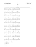 |  |
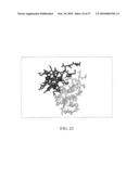 | 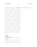 |
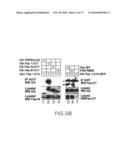 | 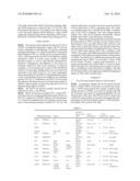 |
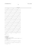 | 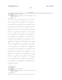 |
 |  |
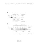 |  |
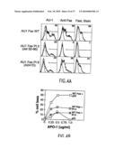 | 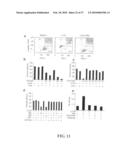 |
 |  |
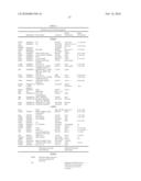 | 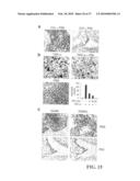 |
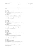 | 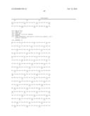 |
 | 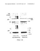 |
 | 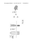 |
 | 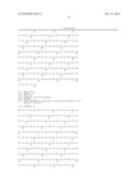 |
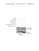 | 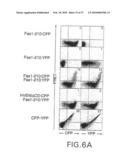 |
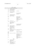 |  |
 |  |
 | 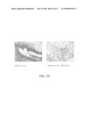 |
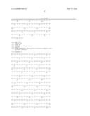 | 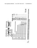 |
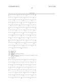 |  |
 |  |
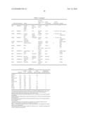 | 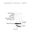 |
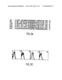 | 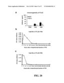 |
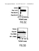 | 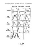 |
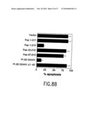 | 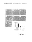 |
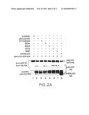 | 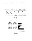 |
 | 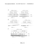 |
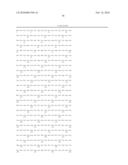 | 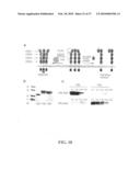 |
 |  |
 |  |
 | 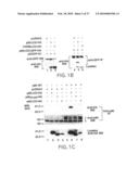 |
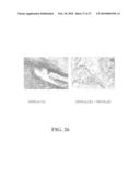 | 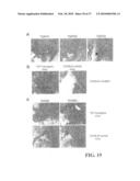 |
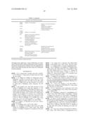 | 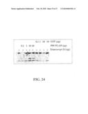 |
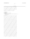 | 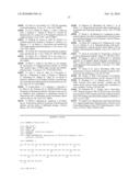 |
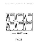 |  |
 | 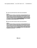 |
 | 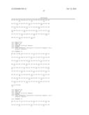 |
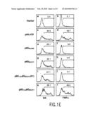 | 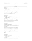 |
| Similar patent applications: | |
| Date | Title |
|---|---|
| 2010-03-04 | Association of an anti-atherothrombotic agent and an anti-platelet-aggregation agent |
| 2010-03-04 | Chromen-2-one derivatives and their use as monoamine neurotransmitter re-uptake inhibitors |
| 2010-03-04 | Compositions and methods for administering gdnf ligand family proteins |
| 2010-02-25 | Adrenal grk2 activity as a therapeutic target for heart failure |
| 2010-02-25 | Therapeutic uses of bicyclic ligands of sigma 2 receptor |
| New patent applications in this class: | |
| Date | Title |
|---|---|
| 2010-10-21 | Use of cytochrome p450-metabolized drugs and grf molecules in combination therapy |
| 2010-10-21 | Use of protease inhibitors and grf molecules in combination therapy |
| 2010-10-14 | Tissue adhesive using engineered proteins |
| 2010-10-14 | Purification and use of a factor for supporting wound healing |
| 2010-09-30 | Nutritional composition comprising curcuminoids and methods of manufacture |
| Top Inventors for class "Drug, bio-affecting and body treating compositions" | |
| Rank | Inventor's name |
|---|---|
| 1 | Anthony W. Czarnik |
| 2 | Ulrike Wachendorff-Neumann |
| 3 | Ken Chow |
| 4 | John E. Donello |
| 5 | Rajinder Singh |