Patent application title: PROTEIN KINASE A REPORTERS USEFUL IN HIGH THROUGHPUT ASSAYS
Inventors:
Jin Zhang (Baltimore, MD, US)
Qiang Ni (Baltimore, MD, US)
Michael David Allen (Baltimore, MD, US)
Assignees:
THE JOHNS HOPKINS UNIVERSITY
IPC8 Class: AA61K4900FI
USPC Class:
424 96
Class name: Drug, bio-affecting and body treating compositions in vivo diagnosis or in vivo testing diagnostic or test agent produces in vivo fluorescence
Publication date: 2009-12-24
Patent application number: 20090317333
Inventors list |
Agents list |
Assignees list |
List by place |
Classification tree browser |
Top 100 Inventors |
Top 100 Agents |
Top 100 Assignees |
Usenet FAQ Index |
Documents |
Other FAQs |
Patent application title: PROTEIN KINASE A REPORTERS USEFUL IN HIGH THROUGHPUT ASSAYS
Inventors:
Jin Zhang
Qiang Ni
Michael David Allen
Agents:
BANNER & WITCOFF, LTD.
Assignees:
THE JOHNS HOPKINS UNIVERSITY
Origin: WASHINGTON, DC US
IPC8 Class: AA61K4900FI
USPC Class:
424 96
Patent application number: 20090317333
Abstract:
Protein kinase A reporters useful for obtaining measurements of protein
kinase A activity with high spatial and temporal resolution can be used
in high throughput assays to identify potentially therapeutic compounds.Claims:
1. A protein kinase A (PKA) reporter, comprising from N to C terminus:(a)
amino acids 1-227 of an enhanced cyan fluorescent protein
(ECFP1-227);(b) a forkhead associated domain 1 (FHA1) covalently
linked to the ECFP1-227 with an amino acid linker RMH;(c) the amino
acid sequence SEQ ID NO:3 covalently linked to the FHA1; and(d) a
circularly permuted variant of a yellow fluorescent protein covalently
linked to the amino acid sequence SEQ ID NO:3.
2. The PKA reporter of claim 1 which comprises the amino acid sequence SEQ ID NO:1.
3. A PKA reporter, comprising from N to C terminus:(a) a green fluorescent protein;(b) a forkhead associated domain 1 (FHA1) covalently linked to the green fluorescent protein with an amino acid linker RMH;(c) the amino acid sequence SEQ ID NO:3 covalently linked to the FHA1; and(d) a red fluorescent protein covalently linked to the amino acid sequence SEQ ID NO:3.
4. The PKA reporter of claim 3 which comprises the amino acid sequence SEQ ID NO:18.
5. A PKA reporter, comprising from N to C terminus:(a) a ceruelan fluorescent protein;(b) a forkhead associated domain 1 (FHA1) covalently linked to the cerulean protein with an amino acid linker RMH;(c) the amino acid sequence SEQ ID NO:3 covalently linked to the FHA1; and(d) a circularly permuted variant of yellow fluorescent protein covalently linked to the amino acid sequence SEQ ID NO:3.
6. The PKA reporter of claim 5 which comprises the amino acid sequence SEQ ID NO:4.
7. The PKA reporter of claim 1 which comprises a subcellular targeting sequence.
8. The PKA reporter of claim 1 which comprises a subcellular targeting sequence which targets the reporter to a subcellular location selected from the group consisting of a plasma membrane, a nuclear membrane, a cytosol, an endoplasmic reticulum, a mitochondria, a mitochondrial matrix, a chloroplast, a medial trans-Golgi cisternae, a lumen of a lysosome, and a lumen of an endosome.
9. The PKA reporter of claim 1 which comprises a subcellular targeting sequence selected from the group consisting of a plasma membrane targeting sequence comprising SEQ ID NO:12, a nuclear localization signal sequence comprising SEQ ID NO:13, a mitochondrial localization sequence comprising SEQ ID NO:14, and a mitochondrial matrix targeting signal comprising SEQ ID NO:15.
10. A nucleic acid molecule which encodes the PKA reporter of any claim 1.
11. The nucleic acid molecule of claim 10 which comprises the nucleotide sequence SEQ ID NO:2.
12. The nucleic acid molecule of claim 10 which comprises the nucleotide sequence SEQ ID NO:5.
13. The nucleic acid molecule of claim 10 which comprises the nucleotide sequence SEQ ID NO:19.
14. A host cell comprising the nucleic acid molecule of claim 10.
15. A method for detecting a change in PKA activity, comprising:detecting a first resonance energy transfer of the PKA reporter of claim 1 at a first time point;detecting a second resonance energy transfer of the PKA reporter at a second time point; andcomparing the first and the second resonance energy transfers, wherein a difference between the first and the second resonance energy transfers reflects the change in PKA activity.
16. The method of claim 15 wherein the baseline and test resonance energy transfers are detected by determining a property selected from the group consisting of a quantum efficiency, an excitation spectrum, an emission spectrum, an excitation wavelength maximum, an emission wavelength maximum, a ratio of excitation amplitudes at two wavelengths, a ratio of emission amplitudes at two wavelengths, an excited state lifetime, anisotropy, a polarization of emitted light, resonance energy transfer, and a quenching of emission at a wavelength.
17. The method of claim 15 wherein the PKA reporter is in a cell-free system.
18. The method of claim 15 wherein the PKA reporter is in a cell.
19. The method of claim 18 wherein the cell is in vivo.
20. The method of claim 18 wherein the cell is in vitro.
21. The method of claim 18 wherein the cell is in a tissue sample.
22. The method of claim 18 wherein the cell is in a whole organ.
23. The method of claim 18 wherein the cell is in a well of a multi-well plate.
24. The method of claim 23 wherein each of a plurality of wells of the multi-well plate comprises a cell which comprises the PKA reporter.
25. The method of claim 15 further comprising determining the test resonance energy transfer in the presence of a test compound.
26. The method of claim 24 further comprising contacting each well of the plurality with a different test compound and determining test resonance energy transfers in the presence of the different test compounds.
27. The method of claim 15 wherein the first and second resonance energy transfers are detected using fluorescence activated cell sorting.
28. The method of claim 15 further comprising detecting a cAMP concentration.
29. A kit, comprising:(a) the PKA reporter of claim 1; and(b) instructions for a method for detecting a change in PKA activity comprising:detecting a first resonance energy transfer of the PKA reporter of claim 1 at a first time point;detecting a second resonance energy transfer of the PKA reporter at a second time point; andcomparing the first and the second resonance energy transfers wherein a difference between the first and the second resonance energy transfers reflects the change in PKA activity.
30. The kit of claim 29 further comprising a cAMP reporter.
31. A protein kinase A (PKA) reporter, comprising from N to C terminus:(a) amino acids 1-227 of an enhanced cyan fluorescent protein (ECFP1-227);(b) a phosphoamino acid binding domain covalently linked to the ECFP1-227, wherein the phosphoamino acid binding domain is selected from the group consisting of a forkhead associated domain 1 (FHA1), FHA2, 14-3-3, a WW domain, and a Polo box domain;(c) a PKA substrate covalently linked to the phosphoamino acid binding domain, wherein the PKA substrate is selected from the group consisting of SEQ ID NO:20, SEQ ID NO:21, SEQ ID NO:22, SEQ ID NO:23, SEQ ID NO:24, SEQ ID NO:25, RRXS, and RRXT, wherein X is any amino acid; and(d) a circularly permuted variant of a yellow fluorescent protein covalently linked to the amino acid sequence SEQ ID NO:3.
32. A PKA reporter, comprising from N to C terminus:(a) a green fluorescent protein;(b) a phosphoamino acid binding domain covalently linked to the ECFP1-227, wherein the phosphoamino acid binding domain is selected from the group consisting of a forkhead associated domain 1 (FHA1), FHA2, 14-3-3, a WW domain, and a Polo box domain;(c) a PKA substrate covalently linked to the phosphoamino acid binding domain, wherein the PKA substrate is selected from the group consisting of SEQ ID NO:20, SEQ ID NO:21, SEQ ID NO:22, SEQ ID NO:23, SEQ ID NO:24, SEQ ID NO:25, RRXS, and RRXT, wherein X is any amino acid; and(d) a red fluorescent protein covalently linked to the amino acid sequence SEQ ID NO:3.
33. A PKA reporter, comprising from N to C terminus:(a) a ceruelan fluorescent protein;(b) a phosphoamino acid binding domain covalently linked to the ECFP1-227, wherein the phosphoamino acid binding domain is selected from the group consisting of a forkhead associated domain 1 (FHA1), FHA2, 14-3-3, a WW domain, and a Polo box domain;(c) a PKA substrate covalently linked to the phosphoamino acid binding domain, wherein the PKA substrate is selected from the group consisting of SEQ ID NO:20, SEQ ID NO:21, SEQ ID NO:22, SEQ ID NO:23, SEQ ID NO:24, SEQ ID NO:25, RRXS, and RRXT, wherein X is any amino acid; and(d) a circularly permuted variant of yellow fluorescent protein covalently linked to the amino acid sequence SEQ ID NO:3.
Description:
[0001]This application claims the benefit of and incorporates by reference
Ser. No. 60/730,750 filed Oct. 27, 2005 and 60/800,451 filed May 15,
2006. Each of these applications is incorporated herein by reference in
its entirety.
FIELD OF THE INVENTION
[0003]The invention relates to detection of protein kinase A (PKA) activity, in particular to use of live cell high-throughput assays based on PKA reporters for drug screens, pharmacological profiling, and functional genomics studies.
BACKGROUND OF THE INVENTION
[0004]Genetically-encoded reporters based on fluorescence resonance energy transfer (FRET) have become powerful tools for monitoring the activities of protein kinases and second messengers in live-cell imaging, but their application in high throughput assays has yet to be realized. There is a need in the art for sensitive PKA reporters which can be used for accurate measurements of spatial and temporal PKA activities in living cells.
BRIEF DESCRIPTION OF THE FIGURES
[0005]FIG. 1. High-throughput activity assay based on improved A-Kinase Activity Reporter (AKAR). FIG. 1a, scheme for using live cell high-throughput assays based on FRET reporters (e.g., AKAR for PKA activity) for drug screens, pharmacological profiling, and functional genomics studies. FIG. 1b, representative time course of emission ratio (yellow/cyan) change of AKAR3, indicated by pseudocolor images. A HEK-293 cell expressing AKAR3 was stimulated with 1 μM isoproterenol (ISO), followed by 50 μM forskolin (Fsk), or calyculin A (a phosphatase inhibitor). FIG. 1c, representative data from five independent runs in 96-well format showing emission ratio (yellow/cyan) changes of AKAR3 in HEK-293 cells treated with ISO, Fsk, or 2% dimethylsulfoxide (DMSO). FIG. 1D, representative data from three independent runs showing emission ratio (yellow/cyan) changes of AKAR3 NES (nuclear export signal) in HEK-293 cells treated with indicated drugs. The inset shows a representative fluorescence image of cells expressing AKAR NES. Error bars represent standard deviation (n=3).
[0006]FIG. 2. High-throughput activity assays for cAMP and PKA in pharmacological profiling. FIGS. 2A and 2C, representative data from five independent runs showing emission ratio (cyan/yellow) changes of ICUE3 in HEK-293 cells treated with indicated drugs. 1 cycle=92 sec. FIGS. 2B and 2D, representative data from five independent runs showing emission ratio (cyan/yellow) changes of AKAR3 in HEK-293 cells treated with indicated drugs. 1 cycle=64 sec. Error bars represent standard deviation (n=3).
[0007]FIG. 3. High-throughput screening. FIG. 3a, normalized emission ratio (yellow/cyan) from cells expressing AKAR3 treated with individual library compounds, compared to the negative control in which only buffer was added and the positive control in which ISO (250 nM) was added (agonist screen). The asterisks indicate responses of ritrodrine, ISO, and epinephrine. FIG. 3b, normalized emission ratio (yellow/cyan) from cells expressing AKAR3 first treated with individual library compounds for about 15 minutes, then stimulated by ISO (250 nM) (antagonist screen). The positive control in which only ISO was added and the negative control in which only buffer was added are shown as labeled. The asterisks indicate responses from epinephrine and propranolol treated cells in response to ISO. FIG. 3c, representative time courses of emission ratio (yellow/cyan) change of cells expressing AKAR3 treated with 10 μM epinephrine followed by 50 μM FSK in single-cell imaging experiments. Error bars represent standard deviation (n=3). FIG. 3d, representative time courses of emission ratio (yellow/cyan) change of cells expressing AKAR3 treated with 50 μM ritodrine followed by 100 μM 3-isobutyl-1-methylxanthine (IBMX) in single-cell imaging experiments. Error bars represent standard deviation (n=2). FIG. 3e, representative time courses of emission ratio (yellow/cyan) change of ICUE pretreated with bilirubin (1 μM and 10 μM) followed by ISO stimulation in the presence of IBMX. Error bars represent standard deviation (n=22, 9, and 10).
[0008]FIG. 4. Improved AKAR. FIG. 4a, domain structure of AKAR constructs with responses (average±standard deviation); AKAR3 contains a circularly permutated Venus (cp Venus E172) as the FRET acceptor. FIG. 4B, representative time course of HEK-293 cells expressing AKAR3 (circles) treated with ISO followed by Fsk stimulation, in comparison to AKAR2 response (squares). An AKAR3 mutant in which the phosphorylation site threonine is mutated to an alanine showed no response. FIG. 4c, treatment of AKAR3-expressing HEK-293 cells with 1 μM ISO in the presence or absence of PKI, a specific peptide inhibitor of PKA.
[0009]FIG. 5. Improved ICUE. FIG. 5a, domain structure of ICUE3, which contains a circularly permutated Venus (cp Venus L194) as the FRET acceptor. FIG. 5b, representative time courses of HEK-293 cells expressing ICUE3 (circles) and ICUE3 (R375E) (triangles), treated with 50 μM Fsk. ICUE3 maintained the specificity for cAMP as a loss-of-function mutation in the cAMP binding site (R375E) abolished the FRET response.
[0010]FIG. 6. Representative cellular response using AKAR3.2. HEK-293 cells expressing AKAR3.2 were treated with 1 μM ISO. The subsequent elevation in emission ratio is typical of the new AKAR variant AKAR3.2, showing a maximum response of approximately 65%.
[0011]FIG. 7. Further characterization of AKAR3. Representative time courses of plasma membrane-targeted AKAR3 (FIG. 7a) and nuclear-targeted AKAR3 (FIG. 7b) in HEK-293 cells stimulated with 1 μM ISO and 50 μM FSK, respectively. FIG. 7C, AKAR3-expressing HEK-293 cells were treated with 200 nM phorbol dibutyrate (PDBu), a protein kinase C activator, followed by the addition of 1 μM isoproterenol, then 50 μM forskolin. FIG. 7d, representative time courses of the emission ratio change of AKAR3 in the presence (closed squars) and absence (open circles) of the specific PKA inhibitor, PKI. FIG. 7e, emission spectra of the purified AKAR3 before (squares) and after (diamonds) phosphorylation by PKA (excitation 434 nm). The curve using triangle symbols depicts the spectrum of the reporter after digestion with trypsin to quench the energy transfer and quantify the FRET. FIG. 7f, a representative phosphorylation time course for purified AKAR3 protein in the presence of PKA catalytic subunit and 1 mM ATP.
DETAILED DESCRIPTION OF THE INVENTION
[0012]The invention provides highly sensitive reporter molecules by which temporal and spatial protein kinase A (PKA) activity can be determined in living tissues and cells. The invention also provides high-throughput assays that use PKA reporters of the invention. High-throughput assays of the invention permit detection of PKA activity in single, live cells and can be used, inter alia, to screen compounds for their ability to modulate dynamic kinase activities in living cells.
PKA Reporters
[0013]PKA reporters of the invention are fusion proteins which comprise a FRET donor and acceptor pair (i.e., a "donor moiety" and "acceptor moiety"; see below) separated by a phosphoamino acid binding domain and a PKA substrate. PKA substrates can be, e.g., LRRATLVD (amino acids 15-22 of SEQ ID NO:3), LRRASLP (SEQ ID NO:20), LRRATLP (SEQ ID NO:21), LRRASP (SEQ ID NO:22), LRRATP (SEQ ID NO:23), RRASFVF (SEQ ID NO:24), RRATFVF (SEQ ID NO:25), RRXS (in which X can be any amino acid), or RRXT (in which X can be any amino acid). Phosphoamino acid binding domains can be, e.g., 14-3-3, isoforms of FHA domains such as FHA-1 and FHA-2, WW domains, or a Polo-box domain. Portions of the PKA reporters are covalently linked to form the fusion proteins. "Covalently linked" according to the invention includes direct covalent linkage and linkage by way of covalent bonds to an amino acid linker sequence.
[0014]In one embodiment, a PKA reporter of the invention ("AKAR3"; SEQ ID NO:1) comprises, from N to C terminus, (a) amino acids 1-227 of enhanced cyan fluorescent protein (ECFP1-227) (e.g., amino acids 1-228 of SEQ ID NO:1; (b) a forkhead associated domain 1 (FHA1; e.g., amino acids 232-373 of SEQ ID NO:1) covalently linked to the ECFP1-227 with an amino acid linker RMH; (c) the amino acid sequence AGTKPGSGEGSTKGLRRATLVDGGTGGSEL (SEQ ID NO:3) covalently linked to the FHA1; and (d) a circularly permuted variant of yellow fluorescent protein (e.g., amino acids 403-647 of SEQ ID NO:1) covalently linked to the amino acid sequence SEQ ID NO:3. SEQ ID NO:3 comprises a PKA substrate (amino acids 15-22 of SEQ ID NO:3) and two amino acid linkers (amino acids 1-14 and 23-30 of SEQ ID NO:3). AKAR3 has shows an increase in emission ratio (yellow over cyan) of about 40%.
[0015]In another embodiment, a PKA reporter ("GRet_AKAR"; SEQ ID NO:18) comprises, from N to C terminus, (a) a green fluorescent protein (e.g., amino acids 1-228 of SEQ ID NO:18); (b) a forkhead associated domain 1 (FHA1) covalently linked to the green fluorescent protein with an amino acid linker RMH; (c) the amino acid sequence SEQ ID NO:3 covalently linked to the FHA1; and (d) a red fluorescent protein (e.g., amino acids 403-638 of SEQ ID NO:18) covalently linked to the amino acid sequence SEQ ID NO:3.
[0016]In yet another embodiment, a PKA reporter ("AKAR3.2"; SEQ ID NO:4) comprises, from N to C terminus, (a) a ceruelan fluorescent protein (e.g., amino acids 1-239 of SEQ ID NO:4); (b) a forkhead associated domain 1 (FHA1) covalently linked to the cerulean protein with an amino acid linker RMH; (c) the amino acid sequence SEQ ID NO:3 covalently linked to the FHA1; and (d) a circularly permuted variant of yellow fluorescent protein (e.g., amino acids 414-658 of SEQ ID NO:4) covalently linked to the amino acid sequence SEQ ID NO:3. Because of the enhanced brightness of the Cerulean fluorescent protein as compared to eCFP, AKAR3.2 has a dynamic range of 55-65%.
[0017]Donor and Acceptor Moieties
[0018]As used here, a "donor moiety" is a fluorophore or a luminescent moiety. The absorption spectrum of the "acceptor moiety" overlaps the emission spectrum of the donor moiety. The acceptor moiety does not need to be fluorescent and can be a fluorophore, chromophore, or quencher. In some embodiments both the donor and acceptor moieties are fluorescent proteins. In other embodiments both the donor and acceptor moieties are luminescent moieties. In yet other embodiments, either one of the donor or acceptor moieties can be a fluorescent protein while the other moiety is a luminescent moiety. In other embodiments, the acceptor moiety is a "quencher moiety."
[0019]When both the donor and acceptor moieties are fluorophores, resonance energy transfer is detected as "fluorescence resonance energy transfer" (FRET). If a luminescent moiety is involved, resonance energy transfer is detected as "luminescent resonance energy transfer" (LRET). LRET includes "bioluminescent resonance energy transfer" (BRET; Boute et al., Trends Pharmacol. Sci. 23, 351-54, 2002; Ayoub et al., J. Biol. Chem. 277, 21522-28, 2002). Because excitation of the donor moiety does not require exogenous illumination in an LRET method, such methods are particularly useful in live tissue and animal imaging, because penetration of the excitation light is no longer a concern. LRET methods have a high contrast and high signal-to-noise ratio; 2) no photobleaching occurs; and 3) quantification is simplified because the acceptor moiety is not directly excited.
[0020]Suitable acceptor moieties include, for example, a coumarin, a xanthene, a fluorescein, a fluorescent protein, a circularly permuted fluorescent protein, a rhodol, a rhodamine, a resorufin, a cyanine, a difluoroboradiazaindacene, a phthalocyanine, an indigo, a benzoquinone, an anthraquinone, an azo compound, a nitro compound, an indoaniline, a diphenylmethane, a triphenylmethane, and a zwifterionic azopyridinium compound.
[0021]Suitable donor moieties include, but are not limited to, a coumarin, a xanthene, a rhodol, a rhodamine, a resorufin, a cyanine dye, a bimane, an acridine, an isoindole, a dansyl dye, an aminophthalic hydrazide, an aminophthalimide, an aminonaphthalimide, an aminobenzofuran, an aminoquinoline, a dicyanohydroquinone, a semiconductor fluorescent nanocrystal, a fluorescent protein, a circularly permuted fluorescent protein, and fluorescent lanthanide chelate.
[0022]Fluorescent Proteins
[0023]In some preferred embodiments either or both of the donor and acceptor moieties is a fluorescent protein. Suitable fluorescent proteins include green fluorescent proteins (GFP), red fluorescent proteins (RFP), yellow fluorescent proteins (YFP), and cyan fluorescent proteins (CFP). Useful fluorescent proteins also include mutants and spectral variants of these proteins which retain the ability to fluoresce.
[0024]RFPs include Discosoma RFPs, such Discosoma DsRed (SEQ ID NO:6) or a mutant thereof which includes an Ile125Arg mutation, or a non-oligomerizing tandem DsRed containing, for example, two RFP monomers linked by a peptide linker. For example, a non-oligomerizing tandem RFP can contain two DsRed monomers or two mutant DsRed-I125R monomers linked by a peptide (having, for example, the amino acid sequence shown in SEQ ID NO:7).
[0025]Useful GFPs include an Aequorea GFP (e.g., SEQ ID NO:8), a Renilla GFP, a Phialidium GFP, and related fluorescent proteins for example, a cyan fluorescent protein (CFP), a yellow fluorescent protein (YFP), or a spectral variant of the CFP or YFP. CFP (cyan) and YFP (yellow) are color variants of GFP. CFP and YFP contain 6 and 4 mutations, respectively. They are Tyr66Try, Phe66Leu, Ser65Thr, Asn145Ile, Met153Thr, and Val163Ala in CFP and Ser65Gly, Val168Leu, Ser72Ala, and Thr203Tyr. Spectral variants include an enhanced GFP (EGFP; SEQ ID NO:9), an enhanced CFP (ECFP; SEQ ID NO:10), an enhanced YFP (EYFP; SEQ ID NO:11), and an EYFP with V68L and Q69K mutations. Other examples of fluorescent proteins comprising mutations are Aequorea GFP with one or more mutations at amino acid residues A206, L221 or F223 of SEQ ID NO:8 (e.g., mutations A206K, L221K, F223R, Q80R); mutations L221K and F223R of ECFP (SEQ ID NO:9), and EYFP V68L/Q69K of SEQ ID NO:8. See also US 2004/0180378; U.S. Pat. Nos. 6,150,176; 6,124,128; 6,077,707; 6,066,476; 5,998,204; and 5,777,079; Chalfie et al., Science 263:802-805, 1994.
[0026]Other useful GFP-related fluorescent proteins include those having one or more folding mutations, and fragments of the proteins that are fluorescent, for example, an A. victoria GFP from which the two N-terminal amino acid residues have been removed. Several of these fluorescent proteins contain different aromatic amino acids within the central chromophore and fluoresce at a distinctly shorter wavelength than the wild type GFP species. For example, the engineered GFP proteins designated P4 and P4-3 contain, in addition to other mutations, the substitution Y66H; and the engineered GFP proteins designated W2 and W7 contain, in addition to other mutations, Y66W.
[0027]Folding mutations in Aequorea GFP-related fluorescent proteins improve the ability of the fluorescent proteins to fold at higher temperatures and to be more fluorescent when expressed in mammalian cells, but have little or no effect on the peak wavelengths of excitation and emission. If desired, these mutations can be combined with additional mutations that influence the spectral properties of GFP to produce proteins with altered spectral and folding properties, and, particularly, with mutations that reduce or eliminate the propensity of the fluorescent proteins to oligomerize. Folding mutations, with respect to SEQ ID NO:8, include the substitutions F64L, V68L, S72A, T44A, F99S, Y145F, N1461, M153T, M153A, V163A, I167T, S175G, S205T, and N212K.
[0028]Luminescent Moieties
[0029]Luminescent moieties useful in a cAMP reporter include lanthanides, which can be in the form of a chelate, including a lanthanide complex containing the chelate (e.g, β-diketone chelates of cerium, praseodymium, neodymium, promethium, samarium, europium, gadolinium, terbium, dysprosium, holmium, erbium, thulium, or ytterbium). Lanthanide chelates are well known in the art. See Soini and Kojola, Clin. Chem. 29, 65, 1983; Hemmila et al., Anal. Biochem. 137, 335 1984; Lovgren et al., In: Collins & Hoh, eds., Alternative Immunoassays, Wiley, Chichester, U.K., p. 203, 1985; Hemmila, Scand. J. Clin. Lab. Invest. 48, 389, 1988; Mikola et al., Bioconjugate Chem. 6, 235, 1995; Peruski et al., J. Immunol. Methods 263, 35-41, 2002; U.S. Pat. No. 4,374,120; and U.S. Pat. No. 6,037,185. Suitable β-diketones are, for example, 2-naphthoyltrifluoroacetone (2-NTA), 1-naphthoyltrifluoroacetone (1-NTA), p-methoxybenzoyltrifluoroacetone (MO-BTA), p-fluorobenzoyltrifluoroacetone (F-BTA), benzoyltrifluoroacetone (BTA), furoyltrifluoroacetone (FTA), naphthoylfuroylmethane (NFM), dithenoylmethane (DTM), and dibenzoylmethane (DBM). See also US 20040146895.
[0030]Luminescent proteins include, but are not limited to, lux proteins (e.g., luxCDABE from Vibrio fischerii), luciferase proteins (e.g., firefly luciferase, Gaussia luciferase, Pleuromamma luciferase, and luciferase proteins of other beetles, Dinoflagellates (Gonylaulax; Pyrocystis), Annelids (Dipocardia), Molluscs (Lativa), and Crustacea (Vargula; Cypridina), and green fluorescent proteins of bioluminescent coelenterates (e.g., Aequorea Victoria, Renilla mullerei, Renilla reniformis; see Prendergast et al., Biochemistry 17, 3448-53, 1978; Ward et al., Photochem. Photobiol. 27, 389-96, 1978; Ward et al., J. Biol. Chem. 254, 781-88, 1979; Ward et al., Photochem. Photobiol. Rev 4, 1-57, 1979; Ward et al., Biochemistry 21, 4535-40, 1982). Many of these proteins are commercially available. Firefly luciferase is available from Sigma, St. Louis, Mo., and Boehringer Mannheim Biochemicals, Indianapolis, Ind. Recombinantly produced firefly luciferase is available from Promega Corporation, Madison, Wis. Jellyfish aequorin and luciferase from Renilla are commercially available from Sealite Sciences, Bogart, Ga.
[0031]The DNA sequences of the aequorin and other luciferases employed for preparation of some cAMP reporters of the invention can be derived from a variety of sources. For example, cDNA can be prepared from mRNA isolated from the species disclosed above. See Faust, et al., Biochem. 18, 1106-19, 1979; De Wet et al., Proc. Natl. Acad. Sci. USA 82, 7870-73, 1985.
[0032]Luciferase substrates (luciferins) are well known and include coelenterazine (available from Molecular Probes, Eugene, Oreg.) and ENDUREN®. These cell-permeable reagents can be directly administered to cells, as is known in the art. Luciferin compounds can be prepared according to the methods disclosed by Hori et al., Biochemistry 14, 2371-76, 1975; Hori et al., Proc. Natl. Acad. Sci. USA 74, 4285-87, 1977).
[0033]Dark Quenchers
[0034]In some embodiments the acceptor moiety is a quencher moiety, preferably a "dark quencher" (or "black hole quencher") as is known in the art. In this case, the change in conformation which occurs upon cAMP binding eliminates quenching, resulting in an increase in energy emission from the donor moiety. "Dark quenchers" themselves do not emit photons. Use of a "dark quencher" reduces or eliminates background fluorescence or luminescence which would otherwise occur as a result of energy transfer from the donor moiety. Suitable quencher moieties include dabcyl (4-(4'-dimethylaminophenylazo)-benzoic acid), QSY®-7 carboxylic acid, succinimidyl ester (N,N'-dimethyl-N,N'-diphenyl-4-((5-t-butoxycarbonylaminopentyl)aminocarbo- n yl) piperidinylsulfone-rhodamine (a diarylrhodamine derivative from Molecular Probes, Eugene, Oreg.). Suitable quencher moieties are disclosed, for example, in US 2005/0118619; US 20050112673; and US 20040146959.
[0035]Any suitable fluorophore may be used as the donor moiety provided its spectral properties are favorable for use with the chosen dark quencher. The donor moiety can be, for example, a Cy-dye, Texas Red, a Bodipy dye, or an Alexa dye. Typically, the fluorophore is an aromatic or heteroaromatic compound and can be a pyrene, anthracene, naphthalene, acridine, stilbene, indole, benzindole, oxazole, thiazole, benzothiazole, cyanine, carbocyanine, salicylate, anthranilate, coumarin, a fluorescein (e.g., fluorescein, tetrachlorofluorescein, hexachlorofluorescein), rhodamine, tetramethyl-rhodamine, or other like compound. Suitable fluorescent moieties for use with dark quenchers include xanthene dyes, such as fluorescein or rhodamine dyes, including 6-carboxyfluorescein (FAM), 2'7'-dimethoxy-4'5'-dichloro-6-carboxyfluorescein (JOE), tetrachlorofluorescein (TET), 6-carboxyrhodamine (R6G), N,N,N;N'-tetramethyl-6-carboxyrhodamine (TAMRA), 6-carboxy-X-rhodamine (ROX). Suitable fluorescent reporters also include the naphthylamine dyes that have an amino group in the alpha or beta position. For example, naphthylamino compounds include 1-dimethylaminonaphthyl-5-sulfonate, 1-anilino-8-naphthalene sulfonate and 2-p-toluidinyl-6-naphthalene sulfonate, 5-(2'-aminoethyl)aminonaphthalene-1-sulfonic acid (EDANS).
[0036]Other suitable fluorescent moieties include coumarins, such as 3-phenyl-7-isocyanatocoumarin; acridines, such as 9-isothiocyanatoacridin-e and acridine orange; N-(p-(2-benzoxazolyl)phenyl)maleimide; cyanines, such as indodicarbocyanine 3 (Cy3), indodicarbocyanine 5 (Cy5), indodicarbocyanine 5.5 (Cy5.5), 3-1-carboxy-pentyl)-3'-ethyl-5,5'-dimethyl-loxacarbocyanine (CyA); 1H,5H,1H,15H-Xantheno[2,3,4-ij:5,6,7-i'j']diquinol-izin-18-ium, 9-[2(or 4)-[[[6-[2,5-dioxo-1-pyrrolidinyl)oxy]-6-oxohexyl]amino]sulfonyl]-4(or 2)-sulfophenyl]-2,3,6,7,12,13,16,17-octahyd-ro-inner salt (TR or Texas Red); BODIPY® dyes; benzoxaazoles; stilbenes; pyrenes; and the like.
[0037]Subcellular Targeting Sequences
[0038]PKA reporters of the invention optionally can include a subcellular targeting sequence which can target a PKA reporter to a subcellular domain such as a plasma membrane, a nuclear membrane, a cytosol, an endoplasmic reticulum, a mitochondria, a mitochondrial matrix, a chloroplast, a medial trans-Golgi cistemae, a lumen of a lysosome, or a lumen of an endosome. Many such targeting sequences are known in the art. Examples include the plasma membrane targeting sequence shown in SEQ ID NO:12, the nuclear localization signal sequence shown in SEQ ID NO:13, the mitochondrial localization sequence shown in SEQ ID NO:14, and the mitochondrial matrix targeting signal shown in SEQ ID NO:15. Targeting sequences can be linked to PKA reporters using, for example, a tetracysteine motif such as Cys Cys Xaa Xaa Cys Cys (SEQ ID NO:16). Targeting sequences can be linked at either the N- or C-terminus of a PKA reporter or at intermediate points in the reporter.
[0039]Assembly of PKA Reporters
[0040]PKA reporters of the invention are fusion proteins and preferably are expressed recombinantly. The invention provides nucleic acid molecules for this purpose. A nucleic acid molecule encoding a PKA reporter can comprise any nucleotide sequence which encodes the amino acid sequence of the reporter. Particular nucleic acid sequences are provided as SEQ ID NO:2 (AKAR3), SEQ ID NO:5 (AKAR3.2), and SEQ ID NO:18 (GRet_AKAR). Nucleic acid molecules of the invention include single- and double-stranded DNA (including cDNA) and mRNA. Many kits for constructing fusion proteins are available from companies such as Promega Corporation (Madison, Wis.), Stratagene (La Jolla, Calif.), CLONTECH (Mountain View, Calif.), Santa Cruz Biotechnology (Santa Cruz, Calif.), MBL International Corporation (MIC; Watertown, Mass.), and Quantum Biotechnologies (Montreal, Canada; 1-888-DNA-KITS).
[0041]In some embodiments the nucleic acid molecules are expression constructs which contain the necessary elements for the transcription and translation of an inserted coding sequence encoding a PKA reporter. Expression constructs can be used as vectors for introducing PKA reporters into cells. Methods which are well known to those skilled in the art can be used to construct expression vectors containing sequences encoding PKA reporters and appropriate transcriptional and translational control elements. These methods include in vitro recombinant DNA techniques, synthetic techniques, and in vivo genetic recombination. Such techniques are described, for example, in Sambrook et al. (1989) and in Ausubel et al., CURRENT PROTOCOLS IN MOLECULAR BIOLOGY, John Wiley & Sons, New York, N.Y., 1989.
[0042]Expression vectors of the invention can be expressed in a variety of host cells. These include, but are not limited to, microorganisms, such as bacteria transformed with recombinant bacteriophage, plasmid, or cosmid DNA expression vectors; yeast transformed with yeast expression vectors, insect cell systems infected with virus expression vectors (e.g., baculovirus), plant cell systems transformed with virus expression vectors (e.g., cauliflower mosaic virus, CaMV; tobacco mosaic virus, TMV) or with bacterial expression vectors (e.g., Ti or pBR322 plasmids), or animal cell systems, particularly mammalian systems, including human systems. See WO 01/98340, which is incorporated herein by reference in its entirety. The choice of vector components and appropriate host cells is well within the capabilities of those skilled in the art.
[0043]Alternatively, protein or non-protein donor and/or acceptor moieties can be linked to the polypeptide by covalent attachment. There are a variety of methods known in the art which are useful for this purpose. For example, the attachment can be direct, via a functional group on the polypeptide (e.g., amino, carboxyl and sulfhydryl groups) and a reactive group on the fluorophore. Free amino groups in the polypeptide can be reacted with fluorophores derivatized with isothiocyanate, maleic anhydride, N-hydroxysuccinimide, tetrafluorylphenyl and pentafluoryl esters. Free carboxyl groups in the polypeptide can be reacted with carbodiimides such as 1-ethyl-3-[dimethylaminopropyl]carbodiimide hydrochloride to create a reactive moiety that will react with an amine moiety on the donor or acceptor moiety. Sulfhydryl groups can be attached to donor or acceptor moieties modified with maleimide and iodoacetyl groups, although such linkages are more susceptible to reduction than linkages involving free amino groups. The polypeptide can also be linked indirectly via an intermediate linker or spacer group, using chemical groups such as those listed above.
[0044]It is also possible to produce PKA reporters of the invention using chemical methods to synthesize the amino acid sequence of the polypeptide and, optionally, one or more fluorescent or luminescent proteins. Methods include direct peptide synthesis using solid-phase techniques (Merrifield, J. Am. Chem. Soc. 85, 2149-2154, 1963; Roberge et al., Science 269, 202-204, 1995). Protein synthesis can be performed using manual techniques or by automation. Automated synthesis can be achieved, for example, using Applied Biosystems 431A Peptide Synthesizer (Perkin Elmer). Optionally, fragments of polypeptide portions of PKA reporters can be separately synthesized and combined using chemical methods to produce a full-length reporter molecule. See WO 01/98340.
[0045]Delivery of PKA Reporters to Cells
[0046]PKA reporters of the invention can be introduced into cells in vitro using reversible permeabilization techniques. See U.S. Pat. No. 6,127,177; U.S. Pat. No. 6,902,931; Russo et al., Nature Biotechnology 15, 278-82, March 1997; Santangelo et al., Nucleic Acids Res. 32, 1-9, Apr. 14, 2004.
[0047]Expression vectors comprising a PKA reporter-encoding nucleotide sequence can be transfected into any cell in vitro in which it is desired to monitor PKA activity. Any transfection method known in the art can be used, including, for example, including, but not limited to, transferrin-polycation-mediated DNA transfer, transfection with naked or encapsulated nucleic acids, liposome-mediated cellular fusion, intracellular transportation of DNA-coated latex beads, protoplast fusion, viral infection, electroporation, "gene gun," and DEAE- or calcium phosphate-mediated transfection.
[0048]Useful vectors and methods of delivering the vectors to cells in vivo are disclosed, for example, in U.S. Pat. No. 6,825,012; U.S. Pat. No. 6,878,549; U.S. Pat. No. 6,645,942; U.S. Pat. No. 6,692,737; U.S. Pat. No. 6,689,758; U.S. Pat. No. 6,669,935; and U.S. Pat. No. 6,821,957.
Methods of Detecting PKA Activity
[0049]The invention provides various methods for detecting PKA activity by detecting conformational changes in a PKA reporter. Broadly, the methods involve detecting a change in resonance energy transfer of a PKA reporter of the invention when the reporter is subjected to an increase or decrease in PKA activity. PKA acts on the PKA substrate portion of the PKA reporter to induce a conformational change that changes resonance energy transfer from the donor moiety to the acceptor moiety.
[0050]A change in resonance energy transfer can readily be detected using methods well known in the art. See, e.g., US 2005/0118619; US 2002/0137115; US 2003/0165920; US 2003/0186229; US 2004/0137479; US 2005/0026234; US 2005/0054573; US 2005/0118619; U.S. Pat. No. 6,773,885; U.S. Pat. No. 6,803,201; U.S. Pat. No. 6,818,420; Ayoub et al., 2002; Boute et al., 2002; Domin et al., Prog. Biomed. Optics and Imaging, Proc. SPIE, vol 5139, 2003, pp 238-242; Evellin et al., Methods Mol. biol. 284, 259-70, 2004; Honda et al., Proc. Natl. Acad. Sci. USA 98, 437-42, Feb. 27, 2001; Honda et al., Methods Mol. Biol. 3, 27-44, 1005; Mongillo et al., Cir. Res. 95, 67-75, Jul. 9, 2004; Mongillo et al., Methods Mol. Biol. 307, 1-14, 2005; Nagai et al., Proc. Natl. Acad. Sci. USA 101, 10554-59, Jul. 20, 2004; Nikolaev et al., J. Biol. Chem. 279, 37215-18, 2004; Polit et al., Eur. J. Biochem. 270, 1413-23, 2003; Ponsioen et al., EMBO Rep. 5, 1176-80, 2004; Santangelo et al., Nucl. Acids Res. 32, 1-9, e-published Apr. 14, 2004; and Warrier et al., Am. J. Physiol. Cell Phiol. 289, C455-61, August 2005. Properties which can be detected as resonance energy transfer (RET) measurements include a quantum efficiency, an excitation spectrum, an emission spectrum, an excitation wavelength maximum, an emission wavelength maximum, a ratio of excitation amplitudes at two wavelengths, a ratio of emission amplitudes at two wavelengths, an excited state lifetime, anisotropy, a polarization of emitted light, resonance energy transfer, and a quenching of emission at a wavelength.
[0051]PKA reporters of the invention can be used in cell-free systems, in isolated cells (for example, in primary cell culture or a cell line) or in cells in situ (e.g., in an isolated tissue sample, an isolated whole organ, or in a mammal). Subcellular distribution of PKA activity or changes in PKA activity can be detected, for example, as described in the specific Examples, below. Absolute PKA activity levels can be detected by obtaining a RET measurement in the assay system and comparing it to a standard curve obtained in vitro.
Simultaneous or Parallel Monitoring of Camp Dynamics and PKA Phosphorylation
[0052]Soluble adenylyl cyclase and regulatory and catalytic subunits of PKA coexist in the nucleus of mammalian cells (Zippin et al., J. Cell Biol. 164, 527-34, 2004). Assays can be carried out using PKA reporters of the invention together with targeted cAMP indicators to examine the temporal correlation of cAMP dynamics and PKA activation within single living cells. For example, a plasma membrane-targeted cAMP reporter and a nuclear-localized PKA reporter can be co-expressed in cells such as HEK-293 cells (see WO 2006/023972). If desired, the assays can be carried out in a high-throughput format (see below). Useful cAMP reporters are disclosed in WO 2006/023972.
[0053]cAMP dynamics and PKA phosphorylation can be monitored either simultaneously or in parallel. Simultaneous assays can be carried out using targeted versions of the reporters. Parallel assays can be carried out using reporters comprising different colors of fluorescent protein.
[0054]In some embodiments, steady-state RET measurements are first obtained and then measurements are taken after addition of a test compound to the assay system. The effects of the test compounds on cAMP concentration and PKA activity can be simultaneously or parallelly monitored by using ICUE and AKAR or to monitor the effect of the test compound on cAMP concentration (e.g., in drug-screening methods). Test compounds can be pharmacologic agents already known in the art to affect PKA activity or can be compounds previously unknown to have such an activity. Compounds known to affect PKA activity include, for example, isoproterenol, epinephrine, ritodrine, 3-isobutyl-1-methylxanthine (IBMX), and H-89.
[0055]Test compounds can be naturally occurring or designed in the laboratory. They can be isolated from microorganisms, animals, or plants, and can be produced recombinantly, or synthesized by chemical methods known in the art. If desired, test compounds can be obtained using any of the numerous combinatorial library methods known in the art, including but not limited to, biological libraries, spatially addressable parallel solid phase or solution phase libraries, synthetic library methods requiring deconvolution, the "one-bead one-compound" library method, and synthetic library methods using affinity chromatography selection.
[0056]Fluorescence activated cell sorting (FACS) is well-suited for use with high throughput methods of the invention. For example, emission ratios of yellow-to-cyan (cyan excitation) for individual cells are detected during the first sorting--not all cells will have the same emission ratio and a distribution for the whole population can be plotted; the cells can be stimulated to activate PKA in the absence or presence of other drugs; emission ratios of individual cells are detected again during the second sorting; the difference in emission ratios, usually presented as a shift in the distribution, will reflect the changes in PKA activity.
[0057]High-Throughput Assays
[0058]High throughput assays of the invention are generally applicable to all kinase targets within the kinome (see FIG. 1a) and are ideally suited for examining dynamic responses of endogenous kinase targets, for evaluating drug candidates which ultimately perform within cellular environments, and for identifying compounds with unique mechanisms of action. Methods of the invention can be extended to follow multiple components of kinase-mediated signaling pathways to screen for pathway modulators.
[0059]High throughput assays of the invention, when combined with use of kinase activity reporters, permit simple, fast, and convenient high-throughput reading of dynamic kinase activities with high spatiotemporal resolution. These methods complement, yet offer unique advantages over, existing methods, including purified target-based biochemical screens and end-point focused phenotypic screens. Activity-based screens of the invention can be combined with phenotypic screens (e.g., Clemons, Curr. Op. Chem. Biol. 8, 334-38, 2004) to provide direct measurement of dynamic cellular activities of defined targets or the activity of a signaling pathway. Compared to in vitro assays, living cells are used as reaction vessels with targets of interest, cofactors, and regulators present at endogenous levels in their natural cellular environment, where spatiotemporal control of signaling activities can be specifically followed. With the complexity of live systems maintained, the quality of the screening process is increased, enabling discovery of compounds with unique mechanisms of action. Thus, the simple yet powerful high-throughput activity assays of the invention should find immediate application in high-throughput screens for pharmacological reagents and drug candidates, as well as in parallel tracking of multiple physiological and pharmacological events at subcellular locations in living cells in chemical and functional genomics studies. Furthermore, this assay platform is generally applicable to most kinases in the kinome, as various kinase activity sensors can be engineered and adapted to this assay format.
Kits
[0060]The invention provides kits comprising one or more PKA reporters of the invention and, optionally, one or more cAMP reporters. The kits also may provide all or a subset of the reagents that are required for practicing the invention. The kits may comprise written instructions, in paper or electronic form, or a reference to an on-line set of instructions. The instructions may contain data against which the results determined using the kit can be compared. Containers which hold the components of any given kit can vary. The kits may be divided into compartments or contain separate vessels for each component. The components may be mixed together or may be separated. Optional components of the kit include means for collecting, processing, and/or storing test samples.
[0061]All patents, patent applications, and references cited in this disclosure are expressly incorporated herein by reference. The above disclosure generally describes the present invention. A more complete understanding can be obtained by reference to the following specific examples, which are provided for purposes of illustration only and are not intended to limit the scope of the invention.
Example 1
Methods for Examples 2-7
[0062]Gene Construction. Different variants of fluorescent protein were amplified using PCR and incorporated into AKAR2 and AKAR2T391A to replace the original ECFP or citrine (Zhang et al., Nature 437, 569-73, 2005). Cytoplasmic targeting of AKAR3 was achieved by genetically adding a nuclear export signal (NES) LPPLERLTL (SEQ ID NO:17) at the C-terminal (Sato et al., Nat. Biotechnol. 20, 287-94, 2002). ICUE ("indicator of cAMP using Epac") constructs containing new fluorescent protein variants were created using the same method as above. All constructs were initially generated in pRSET B (Invitrogen), then subcloned to pcDNA3 (Invitrogen) behind a Kozak sequence for mammalian expression save for AKAR3NES, which used 3' restriction sites in pcDNA3 to introduce the NES.
[0063]Cell Culture and Imaging. HEK-293 cells were plated onto sterilized glass coverslips in 35 mm dishes and grown to ˜50% confluency in DMEM (10% FBS at 37° C. with 5% CO2). Cells were transfected with calcium phosphate and grown for 12-24 hours before imaging. After washing once with Hanks' balanced salt solution (HBSS), cells were maintained in buffer in the dark at 20-25° C. Isoproterenol (ISO; Sigma), forskolin (Fsk; Calbiochem), H89 (Sigma) were added as indicated. Cells were imaged on a Zeiss Axiovert 200M microscope with a 40×/1.3NA oil-immersion objective lens and cooled CCD camera as described in (21). Briefly, dual emission ratio imaging used a 420DF20 excitation filter, a 450DRLP dichroic mirror, and two emission filters (475DF40 for cyan and 535DF25 for yellow). The ratios of yellow-to-cyan (for AKAR) or cyan-to-yellow (for ICUE) emissions were then calculated at different time points and normalized by dividing all ratios by the emission ratio before stimulation, setting basal emission ratio as 1.
[0064]Live Cell Plate Reading. HEK-293 cells were transfected with AKAR3 or ICUE3 using calcium phosphate at 40% confluency and grown for 40 hours. Cells were then trypsinized and plated in a Costar 3603 96-well plate (Corning) at a density of 150,000 cells per well. After incubation for another 24 hours, cells were washed once with HBSS and left in 150 μl of HBSS at 20-25° C. Fluorescence reading was taken on a FLUOstar OPTIMA fluorescence microplate reader (BMG Labtechnologies Inc.) using a 420DF20 excitation filter and two emission filters (470DF40 for cyan and 535DF25 for yellow). A baseline was established in three cycles, each consisting of a full plate reading of yellow intensity, followed by a reading of cyan intensity.
[0065]Each cycle lasted between 64 and 92 seconds. Cells were then treated with ISO, Fsk, H89, Propranolol (Pro; Sigma), or 3-isobutyl-1-methylxanthine (IBMX; Sigma) as indicated. Readings were taken in additional cycles. FRET change was calculated as the percent increase of emission ratios (yellow-to-cyan for AKAR and cyan-to-yellow for ICUE) over baseline for each well during a given cycle. Several parameters were calculated to assess the efficiency of the assay, including Z' factor, coefficient of variation, and signal to noise (S/N) ratio (22).
[0066]Live Cell High-Throughput Screening. HEK-293 cells transiently expressing AKAR or stably expressing ICUE were trypsinized and plated in a Costar 3606 96-well plate (Corning) at a density of 150,000 cells per well. After incubation for 24 hours, cells were washed once with HBSS and left in 190 μl of HBSS at 20-25° C. Fluorescence readings were taken as described above, with each cycle lasting 90 seconds. Following baseline acquisition, cells in each experimental well were treated with a compound from the Johns Hopkins Clinical Compound Library to a final concentration of 10 μM. Control cells were treated with 10 μL of 10% fetal bovine serum (FBS) in a solution of phosphate buffered saline (PBS) at pH 7.4, which is the solution used to dissolve library compounds.
[0067]Readings were taken for 10 cycles spanning a time of approximately 15 minutes, after which cells in experimental wells and positive controls were treated with 0.25 μM ISO (AKAR) or 0.25 μM Iso plus 100 μM IBMX (ICUE), while negative controls received 0.5% DMSO in HBSS. Ten final cycles were then performed.
[0068]FRET responses were calculated as described above. Negative control (10% FBS, 0.5% DMSO) and positive control (10% FBS, 0.25 μM ISO) curves were generated. Agonist hits were defined as compounds eliciting responses larger than six times the standard deviation above the baseline. Antagonist hits were defined as compounds which decreased the ISO stimulated response by 50% or by six times the standard deviation.
Example 2
AKAR3 in Live-Cell Imaging and In Vitro Analysis
[0069]One of the important advantages of genetically encoded reporters of the invention is that they can be targeted to subcellular locations via various targeting motifs. However, in some cases, subcellular targeting may sacrifice the response amplitude of a reporter. To determine whether the improved dynamic range of AKAR3 leads to sensitive detection of subcellular PKA activities, AKAR3 was targeted to two cellular compartments. By attaching a lipid modification domain (Ananthanarayanan et al., Proc. Natl. Acad. Sci. USA 2005 Oct. 18; 102(42):15081-6), the reporter was successfully targeted to the plasma membrane, where it produced an average response of 34.3%±5.4% (n=3) after ISO stimulation (FIG. 7a). Nuclear localization also had minimal effect on the performance of AKAR3 (FIG. 7b). The average nuclear response upon stimulation with FSK was 38.6%±8.45% (n=3) with slower kinetics, consistent with the translocation of the catalytic subunit into the nucleus after cytoplasmic activation. This demonstrates that AKAR3 can be targeted to subcellular compartments while maintaining improved dynamic range.
[0070]Furthermore, AKAR3 maintained its specificity in sensing active PKA but not protein kinase C (FIG. 7C) or calmodulin-dependent protein kinase II. Co-expression of a PKA catalytic subunit inhibitor, PKI (FIG. 7d), or pretreatment with a PKA inhibitor, H89, both abolished the AKAR3 response, indicating that the response is PKA specific. In addition, when the designated threonine with the AKAR substrate LRRATLVD (amino acids 15-22 of SEQ ID NO:3) was mutated to alanine, responses to ISO and FSK were lost completely, indicating that this residue is the crucial PKA phosphorylation site required for the FRET change.
[0071]Lastly, in vitro kinase assays were carried out using purified AKAR3 protein from mammalian cell lysates. As shown in FIG. 7e, phosphorylation by catalytic subunit of PKA in the presence of ATP increased the cpVenus emission at 527 nm at the expense of ECFP emission at 475-500 nm, indicating a substantial increase in FRET between the two fluorophores (FIG. 7e). Over a span of approximately 40 minutes after addition of ATP, the emission ratio of AKAR3 increased 29.1%±1.11% (n=2) (FIG. 7f). The FRET efficiencies were quantified using trypsin to separate CFP and cpV without destroying either fluorescent protein and found to be 38.8%±7.36% and 47.2%±7.67% (n=2) before and after phosphorylation, respectively.
Example 3
Ratiometric Readout of A-Kinase Activity Reporter (AKAR) in a High-Throughput Plate Reader Format
[0072]We generated an improved version of AKAR that doubled the response amplitude of AKAR2 (FIG. 4). This reporter, AKAR3 (FIG. 1b), was tested in a 96-well plate format. Addition of β-adrenergic agonist isoproterenol (ISO) to HEK-203 cells expressing AKAR3 in 06 well plates induced an increase in yellow-to-cyan emission ratio of 22.1%±0.7% (n=3), followed by a slight decrease (FIG. 1c). When cells were treated with adenylyl cyclase activator forskolin (Fsk), sustained responses were observed, as shown in single cell imaging (FIG. 1b), with an average emission ratio change of 25.7%±0.7% (n=3). As negative controls, addition of buffer of 2% DMSO generated minimal changes in emission ratios. This 96-well plate assay was sensitive and reproducible, with a signal to noise ratio of 30.3, a Z' factor of 0.84, and a coefficient of variation (CV) of 1.8% for the Fsk-stimulated response.
Example 4
Specific Readout of Compartmentalized PKA Activities
[0073]Kinase activities are often spatially compartmentalized, and compounds targeting spatiotemporal regulation of kinases could be new classes of modulators. To provide specific readout of compartmentalized PICA activities, we introduced a C-terminal nuclear export signal (NES) to AKAR3. In the plate reader assay using AKAR3-NES (FIG. 1d), cytosolic PKA activity was recorded without contamination of nuclear activity, which has slower kinetics due to diffusional translocation of the catalytic subunit from cytosol to nucleus. As a result, ISO stimulated a larger increase in emission ratio followed by a more rapid decrease than that in the AKAR3 assay (FIG. 1d). The maximum signal for the assay also improved, showing a ratio change of 37.8%±3.2% (n=3). Thus, sensor targeting provides increased temporal and spatial resolution and can be used to reveal how individual signaling microdomains, such as kinase-containing signaling complexes, are affected by drugs or other perturbations, adding another level of capacity to the non-image-based high-throughput assays of the invention.
Example 5
Live Cell High-Throughput cAMP Assay
[0074]This example demonstrates a live cell high-throughput cAMP assay, developed as a functional assay for Gs-coupled GPCRs, which demonstrates that both the assay platform and sensor improvement strategy is generally applicable.
[0075]To follow the activity of the signaling pathway and to assay related drug targets in parallel, we developed a live cell-based high-throughput assay for cAMP, a second messenger that is generated via activation of Gs-coupled GPCRs and exerts its effects by activating PKA and other effectors. Similar improvement approaches were first applied to create a reporter that more than doubled the response of previous "Indicator for cAMP Using Epac" (ICUE), which generates a decrease in FRET upon binding of cAMP (FIG. 5). When tested in plate reader format, this improved version ICUE3 showed consistent responses to ISO, which were inhibited by co-treatment with the β-adrenergic antagonist propranolol (Pro) (FIG. 2a). A maximum response of 24.1%±2.7% was obtained with Fsk stimulation in the presence of 100 μM 3-isobutyl-1-methylxanthine IBMX), a phosphodiesterase (PDE) inhibitor (FIG. 2c). This ICUE assay had a Z' factor of 0.51, an S/N ratio of 17.8, and a CV of 12%. Possible sources of the variation and reduced signal are transfection efficiency and expression variability. Experiments using a stable cell line showed significant improvement of the assay, indicated by a maximum response of 43.3%±2.3% and a Z' factor of 0.78.
Example 6
Parallel Kinetic Profiling
[0076]This example demonstrates parallel kinetic profiling of a panel of agonists and antagonists and reveals their differential effects on PKA activity and cAMP dynamics.
[0077]As shown in FIG. 2, ISO-induced responses can be inhibited by 10 μM Pro, which had no effect on Fsk-stimulated responses. Supplementing IBMX sustained responses and increased their amplitude, indicating that PDEs play an important role in switching off cAMP/PKA signaling. Addition of H89, a PKA inhibitor, diminished the Fsk-stimulated response of AKAR3 but not that of ICUE3 (FIGS. 2C and 2D). Interestingly, H89 increased the ICUE3 response, similar to the effect caused by the combination of Fsk and IBMX (FIG. 2c). This finding was confirmed with single cell imaging and suggests that disruption of a feedback loop possibly involving PKA-dependent activation of PDE and/or inhibition of adenylyl cyclase could directly contribute to enhanced cAMP accumulation. Thus, use of these assays allows parallel evaluation of key targets in the same signaling pathway and can facilitate understanding of complex drug effects.
Example 7
High-Throughput Screening of a Clinical Compound Library
[0078]In a primary screening, 160 compounds from the Johns Hopkins Clinical Compound Library were added to AKAR3-expressing HEK-293 cells in individual wells of 96-well plates to a final concentration of 10 μM after a base line reading. Time courses were recorded to monitor any fluorescence changes upon drug addition and to identify hits which activate PKA as potential agonists. A standard agonist, e.g., ISO, was then added to all wells, and changes of emission ratios were calculated to identify compounds which inhibit such changes as potential antagonists.
[0079]Screening using cells which stably express ICUE was performed in 96-well plates in a similar fashion. Both agonists and antagonists were identified in one primary screening, and some kinetic information can be obtained which facilitates early and direct characterization of hits.
[0080]As shown in FIGS. 3A and 3B, most drugs caused no stimulation or inhibition of AKAR responses. In some cases, abnormal fluorescence changes were observed upon addition of compounds to the cells, which correlated with their fluorescent or calorimetric properties or toxicity. Potential agonists were identified by selecting compounds which stimulated emission ratio changes larger than 6 times the standard deviation above the negative control (FIG. 3a).
[0081]Three such compounds that stimulated AKAR responses on their own without generating abnormal fluorescence changes in individual channels were identified as ISO, ritodrine, and epinephrine. ISO is a general β-adrenergic agonist. Epinephrine, another well-known adrenergic agonist, activates both β-adrenergic receptors (β-AR) and α-adrenergic receptors. In β2AR-expressing HEK-293 cells, 10 μM epinephrine stimulates cAMP production and PKA activation, as confirmed by single cell imaging experiments (FIG. 3c). On the other hand, the β2AR-specific agonist ritodrine stimulated a moderate and gradual response in HEK-293 cells expressing AKAR3 when tested in single cell imaging (FIG. 3d). This small response could be enhanced by blocking PDE activity with IBMX (FIG. 3d), while IBMX alone did not generate any AKAR responses in HEK-293 cells.
[0082]One compound potently inhibited the ISO-stimulated responses in both AKAR and ICUE assays and was identified to be propranolol. Epinephrine also was identified as a hit in the antagonist category, indicating that after inducing a transient response (FIG. 3c), continuous presence of epinephrine decreased the cell response to subsequent stimulation by ISO.
[0083]An additional potential antagonist was identified from the ICUE assay. The inhibitory effects of this compound, bilirubin, were verified in single cell imaging experiments. As shown in FIG. 3e, the ISO-stimulated ICUE response was inhibited by 40% by 1 μM bilirubin, and the inhibition was more than 60% by 10 μM bilirubin. Although bilirubin has been shown to be a protective antioxidant, very high levels can lead to its accumulation in the brain, causing kernicterus. The ability of bilirubin to inhibit PKA in vitro at tens of μM was suggested to play a role in the neurotoxic effects of bilirubin in patients with kernicterus. Our results suggest an alternative mechanism of inhibition of PKA by bilirubin, i.e., through inhibition of cellular cAMP production.
[0084]The ICUE assay may be more sensitive for identify potential antagonists, while the AKAR assay is more sensitive for agonists, as an amplification step is incorporated in the AKAR assay when a single active PKA molecule phosphorylates multiple AKAR substrates.
Sequence CWU
1
251647PRTArtificial SequenceAKAR3 reporter 1Met Val Ser Lys Gly Glu Glu
Leu Phe Thr Gly Val Val Pro Ile Leu1 5 10
15Val Glu Leu Asp Gly Asp Val Asn Gly His Arg Phe Ser
Val Ser Gly 20 25 30Glu Gly
Glu Gly Asp Ala Thr Tyr Gly Lys Leu Thr Leu Lys Phe Ile 35
40 45Cys Thr Thr Gly Lys Leu Pro Val Pro Trp
Pro Thr Leu Val Thr Thr 50 55 60Leu
Thr Trp Gly Val Gln Cys Phe Ser Arg Tyr Pro Asp His Met Lys65
70 75 80Gln His Asp Phe Phe Lys
Ser Ala Met Pro Glu Gly Tyr Val Gln Glu 85
90 95Arg Thr Ile Phe Phe Lys Asp Asp Gly Asn Tyr Lys
Thr Arg Ala Glu 100 105 110Val
Lys Phe Glu Gly Asp Thr Leu Val Asn Arg Ile Glu Leu Lys Gly 115
120 125Ile Asp Phe Lys Glu Asp Gly Asn Ile
Leu Gly His Lys Leu Glu Tyr 130 135
140Asn Tyr Ile Ser His Asn Val Tyr Ile Thr Ala Asp Lys Gln Lys Asn145
150 155 160Gly Ile Lys Ala
His Phe Lys Ile Arg His Asn Ile Glu Asp Gly Ser 165
170 175Val Gln Leu Ala Asp His Tyr Gln Gln Asn
Thr Pro Ile Gly Asp Gly 180 185
190Pro Val Leu Leu Pro Asp Asn His Tyr Leu Ser Thr Gln Ser Ala Leu
195 200 205Ser Lys Asp Pro Asn Glu Lys
Arg Asp His Met Val Leu Leu Glu Phe 210 215
220Val Thr Ala Ala Arg Met His Lys Phe Ser Gln Glu Gln Ile Gly
Glu225 230 235 240Asn Ile
Val Cys Arg Val Ile Cys Thr Thr Gly Gln Ile Pro Ile Arg
245 250 255Asp Leu Ser Ala Asp Ile Ser
Gln Val Leu Lys Glu Lys Arg Ser Ile 260 265
270Lys Lys Val Trp Thr Phe Gly Arg Asn Pro Ala Cys Asp Tyr
His Leu 275 280 285Gly Asn Ile Ser
Arg Leu Ser Asn Lys His Phe Gln Ile Leu Leu Gly 290
295 300Glu Asp Gly Asn Leu Leu Leu Asn Asp Ile Ser Thr
Asn Gly Thr Trp305 310 315
320Leu Asn Gly Gln Lys Val Glu Lys Asn Ser Asn Gln Leu Leu Ser Gln
325 330 335Gly Asp Glu Ile Thr
Val Gly Val Gly Val Glu Ser Asp Ile Leu Ser 340
345 350Leu Val Ile Phe Ile Asn Asp Lys Phe Lys Gln Cys
Leu Glu Gln Asn 355 360 365Lys Val
Asp Arg Ser Ala Gly Lys Pro Gly Ser Gly Glu Gly Ser Thr 370
375 380Lys Gly Leu Arg Arg Ala Thr Leu Val Asp Gly
Gly Thr Gly Gly Ser385 390 395
400Glu Leu Met Gly Gly Val Gln Leu Ala Asp His Tyr Gln Gln Asn Thr
405 410 415Pro Ile Gly Asp
Gly Pro Val Leu Leu Pro Asp Asn His Tyr Leu Ser 420
425 430Tyr Gln Ser Lys Leu Ser Lys Asp Pro Asn Glu
Lys Arg Asp His Met 435 440 445Val
Leu Leu Glu Phe Val Thr Ala Ala Gly Ile Thr Leu Gly Met Asp 450
455 460Glu Leu Tyr Lys Gly Gly Thr Gly Gly Ser
Met Val Ser Lys Gly Glu465 470 475
480Glu Leu Phe Thr Gly Val Val Pro Ile Leu Val Glu Leu Asp Gly
Asp 485 490 495Val Asn Gly
His Lys Phe Ser Val Ser Gly Glu Gly Glu Gly Asp Ala 500
505 510Thr Tyr Gly Lys Leu Thr Leu Lys Leu Ile
Cys Thr Thr Gly Lys Leu 515 520
525Pro Val Pro Trp Pro Thr Leu Val Thr Thr Leu Gly Tyr Gly Leu Gln 530
535 540Cys Phe Ala Arg Tyr Pro Asp His
Met Lys Gln His Asp Phe Phe Lys545 550
555 560Ser Ala Met Pro Glu Gly Tyr Val Gln Glu Arg Thr
Ile Phe Phe Lys 565 570
575Asp Asp Gly Asn Tyr Lys Thr Arg Ala Glu Val Lys Phe Glu Gly Asp
580 585 590Thr Leu Val Asn Arg Ile
Glu Leu Lys Gly Ile Asp Phe Lys Glu Asp 595 600
605Gly Asn Ile Leu Gly His Lys Leu Glu Tyr Asn Tyr Asn Ser
His Asn 610 615 620Val Tyr Ile Thr Ala
Asp Lys Gln Lys Asn Gly Ile Lys Ala Asn Phe625 630
635 640Lys Ile Arg His Asn Ile Glu
64521944DNAArtificial Sequencemisc_feature1278n = A,T,C or G 2atggtgagca
agggcgagga gctgttcacc ggggtggtgc ccatcctggt cgagctggac 60ggcgacgtaa
acggccacag gttcagcgtg tccggcgagg gcgagggcga tgccacctac 120ggcaagctga
ccctgaagtt catctgcacc accggcaagc tgcccgtgcc ctggcccacc 180ctcgtgacca
ccctgacctg gggcgtgcag tgcttcagcc gctaccccga ccacatgaag 240cagcacgact
tcttcaagtc cgccatgccc gaaggctacg tccaggagcg taccatcttc 300ttcaaggacg
acggcaacta caagacccgc gccgaggtga agttcgaggg cgacaccctg 360gtgaaccgca
tcgagctgaa gggcatcgac ttcaaggagg acggcaacat cctggggcac 420aagctggagt
acaactacat cagccacaac gtctatatca ccgccgacaa gcagaagaac 480ggcatcaagg
cccacttcaa gatccgccac aacatcgagg acggcagcgt gcagctcgcc 540gaccactacc
agcagaacac ccccatcggc gacggccccg tgctgctgcc cgacaaccac 600tacctgagca
cccagtccgc cctgagcaaa gaccccaacg agaagcgcga tcacatggtc 660ctgctggagt
tcgtgaccgc cgcccgcatg cataagtttt ctcaagaaca gatcggcgaa 720aacattgtgt
gcagggtcat ttgtaccacg ggtcaaattc ccatccgaga tttgtcagct 780gatatttcac
aagtgcttaa ggaaaaacga tccataaaga aagtttggac atttggtaga 840aacccagcct
gtgactatca tttaggaaac atttcaagac tgtcaaataa gcatttccaa 900atactactag
gagaagacgg taacctttta ttgaatgaca tttccactaa tgggacctgg 960ttaaatgggc
aaaaagtcga gaagaacagc aatcagttac tgtctcaagg tgatgaaata 1020accgttggtg
taggcgtgga atcagatatt ttatctctgg tcattttcat aaacgacaaa 1080tttaagcagt
gcctcgagca gaacaaagtt gatcgctctg caggtaagcc aggcagcggc 1140gagggcagca
ccaagggcct gcgtcgcgcc accctggtgg acggcggcac cggcggcagc 1200gagctcatgg
gcggcgtgca gctcgccgac cactaccagc agaacacccc catcggcgac 1260ggccccgtgc
tgctgccnga caaccactac ctgagctacc agtccaagct gagcaaagac 1320cccaacgaga
agcgcgatca catggtcctg ctggagttcg tgaccgccgc cgggatcact 1380ctcggcatgg
acgagctgta caagggaggt accggtggat ctatggtgag caagggcgag 1440gagctgttca
ccggggtggt gcccatcctg gtcgagctgg acggcgacgt aaacggccac 1500aagttcagcg
tgtccggcga gggcgagggc gatgccacct acggcaagct gaccctgaag 1560ctgatctgca
ccaccggcaa gctgcccgtg ccctggccca ccctcgtgac caccctgggc 1620tacggccttc
agtgcttcgc ccgctacccc gaccacatga agcagcacga cttcttcaag 1680tccgccatgc
ccgaaggcta cgtccaggag cgcaccatct tcttcaagga cgacggcaac 1740tacaagaccc
gcgccgaggt gaagttcgag ggcgacaccc tggtgaaccg catcgagctg 1800aagggcatcg
acttcaagga ggacggcaac atcctggggc acaagctgga gtacaactac 1860aacagccaca
acgtctatat caccgccgac aagcagaaga acggcatcaa ggccaacttc 1920aagatccgcc
acaacatcga gtaa
1944330PRTArtificial Sequencedomain of AKAR3 reporter 3Ala Gly Thr Lys
Pro Gly Ser Gly Glu Gly Ser Thr Lys Gly Leu Arg1 5
10 15Arg Ala Thr Leu Val Asp Gly Gly Thr Gly
Gly Ser Glu Leu 20 25
304689PRTArtificial SequenceAKAR3.2 reporter 4His His His His His His Gly
Met Ala Ser Met Thr Gly Gly Gln Gln1 5 10
15Met Gly Arg Asp Leu Tyr Asp Asp Asp Asp Lys Asp Pro
Met Val Ser 20 25 30Lys Gly
Glu Glu Leu Phe Thr Gly Val Val Pro Ile Leu Val Glu Leu 35
40 45Asp Gly Asp Val Asn Gly His Arg Phe Ser
Val Ser Gly Glu Gly Glu 50 55 60Gly
Asp Ala Thr Tyr Gly Lys Leu Thr Leu Lys Phe Ile Cys Thr Thr65
70 75 80Gly Lys Leu Pro Val Pro
Trp Pro Thr Leu Val Thr Thr Leu Thr Trp 85
90 95Gly Val Gln Cys Phe Ala Arg Tyr Pro Asp His Met
Lys Gln His Asp 100 105 110Phe
Phe Lys Ser Ala Met Pro Glu Gly Tyr Val Gln Glu Arg Thr Ile 115
120 125Phe Phe Lys Asp Asp Gly Asn Tyr Lys
Thr Arg Ala Glu Val Lys Phe 130 135
140Glu Gly Asp Thr Leu Val Asn Arg Ile Glu Leu Lys Gly Ile Asp Phe145
150 155 160Lys Glu Asp Gly
Asn Ile Leu Gly His Lys Leu Glu Tyr Asn Ala Ile 165
170 175Ser Asp Asn Val Tyr Ile Thr Ala Asp Lys
Gln Lys Asn Gly Ile Lys 180 185
190Ala His Phe Lys Ile Arg His Asn Ile Glu Asp Gly Ser Val Gln Leu
195 200 205Ala Asp His Tyr Gln Gln Asn
Thr Pro Ile Gly Asp Gly Pro Val Leu 210 215
220Leu Pro Asp Asn His Tyr Leu Ser Thr Gln Ser Ala Leu Ser Lys
Asp225 230 235 240Pro Asn
Glu Lys Arg Asp His Met Val Leu Leu Glu Phe Val Thr Ala
245 250 255Ala Gly Ile Thr Leu Gly Met
Asp Glu Leu Tyr Lys Arg Met His Lys 260 265
270Phe Ser Gln Glu Gln Ile Gly Glu Asn Ile Val Cys Arg Val
Ile Cys 275 280 285Thr Thr Gly Gln
Ile Pro Ile Arg Asp Leu Ser Ala Asp Ile Ser Gln 290
295 300Val Leu Lys Glu Lys Arg Ser Ile Lys Lys Val Trp
Thr Phe Gly Arg305 310 315
320Asn Pro Ala Cys Asp Tyr His Leu Gly Asn Ile Ser Arg Leu Ser Asn
325 330 335Lys His Phe Gln Ile
Leu Leu Gly Glu Asp Gly Asn Leu Leu Leu Asn 340
345 350Asp Ile Ser Thr Asn Gly Thr Trp Leu Asn Gly Gln
Lys Val Glu Lys 355 360 365Asn Ser
Asn Gln Leu Leu Ser Gln Gly Asp Glu Ile Thr Val Gly Val 370
375 380Gly Val Glu Ser Asp Ile Leu Ser Leu Val Ile
Phe Ile Asn Asp Lys385 390 395
400Phe Lys Gln Cys Leu Glu Gln Asn Lys Val Asp Arg Ser Ala Gly Lys
405 410 415Pro Gly Ser Gly
Glu Gly Ser Thr Lys Gly Leu Arg Arg Ala Thr Leu 420
425 430Val Asp Gly Gly Thr Gly Gly Ser Glu Leu Met
Gly Gly Val Gln Leu 435 440 445Ala
Asp His Tyr Gln Gln Asn Thr Pro Ile Gly Asp Gly Pro Val Leu 450
455 460Leu Pro Asp Asn His Tyr Leu Ser Tyr Gln
Ser Lys Leu Ser Lys Asp465 470 475
480Pro Asn Glu Lys Arg Asp His Met Val Leu Leu Glu Phe Val Thr
Ala 485 490 495Ala Gly Ile
Thr Leu Gly Met Asp Glu Leu Tyr Lys Gly Gly Thr Gly 500
505 510Gly Ser Met Val Ser Lys Gly Glu Glu Leu
Phe Thr Gly Val Val Pro 515 520
525Ile Leu Val Glu Leu Asp Gly Asp Val Asn Gly His Lys Phe Ser Val 530
535 540Ser Gly Glu Gly Glu Gly Asp Ala
Thr Tyr Gly Lys Leu Thr Leu Lys545 550
555 560Leu Ile Cys Thr Thr Gly Lys Leu Pro Val Pro Trp
Pro Thr Leu Val 565 570
575Thr Thr Leu Gly Tyr Gly Leu Gln Cys Phe Ala Arg Tyr Pro Asp His
580 585 590Met Lys Gln His Asp Phe
Phe Lys Ser Ala Met Pro Glu Gly Tyr Val 595 600
605Gln Glu Arg Thr Ile Phe Phe Lys Asp Asp Gly Asn Tyr Lys
Thr Arg 610 615 620Ala Glu Val Lys Phe
Glu Gly Asp Thr Leu Val Asn Arg Ile Glu Leu625 630
635 640Lys Gly Ile Asp Phe Lys Glu Asp Gly Asn
Ile Leu Gly His Lys Leu 645 650
655Glu Tyr Asn Tyr Asn Ser His Asn Val Tyr Ile Thr Ala Asp Lys Gln
660 665 670Lys Asn Gly Ile Lys
Ala Asn Phe Lys Ile Arg His Asn Ile Glu Glu 675
680 685Phe 52070DNAArtificial Sequencemisc_feature1398n =
A,T,C or G 5catcatcatc atcatcatgg tatggctagc atgactggtg gacagcaaat
gggtcgggat 60ctgtacgacg atgacgataa ggatcccatg gtgagcaagg gcgaggagct
gttcaccggg 120gtggtgccca tcctggtcga gctggacggc gacgtaaacg gccacaggtt
cagcgtgtcc 180ggcgagggcg agggcgatgc cacctacggc aagctgaccc tgaagttcat
ctgcaccacc 240ggcaagctgc ccgtgccctg gcccaccctc gtgaccaccc tgacctgggg
cgtgcagtgc 300ttcgcccgct accccgacca catgaagcag cacgacttct tcaagtccgc
catgcccgaa 360ggctacgtcc aggagcgtac catcttcttc aaggacgacg gcaactacaa
gacccgcgcc 420gaggtgaagt tcgagggcga caccctggtg aaccgcatcg agctgaaggg
catcgacttc 480aaggaggacg gcaacatcct ggggcacaag ctggagtaca acgccatcag
tgacaacgtc 540tatatcaccg ccgacaagca gaagaacggc atcaaggccc acttcaagat
ccgccacaac 600atcgaggacg gcagcgtgca gctcgccgac cactaccagc agaacacccc
catcggcgac 660ggccccgtgc tgctgcccga caaccactac ctgagcaccc agtccgccct
gagcaaagac 720cccaacgaga agcgcgatca catggtcctg ctggagttcg tgaccgccgc
cgggatcact 780ctcggcatgg acgagctgta caagcgcatg cataagtttt ctcaagaaca
gatcggcgaa 840aacattgtgt gcagggtcat ttgtaccacg ggtcaaattc ccatccgaga
tttgtcagct 900gatatttcac aagtgcttaa ggaaaaacga tccataaaga aagtttggac
atttggtaga 960aacccagcct gtgactatca tttaggaaac atttcaagac tgtcaaataa
gcatttccaa 1020atactactag gagaagacgg taacctttta ttgaatgaca tttccactaa
tgggacctgg 1080ttaaatgggc aaaaagtcga gaagaacagc aatcagttac tgtctcaagg
tgatgaaata 1140accgttggtg taggcgtgga atcagatatt ttatctctgg tcattttcat
aaacgacaaa 1200tttaagcagt gcctcgagca gaacaaagtt gatcgctctg caggtaagcc
aggcagcggc 1260gagggcagca ccaagggcct gcgtcgcgcc accctggtgg acggcggcac
cggcggcagc 1320gagctcatgg gcggcgtgca gctcgccgac cactaccagc agaacacccc
catcggcgac 1380ggccccgtgc tgctgccnga caaccactac ctgagctacc agtccaagct
gagcaaagac 1440cccaacgaga agcgcgatca catggtcctg ctggagttcg tgaccgccgc
cgggatcact 1500ctcggcatgg acgagctgta caagggaggt accggtggat ctatggtgag
caagggcgag 1560gagctgttca ccggggtggt gcccatcctg gtcgagctgg acggcgacgt
aaacggccac 1620aagttcagcg tgtccggcga gggcgagggc gatgccacct acggcaagct
gaccctgaag 1680ctgatctgca ccaccggcaa gctgcccgtg ccctggccca ccctcgtgac
caccctgggc 1740tacggccttc agtgcttcgc ccgctacccc gaccacatga agcagcacga
cttcttcaag 1800tccgccatgc ccgaaggcta cgtccaggag cgcaccatct tcttcaagga
cgacggcaac 1860tacaagaccc gcgccgaggt gaagttcgag ggcgacaccc tggtgaaccg
catcgagctg 1920aagggcatcg acttcaagga ggacggcaac atcctggggc acaagctgga
gtacaactac 1980aacagccaca acgtctatat caccgccgac aagcagaaga acggcatcaa
ggccaacttc 2040aagatccgcc acaacatcga gtaagaattc
20706225PRTDiscosoma 6Met Arg Ser Ser Lys Asn Val Ile Lys Glu
Phe Met Arg Phe Lys Val1 5 10
15Arg Met Glu Gly Thr Val Asn Gly His Glu Phe Glu Ile Glu Gly Glu
20 25 30Gly Glu Gly Arg Pro Tyr
Glu Gly His Asn Thr Val Lys Leu Lys Val 35 40
45Thr Lys Gly Gly Pro Leu Pro Phe Ala Trp Asp Ile Leu Ser
Pro Gln 50 55 60Phe Gln Tyr Gly Ser
Lys Val Tyr Val Lys His Pro Ala Asp Ile Pro65 70
75 80Asp Tyr Lys Lys Leu Ser Phe Pro Glu Gly
Phe Lys Trp Glu Arg Val 85 90
95Met Asn Phe Glu Asp Gly Gly Val Val Thr Val Thr Gln Asp Ser Ser
100 105 110Leu Gln Asp Gly Cys
Phe Ile Tyr Lys Val Lys Phe Ile Gly Val Asn 115
120 125Phe Pro Ser Asp Gly Pro Val Met Gln Lys Lys Thr
Met Gly Trp Glu 130 135 140Ala Ser Thr
Glu Arg Leu Tyr Pro Arg Asp Gly Val Leu Lys Gly Glu145
150 155 160Ile His Lys Ala Leu Lys Leu
Lys Asp Gly Gly His Tyr Leu Val Glu 165
170 175Phe Lys Ser Ile Tyr Met Ala Lys Lys Pro Val Gln
Leu Pro Gly Tyr 180 185 190Tyr
Tyr Val Asp Ser Lys Leu Asp Ile Thr Ser His Asn Glu Asp Tyr 195
200 205Thr Ile Val Glu Gln Tyr Glu Arg Thr
Glu Gly Arg His His Leu Phe 210 215
220Leu225718PRTArtificial Sequencelinker 7Gly Ser Thr Ser Gly Ser Gly Lys
Pro Gly Ser Gly Glu Gly Ser Thr1 5 10
15Lys Gly8238PRTAequora 8Met Ser Lys Gly Glu Glu Leu Phe Thr
Gly Val Val Pro Ile Leu Val1 5 10
15Glu Leu Asp Gly Asp Val Asn Gly His Lys Phe Ser Val Ser Gly
Glu 20 25 30Gly Glu Gly Asp
Ala Thr Tyr Gly Lys Leu Thr Leu Lys Phe Ile Cys 35
40 45Thr Thr Gly Lys Leu Pro Val Pro Trp Pro Thr Leu
Val Thr Thr Phe 50 55 60Ser Tyr Gly
Val Gln Cys Phe Ser Arg Tyr Pro Asp His Met Lys Gln65 70
75 80His Asp Phe Phe Lys Ser Ala Met
Pro Glu Gly Tyr Val Gln Glu Arg 85 90
95Thr Ile Phe Phe Lys Asp Asp Gly Asn Tyr Lys Thr Arg Ala
Glu Val 100 105 110Lys Phe Glu
Gly Asp Thr Leu Val Asn Arg Ile Glu Leu Lys Gly Ile 115
120 125Asp Phe Lys Glu Asp Gly Asn Ile Leu Gly His
Lys Leu Glu Tyr Asn 130 135 140Tyr Asn
Ser His Asn Val Tyr Ile Met Ala Asp Lys Gln Lys Asn Gly145
150 155 160Ile Lys Val Asn Phe Lys Ile
Arg His Asn Ile Glu Asp Gly Ser Val 165
170 175Gln Leu Ala Asp His Tyr Gln Gln Asn Thr Pro Ile
Gly Asp Gly Pro 180 185 190Val
Leu Leu Pro Asp Asn His Tyr Leu Ser Thr Gln Ser Ala Leu Ser 195
200 205Lys Asp Pro Asn Glu Lys Arg Asp His
Met Val Leu Leu Glu Phe Val 210 215
220Thr Ala Ala Gly Ile Thr His Gly Met Asp Glu Leu Tyr Lys225
230 2359239PRTAequora 9Met Val Ser Lys Gly Glu Glu
Leu Phe Thr Gly Val Val Pro Ile Leu1 5 10
15Val Glu Leu Asp Gly Asp Val Asn Gly His Lys Phe Ser
Val Ser Gly 20 25 30Glu Gly
Glu Gly Asp Ala Thr Tyr Gly Lys Leu Thr Leu Lys Phe Ile 35
40 45Cys Thr Thr Gly Lys Leu Pro Val Pro Trp
Pro Thr Leu Val Thr Thr 50 55 60Leu
Thr Tyr Gly Val Gln Cys Phe Ser Arg Tyr Pro Asp His Met Lys65
70 75 80Gln His Asp Phe Phe Lys
Ser Ala Met Pro Glu Gly Tyr Val Gln Glu 85
90 95Arg Thr Ile Phe Phe Lys Asp Asp Gly Asn Tyr Lys
Thr Arg Ala Glu 100 105 110Val
Lys Phe Glu Gly Asp Thr Leu Val Asn Arg Ile Glu Leu Lys Gly 115
120 125Ile Asp Phe Lys Glu Asp Gly Asn Ile
Leu Gly His Lys Leu Glu Tyr 130 135
140Asn Tyr Asn Ser His Asn Val Tyr Ile Met Ala Asp Lys Gln Lys Asn145
150 155 160Gly Ile Lys Val
Asn Phe Lys Ile Arg His Asn Ile Glu Asp Gly Ser 165
170 175Val Gln Leu Ala Asp His Tyr Gln Gln Asn
Thr Pro Ile Gly Asp Gly 180 185
190Pro Val Leu Leu Pro Asp Asn His Tyr Leu Ser Thr Gln Ser Ala Leu
195 200 205Ser Lys Asp Pro Asn Glu Lys
Arg Asp His Met Val Leu Leu Glu Phe 210 215
220Val Thr Ala Ala Gly Ile Thr Leu Gly Met Asp Glu Leu Tyr Lys225
230 23510239PRTAequora 10Met Val Ser Lys Gly
Glu Glu Leu Phe Thr Gly Val Val Pro Ile Leu1 5
10 15Val Glu Leu Asp Gly Asp Val Asn Gly His Arg
Phe Ser Val Ser Gly 20 25
30Glu Gly Glu Gly Asp Ala Thr Tyr Gly Lys Leu Thr Leu Lys Phe Ile
35 40 45Cys Thr Thr Gly Lys Leu Pro Val
Pro Trp Pro Thr Leu Val Thr Thr 50 55
60Leu Thr Trp Gly Val Gln Cys Phe Ser Arg Tyr Pro Asp His Met Lys65
70 75 80Gln His Asp Phe Phe
Lys Ser Ala Met Pro Glu Gly Tyr Val Gln Glu 85
90 95Arg Thr Ile Phe Phe Lys Asp Asp Gly Asn Tyr
Lys Thr Arg Ala Glu 100 105
110Val Lys Phe Glu Gly Asp Thr Leu Val Asn Arg Ile Glu Leu Lys Gly
115 120 125Ile Asp Phe Lys Glu Asp Gly
Asn Ile Leu Gly His Lys Leu Glu Tyr 130 135
140Asn Tyr Ile Ser His Asn Val Tyr Ile Thr Ala Asp Lys Gln Lys
Asn145 150 155 160Gly Ile
Lys Ala His Phe Lys Ile Arg His Asn Ile Glu Asp Gly Ser
165 170 175Val Gln Leu Ala Asp His Tyr
Gln Gln Asn Thr Pro Ile Gly Asp Gly 180 185
190Pro Val Leu Leu Pro Asp Asn His Tyr Leu Ser Thr Gln Ser
Ala Leu 195 200 205Ser Lys Asp Pro
Asn Glu Lys Arg Asp His Met Val Leu Leu Glu Phe 210
215 220Val Thr Ala Ala Gly Ile Thr Leu Gly Met Asp Glu
Leu Tyr Lys225 230 23511239PRTAequora
11Met Val Ser Lys Gly Glu Glu Leu Phe Thr Gly Val Val Pro Ile Leu1
5 10 15Val Glu Leu Asp Gly Asp
Val Asn Gly His Lys Phe Ser Val Ser Gly 20 25
30Glu Gly Glu Gly Asp Ala Thr Tyr Gly Lys Leu Thr Leu
Lys Phe Ile 35 40 45Cys Thr Thr
Gly Lys Leu Pro Val Pro Trp Pro Thr Leu Val Thr Thr 50
55 60Phe Gly Tyr Gly Leu Lys Cys Phe Ala Arg Tyr Pro
Asp His Met Lys65 70 75
80Gln His Asp Phe Phe Lys Ser Ala Met Pro Glu Gly Tyr Val Gln Glu
85 90 95Arg Thr Ile Phe Phe Lys
Asp Asp Gly Asn Tyr Lys Thr Arg Ala Glu 100
105 110Val Lys Phe Glu Gly Asp Thr Leu Val Asn Arg Ile
Glu Leu Lys Gly 115 120 125Ile Asp
Phe Lys Glu Asp Gly Asn Ile Leu Gly His Lys Leu Glu Tyr 130
135 140Asn Tyr Asn Ser His Asn Val Tyr Ile Met Ala
Asp Lys Gln Lys Asn145 150 155
160Gly Ile Lys Val Asn Phe Lys Ile Arg His Asn Ile Glu Asp Gly Ser
165 170 175Val Gln Leu Ala
Asp His Tyr Gln Gln Asn Thr Pro Ile Gly Asp Gly 180
185 190Pro Val Leu Leu Pro Asp Asn His Tyr Leu Ser
Tyr Gln Ser Ala Leu 195 200 205Ser
Lys Asp Pro Asn Glu Lys Arg Asp His Met Val Leu Leu Glu Phe 210
215 220Val Thr Ala Ala Gly Ile Thr Leu Gly Met
Asp Glu Leu Tyr Lys225 230 2351210PRTHomo
sapiens 12Pro Lys Lys Lys Arg Lys Val Glu Asp Ala1 5
101313PRTHomo sapiens 13Lys Lys Lys Lys Lys Ser Lys Thr Lys
Cys Val Ile Met1 5 101430PRTHomo sapiens
14Met Ala Ile Gln Leu Arg Ser Leu Phe Pro Leu Ala Leu Pro Gly Met1
5 10 15Leu Ala Leu Leu Gly Trp
Trp Trp Phe Phe Ser Arg Lys Lys 20 25
301516PRTHomo sapiens 15Met Leu Ser Leu Arg Gly Ser Ile Arg Phe
Phe Lys Arg Ser Gly Ile1 5 10
15166PRTlArtificial SequenceVARIANT3, 4Xaa = Any Amino Acid 16Cys
Cys Xaa Xaa Cys Cys1 5179PRTHomo sapiens 17Leu Pro Pro Leu
Glu Arg Leu Thr Leu1 518636PRTArtificial SequenceGRet_AKAR
18Met Val Ser Lys Gly Glu Glu Leu Phe Thr Gly Val Val Pro Ile Leu1
5 10 15Val Glu Leu Asp Gly Asp
Val Asn Gly His Lys Phe Ser Val Ser Gly 20 25
30Glu Gly Glu Gly Asp Ala Thr Tyr Gly Lys Leu Thr Leu
Lys Phe Ile 35 40 45Cys Thr Thr
Gly Lys Leu Pro Val Pro Trp Pro Thr Leu Val Thr Thr 50
55 60Leu Thr Tyr Gly Val Gln Cys Phe Ser Arg Tyr Pro
Asp His Met Lys65 70 75
80Gln His Asp Phe Phe Lys Ser Ala Met Pro Glu Gly Tyr Val Gln Glu
85 90 95Arg Thr Ile Phe Phe Lys
Asp Asp Gly Asn Tyr Lys Thr Arg Ala Glu 100
105 110Val Lys Phe Glu Gly Asp Thr Leu Val Asn Arg Ile
Glu Leu Lys Gly 115 120 125Ile Asp
Phe Lys Glu Asp Gly Asn Ile Leu Gly His Lys Leu Glu Tyr 130
135 140Asn Tyr Asn Ser His Asn Val Tyr Ile Met Ala
Asp Lys Gln Lys Asn145 150 155
160Gly Ile Lys Val Asn Phe Lys Ile Arg His Asn Ile Glu Asp Gly Ser
165 170 175Val Gln Leu Ala
Asp His Tyr Gln Gln Asn Thr Pro Ile Gly Asp Gly 180
185 190Pro Val Leu Leu Pro Asp Asn His Tyr Leu Ser
Thr Gln Ser Ala Leu 195 200 205Ser
Lys Asp Pro Asn Glu Lys Arg Asp His Met Val Leu Leu Glu Phe 210
215 220Val Thr Ala Ala Arg Met His Lys Phe Ser
Gln Glu Gln Ile Gly Glu225 230 235
240Asn Ile Val Cys Arg Val Ile Cys Thr Thr Gly Gln Ile Pro Ile
Arg 245 250 255Asp Leu Ser
Ala Asp Ile Ser Gln Val Leu Lys Glu Lys Arg Ser Ile 260
265 270Lys Lys Val Trp Thr Phe Gly Arg Asn Pro
Ala Cys Asp Tyr His Leu 275 280
285Gly Asn Ile Ser Arg Leu Ser Asn Lys His Phe Gln Ile Leu Leu Gly 290
295 300Glu Asp Gly Asn Leu Leu Leu Asn
Asp Ile Ser Thr Asn Gly Thr Trp305 310
315 320Leu Asn Gly Gln Lys Val Glu Lys Asn Ser Asn Gln
Leu Leu Ser Gln 325 330
335Gly Asp Glu Ile Thr Val Gly Val Gly Val Glu Ser Asp Ile Leu Ser
340 345 350Leu Val Ile Phe Ile Asn
Asp Lys Phe Lys Gln Cys Leu Glu Gln Asn 355 360
365Lys Val Asp Arg Ser Ala Gly Lys Pro Gly Ser Gly Glu Gly
Ser Thr 370 375 380Lys Gly Leu Arg Arg
Ala Thr Leu Val Asp Gly Gly Thr Gly Gly Ser385 390
395 400Glu Leu Met Val Ser Lys Gly Glu Glu Asp
Asn Met Ala Ile Ile Lys 405 410
415Glu Phe Met Arg Phe Lys Val His Met Glu Gly Ser Val Asn Gly His
420 425 430Glu Phe Glu Ile Glu
Gly Glu Gly Glu Gly Arg Pro Tyr Glu Gly Thr 435
440 445Gln Thr Ala Lys Leu Lys Val Thr Lys Gly Gly Pro
Leu Pro Phe Ala 450 455 460Trp Asp Ile
Leu Ser Pro Gln Phe Met Tyr Gly Ser Lys Ala Tyr Val465
470 475 480Lys His Pro Ala Asp Ile Pro
Asp Tyr Leu Lys Leu Ser Phe Pro Glu 485
490 495Gly Phe Lys Trp Glu Arg Val Met Asn Phe Glu Asp
Gly Gly Val Val 500 505 510Thr
Val Thr Gln Asp Ser Ser Leu Gln Asp Gly Glu Phe Ile Tyr Lys 515
520 525Val Lys Leu Arg Gly Thr Asn Phe Pro
Ser Asp Gly Pro Val Met Gln 530 535
540Lys Lys Thr Met Gly Trp Glu Ala Ser Ser Met Tyr Pro Glu Asp Gly545
550 555 560Ala Leu Lys Gly
Glu Ile Lys Gln Arg Leu Lys Leu Lys Asp Gly Gly 565
570 575His Tyr Asp Ala Glu Val Lys Thr Thr Tyr
Lys Ala Lys Lys Pro Val 580 585
590Gln Leu Pro Gly Ala Tyr Asn Val Asn Ile Lys Leu Asp Ile Thr Ser
595 600 605His Asn Glu Asp Tyr Thr Ile
Val Glu Gln Tyr Glu Arg Ala Glu Gly 610 615
620Arg His Ser Thr Gly Gly Met Asp Glu Leu Tyr Lys625
630 635191917DNAArtificial SequenceGRet_AKAR
19atggtgagca agggcgagga gctgttcacc ggggtggtgc ccatcctggt cgagctggac
60ggcgacgtaa acggccacaa gttcagcgtg tccggcgagg gcgagggcga tgccacctac
120ggcaagctga ccctgaagtt catctgcacc accggcaagc tgcccgtgcc ctggcccacc
180ctcgtgacca ccctgaccta cggcgtgcag tgcttcagcc gctaccccga ccacatgaag
240cagcacgact tcttcaagtc cgccatgccc gaaggctacg tccaggagcg caccatcttc
300ttcaaggacg acggcaacta caagacccgc gccgaggtga agttcgaggg cgacaccctg
360gtgaaccgca tcgagctgaa gggcatcgac ttcaaggagg acggcaacat cctggggcac
420aagctggagt acaactacaa cagccacaac gtctatatca tggccgacaa gcagaagaac
480ggcatcaagg tgaacttcaa gatccgccac aacatcgagg acggcagcgt gcagctcgcc
540gaccactacc agcagaacac ccccatcggc gacggccccg tgctgctgcc cgacaaccac
600tacctgagca cccagtccgc cctgagcaaa gaccccaacg agaagcgcga tcacatggtc
660ctgctggagt tcgtgaccgc cgcccgcatg cataagtttt ctcaagaaca gatcggcgaa
720aacattgtgt gcagggtcat ttgtaccacg ggtcaaattc ccatccgaga tttgtcagct
780gatatttcac aagtgcttaa ggaaaaacga tccataaaga aagtttggac atttggtaga
840aacccagcct gtgactatca tttaggaaac atttcaagac tgtcaaataa gcatttccaa
900atactactag gagaagacgg taacctttta ttgaatgaca tttccactaa tgggacctgg
960ttaaatgggc aaaaagtcga gaagaacagc aatcagttac tgtctcaagg tgatgaaata
1020accgttggtg taggcgtgga atcagatatt ttatctctgg tcattttcat aaacgacaaa
1080tttaagcagt gcctcgagca gaacaaagtt gatcgctctg caggtaagcc aggcagcggc
1140gagggcagca ccaagggcct gcgtcgcgcc accctggtgg acggcggcac cggcggcagc
1200gagctcatgg tgagcaaggg cgaggaggat aacatggcca tcatcaagga gttcatgcgc
1260ttcaaggtgc acatggaggg ctccgtgaac ggccacgagt tcgagatcga gggcgagggc
1320gagggccgcc cctacgaggg cacccagacc gccaagctga aggtgaccaa gggtggcccc
1380ctgcccttcg cctgggacat cctgtcccct cagttcatgt acggctccaa ggcctacgtg
1440aagcaccccg ccgacatccc cgactacttg aagctgtcct tccccgaggg cttcaagtgg
1500gagcgcgtga tgaacttcga ggacggcggc gtggtgaccg tgacccagga ctcctccctg
1560caggacggcg agttcatcta caaggtgaag ctgcgcggca ccaacttccc ctccgacggc
1620cccgtaatgc agaagaagac catgggctgg gaggcctcct ccgagcggat gtaccccgag
1680gacggcgccc tgaagggcga gatcaagcag aggctgaagc tgaaggacgg cggccactac
1740gacgctgagg tcaagaccac ctacaaggcc aagaagcccg tgcagctgcc cggcgcctac
1800aacgtcaaca tcaagttgga catcacctcc cacaacgagg actacaccat cgtggaacag
1860tacgaacgcg ccgagggccg ccactccacc ggcggcatgg acgagctgta caagtaa
1917207PRTArtificial Sequenceprotein kinase A substrate 20Leu Arg Arg Ala
Ser Leu Pro1 5217PRTArtificial Sequenceprotein kinase A
substrate 21Leu Arg Arg Ala Thr Leu Pro1 5226PRTArtificial
Sequenceprotein kinase A substrate 22Leu Arg Arg Ala Ser Pro1
5236PRTArtificial Sequenceprotein kinase A substrate 23Leu Arg Arg Ala
Thr Pro1 5247PRTArtificial Sequenceprotein kinase A
substrate 24Arg Arg Ala Ser Phe Val Phe1 5257PRTArtificial
Sequenceprotein kinase A substrate 25Arg Arg Ala Thr Phe Val Phe1
5
User Contributions:
comments("1"); ?> comment_form("1"); ?>Inventors list |
Agents list |
Assignees list |
List by place |
Classification tree browser |
Top 100 Inventors |
Top 100 Agents |
Top 100 Assignees |
Usenet FAQ Index |
Documents |
Other FAQs |
User Contributions:
Comment about this patent or add new information about this topic:
| People who visited this patent also read: | |
| Patent application number | Title |
|---|---|
| 20130057345 | Analog Circuit and Display Device and Electronic Device |
| 20130057344 | DIFFERENTIAL RF AMPLIFIER |
| 20130057343 | HIGH-FREQUENCY POWER AMPLIFYING DEVICE |
| 20130057342 | DIGITALLY CONTROLLED POWER SUPPLIES FOR AN AUDIO AMPLIFIER |
| 20130057341 | AMPLIFIER CIRCUIT AND FEEDBACK CIRCUIT |
 | 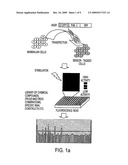 |
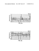 |  |
 |  |
 |  |
 | 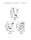 |
 | 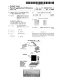 |
 |  |
 | 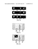 |
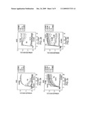 |  |
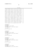 | 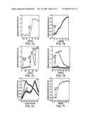 |
 | 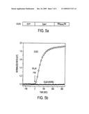 |
 | 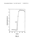 |
 |  |
| Similar patent applications: | |
| Date | Title |
|---|---|
| 2012-06-14 | Lipoxygenase and its use in wound healing |
| 2012-08-02 | Protein nanocarriers for topical delivery |
| 2012-09-27 | Method for incorporating a hydrophobic material into a skin care composition |
| 2012-08-30 | Hiv-1 clade a consensus sequences, antigens, and transgenes |
| New patent applications from these inventors: | |
| Date | Title |
|---|---|
| 2010-08-12 | Intracellular reporters |
| Top Inventors for class "Drug, bio-affecting and body treating compositions" | |
| Rank | Inventor's name |
|---|---|
| 1 | David M. Goldenberg |
| 2 | Hy Si Bui |
| 3 | Lowell L. Wood, Jr. |
| 4 | Roderick A. Hyde |
| 5 | Yat Sun Or |