Patent application title: HUMANIZED ANTIBODIES AND METHODS OF HUMANIZING ANTIBODIES
Inventors:
Edmund Rossi (Woodland Park, NJ, US)
Chien-Hsing Ken Chang (Downington, PA, US)
David M. Goldenberg (Mendham, NJ, US)
Hans Hansen (Picayune, MS, US)
Assignees:
IMMUNOMEDICS, INC.
IPC8 Class: AC07K1600FI
USPC Class:
5303873
Class name: Globulins immunoglobulin, antibody, or fragment thereof, other than immunoglobulin antibody, or fragment thereof that is conjugated or adsorbed chimeric, mutated, or recombined hybrid (e.g., bifunctional, bispecific, rodent-human chimeric, single chain, rfv, immunoglobulin fusion protein, etc.)
Publication date: 2009-09-24
Patent application number: 20090240037
Inventors list |
Agents list |
Assignees list |
List by place |
Classification tree browser |
Top 100 Inventors |
Top 100 Agents |
Top 100 Assignees |
Usenet FAQ Index |
Documents |
Other FAQs |
Patent application title: HUMANIZED ANTIBODIES AND METHODS OF HUMANIZING ANTIBODIES
Inventors:
Hans HANSEN
David M. GOLDENBERG
Edmund ROSSI
Chien-Hsing Ken CHANG
Agents:
ROSSI, KIMMS & McDOWELL LLP.
Assignees:
Immunomedics, Inc.
Origin: ASHBURN, VA US
IPC8 Class: AC07K1600FI
USPC Class:
5303873
Abstract:
Humanized, chimeric and human anti-CD20 antibodies and CD20 antibody
fusion proteins that bind to a human B cell marker, referred to as CD20,
which are useful for the treatment and diagnosis of B-cell disorders,
such as B-cell malignancies and autoimmune diseases, and methods of
treatment and diagnosis are disclosed. Methods of making the humanized,
chimeric and human anti-CD20 antibodies are disclosed. A humanized
anti-HSG (histamine-succinyl-glycyl) monoclonal antibody designated h679
which binds with high affinity to molecules containing the moiety
histamine-succinyl-glycyl (HSG), and methods of making the humanized
anti-HSG antibody also are disclosed.Claims:
1. A method of producing a humanized antibody, comprising:comparing the
amino acid sequence of each individual framework region of a non-human
antibody against a database of amino acid sequences of framework regions
for human antibodies;identifying amino acid residues in the framework
regions of the non-human antibody that differ from amino acid residues at
the corresponding positions for human antibodies in the
database;substituting those amino acid residues in the framework regions
of the non-human antibody that differ from the amino acid residues at the
corresponding positions for human antibodies in the database with an
amino acid residue from the corresponding position in one of the human
antibodies; andretaining complementarity determining regions of the
non-human antibody and all amino acid residues in the framework region of
the non-human antibody that interact with amino acid residues in the
complementarity determining regions.
2. The method according to claiml, wherein different human framework regions in the database have different amino acid residues at corresponding positions, and the amino acid residue that is substituted in the framework region of the non-human antibody is the one that occurs most commonly in the database.
3. The method according to claim 1, wherein the non-human antibody is a murine antibody.
4. A method according to claim 1, wherein the humanized antibody is h679.
5. A humanized antibody comprising complementarity determining regions of a non-human antibody and framework regions that comprise some amino acid residues that correspond to amino acid residues of the non-human antibody and other amino acid residues that correspond to amino acid residues in one or more human antibodies, wherein all residues in the framework region of the non-human antibody that interact with amino acid residues in the complementarity determining regions are residues from the non-human antibodies and those residues that do not interact with amino acid residues in the complementarity determining regions are substituted with an amino acid residue from a human antibody.
6. A humanized antibody according to claim 5, wherein the amino acid residue that is substituted in the framework region of the non-human antibody is an amino acid residue that occurs most commonly in a database of human antibodies.
7. A humanized antibody according to claim 5, wherein the non-human antibody is a murine antibody.
8. A method of producing a chimeric or humanized anti-CD20 antibody, comprising:providing variable domains comprising a light chain variable region CDR1 comprising a sequence RASSSVSYIH (SEQ ID NO: 3); CDR2 comprising a sequence ATSNLAS (SEQ ID NO: 6); and CDR3 comprising a sequence QQWTSNPPT (SEQ ID NO: 7); and a heavy chain variable region CDR1 comprising a sequence SYNMH (SEQ ID NO: 10); CDR2 comprising a sequence AIYPGNGDTSYNQKFKG (SEQ ID NO: 11); and CDR3 comprising a sequence STYYGGDWYFDV (SEQ ID NO: 12) or a sequence VVYYSNSYWYFDV (SEQ ID NO: 15); andproviding constant domains from a human antibody.
9. A method according to claim 8, additionally comprising replacing the sequences of murine framework regions in the variable domains with one or more different human framework regions.
10. A method according to claim 9, additionally comprising replacing one or more residues in one or more of said human framework regions with murine residues.
11. A method of producing a chimeric or humanized anti-CD20 antibody, comprising:transferring light and heavy chain murine complementarity determining regions into the corresponding variable domains of a human antibody, wherein said light and heavy chain complementarity determining regions comprise a light chain variable region CDR1 comprising a sequence RASSSVSYIH (SEQ ID NO: 3); CDR2 comprising a sequence ATSNLAS (SEQ ID NO: 6); and CDR3 comprising a sequence QQWTSNPPT (SEQ ID NO: 7); and a heavy chain variable region CDR1 comprising a sequence SYNMH (SEQ ID NO: 10); CDR2 comprising a sequence AIYPGNGDTSYNQKFKG (SEQ ID NO: 11); and CDR3 comprising a sequence STYYGGDWYFDV (SEQ ID NO: 12) or a sequence VVYYSNSYWYFDV (SEQ ID NO: 15).
12. A method according to claim 11, additionally comprising replacing the sequences of murine framework regions in the variable domains with one or more different human framework regions.
13. A method according to claim 12, additionally comprising replacing one or more residues in one or more of said human framework regions with murine residues.
14. A combination of a chimeric or humanized anti-CD20 antibody and L243 monoclonal antibody.
15. A combination as claimed in claim 14, wherein said combination is a fusion protein of a chimeric or humanized anti-CD20 antibody and L243 monoclonal antibody.
Description:
CROSS REFERENCE TO RELATED APPLICATIONS
[0001]This application claims priority from application Ser. No. 11/754,103, filed Sep. 21, 2006, which is a continuation of Ser. No. 10/366,709, filed on Feb. 14, 2003, now U.S. Pat. No. 7,151,164, which claims priority to U.S. Provisional application Ser. Nos. 60/416,232, filed Oct. 7, 2002, and 60/356,132, filed Feb. 14, 2002, the entire contents of all of these applications being incorporated herein by reference in their entirety. It also claims priority from application Ser. No. 10/270,171, which was incorporated by reference in application Ser. No. 10/366,709, portions of which are expressly incorporated herein. The entire contents of application Ser. No. 10/270,171 also are incorporated herein by reference in their entirety.
BACKGROUND OF THE INVENTION
[0002]1. Field of the Invention
[0003]The present invention relates to humanized, chimeric and human anti-CD20 antibodies, particularly monoclonal antibodies (mAbs) therapeutic and diagnostic conjugates of humanized, chimeric and human anti-CD20 antibodies and methods of treating B cell lymphomas and leukemias and various autoimmune diseases using humanized, chimeric and human anti-CD20 antibodies. The present invention relates to antibody fusion proteins or fragments thereof comprising at least two anti-CD20 mAbs or fragments thereof or at least one anti-CD20 MAb or fragment thereof and at least one second MAb or fragment thereof, other than the antiCD20 MAb or fragment thereof. The humanized, chimeric and human anti-CD20 mAbs, fragments thereof, antibody fusion proteins thereof or fragments thereof may be administered alone, as a therapeutic conjugate or in combination with a therapeutic immunoconjugate, with other naked antibodies, or with therapeutic agents or as a diagnostic conjugate. The present invention relates to DNA sequences encoding humanized, chimeric and human anti-CD20 antibodies, and antibody fusion proteins, vectors and host cells containing the DNA sequences, and methods of making the humanized, chimeric and human anti-CD20 antibodies. The present invention also relates to a humanized anti-HSG (histamine-succinyl-glycyl) monoclonal antibody designated h679 which binds with high affinity to molecules containing the moiety histamine-succinyl-glycyl (HSG), and methods of making the humanized anti-HSG antibody.
[0004]2. Background
[0005]The immune system of vertebrates consists of a number of organs and cell types which have evolved to accurately recognize foreign antigens, specifically bind to, and eliminate/destroy such foreign antigens. Lymphocytes, amongst others are critical to the immune system. Lymphocytes are divided into two major sub-populations, T cells and B cells. Although inter-dependent, T cells are largely responsible for cell-mediated immunity and B cells are largely responsible for antibody production (humoral immunity).
[0006]In humans, each B cell can produce an enormous number of antibody molecules. Such antibody production typically ceases (or substantially decreases) when a foreign antigen has been neutralized. Occasionally, however, proliferation of a particular B cell will continue unabated and may result in a cancer known as a B cell lymphoma. B-cell lymphomas, such as the B-cell subtype of non-Hodgkin's lymphoma, are significant contributors to cancer mortality. The response of B-cell malignancies to various forms of treatment is mixed. For example, in cases in which adequate clinical staging of non-Hodgkin's lymphoma is possible, field radiation therapy can provide satisfactory treatment. Still, about one-half of the patients die from the disease. Devesa et al., J. Nat'l Cancer Inst. 79:701 (1987).
[0007]The majority of chronic lymphocytic leukemias are of B-cell lineage. Freedman, Hematol. Oncol. Clin. North Am. 4:405 (1990). This type of B-cell malignancy is the most common leukemia in the Western world. Goodman et al., Leukemia and Lymphoma 22:1 (1996). The natural history of chronic lymphocytic leukemia falls into several phases. In the early phase, chronic lymphocytic leukemia is an indolent disease, characterized by the accumulation of small mature functionally-incompetent malignant B-cells having a lengthened life span. Eventually, the doubling time of the malignant B-cells decreases and patients become increasingly symptomatic. While treatment can provide symptomatic relief, the overall survival of the patients is only minimally affected. The late stages of chronic lymphocytic leukemia are characterized by significant anemia and/or thrombocytopenia. At this point, the median survival is less than two years. Foon et al., Annals Int. Medicine 113:525 (1990). Due to the very low rate of cellular proliferation, chronic lymphocytic leukemia is resistant to cytotoxic drug treatment.
[0008]Traditional methods of treating B-cell malignancies, including chemotherapy and radiotherapy, have limited utility due to toxic side effects. The use of monoclonal antibodies to direct radionuclides, toxins, or other therapeutic agents offers the possibility that such agents can be delivered selectively to tumor sites, thus limiting toxicity to normal tissues. Also, the presence of B-cell antigens on these B-cell malignancies makes them optimal targets for therapy with unconjugated B-cell antibodies, such as against CD19, CD20, CD21, CD23, and CD22 markers on B-cells. HLA-DR and other antigens may serve as targets for normal and malignant B-cells although they are also expressed on other cell types. Further, certain MUC1, MUC2, MUC3, and MUC4 antigens, preferably MUC1, are also expressed in different hematopoietic malignancies, including B-cell tumors expressing CD20 and other B-cell markers. Still other antigen targets, such as those associated with the vascular endothelium of tumors, including tenascin, vascular endothelium growth factor (VEGF), and placental growth factor (PIGF), as well as other categories of antigens associated with B-cell malignancies, such as oncogene products, are also suitable targets for said complementary antibodies for use in the present invention.
[0009]B cells comprise cell surface proteins which can be utilized as markers for differentiation and identification. One such human B-cell marker is the human B lymphocyte-restricted differentiation antigen Bp35, referred to as CD20. CD20 is expressed during early pre-B cell development and remains until plasma cell differentiation. CD20 is expressed on both normal B cells and malignant B cells whose abnormal growth can lead to B-cell lymphomas. Antibodies against the CD20 antigen have been investigated for the therapy of B-cell lymphomas. For example, a chimeric anti-CD20 antibody, designated as "IDEC-C2B8," has activity against B-cell lymphomas when provided as unconjugated antibodies at repeated injections of doses exceeding 500 mg per injection. Maloney et al., Blood 84:2457 (1994); Longo, Curr. Opin. Oncol. 8:353 (1996). About 50 percent of non-Hodgkin's patients, having the low-grade indolent form, treated with this regimen showed responses. Therapeutic responses have also been obtained using 131I-labeled B1 anti-CD20 murine monoclonal antibody when provided as repeated doses exceeding 600 mg per injection. Kaminski et al., N. Engl. J. Med. 329:459 (1993); Press et al., N. Engl. J. Med. 329:1219 (1993); Press et al., Lancet 346:336 (1995). However, these antibodies, whether provided as unconjugated forms or radiolabeled forms, have not shown high rates of objective and durable responses in patients with the more prevalent and lethal form of B-cell lymphoma, the intermediate or aggressive type. Therefore, a need exists to develop an immunotherapy for B-cell malignancies that achieves a therapeutic response of significant duration.
[0010]Additional studies targeting CD20 surface antigen have been demonstrated using an anti-CD20 murine monoclonal antibody, IF5, which was administered by continuous intravenous infusion to B cell lymphoma patients. Extremely high levels (>2 grams) of 1F5 were reportedly required to deplete circulating tumor cells, and the results were described as being "transient." Press et al., "Monoclonal Antibody 1F5 (Anti-CD20) Serotherapy of Human B-Cell Lymphomas." Blood 69/2:584-591 (1987). However, a potential problem with this approach is that non-human monoclonal antibodies (e.g., murine monoclonal antibodies) typically lack human effector functionality, i.e., they are unable to mediate complement-dependent lysis or lyse human target cells through antibody-dependent cellular toxicity or Fc-receptor mediated phagocytosis. Furthermore, non-human monoclonal antibodies can be recognized by the human host as a foreign protein and, therefore, repeated injections of such foreign antibodies can lead to the induction of immune responses leading to harmful hypersensitivity reactions. For murine-based monoclonal antibodies, this is often referred to as a Human Anti-Mouse Antibody (HAMA) response.
[0011]The use of chimeric antibodies is more preferred because they do not elicit as strong a HAMA response as murine antibodies. Chimeric antibodies are antibodies which comprise portions from two or more different species. For example, Liu, A. Y. et al, "Production of a Mouse-Human Chimeric Monoclonal Antibody to CD20 with Potent Fc-Dependent Biologic Activity" J. Immun. 139/10:3521-3526 (1987), describe a mouse/human chimeric antibody directed against the CD20 antigen. See also, PCT Publication No. WO 88/04936. However, no information is provided as to the ability, efficacy or practicality of using such chimeric antibodies for the treatment of B cell disorders in the reference. It is noted that in vitro functional assays (e.g., complement-dependent lysis (CDC); antibody dependent cellular cytotoxicity (ADCC), etc.) cannot inherently predict the in vivo capability of a chimeric antibody to destroy or deplete target cells expressing the specific antigen. See, for example, Robinson, R. D. et al., "Chimeric mouse-human anti-carcinoma antibodies that mediate different anti-tumor cell biological activities" Hum. Antibod. Hybridomas 2:84-93 (1991) (chimeric mouse-human antibody having undetectable ADCC activity). Therefore, the potential therapeutic efficacy of a chimeric antibody can only truly be assessed by in vivo experimentation, preferably in the species of interest for the specific therapy.
[0012]One approach that has improved the ability of murine monoclonal antibodies to be effective in the treatment of B-cell disorders has been to conjugate a radioactive label or chemotherapeutic agent to the antibody, such that the label or agent is localized at the tumor site. For example, the above-referenced 1F5 antibody and other B-cell antibodies have been labeled with 131I and were reportedly evaluated for biodistribution in two patients. See Eary, J. F. et al., "Imaging and Treatment of B-Cell Lymphoma" J. Nuc. Med. 31/8:1257-1268 (1990); see also, Press, O. W. et al., "Treatment of Refractory Non-Hodgkin's Lymphoma with Radiolabeled MB-1 (Anti-CD37) Antibody" J. Clin. Onc. 7/8:1027-1038 (1989) (indication that one patient treated with 131I-labeled IF-5 achieved a partial response); Goldenberg, D. M. et al. "Targeting, Dosimetry and Radioimmunotherapy of B-Cell Lymphomas with 131I-Labeled LL2 Monoclonal Antibody" J. Clin. Oncol. 9/4:548-564 (1991) (three of eight patients receiving multiple injections reported to have developed a HAMA response to this CD22 murine antibody); Appelbaum, F. R. "Radiolabeled Monoclonal Antibodies in the Treatment of Non-Hodgkin's Lymphoma" Hem./Oncol. Clinics of N. A. 5/5:1013-1025 (1991) (review article); Press, O. W. et al. "Radiolabeled-Antibody Therapy of B-Cell Lymphoma with Autologous Bone Marrow Support." New England Journal of Medicine 329/17: 1219-12223 (1993) (131I-labeled anti-CD20 antibody IF5 and B1); and Kaminski, M. G. et al "Radioimmunotherapy of B-Cell Lymphoma with [131I] Anti-B1 (Anti-CD20) Antibody". NEJM 329/7:459 (1993) (131I-labeled anti-CD20 antibody B1; hereinafter "Kaminski"); PCT published application WO 92/07466 (antibodies conjugated to chemotherapeutic agents such as doxorubicin or mitomycin). However, these approaches have not eliminated the obstacles associated with using murine antibodies, despite the fact that many patients with lymphoma who have received prior aggressive cytotoxic chemotherapy are immune suppressed, thus having lower HAMA rates than lymphoma patients who have not been heavily pretreated.
[0013]Autoimmune diseases are a class of diseases associated with B-cell disorders. Examples include immune-mediated thrombocytopenias, such as acute idiopathic thrombocytopenic purpura and chronic idiopathic thrombocytopenic purpura, myasthenia gravis, lupus nephritis, lupus erythematosus, and rheumatoid arthritis. The most common treatments are corticosteroids and cytotoxic drugs, which can be very toxic. These drugs also suppress the entire immune system, can result in serious infection, and have adverse affects on the bone marrow, liver and kidneys. Other therapeutics that have been used to treat Class III autoimmune diseases to date have been directed against T-cells and macrophages. There is a need for more effective methods of treating autoimmune diseases, particularly Class III autoimmune diseases.
[0014]To address the many issues related to B-cell disorders and their treatment, the present invention provides humanized, chimeric and human anti-CD20 monoclonal antibodies with the same complementarity determining regions (CDRs) that bind to the CD20 antigen of the present invention used alone, conjugated to a therapeutic agent or in combination with other treatment modalities, for the treatment of B cell lymphomas and leukemias and autoimmune disorders in humans and other mammals without the adverse responses associated with using murine antibodies.
SUMMARY OF THE INVENTION
[0015]Accordingly, the present invention provides humanized, chimeric and human anti-CD20 antibodies that bind to a human B cell marker, referred to as CD20 which is useful for the treatment and diagnosis of B-cell disorders, such as B-cell malignancies and autoimmune diseases.
[0016]The present invention further provides methods of treatment of mammalian subjects, such as humans or domestic animals, with one or more humanized, chimeric and human CD20 antibodies, alone, as an antibody fusion protein, as a therapeutic conjugate alone or as part of an antibody fusion protein, in combination, or as a multimodal therapy, with other antibodies, other therapeutic agents or immunomodulators or as an immunoconjugate linked to at least one therapeutic agent, therapeutic radionuclide or immunomodulator. These humanized chimeric and human CD20 antibodies can also be used as a diagnostic imaging agent alone, in combination with other diagnostic imaging agents, and/or in conjunction with therapeutic applications.
[0017]The present invention additionally is directed to anti-CD20 mAbs or fragments thereof that contain specific murine CDRs or a combination of murine CDRs from more than one murine or chimeric anti-CD20 MAb that have specificity for CD20. These mAbs can be humanized, chimeric or human anti-CD20 mAbs. The present invention is further directed to light and/or heavy chain variable regions or fragments thereof of these anti-CD20 Mabs and to light and/or heavy chains or fragments thereof that have specficity for CD20.
[0018]The present invention is also directed to antibody fusion proteins comprising at least two anti-CD20 mAbs or fragments thereof or a first MAb comprising an anti-CD20 mAbs or fragments thereof and a second MAb.
[0019]The present invention is further directed to a therapeutic or diagnostic conjugates of the anti-CD20 mAbs or fragments thereof or antibody fusion proteins of the anti-CD20 mAbs or other mAbs or fragments thereof bound to at least one therapeutic agent or at least one diagnostic agent. Antibody fusion proteins with multiple therapeutic agents of the same or different type are encompassed by the present invention.
[0020]The present invention is additionally directed to a method of using the anti-CD20 mAbs or fragments thereof or antibody fusion proteins thereof or fragments thereof for therapy, either alone, in combination with each other, as the antibody component of a therapeutic immunoconjugate with one or more therapeutic agents or each administered in combination with one or more therapeutic agents or with an immunoconjugate with one or more therapeutic agents.
[0021]The present invention further is directed to a method of using the anti-CD20 mAbs or fragments thereof or antibody fusion proteins thereof or fragments thereof as a diagnostic bound to one or more diagnostic agents.
[0022]The present invention additionally is directed to a method of pretargeting a cell in a patients suffering from a B-cell lymphoma or leukemia or an autoimmune disease using an antibody fusion protein or fragment thereof of the present invention.
BRIEF DESCRIPTION OF THE DRAWINGS
[0023]FIG. 1 shows a schematic representation of the 679 single chain Fv (scFv) polypeptide that is synthesized in E. coli from the 679-scFv-L5 expression plasmid and forms a 679 diabody. The gene construct for the un-processed polypeptide contains the pelB signal peptide, 679VH and VK coding sequences coupled by a 5 amino acid linker, Gly-Gly-Gly-Gly-Ser (G4S), and the carboxyl terminal six histidine (His) affinity tag. The figure also shows a stick figure drawing of the mature polypeptide after proteolytic removal of the pelB leader peptide and a stick FIG. drawing of a 679 diabody, including the HSG binding sites.
[0024]FIG. 2 shows a SDS-PAGE gel stained with Coomassie blue that is used to analyze the expression of 679 scFv from 679scFv-L5-transformed E. coli BL21 p-LysS cultures: lanes 1-5, induced with isopropyl-β-D-galactopyranoside (IPTG) overnight at 20° C.; lanes 6 and 7, not induced. In lane 3, the culture media was concentrated 10-fold. Soluble (lanes 4 and 6) and insoluble (lanes 5 and 7) proteins were fractionated by centrifugation of cell lysates (lane 2). 679scFv was purified from the insoluble fraction by Immobilized Metal Affinity Chromatography (IMAC) following solubilization in 8M urea (lane 1).
[0025]FIG. 3 shows a schematic representation of the hMN14scFv polypeptide that is synthesized in E. coli from the hMN14-scFv-L5 expression plasmid and forms a hMN14 diabody. The gene construct for the un-processed polypeptide contains the pelB signal peptide, hMN14VH and VK coding sequences coupled by a 5 amino acid linker, and the carboxyl terminal 6 histidine affinity tag. The figure also shows a stick figure drawing of the mature polypeptide following proteolytic removal of the pelB leader peptide, and a stick figure drawing of a hMN14 diabody, including CEA binding sites.
[0026]FIG. 4 shows size-exclusion High Performance Liquid Chromatography (HPLC) analysis of purified hMN14 diabody. Figure A is the HPLC elution profile of IMAC-purified hMN14 diabody. The HPLC elution peaks of hMN14 diabody in figures A and B are identified with an arrow. Figure B is the HPLC elution profile of hMN14 diabody purified by W12 anti-idiotype affinity chromatography. The *9.75 indicated on the x-axis of figure B is the HPLC retention time (9.75 min.) of control hMN14-Fab'-S-NEM (MW ˜50 KDa).
[0027]FIG. 5 shows reducing SDS-PAGE gel stained with Coomassie blue (figure A). The gel illustrates the purity of the hMN14 diabody samples following IMAC purification and W12 anti-idiotype affinity chromatography. The positions of the Mr standards and the hMN14scFv polypeptide are indicated with arrows. Lane 1 of figure A contains IMAC-purified hMN14 diabody. Lane 2 of the same figure contains affinity purified hMN14 diabody. Figure B is an isoelectric focusing (IEF) gel. The positions of pl standards and hMN14 diabody are indicated with arrows. Lane 1 of Figure B contains the hMN14 Fab'-S-NEM used as a standard. Lane 2 of the same figure contains the W12 purified hMN14 diabody. Lane 3 contains the unbound flow through fraction from the W12 affinity column and shows the proteins that are removed by this process.
[0028]FIG. 6 shows the levels of 131I-hMN14 diabody observed in a tumor and the blood over the first 96 hours after injection of the diabody. The concentration of 131I-hMN14 diabody, measured as the percentage of the injected dose per gram of tissue (% ID/g), is plotted vs. time. Solid squares mark the data points for tumor samples and open boxes mark those of blood samples.
[0029]FIG. 7 shows the biodistribution of 131I-hMN14 diabody 48 hours after injection in tumors and normal tissues, including liver, spleen, kidney, lungs, blood, stomach, small intestine, and large intestine. The concentration of 131I-hMN14 diabody is displayed as the percentage of the injected dose per gram of tissue (% ID/g).
[0030]FIG. 8 shows a schematic representation of the creation of the pET-ER vector. Figure A illustrates the double stranded DNA sequence of MCS2. Restriction sites are indicated above the sequence. MCS2 was ligated into the Blpl restriction site of pET26b vector shown in Figure B. Figure C shows the diagram of pET-ER vector, including the MCS2 sequence.
[0031]FIG. 9 shows a schematic representation of the steps involved in the generation of constructs used for expression of three 679xhMN14 bispecific diabody variants represented by BS1, BS1.5 and BS2.
[0032]FIG. 10 shows a schematic representation of the di-cistronic expression cassette in the pET-ER vector and also stick figures of the two heterologous polypeptides as synthesized and the formation of 679×hMN14 bispecific diabodies. The di-cistronic cassette codes for a single RNA message generated from T7 RNA polymerase via the T7 promoter. This message contains two ribosomal binding sites (RBS) and the coding sequences for the two heterologous polypeptides. Stick figure drawings show the two mature heterologous polypeptides, 679VH(G4S)hMN14VK (Left) and hMN14VH(G4S)679VK (Right) that are synthesized from the di-cistronic expression cassettes. The 679×hMN14 bispecific diabody (BS1, BS1.5 or BS2) is represented as a stick figure drawing and is formed from the pairing of the heterologous polypeptides.
[0033]FIG. 11 shows a size-exclusion HPLC analysis of BS1.5 after purification. The HPLC elution peak of BS1.5 is at 9.22 min. Soluble proteins from an induced 5 L culture were purified by Ni-NTA IMAC followed by Q-Sepharose anion exchange chromatography. The flow through fraction of the Q-Sepharose column was injected for HPLC analysis.
[0034]FIG. 12 shows a reducing SDS-PAGE gel stained with Coomassie blue and used to analyze the purification of BS2. Arrows indicate the positions of the Mr standards and the BS2 polypeptide constituents, 679VH-hMN14VK and hMN14VH-679VK. Soluble proteins from an induced 5 L culture were loaded on a 4 ml Ni-NTA column. The column was washed/eluted with a buffer containing 40 mM imidazole (lane 3) and then eluted in two fractions with 100 mM imidazole (lanes 1 and 2). Impurities in the 40 mM imidazole eluate were removed by passing the eluate over a Q-Sepharose anion exchange column (lane 4).
[0035]FIG. 13 shows the purity of BS1, BS2 and BS1.5 through an IEF gel. These three diabodies were purified from soluble protein extracts by Ni-NTA IMAC followed by Q-Sepharose anion exchange chromatography. The positions of pl markers are indicated by arrows and the samples are identified above the lanes.
[0036]FIG. 14 shows BIAcore binding curves obtained for various concentrations of BS1.5 using a low-density HSG-coupled sensor chip. These data were used for calculation of the on-rates and off-rates.
[0037]FIG. 15 is a graphical representation of the results of a competitive enzyme-linked immunosorbent assay (ELISA). HRP-conjugated hMN14 IgG (1 nM) was mixed with either BS1.5 or chemically linked 679×hMN14 F (ab')2 at concentrations ranging from 4-500 nM, prior to incubation in CEA-coated (0.5 ag/well) wells. The % inhibition is plotted vs. nM concentration of sample.
[0038]FIG. 16 is a BIAcore sensorgram showing bispecific binding properties of BS1.5 for HSG and W12. BS1.5 (60 ng) was loaded on a high-density HSG-coupled sensor chip and two 400 ng injections of the hMN14-binding anti-idiotype MAb, W12, were allowed to bind to the immobilized BS1.5. Arrows indicate injection times.
[0039]FIG. 17 shows the levels of 131I-BS1.5 diabody in the tumor and the blood over the first 96 hours after injection of the diabody. The concentration of 131I-BS1.5 diabody, measured as the percentage of the injected dose per gram of tissue (% ID/g), is plotted vs. time. Diamonds mark the data points for tumor samples and filled circles mark those of blood samples.
[0040]FIG. 18 shows the biodistribution of 131I-BS1.5 diabody after 12 and 24 hours post injection in tumor and normal tissue, including liver, spleen, kidney, lungs, blood, stomach, small intestine, and large intestine. The concentration of 131I-BS1.5 was measured as the percentage of the injected dose per gram of tissue (% ID/g).
[0041]FIG. 19 shows the biodistribution of 111In-IMP241 peptide in tumor bearing mice pretargeted with BS1.5. GW39 tumor-bearing nude mice were injected with BS1.5 diabody. After 12 hours of clearance, the 111Indium-labeled IMP241 peptide was injected. Radioactivity in the tumor and in normal tissues, including liver, spleen, kidney, lungs, blood, stomach, small intestine, and large intestine, was measured at 3 and 24 hours post injection of 111In-IMP241. The concentration of 111In-IMP241 was measured as the percentage of the injected dose per gram of tissue (% ID/g).
[0042]FIG. 20 shows an alignment of murine (m) and humanized (h) 679 VH and VK amino acid sequences using the Kabat numbering scheme. Amino acid substitutions made during humanization are indicated with arrowheads. The CDR and framework regions are indicated.
[0043]FIG. 21 shows the relative locations of the PCR primers used for humanization of 679scFv-L5. Arrows signify the primers. The intermediate PCR products are also shown (A, B, C and D). All numbering represent nucleic acid positions in 679scFv-L5.
[0044]FIG. 22 shows size-exclusion HPLC analysis of the BS1.5H after purification. The HPLC elution peak of BS1.5H is at 10.16 min. Soluble proteins from an induced 5 L culture were purified by Ni-NTA IMAC followed by Q-Sepharose anion exchange chromatography. The flow through fraction of the Q-Sepharose column was injected for HPLC analysis.
[0045]FIG. 23 is a BIAcore sensorgram showing bispecific binding properties of BS1.5H for HSG and W12. BS1.5H (60 ng) was loaded on a high-density HSG-coupled sensor chip and a 1 μg injection of the hMN14-binding anti-idiotype MAb, W12, was allowed to bind to the immobilized BS1.5H. Arrows indicate injection times.
[0046]FIG. 24 shows the comparison of BIAcore binding curves between BS1.5H, BS1.5 and BS2. Similar amounts of the bispecific diabodies were injected on a low density HSG-coupled sensor chip and the resulting binding curves were superimposed.
[0047]FIG. 25 is the coding sequence of nucleic acids and encoded amino acids for 679-scFv-L5. 1-66 is the coding sequence for the pelB leader peptide. 70-426 is the coding sequence for 679VH. 427-441 is the coding sequence for the linker peptide (GGGGS) 442-780 is the coding sequence for 679VK. 787-804 is the coding sequence for the 6 histidine affinity tag.
[0048]FIG. 26 is the coding sequence of nucleic acids and encoded amino acids for 679-I3Q. 1-66 is the coding sequence for the pelB leader peptide. 70-426 is the coding sequence for 679 VH (I3Q). 427-441 is the coding sequence for the linker peptide (GGGGS). 442-780 is the coding sequence for 679 VK. 787-804 is the coding sequence for the 6 histidine affinity tag.
[0049]FIG. 27 is the coding sequence of nucleic acids and encoded amino acids for 679-C101S. 1-66 is the coding sequence for the pelB leader peptide. 70-426 is the coding sequence for 679 VH. 427-441 is the coding sequence for the linker peptide (GGGGS). 442-780 is the coding sequence for 679 VK (C101 S). 787-804 is the coding sequence for the 6 histidine affinity tag.
[0050]FIG. 28 is the coding sequence and encoded amino acids for 679 I3Q/C101S.
[0051]FIG. 29 is the coding sequence of nucleic acids and encoded amino acids for hMN14-scFv-L5. 1-66 is the coding sequence for the pelB leader peptide. 70-423 is the coding sequence for hMN14 VH. 424-438 is the coding sequence for the linker peptide (GGGGS). 439-759 is the coding sequence for hMN14 VK. 766-783 is the coding sequence for the 6 histidine affinity tag.
[0052]FIG. 30 is the coding sequence of nucleic acids and encoded amino acids for polypeptide #1 of BS1 (679×hMN14 bispecific diabody: variant 1). 1-66 is the coding sequence for the pelB leader peptide. 70-426 is the coding sequence for 679 VH. 427-441 is the coding sequence for the linker peptide (GGGGS). 442-762 is the coding sequence for hMN14 VK. 769-786 is the coding sequence for the 6 histidine affinity tag.
[0053]FIG. 31 is the coding sequence of nucleic acids and encoded amino acids for polypeptide #2 of BS1 (679×hMN14 bispecific diabody: variant 1). 1-66 is the coding sequence for the pelB leader peptide. 70-423 is the coding sequence for hMN14 VH. 424-438 is the coding sequence for the linker peptide (GGGGS). 439-777 is the coding sequence for 679 VK. 784-801 is the coding sequence for the 6 histidine affinity tag.
[0054]FIG. 32 is the coding sequence of nucleic acids and encoded amino acids for polypeptide #1 of BS1.5 (679×hMN14 bispecific diabody: variant 2). 1-66 is the coding sequence for the pelB leader peptide. 70-426 is the coding sequence for 679 VH (I3Q). 427-441 is the coding sequence for the linker peptide (GGGGS). 442-762 is the coding sequence for hMN14 VK. 769-786 is the coding sequence for the 6 histidine affinity tag.
[0055]FIG. 33 is the coding sequence of nucleic acids and encoded amino acids for polypeptide #2 of BS1.5 (679×hMN14 bispecific diabody: variant 2). 1-66 is the coding sequence for the pelB leader peptide. 70-423 is the coding sequence for hMN14VH. 424-438 is the coding sequence for the linker peptide (GGGGS). 439-777 is the coding sequence for 679VK. 784-801 is the coding sequence for the 6 histidine affinity tag.
[0056]FIG. 34 is the coding sequence of nucleic acids and encoded amino acids for polypeptide #1 of BS2 (679×hMN14 bispecific diabody: variant 3). 1-66 is the coding sequence for the pelB leader peptide. 70-426 is the coding sequence for 679VH (I3Q). 427-441 is the coding sequence for the linker peptide (GGGGS). 442-762 is the coding sequence for hMN14VK. 769-786 is the coding sequence for the 6 histidine affinity tag.
[0057]FIG. 35 is the coding sequence of nucleic acids and encoded amino acids for polypeptide #2 of BS2 (679×hMN14 bispecific diabody: variant 3). 1-66 is the coding sequence for the pelB leader peptide. 70-423 is the coding sequence for hMN14VH. 424-438 is the coding sequence for the linker peptide (GGGGS). 439-777 is the coding sequence for 679VK C101S. 784-801 is the coding sequence for the 6 histidine affinity tag.
[0058]FIG. 36 is the coding sequence of nucleic acids and encoded amino acids for h679-scFv-L5. 1-66 is the coding sequence for the pelB leader peptide. 70-426 is the coding sequence for h679VH. 427-441 is the coding sequence for the linker peptide (GGGGS). 442-780 is the coding sequence for h679VK. 787-804 is the coding sequence for the 6 histidine affinity tag.
[0059]FIG. 37 is the coding sequence of nucleic acids and encoded amino acids for polypeptide #1 of BS1.5H (h679 x hMN14 bispecific diabody). 1-66 is the coding sequence for the pelB leader peptide. 70-426 is the coding sequence for h679VH. 427-441 is the coding sequence for the linker peptide (GGGGS). 442-762 is the coding sequence for hMN14VK. 769-786 is the coding sequence for the 6 histidine affinity tag.
[0060]FIG. 38 is the coding sequence of nucleic acids and encoded amino acids for polypeptide #2 of BS1.5H (h679 x hMN14 bispecific diabody). 1-66 is the coding sequence for the pelB leader peptide. 70-423 is the coding sequence for hMN14VH. 424-438 is the coding sequence for the linker peptide (GGGGS). 439-777 is the coding sequence for h679VK C101S. 784-801 is the coding sequence for the 6 histidine affinity tag.
[0061]FIG. 39 discloses the V gene sequences cloned by RT-PCR from a hybridoma cell line producing a murine anti-CD20, and the deduced amino acid sequences of the variable light (FIG. 39A) and heavy chain (FIG. 39B) of the A20 antibody, designated as A20Vk and A20VH, respectively. Underlined arrows indicate the sequences of the PCR primers used for cloning. The putative CDR region sequences, as defined by the Kabat numbering scheme, are shown in bold and underlined. Amino acid sequences are given as single-letter codes below the corresponding nucleotide sequence. The Kabat numbering scheme was used for amino acid residues. Amino acid residues numbered by a letter represent the insertion residue according to Kabat, and have the same number as that of the previous residue. For example, residues 82, 82A, 82B and 82C in FIG. 39B are indicated as 82 A, B, and C, respectively.
[0062]FIG. 40 discloses the Vk, the variable light chain, and the VH, the variable heavy chain, sequences of cA20, a chimeric anti-CD20 antibody. The CDR region sequences are shown in bold and underlined. The amino acid residues and the nucleotides are numbered sequentially and same numbering system is used for humanized V sequences. The light chain variable region is shown in FIG. 40A and the heavy chain variable region is shown in FIG. 40B. The numbering system is the same as for FIG. 39. The restriction sites used for constructing cA20 are underlined.
[0063]FIG. 41 shows a comparison of the binding affinities of the chimeric A20 (cA20), and murine A20, (A20), in a cell surface competitive binding assay against 125I-labled A20. Increasing concentrations of cA20 blocked the binding of radiolabeled A20 to Raji cells (as depicted by closed circles) in a comparable manner as that of murine A20 (depicted by closed diamonds).
[0064]FIG. 42 compares the amino acid sequences of the variable heavy chain (VH) and variable light chain (Vk) of human antibodies, and chimeric and humanzied anti-CD20 antibodies. FIG. 42A compares the amino acid sequences of the variable heavy chain (VH) of the human antibodies, EU and NEWM (FR4 only), the chimeric antibody, (cA20VH) and two humanized antibodies, (hA20VH1 and hA20VH2) and FIG. 42B compares the amino acid sequences of the variable light chain (Vk) of the human antibody, (REIVk), a chimeric antibody, (cA20Vk), and a humanized antibody, (hA20Vk). Dots indicate that the residues in A20 are identical to the corresponding residue in the human antibody. The CDRs are identified as a boxed region. The Kabat numbering scheme was used to number the amino acid residues.
[0065]FIG. 43 discloses the nucleotide sequences of hA20 light chain V genes, (hA20Vk) (FIG. 43A), and heavy chain V genes, hA20VH1 (FIG. 43B) and hA20VH2 (FIG. 43C), as well as the adjacent flanking sequences of the VKpBR2 (FIG. 43A) and VHpBS2 (FIGS. 43B and 43C) staging vectors, respectively. The non-translated nucleotide sequences are shown in lowercase. The restriction sites used for subcloning are underlined and indicated. The secretion signal peptide sequence is indicated by a double underline. Numbering of Vk and VH amino acid residues is same as that in FIG. 40.
[0066]FIG. 44 shows the results of a cell surface competitive binding assay to compare the binding activity of two humanized A20 antibodies, (hA20-1 and hA20-2), with that of A20, cA20 and a chimeric anti-CD20 MAb, c2B8. FIG. 44A shows hA20-1 (closed triangles) and hA20-2 (closed circles) and the murine anti-CD20 antibody, A20 (closed squares) competed equally well for the binding of 125I-A20 to Raji cells. FIG. 44B shows hA20-1 (closed circles), cA20 (closed squares) and c2B8 (closed diamonds) competed equally well for the binding of 125I-c2B8 to Raji cells.
[0067]FIG. 45 discloses the constant region of a human IgG1 (CH-hinge) (FIG. 45A) and the constant region of a human kappa chain (Ck) (FIG. 45B).
[0068]FIG. 46 is a competitive cell surface binding assay. Ag-binding specificity and affinity studies of humanized anti-CD20 Abs (cA20, hA20, and c1F5, purified by affinity chromatography on a Protein A column) were evaluated by a cell surface competitive binding assay with murine 2B8 and rituximab (IDEC Pharmaceuticals Corp., San Diego, Calif.). FIG. 46(A) is a comparison of the binding activities of cA20 (square), hA20-1 (triangle) and hA20-1 (circle) with that of m2B8 (diamond); FIG. 46(B) compares of the binding activities of cA20 (square), c1F5 (triangle) and rituximab (diamond).
[0069]FIG. 47 is a study comparing the binding activities of hA20 with other anti-CD20 Abs, including rituximab and murine B1, by a cell surface competitive binding assay. A constant amount (100,000 cpm, ˜10 Ci/ g) of 125I-labeled rituximab was incubated with Raji cells in the presence of varying concentrations (0.2-700 nM) of competing Abs, hA20 (triangle), mB1 (Downward triangle) or rituximab (square) at 4° C. for 1-2 h.
[0070]FIG. 48 depicts the cytotoxic effect of crosslinked hA20 and other CD20 Abs on cultured lymphoma cells. Total cell and viable cell cell populations were measured by (A) trypan blue staining and cell counting or (B) MTT assay.
[0071]FIG. 49 is a graph of in vivo therapy studies with various anti-CD20 and other Abs. Raji cells administered i.v. to SCID mice, to create a Raji lymphoma model of disseminated disease.
[0072]FIG. 50 is a graph depicting in vivo therapy with hA20 and hLL2. Raji cells administered i.v. to SCID mice, to create a Raji lymphoma model of disseminated disease.
DETAILED DESCRIPTION OF SPECIFIC EMBODIMENTS
1. Overview
[0073]As discussed above, anti-CD20 antibodies that are unconjugated or labeled with a therapeutic radionuclide, have failed to provide high rates of objective and lasting responses in patients with intermediate or aggressive forms of B-cell lymphoma. The present invention provides a humanized, a chimeric and a human anti-CD20 antibody, and antibody fusion proteins thereof, useful for treatment of mammalian subjects humans and domestic animals alone as a conjugate or administered in combination with other therapeutic agents, including other naked antibodies and antibody therapeutic conjugates.
[0074]The anti-CD20 mAbs of the present invention contain specific murine CDRs or a combination of murine CDRs from more than one murine or chimeric anti-CD20 MAb that have specificity for the CD20 antigen. The anti-CD20 mAbs of the present invention are humanized, chimeric or human mAbs, light and/or heavy chains thereof or light and/or heavy chain variable regions thereof, and they contain the amino acids of the CDRs of a murine anti-CD20 MAb and retain substantially the B-cell and B-cell lymphoma and leukemia cell targeting of the murine anti-CD20 MAb. The CDRs of the light chain variable region of the anti-CD20 MAb comprises CDR1 comprising amino acids RASSSVSYIH, RASSSLSFMH or RASSSVSYMH; CDR2 comprising amino acids ATSNLAS; and CDR3 comprising amino acids QQWTSNPPT, HQWSSNPLT or QQSFSNPPT; and the CDRs of the heavy chain variable region of the anti-CD20 MAb comprises CDR1 comprising amino acids SYNMH; CDR2 comprising amino acids AIYPGNGDTSYNQKFKG and CDR3 comprising amino acids STYYGGDWYFDV, STYYGGDWYFNV, SHYGSNYVDYFDV or VVYYSNSYWYFDV. The humanized antibody further comprises the framework regions of the light and heavy chain constant regions of a human antibody.
[0075]In one embodiment, the humanized and chimeric MAb or fragment thereof does not contain the CDR3 of the heavy chain variable region comprising STYYGGDWYFNV. More preferably, CDR1 of the light chain variable region does not comprise RASSSLSFMH when the CDR3 of the light chain variable region comprises HQWSSNPLT and the CDR3 of the heavy chain variable region comprises SHYGSNYVDYFDV. In another embodiment, the CDR3 of the light chain variable region does not comprise HQWSSNPLT when CDR1 of the light chain variable region comprises RASSSLSFMH and when CDR3 of the heavy chain variable region comprises SHYGSNYVDYFDV. In a further embodiment, the CDR3 of the heavy chain variable region does not comprise SHYGSNYVDYFDV when the CDR1 of the light chain variable region comprises RASSSLSFMH and the CDR3 of the light chain variable region comprises HQWSSNPLT. In another embodiment, the CDR1 of the light chain variable region does not comprise RASSSVSYMH when the CDR3 of the light chain variable region comprises QQSFSNPPT and the CDR3 of the heavy chain variable region comprises VVYYSNSYWYFDV.
[0076]Further, in another embodiment, the anti-CD20 monoclonal antibody (MAb) or fragment thereof does not contain CDR3 of the light chain variable region of amino acids QQSFSNPPT when CDR1 of the light chain variable region comprises RASSSVSYMH and the CDR3 of the heavy chain variable region comprises VVYYSNSYWYFDV. Additionally, the anti-CD20 MAb does not contain CDR3 of the heavy chain variable region with amino acids VVYYSNSYWYFDV when the CDR1 of the light chain variable region comprises RASSSVSYMH and the CDR3 of the light chain variable region comprises QQSFSNPPT.
[0077]In a preferred embodiment, the humanized anti-CD20 (hCD20) monoclonal antibody or antigen-binding fragment thereof comprising the complementarity determining regions (CDRs) of at least one murine anti-CD20 MAb variable region and the framework regions (FRs) of at least one human MAb variable region, wherein said humanized anti-CD20 MAb or fragment thereof retains substantially the B-cell and B-cell lymphoma and leukemia cell targeting of said murine anti-CD20 MAb. The humanized antibody's variable region may comprise a light chain variable region, a heavy chain variable region or a both light and heavy chain variable regions. The humanized antibody or fragment thereof may further comprise light and heavy chain constant regions of at least one human antibody.
[0078]The humanized anti-CD20 MAb or fragment thereof of the present invention comprises the CDRs of a murine anti-CD20 MAb and the framework (FR) regions of the light and heavy chain variable regions of a human antibody, while retaining substantially the B-cell, and B-cell lymphoma and leukemia cell targeting of the parent murine anti-CD20 MAb, and wherein the CDRs of the light chain variable region of the murine anti-CD20 MAb comprises CDR1 comprising amino acids RASSSVSYIH, CDR2 comprising amino acids ATSNLAS and CDR3 comprising QQWTSNPPT and the CDRs of the heavy chain variable region of murine anti-CD20MAb comprises CDR1 comprising amino acids SYNMH, CDR2 comprising amino acids AIYPGNGDTSYNQKFKG and CDR3 comprising amino acids STYYGGDWYFDV. But the humanized anti-CD20 MAb or fragment thereof may further contain in the FRs of the light and heavy chain variable regions of the antibody at least one amino acid from the corresponding FRs of the murine MAb. The humanized MAbs may further contain the light and heavy chain constant regions of a human antibody. Specifically, the humanized anti-CD20 MAb or fragment thereof contains at least one amino acid residue 1, 5, 27, 30, 38, 48, 67, 68, 70, 95, 115 and 116 of the murine heavy chain variable region of FIG. 42A, designated as hA20VH1 or hA20VH2 and of at least one amino acid residue 4, 21, 35, 38, 45, 46, 59, 99, 104 and 106 of the murine light chain variable region FIG. 42B, designated hA20Vk. One or more of the murine amino acid sequences can be maintained in the human FR regions of the light and heavy variable chains if necessary to maintain proper binding or to enhance binding to the CD20 antigen. More preferably the humanized anti-CD20MAb or fragment thereof of the present invention comprises the hA20Vk of FIG. 42B and the hA2VH1 of FIG. 42A. Most preferably, the humanized anti-CD20 MAb or fragment thereof of the present invention comprises the hA20Vk of FIG. 42B and the hA2VH2 of FIG. 42A. This latter sequence contains more human amino acid sequences in the FRs of the VH2 chain than the VH1, and thus is more humanized.
[0079]The preferred chimeric anti-CD20 (cCD20) MAb or fragment thereof of the present invention comprises the CDRs of a murine anti-CD20 MAb and the FR regions of the light and heavy chain variable regions of the murine anti-CD 20 MAb, i.e., the Fvs of the parental murine MAb, and the light and heavy chain constant regions of a human antibody, wherein the chimeric anti-CD20 MAb or fragment thereof retains substantially the B-cell, and B-cell lymphoma and leukemia cell targeting of the murine anti-CD20 MAb, wherein the CDRs of the light chain variable region of the chimeric anti-CD20 MAb comprise CDR1 comprising amino acids RASSSVSYIH, RASSSLSFMH or RASSSVSYMH; CDR2 comprising amino acids ATSNLAS; and CDR3 comprising amino acids QQWTSNPPT, HQWSSNPLT or QQSFSNPPT; and the CDRs of the heavy chain variable region of the chimeric anti-CD20 MAb comprise CDR1 comprising amino acids SYNMH; CDR2 comprising amino acids AIYPGNGDTSYNQKFKG and CDR3 comprising STYYGGDWYFDV, STYYGGDWYFNV, SHYGSNYVDYFDV or VVYYSNSYWYFDV with the following provisos,
[0080](a) wherein the CDR3 of the heavy chain variable region does not comprise STYYGGDWYFNV, when the CDR1 of the light chain variable region comprises amino acids RASSSVSYIH, CDR2 of the light chain variable region comprises amino acids ATSNLAS, CDR3 of the light chain variable region comprises amino acids QQWTSNPPT, CDR1 of the heavy chain variable region comprises amino acids SYNMH, and CDR2 of the light chain variable region comprises amino acids AIYPGNGDTSYNQKFKG;
[0081](b) wherein the CDR3 of the heavy chain variable region does not comprise SHYGSNYVDYFDV, when the CDR1 of the light chain variable region comprises amino acids RASSSLSFMH, CDR2 of the light chain variable region comprises amino acids ATSNLAS, CDR3 of the light chain variable region comprises amino acids HQWSSNPLT, CDR1 of the heavy chain variable region comprises amino acids SYNMH, and CDR2 of the light chain variable region comprises amino acids AIYPGNGDTSYNQKFKG; and
[0082](c) wherein the CDR3 of the heavy chain variable region does not comprise VVYYSNSYWYFDV, when the CDR1 of the light chain variable region comprises amino acids RASSSVSYMH, CDR2 of the light chain variable region comprises amino acids ATSNLAS, CDR3 of the light chain variable region comprises amino acids QQSFSNPPT, CDR1 of the heavy chain variable region comprises amino acids SYNMH, and CDR2 of the light chain variable region comprises amino acids AIYPGNGDTSYNQKFKG.
[0083]More preferably the chimeric anti-CD20 MAb or fragment thereof comprising the complementarity-determining regions (CDRS) of a murine anti-CD20MAb and the framework (FR) regions of the light and heavy chain variable regions of the murine anti-CD20 MAb and further, the light and heavy chain constant regions of a human antibody, wherein the chimeric anti-CD20 MAb or fragment thereof retains substantially the B-cell, and B-cell lymphoma and leukemia cell targeting of the murine anti-CD20 MAb, wherein the CDRs of the light chain variable region of the chimeric anti-CD20 MAb comprises the CDRs shown in FIGS. 42B and 42A, respectively, designated cA20Vk and cA20VH. Most preferably, the chimeric anti-CD20 MAb or fragment thereof comprises the light and heavy chain variable regions of murine anti-CD20 MAb shown in FIGS. 42B and 42A, respectively, designated cA20Vk and cA20 VH.
[0084]The present invention also encompasses a human anti-CD20 MAb or fragment thereof comprising the light and heavy chain variable, wherein said human CD20 MAb retains substantially the B-cell, and B-cell lymphoma and leukemia cell targeting and cell binding characteristics of a murine anti-CD20 MAb, wherein the CDRs of the light chain variable region of the human anti-CD20 MAb comprises the same CDRs as set forth above for the chimeric and humanized anti-CD20 mAbs and as shown in FIGS. 42A and 42B. This human anti-CD20 MAb or fragment thereof further comprises light and heavy chain constant regions of at least one human antibody.
[0085]The present invention is also intended to encompass antibody fusion proteins or fragments thereof comprising at least two anti-CD20 mAbs or fragments thereof, as described above. The antibody fusion protein or fragment thereof of the present invention is also intended to encompass an antibody fusion protein or fragment thereof comprising at least one first anti-CD20 MAb or fragment thereof as described above and at least one second MAb or fragment thereof, other than the antiCD20 MAb or fragment described above. More preferably this second MAb is a MAb reactive with CD4, CD5, CD8, CD14, CD15, CD19, CD21, CD22, CD23, CD25, CD33, CD37, CD38, CD40, CD40L, CD46, CD52, CD54, CD74, CD80, CD126, B7, MUC1, MUC2, MUC3, MUC4, Ia, HM1.24, HLA-DR, tenascin, VEGF, P1GF, an oncogene, oncogene product, or a combination thereof, and even an anti-CD20 MAb that is different than the anti-CD20 MAb described herein. The antibody fusion proteins of the present invention may be composed of one CD20 MAb and one or more of the second mAbs to provide specificity to different antigens, and are described in more detail below.
[0086]The humanized, chimeric and human anti-CD20 antibody may possess enhanced affinity binding with the epitope, as well as antitumor and anti-B-cell activity, as a result of CDR mutation and manipulation of the CDR and other sequences in the variable region to obtain a superior therapeutic agent for the treatment of B-cell disorders, including B-cell lymphomas and leukemias and autoimmune diseases. Modification to the binding specificity, affinity or avidity of an antibody is known and described in WO 98/44001, as affinity maturation, and this application summarizes methods of modification and is incorporated in its entirety by reference.
[0087]It may also be desirable to modify the antibodies of the present invention to improve effector function, e.g., so as to enhance antigen-dependent cell-mediated cytotoxicity (ADCC) and/or complement dependent cytotoxicity (CDC) of the antagonist. One or more amino acid substitutions or the introduction of cysteine in the Fc region may be made, thereby improving internalization capability and/or increased complement-mediated cell killing and ADCC. See Caron et al., J. Ex. Med. 176:1191-1195 (1991) and Shopes, B. J. Immunol. 148:2918-2022 (1992), incorporated herein by reference in their entirety. An antibody fusion protein may be prepared that has dual Fc regions with both enhanced complement lysis and ADCC capabilities.
[0088]The present invention is also directed to DNA sequences comprising a nucleic acid encoding a MAb or fragment thereof selected from the group consisting
[0089](a) an anti-CD20 MAb or fragment thereof as described herein,
[0090](b) an antibody fusion protein or fragment thereof comprising at least two of the anti-CD20 mAbs or fragments thereof
[0091](c) an antibody fusion protein or fragment thereof comprising at least one first MAb or fragment thereof comprising the anti-CD20 MAb or fragment thereof as described herein and at least one second MAb or fragment thereof, other than the antiCD20 MAb or fragment thereof, and
[0092](d) an antibody fusion protein or fragment thereof comprising at least one first MAb or fragment thereof comprising the anti-CD20 MAb or fragment thereof and at least one second MAb or fragment thereof, wherein the second MAb is a MAb reactive with CD4, CD5, CD8, CD14, CD15, CD19, CD21, CD22, CD23, CD25, CD33, CD37, CD38, CD40, CD40L, CD46, CD52, CD54, CD74, CD80, CD126, B7, MUC1, MUC2, MUC3, MUC4, Ia, HM1.24, HLA-DR, tenascin, VEGF, P1GF, an oncogene, oncogene product, or a combination thereof.
[0093]Also encompassed by the present invention are expression vectors comprising the DNA sequences. These vectors contain the light and heavy chain constant regions and the hinge region of the human immunoglobulin, in the case of vectors for use in preparing the humanized, chimeric and human anti-CD20 mAbs or antibody fusion proteins thereof or fragments thereof. These vectors additionally contain, where required, promoters that express the mAbs in the selected host cell immunoglobulin enhances and signal or leader sequences. Vectors that are particularly useful in the present invention are pdHL2 or GS, particularly when used to express chimeric, humanized or human antibodies, such as gigs, where the vector codes for the heavy and light chain constant regions and hinge region of IgG1. More preferably, the light and heavy chain constant regions and hinge region are from a human EU myeloma immunoglobulin, where optionally at least one of the amino acid in the allotype positions is changed to that found in a different IgG1 allotype, and wherein optionally amino acid 253 of the heavy chain of EU based on the EU number system may be replaced with alanine. See Edelman et al., Proc. Natl. Acad. Sci. USA 63: 78-85 (1969), incorporated herein in its entirety by reference.
[0094]Host cells containing the DNA sequences encoding the anti-CD20 mAbs or fragments thereof or antibody fusion proteins or fragments thereof of the present invention or host cells containing the vectors that contain these DNA sequences are encompassed by the present invention. Particularly useful host cells are mammalian cells, more specifically lymphocytic cells, such as myeloma cells, discussed in more detail below.
[0095]Also encompassed by the present invention is the method of expressing the anti-CD20 MAb or fragment thereof or antibody fusion protein or fragment thereof comprising: (a) transfecting a mammalian cell with a DNA sequence of encoding the anti-CD20 mAbs or fragments thereof or antibody fusion proteins or fragments thereof and (b) culturing the cell transfected with the DNA sequence that secretes the anti-CD20 or fragment thereof or antibody fusion protein or fragment thereof. Known techniques may be used that include a selection marker on the vector so that host cells that express the mAbs and the marker can be easily selected.
[0096]The present invention particularly encompasses B-lymphoma cell and leukemia cell targeting diagnostic or therapeutic conjugates comprising an antibody component comprising an anti-CD20 MAb or fragment thereof or an antibody fusion protein or fragment thereof of the present invention that binds to the B-lymphoma or leukemia cell, that is bound to at least one diagnostic or at least one therapeutic agent.
[0097]The diagnostic conjugate comprises the antibody component comprising an anti-CD20 MAb or fragment thereof or an antibody fusion protein or fragment thereof, wherein the diagnostic agent comprises at least one photoactive diagnostic agent, and more preferably wherein the label is a radioactive label with an energy between 60 and 4,000 keV or a non-radioactive label. The radioactive label is preferably a gamma-, beta-, and positron-emitting isotope and is selected from the group consisting of 125I, 131I, 123I, 124I, 86Y, 186Re , 188Re , 62CU, 64Cu, 111In, 67Ga, 68Ga, 99mTc, 94mTc, 18F, 11C, 13N, 15O, 76Br and combinations thereof.
[0098]The diagnostic conjugate of the present invention also utilizes a diagnostic agent, such as a contrast agent, for example, such as manganese, iron or gadolinium.
[0099]The therapeutic conjugate of the present invention comprises an antibody component comprising an antibody fusion protein or fragment thereof wherein each of said mAbs or fragments thereof are bound to at least one therapeutic agent. The therapeutic conjugate of preferably is selected from the group consisting of a radioactive label, an immunomodulator, a hormone, a photoactive therapeutic agent, a cytotoxic agent, which may be a drug or a toxin, and a combination thereof. The drugs useful in the present invention are those drugs that possess the pharmaceutical property selected from the group consisting of antimitotic, antikinase, alkylating, antimetabolite, antibiotic, alkaloid, antiangiogenic, apoptotic agents and combinations thereof. More specifically, these drugs are selected from the group consisting of nitrogen mustards, ethylenimine derivatives, alkyl sulfonates nitrosoureas, triazenes, folic acid analogs, COX-2 inhibitors, pyrimidine analogs, purine analogs, antibiotics, enzymes, epipodophyllotoxins, platinum coordination complexes, vinca alkaloids, substituted ureas, methyl hydrazine derivatives adrenocortical suppressants, antagonists, endostatin, taxols, camptothecins, anthracyclines, taxanes, and their analogs, and a combination thereof. The toxins encompassed by the present invention are selected from the group consisting of ricin, abrin, alpha toxin, saporin, ribonuclease (RNase), e.g., onconase, DNase 1 Staphylococcal enterotoxin-A, pokeweed antiviral protein, gelonin, diphtherin toxin Pseudomonas exotoxin, and Pseudomonas endotoxin.
[0100]Useful therapeutic conjugates of the present invention are immunomodulators selected from the group consisting of a cytokine, a stem cell growth factor, a lymphotoxin, a hematopoietic factor, a colony stimulating factor (CSF), an interferon (IFN), erythropoietin, thrombopoietin and a combination thereof. Specifically useful are lymphotoxins such as tumor necrosis factor (TNF), hematopoietic factors, such as interleukin (IL), colony stimulating factor, such as granulocyte-colony stimulating factor (G-CSF) or granulocyte macrophage-colony stimulating factor (GM-CSF)), interferon, such as interferons-α, -βor -γ, and stem cell growth factor, such as designated "S1 factor". More specifically, immunomodulator, such as IL-1, IL-2, IL-3, IL-6, IL-10, IL-12, IL-18, IL-21 interferon-γ, TNF-α or a combination thereof are useful in the present invention.
[0101]Particularly useful therapeutic conjugates comprise one or more radioactive labels that have an energy between 60 and 700 keV. Such radioactive labels are selected from the group consisting of 225Ac, 67Ga, 90Y, 111In, 131I, 125I, 186Re , 188Re , 177Lu, 32P, , 64Cu, 67Cu, 212Bi, 213Bi, 211At and combinations thereof. Other useful therapeutic conjugates are photoactive therapeutic agent, such as a chromogen or dye.
[0102]Other useful therapeutic conjugates comprise oligonucleotides, especially antisense oligonucleotides that preferably are directed against oncogenes and oncogene products of B-cell malignancies, such as bcl-2.
[0103]The present invention particularly encompasses methods of treating a B-cell lymphoma or leukemia cell disease or an autoimmune disease in a subject, such as a mammal, including humans, domestic or companion pets, such as dogs and cats, comprising administering to the subject a therapeutically effective amount of an anti-CD20 MAb or a fragment thereof of the present invention, formulated in a pharmaceutically acceptable vehicle. This therapy utilizes a "naked antibody" that does not have a therapeutic agent bound to it. The administration of the "naked anti-CD20 antibody" can be supplemented by administering to the subject concurrently or sequentially a therapeutically effective amount of another "naked antibody" that binds to or is reactive with another antigen on the surface of the target cell or that has other functions, such as effector functions in the Fc portion of the MAb, that is therapeutic and which is discussed herein. Preferred additional mAbs are at least one humanized, chimeric, human or murine (in the case of non-human animals) MAb selected from the group consisting of a MAb reactive with CD4, CD5, CD8, CD14, CD15, CD19, CD20, CD21, CD22, CD23, CD25, CD33, CD37, CD38, CD40, CD40L, CD46, CD52, CD54, CD74, CD80, CD126, B7, MUC1, Ia, HM1.24, and HLA-DR, tenascin, VEGF, P1GF, an oncogene, oncogene product, or a combination thereof, formulated in a pharmaceutically acceptable vehicle.
[0104]Both the naked anti-CD20 therapy alone or in combination with other naked mAbs as discussed above can be further supplemented with the administration, either concurrently or sequentially, of a therapeutically effective amount of at least one therapeutic agent, formulated in a pharmaceutically acceptable vehicle. As discussed herein the therapeutic agent may comprises a cytotoxic agent, a radioactive label, an oligonucleotide, an immunomodulator, a hormone, an enzyme, an oligonucleotide, a photoactive therapeutic agent or a combination thereof, formulated in a pharmaceutically acceptable vehicle.
[0105]In another therapeutic method, both the naked anti-CD20 therapy alone or in combination with other naked mAbs, as discussed above, can be further supplemented with the administration, either concurrently or sequentially, of a therapeutically effective amount of at least one therapeutic conjugate, described herein and formulated in a pharmaceutically acceptable vehicle. The antibody component of the therapeutic conjugate comprises at least one humanized, chimeric, human or murine (for non-human subjects) MAb selected from the group consisting of a MAb reactive with CD4, CD5, CD8, CD14, CD15, CD19, CD20, CD21, CD22, CD23, CD25, CD33, CD37, CD38, CD40, CD40L, CD46, CD52, CD54, CD74, CD80, CD126, B7, MUC1, MUC2, MUC3, MUC4, Ia, HM1.24, and HLA-DR, tenascin, VEGF, P1GF, an oncogene, oncogene product, or a combination thereof, formulated in a pharmaceutically acceptable vehicle. As discussed herein the therapeutic agent may comprise a cytotoxic agent, a radioactive label, an immunomodulator, a hormone, a photoactive therapeutic agent or a combination thereof, formulated in a pharmaceutically acceptable vehicle.
[0106]As described herein the present invention particularly encompasses a method of treating a B-cell lymphoma or leukemia or an autoimmune disease in a subject comprising administering to a subject a therapeutically effective amount of an antibody fusion protein or fragment thereof comprising at least two anti-CD20 mAbs or fragments thereof of the present invention or comprising at least one anti-CD20 MAb or fragment thereof of the present invention and at least one additional MAb preferably selected from the group consisting of mAbs reactive with CD4, CD5, CD8, CD14, CD15, CD19, CD20, CD21, CD22, CD23, CD25, CD33, CD37, CD38, CD40, CD40L, CD46, CD52, CD54, CD74, CD80, CD126, B7, MUC1, MUC2, MUC3, MUC4, Ia, HM1.24, and HLA-DR, tenascin, VEGF, P1GF, an oncogene, oncogene product, or a combination thereof, formulated in a pharmaceutically acceptable vehicle.
[0107]This therapeutic method can further be supplemented with the administration to the subject concurrently or sequentially of a therapeutically effective amount of at least one therapeutic agent, formulated in a pharmaceutically acceptable vehicle, wherein the therapeutic agent is preferably a cytotoxic agent, a radioactive label, an immunomodulator, a hormone, a photoactive therapeutic agent or a combination thereof, formulated in a pharmaceutically acceptable vehicle.
[0108]Further, the antibody fusion proteins can be administered to a subject concurrently or sequentially a therapeutically effective amount of a therapeutic conjugate comprising at least one MAb bound to at least one therapeutic agent, wherein said MAb component of the conjugate preferably comprises at least one humanized, chimeric, human or murine (for non-human subjects) MAb selected from the group consisting of a MAb reactive with CD4, CD5, CD8, CD14, CD15, CD19, CD20, CD21, CD22, CD23, CD25, CD33, CD37, CD38, CD40, CD40L, CD46, CD52, CD54, CD74, CD80, CD126, B7, MUC1, MUC2, MUC3, MUC4, Ia, HM1.24, and HLA-DR, tenascin, VEGF, P1GF, an oncogene, oncogene product, or a combination thereof, formulated in a pharmaceutically acceptable vehicle. The antibody fusion protein itself can be conjugated to a therapeutic agent and thus provides a vehicle to attach more than one therapeutic agent to an antibody component and these therapeutic agents can be a combination of different recited agents or combinations of the same agents, such as two different therapeutic radioactive labels.
[0109]Also encompassed by the present invention is a method of diagnosing a B-cell lymphoma or leukemia in a subject comprising administering to the subject, such as a mammal, including humans and domestic and companion pets, such as dogs, cats, rabbits, guinea pigs, a diagnostic conjugate comprising an anti-CD20 MAb or fragment thereof or an antibody fusion protein or fragment thereof of the present invention that binds to the lymphoma or leukemia cell, wherein the anti-CD20 MAb or fragment thereof or antibody fusion protein or fragment thereof is bound to at least one diagnostic agent, formulated in a pharmaceutically acceptable vehicle. The useful diagnostic agents are described herein.
[0110]This invention also relates to a multivalent, multi-specific binding protein comprising at least one binding site for a hapten moiety and at least one binding site for a target antigen. The hapten is connected to a small molecule that carries a diagnostic agent and/or a therapeutic agent. The present invention further relates to bispecific diabodies that bind with hapten moieties and target antigens and to recombinant vectors useful for the expression of these functional diabodies in a microbial host.
[0111]Structurally, whole antibodies are composed of one or more copies of an Y-shaped unit that contains four polypeptides chains. Two chains are identical copies of a polypeptide, referred to as the heavy chain, and two chains are identical copies of a polypeptide, referred to as the light chain. The two heavy chains are linked together by one or more disulfide bonds and each light chain is linked to one of the heavy chains by one disulfide bond. Each chain has a N-terminal variable domains, referred to as VH and VL for the heavy and the light chains, respectively, and the non-covalent association of a pair of VH and VL, referred to as the Fv fragment, forms one antigen-binding site.
[0112]Discrete Fv fragments are prone to dissociation at low protein concentrations and under physiological conditions [Glockshuber et al., Biochemistry (1990) 29: 1362-1367], and therefore are not of much practical use. To improve stability and enhance potential utility, recombinant single-chain Fv (scFv) fragments have been produced and studied extensively, in which the C-terminal of the VH domain (or VL) is joined to the N-terminal of the VL domain (or VH) via a peptide linker of variable length. [For a recent review, see Hudson and Kortt, J. Immunological methods (1999) 231: 177-189].
[0113]ScFvs with linkers greater than 12 amino acid residues in length (for example, 15- or 18-residue linkers) allow interaction between the VH and VL domains on the same chain and generally form a mixture of monomers, dimers (termed diabodies) and small amounts of higher mass multimers, [Kortt et al., Eur. J. Biochem. (1994) 221: 151-157]. ScFvs with linkers of 5 or less amino acid residues, however, prohibit intramolecular pairing of the VH and VL domains on the same chain, forcing pairing with VH and VL domains on a different chain. Linkers between 3- and 12-residues form predominantly dimers [Atwell et al., Protein Engineering (1999) 12: 597-604]. With linkers between 0 and 2 residues, trimeric (termed triabodies), tetrameric (termed tetrabodies) or higher oligomeric structures of scFvs are in favor; however, the exact patterns of oligomerization appear to depend on the composition as well as the orientation of the V-domains, in addition to the linker length. For example, scFvs of the anti-neuraminidase antibody NC10 formed predominantly trimers (VH to VL orientation) or tetramers (VL to VH orientation) with O-residue linkers [Dolezal et al., Protein Engineering (2000) 13: 565-574]. For scFvs constructed from NC10 with 1- and 2-residue linkers, the VH to VL orientation formed predominantly diabodies [Atwell et al., Protein Engineering (1999) 12: 597-604]; in contrast, the VL to VH orientation formed a mixture of tetramers, trimers, dimers, and higher mass multimers [Dolezal et al., Protein Engineering (2000) 13: 565-574]. For scFvs constructed from the anti-CD19 antibody HD37 in the VH to VL orientation, the 0-residue linker formed exclusively trimers and the 1-residue linker formed exclusively tetramers [Le Gall et al., FEBS Letters (1999) 453: 164-168].
[0114]As the non-covalent association of two or more identical scFv molecules can form functional diabodies, triabodies and tetrabodies, which are multivalent but monospecific, a similar association of two or more different scFv molecules, if constructed properly, may form functional multispecific scFv multimers. Bispecific diabodies are heterodimers of two different scFvs, each scFv consisting of the VH domain from one antibody connected by a short linker to the VL domain of another antibody. Several bispecific diabodies have been made using a di-cistronic expression vector that contains in one cistron a recombinant gene construct comprising VH1-linker-VL2 and in the other cistron a second recombinant gene construct comprising VH2-linker-VL1. [See Holliger et al., Proc. Natl. Acad. Sci. USA (1993) 90: 6444-6448; Atwell et al., Molecular Immunology (1996) 33: 1301-1302; Holliger et al., Nature Biotechnology (1997) 15: 632-631; Helfrich et al., Int. J. Cancer (1998) 76: 232-239; Kipriyanov et al., lnt. J. Cancer (1998) 77: 763-772; Holiger et al., Cancer Research (1999) 59: 2909-2916]. More recently, a tetravalent tandem diabody (termed tandab) with dual specificity has also been reported [Cochlovius et al., Cancer Research (2000) 60: 4336-4341]. The bispecific tandab is a homodimer of two polypeptides, each containing four variable domains of two different antibodies (VH1, VL1, VH2, VL2) linked in an orientation to facilitate the formation of two potential binding sites for each of the two different specificities upon self-association. Methods of constructing scFvs are disclosed in U.S. Pat. No. 4,946,778 (1990) and U.S. Pat. No. 5,132,405 (1992). Methods of producing scFv-based agents of multivalency and multispecificity as described above are disclosed in U.S. Pat. No. 5,837,242 (1998), U.S. Pat. No. 5,844,094 (1998) and WO 98/44001 (1998) for bispecific diabodies, and in PCT/DE99/01350 for tandem diabodies.
[0115]Alternative methods of manufacturing multispecific and multivalent antigen-binding proteins from VH and VL domains are disclosed in U.S. Pat. No. 5,989,830 and U.S. Pat. No. 6,239,259. Such multivalent and multispecific antigen-binding proteins are obtained by expressing a discistronic vector which encodes two polypeptide chains with one polypeptide chain consisting of two or more VH domains (from the same or different antibodies) connected in series by a peptide linker and the other polypeptide chain consisting of complementary VL domains connected in series by a peptide linker.
[0116]In this aspect of the invention, the present invention utilizes two monoclonal antibodies, 679 and hMN14, and two point mutations of 679, (679-VH (I3Q) and 679-VK(C101S)), to produce antigen specific diabodies. In addition, a bispecific diabody is produced from hMN14 and h679, which is obtained by grafting the CDRs of 679 onto a framework of amino acid residues found in human antibodies. The murine monoclonal antibody designated 679 (an IgG1, K) binds with high affinity to molecules containing the moiety histamine-succinyl-glycyl (HSG) (Morel et al., Molecular Immunology, 27, 995-1000, 1990). The nucleotide sequence pertaining to the variable domains (VH and VK) of 679 has been determined (Qu et al., unpublished results). VK is one of two isotypes of the antibody light chains, VL. As depicted in FIG. 1, the design of the gene construct (679-scFv-L5) for expressing a 679 diabody possesses the following features: 1) The carboxyl terminal end of VH is linked to the amino terminal end of VK by the peptide linker Gly-Gly-Gly-Gly-Ser (G4S). The use of the G4S peptide linker enables the secreted polypeptide to dimerize into a diabody, forming two binding sites for HSG. 2) A pelB leader signal peptide sequence precedes the VH gene to facilitate the transport of the polypeptide to the periplasmic space of E. coli. 3) Six histidine (His) residues are added to the carboxyl terminus to allow purification by IMAC. The DNA coding sequence and the corresponding encoded amino acids for 679-scFv-L5 are contained in FIG. 25 (Seq IDs). The DNA coding sequence and the corresponding encoded amino acids for 679-I3Q are contained in FIG. 26 (Seq IDs). The DNA coding sequence and the corresponding encoded amino acids for 679-C101S are contained in FIG. 27 (Seq IDs). FIG. 1 also includes a stick figure drawing of the mature polypeptide after proteolytic removal of the pelB leader peptide and a stick figure drawing of a 679 diabody, including the HSG binding sites.
[0117]Two site-directed point mutations were made to increase the amount of 679 diabodies in soluble extracts. Specifically, converting residue 3 in the 679VH sequence from Ile to Gln (I3Q), or residue 101 in the 679VK sequence from Cys to Ser (C101S), or both (I3Q/C101S), resulted in at least a ten-fold increase in soluble expression levels. Moreover, 679 can be humanized or fully human to help avoid an adverse response to the murine antibody.
[0118]hMN14 is a humanized monoclonal antibody (Mab) that binds specifically to CEA (Shevitz et al, J. Nucl. Med., Supp., 34, 217P, 1993; U.S. Pat. No. 6,254,868 (2001)). While the original Mabs were murine, humanized antibody reagents are now utilized to reduce the human anti-mouse antibody response. The variable regions of this antibody were engineered into an expression construct (hMN14-scFv-L5) in a similar fashion to 679-scFv-L5 as described in Example 1. As depicted in FIG. 3, the design of the gene construct (hMN14-scFv-L5) for expressing an hMN14 diabody possesses the following features: 1) The carboxyl terminal end of VH is linked to the amino terminal end of VK by the peptide linker Gly-Gly-Gly-Gly-Ser (G4S). The use of the G4S peptide linker enables the secreted polypeptide to dimerize into a diabody, forming two binding sites for CEA. 2) A pelB leader sequence precedes the VH gene to facilitate the transport of the polypeptide to the periplasmic space of E. coli. 3) Six histidine (His) residues are added to the carboxyl terminus to allow purification by IMAC. The DNA coding sequence and the corresponding encoded amino acids for hMN14-scFv-L5 are contained in FIG. 29 (Seq IDs). FIG. 3 also shows a stick figure drawing of the mature polypeptide following proteolytic removal of the pelB leader peptide, and a stick figure drawing of a hMN14 diabody, including CEA binding sites.
2. DEFINITIONS
[0119]In the description that follows, a number of terms are used and the following definitions are provided to facilitate understanding of the present invention.
[0120]An antibody, as described herein, refers to a full-length (i.e., naturally occurring or formed by normal immunoglobulin gene fragment recombinatorial processes) immunoglobulin molecule (e.g., an IgG antibody) or an immunologically active (i.e., specifically binding) portion of an immunoglobulin molecule, like an antibody fragment.
[0121]An antibody fragment is a portion of an antibody such as F(ab')2, F(ab)2, Fab', Fab, Fv, scFv and the like. Regardless of structure, an antibody fragment binds with the same antigen that is recognized by the intact antibody. For example, an anti-CD20 monoclonal antibody fragment binds with an epitope of CD20. The term "antibody fragment" also includes any synthetic or genetically engineered protein that acts like an antibody by binding to a specific antigen to form a complex. For example, antibody fragments include isolated fragments consisting of the variable regions, such as the "Fv" fragments consisting of the variable regions of the heavy and light chains recombinant single chain polypeptide molecules in which light and heavy variable regions are connected by a peptide linker ("scFv proteins"), and minimal recognition units consisting of the amino acid residues that mimic the hypervariable region.
[0122]A naked antibody is generally an entire antibody which is not conjugated to a therapeutic agent. This is so because the Fc portion of the antibody molecule provides effector functions, such as complement fixation and ADCC (antibody dependent cell cytotoxicity), which set mechanisms into action that may result in cell lysis. However, it is possible that the Fc portion is not required for therapeutic function, with other mechanisms, such as apoptosis, coming into play. Naked antibodies include both polyclonal and monoclonal antibodies, as well as certain recombinant antibodies, such as chimeric, humanized or human antibodies.
[0123]A chimeric antibody is a recombinant protein that contains the variable domains including the complementarity determining regions (CDRs) of an antibody derived from one species, preferably a rodent antibody, while the constant domains of the antibody molecule is derived from those of a human antibody. For veterinary applications, the constant domains of the chimeric antibody may be derived from that of other species, such as a cat or dog.
[0124]A humanized antibody is a recombinant protein in which the CDRs from an antibody from one species; e.g., a rodent antibody, is transferred from the heavy and light variable chains of the rodent antibody into human heavy and light variable domains. The constant domains of the antibody molecule is derived from those of a human antibody.
[0125]A human antibody is an antibody obtained from transgenic mice that have been "engineered" to produce specific human antibodies in response to antigenic challenge. In this technique, elements of the human heavy and light chain locus are introduced into strains of mice derived from embryonic stem cell lines that contain targeted disruptions of the endogenous heavy chain and light chain loci. The transgenic mice can synthesize human antibodies specific for human antigens, and the mice can be used to produce human antibody-secreting hybridomas. Methods for obtaining human antibodies from transgenic mice are described by Green et al., Nature Genet. 7:13 (1994), Lonberg et al., Nature 368:856 (1994), and Taylor et al., Int. Immun. 6:579 (1994). A fully human antibody also can be constructed by genetic or chromosomal transfection methods, as well as phage display technology, all of which are known in the art. See for example, McCafferty et al., Nature 348:552-553 (1990) for the production of human antibodies and fragments thereof in vitro, from immunoglobulin variable domain gene repertoires from unimmunized donors. In this technique, antibody variable domain genes are cloned in-frame into either a major or minor coat protein gene of a filamentous bacteriophage, and displayed as functional antibody fragments on the surface of the phage particle. Because the filamentous particle contains a single-stranded DNA copy of the phage genome, selections based on the functional properties of the antibody also result in selection of the gene encoding the antibody exhibiting those properties. In this way, the phage mimics some of the properties of the B cell. Phage display can be performed in a variety of formats, for their review, see e.g. Johnson and Chiswell, Current Opinion in Structural Biology 3:5564-571 (1993).
[0126]Human antibodies may also be generated by in vitro activated B cells. See U.S. Pat. Nos. 5,567,610 and 5,229,275, which are incorporated in their entirety by reference.
[0127]A therapeutic agent is a molecule or atom which is administered separately, concurrently or sequentially with an antibody moiety or conjugated to an antibody moiety, i.e., antibody or antibody fragment, or a subfragment, and is useful in the treatment of a disease. Examples of therapeutic agents include antibodies antibody fragments, drugs, toxins, nucleases, hormones, immunomodulators chelators, boron compounds, photoactive agents or dyes and radioisotopes.
[0128]A diagnostic agent is a molecule or atom which is administered conjugated to an antibody moiety, i.e., antibody or antibody fragment, or subfragment, and is useful in diagnosing a disease by locating the cells containing the antigen. Useful diagnostic agents include, but are not limited to, radioisotopes, dyes (such as with the biotin-streptavidin complex), contrast agents, fluorescent compounds or molecules and enhancing agents (e.g. paramagnetic ions) for magnetic resonance imaging (MRI). U.S. Pat. No. 6,331,175 describes MRI technique and the preparation of antibodies conjugated to a MRI enhancing agent and is incorporated in its entirety by reference. Preferably, the diagnostic agents are selected from the group consisting of radioisotopes, enhancing agents for use in magnetic resonance imaging, and fluorescent compounds. In order to load an antibody component with radioactive metals or paramagnetic ions, it may be necessary to react it with a reagent having a long tail to which are attached a multiplicity of chelating groups for binding the ions. Such a tail can be a polymer such as a polylysine, polysaccharide, or other derivatized or derivatizable chain having pendant groups to which can be bound chelating groups such as, e.g., ethylenediaminetetraacetic acid (EDTA), diethylenetriaminepentaacetic acid (DTPA), porphyrins, polyamines, crown ethers, bis-thiosemicarbazones, polyoximes, and like groups known to be useful for this purpose. Chelates are coupled to the peptide antigens using standard chemistries. The chelate is normally linked to the antibody by a group which enables formation of a bond to the molecule with minimal loss of immunoreactivity and minimal aggregation and/or internal cross-linking. other, more unusual, methods and reagents for conjugating chelates to antibodies are disclosed in U.S. Pat. No. 4,824,659 to Hawthorne, entitled "Antibody Conjugates", issued Apr. 25, 1989, the disclosure of which is incorporated herein in its entirety by reference. Particularly useful metal-chelate combinations include 2-benzyl-DTPA and its monomethyl and cyclohexyl analogs, used with diagnostic isotopes in the general energy range of 60 to 4,000 keV, such as 125I, 131I, 123I, 124I, 62Cu, 64Cu, 18F, 111In, 67Ga, 68Ga, 99mTc, 94mTc, 11C, 13N, 15O, 76Br , for radio-imaging. The same chelates, when complexed with non-radioactive metals, such as manganese, iron and gadolinium are useful for MRI, when used along with the antibodies of the invention. Macrocyclic chelates such as NOTA, DOTA, and TETA are of use with a variety of metals and radiometals, most particularly with radionuclides of gallium, yttrium and copper, respectively. Such metal-chelate complexes can be made very stable by tailoring the ring size to the metal of interest. Other ring-type chelates such as macrocyclic polyethers, which are of interest for stably binding nuclides, such as 223Ra for RAIT are encompassed by the invention.
[0129]An immunoconjugate is a conjugate of an antibody component with a therapeutic or diagnostic agent. The diagnostic agent can comprise a radioactive or non-radioactive label, a contrast agent (such as for magnetic resonance imaging, computed tomography or ultrasound), and the radioactive label can be a gamma-, beta-, alpha-, Auger electron-, or positron-emitting isotope.
[0130]An expression vector is a DNA molecules comprising a gene that is expressed in a host cell. Typically, gene expression is placed under the control of certain regulatory elements, including constitutive or inducible promoters, tissue-specific regulatory elements and enhancers. Such a gene is said to be "operably linked to" the regulatory elements.
[0131]A recombinant host may be any prokaryotic or eukaryotic cell that contains either a cloning vector or expression vector. This term also includes those prokaryotic or eukaryotic cells, as well as a transgenic animal, that have been genetically engineered to contain the cloned gene(s) in the chromosome or genome of the host cell or cells of the host cells. Suitable mammalian host cells include myeloma cells, such as SP2/0 cells, and NS0 cells, as well as Chinese Hamster Ovary (CHO) cells, hybridoma cell lines and other mammalian host cell useful for expressing antibodies. Also particularly useful to express mAbs and other fusion proteins, is a human cell line, PER.C6 disclosed in WO 0063403 A2, which produces 2 to 200-fold more recombinant protein as compared to conventional mammalian cell lines, such as CHO, COS, Vero, Hela, BHK and SP2-cell lines. Special transgenic animals with a modified immune system are particularly useful for making fully human antibodies.
[0132]As used herein, the term antibody fusion protein is a recombinantly produced antigen-binding molecule in which two or more of the same or different single-chain antibody or antibody fragment segments with the same or different specificities are linked. Valency of the fusion protein indicates how many binding arms or sites the fusion protein has to a single antigen or epitope; i.e., monovalent, bivalent, trivalent or mutlivalent. The multivalency of the antibody fusion protein means that it can take advantage of multiple interactions in binding to an antigen, thus increasing the avidity of binding to the antigen. Specificity indicates how many antigens or epitopes an antibody fusion protein is able to bind; i.e., monospecific, bispecific, trispecific, multispecific. Using these definitions, a natural antibody, e.g., an IgG, is bivalent because it has two binding arms but is monospecific because it binds to one epitope. Monospecific, multivalent fusion proteins have more than one binding site for an epitope but only binds with one epitope, for example a diabody with two binding site reactive with the same antigen. The fusion protein may comprise a single antibody component, a multivalent or multispecific combination of different antibody components or multiple copies of the same antibody component. The fusion protein may additionally comprise an antibody or an antibody fragment and a therapeutic agent. Examples of therapeutic agents suitable for such fusion proteins include immunomodulators ("antibody-immunomodulator fusion protein") and toxins ("antibody-toxin fusion protein"). One preferred toxin comprises a ribonuclease (RNase), preferably a recombinant RNase.
[0133]A multispecific antibody is an antibody that can bind simultaneously to at least two targets that are of different structure, e.g., two different antigens, two different epitopes on the same antigen, or a hapten and/or an antigen or epitope. One specificity would be for a B-cell, T-cell, myeloid-, plasma-, and mast-cell antigen or epitope. Another specificity could be to a different antigen on the same cell type, such as CD20, CD19, CD21, CD23, CD46, CD80, HLA-DR, CD74, MUC1, and CD22 on B-cells. Multispecific, multivalent antibodies are constructs that have more than one binding site, and the binding sites are of different specificity. For example, a diabody, where one binding site reacts with one antigen and the other with the other antigen.
[0134]A bispecific antibody is an antibody that can bind simultaneously to two targets which are of different structure. Bispecific antibodies (bsAb) and bispecific antibody fragments (bsFab) have at least one arm that specifically binds to, for example, a B-cell, T-cell, myeloid-, plasma-, and mast-cell antigen or epitope and at least one other arm that specifically binds to a targetable conjugate that bears a therapeutic or diagnostic agent. A variety of bispecific fusion proteins can be produced using molecular engineering. In one form, the bispecific fusion protein is monovalent consisting of, for example, a scFv with a single binding site for one antigen and a Fab fragment with a single binding site for a second antigen. In another form, the bispecific fusion protein is divalent, consisting of, for example, an IgG with a binding site for one antigen and two scFv with two binding sites for a second antigen.
[0135]Caninized or felinized antibodies are recombinant proteins in which rodent (or another species) complementarity determining regions of a monoclonal antibody have been transferred from heavy and light variable chains of rodent (or another species) immunoglobulin into a dog or cat, respectively, immunoglobulin variable domain.
[0136]Domestic animals include large animals such as horses, cattle, sheep, goats, llamas, alpacas, and pigs, as well as companion animals. In a preferred embodiment, the domestic animal is a horse.
[0137]Companion animals include animals kept as pets. These are primarily dogs and cats, although small rodents, such as guinea pigs, hamsters, rats, and ferrets, are also included, as are subhuman primates such as monkeys. In a preferred embodiment the companion animal is a dog or a cat.
3. PREPARATION OF MONOCLONAL ANTIBODIES INCLUDING CHIMERIC, HUMANIZED AND HUMAN ANTIBODIES
[0138]Monoclonal antibodies (MAbs) are a homogeneous population of antibodies to a particular antigen and the antibody comprises only one type of antigen binding site and binds to only one epitope on an antigenic determinant. Rodent monoclonal antibodies to specific antigens may be obtained by methods known to those skilled in the art. See, for example, Kohler and Milstein, Nature 256: 495 (1975), and Coligan et al. (eds.), CURRENT PROTOCOLS IN IMMUNOLOGY, VOL. 1, pages 2.5.1-2.6.7 (John Wiley & Sons 1991) [hereinafter "Coligan"]. Briefly, monoclonal antibodies can be obtained by injecting mice with a composition comprising an antigen, verifying the presence of antibody production by removing a serum sample, removing the spleen to obtain B-lymphocytes, fusing the B-lymphocytes with myeloma cells to produce hybridomas, cloning the hybridomas, selecting positive clones which produce antibodies to the antigen, culturing the clones that produce antibodies to the antigen, and isolating the antibodies from the hybridoma cultures.
[0139]MAbs can be isolated and purified from hybridoma cultures by a variety of well-established techniques. Such isolation techniques include affinity chromatography with Protein-A Sepharose, size-exclusion chromatography, and ion-exchange chromatography. See, for example, Coligan at pages 2.7.1-2.7.12 and pages 2.9.1-2.9.3. Also, see Baines et al., "Purification of Immunoglobulin G (IgG)," in METHODS IN MOLECULAR BIOLOGY, VOL. 10, pages 79-104 (The Humana Press Inc. 1992).
[0140]After the initial raising of antibodies to the immunogen, the antibodies can be sequenced and subsequently prepared by recombinant techniques. Humanization and chimerization of murine antibodies and antibody fragments are well known to those skilled in the art. For example, humanized monoclonal antibodies are produced by transferring mouse complementary determining regions from heavy and light variable chains of the mouse immunoglobulin into a human variable domain, and then, substituting human residues in the framework regions of the murine counterparts. The use of antibody components derived from humanized monoclonal antibodies obviates potential problems associated with the immunogenicity of murine constant regions.
[0141]General techniques for cloning murine immunoglobulin variable domains are described, for example, by the publication of Orlandi et al., Proc. Nat'l Acad. Sci. USA 86: 3833 (1989), which is incorporated by reference in its entirety. Techniques for constructing chimeric antibodies are well known to those of skill in the art. As an example, Leung et al., Hybridoma 13:469 (1994), describe how they produced an LL2 chimera by combining DNA sequences encoding the V, and VH domains of LL2 monoclonal antibody, an anti-CD22 antibody, with respective human K and IgG, constant region domains. This publication also provides the nucleotide sequences of the LL2 light and heavy chain variable regions, V.sub.κ and VH, respectively. Techniques for producing humanized MAbs are described, for example, by Jones et al., Nature 321: 522 (1986), Riechmann et al., Nature 332: 323 (1988), Verhoeyen et al., Science 239: 1534 (1988), Carter et al., Proc. Nat'l Acad. Sci. USA 89: 4285 (1992), Sandhu, Crit. Rev. Biotech. 12: 437 (1992), and Singer et al., J. Immun. 150: 2844 (1993), each of which is hereby incorporated by reference.
[0142]A chimeric antibody is a recombinant protein that contains the variable domains including the CDRs derived from one species of animal, such as a rodent antibody, while the remainder of the antibody molecule; i.e., the constant domains, is derived from a human antibody. Accordingly, a chimeric monoclonal antibody can also be humanized by replacing the sequences of the murine FR in the variable domains of the chimeric MAb with one or more different human FR. Specifically, mouse CDRs are transferred from heavy and light variable chains of the mouse immunoglobulin into the corresponding variable domains of a human antibody. As simply transferring mouse CDRs into human FRs often results in a reduction or even loss of antibody affinity, additional modification might be required in order to restore the original affinity of the murine antibody. This can be accomplished by the replacement of one or more human residues in the FR regions with their murine counterparts to obtain an antibody that possesses good binding affinity to its epitope. See, for example, Tempest et al., Biotechnology 9:266 (1991) and Verhoeyen et al., Science 239: 1534 (1988). Further, the affinity of humanized, chimeric and human MAbs to a specific epitope can be increased by mutagenesis of the CDRs, so that a lower dose of antibody may be as effective as a higher dose of a lower affinity MAb prior to mutagenesis. See for example, WO0029584A1.
[0143]Alternatively, a humanized version of an antibody can be produced by a strategy of retaining all CDR residues and those residues known to interact with the CDR residues and substituting only those residues of the mouse frameworks that are not found in the database of human frameworks at corresponding positions. In such cases if more than one amino acid residue of the human frameworks is known for the same position, the most common one is selected for humanization. This technique has been used to generate a humanized version of the murine anti-HSG antibody 679 that exhibits HSG binding affinity comparable to the murine forms.
[0144]Another method for producing the antibodies of the present invention is by production in the milk of transgenic livestock. See, e.g., Colman, A., Biochem. Soc. Symp., 63: 141-147, 1998; U.S. Pat. No. 5,827,690, both of which are incorporated in their entirety by reference. Two DNA constructs are prepared which contain respectively, DNA segments encoding paired immunoglobulin heavy and light chains. The DNA segments are cloned into expression vectors which contain a promoter sequence that is preferentially expressed in mammary epithelial cells. Examples include, but are not limited to, promoters from rabbit, cow and sheep casein genes, the cow α-lactoglobulin gene, the sheep β-lactoglobulin gene and the mouse whey acid protein gene. Preferably, the inserted fragment is flanked on its 3' side by cognate genomic sequences from a mammary-specific gene. This provides a polyadenylation site and transcript-stabilizing sequences. The expression cassettes are co-injected into the pronuclei of fertilized, mammalian eggs, which are then implanted into the uterus of a recipient female and allowed to gestate. After birth, the progeny are screened for the presence of both transgenes by Southern analysis. In order for the antibody to be present, both heavy and light chain genes must be expressed concurrently in the same cell. Milk from transgenic females is analyzed for the presence and functionality of the antibody or antibody fragment using standard immunological methods known in the art. The antibody can be purified from the milk using standard methods known in the art.
[0145]A fully human antibody of the present invention, i.e., human anti-CD20MAbs or other human antibodies, such as anti-CD22, anti-CD19, anti-CD23, or anti-CD21 MAbs for combination therapy with humanized, chimeric or human anti-CD20 antibodies, can be obtained from a transgenic non-human animal. See, e.g., Mendez et al., Nature Genetics, 15: 146-156 (1997); U.S. Pat. No. 5,633,425, which are incorporated in their entirety by reference. For example, a human antibody can be recovered from a transgenic mouse possessing human immunoglobulin loci. The mouse humoral immune system is humanized by inactivating the endogenous immunoglobulin genes and introducing human immunoglobulin loci. The human immunoglobulin loci are exceedingly complex and comprise a large number of discrete segments which together occupy almost 0.2% of the human genome. To ensure that transgenic mice are capable of producing adequate repertoires of antibodies, large portions of human heavy- and light-chain loci must be introduced into the mouse genome. This is accomplished in a stepwise process beginning with the formation of yeast artificial chromosomes (YACs) containing either human heavy- or light-chain immunoglobulin loci in germline configuration. Since each insert is approximately 1 Mb in size, YAC construction requires homologous recombination of overlapping fragments of the immunoglobulin loci. The two YACs, one containing the heavy-chain loci and one containing the light-chain loci, are introduced separately into mice via fusion of YAC-containing yeast spheroblasts with mouse embryonic stem cells. Embryonic stem cell clones are then microinjected into mouse blastocysts. Resulting chimeric males are screened for their ability to transmit the YAC through their germline and are bred with mice deficient in murine antibody production. Breeding the two transgenic strains, one containing the human heavy-chain loci and the other containing the human light-chain loci, creates progeny which produce human antibodies in response to immunization.
[0146]Further recent methods for producing bispecific mAbs include engineered recombinant mAbs which have additional cysteine residues so that they crosslink more strongly than the more common immunoglobulin isotypes. See, e.g., FitzGerald et al., Protein Eng. 10(10):1221-1225, 1997. Another approach is to engineer recombinant fusion proteins linking two or more different single-chain antibody or antibody fragment segments with the needed dual specificities. See, e.g., Coloma et al., Nature Biotech. 15:159-163, 1997. A variety of bispecific fusion proteins can be produced using molecular engineering. In one form, the bispecific fusion protein is monovalent, consisting of, for example, a scFv with a single binding site for one antigen and a Fab fragment with a single binding site for a second antigen. In another form, the bispecific fusion protein is divalent, consisting of, for example, an IgG with two binding sites for one antigen and two scFv with two binding sites for a second antigen.
[0147]Bispecific fusion proteins linking two or more different single-chain antibodies or antibody fragments are produced in similar manner. Recombinant methods can be used to produce a variety of fusion proteins. For example a fusion protein comprising a Fab fragment derived from a humanized monoclonal anti-CD20 antibody and a scFv derived from a murine anti-diDTPA can be produced. A flexible linker, such as GGGS connects the scFv to the constant region of the heavy chain of the anti-CD20 antibody. Alternatively, the scFv can be connected to the constant region of the light chain of another humanized antibody. Appropriate linker sequences necessary for the in-frame connection of the heavy chain Fd to the scFv are introduced into the VL and VK domains through PCR reactions. The DNA fragment encoding the scFv is then ligated into a staging vector containing a DNA sequence encoding the CH1 domain. The resulting scFv-CH1 construct is excised and ligated into a vector containing a DNA sequence encoding the VH region of an anti-CD20antibody. The resulting vector can be used to transfect an appropriate host cell, such as a mammalian cell for the expression of the bispecific fusion protein.
4. PRODUCTION OF ANTIBODY FRAGMENTS
[0148]Antibody fragments which recognize specific epitopes can be generated by known techniques. The antibody fragments are antigen binding portions of an antibody, such as F(ab')2, Fab', Fab, Fv, sFv and the like. Other antibody fragments include, but are not limited to: the F(ab)'2 fragments which can be produced by pepsin digestion of the antibody molecule and the Fab' fragments, which can be generated by reducing disulfide bridges of the F(ab)'2 fragments. Alternatively, Fab' expression libraries can be constructed (Huse et al., 1989, Science, 246:1274-1281) to allow rapid and easy identification of monoclonal Fab' fragments with the desired specificity. The present invention encompasses antibodies and antibody fragments.
[0149]A single chain Fv molecule (scFv) comprises a VL domain and a VH domain. The VL and VH domains associate to form a target binding site. These two domains are further covalently linked by a peptide linker (L). A scFv molecule is denoted as either VL-L-VH if the VL domain is the N-terminal part of the scFv molecule, or as VH-L-VL if the VH domain is the N-terminal part of the scFv molecule. Methods for making scFv molecules and designing suitable peptide linkers are described in U.S. Pat. No. 4,704,692, U.S. Pat. No. 4,946,778, R. Raag and M. Whitlow, "Single Chain Fvs." FASEB Vol 9:73-80 (1995) and R. E. Bird and B. W. Walker, "Single Chain Antibody Variable Regions," TIBTECH, Vol 9: 132-137 (1991). These references are incorporated herein by reference.
[0150]An antibody fragment can be prepared by proteolytic hydrolysis of the full length antibody or by expression in E. coli or another host of the DNA coding for the fragment. An antibody fragment can be obtained by pepsin or papain digestion of full length antibodies by conventional methods. For example, an antibody fragment can be produced by enzymatic cleavage of antibodies with pepsin to provide a 5S fragment denoted F(ab')2. This fragment can be further cleaved using a thiol reducing agent, and optionally a blocking group for the sulfhydryl groups resulting from cleavage of disulfide linkages, to produce 3.5S Fab' monovalent fragments. Alternatively, an enzymatic cleavage using papain produces two monovalent Fab fragments and an Fc fragment directly. These methods are described, for example, by Goldenberg, U.S. Pat. Nos. 4,036,945 and 4,331,647 and references contained therein, which patents are incorporated herein in their entireties by reference. Also see Nisonoff et al., Arch Biochem. Biophys. 89: 230 (1960); Porter, Biochem. J. 73: 119 (1959), Edelman et al., in METHODS IN ENZYMOLOGY VOL. 1, page 422 (Academic Press 1967), and Coligan at pages 2.8.1-2.8.10 and 2.10.-2.10.4.
[0151]Another form of an antibody fragment is a peptide coding for a single complementarity-determining region (CDR). A CDR is a segment of the variable region of an antibody that is complementary in structure to the epitope to which the antibody binds and is more variable than the rest of the variable region. Accordingly, a CDR is sometimes referred to as hypervariable region. A variable region comprises three CDRs. CDR peptides can be obtained by constructing genes encoding the CDR of an antibody of interest. Such genes are prepared for example by using the polymerase chain reaction to synthesize the variable region from RNA of antibody-producing cells. See, for example, Larrick et al., Methods: A Companion to Methods in Enzymology 2: 106 (1991); Courtenay-Luck, "Genetic Manipulation of Monoclonal Antibodies," in MONOCLONAL ANTIBODIES: PRODUCTION, ENGINEERING AND CLINICAL APPLICATION, Ritter et al. (eds.), pages 166-179 (Cambridge University Press 1995); and Ward et al., "Genetic Manipulation and Expression of Antibodies," in MONOCLONAL ANTIBODIES: PRINCIPLES AND APPLICATIONS, Birch et al. (eds.), pages 137-185 (Wiley-Liss, Inc. 1995).
[0152]Other methods of cleaving antibodies, such as separation of heavy chains to form monovalent light-heavy chain fragments, further cleavage of fragments, or other enzymatic, chemical or genetic techniques may also be used, so long as the fragments bind to the antigen that is recognized by the intact antibody.
5. MULTISPECIFIC AND MULTIVALENT ANTIBODIES
[0153]The anti-CD20 antibodies, as well as other antibodies with different specificities for use in combination therapy, described herein, can also be made as multispecific antibodies (comprising at least one binding site to a CD20 epitope or antigen and at least one binding site to another epitope on CD20 or another antigen) and multivalent antibodies (comprising multiple binding sites to the same epitope or antigen). Multivalent target binding proteins are described in U.S. Ser. No. 09/911,610 (Leung et al.), which is incorporated herein by reference in its entirety.
[0154]The present invention provides a bispecific antibody or antibody fragment having at least a binding region that specifically binds a targeted cell marker and at least one other binding region that specifically binds a targetable conjugate. The targetable conjugate comprises a carrier portion which comprises or bears at least one epitope recognized by at least one binding region of the bispecific antibody or antibody fragment.
[0155]A variety of recombinant methods can be used to produce bispecific antibodies and antibody fragments as described above.
[0156]An anti-CD20 multivalent antibody is also contemplated in the present invention. This multivalent target binding protein is constructed by association of a first and a second polypeptide. The first polypeptide comprises a first single chain Fv molecule covalently linked to a first immunoglobulin-like domain which preferably is an immunoglobulin light chain variable region domain. The second polypeptide comprises a second single chain Fv molecule covalently linked to a second immunoglobulin-like domain which preferably is an immunoglobulin heavy chain variable region domain. Each of the first and second single chain Fv molecules forms a target binding site, and the first and second immunoglobulin-like domains associate to form a third target binding site.
[0157]A single chain Fv molecule with the VL-L-VH configuration, wherein L is a linker, may associate with another single chain Fv molecule with the VH-L-VL configuration to form a bivalent dimer. In this case, the VL domain of the first scFv and the VH domain of the second scFv molecule associate to form one target binding site, while the VH domain of the first scFv and the VL domain of the second scFv associate to form the other target binding site.
[0158]Another embodiment of the present invention is a CD20 bispecific, trivalent targeting protein comprising two heterologous polypeptide chains associated non-covalently to form three binding sites, two of which have affinity for one target and a third which has affinity for a hapten that can be made and attached to a carrier for a diagnostic and/or therapeutic agent. Preferably, the binding protein has two CD20 binding sites and one CD22 binding site. The bispecific, trivalent targeting agents have two different scFvs, one scFv contains two VH domains from one antibody connected by a short linker to the VL domain of another antibody and the second scFv contains two VL domains from the first antibody connected by a short linker to the VH domain of the other antibody. The methods for generating multivalent, multispecific agents from VH and VL domains provide that individual chains synthesized from a DNA plasmid in a host organism are composed entirely of VH domains (the VH-chain) or entirely of VL domains (the VL-chain) in such a way that any agent of multivalency and multispecificity can be produced by non-covalent association of one VH-chain with one VL-chain. For example, forming a trivalent, trispecific agent, the VH-chain will consist of the amino acid sequences of three VH domains, each from an antibody of different specificity, joined by peptide linkers of variable lengths, and the VL-chain will consist of complementary VL domains, joined by peptide linkers similar to those used for the VH-chain. Since the VH and VL domains of antibodies associate in an anti-parallel fashion the preferred method in this invention has the VL domains in the VL-chain arranged in the reverse order of the VH domains in the VH-chain.
6. DIABODIES, TRIABODIES AND TETRABODIES
[0159]The anti-CD20 and anti-HSG antibodies of the present invention can also be used to prepare functional bispecific single-chain antibodies (bscAb), also called diabodies, and can be produced in mammalian cells using recombinant methods. See, e.g., Mack et al., Proc. Natl. Acad. Sci., 92: 7021-7025, 1995, incorporated. For example, bscAb are produced by joining two single-chain Fv fragments via a glycine-serine linker using recombinant methods. The V light-chain (VL) and V heavy-chain (VH) domains of two antibodies of interest are isolated using standard PCR methods. The VL and VH cDNA's obtained from each hybridoma are then joined to form a single-chain fragment in a two-step fusion PCR. The first PCR step introduces the (Gly4-Ser1).sub.3 linker, and the second step joins the VL and VH amplicons. Each single chain molecule is then cloned into a bacterial expression vector. Following amplification, one of the single-chain molecules is excised and sub-cloned into the other vector containing the second single-chain molecule of interest. The resulting bscAb fragment is subcloned into an eukaryotic expression vector. Functional protein expression can be obtained by transfecting the vector into chinese hamster ovary cells. Bispecific fusion proteins are prepared in a similar manner. Bispecific single-chain antibodies and bispecific fusion proteins are included within the scope of the present invention.
[0160]For example, a humanized, chimeric or human anti-CD20 monoclonal antibody can be used to produce antigen specific diabodies, triabodies, and tetrabodies. The monospecific diabodies, triabodies, and tetrabodies bind selectively to targeted antigens and as the number of binding sites on the molecule increases, the affinity for the target cell increases and a longer residence time is observed at the desired location. For diabodies, the two chains comprising the VH polypeptide of the humanized CD20 MAb connected to the VK polypeptide of the humanized CD20 MAb by a five amino acid residue linker are utilized. Each chain forms one half of the humanized CD20 diabody. In the case of triabodies, the three chains comprising VH polypeptide of the humanized CD20 MAb connected to the VK polypeptide of the humanized CD20 MAb by no linker are utilized. Each chain forms one third of the hCD20 triabody.
[0161]The ultimate use of the bispecific diabodies described herein is for pre-targeting CD20 positive tumors for subsequent specific delivery of diagnostic or therapeutic agents. These diabodies bind selectively to targeted antigens allowing for increased affinity and a longer residence time at the desired location. Moreover, non-antigen bound diabodies are cleared from the body quickly and exposure of normal tissues is minimized. Bispecific antibody point mutations for enhancing the rate of clearance can be found in U.S. Provisional Application No. 60/361,037 to Qu et al. (Atty Docket No. 18733/1037), which is incorporated herein by reference in its entirety. Bispecific diabodies for affinity enhancement are disclosed in U.S. application Ser. Nos. 10/270,071 (Rossi et al.), 10/270,073 (Rossi et al.) and 10/328,190 (Rossi et al.), which are incorporated herein by reference in their entirety. The diagnostic and therapeutic agents can include isotopes, drugs, toxins, cytokines, hormones, growth factors, conjugates, radionuclides, and metals. For example, gadolinium metal is used for magnetic resonance imaging (MRI). Examples of radionuclides are 225Ac, 18F, 68Ga, 67Ga, 90Y, 86Y, 111In, 131I, 125I, 123I, 99mTc, 94mTc, 186Re , 188Re , 177Lu, 62Cu, 64CU, 67CU, 212Bi, 213Bi, 32P, 11C, 13N, 15O, 76Br, and 211At. Other radionuclides are also available as diagnostic and therapeutic agents, especially those in the energy range of 60 to 4,000 keV.
[0162]More recently, a tetravalent tandem diabody (termed tandab) with dual specificity has also been reported (Cochlovius et al., Cancer Research (2000) 60: 4336-4341). The bispecific tandab is a dimer of two identical polypeptides, each containing four variable domains of two different antibodies (VH1, VL1, VH2, VL2) linked in an orientation to facilitate the formation of two potential binding sites for each of the two different specificities upon self-association.
7. CONJUGATED MULTIVALENT AND MULTISPECIFIC ANTI-CD20 ANTIBODIES
[0163]In another embodiment of the instant invention is a conjugated multivalent anti-CD20 antibody. Additional amino acid residues may be added to either the N- or C-terminus of the first or the second polypeptide. The additional amino acid residues may comprise a peptide tag, a signal peptide, a cytokine, an enzyme (for example, a pro-drug activating enzyme), a hormone, a peptide toxin, such as pseudomonas extoxin, a peptide drug, a cytotoxic protein or other functional proteins. As used herein, a functional protein is a protein which has a biological function.
[0164]In one embodiment, drugs, toxins, radioactive compounds, enzymes, hormones, cytotoxic proteins, chelates, cytokines and other functional agents may be conjugated to the multivalent target binding protein, preferably through covalent attachments to the side chains of the amino acid residues of the multivalent target binding protein, for example amine, carboxyl, phenyl, thiol or hydroxyl groups. Various conventional linkers may be used for this purpose, for example, diisocyanates, diisothiocyanates, bis(hydroxysuccinimide) esters, carbodiimides, maleimide-hydroxysuccinimide esters, glutaraldehyde and the like. Conjugation of agents to the multivalent protein preferably does not significantly affect the protein's binding specificity or affinity to its target. As used herein, a functional agent is an agent which has a biological function. A preferred functional agent is a cytotoxic agent.
[0165]In still other embodiments, bispecific antibody-directed delivery of therapeutics or prodrug polymers to in vivo targets can be combined with bispecific antibody delivery of radionuclides, such that combination chemotherapy and radioimmunotherapy is achieved. Each therapy can be conjugated to the targetable conjugate and administered simultaneously, or the nuclide can be given as part of a first targetable conjugate and the drug given in a later step as part of a second targetable conjugate.
[0166]In another embodiment, cytotoxic agents may be conjugated to a polymeric carrier, and the polymeric carrier may subsequently be conjugated to the multivalent target binding protein. For this method, see Ryser et al., Proc. Natl. Acad. Sci. USA, 75:3867-3870, 1978, U.S. Pat. No. 4,699,784 and U.S. Pat. No. 4,046,722, which are incorporated herein by reference. Conjugation preferably does not significantly affect the binding specificity or affinity of the multivalent binding protein.
8. HUMANIZED, CHIMERIC AND HUMAN ANTIBODIES USE FOR TREATMENT AND DIAGNOSIS
[0167]Humanized, chimeric and human monoclonal antibodies, i.e., anti-CD20MAbs and other MAbs described herein, in accordance with this invention are suitable for use in therapeutic methods and diagnostic methods. Accordingly, the present invention contemplates the administration of the humanized, chimeric and human antibodies of the present invention alone as a naked antibody or administered as a multimodal therapy, temporally according to a dosing regimen, but not conjugated to, a therapeutic agent. The efficacy of the naked anti-CD20 MAbs can be enhanced by supplementing naked antibodies with one or more other naked antibodies, i.e., MAbs to specific antigens, such as CD4, CD5, CD8, CD14, CD15, CD19, CD21, CD22, CD23, CD25, CD33, CD37, CD38, CD40, CD40L, CD46, CD52, CD54, CD74, CD80, CD126, B7, MUC1, Ia, HM1.24, or HLA-DR, tenascin, VEGF, P1GF, an oncogene, an oncogene product, or a combination thereofwith one or more immunoconjugates of anti-CD20, or antibodies to theses recited antigens, conjugated with therapeutic agents, including drugs, toxins, immunomodulators, hormones, therapeutic radionuclides, etc., with one or more therapeutic agents, including drugs, oligonucleotide, toxins, immunomodulators, hormones, therapeutic radionuclides, etc., administered concurrently or sequentially or according to a prescribed dosing regimen, with the MAbs. Preferred B-cell antigens include those equivalent to human CD19, CD20, CD21, CD22, CD23, CD46, CD52, CD74, CD80, and CD5 antigens. Preferred T-cell antigens include those equivalent to human CD4, CD8 and CD25 (the IL-2 receptor) antigens. An equivalent to HLA-DR antigen can be used in treatment of both B-cell and T-cell disorders. Particularly preferred B-cell antigens are those equivalent to human CD19, CD22, CD21, CD23, CD74, CD80, and HLA-DR antigens. Particularly preferred T-cell antigens are those equivalent to human CD4, CD8 and CD25 antigens. CD46 is an antigen on the surface of cancer cells that block complement-dependent lysis (CDC).
[0168]Further, the present invention contemplates the administration of an immunoconjugate for diagnostic and therapeutic uses in B cell lymphomas and other disease or disorders. An immunoconjugate, as described herein, is a molecule comprising an antibody component and a therapeutic or diagnostic agent, including a peptide which may bear the diagnostic or therapeutic agent. An immunoconjugate retains the immunoreactivity of the antibody component, i.e., the antibody moiety has about the same or slightly reduced ability to bind the cognate antigen after conjugation as before conjugation.
[0169]A wide variety of diagnostic and therapeutic agents can be advantageously conjugated to the antibodies of the invention. The therapeutic agents recited here are those agents that also are useful for administration separately with the naked antibody as described above. Therapeutic agents include, for example, chemotherapeutic drugs such as vinca alkaloids, anthracyclines, epidophyllotoxin, taxanes, antimetabolites, alkylating agents, antikinase agents, antibiotics, Cox-2 inhibitors, antimitotics, antiangiogenic and apoptotoic agents, particularly doxorubicin, methotrexate, taxol, CPT-11, camptothecans, and others from these and other classes of anticancer agents , and the like. Other useful cancer chemotherapeutic drugs for the preparation of immunoconjugates and antibody fusion proteins include nitrogen mustards, alkyl sulfonates, nitrosoureas, triazenes, folic acid analogs, COX-2 inhibitors, pyrimidine analogs, purine analogs, platinum coordination complexes, hormones, and the like. Suitable chemotherapeutic agents are described in REMINGTON'S PHARMACEUTICAL SCIENCES, 19th Ed. (Mack Publishing Co. 1995), and in GOODMAN AND GILMAN'S THE PHARMACOLOGICAL BASIS OF THERAPEUTICS, 7th Ed. (MacMillan Publishing Co. 1985), as well as revised editions of these publications. Other suitable chemotherapeutic agents, such as experimental drugs, are known to those of skill in the art.
[0170]Additionally, a chelator such as DTPA (such as Mx-DTPA), DOTA, TETA, or NOTA or a suitable peptide, to which a detectable label, such as a fluorescent molecule, or cytotoxic agent, such as a heavy metal or radionuclide, can be conjugated. For example, a diagnostically or therapeutically useful immunoconjugate can be obtained by conjugating a photoactive agent or dye to an antibody composite. Fluorescent compositions, such as fluorochrome, and other chromogens, or dyes, such as porphyrins sensitive to visible light, have been used to detect and to treat lesions by directing the suitable light to the lesion. In therapy, this has been termed photoradiation, phototherapy, or photodynamic therapy (Jori et al. (eds.), PHOTODYNAMIC THERAPY OF TUMORS AND OTHER DISEASES (Libreria Progetto 1985); van den Bergh, Chem. Britain 22:430 (1986)). Moreover, monoclonal antibodies have been coupled with photoactivated dyes for achieving phototherapy. Mew et al., J. Immunol. 130:1473 (1983); idem., Cancer Res. 45:4380 (1985); Oseroff et al., Proc. Natl. Acad. Sci. USA 83:8744 (1986); idem., Photochem. Photobiol. 46:83 (1987); Hasan et al., Prog. Clin. Biol. Res. 288:471 (1989); Tatsuta et al., Lasers Surg. Med. 9:422 (1989); Pelegrin et al., Cancer 67:2529 (1991). However, these earlier studies did not include use of endoscopic therapy applications, especially with the use of antibody fragments or subfragments. Thus, the present invention contemplates the therapeutic use of immunoconjugates comprising photoactive agents or dyes.
[0171]Also contemplated by the present invention is the use of radioactive and non-radioactive agents as diagnostic agents. A suitable non-radioactive diagnostic agent is a contrast agent suitable for magnetic resonance imaging, computed tomography or ultrasound. Magnetic imaging agents include, for example, non-radioactive metals, such as manganese, iron and gadolinium, complexed with metal-chelate combinations that include 2-benzyl-DTPA and its monomethyl and cyclohexyl analogs, when used along with the antibodies of the invention. See U.S. Ser. No. 09/921,290 filed on Oct. 10, 2001, which is incorporated in its entirety by reference.
[0172]Furthermore, a radiolabeled antibody or immunoconjugate may comprise a Remitting radioisotope or a positron-emitter useful for diagnostic imaging. Suitable radioisotopes, particularly in the energy range of 60 to 4,000 keV, include 131I, 123I, 124I, 86Y, 62Cu, 64Cu, 111In, 67Ga, 68Ga, 99mTc, 94mTc, 18F, 11C, 13N, 15O, 75Br, and the like. See for example, U.S. patent application entitled "Labeling Targeting Agents with Gallium-68"-Inventors G. L. Griffiths and W. J. McBride, (U.S. Provisional Application No. 60/342,104), which discloses positron emitters, such as 18F, 68Ga, 94mTc and the like, for imaging purposes and which is incorporated in its entirety by reference. Particularly useful therapeutic radionuclides include, but are not limited to, 32P, 33P, 47Sc, 64Cu, 67Cu, 67Ga, 90Y, 111Ag, 111In, 125I, 131I, 142Pr, 153Sm, 161Tb, 166Dy, 166Ho, 177Lu, 186Re, 188Re, 189Re, 212Pb, 212Bi, 213Bi, 211At, 223Ra and 225Ac. Particularly useful diagnostic/detection radionuclides include, but are not limited to, 18F52Fe, 62Cu, 64Cu, 67Cu, 67Ga, 68Ga, 86Y, 89Zr, 94mTc, 94Tc, 99mTc, 111In, 123I, 124I, 125I, 131I, 154-158Gd, 32P, 90Y, 188Re , and 175Lu.
[0173]A toxin, such as Pseudomonas exotoxin, may also be complexed to or form the therapeutic agent portion of an antibody fusion protein of an anti-CD20 antibody of the present invention. Other toxins suitably employed in the preparation of such conjugates or other fusion proteins, include ricin, abrin, ribonuclease (RNase), DNase I, Staphylococcal enterotoxin-A, pokeweed antiviral protein, gelonin, diphtherin toxin, Pseudomonas exotoxin, and Pseudomonas endotoxin. See, for example, Pastan et al., Cell 47:641 (1986), and Goldenberg, C A --A Cancer Journal for Clinicians 44:43 (1994). Additional toxins suitable for use in the present invention are known to those of skill in the art and are disclosed in U.S. Pat. No. 6,077,499, which is incorporated in its entirety by reference.
[0174]An immunomodulator, such as a cytokine may also be conjugated to, or form the therapeutic agent portion of an antibody fusion protein or be administered with the humanized anti-CD20 antibodies of the present invention. Suitable cytokines for the present invention include, but are not limited to, interferons and interleukins, as described below.
[0175]An oligonucleotide, such the antisense molecules inhibiting bcl-2 expression that are described in U.S. Pat. No. 5,734,033 (Reed) which is incorporated by reference in its entirety, may also be conjugated to, or form the therapeutic agent portion of an antibody fusion protein or be administered with the humanized anti-CD20 antibodies of the present invention.
[0176]Delivering a 679xhMN14 diagnostic or a therapeutic agent to a target for diagnosis or treatment in accordance with the invention includes administering a patient with the binding protein, waiting a sufficient amount of time for an amount of the non-binding protein to clear the patient's blood stream, and administering a diagnostic or therapeutic agent that binds to a binding site of the binding protein. Diagnosis further requires the step of detecting the bound proteins with known techniques. The diagnostic or therapeutic carrier molecule comprises a diagnostically or therapeutically efficient agent, a linking moiety, and one or more hapten moieties. The hapten moieties are positioned to permit simultaneous binding of the hapten moieties with the binding protein.
[0177]Administration of the binding protein and diagnostic or therapeutic agents of the present invention to a mammal may be intravenous, intraarterial, intraperitoneal, intramuscular, subcutaneous, intrapleural, intrathecal, by perfusion through a regional catheter, or by direct intralesional injection. When administering the binding moiety by injection, the administration may be by continuous infusion or by single or multiple boluses.
[0178]The unmixed diagnostic or therapeutic agent and bispecific antibody may be provided as a kit for human therapeutic and diagnostic use in a pharmaceutically acceptable injection vehicle, preferably phosphate-buffered saline (PBS) at physiological pH and concentration. The preparation preferably will be sterile especially if it is intended for use in humans. Optional components of such kits would normally be containers of stabilizers, buffers, labeling reagents, radioisotopes, paramagnetic compounds, second antibody for enhanced clearance, and conventional syringes, columns, vials and the like.
9. PREPARATION OF IMMUNOCONJUGATES
[0179]Any of the antibodies or antibody fusion proteins of the present invention can be conjugated with one or more therapeutic or diagnostic agents. Generally, one therapeutic or diagnostic agent is attached to each antibody or antibody fragment but more than one therapeutic agent or diagnostic agent can be attached to the same antibody or antibody fragment. The antibody fusion proteins of the present invention comprise two or more antibodies or fragments thereof and each of the antibodies that composes this fusion protein can contain a therapeutic agent or diagnostic agent. Additionally, one or more of the antibodies of the antibody fusion protein can have more than one therapeutic of diagnostic agent attached. Further, the therapeutic agents do not need to be the same but can be different therapeutic agents. For example, one can attach a drug and a radioisotope to the same fusion protein. Particularly, an IgG can be radiolabeled with 131I and attached to a drug. The 131I can be incorporated into the tyrosine of the IgG and the drug attached to the epsilon amino group of the IgG lysines. Both therapeutic and diagnostic agents also can be attached to reduced SH groups and to the carbohydrate side chains.
[0180]Radionuclides suitable for treating a disease tissue substantially decay by beta-particle emission and include, but are not limited to: 32P, 33P, 47Sc, 59Fe, 64Cu 67Cu, 75Se, 77As, 89Sr, 90Y, 99Mo, 105Rh, 109Pd, 111Ag, 125I, 131I, 142Pr, 143Pr, 149Pm, 153Sm, 161Tb, 166Ho, 169Er, 177Lu, 186Re , 188Re , 189Re , 194Ir, 198Au, 199Au, 211Pb, 212Pb and 213Bi. Maximum decay energies of useful beta-particle-emitting nuclides are preferably 20-5,000 keV, more preferably 100-4,000 keV, and most preferably 500-2,500 keV. Also preferred are radionuclides that substantially decay with Auger-emitting particles. For example, 58Co, 67Ga, 80mBr, 99mTc, .sup.103mRh, 109Pt, 111In, 119Sb, 125I, 161Ho, 189mOs and 192Ir. Decay energies of useful Auger-particle-emitting nuclides are preferably <1,000 keV, more preferably <100 keV, and most preferably <70 keV. Also preferred are radionuclides that substantially decay with generation of alpha-particles. Such radionuclides include, but are not limited to: 152Dy, 211At, 212Bi, 223Ra, 219Rn, 215Po, 211Bi, 225Ac, 221Fr, 217At, 213Bi and 255Fm. Decay energies of useful alpha-particle-emitting radionuclides are preferably 2,000-10,000 keV, more preferably 3,000-8,000 keV, and most preferably 4,000-7,000 keV.
[0181]Radionuclides useful as diagnostic agents utilizing gamma-ray detection include, but are not limited to: 51Cr, 57Co, 58Co, 59Fe, 67Cu, 67Ga, 75Se, 97Ru, 99mTc, 111In, 114mIn, 123I, 125I, 131I, 169Yb, 197Hg, and 201TI. Decay energies of useful gamma-ray emitting radionuclides are preferably 20-2000 keV, more preferably 60-600 keV and most preferably 100-300 keV.
[0182]Radionuclides useful for positron emission tomography include, but are not limited to: 18F, 51Mn, 52mMn, 52Fe, 55Co, 62Cu, 64Cu, 68Ga, 72As, 75Br, 76Br, 82mRb, 83Sr, 86Y, 89Zr, 94mTc, 110In, 120I, and 124I. Total decay energies of useful positron-emitting radionuclides are preferably <2,000 keV, more preferably under 1,000 keV, and most preferably <700 keV.
[0183]Bispecific antibodies of the present invention are useful in pretargeting methods and provide a preferred way to deliver two therapeutic agents or two diagnostic agents to a subject. U.S. Ser. Nos. 09/382,186 and 09/337,756 discloses a method of pretargeting using a bispecific antibody, in which the bispecific antibody is labeled with 125I and delivered to a subject, followed by a divalent peptide labeled with 99mTc, and are incorporated herein by reference in their entirety. Pretargeting methods are also described in U.S. Ser. Nos. 09/823,746 (Hansen et al.) and 10/150,654 (Goldenberg et al.), and US Provisional Application filed Jan. 31, 2003, entitled "Methods and Compositions for Administration of Therapeutic and Diagnostic Agents, Atty Docket No. 018733/1103 (McBride et al.), which are all also incorporated herein by reference in their entirety. The delivery results in excellent tumor/normal tissue ratios for 125I and 99mTc, thus showing the utility of two diagnostic radioisotopes. Any combination of known therapeutic agents or diagnostic agents can be used to label the antibodies and antibody fusion proteins. The binding specificity of the antibody component of the MAb conjugate, the efficacy of the therapeutic agent or diagnostic agent and the effector activity of the Fc portion of the antibody can be determined by standard testing of the conjugates.
[0184]The invention is directed to a method for pretargeting a cell in a patients suffering from a B-cell lymphoma or leukemia or an autoimmune disease comprising:
[0185](i) administering an antibody fusion protein or fragment thereof that is multispecific having at least one arm that specifically binds the cell and at least one other arm that specifically binds a targetable conjugate;
[0186](ii) optionally, administering to the patient a clearing composition, and allowing the composition to clear non-antigen bound antibody fusion protein or fragment thereof from circulation; and
[0187](iii) administering to the patient a targetable conjugate comprising a carrier portion which comprises or bears at least one epitope recognizable by at least one other arm of the antibody fusion protein or fragment thereof, and is conjugated at least one first therapeutic or diagnostic agent.
[0188]The antibody fusion protein of the present invention should be a multispecific antibody. In a preferred embodiment the antibody is a bispecific antibody, and can be a diabody. The first therapeutic agent is selected from the group consisting of a radioactive label, an immunomodulator, a hormone, a photoactive therapeutic agent, a cytotoxic agent, an oligonucleotide and a combination thereof and wherein the first diagnostic agent is at least one of a radioactive label, a photoactive diagnostic agent or a non-radioactive label.
[0189]The antibody fusion protein or fragment thereof also may be conjugated to a second therapeutic, such as at least one radioactive label, an immunomodulator, a hormone, a photoactive therapeutic agent, a cytotoxic agent, an oligonucleotide and a combination thereof or may be conjugated the second diagnostic agent, such as at least one of a radioactive label, a photoactive diagnostic agent or a non-radioactive label. In one embodiment, the first and second therapeutic agent or diagnostic agent are the same.
[0190]A therapeutic or diagnostic agent can be attached at the hinge region of a reduced antibody component via disulfide bond formation. As an alternative, such peptides can be attached to the antibody component using a heterobifunctional cross-linker, such as N-succinyl 3-(2-pyridyldithio)propionate (SPDP). Yu et al., Int. J. Cancer 56: 244 (1994). General techniques for such conjugation are well-known in the art. See, for example, Wong, CHEMISTRY OF PROTEIN CONJUGATION AND CROSS-LINKING (CRC Press 1991); Upeslacis et al., "Modification of Antibodies by Chemical Methods," in MONOCLONAL ANTIBODIES: PRINCIPLES AND APPLICATIONS, Birch et al. (eds.), pages 187-230 (Wiley-Liss, Inc. 1995); Price, "Production and Characterization of Synthetic Peptide-Derived Antibodies," in MONOCLONAL ANTIBODIES: PRODUCTION, ENGINEERING AND CLINICAL APPLICATION, Ritter et al. (eds.), pages 60-84 (Cambridge University Press 1995). Alternatively, the therapeutic or diagnostic agent can be conjugated via a carbohydrate moiety in the Fc region of the antibody. The carbohydrate group can be used to increase the loading of the same peptide that is bound to a thiol group, or the carbohydrate moiety can be used to bind a different peptide.
[0191]Methods for conjugating peptides to antibody components via an antibody carbohydrate moiety are well-known to those of skill in the art. See, for example, Shih et al., Int. J. Cancer 41: 832 (1988); Shih et al., Int. J. Cancer 46: 1101 (1990); and Shih et al., U.S. Pat. No. 5,057,313, all of which are incorporated in their entirety by reference. The general method involves reacting an antibody component having an oxidized carbohydrate portion with a carrier polymer that has at least one free amine function and that is loaded with a plurality of peptide. This reaction results in an initial Schiff base (imine) linkage, which can be stabilized by reduction to a secondary amine to form the final conjugate.
[0192]The Fc region is absent if the antibody used as the antibody component of the immunoconjugate is an antibody fragment. However, it is possible to introduce a carbohydrate moiety into the light chain variable region of a full length antibody or antibody fragment. See, for example, Leung et al., J. Immunol. 154: 5919 (1995); Hansen et al., U.S. Pat. No. 5,443,953 (1995), Leung et al., U.S. Pat. No. 6,254,868, all of which are incorporated in their entirety by reference. The engineered carbohydrate moiety is used to attach the therapeutic or diagnostic agent.
10. PHARMACEUTICALLY ACCEPTABLE EXCIPIENTS
[0193]The humanized, chimeric and human anti-CD20 mAbs to be delivered to a subject can consist of the MAb alone, immunoconjugate, fusion protein, or can comprise one or more pharmaceutically suitable excipients, one or more additional ingredients, or some combination of these.
[0194]The immunoconjugate or naked antibody of the present invention can be formulated according to known methods to prepare pharmaceutically useful compositions, whereby the immunoconjugate or naked antibody are combined in a mixture with a pharmaceutically suitable excipient. Sterile phosphate-buffered saline is one example of a pharmaceutically suitable excipient. Other suitable excipients are well-known to those in the art. See, for example, Ansel et al., PHARMACEUTICAL DOSAGE FORMS AND DRUG DELIVERY SYSTEMS, 5th Edition (Lea & Febiger 1990), and Gennaro (ed.), REMINGTON'S PHARMACEUTICAL SCIENCES, 18th Edition (Mack Publishing Company 1990), and revised editions thereof.
[0195]The immunoconjugate or naked antibody of the present invention can be formulated for intravenous administration via, for example, bolus injection or continuous infusion. Preferably, the antibody of the present invention is infused over a period of less than about 4 hours, and more preferably, over a period of less than about 3 hours. For example, the first 25-50 mg could be infused within 30 minutes preferably even 15 min, and the remainder infused over the next 2-3 hrs. Formulations for injection can be presented in unit dosage form, e.g., in ampules or in multi-dose containers, with an added preservative. The compositions can take such forms as suspensions, solutions or emulsions in oily or aqueous vehicles, and can contain formulatory agents such as suspending, stabilizing and/or dispersing agents. Alternatively, the active ingredient can be in powder form for constitution with a suitable vehicle, e.g., sterile pyrogen-free water, before use.
[0196]Additional pharmaceutical methods may be employed to control the duration of action of the therapeutic or diagnostic conjugate or naked antibody. Control release preparations can be prepared through the use of polymers to complex or adsorb the immunoconjugate or naked antibody. For example, biocompatible polymers include matrices of poly(ethylene-co-vinyl acetate) and matrices of a polyanhydride copolymer of a stearic acid dimer and sebacic acid. Sherwood et al., Bio/Technology 10: 1446 (1992). The rate of release of an immunoconjugate or antibody from such a matrix depends upon the molecular weight of the immunoconjugate or antibody, the amount of immunoconjugate, antibody within the matrix, and the size of dispersed particles. Saltzman et al., Biophys. J. 55: 163 (1989); Sherwood et al., supra. Other solid dosage forms are described in Ansel et al. PHARMACEUTICAL DOSAGE FORMS AND DRUG DELIVERY SYSTEMS, 5th Edition (Lea & Febiger 1990), and Gennaro (ed.), REMINGTON'S PHARMACEUTICAL SCIENCES, 18th Edition (Mack Publishing Company 1990), and revised editions thereof.
[0197]The immunoconjugate, antibody fusion proteins, or naked antibody may also be administered to a mammal subcutaneously or even by other parenteral routes. Moreover, the administration may be by continuous infusion or by single or multiple boluses. Preferably, the antibody of the present invention is infused over a period of less than about 4 hours, and more preferably, over a period of less than about 3 hours. This is preferably performed by infusing slowly at first. For example, a dose of 25 to 50 mg is infused within 15-30 minutes and the remainder of the dose is infused over a period of up to 2-3 hrs. In general, the dosage of an administered immunoconjugate, fusion protein or naked antibody for humans will vary depending upon such factors as the patient's age, weight, height, sex, general medical condition and previous medical history. Typically, it is desirable to provide the recipient with a dosage of immunoconjugate, antibody fusion protein or naked antibody that is in the range of from about 1 mg/kg to 20 mg/kg as a single intravenous infusion, although a lower or higher dosage also may be administered as circumstances dictate. Therefore, 1-20 mg/kg for a 70 kg patient, for example, is a dose of 70-1,400 mg, or 41-824 mg/m2 for a 1.7-m patient. This dosage may be repeated as needed, for example, once per week for 4-10 weeks, preferably once per week for 8 weeks, and more preferably, once per week for 4 weeks. It may also be given less frequently, such as every other week for several months. More specifically, an antibody of the present invention, such as naked anti-CD20, may be administered as one dosage every 2 or 3 weeks repeated for a total of at least 3 dosages. Also preferred, the antibodies of the present invention may be administered once per week for 4-8 weeks. In other words, if the dosage is lowered to approximately 200-300 mg/m2 (which is 340 mg per dosage for a 1.7-m patient, or 4.9 mg/kg for a 70 kg patient), it may be administered once weekly for 4 to 8 weeks. Alternatively, the dosage schedule may be decreased, namely every 2 or 3 weeks for 2-3 months; for example, if the dosage is 300-500 mg/m2 (i.e., 510-850 mg for a 1.7-m patient, or 7.3-12 mg/kg for a 70 kg patient). The dosing schedule can optionally be repeated at other intervals and dosage may be given through various parenteral routes, with appropriate adjustment of the dose and schedule.
[0198]For purposes of therapy, the immunoconjugate, fusion protein, or naked antibody is administered to a mammal in a therapeutically effective amount. A suitable subject for the present invention are usually a human, although a non-human animal subject is also contemplated. An antibody preparation is said to be administered in a "therapeutically effective amount" if the amount administered is physiologically significant. An agent is physiologically significant if its presence results in a detectable change in the physiology of a recipient mammal. In particular, an antibody preparation of the present invention is physiologically significant if its presence invokes an antitumor response or mitigates the signs and symptoms of an autoimmune disease state. A physiologically significant effect could also be the evocation of a humoral and/or cellular immune response in the recipient mammal.
11. METHODS OF TREATMENT
[0199]The present invention contemplates the use of naked anti-CD20 antibodies of the present invention as the primary composition for treatment of B cell disorders and other diseases. In particular, the compositions described herein are particularly useful for treatment of various autoimmune as well as indolent forms of B-cell lymphomas, aggressive forms of B-cell lymphomas, chronic lymphatic leukemias, acute lymphatic leukemias, and Waldenstrom's macroglobulinemia. For example, the humanized anti-CD20 antibody components and immunoconjugates can be used to treat both indolent and aggressive forms of non-Hodgkin's lymphoma.
[0200]The compositions for treatment contain at least one humanized, chimeric or human monoclonal anti-CD20 antibody alone or in combination with other antibodies, such as other humanized, chimeric, or human antibodies, therapeutic agents or immunomodulators. In particular, combination therapy with a fully human antibody is also contemplated and is produced by the methods as set forth above.
[0201]Naked or conjugated antibodies to the same or different epitope or antigen may be also be combined with one or more of the antibodies of the present invention. For example, a humanized, chimeric or human naked anti-CD20 antibody may be combined with another naked humanized, naked chimeric or naked human anti-CD20, a humanized, chimeric or human naked anti-CD20 antibody may be combined with an anti-CD20 immunoconjugate, a naked anti-CD20 antibody may be combined with an anti-CD22 radioconjugate or an anti-CD22 naked antibody may be combined with a humanized, chimeric or human anti-CD20 antibody conjugated to an isotope, or one or more chemotherapeutic agents, cytokines, toxins or a combination thereof. A fusion protein of a humanized, chimeric or human CD20 antibody and a toxin or immunomodulator, or a fusion protein of at least two different B-cell antibodies (e.g., a CD20 and a CD22 MAb) may also be used in this invention. Many different antibody combinations, targeting at least two different antigens associated with B-cell disorders, as listed already above, may be constructed, either as naked antibodies or as partly naked and partly conjugated with a therapeutic agent or immunomodulator, or merely in combination with another therapeutic agents, such as a cytotoxic drug or with radiation.
[0202]As used herein, the term "immunomodulator" includes cytokines, stem cell growth factors, lymphotoxins, such as tumor necrosis factor (TNF), and hematopoietic factors, such as interleukins (e.g., interleukin-1 (IL-1), IL-2, IL-3, IL-6, IL-10, IL-12, IL-21 and IL-18), colony stimulating factors (e.g., granulocyte-colony stimulating factor (G-CSF) and granulocyte macrophage-colony stimulating factor (GM-CSF)), interferons (e.g., interferons-α, -β and -γ), the stem cell growth factor designated "S1 factor," erythropoietin and thrombopoietin. Examples of suitable immunomodulator moieties include IL-2, IL-6, IL-10, IL-12, IL-18, IL-21, interferon-γ, TNF-α, and the like. Alternatively, subjects can receive naked anti-CD20 antibodies and a separately administered cytokine, which can be administered before concurrently or after administration of the naked anti-CD20 antibodies. As discussed supra, the anti-CD20 antibody may also be conjugated to the immunomodulator. The immunomodulator may also be conjugated to a hybrid antibody consisting of one or more antibodies binding to different antigens.
[0203]Multimodal therapies of the present invention further include immunotherapy with naked anti-CD20 antibodies supplemented with administration of anti-CD22, anti-CD19, anti-CD21, anti-CD74, anti-CD80, anti-CD23, anti-CD46 or HLA-DR (including the invariant chain) antibodies in the form of naked antibodies fusion proteins, or as immunoconjugates. The naked anti-CD20 antibodies or fragments thereof may also be supplemented with naked antibodies against a MUC1 antigen that is expressed on certain B-cells. These antibodies include polyclonal, monoclonal, chimeric, human or humanized antibodies that recognize at least one epitope on these antigenic determinants. Anti-CD19 and anti-CD22 antibodies are known to those of skill in the art. See, for example, Ghetie et al., Cancer Res. 48:2610 (1988); Hekman et al., Cancer Immunol. Immunother. 32:364 (1991); Longo, Curr. Opin. Oncol. 8:353 (1996) and U.S. Pat. Nos. 5,798,554 and 6,187,287, incorporated in their entirety by reference.
[0204]In another form of multimodal therapy, subjects receive naked anti-CD20 antibodies, and/or immunoconjugates, in conjunction with standard cancer chemotherapy. For example, "CVB" (1.5 g/m2 cyclophosphamide, 200-400 mg/m2 etoposide, and 150-200 mg/m2 carmustine) is a regimen used to treat non-Hodgkin's lymphoma. Patti et al., Eur. J. Haematol. 51: 18 (1993). Other suitable combination chemotherapeutic regimens are well-known to those of skill in the art. See, for example, Freedman et al., "Non-Hodgkin's Lymphomas," in CANCER MEDICINE, VOLUME 2, 3rd Edition, Holland et al. (eds.), pages 2028-2068 (Lea & Febiger 1993). As an illustration, first generation chemotherapeutic regimens for treatment of intermediate-grade non-Hodgkin's lymphoma (NHL) include C-MOPP (cyclophosphamide, vincristine, procarbazine and prednisone) and CHOP (cyclophosphamide, doxorubicin, vincristine, and prednisone). A useful second generation chemotherapeutic regimen is m-BACOD (methotrexate, bleomycin, doxorubicin, cyclophosphamide, vincristine, dexamethasone and leucovorin), while a suitable third generation regimen is MACOP-B (methotrexate, doxorubicin cyclophosphamide, vincristine, prednisone, bleomycin and leucovorin). Additional useful drugs include phenyl butyrate and brostatin-1. In a preferred multimodal therapy, both chemotherapeutic drugs and cytokines are co-administered with an antibody, immunoconjugate or fusion protein according to the present invention. The cytokines, chemotherapeutic drugs and antibody or immunoconjugate can be administered in any order, or together.
[0205]In a preferred embodiment, NHL or the autoimmune disease is treated with 4 weekly infusions of the humanized anti-CD20 antibody at a does of 200-400 mg/m2 weekly for 4 consecutive weeks (iv over 2-6 hours), repeated as needed over the next months/yrs. Preferably, the humanized anti-CD-20 antibody is administered at a dose of 200-300 mg/m2 once every other week or every third week, for 4 to 8 injections. Also preferred, NHL is treated with 4 weekly infusions as above, or injections less frequently as above, but combined with epratuzumAb (anti-CD22 humanized antibody) on the same days, at a dose of 360 mg/m2, given as iv infusion over 1 hour, either before, during or after the anti-CD20 monoclonal antibody infusion. Or, the antibodies used in combination therapy may also be infused in alternative sequences, such that they are alternated on different weeks, resulting in each being given every other week for a total injection sequence for each of 4 to 8 or more doses. These dosage schedules can then be repeated at different intervals, such as every 3-6 months, depending on the patient's clinical status and response to each therapy regimen. Still preferred, NHL is treated with 4 weekly infusions, or less frequent infusions, of the anti-CD20 antibody as above, combined with one or more injections of CD22 MAb radiolabeled with a therapeutic isotope such as yttrium-90 (at a total dose of Y-90 between 5 and 35 mCi/meter-square as one or more injections over a period of weeks or months). U.S. Ser. No. 09/590,284 (Goldenberg et al.) discloses immunotherapy of autoimmune disorders using an anti-CD22 antibody, which is incorporated herein by reference in its entirety.
[0206]In addition, a therapeutic composition of the present invention can contain a mixture or hybrid molecules of monoclonal naked anti-CD20 antibodies directed to different, non-blocking CD20 epitopes. Accordingly, the present invention contemplates therapeutic compositions comprising a mixture of monoclonal anti-CD20 antibodies that bind at least two CD20 epitopes. Additionally, the therapeutic composition described herein may contain a mixture of anti-CD20 antibodies with varying CDR sequences.
[0207]Although naked anti-CD20 antibodies are the primary therapeutic compositions for treatment of B cell lymphoma and autoimmune diseases, the efficacy of such antibody therapy can be enhanced by supplementing the naked antibodies with supplemental agents, such as immunomodulators, like interferons, including IFN, IFN and IFNγ, interleukins including IL-1, IL-2, IL-6, IL-12, IL-15, IL-18, IL-21, and cytokines including G-CSF and GM-CSF. Accordingly, the CD20 antibodies can be combined not only with antibodies and cytokines, either as mixtures (given separately or in some predetermined dosing regiment) or as conjugates or fusion proteins to the anti-CD20 antibody, but also can be given as a combination with drugs. For example, the anti-CD20 antibody may be combined with CHOP as a 4-drug chemotherapy regimen. Additionally a naked anti-CD20 antibody may be combined with a naked anti-CD22 antibodies and CHOP or fludarabine as a drug combination for NHL therapy. Immunotherapy of B-cell malignancies using an anti-CD22 antibody is described in U.S. Pat. No. 6,183,744 (Goldenberg et al.) and U.S. Ser. No. 09/307,816 (Goldenberg et al.), which are incorporated herein by reference in their entirety. The supplemental therapeutic compositions can be administered before concurrently or after administration of the anti-CD20 antibodies.
[0208]As discussed supra, the antibodies of the present invention can be used for treating B cell lymphoma and leukemia, and other B cell diseases or disorders. For example, anti-CD20 antibodies can be used to treat B-cell related autoimmune diseases, including Class III autoimmune diseases such as immune-mediated thrombocytopenias, such as acute idiopathic thrombocytopenic purpura and chronic idiopathic thrombocytopenic purpura, dermatomyositis, Sjogren's syndrome, multiple sclerosis, Sydenham's chorea, myasthenia gravis, systemic lupus erythematosus lupus nephritis, rheumatic fever, rheumatoid arthritis, polyglandular syndromes bullous pemphigoid, diabetes mellitus, Henoch-Schonlein purpura, post-streptococcal nephritis, erythema nodosum, Takayasu's arteritis, Addison's disease, rheumatoid arthritis, sarcoidosis, ulcerative colitis, erythema multiforme, IgA nephropathy, polyarteritis nodosa, ankylosing spondylitis, Goodpasture's syndrome, thromboangitis ubiterans, primary biliary cirrhosis, Hashimoto's thyroiditis, thyrotoxicosis scleroderma, chronic active hepatitis, polymyositis/dermatomyositis, polychondritis pamphigus vulgaris, Wegener's granulomatosis, membranous nephropathy, amyotrophic lateral sclerosis, tabes dorsalis, giant cell arteritis/polymyalgia, pernicious anemia, rapidly progressive glomerulonephritis and fibrosing alveolitis.
[0209]Anti-CD20 antibodies may also induce apoptosis in cells expressing the CD20 antigen. Evidence of this induction is supported in the literature. For example, it was demonstrated that apoptosis could be induced using lymphoid cells that have Fc-receptors reactive with the IgG1-Fc of CD20 MAbs that crosslinked. See Shan et al., Cancer Immunol. Immunother. 48(12):673-683 (2000). Further, it was reported that aggregates of a chimeric CD20 MAb, i.e., homopolymers, induced apoptosis. See Ghetie et al., Blood 97(5): 1392-1398 (2000) and Ghetie et al., Proc. Natl. Acad. Sci. USA 94(14): 7509-7514 (1997).
[0210]Antibodies specific to the CD20 surface antigen of B cells can be injected into a mammalian subject, which then bind to the CD20 cell surface antigen of both normal and malignant B cells. A mammalian subject includes humans and domestic animals, including pets, such as dogs and cats. The anti-CD20 mAbs of the present invention, i.e., humanized, chimeric, human, caninized and felinized, and even murine anti-CD20 mAbs, can be used to treat the non-human mammalian subjects when there is a species crossreactivity for the CD20 antigen. See Examples 10 and 11, below. The murine mAbs, which are immunogenic in humans, are usually less immunogenic in non-human mammalian subjects. The anti-CD20 antibody bound to the CD20 surface antigen leads to the destruction and depletion of neoplastic B cells. Because both normal and malignant B cells express the CD20 antigen, the anti-CD20 antibody will result in B cell death. However, only normal B cells will repopulate and the malignant B cells will be eradicated or significantly reduced. Additionally, chemical agents or radioactive labels having the potential to destroy the tumor can be conjugated to the anti-CD20 antibody such that the agent is specifically targeted to the neoplastic B cells.
12. EXPRESSION VECTORS
[0211]The DNA sequence encoding a humanized, chimeric or human anti-CD20 MAb can be recombinantly engineered into a variety of known host vectors that provide for replication of the nucleic acid. These vectors can be designed, using known methods, to contain the elements necessary for directing transcription, translation, or both, of the nucleic acid in a cell to which it is delivered. Known methodology can be used to generate expression constructs the have a protein-coding sequence operably linked with appropriate transcriptional/translational control signals. These methods include in vitro recombinant DNA techniques and synthetic techniques. For example, see Sambrook et al., 1989, MOLECULAR CLONING: A LABORATORY MANUAL, Cold Spring Harbor Laboratory (New York); Ausubel et al., 1997, CURRENT PROTOCOLS IN MOLECULAR BIOLOGY, John Wiley & Sons (New York). Also provided for in this invention is the delivery of a polynucleotide not associated with a vector.
[0212]Vectors suitable for use in the instant invention can be viral or non-viral. Particular examples of viral vectors include adenovirus, AAV, herpes simplex virus lentivirus, and retrovirus vectors. An example of a non-viral vector is a plasmid. In a preferred embodiment, the vector is a plasmid.
[0213]An expression vector, as described herein, is a polynucleotide comprising a gene that is expressed in a host cell. Typically, gene expression is placed under the control of certain regulatory elements, including constitutive or inducible promoters, tissue-specific regulatory elements, and enhancers. Such a gene is said to be "operably linked to" the regulatory elements.
[0214]Preferably, the expression vector of the instant invention comprises the DNA sequence encoding a humanized, chimeric or human anti-CD20 MAb, which includes both the heavy and the light chain variable and constant regions. However two expression vectors may be used, with one comprising the heavy chain variable and constant regions and the other comprising the light chain variable and constant regions. Still preferred, the expression vector further comprises a promoter. Because any strong promoter can be used, a DNA sequence encoding a secretion signal peptide, a genomic sequence encoding a human IgG1 heavy chain constant region, an Ig enhancer element and at least one DNA sequence encoding a selection marker.
[0215]Also contemplated herein is a method for expressing a humanized anti-CD20 MAb, comprising (i) linearizing at least one expression vector comprising a DNA sequence encoding a humanized, chimeric, or human anti-CD20 MAb, (ii) transfecting mammalian cells with at least one of said linearized vector, (iii) selecting transfected cells which express a marker gene, and (iv) identifying the cells secreting the humanized anti-CD20 MAb from the transfected cells.
[0216]Di-cistronic expression vectors for 679xhMN14 were constructed through a series of sub-cloning procedures outlined in FIGS. 8 and 9 and described in Example 6. The di-cistronic expression cassette for bispecific hMN14x679 diabody is shown schematically in FIG. 10. The expression cassette may be contained in a plasmid, which is a small, double-stranded DNA forming an extra-chromosomal self-replicating genetic element in many bacteria and some eukaryotes and is widely used in genetic engineering as a cloning vector. A cloning vector is a DNA molecule that can replicate on its own in a microbial host cell. This invention describes a vector that expresses bispecific diabodies. A host cell accepts a vector for reproduction and the vector replicates each time the host cell divides. A commonly used host cell is Escherichia Coli (E. Coli), however, other host cells are available.
[0217]When the di-cistronic cassette as shown in FIG. 10 is expressed in E. coli, some of the polypeptides fold and spontaneously form soluble bispecific diabodies. The bispecific diabody shown in FIG. 10 forms one binding site having high affinity for HSG and one binding site having high affinity for CEA.
[0218]In this instance, the carboxyl terminal end of the VH segment of the 679 MAb is connected to the amino terminal end of the VK segment of the hMN14 MAb by a five amino acid residue linker, and the carboxyl terminal end of the VH segment of the hMN14 MAb is connected to the amino terminal end of the VK segment of the 679 MAb by the same five amino acid residue linker. Three variants of 679×hMN14 bispecific diabodies have been produced and tested. BS1 is composed of the wild-type sequences for both 679 and hMN14 variable regions. BS1.5 incorporates the 679VH I3Q mutation. BS2 incorporates both the 679VH I3Q and the 679VK C101S mutations. The DNA coding sequences and the corresponding encoded amino acids for the two polypeptides of BS1, BS1.5, and BS2 are contained in FIGS. 30 & 31, 32 & 33, and 34 & 35 (Seq IDs), respectively. Additionally, a bispecific diabody of h679xhMN14 has been constructed and named BS1.5H (See FIGS. 37 & 38).
[0219]The ultimate use of these 679xhMN14 bispecific diabodies is for pre-targeting CEA positive tumors for subsequent specific delivery of therapeutic radioisotopes carried by HSG containing peptides. These diabodies bind selectively to targeted antigens and when combined with a bivalent di-HSG hapten allow for increased affinity and a longer residence time at the desired location. Moreover, non-antigen bound diabodies are cleared from the body quickly and exposure of normal tissues is minimized.
13. METHODS OF MAKING ANTI-CD20 ANTIBODIES
[0220]In general, the VK (variable light chain) and VH (variable heavy chain) sequences for an anti-CD20 MAb can be obtained by a variety of molecular cloning procedures, such as RT-PCR, 5'-RACE, and cDNA library screening. Specifically, the V genes of an anti-CD20 MAb can be cloned by PCR amplification from a cell that expresses a murine or chimeric anti-CD20 MAb, sequenced. To confirm their authenticity, the cloned VL and VH genes can be expressed in cell culture as a chimeric Ab as described by Orlandi et al., (Proc. Natl. Acad. Sci., USA, 86: 3833 (1989)) which is incorporated by reference. Based on the V gene sequences, a humanized anti-CD20 MAb can then be designed and constructed as described by Leung et al. (Mol. Immunol., 32: 1413 (1995)), which is incorporated by reference. cDNA can be prepared from any known hybridoma line or transfected cell line producing a murine or chimeric anti-CD20 MAb by general molecular cloning techniques (Sambrook et al., Molecular Cloning, A laboratory manual, 2nd Ed (1989)). The VK sequence for the MAb may be amplified using the primers VK1 BACK and VK1 FOR (Orlandi et al., 1989) or the extended primer set described by Leung et al. (BioTechniques, 15: 286 (1993)), which is incorporated by reference, while VH sequences can be amplified using the primer pair VH1 BACK/VH1 FOR (Orlandi et al., 1989 above), or the primers annealing to the constant region of murine IgG described by Leung et al. (Hybridoma, 13:469 (1994)), which is incorporated by reference. The PCR reaction mixtures containing 10 μl of the first strand cDNA product, 10 μl of 10×PCR buffer [500 mM KCl, 100 mM Tris-HCl (pH 8.3), 15 mM MgCl2, and 0.01% (w/v) gelatin] (Perkin Elmer Cetus, Norwalk, Conn.), 250 μM of each dNTP, 200 nM of the primers, and 5 units of Taq DNA polymerase (Perkin Elmer Cetus) can be subjected to 30 cycles of PCR. Each PCR cycle preferably consists of denaturation at 94° C. for 1 min, annealing at 50° C. for 1.5 min, and polymerization at 72° C. for 1.5 min. Amplified VK and VH fragments can be purified on 2% agarose (BioRad, Richmond, Calif.). Similarly, the humanized V genes can be constructed by a combination of long oligonucleotide template syntheses and PCR amplification as described by Leung et al. (Mol. Immunol., 32: 1413 (1995)). See Example 3 for a method for the synthesis of an oligo A and an oligo B on an automated RNA/DNA synthesizer (Applied Biosystems, foster City, Calif.) for use in constructing humanized V genes.
[0221]PCR products for VK can be subcloned into a staging vector, such as a pBR327-based staging vector, VKPBR, that contains an Ig promoter, a signal peptide sequence and convenient restriction sites to facilitate in-frame ligation of the VK PCR products. PCR products for VH can be subcloned into a similar staging vector, such as the pBluescript-based VHPBS. Individual clones containing the respective PCR products may be sequenced by, for example, the method of Sanger et al. (Proc. Natl. Acad. Sci., USA, 74: 5463 (1977)), which is incorporated by reference.
[0222]The DNA sequences described herein are to be taken as including all alleles, mutants and variants thereof, whether occurring naturally or induced.
[0223]The expression cassettes containing the VK and VH, together with the promoter and signal peptide sequences can be excised from VKPBR and VHPBS, respectively, by double restriction digestion as HindIII-BamHI fragments. The VK and VH expression cassettes can then be ligated into appropriate expression vectors, such as pKh and pglg, respectively (Leung et al., Hybridoma, 13:469 (1994)). The expression vectors can be co-transfected into an appropriate cell, e.g., myeloma Sp2/0-Ag14 (ATCC, VA), colonies selected for hygromycin resistance, and supernatant fluids monitored for production of a chimeric or humanized anti-CD20MAb by, for example, an ELISA assay, as described below. Alternately, the VK and VH expression cassettes can be assembled in the modified staging vectors, VKpBR2 and VHpBS2, excised as XbaI/BamHI and XhoI/BamHI fragments, respectively, and subcloned into a single expression vector, such as pdHL2, as described by Gilles et al. (J. Immunol. Methods 125:191 (1989) and also shown in Losman et al., Cancer, 80:2660 (1997)) for the expression in Sp2/0-Ag14 cells. Another vector that is useful in the present invention is the GS vector, as described in Barnes et al., Cytotechnology 32:109-123 (2000), which is preferably expressed in the NS0 cell line and CHO cells. Other appropriate mammalian expression systems are described in Werner et al. Arzneim.-Forsch./Drug Res. 48(II), Nr. 8, 870-880 (1998).
[0224]Co-transfection and assay for antibody secreting clones by ELISA, can be carried out as follows. About 10 μg of VKpKh (light chain expression vector) and 20 μg of VHpG1g (heavy chain expression vector) can be used for the transfection of 5×10⁶ SP2/0 myeloma cells by electroporation (BioRad, Richmond, Calif.) according to Co et al., J. Immunol., 148: 1149 (1992) which is incorporated by reference. Following transfection, cells may be grown in 96-well microtiter plates in complete HSFM medium (Life Technologies, Inc., Grand Island, N.Y.) at 37° C., 5% CO2. The selection process can be initiated after two days by the addition of hygromycin selection medium (Calbiochem, San Diego, Calif.) at a final concentration of 500 units/ml of hygromycin. Colonies typically emerge 2-3 weeks post-electroporation. The cultures can then be expanded for further analysis.
[0225]Transfectoma clones that are positive for the secretion of chimeric or humanized heavy chain can be identified by ELISA assay. Briefly, supernatant samples (˜100 μl) from transfectoma cultures are added in triplicate to ELISA microtiter plates precoated with goat anti-human (GAH)-IgG, F(ab')2 fragment-specific antibody (Jackson ImmunoResearch, West Grove, Pa.). Plates are incubated for 1 h at room temperature. Unbound proteins are removed by washing three times with wash buffer (PBS containing 0.05% polysorbate 20). Horseradish peroxidase (HRP) conjugated GAH-IgG, Fc fragment-specific antibodies (Jackson ImmunoResearch) are added to the wells, (100 μl of antibody stock diluted×104, supplemented with the unconjugated antibody to a final concentration of 1.0 μg/ml). Following an incubation of 1 h, the plates are washed, typically three times. A reaction solution, [100 μl, containing 167 μg of orthophenylene-diamine (OPD) (Sigma, St. Louis, Mo.), 0.025% hydrogen peroxide in PBS], is added to the wells. Color is allowed to develop in the dark for 30 minutes. The reaction is stopped by the addition of 50 μl of 4 N HCl solution into each well before measuring absorbance at 490 nm in an automated ELISA reader (Bio-Tek instruments, Winooski, Vt.). Bound chimeric antibodies are than determined relative to an irrelevant chimeric antibody standard (obtainable from Scotgen, Ltd., Edinburg, Scotland).
[0226]Antibodies can be isolated from cell culture media as follows. Transfectoma cultures are adapted to serum-free medium. For production of chimeric antibody, cells are grown as a 500 ml culture in roller bottles using HSFM. Cultures are centrifuged and the supernatant filtered through a 0.2μ membrane. The filtered medium is passed through a protein A column (1×3 cm) at a flow rate of 1 ml/min. The resin is then washed with about 10 column volumes of PBS and protein A-bound antibody is eluted from the column with 0.1 M glycine buffer (pH 3.5) containing 10 mM EDTA. Fractions of 1.0 ml are collected in tubes containing 10 μl of 3 M Tris (pH 8.6), and protein concentrations determined from the absorbance at 280/260 nm. Peak fractions are pooled, dialyzed against PBS, and the antibody concentrated, for example, with the Centricon 30 (Amicon, Beverly, Mass.). The antibody concentration is determined by ELISA, as before, and its concentration adjusted to about 1 mg/ml using PBS. Sodium azide, 0.01% (w/v), is conveniently added to the sample as preservative.
[0227]The following are the nucleotide sequences of the primers used to prepare the anti-CD20 antibodies:
TABLE-US-00001 hA20VKA 5'-CATCTCTGAG CGCATCTGTT GGAGATAGGG TCACTATGAC TTGTAGGGCC AGCTCAAGTG TAAGTTACAT CCACTGGTTC CAGCAGAAAC CAGGGAAAGC ACCTAAACCC TGGATTTATG-3' hA20VKB 5'-GGTGTCCCTG TCCGATTCTC TGGCAGCGGA TCTGGGACAG ATTACACTTT CACCATCAGC TCTCTTCAAC CAGAAGACAT TGCAACATAT TATTGTCAGC AGTGGACTAG TAACCCACCC ACGTTCGGTG-3' hA20VKA-Backward 5'-CAGCTGACCC AGTCTCCATC ATCTCTGAGC GCATCTGTTG-3' hA20VKA-Forward 5'-AGGTTCGAAG TGGCATAAAT CCAGGGTTTA GGTGCT-3' hA20VKB Backward 5'-CACTTCGAAC CTGGCTTCTG GTGTCCCTGT CCGATTCTC-3' hA20VKB Forward 5'-ACGTTAGATC TCCAGCTTGG TCCCTCCACC GAACGTGGGT GGGTTA-3' hA20VHA 5'-CTGAAGTCAA GAAACCTGGG TCATCGGTGA AGGTCTCCTG CAAGGCTTCT GGCTACACCT TTACTAGTTA CAATATGCAC TGGGTCAAGC AGGCACCTGG ACAGGGTCTG GAATGGATTG G-3' hA20VHB 5'-ATCAGAAGTT CAAGGGTAAA GCCACACTGA CTGCCGACGA ATCCACCAAT ACAGCCTACA TGGAGCTGAG CAGCCTGAGG TCTGAGGACA CGGCATTTTA TTACTGTGCA AGATCGACTT ACTACGGCGG TGACTGGTAC TTCGATGTCT G-3' hA20VHA Backward 5'-CAGCTGCAGC AATCAGGGGC TGAAGTCAAG AAACCTGGG-3' hA20VHA Forward 5'-TTCCGGGATA AATAGCTCCA ATCCATTCCA GACCCTG-3' hA20VHB Backward 5'-ATCCCGGAAA TGGTGATACT TCCTACAATC AGAAGTTCAA GGGTAAAGCC A-3' hA20VHB Forward 5'-GGAGACGGTG ACCGTGGTGC CTTGGCCCCA GACATCGAAG TACCAG-3' hA20VH2A 5'-CTGAAGTCAA GAAACCTGGG TCATCAGTGA AGGTCTCCTG CAAGGCTTCT GGCTACACCT TTAGTAGTTA CAATATGCAC TGGGTCAGAC AGGCACCTGG ACAGGGTCTG GAATGGATGG G-3' hA20VH2B 5'-ATCAGAAGTT CAAGGGTAGA GCCACAATAA CTGCCGACGA ATCCACCAAT ACAGCCTACA TGGAGCTGAG CAGCCTGAGG TCTGAGGACA CGGCATTTTA TTTTTGTGCA AGATCGACTT ACTACGGCGG TGACTGGTAC TTCGATGTCT G-3' hA20VH2A Forward 5'-TTCCGGGATA AATAGCTCCC ATCCATTCCA GACCCTG-3' hA20VH2B Backward 5'-ATCCCGGAAA TGGTGATACT TCCTACAATC AGAAGTTCAA GGGTAGAGCC A-3'
[0228]The invention is further described by reference to the following examples, which are provided for illustration only. The invention is not limited to the examples but rather includes all variations that are evident from the teachings provided herein.
EXAMPLES
Example 1
Construction of a Humanized Anti-CD20 Antibody
[0229]The VH and VK genes of A20, an anti-CD20 antibody, was obtained by RT-PCR using the primer pairs VH1BACK/VH1FOR and VK1BACK/VK1FOR, respectively Orlandi et al., (Proc. Natl. Acad. Sci., USA, 86: 3833 (1989)). Multiple independent clones were sequenced to eliminate possible errors resulting from the PCR reaction. The cloned murine VH and VK sequences as the final PCR product were designated A20Vk (FIG. 39A) and A20VH (FIG. 39B), respectively. A chimeric A20 (cA20) antibody was constructed and expressed in Sp2/0 cell. The Vk and VH of sequences of cA20 are shown in FIG. 40. The cA20 antibody bound to Raji cell and competed with radiolabeled A20 purified from the hybridoma cell culture supernatant (FIG. 41). This result confirmed the authenticity of the cloned V genes.
[0230]A single light chain and two heavy chain variable region sequences encoding the humanized anti-hCD20 (hA20) antibody were designed and constructed. Human REI framework sequences were used for VK (FIG. 39A), and a combination of EU and NEWM framework sequences were used for VH (FIG. 39B). There are a number of amino acid changes in each chain outside of the CDR regions when compared to the starting human antibody frameworks. The heavy chain of hA20, hA20VH1, contains nine changes, while hA20VH2 contains three changes from the human EU frameworks (FIG. 42A). hA20VH2 is preferred because it contains more amino acids from the human antibody framework region than hA20VH1. The light chain of hA20, hA20VK, contains seven amino acid changes from the REI framework (FIG. 42B).
Example 2
Method of hA20 Antibody Construction
[0231]Each variable chain was constructed in two parts, a 5'- and 3'-half designated as "A" and "B" respectively. Each half was produced by PCR amplification of a single strand synthetic oligonucleotide template with two short flanking primers, using Taq polymerase. The amplified fragments were first cloned into the pCR4TA cloning vector from Invitrogen (Carlsbad, Calif.) and subjected to DNA sequencing. The templates and primer pairs are listed as follows:
TABLE-US-00002 Template Primers VKA VkA-Backward/VkA-Forward VKB VkB-Backward/VkB-Forward VH1A VHA-Backward/VH1A-Forward VH1B VH1B-Backward/VHB-Forward VH2A VHA-Backward/VH2A-Forward VH2B VH2B-Backward/VHB-Forward
Light Chain
[0232]For constructing the full-length DNA of the humanized VK sequence, oligo hA20VKA (120 mer) and hA20VKB (130 mer) were synthesized on an automated RNA/DNA synthesizer (Applied Biosystems). hA20VKA and B represent the nt 26-145 and 166-195, respectively, of the hA20 VK. (See FIG. 43A) Oligo hA20VKA and B were cleaved from the support and deprotected by treatment with concentrated ammonium hydroxide. After samples were vacuum-tried and resuspended in 100 μl of water, incomplete oligomers (less than 100-mer) were removed by centrifugation through a ChormaSpin-100 column (Clontech, Palo Alto, Calif.). All flanking primers were prepared similarly, except ChromaSpin-30 columns were used to remove synthesis by-products. 1 μl of ChromaSpin column purified hA20VKA was PCR amplified in a reaction volume of 100 μl containing 10 μl of 10×PCR buffer [500 mM KCl, 100 mM Tris-HCl (pH 8.3), 15 mM MgCl2, and 0.01% (w/v) gelatin] (Perkin Elmer Cetus, Norwalk, Conn.), 250 μM of each dNTP, 200 nM of VkA-Backward and VkA-Forward, and 5 units of Taq DNA polymerase (Perkin Elmer Cetus). This reaction mixture was subjected to 30 cycles of PCR reaction consisting of denaturation at 94° C. for 1 min, annealing at 50° C. for 1.5 min, and polymerization at 72° C. for 1.5 min. hA20VKB was PCR-amplified by the primer pair VkB-Backward and VkB-Forward under similar condition. The amplified VKA and VKA fragments were purified on 2% agarose (BioRad, Richmond, Calif.). Unique restriction sites were designed at the ends of each fragment to facilitate joining through DNA ligation. The amplified VKA fragment contained a PvuII restriction site, CAGCTG, at its 5'-end and a BstBI restriction site, TTCGAA, at the 3'-end. The amplified VKB fragment contained a BstBI restriction site at its 5'-end and a BglII restriction site, AGATCT, at the 3'-end. Assembly of the full-length VK chain was accomplished by restriction enzyme digestion of each fragment with the appropriate 5'- and 3'-enzymes and ligation into the VKpBR2 vector previously digested with PvuII and Bcll (Bcll digested end is compatible with that of BglII). The resulting ligated product contains the A fragment ligated to the PvuII site, the B fragment ligated to the Bcll site, and the A and B fragments joined together at the BstBI site (FIG. 43A). VKpBR2 is a modified staging vector of VKPBR (Leung et al., Hybridoma, 13:469 (1994)), into which a XbaI restriction site was introduced at 14 bases upstream of the translation initiation codon. Upon confirmation of a correct open reading frame by DNA sequencing, the intact chain was removed from VKpBR2 as a XbaI to BamHI fragment and ligated into the pdHL2 expression vector. The vector containing only VK sequence was designated as hA20VKpdHL2. pdHL2 contains the expression cassettes for both human IgG1 C1, C2, C3, and hinge regions (FIG. 45A) and the human kappa chain Ck (FIG. 45B) under the control of IgH enhancer and MT, promoter, as well as a mouse dhfr gene, controlled by a weak SV40 promotor, as a marker for selection of transfectants and co-amplification of the trans-genes (Gillies et al., J. Immunol. Methods 125:191 (1989); Losman et al., Cancer 80:2660 (1997)). By replacing the VK and VH segments of pdHL2, different chimeric or humanized Abs can be expressed.
Heavy Chain
[0233]For the construction of hA20VH1, oligo VH1A (121 mer) and VH1B (151 mer), representing the nt 23-143 and 179-329, respectively, (See FIG. 43B) were synthesized as described above. Similarly, for hA20VH2, oligo VH2A and VH2B were prepared. These oligos were PCR-amplified by their respective primer pairs as listed in Example 2. The same construction method as done for VK was carried out for both types of VH, with the following modifications: the 5'-end restriction site of the A fragments was PstI (CTGCAG) and the 3'-end restriction site of B fragments was BstEII (GGTCACC). These fragments were joined together upon ligation into the VHpBS2 vector at a common Ncil site (CCCGG), resulting in full-length VH sequences (FIGS. 43B and 43C) and confirmed by DNA sequencing. VHpBS2 is a modified staging vector of VHPBS (Leung et al., Hybridoma, 13:469 (1994)), into which a XhoI restriction site was introduced at 16 bases upstream of the translation initiation codon. The assembled VH genes were subcloned as XhoI-BamHI restriction fragments into the expression vector containing the VK sequence, hA20VKpdHL2. Since the heavy chain region of pdHL2 lacks a BamHI restriction site, this ligation required use of the HNB linker to provide a bridge between the BamHI site of the variable chain and the HindIII site present in the pdHL2 vector. The resulting expression vectors were designated as hA20-1 pdHL2 and hA20-2pdHL2.
TABLE-US-00003 HNB linker 5'-AGCT TGCGGCCGC-3' 3'-ACGCCGGCGCTAG-5'
Example 3
Transfection and Expression of hA20 Antibodies
[0234]Approximately 30 μg of the expression vectors for hA20 were linearized by digestion with SalI and transfected into Sp2/0-Ag14 cells by electroporation (450V and 25 pF). The transfected cells were plated into 96-well plates for 2 days and then selected for drug-resistance by adding MTX into the medium at a final concentration of 0.025 μM. MTX-resistant colonies emerged in the wells 2-3 weeks. Supernatants from colonies surviving selection were screened for human Ab secretion by ELISA assay. Briefly, 100 μl supernatants were added into the wells of a microtiter plate precoated with GAH-IgG, F(ab')2 fragment-specific Ab and incubated for 1 h at room temperature. Unbound proteins were removed by washing three times with wash buffer (PBS containing 0.05% polysorbate 20). HRP-conjugated GAH-IgG, Fc fragment-specific Ab was added to the wells. Following an incubation of 1 h, the plate was washed. The bound HRP-conjugated Ab was revealed by reading A490 nm after the addition of a substrate solution containing 4 mM OPD and 0.04% H2O2. Positive cell clones were expanded and hA20-1 and hA20-2 were purified from cell culture supernatant by affinity chromatography on a Protein A column.
Example 4
Binding Activity of Anti-CD20 Antibodies
[0235]A competition cell-binding assay was carried out to assess the immunoreactivity of hA20 relative to the parent cA20 and the anti-CD20 Ab c2B8. A constant amount of 125I-labeled murine A20 or c2B8 (100,000 cpm, ˜10 pCi/μg) was incubated with Raji cell in the presence of varying concentrations (0.2-700 nM) of hA20-1, -2, murine A20, cA20, or c2B8 at 4° C. for 1-2 h. Unbound Abs were removed by washing the cells in PBS. The radioactivity associated with cells was determined after washing. As shown in FIG. 44, both humanized A20 mAbs, hA20-1 and hA20-2, exhibited comparable binding activities as A20, the murine anti-CD20 MAb, cA20, and c2B8, a chimeric anti-CD20 MAb, when competing with binding of 125I-A20 or 125I-c2B8 to Raji cells.
[0236]By direct binding of radiolabeled Mabs to Raji cells and Scatchard plot anlaysis, the dissociation constants were measured to be 2.9 and 4.2 nm for cA20 and hA20, respectively, in comparison to 3.9 nM for C2B8. In vitro crosslinking experiments, using a goat anti-human IgG, Fc fragment specific antibody to complex with the antibodies showed similar killing of Raji NHL cells between cA20 and hA20 as well as C2B8.
Example 5
Treatment of a Patient with Relapsed Intermediate-Grade Non-Hodgkin's Lymphoma
[0237]A patient with intermediate grade non-Hodgkin's lymphoma has failed prior aggressive chemotherapy, consisting of CHOP×6, which led to a complete remission for four months, another course of CHOP×6, resulting in progression, D-MOPP×2, resulting in stable disease for three months, and CVB with peripheral stem cell transplantation, which led to a partial remission for five months. The patient presents with recurrent lymphoma in a neck lymph node, measurable by computerized tomography and palpation.
[0238]The patient is infused within 3 hrs with 450 mg of humanized CD20 monoclonal antibody A20 on days 0, 14, 28, and 42 with no serious adverse effects noted either during or immediately after the infusions. Eight weeks later, palpation of the neck node enlargement shows a measurable decrease of about 50%. Follow-up measurements made at twenty weeks post therapy show no evidence of the disease in the neck, and nowhere else, as confirmed by computed tomography studies of the body. Since new disease is not detected elsewhere, the patient is considered to be in complete remission. Follow-up studies every 10-12 weeks confirms a complete remission for at least ten months post therapy.
Example 6
Treatment of a Patient with Chronic Idiopathic Thrombocytopenia Purpura
[0239]A 45-year-old female with chronic idiopathic thrombocytopenia purpura has been treated with prednisone, gamma globulins, and high dose dexamethasone, but the disease progresses. She undergoes splenectomy, which fails to stabilize the disease. Her platelet count falls to less than 30,000/microliter, and hemorrhagic events increase in frequency. The patient is then treated with the humanized CD20A20 MAb, 500 mg intravenously on the first week, followed by a dose of 250 mg given once every other week for a total of 4 injections. Ten weeks after the last dose of A20 a marked increase in platelet number is observed, to 150,000/microliter, and the hemorrhagic events disappear. Five months after the last antibody infusion the disease is in remission.
Example 7
Treatment of a Patient with Progressive Rheumatoid Arthritis
[0240]A 70 year old female, with severe progressive rheumatoid arthritis of the finger joints, wrists, and elbows, has failed therapy with methotrexate, and obtains only minor relief when placed on Enbrel therapy. The patient is then treated with A20 humanized CD20 MAb, 300 mg intravenously each week, for a period of four weeks. After 3 months, a 40% improvement in measures of disease activity is observed, which is maintained for 5 months. The patient is again treated with A20, at the same dose and frequency. The patient continues to improve, and 6 months after the second A20 MAb therapy, a 60% improvement is observed. No human anti-A20 antibodies are observed at any time during, or after the A20 therapy. Although normal B-cells are depleted from the blood, no infectious complications, or other drug-related severe toxicity is observed.
Example 8
Treatment of a Patient with Myasthenia Gravis
[0241]A 65 year old male has failed all conventional therapy for myasthenia gravis, and is admitted to a neurological intensive therapy unit. The patient was stabilized by plasma exchange, and given intravenous immunoglobulin to reduce the titer of antiacetylcholine receptor antibody. The patient remained bedridden, and was then treated with A20 humanized CD20 MAb, 400 mg intravenously once every other week, for a period of ten weeks. One week after the last dose of A20, no blood B-cells were detectable, and a significant drop in the titer of the anti-acetylcholine antibody was observed. Four months after the last A20 MAb dose the patient was mobile, and was released from the hospital.
Example 9
Treatment of a Dog with Aggressive Non-Hodgkin's B-cell Lymphoma in Lymph Nodes and Bone Marrow
[0242]A 65-pound, 7-year old male Golden Retriever is diagnosed with diffuse large cell aggressive lymphoma. The dog is placed on combination chemotherapy with vincristine, cyclophosphamide, prednisolone, and doxorubicin, with good response. However, the dog subsequently is characterized as having progressive lymphadenopathy, and seven months after this is found to have extensive lymphoma infiltration of bone marrow, extensive lymphoadenopathy of neck, chest, abdomen pelvis, and hepatosplenomegaly.
[0243]The dog is given therapy with 1F5 chimeric monoclonal antibody. The dog is infused intravenously with 120 mg of 1F5 antibody, and the treatment is repeated weekly for 4 weeks following this initial treatment. Four months after the final dose of 1F5, a computerized tomography scan of the patient shows no evidence of lymphoma, and all signs and symptoms of the disease were not evident.
Example 10
Treatment of a Dog with Relapsed Intermediate-Grade Non-Hodgkin's Lymphoma
[0244]A 78-pound, 9-year old, German Shepherd dog with intermediate grade non-Hodgkin's lymphoma receives chemotherapy, which initially leads to a complete remission for five months, followed by another course of chemotherapy which results in stable disease for six months. The dog then presents with recurrent lymphoma in the chest and in a neck lymph node, both measurable by computerized tomography and palpation, respectively.
[0245]The patient is infused with a 90Y-labeled immunoconjugate of L243 (HLA-DR) monoclonal antibody weekly for two weeks, at a radiation dose of 8 mCi in 50 mg of antibody protein, in combination with the A20 humanized CD20 antibody at a dose of 100 mg per each weekly infusion. Three weeks later, palpation of the neck node enlargement shows a measurable decrease, while a repeat computerized tomography scan of the chest shows a marked reduction in tumor. Follow-up measurements made at ten weeks post therapy show evidence of the disease in the neck or the chest being reduced by about 60 percent. Since new disease is not detected elsewhere, the patient is considered to be in partial remission. Follow-up studies every 10-12 weeks confirms a partial remission for at least 7 months post therapy.
Example 11
Treatment of a Cat with Relapsed Lymphoma
[0246]A 10-pound, 12-year-old, domestic short hair presents with enlargement of a single submandibular lymph node. After excision, there is recurrence of the lesion at 6 months. The lesion is again excised, but then reappears 6 months later. The cat is then given weekly treatments for 4 weeks with an 131I-labeled immunoconjugate of anti-CD20 B1 monoclonal antibody, at a radiation dose of 15 mCi in 45 mg antibody protein. The treatment is repeated 3 months later. When examined 3 months after the last treatment, a marked decrease can be palpated. No recurrence of the disease is observed for over one year.
Example 12
Evalulation of Chimeric and Humanized Anti-CD20 Mabs in Human NHL Cells in Culture or Xenografted in SCID Mice
[0247]The properties of a chimeric (cA20) and humanized (hA20) CD20 antibody was assessed in NHL cell lines. The results demonstrate that cA20 and hA20 behave similarly to Rituximab, staining more than 99% of Raji, Ramos, RL, Daudi and Su-DHL-6 cells and reacting with approximately 5% of lymphocytes (expected % B-cells). In all B-cell lines, specific growth inhibition was seen with the Mabs, but the level of inhibition varied between the cell lines, with Su-DHL being the most sensitive. In the absence of cross-linking, murine anti-CD20, cA20, hA20 and rituximab all yielded between 77 and 90% inhibition. With cross-linking, inhibition of proliferataion ranged from 94-98%. Rituximab, cA20, and hA20 were also similar in their ability to induce apoptosis in Raji cells in the presence of a cross-linking second monoclonal antibody.
[0248]Also, SCID mice were injected intravenously with 2.5X10⁶ Raji cells on day 0. Injections of murine, chimeric and humanized anti-CD20 antibodies, and the cA20 F(ab')2 fragment were initiated on day-1 with 100 μg/injection of intact antibody, or 67 μg/injection F(ab')2 fragment, five times per week for two weeks, then twice weekly for three weeks. In one study, control mice died of disseminated disease with a median survivial time of 15 days post tumor innoculation, but median survival was extended to 38 days for cA20, 22.5 days for hA20, and 21 days for murine anti-CD20 treated mice (all statistically significant by log-rank analysis (p<0.005)). In another study, control mice died of disseminated disease manifested with CNS paralysis with a median survival time of 16.5 days post tumor innoculation, but median survival was extended to 105 days for cA20, 70 days for hA20, and 98 days for rituximab treated mice (all statistically significant extensions by log-rank analysis (p<0.0001), FIG. 49).
Example 13
Competitive Cell Surface Binding Assay
[0249]Ag-binding specificity and affinity studies of humanized anti-CD20 Abs (cA20, hA20, and c1F5), purified by affinity chromatography on a Protein A column) were evaluated by a cell surface competitive binding assay with murine 2B8 and rituximab (IDEC Pharmaceuticals Corp., San Diego, Calif.) (FIG. 46). Briefly, a constant amount (100,000 cpm, ˜10 Ci/ g) of 125I-labeled (A) m2B8 or (B) rituximab was incubated with Raji cells in the presence of varying concentrations (0.2-700 nM) of competing Abs (cA20, hA20, m2B8, c1F5, or rituximab) at 4° C. for 1-2 h. Unbound Abs were removed by washing the cells with PBS. Radioactivity associated with the cells was determined after washing. FIG. 46(A) is a comparison of the binding activities of cA20 (square), hA20-1 (triangle) and hA20-1 (circle) with that of m2B8 (diamond); FIG. 46(B) Compares the binding activities of cA20 (square), c1F5 (triangle) and rituximab (diamond).
[0250]In another study, the binding activities of hA20 with other anti-CD20 Abs, rituximab and murine B1 were compared by a cell surface competitive binding assay (FIG. 9). Briefly, a constant amount (100,000 cpm, ˜10 Ci/ g) of 125I-labeled rituximab was incubated with Raji cells in the presence of varying concentrations (0.2-700 nM) of competing Abs, hA20 (triangle), mB1 (Downward triangle) or rituximab (square) at 4° C. for 1-2 h. Unbound Abs were removed by washing the cells with PBS. Radioactivity associated with the cells was determined after washing. The IC50 values for these three Abs were calculated to be 6.8, 34, and 5, respectively.
Example 14
Cytotoxic Effect of Crosslinked hA20 and Other CD20 Abs on Cultured Lymphoma Cells
[0251]Raji cells were treated with various CD20 Abs in the presence of a crosslinker (an anti-human IgG, Fc fragment specific antibody) to complex the CD20 antibodies (FIG. 48). A final concentration of 5 g/ml of hA20, cA20, rituximab, or a positive control Ab, hLL1, was incubated with Raji cells, with 20 μg/ml of the crosslinker (red), without the crosslinker (orange), or with an anti-mouse IgG, Fc fragment specific antibody (blue) for 48 h. Total cell and viable cell populations were measured by (A) trypan blue staining and cell counting or (B) MTT assay (B). The data show a similar effect of hA20 and rituximab on Raji NHL cell survival, and that the cytotoxic effect is dependent on the specific crosslinking of the antibodies.
Example 15
In Vivo Therapy with hA20 and hLL2
[0252]Raji cells wer administered i.v. to 60 SCID mice, at 2.5×10⁶ cells/100 el/mouse (FIG. 50). MAbs were administered i.p. on days 1 to 11, followed by MAb injections twice per week, for approximately 3 weeks. The body weight of the animals was measured weekly until the study was terminated. The animals were examined daily for paralysis of the hind legs. When paralysis occurred, the animals were sacrificed and necropsied for visual inspection of disseminated tumor nodules (specifically in lungs, kidneys, and ovaries). Control mice treated with a control humanized IgG1 Ab, hMN-14 (an anti-CEA antibody), died of disseminated disease manifested with CNS paralysis. The median survival time was 13 days post tumor i.v. inoculation. Median survival in the group treated with hA20 was extended to about 25 days. This value represents median survival increase of approximately 2 fold for hA20. Although the group treated with hLL2 alone showed the same median survival time compared to the control mice, treatment with combination of hA20 and hLL2 increased the median survival time of the mice to approximately 30 days.
Example 16
Construction of Plasmids for Expression of 679 Diabody in E. coli
[0253]Standard recombinant DNA methods were used to obtain 679-scFv-L5 as follows. A plasmid containing the VH sequence of 679 was used as the template for polymerase chain reaction (PCR) using Pfu polymerase and the two oligonucleotide primers specified below:
TABLE-US-00004 679 VH-Left 5'-TCAGCCATGGAAGTGATCCTGGTGGAGTCAGGGGGAGACT-3' 679VH-Right (G4S) 5'-TGAGGATCCGCCACCTCCTGAGGAGACGGAGACCGTGGTC-3'
[0254]The left PCR primer contains a 5' NcoI restriction site. The right PCR primer contains the sequence for a 5 amino acid residue linker (G4S) and a BamHI restriction site. The PCR product was digested with NcoI and BamHI and ligated in frame with the pelB leader sequence into NcoI/BamHI digested pET-26b vector (Novagen) to generate 679VHL5-pET26.
[0255]A plasmid containing the VK sequence of 679 was used as the template for PCR using Pfu polymerase and the two oligonucleotide primers specified below:
TABLE-US-00005 679 VK-Left 5'-CTGAGGATCCGACATTGTGATGTCACAATCT-3' 679VK-Right 5'-ATCCTCGAGCCGTTTCAGCTCCAGCTTGGT-3'
[0256]The left and right PCR primers contain BamHI and XhoI restriction sites respectively. The PCR product was digested with XhoI and BamHI and ligated (in frame with the 679VH, G4S linker, and 6H is sequences) into the XhoI/BamHI digested 679VHL5-pET26 to generate the expression construct 679-scFv-L5. The DNA sequence of the inserted gene confirmed that the VH and VK sequences were identical to those of the original cDNA clones and the sequences of the ligation sites and linker regions were as designed. The gene construct, 679-scFv-L5, is illustrated in FIG. 1.
Example 17
Expression of 679 Diabody in E. coli
[0257]Competent E. coli BL21 (P-Lys-S) cells were transformed with 679-scFv-L5 by standard methods. Cultures were shaken in 2xYT media supplemented with 100 μg/ml kanamycin sulphate and 34 μg/ml chloramphenicol and grown at 37° C. to OD600 of 1.6-1.8. An equal volume of room temperature 2xYT media supplemented with antibiotics and 0.8M sucrose was added to the cultures which were then transferred to 20° C. After 30 minutes at 20° C., expression was induced by the addition of 40 μM IPTG and continued at 20° C. for 15-18 hours.
[0258]The expression of 679 diabody was examined in (1) cell culture conditioned media, (2) soluble proteins extracted under non-denaturing conditions from the cell pellet following centrifugation, and (3) insoluble material remained in the pellet following several cycles of extraction and centrifugation.
[0259]Soluble proteins were extracted from bacterial cell pellets as follows. Pellets were frozen and thawed then re-suspended in lysis buffer (2% Triton-X 100; 300 mM NaCl; 10 mM imidazole; 5 mM MgSO4; 25 units/ml benzonase; 50 mM NaH2PO4, pH 8.0) using an amount equal to 1% of the culture volume. The suspension was homogenized by sonication, clarified by centrifugation, and loaded onto Ni-NTA IMAC columns. After being washed with buffer containing 20 mM imidazole, the columns were eluted with 100 mM imidazole buffer (100 mM imidazole; 50 mM NaCl; 25 mM Tris, pH 7.5) and the eluate obtained was further purified on a Q-Sepharose column.
[0260]The insoluble material was solubilized in denaturing Ni-NTA binding buffer (8M urea; 10 mM imidazole; 0.1M NaH2PO4; 10 mM Tris, pH 8.0) and mixed with 1 ml of Ni-NTA agarose (Qiagen, inc.). The mixture was rocked at room temperature for 1 hour then the resin was washed once with 50 ml of the same buffer and loaded onto a column. The column was washed with 20 ml of the same buffer followed by 20 ml of wash buffer (8M urea; 20 mM imidazole; 0.1M NaH2PO4; 10 mM Tris, pH 8.0). Bound proteins were eluted with 5 ml of denaturing elution buffer (8M urea; 250 mM imidazole; 0.1M NaH2PO4; 10 mM Tris, pH 8.0).
[0261]As shown by the results of reducing SDS-PAGE in FIG. 2, a robust induction was evident in the whole cell lysate (lane 2), which displayed a predominant band corresponding to a protein of approximately 28 kD (the predicted MW for 679 scFv). However, virtually all of the induced protein was present in the insoluble fraction (lane 5), and none was detected in the 10× concentrated culture media (lane 3). The induced protein was purified from the insoluble fraction following solubilization and elution of the bound material off a Ni-NTA column under denaturing conditions (lane 1). The soluble extract contained a trace amount of HSG-binding material, estimated to be about 1 ug per liter of culture by BIAcore analysis.
Example 18
679 Diabodies Formed from scFv Mutants
[0262]Two site-directed point mutations were made to increase the amount of 679 diabodies in soluble extracts. Specifically, converting residue 3 in the 679VH sequence from Ile to Gln (I3Q), or residue 101 in the 679VK sequence from Cys to Ser (C101S), or both (I3Q/C101S), resulted in at least a ten-fold increase in soluble expression levels. The mutations were introduced in synthetic oligonucleotides used for PCR. The VH-I3Q mutation was incorporated in the oligonucleotide primer depicted below:
TABLE-US-00006 679VH I3Q-Left 5'-CCATGGAAGTGCAGCTGGTGGAGTCAGGG-3'
[0263]This primer was paired with 679VH-Right (Example 16) to generate the VH-I3Q mutant by PCR from 679-scFv-L5 template using Pfu polymerase.
[0264]The 679VK-C101S mutation was incorporated in the oligonucleotide primer specified below:
TABLE-US-00007 679VK C101S-Right 5'-GCTCGAGCCGTTTCAGCTCCAGCTTGGTCCCAGCACCGAACGTGCTC AGATAATAAACTTGAG-3'
[0265]This primer was paired with 679-VK Left (Example 16) to generate 679VK-C101S mutant by PCR from 679-scFv-L5 template using Pfu polymerase. The PCR products were cloned into pET26b following the same procedure as described above in Example 16.
[0266]Expression levels in the soluble fractions were estimated by BIAcore analysis using a HSG coupled sensor chip. The expression levels of I3Q, C101S, or I3Q/C101S mutant 679 diabody were about 10 ug/L as compared to about 1 ug/L for the wild type.
Example 19
Construction of Plasmids for Expression of hMN14 Diabody in E. coli
[0267]Standard recombinant DNA methods were used to obtain hMN14-scFv-L5 as follows. The hMN14 VH and VK sequences were amplified from a vector constructed for expressing hMN14 Fab' (Leung et al., Cancer Research, Supp., 55, 5968s-5972s, 1995) by PCR with Pfu polymerase. The hMN14VH sequence was amplified using the oligonucleotide primers specified below:
TABLE-US-00008 hMN14VH-Left 5'-CGTACCATGGAGGTCCAACTGGTGGAGA-3' hMN14VH-Right (G4S) 5'-CATAGGATCCACCGCCTCCGGAGACGGTGACCGGGGT-3'
[0268]The left PCR primer contains a 5' NcoI restriction site. The right PCR primer contains a sequence for a 5 amino acid residue linker (G4S) and a BamHI restriction site. The PCR product was digested with NcoI and BamHI and ligated, in frame with the pelB leader sequence, into NcoI/BamHI digested pET-26b vector to generate hMN14VHL5-pET26. The hMN14VK sequence was amplified using the oligonucleotide primers specified below:
TABLE-US-00009 hMN14VK-Left 5'-CTGAGGATCCGACATCCAGCTGACCCAGAG-3' hMN14VK-Right 5'-GCTACTCGAGACGTTTGATTTCCACCTTGG-3'
[0269]The left and right PCR primers contain BamHI and XhoI restriction sites respectively. The PCR product was digested with XhoI and BamHI and ligated, in frame with the hMN14VH, G4S linker and 6H is sequences, into the XhoI/BamHI digested hMN14VHL5-pET26 construct to generate the expression construct hMN14-scFv-L5. The DNA sequence of this construct was verified by automated DNA sequencing. The gene construct, hMN14-scFv-L5, is illustrated in FIG. 3.
Example 20
Expression of hMN14 Diabody in E. coli
[0270]The hMN14-scFv-L5 construct was used to transform BL21(P-LysS) E. coli. Culture conditions, induction, and purification were carried out similar to those described for the 679 diabody in Example 16, except that the hMN14 diabody was purified by affinity chromatography, instead of Q-Sepharose anion exchange chromatography, via binding to an anti-id antibody immobilized on Affi-gel. Soluble proteins that bound and eluted from Ni-NTA resin were loaded on a W12 anti-idiotype affinity column. The column was washed with PBS and the product was eluted with 0.1M Glycine; 0.1M NaCl, pH 2.5 and neutralized immediately.
[0271]Although most of the hMN14scFv expressed was present as insoluble protein, approximately 1.5 mg/L culture of soluble hMN14scFv was purified from the soluble fraction. As shown by size-exclusion high performance liquid chromatography (HPLC), a predominant peak was observed in FIGS. 4A and 4B at 9.8 min for the IMAC purified as well as the affinity purified material. The retention time of hMN14 Fab', which has a molecular weight of approximately 50 kDa, was 9.75 minutes as indicated on the x-axis of FIG. 4B. The very similar retention time of hMN14scFv indicates that it exists in solution as a dimer or diabody since the calculated molecular weight of the monomeric hMN14scFv is 26 kDa. SDS-PAGE gel analysis in FIG. 5A shows a single band of the predicted Mr at 26 kDa, and the isoelectric focusing (IEF) gel analysis in FIG. 5B yields a band with pl of 8.2, close to the calculated pl of 7.9. A competitive ELISA showed that the hMN14 diabody is functionally active and displays excellent binding properties.
[0272]Nude mice bearing the CEA positive GW39 tumor were injected with 131I-labeled hMN14 diabody and the biodistribution was analyzed at various times post injection. While a significant amount of the diabody remained associated with the tumor for more than 96 hours, much of the free diabody cleared the blood rapidly as illustrated in FIG. 6. FIG. 7 shows the percentage of the injected dose that is associated with the tumor and with normal tissues, such as liver, spleen, kidney, lungs, blood, stomach, small intestine, and large intestine, at 48 hours after the injection. The amount of the injected dose in each normal tissue is very low when compared to the amount in the tumor. Table 1 summarizes the relative amounts of activity found in normal tissues compared to that in the tumor at 24, 48 and 72 hours.
TABLE-US-00010 TABLE 1 Tumor to non-tumor ratios 24 hrs 48 hrs 72 hrs Tumor 1.00 1.00 1.00 Liver 22.47 31.85 28.32 Spleen 25.41 39.51 41.03 Kidney 9.12 12.12 10.54 Lung 15.49 25.70 31.75 Blood 9.84 17.32 21.80 Stomach 9.98 17.50 23.13 Sm. Int. 37.23 65.60 50.58 Lg. Int. 35.87 66.54 45.66
Example 21
679×hMN14 Bispecific Diabody (BS1, BS1.5 and BS2)
Construction of PET-ER
[0273]Before proceeding to expression vectors that direct the synthesis of bispecific diabodies capable of binding to both HSG and CEA, a new vector (pET-ER) was generated by the addition of a multiple cloning site, MCS2, shown in FIG. 8A, into the pET-26b vector, shown in FIG. 8B. Two complimentary oligonucleotides were synthesized and phosphorylated with T4 polynucleotide kinase. The oligonucleotides were mixed in equal molar concentrations, heated to 95° C. then allowed to anneal as the mixture was slowly cooled to room temperature. The duplex structure, MCS2, was ligated into the Blpl restriction site of pET-26b to generate the pET-ER vector as illustrated in FIG. 8C. This vector facilitates the construction of di-cistronic expression cassettes and allows for stoichiometric expression of two heterologous polypeptides in a single E. coli cell.
Construction and Expression of 679xhMN14 Diabodies in E. coli
[0274]The di-cistronic expression vectors were constructed through a series of sub-cloning procedures that are outlined in FIG. 9. Initially, the VK sequences of 679-scFv-L5 and hMN14-scFv-L5 were exchanged by excision with BamHI and XhoI to generate two intermediate constructs in pET26b. A DNA fragment containing the sequence 679VH-L5-hMN14VK, excised from a pET26b construct with NcoI and XhoII was ligated into the same restriction sites in pET-ER vector to generate an intermediate clone (679VH-L5-hMN14VK-pET-ER). A 900 bp DNA fragment, which includes a ribosomal binding site in addition to the coding sequence for polypeptide 2 (below), was excised from hMN14VH-L5-679VK-pET26b with XbaI and Blpl. This fragment was ligated into the SpeI and Blpl restriction sites of 679VH-L5-hMN14VK-pET-ER to create the final bispecific expression constructs. The di-cistronic expression cassette for bispecific hMN14x679 diabody is shown schematically in FIG. 10. The DNA coding sequence of nucleic acids and the corresponding encoded amino acids for the first and second polypeptide sequences of BS1, BS1.5, and BS2 are contained in Figures, 30 & 31, 32 & 33, and 34 & 35 (Seq IDs), respectively. The di-cistronic expression cassette codes for two polypeptides that are arranged as follows:
TABLE-US-00011 Polypeptide 1 Pel B; 679VH; GGGGS linker; hMN14VK; 6 His Polypeptide 2 Pel B; hMN14VH; GGGGS linker; 679VK; 6His
[0275]When this cassette is expressed in E. coli, some of the polypeptides fold and spontaneously form soluble bispecific diabodies. The bispecific diabody, having four polypeptides interacting with each other, is shown in FIG. 10. In this instance the carboxyl terminal end of the VH segment of the 679 MAb is connected to the amino terminal end of the VK segment of the hMN14 MAb by a five amino acid residue linker and the carboxyl terminal end of the VH segment of the hMN14 MAb is connected to the amino terminal end of the VK segment of the 679 MAb by the same five amino acid residue linker. Each chain forms one half of the 679xhMN14 diabody. The three constructs for expression of 679×hMN14 bispecific diabodies, BS1, BS1.5, and BS2 were expressed and purified as described for 679scFv in Example 1. The results are described in detail below for BS1.5.
[0276]Following IPTG induction, BS1.5-transformed E. coli (BL21-pLysS) cultures expressed 0.5 mg of soluble bispecific diabody per liter of culture. From 5 L induction, 2.4 mg of highly purified BS1.5 diabody was isolated following the procedures similar to those described in Example 16. Soluble cell extracts were loaded onto a 4 ml of Ni-NTA agarose column (Qiagen), which was washed with 20 bed volumes of 10 mM imidazole buffer and 5 bed volumes of 20 mM imidazole buffer. The diabody was eluted from the IMAC column in 15 ml of 100 mM imidazole elution buffer. The eluate was directly passed over a 4-ml Q-Sepharose anion exchange column and the highly purified BS1.5 was collected in the flow through fraction. HPLC analysis showed a single peak illustrated in FIG. 11 with a retention time of 9.2 minutes demonstrating that the two heterologous polypeptides, 679VH-GGGGS-hMN14VK and hMN14VH-GGGGS-679VK, exclusively form a dimer or diabody. The purity of the three 679xhMN14 bispecific diabodies was further demonstrated by reducing SDS-PAGE and IEF. A single protein band is seen in FIG. 12 at approximately 27 kDa in a Coomassie blue-stained SDS-PAGE gel for BS2. The two polypeptides essentially co-migrate, since their calculated MWs are 26.5 kDa and 27.2 kDa. On IEF gel, as shown in FIG. 13, BS1, BS1.5 and BS2 each shows the presence of a single band with a pl of approximately 8.3, which is close to the predicted pl of 7.9 for the three bispecific diabodies.
[0277]The binding kinetics of BS1.5 was evaluated by BIAcore using a low density HSG-coupled sensor chip. Binding sensograms were obtained for BS1.5 concentrations from 0 to 54 nM and the resulting data were analyzed with the BIAcore BiaEvaluation software using 1:1 Langmuir binding model, yielding an association constant of the interaction, Kd, of 2.4 nM for the binding of BS1.5 to immobilized HSG. FIG. 14 shows the BIAcore binding curves at various concentrations of BS1.5. Using the same method, a chemically prepared 679×hMN14 F(ab')2 conjugate yields a Kd of 1.55 nM. The binding properties for BS1.5 as compared to BS1 and BS2 are summarized in Table 2. A lower Kd suggests a higher affinity to the antigen. BS1.5 has the lowest Kd and therefore exhibits the greatest affinity to HSG. Kd is a measure of the ratio of the off rate and on rate constants, Koff and Kon, where Kd=Koff/Kon.
TABLE-US-00012 TABLE 2 Properties of bispecific diabodies. Kd kon (1/Ms) koff (1/s) Expression BS1 4.7 nM 2.12e5 1.01e-3 0.25 mg/L BS1.5 2.5 nM 4.05e5 1.01e-3 0.5 mg/L BS2 10.6 nM 3.58e5 3.81e-3 1.0 mg/L
[0278]The binding of BS1.5 to CEA was demonstrated by competitive ELISA. Microtiter plates were coated with 0.5 μg/well with soluble CEA (Scripps Laboratories). BS1.5 at concentrations ranging from 4-500 nM were allowed to compete for CEA binding with HRP-conjugated hMN14 IgG (1 nM). BS1.5 shows a competitive binding curve similar to that of the 679×hMN14 F(ab')2 chemical conjugate. These data indicate that the BS1.5 has a CEA binding affinity similar to the parental hMN14 antibody. The bispecific binding properties of BS1.5 was also analyzed by BIAcore with a high-density HSG-coupled biosensor chip. BS1.5 was pre-bound to the sensor chip before injection of an anti-idiotype MAb designated W12 that is highly specific for hMN14. Soluble CEA was also used in place of W12 and gave similar results. As shown in FIG. 16, injection of 60 ng of BS1.5 gave a relative response of 620 RU. Subsequent injection of 400 ng of W12 increased the signal by 400 RU. Binding approached saturation with a second W12 injection (400 ng), as a total of 520 RU were added to the 620RU signal of BS1.5. Injection of W12 following pre-binding with 679 F(ab')2 or without pre-binding yielded a negligible response. These data demonstrate that BS1.5 has the capability of binding HSG and CEA simultaneously.
[0279]BS1 and BS2 each differ from BS1.5 by single point mutations in the 679 component of the diabody. Some of the properties of these molecules are summarized in Table 2. ELISA experiments demonstrate that each of these proteins exhibits similar CEA binding properties, which is not surprising given that the hMN14 component of the diabody is consistent among the three diabodies. Further, BS1 and BS2 are demonstrated by BIAcore analysis to be bispecific and capable of binding to CEA and HSG simultaneously. BS1.5 includes the 679VH I3Q mutation that is not included in BS1, which is composed entirely of the wildtype sequences. This mutation doubles the yield of soluble diabody that is expressed without compromising the binding affinity for HSG. BS2 includes the additional 679VK C101S mutation as well as the 679VH I3Q. With this second change, soluble BS2 is expressed at twice the level of BS1.5, however, the binding affinity for HSG decreased measurably.
Example 22
In Vivo Targeting
[0280]The potential of these bispecific diabodies for use as pre-targeting CEA positive tumors for subsequent specific delivery of therapeutic radioisotopes carried by HSG containing peptides is demonstrated by BS1.5. Nude mice bearing GW39 (CEA positive) tumors were pre-targeted with BS1.5. Initially, the bio-distribution was followed with 131I-labeled BS1.5. The results are shown in FIG. 17. The diabody rapidly accumulated in the tumor within one hour and slowly cleared. The diabody also accumulated in the blood within one hour, however, significant blood clearance occurred within 8 to 12 hours. At 12 and 24 hour clearance times, the tumors were enriched appreciably with 131I-BS1.5 as compared to normal tissues, such as liver, spleen, kidney, lungs, blood, stomach, small intestine, and large intestine, as illustrated in FIG. 18. Pre-targeting experiments were performed with 12 or 24 hour clearance times following injection of BS1.5 (unlabeled). IMP241, a peptide containing two HSG groups and a DOTA moiety, was loaded with 111Indium and injected in BS1.5 pre-targeted mice. The bio-distribution of the 111In-IMP241 was examined at 3 and 24 hours after injection. FIG. 19 shows the activity in the tumor and normal tissues in pre-targeted mice with 12 hour clearance. Substantial radioactivity accumulates in tumors within 3 hours with only minimal loss after 24 hours. Small amounts of radioactivity was detected in all normal tissues besides the kidney at both time points, suggesting that the diabody is specific to the tumor and radioactive isotopes, but avoids uptake into normal tissues. The tumor to non-tumor ratios of 111In-IMP241 are summarized in Table 3.
TABLE-US-00013 TABLE 3 Tumor to non-tumor ratios for 111In-IMP241 after BS1.5 injection and 12-hour clearance. 3 hrs post 111I-IMP241 24 hrs post 111I-IMP241 Tumor 1.00 1.00 Kidney 3.30 4.96 Liver 82.34 60.66 Spleen 179.23 67.79 Lung 28.57 74.75 Blood 154.78 157.18 Stomach 494.84 328.03 Sm. Int. 132.72 184.14 Lg Int. 34.60 172.47
Example 23
Humanization of 679 V Domains
[0281]A humanized version of 679-based diabody has been generated that exhibits HSG binding affinity comparable to the murine forms. The strategy employed was to retain all CDR residues and those residues known to interact with the CDR residues while substituting only those residues of the mouse frameworks that are not found in the database of human frameworks at corresponding positions. In such cases if more than one amino acid residue of the human frameworks is known for the same position, the most common one is selected for humanization.
[0282]The amino acid sequence for each of the framework regions of m679VH or m679VK were used to query the NCBI database and aligned with human antibody (h-Ab) sequences. Most amino acid residues of the murine 679 frameworks are identical with some or all of the human frameworks in the database at corresponding positions and therefore they are conserved for h679. For those amino acid residues of the murine 679 frameworks that are not found in any of the human frameworks, they are substituted with the most common residue found in the homologous h-Abs at the corresponding positions. However, if a residue in a particular position is likely to interact with the CDRs or to be involved in the VH and VK association (E. A. Padlan Molecular Immunology, 31, 169-217, 1994), the residue in m679 is retained in h679.
Substitutions
[0283]FIG. 20 shows an alignment of m679 and humanized h679. The Kabat numbering system is used and framework regions (FR) as well as CDRs are indicated. Arrows signify amino acid substitutions. For all of the considerations below, human sequences with high levels of sequence identity were compared to m679.
VH Framework Region 1 (VHFR-1)
[0284]All but one of the m679 VHFR-1 amino acids is commonly found in the h-Abs and were therefore left unchanged in h679. At position VH-3, glutamine (Q), which is always in this position in the h-Abs, was substituted for isoleucine (1), which is not found in the h-Abs. The VHI3Q substitution has previously been introduced into both m679 diabodies and bi-specific diabodies and was found to increase the solubility levels of expressed products.
VH Framework Region 2 (VHFR-2)
[0285]This region is small yet divergent. Residues found in three positions in VHFR-2 of m679 are not found in h-Abs. In m679, leucine (L) is in position VH-37, which in h-Abs is almost always valine. However, the leucine was retained in h679 because this position is known to be strongly involved in VH to VK association and often is in contact with CDR residues. Positions VH 42 and 44 are always glycine in the h-Abs and do not contact the VK or CDRs. Therefore, glutamic acid (E) at VH-42 and arginine (R) at VH-44 were each substituted with glycine.
VH Framework Region 3 (VHFR-3)
[0286]Substitutions at three of 32 positions in VHFR-3 made this region of h679 entirely humanized. None of the three positions are known to be involved in VH-VK or CDR contact so the following substitutions were made with the most common h-Ab amino acid for the respective positions; serine (S) for asparagine (N) at VH-77; alanine (A) for serine (S) at VH-84; and glutamic acid (E) for alanine (A) at VH-85.
VH Framework Region 4 (VHFR-4)
[0287]Substitution of threonine (T) for serine (S) at VH-110 would make this region completely humanized. However, for technical reasons, we chose to keep T in the VHFR-4 of h679.
VK framework region 1 (VKFR-1)
[0288]This region has considerable variability amongst the h-Abs. The m679 amino acids at 20 of the 23 positions in VKFR-1 are acceptable for h-Abs. The following substitutions were made at three positions with the most common h-Ab amino acid for the respective positions: threonine (T) for serine (S) at VK-5; arginine (R) for lysine (K) at VK-18; and leucine (L) for methionine (M) at VK-21. These positions are not known to be involved in VH-VK or CDR contacts.
VK Framework Region 2 (VKFR-2)
[0289]This short region resembles the human sequences and is acceptable as is.
VK Framework Region 3 (VKFR-3)
[0290]This large (31 amino acids) region requires four substitutions for complete humanization. Serine (S), always found in h-Abs at VK-63, replaced threonine (T). Leucine (L), always found in h-Abs at VK-78, replaced valine (V). Alanine (A), usually found in h-Abs at VK-80, replaced serine (S). Valine (V), always found in h-Abs at VK-83, replaced leucine (L). None of these positions are known to be involved in VH-VK or CDR contacts.
VK Framework Region 4 (VKFR-4)
[0291]This short region resembles the human sequences and is acceptable as is.
[0292]With a total of only 13 amino acid substitutions made in the VH and VK frameworks of m679 as described above, the new frameworks contain all residues found in h-Abs, except two, namely, leucine at position VH-37, which is retained due to its involvement in the VH and VK contact, and threonine at position VH-110, which is retained because of technical reasons.
Methods
[0293]Eight oligonucleotide PCR primers, which together contain 12 of the 13 mutations described above to convert m679scFv into h679 diabody, were synthesized and used to generate 4 PCR products. The mutant sequences were amplified from the 679scFv-L5 plasmid construct using Taq polymerase. Restriction sites were engineered into the primers to allow ligation of the PCR products while conserving the encoded amino acid sequence. The sequences, coding regions, restriction sites and specific mutations contained on each of the primers are summarized in Table 4. The relative location of the primers and the PCR products are shown schematically in FIG. 21. The PCR products were each cloned into the PCR cloning vector pGemT (Promega). Through several rounds of sub-cloning using standard methods, the four PCR sequences were assembled and added to the first 120 nucleotides of 679VHI3Q to generate the h679scFv-L5-pGemT construct. From this construct the VH and VK domains were transferred together into the pET26b expression vector for h679 diabody or individually to make fully humanized bi-specific diabodies. The sub-cloning process is described in detail below.
TABLE-US-00014 TABLE 4 PCR Primers for humanization of 679scFv-L5. Restric- Base tion Primer Pairs Site Mutations Sequence A-Left 121-150 Xma I VH-E42G 5'GCTTCCCGGGAAAG VH-R44G GGGCTGGAGTGGGTCG CAACC3' A-Right 212-247 Pst I VH-N77S 5'CGATCTGCAGATAT AGGCTGTTCTTGGCAT TGTCTCTGG3' B-Left 241-284 Pst I VH-S84A 5'CTGCAGATGAACAG VH-A85E TCTAAGGGCTGAGGAC ACGGCCTTGTATTA3' B-Right 365-421 Xma I VK-S5T 5'CCCCGGGTGACACA GCCAGGGAGGATGGAG ATTGTGTCATCACAAT GTCGGATCCGC3' C-Left 414-455 Xma I VK-K18R 5'ACCCGGGGAGAGGG VK-M21L TCACTCTGACCTGCAA ATCCAGTCAGAG3' C-Right 565-595 Bsp EI VK-T63S 5'TTCCGGATCCACTG CCTGAGAAGCGATCAG GGACCCCAGA3' D-Left 588-659 Bsp EI VK-V78L 5'ATCCGGAACAGATT VK-S80A TCACTCTCACCATCAA VK-L83V CAGTCTGCAGGCTGAA GACGTGGCAGTTTATT ACTGCACTCA3' D-Right 687-717 Xho I None 5'ATCCTCGAGCCGTT TCAGCTCCAGCTTGG T3'
Construct A. 1-247 with 3 VH Mutations
[0294]A plasmid clone containing the 679VH-I3Q mutation (679VHI3Q-pGemT) was digested with the restriction enzymes BspEI (base pair 121) and PstI (in pGemT vector 3' of the insert), leaving the first 121 base pairs of 679VHI3Q with the vector. This vector fragment was ligated with PCR product A that was digested with Xmal (5' end) and Pst I (3' end) to generate construct A. It is important to note that the BspEI-Xmal ligation destroys both sites as each of these restriction enzymes was used in subsequent steps.
Construct B. 1-415 with 2 Additional VH and 1 VK Mutations
[0295]PCR product B was cloned into pGem T and screened for clones in the T7 orientation. The B fragment was excised from the pGemT clone with PstI and ligated into the PstI site of construct A. Clones were screened for proper insert orientation for construct B.
Construct C. 1-589 with 3 Additional VK Mutations
[0296]PCR product C was cloned into pGem T and screened for clones in the T7 orientation. The C fragment was excised from the pGemT clone with Xmal and NdeI (vector site) and then ligated into construct B that was digested with the same enzymes.
Construct D. Humanized 679scFv in pGemT
[0297]PCR product D was cloned into pGem T and screened for clones in the T7 orientation. The D fragment was excised from the pGemT clone with BspEI and NdeI and then ligated into construct C that was digested with the same enzymes.
H679scFv-L5 Construction and Production of h679 Diabody
[0298]The h679scFv-L5 sequence was excised from the pGemT construct with NcoI and XhoI and ligated into similarly digested pET26b vector. This construct was used to transform BL21 (P-LysS) E. coli. Culture conditions, induction, and purification were carried out similar to those described for the m679 diabody in Example 2. Expression levels in the soluble fractions were estimated by BIAcore analysis using a HSG coupled sensor chip. The expression level of h679 diabody was 50 μg/L as compared to 1 ug/L for the wild type m679 diabody or 10 ug/L for m67913Q diabody. The h679 diabody displayed comparable binding properties to the m67913Q diabody with BIAcore analysis.
Example 24
BS1.5H
[0299]Using the methods described in Example 6, the h679VH and h679VK domains were incorporated into the pET-ER vector with the VH and VK of hMN14 to make the fully humanized BS1.5H bispecific diabody construct. The di-cistronic expression vector was constructed through a series of sub-cloning procedures that are outlined in FIG. 9. Initially, the VK sequences of h679-scFv-L5 and hMN14-scFv-L5 were exchanged by excision with BamHI and XhoI to generate two intermediate constructs in pET26b. A DNA fragment containing the sequence h679VH-L5-hMN14VK, excised from a pET26b construct with NcoI and XhoI, was ligated into the same restriction sites in pET-ER vector to generate an intermediate clone (h679VH-L5-hMN14VK-pET-ER). A 900 bp DNA fragment, which includes a ribosomal binding site in addition to the coding sequence for polypeptide 2 (below), was excised from hMN14VH-L5-h679VK-pET26b with XbaI and Blpl. This fragment was ligated into the SpeI and Blpl restriction sites of h679VH-L5-hMN14VK-pET-ER to create the final bispecific expression construct, BS1.5H. The di-cistronic expression cassette codes for two polypeptides that are arranged as follows:
TABLE-US-00015 Polypeptide 1 Pel B; h679VH; GGGGS linker; hMN14VK; 6 His Polypeptide 2 Pel B; hMN14VH; GGGGS linker; h679VK; 6His
[0300]When this cassette is expressed in E. coli, some of the polypeptides fold and spontaneously form soluble bispecific diabodies.
[0301]The BS1.5H construct was used to transform E. coli (BL21-pLysS) cells. The recombinant BS1.5H protein was expressed and purified as described in Example 6. The level of soluble protein expression was 0.55 mg/L, about 10% higher than BS1.5. Size exclusion HPLC analysis of the purified BS1.5H yielded a single protein peak at 10.16 minutes (FIG. 22). Comparatively, BS2 had a retention time of 10.04 minutes under identical conditions, indicating that BS1.5H polypeptides exclusively form diabodies. The bispecific (CEA/HSG) binding properties of BS1.5H were confirmed by BIAcore analysis (FIG. 23). BS1.5H was pre-bound to a HSG-coupled sensor chip before injection of W12 (hMN14-specific anti-idiotype MAb). As shown in FIG. 23, injection of 60 ng of BS1.5H gave a relative response of 660 RU. Subsequent injection of 1 μg of W12 increased the signal by 760 RU. Injection of W12 following pre-binding with 679 F(ab')2 or without pre-binding yielded a negligible response. These data demonstrate that BS1.5H has the capability of binding HSG and CEA simultaneously. BS1.5H differs from BS1.5 by the humanization of the 679 moiety, which was accomplished by substitutions of 13 amino acid residues. To determine if the HSG binding affinity was affected by these changes, BIAcore binding curves for HSG binding of BS1.5H were compared with those of BS1.5 and BS2. As exemplified in FIG. 24, the off rates for BS1.5H were very similar to those of BS1.5 and not BS2, which has lower HSG binding affinity. This was consistently the case over a range of analyte concentrations, demonstrating that the HSG binding affinity was largely unaffected by the humanization.
[0302]Although the foregoing refers to particular preferred embodiments, it will be understood that the present invention is not so limited. It will occur to those of ordinary skill in the art that various modifications may be made to the disclosed embodiments and that such modifications are intended to be within the scope of the present invention, which is defined by the following claims.
[0303]All of the publications and patent applications and patents cited in this specification are herein incorporated in their entirety by reference.
Sequence CWU
1
11015PRTArtificial SequenceDescription of Artificial Sequence Synthetic
peptide 1Gly Gly Gly Gly Ser1 526PRTArtificial
SequenceDescription of Artificial Sequence Synthetic 6xHis tag 2His
His His His His His1 5310PRTArtificial SequenceDescription
of Artificial Sequence Synthetic peptide 3Arg Ala Ser Ser Ser Val
Ser Tyr Ile His1 5 10410PRTArtificial
SequenceDescription of Artificial Sequence Synthetic peptide 4Arg
Ala Ser Ser Ser Leu Ser Phe Met His1 5
10510PRTArtificial SequenceDescription of Artificial Sequence Synthetic
peptide 5Arg Ala Ser Ser Ser Val Ser Tyr Met His1 5
1067PRTArtificial SequenceDescription of Artificial
Sequence Synthetic peptide 6Ala Thr Ser Asn Leu Ala Ser1
579PRTArtificial SequenceDescription of Artificial Sequence Synthetic
peptide 7Gln Gln Trp Thr Ser Asn Pro Pro Thr1
589PRTArtificial SequenceDescription of Artificial Sequence Synthetic
peptide 8His Gln Trp Ser Ser Asn Pro Leu Thr1
599PRTArtificial SequenceDescription of Artificial Sequence Synthetic
peptide 9Gln Gln Ser Phe Ser Asn Pro Pro Thr1
5105PRTArtificial SequenceDescription of Artificial Sequence Synthetic
peptide 10Ser Tyr Asn Met His1 51117PRTArtificial
SequenceDescription of Artificial Sequence Synthetic peptide 11Ala
Ile Tyr Pro Gly Asn Gly Asp Thr Ser Tyr Asn Gln Lys Phe Lys1
5 10 15Gly1212PRTArtificial
SequenceDescription of Artificial Sequence Synthetic peptide 12Ser
Thr Tyr Tyr Gly Gly Asp Trp Tyr Phe Asp Val1 5
101312PRTArtificial SequenceDescription of Artificial Sequence
Synthetic peptide 13Ser Thr Tyr Tyr Gly Gly Asp Trp Tyr Phe Asn
Val1 5 101413PRTArtificial
SequenceDescription of Artificial Sequence Synthetic peptide 14Ser
His Tyr Gly Ser Asn Tyr Val Asp Tyr Phe Asp Val1 5
101513PRTArtificial SequenceDescription of Artificial Sequence
Synthetic peptide 15Val Val Tyr Tyr Ser Asn Ser Tyr Trp Tyr Phe Asp
Val1 5 1016120DNAArtificial
SequenceDescription of Artificial Sequence Synthetic primer
16catctctgag cgcatctgtt ggagataggg tcactatgac ttgtagggcc agctcaagtg
60taagttacat ccactggttc cagcagaaac cagggaaagc acctaaaccc tggatttatg
12017130DNAArtificial SequenceDescription of Artificial Sequence
Synthetic primer 17ggtgtccctg tccgattctc tggcagcgga tctgggacag
attacacttt caccatcagc 60tctcttcaac cagaagacat tgcaacatat tattgtcagc
agtggactag taacccaccc 120acgttcggtg
1301840DNAArtificial SequenceDescription of
Artificial Sequence Synthetic primer 18cagctgaccc agtctccatc
atctctgagc gcatctgttg 401936DNAArtificial
SequenceDescription of Artificial Sequence Synthetic primer
19aggttcgaag tggcataaat ccagggttta ggtgct
362039DNAArtificial SequenceDescription of Artificial Sequence Synthetic
primer 20cacttcgaac ctggcttctg gtgtccctgt ccgattctc
392146DNAArtificial SequenceDescription of Artificial Sequence
Synthetic primer 21acgttagatc tccagcttgg tccctccacc gaacgtgggt
gggtta 4622121DNAArtificial SequenceDescription of
Artificial Sequence Synthetic primer 22ctgaagtcaa gaaacctggg
tcatcggtga aggtctcctg caaggcttct ggctacacct 60ttactagtta caatatgcac
tgggtcaagc aggcacctgg acagggtctg gaatggattg 120g
12123151DNAArtificial
SequenceDescription of Artificial Sequence Synthetic primer
23atcagaagtt caagggtaaa gccacactga ctgccgacga atccaccaat acagcctaca
60tggagctgag cagcctgagg tctgaggaca cggcatttta ttactgtgca agatcgactt
120actacggcgg tgactggtac ttcgatgtct g
1512439DNAArtificial SequenceDescription of Artificial Sequence Synthetic
primer 24cagctgcagc aatcaggggc tgaagtcaag aaacctggg
392537DNAArtificial SequenceDescription of Artificial Sequence
Synthetic primer 25ttccgggata aatagctcca atccattcca gaccctg
372651DNAArtificial SequenceDescription of
Artificial Sequence Synthetic primer 26atcccggaaa tggtgatact
tcctacaatc agaagttcaa gggtaaagcc a 512746DNAArtificial
SequenceDescription of Artificial Sequence Synthetic primer
27ggagacggtg accgtggtgc cttggcccca gacatcgaag taccag
4628121DNAArtificial SequenceDescription of Artificial Sequence Synthetic
primer 28ctgaagtcaa gaaacctggg tcatcagtga aggtctcctg caaggcttct
ggctacacct 60ttagtagtta caatatgcac tgggtcagac aggcacctgg acagggtctg
gaatggatgg 120g
12129151DNAArtificial SequenceDescription of Artificial
Sequence Synthetic primer 29atcagaagtt caagggtaga gccacaataa
ctgccgacga atccaccaat acagcctaca 60tggagctgag cagcctgagg tctgaggaca
cggcatttta tttttgtgca agatcgactt 120actacggcgg tgactggtac ttcgatgtct g
1513037DNAArtificial
SequenceDescription of Artificial Sequence Synthetic primer
30ttccgggata aatagctccc atccattcca gaccctg
373151DNAArtificial SequenceDescription of Artificial Sequence Synthetic
primer 31atcccggaaa tggtgatact tcctacaatc agaagttcaa gggtagagcc a
513213DNAArtificial SequenceDescription of Artificial Sequence
Synthetic oligonucleotide 32agcttgcggc cgc
133313DNAArtificial SequenceDescription of
Artificial Sequence Synthetic oligonucleotide 33gatcgcggcc gca
133440DNAArtificial
SequenceDescription of Artificial Sequence Synthetic primer
34tcagccatgg aagtgatcct ggtggagtca gggggagact
403540DNAArtificial SequenceDescription of Artificial Sequence Synthetic
primer 35tgaggatccg ccacctcctg aggagacgga gaccgtggtc
403631DNAArtificial SequenceDescription of Artificial Sequence
Synthetic primer 36ctgaggatcc gacattgtga tgtcacaatc t
313730DNAArtificial SequenceDescription of
Artificial Sequence Synthetic primer 37atcctcgagc cgtttcagct
ccagcttggt 303829DNAArtificial
SequenceDescription of Artificial Sequence Synthetic primer
38ccatggaagt gcagctggtg gagtcaggg
293963DNAArtificial SequenceDescription of Artificial Sequence Synthetic
primer 39gctcgagccg tttcagctcc agcttggtcc cagcaccgaa cgtgctcaga
taataaactt 60gag
634028DNAArtificial SequenceDescription of Artificial
Sequence Synthetic primer 40cgtaccatgg aggtccaact ggtggaga
284137DNAArtificial SequenceDescription of
Artificial Sequence Synthetic primer 41cataggatcc accgcctccg
gagacggtga ccggggt 374230DNAArtificial
SequenceDescription of Artificial Sequence Synthetic primer
42ctgaggatcc gacatccagc tgacccagag
304330DNAArtificial SequenceDescription of Artificial Sequence Synthetic
primer 43gctactcgag acgtttgatt tccaccttgg
304444DNAArtificial SequenceDescription of Artificial Sequence
Synthetic oligonucleotide 44tgaccggtct gcagactagt ggtaccgtcg
acaggcctgc tagc 444544DNAArtificial
SequenceDescription of Artificial Sequence Synthetic
oligonucleotide 45tcagctagca ggcctgtcga cggtaccact agtctgcaga ccgg
4446119PRTMus sp. 46Glu Val Ile Leu Val Glu Ser Gly Gly Asp
Leu Val Lys Pro Gly Gly1 5 10
15Ser Leu Lys Leu Ser Cys Ala Ala Ser Gly Phe Thr Phe Ser Ile Tyr20
25 30Thr Met Ser Trp Leu Arg Gln Thr Pro
Glu Lys Arg Leu Glu Trp Val35 40 45Ala
Thr Leu Ser Gly Asp Gly Asp Asp Ile Tyr Tyr Pro Asp Ser Val50
55 60Lys Gly Arg Phe Thr Ile Ser Arg Asp Asn Ala
Lys Asn Asn Leu Tyr65 70 75
80Leu Gln Met Asn Ser Leu Arg Ser Ala Asp Thr Ala Leu Tyr Tyr Cys85
90 95Ala Arg Val Arg Leu Gly Asp Trp Asp
Phe Asp Val Trp Gly Gln Gly100 105 110Thr
Thr Val Ser Val Ser Ser11547119PRTArtificial SequenceDescription of
Artificial Sequence Synthetic polypeptide 47Glu Val Gln Leu Val Glu
Ser Gly Gly Asp Leu Val Lys Pro Gly Gly1 5
10 15Ser Leu Lys Leu Ser Cys Ala Ala Ser Gly Phe Thr
Phe Ser Ile Tyr20 25 30Thr Met Ser Trp
Leu Arg Gln Thr Pro Glu Lys Arg Leu Glu Trp Val35 40
45Ala Thr Leu Ser Gly Asp Gly Asp Asp Ile Tyr Tyr Pro Asp
Ser Val50 55 60Lys Gly Arg Phe Thr Ile
Ser Arg Asp Asn Ala Lys Asn Asn Leu Tyr65 70
75 80Leu Gln Met Asn Ser Leu Arg Ser Ala Asp Thr
Ala Leu Tyr Tyr Cys85 90 95Ala Arg Val
Arg Leu Gly Asp Trp Asp Phe Asp Val Trp Gly Gln Gly100
105 110Thr Thr Val Ser Val Ser Ser11548113PRTMus sp.
48Asp Ile Val Met Ser Gln Ser Pro Ser Ser Leu Ala Val Ser Pro Gly1
5 10 15Glu Lys Val Thr Met Thr
Cys Lys Ser Ser Gln Ser Leu Phe Asn Ser20 25
30Arg Thr Arg Lys Asn Tyr Leu Gly Trp Tyr Gln Gln Lys Pro Gly Gln35
40 45Ser Pro Lys Leu Leu Ile Tyr Trp Ala
Ser Thr Arg Glu Ser Gly Val50 55 60Pro
Asp Arg Phe Thr Gly Ser Gly Ser Gly Thr Asp Phe Thr Leu Thr65
70 75 80Ile Asn Ser Val Gln Ser
Glu Asp Leu Ala Val Tyr Tyr Cys Thr Gln85 90
95Val Tyr Tyr Leu Ser Thr Phe Gly Ala Gly Thr Lys Leu Glu Leu Lys100
105 110Arg49113PRTArtificial
SequenceDescription of Artificial Sequence Synthetic polypeptide
49Asp Ile Val Met Thr Gln Ser Pro Ser Ser Leu Ala Val Ser Pro Gly1
5 10 15Glu Arg Val Thr Leu Thr
Cys Lys Ser Ser Gln Ser Leu Phe Asn Ser20 25
30Arg Thr Arg Lys Asn Tyr Leu Gly Trp Tyr Gln Gln Lys Pro Gly Gln35
40 45Ser Pro Lys Leu Leu Ile Tyr Trp Ala
Ser Thr Arg Glu Ser Gly Val50 55 60Pro
Asp Arg Phe Ser Gly Ser Gly Ser Gly Thr Asp Phe Thr Leu Thr65
70 75 80Ile Asn Ser Leu Gln Ala
Glu Asp Val Ala Val Tyr Tyr Cys Thr Gln85 90
95Val Tyr Tyr Leu Cys Thr Phe Gly Ala Gly Thr Lys Leu Glu Leu Lys100
105 110Arg50807DNAArtificial
SequenceDescription of Artificial Sequence Synthetic polynucleotide
50atg aaa tac ctg ctg ccg acc gct gct gct ggt ctg ctg ctc ctc gct
48Met Lys Tyr Leu Leu Pro Thr Ala Ala Ala Gly Leu Leu Leu Leu Ala1
5 10 15gcc cag ccg gcg atg gcc
atg gaa gtg atc ctg gtg gag tca ggg gga 96Ala Gln Pro Ala Met Ala
Met Glu Val Ile Leu Val Glu Ser Gly Gly20 25
30gac tta gtg aag cct gga ggg tcc ctg aaa ctc tcc tgt gca gcc tct
144Asp Leu Val Lys Pro Gly Gly Ser Leu Lys Leu Ser Cys Ala Ala Ser35
40 45gga ttc act ttc agt att tac acc atg
tct tgg ctt cgc cag act ccg 192Gly Phe Thr Phe Ser Ile Tyr Thr Met
Ser Trp Leu Arg Gln Thr Pro50 55 60gaa
aag agg ctg gag tgg gtc gca acc ctg agt ggt gat ggt gat gac 240Glu
Lys Arg Leu Glu Trp Val Ala Thr Leu Ser Gly Asp Gly Asp Asp65
70 75 80atc tac tat cca gac agt
gtg aag ggt cga ttc acc atc tcc aga gac 288Ile Tyr Tyr Pro Asp Ser
Val Lys Gly Arg Phe Thr Ile Ser Arg Asp85 90
95aat gcc aag aac aac cta tat ctg caa atg aac agt cta agg tct gcg
336Asn Ala Lys Asn Asn Leu Tyr Leu Gln Met Asn Ser Leu Arg Ser Ala100
105 110gac acg gcc ttg tat tac tgt gca agg
gtg cga ctt ggg gac tgg gac 384Asp Thr Ala Leu Tyr Tyr Cys Ala Arg
Val Arg Leu Gly Asp Trp Asp115 120 125ttc
gat gtc tgg ggc caa ggg acc acg gtc tcc gtc tcc tca gga ggt 432Phe
Asp Val Trp Gly Gln Gly Thr Thr Val Ser Val Ser Ser Gly Gly130
135 140ggc gga tcc gac att gtg atg tca caa tct cca
tcc tcc ctg gct gtg 480Gly Gly Ser Asp Ile Val Met Ser Gln Ser Pro
Ser Ser Leu Ala Val145 150 155
160tca cca gga gag aag gtc act atg acc tgc aaa tcc agt cag agt ctg
528Ser Pro Gly Glu Lys Val Thr Met Thr Cys Lys Ser Ser Gln Ser Leu165
170 175ttc aac agt aga acc cga aag aac tac
ttg ggt tgg tac cag cag aaa 576Phe Asn Ser Arg Thr Arg Lys Asn Tyr
Leu Gly Trp Tyr Gln Gln Lys180 185 190cca
ggg cag tct cct aaa ctt ctg atc tac tgg gca tct act cgg gaa 624Pro
Gly Gln Ser Pro Lys Leu Leu Ile Tyr Trp Ala Ser Thr Arg Glu195
200 205tct ggg gtc cct gat cgc ttc aca ggc agt gga
tct ggg aca gat ttc 672Ser Gly Val Pro Asp Arg Phe Thr Gly Ser Gly
Ser Gly Thr Asp Phe210 215 220act ctc acc
atc aac agt gtg cag tct gaa gac ctg gca gtt tat tac 720Thr Leu Thr
Ile Asn Ser Val Gln Ser Glu Asp Leu Ala Val Tyr Tyr225
230 235 240tgc act caa gtt tat tat ctg
tgc acg ttc ggt gct ggg acc aag ctg 768Cys Thr Gln Val Tyr Tyr Leu
Cys Thr Phe Gly Ala Gly Thr Lys Leu245 250
255gag ctg aaa cgg ctc gag cac cac cac cac cac cac tga
807Glu Leu Lys Arg Leu Glu His His His His His His260
26551268PRTArtificial SequenceDescription of Artificial Sequence
Synthetic polypeptide 51Met Lys Tyr Leu Leu Pro Thr Ala Ala Ala Gly
Leu Leu Leu Leu Ala1 5 10
15Ala Gln Pro Ala Met Ala Met Glu Val Ile Leu Val Glu Ser Gly Gly20
25 30Asp Leu Val Lys Pro Gly Gly Ser Leu Lys
Leu Ser Cys Ala Ala Ser35 40 45Gly Phe
Thr Phe Ser Ile Tyr Thr Met Ser Trp Leu Arg Gln Thr Pro50
55 60Glu Lys Arg Leu Glu Trp Val Ala Thr Leu Ser Gly
Asp Gly Asp Asp65 70 75
80Ile Tyr Tyr Pro Asp Ser Val Lys Gly Arg Phe Thr Ile Ser Arg Asp85
90 95Asn Ala Lys Asn Asn Leu Tyr Leu Gln Met
Asn Ser Leu Arg Ser Ala100 105 110Asp Thr
Ala Leu Tyr Tyr Cys Ala Arg Val Arg Leu Gly Asp Trp Asp115
120 125Phe Asp Val Trp Gly Gln Gly Thr Thr Val Ser Val
Ser Ser Gly Gly130 135 140Gly Gly Ser Asp
Ile Val Met Ser Gln Ser Pro Ser Ser Leu Ala Val145 150
155 160Ser Pro Gly Glu Lys Val Thr Met Thr
Cys Lys Ser Ser Gln Ser Leu165 170 175Phe
Asn Ser Arg Thr Arg Lys Asn Tyr Leu Gly Trp Tyr Gln Gln Lys180
185 190Pro Gly Gln Ser Pro Lys Leu Leu Ile Tyr Trp
Ala Ser Thr Arg Glu195 200 205Ser Gly Val
Pro Asp Arg Phe Thr Gly Ser Gly Ser Gly Thr Asp Phe210
215 220Thr Leu Thr Ile Asn Ser Val Gln Ser Glu Asp Leu
Ala Val Tyr Tyr225 230 235
240Cys Thr Gln Val Tyr Tyr Leu Cys Thr Phe Gly Ala Gly Thr Lys Leu245
250 255Glu Leu Lys Arg Leu Glu His His His
His His His260 26552807DNAArtificial SequenceDescription
of Artificial Sequence Synthetic polynucleotide 52atg aaa tac ctg
ctg ccg acc gct gct gct ggt ctg ctg ctc ctc gct 48Met Lys Tyr Leu
Leu Pro Thr Ala Ala Ala Gly Leu Leu Leu Leu Ala1 5
10 15gcc cag ccg gcg atg gcc atg gaa gtg cag
ctg gtg gag tca ggg gga 96Ala Gln Pro Ala Met Ala Met Glu Val Gln
Leu Val Glu Ser Gly Gly20 25 30gac tta
gtg aag cct gga ggg tcc ctg aaa ctc tcc tgt gca gcc tct 144Asp Leu
Val Lys Pro Gly Gly Ser Leu Lys Leu Ser Cys Ala Ala Ser35
40 45gga ttc act ttc agt att tac acc atg tct tgg ctt
cgc cag act ccg 192Gly Phe Thr Phe Ser Ile Tyr Thr Met Ser Trp Leu
Arg Gln Thr Pro50 55 60gaa aag agg ctg
gag tgg gtc gca acc ctg agt ggt gat ggt gat gac 240Glu Lys Arg Leu
Glu Trp Val Ala Thr Leu Ser Gly Asp Gly Asp Asp65 70
75 80atc tac tat cca gac agt gtg aag ggt
cga ttc acc atc tcc aga gac 288Ile Tyr Tyr Pro Asp Ser Val Lys Gly
Arg Phe Thr Ile Ser Arg Asp85 90 95aat
gcc aag aac aac cta tat ctg caa atg aac agt cta agg tct gcg 336Asn
Ala Lys Asn Asn Leu Tyr Leu Gln Met Asn Ser Leu Arg Ser Ala100
105 110gac acg gcc ttg tat tac tgt gca agg gtg cga
ctt ggg gac tgg gac 384Asp Thr Ala Leu Tyr Tyr Cys Ala Arg Val Arg
Leu Gly Asp Trp Asp115 120 125ttc gat gtc
tgg ggc caa ggg acc acg gtc tcc gtc tcc tca gga ggt 432Phe Asp Val
Trp Gly Gln Gly Thr Thr Val Ser Val Ser Ser Gly Gly130
135 140ggc gga tcc gac att gtg atg tca caa tct cca tcc
tcc ctg gct gtg 480Gly Gly Ser Asp Ile Val Met Ser Gln Ser Pro Ser
Ser Leu Ala Val145 150 155
160tca cca gga gag aag gtc act atg acc tgc aaa tcc agt cag agt ctg
528Ser Pro Gly Glu Lys Val Thr Met Thr Cys Lys Ser Ser Gln Ser Leu165
170 175ttc aac agt aga acc cga aag aac tac
ttg ggt tgg tac cag cag aaa 576Phe Asn Ser Arg Thr Arg Lys Asn Tyr
Leu Gly Trp Tyr Gln Gln Lys180 185 190cca
ggg cag tct cct aaa ctt ctg atc tac tgg gca tct act cgg gaa 624Pro
Gly Gln Ser Pro Lys Leu Leu Ile Tyr Trp Ala Ser Thr Arg Glu195
200 205tct ggg gtc cct gat cgc ttc aca ggc agt gga
tct ggg aca gat ttc 672Ser Gly Val Pro Asp Arg Phe Thr Gly Ser Gly
Ser Gly Thr Asp Phe210 215 220act ctc acc
atc aac agt gtg cag tct gaa gac ctg gca gtt tat tac 720Thr Leu Thr
Ile Asn Ser Val Gln Ser Glu Asp Leu Ala Val Tyr Tyr225
230 235 240tgc act caa gtt tat tat ctg
tgc acg ttc ggt gct ggg acc aag ctg 768Cys Thr Gln Val Tyr Tyr Leu
Cys Thr Phe Gly Ala Gly Thr Lys Leu245 250
255gag ctg aaa cgg ctc gag cac cac cac cac cac cac tga
807Glu Leu Lys Arg Leu Glu His His His His His His260
26553268PRTArtificial SequenceDescription of Artificial Sequence
Synthetic polypeptide 53Met Lys Tyr Leu Leu Pro Thr Ala Ala Ala Gly
Leu Leu Leu Leu Ala1 5 10
15Ala Gln Pro Ala Met Ala Met Glu Val Gln Leu Val Glu Ser Gly Gly20
25 30Asp Leu Val Lys Pro Gly Gly Ser Leu Lys
Leu Ser Cys Ala Ala Ser35 40 45Gly Phe
Thr Phe Ser Ile Tyr Thr Met Ser Trp Leu Arg Gln Thr Pro50
55 60Glu Lys Arg Leu Glu Trp Val Ala Thr Leu Ser Gly
Asp Gly Asp Asp65 70 75
80Ile Tyr Tyr Pro Asp Ser Val Lys Gly Arg Phe Thr Ile Ser Arg Asp85
90 95Asn Ala Lys Asn Asn Leu Tyr Leu Gln Met
Asn Ser Leu Arg Ser Ala100 105 110Asp Thr
Ala Leu Tyr Tyr Cys Ala Arg Val Arg Leu Gly Asp Trp Asp115
120 125Phe Asp Val Trp Gly Gln Gly Thr Thr Val Ser Val
Ser Ser Gly Gly130 135 140Gly Gly Ser Asp
Ile Val Met Ser Gln Ser Pro Ser Ser Leu Ala Val145 150
155 160Ser Pro Gly Glu Lys Val Thr Met Thr
Cys Lys Ser Ser Gln Ser Leu165 170 175Phe
Asn Ser Arg Thr Arg Lys Asn Tyr Leu Gly Trp Tyr Gln Gln Lys180
185 190Pro Gly Gln Ser Pro Lys Leu Leu Ile Tyr Trp
Ala Ser Thr Arg Glu195 200 205Ser Gly Val
Pro Asp Arg Phe Thr Gly Ser Gly Ser Gly Thr Asp Phe210
215 220Thr Leu Thr Ile Asn Ser Val Gln Ser Glu Asp Leu
Ala Val Tyr Tyr225 230 235
240Cys Thr Gln Val Tyr Tyr Leu Cys Thr Phe Gly Ala Gly Thr Lys Leu245
250 255Glu Leu Lys Arg Leu Glu His His His
His His His260 26554807DNAArtificial SequenceDescription
of Artificial Sequence Synthetic polynucleotide 54atg aaa tac ctg
ctg ccg acc gct gct gct ggt ctg ctg ctc ctc gct 48Met Lys Tyr Leu
Leu Pro Thr Ala Ala Ala Gly Leu Leu Leu Leu Ala1 5
10 15gcc cag ccg gcg atg gcc atg gaa gtg atc
ctg gtg gag tca ggg gga 96Ala Gln Pro Ala Met Ala Met Glu Val Ile
Leu Val Glu Ser Gly Gly20 25 30gac tta
gtg aag cct gga ggg tcc ctg aaa ctc tcc tgt gca gcc tct 144Asp Leu
Val Lys Pro Gly Gly Ser Leu Lys Leu Ser Cys Ala Ala Ser35
40 45gga ttc act ttc agt att tac acc atg tct tgg ctt
cgc cag act ccg 192Gly Phe Thr Phe Ser Ile Tyr Thr Met Ser Trp Leu
Arg Gln Thr Pro50 55 60gaa aag agg ctg
gag tgg gtc gca acc ctg agt ggt gat ggt gat gac 240Glu Lys Arg Leu
Glu Trp Val Ala Thr Leu Ser Gly Asp Gly Asp Asp65 70
75 80atc tac tat cca gac agt gtg aag ggt
cga ttc acc atc tcc aga gac 288Ile Tyr Tyr Pro Asp Ser Val Lys Gly
Arg Phe Thr Ile Ser Arg Asp85 90 95aat
gcc aag aac aac cta tat ctg caa atg aac agt cta agg tct gcg 336Asn
Ala Lys Asn Asn Leu Tyr Leu Gln Met Asn Ser Leu Arg Ser Ala100
105 110gac acg gcc ttg tat tac tgt gca agg gtg cga
ctt ggg gac tgg gac 384Asp Thr Ala Leu Tyr Tyr Cys Ala Arg Val Arg
Leu Gly Asp Trp Asp115 120 125ttc gat gtc
tgg ggc caa ggg acc acg gtc tcc gtc tcc tca gga ggt 432Phe Asp Val
Trp Gly Gln Gly Thr Thr Val Ser Val Ser Ser Gly Gly130
135 140ggc gga tcc gac att gtg atg tca caa tct cca tcc
tcc ctg gct gtg 480Gly Gly Ser Asp Ile Val Met Ser Gln Ser Pro Ser
Ser Leu Ala Val145 150 155
160tca cca gga gag aag gtc act atg acc tgc aaa tcc agt cag agt ctg
528Ser Pro Gly Glu Lys Val Thr Met Thr Cys Lys Ser Ser Gln Ser Leu165
170 175ttc aac agt aga acc cga aag aac tac
ttg ggt tgg tac cag cag aaa 576Phe Asn Ser Arg Thr Arg Lys Asn Tyr
Leu Gly Trp Tyr Gln Gln Lys180 185 190cca
ggg cag tct cct aaa ctt ctg atc tac tgg gca tct act cgg gaa 624Pro
Gly Gln Ser Pro Lys Leu Leu Ile Tyr Trp Ala Ser Thr Arg Glu195
200 205tct ggg gtc cct gat cgc ttc aca ggc agt gga
tct ggg aca gat ttc 672Ser Gly Val Pro Asp Arg Phe Thr Gly Ser Gly
Ser Gly Thr Asp Phe210 215 220act ctc acc
atc aac agt gtg cag tct gaa gac ctg gca gtt tat tac 720Thr Leu Thr
Ile Asn Ser Val Gln Ser Glu Asp Leu Ala Val Tyr Tyr225
230 235 240tgc act caa gtt tat tat ctg
agc acg ttc ggt gct ggg acc aag ctg 768Cys Thr Gln Val Tyr Tyr Leu
Ser Thr Phe Gly Ala Gly Thr Lys Leu245 250
255gag ctg aaa cgg ctc gag cac cac cac cac cac cac tga
807Glu Leu Lys Arg Leu Glu His His His His His His260
26555268PRTArtificial SequenceDescription of Artificial Sequence
Synthetic polypeptide 55Met Lys Tyr Leu Leu Pro Thr Ala Ala Ala Gly
Leu Leu Leu Leu Ala1 5 10
15Ala Gln Pro Ala Met Ala Met Glu Val Ile Leu Val Glu Ser Gly Gly20
25 30Asp Leu Val Lys Pro Gly Gly Ser Leu Lys
Leu Ser Cys Ala Ala Ser35 40 45Gly Phe
Thr Phe Ser Ile Tyr Thr Met Ser Trp Leu Arg Gln Thr Pro50
55 60Glu Lys Arg Leu Glu Trp Val Ala Thr Leu Ser Gly
Asp Gly Asp Asp65 70 75
80Ile Tyr Tyr Pro Asp Ser Val Lys Gly Arg Phe Thr Ile Ser Arg Asp85
90 95Asn Ala Lys Asn Asn Leu Tyr Leu Gln Met
Asn Ser Leu Arg Ser Ala100 105 110Asp Thr
Ala Leu Tyr Tyr Cys Ala Arg Val Arg Leu Gly Asp Trp Asp115
120 125Phe Asp Val Trp Gly Gln Gly Thr Thr Val Ser Val
Ser Ser Gly Gly130 135 140Gly Gly Ser Asp
Ile Val Met Ser Gln Ser Pro Ser Ser Leu Ala Val145 150
155 160Ser Pro Gly Glu Lys Val Thr Met Thr
Cys Lys Ser Ser Gln Ser Leu165 170 175Phe
Asn Ser Arg Thr Arg Lys Asn Tyr Leu Gly Trp Tyr Gln Gln Lys180
185 190Pro Gly Gln Ser Pro Lys Leu Leu Ile Tyr Trp
Ala Ser Thr Arg Glu195 200 205Ser Gly Val
Pro Asp Arg Phe Thr Gly Ser Gly Ser Gly Thr Asp Phe210
215 220Thr Leu Thr Ile Asn Ser Val Gln Ser Glu Asp Leu
Ala Val Tyr Tyr225 230 235
240Cys Thr Gln Val Tyr Tyr Leu Ser Thr Phe Gly Ala Gly Thr Lys Leu245
250 255Glu Leu Lys Arg Leu Glu His His His
His His His260 26556807DNAArtificial SequenceDescription
of Artificial Sequence Synthetic polynucleotide 56atg aaa tac ctg
ctg ccg acc gct gct gct ggt ctg ctg ctc ctc gct 48Met Lys Tyr Leu
Leu Pro Thr Ala Ala Ala Gly Leu Leu Leu Leu Ala1 5
10 15gcc cag ccg gcg atg gcc atg gaa gtg cag
ctg gtg gag tca ggg gga 96Ala Gln Pro Ala Met Ala Met Glu Val Gln
Leu Val Glu Ser Gly Gly20 25 30gac tta
gtg aag cct gga ggg tcc ctg aaa ctc tcc tgt gca gcc tct 144Asp Leu
Val Lys Pro Gly Gly Ser Leu Lys Leu Ser Cys Ala Ala Ser35
40 45gga ttc act ttc agt att tac acc atg tct tgg ctt
cgc cag act ccg 192Gly Phe Thr Phe Ser Ile Tyr Thr Met Ser Trp Leu
Arg Gln Thr Pro50 55 60gaa aag agg ctg
gag tgg gtc gca acc ctg agt ggt gat ggt gat gac 240Glu Lys Arg Leu
Glu Trp Val Ala Thr Leu Ser Gly Asp Gly Asp Asp65 70
75 80atc tac tat cca gac agt gtg aag ggt
cga ttc acc atc tcc aga gac 288Ile Tyr Tyr Pro Asp Ser Val Lys Gly
Arg Phe Thr Ile Ser Arg Asp85 90 95aat
gcc aag aac aac cta tat ctg caa atg aac agt cta agg tct gcg 336Asn
Ala Lys Asn Asn Leu Tyr Leu Gln Met Asn Ser Leu Arg Ser Ala100
105 110gac acg gcc ttg tat tac tgt gca agg gtg cga
ctt ggg gac tgg gac 384Asp Thr Ala Leu Tyr Tyr Cys Ala Arg Val Arg
Leu Gly Asp Trp Asp115 120 125ttc gat gtc
tgg ggc caa ggg acc acg gtc tcc gtc tcc tca gga ggt 432Phe Asp Val
Trp Gly Gln Gly Thr Thr Val Ser Val Ser Ser Gly Gly130
135 140ggc gga tcc gac att gtg atg tca caa tct cca tcc
tcc ctg gct gtg 480Gly Gly Ser Asp Ile Val Met Ser Gln Ser Pro Ser
Ser Leu Ala Val145 150 155
160tca cca gga gag aag gtc act atg acc tgc aaa tcc agt cag agt ctg
528Ser Pro Gly Glu Lys Val Thr Met Thr Cys Lys Ser Ser Gln Ser Leu165
170 175ttc aac agt aga acc cga aag aac tac
ttg ggt tgg tac cag cag aaa 576Phe Asn Ser Arg Thr Arg Lys Asn Tyr
Leu Gly Trp Tyr Gln Gln Lys180 185 190cca
ggg cag tct cct aaa ctt ctg atc tac tgg gca tct act cgg gaa 624Pro
Gly Gln Ser Pro Lys Leu Leu Ile Tyr Trp Ala Ser Thr Arg Glu195
200 205tct ggg gtc cct gat cgc ttc aca ggc agt gga
tct ggg aca gat ttc 672Ser Gly Val Pro Asp Arg Phe Thr Gly Ser Gly
Ser Gly Thr Asp Phe210 215 220act ctc acc
atc aac agt gtg cag tct gaa gac ctg gca gtt tat tac 720Thr Leu Thr
Ile Asn Ser Val Gln Ser Glu Asp Leu Ala Val Tyr Tyr225
230 235 240tgc act caa gtt tat tat ctg
agc acg ttc ggt gct ggg acc aag ctg 768Cys Thr Gln Val Tyr Tyr Leu
Ser Thr Phe Gly Ala Gly Thr Lys Leu245 250
255gag ctg aaa cgg ctc gag cac cac cac cac cac cac tga
807Glu Leu Lys Arg Leu Glu His His His His His His260
26557268PRTArtificial SequenceDescription of Artificial Sequence
Synthetic polypeptide 57Met Lys Tyr Leu Leu Pro Thr Ala Ala Ala Gly
Leu Leu Leu Leu Ala1 5 10
15Ala Gln Pro Ala Met Ala Met Glu Val Gln Leu Val Glu Ser Gly Gly20
25 30Asp Leu Val Lys Pro Gly Gly Ser Leu Lys
Leu Ser Cys Ala Ala Ser35 40 45Gly Phe
Thr Phe Ser Ile Tyr Thr Met Ser Trp Leu Arg Gln Thr Pro50
55 60Glu Lys Arg Leu Glu Trp Val Ala Thr Leu Ser Gly
Asp Gly Asp Asp65 70 75
80Ile Tyr Tyr Pro Asp Ser Val Lys Gly Arg Phe Thr Ile Ser Arg Asp85
90 95Asn Ala Lys Asn Asn Leu Tyr Leu Gln Met
Asn Ser Leu Arg Ser Ala100 105 110Asp Thr
Ala Leu Tyr Tyr Cys Ala Arg Val Arg Leu Gly Asp Trp Asp115
120 125Phe Asp Val Trp Gly Gln Gly Thr Thr Val Ser Val
Ser Ser Gly Gly130 135 140Gly Gly Ser Asp
Ile Val Met Ser Gln Ser Pro Ser Ser Leu Ala Val145 150
155 160Ser Pro Gly Glu Lys Val Thr Met Thr
Cys Lys Ser Ser Gln Ser Leu165 170 175Phe
Asn Ser Arg Thr Arg Lys Asn Tyr Leu Gly Trp Tyr Gln Gln Lys180
185 190Pro Gly Gln Ser Pro Lys Leu Leu Ile Tyr Trp
Ala Ser Thr Arg Glu195 200 205Ser Gly Val
Pro Asp Arg Phe Thr Gly Ser Gly Ser Gly Thr Asp Phe210
215 220Thr Leu Thr Ile Asn Ser Val Gln Ser Glu Asp Leu
Ala Val Tyr Tyr225 230 235
240Cys Thr Gln Val Tyr Tyr Leu Ser Thr Phe Gly Ala Gly Thr Lys Leu245
250 255Glu Leu Lys Arg Leu Glu His His His
His His His260 26558786DNAArtificial SequenceDescription
of Artificial Sequence Synthetic polynucleotide 58atg aaa tac ctg
ctg ccg acc gct gct gct ggt ctg ctg ctc ctc gct 48Met Lys Tyr Leu
Leu Pro Thr Ala Ala Ala Gly Leu Leu Leu Leu Ala1 5
10 15gcc cag ccg gcg atg gcc atg gag gtc caa
ctg gtg gag agc ggt gga 96Ala Gln Pro Ala Met Ala Met Glu Val Gln
Leu Val Glu Ser Gly Gly20 25 30ggt gtt
gtg caa cct ggc cgg tcc ctg cgc ctg tcc tgc tcc gca tct 144Gly Val
Val Gln Pro Gly Arg Ser Leu Arg Leu Ser Cys Ser Ala Ser35
40 45ggc ttc gat ttc acc aca tat tgg atg agt tgg gtg
aga cag gca cct 192Gly Phe Asp Phe Thr Thr Tyr Trp Met Ser Trp Val
Arg Gln Ala Pro50 55 60gga aaa ggt ctt
gag tgg att gga gaa att cat cca gat agc agt acg 240Gly Lys Gly Leu
Glu Trp Ile Gly Glu Ile His Pro Asp Ser Ser Thr65 70
75 80att aac tat gcg ccg tct cta aag gat
aga ttt aca ata tcg cga gac 288Ile Asn Tyr Ala Pro Ser Leu Lys Asp
Arg Phe Thr Ile Ser Arg Asp85 90 95aac
gcc aag aac aca ttg ttc ctg caa atg gac agc ctg aga ccc gaa 336Asn
Ala Lys Asn Thr Leu Phe Leu Gln Met Asp Ser Leu Arg Pro Glu100
105 110gac acc ggg gtc tat ttt tgt gca agc ctt tac
ttc ggc ttc ccc tgg 384Asp Thr Gly Val Tyr Phe Cys Ala Ser Leu Tyr
Phe Gly Phe Pro Trp115 120 125ttt gct tat
tgg ggc caa ggg acc ccg gtc acc gtc tcc gga ggc ggt 432Phe Ala Tyr
Trp Gly Gln Gly Thr Pro Val Thr Val Ser Gly Gly Gly130
135 140gga tcc gac atc cag ctg acc cag agc cca agc agc
ctg agc gcc agc 480Gly Ser Asp Ile Gln Leu Thr Gln Ser Pro Ser Ser
Leu Ser Ala Ser145 150 155
160gtg ggt gac aga gtg acc atc acc tgt aag gcc agt cag gat gtg ggt
528Val Gly Asp Arg Val Thr Ile Thr Cys Lys Ala Ser Gln Asp Val Gly165
170 175act tct gta gcc tgg tac cag cag aag
cca ggt aag gct cca aag ctg 576Thr Ser Val Ala Trp Tyr Gln Gln Lys
Pro Gly Lys Ala Pro Lys Leu180 185 190ctg
atc tac tgg aca tcc acc cgg cac act ggt gtg cca agc aga ttc 624Leu
Ile Tyr Trp Thr Ser Thr Arg His Thr Gly Val Pro Ser Arg Phe195
200 205agc ggt agc ggt agc ggt acc gac ttc acc ttc
acc atc agc agc ctc 672Ser Gly Ser Gly Ser Gly Thr Asp Phe Thr Phe
Thr Ile Ser Ser Leu210 215 220cag cca gag
gac atc gcc acc tac tac tgc cag caa tat agc ctc tat 720Gln Pro Glu
Asp Ile Ala Thr Tyr Tyr Cys Gln Gln Tyr Ser Leu Tyr225
230 235 240cgg tcg ttc ggc caa ggg acc
aag gtg gaa atc aaa cgt ctc gag cac 768Arg Ser Phe Gly Gln Gly Thr
Lys Val Glu Ile Lys Arg Leu Glu His245 250
255cac cac cac cac cac tga
786His His His His His26059261PRTArtificial SequenceDescription of
Artificial Sequence Synthetic polypeptide 59Met Lys Tyr Leu Leu Pro
Thr Ala Ala Ala Gly Leu Leu Leu Leu Ala1 5
10 15Ala Gln Pro Ala Met Ala Met Glu Val Gln Leu Val
Glu Ser Gly Gly20 25 30Gly Val Val Gln
Pro Gly Arg Ser Leu Arg Leu Ser Cys Ser Ala Ser35 40
45Gly Phe Asp Phe Thr Thr Tyr Trp Met Ser Trp Val Arg Gln
Ala Pro50 55 60Gly Lys Gly Leu Glu Trp
Ile Gly Glu Ile His Pro Asp Ser Ser Thr65 70
75 80Ile Asn Tyr Ala Pro Ser Leu Lys Asp Arg Phe
Thr Ile Ser Arg Asp85 90 95Asn Ala Lys
Asn Thr Leu Phe Leu Gln Met Asp Ser Leu Arg Pro Glu100
105 110Asp Thr Gly Val Tyr Phe Cys Ala Ser Leu Tyr Phe
Gly Phe Pro Trp115 120 125Phe Ala Tyr Trp
Gly Gln Gly Thr Pro Val Thr Val Ser Gly Gly Gly130 135
140Gly Ser Asp Ile Gln Leu Thr Gln Ser Pro Ser Ser Leu Ser
Ala Ser145 150 155 160Val
Gly Asp Arg Val Thr Ile Thr Cys Lys Ala Ser Gln Asp Val Gly165
170 175Thr Ser Val Ala Trp Tyr Gln Gln Lys Pro Gly
Lys Ala Pro Lys Leu180 185 190Leu Ile Tyr
Trp Thr Ser Thr Arg His Thr Gly Val Pro Ser Arg Phe195
200 205Ser Gly Ser Gly Ser Gly Thr Asp Phe Thr Phe Thr
Ile Ser Ser Leu210 215 220Gln Pro Glu Asp
Ile Ala Thr Tyr Tyr Cys Gln Gln Tyr Ser Leu Tyr225 230
235 240Arg Ser Phe Gly Gln Gly Thr Lys Val
Glu Ile Lys Arg Leu Glu His245 250 255His
His His His His26060789DNAArtificial SequenceDescription of Artificial
Sequence Synthetic polynucleotide 60atg aaa tac ctg ctg ccg acc gct
gct gct ggt ctg ctg ctc ctc gct 48Met Lys Tyr Leu Leu Pro Thr Ala
Ala Ala Gly Leu Leu Leu Leu Ala1 5 10
15gcc cag ccg gcg atg gcc atg gaa gtg atc ctg gtg gag tca
ggg gga 96Ala Gln Pro Ala Met Ala Met Glu Val Ile Leu Val Glu Ser
Gly Gly20 25 30gac tta gtg aag cct gga
ggg tcc ctg aaa ctc tcc tgt gca gcc tct 144Asp Leu Val Lys Pro Gly
Gly Ser Leu Lys Leu Ser Cys Ala Ala Ser35 40
45gga ttc act ttc agt att tac acc atg tct tgg ctt cgc cag act ccg
192Gly Phe Thr Phe Ser Ile Tyr Thr Met Ser Trp Leu Arg Gln Thr Pro50
55 60gaa aag agg ctg gag tgg gtc gca acc
ctg agt ggt gat ggt gat gac 240Glu Lys Arg Leu Glu Trp Val Ala Thr
Leu Ser Gly Asp Gly Asp Asp65 70 75
80atc tac tat cca gac agt gtg aag ggt cga ttc acc atc tcc
aga gac 288Ile Tyr Tyr Pro Asp Ser Val Lys Gly Arg Phe Thr Ile Ser
Arg Asp85 90 95aat gcc aag aac aac cta
tat ctg caa atg aac agt cta agg tct gcg 336Asn Ala Lys Asn Asn Leu
Tyr Leu Gln Met Asn Ser Leu Arg Ser Ala100 105
110gac acg gcc ttg tat tac tgt gca agg gtg cga ctt ggg gac tgg gac
384Asp Thr Ala Leu Tyr Tyr Cys Ala Arg Val Arg Leu Gly Asp Trp Asp115
120 125ttc gat gtc tgg ggc caa ggg acc acg
gtc tcc gtc tcc tca gga ggt 432Phe Asp Val Trp Gly Gln Gly Thr Thr
Val Ser Val Ser Ser Gly Gly130 135 140ggc
gga tcc gac atc cag ctg acc cag agc cca agc agc ctg agc gcc 480Gly
Gly Ser Asp Ile Gln Leu Thr Gln Ser Pro Ser Ser Leu Ser Ala145
150 155 160agc gtg ggt gac aga gtg
acc atc acc tgt aag gcc agt cag gat gtg 528Ser Val Gly Asp Arg Val
Thr Ile Thr Cys Lys Ala Ser Gln Asp Val165 170
175ggt act tct gta gcc tgg tac cag cag aag cca ggt aag gct cca aag
576Gly Thr Ser Val Ala Trp Tyr Gln Gln Lys Pro Gly Lys Ala Pro Lys180
185 190ctg ctg atc tac tgg aca tcc acc cgg
cac act ggt gtg cca agc aga 624Leu Leu Ile Tyr Trp Thr Ser Thr Arg
His Thr Gly Val Pro Ser Arg195 200 205ttc
agc ggt agc ggt agc ggt acc gac ttc acc ttc acc atc agc agc 672Phe
Ser Gly Ser Gly Ser Gly Thr Asp Phe Thr Phe Thr Ile Ser Ser210
215 220ctc cag cca gag gac atc gcc acc tac tac tgc
cag caa tat agc ctc 720Leu Gln Pro Glu Asp Ile Ala Thr Tyr Tyr Cys
Gln Gln Tyr Ser Leu225 230 235
240tat cgg tcg ttc ggc caa ggg acc aag gtg gaa atc aaa cgt ctc gag
768Tyr Arg Ser Phe Gly Gln Gly Thr Lys Val Glu Ile Lys Arg Leu Glu245
250 255cac cac cac cac cac cac tga
789His His His His His
His26061262PRTArtificial SequenceDescription of Artificial Sequence
Synthetic polypeptide 61Met Lys Tyr Leu Leu Pro Thr Ala Ala Ala Gly
Leu Leu Leu Leu Ala1 5 10
15Ala Gln Pro Ala Met Ala Met Glu Val Ile Leu Val Glu Ser Gly Gly20
25 30Asp Leu Val Lys Pro Gly Gly Ser Leu Lys
Leu Ser Cys Ala Ala Ser35 40 45Gly Phe
Thr Phe Ser Ile Tyr Thr Met Ser Trp Leu Arg Gln Thr Pro50
55 60Glu Lys Arg Leu Glu Trp Val Ala Thr Leu Ser Gly
Asp Gly Asp Asp65 70 75
80Ile Tyr Tyr Pro Asp Ser Val Lys Gly Arg Phe Thr Ile Ser Arg Asp85
90 95Asn Ala Lys Asn Asn Leu Tyr Leu Gln Met
Asn Ser Leu Arg Ser Ala100 105 110Asp Thr
Ala Leu Tyr Tyr Cys Ala Arg Val Arg Leu Gly Asp Trp Asp115
120 125Phe Asp Val Trp Gly Gln Gly Thr Thr Val Ser Val
Ser Ser Gly Gly130 135 140Gly Gly Ser Asp
Ile Gln Leu Thr Gln Ser Pro Ser Ser Leu Ser Ala145 150
155 160Ser Val Gly Asp Arg Val Thr Ile Thr
Cys Lys Ala Ser Gln Asp Val165 170 175Gly
Thr Ser Val Ala Trp Tyr Gln Gln Lys Pro Gly Lys Ala Pro Lys180
185 190Leu Leu Ile Tyr Trp Thr Ser Thr Arg His Thr
Gly Val Pro Ser Arg195 200 205Phe Ser Gly
Ser Gly Ser Gly Thr Asp Phe Thr Phe Thr Ile Ser Ser210
215 220Leu Gln Pro Glu Asp Ile Ala Thr Tyr Tyr Cys Gln
Gln Tyr Ser Leu225 230 235
240Tyr Arg Ser Phe Gly Gln Gly Thr Lys Val Glu Ile Lys Arg Leu Glu245
250 255His His His His His
His26062804DNAArtificial SequenceDescription of Artificial Sequence
Synthetic polynucleotide 62atg aaa tac ctg ctg ccg acc gct gct gct
ggt ctg ctg ctc ctc gct 48Met Lys Tyr Leu Leu Pro Thr Ala Ala Ala
Gly Leu Leu Leu Leu Ala1 5 10
15gcc cag ccg gcg atg gcc atg gag gtc caa ctg gtg gag agc ggt gga
96Ala Gln Pro Ala Met Ala Met Glu Val Gln Leu Val Glu Ser Gly Gly20
25 30ggt gtt gtg caa cct ggc cgg tcc ctg
cgc ctg tcc tgc tcc gca tct 144Gly Val Val Gln Pro Gly Arg Ser Leu
Arg Leu Ser Cys Ser Ala Ser35 40 45ggc
ttc gat ttc acc aca tat tgg atg agt tgg gtg aga cag gca cct 192Gly
Phe Asp Phe Thr Thr Tyr Trp Met Ser Trp Val Arg Gln Ala Pro50
55 60gga aaa ggt ctt gag tgg att gga gaa att cat
cca gat agc agt acg 240Gly Lys Gly Leu Glu Trp Ile Gly Glu Ile His
Pro Asp Ser Ser Thr65 70 75
80att aac tat gcg ccg tct cta aag gat aga ttt aca ata tcg cga gac
288Ile Asn Tyr Ala Pro Ser Leu Lys Asp Arg Phe Thr Ile Ser Arg Asp85
90 95aac gcc aag aac aca ttg ttc ctg caa
atg gac agc ctg aga ccc gaa 336Asn Ala Lys Asn Thr Leu Phe Leu Gln
Met Asp Ser Leu Arg Pro Glu100 105 110gac
acc ggg gtc tat ttt tgt gca agc ctt tac ttc ggc ttc ccc tgg 384Asp
Thr Gly Val Tyr Phe Cys Ala Ser Leu Tyr Phe Gly Phe Pro Trp115
120 125ttt gct tat tgg ggc caa ggg acc ccg gtc acc
gtc tcc gga ggc ggt 432Phe Ala Tyr Trp Gly Gln Gly Thr Pro Val Thr
Val Ser Gly Gly Gly130 135 140gga tcc gac
att gtg atg tca caa tct cca tcc tcc ctg gct gtg tca 480Gly Ser Asp
Ile Val Met Ser Gln Ser Pro Ser Ser Leu Ala Val Ser145
150 155 160cca gga gag aag gtc act atg
acc tgc aaa tcc agt cag agt ctg ttc 528Pro Gly Glu Lys Val Thr Met
Thr Cys Lys Ser Ser Gln Ser Leu Phe165 170
175aac agt aga acc cga aag aac tac ttg ggt tgg tac cag cag aaa cca
576Asn Ser Arg Thr Arg Lys Asn Tyr Leu Gly Trp Tyr Gln Gln Lys Pro180
185 190ggg cag tct cct aaa ctt ctg atc tac
tgg gca tct act cgg gaa tct 624Gly Gln Ser Pro Lys Leu Leu Ile Tyr
Trp Ala Ser Thr Arg Glu Ser195 200 205ggg
gtc cct gat cgc ttc aca ggc agt gga tct ggg aca gat ttc act 672Gly
Val Pro Asp Arg Phe Thr Gly Ser Gly Ser Gly Thr Asp Phe Thr210
215 220ctc acc atc aac agt gtg cag tct gaa gac ctg
gca gtt tat tac tgc 720Leu Thr Ile Asn Ser Val Gln Ser Glu Asp Leu
Ala Val Tyr Tyr Cys225 230 235
240act caa gtt tat tat ctg tgc acg ttc ggt gct ggg acc aag ctg gag
768Thr Gln Val Tyr Tyr Leu Cys Thr Phe Gly Ala Gly Thr Lys Leu Glu245
250 255ctg aaa cgg ctc gag cac cac cac cac
cac cac tga 804Leu Lys Arg Leu Glu His His His His
His His260 26563267PRTArtificial SequenceDescription of
Artificial Sequence Synthetic polypeptide 63Met Lys Tyr Leu Leu Pro
Thr Ala Ala Ala Gly Leu Leu Leu Leu Ala1 5
10 15Ala Gln Pro Ala Met Ala Met Glu Val Gln Leu Val
Glu Ser Gly Gly20 25 30Gly Val Val Gln
Pro Gly Arg Ser Leu Arg Leu Ser Cys Ser Ala Ser35 40
45Gly Phe Asp Phe Thr Thr Tyr Trp Met Ser Trp Val Arg Gln
Ala Pro50 55 60Gly Lys Gly Leu Glu Trp
Ile Gly Glu Ile His Pro Asp Ser Ser Thr65 70
75 80Ile Asn Tyr Ala Pro Ser Leu Lys Asp Arg Phe
Thr Ile Ser Arg Asp85 90 95Asn Ala Lys
Asn Thr Leu Phe Leu Gln Met Asp Ser Leu Arg Pro Glu100
105 110Asp Thr Gly Val Tyr Phe Cys Ala Ser Leu Tyr Phe
Gly Phe Pro Trp115 120 125Phe Ala Tyr Trp
Gly Gln Gly Thr Pro Val Thr Val Ser Gly Gly Gly130 135
140Gly Ser Asp Ile Val Met Ser Gln Ser Pro Ser Ser Leu Ala
Val Ser145 150 155 160Pro
Gly Glu Lys Val Thr Met Thr Cys Lys Ser Ser Gln Ser Leu Phe165
170 175Asn Ser Arg Thr Arg Lys Asn Tyr Leu Gly Trp
Tyr Gln Gln Lys Pro180 185 190Gly Gln Ser
Pro Lys Leu Leu Ile Tyr Trp Ala Ser Thr Arg Glu Ser195
200 205Gly Val Pro Asp Arg Phe Thr Gly Ser Gly Ser Gly
Thr Asp Phe Thr210 215 220Leu Thr Ile Asn
Ser Val Gln Ser Glu Asp Leu Ala Val Tyr Tyr Cys225 230
235 240Thr Gln Val Tyr Tyr Leu Cys Thr Phe
Gly Ala Gly Thr Lys Leu Glu245 250 255Leu
Lys Arg Leu Glu His His His His His His260
26564789DNAArtificial SequenceDescription of Artificial Sequence
Synthetic polynucleotide 64atg aaa tac ctg ctg ccg acc gct gct gct
ggt ctg ctg ctc ctc gct 48Met Lys Tyr Leu Leu Pro Thr Ala Ala Ala
Gly Leu Leu Leu Leu Ala1 5 10
15gcc cag ccg gcg atg gcc atg gaa gtg cag ctg gtg gag tca ggg gga
96Ala Gln Pro Ala Met Ala Met Glu Val Gln Leu Val Glu Ser Gly Gly20
25 30gac tta gtg aag cct gga ggg tcc ctg
aaa ctc tcc tgt gca gcc tct 144Asp Leu Val Lys Pro Gly Gly Ser Leu
Lys Leu Ser Cys Ala Ala Ser35 40 45gga
ttc act ttc agt att tac acc atg tct tgg ctt cgc cag act ccg 192Gly
Phe Thr Phe Ser Ile Tyr Thr Met Ser Trp Leu Arg Gln Thr Pro50
55 60gaa aag agg ctg gag tgg gtc gca acc ctg agt
ggt gat ggt gat gac 240Glu Lys Arg Leu Glu Trp Val Ala Thr Leu Ser
Gly Asp Gly Asp Asp65 70 75
80atc tac tat cca gac agt gtg aag ggt cga ttc acc atc tcc aga gac
288Ile Tyr Tyr Pro Asp Ser Val Lys Gly Arg Phe Thr Ile Ser Arg Asp85
90 95aat gcc aag aac aac cta tat ctg caa
atg aac agt cta agg tct gcg 336Asn Ala Lys Asn Asn Leu Tyr Leu Gln
Met Asn Ser Leu Arg Ser Ala100 105 110gac
acg gcc ttg tat tac tgt gca agg gtg cga ctt ggg gac tgg gac 384Asp
Thr Ala Leu Tyr Tyr Cys Ala Arg Val Arg Leu Gly Asp Trp Asp115
120 125ttc gat gtc tgg ggc caa ggg acc acg gtc tcc
gtc tcc tca gga ggt 432Phe Asp Val Trp Gly Gln Gly Thr Thr Val Ser
Val Ser Ser Gly Gly130 135 140ggc gga tcc
gac atc cag ctg acc cag agc cca agc agc ctg agc gcc 480Gly Gly Ser
Asp Ile Gln Leu Thr Gln Ser Pro Ser Ser Leu Ser Ala145
150 155 160agc gtg ggt gac aga gtg acc
atc acc tgt aag gcc agt cag gat gtg 528Ser Val Gly Asp Arg Val Thr
Ile Thr Cys Lys Ala Ser Gln Asp Val165 170
175ggt act tct gta gcc tgg tac cag cag aag cca ggt aag gct cca aag
576Gly Thr Ser Val Ala Trp Tyr Gln Gln Lys Pro Gly Lys Ala Pro Lys180
185 190ctg ctg atc tac tgg aca tcc acc cgg
cac act ggt gtg cca agc aga 624Leu Leu Ile Tyr Trp Thr Ser Thr Arg
His Thr Gly Val Pro Ser Arg195 200 205ttc
agc ggt agc ggt agc ggt acc gac ttc acc ttc acc atc agc agc 672Phe
Ser Gly Ser Gly Ser Gly Thr Asp Phe Thr Phe Thr Ile Ser Ser210
215 220ctc cag cca gag gac atc gcc acc tac tac tgc
cag caa tat agc ctc 720Leu Gln Pro Glu Asp Ile Ala Thr Tyr Tyr Cys
Gln Gln Tyr Ser Leu225 230 235
240tat cgg tcg ttc ggc caa ggg acc aag gtg gaa atc aaa cgt ctc gag
768Tyr Arg Ser Phe Gly Gln Gly Thr Lys Val Glu Ile Lys Arg Leu Glu245
250 255cac cac cac cac cac cac tga
789His His His His His
His26065262PRTArtificial SequenceDescription of Artificial Sequence
Synthetic polypeptide 65Met Lys Tyr Leu Leu Pro Thr Ala Ala Ala Gly
Leu Leu Leu Leu Ala1 5 10
15Ala Gln Pro Ala Met Ala Met Glu Val Gln Leu Val Glu Ser Gly Gly20
25 30Asp Leu Val Lys Pro Gly Gly Ser Leu Lys
Leu Ser Cys Ala Ala Ser35 40 45Gly Phe
Thr Phe Ser Ile Tyr Thr Met Ser Trp Leu Arg Gln Thr Pro50
55 60Glu Lys Arg Leu Glu Trp Val Ala Thr Leu Ser Gly
Asp Gly Asp Asp65 70 75
80Ile Tyr Tyr Pro Asp Ser Val Lys Gly Arg Phe Thr Ile Ser Arg Asp85
90 95Asn Ala Lys Asn Asn Leu Tyr Leu Gln Met
Asn Ser Leu Arg Ser Ala100 105 110Asp Thr
Ala Leu Tyr Tyr Cys Ala Arg Val Arg Leu Gly Asp Trp Asp115
120 125Phe Asp Val Trp Gly Gln Gly Thr Thr Val Ser Val
Ser Ser Gly Gly130 135 140Gly Gly Ser Asp
Ile Gln Leu Thr Gln Ser Pro Ser Ser Leu Ser Ala145 150
155 160Ser Val Gly Asp Arg Val Thr Ile Thr
Cys Lys Ala Ser Gln Asp Val165 170 175Gly
Thr Ser Val Ala Trp Tyr Gln Gln Lys Pro Gly Lys Ala Pro Lys180
185 190Leu Leu Ile Tyr Trp Thr Ser Thr Arg His Thr
Gly Val Pro Ser Arg195 200 205Phe Ser Gly
Ser Gly Ser Gly Thr Asp Phe Thr Phe Thr Ile Ser Ser210
215 220Leu Gln Pro Glu Asp Ile Ala Thr Tyr Tyr Cys Gln
Gln Tyr Ser Leu225 230 235
240Tyr Arg Ser Phe Gly Gln Gly Thr Lys Val Glu Ile Lys Arg Leu Glu245
250 255His His His His His
His26066804DNAArtificial SequenceDescription of Artificial Sequence
Synthetic polynucleotide 66atg aaa tac ctg ctg ccg acc gct gct gct
ggt ctg ctg ctc ctc gct 48Met Lys Tyr Leu Leu Pro Thr Ala Ala Ala
Gly Leu Leu Leu Leu Ala1 5 10
15gcc cag ccg gcg atg gcc atg gag gtc caa ctg gtg gag agc ggt gga
96Ala Gln Pro Ala Met Ala Met Glu Val Gln Leu Val Glu Ser Gly Gly20
25 30ggt gtt gtg caa cct ggc cgg tcc ctg
cgc ctg tcc tgc tcc gca tct 144Gly Val Val Gln Pro Gly Arg Ser Leu
Arg Leu Ser Cys Ser Ala Ser35 40 45ggc
ttc gat ttc acc aca tat tgg atg agt tgg gtg aga cag gca cct 192Gly
Phe Asp Phe Thr Thr Tyr Trp Met Ser Trp Val Arg Gln Ala Pro50
55 60gga aaa ggt ctt gag tgg att gga gaa att cat
cca gat agc agt acg 240Gly Lys Gly Leu Glu Trp Ile Gly Glu Ile His
Pro Asp Ser Ser Thr65 70 75
80att aac tat gcg ccg tct cta aag gat aga ttt aca ata tcg cga gac
288Ile Asn Tyr Ala Pro Ser Leu Lys Asp Arg Phe Thr Ile Ser Arg Asp85
90 95aac gcc aag aac aca ttg ttc ctg caa
atg gac agc ctg aga ccc gaa 336Asn Ala Lys Asn Thr Leu Phe Leu Gln
Met Asp Ser Leu Arg Pro Glu100 105 110gac
acc ggg gtc tat ttt tgt gca agc ctt tac ttc ggc ttc ccc tgg 384Asp
Thr Gly Val Tyr Phe Cys Ala Ser Leu Tyr Phe Gly Phe Pro Trp115
120 125ttt gct tat tgg ggc caa ggg acc ccg gtc acc
gtc tcc gga ggc ggt 432Phe Ala Tyr Trp Gly Gln Gly Thr Pro Val Thr
Val Ser Gly Gly Gly130 135 140gga tcc gac
att gtg atg tca caa tct cca tcc tcc ctg gct gtg tca 480Gly Ser Asp
Ile Val Met Ser Gln Ser Pro Ser Ser Leu Ala Val Ser145
150 155 160cca gga gag aag gtc act atg
acc tgc aaa tcc agt cag agt ctg ttc 528Pro Gly Glu Lys Val Thr Met
Thr Cys Lys Ser Ser Gln Ser Leu Phe165 170
175aac agt aga acc cga aag aac tac ttg ggt tgg tac cag cag aaa cca
576Asn Ser Arg Thr Arg Lys Asn Tyr Leu Gly Trp Tyr Gln Gln Lys Pro180
185 190ggg cag tct cct aaa ctt ctg atc tac
tgg gca tct act cgg gaa tct 624Gly Gln Ser Pro Lys Leu Leu Ile Tyr
Trp Ala Ser Thr Arg Glu Ser195 200 205ggg
gtc cct gat cgc ttc aca ggc agt gga tct ggg aca gat ttc act 672Gly
Val Pro Asp Arg Phe Thr Gly Ser Gly Ser Gly Thr Asp Phe Thr210
215 220ctc acc atc aac agt gtg cag tct gaa gac ctg
gca gtt tat tac tgc 720Leu Thr Ile Asn Ser Val Gln Ser Glu Asp Leu
Ala Val Tyr Tyr Cys225 230 235
240act caa gtt tat tat ctg tgc acg ttc ggt gct ggg acc aag ctg gag
768Thr Gln Val Tyr Tyr Leu Cys Thr Phe Gly Ala Gly Thr Lys Leu Glu245
250 255ctg aaa cgg ctc gag cac cac cac cac
cac cac tga 804Leu Lys Arg Leu Glu His His His His
His His260 26567267PRTArtificial SequenceDescription of
Artificial Sequence Synthetic polypeptide 67Met Lys Tyr Leu Leu Pro
Thr Ala Ala Ala Gly Leu Leu Leu Leu Ala1 5
10 15Ala Gln Pro Ala Met Ala Met Glu Val Gln Leu Val
Glu Ser Gly Gly20 25 30Gly Val Val Gln
Pro Gly Arg Ser Leu Arg Leu Ser Cys Ser Ala Ser35 40
45Gly Phe Asp Phe Thr Thr Tyr Trp Met Ser Trp Val Arg Gln
Ala Pro50 55 60Gly Lys Gly Leu Glu Trp
Ile Gly Glu Ile His Pro Asp Ser Ser Thr65 70
75 80Ile Asn Tyr Ala Pro Ser Leu Lys Asp Arg Phe
Thr Ile Ser Arg Asp85 90 95Asn Ala Lys
Asn Thr Leu Phe Leu Gln Met Asp Ser Leu Arg Pro Glu100
105 110Asp Thr Gly Val Tyr Phe Cys Ala Ser Leu Tyr Phe
Gly Phe Pro Trp115 120 125Phe Ala Tyr Trp
Gly Gln Gly Thr Pro Val Thr Val Ser Gly Gly Gly130 135
140Gly Ser Asp Ile Val Met Ser Gln Ser Pro Ser Ser Leu Ala
Val Ser145 150 155 160Pro
Gly Glu Lys Val Thr Met Thr Cys Lys Ser Ser Gln Ser Leu Phe165
170 175Asn Ser Arg Thr Arg Lys Asn Tyr Leu Gly Trp
Tyr Gln Gln Lys Pro180 185 190Gly Gln Ser
Pro Lys Leu Leu Ile Tyr Trp Ala Ser Thr Arg Glu Ser195
200 205Gly Val Pro Asp Arg Phe Thr Gly Ser Gly Ser Gly
Thr Asp Phe Thr210 215 220Leu Thr Ile Asn
Ser Val Gln Ser Glu Asp Leu Ala Val Tyr Tyr Cys225 230
235 240Thr Gln Val Tyr Tyr Leu Cys Thr Phe
Gly Ala Gly Thr Lys Leu Glu245 250 255Leu
Lys Arg Leu Glu His His His His His His260
26568789DNAArtificial SequenceDescription of Artificial Sequence
Synthetic polynucleotide 68atg aaa tac ctg ctg ccg acc gct gct gct
ggt ctg ctg ctc ctc gct 48Met Lys Tyr Leu Leu Pro Thr Ala Ala Ala
Gly Leu Leu Leu Leu Ala1 5 10
15gcc cag ccg gcg atg gcc atg gaa gtg cag ctg gtg gag tca ggg gga
96Ala Gln Pro Ala Met Ala Met Glu Val Gln Leu Val Glu Ser Gly Gly20
25 30gac tta gtg aag cct gga ggg tcc ctg
aaa ctc tcc tgt gca gcc tct 144Asp Leu Val Lys Pro Gly Gly Ser Leu
Lys Leu Ser Cys Ala Ala Ser35 40 45gga
ttc act ttc agt att tac acc atg tct tgg ctt cgc cag act ccg 192Gly
Phe Thr Phe Ser Ile Tyr Thr Met Ser Trp Leu Arg Gln Thr Pro50
55 60gaa aag agg ctg gag tgg gtc gca acc ctg agt
ggt gat ggt gat gac 240Glu Lys Arg Leu Glu Trp Val Ala Thr Leu Ser
Gly Asp Gly Asp Asp65 70 75
80atc tac tat cca gac agt gtg aag ggt cga ttc acc atc tcc aga gac
288Ile Tyr Tyr Pro Asp Ser Val Lys Gly Arg Phe Thr Ile Ser Arg Asp85
90 95aat gcc aag aac aac cta tat ctg caa
atg aac agt cta agg tct gcg 336Asn Ala Lys Asn Asn Leu Tyr Leu Gln
Met Asn Ser Leu Arg Ser Ala100 105 110gac
acg gcc ttg tat tac tgt gca agg gtg cga ctt ggg gac tgg gac 384Asp
Thr Ala Leu Tyr Tyr Cys Ala Arg Val Arg Leu Gly Asp Trp Asp115
120 125ttc gat gtc tgg ggc caa ggg acc acg gtc tcc
gtc tcc tca gga ggt 432Phe Asp Val Trp Gly Gln Gly Thr Thr Val Ser
Val Ser Ser Gly Gly130 135 140ggc gga tcc
gac atc cag ctg acc cag agc cca agc agc ctg agc gcc 480Gly Gly Ser
Asp Ile Gln Leu Thr Gln Ser Pro Ser Ser Leu Ser Ala145
150 155 160agc gtg ggt gac aga gtg acc
atc acc tgt aag gcc agt cag gat gtg 528Ser Val Gly Asp Arg Val Thr
Ile Thr Cys Lys Ala Ser Gln Asp Val165 170
175ggt act tct gta gcc tgg tac cag cag aag cca ggt aag gct cca aag
576Gly Thr Ser Val Ala Trp Tyr Gln Gln Lys Pro Gly Lys Ala Pro Lys180
185 190ctg ctg atc tac tgg aca tcc acc cgg
cac act ggt gtg cca agc aga 624Leu Leu Ile Tyr Trp Thr Ser Thr Arg
His Thr Gly Val Pro Ser Arg195 200 205ttc
agc ggt agc ggt agc ggt acc gac ttc acc ttc acc atc agc agc 672Phe
Ser Gly Ser Gly Ser Gly Thr Asp Phe Thr Phe Thr Ile Ser Ser210
215 220ctc cag cca gag gac atc gcc acc tac tac tgc
cag caa tat agc ctc 720Leu Gln Pro Glu Asp Ile Ala Thr Tyr Tyr Cys
Gln Gln Tyr Ser Leu225 230 235
240tat cgg tcg ttc ggc caa ggg acc aag gtg gaa atc aaa cgt ctc gag
768Tyr Arg Ser Phe Gly Gln Gly Thr Lys Val Glu Ile Lys Arg Leu Glu245
250 255cac cac cac cac cac cac tga
789His His His His His
His26069262PRTArtificial SequenceDescription of Artificial Sequence
Synthetic polypeptide 69Met Lys Tyr Leu Leu Pro Thr Ala Ala Ala Gly
Leu Leu Leu Leu Ala1 5 10
15Ala Gln Pro Ala Met Ala Met Glu Val Gln Leu Val Glu Ser Gly Gly20
25 30Asp Leu Val Lys Pro Gly Gly Ser Leu Lys
Leu Ser Cys Ala Ala Ser35 40 45Gly Phe
Thr Phe Ser Ile Tyr Thr Met Ser Trp Leu Arg Gln Thr Pro50
55 60Glu Lys Arg Leu Glu Trp Val Ala Thr Leu Ser Gly
Asp Gly Asp Asp65 70 75
80Ile Tyr Tyr Pro Asp Ser Val Lys Gly Arg Phe Thr Ile Ser Arg Asp85
90 95Asn Ala Lys Asn Asn Leu Tyr Leu Gln Met
Asn Ser Leu Arg Ser Ala100 105 110Asp Thr
Ala Leu Tyr Tyr Cys Ala Arg Val Arg Leu Gly Asp Trp Asp115
120 125Phe Asp Val Trp Gly Gln Gly Thr Thr Val Ser Val
Ser Ser Gly Gly130 135 140Gly Gly Ser Asp
Ile Gln Leu Thr Gln Ser Pro Ser Ser Leu Ser Ala145 150
155 160Ser Val Gly Asp Arg Val Thr Ile Thr
Cys Lys Ala Ser Gln Asp Val165 170 175Gly
Thr Ser Val Ala Trp Tyr Gln Gln Lys Pro Gly Lys Ala Pro Lys180
185 190Leu Leu Ile Tyr Trp Thr Ser Thr Arg His Thr
Gly Val Pro Ser Arg195 200 205Phe Ser Gly
Ser Gly Ser Gly Thr Asp Phe Thr Phe Thr Ile Ser Ser210
215 220Leu Gln Pro Glu Asp Ile Ala Thr Tyr Tyr Cys Gln
Gln Tyr Ser Leu225 230 235
240Tyr Arg Ser Phe Gly Gln Gly Thr Lys Val Glu Ile Lys Arg Leu Glu245
250 255His His His His His
His26070804DNAArtificial SequenceDescription of Artificial Sequence
Synthetic polynucleotide 70atg aaa tac ctg ctg ccg acc gct gct gct
ggt ctg ctg ctc ctc gct 48Met Lys Tyr Leu Leu Pro Thr Ala Ala Ala
Gly Leu Leu Leu Leu Ala1 5 10
15gcc cag ccg gcg atg gcc atg gag gtc caa ctg gtg gag agc ggt gga
96Ala Gln Pro Ala Met Ala Met Glu Val Gln Leu Val Glu Ser Gly Gly20
25 30ggt gtt gtg caa cct ggc cgg tcc ctg
cgc ctg tcc tgc tcc gca tct 144Gly Val Val Gln Pro Gly Arg Ser Leu
Arg Leu Ser Cys Ser Ala Ser35 40 45ggc
ttc gat ttc acc aca tat tgg atg agt tgg gtg aga cag gca cct 192Gly
Phe Asp Phe Thr Thr Tyr Trp Met Ser Trp Val Arg Gln Ala Pro50
55 60gga aaa ggt ctt gag tgg att gga gaa att cat
cca gat agc agt acg 240Gly Lys Gly Leu Glu Trp Ile Gly Glu Ile His
Pro Asp Ser Ser Thr65 70 75
80att aac tat gcg ccg tct cta aag gat aga ttt aca ata tcg cga gac
288Ile Asn Tyr Ala Pro Ser Leu Lys Asp Arg Phe Thr Ile Ser Arg Asp85
90 95aac gcc aag aac aca ttg ttc ctg caa
atg gac agc ctg aga ccc gaa 336Asn Ala Lys Asn Thr Leu Phe Leu Gln
Met Asp Ser Leu Arg Pro Glu100 105 110gac
acc ggg gtc tat ttt tgt gca agc ctt tac ttc ggc ttc ccc tgg 384Asp
Thr Gly Val Tyr Phe Cys Ala Ser Leu Tyr Phe Gly Phe Pro Trp115
120 125ttt gct tat tgg ggc caa ggg acc ccg gtc acc
gtc tcc gga ggc ggt 432Phe Ala Tyr Trp Gly Gln Gly Thr Pro Val Thr
Val Ser Gly Gly Gly130 135 140gga tcc gac
att gtg atg tca caa tct cca tcc tcc ctg gct gtg tca 480Gly Ser Asp
Ile Val Met Ser Gln Ser Pro Ser Ser Leu Ala Val Ser145
150 155 160cca gga gag aag gtc act atg
acc tgc aaa tcc agt cag agt ctg ttc 528Pro Gly Glu Lys Val Thr Met
Thr Cys Lys Ser Ser Gln Ser Leu Phe165 170
175aac agt aga acc cga aag aac tac ttg ggt tgg tac cag cag aaa cca
576Asn Ser Arg Thr Arg Lys Asn Tyr Leu Gly Trp Tyr Gln Gln Lys Pro180
185 190ggg cag tct cct aaa ctt ctg atc tac
tgg gca tct act cgg gaa tct 624Gly Gln Ser Pro Lys Leu Leu Ile Tyr
Trp Ala Ser Thr Arg Glu Ser195 200 205ggg
gtc cct gat cgc ttc aca ggc agt gga tct ggg aca gat ttc act 672Gly
Val Pro Asp Arg Phe Thr Gly Ser Gly Ser Gly Thr Asp Phe Thr210
215 220ctc acc atc aac agt gtg cag tct gaa gac ctg
gca gtt tat tac tgc 720Leu Thr Ile Asn Ser Val Gln Ser Glu Asp Leu
Ala Val Tyr Tyr Cys225 230 235
240act caa gtt tat tat ctg agc acg ttc ggt gct ggg acc aag ctg gag
768Thr Gln Val Tyr Tyr Leu Ser Thr Phe Gly Ala Gly Thr Lys Leu Glu245
250 255ctg aaa cgg ctc gag cac cac cac cac
cac cac tga 804Leu Lys Arg Leu Glu His His His His
His His260 26571267PRTArtificial SequenceDescription of
Artificial Sequence Synthetic polypeptide 71Met Lys Tyr Leu Leu Pro
Thr Ala Ala Ala Gly Leu Leu Leu Leu Ala1 5
10 15Ala Gln Pro Ala Met Ala Met Glu Val Gln Leu Val
Glu Ser Gly Gly20 25 30Gly Val Val Gln
Pro Gly Arg Ser Leu Arg Leu Ser Cys Ser Ala Ser35 40
45Gly Phe Asp Phe Thr Thr Tyr Trp Met Ser Trp Val Arg Gln
Ala Pro50 55 60Gly Lys Gly Leu Glu Trp
Ile Gly Glu Ile His Pro Asp Ser Ser Thr65 70
75 80Ile Asn Tyr Ala Pro Ser Leu Lys Asp Arg Phe
Thr Ile Ser Arg Asp85 90 95Asn Ala Lys
Asn Thr Leu Phe Leu Gln Met Asp Ser Leu Arg Pro Glu100
105 110Asp Thr Gly Val Tyr Phe Cys Ala Ser Leu Tyr Phe
Gly Phe Pro Trp115 120 125Phe Ala Tyr Trp
Gly Gln Gly Thr Pro Val Thr Val Ser Gly Gly Gly130 135
140Gly Ser Asp Ile Val Met Ser Gln Ser Pro Ser Ser Leu Ala
Val Ser145 150 155 160Pro
Gly Glu Lys Val Thr Met Thr Cys Lys Ser Ser Gln Ser Leu Phe165
170 175Asn Ser Arg Thr Arg Lys Asn Tyr Leu Gly Trp
Tyr Gln Gln Lys Pro180 185 190Gly Gln Ser
Pro Lys Leu Leu Ile Tyr Trp Ala Ser Thr Arg Glu Ser195
200 205Gly Val Pro Asp Arg Phe Thr Gly Ser Gly Ser Gly
Thr Asp Phe Thr210 215 220Leu Thr Ile Asn
Ser Val Gln Ser Glu Asp Leu Ala Val Tyr Tyr Cys225 230
235 240Thr Gln Val Tyr Tyr Leu Ser Thr Phe
Gly Ala Gly Thr Lys Leu Glu245 250 255Leu
Lys Arg Leu Glu His His His His His His260
26572807DNAArtificial SequenceDescription of Artificial Sequence
Synthetic polynucleotide 72atg aaa tac ctg ctg ccg acc gct gct gct
ggt ctg ctg ctc ctc gct 48Met Lys Tyr Leu Leu Pro Thr Ala Ala Ala
Gly Leu Leu Leu Leu Ala1 5 10
15gcc cag ccg gcg atg gcc atg gaa gtg cag ctg gtg gag tca ggg gga
96Ala Gln Pro Ala Met Ala Met Glu Val Gln Leu Val Glu Ser Gly Gly20
25 30gac tta gtg aag cct gga ggg tcc ctg
aaa ctc tcc tgt gca gcc tct 144Asp Leu Val Lys Pro Gly Gly Ser Leu
Lys Leu Ser Cys Ala Ala Ser35 40 45gga
ttc act ttc agt att tac acc atg tct tgg ctt cgc cag act ccg 192Gly
Phe Thr Phe Ser Ile Tyr Thr Met Ser Trp Leu Arg Gln Thr Pro50
55 60gga aag ggg ctg gag tgg gtc gca acc ctg agt
ggt gat ggt gat gac 240Gly Lys Gly Leu Glu Trp Val Ala Thr Leu Ser
Gly Asp Gly Asp Asp65 70 75
80atc tac tat cca gac agt gtg aag ggt cga ttc acc atc tcc aga gac
288Ile Tyr Tyr Pro Asp Ser Val Lys Gly Arg Phe Thr Ile Ser Arg Asp85
90 95aat gcc aag aac agc cta tat ctg cag
atg aac agt cta agg gct gag 336Asn Ala Lys Asn Ser Leu Tyr Leu Gln
Met Asn Ser Leu Arg Ala Glu100 105 110gac
acg gcc ttg tat tac tgt gca agg gtg cga ctt ggg gac tgg gac 384Asp
Thr Ala Leu Tyr Tyr Cys Ala Arg Val Arg Leu Gly Asp Trp Asp115
120 125ttc gat gtc tgg ggc caa ggg acc acg gtc tcc
gtc tcc tca gga ggt 432Phe Asp Val Trp Gly Gln Gly Thr Thr Val Ser
Val Ser Ser Gly Gly130 135 140ggc gga tcc
gac att gtg atg aca caa tct cca tcc tcc ctg gct gtg 480Gly Gly Ser
Asp Ile Val Met Thr Gln Ser Pro Ser Ser Leu Ala Val145
150 155 160tca ccc ggg gag agg gtc act
ctg acc tgc aaa tcc agt cag agt ctg 528Ser Pro Gly Glu Arg Val Thr
Leu Thr Cys Lys Ser Ser Gln Ser Leu165 170
175ttc aac agt aga acc cga aag aac tac ttg ggt tgg tac cag cag aaa
576Phe Asn Ser Arg Thr Arg Lys Asn Tyr Leu Gly Trp Tyr Gln Gln Lys180
185 190cca ggg cag tct cct aaa ctt ctg atc
tac tgg gca tct act cgg gaa 624Pro Gly Gln Ser Pro Lys Leu Leu Ile
Tyr Trp Ala Ser Thr Arg Glu195 200 205tct
ggg gtc cct gat cgc ttc tca ggc agt gga tcc gga aca gat ttc 672Ser
Gly Val Pro Asp Arg Phe Ser Gly Ser Gly Ser Gly Thr Asp Phe210
215 220act ctc acc atc aac agt ctg cag gct gaa gac
gtg gca gtt tat tac 720Thr Leu Thr Ile Asn Ser Leu Gln Ala Glu Asp
Val Ala Val Tyr Tyr225 230 235
240tgc act caa gtt tat tat ctg tgc acg ttc ggt gct ggg acc aag ctg
768Cys Thr Gln Val Tyr Tyr Leu Cys Thr Phe Gly Ala Gly Thr Lys Leu245
250 255gag ctg aaa cgg ctc gag cac cac cac
cac cac cac tga 807Glu Leu Lys Arg Leu Glu His His His
His His His260 26573268PRTArtificial SequenceDescription
of Artificial Sequence Synthetic polypeptide 73Met Lys Tyr Leu Leu
Pro Thr Ala Ala Ala Gly Leu Leu Leu Leu Ala1 5
10 15Ala Gln Pro Ala Met Ala Met Glu Val Gln Leu
Val Glu Ser Gly Gly20 25 30Asp Leu Val
Lys Pro Gly Gly Ser Leu Lys Leu Ser Cys Ala Ala Ser35 40
45Gly Phe Thr Phe Ser Ile Tyr Thr Met Ser Trp Leu Arg
Gln Thr Pro50 55 60Gly Lys Gly Leu Glu
Trp Val Ala Thr Leu Ser Gly Asp Gly Asp Asp65 70
75 80Ile Tyr Tyr Pro Asp Ser Val Lys Gly Arg
Phe Thr Ile Ser Arg Asp85 90 95Asn Ala
Lys Asn Ser Leu Tyr Leu Gln Met Asn Ser Leu Arg Ala Glu100
105 110Asp Thr Ala Leu Tyr Tyr Cys Ala Arg Val Arg Leu
Gly Asp Trp Asp115 120 125Phe Asp Val Trp
Gly Gln Gly Thr Thr Val Ser Val Ser Ser Gly Gly130 135
140Gly Gly Ser Asp Ile Val Met Thr Gln Ser Pro Ser Ser Leu
Ala Val145 150 155 160Ser
Pro Gly Glu Arg Val Thr Leu Thr Cys Lys Ser Ser Gln Ser Leu165
170 175Phe Asn Ser Arg Thr Arg Lys Asn Tyr Leu Gly
Trp Tyr Gln Gln Lys180 185 190Pro Gly Gln
Ser Pro Lys Leu Leu Ile Tyr Trp Ala Ser Thr Arg Glu195
200 205Ser Gly Val Pro Asp Arg Phe Ser Gly Ser Gly Ser
Gly Thr Asp Phe210 215 220Thr Leu Thr Ile
Asn Ser Leu Gln Ala Glu Asp Val Ala Val Tyr Tyr225 230
235 240Cys Thr Gln Val Tyr Tyr Leu Cys Thr
Phe Gly Ala Gly Thr Lys Leu245 250 255Glu
Leu Lys Arg Leu Glu His His His His His His260
26574789DNAArtificial SequenceDescription of Artificial Sequence
Synthetic polynucleotide 74atg aaa tac ctg ctg ccg acc gct gct gct
ggt ctg ctg ctc ctc gct 48Met Lys Tyr Leu Leu Pro Thr Ala Ala Ala
Gly Leu Leu Leu Leu Ala1 5 10
15gcc cag ccg gcg atg gcc atg gaa gtg cag ctg gtg gag tca ggg gga
96Ala Gln Pro Ala Met Ala Met Glu Val Gln Leu Val Glu Ser Gly Gly20
25 30gac tta gtg aag cct gga ggg tcc ctg
aaa ctc tcc tgt gca gcc tct 144Asp Leu Val Lys Pro Gly Gly Ser Leu
Lys Leu Ser Cys Ala Ala Ser35 40 45gga
ttc act ttc agt att tac acc atg tct tgg ctt cgc cag act ccg 192Gly
Phe Thr Phe Ser Ile Tyr Thr Met Ser Trp Leu Arg Gln Thr Pro50
55 60gga aag ggg ctg gag tgg gtc gca acc ctg agt
ggt gat ggt gat gac 240Gly Lys Gly Leu Glu Trp Val Ala Thr Leu Ser
Gly Asp Gly Asp Asp65 70 75
80atc tac tat cca gac agt gtg aag ggt cga ttc acc atc tcc aga gac
288Ile Tyr Tyr Pro Asp Ser Val Lys Gly Arg Phe Thr Ile Ser Arg Asp85
90 95aat gcc aag aac agc cta tat ctg cag
atg aac agt cta agg gct gag 336Asn Ala Lys Asn Ser Leu Tyr Leu Gln
Met Asn Ser Leu Arg Ala Glu100 105 110gac
acg gcc ttg tat tac tgt gca agg gtg cga ctt ggg gac tgg gac 384Asp
Thr Ala Leu Tyr Tyr Cys Ala Arg Val Arg Leu Gly Asp Trp Asp115
120 125ttc gat gtc tgg ggc caa ggg acc acg gtc tcc
gtc tcc tca gga ggt 432Phe Asp Val Trp Gly Gln Gly Thr Thr Val Ser
Val Ser Ser Gly Gly130 135 140ggc gga tcc
gac atc cag ctg acc cag agc cca agc agc ctg agc gcc 480Gly Gly Ser
Asp Ile Gln Leu Thr Gln Ser Pro Ser Ser Leu Ser Ala145
150 155 160agc gtg ggt gac aga gtg acc
atc acc tgt aag gcc agt cag gat gtg 528Ser Val Gly Asp Arg Val Thr
Ile Thr Cys Lys Ala Ser Gln Asp Val165 170
175ggt act tct gta gct tgg tac cag cag aag cca ggt aag gct cca aag
576Gly Thr Ser Val Ala Trp Tyr Gln Gln Lys Pro Gly Lys Ala Pro Lys180
185 190ctg ctg atc tac tgg aca tcc acc cgg
cac act ggt gtg cca agc aga 624Leu Leu Ile Tyr Trp Thr Ser Thr Arg
His Thr Gly Val Pro Ser Arg195 200 205ttc
agc ggt agc ggt agc ggt acc gac ttc acc ttc acc atc agc agc 672Phe
Ser Gly Ser Gly Ser Gly Thr Asp Phe Thr Phe Thr Ile Ser Ser210
215 220ctc cag cca gag gac atc gcc acc tac tac tgc
cag caa tat agc ctc 720Leu Gln Pro Glu Asp Ile Ala Thr Tyr Tyr Cys
Gln Gln Tyr Ser Leu225 230 235
240tat cgg tcg ttc ggc caa ggg acc aag gtg gaa atc aaa cgt ctc gag
768Tyr Arg Ser Phe Gly Gln Gly Thr Lys Val Glu Ile Lys Arg Leu Glu245
250 255cac cac cac cac cac cac tga
789His His His His His
His26075262PRTArtificial SequenceDescription of Artificial Sequence
Synthetic polypeptide 75Met Lys Tyr Leu Leu Pro Thr Ala Ala Ala Gly
Leu Leu Leu Leu Ala1 5 10
15Ala Gln Pro Ala Met Ala Met Glu Val Gln Leu Val Glu Ser Gly Gly20
25 30Asp Leu Val Lys Pro Gly Gly Ser Leu Lys
Leu Ser Cys Ala Ala Ser35 40 45Gly Phe
Thr Phe Ser Ile Tyr Thr Met Ser Trp Leu Arg Gln Thr Pro50
55 60Gly Lys Gly Leu Glu Trp Val Ala Thr Leu Ser Gly
Asp Gly Asp Asp65 70 75
80Ile Tyr Tyr Pro Asp Ser Val Lys Gly Arg Phe Thr Ile Ser Arg Asp85
90 95Asn Ala Lys Asn Ser Leu Tyr Leu Gln Met
Asn Ser Leu Arg Ala Glu100 105 110Asp Thr
Ala Leu Tyr Tyr Cys Ala Arg Val Arg Leu Gly Asp Trp Asp115
120 125Phe Asp Val Trp Gly Gln Gly Thr Thr Val Ser Val
Ser Ser Gly Gly130 135 140Gly Gly Ser Asp
Ile Gln Leu Thr Gln Ser Pro Ser Ser Leu Ser Ala145 150
155 160Ser Val Gly Asp Arg Val Thr Ile Thr
Cys Lys Ala Ser Gln Asp Val165 170 175Gly
Thr Ser Val Ala Trp Tyr Gln Gln Lys Pro Gly Lys Ala Pro Lys180
185 190Leu Leu Ile Tyr Trp Thr Ser Thr Arg His Thr
Gly Val Pro Ser Arg195 200 205Phe Ser Gly
Ser Gly Ser Gly Thr Asp Phe Thr Phe Thr Ile Ser Ser210
215 220Leu Gln Pro Glu Asp Ile Ala Thr Tyr Tyr Cys Gln
Gln Tyr Ser Leu225 230 235
240Tyr Arg Ser Phe Gly Gln Gly Thr Lys Val Glu Ile Lys Arg Leu Glu245
250 255His His His His His
His26076804DNAArtificial SequenceDescription of Artificial Sequence
Synthetic polynucleotide 76atg aaa tac ctg ctg ccg acc gct gct gct
ggt ctg ctg ctc ctc gct 48Met Lys Tyr Leu Leu Pro Thr Ala Ala Ala
Gly Leu Leu Leu Leu Ala1 5 10
15gcc cag ccg gcg atg gcc atg gag gtc caa ctg gtg gag agc ggt gga
96Ala Gln Pro Ala Met Ala Met Glu Val Gln Leu Val Glu Ser Gly Gly20
25 30ggt gtt gtg caa cct ggc cgg tcc ctg
cgc ctg tcc tgc tcc gca tct 144Gly Val Val Gln Pro Gly Arg Ser Leu
Arg Leu Ser Cys Ser Ala Ser35 40 45ggc
ttc gat ttc acc aca tat tgg atg agt tgg gtg aga cag gca cct 192Gly
Phe Asp Phe Thr Thr Tyr Trp Met Ser Trp Val Arg Gln Ala Pro50
55 60gga aaa ggt ctt gag tgg att gga gaa att cat
cca gat agc agt acg 240Gly Lys Gly Leu Glu Trp Ile Gly Glu Ile His
Pro Asp Ser Ser Thr65 70 75
80att aac tat gcg ccg tct cta aag gat aga ttt aca ata tcg cga gac
288Ile Asn Tyr Ala Pro Ser Leu Lys Asp Arg Phe Thr Ile Ser Arg Asp85
90 95aac gcc aag aac aca ttg ttc ctg caa
atg gac agc ctg aga ccc gaa 336Asn Ala Lys Asn Thr Leu Phe Leu Gln
Met Asp Ser Leu Arg Pro Glu100 105 110gac
acc ggg gtc tat ttt tgt gca agc ctt tac ttc ggc ttc ccc tgg 384Asp
Thr Gly Val Tyr Phe Cys Ala Ser Leu Tyr Phe Gly Phe Pro Trp115
120 125ttt gct tat tgg ggc caa ggg acc ccg gtc acc
gtc tcc gga ggc ggt 432Phe Ala Tyr Trp Gly Gln Gly Thr Pro Val Thr
Val Ser Gly Gly Gly130 135 140gga tcc gac
att gtg atg aca caa tct cca tcc tcc ctg gct gtg tca 480Gly Ser Asp
Ile Val Met Thr Gln Ser Pro Ser Ser Leu Ala Val Ser145
150 155 160ccc ggg gag agg gtc act ctg
acc tgc aaa tcc agt cag agt ctg ttc 528Pro Gly Glu Arg Val Thr Leu
Thr Cys Lys Ser Ser Gln Ser Leu Phe165 170
175aac agt aga acc cga aag aac tac ttg ggt tgg tac cag cag aaa cca
576Asn Ser Arg Thr Arg Lys Asn Tyr Leu Gly Trp Tyr Gln Gln Lys Pro180
185 190ggg cag tct cct aaa ctt ctg atc tac
tgg gca tct act cgg gaa tct 624Gly Gln Ser Pro Lys Leu Leu Ile Tyr
Trp Ala Ser Thr Arg Glu Ser195 200 205ggg
gtc cct gat cgc ttc tca ggc agt gga tcc gga aca gat ttc act 672Gly
Val Pro Asp Arg Phe Ser Gly Ser Gly Ser Gly Thr Asp Phe Thr210
215 220ctc acc atc aac agt ctg cag gct gaa gac gtg
gca gtt tat tac tgc 720Leu Thr Ile Asn Ser Leu Gln Ala Glu Asp Val
Ala Val Tyr Tyr Cys225 230 235
240act caa gtt tat tat ctg tgc acg ttc ggt gct ggg acc aag ctg gag
768Thr Gln Val Tyr Tyr Leu Cys Thr Phe Gly Ala Gly Thr Lys Leu Glu245
250 255ctg aaa cgg ctc gag cac cac cac cac
cac cac tga 804Leu Lys Arg Leu Glu His His His His
His His260 26577267PRTArtificial SequenceDescription of
Artificial Sequence Synthetic polypeptide 77Met Lys Tyr Leu Leu Pro
Thr Ala Ala Ala Gly Leu Leu Leu Leu Ala1 5
10 15Ala Gln Pro Ala Met Ala Met Glu Val Gln Leu Val
Glu Ser Gly Gly20 25 30Gly Val Val Gln
Pro Gly Arg Ser Leu Arg Leu Ser Cys Ser Ala Ser35 40
45Gly Phe Asp Phe Thr Thr Tyr Trp Met Ser Trp Val Arg Gln
Ala Pro50 55 60Gly Lys Gly Leu Glu Trp
Ile Gly Glu Ile His Pro Asp Ser Ser Thr65 70
75 80Ile Asn Tyr Ala Pro Ser Leu Lys Asp Arg Phe
Thr Ile Ser Arg Asp85 90 95Asn Ala Lys
Asn Thr Leu Phe Leu Gln Met Asp Ser Leu Arg Pro Glu100
105 110Asp Thr Gly Val Tyr Phe Cys Ala Ser Leu Tyr Phe
Gly Phe Pro Trp115 120 125Phe Ala Tyr Trp
Gly Gln Gly Thr Pro Val Thr Val Ser Gly Gly Gly130 135
140Gly Ser Asp Ile Val Met Thr Gln Ser Pro Ser Ser Leu Ala
Val Ser145 150 155 160Pro
Gly Glu Arg Val Thr Leu Thr Cys Lys Ser Ser Gln Ser Leu Phe165
170 175Asn Ser Arg Thr Arg Lys Asn Tyr Leu Gly Trp
Tyr Gln Gln Lys Pro180 185 190Gly Gln Ser
Pro Lys Leu Leu Ile Tyr Trp Ala Ser Thr Arg Glu Ser195
200 205Gly Val Pro Asp Arg Phe Ser Gly Ser Gly Ser Gly
Thr Asp Phe Thr210 215 220Leu Thr Ile Asn
Ser Leu Gln Ala Glu Asp Val Ala Val Tyr Tyr Cys225 230
235 240Thr Gln Val Tyr Tyr Leu Cys Thr Phe
Gly Ala Gly Thr Lys Leu Glu245 250 255Leu
Lys Arg Leu Glu His His His His His His260
26578319DNAArtificial SequenceDescription of Artificial Sequence
Synthetic polynucleotide 78gac att cag ctg acc cag tct cca gca atc
ctg tct gca tct cca ggg 48Asp Ile Gln Leu Thr Gln Ser Pro Ala Ile
Leu Ser Ala Ser Pro Gly1 5 10
15gag aag gtc aca atg act tgc agg gcc agc tca agt gta agt tac atc
96Glu Lys Val Thr Met Thr Cys Arg Ala Ser Ser Ser Val Ser Tyr Ile20
25 30cac tgg ttc cag cag aag cca gga tcc
tcc ccc aaa ccc tgg att tat 144His Trp Phe Gln Gln Lys Pro Gly Ser
Ser Pro Lys Pro Trp Ile Tyr35 40 45gcc
aca tcc aac ctg gct tct gga gtc cct gtt cgc ttc agt ggc agt 192Ala
Thr Ser Asn Leu Ala Ser Gly Val Pro Val Arg Phe Ser Gly Ser50
55 60ggg tct ggg act tct tac tct ctc aca atc agc
aga gtg gag gct gaa 240Gly Ser Gly Thr Ser Tyr Ser Leu Thr Ile Ser
Arg Val Glu Ala Glu65 70 75
80gat gct gcc act tat tac tgc cag cag tgg act agt aac cca ccc acg
288Asp Ala Ala Thr Tyr Tyr Cys Gln Gln Trp Thr Ser Asn Pro Pro Thr85
90 95ttc gga ggg ggg acc aag ctg gag atc
taac 319Phe Gly Gly Gly Thr Lys Leu Glu
Ile100 10579105PRTArtificial SequenceDescription of
Artificial Sequence Synthetic polypeptide 79Asp Ile Gln Leu Thr Gln
Ser Pro Ala Ile Leu Ser Ala Ser Pro Gly1 5
10 15Glu Lys Val Thr Met Thr Cys Arg Ala Ser Ser Ser
Val Ser Tyr Ile20 25 30His Trp Phe Gln
Gln Lys Pro Gly Ser Ser Pro Lys Pro Trp Ile Tyr35 40
45Ala Thr Ser Asn Leu Ala Ser Gly Val Pro Val Arg Phe Ser
Gly Ser50 55 60Gly Ser Gly Thr Ser Tyr
Ser Leu Thr Ile Ser Arg Val Glu Ala Glu65 70
75 80Asp Ala Ala Thr Tyr Tyr Cys Gln Gln Trp Thr
Ser Asn Pro Pro Thr85 90 95Phe Gly Gly
Gly Thr Lys Leu Glu Ile100 10580360DNAArtificial
SequenceDescription of Artificial Sequence Synthetic polynucleotide
80gta caa ctg cag cag cct ggg gct gag ctg gtg aag cct ggg gcc tca
48Val Gln Leu Gln Gln Pro Gly Ala Glu Leu Val Lys Pro Gly Ala Ser1
5 10 15gtg aag atg tcc tgc aag
gct tct ggc tac aca ttt acc agt tac aat 96Val Lys Met Ser Cys Lys
Ala Ser Gly Tyr Thr Phe Thr Ser Tyr Asn20 25
30atg cac tgg gta aaa cag aca cct ggt cgg ggc ctg gaa tgg att gga
144Met His Trp Val Lys Gln Thr Pro Gly Arg Gly Leu Glu Trp Ile Gly35
40 45gct att tat ccc gga aat ggt gat act
tcc tac aat cag aag ttc aaa 192Ala Ile Tyr Pro Gly Asn Gly Asp Thr
Ser Tyr Asn Gln Lys Phe Lys50 55 60ggc
aag gcc aca ttg act gca gac aaa tcc tcc agc aca gcc tac atg 240Gly
Lys Ala Thr Leu Thr Ala Asp Lys Ser Ser Ser Thr Ala Tyr Met65
70 75 80cag ctc agc agc ctg aca
tct gag gac tct gcg gtc tat tac tgt gca 288Gln Leu Ser Ser Leu Thr
Ser Glu Asp Ser Ala Val Tyr Tyr Cys Ala85 90
95aga tcg act tac tac ggc ggt gac tgg tac ttc gat gtc tgg ggc caa
336Arg Ser Thr Tyr Tyr Gly Gly Asp Trp Tyr Phe Asp Val Trp Gly Gln100
105 110ggg acc acg gtc acc gtc tcc tca
360Gly Thr Thr Val Thr Val Ser Ser115
12081120PRTArtificial SequenceDescription of Artificial
Sequence Synthetic polypeptide 81Val Gln Leu Gln Gln Pro Gly Ala
Glu Leu Val Lys Pro Gly Ala Ser1 5 10
15Val Lys Met Ser Cys Lys Ala Ser Gly Tyr Thr Phe Thr Ser
Tyr Asn20 25 30Met His Trp Val Lys Gln
Thr Pro Gly Arg Gly Leu Glu Trp Ile Gly35 40
45Ala Ile Tyr Pro Gly Asn Gly Asp Thr Ser Tyr Asn Gln Lys Phe Lys50
55 60Gly Lys Ala Thr Leu Thr Ala Asp Lys
Ser Ser Ser Thr Ala Tyr Met65 70 75
80Gln Leu Ser Ser Leu Thr Ser Glu Asp Ser Ala Val Tyr Tyr
Cys Ala85 90 95Arg Ser Thr Tyr Tyr Gly
Gly Asp Trp Tyr Phe Asp Val Trp Gly Gln100 105
110Gly Thr Thr Val Thr Val Ser Ser115
12082318DNAArtificial SequenceDescription of Artificial Sequence
Synthetic polynucleotide 82gac atc cag ctg acc cag tct cca gca atc
ctg tct gca tct cca ggg 48Asp Ile Gln Leu Thr Gln Ser Pro Ala Ile
Leu Ser Ala Ser Pro Gly1 5 10
15gag aag gtc aca atg act tgc agg gcc agc tca agt gta agt tac atc
96Glu Lys Val Thr Met Thr Cys Arg Ala Ser Ser Ser Val Ser Tyr Ile20
25 30cac tgg ttc cag cag aag cca gga tcc
tcc ccc aaa ccc tgg att tat 144His Trp Phe Gln Gln Lys Pro Gly Ser
Ser Pro Lys Pro Trp Ile Tyr35 40 45gcc
aca tcc aac ctg gct tct gga gtc cct gtt cgc ttc agt ggc agt 192Ala
Thr Ser Asn Leu Ala Ser Gly Val Pro Val Arg Phe Ser Gly Ser50
55 60ggg tct ggg act tct tac tct ctc aca atc agc
aga gtg gag gct gaa 240Gly Ser Gly Thr Ser Tyr Ser Leu Thr Ile Ser
Arg Val Glu Ala Glu65 70 75
80gat gct gcc act tat tac tgc cag cag tgg act agt aac cca ccc acg
288Asp Ala Ala Thr Tyr Tyr Cys Gln Gln Trp Thr Ser Asn Pro Pro Thr85
90 95ttc gga ggg ggg acc aag ctg gag atc
aaa 318Phe Gly Gly Gly Thr Lys Leu Glu Ile
Lys100 10583106PRTArtificial SequenceDescription of
Artificial Sequence Synthetic polypeptide 83Asp Ile Gln Leu Thr Gln
Ser Pro Ala Ile Leu Ser Ala Ser Pro Gly1 5
10 15Glu Lys Val Thr Met Thr Cys Arg Ala Ser Ser Ser
Val Ser Tyr Ile20 25 30His Trp Phe Gln
Gln Lys Pro Gly Ser Ser Pro Lys Pro Trp Ile Tyr35 40
45Ala Thr Ser Asn Leu Ala Ser Gly Val Pro Val Arg Phe Ser
Gly Ser50 55 60Gly Ser Gly Thr Ser Tyr
Ser Leu Thr Ile Ser Arg Val Glu Ala Glu65 70
75 80Asp Ala Ala Thr Tyr Tyr Cys Gln Gln Trp Thr
Ser Asn Pro Pro Thr85 90 95Phe Gly Gly
Gly Thr Lys Leu Glu Ile Lys100 10584363DNAArtificial
SequenceDescription of Artificial Sequence Synthetic polynucleotide
84cag gtc caa ctg cag cag cct ggg gct gag ctg gtg aag cct ggg gcc
48Gln Val Gln Leu Gln Gln Pro Gly Ala Glu Leu Val Lys Pro Gly Ala1
5 10 15tca gtg aag atg tcc tgc
aag gct tct ggc tac aca ttt acc agt tac 96Ser Val Lys Met Ser Cys
Lys Ala Ser Gly Tyr Thr Phe Thr Ser Tyr20 25
30aat atg cac tgg gta aaa cag aca cct ggt cgg ggc ctg gaa tgg att
144Asn Met His Trp Val Lys Gln Thr Pro Gly Arg Gly Leu Glu Trp Ile35
40 45gga gct att tat ccc gga aat ggt gat
act tcc tac aat cag aag ttc 192Gly Ala Ile Tyr Pro Gly Asn Gly Asp
Thr Ser Tyr Asn Gln Lys Phe50 55 60aaa
ggc aag gcc aca ttg act gca gac aaa tcc tcc agc aca gcc tac 240Lys
Gly Lys Ala Thr Leu Thr Ala Asp Lys Ser Ser Ser Thr Ala Tyr65
70 75 80atg cag ctc agc agc ctg
aca tct gag gac tct gcg gtc tat tac tgt 288Met Gln Leu Ser Ser Leu
Thr Ser Glu Asp Ser Ala Val Tyr Tyr Cys85 90
95gca aga tcg act tac tac ggc ggt gac tgg tac ttc gat gtc tgg ggc
336Ala Arg Ser Thr Tyr Tyr Gly Gly Asp Trp Tyr Phe Asp Val Trp Gly100
105 110caa ggg acc acg gtc acc gtc tcc tca
363Gln Gly Thr Thr Val Thr Val Ser Ser115
12085121PRTArtificial SequenceDescription of Artificial
Sequence Synthetic polypeptide 85Gln Val Gln Leu Gln Gln Pro Gly
Ala Glu Leu Val Lys Pro Gly Ala1 5 10
15Ser Val Lys Met Ser Cys Lys Ala Ser Gly Tyr Thr Phe Thr
Ser Tyr20 25 30Asn Met His Trp Val Lys
Gln Thr Pro Gly Arg Gly Leu Glu Trp Ile35 40
45Gly Ala Ile Tyr Pro Gly Asn Gly Asp Thr Ser Tyr Asn Gln Lys Phe50
55 60Lys Gly Lys Ala Thr Leu Thr Ala Asp
Lys Ser Ser Ser Thr Ala Tyr65 70 75
80Met Gln Leu Ser Ser Leu Thr Ser Glu Asp Ser Ala Val Tyr
Tyr Cys85 90 95Ala Arg Ser Thr Tyr Tyr
Gly Gly Asp Trp Tyr Phe Asp Val Trp Gly100 105
110Gln Gly Thr Thr Val Thr Val Ser Ser115
12086116PRTHomo sapiens 86Pro Val Gln Leu Val Gln Ser Gly Ala Glu Val Lys
Lys Pro Gly Ser1 5 10
15Ser Val Lys Val Ser Cys Lys Ala Ser Gly Gly Thr Phe Ser Arg Ser20
25 30Ala Ile Ile Trp Val Arg Gln Ala Pro Gly
Gln Gly Leu Glu Trp Met35 40 45Gly Gly
Ile Val Pro Met Phe Gly Pro Pro Asn Tyr Ala Gln Lys Phe50
55 60Gln Gly Arg Val Thr Ile Thr Ala Asp Glu Ser Thr
Asn Thr Ala Tyr65 70 75
80Met Glu Leu Ser Ser Leu Arg Ser Glu Asp Thr Ala Phe Tyr Phe Cys85
90 95Ala Gly Gly Tyr Gly Ile Tyr Ser Pro Glu
Glu Tyr Asn Gly Gly Leu100 105 110Val Thr
Val Ser11587121PRTArtificial SequenceDescription of Artificial Sequence
Synthetic polypeptide 87Gln Val Gln Leu Gln Gln Ser Gly Ala Glu Val
Lys Lys Pro Gly Ser1 5 10
15Ser Val Lys Val Ser Cys Lys Ala Ser Gly Tyr Thr Phe Thr Ser Tyr20
25 30Asn Met His Trp Val Lys Gln Ala Pro Gly
Gln Gly Leu Glu Trp Ile35 40 45Gly Ala
Ile Tyr Pro Gly Asn Gly Asp Thr Ser Tyr Asn Gln Lys Phe50
55 60Lys Gly Lys Ala Thr Leu Thr Ala Asp Glu Ser Thr
Asn Thr Ala Tyr65 70 75
80Met Glu Leu Ser Ser Leu Arg Ser Glu Asp Thr Ala Phe Tyr Tyr Cys85
90 95Ala Arg Ser Thr Tyr Tyr Gly Gly Asp Trp
Tyr Phe Asp Val Trp Gly100 105 110Gln Gly
Thr Thr Val Thr Val Ser Ser115 12088121PRTArtificial
SequenceDescription of Artificial Sequence Synthetic polypeptide
88Gln Val Gln Leu Gln Gln Ser Gly Ala Glu Val Lys Lys Pro Gly Ser1
5 10 15Ser Val Lys Val Ser Cys
Lys Ala Ser Gly Tyr Thr Phe Ser Ser Tyr20 25
30Asn Met His Trp Val Arg Gln Ala Pro Gly Gln Gly Leu Glu Trp Met35
40 45Gly Ala Ile Tyr Pro Gly Asn Gly Asp
Thr Ser Tyr Asn Gln Lys Phe50 55 60Lys
Gly Arg Ala Thr Ile Thr Ala Asp Glu Ser Thr Asn Thr Ala Tyr65
70 75 80Met Glu Leu Ser Ser Leu
Arg Ser Glu Asp Thr Ala Phe Tyr Phe Cys85 90
95Ala Arg Ser Thr Tyr Tyr Gly Gly Asp Trp Tyr Phe Asp Val Trp Gly100
105 110Gln Gly Thr Thr Val Thr Val Ser
Ser115 1208911PRTHomo sapiens 89Trp Gly Gln Gly Ser Leu
Val Thr Val Ser Ser1 5 1090107PRTHomo
sapiens 90Asp Ile Gln Met Thr Gln Ser Pro Ser Ser Leu Ser Ala Ser Val
Gly1 5 10 15Asp Arg Val
Thr Ile Thr Cys Gln Ala Ser Gln Asp Ile Ile Lys Tyr20 25
30Leu Asn Trp Tyr Gln Gln Thr Pro Gly Lys Ala Pro Lys
Leu Leu Ile35 40 45Tyr Glu Ala Ser Asn
Leu Gln Ala Gly Val Pro Ser Arg Phe Ser Gly50 55
60Ser Gly Ser Gly Thr Asp Tyr Thr Phe Thr Ile Ser Ser Leu Gln
Pro65 70 75 80Glu Asp
Ile Ala Thr Tyr Tyr Cys Gln Gln Tyr Gln Ser Leu Pro Tyr85
90 95Thr Phe Gly Gln Gly Thr Lys Leu Gln Ile Thr100
10591516DNAArtificial SequenceDescription of Artificial
Sequence Synthetic polynucleotide 91tctagacaca ggacctcacc atg gga
tgg agc tgt atc atc ctc ttc ttg gta 53Met Gly Trp Ser Cys Ile Ile Leu
Phe Leu Val1 5 10gca aca gct aca
ggtaaggggc tcacagtagc aggcttgagg tctggacata 105Ala Thr Ala
Thr15tatatgggtg acaatgacat ccactttgcc tttctctcca ca ggt gtc cac tcc
159Gly Val His Sergac atc cag ctg acc cag tct cca tca tct ctg agc gca tct
gtt gga 207Asp Ile Gln Leu Thr Gln Ser Pro Ser Ser Leu Ser Ala Ser
Val Gly20 25 30 35gat
agg gtc act atg act tgt agg gcc agc tca agt gta agt tac atc 255Asp
Arg Val Thr Met Thr Cys Arg Ala Ser Ser Ser Val Ser Tyr Ile40
45 50cac tgg ttc cag cag aaa cca ggg aaa gca cct
aaa ccc tgg att tat 303His Trp Phe Gln Gln Lys Pro Gly Lys Ala Pro
Lys Pro Trp Ile Tyr55 60 65gcc act tcg
aac ctg gct tct ggt gtc cct gtc cga ttc tct ggc agc 351Ala Thr Ser
Asn Leu Ala Ser Gly Val Pro Val Arg Phe Ser Gly Ser70 75
80gga tct ggg aca gat tac act ttc acc atc agc tct ctt
caa cca gaa 399Gly Ser Gly Thr Asp Tyr Thr Phe Thr Ile Ser Ser Leu
Gln Pro Glu85 90 95gac att gca aca tat
tat tgt cag cag tgg act agt aac cca ccc acg 447Asp Ile Ala Thr Tyr
Tyr Cys Gln Gln Trp Thr Ser Asn Pro Pro Thr100 105
110 115ttc ggt gga ggg acc aag ctg gag atc aaa
cgtgagtaga atttaaactt 497Phe Gly Gly Gly Thr Lys Leu Glu Ile
Lys120 125tgcttcctca gttggatcc
5169215PRTArtificial SequenceDescription of
Artificial Sequence Synthetic peptide 92Met Gly Trp Ser Cys Ile Ile
Leu Phe Leu Val Ala Thr Ala Thr1 5 10
1593110PRTArtificial SequenceDescription of Artificial
Sequence Synthetic polypeptide 93Gly Val His Ser Asp Ile Gln Leu
Thr Gln Ser Pro Ser Ser Leu Ser1 5 10
15Ala Ser Val Gly Asp Arg Val Thr Met Thr Cys Arg Ala Ser
Ser Ser20 25 30Val Ser Tyr Ile His Trp
Phe Gln Gln Lys Pro Gly Lys Ala Pro Lys35 40
45Pro Trp Ile Tyr Ala Thr Ser Asn Leu Ala Ser Gly Val Pro Val Arg50
55 60Phe Ser Gly Ser Gly Ser Gly Thr Asp
Tyr Thr Phe Thr Ile Ser Ser65 70 75
80Leu Gln Pro Glu Asp Ile Ala Thr Tyr Tyr Cys Gln Gln Trp
Thr Ser85 90 95Asn Pro Pro Thr Phe Gly
Gly Gly Thr Lys Leu Glu Ile Lys100 105
11094726DNAArtificial SequenceDescription of Artificial Sequence
Synthetic polynucleotide 94ctcgagcaca caggacctca cc atg gga tgg agc
tgt atc atc ctc ttc ttg 52Met Gly Trp Ser Cys Ile Ile Leu Phe Leu1
5 10gta gca aca gct aca ggtaaggggc
tcacagtagc aggcttgagg tctggacata 107Val Ala Thr Ala Thr15tatatgggtg
acaatgacat ccactttgcc tttctctcca ca ggt gtc cac tcc 161Gly Val His
Sercag gtc caa ctg cag caa tca ggg gct gaa gtc aag aaa cct ggg tca
209Gln Val Gln Leu Gln Gln Ser Gly Ala Glu Val Lys Lys Pro Gly Ser20
25 30 35tcg gtg aag gtc tcc
tgc aag gct tct ggc tac acc ttt act agt tac 257Ser Val Lys Val Ser
Cys Lys Ala Ser Gly Tyr Thr Phe Thr Ser Tyr40 45
50aat atg cac tgg gtc aag cag gca cct gga cag ggt ctg gaa tgg
att 305Asn Met His Trp Val Lys Gln Ala Pro Gly Gln Gly Leu Glu Trp
Ile55 60 65gga gct att tat ccc gga aat
ggt gat act tcc tac aat cag aag ttc 353Gly Ala Ile Tyr Pro Gly Asn
Gly Asp Thr Ser Tyr Asn Gln Lys Phe70 75
80aag ggt aaa gcc aca ctg act gcc gac gaa tcc acc aat aca gcc tac
401Lys Gly Lys Ala Thr Leu Thr Ala Asp Glu Ser Thr Asn Thr Ala Tyr85
90 95atg gag ctg agc agc ctg agg tct gag gac
acg gca ttt tat tac tgt 449Met Glu Leu Ser Ser Leu Arg Ser Glu Asp
Thr Ala Phe Tyr Tyr Cys100 105 110
115gca aga tcg act tac tac ggc ggt gac tgg tac ttc gat gtc tgg
ggc 497Ala Arg Ser Thr Tyr Tyr Gly Gly Asp Trp Tyr Phe Asp Val Trp
Gly120 125 130caa ggc acc acg gtc acc gtc
tcc tca ggtgagtcct tacaacctct 544Gln Gly Thr Thr Val Thr Val
Ser Ser135 140ctcttctatt cagcttaaat agattttact gcatttgttg
ggggggaaat gtgtgtatct 604gaatttcagg tcatgaagga ctagggacac cttgggagtc
agaaagggtc attgggagcc 664cgggctgatg cagacagaca tcctcagctc ccagacttca
tggccagaga tttataggat 724cc
7269515PRTArtificial SequenceDescription of
Artificial Sequence Synthetic peptide 95Met Gly Trp Ser Cys Ile Ile
Leu Phe Leu Val Ala Thr Ala Thr1 5 10
1596125PRTArtificial SequenceDescription of Artificial
Sequence Synthetic polypeptide 96Gly Val His Ser Gln Val Gln Leu
Gln Gln Ser Gly Ala Glu Val Lys1 5 10
15Lys Pro Gly Ser Ser Val Lys Val Ser Cys Lys Ala Ser Gly
Tyr Thr20 25 30Phe Thr Ser Tyr Asn Met
His Trp Val Lys Gln Ala Pro Gly Gln Gly35 40
45Leu Glu Trp Ile Gly Ala Ile Tyr Pro Gly Asn Gly Asp Thr Ser Tyr50
55 60Asn Gln Lys Phe Lys Gly Lys Ala Thr
Leu Thr Ala Asp Glu Ser Thr65 70 75
80Asn Thr Ala Tyr Met Glu Leu Ser Ser Leu Arg Ser Glu Asp
Thr Ala85 90 95Phe Tyr Tyr Cys Ala Arg
Ser Thr Tyr Tyr Gly Gly Asp Trp Tyr Phe100 105
110Asp Val Trp Gly Gln Gly Thr Thr Val Thr Val Ser Ser115
120 12597726DNAArtificial SequenceDescription of
Artificial Sequence Synthetic polynucleotide 97ctcgagcaca
caggacctca cc atg gga tgg agc tgt atc atc ctc ttc ttg 52Met Gly Trp
Ser Cys Ile Ile Leu Phe Leu1 5 10gta gca
aca gct aca ggtaaggggc tcacagtagc aggcttgagg tctggacata 107Val Ala
Thr Ala Thr15tatatgggtg acaatgacat ccactttgcc tttctctcca ca ggt gtc cac
tcc 161Gly Val His Sercag gtc caa ctg cag caa tca ggg gct gaa gtc
aag aaa cct ggg tca 209Gln Val Gln Leu Gln Gln Ser Gly Ala Glu Val
Lys Lys Pro Gly Ser20 25 30
35tca gtg aag gtc tcc tgc aag gct tct ggc tac acc ttt agt agt tac
257Ser Val Lys Val Ser Cys Lys Ala Ser Gly Tyr Thr Phe Ser Ser Tyr40
45 50aat atg cac tgg gtc aga cag gca cct
gga cag ggt ctg gaa tgg atg 305Asn Met His Trp Val Arg Gln Ala Pro
Gly Gln Gly Leu Glu Trp Met55 60 65gga
gct att tat ccc gga aat ggt gat act tcc tac aat cag aag ttc 353Gly
Ala Ile Tyr Pro Gly Asn Gly Asp Thr Ser Tyr Asn Gln Lys Phe70
75 80aag ggt aga gcc aca ata act gcc gac gaa tcc
acc aat aca gcc tac 401Lys Gly Arg Ala Thr Ile Thr Ala Asp Glu Ser
Thr Asn Thr Ala Tyr85 90 95atg gag ctg
agc agc ctg agg tct gag gac acg gca ttt tat ttt tgt 449Met Glu Leu
Ser Ser Leu Arg Ser Glu Asp Thr Ala Phe Tyr Phe Cys100
105 110 115gca aga tcg act tac tac ggc
ggt gac tgg tac ttc gat gtc tgg ggc 497Ala Arg Ser Thr Tyr Tyr Gly
Gly Asp Trp Tyr Phe Asp Val Trp Gly120 125
130caa ggc acc acg gtc acc gtc tcc tca ggtgagtcct tacaacctct
544Gln Gly Thr Thr Val Thr Val Ser Ser135 140ctcttctatt
cagcttaaat agattttact gcatttgttg ggggggaaat gtgtgtatct 604gaatttcagg
tcatgaagga ctagggacac cttgggagtc agaaagggtc attgggagcc 664cgggctgatg
cagacagaca tcctcagctc ccagacttca tggccagaga tttataggat 724cc
7269815PRTArtificial SequenceDescription of Artificial Sequence Synthetic
peptide 98Met Gly Trp Ser Cys Ile Ile Leu Phe Leu Val Ala Thr Ala
Thr1 5 10
1599125PRTArtificial SequenceDescription of Artificial Sequence Synthetic
polypeptide 99Gly Val His Ser Gln Val Gln Leu Gln Gln Ser Gly Ala
Glu Val Lys1 5 10 15Lys
Pro Gly Ser Ser Val Lys Val Ser Cys Lys Ala Ser Gly Tyr Thr20
25 30Phe Ser Ser Tyr Asn Met His Trp Val Arg Gln
Ala Pro Gly Gln Gly35 40 45Leu Glu Trp
Met Gly Ala Ile Tyr Pro Gly Asn Gly Asp Thr Ser Tyr50 55
60Asn Gln Lys Phe Lys Gly Arg Ala Thr Ile Thr Ala Asp
Glu Ser Thr65 70 75
80Asn Thr Ala Tyr Met Glu Leu Ser Ser Leu Arg Ser Glu Asp Thr Ala85
90 95Phe Tyr Phe Cys Ala Arg Ser Thr Tyr Tyr
Gly Gly Asp Trp Tyr Phe100 105 110Asp Val
Trp Gly Gln Gly Thr Thr Val Thr Val Ser Ser115 120
125100993DNAHomo sapiensCDS(1)..(993) 100gcc tcc acc aag ggc cca
tcg gtc ttc ccc ctg gca ccc tcc tcc aag 48Ala Ser Thr Lys Gly Pro
Ser Val Phe Pro Leu Ala Pro Ser Ser Lys1 5
10 15agc acc tct ggg ggc aca gcg gcc ctg ggc tgc ctg
gtc aag gac tac 96Ser Thr Ser Gly Gly Thr Ala Ala Leu Gly Cys Leu
Val Lys Asp Tyr20 25 30ttc ccc gaa ccg
gtg acg gtg tcg tgg aac tca ggc gcc ctg acc agc 144Phe Pro Glu Pro
Val Thr Val Ser Trp Asn Ser Gly Ala Leu Thr Ser35 40
45ggc gtg cac acc ttc ccg gct gtc cta cag tcc tca gga ctc
tac tcc 192Gly Val His Thr Phe Pro Ala Val Leu Gln Ser Ser Gly Leu
Tyr Ser50 55 60ctc agc agc gtg gtg acc
gtg ccc tcc agc agc ttg ggc acc cag acc 240Leu Ser Ser Val Val Thr
Val Pro Ser Ser Ser Leu Gly Thr Gln Thr65 70
75 80tac atc tgc aac gtg aat cac aag ccc agc aac
acc aag gtg gac aag 288Tyr Ile Cys Asn Val Asn His Lys Pro Ser Asn
Thr Lys Val Asp Lys85 90 95aga gtt gag
ccc aaa tct tgt gac aaa act cac aca tgc cca ccg tgc 336Arg Val Glu
Pro Lys Ser Cys Asp Lys Thr His Thr Cys Pro Pro Cys100
105 110cca gca cct gaa ctc ctg ggg gga ccg tca gtc ttc
ctc ttc ccc cca 384Pro Ala Pro Glu Leu Leu Gly Gly Pro Ser Val Phe
Leu Phe Pro Pro115 120 125aaa ccc aag gac
acc ctc atg atc tcc cgg acc cct gag gtc aca tgc 432Lys Pro Lys Asp
Thr Leu Met Ile Ser Arg Thr Pro Glu Val Thr Cys130 135
140gtg gtg gtg gac gtg agc cac gaa gac cct gag gtc aag ttc
aac tgg 480Val Val Val Asp Val Ser His Glu Asp Pro Glu Val Lys Phe
Asn Trp145 150 155 160tac
gtg gac ggc gtg gag gtg cat aat gcc aag aca aag ccg cgg gag 528Tyr
Val Asp Gly Val Glu Val His Asn Ala Lys Thr Lys Pro Arg Glu165
170 175gag cag tac aac agc acg tac cgt gtg gtc agc
gtc ctc acc gtc ctg 576Glu Gln Tyr Asn Ser Thr Tyr Arg Val Val Ser
Val Leu Thr Val Leu180 185 190cac cag gac
tgg ctg aat ggc aag gag tac aag tgc aag gtc tcc aac 624His Gln Asp
Trp Leu Asn Gly Lys Glu Tyr Lys Cys Lys Val Ser Asn195
200 205aaa gcc ctc cca gcc ccc atc gag aaa acc atc tcc
aaa gcc aaa ggg 672Lys Ala Leu Pro Ala Pro Ile Glu Lys Thr Ile Ser
Lys Ala Lys Gly210 215 220cag ccc cga gaa
cca cag gtg tac acc ctg ccc cca tcc cgg gag gag 720Gln Pro Arg Glu
Pro Gln Val Tyr Thr Leu Pro Pro Ser Arg Glu Glu225 230
235 240atg acc aag aac cag gtc agc ctg acc
tgc ctg gtc aaa ggc ttc tat 768Met Thr Lys Asn Gln Val Ser Leu Thr
Cys Leu Val Lys Gly Phe Tyr245 250 255ccc
agc gac atc gcc gtg gag tgg gag agc aat ggg cag ccg gag aac 816Pro
Ser Asp Ile Ala Val Glu Trp Glu Ser Asn Gly Gln Pro Glu Asn260
265 270aac tac aag acc acg cct ccc gtg ctg gac tcc
gac ggc tcc ttc ttc 864Asn Tyr Lys Thr Thr Pro Pro Val Leu Asp Ser
Asp Gly Ser Phe Phe275 280 285ctc tat agc
aag ctc acc gtg gac aag agc agg tgg cag cag ggg aac 912Leu Tyr Ser
Lys Leu Thr Val Asp Lys Ser Arg Trp Gln Gln Gly Asn290
295 300gtc ttc tca tgc tcc gtg atg cat gag gct ctg cac
aac cac tac acg 960Val Phe Ser Cys Ser Val Met His Glu Ala Leu His
Asn His Tyr Thr305 310 315
320cag aag agc ctc tcc ctg tct ccg ggt aaa tga
993Gln Lys Ser Leu Ser Leu Ser Pro Gly Lys325
330101330PRTHomo sapiens 101Ala Ser Thr Lys Gly Pro Ser Val Phe Pro Leu
Ala Pro Ser Ser Lys1 5 10
15Ser Thr Ser Gly Gly Thr Ala Ala Leu Gly Cys Leu Val Lys Asp Tyr20
25 30Phe Pro Glu Pro Val Thr Val Ser Trp Asn
Ser Gly Ala Leu Thr Ser35 40 45Gly Val
His Thr Phe Pro Ala Val Leu Gln Ser Ser Gly Leu Tyr Ser50
55 60Leu Ser Ser Val Val Thr Val Pro Ser Ser Ser Leu
Gly Thr Gln Thr65 70 75
80Tyr Ile Cys Asn Val Asn His Lys Pro Ser Asn Thr Lys Val Asp Lys85
90 95Arg Val Glu Pro Lys Ser Cys Asp Lys Thr
His Thr Cys Pro Pro Cys100 105 110Pro Ala
Pro Glu Leu Leu Gly Gly Pro Ser Val Phe Leu Phe Pro Pro115
120 125Lys Pro Lys Asp Thr Leu Met Ile Ser Arg Thr Pro
Glu Val Thr Cys130 135 140Val Val Val Asp
Val Ser His Glu Asp Pro Glu Val Lys Phe Asn Trp145 150
155 160Tyr Val Asp Gly Val Glu Val His Asn
Ala Lys Thr Lys Pro Arg Glu165 170 175Glu
Gln Tyr Asn Ser Thr Tyr Arg Val Val Ser Val Leu Thr Val Leu180
185 190His Gln Asp Trp Leu Asn Gly Lys Glu Tyr Lys
Cys Lys Val Ser Asn195 200 205Lys Ala Leu
Pro Ala Pro Ile Glu Lys Thr Ile Ser Lys Ala Lys Gly210
215 220Gln Pro Arg Glu Pro Gln Val Tyr Thr Leu Pro Pro
Ser Arg Glu Glu225 230 235
240Met Thr Lys Asn Gln Val Ser Leu Thr Cys Leu Val Lys Gly Phe Tyr245
250 255Pro Ser Asp Ile Ala Val Glu Trp Glu
Ser Asn Gly Gln Pro Glu Asn260 265 270Asn
Tyr Lys Thr Thr Pro Pro Val Leu Asp Ser Asp Gly Ser Phe Phe275
280 285Leu Tyr Ser Lys Leu Thr Val Asp Lys Ser Arg
Trp Gln Gln Gly Asn290 295 300Val Phe Ser
Cys Ser Val Met His Glu Ala Leu His Asn His Tyr Thr305
310 315 320Gln Lys Ser Leu Ser Leu Ser
Pro Gly Lys325 330102324DNAHomo sapiensCDS(1)..(324)
102cga act gtg gct gca cca tct gtc ttc atc ttc ccg cca tct gat gag
48Arg Thr Val Ala Ala Pro Ser Val Phe Ile Phe Pro Pro Ser Asp Glu1
5 10 15cag ttg aaa tct gga act
gcc tct gtt gtg tgc ctg ctg aat aac ttc 96Gln Leu Lys Ser Gly Thr
Ala Ser Val Val Cys Leu Leu Asn Asn Phe20 25
30tat ccc aga gag gcc aaa gta cag tgg aag gtg gat aac gcc ctc caa
144Tyr Pro Arg Glu Ala Lys Val Gln Trp Lys Val Asp Asn Ala Leu Gln35
40 45tcg ggt aac tcc cag gag agt gtc aca
gag cag gac agc aag gac agc 192Ser Gly Asn Ser Gln Glu Ser Val Thr
Glu Gln Asp Ser Lys Asp Ser50 55 60acc
tac agc ctc agc agc acc ctg acg ctg agc aaa gca gac tac gag 240Thr
Tyr Ser Leu Ser Ser Thr Leu Thr Leu Ser Lys Ala Asp Tyr Glu65
70 75 80aaa cac aaa gtc tac gcc
tgc gaa gtc acc cat cag ggc ctg agc tcg 288Lys His Lys Val Tyr Ala
Cys Glu Val Thr His Gln Gly Leu Ser Ser85 90
95ccc gtc aca aag agc ttc aac agg gga gag tgt tga
324Pro Val Thr Lys Ser Phe Asn Arg Gly Glu Cys100
105103107PRTHomo sapiens 103Arg Thr Val Ala Ala Pro Ser Val Phe Ile Phe
Pro Pro Ser Asp Glu1 5 10
15Gln Leu Lys Ser Gly Thr Ala Ser Val Val Cys Leu Leu Asn Asn Phe20
25 30Tyr Pro Arg Glu Ala Lys Val Gln Trp Lys
Val Asp Asn Ala Leu Gln35 40 45Ser Gly
Asn Ser Gln Glu Ser Val Thr Glu Gln Asp Ser Lys Asp Ser50
55 60Thr Tyr Ser Leu Ser Ser Thr Leu Thr Leu Ser Lys
Ala Asp Tyr Glu65 70 75
80Lys His Lys Val Tyr Ala Cys Glu Val Thr His Gln Gly Leu Ser Ser85
90 95Pro Val Thr Lys Ser Phe Asn Arg Gly Glu
Cys100 10510435DNAArtificial SequenceDescription of
Artificial Sequence Synthetic primer 104gcttcccggg aaaggggctg
gagtgggtcg caacc 3510539DNAArtificial
SequenceDescription of Artificial Sequence Synthetic primer
105cgatctgcag atataggctg ttcttggcat tgtctctgg
3910644DNAArtificial SequenceDescription of Artificial Sequence Synthetic
primer 106ctgcagatga acagtctaag ggctgaggac acggccttgt atta
4410757DNAArtificial SequenceDescription of Artificial
Sequence Synthetic primer 107ccccgggtga cacagccagg gaggatggag
attgtgtcat cacaatgtcg gatccgc 5710842DNAArtificial
SequenceDescription of Artificial Sequence Synthetic primer
108acccggggag agggtcactc tgacctgcaa atccagtcag ag
4210940DNAArtificial SequenceDescription of Artificial Sequence Synthetic
primer 109ttccggatcc actgcctgag aagcgatcag ggaccccaga
4011072DNAArtificial SequenceDescription of Artificial
Sequence Synthetic primer 110atccggaaca gatttcactc tcaccatcaa
cagtctgcag gctgaagacg tggcagttta 60ttactgcact ca
72
User Contributions:
comments("1"); ?> comment_form("1"); ?>Inventors list |
Agents list |
Assignees list |
List by place |
Classification tree browser |
Top 100 Inventors |
Top 100 Agents |
Top 100 Assignees |
Usenet FAQ Index |
Documents |
Other FAQs |
User Contributions:
Comment about this patent or add new information about this topic:
| People who visited this patent also read: | |
| Patent application number | Title |
|---|---|
| 20090307470 | MULTI THREAD PROCESSOR HAVING DYNAMIC RECONFIGURATION LOGIC CIRCUIT |
| 20090307469 | REGISTER SET USED IN MULTITHREADED PARALLEL PROCESSOR ARCHITECTURE |
| 20090307468 | Generating a Test Case Micro Generator During Processor Design Verification and Validation |
| 20090307467 | Performing An Allreduce Operation On A Plurality Of Compute Nodes Of A Parallel Computer |
| 20090307466 | Resource Sharing Techniques in a Parallel Processing Computing System |
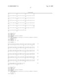 | 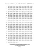 |
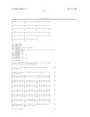 | 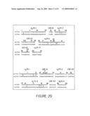 |
 | 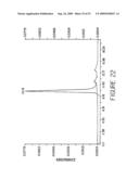 |
 |  |
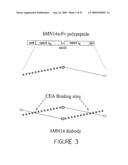 | 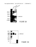 |
 | 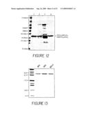 |
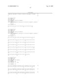 | 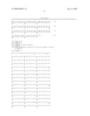 |
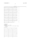 | 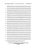 |
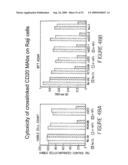 | 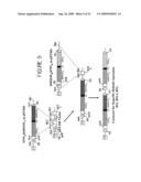 |
 | 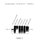 |
 |  |
 | 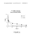 |
 |  |
 | 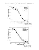 |
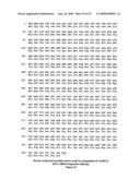 | 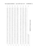 |
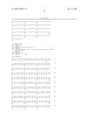 | 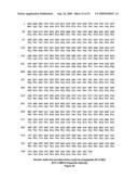 |
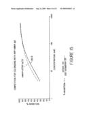 | 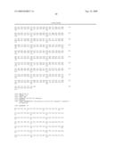 |
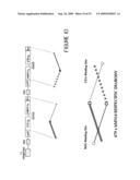 | 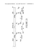 |
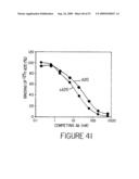 |  |
 |  |
 |  |
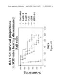 |  |
 |  |
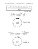 | 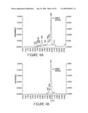 |
 | 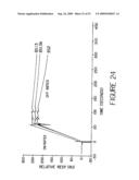 |
 | 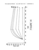 |
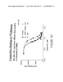 | 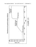 |
 | 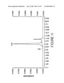 |
 |  |
 | 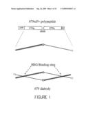 |
 |  |
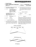 |  |
 |  |
 |  |
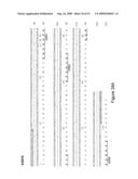 | 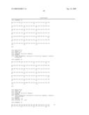 |
 | 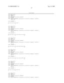 |
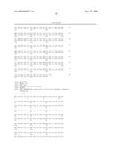 | 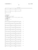 |
 | 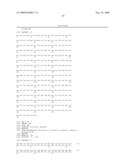 |
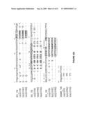 | 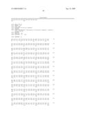 |
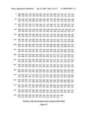 | 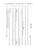 |
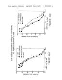 | 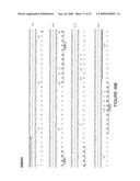 |
 |  |
 |  |
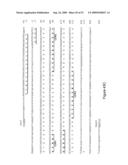 | 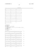 |
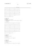 | 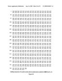 |
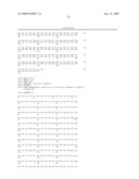 | 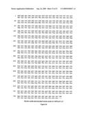 |
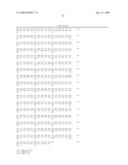 | 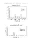 |
 |  |
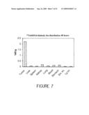 |  |
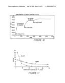 |  |
 | 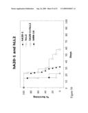 |
 |  |
 |  |
 |
| Similar patent applications: | |
| Date | Title |
|---|---|
| 2013-10-17 | Humanized antibodies against cxcr3 |
| 2010-07-29 | Antibodies to human erbb4 |
| 2009-11-12 | Rigidized trimethine cyanine dyes |
| 2013-07-18 | Super-humanized antibodies |
| 2013-10-10 | Antibodies and diagnostics |
| New patent applications in this class: | |
| Date | Title |
|---|---|
| 2022-05-05 | Bispecific antibodies that bind pd-l1 and cd28 |
| 2022-05-05 | Polypeptide modification method for purifying polypeptide multimers |
| 2019-05-16 | Generating targeted sequence diversity in fusion proteins |
| 2019-05-16 | Anti-pd-1 monoclonal antibody and obtaining method therefor |
| 2019-05-16 | Modified chimeric antigen receptors and methods of use |
| Top Inventors for class "Chemistry: natural resins or derivatives; peptides or proteins; lignins or reaction products thereof" | |
| Rank | Inventor's name |
|---|---|
| 1 | Kevin I. Segall |
| 2 | Martin Schweizer |
| 3 | John R. Desjarlais |
| 4 | Brent E. Green |
| 5 | David M. Goldenberg |