Patent application title: Composition and Method For Enhancing Immune Response
Inventors:
Tsafrir S. Mor (Tempe, AZ, US)
Nobuyuki Matoba (Tempe, AZ, US)
Charles J. Arntzen (Superstition Mountain, AZ, US)
IPC8 Class: AA61K3900FI
USPC Class:
4241921
Class name: Drug, bio-affecting and body treating compositions antigen, epitope, or other immunospecific immunoeffector (e.g., immunospecific vaccine, immunospecific stimulator of cell-mediated immunity, immunospecific tolerogen, immunospecific immunosuppressor, etc.) fusion protein or fusion polypeptide (i.e., expression product of gene fusion)
Publication date: 2009-05-07
Patent application number: 20090117145
Claims:
1-13. (canceled)
14. A method for delivering a cargo protein to an animal cell, comprising: providing a fusion protein comprising a cargo protein linked to a peptide selected from the group SEQ ID NO: 1, SEQ ID NO: 3, SEQ ID NO: 4, SEQ ID NO: 5, SEQ ID NO: 6, SEQ ID NO: 7; and administering the fusion protein to the animal.
15-16. (canceled)
17. The method of claim 14, wherein the cargo protein is an antigen.
18. The method of claim 17, wherein the fusion protein presents the antigen to the immune system of the animal.
19. The method of claim 17, wherein the antigen is a cholera toxin.
20. The method of claim 14, wherein the fusion protein is encoded by a DNA sequence capable of being incorporated into a viral DNA vector.
22-28. (canceled)
Description:
CLAIM TO DOMESTIC PRIORITY
[0001]This application is a divisional application of currently pending application Ser. No. 10/506,796, filed Sep. 3, 2004, which is a U.S. National Stage Application filed under 35 U.S.C. 371 claiming priority from the International Application No. PCT/US03/07073, filed Mar. 6, 2003, which claims the benefit of U.S. Provisional Patent Application Ser. No. 60/362,247, filed Mar. 6, 2002, and which applications are incorporated herein by reference.
FIELD OF THE INVENTION
[0002]The present invention relates generally to a composition and method for enhancing immune responses, and more specifically, to a composition and method using HIV-related peptides as an agent to increase immunogenic responses and for delivering fusion proteins to animal cells.
BACKGROUND OF THE INVENTION
[0003]Most currently available vaccines consist of killed or live-attenuated pathogens delivered by injection. Despite their success in preventing disease, compelling conceptual, technical and economical reasons exist to seek alternatives to traditional "Jennerian" vaccines.
[0004]Vaccines delivered parenterally require injections that must be given by medically trained personnel. Additionally, injection risks possible transmission of infection. Finally, parenteral delivery of vaccines invokes a systemic response, but not a mucosal response.
[0005]Subunit vaccines, especially those vaccines that target the mucosal immune system, are viable, safe and effective alternatives. Muscosal vaccines require do not require injection, thus, risk of transmission of infection is minimal. Finally, mucosal vaccines elicit immune response both systemically and mucosally.
[0006]Additionally, recent breakthroughs suggest that vaccines can be produced in edible tissues of transgenic plants that can then be orally immunogenic. The concept of using transgenic plants as vectors for the production and delivery of edible vaccines has been previously demonstrated.
[0007]However, to be effective, mucosal subunit vaccines often need to be co-administered with an "adjuvant." An "adjuvant" is an immunostimulatory agent that would enhance the specific immune responses against the vaccine candidate.
[0008]Therefore, a need exists for an immunostimulatory, mucosally-active composition that can be used as a systemic, mucosal, or epidermal adjuvant.
BRIEF DESCRIPTION OF THE DRAWINGS
[0009]FIG. 1 depicts the structure of a human immunodeficiency virus (HIV);
[0010]FIG. 2 depicts the structure of an adjuvant according to one embodiment;
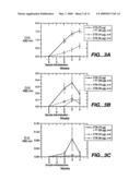
[0011]FIG. 3 illustrates an ELISA determination of anti-CTB antibodies following immunization by gavage;
[0012]FIG. 4 shows end point titers of anti-CTB antibodies;
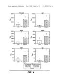
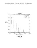
[0013]FIG. 5 illustrates reciprocal dilution of serum IgG1.
[0014]FIG. 6 illustrates subclass titers of total serum IgG, IgG1, IgG2a, IgG2b, IgG3 and IgA.
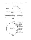
[0015]FIG. 7 is a flowchart illustrating immune response of Th1 and Th2.
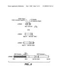
[0016]FIG. 8 depicts the synthesis of a plant-expression optimized DNA molecule encoding for the P1 peptide;
[0017]FIG. 9 depicts maps of plasmids comprised of DNA sequences of CTB, P1, CTB-P1 fusion, and for the plant-expression of the CTB-P1 fusion;
[0018]FIG. 10 is a flowchart illustrating the construction of a CTB-P1 fusion protein for plant-expression;
[0019]FIG. 11 depicts maps of plasmids for expression of CTB-P1 fusion protein and CTB in tomato;
[0020]FIG. 12 shows the expression of CTB-P1 fusion protein in E. Coli cells; and
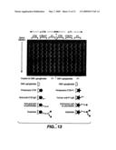
[0021]FIG. 13 illustrates an ELISA detection of anti-CTB and anti-P1 in E. Coli cells.
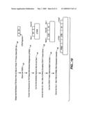
SUMMARY OF THE INVENTION
[0022]The present invention provides a composition and method for enhancing immune response in living organisms, for example, in humans. In one embodiment, and by way of example only, the composition includes, a peptide that when administered to a living organism, enhances the organism's immune response. The composition may also include an antigen, for example, a cholera toxin. The composition may further include the peptide and the antigen together as a fusion protein. The adjuvant peptide may function as a systemic, mucosal or epidermal adjuvant.
[0023]In another exemplary embodiment, the adjuvant peptide may be encoded by a genetically-modified living cell. The genetically-modified living cell may also encode an antigen. The peptide and antigen may also be encoded as a fusion protein.
[0024]Other independent features and advantages of the method for decreasing nicotine use in living organisms will become apparent from the following detailed description, taken in conjunction with the accompanying drawings which illustrate, by way of example, the principles of the invention.
DETAILED DESCRIPTION OF VARIOUS EMBODIMENTS
[0025]This description discloses a composition and method for enhancing immune response in living organisms by administering an oral, mucosal or epidermal adjuvant comprised of one or more HIV-related peptides.
[0026]FIG. 1 depicts the structure of an HIV retrovirus. HIV retrovirus 10 is an enveloped retrovirus. HIV retrovirus 10 is comprised of a viral membrane 12, ampiphilic regions 14, charged helices 16, calcium (Ca2+) binding sites 18, gp41 subunits 20, and gp120 subunits 22. Adjuvant peptide 24 facilitates HIV transcytosis across muscosal barriers toward the serosal environment by binding to galactosyl ceramide (GalCer) on the surface of mucosal epithelial cells.
[0027]Adjuvant peptide 24 comprises 36 amino acids (SEQ. ID. NO: 1) corresponds to a portion of the gp41 envelope. This peptide includes a conserved epitope (SEQ. ID. NO: 2), which is recognized by the neutralizing human IgG 2F5 and secretory IgAs that functionally neutralize HIV transcytosis through epithelial cells. The conserved aromatic residues are important for GalCer binding.
[0028]FIG. 2 depicts the structure of an adjuvant 30 according to one embodiment. Adjuvant 30 comprises peptides 32, linkers 34, and cargo proteins 36. However, an alternate embodiments envisions adjuvant 30 comprising at least peptides 32, but not necessarily linkers 34 and cargo proteins 36. Peptides 32 may comprise adjuvant peptides as one or more portions of P1 peptides, P5 peptides, or their functional equivalents. In one embodiment, cargo protein 36 is an antigen, for example cholera toxin. In an alternate embodiment, cargo protein 36 is any protein to be delivered to an animal cell.
[0029]According to one embodiment, an adjuvant peptide is a portion of the P1 peptide, HIV envelope protein gp41, which includes the conserved epitope, lectin binding site (SEQ. ID. NO: 2). According to an alternate embodiment, the adjuvant peptide is a portion of the P5 peptide, HIV envelope protein gp41 which includes the P1 peptide and a calcium binding site (residue number 622-684). P1 and P5 peptides also include their functional equivalents.
[0030]Functional equivalents of adjuvant peptides include peptides or portions of larger proteins with overall sequence or structural similarity to P1 or P5 peptides, and their derivatives, which allow the functionality disclosed here, including, but not limited to, one or more of the following: the enhancing immune response, GalCer binding, binding to the surface of cells containing GalCer, endocytosis to such cells or transcytosis across a tight cell barrier.
[0031]Examples of functional equivalents include portions of variants of gp41 in naturally occurring strains of HIV or in laboratory-derived strains of HIV, including, but not limited to, site-directed mutated versions of the gp41 portion of the molecule. Specific, non-limiting, examples of functional equivalents are HIV-1 isolate MN clone V5 (SEQ. ID. NO: 4), HIV-1 isolate 593 clone (SEQ. ID. NO: 5), HIV-1 isolate 98BRRS012 (SEQ. ID. NO: 6), and HIV-1 isolate 19242v3.20 (SEQ. ID. NO: 7).
[0032]Adjuvant 30 is capable of mucosal administration. Mucosal administration includes oral, nasal, vaginal, or rectal administration. Adjuvant 30 is also capable of functioning as a systemic, mucosal, or epidermal adjuvant.
Example 1
[0033]Example 1 demonstrates that adjuvant peptide enhances immune responses against cholera toxin B subunit by mucosal co-administration of adjuvant peptide and cholera toxin B subunit. Synthetic adjuvant peptide (SEQ. ID. NO: 3) with a C-terminal CONH2, was synthesized by Eurogentec (Belgium) and by the Protein Chemistry Laboratory at Arizona State University. A cystine residue was added to the beginning of SEQ. ID. NO: 1 to allow for dimerization (residue 649). Cholera Toxin B (CTB) subunit was chosen for co-administration because it is non-toxic and it is a strong mucosal adjuvant. Additionally, CTB binds to GM1 ganglioside whereby being able to target the fused antigen to mucosa.
[0034]Synthetic adjuvant peptide 30 micrograms (μg), adjuvant peptide plus Cholera-Toxin B subunit (CTB) (30 and 70 μg, respectively), and CTB (70 μg) were given orally to CD1 female mice (6-7 weeks old) by a gastric feeding tube on day one, eight, and fifteen. The serum, fecal pellets and vaginal secretions were collected prior to and on the second, third and fourth weeks after the first administration. Levels of anti-adjuvant peptide and anti-CTB antibodies were determined by ELISA in each sample.
[0035]FIG. 3 illustrates an ELISA determination of anti-CTB antibodies following immunization by gavage of CTB (70 μg), CTB+P1 (70 μg and 30 μg, respectively) or P1 (30 μg). Mice were gavaged on days indicated by arrows and samples of serum (A), fecal (B), and vaginal (C) secretions were collected when indicated. Serum (A) detected systematic levels. Fecal (B) and vaginal (C) both detected mucosal levels.
[0036]Samples were serially diluted in phosphate buffered saline containing 0.05% Tween-20 (PBST) containing 1% nonfat dry milk. Plates were coated with CTB overnight at 4° C., blocked with PBST containing 5% nonfat dry milk and then incubated with samples. Antibodies were detected by horseradish peroxidase-conjugated secondary antiisotypic antisera against the appropriate mouse antibodies (rabbit anti-mouse total IgG from CalBiochem, and the following anti mouse antisera: Anti-IgG1, anti-IgG2a, anti-IgG2b, anti-IgG3 from Santa Cruz Biotechnology; and anti-IgA from Sigma. FIG. 3A-3C shown maximal dilutions that allowed quantification.
[0037]FIG. 4 illustrates end point of anti-CTB antibodies four weeks after immunization. Chemiluminescent ELISA was conducted as described in FIG. 3. Titers in FIG. 4 are defined as reciprocals of the highest dilution giving a positive A490 reading above 0.1. FIG. 5 illustrates, for example, reciprocal dilution of serum IgG1. FIG. 6 illustrates subclass titers of total IgG, IgG1, IgG2a, IgG2b, IgG3 and IgA.
[0038]While in FIG. 4 antibody titers were below detection levels, co-administration of P1 and CTB to mice resulted in significantly higher titers of anti-CTB antibodies as compared to mice that were given CTB alone. Specifically, in FIG. 3, the level of fecal and vaginal anti-CTB IgA in the second and third week and serum anti-CTB in the second, third and fourth week appeared to be higher in mice fed P1 with CTB than in mice fed only CTB. Moreover, as illustrated in FIG. 6, co-administration of P1 with CTB resulted in increasing all serum anti-CTB IgG subclass (IgG1, IgG2a, IgG2b, IgG3) titers by five to ten times in the fourth week, as compared to administration of CTB alone, as shown in FIG. 4.
[0039]Therefore, P1 peptide was shown to augment the production of mucosal IgA and serum IgG to co-administered CTB. Because CTB is a strong mucosal immunogen by itself, the increase of both anti-CTB IgG1 and IgG2a levels suggest that the immune enhancement effect of P1 peptide is attributable to activating both Th1 and Th2 response. Th1 and Th2 response is illustrated in FIG. 7. IgG2a 40 effects T1 response 38 through cell-mediated immunity, targeting intracellular pathogens 42. Antibodies 46, such as IgG1 and IgA, affect Th2 response 44 targeting extracellular parasites, viruses and bacteria 48. Secondly, P1 peptide did not induce antibody production against itself, even in the presence of CTB. Therefore, P1 peptide can be used a mucosal adjuvant to enhance immune response in living organisms.
Example 2
[0040]In example 2, plasmids were created for the co-expression of adjuvant peptide and GFP in transgenic plants for oral delivery. FIG. 8 depicts the synthesis of a plant-expression optimized DNA molecule encoding for an adjuvant peptide-GFP fusion protein. The sequence coding for adjuvant peptide was inserted behind a DNA spacer encoding a Glycine-Proline-Glycine-Proline (GPGP) hinge. A BsrGI-SacI fragment of this plasmid was cloned in behind a 35 S Promoter. A PstI-EcoRI fragment contains the plant expression cassette. FIG. 8 represents a model delivery system for using fusion proteins to deliver cargo proteins to an animal cell.
[0041]FIG. 9 depicts maps of plasmids comprised of DNA sequences of CTB, P1, CTB-P1 fusion and for the plant-expression of the CTB-P1 fusion protein. A plant-expression optimized DNA molecule encoding for P1 peptide was synthesized. The sequence was inserted behind a portion of the gene encoding the C-terminus of the CTB molecule behind a DNA spacer encoding a Glycine-Proline-Glycine-Proline (GPGP) hinge. An endoplasmic reticulum (ER) retention signal was engineered at the Carboxyl-Terminus. The PCR product was closed into the cloning vector TOPO2.1 (Invitrogen) to create pTM058.
[0042]Still referring to FIG. 9, a HindIII-SacI fragment of this plasmid was then cloned into pTM042 to create a gene encoding a Carbosyl-terminus fusion of CTB and the P1 peptide in the plasmid pTM065. A BspHI-SacI fragment of this plasmid was cloned into pIBT210.1 (Haq, et al. 1995) behind a CaMV35S promoter and the 5' UTR of Tobacco Etch Virus and in front of the 3' UTR of the soy bean vspB gene to form pTM066. A PstI-EcoRI fragment containing the plant expression cassette was cloned into the T1 plasmid derivative pGPTV-Kan (Becker, et al. 1992) to form pTM067 (not shown).
[0043]FIG. 10 is a flowchart illustrating the steps involved in creating a CTB-P1 fusion protein. In step 50, CTB (from HindIII site to the 3' end)-P1 fusion gene is designed and synthesized, a length of 234 base pairs (bp) (SEQ. ID. NO: 8). Next, in step 52, the CTB-P1 fusion gene is cloned into TOPO.
[0044]In step 54, the sequence is corrected by PCR-based site-directed mutagenesis to form pTM58 (pTM058). In step 56, the synthetic gene is cut out by HindIII and Sac I Then in step 58, the synthetic gene is cloned into HindIII-SacI site of pTM42 (pTM042). This represents the complete CTB-P1 fusion gene.
[0045]The CTB-P1 fusion gene is then cut out by BspHI and SacI in step 60. Finally, in step 62, the cut out CTB-P1 fusion gene is cloned into the NcoI-SacI site of pTM38. Thus, step 62 clones the CTB-P1 fusion gene into the plant expression cassette. The CTB-P1 fusion gene encodes the CTB-P1 fusion protein (SEQ. ID. NO: 9).
[0046]FIG. 11 illustrates an example for a construct for a potentiated edible vaccine. In step 80, pTM086 containing the CTB-P1 fusion gene and the plant expression cassette was cloned into the T1 plasmid derivative pGPTV-Kan (Becker, et al. 1992). The plasmid is then transformed into Agrobacterium (LBA4404) in step 82. Finally, in step 84, the Agrobacterium is transformed into a tomato, for example MicroTom, cotyledon and hypocotyl explants.
[0047]In this example, the target organism for the adjuvant includes, but is not limited to, Vibrio cholerae, enterotoxigenic Escherchia coli. Other examples would include virus-like particles, for example, Norwalk virus capsid, and antigenic determinants of other pathogens, for example, bacterial, viral or parasitic.
Example 3
[0048]The flowchart in FIG. 12 depicts purification protocol of CTB-P1 fusion protein produced in E. coli. Resultant fractions from this protocol were resolved by SDS PAGE on the right panel. The following were placed in the first three lanes: Lane 1: molecular weight standards; Lane 2: a mixture of denatured (lower monomeric band) and non-denatured commercially available CT-B (pentameric upper band); Lane 3: whole cell extract from an IPTG-induced E. coli.
[0049]Following sonication and centrifugations, in step 70, extracts are separated into soluble (Lane 4) and insoluble (lane 5) fraction. The insoluble fraction, in step 72, is solubilized in 6.5 M urea and affinity purification on nickel column in step 74. The eluate (Lane 6) is more than 90% pure and can be subjected to dialysis promoting the refolding and oligomerization of the monomeric CTB-P1 fusion protein. By its mobility we conclude that the fusion protein can assemble into pentamers.
[0050]Finally, Eluate was dialyzed against PBS in step 76, and the purified, refolded CTB-P1 is shown in Lane 7. As noted in Lane 7, a CTB-P1 pentameter was produced. Additionally, a CTB-P1 monomer with an intramolecular disulfide bond was also produced.
[0051]FIG. 13 demonstrates that the pentameric nature of the fusion protein allows it to bind to GM1 gangliosides. The ELISA plate was coated with GM1-ganglioside. On the left half of the plate, anti-CTB was used for detection. On the left half of the plate, anti-P1 was used for detection. CTB, CTB-P1 and P1 samples were applied to the plate as shown. The CTB is commercially available preparation of CTB and P1. CTB-P1 and P1 synthetic peptide are refolded samples purified as explained in FIG. 12. Anti-CTB and anti-P1 are CTB- and P1-specific antibodies, respectively. These results demonstrate that the fusion is both able to retain its pentameric structure as well as its P1 epitope.
[0052]Various embodiments of the invention are described above in the Drawings and Description of Various Embodiments. While these descriptions directly describe the above embodiments, it is understood that those skilled in the art may conceive modifications and/or variations to the specific embodiments shown and described herein. Any such modifications or variations that fall within the purview of this description are intended to be included therein as well. Unless specifically noted, it is the intention of the inventor that the words and phrases in the specification and claims be given the ordinary and accustomed meanings to those of ordinary skill in the applicable art(s). The foregoing description of a preferred embodiment and best mode of the invention known to the applicant at the time of filing the application has been presented and is intended for the purposes of illustration and description. It is not intended to be exhaustive or to limit the invention to the precise form disclosed, and many modifications and variations are possible in the light of the above teachings. The embodiment was chosen and described in order to best explain the principles of the invention and its practical application and to enable others skilled in the art to best utilize the invention in various embodiments and with various modifications as are suited to the particular use contemplated. Therefore, it is intended that the invention not be limited to the particular embodiments disclosed for carrying out this invention, but that the invention will include all embodiments falling within the scope of the appended claims.
Sequence CWU
1
9135PRTHuman immunodeficiency virus type 1PEPTIDE(1)..(35)HIV-1 gp41
peptide portion (residues 650-685) 1Ser Gln Thr Gln Gln Glu Lys Asn Glu
Gln Glu Leu Leu Glu Leu Asp1 5 10
15Lys Trp Ala Ser Leu Trp Asn Trp Phe Asp Ile Thr Asn Trp Leu
Trp 20 25 30Tyr Ile Lys
3526PRTHuman immunodeficiency virus type 1PEPTIDE(1)..(6)HIV-1 gp41
peptide portion (residues 663-668) 2Glu Leu Asp Lys Trp Ala1
5336PRTHuman immunodeficiency virus type 1 3Cys Ser Gln Thr Gln Gln Glu
Lys Asn Glu Gln Glu Leu Leu Glu Leu1 5 10
15Asp Lys Trp Ala Ser Leu Trp Asn Trp Phe Asp Ile Thr
Asn Trp Leu 20 25 30Trp Tyr
Ile Lys 35436PRTHuman immunodeficiency virus type
1PEPTIDE(1)..(35)HIV-1 isolate MN clone v5 (residues 649-685) 4Ser Gln
Thr Gln Gln Glu Lys Asn Glu Gln Glu Leu Leu Gly Leu Asp1 5
10 15Lys Trp Glu Ser Leu Trp Asn Trp
Phe Asp Ile Thr Asn Trp Leu Trp 20 25
30Tyr Ile Lys Ile 35536PRTHuman immunodeficiency virus
type 1PEPTIDE(1)..(36)HIV-1 isolate 593 clone (residues 649-685) 5Ser Gln
Asn Gln Gln Glu Lys Asn Glu Gln Glu Leu Leu Glu Leu Asp1 5
10 15Lys Trp Ala Gly Leu Trp Asn Trp
Phe Glu Ile Thr Asn Trp Leu Trp 20 25
30Tyr Ile Lys Ile 35636PRTHuman immunodeficiency virus
type 1PEPTIDE(1)..(36)HIV-1 isolate 98BRRS012 (residues 649-685) 6Ser Gln
Asn Gln Gln Glu Lys Asn Glu His Glu Leu Leu Glu Leu Asp1 5
10 15Lys Trp Ala Asn Leu Trp Asn Trp
Phe Asp Ile Thr Asn Trp Leu Trp 20 25
30Tyr Ile Lys Ile 35736PRTHuman immunodeficiency virus
type 1PEPTIDE(1)..(36)HIV-1 isolate 1924v3.20 (residues 649-685) 7Ser Gln
Asn Gln Gln Glu Lys Asn Glu Gln Asp Leu Leu Glu Leu Asp1 5
10 15Lys Trp Ala Ser Leu Trp Asn Trp
Phe Asp Ile Ser Asn Trp Leu Trp 20 25
30Tyr Ile Lys Ile 358522DNAArtificial SequenceDescription
of Artificial Sequence; note = synthetic construct 8ccatggctat
caagctcaag tttggagtgt tcttcactgt gctccttagc tctgcctatg 60cacatggcac
cccacaaaac atcactgact tgtgtgctga gtaccacaac acccaaatcc 120acaaccctca
atgacaagat ctttagctac accgagagcc ttgctggcaa gagggagatg 180gctatcatcc
cttcaagaat ggtgctacct tccaagtgga ggtgcctgga agccaacaca 240ttgatagcca
aaagaaggcc attgagagga tgaaggacac attaggatag cttacctcac 300tgaggctaag
gtggagaagc tttgtgtgtg gaacaacaag actccacatg ctattgctgc 360cattagcatg
gcaaatggtc ctggaccttc ccaaacccaa caagagaaga atgagcaaga 420gcttttggag
ttggacaagt ggcaagcctt tggaattggt ttgacatcac caattggctt 480tggtatatca
agatctctga gaaggatgaa ctctaagagc tc
5229171PRTArtificial SequenceDescription of Artificial Sequence; note =
synthetic construct 9Met Ala Ile Lys Leu Lys Phe Gly Val Phe Phe Thr
Val Leu Leu Ser1 5 10
15Ser Ala Trp Ala His Gly Thr Pro Gln Asn Ile Thr Asp Leu Cys Ala
20 25 30Glu Tyr His Asn Thr Gln Ile
His Thr Leu Asn Asp Lys Ile Phe Ser 35 40
45Tyr Thr Glu Ser Leu Ala Gly Lys Arg Glu Met Ala Ile Ile Thr
Phe 50 55 60Lys Asn Gly Ala Thr Phe
Gln Val Glu Val Pro Gly Ser Gln His Ile65 70
75 80Asp Ser Gln Lys Lys Ala Ile Glu Arg Met Lys
Asp Thr Leu Arg Ile 85 90
95Ala Thr Leu Thr Glu Ala Lys Val Glu Lys Leu Cys Val Trp Asn Asn
100 105 110Lys Thr Pro His Ala Ile
Ala Ala Ile Ser Met Ala Asn Gly Pro Gly 115 120
125Pro Ser Gln Thr Gln Gln Glu Lys Asn Glu Gln Glu Leu Leu
Glu Leu 130 135 140Asp Lys Trp Ala Ser
Leu Trp Asn Trp Phe Asp Ile Thr Asn Trp Leu145 150
155 160Trp Tyr Ile Lys Ile Ser Glu Lys Asp Glu
Leu 165 170
User Contributions:
Comment about this patent or add new information about this topic:
 |  |
 |  |
 |  |
 |  |
 |  |
 |  |
 |  |
| Similar patent applications: | |
| Date | Title |
|---|---|
| 2011-03-24 | Method of making flexible bioresorbable hemostatic packing and stent having a preselectable in-vivo residence time |
| 2011-03-10 | Polypeptides involved in immune response |
| 2011-03-17 | Composition for treating skin lesions |
| 2011-03-24 | Substrate comprising a lotion composition limiting the adherence of feces or menses to the skin |
| 2011-02-24 | Modulation of the immune response |
| New patent applications in this class: | |
| Date | Title |
|---|---|
| 2017-08-17 | Synthetic active peptide fragments |
| 2016-12-29 | Kidney-specific tumor vaccine directed against kidney tumor antigen g-250 |
| 2016-06-16 | Combination vaccine |
| 2016-05-19 | Recombinant fusion antigen gene, recombinant fusion antigen protein and subunit vaccine composition having the same against infection of porcine reproductive and respiratory syndrome virus |
| 2016-05-05 | Semi-live respiratory syncytial virus vaccine |
| New patent applications from these inventors: | |
| Date | Title |
|---|---|
| 2009-11-05 | Production and use of human butyrylcholinesterase |
| Top Inventors for class "Drug, bio-affecting and body treating compositions" | |
| Rank | Inventor's name |
|---|---|
| 1 | David M. Goldenberg |
| 2 | Hy Si Bui |
| 3 | Lowell L. Wood, Jr. |
| 4 | Roderick A. Hyde |
| 5 | Yat Sun Or |