Patent application title: ASBESTOS DETECTION METHOD, ASBESTOS DETECTION AGENT, ASBESTOS DETECTION KIT, METHOD FOR SCREENING CANDIDATE FOR AGENT AIMING AT PREVENTING OR TREATING DISEASE FOR WHICH ASBESTOS IS CAUSATIVE OR WORSENING FACTOR
Inventors:
Akio Kuroda (Hiroshima, JP)
Akio Kuroda (Hiroshima, JP)
Kazutaka Nomura (Hiroshima, JP)
Assignees:
NATIONAL UNIVERISTY OF CORPORATION HIROSHIMA UNIVERSITY
IPC8 Class: AG01N3353FI
USPC Class:
435 79
Class name: Measuring or testing process involving enzymes or micro-organisms; composition or test strip therefore; processes of forming such composition or test strip involving antigen-antibody binding, specific binding protein assay or specific ligand-receptor binding assay assay in which an enzyme present is a label
Publication date: 2009-04-16
Patent application number: 20090098578
Inventors list |
Agents list |
Assignees list |
List by place |
Classification tree browser |
Top 100 Inventors |
Top 100 Agents |
Top 100 Assignees |
Usenet FAQ Index |
Documents |
Other FAQs |
Patent application title: ASBESTOS DETECTION METHOD, ASBESTOS DETECTION AGENT, ASBESTOS DETECTION KIT, METHOD FOR SCREENING CANDIDATE FOR AGENT AIMING AT PREVENTING OR TREATING DISEASE FOR WHICH ASBESTOS IS CAUSATIVE OR WORSENING FACTOR
Inventors:
Akio Kuroda
Kazutaka Nomura
Agents:
MORRISON & FOERSTER LLP
Assignees:
NATIONAL UNIVERISTY OF CORPORATION HIROSHIMA UNIVERSITY
Origin: SAN FRANCISCO, CA US
IPC8 Class: AG01N3353FI
USPC Class:
435 79
Abstract:
The present invention provides a prompt and easy asbestos detection method
and a method for screening a candidate for an agent aiming at preventing
or treating a disease for which asbestos is a causative or worsening
factor. It is possible to quickly and easily detect asbestos in a sample
by finding a protein capable of binding specifically to asbestos,
allowing the protein or a fusion protein of the protein and a reporter
protein to bind to asbestos in the sample, and then detecting the protein
or the fusion protein having been bound to asbestos. A substance
inhibiting the binding of actin to asbestos, which has been found out as
a protein capable of binding specifically to asbestos, is a candidate for
an agent aiming at preventing or treating a disease for which asbestos is
a causative or worsening factor.Claims:
1. An asbestos detection method comprising:the step of bringing a protein
capable of binding to asbestos in a solution containing at least 0.1 M or
more sodium chloride into contact with asbestos in a sample; andthe step
of detecting the protein binding to the asbestos.
2. An asbestos detection method comprising:the step of obtaining a fusion protein of (i) a protein capable of binding to asbestos in a solution containing at least 0.1 M or more sodium chloride and (ii) a reporter protein;the step of bringing the obtained fusion protein into contact with asbestos in a sample; andthe step of detecting the fusion protein binding to the asbestos.
3. The asbestos detection method according to claim 1, whereinthe protein capable of biding to asbestos in the solution containing at least 0.1 M or more sodium chloride is at least one type of protein selected from: a protein with an amino acid sequence represented by SEQ ID NO: 1, 3, 5, 7, 9, 11, 13, 15, 17, 19, 21, 23, or 25; and a protein with amino acid sequence having deletion, substitution, or addition of one or several amino acids in an amino acid sequence represented by SEQ ID NO: 1, 3, 5, 7, 9, 11, 13, 15, 17, 19, 21, 23, and 25.
4. The asbestos detection method according to claim 1, whereinthe protein capable of binding to asbestos in the solution containing at least 0.1 M or more sodium chloride is actin.
5. The asbestos detection method according to claim 2, whereinthe reporter protein is a protein selected form fluorescent protein, luciferase, alkaline phosphatase, beta galactosidase, diaphorase, and peroxidase.
6. An asbestos detection agent containing a protein capable of binding to asbestos in a solution containing at least 0.1 M or more sodium chloride.
7. An asbestos detection agent containing a fusion protein of (i) a protein capable of binding to asbestos in a solution containing at least 0.1 M or more sodium chloride and (ii) a reporter protein.
8. The asbestos detection agent according to claim 6, whereinthe protein capable of biding to asbestos in the solution containing at least 0.1 M or more sodium chloride is at least one type of protein selected from: a protein with an amino acid sequence represented by SEQ ID NO: 1, 3, 5, 7, 9, 11, 13, 15, 17, 19, 21, 23, or 25; and a protein with amino acid sequence having deletion, substitution, or addition of one or several amino acids in an amino acid sequence represented by SEQ ID NO: 1, 3, 5, 7, 9, 11, 13, 15, 17, 19, 21, 23, and 25.
9. The asbestos detection agent according to claim 6, whereinthe protein capable of binding to asbestos in the solution containing at least 0.1 M or more sodium chloride is actin.
10. The asbestos detection agent according to claim 7, whereinthe reporter protein is a protein selected form fluorescent protein, luciferase, alkaline phosphatase, beta galactosidase, diaphorase, and peroxidase.
11. An asbestos detection kit comprising an asbestos detection agent according to claim 6.
12. A screening method for screening a candidate for an agent aiming at preventing or treating a disease for which asbestos is a causative or worsening factor, the method comprising:the step of bringing a test substance, asbestos, and actin into contact with each other in a solution containing at least 0.1 M or more sodium chloride; andthe step of measuring binding level of the asbestos and the actin in the solution.
13. The asbestos detection method according to claim 2, whereinthe protein capable of biding to asbestos in the solution containing at least 0.1 M or more sodium chloride is at least one type of protein selected from: a protein with an amino acid sequence represented by SEQ ID NO: 1, 3, 5, 7, 9, 11, 13, 15, 17, 19, 21, 23, or 25; and a protein with amino acid sequence having deletion, substitution, or addition of one or several amino acids in an amino acid sequence represented by SEQ ID NO: 1, 3, 5, 7, 9, 11, 13, 15, 17, 19, 21, 23, and 25.
14. The asbestos detection method according to claim 2, whereinthe protein capable of binding to asbestos in the solution containing at least 0.1 M or more sodium chloride is actin.
15. The asbestos detection agent according to claim 7, whereinthe protein capable of biding to asbestos in the solution containing at least 0.1 M or more sodium chloride is at least one type of protein selected from: a protein with an amino acid sequence represented by SEQ ID NO: 1, 3, 5, 7, 9, 11, 13, 15, 17, 19, 21, 23, or 25; and a protein with amino acid sequence having deletion, substitution, or addition of one or several amino acids in an amino acid sequence represented by SEQ ID NO: 1, 3, 5, 7, 9, 11, 13, 15, 17, 19, 21, 23, and 25.
16. The asbestos detection agent according to claim 7, whereinthe protein capable of binding to asbestos in the solution containing at least 0.1 M or more sodium chloride is actin.
17. An asbestos detection kit comprising an asbestos detection agent according to claim 7.
18. An asbestos detection kit comprising an asbestos detection agent according to claim 8.
19. An asbestos detection kit comprising an asbestos detection agent according to claim 9.
20. An asbestos detection kit comprising an asbestos detection agent according to claim 10.
Description:
TECHNICAL FIELD
[0001]The present invention relates to a method for detecting asbestos in a sample by using a protein capable of binding specifically to asbestos, an asbestos detection agent containing the protein, an asbestos detection kit including the asbestos detection agent, and a method for screening a candidate for an agent aiming at preventing or treating a disease for which asbestos is a causative or worsening factor, by using the protein.
BACKGROUND ART
[0002]Recently, asbestos (fibrous silicate) has become a problem since it has adverse effects on human body. Specifically, a company has disclosed to the public that workers who have been involved in producing asbestos or handling asbestos develop health problems such as lung cancer and mesothelioma at a high incidence rate. In addition, it has been reported that inhalation of asbestos powder dust may cause health problems mainly including asbestosis, lung cancer, and malignant mesothelioma.
[0003]Asbestosis is a kind of pulmonary fibrosis (pneumonoconiosis), which is a disease that causes fibrosing lung. There are many factors that cause the fibrosing lung, including other mineral powder dust. However, pulmonary fibrosis caused by exposure to asbestos is classified especially into asbestosis. Lung cancer is developed mainly by physical stimulation of asbestos fibers taken up in alveoli of the lung. The level of carcinogenicity of asbestos varies depending upon the type, thickness, and length, of asbestos. Malignant mesothelioma is a malignant tumor of a pleura surrounding lung, peritoneum surrounding organs such as liver and stomach, and the like part.
[0004]Examples of asbestos include chrysotile (white asbestos), crocidolite (blue asbestos), amosite (brown asbestos), anthophylite, toremolite, and actinolite. More than 90% of the use of asbestos is for building materials. In other examples, asbestos is used for sealing materials for chemical plant setup, and industrial products such as friction materials. Manufacture and handling of building materials, friction materials, and adhesives containing asbestos have been banned since Oct. 1, 2004. However, there were circumstances where a large amount of asbestos was used in the past, and asbestos has remained in many buildings.
[0005]Detection of asbestos is performed by the following method. That is, for the detection of asbestos in the air, asbestos is trapped by a filter when the air is taken in by a pump, and the filter is subjected to achromatization by using acetone to observe the filter through a phase microscope. For the detection of asbestos in building material, a sample is collected by appropriate amount from a building material to be analyzed, and the collected sample is subjected to grinding, pulverization, heating, and/or other treatments according to the form of the building material and the state of a substance that coexists with the building material to prepare a sample to be analyzed. Next, the sample to be analyzed is subjected to qualitative analysis by disperse dyeing analysis method through a phase microscope and qualitative analysis by X-ray diffraction analysis so as to determine whether the sample contains asbestos. The sample which has been identified as the one containing asbestos is treated with formic acid to prepare a sample to be subjected to quantitative analysis. Then, the sample is subjected to quantitative analysis by X-ray diffraction analysis method that uses base standard absorption correction method to find the amount of asbestos contained and calculate the percentage of asbestos content (see Non-Patent Documents 1 and 2).
[0006]However, observation through a phase microscope requires advanced skills and requires a considerable time. Therefore, it is difficult to perform many operations at the same time. Besides, X-ray analyzer is very expensive and cannot be used easily by anyone.
[0007][Non-Patent Document 1]
[0008]Working Environment measurement Series No. 3: Manual of Fibrous Substance Measurement, Japan Association for Working Environment Measurement, Jul. 28, 2004
[0009][Non-Patent Document 2]
[0010]Method of Analyzing Asbestos Content in Building Material, Kiankahatsu (Ministry of Health, Labour, and Welfare, Labour Standards Bureau, Industrial Safety and Health Department, Chemical Hazards Control Division) No. 0622001 , Jun. 22, 2005
DISCLOSURE OF INVENTION
[0011]As described previously, it is revealed that asbestos is a causative or worsening factor. However, mechanism of induction of the above diseases by asbestos has been hardly clarified, and research on the treatment of diseases induced by asbestos has hardly proceeded. Under such circumstances, there has been a growing demand from society as a whole for the development of a method by which asbestos can be detected promptly and easily.
[0012]If a protein capable of binding specifically to asbestos is found out, it is very useful for prompt and easy asbestos detection, and is very significant in the filed of public sanitation. In addition, if it was found out that a protein capable of binding specifically to asbestos which protein exists in a living body, particularly respiratory organs such as lung of a mammal, such finding makes a significant contribution to the clarification of the mechanism of induction of the diseases by asbestos, and it is expected that inhibition of binding of protein to asbestos leads to prevention or treatment for diseases induced by asbestos.
[0013]However, the existence of a protein capable of binding specifically to asbestos was not reported in the past.
[0014]The present invention has been attained in view of the above problems, and an object of the present invention is to find out a protein capable of binding specifically to asbestos, realize a prompt and easy asbestos detection method, find out a protein capable of binding specifically to asbestos in respiratory organs of a mammal, and provide a method for screening a candidate for an agent aiming at preventing or treating a disease for which asbestos is a causative or worsening factor.
[0015]In order to solve the above problems, the inventors of the present invention found out a protein capable of binding to asbestos from among various kinds of bacterium-derived proteins, found out that it is possible to promptly and easily detect asbestos by using a fusion protein of such a protein and a reporter protein. In addition, the inventors of the present invention accomplished the present invention by finding out a protein capable of binding to asbestos from among proteins that exist in mouse lung, and identifying the protein as actin.
[0016]That is, an asbestos detection method according to the present invention includes: the step of bringing a protein capable of binding to asbestos in a solution containing at least 0.1 M or more sodium chloride into contact with asbestos in a sample; and the step of detecting the protein binding to the asbestos.
[0017]Further, an asbestos detection method according to the present invention includes: the step of obtaining a fusion protein of (i) a protein capable of binding to asbestos in a solution containing at least 0.1 M or more sodium chloride and (ii) a reporter protein; the step of bringing the obtained fusion protein into contact with asbestos in a sample; and the step of detecting the fusion protein binding to the asbestos.
[0018]An asbestos detection agent according to the present invention contains a protein capable of binding to asbestos in a solution containing at least 0.1 M or more sodium chloride.
[0019]Further, an asbestos detection agent according to the present invention contains a fusion protein of (i) a protein capable of binding to asbestos in a solution containing at least 0.1 M or more sodium chloride and (ii) a reporter protein.
[0020]The asbestos detection method and the asbestos detection agent according to the present invention are preferably such that the protein capable of biding to asbestos in the solution containing at least 0.1 M or more sodium chloride is at least one type of protein selected from: a protein with an amino acid sequence represented by SEQ ID NO: 1, 3, 5, 7, 9, 11, 13, 15, 17, 19, 21, 23, or 25; and a protein with amino acid sequence having deletion, substitution, or addition of one or several amino acids in an amino acid sequence represented by SEQ ID NO: 1, 3, 5, 7, 9, 11, 13, 15, 17, 19, 21, 23, and 25.
[0021]The asbestos detection method and the asbestos detection agent according to the present invention are preferably such that the protein capable of binding to asbestos in the solution containing at least 0.1 M or more sodium chloride is actin.
[0022]The asbestos detection method and the asbestos detection agent according to the present invention are preferably such that the reporter protein is a protein selected form fluorescent protein, luciferase, alkaline phosphatase, beta galactosidase, diaphorase, and peroxidase.
[0023]An asbestos detection kit according to the present invention includes the asbestos detection agent according to the present invention.
[0024]A screening method according to the present invention is a screening method for screening a candidate for an agent aiming at preventing or treating a disease for which asbestos is a causative or worsening factor, the method including: the step of bringing a test substance, asbestos, and actin into contact with each other in a solution containing at least 0.1 M or more sodium chloride; and the step of measuring binding level of the asbestos and the actin in the solution.
[0025]With the asbestos detection method and the asbestos detection agent according to the present invention, it is possible to promptly and easily detect the presence or absence of asbestos in a sample. By using a biological sample, the present invention can be applied to diagnosis for asbestos-related health problems.
[0026]With the screening method according to the present invention, it is possible to promptly and efficiently screen a candidate for an agent aiming at preventing or treating a disease for which asbestos is a causative or worsening factor.
[0027]Additional objects, features, and strengths of the present invention will be made clear by the description below. Further, the advantages of the present invention will be evident from the following explanation in reference to the drawings.
BRIEF DESCRIPTION OF DRAWINGS
[0028]FIG. 1 is an electrophoretogram showing that an asbestos (chrysotile) binding protein was obtained from a cell lysate of Escherichia coli K12.
[0029]FIG. 2 is an electrophoretogram showing that an asbestos (chrysotile) binding protein was obtained from a cell lysate of Pseudomonas putida KT2440.
[0030]FIG. 3 is an electrophoretogram showing that an asbestos (chrysotile) binding protein was obtained from a cell lysate of Corynebacterium glutamicum ATCC13032.
[0031]FIG. 4(a) is an electrophoretogram showing that AP protein binds to asbestos (chrysotile).
[0032]FIG. 4(b) is an electrophoretogram showing that DksA-AP fusion protein tightly binds to asbestos (chrysotile).
[0033]FIG. 5 is a view showing that asbestos (chrysotile) was detected by coloring with the use of DksA-AP fusion protein.
[0034]FIG. 6 is a view showing the result of measurement of detection sensitivities in a system of detecting asbestos (chrysotile) by coloring with the use of DksA-AP fusion protein.
[0035]FIG. 7 is a view showing the result of asbestos (chrysotile) detection by using DksA-AP fusion protein and a filter.
[0036]FIG. 8 is an electrophoretogram showing that an asbestos (chrysotile) binding protein was obtained from a mouse lung protein sample.
[0037]FIG. 9 is an electrophoretogram showing that an asbestos (crocidolite/amosite) binding protein was obtained from a cell lysate of Escherichia coli K12.
[0038]FIG. 10 is an electrophoretogram showing that an asbestos (crocidolite/amosite) binding protein was obtained from a cell lysate of highly thermophilic bacterium HB27.
[0039]FIG. 11 is a view showing the result of asbestos (amosite) detection by using TTC0984-AP fusion protein and a filter.
[0040]FIG. 12 is a photograph of three types of building materials used as samples in Example 4.
[0041]FIG. 13 is a view showing the result of detection of asbestos in building materials.
[0042]FIG. 14 is a photograph of rock wool used as a sample in Example 5.
[0043]FIG. 15 is a view showing the result of detection of asbestos contained in rock wool.
[0044]FIG. 16 is a view showing the result of detection of asbestos contained in rock wool on the slide glass.
[0045]FIG. 17 is an image showing the result of detection of asbestos through a fluorescence microscope by using DksA-GFP fusion protein.
BEST MODE FOR CARRYING OUT THE INVENTION
Obtaining and Identification of Protein Binding to Asbestos
[0046]A protein used in the present invention is a protein capable of binding to asbestos in a solution containing at least 0.1 M or more sodium chloride. The "protein capable of binding to asbestos in a solution containing at least 0.1 M or more sodium chloride" is herein referred to as "protein binding to asbestos". The protein may be derived from, but are not limited to, bacterium, yeast, plant, animal, or any living organisms.
[0047]The type of the asbestos is not limited. The protein used in the present invention has only to be a protein capable of binding to any type of asbestos in a solution containing at least 0.1 M or more sodium chloride. For example, the "protein binding to asbestos" includes a protein capable of binding to chrysotile (white asbestos), not to crocidolite (blue asbestos), in a solution containing at least 0.1 M or more sodium chloride.
[0048]The word "protein" herein is used interchangeably with "polypeptide" or "peptide". The "protein" includes a fragment of a protein. Further, the "protein" includes a fusion protein. The "fusion protein" is a protein in which part (fragment) or whole of at least two heteroproteins are bound to each other.
[0049]For example, the protein binding to asbestos which protein used in the present invention can be obtained by the following method. However, this is not the only possibility. The protein capable of binding to asbestos in a solution containing at least 0.1 M or more sodium chloride can be suitably used in the present invention.
[0050]That is, the protein binding to asbestos which protein used in the present invention can be obtained by adding asbestos to a solution containing one or more types of proteins, collecting the asbestos, washing the collected asbestos in a solution containing at least 0.1 M or more sodium chloride, and then isolating the protein(s) binding to the asbestos even after the washing.
[0051]As the solution containing at least one or more types of proteins (hereinafter referred to as "protein solution"), for example, a cell lysate can be suitably used. For example, a random peptide library derived from a phage library or synthesized peptide library can be suitably used. However, this is not the only possibility. The protein solution may contain substances other than a protein.
[0052]The protein solution may be prepared by a known method that is appropriately selected according to a material as used. For example, the cell lysate can be prepared by a method of physically disrupting cells by means of a homogenizer, ultrasonic waves, or the like, a method of crushing cells by using an enzyme or a surface activating agent, a method of disrupting cells by a combined use of enzyme or a surface activating agent with a physical method, or other method.
[0053]The amount of asbestos to be added is not particularly limited. For example, the inventors of the present invention added 5 mg of asbestos (chrysotile, amosite or crocidolite) to 1 ml of bacterial-derived cell lysate (see Examples 1 and 3). Further, the inventors of the present invention added 5 mg of asbestos (chrysotile) to 0.6 ml of mouse lung-derived cell crushing solution (see Example 2).
[0054]After the addition of asbestos to the protein solution, it is preferable to sufficiently mix a mixture solution of the protein and the asbestos. Mixture conditions are not particularly limited. For example, the mixture solution is mixed by inversion at 4° C. for 15 to 30 minutes.
[0055]The collection of the asbestos can be performed, for example, by centrifuging the mixture solution at such revolutions that allow the asbestos to precipitate, and then removing a supernatant from the mixture solution. Besides, the collection of the asbestos can be performed by filtering the mixture solution through a filter having an appropriate pore size. However, these methods are not the only possibility. By the collection operation, it is possible to remove a protein which does not bind to asbestos.
[0056]The washing is performed to remove a protein binding non-specifically to asbestos. The washing is performed, for example, by a method of adding a solution containing at least 0.1 M or more sodium chloride to the asbestos collected as above, sufficiently mixing the mixture solution by pipetting or the like, and then performing centrifugation and filtering as in the above case. Repeating this operation several times enhances the washing effect. Further, preparation of the protein solution by using a solution containing at least 0.1 M or more sodium chloride makes it possible to enhance the washing effect (effect of removing non-specific binding).
[0057]A washing solution is not particularly limited as long as it contains at least 0.1 M or more sodium chloride. However, the washing solution is preferably a buffer solution having near neutral pH. The "solution containing at least 0.1 M or more sodium chloride" is a solution excluding a sodium chloride concentration of below 0.1 M at which many proteins weakly bind non-specifically to asbestos. The protein binding to asbestos weakly can bind to inorganic substances other than asbestos at a sodium chloride concentration of below 0.1 M. The use of the "solution containing at least 0.1 M or more sodium chloride" enables exclusion of adsorption caused by such weak binding, thus enhancing specificity.
[0058]The protein binding to asbestos which protein is used in the present invention has only to be a protein capable of binding to asbestos even when washed with the solution containing at least 0.1 M sodium chloride. However, with a washing solution containing a high sodium chloride concentration, it is possible to obtain a protein tightly binding to asbestos. Therefore, it is preferable to use a solution containing sodium chloride with a concentration higher than 0.1 M so as to obtain a protein binding to asbestos. Further, it is possible to obtain a protein exhibiting a higher binding specificity by adding a surface activating agent to the washing solution.
[0059]In order to obtain a bacteria-derived protein binding to asbestos, the inventors of the present invention used, as a washing buffer solution, 25 mM Tris-HCl buffer solution (pH 7.5 or pH 8.0) containing 0.1 M or 1M sodium chloride and 0.5% polyoxyethylene sorbitan monolaurate (product name: Tween 20®) (see Examples 1 and 3). Furthermore, in order to obtain a mouse lung-derived protein binding to asbestos, the inventors of the present invention used, as a washing buffer solution, 25 mM Tris-HCl buffer solution (pH 8.0) containing 0.5 M or 1M sodium chloride and 0.5% polyoxyethylene sorbitan monolaurate (product name: Tween 20®) (see Example 2).
[0060]Examples of a method of releasing a asbestos binding protein from asbestos include, but are not limited to, a method of using a surface activating agent such as dodecyl sodium sulfate, a method of decreasing pH, and a method of increasing salt concentration in a solution (increasing a sodium chloride concentration to a concentration of approximately 2M). The inventors of the present invention used a solution containing 1% dodecyl sodium sulfate and 2% mercaptoethanol (see Examples).
[0061]Identification of the thus obtained protein binding to asbestos can be performed by a known method. For example, a protein released from asbestos as above is separated by polyacrylamide gel electrophoresis, and transferred on a polyvinylidene difluoride (PVDF) film. The film is stained with coomassie brilliant blue, and thereafter a band of a target protein is cut out. A tryptic digest of the protein in the cut band is analyzed by matrix-assisted laser desorption/ionization time-of-flight mass spectrometer (MALDI-TOF-MS), and the target protein is identified by peptide mass fingerprint analysis. As a result, it is possible to obtain an amino acid sequence of the target protein from a known protein database. For example, it is possible to determine an amino acid sequence by using an automatic peptide sequencer.
[0062]Determination of an amino acid sequence enables obtaining of a base sequence of a gene encoding the target protein from a known gene database, for example. In another example, a DNA fragment encoding the target protein is cloned with a primer or a probe designed on the basis of the amino acid sequence of the target protein. Thus, it is possible to determine a base sequence of the DNA fragment by using a DNA sequencer.
[0063]A protein binding to asbestos which protein can be suitably used in the present invention can be a protein having an amino acid sequence represented by SEQ ID NO: 1, 3, 5, 7, 9, 11, 13, 15, 17, 19, 21, 23, or 25. The inventors of the present invention identified these proteins as proteins binding to asbestos. Among these proteins, the protein having an amino acid sequence represented by SEQ ID NO: 3 and the protein having an amino acid sequence represented by SEQ ID NO: 5 were confirmed to bind to all of the following asbestos, chrysotile (white asbestos), amosite (blue asbestos), and crocidolite (brown asbestos). The inventors of the present invention found out for the first time that these proteins, which are all known proteins, are capable of binding to asbestos. The protein having an amino acid sequence represented by SEQ ID NO: 11 has specificity such that it binds to chrysotile, not to amosite, crocidolite, and silica which is similar to asbestos. Therefore, the protein having an amino acid sequence represented by SEQ ID NO: 11 can be suitably used in the present invention.
[0064]Also, the following protein can be suitably used in the present invention. That is, a protein with an amino acid sequence having deletion, substitution, or addition of one or several amino acids in an amino acid sequence represented by SEQ ID NO: 1, 3, 5, 7, 9, 11, 13, 15, 17, 19, 21, 23, or 25, the protein capable of binding to asbestos in a solution containing at least 0.1 M or more sodium chloride.
[0065]The wording "deletion, substitution, or addition of one or several amino acids" means deletion, substitution, or addition of amino acids the number of which is the number of amino acids that can be deleted, substituted, or added by a known mutant peptide producing method such as a site-specific mutagenesis (preferably not more than 10, more preferably not more than 7, and further preferably not more than 5). Such a mutant protein is not limited to a protein that is artificially mutated by a known mutant polypeptide producing method, and it may be obtained by isolating and purifying naturally occurring proteins.
[0066]It is well known in the art that some of the amino acids in the amino acid sequence of a protein can easily be modified without significantly affecting the structure or function of the protein. It is also known that such a mutant with no significant structural or functional change occurs not only in artificially modified proteins but in nature as well.
[0067]The mutant preferably includes substitution, deletion, or addition of amino acid, which may be conservative or non-conservative. Silent substitution, silent addition, and silent deletion are preferable, and conservative substitution is particularly preferable. Neither of these modifications changes the polypeptide activities as described in the present invention.
[0068]Representative examples of conservative substitution include: substitution of one of aliphatic amino acids Ala, Val, Leu, and Ile with another amino acid; exchange of hydroxyl residues Ser and Thr; exchange of acidic residues Asp and Glu; substitution between amide residues Asn and Gln; exchange of basic residues Lys and Arg; and substitution between aromatic residues Phe and Tyr.
[0069]The protein binding to asbestos according to the present invention may include an additional peptide. An example of an additional peptide is polyarginine tag (Arg-tag), polyhistidine tag (His-tag), or an epitope-labeled peptide such as Myc or Flag.
[0070]The protein binding to asbestos according to the present invention can be produced by culturing cells as a supply source of the protein, and isolating and purifying the protein from the cell. Also, the protein binding to asbestos according to the present invention can be produced by constructing a recombinant expression vector by a known genetic engineering technique, and introducing the recombinant expression vector into an appropriate host cell to express a recombinant protein.
[0071]For convenience of explanation, the above protein binding to asbestos according to the present invention is hereinafter referred to as "asbestos binding protein" if necessary.
[Asbestos Detection Method]
[0072]An embodiment of an asbestos detection method according to the present invention includes: a step of bringing the protein capable of binding to asbestos in a solution containing at least 0.1 M or more sodium chloride (asbestos binding protein) into contact with asbestos in a sample; and a step of detecting the protein binding to asbestos (asbestos binding protein). Note that the asbestos detection method according to the present invention may include step(s) other than the above steps, and details of the other step(s) are not limited.
[0073]A sample applied to the method of the present invention may be anything as long as it contains asbestos. A form of the sample is not particularly limited, and the sample may be in the form of a liquid, a solid, a grain, a powder, a fluid, a tissue section, or in other form.
[0074]In the step of bringing the protein capable of binding to asbestos in a solution containing at least 0.1 M or more sodium chloride (asbestos binding protein) into contact with asbestos in a sample, an asbestos binding protein solution is added to the sample, or the sample is added to the asbestos binding protein solution. In order to make the asbestos in the sample sufficiently bind to the asbestos binding protein, it is preferable to incubate them at a temperature in the range from 4° C. to room temperature for approximately 5 to 30 minutes. However, incubation conditions are not limited to this.
[0075]Note that the wording "bringing the protein capable of binding to asbestos in a solution containing at least 0.1 M or more sodium chloride (asbestos binding protein) into contact with asbestos in a sample" does not intend to mean that the asbestos in the sample and the asbestos binding protein are brought into contact with each other in the solution containing at least 0.1 M or more sodium chloride, but intends to mean that an asbestos binding protein having the capability of binding to asbestos in the solution containing at least 0.1 M or more sodium chloride, i.e. the capability of binding to asbestos even when washed in the solution containing at least 0.1 M or more sodium chloride, is brought into contact with the asbestos in the sample. Therefore, the asbestos binding protein and the asbestos in the sample may contact with each other under any conditions.
[0076]At the subsequent stage to the above steps, it is preferable to provide a step for removing a protein that does not bind to the asbestos in the sample. For example, the step can be performed by a method of removing a protein released by centrifugation or filtering, a method of washing the surface of the sample with water, or other method.
[0077]In the step of detecting an asbestos binding protein capable of binding to asbestos, the asbestos binding protein can be detected by using a known method. For example, it is possible to immunochemically detect the asbestos binding protein by using an antibody capable of binding specifically to the asbestos binding protein as used. Specifically, it is possible to detect the asbestos binding protein by ELISA method or other method. Further, the asbestos binding protein may be a modified protein. For example, it is possible to detect target asbestos by fluorescence detection using an asbestos binding protein labeled with a fluorescent material such as Cy3, Cy5, or fluorescein. Note that what the asbestos binding protein is labeled with is not limited to fluorescence.
[0078]Another embodiment of the method of detecting asbestos in a sample according to the present invention includes: a step of obtaining a fusion protein composed of (i) a protein capable of binding to asbestos in a solution containing at least 0.1 M or more sodium chloride (asbestos binding protein) and (ii) a reporter protein; a step of bringing the obtained fusion protein into contact with asbestos in a sample; and a step of detecting the fusion protein binding to asbestos. Note that a step(s) other than the above steps may be provided, and details of the other step(s) are not limited.
[0079]In this case, the reporter protein is a protein that is capable of maintaining its reporter function even when fused with an asbestos binding protein to form a fusion protein. Particularly, the reporter protein is preferably a protein by which its reporter function can be easily detected. Examples of the reporter protein include a fluorescent protein and enzyme. More specifically, examples of the reporter protein include a green fluorescent protein, luciferase, alkaline phosphatase, beta galactosidase, diaphorase, and peroxidase.
[0080]In the step of obtaining the fusion protein of the asbestos binding protein and the reporter protein, the fusion protein is obtained as a recombinant protein by using a known genetic engineering technique. That is, it is possible to use a method of artificially coupling a gene encoding the asbestos binding protein with a gene encoding the reporter protein to produce a fusion gene (hybrid gene), inserting the fusion gene downstream of a promoter of an expression vector, and introducing the expression vector into a host cell such as Escherichia coli to express the fusion gene. Specific examples of methods for construction of the expression vector for the fusion protein and expression and purification of the fusion protein will be described in Examples below.
[0081]The step of bringing the fusion protein into contact with the asbestos in the sample can be performed according to the foregoing step, i.e. the step of bringing a protein capable of binding to asbestos in a solution containing at least 0.1 M or more sodium chloride (asbestos binding protein) into contact with asbestos in a sample. At the subsequent stage to this step, it is preferable to provide a step for removing a protein that does not bind to the asbestos in the sample. The removing step can also be performed according to the foregoing step.
[0082]In the step of detecting a fusion protein binding to the asbestos, the function of the reporter protein portion in the fusion protein is detected.
[0083]For example, in a case where GFP is used as the reporter protein portion of the fusion protein, the presence or absence of particles emitting green fluorescence is observed by using a fluorescence microscope. Further, in a case where luciferase is used for the reporter protein, luciferin, as a substrate, and ATP are added so that the amount of chemiluminescence is measured by using a luminometer.
[0084]In a case where an enzyme such as alkaline phosphatase, beta galactosidase, diaphorase, and peroxidase is used for the reporter protein, the amount of fluorescence or the amount of coloring caused with the use of fluorescent substrate or chromogenic substrate is measured by using a fluorophotometer or a spectrophotometer. In a case where chromogenic substrate is used, detection by visual observation is also possible.
[0085]In an asbestos detection method according to the present invention, an asbestos binding protein is preferably a protein with an amino acid sequence represented by SEQ ID NO: 1, 3, 5, 7, 9, 11, 13, 15, 17, 19, 21, 23, or 25, and at least one type of protein selected from proteins with an amino acid sequence having deletion, substitution, or addition of one or several amino acids in an amino acid sequence represented by SEQ ID NO: 1, 3, 5, 7, 9, 11, 13, 15, 17, 19, 21, 23, or 25. The protein with an amino acid sequence represented by SEQ ID NO: 1, 3, 5, 7, 9, 11, 13, 15, 17, 19, 21, 23, or 25 is identified as a protein capable of binding to asbestos by the inventors.
[0086]The protein with an amino acid sequence represented by SEQ ID NO: 19 is a protein capable of binding to asbestos, and is an alkaline phosphatase used as the reporter protein. Therefore, it is preferable to use alkaline phosphatase as the reporter protein of the fusion protein for use in asbestos detection.
[0087]The protein with an amino acid sequence represented by SEQ ID NO: 21 or 23 is actin that was found out as a protein binding to asbestos among proteins in a mouse lung tissue. Specifically, the protein with an amino acid sequence represented by SEQ ID NO: 21 is γ-actin derived from mouse cytoplasm. The protein with an amino acid sequence represented by SEQ ID NO: 23 is β-actin derived from mouse cytoplasm.
[0088]Actin is a protein existing in all eucaryotic cells, and the amount of actin is the largest among all proteins existing in the eucaryotic cells (5% out of all proteins, 20% out of the entire weight of skeletal muscle cells in a vertebrate). Actin plays an essential role in living and serves as a basic constituent element of cytoskeleton. In addition, actin is utilized in the activities involving in motion such as muscle contraction, karyokinesis, protoplasmic streaming, endocytosis, and exocytosis. Actin exists as F-actin (fibrous actin) in a muscle, and a complex (actomyosin) of actin and myosin is formed, thereby producing contractive force.
[0089]As one of the big features of actin, it is known that the structure of actin is conserved with highly excellent conditions in all organisms. For example, human actin has 90% amino acid sequence identity with yeast actin, and human actin has approximately 60% amino acid sequence identity even with protozoan actin, which has the lowest homology to mammalian actin. At least 6 types of actins exist in mammals, whereas one type of actin exists in yeast. α-actin exists in muscle cells, and β-actin and γ-actin exist in nonmuscle cells. Actin is obtained from actin filament as a result of treatment of dry muscle powder with a dilute saline solution.
[0090]Human, mouse, rat, rabbit, and chicken are identical in amino acid sequence of skeletal muscle α-actin. Human, mouse, rat, cow, and chicken are identical in amino acid sequence of cytoplasmic β-actin (for example, see Hennessey E S, Drummond D R, Sparrow J C. Molecular genetics of actin function. Biochem J. 282, 657-671, 1993). As to γ-actin, at least human and mouse are identical in its amino acid sequence.
Note that an amino acid sequence of human γ-actin (ACCESSION: NM--001614) and a base sequence of a gene encoding human γ-actin were represented by SEQ ID NO: 27 and 28, respectively. An amino acid sequence of human β-actin (ACCESSION: NM--001101) and a base sequence of a gene encoding human β-actin were represented by SEQ ID NO: 29 and 3034, respectively.
[0091]Therefore, it can be easily supposed that actin usable for asbestos detection is not limited to the actin with an amino acid sequence represented by SEQ ID NO: 21 or 23, and all of the actins capable of binding to asbestos can be used in the asbestos detection method according to the present invention. An amino acid sequence of actin in a mammal including human is identical with or highly homologous to the above amino acid sequence represented by SEQ ID NO: 21 or 23. Therefore, mammalian actin is suitable for use in asbestos detection. In addition, mammalian actin (e.g. skeletal muscle-derived actin of cow, pig, and rabbit) is suitable, considering that the mammalian actin is easily available since it has been commercialized as a reagent.
[0092]Further, phalloidin is known as a protein capable of binding specifically to F-actin (fibrous actin). Phalloidin is a cycric peptide consisting of seven amino acids separated as toxic component of Amanita phalloides, and is collectively called phallotoxins, including similar compounds whose structure was determined after phalloidin had been found out. Phalloidin binds specifically to actins of various species including plants and animals. Therefore, various kinds of labeled phalloidins (synthesized phalloidin derivatives) are now on the market as specific dyeing agent for actin (e.g. Takara bio Inc. or Invitrogen). Accordingly, in a case where actin is used in asbestos detection, labeled phalloidin can be used suitably in the step of detecting actin binding to asbestos.
[0093]The sample is prepared by the following method. However, this is not the only possibility.
[0094]An air suspected of containing asbestos dust is filtered through a filter, or a biological sample, such as coughed-up sputum, suspected of containing asbestos is filtered through a filter, and the dust and others collected on the filter are dissolved (suspended) in a suitable buffer solution. Alternatively, the biological sample such as coughed-up sputum can be burned to remove organic substances from it and then dissolved (suspended) in a buffer solution. Furthermore, a building material or the like suspected of containing asbestos may be used as the sample as it is.
[0095]To the thus prepared sample is added a solution of the fusion protein of the asbestos binding protein and alkaline phosphatase. Then, asbestos to which the fusion protein has bound is collected from the resulting solution by centrifugation or other method. To the collected asbestos is added chromogenic substrate (BCIP (5-bromo-4-chloro-3-indolyl-phosphate) and NBT (nitro blue tetrazolium)). Thus, it is possible to detect asbestos by measuring the extent to which the asbestos is colored by visual observation or optical absorbance measurement.
[0096]Alternatively, by adding the chromogenic substrate directly on the filter after the filtration, it is possible to visually observe the presence or absence of coloring on the filter.
[0097]In a case where actin is used, actin is added to the sample prepared as above, asbestos to which actin has bounds is collected by centrifugation or other method, and labeled phalloidin (e.g. fluorescently labeled phalloidin) is added to the collected asbestos. In this manner, it is possible to detect asbestos by observation through a fluorescence microscope or by measurement through a fluorometer.
[0098]Standards for certifying a disease caused by asbestos (Labour Standards Bureau of Ministry of Health, Labour and Welfare) are the presence of symptoms of (i) "pleral plaque", which is a pleural thickening, and (ii) "fibrosis", which is hardened lung tissue, identified by chest X-ray or CT (computerized topography). According to these standards, it is certified that a patient has inhaled asbestos the amount of which has twice higher risk of developing lung cancer. However, a patient may develop lung cancer without these symptoms, only by inhalation of asbestos. In view of this, a disease caused by asbestos is also certified according to another standard, i.e. whether or not the number of "asbestos bodies", which are asbestos fibers on which proteins or the like are deposited, is 5000 per gram in lung. Actins binds to asbestos fibers. Therefore, the use of fluorescently labeled phalloidin enables fluorescence observation of asbestos bodies in a tissue section and thus allows for easy determination of the disease caused by asbestos. In this manner, the asbestos binding protein can be used in diagnosis for asbestos-related disorders.
[0099][Asbestos Detection Agent and Asbestos Detection Kit According to the Present Invention]
[0100]An asbestos detection agent according to the present invention is an agent containing a protein (asbestos binding protein) capable of binding to asbestos in a solution containing at least 0.1 M or more sodium chloride or an agent containing a fusion protein of (i) a protein (asbestos binding protein) capable of binding to asbestos in a solution containing at least 0.1 M or more sodium chloride and (ii) a reporter protein.
[0101]The reporter protein is preferably alkaline phosphatase capable of binding to asbestos.
[0102]Constituents other than the asbestos binding protein or the fusion protein are not particularly limited. Note that the protein (asbestos binding protein) capable of binding to asbestos in a solution containing at least 0.1 M or more sodium chloride and the fusion protein are as explained previously. An asbestos detection agent according to the present invention can be used according to the foregoing asbestos detection method.
[0103]A detection agent according to the present invention and a detection agent according to the present invention can be used, for example, in the form of asbestos-binding-protein solution, fusion-protein solution, freeze-dried asbestos binding protein, or freeze-dried fusion protein.
[0104]Asbestos detection kit according to the present invention includes the asbestos detection agent according to the present invention. Other specific components of the kit are not particularly limited, and the kit may include necessary reagents, equipment, and others selected appropriately. For example, the kit can include a buffer solution for washing, a substrate solution, a tube for reaction, and a filter. Note that the asbestos detection kit according to the present invention can be a kit including an expression vector for the fusion protein of the asbestos binding protein and the reporter protein.
[0105]Examples of a specific kit includes a kit consisting of bottle A (buffer solution for washing), bottle B (freeze-dried fusion protein), bottle C (chromogenic substrate solution), a filter, a dropper, an instruction manual, and a color sample.
[0106]The term "kit" herein refers to a package including a container (e.g. bottle, plate, tube, dish) for a particular material. The kit is preferably provided with an instruction manual for the use of the material. The instruction manual may be descriptions written or printed on paper or other medium, or may be contained in an electronic medium such as magnetic tape, computer-readable disk or tape, or CD-ROM.
[Screening Method]
[0107]A screening method according to the present invention is a method for screening a candidate for an agent aiming at preventing or treating a disease for which asbestos is a causative or worsening factor, the method including: the step of bringing a test substance, asbestos, and actin into contact with each other in a solution containing at least 0.1 M or more sodium chloride; and the step of measuring binding level of the asbestos and the actin in the solution. Note that the screening method of the present invention may include step(s) other than the above steps, and details of the other step(s) are not limited.
[0108]With the use of the screening method according to the present invention, it is possible to easily and effectively screen a candidate for an agent aiming at preventing or treating a disease for which asbestos is a causative or worsening factor.
[0109]Examples of the disease for which asbestos is a causative or worsening factor include, but not limited to, lung asbestosis, pulmonary asbestosis (pneumonoconiosis), lung cancer, and malignant mesothelioma.
[0110]The asbestos binding proteins found from mouse lung-derived proteins by the inventors of the present invention are specifically a mouse cytoplasm-derived γ-actin with an amino acid sequence represented by SEQ ID NO: 21 and a mouse cytoplasm-derived β-actin with an amino acid sequence represented by SEQ ID NO: 23.
[0111]However, as described previously, it is known that the structure of actin is conserved with highly excellent conditions in all organisms. Therefore, it can be easily supposed that an actin usable in the screening method of the present invention is not limited to an actin with an amino acid sequence represented by SEQ ID NO: 21 or 23, and all of the actins capable of binding to asbestos can be used in the screening method of the present invention. Particularly, actins of mammals including human, whose amino acid sequence is identical with or highly homologous to the above amino acid sequence represented by SEQ ID NO: 21 or 23, can be used suitably for the screening method of the present invention.
[0112]As is known conventionally, actin is a main member constructing cytoskeleton as well as a microtubule and is deeply involved in cell migration such as tumor metastasis. For example, Muller et al. revealed that polymerization and pseudopodia formation of fibrous actin (F-actin) were initiated as a result of interaction between chemokine and receptors CXCR4 and CCR7, in the process of studying the function of chemokine in the migration of tumor cells. That is, it was reported that pseudopodia formation of F-actin, which is one of the main constituents for cytoskeleton, is an important element in the mechanism of cancer cell migration caused by chemokine (Muller A, Homey B, Soto H, Ge N, Catron D, Buchanan M E, McClanahan T, Murphy E, Yuan W, Wagner S N, Barrera J L, Mohar A, Verastegui E, Zlotnik A. Involvement of chemokine receptors in breast cancer metastasis. Nature. 2001 Mar. 1; 410 (6824):50-6.).
[0113]As described above, actin is a protein involving in cell migration and essential for living, and is also an important element in the mechanism of tumor metastasis. Therefore, it can be sufficiently expected that binding of actin to asbestos due to inhalation of asbestos powder dust causes some kind of change in the function of actin and result in health problems. Consequently, it can be easily understood that a substance capable of inhibiting binding of actin to asbestos can be a candidate for an agent aiming at preventing or treating a disease for which asbestos is a causative or worsening factor.
[0114]The step of bringing a test substance, asbestos, and actin into contact with each other in a solution containing at least 0.1 M or more sodium chloride is realized, for example, by addition of a test substance, asbestos, and actin to a solution containing 0.1M sodium chloride. However, results can vary depending upon the order of the test substance, asbestos, and actin to be added. In view of this, it is preferable to prepare independent subject groups for each order of addition. Further, it is preferable to provide a control group in which the test substance is not added. A concentration of sodium chloride contained in the solution is not limited as long as it is higher than at least 0.1 M, and may be set to be a suitable concentration according to specific asbestos and asbestos binding protein. Note that the "solution containing at least 0.1 M or more sodium chloride" is a solution excluding a sodium chloride concentration of below 0.1 M.
[0115]A mixture solution of the test substance, asbestos, and actin is preferably incubated at a temperature in the range from 4° C. to room temperature for approximately 5 to 30 minutes, for example. However, the incubation conditions are not limited to this.
[0116]In the step of measuring binding level of the asbestos and the actin in the solution, binding level of asbestos and actin in the mixture solution of the test substance, asbestos, and actin is measured. Specifically, actin not binding to asbestos is removed from the mixture solution, for example, by centrifugation, filtration, or other method, and actin binding to asbestos is then detected. By comparison between the result of the subject groups and the result of the control group, it is possible to determine the binding inhibition strength of the test substrate.
[0117]For the detection of actin binding to asbestos, the foregoing labeled phalloidin can be used suitably.
[0118]The candidate for an agent aiming at preventing or treating a disease for which asbestos is a causative or worsening factor is preferably a substance which inhibits binding of asbestos and actin in the control group in which the test substance is not added to decrease their binding level by 50%, more preferably 70%, further preferably 90%, particularly preferably 100%.
[0119]A substance obtained by the screening method of the present invention is useful as the candidate for an agent aiming at preventing or treating a disease for which asbestos is a causative or worsening factor. Furthermore, if effectiveness of the obtained candidate is found by a pharmacological test, the candidate becomes an effective component of an agent aiming at preventing or treating a disease for which asbestos is a causative or worsening factor.
[0120]In addition, the substance obtained by the screening method of the present invention is very useful as a research tool for the mechanism of asbestos working as a causative or worsening factor for a disease.
EXAMPLE 1
Asbestos (Chrysotile) Binding Protein
[0121](1) Bacterial Strains as Used
[0122]The following three types of bacterial strains were used: Escherichia coli K12 (ATCC 700926); Pseudomonas putida KT2440; and Corynebacterium glutamicum ATCC 13032.
[0123](2) Preparation of Cell Lysate
[0124]Each type of the bacterial strains was cultured overnight at 37° C. in 2×YT medium. Then, 1% of the obtained bacteria was inoculated into another 2×YT medium and cultured therein at 37° C. for 4 hours. The cultured bacteria were centrifuged to be collected, and then the collected bacteria was suspended in 50 mM Tris-HCl having pH 7.5 with sucrose concentration of 10%. The suspension was subjected to freeze thawing, and lysozyme was added thereto at a concentration of 250 μg/ml. The resulting solution was left on ice for 30 minutes. After reacted at 37° C. for 5 minutes, the solution was left on ice for another 10 minutes, and subjected to ultrasonic disruption until its viscosity was lost. Then, each solution was centrifuged for 15 minutes at 20,000×g, and the obtained supernatant was used as a cell lysate.
[0125](3) Obtaining of Asbestos (Chrysotile) Binding Protein
[0126]The obtained cell lysate was diluted with 25 mM Tris-HCl having pH 7.5, 0.5% Tween20, 0.1M NaCl so as to prepare a solution with a protein concentration of 1 mg/ml. To 1 ml of the prepared solution was added 5 mg of chrysotile (white asbestos, Japan Association for Working Environment Measurement). The resulting solution was mixed by inversion at 4° C. for 30 minutes. After centrifugation (10,000×g, 3 minutes), the supernatant was removed. To the precipitate were added 1 ml of solution containing 25 mM Tris-HCl having pH 7.5, 0.5% Tween20, and 0.1M NaCl, and the resulting solution was vortexed to dissolve. Such a washing operation was performed three times in total. To the precipitate obtained after the washing, 50 μl of SDS sample buffer (1% dodecyl sodium sulfate [SDS], 75 mM Tris-HCl pH 7.5, 10% glycerol, 1% beta-mercaptoethanol) was added, and the resulting solution was incubated at 100° C. for 5 minutes. The extracted protein was separated by a typical polyacrylamide electrophoresis (Laemmli method).
[0127]FIG. 1 shows the result of electrophoresis with the use of Escherichia coli K12. As is apparent from FIG. 1, bands of asbestos (chrysotile) binding protein (hereinafter referred to as "asbestos binding protein") were found at the positions corresponding to molecular weights of approximately 14 kD, 15 kD, 17 kD, 20 kD, 37 kD, 40 kD, and 65 kD.
[0128]FIG. 2 shows the result of electrophoresis with the use of Pseudomonas putida KT2440. As is apparent from FIG. 2, a band of the asbestos binding protein was found at the position corresponding to molecular weight of approximately 5 kD.
[0129]FIG. 3 shows the result of electrophoresis with the use of Corynebacterium glutamicum ATCC13032. As is apparent from FIG. 3, a band of the asbestos binding protein was found at the position corresponding to molecular weight of approximately 50 kD.
[0130](4) Determination of Amino Acid Sequence and Base Sequence
[0131]The obtained protein was separated by polyacrylamide gel electrophoresis, and transferred on a polyvinylidene difluoride (PVDF) film. The film was stained with coomassie brilliant blue, and thereafter a band of the target protein was cut out. A portion of the film was immersed into 100% acetonitrile, and then reacted at 37° C. for 30 minutes in 100 μl of solution containing 100 mM acetic acid, 0.5% polyvinylpyrrolidone K-30, and 1% methionine. After washed with 1 ml of ultrapure water 10 times, the film portion was further washed with 100 μl of solution containing 50 mM ammonium bicarbonate and 5% acetonitrile 3 times. Then, the film portion was digested at 37° C. for 24 hours in 20 μl of 0.5 μg/ml trypsin solution (50 mM ammonium bicarbonate, 5% acetonitrile). A tryptic digest was desalted with ZipTipC18 (Millipore). Desalting was performed by a method according to Millipore's protocol. The desalted sample was analyzed by matrix-assisted laser desorption/ionization time-of-flight mass spectrometer (BiflexIV: BRUKER DALTONICS), and the protein and a gene encoding the protein was identified by peptide mass fingerprint analysis. An amino acid sequence of the identified protein was obtained from database (DDBJ). A base sequence of the gene encoding the identified protein was also obtained from database (DDBJ).
[0132]As a result of this, it was found out that Escherichia coli K12-derived asbestos binding protein at an approximately 65 kD molecular weight band was a protein with an amino acid sequence represented by SEQ ID NO: 1 (ACCESSION:NP--414555), and the protein was encoded by a gene (dnaK) with a base sequence represented by SEQ ID NO: 2.
[0133]It was found out that Escherichia coli K12-derived asbestos binding protein at an approximately 37 kD molecular weight band was a protein with an amino acid sequence represented by SEQ ID NO: 3 (ACCESSION:NP--415477), and the protein was encoded by a gene (ompA) with a base sequence represented by SEQ ID NO: 4.
[0134]It was found out that Escherichia coli K12-derived asbestos binding protein at an approximately 40 kD molecular weight band was a protein with an amino acid sequence represented by SEQ ID NO: 5 (ACCESSION:NP--416719), and the protein was encoded by a gene (ompC) with a base sequence represented by SEQ ID NO: 6.
[0135]It was found out that Escherichia coli K12-derived asbestos binding protein at an approximately 17 kD molecular weight band was a protein with an amino acid sequence represented by SEQ ID NO: 7 (ACCESSION:NP--414720), and the protein was encoded by a gene (hlpA) with a base sequence represented by SEQ ID NO: 8.
[0136]It was found out that Escherichia coli K12-derived asbestos binding protein at an approximately 14 kD molecular weight band was a protein with an amino acid sequence represented by SEQ ID NO: 9 (ACCESSION:NP--417496), and the protein was encoded by a gene (ygiW) with a base sequence represented by SEQ ID NO: 10.
[0137]It was found out that Escherichia coli K12-derived asbestos binding protein at an approximately 20 kD molecular weight band was a protein with an amino acid sequence represented by SEQ ID NO: 11 (ACCESSION:NP--414687), and the protein was encoded by a gene (dksA) with a base sequence represented by SEQ ID NO: 12.
[0138]It was found out that Escherichia coli K12-derived asbestos binding protein at an approximately 15 kD molecular weight band was a protein with an amino acid sequence represented by SEQ ID NO: 13 (ACCESSION:NP--415753), and the protein was encoded by a gene (hns) with a base sequence represented by SEQ ID NO: 14.
[0139]It was found out that Pseudomonas putida KT2440-derived asbestos binding protein at an approximately 5 kD molecular weight band was a protein with an amino acid sequence represented by SEQ ID NO: 15 (ACCESSION:NP--744611), and the protein was encoded by a gene (cspA-2) with a base sequence represented by SEQ ID NO: 16.
[0140]It was found out that Corynebacterium glutamicum ATCC13032-derived asbestos binding protein at an approximately 50 kD molecular weight band was a protein with an amino acid sequence represented by SEQ ID NO: 17 (ACCESSION:NP--600201), and the protein was encoded by a gene (Cgl0974) with a base sequence represented by SEQ ID NO: 18.
[0141]The following experiment was conducted using the Escherichia coli K12-derived asbestos binding protein (DksA) at an approximately 20 kD molecular weight band and the Escherichia coli K12-derived asbestos binding protein (PhoA: alkaline phosphatase (ACCESSION: NP--414917), hereinafter referred to as "AP") at an approximately 50 kD molecular weight band. An amino acid sequence of the AP was represented by SEQ ID NO: 19, and a base sequence of a gene encoding the AP was represented by SEQ ID NO: 20.
[0142](5) Construction of Expression Vector of DksA-AP Fusion Protein
[0143]First, AP expression vector was constructed. Two types of oligonucleotide primers, P1: GTTAAGCTTCGGACACCAGAAATGCCTGT (SEQ ID NO: 31) and P2: GTTGCGGCCGCTTTCAGCCCCAGAGCGGCT (SEQ ID NO: 32), were produced based on a known phoA gene sequence (SEQ ID NO: 20). PCR was performed with the oligonucleotide primers P1 and P2 by using a chromosome of Echerichia coli K12 as a template. The reaction was performed using KOD Plus DNA polymerase (TOYOBO) according to TOYOBO's protocol. PCR products and expression vector pET21-b (Novagen) were treated with restriction enzymes HindIII and NotI at 37° C. for 2 hours, and then subjected to agarose gel electrophoresis. Each of the respective DNA fragments cut out from gel was ligated with Ligation High (TOYOBO) at 16° C. for 2 hours, and then transformed into Echerichia coli MV1184. From the obtained colony, a plasmid into which the target DNA fragment was inserted was extracted. The plasmid was named as pET AP.
[0144]Next, DksA-AP expression vector was constructed. Two types of oligonucleotide primers, P3: GGAATTCGCTAGCATGCAAGAAGGGCAAAACCG (SEQ ID NO: 33) and P4: GTTGGATCCCCGCCAGCCATCTGTTTTTCGC (SEQ ID NO: 34), were produced based on a base sequence (SEQ ID NO: 12). PCR was performed with the oligonucleotide primers P3 and P4 by using a chromosome of Echerichia coli K12 as a template. PCR products and the pET AP were treated with restriction enzymes NheI and BamHI at 37° C. for 2 hours, and then subjected to agarose gel electrophoresis. Each of the respective DNA fragments cut out from gel was ligated with Ligation High (TOYOBO) at 16° C. for 2 hours, and then transformed into Echerichia coli MV1184. From the obtained colony, a plasmid into which the target DNA fragment was inserted was extracted. The plasmid was named as pET DksA-AP.
[0145](6) Expression of AP Protein and DksA-AP Fusion Protein
[0146]Rosetta BL21 (DE3) pLysS (Novagen) which had been transformed with the pET AP or the pET DksA-AP was cultured overnight at 37° C. in 2×YT medium. Then, 1% of the obtained cells was inoculated into another 2×YT medium. After the cells were cultured at 28° C. until OD600 reached 0.6, IPTG was added to the cultured cells so that a final concentration became 0.2 mM. Then, the resulting solution was cultured for 4 hours. The pellet was suspended with 500 μl of buffer solution (25 mM Tris-HCl having pH 7.5, 0.5% Tween20), and the suspension was subjected to ultrasonic disruption. Thereafter, the obtained solution was centrifuged for 15 minutes at 20,000×g to obtain respective supernatants. The obtained supernatants were a cell lysate containing AP protein (hereafter referred to as "AP cell lysate") and a cell lysate containing DksA-AP fusion protein (hereafter referred to as "DksA-AP cell lysate"), respectively.
[0147](7) Confirmation of Binding of AP Protein and DksA-AP Fusion Protein to Asbestos (Chrysotile)
[0148]50 μl of AP cell lysate or DksA-AP cell lysate was mixed with 950 μl of buffer solution (25 mM Tris-HCl having pH 7.5, 0.5% Tween20). Thereafter, 5 mg of asbestos (chrysotile, Japan Association for Working Environment Measurement) was added to each mixture solution, and the resulting solution was left at room temperature for 5 minutes for binding. The asbestos was precipitated by centrifugation (10,000×g, 3 minutes), and suspended with a buffer solution (25 mM Tris-HCl having pH 7.5, 0.5% Tween20) having NaCl concentration of 0 mM, 100 mM, or 300 mM. Then, the asbestos was collected by another centrifugation, and proteins binding to the asbestos were determined by electrophoresis.
[0149]FIGS. 4(a) and 4(b) show the results of electrophoresis. FIG. 4(a) shows the result of electrophoresis using the AP cell lysate. FIG. 4(b) shows the result of electrophoresis using the DksA-AP cell lysate. In both FIGS. 4(a) and 4(b), lane 1 shows the result of electrophoresis with the use of only 0.2 μl of cell lysate as a sample, lane 2 shows the result of electrophoresis in which proteins binding to asbestos precipitate which proteins were obtained by using the buffer solution without containing NaCl were used as a sample, lane 3 shows the result of electrophoresis in which proteins binding to asbestos precipitate which proteins were obtained by using the buffer solution containing 100 mM NaCl were used as a sample, and lane 4 shows the result of electrophoresis in which proteins binding to asbestos precipitate which proteins were obtained by using the buffer solution containing 300 mM NaCl were used as a sample.
[0150]As is apparent from FIG. 4(a), it was confirmed that the AP had the capability of weakly binding to asbestos. As is apparent from FIG. 4(b), the DksA-AP fusion protein tightly bound to asbestos and was in the concentrated state in the asbestos precipitate.
[0151](8) Detection of Binding of DksA-AP Fusion Protein to Asbestos (Chrysotile) by Coloring
[0152]50 μl of AP cell lysate, 50 μl of DksA-AP cell lysate, or Echerichia coli-derived cell lysate in which recombinant protein was not made expressed was mixed with 950 μl of buffer solution (25 mM Tris-HCl having pH 7.5, 0.5% Tween20). Thereafter, 5 mg of asbestos (chrysotile) was added to each mixture solution, and the resulting solution was left at room temperature for 5 minutes for binding. In addition, two controls were prepared. One of the controls was obtained without addition of asbestos, and the other was obtained without using any cell lysates. The asbestos was precipitated by centrifugation (10,000×g, 3 minutes), and suspended with a buffer solution (25 mM Tris-HCl having pH 7.5, 0.5% Tween20) without containing NaCl. Then, the asbestos was collected by another centrifugation.
[0153]The collected asbestos was suspended with 100 μl of coloring reagent (100 mM Tris-HCl having pH 9.5, 100 mM NaCl, 50 mM MgCl2, 0.4 mM BCIP, 0.3 mM NBT), and the suspension was reacted at 37° C. for 5 minutes. The samples after the reaction were moved on a 96-well plate and scanned by a scanner. Note that BCIP (5-bromo-4-chloro-3-indolyl-phosphate) and NBT (nitro blue tetrazolium) are generally used as chromogenic substrates of alkaline phosphatase.
[0154]FIG. 5 shows the results. As is apparent from FIG. 5, the followings were confirmed. That is, in a case where the Echerichia coli-derived cell lysate in which DksA-AP fusion protein or AP was not made expressed was used, no coloring was recognized even under circumstances where no asbestos existed. However, in cases where the AP cell lysate and the DksA-AP cell lysate were used, coloring was recognized under circumstances where asbestos existed. As a result of comparison between the case where the AP cell lysate was used and the case where the DksA-AP cell lysate was used, it became evident that a higher level of asbestos-binding activity was obtained by using the DksA-AP cell lysate.
[0155](9) Consideration of Sensitivity Measured by an Asbestos (Chrysotile) Detecting System Using DksA-AP Fusion Protein
[0156]10 μl of DksA-AP cell lysate was mixed with 90 μl of buffer solution (25 mM Tris-HCl having pH 7.5, 0.5% Tween20). Thereafter, 0 mg, 0.005 mg, 0.01 mg, 0.05 mg, 0.1 mg, 0.5 mg, or 1 mg of asbestos (chrysotile, Japan Association for Working Environment Measurement) was added to the mixture solution, and the mixture solution was left at room temperature for 5 minutes for binding. The asbestos was precipitated by centrifugation (10,000×g, 3 minutes), and suspended with a buffer solution (25 mM Tris-HCl having pH 7.5, 0.5% Tween20) without containing NaCl. Then, the asbestos was collected by another centrifugation.
[0157]The collected asbestos was suspended with 100 μl of coloring reagent (100 mM Tris-HCl having pH 9.5, 100 mM NaCl, 50 mM MgCl2, 0.4 mM BCIP, 0.3 mM NBT), and the suspension was reacted at 37° C. for 5 to 60 minutes. The samples after the reaction were moved on a 96-well plate, and optical absorbance was measured by using a plate reader.
[0158]FIG. 6 shows the results. In this experiment system, it was found that asbestos detection sensitivity by visual observation was approximately 50 μg, and detection sensitivity by plate reader was approximately 5 μg. A longer coloring reaction time slightly improved detection sensitivity and allows 10 μg of asbestos to be detected by visual observation.
[0159](10) Asbestos (Chrysotile) Detection Using Filter
[0160]10 μl of DksA-AP cell lysate was mixed with 990 μl of buffer solution (25 mM Tris-HCl having pH 7.5, 0.5% Tween20). Thereafter, 0 μg, 5 μg, and 50 μg of asbestos (chrysotile, Japan Association for Working Environment Measurement) each were added to the mixture solution, and the obtained mixture solutions were left at 4° C. for 15 minutes for binding. After the binding, the solutions were filtered through DURAPORE MEMBRANE FILTERS (13 mm in diameter, 0.45 mm in pore diameter, MILLIPORE) placed on a SWINNEX filter holder (MILLIPORE), by using a 1 ml-syringe. Each of the filters was washed with 1 ml of the above-mentioned buffer solution three times. Thereafter, each of the filters was reacted at 37° C. for 10 minutes with its surface covered with 50 μl of coloring reagent (100 mM Tris-HCl having pH 9.5, 100 mM NaCl, 50 mM MgCl2, 0.4 mM BCIP, 0.3 mM NBT). After the reaction, the filters were dried and scanned by a scanner.
[0161]FIG. 7 shows the results. As is apparent from FIG. 7, in the case of detection through the filter, 5 μg of asbestos was detected by visual observation. As shown in FIG. 6, detection sensitivity by visual observation with the use of 96-well plate was approximately 50-μg. As a result of comparison, it became evident that a detecting system using filters brings a higher detection sensitivity by visual observation.
EXAMPLE 2
Asbestos (Chrysotile)-Binding Mouse Lung
[0162](1) Mouse Lung as Used
[0163]Mouse lung was purchased from Funakoshi Corporation (Product code: J110, a line of mouse: C57BL/6J).
[0164](2) Preparation of Sample
[0165]To mouse lung (170 mg) were added 1.7 ml of M-PER® (manufactured by PIERCE, mammalian protein extraction reagent) and 0.017 ml of protease inhibitor (manufactured by Sigma Corporation). The mixture solution was subjected to ultrasonic treatment (for 7.5 minutes) to disrupt the cells. Then, the solution was subjected to centrifugation (10,000×g, 15 minutes) to collect the supernatant. Further, the collected supernatant was subjected to centrifugation (10,000×g, 20 minutes) to collect the supernatant as a sample.
[0166](3) Obtaining of Asbestos (Chrysotile)-Binding Mouse Lung Protein
[0167]To 0.15 ml of the obtained sample (protein concentration: 4 mg/ml) was added 0.45 ml of buffer solution (25 mM Tris-HCl having pH 8.0, 0.5% Tween20, 0.1M NaCl). A total amount of the obtained solution (protein concentration: 1 mg/ml) was 0.6 ml. To the obtained solution was added 5 mg of chrysotile (white asbestos, Japan Association for Working Environment Measurement). The resulting solution was mixed by inversion at 4° C. for 30 minutes. Thereafter, the solution was subjected to centrifugation (20,000×g, 10 minutes), and asbestos (chrysotile) was collected as a precipitate. To the precipitate was added buffer solutions (25 mM Tris-HCl having pH 8.0, 0.5% Tween20) respectively containing 0.5 M and 1M NaCl 25, and the resulting solution was vortexed to dissolve. Such a washing operation was performed three times in total. To the precipitate obtained after the washing, 50 μl of SDS sample buffer (1% dodecyl sodium sulfate [SDS], 75 mM Tris-HCl having pH 7.5, 10% glycerol, 1% beta-mercaptoethanol) was added, and the resulting solution was incubated at 100° C. for 5 minutes. The extracted protein was separated by a typical polyacrylamide electrophoresis (Laemmli method).
[0168]FIG. 8 shows the results. In FIG. 8, lane 1 shows the result of electrophoresis in which proteins binding to asbestos (chrysotile) which proteins were obtained after washed with the buffer solution containing 0.5 M NaCl were used, and lane 2 shows the result of electrophoresis in which proteins binding to asbestos (chrysotile) which proteins were obtained after washed with the buffer solution containing 1 M NaCl were used. As is apparent from FIG. 8, a band of asbestos binding protein was found at the position corresponding to molecular weight of approximately 42 kD, even at a high salt concentration (1M NaCl).
[0169](4) Determination of Amino Acid Sequence and Base Sequence
[0170]An amino acid sequence of the asbestos binding protein and a base sequence of a gene encoding the asbestos binding protein were determined by a method as in Example 1. As a result of this, it was revealed that the asbestos binding protein was at least one type of a protein (ACCESSION: NM--009609) with an amino acid sequence represented by SEQ ID NO: 21 and a protein (ACCESSION: NM--007393) with an amino acid sequence represented by SEQ ID NO: 23. That is, it was revealed that the asbestos binding protein was actin. Note that the protein with an amino acid sequence represented by SEQ ID NO: 21 is a protein encoded by a gene (actg1) with a base sequence represented by SEQ ID NO: 22, and the protein with an amino acid sequence represented by SEQ ID NO: 23 is a protein encoded by a gene (actb) with a base sequence represented by SEQ ID NO: 24.
EXAMPLE 3
Asbestos (Crocidolite and Amosite) Binding Protein
[0171](1) Bacterial Strains as Used
[0172]In Example 3 were used the following two types of bacterial strains: Escherichia coli K12 (ATCC 700926); and highly thermophilic bacterium (Thermus thermophilus) HB27 (ATCC BAA-163).
[0173](2) Preparation of Cell Lysate
[0174]A cell lysate was prepared by a method as in Example 1.
[0175](3) Obtaining of Asbestos (Crocidolite and Amosite) Binding Protein
[0176]The obtained cell lysate was diluted with 25 mM Tris-HCl having pH 8.0, 0.5% Tween20, 0.1M NaCl so as to prepare a solution with a protein concentration of 1 mg/ml. To 1 mL of the prepared solution was added 5 mg of amosite (blue asbestos, Japan Association for Working Environment Measurement) and crocidolite (brown asbestos, Japan Association for Working Environment Measurement). The respective resulting solutions were mixed by inversion at 4° C. for 30 minutes. After centrifugation (10,000×g, 3 minutes), the supernatant was removed. To the precipitate were added 1 ml of solution containing 25 mM Tris-HCl having pH 8.0, 0.5% Tween20, and 0.1M NaCl, and the resulting solution was vortexed to dissolve. Such a washing operation was performed three times in total. To the precipitate obtained after the washing, 50 μl of SDS sample buffer (1% dodecyl sodium sulfate [SDS], 75 mM Tris-HCl pH 7.5, 10% glycerol, 1% beta-mercaptoethanol) was added, and the resulting solution was incubated at 100° C. for 5 minutes. The extracted protein was separated by a typical polyacrylamide electrophoresis (Laemmli method).
[0177]FIG. 9 shows the result of polyacrylamide electrophoresis using Escherichia coli K12. As is apparent from FIG. 9, there was no difference between a band pattern of amosite and a band pattern of crocidolite, and bands of asbestos binding protein were found at the positions corresponding to molecular weights of approximately 30 kD and 40 kD.
[0178]FIG. 10 shows the result of polyacrylamide electrophoresis using highly thermophilic bacterium. As is apparent from FIG. 10, there was no difference between a band pattern of amosite and a band pattern of crocidolite, and a band of asbestos binding protein was found at the position corresponding to molecular weight of approximately 10 kD.
[0179](4) Determination of Amino Acid Sequence and Base Sequence
[0180]An amino acid sequence of the protein binding to the above three types of asbestos (crocidolite/amosite) and base sequence of a gene encoding the asbestos binding protein were determined by a method as in Example 1.
[0181]It was revealed that Escherichia coli K12-derived asbestos binding protein having molecular weight of approximately 30 kD was a protein (ACCESSION: NP--415477) with an amino acid sequence represented by SEQ ID NO: 3, and a protein encoded by a gene (ompA) with a base sequence represented by SEQ ID NO: 4. The ompA is lower in molecular weight than the OmpA discovered in Example 1, which is considered to occur due to decomposition by endogenous protease in the ompA.
[0182]It was revealed that Escherichia coli K12-derived asbestos binding protein having molecular weight of approximately 40 kD was a protein (ACCESSION:NP--416719) with an amino acid sequence represented by SEQ ID NO: 5, and a protein encoded by a gene (ompC) with a base sequence represented by SEQ ID NO: 6.
[0183]It was revealed that highly thermophilic bacterium HB27-derived asbestos binding protein having molecular weight of approximately 10 kD was a protein (ACCESSION:YP--004953) with an amino acid sequence represented by SEQ ID NO: 25, and a protein encoded by a gene (ttc0984) with a base sequence represented by SEQ ID NO: 26.
[0184](5) Construction of Expression Vector of TTC0984-AP Fusion Protein
[0185]Two types of oligonucleotide primers, P5: GGAATTCCATATGGCTGCGAAGAAGACGGT (SEQ ID NO: 35) and P6: GTTGGATCCCCCTTCTTGACCTTATCCTTC (SEQ ID NO: 36), were produced based on a base sequence (SEQ ID NO: 26). PCR was performed with the oligonucleotide primers P5 and P6 by using a chromosome of highly thermophilic bacterium HB27 as a template. The reaction was performed using KOD Plus DNA polymerase (TOYOBO) according to TOYOBO's protocol. PCR products and the expression vector pET AP produced in Example 1 were treated with restriction enzymes NdeI and BamHI at 37° C. for 2 hours, and then subjected to agarose gel electrophoresis. Each of the respective DNA fragments cut out from gel was ligated with Ligation High (TOYOBO) at 16° C. for 2 hours, and then transformed into Echerichia coli MV1184. From the obtained colony, a plasmid into which the target DNA fragment was inserted was extracted. The plasmid was named as pET TTC0984-AP.
[0186](6) Expression and Purification of TTC0984-AP Fusion Protein
[0187]Rosetta gamiB pLysS (Novagen) which had been transformed with the pET TTC0984-AP was cultured overnight at 37° C. in 2×YT medium. Then, 1% of the obtained cells was inoculated into another 2×YT medium. After the cells were cultured at 28° C. until OD600 reached 0.6, IPTG was added to the cultured cells so that a final concentration became 0.2 mM. Then, the resulting solution was cultured for 16 hours. After the cells were collected, a cell lysate was prepared by a method as in (2) of Example 1, and the cell lysate was purified by using a column of 1 ml of HisTrap HP (Amersham Bioscience). The cell lysate was washed with a buffer solution A (50 mM sodium phosphate having pH 7.4, 20% glycerose) containing 10 mM imidazole, and then eluted with the solution A containing 0.5M imidazole. A purification degree of the purified protein determined by polyacrylamide gel electrophoresis was more than 95%.
[0188](7) Asbestos (Amosite) Detection Using Filter
[0189]1 mL of ultrapure water containing 1μ or 10μ gamosite (Japan Association for Working Environment Measurement) was filtered through DURAPORE MEMBRANE FILTERS (13 mm in diameter, 0.45 mm in pore diameter, MILLIPORE) by using a suction filtration apparatus (constructed by a filter holder, a suction bottle, and a pump). The filter was washed once with 1 ml of the above buffer solution, and the surface of the filter was covered with 50 μl of the above buffer solution containing 5 μg of purified TTC0984-AP protein. Then, the filter was reacted at room temperature for 1 minute. Further, the filter was washed with 1 ml of the above buffer solution three times. Thereafter, the surface of the filter was covered with 50 μl of coloring reagent (BCIP/NBT PLUS: Moss, INC.). Then, the filter was reacted at 37° C. for 10 minutes. After the reaction, the filter was dried and scanned by a scanner.
[0190]FIG. 11 shows the result. As is apparent from FIG. 11, 1 μg of amosite was sufficiently detected by visual observation.
EXAMPLE 4
Detection of Asbestos in Building Material (1)
[0191](1) Sample and Preparation of the Same
[0192]Three types of building materials, which have been used in practice, were used as samples. FIG. 12 shows the three types of building materials as samples. It is recognized that no asbestos is contained in the three types of building materials.
[0193]The sample building materials were pulverized into powder by using a mortar, and 30 mg of building material was placed as a sample in each tube by weighing. Two tubes were prepared for each building material. To one of the two tubes, 300 μg (1% of sample) of chrysotile standard substance (Japan Association for Working Environment Measurement) was added.
[0194](2) Operational Procedure
[0195]The operation was performed by the following procedural steps (a) through (f).
[0196](a) Pre-Washing of Sample
[0197]To the sample prepared in (1) above and contained in the microtube, 1 ml of a buffer solution (25 mM Tris-HCl having pH 7.5, 0.5% Tween20) was added for mixing. Then, the mixture solution was subjected to centrifugation (10,000×g, 3 minutes) to precipitate the sample and remove the buffer solution.
[0198](b) Blocking
[0199]To the sample was added 1 ml of blocking solution (solution obtained by addition of 0.5% casein to the buffer solution in (a)), and the resulting solution was mixed for 5 minutes by using a mixer (RVM-2000, manufactured by IWAKI Co., Ltd.). Then, the resulting solution was subjected to centrifugation (10,000×g, 3 minutes) to precipitate the sample and remove the blocking solution.
[0200](c) Binding of Enzyme
[0201]To the sample was added 50 μl of DksA-AP cell lysate having protein concentration of 0.5 mg/ml, and the resulting solution was mixed for 5 minutes by using a mixer (T360, manufactured by TOMY SEIKO Co., Ltd.). Then, the resulting solution was subjected to centrifugation (10,000×g, 3 minutes) to precipitate the sample and remove extra enzyme solution.
[0202](d) Washing
[0203]To the sample was added 1 ml of buffer solution in (a), and the resulting solution was subjected to centrifugation (10,000×g, 3 minutes) to precipitate the sample and remove the buffer solution. This operation was performed three times in total.
[0204](e) Addition of Substrate
[0205]To the sample was added 50 μl of chromogenic solution (BCIP/NBT PLUS: Moss, INC.), and the resulting solution was mixed.
[0206](f) Evaluation by Color
[0207]Color change in the solution was visually observed a given time period after addition of the substrate, to evaluate the presence or absence of asbestos.
[0208](3) Result
[0209]The result is shown in FIG. 13. As is apparent from FIG. 13, it was confirmed that as to three types of samples having 1% chrysotile added thereto, it is possible to sufficiently make visual evaluation on color changes in the reaction solution after 10-minute chromogenic reaction time. Even 30-minute chromogenic reaction time made clearer color change in the solution, without affecting the evaluation. Therefore, it is evident that a reaction time longer than 10 minutes is no problem.
EXAMPLE 5
Detection of Asbestos in Building Material (2)
[0210]As with asbestos, rock wool is made up of mineral fibers. However, rock wool is different from asbestos in that rock wool is made up of artificial mineral fibers while asbestos is made up of natural mineral fibers. Although rock wool containing asbestos is not currently on the market, such rockwool was on the market in the past. Therefore, a method by which the presence or absence of asbestos in rock wool can be easily detected is required. However, rock wool and asbestos, both of which are made up of mineral fibers, make it difficult to evaluate the presence or absence of asbestos in them by the conventional method using a phase microscope. In view of this, it was determined whether it is possible to distinguish between asbestos-containing rock wool and non-asbestos-containing rock wool by using a method according to the present invention.
[0211]FIG. 14 shows rock wool used as a sample.
[0212]To 30 mg of rock wool placed in each tube by weighing, 0 μg, 30 μg, 150 μg, and 300 μg of chrysotile standard substance (Japan Association for Working Environment Measurement) were added. As a result, these samples contain 0.1%, 0.5%, and 1.0% asbestos, respectively.
[0213]By using these samples, the operation was performed according to the procedural steps (a) through (f). Then, 10, 30, and 60 minutes after the addition of substrate, color changes were visually observed.
[0214]The result is shown in FIG. 15. As is apparent from FIG. 15, it was confirmed that as to rock wool containing 0.5% or more asbestos, it is possible to sufficiently make visual evaluation on color changes in the reaction solution after 10-minute chromogenic reaction time. Even 30-minute or 60-minute chromogenic reaction time made clearer color changes in the solution, without affecting the evaluation. Therefore, it is evident that a reaction time longer than 10 minutes is no problem.
EXAMPLE 6
Easy Detection of Asbestos in Building Material
[0215](1) Sample and Preparation of the Same
[0216]Sample preparation for experiment was performed by the following method. Samples as used were rock wool only (5 mg) and a mixture of 4 mg of rock wool and 1 mg of chrysotile standard substance (Japan Association for Working Environment Measurement).
[0217](2) Operational Procedure
[0218]The operation was performed by the following procedural steps (a) through (f).
[0219](a) Fixing of Samples
[0220]A trace amount (5 mg) of sample was applied and fixed by sticking onto a slide glass (manufactured by Matsunami Glass Ind., Ltd., 1 mm-thick MICRO SLIDE GLASS, white, ground edges) on which a double-sided adhesive tape was placed.
[0221](b) Blocking
[0222]To the fixed sample was dropped 100 μl of blocking solution (solution obtained by addition of 0.5% casein to a buffer solution containing 25 mM Tris-HCl having pH 7.5, 0.5% Tween20), and the resulting solution was left at room temperature for 5 minutes. Then, the slide glass was immersed into 100 ml of water in a beaker for 1 minute to wash it and remove extra blocking agent. Thereafter, the slide glass was taken out of the beaker and left at room temperature for 5 minutes to dry it.
[0223](c) Binding of Enzyme
[0224]To the sample was added 50 μl of DksA-AP cell lysate as in Example, and the resulting solution was left at room temperature for 5 minutes to bind enzyme to asbestos contained in the sample.
[0225](d) Washing
[0226]The slide glass was immersed into 100 ml of water in a beaker for 1 minute and then taken out of the beaker. The slide glass was immersed into another water in a beaker for 1 minute. Such an operation was performed twice in total. Thereafter, the slide glass was taken out and left for 5 minutes to dry it.
[0227](e) Addition of Substrate
[0228]To the sample was added 50 μl of chromogenic solution (BCIP/NBT PLUS: Moss, INC.), and the resulting solution was reacted at room temperature.
[0229](f) Evaluation by Color
[0230]Color change in the solution was visually observed a given time period after addition of the substrate, to evaluate the presence or absence of asbestos.
[0231](3) Result
[0232]The result is shown in FIG. 16. As is apparent from FIG. 16, the substrate dropped onto the sample containing chrysotile turned purple in appropriately 5 minutes, whereas no color change in the substrate dropped onto the sample not containing chrysotile was found. This result conforms the effectiveness of the method of the present invention. Even 20-minute reaction time made clearer color change, without affecting the evaluation. Therefore, it is evident that a reaction time longer than 5 minutes is no problem.
EXAMPLE 7
Detection of Asbestos Using Fluorescence Microscope
[0233](1) Construction of Expression Vector of DksA-GFP Fusion Protein
[0234]In Example 7, phoA gene of the expression vector pET DksA-AP produced in Example 1 was substituted with gfp gene to produce an expression vector of DksA-GFP fusion protein. That is, two types of oligonucleotide primers, P7: AGAAAAGCTTAGTAAAGGAGAAGAACTTTTCACT (SEQ ID NO: 37) and P8: TCATGCGGCCGCAAGCTCATCCATGCCATGTGTA (SEQ ID NO: 38), were produced based on a known gfp gene sequence (ACCESSION No. U62636: cloning vector pGFVuv). PCR was performed with the oligonucleotide primers P7 and P8 by using pGFPuv vector (clontech) as a template. The reaction was performed using KOD Plus DNA polymerase (TOYOBO) according to TOYOBO's protocol. PCR products and pET DksA-AP were treated with restriction enzymes NotI and HindIII at 37° C. for 2 hours, and then subjected to agarose gel electrophoresis. Each of the respective DNA fragments cut out from gel was ligated with Ligation High (TOYOBO) at 16° C. for 2 hours, and then transformed into Echerichia coli MV1184. From the obtained colony, a plasmid into which the target DNA fragment was inserted was extracted. The plasmid was named as pET DksA-GFP.
[0235](2) Expression and Purification of DksA-GFP Fusion Protein
[0236]Rosetta BL21 (DE3) pLysS (Novagen) which had been transformed with the pET DksA-GFP was cultured overnight at 37° C. in 2×YT medium. Then, 1% of the obtained cells was inoculated into another 2×YT medium. After the cells were cultured at 28° C. until OD600 reached 0.6, IPTG was added to the cultured cells so that a final concentration became 0.3 mM. Then, the resulting solution was cultured for 16 hours. After the cells were collected, a cell lysate was prepared by a method as in (6) of Example 3, and the cell lysate was purified by affinity column. A purification degree of the purified protein determined by polyacrylamide gel electrophoresis was more than 95%.
[0237](3) Fluorescence Detection of Chrysotile Using DksA-GFP
[0238]0.2 μg of purified DksA-GFP and 40 μg of chrysotile (white asbestos, Japan Association for Working Environment Measurement) were mixed in 10 μl of buffer solution (25 mM Tris-HCl having pH 7.5, 0.5% Tween20), and the mixture solution was left at room temperature for 10 minutes for binding. After the binding, 3 μl of sample was dropped onto a slide glass (Matsunami Glass Ind., Ltd., 1 mm-thick MICRO SLIDE GLASS, white, ground edges), and observed through an incident-light fluorescence microscope BX-60 (OLYMPUS). For fluorescence observation of GFP, U-MNIBA cube (dichroic mirror: DM505, excitation filter: BP470-490, absorption filter: BA515IF) was used. For image capturing, microscope digital camera DP70 (OLYMPUS) was used.
[0239]The result is shown in FIG. 17. As is apparent from FIG. 17, it was possible to observe, under fluorescent light irradiation, chrysotile fibers which can be seen under visible light irradiation. In addition, it was possible to observe, under fluorescent light irradiation, fibers which are difficult to see by naked eyes under visible light irradiation, although it is unclear in FIG. 17.
[0240]Specific embodiments or examples implemented in BEST MODE FOR CARRYING OUT THE INVENTION only show technical features of the present invention and are not intended to limit the scope of the invention. Variations can be effected within the spirit of the present invention and the scope of the following claims.
[0241]Further, all of the academic documents and patent documents listed herein are incorporated by reference herein.
INDUSTRIAL APPLICABILITY
[0242]The present invention is applicable to fields including public sanitation, environmental sanitation, and medical treatment. Furthermore, the present invention is applicable to fields including pharmaceutical industry and basic medical science.
Sequence CWU
1
381638PRTEscherichia coli 1Met Gly Lys Ile Ile Gly Ile Asp Leu Gly Thr Thr
Asn Ser Cys Val1 5 10
15Ala Ile Met Asp Gly Thr Thr Pro Arg Val Leu Glu Asn Ala Glu Gly
20 25 30Asp Arg Thr Thr Pro Ser Ile
Ile Ala Tyr Thr Gln Asp Gly Glu Thr 35 40
45Leu Val Gly Gln Pro Ala Lys Arg Gln Ala Val Thr Asn Pro Gln
Asn 50 55 60Thr Leu Phe Ala Ile Lys
Arg Leu Ile Gly Arg Arg Phe Gln Asp Glu65 70
75 80Glu Val Gln Arg Asp Val Ser Ile Met Pro Phe
Lys Ile Ile Ala Ala 85 90
95Asp Asn Gly Asp Ala Trp Val Glu Val Lys Gly Gln Lys Met Ala Pro
100 105 110Pro Gln Ile Ser Ala Glu
Val Leu Lys Lys Met Lys Lys Thr Ala Glu 115 120
125Asp Tyr Leu Gly Glu Pro Val Thr Glu Ala Val Ile Thr Val
Pro Ala 130 135 140Tyr Phe Asn Asp Ala
Gln Arg Gln Ala Thr Lys Asp Ala Gly Arg Ile145 150
155 160Ala Gly Leu Glu Val Lys Arg Ile Ile Asn
Glu Pro Thr Ala Ala Ala 165 170
175Leu Ala Tyr Gly Leu Asp Lys Gly Thr Gly Asn Arg Thr Ile Ala Val
180 185 190Tyr Asp Leu Gly Gly
Gly Thr Phe Asp Ile Ser Ile Ile Glu Ile Asp 195
200 205Glu Val Asp Gly Glu Lys Thr Phe Glu Val Leu Ala
Thr Asn Gly Asp 210 215 220Thr His Leu
Gly Gly Glu Asp Phe Asp Ser Arg Leu Ile Asn Tyr Leu225
230 235 240Val Glu Glu Phe Lys Lys Asp
Gln Gly Ile Asp Leu Arg Asn Asp Pro 245
250 255Leu Ala Met Gln Arg Leu Lys Glu Ala Ala Glu Lys
Ala Lys Ile Glu 260 265 270Leu
Ser Ser Ala Gln Gln Thr Asp Val Asn Leu Pro Tyr Ile Thr Ala 275
280 285Asp Ala Thr Gly Pro Lys His Met Asn
Ile Lys Val Thr Arg Ala Lys 290 295
300Leu Glu Ser Leu Val Glu Asp Leu Val Asn Arg Ser Ile Glu Pro Leu305
310 315 320Lys Val Ala Leu
Gln Asp Ala Gly Leu Ser Val Ser Asp Ile Asp Asp 325
330 335Val Ile Leu Val Gly Gly Gln Thr Arg Met
Pro Met Val Gln Lys Lys 340 345
350Val Ala Glu Phe Phe Gly Lys Glu Pro Arg Lys Asp Val Asn Pro Asp
355 360 365Glu Ala Val Ala Ile Gly Ala
Ala Val Gln Gly Gly Val Leu Thr Gly 370 375
380Asp Val Lys Asp Val Leu Leu Leu Asp Val Thr Pro Leu Ser Leu
Gly385 390 395 400Ile Glu
Thr Met Gly Gly Val Met Thr Thr Leu Ile Ala Lys Asn Thr
405 410 415Thr Ile Pro Thr Lys His Ser
Gln Val Phe Ser Thr Ala Glu Asp Asn 420 425
430Gln Ser Ala Val Thr Ile His Val Leu Gln Gly Glu Arg Lys
Arg Ala 435 440 445Ala Asp Asn Lys
Ser Leu Gly Gln Phe Asn Leu Asp Gly Ile Asn Pro 450
455 460Ala Pro Arg Gly Met Pro Gln Ile Glu Val Thr Phe
Asp Ile Asp Ala465 470 475
480Asp Gly Ile Leu His Val Ser Ala Lys Asp Lys Asn Ser Gly Lys Glu
485 490 495Gln Lys Ile Thr Ile
Lys Ala Ser Ser Gly Leu Asn Glu Asp Glu Ile 500
505 510Gln Lys Met Val Arg Asp Ala Glu Ala Asn Ala Glu
Ala Asp Arg Lys 515 520 525Phe Glu
Glu Leu Val Gln Thr Arg Asn Gln Gly Asp His Leu Leu His 530
535 540Ser Thr Arg Lys Gln Val Glu Glu Ala Gly Asp
Lys Leu Pro Ala Asp545 550 555
560Asp Lys Thr Ala Ile Glu Ser Ala Leu Thr Ala Leu Glu Thr Ala Leu
565 570 575Lys Gly Glu Asp
Lys Ala Ala Ile Glu Ala Lys Met Gln Glu Leu Ala 580
585 590Gln Val Ser Gln Lys Leu Met Glu Ile Ala Gln
Gln Gln His Ala Gln 595 600 605Gln
Gln Thr Ala Gly Ala Asp Ala Ser Ala Asn Asn Ala Lys Asp Asp 610
615 620Asp Val Val Asp Ala Glu Phe Glu Glu Val
Lys Asp Lys Lys625 630
63521917DNAEscherichia coli 2atgggtaaaa taattggtat cgacctgggt actaccaact
cttgtgtagc gattatggat 60ggcaccactc ctcgcgtgct ggagaacgcc gaaggcgatc
gcaccacgcc ttctatcatt 120gcctataccc aggatggtga aactctagtt ggtcagccgg
ctaaacgtca ggcagtgacg 180aacccgcaaa acactctgtt tgcgattaaa cgcctgattg
gtcgccgctt ccaggacgaa 240gaagtacagc gtgatgtttc catcatgccg ttcaaaatta
ttgctgctga taacggcgac 300gcatgggtcg aagttaaagg ccagaaaatg gcaccgccgc
agatttctgc tgaagtgctg 360aaaaaaatga agaaaaccgc tgaagattac ctgggtgaac
cggtaactga agctgttatc 420accgtaccgg catactttaa cgatgctcag cgtcaggcaa
ccaaagacgc aggccgtatc 480gctggtctgg aagtaaaacg tatcatcaac gaaccgaccg
cagctgcgct ggcttacggt 540ctggacaaag gcactggcaa ccgtactatc gcggtttatg
acctgggtgg tggtactttc 600gatatttcta ttatcgaaat cgacgaagtt gacggcgaaa
aaaccttcga agttctggca 660accaacggtg atacccacct ggggggtgaa gacttcgaca
gccgtctgat caactatctg 720gttgaagaat tcaagaaaga tcagggcatt gacctgcgca
acgatccgct ggcaatgcag 780cgcctgaaag aagcggcaga aaaagcgaaa atcgaactgt
cttccgctca gcagaccgac 840gttaacctgc catacatcac tgcagacgcg accggtccga
aacacatgaa catcaaagtg 900actcgtgcga aactggaaag cctggttgaa gatctggtaa
accgttccat tgagccgctg 960aaagttgcac tgcaggacgc tggcctgtcc gtatctgata
tcgacgacgt tatcctcgtt 1020ggtggtcaga ctcgtatgcc aatggttcag aagaaagttg
ctgagttctt tggtaaagag 1080ccgcgtaaag acgttaaccc ggacgaagct gtagcaatcg
gtgctgctgt tcagggtggt 1140gttctgactg gtgacgtaaa agacgtactg ctgctggacg
ttaccccgct gtctctgggt 1200atcgaaacca tgggcggtgt gatgacgacg ctgatcgcga
aaaacaccac tatcccgacc 1260aagcacagcc aggtgttctc taccgctgaa gacaaccagt
ctgcggtaac catccatgtg 1320ctgcagggtg aacgtaaacg tgcggctgat aacaaatctc
tgggtcagtt caacctagat 1380ggtatcaacc cggcaccgcg cggcatgccg cagatcgaag
ttaccttcga tatcgatgct 1440gacggtatcc tgcacgtttc cgcgaaagat aaaaacagcg
gtaaagagca gaagatcacc 1500atcaaggctt cttctggtct gaacgaagat gaaatccaga
aaatggtacg cgacgcagaa 1560gctaacgccg aagctgaccg taagtttgaa gagctggtac
agactcgcaa ccagggcgac 1620catctgctgc acagcacccg taagcaggtt gaagaagcag
gcgacaaact gccggctgac 1680gacaaaactg ctatcgagtc tgcgctgact gcactggaaa
ctgctctgaa aggtgaagac 1740aaagccgcta tcgaagcgaa aatgcaggaa ctggcacagg
tttcccagaa actgatggaa 1800atcgcccagc agcaacatgc ccagcagcag actgccggtg
ctgatgcttc tgcaaacaac 1860gcgaaagatg acgatgttgt cgacgctgaa tttgaagaag
tcaaagacaa aaaataa 19173346PRTEscherichia coli 3Met Lys Lys Thr Ala
Ile Ala Ile Ala Val Ala Leu Ala Gly Phe Ala1 5
10 15Thr Val Ala Gln Ala Ala Pro Lys Asp Asn Thr
Trp Tyr Thr Gly Ala 20 25
30Lys Leu Gly Trp Ser Gln Tyr His Asp Thr Gly Phe Ile Asn Asn Asn
35 40 45Gly Pro Thr His Glu Asn Gln Leu
Gly Ala Gly Ala Phe Gly Gly Tyr 50 55
60Gln Val Asn Pro Tyr Val Gly Phe Glu Met Gly Tyr Asp Trp Leu Gly65
70 75 80Arg Met Pro Tyr Lys
Gly Ser Val Glu Asn Gly Ala Tyr Lys Ala Gln 85
90 95Gly Val Gln Leu Thr Ala Lys Leu Gly Tyr Pro
Ile Thr Asp Asp Leu 100 105
110Asp Ile Tyr Thr Arg Leu Gly Gly Met Val Trp Arg Ala Asp Thr Lys
115 120 125Ser Asn Val Tyr Gly Lys Asn
His Asp Thr Gly Val Ser Pro Val Phe 130 135
140Ala Gly Gly Val Glu Tyr Ala Ile Thr Pro Glu Ile Ala Thr Arg
Leu145 150 155 160Glu Tyr
Gln Trp Thr Asn Asn Ile Gly Asp Ala His Thr Ile Gly Thr
165 170 175Arg Pro Asp Asn Gly Met Leu
Ser Leu Gly Val Ser Tyr Arg Phe Gly 180 185
190Gln Gly Glu Ala Ala Pro Val Val Ala Pro Ala Pro Ala Pro
Ala Pro 195 200 205Glu Val Gln Thr
Lys His Phe Thr Leu Lys Ser Asp Val Leu Phe Asn 210
215 220Phe Asn Lys Ala Thr Leu Lys Pro Glu Gly Gln Ala
Ala Leu Asp Gln225 230 235
240Leu Tyr Ser Gln Leu Ser Asn Leu Asp Pro Lys Asp Gly Ser Val Val
245 250 255Val Leu Gly Tyr Thr
Asp Arg Ile Gly Ser Asp Ala Tyr Asn Gln Gly 260
265 270Leu Ser Glu Arg Arg Ala Gln Ser Val Val Asp Tyr
Leu Ile Ser Lys 275 280 285Gly Ile
Pro Ala Asp Lys Ile Ser Ala Arg Gly Met Gly Glu Ser Asn 290
295 300Pro Val Thr Gly Asn Thr Cys Asp Asn Val Lys
Gln Arg Ala Ala Leu305 310 315
320Ile Asp Cys Leu Ala Pro Asp Arg Arg Val Glu Ile Glu Val Lys Gly
325 330 335Ile Lys Asp Val
Val Thr Gln Pro Gln Ala 340
34541041DNAEscherichia coli 4atgaaaaaga cagctatcgc gattgcagtg gcactggctg
gtttcgctac cgtagcgcag 60gccgctccga aagataacac ctggtacact ggtgctaaac
tgggctggtc ccagtaccat 120gacactggtt tcatcaacaa caatggcccg acccatgaaa
accaactggg cgctggtgct 180tttggtggtt accaggttaa cccgtatgtt ggctttgaaa
tgggttacga ctggttaggt 240cgtatgccgt acaaaggcag cgttgaaaac ggtgcataca
aagctcaggg cgttcaactg 300accgctaaac tgggttaccc aatcactgac gacctggaca
tctacactcg tctgggtggc 360atggtatggc gtgcagacac taaatccaac gtttatggta
aaaaccacga caccggcgtt 420tctccggtct tcgctggcgg tgttgagtac gcgatcactc
ctgaaatcgc tacccgtctg 480gaataccagt ggaccaacaa catcggtgac gcacacacca
tcggcactcg tccggacaac 540ggcatgctga gcctgggtgt ttcctaccgt ttcggtcagg
gcgaagcagc tccagtagtt 600gctccggctc cagctccggc accggaagta cagaccaagc
acttcactct gaagtctgac 660gttctgttca acttcaacaa agcaaccctg aaaccggaag
gtcaggctgc tctggatcag 720ctgtacagcc agctgagcaa cctggatccg aaagacggtt
ccgtagttgt tctgggttac 780accgaccgca tcggttctga cgcttacaac cagggtctgt
ccgagcgccg tgctcagtct 840gttgttgatt acctgatctc caaaggtatc ccggcagaca
agatctccgc acgtggtatg 900ggcgaatcca acccggttac tggcaacacc tgtgacaacg
tgaaacagcg tgctgcactg 960atcgactgcc tggctccgga tcgtcgcgta gagatcgaag
ttaaaggtat caaagacgtt 1020gtaactcagc cgcaggctta a
10415367PRTEscherichia coli 5Met Lys Val Lys Val
Leu Ser Leu Leu Val Pro Ala Leu Leu Val Ala1 5
10 15Gly Ala Ala Asn Ala Ala Glu Val Tyr Asn Lys
Asp Gly Asn Lys Leu 20 25
30Asp Leu Tyr Gly Lys Val Asp Gly Leu His Tyr Phe Ser Asp Asn Lys
35 40 45Asp Val Asp Gly Asp Gln Thr Tyr
Met Arg Leu Gly Phe Lys Gly Glu 50 55
60Thr Gln Val Thr Asp Gln Leu Thr Gly Tyr Gly Gln Trp Glu Tyr Gln65
70 75 80Ile Gln Gly Asn Ser
Ala Glu Asn Glu Asn Asn Ser Trp Thr Arg Val 85
90 95Ala Phe Ala Gly Leu Lys Phe Gln Asp Val Gly
Ser Phe Asp Tyr Gly 100 105
110Arg Asn Tyr Gly Val Val Tyr Asp Val Thr Ser Trp Thr Asp Val Leu
115 120 125Pro Glu Phe Gly Gly Asp Thr
Tyr Gly Ser Asp Asn Phe Met Gln Gln 130 135
140Arg Gly Asn Gly Phe Ala Thr Tyr Arg Asn Thr Asp Phe Phe Gly
Leu145 150 155 160Val Asp
Gly Leu Asn Phe Ala Val Gln Tyr Gln Gly Lys Asn Gly Asn
165 170 175Pro Ser Gly Glu Gly Phe Thr
Ser Gly Val Thr Asn Asn Gly Arg Asp 180 185
190Ala Leu Arg Gln Asn Gly Asp Gly Val Gly Gly Ser Ile Thr
Tyr Asp 195 200 205Tyr Glu Gly Phe
Gly Ile Gly Gly Ala Ile Ser Ser Ser Lys Arg Thr 210
215 220Asp Ala Gln Asn Thr Ala Ala Tyr Ile Gly Asn Gly
Asp Arg Ala Glu225 230 235
240Thr Tyr Thr Gly Gly Leu Lys Tyr Asp Ala Asn Asn Ile Tyr Leu Ala
245 250 255Ala Gln Tyr Thr Gln
Thr Tyr Asn Ala Thr Arg Val Gly Ser Leu Gly 260
265 270Trp Ala Asn Lys Ala Gln Asn Phe Glu Ala Val Ala
Gln Tyr Gln Phe 275 280 285Asp Phe
Gly Leu Arg Pro Ser Leu Ala Tyr Leu Gln Ser Lys Gly Lys 290
295 300Asn Leu Gly Arg Gly Tyr Asp Asp Glu Asp Ile
Leu Lys Tyr Val Asp305 310 315
320Val Gly Ala Thr Tyr Tyr Phe Asn Lys Asn Met Ser Thr Tyr Val Asp
325 330 335Tyr Lys Ile Asn
Leu Leu Asp Asp Asn Gln Phe Thr Arg Asp Ala Gly 340
345 350Ile Asn Thr Asp Asn Ile Val Ala Leu Gly Leu
Val Tyr Gln Phe 355 360
36561104DNAEscherichia coli 6atgaaagtta aagtactgtc cctcctggtc ccagctctgc
tggtagcagg cgcagcaaac 60gctgctgaag tttacaacaa agacggcaac aaattagatc
tgtacggtaa agtagacggc 120ctgcactatt tctctgacaa caaagatgta gatggcgacc
agacctacat gcgtcttggc 180ttcaaaggtg aaactcaggt tactgaccag ctgaccggtt
acggccagtg ggaatatcag 240atccagggca acagcgctga aaacgaaaac aactcctgga
cccgtgtggc attcgcaggt 300ctgaaattcc aggatgtggg ttctttcgac tacggtcgta
actacggcgt tgtttatgac 360gtaacttcct ggaccgacgt actgccagaa ttcggtggtg
acacctacgg ttctgacaac 420ttcatgcagc agcgtggtaa cggcttcgcg acctaccgta
acactgactt cttcggtctg 480gttgacggcc tgaactttgc tgttcagtac cagggtaaaa
acggcaaccc atctggtgaa 540ggctttacta gtggcgtaac taacaacggt cgtgacgcac
tgcgtcaaaa cggcgacggc 600gtcggcggtt ctatcactta tgattacgaa ggtttcggta
tcggtggtgc gatctccagc 660tccaaacgta ctgatgctca gaacaccgct gcttacatcg
gtaacggcga ccgtgctgaa 720acctacactg gtggtctgaa atacgacgct aacaacatct
acctggctgc tcagtacacc 780cagacctaca acgcaactcg cgtaggttcc ctgggttggg
cgaacaaagc acagaacttc 840gaagctgttg ctcagtacca gttcgacttc ggtctgcgtc
cgtccctggc ttacctgcag 900tctaaaggta aaaacctggg tcgtggctac gacgacgaag
atatcctgaa atatgttgat 960gttggtgcta cctactactt caacaaaaac atgtccacct
acgttgacta caaaatcaac 1020ctgctggacg acaaccagtt cactcgtgac gctggcatca
acactgataa catcgtagct 1080ctgggtctgg tttaccagtt ctaa
11047161PRTEscherichia coli 7Met Lys Lys Trp Leu
Leu Ala Ala Gly Leu Gly Leu Ala Leu Ala Thr1 5
10 15Ser Ala Gln Ala Ala Asp Lys Ile Ala Ile Val
Asn Met Gly Ser Leu 20 25
30Phe Gln Gln Val Ala Gln Lys Thr Gly Val Ser Asn Thr Leu Glu Asn
35 40 45Glu Phe Lys Gly Arg Ala Ser Glu
Leu Gln Arg Met Glu Thr Asp Leu 50 55
60Gln Ala Lys Met Lys Lys Leu Gln Ser Met Lys Ala Gly Ser Asp Arg65
70 75 80Thr Lys Leu Glu Lys
Asp Val Met Ala Gln Arg Gln Thr Phe Ala Gln 85
90 95Lys Ala Gln Ala Phe Glu Gln Asp Arg Ala Arg
Arg Ser Asn Glu Glu 100 105
110Arg Gly Lys Leu Val Thr Arg Ile Gln Thr Ala Val Lys Ser Val Ala
115 120 125Asn Ser Gln Asp Ile Asp Leu
Val Val Asp Ala Asn Ala Val Ala Tyr 130 135
140Asn Ser Ser Asp Val Lys Asp Ile Thr Ala Asp Val Leu Lys Gln
Val145 150 155
160Lys8486DNAEscherichia coli 8gtgaaaaagt ggttattagc tgcaggtctc
ggtttagcac tggcaacttc tgctcaggcg 60gctgacaaaa ttgcaatcgt caacatgggc
agcctgttcc agcaggtagc gcagaaaacc 120ggtgtttcta acacgctgga aaatgagttc
aaaggccgtg ccagcgaact gcagcgtatg 180gaaaccgatc tgcaggctaa aatgaaaaag
ctgcagtcca tgaaagcggg cagcgatcgc 240actaagctgg aaaaagacgt gatggctcag
cgccagactt ttgctcagaa agcgcaggct 300tttgagcagg atcgcgcacg tcgttccaac
gaagaacgcg gcaaactggt tactcgtatc 360cagactgctg tgaaatccgt tgccaacagc
caggatatcg atctggttgt tgatgcaaac 420gccgttgctt acaacagcag cgatgtaaaa
gacatcactg ccgacgtact gaaacaggtt 480aaataa
4869130PRTEscherichia coli 9Met Lys Lys
Phe Ala Ala Val Ile Ala Val Met Ala Leu Cys Ser Ala1 5
10 15Pro Val Met Ala Ala Glu Gln Gly Gly
Phe Ser Gly Pro Ser Ala Thr 20 25
30Gln Ser Gln Ala Gly Gly Phe Gln Gly Pro Asn Gly Ser Val Thr Thr
35 40 45Val Glu Ser Ala Lys Ser Leu
Arg Asp Asp Thr Trp Val Thr Leu Arg 50 55
60Gly Asn Ile Val Glu Arg Ile Ser Asp Asp Leu Tyr Val Phe Lys Asp65
70 75 80Ala Ser Gly Thr
Ile Asn Val Asp Ile Asp His Lys Arg Trp Asn Gly 85
90 95Val Thr Val Thr Pro Lys Asp Thr Val Glu
Ile Gln Gly Glu Val Asp 100 105
110Lys Asp Trp Asn Ser Val Glu Ile Asp Val Lys Gln Ile Arg Lys Val
115 120 125Asn Pro
13010393DNAEscherichia coli 10atgaaaaaat tcgcagcagt aatcgcagta atggccctgt
gcagcgcacc ggtgatggca 60gcagagcagg gcggtttttc tggcccatcg gcaacgcaaa
gtcaggccgg aggattccag 120gggccgaacg gcagcgtaac gactgtagaa agcgcaaaat
ccctgcgtga cgacacctgg 180gtaaccctgc gcggcaatat cgttgaacgc atctctgacg
atctctacgt gttcaaagat 240gccagcggta ctatcaatgt tgatatcgac cacaaacgct
ggaacggcgt gacggtgacg 300ccgaaagata cggttgagat tcagggtgaa gtcgataaag
actggaattc tgttgaaatt 360gacgtcaaac agatccgcaa agtaaatccg taa
39311151PRTEscherichia coli 11Met Gln Glu Gly Gln
Asn Arg Lys Thr Ser Ser Leu Ser Ile Leu Ala1 5
10 15Ile Ala Gly Val Glu Pro Tyr Gln Glu Lys Pro
Gly Glu Glu Tyr Met 20 25
30Asn Glu Ala Gln Leu Ala His Phe Arg Arg Ile Leu Glu Ala Trp Arg
35 40 45Asn Gln Leu Arg Asp Glu Val Asp
Arg Thr Val Thr His Met Gln Asp 50 55
60Glu Ala Ala Asn Phe Pro Asp Pro Val Asp Arg Ala Ala Gln Glu Glu65
70 75 80Glu Phe Ser Leu Glu
Leu Arg Asn Arg Asp Arg Glu Arg Lys Leu Ile 85
90 95Lys Lys Ile Glu Lys Thr Leu Lys Lys Val Glu
Asp Glu Asp Phe Gly 100 105
110Tyr Cys Glu Ser Cys Gly Val Glu Ile Gly Ile Arg Arg Leu Glu Ala
115 120 125Arg Pro Thr Ala Asp Leu Cys
Ile Asp Cys Lys Thr Leu Ala Glu Ile 130 135
140Arg Glu Lys Gln Met Ala Gly145
15012456DNAEscherichia coli 12atgcaagaag ggcaaaaccg taaaacatcg tccctgagta
ttctcgccat cgctggggtg 60gaaccatatc aggagaagcc gggcgaagag tatatgaatg
aagcccagct ggcgcacttc 120cgtcgtattc tggaagcatg gcgtaatcaa ctcagggatg
aagtcgatcg caccgttaca 180catatgcagg atgaagcagc caacttcccg gacccggtag
accgtgcagc ccaggaagaa 240gagttcagcc tcgaactgcg taaccgcgat cgcgagcgta
agctgatcaa aaagatcgag 300aagacgctga aaaaagtgga agacgaagat ttcggctact
gcgaatcctg cggtgttgaa 360attggtattc gccgtctgga agcgcgcccg acagccgatc
tgtgcatcga ctgcaaaacg 420ctggctgaaa ttcgcgaaaa acagatggct ggctaa
45613137PRTEscherichia coli 13Met Ser Glu Ala Leu
Lys Ile Leu Asn Asn Ile Arg Thr Leu Arg Ala1 5
10 15Gln Ala Arg Glu Cys Thr Leu Glu Thr Leu Glu
Glu Met Leu Glu Lys 20 25
30Leu Glu Val Val Val Asn Glu Arg Arg Glu Glu Glu Ser Ala Ala Ala
35 40 45Ala Glu Val Glu Glu Arg Thr Arg
Lys Leu Gln Gln Tyr Arg Glu Met 50 55
60Leu Ile Ala Asp Gly Ile Asp Pro Asn Glu Leu Leu Asn Ser Leu Ala65
70 75 80Ala Val Lys Ser Gly
Thr Lys Ala Lys Arg Ala Gln Arg Pro Ala Lys 85
90 95Tyr Ser Tyr Val Asp Glu Asn Gly Glu Thr Lys
Thr Trp Thr Gly Gln 100 105
110Gly Arg Thr Pro Ala Val Ile Lys Lys Ala Met Asp Glu Gln Gly Lys
115 120 125Ser Leu Asp Asp Phe Leu Ile
Lys Gln 130 13514414DNAEscherichia coli 14atgagcgaag
cacttaaaat tctgaacaac atccgtactc ttcgtgcgca ggcaagagaa 60tgtacacttg
aaacgctgga agaaatgctg gaaaaattag aagttgttgt taacgaacgt 120cgcgaagaag
aaagcgcggc tgctgctgaa gttgaagagc gcactcgtaa actgcagcaa 180tatcgcgaaa
tgctgatcgc tgacggtatt gacccgaacg aactgctgaa tagccttgct 240gccgttaaat
ctggcaccaa agctaaacgt gctcagcgtc cggcaaaata tagctacgtt 300gacgaaaacg
gcgaaactaa aacctggact ggccaaggcc gtactccagc tgtaatcaaa 360aaagcaatgg
atgagcaagg taaatccctc gacgatttcc tgatcaagca ataa
4141570PRTPseudomonas putida 15Met Ser Asn Arg Gln Gln Gly Thr Val Lys
Trp Phe Asn Asp Glu Lys1 5 10
15Gly Tyr Gly Phe Ile Thr Pro Ala Gly Gly Gly Asp Asp Leu Phe Val
20 25 30His Phe Lys Ala Ile Glu
Ser Asp Gly Phe Lys Ser Leu Lys Glu Gly 35 40
45Gln Thr Val Ser Phe Val Ala Glu Arg Gly Gln Lys Gly Met
Gln Ala 50 55 60Ala Gln Val Arg Pro
Glu65 7016213DNAPseudomonas putida 16atgtccaatc
gccaacaagg caccgtcaaa tggttcaatg atgagaaagg ctacggcttc 60atcaccccag
caggcggcgg cgacgacctg ttcgtacact tcaaagccat cgaatctgac 120ggcttcaaga
gcctgaaaga aggccagact gtttccttcg tcgccgagcg cggccagaag 180ggcatgcagg
ctgcacaggt tcgtccggag taa
21317425PRTCorynebacterium glutamicum 17Met Ala Glu Ile Met His Val Phe
Ala Arg Glu Ile Leu Asp Ser Arg1 5 10
15Gly Asn Pro Thr Val Glu Ala Glu Val Phe Leu Asp Asp Gly
Ser His 20 25 30Gly Val Ala
Gly Val Pro Ser Gly Ala Ser Thr Gly Val His Glu Ala 35
40 45His Glu Leu Arg Asp Gly Gly Asp Arg Tyr Leu
Gly Lys Gly Val Leu 50 55 60Lys Ala
Val Glu Asn Val Asn Glu Glu Ile Gly Asp Glu Leu Ala Gly65
70 75 80Leu Glu Ala Asp Asp Gln Arg
Leu Ile Asp Glu Ala Met Ile Lys Leu 85 90
95Asp Gly Thr Ala Asn Lys Ser Arg Leu Gly Ala Asn Ala
Ile Leu Gly 100 105 110Val Ser
Met Ala Val Ala Lys Ala Ala Ala Asp Ser Ala Gly Leu Pro 115
120 125Leu Phe Arg Tyr Ile Gly Gly Pro Asn Ala
His Val Leu Pro Val Pro 130 135 140Met
Met Asn Ile Ile Asn Gly Gly Ala His Ala Asp Ser Gly Val Asp145
150 155 160Val Gln Glu Phe Met Ile
Ala Pro Ile Gly Ala Glu Thr Phe Ser Glu 165
170 175Ala Leu Arg Asn Gly Ala Glu Val Tyr His Ala Leu
Lys Ser Val Ile 180 185 190Lys
Glu Lys Gly Leu Ser Thr Gly Leu Gly Asp Glu Gly Gly Phe Ala 195
200 205Pro Ser Val Gly Ser Thr Arg Glu Ala
Leu Asp Leu Ile Val Glu Ala 210 215
220Ile Glu Lys Ala Gly Phe Thr Pro Gly Lys Asp Ile Ala Leu Ala Leu225
230 235 240Asp Val Ala Ser
Ser Glu Phe Phe Lys Asp Gly Thr Tyr His Phe Glu 245
250 255Gly Gly Gln His Ser Ala Ala Glu Met Ala
Asn Val Tyr Ala Glu Leu 260 265
270Val Asp Ala Tyr Pro Ile Val Ser Ile Glu Asp Pro Leu Gln Glu Asp
275 280 285Asp Trp Glu Gly Tyr Thr Asn
Leu Thr Ala Thr Ile Gly Asp Lys Val 290 295
300Gln Ile Val Gly Asp Asp Phe Phe Val Thr Asn Pro Glu Arg Leu
Lys305 310 315 320Glu Gly
Ile Ala Lys Lys Ala Ala Asn Ser Ile Leu Val Lys Val Asn
325 330 335Gln Ile Gly Thr Leu Thr Glu
Thr Phe Asp Ala Val Asp Met Ala His 340 345
350Arg Ala Gly Tyr Thr Ser Met Met Ser His Arg Ser Gly Glu
Thr Glu 355 360 365Asp Thr Thr Ile
Ala Asp Leu Ala Val Ala Leu Asn Cys Gly Gln Ile 370
375 380Lys Thr Gly Ala Pro Ala Arg Ser Asp Arg Val Ala
Lys Tyr Asn Gln385 390 395
400Leu Leu Arg Ile Glu Gln Leu Leu Gly Asp Ala Gly Val Tyr Ala Gly
405 410 415Arg Ser Ala Phe Pro
Arg Phe Gln Gly 420
425181278DNACorynebacterium glutamicum 18gtggctgaaa tcatgcacgt attcgctcgc
gaaattctcg actcccgcgg taacccaacc 60gtcgaggcag aggttttcct ggatgacggt
tcccacggtg tcgcaggtgt tccatccggc 120gcatccaccg gcgtccacga ggctcatgag
ctgcgtgacg gtggcgatcg ctacctgggc 180aagggcgttt tgaaggcagt tgaaaacgtc
aacgaagaaa tcggcgacga gctcgctggc 240ctagaggctg acgatcagcg cctcatcgac
gaagcaatga tcaagcttga tggcaccgcc 300aacaagtccc gcctgggtgc aaacgcaatc
cttggtgttt ccatggctgt tgcaaaggct 360gctgctgatt ccgcaggcct cccactgttc
cgctacatcg gtggaccaaa cgcacacgtt 420cttccagttc caatgatgaa catcatcaac
ggtggcgctc acgctgactc cggtgttgac 480gttcaggaat tcatgatcgc tccaatcggt
gcagagacct tctctgaggc tctccgcaac 540ggcgcggagg tctaccacgc actgaagtcc
gtcatcaagg aaaagggcct gtccaccgga 600cttggcgatg agggcggctt cgctccttcc
gtcggctcca cccgtgaggc tcttgacctt 660atcgttgagg caatcgagaa ggctggcttc
accccaggca aggacatcgc tcttgctctg 720gacgttgctt cctctgagtt cttcaaggac
ggcacctacc acttcgaagg tggccagcac 780tccgcagctg agatggcaaa cgtttacgct
gagctcgttg acgcgtaccc aatcgtctcc 840atcgaggacc cactgcagga agatgactgg
gagggttaca ccaacctcac cgcaaccatc 900ggcgacaagg ttcagatcgt tggcgacgac
ttcttcgtca ccaaccctga gcgcctgaag 960gagggcatcg ctaagaaggc tgccaactcc
atcctggtta aggtgaacca gatcggtacc 1020ctcaccgaga ccttcgacgc tgtcgacatg
gctcaccgcg caggctacac ctccatgatg 1080tcccaccgtt ccggtgagac cgaggacacc
accattgctg acctcgcagt tgcactcaac 1140tgtggccaga tcaagactgg tgctccagca
cgttccgacc gtgtcgcaaa gtacaaccag 1200cttctccgca tcgagcagct gcttggcgac
gccggcgtct acgcaggtcg cagcgcattc 1260ccacgctttc agggctaa
127819471PRTEscherichia coli 19Met Lys
Gln Ser Thr Ile Ala Leu Ala Leu Leu Pro Leu Leu Phe Thr1 5
10 15Pro Val Thr Lys Ala Arg Thr Pro
Glu Met Pro Val Leu Glu Asn Arg 20 25
30Ala Ala Gln Gly Asp Ile Thr Ala Pro Gly Gly Ala Arg Arg Leu
Thr 35 40 45Gly Asp Gln Thr Ala
Ala Leu Arg Asp Ser Leu Ser Asp Lys Pro Ala 50 55
60Lys Asn Ile Ile Leu Leu Ile Gly Asp Gly Met Gly Asp Ser
Glu Ile65 70 75 80Thr
Ala Ala Arg Asn Tyr Ala Glu Gly Ala Gly Gly Phe Phe Lys Gly
85 90 95Ile Asp Ala Leu Pro Leu Thr
Gly Gln Tyr Thr His Tyr Ala Leu Asn 100 105
110Lys Lys Thr Gly Lys Pro Asp Tyr Val Thr Asp Ser Ala Ala
Ser Ala 115 120 125Thr Ala Trp Ser
Thr Gly Val Lys Thr Tyr Asn Gly Ala Leu Gly Val 130
135 140Asp Ile His Glu Lys Asp His Pro Thr Ile Leu Glu
Met Ala Lys Ala145 150 155
160Ala Gly Leu Ala Thr Gly Asn Val Ser Thr Ala Glu Leu Gln Asp Ala
165 170 175Thr Pro Ala Ala Leu
Val Ala His Val Thr Ser Arg Lys Cys Tyr Gly 180
185 190Pro Ser Ala Thr Ser Glu Lys Cys Pro Gly Asn Ala
Leu Glu Lys Gly 195 200 205Gly Lys
Gly Ser Ile Thr Glu Gln Leu Leu Asn Ala Arg Ala Asp Val 210
215 220Thr Leu Gly Gly Gly Ala Lys Thr Phe Ala Glu
Thr Ala Thr Ala Gly225 230 235
240Glu Trp Gln Gly Lys Thr Leu Arg Glu Gln Ala Gln Ala Arg Gly Tyr
245 250 255Gln Leu Val Ser
Asp Ala Ala Ser Leu Asn Ser Val Thr Glu Ala Asn 260
265 270Gln Gln Lys Pro Leu Leu Gly Leu Phe Ala Asp
Gly Asn Met Pro Val 275 280 285Arg
Trp Leu Gly Pro Lys Ala Thr Tyr His Gly Asn Ile Asp Lys Pro 290
295 300Ala Val Thr Cys Thr Pro Asn Pro Gln Arg
Asn Asp Ser Val Pro Thr305 310 315
320Leu Ala Gln Met Thr Asp Lys Ala Ile Glu Leu Leu Ser Lys Asn
Glu 325 330 335Lys Gly Phe
Phe Leu Gln Val Glu Gly Ala Ser Ile Asp Lys Gln Asp 340
345 350His Ala Ala Asn Pro Cys Gly Gln Ile Gly
Glu Thr Val Asp Leu Asp 355 360
365Glu Ala Val Gln Arg Ala Leu Glu Phe Ala Lys Lys Glu Gly Asn Thr 370
375 380Leu Val Ile Val Thr Ala Asp His
Ala His Ala Ser Gln Ile Val Ala385 390
395 400Pro Asp Thr Lys Ala Pro Gly Leu Thr Gln Ala Leu
Asn Thr Lys Asp 405 410
415Gly Ala Val Met Val Met Ser Tyr Gly Asn Ser Glu Glu Asp Ser Gln
420 425 430Glu His Thr Gly Ser Gln
Leu Arg Ile Ala Ala Tyr Gly Pro His Ala 435 440
445Ala Asn Val Val Gly Leu Thr Asp Gln Thr Asp Leu Phe Tyr
Thr Met 450 455 460Lys Ala Ala Leu Gly
Leu Lys465 470201416DNAEscherichia coli 20gtgaaacaaa
gcactattgc actggcactc ttaccgttac tgtttacccc tgtgacaaaa 60gcccggacac
cagaaatgcc tgttctggaa aaccgggctg ctcagggcga tattactgca 120cccggcggtg
ctcgccgttt aacgggtgat cagactgccg ctctgcgtga ttctcttagc 180gataaacctg
caaaaaatat tattttgctg attggcgatg ggatggggga ctcggaaatt 240actgccgcac
gtaattatgc cgaaggtgcg ggcggctttt ttaaaggtat agatgcctta 300ccgcttaccg
ggcaatacac tcactatgcg ctgaataaaa aaaccggcaa accggactac 360gtcaccgact
cggctgcatc agcaaccgcc tggtcaaccg gtgtcaaaac ctataacggc 420gcgctgggcg
tcgatattca cgaaaaagat cacccaacga ttctggaaat ggcaaaagcc 480gcaggtctgg
cgaccggtaa cgtttctacc gcagagttgc aggatgccac gcccgctgcg 540ctggtggcac
atgtgacctc gcgcaaatgc tacggtccga gcgcgaccag tgaaaaatgt 600ccgggtaacg
ctctggaaaa aggcggaaaa ggatcgatta ccgaacagct gcttaacgct 660cgtgccgacg
ttacgcttgg cggcggcgca aaaacctttg ctgaaacggc aaccgctggt 720gaatggcagg
gaaaaacgct gcgtgaacag gcacaggcgc gtggttatca gttggtgagc 780gatgctgcct
cactgaattc ggtgacggaa gcgaatcagc aaaaacccct gcttggcctg 840tttgctgacg
gcaatatgcc agtgcgctgg ctaggaccga aagcaacgta ccatggcaat 900atcgataagc
ccgcagtcac ctgtacgcca aatccgcaac gtaatgacag tgtaccaacc 960ctggcgcaga
tgaccgacaa agccattgaa ttgttgagta aaaatgagaa aggctttttc 1020ctgcaagttg
aaggtgcgtc aatcgataaa caggatcatg ctgcgaatcc ttgtgggcaa 1080attggcgaga
cggtcgatct cgatgaagcc gtacaacggg cgctggaatt cgctaaaaag 1140gagggtaaca
cgctggtcat agtcaccgct gatcacgccc acgccagcca gattgttgcg 1200ccggatacca
aagctccggg cctcacccag gcgctaaata ccaaagatgg cgcagtgatg 1260gtgatgagtt
acgggaactc cgaagaggat tcacaagaac ataccggcag tcagttgcgt 1320attgcggcgt
atggcccgca tgccgccaat gttgttggac tgaccgacca gaccgatctc 1380ttctacacca
tgaaagccgc tctggggctg aaataa 141621375PRTMus
musculus 21Met Glu Glu Glu Ile Ala Ala Leu Val Ile Asp Asn Gly Ser Gly
Met1 5 10 15Cys Lys Ala
Gly Phe Ala Gly Asp Asp Ala Pro Arg Ala Val Phe Pro 20
25 30Ser Ile Val Gly Arg Pro Arg His Gln Gly
Val Met Val Gly Met Gly 35 40
45Gln Lys Asp Ser Tyr Val Gly Asp Glu Ala Gln Ser Lys Arg Gly Ile 50
55 60Leu Thr Leu Lys Tyr Pro Ile Glu His
Gly Ile Val Thr Asn Trp Asp65 70 75
80Asp Met Glu Lys Ile Trp His His Thr Phe Tyr Asn Glu Leu
Arg Val 85 90 95Ala Pro
Glu Glu His Pro Val Leu Leu Thr Glu Ala Pro Leu Asn Pro 100
105 110Lys Ala Asn Arg Glu Lys Met Thr Gln
Ile Met Phe Glu Thr Phe Asn 115 120
125Thr Pro Ala Met Tyr Val Ala Ile Gln Ala Val Leu Ser Leu Tyr Ala
130 135 140Ser Gly Arg Thr Thr Gly Ile
Val Met Asp Ser Gly Asp Gly Val Thr145 150
155 160His Thr Val Pro Ile Tyr Glu Gly Tyr Ala Leu Pro
His Ala Ile Leu 165 170
175Arg Leu Asp Leu Ala Gly Arg Asp Leu Thr Asp Tyr Leu Met Lys Ile
180 185 190Leu Thr Glu Arg Gly Tyr
Ser Phe Thr Thr Thr Ala Glu Arg Glu Ile 195 200
205Val Arg Asp Ile Lys Glu Lys Leu Cys Tyr Val Ala Leu Asp
Phe Glu 210 215 220Gln Glu Met Ala Thr
Ala Ala Ser Ser Ser Ser Leu Glu Lys Ser Tyr225 230
235 240Glu Leu Pro Asp Gly Gln Val Ile Thr Ile
Gly Asn Glu Arg Phe Arg 245 250
255Cys Pro Glu Ala Leu Phe Gln Pro Ser Phe Leu Gly Met Glu Ser Cys
260 265 270Gly Ile His Glu Thr
Thr Phe Asn Ser Ile Met Lys Cys Asp Val Asp 275
280 285Ile Arg Lys Asp Leu Tyr Ala Asn Thr Val Leu Ser
Gly Gly Thr Thr 290 295 300Met Tyr Pro
Gly Ile Ala Asp Arg Met Gln Lys Glu Ile Thr Ala Leu305
310 315 320Ala Pro Ser Thr Met Lys Ile
Lys Ile Ile Ala Pro Pro Glu Arg Lys 325
330 335Tyr Ser Val Trp Ile Gly Gly Ser Ile Leu Ala Ser
Leu Ser Thr Phe 340 345 350Gln
Gln Met Trp Ile Ser Lys Gln Glu Tyr Asp Glu Ser Gly Pro Ser 355
360 365Ile Val His Arg Lys Cys Phe 370
375221128DNAMus musculus 22atggaagaag aaatcgccgc actcgtcatt
gacaatggct ccggcatgtg caaagccggc 60tttgctggcg acgacgcccc cagggccgtg
ttcccttcca tcgtagggcg cccccgacac 120cagggcgtca tggtgggcat gggccagaaa
gactcatacg tgggtgacga ggcccagagc 180aagaggggta tcctgaccct gaagtaccct
atcgaacacg gcattgtcac taactgggac 240gacatggaga agatctggca ccacaccttc
tacaatgagc tgcgtgtggc tcctgaggag 300cacccggtgc ttctgaccga ggcccccctg
aaccccaaag ctaacagaga gaagatgacg 360cagataatgt ttgaaacctt caatacccca
gccatgtacg tggccattca ggcggtgctg 420tccttgtatg catctgggcg caccactggc
attgtcatgg actctggtga cggggtcaca 480cacacagtgc ccatctatga gggctacgcc
cttccccacg ccatcttgcg tctggacctg 540gctggccggg acctgacaga ctacctcatg
aagatcctga ctgaacgggg ctacagcttt 600accaccactg ctgagaggga aattgttcgt
gacataaagg agaagctgtg ctatgttgcc 660ctggattttg agcaagaaat ggctactgct
gcatcatctt cctccttgga gaagagttac 720gagctgcccg acgggcaggt gatcaccatt
ggcaatgagc ggttccggtg tccggaggca 780ctcttccagc cttccttcct gggcatggag
tcctgtggta tccatgagac cactttcaac 840tccatcatga agtgtgatgt ggatatccgc
aaagacctgt atgccaatac agtgctgtct 900ggtggtacca ccatgtaccc aggcattgct
gacaggatgc agaaggagat cacagcccta 960gcacctagca cgatgaagat taagatcatt
gctccccctg agcgcaagta ctcagtctgg 1020atcggtggct ccattctggc ctcactgtcc
accttccagc agatgtggat cagcaagcag 1080gagtatgatg agtcaggccc ctccatcgtc
caccgcaaat gcttctag 112823375PRTMus musculus 23Met Asp Asp
Asp Ile Ala Ala Leu Val Val Asp Asn Gly Ser Gly Met1 5
10 15Cys Lys Ala Gly Phe Ala Gly Asp Asp
Ala Pro Arg Ala Val Phe Pro 20 25
30Ser Ile Val Gly Arg Pro Arg His Gln Gly Val Met Val Gly Met Gly
35 40 45Gln Lys Asp Ser Tyr Val Gly
Asp Glu Ala Gln Ser Lys Arg Gly Ile 50 55
60Leu Thr Leu Lys Tyr Pro Ile Glu His Gly Ile Val Thr Asn Trp Asp65
70 75 80Asp Met Glu Lys
Ile Trp His His Thr Phe Tyr Asn Glu Leu Arg Val 85
90 95Ala Pro Glu Glu His Pro Val Leu Leu Thr
Glu Ala Pro Leu Asn Pro 100 105
110Lys Ala Asn Arg Glu Lys Met Thr Gln Ile Met Phe Glu Thr Phe Asn
115 120 125Thr Pro Ala Met Tyr Val Ala
Ile Gln Ala Val Leu Ser Leu Tyr Ala 130 135
140Ser Gly Arg Thr Thr Gly Ile Val Met Asp Ser Gly Asp Gly Val
Thr145 150 155 160His Thr
Val Pro Ile Tyr Glu Gly Tyr Ala Leu Pro His Ala Ile Leu
165 170 175Arg Leu Asp Leu Ala Gly Arg
Asp Leu Thr Asp Tyr Leu Met Lys Ile 180 185
190Leu Thr Glu Arg Gly Tyr Ser Phe Thr Thr Thr Ala Glu Arg
Glu Ile 195 200 205Val Arg Asp Ile
Lys Glu Lys Leu Cys Tyr Val Ala Leu Asp Phe Glu 210
215 220Gln Glu Met Ala Thr Ala Ala Ser Ser Ser Ser Leu
Glu Lys Ser Tyr225 230 235
240Glu Leu Pro Asp Gly Gln Val Ile Thr Ile Gly Asn Glu Arg Phe Arg
245 250 255Cys Pro Glu Ala Leu
Phe Gln Pro Ser Phe Leu Gly Met Glu Ser Cys 260
265 270Gly Ile His Glu Thr Thr Phe Asn Ser Ile Met Lys
Cys Asp Val Asp 275 280 285Ile Arg
Lys Asp Leu Tyr Ala Asn Thr Val Leu Ser Gly Gly Thr Thr 290
295 300Met Tyr Pro Gly Ile Ala Asp Arg Met Gln Lys
Glu Ile Thr Ala Leu305 310 315
320Ala Pro Ser Thr Met Lys Ile Lys Ile Ile Ala Pro Pro Glu Arg Lys
325 330 335Tyr Ser Val Trp
Ile Gly Gly Ser Ile Leu Ala Ser Leu Ser Thr Phe 340
345 350Gln Gln Met Trp Ile Ser Lys Gln Glu Tyr Asp
Glu Ser Gly Pro Ser 355 360 365Ile
Val His Arg Lys Cys Phe 370 375241128DNAMus musculus
24atggatgacg atatcgctgc gctggtcgtc gacaacggct ccggcatgtg caaagccggc
60ttcgcgggcg acgatgctcc ccgggctgta ttcccctcca tcgtgggccg ccctaggcac
120cagggtgtga tggtgggaat gggtcagaag gactcctatg tgggtgacga ggcccagagc
180aagagaggta tcctgaccct gaagtacccc attgaacatg gcattgttac caactgggac
240gacatggaga agatctggca ccacaccttc tacaatgagc tgcgtgtggc ccctgaggag
300caccctgtgc tgctcaccga ggcccccctg aaccctaagg ccaaccgtga aaagatgacc
360cagatcatgt ttgagacctt caacacccca gccatgtacg tagccatcca ggctgtgctg
420tccctgtatg cctctggtcg taccacaggc attgtgatgg actccggaga cggggtcacc
480cacactgtgc ccatctacga gggctatgct ctccctcacg ccatcctgcg tctggacctg
540gctggccggg acctgacaga ctacctcatg aagatcctga ccgagcgtgg ctacagcttc
600accaccacag ctgagaggga aatcgtgcgt gacatcaaag agaagctgtg ctatgttgct
660ctagacttcg agcaggagat ggccactgcc gcatcctctt cctccctgga gaagagctat
720gagctgcctg acggccaggt catcactatt ggcaacgagc ggttccgatg ccctgaggct
780cttttccagc cttccttctt gggtatggaa tcctgtggca tccatgaaac tacattcaat
840tccatcatga agtgtgacgt tgacatccgt aaagacctct atgccaacac agtgctgtct
900ggtggtacca ccatgtaccc aggcattgct gacaggatgc agaaggagat tactgctctg
960gctcctagca ccatgaagat caagatcatt gctcctcctg agcgcaagta ctctgtgtgg
1020atcggtggct ccatcctggc ctcactgtcc accttccagc agatgtggat cagcaagcag
1080gagtacgatg agtccggccc ctccatcgtg caccgcaagt gcttctag
11282597PRTThermus thermophilus 25Met Ala Ala Lys Lys Thr Val Thr Lys Ala
Asp Leu Val Asp Gln Val1 5 10
15Ala Gln Ala Thr Gly Leu Lys Lys Lys Asp Val Lys Ala Met Val Asp
20 25 30Ala Leu Leu Ala Lys Val
Glu Glu Ala Leu Ala Asn Gly Ser Lys Val 35 40
45Gln Leu Thr Gly Phe Gly Thr Phe Glu Val Arg Lys Arg Lys
Ala Arg 50 55 60Thr Gly Val Lys Pro
Gly Thr Lys Glu Lys Ile Lys Ile Pro Ala Thr65 70
75 80Gln Tyr Pro Ala Phe Lys Pro Gly Lys Ala
Leu Lys Asp Lys Val Lys 85 90
95Lys26294DNAThermus thermophilus 26atggctgcga agaagacggt gaccaaagcg
gatctggtgg accaggtggc ccaggccacc 60gggctcaaga agaaggacgt gaaggccatg
gtggacgccc tgctggccaa ggtggaggag 120gccttggcca acgggagcaa ggtccagctc
acgggcttcg gcacctttga ggtgcgcaag 180cgcaaggccc gcaccggggt gaagccgggc
accaaggaga agatcaagat ccccgccacc 240cagtatcccg ccttcaagcc cggcaaggcc
ctgaaggata aggtcaagaa gtaa 29427375PRTHomo sapiens 27Met Glu Glu
Glu Ile Ala Ala Leu Val Ile Asp Asn Gly Ser Gly Met1 5
10 15Cys Lys Ala Gly Phe Ala Gly Asp Asp
Ala Pro Arg Ala Val Phe Pro 20 25
30Ser Ile Val Gly Arg Pro Arg His Gln Gly Val Met Val Gly Met Gly
35 40 45Gln Lys Asp Ser Tyr Val Gly
Asp Glu Ala Gln Ser Lys Arg Gly Ile 50 55
60Leu Thr Leu Lys Tyr Pro Ile Glu His Gly Ile Val Thr Asn Trp Asp65
70 75 80Asp Met Glu Lys
Ile Trp His His Thr Phe Tyr Asn Glu Leu Arg Val 85
90 95Ala Pro Glu Glu His Pro Val Leu Leu Thr
Glu Ala Pro Leu Asn Pro 100 105
110Lys Ala Asn Arg Glu Lys Met Thr Gln Ile Met Phe Glu Thr Phe Asn
115 120 125Thr Pro Ala Met Tyr Val Ala
Ile Gln Ala Val Leu Ser Leu Tyr Ala 130 135
140Ser Gly Arg Thr Thr Gly Ile Val Met Asp Ser Gly Asp Gly Val
Thr145 150 155 160His Thr
Val Pro Ile Tyr Glu Gly Tyr Ala Leu Pro His Ala Ile Leu
165 170 175Arg Leu Asp Leu Ala Gly Arg
Asp Leu Thr Asp Tyr Leu Met Lys Ile 180 185
190Leu Thr Glu Arg Gly Tyr Ser Phe Thr Thr Thr Ala Glu Arg
Glu Ile 195 200 205Val Arg Asp Ile
Lys Glu Lys Leu Cys Tyr Val Ala Leu Asp Phe Glu 210
215 220Gln Glu Met Ala Thr Ala Ala Ser Ser Ser Ser Leu
Glu Lys Ser Tyr225 230 235
240Glu Leu Pro Asp Gly Gln Val Ile Thr Ile Gly Asn Glu Arg Phe Arg
245 250 255Cys Pro Glu Ala Leu
Phe Gln Pro Ser Phe Leu Gly Met Glu Ser Cys 260
265 270Gly Ile His Glu Thr Thr Phe Asn Ser Ile Met Lys
Cys Asp Val Asp 275 280 285Ile Arg
Lys Asp Leu Tyr Ala Asn Thr Val Leu Ser Gly Gly Thr Thr 290
295 300Met Tyr Pro Gly Ile Ala Asp Arg Met Gln Lys
Glu Ile Thr Ala Leu305 310 315
320Ala Pro Ser Thr Met Lys Ile Lys Ile Ile Ala Pro Pro Glu Arg Lys
325 330 335Tyr Ser Val Trp
Ile Gly Gly Ser Ile Leu Ala Ser Leu Ser Thr Phe 340
345 350Gln Gln Met Trp Ile Ser Lys Gln Glu Tyr Asp
Glu Ser Gly Pro Ser 355 360 365Ile
Val His Arg Lys Cys Phe 370 375281128DNAHomo sapiens
28atggaagaag agatcgccgc gctggtcatt gacaatggct ccggcatgtg caaagctggt
60tttgctgggg acgacgctcc ccgagccgtg tttccttcca tcgtcgggcg ccccagacac
120cagggcgtca tggtgggcat gggccagaag gactcctacg tgggcgacga ggcccagagc
180aagcgtggca tcctgaccct gaagtacccc attgagcatg gcatcgtcac caactgggac
240gacatggaga agatctggca ccacaccttc tacaacgagc tgcgcgtggc cccggaggag
300cacccagtgc tgctgaccga ggcccccctg aaccccaagg ccaacagaga gaagatgact
360cagattatgt ttgagacctt caacaccccg gccatgtacg tggccatcca ggccgtgctg
420tccctctacg cctctgggcg caccactggc attgtcatgg actctggaga cggggtcacc
480cacacggtgc ccatctacga gggctacgcc ctcccccacg ccatcctgcg tctggacctg
540gctggccggg acctgaccga ctacctcatg aagatcctca ctgagcgagg ctacagcttc
600accaccacgg ccgagcggga aatcgtgcgc gacatcaagg agaagctgtg ctacgtcgcc
660ctggacttcg agcaggagat ggccaccgcc gcatcctcct cttctctgga gaagagctac
720gagctgcccg atggccaggt catcaccatt ggcaatgagc ggttccggtg tccggaggcg
780ctgttccagc cttccttcct gggtatggaa tcttgcggca tccacgagac caccttcaac
840tccatcatga agtgtgacgt ggacatccgc aaagacctgt acgccaacac ggtgctgtcg
900ggcggcacca ccatgtaccc gggcattgcc gacaggatgc agaaggagat caccgccctg
960gcgcccagca ccatgaagat caagatcatc gcacccccag agcgcaagta ctcggtgtgg
1020atcggtggct ccatcctggc ctcactgtcc accttccagc agatgtggat tagcaagcag
1080gagtacgacg agtcgggccc ctccatcgtc caccgcaaat gcttctaa
112829375PRTHomo sapiens 29Met Asp Asp Asp Ile Ala Ala Leu Val Val Asp
Asn Gly Ser Gly Met1 5 10
15Cys Lys Ala Gly Phe Ala Gly Asp Asp Ala Pro Arg Ala Val Phe Pro
20 25 30Ser Ile Val Gly Arg Pro Arg
His Gln Gly Val Met Val Gly Met Gly 35 40
45Gln Lys Asp Ser Tyr Val Gly Asp Glu Ala Gln Ser Lys Arg Gly
Ile 50 55 60Leu Thr Leu Lys Tyr Pro
Ile Glu His Gly Ile Val Thr Asn Trp Asp65 70
75 80Asp Met Glu Lys Ile Trp His His Thr Phe Tyr
Asn Glu Leu Arg Val 85 90
95Ala Pro Glu Glu His Pro Val Leu Leu Thr Glu Ala Pro Leu Asn Pro
100 105 110Lys Ala Asn Arg Glu Lys
Met Thr Gln Ile Met Phe Glu Thr Phe Asn 115 120
125Thr Pro Ala Met Tyr Val Ala Ile Gln Ala Val Leu Ser Leu
Tyr Ala 130 135 140Ser Gly Arg Thr Thr
Gly Ile Val Met Asp Ser Gly Asp Gly Val Thr145 150
155 160His Thr Val Pro Ile Tyr Glu Gly Tyr Ala
Leu Pro His Ala Ile Leu 165 170
175Arg Leu Asp Leu Ala Gly Arg Asp Leu Thr Asp Tyr Leu Met Lys Ile
180 185 190Leu Thr Glu Arg Gly
Tyr Ser Phe Thr Thr Thr Ala Glu Arg Glu Ile 195
200 205Val Arg Asp Ile Lys Glu Lys Leu Cys Tyr Val Ala
Leu Asp Phe Glu 210 215 220Gln Glu Met
Ala Thr Ala Ala Ser Ser Ser Ser Leu Glu Lys Ser Tyr225
230 235 240Glu Leu Pro Asp Gly Gln Val
Ile Thr Ile Gly Asn Glu Arg Phe Arg 245
250 255Cys Pro Glu Ala Leu Phe Gln Pro Ser Phe Leu Gly
Met Glu Ser Cys 260 265 270Gly
Ile His Glu Thr Thr Phe Asn Ser Ile Met Lys Cys Asp Val Asp 275
280 285Ile Arg Lys Asp Leu Tyr Ala Asn Thr
Val Leu Ser Gly Gly Thr Thr 290 295
300Met Tyr Pro Gly Ile Ala Asp Arg Met Gln Lys Glu Ile Thr Ala Leu305
310 315 320Ala Pro Ser Thr
Met Lys Ile Lys Ile Ile Ala Pro Pro Glu Arg Lys 325
330 335Tyr Ser Val Trp Ile Gly Gly Ser Ile Leu
Ala Ser Leu Ser Thr Phe 340 345
350Gln Gln Met Trp Ile Ser Lys Gln Glu Tyr Asp Glu Ser Gly Pro Ser
355 360 365Ile Val His Arg Lys Cys Phe
370 375301128DNAHomo sapiens 30atggaagaag agatcgccgc
gctggtcatt gacaatggct ccggcatgtg caaagctggt 60tttgctgggg acgacgctcc
ccgagccgtg tttccttcca tcgtcgggcg ccccagacac 120cagggcgtca tggtgggcat
gggccagaag gactcctacg tgggcgacga ggcccagagc 180aagcgtggca tcctgaccct
gaagtacccc attgagcatg gcatcgtcac caactgggac 240gacatggaga agatctggca
ccacaccttc tacaacgagc tgcgcgtggc cccggaggag 300cacccagtgc tgctgaccga
ggcccccctg aaccccaagg ccaacagaga gaagatgact 360cagattatgt ttgagacctt
caacaccccg gccatgtacg tggccatcca ggccgtgctg 420tccctctacg cctctgggcg
caccactggc attgtcatgg actctggaga cggggtcacc 480cacacggtgc ccatctacga
gggctacgcc ctcccccacg ccatcctgcg tctggacctg 540gctggccggg acctgaccga
ctacctcatg aagatcctca ctgagcgagg ctacagcttc 600accaccacgg ccgagcggga
aatcgtgcgc gacatcaagg agaagctgtg ctacgtcgcc 660ctggacttcg agcaggagat
ggccaccgcc gcatcctcct cttctctgga gaagagctac 720gagctgcccg atggccaggt
catcaccatt ggcaatgagc ggttccggtg tccggaggcg 780ctgttccagc cttccttcct
gggtatggaa tcttgcggca tccacgagac caccttcaac 840tccatcatga agtgtgacgt
ggacatccgc aaagacctgt acgccaacac ggtgctgtcg 900ggcggcacca ccatgtaccc
gggcattgcc gacaggatgc agaaggagat caccgccctg 960gcgcccagca ccatgaagat
caagatcatc gcacccccag agcgcaagta ctcggtgtgg 1020atcggtggct ccatcctggc
ctcactgtcc accttccagc agatgtggat tagcaagcag 1080gagtacgacg agtcgggccc
ctccatcgtc caccgcaaat gcttctaa 11283129DNAArtificial
SequenceDescription of Artificial SequenceArtificially Synthesized
Primer Sequence 31gttaagcttc ggacaccaga aatgcctgt
293230DNAArtificial SequenceDescription of Artificial
SequenceArtificially Synthesized Primer Sequence 32gttgcggccg
ctttcagccc cagagcggct
303333DNAArtificial SequenceDescription of Artificial
SequenceArtificially Synthesized Primer Sequence 33ggaattcgct
agcatgcaag aagggcaaaa ccg
333431DNAArtificial SequenceDescription of Artificial
SequenceArtificially Synthesized Primer Sequence 34gttggatccc
cgccagccat ctgtttttcg c
313530DNAArtificial SequenceDescription of Artificial
SequenceArtificially Synthesized Primer Sequence 35ggaattccat
atggctgcga agaagacggt
303630DNAArtificial SequenceDescription of Artificial
SequenceArtificially Synthesized Primer Sequence 36gttggatccc
ccttcttgac cttatccttc
303734DNAArtificial SequenceDescription of Artificial
SequenceArtificially Synthesized Primer Sequence 37agaaaagctt
agtaaaggag aagaactttt cact
343834DNAArtificial SequenceDescription of Artificial
SequenceArtificially Synthesized Primer Sequence 38tcatgcggcc
gcaagctcat ccatgccatg tgta 34
User Contributions:
comments("1"); ?> comment_form("1"); ?>Inventors list |
Agents list |
Assignees list |
List by place |
Classification tree browser |
Top 100 Inventors |
Top 100 Agents |
Top 100 Assignees |
Usenet FAQ Index |
Documents |
Other FAQs |
User Contributions:
Comment about this patent or add new information about this topic:
| People who visited this patent also read: | |
| Patent application number | Title |
|---|---|
| 20140250459 | META CHANNEL MEDIA SYSTEM CONTROL AND ADVERTISEMENT TECHNOLOGY |
| 20140250458 | ELECTRONIC-MAIL REMINDER FOR AN INTERNET TELEVISION PROGRAM GUIDE |
| 20140250457 | VIDEO ANALYSIS SYSTEM |
| 20140250456 | CONCURRENT SERVICE ALERTS DURING ACTIVE COMMUNICATION SESSIONS |
| 20140250455 | SYSTEM AND METHOD OF INTERACTIVE CONSUMER MARKETING |
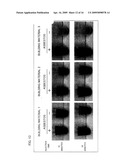 | 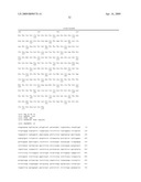 |
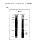 | 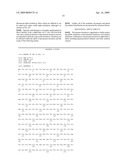 |
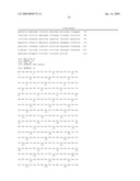 | 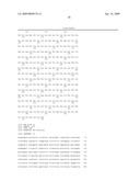 |
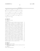 | 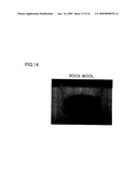 |
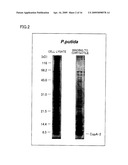 |  |
 |  |
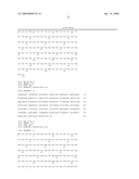 | 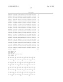 |
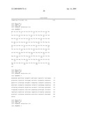 | 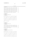 |
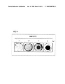 |  |
 |  |
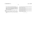 | 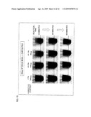 |
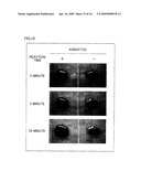 | 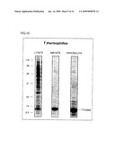 |
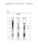 | 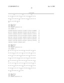 |
 | 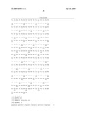 |
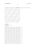 | 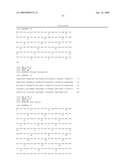 |
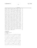 | 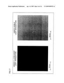 |
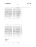 | 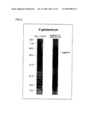 |
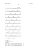 |
| Similar patent applications: | |
| Date | Title |
|---|---|
| 2010-07-29 | Methods of screening compounds for bioactivity in organized tissue |
| 2010-07-22 | Microtome sectionable biopsy support for orienting tissue samples |
| 2010-07-22 | System, method, and device for the expression or repression of proteins |
| 2010-07-29 | Development of low allergen soybean seeds using molecular markers for the p34 allele |
| 2010-07-29 | Methods of generating variant proteins with increased host string content |
| New patent applications in this class: | |
| Date | Title |
|---|---|
| 2019-05-16 | Compounds and methods for use in detecting gabapentin |
| 2019-05-16 | Multisignal reagents for labeling analytes |
| 2016-06-23 | Immunoassay product and process |
| 2016-05-19 | Optically-detectable enzyme substrates and their method of use |
| 2016-04-28 | Calibration of fluidic devices |
| New patent applications from these inventors: | |
| Date | Title |
|---|---|
| 2020-12-31 | Transformant, and method using said transformant to detect presence or absence of reduced phosphorous compound |
| 2015-08-13 | Quantification method, quantification device, and quantification kit |
| 2015-05-07 | Method for selectively culturing microorganism using phosphite dehydrogenase gene as marker |
| 2014-06-19 | Method for detecting asbestos |
| 2013-07-04 | Method and kit for measurement of endotoxin level using bioluminescent assay |
| Top Inventors for class "Chemistry: molecular biology and microbiology" | |
| Rank | Inventor's name |
|---|---|
| 1 | Marshall Medoff |
| 2 | Anthony P. Burgard |
| 3 | Mark J. Burk |
| 4 | Robin E. Osterhout |
| 5 | Rangarajan Sampath |