Patent application title: Plus end-directed microtubule motor required for chromosome congression
Inventors:
Kenneth W. Wood (Foster City, CA, US)
Roman Sakowicz (Foster City, CA, US)
Lawrence S.b. Goldstein (San Diego, CA, US)
Don W. Cleveland (Delmar, CA, US)
Assignees:
THE REGENTS OF THE UNIVERSITY OF CALIFORNIA
IPC8 Class: AC07K1600FI
USPC Class:
5303873
Class name: Globulins immunoglobulin, antibody, or fragment thereof, other than immunoglobulin antibody, or fragment thereof that is conjugated or adsorbed chimeric, mutated, or recombined hybrid (e.g., bifunctional, bispecific, rodent-human chimeric, single chain, rfv, immunoglobulin fusion protein, etc.)
Publication date: 2009-03-19
Patent application number: 20090076248
Inventors list |
Agents list |
Assignees list |
List by place |
Classification tree browser |
Top 100 Inventors |
Top 100 Agents |
Top 100 Assignees |
Usenet FAQ Index |
Documents |
Other FAQs |
Patent application title: Plus end-directed microtubule motor required for chromosome congression
Inventors:
Roman Sakowicz
Kenneth W. Wood
Lawrence S.B. Goldstein
Don W. Cleveland
Agents:
Medlen & Carroll, LLP
Assignees:
The Regents of the University of California
Origin: SAN FRANCISCO, CA US
IPC8 Class: AC07K1600FI
USPC Class:
5303873
Abstract:
The invention provides isolated nucleic acid and amino acid sequences of
Xenopus CENP-E (XCENP-E), antibodies to XCENP-E, methods of screening for
CENP-E modulators using biologically active CENP-E, and kits for
screening for CENP-E modulators.Claims:
1-42. (canceled)
43. An antibody which specifically binds to an isolated biologically active centromere binding protein E (CENP-E), wherein the CENP-E (i) has plus end-directed microtubule motor activity, and (ii) comprises an amino acid sequence having at least 80% sequence identity with amino acid residues 1-324 of SEQ ID NO: 1.
44. The antibody according to claim 43, wherein the antibody is a polyclonal antibody.
45. The antibody according to claim 43, wherein the antibody is a monoclonal antibody.
46. The antibody according to claim 43, wherein the antibody is humanized.
47. The antibody according to claim 43, wherein the antibody is a chimeric antibody.
48. The antibody according to claim 43, wherein the antibody is labeled.
49. The antibody according to claim 43, wherein the antibody inhibits at least one CENP-E activity upon binding to CENP-E.
50. The antibody according to claim 49, wherein the at least one CENP-E activity is plus end-directed microtubule motor activity.
51. The antibody according to claim 49, wherein the at least one CENP-E activity is ATPase activity.
52. The antibody according to claim 49, wherein the at least one CENP-E activity is microtubule binding activity.
53. An antibody which specifically binds to an isolated polypeptide consisting of SEQ ID NO: 1.
54. The antibody according to claim 53, wherein the antibody is a polyclonal antibody.
55. The antibody according to claim 53, wherein the antibody is a monoclonal antibody.
56. The antibody according to claim 53, wherein the antibody is humanized.
57. The antibody according to claim 53, wherein the antibody is a chimeric antibody.
58. The antibody according to claim 53, wherein the antibody is labeled.
59. The antibody according to claim 53, wherein the antibody inhibits at least one CENP-E activity upon binding to CENP-E.
60. The antibody according to claim 59, wherein the at least one CENP-E activity is plus end-directed microtubule motor activity.
61. The antibody according to claim 59, wherein the at least one CENP-E activity is ATPase activity.
62. The antibody according to claim 59, wherein the at least one CENP-E activity is microtubule binding activity.
63. An antibody which specifically binds to a polypeptide domain consisting of amino acids 826-1106 of SEQ ID NO: 1.
64. The antibody according to claim 63, wherein the antibody is a polyclonal antibody.
65. The antibody according to claim 63, wherein the antibody is a monoclonal antibody.
66. The antibody according to claim 63, wherein the antibody is humanized.
67. The antibody according to claim 63, wherein the antibody is a chimeric antibody.
68. The antibody according to claim 63, wherein the antibody is labeled.
69. An antibody which specifically binds to a polypeptide domain consisting of amino acids 826-1106 of SEQ ID NO: 1.
70. The antibody according to claim 69, wherein the antibody is a polyclonal antibody.
71. The antibody according to claim 69, wherein the antibody is a monoclonal antibody.
72. The antibody according to claim 69, wherein the antibody is humanized.
73. The antibody according to claim 69, wherein the antibody is a chimeric antibody.
74. The antibody according to claim 69, wherein the antibody is labeled.
Description:
CROSS REFERENCE TO RELATED APPLICATIONS
[0001]This application claims the benefit of U.S. Ser. No. 60/058,645, filed Sep. 11, 1997, herein incorporated by reference.
FIELD OF THE INVENTION
[0003]The invention provides isolated nucleic acid and amino acid sequences of Xenopus CENP-E (XCENP-E), antibodies to XCENP-E, methods of screening for CENP-E modulators using biologically active CENP-E, and kits for screening for CENP-E modulators.
BACKGROUND OF THE INVENTION
[0004]Segregation of genetic material during mitosis is mediated by the microtubules of the mitotic spindle (see, e.g., McIntosh, in Microtubules, pp. 413-434 (Hyams & Lloyd, eds., 1994). During mitosis, chromosomes are dynamically attached to spindle microtubules via the kinetochore, which is a structure located at the centromere of the chromosome. Kinetochores are involved in coordinating chromosome movement via microtubule assembly and disassembly. The kinetochore and its component proteins thus play an important role in the dynamics of mitosis.
[0005]Spindle microtubules have a defined polarity, with their slow-growing, "minus" ends anchored at or near the spindle pole, and their dynamic, fast-growing "plus" ends interacting with chromosomes (McIntosh, et al., J. Cell Biol. 98:525-533 (1984)). During prometaphase, chromosomes establish interactions with the fast-growing plus ends of microtubules via the kinetochore. Chromosomes then undergo a series of microtubule-dependent movements, culminating in alignment at the metaphase plate, equidistant from the two spindle poles. This process is called "congression." However, the molecular mechanisms underlying chromosome congression are poorly understood (see, e.g., Rieder, et al., J. Cell Biol. 124:223-33 (1994)). A major question has been whether any kinetochore-associated microtubule motors play an important role in congression.
[0006]The two predominant and opposing forces are currently thought to be responsible for chromosome movement during congression: (1) an anti-poleward polar force associated with regions of high microtubule density near the spindle poles, and (2) a poleward force generated at the kinetochore (Khodjakov, et al., J. Cell Biol. 135:315-327 (1996); Waters, et al., J. Cell Sci. 109:2823-2831 (1996); reviewed in Rieder, et al., Int. Rev. Cytol. 79:1-57 (1982); Mitchison, et al., Annu. Rev. Cell Biol. 4:527-49 (1988); Rieder, et al., J. Cell Biol. 124:223-33 (1994)).
[0007]Studies in vitro have demonstrated the presence of both plus and minus end-directed microtubule motor activities on kinetochores that may be responsible for these chromosome movements (Mitchison, et al., J. Cell Biol. 101:766-77 (1985); Hyman, et al., Nature 351:206-211 (1991)). The outstanding issue, however, has been the identity of the molecules at the kinetochore which act as motors and generate the force for chromosome movement.
[0008]In general, both genetic and biochemical approaches have demonstrated crucial roles for microtubule motors in spindle assembly, spindle pole separation, and regulation of spindle microtubule dynamics. These motors include Eg5, CHO1/MKlp1, ncd, cut7, bimC, CIN8, KIP1, KAR3, Xklp2, XKCM1, and XCTK2 (Sawin, et al., Nature 359:540-543 (1992); Blangy, et al., Cell 83:1159-1169 (1995); Sawin, et al., J. Cell Biol. 112:925-940 (1991); Nislow, et al., J. Cell Biol. 111:511-522 (1990); Endow, et al., J. Cell Sci. 107:859-867 (1994); Hagan, et al., Nature 347:563-566 (1990); Hagan, et al., Nature 356:74-76 (1992); Enos, et al., Cell 60:1019-1027 (1990); Hoyt, et al., J. Cell Biol. 118:109-120; Roof, et al., J. Cell Biol. 118:95-108 (1992); Saunders, et al., Cell 70:451-458 (1992), Boleti, et al., J. Cell. Biol. 125:1303-1312; Walczak, et al., Cell 84:37-47 (1996); Walczak, et al., J. Cell Biol. 136:859-70 (1997)). Two kinesin superfamily members, Xenopus Xklp1 and Drosophila nod localize to chromosome arms. With the exception of these two chromatin-associated motors, which are thought to mediate polar ejection forces, none of these other proteins have been implicated directly in congression or in chromosome movement during other phases of mitosis (Theurkauf, et al., J. Cell Biol. 116:1167-1180 (1992); Afshar, et al., Cell 81:129, Cell 81:128-138 (1995); Vernos, et al., Trends in Cell Biol. 5:297-301 (1995)).
[0009]A candidate for powering chromosome movement in mitosis is centromere-associated protein-E (CENP-E), a member of the kinesin superfamily of microtubule motor proteins. Human CENP-E has been cloned and is an integral component of the kinetochore (Yen, et al., Nature 359:536-539 (1992); Yao, et al., The microtubule motor CENP-E is an integral component of kinetochore corona fibers that link centromeres to spindle microtubules (manuscript)). CENP-E localizes to kinetochores throughout all phases of mitotic chromosome movement (early prometaphase through anaphase A) (Yen, et al., Nature 359:536-539 (1992); Brown, et al., J. Cell. Biol. 125:1303-1312 (1994); Lombillo, et al., J. Cell Biol. 128:107-115 (1995)).
[0010]Previous efforts have suggested a role for CENP-E in mitosis. Microinjection of a monoclonal antibody directed against CENP-E into cultured human cells delays anaphase onset (Yen, et al., EMBO J. 10:1245-1254 (1991)). Anti-CENP-E antibody injection into maturing mouse oocytes induces arrest at the first reductional division of meiosis (Duesbery, et al., Proc. Natl. Acad. Sci. USA (in press, 1997)). Antibodies against CENP-E block microtubule depolymerization-dependent minus end-directed movement of purified chromosomes in vitro (Lombillo, et al., J. Cell Biol. 128:107-115 (1995)).
[0011]However, these experiments have not demonstrated the precise role of CENP-E in mitosis, nor have they shown the activity of CENP-E, in particular any motor activity. Recently, CENP-E was reported to be associated with minus end-directed microtubule motor activity, raising the possibility that CENP-E might be responsible for poleward kinetochore movements (Thrower, et al., EMBO J. 14:918-926 (1995)). However, biologically active CENP-E has never been isolated, neither from naturally occurring nor recombinant sources.
SUMMARY OF THE INVENTION
[0012]The present invention provides for the first time biologically active CENP-E and surprisingly demonstrates, contrary to previous reports, that CENP-E is a motor that powers chromosome movement toward microtubule plus ends. Using immunodepletion and antibody addition to Xenopus egg extracts, the present invention further demonstrates that CENP-E plays an essential role in congression. The present invention also provides for the first time the nucleotide and amino acid sequence of isolated Xenopus CENP-E.
[0013]In one aspect, the invention provides an isolated, biologically active CENP-E protein, wherein the CENP-E protein has the following properties: (i) at least one activity selected from the group consisting of plus end-directed microtubule motor activity, ATPase activity, and microtubule binding activity; and (ii) the ability to specifically bind to polyclonal antibodies generated against CENP-E. In one embodiment, the CENP-E protein has an average molecular weight of about 300-350 kDa.
[0014]In one embodiment, the CENP-E protein has an amino acid sequence having at least 34%, or alternatively at least 45%, or alternatively at least 55% sequence identity with a XCENP-E motor domain of SEQ ID NO:1. Alternatively, CENP-E has at least 60%, 65% or 70% sequence identity with a XCENP-E motor domain of SEQ ID NO:1. In an alternative embodiment, the CENP-E has 70%, or alternatively 75%, or alternatively 80%, or alternatively 85%, or alternatively 90% or alternatively 95% amino acid sequence identity to a Xenopus CENP-E core motor domain as measured using a sequence comparison algorithm. In an alternative embodiment, the CENP-E protein has an amino acid sequence of SEQ ID NO:1.
[0015]In another embodiment provided herein, the CENP-E protein is encoded by a nucleic acid sequence having at least 70% sequence identity with SEQ ID NO:2. In another aspect of the present invention, the CENP-E protein is encoded by a nucleic acid which hybridizes under high stringency to a nucleic acid having a sequence complementary to that of SEQ ID NO:2.
[0016]In one embodiment, the CENP-E protein is from a human. In alternative embodiments provided herein, the CENP-E protein is from fungus, insects, or plants.
[0017]In an alternative embodiment provided herein, the CENP-E protein specifically binds to antibodies generated against Xenopus CENP-E (XCENP-E). In this embodiment, the CENP-E protein has an amino acid sequence having greater than 70%, or alternatively 75% sequence identity with a XCENP-E motor domain of SEQ ID NO:1. In another embodiment, the CENP-E protein has an amino acid sequence of a XCENP-E motor domain of SEQ ID NO:1.
[0018]In the embodiments wherein the CENP-E is biologically active as described herein, the amino acid sequence can have 74% or less sequence identity with the motor domain of SEQ ID NO:1.
[0019]Also provided herein is an isolated nucleic acid sequence encoding a CENP-E gene product, said sequence encoding a protein having a core motor domain that has greater than 70% or alternatively 75% amino acid sequence identity to a Xenopus CENP-E (XCENP-E) core motor domain as measured using a sequence comparison algorithm, and specifically binding to antibodies raised against CENP-E. In one embodiment, the sequence has a nucleotide sequence of SEQ ID NO:2. The sequence comparison algorithm can be PILEUP.
[0020]In another aspect of the invention, an antibody which specifically binds to CENP-E is provided.
[0021]Also provided herein is a method for identifying a candidate agent as a compound which modulates CENP-E activity. The method comprising the steps of determining CENP-E activity in the presence of a candidate agent at a control concentration. The CENP-E activity is selected from the group consisting of plus end-directed microtubule motor activity, ATPase activity and microtubule binding activity. The method further comprises the steps of determining said CENP-E activity in the presence the candidate agent at a test concentration, wherein a change in activity between the test concentration and the control concentration of said candidate agent indicates the identification of a compound which modulates CENP-E activity. The method can further comprise the step of isolating biologically active CENP-E from a cell sample.
[0022]The compound to be identified can be a lead therapeutic, bioagricultural compound or diagnostic. Preferably the compound is an antibody which specifically binds CENP-E. In one embodiment the method further comprises the step of modifying the antibody to be a humanized antibody. In one embodiment, the method is performed in a plurality such that many candidate agents are screened simultaneously.
[0023]The invention also includes kits for screening for modulators of CENP-E. The kit includes a container holding biologically active CENP-E and instructions for assaying for CENP-E activity, wherein the CENP-E activity is plus end-directed microtubule motor activity or ATPase activity.
[0024]The invention also provides a method of producing a biologically active CENP-E polypeptide. The method includes the steps of transforming a cell with a vector comprising the nucleic acid sequence encoding the motor domain of CENP-E; expressing said nucleic acid to produce a gene product; purifying said gene product; and identifying ATPase activity or plus-end directed microtubule activity of said gene product.
[0025]In another aspect of the invention, a method of moving microtubules in a plus ended direction is provided wherein microtubules are contacted with biologically active CENP-E.
[0026]In one embodiment the CENP-E is provided in gene form to a cell comprising microtubules.
BRIEF DESCRIPTION OF THE DRAWINGS
[0027]FIG. 1A-C: Identification of Xenopus CENP-E
[0028]FIG. 1A: Structural comparison of Xenopus and human CENP-E. Hatched regions represent regions predicted to form a-helical coiled coils (Lupas, et al., Science 252, 1162-1164 (1991)). Within the N-terminal globular domains of both hCENP-E and XCENP-E there is a domain of ˜324 amino acids corresponding to the kinesin like motor domain. Within these 324 amino acids XCENP-E and hCENP-E are 74% identical. One cDNA clone encoded a protein with a 9 amino acid insertion relative to the other cDNAs isolated (see Example I and methods). The position of this insertion is marked by the arrowhead. XCENP-E contains a putative nuclear localization signal (NLS) at the C-terminal end of the rod domain not present in hCENP-E.
[0029]FIG. 1B: XCENP-E fusion proteins used for polyclonal antibody production.
[0030]FIG. 1C: Deduced amino acid sequence of Xenopus CENP-E. cDNA sequence was compiled from 6 overlapping cDNA clones. Residues identical in hCENP-E and XCENP-E are shaded. The boxed region at the amino-terminus of the sequence is that portion of XCENP-E containing the motor domain and used to assay motility in vitro. The boxed sequence at the C-terminus is that portion of XCENP-E designated as the tail. The underlined sequence NSREHSINA (SEQ ID NO:3) at position 599 is the 9 amino acid relative insertion encoded by one of the cDNAs isolated (see FIG. 1A). The putative NLS, RKKTK (SEQ ID NO:4), immediately adjacent to the boxed tail domain is underlined.
[0031]FIG. 2A-B: XCENP-E is a Plus End-Directed Microtubule Motor
[0032]FIG. 2A: Expression of recombinant XCENP-E in E. coli. XCENP-E amino acid residues 1-473 of XCENP-E were fused at the C-terminus to a c-myc epitope followed by a hexahistidine tag, expressed in E. coli, and purified over Ni-NTA-agarose resin. Coomassie stain of XCENP-E fusion protein used for motility (lane 1), immunoblot of XCENP-E fusion protein probed with α-myc monoclonal antibody (lane 2). Arrowheads indicate XCENP-E fusion protein.
[0033]FIG. 2B: XCENP-E Motility Assay. Microtubules marked near their minus ends with brightly fluorescent seeds were added with ATP to a flow chamber containing purified XCENP-E fusion protein tethered to the coverslip with α-myc monoclonal antibody. Gliding of microtubules was monitored by time-lapse digital fluorescence microscopy. Selected time points from one time lapse series, spaced 90 seconds apart are presented. As reference points, the positions of the plus ends of microtubules numbered 1, 2, and 3 at the start of continual gliding are marked with solid white dots, and the position of a stationary microtubule end is marked with an arrowhead. The bright seed of microtubule 3 enters the plane of focus at 1.5 minutes, and glides 13.6 μM downward with the bright seed leading over the following 3 minutes. Microtubule 2 moves continually during the first three minutes, after which point it detaches and reattaches further toward the bottom of the frame. Microtubule 1 glides minus-end leading throughout the entire time course. The average microtubule velocity of all microtubules was 5.1 μm/min±1.7 (n=49). Of those, 33 microtubules were unambiguously polarity marked, and all glided with their bright seeds leading. Scalebar is 5 μm.
DETAILED DESCRIPTION OF THE INVENTION
I. Introduction
[0034]The present invention provides for the first time biologically active CENP-E and demonstrates that CENP-E has a plus end-directed microtubule motor activity. Biologically active CENP-E was used to show that the kinesin-like motor domain of CENP-E powers chromosome movement toward microtubule plus ends. Finally, quantitative removal of Xenopus CENP-E ("XCENP-E") from Xenopus egg extracts normally capable of assembling mitotic spindles in vitro impairs congression of chromosomes to the metaphase plate. Together, these findings demonstrate that CENP-E plus-end directed microtubule motor activity in vivo is essential for congression during mitosis.
[0035]Functionally, CENP-E is localized in the kinetochores of condensed chromosomes in mitotic cells and has a plus-end directed microtubule motor activity that is ATP dependent (see, e.g., Example II, where ATP or another nucleotide triphosphate is included in the motility assay for motor activity). This activity is responsible for chromosome movement during mitosis. Structurally, the full length nucleotide sequence of XCENP-E (SEQ ID NO:2) encodes a protein of 2954 amino acids with a predicted molecular mass of 340 kDa (SEQ ID NO:1, FIG. 1C). XCENP-E is a member of the kinesin superfamily of motor proteins as evidenced by the sequence of its motor domain. The predicted structure of XCENP-E consists of a 500 amino acid globular amino-terminal domain containing a kinesin-like microtubule motor domain linked to a globular tail domain by a region predicted to form a long, discontinuous α-helical coiled coil (Lupas, et al., Science 252, 1162-1164 (1991); Berger, et al., Proc. Natl. Acad. Sci. USA 92:8259-8263 (1995)) (FIG. 1A). Within the core of the motor domain (residues 1-324) XCENP-E and human CENP-E ("hCENP-E") share 74% identity (Moore, et al., Bioessays 18:207-219 (1996)). Outside the amino-terminal domain lie three additional regions which share greater than 25% identity with hCENP-E, but not with other kinesin-like proteins (FIG. 1). CENP-E is found in Xenopus, mammalian cells, and is predicted to exist in some fungi and perhaps Drosophila.
[0036]The isolation of biologically active CENP-E for the first time provides a means for assaying for enhancers or inhibitors (i.e., modulators) of this essential mitotic protein. Biologically active CENP-E is useful for testing for enhancers or inhibitors using in vitro assays such as microtubule gliding assays (see, e.g., Example II) or ATPase assays (Kodama et al., J. Biochem. 99: 1465-1472 (1986); Stewart et al., Proc. Nat'l Acad. Sci. USA 90: 5209-5213 (1993); Sakowicz et al., Science 280:292-295 (1998)). For example, inhibitors identified using biologically active CENP-E can be used therapeutically to treat diseases of proliferating cells, including, e.g., cancers, hyperplasias, restenosis, cardiac hypertrophy, immune disorders, and inflammation. CENP-E also provides a convenient diagnostic marker for dividing cells. Antibodies or other probes for CENP-E can be used in vitro to identify cells that are entering mitosis. Inhibitors of CENP-E can also be used in vitro to synchronize cells just prior to entry into mitosis for use in cell culture.
II. Definitions
[0037]As used herein, the following terms have the meanings ascribed to them unless specified otherwise.
[0038]The terms "isolated" "purified" or "biologically pure" refer to material that is substantially or essentially free from components which normally accompany it as found in its native state. Purity and homogeneity are typically determined using analytical chemistry techniques such as polyacrylamide gel electrophoresis or high performance liquid chromatography. A protein that is the predominant species present in a preparation is substantially purified. In particular, an isolated XCENP-E nucleic acid is separated from open reading frames which flank the XCENP-E gene and encode proteins other than XCENP-E. The term "purified" denotes that a nucleic acid or protein gives rise to essentially one band in an electrophoretic gel. Particularly, it means that the nucleic acid or protein is at least 85% pure, more preferably at least 95% pure, and most preferably at least 99% pure.
[0039]The term "nucleic acid" refers to deoxyribonucleotides or ribonucleotides and polymers thereof in either single- or double-stranded form. Unless specifically limited, the term encompasses nucleic acids containing known analogues of natural nucleotides which have similar binding properties as the reference nucleic acid and are metabolized in a manner similar to naturally occurring nucleotides. Unless otherwise indicated, a particular nucleic acid sequence also implicitly encompasses conservatively modified variants thereof (e.g., degenerate codon substitutions) and complementary sequences and as well as the sequence explicitly indicated. Specifically, degenerate codon substitutions may be achieved by generating sequences in which the third position of one or more selected (or all) codons is substituted with mixed-base and/or deoxyinosine residues (Batzer et al., Nucleic Acid Res. 19:5081 (1991); Ohtsuka et al., J. Biol. Chem. 260:2605-2608 (1985); Cassol et al., 1992; Rossolini et al., Mol. Cell. Probes 8:91-98 (1994)). The term nucleic acid is used interchangeably with gene, cDNA, and mRNA encoded by a gene.
[0040]The terms "polypeptide," "peptide" and "protein" are used interchangeably herein to refer to a polymer of amino acid residues. The terms apply to amino acid polymers in which one or more amino acid residue is an artificial chemical analogue of a corresponding naturally occurring amino acid, as well as to naturally occurring amino acid polymers. A CENP-E polypeptide comprises a polypeptide demonstrated to have at least ATPase activity or plus end-directed microtubule motor activity and that binds to an antibody generated against CENP-E.
[0041]A "label" is a composition detectable by spectroscopic, photochemical, biochemical, immunochemical, or chemical means. For example, useful labels include 32P, fluorescent dyes, electron-dense reagents, enzymes (e.g., as commonly used in an ELISA), biotin, dioxigenin, or haptens and proteins for which antisera or monoclonal antibodies are available (e.g., the peptide of SEQ ID NO:1 can be made detectable, e.g., by incorporating a radio-label into the peptide, and used to detect antibodies specifically reactive with the peptide).
[0042]As used herein a "nucleic acid probe or oligonucleotide" is defined as a nucleic acid capable of binding to a target nucleic acid of complementary sequence through one or more types of chemical bonds, usually through complementary base pairing, usually through hydrogen bond formation. As used herein, a probe may include natural (i.e., A, G, C, or T) or modified bases (7-deazaguanosine, inosine, etc.). In addition, the bases in a probe may be joined by a linkage other than a phosphodiester bond, so long as it does not interfere with hybridization. Thus, for example, probes may be peptide nucleic acids in which the constituent bases are joined by peptide bonds rather than phosphodiester linkages. It will be understood by one of skill in the art that probes may bind target sequences lacking complete complementarity with the probe sequence depending upon the stringency of the hybridization conditions. The probes are preferably directly labeled as with isotopes, chromophores, lumiphores, chromogens, or indirectly labeled such as with biotin to which a streptavidin complex may later bind. By assaying for the presence or absence of the probe, one can detect the presence or absence of the select sequence or subsequence.
[0043]A "labeled nucleic acid probe or oligonucleotide" is one that is bound, either covalently, through a linker, or through ionic, van der Waals or hydrogen bonds to a label such that the presence of the probe may be detected by detecting the presence of the label bound to the probe.
[0044]"Amplification" primers are oligonucleotides comprising either natural or analogue nucleotides that can serve as the basis for the amplification of a select nucleic acid sequence. They include, e.g., polymerase chain reaction primers and ligase chain reaction oligonucleotides.
[0045]The term "recombinant" when used with reference to a cell, or nucleic acid, or vector, indicates that the cell, or nucleic acid, or vector, has been modified by the introduction of a heterologous nucleic acid or the alteration of a native nucleic acid, or that the cell is derived from a cell so modified. Thus, for example, recombinant cells express genes that are not found within the native (non-recombinant) form of the cell or express native genes that are otherwise abnormally expressed, under expressed or not expressed at all.
[0046]The terms "identical" or percent "identity," in the context of two or more nucleic acids or polypeptide sequences, refer to two or more sequences or subsequences that are the same or have a specified percentage of amino acid residues or nucleotides that are the same, when compared and aligned for maximum correspondence over a comparison window, as measured using one of the following sequence comparison algorithms or by manual alignment and visual inspection. This definition also refers to the complement of a test sequence, which has a designated percent sequence or subsequence complementarity when the test sequence has a designated or substantial identity to a reference sequence. For example, a designated amino acid percent identity of 70% refers to sequences or subsequences that have at least about 70% amino acid identity when aligned for maximum correspondence over a comparison window as measured using one of the following sequence comparison algorithms or by manual alignment and visual inspection. Preferably, the percent identity exists over a region of the sequence that is at least about 25 amino acids in length, more preferably over a region that is 50 or 100 amino acids in length.
[0047]When percentage of sequence identity is used in reference to proteins or peptides, it is recognized that residue positions that are not identical often differ by conservative amino acid substitutions, where amino acids residues are substituted for other amino acid residues with similar chemical properties (e.g., charge or hydrophobicity) and therefore do not change the functional properties of the molecule. Where sequences differ in conservative substitutions, the percent sequence identity may be adjusted upwards to correct for the conservative nature of the substitution. Means for making this adjustment are well known to those of skill in the art. Typically this involves scoring a conservative substitution as a partial rather than a full mismatch, thereby increasing the percentage sequence identity. Thus, for example, where an identical amino acid is given a score of 1 and a non-conservative substitution is given a score of zero, a conservative substitution is given a score between zero and 1. The scoring of conservative substitutions is calculated according to, e.g., the algorithm of Meyers & Miller, Computer Applic. Biol. Sci. 4:11-17 (1988) e.g., as implemented in the program PC/GENE (Intelligenetics, Mountain View, Calif., USA).
[0048]For sequence comparison, typically one sequence acts as a reference sequence, to which test sequences are compared. When using a sequence comparison algorithm, test and reference sequences are entered into a computer, subsequence coordinates are designated, if necessary, and sequence algorithm program parameters are designated. Default program parameters can be used, or alternative parameters can be designated. The sequence comparison algorithm then calculates the percent sequence identity for the test sequence(s) relative to the reference sequence, based on the designated or default program parameters.
[0049]A "comparison window", as used herein, includes reference to a segment of any one of the number of contiguous positions selected from the group consisting of from 25 to 600, usually about 50 to about 200, more usually about 100 to about 150 in which a sequence may be compared to a reference sequence of the same number of contiguous positions after the two sequences are optimally aligned. Methods of alignment of sequences for comparison are well-known in the art. Optimal alignment of sequences for comparison can be conducted, e.g., by the local homology algorithm of Smith & Waterman, Adv. Appl. Math. 2:482 (1981), by the homology alignment algorithm of Needleman & Wunsch, J. Mol. Biol. 48:443 (1970), by the search for similarity method of Pearson & Lipman, Proc. Nat'l. Acad. Sci. USA 85:2444 (1988), by computerized implementations of these algorithms (GAP, BESTFIT, FASTA, and TFASTA in the Wisconsin Genetics Software Package, Genetics Computer Group, 575 Science Dr., Madison, Wis.), or by manual alignment and visual inspection (see, e.g., Ausubel et al., supra).
[0050]One example of a useful algorithm is PILEUP. PILEUP creates a multiple sequence alignment from a group of related sequences using progressive, pairwise alignments. It can also plot a tree showing the clustering relationships used to create the alignment. PILEUP uses a simplification of the progressive alignment method of Feng & Doolittle, J. Mol. Evol. 35:351-360 (1987). The method used is similar to the method described by Higgins & Sharp, CABIOS 5: 151-153 (1989). The program can align up to 300 sequences of a maximum length of 5,000. The multiple alignment procedure begins with the pairwise alignment of the two most similar sequences, producing a cluster of two aligned sequences. This cluster can then be aligned to the next most related sequence or cluster of aligned sequences. Two clusters of sequences can be aligned by a simple extension of the pairwise alignment of two individual sequences. The final alignment is achieved by a series of progressive, pairwise alignments. The program can also be used to plot a dendogram or tree representation of clustering relationships. The program is run by designating specific sequences and their amino acid or nucleotide coordinates for regions of sequence comparison, e.g., the core motor region of CENP-E. In one example, hCENP-E, XCENP-E and ustilago CENP-E were compared to other kinesin superfamily sequences using the following parameters: default gap weight (3.00), default gap length weight (0.10), and weighted end gaps. The resulting dendogram placed hCENP-E and XCENP-E in one cluster as the most closely related sequences, with ustilago CENP-E in the next most closely related cluster.
[0051]Another example of algorithm that is suitable for determining percent sequence identity (i.e., substantial similarity or identity) is the BLAST algorithm, which is described in Altschul et al., J. Mol. Biol. 215:403-410 (1990). Software for performing BLAST analyses is publicly available through the National Center for Biotechnology Information (http://www.ncbi.nlm.nih.gov/). This algorithm involves first identifying high scoring sequence pairs (HSPs) by identifying short words of length W in the query sequence, which either match or satisfy some positive-valued threshold score T when aligned with a word of the same length in a database sequence. T is referred to as the neighborhood word score threshold (Altschul et al, supra). These initial neighborhood word hits act as seeds for initiating searches to find longer HSPs containing them. The word hits are then extended in both directions along each sequence for as far as the cumulative alignment score can be increased. Cumulative scores are calculated using, for nucleotide sequences, the parameters M (reward score for a pair of matching residues; always >0) and N (penalty score for mismatching residues, always <0). For amino acid sequences, a scoring matrix is used to calculate the cumulative score. Extension of the word hits in each direction are halted when: the cumulative alignment score falls off by the quantity X from its maximum achieved value; the cumulative score goes to zero or below, due to the accumulation of one or more negative-scoring residue alignments; or the end of either sequence is reached. The BLAST algorithm parameters W, T, and X determine the sensitivity and speed of the alignment. The BLASTN program (for nucleotide sequences) uses as defaults a wordlength (W) of 11, an expectation (E) of 10, M=5, N=4, and a comparison of both strands. For amino acid sequences, the BLASTP program uses as default parameters a wordlength (W) of 3, an expectation (E) of 10, and the BLOSUM62 scoring matrix (see Henikoff & Henikoff, Proc. Natl. Acad. Sci. USA 89:10915 (1989)).
[0052]The BLAST algorithm also performs a statistical analysis of the similarity between two sequences (see, e.g., Karlin & Altschul, Proc. Nat'l. Acad. Sci. USA 90:5873-5787 (1993)). One measure of similarity provided by the BLAST algorithm is the smallest sum probability (P(N)), which provides an indication of the probability by which a match between two nucleotide or amino acid sequences would occur by chance. For example, a nucleic acid is considered similar to a reference sequence if the smallest sum probability in a comparison of the test nucleic acid to the reference nucleic acid is less than about 0.1, more preferably less than about 0.01, and most preferably less than about 0.001.
[0053]An indication that two nucleic acid sequences or polypeptides are substantially identical is that the polypeptide encoded by the first nucleic acid is immunologically cross reactive with the antibodies raised against the polypeptide encoded by the second nucleic acid, as described below. Thus, a polypeptide is typically substantially identical to a second polypeptide, for example, where the two peptides differ only by conservative substitutions. Another indication that two nucleic acid sequences are substantially identical is that the two molecules or their complements hybridize to each other under stringent conditions, as described below.
[0054]The phrase "hybridizing specifically to" refers to the binding, duplexing, or hybridizing of a molecule only to a particular nucleotide sequence under stringent conditions when that sequence is present in a complex mixture (e.g., total cellular) DNA or RNA. Stringent conditions are sequence-dependent and will be different in different circumstances. Longer sequences hybridize specifically at higher temperatures. Generally, stringent conditions are selected to be about 5° C. lower than the thermal melting point (Tm) for the specific sequence at a defined ionic strength and pH. The Tm is the temperature (under defined ionic strength, pH, and nucleic acid concentration) at which 50% of the probes complementary to the target sequence hybridize to the target sequence at equilibrium (as the target sequences are generally present in excess, at Tm, 50% of the probes are occupied at equilibrium). Typically, stringent conditions will be those in which the salt concentration is less than about 1.0 M sodium ion, typically about 0.01 to 1.0 M sodium ion concentration (or other salts) at pH 7.0 to 8.3 and the temperature is at least about 30° C. for short probes (e.g., 10 to 0.50 nucleotides) and at least about 60° C. for long probes (e.g., greater than 50 nucleotides). Stringent conditions may also be achieved with the addition of destabilizing agents such as formamide.
[0055]"High stringency conditions", as defined herein, may be identified by those that: (1) employ low ionic strength and high temperature for washing, for example 0.015 M sodium chloride/0.0015 M sodium citrate/0.1% sodium dodecyl sulfate at 50° C.; (2) employ during hybridization a denaturing agent, such as formamide, for example, 50% (v/v) formamide with 0.1% bovine serum albumin/0.1% Ficoll/0.1% polyvinylpyrrolidone/50 mM sodium phosphate buffer at pH 6.5 with 750 mM sodium chloride, 75 mM sodium citrate at 42° C.; or (3) employ 50% formamide, 5×SSC (0.75 M NaCl, 0.075 M sodium citrate), 50 mM sodium phosphate (pH 6.8), 0.1% sodium pyrophosphate, 5×Denhardt's solution, sonicated salmon sperm DNA (50 μg/ml), 0.1. % SDS, and 10% dextran sulfate at 42° C., with washes at 42° C. in 0.2×SSC (sodium chloride/sodium citrate) and 50% formamide at 55° C., followed by a high-stringency wash consisting of 0.1×SSC containing EDTA at 55° C.
[0056]"Moderately stringent conditions" may be identified as described by Sambrook et al., Molecular Cloning: A Laboratory Manual, New York: Cold Spring Harbor Press, 1989, and include the use of washing solution and hybridization conditions (e.g., temperature, ionic strength and % SDS) less stringent that those described above. An example of moderately stringent conditions is overnight incubation at 37° C. in a solution comprising: 20% formamide, 5×SSC (150 mM NaCl, 15 mM trisodium citrate), 50 mM sodium phosphate (pH 7.6), 5×Denhardt's solution, 10% dextran sulfate, and 20 mg/mL denatured sheared salmon sperm DNA, followed by washing the filters in 1×SSC at about 37-50° C. The skilled artisan will recognize how to adjust the temperature, ionic strength, etc. as necessary to accommodate factors such as probe length and the like.
[0057]The phrase "a sequence encoding a gene product" refers to a nucleic acid that contains sequence information, e.g., for a structural RNA such as rRNA, a tRNA, the primary amino acid sequence of a specific protein or peptide, a binding site for a trans-acting regulatory agent, an antisense RNA or a ribozyme. This phrase specifically encompasses degenerate codons (i.e., different codons which encode a single amino acid) of the native sequence or sequences which may be introduced to conform with codon preference in a specific host cell.
[0058]"Antibody" refers to a polypeptide substantially encoded by an immunoglobulin gene or immunoglobulin genes, or fragments thereof which specifically bind and recognize an analyte (antigen). The recognized immunoglobulin genes include the kappa, lambda, alpha, gamma, delta, epsilon and mu constant region genes, as well as the myriad immunoglobulin variable region genes. Light chains are classified as either kappa or lambda. Heavy chains are classified as gamma, mu, alpha, delta, or epsilon, which in turn define the immunoglobulin classes, IgG, IgM, IgA, IgD and IgE, respectively.
[0059]An exemplary immunoglobulin (antibody) structural unit comprises a tetramer. Each tetramer is composed of two identical pairs of polypeptide chains, each pair having one "light" (about 25 kDa) and one "heavy" chain (about 50-70 kDa). The N-terminus of each chain defines a variable region of about 100 to 110 or more amino acids primarily responsible for antigen recognition. The terms variable light chain (V) and variable heavy chain (VH) refer to these light and heavy chains respectively.
[0060]Antibodies exist e.g., as intact immunoglobulins or as a number of well characterized fragments produced by digestion with various peptidases. Thus, for example, pepsin digests an antibody below the disulfide linkages in the hinge region to produce F(ab)'2, a dimer of Fab which itself is a light chain joined to VH--CH1I by a disulfide bond. The F(ab)'2 may be reduced under mild conditions to break the disulfide linkage in the hinge region, thereby converting the F(ab)'2 dimer into an Fab' monomer. The Fab' monomer is essentially an Fab with part of the hinge region (see Fundamental Immunology (Paul, ed., 3d ed. 1993). While various antibody fragments are defined in terms of the digestion of an intact antibody, one of skill will appreciate that such fragments may be synthesized de novo either chemically or by using recombinant DNA methodology. Thus, the term antibody, as used herein, also includes antibody fragments either produced by the modification of whole antibodies or those synthesized de novo using recombinant DNA methodologies (e.g., single chain Fv).
[0061]An "anti-XCENP-E" antibody is an antibody or antibody fragment that specifically binds a polypeptide encoded by the XCENP-E gene, cDNA, or a subsequence thereof.
[0062]Humanized forms of non-human antibodies are chimeric immunoglobulins, immunoglobulin chains or fragments thereof (such as Fv, Fab, Fab', F(ab')2 or other antigen-binding subsequences of antibodies) which contain minimal sequence derived from non-human immunoglobulin. Humanized antibodies include human immunoglobulins (recipient antibody) in which residues from a complementary determining region (CDR) of the recipient are replaced by residues from a CDR of a non-human species (donor antibody) such as mouse, rat or rabbit having the desired specificity, affinity and capacity. In some instances, Fv framework residues of the human immunoglobulin are replaced by corresponding non-human residues. Humanized antibodies may also comprise residues which are found neither in the recipient antibody nor in the imported CDR or framework sequences. In general, the humanized antibody will comprise substantially all of at least one, and typically two, variable domains, in which all or substantially all of the CDR regions correspond to those of a non-human immunoglobulin and all or substantially all of the FR regions are those of a human immunoglobulin consensus sequence. The humanized antibody optimally also will comprise at least a portion of an immunoglobulin constant region (Fc), typically that of a human immunoglobulin (Jones et al., Nature, 321:522-525 (1986); Riechmann et al., Nature, 332:323-329 (1988); and Presta, Curr. Op. Struct. Biol., 2:593-596 (1992)).
[0063]Methods for humanizing non-human antibodies are well known in the art. Generally, a humanized antibody has one or more amino acid residues introduced into it from a source which is non-human. These non-human amino acid residues are often referred to as "import" residues, which are typically taken from an "import" variable domain. Humanization can be essentially performed following the method of Winter and co-workers (Jones et al., Nature, 321:522-525 (1986); Riechmann et al., Nature, 332:323-327 (1988); Verhoeyen et al., Science, 239:1534-1536 (1988)), by substituting rodent CDRs or CDR sequences for the corresponding sequences of a human antibody. Accordingly, such "humanized" antibodies are chimeric antibodies (U.S. Pat. No. 4,816,567), wherein substantially less than an intact human variable domain has been substituted by the corresponding sequence from a non-human species. In practice, humanized antibodies are typically human antibodies in which some CDR residues and possibly some FR residues are substituted by residues from analogous sites in rodent antibodies.
[0064]Human antibodies can also be produced using various techniques known in the art, including phage display libraries (Hoogenboom & Winter, J. Mol. Biol., 227:381 (1991); Marks et al., J. Mol. Biol., 222:581 (1991)). The techniques of Cole et al. and Boerner et al. are also available for the preparation of human monoclonal antibodies (Cole et al., Monoclonal Antibodies and Cancer Therapy, Alan R. Liss, p. 77 (1985) and Boerner et al., J. Immunol., 147(1):86-95 (1991)). Similarly, human antibodies can be made by introducing of human immunoglobulin loci into transgenic animals, e.g., mice in which the endogenous immunoglobulin genes have been partially or completely inactivated. Upon challenge, human antibody production is observed, which closely resembles that seen in humans in all respects, including gene rearrangement, assembly, and antibody repertoire. This approach is described, for example, in U.S. Pat. Nos. 5,545,807; 5,545,806; 5,569,825; 5,625,126; 5,633,425; 5,661,016, and in the following scientific publications: Marks et al., Bio/Technology 10, 779-783 (1992); Lonberg et al., Nature 368 856-859 (1994); Morrison, Nature 368, 812-13 (1994); Fishwild et al., Nature Biotechnology 14, 845-51 (1996); Neuberger, Nature Biotechnology 14, 826 (1996); Lonberg & Huszar, Intern. Rev. Immunol. 13 65-93 (1995).
[0065]A "chimeric antibody" is an antibody molecule in which (a) the constant region, or a portion thereof, is altered, replaced or exchanged so that the antigen binding site (variable region) is linked to a constant region of a different or altered class, effector function and/or species, or an entirely different molecule which confers new properties to the chimeric antibody, e.g., an enzyme, toxin, hormone, growth factor, drug, etc.; or (b) the variable region, or a portion thereof, is altered, replaced or exchanged with a variable region having a different or altered antigen specificity.
[0066]The term "immunoassay" is an assay that uses an antibody to specifically bind an analyte. The immunoassay is characterized by the use of specific binding properties of a particular antibody to isolate, target, and/or quantify the analyte.
[0067]The phrase "specifically (or selectively) binds to an antibody" or "specifically (or selectively) immunoreactive with," when referring to a protein or peptide, refers to a binding reaction that is determinative of the presence of the protein in a heterogeneous population of proteins and other biologics. Thus, under designated immunoassay conditions, the specified antibodies bind to a particular protein and do not bind in a significant amount to other proteins present in the sample. Specific binding to an antibody under such conditions may require an antibody that is selected for its specificity for a particular protein. For example, antibodies raised to XCENP-E with the amino acid sequence encoded in SEQ ID NO:1 can be selected to obtain antibodies specifically immunoreactive with that protein and not with other proteins, except for polymorphic variants. A variety of immunoassay formats may be used to select antibodies specifically immunoreactive with a particular protein. For example, solid-phase ELISA immunoassays are routinely used to select antibodies specifically immunoreactive with a protein (see, e.g., Harlow & Lane, Antibodies, A Laboratory Manual (1988), for a description of immunoassay formats and conditions that can be used to determine specific immunoreactivity). Typically a specific or selective reaction will be at least twice background signal or noise and more typically more than 10 to 100 times background.
[0068]The phrase "plus end-directed microtubule motor activity" refers to the activity of a motor protein such as CENP-E to power movement toward the "plus" ends of microtubules. Microtubules are conventionally referred to as having plus (fast growing) and minus ends (slow growing). For example, microtubules of the mitotic spindle have their slow growing, minus ends anchored at or near the spindle pole, and their dynamic, fast growing plus ends interacting with chromosomes and with microtubules emanating from the opposite pole.
[0069]The term "motor domain" or "core motor domain" refers to the domain of CENP-E that confers the plus end-microtubule motor activity on the protein.
[0070]"CENP-E" refers to centromere-associated protein, which is a member of the kinesin superfamily of microtubule motor proteins. CENP-E is an integral component of the kinetochore structure of the chromosome, which links the chromosome to the spindle microtubules. "XCENP-E" refers to CENP-E isolated from Xenopus. CENP-E has activity such as ATPase activity, microtubule binding activity, and plus end-directed microtubule motor activity.
[0071]"Modulators of CENP-E" refers to modulatory molecules identified using an in vitro assays for CENP-E activity (e.g., inhibitors and activators or enhancers). Such assays include ATPase activity, microtubule gliding, spindle assembly, microtubule depolymerizing activity, and metaphase arrest. Samples or assays that are treated with a at least one candidate agent at a test concentration are compared to control samples having the candidate agent at a control concentration (which can be zero), to examine the extent of modulation. Control samples are assigned a relative CENP-E activity value of 100. Modulation of CENP-E is achieved when the CENP-E activity value relative to the control is increased or decreased about at least 10%, 20%, 30%, 40%, 50%, 75%, or preferably, at least 100%.
[0072]"Biologically active" CENP-E refers to CENP-E that has at least one activity selected ATPase activity, microtubule binding activity, and plus end-directed microtubule motor activity, as tested in an ATPase assay, microtubule binding assay, or a microtubule gliding assay. "ATPase activity" refers to the ability of CENP-E to hydrolyze ATP. In a preferred embodiment, CENP-E has plus-end directed microtubule activity.
III. Isolation of the XCENP-E Gene
[0073]A. General Recombinant DNA Methods
[0074]This invention relies on routine techniques in the field of recombinant genetics. Basic texts disclosing the general methods of use in this invention include Sambrook et al., Molecular Cloning, A Laboratory Manual (2nd ed. 1989); Kriegler, Gene Transfer and Expression: A Laboratory Manual (1990); and Current Protocols in Molecular Biology (Ausubel et al., eds., 1994)).
[0075]For nucleic acids, sizes are given in either kilobases (kb) or base pairs (bp). These are estimates derived from agarose or acrylamide gel electrophoresis, from sequenced nucleic acids, or from published DNA sequences. For proteins, sizes are given in kilodaltons (kDa) or amino acid residue numbers. Proteins sizes are estimated from gel electrophoresis, from sequenced proteins, from derived amino acid sequences, or from published protein sequences.
[0076]Oligonucleotides that are not commercially available can be chemically synthesized according to the solid phase phosphoramidite triester method first described by Beaucage & Caruthers, Tetrahedron Letts., 22:1859-1862 (1981), using an automated synthesizer, as described in Van Devanter et. al., Nucleic Acids Res., 12:6159-6168 (1984). Purification of oligonucleotides is by either native acrylamide gel electrophoresis or by anion-exchange HPLC as described in Pearson & Reanier, J. Chrom., 255:137-149 (1983).
[0077]The sequence of the cloned genes and synthetic oligonucleotides can be verified after cloning using, e.g., the chain termination method for sequencing double-stranded templates of Wallace et al., Gene, 16:21-26, 1981.
[0078]B. Cloning Methods for the Isolation of Nucleotide Sequences Encoding XCENP-E
[0079]In general, the nucleic acid sequences encoding XCENP-E and related nucleic acid sequence homologues are cloned from cDNA and genomic DNA libraries or isolated using amplification techniques with oligonucleotide primers. For example, XCENP-E sequences can be isolated from Xenopus DNA libraries by hybridizing with a nucleic acid probe, the sequence of which can be derived from human CENP-E. XCENP-E and XCENP-E homologues that are substantially identical to XCENP-E can be isolated using XCENP-E nucleic acid probes and oligonucleotides under stringent hybridization conditions, by screening libraries. Alternatively, expression libraries can be used to clone XCENP-E and XCENP-E homologues, by detecting expressed homologues immunologically with antisera or purified antibodies made against XCENP-E that also recognize and selectively bind to the XCENP-E homologue. Finally, amplification techniques using primers can be used to amplify and isolate XCENP-E from DNA or RNA. Amplification techniques using degenerate primers can also be used to amplify and isolate XCENP-E homologues. Amplification techniques using primers can also be used to isolate a nucleic acid encoding XCENP-E. These primers can be used, e.g., to amplify a probe of several hundred nucleotides, which is then used to screen a library for full-length XCENP-E. The following primers can be used in such a manner: SEQ ID NO:5 GGGCTGCCCAGGAAGAG and SEQ ID NO:6 GACAGCATTGATCGGCG. Alternatively, the nucleic acid for XCENP-E can be directly amplified using the following primers: SEQ ID NO:7 GAGGGTTCGGCCGCTTA and SEQ ID NO:8 TCTGGGGCCATCCATGC.
[0080]Appropriate primers and probes for identifying the gene encoding CENP-E in other species such as Drosophila and fungi are generated from comparisons of the sequences provided herein (SEQ ID NOS:1 and 2). As described above, antibodies can be used to identify XCENP-E homologues. For example, antibodies made to the motor domain of XCENP-E, the tail domain of XCENP-E, or to the whole protein are useful for identifying XCENP-E homologues (see Example section, below).
[0081]To make a cDNA library, one should choose a source that is rich in the mRNA of choice, e.g., XCENP-E. For example, ovary tissue is enriched for XCENP-E mRNA. The mRNA is then made into cDNA using reverse transcriptase, ligated into a recombinant vector, and transfected into a recombinant host for propagation, screening and cloning. Methods for making and screening cDNA libraries are well known (see, e.g., Gubler & Hoffman, Gene 25: 263-269 (1983); Sambrook et al., supra; Ausubel et al., supra).
[0082]For a genomic library, the DNA is extracted from the tissue and either mechanically sheared or enzymatically digested to yield fragments of about 12-20 kb. The fragments are then separated by gradient centrifugation from undesired sizes and are constructed in bacteriophage lambda vectors. These vectors and phage are packaged in vitro. Recombinant phage are analyzed by plaque hybridization as described in Benton & Davis, Science 196:180-182 (1977). Colony hybridization is carried out as generally described in Grunstein et al., Proc. Natl. Acad. Sci. USA., 72:3961-3965 (1975).
[0083]An alternative method of isolating XCENP-E nucleic acid and its homologues combines the use of synthetic oligonucleotide primers and amplification of an RNA or DNA template (see U.S. Pat. Nos. 4,683,195 and 4,683,202; PCR Protocols: A Guide to Methods and Applications (Innis et al., eds, 1990)). Methods such as polymerase chain reaction (PCR) and ligase chain reaction (LCR) can be used to amplify nucleic acid sequences of CENP-E directly from mRNA, from cDNA, from genomic libraries or cDNA libraries. Degenerate oligonucleotides can be designed to amplify XCENP-E homologues using the sequences provided herein. Restriction endonuclease sites can be incorporated into the primers. Polymerase chain reaction or other in vitro amplification methods may also be useful, for example, to clone nucleic acid sequences that code for proteins to be expressed, to make nucleic acids to use as probes for detecting the presence of CENP-E encoding mRNA in physiological samples, for nucleic acid sequencing, or for other purposes. Genes amplified by the PCR reaction can be purified from agarose gels and cloned into an appropriate vector.
[0084]Gene expression of CENP-E can also be analyzed by techniques known in the art, e.g., reverse transcription and amplification of mRNA, isolation of total RNA or poly A+RNA, northern blotting, dot blotting, in situ hybridization, RNase protection and the like.
[0085]Synthetic oligonucleotides can be used to construct recombinant XCENP-E genes for use as probes or for expression of protein. This method is performed using a series of overlapping oligonucleotides usually 40-120 bp in length, representing both the sense and nonsense strands of the gene. These DNA fragments are then annealed, ligated and cloned. Alternatively, amplification techniques can be used with precise primers to amplify a specific subsequence of the XCENP-E gene. The specific subsequence is then ligated into an expression vector.
[0086]The gene for Xenopus CENP-E is typically cloned into intermediate vectors before transformation into prokaryotic or eukaryotic cells for replication and/or expression. These intermediate vectors are typically prokaryote vectors or shuttle vectors.
[0087]C. Expression in Prokaryotes and Eukaryotes
[0088]To obtain high level expression of a cloned gene, such as those cDNAs encoding CENP-E, it is important to construct an expression vector that contains, at the minimum, a strong promoter to direct transcription, a ribosome binding site for translational initiation, and a transcription/translation terminator. Suitable bacterial promoters are well known in the art and described, e.g., in Sambrook et al. and Ausubel et al. Bacterial expression systems for expressing the CENP-E protein are available in, e.g., E. coli, Bacillus sp., and Salmonella (Palva et al., Gene 22:229-235 (1983); Mosbach, et al., Nature, 302:543-545 (1983). Kits for such expression systems are commercially available. Eukaryotic expression systems for mammalian cells, yeast, and insect cells are well known in the art and are also commercially available. The pET23D expression system (Novagen) is a preferred prokaryotic expression system.
[0089]A "promoter" is defined as an array of nucleic acid control sequences that direct transcription of a nucleic acid. As used herein, a promoter includes necessary nucleic acid sequences near the start site of transcription, such as, in the case of a polymerase II type promoter, a TATA element. A promoter also optionally includes distal enhancer or repressor elements which can be located as much as several thousand base pairs from the start site of transcription. A "constitutive" promoter is a promoter which is active under most environmental and developmental conditions. An "inducible" promoter is a promoter which is under environmental or developmental regulation. The term "operably linked" refers to a functional linkage between a nucleic acid expression control sequence (such as a promoter, or array of transcription factor binding sites) and a second nucleic acid sequence, wherein the expression control sequence directs transcription of the nucleic acid corresponding to the second sequence.
[0090]The term "heterologous" when used with reference to portions of a nucleic acid indicates that the nucleic acid comprises two or more subsequences which are not found in the same relationship to each other in nature. For instance, the nucleic acid is typically recombinantly produced, having two or more sequences from unrelated genes arranged to make a new functional nucleic acid.
[0091]The promoter used to direct expression of a heterologous nucleic acid depends on the particular application. The promoter is preferably positioned about the same distance from the heterologous transcription start site as it is from the transcription start site in its natural setting. As is known in the art, however, some variation in this distance can be accommodated without loss of promoter function.
[0092]In addition to the promoter, the expression vector typically contains a transcription unit or expression cassette that contains all the additional elements required for the expression of the CENP-E encoding DNA in host cells. A typical expression cassette thus contains a promoter operably linked to the DNA sequence encoding CENP-E and signals required for efficient polyadenylation of the transcript, ribosome binding sites, and translation termination. The DNA sequence encoding the CENP-E may typically be linked to a cleavable signal peptide sequence to promote secretion of the encoded protein by the transformed cell. Such signal peptides would include, among others, the signal peptides from tissue plasminogen activator, insulin, and neuron growth factor, and juvenile hormone esterase of Heliothis virescens. Additional elements of the cassette may include enhancers and, if genomic DNA is used as the structural gene, introns with functional splice donor and acceptor sites.
[0093]In addition to a promoter sequence, the expression cassette should also contain a transcription termination region downstream of the structural gene to provide for efficient termination. The termination region may be obtained from the same gene as the promoter sequence or may be obtained from different genes.
[0094]The particular expression vector used to transport the genetic information into the cell is not particularly critical. Any of the conventional vectors used for expression in eukaryotic or prokaryotic cells may be used. Standard bacterial expression vectors include plasmids such as pBR322 based plasmids, pSKF, and fusion expression systems such as GST and LacZ. Epitope tags can also be added to recombinant proteins to provide convenient methods of isolation. One preferred embodiment of an epitope tag is c-myc.
[0095]Expression vectors containing regulatory elements from eukaryotic viruses are typically used in eukaryotic expression vectors, e.g., SV40 vectors, papilloma virus vectors, and vectors derived from Epstein Bar virus. Other exemplary eukaryotic vectors include pMSG, pAV009/A.sup.+, pMTO10/A.sup.+, pMAMneo-5, baculovirus pDSVE, and any other vector allowing expression of proteins under the direction of the SV40 early promoter, SV40 later promoter, metallothionein promoter, murine mammary tumor virus promoter, Rous sarcoma virus promoter, polyhedrin promoter, or other promoters shown effective for expression in eukaryotic cells.
[0096]Some expression systems have markers that provide gene amplification such as thymidine kinase, hygromycin B phosphotransferase, and dihydrofolate reductase. Alternatively, high yield expression systems not involving gene amplification are also suitable, such as using a baculovirus vector in insect cells, with a CENP-E encoding sequence under the direction of the polyhedrin promoter or other strong baculovirus promoters.
[0097]The elements that are typically included in expression vectors also include a replicon that functions in E. coli, a gene encoding antibiotic resistance to permit selection of bacteria that harbor recombinant plasmids, and unique restriction sites in nonessential regions of the plasmid to allow insertion of eukaryotic sequences. The particular antibiotic resistance gene chosen is not critical, any of the many resistance genes known in the art are suitable. The prokaryotic sequences are preferably chosen such that they do not interfere with the replication of the DNA in eukaryotic cells, if necessary.
[0098]Standard transfection methods are used to produce bacterial, mammalian, yeast or insect cell lines that express large quantities of CENP-E protein, which are then purified using standard techniques (see, e.g., Colley et al., J. Biol. Chem. 264:17619-17622 (1989); Guide to Protein Purification, in Methods in Enzymology, vol. 182 (Deutscher ed., 1990)). Transformation of eukaryotic and prokaryotic cells are performed according to standard techniques (see, e.g., Morrison, J. Bact., 132:349-351 (1977); Clark-Curtiss & Curtiss, Methods in Enzymology, 101:347-362 (Wu et al., eds, 1983).
[0099]Any of the well known procedures for introducing foreign nucleotide sequences into host cells may be used. These include the use of calcium phosphate transfection, polybrene, protoplast fusion, electroporation, liposomes, microinjection, plasma vectors, viral vectors and any of the other well known methods for introducing cloned genomic DNA, cDNA, synthetic DNA or other foreign genetic material into a host cell (see, e.g., Sambrook et al., supra). It is only necessary that the particular genetic engineering procedure used be capable of successfully introducing at least one gene into the host cell capable of expressing the CENP-E protein.
[0100]After the expression vector is introduced into the cells, the transfected cells are cultured under conditions favoring expression of the CENP-E protein which is recovered from the culture using standard techniques identified below.
IV. Purification of CENP-E Protein
[0101]Either naturally occurring or recombinant CENP-E can be purified for use in functional assays. Naturally occurring CENP-E is purified, e.g., from Xenopus and any other source of an XCENP-E homologue, such as Drosophila or fungi. Recombinant CENP-E is purified from any suitable expression system.
[0102]CENP-E may be purified to substantial purity by standard techniques, including selective precipitation with such substances as ammonium sulfate; column chromatography, immunopurification methods, and others (see, e.g., Scopes, Protein Purification: Principles and Practice (1982); U.S. Pat. No. 4,673,641; Ausubel et al., supra; and Sambrook et al., supra). A preferred method of purification is use of Ni-NTA agarose (Qiagen).
[0103]A number of procedures can be employed when recombinant CENP-E is being purified. For example, proteins having established molecular adhesion properties can be reversible fused to CENP-E. With the appropriate ligand, CENP-E can be selectively adsorbed to a purification column and then freed from the column in a relatively pure form. The fused protein is then removed by enzymatic activity. Finally CENP-E could be purified using immunoaffinity columns.
[0104]A. Purification of CENP-E from Recombinant Bacteria
[0105]Recombinant proteins are expressed by transformed bacteria in large amounts, typically after promoter induction; but expression can be constitutive. Bacteria are grown according to standard procedures in the art. Because CENP-E is a protein that is difficult to isolate with intact biological activity, preferably fresh bacteria cells are used for isolation of protein. Use of cells that are frozen after growth but prior to lysis typically results in negligible yields of active protein.
[0106]Proteins expressed in bacteria may form insoluble aggregates ("inclusion bodies"). Several protocols are suitable for purification of CENP-E inclusion bodies. For example, purification of inclusion bodies typically involves the extraction, separation and/or purification of inclusion bodies by disruption of bacterial cells, e.g., by incubation in a buffer of about 100-150 μg/ml lysozyme and 0.1% Nonidet P40, a non-ionic detergent. The cell suspension can be homogenized using a Polytron (Brinkman Instruments, Westbury, N.Y.). Alternatively, the cells can be sonicated on ice. Alternate methods of lysing bacteria are apparent to those of skill in the art (see, e.g., Sambrook et al., supra; Ausubel et al., supra).
[0107]The cell suspension is generally centrifuged and the pellet containing the inclusion bodies resuspended in buffer that does not dissolve but washes the inclusion bodies, e.g., 20 mM Tris-HCl (pH 7.2), 1 mM EDTA, 150 mM NaCl and 2% Triton-X 100, a non-ionic detergent. It may be necessary to repeat the wash step to remove as much cellular debris as possible. The remaining pellet of inclusion bodies may be resuspended in an appropriate buffer (e.g., 20 mM sodium phosphate, pH 6.8, 150 mM NaCl). Other appropriate buffers will be apparent to those of skill in the art.
[0108]Following the washing step, the inclusion bodies are solubilized by the addition of a solvent that is both a strong hydrogen acceptor and a strong hydrogen donor (or a combination of solvents each having one of these properties); the proteins that formed the inclusion bodies may then be renatured by dilution or dialysis with a compatible buffer. Suitable solvents include, but are not limited to urea (from about 4 M to about 8 M), formamide (at least about 80%, volume/volume basis), and guanidine hydrochloride (from about 4 M to about 8 M). Some solvents which are capable of solubilizing aggregate-forming proteins, for example SDS (sodium dodecyl sulfate), 70% formic acid, are inappropriate for use in this procedure due to the possibility of irreversible denaturation of the proteins, accompanied by a lack of immunogenicity and/or activity. Although guanidine hydrochloride and similar agents are denaturants, this denaturation is not irreversible and renaturation may occur upon removal (by dialysis, for example) or dilution of the denaturant, allowing re-formation of immunologically and/or biologically active protein. After solubilization, the protein can be separated from other bacterial proteins by standard separation techniques.
[0109]Alternatively, it is possible to purify CENP-E from bacteria periplasm. Where CENP-E is exported into the periplasm of the bacteria, the periplasmic fraction of the bacteria can be isolated by cold osmotic shock in addition to other methods known to skill in the art. To isolate recombinant proteins from the periplasm, the bacterial cells are centrifuged to form a pellet. The pellet is resuspended in a buffer containing 20% sucrose. To lyse the cells, the bacteria are centrifuged and the pellet is resuspended in ice-cold 5 mM MgSO4 and kept in an ice bath for approximately 10 minutes. The cell suspension is centrifuged and the supernatant decanted and saved. The recombinant proteins present in the supernatant can be separated from the host proteins by standard separation techniques well known to those of skill in the art.
[0110]B. Standard Protein Separation Techniques for Purifying CENP-E
[0111]Solubility Fractionation
[0112]Often as an initial step, particularly if the protein mixture is complex, an initial salt fractionation can separate many of the unwanted host cell proteins (or proteins derived from the cell culture media) from the recombinant protein of interest. The preferred salt is ammonium sulfate. Ammonium sulfate precipitates proteins by effectively reducing the amount of water in the protein mixture. Proteins then precipitate on the basis of their solubility. The more hydrophobic a protein is, the more likely it is to precipitate at lower ammonium sulfate concentrations. A typical protocol includes adding saturated ammonium sulfate to a protein solution so that the resultant ammonium sulfate concentration is between 20-30%. This concentration will precipitate the most hydrophobic of proteins. The precipitate is then discarded (unless the protein of interest is hydrophobic) and ammonium sulfate is added to the supernatant to a concentration known to precipitate the protein of interest. The precipitate is then solubilized in buffer and the excess salt removed if necessary, either through dialysis or diafiltration. Other methods that rely on solubility of proteins, such as cold ethanol precipitation, are well known to those of skill in the art and can be used to fractionate complex protein mixtures.
[0113]Size Differential Filtration
[0114]CENP-E has a known molecular weight and this knowledge can be used to isolated it from proteins of greater and lesser size using ultrafiltration through membranes of different pore size (for example, Amicon or Millipore membranes). As a first step, the protein mixture is ultrafiltered through a membrane with a pore size that has a lower molecular weight cut-off than the molecular weight of the protein of interest. The retentate of the ultrafiltration is then ultrafiltered against a membrane with a molecular cut off greater than the molecular weight of the protein of interest. The recombinant protein will pass through the membrane into the filtrate. The filtrate can then be chromatographed as described below.
[0115]Column Chromatography
[0116]CENP-E can also be separated from other proteins on the basis of its size, net surface charge, hydrophobicity, and affinity for ligands. In addition, antibodies raised against proteins can be conjugated to column matrices and the proteins immunopurified. All of these methods are well known in the art. It will be apparent to one of skill that chromatographic techniques can be performed at any scale and using equipment from many different manufacturers (e.g., Pharmacia Biotech).
V. Immunological Detection of CENP-E
[0117]In addition to the detection of CENP-E genes and gene expression using nucleic acid hybridization technology, one can also use immunoassays to detect CENP-E. Immunoassays can be used to qualitatively or quantitatively analyze CENP-E. A general overview of the applicable technology can be found in Harlow & Lane, Antibodies: A Laboratory Manual (1988).
[0118]A. Antibodies to CENP-E
[0119]Methods of producing polyclonal and monoclonal antibodies that react specifically with CENP-E are known to those of skill in the art (see, e.g., Coligan, Current Protocols in Immunology (1991); Harlow & Lane, supra; Goding, Monoclonal Antibodies: Principles and Practice (2d ed. 1986); and Kohler & Milstein, Nature, 256:495-497 (1975). Such techniques include antibody preparation by selection of antibodies from libraries of recombinant antibodies in phage or similar vectors, as well as preparation of polyclonal and monoclonal antibodies by immunizing rabbits or mice (see, e.g., Huse et al., Science 246:1275-1281 (1989); Ward et al., Nature 341:544-546 (1989)).
[0120]A number of CENP-E comprising immunogens may be used to produce antibodies specifically reactive with CENP-E. For example, recombinant XCENP-E or a antigenic fragment thereof such as the motor or tail domain, is isolated as described herein. Recombinant protein can be expressed in eukaryotic or prokaryotic cells as described above, and purified as generally described above. Recombinant protein is the preferred immunogen for the production of monoclonal or polyclonal antibodies. Alternatively, a synthetic peptide derived from the sequences disclosed herein and conjugated to a carrier protein can be used an immunogen. Naturally occurring protein may also be used either in pure or impure form. The product is then injected into an animal capable of producing antibodies. Either monoclonal or polyclonal antibodies may be generated, for subsequent use in immunoassays to measure the protein.
[0121]Methods of production of polyclonal antibodies are known to those of skill in the art. An inbred strain of mice or rabbits is immunized with the protein using a standard adjuvant, such as Freund's adjuvant, and a standard immunization protocol. The animal's immune response to the immunogen preparation is monitored by taking test bleeds and determining the titer of reactivity to CENP-E. When appropriately high titers of antibody to the immunogen are obtained, blood is collected from the animal and antisera are prepared. Further fractionation of the antisera to enrich for antibodies reactive to the protein can be done if desired (see Harlow & Lane, supra).
[0122]Monoclonal antibodies may be obtained by various techniques familiar to those skilled in the art. Briefly, spleen cells from an animal immunized with a desired antigen are immortalized, commonly by fusion with a myeloma cell (see Kohler & Milstein, Eur. J. Immunol. 6:511-519 (1976)). Alternative methods of immortalization include transformation with Epstein Barr Virus, oncogenes, or retroviruses, or other methods well known in the art. Colonies arising from single immortalized cells are screened for production of antibodies of the desired specificity and affinity for the antigen, and yield of the monoclonal antibodies produced by such cells may be enhanced by various techniques, including injection into the peritoneal cavity of a vertebrate host. Alternatively, one may isolate DNA sequences which encode a monoclonal antibody or a binding fragment thereof by screening a DNA library from human B cells according to the general protocol outlined by Huse et al., Science 246:1275-1281 (1989).
[0123]Monoclonal antibodies and polyclonal sera are collected and titered against the immunogen protein in an immunoassay, for example, a solid phase immunoassay with the immunogen immobilized on a solid support. Polyclonal antisera with a titer of 104 or greater are selected and tested for their cross reactivity against non-CENP-E proteins or even other homologous proteins from other organisms, using a competitive binding immunoassay. Specific polyclonal antisera and monoclonal antibodies will usually bind with a KD of at least about 0.1 mM, more usually at least about 1 μM, preferably at least about 0.1 μM or better, and most preferably, 0.01 μM or better.
[0124]Once CENP-E specific antibodies are available, CENP-E can be detected by a variety of immunoassay methods. For a review of immunological and immunoassay procedures, see Basic and Clinical Immunology (Stites & Terr eds., 7th ed. 1991). Moreover, the immunoassays of the present invention can be performed in any of several configurations, which are reviewed extensively in Enzyme Immunoassay (Maggio, ed., 1980); and Harlow & Lane, supra.
[0125]B. Immunological Binding Assays
[0126]As explained above, CENP-E expression is associated with mitosis. Thus, CENP-E provides a marker with which to examine actively dividing cells, including pathological cells such as cancers or hyperplasias. In a preferred embodiment, CENP-E is detected and/or quantified using any of a number of well recognized immunological binding assays (see, e.g., U.S. Pat. Nos. 4,366,241; 4,376,110; 4,517,288; and 4,837,168). For a review of the general immunoassays, see also Methods in Cell Biology Volume 37: Antibodies in Cell Biology (Asai, ed. 1993); Basic and Clinical Immunology (Stites & Terr, eds., 7th ed. 1991). Immunological binding assays (or immunoassays) typically utilize a "capture agent" to specifically bind to and often immobilize the analyte (in this case the CENP-E or antigenic subsequence thereof). The capture agent is a moiety that specifically binds to the analyte. The antibody (anti-CENP-E) may be produced by any of a number of means well known to those of skill in the art and as described above.
[0127]Immunoassays also often utilize a labeling agent to specifically bind to and label the binding complex formed by the capture agent and the analyte. The labeling agent may itself be one of the moieties comprising the antibody/analyte complex. Thus, the labeling agent may be a labeled CENP-E polypeptide or a labeled anti-CENP-E antibody. Alternatively, the labeling agent may be a third moiety, such as another antibody, that specifically binds to the antibody/CENP-E complex.
[0128]In a preferred embodiment, the labeling agent is a second CENP-E bearing a label. Alternatively, the second antibody may lack a label, but it may, in turn, be bound by a labeled third antibody specific to antibodies of the species from which the second antibody is derived. The second can be modified with a detectable moiety, such as biotin, to which a third labeled molecule can specifically bind, such as enzyme-labeled streptavidin.
[0129]Other proteins capable of specifically binding immunoglobulin constant regions, such as protein A or protein G may also be used as the label agent. These proteins are normal constituents of the cell walls of streptococcal bacteria. They exhibit a strong non-immunogenic reactivity with immunoglobulin constant regions from a variety of species (see generally Kronval, et al., J. Immunol., 111: 1401-1406 (1973); Akerstrom, et al., J. Immunol., 135: 2589-2542 (1985)).
[0130]Throughout the assays, incubation and/or washing steps may be required after each combination of reagents. Incubation steps can vary from about 5 seconds to several hours, preferably from about 5 minutes to about 24 hours. However, the incubation time will depend upon the assay format, analyte, volume of solution, concentrations, and the like. Usually, the assays will be carried out at ambient temperature, although they can be conducted over a range of temperatures, such as 10° C. to 40° C.
[0131]Non-Competitive Assay Formats
[0132]Immunoassays for detecting CENP-E in samples may be either competitive or noncompetitive. Noncompetitive immunoassays are assays in which the amount of captured analyte (in this case the protein) is directly measured. In one preferred "sandwich" assay, for example, the capture agent (anti-CENP-E antibodies) can be bound directly to a solid substrate on which they are immobilized. These immobilized antibodies then capture CENP-E present in the test sample. CENP-E is thus immobilized is then bound by a labeling agent, such as a second CENP-E antibody bearing a label. Alternatively, the second antibody may lack a label, but it may, in turn, be bound by a labeled third antibody specific to antibodies of the species from which the second antibody is derived. The second can be modified with a detectable moiety, such as biotin, to which a third labeled molecule can specifically bind, such as enzyme-labeled streptavidin.
[0133]Competitive Assay Formats
[0134]In competitive assays, the amount of CENP-E (analyte) present in the sample is measured indirectly by measuring the amount of an added (exogenous) analyte (i.e the CENP-E) displaced (or competed away) from a capture agent (anti-CENP-E antibody) by the analyte present in the sample. In one competitive assay, a known amount of, in this case, the CENP-E is added to the sample and the sample is then contacted with a capture agent, in this case an antibody that specifically binds to the CENP-E. The amount of CENP-E bound to the antibody is inversely proportional to the concentration of CENP-E present in the sample. In a particularly preferred embodiment, the antibody is immobilized on a solid substrate. The amount of the CENP-E bound to the antibody may be determined either by measuring the amount of CENP-E present in an CENP-E/antibody complex, or alternatively by measuring the amount of remaining uncomplexed protein. The amount of CENP-E may be detected by providing a labeled CENP-E molecule.
[0135]A hapten inhibition assay is another preferred competitive assay. In this assay a known analyte, in this case CENP-E, is immobilized on a solid substrate. A known amount of anti-CENP-E antibody is added to the sample, and the sample is then contacted with the immobilized CENP-E. The amount of anti-CENP-E antibody bound to the immobilized CENP-E is inversely proportional to the amount of CENP-E present in the sample. Again, the amount of immobilized antibody may be detected by detecting either the immobilized fraction of antibody or the fraction of the antibody that remains in solution. Detection may be direct where the antibody is labeled or indirect by the subsequent addition of a labeled moiety that specifically binds to the antibody as described above.
[0136]Immunoassays in the competitive binding format can be used for crossreactivity determinations. For example, a protein partially encoded by SEQ ID NO:1 can be immobilized to a solid support. Proteins are added to the assay that compete with the binding of the antisera to the immobilized antigen. The ability of the above proteins to compete with the binding of the antisera to the immobilized protein is compared to CENP-E encoded by SEQ ID NO:1. The percent crossreactivity for the above proteins is calculated, using standard calculations. Those antisera with less than 10% crossreactivity with each of the proteins listed above are selected and pooled. The cross-reacting antibodies are optionally removed from the pooled antisera by immunoabsorption with the considered proteins, e.g., distantly related homologues.
[0137]The immunoabsorbed and pooled antisera are then used in a competitive binding immunoassay as described above to compare a second protein, thought to be perhaps the protein of this invention, to the immunogen protein (i.e., CENP-E of SEQ ID NO:1). In order to make this comparison, the two proteins are each assayed at a wide range of concentrations and the amount of each protein required to inhibit 50% of the binding of the antisera to the immobilized protein is determined. If the amount of the second protein required to inhibit 50% of binding is less than 10 times the amount of the protein partially encoded by SEQ ID NO:1 that is required to inhibit 50% of binding, then the second protein is said to specifically bind to the polyclonal antibodies generated to an CENP-E immunogen.
[0138]Other Assay Formats
[0139]Western blot (immunoblot) analysis is used to detect and quantify the presence of CENP-E in the sample. The technique generally comprises separating sample proteins by gel electrophoresis on the basis of molecular weight, transferring the separated proteins to a suitable solid support, (such as a nitrocellulose filter, a nylon filter, or derivatized nylon filter), and incubating the sample with the antibodies that specifically bind the CENP-E. The anti-CENP-E antibodies specifically bind to the CENP-E on the solid support. These antibodies may be directly labeled or alternatively may be subsequently detected using labeled antibodies (e.g., labeled sheep anti-mouse antibodies) that specifically bind to the anti-CENP-E antibodies.
[0140]Other assay formats include liposome immunoassays (LIA), which use liposomes designed to bind specific molecules (e.g., antibodies) and release encapsulated reagents or markers. The released chemicals are then detected according to standard techniques (see Monroe et al., Amer. Clin. Prod. Rev. 5:3441 (1986)).
[0141]Reduction of Non-Specific Binding
[0142]One of skill in the art will appreciate that it is often desirable to minimize non-specific binding in immunoassays. Particularly, where the assay involves an antigen or antibody immobilized on a solid substrate it is desirable to minimize the amount of non-specific binding to the substrate. Means of reducing such non-specific binding are well known to those of skill in the art. Typically, this technique involves coating the substrate with a proteinaceous composition. In particular, protein compositions such as bovine serum albumin (BSA), nonfat powdered milk, and gelatin are widely used with powdered milk being most preferred.
[0143]Labels
[0144]The particular label or detectable group used in the assay is not a critical aspect of the invention, as long as it does not significantly interfere with the specific binding of the antibody used in the assay. The detectable group can be any material having a detectable physical or chemical property. Such detectable labels have been well-developed in the field of immunoassays and, in general, most any label useful in such methods can be applied to the present invention. Thus, a label is any composition detectable by spectroscopic, photochemical, biochemical, immunochemical, electrical, optical or chemical means. Useful labels in the present invention include magnetic beads (e.g., Dynabeads®), fluorescent dyes (e.g., fluorescein isothiocyanate, Texas red, rhodamine, and the like), radiolabels (e.g., 3H, 125I, 35S, 14C, or 32P), enzymes (e.g., horse radish peroxidase, alkaline phosphatase and others commonly used in an ELISA), and colorimetric labels such as colloidal gold or colored glass or plastic (e.g., polystyrene, polypropylene, latex, etc.) beads.
[0145]The label may be coupled directly or indirectly to the desired component of the assay according to methods well known in the art. As indicated above, a wide variety of labels may be used, with the choice of label depending on sensitivity required, ease of conjugation with the compound, stability requirements, available instrumentation, and disposal provisions.
[0146]Non-radioactive labels are often attached by indirect means. Generally, a ligand molecule (e.g., biotin) is covalently bound to the molecule. The ligand then binds to an anti-ligand (e.g., streptavidin) molecule which is either inherently detectable or covalently bound to a signal system, such as a detectable enzyme, a fluorescent compound, or a chemiluminescent compound. A number of ligands and anti-ligands can be used in conjunction with the labeled, naturally occurring anti-ligands. Alternatively, any haptenic or antigenic compound can be used in combination with an antibody.
[0147]The molecules can also be conjugated directly to signal generating compounds, e.g., by conjugation with an enzyme or fluorophore. Enzymes of interest as labels will primarily be hydrolases, particularly phosphatases, esterases and glycosidases, or oxidotases, particularly peroxidases. Fluorescent compounds include fluorescein and its derivatives, rhodamine and its derivatives, dansyl, umbelliferone, etc. Chemiluminescent compounds include luciferin, and 2,3-dihydrophthalazinediones, e.g., luminol. For a review of various labeling or signal producing systems which may be used, see U.S. Pat. No. 4,391,904.
[0148]Means of detecting labels are well known to those of skill in the art. Thus, for example, where the label is a radioactive label, means for detection include a scintillation counter or photographic film as in autoradiography. Where the label is a fluorescent label, it may be detected by exciting the fluorochrome with the appropriate wavelength of light and detecting the resulting fluorescence. The fluorescence may be detected visually, by means of photographic film, by the use of electronic detectors such as charge coupled devices (CCDs) or photomultipliers and the like. Similarly, enzymatic labels may be detected by providing the appropriate substrates for the enzyme and detecting the resulting reaction product. Finally simple colorimetric labels may be detected simply by observing the color associated with the label. Thus, in various dipstick assays, conjugated gold often appears pink, while various conjugated beads appear the color of the bead.
[0149]Some assay formats do not require the use of labeled components. For instance, agglutination assays can be used to detect the presence of the target antibodies. In this case, antigen-coated particles are agglutinated by samples comprising the target antibodies. In this format, none of the components need be labeled and the presence of the target antibody is detected by simple visual inspection.
VI. Assays for Modulators of CENP-E
[0150]CENP-E is a plus end-directed microtubule motor that is required for mitosis. The present invention provides for the first time biologically active CENP-E. The activity of CENP-E can be assessed using a variety of in vitro assays, e.g., microtubule gliding assays (see Example II) or ATPase assays (Kodama et al., J. Biochem. 99: 1465-1472 (1986); Stewart et al., Proc. Nat'l Acad. Sci. USA 90: 5209-5213 (1993). Microtubule depolymerization assays can also be used to examine CENP-E activity (Lombillo et al., J. Cell Biol. 128:107-115 (1995)).
[0151]In addition, CENP-E activity can be examined by comparing antibody depletion of CENP-E or inhibition of CENP-E in vitro using cultured cells or egg extracts. Samples that have been depleted or inhibited are compared to control samples that are not inhibited/depleted or that have biologically CENP-E added back to the sample. Characteristics such as spindle assembly and metaphase arrest are used to compare the effect of CENP-E inhibition or depletion.
[0152]Such assays can be used to test for the activity of CENP-E isolated from endogenous sources or recombinant sources. Furthermore, such assays can be used to test for modulators of CENP-E. Because the plus end-directed microtubule motor activity of CENP-E is essential for mitosis, inhibition of CENP-E can be used to control cell proliferation.
[0153]Candidate agents encompass numerous chemical classes, though typically they are organic molecules, preferably small organic compounds having a molecular weight of more than 100 and less than about 2,500 daltons. Candidate agents comprise functional groups necessary for structural interaction with proteins, particularly hydrogen bonding, or ionic (electrostatic) interactions and typically include at least an amine, carbonyl, hydroxyl, sulfonyl or carboxyl group, preferably at least two of the functional chemical groups. The candidate agents often comprise cyclical carbon or heterocyclic structures and/or aromatic or polyaromatic structures substituted with one or more of the above functional groups. Candidate agents are also found among biomolecules including peptides, saccharides, fatty acids, steroids, purines, pyrimidines, derivatives, structural analogs or combinations thereof. Candidate agents are obtained from a wide variety of sources including libraries of synthetic or natural compounds. For example, numerous means are available for random and directed synthesis of a wide variety of organic compounds and biomolecules, including expression of randomized oligonucleotides. Alternatively, libraries of natural compounds in the form of bacterial, fungal, plant and animal extracts are available or readily produced. Additionally, natural or synthetically produced libraries and compounds are readily modified through conventional chemical, physical and biochemical means. Known pharmacological agents may be subjected to directed or random chemical modifications, such as acylation, alkylation, esterification, amidification to produce structural analogs.
[0154]In an embodiment provided herein, the candidate bioactive agents are proteins. The protein may be made up of naturally occurring amino acids and peptide bonds, or synthetic peptidomimetic structures. For example, homo-phenylalanine, citrulline and noreleucine are considered amino acids for the purposes of the invention. "Amino acid" also includes imino acid residues such as proline and hydroxyproline. The side chains may be in either the (R) or the (S) configuration. In the preferred embodiment, the amino acids are in the (S) or L-configuration. If non-naturally occurring side chains are used, non-amino acid substituents may be used, for example to prevent or retard in vivo degradations.
[0155]In another embodiment, the candidate bioactive agents are naturally occurring proteins or fragments of naturally occurring proteins. Thus, for example, cellular extracts containing proteins, or random or directed digests of proteinaceous cellular extracts, may be used. In one embodiment, the libraries are of bacterial, fungal, viral, and mammalian proteins, with the latter being preferred, and human proteins being especially preferred.
[0156]In one embodiment, the candidate agents are peptides of from about 5 to about 30 amino acids, with from about 5 to about 20 amino acids being preferred, and from about 7 to about 15 being particularly preferred. The peptides may be digests of naturally occurring proteins as is outlined above, or random peptides. By randomized or grammatical equivalents herein is meant that each nucleic acid and peptide consists of essentially random nucleotides and amino acids, respectively. Since generally these random peptides (or nucleic acids, discussed below) are chemically synthesized, they may incorporate any nucleotide or amino acid at any position. The synthetic process can be designed to generate randomized proteins or nucleic acids, to allow the formation of all or most of the possible combinations over the length of the sequence, thus forming a library of randomized candidate bioactive proteinaceous agents.
[0157]In one embodiment, the library is fully randomized, with no sequence preferences or constants at any position. In a preferred embodiment, the library is biased. That is, some positions within the sequence are either held constant, or are selected from a limited number of possibilities. For example, in a preferred embodiment, the nucleotides or amino acid residues are randomized within a defined class, for example, of hydrophobic amino acids, hydrophilic residues, sterically biased (either small or large) residues, towards the creation of cysteines, for cross-linking, prolines for SH-3 domains, serines, threonines, tyrosines or histidines for phosphorylation sites, etc., or to purines, etc.
[0158]In another embodiment, the candidate agents are nucleic acids. By nucleic acid or "oligonucleotide" or grammatical equivalents herein means at least two nucleotides covalently linked together. A nucleic acid of the present invention will generally contain phosphodiester bonds, although in some cases, as outlined below, nucleic acid analogs are included that may have alternate backbones, comprising, for example, phosphoramide (Beaucage et al., Tetrahedron 49(10):1925 (1993) and references therein; Letsinger, J. Org. Chem. 35:3800 (1970); Sprinzl et al., Eur. J. Biochem. 81:579 (1977); Letsinger et al., Nucl. Acids Res. 14:3487 (1986); Sawai et al., Chem. Lett. 805 (1984), Letsinger et al., J. Am. Chem. Soc. 110:4470 (1988); and Pauwels et al., Chemica Scripta 26:141 91986)), phosphorothioate (Mag et al., Nucleic Acids Res. 19:1437 (1991); and U.S. Pat. No. 5,644,048), phosphorodithioate (Briu et al., J. Am. Chem. Soc. 111:2321 (1989), O-methylphosphoroamidite linkages (see Eckstein, Oligonucleotides and Analogues: A Practical Approach, Oxford University Press), and peptide nucleic acid backbones and linkages (see Egholm, J. Am. Chem. Soc. 114:1895 (1992); Meier et al., Chem. Int. Ed. Engl. 31:1008 (1992); Nielsen, Nature, 365:566 (1993); Carlsson et al., Nature 380:207 (1996), all of which are incorporated by reference). Other analog nucleic acids include those with positive backbones (Denpcy et al., Proc. Natl. Acad. Sci. USA 92:6097 (1995); non-ionic backbones (U.S. Pat. Nos. 5,386,023, 5,637,684, 5,602,240, 5,216,141 and 4,469,863; Kiedrowshi et al., Angew. Chem. Intl. Ed. English 30:423 (1991); Letsinger et al., J. Am. Chem. Soc. 110:4470 (1988); Letsinger et al., Nucleoside & Nucleotide 13:1597 (1994); Chapters 2 and 3, ASC Symposium Series 580, Carbohydrate Modifications in Antisense Research, Ed. Y. S. Sanghui and P. Dan Cook; Mesmaeker et al., Bioorganic & Medicinal Chem. Lett. 4:395 (1994); Jeffs et al., J. Biomolecular NMR 34:17 (1994); Tetrahedron Lett. 37:743 (1996)) and non-ribose backbones, including those described in U.S. Pat. Nos. 5,235,033 and 5,034,506, and Chapters 6 and 7, ASC Symposium Series 580, Carbohydrate Modifications in Antisense Research, Ed. Y. S. Sanghui and P. Dan Cook. Nucleic acids containing one or more carbocyclic sugars are also included within the definition of nucleic acids (see Jenkins et al., Chem. Soc. Rev. (1995) pp. 169-176). Several nucleic acid analogs are described in Rawls, C & E News Jun. 2, 1997 page 35. All of these references are hereby expressly incorporated by reference. These modifications of the ribose-phosphate backbone may be done to facilitate the addition of additional moieties such as labels, or to increase the stability and half-life of such molecules in physiological environments. In addition, mixtures of naturally occurring nucleic acids and analogs can be made. Alternatively, mixtures of different nucleic acid analogs, and mixtures of naturally occurring nucleic acids and analogs may be made. The nucleic acids may be single stranded or double stranded, as specified, or contain portions of both double stranded or single stranded sequence. The nucleic acid may be DNA, both genomic and cDNA, RNA or a hybrid, where the nucleic acid contains any combination of deoxyribo- and ribo-nucleotides, and any combination of bases, including uracil, adenine, thymine, cytosine, guanine, inosine, xanthine, hypoxanthine, isocytosine, isoguanine, etc.
[0159]As described above generally for proteins, nucleic acid candidate agents may be naturally occurring nucleic acids, random nucleic acids, or biased random nucleic acids. For example, digests of procaryotic or eukaryotic genomes may be used as is outlined above for proteins.
[0160]In a preferred embodiment, the candidate bioactive agents are organic chemical moieties, a wide variety of which are available in the literature.
[0161]In a preferred embodiment, the candidate agent is a small molecule. The small molecule is preferably 4 kilodaltons (kd) or less. In another embodiment, the compound is less than 3 kd, 2 kd or 1 kd. In another embodiment the compound is less than 800 daltons (D), 500 D, 300 D or 200 D. Alternatively, the small molecule is about 75 D to 100 D, or alternatively, 100 D to about 200 D.
[0162]The modulators that are identified herein may be useful as lead compounds for therapeutics, bioagricultural compounds, or diagnostics. A therapeutic as used herein refers to a compound which is believed to be capable of modulating CENP-E in vivo which can have treatment application in both human and animal disease. Modulation of CENP-E would be desirable in a number of conditions including but not limited to: abnormal stimulation of endothelial cells (e.g., atherosclerosis), solid and hematopoetic tumors and tumor metastasis, benign tumors, for example, hemangiomas, acoustic neuromas, neurofibromas, vascular malfunctions, abnormal wound healing, inflammatory and immune disorders such as Rheumatoid Arthritis, Bechet's disease, gout or gouty arthritis, abnormal angiogenesis accompanying: rheumatoid arthritis, psoriasis, diabetic retinopathy, and other ocular angiogenic diseases such as, macular degeneration, corneal graft rejection, corneal overgrowth, glaucoma, Osler Webber syndrome, cardiovascular diseases such as hypertension, cardiac ischemia and systolic and diastolic dysfunction and fungal diseases such as aspergillosis, candidiasis and topical fungal diseases such as tinea pedis.
[0163]A bioagricultural compound as used herein refers to a chemical or biological compound that has utility in agriculture and functions to foster food or fiber crop protection or yield improvement. For example, one such compound may serve as a herbicide to selectively control weeds, as a fungicide to control the spreading of plant diseases, as an insecticide to ward off and destroy insect and mite pests. In addition, one such compound may demonstrate utility in seed treatment to improve the growth environment of a germinating seed, seedling or young plant as a plant regulator or activator.
[0164]A diagnostic as used herein is a compound that assists in the identification and characterization of a health or disease state in humans or other animals. The diagnostic can be used in standard assays as is known in the art.
[0165]The modulators can be applied to generally any cell type wherein CENP-E modulation is desired, i.e., eukaryotic, single celled and multicelled organisms, plant and animal, vertebrate, invertebrate and mammalian.
[0166]Modulators of CENP-E are tested using biologically active CENP-E, preferably biologically active XCENP-E. Modulation is tested using one of the in vitro assays described above, e.g., ATPase, microtubule binding and/or gliding, spindle assembly, and metaphase arrest. It is understood that any of the assays can be repeated or different types of assays can be used on the same candidate agent to further characterize the candidate agent as a CENP-E modulator. In particular, where more than one candidate agent is used, the assay can be repeated using individual candidate agents. Moreover, where a candidate agent is a lead compound, further assays can be performed to optimize results until it is established whether that compound or one similar thereto has the desired effect.
[0167]As described above, CENP-E is also a useful diagnostic tool in vitro for determining when a cell is entering mitosis. Reversible inhibitors of CENP-E can be used to synchronize cells in culture. Fungal homologues of XCENP-E also provide a diagnostic tool for identifying fungal infections.
[0168]The present invention also provides for kits for screening for modulators of CENP-E. Such kits can be prepared from readily available materials and reagents. For example, such kits can comprise any one or more of the following materials: biologically active CENP-E, reaction tubes, and instructions for testing CENP-E activity. Preferably, the kit contains biologically active XCENP-E. A wide variety of kits and components can be prepared according to the present invention, depending upon the intended user of the kit and the particular needs of the user. For example, the kit can be tailored for ATPase assays or microtubule gliding assays.
[0169]All publications and patent applications cited in this specification are herein incorporated by reference as if each individual publication or patent application were specifically and individually indicated to be incorporated by reference.
[0170]Although the foregoing invention has been described in some detail by way of illustration and example for purposes of clarity of understanding, it will be readily apparent to one of ordinary skill in the art in light of the teachings of this invention that certain changes and modifications may be made thereto without departing from the spirit or scope of the appended claims.
EXAMPLES
[0171]The following examples are provided by way of illustration only and not by way of limitation. Those of skill in the art will readily recognize a variety of noncritical parameters that could be changed or modified to yield essentially similar results.
Methods
[0172]A. Isolation of XCENP-E cDNA and DNA Constructs
[0173]Fragments spanning nucleotides 1-1707 and 6376-8080 of human CENP-E cDNA (Yen, et al., Nature 359:536-539 (1992)) were used to screen a λgt1O adult Xenopus ovary cDNA library (Rebagliati, et al., Cell 42:769-777 (1985)), hybridizing at 42° C. according to Church & Gilbert (Church, et al., Proc. Natl. Acad. Sci. USA 81:1991-1995 (1984)). cDNA clones hybridizing to both probes were isolated and used in combination to isolate overlapping cDNA clones spanning the intervening region. The sequence of both cDNA strands was determined by a combination of automated cycle sequencing (Applied Biosystems, Perkin Elmer) and manual sequencing using Sequence version 2.0 (USB).
[0174]Overlapping regions of the various cDNA clones were often not absolutely identical, but displayed single base differences at multiple positions. One clone encoding the N-terminal region of the rod domain contained 27 additional nucleotides relative to one other clone spanning that region. Single base differences between cDNA clones were attributed to polymorphisms present in the outbred psuedotetraploid source material used to construct the cDNA library. The 27 nucleotide relative insertion may be a polymorphism, or may represent an alternatively spliced XCENP-E isoform. Overlapping sequence was compiled using MacVector software (Kodak Scientific Imaging Systems, Rochester, N.Y.).
[0175]B. Expression and Purification of XCENP-E Motor Domain
[0176]Recombinant CENP-E was prepared in order to test its activity in a microtubule gliding assay. The recombinant XCENP-E was prepared as a fusion protein inducible by IPTG, with a c-myc epitope tag.
[0177]First, an XCENP-E was cloned into an expression plasmid. The 5' untranslated region of XCENP-E was removed by PCR using two primers: SEQ ID NO:9 CATATGACCATGGCCGAGGGAGATGCAG and SEQ ID NO:10 GTCAGGTCAGCAACATACACG. These primers were used to amplify the 5' end of the X-CENP-E cDNA and introduce NdeI and NcoI sites adjacent to and at the start codon, respectively. This PCR product was subcloned into pCRII (InVitrogen) and then joined at the NruI site with a portion of the XCENP-E cDNA, to reconstruct a motor domain encoding cDNA with an altered start codon.
[0178]An NcoI-XhoI fragment spanning nucleotides 143-1939 was excised from the reconstructed cDNA fragment and ligated into NcoI/XhoI cut pET23d (Novagen) to yield pET23dXCE. This plasmid was digested with BsrGI and XhoI, blunted with Klenow, and a Klenow blunted 60 bp HincII-EcoRI fragment from pBSKS+myc (gift from S. Michealis) was ligated to the digested pET23dXCE backbone in the presence of BsrGI to bias orientation of the insert. The resulting plasmid, pET23dXCEMycHis, encodes amino acids 1-473 of XCENP-E linked at the C-terminus to the following sequence: SEQ ID NO:11 TVSISLGDLTMEQKLISEEDLNFEHHHHHH. The c-myc epitope is underlined.
[0179]This plasmid was transformed into E. coli strain BL21 (DE3) pLysS. A culture inoculated with a single colony was gown at 37° C. in a modified LB medium (10 g bactotryptone, 5 g yeast extract, 5 g NaCl, 2 g MgSO4, 1 g casaminoacids per liter, and 200 mg ampicillin per liter) to OD 600 of -1. The cultures were allowed to cool to room temperature and expression of fusion protein was induced with 0.5 mM IPTG at room temperature. After induction, the cells were used immediately to prepare fusion protein. Cells that were pelleted and stored at low temperatures prior to protein isolation gave low to no yield of active protein; due to CENP-E sensitivity to denaturation.
[0180]Cells were harvested 4 hours after induction, immediately resuspended in lysis buffer (50 mM Tris-HCl pH 7.5, 50 mM NaCl, 1 mM PMSF, 0.1 mM ATP), and lysed by 3 passages through a trench press. Insoluble debris was removed by a centrifugation at 35,000 rpm in Sorvall T647.5 rotor for 40 minutes at 4° C. Soluble protein in the supernatant was bound in batch to 0.5 ml of NTI-agarose resin (Qiagen) for 15 minutes at 4° C. The resin was placed in a column, washed with 5 ml of the lysis buffer supplemented with 20 mM Imidazole. XCENP-E fusion protein recovered by elution in lysis buffer containing 100 mM imidazole and 1 mM DTT. A typical yield was 2 mg of soluble XCENP-E protein from 1 liter of bacterial culture. Freshly prepared protein was used to assay motility. Incubation of bacterially expressed XCENP-E motor protein for longer than 24 hours at 4° C. led to loss of motility.
[0181]C. Fusion Protein Expression, Antibody Production, and Immunoblotting
[0182]The tail and rod regions (see FIG. 1) of XCENP-E were used to make antibodies to CENP-E. Antigens for α-XCENP-ETAIL (aa 2396-2954) and α-XCENP-EROD (aa 826-1106) were produced in E. coli strain BL21 (DE3) pLysS as hexahistidine fusion proteins using the pRSETB expression plasmid (InVitrogen). Following induction with IPTG for 4-16 hours, bacteria were pelleted, washed and lysed by rapid freeze thaw followed by sonication. Inclusion bodies containing the fusion proteins were purified and solubilized in 8M urea, 0.1 M sodium phosphate pH 8.0.
[0183]α-XCENP-EROD fusion protein was further purified over Ni-NTA agarose (Qiagen) according to the manufacturer's instructions. α-XCENP-ETAIL protein was isolated from preparative SDS-PAGE gels as described in (Harlow, et al., Antibodies, A Laboratory Manual: Cold Spring Harbor Laboratory) (1988)). These antigens were used to raise polyclonal antibodies in rabbits.
[0184]For affinity purification, antigen was coupled to cyanogen bromide activated Sepharose (Pharmacia) according to the manufacturers instructions. Antibodies were purified (Harlow, et al., Antibodies, A Laboratory Manual (1988)), eluting with 0.2 M glycine pH 2.5. Antibodies were dialyzed into 10 mM K-HEPES pH 7.8, 100 mM KC1, 1 mM MgCl2 and concentrated using prerinsed centricon spin concentrators (Amicon, Beverly, Mass.) or Nanospin filter concentrators (Gelman Sciences, Ann Arbor, Mich.).
[0185]For immunoblots, cytoplasmic extract prepared from metaphase H arrested Xenopus eggs (Murray, in Methods in Cell Biology, pp. 581-605 (Kay & Peng, eds., (1991)) was resolved on a 4% polyacrylamide gel (˜50 μg/lane), transferred to nitrocellulose and lanes individually probed with affinity purified α-XCENP-ETAIL or α-XCENP-EROD.
[0186]For localization of XCENP-E in cultured Xenopus XTC cells, asynchronous cultures of XTC cells were fixed in methanol and simultaneously stained with mouse monoclonal anti-α-tubulin antibody and affinity purified rabbit α-XCENP-ETAIL antibody. Chromatin was visualized by staining with DAPI. Selected cells at progressive stages of the cell cycle were examined on the blot: interphase, prophase, prometaphase, metaphase, anaphase, and telophase. Similar staining was observed using (x-XCENP-EROD antibody.
[0187]Immunoblots were prepared as follows: proteins resolved by SDS-PAGE, transferred to nitrocellulose, blocked with TBS 5% nonfat dried milk (NFDM), and probed with 2 μg/ml affinity purified antibody overnight in TBS containing 0.05% Tween (TBST) containing 5% NFDM. Primary antibody was visualized using 125I-Protein A (Amersham) followed by autoradiography. Occasionally, instead of 125I-protein A, alkaline phosphatase conjugated goat anti-rabbit secondary antibody (Promega) was used according to the manufacturers instructions. Quantitative phosphoimaging was performed using a Molecular Dynamics model 445 SI phosphorimager.
[0188]D. Spindle Assembly In Vitro
[0189]CSF-arrested extract is an Xenopus egg extract that is arrested in metaphase using cytostatic factor. CSF-arrested extract was prepared from Xenopus eggs essentially as described in Murray, in Methods in Cell Biology, pp. 581-605 (Kay & Peng, eds., 1991); Sawin, et al., J. Cell Biol. 112:925-940 (1991)). 10 mg/ml rhodamine labelled bovine brain tubulin (Hyman, et al., in Methods in Enzymology, pp. 478-485 (Vallee, ed., 1992)) was added at a 1 μl/300 μl of extract.
[0190]Localization of XCENP-E was examined on mitotic spindles assembled in vitro. Tubulin, DAPI-stained chromatin, and α-XCENP-ETAIL staining was examined. Metaphase spindles were assembled in vitro by cycling CSF-arrested Xenopus egg extract containing Xenopus sperm chromatin through interphase and arresting at the following metaphase as described (Sawin, et al., J. Cell Biol. 112:925-940 (1991)). Rhodamine labelled tubulin was added to the extracts to visualize tubulin containing structures. Spindles were sedimented onto coverslips and stained with affinity purified α-XCENP-ETAIL antibody, followed by FITC-conjugated secondary antibody and DAPI.
[0191]For immunodepletion of extract, 100 μg of affinity purified α-XCENP-E antibody or non-immune rabbit IgG (Calbiochem, San Diego, Calif.) was bound to 30 μl slurry of protein A Affiprep beads (BioRad, Hercules, Calif.) for 1 hour at 4° C. in CSF-XB (Murray, 1991). Beads were sedimented, unbound antibody removed, and serially washed with CSF-XB, CSF-XB containing 0.5 M NaCl, and CSF-XB containing leupeptin, pepstatin A, and chymostatic (10 μg/ml each), and cytochalasin B 10 μg/ml. 100 μl of CSF extract was added to the beads and incubated rocking for 1 hour at 4° C. After sedimenting beads, depleted extract was removed and stored on ice until use.
[0192]Demembranated sperm prepared as described (Newmeyer, et al., in Methods in Cell Biology, pp. 607-634 (Kay & Peng, eds. (1991)) were added to a portion of the extract at 1-2×10⁵/ml, and exit from metaphase arrest induced at room temperature by addition of CaCl2 to 0.6-0.8 mM final concentration. Extracts were periodically monitored by fluorescence microscopic examination of 1 μl aliquots squashed under a coverslip (Murray, in Methods in Cell Biology, pp. 581-605 (Kay & Peng, eds., 1991)). At 80 minutes following exit from metaphase one half volume of the appropriate extract was added and the reaction incubated for an additional 80-120 minutes.
[0193]M-phase structures accumulating in extracts were scored at 160-200 minutes total elapsed time. Both mock depleted and XCENP-E depleted extracts frequently failed to exit interphase, or failed to remain arrested at the second metaphase, probably as a consequence of experimental manipulation. Immunoprecipitates were washed 3 times with CSF-XB containing protease inhibitors and 0.1% Triton X-100 and examined by SDS-PAGE and Coomassie staining.
[0194]For Coomassie staining and α-XCENP-EROD blot of α-XCENP-E immunoprecipitates, immunoprecipitates were prepared from CSF-arrested extract (˜10 mg total protein) using affinity purified α-XCENP-ETAIL antibody, affinity purified (α-XCENP-EROD antibody, or non-immune rabbit IgG. Immunoprecipitates were gently washed three times with TBS containing 0.1% Triton-X100. 80% of each precipitate was resolved by SDS-PAGE on a 5-15% gel and proteins visualized by staining with Coomassie brilliant blue.
[0195]For antibody addition experiments, purified anti-XCENP-E antibody or non-immune rabbit IgG (Calbiochem) at 10 mg/ml was added to CSF-arrested extract at a 1:20 dilution, followed by demembranated sperm nuclei and CaCl2. 80 minutes later, when a half volume of CSF arrested extract was added, a proportional amount of the appropriate antibody was added as well.
[0196]Representative structures formed in the presence of 0.5 mg/ml rabbit IgG and in the absence of added antibody, and in the presence of 0.5 mg/ml α-XCENP-ETAIL antibody were examined. Rabbit IgG and α-XCENP-ETAIL (both at 10 mg/ml) were added to CSF-arrested metaphase Xenopus egg extract at a 1:20 dilution along with Xenopus sperm. Extracts were then cycled through interphase. At 80 minutes into interphase (prophase) a half volume of metaphase arrested extract containing 0.5 mg/ml of the appropriate antibody was added. 80 minutes later structures were scored and images collected.
[0197]Quantitation of structures formed in extract containing no antibody (n=138), extract containing 0.5 mg/ml non-immune rabbit IgG (n=132), and extract containing 0.5 mg/ml α-XCENP-ETAIL (n=114) at 80 minutes after exit from interphase were examined. Structures present in the respective extracts were examined and scored as belonging to one of four categories: bipolar spindles with chromatin aligned at the metaphase plate; bipolar spindles with misaligned chromosomes; monopolar spindles, including radial asters, half spindles and chromosomes associated with microtubules with indeterminant organization; and or other, including multipolar structures and groups of chromosomes apparently unassociated with microtubules.
[0198]E. Immunofluorescence Microscopy
[0199]Extract containing mitotic spindles assembled in vitro was diluted 30-50 fold in BRB80 (80 mM KPIPES, 6 mM MgCl2, 1 mM EGTA) containing 0.5% Triton X-100 and 30% glycerol. Spindles were sedimented at room temperature onto a coverslip through a 3 ml cushion of BRB80 containing 0.5% Triton X-100 and 40% glycerol at 7000 rpm in a Sorvall HS4 rotor. Coverslips were fixed in -20° C. methanol, rehydrated in TBS-Tx (150 mM NaCl, 20 mM Tris pH 7.6, 0.1% Triton X-100), blocked for 1 hour with 1% BSA in TBS-Tx and probed with 5 μg/ml affinity purified antibody in 1% BSA in TBS-Tx. After washing with TBS-Tx, primary antibody was visualized using by FITC-conjugated secondary goat anti-rabbit antibody (Cappel).
[0200]Xenopus XTC cells cultured on coverslips in 60% L15 medium containing 10% fetal calf serum at room temperature in ambient atmosphere were rinsed in TBS, fixed in -20° C. methanol and stained with affinity purified antibody as described above. Monoclonal anti-alpha tubulin antibody DM1A (Sigma) was used at a dilution of 1:1000 to stain microtubules.
[0201]Fluorescent images were collected using a Princeton Instruments cooled CCD mounted on a Zeiss Axioplan microscope controlled by Metamorph software (Universal Imaging, West Chester, Pa.). Image processing was performed using both Metamorph and Adobe Photoshop software.
[0202]F. Preparation of Polarity Marked Microtubules and Motility Assay
[0203]Taxol stabilized microtubule seeds brightly labelled with rhodamine were prepared by incubating a 1:1 ratio of rhodamine labelled bovine brain tubulin (Hyman, et al., In Methods in Enzymology, pp. 478-485 (Vallee, ed., 1992)) with unlabelled bovine brain tubulin at a final tubulin concentration of 2.5 mg/ml in PEM80 (80 mM Pipes pH 6.9, 1 mM EGTA, 1 mM MgCl2) containing 10% glycerol, 1 μM taxol, 1 mM GTP at 37° C. for 15 minutes. This mixture was then diluted with 2.75 volumes of warm PEM80 containing 20 μM taxol and 2 mM GTP, and sheared by 5 passes through a Hamilton syringe.
[0204]Dimly rhodamine-labelled extensions were grown from the brightly labelled seeds in PEM80 containing 1 mM GTP and 1.5 mg/ml tubulin cocktail consisting of a mixture of N-ethyl maleimide modified tubulin (Hyman, et al., in Methods in Enzymology, pp. 478-485 (Vallee, ed., 1992)), unlabelled tubulin and rhodamine labelled tubulin at a ratio of 0.1/0.52/0.38 for 30 minutes at 37° C. The resulting suspension of polarity marked microtubules was diluted with PEM80 containing 10 μM taxol and used to test motility.
[0205]25 μl flow chambers prepared from cover slips sealed with an Apiezon grease, were preadsorbed with a 1:10 diluted mouse ascities fluid containing anti-myc monoclonal antibody 9EI0 (Evans, et al., Mol. Cell. Biol. 5:3610-3616 (1985)), washed with 50 μl PEM80, incubated with XCENP-E motor protein diluted to 0.1 mg/ml, and unbound protein removed by rinsing with 50 μl of PEM80. A microtubule/ATP mix consisting polarity marked microtubules in PEM80 containing 10 μM taxol, 2 mM MgATP, and an oxygen scavenging system (0.1 mg/ml catalase, 0.03 mg/ml glucose oxidase, 10 mM glucose, 0.1% β-mercaptoethanol (Kishino, et al., Nature 33:74-76 (1989)) was then flowed into the chamber.
[0206]Movement of microtubules was monitored at room temperature on a Zeiss Axioplan fluorescence microscope fitted with 63X Plan-Apochromat oil immersion objective, and a Princeton instruments cooled CCD. Automated time-lapse image acquisition and data analysis was performed using the MetaMorph software package (Universal Imaging, West Chester, Pa.).
Example I
Identification of Xenopus CENP-E
[0207]To investigate the role of CENP-E in mitotic spindle formation in vitro using extracts of Xenopus eggs used low stringency hybridization followed by library rescreening was used to clone the Xenopus homologue of CENP-E. This clone was subsequently used to raise antibodies suitable for immunodepletion and antibody addition studies. The nucleotide sequence (SEQ ID NO:2) encodes a protein of 2954 amino acids with a predicted molecular mass of 340 kDa (SEQ ID NO:1, FIG. 1C). The predicted structure of Xenopus CENP-E (XCENP-E) is similar to human CENP-E (hCENP-E), consisting of a 500 amino acid globular amino-terminal domain containing a kinesin-like microtubule motor domain linked to a globular tail domain by a region predicted to form a long, discontinuous α-helical coiled coil (Lupas, et al., Science 252, 1162-1164 (1991); Berger, et al., Proc. Natl. Acad. Sci. USA 92:8259-8263 (1995)) (FIG. 1A). Within the core of the motor domain (residues 1-324) XCENP-E and hCENP-E share 74% identity, significantly greater than that shared between XCENP-E and its nearest phylogenetic (evolutionary) neighbors (Moore, et al., Bioessays 18:207-219 (1996)). Outside the amino-terminal domain lie three additional regions which share greater than 25% identity with human CENP-E, but not with other kinesin-like proteins (FIG. 1). On the basis of these regions of identity and its large predicted size, the conclusion was made that XCENP-E is the Xenopus homologue of human CENP-E.
Example II
XCENP-E is a Plus End-Directed Microtubule Motor
[0208]Both human and Xenopus CENP-E are localized to the centromeres of mitotic chromosomes throughout all phases of chromosome movement. This localization places CENP-E in a position to mediate attachment of chromosomes to microtubules, movement of chromosomes during congression, and movement of chromosomes toward the spindle poles during anaphase A.
[0209]To test directly if CENP-E is a microtubule motor and to determine the directionality of CENP-E movement, the amino-terminal 473 amino acids of XCENP-E, containing the kinesin-like motor domain, was fused at the C-terminus to 31 amino acids containing an 9 amino acid c-myc epitope tag followed by a hexahistidine tag (see FIG. 2A, and FIG. 1C, amino-terminal boxed region).
[0210]This protein was produced in E. coli, and purified over nickel-agarose, yielding the expected 57 kDa polypeptide as the major product (FIG. 2A, lane 1). Immunoblotting with a α-myc monoclonal antibody (9ElO) (Evans, et al., Mol. Cell. Biol. 5:3610-3616 (1985)) confirmed the 57 kDa protein as the XCENP-E fusion protein (FIG. 2B, lane 2, arrowheads).
[0211]The XCENP-E fusion protein was tethered to a glass coverslip using the α-myc antibody and gliding of polarity marked microtubules containing brightly fluorescent rhodamine labelled seeds near their minus ends (Howard, et al., in Motility Assays for Motor Proteins, pp. 105-113 (Scholey, ed., 1993)) was recorded by time-lapse digital fluorescence microscopy. Representative time points demonstrating three examples of plus end-directed movement are presented in FIG. 2B. Microtubules moved at a velocity of 5.1 μM/min±1.7 (n=49) with brightly fluorescent seeds leading, indicating that the immobilized XCENP-E fusion protein was moving toward microtubule plus ends. No movement was observed in the absence of fusion protein. When assayed in the absence of α-myc antibody the XCENP-E fusion protein also supported microtubule gliding, albeit less robustly.
[0212]This experiment demonstrates that CENP-E has plus-ended microtubule motor activity. Furthermore, by perturbing CENP-E function in Xenopus egg extracts, as shown below in Examples III-V, it was shown that congression in vitro requires a kinetochore-associated microtubule motor. This result contrasts with a prevailing model describing mitotic spindle formation in Xenopus egg extracts in vitro (Vernos, et al., Trends in Cell Biol. 5:297-301 (1995); Heald, et al., Nature 382, 420-5 (1996); Hyman, et al., Cell 84:401-410 (1996)). Both bipolar spindles with misaligned chromosomes and monopolar structures were observed when XCENP-E, a kinetochore-specific protein, was removed, or when XCENP-E function is impaired by addition of α-XCENP-E antibody (see Examples III-V below). These findings indicate that during normal mitotic spindle formation, CENP-E plays an essential role in mitotic spindle assembly and in prometaphase chromosome movements that result in metaphase chromosome alignment, via its activity as a plus-end directed microtubule motor activity.
Example III
XCENP-E Associates with Xenopus Centromeres In Vivo and In Vitro
[0213]To verify that Xenopus CENP-E exhibits a cell cycle-dependent kinetochore association, polyclonal antibodies were raised against two recombinant antigens, one spanning the tail and C-terminal portion of the rod (α-XCENP-ETAIL, FIG. 1B) and the other corresponding to a portion of the N-terminus of the rod domain (α-XCENP-EROD; FIG. 1B).
[0214]Immunoblotting of Xenopus egg extract reveals that the α-XCENP-ETAIL antibody specifically recognizes XCENP-E as a single band of greater than 300 kDa. The α-XCENP-EROD antibody specifically recognizes XCENP-E and another protein of slightly lower molecular weight that may be a distinct isoform of XCENP-E lacking the tail domain, or XCENP-E that has lost its tail domain as a result of partial proteolysis.
[0215]Immunostaining of cultured Xenopus XTC cells using α-XCENP-ETAIL antibody revealed patterns of cell cycle-dependent localization similar to that observed for mammalian CENP-E (Yen, et al., Nature 359:536-539 (1992); Brown, et al., J. Cell Sci. 109:961-969 (1996)) with the exception that during interphase XCENP-E was localized to the nucleus, consistent with the presence of a nuclear localization signal (Boulikas, et al., Gene Express. 3:193-227 1993)) at the C-terminal end of the rod domain (FIGS. 1A, NLS, and 1C underlined sequence, RKKTK). Nuclear staining intensity was variable from cell to cell, probably reflecting different levels of XCENP-E accumulation, as observed for cytoplasmic CENP-E staining of interphase human cells (Yen, et al., Nature 359:536-539 (1992); Brown, et al., J. Cell. Biol. 125:1303:1312 (1994)).
[0216]Early in prometaphase XCENP-E localizes to discrete spots associated with condensed mitotic chromosomes. During metaphase and early anaphase, XCENP-E remains in discrete foci on chromosomes, and is also apparent at the spindle poles. XCENP-E is found at the spindle midzone during late anaphase and telophase. α-XCENP-ETAIL immunostaining of metaphase spindles assembled using cytostatic factor (CSF)-arrested Xenopus egg extracts cycled through interphase (Murray, in Methods in Cell Biology, pp. 581-605 (Kay & Peng, eds. 1991); Sawin, et al., J. Cell Biol. 112:925-940 (1991)) revealed that XCENP-E was also associated with kinetochores assembled in vitro. Similar patterns of staining were observed in XTC cells and on spindles assembled in vitro using α-XCENP-EROD antibody.
Example IV
XCENP-E is Required for Congression
[0217]To determine the aspect(s) of mitosis for which XCENP-E is required, α-XCENP-ETAIL antibody (made to the tail domain of XCENP-E0 was used to deplete XCENP-E from Xenopus egg extracts arrested in metaphase (CSF-extract). Immunoblotting of control and XCENP-E depleted CSF-extracts revealed that greater than 95% of XCENP-E could be removed by immunodepletion with this antibody. Unrelated antigens, such as XNuMA (Merdes, et al., Cell 87:447-458 (1996)), were unaffected by depletion of XCENP-E.
[0218]To examine the effects of immunodepletion on spindle assembly and chromosome movement, demembranated Xenopus sperm nuclei were added to undepleted, mock depleted and XCENP-E depleted CSF-extracts. Extracts were released from CSF-imposed metaphase arrest by addition of calcium and allowed to cycle through interphase and into the subsequent M-phase, whereupon an additional aliquot of the appropriate uncycled, metaphase-arrested XCENP-E depleted, mock depleted or undepleted extract was added to re-impose a metaphase arrest, thus allowing the accumulation of M-phase structures.
[0219]While mock depleted and undepleted extracts yielded predominantly bipolar spindles with chromosomes aligned at the metaphase plate, depletion of XCENP-E produced a five-fold increase in the number of bipolar spindles with misaligned chromosomes, as well as smaller increase in the percentage of monopolar structures, including radial asters, half spindles, and chromosomes associated with microtubules with indeterminate organization. Extended incubation failed to alter the proportion of bipolar spindles with properly aligned chromosomes. This finding, and the presence of chromosomes resembling non-disjoined metaphase sister chromatids on structures formed in the absence of XCENP-E indicates that depletion of XCENP-E prevents congression of chromosomes to the metaphase plate despite apparently normal spindle assembly and chromosome attachment.
[0220]Three independent experiments revealed in every case a decrease in the percentage of metaphase spindles accompanied by an increased percentage of bipolar/misaligned and monopolar structures, although the distribution of the aberrant structures between the monopolar and bipolar/misaligned classes was variable. Failure of XCENP-E depletion to totally prevent the appearance of bipolar spindles with properly aligned chromosomes could be due to residual XCENP-E (below detection limit), may reflect the actions of other motor proteins functioning in partial redundancy with XCENP-E, or may simply reflect that proportion of spindles in which the chromosomes were already sufficiently aligned. These data indicate that XCENP-E, or a complex containing XCENP-E, is required for chromosome congression.
[0221]To test the possibility that a multiprotein complex had been removed, the proteins immunodepleted by α-XCENP-ETAIL were compared with those precipitated by α-XCENP-EROD antibody. Examination of the proteins immunodepleted by α-XCENP-ETAIL revealed the presence of multiple bands. This result was not surprising, given that XCENP-E is relatively low in abundance compared to other spindle proteins such as NuMA (8-14 μg/ml (Merdes, et al., Cell 87:447-458 (1996)), XKCM1 (10 μg/ml), and Xklp2 (16 μg/ml, (Boleti, et al., J. Cell. Biol. 125:1303-1312).
[0222]Immunoprecipitates prepared with α-XCENP-EROD antibody also contained multiple proteins, only two of which were obviously held in common with the α-XCENP-ETAIL immunoprecipitate. Immunoblotting of α-XCENP-ETAIL and α-XCENP-EROD immunoprecipitates with α-XCENP-EROD antibody revealed that one of the proteins is XCENP-E, and that the other protein of slightly lower molecular weight, is the additional XCENP-E related protein shown earlier to be recognized in unmanipulated extract by the α-XCENP-EROD antibody. The presence of this XCENP-E-related protein in immunoprecipitates prepared using the α-XCENP-ETAIL antibody, which does not directly recognize this lower molecular weight species, provides evidence that like most kinesin-related proteins, XCENP-E exists in a complex that is at least dimeric.
Example V
Addition of α-XCENP-E Antibody Disrupts Metaphase Spindle Formation
[0223]As a further test of the requirement of CENP-E in mediating chromosome congression, especially in view of the removal of multiple proteins upon immunodepletion of XCENP-E, XCENP-E function was perturbed in situ by addition of the monospecific α-XCENP-ETAIL antibody to CSF-arrested Xenopus egg extracts. These extracts were cycled through interphase and arrested at the subsequent M-phase.
[0224]As observed upon immunodepletion of XCENP-E, addition of α-XCENP-ETAIL antibody resulted in almost total elimination of bipolar spindles with properly aligned chromosomes. This loss was accompanied by an increase in the percentage of bipolar spindles with misaligned chromosomes, indicating a role for XCENP-E in congression. Also observed was a large increase in the proportion of monopolar structures suggesting an additional role for XCENP-E in establishment or maintenance of spindle bipolarity. Similar results were obtained in four independent experiments, and also using α-XCENP-EROD antibody.
[0225]The monopolar structures observed upon addition of α-XCENP-E antibody could arise from disruption of bipolar spindle assembly. This sort of spindle perturbation has also been observed following overexpression of the p5O subunit of dynactin, which also localizes to kinetochores (Echeverri, et al., J. Cell Biol. 132:617-633 (1996)). p5O overexpression disrupts spindle bipolarity, yielding two apparent monopoles. On the other hand, monopoles may also arise from disruption of sister chromatid cohesion upon entry into anaphase (Murray, et al., Proc. Natl. Acad. Sci. USA 93:12327-12332 (1996)). Since apparently non-disjoined sister chromatids are visible in structures formed in the presence of α-XCENP-ETAIL antibody, the monopolar structures observed for XCENP-E are unlikely to be the products of premature anaphase. These findings support a role for XCENP-E during prometaphase in establishment or maintenance of bipolarity, as well as in congression.
[0226]Consistent with an essential role for XCENP-E in chromosome movement, chromosomes associated with monopolar structures formed in the presence of anti-XCENP-E antibody were often found distributed both at the periphery and within the aster of microtubules. In contrast, the small proportion of monopolar structures formed in control extracts chromosomes were invariably localized at the periphery of the aster. Unlike perturbation of XKCM1, a relatively abundant Xenopus kinesin superfamily member, which induces formation of large asters as a consequence of decreased microtubule catastrophe (Walczak, et al., Cell 84:3747 (1996)), the asters formed in extracts to which α-XCENP-ETAIL antibody was added, or from which XCENP-E had been removed, were not unusually large. This observation suggests that XCENP-E does not play a role in regulating microtubule dynamics analogous to that played by XKCM1.
Sequence CWU
1
1112954PRTXenopus sp.Xenopus centromere-associated protein-E
(XCENP-E) member of the kinesin superfamily of microtubule motor
proteins 1Met Ser Glu Gly Asp Ala Val Lys Val Cys Val Arg Val Arg Pro
Leu1 5 10 15Ile Gln Arg
Glu Gln Gly Asp Gln Ala Asn Leu Gln Trp Lys Ala Gly20 25
30Asn Asn Thr Ile Ser Gln Val Asp Gly Thr Lys Ser Phe
Asn Phe Asp35 40 45Arg Val Phe Asn Ser
His Glu Ser Thr Ser Gln Ile Tyr Gln Glu Ile50 55
60Ala Val Pro Ile Ile Arg Ser Ala Leu Gln Gly Tyr Asn Gly Thr
Ile65 70 75 80Phe Ala
Tyr Gly Gln Thr Ser Ser Gly Lys Thr Tyr Thr Met Met Gly85
90 95Thr Pro Asn Ser Leu Gly Ile Ile Pro Gln Ala Ile
Gln Glu Val Phe100 105 110Lys Ile Ile Gln
Glu Ile Pro Asn Arg Glu Phe Leu Leu Arg Val Ser115 120
125Tyr Met Glu Ile Tyr Asn Glu Thr Val Lys Asp Leu Leu Cys
Asp Asp130 135 140Arg Arg Lys Lys Pro Leu
Glu Ile Arg Glu Asp Phe Asn Arg Asn Val145 150
155 160Tyr Val Ala Asp Leu Thr Glu Glu Leu Val Met
Val Pro Glu His Val165 170 175Ile Gln Trp
Ile Lys Lys Gly Glu Lys Asn Arg His Tyr Gly Glu Thr180
185 190Lys Met Asn Asp His Ser Ser Arg Ser His Thr Ile
Phe Arg Met Ile195 200 205Val Glu Ser Arg
Asp Arg Asn Asp Pro Thr Asn Ser Glu Asn Cys Asp210 215
220Gly Ala Val Met Val Ser His Leu Asn Leu Val Asp Leu Ala
Gly Ser225 230 235 240Glu
Arg Ala Ser Gln Thr Gly Ala Glu Gly Val Arg Leu Lys Glu Gly245
250 255Cys Asn Ile Asn Arg Ser Leu Phe Ile Leu Gly
Gln Val Ile Lys Lys260 265 270Leu Ser Asp
Gly Gln Ala Gly Gly Phe Ile Asn Tyr Arg Asp Ser Lys275
280 285Leu Thr Arg Ile Leu Gln Asn Ser Leu Gly Gly Asn
Ala Lys Thr Val290 295 300Ile Ile Cys Thr
Ile Thr Pro Val Ser Phe Asp Glu Thr Leu Ser Thr305 310
315 320Leu Gln Phe Ala Ser Thr Ala Lys His
Val Arg Asn Thr Pro His Val325 330 335Asn
Glu Val Leu Asp Asp Glu Ala Leu Leu Lys Arg Tyr Arg Lys Glu340
345 350Ile Leu Asp Leu Lys Lys Gln Leu Glu Asn Leu
Glu Ser Ser Ser Glu355 360 365Thr Lys Ala
Gln Ala Met Ala Lys Glu Glu His Thr Gln Leu Leu Ala370
375 380Glu Ile Lys Gln Leu His Lys Glu Arg Glu Asp Arg
Ile Trp His Leu385 390 395
400Thr Asn Ile Val Val Ala Ser Ser Gln Glu Ser Gln Gln Asp Gln Arg405
410 415Val Lys Arg Lys Arg Arg Val Thr Trp
Ala Pro Gly Lys Ile Gln Asn420 425 430Ser
Leu His Ala Ser Gly Val Ser Asp Phe Asp Met Leu Ser Arg Leu435
440 445Pro Gly Asn Phe Ser Lys Lys Ala Lys Phe Ser
Asp Met Pro Ser Phe450 455 460Pro Glu Ile
Asp Asp Ser Val Cys Thr Glu Phe Ser Asp Phe Asp Asp465
470 475 480Ala Leu Ser Met Met Asp Ser
Asn Gly Ile Asp Ala Glu Trp Asn Leu485 490
495Ala Ser Lys Val Thr His Arg Glu Lys Thr Ser Leu His Gln Ser Met500
505 510Ile Asp Phe Gly Gln Ile Ser Asp Ser
Val Gln Phe His Asp Ser Ser515 520 525Lys
Glu Asn Gln Leu Gln Tyr Leu Pro Lys Asp Ser Gly Asp Met Ala530
535 540Glu Cys Arg Lys Ala Ser Phe Glu Lys Glu Ile
Thr Ser Leu Gln Gln545 550 555
560Gln Leu Gln Ser Lys Glu Glu Glu Lys Lys Glu Leu Val Gln Ser
Phe565 570 575Glu Leu Lys Ile Ala Glu Leu
Glu Glu Gln Leu Ser Val Lys Ala Lys580 585
590Asn Leu Glu Met Val Thr Asn Ser Arg Glu His Ser Ile Asn Ala Glu595
600 605Val Gln Thr Asp Val Glu Lys Glu Val
Val Arg Lys Glu Met Ser Val610 615 620Leu
Gly Asp Ser Gly Tyr Asn Ala Ser Asn Ser Asp Leu Gln Asp Ser625
630 635 640Ser Val Asp Gly Lys Arg
Leu Ser Ser Ser His Asp Glu Cys Ile Glu645 650
655His Arg Lys Met Leu Glu Gln Lys Ile Val Asp Leu Glu Glu Phe
Ile660 665 670Glu Asn Leu Asn Lys Lys Ser
Glu Asn Asp Lys Gln Lys Ser Ser Glu675 680
685Gln Asp Phe Met Glu Ser Ile Gln Leu Cys Glu Ala Ile Met Ala Glu690
695 700Lys Ala Asn Ala Leu Glu Glu Leu Ala
Leu Met Arg Asp Asn Phe Asp705 710 715
720Asn Ile Ile Leu Glu Asn Glu Thr Leu Lys Arg Glu Ile Ala
Asp Leu725 730 735Glu Arg Ser Leu Lys Glu
Asn Gln Glu Thr Asn Glu Phe Glu Ile Leu740 745
750Glu Lys Glu Thr Gln Lys Glu His Glu Ala Gln Leu Ile His Glu
Ile755 760 765Gly Ser Leu Lys Lys Leu Val
Glu Asn Ala Glu Met Tyr Asn Gln Asn770 775
780Leu Glu Glu Asp Leu Glu Thr Lys Thr Lys Leu Leu Lys Glu Gln Glu785
790 795 800Ile Gln Leu Ala
Glu Leu Arg Lys Arg Ala Asp Asn Leu Gln Lys Lys805 810
815Val Arg Asn Phe Asp Leu Ser Val Ser Met Gly Asp Ser Glu
Lys Leu820 825 830Cys Glu Glu Ile Phe Gln
Leu Lys Gln Ser Leu Ser Asp Ala Glu Ala835 840
845Val Thr Arg Asp Ala Gln Lys Glu Cys Ser Phe Leu Arg Ser Glu
Asn850 855 860Leu Glu Leu Lys Glu Lys Met
Glu Asp Thr Ser Asn Trp Tyr Asn Gln865 870
875 880Lys Glu Lys Ala Ala Ser Leu Phe Glu Lys Gln Leu
Glu Thr Glu Lys885 890 895Ser Asn Tyr Lys
Lys Met Glu Ala Asp Leu Gln Lys Glu Leu Gln Ser900 905
910Ala Phe Asn Glu Ile Asn Tyr Leu Asn Gly Leu Leu Ala Gly
Lys Val915 920 925Pro Arg Asp Leu Leu Ser
Arg Val Glu Leu Glu Lys Lys Val Ser Glu930 935
940Phe Ser Lys Gln Leu Glu Lys Ala Leu Glu Glu Lys Asn Ala Leu
Glu945 950 955 960Asn Glu
Val Thr Cys Leu Ser Glu Tyr Lys Phe Leu Pro Asn Glu Val965
970 975Glu Cys Leu Lys Asn Gln Ile Ser Lys Ala Ser Glu
Glu Ile Met Leu980 985 990Leu Lys Gln Glu
Gly Glu His Ser Ala Ser Ile Ile Ser Lys Gln Glu995 1000
1005Ile Ile Met Gln Glu Gln Ser Glu Gln Ile Leu Gln Leu Thr
Asp Glu1010 1015 1020Val Thr His Thr Gln
Ser Lys Val Gln Gln Thr Glu Glu Gln Tyr Leu1025 1030
1035 1040Glu Met Lys Lys Met His Asp Asp Leu Phe
Glu Lys Tyr Ile Arg Asn1045 1050 1055Lys
Ser Glu Ala Glu Asp Leu Leu Arg Glu Met Glu Asn Leu Lys Gly1060
1065 1070Thr Met Glu Ser Val Glu Val Lys Ile Ala Asp
Thr Lys His Glu Leu1075 1080 1085Glu Glu
Thr Ile Arg Asp Lys Glu Gln Leu Leu His Glu Lys Lys Tyr1090
1095 1100Phe Phe Gln Ala Met Gln Thr Ile Phe Pro Ile Thr
Pro Leu Ser Asp1105 1110 1115
1120Ser Leu Pro Pro Ser Lys Leu Val Glu Gly Asn Ser Gln Asp Pro Ile1125
1130 1135Glu Ile Asn Asp Tyr His Asn Leu Ile
Ala Leu Ala Thr Glu Arg Asn1140 1145
1150Asn Ile Met Val Cys Leu Glu Thr Glu Arg Asn Ser Leu Lys Glu Gln1155
1160 1165Val Ile Asp Leu Asn Thr Gln Leu Gln
Ser Leu Gln Ala Gln Ser Ile1170 1175
1180Glu Lys Ser Asp Leu Gln Lys Pro Lys Gln Asp Leu Glu Glu Gly Glu1185
1190 1195 1200Val Lys Leu Leu
Leu Glu Met Glu Leu Leu Lys Gly His Leu Thr Asp1205 1210
1215Ser Gln Leu Ser Ile Glu Lys Leu Gln Leu Glu Asn Leu Glu
Val Thr1220 1225 1230Glu Lys Leu Gln Thr
Leu Gln Glu Glu Met Lys Asn Ile Thr Ile Glu1235 1240
1245Arg Asn Glu Leu Gln Thr Asn Phe Glu Asp Leu Lys Ala Glu His
Asp1250 1255 1260Ser Leu Lys Gln Asp Leu
Ser Glu Asn Ile Glu Gln Ser Ile Glu Thr1265 1270
1275 1280Gln Asp Glu Leu Arg Ala Ala Gln Glu Glu Leu
Arg Glu Gln Lys Gln1285 1290 1295Leu Val
Asp Ser Phe Arg Gln Gln Leu Leu Asp Cys Ser Val Gly Ile1300
1305 1310Ser Ser Pro Asn His Asp Ala Val Ala Asn Gln Glu
Lys Val Ser Leu1315 1320 1325Gly Glu Val
Asn Ser Leu Gln Ser Glu Met Leu Arg Gly Glu Arg Asp1330
1335 1340Glu Leu Gln Thr Ser Cys Lys Ala Leu Val Ser Glu
Leu Glu Leu Leu1345 1350 1355
1360Arg Ala His Val Lys Ser Val Glu Gly Glu Asn Leu Glu Ile Thr Lys1365
1370 1375Lys Leu Asn Gly Leu Glu Lys Glu Ile
Leu Gly Lys Ser Glu Glu Ser1380 1385
1390Glu Val Leu Lys Ser Met Leu Glu Asn Leu Lys Glu Asp Asn Asn Lys1395
1400 1405Leu Lys Glu Gln Ala Glu Glu Tyr Ser
Ser Lys Glu Asn Gln Phe Ser1410 1415
1420Leu Glu Glu Val Phe Ser Gly Ser Gln Lys Leu Val Asp Glu Ile Glu1425
1430 1435 1440Val Leu Lys Ala
Gln Leu Lys Ala Ala Glu Glu Arg Leu Glu Ile Lys1445 1450
1455Asp Arg Asp Tyr Phe Glu Leu Val Gln Thr Ala Asn Thr Asn
Leu Val1460 1465 1470Glu Gly Lys Leu Glu
Thr Pro Leu Gln Ala Asp His Glu Glu Asp Ser1475 1480
1485Ile Asp Arg Arg Ser Glu Glu Met Glu Ile Lys Val Leu Gly Glu
Lys1490 1495 1500Leu Glu Arg Asn Gln Tyr
Leu Leu Glu Arg Leu Gln Glu Glu Lys Leu1505 1510
1515 1520Glu Leu Ser Asn Lys Leu Glu Ile Leu Gln Lys
Glu Met Glu Thr Ser1525 1530 1535Val Leu
Leu Lys Asp Asp Leu Gln Gln Lys Leu Glu Ser Leu Leu Ser1540
1545 1550Glu Asn Ile Ile Leu Lys Glu Asn Ile Asp Thr Thr
Leu Lys His His1555 1560 1565Ser Asp Thr
Gln Ala Gln Leu Gln Lys Thr Gln Gln Glu Leu Gln Leu1570
1575 1580Ala Lys Asn Leu Ala Ile Ala Ala Ser Asp Asn Cys
Pro Ile Thr Gln1585 1590 1595
1600Glu Lys Glu Thr Ser Ala Asp Cys Val His Pro Leu Glu Glu Lys Ile1605
1610 1615Leu Leu Leu Thr Glu Glu Leu His Gln
Lys Thr Asn Glu Gln Glu Lys1620 1625
1630Leu Leu His Glu Lys Asn Glu Leu Glu Gln Ala Gln Val Glu Leu Lys1635
1640 1645Cys Glu Val Glu His Leu Met Lys Ser
Met Ile Glu Ser Lys Ser Ser1650 1655
1660Leu Glu Ser Leu Gln His Glu Lys His Asp Thr Glu Gln Gln Leu Leu1665
1670 1675 1680Ala Leu Lys Gln
Gln Met Gln Val Val Thr Gln Glu Lys Lys Glu Leu1685 1690
1695Gln Gln Thr His Glu His Leu Thr Ala Glu Val Asp His Leu
Lys Glu1700 1705 1710Asn Ile Glu Leu Gly
Leu Asn Phe Lys Asn Glu Ala Gln Gln Lys Thr1715 1720
1725Thr Lys Glu Gln Cys Leu Leu Asn Glu Asn Lys Glu Leu Glu Gln
Ser1730 1735 1740Gln His Arg Leu Gln Cys
Glu Ile Glu Glu Leu Met Lys Ser Leu Lys1745 1750
1755 1760Asp Lys Glu Ser Ala Leu Glu Thr Leu Lys Glu
Ser Glu Gln Lys Val1765 1770 1775Ile Asn
Leu Asn Gln Glu Met Glu Met Val Met Leu Glu Met Glu Glu1780
1785 1790Leu Lys Asn Ser Gln Arg Thr Val Ile Ala Glu Arg
Asp Gln Leu Gln1795 1800 1805Asp Asp Leu
Arg Glu Ser Val Glu Met Ser Ile Glu Thr Gln Asp Asp1810
1815 1820Leu Arg Lys Ala Gln Glu Ala Leu Gln Gln Gln Lys
Asp Lys Val Gln1825 1830 1835
1840Glu Leu Thr Ser Gln Ile Ser Val Leu Gln Glu Lys Ile Ser Leu Leu1845
1850 1855Glu Asn Gln Met Leu Tyr Asn Val Ala
Thr Val Lys Glu Thr Leu Ser1860 1865
1870Glu Arg Asp Asp Leu Asn Gln Ser Lys Gln His Leu Phe Ser Glu Ile1875
1880 1885Glu Thr Leu Ser Leu Ser Leu Lys Glu
Lys Glu Phe Ala Leu Glu Gln1890 1895
1900Ala Glu Lys Asp Lys Ala Asp Ala Ala Arg Lys Thr Ile Asp Ile Thr1905
1910 1915 1920Glu Lys Ile Ser
Asn Ile Glu Glu Gln Leu Leu Gln Gln Ala Thr Asn1925 1930
1935Leu Lys Glu Thr Leu Tyr Glu Arg Glu Ser Leu Ile Gln Cys
Lys Glu1940 1945 1950Gln Leu Ala Leu Asn
Thr Glu His Leu Arg Glu Thr Leu Lys Ser Lys1955 1960
1965Asp Leu Ala Leu Gly Lys Met Glu Gln Glu Arg Asp Glu Ala Ala
Asn1970 1975 1980Lys Val Ile Ala Leu Thr
Glu Lys Met Ser Ser Leu Glu Glu Gln Ile1985 1990
1995 2000Asn Glu Asn Val Thr Thr Leu Lys Glu Gly Glu
Gly Glu Lys Glu Thr2005 2010 2015Phe Tyr
Leu Gln Arg Pro Ser Lys Gln Gln Ser Ser Ser Gln Met Glu2020
2025 2030Glu Leu Arg Glu Ser Leu Lys Thr Lys Asp Leu Gln
Leu Glu Glu Ala2035 2040 2045Glu Lys Glu
Ile Ser Glu Ala Thr Asn Glu Ile Lys Asn Leu Thr Ala2050
2055 2060Lys Ile Ser Ser Leu Glu Glu Glu Ile Leu Gln Asn
Ala Ser Ile Leu2065 2070 2075
2080Asn Glu Ala Val Ser Glu Arg Glu Asn Leu Arg His Ser Lys Gln Gln2085
2090 2095Leu Val Ser Glu Leu Glu Gln Leu Ser
Leu Thr Leu Lys Ser Arg Asp2100 2105
2110His Ala Phe Ala Gln Ser Lys Arg Glu Lys Asp Glu Ala Val Asn Lys2115
2120 2125Ile Ala Ser Leu Ala Glu Glu Ile Lys
Ile Leu Thr Lys Glu Met Asp2130 2135
2140Glu Phe Arg Asp Ser Lys Glu Ser Leu Gln Glu Gln Ser Ser His Leu2145
2150 2155 2160Ser Glu Glu Leu
Cys Thr Tyr Lys Thr Glu Leu Gln Met Leu Lys Gln2165 2170
2175Gln Lys Glu Asp Ile Asn Asn Lys Leu Ala Glu Lys Val Lys
Glu Val2180 2185 2190Asp Glu Leu Leu Gln
His Leu Ser Ser Leu Lys Glu Gln Leu Asp Gln2195 2200
2205Ile Gln Met Glu Leu Arg Asn Glu Lys Leu Arg Asn Tyr Glu Leu
Cys2210 2215 2220Glu Lys Met Asp Ile Met
Glu Lys Glu Ile Ser Val Leu Arg Leu Met2225 2230
2235 2240Gln Asn Glu Pro Gln Gln Glu Glu Asp Asp Val
Ala Glu Arg Met Asp2245 2250 2255Ile Leu
Glu Ser Arg Asn Gln Glu Ile Gln Glu Leu Met Glu Lys Ile2260
2265 2270Ser Ala Val Tyr Ser Glu Gln His Thr Leu Leu Ser
Ser Leu Ser Ser2275 2280 2285Glu Leu Gln
Lys Glu Thr Glu Ala His Lys His Cys Met Leu Asn Ile2290
2295 2300Lys Glu Ser Leu Ser Ser Thr Leu Ser Arg Ser Phe
Gly Ser Leu Gln2305 2310 2315
2320Thr Glu His Val Lys Leu Asn Thr Gln Leu Gln Thr Leu Leu Asn Lys2325
2330 2335Phe Lys Val Val Tyr Arg Thr Ala Ala
Val Lys Glu Asp His Ser Leu2340 2345
2350Ile Lys Asp Tyr Glu Lys Asp Leu Ala Ala Glu Gln Lys Arg His Asp2355
2360 2365Glu Leu Arg Leu Gln Leu Gln Cys Leu
Glu Gln His Gly Arg Lys Trp2370 2375
2380Ser Asp Ser Ala Ser Glu Glu Leu Lys Phe Cys Glu Ile Glu Phe Leu2385
2390 2395 2400Asn Glu Leu Leu
Phe Lys Lys Ala Asn Ile Ile Gln Ser Val Gln Asp2405 2410
2415Asp Phe Ser Glu Val Gln Val Phe Leu Asn Gln Val Gly Ser
Thr Leu2420 2425 2430Gln Glu Glu Leu Glu
His Lys Lys Gly Phe Met Gln Trp Leu Glu Glu2435 2440
2445Phe Gly Asp Leu His Val Asp Ala Lys Lys Leu Ser Glu Gly Met
Gln2450 2455 2460Gln Glu Asn Arg Arg Ile
Ala Ser Thr Ile Gln Leu Leu Thr Lys Arg2465 2470
2475 2480Leu Lys Ala Val Val Gln Ser Lys Ile Gln Arg
Glu Ile Thr Val Tyr2485 2490 2495Leu Asn
Gln Phe Glu Ala Lys Leu Gln Glu Lys Lys Glu Gln Asn Lys2500
2505 2510Glu Leu Met Arg Arg Met Glu His His Gly Pro Ser
Ala Ser Val Met2515 2520 2525Glu Glu Glu
Asn Ala Arg Leu Leu Gly Ile Leu Lys Thr Val Gln Asp2530
2535 2540Glu Ser Lys Lys Leu Gln Ser Arg Ile Lys Met Leu
Glu Asn Glu Leu2545 2550 2555
2560Asn Leu Val Lys Asp Asp Ala Met His Lys Gly Glu Lys Val Ala Ile2565
2570 2575Leu Gln Asp Lys Leu Leu Ser Arg Asn
Ala Glu Ala Glu Leu Asn Ala2580 2585
2590Met Gln Val Lys Leu Thr Lys Lys Gln Asp Asn Leu Gln Ala Ala Met2595
2600 2605Lys Glu Ile Glu Asn Leu Gln Lys Met
Val Ala Lys Gly Ala Val Pro2610 2615
2620Tyr Lys Glu Glu Ile Asp Asn Leu Lys Thr Lys Val Val Lys Ile Glu2625
2630 2635 2640Met Glu Lys Ile
Lys Tyr Ser Lys Ala Thr Asp Gln Glu Ile Ala Tyr2645 2650
2655Leu Lys Ser Cys Leu Glu Asp Lys Glu Glu Gly Leu Arg Arg
Leu Lys2660 2665 2670Glu Glu Leu Arg Arg
Ala Gln Ala Asp Asn Asp Thr Thr Val Cys Val2675 2680
2685Pro Lys Asp Tyr Gln Lys Ala Ser Thr Phe Pro Val Thr Cys Gly
Gly2690 2695 2700Gly Ser Gly Ile Val Gln
Ser Thr Ala Met Leu Val Leu Gln Ser Glu2705 2710
2715 2720Lys Ala Ala Leu Glu Arg Glu Leu Ser His Tyr
Lys Lys Lys Tyr His2725 2730 2735His Leu
Ser Arg Thr Met Ser Ser Ser Glu Asp Arg Lys Lys Thr Lys2740
2745 2750Ala Lys Ser Asp Ala His Ser Ser His Thr Gly Ser
Ser His Arg Gly2755 2760 2765Ser Pro His
Lys Thr Glu Thr Tyr Arg His Gly Pro Val Thr Pro Glu2770
2775 2780Arg Ser Glu Met Pro Ser Leu His Leu Gly Ser Pro
Lys Lys Ser Glu2785 2790 2795
2800Ser Ser Thr Lys Arg Val Val Ser Pro Asn Arg Ser Glu Ile Tyr Ser2805
2810 2815Gln Leu Val Met Ser Pro Gly Lys Thr
Gly Met His Lys His Ile Leu2820 2825
2830Ser Pro Ser Lys Val Gly Leu His Lys Lys Arg Ala Leu Ser Pro Asn2835
2840 2845Arg Ser Glu Met Pro Thr Gln His Val
Ile Ser Pro Gly Lys Thr Gly2850 2855
2860Leu His Lys Asn Leu Thr Glu Ser Thr Leu Phe Asp Asn Leu Ser Ser2865
2870 2875 2880Pro Cys Lys Gln
Gln Lys Val Gln Glu Asn Leu Asn Ser Pro Lys Gly2885 2890
2895Lys Leu Phe Asp Val Lys Ser Lys Ser Met Pro Tyr Cys Pro
Ser Gln2900 2905 2910Phe Phe Asp Asn Ser
Lys Leu Gly Asp Phe Ser Glu Leu Asn Thr Ala2915 2920
2925Glu Ser Asn Asp Lys Ser Gln Ala Glu Asn Trp Trp Tyr Glu Ala
Lys2930 2935 2940Lys Glu Thr Ala Pro Glu
Cys Lys Thr Ser2945 295029626DNAXenopus sp.XCENP-E
nucleotide sequence 2gaattccgga gtcggatagg ctagtcgggc gagggaaatt
caaactggtt atagaagaaa 60cttgaaccgc cgccaaaagg gactaaagtg acagagacag
ggagcggtgt cggtaccgat 120ttctccacta atcggtctca aaatgtccga gggagatgca
gttaaagtgt gtgtgagggt 180tcggccgctt atacagagag aacaagggga tcaagccaac
ctgcaatgga aggctggaaa 240caacaccatt tcccaagttg atgggacaaa gtctttcaat
ttcgatcgtg tatttaattc 300tcacgaatca acaagtcaaa tttaccaaga aatagcagta
cctatcatac gatcagcttt 360gcagggatat aatggcacaa tatttgcata cggacagaca
tcttcaggca agacgtacac 420aatgatggga acaccaaatt cattgggcat aataccccaa
gccatacagg aagtttttaa 480aattattcag gagataccga acagagagtt tcttctaaga
gtttcttata tggagattta 540caatgaaact gtgaaagacc tactgtgtga tgacagaaga
aagaagccct tggaaattcg 600cgaggatttt aatagaaacg tgtatgttgc tgacctgact
gaagaacttg taatggttcc 660tgaacatgta atacagtgga tcaaaaaggg tgaaaaaaac
agacattatg gagagactaa 720aatgaatgat catagtagtc gttcacatac aatatttaga
atgattgttg aaagccgaga 780cagaaatgat cccacaaatt cagagaactg tgatggagct
gtcatggtat ctcacttgaa 840tttggtagat cttgctggca gtgaaagagc aagccaaact
ggagctgaag gtgtgagact 900taaggaaggc tgcaacatca accgcagctt gtttatcctt
ggacaggtta ttaagaagct 960tagcgacggc caggctggtg gatttataaa ctacagagac
agcaaactca ccagaattct 1020ccaaaattca ttgggaggaa atgctaaaac ggttataatt
tgcacaatta cgccagtttc 1080ttttgatgag actctaagta cacttcagtt tgccagtact
gccaaacatg tgagaaatac 1140tccccatgtt aatgaggtcc tggatgatga agcgttgcta
aaaaggtaca gaaaggaaat 1200cttggattta aagaaacaat tagagaattt agagtcatcg
tctgaaacaa aagctcaagc 1260aatggctaaa gaagagcata cacagttgct agctgaaatc
aaacaactac acaaagagag 1320agaagataga atatggcact tgacaaatat tgttgttgct
tcatcccaag aatctcaaca 1380ggaccaaagg gtcaaacgaa aacgaagagt tacgtgggcg
ccaggaaaaa tccaaaatag 1440tttacatgct tctggtgttt ctgactttga tatgctatcc
agattacctg gcaattttag 1500caagaaggca aagttctctg acatgccttc atttccagaa
attgatgact ctgtttgtac 1560agagttttct gattttgatg acgccctctc catgatggac
agcaatggaa tagatgcaga 1620atggaattta gccagtaaag taactcacag agaaaagaca
tcacttcatc aatcaatgat 1680agactttgga cagatttctg acagtgttca gtttcatgat
tcttctaagg aaaaccagct 1740acaatacctc cccaaagact ctggtgatat ggctgaatgc
agaaaagctt cttttgaaaa 1800agagatcaca agcctccagc aacaactaca gtcaaaggag
gaagaaaaaa aggaacttgt 1860acaaagcttc gagctcaaga tagcagaact ggaagagcag
cttagtgtca aagctaaaaa 1920tctagagatg gttacaaact cgagagagca ttccattaat
gctgaagtcc aaacagatgt 1980tgaaaaggaa gttgtgagaa aagaaatgtc agtccttgga
gactctggtt acaatgcatc 2040aaacagtgac ctacaggata gttctgttga tggtaaacgt
ctaagcagct cccatgatga 2100gtgtatagaa cacagaaaaa tgctggaaca aaagatcgtt
gatttagaag agtttattga 2160aaaccttaac aagaaaagtg agaatgataa acaaaaatct
tctgagcaag attttatgga 2220gagtattcag ctatgtgaag ctataatggc agaaaaggca
aatgcactgg aggaactggc 2280acttatgcga gataattttg acaatattat tctagagaat
gaaactctaa aaagggaaat 2340tgcagatctg gaacgttcac tcaaggagaa tcaagaaacc
aatgagtttg aaattctgga 2400gaaggaaact caaaaagaac acgaggcaca actaatccat
gagattggca gtttaaagaa 2460attagttgaa aatgcagaga tgtacaatca aaatcttgag
gaagatctag aaactaaaac 2520aaaacttctg aaagagcagg aaattcaact tgcagaatta
aggaaacgcg cagataactt 2580gcagaaaaaa gtacgaaatt ttgatctctc ggtttccatg
ggtgatagtg agaaactctg 2640tgaagaaatc tttcaactga agcaatctct ttctgatgct
gaagctgtga ctcgcgatgc 2700tcagaaggaa tgttctttcc tcagaagtga aaatctagag
ctgaaggaga aaatggagga 2760cacatcaaac tggtacaatc aaaaagaaaa ggctgcgtct
ttgtttgaga agcagctgga 2820aactgaaaaa tcaaactaca agaaaatgga agctgatttg
cagaaagagt tgcaaagtgc 2880ttttaatgag attaactact taaatggcct tctggcagga
aaggtcccca gagatttgct 2940ttctcgtgtt gaattagaga aaaaggtttc tgagttctca
aagcagcttg agaaagcatt 3000ggaagaaaaa aatgccttgg agaatgaagt gacttgccta
tcagaataca aatttttgcc 3060aaatgaagtt gaatgcttga aaaatcagat cagcaaggct
tctgaagaga taatgttatt 3120aaagcaagaa ggagaacatt ctgcatctat tataagcaaa
caagagatta tcatgcagga 3180gcaatctgag cagattttac aactgactga cgaagtgaca
cacacacagt caaaagtgca 3240gcagactgaa gagcaatact tggagatgaa gaaaatgcat
gatgatcttt ttgaaaagta 3300tatcagaaac aaaagtgaag ctgaagacct tttaagagaa
atggagaacc ttaaaggcac 3360tatggagtct gtggaagtaa agattgctga cacaaaacat
gaacttgaag aaactataag 3420ggataaggag caactgcttc atgagaaaaa atactttttt
caagcaatgc agactatatt 3480tccgattaca cctctttcag actcgcttcc tccctcaaaa
ttagttgaag ggaactctca 3540agaccccata gaaatcaatg actaccacaa tttaatagcc
cttgctacag aaaggaacaa 3600cattatggtg tgtctagaga ctgaaagaaa cagtctcaag
gagcaagtta ttgatttgaa 3660cactcaactt caaagtcttc aagcacaaag tattgaaaag
tctgatctcc agaagccaaa 3720gcaagacttg gaagaaggag aggttaaact gcttttggag
atggaactac tgaagggaca 3780cctaactgac tcacagctgt ctatagagaa attgcagctg
gagaatctgg aagttacaga 3840aaaactccaa acacttcaag aagagatgaa aaatattact
atagaaagga atgagcttca 3900gaccaacttc gaagacctga aagcagagca tgatagccta
aaacaagacc ttagtgaaaa 3960cattgagcag tcaattgaaa cacaagatga attaagggct
gcccaggaag agctaagaga 4020acagaagcaa ctggttgata gctttagaca acagctttta
gattgttctg taggaatttc 4080atcaccaaac catgatgcag ttgctaacca ggaaaaggtg
tcattgggtg aagttaattc 4140gttacaaagt gaaatgctgc gtggtgaaag agatgagctt
caaacatctt gtaaggcatt 4200agtttcagaa ctggagctac ttcgtgctca tgtaaaatct
gtggagggag aaaaccttga 4260aatcacaaaa aaactcaatg gccttgaaaa ggagatattg
ggcaaatctg aagaaagcga 4320agtgttgaag tccatgttgg agaatctaaa ggaagacaac
aataagctca aagaacaagc 4380agaggaatat tctagtaaag aaaatcaatt cagtttagaa
gaggtgttca gtggttcaca 4440gaagctggta gatgagatag aggtcctgaa agcacagcta
aaggcagcag aagaaaggct 4500ggaaataaag gatagagatt attttgaact tgtacaaact
gcaaacacca atttagttga 4560gggaaaattg gaaactccat tgcaagctga ccatgaggaa
gacagcattg atcggcgttc 4620tgaagaaatg gagataaaag ttcttggaga aaaacttgag
cgaaatcagt atttactgga 4680aagattgcaa gaagaaaagc tggaactgtc taacaaactt
gaaatccttc agaaagagat 4740ggagacgtcc gttctattaa aagatgacct gcaacagaag
ctagaaagct tgctgagtga 4800aaacattatt ctaaaagaga atattgacac aaccctaaag
catcattcag atactcaagc 4860tcagctgcag aaaacacagc aagagctaca gttggctaag
aatcttgcaa tcgctgcttc 4920tgacaattgt ccaataactc aagaaaagga aacctctgca
gattgtgtgc atcctctgga 4980agaaaagata ttgttattaa ctgaagaatt gcatcaaaaa
actaatgaac aggaaaaatt 5040actacatgaa aagaatgaac ttgaacaagc tcaggttgag
ctaaagtgcg aggtggaaca 5100tctgatgaag agtatgatcg aatcgaagtc ctcacttgag
tccttgcagc atgagaaaca 5160tgatactgaa caacaacttc ttgctcttaa acagcagatg
caagtagtta ctcaagaaaa 5220gaaagagctg caacaaaccc atgaacactt aacagctgag
gtggaccatc taaaagagaa 5280tattgaattg ggtttgaatt ttaaaaatga agcgcagcaa
aagaccacta aagagcaatg 5340tctgctaaat gagaataaag aacttgagca aagccagcac
agacttcaat gtgagataga 5400agagcttatg aaaagcttaa aggataaaga gtcagcgctg
gaaactttaa aagaatctga 5460acaaaaagta attaatctga accaagaaat ggaaatggtt
atgctggaaa tggaggaatt 5520gaaaaatagc cagaggactg taattgctga gagggaccag
ctgcaagacg acctaaggga 5580gagtgttgaa atgtccattg aaactcaaga tgatctaaga
aaggctcaag aagcattgca 5640gcagcagaaa gataaagttc aggaactgac ctcccagatt
tctgtcctgc aggaaaagat 5700ctctcttctg gaaaatcaga tgctttataa tgttgcaact
gtgaaagaaa ctctaagcga 5760aagagatgac ctgaaccagt ctaagcaaca cctgttctca
gaaattgaaa ctcttagtct 5820gtctttaaaa gaaaaggaat ttgcattgga acaagcagag
aaggacaaag ctgatgctgc 5880caggaaaaca atagatatca cagagaagat atcaaatata
gaagaacagt tacttcaaca 5940agccaccaat ttaaaggaaa ctctttatga aagagagagt
cttatccagt gtaaggagca 6000actggctttg aacacagaac accttaggga aacattgaag
agcaaagact tggcattggg 6060taaaatggag caggagagag atgaagctgc caataaagta
atagctctta cagaaaagat 6120gtcttctcta gaagaacaga tcaatgaaaa tgttactact
ctaaaagaag gtgagggtga 6180aaaagagacc ttctaccttc agagaccttc aaagcagcag
tcttcttccc agatggaaga 6240gctcagagag tctttaaaga ccaaagattt gcagttggaa
gaggccgaga aggagataag 6300tgaagctact aatgaaataa agaatctcac tgctaagatc
tcttctctag aggaggagat 6360tcttcagaat gctagcattt tgaatgaagc tgtaagcgaa
agggaaaacc ttcgccattc 6420gaagcagcaa ctggtttcag aattggagca gctatcactg
acattaaaga gtagagacca 6480tgcatttgcg caatctaaac gcgaaaagga tgaagctgta
aataaaatag ccagtctcgc 6540tgaagaaata aagatcctga caaaagagat ggatgaattc
agagattcaa aggaatcctt 6600gcaagaacag tcttcccatc taagtgaaga gttatgtaca
tataagactg aacttcaaat 6660gctcaagcaa cagaaagaag acatcaacaa caaacttgca
gagaaagtta aggaagtgga 6720tgagctattg caacacttat catctctaaa ggaacagctg
gaccaaatac agatggagct 6780aaggaatgaa aagctcagaa actatgaact ctgcgaaaag
atggatatca tggaaaaaga 6840aatctcagtg ctgcgtttaa tgcagaacga gcctcagcag
gaagaagatg atgttgcaga 6900acgtatggat atacttgaga gcagaaacca agaaatacag
gagctgatgg aaaaaatctc 6960cgctgtgtat tcagagcaac acactttgct cagcagtctc
tctagtgagc ttcaaaagga 7020aactgaagca cacaaacatt gcatgttaaa tataaaggaa
tctctgtcat ccacgctctc 7080cagatccttt ggcagcttgc aaactgagca tgttaagcta
aatactcaac tgcagaccct 7140tctgaacaaa tttaaggttg tataccgaac tgctgcagtc
aaagaagatc atagcttgat 7200caaagattat gagaaggacc ttgctgctga gcaaaagagg
catgatgagc tgcgactcca 7260actgcagtgt ttggagcagc acggcagaaa atggtcggat
tctgcatctg aggaactcaa 7320gttctgtgaa attgaattct tgaatgagtt actttttaaa
aaagcaaata taattcagag 7380tgtccaggat gacttttcag aggtgcaggt attcctaaat
caagtaggat caacactgca 7440agaagagctt gagcacaaga aaggctttat gcagtggttg
gaggaatttg gagatctgca 7500cgtcgatgct aagaaactca gtgaaggcat gcaacaggaa
aataggcgca ttgcttctac 7560catacagctc ttaacaaaaa ggctaaaggc agttgttcag
tcgaaaatac aacgtgagat 7620aaccgtatat ctgaaccagt ttgaagcgaa attgcaagag
aagaaagaac aaaacaaaga 7680acttatgcgc agaatggagc accacggccc tagtgctagt
gtaatggagg aagaaaatgc 7740tagactttta ggcatactga aaactgttca agatgaatcc
aagaaactcc aatcaaggat 7800caaaatgcta gaaaatgaac tgaacttggt caaagatgat
gccatgcaca aaggtgaaaa 7860agttgcaatt ttgcaagaca aactactaag cagaaacgcc
gaagctgagc taaacgcaat 7920gcaggtgaaa ctaactaaaa agcaagataa tcttcaggct
gcaatgaaag aaatagaaaa 7980cctacagaaa atggttgcca aaggtgcagt accatataaa
gaagaaattg acaaccttaa 8040aactaaggtg gtaaagattg aaatggaaaa aataaagtac
tcaaaagcaa cagaccaaga 8100gattgcctac ttaaagtctt gtttggaaga taaggaagaa
ggcttgcgta ggttaaaaga 8160ggaacttagg cgagcacagg cagacaacga tacaacagtt
tgtgttccaa aagattatca 8220gaaagcttca actttccctg tgacttgtgg tggtggaagc
ggtatagtgc agagcacagc 8280aatgcttgtg ctgcagtcgg aaaaagccgc cttggaaagg
gagctgtcac attataagaa 8340gaaatatcat catttatcac ggactatgtc aagttctgaa
gatcgaaaga aaacaaaggc 8400aaaatctgat gctcattctt ctcatactgg atcatcacac
agaggctcgc ctcacaaaac 8460tgaaacttac agacatggcc ctgttactcc agaaaggtct
gaaatgccaa gcctacacct 8520aggatctcca aagaagtcgg agtccagcac taaacgtgtt
gtgtcaccaa acaggtccga 8580aatctacagc caattagtaa tgtctccagg caagaccggg
atgcataaac atatactttc 8640tccaagcaag gttggactgc acaaaaagcg tgccctgtct
ccaaacagat cggagatgcc 8700cacccagcat gtcatatccc ctggcaagac cggactgcat
aaaaatctaa ctgaaagcac 8760gttattcgac aatttgtctt ctccatgcaa acagcaaaaa
gtacaggaaa atctaaattc 8820ccctaaaggc aaattatttg atgtgaagtc aaaatcgatg
ccttactgtc catctcagtt 8880ttttgataat tctaagcttg gtgatttttc agagctcaac
acagcagaga gcaatgacaa 8940aagtcaggct gagaactggt ggtatgaagc aaaaaaagaa
acggcacctg aatgtaaaac 9000atcctagatc cctgtacatc tgactctcct gtcctgcaaa
gagacttgct actctgcctt 9060cttgtaggaa gaaacactag aaactgccat gtctgcataa
aggagtctca ctggaagcaa 9120aagttgttct ttagtagtaa tcactggttg ggcgagtggt
tacgtcttta aaataaagtg 9180caatacgtct cacgtctatt ttatatgtta tgtctgtgta
tttgttacac ttttaagtcc 9240cttgacttca tatttggctc atctgtagtt tctttgtgtt
tgcgcacaca cacactggtg 9300aaaaatgaca tttgcagtgt attgttttac tgactggtct
ctctggggcc atccatgcaa 9360agcaccatta gtgtgccaat gtttttcact acttattatt
atgtctgact ttgtgaaata 9420gaaaatacta caaaagatag ggcaaaagtt gttttctcac
taggtaaacg atatgggttt 9480aacttaatta ttctcctaat aaaatattct atataatggc
atctcattag gtgacctaat 9540ggttattctg cactcttgta aacctttttt aaatttcact
cgtaataaag cagccctgat 9600tttaaattaa aaaaaaaacg gaattc
962639PRTArtificial SequenceDescription of
Artificial Sequence9 amino acid insertion encoded by one cDNA
isolated 3Asn Ser Arg Glu His Ser Ile Asn Ala1
545PRTArtificial SequenceDescription of Artificial Sequenceputative
nuclear localization signal (NLS) 4Arg Lys Lys Thr Lys1
5517DNAArtificial SequenceDescription of Artificial Sequenceprimer
5gggctgccca ggaagag
17617DNAArtificial SequenceDescription of Artificial Sequenceprimer
6gacagcattg atcggcg
17717DNAArtificial SequenceDescription of Artificial Sequenceprimer
7gagggttcgg ccgctta
17817DNAArtificial SequenceDescription of Artificial Sequenceprimer
8tctggggcca tccatgc
17928DNAArtificial SequenceDescription of Artificial Sequenceprimer
9catatgacca tggccgaggg agatgcag
281021DNAArtificial SequenceDescription of Artificial Sequenceprimer
10gtcaggtcag caacatacac g
211130PRTArtificial SequenceDescription of Artificial Sequencesequence
linked at C-terminus of amino acids 1-473 of XCENP-E in plasmid
pET23dXCEMycHis 11Thr Val Ser Ile Ser Leu Gly Asp Leu Thr Met Glu Gln Lys
Leu Ile1 5 10 15Ser Glu
Glu Asp Leu Asn Phe Glu His His His His His His20 25
30
User Contributions:
comments("1"); ?> comment_form("1"); ?>Inventors list |
Agents list |
Assignees list |
List by place |
Classification tree browser |
Top 100 Inventors |
Top 100 Agents |
Top 100 Assignees |
Usenet FAQ Index |
Documents |
Other FAQs |
User Contributions:
Comment about this patent or add new information about this topic:
 |  |
 |  |
 |  |
 | 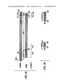 |
 |  |
 |  |
 | 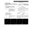 |
 |  |
 |  |
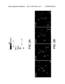 |  |
| Similar patent applications: | |
| Date | Title |
|---|---|
| 2010-02-25 | Bacterial leader sequences for increased expression |
| 2010-05-13 | Template-directed assembly of receptor signaling complexes |
| 2012-08-09 | Fragment ligated inhibitors selective for the polo box domain of plk1 |
| 2010-12-02 | Novel receptor trem (triggering receptor expressed on myeloid cells) and uses thereof |
| 2009-08-27 | Optimized fc variants and methods for their generation |
| New patent applications in this class: | |
| Date | Title |
|---|---|
| 2022-05-05 | Bispecific antibodies that bind pd-l1 and cd28 |
| 2022-05-05 | Polypeptide modification method for purifying polypeptide multimers |
| 2019-05-16 | Generating targeted sequence diversity in fusion proteins |
| 2019-05-16 | Anti-pd-1 monoclonal antibody and obtaining method therefor |
| 2019-05-16 | Modified chimeric antigen receptors and methods of use |
| New patent applications from these inventors: | |
| Date | Title |
|---|---|
| 2010-10-14 | Methods of screening for modulators of cell proliferation and methods of diagnosing cell proliferation states |
| 2009-09-03 | Certain compositions and methods of treatment |
| 2009-05-07 | Compositions and assays utilizing adp or phosphate for detecting protein modulators |
| 2009-01-29 | Compositions and assays utilizing adp or phosphate for detecting protein modulators |
| Top Inventors for class "Chemistry: natural resins or derivatives; peptides or proteins; lignins or reaction products thereof" | |
| Rank | Inventor's name |
|---|---|
| 1 | Kevin I. Segall |
| 2 | Martin Schweizer |
| 3 | John R. Desjarlais |
| 4 | Brent E. Green |
| 5 | David M. Goldenberg |