Patent application title: Multimetric Biosensors and Methods of Using Same
Inventors:
Thijs Kaper (Palo Alto, CA, US)
Wolf B Frommer (Stanford, CA, US)
IPC8 Class: AC12N1511FI
USPC Class:
424 96
Class name: Drug, bio-affecting and body treating compositions in vivo diagnosis or in vivo testing diagnostic or test agent produces in vivo fluorescence
Publication date: 2008-12-18
Patent application number: 20080311047
Inventors list |
Agents list |
Assignees list |
List by place |
Classification tree browser |
Top 100 Inventors |
Top 100 Agents |
Top 100 Assignees |
Usenet FAQ Index |
Documents |
Other FAQs |
Patent application title: Multimetric Biosensors and Methods of Using Same
Inventors:
Thijs Kaper
Wolf B. Frommer
Agents:
MORGAN LEWIS & BOCKIUS LLP
Assignees:
Origin: WASHINGTON, DC US
IPC8 Class: AC12N1511FI
USPC Class:
424 96
Abstract:
Multimeric tryptophan biosensors are disclosed, which comprise
tryptophan-binding domains conjugated to donor and fluorescent moieties
that permit detection and measurement of Fluorescence Resonance Energy
Transfer upon tryptophan binding. Such biosensors are useful for real
time monitoring of tryptophan metabolism in living cells.Claims:
1. An isolated nucleic acid which encodes a ligand binding fluorescent
indicator, the indicator comprising:at least one ligand binding protein
moiety of a multimeric ligand binding protein complex;a donor fluorophore
moiety fused to the ligand binding protein moiety; andan acceptor
fluorophore moiety fused to the ligand binding protein moiety;wherein
fluorescence resonance energy transfer (FRET) between the donor moiety
and the acceptor moiety is altered when the donor moiety is excited and
said ligand binds to the ligand binding protein moiety.
2. The isolated nucleic acid of claim 1, wherein said multimeric ligand binding protein complex is selected from the group consisting of dimers, trimers, tetramers and hexamers.
3. The isolated nucleic acid of claim 2, wherein said multimeric ligand binding protein complex is a dimer.
4. The isolated nucleic acid of claim 3, wherein said multimeric ligand binding protein complex is a homodimer.
5. The isolated nucleic acid of claim 1, comprising at least two ligand binding protein moieties from separate proteins of a multimeric ligand binding protein complex.
6. The isolated nucleic acid of claim 5, wherein said multimeric ligand binding protein complex is selected from the group consisting of dimers, trimers, tetramers and hexamers.
7. The isolated nucleic acid of claim 6, wherein said multimeric ligand binding protein complex is a dimer.
8. The isolated nucleic acid of claim 7, wherein said multimeric ligand binding protein complex is a homodimer.
9. The isolated nucleic acid of claim 7, wherein said ligand binding fluorescent indicator comprises a ligand binding single chain dimer fused to donor and acceptor fluorophores.
10. The isolated nucleic acid of claim 5, wherein said ligand binding fluorescent indicator comprises a structure according to the following formula (I):A-B-C-D, (I)wherein A and C are fluorophore moieties, and B and D are ligand binding protein moieties.
11. The isolated nucleic acid of claim 5, wherein said ligand binding fluorescent indicator comprises a structure according to the following formula (I):A-B-C-D, (I)wherein A and C are ligand binding protein moieties, and B and D are fluorophore moieties.
12. The isolated nucleic acid of claim 5, wherein said ligand binding fluorescent indicator comprises a structure according to the following formula (I):A-B-C-D, (I)wherein A and D are ligand binding protein moieties, and B and C are fluorophore moieties.
13. The isolated nucleic acid of claim 5, wherein said ligand binding fluorescent indicator comprises a structure according to the following formula (I):A-B-C-D, (I)wherein A and D are fluorophore moieties, and B and C are ligand binding protein moieties.
14. The isolated nucleic acid of claim 1, wherein said multimeric ligand binding protein complex is selected from the group consisting of repressor proteins, enzymes, ligands, nucleic acid binding proteins, growth regulatory factors, differentiative factors, and chemotactic factors, hormone receptors, steroid receptors, serotonin receptors, dopamine receptors, metabotropic and ionotropic glutamate receptors, insulin receptors, IGF1 receptors, G-protein-coupled receptors, immune cell receptors and antibodies.
15. The isolated nucleic acid of claim 14, wherein said multimeric ligand binding protein complex is a bacterial repressor protein.
16. The isolated nucleic acid of claim 15, wherein the bacterial repressor protein is selected from the group consisting of lactose, galactose, purine, tetracycline, tyrosine, multidrug-binding protein QacR, arabinose (AraC), mercury (MerR), and tryptophan repressor proteins, histone deacetylase (HDAC), MEF2-interacting transcription repressor (MITR), silencing mediator for retinoid and thyroid hormone receptors (SMRT), nuclear corepressor (N-CoR), Small Unique Nuclear receptor CoRepressor (SUN-CoR), TG interacting factor (TGIF).
17. The isolated nucleic acid of claim 16, wherein the bacterial repressor protein is a tryptophan repressor protein.
18. The isolated nucleic acid of claim 16, wherein the bacterial repressor protein is a purine repressor protein.
19. The isolated nucleic acid of claim 1, wherein the donor and acceptor moieties are genetically fused to said binding protein moiety.
20. The isolated nucleic acid of claim 19, wherein the donor and acceptor moieties are genetically fused to the termini of the binding protein moiety.
21. The isolated nucleic acid of claim 19, wherein one or both the donor and acceptor moieties are genetically fused to an internal position of said ligand binding protein moiety.
22. The isolated nucleic acid of claim 1, wherein said donor fluorophore is selected from the group consisting of a GFP, a CFP, a BFP, a YFP, a dsRED, CoralHue Midoriishi-Cyan (MiCy) and monomeric CoralHue Kusabira-Orange (mKO).
23. The isolated nucleic acid of claim 1, wherein said acceptor fluorophore moiety is selected from the group consisting of a GFP, a CFP, a BFP, a YFP, a dsRED, CoralHue Midoriishi-Cyan (MiCy) and monomeric CoralHue Kusabira-Orange (mKO).
24. The isolated nucleic acid of claim 22, wherein said donor fluorophore moiety is a genetically altered version of eCFP.
25. The isolated nucleic acid of claim 24, wherein said ligand binding moiety nucleic acid sequence contains the sequence SEQ ID NO: 1.
26. The isolated nucleic acid of claim 1, wherein said acceptor fluorophore moiety is a genetically altered version of YFP VENUS.
27. The isolated nucleic acid of claim 26, wherein said fluorophore nucleic acid sequence is selected from the group consisting of the sequence SEQ ID NOs: 2, 4, and 6.
28. A cell expressing the nucleic acid of claim 1.
29. An expression vector comprising the nucleic acid of claim 1.
30. A cell comprising the vector of claim 29.
31. The expression vector of claim 29 adapted for function in a prokaryotic cell.
32. The expression vector of claim 29 adapted for function in a eukaryotic cell.
33. The cell of claim 30, wherein the cell is a prokaryote.
34. The cell of claim 33, wherein the cell is E. coli.
35. The cell of claim 26, wherein the cell is a eukaryotic cell.
36. The cell of claim 35, wherein the cell is a yeast cell.
37. The cell of claim 35, wherein the cell is an animal cell.
38. The cell of claim 35, wherein said cell is a plant cell.
39. A transgenic animal expressing the nucleic acid of claim 1.
40. A transgenic plant expressing the nucleic acid of claim 1.
41. The isolated nucleic acid of claim 1, further comprising one or more nucleic acid substitutions that modify the affinity of the ligand binding protein moiety to said ligand.
42. A ligand binding fluorescent indicator encoded by the nucleic acid of claim 1.
43. A method of detecting changes in the level of a ligand in a sample, comprising:(a) providing a cell expressing the nucleic acid of claim 1 and a sample comprising said ligand; and(b) detecting a change in FRET between said donor fluorophore moiety and said acceptor fluorophore moiety,wherein a change in FRET between said donor moiety and said acceptor moiety indicates a change in the level of said ligand in the sample.
44. The method of claim 43, wherein the step of determining FRET comprises measuring light emitted from the acceptor fluorophore moiety.
45. The method of claim 43, wherein determining FRET comprises measuring light emitted from the donor fluorophore moiety, measuring light emitted from the acceptor fluorophore moiety, and calculating a ratio of the light emitted from the donor fluorophore moiety and the light emitted from the acceptor fluorophore moiety.
46. The method of claim 43, wherein the step of determining FRET comprises measuring the excited state lifetime of the donor moiety.
47. The method of claim 43, wherein said cell is contained in vivo.
48. The method of claim 43, wherein said cell is contained in vitro.
49. The method of claim 43, wherein fluorescence resonance energy transfer (FRET) between the donor moiety and the acceptor moiety is increased when the donor moiety is excited and said ligand binds to the ligand binding protein moiety.
50. The method of claim 43, wherein fluorescence resonance energy transfer (FRET) between the donor moiety and the acceptor moiety is decreased when the donor moiety is excited and said ligand binds to the ligand binding protein moiety.
Description:
RELATED APPLICATIONS
[0001]This application claims the benefit of priority of U.S. Provisional Application 60/736,878, filed Nov. 16, 2005.
FIELD OF INVENTION
[0003]The invention relates generally to the construction of multimeric ligand binding biosensors and methods for measuring and detecting changes in ligand concentration using fluorescence resonance energy transfer (FRET). In particular, the invention provides single chain protein sensors constructed from dimeric proteins such as the tryptophan repressor and other multimeric ligand binding proteins.
BACKGROUND OF INVENTION
[0004]All publications and patent applications herein are incorporated by reference to the same extent as if each individual publication or patent application was specifically and individually indicated to be incorporated by reference.
[0005]The publications discussed herein are provided solely for their disclosure prior to the filing date of the present application. Nothing herein is to be construed as an admission that the present invention is not entitled to antedate such publication by virtue of prior invention.
[0006]This application is related to International Application PCT/US05/36956, International Application PCT/US05/36957, International Application PCT/US05/36954, International Application PCT/US05/36952, International Application PCT/US05/36957, International Application PCT/US05/36954, and International Application PCT/US05/36953, which are herein incorporated by reference in their entireties.
[0007]Tryptophan (Trp or W) is an essential amino acid for mammals, which rely on dietary intake of tryptophan to meet its daily requirements. Tryptophan has a number of interesting medicinal qualities including treatment of insomnia as well as an adjunct in the treatment of a number of psychiatric disorders. Tryptophan levels in human cells depend on transport of tryptophan across the cell membrane. Defects in tryptophan transport in cells or organs may lead to various disorders. For instances, Hartnup disease is an autosomal recessive disorder caused by defective transport of neutral (i.e., monoaminomonocarboxylic) amino acids such as tryptophan in the small intestine and the kidneys.
[0008]After absorption, tryptophan circulates in the blood as approximately 80% bound to plasma albumin with the remaining 20% circulating as free tryptophan, and under appropriate conditions, tryptophan is transported into the brain. Once across the blood brain barrier (BBB), tryptophan becomes available for metabolism into serotonin, a neurotransmitter implicated in mood, hunger, and sleep. Low levels of serotonin are associated with depression, fibromyalgia, chronic pain, altered mood, insomnia, PMS, and headaches. Tryptophan metabolism to serotonin also serves well in conditions where depleted serotonin levels exist such as anxiety disorders, obsessive-compulsive disorders, aggression and eating disorders. Parkinson's disease is primarily due to the hypofunction of serotonin nerves, in which serotonin levels are related directly to tryptophan levels.
[0009]Subsequently, serotonin, in turn, is metabolized to melatonin, a sleep related hormone produced especially at night in the pineal gland, a small cone-like structure in the epithalamus of the brain that regulates the 24-hour circadian rhythm in humans. Ingestion of a sufficient quantity of tryptophan per se consistently results in reduced sleep latency i.e. the time from "lights out" to sleep, and an improvement in overall quality of sleep through improved sleep architecture (Boman, 1988).
[0010]In plants, L-tryptophan is a precursor for auxin, a plant hormone critical for plant growth and that orchestrates many developmental processes. Though many natural and synthetic compounds exhibit auxin-like activity in bioassays, indole-3-acetic acid (IAA) is recognized as the key auxin in most plants. Auxin regulates plant tropic responses (growth toward or away from environmental signals) and apical dominance (repression of branch outgrowth by cells at the shoot tip). Plant growth in response to gravity and light requires asymmetrically distributed auxin across the stem or root. This causes one side to grow more than the other. Similarly, the production of auxin by the "apically dominant" shoot tip, followed by its transport down through the stem, represses the outgrowth of lateral buds.
[0011]In bacteria, tryptophan synthesis is regulated by the tryptophan repressor protein (TrpR). TrpR regulates gene expression of the E. coli trpR, trp EDCBA and aroH operons. Purified protein, when activated with L-tryptophan binds to operator DNA sequences (Gunsalus et al., 1980), thus blocking transcription of the structural genes for tryptophan synthesis. The functional unit of TrpR is a dimer in which five of the six helices are interlinked (Schevitz et al. 1985). Two TrpR molecules are necessary to make up two functional binding sites. Binding of L-tryptophan by the TrpR dimer results in conformational changes which promote binding to DNA (Zhang et al. 1987). The L-tryptophan molecule in the TrpR-L-tryptophan complex is directly involved in the interaction with DNA (Otwinowski et al. 1988). TrpR is able to bind a wide variety of tryptophan analogues with varying affinities (Marmorstein et al. 1987). Several of the resulting complexes of TrpR and tryptophan analogues are able to bind to the trp operon sequence (Marmorstein et al. 1989).
[0012]Given the important roles tryptophan plays in the normal functioning of plants and living organisms, it would be desirable to provide convenient and real time methods of monitoring tryptophan levels in vitro and in vivo. To be able to measure tryptophan levels directly in living cells, it would be useful to have a nanosensor for tryptophan and its analogs. A tryptophan sensor would be an excellent tool for discovery and drug screening. The response of tryptophan levels could be measured in real time in response to chemicals, metabolic events, transport steps, and signaling processes.
[0013]Recently a number of bacterial periplasmic binding proteins (PBP), which undergo a venus flytrap-like closure of two lobes upon substrate binding, have been successfully used as the scaffold of metabolite nanosensors (Fehr et al. 2002; Fehr et al. 2003; Lager et al. 2003). Based on these findings, various fluorescent indicator proteins have been developed for the detection of metabolites such as glucose (Fehr et al. 2004), maltose (Fehr et al. 2002), ribose (Lager et al. 2003) glutamate (Okumoto et al. 2005) (International Application PCT/US05/36956), phosphate (International Application PCT/US05/36955), sucrose (International Application PCT/US05/36951) and polyamine (International Application PCT/US05/36952), each of which is herein incorporated by reference in its entirety.
[0014]These sensors consist of a protein of the periplasmic binding protein family, sandwiched between a pair of green fluorescent protein variants fluorescence capable of resonance energy transfer (FRET), the efficiency of which depends on the distance and orientation of the fluorophores. Ligand-binding induced conformational changes in such sensors result in altered FRET signals, which are a measure for the levels of the respective metabolites. The successful development of these biosensors has suggested to the inventors that a tryptophan biosensor may also be constructed because it has been observed that the tryptophan repressor protein also undergoes conformational changes upon binding of L-tryptophan. However, unlike periplasmic binding proteins which are monomers, bacterial tryptophan repressor proteins as discussed above function as dimers.
[0015]Recently, FRET has been successfully used to detect formation of multimeric protein complexes. For instance, FRET technology has been used in the detection of multimeric complex formation of estrogen receptor and nuclear coactivators (Liu et al. 2003). FRET has also been applied to the study of homomultimerization of the coxsackievirus 2B protein in living cells by a FRET biosensor comprising one component of the homomultimeric complex such as 2B fused to the fluorescent protein (Van Kuppeveld et al. 2002) and dimerization of mammalian adenylate cyclases by cotransfecting different single unit sensors into the cells (Gu et al. 2002). However, none of these studies has designed and employed single chain biosensors with multimeric or dimeric moieties.
SUMMARY OF INVENTION
[0016]The present inventors have surprisingly found that multimeric biosensors may be successfully constructed by incorporating multiple copies of genes of interest into constructs of the biosensors. Thus, the present invention provides an isolated nucleic acid which encodes a ligand binding fluorescent indicator, the indicator comprising at least one ligand binding protein moiety of a multimeric ligand binding protein complex, a donor fluorophore moiety fused to the ligand binding protein moiety, and an acceptor fluorophore moiety fused to the ligand binding protein moiety.
[0017]The present invention further provides tryptophan biosensors that may be used for detecting and measuring changes in tryptophan concentrations in living cells and optimization of the sensors with multimeric tryptophan repressor domains by encoding multiple copies of tryptophan repressor domains in single gene products. Further, the present invention provides use of repressor and/or DNA binding and/or RNA synthesis regulatory proteins for the construction of multimeric ligand binding protein sensors.
[0018]In particular, the invention provides an isolated nucleic acid which encodes a tryptophan fluorescent indicator, the indicator comprising at least one tryptophan binding protein moiety of a dimeric tryptophan repressor protein complex, a donor fluorescent protein moiety covalently coupled to the tryptophan binding protein moiety, and an acceptor fluorescent protein moiety covalently coupled to the at least one tryptophan binding protein moiety, wherein fluorescence resonance energy transfer (FRET) between the donor moiety and the acceptor moiety is altered when the donor moiety is excited and tryptophan binds to the tryptophan binding protein moiety. Vectors, including expression vectors, and host cells comprising the inventive nucleic acids are also provided, as well as biosensor proteins encoded by the nucleic acids. Such nucleic acids, vectors, host cells and proteins may be used in methods of detecting tryptophan binding and changes in levels of tryptophan, and in methods of identifying compounds that modulate tryptophan binding or tryptophan-mediated activities.
BRIEF DESCRIPTION OF THE DRAWINGS
[0019]FIGS. 1(A)-1(D) show tryptophan sensors based on the E. coli tryptophan repressor TrpR. (A) TrpR dimer (yellow, red) in complex with L-tryptophan (black) bound to the trp operator (green, blue) (PDB: 1TRO (Otwinowski et al. 1988)). (B) Constructed FLIPW variants. (C) Normalized FRET ratio change of FLIPW-CTY in presence of L-tryptophan (red squares), D-tryptophan (cyan circles), 5-hydroxy-L-tryptophan (yellow squares) and 5-methyl-L-tryptophan (green triangles). (D) Normalized FRET ratio change of FLIPW-CTY (red squares), FLIPW-TCTY (cyan circles), FLIPW-CTYT (green triangles), and FLIPW-CTTY (yellow squares) in the presence of L-tryptophan.
[0020]FIG. 2 shows the plasmid map of pTK164, DNA sequence of pTK164 and protein sequence of FLIPW-CTY (SEQ ID NOs: 2 and 3).
[0021]FIG. 3 shows the plasmid map of pTK203, DNA sequence of pTK203 and protein sequence of FLIPW-TCTY (SEQ ID NOs: 4 and 5).
[0022]FIG. 4 shows the plasmid map of pTK204, DNA sequence of pTK204 and protein sequence of FLIPW-CTYT (SEQ ID NOs: 6 and 7).
[0023]FIG. 5 shows the plasmid map of pTK205, DNA sequence of pTK205 and protein sequence of FLIPW-CTTY (SEQ ID NOs: 8 and 9).
[0024]FIG. 6 shows the plasmid map of pTK222 and DNA sequence of pTK222 (SEQ ID NO: 10). FLIPW-CTYT as encoded on pTK204 (FIG. 4, SEQ ID NO: 7).
[0025]FIGS. 7(A)-7(B) show structural models of FLIPW-CTY and FLIPW-CTYT tryptophan sensors. TrpR: green, magenta (PDB: 1WRP; PDB: Protein data bank at http://www.rcsb.org/pdb/home/home.do), eCFP: blue (based on PDB: 1MYW) and Venus: yellow (PDB: 1MYW). (A) Dimer of two FLIPW-CTY chains resulting in a TrpR dimer that can bind tryptophan. (B) FLIPW-CTYT monomer.
[0026]FIGS. 8(A)-8(B) show uptake of tryptophan by COS-7 cell cultures in 96-well microplates monitored with FLIPW-CTYT. (a) FRET ratio change of cell cultures in presence of Tyrode's buffer (squares) and 100 μM L-Trp in Tyrode's buffer (circles). Data correspond to means±S.E. (n=12). (b) Velocity of intracellular FLIPW-CTYT response versus external tryptophan concentration fitted with the Michaelis-Menten equation. Cells were incubated with 0.05, 0.1, 0.25, 0.5, 1, 5, 10 and, 25 μM L-Trp. Data correspond to means±S.E. (n=6).
[0027]FIGS. 9(A)-9(B) show hypoxanthine sensor based on the corepressor-binding domain of E. coli PurR. (a) PurR dimer (red, yellow) in complex with hypoxanthine (black) bound to the purF operator site (blue) (PDB: 1PNR (22)). (b) Saturation of the FLIPpur sensor in the presence of hypoxanthine.
[0028]FIG. 10 shows relative position of the components of the FLIPW-CTYT sensor. The TrpR dimer (green, magenta, PDB: 1WRP) and Venus (yellow, PDB: 1MYW) are modeled to be sterically compatible, with the termini approaching within 1 Å.
[0029]FIGS. 11(A)-11(B) show structural models of FLIPW-TCTY and FLIPW-CTYT tryptophan sensors. TrpR: green, magenta (PDB: 1WRP), eCFP: blue (based on PDB: 1MYW) and Venus: yellow (PDB: 1MYW). (a) FLIPW-TCTY monomer, (b) FLIPW-CTYT monomer.
[0030]FIG. 12 shows perfusion of HEK293T cells transfected with pTK222. Cells were perfused with Tyrode's buffer. Between 1'30'' and 3' (indicated by triangles) buffer was supplemented with 10 μM L-tryptophan. Response of the sensor is determined from the ratio of fluorescence output at 528 nm and 485 nm. During perfusion with tryptophan, the intracellular tryptophan levels increase. The Trp levels decrease during subsequent perfusion with buffer due to efflux and metabolism.
DETAILED DESCRIPTION OF THE INVENTION
[0031]The following description includes information that may be useful in understanding the present invention. It is not an admission that any of the information provided herein is prior art or relevant to the presently claimed inventions, or that any publication specifically or implicitly referenced is prior art.
[0032]Other objects, advantages and features of the present invention become apparent to one skilled in the art upon reviewing the specification and the drawings provided herein. Thus, further objects and advantages of the present invention will be clear from the description that follows.
[0033]Biosensors
[0034]The present invention provides biosensors of multimeric ligand binding proteins for detecting and measuring changes in analyte concentrations using Fluorescence Resonance Energy Transfer (FRET). One embodiment, among others, is an isolated nucleic acid which encodes a ligand binding fluorescent indicator, the indicator comprising: at least one ligand binding protein moiety of a multimeric ligand binding protein complex, a donor fluorescent protein moiety covalently coupled to the ligand binding protein moiety, and an acceptor fluorescent protein moiety covalently coupled to the ligand binding protein moiety, wherein FRET between the donor moiety and the acceptor moiety is altered when the donor moiety is excited and ligand binds to the ligand binding protein moiety.
[0035]As used herein, the term "multimer" and grammatical variations thereof refer to formation of a multimeric complex between two or more distinct molecules. The multimer complex may comprise, for example, two or more molecules of the same protein (e.g., a homo-dimer, -trimer, -tetramer or higher order multimer) or a mixture of two or more different (i.e., non-identical) proteins (e.g. a hetero-dimer, -trimer, -tetramer or higher multimer). For example, multimeric antibodies may comprise the same antibody or two or more different antibodies, each of which have two or more functions or activities (e.g., bind to two or more epitopes).
[0036]As used herein, "covalently coupled" means that the donor and acceptor fluorescent moieties may be conjugated to the ligand binding protein moiety via a chemical linkage, for instance to a selected amino acid in said ligand binding protein moiety. Covalently coupled also means that the donor and acceptor moieties may be genetically fused to the ligand binding protein moiety such that the ligand binding protein moiety is expressed as a fusion protein comprising the donor and acceptor moieties.
[0037]The isolated nucleic acid that encodes the multimeric ligand binding protein moiety can be any nucleic acid, and preferably is the nucleic acid that encodes portions of multimeric proteins. In one embodiment, the isolated nucleic acid of interest encodes a hetero- or homo-dimer, -trimer, -tetramer, -pentamer, -hexamer or higher order multimer. Multimeric proteins may be selected, for example, from a binding protein (e.g. an antigen binding polypeptide), enzyme, receptor, ligand, nucleic acid binding protein (e.g. a repressor protein binding DNA), growth regulatory factor, differentiative factor, and chemotactic factor. For example, the repressor protein, lac repressor acts as a tetramer and the tyrosine repressor acts as a hexamer.
[0038]Nucleic acids encoding protein and peptide hormones are a preferred class of nucleic acids of interest in the present invention. Such protein and peptide hormones are synthesized throughout the endocrine system and include, but are not limited to, hypothalamic hormones and hypophysiotropic hormones, anterior, intermediate and posterior pituitary hormones, pancreatic islet hormones, hormones made in the gastrointestinal system, renal hormones, thymic hormones, parathyroid hormones, adrenal cortical and medullary hormones. Specifically, hormones that can be utilized by the present invention include, but are not limited to, chorionic gonadotropin, corticotropin, erythropoietin, glucagons, IGF-1, oxytocin, platelet-derived growth factor, vascular endothelial growth factor, calcitonin, follicle-stimulating hormone, luteinizing hormone, thyroid-stimulating hormone, insulin, gonadotropin-releasing hormone and its analogs, vasopressin, octreotide, somatostatin, prolactin, adrenocorticotropic hormone, antidiuretic hormone, thyrotropin-releasing hormone (TRH), growth hormone-releasing hormone (GHRH), dopamine, melatonin, thyroxin (T4), parathyroid hormone (PTH), glucocorticoids such as cortisol, mineralocorticoids such as aldosterone, androgens such as testosterone, adrenaline (epinephrine), noradrenaline (norepineplirine), estrogens such as estradiol, progesterone, glucagons, calcitrol, calciferol, atrial-natriuretic peptide, gastrin, secretin, cholecystokinin (CCK), neuropeptide Y, ghrelin, PYY3-36, angiotensinogen, thrombopoietin, and leptin.
[0039]Further included in the present invention are nucleic acids of interest that encode multimeric receptors. Multimeric receptors include homodimers (e.g., PDGF receptor αα, and ββ isoforms, erythropoietin receptor, MPL, and G-CSF receptor), heterodimers whose subunits each have ligand-binding and effector domains (e.g., PDGF receptor αβ isoform), and multimers having component subunits with disparate functions (e.g., IL-2, IL-3, IL-4, IL-5, IL-6, IL-7, and GM-CSF receptors). Some receptor subunits are common to a plurality of receptors. For example, the AIC2B subunit, which cannot bind ligand on its own but includes an intracellular signal transduction domain, is a component of IL-3 and GM-CSF receptors. Many cytokine receptors can be placed into one of four related families on the basis of the structure and function. Hematopoietic receptors, for example, are characterized by the presence of a domain containing conserved cysteine residues and the WSXWS motif. Cytokine receptor structure has been reviewed by Urdal, Ann. Reports Med. Chem. 26:221-228, 1991 and Cosman, Cytokine 5:95-106, 1993. Under selective pressure for organisms to acquire new biological functions, new receptor family members likely arise from duplication of existing receptor genes leading to the existence of multi-gene families. Family members thus contain vestiges of the ancestral gene, and these characteristic features can be exploited in the isolation and identification of additional family members. Thus, the cytokine receptor superfamily is subdivided into several families, for example, the immunoglobulin family (including CSF-1, MGF, IL-1, and PDGF receptors); the hematopoietin family (including IL-2 receptor β-subunit, GM-CSF receptor α-subunit, GM-CSF receptor β-subunit; and G-CSF, EPO, IL-3, IL-4, IL-5, IL-6, IL-7, and IL-9 receptors); TNF receptor family (including TNF (p80) TNF (p60) receptors, CD27, CD30, CD40, Fas, and NGF receptor). Multimeric receptors also include hormone receptors (TSH, FSH, CG, VEGF, PDGF, EGF, etc.), steroid receptors, serotonin receptors, dopamine receptors, metabotropic and ionotropic glutamate receptors, insulin receptors, IGF1 receptors, G-protein-coupled receptors (including leukotriene B(4) receptor and BLT1 as dimer).
[0040]Other multimeric proteins that may be utilized using the present invention are as follows: factors involved in the synthesis or replication of DNA, such as DNA polymerase alpha and DNA polymerase delta; proteins involved in the production of mRNA, such as TFIID and TFIIH; cell, nuclear and other membrane-associated proteins, such as hormone and other signal transduction receptors, active transport proteins and ion channels, multimeric proteins in the blood, including hemoglobin, fibrinogen and von Willabrand's Factor; proteins that form structures within the cell, such as actin, myosin, and tubulin and other cytoskeletal proteins; proteins that form structures in the extra cellular environment, such as collagen, elastin and fibronectin; proteins involved in intra- and extra-cellular transport, such as kinesin and dynein, the SNARE family of proteins (soluble NSF attachment protein receptor) and clathrin; proteins that help regulate chromatin structure, such as histones and protamines, Swi3p, Rsc8p and moira; multimeric transcription factors such as Fos, Jun and CBTF (CCAAT box transcription factor); multimeric enzymes such as acetylcholinesterase and alcohol dehydrogenase; chaperone proteins such as GroE, Gro EL (chaperonin 60) and Gro ES (chaperonin 10); anti-toxins, such as snake venom, botulism toxin, Streptococcus super antigens; lysins (enzymes from bacteriophage and viruses); as well as most allosteric proteins.
[0041]In another embodiment, the present invention provides an isolated nucleic acid which encodes a ligand binding fluorescent indicator, the indicator comprising: at least one ligand binding protein from a repressor protein, a donor fluorescent protein moiety covalently coupled to the ligand binding protein moiety, and an acceptor fluorescent protein moiety covalently coupled to the ligand binding protein moiety, wherein FRET between the donor moiety and the acceptor moiety is altered when the donor moiety is excited and ligand binds to the ligand binding protein moiety. As used herein, the term "repression" refers to transcriptional repression as by a transcriptional repressor such as a DNA binding transcriptional repressor, which binds a target promoter to be repressed.
[0042]Suitable repressor proteins may also include, but are not limited to, lactose, galactose, purine, tetracycline, tyrosine, tryptophan repressor proteins, multidrug-binding protein QacR, arabinose (AraC), mercury (MerR), histone deacetylase (HDAC), MEF2-interacting transcription repressor (MITR), silencing mediator for retinoid and thyroid hormone receptors (SMRT), nuclear corepressor (N-CoR), Small Unique Nuclear receptor CoRepressor (SUN-CoR), TG interacting factor (TGIF), Sloan Kettering virus oncogene homolog (Ski), Ski-related novel gene (Sno), NGFI-A-binding protein (NAB), or Friend of GATA (FOG).
[0043]In yet another embodiment, the invention provides isolated nucleic acids encoding tryptophan binding fluorescent indicators and the tryptophan fluorescent indicators encoded thereby. The embodiment, among others, is an isolated nucleic acid which encodes a tryptophan binding fluorescent indicator, the indicator comprising: at least one tryptophan binding protein moiety of a multimeric ligand binding protein complex, a donor fluorescent protein moiety covalently coupled to the tryptophan binding protein moiety, and an acceptor fluorescent protein moiety covalently coupled to the tryptophan binding protein moiety, wherein FRET between the donor moiety and the acceptor moiety is altered when the donor moiety is excited and tryptophan binds to the tryptophan binding protein moiety.
[0044]As an example, the tryptophan binding protein moiety, among others, is a tryptophan binding protein moiety from E. coli having the following sequence:
MAQQSPYSAA MAEQRHQEWL RFVDLLKNAY QNDLHLPLLN LMLTPDEREA LGTRVRIVEE LLRGEMSQRE LKNELGAGIA TITRGSNSLK AAPVELRQWL EEVLLKSD (SEQ ID NO: 1).
[0045]Any portion of the tryptophan repressor DNA sequence which encodes a tryptophan binding region may be used in the nucleic acids of the present invention. Tryptophan binding portions of tryptophan binding protein (BP) or any of its homologues from other organisms, for instance Gram negative bacteria including thermophilic and hyperthermophilic organisms, may be cloned into the vectors described herein and screened for activity according to the disclosed assays. Ligand binding proteins of thermophilic and hyperthermophilic organisms are particularly useful for constructing sensors having increased stability and resistance to heat or harsh environmental conditions. See International Application PCT/US05/36954, which is herein incorporated by reference in its entirety.
[0046]Naturally occurring species variants of tryptophan BP may also be used, in addition to artificially engineered variants comprising site-specific mutations, deletions or insertions that maintain measurable tryptophan binding function. Variant nucleic acid sequences suitable for use in the nucleic acid constructs of the present invention will preferably have at least 30, 40, 50, 60, 70, 75, 80, 85, 90, 95, or 99% similarity or identity to the gene sequence for tryptophan BP. Suitable variant nucleic acid sequences may also hybridize to the gene for tryptophan BP under highly stringent hybridization conditions. High stringency conditions are known in the art; see for example Maniatis et al., Molecular Cloning: A Laboratory Manual, 2d Edition, 1989, and Short Protocols in Molecular Biology, ed. Ausubel, et al., both of which are hereby incorporated by reference. Stringent conditions are sequence-dependent and will be different in different circumstances. Longer sequences hybridize specifically at higher temperatures. An extensive guide to the hybridization of nucleic acids is found in Tijssen, Techniques in Biochemistry and Molecular Biology--Hybridization with Nucleic Acid Probes, "Overview of principles of hybridization and the strategy of nucleic acid assays" (1993), which is herein incorporated by reference. Generally, stringent conditions are selected to be about 5-10° C. lower than the thermal melting point (Tm) for the specific sequence at a defined ionic strength pH. The Tm is the temperature (under defined ionic strength, pH and nucleic acid concentration) at which 50% of the probes complementary to the target hybridize to the target sequence at equilibrium (as the target sequences are present in excess, at Tm, 50% of the probes are occupied at equilibrium). Stringent conditions will be those in which the salt concentration is less than about 1.0 M sodium ion, typically about 0.01 to 1.0M sodium ion concentration (or other salts) at pH 7.0 to 8.3 and the temperature is at least about 30° C. for short probes (e.g. 10 to 50 nucleotides) and at least about 60° C. for long probes (e.g. greater than 50 nucleotides). Stringent conditions may also be achieved with the addition of destabilizing agents such as formamide.
[0047]Preferred artificial variants of the present invention may be designed to exhibit decreased affinity for the ligand, in order to expand the range of ligand concentration that can be measured by the disclosed nanosensors. Additional artificial variants showing decreased or increased binding affinity for ligands may be constructed by random or site-directed mutagenesis and other known mutagenesis techniques, and cloned into the vectors described herein and screened for activity according to the disclosed assays. The binding specificity of disclosed biosensors may also be altered by mutagenesis so as to alter the ligand recognized by the biosensor. See, for instance, Looger et al., Nature, 423 (6936): 185-190.
[0048]The sensors of the invention may also be designed with tryptophan binding moieties and one or more additional protein binding moieties that are covalently coupled or fused together and to the donor and acceptor fluorescent moieties in order to generate an allosteric enzyme whose activity is controlled by more than one ligand. Allosteric enzymes containing dual specificity for more than one ligand have been described in the art, and may be used to construct the FRET biosensors described herein (Guntas and Ostermeier, 2004, J. Mol. Biol. 336(1): 263-73).
[0049]As described herein, the donor and acceptor moieties may be fused to the termini of the at least one ligand binding moiety of a multimeric ligand binding protein complex or to an internal position within the at least one ligand binding moiety of a multimeric ligand binding protein complex so long as FRET between the donor moiety and the acceptor moiety is altered when the donor moiety is excited and a ligand binds to the ligand binding protein moiety. See International Application PCT/US05/36957, which is herein incorporated by reference in its entirety.
[0050]The isolated nucleic acids of multimeric binding protein complex of the invention may comprise a structure according to the following formula (I):
A-B-C-D (I)
wherein A and C are fluorophore moieties, and B and D are ligand binding protein moieties.
[0051]In another embodiment, the present invention provides an isolated nucleic acid, wherein said ligand binding fluorescent indicator comprises a structure of formula (I), wherein A and C are ligand binding protein moieties, and B and D are fluorophore moieties.
[0052]In yet another embodiment, the present invention provides an isolated nucleic acid, wherein said ligand binding fluorescent indicator comprises a structure of formula (I), wherein A and D are ligand binding protein moieties, and B and C are fluorophore moieties.
[0053]In yet another embodiment, the present invention provides an isolated nucleic acid, wherein said ligand binding fluorescent indicator comprises a structure of formula (I), wherein A and D are fluorophore moieties, and B and C are ligand binding protein moieties.
[0054]The ligand binding protein moieties may be from separate proteins of a multimeric ligand binding protein complex. Thus, the present invention provides an isolated nucleic acid with two or more polynucleotide moieties, each of which encodes a ligand binding protein that forms a part of the multimeric protein complex wherein the nucleic acid encodes a protein comprising a donor fluorophore moiety fused to the two or more ligand binding protein moieties, and an acceptor fluorophore moiety fused to the two or more ligand binding protein moieties.
[0055]The isolated nucleic acids of the invention may incorporate any suitable donor and acceptor fluorescent protein moieties that are capable in combination of serving as donor and acceptor moieties in FRET. Preferred donor and acceptor moieties are selected from the group consisting of GFP (green fluorescent protein), CFP (cyan fluorescent protein), BFP (blue fluorescent protein), YFP (yellow fluorescent protein), and enhanced variants thereof such as enhanced YFP (EYFP), with a particularly preferred embodiment provided by the donor/acceptor pair CFP/YFP Venus, a variant of YFP with improved pH tolerance and maturation time (Nagai et al. 2002). A variant of yellow fluorescent protein with fast and efficient maturation for cell-biological applications. Nat. Biotechnol. 20, 87-90). An alternative is the MiCy/mKO pair with higher pH stability and a larger spectral separation (Karasawa et al. 2004). Also suitable as either a donor or acceptor is native DsRed from a Discosoma species, an ortholog of DsRed from another genus, or a variant of a native DsRed with optimized properties (e.g. a K83M variant or DsRed2 (available from Clontech)). Criteria to consider when selecting donor and acceptor fluorescent moieties is known in the art, for instance as disclosed in U.S. Pat. No. 6,197,928, which is herein incorporated by reference in its entirety.
[0056]As used herein, the term "variant" is intended to refer to polypeptides with at least about 30%, 40%, 50%, 60%, 70%, more preferably at least 75% identity, including at least 80%, 90%, 95% or greater identity to native fluorescent molecules. Many such variants are known in the art, or can be readily prepared by random or directed mutagenesis of a native fluorescent molecules (see, for example, Fradkov et al., FEBS Lett. 479:127-130 (2000)).
[0057]When the fluorophores of the biosensor contain stretches of similar or related sequence(s), the present inventors have recently discovered that gene silencing may adversely affect expression of the biosensor in certain cells and particularly whole organisms. In such instances, it is possible to modify the fluorophore coding sequences at one or more degenerate or wobble positions of the codons of each fluorophore, such that the nucleic acid sequences of the fluorophores are modified but not the encoded amino acid sequences. Alternatively, one or more conservative substitutions that do not adversely affect the function of the fluorophores may also be incorporated. See PCT application [Attorney Docket No. 056100-5054, "Methods of Reducing Repeat-Induced Silencing of Transgene Expression and Improved Fluorescent Biosensors], which is herein incorporated by reference in its entirety.
[0058]It is also possible to use or luminescent quantum dots (QD) for FRET (Clapp et al., 2005, J. Am. Chem. Soc. 127(4): 1242-50), dyes, including but not limited to TOTO dyes (Laib and Seeger, 2004, J Fluoresc. 14(2):187-91), Cy3 and Cy5 (Churchman et al., 2005, Proc Natl Acad Sci USA. 102(5): 1419-23), Texas Red, fluorescein, and tetramethylrhodamine (TAMRA) (Unruh et al., Photochem Photobiol. 2004 Oct. 1), AlexaFluor 488, to name a few, as well as fluorescent tags (see, for example, Hoffman et al., 2005, Nat. Methods 2(3): 171-76).
[0059]The invention further provides vectors containing isolated nucleic acid molecules encoding the biosensor polypeptides described herein. Exemplary vectors include vectors derived from a virus, such as a bacteriophage, a baculovirus or a retrovirus, and vectors derived from bacteria or a combination of bacterial sequences and sequences from other organisms, such as a cosmid or a plasmid. Such vectors include expression vectors containing expression control sequences operatively linked to the nucleic acid sequence coding for the biosensor. Vectors may be adapted for function in a prokaryotic cell, such as E. coli or other bacteria, or a eukaryotic cell, including animal cells or plant cells. For instance, the vectors of the invention will generally contain elements such as an origin of replication compatible with the intended host cells, one or more selectable markers compatible with the intended host cells and one or more multiple cloning sites. The choice of particular elements to include in a vector will depend on factors such as the intended host cells, the insert size, whether regulated expression of the inserted sequence is desired, i.e., for instance through the use of an inducible or regulatable promoter, the desired copy number of the vector, the desired selection system, and the like. The factors involved in ensuring compatibility between a host cell and a vector for different applications are well known in the art.
[0060]Preferred vectors for use in the present invention will permit cloning of the tryptophan binding domain or receptor between nucleic acids encoding donor and acceptor fluorescent molecules, resulting in expression of a chimeric or fusion protein comprising the tryptophan binding domain covalently coupled to donor and acceptor fluorescent molecules. Exemplary vectors include the bacterial pFLIP derivatives disclosed in Fehr et al. (2002) (Visualization of maltose uptake in living yeast cells by fluorescent nanosensors, Proc. Natl. Acad. Sci. USA 99, 9846-9851), which is herein incorporated by reference in its entirety. Methods of cloning nucleic acids into vectors in the correct frame so as to express a fusion protein are well known in the art.
[0061]The tryptophan biosensors of the present invention may be expressed in any location in the cell, including the cytoplasm, cell surface or subcellular organelles such as the nucleus, vesicles, ER, vacuole, etc. Methods and vector components for targeting the expression of proteins to different cellular compartments are well known in the art, with the choice dependent on the particular cell or organism in which the biosensor is expressed. See, for instance, Okumoto, S., Looger, L. L., Micheva, K. D., Reimer, R. J., Smith, S. J., and Frommer, W. B. (2005) P Natl Acad Sci USA 102(24), 8740-8745; Fehr, M., Lalonde, S., Ehrhardt, D. W., and Frommer, W. B. (2004) J Fluoresc 14(5), 603-609, which are herein incorporated by reference in their entireties.
[0062]The chimeric nucleic acids of the present invention may be constructed such that the donor and acceptor fluorescent moiety coding sequences are fused to separate termini of the ligand binding domain in a manner such that changes in FRET between donor and acceptor may be detected upon ligand binding. Fluorescent domains can optionally be separated from the ligand binding domain by one or more flexible linker sequences. Such linker moieties are preferably between about 1 and 50 amino acid residues in length, and more preferably between about 1 and 30 amino acid residues. Linker moieties and their applications are well known in the art and described, for example, in U.S. Pat. Nos. 5,998,204 and 5,981,200, and Newton et al., Biochemistry 35:545-553 (1996). Alternatively, shortened versions of linkers or any of the fluorophores described herein may be used. For example, the inventors have shown that deleting N- or C-terminal portions of any of the three modules can lead to increased FRET ratio changes, as described in Application Ser. No. 60/658,141, which is herein incorporated by reference in its entirety.
[0063]It will also be possible depending on the nature and size of the ligand binding domains to insert one or both of the fluorescent molecule coding sequences within the open reading frames of the binding proteins such that the fluorescent moieties are expressed and displayed from a location within the biosensor rather than at the termini. Such sensors are generally described in U.S. application Ser. No. 60/658,141, which is herein incorporated by reference in its entirety. It will also be possible to insert a ligand binding sequence into a single fluorophore coding sequence, i.e. a sequence encoding a GFP, YFP, CFP, BFP, etc., rather than between tandem molecules. According to the disclosures of U.S. Pat. No. 6,469,154 and U.S. Pat. No. 6,783,958, each of which is incorporated herein by reference in their entirety, such sensors respond by producing detectable changes within the protein that influence the activity of the fluorophore.
[0064]The invention also includes host cells transfected with a vector or an expression vector of the invention, including prokaryotic cells, such as E. coli or other bacteria, or eukaryotic cells, such as yeast cells, animal cells or plant cells. In another aspect, the invention features a transgenic non-human animal having a phenotype characterized by expression of the nucleic acid sequence coding for the expression of the environmentally stable biosensor. The phenotype is conferred by a transgene contained in the somatic and germ cells of the animal, which may be produced by (a) introducing a transgene into a zygote of an animal, the transgene comprising a DNA construct encoding the tryptophan biosensor; (b) transplanting the zygote into a pseudopregnant animal; (c) allowing the zygote to develop to term; and (d) identifying at least one transgenic offspring containing the transgene. The step of introducing of the transgene into the embryo can be achieved by introducing an embryonic stem cell containing the transgene into the embryo, or infecting the embryo with a retrovirus containing the transgene. Transgenic animals of the invention include transgenic C. elegans and transgenic mice and other animals. Transgenic plants are also included.
[0065]The present invention also encompasses isolated biosensor molecules having the properties described herein, particularly tryptophan binding fluorescent indicators. Such polypeptides may be recombinantly expressed using the nucleic acid constructs described herein, or produced by chemically coupling some or all of the component domains. The expressed polypeptides can optionally be produced in and/or isolated from a transcription-translation system or from a recombinant cell, by biochemical and/or immunological purification methods known in the art. The polypeptides of the invention can be introduced into a lipid bilayer, such as a cellular membrane extract, or an artificial lipid bilayer (e.g. a liposome vesicle) or nanoparticle.
[0066]Methods of Detecting Ligands
[0067]In one aspect, the present invention provides methods for the rapid and efficient detection of a plurality of ligand samples using a biosensor of multimeric ligand binding moieties. The methods of the invention can be utilized with any ligand. The ligand may be monovalent, divalent or polyvalent. Exemplary ligands that can be used in the methods of the invention include, but are not limited to, proteins, including, but not limited to, antibodies (or fragments thereof), receptors and enzymes; nucleic acids; carbohydrates; lipids; and small molecules. Similarly, the methods of the invention can be used with any binding partner. As used herein, a binding partner is a molecule that binds to one, two or more multimeric ligand binding moieties of the biosensor in a specific manner. The binding partner can be monovalent, bivalent or polyvalent. Exemplary binding partners that can be used in the methods of the invention include, but are not limited to, proteins, including, but not limited to, antigens, polyclonal antibodies, monoclonal antibodies, single chain antibodies, (scFv), F(ab) fragments, F(ab')2 fragments, Fv fragments, receptors and enzymes; nucleic acids; oligonucleotides, carbohydrates such as monosaccharides, disaccharides, polysaccharides; lipids, fatty acids, amino acids, oligopeptides, polypeptides, proteoglycans, glycoprotein, natural or synthetic polymers, and small molecular weight compounds such as drugs or drug candidates, cell, virus, bacteria, and biological sample. A biological sample can be, for example, blood, plasma, serum, gastrointestinal secretions, homogenates of tissues or tumors, synovial fluid, feces, saliva, sputum, cyst fluid, amniotic fluid, cerebrospinal fluid, peritoneal fluid, lung lavage fluid, semen, lymphatic fluid, tears, and prostatitc fluid.
[0068]In a preferred embodiment, the ligand or binding partner is tryptophan. Thus, the biosensor nucleic acids and proteins of the present invention are useful for detecting tryptophan binding and measuring changes in the levels of tryptophan both in vitro and in a plant or an animal. In one embodiment, the invention comprises a method of detecting changes in the level of tryptophan in a sample of cells, comprising (a) providing a cell expressing a nucleic acid encoding a tryptophan biosensor as described herein and a sample of cells; and (b) detecting a change in FRET between a donor fluorescent protein moiety and an acceptor fluorescent protein moiety, each covalently attached to the tryptophan binding domain, wherein a change in FRET between said donor moiety and said acceptor moiety indicates a change in the level of tryptophan in the sample of cells.
[0069]FRET may be measured using a variety of techniques known in the art. For instance, the step of determining FRET may comprise measuring light emitted from the acceptor fluorescent protein moiety. Alternatively, the step of determining FRET may comprise measuring light emitted from the donor fluorescent protein moiety, measuring light emitted from the acceptor fluorescent protein moiety, and calculating a ratio of the light emitted from the donor fluorescent protein moiety and the light emitted from the acceptor fluorescent protein moiety. The step of determining FRET may also comprise measuring the excited state lifetime of the donor moiety or anisotropy changes (Squire A, Verveer P J, Rocks O, Bastiaens P I. J Struct Biol. 2004 July; 147(1):62-9. Red-edge anisotropy microscopy enables dynamic imaging of homo-FRET between green fluorescent proteins in cells.). Such methods are known in the art and described generally in U.S. Pat. No. 6,197,928, which is herein incorporated by reference in its entirety.
[0070]The amount of tryptophan and its analogs in a sample of cells can be determined by determining the degree of FRET. First the sensor must be introduced into the sample. Changes in tryptophan concentration can be determined by monitoring FRET at a first and second time after contact between the sample and the fluorescent indicator and determining the difference in the degree of FRET. The amount of tryptophan in the sample can be quantified for example by using a calibration curve established by titration.
[0071]The cell sample to be analyzed by the methods of the invention may be contained in vivo, for instance in the measurement of tryptophan transport or signaling on the surface of cells, or in vitro, wherein tryptophan efflux may be measured in cell culture. Alternatively, a fluid extract from cells or tissues may be used as a sample from which tryptophan is detected or measured.
[0072]Methods for detecting tryptophan levels as disclosed herein may be used to screen and identify compounds that may be used to modulate tryptophan concentrations and activities relating to tryptophan changes. In one embodiment, among others, the invention comprises a method of identifying a compound that modulates tryptophan homeostasis (metabolism & uptake) or levels comprising (a) contacting a mixture comprising a cell expressing a tryptophan biosensor as disclosed herein and a sample of cells with one or more test compounds, and (b) determining FRET between said donor fluorescent domain and said acceptor fluorescent domain following said contacting, wherein increased or decreased FRET following said contacting indicates that said test compound is a compound that modulates tryptophan binding activity or tryptophan levels.
[0073]The term "modulate" in this embodiment means that such compounds may increase or decrease tryptophan binding homeostasis (metabolism & uptake) activity, or may affect activities, i.e., cell functions or signaling cascades, that affect tryptophan levels. Compounds that increase or decrease tryptophan homeostasis (metabolism & uptake) activity may be targets for therapeutic intervention and treatment of disorders associated with aberrant tryptophan activity, or with aberrant cell metabolism or signal transduction, as described above. Other compounds that increase or decrease tryptophan homeostasis (metabolism & uptake) activity or tryptophan levels associated with cellular functions may be developed into therapeutic products for the treatment of disorders associated with ligand binding activity.
[0074]Utilities
[0075]The multimeric sensors of the present invention will be useful for a wide range of applications. The sensors may be expressed in living plant and animal cells where they may be used for monitoring steady state levels of ligands, both in vivo and in vitro. In particular, when the tryptophan biosensors are expressed in bacterial or yeast cells they can be used for screening of genome libraries for the identification and cloning of tryptophan transporters. When expressed in mammalian cells, the sensors can be used in screens for the identification of drugs that influence the uptake of tryptophan. Increased uptake of tryptophan will diminish the symptoms associated with a variety of medical conditions such as Hartnup disease and depression. Compounds that lead to a decrease in tryptophan levels might serve as drug leads for treatment of Parkinson's disease. Thus, the multimeric tryptophan sensors can be used to monitor progress of tryptophan treatment in various diseases. Alternatively, the biosensor can be used to screen and identify compounds that may be used to modulate tryptophan concentrations and activities.
[0076]The tryptophan sensors may also serve as a basis for the development of sensors for the detection of tryptophan derivatives such as indoles, serotonin, and melatonin. Serotonin sensors can be used for identification of drugs for the treatment of above-mentioned medical conditions by analysis of the effect of chemical libraries on serotonin levels in the neuron cells and synaptic cleft. Melatonin sensors expressed in pinal gland cells can be used for identification of compounds that effect melatonin production levels, which would provide drug leads for treatment of insomnia.
[0077]The tryptophan sensors may also serve as a useful tool in measuring tryptophan levels in plant. Tryptophan sensors expressed in plant cells allow for identification of agents that can up regulate or down regulate tryptophan biosynthesis and that can be used as additions to fertilizers. Tryptophan levels in plant could be important for auxin levels as tryptophan is a precursor of auxin, of which indole-3-acetic acid (IAA) is the most prominent. Sensors for tryptophan thus serve as basis for development of sensors for auxin which both have potential for several applications that can lead to development of improved crops or be used as tools for plant metabolite analysis. Plant development is under tight control of temporal and spatial auxin levels. IAA sensors can be used for identification of these levels and locations of auxin action. Because auxin is transported throughout the plant, auxin sensors expressed in yeast cells can be used for identification of auxin transporters by screening of genomic plant DNA libraries. Auxin levels are also indicative for crop development. Thus, auxin sensors can be used for rapid and easy analysis of auxin levels in the field and thus aid in determination of optimal harvest yields.
[0078]The present invention provides a method of determining the amount of a ligand, such as Trp. As an example, the method of the present invention comprises contacting a biological sample with a ligand binding fluorescent indicator containing a ligand binding protein moiety of a multimeric ligand binding protein complex, a donor fluorophore fused to the ligand binding protein moiety, and an acceptor fluorophore fused to the ligand binding protein moiety; and monitoring the level of FRET in the biological sample as a measure of the level of ligand, such as Trp in the sample.
[0079]The present invention also provides a method of diagnosing diseases associated with abnormal amounts of a ligand in a subject. The present invention provides methods of monitoring the onset, progression or regression of a disease associated with a ligand by detecting the level of a ligand in a subject. As an example, detecting the amount of Trp in a biological sample by monitoring the level of FRET in the biological sample containing the ligand binding fluorescent indicator of the present invention.
[0080]In one embodiment, the present invention provides methods of using the ligand binding fluorescent indicator of the present invention to identify agonists and antagonists that modulate the binding of a ligand, such as Trp. As an example, the present invention may be used to identify agonists and antagonists of Trp metabolism. In another embodiment the present invention provides methods of using the ligand binding fluorescent indicator of the present invention to evaluate the effect of a pharmacological agent on ligand binding, such as the binding of Trp to TrpR.
[0081]As described of above, the methods of the present invention comprises monitoring the level of FRET. Monitoring FRET in a sample containing the ligand binding fluorescent indicator may involve measuring FRET, detecting FRET or detecting a FRET signal by methods known to a person of ordinary skill in the art. Monitoring FRET may also involve comparing FRET measured from control samples.
[0082]In one aspect, the ligand binding fluorescent indicators of the present invention are used with biological samples. The biological samples may comprise cells, tissues, or bodily fluid from a subject such as an animal or a plant. In another aspect, the methods of the present invention are applicable to the in vivo use of the ligand binding fluorescent indicator in a subject and the analysis of ligands such as metabolites and nutrients in vivo.
[0083]The present invention also provides kits for various uses of the ligand binding fluorescent indicator such as determining the amount of Trp in a sample or diagnosing a disease associated with abnormal amounts of Trp. The kits include a ligand binding fluorescent indicator of the present invention and instructions for the use of the indicator. The kit may also provide a method for measuring or detecting FRET.
[0084]Tryptophan Sensor
[0085]L-Tryptophan is an essential amino acid and is necessary for protein synthesis in mammalian cells. In addition, it is the precursor for the inhibitory neurotransmitter serotonin, the circadian-clock-regulating hormone melatonin, and vitamin B3 niacin, necessary for the synthesis of coenzymes NAD and NADP. Degradation products of L-tryptophan are involved in suppression of T-cell mediated immune response. Mammalian cells cannot synthesize L-tryptophan and depend on transport machineries for its uptake. Traditionally, uptake has been determined using radiolabeled substrates, and levels have been measured in cell extracts via LC/GC-MS. Both methods are neither time-resolved nor specific, and lack high temporal or cellular/subcellular resolution. Given the importance of L-tryptophan for human health, an analytical tool for non-invasive, time-resolved determination of intracellular L-tryptophan levels was deemed highly desirable.
[0086]Fluorescent indicator proteins (FLIPs) have been successful tools for real-time monitoring of metabolite levels in living cells. Typically, the nanosensors consist of a ligand-sensing domain, allosterically coupled to a pair of green fluorescent protein variants capable of resonance energy transfer (FRET), the efficiency of which depends on the distance between and relative orientation of the fluorophore dipoles. Ligand-binding induced conformational changes in the sensors result in altered FRET efficiencies, which correlate with the levels of the respective metabolites. Periplasmic binding proteins (PBPs) have been successfully exploited for the construction of FLIPs for imaging of key metabolites such as glucose (Fehr et al. 2003), maltose (Fehr et al. 2002), ribose (Lager et al. 2003) and glutamate (Okumoto et al. 2005). However, no tryptophan-binding PBPs have been described to date, thus an alternative ligand-sensing scaffold was explored for construction of a tryptophan nanosensor.
[0087]In γ-proteobacteria like Escherichia coli, transcription of the tryptophan biosynthetic operon is regulated through attenuation (Yanofsky 1981) and inhibitory binding of the tryptophan repressor protein TrpR to the trp operator (Joachimiak et al. 1983), in which binding of L-tryptophan to the repressor results in conformational changes that enhance the repressor's affinity for the operator sequence (Zhang et al. 1987). We have exploited the ligand-induced conformational changes of TrpR for the construction of novel genetically encoded sensors for monitoring of in vivo L-tryptophan levels. It demonstrated the applicability of the metabolite FRET sensor concept to novel ligand-sensing domains and opened up new ways for the construction of nanosensors for metabolites that are only present inside the cell. In addition, a novel strategy was employed for the optimization of the FRET signal, based on the particular topology and conformation of TrpR. The tryptophan nanosensor can be used for the characterization of the dynamics of tryptophan levels in single cells and is compatible with a 96-well screening format. By construction of an additional FRET sensor for hypoxanthine based on the E. coli purine repressor PurR, we further demonstrate the suitability of effector-modulated transcriptional regulators as recognition elements for nanosensors.
[0088]In summary, since mammalian cells cannot synthesize tryptophan and depend on its transport across the membrane for the creation of important molecules such as the neurotransmitter serotonin, the hormone melatonin and vitamin B3 niacin, there is a need to develop a method for measuring tryptophan in living cells. The present invention is based in part on the inventors development of a novel genetically-encoded fluorescence resonance energy transfer (FRET) nanosensors for real-time imaging of intracellular tryptophan levels by allosteric coupling of the dimeric E. coli tryptophan repressor (TrpR) to a cyan (eCFP) and yellow (Venus) fluorescent protein FRET pair in various topologies. A twin cassette FRET nanosensor variant consisting of eCFP-TrpR-Venus-TrpR was produced in monkey kidney COS-7 cells for time-resolved monitoring of tryptophan levels in cell cultures in 96-well plates and individual cells. A hypoxanthine FRET sensor based on E. coli PurR demonstrated the generalizability of bacterial repressors as backbones for in vivo sensors. In perfusion experiments with HEK293T cells transfected with genetically-encoded FRET nonosensors, the intracellular tryptophan levels increase during perfusion with tryptophan. The Trp levels decrease during subsequence perfusion with buffer due to efflux and metabolism.
[0089]The following examples are provided to describe and illustrate the present invention. As such, they should not be construed to limit the scope of the invention. Those in the art will well appreciate that many other embodiments also fall within the scope of the invention, as it is described hereinabove and in the claims.
EXAMPLES
[0090]Materials and Methods
[0091]Chemicals, Strains, Plasmids: All chemicals were of analytical grade and purchased from Sigma-Aldrich. E. coli strains DH5α, TOP10 F' and BL21(DE3)gold (Stratagene) were used for transformation of Gateway reactions, cloning, and protein production, respectively.
[0092]Construction of FLIPW and FLIPpur Sensors: The E. coli trpR gene (Gunsalus et al. 1980) (EcoGene EG11029, TrpR: UniProt P0A881) was amplified from genomic DNA by PCR for cloning in plasmid pGWF1 through pDONR using forward primer (5'-GGGGACAAGTTTGTACAAAAAAGCAGGCTCGGCCCAACAATCACCCTATTCA GC-3'; SEQ ID NO: 11) and reverse primer (5'-GGGGACCACTTTGTACAAGAAAGCTGGGTT ATCGCTTTTCAGCAACACCTCTTC-3'; SEQ ID NO: 12) using the Gateway protocol provided by the manufacturer (Invitrogen). Plasmid pGWF1 is based on the pRSETb expression vector (Novagen) and contains genes for enhanced cyan fluorescent protein (eCFP) and Venus, a yellow fluorescent protein variant, cloned under control of the bacteriophage T7 promoter. Between the gene sequences of eCFP and Venus a chloramphenicol-resistancy gene and lethal ccdB gene are flanked by the necessary attP DNA sequences for insertion of DNA sequences using Gateway cloning technology. The trpR gene was sandwiched between the eCFP and Venus coding sequences resulting in plasmid pTK164. The protein sequence encoded on pTK164 was denoted FLIPW-CTY. By PCR, trpR copies flanked with BamHI or HindIII restriction site sequences were produced. Twin cassette sensor variants were constructed by insertion of trpR copies into pTK164 using unique BamHI and HindIII restriction sites respectively before the ECFP coding sequence (resulting in pTK203) and after the Venus encoding sequence (resulting in pTK204), resulting in sensor variants encoding the repressor dimer in a single gene. A construct in which two trpR copies were connected with a Gly7 linker was denoted pTK205. The gene products of pTK203, pTK204, and pTK205 were denoted FLIPW-TCTY, FLIPW-CTYT, and FLIPW-CTTY, respectively. The part of the E. coli purR gene (EcoGene EG10800, PurR: Uniprot P0ACP7) encoding amino acid residues 56 to 341 was amplified from genomic DNA by PCR using forward primer (5'-GGTACCGGAGGCGG CGTTAACCACACCAAGTCTATCG-3'; SEQ ID NO: 13) and reverse primer (5'-GGTACCGG CGCCTTTACGACGATAGTCGCGGAACGG-3'; SEQ ID NO: 14) and cloned into pCR4TopoBlunt (Invitrogen). DNA sequencing revealed two T→C mutations at positions 534 and 788, resulting in substitution Leu263Pro (intact PurR numbering). Previously-described affinity mutation Arg190Gln (Lu et al. 1998) was introduced by PCR using primers (5'-GAAATCGGCGTCATCCCCGGCCCGCTGGAACA GAACACCGGCGCAG-3'; SEQ ID NO: 15) and (5'-CTGCGCCGGTGTTCTGTTCCAGCGGGCC GGGGATGACGCCGATTTC-3'; SEQ ID NO: 16). PurR_R190Q was excised from pCR4TopoBluntPurR_R190Q by KpnI and cloned into KpnI-digested pRSET_Flip derived from FLIPrib-250n (Lager et al. 2003), resulting in pFLIPpur encoding a His6-eCFP-PurR-eYFP fusion protein. FLIPW and FLIPpur constructs were harbored in E. coli BL21(DE3)gold and sensor proteins were produced and purified as described previously (Fehr et al. 2002).
[0093]In Vitro Characterization of FLIPW and FLIPpur Sensors: Purified sensor was added to a dilution series of ligand in 20 mM MOPS pH 7.0 (FLIPW) or 20 mM MES pH7.0 (FLIPpur) in the range of 10-2 to 10-6 M and analyzed in a monochromator microplate reader (Safire, Tecan, Austria; excitation 433/12 nm, emission 485/12 and 528/12 nm). Protein was diluted to give Venus/eYFP readouts of 20,000 to 30,000 at a manual gain between 70-75. By using the change in FRET ratio upon binding of ligand, binding constants (Kd) were determined by fitting the substrate titration curves to a single-site-binding isotherm. Formulas for decrease in ratio upon ligand binding:
R=Rmax-(n[S])/(Kd+[S])(Rmax-Rmin)
with [S], substrate concentration; n, number of equal binding sites; R, ratio; Rmax, maximum ratio in the absence of ligand; and Rmin, minimum ratio at saturation with ligand. Formula for increase in ratio upon ligand binding:
R=Rmin+(Rmax-Rmin)(n[S])/(Kd+[S])
with [S], substrate concentration; n, number of equal binding sites; R, ratio; Rmax, maximum ratio at saturation with ligand; and Rmin, minimum ratio in absence of ligand. Three independent protein preparations were analyzed and each protein preparation was analyzed in triplicate.
[0094]Tissue Culture and Transfection: For cytosolic expression in COS-7 cells, the gene encoding CTYT was amplified by PCR with primers encoding unique BamHI and EcoRI restriction sites at the 5' and 3' end, respectively, and cloned into BamHI/EcoRI digested pcDNA3.1(+) vector (Invitrogen), resulting in plasmid pTK222. COS-7 cells were grown in Dulbecco's modified Eagle's medium (high glucose; DMEM, Gibco) with 10% fetal calf serum and 50 μg/ml penicillin and streptomycin (Gibco). Cells were cultured at 37° C. and 5% CO2. For imaging, cells were cultured in 8-well LabTekII German tissue culture glass slides (Nalg Nunc International) and transiently transfected at 50-70% confluence using Lipofectamine 2000 Reagent (Invitrogen) in Opti-MEM I reduced serum medium (Gibco). After transfection, cells were cultured for 16 hours in Opti-MEM followed by 3 hours in DMEM prior to imaging. Transfection efficiency as determined by counting fluorescing cells was at least 30%.
[0095]Microplate Assays: Adherent cells in 96-well microplates were washed once with 100 μl Tyrode's buffer (119 mM NaCl, 2.5 mM KCl, 2 mM CaCl2, 2 mM MgCl2, 25 mM HEPES, 30 mM glucose, pH 7.3-7.4). The initial FRET ratio was measured by recording the eCFP and Venus emissions at 485 nm and 528 nm, respectively, after excitation of eCFP at 433 nm in a Safire monochromator microplate reader (Tecan, Grodig, Austria). Standard deviation of the initial ratios was less than 10%. After addition of 100 μl tryptophan in Tyrode's buffer the FRET ratio was recorded with 2 min intervals for up to two hours. Uptake rates were determined from linear parts in the initial FRET change and fitted with the non-linear regression program Origin 6.1 (OriginLab, Northhampton, Me., USA).
[0096]Imaging: Ratio imaging was performed on an inverted fluorescence microscope (DM IRE2, Leica) with a CoolSnap HQ digital camera (Roper) and 20× immersion Corr, 40× oil, or 63× water immersion lenses (HC PL APO 20×/0.7 or HCX PL APO, Leica, Germany). Dual emission intensity ratios were simultaneously recorded using a DualView with an OI-5-EM filter set (eCFP 480/30; eYFP 535/40; Optical 17 Insights, USA) and Metafluor 6.3r7 software (Molecular Devices, USA). Excitation was provided by a Sutter Instruments Lambda DG4. Images were acquired within the linear detection range of the camera and depending on the expression level, exposure times varied between 50 to 200 ms, with software binning between 2 and 3. Fluorescence intensities for eCFP and Venus were typically in the range of 1500-2000 and 2500-3000, respectively. Typical background values were around 100. Cells were perfused with Tyrode's buffer at flow rates of 1.0 ml/min in a chamber with a total volume of 0.5 ml. Analyses were repeated at least three times with similar results.
[0097]3D Modeling of FLIPTrpR Variants: Structural models of FLIPW sensors were constructed using the crystal structures of Trp repressor in complex with L-Trp (PDB identifier 1WRP; PDB: Protein data bank at http://www.rcsb.org/pdb/home/home.do) and Venus (PDB identifier 1 MYW). Proteins were manually docked in the various topologies using MAGE (kinemage.biochem.duke.edu).
[0098]Perfusion of HEK293T cells transfected with pTK222: Cells were perfused with Tyrode's buffer. Between 1'30'' and 3' (indicated by triangles in FIG. 12) buffer was supplemented with 10 μM L-tryptophan. Response of the sensor is determined from the ratio of fluorescence output at 528 nm and 485 nm. During perfusion with tryptophan, the intracellular tryptophan levels increase. The Trp levels decrease during subsequent perfusion with buffer due to efflux and metabolism.
Example 1
A Ligand-Binding Scaffold for L-Tryptophan
[0099]In this Example, the tryptophan operon repressor TrpR is the ligand binding protein moiety used to generate biosensors. The E. coli tryptophan operon repressor TrpR is an all-helical protein of 108 amino acids organized into 6 α-helices that selectively binds L-tryptophan with micromolar affinity (FIG. 1A) (Marmorstein et al. 1987). The active conformation of TrpR is a dimer in which 5 of the 6 helices are involved in intermolecular contacts (Schevitz et al. 1985). With both chains contributing to the tryptophan-binding sites, two TrpR molecules are necessary to form two functional sites (FIG. 1A). In the absence of tryptophan, a part of TrpR is unfolded (Reedstrom et al. 1995), which likely corresponds to the helix-turn-helix motifs that form the `DNA-reading heads`, since they undergo structural rearrangements upon binding of tryptophan in TrpR crystal structures (Zhang et al. 1987) and their flexibility is essential for the recognition of operator sequences (Gryk et al. 1996). In addition, tryptophan binding results in a shift of the relative distance and orientation of the N- and C-termini of the repressor with respect to one another (Zhang et al. 1987), which was detected as a change in fluorescence resonance energy transfer (FRET) between fused fluorophores. The E. coli tryptophan repressor gene was sandwiched between eCFP and Venus coding sequences (FIG. 1B). Production of the translated fusion product FLIPW-CTY (CTY=eCFP-TrpR-VenusYFP) could be readily detected by recording the emission spectrum of the eCFP-Venus FRET signal in whole cell cultures. Indeed, addition of tryptophan decreased the FRET efficiency of the purified protein in vitro, which was visible as a decrease in Venus fluorescence intensity (FIG. 1C) (see Table 1 for details). FLIPW-CTY bound L-tryptophan with an apparent Kd of 220±20 μM, which is about an order of magnitude larger than free TrpR (Marmostein et al. 1987). Titration of FLIPW-CTY with D-tryptophan resulted in a decrease of FRET ratio at about 5-fold higher concentrations than L-tryptophan, thus controlling for the possibility that the decrease in FRET ratio of FLIPW-CTY resulted from quenching of the chromophores (FIG. 1C). Analogous to wild-type TrpR, FLIPW-CTY binds ligands in order of decreasing affinity: L-5-methyl-tryptophan>L-tryptophan>D-tryptophan>L-5-hydroxy-tryp- tophan (see Supporting Table 2 for details) (Marmostein et al. 1987).
TABLE-US-00001 TABLE 1 Signal Change and L-Tryptophan Affinities of FLIPW Sensors Sensor Apo ratio* Δratio Δratio (%) Kd (μM) FLIPW-CTY 4.10 -0.41 -10 220 ± 20 FLIPW-TCTY 1.57 -0.03 -2 20 FLIPW-CTYT 2.08 0.35 17 210 ± 20 FLIPW-CTTY 2.85 n.d..sup.† n.d..sup.† n.d..sup.† *ratio defined as fluorescence intensity quotient of emission at 528 nm/485 nm .sup.†n.d. not determined
TABLE-US-00002 TABLE 2 Affinities of FLIPW-CTY and FLIPW-CTYT for Tryptophan Substrates (mM) Substrate FLIPW-CTY FLIPW-CTYT L-tryptophan 0.22 ± 0.02 0.21 ± 0.02 D-tryptophan 3.1 ± 0.3 n.d.* L-5-methyl-tryptophan 0.06 ± 0.01 0.06 ± 0.02 L-5-hydroxy-tryptophan 6.0 ± 0.8 n.d.* *n.d. not determined
Example 2
Twin-Cassette FLIPW Nanosensor Variants
[0100]As a result of the unique conformational properties of TrpR, the functional FLIPW-CTY sensor consisted of two TrpR molecules and four fluorophores tightly packed together, which potentially could affect the binding affinity due to steric hindrance or result in signal loss due to averaging of the fluorophore signals. It was rationalized that sensors with a single fluorophore pair would have improved sensing characteristics, and therefore the three possible sensors containing two TrpR copies in a single gene product (treating the two fluorophores as equivalent) were constructed (FIG. 1B). In variants FLIPW-TCTY and FLIPW-CTYT the termini of the green fluorescent protein variants span the distance between the N- and C-termini of the respective TrpR molecules in the dimer (˜22 Å, see FIG. 10). One of the fluorophores in these variants should therefore be rotationally restrained by these attachment points, which could improve signal change due to decreased conformational averaging (Deuschle et al. 2005; Van der Meer et al. 1994). For the construction of FLIPW-CTTY, two copies of the repressor gene were connected by a flexible linker consisting of 7 glycine residues and inserted between the fluorophores. This linker was designed to loosely connect the two TrpR proteins without changing the dimer conformation and was based on a model constructed in Modeller8v1 (Marti-Renom et al. 2000). The FRET ratio of FLIPW-CTTY and FLIPW-TCTY changed only slightly when titrated with L-tryptophan (FIG. 1D). The apparent binding constant of FLIPW-TCTY for L-tryptophan was around 20 μM, which is comparable to wild-type TrpR (Marmorstein et al. 1987). The ratio change of FLIPW-CTTY was not uniform, slightly decreasing around 20 μM L-Trp similar to FLIPW-TCTY and increasing above 1 mM. Interestingly, when FLIPW-CTYT was titrated with L-tryptophan an increase in FRET ratio was observed, indicating a significant change in chromophore orientation with respect to FLIPW-CTY. The absolute FRET ratio of FLIPW-CTYT was half that of FLIPW-CTY (see Table 1). FLIPW-CTYT bound L-tryptophan with an apparent affinity of 210±20 μM. This made the sensor suitable for monitoring physiological tryptophan levels in mammalian cells (˜25 μM-2 mM).
Example 3
Molecular Modeling of FLIPW Sensors
[0101]In this Example, molecular modeling was performed to explain the observed FRET signal changes. The original sensor, FLIPW-CTY, is predicted to dimerize, resulting in two pairs of eCFP-Venus fluorophores close to one another on either side of the TrpR dimer (FIG. 7A). This sensor showed the highest FRET efficiency and a significant FRET decrease upon ligand binding. The FLIPW-CTYT sensor is modeled to form the functional TrpR dimer intra-molecularly, resulting in a single eCFP and a single Venus molecule per sensor (FIG. 7B). This sensor has a lower initial FRET ratio, consistent with the greater distance between the fluorophore dipoles, but the relative FRET change is higher, perhaps due to the rigidification of the Venus molecule by its fusion to both TrpR monomers. The FLIPW-CTTY and FLIPW-TCTY sensors do not show sufficient ligand-dependent ratio changes to be useful as sensors. Molecular modeling explains the surprising disparity between the FLIPW-CTYT and FLIPW-TCTY sensors, due to the inequivalence of the TrpR termini (FIG. 11).
Example 4
Tryptophan Uptake in COS-7 Cell Cultures in 96-Well Microplates
[0102]In this Example, tryptophan uptake in COS-7 cell cultures in 96-well microplates were investigated. COS-7 cell cultures seeded in a 96-well microplate were transiently transfected with pTK222 for cytosolic production of FLIPW-CTYT sensor. Microscopic analysis of transfected cells showed that FLIPW-CTYT was produced exclusively in the cytosol and did not enter the nucleus similar to results obtained with the glucose FRET sensor in COS-7 cells (Fehr et al. 2003) (data not shown). When microwell-grown cells expressing FLIPW-CTYT were incubated in Tyrode's buffer containing tryptophan and analyzed in a microplate reader, an increase in FRET ratio was observed indicating an increase in cytosolic tryptophan levels as a result of uptake (FIG. 8A). The rate in FRET increase depended on the external tryptophan concentration and showed Michaelis-Menten type kinetics with an apparent enzymatic specificity constant KM of 0.88±0.27 μM for combined transport and metabolism (FIG. 8B). The FLIPW-CTYT sensor is thus suitable to study factors that influence tryptophan transport and metabolism and can be used in high-throughput fluorescence-based assay systems.
Example 5
A Hypoxanthine Sensor Based on E. coli PurR
[0103]The successful use of the E. coli tryptophan repressor as a recognition element for FRET nanosensors encouraged further exploration of transcriptional regulators as ligand-sensing domains. The LacI/GalR family of repressors comprises PBP-related effector-binding domains that bind a wide variety of corepressors i.e. lactose (LacI), fructose (Vibrio cholera VCA0519), trehalose-6-phosphate (TreR), and purines (PurR) (Fukami-Kobayashi et al. 2003). The latter E. coli purine repressor PurR controls expression of genes encoding enzymes for de novo synthesis of purines (Meng et al. 1990). PurR has a bipartite structure with a DNA-binding N-terminal domain and a larger corepressor-binding C-terminal domain (FIG. 9A) (Schumacher et al. 1994). Binding of the end products hypoxanthine or guanine to PurR introduces conformational changes (Choi et al. 1992). The C-terminal part of PurR Arg190Gln (Lu et al. 1998) was sandwiched between eCFP and eYFP and the resulting FLIPpur sensor showed a 3% decrease in FRET ratio in the presence of hypoxanthine with an apparent Kd of 5.6±0.7 μM (FIG. 9B), which is identical to that of free PurR Arg190Gln (Lu et al. 1998). For in vivo use of FLIPpur, the current signal-to-noise ratio will need to be improved by employing previously reported strategies (Deuschle et al. 2005). This result shows that the ligand-binding domains of the LacI/GalR repressor family provide additional scaffolds for the construction of FRET nanosensors with novel specificities.
Example 6
Perfusion of HEK293T Cells Transfected with pTK222
[0104]Cells were perfused with Tyrode's buffer. Between 1'30'' and 3' (indicated by triangles in FIG. 12) buffer was supplemented with 10 μM L-tryptophan. Response of the sensor is determined from the ratio of fluorescence output at 528 nm and 485 nm. During perfusion with tryptophan, the intracellular tryptophan levels increase. The Trp levels decrease during subsequent perfusion with buffer due to efflux and metabolism.
DISCUSSION
[0105]Mammalian cells cannot synthesize the amino acid tryptophan and rely on its transport across the plasma membrane for basic cell functioning. Tryptophan is necessary for protein synthesis, as it accounts for 1.3% of the amino acids in human proteins. Furthermore, tryptophan is the precursor of other vital molecules like serotonin, melatonin and niacin.
[0106]The FLIPW nanosensors described in this study allow for non-invasive real-time spatio-temporal imaging of intracellular tryptophan levels and flux, offering advantages over conventional analysis methods. The properties of the E. coli transcriptional regulator TrpR have been employed as the recognition element for the construction of FRET sensors for tryptophan. As noted previously, the use of bacterial proteins for the construction of intracellular sensors reduces the problem of cross-interference with endogenous metabolic and signal transduction pathways in eukaryotic cells (Belousov et al. 2006). Genetically-encoded nanosensors further offer the advantage of subcellular sensor targeting through judicious choice of leader sequences i.e, nuclear- and ER-targeted glucose nanosensors (Fehr et al. 2004; Fehr et al. 2005) and cell-surface display of a glutamate nanosenor (Okumoto et al. 2005).
[0107]Most FRET nanosensors have been based on the ligand-binding-induced Venus-fly-trap-like conformational changes of bacterial periplasmatic binding proteins (PBPs) (Fehr et al. 2003; Fehr et al., 2002; Lager et al. 2003; Okumoto et al. 2005), which consist of two well-structured lobes with the ligand-binding site located at the interface. TrpR is about three times smaller than the average PBP and partially unfolded in the absence of tryptophan (Reedstrom et al. 1995). In the presence of tryptophan the protein adopts the conformation observed in crystal structures (Zhang et al., 1987) and the concomitant conformational changes allow for the detection of tryptophan binding by FRET. The FLIPW sensors, therefore, represent a novel class of nanosensors.
[0108]The ligand-binding affinities of the PBP-based FRET nanosensors are typically similar to those of the free PBPs (Fehr et al. 2003; Fehr et al., 2002; Lager et al. 2003; Okumoto et al. 2005), at least for linearly-fused fluorophores. In the case of FLIPW sensors, the situation is more complex, since the two binding sites are formed at the interface of the dimer. It is thus possible that the addition of fluorophores may affect ligand-induced folding and, as such, formation of the tryptophan-binding sites. Indeed, fusion of the tryptophan repressor to eCFP and Venus resulted in 10-fold reduced affinities of the CTY nanosensor for all tested tryptophan substrates compared to reported dissociation constants for TrpR (Marmorstein et al. 1987). Fortuitously, the addition of the fluorophores resulted in a sensor dynamic range suitable for detection of cytosolic tryptophan levels.
[0109]FRET has been a successful reporter signal for small molecule sensors (Lalonde et al. 2005; De et al. 2005). According to the Forster theory, the efficiency of the energy transfer depends on the distance between the fluorophores and their dipole orientation (Jares-Erijman et al 2003). These small molecule sensors can be engineered by modification of linker sequences between reporter and sensing domains and/or insertion of fluorophores in surface loops of the sensing domain, resulting in increased and/or reversed signal outputs of FRET nanosensors (Deuschle et al. 2005). Since TrpR dimerizes to form its ligand-binding sites, we applied a novel approach for engineering of the FRET signal. Insertion of a second TrpR coding sequence in the principal FLIPW-CTY changed the FRET output depending on the position of the insertion site. While insertion before eCFP and between eCFP and Venus largely killed the FRET response, a TrpR copy after eCFP reversed the FRET response and increased the ratio change. Comparison of structural models of the FLIPW-CTY and FLIPW-CTYT sensors predicted that the fluorophores would be closer together in the former. Since FRET efficiency is inversely correlated with the distance between the fluorophores as described in the Forster equation (Jares-Erijman et al. 2003), the experimentally determined FRET ratio and the models are consistent with each other.
[0110]FLIPW-CTYT was used for monitoring tryptophan uptake in cell cultures grown in 96-well microtiter plates. Based on the measurements, the effective KM for combined tryptophan uptake and metabolism in COS-7 cells was in the micromolar range. Previously, transport systems for tryptophan, B0 (Broer et al. 2004), TAT1 (Kim et al. 2002), LAT1 (Kanai et al. 1998), and LAT2 (Rossier et al. 1999; Segawa et al. 1998; Pineda et al. 1999), had been characterized by uptake of radiolabeled amino acids in Xenopus oocytes. Tryptophan uptake affinities have only been determined for human TAT1 (450 μM) (Kim et al. 2002) and LAT2 (58 μM) (Pineda et al. 1999) and relate to the sum of intracellular pools of free, incorporated, and degraded tryptophan in oocytes. Affinities obtained using FLIPW-CTYT, on the other hand, are more specific as they have been determined for the pool of free tryptophan in the targeted subcellular compartment. The new sensor thus provides a complementary tool for monitoring steady state levels, uptake, counterexchange and will be an important tool for analyzing the factors that control tryptophan flux in living cells. High-throughput assays can be devised in which the effect of drugs or siRNAs is tested systematically (Myers et al. 2003).
[0111]In perfusion experiment with HEK293T cells transfected with FLIPW-CTYT, the intracellular tryptophan levels increase during perfusion with tryptophan. The Trp levels decrease during subsequent perfusion with buffer due to efflux and metabolism.
[0112]The PBP family encompasses a wide variety of ligand specificities including carbohydrates, amino acids, anions, metal ions, dipeptides and oligopeptides, and has up to now yielded sensing domains of FRET nanosensors for maltose (Fehr et al. 2002), glucose (Fehr et al. 2003), ribose (Lager et al. 2003) and glutamate (Okumoto et al. 2005). As the FLIPW sensors demonstrate, other protein scaffolds that undergo conformational changes upon ligand binding can provide sensing domains for nanosensors with specificities that are not represented in the PBP family, such as tryptophan. E. coli tryptophan repressor TrpR is not part of a protein family with different substrate specificities, which could be used for the expansion of the current set of nanosensors. However, the wealth of bacterial transcriptional regulators, which change affinity for operator sequences upon binding of effectors, may provide potential sensing domains for novel FRET metabolite nanosensors. As an example, we constructed FLIPpur, a hypoxanthine FRET sensor, based on the effector-binding domain of E. coli PurR of the LacI/GalR family of repressors. In addition, a fluorescent sensor for hydrogen peroxide, HyPer, was recently constructed by insertion of circularly permuted yellow fluorescent protein in the H2O2-sensing part of bacterial transcriptional regulator OxyR (Belousov et at. 2006). Among the effectors to which transcriptional regulators have been evolved to respond to are many molecules that are desirable to monitor in light of metabolic imaging or their medical relevance e.g. salicylate (Pseudomonas sp. NahR) (Park et al. 2005), nucleosides (E. coli XapR) (Jorgensen et al. 1999), and tetracycline (TetR) (Orth et al. 2000). Alternatively, novel ligand specificities may be engineered (Galvao et al. 2006).
[0113]All publications, patents and patent applications discussed herein are incorporated herein by reference. While the invention has been described in connection with specific embodiments thereof, it will be understood that it is capable of further modifications and this application is intended to cover any variations, uses, or adaptations of the invention following, in general, the principles of the invention and including such departures from the present disclosure as come within known or customary practice within the art to which the invention pertains and as may be applied to the essential features hereinbefore set forth and as follows in the scope of the appended claims.
REFERENCES
[0114]Belousov, V. V., Fradkov, A. F., Lukyanov, K. A., Staroverov, D. B., Shakhbazov, K. S., Terskikh, A. V. & Lukyanov, S. (2006) Nat Methods 3:281-286. [0115]Broer, A., Klingel, K., Kowalczuk, S., Rasko, J. E., Cavanaugh, J., Broer, S. & School of, B. (2004) J Biol Chem 279:24467-24476. [0116]Broer, S., Cavanaugh, J. A. & Rasko, J. E. J. (2005) Biochem Soc Trans 33, 233-236. [0117]Choi, K. Y. & Zalkin, H. (1992) J Bacteriol 174, 6207-6214. [0118]De, S., Macara, I. G. & Lannigan, D. A. (2005) J Steroid Biochem Mol Biol 96:235-244. [0119]Deuschle, K., Okumoto, S., Fehr, M., Looger, L. L., Kozhukh, L. & Frommer, W. B. (2005) Protein Sci 14:2304-2314. [0120]Fehr, M., Lalonde, S., Ehrhardt, D. W. & Frommer, W. B. (2004). "Live imaging of glucose homeostasis in nuclei of COS-7 cells." Journal of fluorescence. 14(5):603-609. [0121]Fehr, M., Lalonde, S., Lager, I., Wolff, M. W., and Frommer, W. B. (2003) "In vivo imaging of the dynamics of glucose uptake in the cytosol of COS-7 cells by fluorescent nanosensors." J. Biol. Chem. 278:19127-19133. [0122]Fehr, M., Takanaga, H., Ehrhardt, D. W. & Frommer, W. B. (2005) Mol Cell Biol 25:11102-11112. [0123]Fehr, M., W. B. Frommer & Lalonde, S. (2002). "Visualization of maltose uptake in living yeast cells by fluorescent nanosensors." Proc Natl Acad Sci USA 99(15):9846-9851. [0124]Fukami-Kobayashi, K., Tateno, Y. & Nishikawa, K. (2003) Mol Biol Evol 20:267-277. [0125]Galvao, T. C. & de Lorenzo, V. (2006) Curr Opin Biotechnol 17:34-42. [0126]Gryk, M. R., Jardetzky, O., Klig, L. S. & Yanofsky, C. (1996) Protein Sci 5:1195-1197. [0127]Gu, C., J. J. Cali, D. M. F. Cooper, (2002). "Dimerization of mammalian adenylate cyclases" European Journal of Biochemistry 269: 413-421. [0128]Gunsalus, R. P. and Yanofsky, C. (1980). "Nucleotide sequence and expression of Escherichia coli trpR, the structural gene for the trp aporepressor." Proc Natl Acad Sci USA. 77(12):7117-7121. [0129]Jares-Erijman, E. A. & Jovin, T. M. (2003) Nat Biotechnol 21:1387-1395. [0130]Joachimiak, A., Kelley, R. L., Gunsalus, R. P., Yanofsky, C. & Sigler, P. B. (1983) Proc Natl Acad Sci USA 80(3), 668-672. [0131]Jorgensen, C. & Dandanell, G. (1999) J Bacteriol 181, 4397-4403. [0132]Kanai, Y., Segawa, H., Miyamoto, K., Uchino, H., Takeda, E. & Endou, H. (1998) J Biol Chem 273:23629-23632. [0133]Karasawa S, Araki T, Nagai T, Mizuno H, Miyawaki A. Cyan-emitting and orange-emitting fluorescent proteins as a donor/acceptor pair for fluorescence resonance energy transfer. Biochem J. 2004 381:307-312. [0134]Kim, D. K., Kanai, Y., Matsuo, H., Kim, J. Y., Chairoungdua, A., Kobayashi, Y., Enomoto, A., Cha, S. H., Goya, T. & Endou, H. (2002) Genomics. 79:95-103. [0135]Lager, I., Fehr, M., Frommer, W. B. & Lalonde, S. (2003). "Development of a fluorescent nanosensor for ribose." FEBS Lett 553(1-2):85-89. [0136]Lalonde, S., Ehrhardt, D. W. & Frommer, W. B. (2005) Curr Opin Plant Biol 8:574-581. [0137]Liu, J., K. S. Knappenberger,. K. Helena, A. Gunilla, E. Nilsson, C. Dartsch, C. W. Scott, et al. (2003). "A homogeneous in vitro functional assay for estragon receptors: coactivator recruitment" Molicular Endocrinology 17(3): 346-355. [0138]Lu, F., Schumacher, M. A., Arvidson, D. N., Haldimann, A., Wanner, B. L., Zalkin, H. & Brennan, R. G. (1998) Biochemistry 37, 971-982. [0139]Marmorstein, R. Q., Joachimiak, A., Sprinzl, M. & Sigler, P. B. (1987). "The structural basis for the interaction between L-tryptophan and the Escherichia coli trp aporepressor." The Journal of biological chemistry. 262(10): 4922-4927. [0140]Marmorstein, R. Q., P. B. Sigler, et al. (1989). "Stereochemical effects of L-tryptophan and its analogues on trp repressor's affinity for operator-DNA." The Journal of biological chemistry. 264(16): 9149-9154. [0141]Marti-Renom, M. A., Stuart, A. C., Fiser, A., Saanchez, R., Melo, F. & Sali, A. (2000) Ann Rev Bioph Biomol Struct. 29, 291-325. [0142]Meng, L. M., Kilstrup, M. & Nygaard, P. (1990) Eur J Biochem 187, 373-379. [0143]Myers, J. W., Jones, J. T., Meyer, T. & Ferrell, J. E., Jr. (2003) Nat Biotechnol 21, 324-328. [0144]Okumoto, S., Looger, L. L., Micheva, K. D., Reimer, R. J., Smith, S. J. & Frommer, W. B. (2005). "Detection of glutamate release from neurons by genetically encoded surface-displayed FRET nanosensors." Proceedings of the National Academy of Sciences of the United States of America. 102(24): 8740-8745. [0145]Orth, P., Schnappinger, D., Hillen, W., Saenger, W. & Hinrichs, W. (2000) Nat Struct Biol 7, 215-219. [0146]Otwinowski, Z., Schevitz, R. W., Zhang, R. G., Lawson, C. L., Joachimiak, A., Marmorstein, R. Q., Luisi, B. F. & Sigler, P. B. (1988). "Crystal structure of trp repressor/operator complex at atomic resolution." Nature. 335(6188): 321-329. [0147]Park, H. H., Lee, H. Y., Lim, W. K. & Shin, H. J. (2005) Arch Biochem Biophys 434, 67-74. [0148]Pineda, M., Fernandez, E., Torrents, D., Estevez, R., Lopez, C., Camps, M., Lloberas, J., Zorzano, A. & Palacin, M. (1999) J Biol Chem 274, 19738-19744. [0149]Reedstrom, R. J. & Royer, C. A. (1995) J Mol. Biol. 253, 266-276. [0150]Rossier, G., Meier, C., Bauch, C., Summa, V., Sordat, B., Verrey, F. & Kuhn, L. C. (1999) J Biol Chem 274, 34948-34954. [0151]Schevitz, R. W., Otwinowski, Z., Joachimiak, A., Lawson, C. L. & Sigler, P. B. (1985). "The three-dimensional structure of trp repressor." Nature. 317(6040): 782-786. [0152]Schumacher, M. A., Choi, K. Y., Zalkin, H. & Brennan, R. G. (1994) Science 266, 763-770. [0153]Segawa, H., Fukasawa, Y., Miyamoto, K., Takeda, E., Endou, H. & Kanai, Y. (1999) J Biol Chem 274, 19745-19751. [0154]Van der Meer, B. W., Cooker, G. I. & Chen, S. Y. S. (1994) Resonance energy transfer (VCH Publishers, New York). [0155]van Kuppeveld, F. J. M., W. J. G. Melchers, P. H. G. M. Willems, T. W. J. Gadella, (2002). "Homomultimerization of the coxsackievirus 2B protein in living cells visualized by fluorescence resonance energy transfer microscopy Journal of Virology 76(18): 9446-56. [0156]Verrey, F. (2003) Eur J Physiol 44, 529-533. [0157]Yanagida, O., Kanai, Y., Chairoungdua, A., Kim, D. K., Segawa, H., Nii, T., Cha, S. H., Matsuo, H., Fukushima, J., Fukasawa, Y., Tani, Y., Taketani, Y., Uchino, H., Kim, J. Y., Inatomi, J., Okayasu, I., Miyamoto, K., Takeda, E., Goya, T. & Endou, H. (2001) Biochim Biophys Acta 1514, 291-302. [0158]Yanofsky, C. (1981) Nature 289, 751-8. [0159]Zhang, R. G., Joachimiak, A., Lawson, C. L., Schevitz, R. W., Otwinowski, Z. & Sigler, P. B. (1987). "The crystal structure of trp aporepressor at 1.8 A shows how binding tryptophan enhances DNA affinity." Nature. 327(6123): 591-597.
Sequence CWU
1
161108PRTEscherichia coli 1Met Ala Gln Gln Ser Pro Tyr Ser Ala Ala Met Ala
Glu Gln Arg His1 5 10
15Gln Glu Trp Leu Arg Phe Val Asp Leu Leu Lys Asn Ala Tyr Gln Asn
20 25 30Asp Leu His Leu Pro Leu Leu
Asn Leu Met Leu Thr Pro Asp Glu Arg 35 40
45Glu Ala Leu Gly Thr Arg Val Arg Ile Val Glu Glu Leu Leu Arg
Gly 50 55 60Glu Met Ser Gln Arg Glu
Leu Lys Asn Glu Leu Gly Ala Gly Ile Ala65 70
75 80Thr Ile Thr Arg Gly Ser Asn Ser Leu Lys Ala
Ala Pro Val Glu Leu 85 90
95Arg Gln Trp Leu Glu Glu Val Leu Leu Lys Ser Asp 100
10524708DNAArtificial sequenceDNA sequence of pTK164 2atctcgatcc
cgcgaaatta atacgactca ctatagggag accacaacgg tttccctcta 60gataattttg
tttaacttta agaaggagat atacatatgc ggggttctca tcatcatcat 120catcatggta
tggctagcat gactggtgga cagcaaatgg gtcgggatct gtacgacgat 180gacgataagg
atccgggccg catggtgagc aagggcgagg agctgttcac cggggtggtg 240cccatcctgg
tcgagctgga cggcgacgta aacggccaca agttcagcgt gtccggcgag 300ggcgagggcg
atgccaccta cggcaagctg accctgaagt tcatctgcac caccggcaag 360ctgcccgtgc
cctggcccac cctcgtgacc accctgacct ggggcgtgca gtgcttcagc 420cgctaccccg
accacatgaa gcagcacgac ttcttcaagt ccgccatgcc cgaaggctac 480gtccaggagc
gcaccatctt cttcaaggac gacggcaact acaagacccg cgccgaggtg 540aagttcgagg
gcgacaccct ggtgaaccgc atcgagctga agggcatcga cttcaaggag 600gacggcaaca
tcctggggca caagctggag tacaactaca tcagccacaa cgtctatatc 660accgccgaca
agcagaagaa cggcatcaag gccaacttca agatccgcca caacatcgag 720gacggcagcg
tgcagctcgc cgaccactac cagcagaaca cccccatcgg cgacggcccc 780gtgctgctgc
ccgacaacca ctacctgagc acccagtccg ccctgagcaa agaccccaac 840gagaagcgcg
atcacatggt cctgctggag ttcgtgaccg ccgccgggat cactgatatc 900acaagtttgt
acaaaaaagc tgaacgagcc caacaatcac cctattcagc agcgatggca 960gaacagcgtc
accaggagtg gttacgtttt gtcgacctgc ttaagaatgc ctaccaaaac 1020gatctccatt
taccgttgtt aaacctgatg ctgacgccag atgagcgcga agcgttgggg 1080actcgcgtgc
gtattgtcga agagctgttg cgcggcgaaa tgagccagcg tgagttaaaa 1140aatgaactcg
gcgcaggcat cgcgacgatt acgcgtggat ctaacagcct gaaagccgcg 1200cccgtcgagc
tgcgccagtg gctggaagag gtgttgctga aaagcgataa cccagctttc 1260ttgtacaaag
tggtgatatc ggtgagcaag ggcgaggagc tgttcaccgg ggtggtgccc 1320atcctggtcg
agctggacgg cgacgtaaac ggccacaagt tcagcgtgtc cggcgagggc 1380gagggcgatg
ccacctacgg caagctgacc ctgaagttca tctgcaccac cggcaagctg 1440cccgtgccct
ggcccaccct cgtgaccacc ttcggctacg gcctgcagtg cttcgcccgc 1500taccccgacc
acatgaagca gcacgacttc ttcaagtccg ccatgcccga aggctacgtc 1560caggagcgca
ccatcttctt caaggacgac ggcaactaca agacccgcgc cgaggtgaag 1620ttcgagggcg
acaccctggt gaaccgcatc gagctgaagg gcatcgactt caaggaggac 1680ggcaacatcc
tggggcacaa gctggagtac aactacaaca gccacaacgt ctatatcatg 1740gccgacaagc
agaagaacgg catcaaggtg aacttcaaga tccgccacaa catcgaggac 1800ggcagcgtgc
agctcgccga ccactaccag cagaacaccc ccatcggcga cggccccgtg 1860ctgctgcccg
acaaccacta cctgagctac cagtccgccc tgagcaaaga ccccaacgag 1920aagcgcgatc
acatggtcct gctggagttc gtgaccgccg ccgggatcac tctcggcatg 1980gacgagctgt
acaagtaaaa gcttgatccg gctgctaaca aagcccgaaa ggaagctgag 2040ttggctgctg
ccaccgctga gcaataacta gcataacccc ttggggcctc taaacgggtc 2100ttgaggggtt
ttttgctgaa aggaggaact atatccggat ctggcgtaat agcgaagagg 2160cccgcaccga
tcgcccttcc caacagttgc gcagcctgaa tggcgaatgg gacgcgccct 2220gtagcggcgc
attaagcgcg gcgggtgtgg tggttacgcg cagcgtgacc gctacacttg 2280ccagcgccct
agcgcccgct cctttcgctt tcttcccttc ctttctcgcc acgttcgccg 2340gctttccccg
tcaagctcta aatcgggggc tccctttagg gttccgattt agagctttac 2400ggcacctcga
ccgcaaaaaa cttgatttgg gtgatggttc acgtagtggg ccatcgccct 2460gatagacggt
ttttcgccct ttgacgttgg agtccacgtt ctttaatagt ggactcttgt 2520tccaaactgg
aacaacactc aaccctatcg cggtctattc ttttgattta taagggattt 2580tgccgatttc
ggcctattgg ttaaaaaatg agctgattta acaaatattt aacgcgaatt 2640ttaacaaaat
attaacgttt acaatttcgc ctgatgcggt attttctcct tacgcatctg 2700tgcggtattt
cacaccgcat acaggtggca cttttcgggg aaatgtgcgc ggaaccccta 2760tttgtttatt
tttctaaata cattcaaata tgtatccgct catgagacaa taaccctgat 2820aaatgcttca
ataatattga aaaaggaaga gtatgagtat tcaacatttc cgtgtcgccc 2880ttattccctt
ttttgcggca ttttgccttc ctgtttttgc tcacccagaa acgctggtga 2940aagtaaaaga
tgctgaagat cagttgggtg cacgagtggg ttacatcgaa ctggatctca 3000acagcggtaa
gatccttgag agttttcgcc ccgaagaacg ttttccaatg atgagcactt 3060ttaaagttct
gctatgtgat acactattat cccgtattga cgccgggcaa gagcaactcg 3120gtcgccgcat
acactattct cagaatgact tggttgagta ctcaccagtc acagaaaagc 3180atcttacgga
tggcatgaca gtaagagaat tatgcagtgc tgccataacc atgagtgata 3240acactgcggc
caacttactt ctgacaacga tcggaggacc gaaggagcta accgcttttt 3300tgcacaacat
gggggatcat gtaactcgcc ttgatcgttg ggaaccggag ctgaatgaag 3360ccataccaaa
cgacgagagt gacaccacga tgcctgtagc aatgccaaca acgttgcgca 3420aactattaac
tggcgaacta cttactctag cttcccggca acaattaata gactgaatgg 3480aggcggataa
agttgcagga ccacttctgc gctcggccct tccggctggc tggtttattg 3540ctgataaatc
tggagccggt gagcgtgggt ctcgcggtat cattgcagca ctggggccag 3600atggtaagcg
ctcccgtatc gtagttatct acacgacggg gagtcaggca actatggatg 3660aacgaaatag
acagatcgct gagataggtg cctcactgat taagcattgg taactgtcag 3720accaagttta
ctcatatata ctttagattg atttaaaact tcatttttaa tttaaaagga 3780tctaggtgaa
gatccttttt gataatctca tgaccaaaat cccttaacgt gagttttcgt 3840tccactgagc
gtcagacccc gtagaaaaga tcaaaggatc ttcttgagat cctttttttc 3900tgcgcgtaat
ctgctgcttg caaacaaaaa aaccaccgct accagcggtg gtttgtttgc 3960cggatcaaga
gctaccaact ctttttccga aggtaactgg cttcagcaga gcgcagatac 4020caaatactgt
ccttctagtg tagccgtagt taggccacca cttcaagaac tctgtagcac 4080cgcctacata
cctcgctctg ctaatcctgt taccagtggc tgctgccagt ggcgataagt 4140cgtgtcttac
cgggttggac tcaagacgat agttaccgga taaggcgcag cggtcgggct 4200gaacgggggg
ttcgtgcaca cagcccagct tggagcgaac gacctacacc gaactgagat 4260acctacagcg
tgagctatga gaaagcgcca cgcttcccga agggagaaag gcggacaggt 4320atccggtaag
cggcagggtc ggaacaggag agcgcacgag ggagcttcca gggggaaacg 4380cctggtatct
ttatagtcct gtcgggtttc gccacctctg acttgagcgt cgatttttgt 4440gatgctcgtc
aggggggcgg agcctatgga aaaacgccag caacgcggcc tttttacggt 4500tcctgggctt
ttgctggcct tttgctcaca tgttctttcc tgcgttatcc cctgattctg 4560tggataaccg
tattaccgcc tttgagtgag ctgataccgc tcgccgcagc cgaacgaccg 4620agcgcagcga
gtcagtgagc gaggaagcgg aagagcgccc aatacgcaaa ccgcctctcc 4680ccgcgcgttg
gccgattcat taatgcag
47083633PRTArtificial sequenceProtein sequence of FLIPW-CTY 3Met Arg Gly
Ser His His His His His His Gly Met Ala Ser Met Thr1 5
10 15Gly Gly Gln Gln Met Gly Arg Asp Leu
Tyr Asp Asp Asp Asp Lys Asp 20 25
30Pro Gly Arg Met Val Ser Lys Gly Glu Glu Leu Phe Thr Gly Val Val
35 40 45Pro Ile Leu Val Glu Leu Asp
Gly Asp Val Asn Gly His Lys Phe Ser 50 55
60Val Ser Gly Glu Gly Glu Gly Asp Ala Thr Tyr Gly Lys Leu Thr Leu65
70 75 80Lys Phe Ile Cys
Thr Thr Gly Lys Leu Pro Val Pro Trp Pro Thr Leu 85
90 95Val Thr Thr Leu Thr Trp Gly Val Gln Cys
Phe Ser Arg Tyr Pro Asp 100 105
110His Met Lys Gln His Asp Phe Phe Lys Ser Ala Met Pro Glu Gly Tyr
115 120 125Val Gln Glu Arg Thr Ile Phe
Phe Lys Asp Asp Gly Asn Tyr Lys Thr 130 135
140Arg Ala Glu Val Lys Phe Glu Gly Asp Thr Leu Val Asn Arg Ile
Glu145 150 155 160Leu Lys
Gly Ile Asp Phe Lys Glu Asp Gly Asn Ile Leu Gly His Lys
165 170 175Leu Glu Tyr Asn Tyr Ile Ser
His Asn Val Tyr Ile Thr Ala Asp Lys 180 185
190Gln Lys Asn Gly Ile Lys Ala Asn Phe Lys Ile Arg His Asn
Ile Glu 195 200 205Asp Gly Ser Val
Gln Leu Ala Asp His Tyr Gln Gln Asn Thr Pro Ile 210
215 220Gly Asp Gly Pro Val Leu Leu Pro Asp Asn His Tyr
Leu Ser Thr Gln225 230 235
240Ser Ala Leu Ser Lys Asp Pro Asn Glu Lys Arg Asp His Met Val Leu
245 250 255Leu Glu Phe Val Thr
Ala Ala Gly Ile Thr Asp Ile Thr Ser Leu Tyr 260
265 270Lys Lys Ala Glu Arg Ala Gln Gln Ser Pro Tyr Ser
Ala Ala Met Ala 275 280 285Glu Gln
Arg His Gln Glu Trp Leu Arg Phe Val Asp Leu Leu Lys Asn 290
295 300Ala Tyr Gln Asn Asp Leu His Leu Pro Leu Leu
Asn Leu Met Leu Thr305 310 315
320Pro Asp Glu Arg Glu Ala Leu Gly Thr Arg Val Arg Ile Val Glu Glu
325 330 335Leu Leu Arg Gly
Glu Met Ser Gln Arg Glu Leu Lys Asn Glu Leu Gly 340
345 350Ala Gly Ile Ala Thr Ile Thr Arg Gly Ser Asn
Ser Leu Lys Ala Ala 355 360 365Pro
Val Glu Leu Arg Gln Trp Leu Glu Glu Val Leu Leu Lys Ser Asp 370
375 380Asn Pro Ala Phe Leu Tyr Lys Val Val Ile
Ser Val Ser Lys Gly Glu385 390 395
400Glu Leu Phe Thr Gly Val Val Pro Ile Leu Val Glu Leu Asp Gly
Asp 405 410 415Val Asn Gly
His Lys Phe Ser Val Ser Gly Glu Gly Glu Gly Asp Ala 420
425 430Thr Tyr Gly Lys Leu Thr Leu Lys Phe Ile
Cys Thr Thr Gly Lys Leu 435 440
445Pro Val Pro Trp Pro Thr Leu Val Thr Thr Phe Gly Tyr Gly Leu Gln 450
455 460Cys Phe Ala Arg Tyr Pro Asp His
Met Lys Gln His Asp Phe Phe Lys465 470
475 480Ser Ala Met Pro Glu Gly Tyr Val Gln Glu Arg Thr
Ile Phe Phe Lys 485 490
495Asp Asp Gly Asn Tyr Lys Thr Arg Ala Glu Val Lys Phe Glu Gly Asp
500 505 510Thr Leu Val Asn Arg Ile
Glu Leu Lys Gly Ile Asp Phe Lys Glu Asp 515 520
525Gly Asn Ile Leu Gly His Lys Leu Glu Tyr Asn Tyr Asn Ser
His Asn 530 535 540Val Tyr Ile Met Ala
Asp Lys Gln Lys Asn Gly Ile Lys Val Asn Phe545 550
555 560Lys Ile Arg His Asn Ile Glu Asp Gly Ser
Val Gln Leu Ala Asp His 565 570
575Tyr Gln Gln Asn Thr Pro Ile Gly Asp Gly Pro Val Leu Leu Pro Asp
580 585 590Asn His Tyr Leu Ser
Tyr Gln Ser Ala Leu Ser Lys Asp Pro Asn Glu 595
600 605Lys Arg Asp His Met Val Leu Leu Glu Phe Val Thr
Ala Ala Gly Ile 610 615 620Thr Leu Gly
Met Asp Glu Leu Tyr Lys625 63045035DNAArtificial
sequenceDNA sequence of pTK203 4gatccgggcc gcatggtgag caagggcgag
gagctgttca ccggggtggt gcccatcctg 60gtcgagctgg acggcgacgt aaacggccac
aagttcagcg tgtccggcga gggcgagggc 120gatgccacct acggcaagct gaccctgaag
ttcatctgca ccaccggcaa gctgcccgtg 180ccctggccca ccctcgtgac caccctgacc
tggggcgtgc agtgcttcag ccgctacccc 240gaccacatga agcagcacga cttcttcaag
tccgccatgc ccgaaggcta cgtccaggag 300cgcaccatct tcttcaagga cgacggcaac
tacaagaccc gcgccgaggt gaagttcgag 360ggcgacaccc tggtgaaccg catcgagctg
aagggcatcg acttcaagga ggacggcaac 420atcctggggc acaagctgga gtacaactac
atcagccaca acgtctatat caccgccgac 480aagcagaaga acggcatcaa ggccaacttc
aagatccgcc acaacatcga ggacggcagc 540gtgcagctcg ccgaccacta ccagcagaac
acccccatcg gcgacggccc cgtgctgctg 600cccgacaacc actacctgag cacccagtcc
gccctgagca aagaccccaa cgagaagcgc 660gatcacatgg tcctgctgga gttcgtgacc
gccgccggga tcactgatat cacaagtttg 720tacaaaaaag ctgaacgagc ccaacaatca
ccctattcag cagcgatggc agaacagcgt 780caccaggagt ggttacgttt tgtcgacctg
cttaagaatg cctaccaaaa cgatctccat 840ttaccgttgt taaacctgat gctgacgcca
gatgagcgcg aagcgttggg gactcgcgtg 900cgtattgtcg aagagctgtt gcgcggcgaa
atgagccagc gtgagttaaa aaatgaactc 960ggcgcaggca tcgcgacgat tacgcgtgga
tctaacagcc tgaaagccgc gcccgtcgag 1020ctgcgccagt ggctggaaga ggtgttgctg
aaaagcgata acccagcttt cttgtacaaa 1080gtggtgatat cggtgagcaa gggcgaggag
ctgttcaccg gggtggtgcc catcctggtc 1140gagctggacg gcgacgtaaa cggccacaag
ttcagcgtgt ccggcgaggg cgagggcgat 1200gccacctacg gcaagctgac cctgaagttc
atctgcacca ccggcaagct gcccgtgccc 1260tggcccaccc tcgtgaccac cttcggctac
ggcctgcagt gcttcgcccg ctaccccgac 1320cacatgaagc agcacgactt cttcaagtcc
gccatgcccg aaggctacgt ccaggagcgc 1380accatcttct tcaaggacga cggcaactac
aagacccgcg ccgaggtgaa gttcgagggc 1440gacaccctgg tgaaccgcat cgagctgaag
ggcatcgact tcaaggagga cggcaacatc 1500ctggggcaca agctggagta caactacaac
agccacaacg tctatatcat ggccgacaag 1560cagaagaacg gcatcaaggt gaacttcaag
atccgccaca acatcgagga cggcagcgtg 1620cagctcgccg accactacca gcagaacacc
cccatcggcg acggccccgt gctgctgccc 1680gacaaccact acctgagcta ccagtccgcc
ctgagcaaag accccaacga gaagcgcgat 1740cacatggtcc tgctggagtt cgtgaccgcc
gccgggatca ctctcggcat ggacgagctg 1800tacaagtaaa agcttgatcc ggctgctaac
aaagcccgaa aggaagctga gttggctgct 1860gccaccgctg agcaataact agcataaccc
cttggggcct ctaaacgggt cttgaggggt 1920tttttgctga aaggaggaac tatatccgga
tctggcgtaa tagcgaagag gcccgcaccg 1980atcgcccttc ccaacagttg cgcagcctga
atggcgaatg ggacgcgccc tgtagcggcg 2040cattaagcgc ggcgggtgtg gtggttacgc
gcagcgtgac cgctacactt gccagcgccc 2100tagcgcccgc tcctttcgct ttcttccctt
cctttctcgc cacgttcgcc ggctttcccc 2160gtcaagctct aaatcggggg ctccctttag
ggttccgatt tagagcttta cggcacctcg 2220accgcaaaaa acttgatttg ggtgatggtt
cacgtagtgg gccatcgccc tgatagacgg 2280tttttcgccc tttgacgttg gagtccacgt
tctttaatag tggactcttg ttccaaactg 2340gaacaacact caaccctatc gcggtctatt
cttttgattt ataagggatt ttgccgattt 2400cggcctattg gttaaaaaat gagctgattt
aacaaatatt taacgcgaat tttaacaaaa 2460tattaacgtt tacaatttcg cctgatgcgg
tattttctcc ttacgcatct gtgcggtatt 2520tcacaccgca tacaggtggc acttttcggg
gaaatgtgcg cggaacccct atttgtttat 2580ttttctaaat acattcaaat atgtatccgc
tcatgagaca ataaccctga taaatgcttc 2640aataatattg aaaaaggaag agtatgagta
ttcaacattt ccgtgtcgcc cttattccct 2700tttttgcggc attttgcctt cctgtttttg
ctcacccaga aacgctggtg aaagtaaaag 2760atgctgaaga tcagttgggt gcacgagtgg
gttacatcga actggatctc aacagcggta 2820agatccttga gagttttcgc cccgaagaac
gttttccaat gatgagcact tttaaagttc 2880tgctatgtga tacactatta tcccgtattg
acgccgggca agagcaactc ggtcgccgca 2940tacactattc tcagaatgac ttggttgagt
actcaccagt cacagaaaag catcttacgg 3000atggcatgac agtaagagaa ttatgcagtg
ctgccataac catgagtgat aacactgcgg 3060ccaacttact tctgacaacg atcggaggac
cgaaggagct aaccgctttt ttgcacaaca 3120tgggggatca tgtaactcgc cttgatcgtt
gggaaccgga gctgaatgaa gccataccaa 3180acgacgagag tgacaccacg atgcctgtag
caatgccaac aacgttgcgc aaactattaa 3240ctggcgaact acttactcta gcttcccggc
aacaattaat agactgaatg gaggcggata 3300aagttgcagg accacttctg cgctcggccc
ttccggctgg ctggtttatt gctgataaat 3360ctggagccgg tgagcgtggg tctcgcggta
tcattgcagc actggggcca gatggtaagc 3420gctcccgtat cgtagttatc tacacgacgg
ggagtcaggc aactatggat gaacgaaata 3480gacagatcgc tgagataggt gcctcactga
ttaagcattg gtaactgtca gaccaagttt 3540actcatatat actttagatt gatttaaaac
ttcattttta atttaaaagg atctaggtga 3600agatcctttt tgataatctc atgaccaaaa
tcccttaacg tgagttttcg ttccactgag 3660cgtcagaccc cgtagaaaag atcaaaggat
cttcttgaga tccttttttt ctgcgcgtaa 3720tctgctgctt gcaaacaaaa aaaccaccgc
taccagcggt ggtttgtttg ccggatcaag 3780agctaccaac tctttttccg aaggtaactg
gcttcagcag agcgcagata ccaaatactg 3840tccttctagt gtagccgtag ttaggccacc
acttcaagaa ctctgtagca ccgcctacat 3900acctcgctct gctaatcctg ttaccagtgg
ctgctgccag tggcgataag tcgtgtctta 3960ccgggttgga ctcaagacga tagttaccgg
ataaggcgca gcggtcgggc tgaacggggg 4020gttcgtgcac acagcccagc ttggagcgaa
cgacctacac cgaactgaga tacctacagc 4080gtgagctatg agaaagcgcc acgcttcccg
aagggagaaa ggcggacagg tatccggtaa 4140gcggcagggt cggaacagga gagcgcacga
gggagcttcc agggggaaac gcctggtatc 4200tttatagtcc tgtcgggttt cgccacctct
gacttgagcg tcgatttttg tgatgctcgt 4260caggggggcg gagcctatgg aaaaacgcca
gcaacgcggc ctttttacgg ttcctgggct 4320tttgctggcc ttttgctcac atgttctttc
ctgcgttatc ccctgattct gtggataacc 4380gtattaccgc ctttgagtga gctgataccg
ctcgccgcag ccgaacgacc gagcgcagcg 4440agtcagtgag cgaggaagcg gaagagcgcc
caatacgcaa accgcctctc cccgcgcgtt 4500ggccgattca ttaatgcaga tctcgatccc
gcgaaattaa tacgactcac tatagggaga 4560ccacaacggt ttccctctag ataattttgt
ttaactttaa gaaggagata tacatatgcg 4620gggttctcat catcatcatc atcatggtat
ggctagcatg actggtggac agcaaatggg 4680tcgggatctg tacgacgatg acgataagga
tccggcccaa caatcaccct attcagcagc 4740gatggcagaa cagcgtcacc aggagtggtt
acgttttgtc gacctgctta agaatgccta 4800ccaaaacgat ctccatttac cgttgttaaa
cctgatgctg acgccagatg agcgcgaagc 4860gttggggact cgcgtgcgta ttgtcgaaga
gctgttgcgc ggcgaaatga gccagcgtga 4920gttaaaaaat gaactcggcg caggcatcgc
gacgattacg cgtggatcta acagcctgaa 4980agccgcgccc gtcgagctgc gccagtggct
ggaagaggtg ttgctgaaaa gcgag 50355742PRTArtificial sequenceProtein
sequence of FLIPW-TCTY 5Met Arg Gly Ser His His His His His His Gly Met
Ala Ser Met Thr1 5 10
15Gly Gly Gln Gln Met Gly Arg Asp Leu Tyr Asp Asp Asp Asp Lys Asp
20 25 30Pro Ala Gln Gln Ser Pro Tyr
Ser Ala Ala Met Ala Glu Gln Arg His 35 40
45Gln Glu Trp Leu Arg Phe Val Asp Leu Leu Lys Asn Ala Tyr Gln
Asn 50 55 60Asp Leu His Leu Pro Leu
Leu Asn Leu Met Leu Thr Pro Asp Glu Arg65 70
75 80Glu Ala Leu Gly Thr Arg Val Arg Ile Val Glu
Glu Leu Leu Arg Gly 85 90
95Glu Met Ser Gln Arg Glu Leu Lys Asn Glu Leu Gly Ala Gly Ile Ala
100 105 110Thr Ile Thr Arg Gly Ser
Asn Ser Leu Lys Ala Ala Pro Val Glu Leu 115 120
125Arg Gln Trp Leu Glu Glu Val Leu Leu Lys Ser Glu Asp Pro
Gly Arg 130 135 140Met Val Ser Lys Gly
Glu Glu Leu Phe Thr Gly Val Val Pro Ile Leu145 150
155 160Val Glu Leu Asp Gly Asp Val Asn Gly His
Lys Phe Ser Val Ser Gly 165 170
175Glu Gly Glu Gly Asp Ala Thr Tyr Gly Lys Leu Thr Leu Lys Phe Ile
180 185 190Cys Thr Thr Gly Lys
Leu Pro Val Pro Trp Pro Thr Leu Val Thr Thr 195
200 205Leu Thr Trp Gly Val Gln Cys Phe Ser Arg Tyr Pro
Asp His Met Lys 210 215 220Gln His Asp
Phe Phe Lys Ser Ala Met Pro Glu Gly Tyr Val Gln Glu225
230 235 240Arg Thr Ile Phe Phe Lys Asp
Asp Gly Asn Tyr Lys Thr Arg Ala Glu 245
250 255Val Lys Phe Glu Gly Asp Thr Leu Val Asn Arg Ile
Glu Leu Lys Gly 260 265 270Ile
Asp Phe Lys Glu Asp Gly Asn Ile Leu Gly His Lys Leu Glu Tyr 275
280 285Asn Tyr Ile Ser His Asn Val Tyr Ile
Thr Ala Asp Lys Gln Lys Asn 290 295
300Gly Ile Lys Ala Asn Phe Lys Ile Arg His Asn Ile Glu Asp Gly Ser305
310 315 320Val Gln Leu Ala
Asp His Tyr Gln Gln Asn Thr Pro Ile Gly Asp Gly 325
330 335Pro Val Leu Leu Pro Asp Asn His Tyr Leu
Ser Thr Gln Ser Ala Leu 340 345
350Ser Lys Asp Pro Asn Glu Lys Arg Asp His Met Val Leu Leu Glu Phe
355 360 365Val Thr Ala Ala Gly Ile Thr
Asp Ile Thr Ser Leu Tyr Lys Lys Ala 370 375
380Glu Arg Ala Gln Gln Ser Pro Tyr Ser Ala Ala Met Ala Glu Gln
Arg385 390 395 400His Gln
Glu Trp Leu Arg Phe Val Asp Leu Leu Lys Asn Ala Tyr Gln
405 410 415Asn Asp Leu His Leu Pro Leu
Leu Asn Leu Met Leu Thr Pro Asp Glu 420 425
430Arg Glu Ala Leu Gly Thr Arg Val Arg Ile Val Glu Glu Leu
Leu Arg 435 440 445Gly Glu Met Ser
Gln Arg Glu Leu Lys Asn Glu Leu Gly Ala Gly Ile 450
455 460Ala Thr Ile Thr Arg Gly Ser Asn Ser Leu Lys Ala
Ala Pro Val Glu465 470 475
480Leu Arg Gln Trp Leu Glu Glu Val Leu Leu Lys Ser Asp Asn Pro Ala
485 490 495Phe Leu Tyr Lys Val
Val Ile Ser Val Ser Lys Gly Glu Glu Leu Phe 500
505 510Thr Gly Val Val Pro Ile Leu Val Glu Leu Asp Gly
Asp Val Asn Gly 515 520 525His Lys
Phe Ser Val Ser Gly Glu Gly Glu Gly Asp Ala Thr Tyr Gly 530
535 540Lys Leu Thr Leu Lys Phe Ile Cys Thr Thr Gly
Lys Leu Pro Val Pro545 550 555
560Trp Pro Thr Leu Val Thr Thr Phe Gly Tyr Gly Leu Gln Cys Phe Ala
565 570 575Arg Tyr Pro Asp
His Met Lys Gln His Asp Phe Phe Lys Ser Ala Met 580
585 590Pro Glu Gly Tyr Val Gln Glu Arg Thr Ile Phe
Phe Lys Asp Asp Gly 595 600 605Asn
Tyr Lys Thr Arg Ala Glu Val Lys Phe Glu Gly Asp Thr Leu Val 610
615 620Asn Arg Ile Glu Leu Lys Gly Ile Asp Phe
Lys Glu Asp Gly Asn Ile625 630 635
640Leu Gly His Lys Leu Glu Tyr Asn Tyr Asn Ser His Asn Val Tyr
Ile 645 650 655Met Ala Asp
Lys Gln Lys Asn Gly Ile Lys Val Asn Phe Lys Ile Arg 660
665 670His Asn Ile Glu Asp Gly Ser Val Gln Leu
Ala Asp His Tyr Gln Gln 675 680
685Asn Thr Pro Ile Gly Asp Gly Pro Val Leu Leu Pro Asp Asn His Tyr 690
695 700Leu Ser Tyr Gln Ser Ala Leu Ser
Lys Asp Pro Asn Glu Lys Arg Asp705 710
715 720His Met Val Leu Leu Glu Phe Val Thr Ala Ala Gly
Ile Thr Leu Gly 725 730
735Met Asp Glu Leu Tyr Lys 74065038DNAArtificial sequenceDNA
sequence of pTK204 6agcttatggc ccaacaatca ccctattcag cagcgatggc
agaacagcgt caccaggagt 60ggttacgttt tgtcgacctg cttaagaatg cctaccaaaa
cgatctccat ttaccgttgt 120taaacctgat gctgacgcca gatgagcgcg aagcgttggg
gactcgcgtg cgtattgtcg 180aagagctgtt gcgcggcgaa atgagccagc gtgagttaaa
aaatgaactc ggcgcaggca 240tcgcgacgat tacgcgtgga tctaacagcc tgaaagccgc
gcccgtcgag ctgcgccagt 300ggctggaaga ggtgttgctg aaaagcgatt gaaagcttga
tccggctgct aacaaagccc 360gaaaggaagc tgagttggct gctgccaccg ctgagcaata
actagcataa ccccttgggg 420cctctaaacg ggtcttgagg ggttttttgc tgaaaggagg
aactatatcc ggatctggcg 480taatagcgaa gaggcccgca ccgatcgccc ttcccaacag
ttgcgcagcc tgaatggcga 540atgggacgcg ccctgtagcg gcgcattaag cgcggcgggt
gtggtggtta cgcgcagcgt 600gaccgctaca cttgccagcg ccctagcgcc cgctcctttc
gctttcttcc cttcctttct 660cgccacgttc gccggctttc cccgtcaagc tctaaatcgg
gggctccctt tagggttccg 720atttagagct ttacggcacc tcgaccgcaa aaaacttgat
ttgggtgatg gttcacgtag 780tgggccatcg ccctgataga cggtttttcg ccctttgacg
ttggagtcca cgttctttaa 840tagtggactc ttgttccaaa ctggaacaac actcaaccct
atcgcggtct attcttttga 900tttataaggg attttgccga tttcggccta ttggttaaaa
aatgagctga tttaacaaat 960atttaacgcg aattttaaca aaatattaac gtttacaatt
tcgcctgatg cggtattttc 1020tccttacgca tctgtgcggt atttcacacc gcatacaggt
ggcacttttc ggggaaatgt 1080gcgcggaacc cctatttgtt tatttttcta aatacattca
aatatgtatc cgctcatgag 1140acaataaccc tgataaatgc ttcaataata ttgaaaaagg
aagagtatga gtattcaaca 1200tttccgtgtc gcccttattc ccttttttgc ggcattttgc
cttcctgttt ttgctcaccc 1260agaaacgctg gtgaaagtaa aagatgctga agatcagttg
ggtgcacgag tgggttacat 1320cgaactggat ctcaacagcg gtaagatcct tgagagtttt
cgccccgaag aacgttttcc 1380aatgatgagc acttttaaag ttctgctatg tgatacacta
ttatcccgta ttgacgccgg 1440gcaagagcaa ctcggtcgcc gcatacacta ttctcagaat
gacttggttg agtactcacc 1500agtcacagaa aagcatctta cggatggcat gacagtaaga
gaattatgca gtgctgccat 1560aaccatgagt gataacactg cggccaactt acttctgaca
acgatcggag gaccgaagga 1620gctaaccgct tttttgcaca acatggggga tcatgtaact
cgccttgatc gttgggaacc 1680ggagctgaat gaagccatac caaacgacga gagtgacacc
acgatgcctg tagcaatgcc 1740aacaacgttg cgcaaactat taactggcga actacttact
ctagcttccc ggcaacaatt 1800aatagactga atggaggcgg ataaagttgc aggaccactt
ctgcgctcgg cccttccggc 1860tggctggttt attgctgata aatctggagc cggtgagcgt
gggtctcgcg gtatcattgc 1920agcactgggg ccagatggta agcgctcccg tatcgtagtt
atctacacga cggggagtca 1980ggcaactatg gatgaacgaa atagacagat cgctgagata
ggtgcctcac tgattaagca 2040ttggtaactg tcagaccaag tttactcata tatactttag
attgatttaa aacttcattt 2100ttaatttaaa aggatctagg tgaagatcct ttttgataat
ctcatgacca aaatccctta 2160acgtgagttt tcgttccact gagcgtcaga ccccgtagaa
aagatcaaag gatcttcttg 2220agatcctttt tttctgcgcg taatctgctg cttgcaaaca
aaaaaaccac cgctaccagc 2280ggtggtttgt ttgccggatc aagagctacc aactcttttt
ccgaaggtaa ctggcttcag 2340cagagcgcag ataccaaata ctgtccttct agtgtagccg
tagttaggcc accacttcaa 2400gaactctgta gcaccgccta catacctcgc tctgctaatc
ctgttaccag tggctgctgc 2460cagtggcgat aagtcgtgtc ttaccgggtt ggactcaaga
cgatagttac cggataaggc 2520gcagcggtcg ggctgaacgg ggggttcgtg cacacagccc
agcttggagc gaacgaccta 2580caccgaactg agatacctac agcgtgagct atgagaaagc
gccacgcttc ccgaagggag 2640aaaggcggac aggtatccgg taagcggcag ggtcggaaca
ggagagcgca cgagggagct 2700tccaggggga aacgcctggt atctttatag tcctgtcggg
tttcgccacc tctgacttga 2760gcgtcgattt ttgtgatgct cgtcaggggg gcggagccta
tggaaaaacg ccagcaacgc 2820ggccttttta cggttcctgg gcttttgctg gccttttgct
cacatgttct ttcctgcgtt 2880atcccctgat tctgtggata accgtattac cgcctttgag
tgagctgata ccgctcgccg 2940cagccgaacg accgagcgca gcgagtcagt gagcgaggaa
gcggaagagc gcccaatacg 3000caaaccgcct ctccccgcgc gttggccgat tcattaatgc
agatctcgat cccgcgaaat 3060taatacgact cactataggg agaccacaac ggtttccctc
tagataattt tgtttaactt 3120taagaaggag atatacatat gcggggttct catcatcatc
atcatcatgg tatggctagc 3180atgactggtg gacagcaaat gggtcgggat ctgtacgacg
atgacgataa ggatccgggc 3240cgcatggtga gcaagggcga ggagctgttc accggggtgg
tgcccatcct ggtcgagctg 3300gacggcgacg taaacggcca caagttcagc gtgtccggcg
agggcgaggg cgatgccacc 3360tacggcaagc tgaccctgaa gttcatctgc accaccggca
agctgcccgt gccctggccc 3420accctcgtga ccaccctgac ctggggcgtg cagtgcttca
gccgctaccc cgaccacatg 3480aagcagcacg acttcttcaa gtccgccatg cccgaaggct
acgtccagga gcgcaccatc 3540ttcttcaagg acgacggcaa ctacaagacc cgcgccgagg
tgaagttcga gggcgacacc 3600ctggtgaacc gcatcgagct gaagggcatc gacttcaagg
aggacggcaa catcctgggg 3660cacaagctgg agtacaacta catcagccac aacgtctata
tcaccgccga caagcagaag 3720aacggcatca aggccaactt caagatccgc cacaacatcg
aggacggcag cgtgcagctc 3780gccgaccact accagcagaa cacccccatc ggcgacggcc
ccgtgctgct gcccgacaac 3840cactacctga gcacccagtc cgccctgagc aaagacccca
acgagaagcg cgatcacatg 3900gtcctgctgg agttcgtgac cgccgccggg atcactgata
tcacaagttt gtacaaaaaa 3960gctgaacgag cccaacaatc accctattca gcagcgatgg
cagaacagcg tcaccaggag 4020tggttacgtt ttgtcgacct gcttaagaat gcctaccaaa
acgatctcca tttaccgttg 4080ttaaacctga tgctgacgcc agatgagcgc gaagcgttgg
ggactcgcgt gcgtattgtc 4140gaagagctgt tgcgcggcga aatgagccag cgtgagttaa
aaaatgaact cggcgcaggc 4200atcgcgacga ttacgcgtgg atctaacagc ctgaaagccg
cgcccgtcga gctgcgccag 4260tggctggaag aggtgttgct gaaaagcgat aacccagctt
tcttgtacaa agtggtgata 4320tcggtgagca agggcgagga gctgttcacc ggggtggtgc
ccatcctggt cgagctggac 4380ggcgacgtaa acggccacaa gttcagcgtg tccggcgagg
gcgagggcga tgccacctac 4440ggcaagctga ccctgaagtt catctgcacc accggcaagc
tgcccgtgcc ctggcccacc 4500ctcgtgacca ccttcggcta cggcctgcag tgcttcgccc
gctaccccga ccacatgaag 4560cagcacgact tcttcaagtc cgccatgccc gaaggctacg
tccaggagcg caccatcttc 4620ttcaaggacg acggcaacta caagacccgc gccgaggtga
agttcgaggg cgacaccctg 4680gtgaaccgca tcgagctgaa gggcatcgac ttcaaggagg
acggcaacat cctggggcac 4740aagctggagt acaactacaa cagccacaac gtctatatca
tggccgacaa gcagaagaac 4800ggcatcaagg tgaacttcaa gatccgccac aacatcgagg
acggcagcgt gcagctcgcc 4860gaccactacc agcagaacac ccccatcggc gacggccccg
tgctgctgcc cgacaaccac 4920tacctgagct accagtccgc cctgagcaaa gaccccaacg
agaagcgcga tcacatggtc 4980ctgctggagt tcgtgaccgc cgccgggatc actctcggca
tggacgagct gtacaaga 50387743PRTArtificial sequenceProtein sequence of
FLIPW-CTYT 7Met Arg Gly Ser His His His His His His Gly Met Ala Ser Met
Thr1 5 10 15Gly Gly Gln
Gln Met Gly Arg Asp Leu Tyr Asp Asp Asp Asp Lys Asp 20
25 30Pro Gly Arg Met Val Ser Lys Gly Glu Glu
Leu Phe Thr Gly Val Val 35 40
45Pro Ile Leu Val Glu Leu Asp Gly Asp Val Asn Gly His Lys Phe Ser 50
55 60Val Ser Gly Glu Gly Glu Gly Asp Ala
Thr Tyr Gly Lys Leu Thr Leu65 70 75
80Lys Phe Ile Cys Thr Thr Gly Lys Leu Pro Val Pro Trp Pro
Thr Leu 85 90 95Val Thr
Thr Leu Thr Trp Gly Val Gln Cys Phe Ser Arg Tyr Pro Asp 100
105 110His Met Lys Gln His Asp Phe Phe Lys
Ser Ala Met Pro Glu Gly Tyr 115 120
125Val Gln Glu Arg Thr Ile Phe Phe Lys Asp Asp Gly Asn Tyr Lys Thr
130 135 140Arg Ala Glu Val Lys Phe Glu
Gly Asp Thr Leu Val Asn Arg Ile Glu145 150
155 160Leu Lys Gly Ile Asp Phe Lys Glu Asp Gly Asn Ile
Leu Gly His Lys 165 170
175Leu Glu Tyr Asn Tyr Ile Ser His Asn Val Tyr Ile Thr Ala Asp Lys
180 185 190Gln Lys Asn Gly Ile Lys
Ala Asn Phe Lys Ile Arg His Asn Ile Glu 195 200
205Asp Gly Ser Val Gln Leu Ala Asp His Tyr Gln Gln Asn Thr
Pro Ile 210 215 220Gly Asp Gly Pro Val
Leu Leu Pro Asp Asn His Tyr Leu Ser Thr Gln225 230
235 240Ser Ala Leu Ser Lys Asp Pro Asn Glu Lys
Arg Asp His Met Val Leu 245 250
255Leu Glu Phe Val Thr Ala Ala Gly Ile Thr Asp Ile Thr Ser Leu Tyr
260 265 270Lys Lys Ala Glu Arg
Ala Gln Gln Ser Pro Tyr Ser Ala Ala Met Ala 275
280 285Glu Gln Arg His Gln Glu Trp Leu Arg Phe Val Asp
Leu Leu Lys Asn 290 295 300Ala Tyr Gln
Asn Asp Leu His Leu Pro Leu Leu Asn Leu Met Leu Thr305
310 315 320Pro Asp Glu Arg Glu Ala Leu
Gly Thr Arg Val Arg Ile Val Glu Glu 325
330 335Leu Leu Arg Gly Glu Met Ser Gln Arg Glu Leu Lys
Asn Glu Leu Gly 340 345 350Ala
Gly Ile Ala Thr Ile Thr Arg Gly Ser Asn Ser Leu Lys Ala Ala 355
360 365Pro Val Glu Leu Arg Gln Trp Leu Glu
Glu Val Leu Leu Lys Ser Asp 370 375
380Asn Pro Ala Phe Leu Tyr Lys Val Val Ile Ser Val Ser Lys Gly Glu385
390 395 400Glu Leu Phe Thr
Gly Val Val Pro Ile Leu Val Glu Leu Asp Gly Asp 405
410 415Val Asn Gly His Lys Phe Ser Val Ser Gly
Glu Gly Glu Gly Asp Ala 420 425
430Thr Tyr Gly Lys Leu Thr Leu Lys Phe Ile Cys Thr Thr Gly Lys Leu
435 440 445Pro Val Pro Trp Pro Thr Leu
Val Thr Thr Phe Gly Tyr Gly Leu Gln 450 455
460Cys Phe Ala Arg Tyr Pro Asp His Met Lys Gln His Asp Phe Phe
Lys465 470 475 480Ser Ala
Met Pro Glu Gly Tyr Val Gln Glu Arg Thr Ile Phe Phe Lys
485 490 495Asp Asp Gly Asn Tyr Lys Thr
Arg Ala Glu Val Lys Phe Glu Gly Asp 500 505
510Thr Leu Val Asn Arg Ile Glu Leu Lys Gly Ile Asp Phe Lys
Glu Asp 515 520 525Gly Asn Ile Leu
Gly His Lys Leu Glu Tyr Asn Tyr Asn Ser His Asn 530
535 540Val Tyr Ile Met Ala Asp Lys Gln Lys Asn Gly Ile
Lys Val Asn Phe545 550 555
560Lys Ile Arg His Asn Ile Glu Asp Gly Ser Val Gln Leu Ala Asp His
565 570 575Tyr Gln Gln Asn Thr
Pro Ile Gly Asp Gly Pro Val Leu Leu Pro Asp 580
585 590Asn His Tyr Leu Ser Tyr Gln Ser Ala Leu Ser Lys
Asp Pro Asn Glu 595 600 605Lys Arg
Asp His Met Val Leu Leu Glu Phe Val Thr Ala Ala Gly Ile 610
615 620Thr Leu Gly Met Asp Glu Leu Tyr Lys Lys Leu
Met Ala Gln Gln Ser625 630 635
640Pro Tyr Ser Ala Ala Met Ala Glu Gln Arg His Gln Glu Trp Leu Arg
645 650 655Phe Val Asp Leu
Leu Lys Asn Ala Tyr Gln Asn Asp Leu His Leu Pro 660
665 670Leu Leu Asn Leu Met Leu Thr Pro Asp Glu Arg
Glu Ala Leu Gly Thr 675 680 685Arg
Val Arg Ile Val Glu Glu Leu Leu Arg Gly Glu Met Ser Gln Arg 690
695 700Glu Leu Lys Asn Glu Leu Gly Ala Gly Ile
Ala Thr Ile Thr Arg Gly705 710 715
720Ser Asn Ser Leu Lys Ala Ala Pro Val Glu Leu Arg Gln Trp Leu
Glu 725 730 735Glu Val Leu
Leu Lys Ser Asp 74085065DNAArtificial sequenceDNA sequence of
pTK205 8atctcgatcc cgcgaaatta atacgactca ctatagggag accacaacgg tttccctcta
60gataattttg tttaacttta agaaggagat atacatatgc ggggttctca tcatcatcat
120catcatggta tggctagcat gactggtgga cagcaaatgg gtcgggatct gtacgacgat
180gacgataagg atccgggccg catggtgagc aagggcgagg agctgttcac cggggtggtg
240cccatcctgg tcgagctgga cggcgacgta aacggccaca agttcagcgt gtccggcgag
300ggcgagggcg atgccaccta cggcaagctg accctgaagt tcatctgcac caccggcaag
360ctgcccgtgc cctggcccac cctcgtgacc accctgacct ggggcgtgca gtgcttcagc
420cgctaccccg accacatgaa gcagcacgac ttcttcaagt ccgccatgcc cgaaggctac
480gtccaggagc gcaccatctt cttcaaggac gacggcaact acaagacccg cgccgaggtg
540aagttcgagg gcgacaccct ggtgaaccgc atcgagctga agggcatcga cttcaaggag
600gacggcaaca tcctggggca caagctggag tacaactaca tcagccacaa cgtctatatc
660accgccgaca agcagaagaa cggcatcaag gccaacttca agatccgcca caacatcgag
720gacggcagcg tgcagctcgc cgaccactac cagcagaaca cccccatcgg cgacggcccc
780gtgctgctgc ccgacaacca ctacctgagc acccagtccg ccctgagcaa agaccccaac
840gagaagcgcg atcacatggt cctgctggag ttcgtgaccg ccgccgggat cactgatatc
900acaagtttgt acaaaaaagc tgaacgagcc caacaatcac cctattcagc agcgatggca
960gaacagcgtc accaggagtg gttacgtttt gtcgacctgc ttaagaatgc ctaccaaaac
1020gatctccatt taccgttgtt aaacctgatg ctgacgccag atgagcgcga agcgttgggg
1080actcgcgtgc gtattgtcga agagctgttg cgcggcgaaa tgagccagcg tgagttaaaa
1140aatgaactcg gcgcaggcat cgcgacgatt acgcgtggat ctaacagcct gaaagccgcg
1200cccgtcgagc tgcgccagtg gctggaagag gtggtgttgc tgaaaagcga attcggcggc
1260ggcggcggcg gcggcggcgg catggcccaa caatcaccct attcagcagc gatggcagaa
1320cagcgtcacc aggagtggtt acgttttgtc gacctgctta agaatgccta ccaaaacgat
1380ctccatttac cgttgttaaa cctgatgctg acgccagatg agcgcgaagc gttggggact
1440cgcgtgcgta ttgtcgaaga gctgttgcgc ggcgaaatga gccagcgtga gttaaaaaat
1500gaactcggcg caggcatcgc gacgattacg cgtggatcta acagcctgaa agccgcgccc
1560gtcgagctgc gccagtggct ggaagaggtg ttgctgaaaa gcgaattccc agctttcttg
1620tacaaagtgg tgatatcggt gagcaagggc gaggagctgt tcaccggggt ggtgcccatc
1680ctggtcgagc tggacggcga cgtaaacggc cacaagttca gcgtgtccgg cgagggcgag
1740ggcgatgcca cctacggcaa gctgaccctg aagttcatct gcaccaccgg caagctgccc
1800gtgccctggc ccaccctcgt gaccaccttc ggctacggcc tgcagtgctt cgcccgctac
1860cccgaccaca tgaagcagca cgacttcttc aagtccgcca tgcccgaagg ctacgtccag
1920gagcgcacca tcttcttcaa ggacgacggc aactacaaga cccgcgccga ggtgaagttc
1980gagggcgaca ccctggtgaa ccgcatcgag ctgaagggca tcgacttcaa ggaggacggc
2040aacatcctgg ggcacaagct ggagtacaac tacaacagcc acaacgtcta tatcatggcc
2100gacaagcaga agaacggcat caaggtgaac ttcaagatcc gccacaacat cgaggacggc
2160agcgtgcagc tcgccgacca ctaccagcag aacaccccca tcggcgacgg ccccgtgctg
2220ctgcccgaca accactacct gagctaccag tccgccctga gcaaagaccc caacgagaag
2280cgcgatcaca tggtcctgct ggagttcgtg accgccgccg ggatcactct cggcatggac
2340gagctgtaca agtaaaagct tgatccggct gctaacaaag cccgaaagga agctgagttg
2400gctgctgcca ccgctgagca ataactagca taaccccttg gggcctctaa acgggtcttg
2460aggggttttt tgctgaaagg aggaactata tccggatctg gcgtaatagc gaagaggccc
2520gcaccgatcg cccttcccaa cagttgcgca gcctgaatgg cgaatgggac gcgccctgta
2580gcggcgcatt aagcgcggcg ggtgtggtgg ttacgcgcag cgtgaccgct acacttgcca
2640gcgccctagc gcccgctcct ttcgctttct tcccttcctt tctcgccacg ttcgccggct
2700ttccccgtca agctctaaat cgggggctcc ctttagggtt ccgatttaga gctttacggc
2760acctcgaccg caaaaaactt gatttgggtg atggttcacg tagtgggcca tcgccctgat
2820agacggtttt tcgccctttg acgttggagt ccacgttctt taatagtgga ctcttgttcc
2880aaactggaac aacactcaac cctatcgcgg tctattcttt tgatttataa gggattttgc
2940cgatttcggc ctattggtta aaaaatgagc tgatttaaca aatatttaac gcgaatttta
3000acaaaatatt aacgtttaca atttcgcctg atgcggtatt ttctccttac gcatctgtgc
3060ggtatttcac accgcataca ggtggcactt ttcggggaaa tgtgcgcgga acccctattt
3120gtttattttt ctaaatacat tcaaatatgt atccgctcat gagacaataa ccctgataaa
3180tgcttcaata atattgaaaa aggaagagta tgagtattca acatttccgt gtcgccctta
3240ttcccttttt tgcggcattt tgccttcctg tttttgctca cccagaaacg ctggtgaaag
3300taaaagatgc tgaagatcag ttgggtgcac gagtgggtta catcgaactg gatctcaaca
3360gcggtaagat ccttgagagt tttcgccccg aagaacgttt tccaatgatg agcactttta
3420aagttctgct atgtgataca ctattatccc gtattgacgc cgggcaagag caactcggtc
3480gccgcataca ctattctcag aatgacttgg ttgagtactc accagtcaca gaaaagcatc
3540ttacggatgg catgacagta agagaattat gcagtgctgc cataaccatg agtgataaca
3600ctgcggccaa cttacttctg acaacgatcg gaggaccgaa ggagctaacc gcttttttgc
3660acaacatggg ggatcatgta actcgccttg atcgttggga accggagctg aatgaagcca
3720taccaaacga cgagagtgac accacgatgc ctgtagcaat gccaacaacg ttgcgcaaac
3780tattaactgg cgaactactt actctagctt cccggcaaca attaatagac tgaatggagg
3840cggataaagt tgcaggacca cttctgcgct cggcccttcc ggctggctgg tttattgctg
3900ataaatctgg agccggtgag cgtgggtctc gcggtatcat tgcagcactg gggccagatg
3960gtaagcgctc ccgtatcgta gttatctaca cgacggggag tcaggcaact atggatgaac
4020gaaatagaca gatcgctgag ataggtgcct cactgattaa gcattggtaa ctgtcagacc
4080aagtttactc atatatactt tagattgatt taaaacttca tttttaattt aaaaggatct
4140aggtgaagat cctttttgat aatctcatga ccaaaatccc ttaacgtgag ttttcgttcc
4200actgagcgtc agaccccgta gaaaagatca aaggatcttc ttgagatcct ttttttctgc
4260gcgtaatctg ctgcttgcaa acaaaaaaac caccgctacc agcggtggtt tgtttgccgg
4320atcaagagct accaactctt tttccgaagg taactggctt cagcagagcg cagataccaa
4380atactgtcct tctagtgtag ccgtagttag gccaccactt caagaactct gtagcaccgc
4440ctacatacct cgctctgcta atcctgttac cagtggctgc tgccagtggc gataagtcgt
4500gtcttaccgg gttggactca agacgatagt taccggataa ggcgcagcgg tcgggctgaa
4560cggggggttc gtgcacacag cccagcttgg agcgaacgac ctacaccgaa ctgagatacc
4620tacagcgtga gctatgagaa agcgccacgc ttcccgaagg gagaaaggcg gacaggtatc
4680cggtaagcgg cagggtcgga acaggagagc gcacgaggga gcttccaggg ggaaacgcct
4740ggtatcttta tagtcctgtc gggtttcgcc acctctgact tgagcgtcga tttttgtgat
4800gctcgtcagg ggggcggagc ctatggaaaa acgccagcaa cgcggccttt ttacggttcc
4860tgggcttttg ctggcctttt gctcacatgt tctttcctgc gttatcccct gattctgtgg
4920ataaccgtat taccgccttt gagtgagctg ataccgctcg ccgcagccga acgaccgagc
4980gcagcgagtc agtgagcgag gaagcggaag agcgcccaat acgcaaaccg cctctccccg
5040cgcgttggcc gattcattaa tgcag
50659750PRTArtificial sequenceProtein sequence of FLIPW-CTTY 9Met Arg Gly
Ser His His His His His His Gly Met Ala Ser Met Thr1 5
10 15Gly Gly Gln Gln Met Gly Arg Asp Leu
Tyr Asp Asp Asp Asp Lys Asp 20 25
30Pro Gly Arg Met Val Ser Lys Gly Glu Glu Leu Phe Thr Gly Val Val
35 40 45Pro Ile Leu Val Glu Leu Asp
Gly Asp Val Asn Gly His Lys Phe Ser 50 55
60Val Ser Gly Glu Gly Glu Gly Asp Ala Thr Tyr Gly Lys Leu Thr Leu65
70 75 80Lys Phe Ile Cys
Thr Thr Gly Lys Leu Pro Val Pro Trp Pro Thr Leu 85
90 95Val Thr Thr Leu Thr Trp Gly Val Gln Cys
Phe Ser Arg Tyr Pro Asp 100 105
110His Met Lys Gln His Asp Phe Phe Lys Ser Ala Met Pro Glu Gly Tyr
115 120 125Val Gln Glu Arg Thr Ile Phe
Phe Lys Asp Asp Gly Asn Tyr Lys Thr 130 135
140Arg Ala Glu Val Lys Phe Glu Gly Asp Thr Leu Val Asn Arg Ile
Glu145 150 155 160Leu Lys
Gly Ile Asp Phe Lys Glu Asp Gly Asn Ile Leu Gly His Lys
165 170 175Leu Glu Tyr Asn Tyr Ile Ser
His Asn Val Tyr Ile Thr Ala Asp Lys 180 185
190Gln Lys Asn Gly Ile Lys Ala Asn Phe Lys Ile Arg His Asn
Ile Glu 195 200 205Asp Gly Ser Val
Gln Leu Ala Asp His Tyr Gln Gln Asn Thr Pro Ile 210
215 220Gly Asp Gly Pro Val Leu Leu Pro Asp Asn His Tyr
Leu Ser Thr Gln225 230 235
240Ser Ala Leu Ser Lys Asp Pro Asn Glu Lys Arg Asp His Met Val Leu
245 250 255Leu Glu Phe Val Thr
Ala Ala Gly Ile Thr Asp Ile Thr Ser Leu Tyr 260
265 270Lys Lys Ala Glu Arg Ala Gln Gln Ser Pro Tyr Ser
Ala Ala Met Ala 275 280 285Glu Gln
Arg His Gln Glu Trp Leu Arg Phe Val Asp Leu Leu Lys Asn 290
295 300Ala Tyr Gln Asn Asp Leu His Leu Pro Leu Leu
Asn Leu Met Leu Thr305 310 315
320Pro Asp Glu Arg Glu Ala Leu Gly Thr Arg Val Arg Ile Val Glu Glu
325 330 335Leu Leu Arg Gly
Glu Met Ser Gln Arg Glu Leu Lys Asn Glu Leu Gly 340
345 350Ala Gly Ile Ala Thr Ile Thr Arg Gly Ser Asn
Ser Leu Lys Ala Ala 355 360 365Pro
Val Glu Leu Arg Gln Trp Leu Glu Glu Val Val Leu Leu Lys Ser 370
375 380Glu Phe Gly Gly Gly Gly Gly Gly Gly Met
Ala Gln Gln Ser Pro Tyr385 390 395
400Ser Ala Ala Met Ala Glu Gln Arg His Gln Glu Trp Leu Arg Phe
Val 405 410 415Asp Leu Leu
Lys Asn Ala Tyr Gln Asn Asp Leu His Leu Pro Leu Leu 420
425 430Asn Leu Met Leu Thr Pro Asp Glu Arg Glu
Ala Leu Gly Thr Arg Val 435 440
445Arg Ile Val Glu Glu Leu Leu Arg Gly Glu Met Ser Gln Arg Glu Leu 450
455 460Lys Asn Glu Leu Gly Ala Gly Ile
Ala Thr Ile Thr Arg Gly Ser Asn465 470
475 480Ser Leu Lys Ala Ala Pro Val Glu Leu Arg Gln Trp
Leu Glu Glu Val 485 490
495Leu Leu Lys Ser Glu Phe Pro Ala Phe Leu Tyr Lys Val Val Ile Ser
500 505 510Val Ser Lys Gly Glu Glu
Leu Phe Thr Gly Val Val Pro Ile Leu Val 515 520
525Glu Leu Asp Gly Asp Val Asn Gly His Lys Phe Ser Val Ser
Gly Glu 530 535 540Gly Glu Gly Asp Ala
Thr Tyr Gly Lys Leu Thr Leu Lys Phe Ile Cys545 550
555 560Thr Thr Gly Lys Leu Pro Val Pro Trp Pro
Thr Leu Val Thr Thr Phe 565 570
575Gly Tyr Gly Leu Gln Cys Phe Ala Arg Tyr Pro Asp His Met Lys Gln
580 585 590His Asp Phe Phe Lys
Ser Ala Met Pro Glu Gly Tyr Val Gln Glu Arg 595
600 605Thr Ile Phe Phe Lys Asp Asp Gly Asn Tyr Lys Thr
Arg Ala Glu Val 610 615 620Lys Phe Glu
Gly Asp Thr Leu Val Asn Arg Ile Glu Leu Lys Gly Ile625
630 635 640Asp Phe Lys Glu Asp Gly Asn
Ile Leu Gly His Lys Leu Glu Tyr Asn 645
650 655Tyr Asn Ser His Asn Val Tyr Ile Met Ala Asp Lys
Gln Lys Asn Gly 660 665 670Ile
Lys Val Asn Phe Lys Ile Arg His Asn Ile Glu Asp Gly Ser Val 675
680 685Gln Leu Ala Asp His Tyr Gln Gln Asn
Thr Pro Ile Gly Asp Gly Pro 690 695
700Val Leu Leu Pro Asp Asn His Tyr Leu Ser Tyr Gln Ser Ala Leu Ser705
710 715 720Lys Asp Pro Asn
Glu Lys Arg Asp His Met Val Leu Leu Glu Phe Val 725
730 735Thr Ala Ala Gly Ile Thr Leu Gly Met Asp
Glu Leu Tyr Lys 740 745
750107585DNAArtificial sequenceDNA sequence of pTK222 10agcttatggc
ccaacaatca ccctattcag cagcgatggc agaacagcgt caccaggagt 60ggttacgttt
tgtcgacctg cttaagaatg cctaccaaaa cgatctccat ttaccgttgt 120taaacctgat
gctgacgcca gatgagcgcg aagcgttggg gactcgcgtg cgtattgtcg 180aagagctgtt
gcgcggcgaa atgagccagc gtgagttaaa aaatgaactc ggcgcaggca 240tcgcgacgat
tacgcgtgga tctaacagcc tgaaagccgc gcccgtcgag ctgcgccagt 300ggctggaaga
ggtgttgctg aaaagcgatt gaaagcttga tccggctgct aacaaagccc 360gaaaggaagc
tgagttggct gctgccaccg ctgagcaata actagcataa ccccttgggg 420cccgtttaaa
cccgctgatc agcctcgact gtgccttcta gttgccagcc atctgttgtt 480tgcccctccc
ccgtgccttc cttgaccctg gaaggtgcca ctcccactgt cctttcctaa 540taaaatgagg
aaattgcatc gcattgtctg agtaggtgtc attctattct ggggggtggg 600gtggggcagg
acagcaaggg ggaggattgg gaagacaata gcaggcatgc tggggatgcg 660gtgggctcta
tggcttctga ggcggaaaga accagctggg gctctagggg gtatccccac 720gcgccctgta
gcggcgcatt aagcgcggcg ggtgtggtgg ttacgcgcag cgtgaccgct 780acacttgcca
gcgccctagc gcccgctcct ttcgctttct tcccttcctt tctcgccacg 840ttcgccggct
ttccccgtca agctctaaat cgggggctcc ctttagggtt ccgatttagt 900gctttacggc
acctcgaccc caaaaaactt gattagggtg atggttcacg tagtgggcca 960tcgccctgat
agacggtttt tcgccctttg acgttggagt ccacgttctt taatagtgga 1020ctcttgttcc
aaactggaac aacactcaac cctatctcgg tctattcttt tgatttataa 1080gggattttgc
cgatttcggc ctattggtta aaaaatgagc tgatttaaca aaaatttaac 1140gcgaattaat
tctgtggaat gtgtgtcagt tagggtgtgg aaagtcccca ggctccccag 1200caggcagaag
tatgcaaagc atgcatctca attagtcagc aaccaggtgt ggaaagtccc 1260caggctcccc
agcaggcaga agtatgcaaa gcatgcatct caattagtca gcaaccatag 1320tcccgcccct
aactccgccc atcccgcccc taactccgcc cagttccgcc cattctccgc 1380cccatggctg
actaattttt tttatttatg cagaggccga ggccgcctct gcctctgagc 1440tattccagaa
gtagtgagga ggcttttttg gaggcctagg cttttgcaaa aagctcccgg 1500gagcttgtat
atccattttc ggatctgatc aagagacagg atgaggatcg tttcgcatga 1560ttgaacaaga
tggattgcac gcaggttctc cggccgcttg ggtggagagg ctattcggct 1620atgactgggc
acaacagaca atcggctgct ctgatgccgc cgtgttccgg ctgtcagcgc 1680aggggcgccc
ggttcttttt gtcaagaccg acctgtccgg tgccctgaat gaactgcagg 1740acgaggcagc
gcggctatcg tggctggcca cgacgggcgt tccttgcgca gctgtgctcg 1800acgttgtcac
tgaagcggga agggactggc tgctattggg cgaagtgccg gggcaggatc 1860tcctgtcatc
tcaccttgct cctgccgaga aagtatccat catggctgat gcaatgcggc 1920ggctgcatac
gcttgatccg gctacctgcc cattcgacca ccaagcgaaa catcgcatcg 1980agcgagcacg
tactcggatg gaagccggtc ttgtcgatca ggatgatctg gacgaagagc 2040atcaggggct
cgcgccagcc gaactgttcg ccaggctcaa ggcgcgcatg cccgacggcg 2100aggatctcgt
cgtgacccat ggcgatgcct gcttgccgaa tatcatggtg gaaaatggcc 2160gcttttctgg
attcatcgac tgtggccggc tgggtgtggc ggaccgctat caggacatag 2220cgttggctac
ccgtgatatt gctgaagagc ttggcggcga atgggctgac cgcttcctcg 2280tgctttacgg
tatcgccgct cccgattcgc agcgcatcgc cttctatcgc cttcttgacg 2340agttcttctg
agcgggactc tggggttcga aatgaccgac caagcgacgc ccaacctgcc 2400atcacgagat
ttcgattcca ccgccgcctt ctatgaaagg ttgggcttcg gaatcgtttt 2460ccgggacgcc
ggctggatga tcctccagcg cggggatctc atgctggagt tcttcgccca 2520ccccaacttg
tttattgcag cttataatgg ttacaaataa agcaatagca tcacaaattt 2580cacaaataaa
gcattttttt cactgcattc tagttgtggt ttgtccaaac tcatcaatgt 2640atcttatcat
gtctgtatac cgtcgacctc tagctagagc ttggcgtaat catggtcata 2700gctgtttcct
gtgtgaaatt gttatccgct cacaattcca cacaacatac gagccggaag 2760cataaagtgt
aaagcctggg gtgcctaatg agtgagctaa ctcacattaa ttgcgttgcg 2820ctcactgccc
gctttccagt cgggaaacct gtcgtgccag ctgcattaat gaatcggcca 2880acgcgcgggg
agaggcggtt tgcgtattgg gcgctcttcc gcttcctcgc tcactgactc 2940gctgcgctcg
gtcgttcggc tgcggcgagc ggtatcagct cactcaaagg cggtaatacg 3000gttatccaca
gaatcagggg ataacgcagg aaagaacatg tgagcaaaag gccagcaaaa 3060ggccaggaac
cgtaaaaagg ccgcgttgct ggcgtttttc cataggctcc gcccccctga 3120cgagcatcac
aaaaatcgac gctcaagtca gaggtggcga aacccgacag gactataaag 3180ataccaggcg
tttccccctg gaagctccct cgtgcgctct cctgttccga ccctgccgct 3240taccggatac
ctgtccgcct ttctcccttc gggaagcgtg gcgctttctc atagctcacg 3300ctgtaggtat
ctcagttcgg tgtaggtcgt tcgctccaag ctgggctgtg tgcacgaacc 3360ccccgttcag
cccgaccgct gcgccttatc cggtaactat cgtcttgagt ccaacccggt 3420aagacacgac
ttatcgccac tggcagcagc cactggtaac aggattagca gagcgaggta 3480tgtaggcggt
gctacagagt tcttgaagtg gtggcctaac tacggctaca ctagaagaac 3540agtatttggt
atctgcgctc tgctgaagcc agttaccttc ggaaaaagag ttggtagctc 3600ttgatccggc
aaacaaacca ccgctggtag cggttttttt gtttgcaagc agcagattac 3660gcgcagaaaa
aaaggatctc aagaagatcc tttgatcttt tctacggggt ctgacgctca 3720gtggaacgaa
aactcacgtt aagggatttt ggtcatgaga ttatcaaaaa ggatcttcac 3780ctagatcctt
ttaaattaaa aatgaagttt taaatcaatc taaagtatat atgagtaaac 3840ttggtctgac
agttaccaat gcttaatcag tgaggcacct atctcagcga tctgtctatt 3900tcgttcatcc
atagttgcct gactccccgt cgtgtagata actacgatac gggagggctt 3960accatctggc
cccagtgctg caatgatacc gcgagaccca cgctcaccgg ctccagattt 4020atcagcaata
aaccagccag ccggaagggc cgagcgcaga agtggtcctg caactttatc 4080cgcctccatc
cagtctatta attgttgccg ggaagctaga gtaagtagtt cgccagttaa 4140tagtttgcgc
aacgttgttg ccattgctac aggcatcgtg gtgtcacgct cgtcgtttgg 4200tatggcttca
ttcagctccg gttcccaacg atcaaggcga gttacatgat cccccatgtt 4260gtgcaaaaaa
gcggttagct ccttcggtcc tccgatcgtt gtcagaagta agttggccgc 4320agtgttatca
ctcatggtta tggcagcact gcataattct cttactgtca tgccatccgt 4380aagatgcttt
tctgtgactg gtgagtactc aaccaagtca ttctgagaat agtgtatgcg 4440gcgaccgagt
tgctcttgcc cggcgtcaat acgggataat accgcgccac atagcagaac 4500tttaaaagtg
ctcatcattg gaaaacgttc ttcggggcga aaactctcaa ggatcttacc 4560gctgttgaga
tccagttcga tgtaacccac tcgtgcaccc aactgatctt cagcatcttt 4620tactttcacc
agcgtttctg ggtgagcaaa aacaggaagg caaaatgccg caaaaaaggg 4680aataagggcg
acacggaaat gttgaatact catactcttc ctttttcaat attattgaag 4740catttatcag
ggttattgtc tcatgagcgg atacatattt gaatgtattt agaaaaataa 4800acaaataggg
gttccgcgca catttccccg aaaagtgcca cctgacgtcg acggatcggg 4860agatctcccg
atcccctatg gtgcactctc agtacaatct gctctgatgc cgcatagtta 4920agccagtatc
tgctccctgc ttgtgtgttg gaggtcgctg agtagtgcgc gagcaaaatt 4980taagctacaa
caaggcaagg cttgaccgac aattgcatga agaatctgct tagggttagg 5040cgttttgcgc
tgcttcgcga tgtacgggcc agatatacgc gttgacattg attattgact 5100agttattaat
agtaatcaat tacggggtca ttagttcata gcccatatat ggagttccgc 5160gttacataac
ttacggtaaa tggcccgcct ggctgaccgc ccaacgaccc ccgcccattg 5220acgtcaataa
tgacgtatgt tcccatagta acgccaatag ggactttcca ttgacgtcaa 5280tgggtggagt
atttacggta aactgcccac ttggcagtac atcaagtgta tcatatgcca 5340agtacgcccc
ctattgacgt caatgacggt aaatggcccg cctggcatta tgcccagtac 5400atgaccttat
gggactttcc tacttggcag tacatctacg tattagtcat cgctattacc 5460atggtgatgc
ggttttggca gtacatcaat gggcgtggat agcggtttga ctcacgggga 5520tttccaagtc
tccaccccat tgacgtcaat gggagtttgt tttggcacca aaatcaacgg 5580gactttccaa
aatgtcgtaa caactccgcc ccattgacgc aaatgggcgg taggcgtgta 5640cggtgggagg
tctatataag cagagctctc tggctaacta gagaacccac tgcttactgg 5700cttatcgaaa
ttaatacgac tcactatagg gagacccaag ctggctagcg tttaaactta 5760agcttggtac
cgagctcgga tccgggccgc atggtgagca agggcgagga gctgttcacc 5820ggggtggtgc
ccatcctggt cgagctggac ggcgacgtaa acggccacaa gttcagcgtg 5880tccggcgagg
gcgagggcga tgccacctac ggcaagctga ccctgaagtt catctgcacc 5940accggcaagc
tgcccgtgcc ctggcccacc ctcgtgacca ccctgacctg gggcgtgcag 6000tgcttcagcc
gctaccccga ccacatgaag cagcacgact tcttcaagtc cgccatgccc 6060gaaggctacg
tccaggagcg caccatcttc ttcaaggacg acggcaacta caagacccgc 6120gccgaggtga
agttcgaggg cgacaccctg gtgaaccgca tcgagctgaa gggcatcgac 6180ttcaaggagg
acggcaacat cctggggcac aagctggagt acaactacat cagccacaac 6240gtctatatca
ccgccgacaa gcagaagaac ggcatcaagg ccaacttcaa gatccgccac 6300aacatcgagg
acggcagcgt gcagctcgcc gaccactacc agcagaacac ccccatcggc 6360gacggccccg
tgctgctgcc cgacaaccac tacctgagca cccagtccgc cctgagcaaa 6420gaccccaacg
agaagcgcga tcacatggtc ctgctggagt tcgtgaccgc cgccgggatc 6480actgatatca
caagtttgta caaaaaagct gaacgagccc aacaatcacc ctattcagca 6540gcgatggcag
aacagcgtca ccaggagtgg ttacgttttg tcgacctgct taagaatgcc 6600taccaaaacg
atctccattt accgttgtta aacctgatgc tgacgccaga tgagcgcgaa 6660gcgttgggga
ctcgcgtgcg tattgtcgaa gagctgttgc gcggcgaaat gagccagcgt 6720gagttaaaaa
atgaactcgg cgcaggcatc gcgacgatta cgcgtggatc taacagcctg 6780aaagccgcgc
ccgtcgagct gcgccagtgg ctggaagagg tgttgctgaa aagcgataac 6840ccagctttct
tgtacaaagt ggtgatatcg gtgagcaagg gcgaggagct gttcaccggg 6900gtggtgccca
tcctggtcga gctggacggc gacgtaaacg gccacaagtt cagcgtgtcc 6960ggcgagggcg
agggcgatgc cacctacggc aagctgaccc tgaagttcat ctgcaccacc 7020ggcaagctgc
ccgtgccctg gcccaccctc gtgaccacct tcggctacgg cctgcagtgc 7080ttcgcccgct
accccgacca catgaagcag cacgacttct tcaagtccgc catgcccgaa 7140ggctacgtcc
aggagcgcac catcttcttc aaggacgacg gcaactacaa gacccgcgcc 7200gaggtgaagt
tcgagggcga caccctggtg aaccgcatcg agctgaaggg catcgacttc 7260aaggaggacg
gcaacatcct ggggcacaag ctggagtaca actacaacag ccacaacgtc 7320tatatcatgg
ccgacaagca gaagaacggc atcaaggtga acttcaagat ccgccacaac 7380atcgaggacg
gcagcgtgca gctcgccgac cactaccagc agaacacccc catcggcgac 7440ggccccgtgc
tgctgcccga caaccactac ctgagctacc agtccgccct gagcaaagac 7500cccaacgaga
agcgcgatca catggtcctg ctggagttcg tgaccgccgc cgggatcact 7560ctcggcatgg
acgagctgta caaga
75851154DNAArtificial sequenceForward primer 11ggggacaagt ttgtacaaaa
aagcaggctc ggcccaacaa tcaccctatt cagc 541254DNAArtificial
sequenceReverse primer 12ggggaccact ttgtacaaga aagctgggtt atcgcttttc
agcaacacct cttc 541337DNAArtificial sequenceForward primer
13ggtaccggag gcggcgttaa ccacaccaag tctatcg
371436DNAArtificial sequenceReverse primer 14ggtaccggcg cctttacgac
gatagtcgcg gaacgg 361546DNAArtificial
sequencePrimer 15gaaatcggcg tcatccccgg cccgctggaa cagaacaccg gcgcag
461646DNAArtificial sequencePrimer 16ctgcgccggt gttctgttcc
agcgggccgg ggatgacgcc gatttc 46
User Contributions:
comments("1"); ?> comment_form("1"); ?>Inventors list |
Agents list |
Assignees list |
List by place |
Classification tree browser |
Top 100 Inventors |
Top 100 Agents |
Top 100 Assignees |
Usenet FAQ Index |
Documents |
Other FAQs |
User Contributions:
Comment about this patent or add new information about this topic:
 | 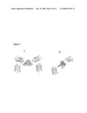 |
 | 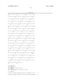 |
 | 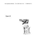 |
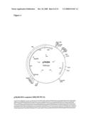 |  |
 |  |
 |  |
 |  |
 |  |
 |  |
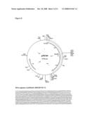 |  |
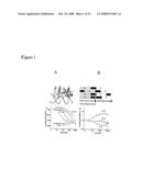 | 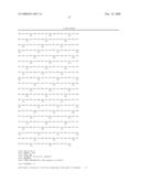 |
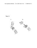 | 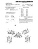 |
 | 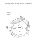 |
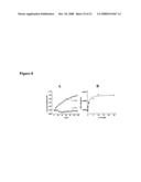 | 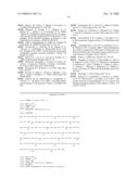 |
 | 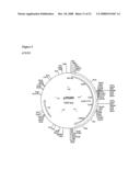 |
 |  |
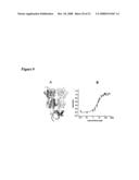 |  |
 |  |
 | 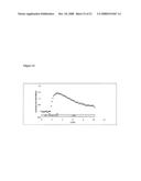 |
 | 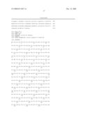 |
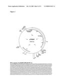 |  |
| Similar patent applications: | |
| Date | Title |
|---|---|
| 2011-04-07 | Aerosolized genetic vaccines and methods of use |
| 2011-04-07 | Purification method and method of producing vaccine |
| 2011-04-14 | Zinc finger nuclease for the cftr gene and methods of use thereof |
| 2011-04-07 | Stabilized oleosome preparations and methods of making them |
| 2011-04-14 | Medical food composition and methods for management of inflammatory processes in mammals |
| New patent applications from these inventors: | |
| Date | Title |
|---|---|
| 2021-11-25 | Subtilisin variants |
| 2011-09-15 | Trp/his exchange and kynurenin induced trp transport |
| 2011-08-25 | Novel sugar transporters |
| 2010-06-03 | Methods of reducing repeat-induced silencing of transgene expression and improved fluorescent biosensors |
| 2010-02-18 | Compositions and methods comprising cellulase variants with reduced affinity to non-cellulosic materials |
| Top Inventors for class "Drug, bio-affecting and body treating compositions" | |
| Rank | Inventor's name |
|---|---|
| 1 | David M. Goldenberg |
| 2 | Hy Si Bui |
| 3 | Lowell L. Wood, Jr. |
| 4 | Roderick A. Hyde |
| 5 | Yat Sun Or |