Patent application title: Immunoassay For Determining The Release Of Neurotensin Into The Circulation
Inventors:
Andrea Ernst (Hennigsdorf, DE)
Assignees:
SPHINGOTEC GMBH
IPC8 Class: AG01N3353FI
USPC Class:
435 71
Class name: Chemistry: molecular biology and microbiology measuring or testing process involving enzymes or micro-organisms; composition or test strip therefore; processes of forming such composition or test strip involving antigen-antibody binding, specific binding protein assay or specific ligand-receptor binding assay
Publication date: 2008-11-13
Patent application number: 20080280306
Claims:
1. An immunodiagnostic assay method for determining the release of
neurotensin into the mammalian circulation, wherein an immunoreactivity
of the N-terminal portion of a mammalian proneurotensin (PNT
immunoreactivity) is selectively determined in a serum sample or plasma
sample of a test mammal, said immunoreactivity not being neurotensin or
neuromedin immunoreactivity.
2. The method as claimed in claim 1, wherein the immunoreactivity of the N-terminal portion of the PNT is determined in the serum sample or plasma sample with the aid of antibodies which undergo specific binding to partial sequences of the N-terminal portion of the PNT.
3. The method as claimed in claim 2, wherein the PNT immunoreactivity in the serum sample or plasma sample is determined in a sandwich assay with the use of two different antibodies which undergo specific binding to different partial sequences of the N-terminal portion of the PNT.
4. The method of claim 1, wherein the mammalian proneurotensin is human proneurotensin which is determined in human serum or plasma.
5. The method as claimed in claim 4, wherein a PNT immunoreactivity which is to be coordinated with peptide sequences of human preproneurotensin (SEQ ID NO:1) in the region of the amino acids 24 to 140 (SEQ ID NO:6) of the complete human preproneurotensin is selectively determined.
6. The method as claimed in claim 4 or 5, wherein the determination is carried out as a sandwich assay with two antibodies which bind specifically to the amino acid sequences 67 to 85 (SEQ ID NO: 4) and 121 to 140 (SEQ ID NO:5) of the complete human preproneurotensin (SEQ ID NO:1).
7. The method of claim 2, wherein the antibodies are specific monoclonal and/or affinity-purified polyclonal antibodies.
8. The method of claim 2, wherein one of the antibodies is obtained by immunization of an animal with an antigen which contains a synthetic peptide sequence which comprises the amino acids 67 to 85 (SEQ ID NO:4) of human preproneurotensin (SEQ ID NO:1), and the other of the antibodies is obtained by immunization with an antigen which contains a synthetic peptide sequence which comprises the corresponding amino acids 121 to 140 (SEQ ID NO:5).
9. The method of claim 3, wherein one of the antibodies is used in a form bound to a solid phase, and the other antibody is marked or is selectively markable.
10. The method as claimed in claim 9, wherein marking with a radioisotope or a chemiluminescence, bioluminescence, fluorescence or enzyme label is used for marking the marked antibody or antibody to be marked.
11. The method of claim 3, wherein both the first and the second antibody are used in dispersion in a liquid reaction mixture, and wherein a first marking component which is part of a marking system based on fluorescence or chemiluminescence extinction or amplification is bound to the first antibody, and wherein the second marking component of this marking system is bound to the second antibody so that, after binding of both antibodies to the proneurotensin to be detected, the measurable signal is produced which permits detection of the resulting sandwich complexes in the measuring solution.
12. The method as claimed in claim 11, wherein the marking system comprises rare earth cryptates or chelates in combination with a fluorescent or chemiluminescent dye, in particular of the cyanine type.
13. The method of claim 1, which is carried out as a point-of-care method with the use of an immunochromatographic measuring apparatus, and a directly visually detectable label is used as a label for marking.
14. The method of claim 1, which is carried out for monitoring the nutrition status and/or the digestive functions of a patient.
15. The method as claimed in claim 14, wherein the patients are artificially fed patients.
16. The method of claim 1, wherein the determination is effected in association with diagnosis, monitoring or treatment with the use of samples of patients who are suffering from diseases which are selected from Parkinson's disease, celiac disease, chronic inflammatory intestinal diseases (Crohn's disease or Colitis ulcerosa), intestinal cancer or cancer of the pancreas, pancreatitis, thyroid diseases, cystic fibrosis and hepatitis.
17. Kit for carrying out the method of claims 3, which, in addition to customary auxiliary reagents, such as buffer solutions and wash solutions, at least comprises:a first immune reagent which comprises a first specific antibody against a partial sequence of human proneurotensin in a form immobilized on a solid phase,a second immune reagent which comprises a second specific antibody against a partial sequence of human proneurotensin in solubilized and marked form, andstandard and calibration solutions which contain defined amounts of a peptide preparation which has binding sites for the first and second specific antibody.
18. The kit as claimed in claim 17, wherein the solid phase is the wall of a test tube and the second antibody is marked by chemiluminescence marking.
Description:
[0001]The present invention relates to a method suitable for routine
diagnosis and intended for determining the release of the peptide
neurotensin into the circulation.
[0002]It should first be noted that, for reasons of simplification, terms such as "diagnosis" or "routine diagnosis" are used as a rule in the present description in a global sense which is intended to cover the determination of the measured biomolecules (peptides) not only for diagnostic purposes in the narrower sense but also for other purposes, such as, for example, for the determination of the metabolic state of a test subject before the beginning of a treatment or of an investigation for diagnostic purposes or for observation and monitoring of a patient over relatively long periods, for example for monitoring the course of a disease or therapeutic measures. Determinations for research purposes, for example in combination with the development of pharmaceuticals, for example neurotensin receptor agonists, or of products for nutrition, are also to be subsumed under the term "diagnosis", unless ruled out in specific cases owing to the more specific relationships discussed.
[0003]It is also true that applications in the area of veterinary medicine or animal breading are also to be included in principle even when the more specific statements on the invention in the present Application relate in particular to human medicine. Thus, the applicability of most statements on the present invention to non-human mammals is evident to the person skilled in the art without extensive explanations being required. The analogous applicability of the method described in more detail for human medicine to veterinary medicine follows, for example, from the fact that the corresponding peptide sequences are known for many other mammals and, in addition to veterinary applications, applications which relate rather to mammal breeding, for example as monitoring of the feeding state and the feed utilization of commercial animals, are also conceivable . . . .
[0004]Neurotensin is a tridecapeptide which occurs in various tissues and in the circulation of various mammals, including humans, and whose primary structure appears to be identical in all mammals. It has the amino acid sequence which is shown in the sequence listing as SEQ ID NO:2.
[0005]Like most biologically active peptides (5; references in the form of numbers in brackets relate to the attached list of references), neurotensin is formed by enzymatic processing from a precursor molecule which is referred to as preproneurotensin (with signal sequence) or proneurotensin (PNT; without signal sequence). In addition to the neurotensin (NT), the preproneurotensin/proneurotensin of mammals contains a further biologically active peptide, the hexapeptide neuromedin N (NMN; cf. SEQ ID NO:3). Neurotensin precursor peptides were isolated, inter alia, from canine, bovine and rodent tissues and finally also from human tissues. While the mature peptides NT and NMN have an identical amino acid sequence for different mammals, the precursor molecules, i.e. the preproneurotensins or proneurotensins, of different mammal species differ with regard to a number of amino acids. Thus, rat or bovine preproneurotensin has, for example, only 169 amino acids, whereas human preproneurotensin has 170 amino acids (17).
[0006]The nucleotide and amino acid sequences of human preproneurotensin have been known for some years (SEQ ID NO:1; cf. also U.S. Pat. No. 6,274,720 B1), exactly like those of dog (9), rat (17) and cattle (17; cf. also the comparison of different preproneurotensin sequences in FIG. 2 of the abovementioned US patent). The amino acids 1 to 23 of preproneurotensin (SEQ ID NO:1) represent the so-called signal sequence.
[0007]The expression of proneurotensin takes place primarily in the central nervous system (CNS) and in special enteroendocrine cells, the so-called N cells in the distal small intestine, and in nerve fibers which pass through the intestinal tract (13). However, PNT and NT were also detected in the adrenal glands and in heart tissue (24; 23).
[0008]The biologically active neurotensin (NT) formed from proneurotensin was detected in various mammalian tissues and discussed in relation to numerous different functions. Thus, neurotensin plays a major role as a neurotransmitter in the brain and has analgesic and thermoregulatory effects (6; 8) Furthermore, NT regulates the release of the neurotransmitters acetylcholine and glutamate (19; 18), stimulates the secretion of pituitary hormones (29) and influences dopamine neurotransmission (18), which suggests a relationship with Parkinson's disease (28).
[0009]The secretion of neurotensin in the gastrointestinal tract is stimulated by food intake (15). Even shortly after eating, there is a significant NT increase which persists over several hours (14). It was possible to show that the consumption of fat, in comparison with protein and glucose, had the strongest influence on NT production (27). Neurotensin evidently represents a sort of "saturation factor" since the secretion of NT leads to suppression of the appetite and hence to reduction of food consumption (34). In adipose (obese) patients, the plasma concentration of NT is reduced in comparison with persons of normal weight, which is presumably due to an increased appetite and increased consumption of food (35; 4). Interestingly, smokers, too, showed a significantly greater increase in the NT concentration after food consumption than nonsmokers (26). This indicates that a changed neurotensin secretion is a possible cause of the increased appetite and the frequently observed weight increase of former smokers after stopping smoking.
[0010]In the gastrointestinal tract, neurotensin stimulates the secretion of pancreas hormones such as insulin and glucagon, inhibits the secretion of gastric acid and stimulates the motility of the large intestine (33). NT promotes the growth of gastrointestinal tissue, such as pancreas, large intestine and small intestine tissue (12; 36; 10), which gives rise to the hypothesis that NT contributes to the formation of tumors in these tissues.
[0011]The function of the second peptide neuromedin N (SEQ ID NO:3), formed from the same precursor peptide, is less well characterized. Presumably, it has effects similar to those of neurotensin since the two have similarities in their amino acid sequence and bind to the same receptors (33).
[0012]By investigating different tissue extracts with the use of radioimmunoassays employing antibodies which are specific for free C-termini of NT or NMN and therefore do not recognize proneurotensin forms not proteolytically processed and without such exposed COOH termini, partly in combination with HPLC techniques for separating the fractions according to their molecular size, it was found that, in addition to NT and NMN, incompletely cleaved, so-called large NT and/or NMN peptides can also be formed in the processing of PNT in a tissue-dependent manner (7; 13; 25).
[0013]Numerous investigations are concerned with the formation or the occurrence of neurotensin in various diseases, these investigations having been carried out primarily with tissue samples and tissue extracts. Regarding investigations which are based on measurements of neurotensin in patients' blood samples, it should be mentioned, for example, that the concentration of neurotensin in the plasma of patients with Parkinson's disease was found to be increased (30). In celiac patients who have an intolerance to cereal gluten, an increased neurotensin concentration was detected in the plasma in comparison with control persons in the fasted state (2). The plasma concentrations of neurotensin in patients with pancreatitis are increased after a meal in comparison with healthy control persons (22). In the case of thyroid diseases, significantly lower neurotensin concentrations were found in the plasma of patients with central (or secondary) thyroid underfunction (31). Secondary thyroid underfunction is caused by a disturbance in the region of the pituitary gland, which controls the secretion of thyroid hormones. On the other hand, significantly higher neurotensin levels (31) were found in the blood of patients with peripheral (or primary) thyroid underfunction or thyroid overfunction in comparison with controls. Primary thyroid underfunction is caused by dysfunction of the thyroid itself. Patients with cystic fibrosis (mucoviscidosis) have higher neurotensin concentrations in the plasma than healthy control persons both before and after food consumption (16; 1; 21).
[0014]The neurotensin levels in blood were invariably determined using assays of the radioimmunoassay type, in which the competition of the neurotensin to be determined in the sample with an added marked neurotensin or neurotensin fragment for the binding sites of a single antibody which binds specifically to neurotensin or certain partial sequences of free neurotensin, for example with a free C-terminus of neurotensin, is determined. Measurements of the NMN levels were carried out in a similar manner.
[0015]The above and similar results for the occurrence of neurotensin in the case of certain diseases and the demonstrated possibility of stimulating neurotensin formation as a function of food consumption, which also proved to be significantly different for certain pathological states, make the determination of neurotensin in blood samples an essentially promising and clinically versatile diagnostic tool. Thus, it appears possible to use such a determination, for example, in the routine investigation of patients, for their monitoring during their clinical care, for monitoring of their ability to utilize food, in monitoring of the intestinal functions or as a therapy-accompanying measure for monitoring the success of treatments, as well as in other contexts which arise out of the abovementioned discussion of the scientific literature.
[0016]However, neurotensin determinations in blood samples have not to date been adopted in routine medical diagnosis. A first substantial reason for this is that the stability of the neurotensin concentrations in the blood samples ex vivo at ambient temperature is too low for routine measurements (20) (half-life of only 3 to 4 h), which, in combination with existing laboratory logistics (of sampling, plasma recovery, transport to the laboratory up to carrying out of corresponding laboratory tests) has so far prevented the use of neurotensin in routine diagnosis. A second reason is the known short half-life of only 2 to 6 min for neurotensin in blood in vivo, which is attributed to rapid and effective binding to neurotensin receptors and/or degradation of the neurotensin (3) and burdens a measurement with a time dependence which is potentially difficult to estimate. Instantaneously measurable NT levels in blood samples are not a measure of the total biosynthesis and release of NT into the circulation as a result of a stimulation, e.g. supply of food, up to the time of measurement, but rather represent a random transitional state in which the NT elimination by binding to receptors and by degradation greatly dilutes the inflammation on the biosynthesis and release into the circulation.
[0017]It is an object of the present invention to provide a possibility for determining the neurotensin release in samples of test mammals, in particular human patients, which is suitable for routine use and makes it possible to utilize the potential outlined above for a determination of the neurotensin release for medical diagnosis and to implement it for medical/clinical routine.
[0018]This object is achieved by an immunodiagnostic assay method for determining the release of neurotensin into the mammalian circulation, which method, in its most general form according to claim 1, is characterized in that an immunoreactivity of the N-terminal portion of a mammalian proneurotensin (PNT immunoreactivity) is selectively determined in a serum sample or plasma sample of a test mammal, said immunoreactivity not being a neurotensin or neuromedin N immunoreactivity.
[0019]Advantageous developments of such a method are reproduced by claims 2 to 16 in conjunction with the statements on preferred embodiments in the description.
[0020]The object is furthermore achieved by a kit for a preferred embodiment of such a method according to claim 1, as described in claims 17 and 18.
[0021]Below, the novel discoveries on which the invention is based, and the applications according to the invention which are derived therefrom, are explained in more detail with reference to specific test results and figures.
[0022]Immunoreactivity of the N-terminal portion of mammalian proneurotensin" means an immunoreactivity which is localized in the region of the amino acid in position 24 (position 1 after the signal peptide) up to the third last amino acid before the first amino acid of neuromedin N (Lys) of the respective mammalian preproneurotensin. In all known cases, the region discussed comprises at least the respective amino acids 24 to 139 or 140, i.e. a sequence as shown for the human peptide in SEQ ID NO:6.
[0023]In the figures:
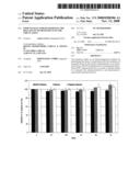
[0024]FIG. 1 shows results of the measurement of the ex vivo stability of the immunoreactivity which is to be assigned to N-terminal sequences of human PNT, in serum, EDTA plasma and heparin plasma samples obtained from the blood of test subjects, at room temperature over periods of up to 7 days;
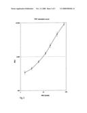
[0025]FIG. 2 shows the standard curve of a PNT sandwich assay as described in more detail in the experimental section;
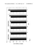
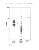
[0026]FIG. 3 shows the results of the measurement of the PNT immunoreactivity in plasma in the case of normally fed normal persons and artificially fed patients by means of the sandwich assay described;
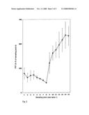
[0027]FIG. 4 shows the results of the measurement of changes of the PNT immunoreactivity as a function of time in the plasma of normally fed normal persons, depending on the supply of food, by means of the sandwich assay described;
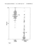
[0028]FIG. 5 shows the results of the measurement of the PNT immunoreactivity in plasma in the case of normally fed normal persons, patients with chronic inflammatory intestinal diseases (Crohn's disease/Colitis ulcerosa) and hepatitis patients by means of the sandwich assay described.
[0029]To examine the question as to whether reproducible results which correspond to the literature data on the occurrence of NT can be obtained in a determination of an immunoreactivity which is to be assigned to an N-terminal portion of human proneurotensin, in particular a portion which contains neither NMN nor NT and is eliminated in the proteolytic processing with formation of free NMN or NT and then represents a sort of waste product, in a blood sample or serum sample or plasma sample, a sandwich assay specific for this N-terminus of PNT, as described in more detail in the experimental section, was developed. This assay specifically recognizes only N-terminal peptides of human PNT, which contain both the amino acids 67 to 85 and the amino acids 121 to 140 of human preproneurotensin.
[0030]Measurements of human blood samples using this assay, which are described below in more detail, showed that PNT immunoreactivities which, according to occurrence and dynamics, reflect the formation/release of NT are measured therewith.
[0031]In the case of the repeated measurement of serum samples and plasma samples obtained from such blood samples on storage at room temperature over relatively long periods, it was furthermore found that the stability of the PNT immunoreactivity in plasma ex vivo is surprisingly high. Since no recognizable loss of determinable immunoreactivity was detectable even after 7 days, its half-life at room temperature is far more than 7 days and hence well above the ex vivo stability of mature neurotensin (cf. 3) or that of one of the large, incompletely processed proneurotensin peptides.
[0032]The results of the stability measurements are shown in FIG. 1.
[0033]Thus, the determination of proneurotensin or PNT fragments in blood samples is entirely fit for routine use and is suitable for making the NT/NMN/PNT release rate in the blood accessible to routine medical diagnosis.
[0034]The preparation of the assay used, its use in measurements and the measured results obtained with it are described more exactly in the following experimental section and the examples and the associated explanations.
EXPERIMENTAL SECTION
[0035]Determination of an immunoreactivity to be assigned to N-terminal proneurotensin sequences.
1. Preparation of Antibodies
1.1. Immunogens
[0036]2 different peptide partial sequences of human preproneurotensin (SEQ ID NO:1), namely PNT3 (SEQ ID NO:4) and PNT4 (SEQ ID NO:5), were selected and were synthesized as synthetic peptides by Jerini (Berlin, Germany). Each peptide was additionally provided with an amino-terminal cysteine residue (Cys0).
1.2. Antibody Recovery
[0037]For immunization purposes, the peptides PNT3 (SEQ ID NO:4) and PNT4 (SEQ ID NO:5) supplemented by N-terminal cysteine residues were conjugated with the hemocyanine from Limulus polyphemus and used according to standard methods for immunizing rabbits, from which antisera against the PNT peptide conjugates were obtained.
1.3. Purification of the Antibodies
[0038]The polyclonal antibodies of the rabbit antisera were purified by means of ligand-specific affinity purification. For this purpose, the Cys(0) peptides PNT3 (SEQ ID NO:4) and PNT4 (SEQ ID NO:5) were first coupled to SulfoLink gel from Pierce (Boston, USA). The binding was effected by the manufacturer's method as follows:
[0039]Polycarbonate columns (15 mm×80 mm) were filled with 5 ml of affinity matrix. After equilibration of the columns with PBS (136 mM NaCl, 1.5 mM KH2PO4 20.4 mM Na2HPO4.2H2O, 2.7 mM KCl, pH 7.12), 5 mg of the respective abovementioned peptides were weighed out, dissolved in PBS and added to the closed columns, and the gel material was homogenized by swirling. After incubation for 15 minutes at room temperature and settling of the gel material, the columns were washed 5 times with 3 ml portions of PBS. For saturation of free binding sites 5 ml of 50 mM L-cysteine solution were added in each case to the column material and, after homogenization, the gel material was incubated again at room temperature for 15 min. After settling of the gel material, each column was rinsed 6 times with 5 ml portions of a 1 M NaCl solution and then rinsed again with PBS.
[0040]The gel material was mixed with 25 ml of the respective rabbit antiserum pool and incubated over night at room temperature with gentle swirling. The serum-gel mixtures were transferred to polycarbonate columns and excess serum was removed. The columns were then washed with 250 ml of PBS in order to remove serum proteins which were not bound. The desorption of the bound antibodies was effected by elution of the column with 50 mM citric acid (pH 2.2). The eluate was collected in 1 ml fractions. The protein concentration of each fraction was determined with the aid of the BCA protein assay kit from Perbio (Bonn, Germany), and fractions having a protein content of >1 mg/ml were combined. The affinity-purified antibodies were rebuffered by means of dialysis in PBS and the protein content was determined again. Storage was then carried out at 4° C.
2. Assay Preparation
2.1. Solid Phase
[0041]The affinity-purified polyclonal rabbit antibody against the peptide PNT3 was immobilized on polystyrene tubes (Startubes, 12 mm×75 mm, from Greiner, Germany). For this purpose, the antibody solution was diluted to a protein concentration of 6.7 μg/ml with PBS, and 300 μl thereof were pipetted per tube (corresponds to 2 μg of antibodies per tube). These were incubated for 24 h at room temperature and then washed 3 times with 4 ml portions of PBS . . . . The tubes were stored at 4° C. until required for further use.
2.2. Marked Antibodies
[0042]The affinity-purified polyclonal rabbit antibody against PNT4 (1 mg/ml in PBS) was luminescence-marked with acridinium ester N-hydroxysuccinimide (1 mg/ml in acetonitrile, from InVent, Hennigsdorf, Germany). For the marking, 200 μl of antibodies were mixed with 4 μl of acridinium ester and incubated for 20 min and free acridinium ester bonds were saturated by addition of 40 μl of a 50 mM glycine solution. The marking batch was separated from free acridinium ester by means of HPLC on a BioSil 400 gel filtration column (from BioRad, Munich, Germany). The mobile phase used was PBS.
3. Carrying Out PNT Determinations
3.1.1 Measuring Conditions
[0043]50 μl of a plasma sample to be investigated and 150 μl of assay buffer (PBS buffer, 10 mM EDTA) were pipetted antibody-coated tube (2.1) and incubated for 16 h at room temperature. The tubes were then washed 5 times with 1 ml portions of PBS. 20 ng of the marked antibody (2.1.) (in 100 μl of PBS buffer, 10 mM EDTA) were then added to each of the tubes. The tubes were incubated for 2 h at room temperature, and unbound tracer antibody was then removed by washing 5 times with 1 ml portions of PBS.
[0044]Marked antibody bound to the tube was quantified by means of luminescence measurement in a commercial luminometer (Berthold LB 952T/16).
3.2. Calibration
[0045]In order to be able to determine the concentrations or immunoreactivities of proneurotensin in the samples to be measured, 50 EDTA plasmas of apparently healthy persons were screened for PNT, and plasmas having the highest PNT immunoreactivities were pooled. From this plasma pool, a plurality of dilution stages were produced with horse serum (Sigma, Germany). A fictitious concentration of 100 units/ml was assigned to the highest standard (undiluted EDTA plasma). FIG. 2 shows a PNT standard curve. The analytical sensitivity of the proneurotensin assay is about 1 U/ml.
4. Results of Measurements
4.1. Proneurotensin Immunoreactivity in Plasmas of Apparently Healthy Persons
[0046]Apparently healthy control persons have high measured PNT values in the blood. There is a frequency distribution, which is shown in FIG. 4. The median was determined as 35.1 U/ml.
4.2. Determination of the PNT Immunoreactivity in the Circulation of Healthy Test Subjects During and after the Consumption of Food
[0047]It is known that the formation or release of neurotensin is stimulated by consumption of food (15; 14). In order to determine whether the PNT concentration in the blood is also influenced by the consumption of food and whether the dynamics of any change in the PNT immunoreactivity runs parallel to the above-described concentration of NT, the following test was carried out: 6 test subjects (three male and three female persons) fasted for 14 h. A blood sample was then taken from them. The test subjects then consumed a defined amount of liquid or food at specified times, and blood samples were taken again at certain times. The course of the test is summarized in table 1.
TABLE-US-00001 TABLE 1 Course of the food stimulation test Sampling point Nutrition status 1 after fasting for 14 h 2 after fasting for 15 h 3 5 min after liquid intake (1 l of water) 4 15 min after liquid intake 5 45 min after liquid intake 6 1 h 15 min after liquid intake 7 1 h 45 min after liquid intake 8 3 h after liquid intake 9 5 min after consumption of food (salad with approx. 250 kcal and pizza with approx. 100 kcal) 10 15 min after consumption of food 11 30 min after consumption of food 12 1 h after consumption of food 13 2 h after consumption of food 14 3 h after consumption of food 15 4 h after consumption of food
[0048]It is found that the secretion of PNT is not stimulated by intake of liquid. Shortly before the consumption of food, i.e. after the longest fasting time, the PNT concentration is the lowest. It was defined as 100% for each individual test subject, and all other measurements of the respective test subject were based on this value. These percentage changes of the measured PNT immunoreactivity of the 6 test subjects were averaged and are shown graphically in FIG. 3 (P<0.05). Immediately after eating, the PNT concentration in the blood increases significantly. The last two sampling times 3 h and 4 h after consumption of food have the highest measured values. Overall, the PNT concentration increases by 4.5 to 8 times after eating.
[0049]Immunoreactivity which can be determined by the abovementioned assay and is to be coordinated with one or more N-terminal proneurotensin fragment(s) having binding sites for the two antibodies used thus increases, as described for the mature peptide NT, owing to stimulation by consumption of food.
[0050]The measurement of PNT immunoreactivity can therefore be used instead of the mature neurotensin for evaluating the metabolic status of a patient/test person. By administration of a defined amount of nutrients, the individual stimulability of the PNT release can be determined as the difference Δ between a measured value at a specified time after the consumption of food and a base value (e.g. PNT3h after consumption of food and PNTfasted) or as a ratio or factor of such values. Table 2 shows a corresponding evaluation by way of example.
TABLE-US-00002 TABLE 2 PNT immunoreactivities (in U/ml) before and after food simulation and indication of absolute differences and factors Test Test Test Test Test Test subject subject subject subject subject subject 1 2 3 4 5 6 PNTB 10.5 2.3 4.8 5.0 5.7 5.8 PNT5 min 32.3 10.7 9.6 23.0 27.1 15.5 PNT3 h 42.4 18.4 20.0 22.7 41.5 40.6 Δ 21.8 8.45 4.8 18.0 21.4 9.7 PNT5 min minus PNTB Δ 31.9 16.1 15.2 17.7 35.8 34.8 PNT3 h minus PNTB PNT5 min/PNTB 3.1 4.7 2 4.6 4.8 2.7 PNT3 h/PNTB 4.0 8 4.2 4.5 7.3 7 In the table, the meanings are as follows: PNTB = PNT immunoreactivity after fasting for 18 h (base value) PNT5 min = PNT immunoreactivity 5 min after consumption of food PNT3 h = PNT immunoreactivity 3 h after consumption of food
4.3. Determination of the PNT Concentrations in the Circulation of Artificially Fed Patients
[0051]As already shown, healthy persons have a certain blood PNT concentration which can be stimulated by consumption of food. In a corresponding measurement of the PNT immunoreactivity in plasmas of patients artificially fed parenterally (by means of a vein catheter) and generally in intensive care (for example with an existing sepsis or a polytrauma), it was found that they have significantly lower PNT concentrations than normally fed controls. In such measurements, a median relative PNT immunoreactivity of 2.7 U/ml, a factor of 13 lower, was found (cf. FIG. 4).
4.4. Determination of the PNT Immunoreactivity in the Circulation of Patients with Inflammatory Intestinal Diseases (Crohn's Disease or Colitis Ulcerosa)
[0052]In the measurement of samples of patients with an inflammatory intestinal disease, such as Crohn's disease and Colitis ulcerosa, it was found that here too the median measurable PNT immunoreactivity of 10 U/ml is below that of the control persons (FIG. 5) However, individuals among the patients also had substantially higher measured values (above 100 U/ml).
4.5. Determination of the PNT Immunoreactivity in the Circulation of Patients Having a Hepatitis a or C
[0053]In the measurement of samples of patients with a hepatitis A or C, on the other hand, higher PNT concentrations were found in the circulation, with a median of 112 U/ml, compared with the healthy controls (FIG. 5).
5. Isolation and Characterization of Proneurotensin Immunoreactivity in Human Serum
5.1. Preparation of the Affinity Column
[0054]5 mg of the purified polyclonal rabbit antibody against the PNT3 peptide (SEQ ID NO:4; cf. 1.1.) were coupled to 2 ml of CarboLink gel (from Pierce, Boston, USA), according to the manufacturer's method and transferred to a polycarbonate column (15 mm×80 mm). The gel was then washed with 20 ml of PBS.
5.2. Affinity Purification of PNT Immunoreactivity
[0055]A mixed serum sample comprising 10 individual sera of 100 ml with a relative concentration of 32 U/ml was mixed with Na EDTA (final concentration 10 mM) and then filtered over 0.2 μm filter. The prepared mixed serum sample was pumped at 4° C. and at a flow rate of 2 ml/min six times in succession over the affinity column. Thereafter, the column was washed with 50 ml of PBS and the bound PMT immunoreactivity was eluted with a 50 mM glycine/HCl solution (pH 2.0). The column outflow was monitored continuously for absorption at 280 nm and the protein fraction (final volume 3 ml) eluted by the glycine/HCl solution was further analyzed by means of HPLC.
5.3. Reversed-Phase HPLC Analysis
[0056]The material obtained by affinity purification was purified by means of reversed-phase HPLC on a C18 column μ Bondapak 0.4×30 mm from Waters (Eschborn, Germany). The flow rate of 1 ml/min. The mobile phase and elution conditions used are shown in table 3 below.
TABLE-US-00003 TABLE 3 Elution conditions of the RP-HPLC of proneurotensin-IR Mobile phase A: 5% acetonitrile 20 mM NH4 acetate Mobile phase B: 90% acetonitrile 20 mM NH4 acetate Gradient 0.0 min 100% A/0% B 2.5 min 100% A/0% B 5.0 min 89% A/11% B 55.0 min 30% A/70% B 60.0 min 0% A/100% B
[0057]The column outflow was measured continuously for its absorption at 215 nm and collected in 0.5 ml fractions. With the aid of the PNT assay described in the above sections 2. and 3., those fractions in which a PNT immunoreactivity was detectable were determined. It was found that the main immunoreactivity was eluted in fraction 67.
[0058]Fraction 67, which had a positive PNT immunoreactivity, was dried by treatment with nitrogen gas. Samples were then analyzed by mass spectrometry.
5.4. Mass Spectrometric Analysis
[0059]The peptide was digested by means of protease Glu-C or trypsin and the resulting fragments were obtained in a manner known per se via SMART HPLC and then investigated by mass spectrometry.
[0060]In the mass spectrometric analysis, a sequence (SEQ ID NO:6) which agreed completely with the sequence of the amino acids 24-140 of the known preproneurotensin 1-170 (SEQ ID NO:1) was determined. It was thus shown that a proneurotensin peptide which comprises 117 amino acids and is designated as preproneurotensin 24-140 circulates in the blood of humans. There was no indication at all that the species determined as immunoreactivity by means of the method described still contains the C-terminal mature peptides neuromedin N and neurotensin.
Sequence CWU
1
51170PRTHomo Sapiens 1Met Met Ala Gly Met Lys Ile Gln Leu Val Cys Met Leu
Leu Leu Ala1 5 10 15Phe
Ser Ser Trp Ser Leu Cys Ser Asp Ser Glu Glu Glu Met Lys Ala 20
25 30Leu Glu Ala Asp Phe Leu Thr Asn
Met His Thr Ser Lys Ile Ser Lys 35 40
45Ala His Val Pro Ser Trp Lys Met Thr Leu Leu Asn Val Cys Ser Leu
50 55 60Val Asn Asn Leu Asn Ser Pro Ala
Glu Glu Thr Gly Glu Val His Glu65 70 75
80Glu Glu Leu Val Ala Arg Arg Lys Leu Pro Thr Ala Leu
Asp Gly Phe 85 90 95Ser
Leu Glu Ala Met Leu Thr Ile Tyr Gln Leu His Lys Ile Cys His
100 105 110Ser Arg Ala Phe Gln His Trp
Glu Leu Ile Gln Glu Asp Ile Leu Asp 115 120
125Thr Gly Asn Asp Lys Asn Gly Lys Glu Glu Val Ile Lys Arg Lys
Ile 130 135 140Pro Tyr Ile Leu Lys Arg
Gln Leu Tyr Glu Asn Lys Pro Arg Arg Pro145 150
155 160Tyr Ile Leu Lys Arg Asp Ser Tyr Tyr Tyr
165 170213PRTHomo Sapiens 2Gln Leu Tyr Glu Asn
Lys Pro Arg Arg Pro Tyr Ile Leu1 5
1036PRTHomo sapiens 3Lys Ile Pro Tyr Ile Leu1
5419PRTArtificialSynthetic pepitde 4Asn Leu Asn Ser Pro Ala Glu Glu Thr
Gly Glu Val His Glu Glu Glu1 5 10
15Leu Val Ala520PRTArtificialSynthetic peptide 5Leu Ile Gln Glu
Asp Ile Leu Asp Thr Gly Asn Asp Lys Asn Gly Lys1 5
10 15Glu Glu Val Ile 20
User Contributions:
Comment about this patent or add new information about this topic:
 |  |
 |  |
 |  |
 |  |
 |  |
 |
| New patent applications from these inventors: | |
| Date | Title |
|---|---|
| 2016-04-07 | Csf diagnostic in vitro method for diagnosis of dementias and neuroinflammatory diseases |
| 2012-09-13 | Immunoassay for determining the release of neurotensin into the circulation |
| 2011-11-10 | Use of precursors of tachykinins and/or their fragments in medical diagnostic |
| 2010-03-11 | In vitro multiparameter determination method for the diagnosis and early diagnosis of neurodegenerative disorders |
| 2010-02-11 | In vitro method for the diagnosis and early diagnosis of neurodegenerative disorders |
| Top Inventors for class "Chemistry: molecular biology and microbiology" | |
| Rank | Inventor's name |
|---|---|
| 1 | Marshall Medoff |
| 2 | Anthony P. Burgard |
| 3 | Mark J. Burk |
| 4 | Robin E. Osterhout |
| 5 | Rangarajan Sampath |