Patent application title: Methods and Compositions Related to Delivery of Chemical Compounds to Invertebrate Embryos
Inventors:
Aloisia T. Schmid (Salt Lake City, UT, US)
Assignees:
UNIVERSITY OF UTAH RESEARCH FOUNDATION
IPC8 Class: AA01K67027FI
USPC Class:
800 3
Class name: Multicellular living organisms and unmodified parts thereof and related processes method of using a transgenic nonhuman animal in an in vivo test method (e.g., drug efficacy tests, etc.)
Publication date: 2008-11-06
Patent application number: 20080276327
Inventors list |
Agents list |
Assignees list |
List by place |
Classification tree browser |
Top 100 Inventors |
Top 100 Agents |
Top 100 Assignees |
Usenet FAQ Index |
Documents |
Other FAQs |
Patent application title: Methods and Compositions Related to Delivery of Chemical Compounds to Invertebrate Embryos
Inventors:
Aloisia T. Schmid
Agents:
Ballard Spahr Andrews & Ingersoll, LLP
Assignees:
UNIVERSITY OF UTAH RESEARCH FOUNDATION
Origin: ATLANTA, GA US
IPC8 Class: AA01K67027FI
USPC Class:
800 3
Abstract:
Disclosed are methods and compositions related to the delivery of chemical
compounds to invertebrate embryos.Claims:
1. A method of manipulating an invertebrate animal embryo, comprising
delivering a composition to the embryo.
2. A method of delivering a composition to an invertebrate animal embryo comprising:a) dechorionating the embryo;b) exposing the embryo to DMSO; andc) delivering the composition to the embryo.
3. A method of making an invertebrate animal embryo useful for screening compounds comprising:a) dechorionating the embryo; andb) exposing the embryo to DMSO.
4. The method of claim 3, wherein the embryo has been genetically modified.
5. A method of screening a candidate compound for its effect on a disease comprising:a) administering the compound to an invertebrate animal embryo; andb) assaying the effect of the compound on the embryo.
6. A method of screening a candidate compound for its effect on a disease comprising:a) administering the compound to an invertebrate animal embryo;b) comparing phenotypes of the transgenic embryo treated with the compound to a transgenic invertebrate embryo not treated with the compound, wherein a difference in the phenotypes is indicative of an alleviating activity of the candidate compound.
7. A method of screening a compound for its effect on an invertebrate embryo comprisinga) dechorionating the embryo,b) incubating the dechorionated embryo with the compound and DMSO, andc) assaying the effect of the compound on the embryo.
8. A kit for use in screening compounds, said kit comprising transgenic invertebrate animal embryos and a suitable container.
9. A method of making a genetic assay for a neurodegenerative disease, comprising:a) introducing a nucleic acid into an invertebrate animal, wherein the nucleic acid encodes a peptide associated with a phenotype of the neurodegenerative disease;b) introducing a fluorescent protein into the invertebrate animal of step a;c) introducing a UAS-Gal4 expression system into the invertebrate animal of step a;d) expressing the phenotype in an embryo of the invertebrate animal of step c; ande) assaying the phenotype.
10. The method of claim 9, wherein the peptide associated with a phenotype of the neurodegenerative disease is an amyloidgenic peptide.
11. The method of claim 10, wherein the amyloidgenic peptide is Abeta.sub.1-42.
12. The method of claim 10, wherein the amyloidgenic peptide is Abeta.sub.1-40.
13. The method of claim 9, wherein a signal sequence is also introduced into the invertebrate animal.
14. A method of screening a candidate compound for its effect on a disease comprising:a) introducing a nucleic acid into an invertebrate animal, wherein the nucleic acid encodes a peptide associated with a phenotype of the disease,b) expressing the phenotype in an embryo of the invertebrate animal of step a;c) aliquoting the embryos of step c into wells of a plate;d) delivering a test compound to the well of the plate of step d; ande) screening for a change in the phenotype associated with the disease, a change indicating a compound with an effect upon the disease.
15. A method of making a genetic assay comprising:a) introducing a nucleic acid into an invertebrate animal, wherein the nucleic acid encodes a peptide associated with a phenotype of a disease;b) expressing the phenotype in an embryo of the invertebrate animal of step a;c) aliquoting the embryos of step b into wells of a plate; andd) delivering a test compound to the well of the plate of step c.
16. The method of any one of claims 5-15, wherein the disease is Parkinson's disease.
17. The method of any one of claims 5-15, wherein the disease is Alzheimer's disease.
18. The method of any one of claims 5-15, wherein the disease is depression.
19. A method of introducing a compound into an invertebrate embryo, comprising administering to the embryo an effective amount of DMSO.
20. The method of claim 19, wherein the effective amount of DMSO is 0.1% to 10%.
21. The method of claim 20, wherein the effective amount of DMSO is 1% to 5%.
22. A method of screening for a test compound with an effect on a phenotype associated with a neurodegenerative disease comprising:a) placing transgenic embryos in a read out assay;b) delivering a test compound to the read out assay of step a;c) scoring the embryos for the ability to extend axons across segment boundaries, said ability indicative of a test compound with an effect on a phenotype associated with a neurodegenerative disease.
23. The method of claim 0, wherein transgenic embryos express a membrane-tagged fluorescent protein.
24. A method of scoring a plate comprising:a) placing transgenic embryos expressing a phenotype associated with a disease in a read out assay;b) delivering a compound to the read out assay; andc) using a seed fill algorithm to score the embryos, wherein the algorithm evaluates contiguous lines of pixels, wherein a compound that ameliorates the phenotype gives a higher score in comparison to a control embryo.
Description:
CROSS-REFERENCE TO RELATED APPLICATIONS
[0001]This application claims priority to U.S. provisional application Ser. No. 60/573,194 filed on May 21, 2004. The 60/573,194 provisional patent application is herein incorporated by this reference in its entirety.
I. BACKGROUND
[0002]In the pharmaceutical industry, chemical compound screening has been performed using cell-based assays in high throughput screening approaches. Template compounds are identified, optimized via medicinal chemistry and then introduced to whole animal systems for evaluation of toxicity. Judging the "success" of the new paradigm of drug discovery on the basis of published data has been difficult (Drews, 2000). High throughput screening has resulted in a large number of "hits," However, very few leads and development compounds, if any, can be credited to the new drug discovery paradigm. (Jurgens, 1999). The major reason these hits have not proved useful for lead development is that the lead that is identified in the cell-based drug screening approach is very often later proved toxic in whole animals (Drews, 2000). Therefore, what is needed in the art are compositions and methods for delivering chemical compounds to invertebrate embryos, with the ability to do so in a high throughput screening assay.
II. SUMMARY
[0003]Disclosed are methods and compositions related to the genetic modification of invertebrate embryos, as well as the delivery of chemical compounds to invertebrate embryos.
III. BRIEF DESCRIPTION OF THE DRAWINGS
[0004]The accompanying drawings, which are incorporated in and constitute a part of this specification, illustrate several embodiments and together with the description illustrate the disclosed compositions and methods.
[0005]FIG. 1 shows the effects of gamma secretase inhibitors on Drosophila development. In all cases, anterior is to the left. All panels are confocal projections of whole mount embryos stained with rhodaminated antibodies directed against HRP, recognizing an antigen on the surface of all insect neurons. Panel A shows a horizontal view of wild type embryo demonstrating an intact central nervous system (CNS) with well organized peripheral nervous system, consisting of motoneuronal projections and sensory inputs. Panel B shows a diagram of the structures seen in wild type embryos. Panel C shows embryos exposed to 10 μM concentrations of gamma secretase inhibitors generate severe developmental defects, and never complete gastrulation. Panel D shows at 1 μM concentrations, the same drugs allow early cell populations to form but stall development prior to neurogenesis. Panel E shows 100 nM concentrations of gamma secretase inhibitors generate classic Notch neurogenic phenotypes. Panel F shows 10 nM concentrations generate slightly less severe neurogenic defects. Panels I (10 fM) and J (1 fM) show femtomolar concentrations of gamma secretase inhibitors generate defects only in peripheral nervous system pathfinding, a finding consistent with mildest hypomorphic alleles. Panels G and H show another gamma secretase inhibitor generated more severe defects in ventral nerve cord condensation.
[0006]FIG. 2 shows allelic series of the phenotypes associated with the misexpression of human APP fragments in the developing cells of the embryonic central nervous system. Pan-neuronal drivers were used to misexpress the amyloidgenic peptides associated with AD. The most severe phenotypes were consistently observed using ssAbeta1-42.
[0007]FIG. 3 shows that when DiI labeling experiments were performed to examine the single cell defects associated with the misexpression of Abeta peptides pan-neuronally, it was observed that the population of neurons most strongly affected were longitudinal projection neurons. These neurons consistently failed to extend beyond segment boundaries in this gain of function background. When staining was done with fascicular antibodies, clear breaks could be visualized at segment boundaries that typically extended for 25% of the Anteroposterior length of the hemisegment.
[0008]FIG. 4 shows a summary of an example of an Alzheimer's model (Example 2). This is an example of a phenotype on which a compound screen can be based.
[0009]FIG. 5 shows a laser-based embryo sorter. The sorter uses a laser-based technology to detect the presence of GFP-tagged balancer chromosomes, eliminates those embryos from further study, but aliquots the embryos without such balancer chromosomes into each well of a 96 well plate, robotically. Each 96 well plate can be filled with 10 embryos per well, within 15 minutes.
[0010]FIG. 6 shows a plate reader. Embryos develop in the drug, absorb the drug, and are then scored for the ability to extend axons across segment boundaries, as revealed by Red/Blue/Yellow membrane-tagged Fluorescent Protein expression patterns. An entire 96 well plate can be scanned and photographed in 10 minutes.
[0011]FIG. 7 shows a seed fill algorithm. Each image is divided into approximately 250,000 pixels, and each pixel is assigned a digital value for intensity, on a scale between 1-256. The computer evaluates the intensity of each pixel and then compares pairs of pixels and looks for neighbors that are equally bright. Fluorescent proteins never vary in their intensity, and plate readers can be used with seed fill algorithms. Essentially, the algorithm evaluates contiguous lines of pixels. So if membrane tagged fluorescent axons (pixels) are only 100 pixels long in the untreated controls, then a drug that ameliorates the phenotype can be expected to improve this number by what would be a statistically significant amount.
[0012]FIG. 8 shows RNAi for SMN generates motorneuronal and muscular defects in embryogenesis. Wild type stage 16 embryos of Drosophila melanogaster were filleted and stained with antibodies directed against a strongly conserved epitope of human SMN1; these were counterstained with A488-conjugated phalloidin (panels A-C) and photographed on a Biorad Radiance 2000 confocal microscope. The SMN1 antibodies appear to recognize an epitope in Drosophila that is specific to neurons, their axons (asterisk, panel B) and muscles (arrow, panel B). RNAi experiments at 5 uM concentrations (panels D-F) revealed that the antibody staining is specific, as its cellular specificity disappears and is replaced by relatively low level non-specific background staining (panel E). Motoneuronal projections, visible as regular processes exiting from the CNS (panel A) are absent in RNAi backgrounds (Compare panels A and D). Furthermore, muscles continue to extend myopodia in RNAi backgrounds (arrow, Panel D), apparently because their extension has not been suppressed by successful motoneuronal innervation, which would normally be intact by stage 16. At 10 uM concentrations (panels G-I), little CNS cytoarchitecture remains and SMN staining is non-specific and confined to overlying fat cells. The number of segments in which recognizeable muscles are formed is less than 40%; these panels demonstrate that muscles can form, but continue to exhibit abnormal morphology, including the prolonged extension of myopodia. In all cases, anterior is down. Panels A-C and G-I are photographed at 60×. Panels D-F were photographed at 84× (indicated by size bars). Single cell labelings are in progress to determine whether or not motorneurons do indeed fail to form, or extend axons.
[0013]FIG. 9 shows that at the single cell level, motoraxons fail to exit the CNS. On the left are DiI lineages that were created in backgrounds in which gene function had been removed by RNAi. (See figure X for method) The embryonic CNS contains 34 stem cells (neuroblasts, NB) that generate invariant lineages. NB 1-2 in thoracic segments generates the DC motorneurons which are the most robust motomeurons of the CNS and which innervate the mouth hook muscles. Larvae are able to retract their heads to the insides of their bodies as a defensive mechanism, when touched or threatened, by a relfelx mediated by the DC motorneuronal innervation mouth hook muscles. In the figure provided, the wild type lineages (including thoracic lineages with robust DC motomeurons) are depicted in the wild type panels on the right, whereas RNAi mutant clones are presented on the left. Even these very large calibre motomeurons are not able to exit the CNS when SMN function is removed. All of the lineages of the ventral nerve cord were examined, and the same failure to extend axons to target fields were found in every lineage examined at high doses. A dose responsiveness to these phenotypes was also found; lower doses of dsRNA allowed some motomeurons to escape this axonal failure. At 5 uM concentrations, virtually all motorneurons were affected identically, as shown in this figure.
[0014]FIG. 10 shows RNAi followed by lineage tracing. Embryos were injected with double-stranded RNA at presyncytial blastoderm stages. They were then allowed to develop to stage 8 at 16 degrees C. Then they were injected with 1 micron droplets of DiI suspended in vegetable oil. Single neurectodermal cells take up this dye and are able to transfer this dye to other cells only by lineal transfer during cytokinesis. In this way the entire lineage was visualized, in loss of function backgrounds. The RNAi can be used to inactive one or more genes simultaneously.
[0015]FIG. 11 shows filleted Drosophila embryos were fixed and stained with Monoclonal antibody 22C10, staining a known subset of neurons in the embryonic CNS. Panel A: wild type, Panel B: RNAi for Flywolf, C: Silver, a Drosophila ortholog of Carboxypeptidase E, and D: GABA B1, the Drosophila ortholog of the AGAB receptor. Carboxypeptidase E and GABA B1 are putative interactors with Wolfram. Drosophila alleles were tested for similarity of phenotype, and even though the Flywolf RNAi experiments demonstrate cell specific losses, defects observed with carboxypeptidase and GABA B1 orthologs did not generate phenotypes that appeared to place them in the Flywolf pathway. Thus, Flywolf reveals a new genetic pathway related to depression and hearing defects.
[0016]FIG. 12 shows embryos were injected with dsRNA. They were allowed to develop to stage 8 and were then injected with single droplets of DiI suspended in Wesson Vegetable oil. In wild type embryos, NB 2-4 generates a single contralaterally projecting motomeuron, and a small cluster of local interneurons. In Flywolf RNAi backgrounds, the motomeuron is relatively unperturbed, but the local interneurons fail to extend axons, both ipsilaterally and contralaterally. This phenoptye is conserved with those reported in human Wolfram Syndrome patients.
IV. DETAILED DESCRIPTION
[0017]Before the present compounds, compositions, articles, devices, and/or methods are disclosed and described, it is to be understood that they are not limited to specific synthetic methods or specific recombinant biotechnology methods unless otherwise specified, or to particular reagents unless otherwise specified, as such may, of course, vary. It is also to be understood that the terminology used herein is for the purpose of describing particular embodiments only and is not intended to be limiting.
A. DEFINITIONS
[0018]As used in the specification and the appended claims, the singular forms "a," "an" and "the" include plural referents unless the context clearly dictates otherwise. Thus, for example, reference to "a pharmaceutical carrier" includes mixtures of two or more such carriers, and the like.
[0019]Ranges can be expressed herein as from "about" one particular value, and/or to "about" another particular value. When such a range is expressed, another embodiment includes from the one particular value and/or to the other particular value. Similarly, when values are expressed as approximations, by use of the antecedent "about," it will be understood that the particular value forms another embodiment. It will be further understood that the endpoints of each of the ranges are significant both in relation to the other endpoint, and independently of the other endpoint. It is also understood that there are a number of values disclosed herein, and that each value is also herein disclosed as "about" that particular value in addition to the value itself. For example, if the value "10" is disclosed, then "about 10" is also disclosed. It is also understood that when a value is disclosed that "less than or equal to" the value, "greater than or equal to the value" and possible ranges between values are also disclosed, as appropriately understood by the skilled artisan. For example, if the value "10" is disclosed the "less than or equal to 10" as well as "greater than or equal to 10" is also disclosed. It is also understood that the throughout the application, data is provided in a number of different formats, and that this data, represents endpoints and starting points, and ranges for any combination of the data points. For example, if a particular data point "10" and a particular data point 15 are disclosed, it is understood that greater than, greater than or equal to, less than, less than or equal to, and equal to 10 and 15 are considered disclosed as well as between 10 and 15.
[0020]In this specification and in the claims which follow, reference will be made to a number of terms which shall be defined to have the following meanings:
[0021]Optional" or "optionally" means that the subsequently described event or circumstance may or may not occur, and that the description includes instances where said event or circumstance occurs and instances where it does not.
[0022]Primers" are a subset of probes which are capable of supporting some type of enzymatic manipulation and which can hybridize with a target nucleic acid such that the enzymatic manipulation can occur. A primer can be made from any combination of nucleotides or nucleotide derivatives or analogs available in the art which do not interfere with the enzymatic manipulation.
[0023]Probes" are molecules capable of interacting with a target nucleic acid, typically in a sequence specific manner, for example through hybridization. The hybridization of nucleic acids is well understood in the art and discussed herein. Typically a probe can be made from any combination of nucleotides or nucleotide derivatives or analogs available in the art.
[0024]The term "transgene" is used herein to describe genetic material which is artificially inserted into the genome of an invertebrate cell. The transgene encodes a product that, when expressed in embryos, gives rise to a specific phenotype. Generally, the transgene encodes a transcription factor or mimetic thereof having the desired result.
[0025]The terms "alleviating" or "ameliorating" denotes a lessening of an effect of a condition or disorder, such as a detrimental affect, in the animal or situation having the effect, such as the invertebrate embryo. This lessening of the effect can occur phenotypically or genotypically.
[0026]The term "therapeutically effective" means that the amount of a composition used is of sufficient quantity to ameliorate at least one effect, such as the cause, of a disease.
[0027]Throughout this application, various publications are referenced. The disclosures of these publications in their entireties are hereby incorporated by reference into this application in order to more fully describe the state of the art to which this pertains. The references disclosed are also individually and specifically incorporated by reference herein for the material contained in them that is discussed in the sentence in which the reference is relied upon.
B. METHODS AND COMPOSITIONS
[0028]Disclosed are methods and compositions for drug screening in insects, such as Diptera, such as Drosophila melangastor, and hymenoptera, such as the common honey bee. These methods and compositions allow for high-throughput screening of compounds for characteristics such as toxicity and effectiveness, and in certain embodiments, the screening in the insects can correlate with target disease states, in for example, humans, which is aided by correlative data. Disclosed herein, are methods and compositions that are associated with human neurodegenerative diseases, wherein models in the fly are predictive of phenotypes of these diseases. Furthermore, these predictive states in certain embodiments, are identifiable at the embryo stage of the insect. Diseases such as Parkinson's and Alzheimer's are disclosed. These methods and compositions are related to the understanding disclosed herein that embryos of insects, such as those in Diptera and Hymenoptera, can be treated in certain ways, to allow for the entry of molecules into the embryo, without destroying the embryo or destroying the development potential of the embryo. This allows for the screening methods disclosed herein. Furthermore, the insects, can be genetically manipulated, such as the Diptera and Hymenoptera, so that these screens can be performed on particular genetic backgrounds. For example, the insects can be genetically manipulated such that the insects harbor a human transgene, such that when the gene is expressed when a phenotype arises in the insect that is predictive of a particular disease state in humans, such as a neurodegenerative state, such as Parkinson's or Alzheimer's. In association with this, disclosed are particular transgenes that are shown herein to have a predictive phenotype in insects for the disclosed human disease states.
[0029]Since, in certain embodiments, high-throughput screening is desired, also disclosed herein are machines and algorithms to facilitate the sorting of insect embryos, the categorizing of insect embryos, and the analyzing of the embryos, for example.
[0030]In part, provided below is a discussion of general drug screening and high-throughput drug screening. Then a discussion of the developmental pathway of insects, such as Diptera, along with important developmental steps related to the embryo stage. Also provided is a discussion of the various parts of the disclosed compositions, such as the altered insect embryos, and the various permutations and alterations on the screening methods disclosed herein. In addition, there is a discussion of a number of the important aspects of, for example, molecular biology techniques and structural aspects of biological macromolecules, such as proteins. Molecules that can be used for screening are also discussed.
[0031]1. Drug Screening
[0032]Compound screening is typically performed using cell-based assays. Template compounds are identified, optimized via medicinal chemistry and then introduced to whole animal systems for evaluation of toxicity. Previously, drug discovery was performed in whole animal models, usually rodents. When whole animal studies became prohibitively expensive or too time consuming, rodent models were replaced with cell-based assays so that high throughput screening (HTS) approaches could be developed. High-throughput screening can be any screen that allows for the analysis of at least 2000, or more compounds in a day. In certain high-throughput screening methods, more than one compound, such as 10, 20, 30, 40, 50, 60, 70, 80, 90, 100, 150, 200, 250, 300, 350, 400, 450, 500, 600, 700, 800, 900, 1000, 2000, 3000, 4000, 5000, or more compounds are screened simultaneously, meaning analyzed in the same set of experimental manipulations.
[0033]2. Parkinson's Disease
[0034]Parkinson's Disease (PD) is the second most common neurodegenerative disease in aging populations. It affects dopaminergic neurons specifically and is characterized by the presence of Lewy bodies and Lewy neurites, the major component of which is α-synuclein. The disease is further characterized by severe behavioral deficits including dyskinesia and tremor. Hereditary forms of Parkinson's disease have been linked to mutations in proteasomal proteins (e.g., parkin, an E3 ubiquitin ligase) and to mutations in α-synuclein, such as the A53T α-synuclein mutation, and the A30P α-synuclein mutation.
[0035]3. Alzheimer's Disease
[0036]Alzheimer's Disease is even more common than Parkinson's Disease, affecting an estimated 35% of Americans over the age of 75, and 50% of Americans over the age of 80. It is correlated with the deposition of amyloid plaques containing cleaved products of the Amyloid Precursor Protein (APP); these cleavage products (Abeta1-40 and Abeta1-42) are the result of cleavage in APP's extracellular domain by beta-secretase (BACE) and in the transmembrane domain by gamma-secretase.
[0037]4. Flies as Models of Human Disease
[0038]The introduction of human amyloidgenic peptides into the fly have been observed to produce reliable models of human neurodegenerative diseases (Feany and Bender, 2000; Pendelton et al, 2002; Bonini 2001). The fly model of Parkinson's Disease relies on the expression of human mutant α-synuclein in the fly's own dopaminergic neurons. These Drosophila neurons, expressing human mutant α-synuclein, are observed to die in an age-related manner, whereas non-dopaminergic neurons are not affected. α-synuclein-positive fibrils are observed to form Lewy-body-like structures in the brains of these flies, something that has never been observed in any other animal model of Parkinson's Disease. Flies expressing human mutant α-synuclein also experience behavioral deficits that mimic those observed in PD patients, namely dyskinesia and tremor.
[0039]When treated with the same drugs used to treat human PD patients, flies respond with the same dose-responsiveness and in the same efficacy order observed in humans. In other words, flies respond to drugs in the same dose ranges used for humans (normalized for body weight), and respond well to drugs that are effective in humans, and show less amelioration of their phenotypes when exposed to drugs that are not particularly effective in human patients (Pendleton et al, 2002). When modifier screens were performed using this model, Hsp70 proteins were identified as being able to suppress (albeit incompletely) the age-related neurodegeneration of human mutant α-synuclein in dopaminergic neurons when simultaneously expressed in the same cells. When human PD brains were probed for the expression of these Hsp70 proteins, they were indeed identified as colocalizing with mutant α-synuclein in the Lewy bodies of these brains, revealing for the first time that the fly could be used to predict the pathology of the human (Auluck et al, 2002).
[0040]Thus, the Drosophila model of PD generates a convincing replica of human age-related dopaminergic neurodegeneration that responds to compounds in a way that suggests the underlying cell biology is identical in both systems; genes/proteins identified as modifying these pathologies in the fly have been identified in identical contexts in humans. However, drug studies using age-related neurodegeneration as a read-out are exceptionally slow, laborious and costly, limiting the number of compounds that can be screened in large adult flies (Reifegerste et al. WO 03/028446, 2003).
[0041]The value of Drosophila as a screening system for evaluating the biological activities of chemicals has been well-documented (Schulz, et al., 1955). Previously, small numbers of chemical substances were administered to larvae or flies by feeding, and the adult flies were then analyzed for survival and for phenotypic alteration. Alternatively, flies were injected with compounds and then the adult flies were monitored. These methods, however, did not permit high-throughput screening, nor permit the directed search for small molecular weight compounds that interfere with a specific morphogenetic pathway related to a human disease condition.
[0042]What is needed in the art is the ability to introduce chemical compounds into invertebrate embryos, which improves the accuracy and speed of both template identification and the evaluation of toxicity, as well as analysis of the effectiveness of compounds. In addition, these systems, if appropriately modified will allow for the identification of drugs which can treat a wide variety of diseases, such as neurodegenerative diseases. Such a system would allow for high throughput screening approaches in genetic model systems, using minute concentrations of compounds.
[0043]5. Drosophila Genetics
[0044]The use of invertebrate model organism genetics and related technologies can greatly facilitate the elucidation of biological pathways (Scangos, 1997). Drosophila melanogaster is the premier model system of genetics. It has a number of advantages that make it ideally suited to compound screening: 1) Drosophila are fast-growing, 2) Dropsophila generate large populations of offspring, and 3) Drosophila cost very little to maintain. Furthermore, because embryonic development requires only 24 hours at room temperature, the growth and maturity of new populations occurs very rapidly, and the phenotypic analysis can proceed. Drosophila have within them, and can accept, transposable elements allowing for the easy introduction of foreign DNA (including human) into its genome. Drosophila also have fluorescently tagged balancer chromosomes allowing for the easy maintenance of lab stocks, the detection of genotype in the embryo and the sorting of heterozygotes from homozygotes. The invariant central nervous system (CNS) lineages are known (Schmid et al, 1999), making single cell phenotypic analysis of CNS defects relatively easy. Furthermore, a developing embryo is an organism with complex organ systems, and almost the entire genome is expressed during embryonic development, making toxicity observed in an embryo reflective of toxicity in an adult, as disclosed herein.
[0045]The expression of human disease genes or their homologs within developing Drosophila larva models as shown herein can have distinct effects on Drosophila. The effects of these genes in human cells can be elucidated or mimicked in Drosophila, and subsequently the phenotypes which are modified by either the mutations within these interacting genes, or by compounds which block the function of the corresponding gene product, can be produced. These gene products are prime candidates as targets for small compounds which interfere with their function.
[0046]6. Screening Assays
[0047]Disclosed herein are high throughput assays with a readout system comprised of invertebrate embryos which are genetically-sensitized for a specific disease pathway, such as a human disease pathway.
[0048]Disclosed herein are methods for conducting a wide variety of biological assays using invertebrate embryos, such as insects, such as Endopterygota, such as Diptera, such as Drosophila melangastor, or Hymenoptera, such as bees. These biological assays can be conducted in a number of ways, including, but not limited to, high throughput screens or screens containing one, two, or more embryos. Large numbers of compositions can be screened simultaneously, or compositions can be screened one at a time (individually). Biological assays can also be used to detect interaction between compositions, as well as to analyze the function of molecules, such as small molecular weight compounds. Large numbers of compounds can be screened for biological/therapeutic activity in a rapid, quantitative and highly efficacious manner. Such assays can be conducted on chemical compounds or any molecule of biological interest, included but not limited to drug candidates, such as those found in combinatorial libraries, siRNAs, and antibodies. High-throughput screening of collections of chemically-synthesized molecules and of natural products can be carried out using the disclosed methods. Furthermore, these methods can be performed on embryos that have been specially engineered to display a desired phenotype. For example, the phenotype can be a phenotype that correlates with a known human or other non-fly animal condition, which has an orthologous condition or response which is correlative within the Drosophila. In this fashion, models for human diseases can be made, within the drosophila, and specifically, a phenotype that is identifiable within the embryo of the Drosophila can be utilized. Such phenotypes are disclosed herein.
[0049]Also provided are methods for profiling multiple biological responses of drug candidates on invertebrate embryos. The potent and specific biological activities of many low molecular weight molecules make these molecules attractive starting points for therapeutic drug discovery (Hirschmann, et al., 1991). Also disclosed are methods of identifying pharmacological agents for the treatment of disease. For example, by working in the disclosed systems, toxicology and efficacy testing can be performed simultaneously, eliminating the need for doing, for example preclinical data, to arrive at a candidate therapeutic compound, only to have that compound be untractable in a therapeutic setting because of a later identified toxicity problem associated with the compound.
[0050]In certain embodiments, the methods involve administering a compound to an insect embryo. Typically, the insect embryo has been manipulated. For example, in certain embodiments, the insect embryo first has a hard shell, external to the vitelline membrane, which is removed or degraded using any means. Then, for example, the vitelline membrane is compromised, for example, with any material, such as DMSO, so that materials can enter into the embryo. Thus, disclosed are methods that make and/or utilize embryos that have been treated in this way.
[0051]a) Development and Embryos
[0052]Any invertebrate animal embryo can be used with the methods disclosed herein. Invertebrate organisms include, but are not limited to, arthropods; particularly insects species such as Drosophila (such as the fruit fly D. melanogaster), Apis (such as the honeybee A. mellifera), and Leptotarsa; acarids; crustacean; mollusks; worms such as Caenorhabditis elegans; coelomates and pseudocoelomates. Of particular use is the insect model organism, Drosophila melanogaster.
[0053]Drosophila development takes approximately ten days, from the fertilization of an egg by a sperm cell to the eclosion, or hatching, of a mature adult. There are four major stages of development in that ten day period of time, and each of these is separated by a clear, punctuated event.
[0054]Embryonic development occurs within the first 24 hours following fertilization, and encompasses 17 stages, as seen in Table 1 (http://www.flybase.org; http://sdb.bio.purdue.edu/fly/aimain/1aahome.htm). Embryonic development ends with hatching from the egg shell, with a fully functioning larvae. The hatching event is a test of development, as most genetic defects render an embryo incapable of moving on to larval development.
[0055]There are two major developmental strategies that have evolved through time: hemimetabolous (partial metamorphic changes) and holometabolous. Hemimetabolous reproduction is the type in which post-hatching developmental stages are primarily enlargement stages and do not require major structural rearrangement or maturation. Fruit flies are holometabolous organisms, meaning embryonic development occurs over a much shorter timespan. The eggs are smaller, invested with far fewer calories, requiring the developing embryo to hatch earlier, but also demanding much less of it in terms of its behavioral repertoire. Fly larvae find and store calories from external sources, as these calories have not been maternally provided.
[0056]There are three larval stages in Drosophila. These are referred to as larval instars. Each transition in larval development requires a molting, a shedding of the exoskeleton and the formation of a new larger skin (exoskeleton). Each instar is therefore defined from one molting to the next. The first instar spans the period from hatching from the egg, to its first instar molting, and this requires approximately 24-26 hours at room temperature. The second instar goes from the end of the first instar to the start of the third instar, at the second molting and this requires approximately 36 hours. The final instar stages require about 48 hours and is the major feeding stage of Drosophila development, the larvae having grown from approximately 0.5 mm in length to 4.5 mm in length, and increasing in body mass approximately 50-fold and with approximately 80% of the growth occurring during third instar larval stage.
[0057]White pupae are the earliest stages of pupae development. During larval and pupal development, the tissues fated for perdurance in the adult become polytene, expressing thousands of copies of each gene, in order to facilitate rapid transcription and growth. The only tissues which will endure to adulthood that do not become polytene are the cells of the central nervous system. The larval polytene tissues are referred to as imaginal discs and histoblast nests. During pupal development, the tissues of the larvae that are not CNS, imaginal discs or histoblast nests, liquefy, and are then remodeled into new configurations. All of pupal development requires 4-5 days. The first 65% of pupal development is white pupal stages, in which these remodeling events occur, and the final pupal stages are referred to as pigmented pupal stages, and are melanization stages, where cuticle is formed and pigmented, where final eye development and photoreceptor maturation occurs (as visualized by eye pigment deposition) and where wing, leg, and antennal structures are melanized. Eclosion is the final hatching from the pupal case (the chrysalis) and occurs almost exactly ten days from fertilization.
[0058]The present methods can be used with an embryo in any of the various stages of development described above. In one embodiment, the embryos are collected from a cage containing many flies, such as thousands or millions of flies. In one embodiment, these cages comprise plastic cylinders approximately one foot in diameter. One end is covered with a mesh circle, allowing free air circulation and at the other end are mesh sleeves allowing easy access to the interior of the cage. The sleeves allow collection plates to be inserted and easily retrieved. Large petri dishes with yeast as a food source are inserted, allowed to remain for approximately 30 minutes and are then removed. Optimally functioning cages can produce plates with millions of embryos within in a 30 minute time span. An automated embryo collector (such as these cages described above) can be used. Embryos can be collected at various times. The embryos can be collected, for example, every 5, 10, 15, 20, 30, 40, 50 or 60 minutes. Embryos can be collected within 30-60 minutes of egg laying so as to allow drugs to penetrate embryos from the earliest stages.
[0059]The chorion, or the "shell" on the embryos, can be removed after the embryo is collected. The chorion is a soft waxy shell external to the vitelline membrane of the egg. The chorion contains a number of subparts, such as the endochorion, exochorion, and inner chorionic layer. The various layers of the chorion can be used as assays for follicular cell defects, the follicle cells being the ones that secrete the chorion. Drosophila embryos are referred to as having a chorionic membrane, which is completely removed in 100% bleach. When dechorionating an embryo, one is degrading the chorion layer. There are many known methods of dechorionation, including exposure to bleach or sticky tape. When exposed to bleach, the embryo can, for example, be soaked in a 10%, 20%, 30%, 40%, 50%, 60%, 70%, 80%, 90%, or 100% solution. The embryos can be soaked for 1, 2, 3, 4, 5, 6, 7, 8, 9, 10, 15, 30, 45, or 60 or more minutes. Alternatively, the embryos can be dechorionated by brushing the embryos onto a piece of tape, such as double-stick tape, and then removing them, leaving the chorion behind. Tungsten needles can also be used. After dechorionation, the embryo can be treated to facilitate the uptake of a composition. For example, the embryo can be subjected to electroporation, or exposed to a substance that increases the permeability of the embryo, such as an alkaline solution such as soap, tritonX-100 or Tween20, or, preferably, DMSO.
[0060]In one embodiment, the embryo can be exposed to the alkaline solution, such as DMSO, at a concentration of less than or equal to 0.1%, 0.2%, 0.3%, 0.4%, 0.5%, 0.6%, 0.7%, 0.8%, 0.9%, 1.0%, 1.5%, 2.0%, 2.5%, 3.0%, 3.5%, 4.0%, 4.5%, 5.0%, 5.5%, 6.0%, 6.5%, 7.0%, 7.5%, 8.0%, 8.5%, 9.0%, 9.5%, 10%, 11%, 12%, 13%, 14%, or 15% or greater. Preferably, the embryo is exposed to 1.0%-5.0% DMSO.
[0061]The embryos can be exposed to the alkaline solution, such as DMSO, at any temperature, for example 10° C., 12° C., 14° C., 16° C., 18° C., 20° C., 22° C., 24° C., 25° C., 26° C., 27° C., 28° C., 30° C., 32° C., 34° C., or 36° C. The warmer the temperature the greater the effect of the DMSO on the embryo. The length of time the embryos are exposed to DMSO can vary, as well. For example, the embryos can be exposed 12, 18, 24, 30, 36, 42, or 48 hours or more, and can be exposed before, during, or after exposure to the compositions of interest. Also, the longer the time the embryos are exposed the greater the effect of the DMSO. For example, a typical protocol can include exposing the embryo to DMSO for 36 hours at 18° C. Another protocol can include exposing the embryo to DMSO for 24 hours at 25° C.
[0062]The embryos can be, exposed from the earliest stages of embryonic development. This allows for maximum uptake and exposure to the compositions of interest. For example, if embryos are collected and sorted before embryonic stage 5, they are still presyncytial blastoderms. This allows them to have compositions for screening available to them at the points at which they segregate CNS tissues, for example. Table 1 shows the 17 classic stages of embryonic development
TABLE-US-00001 TABLE 1 Drosophila Embryo Stages of Development stage minutes after number fertilization developmental activity 1 0-15 Pronuclear fusion 2 15-70 Preblastoderm (mitotic cycles 1-9) - early cell division - start of cleavage 3 70-90 Pole bud formation - nuclear division 9 4 90-130 Syncytial blastoderm (mitotic cycles 10-13) - end of cleavage divisions 5 130-180 Cellularization of the blastoderm 6 180-195 Gastrulation to form mesoderm and endoderm - pole cells included in posterior midgut primordium 7 195-200 Germ band elongation - lengthening of the ventral epidermis 8 200-230 Rapid germ band elongation - start of first postblastoderm mitosis - ends with mesodermal parasegmentation 9 230-260 Slow germ band elongation - segmentation of neuroblasts - end of first and start of second postblastoderm mitosis - cephalic furrow formation 10 260-320 Gnathal and clypeolabral lobe formation (head features) - stomodeal invagination - end of second and start of third postblastoderm mitosis 11 320-440 Epidermal parasegmentation evident - tracheal pits invaginate - mesectodermal cell ingress - end of third postblastoderm mitosis - end of neuroblast formation 12 440-580 Germ band retraction - optic lobe invagination - ventral closure - segment formation - fusion of anterior and posterior midgut 13 560-620 End of germ band retraction - CNS and PNS differentation 14 620-680 Dorsal closure of midgut and epidermis - head involution begins 15 680-800 End of dorsal closure - head involution - discs invaginate - cuticle deposition begins - dorsal epidermal segmentation 16 800-900 Advanced denticles visible - Shortening of the ventral nerve cord 17 Lasts until The tracheal tree fills with air - Retraction of the ventral cord hatching continues Hatch 21-22 hours Hatch to first instar larva
[0063]b) Genetic Manipulation of Insects and Insertion of Transgenes
[0064]In certain embodiments, it is desired that the insect, such as the Drosophila, have been genetically manipulated prior to being used in one of the screening methods. For example, often, the insect will have one or more transgenes inserted into its genome so that a particular genetic product can be produced. Typically genetic manipulation of insects involves constructs having three components, the transferring element, understood as the vector component, a promoter region, which can also include an enhancer, and the sequence to be expressed, such as a sequence which encodes for a protein, or for example, a siRNA.
[0065]The genetic modification may be produced by a naturally-occurring, non-wild-type allele of a specific gene which is isolated from a genetic mutagenesis screening assay well-known to those individuals skilled within the art. Alternatively, the genetic modification may be produced by genetic manipulation using genetic recombination/molecular biological techniques known to those skilled within the art. The preferred genetic modification is a non-wild-type allele which, when present in the heterozygous state, results in an altered phenotype that is dose dependent. As utilized herein, the term "dose dependent" is designated as meaning that the genetically-sensitized Drosophila strain exhibits an observably different phenotype for each specific genetic state when it possesses either none, one or two copies of the modified allele of the gene of interest. Loss of function phenotypes are also included. For example, in SMN (Spinal motorneuronal atrophy), the human gene exists as an almost perfect duplication, known as SMN1 and SMN2 (SEQ ID NOs:9-14). In patients homozygous for loss of SMN1, duplications of SMN2 can rescue the phenotype, but only partially. This syndrome can be duplicated using Drosophila versions of these genes. The SMN2 gene is part of a 500 kb inverted duplication on chromosome 5q13. This duplicated region contains at least four genes and repetitive elements which make it prone to rearrangements and deletions. The repetitiveness and complexity of the sequence have also caused difficulty in determining the organization of this genomic region. The telomeric and centromeric copies of this gene are nearly identical and encode the same protein. While mutations in the telomeric copy are associated with spinal muscular atrophy, mutations in this gene, the centromeric copy, do not lead to disease. This gene may be a modifier of disease caused by mutation in the telomeric copy. The critical sequence difference between the two genes is a single nucleotide in exon 7 which is thought to be an exon splice enhancer. It is thought that gene conversion events may involve the two genes, leading to varying copy numbers of each gene. The full length protein encoded by this gene localizes to both the cytoplasm and the nucleus. Within the nucleus, the protein localizes to subnuclear bodies called gems which are found near coiled bodies containing high concentrations of small ribonucleoproteins (snRNPs). This protein forms heteromeric complexes with proteins such as SIP1 and GEMIN4, and also interacts with several proteins known to be involved in the biogenesis of snRNPs, such as hnRNP U protein and the small nucleolar RNA binding protein. Four transcript variants are produced by this gene: centromeric Isoform Variants: Isoform a: Transcript Variant: This variant (a) lacks exon 7, which leads to a premature termination codon. This variant is thought to be the predominant transcript produced by this copy of the gene. Apparently, no Drosophila ortholog has been identified, but vertebrate SMN2 has been shown to be regulated by transformer (a drosophila sex determination gene) and can regulate the splicing of double sex (another gene in that sex regulation pathway). The theory is that SMN2 arose as a perfect duplication of SMN1 during one of the metameric duplications, but that a single base change resulted in an alternative splice site, resulting in these various isoforms. This would account for why Drosophila orthologs do not exist (there are two metameric duplications separating the invertebrates from vertebrates, so the formation of SMN2 and its complicated alternative splice sites would not yet have happened in the fly.) Other "loss of function" phenotypes are included as well.
[0066]7. Transferring Element
[0067]Transgenic flies can be prepared using any convenient protocol that provides for stable integration of the transgene into the fly genome in a manner sufficient to provide for the requisite spatial and temporal expression of the transgene, i.e. in embryonic neuroblasts. A number of different strategies can be employed to obtain the integration of the transgene with the requisite expression pattern. Generally, methods of producing the subject transgenic flies involve stable integration of the transgene into the fly genome. Stable integration is achieved by first introducing the transgene into a cell or cells of the fly, e.g. a fly embryo. The transgene is generally present on a suitable vector, such as a plasmid. Transgene introduction may be accomplished using any convenient protocol, where suitable protocols include: electroporation, microinjection, vesicle delivery, e.g. liposome delivery vehicles, and the like. Following introduction of the transgene into the cell(s), the transgene is stably integrated into the genome of the cell. Stable integration may be either site specific or random, but is generally random.
[0068]Where integration is random, the transgene is typically integrated with the use of transposase. In such embodiments, the transgene is introduced into the cell(s) within a vector that includes the requisite P element, terminal 31 base pair inverted repeats. Where the cell into which the transgene is to be integrated does not comprise an endogenous transposase, a vector encoding a transposase is also introduced into the cell, e.g. a helper plasmid comprising a transposase gene, such as pTURBO (Steller & Pirrotta, 1986). Methods of random integration of transgenes into the genome of a target Drosophila melanogaster cell(s) are disclosed in U.S. Pat. No. 4,670,388, the disclosure of which is herein incorporated by reference.
[0069]In those embodiments in which the transgene is stably integrated in a random fashion into the fly genome, means are also provided for selectively expressing the transgene at the appropriate time during development of the fly. In other words, means are provided for obtaining targeted expression of the transgene. To obtain the desired targeted expression of the randomly integrated transgene, integration of particular promoter upstream of the transgene, as a single unit in the P element vector may be employed. Alternatively, a transactivator that mediates expression of the transgene may be employed. Of particular interest is the GAL4 system as described herein.
[0070]As mentioned above, an example of a transposable element is the P element (Rubin and Spradling 1982, Spradling and Rubin 1982). The gene of interest is placed between P element ends, usually within a plasmid, and injected into pre-blastoderm embryos in the presence of transposase. This P element, with the gene as cargo, then transposes from the plasmid to a random chromosomal site. P-elements are small transposons with terminal 31-bp inverted repeats, and the element generates 8-bp direct repeats of target DNA sequences upon insertion. The complete element is 2907 bp and is autonomous because it encodes a functional transposase. Incomplete P elements have lost the transposition ability because the transposase has been mutated. But if a complete (autonomous) element exists in the same cell as an incomplete (non-autonomous) element, then the incomplete element can transpose because of the presence of the transposase in the cell (Ashburner 1989, Spradling 1986).
[0071]Examples of other transposable elements include piggyBAC and Mariner. The piggyBAC element is 2.4 kb in length and terminates in 13 bp perfect inverted repeats, with additional internal 19 bp inverted repeats located asymmetrically with respect to the ends (Cary et al. 1989). The initial sequence analysis of the piggyBAC element revealed a potential RNA polymerase II promoter sequence configuration, typical Kozak translational start signal, and two apparently overlapping long open reading frames. Mariner belongs to a superfamily of DNA-based transposons that includes the C. elegans Tc1 element. It is small (1.3 kb), encoding one protein (the transposase) flanked by 28 bp inverted repeats (Medhora, M. M., Maruyama, K. and Hartl, D. L. (1991) Genetics 128:311.)
[0072](1) Promoter Element
[0073]In certain embodiments, the expression of the transgene is targeted to occur in a non-adult stage of the animal. Typically, the transgene is stably integrated into the genome of the animal in a manner such that its expression is directed both spatially and temporally to the desired cell type and the correct developmental stage, i.e. to expression in embryonic neuroblasts, but the transgenes can also be expressed constitutively as disclosed herein. Specifically, the subject transgene is stably integrated into the genome of the animal under the control of a promoter that provides for expression. The transgene may be under the control of any convenient promoter that provides for this requisite spatial and temporal expression pattern, where the promoter can be endogenous or exogenous. For example, a suitable promoter is the promoter located in the Drosophila melanogaster genome at position 86E1-3.
[0074]Another suitable promoter of the Drosophila origin includes the Drosophila metallothionein promoter (Lastowski-Perry, 1985). This inducible promoter directs high-level transcription of the gene in the presence of metals, e.g., CuSO4. Use of the Drosophila metallothionein promoter results in the expression system of the invention retaining full regulation even at very high copy number. This is in direct contrast to the use of the mammalian metallothionein promoter in mammalian cells in which the regulatory effect of the metal is diminished as copy number increases. In the Drosophila expression system, this retained inducibility effect increases expression of the gene product in the Drosophila cell at high copy number. Examples of expression systems useful for this method include, but are not limited to, the gene switch protocol and the Haig Keshishian RU486 method. The gene switch system, also known as the Gal80 system, works as follows: a temperature sensitive allele of Gal80 is combined with the usual gal4-UAS constructs: a gal4 driver, a tissue-specific reporter and a Gal80 cassette to repress Gal4 and stop its ectopic gene expression. It takes approximately 3-6 hours to get peak expression and then 15 hours to turn peak expression off, so if toxicity were a problem with any of these constructs, this can be a useful misexpression system. The Haig Keshishian system uses RU486, which is also referred to as a gene switch, and is similar to a Gal4 system developed using estrogen receptors, allowing steroid hormones to act as the activating switch. This system can be thought of as a modified Gal4 system, dependent on the presence of mifepristone (RU486). Transgenic lines express this modified Gal4 protein, which remains inactive until bound to mifepristone; it can then bind to UAS sequences as would a normal Gal4 molecule and initiate transcription. (Osterwalder et al (2001) PNAS 98(22): 12596-601.)
[0075]The Drosophila actin 5C gene promoter (B. J. Bond et al, 1986) is also a desirable promoter sequence. The actin 5C promoter is a constitutive promoter and does not require addition of metal. Therefore, it is better-suited for use in a large scale production system, like a perfusion system, than is the Drosophila metallothionein promoter. An additional advantage is that the absence of a high concentration of copper in the media maintains the cells in a healthier state for longer periods of time.
[0076]Examples of other known Drosophila promoters include, e.g., the inducible heatshock (Hsp70) and COPIA LTR promoters. The SV40 early promoter gives lower levels of expression than the Drosophila metallothionein promoter.
[0077]The transgene may be integrated into the fly genome in a manner that provides for direct or indirect expression activation by the promoter, i.e. in a manner that provides for either cis or trans activation of gene expression by the promoter. In other words, expression of the transgene may be mediated directly by the promoter, or through one or more transactivating agents. Where the transgene is under direct control of the promoter, i.e. the promoter regulates expression of the transgene in a cis fashion, the transgene is stably integrated into the genome of the fly at a site sufficiently proximal to the promoter and in frame with the promoter such that cis regulation by the promoter occurs.
[0078]In other embodiments where expression of the transgene is indirectly mediated by the endogenous promoter, the promoter controls expression of the transgene through one or more transactivating agents, usually one transactivating agent, i.e. an agent whose expression is directly controlled by the promoter and which binds to the region of the transgene in a manner sufficient to turn on expression of the transgene. Any convenient transactivator may be employed. The GAL4 transactivator system is an example of such a system.
[0079]The GAL4 encoding sequence can be stably integrated into the genome of the animal in a manner such that it is operatively linked to the endogenous promoter that provides expression in the appropriate location. The GAL4 system consists of the yeast transcriptional activator GAL4 and its target the upstream activating sequence (UAS) located within the P-element. Initially, GAL4 and UAS are in separate lines. The UAS is mobilized to generate new UAS insertion lines which remain silent until a source of GAL4 is made available. Under the control of a promoter, the expression of GAL4 is directed in a particular pattern. Specialized promoters can be used to drive expression of GAL4 in tissue and cell specific manners. The GAL4 containing line is then crossed to the UAS containing line. The UAS in the presence of GAL4 directs the expression of any genes adjacent to its insertion site. When the insertion site is located upstream from the coding region over- or ectopic expression occurs.
[0080]Flies of line 31-1 (also referred to as 1822), as disclosed in Brand & Perrimon, 1993 express GAL4 in this manner, and are known to those of skill in the art. The transgene is stably integrated into a different location of the genome, generally a random location in the genome, where the transgene is operatively linked to an upstream activator sequence, i.e. UAS sequence, to which GAL4 binds and turns on expression of the transgene. Transgenic flies having a UAS: GAL4 transactivation system are known to those of skill in the art and are described in Phelps & Brand, 1998.
[0081]In one embodiment, the subject transgenic flies are produced by: (1) generating two separate lines of transgenic flies: (a) a first line that expresses GAL4; and (b) a second line in which the transgene is stably integrated into the cell genome and is fused to a UAS domain; (2) crossing the two lines; and (3) screening the progeny for the desired phenotype, i.e. adult onset neurodegeneration. Each of the above steps are well known to those of skill in the art.
[0082](2) Transgenes
[0083](a) Neurodegenerative Transgenes
[0084]Neurodegenerative diseases or disorders according to the disclosed methods comprise Alzheimer's disease, Parkinson's disease, Huntington's disease, amyotrophic lateral sclerosis, Pick's disease, fronto-temporal dementia, progressive nuclear palsy, corticobasal degeneration, cerebro-vascular dementia, multiple system atrophy, argyrophilic grain dementia and other tauopathies, and mild-cognitive impairment. Further conditions involving neurodegenerative processes are, for instance, ischemic stroke, age-related macular degeneration, narcolepsy, motor neuron diseases, prion diseases, traumatic nerve injury and repair, and multiple sclerosis. There are, in addition, a number of other diseases that can be modeled effectively in the fly via loss of function genetics, and that could be used in this screen: SMA (spinal motorneuronal atrophy) AD-HSP (autosomal dominant hereditary spastic paraplegia), dHMNII (distal hereditary motor neuropathy type II, schizophrenia (strophinl and dysbindin), depression (wolframin), ADOA (autosomal dominant optic atrophy, caused by mutations in dynamin), GDNF for Parkinson's disease (glial cell line derived neurotrophic factor).
[0085]Many neurodegenerative diseases have been linked to amyloidgenic peptides, and are therefore known as amyloidgenic diseases. Most of the amyloidgenic peptides cause diseases in the elderly and seem related to long term deposition of insoluble fibrils that lead to cellular toxicity by as yet unknown mechanisms. Examples of amyloidgenic diseases include, but are not limited to, Alzheimer's Disease, Parkinson's Disease, Huntington's Disease, massive bleeding strokes (CAA), as well as some forms of Diabetes, polycystic kidney disease, and cardiac amyloidosis.
[0086]Alzheimer's disease is related to the Amyloid precursor protein and its related peptides. Parkinson's Disease is related to human mutant alpha-synuclein. Huntington's Disease is related to huntingtin. Amyetropic lateral Sclerosis is related to Superoxide Dismutase 1 (SOD1). Progressive nuclearpalsyis related to alpha-syncuclein and microtubule associated tau protein. Corticobasal degeneration is related to tau and alpha-synuclein. Age-related macular degeneration is modeled by d-best (drosophila ortholog of bestrophin, implicated in juvenile onset macular degeneration). Motomeuron diseases are related to SMN 1, 2. Depression is related to wolframin. Schizophrenia is related to dysbindin.
[0087]Amyloidgenic diseases are generally characterized as being diverse, causing disease only in gain of function contexts (i.e., loss of gene function does not lead to amyloidgenic disease), and having causative agents that lack homology to one another. These causative agents are known as amyloidgenic peptides and are characterized by the presence of extensive amounts of beta sheet structure, staining by Congo Red and other Beta sheet intercalators, toxic conformiational states associated with the deposition of fibrillar material, and fibrils that develop from pre-fibrillar soluble states.
[0088]A large number of peptides are now recognized as being amyloidgenic. these include prions (SEQ ID NO: 1), Amyloid A (AA) (SEQ ID NO: 2), Amyloid Precursor Protein (APP) and its derivatives, IAPP (diabetes), TTR, Huntington, alpha-synuclein, polyglutamine expansion repeat proteins, such as SCA1 and Fragile X protein.
[0089]The protein encoded by SEQ ID NO: 1 is a membrane glycosylphosphatidylinositol-anchored glycoprotein that tends to aggregate into rod-like structures. The encoded protein contains a highly unstable region of five tandem octapeptide repeats. This gene is found on chromosome 20, approximately 20 kbp upstream of a gene which encodes a biochemically and structurally similar protein to the one encoded by this gene. Mutations in the repeat region as well as elsewhere in this gene have been associated with Creutzfeldt-Jakob disease, fatal familial insomnia, Gerstmann-Straussler disease, Huntington disease-like 1, and kuru. Two transcript variants encoding the same protein have been found for this gene. This sequence can be used to generate invertebrate embryo phenotypes which are useful with the methods disclosed herein.
[0090]The protein encoded by SEQ ID NO: 2 is Amyloid A, which is associated with Cerebral Amyloid Angiopathy. Cerebral Amyloid Angiopathy (CAA) is recognized as a major cause of strokes in the elderly. The term CAA refers to precipitates of Amyloid A protein (an amyloidgenic peptide) in blood vessels that allow blood to leak out and cause hemorrhagic (bleeding) strokes. This sequence can be used to generate invertebrate embryo phenotypes which are useful with the methods disclosed herein.
[0091]Other sequences that can be used to generate fly phenotypes which are useful with the methods disclosed herein include, but are not limited to, those related to the Amyloid Precursor Protein (APP) and fragments and derivatives thereof. Examples of those fragments and derivatives are known in the art and include APPL, CT100, β-CTF, α-CTF, γ-CTF, Abeta1-40, Abeta1-42, Abetass1-40, and Abetass1-42 All sequences referred to herein can be found in GenBank, which sequences are herein incorporated by reference in their entirety. The extracellular portion of APP is cleaved by either α-secretase or β-secretase. This appears to be in response to a signaling event. γ-secretase is then cleaved within the membrane by a process known as regulated intramembraneous proteolyis (RIP), thereby generating the apparently toxic Abeta fragment. There is slippage in the γ-secretase cleavage site. Cleavage of the 100 amino acid C-terminal fragment (CT100) can occur at residues 40, 42, and 57. The sequences of these amyloigenic peptides are as follows
TABLE-US-00002 SEQ ID NO: 3: DAEFRHDSGYEVHHQKLVFFAEDVGSNKGAIIGLMV (Abeta1-40) SEQ ID NO: 4: DAEFRHDSGYEVHHQKLVFFAEDVGSNKGAIIGLMVIA (Abeta1-42) SEQ ID NO: 5: DAEFRHDSGYEVHHQKLVFFAEDVGSNKGAIIGLMVIATVIVITLVMLKK QYTS (Abeta1-57) SEQ ID NO: 6: DAEFRHDSGYEVHHQKLVFFAEDVGSNKGAIIGLMVIATVIVITLVMLKK QYTSIHHGVVEVDAAVTPEERHLSKMQQNGYENPTYKFFEQMGNCOO (CT100) (also known as CT99)
[0092]As described above, transgenic invertebrate strains can be made which express genes relating to a neurodegenerative diseases, and specifically to amyloidgenic diseases. Examples of such transgenes include, but are not limited to, UAS-Abeta1-40 UAS-ssAbeta1-40 UAS-Abeta1-42, UAS-ssAbeta1-42, UAS-CT100, UAS-ssCT100, UAS-A53T α-synuclein, UAS-α-synuclein, UAS-wolframin, and UAS-Flywolf. Abeta1-40 and Abeta1-42 are expressed throughout the CNS with pan-neuronal drivers (Example 2). These transgenes can comprise a gene coding for a vertebrate amyloid precursor protein (APP), or a fragment, or a derivative or a variant thereof, a gene coding for a vertebrate α-secretase, for example. Each of these sequences is identified from published sequences, is attached (ligated) to sequences containing UAS binding sites within a vector containing p-element insertion sites. These vectors encoding the assembled transgenes are then injected into Drosophila embryos for the generation of transgenic fly strains. Abeta transgenes relate to Alzheimer's disease, Alpha-synuclein transgenes relate to Parkinson's disease, wolframin and Flywolf are related to a model of depression. Specifically, 12 gain of function mutant lines related to Parksinson's Disease have been generated. These are UAS-A53T alpha synuclein lines. These were generated by Genetic Services, Inc. A sequence for human A53T alpha synuclein was cloned into the pUAST vector, purified, checked for sequence and orientation, and sent to Genetic Services for injection. The twelve lines have been balanced and mapped. Chromosomal insertion sites are as follows: one insert was on the X, three were on the Second Chromosome and five were on the third chromosome. Of the 12 lines, most were homozygous viable, including SZ8. It has been misexpressed and examined for embryonic ventral nerve cords for loss of dopaminergic neurons, and stained with Drosophila TH antibodies and with TUNEL staining (Moelcular Probes).
[0093]Regarding depression, it has been demonstrated that the mood disorders associated with Wolfram's Syndrome are correlated with disruptions of the small cell layers of the cortex and show that the subcellular pathology is similar in flies and humans (Example 6).
[0094]In one embodiment, the embryo also expresses a gene coding for a protein having γ-secretase activity. The gene can code for a presenilin, which can be a vertebrate or an invertebrate gene. It can be a human gene, in particular presenilin-1 or presenilin-2, or fragments, derivatives or variants thereof, for instance known human mutated versions thereof that have been implicated in the early-onset, familiar form of Alzheimer's disease. However, it might also be preferred that the animal of the instant invention expresses the endogenous gene coding for presenilin, or a fragment, or a derivative, or a variant thereof. In one embodiment of the invention, a gene coding for a protein having γ-secretase activity, in particular a gene coding for presenilin, is over-expressed or mis-expressed. The expression can be ectopic, which means that said genes are expressed in a tissue or cell or at a defined developmental stage of the invertebrate animal where they are not normally expressed.
[0095]The UAS-Gal4 system was used to misexpress the A53T alpha synuclein gene in dopaminergic neurons via the dopadecarboxylase gal4 (Ddc-gal4) line. A53T had been identified in Mediterranean families as a strong link to early onset Parksinson's Disease. The A30P point mutated form of alpha synuclein (identified in a German family as being strongly correlated with early onset Parkinson's disease) was misexpressed in dopaminergic neurons via the Ddc-gal4 driver. A specific age-related neurodegeneration, motordefects that mimicked those observed in human PD patients, and the development of Lewy Body like neurofibrillary tangles in dopaminergic neurons was found. It has been found that these drugs ameliorated the symptoms in flies in the same dosage and efficacy ranges as observed in humans.
[0096](b) Functional Nucleic Acids
[0097]Another set of molecules that can make up the transgene are functional nucleic acids.
[0098]Functional nucleic acids are nucleic acid molecules that have a specific function, such as binding a target molecule or catalyzing a specific reaction. Functional nucleic acid molecules can be divided into the following categories, which are not meant to be limiting. For example, functional nucleic acids include antisense molecules, aptamers, ribozymes, triplex forming molecules, and external guide sequences. These functional nucleic acids can be used in the screening methods disclosed herein. The functional nucleic acid molecules can act as affectors, inhibitors, modulators, and stimulators of a specific activity possessed by a target molecule, or the functional nucleic acid molecules can possess a de novo activity independent of any other molecules.
[0099]RNA interference (RNAi) and small interfering RNA (SiRNA) molecules are examples of functional nucleic acids and can be used with the screening methods disclosed herein. It is thought that RNAi involves a two-step mechanism for RNA interference (RNAi): an initiation step and an effector step. For example, in the first step, input double-stranded (ds) RNA is processed into small fragments (siRNA), such as 21-23-nucleotide `guide sequences`. RNA amplification appears to be able to occur in whole animals. Typically then, the guide RNAs can be incorporated into a protein RNA complex which is cable of degrading RNA, the nuclease complex, which has been called the RNA-induced silencing complex (RISC). This RISC complex acts in the second effector step to destroy mRNAs that are recognized by the guide RNAs through base-pairing interactions. RNAi involves the introduction by any means of double stranded RNA into the cell which triggers events that cause the degradation of a target RNA. RNAi is a form of post-transcriptional gene silencing. Disclosed are RNA hairpins that an act in RNAi.
[0100]RNAi has been shown to work in a number of cells, including mammalian and invertebrate cells. In certain embodiments the RNA molecules which will be used as targeting sequences within the RISC complex are shorter. For example, less than or equal to 50 or 40 or 30 or 29, 28, 27, 26, 25, 24, 23, 22, 21, 20, 19, 18, 17, 16, 15, 14, 13, 12, 11, or 10 nucleotides in length. These RNA molecules can also have overhangs on the 3' or 5' ends relative to the target RNA which is to be cleaved. These overhangs can be at least or less than or equal to 1, 2, 3, 4, 5, 6, 7, 8, 9, 10, 15, or 20 nucleotides long.
[0101]Methods of RNAi and SiRNA are described in detail in Hannon et al. (2002), RNA Interference, Nature 418:244-250; Brummelkamp et al. (2002), A System for Stable Expression of Short Interfering RNAs in Mammalian Cells, Science 296:550-508; Paul et al. (2002), Effective expression of small interfering RNA in human cells, Nature Biotechnology 20: 505-508, which are each incorporated by reference in their entirety for methods of RNAi and SiRNA and for designing and testing various oligos useful therein.
[0102]Functional nucleic acid molecules can interact with any macromolecule, such as DNA, RNA, polypeptides, or carbohydrate chains. Often functional nucleic acids are designed to interact with other nucleic acids based on sequence homology between the target molecule and the functional nucleic acid molecule. In other situations, the specific recognition between the functional nucleic acid molecule and the target molecule is not based on sequence homology between the functional nucleic acid molecule and the target molecule, but rather is based on the formation of tertiary structure that allows specific recognition to take place.
[0103]Antisense molecules are designed to interact with a target nucleic acid molecule through either canonical or non-canonical base pairing. The interaction of the antisense molecule and the target molecule is designed to promote the destruction of the target molecule through, for example, RNAseH mediated RNA-DNA hybrid degradation. Alternatively the antisense molecule is designed to interrupt a processing function that normally would take place on the target molecule, such as transcription or replication. Antisense molecules can be designed based on the sequence of the target molecule. Numerous methods for optimization of antisense efficiency by finding the most accessible regions of the target molecule exist. Exemplary methods would be in vitro selection experiments and DNA modification studies using DMS and DEPC. It is preferred that antisense molecules bind the target molecule with a dissociation constant (kd) less than or equal to 10-6, 10-8, 10-10, or 10-12. A representative sample of methods and techniques which aid in the design and use of antisense molecules can be found in the following non-limiting list of United States patents: U.S. Pat. Nos. 5,135,917, 5,294,533, 5,627,158, 5,641,754, 5,691,317, 5,780,607, 5,786,138, 5,849,903, 5,856,103, 5,919,772, 5,955,590, 5,990,088, 5,994,320, 5,998,602, 6,005,095, 6,007,995, 6,013,522, 6,017,898, 6,018,042, 6,025,198, 6,033,910, 6,040,296, 6,046,004, 6,046,319, and 6,057,437.
[0104]Aptamers are molecules that interact with a target molecule, preferably in a specific way. Typically aptamers are small nucleic acids ranging from 15-50 bases in length that fold into defined secondary and tertiary structures, such as stem-loops or G-quartets. Aptamers can bind small molecules, such as ATP (U.S. Pat. No. 5,631,146) and theophiline (U.S. Pat. No. 5,580,737), as well as large molecules, such as reverse transcriptase (U.S. Pat. No. 5,786,462) and thrombin (U.S. Pat. No. 5,543,293). Aptamers can bind very tightly with kds from the target molecule of less than 10-12 M. It is preferred that the aptamers bind the target molecule with a kd less than 10-6, 10-8, 10-10, or 10-12. Aptamers can bind the target molecule with a very high degree of specificity. For example, aptamers have been isolated that have greater than a 10000 fold difference in binding affinities between the target molecule and another molecule that differ at only a single position on the molecule (U.S. Pat. No. 5,543,293). It is preferred that the aptamer have a kd with the target molecule at least 10, 100, 1000, 10,000, or 100,000 fold lower than the kd with a background binding molecule. It is preferred when doing the comparison for a polypeptide for example, that the background molecule be a different polypeptide. Representative examples of how to make and use aptamers to bind a variety of different target molecules can be found in the following non-limiting list of United States patents: U.S. Pat. Nos. 5,476,766, 5,503,978, 5,631,146, 5,731,424, 5,780,228, 5,792,613, 5,795,721, 5,846,713, 5,858,660, 5,861,254, 5,864,026, 5,869,641, 5,958,691, 6,001,988, 6,011,020, 6,013,443, 6,020,130, 6,028,186, 6,030,776, and 6,051,698.
[0105]Ribozymes are nucleic acid molecules that are capable of catalyzing a chemical reaction, either intramolecularly or intermolecularly. Ribozymes are thus catalytic nucleic acid. It is preferred that the ribozymes catalyze intermolecular reactions. There are a number of different types of ribozymes that catalyze nuclease or nucleic acid polymerase type reactions which are based on ribozymes found in natural systems, such as hammerhead ribozymes, (for example, but not limited to the following United States patents: U.S. Pat. Nos. 5,334,711, 5,436,330, 5,616,466, 5,633,133, 5,646,020, 5,652,094, 5,712,384, 5,770,715, 5,856,463, 5,861,288, 5,891,683, 5,891,684, 5,985,621, 5,989,908, 5,998,193, 5,998,203, WO 9858058 by Ludwig and Sproat, WO 9858057 by Ludwig and Sproat, and WO 9718312 by Ludwig and Sproat) hairpin ribozymes (for example, but not limited to the following United States patents: U.S. Pat. Nos. 5,631,115, 5,646,031, 5,683,902, 5,712,384, 5,856,188, 5,866,701, 5,869,339, and 6,022,962), and tetrahymena ribozymes (for example, but not limited to the following United States patents: U.S. Pat. Nos. 5,595,873 and 5,652,107). There are also a number of ribozymes that are not found in natural systems, but which have been engineered to catalyze specific reactions de novo (for example, but not limited to the following United States patents: U.S. Pat. Nos. 5,580,967, 5,688,670, 5,807,718, and 5,910,408). Preferred ribozymes cleave RNA or DNA substrates, and more preferably cleave RNA substrates. Ribozymes typically cleave nucleic acid substrates through recognition and binding of the target substrate with subsequent cleavage. This recognition is often based mostly on canonical or non-canonical base pair interactions. This property makes ribozymes particularly good candidates for target specific cleavage of nucleic acids because recognition of the target substrate is based on the target substrates sequence. Representative examples of how to make and use ribozymes to catalyze a variety of different reactions can be found in the following non-limiting list of United States patents: U.S. Pat. Nos. 5,646,042, 5,693,535, 5,731,295, 5,811,300, 5,837,855, 5,869,253, 5,877,021, 5,877,022, 5,972,699, 5,972,704, 5,989,906, and 6,017,756.
[0106]Triplex forming functional nucleic acid molecules are molecules that can interact with either double-stranded or single-stranded nucleic acid. When triplex molecules interact with a target region, a structure called a triplex is formed, in which there are three strands of DNA forming a complex dependant on both Watson-Crick and Hoogsteen base-pairing. Triplex molecules are preferred because they can bind target regions with high affinity and specificity. It is preferred that the triplex forming molecules bind the target molecule with a kd less than 10-6, 10-8, 10-10, or 10-12. Representative examples of how to make and use triplex forming molecules to bind a variety of different target molecules can be found in the following non-limiting list of United States patents: U.S. Pat. Nos. 5,176,996, 5,645,985, 5,650,316, 5,683,874, 5,693,773, 5,834,185, 5,869,246, 5,874,566, and 5,962,426.
[0107]External guide sequences (EGSs) are molecules that bind a target nucleic acid molecule forming a complex, and this complex is recognized by RNase P, which cleaves the target molecule. EGSs can be designed to specifically target a RNA molecule of choice. RNAse P aids in processing transfer RNA (tRNA) within a cell. Bacterial RNAse P can be recruited to cleave virtually any RNA sequence by using an EGS that causes the target RNA:EGS complex to mimic the natural tRNA substrate. (WO 92/03566 by Yale, and Forster and Altman, Science 238:407-409 (1990)).
[0108]Similarly, eukaryotic EGS/RNAse P-directed cleavage of RNA can be utilized to cleave desired targets within eukarotic cells. (Yuan et al., Proc. Natl. Acad. Sci. USA 89:8006-8010 (1992); WO 93/22434 by Yale; WO 95/24489 by Yale; Yuan and Altman, EMBO J. 14:159-168 (1995), and Carrara et al., Proc. Natl. Acad. Sci. (USA) 92:2627-2631 (1995)). Representative examples of how to make and use EGS molecules to facilitate cleavage of a variety of different target molecules be found in the following non-limiting list of United States patents: U.S. Pat. Nos. 5,168,053, 5,624,824, 5,683,873, 5,728,521, 5,869,248, and 5,877,162.
[0109](3) Other Possible Components for Transgene Constructs
[0110]A desirable gene expression unit or expression vector for the protein of interest can also be constructed by fusing the protein coding sequence to a desirable signal sequence. The signal sequence functions to direct secretion of the protein from the host cell. Such a signal sequence may be derived from the sequence of tissue plasminogen activator (tPA). Other available signal sequences include, e.g., those derived from Herpes Simplex virus gene HSV-IgD (Lasky et al, Science, 233:209-212 1986).
[0111]The DNA coding sequence can also be followed by a polyadenylation (poly A) region, such as an SV40 early poly A region. The poly A region which functions in the polyadenylation of RNA transcripts appears to play a role in stabilizing transcription. A similar poly A region can be derived from a variety of genes in which it is naturally present. This region can also be modified to alter its sequence provided that polyadenylation and transcript stabilization functions are not significantly adversely affected.
[0112]The recombinant DNA molecule can also carry a genetic selection marker, as well as the protein gene functions. The selection marker can be any gene or genes which cause a readily detectable phenotypic change in a transfected host cell. Such phenotypic change can be, for example, drug resistance, such as the gene for hygromycin B resistance (i.e., hygromycin B phosphotransferase).
[0113]Alternatively, a selection system using the drug methotrexate, and prokaryotic dihydrofolate reductase (DHFR) gene, can be used with invertebrate cells. The endogenous eukaryotic DHFR of the cells is inhibited by methotrexate. Therefore, by transfecting the cells with a plasmid containing the prokaryotic DHFR which is insensitive to methotrexate and selecting with methotrexate, only cells transfected with and expressing the prokaryotic DHFR will survive. Unlike methotrexate, selection of transformed mammalian and bacterial cells, in the Drosophila system, methotrexate can be used to initially high-copy number transfectants. Only cells which have incorporated the protective prokaryotic DHFR gene will survive. Concomitantly, these cells have the gene expression unit of interest.
[0114](4) Analysis of the Transgene
[0115]Transcription and expression of the heterologous protein coding sequences can be monitored. For example, Southern blot analysis can be used to determine copy number of the gp120 gene. Northern blot analysis provides information regarding the size of the transcribed gene sequence. The level of transcription can also be quantitated. Expression of the selected protein in the recombinant cells can be further verified through Western blot analysis, for example. The transcripts and inserts can also be monitored using the polymerase chain reaction or other amplification procedures used to amplify specific sequences, understood in the art. Furthermore, any nucleic acid detection assay, such as DNA chip technology can also be used to monitor the presence of specific nucleic acids.
[0116](5) Strains
[0117]Any Drosophila cell line can be used with the methods described herein. Table 2, for example, provides a list of cell lines that can be used with the methods described herein. Examples of Drosophila cell lines can also be found at The FlyBase Consortium (The FlyBase database of the Drosophila genome projects and community literature. 2003, Nucleic Acids Research 31:172-175. http://flybase.org/).
[0118]It will be readily apparent to those individuals skilled in the art that any genetically-modified Drosophila strains can be used with the methods disclosed herein. Preferably, the Drosophila strain has an easily monitored phenotype which is detectably altered in response to the modification of genes related to disease pathways. Appropriate disease pathways include, but are not limited to, neurodegenerative disease signaling pathways such as Parkinson's and Alzheimer's; singaling pathways controlled by the Ras proto-oncogene; the WNT tumor suppressor gene; Rb (retinoblastoma tumor suppressor gene); HH (hedgehog development regulator) or the HH vertebrate homolog SHH (sonic hedgehog developmental regulator); activated protein kinase B (PKB/AKT); insulin receptor; insulin receptor substrates (IRS); c-src proto-oncogene; c-Jun proto-oncogene; c-myc proto-oncogene; p53; Janus kinases (JAK/STAT pathway); nitric oxide (NO); calmodulin; cAMP dependent protein kinase (PKA); Ca2+ dependent protein kinase (PKC); growth factors such as GH, TGF, PDGF and the like; receptor tyrosine kinases (RTKs); interferons (IFN); lipid metabolites; steroid hormones; phosphatidylinositol; G-protein coupled receptors; c-abl proto-oncogene; TGF, and Smad gene family members; interleukins; GTPases; and ionophores.
[0119]8. Insects
[0120]Arthropods include Crustacea, which are things like prawns, crabs and woodlice; Myriapoda, which are centipedes, millipedes and such; Chelicerata (Arachnida), which are spiders, scorpions and harvestmen etc., and Uniramia (Insecta), which are things like beetles, bees and flies.
[0121]Insects are found in the phylum Arthorpoda, Subphylum Insecta (also often called a class), Class Hexapoda, and Subclasses Apterygota, Exopterygota, and Endopterygota. The Apterygota includes the orders Protura, Collembola (Springtails), Thysanura (Silverfish), Diplura (Two Pronged Bristle-tails). The Exopterygota includes the orders Ephemeroptera (Mayflies), Odonata (Dragonflies), Plecoptera (Stoneflies), Grylloblatodea, Orthoptera, Phasmida (Stick-Insects), Dermaptera (Earwigs), Embioptera (Web Spinners), Dictyoptera (Cockroaches and Mantids), Isoptera (Termites), Zoraptera, Psocoptera (Bark and Book Lice), Mallophaga (Biting Lice), Siphunculata (Sucking Lice), Hemiptera (True Bugs) Thysanoptera, The Endopterygota includes the orders Neuropter (Lacewings), Coleoptera (Beetles), Strepsiptera (Stylops), Mecoptera (Scorpionflies), Siphonaptera (Fleas), Diptera (True Flies which are unusual in that they only have one pair of functional wings. The other pair is reduced to a pair of knoblike organs, called halteres, which play a part in stabilizing these insects during flight. True flies include house flies and bluebottles, mosquitoes, horseflies, midges, and antler-headed flies. Also included are Lepidoptera (Butterflies and Moths), Trichoptera (Caddis Flies), and Hymenoptera (Ants Bees and Wasps).
[0122]An example of Drosophila species that are useful include, but are not limited to, D. pseudoobscura, D. virilis, D. hydei, D. mauritiana, D. simulans, D. subobscura, and D. odysseus.
[0123]9. Sorting of Embryos
[0124]Drosophila lethal mutations are maintained over a balancer chromosome. Balancer chromosomes are multiply inverted chromosomes, used to minimize recombination events, that carry lethal mutations so that homozygous balancer embryos die and that also carry dominant markers so that the presence of a balancer chromosome can be readily identified. Embryos produced by parents with the genotype mutation/balancer produce offspring of the following genotypes: homozygous mutant embryos (mutation/mutation), heterozygous embryos (mutation/balancer) and homozygous balancer embryos (balancer/balancer). Only the heterozygous mutant/balancer embryos can develop into adults and so maintain a stable stock. However, for any embryo collection from these parents, only about 25% of the embryos contain the homozygous mutation of interest. Using a GFP marker gene on the balancer chromosome it is possible to identify this minority population of mutant embryos from their balancer containing siblings, in living embryos.
[0125]In one embodiment, embryos can be sorted using an automated system. An automated sorter can be used to achieve two important goals: 1) heterozygous embryos (which have normal phenotypes in comparison to homozygous siblings) can be separated from homozygous embryos; 2) homozygous embryos can be precisely aliquoted. The principle of automated sorting of living Drosophila embryos can be demonstrated using flow cytometry technology. The Union Biometrica COPAS SELECTsystem® is an example of such a system. It can separate Drosophila embryos expressing genes of interest from initially large populations of embryos.
[0126]The COPAS SELECT can sort embryos exhibiting genetic expression by measuring the fluorescence intensity of the protein reporter expressed during early embryo development. For example, a population of embryos containing GFP-tagged balancer chromosomes such as CyO armadillo-GFP, CyOactinGFP, or CyOKruppelGFP. The same drivers direct GFP expression on the third chromosome balancers (TM3 and TM6) can be used. Those embryos not expressing GFP can be selected and sorted from the original population with a high level of accuracy and purity. Embryos can also be isolated that display specific morphological characteristics. For example, an embryo can be sorted out of a population based on its size. The relative size of each embryo can also be analyzed. This information may be used in conjunction with fluorescence intensity, to isolate unique embryos.
[0127]In one embodiment, Drosophila embryos are diluted using an appropriate solution such as "Liquid Gold". The embryo concentration can then be checked by extracting a known volume of liquid out from the preparation and by counting the amount of embryos present. Multiple aliquots of the solution can be extracted in order to confirm the recommended concentration of embryos. The diluted embryos can then be analyzed. In the COPAS SELECTsystem®, two parameters of optical characteristics, Green Fluorescence Intensity (FLU1) and Extinction (EXT), can be used to initially analyze the population. EXT is a measure of optical density and FLU is a measure of fluorescence including autofluorescence, fluorescent protein expression, or fluorescent markers.
[0128]All of the embryos analyzed during the experiment can be displayed simultaneously. The desired embryos are then isolated from the whole population. The sort region can be defined using FLU1 (green fluorescence intensity) and FLU2 (red fluorescence intensity) as the two parameters. A collection vial can then be used to gather the embryos of interest. When using these methods, a greater than 95% purity when 15 to 25% of the total population was selected can be observed (COPAS SELECTsystem Application Guide S01, http://www.unionbio.com/applications/app_notes/d_melan_files/DrosophilaAN- S01.pdf).
[0129]10. Analysis/Readout Assay
[0130]After sorting, Drosophila embryos can be used in the screening methods disclosed herein. Screening optionally takes place in multi-well plates. Multi-well plates are standard in the art and come in a variety of sizes and shapes. For example, the multi-well plate can be 24, 48, or 96 well plates. Such screening assays can be automated or further modified for high throughput analysis.
[0131]Chemical compounds are then delivered to embryos in the well plates. For high throughput screening, each well can include numerous test components. An assay of phenotypic improvement is then carried out using seed fill algorithms. If a positive reaction is detected in a well, the screening is repeated with one of the test compounds contained in a single well.
[0132]Seed fill algorithms can be used in the following manner. Embryos develop in the drug, absorb the drug, and are then scored for their reaction to the drug. The reaction can be revealed, for instance, by the expression of Red/Blue/Yellow Fluorescent Protein. A plate reader can use a number of extra features to minimize error in pattern detection, and image data can then be collected, transferred via high speed data storage systems, and then analyzed. The initial analysis can be performed by computer, using a seed fill algorithm, which determines whether a data point should be kept for subsequent study or whether it should be eliminated. Example 3 describes one embodiment of a seed fill algorithm. For example, the plates can be black-welled plates available from a number of suppliers. These are flat-bottomed plates with black walls to minimize fluorescent bleed-through, effectively isolating the contents of each well for optical analysis. Plates are automatically loaded onto the plate reader. Internal standards direct the focal plane to a particular focal plane within the embryo. Such standards include a number of specific markers such as vital dyes which are membrane specific. Because the axons of the embryonic CNS lie on the dorsal surface of the CNS, the highest concentration of this dye (in the violet visible light range) directs the focal plane to the dorsal surface of the CNS. Focusing 35 microns below or above the center of this intense purple stain directs the reader to photograph at the level of dopaminergic neurons in the vnc nerve cord, for example. Such specific landmarks can be determined a priori for each disease model of interest. Z-series stacks can be acquired, or single images and these comprise the data sets for each well. These data sets are subjected to analysis by seed fill algorithms.
[0133]Seed fill algorithms are commonly used algorithms in which the computer performs a pixel by pixel analysis of intensity levels. Since fluorescent proteins never vary in intensity, seed fill algorithms can analyze them with consistency and reproducibility. changes in the number of contiguous pixels meeting the intensity requirement are scored as positive or negative hits, depending on the assay and disease being assayed. For example, for the Alzheimer's model, amelioration of the phenotype can be scored as increased numbers of contiguous pixels. For a Parkinson's model in which distance between intensity loci are measured, decreased readouts can be scored as hits.
[0134]11. Exemplary Embodiments
[0135]Specifically, disclosed are methods of screening a candidate compound for its effect on a disease. These methods include administering the compound to an invertebrate animal embryo and assaying the effect of the compound on the embryo. These methods also include comparing phenotypes of the transgenic embryo treated with the compound to a transgenic invertebrate embryo not treated with the compound, wherein a difference in the phenotypes is indicative of the alleviating activity of the candidate compound.
[0136]These methods also include introducing a nucleic acid into an invertebrate animal, wherein the nucleic acid encodes a peptide associated with a phenotype of the disease, expressing the phenotype in an embryo of the invertebrate animal, aliquoting the embryos of into wells of a plate, delivering a test compound to the well of the plate, and screening for a change in the phenotype associated with the disease, a change indicating a compound with an effect upon the disease.
[0137]Also disclosed are methods of screening a compound for its effect on an invertebrate embryo. These methods include dechorionating the embryo, incubating the dechorionated embryo with the compound and a cell perforating agent such as DMSO, and assaying the effect of the compound on the embryo. Also disclosed are high through put mechanisms utilizing the above screening methods.
[0138]Disclosed herein are a methods of manipulating an invertebrate animal embryo, comprising delivering a composition to the embryo. Also disclosed is a method of introducing a compound into an invertebrate embryo, comprising administering to the embryo an effective amount of an agent that facilitates cellular uptake, such as DMSO.
[0139]Disclosed are methods of screening for a test compound with an effect on a phenotype associated with a neurodegenerative disease. These methods can include, for example, the steps of placing transgenic embryos in a read out assay, delivering a test compound to the read out assay, and scoring the embryos for the ability to extend axons across segment boundaries, said ability indicative of a test compound with an effect on a phenotype associated with a neurodegenerative disease. The transgenic embryos can express a membrane-tagged fluorescent protein, for example.
[0140]Also disclosed are methods for evaluating the phenotypes of invertebrate animal embryos. Disclosed herein are a methods of manipulating an invertebrate animal embryo, comprising delivering a composition to the embryo. Also disclosed is a method of introducing a compound into an invertebrate embryo, comprising administering to the embryo an effective amount of an agent that facilitates cellular uptake, such as DMSO
[0141]Also disclosed are methods of making a genetic assay. These methods include introducing a nucleic acid into an invertebrate animal, wherein the nucleic acid encodes a peptide associated with a phenotype of a disease, expressing the phenotype in an embryo of the invertebrate animal, aliquoting the embryos into wells of a plate, and delivering a test compound to the well of the plate.
[0142]Specifically, disclosed are methods of making a genetic assay for a neurodegenerative disease. Included in these methods are, for example, the steps of introducing a nucleic acid into an invertebrate animal, wherein the nucleic acid encodes a peptide associated with a phenotype of the neurodegenerative disease, introducing a membrane-bound fluorescent protein in to the invertebrate animal, introducing a UAS-Gal4 system into the invertebrate animal, and expressing the phenotype in an embryo of the invertebrate animal. The peptide associated with a phenotype of the neurodegenerative disease can be, for example, an amyloidgenic peptide, such as Abeta1-42 or Abeta1-40. A signal sequence can also be introduced into the invertebrate animal.
[0143]Disclosed are methods of scoring a plate. Such methods can include the steps of placing transgenic embryos expressing a phenotype associated with a disease in a read out assay, delivering a compound to the read out assay, and using a seed fill algorithm to score the embryos, wherein the algorithm evaluates contiguous lines of pixels, wherein a compound that ameliorates the phenotype gives a higher score in comparison to a control embryo.
[0144]Also disclosed are kits for use in screening compounds. The kit can include, for example, transgenic invertebrate animal embryos and a suitable container. Also disclosed is an embryo sorter capable of selecting homozygous embryos, as well as a read out assay useful in a high throughput screen of invertebrate embryos. An example of a read out assay comprises using the following algorithm: 1) images are collected at 512×512 sizes; 2) each of the resulting 262,144 pixels is evaluated on a typical 1-256 grey scale. Because the fluorescent proteins do not vary in intensity, there is no variability from prep to prep or among embryos within a prep. 3) Each pixel is analyzed for its intensity. If it meets the threshold value (e.g. 200), it is assigned a value of 1. If it fails to meet that intensity level, it is assigned a value of 0. 4) Each pixel is evaluated in a pairwise fashion, as follows: Each pixel is surrounded by 8 other pixels. Consider a number pad of the type found on telephones or calculators. If the number 5, surrounded by the numbers/pixels 7, 8, 9, 6, 3, 2, 1, and 4 is found to be adjacent to pixels sufficiently bright to meet the cutoff at positions 2 and 8, the algorithm moves in both of those vertical directions until it encounters a pixel that fails to meet that criterion. The logic string determines a directionality. So that if 8 and 2 are found to meet the intensity criteria, then future consideration is given only to pixels in that column and branching in non-linear directions is discounted. However, unless a directionality is specified, all linear clusters will be counted--including horizontal ones; branched clusters are never considered. 5) The longest strings are counted and retained as the data point for that image, and then the computer moves on to the next field of view.
[0145]For example, the read out for the Alzheimer's Disease phenotype is that axons extend further in the presence of drugs which suppress the ability of the Abeta peptides to stall them. So if the Abeta peptide poisons axonal transport, then molecules that improve axonal transport by for example, upregulating the expression or function of kinesins (the motors that bind cargo to microtubules for axonal transport to the synapse) are be expected to improve the ability of axons to extend in these genetic backgrounds. The abeta peptide is still there, but the drug is directly interfering with its pathological properties. In the case of Parksinon's Disease, in which A53T alpha-synculein misexpression in dopaminergic neurons leads to the loss or shrinkage of dopaminergic neurons, the seed fill algorithm is designed to either determine absolute size of dopaminergic neurons, or the distance between dopaminergic neurons. Drugs that interfere with A53T alpha synuclein toxicity, perhaps by upregulating degradation of even insoluble peptides by the proteasome, or by enhancing the ability of the mitochondria to tolerate the oxidative stress putatively incurred in alpha-synuclein overload would be recovered as positive "hits". Also recovered as hits in a Parkinson's disease screen are molecules that inhibit dopamine synthesis, since the neurotransmitter, dopamine, actually stabilizes the amyloidgenic fibrils derived from mutated forms of alpha-synuclein (like A53T Alpha synuclein); in other words, the reason dopaminergic neurons are susceptible to the accumulation of alpha synuclein plaques can be because dopamine actually reinforces the plaques.
[0146]Also disclosed are methods of making an invertebrate animal embryo useful for screening compounds. For example, the embryo can be genetically modified. A recombinant DNA molecule or vector containing a heterologous protein gene expression unit can be used to transfect invertebrate cells (U.S. Pat. Nos. 4,670,388 and 5,550,043, herein incorporated by reference in their entirety.) A gene expression unit can contain a DNA coding sequence for a selected protein or for a derivative thereof. Such derivatives can be obtained by manipulation of the gene sequence using traditional genetic engineering techniques, e.g., mutagenesis, restriction endonuclease treatment, ligation of other gene sequences including synthetic sequences and the like (T. Maniatis et al, Molecular Cloning, A Laboratory Manual., Cold Spring Harbor Laboratory, Cold Spring Harbor, N.Y. (1982).
C. COMPOSITIONS
[0147]Disclosed are the components to be used to prepare the disclosed compositions as well as the compositions themselves to be used within the methods disclosed herein. These and other materials are disclosed herein, and it is understood that when combinations, subsets, interactions, groups, etc. of these materials are disclosed that while specific reference of each various individual and collective combinations and permutation of these compounds may not be explicitly disclosed, each is specifically contemplated and described herein. For example, if a particular compound is disclosed and discussed and a number of modifications that can be made to a number of molecules including the amino acids are discussed, specifically contemplated is each and every combination and permutation of the transgene and the modifications that are possible unless specifically indicated to the contrary. Thus, if a class of molecules A, B, and C are disclosed as well as a class of molecules D, E, and F and an example of a combination molecule, A-D is disclosed, then even if each is not individually recited each is individually and collectively contemplated meaning combinations, A-E, A-F, B-D, B-E, B-F, C-D, C-E, and C-F are considered disclosed. Likewise, any subset or combination of these is also disclosed. Thus, for example, the sub-group of A-E, B-F, and C-E would be considered disclosed. This concept applies to all aspects of this application including, but not limited to, steps in methods of making and using the disclosed compositions. Thus, if there are a variety of additional steps that can be performed it is understood that each of these additional steps can be performed with any specific embodiment or combination of embodiments of the disclosed methods.
[0148]1. Compositions Used in Screening
[0149]The ability to introduce chemical compounds into Drosophila embryos is the basis for screening methods that provide four major advances for high-throughput screens of chemical compounds: it improves the accuracy and speed of template identification; it allows for the simultaneous evaluation of compound efficacy and organism-wide compound toxicity; it allows high throughput screening approaches in genetic model systems in which human diseases can be modeled with unparalleled success (Feany and Bender, 2000); and it is the most cost-effective approach for compound screening to date, requiring on the order of 1/1000th the drug typically required in mammalian cell-based assays. The ease of secondary assays to validate targets is an additional advantage.
[0150]The composition can be any compound of a suitable size for uptake by the embryo. For example, the composition can follow the guidelines of "Lipinski's Rule of Five." (Lipinski, 1997). Lipinski's Rule of Five is particularly useful when the goals of compound design are (i) to have less than 5 hydrogen donors, (ii) less than 10 hydrogen bond acceptors, (iii) molecular weight of less than 500 Daltons and (iv) the log of the partition coefficient, P (where P the concentration of the compound in water divided by the concentration of the compound in 1 octanol) is less than 5. The Lipinski Rule of Five is a useful guideline, however, the composition is not limited to these parameters.
[0151]A wide variety of small molecular weight compounds can be used in the screening methods disclosed herein. Such compounds include, but are not limited to, any compositions which are being tested for drug discovery or development. Such compounds include, but are not limited to, nucleic acids including functional nucleic acids, amino acids including peptides and proteins and fragments thereof, and various other chemical compounds. Compounds can be aqueous- or lipid-soluble. Compounds can be delivered individually to invertebrate animal embryos or may be delivered to as one of a plurality of different chemical compounds. Compounds can be dissolved or suspended within solution, or affixed to a solid-support. Solid supports may include, but are not limited to, insoluble polymer beads or a polymeric matrix coated with one or a plurality of individual compounds, or with combinatorial chemistries. Dosages and volumes which are administered to the Drosophila embryo can be varied so as to optimize dosages for further studies or to rank compounds as to their toxicity and/or potency. Information resulting from variations in conditions can be used to prioritize chemicals for further study, to delineate the relative toxicities of structurally related chemicals, and/or to identify the proper dose range for subsequent toxicity studies (see e.g., Harris, et al., Fundatn. Appl. Toxicol. 19:186-196).
[0152]The carbon-carbon backbone of the compounds can be saturated or unsaturated, cyclic or linear. These aforementioned compounds include, but are not limited to, carbohydrates, polyalcohols (e.g., ethylene glycol and glycerol) and polyphenols (e.g., hydroquinones and tetracylines). Carbohydrate- and polysaccharide-transformed compounds are defined herein so as to include all chemical moieties possessing a saccharide unit or which are transformed from a saccharide. These compounds can also include glycopeptides, glycolipids and other biopolymers (or biomacromolecules) containing saccharides, either in their entirety or as part of the molecular framework. The term carbohydrates merely represent a portion of a much larger family of polyhydroxylated organic compounds. In addition, carbohydrated/polyhydroxylated organic compounds include, but are not limited to: monomeric acyclic compounds (e.g., ethylene glycol, glycerol and 1,2,3-trihydroxy pentane); polymeric acyclic compounds (e.g., di- or tri-ethylene diglycol; monomeric cyclic compounds (e.g., inositol and 1,2,3-trihydroxycyclopentane); polymeric cyclic compounds (e.g., di-inositol); polymeric and monomeric unsaturated compounds (e.g., tetrahydroxy-1,4-quinone) and polyphenols (e.g., tetracyclines) and derivatives, analogs and fragments thereof.
[0153]With respect to the generation of small molecular weight compound libraries, the combination of biochemical diversity is often synergistic with the metabolic diversity obtained from the in vivo production of "natural products". Collections of starting compounds, for example peptides, can be administered to cultures of microorganisms. In accord, each microbial strain may potentially create numerous modified peptides or peptide byproducts, thus generating a "metabolite library". Because each of these aforementioned cultures can contain a very complex mixture of metabolites, a highly efficacious method of screening is required (i.e., HTS). An aliquot of the library is incubated with each of the many strains typical of a microorganism fermentation screening program, and the media screened utilizing an HTS-based assay. Furthermore, natural product diversity can be screened by creating a mixture of combinatorially-tagged liposomes; wherein each liposome preferably encapsulates only one member or a simple mixture of a natural product compound library. The libraries which are generated by the methodologies disclosed herein may be screened for any biological activity known within the art. These include, but are not limited to: anti-microbial activity, anti-tumor activity, enzyme inhibiting activity, receptor binding, growth promotion activity, and in vitro and in vivo tests for biological responses. Compounds may be based on naturally occurring extracellular or intracellular signaling molecules or their derivatives or the like (see, e.g., Alberts, et al., 1989. "Chapter 12: Cell Signaling." 2nd Edition. Garland Publishing, Inc., New York, N.Y., pp. 681-726).
[0154]2. General Composition Parameters or Characteristics
[0155]a) Nucleic Acids
[0156]There are a variety of molecules disclosed herein that are nucleic acid based, including for example the neurodegenerative transgenes disclosed herein, as well as those nucleic acids that can be used in the screening methods disclosed herein. The disclosed nucleic acids are made up of for example, nucleotides, nucleotide analogs, or nucleotide substitutes. Non-limiting examples of these and other molecules are discussed herein. It is understood that for example, when a vector is expressed in a cell, that the expressed mRNA will typically be made up of A, C, G, and U. Likewise, it is understood that if, for example, an antisense molecule is introduced into a cell or cell environment through for example exogenous delivery, it is advantageous that the antisense molecule be made up of nucleotide analogs that reduce the degradation of the antisense molecule in the cellular environment.
[0157](1) Nucleotides and Related Molecules
[0158]A nucleotide is a molecule that contains a base moiety, a sugar moiety and a phosphate moiety. Nucleotides can be linked together through their phosphate moieties and sugar moieties creating an internucleoside linkage. The base moiety of a nucleotide can be adenin-9-yl (A), cytosin-1-yl (C), guanin-9-yl (G), uracil-1-yl (U), and thymin-1-yl (T). The sugar moiety of a nucleotide is a ribose or a deoxyribose. The phosphate moiety of a nucleotide is pentavalent phosphate. A non-limiting example of a nucleotide would be 3'-AMP (3'-adenosine monophosphate) or 5'-GMP (5'-guanosine monophosphate).
[0159]A nucleotide analog is a nucleotide which contains some type of modification to either the base, sugar, or phosphate moieties. Modifications to nucleotides are well known in the art and would include for example, 5-methylcytosine (5-me-C), 5-hydroxymethyl cytosine, xanthine, hypoxanthine, and 2-aminoadenine as well as modifications at the sugar or phosphate moieties.
[0160]Nucleotide substitutes are molecules having similar functional properties to nucleotides, but which do not contain a phosphate moiety, such as peptide nucleic acid (PNA). Nucleotide substitutes are molecules that will recognize nucleic acids in a Watson-Crick or Hoogsteen manner, but which are linked together through a moiety other than a phosphate moiety. Nucleotide substitutes are able to conform to a double helix type structure when interacting with the appropriate target nucleic acid.
[0161]It is also possible to link other types of molecules (conjugates) to nucleotides or nucleotide analogs to enhance for example, cellular uptake. Conjugates can be chemically linked to the nucleotide or nucleotide analogs. Such conjugates include but are not limited to lipid moieties such as a cholesterol moiety. (Letsinger et al., Proc. Natl. Acad. Sci. USA, 1989, 86, 6553-6556),
[0162]A Watson-Crick interaction is at least one interaction with the Watson-Crick face of a nucleotide, nucleotide analog, or nucleotide substitute. The Watson-Crick face of a nucleotide, nucleotide analog, or nucleotide substitute includes the C2, N1, and C6 positions of a purine based nucleotide, nucleotide analog, or nucleotide substitute and the C2, N3, C4 positions of a pyrimidine based nucleotide, nucleotide analog, or nucleotide substitute.
[0163]A Hoogsteen interaction is the interaction that takes place on the Hoogsteen face of a nucleotide or nucleotide analog, which is exposed in the major groove of duplex DNA. The Hoogsteen face includes the N7 position and reactive groups (NH2 or O) at the C6 position of purine nucleotides.
[0164](2) Sequences
[0165]There are a variety of sequences related to, for example, Abeta1-40 and Abeta1-42 as well as any other protein disclosed herein that are disclosed on Genbank, and these sequences and others are herein incorporated by reference in their entireties as well as for individual subsequences contained therein.
[0166]A variety of sequences are provided herein and these and others can be found in Genbank, at www.pubmed.gov. Those of skill in the art understand how to resolve sequence discrepancies and differences and to adjust the compositions and methods relating to a particular sequence to other related sequences. Primers and/or probes can be designed for any sequence given the information disclosed herein and known in the art.
[0167](3) Primers and Probes
[0168]Disclosed are compositions including primers and probes, which are capable of interacting with the genes disclosed herein. In certain embodiments the primers are used to support DNA amplification reactions. Typically the primers will be capable of being extended in a sequence specific manner. Extension of a primer in a sequence specific manner includes any methods wherein the sequence and/or composition of the nucleic acid molecule to which the primer is hybridized or otherwise associated directs or influences the composition or sequence of the product produced by the extension of the primer. Extension of the primer in a sequence specific manner therefore includes, but is not limited to, PCR, DNA sequencing, DNA extension, DNA polymerization, RNA transcription, or reverse transcription. Techniques and conditions that amplify the primer in a sequence specific manner are preferred. In certain embodiments the primers are used for the DNA amplification reactions, such as PCR or direct sequencing. It is understood that in certain embodiments the primers can also be extended using non-enzymatic techniques, where for example, the nucleotides or oligonucleotides used to extend the primer are modified such that they will chemically react to extend the primer in a sequence specific manner. Typically the disclosed primers hybridize with the nucleic acid or region of the nucleic acid or they hybridize with the complement of the nucleic acid or complement of a region of the nucleic acid.
[0169](4) Homology/Identity
[0170]It is understood that one way to define any known variants and derivatives or those that might arise, of the disclosed genes and proteins herein is through defining the variants and derivatives in terms of homology to specific known sequences. For example SEQ ID NO: 7 sets forth a particular sequence of a membrane glycosylphosphatidylinositol-anchored glycoprotein, and SEQ ID NO: see note above sets forth a particular sequence of the protein encoded by SEQ ID NO: 8, an Amyloid A protein. Specifically disclosed are variants of these and other genes and proteins herein disclosed which have at least, 70, 71, 72, 73, 74, 75, 76, 77, 78, 79, 80, 81, 82, 83, 84, 85, 86, 87, 88, 89, 90, 91, 92, 93, 94, 95, 96, 97, 98, 99 percent homology to the stated sequence. Those of skill in the art readily understand how to determine the homology of two proteins or nucleic acids, such as genes. For example, the homology can be calculated after aligning the two sequences so that the homology is at its highest level.
[0171]Another way of calculating homology can be performed by published algorithms. Optimal alignment of sequences for comparison may be conducted by the local homology algorithm of Smith and Waterman Adv. Appl. Math. 2: 482 (1981), by the homology alignment algorithm of Needleman. and Wunsch, J. Mol. Biol. 48: 443 (1970), by the search for similarity method of Pearson and Lipman, Proc. Natl. Acad. Sci. U.S.A. 85: 2444 (1988), by computerized implementations of these algorithms (GAP, BESTFIT, FASTA, and TFASTA in the Wisconsin Genetics Software Package, Genetics Computer Group, 575 Science Dr., Madison, Wis.), or by inspection.
[0172]The same types of homology can be obtained for nucleic acids by for example the algorithms disclosed in Zuker, M. Science 244:48-52, 1989, Jaeger et al. Proc. Natl. Acad. Sci. USA 86:7706-7710, 1989, Jaeger et al. Methods Enzymol. 183:281-306, 1989 which are herein incorporated by reference for at least material related to nucleic acid alignment.
[0173]b) Peptides
[0174](1) Protein Variants
[0175]As discussed herein there are numerous variants of amyloidgenic peptides, for example, that are known and herein contemplated. In addition to the known functional variants herein described, for example those related to the Amyloid Precursor Protein such as APPL, CT100, β-CTF, α-CTF, γ-CTF, Abeta1-40, Abeta1-42, Abetass1-40, and Abetass1-42, there are derivatives of APP which also function in the disclosed methods and compositions. Protein variants and derivatives are well understood to those of skill in the art and in can involve amino acid sequence modifications. For example, amino acid sequence modifications typically fall into one or more of three classes: substitutional, insertional or deletional variants. Insertions include amino and/or carboxyl terminal fusions as well as intrasequence insertions of single or multiple amino acid residues. Insertions ordinarily will be smaller insertions than those of amino or carboxyl terminal fusions, for example, on the order of one to four residues. Immunogenic fusion protein derivatives, such as those described in the examples, are made by fusing a polypeptide sufficiently large to confer immunogenicity to the target sequence by cross-linking in vitro or by recombinant cell culture transformed with DNA encoding the fusion. Deletions are characterized by the removal of one or more amino acid residues from the protein sequence. Typically, no more than about from 2 to 6 residues are deleted at any one site within the protein molecule. These variants ordinarily are prepared by site specific mutagenesis of nucleotides in the DNA encoding the protein, thereby producing DNA encoding the variant, and thereafter expressing the DNA in recombinant cell culture. Techniques for making substitution mutations at predetermined sites in DNA having a known sequence are well known, for example M13 primer mutagenesis and PCR mutagenesis. Amino acid substitutions are typically of single residues, but can occur at a number of different locations at once; insertions usually will be on the order of about from 1 to 10 amino acid residues; and deletions will range about from 1 to 30 residues. Deletions or insertions preferably are made in adjacent pairs, i.e. a deletion of 2 residues or insertion of 2 residues. Substitutions, deletions, insertions or any combination thereof may be combined to arrive at a final construct. The mutations must not place the sequence out of reading frame and preferably will not create complementary regions that could produce secondary mRNA structure. Substitutional variants are those in which at least one residue has been removed and a different residue inserted in its place. Such substitutions generally are made in accordance with the following Tables 3 and 4 and are referred to as conservative substitutions.
TABLE-US-00003 TABLE 3 Amino Acid Abbreviations Amino Acid Abbreviations alanine AlaA allosoleucine AIle arginine ArgR asparagine AsnN aspartic acid AspD cysteine CysC glutamic acid GluE glutamine GlnK glycine GlyG histidine HisH isolelucine IleI leucine LeuL lysine LysK phenylalanine PheF proline ProP pyroglutamic acidp Glu serine SerS threonine ThrT tyrosine TyrY tryptophan TrpW valine ValV
TABLE-US-00004 TABLE 4 Amino Acid Substitutions Original Residue Exemplary Conservative Substitutions, others are known in the art. Alaser Arglys, gln Asngln; his Aspglu Cysser Glnasn, lys Gluasp Glypro Hisasn; gln Ileleu; val Leuile; val Lysarg; gln; MetLeu; ile Phemet; leu; tyr Serthr Thrser Trptyr Tyrtrp; phe Valile; leu
[0176]Substantial changes in function or immunological identity are made by selecting substitutions that are less conservative than those in Table 4, i.e., selecting residues that differ more significantly in their effect on maintaining (a) the structure of the polypeptide backbone in the area of the substitution, for example as a sheet or helical conformation, (b) the charge or hydrophobicity of the molecule at the target site or (c) the bulk of the side chain. The substitutions which in general are expected to produce the greatest changes in the protein properties will be those in which (a) a hydrophilic residue, e.g. seryl or threonyl, is substituted for (or by) a hydrophobic residue, e.g. leucyl, isoleucyl, phenylalanyl, valyl or alanyl; (b) a cysteine or proline is substituted for (or by) any other residue; (c) a residue having an electropositive side chain, e.g., lysyl, arginyl, or histidyl, is substituted for (or by) an electronegative residue, e.g., glutamyl or aspartyl; or (d) a residue having a bulky side chain, e.g., phenylalanine, is substituted for (or by) one not having a side chain, e.g., glycine, in this case, (e) by increasing the number of sites for sulfation and/or glycosylation.
[0177]For example, the replacement of one amino acid residue with another that is biologically and/or chemically similar is known to those skilled in the art as a conservative substitution. For example, a conservative substitution would be replacing one hydrophobic residue for another, or one polar residue for another. The substitutions include combinations such as, for example, Gly, Ala; Val, Ile, Leu; Asp, Glu; Asn, Gln; Ser, Thr; Lys, Arg; and Phe, Tyr. Such conservatively substituted variations of each explicitly disclosed sequence are included within the mosaic polypeptides provided herein.
[0178]Substitutional or deletional mutagenesis can be employed to insert sites for N-glycosylation (Asn-X-Thr/Ser) or O-glycosylation (Ser or Thr). Deletions of cysteine or other labile residues also may be desirable. Deletions or substitutions of potential proteolysis sites, e.g. Arg, is accomplished for example by deleting one of the basic residues or substituting one by glutaminyl or histidyl residues.
[0179]Certain post-translational derivatizations are the result of the action of recombinant host cells on the expressed polypeptide. Glutaminyl and asparaginyl residues are frequently post-translationally deamidated to the corresponding glutamyl and asparyl residues. Alternatively, these residues are deamidated under mildly acidic conditions. Other post-translational modifications include hydroxylation of proline and lysine, phosphorylation of hydroxyl groups of seryl or threonyl residues, methylation of the o-amino groups of lysine, arginine, and histidine side chains (T. E. Creighton, Proteins: Structure and Molecular Properties, W. H. Freeman & Co., San Francisco pp 79-86 [1983]), acetylation of the N-terminal amine and, in some instances, amidation of the C-terminal carboxyl.
[0180]It is understood that one way to define the variants and derivatives of the disclosed proteins herein is through defining the variants and derivatives in terms of homology/identity to specific known sequences. Specifically disclosed are variants of these and other proteins herein disclosed which have at least, 70% or 75% or 80% or 85% or 90% or 95% homology to the stated sequence. Those of skill in the art readily understand how to determine the homology of two proteins. For example, the homology can be calculated after aligning the two sequences so that the homology is at its highest level.
[0181]Another way of calculating homology can be performed by published algorithms. Optimal alignment of sequences for comparison may be conducted by the local homology algorithm of Smith and Waterman Adv. Appl. Math. 2: 482 (1981), by the homology alignment algorithm of Needleman and Wunsch, J. MoL Biol. 48: 443 (1970), by the search for similarity method of Pearson and Lipman, Proc. Natl. Acad. Sci. U.S.A. 85: 2444 (1988), by computerized implementations of these algorithms (GAP, BESTFIT, FASTA, and TFASTA in the Wisconsin Genetics Software Package, Genetics Computer Group, 575 Science Dr., Madison, Wis.), or by inspection.
[0182]The same types of homology can be obtained for nucleic acids by for example the algorithms disclosed in Zuker, M. Science 244:48-52, 1989, Jaeger et al. Proc. Natl. Acad. Sci. USA 86:7706-7710, 1989, Jaeger et al. Methods Enzymol. 183:281-306, 1989 which are herein incorporated by reference for at least material related to nucleic acid alignment.
[0183]It is understood that the description of conservative mutations and homology can be combined together in any combination, such as embodiments that have at least 70% homology to a particular sequence wherein the variants are conservative mutations.
[0184]As this specification discusses various proteins and protein sequences it is understood that the nucleic acids that can encode those protein sequences are also disclosed. This would include all degenerate sequences related to a specific protein sequence, i.e. all nucleic acids having a sequence that encodes one particular protein sequence as well as all nucleic acids, including degenerate nucleic acids, encoding the disclosed variants and derivatives of the protein sequences. Thus, while each particular nucleic acid sequence may not be written out herein, it is understood that each and every sequence is in fact disclosed and described herein through the disclosed protein sequence. It is understood that for a mutation all of the nucleic acid sequences that encode a particular derivative are also disclosed. It is also understood that while no amino acid sequence indicates what particular DNA sequence encodes that protein within an organism, where particular variants of a disclosed protein are disclosed herein, the known nucleic acid sequence that encodes that protein in the particular location from which that protein arises is also known and herein disclosed and described.
[0185]It is understood that there are numerous amino acid and peptide analogs which can be incorporated into the disclosed compositions. For example, there are numerous D amino acids or amino acids which have a different functional substituent then the amino acids shown in Table 1 and Table 2. The opposite stereo isomers of naturally occurring peptides are disclosed, as well as the stereo isomers of peptide analogs. These amino acids can readily be incorporated into polypeptide chains by charging tRNA molecules with the amino acid of choice and engineering genetic constructs that utilize, for example, amber codons, to insert the analog amino acid into a peptide chain in a site specific way (Thorson et al., Methods in Molec. Biol. 77:43-73 (1991), Zoller, Current Opinion in Biotechnology, 3:348-354 (1992); Ibba, Biotechnology & Genetic Engineering Reviews 13:197-216 (1995), Cahill et al., TIBS, 14(10):400-403 (1989); Benner, TIB Tech, 12:158-163 (1994); Ibba and Hennecke, Bio/technology, 12:678-682 (1994) all of which are herein incorporated by reference at least for material related to amino acid analogs).
[0186]Molecules can be produced that resemble peptides, but which are not connected via a natural peptide linkage. For example, linkages for amino acids or amino acid analogs can include CH2NH--, --CH2S--, --CH2--CH2--, --CH═CH--(cis and trans), --COCH2--, --CH(OH)CH2--, and --CHH2SO-- (These and others can be found in Spatola, A. F. in Chemistry and Biochemistry of Amino Acids, Peptides, and Proteins, B. Weinstein, eds., Marcel Dekker, New York, p. 267 (1983); Spatola, A. F., Vega Data (March 1983), Vol. 1, Issue 3, Peptide Backbone Modifications (general review); Morley, Trends Pharm Sci (1980) pp. 463-468; Hudson, D. et al., Int J Pept Prot Res 14:177-185 (1979) (--CH2NH--, CH2CH2--); Spatola et al. Life Sci 38:1243-1249 (1986) (--CHH2--S); Hann J. Chem. Soc Perkin Trans. I 307-314 (1982) (--CH--CH--, cis and trans); Almquist et al. J. Med. Chem. 23:1392-1398 (1980) (--COCH2--); Jennings-White et al. Tetrahedron Lett 23:2533 (1982) (--COCH2--); Szelke et al. European Appln, EP 45665 CA (1982): 97:39405 (1982) (--CH(OH)CH2--); Holladay et al. Tetrahedron. Lett 24:4401-4404 (1983) (--C(OH)CH2--); and Hruby Life Sci 31:189-199 (1982) (--CH2--S--); each of which is incorporated herein by reference. A particularly preferred non-peptide linkage is --CH2NH--. It is understood that peptide analogs can have more than one atom between the bond atoms, such as b-alanine, g-aminobutyric acid, and the like.
[0187]Amino acid analogs and analogs and peptide analogs often have enhanced or desirable properties, such as, more economical production, greater chemical stability, enhanced pharmacological properties (half-life, absorption, potency, efficacy, etc.), altered specificity (e.g., a broad-spectrum of biological activities), reduced antigenicity, and others.
[0188]D-amino acids can be used to generate more stable peptides, because D amino acids are not recognized by peptidases and such. Systematic substitution of one or more amino acids of a consensus sequence with a D-amino acid of the same type (e.g., D-lysine in place of L-lysine) can be used to generate more stable peptides. Cysteine residues can be used to cyclize or attach two or more peptides together. This can be beneficial to constrain peptides into particular conformations. (Rizo and Gierasch Ann. Rev. Biochem. 61:387 (1992), incorporated herein by reference).
[0189]c) Antibodies
[0190](1) Antibodies Generally
[0191]The term "antibodies" is used herein in a broad sense and includes both polyclonal and monoclonal antibodies. In addition to intact immunoglobulin molecules, also included in the term "antibodies" are fragments or polymers of those immunoglobulin molecules, and human or humanized versions of immunoglobulin molecules or fragments thereof, as long as they are chosen for their ability to interact with an invertebrate embryo in the screening methods disclosed herein.
[0192]The term "monoclonal antibody" as used herein refers to an antibody obtained from a substantially homogeneous population of antibodies, i.e., the individual antibodies within the population are identical except for possible naturally occurring mutations that may be present in a small subset of the antibody molecules. The monoclonal antibodies herein specifically include "chimeric" antibodies in which a portion of the heavy and/or light chain is identical with or homologous to corresponding sequences in antibodies derived from a particular species or belonging to a particular antibody class or subclass, while the remainder of the chain(s) is identical with or homologous to corresponding sequences in antibodies derived from another species or belonging to another antibody class or subclass, as well as fragments of such antibodies, as long as they exhibit the desired antagonistic activity (See, U.S. Pat. No. 4,816,567 and Morrison et al., Proc. Natl. Acad. Sci. USA, 81:6851-6855 (1984)).
[0193]The disclosed monoclonal antibodies can be made using any procedure which produces monoclonal antibodies. For example, disclosed monoclonal antibodies can be prepared using hybridoma methods, such as those described by Kohler and Milstein, Nature, 256:495 (1975). In a hybridoma method, a mouse or other appropriate host animal is typically immunized with an immunizing agent to elicit lymphocytes that produce or are capable of producing antibodies that will specifically bind to the immunizing agent. Alternatively, the lymphocytes may be immunized in vitro, e.g., using the HIV Env-CD4-co-receptor complexes described herein.
[0194]The monoclonal antibodies may also be made by recombinant DNA methods, such as those described in U.S. Pat. No. 4,816,567 (Cabilly et al.). DNA encoding the disclosed monoclonal antibodies can be readily isolated and sequenced using conventional procedures (e.g., by using oligonucleotide probes that are capable of binding specifically to genes encoding the heavy and light chains of murine antibodies). Libraries of antibodies or active antibody fragments can also be generated and screened using phage display techniques, e.g., as described in U.S. Pat. No. 5,804,440 to Burton et al. and U.S. Pat. No. 6,096,441 to Barbas et al.
[0195]In vitro methods are also suitable for preparing monovalent antibodies. Digestion of antibodies to produce fragments thereof, particularly, Fab fragments, can be accomplished using routine techniques known in the art. For instance, digestion can be performed using papain. Examples of papain digestion are described in WO 94/29348 published Dec. 22, 1994 and U.S. Pat. No. 4,342,566. Papain digestion of antibodies typically produces two identical antigen binding fragments, called Fab fragments, each with a single antigen binding site, and a residual Fc fragment. Pepsin treatment yields a fragment that has two antigen combining sites and is still capable of cross-linking antigen.
[0196]The fragments, whether attached to other sequences or not, can also include insertions, deletions, substitutions, or other selected modifications of particular regions or specific amino acids residues, provided the activity of the antibody or antibody fragment is not significantly altered or impaired compared to the non-modified antibody or antibody fragment. These modifications can provide for some additional property, such as to remove/add amino acids capable of disulfide bonding, to increase its bio-longevity, to alter its secretory characteristics, etc. In any case, the antibody or antibody fragment must possess a bioactive property, such as specific binding to its cognate antigen. Functional or active regions of the antibody or antibody fragment may be identified by mutagenesis of a specific region of the protein, followed by expression and testing of the expressed polypeptide. Such methods are readily apparent to a skilled practitioner in the art and can include site-specific mutagenesis of the nucleic acid encoding the antibody or antibody fragment. (Zoller, M. J. Curr. Opin. Biotechnol. 3:348-354, 1992).
[0197]As used herein, the term "antibody" or "antibodies" can also refer to a human antibody and/or a humanized antibody. Many non-human antibodies (e.g., those derived from mice, rats, or rabbits) are naturally antigenic in humans, and thus can give rise to undesirable-immune responses when administered to humans. Therefore, the use of human or humanized antibodies in the methods serves to lessen the chance that an antibody administered to a human will evoke an undesirable immune response.
[0198](2) Humanized Antibodies
[0199]Antibody humanization techniques generally involve the use of recombinant DNA technology to manipulate the DNA sequence encoding one or more polypeptide chains of an antibody molecule. Accordingly, a humanized form of a non-human antibody (or a fragment thereof) is a chimeric antibody or antibody chain (or a fragment thereof, such as an Fv, Fab, Fab', or other antigen-binding portion of an antibody) which contains a portion of an antigen binding site from a non-human (donor) antibody integrated into the framework of a human (recipient) antibody.
[0200]To generate a humanized antibody, residues from one or more complementarity determining regions (CDRs) of a recipient (human) antibody molecule are replaced by residues from one or more CDRs of a donor (non-human) antibody molecule that is known to have desired antigen binding characteristics (e.g., a certain level of specificity and affinity for the target antigen). In some instances, Fv framework (FR) residues of the human antibody are replaced by corresponding non-human residues. Humanized antibodies may also contain residues which are found neither in the recipient antibody nor in the imported CDR or framework sequences. Generally, a humanized antibody has one or more amino acid residues introduced into it from a source which is non-human. In practice, humanized antibodies are typically human antibodies in which some CDR residues and possibly some FR residues are substituted by residues from analogous sites in rodent antibodies. Humanized antibodies generally contain at least a portion of an antibody constant region (Fc), typically that of a human antibody (Jones et al., Nature, 321:522-525 (1986), Reichmann et al., Nature, 332:323-327 (1988), and Presta, Curr. Opin. Struct. Biol., 2:593-596 (1992)).
[0201]Methods for humanizing non-human antibodies are well known in the art. For example, humanized antibodies can be generated according to the methods of Winter and co-workers (Jones et al., Nature, 321:522-525 (1986), Riechmann et al., Nature, 332:323-327 (1988), Verhoeyen et al., Science, 239:1534-1536 (1988)), by substituting rodent CDRs or CDR sequences for the corresponding sequences of a human antibody. Methods that can be used to produce humanized antibodies are also described in U.S. Pat. No. 4,816,567 (Cabilly et al.), U.S. Pat. No. 5,565,332 (Hoogenboom et al.), U.S. Pat. No. 5,721,367 (Kay et al.), U.S. Pat. No. 5,837,243 (Deo et al.), U.S. Pat. No. 5,939,598 (Kucherlapati et al.), U.S. Pat. No. 6,130,364 (Jakobovits et al.), and U.S. Pat. No. 6,180,377 (Morgan et al.).
[0202]d) Pharmaceutical Carriers
[0203]The compositions can also be used in the screening methods disclosed herein in conjunction with a pharmaceutically acceptable carrier. Alternatively, pharmaceutical carriers themselves can be tested by the screening methods disclosed herein. By "pharmaceutically acceptable" is meant a material that is not biologically or otherwise undesirable, i.e., the material may be administered to a subject, along with a nucleic acid or vector, without causing any undesirable biological effects or interacting in a deleterious manner with any of the other components of the pharmaceutical composition in which it is contained. The carrier would naturally be selected to minimize any degradation of the active ingredient as would be well known to one of skill in the art. The compositions may, for example, be in solution or suspension (i.e. incorporated into microparticles or liposomes).
[0204]Suitable carriers and their formulations are described in Remington: The Science and Practice of Pharmacy (19th ed.) ed. A. R. Gennaro, Mack Publishing Company, Easton, Pa. 1995. For example, an appropriate amount of a pharmaceutically-acceptable salt can be used in the formulation to render the formulation isotonic, and its effect on the invertebrate embryos can also be measured using the screening methods disclosed herein. Examples of the pharmaceutically-acceptable carrier include, but are not limited to, saline, Ringer's solution and dextrose solution. The pH of the solution is preferably from about 5 to about 8, and more preferably from about 7 to about 7.5. Further carriers include sustained release preparations such as semipermeable matrices of solid hydrophobic polymers containing the antibody, which matrices are in the form of shaped articles, e.g., films, liposomes or microparticles. It will be apparent to those persons skilled in the art that certain carriers may be more preferable depending upon, for instance, the route of administration and concentration of composition being administered.
[0205]Pharmaceutical carriers are known to those skilled in the art. These most typically would be standard carriers for administration of drugs to humans, including solutions such as sterile water, saline, and buffered solutions at physiological pH. Pharmaceutical compositions may include carriers, thickeners, diluents, buffers, preservatives, surface active agents and the like in addition to the molecule of choice. Pharmaceutical compositions may also include one or more active ingredients such as antimicrobial agents, antiinflammatory agents, anesthetics, and the like.
[0206]3. Compositions Identified by Screening and Combinatorial Methods
[0207]Disclosed are the compositions that are identified through combinatorial techniques or screening techniques, as disclosed herein, in which the compositions are used in a combinatorial or screening protocol. It is understood that when using the disclosed compositions in combinatorial techniques or screening methods, molecules, such as macromolecular molecules, will be identified that have particular desired properties such as inhibition or stimulation or the transgenic embryo. Thus, the products identified using the combinatorial or screening approaches that involve the disclosed compositions are also considered herein disclosed.
[0208]It is understood that the disclosed methods for identifying molecules that have a desired effect upon the invertebrate embryo can be performed using high through put means disclosed herein.
[0209]There are a number of methods for isolating proteins which either have de novo activity or a modified activity. For example, phage display libraries have been used to isolate numerous peptides that interact with a specific target. (See for example, U.S. Pat. Nos. 6,031,071; 5,824,520; 5,596,079; and 5,565,332 which are herein incorporated by reference). These peptides can be used in the invertebrate embryo screening methods disclosed herein.
[0210]A preferred method for isolating proteins that have a given function, which can be used in the screening methods disclosed herein, is described by Roberts and Szostak, 1997. This combinatorial chemistry method couples the functional power of proteins and the genetic power of nucleic acids. An RNA molecule is generated in which a puromycin molecule is covalently attached to the 3'-end of the RNA molecule. An in vitro translation of this modified RNA molecule causes the correct protein, encoded by the RNA to be translated. In addition, because of the attachment of the puromycin, a peptdyl acceptor which cannot be extended, the growing peptide chain is attached to the puromycin which is attached to the RNA. Thus, the protein molecule is attached to the genetic material that encodes it. Normal in vitro selection procedures can now be done to isolate functional peptides. Once the selection procedure for peptide function is complete traditional nucleic acid manipulation procedures are performed to amplify the nucleic acid that codes for the selected functional peptides. After amplification of the genetic material, new RNA is transcribed with puromycin at the 3'-end, new peptide is translated and another functional round of selection is performed. Thus, protein selection can be performed in an iterative manner just like nucleic acid selection techniques. The peptide which is translated is controlled by the sequence of the RNA attached to the puromycin. This sequence can be anything from a random sequence engineered for optimum translation (i.e. no stop codons etc.) or it can be a degenerate sequence of a known RNA molecule to look for improved or altered function of a known peptide. The conditions for nucleic acid amplification and in vitro translation are well known to those of ordinary skill in the art and are preferably performed as in Roberts and Szostak (Roberts R. W. and Szostak J. W. Proc. Natl. Acad. Sci. USA, 94(23)12997-302 (1997)). These compositions can then be used in the screening methods disclosed herein.
[0211]Another preferred method for combinatorial methods designed to isolate peptides for use in the screening methods herein are described in Cohen et al. (Cohen B. A., et al., Proc. Natl. Acad. Sci. USA 95(24):14272-7 (1998)). This method utilizes and modifies two-hybrid technology. Yeast two-hybrid systems are useful for the detection and analysis of protein:protein interactions. The two-hybrid system, initially described in the yeast Saccharomyces cerevisiae, is a powerful molecular genetic technique for identifying new regulatory molecules, specific to the protein of interest (Fields and Song, Nature 340:245-6 (1989)). Cohen et al., modified this technology so that novel interactions between synthetic or engineered peptide sequences could be identified which bind a molecule of choice. The benefit of this type of technology is that the selection is done in an intracellular environment. The method utilizes a library of peptide molecules that attached to an acidic activation domain. A peptide of choice, for example a neurodegenerative associated peptide, is attached to a DNA binding domain of a transcriptional activation protein, such as Gal4. By performing the two-hybrid technique on this type of system, molecules associated with neurodegenerative diseases, for example, can be identified.
[0212]Using methodology well known to those of skill in the art, in combination with various combinatorial libraries, one can isolate and characterize those small molecules or macromolecules, which bind to or interact with the desired target. The relative binding affinity of these compounds can be compared and optimum compounds identified using competitive binding studies, which are well known to those of skill in the art.
[0213]Techniques for making combinatorial libraries and screening combinatorial libraries to isolate molecules which are well known to those of skill in the art. These combinatorial libraries can then be used with the invertebrate embryo screening methods disclosed herein. Representative techniques and methods can be found in but are not limited to U.S. Pat. Nos. 5,084,824, 5,288,514, 5,449,754, 5,506,337, 5,539,083, 5,545,568, 5,556,762, 5,565,324, 5,565,332, 5,573,905, 5,618,825, 5,619,680, 5,627,210, 5,646,285, 5,663,046, 5,670,326, 5,677,195, 5,683,899, 5,688,696, 5,688,997, 5,698,685, 5,712,146, 5,721,099, 5,723,598, 5,741,713, 5,792,431, 5,807,683, 5,807,754, 5,821,130, 5,831,014, 5,834,195, 5,834,318, 5,834,588, 5,840,500, 5,847,150, 5,856,107, 5,856,496, 5,859,190, 5,864,010, 5,874,443, 5,877,214, 5,880,972, 5,886,126, 5,886,127, 5,891,737, 5,916,899, 5,919,955, 5,925,527, 5,939,268, 5,942,387, 5,945,070, 5,948,696, 5,958,702, 5,958,792, 5,962,337, 5,965,719, 5,972,719, 5,976,894, 5,980,704, 5,985,356, 5,999,086, 6,001,579, 6,004,617, 6,008,321, 6,017,768, 6,025,371, 6,030,917, 6,040,193, 6,045,671, 6,045,755, 6,060,596, and 6,061,636.
[0214]Combinatorial libraries can be made from a wide array of molecules using a number of different synthetic techniques. For example, libraries containing fused 2,4-pyrimidinediones (U.S. Pat. No. 6,025,371) dihydrobenzopyrans (U.S. Pat. Nos. 6,017,768 and 5,821,130), amide alcohols (U.S. Pat. No. 5,976,894), hydroxy-amino acid amides (U.S. Pat. No. 5,972,719) carbohydrates (U.S. Pat. No. 5,965,719), 1,4-benzodiazepin-2,5-diones (U.S. Pat. No. 5,962,337), cyclics (U.S. Pat. No. 5,958,792), biaryl amino acid amides (U.S. Pat. No. 5,948,696), thiophenes (U.S. Pat. No. 5,942,387), tricyclic Tetrahydroquinolines (U.S. Pat. No. 5,925,527), benzofurans (U.S. Pat. No. 5,919,955), isoquinolines (U.S. Pat. No. 5,916,899), hydantoin and thiohydantoin (U.S. Pat. No. 5,859,190), indoles (U.S. Pat. No. 5,856,496), imidazol-pyrido-indole and imidazol-pyrido-benzothiophenes (U.S. Pat. No. 5,856,107) substituted 2-methylene-2,3-dihydrothiazoles (U.S. Pat. No. 5,847,150), quinolines (U.S. Pat. No. 5,840,500), PNA (U.S. Pat. No. 5,831,014), containing tags (U.S. Pat. No. 5,721,099), polyketides (U.S. Pat. No. 5,712,146), morpholino-subunits (U.S. Pat. Nos. 5,698,685 and 5,506,337), sulfamides (U.S. Pat. No. 5,618,825), and benzodiazepines (U.S. Pat. No. 5,288,514).
[0215]4. Computer Assisted Drug Design
[0216]The compositions for use with the screening methods described herein can be used as targets for any molecular modeling technique to identify either the structure of another composition or to identify potential or actual molecules, such as small molecules, which interact in a desired way with the invertebrate embryos disclosed herein. The nucleic acids, peptides, and related molecules disclosed herein can be used as targets in any molecular modeling program or approach.
[0217]After a composition has been found to interact in a desired way with an invertebrate embryo, the composition can be used in modeling techniques. Molecules, such as macromolecular molecules, will be identified that have particular desired properties such as inhibition or stimulation or the target molecule's function. The molecules identified and isolated when using the disclosed methods are also disclosed. Thus, the products produced using the molecular modeling approaches that involve the disclosed screening methods, are also considered herein disclosed.
[0218]Thus, one way to isolate molecules that bind a molecule of choice is through rational design. This is achieved through structural information and computer modeling. Computer modeling technology allows visualization of the three-dimensional atomic structure of a selected molecule and the rational design of new compounds that will interact with the molecule. The three-dimensional construct typically depends on data from x-ray crystallographic analyses or NMR imaging of the selected molecule. The molecular dynamics require force field data. The computer graphics systems enable prediction of how a new compound will link to the target molecule and allow experimental manipulation of the structures of the compound and target molecule to perfect binding specificity. Prediction of what the molecule-compound interaction will be when small changes are made in one or both requires molecular mechanics software and computationally intensive computers, usually coupled with user-friendly, menu-driven interfaces between the molecular design program and the user.
[0219]Examples of molecular modeling systems are the CHARMm and QUANTA programs, Polygen Corporation, Waltham, Mass. CHARMm performs the energy minimization and molecular dynamics functions. QUANTA performs the construction, graphic modeling and analysis of molecular structure. QUANTA allows interactive construction, modification, visualization, and analysis of the behavior of molecules with each other.
[0220]A number of articles review computer modeling of drugs interactive with specific proteins, such as Rotivinen, et al., 1988 Acta Pharmaceutica Fennica 97, 159-166; Ripka, New Scientist 54-57 (Jun. 16, 1988); McKinaly and Rossmann, 1989 Annu. Rev. Pharmacol. Toxiciol. 29, 111-122; Perry and Davies, QSAR: Quantitative Structure-Activity Relationships in Drug Design pp. 189-193 (Alan R. Liss, Inc. 1989); Lewis and Dean, 1989 Proc. R. Soc. Lond. 236, 125-140 and 141-162; and, with respect to a model enzyme for nucleic acid components, Askew, et al., 1989 J. Am. Chem. Soc. 111, 1082-1090. Other computer programs that screen and graphically depict chemicals are available from companies such as BioDesign, Inc., Pasadena, Calif., Allelix, Inc, Mississauga, Ontario, Canada, and Hypercube, Inc., Cambridge, Ontario. Although these are primarily designed for application to drugs specific to particular proteins, they can be adapted to design of molecules specifically interacting with specific regions of DNA or RNA, once that region is identified.
[0221]Although described above with reference to design and generation of compounds which could alter binding, one could also screen libraries of known compounds, including natural products or synthetic chemicals, and biologically active materials, including proteins, for compounds which alter substrate binding or enzymatic activity.
[0222]5. Computer Readable Mediums
[0223]It is understood that the disclosed nucleic acids and proteins can be represented as a sequence consisting of the nucleotides of amino acids. There are a variety of ways to display these sequences, for example the nucleotide guanosine can be represented by G or g. Likewise the amino acid valine can be represented by Val or V. Those of skill in the art understand how to display and express any nucleic acid or protein sequence in any of the variety of ways that exist, each of which is considered herein disclosed. Specifically contemplated herein is the display of these sequences on computer readable mediums, such as, commercially available floppy disks, tapes, chips, hard drives, compact disks, and video disks, or other computer readable mediums. Also disclosed are the binary code representations of the disclosed sequences. Those of skill in the art understand what computer readable mediums. Thus, computer readable mediums on which the nucleic acids or protein sequences are recorded, stored, or saved.
[0224]Disclosed are computer readable mediums comprising the sequences and information regarding the sequences set forth herein. Also disclosed are computer readable mediums comprising the sequences and information regarding the sequences set forth herein.
[0225]6. Kits
[0226]Disclosed herein are kits that are drawn to reagents that can be used in practicing the methods disclosed herein. The kits can include any reagent or combination of reagent discussed herein or that would be understood to be required or beneficial in the practice of the disclosed methods. For example, the kits could include transgenic invertebrate animal embryos and a suitable container. The kit can also include combinatorial libraries of small molecules. The it can also include an embryo sorter. The kit can also include well plates for screening molecules.
[0227]7. Methods of Making the Compositions
[0228]The compositions disclosed herein and the compositions necessary to perform the disclosed methods can be made using any method known to those of skill in the art for that particular reagent or compound unless otherwise specifically noted.
[0229]a) Nucleic Acid Synthesis
[0230]For example, the nucleic acids can be made using standard chemical synthesis methods or can be produced using enzymatic methods or any other known method. Such methods can range from standard enzymatic digestion followed by nucleotide fragment isolation (see for example, Sambrook et al., Molecular Cloning: A Laboratory Manual, 2nd Edition (Cold Spring Harbor Laboratory Press, Cold Spring Harbor, N.Y., 1989) Chapters 5, 6) to purely synthetic methods, for example, by the cyanoethyl phosphoramidite method using a Milligen or Beckman System 1Plus DNA synthesizer (for example, Model 8700 automated synthesizer of Milligen-Biosearch, Burlington, Mass. or ABI Model 380B). Synthetic methods useful for making oligonucleotides are also described by Ikuta et al., Ann. Rev. Biochem. 53:323-356 (1984), (phosphotriester and phosphite-triester methods), and Narang et al., Methods Enzymol., 65:610-620 (1980), (phosphotriester method). Protein nucleic acid molecules can be made using known methods such as those described by Nielsen et al., Bioconjug. Chem. 5:3-7 (1994).
[0231]b) Peptide Synthesis
[0232]One method of producing proteins is to link two or more peptides or polypeptides together by protein chemistry techniques. For example, peptides or polypeptides can be chemically synthesized using currently available laboratory equipment using either Fmoc (9-fluorenylmethyloxycarbonyl) or Boc (tert-butyloxycarbonoyl) chemistry. (Applied Biosystems, Inc., Foster City, Calif.). One skilled in the art can readily appreciate that a peptide or polypeptide corresponding to the disclosed proteins, for example, can be synthesized by standard chemical reactions. For example, a peptide or polypeptide can be synthesized and not cleaved from its synthesis resin whereas the other fragment of a peptide or protein can be synthesized and subsequently cleaved from the resin, thereby exposing a terminal group which is functionally blocked on the other fragment. By peptide condensation reactions, these two fragments can be covalently joined via a peptide bond at their carboxyl and amino termini, respectively, to form an antibody, or fragment thereof. (Grant G A (1992) Synthetic Peptides: A User Guide. W.H. Freeman and Co., N.Y. (1992); Bodansky M and Trost B., Ed. (1993) Principles of Peptide Synthesis. Springer-Verlag Inc., NY (which is herein incorporated by reference at least for material related to peptide synthesis). Alternatively, the peptide or polypeptide is independently synthesized in vivo as described herein. Once isolated, these independent peptides or polypeptides may be linked to form a peptide or fragment thereof via similar peptide condensation reactions.
[0233]For example, enzymatic ligation of cloned or synthetic peptide segments allow relatively short peptide fragments to be joined to produce larger peptide fragments, polypeptides or whole protein domains (Abrahmsen L et al., Biochemistry, 30:4151 (1991)). Alternatively, native chemical ligation of synthetic peptides can be utilized to synthetically construct large peptides or polypeptides from shorter peptide fragments. This method consists of a two step chemical reaction (Dawson et al. Synthesis of Proteins by Native Chemical Ligation. Science, 266:776-779 (1994)). The first step is the chemoselective reaction of an unprotected synthetic peptide--thioester with another unprotected peptide segment containing an amino-terminal Cys residue to give a thioester-linked intermediate as the initial covalent product. Without a change in the reaction conditions, this intermediate undergoes spontaneous, rapid intramolecular reaction to form a native peptide bond at the ligation site (Baggiolini M et al. (1992) FEBS Lett. 307:97-101; Clark-Lewis I et al., J. Biol. Chem., 269:16075 (1994); Clark-Lewis I et al., Biochemistry, 30:3128 (1991); Rajarathnam K et al., Biochemistry 33:6623-30 (1994)).
[0234]Alternatively, unprotected peptide segments are chemically linked where the bond formed between the peptide segments as a result of the chemical ligation is an unnatural (non-peptide) bond (Schnolzer, M et al. Science, 256:221 (1992)). This technique has been used to synthesize analogs of protein domains as well as large amounts of relatively pure proteins with full biological activity (deLisle Milton R C et al., Techniques in Protein Chemistry IV. Academic Press, New York, pp. 257-267 (1992)).
[0235]c) Processes for Making the Compositions
[0236]Disclosed are processes for making the compositions as well as making the intermediates leading to the compositions. There are a variety of methods that can be used for making these compositions, such as synthetic chemical methods and standard molecular biology methods. It is understood that the methods of making these and the other disclosed compositions are specifically disclosed.
[0237]Disclosed are nucleic acid molecules produced by the process comprising linking in an operative way a nucleic acid comprising the sequence set forth in SEQ ID NO: 1 or 2 for example, and a sequence controlling the expression of the nucleic acid. Such sequences controlling the expression of the nucleic acid are disclosed herein.
[0238]Also disclose are nucleic acid molecules produced by the process comprising linking in an operative way a nucleic acid molecule comprising a sequence having 80% identity to a sequence set forth in, for example, SEQ ID NO: 1 or 2, and a sequence controlling the expression of the nucleic acid.
[0239]Disclosed are nucleic acid molecules produced by the process comprising linking in an operative way a nucleic acid molecule comprising a sequence that hybridizes under stringent hybridization conditions to a sequence set forth in SEQ ID NO: 1 or 2 and a sequence controlling the expression of the nucleic acid.
[0240]Disclosed are nucleic acid molecules produced by the process comprising linking in an operative way a nucleic acid molecule comprising a sequence encoding a peptide set forth in SEQ ID NO: 7 or 8 and a sequence controlling an expression of the nucleic acid molecule.
[0241]Disclosed are nucleic acid molecules produced by the process comprising linking in an operative way a nucleic acid molecule comprising a sequence encoding a peptide having 80% identity to a peptide set forth in SEQ ID NO: 7 or 8 and a sequence controlling an expression of the nucleic acid molecule.
[0242]Disclosed are nucleic acids produced by the process comprising linking in an operative way a nucleic acid molecule comprising a sequence encoding a peptide having 80% identity to a peptide set forth in SEQ ID NO: 7 or 8, wherein any change from the sequence are conservative changes and a sequence controlling an expression of the nucleic acid molecule.
[0243]Disclosed are cells, including invertebrate embryos, produced by the process of transforming the cell with any of the disclosed nucleic acids. Disclosed are cells produced by the process of transforming the cell with any of the non-naturally occurring disclosed nucleic acids.
[0244]Disclosed are any of the disclosed peptides produced by the process of expressing any of the disclosed nucleic acids. Disclosed are any of the non-naturally occurring disclosed peptides produced by the process of expressing any of the disclosed nucleic acids. Disclosed are any of the disclosed peptides produced by the process of expressing any of the non-naturally disclosed nucleic acids.
[0245]Disclosed are animals produced by the process of transfecting a cell within the animal with any of the nucleic acid molecules disclosed herein. Disclosed are animals, including invertebrate embryos, produced by the process of transfecting a cell within the animal any of the nucleic acid molecules disclosed herein, wherein the animal is a mammal. Also disclosed are animals produced by the process of transfecting a cell within the animal any of the nucleic acid molecules disclosed herein, wherein the mammal is mouse, rat, rabbit, cow, sheep, pig, or primate.
[0246]Also disclose are animals produced by the process of adding to the animal any of the cells disclosed herein.
D. EXAMPLES
[0247]The following examples are put forth so as to provide those of ordinary skill in the art with a complete disclosure and description of how the compounds, compositions, articles, devices and/or methods claimed herein are made and evaluated, and are intended to be purely exemplary and are not intended to limit the disclosure. Efforts have been made to ensure accuracy with respect to numbers (e.g., amounts, temperature, etc.), but some errors and deviations should be accounted for. Unless indicated otherwise, parts are parts by weight, temperature is in ° C. or is at ambient temperature, and pressure is at or near atmospheric.
1. Example 1
Delivery of Chemical Compounds to Living Drosophila Embryos
[0248]Drosophila embryos are collected at 30 minutes prior to development. They are dechorionated by soaking in 100% bleach for 2 minutes. After extensive rinsing, embryos are transferred to 96 well plates containing chemical compounds at various concentrations, in 1% DMSO. Embryos are allowed to develop at 18 degrees Centigrade and assayed accordingly.
[0249]Initial studies examined the toxicity of DMSO in Drosophila embryos. Dose-response curves revealed that embryonic development proceeds normally to eclosion in concentrations of DMSO as high as 5% v/v.
[0250]Gamma secretase is an enzyme complex that cleaves type I transmembrane proteins. Inhibitors of this complex inhibit the cleavage of gamma secretase substrates, including Notch, a protein essential for normal development at all embryonic stages. A dose response study using gamma secretase inhibitors on developing wild type embryos, generated correspondingly severe Notch phenotypes that were identical to phenotypes reported for Notch genetic hypomorphs. The sensitivity of these assays was extreme, with mild phenotypes detectable at even femtomolar concentrations (FIG. 1).
2. Example 2
Screen Designed Using the Amyloidgenic Peptide Associated with Alzheimer's Disease
[0251]Transgenic Drosophila strains were made in which Abeta1-40 and Abeta1-42 were expressed throughout the CNS with pan-neuronal drivers. The uncleaved form of these peptides (known as CT100) were used and expressed with identical pan-neuronal drivers.
[0252]Alzheimer's Disease is characterized by dementia and deposition of amyloid containing plaques throughout the brain. No one type of neuronal sub-population is affected in this neurodegeneration. Furthermore, it was not known whether cells are insulted by an as yet unknown extracellular toxin, killed, and then dump their intracellular stores of undigestable amyloidgenic peptides into the extracellular space, where they accumulate as plaques, or whether the amyloidgenic peptides are themselves toxic intracellularly, accumulate to lethal levels and kill the neurons, leaving the Abeta plaques behind as the remnant ghosts of neurons that once contained them. In an effort to test both possibilities, the following transgenic strains of Drosophila melanogaster were made: UAS-Abeta1-40, UAS-Abeta1-42, and UAS-CT100.
[0253]Because these proteins accumulate within neurons, the constructs were duplicated and made with insect cuticle protein signal sequences (ss) as well, so that they would be secreted into the extracellular space. These strains then were: UAS-ssAbeta1-40, UAS-ssAbeta1-42, and UAS-ssCT100.
[0254]Taking advantage of the UAS-GAL4 system (Brand and Perrimon, 1993) for the amplification and specific misexpression of reporter genes, pan-neuronal drivers were used to misexpress these peptides in every cell of the embryonic CNS (FIG. 3).
[0255]A model has been proposed by which Alzheimer's Disease arises from impaired axonal transport of vesicles and organelles along microtubules. In this model, APP is packed into vesicles with {tilde over (β)} and {tilde over (γ)} secretase, the vesicles are transported via light chain I of kinesin along microtubules to the synapse, where the secretases function to cleave APP, and in the process, liberate vesicles from microtubules (Gunarwardena and Goldstein, 2001; Kamal et al, 2001).
[0256]When DiI labeling experiments were performed to examine the single cell defects associated with the misexpression of Abeta peptides pan-neuronally, it was observed that the population of neurons most strongly affected were longitudinal projection neurons. These neurons consistently failed to extend beyond segment boundaries in this gain of function background. When staining was done with fascicular antibodies, clear breaks could be visualized at segment boundaries that typically extended for 25% of the Anteroposterior length of the hemisegment.
3. Example 3
High Throughput Screen of Chemical Compounds Using Drosophila Melanogaster Embryos
[0257]Screenable genetic phenotype in Drosophila melanogaster for human neurodegenerative diseases can be observed by the following method. Drosophila embryos are aliquoted into 96 well plates. Chemical compounds are then delivered to embryos in 96 well plates. An assay of phenotypic improvement is then carried out using seed fill algorithms.
[0258]a) Sorting of Embryos
[0259]Because heterozygous embryos appeared wild type, an embryo sorter from Union Biometrica was used to achieve two important goals: 1) heterozygous embryos (which had normal CNS phenotypes in comparison to homozygous sibs) were separated from homozygous embryos; 2) homozygous embryos can be precisely aliquoted into 96 well plates containing drugs in 1 μM concentrations, in 1% DMSO.
[0260]Because embryos are sorted using "Liquid Gold", which might generate as much as 1-2 ul volume of liquid per embryo per well, higher 1% DMSO concentration was used, assuming the final concentration would be 50-60% DMSO, concentrations that were known to be as effective in the gamma secretase inhibitor experiment as the 1% DMSO concentrations.
[0261]b) Delivery of Compounds
[0262]It is necessary to demonstrate that compounds can be delivered into Drosophila embryos, long believed to be impossible, because of the presence of the vitelline membrane, a thick chitinous structure that appears to be impervious to most biologically active molecules, including antibodies.
[0263]First it was shown that DMSO was not deleterious to normal embryonic development. A dose-response curve was performed, and it was demonstrated that normal development occurs in 100% of embryos in concentrations of DMSO as high as 5%.
[0264]It was then demonstrated that small molecules could be delivered to embryos through an intact vitelline membrane. This was tested using two approaches. Fluorescent size markers available from Molecular Probes Corporation were used, and it was demonstrated that fluorescently tagged molecules could enter into embryos with the cut-off size being 3 Kd. Because of the high level of autofluorescence in Drosophila embryos, the experiment was repeated using commercially available inhibitors of gamma secretase and it was demonstrated that drugs could enter into embryos with great ease, using 1% DMSO in PBS as a vehicle. Also, defects could be detected with great sensitivity in these embryos, with known Notch phenotypes being detectable at femtomolar concentrations.
[0265]When a transgenic line is generated that has lethal insertion sites, it is necessary to maintain the parental lab stocks with fluorescently tagged balancer chromosomes. (It is important to note that the need for balancer chromosomes will vary from line to line, and is primarily a function of the disruption at the insertion site). The parental lines themselves were stained and the axonal disruptions observed from the Abeta misexpression background were never found.
[0266]c) The Read-Out Assay
[0267]A seed-fill algorithm can be used as a rapid readout for this screen. Seed fill algorithms work as shown in FIG. 7. Each image is divided into approximately 250,000 pixels, and each pixel is assigned a digital value for intensity, on a scale between 1-256. The computer evaluates the intensity of each pixel and then compares pairs of pixels and looks for neighbors that are equally bright. Fluorescent proteins never vary in their intensity, and plate readers can be used with seed fill algorithms. Essentially, the algorithm evaluates contiguous lines of pixels. So if membrane tagged fluorescent axons (pixels) are only 100 pixels long in the untreated controls, then a drug that ameliorates the phenotype might be expected to improve this number by what would be a statistically significant amount (determined, of course, by the variation in the controls). The algorithm can be designed to fit the plate reader software.
4. Example 4
Screening of Compounds for an Effect on Parkinson's Disease
[0268]The Parkinson's Disease model described above is used. A commercially available compound collection is it aliquoted into 96 well plates in 10 nM concentrations (Genesys Corporation, USDA). These collections are blindly seeded with positive and negative controls. Rotenone and other molecules known to be specific dopaminargic toxins serve as negative controls, and the commercially available agonist of Nurr1 (A receptor and transcription factor that directly results in dopaminergic neuron production) is a powerful positive control. The seed fill algorithm is modified to detect distance between dopaminergic neurons as a read-out rather than axonal length.
5. Example 5
Determining DMSO Concentrations
[0269]Broad dilution series were made of DMSO in PBS. Approximately 50 eggs were placed in each well of a 96 well plate containing these solutions. They were placed at 18° C. or at 25° C. and hatched offspring were counted at the end of embryonic development (36 hours at 18° and 24 hours at 25°). If larvae failed to hatch, particular defects were noted, and percentages that hatched were noted in each dose range. 100% of eggs produced normal larvae at concentrations as high as 5% in PBS, pH 7.2. At 7.5% DMSO the percentage declined dramatically to approximately 50% and by 10% DMSO, the percentage hatching was only ˜10%. At 25°, the percentages surviving in solutions above 5% DMSO were always less than those kept at 18°.
6. Example 6
Depression and the Wolframin Gene
[0270]Wolframin is a gene that is strongly linked to depression in humans. Homozygous patients exhibit diabetes, optic atrophy and deafness, severe intellectual impairment and mood disorders and death by age 30. Heterozygous individuals are phenotypically wildtype with the exception of a marked increase in depression; 26 times as many Wolframin heterozygotes seek treatment for depression as compared to wild type siblings. This gene encodes a 9 pass transmembrane protein, which appears to function in subcellular vesicular trafficking.
[0271]There is one Drosophila ortholog of this gene and it too encodes a 9 pass transmembrane protein albeit with relatively weak homology. There are no extant mutants and the region appears to be chromatin protected in Drosophila. An RNAi approach was used to characterize loss of function phenotypes (FIG. 11, panel B). Strong functional conservation has been validated between the orthologs, and a loss of function mutant can be generated by site-directed mutagenesis.
[0272]22C10 staining of Flywolf loss of function (RNAi) embryos reveals commissural defects that vary by anteroposterior position. Wild type embryos were injected with dsRNA for Flywolf and were allowed to develop to the end of embryonic development. They were then filleted, fixed and stained with a monoclonal antibody (22C10) that stains pioneer neurons of each of the major tracts of the embryonic ventral nerve cord. Images were collected on a Biorad Microradiance 2000 confocal microscope. Also, the RNAi and DiI labeling protocol was followed to generate the single cell clones (FIG. 12), in embryos in which flywolf function had been removed by RNAi. The NB 2-4 clone in loss of function Flywolf backgrounds generates contralaterally projecting motoneurons, but fails to project local interneuronal projections contralaterally. The loss of commissural interneuronal projection occurred in every clone examined.
[0273]These data are consistent with reports from vertebrate autopsies suggesting that the mood disorders associated with Wolfram's Syndrome are correlated with disruptions of the small cell layers of the cortex and show that the subcellular pathology appears to be similar in flies and humans. Experiments that demonstrate a strong functional conservation between fly and human version of the Wolframin protein have been conducted.
7. Example 7
Neuronal Toxicity Assays
[0274]Using Molecular Probe's TUNEL kit, it was found that pan-neuronal misexpression of Abeta 1-42 generated wide-spread neuronal death in flies. Using an antibody specific for Abeta 1-42, it was found that embryonic ventral nerve cords can generate amyloid peptides, and cell death due to misexpression of Abeta does not occur only in the cells misexpressing Abeta, which is not unexpected as the peptides are secreted.
E. REFERENCES
[0275]Throughout this application, various publications are referenced. The disclosures of these publications in their entireties are hereby incorporated by reference into this application in order to more fully describe the state of the art to which this invention pertains. [0276]Campos-Ortega, J. A., and Hartenstein, V., eds., The Embryonic Development of Drosophila melanogaster, 2nd ed. (1996). Springer Verlag Press, Berlin. [0277]King, R. C. (1970) Ovarian Development in Drosophila Melanogaster. Academic Press, New York. [0278]Drew, J (1999) In Quest of Tomorrow's Medicines. springer Verlag, New York. [0279]Mitchison T J, and J. Sedat (1983). "Localization of antigenic determinants in whole Drosophila embryos" Dev Biol 99(1):261-4 [0280]Christopher A. Lipinski, Franco Lombardo, Beryl W. Dominy, Paul J. Feeney "Experimental and computational approaches to estimate solubility and permeability in drug discovery and development settings", Adv. Drug Delivery Rev., 1997, 23(1-3), 3-25. [0281]Rubin, G. M. and A. C. Spradling. 1982. Genetic transformation of Drosophila with transposable element vectors. Science 218:348-353. [0282]Salz, H. K., T. W. Cline and P. Schedl. 1987. Functional changes associated with structural alterations induced by mobilization of a P element inserted in the Sex-lethal gene of Drosophila. Genetics 117:221-231. [0283]Searles, L. L., R. S. Jokerst, P. M. Bingham, R. A. Voelker and A. L. Greenleaf. 1982. Molecular cloning of sequences from a Drosophila RNA polymerase II locus by P element transposon tagging. Cell 31:585-592. [0284]Sentry, J. W. and K. Kaiser. 1994. Application of inverse PCR to site-selected mutagenesis of Drosophila. Nucleic Acids Res 22:3429-30. [0285]Serano, T. L., H. K. Cheung, L. H. Frank and R. S. Cohen. 1994. P element transformation vectors for studying Drosophila melanogaster oogenesis and early embryogenesis. Gene. 138:181-186. [0286]Spradling, A. C. 1986. P element-mediated transformation. pp. 175-197 in Drosophila: A Practical Approach, edited by D. B. Roberts. IRL Press, Oxford. [0287]The FlyBase Consortium (2003). The FlyBase database of the Drosophila genome projects and community literature. Nucleic Acids Research 31:172-175. http://flybase.org/ [0288]Spradling, A. C. and G. M. Rubin. 1982. Transposition of cloned P elements into Drosophila germ line chromosomes. Science 218:341-347. [0289]Ashburner, M. 1989. Drosophila, A laboratory manual. Cold Spring Harbor Press, Cold Spring Harbor, N.Y. [0290]Cary, L. C. et al. Transposon mutagenesis of baculoviruses: analysis of Trichoplusia ni transposon IFP2 insertions within the FP-locus of nuclear polyhedrosis viruses. Virology 172, 156-69 (1989). [0291]Medhora, M. M., Maruyama, K. and Hartl, D. L. (1991) Genetics 128:311. [0292]Hirschmann, et al., 1991. Angew. Chem. Int. Ed. Engl. 30:1278-1301 [0293]Schulz, et al., 1955. Cancer Res. 3(suppl.): 86-100; Schuler, et al., 1982. Terat. Carcin. Mutag. 2:293-301 [0294]Casso, D., Ramirez-Weber, F., Komberg, T. GFP-Tagged balancer chromosomes for Drosophila melanogaster. Mechanisms of Development, 91: 451-454, 2000. [0295]Furlong, E., Profitt, D., Scott, M. Automated sorting of live transgenic embryos. Nature Biotechnology, 19: 153-156, 2001. [0296]Lawrence, Peter. (1992) The Making of a Fly, The Genetics of Animal Design. Blackwell Science Ltd, Oxford. [0297]Scangos, Nat. Biotechnol. (1997) 15:1220-1221 [0298]Lastowski-Perry et al, J. Biol. Chem., 260:1527, 1985 [0299]B. J. Bond et al, Mol. Cell. Biol., 6: 2080, 1986 [0300]Brand & Perrimon, Development (1993) 118: 401-415 [0301]Phelps & Brand, Methods (April 1998) 14:367-379 [0302]Steller & Pirrotta, Mol. Cell. Biol. 6:1640-1649, 1986 [0303]Christopher A. Lipinski, Franco Lombardo, Beryl W. Dominy, Paul J. Feeney, Adv. Drug Delivery Rev., 1997, 23(1-3), 3-25. [0304]Roberts R. W. and Szostak J. W. Proc. Natl. Acad. Sci. USA, 94(23)12997-302 (1997). [0305]Brand A H and N Perrimon (1993). "Targeted gene expression as a means of altering cell fates and generating dominant phenotypes." Development 118(2): 401-15. [0306]Brand A, et al (1994). "Ectopic expression in Drosophila". Methods Cell Biol 44: 635-54. [0307]Schmid A, et al, (1999). "Clonal analysis of Drosophila embryonic neuroblasts: neural cell types, axon projections and muscle targets." Development 126: 4653-89. [0308]Feany M B, and W M Bender (2000). "A Drosophila Model of Parkinson's Disease". Nature 404: 394-8. [0309]Pendleton R G, et al (2002). "Effects of Pharmacological Agents upon a Transgenic Model of Parkinson's Disease in Drosophila melanogaster." The Journal of Pharmacology and Experimental Therapeutics 300(1): 91-6. [0310]Auluck P K et al (2002). "Chaperone Suppression of α-Synuclein Toxicity in a Drosophila Model for Parkinson's Disease". Science 295: 865-8 [0311]Drews J, (2000). "Drug Discovery: A historical Perspective". Science 287: 1960-4. [0312]Gunawardena S, and L S B Goldstein (2001). "Disruption of Axonal Transport and neuronal Viability by Amyloid Precursor Protein Mutations in Drosophila." Neuron 32: 389-401 [0313]Kamal A, et al (2001). "Kinesin-mediated axonal transport of a membrane compartment containing B-secretase and presenilin-1 requires APP." Nature 414: 643-8.
Sequence CWU
1
2712479DNAArtificial SequenceDescription of Artificial Sequence; note =
synthetic construct 1ccccctcggc cccgcgcgtc gcctgtcctc cgagccagtc
gctgacagcc gcggcgccgc 60gagcttctcc tctcctcacg accgagagca gtcattatgg
cgaaccttgg ctgctggatg 120ctggttctct ttgtggccac atggagtgac ctgggcctct
gcaagaagcg cccgaagcct 180ggaggatgga acactggggg cagccgatac ccggggcagg
gcagccctgg aggcaaccgc 240tacccacctc agggcggtgg tggctggggg cagcctcatg
gtggtggctg ggggcagcct 300catggtggtg gctgggggca gccccatggt ggtggctggg
gacagcctca tggtggtggc 360tggggtcaag gaggtggcac ccacagtcag tggaacaagc
cgagtaagcc aaaaaccaac 420atgaagcaca tggctggtgc tgcagcagct ggggcagtgg
tggggggcct tggcggctac 480atgctgggaa gtgccatgag caggcccatc atacatttcg
gcagtgacta tgaggaccgt 540tactatcgtg aaaacatgca ccgttacccc aaccaagtgt
actacaggcc catggatgag 600tacagcaacc agaacaactt tgtgcacgac tgcgtcaata
tcacaatcaa gcagcacacg 660gtcaccacaa ccaccaaggg ggagaacttc accgagaccg
acgttaagat gatggagcgc 720gtggttgagc agatgtgtat cacccagtac gagagggaat
ctcaggccta ttaccagaga 780ggatcgagca tggtcctctt ctcctctcca cctgtgatcc
tcctgatctc tttcctcatc 840ttcctgatag tgggatgagg aaggtcttcc tgttttcacc
atctttctaa tctttttcca 900gcttgaggga ggcggtatcc acctgcagcc cttttagtgg
tggtgtctca ctctttcttc 960tctctttgtc ccggataggc taatcaatac ccttggcact
gatgggcact ggaaaacata 1020gagtagacct gagatgctgg tcaagccccc tttgattgag
ttcatcatga gccgttgcta 1080atgccaggcc agtaaaagta taacagcaaa taaccattgg
ttaatctgga cttatttttg 1140gacttagtgc aacaggttga ggctaaaaca aatctcagaa
cagtctgaaa tacctttgcc 1200tggatacctc tggctccttc agcagctaga gctcagtata
ctaatgccct atcttagtag 1260agatttcata gctatttaga gatattttcc attttaagaa
aacccgacaa catttctgcc 1320aggtttgtta ggaggccaca tgatacttat tcaaaaaaat
cctagagatt cttagctctt 1380gggatgcagg ctcagcccgc tggagcatga gctctgtgtg
taccgagaac tggggtgatg 1440ttttactttt cacagtatgg gctacacagc agctgttcaa
caagagtaaa tattgtcaca 1500acactgaacc tctggctaga ggacatattc acagtgaaca
taactgtaac atatatgaaa 1560ggcttctggg acttgaaatc aaatgtttgg gaatggtgcc
cttggaggca acctcccatt 1620ttagatgttt aaaggaccct atatgtggca ttcctttctt
taaactatag gtaattaagg 1680cagctgaaaa gtaaattgcc ttctagacac tgaaggcaaa
tctcctttgt ccatttacct 1740ggaaaccaga atgattttga catacaggag agctgcagtt
gtgaaagcac catcatcata 1800gaggatgatg taattaaaaa atggtcagtg tgcaaagaaa
agaactgctt gcatttcttt 1860atttctgtct cataattgtc aaaaaccaga attaggtcaa
gttcatagtt tctgtaattg 1920gcttttgaat caaagaatag ggagacaatc taaaaaatat
cttaggttgg agatgacaga 1980aatatgattg atttgaagtg gaaaaagaaa ttctgttaat
gttaattaaa gtaaaattat 2040tccctgaatt gtttgatatt gtcacctagc agatatgtat
tacttttctg caatgttatt 2100attggcttgc actttgtgag tattctatgt aaaaatatat
atgtatataa aatatatatt 2160gcataggaca gacttaggag ttttgtttag agcagttaac
atctgaagtg tctaatgcat 2220taacttttgt aaggtactga atacttaata tgtgggaaac
ccttttgcgt ggtccttagg 2280cttacaatgt gcactgaatc gtttcatgta agaatccaaa
gtggacacca ttaacaggtc 2340tttgaaatat gcatgtactt tatattttct atatttgtaa
ctttgcatgt tcttgttttg 2400ttatataaaa aaattgtaaa tgtttaatat ctgactgaaa
ttaaacgagc gaagatgagc 2460accaaaaaaa aaaaaaaaa
24792722DNAArtificial SequenceDescription of
Artificial Sequence; note = synthetic construct 2aaggctcagt
ataaatagca gccaccgctc cctggcaggc agggacccgc agctcagcta 60cagcacagat
caggtgagga gcacaccaag gagtgatttt taaaacttac tctgttttct 120ctttcccaac
aagattatca tttcctttaa aaaaaatagt tatcctgggg catacagcca 180taccattctg
aaggtgtctt atctcctctg atctagagag caccatgaag cttctcacgg 240gcctggtttt
ctgctccttg gtcctgggtg tcagcagccg aagcttcttt tcgttccttg 300gcgaggcttt
tgatggggct cgggacatgt ggagagccta ctctgacatg agagaagcca 360attacatcgg
ctcagacaaa tacttccatg ctcgggggaa ctatgatgct gccaaaaggg 420gacctggggg
tgcctgggct gcagaagtga tcagcgatgc cagagagaat atccagagat 480tctttggcca
tggtgcggag gactcgctgg ctgatcaggc tgccaatgaa tggggcagga 540gtggcaaaga
ccccaatcac ttccgacctg ctggcctgcc tgagaaatac tgagcttcct 600cttcactctg
ctctcaggag atctggctgt gaggccctca gggcagggat acaaagcggg 660gagagggtac
acaatgggta tctaataaat acttaagagg tggaaaaaaa aaaaaaaaaa 720aa
722336PRTArtificial SequenceDescription of Artificial Sequence; note =
synthetic construct 3Asp Ala Glu Phe Arg His Asp Ser Gly Tyr Glu Val
His His Gln Lys 1 5 10
15Leu Val Phe Phe Ala Glu Asp Val Gly Ser Asn Lys Gly Ala Ile Ile
20 25 30Gly Leu Met Val
35438PRTArtificial SequenceDescription of Artificial Sequence; note =
synthetic construct 4Asp Ala Glu Phe Arg His Asp Ser Gly Tyr Glu Val His
His Gln Lys 1 5 10 15Leu
Val Phe Phe Ala Glu Asp Val Gly Ser Asn Lys Gly Ala Ile Ile 20
25 30Gly Leu Met Val Ile Ala
35554PRTArtificial SequenceDescription of Artificial Sequence; note =
synthetic construct 5Asp Ala Glu Phe Arg His Asp Ser Gly Tyr Glu Val His
His Gln Lys 1 5 10 15Leu
Val Phe Phe Ala Glu Asp Val Gly Ser Asn Lys Gly Ala Ile Ile 20
25 30Gly Leu Met Val Ile Ala Thr Val
Ile Val Ile Thr Leu Val Met Leu 35 40
45Lys Lys Gln Tyr Thr Ser 50695PRTArtificial SequenceDescription
of Artificial Sequence; note = synthetic construct 6Asp Ala Glu Phe
Arg His Asp Ser Gly Tyr Glu Val His His Gln Lys 1 5
10 15Leu Val Phe Phe Ala Glu Asp Val Gly Ser
Asn Lys Gly Ala Ile Ile 20 25
30Gly Leu Met Val Ile Ala Thr Val Ile Val Ile Thr Leu Val Met Leu
35 40 45Lys Lys Gln Tyr Thr Ser Ile His
His Gly Val Val Glu Val Asp Ala 50 55
60Ala Val Thr Pro Glu Glu Arg His Leu Ser Lys Met Gln Gln Asn Gly65
70 75 80Tyr Glu Asn Pro Thr
Tyr Lys Phe Phe Glu Gln Met Gly Asn Cys 85
90 957253PRTArtificial SequenceDescription of
Artificial Sequence; note = synthetic construct 7Met Ala Asn Leu Gly
Cys Trp Met Leu Val Leu Phe Val Ala Thr Trp 1 5
10 15Ser Asp Leu Gly Leu Cys Lys Lys Arg Pro Lys
Pro Gly Gly Trp Asn 20 25
30Thr Gly Gly Ser Arg Tyr Pro Gly Gln Gly Ser Pro Gly Gly Asn Arg
35 40 45Tyr Pro Pro Gln Gly Gly Gly Gly
Trp Gly Gln Pro His Gly Gly Gly 50 55
60Trp Gly Gln Pro His Gly Gly Gly Trp Gly Gln Pro His Gly Gly Gly65
70 75 80Trp Gly Gln Pro His
Gly Gly Gly Trp Gly Gln Gly Gly Gly Thr His 85
90 95Ser Gln Trp Asn Lys Pro Ser Lys Pro Lys Thr
Asn Met Lys His Met 100 105
110Ala Gly Ala Ala Ala Ala Gly Ala Val Val Gly Gly Leu Gly Gly Tyr
115 120 125Met Leu Gly Ser Ala Met Ser
Arg Pro Ile Ile His Phe Gly Ser Asp 130 135
140Tyr Glu Asp Arg Tyr Tyr Arg Glu Asn Met His Arg Tyr Pro Asn
Gln145 150 155 160Val Tyr
Tyr Arg Pro Met Asp Glu Tyr Ser Asn Gln Asn Asn Phe Val
165 170 175His Asp Cys Val Asn Ile Thr
Ile Lys Gln His Thr Val Thr Thr Thr 180 185
190Thr Lys Gly Glu Asn Phe Thr Glu Thr Asp Val Lys Met Met
Glu Arg 195 200 205Val Val Glu Gln
Met Cys Ile Thr Gln Tyr Glu Arg Glu Ser Gln Ala 210
215 220Tyr Tyr Gln Arg Gly Ser Ser Met Val Leu Phe Ser
Ser Pro Pro Val225 230 235
240Ile Leu Leu Ile Ser Phe Leu Ile Phe Leu Ile Val Gly
245 2508695PRTArtificial SequenceDescription of
Artificial Sequence; note = synthetic construct 8Met Leu Pro Ser Leu
Ala Leu Leu Leu Leu Ala Ala Trp Thr Val Arg 1 5
10 15Ala Leu Glu Val Pro Thr Asp Gly Asn Ala Gly
Leu Leu Ala Glu Pro 20 25
30Gln Ile Ala Met Phe Cys Gly Lys Leu Asn Met His Met Asn Val Gln
35 40 45Asn Gly Lys Trp Glu Ser Asp Pro
Ser Gly Thr Lys Thr Cys Ile Gly 50 55
60Thr Lys Glu Gly Ile Leu Gln Tyr Cys Gln Glu Val Tyr Pro Glu Leu65
70 75 80Gln Ile Thr Asn Val
Val Glu Ala Asn Gln Pro Val Thr Ile Gln Asn 85
90 95Trp Cys Lys Arg Gly Arg Lys Gln Cys Lys Thr
His Thr His Ile Val 100 105
110Ile Pro Tyr Arg Cys Leu Val Gly Glu Phe Val Ser Asp Ala Leu Leu
115 120 125Val Pro Asp Lys Cys Lys Phe
Leu His Gln Glu Arg Met Asp Val Cys 130 135
140Glu Thr His Leu His Trp His Thr Val Ala Lys Glu Thr Cys Ser
Glu145 150 155 160Lys Ser
Thr Asn Leu His Asp Tyr Gly Met Leu Leu Pro Cys Gly Ile
165 170 175Asp Lys Phe Arg Gly Val Glu
Phe Val Cys Cys Pro Leu Ala Glu Glu 180 185
190Ser Asp Ser Val Asp Ser Ala Asp Ala Glu Glu Asp Asp Ser
Asp Val 195 200 205Trp Trp Gly Gly
Ala Asp Thr Asp Tyr Ala Asp Gly Gly Glu Asp Lys 210
215 220Val Val Glu Val Ala Glu Glu Glu Glu Val Ala Asp
Val Glu Glu Glu225 230 235
240Glu Ala Asp Asp Asp Glu Asp Val Glu Asp Gly Asp Glu Val Glu Glu
245 250 255Glu Ala Glu Glu Pro
Tyr Glu Glu Ala Thr Glu Arg Thr Thr Ser Thr 260
265 270Ala Thr Thr Thr Thr Thr Thr Thr Glu Ser Val Glu
Glu Val Val Arg 275 280 285Val Pro
Thr Thr Ala Ala Ser Thr Pro Asp Ala Val Asp Lys Tyr Leu 290
295 300Glu Thr Pro Gly Asp Glu Asn Glu His Ala His
Phe Gln Lys Ala Lys305 310 315
320Glu Arg Leu Glu Ala Lys His Arg Glu Arg Met Ser Gln Val Met Arg
325 330 335Glu Trp Glu Glu
Ala Glu Arg Gln Ala Lys Asn Leu Pro Lys Ala Asp 340
345 350Lys Lys Ala Val Ile Gln His Phe Gln Glu Lys
Val Glu Ser Leu Glu 355 360 365Gln
Glu Ala Ala Asn Glu Arg Gln Gln Leu Val Glu Thr His Met Ala 370
375 380Arg Val Glu Ala Met Leu Asn Asp Arg Arg
Arg Leu Ala Leu Glu Asn385 390 395
400Tyr Ile Thr Ala Leu Gln Ala Val Pro Pro Arg Pro His His Val
Phe 405 410 415Asn Met Leu
Lys Lys Tyr Val Arg Ala Glu Gln Lys Asp Arg Gln His 420
425 430Thr Leu Lys His Phe Glu His Val Arg Met
Val Asp Pro Lys Lys Ala 435 440
445Ala Gln Ile Arg Ser Gln Val Met Thr His Leu Arg Val Ile Tyr Glu 450
455 460Arg Met Asn Gln Ser Leu Ser Leu
Leu Tyr Asn Val Pro Ala Val Ala465 470
475 480Glu Glu Ile Gln Asp Glu Val Asp Glu Leu Leu Gln
Lys Glu Gln Asn 485 490
495Tyr Ser Asp Asp Val Leu Ala Asn Met Ile Ser Glu Pro Arg Ile Ser
500 505 510Tyr Gly Asn Asp Ala Leu
Met Pro Ser Leu Thr Glu Thr Lys Thr Thr 515 520
525Val Glu Leu Leu Pro Val Asn Gly Glu Phe Ser Leu Asp Asp
Leu Gln 530 535 540Pro Trp His Pro Phe
Gly Val Asp Ser Val Pro Ala Asn Thr Glu Asn545 550
555 560Glu Val Glu Pro Val Asp Ala Arg Pro Ala
Ala Asp Arg Gly Leu Thr 565 570
575Thr Arg Pro Gly Ser Gly Leu Thr Asn Ile Lys Thr Glu Glu Ile Ser
580 585 590Glu Val Lys Met Asp
Ala Glu Phe Gly His Asp Ser Gly Phe Glu Val 595
600 605Arg His Gln Lys Leu Val Phe Phe Ala Glu Asp Val
Gly Ser Asn Lys 610 615 620Gly Ala Ile
Ile Gly Leu Met Val Gly Gly Val Val Ile Ala Thr Val625
630 635 640Ile Val Ile Thr Leu Val Met
Leu Lys Lys Lys Gln Tyr Thr Ser Ile 645
650 655His His Gly Val Val Glu Val Asp Ala Ala Val Thr
Pro Glu Glu Arg 660 665 670His
Leu Ser Lys Met Gln Gln Asn Gly Tyr Glu Asn Pro Thr Tyr Lys 675
680 685Phe Phe Glu Gln Met Gln Asn 690
6959360PRTArtificial SequenceDescription of Artificial
Sequence; note = synthetic construct 9Met Glu Glu Arg Glu Arg Gly
Ala Arg Ser Ala Gly Ala Gly Ser Pro 1 5 10
15Ala Arg Pro Pro Ser Pro Arg Leu Asp Val Ser Ser Asp
Ser Phe Asp 20 25 30Pro Leu
Leu Ala Leu Tyr Ala Pro Arg Leu Pro Pro Ile Pro Tyr Pro 35
40 45Asn Ala Pro Cys Phe Asn Asn Val Ala Glu
Tyr Glu Ser Phe Leu Arg 50 55 60Thr
Gly Val Arg Gly Gly Gly Arg Gly Arg Gly Arg Ala Arg Gly Ala65
70 75 80Ala Ala Gly Ser Gly Val
Pro Ala Ala Pro Gly Pro Ser Gly Arg Thr 85
90 95Arg Arg Arg Pro Asp Ala Pro Ala Pro Asp Pro Glu
Arg Ile Gln Arg 100 105 110Leu
Arg Arg Leu Met Val Ala Lys Glu Glu Gly Asp Gly Ala Ala Gly 115
120 125Ala Gly Arg Arg Gly Pro Gly Arg Ser
Arg Lys Ala Pro Arg Asn Val 130 135
140Leu Thr Arg Met Pro Leu His Glu Gly Ser Pro Leu Gly Glu Leu His145
150 155 160Arg Cys Ile Arg
Glu Gly Val Lys Val Asn Val His Ile Arg Thr Phe 165
170 175Lys Gly Leu Arg Gly Val Cys Thr Gly Phe
Leu Val Ala Phe Asp Lys 180 185
190Phe Trp Asn Met Ala Leu Thr Asp Val Asp Glu Thr Tyr Arg Lys Pro
195 200 205Val Leu Gly Lys Ala Tyr Glu
Arg Asp Ser Ser Leu Thr Leu Thr Arg 210 215
220Leu Phe Asp Arg Leu Lys Leu Gln Asp Ser Ser Lys Lys Glu Ala
Asp225 230 235 240Ser Lys
Ser Ala Val Glu Asp Ser Thr Leu Ser Arg Tyr Ser Gln Thr
245 250 255Ser Thr Trp Lys Leu Ala Ser
Val Trp Gly Arg Ala Asp Thr Gly Arg 260 265
270Gly Ser His Lys Arg Ser Arg Ser Val Pro Ser Ser Leu Gln
Ala Ser 275 280 285Ala Arg Glu Glu
Ser Arg Ser Glu Leu Ser Gly Arg Thr Thr Arg Thr 290
295 300Asp Gly Ser Ser Val Gly Gly Thr Phe Ser Arg Ala
Thr Thr Leu Ser305 310 315
320Arg Gly Gln Ser Arg Lys Lys Lys Arg Lys Pro Lys Val Asp Tyr Gln
325 330 335Gln Val Phe Thr Arg
His Ile Asn Gln Ile Phe Ile Arg Gly Glu Asn 340
345 350Val Leu Leu Val His Leu Ala Gln 355
36010226PRTArtificial SequenceDescription of Artificial
Sequence; note = synthetic construct 10Met Ser Asp Glu Thr Asn Ala
Ala Val Trp Asp Asp Ser Leu Leu Val 1 5 10
15Lys Thr Tyr Asp Glu Ser Val Gly Leu Ala Arg Glu Ala
Leu Ala Arg 20 25 30Arg Leu
Ala Asp Ser Thr Asn Lys Arg Glu Glu Glu Asn Ala Ala Ala 35
40 45Ala Glu Glu Glu Ala Gly Glu Ile Ser Ala
Thr Gly Gly Ala Thr Ser 50 55 60Pro
Glu Pro Val Ser Phe Lys Val Gly Asp Tyr Ala Arg Ala Thr Tyr65
70 75 80Val Asp Gly Val Asp Tyr
Glu Gly Ala Val Val Ser Ile Asn Glu Glu 85
90 95Lys Gly Thr Cys Val Leu Arg Tyr Leu Gly Tyr Glu
Asn Glu Gln Glu 100 105 110Val
Leu Leu Val Asp Leu Leu Pro Ser Trp Gly Lys Arg Val Arg Arg 115
120 125Glu Gln Phe Leu Ile Ala Lys Lys Asp
Glu Asp Glu Gln Leu Ser Arg 130 135
140Pro Lys Ala Ser Ala Gly Ser His Ser Lys Thr Pro Lys Ser Ser Arg145
150 155 160Arg Ser Arg Ile
Ser Gly Gly Leu Val Met Pro Pro Met Pro Pro Val 165
170 175Pro Pro Met Ile Val Gly Gln Gly Asp Gly
Ala Glu Gln Asp Phe Val 180 185
190Ala Met Leu Thr Ala Trp Tyr Met Ser Gly Tyr Tyr Thr Gly Leu Tyr
195 200 205Gln Gly Lys Lys Glu Ala Ser
Thr Thr Ser Gly Lys Lys Lys Thr Pro 210 215
220Lys Lys22511282PRTArtificial SequenceDescription of Artificial
Sequence; note = synthetic construct 11Met Ala Met Ser Ser Gly Gly
Ser Gly Gly Gly Val Pro Glu Gln Glu 1 5 10
15Asp Ser Val Leu Phe Arg Arg Gly Thr Gly Gln Ser Asp
Asp Ser Asp 20 25 30Ile Trp
Asp Asp Thr Ala Leu Ile Lys Ala Tyr Asp Lys Ala Val Ala 35
40 45Ser Phe Lys His Ala Leu Lys Asn Gly Asp
Ile Cys Glu Thr Ser Gly 50 55 60Lys
Pro Lys Thr Thr Pro Lys Arg Lys Pro Ala Lys Lys Asn Lys Ser65
70 75 80Gln Lys Lys Asn Thr Ala
Ala Ser Leu Gln Gln Trp Lys Val Gly Asp 85
90 95Lys Cys Ser Ala Ile Trp Ser Glu Asp Gly Cys Ile
Tyr Pro Ala Thr 100 105 110Ile
Ala Ser Ile Asp Phe Lys Arg Glu Thr Cys Val Val Val Tyr Thr 115
120 125Gly Tyr Gly Asn Arg Glu Glu Gln Asn
Leu Ser Asp Leu Leu Ser Pro 130 135
140Ile Cys Glu Val Ala Asn Asn Ile Glu Gln Asn Ala Gln Glu Asn Glu145
150 155 160Asn Glu Ser Gln
Val Ser Thr Asp Glu Ser Glu Asn Ser Arg Ser Pro 165
170 175Gly Asn Lys Ser Asp Asn Ile Lys Pro Lys
Ser Ala Pro Trp Asn Ser 180 185
190Phe Leu Pro Pro Pro Pro Pro Met Pro Gly Pro Arg Leu Gly Pro Gly
195 200 205Lys Pro Gly Leu Lys Phe Asn
Gly Pro Pro Pro Pro Pro Pro Pro Pro 210 215
220Pro Pro His Leu Leu Ser Cys Trp Leu Pro Pro Phe Pro Ser Gly
Pro225 230 235 240Pro Ile
Ile Pro Pro Pro Pro Pro Ile Cys Pro Asp Ser Leu Asp Asp
245 250 255Ala Asp Ala Leu Gly Ser Met
Leu Ile Ser Trp Tyr Met Ser Gly Tyr 260 265
270His Thr Gly Tyr Tyr Met Glu Met Leu Ala 275
28012262PRTArtificial SequenceDescription of Artificial
Sequence; note = synthetic construct 12Met Ala Met Ser Ser Gly Gly
Ser Gly Gly Gly Val Pro Glu Gln Glu 1 5 10
15Asp Ser Val Leu Phe Arg Arg Gly Thr Gly Gln Ser Asp
Asp Ser Asp 20 25 30Ile Trp
Asp Asp Thr Ala Leu Ile Lys Ala Tyr Asp Lys Ala Val Ala 35
40 45Ser Phe Lys His Ala Leu Lys Asn Gly Asp
Ile Cys Glu Thr Ser Gly 50 55 60Lys
Pro Lys Thr Thr Pro Lys Arg Lys Pro Ala Lys Lys Asn Lys Ser65
70 75 80Gln Lys Lys Asn Thr Ala
Ala Ser Leu Gln Gln Trp Lys Val Gly Asp 85
90 95Lys Cys Ser Ala Ile Trp Ser Glu Asp Gly Cys Ile
Tyr Pro Ala Thr 100 105 110Ile
Ala Ser Ile Asp Phe Lys Arg Glu Thr Cys Val Val Val Tyr Thr 115
120 125Gly Tyr Gly Asn Arg Glu Glu Gln Asn
Leu Ser Asp Leu Leu Ser Pro 130 135
140Ile Cys Glu Val Ala Asn Asn Ile Glu Gln Asn Ala Gln Glu Asn Glu145
150 155 160Asn Glu Ser Gln
Val Ser Thr Asp Glu Ser Glu Asn Ser Arg Ser Pro 165
170 175Gly Asn Lys Ser Asp Asn Ile Lys Pro Lys
Ser Ala Pro Trp Asn Ser 180 185
190Phe Leu Pro Pro Pro Pro Pro Met Pro Gly Pro Arg Leu Gly Pro Gly
195 200 205Lys Ile Ile Pro Pro Pro Pro
Pro Ile Cys Pro Asp Ser Leu Asp Asp 210 215
220Ala Asp Ala Leu Gly Ser Met Leu Ile Ser Trp Tyr Met Ser Gly
Tyr225 230 235 240His Thr
Gly Tyr Tyr Met Gly Phe Arg Gln Asn Gln Lys Glu Gly Arg
245 250 255Cys Ser His Ser Leu Asn
26013250PRTArtificial SequenceDescription of Artificial Sequence;
note = synthetic construct 13Met Ala Met Ser Ser Gly Gly Ser Gly Gly
Gly Val Pro Glu Gln Glu 1 5 10
15Asp Ser Val Leu Phe Arg Arg Gly Thr Gly Gln Ser Asp Asp Ser Asp
20 25 30Ile Trp Asp Asp Thr Ala
Leu Ile Lys Ala Tyr Asp Lys Ala Val Ala 35 40
45Ser Phe Lys His Ala Leu Lys Asn Gly Asp Ile Cys Glu Thr
Ser Gly 50 55 60Lys Pro Lys Thr Thr
Pro Lys Arg Lys Pro Ala Lys Lys Asn Lys Ser65 70
75 80Gln Lys Lys Asn Thr Ala Ala Ser Leu Gln
Gln Trp Lys Val Gly Asp 85 90
95Lys Cys Ser Ala Ile Trp Ser Glu Asp Gly Cys Ile Tyr Pro Ala Thr
100 105 110Ile Ala Ser Ile Asp
Phe Lys Arg Glu Thr Cys Val Val Val Tyr Thr 115
120 125Gly Tyr Gly Asn Arg Glu Glu Gln Asn Leu Ser Asp
Leu Leu Ser Pro 130 135 140Ile Cys Glu
Val Ala Asn Asn Ile Glu Gln Asn Ala Gln Glu Asn Glu145
150 155 160Asn Glu Ser Gln Val Ser Thr
Asp Glu Ser Glu Asn Ser Arg Ser Pro 165
170 175Gly Asn Lys Ser Asp Asn Ile Lys Pro Lys Ser Ala
Pro Trp Asn Ser 180 185 190Phe
Leu Pro Pro Pro Pro Pro Met Pro Gly Pro Arg Leu Gly Pro Gly 195
200 205Lys Ile Ile Pro Pro Pro Pro Pro Ile
Cys Pro Asp Ser Leu Asp Asp 210 215
220Ala Asp Ala Leu Gly Ser Met Leu Ile Ser Trp Tyr Met Ser Gly Tyr225
230 235 240His Thr Gly Tyr
Tyr Met Glu Met Leu Ala 245
25014294PRTArtificial SequenceDescription of Artificial Sequence; note =
synthetic construct 14Met Ala Met Ser Ser Gly Gly Ser Gly Gly Gly Val
Pro Glu Gln Glu 1 5 10
15Asp Ser Val Leu Phe Arg Arg Gly Thr Gly Gln Ser Asp Asp Ser Asp
20 25 30Ile Trp Asp Asp Thr Ala Leu
Ile Lys Ala Tyr Asp Lys Ala Val Ala 35 40
45Ser Phe Lys His Ala Leu Lys Asn Gly Asp Ile Cys Glu Thr Ser
Gly 50 55 60Lys Pro Lys Thr Thr Pro
Lys Arg Lys Pro Ala Lys Lys Asn Lys Ser65 70
75 80Gln Lys Lys Asn Thr Ala Ala Ser Leu Gln Gln
Trp Lys Val Gly Asp 85 90
95Lys Cys Ser Ala Ile Trp Ser Glu Asp Gly Cys Ile Tyr Pro Ala Thr
100 105 110Ile Ala Ser Ile Asp Phe
Lys Arg Glu Thr Cys Val Val Val Tyr Thr 115 120
125Gly Tyr Gly Asn Arg Glu Glu Gln Asn Leu Ser Asp Leu Leu
Ser Pro 130 135 140Ile Cys Glu Val Ala
Asn Asn Ile Glu Gln Asn Ala Gln Glu Asn Glu145 150
155 160Asn Glu Ser Gln Val Ser Thr Asp Glu Ser
Glu Asn Ser Arg Ser Pro 165 170
175Gly Asn Lys Ser Asp Asn Ile Lys Pro Lys Ser Ala Pro Trp Asn Ser
180 185 190Phe Leu Pro Pro Pro
Pro Pro Met Pro Gly Pro Arg Leu Gly Pro Gly 195
200 205Lys Pro Gly Leu Lys Phe Asn Gly Pro Pro Pro Pro
Pro Pro Pro Pro 210 215 220Pro Pro His
Leu Leu Ser Cys Trp Leu Pro Pro Phe Pro Ser Gly Pro225
230 235 240Pro Ile Ile Pro Pro Pro Pro
Pro Ile Cys Pro Asp Ser Leu Asp Asp 245
250 255Ala Asp Ala Leu Gly Ser Met Leu Ile Ser Trp Tyr
Met Ser Gly Tyr 260 265 270His
Thr Gly Tyr Tyr Met Gly Phe Arg Gln Asn Gln Lys Glu Gly Arg 275
280 285Cys Ser His Ser Leu Asn
29015140PRTArtificial SequenceDescription of Artificial Sequence; note =
synthetic construct 15Met Asp Val Phe Met Lys Gly Leu Ser Lys Ala Lys
Glu Gly Val Val 1 5 10
15Ala Ala Ala Glu Lys Thr Lys Gln Gly Val Ala Glu Ala Ala Gly Lys
20 25 30Thr Lys Glu Gly Val Leu Tyr
Val Gly Ser Lys Thr Lys Glu Gly Val 35 40
45Val His Gly Val Ala Thr Val Ala Glu Lys Thr Lys Glu Gln Val
Thr 50 55 60Asn Val Gly Gly Ala Val
Val Thr Gly Val Thr Ala Val Ala Gln Lys65 70
75 80Thr Val Glu Gly Ala Gly Ser Ile Ala Ala Ala
Thr Gly Phe Val Lys 85 90
95Lys Asp Gln Leu Gly Lys Asn Glu Glu Gly Ala Pro Gln Glu Gly Ile
100 105 110Leu Glu Asp Met Pro Val
Asp Pro Asp Asn Glu Ala Tyr Glu Met Pro 115 120
125Ser Glu Glu Gly Tyr Gln Asp Tyr Glu Pro Glu Ala 130
135 140163139PRTArtificial
SequenceDescription of Artificial Sequence; note = synthetic
construct 16Met Ala Thr Leu Glu Lys Leu Met Lys Ala Phe Glu Ser Leu Lys
Ser 1 5 10 15Phe Gln Gln
Gln Gln Gln Gln Gln Gln Gln Gln Gln Gln Gln Gln Gln 20
25 30Gln Gln Gln Gln Gln Gln Gln Gln Pro Pro
Pro Pro Pro Pro Pro Pro 35 40
45Pro Pro Pro Gln Leu Pro Gln Pro Pro Pro Gln Ala Gln Pro Leu Leu 50
55 60Pro Gln Pro Gln Pro Pro Pro Pro Pro
Pro Pro Pro Pro Pro Gly Pro65 70 75
80Ala Val Ala Glu Glu Pro Leu His Arg Pro Lys Lys Glu Leu
Ser Ala 85 90 95Thr Lys
Lys Asp Arg Val Asn His Cys Leu Thr Ile Cys Glu Asn Ile 100
105 110Val Ala Gln Ser Val Arg Asn Ser Pro
Glu Phe Gln Lys Leu Leu Gly 115 120
125Ile Ala Met Glu Leu Phe Leu Leu Cys Ser Asp Asp Ala Glu Ser Asp
130 135 140Val Arg Met Val Ala Asp Glu
Cys Leu Asn Lys Val Ile Lys Ala Leu145 150
155 160Met Asp Ser Asn Leu Pro Arg Leu Gln Leu Glu Leu
Tyr Lys Glu Ile 165 170
175Lys Lys Asn Gly Ala Pro Arg Ser Leu Arg Ala Ala Leu Trp Arg Phe
180 185 190Ala Glu Leu Ala His Leu
Val Arg Pro Gln Lys Cys Arg Pro Tyr Leu 195 200
205Val Asn Leu Leu Pro Cys Leu Thr Arg Thr Ser Lys Arg Pro
Glu Glu 210 215 220Ser Val Gln Glu Thr
Leu Ala Ala Ala Val Pro Lys Ile Met Ala Ser225 230
235 240Phe Gly Asn Phe Ala Asn Asp Asn Glu Ile
Lys Val Leu Leu Lys Ala 245 250
255Phe Ile Ala Asn Leu Lys Ser Ser Ser Pro Thr Ile Arg Arg Thr Ala
260 265 270Ala Gly Ser Ala Val
Ser Ile Cys Gln His Ser Arg Arg Thr Gln Tyr 275
280 285Phe Tyr Ser Trp Leu Leu Asn Val Leu Leu Gly Leu
Leu Val Pro Val 290 295 300Glu Asp Glu
His Ser Thr Leu Leu Ile Leu Gly Val Leu Leu Thr Leu305
310 315 320Arg Tyr Leu Val Pro Leu Leu
Gln Gln Gln Val Lys Asp Thr Ser Leu 325
330 335Lys Gly Ser Phe Gly Val Thr Arg Lys Glu Met Glu
Val Ser Pro Ser 340 345 350Ala
Glu Gln Leu Val Gln Val Tyr Glu Leu Thr Leu His His Thr Gln 355
360 365His Gln Asp His Asn Val Val Thr Gly
Ala Leu Glu Leu Leu Gln Gln 370 375
380Leu Phe Arg Thr Pro Pro Pro Glu Leu Leu Gln Thr Leu Thr Ala Val385
390 395 400Gly Gly Ile Gly
Gln Leu Thr Ala Ala Lys Glu Glu Ser Gly Gly Arg 405
410 415Ser Arg Ser Gly Ser Ile Val Glu Leu Ile
Ala Gly Gly Gly Ser Ser 420 425
430Cys Ser Pro Val Leu Ser Arg Lys Gln Lys Gly Lys Val Leu Leu Gly
435 440 445Glu Glu Glu Ala Leu Glu Asp
Asp Ser Glu Ser Arg Ser Asp Val Ser 450 455
460Ser Ser Ala Leu Thr Ala Ser Val Lys Asp Glu Ile Ser Gly Glu
Leu465 470 475 480Ala Ala
Ser Ser Gly Val Ser Thr Pro Gly Ser Ala Gly His Asp Ile
485 490 495Ile Thr Glu Gln Pro Arg Ser
Gln His Thr Leu Gln Ala Asp Ser Val 500 505
510Asp Leu Ala Ser Cys Asp Leu Thr Ser Ser Ala Thr Asp Gly
Asp Glu 515 520 525Glu Asp Ile Leu
Ser His Ser Ser Ser Gln Val Ser Ala Val Pro Ser 530
535 540Asp Pro Ala Met Asp Leu Asn Asp Gly Thr Gln Ala
Ser Ser Pro Ile545 550 555
560Ser Asp Ser Ser Gln Thr Thr Thr Glu Gly Pro Asp Ser Ala Val Thr
565 570 575Pro Ser Asp Ser Ser
Glu Ile Val Leu Asp Gly Thr Asp Asn Gln Tyr 580
585 590Leu Gly Leu Gln Ile Gly Gln Pro Gln Asp Glu Asp
Glu Glu Ala Thr 595 600 605Gly Ile
Leu Pro Asp Glu Ala Ser Glu Ala Phe Arg Asn Ser Ser Met 610
615 620Ala Leu Gln Gln Ala His Leu Leu Lys Asn Met
Ser His Cys Arg Gln625 630 635
640Pro Ser Asp Ser Ser Val Asp Lys Phe Val Leu Arg Asp Glu Ala Thr
645 650 655Glu Pro Gly Asp
Gln Glu Asn Lys Pro Cys Arg Ile Lys Gly Asp Ile 660
665 670Gly Gln Ser Thr Asp Asp Ser Ala Pro Leu Val
His Cys Val Arg Leu 675 680 685Leu
Ser Ala Ser Phe Leu Leu Thr Gly Gly Lys Asn Val Leu Val Pro 690
695 700Asp Arg Asp Val Arg Val Ser Val Lys Ala
Leu Ala Leu Ser Cys Val705 710 715
720Gly Ala Ala Val Ala Leu His Pro Glu Ser Phe Phe Ser Lys Leu
Tyr 725 730 735Lys Val Pro
Leu Asp Thr Thr Glu Tyr Pro Glu Glu Gln Tyr Val Ser 740
745 750Asp Ile Leu Asn Tyr Ile Asp His Gly Asp
Pro Gln Val Arg Gly Ala 755 760
765Thr Ala Ile Leu Cys Gly Thr Leu Ile Cys Ser Ile Leu Ser Arg Ser 770
775 780Arg Phe His Val Gly Asp Trp Met
Gly Thr Ile Arg Thr Leu Thr Gly785 790
795 800Asn Thr Phe Ser Leu Ala Asp Cys Ile Pro Leu Leu
Arg Lys Thr Leu 805 810
815Lys Asp Glu Ser Ser Val Thr Cys Lys Leu Ala Cys Thr Ala Val Arg
820 825 830Asn Cys Val Met Ser Leu
Cys Ser Ser Ser Tyr Ser Glu Leu Gly Leu 835 840
845Gln Leu Ile Ile Asp Val Leu Thr Leu Arg Asn Ser Ser Tyr
Trp Leu 850 855 860Val Arg Thr Glu Leu
Leu Glu Thr Leu Ala Glu Ile Asp Phe Arg Leu865 870
875 880Val Ser Phe Leu Glu Ala Lys Ala Glu Asn
Leu His Arg Gly Ala His 885 890
895His Tyr Thr Gly Leu Leu Lys Leu Gln Glu Arg Val Leu Asn Asn Val
900 905 910Val Ile His Leu Leu
Gly Asp Glu Asp Pro Arg Val Arg His Val Ala 915
920 925Ala Ala Ser Leu Ile Arg Leu Val Pro Lys Leu Phe
Tyr Lys Cys Asp 930 935 940Gln Gly Gln
Ala Asp Pro Val Val Ala Val Ala Arg Asp Gln Ser Ser945
950 955 960Val Tyr Leu Lys Leu Leu Met
His Glu Thr Gln Pro Pro Ser His Phe 965
970 975Ser Val Ser Thr Ile Thr Arg Ile Tyr Arg Gly Tyr
Asn Leu Leu Pro 980 985 990Ser
Ile Thr Asp Val Thr Met Glu Asn Asn Leu Ser Arg Val Ile Ala 995
1000 1005Ala Val Ser His Glu Leu Ile Thr Ser
Thr Thr Arg Ala Leu Thr Phe 1010 1015
1020Gly Cys Cys Glu Ala Leu Cys Leu Leu Ser Thr Ala Phe Pro Val Cys1025
1030 1035 1040Ile Trp Ser Leu
Gly Trp His Cys Gly Val Pro Pro Leu Ser Ala Ser 1045
1050 1055Asp Glu Ser Arg Lys Ser Cys Thr Val Gly
Met Ala Thr Met Ile Leu 1060 1065
1070Thr Leu Leu Ser Ser Ala Trp Phe Pro Leu Asp Leu Ser Ala His Gln
1075 1080 1085Asp Ala Leu Ile Leu Ala Gly
Asn Leu Leu Ala Ala Ser Ala Pro Lys 1090 1095
1100Ser Leu Arg Ser Ser Trp Ala Ser Glu Glu Glu Ala Asn Pro Ala
Ala1105 1110 1115 1120Thr
Lys Gln Glu Glu Val Trp Pro Ala Leu Gly Asp Arg Ala Leu Val
1125 1130 1135Pro Met Val Glu Gln Leu Phe
Ser His Leu Leu Lys Val Ile Asn Ile 1140 1145
1150Cys Ala His Val Leu Asp Asp Val Ala Pro Gly Pro Ala Ile
Lys Ala 1155 1160 1165Ala Leu Pro
Ser Leu Thr Asn Pro Pro Ser Leu Ser Pro Ile Arg Arg 1170
1175 1180Lys Gly Lys Glu Lys Glu Pro Gly Glu Gln Ala Ser
Val Pro Leu Ser1185 1190 1195
1200Pro Lys Lys Gly Ser Glu Ala Ser Ala Ala Ser Arg Gln Ser Asp Thr
1205 1210 1215Ser Gly Pro Val Thr
Thr Ser Lys Ser Ser Ser Leu Gly Ser Phe Tyr 1220
1225 1230His Leu Pro Ser Tyr Leu Lys Leu His Asp Val Leu
Lys Ala Thr His 1235 1240 1245Ala
Asn Tyr Lys Val Thr Leu Asp Leu Gln Asn Ser Thr Glu Lys Phe 1250
1255 1260Gly Gly Phe Leu Arg Ser Ala Leu Asp Val
Leu Ser Gln Ile Leu Glu1265 1270 1275
1280Leu Ala Thr Leu Gln Asp Ile Gly Lys Cys Val Glu Glu Ile Leu
Gly 1285 1290 1295Tyr Leu
Lys Ser Cys Phe Ser Arg Glu Pro Met Met Ala Thr Val Cys 1300
1305 1310Val Gln Gln Leu Leu Lys Thr Leu Phe
Gly Thr Asn Leu Ala Ser Gln 1315 1320
1325Phe Asp Gly Leu Ser Ser Asn Pro Ser Lys Ser Gln Gly Arg Ala Gln
1330 1335 1340Arg Leu Gly Ser Ser Ser Val
Arg Pro Gly Leu Tyr His Tyr Cys Phe1345 1350
1355 1360Met Ala Pro Tyr Thr His Phe Thr Gln Ala Leu Ala
Asp Ala Ser Leu 1365 1370
1375Arg Asn Met Val Gln Ala Glu Gln Glu Asn Asp Thr Ser Gly Trp Phe
1380 1385 1390Asp Val Leu Gln Lys Val
Ser Thr Gln Leu Lys Thr Asn Leu Thr Ser 1395 1400
1405Val Thr Lys Asn Arg Ala Asp Lys Asn Ala Ile His Asn His
Ile Arg 1410 1415 1420Leu Phe Glu Pro
Leu Val Ile Lys Ala Leu Lys Gln Tyr Thr Thr Thr1425 1430
1435 1440Thr Cys Val Gln Leu Gln Lys Gln Val
Leu Asp Leu Leu Ala Gln Leu 1445 1450
1455Val Gln Leu Arg Val Asn Tyr Cys Leu Leu Asp Ser Asp Gln Val
Phe 1460 1465 1470Ile Gly Phe
Val Leu Lys Gln Phe Glu Tyr Ile Glu Val Gly Gln Phe 1475
1480 1485Arg Glu Ser Glu Ala Ile Ile Pro Asn Ile Phe
Phe Phe Leu Val Leu 1490 1495 1500Leu
Ser Tyr Glu Arg Tyr His Ser Lys Gln Ile Ile Gly Ile Pro Lys1505
1510 1515 1520Ile Ile Gln Leu Cys Asp
Gly Ile Met Ala Ser Gly Arg Lys Ala Val 1525
1530 1535Thr His Ala Ile Pro Ala Leu Gln Pro Ile Val His
Asp Leu Phe Val 1540 1545
1550Leu Arg Gly Thr Asn Lys Ala Asp Ala Gly Lys Glu Leu Glu Thr Gln
1555 1560 1565Lys Glu Val Val Val Ser Met
Leu Leu Arg Leu Ile Gln Tyr His Gln 1570 1575
1580Val Leu Glu Met Phe Ile Leu Val Leu Gln Gln Cys His Lys Glu
Asn1585 1590 1595 1600Glu
Asp Lys Trp Lys Arg Leu Ser Arg Gln Ile Ala Asp Ile Ile Leu
1605 1610 1615Pro Met Leu Ala Lys Gln Gln
Met His Ile Asp Ser His Glu Ala Leu 1620 1625
1630Gly Val Leu Asn Thr Leu Phe Glu Ile Leu Ala Pro Ser Ser
Leu Arg 1635 1640 1645Pro Val Asp
Met Leu Leu Arg Ser Met Phe Val Thr Pro Asn Thr Met 1650
1655 1660Ala Ser Val Ser Thr Val Gln Leu Trp Ile Ser Gly
Ile Leu Ala Ile1665 1670 1675
1680Leu Arg Val Leu Ile Ser Gln Ser Thr Glu Asp Ile Val Leu Ser Arg
1685 1690 1695Ile Gln Glu Leu Ser
Phe Ser Pro Tyr Leu Ile Ser Cys Thr Val Ile 1700
1705 1710Asn Arg Leu Arg Asp Gly Asp Ser Thr Ser Thr Leu
Glu Glu His Ser 1715 1720 1725Glu
Gly Lys Gln Ile Lys Asn Leu Pro Glu Glu Thr Phe Ser Arg Phe 1730
1735 1740Leu Leu Gln Leu Val Gly Ile Leu Leu Glu
Asp Ile Val Lys Gln Leu1745 1750 1755
1760Lys Val Met Ser Gln Gln His Thr Phe Tyr Cys Gln Glu Leu Gly
Thr 1765 1770 1775Leu Leu
Met Cys Leu Ile His Ile Phe Lys Ser Gly Met Phe Arg Arg 1780
1785 1790Ile Thr Ala Ala Ala Thr Arg Leu Phe
Arg Ser Asp Gly Cys Gly Gly 1795 1800
1805Ser Phe Tyr Thr Leu Asp Ser Leu Asn Leu Arg Ala Arg Ser Met Ile
1810 1815 1820Thr Thr His Pro Ala Leu Val
Leu Leu Trp Cys Gln Ile Leu Leu Leu1825 1830
1835 1840Val Asn His Thr Asp Tyr Arg Trp Trp Ala Glu Val
Gln Gln Thr Pro 1845 1850
1855Lys Arg His Ser Leu Ser Ser Thr Lys Leu Leu Ser Pro Gln Met Ser
1860 1865 1870Gly Glu Glu Glu Asp Ser
Asp Leu Ala Ala Lys Leu Gly Met Cys Asn 1875 1880
1885Arg Glu Ile Val Arg Arg Gly Ala Leu Ile Leu Phe Cys Asp
Tyr Val 1890 1895 1900Cys Gln Asn Leu
His Asp Ser Glu His Leu Thr Trp Leu Ile Val Asn1905 1910
1915 1920His Ile Gln Asp Leu Ile Ser Leu Ser
His Glu Pro Pro Val Gln Asp 1925 1930
1935Phe Ile Ser Ala Val His Arg Asn Ser Ala Ala Ser Gly Leu Phe
Ile 1940 1945 1950Gln Ala Ile
Gln Ser Arg Cys Glu Asn Leu Ser Thr Pro Thr Met Leu 1955
1960 1965Lys Lys Thr Leu Gln Cys Leu Glu Gly Ile His
Leu Ser Gln Ser Gly 1970 1975 1980Ala
Val Leu Thr Leu Tyr Val Asp Arg Leu Leu Cys Thr Pro Phe Arg1985
1990 1995 2000Val Leu Ala Arg Met Val
Asp Ile Leu Ala Cys Arg Arg Val Glu Met 2005
2010 2015Leu Leu Ala Ala Asn Gln Ser Ser Met Ala Gln Leu
Pro Met Glu Glu 2020 2025
2030Leu Asn Arg Ile Gln Glu Tyr Leu Gln Ser Ser Gly Leu Ala Gln Arg
2035 2040 2045His Gln Arg Leu Tyr Ser Leu
Leu Asp Arg Phe Arg Leu Ser Thr Met 2050 2055
2060Gln Asp Ser Leu Ser Pro Ser Pro Pro Val Ser Ser His Pro Leu
Asp2065 2070 2075 2080Gly
Asp Gly His Val Ser Leu Glu Thr Val Ser Pro Asp Lys Asp Trp
2085 2090 2095Tyr Val His Leu Val Lys Ser
Gln Cys Trp Thr Arg Ser Asp Ser Ala 2100 2105
2110Leu Leu Glu Gly Ala Glu Leu Val Asn Arg Ile Pro Ala Glu
Asp Met 2115 2120 2125Asn Ala Phe
Met Met Asn Ser Glu Phe Asn Leu Ser Leu Leu Ala Pro 2130
2135 2140Cys Leu Ser Leu Gly Met Ser Glu Ile Ser Gly Gly
Gln Lys Ser Ala2145 2150 2155
2160Leu Phe Glu Ala Ala Arg Glu Val Thr Leu Ala Arg Val Ser Gly Thr
2165 2170 2175Val Gln Gln Leu Pro
Ala Val His His Val Phe Gln Pro Glu Leu Pro 2180
2185 2190Ala Glu Pro Ala Ala Tyr Trp Ser Lys Leu Asn Asp
Leu Phe Gly Asp 2195 2200 2205Ala
Ala Leu Tyr Gln Ser Leu Pro Thr Leu Ala Arg Ala Leu Ala Gln 2210
2215 2220Tyr Leu Val Val Val Ser Lys Leu Pro Ser
His Leu His Leu Pro Pro2225 2230 2235
2240Glu Lys Glu Lys Asp Ile Val Lys Phe Val Val Ala Thr Leu Glu
Ala 2245 2250 2255Leu Ser
Trp His Leu Ile His Glu Gln Ile Pro Leu Ser Leu Asp Leu 2260
2265 2270Gln Ala Gly Leu Asp Cys Cys Cys Leu
Ala Leu Gln Leu Pro Gly Leu 2275 2280
2285Trp Ser Val Val Ser Ser Thr Glu Phe Val Thr His Ala Cys Ser Leu
2290 2295 2300Ile Tyr Cys Val His Phe Ile
Leu Glu Ala Val Ala Val Gln Pro Gly2305 2310
2315 2320Glu Gln Leu Leu Ser Pro Glu Arg Arg Thr Asn Thr
Pro Lys Ala Ile 2325 2330
2335Ser Glu Glu Glu Glu Glu Val Asp Pro Asn Thr Gln Asn Pro Lys Tyr
2340 2345 2350Ile Thr Ala Ala Cys Glu
Met Val Ala Glu Met Val Glu Ser Leu Gln 2355 2360
2365Ser Val Leu Ala Leu Gly His Lys Arg Asn Ser Gly Val Pro
Ala Phe 2370 2375 2380Leu Thr Pro Leu
Leu Arg Asn Ile Ile Ile Ser Leu Ala Arg Leu Pro2385 2390
2395 2400Leu Val Asn Ser Tyr Thr Arg Val Pro
Pro Leu Val Trp Lys Leu Gly 2405 2410
2415Trp Ser Pro Lys Pro Gly Gly Asp Phe Gly Thr Ala Phe Pro Glu
Ile 2420 2425 2430Pro Val Glu
Phe Leu Gln Glu Lys Glu Val Phe Lys Glu Phe Ile Tyr 2435
2440 2445Arg Ile Asn Thr Leu Gly Trp Thr Ser Arg Thr
Gln Phe Glu Glu Thr 2450 2455 2460Trp
Ala Thr Leu Leu Gly Val Leu Val Thr Gln Pro Leu Val Met Glu2465
2470 2475 2480Gln Glu Glu Ser Pro Pro
Glu Glu Asp Thr Glu Arg Thr Gln Ile Asn 2485
2490 2495Val Leu Ala Val Gln Ala Ile Thr Ser Leu Val Leu
Ser Ala Met Thr 2500 2505
2510Val Pro Val Ala Gly Asn Pro Ala Val Ser Cys Leu Glu Gln Gln Pro
2515 2520 2525Arg Asn Lys Pro Leu Lys Ala
Leu Asp Thr Arg Phe Gly Arg Lys Leu 2530 2535
2540Ser Ile Ile Arg Gly Ile Val Glu Gln Glu Ile Gln Ala Met Val
Ser2545 2550 2555 2560Lys
Arg Glu Asn Ile Ala Thr His His Leu Tyr Gln Ala Trp Asp Pro
2565 2570 2575Val Pro Ser Leu Ser Pro Ala
Thr Thr Gly Ala Leu Ile Ser His Glu 2580 2585
2590Lys Leu Leu Leu Gln Ile Asn Pro Glu Arg Glu Leu Gly Ser
Met Ser 2595 2600 2605Tyr Lys Leu
Gly Gln Val Ser Ile His Ser Val Trp Leu Gly Asn Ser 2610
2615 2620Ile Thr Pro Leu Arg Glu Glu Glu Trp Asp Glu Glu
Glu Glu Glu Glu2625 2630 2635
2640Ala Asp Ala Pro Ala Pro Ser Ser Pro Pro Thr Ser Pro Val Asn Ser
2645 2650 2655Arg Lys His Arg Ala
Gly Val Asp Ile His Ser Cys Ser Gln Phe Leu 2660
2665 2670Leu Glu Leu Tyr Ser Arg Trp Ile Leu Pro Ser Ser
Ser Ala Arg Arg 2675 2680 2685Thr
Pro Ala Ile Leu Ile Ser Glu Val Val Arg Ser Leu Leu Val Val 2690
2695 2700Ser Asp Leu Phe Thr Glu Arg Asn Gln Phe
Glu Leu Met Tyr Val Thr2705 2710 2715
2720Leu Thr Glu Leu Arg Arg Val His Pro Ser Glu Asp Glu Ile Leu
Ala 2725 2730 2735Gln Tyr
Leu Val Pro Ala Thr Cys Lys Ala Ala Ala Val Leu Gly Met 2740
2745 2750Asp Lys Ala Val Ala Glu Pro Val Ser
Arg Leu Leu Glu Ser Thr Leu 2755 2760
2765Arg Ser Ser His Leu Pro Ser Arg Val Gly Ala Leu His Gly Val Leu
2770 2775 2780Tyr Val Leu Glu Cys Asp Leu
Leu Asp Asp Thr Ala Lys Gln Leu Ile2785 2790
2795 2800Pro Val Ile Ser Asp Tyr Leu Leu Ser Asn Leu Lys
Gly Ile Ala His 2805 2810
2815Cys Val Asn Ile His Ser Gln Gln His Val Leu Val Met Cys Ala Thr
2820 2825 2830Ala Phe Tyr Leu Ile Glu
Asn Tyr Pro Leu Asp Val Gly Pro Glu Phe 2835 2840
2845Ser Ala Ser Ile Ile Gln Met Cys Gly Val Met Leu Ser Gly
Ser Glu 2850 2855 2860Glu Ser Thr Pro
Ser Ile Ile Tyr His Cys Ala Leu Arg Gly Leu Glu2865 2870
2875 2880Arg Leu Leu Leu Ser Glu Gln Leu Ser
Arg Leu Asp Ala Glu Ser Leu 2885 2890
2895Val Lys Leu Ser Val Asp Arg Val Asn Val His Ser Pro His Arg
Ala 2900 2905 2910Met Ala Ala
Leu Gly Leu Met Leu Thr Cys Met Tyr Thr Gly Lys Glu 2915
2920 2925Lys Val Ser Pro Gly Arg Thr Ser Asp Pro Asn
Pro Ala Ala Pro Asp 2930 2935 2940Ser
Glu Ser Val Ile Val Ala Met Glu Arg Val Ser Val Leu Phe Asp2945
2950 2955 2960Arg Ile Arg Lys Gly Phe
Pro Cys Glu Ala Arg Val Val Ala Arg Ile 2965
2970 2975Leu Pro Gln Phe Leu Asp Asp Phe Phe Pro Pro Gln
Asp Ile Met Asn 2980 2985
2990Lys Val Ile Gly Glu Phe Leu Ser Asn Gln Gln Pro Tyr Pro Gln Phe
2995 3000 3005Met Ala Thr Val Val Tyr Lys
Val Phe Gln Thr Leu His Ser Thr Gly 3010 3015
3020Gln Ser Ser Met Val Arg Asp Trp Val Met Leu Ser Leu Ser Asn
Phe3025 3030 3035 3040Thr
Gln Arg Ala Pro Val Ala Met Ala Thr Trp Ser Leu Ser Cys Phe
3045 3050 3055Phe Val Ser Ala Ser Thr Ser
Pro Trp Val Ala Ala Ile Leu Pro His 3060 3065
3070Val Ile Ser Arg Met Gly Lys Leu Glu Gln Val Asp Val Asn
Leu Phe 3075 3080 3085Cys Leu Val
Ala Thr Asp Phe Tyr Arg His Gln Ile Glu Glu Glu Leu 3090
3095 3100Asp Arg Arg Ala Phe Gln Ser Val Leu Glu Val Val
Ala Ala Pro Gly3105 3110 3115
3120Ser Pro Tyr His Arg Leu Leu Thr Cys Leu Arg Asn Val His Lys Val
3125 3130 3135Thr Thr
Cys17512DNAArtificial SequenceDescription of Artificial Sequence; note =
synthetic construct 17agcaatgttt tcctgtgttg tttatgctta aaaagcgatt
ttaggcgtta ttaaggacgc 60gaaaatggac aaatccaggt ccagtgcgta cgacaagttc
gttggcttcg tggaacaact 120gcgaaatacg gaatgcagcc aaaagcagaa gatcacctgc
tttcagcaga ttgccgagtg 180catcatgtcg ccctcgctgg cgggccacat caactatgcc
gcccactgcg gcacggccac 240gaatgttctg ctgctgttct gcgaggatgt ggattccgtg
gttcggatga gcgccgagga 300aaacctaaat aaaatactga ggtcactcga gaaaacccgg
gtcagtcgca ttcttatgga 360tctgtatggt gaaattaagc gtaatggtaa ccagagatcc
ctgaggattt gcctaaacct 420tttctcctac tatgctccac aaataaagga gaaacacatc
aagtggtatg ccgtgagatt 480gctccaatgt atgaccacca tttcccagcg ca
51218154PRTArtificial SequenceDescription of
Artificial Sequence; note = synthetic construct 18Met Ala Thr Lys
Ala Val Cys Val Leu Lys Gly Asp Gly Pro Val Gln 1 5
10 15Gly Ile Ile Asn Phe Glu Gln Lys Glu Ser
Asn Gly Pro Val Lys Val 20 25
30Trp Gly Ser Ile Lys Gly Leu Thr Glu Gly Leu His Gly Phe His Val
35 40 45His Glu Phe Gly Asp Asn Thr Ala
Gly Cys Thr Ser Ala Gly Pro His 50 55
60Phe Asn Pro Leu Ser Arg Lys His Gly Gly Pro Lys Asp Glu Glu Arg65
70 75 80His Val Gly Asp Leu
Gly Asn Val Thr Ala Asp Lys Asp Gly Val Ala 85
90 95Asp Val Ser Ile Glu Asp Ser Val Ile Ser Leu
Ser Gly Asp His Cys 100 105
110Ile Ile Gly Arg Thr Leu Val Val His Glu Lys Ala Asp Asp Leu Gly
115 120 125Lys Gly Gly Asn Glu Glu Ser
Thr Lys Thr Gly Asn Ala Gly Ser Arg 130 135
140Leu Ala Cys Gly Val Ile Gly Ile Ala Gln145
15019714DNAArtificial SequenceDescription of Artificial Sequence; note =
synthetic construct 19ctacggtcac accatagaag atacctggaa agttctcaac
ttttttcgtt ttgataaatt 60gattaattca ttcgaaatgg tggttaaagc tgtctgcgta
attaacggcg atgccaaggg 120cacggttttc ttcgaacagg agagcagcgg tacgcccgtg
aaggtctccg gtgaggtgtg 180cggcctggcc aagggtctgc acggattcca cgtgcacgag
ttcggtgaca acaccaatgg 240ctgcatgtcg tccggaccgc acttcaatcc gtatggcaag
gagcatggcg ctcccgtcga 300cgagaatcgt cacctgggcg atctgggcaa cattgaggcc
accggcgact gccccaccaa 360ggtcaacatc accgactcca agattacgct cttcggcgcc
gacagcatca tcggacgcac 420cgttgtcgtg cacgccgatg ccgatgatct tggccagggt
ggacacgagc tgagcaagtc 480aacgggcaac gctggtgccc gcatcgggtg cggcgttatt
ggcattgcca aggtctaagc 540gataatctat tccgatgtcg gccactgtgc tgatctactc
tatttagcac tacccactgg 600agatatacaa acgatataca tacttctaaa cataaataca
tagcctgtgg tctgttagtt 660gatacgcaac ctttgaggtt caataaattg gtgttttgaa
attgccccat aaac 71420352PRTArtificial SequenceDescription of
Artificial Sequence; note = synthetic construct 20Met Ala Glu Pro
Arg Gln Glu Phe Glu Val Met Glu Asp His Ala Gly 1 5
10 15Thr Tyr Gly Leu Gly Asp Arg Lys Asp Gln
Gly Gly Tyr Thr Met His 20 25
30Gln Asp Gln Glu Gly Asp Thr Asp Ala Gly Leu Lys Ala Glu Glu Ala
35 40 45Gly Ile Gly Asp Thr Pro Ser Leu
Glu Asp Glu Ala Ala Gly His Val 50 55
60Thr Gln Ala Arg Met Val Ser Lys Ser Lys Asp Gly Thr Gly Ser Asp65
70 75 80Asp Lys Lys Ala Lys
Gly Ala Asp Gly Lys Thr Lys Ile Ala Thr Pro 85
90 95Arg Gly Ala Ala Pro Pro Gly Gln Lys Gly Gln
Ala Asn Ala Thr Arg 100 105
110Ile Pro Ala Lys Thr Pro Pro Ala Pro Lys Thr Pro Pro Ser Ser Gly
115 120 125Glu Pro Pro Lys Ser Gly Asp
Arg Ser Gly Tyr Ser Ser Pro Gly Ser 130 135
140Pro Gly Thr Pro Gly Ser Arg Ser Arg Thr Pro Ser Leu Pro Thr
Pro145 150 155 160Pro Thr
Arg Glu Pro Lys Lys Val Ala Val Val Arg Thr Pro Pro Lys
165 170 175Ser Pro Ser Ser Ala Lys Ser
Arg Leu Gln Thr Ala Pro Val Pro Met 180 185
190Pro Asp Leu Lys Asn Val Lys Ser Lys Ile Gly Ser Thr Glu
Asn Leu 195 200 205Lys His Gln Pro
Gly Gly Gly Lys Val Gln Ile Val Tyr Lys Pro Val 210
215 220Asp Leu Ser Lys Val Thr Ser Lys Cys Gly Ser Leu
Gly Asn Ile His225 230 235
240His Lys Pro Gly Gly Gly Gln Val Glu Val Lys Ser Glu Lys Leu Asp
245 250 255Phe Lys Asp Arg Val
Gln Ser Lys Ile Gly Ser Leu Asp Asn Ile Thr 260
265 270His Val Pro Gly Gly Gly Asn Lys Lys Ile Glu Thr
His Lys Leu Thr 275 280 285Phe Arg
Glu Asn Ala Lys Ala Lys Thr Asp His Gly Ala Glu Ile Val 290
295 300Tyr Lys Ser Pro Val Val Ser Gly Asp Thr Ser
Pro Arg His Leu Ser305 310 315
320Asn Val Ser Ser Thr Gly Ser Ile Asp Met Val Asp Ser Pro Gln Leu
325 330 335Ala Thr Leu Ala
Asp Glu Val Ser Ala Ser Leu Ala Lys Gln Gly Leu 340
345 35021360PRTArtificial SequenceDescription of
Artificial Sequence; note = synthetic construct 21Met Ala Asp Val
Leu Glu Lys Ser Ser Leu Leu Asp Ala Val Pro Pro 1 5
10 15Leu Gly Asp Pro His Pro Pro Leu Pro His
Gln Gln Leu Gln Gln Glu 20 25
30Ala Ala Ala Ala Ala Ala Ala Asn Ala Ala Pro Pro Ala Pro Pro Gln
35 40 45Gln Gln Gln Pro Pro Pro His Gln
Leu Gln Gln Gln Gln Pro Gln Gln 50 55
60Gln Gln Leu Gln Gln Lys Pro Ala Asn Ala Arg Ala Asn Gln Asp Gln65
70 75 80Lys Glu Gly Asp Asn
Asp Ser Gly Val Asp Glu Ser Thr Gln Glu Lys 85
90 95Asp Arg Asn Gly Pro Asn Ser Pro Ser Ser Pro
Val Lys Thr Pro Thr 100 105
110Ser Thr Ser Ser Lys Pro Asp Lys Ser Gly Thr Ser Arg Pro Pro Ser
115 120 125Ala Thr Pro Ser Asn Lys Ser
Ala Pro Lys Ser Arg Ser Ala Ser Lys 130 135
140Asn Arg Leu Leu Leu Lys Thr Pro Glu Pro Glu Pro Val Lys Lys
Val145 150 155 160Pro Met
Asn Lys Val Gln Val Gly His Ala Pro Ser Pro Asn Leu Lys
165 170 175Ala Val Arg Ser Lys Ile Gly
Ser Leu Asp Asn Ala Thr Tyr Lys Pro 180 185
190Gly Gly Gly His Val Lys Ile Glu Ser Lys Lys Ile Asp Ile
Lys Ala 195 200 205Ala Pro Arg Ile
Glu Ala Lys Asn Asp Lys Tyr Met Pro Lys Gly Gly 210
215 220Glu Lys Lys Ile Val Thr Thr Lys Leu Gln Trp Asn
Ala Lys Ser Lys225 230 235
240Ile Gly Ser Leu Glu Asn Ala Ala His Lys Pro Gly Gly Gly Asp Lys
245 250 255Lys Ile Glu Thr Leu
Lys Met Asp Phe Lys Asp Lys Ala Lys Pro Lys 260
265 270Val Gly Ser Thr Ala Asn Val Lys His Gln Pro Gly
Gly Gly Asp Ile 275 280 285Lys Ile
Gln Thr Gln Lys Leu Glu Ile Lys Ala Gln Ser Lys Val Gly 290
295 300Ser Leu Asp Asn Val Lys His Lys Pro Gly Gly
Gly Glu Lys Lys Ile305 310 315
320Phe Asp Asp Lys Asp Tyr Leu Lys Asn Val Glu His Ser Val Ala Leu
325 330 335Thr Thr Pro Pro
Thr Gln Ser Pro Leu Pro Ser Met Thr Ala Ser Gly 340
345 350Ala Asp Glu Asn Leu Asn Gln Gln 355
36022585PRTArtificial SequenceDescription of Artificial
Sequence; note = synthetic construct 22Met Thr Ile Thr Tyr Thr Ser
Gln Val Ala Asn Ala Arg Leu Gly Ser 1 5 10
15Phe Ser Arg Leu Leu Leu Cys Trp Arg Gly Ser Ile Tyr
Lys Leu Leu 20 25 30Tyr Gly
Glu Phe Leu Ile Phe Leu Leu Cys Tyr Tyr Ile Ile Arg Phe 35
40 45Ile Tyr Arg Leu Ala Leu Thr Glu Glu Gln
Gln Leu Met Phe Glu Lys 50 55 60Leu
Thr Leu Tyr Cys Asp Ser Tyr Ile Gln Leu Ile Pro Ile Ser Phe65
70 75 80Val Leu Gly Phe Tyr Val
Thr Leu Val Val Thr Arg Trp Trp Asn Gln 85
90 95Tyr Glu Asn Leu Pro Trp Pro Asp Arg Leu Met Ser
Leu Val Ser Gly 100 105 110Phe
Val Glu Gly Lys Asp Glu Gln Gly Arg Leu Leu Arg Arg Thr Leu 115
120 125Ile Arg Tyr Ala Asn Leu Gly Asn Val
Leu Ile Leu Arg Ser Val Ser 130 135
140Thr Ala Val Tyr Lys Arg Phe Pro Ser Ala Gln His Leu Val Gln Ala145
150 155 160Gly Phe Met Thr
Pro Ala Glu His Lys Gln Leu Glu Lys Leu Ser Leu 165
170 175Pro His Asn Met Phe Trp Val Pro Trp Val
Trp Phe Ala Asn Leu Ser 180 185
190Met Lys Ala Trp Leu Gly Gly Arg Ile Arg Asp Pro Ile Leu Leu Gln
195 200 205Ser Leu Leu Asn Glu Met Asn
Thr Leu Arg Thr Gln Cys Gly His Leu 210 215
220Tyr Ala Tyr Asp Trp Ile Ser Ile Pro Leu Val Tyr Thr Gln Val
Val225 230 235 240Thr Val
Ala Val Tyr Ser Phe Phe Leu Thr Cys Leu Val Gly Arg Gln
245 250 255Phe Leu Asn Pro Ala Lys Ala
Tyr Pro Gly His Glu Leu Asp Leu Val 260 265
270Val Pro Val Phe Thr Phe Leu Gln Phe Phe Phe Tyr Val Gly
Trp Leu 275 280 285Lys Val Ala Glu
Gln Leu Ile Asn Pro Phe Gly Glu Asp Asp Asp Asp 290
295 300Phe Glu Thr Asn Trp Ile Val Asp Arg Asn Leu Gln
Val Ser Leu Leu305 310 315
320Ala Val Asp Glu Met His Gln Asp Leu Pro Arg Met Glu Pro Asp Met
325 330 335Tyr Trp Asn Lys Pro
Glu Pro Gln Pro Pro Tyr Thr Ala Ala Ser Ala 340
345 350Gln Phe Arg Arg Ala Ser Phe Met Gly Ser Thr Phe
Asn Ile Ser Leu 355 360 365Asn Lys
Glu Glu Met Glu Phe Gln Pro Asn Gln Glu Asp Glu Glu Asp 370
375 380Ala His Ala Gly Ile Ile Gly Arg Phe Leu Gly
Leu Gln Ser His Asp385 390 395
400His His Pro Pro Arg Ala Asn Ser Arg Thr Lys Leu Leu Trp Pro Lys
405 410 415Arg Glu Ser Leu
Leu His Glu Gly Leu Pro Lys Asn His Lys Ala Ala 420
425 430Lys Gln Asn Val Arg Gly Gln Glu Asp Asn Lys
Ala Trp Lys Leu Lys 435 440 445Ala
Val Asp Ala Phe Lys Ser Ala Pro Leu Tyr Gln Arg Pro Gly Tyr 450
455 460Tyr Ser Ala Pro Gln Thr Pro Leu Ser Pro
Thr Pro Met Phe Phe Pro465 470 475
480Leu Glu Pro Ser Ala Pro Ser Lys Leu His Ser Val Thr Gly Ile
Asp 485 490 495Thr Lys Asp
Lys Ser Leu Lys Thr Val Ser Ser Gly Ala Lys Lys Ser 500
505 510Phe Glu Leu Leu Ser Glu Ser Asp Gly Ala
Leu Met Glu His Pro Glu 515 520
525Val Ser Gln Val Arg Arg Lys Thr Val Glu Phe Asn Leu Thr Asp Met 530
535 540Pro Glu Ile Pro Glu Asn His Leu
Lys Glu Pro Leu Glu Gln Ser Pro545 550
555 560Thr Asn Ile His Thr Thr Leu Lys Asp His Met Asp
Pro Tyr Trp Ala 565 570
575Leu Glu Asn Arg Asp Glu Ala His Ser 580
58523721PRTArtificial SequenceDescription of Artificial Sequence; note =
synthetic construct 23Met Thr Ile Thr Tyr Thr Gly Glu Val Ala Thr Cys
Arg Gly Phe Gly 1 5 10
15Cys Phe Leu Lys Leu Leu Leu Arg Trp Arg Gly Ser Ile Tyr Lys Leu
20 25 30Val Trp Leu Asp Leu Leu Ala
Phe Leu Thr Ile Tyr Tyr Ala Ile Asn 35 40
45Met Val Tyr Arg Phe Gly Leu Asn Pro Ala Gln Lys Glu Thr Phe
Glu 50 55 60Ala Ile Val Gln Tyr Cys
Asp Ser Tyr Arg Glu Leu Ile Pro Leu Ser65 70
75 80Phe Val Leu Gly Phe Tyr Val Ser Ile Val Met
Thr Arg Trp Trp Asn 85 90
95Gln Tyr Thr Ser Ile Pro Trp Pro Asp Pro Ile Ala Val Phe Val Ser
100 105 110Ser Asn Val His Gly Gln
Asp Glu Arg Gly Arg Met Met Arg Arg Thr 115 120
125Ile Met Arg Tyr Val Cys Leu Cys Leu Thr Met Val Leu Ala
Asn Val 130 135 140Ser Pro Arg Val Lys
Lys Arg Phe Pro Gly Leu Asn Asn Leu Val Glu145 150
155 160Ala Gly Leu Leu Asn Asp Asn Glu Lys Thr
Ile Ile Glu Thr Met Asn 165 170
175Lys Ala Phe Pro Arg Pro Ser Lys His Trp Leu Pro Ile Val Trp Ala
180 185 190Ala Ser Ile Ile Thr
Arg Ala Arg Lys Glu Gly Arg Ile Arg Asp Asp 195
200 205Phe Ala Val Lys Thr Ile Ile Asp Glu Leu Asn Lys
Phe Arg Gly Gln 210 215 220Cys Gly Leu
Leu Ile Ser Tyr Asp Thr Ile Ser Val Pro Leu Val Tyr225
230 235 240Thr Gln Val Val Thr Leu Ala
Val Tyr Ser Tyr Phe Leu Thr Cys Cys 245
250 255Met Gly Gln Gln Trp Thr Asp Gly Lys Val Val Gly
Asn Thr Thr Tyr 260 265 270Leu
Asn Lys Val Asp Leu Tyr Phe Pro Val Phe Thr Thr Leu Gln Phe 275
280 285Phe Phe Tyr Met Gly Trp Leu Lys Val
Ala Glu Ser Leu Ile Asn Pro 290 295
300Phe Gly Glu Asp Asp Asp Asp Phe Glu Val Asn Trp Met Val Asp Arg305
310 315 320Asn Leu Gln Val
Ser Tyr Leu Ile Val Asp Glu Met His His Asp His 325
330 335Pro Glu Leu Leu Lys Asp Gln Tyr Trp Asp
Glu Val Phe Pro Asn Glu 340 345
350Leu Pro Tyr Thr Ile Ala Ala Glu Arg Phe Arg Glu Asn His Pro Glu
355 360 365Pro Ser Thr Ala Lys Ile Glu
Val Pro Lys Asn Ala Ala Met Pro Ser 370 375
380Thr Met Ser Ser Val Arg Ile Asp Glu Met Ala Asp Asp Ala Ser
Gly385 390 395 400Ile His
Phe Ser Ala Gly Asn Gly Lys Met Arg Leu Asp Ser Ser Pro
405 410 415Ser Leu Val Ser Val Ser Gly
Thr Leu Ser Arg Val Asn Thr Val Ala 420 425
430Ser Ala Leu Lys Arg Phe Leu Ser Arg Asp Asp Ser Arg Pro
Gly Ser 435 440 445Ala Thr Pro Ser
Gln Asp Gln Pro Tyr Lys Phe Pro Ala Ser Ala Ser 450
455 460Ser Ala Ser Leu Ser Gly Ala Val Val Gly Ser Ala
Thr Ser Ala Gly465 470 475
480Lys Pro Ala Gly Ser Leu Arg Ile Thr Gln Gln Val Ile Glu Glu Val
485 490 495Asp Glu Gln Ala Thr
Ile Thr Ser Met Arg Ala Asn Asp Pro Arg Pro 500
505 510Asn Val Met Asp Ile Phe Ala Gln Thr Ser Ser Gly
Ala Gly Thr Ser 515 520 525Gly Pro
Leu Gln Pro Pro Pro Ala His Ser Glu Pro Val Asp Ile Pro 530
535 540Ser Arg Pro Pro Ser Tyr Asn Arg Ala Gln Ser
Gln Tyr Glu Pro Asn545 550 555
560Leu Phe Pro Pro Gly Gly Val Asp Ala Leu Leu Ser Thr Ser Ala Pro
565 570 575Ala Gly Gly Ser
Pro Leu Leu Leu Ser Asn Ala Ala Thr Ala Pro Ser 580
585 590Ser Pro Val Gly Glu Ser Ser Lys Ser Leu Tyr
Asp Pro Gln Lys Gly 595 600 605Ala
Ser Arg Glu Thr Val Glu Ser Met Asp Leu Arg Ser Ser Thr Asp 610
615 620Leu Leu Gly Asp Ala Ala Val Gln Pro Glu
Asp Glu Gly Asp Asp Phe625 630 635
640Asp Lys Leu Lys Ala Glu Arg Glu Lys Glu Lys Leu Met Arg Gln
Gln 645 650 655Lys Asn Leu
Ala Arg Thr Ile Ser Thr Ala Pro Gly Met Glu Ala Thr 660
665 670Ala Val Pro Met Val Pro Met Val Pro Val
Asn Val Ala Val Gln Gln 675 680
685Ala Gln Leu Gln Pro Val Ala Ser Ser Ala Asp Leu Leu Ala Gly Gly 690
695 700Asp Gln Phe Ser Asn Ser Thr Met
Lys Ser Glu Asp Ala Ile Asn Gly705 710
715 720Ser24890PRTArtificial SequenceDescription of
Artificial Sequence; note = synthetic construct 24Met Asp Ser Asn
Thr Ala Pro Leu Gly Pro Ser Cys Pro Gln Pro Pro 1 5
10 15Pro Ala Pro Gln Pro Gln Ala Arg Ser Arg
Leu Asn Ala Thr Ala Ser 20 25
30Leu Glu Gln Glu Arg Ser Glu Arg Pro Arg Ala Pro Gly Pro Gln Ala
35 40 45Gly Pro Gly Pro Gly Val Arg Asp
Ala Ala Ala Pro Ala Glu Pro Gln 50 55
60Ala Gln His Thr Arg Ser Arg Glu Arg Ala Asp Gly Thr Gly Pro Thr65
70 75 80Lys Gly Asp Met Glu
Ile Pro Phe Glu Glu Val Leu Glu Arg Ala Lys 85
90 95Ala Gly Asp Pro Lys Ala Gln Thr Glu Val Gly
Lys His Tyr Leu Gln 100 105
110Leu Ala Gly Asp Thr Asp Glu Glu Leu Asn Ser Cys Thr Ala Val Asp
115 120 125Trp Leu Val Leu Ala Ala Lys
Gln Gly Arg Arg Glu Ala Val Lys Leu 130 135
140Leu Arg Arg Cys Leu Ala Asp Arg Arg Gly Ile Thr Ser Glu Asn
Glu145 150 155 160Arg Glu
Val Arg Gln Leu Ser Ser Glu Thr Asp Leu Glu Arg Ala Val
165 170 175Arg Lys Ala Ala Leu Val Met
Tyr Trp Lys Leu Asn Pro Lys Lys Lys 180 185
190Lys Gln Val Ala Val Ala Glu Leu Leu Glu Asn Val Gly Gln
Val Asn 195 200 205Glu His Asp Gly
Gly Ala Gln Pro Gly Pro Val Pro Lys Ser Leu Gln 210
215 220Lys Gln Arg Arg Met Leu Glu Arg Leu Val Ser Ser
Glu Ser Lys Asn225 230 235
240Tyr Ile Ala Leu Asp Asp Phe Val Glu Ile Thr Lys Lys Tyr Ala Lys
245 250 255Gly Val Ile Pro Ser
Ser Leu Phe Leu Gln Asp Asp Glu Asp Asp Asp 260
265 270Glu Leu Ala Gly Lys Ser Pro Glu Asp Leu Pro Leu
Arg Leu Lys Val 275 280 285Val Lys
Tyr Pro Leu His Ala Ile Met Glu Ile Lys Glu Tyr Leu Ile 290
295 300Asp Met Ala Ser Arg Ala Gly Met His Trp Leu
Ser Thr Ile Ile Pro305 310 315
320Thr His His Ile Asn Ala Leu Ile Phe Phe Phe Ile Val Ser Asn Leu
325 330 335Thr Ile Asp Phe
Phe Ala Phe Phe Ile Pro Leu Val Ile Phe Tyr Leu 340
345 350Ser Phe Ile Ser Met Val Ile Cys Thr Leu Lys
Val Phe Gln Asp Ser 355 360 365Lys
Ala Trp Glu Asn Phe Arg Thr Leu Thr Asp Leu Leu Leu Arg Phe 370
375 380Glu Pro Asn Leu Asp Val Glu Gln Ala Glu
Val Asn Phe Gly Trp Asn385 390 395
400His Leu Glu Pro Tyr Ala His Phe Leu Leu Ser Val Phe Phe Val
Ile 405 410 415Phe Ser Phe
Pro Ile Ala Ser Lys Asp Cys Ile Pro Cys Ser Glu Leu 420
425 430Ala Val Ile Thr Gly Phe Phe Thr Val Thr
Ser Tyr Leu Ser Leu Ser 435 440
445Thr His Ala Glu Pro Tyr Thr Arg Arg Ala Leu Ala Thr Glu Val Thr 450
455 460Ala Gly Leu Leu Ser Leu Leu Pro
Ser Met Pro Leu Asn Trp Pro Tyr465 470
475 480Leu Lys Val Leu Gly Gln Thr Phe Ile Thr Val Pro
Val Gly His Leu 485 490
495Val Val Leu Asn Val Ser Val Pro Cys Leu Leu Tyr Val Tyr Leu Leu
500 505 510Tyr Leu Phe Phe Arg Met
Ala Gln Leu Arg Asn Phe Lys Gly Thr Tyr 515 520
525Cys Tyr Leu Val Pro Tyr Leu Val Cys Phe Met Trp Cys Glu
Leu Ser 530 535 540Val Val Ile Leu Leu
Glu Ser Thr Gly Leu Gly Leu Leu Arg Ala Ser545 550
555 560Ile Gly Tyr Phe Leu Phe Leu Phe Ala Leu
Pro Ile Leu Val Ala Gly 565 570
575Leu Ala Leu Val Gly Val Leu Gln Phe Ala Arg Trp Phe Thr Ser Leu
580 585 590Glu Leu Thr Lys Ile
Ala Val Thr Val Ala Val Cys Ser Val Pro Leu 595
600 605Leu Leu Arg Trp Trp Thr Lys Ala Ser Phe Ser Val
Val Gly Met Val 610 615 620Lys Ser Leu
Thr Arg Ser Ser Met Val Lys Leu Ile Leu Val Trp Leu625
630 635 640Thr Ala Ile Val Leu Phe Cys
Trp Phe Tyr Val Tyr Arg Ser Glu Gly 645
650 655Met Lys Val Tyr Asn Ser Thr Leu Thr Trp Gln Gln
Tyr Gly Ala Leu 660 665 670Cys
Gly Pro Arg Ala Trp Lys Glu Thr Asn Met Ala Arg Thr Gln Ile 675
680 685Leu Cys Ser His Leu Glu Gly His Arg
Val Thr Trp Thr Gly Arg Phe 690 695
700Lys Tyr Val Arg Val Thr Asp Ile Asp Asn Ser Ala Glu Ser Ala Ile705
710 715 720Asn Met Leu Pro
Phe Phe Ile Gly Asp Trp Met Arg Cys Leu Tyr Gly 725
730 735Glu Ala Tyr Pro Ala Cys Ser Pro Gly Asn
Thr Ser Thr Ala Glu Glu 740 745
750Glu Leu Cys Arg Leu Lys Leu Leu Ala Lys His Pro Cys His Ile Lys
755 760 765Lys Phe Asp Arg Tyr Lys Phe
Glu Ile Thr Val Gly Met Pro Phe Ser 770 775
780Ser Gly Ala Asp Gly Ser Arg Ser Arg Glu Glu Asp Asp Val Thr
Lys785 790 795 800Asp Ile
Val Leu Arg Ala Ser Ser Glu Phe Lys Ser Val Leu Leu Ser
805 810 815Leu Arg Gln Gly Ser Leu Ile
Glu Phe Ser Thr Ile Leu Glu Gly Arg 820 825
830Leu Gly Ser Lys Trp Pro Val Phe Glu Leu Lys Ala Ile Ser
Cys Leu 835 840 845Asn Cys Met Ala
Gln Leu Ser Pro Thr Arg Arg His Val Lys Ile Glu 850
855 860His Asp Trp Arg Ser Thr Val His Gly Ala Val Lys
Phe Ala Phe Asp865 870 875
880Phe Phe Phe Phe Pro Phe Leu Ser Ala Ala 885
89025813PRTArtificial SequenceDescription of Artificial Sequence;
note = synthetic construct 25Met Gln Tyr Asp Leu Ala Lys Glu Leu Leu
Asp Asn Ser Ile Val Glu 1 5 10
15Pro Asn Leu Ala Lys Gly Asn Gln Ser Gln Lys Ala Val Asn Trp Leu
20 25 30Val Ser Ala Ala His Asn
Gly His Glu Asp Ala Val Lys Leu Leu Arg 35 40
45Gln Cys Tyr Asn Asp Gly Ser Gly Ile Thr Ala Glu Asn Thr
Asp Glu 50 55 60Val Arg Arg Cys Leu
Ala Met Thr Pro Gly Glu Arg Ala Ala Arg Lys65 70
75 80Ala Ala Arg Glu Leu Phe Ala Cys Leu Ser
Asn Gly Asn Glu His Ile 85 90
95Thr Pro Lys Gln Leu Glu Arg Lys Met Arg Arg Ile Tyr Asn Leu Gln
100 105 110Arg Lys Arg Arg Arg
Arg Asp Asp Asp Arg Ser Ser Ser Ser Ser Glu 115
120 125Gly Glu Gln Glu Pro Glu Cys Glu Pro Leu Glu Asp
Val Pro Thr Ile 130 135 140Asp Leu Ala
Asn Val Glu Arg Arg Arg Leu Ile Thr Glu Ala His Leu145
150 155 160Val Ser Ala Ala Ser Asn Tyr
Ser Ala Gly Gln Met Pro Ser Val Asn 165
170 175Asp Ala Leu Thr Leu Ser Val Pro Asp Pro Arg Ser
Leu Asp His Val 180 185 190Pro
Cys Phe Tyr Arg Met Ile Phe His Pro Leu Ile Phe Phe Thr Leu 195
200 205Phe Tyr His Arg Leu Leu Asn Leu Ile
Val Ser Ile Pro Asn Val Ile 210 215
220Pro Leu Ser Val Arg Cys Ser Val Leu Val Ala Ile Ser Trp Trp Ser225
230 235 240Ser Arg His Met
Leu Pro Leu Val Ser Tyr Tyr Leu Ser Leu Gly Ile 245
250 255Met Ile Trp Ala Thr Cys Lys Met Leu Lys
Thr Lys Gln Gln Phe Val 260 265
270Asp Phe Arg Ile Trp Ser Gly Leu Phe Leu Ser Tyr Gly Asp Gln Asn
275 280 285Ile Glu Ala Asp Ile Ala Glu
His Arg Phe Leu Arg Asn Asn Met Lys 290 295
300Pro Tyr Leu Tyr Phe Phe Cys Ala Phe Ile Cys Asn Leu Ile Val
Tyr305 310 315 320Pro Leu
Val Thr Asp Ala Trp Leu Pro His Ser Glu Leu Thr Ile Ile
325 330 335Ser Gly Ala Leu Thr Phe Ile
Thr Met Cys Val Ser Met Tyr Ala Ser 340 345
350Ser His Gln Leu Pro Asp Trp Leu Val Ile Val Ser Phe Ala
Val Asn 355 360 365Val Leu Ala Lys
Tyr Pro Tyr Glu Met Asp Glu Val Val Ser Thr Arg 370
375 380Trp Arg Phe Leu Asp Leu Arg Val Pro Thr Phe Ser
Ser Phe Val Ile385 390 395
400Gly Asn Gly Ile Glu Phe Cys Leu Asn Cys Arg Thr Ala Leu Tyr Leu
405 410 415Phe Ile Pro Val Leu
Leu Ile Met Met Ala Lys Arg Ser Arg Trp His 420
425 430Gly Val Tyr Thr Phe Leu Ile Pro His Cys Val Thr
Leu Ser Trp Leu 435 440 445Gln Val
Cys Ile Ala Thr Ser Gln Ser Ser Thr Val Phe Gly Val Met 450
455 460Arg Ala Ala Leu Gly Leu Ala Gly Ile Val Leu
Phe Leu Pro Leu Phe465 470 475
480Gly Ile Val Ala Leu Leu Val Pro Val Phe Val Ala Ile Asp Ser Leu
485 490 495Gly Leu Ala Ser
Glu Gln Leu Arg Trp Gly Ser Thr Ala Leu Ala Cys 500
505 510Gly Leu Val Val Val Leu Ser Cys Ile Leu Ala
Leu Asn Arg Ala Thr 515 520 525Gln
Lys Tyr Ile Thr Met Leu Gln Leu Ile Thr Ala Ile Thr Thr Ala 530
535 540Cys Leu Leu Val Leu Pro Tyr Met Thr Ser
Ser Phe Lys Asp Thr Pro545 550 555
560Arg Phe Asn Ala Met Pro Arg Ala Gly Leu His Ser Leu Ser Glu
Thr 565 570 575Asn Thr Leu
Pro Trp Asp Arg Phe His Ala Leu Cys Ala Gln Pro Val 580
585 590His Glu Gln Pro Asn Lys Ile Lys Ala Gln
Leu Arg Cys Ser Leu Leu 595 600
605Asn Gly Met Pro Val Ile Trp Glu Gly Ser Val Thr Lys Val Glu Ile 610
615 620Ser Arg Val Ser Asn Phe Leu Glu
Asp Thr Ile Ala Asn Tyr Leu Pro625 630
635 640Val Trp Leu Gly Arg Met Leu Arg Cys Leu His Gly
Glu Asn Ile Ser 645 650
655Gln His Phe Lys Cys Asp Pro Lys Leu Asp Ala Gln Cys Glu Glu Trp
660 665 670Arg Ser Val Phe Lys Thr
Phe Asn Ala Gln Ser Gly Ser Cys Thr Leu 675 680
685Gln Arg Trp Asn Arg Tyr Glu Tyr Glu Leu Leu Val Lys Val
Gly Thr 690 695 700Lys Arg Ser Gly Arg
Leu Leu Gly Arg Ser Thr Thr Thr Asp Val Ile705 710
715 720Leu Arg Ala His His Asp Phe Gly Asn Phe
Thr Arg Leu Leu Ser Glu 725 730
735Gly Asp Val Val Leu Phe Tyr Gly Ile Leu His Asn Ser Arg Leu Leu
740 745 750Ala Asp Asn Val Gln
Val Lys Leu Lys Thr Ile Glu Cys Val Glu Cys 755
760 765Arg Ser Arg Asp Leu Gly Thr Ala Ser Ile Glu Arg
Val Val Ala Ala 770 775 780Ser Pro Met
Asp Ala Arg Leu Gln Asp Leu Met Arg Gly Ile Lys Tyr785
790 795 800Leu Leu Asn Ala Leu Leu Asn
Pro Leu Ile Thr Phe Lys 805
81026351PRTArtificial SequenceDescription of Artificial Sequence; note =
synthetic construct 26Met Leu Glu Thr Leu Arg Glu Arg Leu Leu Ser Val
Gln Gln Asp Phe 1 5 10
15Thr Ser Gly Leu Lys Thr Leu Ser Asp Lys Ser Arg Glu Ala Lys Val
20 25 30Lys Ser Lys Pro Arg Thr Val
Pro Phe Leu Pro Lys Tyr Ser Ala Gly 35 40
45Leu Glu Leu Leu Ser Arg Tyr Glu Asp Thr Trp Ala Ala Leu His
Arg 50 55 60Arg Ala Lys Asp Cys Ala
Ser Ala Gly Glu Leu Val Asp Ser Glu Val65 70
75 80Val Met Leu Ser Ala His Trp Glu Lys Lys Lys
Thr Ser Leu Val Glu 85 90
95Leu Gln Glu Gln Leu Gln Gln Leu Pro Ala Leu Ile Ala Asp Leu Glu
100 105 110Ser Met Thr Ala Asn Leu
Thr His Leu Glu Ala Ser Phe Glu Glu Val 115 120
125Glu Asn Asn Leu Leu His Leu Glu Asp Leu Cys Gly Gln Cys
Glu Leu 130 135 140Glu Arg Cys Lys His
Met Gln Ser Gln Gln Leu Glu Asn Tyr Lys Lys145 150
155 160Asn Lys Arg Lys Glu Leu Glu Thr Phe Lys
Ala Glu Leu Asp Ala Glu 165 170
175His Ala Gln Lys Val Leu Glu Met Glu His Thr Gln Gln Met Lys Leu
180 185 190Lys Glu Arg Gln Lys
Phe Phe Glu Glu Ala Phe Gln Gln Asp Met Glu 195
200 205Gln Tyr Leu Ser Thr Gly Tyr Leu Gln Ile Ala Glu
Arg Arg Glu Pro 210 215 220Ile Gly Ser
Met Ser Ser Met Glu Val Asn Val Asp Met Leu Glu Gln225
230 235 240Met Asp Leu Met Asp Ile Ser
Asp Gln Glu Ala Leu Asp Val Phe Leu 245
250 255Asn Ser Gly Gly Glu Glu Asn Thr Val Leu Ser Pro
Ala Leu Gly Pro 260 265 270Glu
Ser Ser Thr Cys Gln Asn Glu Ile Thr Leu Gln Val Pro Asn Pro 275
280 285Ser Glu Leu Arg Ala Lys Pro Pro Ser
Ser Ser Ser Thr Cys Thr Asp 290 295
300Ser Ala Thr Arg Asp Ile Ser Glu Gly Gly Glu Ser Pro Val Val Gln305
310 315 320Ser Asp Glu Glu
Glu Val Gln Val Asp Thr Ala Leu Ala Thr Ser His 325
330 335Thr Asp Arg Glu Ala Thr Pro Asp Gly Gly
Glu Asp Ser Asp Ser 340 345
35027288PRTArtificial SequenceDescription of Artificial Sequence; note =
synthetic construct 27Met Phe Gly Asn Leu Lys Lys Lys Leu Ser Ser Ala
Ile Gln Glu Gly 1 5 10
15Leu Val Ile Ser Glu Asn Leu Gln Gln Gln Tyr Arg Gln Arg Val Ser
20 25 30Ser Gly Asn Ser Gly Ser Ser
Gln Ala Ser Gly Ile Thr Thr Pro Ile 35 40
45Ser Pro Leu Gly Leu Asn Glu Ser Leu Ser Ser Ser Arg Ser Ser
Ser 50 55 60Leu Ser Leu Ser Ala Pro
Phe Gln Leu Thr Asp Gly Val Pro Ser His65 70
75 80Leu Asn Val Ala Ala Gly Cys Ser Leu Leu Ala
Lys Tyr Glu Asp Asp 85 90
95Trp Gln Gln Ile His Gly Ala Asn Glu Lys Asn Ala Glu Lys Ala Ala
100 105 110Gln Ile Ala Asn Gln Ile
Ser Gly Ile Gln Asp Gln Ala Ser His Gln 115 120
125His Arg Ile Met Ser Glu Leu Asn Ser Ser Leu Ala Gly Ile
Pro Thr 130 135 140Leu Ile Ala Gln Leu
Gln Asn Ser Ser Gln Val Leu Asn Ser Leu Glu145 150
155 160Glu Met Gly Lys Gln Leu Glu Ile Glu Leu
Glu Lys Leu Glu Asp Leu 165 170
175Arg Glu Glu Cys Glu Leu Gln Glu Phe Ile Leu Glu Gln Gln Phe Gln
180 185 190Leu Ser Arg His Lys
Gln Lys Lys Leu Asn Glu Leu Glu Gln Tyr Arg 195
200 205Gln Gln Ile Ala Gln Lys His Gln Ser Lys Ile Lys
Asp Gln Glu Gln 210 215 220Thr Leu Leu
Lys Leu Gln Arg Glu Arg Gln Ala Val Phe Asp Asp Ala225
230 235 240Phe Arg Glu Asp Met Glu Glu
Tyr Lys Gln Arg Gly Gln Leu Thr Lys 245
250 255Ile Gln Thr Thr Ser Asn Lys Leu Ala Leu Glu Glu
Val Val Leu Glu 260 265 270Ala
Asn Glu Val Glu Thr Lys Asp Ala Leu Glu Gln Phe Leu Asn Gly 275
280 285
User Contributions:
comments("1"); ?> comment_form("1"); ?>Inventors list |
Agents list |
Assignees list |
List by place |
Classification tree browser |
Top 100 Inventors |
Top 100 Agents |
Top 100 Assignees |
Usenet FAQ Index |
Documents |
Other FAQs |
User Contributions:
Comment about this patent or add new information about this topic:
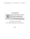 | 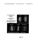 |
 |  |
 |  |
 |  |
 |  |
 |  |
 | 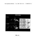 |
 | 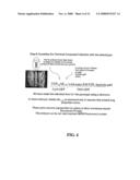 |
 |  |
 |  |
 |  |
 |  |
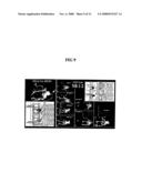 | 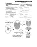 |
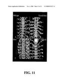 |  |
 |  |
 |  |
 | 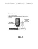 |
 |  |
 |  |
 |  |
 |  |
 |  |
 |  |
| Similar patent applications: | |
| Date | Title |
|---|---|
| 2012-02-23 | Method of delivery of nucleic acids to a developing embryo |
| 2010-05-06 | Methods for assessing the delivery of exogenous agents |
| 2010-09-30 | Methods for assessing the delivery of exogenous agents |
| 2012-07-05 | Transposition of maize ac/ds elements in vertebrates |
| 2012-11-15 | Azoline compounds for combating invertebrate pests |
| New patent applications in this class: | |
| Date | Title |
|---|---|
| 2022-05-05 | Off-target single nucleotide variants caused by single-base editing and high-specificity off-target-free single-base gene editing tool |
| 2017-08-17 | Coelenterazine analogues |
| 2017-08-17 | Pig model for diabetes |
| 2016-12-29 | Non-human animals having a humanized signal-regulatory protein gene |
| 2016-12-29 | High-throughput mouse model for optimizing antibody affinities |
| Top Inventors for class "Multicellular living organisms and unmodified parts thereof and related processes" | |
| Rank | Inventor's name |
|---|---|
| 1 | Gregory J. Holland |
| 2 | William H. Eby |
| 3 | Richard G. Stelpflug |
| 4 | Laron L. Peters |
| 5 | Justin T. Mason |