Patent application title: MODEL SYSTEM FOR IDENTIFYING ANTI-CANCER AGENTS
Inventors:
Vladimir S. Spiegelman (Fitchburg, WI, US)
Neehar Bhatia (Madison, WI, US)
Assignees:
WISCONSIN ALUMNI RESEARCH FOUNDATION (WARF)
IPC8 Class: AC12Q168FI
USPC Class:
435 6
Class name: Chemistry: molecular biology and microbiology measuring or testing process involving enzymes or micro-organisms; composition or test strip therefore; processes of forming such composition or test strip involving nucleic acid
Publication date: 2008-10-30
Patent application number: 20080268438
Inventors list |
Agents list |
Assignees list |
List by place |
Classification tree browser |
Top 100 Inventors |
Top 100 Agents |
Top 100 Assignees |
Usenet FAQ Index |
Documents |
Other FAQs |
Patent application title: MODEL SYSTEM FOR IDENTIFYING ANTI-CANCER AGENTS
Inventors:
Vladimir S. SPIEGELMAN
Neehar BHATIA
Agents:
KENING LI;PINSENT MASONS
Assignees:
WISCONSIN ALUMNI RESEARCH FOUNDATION (WARF)
Origin: WASHINGTON, DC US
IPC8 Class: AC12Q168FI
USPC Class:
435 6
Abstract:
A model system for screening and identification of compounds that
interfere with Gli2 dependent tumorigenesis and provide potential use as
anticancer agents is provided. In particular, the invention includes a
Gli2 protein having an S662A point mutation that interferes with binding
by the ubiquitin-ligase β-TrCP. The mutation inhibits Gli2
degradation by the ubiquitin pathway. Gli2 stability and half-life are
increased in the host cell resulting in an increase in Gli2-dependent
transcription and concomitant neoplasia and tumorigenesis. Expression of
the Gli2 mutant allows for the high throughput screening of compounds
that interfere with the tumorigenesis thereby identifying anticancer
agents.Claims:
1. An isolated polypeptide coding for the Gli2 transcription factor set
forth in SEQ ID NO: 2, wherein the β-TrCP2 binding motif DSGV/M at
residues 661-664 are mutated to Xaa1Xaa2Xaa3Xaa4,
wherein Xaa1 represents any amino acid except aspartic acid,
Xaa2 represents any amino acid except for serine, Xaa3
represents any amino acid except for glycine and Xaa4 represents any
amino acid except valine or methionine such that Gli2 has altered binding
to β-TrCP2.
2. A host cell comprising a genome which is augmented with a nucleic acid molecule encoding the polypeptide of claim 1.
3. The isolated polypeptide of claim 1, wherein the isolated polypeptide has the sequence set forth in SEQ ID NO: 2.
4. A host cell comprising a genome which is augmented with a nucleic acid molecule encoding the polypeptide of claim 3.
5. An isolated polynucleotide comprising a sequence set forth in SEQ ID NO: 5 or conservative variations thereof wherein the polynucleotide codes for the expression of an amino acid sequence according to claim 3.
6. A host cell comprising a genome which is augmented with a nucleic acid molecule encoding the polypeptide of claim 4.
7. A bioassay for evaluating the efficacy of anticancer agents comprising:(a) culturing cells which contain non-endogenous DNA which expresses a Gli2.sup.S662A protein having the sequence set forth in SEQ ID NO: 2;(b) expressing the non-endogenous DNA in the cultured cells;(c) identifying a transformation of the cells to a neoplastic state;(d) assaying for compounds that inhibit or ameliorate the transformation to the neoplastic state;thereby evaluating the efficacy of anticancer agents.
8. The bioassay of claim 7, wherein the neoplastic state comprises skin cancer, medulloblastoma, glioblastoma, rhabdomyosarcoma, lung cancer, prostate cancer, breast cancer or gastrointestinal cancer.
9. The bioassay of claim 7, wherein non-endogenous DNA is included in a reporter plasmid, wherein the reporter plasmid includes one or more copies of 3' Gli binding sites and binding of the Gli2 to the binding site results in expression of a reporter gene included in the reporter plasmid.
10. The bioassay of claim 9, wherein the reporter gene is selected from the group comprising luciferase, β galactosidase and green fluorescent protein.
11. A bioassay for identifying inhibitors of the hedgehog signaling pathway comprising:(a) culturing cells which contain non-endogenous DNA which expresses a Gli2.sup.S662A protein having the sequence set forth in SEQ ID NO: 1;(b) expressing the non-endogenous DNA in the cultured cells;(c) identifying an increase in the hedgehog signaling pathway;(d) contacting the cells with a compound that inhibits the increase in the hedgehog cascade;thereby identifying inhibitors of the hedgehog signaling pathway.
12. An expression vector encoding the isolated polypeptide of SEQ ID NO. 2 operatively linked to a promoter.
13. The expression vector of claim 12, wherein the promoter is an inducible promoter.
14. The expression vector of claim 12, wherein the promoter is a constitutive promoter.
Description:
FIELD OF THE INVENTION
[0002]This invention provides a method of initiating neoplastic cascades and identifying agents that interfere therewith. In particular, the method includes an isolated polypeptide and vector which results in the constitutive expression of the hedgehog cascade.
BACKGROUND OF THE INVENTION
[0003]Hedgehog (Hh) signaling plays a prominent role in embryogenesis, and its deregulation is implicated in tumorigenesis. Cellular responses to the Hedgehog (Hh) signal are controlled by two transmembrane proteins: the tumor suppressor Patched (PTCH) and the proto-oncogene Smoothened (SMO). In the absence of secreted Hh proteins, PTCH actively silences intracellular signaling by inactivating SMO. During physiologic signaling, Hh proteins bind and inactivate PTCH, which alleviates PTCH-mediated suppression of SMO. SMO activation triggers a series of intracellular events, culminating in expression of Hh target genes through the action of the Gli family of transcription factors. Gli1, Gli2 and Gli3, and their Drosophila homolog, Cubitus interruptus (Ci), are zinc finger transcription factors that are downstream effectors of Hh signaling. In the absence of Hh signaling, Ci is truncated at the carboxyl terminal domain to form a truncated repressor protein, whereas Hh activation leads to accumulation of transcriptionally active, full length Ci. The situation with mammalian Gli proteins is more complex. Gli3 functions primarily as a C-terminally truncated repressor, but full-length Gli3 protein accumulates in cells responding to Hh. Gli1, on the other hand, appears to modulate gene expression by acting primarily as a transcriptional activator, but Gli I mutant mice are phenotypically normal, arguing against an essential function for this protein during development or postnatal life. Gli2 appears to be the major nuclear effector of Hh signaling in vivo and functions primarily as a transcriptional activator. However, little is known about the molecular mechanisms regulating Gli2 expression at the protein level.
[0004]The Hh signaling pathway is deregulated in many human malignancies, including skin cancer, such as, basal cell carcinoma (BCC), medulloblastoma, glioblastoma, rhabdomyosarcoma, lung, prostate, breast, and some gastrointestinal cancers. Recent studies have stressed the importance of Hh signaling in human prostate cancer. Elevated Hh signaling pathway activity may distinguish metastatic from localized prostate cancer, and pathway manipulation can modulate invasiveness and metastasis. In contrast to BCCs and mebulloblastomas, which are associated with inactivating mutations in PTCH or gain-of-function mutations in SMO, aberrant Hh signaling in prostate cancers appears to be the result of constitutive overexpression of Sonic hedgehog (SHH). Hence, the growth of many of the prostate cancer cells is inhibited by Hh-neutralizing antibody. Furthermore, cyclopamine, a steroidal alkaloid that interacts with SMOH directly thus inhibiting Hh signaling, was shown to induce apoptosis and inhibit proliferation of prostate cancer cells in vivo as well as in vitro.
[0005]The ubiquitin-proteasome pathway is essential for degradation of proteins regulating growth and cell cycle progression. Ubiquitin-activating enzyme (E1), ubiquitin-conjugating enzyme (E2) and ubiquitin-ligase (E3) sequentially tag proteins for ubiquitination and proteasomal degradation. SCF E3 ubiquitin ligases are composed of Skp1, Cul1, Roc1 and F box proteins, where F box proteins are substrate recognizing subunits. Beta-transducin repeat-containing F box proteins (β-TrCP) recognize substrates phosphorylated within the DSG(X)2+nS destruction motifs. SCF.sup.β-TrCP E3 ligases ubiquitinate specifically phosphorylated substrates and play a pivotal role in the regulation of cell division and various signal transduction pathways, which, in turn, are essential for many aspects of tumorigenesis. Genetic data have suggested that Drosophila slimb protein (orthologue of mammalian β-TrCP) is involved in proteolytic processing of Ci155 to Ci75. However, there is no biochemical evidence that Slimb/β-TrCP proteins are involved in ubiquitination and degradation of Ci/Gli transcription factors.
[0006]Stabilization of the transcription factor Gli2 has been suggested as a key event in the transduction of Hh signals. The potential role of Gli2 in the development of BCC has been well documented. Gli2 is over-expressed in the majority of human BCCs, and skin-targeted over-expression of Gli2 in transgenic mice leads to the development of multiple BCCs. There is growing evidence that the transcriptional regulation of some Hh target genes, including Gli1, E2F1, Bcl2, etc., are Gli2 dependent. The promoter of one such gene, Bcl2, is regulated preferentially by Gli2.
[0007]As disclosed herein, the inventors have found that SCF.sup.β-TrCP E3 ubiquitin ligase is responsible for Gli2 degradation. β-TrCP2 binds wild type Gli2 and promotes its ubiquitination, which can be altered by a single amino acid substitution in the DSGX binding site of Gli2 with β-TrCP2. As disclosed here, mutating residues within the DSGX binding motif, inhibits binding of β-TrCP2 to Gli2, increasing Gli2 half-life and promoting Gli2 dependent transcription. The inventors further show that Gli2 is over-expressed in prostate cancer cell lines and primary tumors. Thus, expression of Gli2 with altered binding to β-TrCP2 provides a model for investigating diseases identified with Gli2 overexpression and for identifying agents that interfere with Gli2 dependent transcription and/or diseases resulting therefrom.
SUMMARY OF THE INVENTION
[0008]The invention provides compositions and methods to direct cells to a neoplastic state. In particular, the invention includes a Gli2 protein having a point mutation that interferes with binding by the ubiquitin-ligase β-TrCP, thus, inhibiting Gli2 degradation by the ubiquitin pathway. Gli2 stability and half-life are increased in the mutant resulting in an increase in Gli2-dependent transcription and concomitant neoplasia and tumorigenesis. The invention provides a model for the screening and identification of compounds that interfere with Gli2-dependent tumorigenesis to provide use as anticancer agents.
[0009]A model system for screening and identification of compounds that interfere with Gli2 dependent tumorigenesis and provide potential use as anticancer agents is provided. In particular, the invention includes a Gli2 protein having an S662A point mutation that interferes with binding by the ubiquitin-ligase β-TrCP. The mutation inhibits Gli2 degradation by the ubiquitin pathway. Gli2 stability and half-life are increased in the host cell resulting in an increase in Gli2-dependent transcription and concomitant neoplasia and tumorigenesis. Expression of the Gli2 mutant allows for the high throughput screening of compounds that interfere with the tumorigenesis thereby identifying anticancer agents.
[0010]Therefore, in one exemplary embodiment, the invention includes an isolated polypeptide having the sequence set forth in SEQ ID NO: 2, comprising the Gli2.sup.S662A mutant. In some embodiments, the invention includes a host cell, the genome of which is augmented with the nucleic acid molecule encoding the amino acid sequence set forth in SEQ ID NO: 2. In still other exemplary embodiments the invention further includes an isolated polynucleotide having the sequence set forth in SEQ ID NO: 5 or conservative variations thereof wherein the polynucleotide codes for the expression of an amino acid sequence as set forth in SEQ ID NO: 2.
[0011]In another exemplary embodiment, the invention includes an isolated polypeptide coding for the Gli2 transcription factor set forth in SEQ ID NO: 2, wherein the β-TrCP2 binding motif DSGV/M is mutated to Xaa1Xaa2Xaa3Xaa4, wherein Xaa1 represents any amino acid except aspartic acid, Xaa2 represents any amino acid except for serine, Xaa3 represents any amino acid except for glycine and Xaa4 represents any amino acid except valine or methionine such that Gli2 no longer binds β-TrCP2. Further, in some aspects according to the invention, the invention further includes a host cell comprising a genome which is augmented with a nucleic acid molecule encoding the polypeptide set forth in SEQ ID NO: 2 wherein the DSGV/M binding motif is disrupted. In still other aspects, the invention further includes an isolated polynucleotide coding for a Gli2 polypeptide having a disrupted β-TrCP2 binding motif.
[0012]In still another exemplary embodiment the invention includes a bioassay for evaluating the efficacy of anticancer agents. In this embodiment, the invention comprises:
[0013](a) culturing cells which contain non-endogenous DNA which expresses a Gli2.sup.S662A protein having the sequence set forth in SEQ ID NO: 2;
[0014](b) expressing the non-endogenous DNA in the cultured cells;
[0015](c) identifying the transformation to a neoplastic state of the cells;
[0016](d) assaying for compounds that inhibit or ameliorate the transformation to a neoplastic state; thereby evaluating the efficacy of anticancer agents.
[0017]In some versions, this embodiment further includes a reporter plasmid wherein the reporter plasmid includes one or more copies of 3' Gli binding sites and where binding of the Gli2 the binding site results in expression of a reporter gene. In various exemplary embodiments, the bioassay further includes a reporter gene. In various exemplary embodiments, the reporter gene may comprise luciferase, β galactosidase, green fluorescent protein or combinations thereof.
[0018]In still other exemplary embodiments, the invention includes a bioassay for identifying inhibitors of the hedgehog signaling pathway comprising:
[0019](a) culturing cells which contain non-endogenous DNA which expresses a Gli2.sup.S662A protein having the sequence set forth in SEQ ID NO: 1;
[0020](b) expressing the non-endogenous DNA in the cultured cells;
[0021](c) identifying an increase in the hedgehog signaling pathway;
[0022](d) contacting the cells with a compound that inhibits the increase in the hedgehog cascade;
thereby identifying inhibitors of the hedgehog signaling pathway.
[0023]In still other preferred embodiments, the invention includes an expression vector encoding the isolated polypeptide of SEQ ID NO. 2, operatively linked to a promoter. In some embodiments, the promoter is an inducible promoter. However, in still other embodiments, the promoter is a constitutive promoter.
[0024]Other objects, features and advantages of the present invention will become apparent after review of the specification, claims and drawings.
BRIEF DESCRIPTION OF THE DRAWINGS
[0025]FIGS. 1A and 1B, are maps of the Flag-MGli2 expression vector. FIG. 1A is a map of the Flag-mGli2 pc, plasmid; FIG 1B is a schematic illustrating the cloning strategy for introducing the S662A point mutation into the Gli2 cDNA.
[0026]FIGS. 2A-2E, are data illustrating that β-TrCP2 interacts with Gli2, and promotes its ubiquitination in vivo. FIG. 2A is a homology chart showing that the β-TrCP recognition motif in Gli2 is conserved across different species. FIG. 2B are western blots illustrating the interaction of wild-type (WT) Gli2 or the mutant Gli2.sup.S662A protein. A representative of three independent experiments is shown. IP=immuno precipitation, IB=immuno blot. FIG. 2C is an immunoblot illustrating the interaction between endogenous proteins as shown. WCE=whole cell extract. FIG. 2D, top panel is a radiogram showing the binding of in vitro-translated and 35S-labeled β-TrCP2 to Flag-Gli2 proteins expressed in 293T cells and immunopurified with Flag antibody before or after treatment with protein phosphatase λ. FIG. 2E shows the in vivo ubiquitination of Flag-Gli2 (wild type or S662A mutant) in 293T cells co-transfected with HA-tagged ubiquitin and β-TrCP2 constructs as indicated. Immunoprecipitation reactions with Flag antibodies were analyzed by means of immunoblotting with HA antibody. Ubiquitinated Flag-Gli2 species ("Gli2˜Ub") are indicated. A representative of two independent experiments is shown.
[0027]FIGS. 3A-3E, are data illustrating that inhibition of the β-TrCP ligase stabilizes the Gli2 protean. FIG. 3A, top panel autoradiograph of Flag-Gli2wt in shRNA transfected 293T cells, metabolically labeled with 35S-methionine/35S-cysteine, cells were harvested at time points indicated. Lower panel is a graph depicting percent of remaining Gli2 (compared to time point "0"). Insert shows the levels of β-TrCP1 and β-TrCP2 expression in 293T cells transfected with the indicated shRNA analyzed by immunoblotting. FIG. 3B, top panel is an autoradiograph of pulse chase analysis of Flag-Gli2Wt protein expressed in 293T cells with or without dominant negative HA-tagged β-TrCP2.sup.ΔN mutant and analyzed as in FIG. 3A. Lower panel is a graph depicting the percent of remaining Gli2 (compared to time point "0"). Insert shows the levels of HA-β-TrCP2.sup.ΔN expression in 293T cells transfected analyzed by immunoblotting with HA antibody. FIG. 3C, pulse chase analysis of endogenous Gli2 protein in 293T cells with or without dominant negative HA-tagged β-TrCP2.sup.ΔN mutant. 293T cells were metabolically labeled with 35S-methionine/35S-cysteine. Cells were harvested at different time points of chase with unlabeled methionine and cysteine. Gli2 was immunoprecipitated with Gli2 antibody (Santa Cruz Biotechnology) and analyzed using autoradiography. A representative of two independent experiments is shown. Insert shows the levels of HA-β-TrCP2.sup.ΔN expression in 293T cells transfected analyzed by immunoblotting with HA antibody. FIG. 3D is an immunoblot with GLI2 antibody showing the effect of the expression of the endogenous expression of GLI2 protein in 293T cells transfected with the indicated shRNA constructs. FIG. 2E top panel is an autoradiograph of pulse chase analysis of Flag-Gli2Wt protein expressed in 293T cells treated with LiCl and analyzed as in FIG. 3A, bottom panel is a graph depicting the percent of remaining Gli2 (compared to time point "0"). Cells were serum starved for 12 hr and then treated with 40 mM LiCl 1 hr prior to the chase. A representative of two independent experiments is shown.
[0028]FIGS. 4A-4C are data showing that Gli2.sup.S662A is expressed in higher levels and is more potent in the activation of Gli-dependent transcription than Gli2Wt. FIG. 4A top panel autoradiograph of Gli2Wt and Gli.sup.S662A transfected 293T cells, metabolically labeled with 35S-methionine/35S-cysteine, and their degradation; cells were harvested at the time pointes indicated. Lower panel is a graph depicting percent of remaining Gli2 and Gli.sup.S662A (compared to time point "0"). A representative of two experiments is shown. FIG. 4B similar to 3A, top panel is an autoradiograph of a pulse chase analysis of Flag-Gli2.sup.S662A protein expressed in 293T cells with or without dominant negative β-TrCP2.sup.ΔN mutant and analyzed as in FIG. 3A. A representative of two independent experiments is shown. Lower panel is a graph depicting percent of remaining Gli.sup.S662A for each construct (compared to time point "0"). Insert shows the levels of HA-β-TrCP2.sup.ΔN expression in 293T cells transfected, analyzed by immunoblotting with HA antibody. FIG. 4C graphs representing results of luciferase assay for HeLa cells transfected with Gli-luciferase (8×3'Gli BS-LucII), pGL3-Bcl2promo luciferase, or K17 luciferase, and respective Gli2 expression plasmids as indicated. Luciferase activity was estimated using Luciferase Reporter Assay Reagent (Promega, Madison, Wis.). β-galactosidase was used for normalization and estimated using β-gal assay reagent (Pierce Biotechnology). *-p<0.01 compared to cells transfected with empty vector (pcDNA3.1); **-p<0.01 compared to cells transfected with Gli2wt, in Student's t-test. Insert shows the levels of Flag-Gli2 expression in HeLa cells transfected with Flag-Gli2wt or Flag-Gli2.sup.S662A analyzed by immunoblotting with Gli2 G-20 antibody (protein loading was normalized by β-galactosidase activity).
[0029]FIGS. 5A-C, FIG. 5A shows an immunoblot of GLI2 expression prostate cancer cells using Gi2 antibody (G20). A representative of two independent experiments is shown. FIG. 5B histogram showing expression of GLI2 mRNA in the prostate cancer cell lines shown in FIG. 5A as assessed by Real Time RT-PCR. GAPDH was used as an internal control. A representative of three independent experiments is shown. FIG. 5C shows histochemical preparations of prostate hyperplasia (left panel) and adenocarcinoma (right panel) at ×400 magnification. Gli2 antibody was used for immunostaining.
DETAILED DESCRIPTION OF THE EXEMPLARY EMBODIMENTS
[0030]Before the present polypeptides, nucleic acids, and methods are described, it is understood that this invention is not limited to the particular methodology, protocols, cell lines, vectors, and reagents described, as these may vary. It is also to be understood that the terminology used herein is for the purpose of describing particular embodiments only, and is not intended to limit the scope of the present invention which will be limited only by the appended claims.
[0031]It must be noted that as used herein and in the appended claims, the singular forms "a", "an", and "the" include plural reference unless the context clearly dictates otherwise. Unless defined otherwise, all technical and scientific terms used herein have the same meanings as commonly understood by one of ordinary skill in the art to which this invention belongs. Although any methods and materials similar or equivalent to those described herein can be used in the practice or testing of the present invention, the preferred methods, devices, and materials are now described. All publications mentioned herein are incorporated herein by reference for the purpose of describing and disclosing the polypeptides, polynucleotides, strains, vectors, and methodologies which are reported in the publications which might be used in connection with the invention. Nothing herein is to be construed as an admission that the invention is not entitled to antedate such disclosure by virtue of prior invention.
[0032]The practice of the present invention will employ, unless otherwise indicated, conventional techniques of molecular biology, microbiology, recombinant DNA, and immunology, which are within the skill of the art. Such techniques are explained fully in the literature. See, for example, Molecular Cloning A Laboratory Manual, 2nd Ed., ed. by Sambrook, Fritsch and Maniatis (Cold Spring Harbor Laboratory Press: 1989); DNA Cloning, Volumes I and II (D. N. Glover ed., 1985); Oligonucleotide Synthesis (M. J. Gait ed., 1984); Mullis et al. U.S. Pat. No: 4,683,195; Nucleic Acid Hybridization (B. D. Hames & S. J. Higgins eds. 1984); Transcription And Translation (B. D. Hames & S. J. Higgins eds. 1984); Culture Of Animal Cells (R. I. Freshney, Alan R. Liss, Inc., 1987); Immobilized Cells And Enzymes (IRL Press, 1986); B. Perbal, A Practical Guide To Molecular Cloning (1984); the treatise, Methods In Enzymology (Academic Press, Inc., N.Y.); Gene Transfer Vectors For Mammalian Cells (J. H. Miller and M. P. Calos eds., 1987, Cold Spring Harbor Laboratory); Methods In Enzymology, Vols. 154 and 155 (Wu et al. eds.), Immunochemical Methods In Cell And Molecular Biology (Mayer and Walker, eds., Academic Press, London, 1987); Handbook Of Experimental Immunology, Volumes I-IV (D. M. Weir and C. C. Blackwell, eds., 1986); Cell Culture and Somatic Cell Genetics of Plants, Vol. 1 (I. K. Vasil, ed. 1984); R. V. Stanier, J. L. Ingraham, M. L. Wheelis, and P. R. Painter, The Microbial World, (1986) 5th Ed. Prentice-Hall.
DEFINITIONS
[0033]The Hedgehog (Hh) signaling pathway plays a crucial role in embryogenesis and has been linked to the development of several human malignancies. The transcription factor Gli2 plays a key role in the transduction of Hh signals by modulating transcription of some Hh target genes, yet, the mechanisms that control Gli2 protein expression are largely unknown. As disclosed herein, β-TrCP E3 ubiquitin ligase is required for Gli2 degradation. β-TrCP2 directly binds wild type Gli2 and promotes its ubiquitination. Single amino acid substitution in Gli2 putative binding site inhibits its interaction with β-TrCP2, its ubiquitination, and stabilizes the Gli2 protein. Stable Gli2 mutant is expressed in higher levels and is more potent in the activation of Gli-dependent transcription as compared with wild type Gli2. The inventors also found that GLI2 protein is highly expressed in prostate cancer cell lines and primary tumors, whereas the level of GLI2 mRNA is not appreciable different in normal and neoplastic prostate. These data identify β-TrCP2 as a pivotal regulator of Gli2 expression, and point to an important role for post-translational modulation of GLI2 protein levels in Hh pathway-associated human prostate cancer.
[0034]The term "biologically active" or "physiologically active", as used herein, refers to a protein, polypeptide, amino acid sequence, or nucleotide sequence encoding a product having structural, regulatory, or biochemical functions of a naturally occurring molecule. For example, a biologically active fragment of HdaA will have the histone deacetylase capabilities of a naturally occurring HdaA molecule disclosed herein.
[0035]Gli2", as used herein, refers to the amino acid sequences of the Gli2 protein obtained from Mammals and having the amino acid sequence given in GENBANK accession No. NP--005261 (See, FIG. 2A). In addition, Gli2 shall also refer to the amino acid sequences of Gli2 or other transcription factors obtained from any species (i.e., orthologs), from any source whether natural, synthetic, semi-synthetic, or recombinant. The term encompasses proteins encoded by nucleotide sequences representing allelic variants as well as those containing single nucleotide polymorphisms (SNPs).
[0036]gli2", as used herein, refers to the nucleotide sequences of the gli2 gene obtained from Mus musculus (GenBank accession no. NP--001074594). In addition, gli2 shall also refer to the nucleotide sequences of the gli2 gene or other transcription factors obtained from any species, particularly mammals, from any source whether natural, synthetic, semi-synthetic, or recombinant. The term encompasses allelic variants and single nucleotide polymorphisms (SNPs).
[0037]Single nucleotide polymorphism" or "SNPs" are defined by their characteristic attributes. A central attribute of such a polymorphism is that it contains a polymorphic site, "X," most preferably occupied by a single nucleotide, which is the site of the polymorphism's variation (Goelet and Knapp U.S. patent application Ser. No. 08/145,145). Methods of identifying SNPs are well known to those of skill in the art (see, e.g., U.S. Pat. No. 5,952,174).
[0038]A "composition comprising a given polynucleotide sequence", as used herein, refers broadly to any composition containing the given polynucleotide sequence. Compositions comprising polynucleotide sequences encoding gli2 or fragments thereof, may be employed as hybridization probes. The probes may be stored in freeze-dried form and may be associated with a stabilizing agent such as a carbohydrate. In hybridizations, the probe may be deployed in an aqueous solution containing salts (e.g., NaCl), detergents (e.g., SDS) and other components (e.g., Denhardt's solution, dry milk, salmon sperm DNA, etc.).
[0039]The term "correlates with expression of a polynucleotide", as used herein, indicates that the detection of the presence of ribonucleic acid is indicative of the presence of mRNA encoding, for example, Gli2 in a sample and thereby correlates with expression of the transcript from the polynucleotide encoding the protein.
[0040]A "deletion", as used herein, refers to a change in the amino acid or nucleotide sequence and results in the absence of one or more amino acid residues or nucleotides.
[0041]The term "homology", as used herein, refers to sequence similarity between two peptides or between two nucleic acid molecules. Homology may be determined by comparing a position in each sequence which may be aligned for purposes of comparison. When a position in the compared sequence is occupied by the same base or amino acid, then the molecules are homologous at that position. A degree of homology between sequences is a function of the number of matching or homologous positions shared by the sequences. A partially complementary sequence that at least partially inhibits an identical sequence from hybridizing to a target nucleic acid is referred to using the functional term "substantially homologous. The inhibition of hybridization of the completely complementary sequence to the target sequence may be examined using a hybridization assay (Southern or northern blot, solution hybridization and the like) under conditions of low stringency. A substantially homologous sequence or hybridization probe will compete for and inhibit the binding of a completely homologous sequence to the target sequence under conditions of low stringency. This is not to say that conditions of low stringency are such that non-specific binding is permitted; low stringency conditions require that the binding of two sequences to one another be a specific (i.e., selective) interaction. The absence of non-specific binding may be tested by the use of a second target sequence which lacks even a partial degree of complementary (e.g., less than about 30% identity). In the absence of non-specific binding, the probe will not hybridize to the second non-complementary target sequence.
[0042]In the art, "identity" means the degree of sequence relatedness between polypeptide or polynucleotide sequences, as the case may be, as determined by the match between strings of such sequences. "Identity" and "homology" can be readily calculated by known methods, including but not limited to those described in (Computational Molecular Biology, Lesk, A. M., ed., Oxford University Press, New York, 1988; Biocomputing: Informatics and Genome Projects, Smith, D. W., ed., Academic Press, New York, 1993; Computer Analysis of Sequence Data, Part I, Griffin, A. M., and Griffin, H. G., eds., Humana Press, New Jersey, 1994; Sequence Analysis in Molecular Biology, von Heinje, G., Academic Press, 1987; and Sequence Analysis Primer, Gribskov, M. and Devereux, J., eds., M Stockton Press, New York, 1991; and Carillo, H., and Lipman, D., SIAM J. Applied Math., 48: 1073 (1988). Preferred methods to determine identity are designed to give the largest match between the sequences tested. Methods to determine identity and homology are codified in publicly available computer programs. Preferred computer program methods to determine identity and homology between two sequences include, but are not limited to, the GCG program package (Devereux, J., et al., Nucleic Acids Research 12(1): 387 (1984)), BLASTP, BLASTN, and FASTA (Atschul, S. F. et al., J. Molec. Biol. 215: 403-410 (1990). The BLAST X program is publicly available from NCBI and other sources (BLAST Manual, Altschul, S., et al, NCBI NLM NIH Bethesda, Md. 20894; Altschul, S., et al., J. Mol. Biol. 215: 403-410 (1990). The well known Smith Waterman algorithm may also be used to determine identity.
[0043]Altered" or "modified" nucleic acid sequences encoding, for example, Gli2 as used herein, include those with deletions, insertions, or substitutions of different nucleotides resulting in a polynucleotide that encodes the same or a functionally equivalent protein. Included within this definition are polymorphisms which may or may not be readily detectable using a particular oligonucleotide probe of the polynucleotide encoding the subject protein, and improper or unexpected hybridization to alleles, with a locus other than the normal chromosomal locus for the respective polynucleotide sequence. The encoded protein may also be "altered" and contain deletions, insertions, or substitutions of amino acid residues which produce a silent change and result in a functionally equivalent protein. Deliberate amino acid substitutions may be made on the basis of similarity in polarity, charge, solubility, hydrophobicity, hydrophilicity, and/or the amphipathic nature of the residues as long as the biological or immunological activity of the subject protein is retained. For example, negatively charged amino acids may include aspartic acid and glutamic acid; positively charged amino acids may include lysine and arginine; and amino acids with uncharged polar head groups having similar hydrophilicity values may include leucine, isoleucine, and valine, glycine and alanine, asparagine and glutamine, serine and threonine, and phenylalanine and tyrosine.
[0044]Amino acid sequence", as used herein, refers to an oligopeptide, peptide, polypeptide, or protein sequence, and fragment thereof. Where "amino acid sequence" is recited herein to refer to a particular amino acid sequence "amino acid sequence", and like terms, are not meant to limit the amino acid sequence to the complete amino acid sequence referenced but shall be understood to include fragments of the complete amino acid sequence. The term shall further encompass synthetic molecules as well as those occurring naturally. The term "portion" or "fragment", as used herein, with regard to an amino acid sequence, specifically refers to segments of that amino acid sequence which are not naturally occurring as fragments and would not be found in the natural state. The segments may range in size from five amino acid residues to the entire amino acid sequence minus one amino acid.
[0045]Amplification", as used herein, refers to the production of additional copies of a nucleic acid sequence and is generally carried out using polymerase chain reaction (PCR) technologies well known in the art (Dieffenbach, C. W. and G. S. Dveksler (1995) PCR Primer, a Laboratory Manual, Cold Spring Harbor Press, Plainview, N.Y.).
[0046]An "insertion", "addition", or "mutation" as used herein, refers to a change in an amino acid or nucleotide sequence resulting in the addition or change of one or more amino acid residues or nucleotides, respectively, as compared to the naturally occurring molecule. Thus, the nomenclature Gli.sup.S662A mutant refers to a protein that has a change in from serine to arginine at residue 662 of its amino acid sequence.
[0047]Isolated" or "purified" or "isolated and purified" means altered "by the hand of man" from its natural state, i.e., if it occurs in nature, it has been changed or removed from its original environment, or both. For example, a polynucleotide or a polypeptide naturally present in a living organism is not "isolated," but the same polynucleotide or polypeptide separated from the coexisting materials of its natural state is "isolated", as the term is employed herein. Moreover, a polynucleotide or polypeptide that is introduced into an organism by transformation, genetic manipulation or by any other recombinant method is "isolated" even if it is still present in said organism, which organism may be living or non-living. As so defined, "isolated nucleic acid" or "isolated polynucleotide" includes nucleic acids integrated into a host cell chromosome at a heterologous site, recombinant fusions of a native fragment to a heterologous sequence, recombinant vectors present as episomes or as integrated into a host cell chromosome. As used herein, the term "substantially purified", refers to nucleic or amino acid sequences that are removed from their natural environment, isolated or separated, and are at least 60% free, preferably 75% free, and most preferably 90% free from other components with which they are naturally associated. As used herein, an isolated nucleic acid "encodes" a reference polypeptide when at least a portion of the nucleic acid, or its complement, can be directly translated to provide the amino acid sequence of the reference polypeptide, or when the isolated nucleic acid can be used, alone or as part of an expression vector, to express the reference polypeptide in vitro, in a prokaryotic host cell, or in a eukaryotic host cell.
[0048]Nucleic acid sequence" or "nucleotide sequence" or polynucleotide sequence", as used herein, refers to an oligonucleotide, nucleotide, or polynucleotide, and fragments thereof, and to DNA or RNA of genomic or synthetic origin which may be single- or double-stranded, and represent the sense or antisense strand. Where "nucleic acid sequence" or "nucleotide sequence" or polynucleotide sequence" is recited herein to refer to a particular nucleotide sequence "nucleotide sequence", and like terms, are not meant to limit the nucleotide sequence to the complete nucleotide sequence referenced but shall be understood to include fragments of the complete nucleotide sequence. In this context, the term "fragment" may be used to specifically refer to those nucleic acid sequences which are not naturally occurring as fragments and would not be found in the natural state. Generally, such fragments are equal to or greater than 15 nucleotides in length, and most preferably includes fragments that are at least 60 nucleotides in length. Such fragments find utility as, for example, probes useful in the detection of nucleotide sequences encoding Gli2.
[0049]A "substitution", as used herein, refers to the replacement of one or more amino acids or nucleotides by different amino acids or nucleotides, respectively. The term "conservative substitution" is used in reference to proteins or peptides to reflect amino acid substitutions that do not substantially alter the activity (specificity or binding affinity) of the molecule. Typically conservative amino acid substitutions involve substitution one amino acid for another amino acid with similar chemical properties (e.g. charge or hydrophobicity). The following six groups each contain amino acids that are typical conservative substitutions for one another: 1) Alanine (A), Serine (S), Threonine (T); 2) Aspartic acid (D), Glutamic acid (E); 3) Asparagine (N), Glutamine (Q); 4) Arginine (R), Lysine (K); 5) Isoleucine (I), Leucine (L), Methionine (M), Valine (V); and 6) Phenylalanine (F), Tyrosine (Y), Tryptophan (W).
[0050]A "plasmid" is a DNA molecule separate form the chromosomal DNA and capable or autonomous replication. "Expression vectors" are defined herein as nucleic acid sequences that direct the transcription of cloned copies of genes/cDNAs and/or the translation of their mRNAs in an appropriate host. Such vectors can be used to express genes or cDNAs in a variety of hosts such as bacteria, bluegreen algae, plant cells, insect cells and animal cells. Expression vectors include, but are not limited to, cloning vectors, modified cloning vectors, specifically designed plasmids or viruses. Specifically designed vectors allow the shuttling of DNA between hosts, such as bacteria-yeast or bacteria-animal cells. An appropriately constructed expression vector preferably contains: an origin of replication for autonomous replication in a host cell, a selectable marker, optionally one or more restriction enzyme sites, optionally one or more constitutive or inducible promoters. In preferred embodiments, an expression vector is a replicable DNA construct in which a DNA sequence encoding a described protein or a fragment thereof is operably linked to suitable control sequences capable of effecting the expression of the products in a suitable host. Control sequences include a transcriptional promoter, an optional operator sequence to control transcription and sequences which control the termination of transcription and translation, and so forth.
[0051]Transformation" or "transfection", as defined herein, describes a process by which exogenous DNA enters and changes a recipient cell. It may occur under natural or artificial conditions using various methods well known in the art. Transformation and transfection may rely on any known method for the insertion of foreign nucleic acid sequences into a prokaryotic or eukaryotic host cell. The method is selected based on the type of host cell being transformed and may include, but is not limited to, viral infection, electroporation, heat shock, lipofection, and particle bombardment. Such "transformed" cells include stably transformed cells in which the inserted DNA is capable of replication either as an autonomously replicating plasmid or as part of the host chromosome. They also include cells which transiently express the inserted DNA or RNA for limited periods of time.
[0052]The terms "polypeptide", "peptide" and "protein" are used interchangeably herein to refer to a polymer of amino acid residues. The terms apply to amino acid polymers in which one or more amino acid residue is an artificial chemical analogue of a corresponding naturally occurring amino acid, as well as to naturally occurring amino acid polymers. The term also includes variations on the traditional peptide linkage joining the amino acids making up the polypeptide. Where the terms are recited herein to refer to a polypeptide, peptide or protein of a naturally occurring protein molecule, the terms are not meant to limit the polypeptide, peptide or protein to the complete, native amino acid sequence associated with the recited protein molecule but shall be understood to include fragments of the complete polypeptide. The term "portion" or "fragment", as used herein, with regard to a protein or polypeptide (as in "a fragment of the Gli2 polypeptide") refers to segments of that polypeptide which are not naturally occurring as fragments in nature. The segments may range in size from five amino acid residues to the entire amino acid sequence minus one amino acid. Thus, a polypeptide encompasses the full-length amino acid sequence as well as segments thereof. Fragments of a described protein preferably are biologically active as defined herein.
[0053]The terms "nucleic acid" or "oligonucleotide" or "polynucleotide" or grammatical equivalents herein refer to at least two nucleotides covalently linked together. A nucleic acid of the present invention is preferably single-stranded or double stranded and will generally contain phosphodiester bonds, although in some cases, as outlined below, nucleic acid analogs are included that may have alternate backbones, comprising, for example, phosphoramide (Beaucage et al. (1993) Tetrahedron 49:1925) and references therein; Letsinger (1970) J. Org. Chem. 35:3800; Sprinzl et al. (1977) Eur. J. Biochem. 81: 579; Letsinger et al. (1986) Nucl. Acids Res. 14: 3487; Sawai et al. (1984) Chem. Lett. 805, Letsinger et al. (1988) J. Am Chem. Soc. 110: 4470; and Pauwels et al. (1986) Chemica Scripta 26: 141 9), phosphorothioate (Mag et al. (1991) Nucleic Acids Res. 19:1437; and U.S. Pat. No. 5,644,048), phosphorodithioate (Briu et al. (1989) J. Am. Chem. Soc. 111 :2321, O-methylphophoroamidite linkages (see Eckstein, Oligonucleotides and Analogues: A Practical Approach, Oxford University Press), and peptide nucleic acid backbones and linkages (see Egholm (1992) J. Am. Chem. Soc. 114:1895; Meier et al. (1992) Chem. Int. Ed. Engl. 31: 1008; Nielsen (1993) Nature, 365: 566; Carlsson et al. (1996) Nature 380: 207). Other analog nucleic acids include those with positive backbones (Denpcy et al. (1995) Proc. Natl. Acad. Sci. USA 92: 6097; non-ionic backbones (U.S. Pat. Nos. 5,386,023, 5,637,684, 5,602,240, 5,216,141 and 4,469,863; Angew. (1991) Chem. Intl. Ed. English 30: 423; Letsinger et al. (1988) J. Am Chem. Soc. 110:4470; Letsinger et al. (1994) Nucleoside & Nucleotide 13:1597; Chapters 2 and 3, ASC Symposium Series 580, "Carbohydrate Modifications in Antisense Research", Ed. Y. S. Sanghui and P. Dan Cook; Mesmaeker et al. (1994), Bioorganic & Medicinal Chem. Lett. 4: 395; Jeffs et al. (1994) J. Biomolecular NMR 34:17; Tetrahedron Lett. 37:743 (1996) and non-ribose backbones, including those described in U.S. Pat. Nos. 5,235,033 and 5,034,506, and Chapters 6 and 7, ASC Symposium Series 580, Carbohydrate Modifications in Antisense Research, Ed. Y. S. Sanghui and P. Dan Cook. Nucleic acids containing one or more carbocyclic sugars are also included within the definition of nucleic acids (see Jenkins et al. (1995), Chem. Soc. Rev. pp 169-176). Several nucleic acid analogs are described in Rawls, C & E News Jun. 2, 1997 page 35. These modifications of the ribose-phosphate backbone may be done to facilitate the addition of additional moieties such as labels, or to increase the stability and half-life of such molecules in physiological environments. As used herein, oligonucleotide is substantially equivalent to the terms "amplimers", "primers", "oligomers", and "probes", as commonly defined in the art.
[0054]The terms "complementary" or "complementarity", as used herein, refer to the natural binding of polynucleotides under permissive salt and temperature conditions by base-pairing. For example, the sequence "A-G-T" binds to the complementary sequence "T-C-A". Complementary between two single-stranded molecules may be "partial", in which only some of the nucleic acids bind, or it may be complete when total complementarity exists between the single stranded molecules. The degree of complementarity between nucleic acid strands has significant effects on the efficiency and strength of hybridization between nucleic acid strands. This is of particular importance in amplification reactions, which depend upon binding between nucleic acids strands and in the design and use of PNA molecules.
[0055]The term "hybridization", as used herein, refers to any process by which a strand of nucleic acid binds with a complementary strand through base pairing.
[0056]The term "hybridization complex", as used herein, refers to a complex formed between two nucleic acid sequences by virtue of the formation of hydrogen bonds between complementary G and C bases and between complementary A and T bases; these hydrogen bonds may be further stabilized by base stacking interactions. The two complementary nucleic acid sequences hydrogen bond in an antiparallel configuration. A hybridization complex may be formed in solution (e.g., C0 t or R0 t analysis) or between one nucleic acid sequence present in solution and another nucleic acid sequence immobilized on a solid support (e.g., paper, membranes, filters, chips, pins or glass slides, or any other appropriate substrate to which cells or their nucleic acids have been fixed).
[0057]As used herein a "dominant negative mutant" refers to a gene product that adversely affect the normal, wild-type gene product within the same cell. A dominant negative mutant may still interact with similar elements as the wild type product but blocks some aspect of its function. When used in reference to a transcription factor, enzyme or other bioactive protein, the dominant negative mutant can bind to its native substrate but fails to include the activation function. As discussed herein, the dominant negative mutant of β-TrCP (β-TrCP2.sup.ΔN) may bind to its native substrate but fails to effect phosphorylation of the substrate, in particular, serine 662 of the Gli2 peptide.
THE INVENTION
[0058]A model system for screening and identification of compounds that interfere with Gli2 dependent tumorigenesis and provide potential use as anticancer agents is provided. In particular, the invention includes a Gli2 protein having an S662A point mutation that interferes with binding by the ubiquitin-ligase β-TrCP. The mutation inhibits Gli2 degradation by the ubiquitin pathway. Gli2 stability and half-life are increased in the host cell resulting in an increase in Gli2-dependent transcription and concomitant neoplasia and tumorigenesis. Expression of the Gli2 mutant allows for the high throughput screening of compounds that interfere with the tumorigenesis thereby identifying anticancer agents.
[0059]Therefore, in one exemplary embodiment, the invention includes an isolated polypeptide having the sequence set forth in SEQ ID NO: 2, comprising the Gli2.sup.S662A mutant. In some embodiments, the invention includes a host cell, the genome of which is augmented with the nucleic acid molecule encoding the amino acid sequence set forth in SEQ ID NO: 2. In still other exemplary embodiments the invention further includes an isolated polynucleotide having the sequence set forth in SEQ ID NO: 5 or conservative variations thereof wherein the polynucleotide codes for the expression of an amino acid sequence as set forth in SEQ ID NO: 2.
[0060]In another exemplary embodiment, the invention includes an isolated polypeptide coding for the Gli2 transcription factor set forth in SEQ ID NO: 2, wherein the β-TrCP2 binding motif DSGV/M is mutated to Xaa1Xaa2Xaa3Xaa4, wherein Xaa1 represents any amino acid except aspartic acid, Xaa2 represents any amino acid except for serine, Xaa3 represents any amino acid except for glycine and Xaa4 represents any amino acid except valine or methionine such that Gli2 no longer binds β-TrCP2. Further, in some aspects according to the invention, the invention further includes a host cell comprising a genome which is augmented with a nucleic acid molecule encoding the polypeptide see forth in SEQ ID NO: 2 wherein the DSGV/M binding motif is disrupted. In still other aspects, the invention further includes an isolated polynucleotide coding for a Gli2 polypeptide having a disrupted β-TrCP2 binding motif.
[0061]In still another exemplary embodiment the invention includes a bioassay for evaluating the efficacy of anticancer agents. In this embodiment, the invention comprises:
[0062](a) culturing cells which contain non-endogenous DNA which expresses a Gli2.sup.S662A protein having the sequence set forth in SEQ ID NO: 2;
[0063](b) expressing the non-endogenous DNA in the cultured cells;
[0064](c) identifying the transformation to a neoplastic state of the cells;
[0065](d) assaying for compounds that inhibit or ameliorate the transformation to a neoplastic state; thereby evaluating the efficacy of anticancer agents.
[0066]In some versions, this embodiment further includes a reporter plasmid wherein the reporter plasmid includes one or more copies of 3' Gli binding sites and where binding of the Gli2 the binding site results in expression of a reporter gene. In various exemplary embodiments, the bioassay further includes a reporter gene. In various exemplary embodiments, the reporter gene may comprise luciferase, β galactosidase, green fluorescent protein or combinations thereof.
[0067]In still other exemplary embodiments, the invention includes a bioassay for identifying inhibitors of the hedgehog signaling pathway comprising:
[0068](a) culturing cells which contain non-endogenous DNA which expresses a Gli2.sup.S662A protein having the sequence set forth in SEQ ID NO: 1;
[0069](b) expressing the non-endogenous DNA in the cultured cells;
[0070](c) identifying an increase in the hedgehog signaling pathway;
[0071](d) contacting the cells with a compound that inhibits the increase in the hedgehog cascade;
thereby identifying inhibitors of the hedgehog signaling pathway.
[0072]In still other preferred embodiments, the invention includes an expression vector encoding the isolated polypeptide of SEQ ID NO. 2, operatively linked to a promoter. In some embodiments, the promoter is an inducible promoter. However, in still other embodiments, the promoter is a constitutive promoter.
EXAMPLE 1
General Experimental Procedures
[0073]DNA Constructs: The pcDNA3.1-Flag-Gli2 plasmid (FIG. 1A) was generated using full-length mouse Gli2 mRNA having the sequence set forth in SEQ ID NO. 1 and having the GenBank accession number NM--001081125 and coding for the Gli2 protein as set forth in GenBank Accession No. NP--001074594 and provided by Drs. Hiroshi Sasaki and Chi-ching Hui (Sasaki, H., Nishizaki, Y., Hui, C., Nakafuku, M., and Kondoh, H. (1999) Development 126, 3915-3924). Replacement of serine 662 with alanine, for the protein sequence as set forth in SEQ ID NO: 2 was carried out using a QuikChange Site-Directed Mutagenesis Kit (Stratagene). The cDNA encoding the S662A point mutant was created using primers: 5' GCC CCC AAC AAT GAC GCC GGC ATG GAG ATG CCG 3' (primer 3, SEQ ID NO: 3) and 5' CGG CAT CTC CAT GCC GGC GTC ATT GTT GGG GGC 3' (primer 4, SEQ ID NO: 4,) resulting in the sequence set forth in SEQ ID NO: 5, mutating the serine codon, AGT to the alanine codon GCC at residues 2333-2335 (wt) which correspond to residues 1984-1986 of the insert. As shown in FIG. 1B, the underlined residues in the primers 3 and 4 represent the nucleotide changes resulting in S662A point mutation. The sequence for the wild-type insert is given in SEQ ID NO 6. HA-tagged β-TrCP2 and β-TrCP2.sup.ΔN encoding plasmids, the constructs for specific knock down of β-TrCP2 (shBTR2), β-TrCP1 (shBTR1), as well as control shRNA construct (shCON) (Kumar, K. G., Tang, W., Ravindranath, A. K., Clark, W. A., Croze, E., and Fuchs, S. Y. (2003) Embo J 22, 5480-5490) were generously provided by Dr. S. Y. Fuchs (Fuchs, S. Y., Chen, A., Xiong, Y., Pan, Z. Q., and Ronai, Z. (1999) Oncogene 18, 2039-2046). 8×3'Gli BS-LucII [8 directly repeated copies of 3'Gli binding site from HNF3β floor plate enhancer cloned into p851LucII (Sasaki, H., Hui, C., Nakafuku, M., and Kondoh, H. (1997) Development 124, 1313-1322) was a gift of Dr. H. Sasaki (Osaka University, Osaka, Japan), K17-luc (K17 driven luciferase reporter) was from Dr. P. Coulombe, pGL3-Bcl2 promoter luciferase plasmid (pGL3 basic vector with 1.9 kb of the putative promoter and 5' untranslated region of human Bcl2) was generously provided by Dr. F. Aberger (Regl, G., Kasper, M., Schnidar, H., Eichberger, T., Neill, G. W., Philpott, M. P., Esterbauer, H., Hauser-Kronberger, C., Frischauf, A. M., and Aberger, F. (2004) Cancer Res 64, 7724-7731). Plasmids for expression of renilla luciferase (pRL-TK) and β-galactosidase (pSV-40 β-gal) were purchased (Promega Corp., Madison, Wis.).
[0074]Tissue Culture and Transfections: 293T human embryo kidney cells and HeLa human cervical adenocarcinoma cells were purchased from ATCC (Manassas, Va., Catalog #CRL-11268). Cells were grown in DMEM in the presence of 10% fetal bovine serum (FBS) and antibiotics at 37° C. and 5% CO2. Transfections were performed using the calcium phosphate procedure or lipofection with Lipofectamine 2000 (Invitrogen, Carlsbad, Calif.).
[0075]Antibodies and Western Blotting: Antibodies against HA (Roche), Gli2, β-actin (Santa Cruz Biotechnology), Flag M2 (Sigma-Aldrich) were purchased. β-TrCP antibody was described previously (Spiegelman, V. S., Tang, W., Katoh, M., Slaga, T. J., and Fuchs, S. Y. (2002) Oncogene 21, 856-860, hereby incorporated by reference in its entirety). Horseradish peroxidase conjugated secondary antibodies were purchased (Cell Signaling, Santa Cruz, Jackson). Immunoprecipitation and immunoblotting procedures were performed as described elsewhere (34).
[0076]In vivo Binding Assay: 293T cells co-transfected with FLAG-tagged Gli2 or Gli2.sup.S662A and HA-tagged β-TrCP2 were lysed in RIPA lysis buffer. Interaction between the expressed proteins was assessed by immunoprecipitation with FLAG or HA antibodies and immunoblotting with HA or FLAG antibodies, respectively. The interaction between endogenous proteins in the protein lysates from HeLa cells was analyzed by immunoprecipitation with Gli2 antibody and immunoblotting with β-TrCP antibody.
[0077]In vitro Binding Assay: Recombinant Gli2 proteins expressed in 293T cells were immunopurified with Flag antibody and protein A/G agarose beads, stringently washed with stripping buffer containing 20 mM Tris HCl (pH 7.5), 1M NaCl, 50 mM NaF, and 0.1% Nonidet P40 and equilibrated with binding buffer (20 mM Tris HCl, pH 7.5, 100 mM NaCl, 50 mM NaF, and 0.1% Nonidet P40). For treatment with phosphatase λ, the beads were washed with the binding buffer without phosphatase inhibitors and incubated with the phosphatase λ, for 1 h at 37° C. followed by washes in stripping buffer and re-equilibration with binding buffer. Flag-Gli2 proteins immobilized on the beads were incubated with in vitro-translated and 35S-labeled β-TrCP2 for 60 min at 4° C. The beads were extensively washed with binding buffer and associated proteins were analyzed by SDS-PAGE and autoradiography.
[0078]S35 labeled β-TrCP2 was synthesized in vitro using TnT kit (Promega). Lysates of 293T cells transfected with FLAG Gli-2 and Gli-2.sup.S662A were incubated with S35 labeled β-TrCP2 for 3 hrs at 4° C. These lysates were immunoprecipitated with FLAG antibody and associated proteins were analyzed by SDS-PAGE and autoradiography.
[0079]In vivo Ubiquitination Assay: 293T cells were co-tranfected with HA-tagged ubiquitin, HA-tagged βTrCP2 and Flag-tagged Gli2 or Gli2.sup.S662A. Cells were lysed in RIPA lysis buffer and immunoprecipitated with FLAG antibody. Immunocomplexes were analyzed by SDS-PAGE and immunoblotting with HA antibody.
[0080]Degradation Assay: Pulse chase analysis was carried out on 293T cells as described elsewhere (DasGupta, R., and Fuchs, E. (1999) Development 126, 4557-4568, hereby incorporated by reference in its entirety ). Briefly, cells were grown in 100 mm dishes and transfected with the indicated plasmids. Cells were starved in methionine and cysteine free DMEM followed by metabolically labeling with a 35S-methionine/35S-cysteine mixture (Perkin Elmer, Inc.). Chase was performed in complete DMEM (10% FBS) supplemented with 2 mM unlabeled methionine and cysteine and cells were harvested at respective time points. Gli2 proteins were immunoprecipitated from RIPA lysates with FLAG antibody, separated by SDS-PAGE and analyzed by autoradiography.
[0081]Luciferase Reporter Assays: Hela cells were transfected with 8×3'Gli BS-LucII reporter, K17 luciferase reporter plasmid, or pGL3-Bcl2promo luciferase reporter plasmid and respective Gli2 expression plasmids. Luciferase activity was estimated using Luciferase Reporter Assay Reagent (Promega). β-galactosidase was used for normalization and estimated using β-gal assay reagent (Pierce Biotechnology).
[0082]Immunohistochemistry: Prostate tissue arrays were purchased (Cybrdi, Inc., Frederick, Md.). Sections were incubated at 4° C. overnight with Gli-2 G20 antibody followed by donkey anti-goat biotin secondary antibody, ABC reagent (Vector Laboratories, Burlingame, Calif.) and developed by DAB (Sigma-Aldrich).
[0083]RNA isolation and Real-Time RT PCR: Real Time RT PCR for quantitative RNA measurements of gli2 were done using SYBR Green PCR Core reagents (Applied Biosystems) as described before (Lamm 2002). GAPDH was used as reference gene.
EXAMPLE 2
β-TrCP2 Interacts with Gli2, and Promotes its Ubiquitination In Vivo
[0084]SCF.sup.β-TrCP ubiquitin ligase recognizes DSG(X)2+nS destruction motif to target proteins for ubiquitination and further degradation (reviewed in (23)). Therefore, the inventors designed a series of experiments to investigate the potential interatction of Gli2 with ligases responsible for ubiquitination and degradation of target molecules.
[0085]Sequence analysis of Gli2 revealed the DSGV/MEMPGTGPGS motif, which is conserved among various mammalian species (FIG. 2A). The inventors analyzed whether substrate recognizing component of SCF.sup.β-TrCP ubiquitin ligase, F-box protein β-TrCP2 interacts with Gli2. The inventors found that exogenously expressed Gli2 and β-TrCP2 (FIG. 2B), as well as endogenous proteins (FIG. 2C) interact in vivo in co-immunoprecipitation assay. Gli2 was also shown to bind in vitro translated β-TrCP2 protein (FIG. 2D). To determine whether the putative β-TrCP recognition motif is responsible for this interaction, the inventors substituted potentially phosphorylated Serine 662 to Alanine in this motif of Gli2 (FIGS. 1B, 2A). This single amino acid substitution is predicted to disrupt Gli2 interactions with β-TrCP. Indeed, in both assays, Gli2.sup.S662A (SEQ ID NO: 2) binding to β-TrCP2 was greatly diminished (FIG. 2B and D). Treatment of Flag-Gli2 with protein phosphatase X abolished the ability of Gli2 to bind β-TrCP2 in vitro (FIG. 2D). These results demonstrate that phosphorylation of Gli2 is necessary for its recognition by β-TrCP ubiquitin ligase receptor. Thus, these data indicate that disruption of Gli2/β-TrCP2 binding, by mutation of any of the residues in the DSGX motif will affect the ubiquitination of Gli2, resulting in stabilization of the Gli2 protein and increased Gli2 dependent transcription.
[0086]Interaction of β-TrCP2 with specific substrates results in ubiquitination of these proteins. Co-transfection of cells with the β-TrCP2 construct accelerated the ubiquitination of wild type Gli2 (FIG. 2E, Lane 3) as measured by in vivo ubiquitination assay. In contrast, the ubiquitination of the Gli2.sup.S662A mutant that interacts poorly with β-TrCP2 was less efficient, and was not affected by β-TrCP2 over-expression (FIG. 2E, Lane 6). These data demonstrate that DSGX motif is indeed the β-TrCP2 binding site, that substitution of residues within the motif inhibits binding of β-TrCP2 and that serine 662 is critical for the interaction between Gli2 and β-TrCP2, and that binding to β-TrCP is important for Gli2 ubiquitination.
EXAMPLE 3
Inhibition of β-TrCP Function Stabilizes Gli2 Protein
[0087]In order to analyze the role of β-TrCP in the degradation of Gli2, the inventors inhibited β-TrCP activity by either knocking down the expression of β-TrCP1 and β-TrCP2 proteins by using short hairpin RNA (shRNA) or by expression of dominant negative mutant of β-TrCP (βTrCP2.sup.ΔN) (Fuchs, S. Y., Chen, A., Xiong, Y., Pan, Z. Q., and Ronai, Z. (1999) Oncogene 18, 2039-2046).
[0088]In these experiments, 293T cells were transfected with Flag-Gli2wt in the presence of the indicated shRNA constructs. Pulse chase analysis of Flag-Gli2 degradation in 293T cells was performed with metabolically labeled with 35S-methionine/35S-cysteine. Cells were harvested at different time points of chase with unlabeled methionine and cysteine. Gli2 was immunoprecipitated with Flag antibody and analyzed using autoradiography. The levels of β-TrCP1 and β-TrCP2 expression in 293T cells transfected with the indicated shRNA analyzed by immunoblotting was also determined (FIG. 3A). The inventors also performed a pulse chase analysis of Flag-Gli2wt protein expressed in 293T cells with or without dominant negative HA-tagged β-TrCP2.sup.ΔN mutant and analyzed as in FIG. 3A. The levels of HA-β-TrCP2.sup.ΔN expression in 293T cells transfected was analyzed by immunoblotting with HA antibody (FIG. 3B). Further, a pulse chase analysis of endogenous Gli2 protein in 293T cells with or without dominant negative HA-tagged β-TrCP2.sup.ΔN mutant. 293T cells were metabolically labeled with 35S-methionine/35S-cysteine. The cells were and harvested at different time points of chase with unlabeled methionine and cysteine. Gli2 was immunoprecipitated with Gli2 antibody (Santa Cruz Biotechnology) and analyzed using autoradiography. The levels of HA-β-TrCP2.sup.ΔN expression in 293T cells transfected was analyzed by immunoblotting with HA antibody (FIG. 3C). In this series of experiments, the expression of endogenous GLI2 protein in 293T cells transfected with indicated shRNA constructs was analyzed by immunoblotting with GLI2 antibody (FIG. 3D). Finally, pulse chase analysis of Flag-Gli2wt protein expressed in 293T cells treated with LiCl was analyzed as in FIG. 3A. Cells were serum starved for 12 hr and then treated with 40 mM LiCl 1 hr prior to the chase (FIG. 3E).
[0089]The results of the above experiments show that inhibition of β-TrCP function leads to stabilization of Gli2 protein (FIG. 3A-C). β-TrCP2.sup.ΔN extends the half-life of Gli2 from about 6 to 12 hours. Interestingly, shRNA against β-TrCP2 appeared to be more effective in the inhibition of Gli2 turnover than β-TrCP1 specific shRNA (FIG. 3A). Importantly, inhibition of β-TrCP function resulted in stabilization (FIG. 3C) and accumulation (FIG. 3D) of endogenous Gli2 in 293T cells. These data suggest that β-TrCP is involved in degradation of Gli2 protein in mammalian cells.
[0090]Although the kinase responsible for Gli2 phosphorylation within β-TrCP recognition motif is not known, phosphorylation of Drosophila Ci by shaggy (Drosophila homologue of GSK-3β) was demonstrated to be a necessary step in Ci proteolysis. Recently Gli2 was shown to be phosphorylated by GSK3β. Treatment of cells with GSK3 inhibitor, LiCl substantially increased the half-life of Gli2 protein (FIG. 3E). These data suggest that GSK3 may be involved in phosphorylation-dependent degradation of Gli2.
EXAMPLE 4
[0091]Gli2 S662A is Expressed in Higher Levels and is More Potent in the Activation of Gli-Dependent Transcription than Gli2wt
[0092]To further confirm the role of β-TrCP in proteolysis of Gli2, the inventors compared the rate of degradation Gli2wt with that of Gli2.sup.S662A mutant. In these experiments, Flag-Gli2 proteins (wild type or S662A mutant) were expressed in 293T cells and their degradation analyzed as in Example 3 (FIG. 4A). Also, a pulse chase analysis of Flag-Gli2.sup.S662A protein expressed in 293T cells was performed with or without dominant negative β-TrCP2.sup.ΔN mutant and analyzed as in Example 3. The levels of HA-β-TrCP2.sup.ΔN expression in 293T cells transfected was analyzed by immunoblotting with HA antibody (FIG. 4B). Further, HeLa cells were transfected with Gli-luciferase (8×3'Gli BS-LucII), pGL3-Bcl2promo luciferase, or K17 luciferase, and respective Gli2 expression plasmids as indicated (FIG. 4C). Luciferase activity was estimated using the Luciferase Reporter Assay Reagent (Promega, Madison, Wis.). β-galactosidase was used for normalization and estimated using β-gal assay reagent (Pierce Biotechnology, Rockford, Ill.). The levels of Flag-Gli2 expression in HeLa cells transfected with Flag-Gli2wt or Flag-Gli2.sup.S662A was analyzed by immunoblotting with Gli2 G-20 antibody with protein loading normalized by β-galactosidase activity FIG. 4C).
[0093]Gli2.sup.S662A poorly interacts with β-TrCP2 (FIGS. 2B, C), and is not ubiquitinated by β-TrCP2 (FIG. 3D). In comparison to Gli2wt, Gli2.sup.S662A mutant protein is more stable and exhibits a half-life of more than 9 hours (FIG. 4A). Furthermore, over expression of dominant negative mutant of β-TrCP (β-TrCP2.sup.ΔN) did not affect the stability of Gli2.sup.S662A (FIG. 4B). These data demonstrate that serine 662 is critical for the interaction between Gli2 and β-TrCP2. Disruption of the DSGX motif by mutation of the serine residue renders it poorly interactive with β-TrCP, hence stabilizing the Gli2.sup.S662A mutant protein.
[0094]Stabilization of Gli2.sup.S662A mutant translates into the higher level of protein expression as compared to the wild type Gli2 (FIG. 4C, insert). HeLa cells transfected with the same amount of appropriate plasmids express higher level of Gli2.sup.S662A mutant protein as compared to the wild type protein. FIG. 4C demonstrates that Gli2.sup.S662A mutant is significantly more effective than Gli2wt in the activation of Gli-dependent transcriptional activity. 8×3'Gli BS-LucII, pGL3-Bcl2promo luciferase or K17 driven luciferase were utilized to measure Gli2 dependent transcriptional activation driven by Gli2wt or Gli2.sup.S662A. Gli2.sup.S662A is about twice as potent, as Gli2wt in activation of transcription as measured by these 3 different reporter constructs. These results demonstrate that elevated Gli2 dependent transcriptional output is likely attributed to the higher levels of Gli2.sup.S662A protein expression. These data strongly suggest that Gli2 protein turnover is an important step in the modulation of Gli2 dependent transcription.
[0095]Gli transcription factors, including Gli1, Gli2 and Gli3, are the key modulators of Hh signaling. Gli2 and Gli3 contain both the amino terminal repressor domain as well as carboxyl terminal activator domain, whereas, Gli1 is comprised of only the carboxyl terminal activator domain. Hence, Gli1 acts as the transcriptional activator and is a secondary target, downstream of Gli2/Gli3. On the other hand, Gli3 is suggested to primarily act as the repressor, although, activator function for Gli3 has also been reported. Gli2, however, has been suggested to be the primary activator of Hh signaling. Mice homozygous for Gli2 mutations exhibit developmental defects and over expression of Gli2 results in formation of BCCs.
[0096]It has been previously shown that β-TrCP targets several essential proteins in cell transformation and signal transduction for ubiquitination and degradation, including IκBα, β-catenin and ATF4 proteins. Genetic evidence suggest that Drosophila Slimb F box protein is involved in Hh signaling pathway in processing of full length Ci155 protein to truncated repressor form Ci75. As disclosed and shown herein, Gli2 is the substrate of β-TrCP for ubiquitination and degradation. However, unlike Ci, there is no evidence suggesting that Gli2 undergoes proteolytic cleavage. The inventors were unable to detect any smaller protein species of Gli2 indicating that Gli2 protein stabilization, rather than inhibition of processing, is important for downstream signaling.
EXAMPLE 5
GLI2 is Over-Expressed in Prostate Cancer Cells
[0097]Overexpression of Gli2 in transgenic mice induces formation of BCCs (Grachtchouk, M., Mo, R., Yu, S., Zhang, X., Sasaki, H., Hui, C. C., and Dlugosz, A. A. (2000) Nat Genet 24, 216-217), and GLI2 mRNA is upregulated in the majority of human BCCs. However, there are no reports examining the expression of GLI2 protein in human cancers. Thus, the inventors examined the expression of GLI2 in a panel of cancer cell lines. Levels of GLI2 expression in prostate cancer cell lines were analyzed using immunoblotting with Gli2 antibody (G20) as shown in FIG. 5A. GLI2 mRNA expression was also assessed by Real Time RT-PCR as shown in FIG. 5B. Finally, a human prostate cancer tissue array was immunostained with GLI2 (G20) for expression of GLI2 full length protein.
[0098]The results of the above described experiments show that GLI2 protein is dramatically induced in prostate cancer cell lines as compared to normal human prostate epithelial (NHPE) cells (FIG. 5A). On the other hand, there is no significant difference in the expression of GLI2 mRNA in these cells (FIG. 5B). These data are in line with previous observations that elevated expression of Gli2 message is a rare event in human prostate tumors and cell lines. In all, this data suggests that a novel post-transcriptional mechanism, most likely protein stabilization, is responsible for high levels of GLI2 expression in prostate cancer cells. Constitutive activation of Hh signaling in prostate cancer has been recently reported (Karhadkar, S. S., Steven Bova, G., Abdallah, N., Dhara, S., Gardner, D., Maitra, A., Isaacs, J. T., Berman, D. M., and Beachy, P. A. (2004) Nature 431, 707-712). The data disclosed herein demonstrates that high levels of GLI2 protein expression strongly correlate with Hh pathway activation in cancer cells, suggesting that GLI2 plays an important role in mediation of Hh signaling in cancer and especially in prostate cancer cells. Immunohistochemical analysis of an array of primary human prostate tumors using GLI2 antibody that recognizes C-terminus of GLI2 protein revealed high levels expression of GLI2 in the majority of malignant prostate epithelial cells as compared to the benign ones. Nuclear localization of GLI2 protein is a hallmark of its transcriptional activity. Importantly, 26 out of 29 prostate adenocarcinomas (90%) exhibited strong nuclear or nuclear-cytoplasmic staining as shown in Table 1. In contrast, only one out of 5 prostate hyperplasia samples (20%) revealed nuclear accumulation of GLI2. FIG. 5C shows a representative GLI2 immuno-staining of malignant and benign prostate tissues. These data further substantiate the importance of GLI2 over expression and activation in prostate cancer development, supporting indicating that GLI2 may be a key component in Hh pathway activation in prostate cancer. Future studies will define the role of GLI2 in prostate tumorigenesis, and delineate the mechanisms by which prostate cancer cells achieve stabilization of GLI2.
TABLE-US-00001 TABLE 1 Gli2 expression in Human Prostate Carcinoma and Hyperpalsia GLI2 NUCLEAR SAMPLE AGE SEX ORGAN PATHOLOGY DIAGNOSIS EXPRESSION 1 78 M Prostate Adenocarcinoma Grade II + 2 66 M Prostate Adenocarcinoma Grade III + 3 60 M Prostate Adenocarcinoma Grade II/III + 4 67 M Prostate Adenocarcinoma Grade II + 5 69 M Prostate Adenocarcinoma Grade I + 6 70 M Prostate Adenocarcinoma Grade III + 7 76 M Prostate Adenocarcinoma Grade III + 8 63 M Prostate Adenocarcinoma Grade III + 9 87 M Prostate Adenocarcinoma Grade III - 10 80 M Prostate Adenocarcinoma Grade II + 11 78 M Prostate Adenocarcinoma Grade III + 12 69 M Prostate Adenocarcinoma Grade I + 13 70 M Prostate Adenocarcinoma Grade III/II + 14 60 M Prostate Adenocarcinoma Grade I + 15 70 M Prostate Adenocarcinoma Grade I/II + 16 82 M Prostate Adenocarcinoma Grade III + 17 75 M Prostate Adenocarcinoma Grade III + 18 81 M Prostate Adenocarcinoma Grade III + 19 72 M Prostate Adenocarcinoma Grade III + 20 85 M Prostate Adenocarcinoma Grade I + 21 80 M Prostate Adenocarcinoma Grade II + 22 89 M Prostate Adenocarcinoma Grade III + 23 64 M Prostate Adenocarcinoma Grade III + 24 73 M Prostate Adenocarcinoma Grade III + 25 65 M Prostate Hyperplasia - 26 64 M Prostate Adenocarcinoma Grade III + 27 75 M Prostate Adenocarcinoma Grade II + 28 78 M Prostate Adenocarcinoma Grade III + 29 60 M Prostate Adenocarcinoma Grade III - 30 73 M Prostate Adenocarcinoma Grade II - 31 72 M Prostate Hyperplasia +/- 32 63 M Prostate Hyperplasia - 33 75 M Prostate Hyperplasia -
[0099]The foregoing data show that elevated levels of Gli2 are present in neoplastic cells. Further, the inventors have shown that by using a Gli2.sup.S662A mutant physiologically active Gli2 can be expressed that does not bind the β-TRCP ubiquitin ligase. Further, without being held to any particular theory, increased levels of Gli2 are indicative of a neoplastic fate. Therefore, expression of physiologically active Gli2 allows investigators to induce neoplastic activity in cell preparations and provides a model by which drugs and/or other chemical compositions can be used to modulate the fate of the preparations. In such models, the ability of Gli2 to transduce signals in the Hh pathway the ultimately lead to malignant states can be investigated and provide clues to the efficacy of treatments interrupting the Gli2/Hh pathway.
[0100]While this invention has been described in conjunction with the various exemplary embodiments outlined above, various alternatives, modifications, variations, improvements and/or substantial equivalents, whether known or that are or may be presently unforeseen, may become apparent to those having at least ordinary skill in the art. Accordingly, the exemplary embodiments according to this invention, as set forth above, are intended to be illustrative, not limiting. Various changes may be made without departing from the spirit and scope of the invention. Therefore, the invention is intended to embrace all known or later-developed alternatives, modifications, variations, improvements, and/or substantial equivalents of these exemplary embodiments. All publications, patents, and patent applications cited herein are hereby incorporated by reference in their entirety for all purposes.
Sequence CWU
1
516708DNAMus musculus, GLI2 mRNA 1aatgagactt cgaggaggag cgggaggcgg
cggcggcggc tacagaggac gcggaggaag 60gcgaggagga gccagaggaa ggagcacggg
tcgtgcgtag catcgcagcc gcccggagct 120gctggcctgg tgtccttagg gactgcccga
gtcccccgcc cgccttggtc ccctcctctg 180cctggccagc cccgccccgg cagccggagc
ccccgcactc ggcagcccca ctccagccaa 240gttgggatgg ggccctgcaa ccactgcccg
gcgcccgaga ggccacctgc atgctagagg 300caaacttttg tctcctcggg tccgccacca
aagagtatga gcctctgaga tggagacttc 360tgccccagcc cctgcactgg agaagaaaga
agccaagagt ggtctcttgg aggacagcag 420cttccccgac ccagggaaaa aggcctgtcc
tctggcggtg gccgcagctg tagccgccca 480cggagttcct cagcagctcc tgccggcttt
ccacgcgcct ttgccgattg acatgagaca 540ccaggaggga aggtaccatt atgaccctca
ctctgtccac agtgtacacg ggcctcccac 600cctaagtggc agccctgtca tctcagatat
ctccttgata cgactttctc cacaccctgc 660tggccctgga gagtcaccct tcagcgccca
ccacccctac gtgaaccccc atatggagca 720ctacctccgg tctgtgcaca gcagccccac
actctcaatg atctctgccg ccaggggcct 780cagccctgct gatgtggccc acgaacatct
gaaagagagg ggactcttta gcctcgcagc 840cccaggcacc aacccttcag actattacca
ccagatgacc ctcatggcaa gccaccccac 900cccttatggg gaccttctaa tgcagagcgg
gggtgctgct agcgcacccc atctccatga 960ctacctcaac cctgtggatg catcacgatt
ctctagtcca cgtgtgaccc cacgactgag 1020ccgcaagcgg gctctgtcca tctccccgct
ctcagatgcc agcctcgacc tacaacgcat 1080gattcggacc tctcccaact cgctggtagc
ttacatcaac aactccagga gcagctcagc 1140agccagtggc tcttatggac atctgtctgc
tggtgccctc agcccagcct tcacttttcc 1200ccaccccatc aatcctgtgg cctaccagca
gatcctgagc cagcagcggg gcctgggctc 1260agcctttgga cacacaccac ccctgatcca
gccttcaccc accttcttgg cccagcagcc 1320catgactctc acctccatca gcaccatgcc
tacccaactc agcagcagta gcagcaactg 1380tctaaatgat gccaaccaga acaagcagaa
cagcgagtca gctgtgagca gcaccgtgaa 1440ccccatcacc attcataagc ggagcaaggt
caagactgag gctgagggcc tgcgtccagc 1500atccccgctt ggactgacac aggagcagct
ggccgatctc aaggaagacc tggacaggga 1560tgactgtaag caggaggccg aggtggtcat
ctacgagacc aactgccact gggcagactg 1620caccaaggag tatgacacac aggagcagct
ggtgcatcat atcaacaatg aacacatcca 1680cggggagaag aaggagttcg tgtgccgctg
gcaggcctgc acgagagagc agaagccctt 1740caaggcccag tacatgctgg ttgttcacat
gcgcagacac acgggtgaga agccacacaa 1800gtgcacgttc gaaggctgtt ccaaggccta
ctctcgcctg gagaacctga agacacacct 1860gcgttcacac acaggagaga agccatatgt
gtgtgaacac gaaggctgta acaaagcctt 1920ctccaatgcc tcagaccgcg ccaagcacca
gaaccgcact cactccaatg agaaacccta 1980catctgcaag atcccaggct gcaccaagag
gtacacagac cccagctcac tccgcaagca 2040tgtgaagact gtccatgggc cagacgccca
tgtcaccaag aaacagcgta atgatgtgca 2100tgtccgtgct ccactgctca aggagaatgg
ggataatgag gccagcgccg agccaggtgg 2160ccggggacct gaggagagtg tggaggccag
tagcaccagc cacactgtgg aggactgcct 2220acatatcaaa gccatcaaga cagagagctc
cgggctttgt cagtccagcc ccggggccca 2280gtcatcctgc agcagcgagc cctctcccct
gggcagtgcc cccaacaatg acagtggcat 2340ggagatgccg gggacagggc ctgggagtct
gggagacctg acagcactgg ctgacacgtg 2400tccaggagct gacacctcag ccctggctgc
accctccact ggtggcctgc agctgcgcaa 2460acacatgagc accgtgcatc gctttgagca
gctgaagaga gagaagctca agtcactgaa 2520ggattcctgc tcgtgggccg gcccagctcc
acacacccgc aacaccaagc tgcctcccct 2580tccagtcaat ggttctgtcc tggaaaactt
caacaataca gggggcggtg gaccggcagg 2640actgctgccc agccagcggc taccagagct
gaccgaagtg acgatgctga gccagctgca 2700ggaacgaaga gacagctcca ccagcaccat
gagctcggcc tacactgtga gccgccgctc 2760ctctggcatc tccccatact tctctagccg
tcgctccagc gaggcttcgc ctctcggtgg 2820cctacgcccg cacaacgcca gctcagcaga
ctcctatgac cccatctcca cagatgcctc 2880tcggcgctcc agtgaagcca gccagtgcag
tggcggtggc ccagggctgc tcaacctcac 2940acctgcgcag cagtacaacc tgcgtgccaa
gtacgcagcg gccacaggtg gaccaccgcc 3000cacgccactg ccgggcctcg atcgtgtaag
ccttcgtacc cgcctggcct tgctggatgc 3060tcctgagcgt gcacttcctg gtgcctgccc
acatccactg gggccacggc gtggcagcga 3120tgggcctacc tatagccatg gtcatggcca
tggctacgca ggtgcggctc cagcattccc 3180ccacgagggg ccaaacagca gcacacggcg
ggccagcgac cctgtgcggc gccctgaccc 3240ccttattctg cctcgagtgc aacgtttcca
cagtacccac aacatgaatc caggttcact 3300gccaccctgc gctgatcggc gtggcctgca
cgtacagagc caccccagcg tagacagcaa 3360cctgacccgc aacgcctact ctcccagacc
ccctagcatc aatgagaacg tggtgatgga 3420ggccgtggct gctggggtag acggcccagg
gctagagtgc gacctggggc tggtggagga 3480tgagctggtg ctgccagatg atgtggtaca
gtacatcaag gctcacaccg gtggtacctt 3540ggatgacggc attcggcagg ggtatcccac
agaaggtact ggcttccccg agaactctaa 3600gctgcccagt cctgggctac aaggccaccg
caggctagca gctgccgact ccaacatggg 3660tccttctgct cctggactcg ggggctgcca
gctgagctac agcccctcct ccaacctcaa 3720caagagcaac atgcctgtgc agtggaatga
ggtgagttct ggcaccgtgg atgccctgcc 3780tacccaggtg aagccacctc ctttccctca
cagcaacctg gctgtggtcc aacagaagcc 3840agcctttggc cagtatccag gatataatcc
acaatccgtg cagagcagct ccggaggtct 3900agacagcacc cagccgcacc tacagcttcg
aggagccccc tctgcatcaa gagggagcta 3960cacgcaacag cctcgacagc cagctgcagg
cagtcagtgc ctgggtatga gtgcggccat 4020gagcccgcag gccagctaca gccaagccca
cccccagctg agcccaaaca ttgtcagcgg 4080atctctgaac cagttttctc cctcctgcag
caatatggca gccaagccca gccacctggg 4140actccctcag caaatggaag ttgtccccaa
tgccaccatc atgaatggcc atcaacggga 4200gcacggggtc cccaattcat ccctggctgc
ggtgtcacaa cctcacccag tcctgagcta 4260tccccagcag gacagctacc aacagggctc
caaccttctg tcatcccatc agcctggctt 4320catggagtcc cagcagaacg cgggctttgg
tctcatgcag cctcggccac ccctggaacc 4380caacacggcc agccgtcacc gtggagtacg
ttctgggcaa cagcagttgt atgccaggac 4440cactggccaa gccatggtca catcagccaa
ccaagagaca gcagaagcta tgcccaaggg 4500accagcaggg accatggtat ccctagctcc
tcagccatct caggacacag ggcgggcaca 4560agatcagaac acgctatact actatggcca
gatccacatg tatgaacaga atggaggctg 4620cccagccgtg cagccccagc cgccacaacc
acaagcttgc tcagacagta tccagcctga 4680gcctttgcct tcaccgggag tcaaccaggt
gtctagcacc gtggactccc agctcctgga 4740gcccccccag attgactttg atgccatcat
ggatgatggt gatcactcga gtttgttttc 4800tggtgcactg agcccaaccc ttctccacaa
tctctcccag aattcctcac gcctcaccac 4860accccggaat tccttgacac tgccctccat
ccctgcgggc atcagcaaca tggccgtggg 4920cgacatgagt tccatgctca ccagcctggc
tgaagagagc aagtttttaa acatgatgac 4980ctaaaggccc tggctcctgg cacaggagac
ctgagcaaca ctagcccctt ggaatatgag 5040ggtctagtct gttctcattg tcccaaagtg
ttaactaggc ctgagagggg ttgggcaatg 5100gccaactcca aatgacagct ctgggccatg
cccaggggtc ttcttgcaga tgttttaaga 5160aaagattaag gggcatcctc tctgctgttt
ggcctatttg gggggggagg ggacacccaa 5220agtcttattc aaaaagaaac tcgttcacat
gacgcttgcc agcctctggt gtttgtgaga 5280ttcccgggtt ctggctcctt tgcctctatc
attcagctaa tgagagagaa ctttggccag 5340gggctttcca agtacacgcc caaagagggg
aggggagcca ctctgggttt gaatctgaga 5400ccatgctgga tattctctgc tctgagaaga
tgagtttgcc attctgccac ctgggaggaa 5460aagggattac tgtttcccct ggattcttcc
atgaagctcg tcaaggttgc tcaccacgga 5520ggagaaaggc gcatcccccg ggatattgta
gtctcctcag ttacttgcta acatctccat 5580ttccttaggt agagtgggac cagcagattt
cctaaatgga gaagaattaa taaaacacgt 5640attttactat tggggggggt acagggtctc
tcctggcctg tttacagtgt tctgaggtat 5700acagtgaagc cctccctggg aagaagactt
gcgtctacat gccttgggtt gctgtggact 5760aggaataggc agggcctcga ggtggaagag
gccttcccca ggaaaaggaa gtcatatgtt 5820tacccgctcc tatttctcca gtgcactatc
tgaccactct ctcatgggag atcccagggc 5880tcctccacac cggctggtgg ccagcagaca
accctgagcg accacctgca gcctccacac 5940tttctggtcc cctgacctct accctcctct
ccaggttatt tggcatggac cagtgatgac 6000atgcatggtt tgtggttgag cggaaggttg
aaggcattga ttcttggact ttgaggaact 6060cttaactgaa tttgcttgtt cagtatggcc
tggccccaga taagaaatgg catgtggaaa 6120ctgactccaa aaagagtcag tcagagcgca
ctgcagggtg aacccatgcc tccaccacca 6180cagacatgtg cgtatcccaa acctgttcct
ttgagttaac agaaactttt gccaaccaaa 6240cctcaccaat cagagcttcc ttctggccat
gaccttggct gtggtgagga gcttcttaac 6300agagtctgga ccctctgtgt cccagggcag
caaacttctc tgtttacaac ccagagacag 6360gaccaactcc aaatgacagc tctgggccat
gcccctgggt ctctggctct tgtggtgtgg 6420actcattgcc tgacatagac cctagcttcc
cattctgtct gtatctattt tgtgtagaca 6480tcgtcttgcc tgaatagact gtgggtgaat
ccaaatttgg tccagccaaa tttatattgc 6540caaacatttt tagctttttc tacatgctat
aaattgagat gacatgctca acttgtaaat 6600aagtcttttt tgtacattaa aagtaatttt
ttcataattt atcttgttga tctgcttttc 6660ccttgacagt agttaatgag aatccaggca
gtaaatttgg tgcatttg 670821544PRTMus
musculus,MUTAGEN(662)..(662)SERINE 662 is replaced by ALANINE 2Met Glu
Thr Ser Ala Pro Ala Pro Ala Leu Glu Lys Lys Glu Ala Lys1 5
10 15Ser Gly Leu Leu Glu Asp Ser Ser
Phe Pro Asp Pro Gly Lys Lys Ala 20 25
30Cys Pro Leu Ala Val Ala Ala Ala Val Ala Ala His Gly Val Pro
Gln 35 40 45Gln Leu Leu Pro Ala
Phe His Ala Pro Leu Pro Ile Asp Met Arg His 50 55
60Gln Glu Gly Arg Tyr His Tyr Asp Pro His Ser Val His Ser
Val His65 70 75 80Gly
Pro Pro Thr Leu Ser Gly Ser Pro Val Ile Ser Asp Ile Ser Leu
85 90 95Ile Arg Leu Ser Pro His Pro
Ala Gly Pro Gly Glu Ser Pro Phe Ser 100 105
110Ala His His Pro Tyr Val Asn Pro His Met Glu His Tyr Leu
Arg Ser 115 120 125Val His Ser Ser
Pro Thr Leu Ser Met Ile Ser Ala Ala Arg Gly Leu 130
135 140Ser Pro Ala Asp Val Ala His Glu His Leu Lys Glu
Arg Gly Leu Phe145 150 155
160Ser Leu Ala Ala Pro Gly Thr Asn Pro Ser Asp Tyr Tyr His Gln Met
165 170 175Thr Leu Met Ala Ser
His Pro Thr Pro Tyr Gly Asp Leu Leu Met Gln 180
185 190Ser Gly Gly Ala Ala Ser Ala Pro His Leu His Asp
Tyr Leu Asn Pro 195 200 205Val Asp
Ala Ser Arg Phe Ser Ser Pro Arg Val Thr Pro Arg Leu Ser 210
215 220Arg Lys Arg Ala Leu Ser Ile Ser Pro Leu Ser
Asp Ala Ser Leu Asp225 230 235
240Leu Gln Arg Met Ile Arg Thr Ser Pro Asn Ser Leu Val Ala Tyr Ile
245 250 255Asn Asn Ser Arg
Ser Ser Ser Ala Ala Ser Gly Ser Tyr Gly His Leu 260
265 270Ser Ala Gly Ala Leu Ser Pro Ala Phe Thr Phe
Pro His Pro Ile Asn 275 280 285Pro
Val Ala Tyr Gln Gln Ile Leu Ser Gln Gln Arg Gly Leu Gly Ser 290
295 300Ala Phe Gly His Thr Pro Pro Leu Ile Gln
Pro Ser Pro Thr Phe Leu305 310 315
320Ala Gln Gln Pro Met Thr Leu Thr Ser Ile Ser Thr Met Pro Thr
Gln 325 330 335Leu Ser Ser
Ser Ser Ser Asn Cys Leu Asn Asp Ala Asn Gln Asn Lys 340
345 350Gln Asn Ser Glu Ser Ala Val Ser Ser Thr
Val Asn Pro Ile Thr Ile 355 360
365His Lys Arg Ser Lys Val Lys Thr Glu Ala Glu Gly Leu Arg Pro Ala 370
375 380Ser Pro Leu Gly Leu Thr Gln Glu
Gln Leu Ala Asp Leu Lys Glu Asp385 390
395 400Leu Asp Arg Asp Asp Cys Lys Gln Glu Ala Glu Val
Val Ile Tyr Glu 405 410
415Thr Asn Cys His Trp Ala Asp Cys Thr Lys Glu Tyr Asp Thr Gln Glu
420 425 430Gln Leu Val His His Ile
Asn Asn Glu His Ile His Gly Glu Lys Lys 435 440
445Glu Phe Val Cys Arg Trp Gln Ala Cys Thr Arg Glu Gln Lys
Pro Phe 450 455 460Lys Ala Gln Tyr Met
Leu Val Val His Met Arg Arg His Thr Gly Glu465 470
475 480Lys Pro His Lys Cys Thr Phe Glu Gly Cys
Ser Lys Ala Tyr Ser Arg 485 490
495Leu Glu Asn Leu Lys Thr His Leu Arg Ser His Thr Gly Glu Lys Pro
500 505 510Tyr Val Cys Glu His
Glu Gly Cys Asn Lys Ala Phe Ser Asn Ala Ser 515
520 525Asp Arg Ala Lys His Gln Asn Arg Thr His Ser Asn
Glu Lys Pro Tyr 530 535 540Ile Cys Lys
Ile Pro Gly Cys Thr Lys Arg Tyr Thr Asp Pro Ser Ser545
550 555 560Leu Arg Lys His Val Lys Thr
Val His Gly Pro Asp Ala His Val Thr 565
570 575Lys Lys Gln Arg Asn Asp Val His Val Arg Ala Pro
Leu Leu Lys Glu 580 585 590Asn
Gly Asp Asn Glu Ala Ser Ala Glu Pro Gly Gly Arg Gly Pro Glu 595
600 605Glu Ser Val Glu Ala Ser Ser Thr Ser
His Thr Val Glu Asp Cys Leu 610 615
620His Ile Lys Ala Ile Lys Thr Glu Ser Ser Gly Leu Cys Gln Ser Ser625
630 635 640Pro Gly Ala Gln
Ser Ser Cys Ser Ser Glu Pro Ser Pro Leu Gly Ser 645
650 655Ala Pro Asn Asn Asp Ala Gly Met Glu Met
Pro Gly Thr Gly Pro Gly 660 665
670Ser Leu Gly Asp Leu Thr Ala Leu Ala Asp Thr Cys Pro Gly Ala Asp
675 680 685Thr Ser Ala Leu Ala Ala Pro
Ser Thr Gly Gly Leu Gln Leu Arg Lys 690 695
700His Met Ser Thr Val His Arg Phe Glu Gln Leu Lys Arg Glu Lys
Leu705 710 715 720Lys Ser
Leu Lys Asp Ser Cys Ser Trp Ala Gly Pro Ala Pro His Thr
725 730 735Arg Asn Thr Lys Leu Pro Pro
Leu Pro Val Asn Gly Ser Val Leu Glu 740 745
750Asn Phe Asn Asn Thr Gly Gly Gly Gly Pro Ala Gly Leu Leu
Pro Ser 755 760 765Gln Arg Leu Pro
Glu Leu Thr Glu Val Thr Met Leu Ser Gln Leu Gln 770
775 780Glu Arg Arg Asp Ser Ser Thr Ser Thr Met Ser Ser
Ala Tyr Thr Val785 790 795
800Ser Arg Arg Ser Ser Gly Ile Ser Pro Tyr Phe Ser Ser Arg Arg Ser
805 810 815Ser Glu Ala Ser Pro
Leu Gly Gly Leu Arg Pro His Asn Ala Ser Ser 820
825 830Ala Asp Ser Tyr Asp Pro Ile Ser Thr Asp Ala Ser
Arg Arg Ser Ser 835 840 845Glu Ala
Ser Gln Cys Ser Gly Gly Gly Pro Gly Leu Leu Asn Leu Thr 850
855 860Pro Ala Gln Gln Tyr Asn Leu Arg Ala Lys Tyr
Ala Ala Ala Thr Gly865 870 875
880Gly Pro Pro Pro Thr Pro Leu Pro Gly Leu Asp Arg Val Ser Leu Arg
885 890 895Thr Arg Leu Ala
Leu Leu Asp Ala Pro Glu Arg Ala Leu Pro Gly Ala 900
905 910Cys Pro His Pro Leu Gly Pro Arg Arg Gly Ser
Asp Gly Pro Thr Tyr 915 920 925Ser
His Gly His Gly His Gly Tyr Ala Gly Ala Ala Pro Ala Phe Pro 930
935 940His Glu Gly Pro Asn Ser Ser Thr Arg Arg
Ala Ser Asp Pro Val Arg945 950 955
960Arg Pro Asp Pro Leu Ile Leu Pro Arg Val Gln Arg Phe His Ser
Thr 965 970 975His Asn Met
Asn Pro Gly Ser Leu Pro Pro Cys Ala Asp Arg Arg Gly 980
985 990Leu His Val Gln Ser His Pro Ser Val Asp
Ser Asn Leu Thr Arg Asn 995 1000
1005Ala Tyr Ser Pro Arg Pro Pro Ser Ile Asn Glu Asn Val Val Met
1010 1015 1020Glu Ala Val Ala Ala Gly
Val Asp Gly Pro Gly Leu Glu Cys Asp1025 1030
1035Leu Gly Leu Val Glu Asp Glu Leu Val Leu Pro Asp Asp Val
Val1040 1045 1050Gln Tyr Ile Lys Ala His
Thr Gly Gly Thr Leu Asp Asp Gly Ile1055 1060
1065Arg Gln Gly Tyr Pro Thr Glu Gly Thr Gly Phe Pro Glu Asn
Ser1070 1075 1080Lys Leu Pro Ser Pro Gly
Leu Gln Gly His Arg Arg Leu Ala Ala1085 1090
1095Ala Asp Ser Asn Met Gly Pro Ser Ala Pro Gly Leu Gly Gly
Cys1100 1105 1110Gln Leu Ser Tyr Ser Pro
Ser Ser Asn Leu Asn Lys Ser Asn Met1115 1120
1125Pro Val Gln Trp Asn Glu Val Ser Ser Gly Thr Val Asp Ala
Leu1130 1135 1140Pro Thr Gln Val Lys Pro
Pro Pro Phe Pro His Ser Asn Leu Ala1145 1150
1155Val Val Gln Gln Lys Pro Ala Phe Gly Gln Tyr Pro Gly Tyr
Asn1160 1165 1170Pro Gln Ser Val Gln Ser
Ser Ser Gly Gly Leu Asp Ser Thr Gln1175 1180
1185Pro His Leu Gln Leu Arg Gly Ala Pro Ser Ala Ser Arg Gly
Ser1190 1195 1200Tyr Thr Gln Gln Pro Arg
Gln Pro Ala Ala Gly Ser Gln Cys Leu1205 1210
1215Gly Met Ser Ala Ala Met Ser Pro Gln Ala Ser Tyr Ser Gln
Ala1220 1225 1230His Pro Gln Leu Ser Pro
Asn Ile Val Ser Gly Ser Leu Asn Gln1235 1240
1245Phe Ser Pro Ser Cys Ser Asn Met Ala Ala Lys Pro Ser His
Leu1250 1255 1260Gly Leu Pro Gln Gln Met
Glu Val Val Pro Asn Ala Thr Ile Met1265 1270
1275Asn Gly His Gln Arg Glu His Gly Val Pro Asn Ser Ser Leu
Ala1280 1285 1290Ala Val Ser Gln Pro His
Pro Val Leu Ser Tyr Pro Gln Gln Asp1295 1300
1305Ser Tyr Gln Gln Gly Ser Asn Leu Leu Ser Ser His Gln Pro
Gly1310 1315 1320Phe Met Glu Ser Gln Gln
Asn Ala Gly Phe Gly Leu Met Gln Pro1325 1330
1335Arg Pro Pro Leu Glu Pro Asn Thr Ala Ser Arg His Arg Gly
Val1340 1345 1350Arg Ser Gly Gln Gln Gln
Leu Tyr Ala Arg Thr Thr Gly Gln Ala1355 1360
1365Met Val Thr Ser Ala Asn Gln Glu Thr Ala Glu Ala Met Pro
Lys1370 1375 1380Gly Pro Ala Gly Thr Met
Val Ser Leu Ala Pro Gln Pro Ser Gln1385 1390
1395Asp Thr Gly Arg Ala Gln Asp Gln Asn Thr Leu Tyr Tyr Tyr
Gly1400 1405 1410Gln Ile His Met Tyr Glu
Gln Asn Gly Gly Cys Pro Ala Val Gln1415 1420
1425Pro Gln Pro Pro Gln Pro Gln Ala Cys Ser Asp Ser Ile Gln
Pro1430 1435 1440Glu Pro Leu Pro Ser Pro
Gly Val Asn Gln Val Ser Ser Thr Val1445 1450
1455Asp Ser Gln Leu Leu Glu Pro Pro Gln Ile Asp Phe Asp Ala
Ile1460 1465 1470Met Asp Asp Gly Asp His
Ser Ser Leu Phe Ser Gly Ala Leu Ser1475 1480
1485Pro Thr Leu Leu His Asn Leu Ser Gln Asn Ser Ser Arg Leu
Thr1490 1495 1500Thr Pro Arg Asn Ser Leu
Thr Leu Pro Ser Ile Pro Ala Gly Ile1505 1510
1515Ser Asn Met Ala Val Gly Asp Met Ser Ser Met Leu Thr Ser
Leu1520 1525 1530Ala Glu Glu Ser Lys Phe
Leu Asn Met Met Thr1535 1540321DNAARTIFICIALPCR PRIMER-
POINT MUTATION 3gacgccggca tggagatgcc g
21433DNAARTIFICIALPCR PRIMER - SITE DIRECTED MUTAGENESIS
4cggcatctcc atgccggcgt cattgttggg ggc
3354634DNAartificialPCR BASED MUATION PRODUCT GENERATED 5atggagactt
ctgccccagc ccctgcactg gagaagaaag aagccaagag tggtctcttg 60gaggacagca
gcttccccga cccagggaaa aaggcctgtc ctctggcggt ggccgcagct 120gtagccgccc
acggagttcc tcagcagctc ctgccggctt tccacgcgcc tttgccgatt 180gacatgagac
accaggaggg aaggtaccat tatgaccctc actctgtcca cagtgtacac 240gggcctccca
ccctaagtgg cagccctgtc atctcagata tctccttgat acgactttct 300ccacaccctg
ctggccctgg agagtcaccc ttcagcgccc accaccccta cgtgaacccc 360catatggagc
actacctccg gtctgtgcac agcagcccca cactctcaat gatctctgcc 420gccaggggcc
tcagccctgc tgatgtggcc cacgaacatc tgaaagagag gggactcttt 480agcctcgcag
ccccaggcac caacccttca gactattacc accagatgac cctcatggca 540agccacccca
ccccttatgg ggaccttcta atgcagagcg ggggtgctgc tagcgcaccc 600catctccatg
actacctcaa ccctgtggat gcatcacgat tctctagtcc acgtgtgacc 660ccacgactga
gccgcaagcg ggctctgtcc atctccccgc tctcagatgc cagcctcgac 720ctacaacgca
tgattcggac ctctcccaac tcgctggtag cttacatcaa caactccagg 780agcagctcag
cagccagtgg ctcttatgga catctgtctg ctggtgccct cagcccagcc 840ttcacttttc
cccaccccat caatcctgtg gcctaccagc agatcctgag ccagcagcgg 900ggcctgggct
cagcctttgg acacacacca cccctgatcc agccttcacc caccttcttg 960gcccagcagc
ccatgactct cacctccatc agcaccatgc ctacccaact cagcagcagt 1020agcagcaact
gtctaaatga tgccaaccag aacaagcaga acagcgagtc agctgtgagc 1080agcaccgtga
accccatcac cattcataag cggagcaagg tcaagactga ggctgagggc 1140ctgcgtccag
catccccgct tggactgaca caggagcagc tggccgatct caaggaagac 1200ctggacaggg
atgactgtaa gcaggaggcc gaggtggtca tctacgagac caactgccac 1260tgggcagact
gcaccaagga gtatgacaca caggagcagc tggtgcatca tatcaacaat 1320gaacacatcc
acggggagaa gaaggagttc gtgtgccgct ggcaggcctg cacgagagag 1380cagaagccct
tcaaggccca gtacatgctg gttgttcaca tgcgcagaca cacgggtgag 1440aagccacaca
agtgcacgtt cgaaggctgt tccaaggcct actctcgcct ggagaacctg 1500aagacacacc
tgcgttcaca cacaggagag aagccatatg tgtgtgaaca cgaaggctgt 1560aacaaagcct
tctccaatgc ctcagaccgc gccaagcacc agaaccgcac tcactccaat 1620gagaaaccct
acatctgcaa gatcccaggc tgcaccaaga ggtacacaga ccccagctca 1680ctccgcaagc
atgtgaagac tgtccatggg ccagacgccc atgtcaccaa gaaacagcgt 1740aatgatgtgc
atgtccgtgc tccactgctc aaggagaatg gggataatga ggccagcgcc 1800gagccaggtg
gccggggacc tgaggagagt gtggaggcca gtagcaccag ccacactgtg 1860gaggactgcc
tacatatcaa agccatcaag acagagagct ccgggctttg tcagtccagc 1920cccggggccc
agtcatcctg cagcagcgag ccctctcccc tgggcagtgc ccccaacaat 1980gacgccggca
tggagatgcc ggggacaggg cctgggagtc tgggagacct gacagcactg 2040gctgacacgt
gtccaggagc tgacacctca gccctggctg caccctccac tggtggcctg 2100cagctgcgca
aacacatgag caccgtgcat cgctttgagc agctgaagag agagaagctc 2160aagtcactga
aggattcctg ctcgtgggcc ggcccagctc cacacacccg caacaccaag 2220ctgcctcccc
ttccagtcaa tggttctgtc ctggaaaact tcaacaatac agggggcggt 2280ggaccggcag
gactgctgcc cagccagcgg ctaccagagc tgaccgaagt gacgatgctg 2340agccagctgc
aggaacgaag agacagctcc accagcacca tgagctcggc ctacactgtg 2400agccgccgct
cctctggcat ctccccatac ttctctagcc gtcgctccag cgaggcttcg 2460cctctcggtg
gcctacgccc gcacaacgcc agctcagcag actcctatga ccccatctcc 2520acagatgcct
ctcggcgctc cagtgaagcc agccagtgca gtggcggtgg cccagggctg 2580ctcaacctca
cacctgcgca gcagtacaac ctgcgtgcca agtacgcagc ggccacaggt 2640ggaccaccgc
ccacgccact gccgggcctc gatcgtgtaa gccttcgtac ccgcctggcc 2700ttgctggatg
ctcctgagcg tgcacttcct ggtgcctgcc cacatccact ggggccacgg 2760cgtggcagcg
atgggcctac ctatagccat ggtcatggcc atggctacgc aggtgcggct 2820ccagcattcc
cccacgaggg gccaaacagc agcacacggc gggccagcga ccctgtgcgg 2880cgccctgacc
cccttattct gcctcgagtg caacgtttcc acagtaccca caacatgaat 2940ccaggttcac
tgccaccctg cgctgatcgg cgtggcctgc acgtacagag ccaccccagc 3000gtagacagca
acctgacccg caacgcctac tctcccagac cccctagcat caatgagaac 3060gtggtgatgg
aggccgtggc tgctggggta gacggcccag ggctagagtg cgacctgggg 3120ctggtggagg
atgagctggt gctgccagat gatgtggtac agtacatcaa ggctcacacc 3180ggtggtacct
tggatgacgg cattcggcag gggtatccca cagaaggtac tggcttcccc 3240gagaactcta
agctgcccag tcctgggcta caaggccacc gcaggctagc agctgccgac 3300tccaacatgg
gtccttctgc tcctggactc gggggctgcc agctgagcta cagcccctcc 3360tccaacctca
acaagagcaa catgcctgtg cagtggaatg aggtgagttc tggcaccgtg 3420gatgccctgc
ctacccaggt gaagccacct cctttccctc acagcaacct ggctgtggtc 3480caacagaagc
cagcctttgg ccagtatcca ggatataatc cacaatccgt gcagagcagc 3540tccggaggtc
tagacagcac ccagccgcac ctacagcttc gaggagcccc ctctgcatca 3600agagggagct
acacgcaaca gcctcgacag ccagctgcag gcagtcagtg cctgggtatg 3660agtgcggcca
tgagcccgca ggccagctac agccaagccc acccccagct gagcccaaac 3720attgtcagcg
gatctctgaa ccagttttct ccctcctgca gcaatatggc agccaagccc 3780agccacctgg
gactccctca gcaaatggaa gttgtcccca atgccaccat catgaatggc 3840catcaacggg
agcacggggt ccccaattca tccctggctg cggtgtcaca acctcaccca 3900gtcctgagct
atccccagca ggacagctac caacagggct ccaaccttct gtcatcccat 3960cagcctggct
tcatggagtc ccagcagaac gcgggctttg gtctcatgca gcctcggcca 4020cccctggaac
ccaacacggc cagccgtcac cgtggagtac gttctgggca acagcagttg 4080tatgccagga
ccactggcca agccatggtc acatcagcca accaagagac agcagaagct 4140atgcccaagg
gaccagcagg gaccatggta tccctagctc ctcagccatc tcaggacaca 4200gggcgggcac
aagatcagaa cacgctatac tactatggcc agatccacat gtatgaacag 4260aatggaggct
gcccagccgt gcagccccag ccgccacaac cacaagcttg ctcagacagt 4320atccagcctg
agcctttgcc ttcaccggga gtcaaccagg tgtctagcac cgtggactcc 4380cagctcctgg
agccccccca gattgacttt gatgccatca tggatgatgg tgatcactcg 4440agtttgtttt
ctggtgcact gagcccaacc cttctccaca atctctccca gaattcctca 4500cgcctcacca
caccccggaa ttccttgaca ctgccctcca tccctgcggg catcagcaac 4560atggccgtgg
gcgacatgag ttccatgctc accagcctgg ctgaagagag caagttttta 4620aacatgatga
ccta 4634
User Contributions:
comments("1"); ?> comment_form("1"); ?>Inventors list |
Agents list |
Assignees list |
List by place |
Classification tree browser |
Top 100 Inventors |
Top 100 Agents |
Top 100 Assignees |
Usenet FAQ Index |
Documents |
Other FAQs |
User Contributions:
Comment about this patent or add new information about this topic:
| People who visited this patent also read: | |
| Patent application number | Title |
|---|---|
| 20200182671 | FLOW MEASUREMENT METHOD AND A FLOW MEASUREMENT DEVICE FOR OPTICAL FLOW MEASUREMENT |
| 20200182670 | Ultrasonic Flow Meter with Improved ADC Arrangement |
| 20200182669 | REFLECTOR CLAMPING MEMBER AND USE THEROF |
| 20200182668 | SMOOTH BORE, CHORDAL TRANSIT-TIME ULTRASONIC METER AND METHOD |
| 20200182667 | METHOD FOR EVALUATING MEASUREMENT DATA SEQUENCES OF AN ULTRASONIC FLOW MEASURING DEVICE AND ULTRASONIC FLOW MEASURING DEVICE |
 | 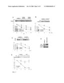 |
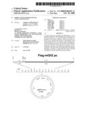 |  |
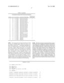 |  |
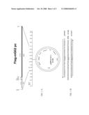 | 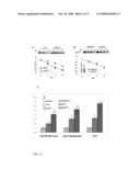 |
 |  |
 | 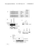 |
 | 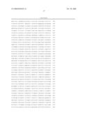 |
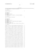 |  |
 |
| Similar patent applications: | |
| Date | Title |
|---|---|
| 2011-06-09 | Identification of bitter ligands that specifically activate human t2r receptors and related assays for identifying human bitter taste modulators |
| 2011-05-26 | Aptamer-based methods for identifying cellular biomarkers |
| 2011-05-19 | Model system for diagnosing lipid metabolism |
| 2010-03-25 | System for performing multi-formatted assays |
| 2010-09-09 | Soft gel systems in modulating stem cell development |
| New patent applications in this class: | |
| Date | Title |
|---|---|
| 2011-06-30 | Apparatus and method of authenticating product using polynucleotides |
| 2011-06-30 | Cyanine compounds, compositions including these compounds and their use in cell analysis |
| 2011-06-30 | Method for detecting multiple small nucleic acids |
| 2011-06-30 | Solid-phase chelators and electronic biosensors |
| 2011-06-30 | Cell-based screening assay to identify molecules that stimulate ifn-alpha/beta target genes |
| New patent applications from these inventors: | |
| Date | Title |
|---|---|
| 2011-02-17 | Methods and materials for reducing gli2 expression |
| Top Inventors for class "Chemistry: molecular biology and microbiology" | |
| Rank | Inventor's name |
|---|---|
| 1 | Marshall Medoff |
| 2 | Anthony P. Burgard |
| 3 | Mark J. Burk |
| 4 | Robin E. Osterhout |
| 5 | Rangarajan Sampath |