Patent application title: MOLECULAR RECOGNITION OF MATERIALS
Inventors:
Angela M. Belcher (Lexington, MA, US)
IPC8 Class: AC40B3004FI
USPC Class:
506 9
Class name: Combinatorial chemistry technology: method, library, apparatus method of screening a library by measuring the ability to specifically bind a target molecule (e.g., antibody-antigen binding, receptor-ligand binding, etc.)
Publication date: 2008-10-02
Patent application number: 20080242552
Inventors list |
Agents list |
Assignees list |
List by place |
Classification tree browser |
Top 100 Inventors |
Top 100 Agents |
Top 100 Assignees |
Usenet FAQ Index |
Documents |
Other FAQs |
Patent application title: MOLECULAR RECOGNITION OF MATERIALS
Inventors:
Angela M. Belcher
Agents:
FOLEY AND LARDNER LLP;SUITE 500
Assignees:
Origin: WASHINGTON, DC US
IPC8 Class: AC40B3004FI
USPC Class:
506 9
Abstract:
The present invention includes methods for selective binding of inorganic
materials and the compositions that made up of the selecting agent and
the target materials. One form of the present invention is a method for
selecting crystal-binding peptides with binding specificity including the
steps of contacting one or more amino acid oligomers with one or more
single-crystals of a semiconductor material so that the oligomers may
bind to the crystal and eluting the bound amino acid oligomers from the
single-crystals.Claims:
1-180. (canceled)
181. A composition comprising one or more peptide binding sequences, wherein at least one peptide binding sequence is selective for a target crystalline face of a target crystal.
182. The composition according to claim 181, wherein the peptide binding sequence comprises one or more peptide binding sequences selected from a phage display library or a combinatorial library.
183. The composition according to claim 181, wherein the peptide binding sequence comprises one or more peptide binding sequences which are between about 7 to 15 amino acids in length.
184. The composition according to claim 181, wherein the peptide binding sequence is part of a virus which comprises DNA which encodes the peptide binding sequence.
185. The composition according to claim 181, wherein the peptide binding sequence is part of a bivalent synthetic peptide.
186. The composition according to claim 181, wherein the peptide binding sequence is part of a virus or bacteriophage.
187. The composition according to claim 181, wherein the peptide binding sequence is part of a p3 or p8 modified bacteriophage.
188. The composition according to claim 181, wherein the target crystal is a single crystal.
189. The composition according to claim 181, wherein the target crystal is part of a heterostructured crystal surface.
190. The composition according to claim 181, wherein the target crystal is an inorganic crystal.
191. The composition according to claim 181, wherein the target crystal is a semiconductor crystal.
192. The composition according to claim 181, wherein the target crystal is a III-V semiconductor crystal or a II-VI semiconductor crystal.
193. The composition according to claim 181, wherein the target crystal is a magnetic crystal.
194. The composition according to claim 181, wherein the target crystal is a mineral or an optical material.
195. The composition according to claim 181, wherein the target crystal is a nanoparticle.
196. The composition according to claim 181, wherein the target crystal has an oxide surface.
197. The composition according to claim 181, wherein the target crystal is gallium arsenide, indium phosphide, gallium nitride, zinc sulfide, cadmium sulfide, aluminum arsenide, gallium stibinide, aluminum gallium arsenide, aluminum stibinide, aluminum arsenide, cadmium selenide, zinc selenide, cadmium telluride, zinc selenide, indium arsenide, silicon, FePd, cobalt, manganese, lithium niobate, iron oxide, silica or calcium carbonate.
198. The composition according to claim 181, wherein the target crystal is a single crystal.
199. The composition according to claim 181, wherein the peptide binding sequence is fused to a protein, and the target crystal is a single crystal.
200. The composition according to claim 181, wherein the peptide binding sequence is part of a virus and is between about 7 to 15 amino acids in length, and wherein the target crystal is a semiconductor crystal.
201. The composition according to claim 181, wherein the peptide binding sequence is bound to a crystal.
202. The composition according to claim 181, wherein the peptide binding sequence is bound to a nanoparticle, a surface, or a patterned surface.
203. The composition according to claim 181, wherein the peptide binding sequence is part of a bivalent synthetic peptide which is bound to a surface and to a nanoparticle.
204. A composition comprising one or more peptide binding sequences, wherein at least one peptide binding sequence selectively binds a target crystalline face of a target crystal.
205. A composition comprising one or more viruses, wherein the virus comprises one or more peptide binding sequences, wherein at least one peptide binding sequence selectively binds a target crystal.
206. A composition comprising one or more viruses, wherein the virus comprises one or more peptide binding sequences, wherein at least one peptide binding sequence is selective for a target inorganic crystalline face of a target inorganic crystal, and the virus is bound to the target single inorganic crystal.
207. The composition according to claim 206, wherein the virus is a bacteriophage, the peptide binding sequence comprises 7 to 15 amino acids, and the target inorganic crystal is a semiconductor crystal.
208. The composition according to claim 206, wherein the virus is bound to a nanoparticle through the peptide binding sequence.
209. The composition according to claim 206, wherein the virus is bound to a nanoparticle and to a surface of the target inorganic crystal.
210. A method of selecting a composition comprising one or more peptide binding sequences, wherein at least one peptide binding sequence is selective for a target crystalline face of a target crystal, the method comprising the step of:contacting a library of peptide binding sequences with a target crystalline surface structure so that selective binding between the peptide binding sequences and the target crystalline surface structure is achieved with face specificity.
211. The method according to claim 210, wherein the peptide binding sequences are parts of viruses.
212. The method according to claim 210, wherein the peptide binding sequences are parts of p3 or p8 modified viruses.
213. The method according to claim 210, wherein the target crystalline surface structure is a semiconductor surface structure.
214. A method of selecting a composition comprising one or more viruses, wherein the virus comprises one or more peptide binding sequences, wherein at least one peptide binding sequence is selective for a target crystal, the method comprising the step of:contacting a library of viruses comprising peptide binding sequences with one or more target crystalline surface structures so that selective binding between the peptide binding sequences and the target crystalline surface structures is achieved.
215. The method of claim 214, wherein the binding is face selective.
216. The method of claim 214, wherein the binding is composition selective.
217. The method according to claim 214, wherein the peptide binding sequences are parts of p3 or p8 modified viruses.
218. The method according to claim 214, wherein the target crystalline surface structure is an inorganic surface structure.
219. The method according to claim 214, wherein the target crystalline surface structure is a semiconductor surface structure.
Description:
CROSS REFERENCE TO RELATED APPLICATIONS
[0001]This application is a Continuation of application Ser. No. 10/155,883, filed May 24, 2002, which claims priority to Provisional Patent Application No. 60/296,013, filed Jun. 5, 2001, the contents of both of which are incorporated herein by reference in their entirety.
FIELD OF THE INVENTION
[0003]The present invention is directed to the selective recognition of inorganic materials in general and specifically toward surface recognition of single crystals of semiconductor and magnetic materials using small organic molecules.
BACKGROUND OF THE INVENTION
[0004]In biological systems, organic molecules exert a remarkable level of control over the nucleation and mineral phase of inorganic materials such as calcium carbonate and silica, and over the assembly of crystallites and other nanoscale building blocks into complex structures required for biological function.
[0005]Materials produced by biological processes are typically soft, and consist of a surprisingly simple collection of molecular building blocks (i.e., lipids, peptides, and nucleic acids) arranged in astoundingly complex architectures. Unlike the semiconductor industry, which relies on a serial lithographic processing approach for constructing the smallest features on an integrated circuit, living organisms execute their architectural "blueprints" using mostly non-covalent forces acting simultaneously upon many molecular components. Furthermore, these structures can often elegantly rearrange between two or more usable forms without changing any of the molecular constituents.
[0006]The use of "biological" materials to process the next generation of microelectronic devices provides a possible solution to resolving the limitations of traditional processing methods. The critical factors in this approach are identifying the appropriate compatibilities and combinations of biological-inorganic materials, and the synthesis of the appropriate building blocks.
SUMMARY OF THE INVENTION
[0007]The ability to direct the assembly of nanoscale components into controlled and sophisticated structures has motivated intense efforts to develop assembly methods that mimic or exploit the recognition capabilities and interactions found in biological systems. Of particular value would be methods that could be applied to materials with interesting electronic or optical properties, but natural evolution has not selected for interactions between biomolecules and such materials.
[0008]The present invention is based on recognition that biological systems efficiently and accurately assemble nanoscale building blocks into complex and functionally sophisticated structures with high perfection, controlled size and compositional uniformity.
[0009]The present invention includes methods for selective binding of inorganic materials and the compositions that are made up of the selecting agent and the target materials. One form of the present invention is a method for selecting crystal-binding peptides with binding specificity and includes the steps of contacting one or more amino acid oligomers with one or more single-crystals of a semiconductor material so that the oligomers may bind to the crystal and eluting the bound amino acid oligomers from the single-crystals. Another form of the present invention is a method for selecting crystal-binding peptides with binding specificity and includes the steps of contacting one or more amino acid oligomers with one or more crystals of a semiconductor, such as a Group III-V or II-VI material; or a magnetic material, such an iron oxide, so that the oligomers may bind to the crystal and eluting the bound amino acid oligomers from the single-crystals.
[0010]Another form of the present invention is a peptide sequence for the binding GaAs (100) chosen from the group consisting of Seq. ID Nos. 1 through 11.
[0011]Still another form of the present invention is a method for selecting polymeric organic molecules, lipids or nucleic acids with binding specificity. A method of the present invention begins by contacting one or more oligomers with one or more single-crystals of a magnetic material so that the oligomers may bind to the crystal and eluting the bound peptide oligomers from the single-crystals. The sequence of the organic polymer is then determined by direct or indirect sequencing.
[0012]Another form of the present invention is a method for selecting crystal-bonding amino acids including the steps of contacting one or more amino acid oligomers with one or more crystals of a target material so that the oligomers may bind to the crystal and eluting the bound amino acid oligomers from the crystals.
[0013]Another form of the present invention is a specificity structure made up of one or more single crystals of gallium arsenide, indium phosphide, mercury cadmium telluride, zinc sulfide, cadmium sulfide, aluminum-gallium-arsenide, zinc selenide, cadmium selenide, cadmium telluride, zinc telluride, aluminum arsenide, indium arsenide and the like and a selective binding amino acid sequence.
[0014]Another form of the present invention is a crystal binding amino acid oligomer made up of the sequence motif (ser/tyr/thr)-(arg/asp/ser)-X-aa-(ser/asn/glu/arg/thr)-Xaa-Xaa-ser/thr/gl- u/asp)-(ser/thr/tyr) (SEQ. ID NO. 159) or Xaa-Xaa-(ser/tyr/thr)-(arg/asp/ser)-Xaa-(ser/asn/glu/arg/thr)-Xaa-Xaa-(se- r/thr/glu/asp)-(ser/thr/tyr)-(ser/thr/his)-Xaa-Xaa (SEQ. ID NO 160).
[0015]The motifs and other polymers referred to in the descriptions of various embodiments of the present invention may be free molecules, e.g. amino acid oligomers, or they may be part of a chimera, such as a phage display.
BRIEF DESCRIPTION OF THE DRAWINGS
[0016]For a more complete understanding of the features and advantages of the present invention, reference is now made to the detailed description of the invention along with the accompanying figures in which corresponding numerals in the different figures refer to corresponding parts and in which:
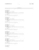
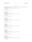
[0017]FIG. 1 depicts selected random amino acid sequences in accordance with the present invention.
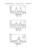
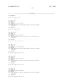
[0018]FIG. 2 depicts XPS spectra of structures in accordance with the present invention
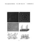
[0019]FIG. 3 depicts phage recognition of heterostructures in accordance with the present invention.
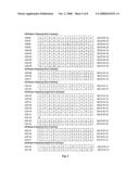
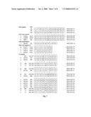
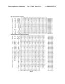
[0020]FIGS. 4-8 depict specific amino acid sequences in accordance with the present invention.
DETAILED DESCRIPTION OF THE INVENTION
[0021]Although making and using various embodiments of the present invention are discussed in detail below, it should be appreciated that the present invention provides many applicable inventive concepts that can be embodied in a wide variety of specific contexts. The specific embodiments discussed herein are merely illustrative of specific ways to make and use the invention, and do not delimit the scope of the invention.
[0022]The facility with which biological systems assemble immensely complicated structure on an exceedingly minute scale has motivated a great deal of interest in the desire to identify non-biological systems that can behave in a similar fashion. Of particular value would be methods that could be applied to materials with interesting electronic or optical properties, but natural evolution has not selected for interactions between biomolecules and such materials.
[0023]The present invention is based on recognition that biological systems efficiently and accurately assemble nanoscale building blocks into complex and functionally sophisticated structures with high perfection, controlled size and compositional uniformity.
[0024]One method of providing a random organic polymer pool is using a Phage-display library, based on a combinatorial library of random peptides containing between 7 and 12 amino acids fused to the pIII coat protein of M13 coliphage, provided different peptides that were reacted with crystalline semiconductor structures. Five copies of the pIII coat protein are located on one end of the phage particle, accounting for 10-16 nm of the particle. The phage-display approach provided a physical linkage between the peptide substrate interaction and the DNA that encodes that interaction. The examples described here used as examples, five different single-crystal semiconductors: GaAs(100), GaAs(111)A, GaAs(111)B, InP(100) and Si(100). These substrates allowed for systematic evaluation of the peptide substrate interactions and confirmation of the general utility of the methodology of the present invention for different crystalline structures.
[0025]Protein sequences that successfully bound to the specific crystal were eluted from the surface, amplified by, e.g., a million-fold, and reacted against the substrate under more stringent conditions. This procedure was repeated five times to select the phage in the library with the most specific binding. After, e.g., the third, fourth and fifth rounds of phage selection, crystal-specific phage were isolated and their DNA sequenced. Peptide binding has been identified that is selective for the crystal composition (for example, binding to GaAs but not to Si) and crystalline face (for example, binding to (100) GaAs, but not to (111)B GaAs).
[0026]Twenty clones selected from GaAs(100) were analyzed to determine epitope binding domains to the GaAs surface. The partial peptide sequences of the modified pIII or pVIII protein are shown in FIG. 1, revealing similar amino-acid sequences among peptides exposed to GaAs. With increasing number of exposures to a GaAs surface, the number of uncharged polar and Lewis-base functional groups increased. Phage clones from third, fourth and fifth round sequencing contained on average 30%, 40% and 44% polar functional groups, respectively, while the fraction of Lewis-base functional groups increased at the same time from 41% to 48% to 55%. The observed increase in Lewis bases, which should constitute only 34% of the functional groups in random 12-mer peptides from our library, suggests that interactions between Lewis bases on the peptides and Lewis-acid sites on the GaAs surface may mediate the selective binding exhibited by these clones.
[0027]The expected structure of the modified 12-mers selected from the library may be an extended conformation, which seems likely for small peptides, making the peptide much longer than the unit cell (5.65 A°) of GaAs. Therefore, only small binding domains would be necessary for the peptide to recognize a GaAs crystal. These short peptide domains, highlighted in FIG. 1, contain serine- and threonine-rich regions in addition to the presence of amine Lewis bases, such as asparagine and glutamine. To determine the exact binding sequence, the surfaces have been screened with shorter libraries, including 7-mer and disulphide constrained 7-mer libraries. Using these shorter libraries that reduce the size and flexibility of the binding domain, fewer peptide-surface interactions are allowed, yielding the expected increase in the strength of interactions between generations of selection.
[0028]Phage, tagged with streptavidin-labelled 20-nm colloidal gold particles bound to the phage through a biotinylated antibody to the M13 coat protein, were used for quantitative assessment of specific binding. X-ray photoelectron spectroscopy (XPS) elemental composition determination was performed, monitoring the phage substrate interaction through the intensity of the gold 4f-electron signal (FIG. 2a-c). Without the presence of the G1-3 phage, the antibody and the gold streptavidin did not bind to the GaAs(100)substrate. The gold-streptavidin binding was, therefore, specific to the phage and an indicator of the phage binding to the substrate. Using XPS it was also found that the G1-3 clone isolated from GaAs(100) bound specifically to GaAs(100) but not to Si(100) (see FIG. 2a). In complementary fashion the S1 clone, screened against the (100) Si surface, showed poor binding to the (100) GaAs surface.
[0029]Some GaAs clones also bound the surface of InP (100), another zinc-blende structure. The basis of the selective binding, whether it is chemical, structural or electronic, is still under investigation. In addition, the presence of native oxide on the substrate surface may alter the selectivity of peptide binding.
[0030]The preferential binding of the G1-3 clone to GaAs(100), over the (111)A (gallium terminated) or (111)B (arsenic terminated) face of GaAs was demonstrated (FIG. 2b, c). The G1-3 clone surface concentration was greater on the (100) surface, which was used for its selection, than on the gallium-rich (111)A or arsenic-rich (111)B surfaces. These different surfaces are known to exhibit different chemical reactivities, and it is not surprising that there is selectivity demonstrated in the phage binding to the various crystal faces. Although the bulk termination of both 111 surfaces give the same geometric structure, the differences between having Ga or As atoms outermost in the surface bilayer become more apparent when comparing surface reconstructions. The composition of the oxides of the various GaAs surfaces is also expected to be different, and this in turn may affect the nature of the peptide binding.
[0031]The intensity of Ga 2 p electrons against the binding energy from substrates that were exposed to the G1-3 phage clone is plotted in 2c. As expected from the results in FIG. 2b, the Ga 2 p intensities observed on the GaAs (100), (111)A and (111)B surfaces are inversely proportional to the gold concentrations. The decrease in Ga 2 p intensity on surfaces with higher gold-streptavidin concentrations was due to the increase in surface coverage by the phage. XPS is a surface technique with a sampling depth of approximately 30 angstroms; therefore, as the thickness of the organic layer increases, the signal from the inorganic substrate decreases. This observation was used to confirm that the intensity of gold-streptavidin was indeed due to the presence of phage containing a crystal specific bonding sequence on the surface of GaAs. Binding studies were performed that correlate with the XPS data, where equal numbers of specific phage clones were exposed to various semiconductor substrates with equal surface areas. Wild-type clones (no random peptide insert) did not bind to GaAs (no plaques were detected). For the G1-3 clone, the eluted phage population was 12 times greater from GaAs(100) than from the GaAs(111)A surface.
[0032]The G1-3, G12-3 and G7-4 clones bound to GaAs(100) and InP(100) were imaged using atomic force microscopy (AFM). The InP crystal has a zinc-blende structure, isostructural with GaAs, although the In--P bond has greater ionic character than the GaAs bond. The 10-nm width and 900-nm length of the observed phage in AFM matches the dimensions of the M13 phage observed by transmission electron microscopy (TEM), and the gold spheres bound to M13 antibodies were observed bound to the phage (data not shown). The InP surface has a high concentration of phage. These data suggest that there are many factors involved in substrate recognition, including atom size, charge, polarity and crystal structure.
[0033]The G1-3 clone (negatively stained) is seen bound to a GaAs crystalline wafer in the TEM image (not shown). The data confirms that binding was directed by the modified pIII protein of G1-3, not through non-specific interactions with the major coat protein. Therefore, peptides of the present invention may be used to direct specific peptide-semiconductor interactions in assembling nanostructures and heterostructures (FIG. 3e).
[0034]X-ray fluorescence microscopy was used to demonstrate the preferential attachment of phage to a zinc-blende surface in close proximity to a surface of differing chemical and structural composition. A nested square pattern was etched into a GaAs wafer; this pattern contained 1-μm lines of GaAs, and 4-μm SiO2 spacings in between each line (FIGS. 3a, 3b). The G12-3 clones were interacted with the GaAs/SiO2 patterned substrate, washed to reduce non-specific binding, and tagged with an immuno-fluorescent probe, tetramethyl rhodamine (TMR). The tagged phage were found as the three red lines and the center dot, in FIG. 3b, corresponding to G12-3 binding only to GaAs. The SiO2 regions of the pattern remain unbound by phage and are dark in color. This result was not observed on a control that was not exposed to phage, but was exposed to the primary antibody and TMR (FIG. 3a). The same result was obtained using non-phage bound G12-3 peptide.
[0035]The GaAs clone G12-3 was observed to be substrate-specific for GaAs over AlGaAs (FIG. 3c). AlAs and GaAs have essentially identical lattice constraints at room temperature, 5.66 A° and 5.65 A°, respectively, and thus ternary alloys of AlxGal-xAs can be epitaxially grown on GaAs substrates. GaAs and AlGaAs have zinc-blende crystal structures, but the G12-3 clone exhibited selectivity in binding only to GaAs. A multilayer substrate was used, consisting of alternating layers of GaAs and of Al0.98Ga0.02As. The substrate material was cleaved and subsequently reacted with the G12-3 clone.
[0036]The G12-3 clones were labeled with 20-nm gold-streptavidin nanoparticles. Examination by scanning electron microscopy (SEM) shows the alternating layers of GaAs and Al0.98Ga0.02As within the heterostructure (FIG. 3c). X-ray elemental analysis of gallium and aluminum was used to map the gold-streptavidin particles exclusively to the GaAs layers of the heterostructure, demonstrating the high degree of binding specificity for chemical composition. In FIG. 3d, a model for the discrimination of phage for semiconductor heterostructures, as seen in the fluorescence and SEM images (FIGS. 3a-c).
[0037]The present invention demonstrates the power use of phage-display libraries to identify, develop and amplify binding between organic peptide sequences and inorganic semiconductor substrates. This peptide recognition and specificity of inorganic crystals has been extended to other substrates, including GaN, ZnS, CdS, Fe3O4, Fe2O3, CdSe, ZnSe and CaCO3 using peptide libraries. Bivalent synthetic peptides with two-component recognition (FIG. 4e) are currently being designed; such peptides have the potential to direct nanoparticles to specific locations on a semiconductor structure. These organic and inorganic pairs should provide powerful building blocks for the fabrication of a new generation of complex, sophisticated electronic structures.
EXAMPLES
[0038]Peptide selection. The phage display or peptide library was contacted with the semiconductor, or other, crystals in Tris-buffered saline (TBS) containing 0.1% TWEEN-20, to reduce phage-phage interactions on the surface. After rocking for 1 h at room temperature, the surfaces were washed with 10 exposures to Tris-buffered saline, pH 7.5, and increasing TWEEN-20 concentrations from 0.1% to 0.5% (v/v). The phage were eluted from the surface by the addition of glycine-HCl (pH 2.2) 10 minute, transferred to a fresh tube and then neutralized with Tris-HCl (pH 9.1). The eluted phage were titred and binding efficiency was compared.
[0039]The phage eluted after third-round substrate exposure were mixed with their Escherichia coli ER2537 host and plated on LB XGal/IPTG plates. Since the library phage were derived from the vector M13mp19, which carries the laczA gene, phage plaques were blue in color when plated on media containing Xgal (5-bromo-4-chloro-3-indoyl-β-D-galac-toside) and IPTG (isopropyl-β-D-thiogalactoside). Blue/white screening was used to select phage plaques with the random peptide insert. Plaques were picked and DNA sequenced from these plates.
[0040]Substrate preparation. Substrate orientations were confirmed by X-ray diffraction, and native oxides were removed by appropriate chemical specific etching. The following etches were tested on GaAs and InP surfaces: NH4OH:H2O 1:10, HCl:H2O 1:10, H3PO4:H2O2:H2O 3:1:50 at 1 minute and 10 minute etch times. The best element ratio and least oxide formation (using XPS) for GaAs and InP etched surfaces was achieved using HCl:H2O for 1 minute followed by a deionized water rinse for 1 minute. However, since an ammonium hydroxide etch was used for GaAs in the initial screening of the library, this etch was used for all other GaAs substrate examples. Si(100) wafers were etched in a solution of HF:H2O 1:40 for one minute, followed by a deionized water rinse. All surfaces were taken directly from the rinse solution and immediately introduced to the phage library. Surfaces of control substrates, not exposed to phage, were characterized and mapped for effectiveness of the etching process and morphology of surfaces by AFM and XPS.
[0041]Multilayer substrates of GaAs and of Al0.98Ga0.02 As were grown by molecular beam epitaxy onto (100) GaAs. The epitaxially grown layers were Si-doped (n-type) at a level of 5×10⁻⁷ cm-3.
[0042]Antibody and Gold Labeling. For the XPS, SEM and AFM examples, substrates were exposed to phage for 1 h in Tris-buffered saline then introduced to an anti-fd bacteriophage-biotin conjugate, an antibody to the pIII protein of fd phage, (1:500 in phosphate buffer, Sigma) for 30 minute and then rinsed in phosphate buffer. A streptavidin/20-nm colloidal gold label (1:200 in phosphate buffered saline (PBS), Sigma) was attached to the biotin-conjugated phage through a biotin-streptavidin interaction; the surfaces were exposed to the label for 30 minutes and then rinsed several times with PBS.
[0043]X-ray Photoelectron Spectroscopy (XPS). The following controls were done for the XPS examples to ensure that the gold signal seen in XPS was from gold bound to the phage and not non-specific antibody interaction with the GaAs surface. The prepared (100) GaAs surface was exposed to (1) antibody and the streptavidin-gold label, but without phage, (2) G1-3 phage and streptavidin-gold label, but without the antibody, and (3) streptavidin-gold label, without either G1-3 phage or antibody.
[0044]The XPS instrument used was a Physical Electronics Phi ESCA 5700 with an aluminum anode producing monochromatic 1,487-eV X-rays. All samples were introduced to the chamber immediately after gold-tagging the phage (as described above) to limit oxidation of the GaAs surfaces, and then pumped overnight at high vacuum to reduce sample outgassing in the XPS chamber.
[0045]Atomic Force Microscopy (AFM). The AFM used was a Digital Instruments Bioscope mounted on a Zeiss Axiovert 100s-2tv, operating in tip scanning mode with a G scanner. The images were taken in air using tapping mode. The AFM probes were etched silicon with 125-mm cantilevers and spring constants of 20±100 Nm-1 driven near their resonant frequency of 200.+-0.400 kHz. Scan rates were of the order of 1.+-0.5 mms-1. Images were leveled using a first-order plane to remove sample tilt.
[0046]Transmission Electron Microscopy (TEM). TEM images were taken using a Philips EM208 at 60 kV. The G1-3 phage (diluted 1:100 in TBS) were incubated with GaAs pieces (500 mm) for 30 minute, centrifuged to separate particles from unbound phage, rinsed with TBS, and resuspended in TBS. Samples were stained with 2% uranyl acetate.
[0047]Scanning Electron Microscopy (SEM). The G12-3 phage (diluted 1:100 in TBS) were incubated with a freshly cleaved hetero-structure surface for 30 minute and rinsed with TBS. The G12-3 phage were tagged with 20-nm colloidal gold. SEM and elemental mapping images were collected using the Norian detection system mounted on a Hitachi 4700 field emission scanning electron microscope at 5 kV.
[0048]Although this invention has been described in reference to illustrative embodiments, this description is not intended to be construed in a limiting sense. Various modifications and combinations of the illustrative embodiments, as well as other embodiments of the invention, will be apparent to persons skilled in the art upon reference to the description. It is therefore intended that the appended claims encompass any such modifications or embodiments.
Sequence CWU
1
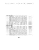
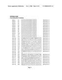
95112PRTartificial sequencepeptide binding sequence retrieved from phage
biopanning 1Ala Met Ala Gly Thr Thr Ser Asp Pro Ser Thr Val1
5 10212PRTartificial sequencepeptide binding
sequence retrieved from phage biopanning 2Ala Ala Ser Pro Thr Gln
Ser Met Ser Gln Ala Pro1 5
10312PRTartificial sequencepeptide binding sequence retrieved from phage
biopanning 3His Thr His Thr Asn Asn Asp Ser Pro Asn Gln Ala1
5 10412PRTartificial sequencepeptide binding
sequence retrieved from phage biopanning 4Asp Thr Gln Gly Phe His
Ser Arg Ser Ser Ser Ala1 5
10512PRTartificial sequencepeptide binding sequence retrieved from phage
biopanning 5Thr Ser Ser Ser Ala Leu Gln Pro Ala His Ala Trp1
5 10612PRTartificial sequencepeptide binding
sequence retrieved from phage biopanning 6Ser Glu Ser Ser Pro Ile
Ser Leu Asp Tyr Arg Ala1 5
10712PRTartificial sequencepeptide binding sequence retrieved from phage
biopanning 7Ser Thr His Asn Tyr Gln Ile Pro Arg Pro Pro Thr1
5 10812PRTartificial sequencepeptide binding
sequence retrieved from phage biopanning 8His Pro Phe Ser Asn Glu
Pro Leu Gln Leu Ser Ser1 5
10912PRTartificial sequencepeptide binding sequence retrieved from phage
biopanning 9Gly Thr Leu Ala Asn Gln Gln Ile Phe Leu Ser Ser1
5 101012PRTartificial sequencepeptide binding
sequence retrieved from phage biopanning 10His Gly Asn Pro Leu Pro
Met Thr Pro Phe Pro Gly1 5
101112PRTartificial sequencepeptide binding sequence retrieved from phage
biopanning 11Arg Leu Glu Leu Ala Ile Pro Leu Gln Gly Ser Gly1
5 10129PRTartificial sequencepeptide binding
sequence retrieved from phage biopanning 12Cys His Ala Ser Asn Arg
Leu Ser Cys1 51312PRTartificial sequencepeptide binding
sequence retrieved from phage biopanning 13Ser Met Asp Arg Ser Asp
Met Thr Met Arg Leu Pro1 5
101412PRTartificial sequencepeptide binding sequence retrieved from phage
biopanning 14Gly Thr Phe Thr Pro Arg Pro Thr Pro Ile Tyr Pro1
5 101512PRTartificial sequencepeptide binding
sequence retrieved from phage biopanning 15Gln Met Ser Glu Asn Leu
Thr Ser Gln Ile Glu Ser1 5
101612PRTartificial sequencepeptide binding sequence retrieved from phage
biopanning 16Asp Met Leu Ala Arg Leu Arg Ala Thr Ala Gly Pro1
5 101712PRTartificial sequencepeptide binding
sequence retrieved from phage biopanning 17Ser Gln Thr Trp Leu Leu
Met Ser Pro Val Ala Thr1 5
101812PRTartificial sequencepeptide binding sequence retrieved from phage
biopanning 18Ala Ser Pro Asp Gln Gln Val Gly Pro Leu Tyr Val1
5 101912PRTartificial sequencepeptide binding
sequence retrieved from phage biopanning 19Leu Thr Trp Ser Pro Leu
Gln Thr Val Ala Arg Phe1 5
102012PRTartificial sequencepeptide binding sequence retrieved from phage
biopanning 20Gln Ile Ser Ala His Gln Met Pro Ser Arg Pro Ile1
5 102112PRTartificial sequencepeptide binding
sequence retrieved from phage biopanning 21Ser Met Lys Tyr Asn Leu
Ile Val Asp Ser Pro Tyr1 5
102212PRTartificial sequencepeptide binding sequence retrieved from phage
biopanning 22Gln Met Pro Ile Arg Asn Gln Leu Ala Trp Pro Met1
5 102312PRTartificial sequencepeptide binding
sequence retrieved from phage biopanning 23Thr Gln Asn Leu Glu Ile
Arg Glu Pro Leu Thr Pro1 5
102412PRTartificial sequencepeptide binding sequence retrieved from phage
biopanning 24Tyr Pro Met Ser Pro Ser Pro Tyr Pro Tyr Gln Leu1
5 102512PRTartificial sequencepeptide binding
sequence retrieved from phage biopanning 25Ser Phe Met Ile Gln Pro
Thr Pro Leu Pro Pro Ser1 5
102612PRTartificial sequencepeptide binding sequence retrieved from phage
biopanning 26Gly Leu Ala Pro His Ile His Ser Leu Asn Glu Ala1
5 102712PRTartificial sequencepeptide binding
sequence retrieved from phage biopanning 27Met Gln Phe Pro Val Thr
Pro Tyr Leu Asn Ala Ser1 5
102812PRTartificial sequencepeptide binding sequence retrieved from phage
biopanning 28Ser Pro Gly Asp Ser Leu Lys Lys Leu Ala Ala Ser1
5 102912PRTartificial sequencepeptide binding
sequence retrieved from phage biopanning 29Gly Tyr His Met Gln Thr
Leu Pro Gly Pro Val Ala1 5
103012PRTartificial sequencepeptide binding sequence retrieved from phage
biopanning 30Ser Leu Thr Pro Leu Thr Thr Ser His Leu Arg Ser1
5 103112PRTartificial sequencepeptide binding
sequence retrieved from phage biopanning 31Thr Leu Thr Asn Gly Pro
Leu Arg Pro Phe Thr Gly1 5
103212PRTartificial sequencepeptide binding sequence retrieved from phage
biopanning 32Leu Asn Thr Pro Lys Pro Phe Thr Leu Gly Gln Asn1
5 10339PRTartificial sequencepeptide binding
sequence retrieved from phage biopanning 33Cys Asp Leu Gln Asn Tyr
Lys Ala Cys1 5349PRTartificial sequencepeptide binding
sequence retrieved from phage biopanning 34Cys Arg His Pro His Thr
Arg Leu Cys1 5359PRTartificial sequencepeptide binding
sequence retrieved from phage biopanning 35Cys Ala Asn Leu Lys Pro
Lys Ala Cys1 5369PRTartificial sequencepeptide binding
sequence retrieved from phage biopanning 36Cys Tyr Ile Asn Pro Pro
Lys Val Cys1 5379PRTartificial sequencepeptide binding
sequence retrieved from phage biopanning 37Cys Asn Asn Lys Val Pro
Val Leu Cys1 5389PRTartificial sequencepeptide binding
sequence retrieved from phage biopanning 38Cys His Ala Ser Lys Thr
Pro Leu Cys1 5399PRTartificial sequencepeptide binding
sequence retrieved from phage biopanning 39Cys Ala Ser Gln Leu Tyr
Pro Ala Cys1 5409PRTartificial sequencepeptide binding
sequence retrieved from phage biopanning 40Cys Asn Met Thr Gln Tyr
Pro Ala Cys1 5419PRTartificial sequencepeptide binding
sequence retrieved from phage biopanning 41Cys Phe Ala Pro Ser Gly
Pro Ala Cys1 5429PRTartificial sequencepeptide binding
sequence retrieved from phage biopanning 42Cys Pro Val Trp Ile Gln
Ala Pro Cys1 5439PRTartificial sequencepeptide binding
sequence retrieved from phage biopanning 43Cys Gln Val Ala Val Asn
Pro Leu Cys1 5449PRTartificial sequencepeptide binding
sequence retrieved from phage biopanning 44Cys Gln Pro Glu Ala Met
Pro Ala Cys1 5459PRTartificial sequencepeptide binding
sequence retrieved from phage biopanning 45Cys His Pro Thr Met Pro
Leu Ala Cys1 5469PRTartificial sequencepeptide binding
sequence retrieved from phage biopanning 46Cys Pro Pro Phe Ala Ala
Pro Ile Cys1 5479PRTartificial sequencepeptide binding
sequence retrieved from phage biopanning 47Cys Asn Lys His Gln Pro
Met His Cys1 5489PRTartificial sequencepeptide binding
sequence retrieved from phage biopanning 48Cys Phe Pro Met Arg Ser
Asn Gln Cys1 5499PRTartificial sequencepeptide binding
sequence retrieved from phage biopanning 49Cys Gln Ser Met Pro His
Asn Arg Cys1 5509PRTartificial sequencepeptide binding
sequence retrieved from phage biopanning 50Cys Asn Asn Pro Met His
Gln Asn Cys1 5519PRTartificial sequencepeptide binding
sequence retrieved from phage biopanning 51Cys His Met Ala Pro Arg
Trp Gln Cys1 5529PRTartificial sequencepeptide binding
sequence retrieved from phage biopanning 52His Val His Ile His Ser
Arg Pro Met1 5539PRTartificial sequencepeptide binding
sequence retrieved from phage biopanning 53Leu Pro Asn Met His Pro
Leu Pro Leu1 5549PRTartificial sequencepeptide binding
sequence retrieved from phage biopanning 54Leu Pro Leu Arg Leu Pro
Pro Met Pro1 5559PRTartificial sequencepeptide binding
sequence retrieved from phage biopanning 55His Ser Met Ile Gly Thr
Pro Thr Thr1 5569PRTartificial sequencepeptide binding
sequence retrieved from phage biopanning 56Ser Val Ser Val Gly Met
Lys Pro Ser1 5579PRTartificial sequencepeptide binding
sequence retrieved from phage biopanning 57Leu Asp Ala Ser Phe Met
Gln Asp Trp1 5589PRTartificial sequencepeptide binding
sequence retrieved from phage biopanning 58Thr Pro Pro Ser Tyr Gln
Met Ala Met1 5599PRTartificial sequencepeptide binding
sequence retrieved from phage biopanning 59Tyr Pro Gln Leu Val Ser
Met Ser Thr1 5609PRTartificial sequencepeptide binding
sequence retrieved from phage biopanning 60Gly Tyr Ser Thr Ile Asn
Met Tyr Ser1 5619PRTartificial sequencepeptide binding
sequence retrieved from phage biopanning 61Asp Arg Met Leu Leu Pro
Phe Asn Leu1 5629PRTartificial sequencepeptide binding
sequence retrieved from phage biopanning 62Ile Pro Met Thr Pro Ser
Tyr Asp Ser1 5639PRTartificial sequencepeptide binding
sequence retrieved from phage biopanning 63Met Tyr Ser Pro Arg Pro
Pro Ala Leu1 5649PRTartificial sequencepeptide binding
sequence retrieved from phage biopanning 64Gln Pro Thr Thr Asp Leu
Met Ala His1 5659PRTartificial sequencepeptide binding
sequence retrieved from phage biopanning 65Ala Thr His Val Gln Met
Ala Trp Ala1 5669PRTartificial sequencepeptide binding
sequence retrieved from phage biopanning 66Ser Met His Ala Thr Leu
Thr Pro Met1 5679PRTartificial sequencepeptide binding
sequence retrieved from phage biopanning 67Ser Gly Pro Ala His Gly
Met Phe Ala1 5689PRTartificial sequencepeptide binding
sequence retrieved from phage biopanning 68Ile Ala Asn Arg Pro Tyr
Ser Ala Gln1 5697PRTartificial sequencepeptide binding
sequence retrieved from phage biopanning 69Val Met Thr Gln Pro Thr
Arg1 5707PRTartificial sequencepeptide binding sequence
retrieved from phage biopanning 70His Met Arg Pro Leu Ser Ile1
57112PRTartificial sequencepeptide binding sequence retrieved
from phage biopanning 71Leu Thr Arg Ser Pro Leu His Val Asp Gln Arg
Arg1 5 107212PRTartificial
sequencepeptide binding sequence retrieved from phage biopanning
72Val Ile Ser Asn His Ala Glu Ser Ser Arg Arg Leu1 5
10737PRTartificial sequencepeptide binding sequence retrieved
from phage biopanning 73His Thr His Ile Pro Asn Gln1
5747PRTartificial sequencepeptide binding sequence retrieved from phage
biopanning 74Leu Ala Pro Val Ser Pro Pro1
5759PRTartificial sequencepeptide binding sequence retrieved from phage
biopanning 75Cys Met Thr Ala Gly Lys Asn Thr Cys1
5769PRTartificial sequencepeptide binding sequence retrieved from phage
biopanning 76Cys Gln Thr Leu Trp Arg Asn Ser Cys1
5779PRTartificial sequencepeptide binding sequence retrieved from phage
biopanning 77Cys Thr Ser Val His Thr Asn Thr Cys1
5789PRTartificial sequencepeptide binding sequence retrieved from phage
biopanning 78Cys Pro Ser Leu Ala Met Asn Ser Cys1
5799PRTartificial sequencepeptide binding sequence retrieved from phage
biopanning 79Cys Ser Asn Asn Thr Val His Ala Cys1
5809PRTartificial sequencepeptide binding sequence retrieved from phage
biopanning 80Cys Leu Pro Ala Gln Gly His Val Cys1
5819PRTartificial sequencepeptide binding sequence retrieved from phage
biopanning 81Cys Leu Pro Ala Gln Val His Val Cys1
5829PRTartificial sequencepeptide binding sequence retrieved from phage
biopanning 82Cys Pro Pro Lys Asn Val Arg Leu Cys1
5839PRTartificial sequencepeptide binding sequence retrieved from phage
biopanning 83Cys Pro His Ile Asn Ala His Ala Cys1
5849PRTartificial sequencepeptide binding sequence retrieved from phage
biopanning 84Cys Ile Val Asn Leu Ala Arg Ala Cys1
58512PRTartificial sequencepeptide binding sequence retrieved from phage
biopanning 85Thr Met Gly Phe Thr Ala Pro Arg Phe Pro His Tyr1
5 108612PRTartificial sequencepeptide binding
sequence retrieved from phage biopanning 86Ala Thr Gln Ser Tyr Val
Arg His Pro Ser Leu Gly1 5
108712PRTartificial sequencepeptide binding sequence retrieved from phage
biopanning 87Thr Ser Thr Thr Gln Gly Ala Leu Ala Tyr Leu Phe1
5 108812PRTartificial sequencepeptide binding
sequence retrieved from phage biopanning 88Asp Pro Pro Trp Ser Ala
Ile Val Arg His Arg Asp1 5
108912PRTartificial sequencepeptide binding sequence retrieved from phage
biopanning 89Phe Asp Asn Lys Pro Phe Leu Arg Val Ala Ser Glu1
5 109012PRTartificial sequencepeptide binding
sequence retrieved from phage biopanning 90His Gln Ser His Thr Gln
Gln Asn Lys Arg His Leu1 5
109112PRTartificial sequencepeptide binding sequence retrieved from phage
biopanning 91Thr Ser Thr Thr Gln Gly Ala Leu Ala Tyr Leu Phe1
5 109212PRTartificial sequencepeptide binding
sequence retrieved from phage biopanning 92Lys Thr Pro Ile His Thr
Ser Ala Trp Glu Phe Gln1 5
109312PRTartificial sequencepeptide binding sequence retrieved from phage
biopanning 93Asp Leu Phe His Leu Lys Pro Val Ser Asn Glu Lys1
5 109412PRTartificial sequencepeptide binding
sequence retrieved from phage biopanning 94Lys Pro Phe Trp Thr Ser
Ser Pro Asp Val Met Thr1 5
109512PRTartificial sequencepeptide binding sequence retrieved from phage
biopanning 95Pro Trp Ala Ala Thr Ser Lys Pro Pro Tyr Ser Ser1
5 10
User Contributions:
comments("1"); ?> comment_form("1"); ?>Inventors list |
Agents list |
Assignees list |
List by place |
Classification tree browser |
Top 100 Inventors |
Top 100 Agents |
Top 100 Assignees |
Usenet FAQ Index |
Documents |
Other FAQs |
User Contributions:
Comment about this patent or add new information about this topic:
 |  |
 |  |
 |  |
 |  |
 |  |
 |  |
 |  |
 |  |
 |  |
 |  |
 |  |
 |  |
 |  |
| Similar patent applications: | |
| Date | Title |
|---|---|
| 2009-02-26 | Method evolved for recognition of thrombophilia (mert) |
| 2010-09-30 | Immobilizing an entity in a desired orientation on a support material |
| 2011-12-08 | Risk assessment for cutaneous adverse drug reactions from antiretroviral agent |
| 2009-10-29 | Molecular characteristics of non-small cell lung cancer |
| 2010-12-23 | Polymer surface functionalization and related applications |
| New patent applications in this class: | |
| Date | Title |
|---|---|
| 2022-05-05 | Microfluidic system for amplifying and detecting polynucleotides in parallel |
| 2019-05-16 | Reagents and methods for detecting protein lysine 2-hydroxyisobutyrylation |
| 2019-05-16 | Lateral flow analyte detection |
| 2019-05-16 | Mutations in the bcr-abl tyrosine kinase associated with resistance to sti-571 |
| 2019-05-16 | Enhanced methods of ribonucleic acid hybridization |
| New patent applications from these inventors: | |
| Date | Title |
|---|---|
| 2016-02-11 | Recycling car batteries for perovskite solar cells |
| 2015-09-17 | Plasmon-enhanced dye-sensitized solar cells |
| 2014-07-31 | Inorganic nanowires |
| 2014-05-15 | Biomolecular recognition of crystal defects |
| 2014-03-20 | Biotemplated inorganic materials |
| Top Inventors for class "Combinatorial chemistry technology: method, library, apparatus" | |
| Rank | Inventor's name |
|---|---|
| 1 | Mehdi Azimi |
| 2 | Kia Silverbrook |
| 3 | Geoffrey Richard Facer |
| 4 | Alireza Moini |
| 5 | William Marshall |