Patent application title: METHOD FOR PRODUCING REAGENT FOR ANTIBODY DETECTION AND USE THEREOF
Inventors:
Junichiro Futami (Okayama, JP)
Junichiro Futami
Kazuhiro Kakimi (Tokyo, JP)
Ryuji Maekawa (Tokyo, JP)
Masato Shiraki (Kanagawa, JP)
IPC8 Class: AC07K1447FI
USPC Class:
436501
Class name: Chemistry: analytical and immunological testing biospecific ligand binding assay
Publication date: 2015-03-05
Patent application number: 20150064801
Abstract:
The present invention provides the following: a method for efficiently
producing a reagent for detecting an antibody that specifically binds
with an insoluble antigen protein present in a liquid sample; a reagent
for antibody detection produced by the production method; and a use of
the antibody. In a step for solubilizing an antigen protein, it is
possible to efficiently solubilize and recover the antigen protein by
using a cationizing agent; therefore, when compared to conventional
methods, it is possible to efficiently produce a reagent for detecting an
antibody that has bound to multiple antigen protein molecules in a
carrier.Claims:
1. A method for producing a reagent for antibody detection comprising an
antigenic protein and a carrier, the method comprising the steps of:
solubilizing the antigenic protein by cationization; and allowing the
cationized antigenic protein to bind to the carrier.
2. The method for producing a reagent for antibody detection according to claim 1, wherein the antigenic protein is a full-length protein.
3. The method for producing a reagent for antibody detection according to claim 1, wherein the antigenic protein is a membrane protein.
4. The method for producing a reagent for antibody detection according to claim 1, wherein the antigenic protein is a cancer antigenic protein.
5. The method for producing a reagent for antibody detection according to claim 1, wherein the cationization is performed by binding of a cationizing agent to thiol groups of the antigenic protein.
6. The method for producing a reagent for antibody detection according to claim 5, wherein the cationizing agent is selected from any one of a thiosulfonate compound, a mixed disulfide compound, a pyridyl sulfide cationizing agent, and an alkyl halide cationizing agent, and mixtures thereof.
7. The method for producing a reagent for antibody detection according to claim 6, wherein the thiosulfonate compound is a compound represented by formula I: ##STR00009## wherein R1 represents a linear alkylene group having 2 to 20 carbon atoms; R2 represents an alkyl group having 1 to 3 carbon atoms; and n represents any integer of 1 to 3.
8. The method for producing a reagent for antibody detection according to claim 7, wherein the compound is TAPS-Sulfonate wherein and wherein R1 is --(CH2)3; R2 is --CH3; and n is 1.
9. The method for producing a reagent for antibody detection according to claim 7, wherein the compound is TAP3S-Sulfonate wherein and wherein R1 is --(CH2)3; R2 is --CH3; and n is 3.
10. The method for producing a reagent for antibody detection according to claim 6, wherein the alkyl halide cationizing agent is TAP-Br.
11. The method for producing a reagent for antibody detection according to claim 1, wherein the carrier is magnetic beads.
12. A reagent for antibody detection according to claim 1.
13. A method for detecting an antibody-specific antibody contained in a sample, the method comprising the steps of: contacting a reagent for antibody detection according to claim 12 with the sample; adding thereto an antibody-binding labeled secondary antibody to allow the secondary antibody to bind to the antibody; recovering the reagent for antibody detection; and detecting the reagent for antibody detection bound with the antibody.
14. The method according to claim 13, wherein the sample is an isolated body fluid.
15. The reagent according to claim 12, wherein the antigenic protein is solubilized by cationization by binding a cationizing agent to thiol groups of the antigenic protein.
16. The reagent according to claim 15, wherein the cationizing agent is selected from a thiosulfonate compound, a mixed disulfide compound, a pyridyl sulfide cationizing agent, an alkyl halide cationizing agent, and mixtures thereof.
17. The reagent according to claim 16, wherein the alkyl halide cationizing agent is TAP-Br.
18. The reagent according to claim 16, wherein the cationizing agent is a thiosulfonate compound represented by Formula I: ##STR00010## wherein R1 represents a linear alkylene group having 2 to 20 carbon atoms; R2 represents an alkyl group having 1 to 3 carbon atoms; and n represents any integer of 1 to 3.
19. The reagent according to claim 18, wherein R1 is --(CH2)3; R2 is --CH3; and n is 1.
20. The reagent according to claim 18, wherein R1 is --(CH2)3; R2 is --CH3; and n is 3.
Description:
TECHNICAL FIELD
[0001] The present invention relates to a method for producing a reagent for antibody detection. The present invention also relates to a reagent for antibody detection produced by the method and use thereof.
BACKGROUND ART
[0002] There exist a large number of methods for detecting antibodies contained in liquid samples, for example, radioimmunoassay and enzyme-linked immunosorbent assay (ELISA). ELISA is a method which involves: immobilizing particular antigens onto a microplate; after serial dilution of an antibody-containing sample, performing antigen-antibody reaction on the microplate; and detecting the bound antibody using an enzyme-labeled secondary antibody. This method requires evaluating antibody tests directed to individual antigens using separate assay plates.
[0003] As an approach for solving this problem, a multiplex technique has received attention, which can simultaneously analyze antibodies against diverse antigens by use of microbeads bearing reporter fluorescence as a carrier for immobilization.
[0004] In the application of any of these techniques, such diverse antigens must be prepared in water-soluble forms. Proteins of native structures that can be prepared in water-soluble forms or chemically synthesized polypeptide fragments have conventionally been used in most cases.
[0005] For example, a method for detecting an antibody contained in the blood of a cancer patient is described in Patent Literature 1 or 2. This method involves immobilizing an antigen epitope peptide (antigenic peptide composed of several amino acids) onto beads; contacting the beads with the blood components of a subject; and detecting antigen epitope peptide-specific antibody contained in the blood of the subject. The epitope portion to which the antibody binds, however, differs depending on the type of HLA. Use of the method described in Patent Literature 1 or 2 therefore requires clearing various conditions such as the examination of the HLA type of the subject and the identification of an epitope peptide appropriate for the HLA type of the subject.
[0006] For preparing a detection reagent for an antibody against a particular antigenic protein, the reagent to be prepared comprises all epitope portions derived from one type of antigenic protein on the surface of one type of bead and performs highly sensitive and stable detection. For this purpose, it is preferred to obtain an antigen having a water-soluble and flexible structure. Nonetheless, most of denatured proteins, poorly soluble proteins (e.g., membrane proteins), or proteins having unstable physical properties tend to aggregate. In this respect, partial peptides capable of exhibiting stable physical properties have conventionally been used in most cases.
[0007] Use of such partial peptides requires synthesizing diverse overlapping peptides for covering all epitopes and also requires preparing many types of beads. In addition, it is practically difficult to provide seamless epitope peptides. Even if a full-length antigen having a native structure can be obtained luckily, a general protein, which has a higher-order structure with a hydrophobic moiety buried in the interior, does not always expose its epitope to react with an antibody.
[0008] Even in a reagent for antibody detection prepared using solubilized proteins, thiol groups contained in the proteins might form a disulfide bond over time and thereby influence the antibody detection.
[0009] For example, Patent Literature 3 describes a method for detecting an anti-HCV antibody contained in the serum of a subject by use of the long-chain polypeptide of human hepatitis C virus (HCV). Patent Literature 3 states that an intraprotein or interprotein disulfide bond generated over time reduces antibody detection sensitivity. The solution to this problem described therein is to dissociate the intraprotein or interprotein disulfide bond using a reducing agent before or during detection, thereby improving the antibody detection sensitivity. Since even the solubilized proteins might precipitate over time, some approach is necessary for solving this problem.
[0010] TAPS-sulfonate (trimethylammoniopropyl methanethiosulfonate; hereinafter, referred to as TAPS) is known as a compound that solubilizes proteins. TAPS can bind to thiol groups in a protein to reversibly cationize the protein (see e.g., Non Patent Literatures 1 and 2).
[0011] The cationized protein exhibits improved solubility in water. Since the binding of TAPS to the protein is reversible reaction, TAPS is known to dissociate from the protein upon cellular uptake so that the protein can exert its original functions as a result of refolding. Nonetheless, no attempt has been made so far on the process of preparing a reagent for antibody detection by use of solubilization using TAPS. Also, antibodies are generally known to bind to glycosylated proteins. No previous report, however, shows whether an antibody can bind to a protein bound with an artificially synthesized compound such as TAPS.
CITATION LIST
Patent Literature
[0012] Patent Literature 1: Japanese Patent No. 3960614 (Immunodia Co., Ltd.)
[0013] Patent Literature 2: JP 2005-098877 A (Hitachi Software Engineering Co., Ltd.)
[0014] Patent Literature 3: Japanese Patent No. 3225468 (Dainabot Co., Ltd.)
Non Patent Literature
[0014]
[0015] Non Patent Literature 1: M. Seno et al., Growth Factors, 15, 215-229 (1998)
[0016] Non Patent Literature 2: M. Inoue et al., Biotechnol. Appl. Biochem., 28, 207-213 (1998)
SUMMARY OF INVENTION
Technical Problem
[0017] The present invention has been made in light of the circumstances mentioned above. The present invention provides a method for efficiently producing a reagent for the detection of an antibody present in a liquid sample, the antibody specifically binding to a poorly soluble antigenic protein. The present invention also provides a reagent for antibody detection produced by the production method and use thereof.
Solution to Problem
[0018] Specifically, an object of the present invention is to provide the following aspects:
[0019] (1) A method for producing a reagent for antibody detection comprising an antigenic protein and a carrier, the method comprising the steps of: solubilizing the antigenic protein by cationization; and allowing the cationized antigenic protein to bind to the carrier.
[0020] (2) The method for producing a reagent for antibody detection according to (1), wherein the antigenic protein is a full-length protein.
[0021] (3) The method for producing a reagent for antibody detection according to (1) or (2), wherein the antigenic protein is a membrane protein.
[0022] (4) The method for producing a reagent for antibody detection according to any one of (1) to (3), wherein the antigenic protein is a cancer antigenic protein.
[0023] (5) The method for producing a reagent for antibody detection according to any one of (1) to (4), wherein the cationization is performed by the binding of a cationizing agent to thiol groups of the antigenic protein.
[0024] (6) The method for producing a reagent for antibody detection according to (5), wherein the cationizing agent is selected from any one of a thiosulfonate compound, a mixed disulfide compound, a pyridyl sulfide cationizing agent, and an alkyl halide cationizing agent, and mixtures thereof.
[0025] (7) The method for producing a reagent for antibody detection according to (6), wherein the thiosulfonate compound is a compound represented by the following formula:
##STR00001##
wherein R1 represents a linear alkylene group having 2 to 20 carbon atoms; R2 represents an alkyl group having 1 to 3 carbon atoms; and n represents any integer of 1 to 3.
[0026] (8) The method according to (7), wherein the compound is TAPS-sulfonate wherein R1 is --(CH2)3--; R2 is CH3--; and n is 1.
[0027] (9) The method for producing a reagent for antibody detection according to (7), wherein the compound is TAP3S-sulfonate wherein R' is --(CH2)3--; R2 is CH3--; and n is 3.
[0028] (10) The method for producing a reagent for antibody detection according to (6), wherein the alkyl halide cationizing agent is TAP-Br.
[0029] (11) The method for producing a reagent for antibody detection according to any one of (1) to (10), wherein the carrier is magnetic beads.
[0030] (12) A reagent for antibody detection produced by a method according to any one of (1) to (11).
[0031] (13) A method for detecting an antigen-specific antibody contained in a sample, the method comprising the steps of: contacting a reagent for antibody detection according to (12) with the sample; adding thereto an antibody-binding labeled secondary antibody to allow the secondary antibody to bind to the antibody; recovering the reagent for antibody detection; and detecting the reagent for antibody detection bound with the antibody.
[0032] (14) The method according to (13), wherein the sample is an isolated body fluid.
[0033] The present inventors have completed the present invention by finding that a reagent for antibody detection that is intended to detect an antibody against an antigenic protein can be produced by the configuration as described above.
Advantageous Effects of Invention
[0034] A cationizing agent can be used in an antigenic protein solubilization step to thereby efficiently solubilize and recover antigenic proteins. Hence, a reagent for antibody detection comprising a large number of antigenic protein molecules bound with a carrier can be efficiently produced, compared with conventional methods.
[0035] Furthermore, this reagent for antibody detection is much more stable than reagents produced by the conventional methods and can thus be stored for a long period.
[0036] In one aspect of the present invention, poorly soluble antigenic proteins can be used as antigens for antibody detection. Use of such antigenic proteins permits detection of antibodies even if the antigenic proteins, because of their difference in HLA type, differ in epitope portions which can be recognized by the antibodies. This eliminates the need of producing a plurality of reagents according to the HLA type of a subject and can provide an efficient production method, compared with the methods involving the immobilization of epitope peptides on beads.
[0037] In addition, the cationizing agent bound with thiol groups in an antigenic protein can inhibit the time-dependent generation of an intraprotein or interprotein disulfide bond. This can be expected to be effective for preventing reagents for antibody detection from aggregating over time through interprotein disulfide bonds. Thus, the reagents for antibody detection can maintain their functions, compared with the conventional methods, even when poorly soluble antigenic proteins are bound with a carrier or even after long-term storage at room temperature or at 4° C. or -20° C.
[0038] On the other hand, such artificially synthesized compounds bound with antigenic proteins might hinder the antigenic proteins from binding to antibodies. However, the present inventors have revealed that antibodies can be detected using antigenic proteins even bound with cationizing agents.
[0039] According to these features, a reagent for antibody detection can be efficiently produced by use of the production method of the present invention. The reagent for antibody detection produced by the production method of the present invention can detect an antibody (against an antigenic protein) present in a liquid sample and as such, can detect a cancer antigenic protein-specific antibody from a serum sample, for example, regardless of the HLA type of a cancer patient.
[0040] TAPS-sulfonate or TAP-Br, in particular, has a low molecular weight. This compound can therefore minimize steric hindrance that inhibits protein-antibody reaction, while maintaining its high solubilizing ability. This low molecular weight also facilitates the binding of a plurality of its molecules to a protein. As a result, all SH groups contained in the protein can be cationized. This can prevent beads from aggregating during long-term storage. By virtue of these features, the method of the present invention using TAPS-sulfonate or TAP-Br is more effective than the conventional methods.
BRIEF DESCRIPTION OF DRAWINGS
[0041] FIG. 1 is a diagram illustrating protein cationization.
[0042] FIG. 2 is a diagram showing results of SDS-PAGE analysis after cationization of WT-1 with TAPS.
[0043] FIG. 3 is a diagram showing results of SDS-PAGE analysis after cationization of MAGE-A4 with TAPS.
[0044] FIG. 4 is a diagram showing results of HPLC purification of TAPS-bound WT-1.
[0045] FIG. 5 is a diagram showing results of HPLC purification of TAPS-bound MAGE-A4.
[0046] FIG. 6 is a diagram showing results of SDS-PAGE analysis after HPLC purification of TAPS-bound WT-1 and MAGE-A4. Min represents an elution time in the HPLC purification (FIG. 4 or 5). For example, the lane indicated by "49 min" depicts the migration of fractions collected for 1 minute from the elution time of 49 minutes.
[0047] FIG. 7 is a diagram showing results of SDS-PAGE analysis of prepared antigenic proteins. Each lane was charged with 2 μg of each antigenic protein.
[0048] FIG. 8 is a diagram showing results of detecting antibodies against 6 types of cancer antigenic proteins contained in the serum of cancer patients using a reagent for antibody detection produced by the production method of the present invention.
[0049] FIG. 9 is a diagram showing results of detecting antibodies against 3 types of cancer antigenic proteins contained in the serum of cancer patients using a reagent for antibody detection produced by the production method of the present invention.
[0050] FIG. 10 is a diagram showing results of detecting antibodies against 3 types of cancer antigenic proteins contained in the serum of cancer patients using a reagent for antibody detection produced by the production method of the present invention.
[0051] FIG. 11 is a diagram showing results of reverse-phase HPLC (high-performance liquid chromatography) performed on TAPS-bound, TAP3S-bound, native, and reduced lysozymes.
[0052] FIG. 12 is a diagram showing the percentages of bead aggregates contained in suspensions of TAPS-bound, TAP3S-bound, native, and reduced MAGE-A4-immobilized beads stored at 4° C. for 5 days or at 37° C. for 10 days or 41 days.
[0053] FIG. 13 is a diagram showing results of Western blotting performed on storage solutions of TAPS-bound MAGE-A4-immobilized beads (lane 2), TAP3 S-bound MAGE-A4-immobilized beads (lane 3), and native MAGE-A4-immobilized beads (lane 4) stored for 104 days. Lane 1 depicts the migration of 100 ng of the MAGE-A4 protein as a control.
[0054] FIG. 14 provides images showing the detection of proteins bound with the surface of Bio-Plex COOH beads, TAPS-bound MAGE-A4-immobilized beads stored for 104 days, TAP3S-bound MAGE-A4-immobilized beads stored for 104 days, and native MAGE-A4-immobilized beads stored for 104 days. The left images are images taken by a confocal laser scanning fluorescence microscope. The right images are differential interference images (bright field) taken at the same time therewith.
DESCRIPTION OF EMBODIMENTS
[0055] Hereinafter, embodiments of the present invention will be described.
[0056] First, the method for producing a reagent for antibody detection according to the present invention (hereinafter, also referred to as the production method of the present invention) will be described.
[0057] The production method of the present invention is a method for producing a reagent for antibody detection, comprising the steps of: solubilizing the antigenic protein by cationization; and allowing the cationized antigenic protein to bind to the carrier. The antigenic protein may be a poorly soluble protein.
[0058] In the present specification, the term "poorly soluble" is provided merely for illustrating a property of the protein and refers to the property of not forming a uniform mixed solution by dissolution in a liquid, particularly, the property of being impossible or difficult to dissolve in water or a physiological solvent. The poorly soluble proteins of types described herein can be defined as poorly soluble proteins when substantially the majorities thereof are recovered into precipitated fractions after centrifugation at 15,100×g for 1 hour of the proteins in water, in water and a salt, or in water and a physiological solvent that does not denature the proteins.
[0059] The "protein" described herein includes peptides, polypeptides, and the like. The protein is not limited to naturally occurring proteins that are found in nature and also encompasses recombinant proteins derived from cells transformed by gene transfer or the like, proteins expressed using an in vitro cell-free protein expression system, and synthetic proteins prepared in a synthetic organic chemistry manner. Alternatively, a functional group may be added to a portion or the whole of amino acids constituting the protein in such a way that the amino acid(s) is acetylated, phosphorylated, or methylated, or a portion or the whole of amino acids constituting the protein may be modified with a sugar, a protein, a lipid, or the like.
[0060] The "poorly soluble protein" described herein refers to a protein that is impossible or difficult to dissolve even by stirring in water or a physiological solvent at room temperature and may be dissolved by use of, for example, a denaturant but forms precipitates as a result of the replacement of the denaturant with a physiological solvent. Even in the case of a protein that is soluble in nature, the protein of interest is also expressed as a poorly soluble protein when this protein of interest is recovered in the form of an inclusion body by the expression thereof as a recombinant protein using an organism of different species (e.g., by the construction of an expression system in E. coli using a gene recombination technique). Examples of the poorly soluble protein include, but are not limited to, full-length proteins, membrane proteins, and cancer antigenic proteins.
[0061] In the present specification, the term "solubilization" refers to the dissolution of a protein in a physiological solvent with its amino acid sequence maintained. The term "solubilization" means that when a solution containing a protein dissolved with a denaturant is centrifuged after replacement with a physiological solvent, the amount of the protein recovered into a supernatant is increased after the centrifugation.
[0062] The full-length proteins mean not only natural proteins confirmed to exist in vivo but a protein encoded by the largest open reading frame (ORF) predicted from the genomic sequence. The amino acid sequences of such full-length proteins can be obtained from, but not limited to, database, for example, The ORFeome Collaboration (http://www.orfeomecollaboration.org/) or GeneCards® (http://www.genecards.org/).
[0063] The membrane proteins refer to proteins having a transmembrane structure. These proteins are present at the surface of cell membranes, nuclear envelopes, and other intracellular organelles. One protein molecule contains a hydrophilic moiety which is within the cell or is in contact with the outside of the cell, and a hydrophobic moiety which is buried in the cell membrane. For this reason, these membrane proteins, when obtained by expression in E. coli or the like, rarely form a three-dimensional structure in an aqueous solution and tend to form an inclusion body.
[0064] The cancer antigenic proteins refer to antigenic proteins that are expressed in tumors and induce immune response or antigenic proteins that can serve as an index for the presence of tumors. Examples of the cancer antigenic proteins include proteins that are expressed at increased levels by the malignant transformation of cells, and proteins having one or some amino acids thereof mutated as a result of the malignant transformation of cells.
[0065] A feature of the production method of the present invention is to cationize a protein. More preferably, a feature of the production method of the present invention is to cationize a poorly soluble protein.
[0066] The cationization of the protein refers to the addition of excessive positive charges to the protein. The cationized protein exhibits improved solubility in water owing to charge repulsion. Examples of an approach for the protein cationization include the binding of a cationizing agent to a protein.
[0067] The "carrier" refers to a material having solid-phase surface to which the antigenic protein is to be bound. Specific examples thereof include, but are not limited to, glass, nylon membranes, semiconductor wafers, and microbeads.
[0068] The binding of the antigenic protein to the carrier means that the "antigenic protein" is immobilized directly onto the surface of the carrier using a technique known in the art. Alternatively, the antigenic protein may be immobilized indirectly thereonto via, for example, a biotin-avidin bond or via a linker molecule.
[0069] The "sample" refers to a test piece containing the antibody (including subtypes such as IgG, IgA, IgM, IgD, and IgE) and an active fragment thereof (including e.g., Fab and F(ab')2 fragments) to be detected by the reagent for antibody detection according to the present invention.
[0070] The "body fluid" refers to a sample in a liquid state that can be collected from an organism. The body fluid corresponds to peripheral blood, bone marrow fluid, cord blood, pleural fluid, ascitic fluid, urine, and the like. The body fluid also corresponds to samples obtained by the treatment of these body fluids according to methods well known to those skilled in the art (e.g., plasma or serum obtained from a supernatant by the centrifugation of peripheral blood).
[0071] The "secondary antibody" refers to an antibody and an active fragment thereof that recognizes the antibody (including subtypes such as IgG, IgA, IgM, IgD, and IgE) and an active fragment thereof (including e.g., Fab and F(ab')2 fragments) to be detected by the reagent for antibody detection according to the present invention. The "labeled secondary antibody" refers to a secondary antibody bound with a label such as a radioisotope, a luminescent agent, or a fluorophore. Alternatively, a protein such as an enzyme (such as luciferase or peroxidase), biotin or green fluorescent protein (GFP) may be used as the "label". In the case of such a "label" derived from the protein, the "labeled secondary antibody" may be prepared in a genetic engineering manner as one recombinant protein in the form of a fusion protein.
[0072] The cationizing agent that can be used in the production method of the present invention can be any of various compounds that can add positive charges to the protein via disulfide bonds (FIG. 1). For example, a thiosulfonate compound, a mixed disulfide compound, a pyridyl disulfide cationizing agent, or an alkyl halide cationizing agent can be used.
[0073] The thiosulfonate compound used in the production method of the present invention is a compound represented by [Formula 2] given below. In this formula, X represents a group having a cation. One group having a cation represented by X may be used, or a linkage of the groups represented by X may be used. In the formula, R2 represents a lower alkyl group having 1 to 3 carbon atoms. Specifically, the thiosulfonate compound used in the production method of the present invention is a thiosulfonate compound having one or more cations derived from X in one molecule. Examples of the group represented by X include quaternary ammonium groups.
##STR00002##
[0074] The thiosulfonate compound used in the production method of the present invention is a thiosulfonate compound represented by [Formula 3] given below having one or more quaternary ammonium group-derived cations in one molecule.
##STR00003##
wherein R1 represents a linear or branched alkylene group having 2, 3, 4, 5, 6, 7, 8, 9, 10, 11, 12, 13, 14, 15, 16, 17, 18, 19, or 20 carbon atoms; R2 represents a lower alkyl group having 1, 2, or 3 carbon atoms; and n represents any integer of 1, 2, 3, 4, 5, 6, 7, 8, 9, and 10, more preferably any integer of 1, 2, and 3.
[0075] Examples of features of the thiosulfonate compound include an inert dissociable group formed as the dissociable group R2 after protein cationization reaction as illustrated in FIG. 1.
[0076] Examples of the compound containing one quaternary ammonium group-derived cation in one molecule include trimethylammoniopropyl methanethiosulfonate (TAPS-sulfonate; hereinafter, referred to as TAPS). Examples of the compound containing three quaternary ammonium group-derived cations in one molecule include TAP3S-sulfonate (hereinafter, referred to as TAP3S).
[0077] TAPS refers to a compound represented by [Formula 4] given below. TAPS has a strongly positively charged quaternary amine in the molecule and cationizes a protein through cysteine residue-mediated binding to the protein. The cationized protein exhibits improved solubility and contains stably protected SH groups. TAPS can be synthesized with reference to, for example, Biotechnol. Appl. Biochem. (1998) 28, 207-213 or purchased as a reagent in the form of Br salt (represented by [Formula 5] given below) (molecular weight: 292.26) (from Wako Pure Chemical Industries, Ltd., Katayama Chemical, Ltd., etc.).
##STR00004##
[0078] TAP3S refers to a molecular-weight compound represented by [Formula 6] given below. TAP3S has 3 strongly positively charged quaternary amines in the molecule and can more strongly cationize a protein than TAPS. TAP3S can be synthesized with reference to, for example, International Publication No. WO 2011/118731.
##STR00005##
[0079] Examples of the mixed disulfide compound used in the production method of the present invention include cystamine as represented by [Formula 7] given below. In the case of cystamine, NH3.sup.+ is the group having a cation.
##STR00006##
[0080] Examples of the pyridyl sulfide cationizing agent used in the production method of the present invention include compounds as represented by [Formula 8] given below. X represents a group having a cation. One group having a cation represented by X may be used, or a linkage of the groups represented by X may be used.
##STR00007##
[0081] The cationizing agent that can be used in the production method of the present invention may be an alkyl halide cationizing agent that can add positive charges to a protein through S-alkylation. For example, (3-bromopropyl)trimethylammonium (TAP-Br) can be used. TAP-Br irreversibly binds SH groups in a protein and thereby improves its protein-solubilizing ability. TAP-Br (molecular weight: 261.00) can be purchased, for example, as a reagent in the form of Br salt (represented by [Formula 9]) (e.g., from Sigma-Aldrich Corp.).
##STR00008##
[0082] Various cationizing agents can be used in the production method of the present invention. TAPS-sulfonate or TAP-Br, in particular, has a low molecular weight. This compound can therefore minimize steric hindrance that inhibits protein-antibody reaction, while maintaining its high solubilizing ability. This low molecular weight also facilitates the binding of a plurality of its molecules to a protein. As a result, all SH groups contained in the protein can be cationized. This can prevent beads from aggregating during long-term storage.
[0083] Hereinafter, the steps of preparing an antigenic protein and preparing a TAPS-bound antigenic protein using TAPS will be described as one example of the obtainment of a cationized antigenic protein.
[0084] A gene of the antigenic protein is transferred to E. coli, which is then cultured. The cultured E. coli is homogenized to recover the antigenic protein. In this recovery, the antigenic protein may be in the form of an inclusion body.
[0085] The recovered inclusion body is solubilized with a denaturant. Examples of the denaturant used include, but are not limited to, urea, guanidine hydrochloride, and surfactants.
[0086] The antigenic protein thus denatured is treated with a reducing agent to cleave the SS bonds. Examples of the reducing agent include, but are not limited to, dithiothreitol (DTT) and 2-mercaptoethanol (2ME).
[0087] TAPS is added to the solution containing the denatured antigenic protein, and the mixture is left standing at room temperature for 30 minutes. The amount of TAPS added is preferably a TAPS molar concentration of 1 to 10 times, more preferably 1.1 to 1.2 times the molar concentration of thiol groups contained in the antigenic protein and the solution.
[0088] After 30 minutes, polyethyleneimine having an average molecular weight of 600 is added at a final concentration of 0.2% to the solution containing the TAPS-bound antigenic protein. Then, 10% acetic acid is added thereto in 4 times the amount of the resulting solution. The solution containing the TAPS-bound antigenic protein is centrifuged to recover a supernatant.
[0089] Subsequently, the TAPS-bound antigenic protein contained in the supernatant is purified. In the purification, a method such as dialysis or column chromatography can be used. For example, the supernatant is transferred to a dialysis tube and dialyzed against pure water or up to 0.5% acetic acid at 4° C. to remove the denaturant. The dialyzed liquid is centrifuged at 12,000 rpm at 4° C. to room temperature for 15 minutes to recover a supernatant.
[0090] The cationized antigenic protein is obtained through these steps.
[0091] The cationized antigenic protein exhibits high water solubility, is easily prepared, and exhibits very high stability under weakly acidic conditions (preferably pH 6 or lower, more preferably pH 2 to 5). The cationized antigenic protein therefore can maintain its water solubility in a state where epitopes in its full-length protein are seamlessly exposed. For this reason, all epitopes contained in one antigenic protein can be immobilized on one type of bead so as to facilitate the reaction of the epitopes with antibodies.
[0092] Next, the TAPS-bound protein is allowed to bind to a carrier.
[0093] In one aspect, magnetic beads, non-magnetic beads, a microplate, or the like can be used as the carrier. Magnetic beads are preferably used in terms of convenient analysis operation. Hereinafter, a case using the beads as the carrier will be described.
[0094] The beads are suspended in a solution for binding. In the case where the beads have already been modified to help the beads bind to the protein, the beads are bound directly to the TAPS-bound protein. In the absence of such modification to help the beads bind to the protein, the beads are modified with a material, such as skimmed milk, which does not inhibit the reaction of the TAPS-bound protein with an antibody during antibody detection.
[0095] The solution of the TAPS-bound protein is mixed with the suspension of the beads. For example, 2 to 16 hours are preferred as the mixing time of beads activated into a state reactive with amino groups.
[0096] The beads are recovered, then suspended in a solution for washing, and washed by centrifugation. Examples of the solution for washing include phosphate buffer solutions. For storing the beads thus washed, the beads are suspended in a buffer for storage and stored. Examples of the buffer for storage include a storage buffer available from Bio-Rad Laboratories, Inc. A weakly acidic (preferably pH 6 or lower, more preferably pH 2 to 5) buffer for storage more stably maintains the solubility of the TAPS-bound protein.
[0097] The reagent for antibody detection can be produced through these steps.
[0098] Next, an antibody detection method will be described in detail as one aspect using the reagent for antibody detection produced by the production method of the present invention (hereinafter, referred to as the reagent of the present invention).
[0099] The reagent of the present invention may be a reagent for antibody detection comprising a cationized poorly soluble antigenic protein and beads.
[0100] A sample presumed to contain an antibody is obtained from a subject. A body fluid can be used as the sample. Peripheral blood, bone marrow fluid, cord blood, pleural fluid, ascitic fluid, urine, or the like can be used as the body fluid. Peripheral blood is preferably used in consideration of easy collection and a small burden on the subject. The amount of the sample collected is preferably an amount that puts no heavy burden on the subject. The peripheral blood can be collected by use of a whole blood collection method using a vacuum blood collection tube, a blood collection bag, or the like. For blood collection, heparin or the like may be added in order to prevent the coagulation of blood.
[0101] Plasma is collected from the collected blood by centrifugation. Alternatively, the plasma may be obtained during the partial collection of blood by use of an apheresis apparatus.
[0102] Also, peripheral blood may be collected, and serum can be obtained by the removal of blood cell components and blood-clotting components and also used as the sample.
[0103] For antibody detection, the plasma or the serum is diluted, if necessary.
[0104] The reagent of the present invention is mixed with the plasma. The mixing time is preferably approximately 2 hours, for example, for reaction at room temperature or overnight reaction at 4° C. The reagent of the present invention is recovered using a centrifugation method or a magnetic apparatus and washed.
[0105] There may be apprehension that the cationization influences antigen-antibody reaction. In such a case, the influence can be easily tested, because the cationization can be canceled by treatment with a reducing agent such as dithiothreitol.
[0106] For antibody detection, a secondary antibody is allowed to bind to the antibody. In the case of, for example, a human subject, the antibody bound with the reagent of the present invention is a human antibody. Thus, a labeled anti-human antibody is used as the secondary antibody for labeling. Examples thereof include biotinylated anti-human IgG mouse monoclonal antibodies.
[0107] The beads bound with the secondary antibody are detected using a flow cytometer. Bio-Flex (Bio-Rad Laboratories, Inc.), in which beads themselves are stained, may be used in the detection. In such a case, plural types of beads can be applied simultaneously to an apparatus. Thus, plural types of antibodies can be analyzed by one operation. This can shorten the analysis time.
[0108] An antibody can also be detected from other liquid samples using the reagent of the present invention in the same way as described in the above paragraphs except that the plasma is changed to the liquid samples such as serum.
[0109] The analysis of an antibody contained in the blood of a subject according to these embodiments presumably achieves the following situations:
[0110] The allergenic reactivity of the subject is determined by the analysis of in vivo antibodies in the subject.
[0111] An antibody that correlates with therapeutic effects or a progress after treatment is found by antibody analysis conducted before and after the treatment. The antibody thus found is used as an index to decide a therapeutic strategy or to predict or determine prognosis.
[0112] For immune cell therapy, an antigen whose specific antibody has been detected in the serum of a patient is used in the treatment to thereby improve therapeutic effects.
[0113] Before and after radiotherapy, antibodies are analyzed to thereby predict therapeutic effects reportedly involving immune functions, such as abscopal effects.
[0114] Although the effects mentioned above may be attained by a reagent for antibody detection using an epitope peptide, the reagent of the present invention is superior in that proteins can be used. This is because use of the proteins allows antibodies to be detected without being limited by the HLA type of a subject. As a result, even an HLA type that is generally regarded as being minor and thus less understood in epitope peptide analysis can be analyzed.
Example 1
[0115] Hereinafter, the present invention will be described in detail with reference to Examples. However, the present invention is not intended to be limited by these Examples by any means, as a matter of course.
<Study on Large-Scale Culture and Preparation Methods for MAGE-A4 and WT-1>
[0116] First, each antigenic protein for use in antibody detection was prepared.
[0117] E. coli T7 Express was transformed with each of 2 types of plasmid DNAs of His-tag-fused MAGE-A4 (SEQ ID NOs: 16 and 17, antigenic protein sequence: SEQ ID NOs: 3 and 4) and His-tag-fused WT-1 (SEQ ID NOs: 14 and 15, antigenic protein sequence: SEQ ID NOs: 1 and 2) cloned into pET28b vectors. Several colonies formed on a plate were picked up, then added to 10 mL of an LB/Kan25 medium, and shake-cultured for 2 hours.
[0118] Next, a small amount of the cultures was added to 400 mL of a TB medium and further cultured at 37° C. When the bacterial cell concentration reached OD600=0.7 to 0.8, IPTG was added thereto at a final concentration of 0.5 mM. The cells were further cultured at 37° C. for 3 hours.
[0119] The bacterial cells were dispersed using a sonicator in 40 mL of a lysis buffer (20 mM Tris-HCl (pH 8.0), 50 mM NaCl, 5 mM MgSO4, and 0.2% Tween 20) and further homogenized by 1 minute×3 sets.
[0120] To the solution containing the bacterial cell homogenates, 1 μL of a strong nuclease Benzonase (HC) was added, and the mixture was left standing at room temperature for approximately 15 minutes.
[0121] For WT-1, precipitates (inclusion body) were recovered by centrifugation (8 krpm, 10 to 15 min, room temperature), while the supernatant was removed. To the recovered precipitates, 50 to 100 mL of RO water was added, and the precipitates were resuspended using a sonicator and recovered by centrifugation (8 krpm, 10 to 15 min, room temperature).
[0122] For MAGE-A4, a supernatant was recovered by centrifugation (8 krpm, 10 to 15 min, room temperature).
<Solubilization of WT-1 by Reversible Cationization Method of Denatured Precipitated Fraction>
[0123] The precipitates were dissolved in 5 to 10 mL of 6 M guanidine and 0.1 M Tris-HCl (pH 8)+1 mM EDTA. After deaeration and nitrogen substitution, 30 mM DTT (solid) was added thereto, followed by treatment at 37° C. for approximately 1 hour.
[0124] To the solution containing WT-1, 70 mM TAPS-sulfonate was added, followed by treatment at 37° C. for approximately 30 minutes.
[0125] To the solution containing WT-1 and TAPS-sulfonate, acetic acid in an amount of 1/10 thereof and 0.1% PEI600 were added, and the mixture was then well dialyzed against Milli-Q water at 4° C. After the dialysis, SDS-PAGE was performed. The results are shown in FIG. 2. The lane indicated by sup depicts the migration of the solution thus dialyzed. The lane indicated by ppt depicts the migration of a sample obtained as an insoluble fraction after the dialysis. The lane indicated by "Reduced" depicts the migration of the protein separated from TAPS by the addition of a reducing agent to the sample. The lane indicated by "Non-reduced" depicts the migration of the TAPS-bound protein without the addition of a reducing agent to the sample. The SDS-PAGE analysis results shown in FIG. 2 demonstrated that WT-1 was successfully TAPS-bound and solubilized.
<His-Tag Purification of MAGE-A4 Using Co2+ Column>
[0126] A Co2+ column was equilibrated with solution A (20 mM Tris-HCl (pH 8.0), 50 mM NaCl, and 0.2% Tween 20). Then, the supernatant of MAGE-A4 was adsorbed onto the resin. Then, the resin was thoroughly washed with solution A. Next, 30 mL of solution A+5 mM imidazole was injected thereto. After addition of 4 mL of solution A+200 mM imidazole, the column was left standing for 5 minutes, followed by protein elution. This operation was repeated five times to recover purified MAGE-A4.
<Binding of TAPS to MAGE-A4>
[0127] MAGE-A4 thus affinity-purified was added to an eggplant-shaped flask, then dehydrated using a freeze dryer, and dissolved in 3 mL of 6 M guanidine and 0.1 M Tris-HCl (pH 8). After deaeration and nitrogen substitution, 30 mM DTT (solid) was added thereto, followed by treatment at 37° C. for approximately 1 hour. To the solution containing MAGE-A4, 70 mM TAPS-sulfonate was added, followed by treatment at 37° C. for approximately 30 minutes.
[0128] To the solution containing MAGE-A4 and TAPS-sulfonate, acetic acid in an amount of 1/10 thereof and 0.1% PEI600 were added, and the mixture was then well dialyzed against Milli-Q water at 4° C. After the dialysis, SDS-PAGE was performed. The results are shown in FIG. 3.
<HPLC Purification>
[0129] TAPS-bound MAGE-A4 and WT-1 were purified using reverse-phase HPLC. The chromatograms are shown in FIGS. 4 and 5.
[0130] After HPLC, TAPS-bound MAGE-A4 and WT-1 were analyzed by SDS-PAGE. The results are shown in FIG. 6. The analysis results demonstrated that a highly pure antigenic protein can be prepared by the reverse-phase HPLC purification of the TAPS-bound protein. Fractions were recovered as MAGE-A4 at elution times of 50 minutes to 51 minutes (lane "50 min" in FIG. 6), while fractions were recovered as WT-1 at elution times of 52 minutes to 53 minutes (lane "52 min" in FIG. 6). These fractions were used in the subsequent steps.
[0131] His-tag-fused NY-ESO-1 (SEQ ID NOs: 18 and 19, antigenic protein sequence: SEQ ID NOs: 5 and 6), XAGE1b (SEQ ID NOs: 20 and 21, antigenic protein sequence: SEQ ID NOs: 7 and 8), gp100 (SEQ ID NOs: 22 and 23, antigenic protein sequence: SEQ ID NOs: 9 and 10), Survivin-2B (SEQ ID NOs: 24 and 25, antigenic protein sequence: SEQ ID NOs: 11 and 12) were prepared by the same method as that for WT-1. The prepared proteins were analyzed by SDS-PAGE. The results are shown in FIG. 7.
<Binding to Bio-Plex COOH Beads (Manufactured by Bio-Rad Laboratories, Inc.)>
[0132] The antigens were immobilized onto 6 types of Bio-Plex COOH beads (amount of 1/10 (1.25×10⁶ beads)) using Bio-Plex amine coupling kit (manufactured by Bio-Rad Laboratories, Inc.). All of the antigens used were antigens in a denatured state solubilized by TAPS binding (11 μg each).
[0133] Each solubilized protein was dissolved in a buffer and left standing on ice.
[0134] The container of the COOH beads was shaken for 30 seconds with a vortex mixer, and the beads were suspended by sonication for 30 seconds.
[0135] The COOH bead suspension was centrifuged at 14,000×g for 4 minutes to remove a supernatant.
[0136] To the recovered precipitates, 100 μL of a bead wash buffer was added. The mixture was shaken for 10 seconds with a vortex mixer, and the beads were suspended by sonication for 10 seconds. The COOH bead suspension was centrifuged at 14,000×g for 4 minutes to remove a supernatant.
[0137] To the recovered precipitates, 80 μL of a bead activation buffer was added. The mixture was well shaken for 30 seconds with a vortex mixer, and the beads were suspended by sonication for 30 seconds.
[0138] To the COOH bead suspension, 10% of 50 mg/mL EDAC was added, and then, 10 μL of 50 mg/mL S--NHS (N-hydroxysulfosuccinimide) was added. The COOH bead suspension was shaken for 30 seconds with a vortex mixer.
[0139] While the beads were shielded from light, the COOH bead suspension was shaken for 20 minutes in a rotary incubator.
[0140] To the COOH bead suspension, 150 μL of PBS was added, and the mixture was shaken for 10 seconds with a vortex mixer. The COOH bead suspension was centrifuged at 14,000×g for 4 minutes to remove a supernatant. This operation was repeated again.
[0141] To the recovered precipitates, 100 μL of PBS was added. The mixture was shaken for 30 seconds with a vortex mixer, and the beads were suspended by sonication for 15 seconds.
[0142] The COOH bead suspension was mixed with the TAPS-bound protein suspension. The total amount of the mixture was adjusted to 500 μL by the addition of PBS.
[0143] While the beads were shielded from light, the COOH bead suspension was shaken at room temperature for 2 hours in a rotary incubator.
[0144] The suspension containing the COOH beads and the TAPS-bound protein was centrifuged at 14,000×g for 4 minutes to remove a supernatant.
[0145] The COOH beads (on which the TAPS-bound protein was immobilized) recovered as precipitates were washed by the addition of 500 μL of PBS. The COOH bead suspension was centrifuged at 14,000×g for 4 minutes to remove a supernatant.
[0146] The COOH beads recovered as precipitates were resuspended by the addition of 250 μL of a blocking buffer. The COOH bead suspension was shaken for 15 seconds with a vortex mixer.
[0147] While the COOH beads were shielded from light using an aluminum foil, the COOH bead suspension was shaken at room temperature for 30 minutes in a rotary incubator.
[0148] The COOH bead suspension was centrifuged at 14,000×g for 4 minutes to remove a supernatant.
[0149] The COOH beads recovered as precipitates were resuspended by the addition of 500 μL of a buffer for storage. The COOH bead suspension was centrifuged at 16,000×g for 6 minutes to remove a supernatant.
[0150] The TAPS-bound protein-immobilized beads thus obtained were suspended in 150 μL of a storage buffer and stored therein. Table 1 shows the concentration of each type of bead.
TABLE-US-00001 TABLE 1 Bead concentration Cancer antigenic protein Bead No (the number of beads/mL) NY-ESO-1 26 4.33 × 10⁶ XAGE1b 28 5.59 × 10⁶ MAGE-A4 43 3.68 × 10⁶ WT-1 45 1.95 × 10⁶ gp100 62 6.09 × 10⁶ Survivin-2B 64 1.61 × 10⁶
Example 2
Analysis of Antibody Contained in Serum of Cancer Patient--1
[0151] Blood was collected from two cancer patients (Donor 1 and Donor 2), and serum was obtained therefrom.
[0152] Six types of magnetic beads on which the antigenic proteins (NY-ESO-1, WT-1, MAGE-A4, XAGE1b, gp100, and Survivin-2B) were respectively immobilized were mixed at the same concentrations and dispensed to the wells of a 96-well plate (Bio-Rad Laboratories, Inc., #171-025001). The serum of each patient was added to each well. While shielded from light using an aluminum foil, the plate was shaken at room temperature for 1 hour for reaction. The beads were washed with Wash Station (Bio-Rad Laboratories, Inc.). For antibody detection, biotinylated anti-human IgG (H+L) (manufactured by Vector Laboratories, Inc., BA-3000) was added as a secondary antibody to each well. While shielded from light using an aluminum foil, the plate was shaken at room temperature for 30 minutes for reaction. The beads were washed with Wash Station (Bio-Rad Laboratories, Inc.). (R-)Phycoerythrin (PE)-labeled streptavidin (Vector Laboratories, Inc.) was added to each well. While shielded from light using an aluminum foil, the plate was shaken at room temperature for 10 minutes for reaction. The beads were washed with Wash Station (Bio-Rad Laboratories, Inc.) and analyzed using Bio-Plex (Bio-Rad Laboratories, Inc.). The serum of each patient used was diluted 400-fold, 1600-fold, and 6400-fold.
[0153] As shown in FIG. 8, the sample derived from Donor 1 was confirmed to respond to NY-ESO-1.
[0154] This result suggested that: the blood of Donor 1 contained an antibody recognizing NY-ESO-1; cancer in Donor 1 expressed NY-ESO-I; and peptide vaccine or DC vaccine therapy using NY-ESO-1 was possibly effective for Donor 1.
[0155] As a result of this test, the sample derived from Donor 2 was confirmed to respond to XAGE1b.
[0156] This result suggested that: the blood of Donor 2 contained an antibody recognizing XAGE1b; cancer in Donor 2 expressed XAGE1b; and peptide vaccine or DC vaccine therapy using XAGE1b was possibly effective for Donor 2.
Example 3
Analysis of Antibody Contained in Serum of Cancer Patient--2
[0157] Serum was obtained from 8 renal cell cancer patients (Donor 2 to Donor 9) before and after EP-DC therapy.
[0158] The EP-DC therapy refers to a treatment method which involves: preparing lysates by the freeze-thaw method or the like from tumor tissues removed by surgery from a cancer patient; electroloading the lysates into dendritic cells; and administering the dendritic cell vaccine thus prepared to the patient.
[0159] Six types of magnetic beads on which the antigenic proteins were respectively immobilized were mixed at the same concentrations and dispensed to the wells of a 96-well plate (Bio-Rad Laboratories, Inc., #171-025001). The serum of each patient was added to each well. While shielded from light using an aluminum foil, the plate was shaken at room temperature for 1 hour for reaction. The beads were washed with Wash Station (Bio-Rad Laboratories, Inc.). For antibody detection, biotinylated anti-human IgG (H+L) (manufactured by Vector Laboratories, Inc., BA-3000) was added as a secondary antibody to each well. While shielded from light using an aluminum foil, the plate was shaken at room temperature for 30 minutes for reaction. The beads were washed with Wash Station (Bio-Rad Laboratories, Inc.). PE-labeled streptavidin (Vector Laboratories, Inc.) was added to each well. While shielded from light using an aluminum foil, the plate was shaken at room temperature for 10 minutes for reaction. The beads were washed with Wash Station (Bio-Rad Laboratories, Inc.) and analyzed using an assay apparatus of Bio-Plex (Bio-Rad Laboratories, Inc.). The serum of each patient used was diluted 400-fold, 1600-fold, and 6400-fold.
[0160] As shown in FIG. 9, the serum of Donor 6 and Donor 8 was confirmed to respond to MAGE-A4. Accordingly, the blood of Donor 6 and Donor 8 contained an antibody recognizing MAGE-A4, suggesting that the in vivo tumor tissues of the patients Donor 6 and Donor 8 possibly expressed MAGE-A4. In this case, peptide vaccine or DC vaccine therapy using MAGE-A4 was presumably appropriate for these patients.
[0161] As shown in FIG. 10, the serum of Donor 3 was confirmed to respond more highly to WT-1 after DC vaccine therapy compared with before the therapy. Accordingly, the blood of Donor 3 contained an antibody recognizing WT-1, suggesting that the tumor tissues of Donor 3 possibly expressed WT-1. In this case, peptide vaccine or DC vaccine therapy using WT-1 was presumably appropriate for this patient.
Example 4
Evaluation of Cationizing Reagent for its Ability to Protect SH Group
[0162] Chicken egg white lysozyme (SEQ ID NO: 13; hereinafter, also simply referred to as lysozyme; manufactured by Kewpie Corp.) was used as a sample to evaluate the ability of a cationizing reagent to protect SH groups.
[0163] 15 mg of the lysozyme was weighed into a 100-mL pear-shaped flask and dissolved in 2 mL of 6 M guanidine hydrochloride, 0.1 M Tris-HCl (pH 8.5), and 2 mM EDTA. After deaeration and nitrogen substitution of the obtained solution, dithiothreitol (DTT) was added thereto at a final concentration of 30 mM, and the mixture was reacted for 2 hours in a thermostat bath of 37° C. To the obtained reduced lysozyme, each cationizing reagent (TAPS-sulfonate (manufactured by Katayama Chemical, Ltd.) or TAP3S-sulfonate) was added at a final concentration of 70 mM, and the mixture was further reacted at room temperature for 2 hours. The reaction was stopped by the addition of acetic acid in 1/10 of the amount of the obtained solution. Then, the solution was dialyzed against Milli-Q water at 4° C. for 1 day to obtain TAPS-bound lysozyme and TAP3S-bound lysozyme. The TAP3S-sulfonate used was synthesized according to the approach described in Japanese Patent Application No. 2010-070804.
[0164] TAPS-bound, TAP3S-bound, native, and reduced lysozymes were subjected to reverse-phase HPLC (high-performance liquid chromatography). The native lysozyme contains four SS bonds in one molecule, and has hydrophilicity to some extent because its hydrophobic groups are not exposed. The reduced lysozyme is a denatured protein with its SS bonds reduced and has the highest hydrophobicity (lowest solubility in water). The column used was COSMOSIL Protein-R (manufactured by Nacalai Tesque, Inc.). The solvent used was acetonitrile diluted with 0.1% hydrochloric acid, and the acetonitrile concentration was set to 1% to 50%.
[0165] The results summarizing reverse-phase HPLC charts are shown in FIG. 11. The straight line represents the concentrations of acetonitrile. Peaks positioned closer to the left side mean lower hydrophobicity (higher solubility in water) of samples. The TAP3 S-bound lysozyme was more hydrophilic than the native and reduced lysozymes and was eluted with a low-concentration acetonitrile solvent. TAP3S, however, has the difficulty in protecting and cationizing all of the 8 SH groups in the lysozyme and showed peak dispersion due to contamination by imperfect products in which a portion of SH groups was unprotected. By contrast, in the TAPS-bound lysozyme, all of the 8 SH groups were protected and cationized, demonstrating high homogeneity. Such a difference in the ability to protect SH groups between TAPS and TAP3S is attributed to the sizes of their molecules. This is presumably because, in the case of protecting somewhat dense SH groups on a protein molecule, the large TAP3S molecule causes steric hindrance on the protein molecule to be cationized, resulting in a trace amount of residual SH groups incapable of binding to TAP3S; thus, the subsequent SH/SS exchange reaction proceeds slowly.
Example 5
Evaluation of Storage Stability of Beads
[0166] When a reagent for antibody detection is prepared and stored in a state where the SH groups of the protein are incompletely protected, it is possible that unprotected residual SH groups form SS bonds to cause the aggregation of proteins or the aggregation of reagents for antibody detection. Thus, the storage stability of the reagent for antibody detection was confirmed by the following procedures.
[0167] According to the procedures described in Example 1, TAPS-bound, TAP3S-bound, or native MAGE-A4 was prepared, and beads on which each protein was immobilized were prepared.
[0168] The prepared beads were suspended in a buffer for storage and stored under conditions of 4° C. or 37° C. After a lapse of given time, the abundance of bead aggregates among the beads in the suspension was measured using a hemocytometer. The percentage of bead aggregates shown in the results was calculated according to the number of aggregates each involving 3 or more beads/the total number of beads×100.
[0169] FIG. 12 shows the percentage of bead aggregates contained in each suspension stored at 4° C. for 5 days or at 37° C. for 10 days or 41 days. In the case of storage at 4° C., the percentage of bead aggregates was decreased in the stored TAPS-bound or TAP3 S-bound MAGE-A4-immobilized beads compared with the stored native MAGE-A4-immobilized beads. In the case of storage at 37° C., the percentage of bead aggregates was decreased in the stored TAPS-bound MAGE-A4-immobilized beads compared with the stored native MAGE-A4-immobilized beads.
[0170] Next, the bead storage solution was subjected to SDS-PAGE in order to confirm that this decrease in the percentage of bead aggregates was not attributed to the liberation of the immobilized antigenic protein from the beads.
[0171] MAGE-A4 immobilized on the beads was N-terminally His-tagged. Thus, liberated MAGE-A4 proteins in the storage solution of the MAGE-A4-immobilized beads were detected by Western blotting using an anti-His-tag antibody (OGHis, manufactured by Medical & Biological Laboratories Co., Ltd. (MBL)).
[0172] The results are shown in FIG. 13. The storage solutions of the TAPS-bound MAGE-A4-immobilized beads, the TAP3S-bound MAGE-A4-immobilized beads, and the native MAGE-A4-immobilized beads stored for 104 days were used as the samples migrated in lanes 2, 3, and 4, respectively. Free proteins were detected at the same levels among all of these samples, demonstrating the absence of the phenomenon in which cationization facilitates the liberation of antigenic proteins from beads.
[0173] In addition, bound proteins were detected as to Bio-Plex COOH beads, the TAPS-bound MAGE-A4-immobilized beads stored for 104 days, the TAP3S-bound MAGE-A4-immobilized beads stored for 104 days, and the native MAGE-A4-immobilized beads stored for 104 days.
[0174] An anti-His-tag antibody (OGHis, manufactured by Medical & Biological Laboratories Co., Ltd. (MBL)) diluted to 200 ng/mL in PBS was mixed with the beads of each type at room temperature for 30 minutes. Then, the beads were washed twice with PBS. Next, an anti-mouse IgG-Alexa 488 (secondary antibody; Invitrogen Corp.) diluted to 200 ng/mL in PBS was mixed with the beads of each type at room temperature for 30 minutes. Then, the beads were washed twice with PBS. These beads were observed under a confocal laser scanning fluorescence microscope (excitation light: 488 nm, fluorescence filter LP505) to visualize the presence of the His-tagged MAGE-A4 protein on the surface of the beads. The taken images are shown in FIG. 14. All of the images were taken with the same detection sensitivity. As shown in FIG. 14, the MAGE-A4 protein was confirmed to be bound with the surface of all of the beads.
[0175] The results of Example 5 demonstrated that the beads prepared by the method for producing a reagent for antibody detection disclosed in the present invention are more effective for inhibiting the formation of aggregates than beads prepared using a native protein. This inhibition of the formation of aggregates was also confirmed to be not attributed to the liberation of the antigenic protein from the bead surface. TAP3S is generally known to be superior in protein-solubilizing ability to TAPS. Unlike this solubilizing ability, however, the inhibitory effects on the aggregation of prepared beads were shown to be higher in TAPS.
Example 6
Protein Cationization Using TAP-Br
[0176] XAGE1b or NY-ESO-1 affinity-purified as described above was added to an eggplant-shaped flask, then dehydrated using a freeze dryer, and dissolved in 3 mL of 6 M guanidine and 0.1 M Tris-HCl (pH 8). After deaeration and nitrogen substitution, 30 mM DTT (solid) was added thereto, followed by treatment at 37° C. for approximately 1 hour. To the solution containing XAGE1b or NY-ESO-1, 70 mM TAP-Br was added, followed by treatment at 37° C. for approximately 60 minutes.
[0177] To the solution containing XAGE1b or NY-ESO-1 and TAP-Br, acetic acid in an amount of 1/10 thereof was added, and the mixture was then well dialyzed against Milli-Q water at 4° C. TAP-bound XAGE1b or NY-ESO-1 was purified by reverse-phase HPLC.
[0178] Beads on which TAP-bound XAGE1b, TAPS-bound XAGE1b, TAP-bound NY-ESO-1, and TAPS-bound NY-ESO-1 were respectively immobilized were prepared by the method described in Example 1. The TAP-hound antigenic protein was also confirmed to be successfully immobilized on the beads, as with the TAPS-bound antigenic protein.
[0179] All publications and patents cited herein are incorporated herein by reference in their entirety.
INDUSTRIAL APPLICABILITY
[0180] As described above, a reagent for antibody detection comprising an antigenic protein can be efficiently produced by use of the production method of the present invention. The reagent for antibody detection produced by the production method of the present invention was confirmed to be highly stable and also capable of efficiently detecting an antibody in a liquid sample. Use of the reagent of the present invention can provide an antibody analysis test that is free from the constraints of the HLA type of a subject. This test can probably be carried out, for example, to thereby decide a therapeutic strategy for a disease involving the immunity or to thereby predict or determine therapeutic effects on the disease.
Sequence CWU
1
1
2511554DNAHomo sapiens 1atgcaggacc cggcttccac gtgtgtcccg gagccggcgt
ctcagcacac gctccgctcc 60gggcctgggt gcctacagca gccagagcag cagggagtcc
gggacccggg cggcatctgg 120gccaagttag gcgccgccga ggccagcgct gaacgtctcc
agggccggag gagccgcggg 180gcgtccgggt ctgagccgca gcaaatgggc tccgacgtgc
gggacctgaa cgcgctgctg 240cccgccgtcc cctccctggg tggcggcggc ggctgtgccc
tgcctgtgag cggcgcggcg 300cagtgggcgc cggtgctgga ctttgcgccc ccgggcgctt
cggcttacgg gtcgttgggc 360ggccccgcgc cgccaccggc tccgccgcca cccccgccgc
cgccgcctca ctccttcatc 420aaacaggagc cgagctgggg cggcgcggag ccgcacgagg
agcagtgcct gagcgccttc 480actgtccact tttccggcca gttcactggc acagccggag
cctgtcgcta cgggcccttc 540ggtcctcctc cgcccagcca ggcgtcatcc ggccaggcca
ggatgtttcc taacgcgccc 600tacctgccca gctgcctcga gagccagccc gctattcgca
atcagggtta cagcacggtc 660accttcgacg ggacgcccag ctacggtcac acgccctcgc
accatgcggc gcagttcccc 720aaccactcat tcaagcatga ggatcccatg ggccagcagg
gctcgctggg tgagcagcag 780tactcggtgc cgcccccggt ctatggctgc cacaccccca
ccgacagctg caccggcagc 840caggctttgc tgctgaggac gccctacagc agtgacaatt
tataccaaat gacatcccag 900cttgaatgca tgacctggaa tcagatgaac ttaggagcca
ccttaaaggg agttgctgct 960gggagctcca gctcagtgaa atggacagaa gggcagagca
accacagcac agggtacgag 1020agcgataacc acacaacgcc catcctctgc ggagcccaat
acagaataca cacgcacggt 1080gtcttcagag gcattcagga tgtgcgacgt gtgcctggag
tagccccgac tcttgtacgg 1140tcggcatctg agaccagtga gaaacgcccc ttcatgtgtg
cttacccagg ctgcaataag 1200agatatttta agctgtccca cttacagatg cacagcagga
agcacactgg tgagaaacca 1260taccagtgtg acttcaagga ctgtgaacga aggttttctc
gttcagacca gctcaaaaga 1320caccaaagga gacatacagg tgtgaaacca ttccagtgta
aaacttgtca gcgaaagttc 1380tcccggtccg accacctgaa gacccacacc aggactcata
caggtaaaac aagtgaaaag 1440cccttcagct gtcggtggcc aagttgtcag aaaaagtttg
cccggtcaga tgaattagtc 1500cgccatcaca acatgcatca gagaaacatg accaaactcc
agctggcgct ttga 15542517PRTHomo sapiens 2Met Gln Asp Pro Ala Ser
Thr Cys Val Pro Glu Pro Ala Ser Gln His 1 5
10 15 Thr Leu Arg Ser Gly Pro Gly Cys Leu Gln Gln
Pro Glu Gln Gln Gly 20 25
30 Val Arg Asp Pro Gly Gly Ile Trp Ala Lys Leu Gly Ala Ala Glu
Ala 35 40 45 Ser
Ala Glu Arg Leu Gln Gly Arg Arg Ser Arg Gly Ala Ser Gly Ser 50
55 60 Glu Pro Gln Gln Met Gly
Ser Asp Val Arg Asp Leu Asn Ala Leu Leu 65 70
75 80 Pro Ala Val Pro Ser Leu Gly Gly Gly Gly Gly
Cys Ala Leu Pro Val 85 90
95 Ser Gly Ala Ala Gln Trp Ala Pro Val Leu Asp Phe Ala Pro Pro Gly
100 105 110 Ala Ser
Ala Tyr Gly Ser Leu Gly Gly Pro Ala Pro Pro Pro Ala Pro 115
120 125 Pro Pro Pro Pro Pro Pro Pro
Pro His Ser Phe Ile Lys Gln Glu Pro 130 135
140 Ser Trp Gly Gly Ala Glu Pro His Glu Glu Gln Cys
Leu Ser Ala Phe 145 150 155
160 Thr Val His Phe Ser Gly Gln Phe Thr Gly Thr Ala Gly Ala Cys Arg
165 170 175 Tyr Gly Pro
Phe Gly Pro Pro Pro Pro Ser Gln Ala Ser Ser Gly Gln 180
185 190 Ala Arg Met Phe Pro Asn Ala Pro
Tyr Leu Pro Ser Cys Leu Glu Ser 195 200
205 Gln Pro Ala Ile Arg Asn Gln Gly Tyr Ser Thr Val Thr
Phe Asp Gly 210 215 220
Thr Pro Ser Tyr Gly His Thr Pro Ser His His Ala Ala Gln Phe Pro 225
230 235 240 Asn His Ser Phe
Lys His Glu Asp Pro Met Gly Gln Gln Gly Ser Leu 245
250 255 Gly Glu Gln Gln Tyr Ser Val Pro Pro
Pro Val Tyr Gly Cys His Thr 260 265
270 Pro Thr Asp Ser Cys Thr Gly Ser Gln Ala Leu Leu Leu Arg
Thr Pro 275 280 285
Tyr Ser Ser Asp Asn Leu Tyr Gln Met Thr Ser Gln Leu Glu Cys Met 290
295 300 Thr Trp Asn Gln Met
Asn Leu Gly Ala Thr Leu Lys Gly Val Ala Ala 305 310
315 320 Gly Ser Ser Ser Ser Val Lys Trp Thr Glu
Gly Gln Ser Asn His Ser 325 330
335 Thr Gly Tyr Glu Ser Asp Asn His Thr Thr Pro Ile Leu Cys Gly
Ala 340 345 350 Gln
Tyr Arg Ile His Thr His Gly Val Phe Arg Gly Ile Gln Asp Val 355
360 365 Arg Arg Val Pro Gly Val
Ala Pro Thr Leu Val Arg Ser Ala Ser Glu 370 375
380 Thr Ser Glu Lys Arg Pro Phe Met Cys Ala Tyr
Pro Gly Cys Asn Lys 385 390 395
400 Arg Tyr Phe Lys Leu Ser His Leu Gln Met His Ser Arg Lys His Thr
405 410 415 Gly Glu
Lys Pro Tyr Gln Cys Asp Phe Lys Asp Cys Glu Arg Arg Phe 420
425 430 Ser Arg Ser Asp Gln Leu Lys
Arg His Gln Arg Arg His Thr Gly Val 435 440
445 Lys Pro Phe Gln Cys Lys Thr Cys Gln Arg Lys Phe
Ser Arg Ser Asp 450 455 460
His Leu Lys Thr His Thr Arg Thr His Thr Gly Lys Thr Ser Glu Lys 465
470 475 480 Pro Phe Ser
Cys Arg Trp Pro Ser Cys Gln Lys Lys Phe Ala Arg Ser 485
490 495 Asp Glu Leu Val Arg His His Asn
Met His Gln Arg Asn Met Thr Lys 500 505
510 Leu Gln Leu Ala Leu 515 3954DNAHomo
sapiens 3atgtcttctg agcagaagag tcagcactgc aagcctgagg aaggcgttga
ggcccaagaa 60gaggccctgg gcctggtggg tgcgcaggct cctactactg aggagcagga
ggctgctgtc 120tcctcctcct ctcctctggt ccctggcacc ctggaggaag tgcctgctgc
tgagtcagca 180ggtcctcccc agagtcctca gggagcctct gccttaccca ctaccatcag
cttcacttgc 240tggaggcaac ccaatgaggg ttccagcagc caagaagagg aggggccaag
cacctcgcct 300gacgcagagt ccttgttccg agaagcactc agtaacaagg tggatgagtt
ggctcatttt 360ctgctccgca agtatcgagc caaggagctg gtcacaaagg cagaaatgct
ggagagagtc 420atcaaaaatt acaagcgctg ctttcctgtg atcttcggca aagcctccga
gtccctgaag 480atgatctttg gcattgacgt gaaggaagtg gaccccacca gcaacaccta
cacccttgtc 540acctgcctgg gcctttccta tgatggcctg ctgggtaata atcagatctt
tcccaagaca 600ggccttctga taatcgtcct gggcacaatt gcaatggagg gcgacagcgc
ctctgaggag 660gaaatctggg aggagctggg tgtgatgggg gtgtatgatg ggagggagca
cactgtctat 720ggggagccca ggaaactgct cacccaagat tgggtgcagg aaaactacct
ggagtaccgg 780caggtacccg gcagtaatcc tgcgcgctat gagttcctgt ggggtccaag
ggctctggct 840gaaaccagct atgtgaaagt cctggagcat gtggtcaggg tcaatgcaag
agttcgcatt 900gcctacccat ccctgcgtga agcagctttg ttagaggagg aagagggagt
ctga 9544317PRTHomo sapiens 4Met Ser Ser Glu Gln Lys Ser Gln His
Cys Lys Pro Glu Glu Gly Val 1 5 10
15 Glu Ala Gln Glu Glu Ala Leu Gly Leu Val Gly Ala Gln Ala
Pro Thr 20 25 30
Thr Glu Glu Gln Glu Ala Ala Val Ser Ser Ser Ser Pro Leu Val Pro
35 40 45 Gly Thr Leu Glu
Glu Val Pro Ala Ala Glu Ser Ala Gly Pro Pro Gln 50
55 60 Ser Pro Gln Gly Ala Ser Ala Leu
Pro Thr Thr Ile Ser Phe Thr Cys 65 70
75 80 Trp Arg Gln Pro Asn Glu Gly Ser Ser Ser Gln Glu
Glu Glu Gly Pro 85 90
95 Ser Thr Ser Pro Asp Ala Glu Ser Leu Phe Arg Glu Ala Leu Ser Asn
100 105 110 Lys Val Asp
Glu Leu Ala His Phe Leu Leu Arg Lys Tyr Arg Ala Lys 115
120 125 Glu Leu Val Thr Lys Ala Glu Met
Leu Glu Arg Val Ile Lys Asn Tyr 130 135
140 Lys Arg Cys Phe Pro Val Ile Phe Gly Lys Ala Ser Glu
Ser Leu Lys 145 150 155
160 Met Ile Phe Gly Ile Asp Val Lys Glu Val Asp Pro Thr Ser Asn Thr
165 170 175 Tyr Thr Leu Val
Thr Cys Leu Gly Leu Ser Tyr Asp Gly Leu Leu Gly 180
185 190 Asn Asn Gln Ile Phe Pro Lys Thr Gly
Leu Leu Ile Ile Val Leu Gly 195 200
205 Thr Ile Ala Met Glu Gly Asp Ser Ala Ser Glu Glu Glu Ile
Trp Glu 210 215 220
Glu Leu Gly Val Met Gly Val Tyr Asp Gly Arg Glu His Thr Val Tyr 225
230 235 240 Gly Glu Pro Arg Lys
Leu Leu Thr Gln Asp Trp Val Gln Glu Asn Tyr 245
250 255 Leu Glu Tyr Arg Gln Val Pro Gly Ser Asn
Pro Ala Arg Tyr Glu Phe 260 265
270 Leu Trp Gly Pro Arg Ala Leu Ala Glu Thr Ser Tyr Val Lys Val
Leu 275 280 285 Glu
His Val Val Arg Val Asn Ala Arg Val Arg Ile Ala Tyr Pro Ser 290
295 300 Leu Arg Glu Ala Ala Leu
Leu Glu Glu Glu Glu Gly Val 305 310 315
5543DNAHomo sapiens 5atgcaggccg aaggccgggg cacagggggt tcgacgggcg
atgctgatgg cccaggaggc 60cctggcattc ctgatggccc agggggcaat gctggcggcc
caggagaggc gggtgccacg 120ggcggcagag gtccccgggg cgcaggggca gcaagggcct
cggggccggg aggaggcgcc 180ccgcggggtc cgcatggcgg cgcggcttca gggctgaatg
gatgctgcag atgcggggcc 240agggggccgg agagccgcct gcttgagttc tacctcgcca
tgcctttcgc gacacccatg 300gaagcagagc tggcccgcag gagcctggcc caggatgccc
caccgcttcc cgtgccaggg 360gtgcttctga aggagttcac tgtgtccggc aacatactga
ctatccgact gactgctgca 420gaccaccgcc aactgcagct ctccatcagc tcctgtctcc
agcagctttc cctgttgatg 480tggatcacgc agtgctttct gcccgtgttt ttggctcagc
ctccctcagg gcagaggcgc 540taa
5436180PRTHomo sapiens 6Met Gln Ala Glu Gly Arg
Gly Thr Gly Gly Ser Thr Gly Asp Ala Asp 1 5
10 15 Gly Pro Gly Gly Pro Gly Ile Pro Asp Gly Pro
Gly Gly Asn Ala Gly 20 25
30 Gly Pro Gly Glu Ala Gly Ala Thr Gly Gly Arg Gly Pro Arg Gly
Ala 35 40 45 Gly
Ala Ala Arg Ala Ser Gly Pro Gly Gly Gly Ala Pro Arg Gly Pro 50
55 60 His Gly Gly Ala Ala Ser
Gly Leu Asn Gly Cys Cys Arg Cys Gly Ala 65 70
75 80 Arg Gly Pro Glu Ser Arg Leu Leu Glu Phe Tyr
Leu Ala Met Pro Phe 85 90
95 Ala Thr Pro Met Glu Ala Glu Leu Ala Arg Arg Ser Leu Ala Gln Asp
100 105 110 Ala Pro
Pro Leu Pro Val Pro Gly Val Leu Leu Lys Glu Phe Thr Val 115
120 125 Ser Gly Asn Ile Leu Thr Ile
Arg Leu Thr Ala Ala Asp His Arg Gln 130 135
140 Leu Gln Leu Ser Ile Ser Ser Cys Leu Gln Gln Leu
Ser Leu Leu Met 145 150 155
160 Trp Ile Thr Gln Cys Phe Leu Pro Val Phe Leu Ala Gln Pro Pro Ser
165 170 175 Gly Gln Arg
Arg 180 7246DNAHomo sapiens 7atggagagcc ccaaaaagaa gaaccagcag
ctgaaagtcg ggatcctaca cctgggcagc 60agacagaaga agatcaggat acagctgaga
tcccagtgcg cgacatggaa ggtgatctgc 120aagagctgca tcagtcaaac accggggata
aatctggatt tgggttccgg cgtcaaggtg 180aagataatac ctaaagagga acactgtaaa
atgccagaag caggtgaaga gcaaccacaa 240gtttaa
246881PRTHomo sapiens 8Met Glu Ser Pro
Lys Lys Lys Asn Gln Gln Leu Lys Val Gly Ile Leu 1 5
10 15 His Leu Gly Ser Arg Gln Lys Lys Ile
Arg Ile Gln Leu Arg Ser Gln 20 25
30 Cys Ala Thr Trp Lys Val Ile Cys Lys Ser Cys Ile Ser Gln
Thr Pro 35 40 45
Gly Ile Asn Leu Asp Leu Gly Ser Gly Val Lys Val Lys Ile Ile Pro 50
55 60 Lys Glu Glu His Cys
Lys Met Pro Glu Ala Gly Glu Glu Gln Pro Gln 65 70
75 80 Val 91698DNAHomo sapiens 9atgaaagtac
ccagaaacca ggactggctt ggtgtctcaa ggcaactcag aaccaaagcc 60tggaacaggc
agctgtatcc agagtggaca gaagcccaga gacttgactg ctggagaggt 120ggtcaagtgt
ccctcaaggt cagtaatgat gggcctacac tgattggtgc aaatgcctcc 180ttctctattg
ccttgaactt ccctggaagc caaaaggtat tgccagatgg gcaggttatc 240tgggtcaaca
ataccatcat caatgggagc caggtgtggg gaggacagcc agtgtatccc 300caggaaactg
acgatgcctg catcttccct gatggtggac cttgcccatc tggctcttgg 360tctcagaaga
gaagctttgt ttatgtctgg aagacctggg gccaatactg gcaagttcta 420gggggcccag
tgtctgggct gagcattggg acaggcaggg caatgctggg cacacacacc 480atggaagtga
ctgtctacca tcgccgggga tcccggagct atgtgcctct tgctcattcc 540agctcagcct
tcaccattac tgaccaggtg cctttctccg tgagcgtgtc ccagttgcgg 600gccttggatg
gagggaacaa gcacttcctg agaaatcagc ctctgacctt tgccctccag 660ctccatgacc
ctagtggcta tctggctgaa gctgacctct cctacacctg ggactttgga 720gacagtagtg
gaaccctgat ctctcgggca cttgtggtca ctcatactta cctggagcct 780ggcccagtca
ctgcccaggt ggtcctgcag gctgccattc ctctcacctc ctgtggctac 840tccccagttc
caggcaccac agatgggcac aggccaactg cagaggcccc taacaccaca 900gctggccaag
tgcctactac agaagttgtg ggtactacac ctggtcaggc gccaactgca 960gagccctctg
gaaccacatc tgtgcaggtg ccaaccactg aagtcataag cactgcacct 1020gtgcagatgc
caactgcaga gagcacaggt atgacacctg agaaggtgcc agtttcagag 1080gtcatgggta
ccacactggc agagatgtca actccagagg ctacaggtat gacacctgca 1140gaggtatcaa
ttgtggtgct ttctggaacc acagctgcac aggtaacaac tacagagtgg 1200gtggagacca
cagctagaga gctacctatc cctgagcctg aaggtccaga tgccagctca 1260atcatgtcta
cggaaagtat tacaggttcc ctgggccccc tgctggatgg tacagccacc 1320ttaaggctgg
tgaagagaca agtccccctg gattgtgttc tgtatcgata tggttccttt 1380tccgtcaccc
tggacattgt ccagggtatt gaaagtgccg agatcctgca ggctgtgccg 1440tccggtgagg
gggatgcatt tgagctgact gtgtcctgcc aaggcgggct gcccaaggaa 1500gcctgcatgg
agatctcatc gccagggtgc cagccccctg cccagcggct gtgccagcct 1560gtgctaccca
gcccagcctg ccagctggtt ctgcaccaga tactgaaggg tggctcgggg 1620acatactgcc
tcaatgtgtc tctggctgat accaacagcc tggcagtggt cagcacccag 1680cttatcatgc
ctggttaa 169810565PRTHomo
sapiens 10Met Lys Val Pro Arg Asn Gln Asp Trp Leu Gly Val Ser Arg Gln Leu
1 5 10 15 Arg Thr
Lys Ala Trp Asn Arg Gln Leu Tyr Pro Glu Trp Thr Glu Ala 20
25 30 Gln Arg Leu Asp Cys Trp Arg
Gly Gly Gln Val Ser Leu Lys Val Ser 35 40
45 Asn Asp Gly Pro Thr Leu Ile Gly Ala Asn Ala Ser
Phe Ser Ile Ala 50 55 60
Leu Asn Phe Pro Gly Ser Gln Lys Val Leu Pro Asp Gly Gln Val Ile 65
70 75 80 Trp Val Asn
Asn Thr Ile Ile Asn Gly Ser Gln Val Trp Gly Gly Gln 85
90 95 Pro Val Tyr Pro Gln Glu Thr Asp
Asp Ala Cys Ile Phe Pro Asp Gly 100 105
110 Gly Pro Cys Pro Ser Gly Ser Trp Ser Gln Lys Arg Ser
Phe Val Tyr 115 120 125
Val Trp Lys Thr Trp Gly Gln Tyr Trp Gln Val Leu Gly Gly Pro Val 130
135 140 Ser Gly Leu Ser
Ile Gly Thr Gly Arg Ala Met Leu Gly Thr His Thr 145 150
155 160 Met Glu Val Thr Val Tyr His Arg Arg
Gly Ser Arg Ser Tyr Val Pro 165 170
175 Leu Ala His Ser Ser Ser Ala Phe Thr Ile Thr Asp Gln Val
Pro Phe 180 185 190
Ser Val Ser Val Ser Gln Leu Arg Ala Leu Asp Gly Gly Asn Lys His
195 200 205 Phe Leu Arg Asn
Gln Pro Leu Thr Phe Ala Leu Gln Leu His Asp Pro 210
215 220 Ser Gly Tyr Leu Ala Glu Ala Asp
Leu Ser Tyr Thr Trp Asp Phe Gly 225 230
235 240 Asp Ser Ser Gly Thr Leu Ile Ser Arg Ala Leu Val
Val Thr His Thr 245 250
255 Tyr Leu Glu Pro Gly Pro Val Thr Ala Gln Val Val Leu Gln Ala Ala
260 265 270 Ile Pro Leu
Thr Ser Cys Gly Tyr Ser Pro Val Pro Gly Thr Thr Asp 275
280 285 Gly His Arg Pro Thr Ala Glu Ala
Pro Asn Thr Thr Ala Gly Gln Val 290 295
300 Pro Thr Thr Glu Val Val Gly Thr Thr Pro Gly Gln Ala
Pro Thr Ala 305 310 315
320 Glu Pro Ser Gly Thr Thr Ser Val Gln Val Pro Thr Thr Glu Val Ile
325 330 335 Ser Thr Ala Pro
Val Gln Met Pro Thr Ala Glu Ser Thr Gly Met Thr 340
345 350 Pro Glu Lys Val Pro Val Ser Glu Val
Met Gly Thr Thr Leu Ala Glu 355 360
365 Met Ser Thr Pro Glu Ala Thr Gly Met Thr Pro Ala Glu Val
Ser Ile 370 375 380
Val Val Leu Ser Gly Thr Thr Ala Ala Gln Val Thr Thr Thr Glu Trp 385
390 395 400 Val Glu Thr Thr Ala
Arg Glu Leu Pro Ile Pro Glu Pro Glu Gly Pro 405
410 415 Asp Ala Ser Ser Ile Met Ser Thr Glu Ser
Ile Thr Gly Ser Leu Gly 420 425
430 Pro Leu Leu Asp Gly Thr Ala Thr Leu Arg Leu Val Lys Arg Gln
Val 435 440 445 Pro
Leu Asp Cys Val Leu Tyr Arg Tyr Gly Ser Phe Ser Val Thr Leu 450
455 460 Asp Ile Val Gln Gly Ile
Glu Ser Ala Glu Ile Leu Gln Ala Val Pro 465 470
475 480 Ser Gly Glu Gly Asp Ala Phe Glu Leu Thr Val
Ser Cys Gln Gly Gly 485 490
495 Leu Pro Lys Glu Ala Cys Met Glu Ile Ser Ser Pro Gly Cys Gln Pro
500 505 510 Pro Ala
Gln Arg Leu Cys Gln Pro Val Leu Pro Ser Pro Ala Cys Gln 515
520 525 Leu Val Leu His Gln Ile Leu
Lys Gly Gly Ser Gly Thr Tyr Cys Leu 530 535
540 Asn Val Ser Leu Ala Asp Thr Asn Ser Leu Ala Val
Val Ser Thr Gln 545 550 555
560 Leu Ile Met Pro Gly 565 11429DNAHomo sapiens
11atgggtgccc cgacgttgcc ccctgcctgg cagccctttc tcaaggacca ccgcatctct
60acattcaaga actggccctt cttggagggc tgcgcctgca ccccggagcg gatggccgag
120gctggcttca tccactgccc cactgagaac gagccagact tggcccagtg tttcttctgc
180ttcaaggagc tggaaggctg ggagccagat gacgacccca tagaggaaca taaaaagcat
240tcgtccggtt gcgctttcct ttctgtcaag aagcagtttg aagaattaac ccttggtgaa
300tttttgaaac tggacagaga aagagccaag aacaaaattg caaaggaaac caacaataag
360aagaaagaat ttgaggaaac tgcggagaaa gtgcgccgtg ccatcgagca gctggctgcc
420atggattga
42912142PRTHomo sapiens 12Met Gly Ala Pro Thr Leu Pro Pro Ala Trp Gln Pro
Phe Leu Lys Asp 1 5 10
15 His Arg Ile Ser Thr Phe Lys Asn Trp Pro Phe Leu Glu Gly Cys Ala
20 25 30 Cys Thr Pro
Glu Arg Met Ala Glu Ala Gly Phe Ile His Cys Pro Thr 35
40 45 Glu Asn Glu Pro Asp Leu Ala Gln
Cys Phe Phe Cys Phe Lys Glu Leu 50 55
60 Glu Gly Trp Glu Pro Asp Asp Asp Pro Ile Glu Glu His
Lys Lys His 65 70 75
80 Ser Ser Gly Cys Ala Phe Leu Ser Val Lys Lys Gln Phe Glu Glu Leu
85 90 95 Thr Leu Gly Glu
Phe Leu Lys Leu Asp Arg Glu Arg Ala Lys Asn Lys 100
105 110 Ile Ala Lys Glu Thr Asn Asn Lys Lys
Lys Glu Phe Glu Glu Thr Ala 115 120
125 Glu Lys Val Arg Arg Ala Ile Glu Gln Leu Ala Ala Met Asp
130 135 140 13129PRTGallus
gallus 13Lys Val Phe Gly Arg Cys Glu Leu Ala Ala Ala Met Lys Arg His Gly
1 5 10 15 Leu Asp
Asn Tyr Arg Gly Tyr Ser Leu Gly Asn Trp Val Cys Ala Ala 20
25 30 Lys Phe Glu Ser Asn Phe Asn
Thr Gln Ala Thr Asn Arg Asn Thr Asp 35 40
45 Gly Ser Thr Asp Tyr Gly Ile Leu Gln Ile Asn Ser
Arg Trp Trp Cys 50 55 60
Asn Asp Gly Arg Thr Pro Gly Ser Arg Asn Leu Cys Asn Ile Pro Cys 65
70 75 80 Ser Ala Leu
Leu Ser Ser Asp Ile Thr Ala Ser Val Asn Cys Ala Lys 85
90 95 Lys Ile Val Ser Asp Gly Asn Gly
Met Asn Ala Trp Val Ala Trp Arg 100 105
110 Asn Arg Cys Lys Gly Thr Asp Val Gln Ala Trp Ile Arg
Gly Cys Arg 115 120 125
Leu 141618DNAArtificialHis-tag fusion protein 14atgggcagca gccatcatca
tcatcatcac agcagcggcc tggtgccgcg cggcagccat 60atgcaggacc cggcttccac
gtgtgtcccg gagccggcgt ctcagcacac gctccgctcc 120gggcctgggt gcctacagca
gccagagcag cagggagtcc gggacccggg cggcatctgg 180gccaagttag gcgccgccga
ggccagcgct gaacgtctcc agggccggag gagccgcggg 240gcgtccgggt ctgagccgca
gcaaatgggc tccgacgtgc gggacctgaa cgcgctgctg 300cccgccgtcc cctccctggg
tggcggcggc ggctgtgccc tgcctgtgag cggcgcggcg 360cagtgggcgc cggtgctgga
ctttgcgccc ccgggcgctt cggcttacgg gtcgttgggc 420ggccccgcgc cgccaccggc
tccgccgcca cccccgccgc cgccgcctca ctccttcatc 480aaacaggagc cgagctgggg
cggcgcggag ccgcacgagg agcagtgcct gagcgccttc 540actgtccact tttccggcca
gttcactggc acagccggag cctgtcgcta cgggcccttc 600ggtcctcctc cgcccagcca
ggcgtcatcc ggccaggcca ggatgtttcc taacgcgccc 660tacctgccca gctgcctcga
gagccagccc gctattcgca atcagggtta cagcacggtc 720accttcgacg ggacgcccag
ctacggtcac acgccctcgc accatgcggc gcagttcccc 780aaccactcat tcaagcatga
ggatcccatg ggccagcagg gctcgctggg tgagcagcag 840tactcggtgc cgcccccggt
ctatggctgc cacaccccca ccgacagctg caccggcagc 900caggctttgc tgctgaggac
gccctacagc agtgacaatt tataccaaat gacatcccag 960cttgaatgca tgacctggaa
tcagatgaac ttaggagcca ccttaaaggg agttgctgct 1020gggagctcca gctcagtgaa
atggacagaa gggcagagca accacagcac agggtacgag 1080agcgataacc acacaacgcc
catcctctgc ggagcccaat acagaataca cacgcacggt 1140gtcttcagag gcattcagga
tgtgcgacgt gtgcctggag tagccccgac tcttgtacgg 1200tcggcatctg agaccagtga
gaaacgcccc ttcatgtgtg cttacccagg ctgcaataag 1260agatatttta agctgtccca
cttacagatg cacagcagga agcacactgg tgagaaacca 1320taccagtgtg acttcaagga
ctgtgaacga aggttttctc gttcagacca gctcaaaaga 1380caccaaagga gacatacagg
tgtgaaacca ttccagtgta aaacttgtca gcgaaagttc 1440tcccggtccg accacctgaa
gacccacacc aggactcata caggtaaaac aagtgaaaag 1500cccttcagct gtcggtggcc
aagttgtcag aaaaagtttg cccggtcaga tgaattagtc 1560cgccatcaca acatgcatca
gagaaacatg accaaactcc agctggcgct ttgaattc
161815537PRTArtificialHis-tag fusion protein 15Met Gly Ser Ser His His
His His His His Ser Ser Gly Leu Val Pro 1 5
10 15 Arg Gly Ser His Met Gln Asp Pro Ala Ser Thr
Cys Val Pro Glu Pro 20 25
30 Ala Ser Gln His Thr Leu Arg Ser Gly Pro Gly Cys Leu Gln Gln
Pro 35 40 45 Glu
Gln Gln Gly Val Arg Asp Pro Gly Gly Ile Trp Ala Lys Leu Gly 50
55 60 Ala Ala Glu Ala Ser Ala
Glu Arg Leu Gln Gly Arg Arg Ser Arg Gly 65 70
75 80 Ala Ser Gly Ser Glu Pro Gln Gln Met Gly Ser
Asp Val Arg Asp Leu 85 90
95 Asn Ala Leu Leu Pro Ala Val Pro Ser Leu Gly Gly Gly Gly Gly Cys
100 105 110 Ala Leu
Pro Val Ser Gly Ala Ala Gln Trp Ala Pro Val Leu Asp Phe 115
120 125 Ala Pro Pro Gly Ala Ser Ala
Tyr Gly Ser Leu Gly Gly Pro Ala Pro 130 135
140 Pro Pro Ala Pro Pro Pro Pro Pro Pro Pro Pro Pro
His Ser Phe Ile 145 150 155
160 Lys Gln Glu Pro Ser Trp Gly Gly Ala Glu Pro His Glu Glu Gln Cys
165 170 175 Leu Ser Ala
Phe Thr Val His Phe Ser Gly Gln Phe Thr Gly Thr Ala 180
185 190 Gly Ala Cys Arg Tyr Gly Pro Phe
Gly Pro Pro Pro Pro Ser Gln Ala 195 200
205 Ser Ser Gly Gln Ala Arg Met Phe Pro Asn Ala Pro Tyr
Leu Pro Ser 210 215 220
Cys Leu Glu Ser Gln Pro Ala Ile Arg Asn Gln Gly Tyr Ser Thr Val 225
230 235 240 Thr Phe Asp Gly
Thr Pro Ser Tyr Gly His Thr Pro Ser His His Ala 245
250 255 Ala Gln Phe Pro Asn His Ser Phe Lys
His Glu Asp Pro Met Gly Gln 260 265
270 Gln Gly Ser Leu Gly Glu Gln Gln Tyr Ser Val Pro Pro Pro
Val Tyr 275 280 285
Gly Cys His Thr Pro Thr Asp Ser Cys Thr Gly Ser Gln Ala Leu Leu 290
295 300 Leu Arg Thr Pro Tyr
Ser Ser Asp Asn Leu Tyr Gln Met Thr Ser Gln 305 310
315 320 Leu Glu Cys Met Thr Trp Asn Gln Met Asn
Leu Gly Ala Thr Leu Lys 325 330
335 Gly Val Ala Ala Gly Ser Ser Ser Ser Val Lys Trp Thr Glu Gly
Gln 340 345 350 Ser
Asn His Ser Thr Gly Tyr Glu Ser Asp Asn His Thr Thr Pro Ile 355
360 365 Leu Cys Gly Ala Gln Tyr
Arg Ile His Thr His Gly Val Phe Arg Gly 370 375
380 Ile Gln Asp Val Arg Arg Val Pro Gly Val Ala
Pro Thr Leu Val Arg 385 390 395
400 Ser Ala Ser Glu Thr Ser Glu Lys Arg Pro Phe Met Cys Ala Tyr Pro
405 410 415 Gly Cys
Asn Lys Arg Tyr Phe Lys Leu Ser His Leu Gln Met His Ser 420
425 430 Arg Lys His Thr Gly Glu Lys
Pro Tyr Gln Cys Asp Phe Lys Asp Cys 435 440
445 Glu Arg Arg Phe Ser Arg Ser Asp Gln Leu Lys Arg
His Gln Arg Arg 450 455 460
His Thr Gly Val Lys Pro Phe Gln Cys Lys Thr Cys Gln Arg Lys Phe 465
470 475 480 Ser Arg Ser
Asp His Leu Lys Thr His Thr Arg Thr His Thr Gly Lys 485
490 495 Thr Ser Glu Lys Pro Phe Ser Cys
Arg Trp Pro Ser Cys Gln Lys Lys 500 505
510 Phe Ala Arg Ser Asp Glu Leu Val Arg His His Asn Met
His Gln Arg 515 520 525
Asn Met Thr Lys Leu Gln Leu Ala Leu 530 535
161020DNAArtificialHis-tag fusion protein 16atgggcagca gccatcatca
tcatcatcac agcagcggcc tggtgccgcg cggcagccat 60atgtcttctg agcagaagag
tcagcactgc aagcctgagg aaggcgttga ggcccaagaa 120gaggccctgg gcctggtggg
tgcgcaggct cctactactg aggagcagga ggctgctgtc 180tcctcctcct ctcctctggt
ccctggcacc ctggaggaag tgcctgctgc tgagtcagca 240ggtcctcccc agagtcctca
gggagcctct gccttaccca ctaccatcag cttcacttgc 300tggaggcaac ccaatgaggg
ttccagcagc caagaagagg aggggccaag cacctcgcct 360gacgcagagt ccttgttccg
agaagcactc agtaacaagg tggatgagtt ggctcatttt 420ctgctccgca agtatcgagc
caaggagctg gtcacaaagg cagaaatgct ggagagagtc 480atcaaaaatt acaagcgctg
ctttcctgtg atcttcggca aagcctccga gtccctgaag 540atgatctttg gcattgacgt
gaaggaagtg gaccccacca gcaacaccta cacccttgtc 600acctgcctgg gcctttccta
tgatggcctg ctgggtaata atcagatctt tcccaagaca 660ggccttctga taatcgtcct
gggcacaatt gcaatggagg gcgacagcgc ctctgaggag 720gaaatctggg aggagctggg
tgtgatgggg gtgtatgatg ggagggagca cactgtctat 780ggggagccca ggaaactgct
cacccaagat tgggtgcagg aaaactacct ggagtaccgg 840caggtacccg gcagtaatcc
tgcgcgctat gagttcctgt ggggtccaag ggctctggct 900gaaaccagct atgtgaaagt
cctggagcat gtggtcaggg tcaatgcaag agttcgcatt 960gcctacccat ccctgcgtga
agcagctttg ttagaggagg aagagggagt ctgaggatcc
102017337PRTArtificialHis-tag fusion protein 17Met Gly Ser Ser His His
His His His His Ser Ser Gly Leu Val Pro 1 5
10 15 Arg Gly Ser His Met Ser Ser Glu Gln Lys Ser
Gln His Cys Lys Pro 20 25
30 Glu Glu Gly Val Glu Ala Gln Glu Glu Ala Leu Gly Leu Val Gly
Ala 35 40 45 Gln
Ala Pro Thr Thr Glu Glu Gln Glu Ala Ala Val Ser Ser Ser Ser 50
55 60 Pro Leu Val Pro Gly Thr
Leu Glu Glu Val Pro Ala Ala Glu Ser Ala 65 70
75 80 Gly Pro Pro Gln Ser Pro Gln Gly Ala Ser Ala
Leu Pro Thr Thr Ile 85 90
95 Ser Phe Thr Cys Trp Arg Gln Pro Asn Glu Gly Ser Ser Ser Gln Glu
100 105 110 Glu Glu
Gly Pro Ser Thr Ser Pro Asp Ala Glu Ser Leu Phe Arg Glu 115
120 125 Ala Leu Ser Asn Lys Val Asp
Glu Leu Ala His Phe Leu Leu Arg Lys 130 135
140 Tyr Arg Ala Lys Glu Leu Val Thr Lys Ala Glu Met
Leu Glu Arg Val 145 150 155
160 Ile Lys Asn Tyr Lys Arg Cys Phe Pro Val Ile Phe Gly Lys Ala Ser
165 170 175 Glu Ser Leu
Lys Met Ile Phe Gly Ile Asp Val Lys Glu Val Asp Pro 180
185 190 Thr Ser Asn Thr Tyr Thr Leu Val
Thr Cys Leu Gly Leu Ser Tyr Asp 195 200
205 Gly Leu Leu Gly Asn Asn Gln Ile Phe Pro Lys Thr Gly
Leu Leu Ile 210 215 220
Ile Val Leu Gly Thr Ile Ala Met Glu Gly Asp Ser Ala Ser Glu Glu 225
230 235 240 Glu Ile Trp Glu
Glu Leu Gly Val Met Gly Val Tyr Asp Gly Arg Glu 245
250 255 His Thr Val Tyr Gly Glu Pro Arg Lys
Leu Leu Thr Gln Asp Trp Val 260 265
270 Gln Glu Asn Tyr Leu Glu Tyr Arg Gln Val Pro Gly Ser Asn
Pro Ala 275 280 285
Arg Tyr Glu Phe Leu Trp Gly Pro Arg Ala Leu Ala Glu Thr Ser Tyr 290
295 300 Val Lys Val Leu Glu
His Val Val Arg Val Asn Ala Arg Val Arg Ile 305 310
315 320 Ala Tyr Pro Ser Leu Arg Glu Ala Ala Leu
Leu Glu Glu Glu Glu Gly 325 330
335 Val 18735DNAArtificialHis-tag fusion protein 18atgggcagca
gccatcatca tcatcatcac agcagcggcc tggtgccgcg cggcagccat 60atggctagca
tgactggtgg acagcaaatg ggtcgcggat ccatgcaggc cgaaggccgg 120ggcacagggg
gttcgacggg cgatgctgat ggcccaggag gccctggcat tcctgatggc 180ccagggggca
atgctggcgg cccaggagag gcgggtgcca cgggcggcag aggtccccgg 240ggcgcagggg
cagcaagggc ctcggggccg ggaggaggcg ccccgcgggg tccgcatggc 300ggcgcggctt
cagggctgaa tggatgctgc agatgcgggg ccagggggcc ggagagccgc 360ctgcttgagt
tctacctcgc catgcctttc gcgacaccca tggaagcaga gctggcccgc 420aggagcctgg
cccaggatgc cccaccgctt cccgtgccag gggtgcttct gaaggagttc 480actgtgtccg
gcaacatact gactatccga ctgactgctg cagaccaccg ccaactgcag 540ctctccatca
gctcctgtct ccagcagctt tccctgttga tgtggatcac gcagtgcttt 600ctgcccgtgt
ttttggctca gcctccctca gggcagaggc gctaacagct ttccctgttg 660atgtggatca
cgcagtgctt tctgcccgtg ttttggctca gcctcccctc agggcagagg 720cgctaagcca
agctt
73519214PRTArtificialHis-tag fusion protein 19Met Gly Ser Ser His His His
His His His Ser Ser Gly Leu Val Pro 1 5
10 15 Arg Gly Ser His Met Ala Ser Met Thr Gly Gly
Gln Gln Met Gly Arg 20 25
30 Gly Ser Met Gln Ala Glu Gly Arg Gly Thr Gly Gly Ser Thr Gly
Asp 35 40 45 Ala
Asp Gly Pro Gly Gly Pro Gly Ile Pro Asp Gly Pro Gly Gly Asn 50
55 60 Ala Gly Gly Pro Gly Glu
Ala Gly Ala Thr Gly Gly Arg Gly Pro Arg 65 70
75 80 Gly Ala Gly Ala Ala Arg Ala Ser Gly Pro Gly
Gly Gly Ala Pro Arg 85 90
95 Gly Pro His Gly Gly Ala Ala Ser Gly Leu Asn Gly Cys Cys Arg Cys
100 105 110 Gly Ala
Arg Gly Pro Glu Ser Arg Leu Leu Glu Phe Tyr Leu Ala Met 115
120 125 Pro Phe Ala Thr Pro Met Glu
Ala Glu Leu Ala Arg Arg Ser Leu Ala 130 135
140 Gln Asp Ala Pro Pro Leu Pro Val Pro Gly Val Leu
Leu Lys Glu Phe 145 150 155
160 Thr Val Ser Gly Asn Ile Leu Thr Ile Arg Leu Thr Ala Ala Asp His
165 170 175 Arg Gln Leu
Gln Leu Ser Ile Ser Ser Cys Leu Gln Gln Leu Ser Leu 180
185 190 Leu Met Trp Ile Thr Gln Cys Phe
Leu Pro Val Phe Leu Ala Gln Pro 195 200
205 Pro Ser Gly Gln Arg Arg 210
20377DNAArtificialHis-tag fusion protein 20atgggcagca gccatcatca
tcatcatcac agcagcggcc tggtgccgcg cggcagccat 60atggctagca tgactggtgg
acagcaaatg ggtcgcggat ccgaattccg catggagagc 120cccaaaaaga agaaccagca
gctgaaagtc gggatcctac acctgggcag cagacagaag 180aagatcagga tacagctgag
atcccagtgc gcgacatgga aggtgatctg caagagctgc 240atcagtcaaa caccggggat
aaatctggat ttgggttccg gcgtcaaggt gaagataata 300cctaaagagg aacactgtaa
aatgccagaa gcaggtgaag agcaaccaca agtttaaatg 360aagacaagct gctcgag
37721118PRTArtificialHis-tag
fusion protein 21Met Gly Ser Ser His His His His His His Ser Ser Gly Leu
Val Pro 1 5 10 15
Arg Gly Ser His Met Ala Ser Met Thr Gly Gly Gln Gln Met Gly Arg
20 25 30 Gly Ser Glu Phe Arg
Met Glu Ser Pro Lys Lys Lys Asn Gln Gln Leu 35
40 45 Lys Val Gly Ile Leu His Leu Gly Ser
Arg Gln Lys Lys Ile Arg Ile 50 55
60 Gln Leu Arg Ser Gln Cys Ala Thr Trp Lys Val Ile Cys
Lys Ser Cys 65 70 75
80 Ile Ser Gln Thr Pro Gly Ile Asn Leu Asp Leu Gly Ser Gly Val Lys
85 90 95 Val Lys Ile Ile
Pro Lys Glu Glu His Cys Lys Met Pro Glu Ala Gly 100
105 110 Glu Glu Gln Pro Gln Val 115
221766DNAArtificialHis-tag fusion protein 22atgggcagca
gccatcatca tcatcatcac agcagcggcc tggtgccgcg cggcagccat 60atgaaagtac
ccagaaacca ggactggctt ggtgtctcaa ggcaactcag aaccaaagcc 120tggaacaggc
agctgtatcc agagtggaca gaagcccaga gacttgactg ctggagaggt 180ggtcaagtgt
ccctcaaggt cagtaatgat gggcctacac tgattggtgc aaatgcctcc 240ttctctattg
ccttgaactt ccctggaagc caaaaggtat tgccagatgg gcaggttatc 300tgggtcaaca
ataccatcat caatgggagc caggtgtggg gaggacagcc agtgtatccc 360caggaaactg
acgatgcctg catcttccct gatggtggac cttgcccatc tggctcttgg 420tctcagaaga
gaagctttgt ttatgtctgg aagacctggg gccaatactg gcaagttcta 480gggggcccag
tgtctgggct gagcattggg acaggcaggg caatgctggg cacacacacc 540atggaagtga
ctgtctacca tcgccgggga tcccggagct atgtgcctct tgctcattcc 600agctcagcct
tcaccattac tgaccaggtg cctttctccg tgagcgtgtc ccagttgcgg 660gccttggatg
gagggaacaa gcacttcctg agaaatcagc ctctgacctt tgccctccag 720ctccatgacc
ctagtggcta tctggctgaa gctgacctct cctacacctg ggactttgga 780gacagtagtg
gaaccctgat ctctcgggca cttgtggtca ctcatactta cctggagcct 840ggcccagtca
ctgcccaggt ggtcctgcag gctgccattc ctctcacctc ctgtggctac 900tccccagttc
caggcaccac agatgggcac aggccaactg cagaggcccc taacaccaca 960gctggccaag
tgcctactac agaagttgtg ggtactacac ctggtcaggc gccaactgca 1020gagccctctg
gaaccacatc tgtgcaggtg ccaaccactg aagtcataag cactgcacct 1080gtgcagatgc
caactgcaga gagcacaggt atgacacctg agaaggtgcc agtttcagag 1140gtcatgggta
ccacactggc agagatgtca actccagagg ctacaggtat gacacctgca 1200gaggtatcaa
ttgtggtgct ttctggaacc acagctgcac aggtaacaac tacagagtgg 1260gtggagacca
cagctagaga gctacctatc cctgagcctg aaggtccaga tgccagctca 1320atcatgtcta
cggaaagtat tacaggttcc ctgggccccc tgctggatgg tacagccacc 1380ttaaggctgg
tgaagagaca agtccccctg gattgtgttc tgtatcgata tggttccttt 1440tccgtcaccc
tggacattgt ccagggtatt gaaagtgccg agatcctgca ggctgtgccg 1500tccggtgagg
gggatgcatt tgagctgact gtgtcctgcc aaggcgggct gcccaaggaa 1560gcctgcatgg
agatctcatc gccagggtgc cagccccctg cccagcggct gtgccagcct 1620gtgctaccca
gcccagcctg ccagctggtt ctgcaccaga tactgaaggg tggctcgggg 1680acatactgcc
tcaatgtgtc tctggctgat accaacagcc tggcagtggt cagcacccag 1740cttatcatgc
ctggttaagc ggccgc
176623585PRTArtificialHis-tag fusion protein 23Met Gly Ser Ser His His
His His His His Ser Ser Gly Leu Val Pro 1 5
10 15 Arg Gly Ser His Met Lys Val Pro Arg Asn Gln
Asp Trp Leu Gly Val 20 25
30 Ser Arg Gln Leu Arg Thr Lys Ala Trp Asn Arg Gln Leu Tyr Pro
Glu 35 40 45 Trp
Thr Glu Ala Gln Arg Leu Asp Cys Trp Arg Gly Gly Gln Val Ser 50
55 60 Leu Lys Val Ser Asn Asp
Gly Pro Thr Leu Ile Gly Ala Asn Ala Ser 65 70
75 80 Phe Ser Ile Ala Leu Asn Phe Pro Gly Ser Gln
Lys Val Leu Pro Asp 85 90
95 Gly Gln Val Ile Trp Val Asn Asn Thr Ile Ile Asn Gly Ser Gln Val
100 105 110 Trp Gly
Gly Gln Pro Val Tyr Pro Gln Glu Thr Asp Asp Ala Cys Ile 115
120 125 Phe Pro Asp Gly Gly Pro Cys
Pro Ser Gly Ser Trp Ser Gln Lys Arg 130 135
140 Ser Phe Val Tyr Val Trp Lys Thr Trp Gly Gln Tyr
Trp Gln Val Leu 145 150 155
160 Gly Gly Pro Val Ser Gly Leu Ser Ile Gly Thr Gly Arg Ala Met Leu
165 170 175 Gly Thr His
Thr Met Glu Val Thr Val Tyr His Arg Arg Gly Ser Arg 180
185 190 Ser Tyr Val Pro Leu Ala His Ser
Ser Ser Ala Phe Thr Ile Thr Asp 195 200
205 Gln Val Pro Phe Ser Val Ser Val Ser Gln Leu Arg Ala
Leu Asp Gly 210 215 220
Gly Asn Lys His Phe Leu Arg Asn Gln Pro Leu Thr Phe Ala Leu Gln 225
230 235 240 Leu His Asp Pro
Ser Gly Tyr Leu Ala Glu Ala Asp Leu Ser Tyr Thr 245
250 255 Trp Asp Phe Gly Asp Ser Ser Gly Thr
Leu Ile Ser Arg Ala Leu Val 260 265
270 Val Thr His Thr Tyr Leu Glu Pro Gly Pro Val Thr Ala Gln
Val Val 275 280 285
Leu Gln Ala Ala Ile Pro Leu Thr Ser Cys Gly Tyr Ser Pro Val Pro 290
295 300 Gly Thr Thr Asp Gly
His Arg Pro Thr Ala Glu Ala Pro Asn Thr Thr 305 310
315 320 Ala Gly Gln Val Pro Thr Thr Glu Val Val
Gly Thr Thr Pro Gly Gln 325 330
335 Ala Pro Thr Ala Glu Pro Ser Gly Thr Thr Ser Val Gln Val Pro
Thr 340 345 350 Thr
Glu Val Ile Ser Thr Ala Pro Val Gln Met Pro Thr Ala Glu Ser 355
360 365 Thr Gly Met Thr Pro Glu
Lys Val Pro Val Ser Glu Val Met Gly Thr 370 375
380 Thr Leu Ala Glu Met Ser Thr Pro Glu Ala Thr
Gly Met Thr Pro Ala 385 390 395
400 Glu Val Ser Ile Val Val Leu Ser Gly Thr Thr Ala Ala Gln Val Thr
405 410 415 Thr Thr
Glu Trp Val Glu Thr Thr Ala Arg Glu Leu Pro Ile Pro Glu 420
425 430 Pro Glu Gly Pro Asp Ala Ser
Ser Ile Met Ser Thr Glu Ser Ile Thr 435 440
445 Gly Ser Leu Gly Pro Leu Leu Asp Gly Thr Ala Thr
Leu Arg Leu Val 450 455 460
Lys Arg Gln Val Pro Leu Asp Cys Val Leu Tyr Arg Tyr Gly Ser Phe 465
470 475 480 Ser Val Thr
Leu Asp Ile Val Gln Gly Ile Glu Ser Ala Glu Ile Leu 485
490 495 Gln Ala Val Pro Ser Gly Glu Gly
Asp Ala Phe Glu Leu Thr Val Ser 500 505
510 Cys Gln Gly Gly Leu Pro Lys Glu Ala Cys Met Glu Ile
Ser Ser Pro 515 520 525
Gly Cys Gln Pro Pro Ala Gln Arg Leu Cys Gln Pro Val Leu Pro Ser 530
535 540 Pro Ala Cys Gln
Leu Val Leu His Gln Ile Leu Lys Gly Gly Ser Gly 545 550
555 560 Thr Tyr Cys Leu Asn Val Ser Leu Ala
Asp Thr Asn Ser Leu Ala Val 565 570
575 Val Ser Thr Gln Leu Ile Met Pro Gly 580
585 24537DNAArtificialHis-tag fusion protein 24atgggcagca
gccatcatca tcatcatcac agcagcggcc tggtgccgcg cggcagccat 60atggctagca
tgactggtgg acagcaaatg ggtcgcggat ccatgggtgc cccgacgttg 120ccccctgcct
ggcagccctt tctcaaggac caccgcatct ctacattcaa gaactggccc 180ttcttggagg
gctgcgcctg caccccggag cggatggccg aggctggctt catccactgc 240cccactgaga
acgagccaga cttggcccag tgtttcttct gcttcaagga gctggaaggc 300tgggagccag
atgacgaccc catagaggaa cataaaaagc attcgtccgg ttgcgctttc 360ctttctgtca
agaagcagtt tgaagaatta acccttggtg aatttttgaa actggacaga 420gaaagagcca
agaacaaaat tgcaaaggaa accaacaata agaagaaaga atttgaggaa 480actgcggaga
aagtgcgccg tgccatcgag cagctggctg ccatggattg actcgag
53725176PRTArtificialHis-tag fusion protein 25Met Gly Ser Ser His His His
His His His Ser Ser Gly Leu Val Pro 1 5
10 15 Arg Gly Ser His Met Ala Ser Met Thr Gly Gly
Gln Gln Met Gly Arg 20 25
30 Gly Ser Met Gly Ala Pro Thr Leu Pro Pro Ala Trp Gln Pro Phe
Leu 35 40 45 Lys
Asp His Arg Ile Ser Thr Phe Lys Asn Trp Pro Phe Leu Glu Gly 50
55 60 Cys Ala Cys Thr Pro Glu
Arg Met Ala Glu Ala Gly Phe Ile His Cys 65 70
75 80 Pro Thr Glu Asn Glu Pro Asp Leu Ala Gln Cys
Phe Phe Cys Phe Lys 85 90
95 Glu Leu Glu Gly Trp Glu Pro Asp Asp Asp Pro Ile Glu Glu His Lys
100 105 110 Lys His
Ser Ser Gly Cys Ala Phe Leu Ser Val Lys Lys Gln Phe Glu 115
120 125 Glu Leu Thr Leu Gly Glu Phe
Leu Lys Leu Asp Arg Glu Arg Ala Lys 130 135
140 Asn Lys Ile Ala Lys Glu Thr Asn Asn Lys Lys Lys
Glu Phe Glu Glu 145 150 155
160 Thr Ala Glu Lys Val Arg Arg Ala Ile Glu Gln Leu Ala Ala Met Asp
165 170 175
User Contributions:
Comment about this patent or add new information about this topic:
| People who visited this patent also read: | |
| Patent application number | Title |
|---|---|
| 20160095042 | SYSTEM AND METHOD OF NETWORK BASED IP FLOW MOBILITY WITHOUT UE BASED SIGNALING |
| 20160095041 | STATE MIGRATION OF EDGE-OF-NETWORK APPLICATIONS |
| 20160095040 | TRANSMISSION POWER REDUCTION FOR CO-EXISTENCE ON A SHARED COMMUNICATION MEDIUM |
| 20160095039 | TRANSMISSION PUNCTURING FOR CO-EXISTENCE ON A SHARED COMMUNICATION MEDIUM |
| 20160095038 | RADIO COMMUNICATION SYSTEM, LOAD SHARING METHOD, AND BASE STATION |
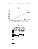 |  |
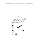 | 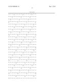 |
 |  |
 |  |
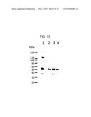 |  |
 |  |
 | 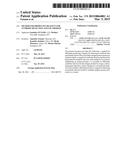 |
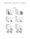 |  |
 |  |
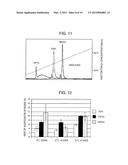 | 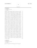 |
 | 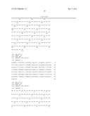 |
 |  |
 | 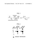 |
 | 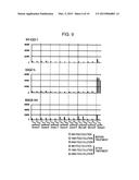 |
 | 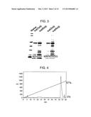 |
| Similar patent applications: | |
| Date | Title |
|---|---|
| 2015-03-19 | System and method for optical detection using capillary action |
| 2015-03-26 | Sulphur detection method and kit |
| 2015-03-26 | Microfluidic device, system and method |
| 2014-10-23 | Protein detection |
| 2015-03-26 | Soil analysis apparatus, method, and system |
| New patent applications in this class: | |
| Date | Title |
|---|---|
| 2022-05-05 | Detection and quantification of small molecules |
| 2019-05-16 | Immunoassay employing sulfated polysaccharide |
| 2017-08-17 | Lateral flow immunoassays for the detection of antibodies against biological drugs |
| 2017-08-17 | Weak affinity chromatography |
| 2017-08-17 | Method for quantitative characterization of substances with regard to their properties of binding to amyloid- (a ) conformers |
| New patent applications from these inventors: | |
| Date | Title |
|---|---|
| 2021-02-04 | Method for detecting an antigen-specific antibody and reagent for use therein |
| 2013-11-14 | Dendritic cells having enhanced yo t-cell activating and/or proliferating activity and methods of preparation or use thereof |
| Top Inventors for class "Chemistry: analytical and immunological testing" | |
| Rank | Inventor's name |
|---|---|
| 1 | Andreas Bergmann |
| 2 | Joachim Struck |
| 3 | Richard E. Reitz |
| 4 | Georg Hess |
| 5 | Tetsuo Nagano |