Patent application title: DNA METHYLATION MARKERS AND METHODS OF USE
Inventors:
Malcolm V. Brock (Owings Mills, MD, US)
Stephen B. Baylin (Baltimore, MD, US)
James G. Herman (Lutherville, MD, US)
Assignees:
THE JOHNS HOPKINS UNIVERSITY
IPC8 Class: AC12Q168FI
USPC Class:
435 611
Class name: Measuring or testing process involving enzymes or micro-organisms; composition or test strip therefore; processes of forming such composition or test strip involving nucleic acid nucleic acid based assay involving a hybridization step with a nucleic acid probe, involving a single nucleotide polymorphism (snp), involving pharmacogenetics, involving genotyping, involving haplotyping, or involving detection of dna methylation gene expression
Publication date: 2015-01-29
Patent application number: 20150031022
Abstract:
The present invention provides methods for identifying metastases by
detecting nucleic acid hypermethylation of one or more genes in one or
more samples, and in particular in the lymph nodes. The invention further
relates to DNA methylation as a predictor of disease recurrence and
patient prognosis, specifically in the field of cancer biology.Claims:
1-82. (canceled)
83. A method for identifying non-small cell lung cancer metastases in a mammalian subject sample comprising: isolating one or more nucleic acids from the mammalian subject sample; assaying the one or more nucleic acids to detect nucleic acid hypermethylation of a p16 gene and an H-cadherin gene; and correlating nucleic acid hypermethylation of the p16 gene and the H-cadherin gene with non-small cell lung cancer metastases.
84. The method of claim 83, wherein the sample comprises cells or tissues selected from the group consisting of: non-small cell tumor, lymph nodes, bone marrow and blood.
85. The method of claim 83, wherein the non-small cell lung cancer metastases are micrometastases.
86. The method of claim 83, further comprising detecting nucleic acid hypermethylation of APC or RASSf1A.
87. A method for detecting or diagnosing metastatic non-small cell lung cancer in a mammalian subject identified as having non-small cell lung cancer comprising: isolating nucleic acids from a non-small cell lung cancer (NSCLC) tumor or one or more lymph nodes of the mammalian subject; assaying the nucleic acids to detect nucleic acid hypermethylation of a p16 gene and an H-cadherin gene, in the NSCLC tumor or the one or more lymph nodes of the mammalian subject, wherein nucleic acid hypermethylation of the p16 gene and the H-cadherin gene is detected using a primer, sequencing, methylation sensitive restriction endonucleases, a nucleic acid probe, PCR allele-specific oligonucleotide probe analysis, or oligonucleotide ligation assays; and correlating nucleic acid hypermethylation of the p16 gene and the H-cadherin gene with metastatic non-small cell lung cancer.
88. A method for predicting a recurrence of non-small cell lung cancer in a mammalian subject comprising: isolating nucleic acids from a cell or tissue sample of the mammalian subject; assaying the one or more nucleic acids to detect nucleic acid hypermethylation of a p16 gene and an H-cadherin gene an H-cadherin gene and an APC gene, an APC gene and a p16 gene, or a RASSF1A gene and a p16 gene in the cell or tissue sample of the subject, wherein nucleic acid hypermethylation of the p16 gene and the H-cadherin gene, the H-cadherin gene and the APC gene, the APC gene and the p16 gene, or the RASSF1A gene and the p16 gene is detected using a primer, sequencing, methylation sensitive restriction endonucleases, a nucleic acid probe, PCR, allele-specific oligonucleotide probe analysis, or oligonucleotide ligation assays; and correlating nucleic acid hypermethylation of the p16 gene and the H-cadherin gene, the H-cadherin gene and the APC gene, the APC gene and the p16 gene, or the RASSF1A gene and the p16 gene with the recurrence of non-small cell lung cancer.
89. The method of claim 88, wherein hypermethylation of one or more genes is detected in non-small cell lung cancer tumor or lymph nodes.
90. A method for staging or re-staging a non-small cell lung cancer in a mammalian subject comprising: isolating nucleic acids from the mammalian subject; assaying the nucleic acids to detect nucleic acid hypermethylation of a p16 gene and an H-cadherin gene, wherein hypermethylation of the p16 gene and the H-cadherin gene is detected using a primer, sequencing, methylation sensitive restriction endonucleases, a nucleic acid probe, PCR allele-specific oligonucleotide probe analysis, or oligonucleotide ligation assays; and correlating nucleic acid hypermethylation of the p16 gene and the H-cadherin gene with non-small cell lung cancer stage.
91. A method for determining a prognosis of a mammalian subject identified as having non-small cell lung cancer comprising: isolating nucleic acids from the mammalian subject; assaying the nucleic acids to detect nucleic acid hypermethylation of a p16 gene and an H-cadherin gene, wherein hypermethylation of the p16 gene and the H-cadherin gene is detected using a primer, sequencing, methylation sensitive restriction endonucleases, a nucleic acid probe, PCR, allele-specific oligonucleotide probe analysis, or oligonucleotide ligation assays; and correlating nucleic acid hypermethylation of the p16 gene and the H-cadherin gene with the prognosis of the mammalian subject.
92. A method for predicting a recurrence of a non-small cell lung cancer (NSCLC) in a human subject comprising: isolating nucleic acids from a lung tumor or lymph node of the subject; assaying the nucleic acids to detect nucleic acid hypermethylation of a p16 gene and an H-cadherin gene; and correlating the nucleic acid hypermethylation state of the p16 gene and the H-cadherin gene with an increased risk of the recurrence of NSCLC in comparison to control lung tumor tissue samples obtained from human subjects without NSCLC recurrence.
93. A kit for identifying non-small cell lung cancer in a mammalian subject sample, comprising a probe for detecting the hypermethylation state of a p16 gene and an H-cadherin gene, an H-cadherin gene and an APC gene, an APC gene and a p16 gene, or a RASSF1A gene and a p16 gene, and instructions for use.
94. A kit for detecting non-small cell lung cancer metastases in a mammalian subject sample by detecting nucleic acid hypermethylation of a p16 gene and an H-cadherin gene, an H-cadherin gene and an APC gene, an APC gene and a p16 gene, or a RASSF1A gene and a p16 gene, the kit comprising gene specific primers for use in polymerase chain reaction (PCR), and instructions for use.
95. The kit of claim 94, wherein the non-small cell lung cancer metastases are micrometastases.
96. The method of claim 83, wherein an increase in methylation relative to a control identifies the presence of non-small cell lung cancer metastases, and a decrease in methylation identifies the absence of non-small cell lung cancer metastases.
97. The method of claim 92, wherein an increase in methylation relative to a control identifies an increased risk of non-small cell lung cancer recurrence, and a decrease in methylation identifies a reduced risk of non-small cell lung cancer recurrence.
Description:
RELATED APPLICATIONS
[0001] This application is a continuation of U.S. application Ser. No. 12/515,735, filed Jun. 30, 2010, which is the U.S. national phase, pursuant to 35 U.S.C. §371, of PCT international application Ser. No. PCT/US2007/024308, filed Nov. 20, 2007, designating the United States and published in English on May 29, 2008 as publication WO 2008/063655 A2, which claims priority to U.S. provisional application Ser. No. 60/860,196, filed Nov. 20, 2006. The entire contents of the aforementioned patent applications are incorporated herein by this reference.
FIELD OF THE INVENTION
[0002] The present invention relates to the use of nucleic acid methylation and methylation profiles to detect metastatic disease. In particular, the invention relates to methods for identifying metastases by detecting nucleic acid hypermethylation of one or more genes in one or more samples and, in particular, in tumor tissue and lymph nodes. The invention further relates to DNA hypermethylation as a predictor of disease recurrence and patient prognosis, specifically in patients suffering from cancer.
BACKGROUND OF THE INVENTION
[0003] Cancer remains one of the leading causes of death in the United States. Clinically, a broad variety of medical approaches, including surgery, radiation therapy and chemotherapeutic drug therapy are currently being used in the treatment of human cancer (see the textbook CANCER: Principles & Practice of Oncology, 2d Edition, De Vita et al., eds., J. B. Lippincott Company, Philadelphia, Pa., 1985). However, it is recognized that such approaches continue to be limited by an inability to predict the likelihood of metastasis and tumor recurrence or the most efficacious treatment regime for minimizing the occurrence of these negative outcomes.
[0004] Human cancer cells typically contain somatically altered nucleic acids, characterized by mutation, amplification, or deletion of critical genes. In addition, the nucleic acids from human cancer cells often display somatic changes in DNA methylation (36, 37, 38). However, a precise role for, and the significance of, abnormal DNA methylation in human tumorigenesis has not been well established.
[0005] Loss of gene function is cancer can occur by both genetic and epigenetic mechanisms. The best-defined epigenetic alteration of cancer genes involves DNA methylation of clustered CpG dinucleotides, or CpG islands, in promoter regions associated with the transcriptional inactivation of the affected genes. CpG islands are short sequences rich in the CpG dinucleotide, and can be found in the 5' region of about half of all human genes. Methylation of cytosine within 5' CGIs is associated with loss of gene expression and has been seen in a number of physiological conditions, including X chromosome inactivation and genomic imprinting. Aberrant methylation of CpG islands has been detected in genetic diseases such as the fragile-X syndrome, in aging cells and in neoplasia. About half of the tumor suppressor genes which have been shown to be mutated in the germline of patients with familial cancer syndromes have also been shown to be aberrantly methylated in some proportion of sporadic cancers, including Rb, VHL, p16, hMLH1, and BRCA1 (reviewed in Baylin, et al, Adv. Cancer Res. 72:141-196 1998). Methylation of tumor suppressor genes in cancer is usually associated with (1) lack of gene transcription and (2) absence of coding region mutation. Thus CpG island methylation can serve as an alternative mechanism of gene inactivation in cancer.
[0006] Cancer treatments, in general, have a higher rate of success if the cancer is diagnosed early, and treatment is started earlier in the disease process. A relationship between improved prognosis and stage of disease at diagnosis can be seen across a majority of cancers. Identification of the earliest changes in cells associated with cancer is thus a major focus in molecular cancer research. Diagnostic approaches based on identification of these changes in specific genes may allow implementation of early detection strategies and novel therapeutic approaches. Targeting these early changes will lead to more effective cancer treatment.
[0007] Despite advances in targeted therapy, surgery with curative intent remains the best therapeutic option for lung cancer patients with the earliest stages of disease. Ensuring in these patients that no occult metastatic cells have disseminated outside the area of curative resection is critical, because early spread of tumor cells is a leading cause of relapse (1-3). Despite the curative aim of early surgery, approximately 30%-40% of lung cancer patients with discrete lesions and histologically proven cancer negative lymph nodes (stage 1:T1-2N0) still die of recurrent disease (4-6). Further, many of these recurrences are systemic, underscoring the probability that these patients had metastatic disease that was undetectable, and beyond the margins of surgical resection.
[0008] Accordingly, there is a need in the art for improved methods of detection of proliferative disease, and in particular, for improved methods of detection of metastatic cancer that is undetectable by current methodologies.
SUMMARY
[0009] The invention features methods for identifying metastases by detecting nucleic acid hypermethylation of one or more genes in one or more samples, and in particular in tumor tissue and lymph nodes.
[0010] In one aspect, the invention features methods for identifying metastases in a subject comprising detecting nucleic acid hypermethylation of one or more genes in one or more samples, wherein detecting nucleic acid hypermethylation identifies metastases.
[0011] In one embodiment, the sample comprises cells or tissues selected from the group consisting of tumor, lymph nodes, bone marrow and blood. In a particular embodiment, the sample is from a tumor. In another particular embodiment, the sample is from a lymph node. In a more particular embodiment, the lymph node is a N1 lymph node or a mediastinal lymph node.
[0012] In another aspect the invention features methods for identifying metastases in a subject comprising detecting nucleic acid hypermethylation of one or more genes in tumor tissue or lymph node, wherein the genes are selected from the group consisting of genes involved in tumor suppression, DNA repair, apoptosis, anti-proliferation, ras signaling, adhesion, differentiation, development, and cell cycle regulation, wherein detecting nucleic acid hypermethylation identifies metastases.
[0013] In certain preferred embodiments of the above aspects, the metastases are micrometastases. In other preferred embodiments of the above aspects, the one or more genes comprise one or more CpG islands. In a further embodiment, the one or more genes is selected from the group consisting of H-cadherin, p16, APC, RASSF1A, MGMT, DAPK, and ASC.
[0014] H-cadherin, in certain exemplary embodiments is encoded by NCBI accession No. AAB18912 and is shown in (SEQ ID NO:1) below:
TABLE-US-00001 1 mqprtplvlc vllsqvlllt saedldctpg fqqkvfhinq paefiedqsi lnltfsdckg 61 ndklryevss pyfkvnsdgg lvalrnitav gktlfvhart phaedmaelv ivggkdiqgs 121 lqdifkfart spvprqkrsi vvspilipen qrqpfprdvg kvvdsdrper skfrltgkgv 181 dqepkgifri nentgsvsvt rtldreviav yqlfvettdv ngktlegpvp levividqnd 241 nrpifregpy ighvmegspt gttvmrmtaf daddpatdna llrynirqqt pdkpspnmfy 301 idpekgdivt vvspalldre tlenpkyeli ieaqdmagld vgltgtatat imiddkndhs 361 pkftkkefqa tveegavgvi vnltvedkdd pttgawraay tiingnpgqs feihtnpqtn 421 egmlsvvkpl dyeisafhtl likvenedpl vpdvsygpss tatvhitvld vnegpvfypd 481 pmmvtrqedl svgsvlltvn atdpdslqhq tirysvykdp agwlninpin gtvdttavld 541 respfvdnsv ytalflaids gnppatgtgt llitledvnd napfiyptva evcddaknls 601 vvilgasdkd lhpntdpfkf eihkqavpdk vwkiskinnt halvsllqnl nkanynlpim 661 vtdsgkppmt nitdlrvqvc scrnskvdcn aagalrfslp svlllslfsl acl
[0015] p-16, in certain exemplary embodiments is encoded by NCBI accession No. CAB58124 and is shown in (SEQ ID NO:2) below:
TABLE-US-00002 1 gshsmryfft svsrpgrgep rfiavgyvdd tqfvrfdsda asqrmeprap wieqegpeyw 61 dgetrkvkah sqtdrvdlgt lrgyynqsea gshtiqmmyg cdvgpdgrll rgyqqdaydg 121 kdyialnedl rswtaadmaa qitqrkweaa rvaeqlrayl egtcvewlrr ylengketlq 181 rt
[0016] APC, in certain exemplary embodiments is encoded by NCBI accession No. NP--000029 and is shown in (SEQ ID NO:3) below:
TABLE-US-00003 1 maaasydqll kqvealkmen snlrqeledn snhltklete asnmkevlkq lqgsiedeam 61 assgqidlle rlkelnldss nfpgvklrsk mslrsygsre gsvssrsgec spvpmgsfpr 121 rgfvngsres tgyleeleke rsllladldk eekekdwyya qlqnltkrid slpltenfsl 181 qtdmtrrqle yearqirvam eeqlgtcqdm ekraqrriar iqqiekdilr irqllqsqat 241 eaerssqnkh etgshdaerq negqgvgein matsgngqgs ttrmdhetas vlssssthsa 301 prrltshlgt kvemvyslls mlgthdkddm srtllamsss qdscismrqs gclplliqll 361 hgndkdsvll gnsrgskear arasaalhni ihsqpddkrg rreirvlhll eqiraycetc 421 wewqeahepg mdqdknpmpa pvehqicpav cvlmklsfde ehrhamnelg glqaiaellq 481 vdcemygltn dhysitlrry agmaltnltf gdvankatlc smkgcmralv aqlksesedl 541 qqviasvlrn lswradvnsk ktlrevgsvk almecalevk kestlksvls alwnlsahct 601 enkadicavd galaflvgtl tyrsqtntla iiesgggilr nvssliatne dhrqilrenn 661 clqtllqhlk shsltivsna cgtlwnlsar npkdqealwd mgavsmlknl ihskhkmiam 721 gsaaalrnlm anrpakykda nimspgsslp slhvrkqkal eaeldaqhls etfdnidnls 781 pkashrskqr hkqslygdyv fdtnrhddnr sdnfntgnmt vlspylnttv lpsssssrgs 841 ldssrsekdr slerergigl gnyhpatenp gtsskrglqi sttaaqiakv meevsaihts 901 qedrssgstt elhcvtdern alrrssaaht hsntynftks ensnrtcsmp yakleykrss 961 ndslnsvsss dgygkrgqmk psiesysedd eskfcsygqy padlahkihs anhmddndge 1021 ldtpinyslk ysdeqlnsgr qspsqnerwa rpkhiiedei kqseqrqsrn qsttypvyte 1081 stddkhlkfq phfgqqecvs pyrsrgangs etnrvgsnhg inqnvsqslc qeddyeddkp 1141 tnyserysee eqheeeerpt nysikyneek rhvdqpidys lkyatdipss qkqsfsfsks 1201 ssgqsskteh mssssentst pssnakrqnq lhpssaqsrs gqpqkaatck vssinqetiq 1261 tycvedtpic fsrcsslssl ssaedeigcn qttqeadsan tlqiaeikek igtrsaedpv 1321 sevpavsqhp rtkssrlqgs slssesarhk avefssgaks psksgaqtpk sppehyvqet 1381 plmfsrctsv ssldsfesrs iassvqsepc sgmvsgiisp sdlpdspgqt mppsrsktpp 1441 pppqtaqtkr evpknkapta ekresgpkqa avnaavqrvq vlpdadtllh fatestpdgf 1501 scssslsals ldepfiqkdv elrimppvqe ndngnetese qpkesnenqe keaektidse 1561 kdllddsddd dieileecii samptkssrk akkpaqtask lpppvarkps qlpvykllps 1621 qnrlqpqkhv sftpgddmpr vycvegtpin fstatslsdl tiesppnela agegvrggaq 1681 sgefekrdti ptegrstdea qggktssvti pelddnkaee gdilaecins ampkgkshkp 1741 frvkkimdqv qqasasssap nknqldgkkk kptspvkpip qnteyrtrvr knadsknnln 1801 aervfsdnkd skkqnlknns kvfndklpnn edrvrgsfaf dsphhytpie gtpycfsrnd 1861 slssldfddd dvdlsrekae lrkakenkes eakvtshtel tsnqqsankt qaiakqpinr 1921 gqpkpilqkq stfpqsskdi pdrgaatdek lqnfaientp vcfshnssls slsdidqenn 1981 nkenepiket eppdsqgeps kpqasgyapk sfhvedtpvc fsrnsslssl sidseddllq 2041 ecissampkk kkpsrlkgdn ekhsprnmgg ilgedltldl kdiqrpdseh glspdsenfd 2101 wkaiqegans ivsslhqaaa aaclsrqass dsdsilslks gislgspfhl tpdqeekpft 2161 snkgprilkp gekstletkk ieseskgikg gkkvykslit gkvrsnseis gqmkqplqan 2221 mpsisrgrtm ihipgvrnss sstspvskkg pplktpasks psegqtatts prgakpsvks 2281 elspvarqts qiggsskaps rsgsrdstps rpaqqplsrp iqspgrnsis pgrngisppn 2341 klsqlprtss pstastkssg sgkmsytspg rqmsqqnltk qtglsknass iprsesaskg 2401 lnqmnngnga nkkvelsrms stkssgsesd rserpvlvrq stfikeapsp tlrrkleesa 2461 sfeslspssr pasptrsqaq tpvlspslpd mslsthssvq aggwrklppn lsptieyndg 2521 rpakrhdiar shsespsrlp inrsgtwkre hskhssslpr vstwrrtgss ssilsasses 2581 sekaksedek hvnsisgtkq skenqvsakg twrkikenef sptnstsqtv ssgatngaes 2641 ktliyqmapa vsktedvwvr iedcpinnpr sgrsptgntp pvidsvseka npnikdskdn 2701 qakqnvgngs vpmrtvglen rlnsfiqvda pdqkgteikp gqnnpvpvse tnessivert 2761 pfsssssskh sspsgtvaar vtpfnynpsp rkssadstsa rpsqiptpvn nntkkrdskt 2821 dstessgtqs pkrhsgsylv tsv
[0017] RASSF1A, in certain exemplary embodiments is encoded by NCBI accession No. NP--009113 and is shown in (SEQ ID NO:4) below:
TABLE-US-00004 1 msgepeliel relapagrag kgrtrleran alriargtac nptrqlvpgr ghrfqpagpa 61 thtwcdlcgd fiwgvvrkgl qcahckftch yrcralvcld ccgprdlgwe paverdtnvd 121 epvewetpdl sqaeieqkik eynaqinsnl fmslnkdgsy tgfikvqlkl vrpvsvpssk 181 kppslqdarr gpgrgtsvrr rtsfylpkda vkhlhvlsrt rarevieall rkflvvddpr 241 kfalferaer hgqvylrkll ddeqplrlrl lagpsdkals fvlkendsge vnwdafsmpe 301 lhnflrilqr eeeehlrqil qkysycrqki qealhacplg
[0018] MGMT, in certain exemplary embodiments is encoded by NCBI accession No. AAH00824 and is shown in (SEQ ID NO:5) below:
TABLE-US-00005 1 mdkdcemkrt tldsplgkle lsgceqglhe ikllgkgtsa adavevpapa avlggpeplm 61 qctawlnayf hqpeaieefp vpalhhpvfq qesftrqvlw kllkvvkfge visyqqlaal 121 agnpkaarav ggamrgnpvp ilipchrvvc ssgavgnysg glavkewlla heghrlgkpg 181 lggssglaga wlkgagatsg sppagrn
[0019] DAPK, in certain exemplary embodiments is encoded by NCBI accession No. NP--004929 and is shown in (SEQ ID NO:6) below:
TABLE-US-00006 1 mtvfrqenvd dyydtgeelg sgqfavvkkc rekstglqya akfikkrrtk ssrrgvsred 61 ierevsilke iqhpnvitlh evyenktdvi lilelvagge lfdflaekes lteeeatefl 121 kqilngvyyl hslqiahfdl kpenimlldr nvpkprikii dfglahkidf gnefknifgt 181 pefvapeivn yeplgleadm wsigvityil lsgaspflgd tkqetlanvs avnyefedey 241 fsntsalakd firrllvkdp kkrmtiqdsl qhpwikpkdt qqalsrkasa vnmekfkkfa 301 arkkwkqsvr lislcqrlsr sflsrsnmsv arsddtldee dsfvmkaiih ainddnvpgl 361 qhllgslsny dvnqpnkhgt pplliaagcg niqilqllik rgsridvqdk ggsnavywaa 421 rhghvdtlkf lsenkcpldv kdksgemalh vaaryghadv aqllcsfgsn pniqdkeeet 481 plhcaawhgy ysvakalcea gcnvniknre getplltasa rgyhdivecl aehgadlnac 541 dkdghialhl avrrcqmevi ktllsqgcfv dyqdrhgntp lhvackdgnm pivvalcean 601 cnldisnkyg rtplhlaann gildvvrylc lmgasvealt tdgktaedla rseqhehvag 661 llarlrkdth rglfiqqlrp tqnlqprikl klfghsgsgk ttlveslkcg llrsffrrrr 721 prlsstnssr fppsplaskp tvsvsinnly pgcenvsvrs rsmmfepglt kgmlevfvap 781 thhphcsadd qstkaidiqn aylngvgdfs vwefsgnpvy fccydyfaan dptsihvvvf 841 sleepyeiql nqvifwlsfl kslvpveepi afggklknpl qvvlvathad imnvprpagg 901 efgydkdtsl lkeirnrfgn dlhisnklfv ldagasgskd mkvlrnhlqe irsqivsvcp 961 pmthlcekii stlpswrkln gpnqlmslqq fvydvqdqln plaseedlrr iaqqlhstge 1021 inimqsetvq dvllldprwl ctnvlgklls vetpralhhy rgrytvediq rlvpdsdvee 1081 llqildamdi cardlssgtm vdvpaliktd nlhrswadee devmvyggvr ivpvehltpf 1141 pcgifhkvqv nlcrwihqqs tegdadirlw vngcklanrg aellvllvnh gqgievqvrg 1201 letekikccl lldsvcstie nvmattlpgl ltvkhylspq qlrehhepvm iyqprdffra 1261 qtlketsltn tmggykesfs simcfgchdv ysqaslgmdi hasdlnlltr rklsrlldpp 1321 dplgkdwcll amnlglpdlv akyntsngap kdflpsplha llrewttype stvgtlmskl 1381 relgrrdaad fllkassvfk inldgngqea yasscnsgts ynsissvvsr
[0020] ASC, in certain exemplary embodiments is encoded by NCBI accession No. NP--037390 and is shown in (SEQ ID NO:7) below:
TABLE-US-00007 1 mgrardaild alenltaeel kkfklkllsv plregygrip rgallsmdal dltdklvsfy 61 letygaelta nvlrdmglqe magqlqaath qgsgaapagi qappqsaakp glhfidqhra 121 aliarvtnve wlldalygkv ltdeqyqavr aeptnpskmr klfsftpawn wtckdlllqa 181 lresqsylve dlers
[0021] In other embodiments of the above aspects, hypermethylation of at least one of the genes is detected. In still other embodiments of the above aspects, hypermethylation of at least two of the genes is detected.
[0022] In other aspects, the invention features methods for identifying micrometastases in a subject comprising detecting nucleic acid hypermethylation of at least one or more genes in a sample comprising tumor and lymph nodes, wherein the sample genes are selected from the group consisting of H-cadherin, p16, APC, RASSF1A, MGMT, DAPK, and ASC, and wherein detecting nucleic acid methylation identifies micrometastases.
[0023] In a preferred embodiment, hypermethylation of at least two of the genes is detected. In another embodiment, at least two of the genes are selected from p-16 and H-cadherin, H-cadherin and APC, APC and p16, or RASSf1A and p16.
[0024] In another further embodiment, the detection of metastases is used to detect or diagnose a proliferative disease.
[0025] In certain embodiments, the detection or diagnosis is performed after surgery or therapy to treat a proliferative disease. In other certain embodiments, the detection is used to predict the recurrence of a proliferative disease. In other certain embodiments, the detection is used to stage a proliferative disease. In still other certain embodiments, the detection is further used to determine a course of treatment for a subject.
[0026] In other aspects, the invention features a method for detecting or diagnosing a proliferative disease in a subject comprising detecting nucleic acid hypermethylation of one or more genes in one or more samples, wherein detecting nucleic acid hypermethylation is used to detect or diagnose a proliferative disease.
[0027] In still other aspects, the invention features a method for predicting the recurrence of a proliferative disease in a subject comprising detecting nucleic acid hypermethylation of one or more genes wherein detecting nucleic acid hypermethylation of one or more genes is a predictor of the recurrence of a proliferative disease.
[0028] In one embodiment, hypermethylation of one or more genes is detected in tumor or lymph nodes.
[0029] In a related embodiment, detection of hypermethylation of one or more genes in lymph nodes is predictive of aggressive disease recurrence.
[0030] In another aspect, the invention features a method for staging or re-staging a proliferative disease in a subject comprising detecting nucleic acid hypermethylation of one or more genes wherein detecting nucleic acid hypermethylation is used for staging or re-staging a proliferative disease.
[0031] In a related embodiment, the stage of proliferative disease is predictive of disease recurrence. In a further embodiment, the stage of proliferative disease determines course of treatment.
[0032] In another aspect, the invention features a method for determining the prognosis of a subject suffering from a proliferative disease comprising detecting nucleic acid hypermethylation of one or more genes wherein the detection of nucleic acid hypermethylation is used for determining the prognosis of a subject suffering from a proliferative disease.
[0033] In a related embodiment, the prognosis determines course of treatment.
[0034] In an embodiment of any of the above-mentioned aspects, the subject is a human.
[0035] In another embodiment of any of the above-mentioned aspects, the method is performed prior to therapeutic intervention for the disease.
[0036] In another embodiment of any of the above-mentioned aspects, the method is performed after therapeutic intervention for the disease. In a related embodiment, the therapeutic intervention is selected from treatment with an agent or surgery. In another related embodiment, hypermethylation is detected in CpG islands of the one or more genes. In a further related embodiment, hypermethylation is detected in CpG islands.
[0037] In another aspect, the invention features methods for detecting or diagnosing a proliferative disease in a subject comprising extracting nucleic acid from one or more cell or tissue samples, detecting nucleic acid hypermethylation of one or more genes in the sample; and identifying the nucleic acid hypermethylation state of one or more genes, wherein nucleic acid hypermethylation of genes indicates a proliferative disease.
[0038] In a further aspect, the invention features methods for predicting the recurrence of a proliferative disease in a subject comprising extracting nucleic acid from one or more cell or tissue samples, detecting nucleic acid hypermethylation of one or more genes in the sample; and identifying the nucleic acid hypermethylation state of one or more genes, wherein nucleic acid hypermethylation of genes is indicative of the recurrence of a proliferative disease.
[0039] In a further aspect, the invention features methods for staging or re-staging a proliferative disease in a subject comprising extracting nucleic acid from one or more cell or tissue samples, detecting nucleic acid hypermethylation of one or more genes in the sample; and identifying the nucleic acid hypermethylation state of one or more genes, wherein nucleic acid hypermethylation of genes is used for staging or re-staging of a proliferative disease.
[0040] In one embodiment of the above-mentioned aspects, the tissue samples are selected from tumor, lymph node, bone marrow or blood or a combination thereof.
[0041] In another embodiment of the above-mentioned aspects, the method determines the course of disease treatment.
[0042] In still another embodiment of the above-mentioned aspects, the method is performed prior to therapeutic intervention for the disease.
[0043] In still another embodiment of the above-metioned aspects, the method is performed after therapeutic intervention for the disease.
[0044] In a further embodiment, the therapeutic intervention is selected from treatment with an agent or surgery.
[0045] In another aspect, the invention features methods of treating a subject having or at risk for having a proliferative disease comprising identifying nucleic acid hypermethylation of one or more genes, where nucleic acid hypermethylation indicates having or a risk for having a proliferative disease, and administering to the subject a therapeutically effective amount of a demethylating agent, thereby treating a subject having or at risk for having a proliferative disease.
[0046] In one particular embodiment, the method is used in combination with one or more chemotherapeutic agents.
[0047] In another particular embodiment of the above-mentioned aspects, the method further comprises comparing the nucleic acid hypermethylation of one or more genes in the sample with comparable samples obtained from a normal subject.
[0048] In a further embodiment of the above-mentioned aspects, detecting nucleic acid hypermethylation of one or more genes indicates the presence of metastases.
[0049] In a particular embodiment, the metastases are micrometastases.
[0050] In another particular embodiment of any one of the above-mentioned aspects, the proliferative disease is a neoplasia. In a preferred embodiment, the neoplasia is cancer. In another preferred embodiment, the cancer is a solid tumor. In a further embodiment, the cancer is selected from the group consisting of lung cancer, pancreatic cancer, esophageal cancer, head and neck cancer, stomach cancer, liver cancer, prostate cancer, gastrointestinal cancer, ovarian cancer, and uterine cancer.
[0051] In another particular embodiment of the above-mentioned aspects, the cells or tissues are selected from the group consisting of tumor, lymph nodes, bone marrow or blood. In a related embodiment, the cells or tissues are from a tumor or the lymph nodes. In a further embodiment, the lymph node is a N1 lymph node or a mediastinal lymph node.
[0052] In another aspect, the invention features a method of identifying an agent that de-methylates hypermethylated nucleic acid comprising identifying one or more cell or tissue samples with hypermethylated nucleic acid, extracting the hypermethylated nucleic acid, contacting the nucleic acid with one or more nucleic acid de-methylating candidate agents and a control agent, identifying the nucleic acid hypermethylation state, wherein nucleic acid de-methylation of genes in the sample by the candidate agent compared to the control indicates a demethylating agent, and thereby identifying an agent that de-methylates hypermethylated nucleic acid.
[0053] In one embodiment of any of the above-mentioned aspects, the one or more genes are selected from the group consisting of genes involved in tumor suppression, DNA repair, anti-proliferation, apoptosis, ras signaling, adhesion, differentiation, development, and cell cycle regulation.
[0054] In another embodiment of any of the above-mentioned aspects, the one or more genes are selected from a panel consisting of (1) genes involved in tumor suppression and cell adhesion, (2) genes involved in cell cycle regulation and adhesion, (3) genes involved in tumor suppression and cell cycle regulation, and (4) genes involved in ras signaling and cell cycle control.
[0055] In still another embodiment of any of the above-mentioned aspects, the one or more genes comprise one or more CpG islands.
[0056] In a related embodiment, the genes are selected from the group consisting of p-16, H-cadherin, APC, RASSF1A, MGMT, DAPK, and ASC.
[0057] In another related embodiment, the hypermethylation of at least one of the genes is detected. In a further related embodiment, the hypermethylation of at least two of the genes is detected. In still another related embodiment, the two genes are selected from p-16 and H-cadherin, H-cadherin and APC, APC and p16, or RASSf1A and p16.
[0058] In another embodiment of any of the above-mentioned aspects, the detection of nucleic acid methylation is by a quantitative method.
[0059] In another embodiment of any of the above-mentioned aspects, the detection of nucleic acid methylation is carried out by polymerase chain reaction (PCR) analysis. In a related embodiment, the PCR is methylation specific PCR (MSP).
[0060] In a particular embodiment, the method of detecting nucleic acid methylation is performed as a high-throughput method.
[0061] In another particular embodiment, the method is used in combination with the detection of other epigenetic markers. In a particular related embodiment, the other epigenetic markers are plasma or tumor epigenetic markers.
[0062] In an embodiment of the above-described aspects, hypermethylation is detected in CpG islands of the one or more genes. In a further embodiment of the above-described aspects, hypermethylation is detected in CpG islands of the promoter region.
[0063] In other aspects, the invention features kits for identifying the nucleic acid hypermethylation state of one or more genes comprising gene specific primers for use in polymerase chain reaction (PCR), and instructions for use.
[0064] In still other aspects, the invention features kits for detecting metastases by detecting nucleic acid hypermethylation of one or more genes, the kit comprising gene specific primers for use in polymerase chain reaction (PCR), and instructions for use.
[0065] In one embodiment, the metastases are micrometastases.
[0066] In another embodiment, the PCR is methylation specific PCR (MSP).
[0067] In still another embodiment, the one or more genes are selected from the group consisting of genes involved in tumor suppression, DNA repair, anti-proliferation, apotosis, ras signaling, adhesion, differentiation, development, and cell cycle regulation.
[0068] In another embodiment, the one or more genes are selected from a panel consisting of (1) genes involved in tumor suppression and cell adhesion, (2) genes involved in cell cycle regulation and adhesion, (3) genes involved in tumor suppression and cell cycle regulation, and (4) genes involved in ras signaling and cell cycle control.
[0069] In a related embodiment, the one or more genes comprise one or more CpG islands. In a further related embodiment, the CpG islands are in the promoter region. In another related embodiment, the genes are selected from the group consisting of p-16, H-cadherin, APC, RASSF1A, MGMT, DAPK, and ASC
[0070] In another embodiment, the hypermethylation of at least one of the genes is detected. In still another embodiment, the hypermethylation of at least two of the genes is detected. In still another further embodiment, the two genes are selected from p-16 and H-cadherin, H-cadherin and APC, APC and p16, or RASSf1A and p16.
[0071] Other aspects of the invention are described infra.
BRIEF DESCRIPTION OF THE FIGURES
[0072] FIG. 1 shows the results of multivariate logistic regression analysis performed using the four genes that exhibited the largest univariate distribution differences in methylation: p16, H-cadherin, APC, and RASSF1A.
[0073] FIG. 2 (A-L) are graphs showing Kaplan-Meier estimates of recurrence-free survival of pathologic Stage 1 lung cancer patients at the Johns Hopkins Hospital, according to number of methylated genes in a 4-gene panel at time of surgical resection.
[0074] FIG. 3 shows Methylation Specific PCR for the H-cadherin gene. For each sample, the presence of a visible PCR product in Lanes marked U indicates the presence of an unmethylated promoter region amplified and serves as a control for sample preparation; the presence of product in Lanes M indicates a methylated gene promoter and was scored as positive for methylation. * represents the molecular weight marker.
DEFINITIONS
[0075] Unless defined otherwise, all technical and scientific terms used herein have the meaning commonly understood by a person skilled in the art to which this invention belongs. The following references provide one of skill with a general definition of many of the terms used in this invention: Singleton et al., Dictionary of Microbiology and Molecular Biology (2nd ed. 1994); The Cambridge Dictionary of Science and Technology (Walker ed., 1988); The Glossary of Genetics, 5th Ed., R. Rieger et al. (eds.), Springer Verlag (1991); and Hale & Marham, The Harper Collins Dictionary of Biology (1991). As used herein, the following terms have the meanings ascribed to them unless specified otherwise.
[0076] In this disclosure, "comprises," "comprising," "containing" and "having" and the like can have the meaning ascribed to them in U.S. Patent law and can mean "includes," "including," and the like; "consisting essentially of" or "consists essentially" likewise has the meaning ascribed in U.S. Patent law and the term is open-ended, allowing for the presence of more than that which is recited so long as basic or novel characteristics of that which is recited is not changed by the presence of more than that which is recited, but excludes prior art embodiments.
[0077] By "control" is meant a standard or reference condition.
[0078] The phrase "in combination with" is intended to refer to all forms of administration that provide a de-methylating agent, or the methods of the instant invention (e.g. methods of detection of hypermethylation) together with a second agent, such as a chemotherapeutic agent, or a de-methylating agent, where the two are administered concurrently or sequentially in any order.
[0079] The term "agent" as used herein is meant to refer to a polypeptide, polynucleotide, or fragment, or analog thereof, small molecule, or other biologically active molecule.
[0080] The term "CpG island" refers to a sequence of nucleic acid with an increased density relative to other nucleic acid regions of the dinucleotide CpG.
[0081] The term "epigenetic marker" or "epigenetic change" as used herein is meant to refer to a change in the DNA sequences or gene expression by a process or processes that do not change the DNA coding sequence itself. In an exemplary embodiment, methylation is an epigenetic marker.
[0082] The term "hypermethylation" as used herein refers to the presence of methylated alleles in one or more nucleic acids. In preferred embodiments, hypermethylation is detected using methylation specific polymerase chain reaction (MSP).
[0083] The term "metastases" is meant to refer to the spread of a malignant tumor from its sight of origin. Cancer cells may metastasize through the bloodstream, through the lymphatic system, across body cavities, or any combination thereof. A metastatic tumor can arise from a multitude of primary tumor types, including but not limited to lung, breast, thyroid, head and neck, brain, lymphoid, gastrointestinal (mouth, esophagus, stomach, small intestine, colon, rectum), genito-urinary tract (uterus, ovary, cervix, bladder, testicle, penis, prostate), kidney, pancreas, liver, bone, muscle or skin.
[0084] The term "micrometastases" is meant to refer to a metastasis that cannot be detected by routine histological evaluation, for example by Hematoxylin and Eosin (H & E) staining and microscopic assessment.
[0085] The term "neoplasm" or "neoplasia" as used herein refers to inappropriately high levels of cell division, inappropriately low levels of apoptosis, or both. A neoplasm creates an unstructured mass (a tumor), which can be either benign or malignant. For example, cancer is a neoplasia. Examples of cancers include, without limitation, leukemias (e.g., acute leukemia, acute lymphocytic leukemia, acute myelocytic leukemia, acute myeloblastic leukemia, acute promyelocytic leukemia, acute myelomonocytic leukemia, acute monocytic leukemia, acute erythroleukemia, chronic leukemia, chronic myelocytic leukemia, chronic lymphocytic leukemia), polycythemia vera, lymphoma (Hodgkin's disease, non-Hodgkin's disease), Waldenstrom's macroglobulinemia, heavy chain disease, and solid tumors such as sarcomas and carcinomas (e.g., fibrosarcoma, myxosarcoma, liposarcoma, chondrosarcoma, osteogenic sarcoma, chordoma, angiosarcoma, endotheliosarcoma, lymphangiosarcoma, lymphangioendotheliosarcoma, synovioma, mesothelioma, Ewing's tumor, leiomyosarcoma, rhabdomyosarcoma, colon carcinoma, pancreatic cancer, breast cancer, ovarian cancer, prostate cancer, squamous cell carcinoma, basal cell carcinoma, adenocarcinoma, sweat gland carcinoma, sebaceous gland carcinoma, papillary carcinoma, papillary adenocarcinomas, cystadenocarcinoma, medullary carcinoma, bronchogenic carcinoma, renal cell carcinoma, hepatoma, nile duct carcinoma, choriocarcinoma, seminoma, embryonal carcinoma, Wilm's tumor, cervical cancer, uterine cancer, testicular cancer, lung carcinoma, small cell lung carcinoma, bladder carcinoma, epithelial carcinoma, glioma, astrocytoma, medulloblastoma, craniopharyngioma, ependymoma, pinealoma, hemangioblastoma, acoustic neuroma, oligodenroglioma, schwannoma, meningioma, melanoma, neuroblastoma, and retinoblastoma). Lymphoproliferative disorders are also considered to be proliferative diseases.
[0086] The phrase "nucleic acid" as used herein refers to an oligonucleotide, nucleotide, polynucleotide, or to a fragment of any of these, to DNA or RNA of genomic or synthetic origin which may be single-stranded or double-stranded and may represent a sense or antisense strand, peptide nucleic acid (PNA), or to any DNA-like or RNA-like material, natural or synthetic in origin. As will be understood by those of skill in the art, when the nucleic acid is RNA, the deoxynucleotides A, G, C, and T are replaced by ribonucleotides A, G, C, and U, respectively.
[0087] The term "proliferative disorder" as used herein refers to an abnormal growth of cells. A cell proliferative disorder as described herein may be a neoplasm.
[0088] The term "promoter" or "promoter region" refers to a minimal sequence sufficient to direct transcription or to render promoter-dependent gene expression that is controllable for cell-type specific, tissue-specific, or is inducible by external signals or agents. Promoters may be located in the 5' or 3' regions of the gene. Promoter regions, in whole or in part, of a number of nucleic acids can be examined for sites of CpG-island methylation.
[0089] The term "sample" as used herein refers to any biological or chemical mixture for use in the method of the invention. The sample can be a biological sample. The biological samples are generally derived from a patient, preferably as a bodily fluid (such as tumor tissue, lymph node, sputum, blood, bone marrow, cerebrospinal fluid, phlegm, saliva, or urine) or cell lysate. The cell lysate can be prepared from a tissue sample (e.g. a tissue sample obtained by biopsy), for example, a tissue sample (e.g. a tissue sample obtained by biopsy), blood, cerebrospinal fluid, phlegm, saliva, urine, or the sample can be cell lysate.
[0090] The term "stage" or "staging" as used herein is meant to refer to the extent or progression of proliferative disease, e.g. cancer, in a subject. Staging can be "clinical" and is according to the "stage classification" corresponding to the TNM classification ("Rinsho, Byori, Genpatsusei Kangan Toriatsukaikiyaku (Clinical and Pathological Codes for Handling Primary Liver Cancer)": 22p. Nihon Kangangaku Kenkyukai (Liver Cancer Study Group of Japan) edition (3rd revised edition), Kanehara Shuppan, 1992). Staging in certain embodiments can refer to "molecular staging" as defined by nucleic acid hypermethylation of one or more genes in one or more samples. In preferred embodiments of the invention, the "molecular stage" stage of a cancer is determined by detection of nucleic acid hypermethylation of one or more genes in a sample from the lymph nodes.
[0091] The term "subject" as used herein is meant to include vertebrates, preferably a mammal. Mammals include, but are not limited to, humans.
[0092] The term "tumor" as used herein is intended to include an abnormal mass or growth of cells or tissue. A tumor can be benign or malignant.
DETAILED DESCRIPTION OF THE INVENTION
[0093] The invention is based upon the discovery that the hypermethylation of certain genes can serve as prognostic and diagnostic markers for cellular proliferative disorders. This is the first time that promoter hypermethylation of certain genes, such as p16, H-cadherin, RASSf1A and APC, in the lymph nodes has been associated with the ability to predict recurrence and aggressiveness of certain cancers, such as lung cancer.
I. Detection of Methylation
[0094] DNA methylases transfer methyl groups from the universal methyl donor S-adenosyl methionine to specific sites on the DNA. Several biological functions have been attributed to the methylated bases in DNA. The most established biological function for methylated DNA is the protection of DNA from digestion by cognate restriction enzymes. The restriction modification phenomenon has, so far, been observed only in bacteria. Mammalian cells, however, possess a different methylase that exclusively methylates cytosine residues that are 5' neighbors of guanine (CpG). This modification of cytosine residues has important regulatory effects on gene expression, especially when involving CpG rich areas, known as CpG islands, located in the promoter regions of many genes.
[0095] Methylation has been shown by several lines of evidence to play a role in gene activity, cell differentiation, tumorigenesis, X-chromosome inactivation, genomic imprinting and other major biological processes (Razin, A., H., and Riggs, R. D. eds. in DNA Methylation Biochemistry and Biological Significance, Springer-Verlag, New York, 1984). In eukaryotic cells, methylation of cytosine residues that are immediately 5' to a guanosine, occurs predominantly in CG poor regions (Bird, A., Nature, 321:209, 1986). In contrast, CpG islands remain unmethylated in normal cells, except during X-chromosome inactivation and parental specific imprinting (Li, et al., Nature, 366:362, 1993) where methylation of 5' regulatory regions can lead to transcriptional repression. De novo methylation of the Rb gene has been demonstrated in a small fraction of retinoblastomas (Sakai, et al., Am. J. Hum. Genet., 48:880, 1991), and recently, a more detailed analysis of the VHL gene showed aberrant methylation in a subset of sporadic renal cell carcinomas (Herman, et al., Proc. Natl. Acad. Sci., U.S.A., 91:9700, 1994). Expression of a tumor suppressor gene can also be abolished by de novo DNA methylation of a normally unmethylated CpG island (Issa, et al., Nature Genet., 7:536, 1994; Herman, et al., supra; Merlo, et al., Nature Med., 1:686, 1995; Herman, et al., Cancer Res., 56:722, 1996; Graff, et al., Cancer Res., 55:5195, 1995; Herman, et al., Cancer Res., 55:4525, 1995).
[0096] In higher order eukaryotes DNA is methylated only at cytosines located 5' to guanosine in the CpG dinucleotide. This modification has important regulatory effects on gene expression, especially when involving CpG rich areas, known as CpG islands, located in the promoter regions of many genes. While almost all gene-associated islands are protected from methylation on autosomal chromosomes, extensive methylation of CpG islands has been associated with transcriptional inactivation of selected imprinted genes and genes on the inactive X-chromosome of females. Aberrant methylation of normally unmethylated CpG islands has been described as a frequent event in immortalized and transformed cells, and has been associated with transcriptional inactivation of defined tumor suppressor genes in human cancers.
[0097] Any method that is sufficient to detect hypermethylation, e.g. a method that can detect methylation of nucleotides at levels as low as 0.1%, is a suitable for use in the methods of the invention. A number of different methods can be used to detect hypermethylation.
[0098] The ability to monitor the real-time progress of the PCR changes the way one approaches PCR-based quantification of DNA and RNA. Reactions are characterized by the point in time during cycling when amplification of a PCR product is first detected rather than the amount of PCR product accumulated after a fixed number of cycles. The higher the starting copy number of the nucleic acid target, the sooner a significant increase in fluorescence is observed. An amplification plot is the plot of fluorescence signal versus cycle number. In the initial cycles of PCR, there is little change in fluorescence signal. This defines the baseline for the amplification plot. An increase in fluorescence above the baseline indicates the detection of accumulated PCR product. A fixed fluorescence threshold can be set above the baseline. The parameter CT (threshold cycle) is defined as the fractional cycle number at which the fluorescence passes the fixed threshold. For example, the PCR cycle number at which fluorescence reaches a threshold value of 10 times the standard deviation of baseline emission may be used as CT and it is inversely proportional to the starting amount of target cDNA. A plot of the log of initial target copy number for a set of standards versus CT is a straight line. Quantification of the amount of target in unknown samples is accomplished by measuring CT and using the standard curve to determine starting copy number.
[0099] The entire process of calculating CTS, preparing a standard curve, and determining starting copy number for unknowns can be performed by software, for example that of the 7700 system or 7900 system of Applied Biosystems. Real-time PCR requires an instrumentation platform that consists of a thermal cycler, computer, optics for fluorescence excitation and emission collection, and data acquisition and analysis software. These machines, available from several manufacturers, differ in sample capacity (some are 96-well standard format, others process fewer samples or require specialized glass capillary tubes), method of excitation (some use lasers, others broad spectrum light sources with tunable filters), and overall sensitivity. There are also platform-specific differences in how the software processes data. Real-time PCR machines are available at core facilities or labs that have the need for high throughput quantitative analysis.
[0100] Briefly, in the Q-PCR method the number of target gene copies can be extrapolated from a standard curve equation using the absolute quantitation method. For each gene, cDNA from a positive control is first generated from RNA by the reverse transcription reaction. Using about 1 μl of this cDNA, the gene under investigation is amplified using the primers by means of a standard PCR reaction. The amount of amplicon obtained is then quantified by spectrophotometry and the number of copies calculated on the basis of the molecular weight of each individual gene amplicon. Serial dilutions of this amplicon are tested with the Q-PCR assay to generate the gene specific standard curve. Optimal standard curves are based on PCR amplification efficiency from 90 to 100% (100% meaning that the amount of template is doubled after each cycle), as demonstrated by the slope of the standard curve equation. Linear regression analysis of all standard curves should show a high correlation (R2 coefficient ≧0.98). Genomic DNA can be similarly quantified.
[0101] When measuring transcripts of a target gene, the starting material, transcripts of a housekeeping gene are quantified as an endogenous control. Beta-actin is one of the most used nonspecific housekeeping genes. For each experimental sample, the value of both the target and the housekeeping gene are extrapolated from the respective standard curve. The target value is then divided by the endogenous reference value to obtain a normalized target value independent of the amount of starting material.
[0102] The above-described quantitative real-time PCR methodology has been adapted to perform quantitative methylation-specific PCR (QM-MSP) by utilizing the external primers pairs in round one (multiplex) PCR and internal primer pairs in round two (real time MSP) PCR. Thus each set of genes has one pair of external primers and two sets of three internal primers/probe (internal sets are specific for unmethylated or methylated DNA). The external primer pairs can co-amplify a cocktail of genes, each pair selectively hybridizing to a member of the panel of genes being investigated using the invention method. The method of methylation-specific PCR (QM-MSP) has been described in US Patent Application 20050239101, incorporated by reference in its entirety herein.
[0103] Hypermethylation can be detected using two-stage, or "nested" PCR, for example as described in U.S. Pat. No. 7,214,485, incorporated by reference in its entirety herein. For example, two-stage, or "nested" polymerase chain reaction method is disclosed for detecting methylated DNA sequences at sufficiently high levels of sensitivity to permit cancer screening in biological fluid samples, such as sputum, obtained non-invasively.
[0104] A method for assessment of the methylation status of any group of CpG sites within a CpG island, independent of the use of methylation-sensitive restriction enzymes, is described in U.S. Pat. No. 6,017,704 incorporated by reference in its entirety herein and described briefly as follows. This method employs primers that specific for the bisulfite reaction such that the PCR reaction itself is used to distinguish between the chemically modified methylated and unmethylated DNA, which adds an improved sensitivity of methylation detection. Unlike previous genomic sequencing methods for methylation identification which utilizes amplification primers which are specifically designed to avoid the CpG sequences, MSP primers themselves are specifically designed to recognize CpG sites to take advantage of the differences in methylation to amplify specific products to be identified by the invention assay. The methods of MSP include modification of DNA by sodium bisulfite or a comparable agent that converts all unmethylated but not methylated cytosines to uracil, and subsequent amplification with primers specific for methylated versus unmethylated DNA. This method of "methylation specific PCR" or MSP, requires only small amounts of DNA, is sensitive to 0.1% of methylated alleles of a given CpG island locus, and can be performed on DNA extracted from paraffin-embedded samples, for example. In addition, MSP eliminates the false positive results inherent to previous PCR-based approaches which relied on differential restriction enzyme cleavage to distinguish methylated from unmethylated DNA.
[0105] MSP provides significant advantages over previous PCR and other methods used for assaying methylation. MSP is markedly more sensitive than Southern analyses, facilitating detection of low numbers of methylated alleles and the study of DNA from small samples. MSP allows the study of paraffin-embedded materials, which could not previously be analyzed by Southern analysis. MSP also allows examination of all CpG sites, not just those within sequences recognized by methylation-sensitive restriction enzymes. This markedly increases the number of such sites which can be assessed and will allow rapid, fine mapping of methylation patterns throughout CpG rich regions. MSP also eliminates the frequent false positive results due to partial digestion of methylation-sensitive enzymes inherent in previous PCR methods for detecting methylation. Furthermore, with MSP, simultaneous detection of unmethylated and methylated products in a single sample confirms the integrity of DNA as a template for PCR and allows a semi-quantitative assessment of allele types which correlates with results of Southern analysis. Finally, the ability to validate the amplified product by differential restriction patterns is an additional advantage.
[0106] MSP can provide similar information as genomic sequencing, but can be performed with some advantages as follows. MSP is simpler and requires less time than genomic sequencing, with a typical PCR and gel analysis taking 4-6 hours. In contrast, genomic sequencing, amplification, cloning, and subsequent sequencing may take days. MSP also avoids the use of expensive sequencing reagents and the use of radioactivity. Both of these factors make MSP better suited for the analysis of large numbers of samples. The use of PCR as the step to distinguish methylated from unmethylated DNA in MSP allows for significant increase in the sensitivity of methylation detection. For example, if cloning is not used prior to genomic sequencing of the DNA, less than 10% methylated DNA in a background of unmethylated DNA cannot be seen (Myohanen, et al., supra). The use of PCR and cloning does allow sensitive detection of methylation patterns in very small amounts of DNA by genomic sequencing (Frommer, et al., Proc. Natl. Acad. Sci. USA, 89:1827, 1992; Clark, et al., Nucleic Acids Research, 22:2990, 1994). However, this means in practice that it would require sequencing analysis of 10 clones to detect 10% methylation, 100 clones to detect 1% methylation, and to reach the level of sensitivity we have demonstrated with MSP (1:1000), one would have to sequence 1000 individual clones.
[0107] "Multiplex methylation-specific PCR" is a unique version of methylation-specific PCR. Methylation-specific PCR is described in U.S. Pat. Nos. 5,786,146; 6,200,756; 6,017,704 and 6,265,171, each of which is incorporated herein by reference in its entirety. Multiplex methylation-specific PCR utilizes MSP primers for a multiplicity of markers, for example three or more different markers, in a two-stage nested PCR amplification reaction. The primers used in the first PCR reaction are selected to amplify a larger portion of the target sequence than the primers of the second PCR reaction. The primers used in the first PCR reaction are referred to herein as "external primers" or DNA primers" and the primers used in the second PCR reaction are referred to herein as "MSP primers." Two sets of primers (i.e., methylated and unmethylated for each of the markers targeted in the reaction) are used as the MSP primers. In addition in multiplex methylation-specific PCR, as described herein, a small amount (i.e., 1 μl) of a 1:10 to about 106 dilution of the reaction product of the first "external" PCR reaction is used in the second "internal" MSP PCR reaction.
[0108] The term "primer" as used herein refers to a sequence comprising two or more deoxyribonucleotides or ribonucleotides, preferably more than three, and most preferably more than 8, which sequence is capable of initiating synthesis of a primer extension product, which is substantially complementary to a polymorphic locus strand. Environmental conditions conducive to synthesis include the presence of nucleoside triphosphates and an agent for polymerization, such as DNA polymerase, and a suitable temperature and pH. The primer is preferably single stranded for maximum efficiency in amplification, but may be double stranded. If double stranded, the primer is first treated to separate its strands before being used to prepare extension products. Preferably, the primer is an oligodeoxy ribonucleotide. The primer must be sufficiently long to prime the synthesis of extension products in the presence of the inducing agent for polymerization. The exact length of primer will depend on many factors, including temperature, buffer, and nucleotide composition. The oligonucleotide primer typically contains 12-20 or more nucleotides, although it may contain fewer nucleotides.
[0109] Primers of the invention are designed to be "substantially" complementary to each strand of the oligonucleotide to be amplified and include the appropriate G or C nucleotides as discussed above. This means that the primers must be sufficiently complementary to hybridize with their respective strands under conditions that allow the agent for polymerization to perform. In other words, the primers should have sufficient complementarity with a 5' and 3' oligonucleotide to hybridize therewith and permit amplification of CpG containing nucleic acid sequence.
[0110] Primers of the invention are employed in the amplification process, which is an enzymatic chain reaction that produces exponentially increasing quantities of target locus relative to the number of reaction steps involved (e.g., polymerase chain reaction or PCR). Typically, one primer is complementary to the negative (-) strand of the locus (antisense primer) and the other is complementary to the positive (+) strand (sense primer). Annealing the primers to denatured nucleic acid followed by extension with an enzyme, such as the large fragment of DNA Polymerase I (Klenow) and nucleotides, results in newly synthesized + and - strands containing the target locus sequence. Because these newly synthesized sequences are also templates, repeated cycles of denaturing, primer annealing, and extension results in exponential production of the region (i.e., the target locus sequence) defined by the primer. The product of the chain reaction is a discrete nucleic acid duplex with termini corresponding to the ends of the specific primers employed.
[0111] The oligonucleotide primers used in invention methods may be prepared using any suitable method, such as conventional phosphotriester and phosphodiester methods or automated embodiments thereof. In one such automated embodiment, diethylphos-phoramidites are used as starting materials and may be synthesized as described by Beaucage, et al. (Tetrahedron Letters, 22:1859-1862, 1981). One method for synthesizing oligonucleotides on a modified solid support is described in U.S. Pat. No. 4,458,066.
[0112] The primers used in the invention for amplification of the CpG-containing nucleic acid in the specimen, after bisulfite modification, specifically distinguish between untreated or unmodified DNA, methylated, and non-methylated DNA. MSP primers for the non-methylated DNA preferably have a T in the 3' CG pair to distinguish it from the C retained in methylated DNA, and the complement is designed for the antisense primer. MSP primers usually contain relatively few Cs or Gs in the sequence since the Cs will be absent in the sense primer and the Gs absent in the antisense primer (C becomes modified to U (uracil) which is amplified as T (thymidine) in the amplification product).
[0113] The primers of the invention embrace oligonucleotides of sufficient length and appropriate sequence so as to provide specific initiation of polymerization on a significant number of nucleic acids in the polymorphic locus. Where the nucleic acid sequence of interest contains two strands, it is necessary to separate the strands of the nucleic acid before it can be used as a template for the amplification process. Strand separation can be effected either as a separate step or simultaneously with the synthesis of the primer extension products. This strand separation can be accomplished using various suitable denaturing conditions, including physical, chemical, or enzymatic means, the word "denaturing" includes all such means. One physical method of separating nucleic acid strands involves heating the nucleic acid until it is denatured. Typical heat denaturation may involve temperatures ranging from about 80° to 105.degree C. for times ranging from about 1 to 10 minutes. Strand separation may also be induced by an enzyme from the class of enzymes known as helicases or by the enzyme RecA, which has helicase activity, and in the presence of riboATP, is known to denature DNA. The reaction conditions suitable for strand separation of nucleic acids with helicases are described by Kuhn Hoffmann-Berling (CSH-Quantitative Biology, 43:63, 1978) and techniques for using RecA are reviewed in C. Radding (Ann. Rev. Genetics, 16:405-437, 1982).
[0114] As described herein, any nucleic acid specimen, in purified or nonpurified form, can be utilized as the starting nucleic acid or acids, provided it contains, or is suspected of containing, the specific nucleic acid sequence containing the target locus (e.g., CpG).
[0115] When complementary strands of nucleic acid or acids are separated, regardless of whether the nucleic acid was originally double or single stranded, the separated strands are ready to be used as a template for the synthesis of additional nucleic acid strands. This synthesis is performed under conditions allowing hybridization of primers to templates to occur. Generally synthesis occurs in a buffered aqueous solution, preferably at a pH of 7-9, most preferably about 8. Preferably, a molar excess (for genomic nucleic acid, usually about 108:1 primer:template) of the two oligonucleotide primers is added to the buffer containing the separated template strands. It is understood, however, that the amount of complementary strand may not be known if the process of the invention is used for diagnostic applications, so that the amount of primer relative to the amount of complementary strand cannot be determined with certainty. As a practical matter, however, the amount of primer added will generally be in molar excess over the amount of complementary strand (template) when the sequence to be amplified is contained in a mixture of complicated lona-chain nucleic acid strands. A large molar excess is preferred to improve the efficiency of the process.
[0116] The deoxyribonucleoside triphosphates dATP, dCTP, dGTP, and dTTP are added to the synthesis mixture, either separately or together with the primers, in adequate amounts and the resulting solution is heated to about 90 C-100 C. from about 1 to 10 minutes, preferably from 1 to 4 minutes. After this heating period, the solution is allowed to cool to room temperature, which is preferable for the primer hybridization. To the cooled mixture is added an appropriate agent for effecting the primer extension reaction (called herein "agent for polymerization"), and the reaction is allowed to occur under conditions known in the art. The agent for polymerization may also be added together with the other reagents if it is heat stable. This synthesis (or amplification) reaction may occur at room temperature up to a temperature above which the agent for polymerization no longer functions. Thus, for example, if DNA polymerase is used as the agent, the temperature is generally no greater than about 40 C. Most conveniently the reaction occurs at room temperature.
[0117] In certain preferred embodiments, the agent for polymerization may be any compound or system which will function to accomplish the synthesis of primer extension products, including enzymes. Suitable enzymes for this purpose include, for example, E. coli DNA polymerase I, Klenow fragment of E. coli DNA polymerase I, T4 DNA polymerase, other available DNA polymerases, polymerase muteins, reverse transcriptase, and other enzymes, including heat-stable enzymes (i.e., those enzymes which perform primer extension after being subjected to temperatures sufficiently elevated to cause denaturation). Suitable enzymes will facilitate combination of the nucleotides in the proper manner to form the primer extension products which are complementary to each locus nucleic acid strand. Generally, the synthesis will be initiated at the 3' end of each primer and proceed in the 5' direction along the template strand, until synthesis terminates, producing molecules of different lengths. There may be agents for polymerization, however, which initiate synthesis at the 5' end and proceed in the other direction, using the same process as described above.
[0118] In nucleic acid hybridization reactions, the conditions used to achieve a particular level of stringency will vary, depending on the nature of the nucleic acids being hybridized. For example, the length, degree of complementarity, nucleotide sequence composition (e.g., GC v. AT content), and nucleic acid type (e.g., RNA v. DNA) of the hybridizing regions of the nucleic acids can be considered in selecting hybridization conditions. An additional consideration is whether one of the nucleic acids is immobilized, for example, on a filter.
[0119] An example of progressively higher stringency conditions is as follows: 2×SSC/0.1% SDS at about room temperature (hybridization conditions); 0.2×SSC/0.1% SDS at about room temperature (low stringency conditions); 0.2×SSC/0.1% SDS at about 42° C. (moderate stringency conditions); and 0.1×SSC at about 68° C. (high stringency conditions). Washing can be carried out using only one of these conditions, e.g., high stringency conditions, or each of the conditions can be used, e.g., for 10-15 minutes each, in the order listed above, repeating any or all of the steps listed. However, as mentioned above, optimal conditions will vary, depending on the particular hybridization reaction involved, and can be determined empirically.
[0120] Preferably, the method of amplifying is by PCR, as described herein and as is commonly used by those of ordinary skill in the art. Alternative methods of amplification have been described and can also be employed as long as the methylated and non-methylated loci amplified by PCR using the primers of the invention is similarly amplified by the alternative means.
[0121] The amplified products are preferably identified as methylated or non-methylated by sequencing. Sequences amplified by the methods of the invention can be further evaluated, detected, cloned, sequenced, and the like, either in solution or after binding to a solid support, by any method usually applied to the detection of a specific DNA sequence such as PCR, oligomer restriction (39), allele-specific oligonucleotide (ASO) probe analysis (40), oligonucleotide ligation assays (OLAs) (41), and the like. Molecular techniques for DNA analysis have been reviewed (42).
[0122] Optionally, the methylation pattern of the nucleic acid can be confirmed by restriction enzyme digestion and Southern blot analysis. Examples of methylation sensitive restriction endonucleases which can be used to detect 5'CpG methylation include SmaI, SacII, EagI, MspI, HpaII, BstUI and BssHII, for example.
[0123] The invention provides a method for detecting a cell having a hypermethylated CpG island or a cell proliferative disorder associated with hypermethylated CpG in a tissue or biological fluid of a subject, comprising contacting a target cellular component suspected of expressing a gene having a methylated CpG or having a CpG-associated disorder, with an agent which binds to the component. The target cell component can be nucleic acid, such as DNA or RNA, or protein. When the component is nucleic acid, the reagent is a nucleic acid probe or PCR primer. When the cell component is protein, the reagent is an antibody probe. The probes can be detectably labeled, for example, with a radioisotope, a fluorescent compound, a bioluminescent compound, a chemiluminescent compound, a metal chelator, or an enzyme. Those of ordinary skill in the art will know of other suitable labels for binding to the antibody, or will be able to ascertain such, using routine experimentation.
[0124] Actively transcribed genes generally contain fewer methylated CGs than the average number in DNA. Hypermethylation can also be detected by restriction endonuclease treatment and Southern blot analysis. Therefore, in certain preferred embodiments, when the cellular component detected is DNA, restriction endonuclease analysis is preferable to detect hypermethylation of the promoter for example. Any restriction endonuclease that includes CG as part of its recognition site and that is inhibited when the C is methylated can be utilized. In certain preferred examples, the methylation sensitive restriction endonuclease is BssHII, MspI, or HpaII, used alone or in combination. Other methylation sensitive restriction endonucleases will be known to those of skill in the art.
[0125] For purposes of the invention, an antibody or nucleic acid probe specific for a gene or gene product may be used to detect the presence of methylation either by detecting the level of polypeptide (using antibody) or methylation of the polynucleotide (using nucleic acid probe) in biological fluids or tissues. For antibody-based detection, the level of the polypeptide is compared with the level of polypeptide found in a corresponding "normal" tissue. Oligonucleotide primers based on any coding sequence region of the promoter in gene selected from genes involved in tumor suppression, nucleic acid repair, apoptosis, anti-proliferation, ras signaling, adhesion, differentiation, development, and cell cycle regulation. In particular, oligonucleotide primers are based on coding sequence region of the promoter in the gene selected from the following are useful for amplifying DNA, for example by PCR:
[0126] H-cadherin, in certain exemplary embodiments is encoded by NCBI accession No. AAB18912, comprising (SEQ ID NO:1) below:
TABLE-US-00008 1 mqprtplvlc vllsqvlllt saedldctpg fqqkvfhinq paefiedqsi lnltfsdckg 61 ndklryevss pyfkvnsdgg lvalrnitav gktlfvhart phaedmaelv ivggkdiqgs 121 lqdifkfart spvprqkrsi vvspilipen qrqpfprdvg kvvdsdrper skfrltgkgv 181 dqepkgifri nentgsvsvt rtldreviav yqlfvettdv ngktlegpvp levividqnd 241 nrpifregpy ighvmegspt gttvmrmtaf daddpatdna llrynirqqt pdkpspnmfy 301 idpekgdivt vvspalldre tlenpkyeli ieaqdmagld vgltgtatat imiddkndhs 361 pkftkkefqa tveegavgvi vnltvedkdd pttgawraay tiingnpgqs feihtnpqtn 421 egmlsvvkpl dyeisafhtl likvenedpl vpdvsygpss tatvhitvld vnegpvfypd 481 pmmvtrqedl svgsvlltvn atdpdslqhq tirysvykdp agwlninpin gtvdttavld 541 respfvdnsv ytalflaids gnppatgtgt llitledvnd napfiyptva evcddaknls 601 vvilgasdkd lhpntdpfkf eihkqavpdk vwkiskinnt halvsllqnl nkanynlpim 661 vtdsgkppmt nitdlrvqvc scrnskvdcn aagalrfslp svlllslfsl acl
[0127] p-16, in certain exemplary embodiments is encoded by NCBI accession No. CAB58124 comprising (SEQ ID NO:2) below:
TABLE-US-00009 1 gshsmryfft svsrpgrgep rfiavgyvdd tqfvrfdsda asqrmeprap wieqegpeyw 61 dgetrkvkah sqtdrvdlgt lrgyynqsea gshtiqmmyg cdvgpdgrll rgyqqdaydg 121 kdyialnedl rswtaadmaa qitqrkweaa rvaeqlrayl egtcvewlrr ylengketlq 181 rt
[0128] APC, in certain exemplary embodiments is encoded by NCBI accession No. NP--000029 comprising (SEQ ID NO:3) below:
TABLE-US-00010 1 maaasydqll kqvealkmen snlrqeledn snhltklete asnmkevlkq lqgsiedeam 61 assgqidlle rlkelnldss nfpgvklrsk mslrsygsre gsvssrsgec spvpmgsfpr 121 rgfvngsres tgyleeleke rsllladldk eekekdwyya qlqnltkrid slpltenfsl 181 qtdmtrrqle yearqirvam eeqlgtcqdm ekraqrriar iqqiekdilr irqllqsqat 241 eaerssqnkh etgshdaerq negqgvgein matsgngqgs ttrmdhetas vlssssthsa 301 prrltshlgt kvemvyslls mlgthdkddm srtllamsss qdscismrqs gclplliqll 361 hgndkdsvll gnsrgskear arasaalhni ihsqpddkrg rreirvlhll eqiraycetc 421 wewqeahepg mdqdknpmpa pvehqicpav cvlmklsfde ehrhamnelg glqaiaellq 481 vdcemygltn dhysitlrry agmaltnltf gdvankatlc smkgcmralv aqlksesedl 541 qqviasvlrn lswradvnsk ktlrevgsvk almecalevk kestlksvls alwnlsahct 601 enkadicavd galaflvgtl tyrsqtntla iiesgggilr nvssliatne dhrqilrenn 661 clqtllqhlk shsltivsna cgtlwnlsar npkdqealwd mgavsmlknl ihskhkmiam 721 gsaaalrnlm anrpakykda nimspgsslp slhvrkqkal eaeldaqhls etfdnidnls 781 pkashrskqr hkqslygdyv fdtnrhddnr sdnfntgnmt vlspylnttv lpsssssrgs 841 ldssrsekdr slerergigl gnyhpatenp gtsskrglqi sttaaqiakv meevsaihts 901 qedrssgstt elhcvtdern alrrssaaht hsntynftks ensnrtcsmp yakleykrss 961 ndslnsvsss dgygkrgqmk psiesysedd eskfcsygqy padlahkihs anhmddndge 1021 ldtpinyslk ysdeqlnsgr qspsqnerwa rpkhiiedei kqseqrqsrn qsttypvyte 1081 stddkhlkfq phfgqqecvs pyrsrgangs etnrvgsnhg inqnvsqslc qeddyeddkp 1141 tnyserysee eqheeeerpt nysikyneek rhvdqpidys lkyatdipss qkqsfsfsks 1201 ssgqsskteh mssssentst pssnakrqnq lhpssaqsrs gqpqkaatck vssinqetiq 1261 tycvedtpic fsrcsslssl ssaedeigcn qttqeadsan tlqiaeikek igtrsaedpv 1321 sevpavsqhp rtkssrlqgs slssesarhk avefssgaks psksgaqtpk sppehyvqet 1381 plmfsrctsv ssldsfesrs iassvqsepc sgmvsgiisp sdlpdspgqt mppsrsktpp 1441 pppqtaqtkr evpknkapta ekresgpkqa avnaavqrvq vlpdadtllh fatestpdgf 1501 scssslsals ldepfiqkdv elrimppvqe ndngnetese qpkesnenqe keaektidse 1561 kdllddsddd dieileecii samptkssrk akkpaqtask lpppvarkps qlpvykllps 1621 qnrlqpqkhv sftpgddmpr vycvegtpin fstatslsdl tiesppnela agegvrggaq 1681 sgefekrdti ptegrstdea qggktssvti pelddnkaee gdilaecins ampkgkshkp 1741 frvkkimdqv qqasasssap nknqldgkkk kptspvkpip qnteyrtrvr knadsknnln 1801 aervfsdnkd skkqnlknns kvfndklpnn edrvrgsfaf dsphhytpie gtpycfsrnd 1861 slssldfddd dvdlsrekae lrkakenkes eakvtshtel tsnqqsankt qaiakqpinr 1921 gqpkpilqkq stfpqsskdi pdrgaatdek lqnfaientp vcfshnssls slsdidqenn 1981 nkenepiket eppdsqgeps kpqasgyapk sfhvedtpvc fsrnsslssl sidseddllq 2041 ecissampkk kkpsrlkgdn ekhsprnmgg ilgedltldl kdiqrpdseh glspdsenfd 2101 wkaiqegans ivsslhqaaa aaclsrqass dsdsilslks gislgspfhl tpdqeekpft 2161 snkgprilkp gekstletkk ieseskgikg gkkvykslit gkvrsnseis gqmkqplqan 2221 mpsisrgrtm ihipgvrnss sstspvskkg pplktpasks psegqtatts prgakpsvks 2281 elspvarqts qiggsskaps rsgsrdstps rpaqqplsrp iqspgrnsis pgrngisppn 2341 klsqlprtss pstastkssg sgkmsytspg rqmsqqnltk qtglsknass iprsesaskg 2401 lnqmnngnga nkkvelsrms stkssgsesd rserpvlvrq stfikeapsp tlrrkleesa 2461 sfeslspssr pasptrsqaq tpvlspslpd mslsthssvq aggwrklppn lsptieyndg 2521 rpakrhdiar shsespsrlp inrsgtwkre hskhssslpr vstwrrtgss ssilsasses 2581 sekaksedek hvnsisgtkq skenqvsakg twrkikenef sptnstsqtv ssgatngaes 2641 ktliyqmapa vsktedvwvr iedcpinnpr sgrsptgntp pvidsvseka npnikdskdn 2701 qakqnvgngs vpmrtvglen rlnsfiqvda pdqkgteikp gqnnpvpvse tnessivert 2761 pfsssssskh sspsgtvaar vtpfnynpsp rkssadstsa rpsqiptpvn nntkkrdskt 2821 dstessgtqs pkrhsgsylv tsv
[0129] RASSF1A, in certain exemplary embodiments is encoded by NCBI accession No. NP--009113 comprising (SEQ ID NO:4) below:
TABLE-US-00011 1 msgepeliel relapagrag kgrtrleran alriargtac nptrqlvpgr ghrfqpagpa 61 thtwcdlcgd fiwgvvrkgl qcahckftch yrcralvcld ccgprdlgwe paverdtnvd 121 epvewetpdl sqaeieqkik eynaqinsnl fmslnkdgsy tgfikvqlkl vrpvsvpssk 181 kppslqdarr gpgrgtsvrr rtsfylpkda vkhlhvlsrt rarevieall rkflvvddpr 241 kfalferaer hgqvylrkll ddeqplrlrl lagpsdkals fvlkendsge vnwdafsmpe 301 lhnflrilqr eeeehlrqil qkysycrqki qealhacplg
[0130] MGMT, in certain exemplary embodiments is encoded by NCBI accession No. AAH00824 comprising (SEQ ID NO:5) below:
TABLE-US-00012 1 mdkdcemkrt tldsplgkle lsgceqglhe ikllgkgtsa adavevpapa avlggpeplm 61 qctawlnayf hqpeaieefp vpalhhpvfq qesftrqvlw kllkvvkfge visyqqlaal 121 agnpkaarav ggamrgnpvp ilipchrvvc ssgavgnysg glavkewlla heghrlgkpg 181 lggssglaga wlkgagatsg sppagrn
[0131] DAPK, in certain exemplary embodiments is encoded by NCBI accession No. NP--004929 comprising (SEQ ID NO:6) below:
TABLE-US-00013 1 mtvfrqenvd dyydtgeelg sgqfavvkkc rekstglqya akfikkrrtk ssrrgvsred 61 ierevsilke iqhpnvitlh evyenktdvi lilelvagge lfdflaekes lteeeatefl 121 kqilngvyyl hslqiahfdl kpenimlldr nvpkprikii dfglahkidf gnefknifgt 181 pefvapeivn yeplgleadm wsigvityil lsgaspflgd tkqetlanvs avnyefedey 241 fsntsalakd firrllvkdp kkrmtiqdsl qhpwikpkdt qqalsrkasa vnmekfkkfa 301 arkkwkqsvr lislcqrlsr sflsrsnmsv arsddtldee dsfvmkaiih ainddnvpgl 361 qhllgslsny dvnqpnkhgt pplliaagcg niqilqllik rgsridvqdk ggsnavywaa 421 rhghvdtlkf lsenkcpldv kdksgemalh vaaryghadv aqllcsfgsn pniqdkeeet 481 plhcaawhgy ysvakalcea gcnvniknre getplltasa rgyhdivecl aehgadlnac 541 dkdghialhl avrrcqmevi ktllsqgcfv dyqdrhgntp lhvackdgnm pivvalcean 601 cnldisnkyg rtplhlaann gildvvrylc lmgasvealt tdgktaedla rseqhehvag 661 llarlrkdth rglfiqqlrp tqnlqprikl klfghsgsgk ttlveslkcg llrsffrrrr 721 prlsstnssr fppsplaskp tvsvsinnly pgcenvsvrs rsmmfepglt kgmlevfvap 781 thhphcsadd qstkaidiqn aylngvgdfs vwefsgnpvy fccydyfaan dptsihvvvf 841 sleepyeiql nqvifwlsfl kslvpveepi afggklknpl qvvlvathad imnvprpagg 901 efgydkdtsl lkeirnrfgn dlhisnklfv ldagasgskd mkvlrnhlqe irsqivsvcp 961 pmthlcekii stlpswrkln gpnqlmslqq fvydvqdqln plaseedlrr iaqqlhstge 1021 inimqsetvq dvllldprwl ctnvlgklls vetpralhhy rgrytvediq rlvpdsdvee 1081 llqildamdi cardlssgtm vdvpaliktd nlhrswadee devmvyggvr ivpvehltpf 1141 pcgifhkvqv nlcrwihqqs tegdadirlw vngcklanrg aellvllvnh gqgievqvrg 1201 letekikccl lldsvcstie nvmattlpgl ltvkhylspq qlrehhepvm iyqprdffra 1261 qtlketsltn tmggykesfs simcfgchdv ysqaslgmdi hasdlnlltr rklsrlldpp 1321 dplgkdwcll amnlglpdlv akyntsngap kdflpsplha llrewttype stvgtlmskl 1381 relgrrdaad fllkassvfk inldgngqea yasscnsgts ynsissvvsr
[0132] ASC, in certain exemplary embodiments is encoded by NCBI accession No. NP--037390 comprising (SEQ ID NO:7) below:
TABLE-US-00014 1 mgrardaild alenltaeel kkfklkllsv plregygrip rgallsmdal dltdklvsfy 61 letygaelta nvlrdmglqe magqlqaath qgsgaapagi qappqsaakp glhfidqhra 121 aliarvtnve wlldalygkv ltdeqyqavr aeptnpskmr klfsftpawn wtckdlllqa 181 lresqsylve dlers
[0133] These genes are merely listed as examples and are not meant to be limiting.
[0134] In certain preferred embodiments of the invention the genes can be detected in panels consisting of the following:
[0135] (1) genes involved in tumor suppression and cell adhesion
[0136] (2) genes involved in cell cycle regulation and adhesion
[0137] (3) genes involved in tumor suppression and cell cycle regulation
[0138] (4) genes involved in ras signaling and cell cycle control.
[0139] Any specimen containing a detectable amount of polynucleotide or antigen can be used. Preferably the subject is human.
[0140] The present invention provides the finding that gene hypermethylation of not only the primary malignancy, but also lymph nodes, may be used to restage and assess prognosis of patients with stage I tumors, in particular examples patients with stage I non small cell lung carcinoma (NSCLC). These markers are shown to also be potential targets for reversal of gene silencing and may be important in adjuvant approaches to reduce disease recurrence.
[0141] Using the methods of the invention, expression of any gene, such as genes involved in tumor suppression, nucleic acid repair, apoptosis, anti-proliferation, ras signaling, adhesion, differentiation, development, and cell cycle regulation, can be identified in a cell and the appropriate course of treatment can be employed (e.g., sense gene therapy or drug therapy). The expression pattern of the gene may vary with the stage of malignancy of a cell, therefore, a sample such as NSCLC or breast tissue can be screened with a panel of gene or gene product specific reagents (i.e., nucleic acid probes or antibodies) to detect gene expression and then diagnose the stage of malignancy of the cell.
[0142] Any of the methods as described herein can be used in high throughput analysis of DNA methylation. For example, U.S. Pat. No. 7,144,701, incorporated by reference in its entirety herein, describes differential methylation hybridization (DMH) for a high-throughput analysis of DNA methylation.
II. Methods of Detection and Diagnosis
[0143] The methods of the invention as described herein are used in certain exemplary embodiments to identify metastases by detecting hypermethylation of one or more genes in one or more samples. In this way, the detection of nucleic acid hypermethylation identifies metastases.
[0144] In mammals, conditions associated with aberrant methylation of genes that can be detected or monitored include, but are not limited to, metastases associated with carcinomas and sarcomas of all kinds, including one or more specific types of cancer, e.g., a lung cancer, breast cancer, an alimentary or gastrointestinal tract cancer such as colon, esophageal and pancreatic cancer, a liver cancer, a skin cancer, an ovarian cancer, an endometrial cancer, a prostate cancer, a lymphoma, hematopoietic tumors, such as a leukemia, a kidney cancer, a bronchial cancer, a muscle cancer, a bone cancer, a bladder cancer or a brain cancer, such as astrocytoma, anaplastic astrocytoma, glioblastoma, medulloblastoma, and neuroblastoma and their metastases. Suitable pre-malignant lesions to be detected or monitored using the invention include, but are not limited to, lobular carcinoma in situ and ductal carcinoma in situ.
[0145] The invention methods can be used to assay the DNA of any mammalian subject, including, but not limited to, humans, pet (e.g., dogs, cats, ferrets) and farm animals (meat and dairy).
[0146] The invention features in certain aspects a method for identifying metastases in a subject comprising detecting nucleic acid hypermethylation of one or more genes in one or more samples, wherein detecting nucleic acid hypermethylation identifies metastases. The term "hypermethylation" as used herein refers to the presence of methylated alleles in one or more nucleic acids. In preferred embodiments, hypermethylation is detected using methylation specific polymerase chain reaction (MSP).
[0147] The samples, in certain embodiments, can be from tumor tissue, lymph nodes, bone marrow or blood. Thus, the invention can be used to identify metastases in a subject comprising detecting nucleic acid hypermethylation of one or more genes in tumor tissues or in lymph nodes, wherein detecting nucleic acid hypermethylation identifies metastases. Hypermethylation can be detected in tumor tissue alone, e.g. primary tumor tissue, or tumor tissue and lymph nodes. In certain preferred embodiments, detection of hypermethylation in the lymph nodes indicates an early recurring disease. In other certain preferred embodiments, detection of hypermethylation in the lymph nodes indicates a more aggressive disease. Often, an early recurring disease is a more aggressive disease although the two are not mutually exclusive.
[0148] In other aspects, the invention features a method for identifying micrometastases in a subject comprising detecting nucleic acid hypermethylation of one or more genes in tumor tissue or lymph node, wherein the genes are selected from the group consisting of: genes involved in tumor suppression, DNA repair, apoptosis, anti-proliferation, ras signaling, adhesion, differentiation, development, and cell cycle regulation, in one or more cells or tissues, wherein detecting nucleic acid hypermethylation identifies micrometastases.
[0149] In other examples, the invention as described herein features a method for identifying micrometastases in a subject comprising detecting nucleic acid hypermethylation of at least one or more genes in a sample comprising tumor and lymph nodes, where the sample genes are selected from genes involved in tumor suppression, nucleic acid repair, apoptosis, anti-proliferation, ras signaling, adhesion, differentiation, development, and cell cycle regulation, in one or more cells or tissues, and where detecting nucleic acid methylation identifies micrometastases.
[0150] In practice, the method for detecting or diagnosing a proliferative disease in a subject comprises, in certain embodiments, extracting nucleic acid from one or more cell or tissue samples, detecting nucleic acid hypermethylation of one or more genes in the sample; and identifying the nucleic acid hypermethylation state of one or more genes, wherein nucleic acid hypermethylation of genes indicates a proliferative disease. In preferred examples, the proliferative disease is cancer.
[0151] As described herein, in certain preferred examples, the one or more genes comprise one or more CpG islands in the promoter regions. Accordingly, any gene that contains one or more CpG island in the promoter region is suitable for use in the methods of the invention; however in certain preferred examples, the one or more genes may be selected from any of p-16, H-cadherin, APC, RASSF1A, MGMT, DAPK, or ASC, and as described in SEQ ID NOs 1-7.
[0152] In certain embodiments, hypermethylation of at least one of the genes is detected. In other certain embodiments, hypermethylation of at least two of the genes is detected. In other certain embodiments, hypermethylation of at least three of the genes is detected.
[0153] The detection of metastases as described in these methods can be used to detect or diagnose a proliferative disease.
[0154] The detection of metastases as described in these methods can be used after surgery or therapy to treat a proliferative disease.
[0155] The detection of metastases as described in these methods can be used to predict the recurrence of a proliferative disease.
[0156] The detection of metastases as described in these methods can be used to stage a proliferative disease.
[0157] The detection of metastases as described in these methods can be used to determine a course of treatment for a subject.
[0158] These embodiments are discussed in further detail herein.
[0159] Methods of Treatment
[0160] The invention as described herein can be used to treat a subject having or at risk for having a proliferative disease, such as cancer. Accordingly, the method comprises identifying nucleic acid hypermethylation of one or more genes, where nucleic acid hypermethylation indicates having or a risk for having a proliferative disease, and administering to the subject a therapeutically effective amount of a demethylating agent, thereby treating a subject having or at risk for having a proliferative disease.
[0161] The method can be used in combination with one or more chemotherapeutic agents. Anti-cancer drugs that may be used in the various embodiments of the invention, including pharmaceutical compositions and dosage forms and kits of the invention, include, but are not limited to: acivicin; aclarubicin; acodazole hydrochloride; acronine; adozelesin; aldesleukin; altretamine; ambomycin; ametantrone acetate; aminoglutethimide; amsacrine; anastrozole; anthramycin; asparaginase; asperlin; azacitidine; azetepa; azotomycin; batimastat; benzodepa; bicalutamide; bisantrene hydrochloride; bisnafide dimesylate; bizelesin; bleomycin sulfate; brequinar sodium; bropirimine; busulfan; cactinomycin; calusterone; caracemide; carbetimer; carboplatin; carmustine; carubicin hydrochloride; carzelesin; cedefingol; chlorambucil; cirolemycin; cisplatin; cladribine; crisnatol mesylate; cyclophosphamide; cytarabine; dacarbazine; dactinomycin; daunorubicin hydrochloride; decitabine; dexormaplatin; dezaguanine; dezaguanine mesylate; diaziquone; docetaxel; doxorubicin; doxorubicin hydrochloride; droloxifene; droloxifene citrate; dromostanolone propionate; duazomycin; edatrexate; eflornithine hydrochloride; elsamitrucin; enloplatin; enpromate; epipropidine; epirubicin hydrochloride; erbulozole; esorubicin hydrochloride; estramustine; estramustine phosphate sodium; etanidazole; etoposide; etoposide phosphate; etoprine; fadrozole hydrochloride; fazarabine; fenretinide; floxuridine; fludarabine phosphate; fluorouracil; flurocitabine; fosquidone; fostriecin sodium; gemcitabine; gemcitabine hydrochloride; hydroxyurea; idarubicin hydrochloride; ifosfamide; ilmofosine; interleukin II (including recombinant interleukin II, or rIL2), interferon alfa-2a; interferon alfa-2b; interferon alfa-n1; interferon alfa-n3; interferon beta-I a; interferon gamma-I b; iproplatin; irinotecan hydrochloride; lanreotide acetate; letrozole; leuprolide acetate; liarozole hydrochloride; lometrexol sodium; lomustine; losoxantrone hydrochloride; masoprocol; maytansine; mechlorethamine, mechlorethamine oxide hydrochloride rethamine hydrochloride; megestrol acetate; melengestrol acetate; melphalan; menogaril; mercaptopurine; methotrexate; methotrexate sodium; metoprine; meturedepa; mitindomide; mitocarcin; mitocromin; mitogillin; mitomalcin; mitomycin; mitosper; mitotane; mitoxantrone hydrochloride; mycophenolic acid; nocodazole; nogalamycin; ormaplatin; oxisuran; paclitaxel; pegaspargase; peliomycin; pentamustine; peplomycin sulfate; perfosfamide; pipobroman; piposulfan; piroxantrone hydrochloride; plicamycin; plomestane; porfimer sodium; porfiromycin; prednimustine; procarbazine hydrochloride; puromycin; puromycin hydrochloride; pyrazofurin; riboprine; rogletimide; safingol; safingol hydrochloride; semustine; simtrazene; sparfosate sodium; sparsomycin; spirogermanium hydrochloride; spiromustine; spiroplatin; streptonigrin; streptozocin; sulofenur; talisomycin; tecogalan sodium; tegafur; teloxantrone hydrochloride; temoporfin; teniposide; teroxirone; testolactone; thiamiprine; thioguanine; thiotepa; tiazofurin; tirapazamine; toremifene citrate; trestolone acetate; triciribine phosphate; trimetrexate; trimetrexate glucuronate; triptorelin; tubulozole hydrochloride; uracil mustard; uredepa; vapreotide; verteporfin; vinblastine sulfate; vincristine sulfate; vindesine; vindesine sulfate; vinepidine sulfate; vinglycinate sulfate; vinleurosine sulfate; vinorelbine tartrate; vinrosidine sulfate; vinzolidine sulfate; vorozole; zeniplatin; zinostatin; zorubicin hydrochloride, improsulfan, benzodepa, carboquone, triethylenemelamine, triethylenephosphoramide, triethylenethiophosphoramide, trimethylolomelamine, chlornaphazine, novembichin, phenesterine, trofosfamide, estermustine, chlorozotocin, gemzar, nimustine, ranimustine, dacarbazine, mannomustine, mitobronitol, aclacinomycins, actinomycin F(1), azaserine, bleomycin, carubicin, carzinophilin, chromomycin, daunorubicin, daunomycin, 6-diazo-5-oxo-1-norleucine, doxorubicin, olivomycin, plicamycin, porfiromycin, puromycin, tubercidin, zorubicin, denopterin, pteropterin, 6-mercaptopurine, ancitabine, 6-azauridine, carmofur, cytarabine, dideoxyuridine, enocitabine, pulmozyme, aceglatone, aldophosphamide glycoside, bestrabucil, defofamide, demecolcine, elfornithine, elliptinium acetate, etoglucid, flutamide, hydroxyurea, lentinan, phenamet, podophyllinic acid, 2-ethylhydrazide, razoxane, spirogermanium, tamoxifen, taxotere, tenuazonic acid, triaziquone, 2,2',2''-trichlorotriethylamine, urethan, vinblastine, vincristine, vindesine and related agents. 20-epi-1,25 dihydroxyvitamin D3; 5-ethynyluracil; abiraterone; aclarubicin; acylfulvene; adecypenol; adozelesin; aldesleukin; ALL-TK antagonists; altretamine; ambamustine; amidox; amifostine; aminolevulinic acid; amrubicin; amsacrine; anagrelide; anastrozole; andrographolide; angiogenesis inhibitors; antagonist D; antagonist G; antarelix; anti-dorsalizing morphogenetic protein-1; antiandrogen, prostatic carcinoma; antiestrogen; antineoplaston; antisense oligonucleotides; aphidicolin glycinate; apoptosis gene modulators; apoptosis regulators; apurinic acid; ara-CDP-DL-PTBA; arginine deaminase; asulacrine; atamestane; atrimustine; axinastatin 1; axinastatin 2; axinastatin 3; azasetron; azatoxin; azatyrosine; baccatin III derivatives; balanol; batimastat; BCR/ABL antagonists; benzochlorins; benzoylstaurosporine; beta lactam derivatives; beta-alethine; betaclamycin B; betulinic acid; bFGF inhibitor; bicalutamide; bisantrene; bisaziridinylspermine; bisnafide; bistratene A; bizelesin; breflate; bropirimine; budotitane; buthionine sulfoximine; calcipotriol; calphostin C; camptothecin derivatives; canarypox IL-2; capecitabine; carboxamide-amino-triazole; carboxyamidotriazole; CaRest M3; CARN 700; cartilage derived inhibitor; carzelesin; casein kinase inhibitors (ICOS); castanospermine; cecropin B; cetrorelix; chlorins; chloroquinoxaline sulfonamide; cicaprost; cisporphyrin; cladribine; clomifene analogues; clotrimazole; collismycin A; collismycin B; combretastatin A4; combretastatin analogue; conagenin; crambescidin 816; crisnatol; cryptophycin 8; cryptophycin A derivatives; curacin A; cyclopentanthraquinones; cycloplatam; cypemycin; cytarabine ocfosfate; cytolytic factor; cytostatin; dacliximab; decitabine; dehydrodidemnin B; deslorelin; dexamethasone; dexifosfamide; dexrazoxane; dexverapamil; diaziquone; didemnin B; didox; diethylnorspermine; dihydro-5-azacytidine; dihydrotaxol, 9-; dioxamycin; diphenyl spiromustine; docetaxel; docosanol; dolasetron; doxifluridine; droloxifene; dronabinol; duocarmycin SA; ebselen; ecomustine; edelfosine; edrecolomab; eflornithine; elemene; emitefur; epirubicin; epristeride; estramustine analogue; estrogen agonists; estrogen antagonists; etanidazole; etoposide phosphate; exemestane; fadrozole; fazarabine; fenretinide; filgrastim; finasteride; flavopiridol; flezelastine; fluasterone; fludarabine; fluorodaunorunicin hydrochloride; forfenimex; formestane; fostriecin; fotemustine; gadolinium texaphyrin; gallium nitrate; galocitabine; ganirelix; gelatinase inhibitors; gemcitabine; glutathione inhibitors; hepsulfam; heregulin; hexamethylene bisacetamide; hypericin; ibandronic acid; idarubicin; idoxifene; idramantone; ilmofosine; ilomastat; imidazoacridones; imiquimod; immunostimulant peptides; insulin-like growth factor-1 receptor inhibitor; interferon agonists; interferons; interleukins; iobenguane; iododoxorubicin; ipomeanol, 4-; iroplact; irsogladine; isobengazole; isohomohalicondrin B; itasetron; jasplakinolide; kahalalide F; lamellarin-N triacetate; lanreotide; leinamycin; lenograstim; lentinan sulfate; leptolstatin; letrozole; leukemia inhibiting factor; leukocyte alpha interferon; leuprolide+estrogen+progesterone; leuprorelin; levamisole; liarozole; linear polyamine analogue; lipophilic disaccharide peptide; lipophilic platinum compounds; lissoclinamide 7; lobaplatin; lombricine; lometrexol; lonidamine; losoxantrone; lovastatin; loxoribine; lurtotecan; lutetium texaphyrin; lysofylline; lytic peptides; maitansine; mannostatin A; marimastat; masoprocol; maspin; matrilysin inhibitors; matrix metalloproteinase inhibitors; menogaril; merbarone; meterelin; methioninase; metoclopramide; MIF inhibitor; mifepristone; miltefosine; mirimostim; mismatched double stranded RNA; mitoguazone; mitolactol; mitomycin analogues; mitonafide; mitotoxin fibroblast growth factor-saporin; mitoxantrone; mofarotene; molgramostim; monoclonal antibody, human chorionic gonadotrophin; monophosphoryl lipid A+myobacterium cell wall sk; mopidamol; multiple drug resistance gene inhibitor; multiple tumor suppressor 1-based therapy; mustard anticancer agent; mycaperoxide B; mycobacterial cell wall extract; myriaporone; N-acetyldinaline; N-substituted benzamides; nafarelin; nagrestip; naloxone+pentazocine; napavin; naphterpin; nartograstim; nedaplatin; nemorubicin; neridronic acid; neutral endopeptidase; nilutamide; nisamycin; nitric oxide modulators; nitroxide antioxidant; nitrullyn; O6-benzylguanine; octreotide; okicenone; oligonucleotides; onapristone; ondansetron; ondansetron; oracin; oral cytokine inducer; ormaplatin; osaterone; oxaliplatin; oxaunomycin; taxel; taxel analogues; taxel derivatives; palauamine; palmitoylrhizoxin; pamidronic acid; panaxytriol; panomifene; parabactin; pazelliptine; pegaspargase; peldesine; pentosan polysulfate sodium; pentostatin; pentrozole; perflubron; perfosfamide; perillyl alcohol; phenazinomycin; phenylacetate; phosphatase inhibitors; picibanil; pilocarpine hydrochloride; pirarubicin; piritrexim; placetin A; placetin B; plasminogen activator inhibitor; platinum complex; platinum compounds; platinum-triamine complex; porfimer sodium; porfiromycin; prednisone; propyl bis-acridone; prostaglandin J2; proteasome inhibitors; protein A-based immune modulator; protein kinase C inhibitor; protein kinase C inhibitors, microalgal; protein tyrosine phosphatase inhibitors; purine nucleoside phosphorylase inhibitors; purpurins; pyrazoloacridine; pyridoxylated hemoglobin polyoxyethylene conjugate; raf antagonists; raltitrexed; ramosetron; ras farnesyl protein transferase inhibitors; ras inhibitors; ras-GAP inhibitor; retelliptine demethylated; rhenium Re 186 etidronate; rhizoxin; ribozymes; RII retinamide; rogletimide; rohitukine; romurtide; roquinimex; rubiginone B1; ruboxyl; safingol; saintopin; SarCNU; sarcophytol A; sargramostim; Sdi 1 mimetics; semustine; senescence derived inhibitor 1; sense oligonucleotides; signal transduction inhibitors; signal transduction modulators; single chain antigen binding protein; sizofiran; sobuzoxane; sodium borocaptate; sodium phenylacetate; solverol; somatomedin binding protein; sonermin; sparfosic acid; spicamycin D; spiromustine; splenopentin; spongistatin 1; squalamine; stem cell inhibitor; stem-cell division inhibitors; stipiamide; stromelysin inhibitors; sulfinosine; superactive vasoactive intestinal peptide antagonist; suradista; suramin; swainsonine; synthetic glycosaminoglycans; tallimustine; tamoxifen methiodide; tauromustine; tazarotene; tecogalan sodium; tegafur; tellurapyrylium; telomerase inhibitors; temoporfin; temozolomide; teniposide; tetrachlorodecaoxide; tetrazomine; thaliblastine; thiocoraline; thrombopoietin; thrombopoietin mimetic; thymalfasin; thymopoietin receptor agonist; thymotrinan; thyroid stimulating hormone; tin ethyl etiopurpurin; tirapazamine; titanocene bichloride; topsentin; toremifene; totipotent stem cell factor; translation inhibitors; tretinoin; triacetyluridine; triciribine; trimetrexate; triptorelin; tropisetron; turosteride; tyrosine kinase inhibitors; tyrphostins; UBC inhibitors; ubenimex; urogenital sinus-derived growth inhibitory factor; urokinase receptor antagonists; vapreotide; variolin B; vector system, erythrocyte gene therapy; velaresol; veramine; verdins; verteporfin; vinorelbine; vinxaltine; vitaxin; vorozole; zanoterone; zeniplatin; zilascorb; and zinostatin stimalamer. Preferred additional anti-cancer drugs are 5-fluorouracil and leucovorin. Additional cancer therapeutics include monoclonal antibodies such as rituximab, trastuzumab and cetuximab.
[0162] Demethylating Agents
[0163] In certain embodiments, the invention features methods of identifying an agent that de-methylates hypermethylated nucleic acids comprising identifying one or more cell or tissue samples with hypermethylated nucleic acid, extracting the hypermethylated nucleic acid, contacting the nucleic acid with one or more nucleic acid de-methylating candidate agents and a control agent, and identifying the nucleic acid hypermethylation state, wherein nucleic acid de-methylation of genes in the sample by the candidate agent compared to the control indicates a demethylating agent, thereby identifying an agent that de-methylates hypermethylated nucleic acid.
III. Methods of Predicting Disease Recurrence
[0164] In other certain aspects, the invention features methods for predicting the recurrence of proliferative diseases, e.g. cancer.
[0165] Accordingly, the invention features methods for predicting the recurrence of a proliferative disease in a subject comprising detecting nucleic acid hypermethylation of one or more genes wherein detecting nucleic acid hypermethylation of one or more genes is a predictor of the recurrence of a proliferative disease.
[0166] In certain preferred embodiments, the method comprises extracting nucleic acid from one or more cell or tissue samples, detecting nucleic acid hypermethylation of one or more genes in the sample, and identifying the nucleic acid hypermethylation state of one or more genes, wherein nucleic acid hypermethylation of genes is indicative of the recurrence of a proliferative disease.
[0167] In certain cases, the rate of recurrence of a proliferative disease can be correlated with the detection of hypermethylation in a cell or tissue sample. In certain embodiments, the cell or tissue sample is tumor tissue or lymph node. In exemplary embodiments, the rate of recurrence of a proliferative disease is more rapid when gene hypermethylation is detected in lymph node. For example, when gene hypermethylation (e.g. p16 or H-cadherin) is detected the primary tumor, the odds of recurrence may be less than when the same genes are hypermethylated in N1 lymph nodes. Moreover, if the same genes (e.g. p16 or H-cadherin) were examined for hypermethylation in the mediastinal lymph nodes, the odds of recurrence may be the greatest compared to the former tissues (primary tumor and N1 lymph nodes).
[0168] The discovery and clinical validation of markers for cancer of all types which can predict prognosis, likelihood of invasive or metastatic spread is one of the major challenges facing in the field of oncology today. Adjuvant and neoadjuvant therapy (e.g. chemotherapy) are promising treatment modalities, however although adjuvant chemotherapy has been demonstrated to improve survival, for example in node negative breast cancer patients (43), problems remain, for example in the uncertainty as to how to best identify patients whose risk of disease recurrence exceeds their risk of significant therapeutic toxicity. Thus, a need remains for methods for that enable clinical decisions on adjuvant and neoadjuvant therapy, tumor surveillance and the likelihood of disease progression based on validated tumor markers correlated with metastasis and recurrence.
[0169] In other certain aspects, the invention features a method for determining the prognosis of a subject suffering from a proliferative disease comprising: detecting nucleic acid hypermethylation of one or more genes wherein the detection of nucleic acid hypermethylation is used for determining the prognosis of a subject suffering from a proliferative disease.
[0170] The prognosis can be used by the clinician to determine the course of treatment, and to monitor the course of treatment. As is understood by the skilled practicioner, prognosis is a prediction and can change during the course of treatment.
IV. Methods of Staging
[0171] The methods of the invention as described herein are used in exemplary embodiments to stage or re-stage a proliferative disease, e.g. a neoplasia or cancer.
[0172] Staging can refer to "clinical" staging or "molecular" staging. Clinical staging describes the extent or severity of an individual's cancer based on the extent of the original (primary) tumor and the extent of spread in the body. Typically, clinical staging is used in determining a subject's course of treatment and to estimate the subject's prognosis.
[0173] The TNM system is one of the most commonly used staging systems. The formal TNM staging system, promulgated by the American Joint Committee on Cancer (AJCC), is based almost exclusively on the anatomical extent of disease, which is assessed using a combination of tumor size or depth (T), lymph node spread (N), and presence or absence of metastases (M). Since its inception in 1958, the TNM system has provided a standardized, anatomical basis for staging with several important functions. It provides a basis for prediction of survival, choice of initial treatment, stratification of patients in clinical trials, accurate communication among healthcare providers, and uniform reporting of outcomes. For most tumor types, disease burden and spread have been considered the most reliable predictors of survival and determinants of the type and intensity of therapy to be used. Less often, tumor grade, histological subtype or patient age has been added to TNM staging when the AJCC became convinced that such information would significantly improve the prediction of survival or response to therapy. In the TMJ system, a number is added to each letter to indicate the size or extent of the tumor and the extent of spread. In a Primary Tumor (T), the designation TX indicates that the primary tumor cannot be evaluated, T0 indicates no evidence of primary tumor, Tis indicates carcinoma in situ (early cancer that has not spread to neighboring tissue) and T1, T2, T3, T4 indicates size and/or extent of the primary tumor. In the regional lymph nodes (N), NX indicates the regional lymph nodes cannot be evaluated, NO indicates there is no regional lymph node involvement (no cancer found in the lymph nodes) and N1, N2, N3 indicates the involvement of regional lymph nodes (number and/or extent of spread). For distant metastasis (M), the designation MX indicates that distant metastasis cannot be evaluated, M0 indicates no distant metastasis (cancer has not spread to other parts of the body), M1 indicates distant metastasis (cancer has spread to distant parts of the body). Criteria for stages differ for different types of cancer. More information on clinical staging can be found on the world wide web, for example at http://www.cancer.gov/cancertopics/factsheet/Detection/staging.
[0174] The instant invention provides the incorporation of biomarkers into TNM staging. The instant invention provides a method of molecular staging and re-staging by determining the nucleic acid hypermethylation of one or more certain genes. The invention provides methods of molecular restaging that can be used to re-stage any cancer with metastatic capability. In preferred embodiments, hypermethylated nucleic acids are detected in the lymph nodes. By molecular staging and re-staging through the detection of hypermethylated nucleic acids in the lymph nodes, the invention provides methods of detection of early recurrence of proliferative disease, e.g. cancer, that are unable to be detected by methods of clinical staging.
[0175] For example, in certain embodiments, molecular re-staging can detect hypermethylation in lymph nodes that are have a clinical designation of N=x, meaning that there is no clinical detection of cancer in the lymph nodes.
[0176] Accordingly, molecular re-staging as described herein can predict early recurrence of cancer, and thereby detect aggressive cancers at an earlier stage in their progression.
[0177] In preferred aspects, the invention features a method for staging or re-staging a proliferative disease in a subject comprising detecting nucleic acid hypermethylation of one or more genes wherein detecting nucleic acid hypermethylation is used for staging a proliferative disease. In certain examples, the stage of proliferative disease is predictive of disease recurrence.
[0178] Determining the stage of a proliferative disease can be used by the clinician to determine the course of treatment. The terms "treat," treating," "treatment," and the like are meant to refer to reducing or ameliorating a disorder and/or symptoms associated therewith. It will be appreciated that, although not precluded, treating a disorder or condition does not require that the disorder, condition or symptoms associated therewith be completely eliminated. In certain cases, an early recurring cancer may be treated with more aggressive therapy. The term "aggressive treatment regimen" is intended to mean reducing or ameliorating a disorder and/or symptoms associated therewith with a method of treatment (e.g. combination of chemotherapeutic agents) more intensive or comprehensive than usual, for instance in dosage or extent. It will be appreciated that, although not precluded, aggressively treating a disorder or condition does not require that the disorder, condition or symptoms associated therewith be completely eliminated.
[0179] The invention also features a method for staging or re-staging a proliferative disease in a subject comprising extracting nucleic acid from one or more cell or tissue samples, detecting nucleic acid hypermethylation of one or more genes in the sample; and identifying the nucleic acid hypermethylation state of one or more genes, wherein nucleic acid hypermethylation of genes is used for staging or re-staging of a proliferative disease.
[0180] Any tissue sample is suitable for use in the methods of staging or re-staging. In preferred examples, the tissue samples are selected from tumor, lymph node, bone marrow or blood or a combination thereof. In certain preferred examples, the samples are from the lymph nodes.
[0181] The molecular grading methods as described herein cab be performed prior to or after therapeutic intervention for the proliferative disease, e.g. cancer. The therapeutic intervention can be selected from treatment with an agent or can be a surgical procedure. In this way, the methods for staging or re-staging a proliferative disease in a subject comprising detecting nucleic acid hypermethylation of one or more genes as described herein can be used as adjuvant or neoadjuvant therapy.
V. Samples
[0182] Samples for use in the methods of the invention include cells or tissues obtained from any solid tumor, samples taken from lymph nodes, from bone marrow or from blood. Additionally, the sample may be a sample that is taken from plasma, serum, sputum, or other fluid. Tumor DNA can be found in various body fluids and these fluids can potentially serve as diagnostic material.
[0183] Any nucleic acid specimen, in purified or nonpurified form, can be utilized as the starting nucleic acid or acids, provided it contains, or is suspected of containing, the specific nucleic acid sequence containing the target locus (e.g., CpG). Thus, the process may employ, for example, DNA or RNA, including messenger RNA, wherein DNA or RNA may be single stranded or double stranded. In the event that RNA is to be used as a template, enzymes, and/or conditions optimal for reverse transcribing the template to DNA would be utilized. In addition, a DNA-RNA hybrid which contains one strand of each may be utilized. A mixture of nucleic acids may also be employed, or the nucleic acids produced in a previous amplification reaction herein, using the same or different primers may be so utilized. The specific nucleic acid sequence to be amplified, i.e., the target locus, may be a fraction of a larger molecule or can be present initially as a discrete molecule, so that the specific sequence constitutes the entire nucleic acid. It is not necessary that the sequence to be amplified be present initially in a pure form; it may be a minor fraction of a complex mixture, such as contained in whole human DNA.
[0184] The nucleic acid-containing sample or specimen used for detection of methylated CpG may be from any solid tumor or any source including brain, colon, urogenital, hematopoietic, thymus, testis, ovarian, uterine, prostate, breast, colon, lung and renal tissue and may be extracted by a variety of techniques such as that described by Maniatis, et al. (Molecular Cloning: A Laboratory Manual, Cold Spring Harbor, N.Y., pp 280, 281, 1982).
[0185] If the extracted sample is impure (e.g., plasma, serum, stool, ejaculate, sputum, saliva, ductal cells, nipple aspiration fluid, ductal lavage fluid, cerebrospinal fluid or blood or a sample embedded in parrafin), it may be treated before amplification with an amount of a reagent effective to open the cells, fluids, tissues, or animal cell membranes of the sample, and to expose and/or separate the strand(s) of the nucleic acid(s). This lysing and nucleic acid denaturing step to expose and separate the strands will allow amplification to occur much more readily
[0186] Preferably, the method of amplifying is by PCR, as described herein and as is commonly used by those of ordinary skill in the art. However, alternative methods of amplification have been described and can also be employed. PCR techniques and many variations of PCR are known. Basic PCR techniques are described by Saiki et al. (1988 Science 239:487-491) and by U.S. Pat. Nos. 4,683,195, 4,683,202 and 4,800,159, each of which is incorporated herein by reference.
[0187] The conditions generally required for PCR include temperature, salt, cation, pH and related conditions needed for efficient copying of the master-cut fragment. PCR conditions include repeated cycles of heat denaturation (i.e. heating to at least about 95° C.) and incubation at a temperature permitting primer: adaptor hybridization and copying of the master-cut DNA fragment by the amplification enzyme. Heat stable amplification enzymes like the pwo, Thermus aquaticus or Thermococcus litoralis DNA polymerases which eliminate the need to add enzyme after each denaturation cycle, are commercially available. The salt, cation, pH and related factors needed for enzymatic amplification activity are available from commercial manufacturers of amplification enzymes.
[0188] As provided herein an amplification enzyme is any enzyme which can be used for in vitro nucleic acid amplification, e.g. by the above-described procedures. Such amplification enzymes include pwo, Escherichia coli DNA polymerase I, Klenow fragment of E. coli polymerase I, T4 DNA polymerase, T7 DNA polymerase, Thermus aquaticus (Taq) DNA polymerase, Thermococcus litoralis DNA polymerase, SP6 RNA polymerase, T7 RNA polymerase, T3 RNA polymerase, T4 polynucleotide kinase, Avian Myeloblastosis Virus reverse transcriptase, Moloney Murine Leukemia Virus reverse transcriptase, T4 DNA ligase, E. coli DNA ligase or Qβ replicase. Preferred amplification enzymes are the pwo and Taq polymerases. The pwo enzyme is especially preferred because of its fidelity in replicating DNA.
[0189] Once amplified, the nucleic acid can be attached to a solid support, such as a membrane, and can be hybridized with any probe of interest, to detect any nucleic acid sequence. Several membranes are known to one of skill in the art for the adhesion of nucleic acid sequences. Specific non-limiting examples of these membranes include nitrocellulose (NITROPURE®) or other membranes used in for detection of gene expression such as polyvinylchloride, diazotized paper and other commercially available membranes such as GENESCREEN®, ZETAPROBE® (Biorad), and NYTRAN® Methods for attaching nucleic acids to these membranes are well known to one of skill in the art. Alternatively, screening can be done in a liquid phase.
[0190] In nucleic acid hybridization reactions, the conditions used to achieve a particular level of stringency will vary, depending on the nature of the nucleic acids being hybridized. For example, the length, degree of complementarity, nucleotide sequence composition (e.g., GC v. AT content), and nucleic acid type (e.g., RNA v. DNA) of the hybridizing regions of the nucleic acids can be considered in selecting hybridization conditions. An additional consideration is whether one of the nucleic acids is immobilized, for example, on a filter.
[0191] An example of progressively higher stringency conditions is as follows:
2×SSC/0.1% SDS at about room temperature (hybridization conditions); 0.2×SSC/0.1% SDS at about room temperature (low stringency conditions); 0.2×SSC/0.1% SDS at about 42° C. (moderate stringency conditions); and 0.1×SSC at about 68° C. (high stringency conditions). Washing can be carried out using only one of these conditions, e.g., high stringency conditions, or each of the conditions can be used, e.g., for 10-15 minutes each, in the order listed above, repeating any or all of the steps listed. However, as mentioned above, optimal conditions will vary, depending on the particular hybridization reaction involved, and can be determined empirically. In general, conditions of high stringency are used for the hybridization of the probe of interest.
[0192] The probe of interest can be detectably labeled, for example, with a radioisotope, a fluorescent compound, a bioluminescent compound, a chemiluminescent compound, a metal chelator, or an enzyme. Those of ordinary skill in the art will know of other suitable labels for binding to the probe, or will be able to ascertain such, using routine experimentation.
VI. Kits
[0193] The methods of the invention are ideally suited for the preparation of kits.
[0194] The invention features kits for identifying the nucleic acid hypermethylation state of one or more genes comprising gene specific primers for use in polymerase chain reaction (PCR), and instructions for use.
[0195] The invention also features kits for detecting metastases by detecting nucleic acid hypermethylation of one or more genes, the kit comprising gene specific primers for use in polymerase chain reaction (PCR), and instructions for use. In preferred embodiments, the metastases are micrometastases.
[0196] As described above, the PCR, in particularly preferred examples, is methylation specific PCR (MSP).
[0197] In certain embodiments, any gene comprising one or more CpG islands in the promoter region can be detected using the kits of the invention. In certain preferred examples, the one or more genes are selected from the group consisting of genes involved in tumor suppression, nucleic acid repair, anti-proliferation, apoptosis, ras signaling, adhesion, differentiation, development, and cell cycle regulation.
[0198] In certain preferred embodiments of the invention the genes can be detected in a panel consisting of the following:
[0199] (1) genes involved in tumor suppression and cell adhesion
[0200] (2) genes involved in cell cycle regulation and adhesion
[0201] (3) genes involved in tumor suppression and cell cycle regulation
[0202] (4) genes involved in ras signaling and cell cycle control
[0203] In certain examples, the genes are selected from the group consisting of: p-16, H-cadherin, APC, RASSF1A, MGMT, DAPK, and ASC
[0204] The kits can be used to detect hypermethylation of at least one of the genes as described herein. In some examples, can be used to detect hypermethylation of at least two of the genes as described herein. In other examples, the kits can be used to detect hypermethylation of at least three of the genes as described herein.
[0205] The two genes can be selected from the following: p-16 and H-cadherin, H-cadherin and APC, APC and p16, or RASSf1A and p16.
[0206] Carrier means are suited for containing one or more container means such as vials, tubes, and the like, each of the container means comprising one of the separate elements to be used in the method. In view of the description provided herein of invention methods, those of skill in the art can readily determine the apportionment of the necessary reagents among the container means. For example, one of the container means can comprise a container containing gene specific primers for use in polymerase chain reaction methods of the invention. In addition, one or more container means can also be included which comprise a methylation sensitive restriction endonuclease.
[0207] The following examples are offered by way of illustration, not by way of limitation. While specific examples have been provided, the above description is illustrative and not restrictive. Any one or more of the features of the previously described embodiments can be combined in any manner with one or more features of any other embodiments in the present invention. Furthermore, many variations of the invention will become apparent to those skilled in the art upon review of the specification. The scope of the invention should, therefore, be determined not with reference to the above description, but instead should be determined with reference to the appended claims along with their full scope of equivalents.
EXAMPLES
[0208] In the examples provided herein, gene hypermethylation versus traditional histopathology was tested to predict disease recurrence in solid tumors. The examples presented herein show that gene hypermethylation of not only the primary malignancy, but also lymph nodes, may be used to restage and assess prognosis of patients with stage I tumors, in particular examples patients with stage I NSCLC. These markers are shown to also be potential targets for reversal of gene silencing and may be important in adjuvant approaches to reduce disease recurrence.
Example 1
Patient Characteristics
[0209] Cases and controls were similar with respect to clinical and demographic variables, as shown in Table 1, below. By the American Society of Anesthesia Physical Status Classification, both cases and controls were equally fit for surgery. The most frequent site of recurrence was the ipsilateral chest (45%) followed by metastases to bone (14%), brain (12%) and mediastinum (12%). Although 15% of controls underwent sublobar resections, all pulmonary resections in controls were curative of cancer for the study period.
TABLE-US-00015 TABLE 1 Characteristics of the patients (N = 187). Recurrent Control Validation Group Group Group Characteristic (N = 51) (N = 116) (N = 20) Age - yr Median 64 67 66 Interquartile range 53-71 60-72 57-72 Sex - no. (%) Male 24 (47.1) 54 (46.6) 8 (40.0) Female 27 (52.9) 62 (53.4) 12 (60.0) Race - no. (%) Caucasian 43 (64.3) 96 (62.7) 15 (75.0) African-American 6 (11.8) 19 (16.4) 5 (25.0) Other 2 (3.9) 1 (0.9) 0 (0.0) Stage - no. (%) 1A (T1N0) 26 (51.0) 75 (64.7) 6 (45.0) 1B (T2N0) 25 (49.0) 41 (35.3) 11 (56.0) Tumor Size - no. (%) <3 cm 25 (40.0) 72 (62.1) 13 (65.0) >3 cm 26 (51.0) 44 (37.9) 7 (35.0) Surgical Procedure - no (%) Lobectomy 46 (60.2) 95 (61.9) 20 (100) Pnuemonoctomy/Bilobectomy 4 (7.8) 4 (3.4) 0 0.0 .sup. Sublobar Resections 1 (2.0) 17 (14.7) 0 0.0 .sup. Histology - no. (%) Adenocarcinoma* 30 (56.8) 62 (53.5) 15 (75.0) Squamous cell 15 (29.4) 42 (36.2) 4 (20.0) Other.sup.† 6 (11.8) 12 (10.3) 1 (5.0) Grade - no. (%) Well Differentiated 5 (9.6) 10 (8.6) 2 (10.0) Moderately Differentiated 22 (43.1) 30 (25.9) 11 (55.0) Poorly Differentiated 20 (39.3) 45 (36.8) 7 (35.0) Unknown 4 (7.8) 31 (26.7) 0 (0.0) ASA Physical Status 3 3 3 Smoking - no. (%) Ever 43 (64.3) 102 (67.9) 20 (100) Never 8 (15.7) 12 (10.4) 0 (0.0) Unknown 0 (0.0) 2 (1.7) 0 (0.0) *Includes bronchioloalveolar carcinoma and adeno-squamous histologies .sup.†Other includes large cell, basaloid, and mucoepidermoid. ASA--American Society Anestesia Physical Status Classification Cases were matched with controls. on age, sex, stage, and date of surgery.
Example 2
Risk of Recurrence--Gene Methylation
[0210] Risk of Recurrence Using Clinical Predictors
[0211] The clinicopathologic covariates of pathologic stage, age, sex, tumor histology, smoking, and race did not predict risk of disease recurrence in NSCLC patients with histological negative lymph nodes, as shown in Table 2, below. Pathologic tumor stage showed the strongest risk for predicting disease recurrence independent of other covariates, with patients with stage 1B disease (T2 malignancies ≧3 cm and/or visceral pleural invasion) having a 1.71 (95% CI, 0.86-3.41) fold risk for disease recurrence compared to patients with smaller sized tumors and no pleural invasion (stage 1A).
TABLE-US-00016 TABLE 2 Supplemental Table 2. Crude and Adjusted Odds Ratios (ORs) and 95% Confidence Limits for Risk of Recurrence for Selected Demographic Characteristics. Characteristic Crude OR 95% CL Adjusted OR 95% CL Stage 1A 1.00 -- 1.00 -- 1B 1.76 0.90-3.43 1.71 0.86-3.41 Age continuous 0.98 0.94-1.01 0.97 0.94-1.01 Sex Female 1.00 -- 1.00 -- Male 1.02 0.53-1.97 0.98 0.49-1.96 Race Caucasian 1.00 -- 1.00 -- African American 0.89 0.38-2.19 0.84 0.33-2.12 Histology Adenocarcinoma 1.00 -- 1.00 -- Squamous cell 0.79 0.37-1.68 0.81 0.37-1.79 Other 1.07 0.36-3.18 0.89 0.28-2.76 Smoking status Never 1.00 -- 1.00 -- Ever 0.63 0.24-1.66 0.72 0.25-1.99 Multivariable logistic regression model adjusted for stage (1A/1B), age (continuous), sex, race (C/AA), histology (adenocarcinoma, squamous cell, other), smoking status (ever/never). indicates data missing or illegible when filed
[0212] Gene Methylation Predicts Risk of Recurrence in Tumor and Lymph Nodes
[0213] Methylation profiles using 7 genes were obtained on 727 of 731 paraffin blocks corresponding to 167 patients. The prevalence for methylation of four genes, p16, H-cadherin, RASSF1A and APC, especially in tumors and/or N2 lymph nodes, differed between cases and controls, as shown in Table 3.
TABLE-US-00017 TABLE 3 Table 2. Prevalence of Individual Gene Hypermethylation in Tumor, N1, and N2 Lymph Nodes in Recurrent Cases and Controls. (N = 167) Tumor Samples N1 Lymph Node Samples N2 Lymph Node Samples Control Case Control Case Control Case (n = 104) (n = 50) (n = 82) (n = 41) (n = 56) (n = 34) Characteristics % % p-value* % % p-value* % % p-value* MGMT 0.87 0.38 0.44 Methylated 36.1 34.7 29.5 37.5 35.8 44.1 ASC 0.65 0.81 0.91 Methylated 34.9 38.8 27.2 29.3 42.9 44.1 DAPK 0.93 0.01 0.41 Methylated 36.3 36.0 41.5 42.5 30.5 39.4 APC 0.61 0.45 0.06 Methylated 34.0 36.0 18.7 13.5 13.0 29.4 RASSF1A 0.10 0.61 0.15 Methylated 36.9 50.0 16.5 12.8 9.0 20.6 P16 <0.01 <0.01 <0.01 Methylated 26.0 52.0 13.7 35.0 16.7 48.5 H-Cadherin 0.04 0.12 0.04 Methylated 22.8 38.8 19.5 32.5 25.0 48.9 *Chi-squared test for homogeneity.
[0214] FIG. 1 shows multivariate logistic regression analysis that was performed using the four genes that exhibited the largest univariate distribution differences in methylation: p16, H-cadherin, APC, and RASSF1A. The prognostic value of each molecular variable was assessed in a model that adjusted for stage (1A/1B), age (continuous), sex, race (Caucasian/African American), histology (adenocarcinoma, squamous cell, other), smoking status (ever/never) and then graphed as a Forest Plot. On the whole, regardless of the genes considered, hypermethylation of the mediastinal lymph node tissue had the highest prognostic value for estimating lung cancer recurrence versus no methylation. As single gene epigenetic markers, methylation of both p16 and H-cadherin had robust odds ratios for recurrence for primary tumor, regional, and mediastinal lymph nodes. Methylation of either RASSF1A or APC in primary tumor or N2 nodes was also associated with a modest elevation in odds of recurrence but this was not statistically significant. If a patient with pathologic stage 1 disease (T1-2N0) had concomitant methylation of p16 as well as H-cadherin in both tumor and mediastinal lymph nodes, the estimated odds of lung cancer recurrence was over 15 as compared to those patients without concomitant methylation in these tissues. In particular, when either p16 or H-cadherin was methylated in the primary tumor, the adjusted odds of recurrence was 3.50 (95% CI, 1.65-7.41) and 2.12 (95% CI, 0.98-4.59), respectively (FIG. 1). When these same genes were methylated in N1 lymph nodes, the odds of recurrence was 3.62 (95% CI, 1.41-9.32) and 1.99 (95% CI, 0.81-4.88), respectively. If methylation of p16 or H-cadherin was observed in mediastinal lymph nodes, the odds of recurrence was increased to 4.67 (95% CI, 1.52-14.4) and 3.98 (95% CI, 1.22-13.0) fold, respectively, as shown in FIG. 1. Methylation of either RASSF1A or APC in primary NSCLC or N2 nodes was also associated with a modest elevation in risk for recurrence but this was not statistically significant (FIG. 1).
[0215] Two gene combinations of methylation, for p16 and H-cadherin, H-cadherin and APC, APC and p16, as well as RASSF1A and p16, all were associated with increased risks of recurrence, especially in either primary tumors or N2 nodes (FIG. 1). Concomitant methylation of the best gene combination, p16 and H-cadherin, was significantly associated with recurrent cancer in all three tissue types--primary tumor, N1, as well as N2 lymph nodes. If a patient exhibited methylation of p16 and H-cadherin in the primary tumor, the odds of recurrence was eight times higher than if there was no methylation. Even more striking, positive p16 and H-cadherin methylation in both paired primary tumors and mediastinal lymph nodes denotes an estimated odds of recurrent cancer of 15.5 (95% CI, 1.61-185) (FIG. 1), with a positive predictive value of 86%.
[0216] The above findings were re-tested for methylation of the 4-gene panel by assaying the methylation status of these genes in a validation set. 162 separate samples were obtained, representing an independent set of 20 stage 1 patients (11 cases and 9 controls) resected at a later date at the Johns Hopkins Hospital ("the institution"), as shown in Table 1, above.
[0217] FIG. 2 shows Kaplan-Meier Estimates of Recurrence-Free Survival of Pathologic Stage 1 Lung Cancer Patients (n=167) at the Johns Hopkins Hospital, according to the number of methylated genes in a 4-gene panel at the time of surgical resection. The Kaplan-Meier estimates for time to recurrence indicate that for both tumor as well as for regional (N1) and mediastinal (N2) lymph nodes, as the number of methylated genes in the panel increases, there is a significant reduction in the recurrence-free survival (Panels A-C, E-G). Furthermore, if concomitant methylation of both tumor and mediastinal lymph nodes versus little or no methylation is considered (Panels D and H), the Kaplan-Meier estimates reflect the recurrence-free survivals of patients with a clinically useful risk classification system as determined by our epigenetic marker panel. Patients without methylation of the gene combination p16 and H-cadherin in both primary tumor and mediastinal lymph nodes have a significantly improved 5 year recurrence-free period than those with these genes methylated (64% vs. 14%, p<0.001, respectively, Panel H). An independent cohort of 20 patients validated the above findings as those with concomitant methylation of both tumor and mediastinal lymph nodes in two or more genes had a shorter time to recurrence than those with two or more genes unmethylated in these tissues (Panel I). Similarly, concomitant methylation of p16 and H-cadherin in tumors and N2 nodes resulted in a worse 5 year disease-free recurrence than if no methylation of this gene combination occurred (Panel J). In the combined original and validation datasets (n=187), patients with 2 or more genes in tumor and N2 nodes methylated also have a worse 5 year recurrence rate than those with these genes unmethylated in both tissues (Panel K). For both the original and validation patients with p16 and H-cadherin measured, both genes methylated in the tumor and N2 nodes resulted in a significantly shorter time to recurrence than if p16 and H-cadherin were unmethylated (Panel L). As was the case with the original cohort, univariate and multivariate analysis showed that methylation of the panel of genes in both tumor and lymph nodes was more predictive of disease recurrence than any clinical or pathological variable (FIG. 2: Panels I and J). In particular, positive p16 and H-cadherin methylation in both paired primary tumors and mediastinal lymph nodes occurred in 4 cases and no controls yielding an infinite odds of recurrent disease reflected in FIG. 2, Panel J. This finding strongly supports the original finding of an elevated odds ratio for recurrence when p16 and H-cadherin methylation is found in these tissues (FIG. 1). Moreover, the estimated odds of recurrent cancer for the original and validation datasets combined, for this gene combination when found in both tumor and N2 nodes, increased to 25.25 (95% CI, 2.53-252.35). This is shown in Table 4, below.
TABLE-US-00018 TABLE 3 Multivariate Odds Ratios (OR) and 95% Confidence Limits (95% CL) for Estimation of Risk of Recurrance by Methylation Status for Selected Gene Markers. Original Original and Validation (n = 167) (n = 187) Gene Marker OR 95% CL OR 95% CL Single Genes Unmethylated APC 1.00 referent 1.00 referent Tumor 0.95 0.45-2.04 1.31 0.67-2.58 N1 0.69 0.22-2.10 0.78 0.32-1.91 N2 2.26 0.66-7.71 1.67 0.65-6.56 Tumor and N2 2.37 0.52-10.83 2.00 0.55-7.33 RASSF1A Tumor 1.79 0.85-3.75 1.86 0.94-3.68 N1 0.71 0.23-2.20 0.83 0.31-2.15 N2 1.66 0.43-6.43 2.13 0.65-6.95 Tumor and N2 0.66 0.11-3.68 0.97 0.23-3.98 P16 Tumor 3.50 1.55-7.41 3.55 1.77-7.13 N1 3.52 1.41-9.32 4.14 1.81-9.49 N2 4.87 1.53-14.42 5.09 1.96-13.16 Tumor and N2 5.23 1.33-20.46 8.41 2.42-23.20 HCAD Tumor 2.12 0.96-4.59 2.33 1.16-4.69 N1 1.99 0.81-4.68 2.67 1.25-5.93 N2 3.38 1.22-13.03 4.04 1.53-13.53 Tumor and N2 6.89 1.36-34.87 7.55 1.99-28.60 Gene Combinations APC & RASSF1A Tumor 1. 6 0.74-4.78 2.25 1.02-5.00 N1 1.22 0.15-14.53 2.34 0.46-11.75 N2 -- -- 3.49 0.30-40.75 Tumor and -- -- 2.37 0.17-32.12 N2 APC & P16 Tumor 4.16 1.60-10.81 4.48 1.91-10.51 N1 2.59 0.34-19.74 2.43 0.59-9. N2 13.10 1.20-142.73 7.45 1.35-41.20 Tumor and N2 5.27 0.39-73.57 7.70 0.70-84.68 APC & HCAD Tumor 1.54 0.55-4.31 2.14 0.94-4.91 N1 2.24 0.41-12.22 3.30 0.88-12.40 N2 4.58 1.50-20.01 3.13 0.89-11.01 Tumor and N2 11.79 0.85-163.34 9.43 0.87-103.18 RASSF1A & P16 Tumor 5.95 2.15-16.56 5.25 2.13-13.00 N1 1.37 0.15-7.56 1.92 0.33-9.45 N2 3.01 0.43-16.84 4.60 0.85-25.03 Tumor and N2 2.91 0.24-35.36 3.87 0.32-41.69 RASSF1A & HCAD Tumor 1.61 0.69-3.69 1.71 0.78-3.74 N1 0.49 0.09-2.63 0.68 0.25-3.04 N2 1.36 0.22-3.43 1.91 0.40-9.21 Tumor and N2 2.49 0.18-34.29 3.51 0.32-38.57 P16 & HCAD Tumor 0.00 2.50-25.51 6.71 2.50-15.03 N1 4.06 1.06-15.70 6.13 1.99-18.83 N2 4.32 1.06-17.55 4.65 1.53-14.15 Tumor and N2 15.50 1.61-185.92 25.25 2.53-252.35 Multivariate logistic regression model adjusted for stage (1A/1B) age (continuous), sex, race ( AA), histology (adenecarcinoma, squamous cell, other), smoking status (ever/never). No Methylation in controls. indicates data missing or illegible when filed
Example 3
Tumor Biology and Translational Implications
[0218] An observation is that, among those 51 patients in the cohort who recurred, the presence of methylation in more than two genes in paired primary tumors and mediastinal lymph nodes identifies those who recur early versus those cases without markers that recur late (9 months (range 5-30) vs. 25 months (range 6-40); p≦0.04). Thus, those who lack these epigenetic marks recur late and appear to behave similarly to control patients who do not have recurrences within the 40 month time frame studied, as is reflected in the various risk for recurrence curves shown in FIG. 2.
[0219] Second, estimates of time to recurrence indicate that for both cases and controls, as the number of methylated genes in the 4-gene panel increases in primary tumor, regional and mediastinal lymph nodes, there is a significant reduction in recurrence-free survival (FIG. 2: Panels A-C, E-G). Moreover, if concomitant methylation of both tumor and mediastinal lymph nodes is considered (Panels D and H), the prognostic value of the epigenetic markers is further increased. In Panels D and H, the recurrence estimates of the stage 1 subjects with ≧2 genes methylated reflect the historic cancer-specific survivals of patients who would be pathologically staged as stage 3 or higher (5). In the original cohort, those patients with the combination of p16 and H-cadherin methylation in tumor and mediastinal lymph nodes have a significantly reduced 5-year recurrence-free survival compared to those without methylation (64% vs. 14%, p<0.001, respectively). This finding also emerged in the small validation study of 20 patients as shown in FIG. 2, panel J. Moreover, patients in the validation set with ≧2 genes methylated in tumor and mediastinal lymph nodes have significantly reduced 5-year recurrence-free survivals compared to those without methylation (FIG. 2, panel I). Thus, when the original and validation datasets are combined, the methylation status for these parameters is now even more strongly prognostic of disease-free survival (FIG. 2, panels K and L).
[0220] In the entire study population including the original and validation patients, 91 individuals, 50 controls and 41 cases, had p16 and H-cadherin methylation measured in both primary tumor and N2 lymph nodes. Of those patients who were positive for both markers in both sites, 10 of 11 were cases. All 10 had disease recurrence within 30 months, 9 within 17 months, and 8 within a year. Thus, the methylation status of p16 and H-cadherin in tumor and N2 nodal DNA, appears to identify a subset, 25%, of stage 1 patients with a likelihood of rapid disease recurrence with a positive predictive value of 91% and a specificity of 98%.
[0221] The results presented herein demonstrate two related features for the detection of promoter region methylation in cancer. First, the detection of promoter methylation within the resected primary tumor for key genes can be associated with a more aggressive, recurrent phenotype as has been shown in other settings (28,29). The loss of function in key regulatory genes for cell cycle control (p16), invasion and metastasis (H-Cadherin, APC), as well as Ras signaling (RASSF1a) might be expected to result in a more aggressive primary tumor. In addition, the ability to detect methylation within local or regional nodes provides a second layer of information to this subset identification, and demonstrates an additional approach for detecting micrometastatic disease.
[0222] The data presented herein demonstrate that this approach may be used to identify aggressive stage 1 lung cancer patients who were not staged optimally by routine pathological analysis. The current staging system for NSCLC is imprecise, and in stage 1 (T1-2N0) disease, in particular, the clinicopathologic criteria understages patients. The results presented herein show that gene promoter methylation detection within NSCLC primary tumors can be used to identify cells with high potential for metastatic spread, and also to detect histologically occult micrometastases in lymph nodes used to stage NSCLC. Although it is possible that the methylation detected could represent free tumor DNA that has drained from the primary tumor via lymphatics, this is unlikely, particularly for N2 nodes, since these are located distant from the lung in a separate body compartment, the mediastinum. While the detection of methylation of these genes in tumor or N1 nodes was often associated with increased risk of recurrence, in the N2 lymph nodes, especially, the markers were very strongly prognostic, strengthening the contention that the DNA methylation is detecting micrometastatic disease. This molecular distribution of markers in patients with rapid recurrence mirrors the biological basis of current histological staging systems that identify intact tumor cells that have traversed the mediastinal pleura to the N2 nodes and are associated with increased risk of recurrence.
[0223] The methods described herein present a molecular tool that parallels the use of histologic examination in accepted clinical pathology practice, but is more sensitive. This differs from previous studies that have relied solely on molecular characteristics of the primary malignancy (30-33). This ability to utilize the predictive power inherent in identifying micrometastases to lymph nodes may allow a more robust and reliable molecular staging built upon tumor characteristics and the detection of micrometastatic disease. Furthermore, recent promising results from examining methylation changes in sputum for predicting risk of lung cancer (34), or its recurrence (18), means the detection of these changes could provide valuable pre-surgical information as to disease stage and the metastatic potential of a patient's tumor.
Methods
[0224] The invention was carried out using methods that include the following.
Patients
[0225] Evidence for recurrent disease was evaluated on 715 pathologically proven stage 1 (T1-2N0) patients diagnosed with non-small cell lung carcinoma (NSCLC) (International Classification of Diseases-ninth revision-Clinical Modification [ICD-9-CM] code 162.3-162.9) who underwent lobectomy or greater resections at the Johns Hopkins Hospital between Jan. 1, 1986 and Jul. 31, 2002. Only patients followed for recurrent disease at the institution were eligible for the analysis. The study cohort consisted of 71 patients (cases) who despite receiving surgery with curative intent for pathological stage 1 (T1-2N0) primary NSCLC, recurred at the institution within 40 months of surgery and died of their cancer. It was estimated that by 40 months approximately 80% of patients with resected stage 1 lung cancer would recur. Using patient age, stage, date of surgery (within 5 years), and sex, the cases were matched to 158 stage 1 patients (controls) from the remainder of the study population. These 71 cases and 158 controls formed the basic case-control population. From the above patients, samples were gathered for methylation analysis for 51 cases and 116 matched controls. Neither cases nor controls received adjuvant chemotherapy since surgery was performed between 1986-2002 when guidelines did not recommend adjuvant therapy for stage 1B patients (20, 21). All patients were staged according to the new TNM classification criteria (5), which include histological status of mediastinal lymph nodes sampled from levels 2, 4, 7, 8, 9, and 10 on the right, and 5, 6, 7, 8, 9 on the left side. Regional lymph nodes, confined to the pleural space, were resected en bloc with the tumor. Patients were excluded as cases if they had surgery involving less than a lobectomy because there is strong evidence that patients with such resections are at significantly increased risk of local recurrence (22). Patients were also excluded as cases if they had any macroscopic or microscopically positive surgical margins, or underwent incomplete resection. In accord with this nested design, seven individuals with recurrences after 40 months postoperatively were considered as controls. The 20 patients in the validation set, consisting of 11 cases and 9 matched controls, had 162 paraffin blocks evaluated. All cases in the independent validation cohort, except for two, underwent resection at our institution after August 2002. The study was approved by the Institutional Review Board of the Johns Hopkins Medical Institutions.
Preparation of Tumor and Lymph Nodes
[0226] All specimens were labeled only with study-specific coded identifiers to blind laboratory investigators as to case or control status as well as to whether DNA samples came from tumor or lymph nodes. DNA was extracted from three sequential 10 μm sections from unstained, paraffin embedded slides of resected tumors and lymph nodes (both N1 and N2). For each sample, adjacent sections were H&E stained for histological confirmation of either the presence of malignancy for tumor samples, or the lack of neoplastic cells for all lymph nodes. Tumor grading was at the time of surgery. Unstained tissue sections were deparaffinized and DNA was extracted as described previously 23. DNA was quantified spectrophotometrically, and 1 μg was denatured with sodium hydroxide and modified with sodium bisulfite. Samples were then purified with the Wizard DNA purification resin (Promega, Madison, Wis.), treated again with sodium hydroxide, precipitated with ethanol, and resuspended in water.
Methylation Specific PCR (MSP)
[0227] DNA methylation, for all lung cancer and lymph node DNA, was determined by MSP performed by 3 individuals blinded to the results of other investigators. Each individual extracted DNA and performed all steps of the MSP reaction separately. There were 889 total samples of tumor and lymph nodes examined. A multiplex-nested MSP assay as previously described was used for all samples 24. The nested approach amplifies bisulfite-modified DNA initially with flanking PCR primers without preferentially amplifying methylated or unmethylated DNA. The resulting fragment is then used as the template for MSP. Primer sequences and conditions of p16, MGMT, DAPK, RASSF1A, H-cadherin, ASC and APC have all been previously described 16, 18, 24-26 including conditions optimized to achieve specific detection of methylation in tumor but not in normal lymphocytes, and are shown in Table 5, below (16, 18, 24-26).
TABLE-US-00019 TABLE 5 Supplemental Table 1: METHYLATED SPECIFIC PCR CONDITIONS USED FLANK PCR for APC, MGMT, ASC, P16, DAPK, H-CADHERIN, RASSF1A: Using 1:500 diluted Flank PCR product as the PCR templates: 95° C. 5 Min 1 cycle 95° C. 30 Sec 55° C. 30 Sec 72° C. 40 Sec 36 cycles 72° C. 5 Min 1 cycle INSIDE PCR for APC, MGMT, DAPK, RASSF1A: 95° C. 5 Min 1 cycle 95° C. 30 Sec 60° C. 30 Sec 72° C. 30 Sec 25 cycles 72° C. 5 Min 1 cycle INSIDE for P16: 95° C. 5 Min 1 cycle 95° C. 30 Sec 64° C. 30 Sec 72° C. 30 Sec 20 cycles 72° C. 5 Min 1 cycle INSIDE for ASC: 93° C. 5 Min 1 cycle 95° C. 30 Sec 66° C. 30 Sec 72° C. 30 Sec 20 cycles 72° C. 5 Min 1 cycle INSIDE for H-CADHERIN: 95° C. 5 Min 1 cycle 95° C. 30 Sec 64° C. 30 Sec 72° C. 30 Sec 30 cycles 72° C. 5 Min 1 cycle
[0228] FIG. 3 shows methylation specific PCR for the H-cadherin gene in 36 samples of tumors and lymph nodes, numbered at top of each panel. * represents the molecular weight marker. For each sample, the presence of a visible PCR product in Lanes U indicates the presence of an unmethylated promoter region amplified and serves as a control for sample preparation; the presence of product in Lanes M indicates a methylated gene promoter and was scored as positive for methylation. Samples 7 and 16 did not amplify for this gene. Samples 1, 2, 3, 4, 8, 9, 11, 13, 14, 20, 21, 22, 23, 26, 30, 32, 33, 36 are scored as unmethylated. Samples 5, 10, 12, 15, 17, 18, 19, 25, 27, 28, 31, 34, and 35 are methylated and samples 24 and 29 had very minimal M amplification that was scored as negative. Placental DNA treated in vitro with SssI methyltransferase (New England Biolabs, Beverly, Mass.) was used as a positive control. DNA from normal lymphocytes and water (bisulfite-modified and unmodified water) were used as negative controls. PCR products were visualized using 2% agarose or 6% nondenaturing polyacrylamide gels, and visually scored as methylated or unmethylated according to the presence or absence of a PCR product (FIG. 3), blinded to both the clinical outcomes and sources of DNA. (9, 25, 27).
Statistical Methods
[0229] Histopathological results and reports of events (death, or recurrent disease) were verified during follow-up by reexamining original hospital paper and electronic records. The primary endpoint was recurrent disease (including local, regional, and distant recurrences), measured from the date of surgery to cancer-related death or censor. Control subjects who were alive and had no evidence of disease at the end of study were censored for recurrence and death. All deaths were cancer-related and no subjects were lost to follow-up. The association between prognostic factors and recurrence (case vs. control) was assessed using univariable and multivariable logistic regression. Results of all models are reported as relative risks with 95% confidence intervals (Stata Statistical Software, College Station, Tex.). Associations were considered to be significant when P was <0.05 (two-sided).
[0230] It was hypothesized that 40 percent or more of the cases would have positive microscopic disease in their lymph nodes. For controls, this number was expected to be less than or equal to 20 percent, yielding a risk ratio of 2. Under these assumptions, the study would have 80 percent power to detect the effect as statistically significant (two-sided 0.05 alpha level test) with 167 subjects and 2:1 matching.
[0231] The present invention has been described in detail, including the preferred embodiments thereof. However, it will be appreciated that those skilled in the art, upon consideration of the present disclosure, may make modifications and/or improvements of this invention and still be within the scope and spirit of this invention as set forth in the following claims.
[0232] All publications and patent documents cited in this application are incorporated by reference in their entirety for all purposes to the same extent as if each individual publication or patent document were so individually denoted. By their citation of various references in this document, Applicants do not admit any particular reference is "prior art" to their invention.
REFERENCES
[0233] 1. Hellman S. Stopping metastases at their source. N Engl J Med 1997; 337:996-7.
[0234] 2. Riethmuller G, Johnson J P. Monoclonal antibodies in the detection and therapy of micrometastatic epithelial cancers. Curr Opin Immunol 1992; 4:647-55.
[0235] 3. Zhu J J, Maruyama T, Jacoby L B, et al. Clonal analysis of a case of multiple meningiomas using multiple molecular genetic approaches: pathology case report. Neurosurgery 1999; 45:409-16.
[0236] 4. Martini N, Bains M S, Burt M E, et al. Incidence of local recurrence and second primary tumors in resected stage I lung cancer. J Thorac Cardiovasc Surg 1995; 109:120-9.
[0237] 5. Mountain C F. Revisions in the International System for Staging Lung Cancer. Chest 1997; 111:1710-7.
[0238] 6. Hoffman P C, Mauer A M, Vokes E E. Lung cancer. Lancet 2000; 355:479-85.
[0239] 7. Herman J G, Baylin S B. Gene silencing in cancer in association with promoter hypermethylation. N Engl J Med 2003; 349:2042-54.
[0240] 8. Baylin S B, Ohm J E. Epigenetic gene silencing in cancer--a mechanism for early oncogenic pathway addiction? Nat Rev Cancer 2006; 6:107-16.
[0241] 9. Herman J G, Graff J R, Myohanen S, Nelkin B D, Baylin S B. Methylation-specific PCR: a novel PCR assay for methylation status of CpG islands. Proc Natl Acad Sci USA 1996; 93:9821-6.
[0242] 10. Esteller M, Corn P G, Baylin S B, Herman J G. A gene hypermethylation profile of human cancer. Cancer Res 2001; 61:3225-9.
[0243] 11. Pellise M, Castells A, Gines A, et al. Detection of lymph node micrometastases by gene promoter hypermethylation in samples obtained by endosonography-guided fine-needle aspiration biopsy. Clin Cancer Res 2004; 10:4444-9.
[0244] 12. Sanchez-Cespedes M, Esteller M, Hibi K, et al. Molecular detection of neoplastic cells in lymph nodes of metastatic colorectal cancer patients predicts recurrence. Clin Cancer Res 1999; 5:2450-4.
[0245] 13. Kollermann J, Muller M, Goessl C, et al. Methylation-specific PCR for DNA-based detection of occult tumor cells in lymph nodes of prostate cancer patients. Eur Urol 2003; 44:533-8.
[0246] 14. Harden S V, Tokumaru Y, Westra W H, et al. Gene promoter hypermethylation in tumors and lymph nodes of stage I lung cancer patients. Clin Cancer Res 2003; 9:1370-5.
[0247] 15. Belinsky S A, Nikula K J, Palmisano W A, et al. Aberrant methylation of p16(INK4a) is an early event in lung cancer and a potential biomarker for early diagnosis. Proc Natl Acad Sci USA 1998; 95:11891-6.
[0248] 16. Toyooka K O, Toyooka S, Virmani A K, et al. Loss of expression and aberrant methylation of the CDH13 (H-cadherin) gene in breast and lung carcinomas. Cancer Res 2001; 61:4556-60.
[0249] 17. Toyooka S, Toyooka K O, Maruyama R, et al. DNA methylation profiles of lung tumors. Mol Cancer Ther 2001; 1:61-7.
[0250] 18. Machida E O, Brock M V, Hooker C M, et al. Hypermethylation of ASC/TMS1 is a sputum marker for late-stage lung cancer. Cancer Res 2006; 66:6210-8.
[0251] 19. Brock M V, Kim M P, Hooker C M, et al. Pulmonary resection in octogenarians with stage I nonsmall cell lung cancer: a 22-year experience. Ann Thorac Surg 2004; 77:271-7.
[0252] 20. Ettinger D S, Cox J D, Ginsberg R J, et al. NCCN Non-Small-Cell Lung Cancer Practice Guidelines. The National Comprehensive Cancer Network. Oncology (Williston Park) 1996; 10:81-111.
[0253] 21. Johnson B E. NCCN: Small cell lung cancer. Cancer Control 2001; 8:32-43.
[0254] 22. Ginsberg R J, Rubinstein L V. Randomized trial of lobectomy versus limited resection for T1 N0 non-small cell lung cancer. Lung Cancer Study Group. Ann Thorac Surg 1995; 60:615-22; discussion 622-3.
[0255] 23. House M G, Guo M, Efron D T, et al. Tumor suppressor gene hypermethylation as a predictor of gastric stromal tumor behavior. J Gastrointest Surg 2003; 7:1004-14; discussion 1014.
[0256] 24. van Engeland M, Weijenberg M P, Roemen G M, et al. Effects of dietary folate and alcohol intake on promoter methylation in sporadic colorectal cancer: the Netherlands cohort study on diet and cancer. Cancer Res 2003; 63:3133-7.
[0257] 25. Guo M, Ren J, House M G, Qi Y, Brock M V, Herman J G. Accumulation of promoter methylation suggests epigenetic progression in squamous cell carcinoma of the esophagus. Clin Cancer Res 2006; 12:4515-22.
[0258] 26. van Engeland M, Roemen G M, Brink M, et al. K-ras mutations and RASSF1A promoter methylation in colorectal cancer. Oncogene 2002; 21:3792-5.
[0259] 27. Esteller M, Garcia-Foncillas J, Andion E, et al. Inactivation of the DNA-repair gene MGMT and the clinical response of gliomas to alkylating agents. N Engl J Med 2000; 343:1350-4.
[0260] 28. Brock M V, Gou M, Akiyama Y, et al. Prognostic importance of promoter hypermethylation of multiple genes in esophageal adenocarcinoma. Clin Cancer Res 2003; 9:2912-9.
[0261] 29. Tang X, Khuri F R, Lee J J, et al. Hypermethylation of the death-associated protein (DAP) kinase promoter and aggressiveness in stage I non-small-cell lung cancer. J Natl Cancer Inst 2000; 92:1511-6.
[0262] 30. Potti A, Mukherjee S, Petersen R, et al. A genomic strategy to refine prognosis in early-stage non-small-cell lung cancer. N Engl J Med 2006; 355:570-80.
[0263] 31. Brundage M D, Davies D, Mackillop W J. Prognostic factors in non-small cell lung cancer: a decade of progress. Chest 2002; 122:1037-57.
[0264] 32. Chen H Y, Yu S L, Chen C H, et al. A five-gene signature and clinical outcome in non-small-cell lung cancer. N Engl J Med 2007; 356:11-20.
[0265] 33. Gu J, Berman D, Lu C, et al. Aberrant promoter methylation profile and association with survival in patients with non-small cell lung cancer. Clin Cancer Res 2006; 12:7329-38.
[0266] 34. Belinsky S A, Liechty K C, Gentry F D, et al. Promoter hypermethylation of multiple genes in sputum precedes lung cancer incidence in a high-risk cohort. Cancer Res 2006; 66:3338-44.
[0267] 35. Gore S D, Baylin S, Sugar E, et al. Combined DNA methyltransferase and histone deacetylase inhibition in the treatment of myeloid neoplasms. Cancer Res 2006; 66:6361-9.
[0268] 36. Fearon, et al., Cell, 61:759, 1990.
[0269] 37. Jones, et al., Cancer Res., 46:461, 1986.
[0270] 38. Baylin, et al., Cancer Cells, 3:383, 1991.
[0271] 39. Saiki, et al., Bio/Technology, 3:1008-1012, 1985.
[0272] 40. Conner, et al., Proc. Natl. Acad. Sci. USA, 80:278, 1983.
[0273] 41. Landegren, et al., Science, 241:1077, 1988.
[0274] 42. Landegren, et al., Science, 242:229-237, 1988.
[0275] 43. Mansour et al., 1989, N. Engl. J. Med. 320: 485-490.
Sequence CWU
1
1
71713PRTHomo sapiens 1Met Gln Pro Arg Thr Pro Leu Val Leu Cys Val Leu Leu
Ser Gln Val 1 5 10 15
Leu Leu Leu Thr Ser Ala Glu Asp Leu Asp Cys Thr Pro Gly Phe Gln
20 25 30 Gln Lys Val Phe
His Ile Asn Gln Pro Ala Glu Phe Ile Glu Asp Gln 35
40 45 Ser Ile Leu Asn Leu Thr Phe Ser Asp
Cys Lys Gly Asn Asp Lys Leu 50 55
60 Arg Tyr Glu Val Ser Ser Pro Tyr Phe Lys Val Asn Ser
Asp Gly Gly 65 70 75
80 Leu Val Ala Leu Arg Asn Ile Thr Ala Val Gly Lys Thr Leu Phe Val
85 90 95 His Ala Arg Thr
Pro His Ala Glu Asp Met Ala Glu Leu Val Ile Val 100
105 110 Gly Gly Lys Asp Ile Gln Gly Ser Leu
Gln Asp Ile Phe Lys Phe Ala 115 120
125 Arg Thr Ser Pro Val Pro Arg Gln Lys Arg Ser Ile Val Val
Ser Pro 130 135 140
Ile Leu Ile Pro Glu Asn Gln Arg Gln Pro Phe Pro Arg Asp Val Gly 145
150 155 160 Lys Val Val Asp Ser
Asp Arg Pro Glu Arg Ser Lys Phe Arg Leu Thr 165
170 175 Gly Lys Gly Val Asp Gln Glu Pro Lys Gly
Ile Phe Arg Ile Asn Glu 180 185
190 Asn Thr Gly Ser Val Ser Val Thr Arg Thr Leu Asp Arg Glu Val
Ile 195 200 205 Ala
Val Tyr Gln Leu Phe Val Glu Thr Thr Asp Val Asn Gly Lys Thr 210
215 220 Leu Glu Gly Pro Val Pro
Leu Glu Val Ile Val Ile Asp Gln Asn Asp 225 230
235 240 Asn Arg Pro Ile Phe Arg Glu Gly Pro Tyr Ile
Gly His Val Met Glu 245 250
255 Gly Ser Pro Thr Gly Thr Thr Val Met Arg Met Thr Ala Phe Asp Ala
260 265 270 Asp Asp
Pro Ala Thr Asp Asn Ala Leu Leu Arg Tyr Asn Ile Arg Gln 275
280 285 Gln Thr Pro Asp Lys Pro Ser
Pro Asn Met Phe Tyr Ile Asp Pro Glu 290 295
300 Lys Gly Asp Ile Val Thr Val Val Ser Pro Ala Leu
Leu Asp Arg Glu 305 310 315
320 Thr Leu Glu Asn Pro Lys Tyr Glu Leu Ile Ile Glu Ala Gln Asp Met
325 330 335 Ala Gly Leu
Asp Val Gly Leu Thr Gly Thr Ala Thr Ala Thr Ile Met 340
345 350 Ile Asp Asp Lys Asn Asp His Ser
Pro Lys Phe Thr Lys Lys Glu Phe 355 360
365 Gln Ala Thr Val Glu Glu Gly Ala Val Gly Val Ile Val
Asn Leu Thr 370 375 380
Val Glu Asp Lys Asp Asp Pro Thr Thr Gly Ala Trp Arg Ala Ala Tyr 385
390 395 400 Thr Ile Ile Asn
Gly Asn Pro Gly Gln Ser Phe Glu Ile His Thr Asn 405
410 415 Pro Gln Thr Asn Glu Gly Met Leu Ser
Val Val Lys Pro Leu Asp Tyr 420 425
430 Glu Ile Ser Ala Phe His Thr Leu Leu Ile Lys Val Glu Asn
Glu Asp 435 440 445
Pro Leu Val Pro Asp Val Ser Tyr Gly Pro Ser Ser Thr Ala Thr Val 450
455 460 His Ile Thr Val Leu
Asp Val Asn Glu Gly Pro Val Phe Tyr Pro Asp 465 470
475 480 Pro Met Met Val Thr Arg Gln Glu Asp Leu
Ser Val Gly Ser Val Leu 485 490
495 Leu Thr Val Asn Ala Thr Asp Pro Asp Ser Leu Gln His Gln Thr
Ile 500 505 510 Arg
Tyr Ser Val Tyr Lys Asp Pro Ala Gly Trp Leu Asn Ile Asn Pro 515
520 525 Ile Asn Gly Thr Val Asp
Thr Thr Ala Val Leu Asp Arg Glu Ser Pro 530 535
540 Phe Val Asp Asn Ser Val Tyr Thr Ala Leu Phe
Leu Ala Ile Asp Ser 545 550 555
560 Gly Asn Pro Pro Ala Thr Gly Thr Gly Thr Leu Leu Ile Thr Leu Glu
565 570 575 Asp Val
Asn Asp Asn Ala Pro Phe Ile Tyr Pro Thr Val Ala Glu Val 580
585 590 Cys Asp Asp Ala Lys Asn Leu
Ser Val Val Ile Leu Gly Ala Ser Asp 595 600
605 Lys Asp Leu His Pro Asn Thr Asp Pro Phe Lys Phe
Glu Ile His Lys 610 615 620
Gln Ala Val Pro Asp Lys Val Trp Lys Ile Ser Lys Ile Asn Asn Thr 625
630 635 640 His Ala Leu
Val Ser Leu Leu Gln Asn Leu Asn Lys Ala Asn Tyr Asn 645
650 655 Leu Pro Ile Met Val Thr Asp Ser
Gly Lys Pro Pro Met Thr Asn Ile 660 665
670 Thr Asp Leu Arg Val Gln Val Cys Ser Cys Arg Asn Ser
Lys Val Asp 675 680 685
Cys Asn Ala Ala Gly Ala Leu Arg Phe Ser Leu Pro Ser Val Leu Leu 690
695 700 Leu Ser Leu Phe
Ser Leu Ala Cys Leu 705 710 2182PRTHomo
sapiens 2Gly Ser His Ser Met Arg Tyr Phe Phe Thr Ser Val Ser Arg Pro Gly
1 5 10 15 Arg Gly
Glu Pro Arg Phe Ile Ala Val Gly Tyr Val Asp Asp Thr Gln 20
25 30 Phe Val Arg Phe Asp Ser Asp
Ala Ala Ser Gln Arg Met Glu Pro Arg 35 40
45 Ala Pro Trp Ile Glu Gln Glu Gly Pro Glu Tyr Trp
Asp Gly Glu Thr 50 55 60
Arg Lys Val Lys Ala His Ser Gln Thr Asp Arg Val Asp Leu Gly Thr 65
70 75 80 Leu Arg Gly
Tyr Tyr Asn Gln Ser Glu Ala Gly Ser His Thr Ile Gln 85
90 95 Met Met Tyr Gly Cys Asp Val Gly
Pro Asp Gly Arg Leu Leu Arg Gly 100 105
110 Tyr Gln Gln Asp Ala Tyr Asp Gly Lys Asp Tyr Ile Ala
Leu Asn Glu 115 120 125
Asp Leu Arg Ser Trp Thr Ala Ala Asp Met Ala Ala Gln Ile Thr Gln 130
135 140 Arg Lys Trp Glu
Ala Ala Arg Val Ala Glu Gln Leu Arg Ala Tyr Leu 145 150
155 160 Glu Gly Thr Cys Val Glu Trp Leu Arg
Arg Tyr Leu Glu Asn Gly Lys 165 170
175 Glu Thr Leu Gln Arg Thr 180
32843PRTHomo sapiens 3Met Ala Ala Ala Ser Tyr Asp Gln Leu Leu Lys Gln Val
Glu Ala Leu 1 5 10 15
Lys Met Glu Asn Ser Asn Leu Arg Gln Glu Leu Glu Asp Asn Ser Asn
20 25 30 His Leu Thr Lys
Leu Glu Thr Glu Ala Ser Asn Met Lys Glu Val Leu 35
40 45 Lys Gln Leu Gln Gly Ser Ile Glu Asp
Glu Ala Met Ala Ser Ser Gly 50 55
60 Gln Ile Asp Leu Leu Glu Arg Leu Lys Glu Leu Asn Leu
Asp Ser Ser 65 70 75
80 Asn Phe Pro Gly Val Lys Leu Arg Ser Lys Met Ser Leu Arg Ser Tyr
85 90 95 Gly Ser Arg Glu
Gly Ser Val Ser Ser Arg Ser Gly Glu Cys Ser Pro 100
105 110 Val Pro Met Gly Ser Phe Pro Arg Arg
Gly Phe Val Asn Gly Ser Arg 115 120
125 Glu Ser Thr Gly Tyr Leu Glu Glu Leu Glu Lys Glu Arg Ser
Leu Leu 130 135 140
Leu Ala Asp Leu Asp Lys Glu Glu Lys Glu Lys Asp Trp Tyr Tyr Ala 145
150 155 160 Gln Leu Gln Asn Leu
Thr Lys Arg Ile Asp Ser Leu Pro Leu Thr Glu 165
170 175 Asn Phe Ser Leu Gln Thr Asp Met Thr Arg
Arg Gln Leu Glu Tyr Glu 180 185
190 Ala Arg Gln Ile Arg Val Ala Met Glu Glu Gln Leu Gly Thr Cys
Gln 195 200 205 Asp
Met Glu Lys Arg Ala Gln Arg Arg Ile Ala Arg Ile Gln Gln Ile 210
215 220 Glu Lys Asp Ile Leu Arg
Ile Arg Gln Leu Leu Gln Ser Gln Ala Thr 225 230
235 240 Glu Ala Glu Arg Ser Ser Gln Asn Lys His Glu
Thr Gly Ser His Asp 245 250
255 Ala Glu Arg Gln Asn Glu Gly Gln Gly Val Gly Glu Ile Asn Met Ala
260 265 270 Thr Ser
Gly Asn Gly Gln Gly Ser Thr Thr Arg Met Asp His Glu Thr 275
280 285 Ala Ser Val Leu Ser Ser Ser
Ser Thr His Ser Ala Pro Arg Arg Leu 290 295
300 Thr Ser His Leu Gly Thr Lys Val Glu Met Val Tyr
Ser Leu Leu Ser 305 310 315
320 Met Leu Gly Thr His Asp Lys Asp Asp Met Ser Arg Thr Leu Leu Ala
325 330 335 Met Ser Ser
Ser Gln Asp Ser Cys Ile Ser Met Arg Gln Ser Gly Cys 340
345 350 Leu Pro Leu Leu Ile Gln Leu Leu
His Gly Asn Asp Lys Asp Ser Val 355 360
365 Leu Leu Gly Asn Ser Arg Gly Ser Lys Glu Ala Arg Ala
Arg Ala Ser 370 375 380
Ala Ala Leu His Asn Ile Ile His Ser Gln Pro Asp Asp Lys Arg Gly 385
390 395 400 Arg Arg Glu Ile
Arg Val Leu His Leu Leu Glu Gln Ile Arg Ala Tyr 405
410 415 Cys Glu Thr Cys Trp Glu Trp Gln Glu
Ala His Glu Pro Gly Met Asp 420 425
430 Gln Asp Lys Asn Pro Met Pro Ala Pro Val Glu His Gln Ile
Cys Pro 435 440 445
Ala Val Cys Val Leu Met Lys Leu Ser Phe Asp Glu Glu His Arg His 450
455 460 Ala Met Asn Glu Leu
Gly Gly Leu Gln Ala Ile Ala Glu Leu Leu Gln 465 470
475 480 Val Asp Cys Glu Met Tyr Gly Leu Thr Asn
Asp His Tyr Ser Ile Thr 485 490
495 Leu Arg Arg Tyr Ala Gly Met Ala Leu Thr Asn Leu Thr Phe Gly
Asp 500 505 510 Val
Ala Asn Lys Ala Thr Leu Cys Ser Met Lys Gly Cys Met Arg Ala 515
520 525 Leu Val Ala Gln Leu Lys
Ser Glu Ser Glu Asp Leu Gln Gln Val Ile 530 535
540 Ala Ser Val Leu Arg Asn Leu Ser Trp Arg Ala
Asp Val Asn Ser Lys 545 550 555
560 Lys Thr Leu Arg Glu Val Gly Ser Val Lys Ala Leu Met Glu Cys Ala
565 570 575 Leu Glu
Val Lys Lys Glu Ser Thr Leu Lys Ser Val Leu Ser Ala Leu 580
585 590 Trp Asn Leu Ser Ala His Cys
Thr Glu Asn Lys Ala Asp Ile Cys Ala 595 600
605 Val Asp Gly Ala Leu Ala Phe Leu Val Gly Thr Leu
Thr Tyr Arg Ser 610 615 620
Gln Thr Asn Thr Leu Ala Ile Ile Glu Ser Gly Gly Gly Ile Leu Arg 625
630 635 640 Asn Val Ser
Ser Leu Ile Ala Thr Asn Glu Asp His Arg Gln Ile Leu 645
650 655 Arg Glu Asn Asn Cys Leu Gln Thr
Leu Leu Gln His Leu Lys Ser His 660 665
670 Ser Leu Thr Ile Val Ser Asn Ala Cys Gly Thr Leu Trp
Asn Leu Ser 675 680 685
Ala Arg Asn Pro Lys Asp Gln Glu Ala Leu Trp Asp Met Gly Ala Val 690
695 700 Ser Met Leu Lys
Asn Leu Ile His Ser Lys His Lys Met Ile Ala Met 705 710
715 720 Gly Ser Ala Ala Ala Leu Arg Asn Leu
Met Ala Asn Arg Pro Ala Lys 725 730
735 Tyr Lys Asp Ala Asn Ile Met Ser Pro Gly Ser Ser Leu Pro
Ser Leu 740 745 750
His Val Arg Lys Gln Lys Ala Leu Glu Ala Glu Leu Asp Ala Gln His
755 760 765 Leu Ser Glu Thr
Phe Asp Asn Ile Asp Asn Leu Ser Pro Lys Ala Ser 770
775 780 His Arg Ser Lys Gln Arg His Lys
Gln Ser Leu Tyr Gly Asp Tyr Val 785 790
795 800 Phe Asp Thr Asn Arg His Asp Asp Asn Arg Ser Asp
Asn Phe Asn Thr 805 810
815 Gly Asn Met Thr Val Leu Ser Pro Tyr Leu Asn Thr Thr Val Leu Pro
820 825 830 Ser Ser Ser
Ser Ser Arg Gly Ser Leu Asp Ser Ser Arg Ser Glu Lys 835
840 845 Asp Arg Ser Leu Glu Arg Glu Arg
Gly Ile Gly Leu Gly Asn Tyr His 850 855
860 Pro Ala Thr Glu Asn Pro Gly Thr Ser Ser Lys Arg Gly
Leu Gln Ile 865 870 875
880 Ser Thr Thr Ala Ala Gln Ile Ala Lys Val Met Glu Glu Val Ser Ala
885 890 895 Ile His Thr Ser
Gln Glu Asp Arg Ser Ser Gly Ser Thr Thr Glu Leu 900
905 910 His Cys Val Thr Asp Glu Arg Asn Ala
Leu Arg Arg Ser Ser Ala Ala 915 920
925 His Thr His Ser Asn Thr Tyr Asn Phe Thr Lys Ser Glu Asn
Ser Asn 930 935 940
Arg Thr Cys Ser Met Pro Tyr Ala Lys Leu Glu Tyr Lys Arg Ser Ser 945
950 955 960 Asn Asp Ser Leu Asn
Ser Val Ser Ser Ser Asp Gly Tyr Gly Lys Arg 965
970 975 Gly Gln Met Lys Pro Ser Ile Glu Ser Tyr
Ser Glu Asp Asp Glu Ser 980 985
990 Lys Phe Cys Ser Tyr Gly Gln Tyr Pro Ala Asp Leu Ala His
Lys Ile 995 1000 1005
His Ser Ala Asn His Met Asp Asp Asn Asp Gly Glu Leu Asp Thr 1010
1015 1020 Pro Ile Asn Tyr Ser
Leu Lys Tyr Ser Asp Glu Gln Leu Asn Ser 1025 1030
1035 Gly Arg Gln Ser Pro Ser Gln Asn Glu Arg
Trp Ala Arg Pro Lys 1040 1045 1050
His Ile Ile Glu Asp Glu Ile Lys Gln Ser Glu Gln Arg Gln Ser
1055 1060 1065 Arg Asn
Gln Ser Thr Thr Tyr Pro Val Tyr Thr Glu Ser Thr Asp 1070
1075 1080 Asp Lys His Leu Lys Phe Gln
Pro His Phe Gly Gln Gln Glu Cys 1085 1090
1095 Val Ser Pro Tyr Arg Ser Arg Gly Ala Asn Gly Ser
Glu Thr Asn 1100 1105 1110
Arg Val Gly Ser Asn His Gly Ile Asn Gln Asn Val Ser Gln Ser 1115
1120 1125 Leu Cys Gln Glu Asp
Asp Tyr Glu Asp Asp Lys Pro Thr Asn Tyr 1130 1135
1140 Ser Glu Arg Tyr Ser Glu Glu Glu Gln His
Glu Glu Glu Glu Arg 1145 1150 1155
Pro Thr Asn Tyr Ser Ile Lys Tyr Asn Glu Glu Lys Arg His Val
1160 1165 1170 Asp Gln
Pro Ile Asp Tyr Ser Leu Lys Tyr Ala Thr Asp Ile Pro 1175
1180 1185 Ser Ser Gln Lys Gln Ser Phe
Ser Phe Ser Lys Ser Ser Ser Gly 1190 1195
1200 Gln Ser Ser Lys Thr Glu His Met Ser Ser Ser Ser
Glu Asn Thr 1205 1210 1215
Ser Thr Pro Ser Ser Asn Ala Lys Arg Gln Asn Gln Leu His Pro 1220
1225 1230 Ser Ser Ala Gln Ser
Arg Ser Gly Gln Pro Gln Lys Ala Ala Thr 1235 1240
1245 Cys Lys Val Ser Ser Ile Asn Gln Glu Thr
Ile Gln Thr Tyr Cys 1250 1255 1260
Val Glu Asp Thr Pro Ile Cys Phe Ser Arg Cys Ser Ser Leu Ser
1265 1270 1275 Ser Leu
Ser Ser Ala Glu Asp Glu Ile Gly Cys Asn Gln Thr Thr 1280
1285 1290 Gln Glu Ala Asp Ser Ala Asn
Thr Leu Gln Ile Ala Glu Ile Lys 1295 1300
1305 Glu Lys Ile Gly Thr Arg Ser Ala Glu Asp Pro Val
Ser Glu Val 1310 1315 1320
Pro Ala Val Ser Gln His Pro Arg Thr Lys Ser Ser Arg Leu Gln 1325
1330 1335 Gly Ser Ser Leu Ser
Ser Glu Ser Ala Arg His Lys Ala Val Glu 1340 1345
1350 Phe Ser Ser Gly Ala Lys Ser Pro Ser Lys
Ser Gly Ala Gln Thr 1355 1360 1365
Pro Lys Ser Pro Pro Glu His Tyr Val Gln Glu Thr Pro Leu Met
1370 1375 1380 Phe Ser
Arg Cys Thr Ser Val Ser Ser Leu Asp Ser Phe Glu Ser 1385
1390 1395 Arg Ser Ile Ala Ser Ser Val
Gln Ser Glu Pro Cys Ser Gly Met 1400 1405
1410 Val Ser Gly Ile Ile Ser Pro Ser Asp Leu Pro Asp
Ser Pro Gly 1415 1420 1425
Gln Thr Met Pro Pro Ser Arg Ser Lys Thr Pro Pro Pro Pro Pro 1430
1435 1440 Gln Thr Ala Gln Thr
Lys Arg Glu Val Pro Lys Asn Lys Ala Pro 1445 1450
1455 Thr Ala Glu Lys Arg Glu Ser Gly Pro Lys
Gln Ala Ala Val Asn 1460 1465 1470
Ala Ala Val Gln Arg Val Gln Val Leu Pro Asp Ala Asp Thr Leu
1475 1480 1485 Leu His
Phe Ala Thr Glu Ser Thr Pro Asp Gly Phe Ser Cys Ser 1490
1495 1500 Ser Ser Leu Ser Ala Leu Ser
Leu Asp Glu Pro Phe Ile Gln Lys 1505 1510
1515 Asp Val Glu Leu Arg Ile Met Pro Pro Val Gln Glu
Asn Asp Asn 1520 1525 1530
Gly Asn Glu Thr Glu Ser Glu Gln Pro Lys Glu Ser Asn Glu Asn 1535
1540 1545 Gln Glu Lys Glu Ala
Glu Lys Thr Ile Asp Ser Glu Lys Asp Leu 1550 1555
1560 Leu Asp Asp Ser Asp Asp Asp Asp Ile Glu
Ile Leu Glu Glu Cys 1565 1570 1575
Ile Ile Ser Ala Met Pro Thr Lys Ser Ser Arg Lys Ala Lys Lys
1580 1585 1590 Pro Ala
Gln Thr Ala Ser Lys Leu Pro Pro Pro Val Ala Arg Lys 1595
1600 1605 Pro Ser Gln Leu Pro Val Tyr
Lys Leu Leu Pro Ser Gln Asn Arg 1610 1615
1620 Leu Gln Pro Gln Lys His Val Ser Phe Thr Pro Gly
Asp Asp Met 1625 1630 1635
Pro Arg Val Tyr Cys Val Glu Gly Thr Pro Ile Asn Phe Ser Thr 1640
1645 1650 Ala Thr Ser Leu Ser
Asp Leu Thr Ile Glu Ser Pro Pro Asn Glu 1655 1660
1665 Leu Ala Ala Gly Glu Gly Val Arg Gly Gly
Ala Gln Ser Gly Glu 1670 1675 1680
Phe Glu Lys Arg Asp Thr Ile Pro Thr Glu Gly Arg Ser Thr Asp
1685 1690 1695 Glu Ala
Gln Gly Gly Lys Thr Ser Ser Val Thr Ile Pro Glu Leu 1700
1705 1710 Asp Asp Asn Lys Ala Glu Glu
Gly Asp Ile Leu Ala Glu Cys Ile 1715 1720
1725 Asn Ser Ala Met Pro Lys Gly Lys Ser His Lys Pro
Phe Arg Val 1730 1735 1740
Lys Lys Ile Met Asp Gln Val Gln Gln Ala Ser Ala Ser Ser Ser 1745
1750 1755 Ala Pro Asn Lys Asn
Gln Leu Asp Gly Lys Lys Lys Lys Pro Thr 1760 1765
1770 Ser Pro Val Lys Pro Ile Pro Gln Asn Thr
Glu Tyr Arg Thr Arg 1775 1780 1785
Val Arg Lys Asn Ala Asp Ser Lys Asn Asn Leu Asn Ala Glu Arg
1790 1795 1800 Val Phe
Ser Asp Asn Lys Asp Ser Lys Lys Gln Asn Leu Lys Asn 1805
1810 1815 Asn Ser Lys Val Phe Asn Asp
Lys Leu Pro Asn Asn Glu Asp Arg 1820 1825
1830 Val Arg Gly Ser Phe Ala Phe Asp Ser Pro His His
Tyr Thr Pro 1835 1840 1845
Ile Glu Gly Thr Pro Tyr Cys Phe Ser Arg Asn Asp Ser Leu Ser 1850
1855 1860 Ser Leu Asp Phe Asp
Asp Asp Asp Val Asp Leu Ser Arg Glu Lys 1865 1870
1875 Ala Glu Leu Arg Lys Ala Lys Glu Asn Lys
Glu Ser Glu Ala Lys 1880 1885 1890
Val Thr Ser His Thr Glu Leu Thr Ser Asn Gln Gln Ser Ala Asn
1895 1900 1905 Lys Thr
Gln Ala Ile Ala Lys Gln Pro Ile Asn Arg Gly Gln Pro 1910
1915 1920 Lys Pro Ile Leu Gln Lys Gln
Ser Thr Phe Pro Gln Ser Ser Lys 1925 1930
1935 Asp Ile Pro Asp Arg Gly Ala Ala Thr Asp Glu Lys
Leu Gln Asn 1940 1945 1950
Phe Ala Ile Glu Asn Thr Pro Val Cys Phe Ser His Asn Ser Ser 1955
1960 1965 Leu Ser Ser Leu Ser
Asp Ile Asp Gln Glu Asn Asn Asn Lys Glu 1970 1975
1980 Asn Glu Pro Ile Lys Glu Thr Glu Pro Pro
Asp Ser Gln Gly Glu 1985 1990 1995
Pro Ser Lys Pro Gln Ala Ser Gly Tyr Ala Pro Lys Ser Phe His
2000 2005 2010 Val Glu
Asp Thr Pro Val Cys Phe Ser Arg Asn Ser Ser Leu Ser 2015
2020 2025 Ser Leu Ser Ile Asp Ser Glu
Asp Asp Leu Leu Gln Glu Cys Ile 2030 2035
2040 Ser Ser Ala Met Pro Lys Lys Lys Lys Pro Ser Arg
Leu Lys Gly 2045 2050 2055
Asp Asn Glu Lys His Ser Pro Arg Asn Met Gly Gly Ile Leu Gly 2060
2065 2070 Glu Asp Leu Thr Leu
Asp Leu Lys Asp Ile Gln Arg Pro Asp Ser 2075 2080
2085 Glu His Gly Leu Ser Pro Asp Ser Glu Asn
Phe Asp Trp Lys Ala 2090 2095 2100
Ile Gln Glu Gly Ala Asn Ser Ile Val Ser Ser Leu His Gln Ala
2105 2110 2115 Ala Ala
Ala Ala Cys Leu Ser Arg Gln Ala Ser Ser Asp Ser Asp 2120
2125 2130 Ser Ile Leu Ser Leu Lys Ser
Gly Ile Ser Leu Gly Ser Pro Phe 2135 2140
2145 His Leu Thr Pro Asp Gln Glu Glu Lys Pro Phe Thr
Ser Asn Lys 2150 2155 2160
Gly Pro Arg Ile Leu Lys Pro Gly Glu Lys Ser Thr Leu Glu Thr 2165
2170 2175 Lys Lys Ile Glu Ser
Glu Ser Lys Gly Ile Lys Gly Gly Lys Lys 2180 2185
2190 Val Tyr Lys Ser Leu Ile Thr Gly Lys Val
Arg Ser Asn Ser Glu 2195 2200 2205
Ile Ser Gly Gln Met Lys Gln Pro Leu Gln Ala Asn Met Pro Ser
2210 2215 2220 Ile Ser
Arg Gly Arg Thr Met Ile His Ile Pro Gly Val Arg Asn 2225
2230 2235 Ser Ser Ser Ser Thr Ser Pro
Val Ser Lys Lys Gly Pro Pro Leu 2240 2245
2250 Lys Thr Pro Ala Ser Lys Ser Pro Ser Glu Gly Gln
Thr Ala Thr 2255 2260 2265
Thr Ser Pro Arg Gly Ala Lys Pro Ser Val Lys Ser Glu Leu Ser 2270
2275 2280 Pro Val Ala Arg Gln
Thr Ser Gln Ile Gly Gly Ser Ser Lys Ala 2285 2290
2295 Pro Ser Arg Ser Gly Ser Arg Asp Ser Thr
Pro Ser Arg Pro Ala 2300 2305 2310
Gln Gln Pro Leu Ser Arg Pro Ile Gln Ser Pro Gly Arg Asn Ser
2315 2320 2325 Ile Ser
Pro Gly Arg Asn Gly Ile Ser Pro Pro Asn Lys Leu Ser 2330
2335 2340 Gln Leu Pro Arg Thr Ser Ser
Pro Ser Thr Ala Ser Thr Lys Ser 2345 2350
2355 Ser Gly Ser Gly Lys Met Ser Tyr Thr Ser Pro Gly
Arg Gln Met 2360 2365 2370
Ser Gln Gln Asn Leu Thr Lys Gln Thr Gly Leu Ser Lys Asn Ala 2375
2380 2385 Ser Ser Ile Pro Arg
Ser Glu Ser Ala Ser Lys Gly Leu Asn Gln 2390 2395
2400 Met Asn Asn Gly Asn Gly Ala Asn Lys Lys
Val Glu Leu Ser Arg 2405 2410 2415
Met Ser Ser Thr Lys Ser Ser Gly Ser Glu Ser Asp Arg Ser Glu
2420 2425 2430 Arg Pro
Val Leu Val Arg Gln Ser Thr Phe Ile Lys Glu Ala Pro 2435
2440 2445 Ser Pro Thr Leu Arg Arg Lys
Leu Glu Glu Ser Ala Ser Phe Glu 2450 2455
2460 Ser Leu Ser Pro Ser Ser Arg Pro Ala Ser Pro Thr
Arg Ser Gln 2465 2470 2475
Ala Gln Thr Pro Val Leu Ser Pro Ser Leu Pro Asp Met Ser Leu 2480
2485 2490 Ser Thr His Ser Ser
Val Gln Ala Gly Gly Trp Arg Lys Leu Pro 2495 2500
2505 Pro Asn Leu Ser Pro Thr Ile Glu Tyr Asn
Asp Gly Arg Pro Ala 2510 2515 2520
Lys Arg His Asp Ile Ala Arg Ser His Ser Glu Ser Pro Ser Arg
2525 2530 2535 Leu Pro
Ile Asn Arg Ser Gly Thr Trp Lys Arg Glu His Ser Lys 2540
2545 2550 His Ser Ser Ser Leu Pro Arg
Val Ser Thr Trp Arg Arg Thr Gly 2555 2560
2565 Ser Ser Ser Ser Ile Leu Ser Ala Ser Ser Glu Ser
Ser Glu Lys 2570 2575 2580
Ala Lys Ser Glu Asp Glu Lys His Val Asn Ser Ile Ser Gly Thr 2585
2590 2595 Lys Gln Ser Lys Glu
Asn Gln Val Ser Ala Lys Gly Thr Trp Arg 2600 2605
2610 Lys Ile Lys Glu Asn Glu Phe Ser Pro Thr
Asn Ser Thr Ser Gln 2615 2620 2625
Thr Val Ser Ser Gly Ala Thr Asn Gly Ala Glu Ser Lys Thr Leu
2630 2635 2640 Ile Tyr
Gln Met Ala Pro Ala Val Ser Lys Thr Glu Asp Val Trp 2645
2650 2655 Val Arg Ile Glu Asp Cys Pro
Ile Asn Asn Pro Arg Ser Gly Arg 2660 2665
2670 Ser Pro Thr Gly Asn Thr Pro Pro Val Ile Asp Ser
Val Ser Glu 2675 2680 2685
Lys Ala Asn Pro Asn Ile Lys Asp Ser Lys Asp Asn Gln Ala Lys 2690
2695 2700 Gln Asn Val Gly Asn
Gly Ser Val Pro Met Arg Thr Val Gly Leu 2705 2710
2715 Glu Asn Arg Leu Asn Ser Phe Ile Gln Val
Asp Ala Pro Asp Gln 2720 2725 2730
Lys Gly Thr Glu Ile Lys Pro Gly Gln Asn Asn Pro Val Pro Val
2735 2740 2745 Ser Glu
Thr Asn Glu Ser Ser Ile Val Glu Arg Thr Pro Phe Ser 2750
2755 2760 Ser Ser Ser Ser Ser Lys His
Ser Ser Pro Ser Gly Thr Val Ala 2765 2770
2775 Ala Arg Val Thr Pro Phe Asn Tyr Asn Pro Ser Pro
Arg Lys Ser 2780 2785 2790
Ser Ala Asp Ser Thr Ser Ala Arg Pro Ser Gln Ile Pro Thr Pro 2795
2800 2805 Val Asn Asn Asn Thr
Lys Lys Arg Asp Ser Lys Thr Asp Ser Thr 2810 2815
2820 Glu Ser Ser Gly Thr Gln Ser Pro Lys Arg
His Ser Gly Ser Tyr 2825 2830 2835
Leu Val Thr Ser Val 2840 4340PRTHomo sapiens
4Met Ser Gly Glu Pro Glu Leu Ile Glu Leu Arg Glu Leu Ala Pro Ala 1
5 10 15 Gly Arg Ala Gly
Lys Gly Arg Thr Arg Leu Glu Arg Ala Asn Ala Leu 20
25 30 Arg Ile Ala Arg Gly Thr Ala Cys Asn
Pro Thr Arg Gln Leu Val Pro 35 40
45 Gly Arg Gly His Arg Phe Gln Pro Ala Gly Pro Ala Thr His
Thr Trp 50 55 60
Cys Asp Leu Cys Gly Asp Phe Ile Trp Gly Val Val Arg Lys Gly Leu 65
70 75 80 Gln Cys Ala His Cys
Lys Phe Thr Cys His Tyr Arg Cys Arg Ala Leu 85
90 95 Val Cys Leu Asp Cys Cys Gly Pro Arg Asp
Leu Gly Trp Glu Pro Ala 100 105
110 Val Glu Arg Asp Thr Asn Val Asp Glu Pro Val Glu Trp Glu Thr
Pro 115 120 125 Asp
Leu Ser Gln Ala Glu Ile Glu Gln Lys Ile Lys Glu Tyr Asn Ala 130
135 140 Gln Ile Asn Ser Asn Leu
Phe Met Ser Leu Asn Lys Asp Gly Ser Tyr 145 150
155 160 Thr Gly Phe Ile Lys Val Gln Leu Lys Leu Val
Arg Pro Val Ser Val 165 170
175 Pro Ser Ser Lys Lys Pro Pro Ser Leu Gln Asp Ala Arg Arg Gly Pro
180 185 190 Gly Arg
Gly Thr Ser Val Arg Arg Arg Thr Ser Phe Tyr Leu Pro Lys 195
200 205 Asp Ala Val Lys His Leu His
Val Leu Ser Arg Thr Arg Ala Arg Glu 210 215
220 Val Ile Glu Ala Leu Leu Arg Lys Phe Leu Val Val
Asp Asp Pro Arg 225 230 235
240 Lys Phe Ala Leu Phe Glu Arg Ala Glu Arg His Gly Gln Val Tyr Leu
245 250 255 Arg Lys Leu
Leu Asp Asp Glu Gln Pro Leu Arg Leu Arg Leu Leu Ala 260
265 270 Gly Pro Ser Asp Lys Ala Leu Ser
Phe Val Leu Lys Glu Asn Asp Ser 275 280
285 Gly Glu Val Asn Trp Asp Ala Phe Ser Met Pro Glu Leu
His Asn Phe 290 295 300
Leu Arg Ile Leu Gln Arg Glu Glu Glu Glu His Leu Arg Gln Ile Leu 305
310 315 320 Gln Lys Tyr Ser
Tyr Cys Arg Gln Lys Ile Gln Glu Ala Leu His Ala 325
330 335 Cys Pro Leu Gly 340
5207PRTHomo sapiens 5Met Asp Lys Asp Cys Glu Met Lys Arg Thr Thr Leu Asp
Ser Pro Leu 1 5 10 15
Gly Lys Leu Glu Leu Ser Gly Cys Glu Gln Gly Leu His Glu Ile Lys
20 25 30 Leu Leu Gly Lys
Gly Thr Ser Ala Ala Asp Ala Val Glu Val Pro Ala 35
40 45 Pro Ala Ala Val Leu Gly Gly Pro Glu
Pro Leu Met Gln Cys Thr Ala 50 55
60 Trp Leu Asn Ala Tyr Phe His Gln Pro Glu Ala Ile Glu
Glu Phe Pro 65 70 75
80 Val Pro Ala Leu His His Pro Val Phe Gln Gln Glu Ser Phe Thr Arg
85 90 95 Gln Val Leu Trp
Lys Leu Leu Lys Val Val Lys Phe Gly Glu Val Ile 100
105 110 Ser Tyr Gln Gln Leu Ala Ala Leu Ala
Gly Asn Pro Lys Ala Ala Arg 115 120
125 Ala Val Gly Gly Ala Met Arg Gly Asn Pro Val Pro Ile Leu
Ile Pro 130 135 140
Cys His Arg Val Val Cys Ser Ser Gly Ala Val Gly Asn Tyr Ser Gly 145
150 155 160 Gly Leu Ala Val Lys
Glu Trp Leu Leu Ala His Glu Gly His Arg Leu 165
170 175 Gly Lys Pro Gly Leu Gly Gly Ser Ser Gly
Leu Ala Gly Ala Trp Leu 180 185
190 Lys Gly Ala Gly Ala Thr Ser Gly Ser Pro Pro Ala Gly Arg Asn
195 200 205 61430PRTHomo
sapiens 6Met Thr Val Phe Arg Gln Glu Asn Val Asp Asp Tyr Tyr Asp Thr Gly
1 5 10 15 Glu Glu
Leu Gly Ser Gly Gln Phe Ala Val Val Lys Lys Cys Arg Glu 20
25 30 Lys Ser Thr Gly Leu Gln Tyr
Ala Ala Lys Phe Ile Lys Lys Arg Arg 35 40
45 Thr Lys Ser Ser Arg Arg Gly Val Ser Arg Glu Asp
Ile Glu Arg Glu 50 55 60
Val Ser Ile Leu Lys Glu Ile Gln His Pro Asn Val Ile Thr Leu His 65
70 75 80 Glu Val Tyr
Glu Asn Lys Thr Asp Val Ile Leu Ile Leu Glu Leu Val 85
90 95 Ala Gly Gly Glu Leu Phe Asp Phe
Leu Ala Glu Lys Glu Ser Leu Thr 100 105
110 Glu Glu Glu Ala Thr Glu Phe Leu Lys Gln Ile Leu Asn
Gly Val Tyr 115 120 125
Tyr Leu His Ser Leu Gln Ile Ala His Phe Asp Leu Lys Pro Glu Asn 130
135 140 Ile Met Leu Leu
Asp Arg Asn Val Pro Lys Pro Arg Ile Lys Ile Ile 145 150
155 160 Asp Phe Gly Leu Ala His Lys Ile Asp
Phe Gly Asn Glu Phe Lys Asn 165 170
175 Ile Phe Gly Thr Pro Glu Phe Val Ala Pro Glu Ile Val Asn
Tyr Glu 180 185 190
Pro Leu Gly Leu Glu Ala Asp Met Trp Ser Ile Gly Val Ile Thr Tyr
195 200 205 Ile Leu Leu Ser
Gly Ala Ser Pro Phe Leu Gly Asp Thr Lys Gln Glu 210
215 220 Thr Leu Ala Asn Val Ser Ala Val
Asn Tyr Glu Phe Glu Asp Glu Tyr 225 230
235 240 Phe Ser Asn Thr Ser Ala Leu Ala Lys Asp Phe Ile
Arg Arg Leu Leu 245 250
255 Val Lys Asp Pro Lys Lys Arg Met Thr Ile Gln Asp Ser Leu Gln His
260 265 270 Pro Trp Ile
Lys Pro Lys Asp Thr Gln Gln Ala Leu Ser Arg Lys Ala 275
280 285 Ser Ala Val Asn Met Glu Lys Phe
Lys Lys Phe Ala Ala Arg Lys Lys 290 295
300 Trp Lys Gln Ser Val Arg Leu Ile Ser Leu Cys Gln Arg
Leu Ser Arg 305 310 315
320 Ser Phe Leu Ser Arg Ser Asn Met Ser Val Ala Arg Ser Asp Asp Thr
325 330 335 Leu Asp Glu Glu
Asp Ser Phe Val Met Lys Ala Ile Ile His Ala Ile 340
345 350 Asn Asp Asp Asn Val Pro Gly Leu Gln
His Leu Leu Gly Ser Leu Ser 355 360
365 Asn Tyr Asp Val Asn Gln Pro Asn Lys His Gly Thr Pro Pro
Leu Leu 370 375 380
Ile Ala Ala Gly Cys Gly Asn Ile Gln Ile Leu Gln Leu Leu Ile Lys 385
390 395 400 Arg Gly Ser Arg Ile
Asp Val Gln Asp Lys Gly Gly Ser Asn Ala Val 405
410 415 Tyr Trp Ala Ala Arg His Gly His Val Asp
Thr Leu Lys Phe Leu Ser 420 425
430 Glu Asn Lys Cys Pro Leu Asp Val Lys Asp Lys Ser Gly Glu Met
Ala 435 440 445 Leu
His Val Ala Ala Arg Tyr Gly His Ala Asp Val Ala Gln Leu Leu 450
455 460 Cys Ser Phe Gly Ser Asn
Pro Asn Ile Gln Asp Lys Glu Glu Glu Thr 465 470
475 480 Pro Leu His Cys Ala Ala Trp His Gly Tyr Tyr
Ser Val Ala Lys Ala 485 490
495 Leu Cys Glu Ala Gly Cys Asn Val Asn Ile Lys Asn Arg Glu Gly Glu
500 505 510 Thr Pro
Leu Leu Thr Ala Ser Ala Arg Gly Tyr His Asp Ile Val Glu 515
520 525 Cys Leu Ala Glu His Gly Ala
Asp Leu Asn Ala Cys Asp Lys Asp Gly 530 535
540 His Ile Ala Leu His Leu Ala Val Arg Arg Cys Gln
Met Glu Val Ile 545 550 555
560 Lys Thr Leu Leu Ser Gln Gly Cys Phe Val Asp Tyr Gln Asp Arg His
565 570 575 Gly Asn Thr
Pro Leu His Val Ala Cys Lys Asp Gly Asn Met Pro Ile 580
585 590 Val Val Ala Leu Cys Glu Ala Asn
Cys Asn Leu Asp Ile Ser Asn Lys 595 600
605 Tyr Gly Arg Thr Pro Leu His Leu Ala Ala Asn Asn Gly
Ile Leu Asp 610 615 620
Val Val Arg Tyr Leu Cys Leu Met Gly Ala Ser Val Glu Ala Leu Thr 625
630 635 640 Thr Asp Gly Lys
Thr Ala Glu Asp Leu Ala Arg Ser Glu Gln His Glu 645
650 655 His Val Ala Gly Leu Leu Ala Arg Leu
Arg Lys Asp Thr His Arg Gly 660 665
670 Leu Phe Ile Gln Gln Leu Arg Pro Thr Gln Asn Leu Gln Pro
Arg Ile 675 680 685
Lys Leu Lys Leu Phe Gly His Ser Gly Ser Gly Lys Thr Thr Leu Val 690
695 700 Glu Ser Leu Lys Cys
Gly Leu Leu Arg Ser Phe Phe Arg Arg Arg Arg 705 710
715 720 Pro Arg Leu Ser Ser Thr Asn Ser Ser Arg
Phe Pro Pro Ser Pro Leu 725 730
735 Ala Ser Lys Pro Thr Val Ser Val Ser Ile Asn Asn Leu Tyr Pro
Gly 740 745 750 Cys
Glu Asn Val Ser Val Arg Ser Arg Ser Met Met Phe Glu Pro Gly 755
760 765 Leu Thr Lys Gly Met Leu
Glu Val Phe Val Ala Pro Thr His His Pro 770 775
780 His Cys Ser Ala Asp Asp Gln Ser Thr Lys Ala
Ile Asp Ile Gln Asn 785 790 795
800 Ala Tyr Leu Asn Gly Val Gly Asp Phe Ser Val Trp Glu Phe Ser Gly
805 810 815 Asn Pro
Val Tyr Phe Cys Cys Tyr Asp Tyr Phe Ala Ala Asn Asp Pro 820
825 830 Thr Ser Ile His Val Val Val
Phe Ser Leu Glu Glu Pro Tyr Glu Ile 835 840
845 Gln Leu Asn Gln Val Ile Phe Trp Leu Ser Phe Leu
Lys Ser Leu Val 850 855 860
Pro Val Glu Glu Pro Ile Ala Phe Gly Gly Lys Leu Lys Asn Pro Leu 865
870 875 880 Gln Val Val
Leu Val Ala Thr His Ala Asp Ile Met Asn Val Pro Arg 885
890 895 Pro Ala Gly Gly Glu Phe Gly Tyr
Asp Lys Asp Thr Ser Leu Leu Lys 900 905
910 Glu Ile Arg Asn Arg Phe Gly Asn Asp Leu His Ile Ser
Asn Lys Leu 915 920 925
Phe Val Leu Asp Ala Gly Ala Ser Gly Ser Lys Asp Met Lys Val Leu 930
935 940 Arg Asn His Leu
Gln Glu Ile Arg Ser Gln Ile Val Ser Val Cys Pro 945 950
955 960 Pro Met Thr His Leu Cys Glu Lys Ile
Ile Ser Thr Leu Pro Ser Trp 965 970
975 Arg Lys Leu Asn Gly Pro Asn Gln Leu Met Ser Leu Gln Gln
Phe Val 980 985 990
Tyr Asp Val Gln Asp Gln Leu Asn Pro Leu Ala Ser Glu Glu Asp Leu
995 1000 1005 Arg Arg Ile
Ala Gln Gln Leu His Ser Thr Gly Glu Ile Asn Ile 1010
1015 1020 Met Gln Ser Glu Thr Val Gln Asp
Val Leu Leu Leu Asp Pro Arg 1025 1030
1035 Trp Leu Cys Thr Asn Val Leu Gly Lys Leu Leu Ser Val
Glu Thr 1040 1045 1050
Pro Arg Ala Leu His His Tyr Arg Gly Arg Tyr Thr Val Glu Asp 1055
1060 1065 Ile Gln Arg Leu Val
Pro Asp Ser Asp Val Glu Glu Leu Leu Gln 1070 1075
1080 Ile Leu Asp Ala Met Asp Ile Cys Ala Arg
Asp Leu Ser Ser Gly 1085 1090 1095
Thr Met Val Asp Val Pro Ala Leu Ile Lys Thr Asp Asn Leu His
1100 1105 1110 Arg Ser
Trp Ala Asp Glu Glu Asp Glu Val Met Val Tyr Gly Gly 1115
1120 1125 Val Arg Ile Val Pro Val Glu
His Leu Thr Pro Phe Pro Cys Gly 1130 1135
1140 Ile Phe His Lys Val Gln Val Asn Leu Cys Arg Trp
Ile His Gln 1145 1150 1155
Gln Ser Thr Glu Gly Asp Ala Asp Ile Arg Leu Trp Val Asn Gly 1160
1165 1170 Cys Lys Leu Ala Asn
Arg Gly Ala Glu Leu Leu Val Leu Leu Val 1175 1180
1185 Asn His Gly Gln Gly Ile Glu Val Gln Val
Arg Gly Leu Glu Thr 1190 1195 1200
Glu Lys Ile Lys Cys Cys Leu Leu Leu Asp Ser Val Cys Ser Thr
1205 1210 1215 Ile Glu
Asn Val Met Ala Thr Thr Leu Pro Gly Leu Leu Thr Val 1220
1225 1230 Lys His Tyr Leu Ser Pro Gln
Gln Leu Arg Glu His His Glu Pro 1235 1240
1245 Val Met Ile Tyr Gln Pro Arg Asp Phe Phe Arg Ala
Gln Thr Leu 1250 1255 1260
Lys Glu Thr Ser Leu Thr Asn Thr Met Gly Gly Tyr Lys Glu Ser 1265
1270 1275 Phe Ser Ser Ile Met
Cys Phe Gly Cys His Asp Val Tyr Ser Gln 1280 1285
1290 Ala Ser Leu Gly Met Asp Ile His Ala Ser
Asp Leu Asn Leu Leu 1295 1300 1305
Thr Arg Arg Lys Leu Ser Arg Leu Leu Asp Pro Pro Asp Pro Leu
1310 1315 1320 Gly Lys
Asp Trp Cys Leu Leu Ala Met Asn Leu Gly Leu Pro Asp 1325
1330 1335 Leu Val Ala Lys Tyr Asn Thr
Ser Asn Gly Ala Pro Lys Asp Phe 1340 1345
1350 Leu Pro Ser Pro Leu His Ala Leu Leu Arg Glu Trp
Thr Thr Tyr 1355 1360 1365
Pro Glu Ser Thr Val Gly Thr Leu Met Ser Lys Leu Arg Glu Leu 1370
1375 1380 Gly Arg Arg Asp Ala
Ala Asp Phe Leu Leu Lys Ala Ser Ser Val 1385 1390
1395 Phe Lys Ile Asn Leu Asp Gly Asn Gly Gln
Glu Ala Tyr Ala Ser 1400 1405 1410
Ser Cys Asn Ser Gly Thr Ser Tyr Asn Ser Ile Ser Ser Val Val
1415 1420 1425 Ser Arg
1430 7195PRTHomo sapiens 7Met Gly Arg Ala Arg Asp Ala Ile Leu Asp Ala
Leu Glu Asn Leu Thr 1 5 10
15 Ala Glu Glu Leu Lys Lys Phe Lys Leu Lys Leu Leu Ser Val Pro Leu
20 25 30 Arg Glu
Gly Tyr Gly Arg Ile Pro Arg Gly Ala Leu Leu Ser Met Asp 35
40 45 Ala Leu Asp Leu Thr Asp Lys
Leu Val Ser Phe Tyr Leu Glu Thr Tyr 50 55
60 Gly Ala Glu Leu Thr Ala Asn Val Leu Arg Asp Met
Gly Leu Gln Glu 65 70 75
80 Met Ala Gly Gln Leu Gln Ala Ala Thr His Gln Gly Ser Gly Ala Ala
85 90 95 Pro Ala Gly
Ile Gln Ala Pro Pro Gln Ser Ala Ala Lys Pro Gly Leu 100
105 110 His Phe Ile Asp Gln His Arg Ala
Ala Leu Ile Ala Arg Val Thr Asn 115 120
125 Val Glu Trp Leu Leu Asp Ala Leu Tyr Gly Lys Val Leu
Thr Asp Glu 130 135 140
Gln Tyr Gln Ala Val Arg Ala Glu Pro Thr Asn Pro Ser Lys Met Arg 145
150 155 160 Lys Leu Phe Ser
Phe Thr Pro Ala Trp Asn Trp Thr Cys Lys Asp Leu 165
170 175 Leu Leu Gln Ala Leu Arg Glu Ser Gln
Ser Tyr Leu Val Glu Asp Leu 180 185
190 Glu Arg Ser 195
User Contributions:
Comment about this patent or add new information about this topic:
 |  |
 | 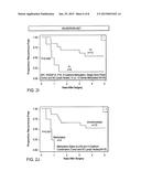 |
 |  |
 |  |
 | 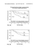 |
 |  |
 | 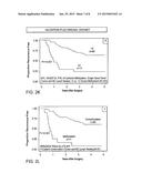 |
 |  |
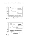 |  |
 |  |
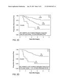 |  |
 |  |
 | 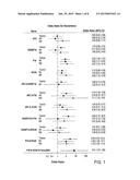 |
 |  |
 |  |
 |  |
 |
| Similar patent applications: | |
| Date | Title |
|---|---|
| 2015-02-26 | Dna controlling mir-140 expression, and screening method of drugs using said dna |
| 2015-02-26 | Apparatus for spreading a fluid across a substrate and method of using the same |
| 2015-02-26 | Compositions, systems, and methods for detecting a dna sequence |
| 2015-02-26 | Cells, nucleic acids, enzymes and use thereof, and methods for the production of sophorolipids |
| 2015-02-26 | Lateral flow devices and methods of manufacture and use |
| New patent applications in this class: | |
| Date | Title |
|---|---|
| 2022-05-05 | Photocleavable mass-tags for multiplexed mass spectrometric imaging of tissues using biomolecular probes |
| 2022-05-05 | Macrophage expression in breast cancer |
| 2022-05-05 | Characterizing methylated dna, rna, and proteins in the detection of lung neoplasia |
| 2022-05-05 | Methods for identifying and improving t cell multipotency |
| 2022-05-05 | Sequence analysis using meta-stable nucleic acid molecules |
| New patent applications from these inventors: | |
| Date | Title |
|---|---|
| 2013-10-31 | Compositions and methods for detecting a neoplasia |
| 2011-06-30 | Methylation markers for sensitivity to microtube based therapies and methods of use |
| 2010-11-25 | Socs-1 gene methylation in cancer |
| 2010-11-04 | Dna methylation markers and methods of use |
| Top Inventors for class "Chemistry: molecular biology and microbiology" | |
| Rank | Inventor's name |
|---|---|
| 1 | Marshall Medoff |
| 2 | Anthony P. Burgard |
| 3 | Mark J. Burk |
| 4 | Robin E. Osterhout |
| 5 | Rangarajan Sampath |