Patent application title: GENES FOR PROGNOSIS OF CANCER
Inventors:
Toshiyuki Saito (Chiba-Shi, JP)
Yoji Mikami (Chiba-Shi, JP)
Masahiro Kinugasa (Nagahama-Shi, JP)
Kazuya Mori (Nagahama-Shi, JP)
Michiyo Sugimoto (Nagahama-Shi, JP)
Koji Uchida (Nagahama-Shi, JP)
Assignees:
MESSENGERSCAPE CO., LTD.
NATIONAL INSTITUTE OF RADIOLOGICAL SCIENCES
Oriental Yeast Co., LTD.
IPC8 Class: AC12Q168FI
USPC Class:
435 611
Class name: Measuring or testing process involving enzymes or micro-organisms; composition or test strip therefore; processes of forming such composition or test strip involving nucleic acid nucleic acid based assay involving a hybridization step with a nucleic acid probe, involving a single nucleotide polymorphism (snp), involving pharmacogenetics, involving genotyping, involving haplotyping, or involving detection of dna methylation gene expression
Publication date: 2012-11-01
Patent application number: 20120276531
Abstract:
To provide a novel method for determining the risk of lymph node
metastasis of breast cancer uses as an index the difference in the
expression levels of marker genes in at least one material selected from
the group consisting of a breast tissue and a breast cell of a patient.
The method includes measuring an expression level of a marker gene in at
least one material selected from the group consisting of a breast tissue
and a breast cell of a patient with breast cancer, and determining the
risk of lymph node metastasis of breast cancer in the patient using the
expression level of the marker gene as an index.Claims:
1. A method for determining a risk of lymph node metastasis of breast
cancer comprising: measuring an expression level of a first marker gene
and a second marker gene in at least one material selected from the group
consisting of a breast tissue and a breast cell of a patient with breast
cancer obtained from a method using at least one selected from the group
consisting of PCR and hybridization, and determining the risk of lymph
node metastasis of breast cancer in the patient using the expression
level of both of the first marker gene and the second marker gene as an
index, wherein the first marker gene is at least one base sequence
selected from the group consisting of SEQ ID Nos. 1 and 2, and the second
marker gene is at least one base sequence selected from the group
consisting of SEQ ID Nos. 3 and 4.Description:
CROSS-REFERENCE TO RELATED APPLICATION
[0001] This is a continuation-in-part of application Ser. No. 12/183,610, filed on Jul. 31, 2008, which is a continuation of Application No. PCT/JP2007/051800, filed on Feb. 2, 2007.
BACKGROUND OF THE INVENTION
[0002] 1. Field of the Invention
[0003] The present invention relates to a novel method for determining the risk of lymph node metastasis of breast cancer. More specifically, the present invention relates to a method for determining the risk of lymph node metastasis of breast cancer that is based on comparison of the expression levels of marker genes having specific base sequences between metastatic breast cancer cells and non-metastatic breast cancer cells.
[0004] 2. Description of the Related Art
[0005] In Japan, the number of breast cancer patients is growing rapidly. The cancer is the most prevalent of all cancers in women. Estrogen, a female hormone, is considered a risk factor of breast cancer: women who have been exposed to estrogen for a longer period of time due to early menarche, late menopause, late age at first birth or nulliparity are more likely to develop breast cancer. Western-style high-fat diet and obesity are also associated with this type of cancer since estrogen is primarily produced in fat tissue in postmenopausal women. The changing lifestyles of Japanese women, such as their active participation in society, also contribute to the increase in the incidence of breast cancer.
[0006] Breast cancer is generally divided into three classes: non-invasive carcinomas, invasive carcinomas and Paget's disease of the breast. Most of the incidences of breast cancer that form lumps are invasive. There are common and special types of invasive breast cancers. The common types include scirrhoma, papillotubular carcinoma and solid-tubular carcinoma. The special types include mucinous carcinoma.
[0007] Because no blood test is available to specifically detect breast cancer, the detection of early breast cancers relies primarily on palpation and X-ray imaging. However, these techniques, even when used in combination, fail to detect as much as 20% of the cancer. In addition, diagnosis by X-ray imaging often requires specialists. The cytodiagnosis conducted before and during the surgical procedures can only be done by a pathologist and is often difficult due to the shortage of experienced pathologists and varying standards of the diagnosis. Thus, no subjective and simple technique for the detection/diagnosis of early breast cancers has ever existed to bridge the gap between detection and diagnosis of the disease. The PET analysis, a new diagnostic technique that can detect tumor tissue 1 mm or less in diameter, requires large-scale facilities and is therefore not readily used for the detection of breast cancer.
[0008] Recent studies have shown that cancers are caused by anomalies in genes. For example, techniques have been proposed that detect cancer cells by making use of the fact that certain genes are expressed at different levels in a cancer tissue and a normal tissue (Japanese Patent Application Laid-Open (JP-A) Nos. 2003-284594 and 2003-284596).
BRIEF SUMMARY OF THE INVENTION
[0009] Once lymph node-metastatic breast cancer has been removed by surgery, prognosis is predicted based on indices such as tumor size, nuclear pleomorphism of the removed cancer cells and of hormone receptor levels. Where necessary, adjuvant therapy is given to prevent metastasis to lymph nodes or the recurrence of cancer. The prediction of prognosis based on these presently available indices is not accurate enough, however, and more accurate indices for the prognosis of breast cancer patients are therefore needed to reduce the risk of recurrence and improve patients' quality of life by proper medication.
[0010] In view of the above-described problems, the present inventors have conducted extensive studies and observed that certain marker genes are expressed at different levels in metastatic breast cancer cells or tissues and in non-metastatic breast cancer cells or tissues. The present inventors found that these marker genes could be used to determine the risk of lymph node metastasis of breast cancer and ultimately devised the present invention. Accordingly, the present invention provides the following measures to address the above-described problems.
[0011] (1) A method for determining the risk of lymph node metastasis of breast cancer, including measuring an expression level of a marker gene in at least one material selected from the group consisting of a breast tissue and a breast cell of a patient with breast cancer, and determining the risk of lymph node metastasis of breast cancer in the patient using the expression level of the marker gene as an index.
[0012] (2) The method according to (1) above, wherein the expression level of the marker gene is determined by the amount of mRNA of the gene.
[0013] (3) The method according to (1) or (2) above, wherein the marker gene is at least one selected from the group consisting of genes having base sequences of GenBank accession Nos. NM000903, NM006804, NM033547, CR611676, NM177967, NM152558, NM178167, NM003752, AK131568, CR592336, NM178507, NM002862, NM006913, NM005794, NM014164, NM000853 and a base sequence extending from 178882962bp to 178883181bp of chromosome 3, and homologs thereof.
[0014] (4) The method according to any one of (1) to (3) above, wherein the expression level of the marker gene in the metastatic breast cancer tissue (or cell) is equal to or higher than twice the expression level in the non-metastatic breast cancer tissue (cells), or equal to or lower than one-half the expression level in the non-metastatic breast cancer tissue.
[0015] The method of the present invention enables quick and simple determination of the risk of lymph node metastasis of breast cancer at the genetic level, thus providing an effective way to prevent metastasis of breast cancer.
BRIEF DESCRIPTION OF THE SEVERAL VIEWS OF THE DRAWINGS
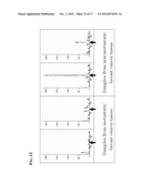
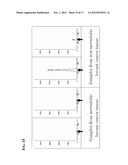
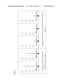
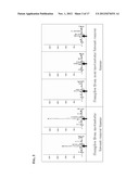
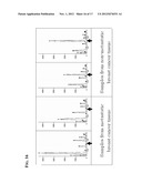
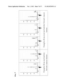
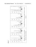
[0016] FIG. 1 is a diagram showing a comparison of the expression levels of a transcript (transcript 1) of a marker gene according to high-coverage gene expression profiling (HiCEP).
[0017] FIG. 2 is a diagram showing a comparison of the expression levels of a transcript (transcript 2) of another marker gene according to HiCEP.
[0018] FIG. 3 is a diagram showing a comparison of the expression levels of a transcript (transcript 3) of another marker gene according to HiCEP.
[0019] FIG. 4 is a diagram showing a comparison of the expression levels of a transcript (transcript 4) of another marker gene according to HiCEP.
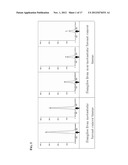
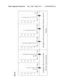
[0020] FIG. 5 is a diagram showing a comparison of the expression levels of a transcript (transcript 5) of another marker gene according to HiCEP.
[0021] FIG. 6 is a diagram showing a comparison of the expression levels of a transcript (transcript 6) of another marker gene according to HiCEP.
[0022] FIG. 7 is a diagram showing a comparison of the expression levels of a transcript (transcript 7) of another marker gene according to HiCEP.
[0023] FIG. 8 is a diagram showing a comparison of the expression levels of a transcript (transcript 8) of another marker gene according to HiCEP.
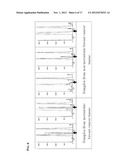
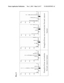
[0024] FIG. 9 is a diagram showing a comparison of the expression levels of a transcript (transcript 9) of another marker gene according to HiCEP.
[0025] FIG. 10 is a diagram showing a comparison of the expression levels of a transcript (transcript 10) of another marker gene according to HiCEP.
[0026] FIG. 11 is a diagram showing a comparison of the expression levels of a transcript (transcript 11) of another marker gene according to HiCEP.
[0027] FIG. 12 is a diagram showing a comparison of the expression levels of a transcript (transcript 12) of another marker gene according to HiCEP.
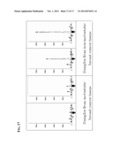
[0028] FIG. 13 is a diagram showing a comparison of the expression levels of a transcript (transcript 13) of another marker gene according to HiCEP.
[0029] FIG. 14 is a diagram showing a comparison of the expression levels of a transcript (transcript 14) of another marker gene according to HiCEP,
[0030] FIG. 15 is a diagram showing a comparison of the expression levels of a transcript (transcript 15) of another marker gene according to HiCEP.
[0031] FIG. 16 is a diagram showing a comparison of the expression levels of a transcript (transcript 16) of another marker gene according to HiCEP.
[0032] FIG. 17 is a diagram showing a comparison of the expression levels of a transcript (transcript 17) of another marker gene according to HiCEP.
DETAILED DESCRIPTION OF THE INVENTION
[0033] The present invention concerns a method for determining the risk of lymph node metastasis of breast cancer that uses as an index of the risk of metastasis the difference in the expression levels of specific marker genes between metastatic breast cancer cells or tissues and non-metastatic breast cancer cells or tissues. As used herein, the term "marker gene" refers to a gene that enables the determination of the risk of metastasis of breast cancer cells by comparing its expression levels between metastatic breast cancer cells or tissues and non-metastatic breast cancer cells or tissues.
[0034] The present invention also concerns a method for determining the risk of lymph node metastasis of breast cancer in which the expression levels of the marker genes are determined by the amounts of mRNA of the marker genes. More specifically, the present invention concerns a method for determining the risk of lymph node metastasis of breast cancer that involves extracting total RNA from cells obtained from metastatic and non-metastatic breast cancer tissues, and comparing the amounts of mRNA transcripts transcribed from the marker genes. Different techniques for gene expression analysis can be used to determine the amounts of mRNA of genes of interest, including PCR and hybridization. As the PCR, quantitive PCR such as qRT-PCR (quantitive RealTime- PCR), comprehensive transcriptome analysis (high-coverage gene expression profiling, HiCEP), or LAMP can be used. As the hybridization, DNA microarrays or northern hybridization can be used. Gene expression analysis techniques that can determine the amounts of mRNA without extracting total RNA from cells, such as in situ hybridization, may also be used in the present invention. The above-described techniques may be used in combination to improve the accuracy of detection. The translated products of the genes of the present invention may also be quantified by, for example, determining the amounts of proteins coded by the genes. For example, a protein detecting method using amino-acid sequence or a partial sequence thereof translated from the genes of the present invention can be used as a method for quantifying proteins translated from the mRNA. Proteins of interest can be quantified by using techniques of protein detecting methods such as immunological assays using antibodies specific for the proteins (such as ELISA, western blotting and RIA), two-dimensional electrophoresis and high-performance liquid chromatography (HPLC). Antibodies specific for the proteins coded by the genes of the present invention can be prepared by common techniques using the proteins coded by the genes as antigens.
[0035] HiCEP is one of the transcriptome analysis techniques and is characterized by its comprehensiveness and high sensitivity. The following is a brief overview of the technique (See Nucleic Acids Res., 2003, Vol. 31, No. 16 e94 for more details): Using common techniques, total RNA is extracted and purified from tissue or cell samples. Double-stranded cDNA is synthesized from the total RNA using 5'-biotinylated oligo(dT) primers. The cDNA is then digested with a restriction enzyme MspI. Poly(A)-containing fragments are collected by avidin beads and 3'-adaptor is ligated to the MspI-digested ends of the collected fragments. The fragments are then digested with a restriction enzyme MseI and 3'-adapter is ligated to the MseI-digested ends. PCR primers are constructed by adding all possible combinations of two selected bases to the same adapter sequences as those ligated to 5' and 3' ends (16 5'-end primers and 16 3'-end primers with 5'-end primers fluorescent-labeled). Using these primers, 256 different quantitative PCRs are performed. The PCR products obtained for each primer pair are loaded on a fragment analyzer to obtain 256 electrophoresis profiles (gene expression profiles), each containing multiple fluorescence peaks, for a sample. The expression levels of transcripts can then be compared by comparing the fluorescence peaks among different samples.
[0036] The marker gene for use in the present invention may be any gene that is expressed at significantly different levels between metastatic breast cancer cells or tissues and non-metastatic breast cancer cells or tissues. For example, the marker gene may be at least one selected from the group consisting of genes having base sequences of GenBank accession Nos. NM000903, NM006804, NM033547, CR611676, NM177967, NM152558, NM178167, NM003752, AK131568, CR592336, NM178507, NM002862, NM006913, NM005794, NM014164 and NM000853 and a base sequence extending from 178882962bp to 178883181bp of chromosome 3, and homologs thereof.
[0037] Data stored in the GenBank database may contain the same gene registered by different researchers, at different times, in different fields and under different names or gene polymorphisms or splicing variants of the same gene registered as novel genes. Thus, different base sequences that can be considered to be originated from a single gene may be registered with different accession numbers. These base sequences are collectively referred to as "homologs." The term is used in the same context throughout this specification.
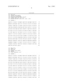
[0038] SEQ ID No. 5 represents an base sequence of GenBank accession No. AK131568. SEQ ID Nos. 1 to 4 represent base sequences corresponding to exon regions for specifically determining AK 131568 from various mRNAs expressed by transcription. SEQ ID No. 1 and SEQ ID No. 3 represent base sequences of two exons which are specific in mRNA of AK131568 respectively indicated by locations in mRNA. SEQ ID No. 2 represents a base sequence corresponding to SEQ ID 1 indicated by location in the genome. SEQ ID No. 4 represents a base sequence corresponding to SEQ ID 3 indicated by location in the genome.
[0039] SEQ ID Nos. 1 and 2 are common in the mRNA of AK131568 and the mRNA of CR592336, whereas SEQ ID Nos. 3 and 4 are specific in the mRNA of AK131568. Therefore, AK131568 can be specifically determined by the sequence of SEQ ID No. 3 or 4. On the other hand, it is known that there exists a large number of unknown mRNAs.
[0040] It is expected that both of SEQ ID No. 3 or 4 and SEQ ID No. 1 or 2, which correspond to internal sequences of same mRNA, have similar expression behavior. Therefore, the expression level of AK131568 can be determined more specifically by confirming the expression level of AK131568 based on both of (i) the expression level of SEQ ID No. 3 or 4 and (ii) the expression level of SEQ ID No. 1 or 2.
[0041] One characteristic feature of the method of the present invention for determining the risk of lymph node metastasis of breast cancer is the use of marker genes that are expressed at significantly different expression levels between metastatic breast cancer cells or tissues and non-metastatic breast cancer cells or tissues. The term "expression level" may refer to either the amount of mRNA transcribed from a marker gene or the amount of a protein translated from mRNA. With regard to the difference in the expression level of a marker gene between metastatic breast cancer cells or tissues and non-metastatic breast cancer cells or tissues, the ratio of the expression level of a marker gene in non-metastatic breast cancer cells or tissues to the expression level of the same gene in metastatic breast cancer cells or tissues is preferably in the range of 1.5 or higher or 2/3 or lower, and more preferably in the range of 2 or higher or 1/2 or lower. A marker gene does not serve as an accurate index of the risk of lymph node metastasis of breast cancer and is therefore not desirable when the ratio of its expression level in non-metastatic breast cancer cells or tissues to that in metastatic breast cancer cells or tissues is outside the above-described range.
[0042] Other aspect of the method of the present invention for determining a risk of lymph node metastasis of breast cancer is a method including, measuring an expression level of a marker gene having a specific base sequence in at least one material selected from the group consisting of a breast tissue and a breast cell of a patient with breast cancer, and determining the risk of lymph node metastasis of breast cancer in the patient based on whether the expression level of the marker gene is higher than or lower than a predetermined threshold value or not.
[0043] Other aspect of the method of the present invention for determining a risk of lymph node metastasis of breast cancer is a method including, measuring an expression level of a marker gene having a specific base sequence in at least one material selected from the group consisting of a breast tissue and a breast cell of a patient with breast cancer, measuring an expression level of an other gene having no change regardless of risk of lymph node metastasis of breast cancer in the material, and determining the risk of lymph node metastasis of breast cancer in the patient based on whether the relative ratio of the expression level of the marker gene to the expression level of the other gene is higher than or lower than a predetermined threshold value or not.
[0044] In case the expression level of AK131568 is determined by the expression level of mRNA, for example, it is preferable to confirm the expression level of AK131568 based on the expression level of SEQ ID No. 3 or 4, and it is more preferable to confirm the expression level of AK131568 based on both of (i) the expression level of SEQ ID No. 3 or 4 and (ii) the expression level of SEQ ID No. 1 or 2.
[0045] As a method for determining the expression level of mRNA, the techniques for gene expression analysis described above can be used.
[0046] In case the expression level of AK131568 is determined by the expression is level of protein, for example, it is preferable to confirm the expression level of AK131568 based on the expression level of amino-acid sequence or a partial sequence thereof translated from SEQ ID No. 3 or 4, and it is more preferable to confirm the expression level of AK131568 based on both of (i) the expression level of amino-acid sequence or a partial sequence thereof translated from SEQ ID No. 3 or 4 and (ii) the expression level of amino-acid sequence or a partial sequence thereof translated from SEQ ID No. 1 or 2.
[0047] As a method for determining the expression level of protein, the techniques of protein detecting methods described above can be used.
[0048] While the method of the present invention can be applied to any type of breast cancer, including breast ductal carcinomas (such as papillotubular carcinoma, solid-tubular carcinoma and scirrhoma), lobular carcinomas, special-type carcinomas (such as mucinous carcinoma, medullary carcinoma and tubular carcinoma) and Paget's disease of the breast, it is preferably applied to scirrhoma, lobular carcinomas or solid-tubular carcinoma.
EXAMPLE
[0049] The present invention will now be described with reference to Example, which is not intended to limit the scope of the invention in any way.
[0050] In this Example, the expression levels (RNA transcription levels) of different genes are compared between human metastatic breast cancer tissue and non-metastatic tissue using one of the gene expression analysis techniques known as high-coverage gene expression profiling technique (HiCEP), a known comprehensive, highly sensitive technique for transcriptome analysis (Nucleic Acids Res., 2003, vol. 31(16), e94).
[0051] The breast cancer tissues used in Example were shown in Table 1. The tissues were collected from five stage II breast cancer patients (commercial products, all Caucasian, primary tumor, lymph node metastasis (2), no lymph node metastasis (3), all had stage II cancer based on TNM classification).
TABLE-US-00001 TABLE 1 Samples Metastasis Tumor size Age #A + 12 cm 57 #B + 2 cm × 1.5 cm × 1.5 cm 69 #C - 2.5 cm 50 #D - 4 cm × 2 cm × 1.7 cm 61 #E - 6 cm × 5.5 cm × 4.5 cm 68
[0052] Total RNA was extracted from the samples by a common kit technique using RNeasy kit (Qiagen). 0.1 μg of total RNA from each sample was used as template and reverse-transcribed using Super Script First Strand Synthesis System for RT-PCR (Invitrogen). The reverse transcript was incubated with DNA polymerase I (80 units), RNAase H (4 units, Invitrogen) and E. coli DNA ligase (40 units, Invitrogen) at 16° C. for 2 hours. The resulting double-stranded DNA was incubated with restriction enzymes Mse I (40 units, New England Biolabs) and Msp I (50 units, TaKaRa Bio) at 37 ° C. for 4 hours. Adaptor sequences were ligated to the ends of the resulting DNA fragments. Selective PCRs were performed using the adaptor-ligated DNA fragments as templates. The amplified products were analyzed by capillary electrophoresis. The waveform data were used to determine gene expression levels, compare the gene expression levels among the samples, and classify the genes into different expression patterns to obtain data for clustering (expression variation peaks).
[0053] The results of the analysis shown in Table 2 and FIGS. 1 through 17 demonstrate that the difference in the fluorescence peak intensity between samples obtained from patients with lymph node metastasis and samples obtained from patients with no metastasis was significant for each of the 17 marker gene transcripts. In this analysis, each sample was assayed in two replicates and the resulting fluorescence peaks were overlapped. Arrows indicate the peaks for the marker gene transcripts.
[0054] Specifically, Transcripts 1 through 11 (as numbered in Table 1) each show a significant fluorescence peak in each of the metastasis samples but show no expression peak or, if any, a peak intensity that is half or less of the peak intensity of the metastasis samples in each of the non-metastasis samples. Conversely, Transcripts 12 through 17 each show a significant fluorescence peak in each of the non-metastasis samples but show no expression peak or, if any, a peak intensity that is half or less of the peak intensity of the non-metastasis samples in each of the metastasis samples. These observations demonstrate that each of the 17 genes can serve as an index of the risk of breast cancer metastasis that allows the determination of the risk of metastasis based on their expression levels.
TABLE-US-00002 TABLE 2 Transcripts as markers for breast cancer metastasis GenBank Transcripts Sequences Accession No. Annotation Characteristics #1 Transcript sequence NM000903 NAD(P)H menadione Experssion containing a base oxidoreductase 1 enhanced in sequence from 68302490bp metastatic to 68317861bp of (-) strand breast cancer of chromosome 16 #2 Transcript sequence N/A N/A containing a base sequence from 178882962bp to 178883181bp of (+) strand #3 Transcript sequence NM006804 steroidogenic acute containing a base regulatory protein sequence from 35050592bp related to 35050643bp of (+) strand of chromosome 17 #4 Transcript sequence NM033547 Homo sapiens containing a base hypothetical gene sequence from 77267542bp MGC16733 similar to to 77272569bp of (-) strand CG12113 (MGC16733), of chromosome 11 mRNA. #5 Transcript sequence CR611676 Similar to Px19-like containing a base protein (25 kDa protein sequence of relevant evolutionary from 176665540bp to and lymphoid interest) 176666255bp of (+) strand (PRELI) (CGI-106) of chromosome 5 (SBBI12) #6 Transcript sequence NM177967 Phosphoglycerate containing a base dehydrogenase like 1 sequence from 98835662bp to 98835862bp of (+) strand of chromosome 13 #7 Transcript sequence NM152558 IQ motif containing E containing a base (IQCE) sequence from 2426581bp to 2426860bp of (+) strand of chromosome 7 #8 Transcript sequence NM178167 Zinc finger protein containing a base 598 sequence from 1987788 bp to 1987865 bp of (-) strand of chromosome 16 #9 Transcript sequence NM003752 Eukaryotic containing a base translation initiation sequence from 228320033 factor 3, subunit 8, bp to 28320077 bp of (-) 110 kDa strand of chromosome 16 #10 Transcript sequence AK131568 V-erb-b2 erythroblastic containing a base leukemia viral oncogene sequence from 35135544 homolog 2, bp to 35135831 bp of (+) neuro/glioblastoma derived strand of chromosome 17 oncogene homolog (avian) #11 Transcript sequence CR592336 V-erb-b2 erythroblastic containing a base leukemia viral oncogene sequence from 35126382 homolog 2, bp to 35127393 bp of (+) neuro/glioblastoma strand of chromosome 17 derived oncogene homolog (avian) #12 Transcript sequence NM178507 NS5ATP13TP2 Expression containing a base protein decreased in sequence from 119605680 metastatic bp to 119605847 bp of (+) breast cancer strand of chromosome 11 #13 Transcript sequence NM002862 Phosphorylase, containing a base glycogen; brain sequence from 25226174 bp to 25226624 bp of (+) strand of chromosome 20 #14 Transcript sequence NM006913 Ring finger protein 5 containing a base sequence from 32256007 bp to 32256297 bp of (+) strand of chromosome 6 #15 Transcript sequence NM005794 Dehydrogenase/reductase containing a base (SDR family) sequence from 23183541 member 2 bp to 23184510 bp of (-) strand of chromosome 14 #16 Transcript sequence NM014164 FXYD domain containing a base containing ion sequence from 40352503 transport regulator 5 bp to 40352595 bp of (+) strand of chromosome 19 #17 Transcript sequence NM000853 Glutathione S- containing a base transferase theta 1 sequence from 22700873 bp to 22700983 bp of (+) strand of chromosome 22
[0055] Genes according to the present invention enable the highly sensitive and subjective, yet simple and quick determination of lymph node metastasis of breast cancer, a task that has never been achieved by any of the conventional techniques. The genes of the present invention therefore serve as markers for the prognosis of breast cancer.
Sequence CWU
1
SEQUENCE LISTING
<160> NUMBER OF SEQ ID NOS: 5
<210> SEQ ID NO 1
<211> LENGTH: 866
<212> TYPE: DNA
<213> ORGANISM: Homo sapiens
<300> PUBLICATION INFORMATION:
<308> DATABASE ACCESSION NUMBER: AK131568
<309> DATABASE ENTRY DATE: 2006-01-20
<313> RELEVANT RESIDUES IN SEQ ID NO: (2006)..(2871)
<400> SEQUENCE: 1
tggcgcctac tcgctgaccc tgcaagggct gggcatcagc tggctggggc tgcgctcact 60
gagggaactg ggcagtggac tggccctcat ccaccataac acccacctct gcttcgtgca 120
cacggtgccc tgggaccagc tctttcggaa cccgcaccaa gctctgctcc acactgccaa 180
ccggccagag gacgagtgtg gtaagacagg gagcccagtg tgcgcactcc ccatctgcca 240
gcacacagca gtgcccaggg ggccctggca gcagcgttct tggacttgtg cagactgccc 300
gtctctgtgc acccttcttg actcagcaca gctctggctg gcttggcctc ttggcatggc 360
ttctctagct gggtcctacc tgccttggca tccttccctc cccctctgtt tctgaaatct 420
cagaactctt cctctcccta catcggcccc acctgtcccc acccctccag cccacagcca 480
tgcccacagc cagttccctg gttcacttgg acctggggcc tcccctaaaa gtcccctgcg 540
gtcccttcct cctcactgca gtgggcgagg gcctggcctg ccaccagctg tgcgcccgag 600
ggcactgctg gggtccaggg cccacccagt gtgtcaactg cagccagttc cttcggggcc 660
aggagtgcgt ggaggaatgc cgagtactgc aggggtatga ggggcggagg agagggtggc 720
tggaggggtg catggggctc ctctcagacc ccctcaccac tgtcccttct ctcaggctcc 780
ccagggagta tgtgaatgcc aggcactgtt tgccgtgcca ccctgagtgt cagccccaga 840
atggctcagt gacctgtttt ggaccg 866
<210> SEQ ID NO 2
<211> LENGTH: 866
<212> TYPE: DNA
<213> ORGANISM: Homo sapiens
<220> FEATURE:
<223> OTHER INFORMATION: chr17
<400> SEQUENCE: 2
tggcgcctac tcgctgaccc tgcaagggct gggcatcagc tggctggggc tgcgctcact 60
gagggaactg ggcagtggac tggccctcat ccaccataac acccacctct gcttcgtgca 120
cacggtgccc tgggaccagc tctttcggaa cccgcaccaa gctctgctcc acactgccaa 180
ccggccagag gacgagtgtg gtaagacagg gagcccagtg tgcgcactcc ccatctgcca 240
gcacacagca gtgcccaggg ggccctggca gcagcgttct tggacttgtg cagactgccc 300
gtctctgtgc acccttcttg actcagcaca gctctggctg gcttggcctc ttggcatggc 360
ttctctagct gggtcctacc tgccttggca tccttccctc cccctctgtt tctgaaatct 420
cagaactctt cctctcccta catcggcccc acctgtcccc acccctccag cccacagcca 480
tgcccacagc cagttccctg gttcacttgg acctggggcc tcccctaaaa gtcccctgcg 540
gtcccttcct cctcactgca gtgggcgagg gcctggcctg ccaccagctg tgcgcccgag 600
ggcactgctg gggtccaggg cccacccagt gtgtcaactg cagccagttc cttcggggcc 660
aggagtgcgt ggaggaatgc cgagtactgc aggggtatga ggggcggagg agagggtggc 720
tggaggggtg catggggctc ctctcagacc ccctcaccac tgtcccttct ctcaggctcc 780
ccagggagta tgtgaatgcc aggcactgtt tgccgtgcca ccctgagtgt cagccccaga 840
atggctcagt gacctgtttt ggaccg 866
<210> SEQ ID NO 3
<211> LENGTH: 1305
<212> TYPE: DNA
<213> ORGANISM: Homo sapiens
<300> PUBLICATION INFORMATION:
<308> DATABASE ACCESSION NUMBER: AK131568
<309> DATABASE ENTRY DATE: 2006-01-20
<313> RELEVANT RESIDUES IN SEQ ID NO: (3628)..(4932)
<400> SEQUENCE: 3
gggatgagct acctggagga tgtgcggctc gtacacaggg acttggccgc tcggaacgtg 60
ctggtcaaga gtcccaacca tgtcaaaatt acagacttcg ggctggctcg gctgctggac 120
attgacgaga cagagtacca tgcagatggg ggcaaggtta ggtgaaggac caaggagcag 180
aggaggctgg gtggagtggt gtctagccca tgggagaact ctgagtggcc acctccccac 240
aacacacagt tggaggactt cctcttctgc cctcccaggt gcccatcaag tggatggcgc 300
tggagtccat tctccgccgg cggttcaccc accagagtga tgtgtggagt tatggtgtgt 360
gatggggggt gttgggaggg gtgggtgagg agccatggct ggagggagga tgagagctgg 420
gatggggaga attacggggc cacctcagca tgtgaaggga gggaaggggc tgcctgtgcc 480
ccaccttgca gggtctgtgc acttcccagg attagggaaa gaccgggtag ggtctgtctc 540
ctggcatcac atctccccct gctacctgcc atgatgctag actcctgagc agaacctctg 600
gctcagtaca ctaaagctcc ctctggccct cccactcctg accctgtctc tgccttaggt 660
gtgactgtgt gggagctgat gacttttggg gccaaacctt acgatgggat cccagcccgg 720
gagatccctg acctgctgga aaagggggag cggctgcccc agccccccat ctgcaccatt 780
gatgtctaca tgatcatggt caaatgtgcg tggctgagct gtgctggctg cctggaggag 840
ggtgggaggt cctgggtgga ggagcccaca aggggcatga aaggggacca ggatgtatgt 900
agacccagga gccctagtat gttaggagcc tcaaaacctt cttgtatccc ttttacagtc 960
aaagtccaaa gccactcttg aggaacactc ttgtacaaaa ttaagctggg cacagtggct 1020
catgcctgta atcccagtac ttttggaggc tgaggtggga ggatcccttg aagccaggag 1080
ttcaagacca gcctgggcaa catagtgaga tcctatctct acaaaaaata aaaaaattat 1140
ctgggtgtgg tggtgtgtgc cagtagtccc agctactcag gagaggctga ggcaggaaga 1200
tcacttgagc ctagtttaag gttgcagtaa gctatgattg caccactgaa atccagcctg 1260
ggtgacagag cgaaacctca tctcaaaaaa ataaaaaagc aaaac 1305
<210> SEQ ID NO 4
<211> LENGTH: 1303
<212> TYPE: DNA
<213> ORGANISM: Homo sapiens
<220> FEATURE:
<223> OTHER INFORMATION: chr17
<400> SEQUENCE: 4
gggatgagct acctggagga tgtgcggctc gtacacaggg acttggccgc tcggaacgtg 60
ctggtcaaga gtcccaacca tgtcaaaatt acagacttcg ggctggctcg gctgctggac 120
attgacgaga cagagtacca tgcagatggg ggcaaggtta ggtgaaggac caaggagcag 180
aggaggctgg gtggagtggt gtctagccca tgggagaact ctgagtggcc acctccccac 240
aacacacagt tggaggactt cctcttctgc cctcccaggt gcccatcaag tggatggcgc 300
tggagtccat tctccgccgg cggttcaccc accagagtga tgtgtggagt tatggtgtgt 360
gatggggggt gttgggaggg gtgggtgagg agccatggct ggagggagga tgagagctgg 420
gatggggaga attacggggc cacctcagca tgtgaaggga gggaaggggc tgcctgtgcc 480
ccaccttgca gggtctgtgc acttcccagg attagggaaa gaccgggtag ggtctgtctc 540
ctggcatcac atctccccct gctacctgcc atgatgctag actcctgagc agaacctctg 600
gctcagtaca ctaaagctcc ctctggccct cccactcctg accctgtctc tgccttaggt 660
gtgactgtgt gggagctgat gacttttggg gccaaacctt acgatgggat cccagcccgg 720
gagatccctg acctgctgga aaagggggag cggctgcccc agccccccat ctgcaccatt 780
gatgtctaca tgatcatggt caaatgtgcg tggctgagct gtgctggctg cctggaggag 840
ggtgggaggt cctgggtgga ggagcccaca aggggcatga aaggggacca ggatgtatgt 900
agacccagga gccctagtat gttaggagcc tcaaaacctt cttgtatccc ttttacagtc 960
aaagtccaaa gccactcttg aggaacactc ttgtacaaaa ttaagctggg cacagtggct 1020
catgcctgta atcccagtac ttttggaggc tgaggtggga ggatcccttg aagccaggag 1080
ttcaagacca gcctgggcaa catagtgaga tcctatctct acaaaaaata aaaaaattat 1140
ctgggtgtgg tggtgtgtgc cagtagtccc agctactcag gagaggctga ggcaggaaga 1200
tcacttgagc ctagtttaag gttgcagtaa gctatgattg caccactgaa atccagcctg 1260
ggtgacagag cgaaacctca tctcaaaaaa ataaaaaagc aaa 1303
<210> SEQ ID NO 5
<211> LENGTH: 4932
<212> TYPE: DNA
<213> ORGANISM: Homo sapiens
<300> PUBLICATION INFORMATION:
<308> DATABASE ACCESSION NUMBER: AK131568
<309> DATABASE ENTRY DATE: 2006-01-20
<400> SEQUENCE: 5
atgatcatgt cccctgtcca cctgctccag ccactatccc tctcccactt acagcagaag 60
aaagggctgg tgagaaaggt ggattacagg cccacttctg ccactgacga gccctatgaa 120
tgtggcctac acccccttag cttcactggg tctcagtttc cctatctgta tattgggagc 180
agttgtgaag ctcagaagag aaatgtctgt gaaaaggtta tgaacaggag ggagagtgga 240
aaccaacctg ctggatcgtg tccacagacc ctggaatggg gccacatgct tggtttgtca 300
aattgcagac gccggccggg tgcgatggct catgcctgta atcccagcac tttgggaggc 360
cgaggcggac agatcacttg aggtcgggag ttcgagacca gcctgaccaa catggagaaa 420
ccccgtctct actgaaaata caaaattagc caggcatggt ggcacatgcc tataatccca 480
gctacttggg aaggctgagg caggagaatc acttgaacct gggagacgga ggttgtggtg 540
agcctagatc gtgccattgt actccagcct gggcaacaag agtgaaactc cgtctcaaaa 600
aaaaaaaatt tgcagacgcc atcccatcca ggcctttgct ttcactgatg aagaaactga 660
gatacagaga gggcagggca cctgttcgga gtttatgaaa tgccccccca ccattatctt 720
tcttgatcat ataagaatct ggtgaggcaa ggtagggcgt gatctttatc tctattttat 780
cgttttattt aagcgggaac aggactgctc agtggctggg ggccttgccc aagatctcca 840
agtactgggg aaccccaggg aggccctggg gggtggcagt gttcctattt cagccccact 900
ctgcttcccc ctcccaggat atccaggagg tgcagggcta cgtgctcatc gctcacaacc 960
aagtgaggca ggtcccactg cagaggctgc ggattgtgcg aggcacccag ctctttgagg 1020
acaactatgc cctggccgtg ctagacaatg gagacccgct gaacaatacc acccctgtca 1080
caggggcctc cccaggaggc ctgcgggagc tgcagcttcg aagcctcaca gagatcttga 1140
aaggaggggt cttgatccag cggaaccccc agctctgcta ccaggacacg attttgtgga 1200
aggacatctt ccacaagaac aaccagctgg ctctcacact gatagacacc aaccgctctc 1260
gggcctgcca cccctgttct ccgatgtgta agggctcccg ctgctgggga gagagttctg 1320
aggattgtca gagcctgacg cgcactgtct gtgccggtgg ctgtgcccgc tgcaaggggc 1380
cactgcccac tgactgctgc catgagcagt gtgctgccgg ctgcacgggc cccaagcact 1440
ctgactgcct ggcctgcctc cacttcaacc acagtggcat ctgtgagctg cactgcccag 1500
ccctggtcac ctacaacaca gacacgtttg agtccatgcc caatcccgag ggccggtata 1560
cattcggcgc cagctgtgtg actgcctgtc cctacaacta cctttctacg gacgtgggat 1620
cctgcaccct cgtctgcccc ctgcacaacc aagaggtgac agcagaggat ggaacacagc 1680
ggtgtgagaa gtgcagcaag ccctgtgccc gagtgtgcta tggtctgggc atggagcact 1740
tgcgagaggt gagggcagtt accagtgcca atatccagga gtttgctggc tgcaagaaga 1800
tctttgggag cctggcattt ctgccggaga gctttgatgg ggacccagcc tccaacactg 1860
ccccgctcca gccagagcag ctccaagtgt ttgagactct ggaagagatc acaggttacc 1920
tatacatctc agcatggccg gacagcctgc ctgacctcag cgtcttccag aacctgcaag 1980
taatccgggg acgaattctg cacaatggcg cctactcgct gaccctgcaa gggctgggca 2040
tcagctggct ggggctgcgc tcactgaggg aactgggcag tggactggcc ctcatccacc 2100
ataacaccca cctctgcttc gtgcacacgg tgccctggga ccagctcttt cggaacccgc 2160
accaagctct gctccacact gccaaccggc cagaggacga gtgtggtaag acagggagcc 2220
cagtgtgcgc actccccatc tgccagcaca cagcagtgcc cagggggccc tggcagcagc 2280
gttcttggac ttgtgcagac tgcccgtctc tgtgcaccct tcttgactca gcacagctct 2340
ggctggcttg gcctcttggc atggcttctc tagctgggtc ctacctgcct tggcatcctt 2400
ccctccccct ctgtttctga aatctcagaa ctcttcctct ccctacatcg gccccacctg 2460
tccccacccc tccagcccac agccatgccc acagccagtt ccctggttca cttggacctg 2520
gggcctcccc taaaagtccc ctgcggtccc ttcctcctca ctgcagtggg cgagggcctg 2580
gcctgccacc agctgtgcgc ccgagggcac tgctggggtc cagggcccac ccagtgtgtc 2640
aactgcagcc agttccttcg gggccaggag tgcgtggagg aatgccgagt actgcagggg 2700
tatgaggggc ggaggagagg gtggctggag gggtgcatgg ggctcctctc agaccccctc 2760
accactgtcc cttctctcag gctccccagg gagtatgtga atgccaggca ctgtttgccg 2820
tgccaccctg agtgtcagcc ccagaatggc tcagtgacct gttttggacc ggaggctgac 2880
cagtgtgtgg cctgtgccca ctataaggac cctcccttct gcgtggcccg ctgccccagc 2940
ggtgtgaaac ctgacctctc ctacatgccc atctggaagt ttccagatga ggagggcgca 3000
tgccagcctt gccccatcaa ctgcacccac tcctgtgtgg acctggatga caagggctgc 3060
cccgccgagc agagagccaa ccctctgacg tccatcgtct ctgcggtggt tggcattctg 3120
ctggtcgtgg tcttgggggt ggtctttggg atcctcatca agcgacggca gcagaagatc 3180
cggaagtaca cgatgcggag actgctgcag gaaacggagc tggtggagcc gctgacacct 3240
agcggagcga tgcccaacca ggcgcagatg cggatcctga aagagacgga gctgaggaag 3300
gtgaaggtgc ttggatctgg cgcttttggc acagtctaca agggcatctg gatccctgat 3360
ggggagaatg tgaaaattcc agtggccatc aaagtgttga gggaaaacac atcccccaaa 3420
gccaacaaag aaatcttaga cgaagcatac gtgatggctg gtgtgggctc cccatatgtc 3480
tcccgccttc tgggcatctg cctgacatcc acggtgcagc tggtgacaca gcttatgccc 3540
tatggctgcc tcttagacca tgtccgggaa aaccgcggac gcctgggctc ccaggacctg 3600
ctgaactggt gtatgcagat tgccaagggg atgagctacc tggaggatgt gcggctcgta 3660
cacagggact tggccgctcg gaacgtgctg gtcaagagtc ccaaccatgt caaaattaca 3720
gacttcgggc tggctcggct gctggacatt gacgagacag agtaccatgc agatgggggc 3780
aaggttaggt gaaggaccaa ggagcagagg aggctgggtg gagtggtgtc tagcccatgg 3840
gagaactctg agtggccacc tccccacaac acacagttgg aggacttcct cttctgccct 3900
cccaggtgcc catcaagtgg atggcgctgg agtccattct ccgccggcgg ttcacccacc 3960
agagtgatgt gtggagttat ggtgtgtgat ggggggtgtt gggaggggtg ggtgaggagc 4020
catggctgga gggaggatga gagctgggat ggggagaatt acggggccac ctcagcatgt 4080
gaagggaggg aaggggctgc ctgtgcccca ccttgcaggg tctgtgcact tcccaggatt 4140
agggaaagac cgggtagggt ctgtctcctg gcatcacatc tccccctgct acctgccatg 4200
atgctagact cctgagcaga acctctggct cagtacacta aagctccctc tggccctccc 4260
actcctgacc ctgtctctgc cttaggtgtg actgtgtggg agctgatgac ttttggggcc 4320
aaaccttacg atgggatccc agcccgggag atccctgacc tgctggaaaa gggggagcgg 4380
ctgccccagc cccccatctg caccattgat gtctacatga tcatggtcaa atgtgcgtgg 4440
ctgagctgtg ctggctgcct ggaggagggt gggaggtcct gggtggagga gcccacaagg 4500
ggcatgaaag gggaccagga tgtatgtaga cccaggagcc ctagtatgtt aggagcctca 4560
aaaccttctt gtatcccttt tacagtcaaa gtccaaagcc actcttgagg aacactcttg 4620
tacaaaatta agctgggcac agtggctcat gcctgtaatc ccagtacttt tggaggctga 4680
ggtgggagga tcccttgaag ccaggagttc aagaccagcc tgggcaacat agtgagatcc 4740
tatctctaca aaaaataaaa aaattatctg ggtgtggtgg tgtgtgccag tagtcccagc 4800
tactcaggag aggctgaggc aggaagatca cttgagccta gtttaaggtt gcagtaagct 4860
atgattgcac cactgaaatc cagcctgggt gacagagcga aacctcatct caaaaaaata 4920
aaaaagcaaa ac 4932
User Contributions:
Comment about this patent or add new information about this topic:
 |  |
 |  |
 |  |
 |  |
 |  |
 |  |
 |  |
 |  |
 |  |
 |  |
 |  |
 |  |
 |
| Similar patent applications: | |
| Date | Title |
|---|---|
| 2009-01-08 | Genes for prognosis of cancer |
| 2011-08-25 | Biomarkers for diagnosis of breast cancer |
| 2011-10-13 | Tissue container for molecular and histology diagnostics incorporating a breakable membrane |
| 2009-08-13 | Kit for diagnosis of cancer |
| 2011-08-11 | Systems and methods for diagnosing cancer |
| New patent applications in this class: | |
| Date | Title |
|---|---|
| 2022-05-05 | Photocleavable mass-tags for multiplexed mass spectrometric imaging of tissues using biomolecular probes |
| 2022-05-05 | Macrophage expression in breast cancer |
| 2022-05-05 | Characterizing methylated dna, rna, and proteins in the detection of lung neoplasia |
| 2022-05-05 | Methods for identifying and improving t cell multipotency |
| 2022-05-05 | Sequence analysis using meta-stable nucleic acid molecules |
| New patent applications from these inventors: | |
| Date | Title |
|---|---|
| 2009-11-05 | Method for detecting mammary cancer cells |
| Top Inventors for class "Chemistry: molecular biology and microbiology" | |
| Rank | Inventor's name |
|---|---|
| 1 | Marshall Medoff |
| 2 | Anthony P. Burgard |
| 3 | Mark J. Burk |
| 4 | Robin E. Osterhout |
| 5 | Rangarajan Sampath |