Patent application title: PEPTIDES AND METHODS FOR THE DETECTION OF LEISHMANIASIS
Inventors:
Jorge Arevalo (Lima, PE)
Eric Deharo (Auterive, FR)
Angela Privat-Maldonado (Lima, PE)
Assignees:
INSTITUT DE RECHERCHE POUR LE DEVELOPPMENT
UNIVERSIDAD PERUANA CAYETANO HEREDIA
IPC8 Class: AG01N33569FI
USPC Class:
435 794
Class name: Assay in which an enzyme present is a label heterogeneous or solid phase assay system (e.g., elisa, etc.) sandwich assay
Publication date: 2016-05-26
Patent application number: 20160146809
Abstract:
The present invention relates to peptides and methods for the detection
of anti-leishmanial antibodies in individuals suspected of infection with
the protozoan parasite of the genus Leishmania, especially infection with
a South American strain causing the American Tegumentary Leishmaniasis
(ATL).Claims:
1. In vitro diagnostic method for the detection of the presence or
absence of antibodies indicative of a South American Leishmania strain
responsible for the American Tegumentary Leishmaniasis, which bind to a
peptide selected in the group consisting of H2A-P9 (SEQ. ID. No 9) and
P2a-P6 (SEQ. ID. No 67) to form an immune complex, comprising the steps
of: a) contacting said peptides H2A-P9 (SEQ. ID. No 9) and P2a-P6 (SEQ.
ID. No 67) with a biological sample for a time and under conditions
sufficient to form an immune complex; and b) detecting the presence or
absence of the immune complex formed in a).
2. In vitro diagnostic method according to claim 1, wherein said South American Leishmania strain is selected in the group consisting of L. braziliensis, L. mexicana, L. major, L. amazonensis or L. infantum.
3. In vitro diagnostic method according to claim 1, wherein said method comprises two additional steps c) and d) consisting in: c) contacting said biological sample with the peptide H2A-P9 of sequence KGGKKGKATPSA (SEQ. ID. No 9) and d) detecting the presence or absence of the immune complex formed in c).
4. Diagnostic kit comprising two peptides of SEQ. ID. No 9 and of SEQ. ID. No 67.
5. Diagnostic kit according to claim 4 comprising: said two peptides of SEQ. ID. No 9 and of SEQ. ID. No 67; reagent(s) to detect polypeptide-antibody immune complex; optionally a biological reference sample lacking antibodies that immunologically bind with said peptides; and optionally a comparison sample comprising antibodies which can specifically bind to said peptides; wherein said peptides, reagent(s), biological reference sample, and comparison sample are present in an amount sufficient to perform said detection.
6. Diagnostic kit according to claim 5 consisting of ELISA kit; said ELISA kit comprising the H2A-P9 (SEQ. ID. No 9) and P2a-P6 (SEQ. ID. No 67) pre-coated ELISA plates, positive control, negative control, acceptable diluents, enzyme conjugated anti-human IgG, substrate chromogen, substrate buffer and an instruction manual to use the kit.
Description:
[0001] The present invention relates to peptides and methods for the
detection of anti-leishmanial antibodies in individuals suspected of
infection with the protozoan parasite of the genus Leishmania, especially
infection with a South American strain causing the American Tegumentary
Leishmaniasis (ATL).
[0002] Leishmaniasis comprises a heterogeneous group of diseases caused by intracellular protozoan parasites of the genus Leishmania that are widespread in 88 endemic countries, with an estimated annual incidence of 2 million cases [1].
[0003] Leishmania organisms are intracellular protozoan parasites of macrophages that cause a wide range of clinical diseases in humans and domestic animals, primarily dogs. The life cycles of Leishmania involve a vertebrate host (e.g., a human) and a vector (a sand fly) that transmits the parasite between vertebrate hosts. In the vector, the parasite takes on a characteristic morphological form known as the promastigote, and reproduces asexually in the vector's gut. When the vector bites a vertebrate host, promastigotes are injected into the host. The promastigotes then enter cells of the vertebrate host and change into a form known as the amastigote. The amastigote reproduces in the host's cells and, when the cells eventually die, the amastigotes are released and infect other cells. The symptoms and pathology associated with leishmaniasis result from the amastigotes killing the host's cells.
[0004] In South America, the most common form of the disease is the cutaneous leishmaniasis (CL), while mucosal Leishmaniasis (ML) and visceral Leishmaniasis (VL) are much less prevalent [2]. The CL and ML clinical manifestations although not fatal, can cause significant morbidity, social consequences and psychological traumas in affected people due to stigmatizing scars. VL, the most severe form of the disease, is less prevalent in the region [3].
[0005] The diagnostic of the disease is carried out by a combination of clinical, epidemiological, parasitological and immunological tests. Giemsa staining of biopsy smears is the most common method employed in rural endemic areas with a sensitivity ranging from 20% to 95% [4-6]. Parasite culture in NNN medium can achieve 54% of sensitivity, but bacterial and fungal contamination is frequent, affecting the success of the culture [6-9]. Furthermore, the sensitivity will vary according to the parasite load, sample collection and technical expertise.
[0006] Molecular diagnosis such as PCR, while more specific and sensitive, is also more expensive and requires more expertise than conventional procedures [4, 5, 10].
[0007] Serological diagnostic method for CL has been pursued in the past using whole parasite extracts; unfortunately, differences in parasite strains, antigen preparation and antigenic characteristics make them inappropriate for the development of standardized serodiagnostic methods [11-15].
[0008] Other studies report the use of recombinant proteins due to the variety of antigenic determinants they contain [16-20]; nevertheless, they might include crossreactive epitopes, which in turn compromise the test specificity [21].
[0009] A new improvement for the serodiagnosis of several diseases is the use of synthetic peptides, which might boost the sensitivity and specificity of the ELISA test [22, 23].
[0010] Histones and acidic ribosomal proteins (ARP) from L. infantum have been previously identified as antigenic proteins by canine VL sera [24] and their linear B cell epitopes were mapped on the most divergent regions of the proteins [18, 24-28]. Nevertheless, these experiments were carried out only with canine samples and only one study with a limited number of human ML sera was done with P2a and P2b [29]. Only one study has been performed with human CL and MCL serum samples, using synthetic peptides to explore the amino terminal region of the L. braziliensis ribosomal protein L25 with unsuccessful results [30], the existence of candidate diagnostic epitopes for serodiagnosis of human CL is still unknown.
[0011] There thus remains a need in the art for a rapid and effective diagnostic test for cutaneous leishmaniasis that may be readily employed in a field situation.
[0012] Inventors have conducted a large study of the linear epitopes of histones and acidic ribosomal proteins from L. infantum, a total of 75 synthetic peptides were screened, and they identified short peptides that are recognized by CL sera.
[0013] Subsequently, these immunodominant peptides were assessed on their diagnosis efficacy with a panel of CL sera; based on their advanced clinical knowledge of the American Tegumentary Leishmaniasis, Inventors have defined two levels of criteria to be applied for selecting the most appropriate peptide(s) for the diagnosis of CL (see examples), they then selected two specific peptides, H2A-P9 and P2a-P6, and designed an improved diagnostic test that allows a very accurate and sensitive diagnostic of South American Leishmania strains causing American Tegumentary Leishmaniasis.
[0014] Inventors have demonstrated that an indirect ELISA test using the peptides H2A-P9 and P2a-P6 can discriminate positive CL from negative sera for leishmaniasis. The obtained diagnostic parameters of this test demonstrate good effectiveness when taken to the clinical settings in endemic areas (see examples).
[0015] The present invention is thus directed to the selection and the combination of two specific immunodominant peptides from L. infantum: H2A-P9 of sequence KGGKKGKATPSA (SEQ. ID. No 9) and P2a-P6 of sequence AGAGAGAVAEAKKEEPEEEE (SEQ. ID. No 67) for use in a method of diagnostic.
[0016] Consequently, the present invention provides a method of using those two peptides for the detection of anti-leishmanial antibodies present in the serum of patient suffering from cutaneous Leishmaniasis or muco-cutaneous Leishmaniasis provoked by infestation of South American Leishmaniasis strain. South American Leishmania strains comprise strains from the Viannia group (L. (Viannia) braziliensis, L. (Viannia) panamensis, L. (Viannia) peruviana, L. (Viannia) guyanensis, L. (Viannia) lainsoni, L. (Viannia) colombiensis) and strains from the Leishmania group (L. (Leishmania) amazonensis, L. (Leishmania) garnhami, L (Leishmania) infantum, L. (Leishmania) mexicana, L. (Leishmania) pifanoi, L. (Leishmania) venezuelensis); the present is preferably directed to the diagnosis of Leishmaniasis provoked by infestation with L. braziliensis, L. mexicana, L. major, L. amazonensis or L. infantum; these disorders are also called American Tegumentary Leishmaniasis.
[0017] The present invention further provides a diagnostic kit comprising the two peptides of SEQ. ID. No 9 and of SEQ. ID. No 67, said kit being useful for detecting anti-leishmanial antibodies present in the serum of patient suffering from cutaneous Leishmaniasis or muco-cutaneous Leishmaniasis provoked by infestation of South American Leishmania strain, more particularly L. braziliensis, L. mexicana, L. major, L. amazonensis or L. infantum; or American Tegumentary Leishmaniasis.
[0018] The diagnostic parameters of the method according to the present invention when applied to the serodiagnosis of CL have demonstrated to be comparable to those obtained for PCR in terms of sensitivity (S) and positive predictive value (PPV) [4]. The sensitivity of this technique is comparable to PCR (97%) and far better than microscopy smears observation (49%) or conventional parasite culture (58%) for CL diagnosis [43-45]. In comparison with serological methods like ELISA, IFAT and Western Blot using crude antigens from L. braziliensis, L. major or L. amazonensis, the method here described renders comparable or better diagnostic results [11, 12].
[0019] In the specific case of CL, accuracy negative predictive value (NPV) appears to be particularly important because an accurate negative diagnosis prevents the administration of pentavalent antimonials for 20 days, which is painful and potentially toxic to individuals with skin ulcers resembling leishmaniasis lesions [42].
[0020] Furthermore, a correct differential diagnosis will contribute to reduce disease burden of patients who must travel long distances to be treated.
[0021] Finally, compared with the classic diagnostic methods employed in rural areas like Leishmanin Skin Test, smears and traditional parasite culture in NNN medium, the present method demonstrated an improved performance, rendering high NPV and S values [48].
[0022] Considering the ease to manufacture synthetic peptides compared to recombinant proteins or isolated crude extract proteins, the present invention allows the development of new serological diagnostic reagents at low costs and amenable to be subjected to quality control assessment.
[0023] The present invention also allows the design of laboratory diagnostic tools that can be applied in primary health settings that use ELISA diagnosis for routine diagnosis.
[0024] A first object of the present invention is an in vitro diagnostic method for the detection of the presence or absence of antibodies indicative of a South American Leishmania strain responsible for the American Tegumentary Leishmaniasis, which bind to a peptide selected in the group consisting of H2A-P9 (SEQ. ID. No 9) and P2a-P6 (SEQ. ID. No 67) to form an immune complex, comprising the steps of:
a) contacting said peptides H2A-P9 (SEQ. ID. No 9) and P2a-P6 (SEQ. ID. No 67) with a biological sample for a time and under conditions sufficient to form an immune complex; and b) detecting the presence or absence of the immune complex formed in a).
[0025] The sequences of the peptides used in the diagnostic method according to the present invention are:
TABLE-US-00001 H2A-P9: (SEQ. ID. No9) KGGKKGKATPSA and P2a-P6: (SEQ. ID. No67) AGAGAGAVAEAKKEEPEEEE.
[0026] These peptides have been identified from the strain L. infantum with the method described in the examples. They may be prepared and isolated by well known techniques, such as solid phase synthesis [41].
[0027] In one embodiment of the present invention, the in vitro diagnostic method makes use of isolated synthetic peptides of sequence SEQ. ID. No 9 and SEQ. ID. No 67.
[0028] These peptides may be each in separate containers or mixed in the same container; optionally they are immobilized on an appropriate support like the lateral flow format. In this device, the diagnostic peptide is immobilized on membrane, such as a Polyvinylidene fluoride or a nitrocellulose membrane.
[0029] For detection purpose, anti human IgG labelled with a signal generator (substrate chromogen or colloidal gold) is deposited on a glass fiber strip (sample application pad); when a solution of serum to be tested is applied on the pad, it dissolves the labelled reporter and this binds to all antibodies in the sample. This mixture is then transported by capillarity (chromatography principle) into the next membrane containing the diagnostic peptide.
[0030] If antibodies against the diagnostic peptide are present, they bind to the diagnostic peptide striped on the membrane generating a signal. An additional antibody specific to the colloidal labelled antibody is used to produce a control signal.
[0031] It should be implicit by one of expert in the art, that any conventional protein assay formats, particularly immunoassay formats, may be designed to use the selected peptides herein for the detection of Leishmania infection. This invention is thus not limited by the selection of the particular assay format, and is believed to encompass assay formats that are known to those of skill in the art.
[0032] Reagents for ELISA or other immunodetection assays can be provided in the form of kits. In one embodiment, a kit contains a mixture of suitable peptides or means for preparing such mixtures, and/or reagents for detecting peptide-antibody complexes. A "biological sample" encompasses a variety of sample types obtained from an individual and can be used in a diagnostic or monitoring assay. The definition encompasses blood and other liquid samples of biological origin, solid tissue samples such as a biopsy specimen or tissue cultures or cells derived therefrom. The term "biological sample" encompasses a clinical sample, and also includes cells in culture, cell supernatants, cell lysates, serum, plasma, biological fluid, and tissue samples.
[0033] The most common technique for conducting step b) is the Enzyme-linked immunosorbent assay (ELISA). It involves at least one antibody with specificity for a particular antigen, in our case the peptides immobilized on a solid support (usually a polystyrene microtiter plate or a dip-stick) via adsorption to the surface or more specifically (via capture by another antibody specific to the same antigen, in a "sandwich" ELISA). The detection antibody (the sample) is added, forming a complex with the peptide. The detection antibody can be covalently linked to an enzyme, or can itself be detected by a secondary antibody that is linked to an enzyme through bioconjugation. Between each step, the plate is typically washed with a mild detergent solution to remove any proteins or antibodies that are not specifically bound. After the final wash step, the plate is developed by adding an enzymatic substrate to produce a visible signal, which indicates the quantity of antigen in the sample.
[0034] According to a particular embodiment, if the result of the in vitro diagnostic method is uncertain, additional steps may be added to test again the serum with peptide H2A-P9 to increase accuracy and sensitivity, said additional steps comprise:
c) contacting said biological sample with the peptide H2A-P9 of sequence KGGKKGKATPSA (SEQ. ID. No 9) and d) detecting the presence or absence of the immune complex formed in c).
[0035] The present invention also provides a diagnostic kit for detecting anti-leishmanial antibodies present in the serum of patient suffering from American Tegumentary Leishmaniasis wherein said diagnostic kit comprises the two peptides of SEQ. ID. No 9 and of SEQ. ID. No 67.
[0036] The kit can include microtiter plates to which the peptide(s) of the invention have been pre-adsorbed, another appropriate assay device, various diluents and buffers, labelled conjugates or other agents for the detection of specifically bound antigens or antibodies, and other signal-generating reagents, such as enzyme substrates, cofactors and chromogens. Other components of a kit can easily be determined by one of skill in the art. Such components may include coating reagents, polyclonal or monoclonal capture antibodies specific for a peptide of the invention, or a cocktail of two or more of the antibodies, purified or semi-purified extracts of these antigens as standards, MAb detector antibodies, an anti-mouse or anti-human antibody with indicator molecule conjugated thereto, an ELISA plate prepared for absorption, indicator charts for colorimetric comparisons, disposable gloves, decontamination instructions, applicator sticks or containers, a sample preparatory cup, etc. In one embodiment, a kit comprises buffers or other reagents appropriate for constituting a reaction medium allowing the formation of a peptide-antibody complex.
[0037] The diagnostic kit preferably comprises: the two peptides of SEQ. ID. No 9 and of SEQ. ID. No 67; reagent(s) to detect polypeptide-antibody immune complex; optionally a biological reference sample lacking antibodies that immunologically bind with said peptides; and optionally a comparison sample comprising antibodies which can specifically bind to said peptides; wherein said peptides, reagent(s), biological reference sample, and comparison sample are present in an amount sufficient to perform said detection.
[0038] In a particular embodiment, the diagnostic kit consists of ELISA kit; preferably, the ELISA kit comprises the H2A-P9 (SEQ. ID. No 9) and P2a-P6 (SEQ. ID. No 67) pre-coated ELISA plates, positive control, negative control, acceptable diluents, enzyme conjugated anti-human IgG, for example, Anti-Human IgG (H&L) in goat conjugate (peroxydase, phosphatase) (see also http://www.polysciences.com/Core/Display.aspx?pageId=98&categoryId=158&pr- oductId=1518), substrate chromogen (examples for peroxidise: 3,3-dimethoxybenzidine, 0-dianisidine (ODN) turns reddish; 3,3-Diaminobenzidine turns reddish; 3-amino-9-ethyl carbazole turns reddish; 4-Chloro-1-naphtol turns bluish; and for alkaline phosphatise: 5-bromo-4-chloro-3-indolyl phosphate (BCIP) turns bluish or nitro blue tetrazolium (NBT) turns bluish), substrate buffer and an instruction manual to use the kit.
[0039] The diagnostic kit may be in the form of a Dipstick which may be dipped in several wells each comprising biological sample, and reagents.
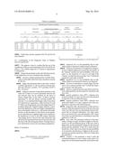
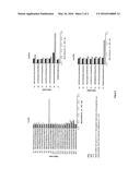
[0040] FIG. 1 represents four graphs that show the reactivity of the CL (open bars) and MCL (solid bars) sera against the synthetic peptides, 20-mer overlapping by five residues (H2A) and ten residues (H2B, H3, H4), covering the entire sequence of the proteins. The OD450 mean values of a pool of two to six sera against each one of the peptides are represented. Sera were used at a dilution of 1:150.
[0041] FIG. 2 represent four graphs that show the reactivity of the CL (open bars) and MCL (solid bars) sera against the synthetic peptides, 20-mer overlapping by five residues, covering the entire sequence of LiP2a, LiP2b and P0. The OD450 mean values of a pool of two to six sera against each one of the peptides are represented. Sera were used at a dilution of 1:150.
[0042] The present invention will be more readily understood by referring to the following example. This example is illustrative of the wide range of applicability of the present invention and is not intended to limit its scope. Modifications and variations can be made therein without departing from the spirit and scope of the invention. Although any method and material similar or equivalent to those described herein can be used in the practice for testing of the present invention, the preferred methods and materials are described.
EXAMPLE 1
Identification of Immunodominant Peptides from L. infantum Histones and ARP
I.A. Methods
I.A.1. Human Sera
[0043] The serum samples used were divided into three groups:
Group I: Sera from twenty ML and twenty one CL patients were obtained from the bank of sera of the Microbiology Laboratory, Faculty of Biology, San Antonio Abad University of Cuzco, Peru. All samples were reactive to at least one conventional laboratory test for ATL, such as parasite culture, IFA or microscopy. This group was tested against recombinant histones and ARP and two to five hyper reactive sera from each clinical manifestation were pooled for epitope mapping. Group II: Sera from fifteen CL, ten negative endemic controls (NEC) from Cusco, Peru were obtained from the bank of sera of FP7 RAPSODI Project. Samples from patients with Chagas (7) and Sporotrichosis (6) from Lima, Peru were also included. These samples were individually tested against the selected ten synthetic peptides. Group III: Eighteen CL and thirteen NEC sera from the bank of sera of the FP7 RAPSODI Project were individually assayed with the two diagnostic candidate epitopes.
I.A.2. Epitope Mapping
[0044] For the epitope mapping, a library of overlapping peptides covering the whole sequence of the L. infantum LiH2A, LiH2B, LiH3, LiH4, LiP0, LiP2a and LiP2b was employed The screening was carried out with a pool of sera obtained from ATL patients.
[0045] Peptides were synthesized by the simultaneous multiple-peptide solid-phase synthetic method using a polyamide resin and FMOC chemistry [41]. Purity was checked by amino acid analysis and HPLC. For LiH2A [26], LiH3 [27], LiP2a and LiP2b [25], and LiP0 [28] peptides overlapped by 5 amino acids. For LiH2B and LiH4, peptides overlapped by 10 amino acids [24]. A total of 75 peptides were assayed, all peptides were 20-mer long, except H2A-P9 (12-mer), P2a-P7 (16-mer), H3-P12 (19-mer) and H2B-P10 and P2b-P7 (21-mer).
I.A.3. ELISA Measurements
[0046] To select both the most reactive sera against recombinant antigens and to carry out the epitope mapping, sera from Group I were tested using the Falcon assay screening test-enzyme-linked immunosorbent assay (FAST-ELISA; Becton Dickinson) [27].
[0047] The antigen concentration for recombinant proteins was 2 μg/ml and 100 μg/ml for synthetic peptides. Antigen-coated lids were incubated for 1 h with the blocking solution and pools of the most reactive two to six serum samples for each recombinant protein were used for the epitope mapping.
[0048] Serum samples were diluted 1:200 for recombinant proteins and 1:150 for synthetic peptides in blocking solution and incubated for 2 h at room temperature with shaking. As secondary antibody, horseradish peroxidase-labelled anti-IgG antibodies (dilution 1:2000, Nordic Immunology) were used. After 1 h of incubation at room temperature, lids were washed and the immune complexes were revealed with orthophenylenediamine as the chromogenic substrate. Absorbance was read at 450 nm.
I.B. Results--B-Cell Epitope Mapping Using Synthetic Peptides
[0049] To select the most reactive serum samples from a panel of CL and ML sera corresponding to Group I, recombinant proteins were individually tested (data not shown). It was possible to combine at least 3 sera for the epitope mapping of H2A, H2B, H4, P0 and P2b. For P2a and H3 only two sera were pooled for ML and CL respectively. A collection of synthetic peptides spanning the whole protein sequences, were tested by FAST-ELISA, with exception of LiP0 protein, where positions between 170 and 180 were not available.
[0050] Peptides located at the N-terminal region of histones (H2A-P1, H2B-P1, H3-P2, H3-P3, H4-P1), at the C-terminal region of LiH2A (H2A-P9), LiP2a (P2a-P6) and LiP2b (P2b-P7) and at the middle of LiH4 (H4-P7) and LiP0 (P0-P8) demonstrated to be immunodominant (FIGS. 1 to 7). A total of ten linear epitopes were selected like peptide candidates for the determination of their diagnostic value: H2A-P1 (SEQ. ID. No 1), H2A-P9 (SEQ. ID. No 9), H2B-P1 (SEQ. ID. No 10), H3-P2 (SEQ. ID. No 21), H3-P3 (SEQ. ID. No 22), H4-P1 (SEQ. ID. No 32), H4-P7 (SEQ. ID. No 38), P0-P8 (SEQ. ID. No 48), P2a-P6 (SEQ. ID. No 67) and P2b-P7 (SEQ. ID. No 75).
EXAMPLE 2
Assessment of the Diagnostic Value of Peptide Candidates: Individual Assay of Peptide Candidates
[0051] The ten selected peptides were tested with serum samples from Group II to confirm their highly antigenic nature; their potential interest for diagnostic purpose has been evaluated using a first set of diagnostic parameters defined by Inventors. Considering that the diagnostic test needs to be sensitive and with a minimal rate of false positive because false positive will lead to the application of unnecessary treatment which is costly, risky and painful, peptides recognized by more than 70% of CL sera and with less than 15% of cross-reactivity were arbitrarily considered as potential diagnostic candidates.
II.A. Method
[0052] Ten immunodominant peptides, selected after the epitope mapping were provided by Bio-Synthesis, Inc. (612 East Main Street Lewisville, Tex. 75057, USA). Peptides were further analyzed using conventional ELISA following the conditions described above and sera from Group II. After the selection of the two diagnostic candidate peptides, they were analyzed using serum samples from Group III. All samples were processed in duplicates.
[0053] Cut-off values were defined by the area under the receiver-operating characteristic curve.
Statistical Analysis
[0054] The results obtained for each serum sample tested were used to construct 2×2 contingency tables where the sera were further classified according to the disease's presence or absence, as positive or negative.
II.B. Results
[0055] Results are presented in Table 1 below.
TABLE-US-00002 TABLE I individual assay of peptide candidates Others All no NEGATIVE OTHER PATHOLOGIES pathologies Leishmaniasis CL CONTROLS Chagas Sporotrichosis combined sera Number of samples tested 15 10 7 6 13 23 N. N. N. N. N. N. % positives % positives % positives % positives % positives % positives H2A-P1 60.00 9 10.00 1 42.86 3 66.67 4 53.85 7 34.78 8 H2A-P9 73.33 11 0.00 0 0.00 0 16.87 1 7.69 1 4.35 1 H2B-P1 93.33 14 20.00 2 14.29 1 33.33 2 23.08 3 21.74 5 H3-P2 80.00 12 10.00 1 28.57 2 16.67 1 23.08 3 17.39 4 H3-P3 46.67 7 30.00 3 28.57 2 33.33 2 30.77 4 30.43 7 H4-P1 66.67 10 20.00 2 28.57 2 66.67 4 46.15 6 34.78 8 H4-P7 60.00 9 30.00 3 0.00 0 16.67 1 7.69 1 17.39 4 P0-P8 66.67 10 10.00 1 0.00 0 0.00 0 0.00 0 4.35 1 P2a-P6 80.00 12 0.00 0 14.29 1 0.00 0 7.69 1 4.35 1 P2b-P7 86.67 13 50.00 5 71.43 5 0.00 0 38.46 5 43.48 10
[0056] Under these criteria, peptides H2A-P9 and P2a-P6 were selected.
II.C. Confirmation of the Diagnostic Value of Peptides H2A-P9 and P2a-P6
[0057] The purpose is here to confirm that the use of the combination of the two selected peptides H2A-P9 and P2a-P6 allows a very reliable diagnostic of American Tegumentary Leishmaniasis.
[0058] Diagnostic parameters used to describe the accuracy of the diagnostic test were calculated; they included:
[0059] accuracy: number and proportion of all the observations in the table which have been classified correctly by the test;
[0060] kappa: this parameter takes on the value 1 if there is perfect agreement; i.e. the test always correctly predicts the outcome (1 perfect, >0.75 excellent, 0.4-0.75 fair, <0.4 poor).
[0061] Kappa is a measure of agreement and takes on the value zero if there is no more agreement between test and outcome then can be expected on the basis of chance. Kappa takes on the value 1 if there is perfect agreement; i.e. the test always correctly predicts the outcome. It is considered that Kappa values lower than 0.4 represent poor agreement, values between 0.4 and 0.75 fair to good agreement, and values higher than 0.75 excellent agreement. Negative Kappa indicates a problem in the application of the test. Kappa is dependent not only on the quality of the test, i.e., the inside of the table, but also on the prevalence of the disease in the population in which the test is applied, kappa is also sensitive to the distribution of cases in the table margin. Basically what Kappa shows is that for the same sensitivity and specificity the agreement between test and outcome will decrease with a decreasing prevalence. In Kappa terms a test will perform worse in low prevalence populations.
Mode of Calculation:
[0062] Kappa = ( Observed agreement - expected agreement ) 1 - expected agreement ##EQU00001## ( see http : // epiville . ccnmtl . columbia . edu / popup / how_to _calculate _kappa . html _ ) ##EQU00001.2##
[0063] sensitivity (S): it is the probability that an individual which is diseased is indeed tested as diseased.
[0064] The level of sensitivity to be applied in a diagnostic test is defined taking into account the seriousness of the "disease" and the cost and availability of the treatment; i.e. the sensitivity of a test is to be high if the "disease" is relatively serious and the "cure" is relatively inexpensive and easily available.
[0065] specificity (E): it is the probability that an individual which is not diseased is tested as not diseased.
[0066] The level of specificity is usually high if the disease is not so serious and the "cure" is relatively expensive in money and other terms.
[0067] There is a tradeoff between specificity and sensitivity, high specificity mostly means low sensitivity, and vice versa.
[0068] positive predictive value (PPV): indicates how much more likely it is to get a positive test in the diseased as opposed to the non-diseased group.
[0069] negative predictive value (NPV): indicates how much more likely it is to get a negative test in the non-diseased as opposed to the diseased group.
[0070] diagnostic odds: this parameter is often used as a measure of the discriminative power of the test; it has the value one if the test does not discriminate between diseased and not diseased individuals. Very high values above one means that a test discriminates well. Values lower than one mean that there is something wrong in the application of the test.
[0071] error odds: this parameter indicates if the probability of being wrongly classified is highest in the diseased or in the non-diseased group. If the error odds is higher than one the probability is highest in the diseased group (and the specificity of the test is better than the sensitivity), if the value is lower than one the probability of an incorrect classification is highest in the non-diseased group (and the sensitivity of the test is better than the specificity).
[0072] Youden's J: this parameter is used to study the overall performance of a test; it takes on the value 1 if a diagnostic test discriminates perfectly and without making any mistakes. As a consequence, if the purpose is to minimize the probability of making an error, the Youden's J has to be maximized; that, of course, is of only theoretical importance, such decisions should be taken on the basis of a cost-benefit analysis of treatment as opposed to no treatment.
[0073] positive predictive accuracy: in a table representative of the population, this parameter gives: 1) the post-test probability, the probability for an individual in the population who tested positive of having the disease; 2) of those who tested positive the fractions who were correctly and who were not-correctly classified.
[0074] negative predictive accuracy: In a table representative of the population this parameter gives: 1) the post-test probability, the probability for an individual in the population who tested negative of not having the disease; 2) of those who tested negative the fractions who were correctly and who were not-correctly classified.
[0075] Based on the extended clinical experience of the American Tegumentary Leishmaniasis of the Inventors, they have selected the parameters, sensitivity and specificity and the two predictive accuracies (accuracy and kappa), that are considered as the most valuable of the indicators.
[0076] The threshold value for each peptide is 3 standard deviations of the value obtained when the peptides were tested with true negative control sera.
[0077] Sensitivity and specificity give a good view of the quality of the test relatively independent of circumstances. The predictive accuracies give a view of what happens in different practical situations in terms of numbers and proportions tested with correct and incorrect results. Predictive accuracies also give the post test probability of having the disease, an essential piece of information to communicate to the patient together with his or her test result.
Results
[0078] To determine the above-mentioned diagnostic parameters of H2A-P9 and P2a-P6, a new panel of serum samples is assayed (group III) and analyzed in combination with the results obtained for group II. All peptides showed high levels of specificity (>80%) but their individual sensitivity were below 80% (Table II). Nonetheless, when the results obtained for each of the three synthetic peptides were analyzed altogether, thirty one out of thirty three of confirmed CL patients were correctly diagnosed. This combination provided 94% sensitivity, 83% specificity, 84% PPV and 94% NPV.
TABLE-US-00003 TABLE II H2A-P9CT H2A-P9 Ct + P2a-P6 Ct P2a-P6 Cases tested: 69 Cases tested: 69 Cases tested: 69 23 + 1 = 24 tested positive 28 + 2 = 30 tested positive 26 + 1 = 27 tested positive 10 + 35 = 45 tested negative 5 + 34 = 39 tested negative 7 + 35 = 42 tested negative 23 + 10 = 33 were positive 28 + 5 = 33 were positive 26 + 7 = 33 were positive 1 + 35 = 36 were negative 2 + 34 = 36 were negative 1 + 35 = 36 were negative True positives True positives True positives 23/69 = 0.333 28/69 = 0.406 26/69 = 0.377 variance: 0.00322; Std. Err: variance: 0.00349; Std. Err: variance: 0.0034; Std. Err: 0.05675 0.05911 0.05834 95% Cl: 0.191 < Tp < 0.475 95% Cl: 0.258 < Tp < 0.554 95% Cl: 0.231 < Tp < 0.523 Wilson Cl: 0.205 < Tp < 0.491 Wilson Cl: 0.266 < Tp < 0.562 Wilson Cl: 0.241 < Tp < 0.534 True negatives True negatives True negatives 35/69 = 0.507 34/69 = 0.493 35/69 = 0.507 variance: 0.00362; Std. Err: variance: 0.00362; Std. Err: variance: 0.00362; Std. Err: 0.06019 0.06019 0.06019 95% Cl: 0.357 < Tn < 0.658 95% Cl: 0.342 < Tn < 0.643 95% Cl: 0.357 < Tn < 0.658 Wilson Cl: 0.356 < Tn < 0.657 Wilson Cl: 0.343 < Tn < 0.644 Wilson Cl: 0.356 < Tn < 0.657 False positives False positives False positives 1/69 = 0.014 2/69 = 0.029 1/69 = 0.014 variance: 0.00021; Std. Err: variance: 0.00041; Std. Err: variance: 0.00021; Std. Err: 0.01439 0.0202 0.01439 95% Cl: -0.021 < Fp < 0.05 95% Cl: -0.022 < Fp < 0.079 95% Cl: -0.021 < Fp < 0.05 Wilson Cl: 0.001 < Fp < 0.119 Wilson Cl: 0.004 < Fp < 0.141 Wilson Cl: 0.001 < Fp < 0.119 False negatives False negatives False negatives 10/69 = 0.145 5/69 = 0.072 7/69 = 0.101 variance: 0.0018; Std. Err: variance: 0.00097; Std. Err: variance: 0.00132; Std. Err: 0.04238 0.03121 0.03635 95% Cl: 0.039 < Fn < 0.251 95% Cl: -0.006 < Fn < 0.15 95% Cl: 0.011 < Fn < 0.192 Wilson Cl: 0.064 < Fn < 0.289 Wilson Cl: 0.022 < Fn < 0.2 Wilson Cl: 0.037 < Fn < 0.237 Accuracy Accuracy Accuracy (23 + 35)/69 = 0.841 (23 + 35)/69 = 0.899 (26 + 35)/69 = 0.884 variance: 0.00194; Std. Err: variance: 0.00132; Std. Err: variance: 0.00149; Std. Err: 0.04407 0.03635 0.03854 95% Cl: 0.73 < Acc < 0.951 95% Cl: 0.808 < Acc < 0.989 95% Cl: 0.788 < Acc < 0.98 Wilson Cl: 0.695 < Acc < 0.926 Wilson Cl: 0.763 < Acc < 0.963 Wilson Cl: 0.746 < Acc < 0.954 Kappa agreement measure = Kappa agreement measure = Kappa agreement measure = (58 - (2412/69))/ . . . /(69 - (62 - (2394/69))/ . . . /(69 - (61 - (2403/69))/ . . . /(69 - (2412/69)) = 0.677 94/69)) = 0.796 (2403/69)) = 0.766 variance = 0.01348; Std. Err: variance = 0.01438; Std. Err: variance = 0.01405; Std. Err: 0.1161 0.11992 0.11852 95% Cl: 0.387 < Kappa < 0.967 95% Cl: 0.496 < Kappa < 1.096 95% Cl: 0.47 < Kappa < 1.062 Sensitivity Sensitivity Sensitivity 23/33 = 0.697 23/33 = 0.848 26/33 = 0.788 variance: 0.0064; Std. Err: 0.08 variance: 0.0039; Std. Err: variance: 0.00506; Std. Err: 0.06242 0.07116 95% Cl: 0.497 < Sens < 0.897 95% Cl: 0.692 < Sens < 1.005 95% Cl: 0.61 < Sens < 0.966 Wilson Cl: 0.465 < Sens < 0.862 Wilson Cl: 0.622 < Sens Wilson Cl: 0.557 < Sens < 0.921 Specificity Specificity Specificity 35/36 = 0.972 34/36 = 0.944 35/36 = 0.972 variance: 0.00075; Std. Err: variance: 0.00146; Std. Err: variance: 0.00075; Std. Err: 0.02739 0.03818 0.02739 95% Cl: 0.904 < Spec < 1.041 95% Cl: 0.849 < Spec < 1.04 95% Cl: 0.904 < Spec < 1.041 Wilson Cl: 0.904 < Spec < 0.999 Wilson Cl: 0.75 < Spec < 0.993 Wilson Cl: 0.788 < Spec < 0.999 Positive Likelihood Positive Likelihood Positive Likelihood 0.697/(1 - 0.972)) = 25.091 0.848/(1 - 0.944)) = 15.273 0.788/(1 - 0.972)) = 28.364 95% Cl: 2.098 < PL < 300.121 95% Cl: 2.714 < PL < 85.957 95% Cl: 2.386 < PL < 337.128 Negative Likelihood Negative LikeIihood Negative Likelihood (1 - 0.697)/0.972 = 0.312 (1 - 0.848)/0.944 = 0.16 (1 - 0.788)/0.972 = 0.218 95% Cl: 0.16 < NL < 0.605 95% Cl: 0.057 < NL < 0.452 95% Cl: 0.094 < NL < 0.506 Diagnostic Odds Diagnostic Odds Diagnostic Odds 0.697/(1 - 0.697))/(0.972/(1 - 0.848/(1 - 0.848))/(0.944/(1 - 0.788/(1 - 0.788))/(0.972/(1 - 0.972) = 80.5 0.944) = 95.2 0.972) = 130 variance: 7595.175; Std. Err: variance: 6934.368; Std. Err: variance 20447.14286; 87.1503 83.27285 Std. Err: 142.99351 95% Cl: -137.376 < 95% Cl: -112.982 < 95% Cl: -227.484 < Dor < 298.376 Dor < 303.382 Dor < 487.484 Wald's Cl: Wald's Cl: Wald's Cl: 5.375 < Eor < 1205.667 10.689 < Eor < 847.904 8.312 < Eor < 2033.288 Error Odds Error Odds Error Odds 0.697/(1 - 0.697))/(0.972/(1 - 0.848/(1 - 0.848))/(0.944/(1 - 0.788/(1 - 0.788))/(0.972/(1 - 0.972) = 0.066 0.944) = 0.329 0.972) = 0.106 variance: 0.00506; Std. Err: variance: 0.08303; Std. Err: variance: 0.01363; Std. Err: 0.07114 0.28814 0.11673 95% Cl: -0.112 < Eor < 0.244 95% Cl: -0.391 < Eor < 1.05 95% Cl: -0.186 < Eor < 0.398 Wald's Cl: 0.004 < Eor < 0.984 Wald's Cl: 0.037 < Eor < 2.934 Wald's Cl: 0.007 < Eor < 1.66 Youden's J Youden's J Youden's J 0.972 + 0.697 - 1 = 0.669 0.944 + 0.848 - 1 = 0.793 0.972 + 0.788 - 1 = 0.76 variance: 0.00715; Std. Err: variance: 0.00535; Std. Err: variance: 0.00581; Std. Err: 0.08456 0.07317 0.07625 95% Cl: 0.458 < J < 0.881 95% Cl: 0.61 < J < 0.976 95% Cl : 0.569 < J < 0.951 Prevalence = (23 + 10)/69 = 0.478 Prevalence = (28 + 5)/69 = 0.478 Prevalence = (26 + 7)/69 = 0.478 variance: 0.00362; Std. Err: variance: 0.00362; Std. Err: variance: 0.00362; Std. Err: 0.06014 0.06014 0.06014 95% Cl: 0.328 < Pr < 0.629 95% Cl: 0.328 < Pr < 0.629 95% Cl : 0.328 < Pr < 0.629 Wilson Cl: 0.33 < Pr < 0.631 Wilson Cl: 0.33 < Pr < 0.631 Wilson Cl: 0.33 < Pr < 0.631 Positive predictive accuracy Positive predictive accuracy Positive predictive accuracy 23/24 = 0.958 28/30 = 0.933 26/27 = 0.963 variance: 0.00166; Std. Err: variance: 0.00207; Std. Err: variance: 0.00132; Std. Err: 0.04079 0.04554 0.03634 95% Cl: 0.856 < pp < 1.06 95% Cl: 0.819 < pp < 1.047 95% Cl: 0.872 < pp < 1.054 Wilson Cl: 0.705 < pp < 0.999 Wilson Cl: 0.709 < pp < 0.992 Wilson Cl: 0.731 < pp < 0.999 Negative predictive accuracy Negative predictive accuracy Negative predictive accuracy 35/45 = 0.778 34/39 = 0.872 35/42 = 0.833 variance: 0.00384; Std. Err: variance: 0.00287; Std. Err: variance: 0.00331; Std. Err: 0.06197 0.05353 0.05751 95% Cl: 0.623 < np < 0.933 95% Cl: 0.738 < np < 1.006 95% Cl: 0.69 < np < 0.977 Wilson Cl: 0.583 < np < 0.9 Wilson Cl: 0.671 < np < 0.962 Wilson Cl : 0.636 < np < 0.938 Chi squares Chi squares Chi squares (All with 1 degree of freedom): (All with 1 degree of freedom): (All with 1 degree of freedom): Pearson's = 33.989 (p = 0) Pearson's = 44.05 (p = 0) Pearson's = 41.763 (p = 0) LRX = 39.536 (p = 0) LRX = 50.957 (p = 0) LRX = 49.123 (p = 0) Yate's = 31.103 (p = 0) Yate's = 40.883 (p = 0) Yate's = 38.633 (p = 0) M-Haenszel = 33.497 (p = 0) M-Haenszel = 43.412 (p = 0) M-Haenszel = 41.158 (p = 0) Pearson's correlation: 0.70185 Pearson's correlation: 0.79901 Pearson's correlation: 0.77799
EXAMPLE 3
Diagnostic Test for New World Leishmaniasis (IgG)
[0079] Materials: The ELISA Kit to measure anti-Leishmania IgG contains components required to perform an enzyme-linked immunoassay for the specific measurement of human IgG. Sufficient quantities of reagents are provided to yield 4 plates of 96 wells if there commended assay procedure and recommended storage and handling of materials are followed as specified on this insert.
Control: Human Serum Control (Monoclonal)
[0080] Form: Liquid, 1 vial×0.2 mL; Storage: Prolonged store at or below -20° C. Use: Gently agitate to dissolve completely prior to use. Human Serum Control is diluted in glycerol [1/2] and the recommended working dilution is 1/150 in Blocking Buffer.
Secondary Antibody: Horseradish Peroxidase Anti-Human IgG
[0081] Form: Powder, 1 vial×0.3 mg; Storage: Lyophilized conjugate may be stored at +4 C.°; prolonged storage at or below -20° C. Use: Reconstitute reagent by adding 0.1 ml sterile distilled water; dissolve it and add an equal volume of glycerol (final concentration of 0.5 mg/ml). Divide into small aliquots, freeze and store at or below -20° C. Prior to use, an aliquot is thawed slowly at ambient temperature and used to prepare working dilutions by adding Blocking Buffer at a ratio of 1:200. Do not prepare more diluted Anti-Human IgG solution than is needed. Repeated thawing and freezing should be avoided. Working dilutions should be stored at +4° C., not refrozen, and preferably used the same day. Chromogen: OPD Tablets from Sigma Cod. P6662 Form: 33 OPD Tablets, 1 mg each; Storage: Store tablets at 2-8° C. Protect from heat, light and moisture. Allow to reach room temperature 10 minutes before use. Use: Dissolve one tablet in 2.5 ml of 0.05M phosphate-citrate buffer, pH 5.0 to a final concentration of 0.4 mg/ml to prepare the Developing Solution. Add 1 μL of fresh 30% hydrogen peroxide per 2.5 ml of substrate buffer solution, immediately prior to use.
Stop Solution: 3M H2SO4 Merck
[0082] Form: Liquid, 1 vial×20 mL; Storage: Store at room temperature. Use: To stop the reaction, add 50 μL of 3M H2SO4 per well.
Blocking Agent: Casein (Commercial Powder Milk)
[0083] Form: Powder, 1 vial×8 g; Storage: Store at 4-8° C. Use: Dissolve casein in Wash Buffer to obtain a solution of Wash Buffer 3% casein. Stir until all the powder is dissolved. Blocking solution must be preferably used the same day.
Wash Buffer: Wash Buffer Concentrate (30λ)
[0084] Form: Liquid, 1 vial×10 mL; Storage: Store at 4-8° C. Use: Dilute 1 volume of the 10× Wash Buffer concentrate with 9 volumes of deionized water to obtain the Working Wash Buffer 1× (ie. 1 mL may be diluted up to 10 mL).
Plate:
[0085] Form: One microplate with selected peptide Coated eight-stripwells for each antigen (A, B, C); Storage: Store at 4-8° C. Use: To perform the analysis of one problem sample, take one well from each microplate and store the remaining wells.
Protocol
[0086] Additional Materials Required: pipettes and timer; microplate reader with a detector that can measure absorbance at 450 nm or color scale; 1 L graduated cylinder; plate washer or wash bottle; polypropylene tubes for standards and sample dilutions, if needed.
Principle of the Assay
[0087] This kit is an indirect type ELISA using a horseradish peroxidase detection system. A microtiter plate coated with specific antigens which are recognized by specific human anti-Leishmania IgG. The antigens in turn bind to the human IgG. The anti-Leishmania IgG is then labeled by a horseradish-peroxidase anti-human IgG reagent. The detection signal is then generated in proportion to the amount of human antibody.
Assay Procedure
[0088] Prior to use, allow the kit to warm to room temperature. Remove the number of stripwells according to your design plan. Sample Dilution Procedure: tested samples and Human Control Serum should be diluted at 1:150 in Blocking Buffer. Wash the wells 3 times with 100 μL Wash Buffer for 5 minutes before using. 1. Block the wells with 100 μL of Blocking Solution for 2 hours. 2. Discard the Blocking Buffer by tapping. 3. Add 100 μl of the appropriate human serum sample dilution to each well. For the positive control wells, add 100 μl of diluted Human Serum Control serum sample. All serum samples should be diluted in Blocking Buffer. Incubate at room temperature for 2 hours. 4. Remove contents inverting the plate into the sink. Add 200 μL of Working Wash Buffer 1× into each well and remove by inverting the plate into the sink and tap on absorbent paper to remove access liquid. Repeat washes, three times, five minutes each wash. 5. Add 100 μL of diluted Horseradish Peroxidase Anti-Human IgG conjugate solution into each well. Incubate at room temperature for 1 hour. 6. Remove contents inverting the plate into the sink. Repeat washes as in Step 4, three times, five minutes each wash. 7. Add 100 μl of the Developing Solution into each well. Incubate at room temperature for 10-15 min. 8. Quickly add 50 μL of Stop Solution into each well and shake for a few seconds. A dramatic color change from yellow to dark orange should occur. 9. Measure absorbance at 490 nm within 1 hour of adding the Stop Solution. Verify the assay: The assay can be considered valid if the protocol has been followed correctly; the Positive Control optical density is greater than 0.8 and the ratio of the Cut-off Calibrator to the Negative Control is greater than 2.0.
Interpret the Results:
[0089] Score results with an optical density greater than Cut-off 0.3 as positive. Score results with an optical density less than Cut-off 0.1 as negative. Results between these values, that is 0.1<Cut-off<0.3, are equivocal and should be repeated to confirm the status.
[0090] If the result is uncertain, additional steps are added to the method to increase accuracy and sensitivity:
c) contacting said biological sample with one the peptide H2A-P9 of sequence KGGKKGKATPSA (SEQ. ID. No 9) and d) detecting the presence or absence of the immune complex formed in c).
REFERENCES
[0091] 1. Braz R F, Nascimento E T, Martins D R, et al. The sensitivity and specificity of Leishmania chagasi recombinant K39 antigen in the diagnosis of American visceral leishmaniasis and in differentiating active from subclinical infection. Am J Trop Med Hyg 2002; 67:344-8
[0092] 2. Reithinger R, Dujardin J C, Louzir H, Pirmez C, Alexander B, Brooker S. Cutaneous leishmaniasis. Lancet Infect Dis. 2007 September; 7(9):581-96. Review. PMID: 17714672
[0093] 3. Alvar J, Croft S and Olliaro P. Chemotherapy in the treatment and control of leishmaniasis. Adv Parasitol 2006; 61:223-74
[0094] 4. Boggild A K, Ramos A P, Espinosa D, et al. Clinical and demographic stratification of test performance: a pooled analysis of five laboratory diagnostic methods for American cutaneous leishmaniasis. Am J Trop Med Hyg 2010; 83:345-50
[0095] 5. Cuba C A C. Diagnostico Parasitologico de la Leishmaniasis Tegumentaria Americana. Revista Peruana de Medicina Experimental y Salud Publica 2000:39-42
[0096] 6. Luz Z M, Silva A R, Silva Fde O, Caligiorne R B, Oliveira E and Rabello A. Lesion aspirate culture for the diagnosis and isolation of Leishmania spp. from patients with cutaneous leishmaniasis. Mem Inst Oswaldo Cruz 2009; 104:62-6
[0097] 7. Romero G A, Sampaio R N, O. M V and Marsden P D. Sensitivity of a vacuum aspiratory culture technique for diagnosis of localized cutaneous leishmaniasis in an endemic area of Leishmania (Viannia) braziliensis transmission. Mem Inst Oswaldo Cruz 1999; 94:505-8
[0098] 8. Marzochi M C, Teixeira P C, Marzochi K B, da ConceiÅao N F, Coutinho W and de Brito D B. Vacuum aspiratory puncture system for Leishmania culturing, isolation and transport. Preliminary report. Rev Inst Med Trop Sao Paulo 1993; 35:301-3
[0099] 9. Saki J, Akhlaghi L, Maraghi S, et al. Evaluation of Modified Novy-MacNeal-Nicolle Medium for Isolation of Leishmania Parasites from Cutaneous Lesions of Patients in Iran. Res J Parasitol 2009; 4:56-62
[0100] 10. Aviles H, Belli A, Armijos R, Monroy F P and Harris E. PCR detection and identification of Leishmania parasites in clinical specimens in Ecuador: a comparison with classical diagnostic methods. J Parasitol 1999; 85:181-7
[0101] 11. Barroso-Freitas A P, Passos S R, Mouta-Confort E, et al. Accuracy of an ELISA and indirect mmunofluorescence for the laboratory diagnosis of American tegumentary leishmaniasis. Trans R Soc Trop Med Hyg 2009; 103:383-9
[0102] 12. Junqueira Pedras M, Orsini M, Castro M, Passos V M and Rabello A. Antibody subclass profile against Leishmania braziliensis and Leishmania amazonensis in the diagnosis and follow-up of mucosal leishmaniasis. Diagn Microbiol Infect Dis 2003; 47:477-85
[0103] 13. Ryan J R, Smithyman A M, Rajasekariah G, Hochberg L, Stiteler J M and Martin S K. Enzyme-Linked Immunosorbent Assay Based on Soluble Promastigote Antigen Detects Immunoglobulin M (IgM) and IgG Antibodies in Sera from Cases of Visceral and Cutaneous Leishmaniasis J Clin Microbiol 2002; 40:1037-43
[0104] 14. Guimaraes M C, Celeste B J and Franco E L. Diagnostic performance indices for immunofluorescent tests and enzyme immunoassays of leishmaniasis sera from northern and north-eastern Brazil. Bull World Health Organ 1990; 68:39-43
[0105] 15. Guimaraes M C, Celeste B J, Franco E L, Cuce L C and Belda W J. Evaluation of serological diagnostic indices for mucocutaneous leishmaniasis: immunofluorescence tests and enzyme-linked immunoassays for IgG, IgM and IgA antibodies. Bull World Health Organ 1989; 67:643-8
[0106] 16. Carmelo E, Martinez E, Gonzales A C, et al. Antigenicity of Leishmania braziliensis Histone H1 during Cutaneous Leishmaniasis: Localization of Antigenic Determinants Clin Diagn Lab Immunol 2002; 9:808-11
[0107] 17. Webb J R, Campos-Neto A, Ovendale P J, et al. Human and murine immune responses to a novel Leishmania major recombinant protein encoded by members of a multicopy gene family. Infect Immun 1998; 66:3279-89
[0108] 18. Montoya Y, Leon C, Talledo M, et al. Recombinant antigens for specific and sensitive serodiagnosis of Latin American tegumentary leishmaniasis Trans R Soc Trop Med Hyg 1997; 91:674-6
[0109] 19. Celeste B J, Angel S O, Castro L G, Gidlund M and Goto H. Leishmania infantum heat shock protein 83 for the serodiagnosis of tegumentary leishmaniasis. Braz J Med Biol Res 2004; 37:1591-3
[0110] 20. Amorim A, Carrington M, Miles M A, Barker D C and de Almeida L C. Identification of the C-terminal region of 70 kDa heat shock protein from Leishmania (Viannia) braziliensis as a target for the humoral immune response. Cell Stress Chaperones 1996; 1:177-87
[0111] 21. Myler H A, McVay S, Kratzsch J. Troubleshooting PEG-hGH detection supporting pharmacokinetic evaluation in growth hormone deficient patients. J Pharmacol Toxicol Methods. 2010 March April; 61(2):92-7. Epub 2010 Jan. 4.
[0112] 22. Noya O, Patarroyo M E, Guzman F and B. AdN. Immunodiagnosis of Parasitic Diseases with Synthetic Peptides. Current Protein and Peptide Science 2003; 4:299-308
[0113] 23. Gomara M J, Haro I. Synthetic peptides for the immunodiagnosis of human diseases. Curr Med Chem 2007; 14:531-46
[0114] 24. Soto M, Requena J M, Quijada L, et al. Antigenicity of the Leishmania infantum histones H2B and H4 during canine viscerocutaneous leishmaniasis. Clin Exp Immunol 1999; 115:342-9
[0115] 25. Soto M, Requena J, Quijada L, et al. During active viscerocutaneous leishmaniasis the anti-P2 humoral response is specifically triggered by the parasite P proteins. Clinical and Experimental Immunology 1995; 100:246-52
[0116] 26. Soto M, Requena J M, Quijada L, et al. Mapping of the linear antigenic determinants from the Leishmania infantum histone H2A recognized by sera from dogs with leishmaniasis. Immunol Lett 1995; 48:209-14
[0117] 27. Soto M, Requena J M, Quijada L, et al. Characterization of the antigenic determinants of the Leishmania infantum histone H3 recognized by antibodies elicited during canine visceral leishmaniasis. Clin Exp Immunol 1996; 106:454-61
[0118] 28. Soto M, Requena J M, Quijada L, Guzman F, Patarroyo M E and Alonso C. Identification of the Leishmania infantum P0 ribosomal protein epitope in canine visceral leishmaniasis. Immunol Lett 1995; 48:23-8
[0119] 29. Soto M, Requena J M, Quijada L and Alonso C. Specific serodiagnosis of human leishmaniasis with recombinant Leishmania P2 acidic ribosomal proteins. Clin Diagn Lab Immunol 1996; 3:387-91
[0120] 30. Gonzalez A C, Martinez E, Carmelo E, et al. Analysis of NLS and rRNA binding motifs in the L25 ribosomal protein from Leishmania (viannia) braziliensis: investigation of its diagnostic capabilities. Parasitology 2002; 125:51-7
[0121] 31. Coleman A S, Rossmann E, Yang X, et al. BBK07 immunodominant peptides as serodiagnostic markers of Lyme disease. Clin Vaccine Immunol 2011; 18:406-13
[0122] 32. Pau C P, Lam L L, Spira T J, et al. Mapping and serodiagnostic application of a dominant epitope within the human herpesvirus 8 ORF 65-encoded protein. J Clin Microbiol 1998; 36:1574-7
[0123] 33. Singh K K, Sharma N, Vargas D, et al. Peptides of a novel Mycobacterium tuberculosis specific cell wall protein for immunodiagnosis of tuberculosis. J Infect Dis 2009; 200:571-81
[0124] 34. Soto M, Requena J M, Gomez L C, Navarrete I and Alonso C. Molecular characterization of a Leishmania donovani infantum antigen identified as histone H2A. Eur J Biochem 1992; 205:211-6
[0125] 35. Soto M, Requena J M, Morales G and Alonso C. The Leishmania infantum histone H3 possesses an extremely divergent N-terminal domain Biochim Biophys Acta 1994; 1219:533-5
[0126] 36. Soto M, Quijada L, Alonso C and Requena J M. Molecular cloning and analysis of expression of the Leishmania infantum histone H4 genes. Mol Biochem Parasitol 1997; 90:439-47
[0127] 37. Iborra S, Soto M, Carrion J, et al. The Leishmania infantum acidic ribosomal protein P0 administered as a DNA vaccine confers protective immunity to Leishmania major infection in BALB/c mice. Infect Immun 2003; 71:6562-72
[0128] 38. Soto M, Requena J M and Alonso C. Isolation, characterization and analysis of the expression of the Leishmania ribosomal PO protein genes. Mol Biochem Parasitol 1993; 61:265-74
[0129] 39. Soto M, Requena J M, Garcia M, Gomez L C, Navarrete I and Alonso C. Genomic organization and expression of two independent gene arrays coding for two antigenic acidic ribosomal proteins of Leishmania. J Biol Chem 1993; 268:21835-43
[0130] 40. Soto M, Requena J, Quijada L and Alonso C. Multicomponent Chimeric Antigen for Serodiagnosis of Canine Visceral Leishmaniasis. Journal of Clinical Microbiology 1998; 36:58-63
[0131] 41. Houghten R A. General method for the rapid solid-phase synthesis of large numbers of peptides: specificity of antigen-antibody interaction at the level of individual amino acids. Proc Natl Acad Sci USA 1985; 82:5131-5
[0132] 42. Zagui D, Panosian C, Gutierrez M A, Gregson A, Taylor E and Ochoa M T. New World cutaneous leishmaniasis: current challenges in diagnosis and parenteral treatment. J Am Acad Dermatol 2011; 64:587-92
[0133] 43. Boggild A K, Miranda-Verastegui C, Espinosa D, et al. Evaluation of a microculture method for isolation of Leishmania parasites from cutaneous lesions of patients in Peru. J Clin Microbiol 2007; 45:3680-4
[0134] 44. Boggild A K, Ramos A P, Valencia B M, et al. Diagnostic performance of filter paper lesion impression PCR for secondarily infected ulcers and nonulcerative lesions caused by cutaneous leishmaniasis J Clin Microbiol 2011; 49:1097-100
[0135] 45. Pirmez C, da Silva Trajano V, Paes-Oliveira Neto M, et al. Use of PCR in diagnosis of human american tegumentary leishmaniasis in Rio de Janeiro, Brazil. J Clin Microbiol 1999; 37:1819-23
[0136] 46. Brito M E, Mendonca M G, Gomes Y M, Jardim M L and Abath F G. Identification of Potentially Diagnostic Leishmania braziliensis Antigens in Human Cutaneous Leishmaniasis by Immunoblot Analysis. Clin Diagn Lab Immunol 2000; 7:318-21
[0137] 47. Goncalves C C, Reiche E M, De Abreu Filho B A, et al. Evaluation of antigens from various Leishmania species in a Western blot for diagnosis of American tegumentary leishmaniasis. Am J Trop Med Hyg 2002; 66:91-102
[0138] 48. Goto H, Lauletta Lindoso J A. Current diagnosis and treatment of cutaneous and mucocutaneous leishmaniasis. Expert Reviews of Anti-infective Therapy 2010; 8:419-33
[0139] 49. Maalej I A, Chenik M, Louzir H, et al. Comparative evaluation of ELISAs based on ten recombinant or purified Leishmania antigens for the serodiagnosis of Mediterranean Visceral Leishmaniasis. Am J Trop Med Hyg 2003; 68:312-20
[0140] 50. Levin M J, Vazquez M, Kaplan D and Schijman A G. The Trypanosoma cruzi Ribosomal P Protein Family: Classification and Antigenicity. Parasitol Today 1993; 9:381-4
TABLE-US-00004
[0140] SEQUENCES LISTING SEQ. ID. No 1 H2A P1 MATPRSAKKAVRKSGSKSAK SEQ. ID. No 2 H2A P2 SKSAKCGLIFPVGRVGGMMR SEQ. ID. No 3 H2A P3 GGMM RRGQYARRIGASGAVY SEQ. ID. No 4 H2A P4 SGAVYLAAVLEYLTAELLEL SEQ. ID. No 5 H2A P5 ELLELSVKAAAQSGKKRCRL SEQ. ID. No 6 H2A P6 KRCRLNPRTVMLAARHDDDI SEQ. ID. No 7 H2A P7 HDDDIGTLLKNVTLSHSGVV SEQ. ID. No 8 H2A P8 HSGVVPNISKAMAKKKGGKK SEQ. ID. No 9 H2A P9 KGGKKGKATPSA SEQ. ID. No 10 H2B P1 MASSRSAPRKASHAHKSHRK SEQ. ID. No 11 H2B P2 ASHAHKSHRKPKRSWNVYVG SEQ. ID. No 12 H2B P3 PKRSWNVYVGRSLKAINAQM SEQ. ID. No 13 H2B P4 RSLKAINAQMSMSHRTMSIV SEQ. ID. No 14 H2B P5 SMSHRTMSIVNSYVNDVMER SEQ. ID. No 15 H2B P6 NSYVNDVMERICMEAASIVR SEQ. ID. No 16 H2B P7 ICMEAASIVRANKKRTLGAR SEQ. ID. No 17 H2B P8 ANKKRTLGAREVQTAVRIVL SEQ. ID. No 18 H2B P9 EVQTAVRIVLPAELAKHAMA SEQ. ID. No 19 H2B P10 PAELAKHAMAEGTKAVSSASA SEQ. ID. No 20 H3 P1 MSRTKETARAKRTITSKKSK SEQ. ID. No 21 H3 P2 KRTITSKKSKKAPSGASGVK SEQ. ID. No 22 H3 P3 KAPSGASGVKRSHRRWRPGT SEQ. ID. No 23 H3 P4 RSHRRWRPGTCAIREIRKFQ SEQ. ID. No 24 H3 P5 CAIREIRKFQKSTSLLIQCA SEQ. ID. No 25 H3 P6 KSTSLLIQCAPFQRLVRGVE SEQ. ID. No 26 H3 P7 PFQRLVRGVERQKEGLRFQS SEQ. ID. No 27 H3 P8 RQKEGLRFQSSAIMALQEAT SEQ. ID. No 28 H3 P9 SAIMALQEATEAYIVSLMAD SEQ. ID. No 29 H3 P10 EAYIVSLMADTNLACIHAKR SEQ. ID. No 30 H3 P11 TNLACIHAKRVTIQPKDIQL SEQ. ID. No 31 H3 P12 VTIQPKDIQLALRLRGERH SEQ. ID. No 32 H4 P1 MAKGKRSTDAKGSQRRQKKV SEQ. ID. No 33 H4 P2 KGSQRRQKKVLRDNIRGITR SEQ. ID. No 34 H4 P3 LRDNIRGITRGCVRRMARRG SEQ. ID. No 35 H4 P4 GCVRRMARRGGVKRISTEVY SEQ. ID. No 36 H4 P5 GVKRISTEVYEEVRRVLKAY SEQ. ID. No 37 H4 P6 EEVRRVLKAYVEDIVRCSTA SEQ. ID. No 38 H4 P7 VEDIVRCSTAYTEYARKKTV SEQ. ID. No 39 H4 P8 YTEYARKKTVTACDVVTALR SEQ. ID. No 40 H4 P9 TACDVVTALRKQGHILYGYA SEQ. ID. No 41 P0 P1 MPSITTAKREYEERLVDCLT SEQ. ID. No 42 P0 P2 VDCLTKYSCVLFVGMDNVRS SEQ. ID. No 43 P0 P3 DNVRSQQVHDVGRALRAKAE SEQ. ID. No 44 P0 P4 RAKAEFMMGKKTLQGKIVEK SEQ. ID. No 45 P0 P5 KIVEKRAQAKDASPEAKHFN SEQ. ID. No 46 P0 P6 AKHFNDQCEEYNLVTRNTGL SEQ. ID. No 47 P0 P7 RNTGLIFTNNAVQEITSVLD SEQ. ID. No 48 P0 P8 TSVLDAHRVKRAARVGAISP SEQ. ID. No 49 P0 P9 GAISPCDVIVAAGSTGMEPT SEQ. ID. No 50 P0 P10 GMEPTQTSFFQALMIATKIA SEQ. ID. No 51 P0 P11 ATKIAKGMVEIVTEKKVLSV SEQ. ID. No 52 P0 P12 LLQKLNISPFYYQVNVLSVW SEQ. ID. No 53 P0 P13 VLSVWDRGDLFTREDLMMTE SEQ. ID. No 54 P0 P14 LMMTEDMVEKMLMEGLSNVA SEQ. ID. No 55 P0 P15 LSNVAAMALGAGIPTSSTIG SEQ. ID. No 56 P0 P16 SSTIGPMLVDAFKNLLAVSV SEQ. ID. No 57 P0 P17 LAVSVATSYEFEEHNGKELR SEQ. ID. No 58 P0 P18 GKELREAAIMGLLAGSCSAA SEQ. ID. No 59 P0 P19 SCSAAAEPAAAAPAAPSAAA SEQ. ID. No 60 P0 P20 PSAAAKEEPEESDEDDFGMG SEQ. ID. No 61 P0 P21 AAKEEPEESDEDDFGMGGLF SEQ. ID. No 62 P2a P1 MQYLAAYALVALSGKTPSKA SEQ. ID. No 63 P2a P2 TPSKADVQAVLKAAGVAVDA SEQ. ID. No 64 P2a P3 VAVDASRVDAVFQEVEGKSF SEQ. ID. No 65 P2a P4 EGKSFDALVAEGRTKLVGSG SEQ. ID. No 66 P2a P5 LVGSGSAAPAGAVSTAGAGA SEQ. ID. No 67 P2a P6 AGAGAGAVAEAKKEEPEEEE SEQ. ID. No 68 P2a P7 PEEEEADDDMGFGLFD SEQ. ID. No 69 P2b P1 MSTKYLAAYALASLSKASPS SEQ. ID. No 70 P2b P2 KASPSQADVEAICKAVHIDV SEQ. ID. No 71 P2b P3 VHIDVDQATLAFVMESVTGR SEQ. ID. No 72 P2b P4 SVTGRDVATLIAEGAAKMSA SEQ. ID. No 73 P2b P5 AKMSAMPAASSGAAAGVTAS SEQ. ID. No 74 P2b P6 GVTASAAGDAAPAAAAAKKD SEQ. ID. No 75 P2b P7 AAKKDEPEEEADDDMGFGLFD
Sequence CWU
1
1
75120PRTLeishmania infantum 1Met Ala Thr Pro Arg Ser Ala Lys Lys Ala Val
Arg Lys Ser Gly Ser 1 5 10
15 Lys Ser Ala Lys 20 220PRTLeishmania infantum 2Ser
Lys Ser Ala Lys Cys Gly Leu Ile Phe Pro Val Gly Arg Val Gly 1
5 10 15 Gly Met Met Arg
20 320PRTLeishmania infantum 3Gly Gly Met Met Arg Arg Gly Gln Tyr
Ala Arg Arg Ile Gly Ala Ser 1 5 10
15 Gly Ala Val Tyr 20 420PRTLeishmania
infantum 4Ser Gly Ala Val Tyr Leu Ala Ala Val Leu Glu Tyr Leu Thr Ala Glu
1 5 10 15 Leu Leu
Glu Leu 20 520PRTLeishmania infantum 5Glu Leu Leu Glu Leu
Ser Val Lys Ala Ala Ala Gln Ser Gly Lys Lys 1 5
10 15 Arg Cys Arg Leu 20
620PRTLeishmania infantum 6Lys Arg Cys Arg Leu Asn Pro Arg Thr Val Met
Leu Ala Ala Arg His 1 5 10
15 Asp Asp Asp Ile 20 720PRTLeishmania infantum 7His
Asp Asp Asp Ile Gly Thr Leu Leu Lys Asn Val Thr Leu Ser His 1
5 10 15 Ser Gly Val Val
20 820PRTLeishmania infantum 8His Ser Gly Val Val Pro Asn Ile Ser
Lys Ala Met Ala Lys Lys Lys 1 5 10
15 Gly Gly Lys Lys 20 912PRTLeishmania
infantum 9Lys Gly Gly Lys Lys Gly Lys Ala Thr Pro Ser Ala 1
5 10 1020PRTLeishmania infantum 10Met Ala Ser
Ser Arg Ser Ala Pro Arg Lys Ala Ser His Ala His Lys 1 5
10 15 Ser His Arg Lys 20
1120PRTLeishmania infantum 11Ala Ser His Ala His Lys Ser His Arg Lys Pro
Lys Arg Ser Trp Asn 1 5 10
15 Val Tyr Val Gly 20 1220PRTLeishmania infantum
12Pro Lys Arg Ser Trp Asn Val Tyr Val Gly Arg Ser Leu Lys Ala Ile 1
5 10 15 Asn Ala Gln Met
20 1320PRTLeishmania infantum 13Arg Ser Leu Lys Ala Ile Asn
Ala Gln Met Ser Met Ser His Arg Thr 1 5
10 15 Met Ser Ile Val 20
1420PRTLeishmania infantum 14Ser Met Ser His Arg Thr Met Ser Ile Val Asn
Ser Tyr Val Asn Asp 1 5 10
15 Val Met Glu Arg 20 1520PRTLeishmania infantum
15Asn Ser Tyr Val Asn Asp Val Met Glu Arg Ile Cys Met Glu Ala Ala 1
5 10 15 Ser Ile Val Arg
20 1620PRTLeishmania infantum 16Ile Cys Met Glu Ala Ala Ser
Ile Val Arg Ala Asn Lys Lys Arg Thr 1 5
10 15 Leu Gly Ala Arg 20
1720PRTLeishmania infantum 17Ala Asn Lys Lys Arg Thr Leu Gly Ala Arg Glu
Val Gln Thr Ala Val 1 5 10
15 Arg Ile Val Leu 20 1820PRTLeishmania infantum
18Glu Val Gln Thr Ala Val Arg Ile Val Leu Pro Ala Glu Leu Ala Lys 1
5 10 15 His Ala Met Ala
20 1921PRTLeishmania infantum 19Pro Ala Glu Leu Ala Lys His
Ala Met Ala Glu Gly Thr Lys Ala Val 1 5
10 15 Ser Ser Ala Ser Ala 20
2020PRTLeishmania infantum 20Met Ser Arg Thr Lys Glu Thr Ala Arg Ala Lys
Arg Thr Ile Thr Ser 1 5 10
15 Lys Lys Ser Lys 20 2120PRTLeishmania infantum
21Lys Arg Thr Ile Thr Ser Lys Lys Ser Lys Lys Ala Pro Ser Gly Ala 1
5 10 15 Ser Gly Val Lys
20 2220PRTLeishmania infantum 22Lys Ala Pro Ser Gly Ala Ser
Gly Val Lys Arg Ser His Arg Arg Trp 1 5
10 15 Arg Pro Gly Thr 20
2320PRTLeishmania infantum 23Arg Ser His Arg Arg Trp Arg Pro Gly Thr Cys
Ala Ile Arg Glu Ile 1 5 10
15 Arg Lys Phe Gln 20 2420PRTLeishmania infantum
24Cys Ala Ile Arg Glu Ile Arg Lys Phe Gln Lys Ser Thr Ser Leu Leu 1
5 10 15 Ile Gln Cys Ala
20 2520PRTLeishmania infantum 25Lys Ser Thr Ser Leu Leu Ile
Gln Cys Ala Pro Phe Gln Arg Leu Val 1 5
10 15 Arg Gly Val Glu 20
2620PRTLeishmania infantum 26Pro Phe Gln Arg Leu Val Arg Gly Val Glu Arg
Gln Lys Glu Gly Leu 1 5 10
15 Arg Phe Gln Ser 20 2720PRTLeishmania infantum
27Arg Gln Lys Glu Gly Leu Arg Phe Gln Ser Ser Ala Ile Met Ala Leu 1
5 10 15 Gln Glu Ala Thr
20 2820PRTLeishmania infantum 28Ser Ala Ile Met Ala Leu Gln
Glu Ala Thr Glu Ala Tyr Ile Val Ser 1 5
10 15 Leu Met Ala Asp 20
2920PRTLeishmania infantum 29Glu Ala Tyr Ile Val Ser Leu Met Ala Asp Thr
Asn Leu Ala Cys Ile 1 5 10
15 His Ala Lys Arg 20 3020PRTLeishmania infantum
30Thr Asn Leu Ala Cys Ile His Ala Lys Arg Val Thr Ile Gln Pro Lys 1
5 10 15 Asp Ile Gln Leu
20 3119PRTLeishmania infantum 31Val Thr Ile Gln Pro Lys Asp
Ile Gln Leu Ala Leu Arg Leu Arg Gly 1 5
10 15 Glu Arg His 3220PRTLeishmania infantum 32Met
Ala Lys Gly Lys Arg Ser Thr Asp Ala Lys Gly Ser Gln Arg Arg 1
5 10 15 Gln Lys Lys Val
20 3320PRTLeishmania infantum 33Lys Gly Ser Gln Arg Arg Gln Lys Lys
Val Leu Arg Asp Asn Ile Arg 1 5 10
15 Gly Ile Thr Arg 20 3420PRTLeishmania
infantum 34Leu Arg Asp Asn Ile Arg Gly Ile Thr Arg Gly Cys Val Arg Arg
Met 1 5 10 15 Ala
Arg Arg Gly 20 3520PRTLeishmania infantum 35Gly Cys Val Arg
Arg Met Ala Arg Arg Gly Gly Val Lys Arg Ile Ser 1 5
10 15 Thr Glu Val Tyr 20
3620PRTLeishmania infantum 36Gly Val Lys Arg Ile Ser Thr Glu Val Tyr Glu
Glu Val Arg Arg Val 1 5 10
15 Leu Lys Ala Tyr 20 3720PRTLeishmania infantum
37Glu Glu Val Arg Arg Val Leu Lys Ala Tyr Val Glu Asp Ile Val Arg 1
5 10 15 Cys Ser Thr Ala
20 3820PRTLeishmania infantum 38Val Glu Asp Ile Val Arg Cys
Ser Thr Ala Tyr Thr Glu Tyr Ala Arg 1 5
10 15 Lys Lys Thr Val 20
3920PRTLeishmania infantum 39Tyr Thr Glu Tyr Ala Arg Lys Lys Thr Val Thr
Ala Cys Asp Val Val 1 5 10
15 Thr Ala Leu Arg 20 4020PRTLeishmania infantum
40Thr Ala Cys Asp Val Val Thr Ala Leu Arg Lys Gln Gly His Ile Leu 1
5 10 15 Tyr Gly Tyr Ala
20 4120PRTLeishmania infantum 41Met Pro Ser Ile Thr Thr Ala
Lys Arg Glu Tyr Glu Glu Arg Leu Val 1 5
10 15 Asp Cys Leu Thr 20
4220PRTLeishmania infantum 42Val Asp Cys Leu Thr Lys Tyr Ser Cys Val Leu
Phe Val Gly Met Asp 1 5 10
15 Asn Val Arg Ser 20 4320PRTLeishmania infantum
43Asp Asn Val Arg Ser Gln Gln Val His Asp Val Gly Arg Ala Leu Arg 1
5 10 15 Ala Lys Ala Glu
20 4420PRTLeishmania infantum 44Arg Ala Lys Ala Glu Phe Met
Met Gly Lys Lys Thr Leu Gln Gly Lys 1 5
10 15 Ile Val Glu Lys 20
4520PRTLeishmania infantum 45Lys Ile Val Glu Lys Arg Ala Gln Ala Lys Asp
Ala Ser Pro Glu Ala 1 5 10
15 Lys His Phe Asn 20 4620PRTLeishmania infantum
46Ala Lys His Phe Asn Asp Gln Cys Glu Glu Tyr Asn Leu Val Thr Arg 1
5 10 15 Asn Thr Gly Leu
20 4720PRTLeishmania infantum 47Arg Asn Thr Gly Leu Ile Phe
Thr Asn Asn Ala Val Gln Glu Ile Thr 1 5
10 15 Ser Val Leu Asp 20
4820PRTLeishmania infantum 48Thr Ser Val Leu Asp Ala His Arg Val Lys Arg
Ala Ala Arg Val Gly 1 5 10
15 Ala Ile Ser Pro 20 4920PRTLeishmania infantum
49Gly Ala Ile Ser Pro Cys Asp Val Ile Val Ala Ala Gly Ser Thr Gly 1
5 10 15 Met Glu Pro Thr
20 5020PRTLeishmania infantum 50Gly Met Glu Pro Thr Gln Thr
Ser Phe Phe Gln Ala Leu Met Ile Ala 1 5
10 15 Thr Lys Ile Ala 20
5120PRTLeishmania infantum 51Ala Thr Lys Ile Ala Lys Gly Met Val Glu Ile
Val Thr Glu Lys Lys 1 5 10
15 Val Leu Ser Val 20 5220PRTLeishmania infantum
52Leu Leu Gln Lys Leu Asn Ile Ser Pro Phe Tyr Tyr Gln Val Asn Val 1
5 10 15 Leu Ser Val Trp
20 5320PRTLeishmania infantum 53Val Leu Ser Val Trp Asp Arg
Gly Asp Leu Phe Thr Arg Glu Asp Leu 1 5
10 15 Met Met Thr Glu 20
5420PRTLeishmania infantum 54Leu Met Met Thr Glu Asp Met Val Glu Lys Met
Leu Met Glu Gly Leu 1 5 10
15 Ser Asn Val Ala 20 5520PRTLeishmania infantum
55Leu Ser Asn Val Ala Ala Met Ala Leu Gly Ala Gly Ile Pro Thr Ser 1
5 10 15 Ser Thr Ile Gly
20 5620PRTLeishmania infantum 56Ser Ser Thr Ile Gly Pro Met
Leu Val Asp Ala Phe Lys Asn Leu Leu 1 5
10 15 Ala Val Ser Val 20
5720PRTLeishmania infantum 57Leu Ala Val Ser Val Ala Thr Ser Tyr Glu Phe
Glu Glu His Asn Gly 1 5 10
15 Lys Glu Leu Arg 20 5820PRTLeishmania infantum
58Gly Lys Glu Leu Arg Glu Ala Ala Ile Met Gly Leu Leu Ala Gly Ser 1
5 10 15 Cys Ser Ala Ala
20 5920PRTLeishmania infantum 59Ser Cys Ser Ala Ala Ala Glu
Pro Ala Ala Ala Ala Pro Ala Ala Pro 1 5
10 15 Ser Ala Ala Ala 20
6020PRTLeishmania infantum 60Pro Ser Ala Ala Ala Lys Glu Glu Pro Glu Glu
Ser Asp Glu Asp Asp 1 5 10
15 Phe Gly Met Gly 20 6120PRTLeishmania infantum
61Ala Ala Lys Glu Glu Pro Glu Glu Ser Asp Glu Asp Asp Phe Gly Met 1
5 10 15 Gly Gly Leu Phe
20 6220PRTLeishmania infantum 62Met Gln Tyr Leu Ala Ala Tyr
Ala Leu Val Ala Leu Ser Gly Lys Thr 1 5
10 15 Pro Ser Lys Ala 20
6320PRTLeishmania infantum 63Thr Pro Ser Lys Ala Asp Val Gln Ala Val Leu
Lys Ala Ala Gly Val 1 5 10
15 Ala Val Asp Ala 20 6420PRTLeishmania infantum
64Val Ala Val Asp Ala Ser Arg Val Asp Ala Val Phe Gln Glu Val Glu 1
5 10 15 Gly Lys Ser Phe
20 6520PRTLeishmania infantum 65Glu Gly Lys Ser Phe Asp Ala
Leu Val Ala Glu Gly Arg Thr Lys Leu 1 5
10 15 Val Gly Ser Gly 20
6620PRTLeishmania infantum 66Leu Val Gly Ser Gly Ser Ala Ala Pro Ala Gly
Ala Val Ser Thr Ala 1 5 10
15 Gly Ala Gly Ala 20 6720PRTLeishmania infantum
67Ala Gly Ala Gly Ala Gly Ala Val Ala Glu Ala Lys Lys Glu Glu Pro 1
5 10 15 Glu Glu Glu Glu
20 6816PRTLeishmania infantum 68Pro Glu Glu Glu Glu Ala Asp
Asp Asp Met Gly Phe Gly Leu Phe Asp 1 5
10 15 6920PRTLeishmania infantum 69Met Ser Thr Lys
Tyr Leu Ala Ala Tyr Ala Leu Ala Ser Leu Ser Lys 1 5
10 15 Ala Ser Pro Ser 20
7020PRTLeishmania infantum 70Lys Ala Ser Pro Ser Gln Ala Asp Val Glu Ala
Ile Cys Lys Ala Val 1 5 10
15 His Ile Asp Val 20 7120PRTLeishmania infantum
71Val His Ile Asp Val Asp Gln Ala Thr Leu Ala Phe Val Met Glu Ser 1
5 10 15 Val Thr Gly Arg
20 7220PRTLeishmania infantum 72Ser Val Thr Gly Arg Asp Val
Ala Thr Leu Ile Ala Glu Gly Ala Ala 1 5
10 15 Lys Met Ser Ala 20
7320PRTLeishmania infantum 73Ala Lys Met Ser Ala Met Pro Ala Ala Ser Ser
Gly Ala Ala Ala Gly 1 5 10
15 Val Thr Ala Ser 20 7420PRTLeishmania infantum
74Gly Val Thr Ala Ser Ala Ala Gly Asp Ala Ala Pro Ala Ala Ala Ala 1
5 10 15 Ala Lys Lys Asp
20 7521PRTLeishmania infantum 75Ala Ala Lys Lys Asp Glu Pro
Glu Glu Glu Ala Asp Asp Asp Met Gly 1 5
10 15 Phe Gly Leu Phe Asp 20
User Contributions:
Comment about this patent or add new information about this topic:
| People who visited this patent also read: | |
| Patent application number | Title |
|---|---|
| 20160377042 | HIGH PRESSURE PUMP |
| 20160377041 | Multiple Element Firing Strategy for Cryogenic Pump |
| 20160377040 | FUEL INJECTION RATE MODULATION BY MAGNETOSTRICTIVE ACTUATOR AND FLUIDOMECHANICAL COUPLER |
| 20160377039 | Variable Fluid Flow Apparatus with Integrated Filter |
| 20160377038 | EJECTOR AND ARRANGEMENT FOR USE IN A MOTOR VEHICLE HAVING A TURBOCHARGER |
 |  |
 |  |
 |  |
 |  |
 |  |
 |  |
 |  |
 |  |
 |  |
 |  |
 |  |
| Similar patent applications: | |
| Date | Title |
|---|---|
| 2015-10-22 | Slide transport system |
| 2016-03-17 | B-type plexin antagonists and uses thereof |
| New patent applications in this class: | |
| Date | Title |
|---|---|
| 2022-05-05 | Method and kit for measuring app669-711 |
| 2019-05-16 | Antibodies with high affinity for alpha-klotho |
| 2018-01-25 | Assay devices, methods for carrying out assays, assay kits and method for manufacturing assay devices |
| 2016-12-29 | Method for diganosing atherosclerotic plaques by measurement of cd36 |
| 2016-12-29 | Method for evaluation of function of phagocyte |
| New patent applications from these inventors: | |
| Date | Title |
|---|---|
| 2016-02-04 | New arylaminoalcohol derivatives with antiplasmodial activity |
| 2014-01-09 | Use of simalikalactone e as an anticancer agent |
| Top Inventors for class "Chemistry: molecular biology and microbiology" | |
| Rank | Inventor's name |
|---|---|
| 1 | Marshall Medoff |
| 2 | Anthony P. Burgard |
| 3 | Mark J. Burk |
| 4 | Robin E. Osterhout |
| 5 | Rangarajan Sampath |