Patent application title: BISPECIFIC HER2 LIGANDS FOR CANCER THERAPY
Inventors:
Rastislav Tamaskovic (Rheinfelden, CH)
Martin Schwill (Zurich, CH)
Andreas Pluckthun (Zurich, CH)
Christian Jost (Zurich, CH)
Assignees:
UNIVERSITAT ZURICH PROREKTORAT MNW
IPC8 Class: AC07K1628FI
USPC Class:
Class name:
Publication date: 2015-10-08
Patent application number: 20150284463
Abstract:
The invention relates to a bispecific HER2-targeting agent comprising a.)
a first polypeptide ligand that binds to HER2 extracellular domain 1, b.)
a second polypeptide ligand that binds to HER2 extracellular domain 4 and
c.) a linker covalently attaching said first polypeptide ligand to said
second polypeptide ligand.Claims:
1. A bispecific HER2-targeting agent comprising a. a first polypeptide
ligand that binds to HER2 extracellular domain 1, b. a second polypeptide
ligand that binds to HER2 extracellular domain 4 and c. a linker
covalently attaching said first polypeptide ligand to said second
polypeptide ligand.
2. A bispecific HER2-targeting agent according to claim 1, wherein said first polypeptide ligand and/or said second polypeptide ligand is selected from a. an immunoglobulin Fab fragment, b. an immunoglobulin scFv fragment, c. an immunoglobulin variable domain, d. a humanized camelid antibody, e. a polypeptide derived from protein A domains, f. a polypeptide derived from fibronectin domain FN3, g. a polypeptide derived from consensus fibronectin domains, h. a polypeptide derived from lipocalins, i. a polypeptide derived from armadillo repeat proteins, j. a polypeptide derived from tetratricopeptide repeat proteins, k. a polypeptide derived from leucine-rich repeat proteins, l. a polypeptide derived from Zinc fingers, m. a polypeptide derived from Src homology domain 2 (SH2), n. a polypeptide derived from Src homology domain 3 (SH3), o. a polypeptide derived from PDZ domains, p. a polypeptide derived from gamma-crystallin, q. a polypeptide derived from ubiquitin, r. a polypeptide derived from a cysteine knot polypeptide, s. a polypeptide derived from a knottin and t. a peptide selected from a random peptide library to bind to domain 1 or domain 4 of HER2.
3. A bispecific HER2-targeting agent according to claim 1, wherein said agent is a bispecific antibody selected from a. an IgG comprising a first Fab fragment binding to domain 1 of HER2 and a second Fab fragment binding to domain 4 of HER2, b. an IgG comprising a VH domain binding to domain 1 of HER2 and VL domain binding to domain 4 of HER2, c. an IgG comprising a VH domain binding to domain 4 of HER2 and VL domain binding to domain 1 of HER2, d. a construct comprising a first scFv fragment binding to domain 1 of HER2, a second scFv fragment binding to domain 4 of HER2 and a linker connecting said first scFv fragment and said second scFv fragment, e. a diabody comprising a first binding site binding to domain 1 of HER2 and a second binding site binding to domain 4 of HER2, f. an IgG targeting HER2 domain 4 connected to a polypeptide ligand selected from the list of ligands recited in claim 2 targeting domain 1 of HER2 or to a peptide ligand of 5 to 35 amino acids selected from a peptide library to bind to domain 1 of HER2, wherein said polypeptide ligand or said peptide ligand is connected to i. the N-terminus of a heavy chain of said IgG, ii. the C-terminus of a heavy chain of said IgG, iii. the N-terminus of a light chain of said IgG or iv. the C-terminus of a light chain of said IgG, g. an IgG targeting HER2 domain 1 connected a polypeptide ligand selected from the list recited in claim 2 targeting domain 4 of HER2 or to a peptide ligand of 5 to 35 amino acids selected from a peptide library to bind to domain 1 of HER2, wherein said polypeptide ligand or said peptide ligand is connected to i. the N-terminus of a heavy chain of said IgG, ii. the C-terminus of a heavy chain of said IgG, iii. the N-terminus of a light chain of said IgG or iv. the C-terminus of a light chain of said IgG.
4. A bispecific HER2-targeting agent according to claim 1, wherein said first polypeptide ligand and/or said second polypeptide ligand is an ankyrin repeat based polypeptide.
5. A bispecific HER2-targeting agent according to claim 4, wherein said first polypeptide ligand comprises or is a sequence selected from the group composed of SEQ ID 10, SEQ ID 11, SEQ ID 12, SEQ ID 13, SEQ ID 14, SEQ ID 15, SEQ ID ,16, SEQ ID 17, SEQ ID 18, SEQ ID 19, SEQ ID 20, SEQ ID 21, SEQ ID 22, SEQ ID 23, SEQ ID 24, SEQ ID 30, SEQ ID 31, SEQ ID 32, SEQ ID 33, SEQ ID 34, SEQ ID 35, SEQ ID 36, SEQ ID 37, SEQ ID 38, SEQ ID 39, SEQ ID 40, SEQ ID 41, SEQ ID 42, SEQ ID 43, SEQ ID 44, SEQ ID 45, SEQ ID 46, SEQ ID 47, SEQ ID 48, SEQ ID 49, SEQ ID 50, SEQ ID 61, SEQ ID 62, SEQ ID 63, SEQ ID 64, SEQ ID 65, SEQ ID 66 and SEQ ID 93, and/or said second polypeptide ligand comprises or is a sequence selected from the group composed of SEQ ID 25, SEQ ID 26, SEQ ID 27, SEQ ID 28, SEQ ID 29, SEQ ID 67,SEQ ID 68, SEQ ID 69, and SEQ ID 92.
6. A bispecific HER2-targeting agent according to claim 1, wherein said first polypeptide ligand and said second polypeptide ligand are attached to each other by an oligopeptide linker, said first polypeptide ligand, said second polypeptide ligand and said linker forming one continuous polypeptide chain.
7. A bispecific HER2-targeting agent according to claim 6, wherein said first polypeptide sequence is located at the N-terminus of said continuous polypeptide chain, said second polypeptide sequence is located at the C-terminus of said continuous polypeptide chain, and said linker is located between said first and said second polypeptide ligand.
8. A bispecific HER2-targeting agent according to claim 1, wherein said first polypeptide ligand and said second polypeptide ligand are covalently attached to each other by a crosslinker.
9. A bispecific HER2-targeting agent according to claim 1, wherein a. said first polypeptide ligand partially or fully interacts non-covalently with i. a first D1 epitope, wherein said first D1 epitope comprises the amino acid residues E87, N89, Y90, L132, R135, D143, 1145, W147, K148, L157, A158, L159, T160, L161 and I162 comprised within the amino acid sequence of HER2, ii. a second D1 epitope, wherein said second D1 epitope comprises the amino acid residues D88, A93, V94, I133, Q134, Q142, T144, L146, F151, H152, K153, N154, Q156 and D163 comprised within the amino acid sequence of HER2, iii. a third D1 epitope characterized by Seq. ID 55, iv. a fourth D1 epitope, wherein the fourth D1 epitope comprises the amino acid residues P100, L101, N102, N103, T104, R135, N136, P137, Y141, D143, T144, or v. a D1 epitope of domain 1 of HER22 (SEQ ID 01), wherein binding to said D1 epitope is competed by a polypeptide selected from SEQ ID 10, SEQ ID 11, SEQ ID 12, SEQ ID 13, SEQ ID 14, SEQ ID 15, SEQ ID 16, SEQ ID 17 SEQ ID 18, SEQ ID 19, SEQ ID 20, SEQ ID 21, SEQ ID 22, SEQ ID 23, SEQ ID 24, SEQ ID 30, SEQ ID 31, SEQ ID 32, SEQ ID 33, SEQ ID 34, SEQ ID 35, SEQ ID 36, SEQ ID 37, SEQ ID 38, SEQ ID 39, SEQ ID 40, SEQ ID 41, SEQ ID 42, SEQ ID 43, SEQ ID 44, SEQ ID 45, SEQ ID 46, SEQ ID 47, SEQ ID 48, SEQ ID 49, SEQ ID 50, SEQ ID 61, SEQ ID 62, SEQ ID 63, SEQ ID 64, SEQ ID 65, SEQ ID 66 and SEQ ID 93, and/or b. said second polypeptide ligand partially or fully interacts non-covalently with i. a first D4 epitope, wherein said first D4 epitope comprises the amino acid residues F512, E521, V524, L525, Q526, Y532, V533, N534, A535, R536, D549, G550, S551, V552, C554, F555 and V563 comprised within the amino acid sequence of HER2, ii. a second D4 epitope, wherein said second D4 epitope comprises the amino acid residues C522, R523, T553, C562 and A564 comprised within the amino acid sequence of HER2, iii. a third D4 epitope characterized by Seq. ID 56, iv. a fourth D4 epitope characterized by Seq. ID 57, v. a fifth D4 epitope, wherein the fifth epitope comprises the amino acid residues P557, E558, A559, D560, Q561, D570, P571, P572, F573, P595, D596, E597, E598, G599, A600, C601, Q602 and P603 comprised within the amino acid sequence of HER2, or vi. a D4 epitope of domain 4 of HER2 (SEQ ID 02), wherein binding to said D4 epitope is competed by a polypeptide having a sequence selected from SEQ ID 25, SEQ ID 26, SEQ ID 27, SEQ ID 28, SEQ ID 29, SEQ ID 67, SEQ ID 68, SEQ ID 69 and SEQ ID 92.
10. A bispecific HER2-targeting agent according to claim 4, wherein a) said first polypeptide ligand is an ankyrin repeat based polypeptide and said second polypeptide ligand is an antibody, an antibody fragment, an antibody variable domain or a polypeptide ligand selected from the list recited in claim 2, b) said first polypeptide ligand is an antibody, antibody fragment, an antibody variable domain or a polypeptide ligand selected from the list recited in claim 2 and said second polypeptide ligand is an ankyrin repeat based polypeptide, c) said first polypeptide ligand is an antibody, an antibody fragment or an antibody variable domain and said second polypeptide ligand is a polypeptide ligand selected from the list recited in claim 2, or d) said second polypeptide ligand is an antibody, an antibody fragment, an antibody variable domain and said first polypeptide ligand is a polypeptide ligand selected from the list recited in claim 2.
11. A bispecific HER2-targeting agent according to claim 1, wherein said linker has a length of equal or less than 65 Å, 60 Å, 55 Å, 50 Å, 45 Å, 40 Å, 35 Å, 30 Å, 25 Å, 20 Å, 15 Å, 10 Å or 5 Å and/or said linker consists of 1, 2, 3, 4, 5, 6, 7, 8, 9, 10, 11, 12, 13, 14, 15, 16, 17, 18, 19, 20, 21, 22, 23, 24, 25, 26, 27, 28, 29, 30, 31, 32, 33, 34, 35, 36, 37, 38, 39 or 40 amino acids.
12. A bispecific HER2-targeting agent according to claim 1, wherein a. said first polypeptide ligand contacts HER2 extracellular domain 1 through a D1 binding site; b. said second polypeptide ligand contacts HER2 extracellular domain 4 through a D4 binding site; c. and said linker is selected to allow a spatial separation between said D1 binding site and said D4 binding site of less than 75 Å, 70 Å, 65 Å, 60 Å, 55 Å, 50 Å, 45 Å, 40 Å, 35 Å, 30 Å, 25 Å, 20 Å, 15 Å, 10 Å or 5 Å.
13. A bispecific HER2 agent according to claim 1, wherein said linker is a polyglycine/serine linker, particularly a linker characterized by an amino acid sequence (GGGGS)n, with n being 1, 2, 3, 4 or 5.
14. A bispecific HER2-targeting agent according to claim 1, which a. leads to a reduction of viability of a cells selected from AU565, BT474 HCC1419, HCC2218, SkBr3 and/or ZR7530, said reduction being higher than the reduction achieved by a similar treatment with trastuzumab, b. leads to a signal reduction of HER2-Y1248, HER3-Y1289, AKT-S473, ERK1/2-T202/Y204 and/or PARP in a Western blot when incubated with cells of the AU565 cell line, c. leads to a signal reduction of HER2-Y1248, HER3-Y1289, AKT-S473 and/or ERK1/2-T202/Y204 in a Western blot when incubated with the HCC 1419 cell line, d. leads to a signal reduction of HER2-Y1248, HER3-Y1289, AKT-S473 and/or ERK1/2-T202/Y204 in a Western blot when incubated with the HCC2218 cell line, e. leads to a signal reduction of HER2-Y1248, HER3-Y1289, AKT-S473, ERK1/2-T202/Y204 and/or PARP in a Western blot when incubated with the ZR7530 cell line, or f. leads to an induction of apoptosis in at least 40% of BT474 cells, in at least 8% of AU565 cells, in at least 20% of HCC1419 cells an/or in at least 20% of HCC2218 cells when incubated with the indicated cell line.
15. A bispecific HER2 agent according to claim 1, wherein said bispecific agent is characterized by an amino acid sequence selected from SEQ ID 03, SEQ ID 04, SEQ ID 05, SEQ ID 06, SEQ ID 07, SEQ ID 08, SEQ ID 09, SEQ ID 58, SEQ ID 59, SEQ ID 60, SEQ ID 70, SEQ ID 71, SEQ ID 72, SEQ ID 73, SEQ ID 74, SEQ ID 75, SEQ ID 76, SEQ ID 77, SEQ ID 78, SEQ ID 79, SEQ ID 80, SEQ ID 81, SEQ ID 82, SEQ ID 83, SEQ ID 84, SEQ ID 85, SEQ ID 86, SEQ ID 87, SEQ ID 88, SEQ ID 89, SEQ ID 90, SEQ ID 91, SEQ ID 100, SEQ ID 103, SEQ ID 104, SEQ ID 105, SEQ ID 106, SEQ ID 107, SEQ ID 108, SEQ ID 109 and SEQ ID 110.
16. A bispecific HER2 agent according to claim 1 for use in a method of treating or preventing cancer.
Description:
DESCRIPTION
[0001] The present invention relates to bispecific targeting agents, particularly to antibodies, antibody fragments or other polypeptide ligands targeting HER2, and their use in cancer therapy.
BACKGROUND
[0002] The members of the HER family of receptor tyrosine kinases are important mediators of cell growth, differentiation, migration and survival. The receptor family includes four distinct members including epidermal growth factor receptor (EGFR, ErbB1, or HER1), HER2 (ErbB2 or p185<neu>), HER3 (ErbB3) and HER4 (ErbB4). The members of the EGFR family are closely related single-chain modular glycoproteins with an extracellular ligand binding region, a single transmembrane domain and an intracellular tyrosine kinase, followed by specific phosphorylation sites which are important for the docking of downstream signaling proteins.
[0003] The extracellular regions of the HER receptor family contain two homologous ligand binding domains (domains 1 and 3) and two cysteine-rich domains (domains 2 and 4), which are important for receptor dimerization. In the absence of a ligand, HER receptors normally exist as inactive monomers, known as the "tethered" structure, which is characterized by close interaction of domain 2 and 4. Ligand binding to the extracellular domain initiates a conformational rearrangement, exposing the dimerization domains 2 and 4. Therefore, binding of growth factors to HER receptors induces conformational changes that allow receptor dimerization. After extracellular receptor dimerization, transmembrane helices switch to an active conformation that enables the intracellular kinase domains to trans-auto-phosphorylate each other. This phosphorylation event allows the recruitment of specific downstream signaling proteins.
[0004] Epidermal Growth factor receptor 1, (EGFR), has been causally implicated in human malignancy. In particular, increased expression of EGFR has been observed in breast, bladder, lung, head, neck and stomach cancer as well as glioblastomas.
[0005] Human epidermal growth factor receptor 2 (HER2, also known as ErbB2 or Neu; UniProtKB/Swiss-Prot No. P04626) consists of 1233 amino acids and is structurally similar to EGFR, with an extracellular domain consisting of four subdomains 1-4, a transmembrane domain, a juxtamembrane domain, an intracellular cytoplasmic tyrosine kinase and a regulatory C-terminal region. The structure of HER2's extracellular region is different in important aspects from the other EGF receptors, however. In the other EGF receptors, in a non-activated state, domain 2 binds to domain 4. Upon binding to domains 1 and 3, the activating growth factor (ligand) selects and stabilizes a conformation that allows a dimerization arm to extend from domain 2 to interact with an ErbB dimer partner. HER2, on the other hand, has a fixed conformation that resembles the ligand-activated state of the other receptor members: the domain 2-4 interaction is absent and the dimerization loop in domain 2 is continuously exposed. HER2 is activated via formation of heterodimeric complexes with other ErbB family members and thereby indirectly regulated by EGFR and HER3 ligands. HER2 is the preferred heterodimerization partner of the three other ErbB receptors, enhancing the affinity of the other ErbB receptors for their ligands by slowing down the rate of ligand-receptor complex dissociation, whereby HER2 enhances and prolongs signaling.
[0006] An excess of HER2 on the cell surface causes transformation of epithelial cells from multiple tissues. Amplification of the human homolog of the neu gene (also known as HER2) is observed in breast and ovarian cancers and correlates with a poor prognosis (U.S. Pat. No. 4,968,603). Overexpression of HER2 has also been observed in other carcinomas including carcinomas of the stomach, endometrium, salivary gland, lung, kidney, colon, thyroid, pancreas and bladder.
[0007] Antibodies Targeting HER2
[0008] Drebin and colleagues have raised antibodies against the rat neu gene product, p185<neu>disclosed in U.S. Pat. No. 6,733,752 (B1).
[0009] Hudziak et al., Mol. Cell. Biol. 9(3):1165-1172 (1989) describe the generation of a panel of HER2 antibodies which were characterized using the human breast tumor cell line SkBr-3. Using a cell proliferation assay, maximum inhibition was obtained with an antibody called 4D5. The antibody 4D5 was further found to sensitize HER2-overexpressing breast tumor cell lines to the cytotoxic effects of TNF-[alpha]; see also U.S. Pat. No. 5,677,171. A recombinant humanized version of the murine HER2 antibody 4D5 (huMAb4D5-8, rhuMAb HER2, trastuzumab or HERCEPTIN; U.S. Pat. No. 5,821,337) is clinically active in patients with HER2-overexpressing metastatic breast cancers that have received extensive prior anti-cancer therapy. Herceptin is approved in combination with chemotherapy for use in patients with HER2-positive metastatic stomach (gastric) cancer.
[0010] Herceptin is widely used for the treatment of patients with early as well as metastatic breast cancer whose tumors overexpress HER2 protein and/or have HER2 gene amplification. The treatment of breast cancer patients with Herceptin/trastuzumab is, for example, recommended and now routine for patients having HER2-positive disease; see US 2002/0064785, US 2003/0170234A1, US2003/0134344 and US 2003/0147884. The prior art thus focuses on the eligibility of breast cancer patients for trastuzumab/Herceptin therapy based on a high HER2 protein expression level (e.g. defined as HER2(3+) by immunohistochemistry (IHC)). HER2-positive disease in breast cancer is defined to be present if a high HER2 (protein) expression level is detected by immunohistochemical methods (e.g. HER2 (+++) or as HER2 gene amplification (e.g. a HER2 gene copy number higher than 4 copies of the HER2 gene per tumor cell) or both, found in samples obtained from the patients such as breast tissue biopsies or breast tissue resections or in tissue derived from metastatic sites. One frequently applied method for detecting HER2 overexpression and amplification at the gene level is fluorescence in situ hybridization (FISH), which is also described in US2003/0152987, Cohen et al.
[0011] Pertuzumab, a humanized antibody, is the first of a new class of agents known as HER dimerization inhibitors (HDIs). Pertuzumab binds to HER2 at its dimerization domain, thereby inhibiting its ability to form active heterodimer receptor complexes, thus blocking the downstream signal cascade that ultimately results in cell growth and division. Pertuzumab is directed against the extracellular domain 2 of HER2. In contrast to trastuzumab, which acts by binding to domain 4 of HER2, pertuzumab is a HER dimerization inhibitor which inhibits dimerization of HER2 with HER3 and the other members of the EGFR receptor family in the presence of the respective activating ligands. By blocking complex formation, pertuzumab prevents the growth-stimulatory effects and cell survival signals activated by ligands of HER1, HER3 and HER4. Pertuzumab has been approved by the FDA under the name Perjeta for treatment in combination with trastuzumab and docetaxel for patients with HER2-positive metastatic breast cancer, who have not received prior anti-HER2 therapy or chemotherapy for metastatic disease. Pertuzumab is a fully humanized recombinant monoclonal antibody based on the human IgG1akappall framework sequences. Patent publications concerning pertuzumab and selection of patients for therapy therewith include: US20060073143 (A1); US2003/0086924; US2004/0013667A1, and US2004/0106161.
[0012] For trastuzumab, while known to show clinical benefits in terms of e.g. prolonged survival in combination with chemotherapy compared to chemotherapy alone, a majority of HER2 positive breast cancer patients were nevertheless found to be non-responders (45% overall response rate for Herceptin + chemotherapy vs. 29% for chemotherapy alone).
[0013] Thus, while monoclonal antibody therapy directed against HER2 has been shown to provide improved treatment in e.g. metastatic breast cancers that overexpress HER2, there is still considerable room for improvement.
[0014] Non-Antibody Scaffolds Targeting HER2
[0015] Alternative targeting proteins have been proposed recently, which are more diverse in molecular structure than human immunoglobulin-derived antibody fragments and antibody-derived constructs and formats, and thus allow additional molecular formats by creating heterodimeric and multimeric assemblies, leading to new biological functions. A number of such targeting proteins have been described (reviewed in (Binz et al., Nat. Biotech 2005, Vol 23:1257-1268)). Non-limiting examples of such targeting proteins are camelid antibodies, protein scaffolds derived from protein A domains (termed "Affibodies", Affibody AB), tendamistat (an alpha-amylase inhibitor, a 74 amino acid beta-sheet protein from Streptomyces tendae), fibronectin, lipocalin ("Anticalins", Pieris), T-cell receptors, ankyrins (designed ankyrin repeat proteins termed "DARPins", Univ. Zurich and Molecular Partners; see US20120142611 (A1)), A-domains of several receptors ("Avimers", Avidia) and PDZ domains, fibronectin domains (FN3) ("Adnectins", Adnexus), consensus fibronectin domains ("Centyrins", Centyrex/Johnson&Johnson) and Ubiquitin ("Affilins", SCIL Proteins) and knottins (Moore and Cochrane, Methods in Enzymology 503 (2012), 223-251 and references cited therein).
[0016] From these proteins, multimeric and multispecific assemblies can be constructed (Caravella and Lugovskoy, Current Opinions in Chemical Biology 2010, 14:520-528; Vanlandschoot et al. Antiviral Research 2011 92:389-407; Lofblom et al. 2011 Current Opinion in Biotechnology 2011 22:843-848, Boersma et al. 2011 Curr. Opin. Biotechnol. 22:849-857). It is also possible to fuse these and other peptidic domains to antibodies to create so-called Zybodies (Zyngenia Inc., Gaithersburg, Md.).
[0017] All of these scaffolds, with different inherent properties, have in common that they can be directed to bind specific epitopes, by using selection technologies well known to practitioners in the field (Binz et al., Nat. Biotech 2005, 23:1257-1268).
[0018] For example, the different individual domains of HER2 can be individually expressed in insect cells, using a baculovirus expression system, as demonstrated for domain 1 and domain 4 (Frei et al., Nat Biotechnol. 2012 30:997-1001). Thereby, it is guaranteed that binders selected will be directed towards the domain of interest. The HER2 domains can then be biotinylated as previously described (Zahnd et al. (2006). Selection and characterization of HER2 binding-designed ankyrin repeat proteins. J. Biol. Chem. 281), and thus be immobilized on streptavidin-coated magnetic beads or on microtiter plates coated with streptavidin or neutravidin (Steiner et al. (2008) J. Mol. Biol. 382, 1211-1227); (Zahnd et al. (2007) J. Mol. Biol. 369, 1015-1028.)). The HER2 domains so immobilized can then serve as targets for diverse protein libraries in either phage display or ribosome display format. A large variety of different antibody libraries has been published (Mondon P. et al., Human antibody libraries: a race to engineer and explore a larger diversity. Frontiers in Bioscience. 13:1117-1129, 2008.) and the technology of selecting binding antibodies is well known to the practitioners of the field. Phage display is a suitable format for antibody fragments (Fab fragments, scFv fragments or single domain antibodies s) (Hoogenboom HR.Nature Biotechnology. 23(9):1105-1116, 2005 September) and any other scaffold that contain disulfide bonds, but it can also be used for scaffolds not containing disulfide bonds (e.g., Steiner et al. (2008) J. Mol. Biol. 382, 1211-1227)(Rentero et al. Chimia. 65(11):843-5, 2011., Skerra A. Current Opinion in Biotechnology. 18(4):295-304, 2007 August). Similarly, ribosome display can be used for antibody fragments (Hanes et al. (2000), Picomolar affinity antibodies from a fully synthetic naive library selected and evolved by ribosome display. Nat. Biotechnol. 18, 1287-1292) and for other scaffolds (Zahnd et al. (2007). Ribosome display: selecting and evolving proteins in vitro that specifically bind to a target. Nat. Methods 4, 269-279; Zahnd et al. (2007) J. Mol. Biol. 369, 1015-28.). A third powerful technology is yeast display (Pepper et al., Combinatorial Chemistry & High Throughput Screening. 11(2):127-134, 2008 February). In this case a library of the binding protein of interest is displayed on the surface of yeast, and the respective domain of HER2 is either directly labeled with a fluorescent dye or its his tag is detected with an anti-histag antibody, which is in turn detected with a secondary antibody. Such methods are well known to the practitioners in the field (Boder et al., Yeast surface display for directed evolution of protein expression, affinity, and stability, Methods in Enzymology. 328:430-44, 2000.).
[0019] Another possibility of engineering represents the connection of those binders to create bispecific or higher multivalent binding molecules. Such connection can be achieved genetically by fusions of two or more of these binding molecules or chemically by crosslinking separately expressed molecules, or by adding a dimerization domain include separate dependent claims for each or any combination thereof (see, e.g. Stefan et al. (2011) J. Mol. Biol. 413:826-843; Boersma et al. (2011) J. Biol. Chem. 286: 41273-41285)).
[0020] A bispecific anti-HER2 camelidae antibody construct (Bispecific Nanobody) is shown in US20110059090 (A1). The document relates to a bispecific molecule that simultaneously targets HER2 at the extracellular domain 2, defined by competition with pertuzumab, and domain 4, defined by competition with trastuzumab. This molecule has been described to exhibit stronger anti-proliferative activity than trastuzumab (Herceptin) in a direct comparison in an in vitro cell culture model using the cell line SkBr3.
[0021] Due to the absence of any known HER2-specific ligand, current HER2 targeting strategies aim to block the dimerization of the receptor by binding to the interaction interface. Today's knowledge of HER2 receptor dimerization is mostly based on the crystal structure of the ligand-bound form of the EGFR homodimer, which is broadly accepted as the active mode of all EGF receptor family members (Garret et al. (2002) Cell 110, 763-773). The two EGFR molecules show a back-to-back interaction. Extending these findings to HER2 and its possible interaction with other members of the EGFR family, one interaction interface is present on domain 2 of the extracellular part of HER2. Pertuzumab binds to domain 2 and is indeed known to block receptor interaction at this interface. Another known interaction is present on domain 4 of the extracellular part of HER2. This interaction interface is presumably blocked by trastuzumab. Yet both antibodies, trastuzumab and pertuzumab, even when simultaneously applied, are not able to block all HER2 interactions to completeness. The interaction of the extracellular part and the kinase domain of HER2 are thought to be linked in such a way as to allow some residual interactions even in the trastuzumab- and pertuzumab-blocked state, which is in accordance with crystal structure data (Lu et al. (2010) Mol. Cell. Biol. (22):5432-5443). The bispecific ligand mentioned above that binds both epitopes (pertuzumab and trastuzumab) simultaneously (US20110059090 A1) reduces the cell growth in a cell culture model by approx. 50%, in comparison to a reduction of about 40% effected by trastuzumab. This same effect, however, can also be achieved by treating with the mixture of trastuzumab and pertuzumab.
[0022] In view of the above mentioned state of the art, the objective of the present invention is to provide improved means and methods for targeting the HER2 protein for use in therapy of cancer. This objective is attained by the subject-matter of the independent claims.
SUMMARY OF THE INVENTION
[0023] According to one aspect of the invention, a bispecific agent is provided, comprising
[0024] a. a first ligand that binds HER2 extracellular domain 1,
[0025] b. a second ligand that binds HER2 extracellular domain 4, and
[0026] c. a linker that connects said first ligand to said second ligand.
[0027] In some embodiments, the bispecific agent is a polypeptide. While the person skilled in the art can conceive of non-polypeptide targeting agents that can be rationally designed simply on the basis of the present specification, such as, by way of non-limiting example, RNA aptamers or L-RNA aptamers (see U.S. Pat. No. 6/605,713 and documents citing this publication), the majority of contemplated embodiments of the present invention relate to polypeptide ligands. For reasons of structural definition, the majority of these embodiments again are linked by a polypeptide linker as part of one single amino acid chain. While non-polypeptide bispecific agents are explicitly encompassed in the present invention, all embodiments mentioned herein below are to be read to explicitly include a polypeptide agent, particularly a single amino acid chain polypeptide agent.
[0028] In some embodiments, the bispecific agent is composed of a single sequence of amino acids. In some embodiments, the first ligand is connected to the second ligand covalently through a bridging moiety attached to amino acid side chains on the first and second ligands. In some embodiments, the first ligand is connected to the second ligand through a dimerization domain binding both the first ligand and the second ligand by non-covalent interactions.
[0029] According to an alternative to this aspect of the invention, a polypeptide is provided, comprising
[0030] a. a first binding site that binds HER2 extracellular domain 1,
[0031] b. a second binding site that binds HER2 extracellular domain 4, and
[0032] c. a linker that covalently links the first binding site and the second binding site.
[0033] The term "binding site" in the context of the present specification refers to the constituent parts, in particular the amino acid residues, of the first or second polypeptide ligand that in binding interact with particular constituent parts, for example a particular epitope, of the extracellular domain 1 or 4 of HER2.
[0034] According to another alternative of this aspect of the invention, a bispecific HER2-targeting agent is provided, comprising
[0035] a. a first polypeptide ligand that binds to HER2 extracellular domain 1 (Seq. ID 01),
[0036] b. a second polypeptide ligand that binds to HER2 extracellular domain 4 (Seq. ID 02) and
[0037] c. a linker covalently attaching the first polypeptide ligand to the second polypeptide ligand.
[0038] The term "bispecific" in the context of the present specification refers to the ability of the agent to specifically bind to two different epitopes of HER2.
[0039] "Binding" or "specifically binding" in the context of the present specification refers to the ability of the first (and respectively, second) polypeptide ligand to specifically and noncovalently attach to domain 1 (or, respectively, domain 4) of HER2 with a dissociation constant of equal or less than 10-7 M, 10-8 M or 10-9 M.
[0040] Domain 1 (SEQ ID 01) of HER2 (ErbB-2; Accession no. NP--004439.2) is the amino acid sequence
TABLE-US-00001 QVCT GTDMKLRLPA SPETHLDMLR HLYQGCQVVQ GNLELTYLPT NASLSFLQDI QEVQGYVLIA HNQVRQVPLQ RLRIVRGTQL FEDNYALAVL DNGDPLNNTT PVTGASPGGL RELQLRSLTE ILKGGVLIQR NPQLCYQDTI LWKDIFHKNN QLALTLIDTN RSRACHPCSP MCKGSRCWGE SSEDCQSLTR TVA.
[0041] Domain 4 (SEQ ID02) of HER2 (ErbB-2; Accession no. NP--004439.2) is the amino acid sequence
TABLE-US-00002 VNCS QFLRGQECVE ECRVLQGLPR EYVNARHCLP CHPECQPQNG SVTCFGPEADQCVACAHYKD PPFCVARCPS GVKPDLSYMP IWKFPDEEGA CQP
[0042] Accession numbers and Gene ID numbers refer to entries in the National Center for Biotechnology Information, Bethesda, Maryland, Md.
[0043] UniProt. No refer to entries in the UniProt Knowledgebase.
[0044] ATCC numbers refer to entries in the American Type Culture Collection.
[0045] PDB IDs refer to entries in the protein data bank.
[0046] In some embodiments, the first polypeptide ligand or the second polypeptide ligand is an antibody, antibody fragment, an antibody-like molecule or a protein A domains derived polypeptide.
[0047] In some embodiments, the antibody is an immunoglobulin consisting of two heavy chains and two light chains. In some embodiments, the antibody is a single domain antibody, consisting of an isolated variable domain from a heavy or light chain. In some embodiments, the antibody is a heavy-chain antibody consisting of only heavy chains such as antibodies found in camelids.
[0048] In some embodiments, the antibody fragment is a Fab fragment, i.e. the antigen-binding fragment of an antibody, or a single-chain variable fragment, i.e. a fusion protein of the variable region of heavy and the light chain of an antibody connected by a peptide linker.
[0049] An antibody-like molecule in the context of the present specification refers to a molecule showing a specific binding to another molecule or target similar to the specific binding of an antibody. In some embodiments, the antibody-like molecule is a repeat protein, such as a designed ankyrin repeat protein (Molecular Partners, Zurich), a polypeptide derived from armadillo repeat proteins, a polypeptide derived from leucine-rich repeat proteins or a polypeptide derived from tetratricopeptide repeat proteins.
[0050] In some embodiments, the antibody-like molecule may also be a polypeptide derived from protein A domains, a polypeptide derived from fibronectin domain FN3, a polypeptide derived from consensus fibronectin domains, a polypeptide derived from lipocalins, a polypeptide derived from Zinc fingers, a polypeptide derived from Src homology domain 2 (SH2), a polypeptide derived from Src homology domain 3 (SH3), a polypeptide derived from PDZ domains, a polypeptide derived from gamma-crystallin, a polypeptide derived from ubiquitin, a polypeptide derived from a cysteine knot polypeptide or a polypeptide derived from a knottin.
[0051] A protein A domains derived polypeptide refers to a molecule that is a derivative of protein A and is capable of specifically binding the Fc region and the Fab region of immunoglobulins.
[0052] An armadillo repeat protein refers to a polypeptide comprising at least one armadillo repeat, wherein a armadillo repeat is characterized by a pair of alpha helices that form a hairpin structure.
[0053] A humanized camelid antibody in the context of the present specification refers to an antibody consisting of only the heavy chain or the variable domain of the heavy chain (VHH domain) and whose amino acid sequence has been modified to increase their similarity to antibodies naturally produced in humans and, thus show a reduced immunogenicity when administered to a human being.
[0054] A general strategy to humanize camelid antibodies is shown in Vincke et al. "General strategy to humanize a camelid single-domain antibody and identification of a universal humanized nanobody scaffold", J Biol Chem. 2009 Jan. 30; 284(5):3273-3284, and US2011165621A1.
[0055] In some embodiments, the first polypeptide ligand and/or the second polypeptide ligand is selected from
[0056] a. an immunoglobulin Fab fragment,
[0057] b. an immunoglobulin scFv fragment,
[0058] c. an immunoglobulin variable domain (domain antibody),
[0059] d. a humanized camelid antibody,
[0060] e. a polypeptide derived from protein A domains,
[0061] f. a polypeptide derived from fibronectin domain FN3,
[0062] g. a polypeptide derived from consensus fibronectin domains,
[0063] h. a polypeptide derived from lipocalins,
[0064] i. a polypeptide derived from armadillo repeat proteins,
[0065] j. a polypeptide derived from tetratricopeptide repeat proteins,
[0066] k. a polypeptide derived from leucine-rich repeat proteins,
[0067] l. a polypeptide derived from Zinc fingers,
[0068] m. a polypeptide derived from Src homology domain 2 (SH2),
[0069] n. a polypeptide derived from Src homology domain 3 (SH3),
[0070] o. a polypeptide derived from PDZ domains,
[0071] p. a polypeptide derived from gamma-crystallin,
[0072] q. a polypeptide derived from ubiquitin,
[0073] r. a polypeptide derived from a cysteine knot polypeptide,
[0074] s. a polypeptide derived from a knottin and
[0075] t. a peptide selected from a random peptide library to bind to domain 1 or domain 4 of HER2.
[0076] According to another aspect of the invention, a bispecific antibody is provided, which is selected from
[0077] a. a bispecific IgG comprising a first Fab fragment binding to domain 1 of HER2 and a second Fab fragment binding to domain 4 of HER2,
[0078] b. an IgG comprising a VH domain binding to domain 1 of HER2 and a VL domain binding to domain 4 of HER2,
[0079] c. an IgG comprising a VH domain binding to domain 4 of HER2 and a VL domain binding to domain 1 of HER2,
[0080] d. a construct comprising a first scFv fragment binding to domain 1 of HER2, a second scFv fragment binding to domain 4 of HER2 and a linker connecting said first scFv fragment and said second scFv fragment,
[0081] e. a diabody comprising a first binding site binding to domain 1 of HER2 and a second binding site binding to domain 4 of HER2,
[0082] f. an IgG targeting HER2 domain 4 connected to a polypeptide ligand selected from the list recited in the above embodiment of the invention targeting domain 1 of HER2, or to a peptide ligand of 5 to 35 amino acids selected from a peptide library to bind to domain 1 of HER2, wherein the polypeptide ligand or the peptide ligand is connected to
[0083] i. the N-terminus of a heavy chain of the IgG,
[0084] ii. the C-terminus of a heavy chain of the IgG,
[0085] iii. the N-terminus of a light chain of the IgG or
[0086] iv. the C-terminus of a light chain of the IgG,
[0087] g. an IgG targeting HER2 domain 1 connected a polypeptide ligand selected from the list recited in the above embodiment of the invention targeting domain 4 of HER2 or to a peptide ligand of 5 to 35 amino acids selected from a peptide library to bind to domain 1 of HER2, wherein the polypeptide ligand or the peptide ligand is connected to
[0088] i. the N-terminus of a heavy chain of the IgG,
[0089] ii. the C-terminus of a heavy chain of the IgG,
[0090] iii. the N-terminus of a light chain of the IgG or
[0091] iv. the C-terminus of a light chain of the IgG.
[0092] The term "diabody" in the context of the present specification refers to a bispecific antibody comprising the VH (variable heavy) domain of a first antibody linked to the VL (variable light) domain of a second antibody and the VL domain of the first antibody fused to the VH domain of the second antibody.
[0093] The term "VL domain" in the context of the present specification refers to the variable domain of the light chain of an antibody.
[0094] Likewise, the term "VH domain" in the context of the present specification refers to the variable domain of the heavy chain of an antibody.
[0095] In some embodiments, a bispecific IgG is provided, consisting exclusively of a VH domain binding to domain 1 of HER2 and a VL domain binding to domain 4 of HER2 or exclusively of a VH domain binding to domain 1 of HER2, a VL domain binding to domain 4 of HER2 and a linker.
[0096] In some embodiments, the bispecific HER2-targeting agent of the invention is a bispecific IgG, consisting exclusively of
[0097] a VH domain binding to domain 4 of HER2 and a VL domain binding to domain 1 of HER2 and a linker connecting the two domains, or of
[0098] a VH domain binding to domain 4 of HER2, a VL domain binding to domain 1 of HER2 and a linker connecting the two domains.
[0099] In some embodiments, the bispecific HER2-targeting agent of the invention is a bispecific IgG, consisting exclusively of an IgG targeting HER2 domain 4, where one or more of the structural loops of the Fc chain have been modified to bind to an epitope in HER2 domain 1 (see Wozniak-Knopp et al. (2010), Protein Engineering, Design and Selection 23, 289-297).
[0100] In some embodiments, the bispecific HER2-targeting agent of the invention is a bispecific IgG, consisting exclusively of an IgG targeting HER2 domain 1, where one or more of the structural loops of the Fc chain have been modified binding to an epitope in HER2 domain 4.
[0101] In some embodiments, the first polypeptide ligand and/or the second polypeptide ligand is an ankyrin repeat based polypeptide.
[0102] An ankyrin repeat based polypeptide in the context of the present specification refers to a polypeptide that comprises repetitive amino acid sequences, each repetitive sequence comprising two α-helices separated by loops.
[0103] In one embodiment, the antibody-like molecules are the Designed Ankyrin Repeat Proteins (DARPins) disclosed in US2012142611 (A1).
[0104] In some embodiments, the first polypeptide ligand comprises or is a sequence selected from the group composed of SEQ ID 10, SEQ ID 11, SEQ ID 12, SEQ ID 13, SEQ ID 14, SEQ ID 15, SEQ ID 16, SEQ ID 17, SEQ ID 18, SEQ ID 19, SEQ ID 20, SEQ ID 21, SEQ ID 22, SEQ ID 23, SEQ ID 24, SEQ ID 30, SEQ ID 31, SEQ ID 32, SEQ ID 33, SEQ ID 34, SEQ ID 35, SEQ ID 36, SEQ ID 37, SEQ ID 38, SEQ ID 39, SEQ ID 40, SEQ ID 41, SEQ ID 42, SEQ ID 43, SEQ ID 44, SEQ ID 45, SEQ ID 46, SEQ ID 47, SEQ ID 48, SEQ ID 49, SEQ ID 50, SEQ ID 61, SEQ ID 62, SEQ ID 63, SEQ ID 64, SEQ ID 65, SEQ ID 66 and SEQ ID 93.
[0105] Such polypeptide, which comprises or is a sequence described in the preceding paragraph, is an ankyrin repeat based polypeptide that binds the extracellular domain 1 of HER2.
[0106] In some embodiments, the second polypeptide ligand comprises or is a sequence from the group composed of SEQ ID 25, SEQ ID 26, SEQ ID 27, SEQ ID 28, SEQ ID 29, SEQ ID 67, SEQ ID 68, SEQ ID 69 and SEQ ID 92.
[0107] Such polypeptide, which comprises or is a sequence described in the preceding paragraph, is an ankyrin repeat based polypeptide that binds the extracellular domain 4 of HER2.
[0108] Where reference is made herein to a polypeptide characterized by a particular sequence, such reference is meant to also encompass polypeptides having an identical function to the particular sequence, and showing a sequence identity of at least 70%, 80%, 90% or 95% to the certain sequence.
[0109] Identity in the context of the present invention is a single quantitative parameter representing the result of a sequence comparison position by position. Methods of sequence comparison are known in the art; the BLAST algorithm available publicly is an example.
[0110] In some embodiments, the first polypeptide ligand and the second polypeptide ligand are attached to each other by an oligopeptide linker, the first polypeptide, the second polypeptide ligand and the linker forming a continuous polypeptide chain.
[0111] One advantage of a bispecific HER2-targeting agent consisting of a continuous polypeptide chain is that such agent easily can be manufactured by recombinant biotechnology in a suitable host such as E. coli, yeast or mammal cells by expression of a single nucleotide sequence coding the continuous polypeptide chain.
[0112] In some embodiments, the first polypeptide ligand is located at the N-terminus of the continuous polypeptide chain, the second polypeptide ligand is located at the C-terminus of the continuous polypeptide chain, and the linker is located between the first and the second polypeptide ligand. Embodiments wherein the agent of the invention is constituted by one continuous polypeptide chain offers advantages of production of the agent in a single step by methods of recombinant biotechnology, facilitating reproducibility of composition of the agent.
[0113] In some embodiments, the first polypeptide ligand and the second polypeptide ligand are attached covalently to each other by a bridging moiety or a crosslinker.
[0114] In some embodiments, the crosslinker connects a functionality such as an amino function on the side chain of lysine or a thiol function on a side chain of cysteine or the N-terminal amino group in the first polypeptide ligand to an amino acid side chain functional group in the second polypeptide ligand.
[0115] In some embodiments, the crosslinker is selected from glutaraldehyde, succinimide, tris[2-maleimidoethyl]amine, 1,4-bismaleimidobutane, and 1,4 bismaleimidyl-2,3-dihydroxybutane.
[0116] In some embodiments, a bispecific HER2-targeting agent according to the above aspects or embodiments of the invention is provided, wherein
[0117] a) the first polypeptide ligand partially or fully interacts non-covalently with
[0118] i. a first D1 (domain 1) epitope, wherein the first D1 epitope comprises the amino acid residues E87, N89, Y90, L132, R135, D143, I145, W147, K148, L157, A158, L159, T160, L161 and I162 comprised within the amino acid sequence of HER2,
[0119] ii. a second D1 epitope, wherein the second D1 epitope comprises the amino acid residues D88, A93, V94, I133, Q134, Q142, T144, L146, F151, H152, K153, N154, Q156 and D163 comprised within the amino acid sequence of HER2,
[0120] iii. a third D1 epitope characterized by Seq. ID 55,
[0121] iv. a fourth D1 epitope, wherein the fourth D1 epitope comprises the amino acid residues P100, L101, N102, N103, T104, R135, N136, P137, Y141, D143, T144, or
[0122] v. a D1 epitope of domain 1 of HER2 (SEQ ID 01), wherein binding to the D1 epitope is competed by a polypeptide selected from SEQ ID 10, SEQ ID 11, SEQ ID 12, SEQ ID 13, SEQ ID 14, SEQ ID 15, SEQ ID 16, SEQ ID 17, SEQ ID 18, SEQ ID 19, SEQ ID 20, SEQ ID 21, SEQ ID 22, SEQ ID 23, SEQ ID 24, SEQ ID 30, SEQ ID 31, SEQ ID 32, SEQ ID 33, SEQ ID 34, SEQ ID 35, SEQ ID 36, SEQ ID 37, SEQ ID 38, SEQ ID 39, SEQ ID 40, SEQ ID 41, SEQ ID 42, SEQ ID 43, SEQ ID 44, SEQ ID 45, SEQ ID 46, SEQ ID 47, SEQ ID 48, SEQ ID 49, SEQ ID 50, SEQ ID 61, SEQ ID 62, SEQ ID 63, SEQ ID 64, SEQ ID 65, SEQ ID 66 and SEQ ID 93,
[0123] and/or,
[0124] b) the second polypeptide ligand partially or fully interacts non-covalently with
[0125] i. a first D4 (domain 4) epitope, wherein the first D4 epitope comprises the amino acid residues F512, E521, V524, L525, Q526, Y532, V533, N534, A535, R536, D549, G550, S551, V552, C554, F555 and V563 comprised within the amino acid sequence of HER2,
[0126] ii. a second D4 epitope, wherein the second D4 epitope comprises the amino acid residues C522, R523, T553, C562 and A564 comprised within the amino acid sequence of HER2,
[0127] iii. a third D4 epitope characterized by Seq. ID 56,
[0128] iv. a fourth D4 epitope characterized by Seq. ID 57,
[0129] v. a fifth D4 epitope, wherein the fifth epitope comprises the amino acid residues P557, E558, A559, D560, Q561, D570, P571, P572, F573, P595, D596, E597, E598, G599, A600, C601, Q602 and P603 comprised within the amino acid sequence of HER2, or
[0130] vi. a D4 epitope of domain 4 of HER2 (SEQ ID 02), wherein binding to the D4 epitope is competed by a polypeptide having a sequence selected from SEQ ID 25, SEQ ID 26, SEQ ID 27, SEQ ID 28, SEQ ID 29, SEQ ID 67, SEQ ID 68, SEQ ID 69 and SEQ ID 92.
[0131] Non-covalent interactions in the context of the present specification include, without being restricted to, electrostatic interaction, hydrophobic interactions and van-der-Waals-interactions.
[0132] In some embodiments, the non-covalently interaction mediates the binding of the polypeptide ligand with a dissociation constant of equal or less than 10-7 M, 10-8 M or 10-9 M.
[0133] The term "epitope" in the context of the present specification refers to the part of the extracellular domain 1 or 4 of HER2 that is bound by the first or second polypeptide.
[0134] A polypeptide ligand is deemed to interact partially with an epitope in the context of the above definition if about 20%, 30%, 40%, 50%, 60%, 70%, 80%, or 90% of the indicated amino acid residues of the epitope, as laid out above, show interaction (e.g. hydrogen bond, van-der-Waals and similar non-covalent interaction) with the polypeptide ligand.
[0135] Likewise, a polypeptide ligand interacts fully with an epitope, when all or at least about 95% of the indicated amino acid residues of the epitope show interaction with the polypeptide ligand.
[0136] In some embodiments, a bispecific HER2-targeting agent according to the invention is provided, wherein
[0137] a) the first polypeptide ligand is an ankyrin repeat based polypeptide, and the second polypeptide ligand is an antibody, an antibody fragment, an antibody variable domain or a polypeptide ligand selected from the list under point a, b, c, d, e, f, g, h, i, j, k, l, m, n, o, p, q, r, sort recited in the above embodiment, or
[0138] b) the first polypeptide ligand is an antibody, an antibody fragment, an antibody variable domain or a polypeptide ligand selected from the list under point a, b, c, d, e, f, g, h, i, j, k, l, m, n, o, p, q, r, s or t recited in the above embodiment, and the second polypeptide ligand is an ankyrin repeat based polypeptide.
[0139] In some embodiments, a bispecific HER2-targeting agent according to the invention is provided, wherein
[0140] a) the first polypeptide ligand is a polypeptide ligand selected from the list under point a, b, c, d, e, f, g, h, i, j, k, l, m, n, o, p, q, r, s or t recited in the above embodiment, and the second polypeptide ligand is an antibody, an antibody fragment or an antibody variable domain, or
[0141] b) the first polypeptide ligand is an antibody, an antibody fragment or an antibody variable domain, and the second polypeptide ligand is polypeptide ligand selected from the list under point a, b, c, d, e, f, g, h, i, j, k, l, m, n, o, p, q, r, s or t recited in the above embodiment.
[0142] In some embodiments, the linker has a length of equal or less than 65 Å, 60 Å, 55 Å, 50 Å, 45 Å, 40 Å, 35 Å, 30 Å, 25 Å, 20 Å, 15 Å, 10 Å or 5 Å.
[0143] In some embodiments, a bispecific HER2-targeting agent according to the above aspects or embodiments is provided, wherein
[0144] a) the first polypeptide ligand contacts the HER2 extracellular domain 1 through a D1 binding site,
[0145] b) the second polypeptide ligand contacts the HER2 extracellular domain 4 through a D4 binding site, and
[0146] c) the linker is selected to allow a direct spatial separation, or in other words a maximal distance between the D1 binding site and the D4 binding site of less than 80 Å, 75 Å. 70 Å, 65 Å, 60 Å, 55 Å, 50 Å, 45 Å, 40 Å, 35 Å, 30 Å, 25 Å, 20 Å, 15 Å, 10 Å or 5 Å.
[0147] In some embodiments, the linker consists of 1, 2, 3, 4, 5, 6, 7, 8, 9, 10, 11, 12, 13, 14, 15, 16, 17, 18, 19, 20, 21, 22, 23, 24, 25, 26, 27, 28, 29, 30, 31, 32, 33, 34, 35, 36, 37, 38, 39 or 40 amino acids. In some embodiments, the linker consists of 1-10, 1-15, 1-20, 5-15, 5-10, 5-20, or 5-25 amino acids.
[0148] In some embodiments, the linker is a polyglycine/serine linker.
[0149] The term "polyglycine/serine linker" refers to a polypeptide linker that is composed of at least 50%, 60%, 70%, 80%, 90% or 100% of glycine and/or serine residues.
[0150] In some embodiments, the linker is characterized by an amino acid sequence (GGGGS)n with n being 1, 2, 3, 4 or 5.
[0151] In some embodiments, the linker has the sequence SEQ ID 51, SEQ ID 52, SEQ ID 53 or SEQ ID 54.
[0152] In an alternative aspect of the present invention, a bispecific HER2-targeting agent is provided that comprises
[0153] a. a first polypeptide ligand that binds to HER2 extracellular domain 1,
[0154] b. a second polypeptide ligand that binds to HER2 extracellular domain 4 and
[0155] c. wherein said first polypeptide ligand and said second polypeptide ligand are covalently linked by a structural element common to said first polypeptide ligand and said second polypeptide ligand.
[0156] In other words, instead of having a flexible linker, the first and second ligands are rigidly connected by a sequence tract defined by structural motif of peptide secondary structure, wherein said connecting sequence tract is common to, or shared by, both of the ligands, such as, by way of non-limiting example, an alpha helix.
[0157] In some embodiments, the linker is formed by the C-terminus of the first polypeptide ligand and the N-terminus of the second polypeptide ligand, or the linker is formed by the C-terminus of the second polypeptide ligand and the N-terminus of the first polypeptide ligand.
[0158] In some embodiments, the linker is or comprises a secondary structure element, which is shared by the first polypeptide ligand and the second polypeptide ligand. In some embodiments, the shared structural element connecting the first polypeptide ligand and the second polypeptide ligand is an α-helix, in other words, the same alpha helix secondary structure motif is shared by the first polypeptide ligand and the second polypeptide ligand.
[0159] In some embodiments, the first polypeptide ligand is an ankyrin repeat based polypeptide, for example a "DARPin" as set forth in US20120142611 (A1), and the second polypeptide is also an ankyrin repeat based polypeptide or DARPin, and the C-terminal a-helix of the first polypeptide ligand and the N-terminal α-helix of the second polypeptide ligand together form a shared α-helix connecting the first polypeptide ligand and the second polypeptide ligand, or the C-terminal α-helix of the second polypeptide ligand and the N-terminal α-helix of the first polypeptide ligand form together a shared α-helix connecting the first polypeptide ligand and the second polypeptide ligand.
[0160] In some embodiments, a bispecific HER2-targeting agent according to the invention is provided, which leads to a higher reduction of the viability of a cell culture cancer cell line selected from the group comprised of AU565 (also AU-565, ATCC number CRL-2351), BT474 (also BT-474, ATCC number HTB-20), HCC1149 (ATCC number CRL-2326), HCC2218 (ATCC number CRL-2343), SkBr3 (ATCC number HTB-30) and/or ZR7530 (ATCC number CRL-1504). A higher reduction of viability in the sense of the above comparison relates to the comparison with similar treatment by the agent trastuzumab.
[0161] Viability in the context of the present specification refers to the ability of a cell to maintain its homeostasis. The viability of a cell may be determined, inter alia, by spectroscopy measuring the concentration of a formazan dye, wherein the formazan dye is formed during reduction of tetrazolium salts such as MTT (3-(4, 5-dimethyl-2-thiazolyl)-2, 5-diphenyl-2H-tetrazolium bromide) or XTT (2,3-bis-(2-methoxy-4-nitro-5-sulfophenyI)-2H-tetrazolium-5-carboxanilide- ) catalyzed by dehydrogenases or reductases of viable cells.
[0162] The ability of a bispecific agent of the invention to reduce the viability of the cancer cells described in the preceding paragraphs is useful in a method for treating cancer.
[0163] A reduction of cell viability may be accompanied by an inhibition of proliferation, a reduction of cell count, a reduced protein content of the cells, a reduced metabolic activity of the cells or an induction of apoptosis of the cells.
[0164] In some embodiments, a bispecific HER2-targeting agent according to the invention is provided, which leads to a signal reduction of HER2-Y1248, HER3-Y1289, AKT-S473, ERK1/2-T202/Y204 and/or PARP in a Western blot when incubated with the AU565 cell line.
[0165] In some embodiments, a bispecific HER2-targeting agent according to the invention is provided, which leads a signal reduction of HER2-Y1248, HER3-Y1289, AKT-S473 and/or ERK1/2-T202N204 in a Western blot when incubated with the HCC1419 cell line.
[0166] In some embodiments, a bispecific HER2-targeting agent according to the invention is provided, which leads to a signal reduction of HER2-Y1248, HER3-Y1289, AKT-S473 and/or ERK1/2-T202N204 in a Western blot when incubated with the HCC2218 cell line.
[0167] In some embodiments, a bispecific HER2-targeting agent according to the invention is provided, which leads to a signal reduction of HER2-Y1248, HER3-Y1289, AKT-S473, ERK1/2-T202N204 and/or PARP in a Western blot when incubated with the ZR7530 cell line.
[0168] The term "HER2-Y1248" in the context of the present specification refers to the human epidermal growth factor receptor 2 (NP--004439.2), wherein the residue Tyr1248 is phosphorylated.
[0169] The term "HER3-Y1289" in the context of the present specification refers to the human epidermal growth factor receptor 3 (NP--001005915.1), wherein the residue TYR1289 is phosphorylated.
[0170] The term "AKT-S473" in the context of the present specification refers to the protein kinase B (UniProt. No P31749), wherein the serine residue 473 is phosphorylated.
[0171] The term "ERK1/2-T202N204" in the context of the present specification refers to the mitogen-activated protein kinase 3 (NP--001035145.1), wherein the residue threonine 202 is phosphorylated, and the mitogen-activated kinase 1 (NP--002736.3), wherein the residue tyrosine 204 is phosphorylated.
[0172] The term "PARP" in the context of the present specification refers to the Poly ADP ribose polymerase 1 (UniProt. No. P09874).
[0173] A signal reduction in a Western blot refers to the decrease of the amount of the indicated protein that is immobilized and stained on the blotting membrane.
[0174] An example of signal reduction of the above described signals caused by the use of agents of the invention is shown in example 1.
[0175] In some embodiments, a bispecific HER2-targeting agent according to the invention is provided, which leads to an induction of apoptosis in at least 40% of BT474 cells, 8% of AU565 cells, 20% of HCC1419 cells an/or 20% of HCC2218 cells when incubated with the indicated cell line.
[0176] According to another aspect of the invention, a bispecific HER2-targeting agent is provided, wherein the bispecific HER2-targeting agent is characterized by a sequence selected from SEQ ID 03, SEQ ID 04, SEQ ID 05, SEQ ID 06, SEQ ID 07, SEQ ID 08, SEQ ID 09, SEQ ID 58, SEQ ID 59, SEQ ID 60, SEQ ID 70, SEQ ID 71, SEQ ID 72, SEQ ID 73, SEQ ID 74, SEQ ID 75, SEQ ID 76, SEQ ID 77, SEQ ID 78, SEQ ID 79, SEQ ID 80, SEQ ID 81, SEQ ID 82, SEQ ID 83, SEQ ID 84, SEQ ID 85, SEQ ID 86, SEQ ID 87, SEQ ID 88, SEQ ID 89, SEQ ID 90, SEQ ID 91, SEQ ID 102, SEQ ID 103, SEQ ID 104, SEQ ID 105, SEQ ID 106, SEQ ID 107, SEQ ID 108, SEQ ID 109 and SEQ ID 110.
[0177] According to another aspect of the invention, a bispecific HER2-targeting agent according to any of the above aspect or embodiments of the invention is provided for use in a method for preventing or treating malignant neoplastic diseases.
[0178] According to another aspect of the invention, a bispecific HER2-targeting agent according to any of the above aspect or embodiments of the invention is provided for use in a method for preventing or treating malignant neoplastic diseases, wherein the disease is characterized by cells overexpressing HER2.
[0179] A disease characterized by cells overexpressing HER2 or a HER2-positive disease is defined in the context of the present specification to be present if a high HER2 (protein) expression level is detected by immunohistochemical methods, by flow-cytometric methods such as FACS, or as HER2 gene amplification, for example a HER2 gene copy number higher than 4 copies of the HER2 gene per tumor cell, or by a combination of these methods, in samples obtained from the patient. One example of such disease is often breast cancer, where cells overexpressing HER2 can be cells obtained from breast tissue biopsies or breast tissue resections or in tissue derived from metastatic sites. One frequently applied method for detecting HER2 overexpression and amplification at the gene level is fluorescence in situ hybridization (FISH), which is also described in US2003/0152987 to Cohen et al.
[0180] In some embodiments, a cell overexpressing HER2 is characterized by at least 2, 4, 6, 8, 10, 15, 20 or 25 copies of the HER2 gene (ERBB2 gene, Gene ID: 2064) in the nucleus in a FISH (fluorescence in-situ hybridization) assay.
[0181] In one embodiment, the copy number of the HER2 gene is measured by fluorescence in situ hybridization.
[0182] In one embodiment, a cell overexpressing HER2 is characterized by at least 2, 4, 6, 8, 10, 15, 20 or 25 signals per nucleus in a fluorescence in situ hybridization assay.
[0183] According to yet another aspect of the invention, a method is provided for treating a patient suffering from malignant neoplastic disease, comprising the administration of a bispecific agent according to any of the above specified aspects or embodiments of the invention to said patient.
[0184] In some embodiments, the malignant neoplasitic disease is a carcinoma of the stomach, endometrium, salivary gland, lung, kidney, colon, thyroid, pancreas or bladder.
DETAILED DESCRIPTION OF CERTAIN EMBODIMENTS
[0185] The Principle of Anti-Tumor Activity of Bispecific Targeting Agents
[0186] The minimal setup of a bispecific targeting agent of the invention is composed of 3 units. Firstly, the bispecific binding agent comprises a binding unit targeting domain 1 of the extracellular domain (ECD) of HER2. Secondly, the bispecific binding agent comprises a binding unit targeting domain 4 of the ECD of HER2. Thirdly, the bispecific binding agent comprises a linker unit or linker in-between the binding unit targeting domain 1 of HER2 and the binding unit targeting domain 4 of HER2, whose optimal length depends on the nature of both binding units.
[0187] In some embodiments, the linker or linker unit is a polypeptide linker.
[0188] In some embodiment, the linker is a polyglycine/serine linker. Such linker has the advantage that it is highly soluble in water, has a flexible fold, is resistant against proteolysis and adopts either a random coil or an extended structure.
[0189] In some embodiments, the linker is a short linker composed of the amino acids: GGGGS (G4S). Bispecific constructs comprising 1 to 4 repeats of G4S show superior anti-tumor activity. Bispecific constructs comprising 5 or more repeats of G4S show decreasing anti-tumor activity with longer linker length. Other amino acid compositions might be used to connect the binding units.
[0190] In some embodiments, the linker or linker unit comprises flexible regions of binding scaffolds described above or is a chemical cross-linker, wherein both binding units are covalently connected by the linker. A chemical cross-linker in the context of the present specification refers to a compound capable of covalently connecting the first and the second polypeptide ligand of the invention. Examples for such chemical crosslinkers include, without being restricted to, glutaraldehyde, bissulfosuccinimidyl suberate, carbodiimide, bis(succinimidyl)penta(ethylene glycol), bis(succinimidyl) nona(ethylene glycol), bis(sulfosuccinimidyl) suberate, dimethyl suberimidate, an ethylene glycol characterized by formula (--CH2OH--CH2OH--)n, wherein n is 2, 3, 4, 5, 6, 7, 8, 9, 10, 11, 12, 13, 14, 15, 16, 17, 18, 19, 20, 21, 22, 23, 24 or 25 and one or both termini of the ethylene glycol are substituted by a succinimide or maleimide group, N-(κ-Maleimidoundecanoyloxy) sulfosuccinimide ester, sulfosuccinimidyl (4-iodoacetyl) aminobenzoate, 1,8-bismaleimidodiethyleneglycol and 1,11-bismaleimidotriethyleneglycol.
[0191] In some embodiments, the linker or linker unit is a dimerization domain or additional functional units inducing the dimerization of both binding units to connect both epitopes on HER2 or, in other words, dimerization domains.
[0192] A dimerization domain in the context of the present specification refers to a functional unit consisting of two polypeptides that are capable of specific binding to each other or dimerizing. The two polypetides may be part of the same polypeptide chain. Non-limiting examples for such dimerization domains are leucine zipper domains such as in GCN4 (UniProt. No. P03069), helix-helix domains, dimerization domains composed of beta-sheets, coiled coil helices such as in c-Jun (Uniprot. No. P05412) or c-Fos (Uniprot. No P01100), helix bundles like in the dimerization domain of the mip protein (Uniprot. No Q70Y11), helix-turn-helix motifs such as in the repressor protein cI (Uniprot. No. P03034) and antibody Fc regions.
[0193] Such linker unit may determine the anti-tumor activity of the bispecific targeting agent. The single binding units used in the examples disclosed here have no or only weak anti-tumor activity as single agents.
[0194] In some embodiments, linkers of other composition can be used, provided they bring said binding domains into a disposition leading to apoptosis in the targeted cell, as can be assayed by the methods provided herein.
[0195] In certain embodiments, DARPin fusions of the composition BinderA-FL-BinderB (FL standing for "flexible linker", for example a linker characterized by formula (G4S)n, wherein n is 1 to 5) are provided, which have strong anti-tumor activity (reduce cancer cell growth by 80-90%). Here "BinderA" can be a DARPin binding to the described epitope in subdomain 1 of HER2, or any other protein binding to an overlapping epitope, while "BinderB" can be a DARPin binding to HER2 subdomain 4, or any other protein binding to an overlapping epitope. "FL" refers to a flexible linker. The examples have been demonstrated with "BinderA" being either DARPin 9.29 (also referred to as "9--29", SEQ ID 18, SEQ ID 19, SEQ ID 20, SEQ ID 21, SEQ ID 61 or SEQ ID 62) or DARPin 9.26 (also referred to as "9--26", SEQ ID 14, SEQ ID 15, SEQ ID 16, SEQ ID 17, SEQ ID 63 or SEQ ID 64) binding to subdomain 1 (SEQ ID 01), while "BinderB" being either DARPin G3 (SEQ ID 25) or H14 (SEQ ID 26, SEQ ID 27, SEQ ID 28 or Seq ID 29) binding to subdomain 4 (SEQ ID 02). In the examples, "FL" was a linker of the composition (Gly-Gly-Gly-Gly-Ser)n with n being 1, 2, 3 or 4.
[0196] The term "flexible linker" in the context of the present specification refers to a polypeptide connecting the first polypeptide ligand and the second ligand that is characterized by a random coil conformation or extended structure. A flexible linker may further be characterized by the absence of secondary structures such as helices or β-sheets or a maximal secondary structure content of 10%, 20% 30% or 40%.
[0197] The term "overlapping epitope" in the context of the present specification refers to an epitope that is partially identical to a certain epitope.
[0198] In some embodiments, binders to the most preferred epitopes are generated in using the display methods described above (phage display, ribosome display or yeast display). The DARPins 926, 929 or G3, whose sequences are disclosed in SEQ ID 14, SEQ ID 15, SEQ ID 16, SEQ ID 17, SEQ ID 18 SEQ ID 19, SEQ ID 20, SEQ ID 21, SEQ ID 25, SEQ ID 61, SEQ ID 62, SEQ ID 63 and SEQ ID 64 can be used as competitors. Their genes can be synthesized and they can be expressed and purified as detailed in Zahnd et al. (2007) J. Mol. Biol. 369, 1015-1028. When the pool of binders selected in ribosome display or in phage display to the HER2 domains immobilized on magnetic beads or in microtiter plates are exposed to the competing DARPins, the binders will be preferentially eluted which show the same epitope.
[0199] In one embodiment, the mode of binding for one bispecific molecule, constructed according to the invention, is intermolecular. The linker unit in the bispecific agents determines the mode of binding. To be more precise, the length of the linker, and the orientation imparted on the binding domains by the attachment points of the linker influence whether the bispecific molecule binds in an intermolecular way, i.e. connecting two HER2 molecules. Hence, upon binding on a cell, the bispecific agents connect domain 1 of one HER2 receptor molecule with the domain 4 of another HER2 receptor molecule.
[0200] In some embodiments, the connection between both epitopes bound by the binding units of particularly active bispecific constructs is bridged by a short linker (5 amino acids or approx. 15 Å).
[0201] In the structure of the whole extracellular domain of HER2 (PDB ID: 1N8Z) (Cho HS, et al. (2003), Nature 421:756-760), the distance between the epitope on domain 1 and the epitope on domain 4 is at least 80 Å long, and it is thus impossible that the bispecific molecule binds in an intramolecular way to this structure of HER2 (i.e., the domain 1 binding moiety and the domain 4 binding moiety cannot bind to domains 1 and 4 of one and the same HER2 molecule).
[0202] Domain 4 of the HER2 receptor is close to the transmembrane helix of the HER2 receptor and therefore restricted in its motional freedom. Domains 1, 2 and 3 are connected to domain 4 by flexible hinges. As it is known for other EGFR receptors, domains 1, 2 and 4 can change their relative orientation upon ligand binding. The conformational change in other EGFR receptors occurs from a state where domain 2 and 4 are in direct contact and domain 1 and 3 are separated (tethered conformation) to a state where domain 2 and 4 separate and domain 1 and 3 are connected via the respective ligand (Mark A. Lemmon, Ligand-induced ErbB receptor dimerization, Experimental Cell Research, 315(4), 2009, Pages 638-664). However, even in the tethered conformation, the distance between domain 1 and domain 4 remains too large to be compatible with a 15 Å linker. Furthermore, the "tethered" conformation is thought to be absent in HER2, due several findings like e.g. the absence of stabilizing amino acids in the domain 4 contact region (e.g. G563 and H565 of HER3 are replaced with P and F) found in the crystal structure of HER2 (Cho et al., 2003 Nature 421: 756-760).
[0203] Hence, without wishing to be bound by theory, a conformation is postulated which is induced or stabilized by the bispecific targeting agents of the invention. This conformation is referred in the following as the stabilized inactive HER2 homodimer conformation. These stabilized inactive homodimers of HER2 may also exist in the context of larger HER2-HER2 interaction units like e.g. trimers, tetramers or up to HER2 clusters. The examples shown herein demonstrate that, in certain embodiments of the present invention, key tyrosine residues on the intracellular part of HER2 at the "phosphorylation tail" and in the kinase domain become dephosphorylated upon treatment with the bispecific targeting agents, while total HER2 levels remain quite constant in cancer cells that have not yet undergone apoptosis.
[0204] In certain embodiments, the stabilization of inactive HER2 homodimers by the bispecific targeting agents disclosed in the present invention consequently inhibits other HER2 interactions, e.g. with HER3. HER2 and HER3 receptor form a heterodimer with strong oncogenic, anti-apoptotic signaling. As a consequence of both inhibition of HER2 phosphorylation and HER3 phosphorylation, both downstream pathways PI3K-AKT and MAPK-ERK, and possibly other signaling pathways, become persistently inactivated and or down-regulated. Both pathways are down-regulated to such an extent that the pro-apoptotic protein BIM becomes increasingly expressed in the cancer cells, leading to caspase activation and finally apoptosis.
[0205] Delineation of the Invention: Design Criteria of Active Bispecific Molecules.
[0206] While the examples provided relate to the DARPins 9.26 or 9.29 linked to the DARPins G3 or H14 by a short flexible linker, a person skilled in the art can replace, in light of the information provided herein, any or both of said DARPins by other scaffolds or antibody Fab fragments or antibody scFv fragments or antibody domains, binding to an overlapping epitope on domain 1 or domain 4, respectively. If the orientation of the binding protein is not known from structural modeling or experimental structure determination, both linkages (BinderA-FL-BinderB and BinderA-FL-BinderB) can be readily constructed and tested in light of the information provided herein. The modular principle of the bispecific targeting agent makes it thus facile for the person skilled in the art to replace single parts in the construct by other binding or linking units.
[0207] Bispecific HER2 Targeting
[0208] The present invention is based on a binding molecule that functions as a HER2-specific molecular crosslinker, which leads to the formation of inactive HER2 homodimers, instead of inhibiting HER2 dimerization. The mechanism of action of the targeting molecule of the invention is thus radically different from the HER2-directed therapies so far described. The agents of the invention lead to HER2 homodimers being linked in such way that they become signalling-inactivated. The examples shown herein demonstrate the dephosphorylation of key tyrosine residues of the intracellular part of HER2. Hence, the so induced HER2 homodimers show a strongly reduced downstream signalling via the MAPK pathway, which is directly shown by the dephosphorylation of the MAP-kinase extracellular-signal regulated kinase 1 and 2 (Erk1/2).
[0209] In addition, these inactive HER2 homodimers fail to interact, in some embodiments, with other members of the EGF receptor family, most importantly with HER3. HER2-HER3 interactions and the corresponding phosphatidylinositol 3-kinase protein kinase B (PI3K-PKB, alternatively called PI3K-AKT) signalling pathway are known to drive cell proliferation and inhibit apoptosis in HER2-overexpressing cancer cells.
[0210] In still other embodiments, by preventing HER2-HER3 interactions by the stabilization of inactive HER2 homodimers, the downstream pathway PI3K-AKT becomes also inhibited. Hence, dephosphorylation of AKT was shown to result from application of the molecules of this invention. The simultaneous inhibition of both pathways, to a higher extent than achieved by the application of trastuzumab or pertuzumab or their combined action, stimulates, in yet other embodiments, the expression of Bcl-2-like protein 11 (BIM).
[0211] The expression of BIM, mainly the short isoform BlMS, finally leads, in certain embodiments, to the induction of the cell's intrinsic apoptotic program. As shown, the mode of action of the bispecific targeting agents is not the sum of actions of known molecular formats, because the building blocks, the single binding units, do not necessarily need to have anti-tumor activity by themselves. However, the connection of both disclosed epitopes in a preferentially intermolecular manner of preferred geometric disposition generates the potent anti-tumor agent.
[0212] Disclosed herein are two epitopes that may be bound by the HER2 targeting molecule, at the level of single amino acids of the HER2 extracellular domain, which are derived from multiple crystal structures of HER2 in complex with the respective binding proteins. Furthermore disclosed is the construction plan of such a bispecific molecule, which enables a person having ordinary skill in the art to readily construct such molecules.
[0213] In certain embodiments, the molecular structure is thus a bispecific binding molecule, which exhibits superior anti-tumor activity in comparison to trastuzumab and pertuzumab and induces apoptosis in HER2-dependent cancer cells. This bispecific binding molecule can, in certain embodiments, be further modified by fusing moieties like e.g. toxins, half life extending groups and other functionalities.
[0214] The invention is exemplarily shown with bispecific binding molecules that are built of designed ankyrin repeat proteins (Binz et al. (2004) Nat. Bio.Tech. 22 575-582; US20120142611 (A1)--2012-06-07). However, there are no DARPin-specific functions in the molecules according to this disclosure, and thus the DARPins can be substituted by other binding proteins that serve to juxtapose the same epitopes such that they bring two HER2 molecules into a similar inactive orientation on the cell surface.
[0215] The agents and methods of the present invention are distinct from any method or reagent combination known in the art that binds to the same epitopes as the bispecific agent of the present invention. When converting IgGs into monovalent binding agents (by producing e.g. Fab fragments, or scFv fragments) the anti-tumor activity can vanish mostly or even completely. The results presented herein show that the scFv of 4D5 has only approx. 20% anti-tumor activity of the full length antibody in cell culture (measured in the absence of secondary functions like ADCC, FIG. 8).
[0216] Importantly, therefore, a bispecific agent comprising binding units that bind to the domain 1 of the ECD of HER2 and to domain 4 of the ECD of HER2 is not the sum of both modes of action that the respective antibody possesses, but is a new molecular entity according to the present invention.
[0217] Wherever alternatives for single separable features such as, for example, a first ligand, a second ligand, a bound epitope, a binding scaffold, a linker length or linker chemical constitution are laid out herein as "embodiments", it is to be understood that such alternatives may be combined freely to form discrete embodiments of the invention disclosed herein. Thus, any of the alternative embodiments for a domain 1 epitope may be combined with any of the alternative embodiments of domain 4 epitope, and these combinations may be combined with any linker mentioned herein.
[0218] The invention is further illustrated by the following examples and figures, from which further embodiments and advantages can be drawn. These examples are meant to illustrate the invention but not to limit its scope.
[0219] Any US patent or US patent application cited in the present specification shall be incorporated herein by reference.
SHORT DESCRIPTION OF THE FIGURES
[0220] FIG. 1 shows the increased anti-tumor activity of bispecific targeting agents in cell proliferation assays. The Y axis shows cell viability in different cell lines expressing HER2 after treatment with any of the agents identified in the legend.
[0221] FIG. 2 shows the quantification of cellular DNA content by flow cytometry in absence and presence of different anti-tumor agents.
[0222] FIG. 3 shows the induction of apoptosis by bispecific targeting agents quantified by terminal transferase dUTP nick end labeling (TUNEL) assays and flow cytometry.
[0223] FIG. 4 shows the Western blot analysis of HER2/HER3 signaling pathway, PI3K/AKT and MAPK pathway and downstream targets of cell cycle and apoptosis.
[0224] FIG. 5 shows quantitative western blot analyses of the treatment time course measuring HER2/HER3 receptor expression and phosphorylation after treatment with anti-HER2 binding agents.
[0225] FIG. 6 shows the inhibition of ligand-stimulated growth by bispecific targeting agents in cell proliferation assays.
[0226] FIG. 7 shows the pictorial summary of anti-HER2 targeting formats.
[0227] FIG. 8 shows the anti-tumor activity of bispecific binding reagents quantified by cell proliferation assays, shown is the effect of different concentrations of anti-tumor agents on the cell viability.
[0228] FIG. 9 shows the anti-tumor activity of all constructs that share a similar epitope on domain I of ECD HER2 in a cell proliferation assay, the Y-axis showing the viability of BT474 cells after treatment with any of the agents identified in the legend.
[0229] FIG. 10 shows the anti-tumor activity of single binding agents. The Y-axis shows the viability of BT474 cells after treatment with any of the agents identified in the legend.
[0230] FIG. 11 shows the effect of combination treatment of the single anti-HER2 binding agents on the cell viability (Y-axis) of BT474 cells after treatment with any of the agents identified in the legend.
[0231] FIG. 12 shows the effect of different anti-tumor agents on the viability of trastuzumab-resistant cell lines in cell proliferation assays, the Y-axis showing the viability of the cell lines determined by absorbance of reduced XTT after treatment with any of the agents identified in the legend.
[0232] FIG. 13 shows the effect of trastuzumab and pertuzumab on the anti-tumor activity of the bispecific targeting agents in cell proliferation assays. Data presentation as in FIG. 11.
[0233] FIG. 14 shows the anti-tumor activity of different anti-tumor agents in cell proliferation assays. Data presentation as in FIG. 11.
[0234] FIG. 15 shows the results of an ELISA with bispecific targeting agents and pertuzumab. The Y-axis shows the concentration of the agents identified in the legend bound to HER2 in presence of pertuzumab.
[0235] FIG. 16 shows the competitive binding of G3 and H14 with trastuzumab. The Y-axis shows the percent binding of the agents identified in the legend to domain 4 of HER2 in presence of trastuzumab.
[0236] FIG. 17 shows the binding affinity, binding stoichiometry and binding mode of single binding units and bispecific binding agent to HER2 on the surface of cancer cells; A and B, on-rate determination of single binding agents; C, on-rate determination of bispecific binding agents; D, off-rate determination of single and bispecific binding agents; MFI mean fluorescence intensity.
[0237] FIG. 18 shows the dissociation from the surface of BT474 cells (A) and the anti-tumor activity (B-G) of single binding agents and bispecific binding agents. A: median fluorescence intensities of fluorescently labeled agents bound to the BT474 surface are plotted as function of dissociation time; B-G: The Y-axes show the viability of BT474 (B-D, F,G) or MCF7 (E) cells after treatment with any of the agents identified in the legend.
[0238] FIG. 19 shows the construction principle of the A21H--4D5LH_A21L (A) and a cartoon of the complete diabody construct as expressed in CHO cells. Here "heavy chain" refers to the VH domain, "light chain" to the VL domain.
[0239] FIG. 20 shows the effect of different anti-tumor agents on the viability of BT474 cells in cell proliferation assays, the Y-axis showing the viability of the cell lines determined by absorbance of reduced XTT after treatment with any of the agents (100 nM) identified in the legend. Data were normalized to the control, which was set to 100%.
[0240] FIG. 21 shows the effect of different anti-tumor agents on the viability of HCC1419 cells in cell proliferation assays, the Y-axis showing the viability of the cell lines determined by absorbance of reduced XTT after treatment with any of the agents (100 nM) identified in the legend. Data were normalized to the control, which was set to 100%.
[0241] FIG. 22 shows the induction of apoptosis in BT474 cells by bispecific targeting agents quantified by terminal transferase dUTP nick end labeling (TUNEL) assays and flow cytometry.
[0242] FIG. 23 shows the Western blot analysis of apoptosis as detected by the cleavage of Poly ADP Ribose Polymerase (PARP). GAPDH is a loading control.
[0243] FIG. 24 shows the effect of different anti-tumor agents on the viability of BT474 cells in cell proliferation assays, the Y-axis showing the viability of the cell lines determined by absorbance of reduced XTT after treatment with any of the agents identified in the legend.
EXAMPLES
Example 1
Anti-Tumor Activity of the Bispecific Anti-HER2 Binding Agents in Comparison to Trastuzumab and Pertuzumab
[0244] A XTT cell proliferation assay was performed with a panel of HER2 overexpressing cancer cell lines in 96-well tissue culture plates (FIG. 1). A defined number of cells were seeded in RPMI1640 medium containing 10% fetal calf serum (FCS). Cancer cells were treated for 4 days with 100 nM of anti-HER2 agents and controls. Measuring points were recorded in triplicates. XTT cell viability assays were developed according to the manufacturer's protocol. At a concentration of 100 nM, all anti-HER2 agents show maximal anti-tumor activity (titration not shown). The average of three data points is plotted with standard error. Data were normalized against the negative control on each plate, which corresponds to untreated cells (maximal growth). Bispecific targeting agents reduce cell growth of HER2-dependent cancer cells by 60-80%, while trastuzumab (hu4D5) reduces cell growth by only 20-60%. Bispecific targeting agents (926-FL-G3, 929-FL-H14) show consistently strong anti-tumor activity in all cell lines, while some cell lines show resistance against trastuzumab treatment. Sensitive cell lines can be roughly defined as HER2 dependent (e.g. HER2 overexpressing) and lacking any PI3K activating mutation.
[0245] FIG. 2 shows that bispecific targeting agents block entrance into S-phase and induce accumulation in G0/1-Phase. BT474 cells were seeded 16 h before treatment in RPMI1640 containing 10% FCS. Anti-HER2 agents were added to a final concentration of 100 nM and cells were treated for 3 days. Afterwards, cells were fixed in 70% EtOH and stained with propidium iodide (PI). FACS measurements were gated to exclude cell debris in a forward vs. side scatter plot and 104 events were recorded. PI fluorescence histograms were analyzed by FlowJo 7.2.5 software, and cell cycle distribution was fitted using the Dean-Jett-Fox algorithm, which excludes the apoptotic SubG1-population of cells. Treatment with bispecific targeting agents (926-FL-G3, 929-FL-H14) reduces S-phase and G2/M-phase content in HER2-dependent cancer cells. It was shown that trastuzumab (hu4D5) treatment induces cell cycle arrest by blocking entrance into S-phase of sensitive HER2 dependent cancer cell lines. Here it is shown that bispecific targeting agents also induce cell cycle arrest in trastuzumab sensitive cell lines.
[0246] The terminal transferase dUTP nick end labeling (TUNEL) assay and quantification by flow cytometry was used to determine the portion of apoptotic cells upon treatment with anti-HER2 agents (FIG. 3). Cancer cells were seeded 16 h before treatment in RPMI1640 containing 10% FCS. Anti-HER2 agents (pertuzumab: hu2C4; trastuzumab: hu4D5; bispecific targeting agents: 926-FL-G3, 929-FL-H14; mock treatment: Off7-FL-Off7) were added to a final concentration of 100 nM and cells were treated for 3 days. Fractions of adherent and non-adherent cells were pooled. Cells were fixed in 2% paraformaldehyde, permeabilized in cold 0.1% sodium citrate containing 0.1% Triton X-100 for 2 min, washed three times with cold PBS and labeled with fluorescein-conjugated dUTP. FACS measurements were gated to exclude cell debris in a forward vs. side scatter plot and 104 events were recorded. Measurements were plotted as an one parameter FL1 histogram plots (FITC fluorescence on the X-axis and counts on the Y-axis). Population of TUNEL positive (shift towards higher FL1) cells were quantified by one-dimensional regional gates which exclude TUNEL negative cells (auto fluorescence). Gates were applied according to negative control to exclude auto fluorescent cells. Treatment with bispecific targeting agents induces DNA degradation in HER2-dependent cancer cells, which is a hallmark of apoptosis. The number of TUNEL-positive cells correlates with the formation of a Sub-G1 population, as determined by cell cycle analysis (data not shown). The quantification shows 30-to 80-fold higher TUNEL signals for the bispecific binding agents than for trastuzumab or pertuzumab in HER2-dependent cancer cells.
[0247] For Western blot analysis of the HER2/HER3 signalling pathway, PI3K/AKT and MAPK pathway and downstream targets of cell cycle and apoptosis, cancer cells were seeded 24 h before treatment in RPMI1640 containing 10% FCS. Anti-HER2 agents were added to a final concentration of 100 nM and cells were treated for 3 days. Afterwards, the fraction of detached apoptotic cells was collected and removed by centrifugation. Remaining attached cells were washed with cold PBS and scraped off into cold PBS_I (PBS containing protease inhibitors (Pefabloc, Leupeptin, Pepstatin, Marimastat) and phosphatase inhibitors (sodium orthovanadate, sodium metavanadate, sodium molybdate, β-glycerol phosphate, sodium fluoride)) on ice. Both cell fractions were pooled and washed in PBS_I. Afterwards, cells were lysed in PBS_I containing 1% Triton X-100 for 30 min at 4° C. on a rocker, and cell lysates were centrifuged at 20,000 g for 20 min at 4° C. Protein concentrations of the respective cell lysates were determined by BCA assays and samples were taken up in lithium dodecyl sulfate (LDS) loading buffer containing 3-mercaptoethanol for complete reduction. Samples were heated for 5 min at 80° C. Samples were loaded on 10% SDS-PAGE and afterwards blotted on PVDF_FL membrane (Millipore) according to a BioRad protocol. After incubation with primary detection antibodies, western blots (FIG. 4.) were stained by secondary antibodies labeled with an infrared dye, and membranes were scanned on an Odyssey IR-fluorescence scanning system (LICOR). The following primary detection antibodies were used: Human Epidermal Growth Factor Receptor 2 (HER2); Phospho-Tyr 1248 Human Epidermal Growth Factor Receptor 2 (HER2-Y1248); Human Epidermal Growth Factor Receptor 3 (HER3); Phospho-Tyr 1289 Human Epidermal Growth Factor Receptor 3 (HER3-Y1289); Protein Kinase B (AKT); Phospho-Ser 473 Protein Kinase B (AKT-S473); 44/42 MAPK (ERK1/2); Phospho-Thr202/Tyr204 p44/42 MAPK (ERK1/2-T202N204); Cyclin-depended Kinase Inhibitor 1B (p27KIP1); CyclinD1 (CyclinD1); Poly ADP Ribose Polymerase (PARP); Bcl-2 Interacting Mediator of Cell Death (BIM); Glyceraldehyde 3-Phosphate Dehydrogenase (GAPDH).
[0248] For quantitative western blot analysis of the time course treatments, BT474 cells were seeded 24 h before treatment in RPMI1640 containing 10% FCS. Anti-HER2 agents were added to a concentration of 100 nM and cells were treated for 3 days. Afterwards, the fraction of only loosely adherent cells was washed away with cold PBS. Attached cells were scraped off in cold PBS_I (PBS containing protease inhibitors (Pefabloc, Leupeptin, Pepstatin, Marimastat) and phosphatase inhibitors (sodium orthovanadate, sodium metavanadate, sodium molybdate, β-glycerol phosphate, sodium fluoride)) on ice. Afterwards, cells were lysed in PBS_I containing 1% Triton X-100 for 30 min at 4° C. on a rocker and cell lysates were centrifuged at 20,000 g for 20 min at 4° C. Protein concentrations of the respective cell lysates were determined by BCA assays. HER2 receptor was immunoprecipitated by 901-FL-zHER2, a DARPin-affibody fusion construct, linked to Biosupport Ultra Link beads. HER2 receptor was depleted from BT474 cell lysate (corresponding to 1 mg protein in the lysate). Beads were washed three times with cold PBS_I. HER2 receptor was eluted from beads by heating to 80° C. for 5 min in LDS loading buffer containing β-mercaptoethanol for complete reduction. HER3 samples were heated for 5 min at 80° C. in LDS loading buffer containing β-mercaptoethanol for complete reduction. Samples were loaded on 10% SDS-PAGE and afterwards blotted on PVDF_FL membrane according to the BioRad protocol. Western blots were stained by secondary antibodies labeled with an infrared dye and membranes were scanned on an Odyssey IR-fluorescence scanning system (LICOR).
[0249] Bispecific agents down-regulate phospho-HER2 levels consistently in all HER2-dependent cancer cells. Down-regulation of phospho-HER2 can correlate with down-regulation of HER2 expression level, which was observed in the fractions of apoptotic cells (FIG. 4). Constant HER2 expression levels were observed in the fraction of attached cells for e.g. BT474 and SkBr3, while phospho-HER2 levels were strongly reduced (FIG. 5). Therefore, down-regulation of HER2 expression can be observed in the apoptotic fraction of HER2-dependent cancer cells but is probably not the cause for induction of apoptosis. Rather, down-regulation of phosho-HER2 simultaneously with reduction of phosho-HER3 is the cause for induction of apoptosis. Down-regulation of phospho-HER3 can be observed after treatment with bispecific targeting agents and trastuzumab. Bispecific targeting agents show stronger down-regulation of phospho-HER3 than trastuzumab. Up-regulation of HER3 expression can be observed after treatment with bispecific targeting agents. A feedback loop sensing inhibition of phospho-AKT and, consequently, up-regulation of HER3 expression has been proposed. Bispecific targeting agents reduce phospho-AKT (downstream HER3) and phospho-ERK (downstream HER2) signaling simultaneously. Trastuzumab treatment mainly down-regulates phospho-AKT, while in ZR7530 cells, trastuzumab treatment leads to a down regulation of phospho-ERK. Cell cycle regulators p27KIP1 (inhibitor of cyclin-dependent kinases) is up-regulated and CyclinD1, which mediates G1/S-phase transition, is down-regulated in several HER2-dependent cancer cell lines. Again, inhibition of the cell cycle is not necessarily observed by bispecific targeting agents, but cell cycle arrest is observed in cell lines which are sensitive to trastuzumab treatment. BIMS up-regulation and PARP cleavage (up-regulation of PARP p89) is observed in all HER2-dependent cancer cell lines, which show induction of apoptosis after treatment with bispecific targeting agents. ZR7530 and BT474 cells show also PARP cleavage after treatment with trastuzumab, but bispecific targeting agents show consistently stronger signals.
[0250] XTT cell proliferation assays were performed with BT474 cells in 96 well tissue culture plates (FIG. 6). Cells were seeded at a density of 104 cells/cm2 16 hours before treatment in RPMI1640 containing 1% FCS (low concentration of additional growth factors). Cells were pre-treated with 100 nM anti-HER2 agents for 2 hours. Afterwards, cells were stimulated by adding heregulin beta-1 (HRG) to a concentration of 1 nM (Recombinant Human NRG1-β1/HRG1-β1: 26.9 kDa). HRG treatment leads to an increase of viable BT474 cells by 20%, compared to the control growth in 1% FCS alone (FIG. 6). The single treatments with anti-HER2 agents are thus compared to the corresponding controls in the absence (100% viability) or presence (120% viability) of HRG. Trastuzumab (hu4D5) treatment reduced viability by 50-60% in the absence of HRG, but did not show anti-tumor activity in ligand-stimulated cancer cells. Trastuzumab completely looses anti-tumor activity in presence of 1 nM HRG. Pertuzumab (hu2C4) treatment reduced viability by 20-30% in the presence or absence of HRG. Bispecific targeting agents reduced the viability by 80-90% in the absence of HRG and also showed 40-50% reduction in the presence of HRG. Therefore, the bispecific targeting agents show strongest anti-tumor activity both in the presence and in the absence of HRG. The additive effect of trastuzumab and pertuzumab resembles the individual maximal anti-tumor activity of the single agents (data not shown), but has no significant mechanistically synergism in in-vitro models. Therefore, the mechanism of action of bispecific targeting agents is superior to the treatment with trastuzumab combined with pertuzumab in in-vitro models. The treatment with the bispecific reagents exceeds the effect of the sum of effects from both antibodies.
[0251] A person skilled in the art will appreciate that the XTT-assay is a suitable test for the determination of the cytotoxicity and for the evaluation of the potential of anti-tumor candidate compounds (see Jost et al, (1992) Journal of Immunological Method, 147, 153-165; Scudiero et al. (1988) Cancer Research, 48, 4827-4833; Andjilani et al, (2005) Int. J. Cancer, 117, 68-81; Rubinstein et al. (1990) J Natl Cancer lnst, 82(13), 1113-1117; Monks et al. (1991) J Natl Cancer lnst, 83(11), 757-766).
Example 2
Construction Plan of Bispecific Anti-HER2 Targeting Agents that Induce Apoptosis in HER2 Dependent Cancer Cells
[0252] Generation of Binding Agents that Form the Components of the Active Molecule
[0253] Binding molecules were obtained by ribosome display selection of ankyrin repeat protein libraries for specific binding to the full length extracellular domain of HER2 (ECD HER2) by methods previously disclosed (Zahnd et al. (2006) J. Biol. Chem. 279, 18870-18877).
[0254] Preparation of the Biotinylated HER2 Target
[0255] In order to obtain binders to the individual domains, the different individual domains of HER2 were individually expressed in insect cells, using a baculovirus expression system. Thereby, it is guaranteed that binders selected will be directed towards the domain of interest. Briefly, recombinant ErbB2-ectodomains carrying an N-terminal melittin signal sequence (MKFLVNVALVFMVVYISYIYA, SEQ ID 101) and an N-terminal His6 tag were expressed in Spodoptera frugiperda (Sf9) cells using baculoviral vectors. Sf9 cells were grown to a density of 4×10⁶ cells/mL and co-infected with the respective virus at a MOI of 1. 72 h post-infection, cells were harvested by centrifugation (30 min, 5,000 g, 4° C.) and the cleared medium was subjected to immobilized metal ion affinity chromatography (IMAC) purification with Ni-NTA Superflow purification resin.
[0256] To generate binders against any domain of the extracellular region, the extracellular domain (residues 1-621) of HER2 was used as target for the selection with ribosome display (Zahnd et al., J. Biol. Chem. (2006) 281: 35167-35175) or, to generate binders against the first three domains, HER2 residues 1-509 was used.
[0257] For immobilization, aliquots of these target proteins (200-600 μg) were chemically biotinylated using EZ-Link Sulfo-NHS-SS-Biotin. Due to the size difference of the target proteins, a variable molar excess of the biotinylating reagent relative to the target protein was used (6-fold for HER2 1-621 or 1-509, 3-fold for the single domains). Reaction conditions were used according to the supplier's manual. Successful biotinylation was confirmed by ELISA and Western blot experiments. The biotinylated HER2 constructs were dialyzed extensively against PBS150.
[0258] Target proteins had to be immobilized for selection. To avoid partial protein denaturation of the target proteins that may result from direct immobilization on solid plastic (i.e. polystyrene) surfaces, biotinylated target proteins were bound to neutravidin or streptavidin, which had been immobilized directly on a solid plastic surface, as follows: neutravidin (66 nM, 100 μl/well) or streptavidin (66 nM, 100 μl/well) in PBS was immobilized on MaxiSorp plates (Nunc, Denmark) by incubation at 4° C. overnight. The wells were blocked with 300 μl of PBSTB (PBS containing 0.1% Tween-20, 0.2% BSA) for 1 h at room temperature. Binding of the biotinylated target proteins (100 μl, 100 nM for selection) in PBSTB was allowed to occur for 1 h at 4° C. For the first selection round on immobilized target protein, requiring larger volumes, neutravidin (66 nM, 4 ml/tube) in PBS was immobilized on MaxiSorp Immunotubes by incubation at 4° C. overnight. The tubes were blocked with 4 ml of PBSTB for 1 h at room temperature. Binding of the biotinylated target proteins (4 ml, 100 nM) in PBSTB was allowed to occur for 1 h at 4° C. For selection on immobilized target protein, neutravidin and streptavidin were used alternately in selection rounds to avoid selection of binders against these proteins.
[0259] Ribosome Display
[0260] Ribosome display followed the published protocols (Dreier et al. (2012) Methods Mol. Biol. 805, 261-286; Zahnd et al. (2007) Nat. Methods 4, 269-279.) Typically 3 or 4 rounds were carried out. The first round was always carried out on plates, the later rounds in some of the selection on plates, in others in solution, where the biotinylated HER2 target is then bound to streptavidin-coated magnetic beads, as described in the protocols in detail (Dreier et al. (2012) Methods Mol. Biol. 805, 261-286; Zahnd et al. (2007) Nat. Methods 4, 269-279.).
[0261] In the forth round, the selection pressure was increased by applying off-rate selection. For this purpose, after the in vitro translation was stopped by 5-fold dilution into ice-cold WBT buffer (50 mM Tris acetate, pH 7.5, 150 mM NaCl, 50 mM Mg(CH3COO)2, 0.05% Tween 20), biotinylated HER2 construct was added to a final concentration of 10 nM, and the translation was allowed to equilibrate for 2 h at 4° C. The translation reaction was split into two aliquots, and non-biotinylated HER2 construct was added to a final concentration of 1 μM to each aliquot, corresponding to a 100-fold excess over biotinylated antigen. The aliquots were incubated for 2 and 20 h, respectively, to increase the selection stringency for slower off rates. Ribosomal complexes were recovered using 30 μl of streptavidin-coated magnetic beads. In a subsequent round, 175 nM biotinylated HER2 construct was immobilized on a NeutrAvidin-coated Maxisorp plate, i.e. rather non-stringent conditions to collect the binder ("collection round") (Dreier et al. (2012) Methods Mol. Biol. 805, 261-286; Zahnd et al. (2007) Nat. Methods 4, 269-279.)
[0262] In all selection rounds on solid-phase immobilized HER2 construct, a prepanning step of 30 min on a neutravidin-coated Maxisorp plate was performed as described (Dreier et al. (2012) Methods Mol. Biol. 805, 261-286; Zahnd et al. (2007) Nat. Methods 4, 269-279.). After prepanning, the translation extracts were allowed to bind for 45 min to HER2 construct-coated Maxisorp plates. Retained complexes were extensively washed with WBT buffer.
[0263] Phage Display
[0264] Phage display of the DARPin library followed the published protocol (Steiner et al. (2008) J. Mol. Biol. 382, 1211-1227). The immobilization of the various biotinylated HER2 constructs has been described above.
[0265] Unless stated otherwise, all steps of the phage display selection were carried out at room temperature. Selection rounds were performed either on biotinylated target protein in solution with subsequent capturing on streptavidin-coated magnetic beads (referred to as: "target protein in solution") or on biotinylated target protein bound to neutravidin or streptavidin, which had been directly immobilized on a solid plastic surface (referred to as: "immobilized target protein"), as described below. Very good results were obtained when performing the first selection round of selection on immobilized target protein, presumably because of the greater efficiency of capturing binders (especially important in the first round), followed by further rounds on target protein in solution, presumably because of the lower enrichment of background binders
[0266] Selection on Target Proteins in Solution
[0267] When the first selection cycle was done in solution, about 2.5×10¹³ phage particles of the phage DARPin library were incubated for 1 hour with 100 nM biotinylated target protein in 2 ml PBSTB for the first round of selection. In subsequent selection rounds, about 1012 phage particles were used (see below). The phage-antigen complexes were then captured on 100 μl streptavidin-coated paramagnetic beads (10 mg/ml) for 20 min. After washing the beads eight times with PBST (PBS, 0.1% Tween-20) the phage particles were eluted with 200 μl of 100 mM triethylamine (Et3N, pH not adjusted) for 6 min, followed by 200 μl of 100 mM glycine-HCl, pH 2, for 10 min. Eluates were neutralized with 100 μl of 1 M Tris-HCl, pH 7, or 18 μl of 2 M Tris-base, respectively, combined and used to infect 5 ml of exponentially growing E. coli XL1-Blue cells. After shaking for 1 hour at 37° C., cells were expanded into 50 ml of fresh 2YT medium (5 g NaCl, 10 g yeast extract, 16 g tryptone per liter) containing 10 μg/ml cam and incubated at 37° C. with shaking. After a maximum of 5 h (shorter times if OD600=0.5 was reached earlier), isopropyl-β-D-thiogalactoside (IPTG) was added to a final concentration of 0.2 mM and 15 minutes later the phage library was rescued by infection with VCS M13 helper phage at 1010 pfu (plaque forming units) per ml (multiplicity of infection 20). Cells were grown overnight at 37° C. without the addition of kanamycin. Cells were removed by centrifugation (5600 g, 4° C., 10 min) and 40 ml of the culture supernatant was incubated on ice for 1 hour with one-fourth volume of ice-cold PEG/NaCl solution (20% polyethylene glycol (PEG) 6000, 2.5 M NaCl). The precipitated phage particles were then collected by centrifugation (5600 g, 4° C., 15 min) and redissolved in 2 ml of PBS and used for the second round of selection.
[0268] For the subsequent selection rounds, about 1012 of the amplified phage particles were used as input and incubated with 100 μl of streptavidin-coated paramagnetic beads for 1 h to remove unspecific and streptavidin binding phage particles. After removing the beads, phage particles were incubated for 1 hour with 100 nM biotinylated target protein, complexes were captured on fresh beads, beads were washed 12 times with PBST, phages eluted with 400 μl of 100 mM glycine-HCl, pH 2, for 10 min, the eluate neutralized with 36 μl of 2 M Tris-base and phage particles amplified and purified as described above.
[0269] After three rounds, enrichment of phage particles displaying DARPins binding specifically to the HER2 target construct was monitored by phage ELISA. About 5×10¹⁰ phage particles (estimated spectrophotometrically) of the initial library and the amplified pools of each selection round were pipetted to wells with and without immobilized target protein and incubated at RT for 2 h. After washing the wells four times with 300 μl of PBST, bound phage particles were detected with mouse anti-M13 antibody horseradish peroxidase conjugate and soluble BM Blue peroxidase (POD) substrate.
[0270] Selection on Immobilized Target Proteins
[0271] For the first selection cycle about 3.5×10¹³ phage particles of the phage DARPin library were added to an immunotube containing the immobilized target protein (biotinylated target protein bound to neutravidin, which had been directly immobilized on the solid plastic surface) and incubated with rotation for 2 h. After rinsing the tube ten times with PBST, the phage particles were eluted with 500 μl of 100 mM Et3N (pH not adjusted) for 6 min, followed by 500 μl of 100 mM glycine-HCl, pH 2, for 10 min. Eluates were neutralized with 250 μl of 1 M Tris-HCl, pH 7, or 45 μl of 2 M Tris-base, respectively, combined and used to infect 13 ml of exponentially growing E. coli XL1-Blue cells. After shaking for 1 hour at 37° C. cells were expanded into 130 ml of fresh 2YT medium containing 10 μg/ml chloramphenicol (cam) and incubated at 37° C. with shaking. Phage amplification and precipitation was done as described above.
[0272] In the subsequent selection rounds about 1012 of the amplified phage particles were first incubated in a blocked immunotube (coated either with neutravidin or streptavidin used for immobilization of the target protein in the previous round of selection and BSA) one hour to remove neutravidin, streptavidin or unspecific binding phage particles. For the binding selection the phage particles were incubated for one hour in four wells containing the immobilized biotinylated target protein (directly coated neutravidin or streptavidin were alternately used in subsequent selection rounds). The wells were washed 12 times with PBST, phages eluted from each well with 100 μl of 100 mM glycine-HCl, pH 2, for 10 min, the combined eluates neutralized with 36 μl of 2 M Tris-base and phage particles amplified and purified as described above. After three rounds, enrichment was determined by phage ELISA as described above.
[0273] Phage Display from Antibody Library
[0274] Single-chain antibody fragments (scFv) were selected for binding to HER2, which have a molecular weight of 30 kDa, from HuCAL-1 (Knappik et al., 2000), a library of synthetic human antibody fragments. The library has a diversity of about 2×10⁹ members (Knappik et al., JMB, 2000, 296(1), 57-86). M13 phages presenting the HuCAL-1 scFv library as a fusion to the CT domain of g3p coat protein were selected for binding to soluble biotinylated HER2 domain 1 or domain 4, which was immobilized on neutravidin or streptavidin on microtiter wells as described above.
[0275] Phage selections were performed by incubating 50 pmol of biotinylated antigen with 1 pmol of phages in 100 μl PBS 0.5% BSA for 1 h at 4° C. The complexes were captured with 1 mg of BSA-blocked streptavidin magnetic particles and washed 10 times with PBS 0.5% BSA.
[0276] Bound phages were eluted with 100 mM glycine, pH 2.2, and neutralized with the same volume of 1 M Tris, pH 8. E. coli TG1 cells were infected with eluted phages and plated on LB agar plates containing 1% glucose and 34 mg/I chloramphenicol. The plates were incubated overnight at 30° C., and bacteria were scraped off to inoculate 2xYT medium containing 1% glucose and 34 mg/l chloramphenicol. The culture was incubated at 37° C. and at OD600=0.5 the phage library was rescued by infection with VCS M13 helper phage (Stratagene). The bacteria were harvested by centrifugation and resuspended in 2xYT medium containing 30 mg/l kanamycin, 34 mg/l chloramphenicol, 0.1 mM IPTG and grown overnight at 30° C. Phages were precipitated from the culture supernatant by addition of polyethylene glycol PEG-6000 (3.3% final concentration), NaCl (0.4 M final concentration). Phages were resuspended in H2O, precipitated by addition of polyethylene glycol PEG-6000 (3.3% final concentration), NaCl (0.4 M final concentration) and resuspended in PBS.
[0277] After the fourth and fifth round of phage display, pools of selected scFv-encoding sequences were subcloned via restriction sites Xbal and EcoRl into the expression plasmid pMX7 (Knappik et al., JMB, 2000, 296(1), 57-86). E. coli SB536 cells were transformed with the constructed vector. Bacteria were grown at 37° C. in 2xYT medium containing 0.1% glucose and 34 mg/I chloramphenicol. At OD600=0.5 cultures were induced with 1 mM IPTG. ScFv fragments are secreted to the periplasm of E. coli. For small-scale expressions, cultures were incubated for 5 h after induction at 30° C. For periplasmic extracts, cells were collected by centrifugation and incubated overnight in 300 mM boric acid, 150 mM NaCl, 2 mM EDTA, pH 8, at 4° C. After centrifugation, the supernatant was used for enzyme linked immuno-sorbent assay (ELISA) screening.
[0278] For large-scale expression of scFv fragments, cultures were incubated for 20 h at 22° C. Bacteria were collected by centrifugation and resuspended in 50 mM NaH2PO4, 300 mM NaCl, pH 8. After addition of a spatula tip of DNAseI and 2 mM MgCl2, bacteria were lysed in a French pressure cell. The lysate was filtered and purified on Ni-NTA agarose, washing with 16 column volumes of 50 mM NaH2PO4, 300 mM NaCl, pH 8; 12 column volumes of 50 mM NaH2PO4, 900 mM NaCl, pH 8; 16 column volumes of 50 mM NaH2PO4, 300 mM NaCl, 0.1% Triton X-100, pH 8; and 8 column volumes of 50 mM NaH2PO4, 300 mM NaCl, pH 8. Eluates were concentrated by ultra-centrifugation and buffer-exchanged to PBS using Micro BioSpin P-6 columns. For proliferation assays, samples were additionally purified on Detoxi-Gel endotoxin removal columns and eluted with PBS. When stored at 4° C. under sterile conditions, purified scFv fragments maintained unchanged binding activity for more than 3 months.
[0279] Bispecific scFv1-Linker-scFv2 Constructs
[0280] Antibody scFv fragments binding to either HER2 domain 1 or HER2 domain 4 were identified by ELISA as described above. From these scFv fragments, a series of bispecific scFv1-linker-scFv2 constructs (bispecific tandem scFv), where always a HER2 domain 1 binder was connected to a HER2 domain 4 binder (in either orientation), was constructed as follows: Since all HuCAL scFv fragments have common internal restriction sites, a vector could be constructed, pHu202, in which the upstream scFv fragment is connected via a flexible linker to the downstream fragment, which does not have a signal sequence, resulting in the arrangement phoA-scFvl-linker-scFv2, where phoA is the secretion signal. The linker segment can be exchanged via unique restriction sites that have been engineered into this fragment at its flanks, NotI and SfiI. Thus, all combinations of potential active bispecific antibodies were conveniently constructed by ligating the linker-scFv2 unit into the secretion vector containing phoA-scFv1, downstream of scFv1. After the active combinations had been identified, the linker was systematically varied in these constructs, by exchanging it into a series of linkers with different length, ligating it via NotI and SfiI.
[0281] For large-scale expression of the scFv1-linker-scFv2 fragments, cultures were incubated for 20 h at 22° C. Bacteria were collected by centrifugation and resuspended in 50 mM NaH2PO4, 300 mM NaCl, pH 8. After addition of a spatula tip of DNAseI and 2 mM MgCl2, bacteria were lysed in a French pressure cell. The lysate was filtered and purified on Ni-NTA agarose, washing with 16 column volumes of 50 mM NaH2PO4, 300 mM NaCl, pH 8; 12 column volumes of 50 mM NaH2PO4, 900 mM NaCl, pH 8; 16 column volumes of 50 mM NaH2PO4, 300 mM NaCl, 0.1% Triton X-100, pH 8; and 8 column volumes of 50 mM NaH2PO4, 300 mM NaCl, pH 8. Eluates were concentrated by ultra-centrifugation and buffer-exchanged to PBS using Micro BioSpin P-6 columns. For proliferation assays, samples were additionally purified on Detoxi-Gel endotoxin removal columns and eluted with PBS.
[0282] Bispecific Diabodies
[0283] The cloning of the bispecific diabodies is similar to that of tandem scFvs, but with some important differences. We needed to clone two genes, phoA-VH1-VL2, followed by phoA-VH2-VL1. For simplicity, we opted for two promoters, each driving one of the genes. VH1 and VL1 are the heavy and light chain variable regions of svFv1, and VH2 and VL2 correspondingly of svFv2, but in the diabody arrangement they are now connected to the partner chain of the other scFv. The modularity of the synthetic HuCAL library with its conserved restriction sites within the synthetic genes makes this cloning very convenient. As can be seen, it was only neccessary to exchange VH (or VL) between to scFv fragments, using the unique restriction sites by which VH and VL are flanked in the scFv fragment (Knappik et al., 2000). The whole cassette, promoter-phoA-VH1-linker-VH2 had been flanked by Not1 and SfiI sites in the newly created vectors pDia202, while in pDia203, the same sites had been engineered downstream of the scFv expression cassette. Thus, the complete unit promoter-phoA-VH1-linker-VH2 could be cloned into a vector already containing promoter-phoA-VH2-linker-VH1. Thus, both chains of the diabody were encoded on the same plasmid. Both are secreted to the periplasm where they assemble.
[0284] For large-scale expression of the diabodies, cultures were incubated for 20 h at 22° C. Bacteria were collected by centrifugation and resuspended in 50 mM NaH2PO4, 300 mM NaCl, pH 8. After addition of a spatula tip of DNAseI and 2 mM MgCl2, bacteria were lysed in a French pressure cell. The lysate was filtered and purified on Ni-NTA agarose, washing with 16 column volumes of 50 mM NaH2PO4, 300 mM NaCl, pH 8; 12 column volumes of 50 mM NaH2PO4, 900 mM NaCl, pH 8; 16 column volumes of 50 mM NaH2PO4, 300 mM NaCl, 0.1% Triton X-100, pH 8; and 8 column volumes of 50 mM NaH2PO4, 300 mM NaCl, pH 8. Eluates were concentrated by ultra-centrifugation and buffer-exchanged to PBS using Micro BioSpin P-6 columns. For proliferation assays, samples were additionally purified on Detoxi-Gel endotoxin removal columns and eluted with PBS.
[0285] In addition, single-chain diabody constructs were constructed as described in Example 5, (analogous to constructs described by Volk& et al. (2001), Protein Engineering 14, 815-823).
[0286] Analysis of Single Binding Agents
[0287] Binding agents were characterized by means of enzyme-linked immunosorbent assay (ELISA). ELISAs, using the full length extracellular domain of HER2 (ECD HER2) for coating, were carried out to show binding of all individual binding agents. ELISA, using a truncated form of ECD HER2 (domain 1-3) as target, were performed to show specific binding of the DARPins to this part of HER2 ECD. This was originally applied to the collection of the 9XX series of binders (molecules originating from the HER2--509 selection). Domain 4 binders G3 and H14 were identified by binding to full length ECD HER2 but an absence of binding to the truncated ECD HER2 comprising only domains 1 to 3.
[0288] Specific binding experiments were carried out on the surface of viable HER2 overexpressing cancer cells e.g. BT474, SkBr3, SkOv3, using standard flow cytometry methods. Multiple fluorescent detection systems, like e.g. detection of the His-Tag by an anti His-tag antibody, followed by a secondary antibody labeled with Alexa488, or alternatively, genetic superfolder GFP (sfGFP) fusions with the binding molecules or using directly Alexa488-labeled binding reagents, were used to confirm specific binding of all single binding reagents to the surface of HER2 overexpressing cancer cells. The binding to a single epitope was confirmed by the analysis of mean fluorescence intensities, resulting in similar values for all binders at saturation, and more importantly, by complete inhibition of the signal when competed to an unlabeled control binding to said epitope. The single binding reagents also passed different quality control measurements like e.g. size exclusion chromatography, multi-angle light scattering and polyacrylamide gel electrophoresis (PAGE).
[0289] Competitive Binding Analysis of Binding Reagents
[0290] Competitive binding analysis was performed to characterize the epitopes of the binding agents of the 9XX collection. All binding agents of the 9XX collection compete for binding to a similar epitope on domain 1 of HER2, except binder 9.01. Competitive binding FACS analysis was also performed with domain 4 binding agents versus trastuzumab. Groups of competing and non-competing binding agents were identified. Importantly, binding to the trastuzumab epitope is not a prerequisite for the anti-tumor activity of the bispecific molecules (G3 does not compete with trastuzumab for binding). Binder H14 does compete with G3 and does show competition with trastuzumab.
[0291] Competitive binding FACS analysis performed with the 9XX binding molecules versus pertuzumab binding did not show competition. None of the single binding agents binds to the pertuzumab epitope. ELISA, using the domain 1 of the ECD HER2 as target, was performed to show specific binding of the 9XX collection.
[0292] Table 1 summarizes properties of preferential binding units (that can be components of bispecific molecules with bioactivity) and control binding units (which do not contribute bioactivity) for the construction of bispecific binding agents with superior anti-tumor activity. Listed are the single domains of the extracellular part of HER2 that are bound by the single agents. The epitope is characterized by inhibition of a binding assay performed in ELISA or on the surface of HER2 overexpressing cancer cells by means of flow cytometry. Crystal structure data are available for the indicated binding agents, which characterize the specific epitopes in detail on the single amino acid level. For the construction of potent bispecific anti-tumor agents, a binding agent which targets domain 1 of HER2 is preferentially fused to a binding agent that targets domain 4 of HER2 from the list of indicated binding agents.
TABLE-US-00003 TABLE 1 Summary of single binding agents Strong Binds Competitive anti-tumor to Binding to Crystal activity in HER2 HER2 known Structure bispecific domain: with: available: setup: G3 IV H14 YES YES H14 IV G3; 4D5 -- YES 902 I 929; 926 -- YES 903 I 929; 926 -- YES 910 I 929; 926 -- YES 916 I 929; 926 -- YES 926 I 929; 926 YES YES 929 I 929; 926 YES YES 930 I 929; 926 -- YES H01 I 929; 926 -- YES H03 I 929; 926 -- YES Off7 none none YES -- 4D5, IV H14, YES YES trastuzumab Nanobody, Zybody 2C4, II Nanobody, YES -- pertuzumab Zybody zHER2 III none YES -- A21 I none YES YES
[0293] The domain 1-binding scFv A21 is described in example 5.
[0294] Expression of Bispecific Binding Agents
[0295] The genes or coding sequences of the bispecific molecules were constructed in a vector pQiBi-01-(or-11-; -12-; -22-; -23-; -33-); using conventional restriction digest and ligation techniques with a BamHI/HindIII restriction site for the N-terminal binding molecules and BglII/BsaI restriction sites for the C-terminal binding molecules. This vector is derived from pQE30, but encodes the laclq gene and unique restriction sites (BamHI/HindIII and BglII/BsaI, respectively) to clone one binder upstream, the other downstream of a linker via BamHI/HindIII. The numbers indicate the different linker lengths, where each unit is a (Gly4Ser) unit. E.g., the pQiBi-22-vector encodes 4 (Gly4Ser) units between the binders.
[0296] Bispecific constructs were expressed in E. coli strains XL1 blue or E. coli BL21 using the lac-operon induction system by isopropyl-β-D-thiogalactopyranoside (IPTG). Bacteria were lysed by the French press method or by sonification. Filtered bacterial lysates were loaded on NiNTA-agarose bench top columns, washed with TBS_W (50 mM Tris, 400 mM NaCl, 20 mM imidazole, pH 7.5) and in addition washed with 70 CV PBS containing 0.1% Triton X-114 for endotoxin removal. Proteins were eluted in PBS containing 250 mM imidazole. Proteins were further purified by size exclusion chromatography using PBS buffer. Limulus amebocyte lysate (LAL)-assays were performed to assess endotoxin content. Protein concentrations were determined by absorbance spectroscopy at 280 nm and or by a BCA-assay.
[0297] Further bispecific agents are described in Examples 5 and 6.
[0298] Analysis of Bispecific Binding Reagents
[0299] Bispecific binding reagents passed quality control measurements for molecular weight, monomeric status and binding to ECD HER2. Bispecific binding agents comprising trastuzumab-competing binders (in the example, DARPin H14) also compete with trastuzumab in the bispecific setup, as expected. Bispecific binders that do not contain a trastuzumab-competing unit did not show competition in the bispecific setup, also as expected. Competitive binding ELISA, using full length HER2 ECD as target, was performed with all bispecific binding agents also versus pertuzumab. None of the bispecific binding agents competes with pertuzumab for binding to full length ECD HER2 in ELISA. Binding to the surface of viable HER2 overexpressing cancer cells was shown by flow cytometry.
[0300] For determination of the anti-tumor activity of the bispecific agents (FIG. 8), BT474 cells were seeded into 96 well plates 16 h before treatment at a density of 10×104 per cm2 in RPMI1640 containing 10% FCS. Titrations from 100 pM to 1 μM of each agent (final concentrations) were added and cells were treated for 4 days in a cell culture incubator. XTT viability assays were used according to the manufacturer's protocol to assess the remaining viability of the cancer cells. The targeting agents can be grouped according to their anti-tumor activity. The single binding agents scFv 4D5 and DARPin H14 reduced the cell growth by a similar extent, by 20-30%. Trastuzumab reduced the cell growth by an extent of approx. 50%. The flexible bispecific agents 926-FL-G3 and 929-FL-H14 reduced the cell growth by a similar extent of 80-90%.
[0301] All bispecific constructs that share a similar epitope with e.g. monovalent DARPin 929 on domain 1 of HER2 ECD show strong anti-tumor activity in cell proliferation assays (FIG. 9). BT474 cells were seeded into 96 well plates 16 h before treatment at a density of 104 per cm2 in RPMI1640 containing 10% FCS. Anti-HER2 binding agents were added to a concentration of 100 nM (final concentration), and cells were treated for 4 days. XTT cell proliferation assays were developed according to the manufacturer's protocol. All bispecfic agents containing 9XX at the N-terminus, which showed competitive binding with 926 and 929 in ELISA to ECD HER2, reduced the viability of the cancer cells by 70-80%, i.e. to a higher extent than trastuzumab.
[0302] For determination of the anti-tumor activity of single binding agents, BT474 cells were seeded into 96 well plates 16 h before treatment at a density of 104 per cm2 in RPMI1640 containing 10% FCS. Anti-HER2 binding agents were added to a concentration of 100 nM, and cells were treated for 4 days. XTT cell proliferation assays were developed according to the manufacturer's protocol. H14, the HER2 domain 4 binding agents which competes for binding with trastuzumab (hu4D5), reduces tumor growth by 20%. The 9XX domain 1 binding agents do not show any anti-tumor activity as single binding agents (FIG. 10).
[0303] The combination treatment of the single anti-HER2 binding agents is shown in FIG. 11. BT474 cells were seeded into 96 well plates 16 h before treatment at a density of 104 per cm2 in RPMI1640 containing 10% FCS. Anti-HER2 binding agents were added to a concentration of 100 nM, and cells were treated for 4 days. XTT cell proliferation assays were developed according to the manufacturer's protocol. The 9XX domain 4 binding agents do not show an additive effect to anti-tumor activity of H14. Thus, the strong anti-tumor activity requires that the binding agents are connected into a bispecific molecule.
[0304] Cell proliferation assays with trastuzumab-resistant cell lines are shown in FIG. 12. Cancer cells were seeded into 96 well plates 16 h before treatment. A serial dilution of anti-HER2 binding agents was added and cells were treated for 4 days. XTT cell proliferation assays were developed according to the manufacturer's protocol. The anti-tumor activity of bispecific targeting agents is similarly modest to trastuzumab in trastuzumab-resistant cell lines.
Example 3
Differentiation from Prior Art Constructs: Comparison of Apoptosis Induced by 7C2 in Combination with 4D5 Versus Bispecific Targeting Agents
[0305] As was demonstrated in the patent (U.S. Pat. No. 7,371,376 B1; US20110033460 (A1) ANTI-ErbB2 ANTIBODIES), the antibody 7C2 is competent as a single agent to induce apoptosis in the following cell lines BT474, SkBr3, SkOv3 or Calu-3. The epitopes on domain 1 of the ECD HER2 bound by 7C2 and 7F3 are different from the epitopes bound by the 9XX collection (see below), and are also different from those of scFv fragment A21 (see example 5 below) The bispecific targeting agents disclosed here induce apoptosis in BT474 and SkBr3 cells, but not in SkOv3 cells. The absence of anti-tumor activity in SkOv3 cells can be explained by the activating mutation H1047R of the PI3-Kinase. The induction of apoptosis by the bispecific targeting agents is thus correlated with a non-mutated, wild-type downstream signaling pathway of HER2 and HER3.
[0306] The absence of anti-tumor activity is another difference to the antibodies 7C2 and 7F3, which show anti-tumor activity as single agents. In US20110033460A1, an additive effect of 7C2 and 4D5 (trastuzumab) to anti-tumor activity is shown. In contrast, the anti-tumor activities of bispecific targeting agents disclosed here are significantly reduced in combination with trastuzumab (FIG. 13).
[0307] Even more importantly, the monospecific, bivalent constructs made in analogy to the targeting agents disclosed here, are not active when mixed (FIG. 14; see detailed description of this experiment below). This is in contradistinction to the mixture of the antibody 7C2 with 4D5 and 7F3 with 4D5. This underlines that the mechanism of action of said antibody mixtures is completely different to the bispecific targeting reagents disclosed herein. For the bispecific targeting reagents disclosed herein, the covalent linking of a domain I binding unit to a domain IV binding unit is essential for the mode of action.
[0308] In the case of H14 fusions this reduced activity can be explained by simple competition for binding to the same epitope, while in the case of G3 fusions, trastuzumab and G3 do not compete for binding to domain 4. Hence, trastuzumab blocks the formation of inactive HER2 homodimers that are induced by the bispecific molecules according to the invention. Therefore, the modes of action of 7C2 in combination with 4D5, in comparison to the bispecific targeting agents according to our invention, are different. Furthermore, the concept for induction of apoptosis in HER2 overexpressing cancer cells is completely different. Here it is shown that through the strong inhibition of the internal cell signalling in these HER2-dependent cancer cells, apoptosis is induced by the bispecific binding molecules. In contrast, 7C2, a homobivalent IgG, is shown to induce apoptosis but not inhibition of cell growth. This mode of action uncouples signalling from apoptosis and is therefore more similar to e.g. death receptor signaling (FAS or TNF receptor). The inventors believe, without wishing to be bound by theory, that the bispecific reagents according to the present invention work mainly by preventing formation of active dimers and act thus at the level of signaling. Downregulation of receptors is not likely to form an intrinsic part of the mechanism of the bispecific molecules disclosed here. In contrast, it may be part of the mechanism of action of the combination of 7C2 in combination with 4D5.
[0309] The antibodies trastuzumab (TT, 4D5) and pertuzumab (PER, 2C4) disrupt the inactive HER2 homodimers formed by bispecific targeting agents (FIG. 13). BT474 cells were seeded into 96 well plates more than 16 h before treatment at a density of 104 per cm2 in RPMI1640 containing 10% FCS. The bispecific targeting agents 926-G3 and 929-H14 were added at a concentration of 100 nM. Subsequently, titration from 10 pM to 1 pM of an anti-HER2 antibody, either trastuzumab (TT, 4D5) or pertuzumab (PER, 2C4), was added. BT474 cells were treated for 4 days in a cell culture incubator at 37° C. and 5% CO2. XTT cell viability assays were performed according to manufacturer's protocol. The absorbance at 450 nm correlates with the number of viable cells. By increasing concentrations of trastuzumab or pertuzumab in the presence of the bispecific agents 926-G3 and 929-H14, the antitumor activity of the bispecific targeting agents is significantly reduced. This indicates that the anti-tumor effect of the bispecific molecules according to this invention is greater than that of trastuzumab or pertuzumab.
[0310] The anti-tumor activity of bispecific targeting agents is not caused by random cross-linking of receptors (FIG. 14; A-control; C--926-22-926/H14R-22-H14R; D--926AvantE-22-926AventE/H14R-22-H14R; E--926AvantE-22-926AventE/H14AvantE-22-H14AventE; F--926-22-926/G3-22-G3; first column=10 pM, second column=100 pM, third column=1 nM, forth column=10 nM, fifth column=100 nM and sixth column=1 μM of C, D, E and F, respectively). BT474 cells were seeded into 96 well plates more than 16 h before treatment at a density of 104 per cm2 in RPMI1640 containing 10% FCS. Combinations of homo-bivalent targeting agents were titrated from 10 pM to 1 μM. The combination of both homo-bivalent targeting agents did not show any signification reduction in the viability of the cancer cells.
[0311] Bispecific targeting agents do not compete for binding with pertuzumab in ELISA (FIG. 15, A-pertuzumab, 2ndAb (no competitor), B-2nd Ab, C-pertuzumab, 2nd Ab (no ErbB2), D-2nd Ab (no ErbB2)). Wells of the MaxiSorp plate were coated with 100 μl PBS containing 66 nM streptavidin for 12 hours at 4° C. Liquids were removed completely after each step. The plastic surface was blocked by PBS_TB (PBS containing 0.1% Tween20, 0.2% BSA) for 1 hour at room temperature with continuous shaking. Afterwards, 20 nM of truncated ErbB2-avidin conjugate was added in 100 μl PBS_TB and incubated for 1 hour. The plate was washed four times with PBS_TB. Then, bivalent DARPins were added to 1 μM in PBS_TB, and binding took place for 3 hours on a shaker. Next, 1 nM of pertuzumab was added and incubated for 30 min. The plate was washed four times in PBS_TB. The secondary anti-human antibody coupled to alkaline phosphatase was incubated in 100 μl PBS_TB for 1 hour. The plate was washed four times with PBS_TB. Finally, 100 μl of freshly prepared and filtered pNPP buffer (3 mM pNPP, 50 mM NaHCO3, 50 mM MgCl2) was added and the color reaction was developed for 5 min at room temperature. Absorbance was detected on an ELISA plate reader at the wavelength of 405 nm.
[0312] Analysis of competitive binding to domain 4 of HER2 was measured by flow cytometry (FIG. 16). 105 BT474 cells were incubated with either 1 μM of G3 or H14 for 30 min at room in 100 μl PBS_BA (PBS, 0.2% NaN3, 1% BSA). Subsequently, Alexa488-trastuzumab, which had been labeled with Alexa488-succinimidyl ester, was added to a concentration of 100 nM and incubated for 30 min at room temperature. Afterwards, cells were washed twice using PBS_BA. Flow cytometry measurements were performed on a Cyflow space system. 104 events were recorded in a FSC/SSC gate to measure cells with proper size. Mean fluorescence intensities were calculated by FlowJo software and data were normalized to the MFI of Alexa488-trastuzumab binding. G3 does not compete with the binding of trastuzumab, while H14 and trastuzumab bind to a very similar epitope and therefore show 100% competition for binding.
[0313] Bivalent binding of the bispecific targeting agent to HER2 at the surface of cancer cells is a prerequisite for strong anti-tumor activity. To confirm the binding of bispecific agents, the association rate constant kon and dissociation rate constant koff on intact cells can be measured by flow cytometry (FIG. 17) (Tamaskovic et al. (2012) Methods Enzymol. 503, 101-134).
[0314] The following tables show the determined binding affinities of single and bispecific binding agents and certain DARPins.
TABLE-US-00004 average average average average kon (M-1s-1) kobs (s-1) koff (s-1) Kd (M) 929 68977 0.0035 2.21 × 10⁻³ 33.47 × 10⁻⁹ H14 196244 0.0037 1.79 × 10⁻⁴ 0.97 × 10⁻⁹ 929-FL-H14 77959 0.0015 3.99 × 10⁻⁵ 0.52 × 10⁻⁹
TABLE-US-00005 DARPin KD (nM) kon (105 M-1s-1) koff (10-3 s-1) 916 (domain 1 binder) 6.9 1.2 0.9 926(domain 1 binder) 1.4 0.7 0.1 929 (domain 1 binder) 3.8 2.0 0.8 H14 (domain 4 binder) 0.2 4.1 0.1
[0315] Preparation of Cancer Cells for Flow Cytometry Measurements
[0316] Cells were detached by collagenase and EDTA for 5 min at 37° C. The solution was quenched by addition of medium and centrifuged at 300 g for 3 min. Cells were washed twice in warm PBS. Cell densities were determined with a CASY cell analyzer and adjusted to 106 cells per sample. Internalization was blocked by incubation in PBS containing 0.2% NaN3 and 1% BSA for 30 min at 37° C.
[0317] Flow Cytometry Measurements
[0318] Samples were resuspended in 1 ml cold PBS and measured on flow cytometer. 10,000 cells per sample were recorded. Results were gated for FSC vs SSC of the cells. Green fluorescence was detected with the FL1 detector. Data were processed by the FlowJo 7.2.5 software.
[0319] Measuring Association of Binding Agents on the Surface of Cancer Cells
[0320] For on-rate determinations, BT474 cells are incubated at a concentration of 1×10⁶cells/ml with 2.5, 7.5, and 22.5 nM DARPin-Alexa Fluor-488 conjugates in PBSBA at room temperature for defined time intervals, ranging from 1 to 60 min. For each time point, a 1 ml aliquot of cells is withdrawn and subjected to FACS. Since the applied concentrations of the labeled ligand conjugates are very low, and since the time resolution of the measurement is to be maintained to ensure the accuracy of the on-rate determination, the samples are processed without further washing. For each time point, at least 104 intact cells (gated as a uniform population on a FSC/SSC scatter plot) are counted, and the MFI (mean fluorescence intensity) is recorded.
[0321] Measuring Dissociation of Binding Agents on the Surface of Cancer Cells
[0322] 106 cells per time point were incubated with 1 μM Alexa488 labeled binding agents in 100 μl PBS (0.2% NaN3, 1% BSA) for 1 hour at 4° C. on the shaker. Corresponding to 100 μl cell suspension, samples were washed twice in 1 ml PBS (0.2% NaN3, 1% BSA) and centrifuged at 600 g for 30 sec at room temperature. Cells were resuspended in 1 ml PBS (0.2% NaN3, 1% BSA) containing 100 nM of equivalent unlabeled binding agent. The dissociation reaction was incubated for the indicated times (15, 30, 60, 120, 180 and 240 min) at room temperature while continuously stirring in the dark. Dissociation was stopped by placing the cell pellets on ice. Each sample was washed once with 1 ml cold PBS.
Example 4
Additional Data Regarding Construction and the Effects of Mono- and Bivalent Constructs on Cell Proliferation and Cell Death
[0323] DARPins that had been selected by phage display or ribosome display to target the full-length ectodomain of HER2 without showing any cross-specificity against other EGFR-family members were characterized concerning which of the four HER2-subdomains forms the epitope. Since DARPins typically recognize conformational epitopes, subdomains were expressed alone and in combination in insect cells using a baculovirus system. To minimize glycosylation for subsequent crystallization, the Asn residues were replaced in predicted N-linked glycosylation sites by Asp. ELISAs on these proteins showed that the epitopes recognized by DARPins 9--26 and 9--29 are located on HER2-I, while DARPin G3 bound to HER2-IV. Competition for binding to HER2-overexpressing cells measured by flow cytometry revealed that DARPins 9--26 and 9--29 compete for the same epitope. DARPin G3, which binds to HER2 subdomain IV, did not compete with trastuzumab but competed with a different HER2-specific DARPin, H.14, which in turn competed with trastuzumab.
[0324] Various bivalent and bispecific constructs were generated by genetically fusing two DARPins by (G4S)n linkers of different lengths. To target two non-overlapping epitopes with a single molecule, DARPins 9--29 or 9--26 were connected to DARPin G3 by a 20 amino acid linker, with either an ECD-I binder at the N-terminal end and the ECD-IV binder at the C-terminus or in opposite orientation. The four different bispecific binders (e.g., 9--26-(G45)4-G3, abbreviated "6--20_G" for the two DARPins and the linker length of 20 amino acids) were tested regarding their binding to HER2-overexpressing cells. G3 with a KD of 90 pM has the highest affinity of the three HER2-binders used in this study, compared to a KD of 1 nM for 9--26 and 1 nM for 9--29. Kinetic experiments on cells in the presence of a competing DARPin (to prevent rebinding) revealed that the off-rates of the bispecific binders were 10 times lower than the off-rates of monovalent G3 (FIG. 18A). The slower off-rate and higher KD of the bispecific constructs, compared to their monovalent building blocks, can be attributed to an avidity effect and indicates bispecific binding to HER2 on the cell.
[0325] The influence of the different DARPin constructs on cell proliferation and cell survival were tested in XTT assays, using BT474 cells as an example of a HER2-addicted cell line. MCF7-cells, which express HER2 at much lower levels than BT474 cells, were used as a control. Calibration experiments showed that a signal decrease by 60%, compared to untreated cells, corresponded to lack of cell proliferation over the 4 days of cell growth before the XTT assay--a larger decrease indicated cell death. The XTT assay were performed as described in example 1.
[0326] None of the characterized monovalent DARPins affected the number of viable cells measured by the XTT assay (FIG. 18B). Mixtures of two different DARPins proved to be equally inert, as did control constructs in which one of the two DARPins in the bispecific molecule had been replaced by a non-HER2-binding DARPin (DARPin off7, targeting maltose-binding-protein) (FIG. 18C). A monospecific bivalent DARPin G--20_G even stimulated cell proliferation (FIG. 18C).
[0327] Bispecific constructs composed of a subdomain I binder at the N- and the subdomain IV binder at the C-terminus (6--20_G or 9--20_G) showed a concentration-dependent decrease of cell viability by up to 75%, while treatment with trastuzumab decreased viability by ˜50% (FIG. 18D). The constructs with reverse orientation (G--20--9) either lacked any effect on cell-growth (G--20--6) or even slightly promoted cell growth. Similar to trastuzumab, bispecific constructs did not affect the cell-proliferation of MCF7-cells (FIG. 18E), suggesting the restriction of the observed effects to HER2-addicted cells. Comparison of constructs with 5, 10, 20, 30 and 40 amino acid linkers showed that for 9_x_G constructs, specific activity and potency decreases with increasing linker length. The most potent constructs proved to be 6--5_G and 9--5_G, with (G4S)-linkers of only five amino acids. They decreased the cell viability in XTT-assays after four days of growth by more than 80% as compared to untreated cells, and showed a half-maximal effect already at a concentration of less than 100 pM compared to ca. 1 nM for 6--20_G and 9--20_G. Conversely, increasing the linker length to forty amino acids, as in 6--40_G and 9--40_G, decreased the biological activity (growth reduction of only 40%) (FIG. 18F). The constructs with inverse orientation, G_x--6 and G_x--9, inactive or even stimulatory at a linker length of 20 amino acids, gained anti-proliferative activity at short linker lengths, but the best construct was found to be only as active as trastuzumab (FIG. 18G).
[0328] Neither the single DARPins nor the bispecific constructs affected internalization or degradation of HER2, as determined by flow cytometry.
Example 5
Bispecific HER2 Bindings agent with One or Two Antibody Fragments
[0329] To demonstrate the cytotoxic activity of bispecific HER2 binding agents constructed from antibody fragments, bispecific constructs of the type scFv1-linker-scFv2; DARPin-linker-scFv; and scFv-linker-DARPin constructs were constructed. Here, in each fusion protein, one of the units (scFv1, scFv2, scFv or DARPin) binds to domain 1, the other one binds to domain 4.
[0330] For a description of scFv1-linker-scFv2 constructs, cf. p. 37.
[0331] To generate a domain 1-binding scFv, the scFv chA21 (A21) was used described in Hu S. et al., (2008) Proteins 70:938-949.). The crystal structure in complex with HER2 had been determined, verifying the binding of this scFv to domain 1. The protein sequence of the heavy and light chain of the scFv A21 was obtained from the PDB file (PDB ID: 2GJJ). A flexible glycine serine linker of 4x GGGGS units (GGGGS GGGGS GGGGS GGGGS, SEQ ID 54) was introduced to connect the heavy and the light chain in either orientation: Two orientations were thus obtained, by either fusing the N-terminal heavy chain to the light chain (A21 HL, SEQ ID 65) or the N-terminal light chain to the heavy chain (A21 LH, SEQ ID 66 or SEQ ID 93) within one single protein sequence connected by the said glycine-serine linker.
[0332] The respective gene sequences were synthesized by Genescript Inc., and they additionally contain a BamHI/HindIII cloning site for directional cloning (see below).
[0333] To generate a domain 4-binding scFv, the scFv of the antibody hu4D5 was constructed. The crystal structure of the corresponding Fab fragment (hu4D5, trastuzumab; Herceptin) in complex with HER2 had been determined, verifying the binding of this scFv to domain 4, as described in Cho et al., (2003) Nature 421:756-760. The protein sequence of the heavy and light chain for the construction of the scFv 4D5 was obtained from the PDB file (PDB ID: 1N8Z). A flexible glycine serine linker of 4x GGGGS units (GGGGS GGGGS GGGGS GGGGS) was introduced to connect the heavy and the light chain in either orientation: Two orientations were thus obtained, by either fusing the N-terminal heavy chain to the light chain (4D5HL, SEQ ID 67) or the N-terminal light chain to the heavy chain (4D5LH, SEQ ID 68 or SEQ ID 92) within one single protein sequence connected by the said glycine-serine linker. Also, an additional scFv 4D5LH (SEQ ID 69) with an alternative has been created.
[0334] The respective gene sequences were synthesized by Genescript Inc., and they additionally contain a BamHI/HindIII cloning site for directional cloning (see below).
[0335] Construction of scFv1-Linker-scFv2; DARPin-Linker-scFv; and scFv-linker-DARPin Fusion Proteins
[0336] For the gene construction of bispecific fusions proteins, which contain a HER2 domain 1 and a domain 4 binding moiety, a generic vector (pMxAC) was used. This vector is based on pMx9 (Rauchenberger, R. et al. (2003) J. Biol. Chem. 278, 38194-38205), and contains an OmpA signal sequence for periplasmic expression in E. coli. The OmpA signal sequence was exchanged by a DsbA signal sequence taken from the vector pDSt066 (see description in Steiner et al. (2008) J. Mol. Biol., 382:1211-1127). In addition, a new multiple cloning site was introduced into the vector pMx9 containing the DsbA signal sequence, in which restriction sites allowed specific cloning on either side of the flexible gly-ser linker. These cloning cassettes therefore allowed the preparation of fusion proteins with different lengths of linkers originating from the plasmid pQiBi-22-(4x GGGGs flexible linker, SEQ ID 54); pQiBi-11-(2x GGGGs flexible linker, SEQ ID 52) and pQiBi-01-(1x GGGGs flexible linker, SEQ ID 51) (Boersma et al. (2011), J. Biol. Chem. 286, 41273-41285.)
[0337] The new vectors were termed pMxAC-22-(4x GGGGs flexible linker, SEQ ID 54); pMxAC-11-(2x GGGGs flexible linker, SEQ ID 52) or pMxAC-01-(1x GGGGs flexible linker SEQ ID 51) respectively.
[0338] These pMxAC vectors contain a BamHI/HindIII cloning site for inserting the N-terminal binding construct (upstream of the linker) and a BgIII/BsaI site (compatible with BamHI/HindIII cloning sites) cloning site for introducing the C-terminal binding moiety (downstream of the linker). In addition, the construct contains a C-terminal 6×His-tag for purification and detection and a FLAG-tag M1 for detection of periplasmic export (Knappik et al. (1994) Biotechniques 17, 754-761.).
[0339] Map of the ORF in the pMxAC-22-Vector
TABLE-US-00006 ##STR00001##
[0340] Legend, Explaining the Different Elements:
TABLE-US-00007 MKKIWLALAGLVLAFSASA: DsbA-signal sequence, which gets cleaved off DYKDDI: FLAG-Tag M1 GS: BamHI cloning site ##STR00002## KL: HindIII cloning site GGGGSGGGGSGGGGSGGGGS: Flexible linker (-22-/FL4, SEQ ID 54) RS: BgIII cloning site ##STR00003## KL: Bsa1 cloning site GS: flexible spacer HHHHHH: 6xHis-Tag
[0341] Alternative Vectors for scFv/DARPin Fusion Proteins
[0342] In addition to the periplasmic expression in E. coli described above, expression of the scFv/DARPin fusion proteins was performed by secretion from Spodoptera frugiperda (Sf9) cells using the Multibac system as described previously (Fitzgerald et al. (2006) Nature Methods 3:1021-32.). In brief, the coding sequences of the fusion proteins were subcloned via ligation-independent cloning (LIC) into the donor vector pFLmLIC introducing an N-terminal melittin signal sequence (SEQ ID 99). The donor vectors were used to introduce the fusion protein coding sequences into the bacmid EmBacY. Baculoviruses for infection of Sf9 cells were generated through transfection of the bacmid DNA into Sf9 cells. For expression, Sf9 cells were grown to a density of 4×10⁶ cells/mL and co-infected with the respective virus at a MOI of 1.72 h post infection, cells were harvested by centrifugation (30 min, 5000 g, 4° C.) and the cleared medium was subjected to immobilized metal ion affinity chromatography (I MAC) purification with Ni-NTA Superflow (Qiagen) purification resin.
[0343] The following table shows the scFv/DARPin fusion proteins which were expressed in Sf9 cells or in E. coli. Note that the N-terminal melittin signal sequence (MVVYISYIY, SEQ ID 99) is cleaved upon protein secretion and not present in the secreted and purified proteins.
TABLE-US-00008 scFv/DARPin fusion protein SEQ ID A21HL_L4_G3 70 A21LH_L4_G3 71 A21HL_L4_H14 72 H14_L4_A21LH 73 H14_L4_A21HL 74 G3_L4_A21LH 75 G3_L4_A21HL 76 A21HL_L1_G3 77 9.29_L1_4D5LH 78 926E-L4-4D5HL 88 926E-L4-4D5LH 89 929-L4-4D5HL 90 929-L4-4D5LH 91
[0344] Expression scFv1-Linker-scFv2 Constructs in the Periplasm of E. coli
[0345] ScFv1-linker-scFv2 constructs were co-expressed with periplasmic chaperones in the periplasm of E. coli. For this purpose, the pMxAC scFv1-linker-scFv2 plasmids were co-transformed with the plasmid pCH-A1 (Schaefer and Pluckthun (2010) Improving expression of scFv fragments by co-expression of periplasmic chaperones, in: Antibody Engineering, Kontermann, and Dubel, eds., Vol. 2, 2nd edit., pp. 345-361, Springer Verlag, Berlin Heidelberg, Germany) into E. coli SF130 (Meerman and Georgiou (1994); Biotechnology (N Y) 12:1107-1110). After transformation, single clones of E. coli were adapted to Terrific Broth growth medium (TB; Cold Spring Harbor Protocols) overnight and transferred to 1 L TB expression culture to an initial OD600 of 0.1. ScFv fusion construct expression was induced by isopropyl-β-D-thiogalactopyranoside (IPTG), and expression was performed overnight at 25° C.
[0346] Purification of scFv1-Linker-scFv2 Constructs from E. coli Expression Culture
[0347] Expression cultures were pelleted by centrifugation, washed with Tris buffer (50 mM Tris base, 150 mM NaCl, pH 7.5) and resuspended in cold Tris buffer containing protease inhibitors (Roche-complete protease inhibitor cocktail) and DNasel (Roche) and kept at 4° C. during the entire process. E. coli were lysed with a French press and centrifuged for 30 min at 20,000 g. The supernatant was adjusted to a final concentration of 20 mM imidazole, 400 mM NaCl, 10% glycerol, pH 7.5, and applied to Ni-NTA bench-top columns. Columns were washed with 30 CV of Tris buffer containing 20 mM imidazole, 400 mM NaCl and 10% glycerol, high-salt washed with 30 CV Tris buffer containing 1 M NaCl, low-salt washed with 30 CV Tris buffer containing 10 mM NaCl. The bound fraction was eluted with Tris buffer containing 300 mM imidazole. Ni-NTA-eluted protein was loaded on a protein-A bench-top column, and endotoxin-washed with 80 CV phosphate buffer saline (Dulbecco's PBS) containing 0.1% Triton X-114, washed with 30 CV PBS and eluted with 4 CV 100 mM glycine buffer pH 3.6 into 4 CV of 1.5 M Tris buffer pH 8, 150 mM NaCl. Proteins were concentrated and dialyzed against HEPES buffer (25 mM HEPES, 150 mM NaCl, pH 7.5).
[0348] The following table shows the scFv1-linker-scFv2 constructs that have been expressed in E. coil:
TABLE-US-00009 scFv1-linker-scFv2 SEQ ID 4D5HL-L1-A21HL 80 4D5HL-L4-A21LH 81 4D5LH-L1-A21HL 82 4D5LH-L4-A21HL 83 4D5LH-L4-A21LH 84 A21HL-L4-4D5LH 85 A21LH-L1-4D5LH 86 A21LH-L4-4D5LH 87 4D5LH-L1-A21LH 100
[0349] Diabody A21H 4D5LH A21L
[0350] The gene of the diabody construct (analogous to constructs described by Volkel et al. (2001), Protein Engineering 14, 815-823), consisting domains from scFv fragments of 4D5 and A21, was synthesized at Genescript Inc. and carries additionally BamHl/Hindlll cloning sites for directional cloning into pcDNA3 (see below).
[0351] The diabody construct A21H--4D5LH_A21L (SEQ ID 79) consists of a first moiety consisting of the A21 heavy chain connected to the 4D5 light chain by a glycine/serine linker characterized by SEQ ID 51, and second moiety consisting of the 4D5 heavy chain connected to the A21 light chain by a glycine/serine linker characterized by SEQ ID 51, wherein the first moiety is connected to the second moiety by a glycine/serine linker characterized by SEQ ID 54 (FIG. 19A).
[0352] Expression of Diabody Constructs in CHO Cells
[0353] For the expression of the diabody construct A21H--4D5LH_A21L a vector plasmid based on pcDNA3.1(+) Hygro has been constructed. A poly linker (multiple cloning site) was synthesized that carries a N-terminal signal sequence of the mouse Ig Kappa light chain followed by BamHl/Hindlll cloning site and a C-terminal 6xHis-tag (FIG. 19B). The vector was termed pcDNA3.1 Seq mlgK.
TABLE-US-00010 (SEQ ID 97) METDTLLLWVLLLWVPGSTGS- (SEQ ID 98) ##STR00004## METDTLLLWVLLLWVPGST: mouse Ig Kappa light chain signal sequence GS: BamHI site KL: HindIII site HHHHHH: 6xHis Tag
[0354] Chinese hamster ovarian cells (CHO) FreeStyle from Invitrogen adapted for serum free suspension growth have been used for transient expression of the diabody construct. The diabody plasmid (pcDNA3.1 Seq mlgK A21H--4D5LH_A21L) was transfected into CHO cells by TransIT-PRO (Mirus) transfection reagent using the manufacturer's protocol. Expression was performed in bioreactors (Sigma) for 1 week in CHO-FreeStyle medium (Invitrogen).
[0355] Purification of Diabody Constructs from Supernatant of CHO Cells
[0356] After expression, the supernatant was collected by centrifugation, filtered and concentrated to a small volume. The supernatant was dialyzed against Tris buffer (50 mM Tris base, 150 mM NaCl, pH 7.5) and afterwards adjusted to 20 mM imidazole, 400 mM NaCl, 10% glycerol and loaded on a Ni-NTA bench top column. The column was washed with 30 CV of Tris buffer containing 20 mM imidazole, 400 mM NaCl, 10% glycerol, pH 7.5, 30 CV of Tris buffer and eluted in 2 CV Tris buffer pH 7.5 containing 300 mM imidazole. Samples were concentrated and dialyzed against HEPES buffer (25 mM HEPES, 150 mM NaCl, pH 7.5).
[0357] Anti-tumor Activity of the Bispecific HER2 Binding Agents in Comparison to Trastuzumab
[0358] To test the cytotoxic activity of the bispecific HER2 binding agents described above, XTT-viability assay were performed as described in example 1. FIG. 20 show the results of the tests in BT474 cells, and FIG. 21 the results of the tests in HCC1419 cells, wherein CTRL means control, no addition; A21 the scFv fragment A21, 4D5 the scFv fragment 4D5; A21+4D5, a mixture of scFv fragment A21 and scFv fragment 4D5; and TZB trastuzumab. Note that the diabody (SEQ ID 79) was used at only 10 nM in the experiments shown in FIG. 21, while all other agents were used at 100 nM.
[0359] These results show that the principle of connecting a binder of domain 1 of HER to a binder of domain of HER2 by a linker leads in order to obtain a compound with strong cytotoxic and/or anti-proliferative effects does work, no matter whether the binder is or comprises an antibody fragment or a DARPin.
[0360] Additionally TUNEL assays as described in example 1 were performed with the above mentioned bispecific HER2 binding agents. As shown in FIG. 22, the percentage of TUNEL-positive cells is significantly higher for the tested bispecific agents than for trastuzumab. These results were verified by Western blot analysis, wherein the apoptosis was detected by the cleavage of Poly ADP Ribose Polymerase (FIG. 23). The Western blot analysis was performed as described in example 1.
[0361] In summary, it could be shown that the bispecific HER2 binding agents comprising one or two antibody fragments are able to trigger apoptosis of the targeted cell much better than trastuzumab.
Example 6
Bispecific HER2 Bindings Agent Comprising Two DARPins Connected by a Shared Helix
[0362] The principle of the bispecific constructs, namely that an HER2_I and an HER2_IV binder are fused in order to bring the respective domains of two different HER2-molecules into proximity, does in principle work with flexible linkers of different lengths. As an alternative to this, DARPin constructs have been created in which the two DARPins have been fused rigidly in different angles and tested in cell viability assay as described in example 1.
[0363] All 9 tested constructs 9.29_SH_G3 #2 (SEQ ID 102), 9.29_SH_G3 #6 (SEQ ID 103), 9.29_SH_G3 #9 (SEQ ID 104), 9.29_SH_G3 #10 (SEQ ID 105), 9.29_SH_G3 #11 (SEQ ID 106), 9.29_SH_G3 #12 (SEQ ID 107), 9.29_SH_G3 #13 (SEQ ID 108), 9.29_SH_G3 #14 (SEQ ID 109), and 9.29_SH_G3 #15 (SEQ ID 110) have strong anti-proliferative activity in cell viability assays with HER2-dependent cancer cells (BT474), however to varying degrees.
[0364] Without wishing to be bound by theory, it is supposed that the target (HER2) can orient in various orientations over the membrane insertion point. Still, in all different orientations, the two transmembrane helices of the bound receptors will be kept at a distance sufficient to inactivate the kinase activity. This blueprint allows some flexibility in the epitopes bound on HER2_I and HER2_IV and in the orientation with which these epitopes are bound.
Sequence CWU
1
1
1101197PRTHomo sapiens 1Gln Val Cys Thr Gly Thr Asp Met Lys Leu Arg Leu
Pro Ala Ser Pro 1 5 10
15 Glu Thr His Leu Asp Met Leu Arg His Leu Tyr Gln Gly Cys Gln Val
20 25 30 Val Gln Gly
Asn Leu Glu Leu Thr Tyr Leu Pro Thr Asn Ala Ser Leu 35
40 45 Ser Phe Leu Gln Asp Ile Gln Glu
Val Gln Gly Tyr Val Leu Ile Ala 50 55
60 His Asn Gln Val Arg Gln Val Pro Leu Gln Arg Leu Arg
Ile Val Arg 65 70 75
80 Gly Thr Gln Leu Phe Glu Asp Asn Tyr Ala Leu Ala Val Leu Asp Asn
85 90 95 Gly Asp Pro Leu
Asn Asn Thr Thr Pro Val Thr Gly Ala Ser Pro Gly 100
105 110 Gly Leu Arg Glu Leu Gln Leu Arg Ser
Leu Thr Glu Ile Leu Lys Gly 115 120
125 Gly Val Leu Ile Gln Arg Asn Pro Gln Leu Cys Tyr Gln Asp
Thr Ile 130 135 140
Leu Trp Lys Asp Ile Phe His Lys Asn Asn Gln Leu Ala Leu Thr Leu 145
150 155 160 Ile Asp Thr Asn Arg
Ser Arg Ala Cys His Pro Cys Ser Pro Met Cys 165
170 175 Lys Gly Ser Arg Cys Trp Gly Glu Ser Ser
Glu Asp Cys Gln Ser Leu 180 185
190 Thr Arg Thr Val Ala 195 297PRTHomo sapiens
2Val Asn Cys Ser Gln Phe Leu Arg Gly Gln Glu Cys Val Glu Glu Cys 1
5 10 15 Arg Val Leu Gln
Gly Leu Pro Arg Glu Tyr Val Asn Ala Arg His Cys 20
25 30 Leu Pro Cys His Pro Glu Cys Gln Pro
Gln Asn Gly Ser Val Thr Cys 35 40
45 Phe Gly Pro Glu Ala Asp Gln Cys Val Ala Cys Ala His Tyr
Lys Asp 50 55 60
Pro Pro Phe Cys Val Ala Arg Cys Pro Ser Gly Val Lys Pro Asp Leu 65
70 75 80 Ser Tyr Met Pro Ile
Trp Lys Phe Pro Asp Glu Glu Gly Ala Cys Gln 85
90 95 Pro 3302PRTArtificial Sequencea
designed akyrin repeat protein domain binding the extracellular
domain 1 of HER2 and a designed akyrin repeat protein domain binding
the extracellular domain 4 of HER2 connected by a glycine/serine
peptide linker and with a N-terminal His-tag 3Met Arg Gly Ser His
His His His His His Gly Ser Asp Leu Gly Lys 1 5
10 15 Lys Leu Leu Glu Ala Ala Arg Ala Gly Gln
Asp Asp Glu Val Arg Ile 20 25
30 Leu Met Ala Asn Gly Ala Asp Val Asn Ala Lys Asp Phe Tyr Gly
Ile 35 40 45 Thr
Pro Leu His Leu Ala Ala Ala Tyr Gly His Leu Glu Ile Val Glu 50
55 60 Val Leu Leu Lys His Gly
Ala Asp Val Asn Ala His Asp Trp Asn Gly 65 70
75 80 Trp Thr Pro Leu His Leu Ala Ala Lys Tyr Gly
His Leu Glu Ile Val 85 90
95 Glu Val Leu Leu Lys His Gly Ala Asp Val Asn Ala Ile Asp Asn Ala
100 105 110 Gly Lys
Thr Pro Leu His Leu Ala Ala Ala His Gly His Leu Glu Ile 115
120 125 Val Glu Val Leu Leu Lys Tyr
Gly Ala Asp Val Asn Ala Gln Asp Lys 130 135
140 Phe Gly Glu Thr Ala Glu Asp Leu Ala Lys Asp Asn
Gly Asn Gln Asp 145 150 155
160 Ile Ala Asp Leu Leu Glu Lys Ala Leu Lys Leu Gly Gly Gly Gly Ser
165 170 175 Arg Ser Asp
Leu Gly Lys Lys Leu Leu Glu Ala Ala Arg Ala Gly Gln 180
185 190 Asp Asp Glu Val Arg Ile Leu Met
Ala Asn Gly Ala Asp Val Asn Ala 195 200
205 Lys Asp Glu Tyr Gly Leu Thr Pro Leu Tyr Leu Ala Thr
Ala His Gly 210 215 220
His Leu Glu Ile Val Glu Val Leu Leu Lys Asn Gly Ala Asp Val Asn 225
230 235 240 Ala Val Asp Ala
Ile Gly Phe Thr Pro Leu His Leu Ala Ala Phe Ile 245
250 255 Gly His Leu Glu Ile Ala Glu Val Leu
Leu Lys His Gly Ala Asp Val 260 265
270 Asn Ala Gln Asp Lys Phe Gly Lys Thr Ala Phe Asp Ile Ser
Ile Gly 275 280 285
Asn Gly Asn Glu Asp Leu Ala Glu Ile Leu Gln Lys Leu Asn 290
295 300 4335PRTArtificial Sequencea
designed akyrin repeat protein domain binding the extracellular
domain 1 of HER2 and a designed akyrin repeat protein domain binding
the extracellular domain 4 of HER2 connected by a glycine/serine
peptide linker and with a N-terminal His-tag 4Met Arg Gly Ser His
His His His His His Gly Ser Asp Leu Gly Lys 1 5
10 15 Lys Leu Leu Glu Ala Ala Arg Ala Gly Gln
Asp Asp Glu Val Arg Ile 20 25
30 Leu Met Ala Asn Gly Ala Asp Val Asn Ala Lys Asp Phe Tyr Gly
Ile 35 40 45 Thr
Pro Leu His Leu Ala Ala Ala Tyr Gly His Leu Glu Ile Val Glu 50
55 60 Val Leu Leu Lys His Gly
Ala Asp Val Asn Ala His Asp Trp Asn Gly 65 70
75 80 Trp Thr Pro Leu His Leu Ala Ala Lys Tyr Gly
His Leu Glu Ile Val 85 90
95 Glu Val Leu Leu Lys His Gly Ala Asp Val Asn Ala Ile Asp Asn Ala
100 105 110 Gly Lys
Thr Pro Leu His Leu Ala Ala Ala His Gly His Leu Glu Ile 115
120 125 Val Glu Val Leu Leu Lys Tyr
Gly Ala Asp Val Asn Ala Gln Asp Lys 130 135
140 Phe Gly Glu Thr Ala Glu Asp Leu Ala Lys Asp Asn
Gly Asn Gln Asp 145 150 155
160 Ile Ala Asp Leu Leu Glu Lys Ala Leu Lys Leu Gly Gly Gly Gly Ser
165 170 175 Arg Ser Asp
Leu Gly Lys Lys Leu Leu Glu Ala Ala Arg Ala Gly Gln 180
185 190 Asp Asp Glu Val Arg Ile Leu Met
Ala Asn Gly Ala Asp Val Asn Ala 195 200
205 Thr Asp Ile His Gly His Thr Pro Leu His Leu Ala Ala
Ala Met Gly 210 215 220
His Leu Glu Ile Val Glu Val Leu Leu Lys Asn Gly Ala Asp Val Asn 225
230 235 240 Ala Asn Asp Trp
Arg Gly Phe Thr Pro Leu His Leu Ala Ala Leu Asn 245
250 255 Gly His Leu Glu Ile Val Glu Val Leu
Leu Lys Asn Gly Ala Asp Val 260 265
270 Asn Ala Thr Asp Thr Ala Gly Asn Thr Pro Leu His Leu Ala
Ala Trp 275 280 285
Phe Gly His Leu Glu Ile Val Glu Val Leu Leu Lys Asn Gly Ala Asp 290
295 300 Val Asn Ala Gln Asp
Lys Phe Gly Lys Thr Ala Phe Asp Ile Ser Ile 305 310
315 320 Asp Asn Gly Asn Glu Asp Leu Ala Glu Ile
Leu Gln Lys Leu Asn 325 330
335 5298PRTArtificial Sequencea designed akyrin repeat protein domain
binding the extracellular domain 1 of HER2 and a designed akyrin
repeat protein domain binding the extracellular domain 4 of HER2
connected by a glycine/serine peptide linker and with a N-terminal
His-tag 5Met Arg Gly Ser His His His His His His Gly Ser Asp Leu Gly Lys
1 5 10 15 Lys Leu
Leu Glu Ala Ala Arg Ala Gly Gln Asp Asp Glu Val Arg Ile 20
25 30 Leu Met Ala Asn Gly Ala
Asp Val Asn Ala His Asp Phe Tyr Gly Ile 35 40
45 Thr Pro Leu His Leu Ala Ala Asn Phe Gly His
Leu Glu Ile Val Glu 50 55 60
Val Leu Leu Lys His Gly Ala Asp Val Asn Ala Phe Asp Tyr Asp Asn
65 70 75 80 Thr Pro
Leu His Leu Ala Ala Asp Ala Gly His Leu Glu Ile Val Glu
85 90 95 Val Leu Leu Lys Tyr Gly
Ala Asp Val Asn Ala Ser Asp Arg Asp Gly 100
105 110 His Thr Pro Leu His Leu Ala Ala Arg Glu
Gly His Leu Glu Ile Val 115 120
125 Glu Val Leu Leu Lys Asn Gly Ala Asp Val Asn Ala Gln Asp
Lys Phe 130 135 140
Gly Lys Thr Ala Phe Asp Ile Ser Ile Asp Asn Gly Asn Glu Asp Leu 145
150 155 160 Ala Glu Ile Leu Gln
Lys Leu Gly Gly Gly Gly Ser Arg Ser Asp Leu 165
170 175 Gly Lys Lys Leu Leu Glu Ala Ala Arg Ala
Gly Gln Asp Asp Glu Val 180 185
190 Arg Ile Leu Met Ala Asn Gly Ala Asp Val Asn Ala Lys Asp Glu
Tyr 195 200 205 Gly
Leu Thr Pro Leu Tyr Leu Ala Thr Ala His Gly His Leu Glu Ile 210
215 220 Val Glu Val Leu Leu Lys
Asn Gly Ala Asp Val Asn Ala Val Asp Ala 225 230
235 240 Ile Gly Phe Thr Pro Leu His Leu Ala Ala Phe
Ile Gly His Leu Glu 245 250
255 Ile Ala Glu Val Leu Leu Lys His Gly Ala Asp Val Asn Ala Gln Asp
260 265 270 Lys Phe
Gly Lys Thr Ala Phe Asp Ile Ser Ile Gly Asn Gly Asn Glu 275
280 285 Asp Leu Ala Glu Ile Leu Gln
Lys Leu Asn 290 295 6331PRTArtificial
Sequencea designed akyrin repeat protein domain binding the
extracellular domain 1 of HER2 and a designed akyrin repeat protein
domain binding the extracellular domain 4 of HER2 connected by a
glycine/serine peptide linker and with a N-terminal His-tag 6Met Arg
Gly Ser His His His His His His Gly Ser Asp Leu Gly Lys 1 5
10 15 Lys Leu Leu Glu Ala Ala Arg
Ala Gly Gln Asp Asp Glu Val Arg Ile 20 25
30 Leu Met Ala Asn Gly Ala Asp Val Asn Ala His Asp
Phe Tyr Gly Ile 35 40 45
Thr Pro Leu His Leu Ala Ala Asn Phe Gly His Leu Glu Ile Val Glu
50 55 60 Val Leu Leu
Lys His Gly Ala Asp Val Asn Ala Phe Asp Tyr Asp Asn 65
70 75 80 Thr Pro Leu His Leu Ala Ala
Asp Ala Gly His Leu Glu Ile Val Glu 85
90 95 Val Leu Leu Lys Tyr Gly Ala Asp Val Asn Ala
Ser Asp Arg Asp Gly 100 105
110 His Thr Pro Leu His Leu Ala Ala Arg Glu Gly His Leu Glu Ile
Val 115 120 125 Glu
Val Leu Leu Lys Asn Gly Ala Asp Val Asn Ala Gln Asp Lys Phe 130
135 140 Gly Lys Thr Ala Phe Asp
Ile Ser Ile Asp Asn Gly Asn Glu Asp Leu 145 150
155 160 Ala Glu Ile Leu Gln Lys Leu Gly Gly Gly Gly
Ser Arg Ser Asp Leu 165 170
175 Gly Lys Lys Leu Leu Glu Ala Ala Arg Ala Gly Gln Asp Asp Glu Val
180 185 190 Arg Ile
Leu Met Ala Asn Gly Ala Asp Val Asn Ala Thr Asp Ile His 195
200 205 Gly His Thr Pro Leu His Leu
Ala Ala Ala Met Gly His Leu Glu Ile 210 215
220 Val Glu Val Leu Leu Lys Asn Gly Ala Asp Val Asn
Ala Asn Asp Trp 225 230 235
240 Arg Gly Phe Thr Pro Leu His Leu Ala Ala Leu Asn Gly His Leu Glu
245 250 255 Ile Val Glu
Val Leu Leu Lys Asn Gly Ala Asp Val Asn Ala Thr Asp 260
265 270 Thr Ala Gly Asn Thr Pro Leu His
Leu Ala Ala Trp Phe Gly His Leu 275 280
285 Glu Ile Val Glu Val Leu Leu Lys Asn Gly Ala Asp Val
Asn Ala Gln 290 295 300
Asp Lys Phe Gly Lys Thr Ala Phe Asp Ile Ser Ile Asp Asn Gly Asn 305
310 315 320 Glu Asp Leu Ala
Glu Ile Leu Gln Lys Leu Asn 325 330
7336PRTArtificial Sequencea designed akyrin repeat protein domain binding
the extracellular domain 1 of HER2 and a designed akyrin repeat
protein domain binding the extracellular domain 4 of HER2 connected
by a glycine/serine peptide linker and with a N-terminal His-tag
7Met Arg Gly Ser His His His His His His Gly Ser Asp Leu Gly Lys 1
5 10 15 Lys Leu Leu Glu
Ala Ala Arg Ala Gly Gln Asp Asp Glu Val Arg Ile 20
25 30 Leu Met Ala Asn Gly Ala Asp Val Asn
Ala His Asp Phe Tyr Gly Ile 35 40
45 Thr Pro Leu His Leu Ala Ala Asn Phe Gly His Leu Glu Ile
Val Glu 50 55 60
Val Leu Leu Lys His Gly Ala Asp Val Asn Ala Phe Asp Tyr Asp Asn 65
70 75 80 Thr Pro Leu His Leu
Ala Ala Asp Ala Gly His Leu Glu Ile Val Glu 85
90 95 Val Leu Leu Lys Tyr Gly Ala Asp Val Asn
Ala Ser Asp Arg Asp Gly 100 105
110 His Thr Pro Leu His Leu Ala Ala Arg Glu Gly His Leu Glu Ile
Val 115 120 125 Glu
Val Leu Leu Lys Asn Gly Ala Asp Val Asn Ala Gln Asp Lys Phe 130
135 140 Gly Lys Thr Ala Phe Asp
Ile Ser Ile Asp Asn Gly Asn Glu Asp Leu 145 150
155 160 Ala Glu Ile Leu Gln Lys Leu Gly Gly Gly Gly
Ser Gly Gly Gly Gly 165 170
175 Ser Arg Ser Asp Leu Gly Lys Lys Leu Leu Glu Ala Ala Arg Ala Gly
180 185 190 Gln Asp
Asp Glu Val Arg Ile Leu Met Ala Asn Gly Ala Asp Val Asn 195
200 205 Ala Thr Asp Ile His Gly His
Thr Pro Leu His Leu Ala Ala Ala Met 210 215
220 Gly His Leu Glu Ile Val Glu Val Leu Leu Lys Asn
Gly Ala Asp Val 225 230 235
240 Asn Ala Asn Asp Trp Arg Gly Phe Thr Pro Leu His Leu Ala Ala Leu
245 250 255 Asn Gly His
Leu Glu Ile Val Glu Val Leu Leu Lys Asn Gly Ala Asp 260
265 270 Val Asn Ala Thr Asp Thr Ala Gly
Asn Thr Pro Leu His Leu Ala Ala 275 280
285 Trp Phe Gly His Leu Glu Ile Val Glu Val Leu Leu Lys
Asn Gly Ala 290 295 300
Asp Val Asn Ala Gln Asp Lys Phe Gly Lys Thr Ala Phe Asp Ile Ser 305
310 315 320 Ile Asp Asn Gly
Asn Glu Asp Leu Ala Glu Ile Leu Gln Lys Leu Asn 325
330 335 8341PRTArtificial Sequencea
designed akyrin repeat protein domain binding the extracellular
domain 1 of HER2 and a designed akyrin repeat protein domain binding
the extracellular domain 4 of HER2 connected by a glycine/serine
peptide linker and with a N-terminal His-tag 8Met Arg Gly Ser His
His His His His His Gly Ser Asp Leu Gly Lys 1 5
10 15 Lys Leu Leu Glu Ala Ala Arg Ala Gly Gln
Asp Asp Glu Val Arg Ile 20 25
30 Leu Met Ala Asn Gly Ala Asp Val Asn Ala His Asp Phe Tyr Gly
Ile 35 40 45 Thr
Pro Leu His Leu Ala Ala Asn Phe Gly His Leu Glu Ile Val Glu 50
55 60 Val Leu Leu Lys His Gly
Ala Asp Val Asn Ala Phe Asp Tyr Asp Asn 65 70
75 80 Thr Pro Leu His Leu Ala Ala Asp Ala Gly His
Leu Glu Ile Val Glu 85 90
95 Val Leu Leu Lys Tyr Gly Ala Asp Val Asn Ala Ser Asp Arg Asp Gly
100 105 110 His Thr
Pro Leu His Leu Ala Ala Arg Glu Gly His Leu Glu Ile Val 115
120 125 Glu Val Leu Leu Lys Asn Gly
Ala Asp Val Asn Ala Gln Asp Lys Phe 130 135
140 Gly Lys Thr Ala Phe Asp Ile Ser Ile Asp Asn Gly
Asn Glu Asp Leu 145 150 155
160 Ala Glu Ile Leu Gln Lys Leu Gly Gly Gly Gly Ser Gly Gly Gly Gly
165 170 175 Ser Gly Gly
Gly Gly Ser Arg Ser Asp Leu Gly Lys Lys Leu Leu Glu 180
185 190 Ala Ala Arg Ala Gly Gln Asp Asp
Glu Val Arg Ile Leu Met Ala Asn 195 200
205 Gly Ala Asp Val Asn Ala Thr Asp Ile His Gly His Thr
Pro Leu His 210 215 220
Leu Ala Ala Ala Met Gly His Leu Glu Ile Val Glu Val Leu Leu Lys 225
230 235 240 Asn Gly Ala Asp
Val Asn Ala Asn Asp Trp Arg Gly Phe Thr Pro Leu 245
250 255 His Leu Ala Ala Leu Asn Gly His Leu
Glu Ile Val Glu Val Leu Leu 260 265
270 Lys Asn Gly Ala Asp Val Asn Ala Thr Asp Thr Ala Gly Asn
Thr Pro 275 280 285
Leu His Leu Ala Ala Trp Phe Gly His Leu Glu Ile Val Glu Val Leu 290
295 300 Leu Lys Asn Gly Ala
Asp Val Asn Ala Gln Asp Lys Phe Gly Lys Thr 305 310
315 320 Ala Phe Asp Ile Ser Ile Asp Asn Gly Asn
Glu Asp Leu Ala Glu Ile 325 330
335 Leu Gln Lys Leu Asn 340 9346PRTArtificial
Sequencea designed akyrin repeat protein domain binding the
extracellular domain 1 of HER2 and a designed akyrin repeat protein
domain binding the extracellular domain 4 of HER2 connected by a
glycine/serine peptide linker and with a N-terminal His-tag 9Met Arg
Gly Ser His His His His His His Gly Ser Asp Leu Gly Lys 1 5
10 15 Lys Leu Leu Glu Ala Ala Arg
Ala Gly Gln Asp Asp Glu Val Arg Ile 20 25
30 Leu Met Ala Asn Gly Ala Asp Val Asn Ala His Asp
Phe Tyr Gly Ile 35 40 45
Thr Pro Leu His Leu Ala Ala Asn Phe Gly His Leu Glu Ile Val Glu
50 55 60 Val Leu Leu
Lys His Gly Ala Asp Val Asn Ala Phe Asp Tyr Asp Asn 65
70 75 80 Thr Pro Leu His Leu Ala Ala
Asp Ala Gly His Leu Glu Ile Val Glu 85
90 95 Val Leu Leu Lys Tyr Gly Ala Asp Val Asn Ala
Ser Asp Arg Asp Gly 100 105
110 His Thr Pro Leu His Leu Ala Ala Arg Glu Gly His Leu Glu Ile
Val 115 120 125 Glu
Val Leu Leu Lys Asn Gly Ala Asp Val Asn Ala Gln Asp Lys Phe 130
135 140 Gly Lys Thr Ala Phe Asp
Ile Ser Ile Asp Asn Gly Asn Glu Asp Leu 145 150
155 160 Ala Glu Ile Leu Gln Lys Leu Gly Gly Gly Gly
Ser Gly Gly Gly Gly 165 170
175 Ser Gly Gly Gly Gly Ser Gly Gly Gly Gly Ser Arg Ser Asp Leu Gly
180 185 190 Lys Lys
Leu Leu Glu Ala Ala Arg Ala Gly Gln Asp Asp Glu Val Arg 195
200 205 Ile Leu Met Ala Asn Gly Ala
Asp Val Asn Ala Thr Asp Ile His Gly 210 215
220 His Thr Pro Leu His Leu Ala Ala Ala Met Gly His
Leu Glu Ile Val 225 230 235
240 Glu Val Leu Leu Lys Asn Gly Ala Asp Val Asn Ala Asn Asp Trp Arg
245 250 255 Gly Phe Thr
Pro Leu His Leu Ala Ala Leu Asn Gly His Leu Glu Ile 260
265 270 Val Glu Val Leu Leu Lys Asn Gly
Ala Asp Val Asn Ala Thr Asp Thr 275 280
285 Ala Gly Asn Thr Pro Leu His Leu Ala Ala Trp Phe Gly
His Leu Glu 290 295 300
Ile Val Glu Val Leu Leu Lys Asn Gly Ala Asp Val Asn Ala Gln Asp 305
310 315 320 Lys Phe Gly Lys
Thr Ala Phe Asp Ile Ser Ile Asp Asn Gly Asn Glu 325
330 335 Asp Leu Ala Glu Ile Leu Gln Lys Leu
Asn 340 345 10158PRTArtificial
Sequencedesigned akyrin repeat protein ligand binding the
extracellular domain 1 of the HER2 10Gly Ser Asp Leu Gly Lys Lys Leu Leu
Glu Ala Ala Arg Ala Gly Gln 1 5 10
15 Asp Asp Glu Val Arg Ile Leu Met Ala Asn Gly Ala Asp Val
Asn Ala 20 25 30
Ser Asp Tyr Tyr Gly Ile Thr Pro Leu His Leu Ala Ala His Thr Gly
35 40 45 His Leu Glu Ile
Val Glu Val Leu Leu Lys His Gly Ala Asp Val Asn 50
55 60 Ala Arg Asn Trp Gly Trp Thr Pro
Leu His Leu Ala Ala Met Thr Gly 65 70
75 80 His Leu Glu Ile Val Glu Val Leu Leu Lys Tyr Gly
Ala Asp Val Asn 85 90
95 Ala Val Asp Glu Asp Gly Asp Thr Pro Leu His Leu Ala Ala Thr His
100 105 110 Gly His Leu
Glu Ile Val Glu Val Leu Leu Lys Tyr Gly Ala Asp Val 115
120 125 Asn Ala Gln Asp Lys Phe Gly Lys
Thr Ala Phe Asp Ile Ser Ile Asp 130 135
140 Asn Gly Asn Glu Asp Leu Ala Glu Ile Leu Gln Lys Leu
Asn 145 150 155
11160PRTArtificial Sequencedesigned akyrin repeat protein ligand binding
the extracellular domain 1 of the HER2 11Gly Ser Asp Leu Gly Lys Lys
Leu Leu Glu Ala Ala Arg Ala Gly Gln 1 5
10 15 Asp Asp Glu Val Arg Ile Leu Met Ala Asn Gly
Ala Asp Val Asn Ala 20 25
30 Lys Asp Trp Tyr Gly Ile Thr Pro Leu His Leu Ala Ala Asp Thr
Gly 35 40 45 His
Leu Glu Ile Val Glu Val Leu Leu Lys His Gly Ala Asp Val Asn 50
55 60 Ala Phe Asp Ser Tyr Thr
Gly His Thr Pro Leu His Leu Ala Ala Gln 65 70
75 80 Lys Gly Gln Leu Glu Ile Val Glu Val Leu Leu
Lys Tyr Gly Ala Asp 85 90
95 Val Asn Ala Ile Asp Arg His Gly Lys Thr Pro Leu His Leu Ala Ala
100 105 110 Leu Met
Gly His Leu Glu Ile Val Glu Val Leu Leu Lys Tyr Gly Ala 115
120 125 Asp Val Asn Ala Gln Asp Lys
Phe Gly Lys Thr Ala Phe Asp Ile Ser 130 135
140 Ile Asp Asn Gly Asn Glu Asp Leu Ala Glu Ile Leu
Gln Lys Leu Asn 145 150 155
160 12125PRTArtificial Sequencedesigned akyrin repeat protein ligand
binding the extracellular domain 1 of the HER2 12Gly Ser Asp Leu Gly
Lys Lys Leu Leu Glu Ala Ala Arg Ala Gly Gln 1 5
10 15 Asp Asp Glu Val Arg Ile Leu Met Ala Asn
Gly Ala Asp Val Asn Ala 20 25
30 Ser Asp Phe Tyr Gly Lys Thr Pro Leu His Leu Ala Ala Thr Ile
Gly 35 40 45 His
Leu Lys Ile Val Glu Val Leu Leu Lys Tyr Gly Ala Asp Val Asn 50
55 60 Ala Thr Asp Trp Gly Asn
Thr Pro Leu His Leu Ala Ala Ile Asn Gly 65 70
75 80 His Leu Glu Ile Val Glu Val Leu Leu Lys His
Gly Ala Asp Val Asn 85 90
95 Ala Gln Asp Lys Phe Gly Lys Thr Ala Phe Asp Ile Ser Ile Asp Asn
100 105 110 Gly Asn
Glu Asp Leu Ala Glu Ile Leu Gln Lys Leu Asn 115
120 125 13159PRTArtificial Sequencedesigned akyrin repeat
protein ligand binding the extracellular domain 1 of the HER2 13Gly
Ser Asp Leu Gly Lys Lys Leu Leu Glu Ala Ala Arg Ala Gly Gln 1
5 10 15 Asp Asp Glu Val Arg Ile
Leu Met Ala Asn Gly Ala Asp Val Asn Ala 20
25 30 His Asp Phe His Gly Leu Thr Pro Leu His
Leu Ala Ala Gly Met Gly 35 40
45 His Leu Glu Ile Val Glu Val Leu Leu Lys Asn Gly Ala Asp
Val Asn 50 55 60
Ala Val Asp Thr Asp Gly Ile Thr Leu Leu His Leu Ala Ala Tyr Tyr 65
70 75 80 Gly His Leu Glu Ile
Val Glu Val Leu Leu Lys His Gly Ala Asp Val 85
90 95 Asn Ala His Asp Tyr Ala Gly Ser Thr Pro
Leu His Leu Ala Ala Asn 100 105
110 Thr Gly His Leu Glu Ile Val Glu Val Leu Leu Lys Asn Gly Ala
Asp 115 120 125 Val
Asn Ala Gln Asp Lys Phe Gly Lys Thr Ala Phe Asp Ile Ser Ile 130
135 140 Asp Asn Gly Asn Glu Asp
Leu Ala Glu Ile Leu Gln Lys Leu Asn 145 150
155 14159PRTArtificial Sequencedesigned akyrin repeat
protein ligand binding the extracellular domain 1 of the HER2 14Gly
Ser Asp Leu Gly Lys Lys Leu Leu Glu Ala Ala Arg Ala Gly Gln 1
5 10 15 Asp Asp Glu Val Arg Ile
Leu Met Ala Asn Gly Ala Asp Val Asn Ala 20
25 30 Lys Asp Phe Tyr Gly Ile Thr Pro Leu His
Leu Ala Ala Ala Tyr Gly 35 40
45 His Leu Glu Ile Val Glu Val Leu Leu Lys His Gly Ala Asp
Val Asn 50 55 60
Ala His Asp Trp Asn Gly Trp Thr Pro Leu His Leu Ala Ala Lys Tyr 65
70 75 80 Gly His Leu Glu Ile
Val Glu Val Leu Leu Lys His Gly Ala Asp Val 85
90 95 Asn Ala Ile Asp Asn Ala Gly Lys Thr Pro
Leu His Leu Ala Ala Ala 100 105
110 His Gly His Leu Glu Ile Val Glu Val Leu Leu Lys Tyr Gly Ala
Asp 115 120 125 Val
Asn Ala Gln Asp Lys Phe Gly Lys Thr Ala Phe Asp Ile Ser Ile 130
135 140 Asp Asn Gly Asn Glu Asp
Leu Ala Glu Ile Leu Gln Lys Leu Asn 145 150
155 15159PRTArtificial Sequencedesigned akyrin repeat
protein ligand binding the extracellular domain 1 of the HER2 15Gly
Ser Asp Leu Gly Lys Lys Leu Leu Glu Ala Ala Arg Ala Gly Gln 1
5 10 15 Asp Asp Glu Val Arg Ile
Leu Met Ala Asn Gly Ala Asp Val Asn Ala 20
25 30 Lys Asp Phe Tyr Gly Ile Thr Pro Leu His
Leu Ala Ala Ala Tyr Gly 35 40
45 His Leu Glu Ile Val Glu Val Leu Leu Lys His Gly Ala Asp
Val Asn 50 55 60
Ala His Asp Trp Asn Gly Trp Thr Pro Leu His Leu Ala Ala Lys Tyr 65
70 75 80 Gly His Leu Glu Ile
Val Glu Val Leu Leu Lys His Gly Ala Asp Val 85
90 95 Asn Ala Ile Asp Asn Ala Gly Lys Thr Pro
Leu His Leu Ala Ala Ala 100 105
110 His Gly His Leu Glu Ile Val Glu Val Leu Leu Lys Tyr Gly Ala
Asp 115 120 125 Val
Asn Ala Gln Asp Lys Phe Gly Lys Thr Pro Phe Asp Leu Ala Ile 130
135 140 Asp Asn Gly Asn Glu Asp
Ile Ala Glu Val Leu Gln Lys Ala Ala 145 150
155 16159PRTArtificial Sequencedesigned akyrin repeat
protein ligand binding the extracellular domain 1 of the HER2 16Gly
Ser Asp Leu Gly Lys Lys Leu Leu Glu Ala Ala Arg Ala Gly Gln 1
5 10 15 Asp Asp Glu Val Arg Ile
Leu Met Ala Asn Gly Ala Asp Val Asn Ala 20
25 30 Lys Asp Phe Tyr Gly Ile Thr Pro Leu His
Leu Ala Ala Ala Tyr Gly 35 40
45 His Leu Glu Ile Val Glu Val Leu Leu Lys His Gly Ala Asp
Val Asn 50 55 60
Ala His Asp Trp Asn Gly Trp Thr Pro Leu His Leu Ala Ala Lys Tyr 65
70 75 80 Gly His Leu Glu Ile
Val Glu Val Leu Leu Lys His Gly Ala Asp Val 85
90 95 Asn Ala Ile Asp Asn Ala Gly Lys Thr Pro
Leu His Leu Ala Ala Ala 100 105
110 His Gly His Leu Glu Ile Val Glu Val Leu Leu Lys Tyr Gly Ala
Asp 115 120 125 Val
Asn Ala Gln Asp Lys Phe Gly Lys Thr Ala Glu Asp Leu Ala Lys 130
135 140 Asp Asn Gly Asn Gln Asp
Ile Ala Asp Leu Leu Glu Lys Ala Leu 145 150
155 17159PRTArtificial Sequencedesigned akyrin repeat
protein ligand binding the extracellular domain 1 of the HER2 17Gly
Ser Asp Leu Gly Lys Lys Leu Leu Glu Ala Ala Arg Ala Gly Gln 1
5 10 15 Asp Asp Glu Val Arg Ile
Leu Met Ala Asn Gly Ala Asp Val Asn Ala 20
25 30 Lys Asp Phe Tyr Gly Ile Thr Pro Leu His
Leu Ala Ala Ala Tyr Gly 35 40
45 His Leu Glu Ile Val Glu Val Leu Leu Lys His Gly Ala Asp
Val Asn 50 55 60
Ala His Asp Trp Asn Gly Trp Thr Pro Leu His Leu Ala Ala Lys Tyr 65
70 75 80 Gly His Leu Glu Ile
Val Glu Val Leu Leu Lys His Gly Ala Asp Val 85
90 95 Asn Ala Ile Asp Asn Ala Gly Lys Thr Pro
Leu His Leu Ala Ala Ala 100 105
110 His Gly His Leu Glu Ile Val Glu Val Leu Leu Lys Tyr Gly Ala
Asp 115 120 125 Val
Asn Ala Gln Asp Lys Phe Gly Lys Thr Ala Met Asp Leu Ala Arg 130
135 140 Asp Asn Gly Asn Glu Asp
Ile Tyr Lys Leu Leu Ala Lys Ala Leu 145 150
155 18158PRTArtificial Sequencedesigned akyrin repeat
protein ligand binding the extracellular domain 1 of the HER2 18Gly
Ser Asp Leu Gly Lys Lys Leu Leu Glu Ala Ala Arg Ala Gly Gln 1
5 10 15 Asp Asp Glu Val Arg Ile
Leu Met Ala Asn Gly Ala Asp Val Asn Ala 20
25 30 His Asp Phe Tyr Gly Ile Thr Pro Leu His
Leu Ala Ala Asn Phe Gly 35 40
45 His Leu Glu Ile Val Glu Val Leu Leu Lys His Gly Ala Asp
Val Asn 50 55 60
Ala Phe Asp Tyr Asp Asn Thr Pro Leu His Leu Ala Ala Asp Ala Gly 65
70 75 80 His Leu Glu Ile Val
Glu Val Leu Leu Lys Tyr Gly Ala Asp Val Asn 85
90 95 Ala Ser Asp Arg Asp Gly His Thr Pro Leu
His Leu Ala Ala Arg Glu 100 105
110 Gly His Leu Glu Ile Val Glu Val Leu Leu Lys Asn Gly Ala Asp
Val 115 120 125 Asn
Ala Gln Asp Lys Phe Gly Lys Thr Ala Phe Asp Ile Ser Ile Asp 130
135 140 Asn Gly Asn Glu Asp Leu
Ala Glu Ile Leu Gln Lys Leu Asn 145 150
155 19158PRTArtificial Sequencedesigned akyrin repeat protein
ligand binding the extracellular domain 1 of the HER2 19Gly Ser Asp
Leu Gly Lys Lys Leu Leu Glu Ala Ala Arg Ala Gly Gln 1 5
10 15 Asp Asp Glu Val Arg Ile Leu Met
Ala Asn Gly Ala Asp Val Asn Ala 20 25
30 His Asp Phe Tyr Gly Ile Thr Pro Leu His Leu Ala Ala
Asn Phe Gly 35 40 45
His Leu Glu Ile Val Glu Val Leu Leu Lys His Gly Ala Asp Val Asn 50
55 60 Ala Phe Asp Tyr
Asp Asn Thr Pro Leu His Leu Ala Ala Asp Ala Gly 65 70
75 80 His Leu Glu Ile Val Glu Val Leu Leu
Lys Tyr Gly Ala Asp Val Asn 85 90
95 Ala Ser Asp Arg Asp Gly His Thr Pro Leu His Leu Ala Ala
Arg Glu 100 105 110
Gly His Leu Glu Ile Val Glu Val Leu Leu Lys Asn Gly Ala Asp Val
115 120 125 Asn Ala Gln Asp
Lys Phe Gly Lys Thr Pro Phe Asp Leu Ala Ile Asp 130
135 140 Asn Gly Asn Glu Asp Ile Ala Glu
Val Leu Gln Lys Ala Ala 145 150 155
20158PRTArtificial Sequencedesigned akyrin repeat protein ligand
binding the extracellular domain 1 of the HER2 20Gly Ser Asp Leu Gly
Lys Lys Leu Leu Glu Ala Ala Arg Ala Gly Gln 1 5
10 15 Asp Asp Glu Val Arg Ile Leu Met Ala Asn
Gly Ala Asp Val Asn Ala 20 25
30 His Asp Phe Tyr Gly Ile Thr Pro Leu His Leu Ala Ala Asn Phe
Gly 35 40 45 His
Leu Glu Ile Val Glu Val Leu Leu Lys His Gly Ala Asp Val Asn 50
55 60 Ala Phe Asp Tyr Asp Asn
Thr Pro Leu His Leu Ala Ala Asp Ala Gly 65 70
75 80 His Leu Glu Ile Val Glu Val Leu Leu Lys Tyr
Gly Ala Asp Val Asn 85 90
95 Ala Ser Asp Arg Asp Gly His Thr Pro Leu His Leu Ala Ala Arg Glu
100 105 110 Gly His
Leu Glu Ile Val Glu Val Leu Leu Lys Asn Gly Ala Asp Val 115
120 125 Asn Ala Gln Asp Lys Phe Gly
Lys Thr Ala Glu Asp Leu Ala Lys Asp 130 135
140 Asn Gly Asn Gln Asp Ile Ala Asp Leu Leu Glu Lys
Ala Leu 145 150 155
21158PRTArtificial Sequencedesigned akyrin repeat protein ligand binding
the extracellular domain 1 of the HER2 21Gly Ser Asp Leu Gly Lys Lys
Leu Leu Glu Ala Ala Arg Ala Gly Gln 1 5
10 15 Asp Asp Glu Val Arg Ile Leu Met Ala Asn Gly
Ala Asp Val Asn Ala 20 25
30 His Asp Phe Tyr Gly Ile Thr Pro Leu His Leu Ala Ala Asn Phe
Gly 35 40 45 His
Leu Glu Ile Val Glu Val Leu Leu Lys His Gly Ala Asp Val Asn 50
55 60 Ala Phe Asp Tyr Asp Asn
Thr Pro Leu His Leu Ala Ala Asp Ala Gly 65 70
75 80 His Leu Glu Ile Val Glu Val Leu Leu Lys Tyr
Gly Ala Asp Val Asn 85 90
95 Ala Ser Asp Arg Asp Gly His Thr Pro Leu His Leu Ala Ala Arg Glu
100 105 110 Gly His
Leu Glu Ile Val Glu Val Leu Leu Lys Asn Gly Ala Asp Val 115
120 125 Asn Ala Gln Asp Lys Phe Gly
Lys Thr Ala Met Asp Leu Ala Arg Asp 130 135
140 Asn Gly Asn Glu Asp Ile Tyr Lys Leu Leu Ala Lys
Ala Leu 145 150 155
22159PRTArtificial Sequencedesigned akyrin repeat protein ligand binding
the extracellular domain 1 of the HER2 22Gly Ser Asp Leu Gly Lys Lys
Leu Leu Glu Thr Ala Arg Ala Gly Gln 1 5
10 15 Asp Asp Glu Val Arg Ile Leu Met Ala Asn Gly
Ala Asp Val Asn Ala 20 25
30 Thr Asp Phe Tyr Gly Leu Thr Pro Leu His Leu Ala Ala Tyr Tyr
Gly 35 40 45 His
Leu Glu Ile Val Glu Val Leu Leu Lys Asn Gly Ala Asp Val Asn 50
55 60 Ala Ser Asp Trp Asn Gly
Tyr Thr Pro Leu Arg Leu Ala Ala Asp Ala 65 70
75 80 Gly His Leu Glu Ile Val Glu Val Leu Leu Lys
Asn Gly Ala Asp Val 85 90
95 Asn Ala Phe Asp Gln Phe Gly Ser Thr Pro Leu His Leu Ala Ala Ala
100 105 110 Thr Gly
His Leu Glu Ile Val Glu Val Leu Leu Lys Tyr Gly Ala Asp 115
120 125 Val Asn Ala Gln Asp Lys Phe
Gly Lys Thr Ala Phe Asp Ile Ser Ile 130 135
140 Asp Asn Gly Asn Glu Asp Leu Ala Glu Ile Leu Gln
Lys Leu Asn 145 150 155
23158PRTArtificial Sequencedesigned akyrin repeat protein ligand binding
the extracellular domain 1 of the HER2 23Gly Ser Asp Leu Gly Lys Lys
Leu Leu Glu Ala Ala Arg Ala Gly Gln 1 5
10 15 Asp Asp Glu Val Arg Ile Leu Met Ala Asn Gly
Ala Asp Val Asn Ala 20 25
30 His Asp Phe Tyr Gly Lys Thr Pro Leu His Leu Ala Ala Ala Ile
Gly 35 40 45 His
Leu Glu Ile Val Glu Val Leu Leu Lys Asn Gly Ala Asp Val Asn 50
55 60 Ala Thr Asp Tyr Gly Leu
Thr Pro Leu His Leu Ala Ala Asp Asn Gly 65 70
75 80 His Leu Glu Ile Val Glu Val Leu Leu Lys Asn
Gly Ala Asp Val Asn 85 90
95 Ala Phe Asp Phe Thr Gly Arg Thr Pro Leu His Leu Ala Ala Ser Gln
100 105 110 Gly His
Leu Glu Ile Val Glu Val Leu Leu Lys Asn Gly Ala Asp Val 115
120 125 Asn Ala Gln Asp Lys Phe Gly
Lys Thr Ala Phe Asp Ile Ser Ile Asp 130 135
140 Asn Gly Asn Glu Asp Leu Ala Glu Ile Leu Gln Lys
Leu Asn 145 150 155
24160PRTArtificial Sequencedesigned akyrin repeat protein ligand binding
the extracellular domain 1 of the HER2 24Gly Ser Asp Leu Gly Lys Lys
Leu Leu Glu Ala Ala Arg Ala Gly Gln 1 5
10 15 Asp Asp Glu Val Arg Ile Leu Met Ala Asn Gly
Ala Asp Val Asn Ala 20 25
30 His Asp Trp His Gly Ile Thr Pro Leu His Leu Ala Ala Phe Tyr
Gly 35 40 45 His
Leu Glu Ile Val Glu Val Leu Leu Lys Asn Gly Ala Asp Val Asn 50
55 60 Ala Phe Asp Asp Tyr Asp
Gly Ser Thr Pro Leu His Leu Ala Ala Trp 65 70
75 80 Met Gly His Leu Glu Ile Val Glu Val Leu Leu
Lys His Gly Ala Asp 85 90
95 Val Asn Ala Thr Asp His Phe Gly Asn Thr Pro Leu His Leu Ala Ala
100 105 110 Ala Met
Gly His Leu Glu Ile Val Glu Val Leu Leu Lys His Gly Ala 115
120 125 Asp Val Asn Ala Gln Asp Lys
Phe Gly Lys Thr Ala Phe Asp Ile Ser 130 135
140 Ile Asp Asn Gly Asn Glu Asp Leu Ala Glu Ile Leu
Gln Lys Leu Asn 145 150 155
160 25126PRTArtificial Sequencedesigned akyrin repeat protein ligand
binding the extracellular domain 4 of the HER2 25Gly Ser Asp Leu Gly
Lys Lys Leu Leu Glu Ala Ala Arg Ala Gly Gln 1 5
10 15 Asp Asp Glu Val Arg Ile Leu Met Ala Asn
Gly Ala Asp Val Asn Ala 20 25
30 Lys Asp Glu Tyr Gly Leu Thr Pro Leu Tyr Leu Ala Thr Ala His
Gly 35 40 45 His
Leu Glu Ile Val Glu Val Leu Leu Lys Asn Gly Ala Asp Val Asn 50
55 60 Ala Val Asp Ala Ile Gly
Phe Thr Pro Leu His Leu Ala Ala Phe Ile 65 70
75 80 Gly His Leu Glu Ile Ala Glu Val Leu Leu Lys
His Gly Ala Asp Val 85 90
95 Asn Ala Gln Asp Lys Phe Gly Lys Thr Ala Phe Asp Ile Ser Ile Gly
100 105 110 Asn Gly
Asn Glu Asp Leu Ala Glu Ile Leu Gln Lys Leu Asn 115
120 125 26159PRTArtificial Sequencedesigned akyrin
repeat protein ligand binding the extracellular domain 4 of the HER2
26Gly Ser Asp Leu Gly Lys Lys Leu Leu Glu Ala Ala Arg Ala Gly Gln 1
5 10 15 Asp Asp Glu Val
Arg Ile Leu Met Ala Asn Gly Ala Asp Val Asn Ala 20
25 30 Thr Asp Ile His Gly His Thr Pro Leu
His Leu Ala Ala Ala Met Gly 35 40
45 His Leu Glu Ile Val Glu Val Leu Leu Lys Asn Gly Ala Asp
Val Asn 50 55 60
Ala Asn Asp Trp Arg Gly Phe Thr Pro Leu His Leu Ala Ala Leu Asn 65
70 75 80 Gly His Leu Glu Ile
Val Glu Val Leu Leu Lys Asn Gly Ala Asp Val 85
90 95 Asn Ala Thr Asp Thr Ala Gly Asn Thr Pro
Leu His Leu Ala Ala Trp 100 105
110 Phe Gly His Leu Glu Ile Val Glu Val Leu Leu Lys Asn Gly Ala
Asp 115 120 125 Val
Asn Ala Gln Asp Lys Phe Gly Lys Thr Ala Phe Asp Ile Ser Ile 130
135 140 Asp Asn Gly Asn Glu Asp
Leu Ala Glu Ile Leu Gln Lys Leu Asn 145 150
155 27159PRTArtificial Sequencedesigned akyrin repeat
protein ligand binding the extracellular domain 4 of the HER2 27Gly
Ser Asp Leu Gly Lys Lys Leu Leu Glu Ala Ala Arg Ala Gly Gln 1
5 10 15 Asp Asp Glu Val Arg
Ile Leu Met Ala Asn Gly Ala Asp Val Asn Ala 20
25 30 Thr Asp Ile His Gly His Thr Pro Leu
His Leu Ala Ala Ala Met Gly 35 40
45 His Leu Glu Ile Val Glu Val Leu Leu Lys Asn Gly Ala Asp
Val Asn 50 55 60
Ala Asn Asp Trp Arg Gly Phe Thr Pro Leu His Leu Ala Ala Leu Asn 65
70 75 80 Gly His Leu Glu Ile
Val Glu Val Leu Leu Lys Asn Gly Ala Asp Val 85
90 95 Asn Ala Thr Asp Thr Ala Gly Asn Thr Pro
Leu His Leu Ala Ala Trp 100 105
110 Phe Gly His Leu Glu Ile Val Glu Val Leu Leu Lys Asn Gly Ala
Asp 115 120 125 Val
Asn Ala Gln Asp Lys Phe Gly Lys Thr Pro Phe Asp Leu Ala Ile 130
135 140 Asp Asn Gly Asn Glu Asp
Ile Ala Glu Val Leu Gln Lys Ala Ala 145 150
155 28159PRTArtificial Sequencedesigned akyrin repeat
protein ligand binding the extracellular domain 4 of the HER2 28Gly
Ser Asp Leu Gly Lys Lys Leu Leu Glu Ala Ala Arg Ala Gly Gln 1
5 10 15 Asp Asp Glu Val Arg Ile
Leu Met Ala Asn Gly Ala Asp Val Asn Ala 20
25 30 Thr Asp Ile His Gly His Thr Pro Leu His
Leu Ala Ala Ala Met Gly 35 40
45 His Leu Glu Ile Val Glu Val Leu Leu Lys Asn Gly Ala Asp
Val Asn 50 55 60
Ala Asn Asp Trp Arg Gly Phe Thr Pro Leu His Leu Ala Ala Leu Asn 65
70 75 80 Gly His Leu Glu Ile
Val Glu Val Leu Leu Lys Asn Gly Ala Asp Val 85
90 95 Asn Ala Thr Asp Thr Ala Gly Asn Thr Pro
Leu His Leu Ala Ala Trp 100 105
110 Phe Gly His Leu Glu Ile Val Glu Val Leu Leu Lys Asn Gly Ala
Asp 115 120 125 Val
Asn Ala Gln Asp Lys Phe Gly Lys Thr Ala Glu Asp Leu Ala Lys 130
135 140 Asp Asn Gly Asn Gln Asp
Ile Ala Asp Leu Leu Glu Lys Ala Leu 145 150
155 29159PRTArtificial Sequencedesigned akyrin repeat
protein ligand binding the extracellular domain 4 of the HER2 29Gly
Ser Asp Leu Gly Lys Lys Leu Leu Glu Ala Ala Arg Ala Gly Gln 1
5 10 15 Asp Asp Glu Val Arg Ile
Leu Met Ala Asn Gly Ala Asp Val Asn Ala 20
25 30 Thr Asp Ile His Gly His Thr Pro Leu His
Leu Ala Ala Ala Met Gly 35 40
45 His Leu Glu Ile Val Glu Val Leu Leu Lys Asn Gly Ala Asp
Val Asn 50 55 60
Ala Asn Asp Trp Arg Gly Phe Thr Pro Leu His Leu Ala Ala Leu Asn 65
70 75 80 Gly His Leu Glu Ile
Val Glu Val Leu Leu Lys Asn Gly Ala Asp Val 85
90 95 Asn Ala Thr Asp Thr Ala Gly Asn Thr Pro
Leu His Leu Ala Ala Trp 100 105
110 Phe Gly His Leu Glu Ile Val Glu Val Leu Leu Lys Asn Gly Ala
Asp 115 120 125 Val
Asn Ala Gln Asp Lys Phe Gly Lys Thr Ala Met Asp Leu Ala Arg 130
135 140 Asp Asn Gly Asn Glu Asp
Ile Tyr Lys Leu Leu Ala Lys Ala Leu 145 150
155 30158PRTArtificial Sequencedesigned akyrin repeat
protein ligand binding the extracellular domain 1 of the HER2 30Gly
Ser Asp Leu Gly Lys Lys Leu Leu Glu Ala Ala Arg Ala Gly Gln 1
5 10 15 Asp Asp Glu Val Arg Ile
Leu Met Ala Asn Gly Ala Asp Val Asn Ala 20
25 30 Ser Asp Tyr Tyr Gly Ile Thr Pro Leu His
Leu Ala Ala His Thr Gly 35 40
45 His Leu Glu Ile Val Glu Val Leu Leu Lys His Gly Ala Asp
Val Asn 50 55 60
Ala Arg Asn Trp Gly Trp Thr Pro Leu His Leu Ala Ala Met Thr Gly 65
70 75 80 His Leu Glu Ile Val
Glu Val Leu Leu Lys Tyr Gly Ala Asp Val Asn 85
90 95 Ala Val Asp Glu Asp Gly Asp Thr Pro Leu
His Leu Ala Ala Thr His 100 105
110 Gly His Leu Glu Ile Val Glu Val Leu Leu Lys Tyr Gly Ala Asp
Val 115 120 125 Asn
Ala Gln Asp Lys Phe Gly Lys Thr Pro Phe Asp Leu Ala Ile Asp 130
135 140 Asn Gly Asn Glu Asp Ile
Ala Glu Val Leu Gln Lys Ala Ala 145 150
155 31158PRTArtificial Sequencedesigned akyrin repeat protein
ligand binding the extracellular domain 1 of the HER2 31Gly Ser Asp
Leu Gly Lys Lys Leu Leu Glu Ala Ala Arg Ala Gly Gln 1 5
10 15 Asp Asp Glu Val Arg Ile Leu Met
Ala Asn Gly Ala Asp Val Asn Ala 20 25
30 Ser Asp Tyr Tyr Gly Ile Thr Pro Leu His Leu Ala Ala
His Thr Gly 35 40 45
His Leu Glu Ile Val Glu Val Leu Leu Lys His Gly Ala Asp Val Asn 50
55 60 Ala Arg Asn Trp
Gly Trp Thr Pro Leu His Leu Ala Ala Met Thr Gly 65 70
75 80 His Leu Glu Ile Val Glu Val Leu Leu
Lys Tyr Gly Ala Asp Val Asn 85 90
95 Ala Val Asp Glu Asp Gly Asp Thr Pro Leu His Leu Ala Ala
Thr His 100 105 110
Gly His Leu Glu Ile Val Glu Val Leu Leu Lys Tyr Gly Ala Asp Val
115 120 125 Asn Ala Gln Asp
Lys Phe Gly Lys Thr Ala Glu Asp Leu Ala Lys Asp 130
135 140 Asn Gly Asn Gln Asp Ile Ala Asp
Leu Leu Glu Lys Ala Leu 145 150 155
32158PRTArtificial Sequencedesigned akyrin repeat protein ligand
binding the extracellular domain 1 of the HER2 32Gly Ser Asp Leu Gly
Lys Lys Leu Leu Glu Ala Ala Arg Ala Gly Gln 1 5
10 15 Asp Asp Glu Val Arg Ile Leu Met Ala Asn
Gly Ala Asp Val Asn Ala 20 25
30 Ser Asp Tyr Tyr Gly Ile Thr Pro Leu His Leu Ala Ala His Thr
Gly 35 40 45 His
Leu Glu Ile Val Glu Val Leu Leu Lys His Gly Ala Asp Val Asn 50
55 60 Ala Arg Asn Trp Gly Trp
Thr Pro Leu His Leu Ala Ala Met Thr Gly 65 70
75 80 His Leu Glu Ile Val Glu Val Leu Leu Lys Tyr
Gly Ala Asp Val Asn 85 90
95 Ala Val Asp Glu Asp Gly Asp Thr Pro Leu His Leu Ala Ala Thr His
100 105 110 Gly His
Leu Glu Ile Val Glu Val Leu Leu Lys Tyr Gly Ala Asp Val 115
120 125 Asn Ala Gln Asp Lys Phe Gly
Lys Thr Ala Met Asp Leu Ala Arg Asp 130 135
140 Asn Gly Asn Glu Asp Ile Tyr Lys Leu Leu Ala Lys
Ala Leu 145 150 155
33160PRTArtificial Sequencedesigned akyrin repeat protein ligand binding
the extracellular domain 1 of the HER2 33Gly Ser Asp Leu Gly Lys Lys
Leu Leu Glu Ala Ala Arg Ala Gly Gln 1 5
10 15 Asp Asp Glu Val Arg Ile Leu Met Ala Asn Gly
Ala Asp Val Asn Ala 20 25
30 Lys Asp Trp Tyr Gly Ile Thr Pro Leu His Leu Ala Ala Asp Thr
Gly 35 40 45 His
Leu Glu Ile Val Glu Val Leu Leu Lys His Gly Ala Asp Val Asn 50
55 60 Ala Phe Asp Ser Tyr Thr
Gly His Thr Pro Leu His Leu Ala Ala Gln 65 70
75 80 Lys Gly Gln Leu Glu Ile Val Glu Val Leu Leu
Lys Tyr Gly Ala Asp 85 90
95 Val Asn Ala Ile Asp Arg His Gly Lys Thr Pro Leu His Leu Ala Ala
100 105 110 Leu Met
Gly His Leu Glu Ile Val Glu Val Leu Leu Lys Tyr Gly Ala 115
120 125 Asp Val Asn Ala Gln Asp Lys
Phe Gly Lys Thr Pro Phe Asp Leu Ala 130 135
140 Ile Asp Asn Gly Asn Glu Asp Ile Ala Glu Val Leu
Gln Lys Ala Ala 145 150 155
160 34160PRTArtificial Sequencedesigned akyrin repeat protein ligand
binding the extracellular domain 1 of the HER2 34Gly Ser Asp Leu Gly
Lys Lys Leu Leu Glu Ala Ala Arg Ala Gly Gln 1 5
10 15 Asp Asp Glu Val Arg Ile Leu Met Ala Asn
Gly Ala Asp Val Asn Ala 20 25
30 Lys Asp Trp Tyr Gly Ile Thr Pro Leu His Leu Ala Ala Asp Thr
Gly 35 40 45 His
Leu Glu Ile Val Glu Val Leu Leu Lys His Gly Ala Asp Val Asn 50
55 60 Ala Phe Asp Ser Tyr Thr
Gly His Thr Pro Leu His Leu Ala Ala Gln 65 70
75 80 Lys Gly Gln Leu Glu Ile Val Glu Val Leu Leu
Lys Tyr Gly Ala Asp 85 90
95 Val Asn Ala Ile Asp Arg His Gly Lys Thr Pro Leu His Leu Ala Ala
100 105 110 Leu Met
Gly His Leu Glu Ile Val Glu Val Leu Leu Lys Tyr Gly Ala 115
120 125 Asp Val Asn Ala Gln Asp Lys
Phe Gly Lys Thr Ala Glu Asp Leu Ala 130 135
140 Lys Asp Asn Gly Asn Gln Asp Ile Ala Asp Leu Leu
Glu Lys Ala Leu 145 150 155
160 35160PRTArtificial Sequencedesigned akyrin repeat protein ligand
binding the extracellular domain 1 of the HER2 35Gly Ser Asp Leu Gly
Lys Lys Leu Leu Glu Ala Ala Arg Ala Gly Gln 1 5
10 15 Asp Asp Glu Val Arg Ile Leu Met Ala Asn
Gly Ala Asp Val Asn Ala 20 25
30 Lys Asp Trp Tyr Gly Ile Thr Pro Leu His Leu Ala Ala Asp Thr
Gly 35 40 45 His
Leu Glu Ile Val Glu Val Leu Leu Lys His Gly Ala Asp Val Asn 50
55 60 Ala Phe Asp Ser Tyr Thr
Gly His Thr Pro Leu His Leu Ala Ala Gln 65 70
75 80 Lys Gly Gln Leu Glu Ile Val Glu Val Leu Leu
Lys Tyr Gly Ala Asp 85 90
95 Val Asn Ala Ile Asp Arg His Gly Lys Thr Pro Leu His Leu Ala Ala
100 105 110 Leu Met
Gly His Leu Glu Ile Val Glu Val Leu Leu Lys Tyr Gly Ala 115
120 125 Asp Val Asn Ala Gln Asp Lys
Phe Gly Lys Thr Ala Met Asp Leu Ala 130 135
140 Arg Asp Asn Gly Asn Glu Asp Ile Tyr Lys Leu Leu
Ala Lys Ala Leu 145 150 155
160 36125PRTArtificial Sequencedesigned akyrin repeat protein ligand
binding the extracellular domain 1 of the HER2 36Gly Ser Asp Leu Gly
Lys Lys Leu Leu Glu Ala Ala Arg Ala Gly Gln 1 5
10 15 Asp Asp Glu Val Arg Ile Leu Met Ala Asn
Gly Ala Asp Val Asn Ala 20 25
30 Ser Asp Phe Tyr Gly Lys Thr Pro Leu His Leu Ala Ala Thr Ile
Gly 35 40 45 His
Leu Lys Ile Val Glu Val Leu Leu Lys Tyr Gly Ala Asp Val Asn 50
55 60 Ala Thr Asp Trp Gly Asn
Thr Pro Leu His Leu Ala Ala Ile Asn Gly 65 70
75 80 His Leu Glu Ile Val Glu Val Leu Leu Lys His
Gly Ala Asp Val Asn 85 90
95 Ala Gln Asp Lys Phe Gly Lys Thr Pro Phe Asp Leu Ala Ile Asp Asn
100 105 110 Gly Asn
Glu Asp Ile Ala Glu Val Leu Gln Lys Ala Ala 115
120 125 37125PRTArtificial Sequencedesigned akyrin repeat
protein ligand binding the extracellular domain 1 of the HER2 37Gly
Ser Asp Leu Gly Lys Lys Leu Leu Glu Ala Ala Arg Ala Gly Gln 1
5 10 15 Asp Asp Glu Val Arg Ile
Leu Met Ala Asn Gly Ala Asp Val Asn Ala 20
25 30 Ser Asp Phe Tyr Gly Lys Thr Pro Leu His
Leu Ala Ala Thr Ile Gly 35 40
45 His Leu Lys Ile Val Glu Val Leu Leu Lys Tyr Gly Ala Asp
Val Asn 50 55 60
Ala Thr Asp Trp Gly Asn Thr Pro Leu His Leu Ala Ala Ile Asn Gly 65
70 75 80 His Leu Glu Ile Val
Glu Val Leu Leu Lys His Gly Ala Asp Val Asn 85
90 95 Ala Gln Asp Lys Phe Gly Lys Thr Ala Glu
Asp Leu Ala Lys Asp Asn 100 105
110 Gly Asn Gln Asp Ile Ala Asp Leu Leu Glu Lys Ala Leu
115 120 125 38125PRTArtificial
Sequencedesigned akyrin repeat protein ligand binding the
extracellular domain 1 of the HER2 38Gly Ser Asp Leu Gly Lys Lys Leu Leu
Glu Ala Ala Arg Ala Gly Gln 1 5 10
15 Asp Asp Glu Val Arg Ile Leu Met Ala Asn Gly Ala Asp Val
Asn Ala 20 25 30
Ser Asp Phe Tyr Gly Lys Thr Pro Leu His Leu Ala Ala Thr Ile Gly
35 40 45 His Leu Lys Ile
Val Glu Val Leu Leu Lys Tyr Gly Ala Asp Val Asn 50
55 60 Ala Thr Asp Trp Gly Asn Thr Pro
Leu His Leu Ala Ala Ile Asn Gly 65 70
75 80 His Leu Glu Ile Val Glu Val Leu Leu Lys His Gly
Ala Asp Val Asn 85 90
95 Ala Gln Asp Lys Phe Gly Lys Thr Ala Met Asp Leu Ala Arg Asp Asn
100 105 110 Gly Asn Glu
Asp Ile Tyr Lys Leu Leu Ala Lys Ala Leu 115 120
125 39159PRTArtificial Sequencedesigned akyrin repeat
protein ligand binding the extracellular domain 1 of the HER2 39Gly
Ser Asp Leu Gly Lys Lys Leu Leu Glu Ala Ala Arg Ala Gly Gln 1
5 10 15 Asp Asp Glu Val Arg Ile
Leu Met Ala Asn Gly Ala Asp Val Asn Ala 20
25 30 His Asp Phe His Gly Leu Thr Pro Leu His
Leu Ala Ala Gly Met Gly 35 40
45 His Leu Glu Ile Val Glu Val Leu Leu Lys Asn Gly Ala Asp
Val Asn 50 55 60
Ala Val Asp Thr Asp Gly Ile Thr Leu Leu His Leu Ala Ala Tyr Tyr 65
70 75 80 Gly His Leu Glu Ile
Val Glu Val Leu Leu Lys His Gly Ala Asp Val 85
90 95 Asn Ala His Asp Tyr Ala Gly Ser Thr Pro
Leu His Leu Ala Ala Asn 100 105
110 Thr Gly His Leu Glu Ile Val Glu Val Leu Leu Lys Asn Gly Ala
Asp 115 120 125 Val
Asn Ala Gln Asp Lys Phe Gly Lys Thr Pro Phe Asp Leu Ala Ile 130
135 140 Asp Asn Gly Asn Glu Asp
Ile Ala Glu Val Leu Gln Lys Ala Ala 145 150
155 40159PRTArtificial Sequencedesigned akyrin repeat
protein ligand binding the extracellular domain 1 of the HER2 40Gly
Ser Asp Leu Gly Lys Lys Leu Leu Glu Ala Ala Arg Ala Gly Gln 1
5 10 15 Asp Asp Glu Val Arg Ile
Leu Met Ala Asn Gly Ala Asp Val Asn Ala 20
25 30 His Asp Phe His Gly Leu Thr Pro Leu His
Leu Ala Ala Gly Met Gly 35 40
45 His Leu Glu Ile Val Glu Val Leu Leu Lys Asn Gly Ala Asp
Val Asn 50 55 60
Ala Val Asp Thr Asp Gly Ile Thr Leu Leu His Leu Ala Ala Tyr Tyr 65
70 75 80 Gly His Leu Glu Ile
Val Glu Val Leu Leu Lys His Gly Ala Asp Val 85
90 95 Asn Ala His Asp Tyr Ala Gly Ser Thr Pro
Leu His Leu Ala Ala Asn 100 105
110 Thr Gly His Leu Glu Ile Val Glu Val Leu Leu Lys Asn Gly Ala
Asp 115 120 125 Val
Asn Ala Gln Asp Lys Phe Gly Lys Thr Ala Glu Asp Leu Ala Lys 130
135 140 Asp Asn Gly Asn Gln Asp
Ile Ala Asp Leu Leu Glu Lys Ala Leu 145 150
155 41159PRTArtificial Sequencedesigned akyrin repeat
protein ligand binding the extracellular domain 1 of the HER2 41Gly
Ser Asp Leu Gly Lys Lys Leu Leu Glu Ala Ala Arg Ala Gly Gln 1
5 10 15 Asp Asp Glu Val Arg Ile
Leu Met Ala Asn Gly Ala Asp Val Asn Ala 20
25 30 His Asp Phe His Gly Leu Thr Pro Leu His
Leu Ala Ala Gly Met Gly 35 40
45 His Leu Glu Ile Val Glu Val Leu Leu Lys Asn Gly Ala Asp
Val Asn 50 55 60
Ala Val Asp Thr Asp Gly Ile Thr Leu Leu His Leu Ala Ala Tyr Tyr 65
70 75 80 Gly His Leu Glu Ile
Val Glu Val Leu Leu Lys His Gly Ala Asp Val 85
90 95 Asn Ala His Asp Tyr Ala Gly Ser Thr Pro
Leu His Leu Ala Ala Asn 100 105
110 Thr Gly His Leu Glu Ile Val Glu Val Leu Leu Lys Asn Gly Ala
Asp 115 120 125 Val
Asn Ala Gln Asp Lys Phe Gly Lys Thr Ala Met Asp Leu Ala Arg 130
135 140 Asp Asn Gly Asn Glu Asp
Ile Tyr Lys Leu Leu Ala Lys Ala Leu 145 150
155 42159PRTArtificial Sequencedesigned akyrin repeat
protein ligand binding the extracellular domain 1 of the HER2 42Gly
Ser Asp Leu Gly Lys Lys Leu Leu Glu Thr Ala Arg Ala Gly Gln 1
5 10 15 Asp Asp Glu Val Arg Ile
Leu Met Ala Asn Gly Ala Asp Val Asn Ala 20
25 30 Thr Asp Phe Tyr Gly Leu Thr Pro Leu His
Leu Ala Ala Tyr Tyr Gly 35 40
45 His Leu Glu Ile Val Glu Val Leu Leu Lys Asn Gly Ala Asp
Val Asn 50 55 60
Ala Ser Asp Trp Asn Gly Tyr Thr Pro Leu Arg Leu Ala Ala Asp Ala 65
70 75 80 Gly His Leu Glu Ile
Val Glu Val Leu Leu Lys Asn Gly Ala Asp Val 85
90 95 Asn Ala Phe Asp Gln Phe Gly Ser Thr Pro
Leu His Leu Ala Ala Ala 100 105
110 Thr Gly His Leu Glu Ile Val Glu Val Leu Leu Lys Tyr Gly Ala
Asp 115 120 125 Val
Asn Ala Gln Asp Lys Phe Gly Lys Thr Pro Phe Asp Leu Ala Ile 130
135 140 Asp Asn Gly Asn Glu Asp
Ile Ala Glu Val Leu Gln Lys Ala Ala 145 150
155 43159PRTArtificial Sequencedesigned akyrin repeat
protein ligand binding the extracellular domain 1 of the HER2 43Gly
Ser Asp Leu Gly Lys Lys Leu Leu Glu Thr Ala Arg Ala Gly Gln 1
5 10 15 Asp Asp Glu Val Arg Ile
Leu Met Ala Asn Gly Ala Asp Val Asn Ala 20
25 30 Thr Asp Phe Tyr Gly Leu Thr Pro Leu His
Leu Ala Ala Tyr Tyr Gly 35 40
45 His Leu Glu Ile Val Glu Val Leu Leu Lys Asn Gly Ala Asp
Val Asn 50 55 60
Ala Ser Asp Trp Asn Gly Tyr Thr Pro Leu Arg Leu Ala Ala Asp Ala 65
70 75 80 Gly His Leu Glu Ile
Val Glu Val Leu Leu Lys Asn Gly Ala Asp Val 85
90 95 Asn Ala Phe Asp Gln Phe Gly Ser Thr Pro
Leu His Leu Ala Ala Ala 100 105
110 Thr Gly His Leu Glu Ile Val Glu Val Leu Leu Lys Tyr Gly Ala
Asp 115 120 125 Val
Asn Ala Gln Asp Lys Phe Gly Lys Thr Ala Glu Asp Leu Ala Lys 130
135 140 Asp Asn Gly Asn Gln Asp
Ile Ala Asp Leu Leu Glu Lys Ala Leu 145 150
155 44159PRTArtificial Sequencedesigned akyrin repeat
protein ligand binding the extracellular domain 1 of the HER2 44Gly
Ser Asp Leu Gly Lys Lys Leu Leu Glu Thr Ala Arg Ala Gly Gln 1
5 10 15 Asp Asp Glu Val Arg Ile
Leu Met Ala Asn Gly Ala Asp Val Asn Ala 20
25 30 Thr Asp Phe Tyr Gly Leu Thr Pro Leu His
Leu Ala Ala Tyr Tyr Gly 35 40
45 His Leu Glu Ile Val Glu Val Leu Leu Lys Asn Gly Ala Asp
Val Asn 50 55 60
Ala Ser Asp Trp Asn Gly Tyr Thr Pro Leu Arg Leu Ala Ala Asp Ala 65
70 75 80 Gly His Leu Glu Ile
Val Glu Val Leu Leu Lys Asn Gly Ala Asp Val 85
90 95 Asn Ala Phe Asp Gln Phe Gly Ser Thr Pro
Leu His Leu Ala Ala Ala 100 105
110 Thr Gly His Leu Glu Ile Val Glu Val Leu Leu Lys Tyr Gly Ala
Asp 115 120 125 Val
Asn Ala Gln Asp Lys Phe Gly Lys Thr Ala Met Asp Leu Ala Arg 130
135 140 Asp Asn Gly Asn Glu Asp
Ile Tyr Lys Leu Leu Ala Lys Ala Leu 145 150
155 45158PRTArtificial Sequencedesigned akyrin repeat
protein ligand binding the extracellular domain 1 of the HER2 45Gly
Ser Asp Leu Gly Lys Lys Leu Leu Glu Ala Ala Arg Ala Gly Gln 1
5 10 15 Asp Asp Glu Val Arg Ile
Leu Met Ala Asn Gly Ala Asp Val Asn Ala 20
25 30 His Asp Phe Tyr Gly Lys Thr Pro Leu His
Leu Ala Ala Ala Ile Gly 35 40
45 His Leu Glu Ile Val Glu Val Leu Leu Lys Asn Gly Ala Asp
Val Asn 50 55 60
Ala Thr Asp Tyr Gly Leu Thr Pro Leu His Leu Ala Ala Asp Asn Gly 65
70 75 80 His Leu Glu Ile Val
Glu Val Leu Leu Lys Asn Gly Ala Asp Val Asn 85
90 95 Ala Phe Asp Phe Thr Gly Arg Thr Pro Leu
His Leu Ala Ala Ser Gln 100 105
110 Gly His Leu Glu Ile Val Glu Val Leu Leu Lys Asn Gly Ala Asp
Val 115 120 125 Asn
Ala Gln Asp Lys Phe Gly Lys Thr Pro Phe Asp Leu Ala Ile Asp 130
135 140 Asn Gly Asn Glu Asp Ile
Ala Glu Val Leu Gln Lys Ala Ala 145 150
155 46158PRTArtificial Sequencedesigned akyrin repeat protein
ligand binding the extracellular domain 1 of the HER2 46Gly Ser Asp
Leu Gly Lys Lys Leu Leu Glu Ala Ala Arg Ala Gly Gln 1 5
10 15 Asp Asp Glu Val Arg Ile Leu Met
Ala Asn Gly Ala Asp Val Asn Ala 20 25
30 His Asp Phe Tyr Gly Lys Thr Pro Leu His Leu Ala Ala
Ala Ile Gly 35 40 45
His Leu Glu Ile Val Glu Val Leu Leu Lys Asn Gly Ala Asp Val Asn 50
55 60 Ala Thr Asp Tyr
Gly Leu Thr Pro Leu His Leu Ala Ala Asp Asn Gly 65 70
75 80 His Leu Glu Ile Val Glu Val Leu Leu
Lys Asn Gly Ala Asp Val Asn 85 90
95 Ala Phe Asp Phe Thr Gly Arg Thr Pro Leu His Leu Ala Ala
Ser Gln 100 105 110
Gly His Leu Glu Ile Val Glu Val Leu Leu Lys Asn Gly Ala Asp Val
115 120 125 Asn Ala Gln Asp
Lys Phe Gly Glu Thr Lys Glu Asp Leu Ala Lys Asp 130
135 140 Asn Gly Asn Gln Asp Ile Ala Asp
Leu Leu Glu Lys Ala Leu 145 150 155
47158PRTArtificial Sequencedesigned akyrin repeat protein ligand
binding the extracellular domain 1 of the HER2 47Gly Ser Asp Leu Gly
Lys Lys Leu Leu Glu Ala Ala Arg Ala Gly Gln 1 5
10 15 Asp Asp Glu Val Arg Ile Leu Met Ala Asn
Gly Ala Asp Val Asn Ala 20 25
30 His Asp Phe Tyr Gly Lys Thr Pro Leu His Leu Ala Ala Ala Ile
Gly 35 40 45 His
Leu Glu Ile Val Glu Val Leu Leu Lys Asn Gly Ala Asp Val Asn 50
55 60 Ala Thr Asp Tyr Gly Leu
Thr Pro Leu His Leu Ala Ala Asp Asn Gly 65 70
75 80 His Leu Glu Ile Val Glu Val Leu Leu Lys Asn
Gly Ala Asp Val Asn 85 90
95 Ala Phe Asp Phe Thr Gly Arg Thr Pro Leu His Leu Ala Ala Ser Gln
100 105 110 Gly His
Leu Glu Ile Val Glu Val Leu Leu Lys Asn Gly Ala Asp Val 115
120 125 Asn Ala Gln Asp Lys Phe Gly
Glu Thr Lys Met Asp Leu Ala Arg Asp 130 135
140 Asn Gly Asn Glu Asp Ile Tyr Lys Leu Leu Ala Lys
Ala Leu 145 150 155
48160PRTArtificial Sequencedesigned akyrin repeat protein ligand binding
the extracellular domain 1 of the HER2 48Gly Ser Asp Leu Gly Lys Lys
Leu Leu Glu Ala Ala Arg Ala Gly Gln 1 5
10 15 Asp Asp Glu Val Arg Ile Leu Met Ala Asn Gly
Ala Asp Val Asn Ala 20 25
30 His Asp Trp His Gly Ile Thr Pro Leu His Leu Ala Ala Phe Tyr
Gly 35 40 45 His
Leu Glu Ile Val Glu Val Leu Leu Lys Asn Gly Ala Asp Val Asn 50
55 60 Ala Phe Asp Asp Tyr Asp
Gly Ser Thr Pro Leu His Leu Ala Ala Trp 65 70
75 80 Met Gly His Leu Glu Ile Val Glu Val Leu Leu
Lys His Gly Ala Asp 85 90
95 Val Asn Ala Thr Asp His Phe Gly Asn Thr Pro Leu His Leu Ala Ala
100 105 110 Ala Met
Gly His Leu Glu Ile Val Glu Val Leu Leu Lys His Gly Ala 115
120 125 Asp Val Asn Ala Gln Asp Lys
Phe Gly Lys Thr Pro Phe Asp Leu Ala 130 135
140 Ile Asp Asn Gly Asn Glu Asp Ile Ala Glu Val Leu
Gln Lys Ala Ala 145 150 155
160 49160PRTArtificial Sequencedesigned akyrin repeat protein ligand
binding the extracellular domain 1 of the HER2 49Gly Ser Asp Leu Gly
Lys Lys Leu Leu Glu Ala Ala Arg Ala Gly Gln 1 5
10 15 Asp Asp Glu Val Arg Ile Leu Met Ala Asn
Gly Ala Asp Val Asn Ala 20 25
30 His Asp Trp His Gly Ile Thr Pro Leu His Leu Ala Ala Phe Tyr
Gly 35 40 45 His
Leu Glu Ile Val Glu Val Leu Leu Lys Asn Gly Ala Asp Val Asn 50
55 60 Ala Phe Asp Asp Tyr Asp
Gly Ser Thr Pro Leu His Leu Ala Ala Trp 65 70
75 80 Met Gly His Leu Glu Ile Val Glu Val Leu Leu
Lys His Gly Ala Asp 85 90
95 Val Asn Ala Thr Asp His Phe Gly Asn Thr Pro Leu His Leu Ala Ala
100 105 110 Ala Met
Gly His Leu Glu Ile Val Glu Val Leu Leu Lys His Gly Ala 115
120 125 Asp Val Asn Ala Gln Asp Lys
Phe Gly Lys Thr Ala Glu Asp Leu Ala 130 135
140 Lys Asp Asn Gly Asn Gln Asp Ile Ala Asp Leu Leu
Glu Lys Ala Leu 145 150 155
160 50160PRTArtificial Sequencedesigned akyrin repeat protein ligand
binding the extracellular domain 1 of the HER2 50Gly Ser Asp Leu Gly
Lys Lys Leu Leu Glu Ala Ala Arg Ala Gly Gln 1 5
10 15 Asp Asp Glu Val Arg Ile Leu Met Ala Asn
Gly Ala Asp Val Asn Ala 20 25
30 His Asp Trp His Gly Ile Thr Pro Leu His Leu Ala Ala Phe Tyr
Gly 35 40 45 His
Leu Glu Ile Val Glu Val Leu Leu Lys Asn Gly Ala Asp Val Asn 50
55 60 Ala Phe Asp Asp Tyr Asp
Gly Ser Thr Pro Leu His Leu Ala Ala Trp 65 70
75 80 Met Gly His Leu Glu Ile Val Glu Val Leu Leu
Lys His Gly Ala Asp 85 90
95 Val Asn Ala Thr Asp His Phe Gly Asn Thr Pro Leu His Leu Ala Ala
100 105 110 Ala Met
Gly His Leu Glu Ile Val Glu Val Leu Leu Lys His Gly Ala 115
120 125 Asp Val Asn Ala Gln Asp Lys
Phe Gly Lys Thr Ala Met Asp Leu Ala 130 135
140 Arg Asp Asn Gly Asn Glu Asp Ile Tyr Lys Leu Leu
Ala Lys Ala Leu 145 150 155
160 515PRTArtificial Sequencedesigned peptide linker 51Gly Gly Gly Gly
Ser 1 5 5210PRTArtificial Sequencedesigned peptide linker
52Gly Gly Gly Gly Ser Gly Gly Gly Gly Ser 1 5
10 5315PRTArtificial Sequencedesigned peptide linker 53Gly Gly Gly
Gly Ser Gly Gly Gly Gly Ser Gly Gly Gly Gly Ser 1 5
10 15 5420PRTArtificial Sequencedesigned
peptide linker 54Gly Gly Gly Gly Ser Gly Gly Gly Gly Ser Gly Gly Gly Gly
Ser Gly 1 5 10 15
Gly Gly Gly Ser 20 5577PRTHomo sapiens 55Glu Asp Asn Tyr Ala
Leu Ala Val Leu Asp Asn Gly Asp Pro Leu Asn 1 5
10 15 Asn Thr Thr Pro Val Thr Gly Ala Ser Pro
Gly Gly Leu Arg Glu Leu 20 25
30 Gln Leu Arg Ser Leu Thr Glu Ile Leu Lys Gly Gly Val Leu Ile
Gln 35 40 45 Arg
Asn Pro Gln Leu Cys Tyr Gln Asp Thr Ile Leu Trp Lys Asp Ile 50
55 60 Phe His Lys Asn Asn Gln
Leu Ala Leu Thr Leu Ile Asp 65 70 75
5627PRTHomo sapiens 56Phe Leu Arg Gly Gln Glu Cys Val Glu Glu Cys
Arg Val Leu Gln Gly 1 5 10
15 Leu Pro Arg Glu Tyr Val Asn Ala Arg His Cys 20
25 5765PRTHomo sapiens 57Leu Pro Cys His Pro Glu Cys
Gln Pro Gln Asn Gly Ser Val Thr Cys 1 5
10 15 Phe Gly Pro Glu Ala Asp Gln Cys Val Ala Cys
Ala His Tyr Lys Asp 20 25
30 Pro Pro Phe Cys Val Ala Arg Cys Pro Ser Gly Val Lys Pro Asp
Leu 35 40 45 Ser
Tyr Met Pro Ile Trp Lys Phe Pro Asp Glu Glu Gly Ala Cys Gln 50
55 60 Pro 65
58313PRTArtificial Sequence9_20_G, a designed akyrin repeat protein
domain binding the extracellular domain 1 of HER2 and a designed
akyrin repeat protein domain binding the extracellular domain 4 of
HER2 connected by a glycine/serine peptide linker and with a 58Met
Arg Gly Ser His His His His His His Gly Ser Asp Leu Gly Lys 1
5 10 15 Lys Leu Leu Glu Ala Ala
Arg Ala Gly Gln Asp Asp Glu Val Arg Ile 20
25 30 Leu Met Ala Asn Gly Ala Asp Val Asn Ala
His Asp Phe Tyr Gly Ile 35 40
45 Thr Pro Leu His Leu Ala Ala Asn Phe Gly His Leu Glu Ile
Val Glu 50 55 60
Val Leu Leu Lys His Gly Ala Asp Val Asn Ala Phe Asp Tyr Asp Asn 65
70 75 80 Thr Pro Leu His Leu
Ala Ala Asp Ala Gly His Leu Glu Ile Val Glu 85
90 95 Val Leu Leu Lys Tyr Gly Ala Asp Val Asn
Ala Ser Asp Arg Asp Gly 100 105
110 His Thr Pro Leu His Leu Ala Ala Arg Glu Gly His Leu Glu Ile
Val 115 120 125 Glu
Val Leu Leu Lys Asn Gly Ala Asp Val Asn Ala Gln Asp Lys Phe 130
135 140 Gly Lys Thr Ala Phe Asp
Ile Ser Ile Asp Asn Gly Asn Glu Asp Leu 145 150
155 160 Ala Glu Ile Leu Gln Lys Leu Gly Gly Gly Gly
Ser Gly Gly Gly Gly 165 170
175 Ser Gly Gly Gly Gly Ser Gly Gly Gly Gly Ser Arg Ser Asp Leu Gly
180 185 190 Lys Lys
Leu Leu Glu Ala Ala Arg Ala Gly Gln Asp Asp Glu Val Arg 195
200 205 Ile Leu Met Ala Asn Gly Ala
Asp Val Asn Ala Lys Asp Glu Tyr Gly 210 215
220 Leu Thr Pro Leu Tyr Leu Ala Thr Ala His Gly His
Leu Glu Ile Val 225 230 235
240 Glu Val Leu Leu Lys Asn Gly Ala Asp Val Asn Ala Val Asp Ala Ile
245 250 255 Gly Phe Thr
Pro Leu His Leu Ala Ala Phe Ile Gly His Leu Glu Ile 260
265 270 Ala Glu Val Leu Leu Lys His Gly
Ala Asp Val Asn Ala Gln Asp Lys 275 280
285 Phe Gly Lys Thr Ala Phe Asp Ile Ser Ile Gly Asn Gly
Asn Glu Asp 290 295 300
Leu Ala Glu Ile Leu Gln Lys Leu Asn 305 310
59303PRTArtificial Sequence9_10_G, a designed akyrin repeat protein
domain binding the extracellular domain 1 of HER2 and a designed
akyrin repeat protein domain binding the extracellular domain 4 of
HER2 connected by a glycine/serine peptide linker and with a 59Met
Arg Gly Ser His His His His His His Gly Ser Asp Leu Gly Lys 1
5 10 15 Lys Leu Leu Glu Ala Ala
Arg Ala Gly Gln Asp Asp Glu Val Arg Ile 20
25 30 Leu Met Ala Asn Gly Ala Asp Val Asn Ala
His Asp Phe Tyr Gly Ile 35 40
45 Thr Pro Leu His Leu Ala Ala Asn Phe Gly His Leu Glu Ile
Val Glu 50 55 60
Val Leu Leu Lys His Gly Ala Asp Val Asn Ala Phe Asp Tyr Asp Asn 65
70 75 80 Thr Pro Leu His Leu
Ala Ala Asp Ala Gly His Leu Glu Ile Val Glu 85
90 95 Val Leu Leu Lys Tyr Gly Ala Asp Val Asn
Ala Ser Asp Arg Asp Gly 100 105
110 His Thr Pro Leu His Leu Ala Ala Arg Glu Gly His Leu Glu Ile
Val 115 120 125 Glu
Val Leu Leu Lys Asn Gly Ala Asp Val Asn Ala Gln Asp Lys Phe 130
135 140 Gly Lys Thr Ala Phe Asp
Ile Ser Ile Asp Asn Gly Asn Glu Asp Leu 145 150
155 160 Ala Glu Ile Leu Gln Lys Leu Gly Gly Gly Gly
Ser Gly Gly Gly Gly 165 170
175 Ser Arg Ser Asp Leu Gly Lys Lys Leu Leu Glu Ala Ala Arg Ala Gly
180 185 190 Gln Asp
Asp Glu Val Arg Ile Leu Met Ala Asn Gly Ala Asp Val Asn 195
200 205 Ala Lys Asp Glu Tyr Gly Leu
Thr Pro Leu Tyr Leu Ala Thr Ala His 210 215
220 Gly His Leu Glu Ile Val Glu Val Leu Leu Lys Asn
Gly Ala Asp Val 225 230 235
240 Asn Ala Val Asp Ala Ile Gly Phe Thr Pro Leu His Leu Ala Ala Phe
245 250 255 Ile Gly His
Leu Glu Ile Ala Glu Val Leu Leu Lys His Gly Ala Asp 260
265 270 Val Asn Ala Gln Asp Lys Phe Gly
Lys Thr Ala Phe Asp Ile Ser Ile 275 280
285 Gly Asn Gly Asn Glu Asp Leu Ala Glu Ile Leu Gln Lys
Leu Asn 290 295 300
60314PRTArtificial Sequence6_20_G, a designed akyrin repeat protein
domain binding the extracellular domain 1 of HER2 and a designed
akyrin repeat protein domain binding the extracellular domain 4 of
HER2 connected by a glycine/serine peptide linker and with a 60Met
Arg Gly Ser His His His His His His Gly Ser Asp Leu Gly Lys 1
5 10 15 Lys Leu Leu Glu Ala Ala
Arg Ala Gly Gln Asp Asp Glu Val Arg Ile 20
25 30 Leu Met Ala Asn Gly Ala Asp Val Asn Ala
Lys Asp Phe Tyr Gly Ile 35 40
45 Thr Pro Leu His Leu Ala Ala Ala Tyr Gly His Leu Glu Ile
Val Glu 50 55 60
Val Leu Leu Lys His Gly Ala Asp Val Asn Ala His Asp Trp Asn Gly 65
70 75 80 Trp Thr Pro Leu His
Leu Ala Ala Lys Tyr Gly His Leu Glu Ile Val 85
90 95 Glu Val Leu Leu Lys His Gly Ala Asp Val
Asn Ala Ile Asp Asn Ala 100 105
110 Gly Lys Thr Pro Leu His Leu Ala Ala Ala His Gly His Leu Glu
Ile 115 120 125 Val
Glu Val Leu Leu Lys Tyr Gly Ala Asp Val Asn Ala Gln Asp Lys 130
135 140 Phe Gly Lys Thr Ala Phe
Asp Ile Ser Ile Asp Asn Gly Asn Glu Asp 145 150
155 160 Leu Ala Glu Ile Leu Gln Lys Leu Gly Gly Gly
Gly Ser Gly Gly Gly 165 170
175 Gly Ser Gly Gly Gly Gly Ser Gly Gly Gly Gly Ser Arg Ser Asp Leu
180 185 190 Gly Lys
Lys Leu Leu Glu Ala Ala Arg Ala Gly Gln Asp Asp Glu Val 195
200 205 Arg Ile Leu Met Ala Asn Gly
Ala Asp Val Asn Ala Lys Asp Glu Tyr 210 215
220 Gly Leu Thr Pro Leu Tyr Leu Ala Thr Ala His Gly
His Leu Glu Ile 225 230 235
240 Val Glu Val Leu Leu Lys Asn Gly Ala Asp Val Asn Ala Val Asp Ala
245 250 255 Ile Gly Phe
Thr Pro Leu His Leu Ala Ala Phe Ile Gly His Leu Glu 260
265 270 Ile Ala Glu Val Leu Leu Lys His
Gly Ala Asp Val Asn Ala Gln Asp 275 280
285 Lys Phe Gly Lys Thr Ala Phe Asp Ile Ser Ile Gly Asn
Gly Asn Glu 290 295 300
Asp Leu Ala Glu Ile Leu Gln Lys Leu Asn 305 310
61168PRTArtificial Sequence9_29, designed akyrin repeat protein
ligand binding the extracellular domain 1 of the HER2 with
N-terminal His-tag 61Met Arg Gly Ser His His His His His His Gly Ser
Asp Leu Gly Lys 1 5 10
15 Lys Leu Leu Glu Ala Ala Arg Ala Gly Gln Asp Asp Glu Val Arg Ile
20 25 30 Leu Met Ala
Asn Gly Ala Asp Val Asn Ala His Asp Phe Tyr Gly Ile 35
40 45 Thr Pro Leu His Leu Ala Ala Asn
Phe Gly His Leu Glu Ile Val Glu 50 55
60 Val Leu Leu Lys His Gly Ala Asp Val Asn Ala Phe Asp
Tyr Asp Asn 65 70 75
80 Thr Pro Leu His Leu Ala Ala Asp Ala Gly His Leu Glu Ile Val Glu
85 90 95 Val Leu Leu Lys
Tyr Gly Ala Asp Val Asn Ala Ser Asp Arg Asp Gly 100
105 110 His Thr Pro Leu His Leu Ala Ala Arg
Glu Gly His Leu Glu Ile Val 115 120
125 Glu Val Leu Leu Lys Asn Gly Ala Asp Val Asn Ala Gln Asp
Lys Phe 130 135 140
Gly Lys Thr Ala Phe Asp Ile Ser Ile Asp Asn Gly Asn Glu Asp Leu 145
150 155 160 Ala Glu Ile Leu Gln
Lys Leu Asn 165 62168PRTArtificial
Sequence9_29_newCcap, designed akyrin repeat protein ligand binding
the extracellular domain 1 of the HER2 with N-terminal His-tag 62Met
Arg Gly Ser His His His His His His Gly Ser Asp Leu Gly Lys 1
5 10 15 Lys Leu Leu Glu Ala Ala
Arg Ala Gly Gln Asp Asp Glu Val Arg Ile 20
25 30 Leu Met Ala Asn Gly Ala Asp Val Asn Ala
His Asp Phe Tyr Gly Ile 35 40
45 Thr Pro Leu His Leu Ala Ala Asn Phe Gly His Leu Glu Ile
Val Glu 50 55 60
Val Leu Leu Lys His Gly Ala Asp Val Asn Ala Phe Asp Tyr Asp Asn 65
70 75 80 Thr Pro Leu His Leu
Ala Ala Asp Ala Gly His Leu Glu Ile Val Glu 85
90 95 Val Leu Leu Lys Tyr Gly Ala Asp Val Asn
Ala Ser Asp Arg Asp Gly 100 105
110 His Thr Pro Leu His Leu Ala Ala Arg Glu Gly His Leu Glu Ile
Val 115 120 125 Glu
Val Leu Leu Lys Asn Gly Ala Asp Val Asn Ala Gln Asp Lys Phe 130
135 140 Gly Lys Thr Pro Phe Asp
Leu Ala Ile Asp Asn Gly Asn Glu Asp Ile 145 150
155 160 Ala Glu Val Leu Gln Lys Ala Ala
165 63169PRTArtificial Sequence9_26, designed akyrin
repeat protein ligand binding the extracellular domain 1 of the HER2
with N-terminal His-tag 63Met Arg Gly Ser His His His His His His
Gly Ser Asp Leu Gly Lys 1 5 10
15 Lys Leu Leu Glu Ala Ala Arg Ala Gly Gln Asp Asp Glu Val Arg
Ile 20 25 30 Leu
Met Ala Asn Gly Ala Asp Val Asn Ala Lys Asp Phe Tyr Gly Ile 35
40 45 Thr Pro Leu His Leu Ala
Ala Ala Tyr Gly His Leu Glu Ile Val Glu 50 55
60 Val Leu Leu Lys His Gly Ala Asp Val Asn Ala
His Asp Trp Asn Gly 65 70 75
80 Trp Thr Pro Leu His Leu Ala Ala Lys Tyr Gly His Leu Glu Ile Val
85 90 95 Glu Val
Leu Leu Lys His Gly Ala Asp Val Asn Ala Ile Asp Asn Ala 100
105 110 Gly Lys Thr Pro Leu His Leu
Ala Ala Ala His Gly His Leu Glu Ile 115 120
125 Val Glu Val Leu Leu Lys Tyr Gly Ala Asp Val Asn
Ala Gln Asp Lys 130 135 140
Phe Gly Lys Thr Ala Phe Asp Ile Ser Ile Asp Asn Gly Asn Glu Asp 145
150 155 160 Leu Ala Glu
Ile Leu Gln Lys Leu Asn 165
64169PRTArtificial Sequence9_26_new_Ccap, designed akyrin repeat protein
ligand binding the extracellular domain 1 of the HER2 with
N-terminal His-tag 64Met Arg Gly Ser His His His His His His Gly Ser Asp
Leu Gly Lys 1 5 10 15
Lys Leu Leu Glu Ala Ala Arg Ala Gly Gln Asp Asp Glu Val Arg Ile
20 25 30 Leu Met Ala Asn
Gly Ala Asp Val Asn Ala Lys Asp Phe Tyr Gly Ile 35
40 45 Thr Pro Leu His Leu Ala Ala Ala Tyr
Gly His Leu Glu Ile Val Glu 50 55
60 Val Leu Leu Lys His Gly Ala Asp Val Asn Ala His Asp
Trp Asn Gly 65 70 75
80 Trp Thr Pro Leu His Leu Ala Ala Lys Tyr Gly His Leu Glu Ile Val
85 90 95 Glu Val Leu Leu
Lys His Gly Ala Asp Val Asn Ala Ile Asp Asn Ala 100
105 110 Gly Lys Thr Pro Leu His Leu Ala Ala
Ala His Gly His Leu Glu Ile 115 120
125 Val Glu Val Leu Leu Lys Tyr Gly Ala Asp Val Asn Ala Gln
Asp Lys 130 135 140
Phe Gly Lys Thr Pro Phe Asp Leu Ala Ile Asp Asn Gly Asn Glu Asp 145
150 155 160 Ile Ala Glu Val Leu
Gln Lys Ala Ala 165 65254PRTArtificial
SequencescFV-A21HL, a scFV binding the extracellular domain 1 of
HER2 with a glycine serine linker connecting heavy and light chain
65Glu Val Gln Leu Gln Gln Ser Gly Pro Glu Val Val Lys Thr Gly Ala 1
5 10 15 Ser Val Lys Ile
Ser Cys Lys Ala Ser Gly Tyr Ser Phe Thr Gly Tyr 20
25 30 Phe Ile Asn Trp Val Lys Lys Asn Ser
Gly Lys Ser Pro Glu Trp Ile 35 40
45 Gly His Ile Ser Ser Ser Tyr Ala Thr Ser Thr Tyr Asn Gln
Lys Phe 50 55 60
Lys Asn Lys Ala Ala Phe Thr Val Asp Thr Ser Ser Ser Thr Ala Phe 65
70 75 80 Met Gln Leu Asn Ser
Leu Thr Ser Glu Asp Ser Ala Asp Tyr Tyr Cys 85
90 95 Val Arg Ser Gly Asn Tyr Glu Glu Tyr Ala
Met Asp Tyr Trp Gly Gln 100 105
110 Gly Thr Ser Val Thr Val Ser Ser Gly Gly Gly Gly Ser Gly Gly
Gly 115 120 125 Gly
Ser Gly Gly Gly Gly Ser Gly Gly Gly Gly Ser Asp Ile Val Leu 130
135 140 Thr Gln Thr Pro Ser Ser
Leu Pro Val Ser Val Gly Glu Lys Val Thr 145 150
155 160 Met Thr Cys Lys Ser Ser Gln Thr Leu Leu Tyr
Ser Asn Asn Gln Lys 165 170
175 Asn Tyr Leu Ala Trp Tyr Gln Gln Lys Pro Gly Gln Ser Pro Lys Leu
180 185 190 Leu Ile
Ser Trp Ala Phe Thr Arg Lys Ser Gly Val Pro Asp Arg Phe 195
200 205 Thr Gly Ser Gly Ser Gly Thr
Asp Phe Thr Leu Thr Ile Gly Ser Val 210 215
220 Lys Ala Glu Asp Leu Ala Val Tyr Tyr Cys Gln Gln
Tyr Ser Asn Tyr 225 230 235
240 Pro Trp Thr Phe Gly Gly Gly Thr Arg Leu Glu Ile Lys Arg
245 250 66254PRTArtificial
SequencescFV A21LH, a scFV binding the extracellular domain 1 of
HER2 with a glycine serine linker connecting heavy and light chain
66Asp Ile Val Leu Thr Gln Thr Pro Ser Ser Leu Pro Val Ser Val Gly 1
5 10 15 Glu Lys Val Thr
Met Thr Cys Lys Ser Ser Gln Thr Leu Leu Tyr Ser 20
25 30 Asn Asn Gln Lys Asn Tyr Leu Ala Trp
Tyr Gln Gln Lys Pro Gly Gln 35 40
45 Ser Pro Lys Leu Leu Ile Ser Trp Ala Phe Thr Arg Lys Ser
Gly Val 50 55 60
Pro Asp Arg Phe Thr Gly Ser Gly Ser Gly Thr Asp Phe Thr Leu Thr 65
70 75 80 Ile Gly Ser Val Lys
Ala Glu Asp Leu Ala Val Tyr Tyr Cys Gln Gln 85
90 95 Tyr Ser Asn Tyr Pro Trp Thr Phe Gly Gly
Gly Thr Arg Leu Glu Ile 100 105
110 Lys Arg Gly Gly Gly Gly Ser Gly Gly Gly Gly Ser Gly Gly Gly
Gly 115 120 125 Ser
Gly Gly Gly Gly Ser Glu Val Gln Leu Gln Gln Ser Gly Pro Glu 130
135 140 Val Val Lys Thr Gly Ala
Ser Val Lys Ile Ser Cys Lys Ala Ser Gly 145 150
155 160 Tyr Ser Phe Thr Gly Tyr Phe Ile Asn Trp Val
Lys Lys Asn Ser Gly 165 170
175 Lys Ser Pro Glu Trp Ile Gly His Ile Ser Ser Ser Tyr Ala Thr Ser
180 185 190 Thr Tyr
Asn Gln Lys Phe Lys Asn Lys Ala Ala Phe Thr Val Asp Thr 195
200 205 Ser Ser Ser Thr Ala Phe Met
Gln Leu Asn Ser Leu Thr Ser Glu Asp 210 215
220 Ser Ala Asp Tyr Tyr Cys Val Arg Ser Gly Asn Tyr
Glu Glu Tyr Ala 225 230 235
240 Met Asp Tyr Trp Gly Gln Gly Thr Ser Val Thr Val Ser Ser
245 250 67248PRTArtificial
SequencescFV 4D5HL, a scFV binding the extracellular domain 4 of
HER2 with a glycine serine linker connecting heavy and light chain
67Glu Val Lys Leu Val Glu Ser Gly Gly Gly Leu Val Gln Pro Gly Gly 1
5 10 15 Ser Leu Arg Leu
Ser Cys Ala Thr Ser Gly Phe Asn Ile Lys Asp Thr 20
25 30 Tyr Ile His Trp Val Arg Gln Ala Pro
Gly Lys Gly Leu Glu Trp Val 35 40
45 Ala Arg Ile Tyr Pro Thr Asn Gly Tyr Thr Arg Tyr Ala Asp
Ser Val 50 55 60
Lys Gly Arg Phe Thr Ile Ser Ala Asp Thr Ser Lys Asn Thr Ala Tyr 65
70 75 80 Leu Gln Met Asn Ser
Leu Arg Ala Glu Asp Thr Ala Val Tyr Tyr Cys 85
90 95 Ser Arg Trp Gly Gly Asp Gly Phe Tyr Ala
Met Asp Tyr Trp Gly Gln 100 105
110 Gly Thr Thr Val Thr Val Ser Ser Gly Gly Gly Gly Ser Gly Gly
Gly 115 120 125 Gly
Ser Gly Gly Gly Gly Ser Gly Gly Gly Gly Ser Asp Ile Val Met 130
135 140 Thr Gln Ser Pro Ser Ser
Leu Ser Ala Ser Val Gly Asp Arg Val Thr 145 150
155 160 Ile Thr Cys Arg Ala Ser Gln Asp Val Asn Thr
Ala Val Ala Trp Tyr 165 170
175 Gln Gln Lys Pro Gly Lys Ala Pro Lys Leu Leu Ile Tyr Ser Ala Ser
180 185 190 Phe Leu
Tyr Ser Gly Val Pro Ser Arg Phe Ser Gly Ser Arg Ser Gly 195
200 205 Thr Asp Phe Thr Leu Thr Ile
Ser Ser Leu Gln Pro Glu Asp Phe Ala 210 215
220 Thr Tyr Tyr Cys Gln Gln His Tyr Thr Thr Pro Pro
Thr Phe Gly Gln 225 230 235
240 Gly Thr Lys Val Glu Leu Lys Arg 245
68248PRTArtificial SequencescFV 4D5LH, a scFV binding the extracellular
domain 4 of HER2 with a glycine serine linker connecting heavy
and light chain 68Asp Ile Val Met Thr Gln Ser Pro Ser Ser Leu Ser Ala Ser
Val Gly 1 5 10 15
Asp Arg Val Thr Ile Thr Cys Arg Ala Ser Gln Asp Val Asn Thr Ala
20 25 30 Val Ala Trp Tyr Gln
Gln Lys Pro Gly Lys Ala Pro Lys Leu Leu Ile 35
40 45 Tyr Ser Ala Ser Phe Leu Tyr Ser Gly
Val Pro Ser Arg Phe Ser Gly 50 55
60 Ser Arg Ser Gly Thr Asp Phe Thr Leu Thr Ile Ser Ser
Leu Gln Pro 65 70 75
80 Glu Asp Phe Ala Thr Tyr Tyr Cys Gln Gln His Tyr Thr Thr Pro Pro
85 90 95 Thr Phe Gly Gln
Gly Thr Lys Val Glu Leu Lys Arg Gly Gly Gly Gly 100
105 110 Ser Gly Gly Gly Gly Ser Gly Gly Gly
Gly Ser Gly Gly Gly Gly Ser 115 120
125 Glu Val Lys Leu Val Glu Ser Gly Gly Gly Leu Val Gln Pro
Gly Gly 130 135 140
Ser Leu Arg Leu Ser Cys Ala Thr Ser Gly Phe Asn Ile Lys Asp Thr 145
150 155 160 Tyr Ile His Trp Val
Arg Gln Ala Pro Gly Lys Gly Leu Glu Trp Val 165
170 175 Ala Arg Ile Tyr Pro Thr Asn Gly Tyr Thr
Arg Tyr Ala Asp Ser Val 180 185
190 Lys Gly Arg Phe Thr Ile Ser Ala Asp Thr Ser Lys Asn Thr Ala
Tyr 195 200 205 Leu
Gln Met Asn Ser Leu Arg Ala Glu Asp Thr Ala Val Tyr Tyr Cys 210
215 220 Ser Arg Trp Gly Gly Asp
Gly Phe Tyr Ala Met Asp Tyr Trp Gly Gln 225 230
235 240 Gly Thr Thr Val Thr Val Ser Ser
245 69249PRTArtificial SequencescFV 4D5LH alternative, a
scFV binding the extracellular domain 4 of HER2 with an alternative
linker connecting heavy and light chain 69Asp Ile Val Met Thr Gln
Ser Pro Ser Ser Leu Ser Ala Ser Val Gly 1 5
10 15 Asp Arg Val Thr Ile Thr Cys Arg Ala Ser Gln
Asp Val Asn Thr Ala 20 25
30 Val Ala Trp Tyr Gln Gln Lys Pro Gly Lys Ala Pro Lys Leu Leu
Ile 35 40 45 Tyr
Ser Ala Ser Phe Leu Tyr Ser Gly Val Pro Ser Arg Phe Ser Gly 50
55 60 Ser Arg Ser Gly Thr Asp
Phe Thr Leu Thr Ile Ser Ser Leu Gln Pro 65 70
75 80 Glu Asp Phe Ala Thr Tyr Tyr Cys Gln Gln His
Tyr Thr Thr Pro Pro 85 90
95 Thr Phe Gly Gln Gly Thr Lys Val Glu Leu Lys Arg Ala Thr Pro Ser
100 105 110 His Asn
Ser His Gln Val Pro Ser Ala Gly Gly Pro Thr Ala Asn Ser 115
120 125 Gly Glu Val Lys Leu Val Glu
Ser Gly Gly Gly Leu Val Gln Pro Gly 130 135
140 Gly Ser Leu Arg Leu Ser Cys Ala Thr Ser Gly Phe
Asn Ile Lys Asp 145 150 155
160 Thr Tyr Ile His Trp Val Arg Gln Ala Pro Gly Lys Gly Leu Glu Trp
165 170 175 Val Ala Arg
Ile Tyr Pro Thr Asn Gly Tyr Thr Arg Tyr Ala Asp Ser 180
185 190 Val Lys Gly Arg Phe Thr Ile Ser
Ala Asp Thr Ser Lys Asn Thr Ala 195 200
205 Tyr Leu Gln Met Asn Ser Leu Arg Ala Glu Asp Thr Ala
Val Tyr Tyr 210 215 220
Cys Ser Arg Trp Gly Gly Asp Gly Phe Tyr Ala Met Asp Tyr Trp Gly 225
230 235 240 Gln Gly Thr Thr
Val Thr Val Ser Ser 245
70412PRTArtificial SequenceA21HL_L4_G3, a bispecific HER2 binding agent
comprising a scFV antibody fragment binding domain 1 of HER2 and
a DARPin binding domain 4 of HER2 connected by a glycine serine
linker 70Ala Gly Ser Glu Val Gln Leu Gln Gln Ser Gly Pro Glu Val Val Lys
1 5 10 15 Thr Gly
Ala Ser Val Lys Ile Ser Cys Lys Ala Ser Gly Tyr Ser Phe 20
25 30 Thr Gly Tyr Phe Ile Asn Trp
Val Lys Lys Asn Ser Gly Lys Ser Pro 35 40
45 Glu Trp Ile Gly His Ile Ser Ser Ser Tyr Ala Thr
Ser Thr Tyr Asn 50 55 60
Gln Lys Phe Lys Asn Lys Ala Ala Phe Thr Val Asp Thr Ser Ser Ser 65
70 75 80 Thr Ala Phe
Met Gln Leu Asn Ser Leu Thr Ser Glu Asp Ser Ala Asp 85
90 95 Tyr Tyr Cys Val Arg Ser Gly Asn
Tyr Glu Glu Tyr Ala Met Asp Tyr 100 105
110 Trp Gly Gln Gly Thr Ser Val Thr Val Ser Ser Gly Gly
Gly Gly Ser 115 120 125
Gly Gly Gly Gly Ser Gly Gly Gly Gly Ser Gly Gly Gly Gly Ser Asp 130
135 140 Ile Val Leu Thr
Gln Thr Pro Ser Ser Leu Pro Val Ser Val Gly Glu 145 150
155 160 Lys Val Thr Met Thr Cys Lys Ser Ser
Gln Thr Leu Leu Tyr Ser Asn 165 170
175 Asn Gln Lys Asn Tyr Leu Ala Trp Tyr Gln Gln Lys Pro Gly
Gln Ser 180 185 190
Pro Lys Leu Leu Ile Ser Trp Ala Phe Thr Arg Lys Ser Gly Val Pro
195 200 205 Asp Arg Phe Thr
Gly Ser Gly Ser Gly Thr Asp Phe Thr Leu Thr Ile 210
215 220 Gly Ser Val Lys Ala Glu Asp Leu
Ala Val Tyr Tyr Cys Gln Gln Tyr 225 230
235 240 Ser Asn Tyr Pro Trp Thr Phe Gly Gly Gly Thr Arg
Leu Glu Ile Lys 245 250
255 Arg Lys Leu Gly Gly Gly Gly Ser Gly Gly Gly Gly Ser Gly Gly Gly
260 265 270 Gly Ser Gly
Gly Gly Gly Ser Arg Ser Asp Leu Gly Lys Lys Leu Leu 275
280 285 Glu Ala Ala Arg Ala Gly Gln Asp
Asp Glu Val Arg Ile Leu Met Ala 290 295
300 Asn Gly Ala Asp Val Asn Ala Lys Asp Glu Tyr Gly Leu
Thr Pro Leu 305 310 315
320 Tyr Leu Ala Thr Ala His Gly His Leu Glu Ile Val Glu Val Leu Leu
325 330 335 Lys Asn Gly Ala
Asp Val Asn Ala Val Asp Ala Ile Gly Phe Thr Pro 340
345 350 Leu His Leu Ala Ala Phe Ile Gly His
Leu Glu Ile Ala Glu Val Leu 355 360
365 Leu Lys His Gly Ala Asp Val Asn Ala Gln Asp Lys Phe Gly
Lys Thr 370 375 380
Ala Phe Asp Ile Ser Ile Gly Asn Gly Asn Glu Asp Leu Ala Glu Ile 385
390 395 400 Leu Gln Lys Leu Gly
Ser His His His His His His 405 410
71412PRTArtificial SequenceA21LH_L4_G3, a bispecific HER2 binding agent
comprising a scFV antibody fragment binding domain 1 of HER2 and
a DARPin binding domain 4 of HER2 connected by a glycine serine
linker 71Ala Gly Ser Asp Ile Val Leu Thr Gln Thr Pro Ser Ser Leu Pro Val
1 5 10 15 Ser Val
Gly Glu Lys Val Thr Met Thr Cys Lys Ser Ser Gln Thr Leu 20
25 30 Leu Tyr Ser Asn Asn Gln Lys
Asn Tyr Leu Ala Trp Tyr Gln Gln Lys 35 40
45 Pro Gly Gln Ser Pro Lys Leu Leu Ile Ser Trp Ala
Phe Thr Arg Lys 50 55 60
Ser Gly Val Pro Asp Arg Phe Thr Gly Ser Gly Ser Gly Thr Asp Phe 65
70 75 80 Thr Leu Thr
Ile Gly Ser Val Lys Ala Glu Asp Leu Ala Val Tyr Tyr 85
90 95 Cys Gln Gln Tyr Ser Asn Tyr Pro
Trp Thr Phe Gly Gly Gly Thr Arg 100 105
110 Leu Glu Ile Lys Arg Gly Gly Gly Gly Ser Gly Gly Gly
Gly Ser Gly 115 120 125
Gly Gly Gly Ser Gly Gly Gly Gly Ser Glu Val Gln Leu Gln Gln Ser 130
135 140 Gly Pro Glu Val
Val Lys Thr Gly Ala Ser Val Lys Ile Ser Cys Lys 145 150
155 160 Ala Ser Gly Tyr Ser Phe Thr Gly Tyr
Phe Ile Asn Trp Val Lys Lys 165 170
175 Asn Ser Gly Lys Ser Pro Glu Trp Ile Gly His Ile Ser Ser
Ser Tyr 180 185 190
Ala Thr Ser Thr Tyr Asn Gln Lys Phe Lys Asn Lys Ala Ala Phe Thr
195 200 205 Val Asp Thr Ser
Ser Ser Thr Ala Phe Met Gln Leu Asn Ser Leu Thr 210
215 220 Ser Glu Asp Ser Ala Asp Tyr Tyr
Cys Val Arg Ser Gly Asn Tyr Glu 225 230
235 240 Glu Tyr Ala Met Asp Tyr Trp Gly Gln Gly Thr Ser
Val Thr Val Ser 245 250
255 Ser Lys Leu Gly Gly Gly Gly Ser Gly Gly Gly Gly Ser Gly Gly Gly
260 265 270 Gly Ser Gly
Gly Gly Gly Ser Arg Ser Asp Leu Gly Lys Lys Leu Leu 275
280 285 Glu Ala Ala Arg Ala Gly Gln Asp
Asp Glu Val Arg Ile Leu Met Ala 290 295
300 Asn Gly Ala Asp Val Asn Ala Lys Asp Glu Tyr Gly Leu
Thr Pro Leu 305 310 315
320 Tyr Leu Ala Thr Ala His Gly His Leu Glu Ile Val Glu Val Leu Leu
325 330 335 Lys Asn Gly Ala
Asp Val Asn Ala Val Asp Ala Ile Gly Phe Thr Pro 340
345 350 Leu His Leu Ala Ala Phe Ile Gly His
Leu Glu Ile Ala Glu Val Leu 355 360
365 Leu Lys His Gly Ala Asp Val Asn Ala Gln Asp Lys Phe Gly
Lys Thr 370 375 380
Ala Phe Asp Ile Ser Ile Gly Asn Gly Asn Glu Asp Leu Ala Glu Ile 385
390 395 400 Leu Gln Lys Leu Gly
Ser His His His His His His 405 410
72445PRTArtificial SequenceA21HL_L4_H14, a bispecific HER2 binding
agent comprising a scFV antibody fragment binding domain 1 of HER2
and a DARPin binding domain 4 of HER2 connected by a glycine serine
linker 72Ala Gly Ser Glu Val Gln Leu Gln Gln Ser Gly Pro Glu Val Val
Lys 1 5 10 15 Thr
Gly Ala Ser Val Lys Ile Ser Cys Lys Ala Ser Gly Tyr Ser Phe
20 25 30 Thr Gly Tyr Phe Ile
Asn Trp Val Lys Lys Asn Ser Gly Lys Ser Pro 35
40 45 Glu Trp Ile Gly His Ile Ser Ser Ser
Tyr Ala Thr Ser Thr Tyr Asn 50 55
60 Gln Lys Phe Lys Asn Lys Ala Ala Phe Thr Val Asp Thr
Ser Ser Ser 65 70 75
80 Thr Ala Phe Met Gln Leu Asn Ser Leu Thr Ser Glu Asp Ser Ala Asp
85 90 95 Tyr Tyr Cys Val
Arg Ser Gly Asn Tyr Glu Glu Tyr Ala Met Asp Tyr 100
105 110 Trp Gly Gln Gly Thr Ser Val Thr Val
Ser Ser Gly Gly Gly Gly Ser 115 120
125 Gly Gly Gly Gly Ser Gly Gly Gly Gly Ser Gly Gly Gly Gly
Ser Asp 130 135 140
Ile Val Leu Thr Gln Thr Pro Ser Ser Leu Pro Val Ser Val Gly Glu 145
150 155 160 Lys Val Thr Met Thr
Cys Lys Ser Ser Gln Thr Leu Leu Tyr Ser Asn 165
170 175 Asn Gln Lys Asn Tyr Leu Ala Trp Tyr Gln
Gln Lys Pro Gly Gln Ser 180 185
190 Pro Lys Leu Leu Ile Ser Trp Ala Phe Thr Arg Lys Ser Gly Val
Pro 195 200 205 Asp
Arg Phe Thr Gly Ser Gly Ser Gly Thr Asp Phe Thr Leu Thr Ile 210
215 220 Gly Ser Val Lys Ala Glu
Asp Leu Ala Val Tyr Tyr Cys Gln Gln Tyr 225 230
235 240 Ser Asn Tyr Pro Trp Thr Phe Gly Gly Gly Thr
Arg Leu Glu Ile Lys 245 250
255 Arg Lys Leu Gly Gly Gly Gly Ser Gly Gly Gly Gly Ser Gly Gly Gly
260 265 270 Gly Ser
Gly Gly Gly Gly Ser Arg Ser Asp Leu Gly Lys Lys Leu Leu 275
280 285 Glu Ala Ala Arg Ala Gly Gln
Asp Asp Glu Val Arg Ile Leu Met Ala 290 295
300 Asn Gly Ala Asp Val Asn Ala Thr Asp Ile His Gly
His Thr Pro Leu 305 310 315
320 His Leu Ala Ala Ala Met Gly His Leu Glu Ile Val Glu Val Leu Leu
325 330 335 Lys Asn Gly
Ala Asp Val Asn Ala Asn Asp Trp Arg Gly Phe Thr Pro 340
345 350 Leu His Leu Ala Ala Leu Asn Gly
His Leu Glu Ile Val Glu Val Leu 355 360
365 Leu Lys Asn Gly Ala Asp Val Asn Ala Thr Asp Thr Ala
Gly Asn Thr 370 375 380
Pro Leu His Leu Ala Ala Trp Phe Gly His Leu Glu Ile Val Glu Val 385
390 395 400 Leu Leu Lys Asn
Gly Ala Asp Val Asn Ala Gln Asp Lys Phe Gly Lys 405
410 415 Thr Ala Phe Asp Ile Ser Ile Asp Asn
Gly Asn Glu Asp Leu Ala Glu 420 425
430 Ile Leu Gln Lys Leu Gly Ser His His His His His His
435 440 445 73438PRTArtificial
SequenceH14_L4_A21LH, a bispecific HER2 binding agent comprising a
DARPin binding domain 4 of HER2 and a scFV antibody fragment binding
domain 1 of HER2connected by a glycine serine linker 73Ala Gly Ser
Asp Leu Gly Lys Lys Leu Leu Glu Ala Ala Arg Ala Gly 1 5
10 15 Gln Asp Asp Glu Val Arg Ile Leu
Met Ala Asn Gly Ala Asp Val Asn 20 25
30 Ala Thr Asp Ile His Gly His Thr Pro Leu His Leu Ala
Ala Ala Met 35 40 45
Gly His Leu Glu Ile Val Glu Val Leu Leu Lys Asn Gly Ala Asp Val 50
55 60 Asn Ala Asn Asp
Trp Arg Gly Phe Thr Pro Leu His Leu Ala Ala Leu 65 70
75 80 Asn Gly His Leu Glu Ile Val Glu Val
Leu Leu Lys Asn Gly Ala Asp 85 90
95 Val Asn Ala Thr Asp Thr Ala Gly Asn Thr Pro Leu His Leu
Ala Ala 100 105 110
Trp Phe Gly His Leu Glu Ile Val Glu Val Leu Leu Lys Asn Gly Ala
115 120 125 Asp Val Asn Ala
Gln Asp Lys Phe Gly Lys Thr Ala Phe Asp Ile Ser 130
135 140 Ile Asp Asn Gly Asn Glu Asp Leu
Ala Glu Ile Leu Gln Lys Leu Gly 145 150
155 160 Gly Gly Gly Ser Gly Gly Gly Gly Ser Gly Gly Gly
Gly Ser Gly Gly 165 170
175 Gly Gly Ser Arg Ser Asp Ile Val Leu Thr Gln Thr Pro Ser Ser Leu
180 185 190 Pro Val Ser
Val Gly Glu Lys Val Thr Met Thr Cys Lys Ser Ser Gln 195
200 205 Thr Leu Leu Tyr Ser Asn Asn Gln
Lys Asn Tyr Leu Ala Trp Tyr Gln 210 215
220 Gln Lys Pro Gly Gln Ser Pro Lys Leu Leu Ile Ser Trp
Ala Phe Thr 225 230 235
240 Arg Lys Ser Gly Val Pro Asp Arg Phe Thr Gly Ser Gly Ser Gly Thr
245 250 255 Asp Phe Thr Leu
Thr Ile Gly Ser Val Lys Ala Glu Asp Leu Ala Val 260
265 270 Tyr Tyr Cys Gln Gln Tyr Ser Asn Tyr
Pro Trp Thr Phe Gly Gly Gly 275 280
285 Thr Arg Leu Glu Ile Lys Arg Gly Gly Gly Gly Ser Gly Gly
Gly Gly 290 295 300
Ser Gly Gly Gly Gly Ser Gly Gly Gly Gly Ser Glu Val Gln Leu Gln 305
310 315 320 Gln Ser Gly Pro Glu
Val Val Lys Thr Gly Ala Ser Val Lys Ile Ser 325
330 335 Cys Lys Ala Ser Gly Tyr Ser Phe Thr Gly
Tyr Phe Ile Asn Trp Val 340 345
350 Lys Lys Asn Ser Gly Lys Ser Pro Glu Trp Ile Gly His Ile Ser
Ser 355 360 365 Ser
Tyr Ala Thr Ser Thr Tyr Asn Gln Lys Phe Lys Asn Lys Ala Ala 370
375 380 Phe Thr Val Asp Thr Ser
Ser Ser Thr Ala Phe Met Gln Leu Asn Ser 385 390
395 400 Leu Thr Ser Glu Asp Ser Ala Asp Tyr Tyr Cys
Val Arg Ser Gly Asn 405 410
415 Tyr Glu Glu Tyr Ala Met Asp Tyr Trp Gly Gln Gly Thr Ser Val Thr
420 425 430 Val Ser
Ser Lys Leu Asn 435 74438PRTArtificial
SequenceH14_L4_A21HL, a bispecific HER2 binding agent comprising a
DARPin binding domain 4 of HER2 and a scFV antibody fragment binding
domain 1 of HER2connected by a glycine serine linker 74Ala Gly Ser
Asp Leu Gly Lys Lys Leu Leu Glu Ala Ala Arg Ala Gly 1 5
10 15 Gln Asp Asp Glu Val Arg Ile Leu
Met Ala Asn Gly Ala Asp Val Asn 20 25
30 Ala Thr Asp Ile His Gly His Thr Pro Leu His Leu Ala
Ala Ala Met 35 40 45
Gly His Leu Glu Ile Val Glu Val Leu Leu Lys Asn Gly Ala Asp Val 50
55 60 Asn Ala Asn Asp
Trp Arg Gly Phe Thr Pro Leu His Leu Ala Ala Leu 65 70
75 80 Asn Gly His Leu Glu Ile Val Glu Val
Leu Leu Lys Asn Gly Ala Asp 85 90
95 Val Asn Ala Thr Asp Thr Ala Gly Asn Thr Pro Leu His Leu
Ala Ala 100 105 110
Trp Phe Gly His Leu Glu Ile Val Glu Val Leu Leu Lys Asn Gly Ala
115 120 125 Asp Val Asn Ala
Gln Asp Lys Phe Gly Lys Thr Ala Phe Asp Ile Ser 130
135 140 Ile Asp Asn Gly Asn Glu Asp Leu
Ala Glu Ile Leu Gln Lys Leu Gly 145 150
155 160 Gly Gly Gly Ser Gly Gly Gly Gly Ser Gly Gly Gly
Gly Ser Gly Gly 165 170
175 Gly Gly Ser Arg Ser Glu Val Gln Leu Gln Gln Ser Gly Pro Glu Val
180 185 190 Val Lys Thr
Gly Ala Ser Val Lys Ile Ser Cys Lys Ala Ser Gly Tyr 195
200 205 Ser Phe Thr Gly Tyr Phe Ile Asn
Trp Val Lys Lys Asn Ser Gly Lys 210 215
220 Ser Pro Glu Trp Ile Gly His Ile Ser Ser Ser Tyr Ala
Thr Ser Thr 225 230 235
240 Tyr Asn Gln Lys Phe Lys Asn Lys Ala Ala Phe Thr Val Asp Thr Ser
245 250 255 Ser Ser Thr Ala
Phe Met Gln Leu Asn Ser Leu Thr Ser Glu Asp Ser 260
265 270 Ala Asp Tyr Tyr Cys Val Arg Ser Gly
Asn Tyr Glu Glu Tyr Ala Met 275 280
285 Asp Tyr Trp Gly Gln Gly Thr Ser Val Thr Val Ser Ser Gly
Gly Gly 290 295 300
Gly Ser Gly Gly Gly Gly Ser Gly Gly Gly Gly Ser Gly Gly Gly Gly 305
310 315 320 Ser Asp Ile Val Leu
Thr Gln Thr Pro Ser Ser Leu Pro Val Ser Val 325
330 335 Gly Glu Lys Val Thr Met Thr Cys Lys Ser
Ser Gln Thr Leu Leu Tyr 340 345
350 Ser Asn Asn Gln Lys Asn Tyr Leu Ala Trp Tyr Gln Gln Lys Pro
Gly 355 360 365 Gln
Ser Pro Lys Leu Leu Ile Ser Trp Ala Phe Thr Arg Lys Ser Gly 370
375 380 Val Pro Asp Arg Phe Thr
Gly Ser Gly Ser Gly Thr Asp Phe Thr Leu 385 390
395 400 Thr Ile Gly Ser Val Lys Ala Glu Asp Leu Ala
Val Tyr Tyr Cys Gln 405 410
415 Gln Tyr Ser Asn Tyr Pro Trp Thr Phe Gly Gly Gly Thr Arg Leu Glu
420 425 430 Ile Lys
Arg Lys Leu Asn 435 75405PRTArtificial
SequenceG3_L4_A21LH, a bispecific HER2 binding agent comprising a
DARPin binding domain 4 of HER2 and a scFV antibody fragment binding
domain 1 of HER2connected by a glycine serine linker 75Ala Gly Ser
Asp Leu Gly Lys Lys Leu Leu Glu Ala Ala Arg Ala Gly 1 5
10 15 Gln Asp Asp Glu Val Arg Ile Leu
Met Ala Asn Gly Ala Asp Val Asn 20 25
30 Ala Lys Asp Glu Tyr Gly Leu Thr Pro Leu Tyr Leu Ala
Thr Ala His 35 40 45
Gly His Leu Glu Ile Val Glu Val Leu Leu Lys Asn Gly Ala Asp Val 50
55 60 Asn Ala Val Asp
Ala Ile Gly Phe Thr Pro Leu His Leu Ala Ala Phe 65 70
75 80 Ile Gly His Leu Glu Ile Ala Glu Val
Leu Leu Lys His Gly Ala Asp 85 90
95 Val Asn Ala Gln Asp Lys Phe Gly Lys Thr Ala Phe Asp Ile
Ser Ile 100 105 110
Gly Asn Gly Asn Glu Asp Leu Ala Glu Ile Leu Gln Lys Leu Gly Gly
115 120 125 Gly Gly Ser Gly
Gly Gly Gly Ser Gly Gly Gly Gly Ser Gly Gly Gly 130
135 140 Gly Ser Arg Ser Asp Ile Val Leu
Thr Gln Thr Pro Ser Ser Leu Pro 145 150
155 160 Val Ser Val Gly Glu Lys Val Thr Met Thr Cys Lys
Ser Ser Gln Thr 165 170
175 Leu Leu Tyr Ser Asn Asn Gln Lys Asn Tyr Leu Ala Trp Tyr Gln Gln
180 185 190 Lys Pro Gly
Gln Ser Pro Lys Leu Leu Ile Ser Trp Ala Phe Thr Arg 195
200 205 Lys Ser Gly Val Pro Asp Arg Phe
Thr Gly Ser Gly Ser Gly Thr Asp 210 215
220 Phe Thr Leu Thr Ile Gly Ser Val Lys Ala Glu Asp Leu
Ala Val Tyr 225 230 235
240 Tyr Cys Gln Gln Tyr Ser Asn Tyr Pro Trp Thr Phe Gly Gly Gly Thr
245 250 255 Arg Leu Glu Ile
Lys Arg Gly Gly Gly Gly Ser Gly Gly Gly Gly Ser 260
265 270 Gly Gly Gly Gly Ser Gly Gly Gly Gly
Ser Glu Val Gln Leu Gln Gln 275 280
285 Ser Gly Pro Glu Val Val Lys Thr Gly Ala Ser Val Lys Ile
Ser Cys 290 295 300
Lys Ala Ser Gly Tyr Ser Phe Thr Gly Tyr Phe Ile Asn Trp Val Lys 305
310 315 320 Lys Asn Ser Gly Lys
Ser Pro Glu Trp Ile Gly His Ile Ser Ser Ser 325
330 335 Tyr Ala Thr Ser Thr Tyr Asn Gln Lys Phe
Lys Asn Lys Ala Ala Phe 340 345
350 Thr Val Asp Thr Ser Ser Ser Thr Ala Phe Met Gln Leu Asn Ser
Leu 355 360 365 Thr
Ser Glu Asp Ser Ala Asp Tyr Tyr Cys Val Arg Ser Gly Asn Tyr 370
375 380 Glu Glu Tyr Ala Met Asp
Tyr Trp Gly Gln Gly Thr Ser Val Thr Val 385 390
395 400 Ser Ser Lys Leu Asn 405
76405PRTArtificial SequenceG3_L4_A21HL, a bispecific HER2 binding agent
comprising a DARPin binding domain 4 of HER2 and a scFV antibody
fragment binding domain 1 of HER2connected by a glycine serine
linker 76Ala Gly Ser Asp Leu Gly Lys Lys Leu Leu Glu Ala Ala Arg Ala Gly
1 5 10 15 Gln Asp
Asp Glu Val Arg Ile Leu Met Ala Asn Gly Ala Asp Val Asn 20
25 30 Ala Lys Asp Glu Tyr Gly Leu
Thr Pro Leu Tyr Leu Ala Thr Ala His 35 40
45 Gly His Leu Glu Ile Val Glu Val Leu Leu Lys Asn
Gly Ala Asp Val 50 55 60
Asn Ala Val Asp Ala Ile Gly Phe Thr Pro Leu His Leu Ala Ala Phe 65
70 75 80 Ile Gly His
Leu Glu Ile Ala Glu Val Leu Leu Lys His Gly Ala Asp 85
90 95 Val Asn Ala Gln Asp Lys Phe Gly
Lys Thr Ala Phe Asp Ile Ser Ile 100 105
110 Gly Asn Gly Asn Glu Asp Leu Ala Glu Ile Leu Gln Lys
Leu Gly Gly 115 120 125
Gly Gly Ser Gly Gly Gly Gly Ser Gly Gly Gly Gly Ser Gly Gly Gly 130
135 140 Gly Ser Arg Ser
Glu Val Gln Leu Gln Gln Ser Gly Pro Glu Val Val 145 150
155 160 Lys Thr Gly Ala Ser Val Lys Ile Ser
Cys Lys Ala Ser Gly Tyr Ser 165 170
175 Phe Thr Gly Tyr Phe Ile Asn Trp Val Lys Lys Asn Ser Gly
Lys Ser 180 185 190
Pro Glu Trp Ile Gly His Ile Ser Ser Ser Tyr Ala Thr Ser Thr Tyr
195 200 205 Asn Gln Lys Phe
Lys Asn Lys Ala Ala Phe Thr Val Asp Thr Ser Ser 210
215 220 Ser Thr Ala Phe Met Gln Leu Asn
Ser Leu Thr Ser Glu Asp Ser Ala 225 230
235 240 Asp Tyr Tyr Cys Val Arg Ser Gly Asn Tyr Glu Glu
Tyr Ala Met Asp 245 250
255 Tyr Trp Gly Gln Gly Thr Ser Val Thr Val Ser Ser Gly Gly Gly Gly
260 265 270 Ser Gly Gly
Gly Gly Ser Gly Gly Gly Gly Ser Gly Gly Gly Gly Ser 275
280 285 Asp Ile Val Leu Thr Gln Thr Pro
Ser Ser Leu Pro Val Ser Val Gly 290 295
300 Glu Lys Val Thr Met Thr Cys Lys Ser Ser Gln Thr Leu
Leu Tyr Ser 305 310 315
320 Asn Asn Gln Lys Asn Tyr Leu Ala Trp Tyr Gln Gln Lys Pro Gly Gln
325 330 335 Ser Pro Lys Leu
Leu Ile Ser Trp Ala Phe Thr Arg Lys Ser Gly Val 340
345 350 Pro Asp Arg Phe Thr Gly Ser Gly Ser
Gly Thr Asp Phe Thr Leu Thr 355 360
365 Ile Gly Ser Val Lys Ala Glu Asp Leu Ala Val Tyr Tyr Cys
Gln Gln 370 375 380
Tyr Ser Asn Tyr Pro Trp Thr Phe Gly Gly Gly Thr Arg Leu Glu Ile 385
390 395 400 Lys Arg Lys Leu Asn
405 77397PRTArtificial SequenceA21HL_L1_G3, a bispecific
HER2 binding agent comprising a DARPin binding domain 4 of HER2 and
a scFV antibody fragment binding domain 1 of HER2connected by a
glycine serine linker 77Ala Gly Ser Glu Val Gln Leu Gln Gln Ser Gly
Pro Glu Val Val Lys 1 5 10
15 Thr Gly Ala Ser Val Lys Ile Ser Cys Lys Ala Ser Gly Tyr Ser Phe
20 25 30 Thr Gly
Tyr Phe Ile Asn Trp Val Lys Lys Asn Ser Gly Lys Ser Pro 35
40 45 Glu Trp Ile Gly His Ile Ser
Ser Ser Tyr Ala Thr Ser Thr Tyr Asn 50 55
60 Gln Lys Phe Lys Asn Lys Ala Ala Phe Thr Val Asp
Thr Ser Ser Ser 65 70 75
80 Thr Ala Phe Met Gln Leu Asn Ser Leu Thr Ser Glu Asp Ser Ala Asp
85 90 95 Tyr Tyr Cys
Val Arg Ser Gly Asn Tyr Glu Glu Tyr Ala Met Asp Tyr 100
105 110 Trp Gly Gln Gly Thr Ser Val Thr
Val Ser Ser Gly Gly Gly Gly Ser 115 120
125 Gly Gly Gly Gly Ser Gly Gly Gly Gly Ser Gly Gly Gly
Gly Ser Asp 130 135 140
Ile Val Leu Thr Gln Thr Pro Ser Ser Leu Pro Val Ser Val Gly Glu 145
150 155 160 Lys Val Thr Met
Thr Cys Lys Ser Ser Gln Thr Leu Leu Tyr Ser Asn 165
170 175 Asn Gln Lys Asn Tyr Leu Ala Trp Tyr
Gln Gln Lys Pro Gly Gln Ser 180 185
190 Pro Lys Leu Leu Ile Ser Trp Ala Phe Thr Arg Lys Ser Gly
Val Pro 195 200 205
Asp Arg Phe Thr Gly Ser Gly Ser Gly Thr Asp Phe Thr Leu Thr Ile 210
215 220 Gly Ser Val Lys Ala
Glu Asp Leu Ala Val Tyr Tyr Cys Gln Gln Tyr 225 230
235 240 Ser Asn Tyr Pro Trp Thr Phe Gly Gly Gly
Thr Arg Leu Glu Ile Lys 245 250
255 Arg Lys Leu Gly Gly Gly Gly Ser Arg Ser Asp Leu Gly Lys Lys
Leu 260 265 270 Leu
Glu Ala Ala Arg Ala Gly Gln Asp Asp Glu Val Arg Ile Leu Met 275
280 285 Ala Asn Gly Ala Asp Val
Asn Ala Lys Asp Glu Tyr Gly Leu Thr Pro 290 295
300 Leu Tyr Leu Ala Thr Ala His Gly His Leu Glu
Ile Val Glu Val Leu 305 310 315
320 Leu Lys Asn Gly Ala Asp Val Asn Ala Val Asp Ala Ile Gly Phe Thr
325 330 335 Pro Leu
His Leu Ala Ala Phe Ile Gly His Leu Glu Ile Ala Glu Val 340
345 350 Leu Leu Lys His Gly Ala Asp
Val Asn Ala Gln Asp Lys Phe Gly Lys 355 360
365 Thr Ala Phe Asp Ile Ser Ile Gly Asn Gly Asn Glu
Asp Leu Ala Glu 370 375 380
Ile Leu Gln Lys Leu Gly Ser His His His His His His 385
390 395 78414PRTArtificial
Sequence9.29_L1_4D5LH, a bispecific HER2 binding agent comprising a
DARPin binding domain 1 of HER2 and a scFV antibody fragment binding
domain 4 of HER2connected by a glycine serine linker 78Ala Gly Ser
Asp Leu Gly Lys Lys Leu Leu Glu Ala Ala Arg Ala Gly 1 5
10 15 Gln Asp Asp Glu Val Arg Ile Leu
Met Ala Asn Gly Ala Asp Val Asn 20 25
30 Ala His Asp Phe Tyr Gly Ile Thr Pro Leu His Leu Ala
Ala Asn Phe 35 40 45
Gly His Leu Glu Ile Val Glu Val Leu Leu Lys His Gly Ala Asp Val 50
55 60 Asn Ala Phe Asp
Tyr Asp Asn Thr Pro Leu His Leu Ala Ala Asp Ala 65 70
75 80 Gly His Leu Glu Ile Val Glu Val Leu
Leu Lys Tyr Gly Ala Asp Val 85 90
95 Asn Ala Ser Asp Arg Asp Gly His Thr Pro Leu His Leu Ala
Ala Arg 100 105 110
Glu Gly His Leu Glu Ile Val Glu Val Leu Leu Lys Asn Gly Ala Asp
115 120 125 Val Asn Ala Gln
Asp Lys Phe Gly Lys Thr Ala Phe Asp Ile Ser Ile 130
135 140 Asp Asn Gly Asn Glu Asp Leu Ala
Glu Ile Leu Gln Lys Leu Gly Gly 145 150
155 160 Gly Gly Ser Arg Ser Asp Ile Val Met Thr Gln Ser
Pro Ser Ser Leu 165 170
175 Ser Ala Ser Val Gly Asp Arg Val Thr Ile Thr Cys Arg Ala Ser Gln
180 185 190 Asp Val Asn
Thr Ala Val Ala Trp Tyr Gln Gln Lys Pro Gly Lys Ala 195
200 205 Pro Lys Leu Leu Ile Tyr Ser Ala
Ser Phe Leu Tyr Ser Gly Val Pro 210 215
220 Ser Arg Phe Ser Gly Ser Arg Ser Gly Thr Asp Phe Thr
Leu Thr Ile 225 230 235
240 Ser Ser Leu Gln Pro Glu Asp Phe Ala Thr Tyr Tyr Cys Gln Gln His
245 250 255 Tyr Thr Thr Pro
Pro Thr Phe Gly Gln Gly Thr Lys Val Glu Leu Lys 260
265 270 Arg Ala Thr Pro Ser His Asn Ser His
Gln Val Pro Ser Ala Gly Gly 275 280
285 Pro Thr Ala Asn Ser Gly Glu Val Lys Leu Val Glu Ser Gly
Gly Gly 290 295 300
Leu Val Gln Pro Gly Gly Ser Leu Arg Leu Ser Cys Ala Thr Ser Gly 305
310 315 320 Phe Asn Ile Lys Asp
Thr Tyr Ile His Trp Val Arg Gln Ala Pro Gly 325
330 335 Lys Gly Leu Glu Trp Val Ala Arg Ile Tyr
Pro Thr Asn Gly Tyr Thr 340 345
350 Arg Tyr Ala Asp Ser Val Lys Gly Arg Phe Thr Ile Ser Ala Asp
Thr 355 360 365 Ser
Lys Asn Thr Ala Tyr Leu Gln Met Asn Ser Leu Arg Ala Glu Asp 370
375 380 Thr Ala Val Tyr Tyr Cys
Ser Arg Trp Gly Gly Asp Gly Phe Tyr Ala 385 390
395 400 Met Asp Tyr Trp Gly Gln Gly Thr Thr Val Thr
Val Ser Ser 405 410
79521PRTArtificial Sequencediabody A21H_4D5LH_A21L, comprising scFV A21
binding to domain 1 of HER2 and scFV 4D5 binding to domain 4 of
HER2 79Met Glu Thr Asp Thr Leu Leu Leu Trp Val Leu Leu Leu Trp Val Pro 1
5 10 15 Gly Ser Thr
Gly Ser Glu Val Gln Leu Gln Gln Ser Gly Pro Glu Val 20
25 30 Val Lys Thr Gly Ala Ser Val Lys
Ile Ser Cys Lys Ala Ser Gly Tyr 35 40
45 Ser Phe Thr Gly Tyr Phe Ile Asn Trp Val Lys Lys Asn
Ser Gly Lys 50 55 60
Ser Pro Glu Trp Ile Gly His Ile Ser Ser Ser Tyr Ala Thr Ser Thr 65
70 75 80 Tyr Asn Gln Lys
Phe Lys Asn Lys Ala Ala Phe Thr Val Asp Thr Ser 85
90 95 Ser Ser Thr Ala Phe Met Gln Leu Asn
Ser Leu Thr Ser Glu Asp Ser 100 105
110 Ala Asp Tyr Tyr Cys Val Arg Ser Gly Asn Tyr Glu Glu Tyr
Ala Met 115 120 125
Asp Tyr Trp Gly Gln Gly Thr Ser Val Thr Val Ser Ser Gly Gly Gly 130
135 140 Gly Ser Asp Ile Val
Met Thr Gln Ser Pro Ser Ser Leu Ser Ala Ser 145 150
155 160 Val Gly Asp Arg Val Thr Ile Thr Cys Arg
Ala Ser Gln Asp Val Asn 165 170
175 Thr Ala Val Ala Trp Tyr Gln Gln Lys Pro Gly Lys Ala Pro Lys
Leu 180 185 190 Leu
Ile Tyr Ser Ala Ser Phe Leu Tyr Ser Gly Val Pro Ser Arg Phe 195
200 205 Ser Gly Ser Arg Ser Gly
Thr Asp Phe Thr Leu Thr Ile Ser Ser Leu 210 215
220 Gln Pro Glu Asp Phe Ala Thr Tyr Tyr Cys Gln
Gln His Tyr Thr Thr 225 230 235
240 Pro Pro Thr Phe Gly Gln Gly Thr Lys Val Glu Leu Lys Arg Gly Gly
245 250 255 Gly Gly
Ser Gly Gly Gly Gly Ser Gly Gly Gly Gly Ser Gly Gly Gly 260
265 270 Gly Ser Glu Val Lys Leu Val
Glu Ser Gly Gly Gly Leu Val Gln Pro 275 280
285 Gly Gly Ser Leu Arg Leu Ser Cys Ala Thr Ser Gly
Phe Asn Ile Lys 290 295 300
Asp Thr Tyr Ile His Trp Val Arg Gln Ala Pro Gly Lys Gly Leu Glu 305
310 315 320 Trp Val Ala
Arg Ile Tyr Pro Thr Asn Gly Tyr Thr Arg Tyr Ala Asp 325
330 335 Ser Val Lys Gly Arg Phe Thr Ile
Ser Ala Asp Thr Ser Lys Asn Thr 340 345
350 Ala Tyr Leu Gln Met Asn Ser Leu Arg Ala Glu Asp Thr
Ala Val Tyr 355 360 365
Tyr Cys Ser Arg Trp Gly Gly Asp Gly Phe Tyr Ala Met Asp Tyr Trp 370
375 380 Gly Gln Gly Thr
Thr Val Thr Val Ser Ser Gly Gly Gly Gly Ser Asp 385 390
395 400 Ile Val Leu Thr Gln Thr Pro Ser Ser
Leu Pro Val Ser Val Gly Glu 405 410
415 Lys Val Thr Met Thr Cys Lys Ser Ser Gln Thr Leu Leu Tyr
Ser Asn 420 425 430
Asn Gln Lys Asn Tyr Leu Ala Trp Tyr Gln Gln Lys Pro Gly Gln Ser
435 440 445 Pro Lys Leu Leu
Ile Ser Trp Ala Phe Thr Arg Lys Ser Gly Val Pro 450
455 460 Asp Arg Phe Thr Gly Ser Gly Ser
Gly Thr Asp Phe Thr Leu Thr Ile 465 470
475 480 Gly Ser Val Lys Ala Glu Asp Leu Ala Val Tyr Tyr
Cys Gln Gln Tyr 485 490
495 Ser Asn Tyr Pro Trp Thr Phe Gly Gly Gly Thr Arg Leu Glu Ile Lys
500 505 510 Arg Lys Leu
His His His His His His 515 520
80548PRTArtificial Sequence4D5HL-L1-A21HL, a bispecific HER2 binding
agent comprising a scFV antibody fragment binding domain I of HER2
and a a scFV antibody fragment binding domain 4 of HER2 connected by
a glycine serine linker 80Met Lys Lys Ile Trp Leu Ala Leu Ala Gly
Leu Val Leu Ala Phe Ser 1 5 10
15 Ala Ser Ala Asp Tyr Lys Asp Asp Ile Gly Ser Glu Val Lys Leu
Val 20 25 30 Glu
Ser Gly Gly Gly Leu Val Gln Pro Gly Gly Ser Leu Arg Leu Ser 35
40 45 Cys Ala Thr Ser Gly Phe
Asn Ile Lys Asp Thr Tyr Ile His Trp Val 50 55
60 Arg Gln Ala Pro Gly Lys Gly Leu Glu Trp Val
Ala Arg Ile Tyr Pro 65 70 75
80 Thr Asn Gly Tyr Thr Arg Tyr Ala Asp Ser Val Lys Gly Arg Phe Thr
85 90 95 Ile Ser
Ala Asp Thr Ser Lys Asn Thr Ala Tyr Leu Gln Met Asn Ser 100
105 110 Leu Arg Ala Glu Asp Thr Ala
Val Tyr Tyr Cys Ser Arg Trp Gly Gly 115 120
125 Asp Gly Phe Tyr Ala Met Asp Tyr Trp Gly Gln Gly
Thr Thr Val Thr 130 135 140
Val Ser Ser Gly Gly Gly Gly Ser Gly Gly Gly Gly Ser Gly Gly Gly 145
150 155 160 Gly Ser Gly
Gly Gly Gly Ser Asp Ile Val Met Thr Gln Ser Pro Ser 165
170 175 Ser Leu Ser Ala Ser Val Gly Asp
Arg Val Thr Ile Thr Cys Arg Ala 180 185
190 Ser Gln Asp Val Asn Thr Ala Val Ala Trp Tyr Gln Gln
Lys Pro Gly 195 200 205
Lys Ala Pro Lys Leu Leu Ile Tyr Ser Ala Ser Phe Leu Tyr Ser Gly 210
215 220 Val Pro Ser Arg
Phe Ser Gly Ser Arg Ser Gly Thr Asp Phe Thr Leu 225 230
235 240 Thr Ile Ser Ser Leu Gln Pro Glu Asp
Phe Ala Thr Tyr Tyr Cys Gln 245 250
255 Gln His Tyr Thr Thr Pro Pro Thr Phe Gly Gln Gly Thr Lys
Val Glu 260 265 270
Leu Lys Arg Lys Leu Gly Gly Gly Gly Ser Arg Ser Glu Val Gln Leu
275 280 285 Gln Gln Ser Gly
Pro Glu Val Val Lys Thr Gly Ala Ser Val Lys Ile 290
295 300 Ser Cys Lys Ala Ser Gly Tyr Ser
Phe Thr Gly Tyr Phe Ile Asn Trp 305 310
315 320 Val Lys Lys Asn Ser Gly Lys Ser Pro Glu Trp Ile
Gly His Ile Ser 325 330
335 Ser Ser Tyr Ala Thr Ser Thr Tyr Asn Gln Lys Phe Lys Asn Lys Ala
340 345 350 Ala Phe Thr
Val Asp Thr Ser Ser Ser Thr Ala Phe Met Gln Leu Asn 355
360 365 Ser Leu Thr Ser Glu Asp Ser Ala
Asp Tyr Tyr Cys Val Arg Ser Gly 370 375
380 Asn Tyr Glu Glu Tyr Ala Met Asp Tyr Trp Gly Gln Gly
Thr Ser Val 385 390 395
400 Thr Val Ser Ser Gly Gly Gly Gly Ser Gly Gly Gly Gly Ser Gly Gly
405 410 415 Gly Gly Ser Gly
Gly Gly Gly Ser Asp Ile Val Leu Thr Gln Thr Pro 420
425 430 Ser Ser Leu Pro Val Ser Val Gly Glu
Lys Val Thr Met Thr Cys Lys 435 440
445 Ser Ser Gln Thr Leu Leu Tyr Ser Asn Asn Gln Lys Asn Tyr
Leu Ala 450 455 460
Trp Tyr Gln Gln Lys Pro Gly Gln Ser Pro Lys Leu Leu Ile Ser Trp 465
470 475 480 Ala Phe Thr Arg Lys
Ser Gly Val Pro Asp Arg Phe Thr Gly Ser Gly 485
490 495 Ser Gly Thr Asp Phe Thr Leu Thr Ile Gly
Ser Val Lys Ala Glu Asp 500 505
510 Leu Ala Val Tyr Tyr Cys Gln Gln Tyr Ser Asn Tyr Pro Trp Thr
Phe 515 520 525 Gly
Gly Gly Thr Arg Leu Glu Ile Lys Arg Lys Leu Gly Ser His His 530
535 540 His His His His 545
81548PRTArtificial Sequence4D5HL-L4-A21LH, a bispecific HER2
binding agent comprising a scFV antibody fragment binding domain 1
of HER2 and a a scFV antibody fragment binding domain 4 of HER2
connected by a glycine serine linker 81Met Lys Lys Ile Trp Leu Ala
Leu Ala Gly Leu Val Leu Ala Phe Ser 1 5
10 15 Ala Ser Ala Asp Tyr Lys Asp Asp Ile Gly Ser
Glu Val Lys Leu Val 20 25
30 Glu Ser Gly Gly Gly Leu Val Gln Pro Gly Gly Ser Leu Arg Leu
Ser 35 40 45 Cys
Ala Thr Ser Gly Phe Asn Ile Lys Asp Thr Tyr Ile His Trp Val 50
55 60 Arg Gln Ala Pro Gly Lys
Gly Leu Glu Trp Val Ala Arg Ile Tyr Pro 65 70
75 80 Thr Asn Gly Tyr Thr Arg Tyr Ala Asp Ser Val
Lys Gly Arg Phe Thr 85 90
95 Ile Ser Ala Asp Thr Ser Lys Asn Thr Ala Tyr Leu Gln Met Asn Ser
100 105 110 Leu Arg
Ala Glu Asp Thr Ala Val Tyr Tyr Cys Ser Arg Trp Gly Gly 115
120 125 Asp Gly Phe Tyr Ala Met Asp
Tyr Trp Gly Gln Gly Thr Thr Val Thr 130 135
140 Val Ser Ser Gly Gly Gly Gly Ser Gly Gly Gly Gly
Ser Gly Gly Gly 145 150 155
160 Gly Ser Gly Gly Gly Gly Ser Asp Ile Val Met Thr Gln Ser Pro Ser
165 170 175 Ser Leu Ser
Ala Ser Val Gly Asp Arg Val Thr Ile Thr Cys Arg Ala 180
185 190 Ser Gln Asp Val Asn Thr Ala Val
Ala Trp Tyr Gln Gln Lys Pro Gly 195 200
205 Lys Ala Pro Lys Leu Leu Ile Tyr Ser Ala Ser Phe Leu
Tyr Ser Gly 210 215 220
Val Pro Ser Arg Phe Ser Gly Ser Arg Ser Gly Thr Asp Phe Thr Leu 225
230 235 240 Thr Ile Ser Ser
Leu Gln Pro Glu Asp Phe Ala Thr Tyr Tyr Cys Gln 245
250 255 Gln His Tyr Thr Thr Pro Pro Thr Phe
Gly Gln Gly Thr Lys Val Glu 260 265
270 Leu Lys Arg Lys Leu Gly Gly Gly Gly Ser Arg Ser Asp Ile
Val Leu 275 280 285
Thr Gln Thr Pro Ser Ser Leu Pro Val Ser Val Gly Glu Lys Val Thr 290
295 300 Met Thr Cys Lys Ser
Ser Gln Thr Leu Leu Tyr Ser Asn Asn Gln Lys 305 310
315 320 Asn Tyr Leu Ala Trp Tyr Gln Gln Lys Pro
Gly Gln Ser Pro Lys Leu 325 330
335 Leu Ile Ser Trp Ala Phe Thr Arg Lys Ser Gly Val Pro Asp Arg
Phe 340 345 350 Thr
Gly Ser Gly Ser Gly Thr Asp Phe Thr Leu Thr Ile Gly Ser Val 355
360 365 Lys Ala Glu Asp Leu Ala
Val Tyr Tyr Cys Gln Gln Tyr Ser Asn Tyr 370 375
380 Pro Trp Thr Phe Gly Gly Gly Thr Arg Leu Glu
Ile Lys Arg Gly Gly 385 390 395
400 Gly Gly Ser Gly Gly Gly Gly Ser Gly Gly Gly Gly Ser Gly Gly Gly
405 410 415 Gly Ser
Glu Val Gln Leu Gln Gln Ser Gly Pro Glu Val Val Lys Thr 420
425 430 Gly Ala Ser Val Lys Ile Ser
Cys Lys Ala Ser Gly Tyr Ser Phe Thr 435 440
445 Gly Tyr Phe Ile Asn Trp Val Lys Lys Asn Ser Gly
Lys Ser Pro Glu 450 455 460
Trp Ile Gly His Ile Ser Ser Ser Tyr Ala Thr Ser Thr Tyr Asn Gln 465
470 475 480 Lys Phe Lys
Asn Lys Ala Ala Phe Thr Val Asp Thr Ser Ser Ser Thr 485
490 495 Ala Phe Met Gln Leu Asn Ser Leu
Thr Ser Glu Asp Ser Ala Asp Tyr 500 505
510 Tyr Cys Val Arg Ser Gly Asn Tyr Glu Glu Tyr Ala Met
Asp Tyr Trp 515 520 525
Gly Gln Gly Thr Ser Val Thr Val Ser Ser Lys Leu Gly Ser His His 530
535 540 His His His His
545 82548PRTArtificial Sequence4D5LH-L1-A21HL, a bispecific
HER2 binding agent comprising a scFV antibody fragment binding
domain 1 of HER2 and a a scFV antibody fragment binding domain 4 of
HER2 connected by a glycine serine linker 82Met Lys Lys Ile Trp Leu
Ala Leu Ala Gly Leu Val Leu Ala Phe Ser 1 5
10 15 Ala Ser Ala Asp Tyr Lys Asp Asp Ile Gly Ser
Asp Ile Val Met Thr 20 25
30 Gln Ser Pro Ser Ser Leu Ser Ala Ser Val Gly Asp Arg Val Thr
Ile 35 40 45 Thr
Cys Arg Ala Ser Gln Asp Val Asn Thr Ala Val Ala Trp Tyr Gln 50
55 60 Gln Lys Pro Gly Lys Ala
Pro Lys Leu Leu Ile Tyr Ser Ala Ser Phe 65 70
75 80 Leu Tyr Ser Gly Val Pro Ser Arg Phe Ser Gly
Ser Arg Ser Gly Thr 85 90
95 Asp Phe Thr Leu Thr Ile Ser Ser Leu Gln Pro Glu Asp Phe Ala Thr
100 105 110 Tyr Tyr
Cys Gln Gln His Tyr Thr Thr Pro Pro Thr Phe Gly Gln Gly 115
120 125 Thr Lys Val Glu Leu Lys Arg
Gly Gly Gly Gly Ser Gly Gly Gly Gly 130 135
140 Ser Gly Gly Gly Gly Ser Gly Gly Gly Gly Ser Glu
Val Lys Leu Val 145 150 155
160 Glu Ser Gly Gly Gly Leu Val Gln Pro Gly Gly Ser Leu Arg Leu Ser
165 170 175 Cys Ala Thr
Ser Gly Phe Asn Ile Lys Asp Thr Tyr Ile His Trp Val 180
185 190 Arg Gln Ala Pro Gly Lys Gly Leu
Glu Trp Val Ala Arg Ile Tyr Pro 195 200
205 Thr Asn Gly Tyr Thr Arg Tyr Ala Asp Ser Val Lys Gly
Arg Phe Thr 210 215 220
Ile Ser Ala Asp Thr Ser Lys Asn Thr Ala Tyr Leu Gln Met Asn Ser 225
230 235 240 Leu Arg Ala Glu
Asp Thr Ala Val Tyr Tyr Cys Ser Arg Trp Gly Gly 245
250 255 Asp Gly Phe Tyr Ala Met Asp Tyr Trp
Gly Gln Gly Thr Thr Val Thr 260 265
270 Val Ser Ser Lys Leu Gly Gly Gly Gly Ser Arg Ser Glu Val
Gln Leu 275 280 285
Gln Gln Ser Gly Pro Glu Val Val Lys Thr Gly Ala Ser Val Lys Ile 290
295 300 Ser Cys Lys Ala Ser
Gly Tyr Ser Phe Thr Gly Tyr Phe Ile Asn Trp 305 310
315 320 Val Lys Lys Asn Ser Gly Lys Ser Pro Glu
Trp Ile Gly His Ile Ser 325 330
335 Ser Ser Tyr Ala Thr Ser Thr Tyr Asn Gln Lys Phe Lys Asn Lys
Ala 340 345 350 Ala
Phe Thr Val Asp Thr Ser Ser Ser Thr Ala Phe Met Gln Leu Asn 355
360 365 Ser Leu Thr Ser Glu Asp
Ser Ala Asp Tyr Tyr Cys Val Arg Ser Gly 370 375
380 Asn Tyr Glu Glu Tyr Ala Met Asp Tyr Trp Gly
Gln Gly Thr Ser Val 385 390 395
400 Thr Val Ser Ser Gly Gly Gly Gly Ser Gly Gly Gly Gly Ser Gly Gly
405 410 415 Gly Gly
Ser Gly Gly Gly Gly Ser Asp Ile Val Leu Thr Gln Thr Pro 420
425 430 Ser Ser Leu Pro Val Ser Val
Gly Glu Lys Val Thr Met Thr Cys Lys 435 440
445 Ser Ser Gln Thr Leu Leu Tyr Ser Asn Asn Gln Lys
Asn Tyr Leu Ala 450 455 460
Trp Tyr Gln Gln Lys Pro Gly Gln Ser Pro Lys Leu Leu Ile Ser Trp 465
470 475 480 Ala Phe Thr
Arg Lys Ser Gly Val Pro Asp Arg Phe Thr Gly Ser Gly 485
490 495 Ser Gly Thr Asp Phe Thr Leu Thr
Ile Gly Ser Val Lys Ala Glu Asp 500 505
510 Leu Ala Val Tyr Tyr Cys Gln Gln Tyr Ser Asn Tyr Pro
Trp Thr Phe 515 520 525
Gly Gly Gly Thr Arg Leu Glu Ile Lys Arg Lys Leu Gly Ser His His 530
535 540 His His His His
545 83563PRTArtificial Sequence4D5LH-L4-A21HL, a bispecific
HER2 binding agent comprising a scFV antibody fragment binding
domain 1 of HER2 and a a scFV antibody fragment binding domain 4 of
HER2 connected by a glycine serine linker 83Met Lys Lys Ile Trp Leu
Ala Leu Ala Gly Leu Val Leu Ala Phe Ser 1 5
10 15 Ala Ser Ala Asp Tyr Lys Asp Asp Ile Gly Ser
Asp Ile Val Met Thr 20 25
30 Gln Ser Pro Ser Ser Leu Ser Ala Ser Val Gly Asp Arg Val Thr
Ile 35 40 45 Thr
Cys Arg Ala Ser Gln Asp Val Asn Thr Ala Val Ala Trp Tyr Gln 50
55 60 Gln Lys Pro Gly Lys Ala
Pro Lys Leu Leu Ile Tyr Ser Ala Ser Phe 65 70
75 80 Leu Tyr Ser Gly Val Pro Ser Arg Phe Ser Gly
Ser Arg Ser Gly Thr 85 90
95 Asp Phe Thr Leu Thr Ile Ser Ser Leu Gln Pro Glu Asp Phe Ala Thr
100 105 110 Tyr Tyr
Cys Gln Gln His Tyr Thr Thr Pro Pro Thr Phe Gly Gln Gly 115
120 125 Thr Lys Val Glu Leu Lys Arg
Gly Gly Gly Gly Ser Gly Gly Gly Gly 130 135
140 Ser Gly Gly Gly Gly Ser Gly Gly Gly Gly Ser Glu
Val Lys Leu Val 145 150 155
160 Glu Ser Gly Gly Gly Leu Val Gln Pro Gly Gly Ser Leu Arg Leu Ser
165 170 175 Cys Ala Thr
Ser Gly Phe Asn Ile Lys Asp Thr Tyr Ile His Trp Val 180
185 190 Arg Gln Ala Pro Gly Lys Gly Leu
Glu Trp Val Ala Arg Ile Tyr Pro 195 200
205 Thr Asn Gly Tyr Thr Arg Tyr Ala Asp Ser Val Lys Gly
Arg Phe Thr 210 215 220
Ile Ser Ala Asp Thr Ser Lys Asn Thr Ala Tyr Leu Gln Met Asn Ser 225
230 235 240 Leu Arg Ala Glu
Asp Thr Ala Val Tyr Tyr Cys Ser Arg Trp Gly Gly 245
250 255 Asp Gly Phe Tyr Ala Met Asp Tyr Trp
Gly Gln Gly Thr Thr Val Thr 260 265
270 Val Ser Ser Lys Leu Gly Gly Gly Gly Ser Gly Gly Gly Gly
Ser Gly 275 280 285
Gly Gly Gly Ser Gly Gly Gly Gly Ser Arg Ser Glu Val Gln Leu Gln 290
295 300 Gln Ser Gly Pro Glu
Val Val Lys Thr Gly Ala Ser Val Lys Ile Ser 305 310
315 320 Cys Lys Ala Ser Gly Tyr Ser Phe Thr Gly
Tyr Phe Ile Asn Trp Val 325 330
335 Lys Lys Asn Ser Gly Lys Ser Pro Glu Trp Ile Gly His Ile Ser
Ser 340 345 350 Ser
Tyr Ala Thr Ser Thr Tyr Asn Gln Lys Phe Lys Asn Lys Ala Ala 355
360 365 Phe Thr Val Asp Thr Ser
Ser Ser Thr Ala Phe Met Gln Leu Asn Ser 370 375
380 Leu Thr Ser Glu Asp Ser Ala Asp Tyr Tyr Cys
Val Arg Ser Gly Asn 385 390 395
400 Tyr Glu Glu Tyr Ala Met Asp Tyr Trp Gly Gln Gly Thr Ser Val Thr
405 410 415 Val Ser
Ser Gly Gly Gly Gly Ser Gly Gly Gly Gly Ser Gly Gly Gly 420
425 430 Gly Ser Gly Gly Gly Gly Ser
Asp Ile Val Leu Thr Gln Thr Pro Ser 435 440
445 Ser Leu Pro Val Ser Val Gly Glu Lys Val Thr Met
Thr Cys Lys Ser 450 455 460
Ser Gln Thr Leu Leu Tyr Ser Asn Asn Gln Lys Asn Tyr Leu Ala Trp 465
470 475 480 Tyr Gln Gln
Lys Pro Gly Gln Ser Pro Lys Leu Leu Ile Ser Trp Ala 485
490 495 Phe Thr Arg Lys Ser Gly Val Pro
Asp Arg Phe Thr Gly Ser Gly Ser 500 505
510 Gly Thr Asp Phe Thr Leu Thr Ile Gly Ser Val Lys Ala
Glu Asp Leu 515 520 525
Ala Val Tyr Tyr Cys Gln Gln Tyr Ser Asn Tyr Pro Trp Thr Phe Gly 530
535 540 Gly Gly Thr Arg
Leu Glu Ile Lys Arg Lys Leu Gly Ser His His His 545 550
555 560 His His His 84563PRTArtificial
Sequence4D5LH-L4-A21LH, a bispecific HER2 binding agent comprising a
scFV antibody fragment binding domain 1 of HER2 and a a scFV
antibody fragment binding domain 4 of HER2 connected by a glycine
serine linker 84Met Lys Lys Ile Trp Leu Ala Leu Ala Gly Leu Val Leu Ala
Phe Ser 1 5 10 15
Ala Ser Ala Asp Tyr Lys Asp Asp Ile Gly Ser Asp Ile Val Met Thr
20 25 30 Gln Ser Pro Ser Ser
Leu Ser Ala Ser Val Gly Asp Arg Val Thr Ile 35
40 45 Thr Cys Arg Ala Ser Gln Asp Val Asn
Thr Ala Val Ala Trp Tyr Gln 50 55
60 Gln Lys Pro Gly Lys Ala Pro Lys Leu Leu Ile Tyr Ser
Ala Ser Phe 65 70 75
80 Leu Tyr Ser Gly Val Pro Ser Arg Phe Ser Gly Ser Arg Ser Gly Thr
85 90 95 Asp Phe Thr Leu
Thr Ile Ser Ser Leu Gln Pro Glu Asp Phe Ala Thr 100
105 110 Tyr Tyr Cys Gln Gln His Tyr Thr Thr
Pro Pro Thr Phe Gly Gln Gly 115 120
125 Thr Lys Val Glu Leu Lys Arg Gly Gly Gly Gly Ser Gly Gly
Gly Gly 130 135 140
Ser Gly Gly Gly Gly Ser Gly Gly Gly Gly Ser Glu Val Lys Leu Val 145
150 155 160 Glu Ser Gly Gly Gly
Leu Val Gln Pro Gly Gly Ser Leu Arg Leu Ser 165
170 175 Cys Ala Thr Ser Gly Phe Asn Ile Lys Asp
Thr Tyr Ile His Trp Val 180 185
190 Arg Gln Ala Pro Gly Lys Gly Leu Glu Trp Val Ala Arg Ile Tyr
Pro 195 200 205 Thr
Asn Gly Tyr Thr Arg Tyr Ala Asp Ser Val Lys Gly Arg Phe Thr 210
215 220 Ile Ser Ala Asp Thr Ser
Lys Asn Thr Ala Tyr Leu Gln Met Asn Ser 225 230
235 240 Leu Arg Ala Glu Asp Thr Ala Val Tyr Tyr Cys
Ser Arg Trp Gly Gly 245 250
255 Asp Gly Phe Tyr Ala Met Asp Tyr Trp Gly Gln Gly Thr Thr Val Thr
260 265 270 Val Ser
Ser Lys Leu Gly Gly Gly Gly Ser Gly Gly Gly Gly Ser Gly 275
280 285 Gly Gly Gly Ser Gly Gly Gly
Gly Ser Arg Ser Asp Ile Val Leu Thr 290 295
300 Gln Thr Pro Ser Ser Leu Pro Val Ser Val Gly Glu
Lys Val Thr Met 305 310 315
320 Thr Cys Lys Ser Ser Gln Thr Leu Leu Tyr Ser Asn Asn Gln Lys Asn
325 330 335 Tyr Leu Ala
Trp Tyr Gln Gln Lys Pro Gly Gln Ser Pro Lys Leu Leu 340
345 350 Ile Ser Trp Ala Phe Thr Arg Lys
Ser Gly Val Pro Asp Arg Phe Thr 355 360
365 Gly Ser Gly Ser Gly Thr Asp Phe Thr Leu Thr Ile Gly
Ser Val Lys 370 375 380
Ala Glu Asp Leu Ala Val Tyr Tyr Cys Gln Gln Tyr Ser Asn Tyr Pro 385
390 395 400 Trp Thr Phe Gly
Gly Gly Thr Arg Leu Glu Ile Lys Arg Gly Gly Gly 405
410 415 Gly Ser Gly Gly Gly Gly Ser Gly Gly
Gly Gly Ser Gly Gly Gly Gly 420 425
430 Ser Glu Val Gln Leu Gln Gln Ser Gly Pro Glu Val Val Lys
Thr Gly 435 440 445
Ala Ser Val Lys Ile Ser Cys Lys Ala Ser Gly Tyr Ser Phe Thr Gly 450
455 460 Tyr Phe Ile Asn Trp
Val Lys Lys Asn Ser Gly Lys Ser Pro Glu Trp 465 470
475 480 Ile Gly His Ile Ser Ser Ser Tyr Ala Thr
Ser Thr Tyr Asn Gln Lys 485 490
495 Phe Lys Asn Lys Ala Ala Phe Thr Val Asp Thr Ser Ser Ser Thr
Ala 500 505 510 Phe
Met Gln Leu Asn Ser Leu Thr Ser Glu Asp Ser Ala Asp Tyr Tyr 515
520 525 Cys Val Arg Ser Gly Asn
Tyr Glu Glu Tyr Ala Met Asp Tyr Trp Gly 530 535
540 Gln Gly Thr Ser Val Thr Val Ser Ser Lys Leu
Gly Ser His His His 545 550 555
560 His His His 85563PRTArtificial SequenceA21HL_L4_4D5LH, a
bispecific HER2 binding agent comprising a scFV antibody fragment
binding domain 1 of HER2 and a a scFV antibody fragment binding
domain 4 of HER2 connected by a glycine serine linker 85Met Lys Lys
Ile Trp Leu Ala Leu Ala Gly Leu Val Leu Ala Phe Ser 1 5
10 15 Ala Ser Ala Asp Tyr Lys Asp Asp
Ile Gly Ser Glu Val Gln Leu Gln 20 25
30 Gln Ser Gly Pro Glu Val Val Lys Thr Gly Ala Ser Val
Lys Ile Ser 35 40 45
Cys Lys Ala Ser Gly Tyr Ser Phe Thr Gly Tyr Phe Ile Asn Trp Val 50
55 60 Lys Lys Asn Ser
Gly Lys Ser Pro Glu Trp Ile Gly His Ile Ser Ser 65 70
75 80 Ser Tyr Ala Thr Ser Thr Tyr Asn Gln
Lys Phe Lys Asn Lys Ala Ala 85 90
95 Phe Thr Val Asp Thr Ser Ser Ser Thr Ala Phe Met Gln Leu
Asn Ser 100 105 110
Leu Thr Ser Glu Asp Ser Ala Asp Tyr Tyr Cys Val Arg Ser Gly Asn
115 120 125 Tyr Glu Glu Tyr
Ala Met Asp Tyr Trp Gly Gln Gly Thr Ser Val Thr 130
135 140 Val Ser Ser Gly Gly Gly Gly Ser
Gly Gly Gly Gly Ser Gly Gly Gly 145 150
155 160 Gly Ser Gly Gly Gly Gly Ser Asp Ile Val Leu Thr
Gln Thr Pro Ser 165 170
175 Ser Leu Pro Val Ser Val Gly Glu Lys Val Thr Met Thr Cys Lys Ser
180 185 190 Ser Gln Thr
Leu Leu Tyr Ser Asn Asn Gln Lys Asn Tyr Leu Ala Trp 195
200 205 Tyr Gln Gln Lys Pro Gly Gln Ser
Pro Lys Leu Leu Ile Ser Trp Ala 210 215
220 Phe Thr Arg Lys Ser Gly Val Pro Asp Arg Phe Thr Gly
Ser Gly Ser 225 230 235
240 Gly Thr Asp Phe Thr Leu Thr Ile Gly Ser Val Lys Ala Glu Asp Leu
245 250 255 Ala Val Tyr Tyr
Cys Gln Gln Tyr Ser Asn Tyr Pro Trp Thr Phe Gly 260
265 270 Gly Gly Thr Arg Leu Glu Ile Lys Arg
Lys Leu Gly Gly Gly Gly Ser 275 280
285 Gly Gly Gly Gly Ser Gly Gly Gly Gly Ser Gly Gly Gly Gly
Ser Arg 290 295 300
Ser Asp Ile Val Met Thr Gln Ser Pro Ser Ser Leu Ser Ala Ser Val 305
310 315 320 Gly Asp Arg Val Thr
Ile Thr Cys Arg Ala Ser Gln Asp Val Asn Thr 325
330 335 Ala Val Ala Trp Tyr Gln Gln Lys Pro Gly
Lys Ala Pro Lys Leu Leu 340 345
350 Ile Tyr Ser Ala Ser Phe Leu Tyr Ser Gly Val Pro Ser Arg Phe
Ser 355 360 365 Gly
Ser Arg Ser Gly Thr Asp Phe Thr Leu Thr Ile Ser Ser Leu Gln 370
375 380 Pro Glu Asp Phe Ala Thr
Tyr Tyr Cys Gln Gln His Tyr Thr Thr Pro 385 390
395 400 Pro Thr Phe Gly Gln Gly Thr Lys Val Glu Leu
Lys Arg Gly Gly Gly 405 410
415 Gly Ser Gly Gly Gly Gly Ser Gly Gly Gly Gly Ser Gly Gly Gly Gly
420 425 430 Ser Glu
Val Lys Leu Val Glu Ser Gly Gly Gly Leu Val Gln Pro Gly 435
440 445 Gly Ser Leu Arg Leu Ser Cys
Ala Thr Ser Gly Phe Asn Ile Lys Asp 450 455
460 Thr Tyr Ile His Trp Val Arg Gln Ala Pro Gly Lys
Gly Leu Glu Trp 465 470 475
480 Val Ala Arg Ile Tyr Pro Thr Asn Gly Tyr Thr Arg Tyr Ala Asp Ser
485 490 495 Val Lys Gly
Arg Phe Thr Ile Ser Ala Asp Thr Ser Lys Asn Thr Ala 500
505 510 Tyr Leu Gln Met Asn Ser Leu Arg
Ala Glu Asp Thr Ala Val Tyr Tyr 515 520
525 Cys Ser Arg Trp Gly Gly Asp Gly Phe Tyr Ala Met Asp
Tyr Trp Gly 530 535 540
Gln Gly Thr Thr Val Thr Val Ser Ser Lys Leu Gly Ser His His His 545
550 555 560 His His His
86548PRTArtificial SequenceA21LH-L1-4D5LH, a bispecific HER2 binding
agent comprising a scFV antibody fragment binding domain 1 of HER2
and a a scFV antibody fragment binding domain 4 of HER2 connected by
a glycine serine linker 86Met Lys Lys Ile Trp Leu Ala Leu Ala Gly Leu Val
Leu Ala Phe Ser 1 5 10
15 Ala Ser Ala Asp Tyr Lys Asp Asp Ile Gly Ser Asp Ile Val Leu Thr
20 25 30 Gln Thr Pro
Ser Ser Leu Pro Val Ser Val Gly Glu Lys Val Thr Met 35
40 45 Thr Cys Lys Ser Ser Gln Thr Leu
Leu Tyr Ser Asn Asn Gln Lys Asn 50 55
60 Tyr Leu Ala Trp Tyr Gln Gln Lys Pro Gly Gln Ser Pro
Lys Leu Leu 65 70 75
80 Ile Ser Trp Ala Phe Thr Arg Lys Ser Gly Val Pro Asp Arg Phe Thr
85 90 95 Gly Ser Gly Ser
Gly Thr Asp Phe Thr Leu Thr Ile Gly Ser Val Lys 100
105 110 Ala Glu Asp Leu Ala Val Tyr Tyr Cys
Gln Gln Tyr Ser Asn Tyr Pro 115 120
125 Trp Thr Phe Gly Gly Gly Thr Arg Leu Glu Ile Lys Arg Gly
Gly Gly 130 135 140
Gly Ser Gly Gly Gly Gly Ser Gly Gly Gly Gly Ser Gly Gly Gly Gly 145
150 155 160 Ser Glu Val Gln Leu
Gln Gln Ser Gly Pro Glu Val Val Lys Thr Gly 165
170 175 Ala Ser Val Lys Ile Ser Cys Lys Ala Ser
Gly Tyr Ser Phe Thr Gly 180 185
190 Tyr Phe Ile Asn Trp Val Lys Lys Asn Ser Gly Lys Ser Pro Glu
Trp 195 200 205 Ile
Gly His Ile Ser Ser Ser Tyr Ala Thr Ser Thr Tyr Asn Gln Lys 210
215 220 Phe Lys Asn Lys Ala Ala
Phe Thr Val Asp Thr Ser Ser Ser Thr Ala 225 230
235 240 Phe Met Gln Leu Asn Ser Leu Thr Ser Glu Asp
Ser Ala Asp Tyr Tyr 245 250
255 Cys Val Arg Ser Gly Asn Tyr Glu Glu Tyr Ala Met Asp Tyr Trp Gly
260 265 270 Gln Gly
Thr Ser Val Thr Val Ser Ser Lys Leu Gly Gly Gly Gly Ser 275
280 285 Arg Ser Asp Ile Val Met Thr
Gln Ser Pro Ser Ser Leu Ser Ala Ser 290 295
300 Val Gly Asp Arg Val Thr Ile Thr Cys Arg Ala Ser
Gln Asp Val Asn 305 310 315
320 Thr Ala Val Ala Trp Tyr Gln Gln Lys Pro Gly Lys Ala Pro Lys Leu
325 330 335 Leu Ile Tyr
Ser Ala Ser Phe Leu Tyr Ser Gly Val Pro Ser Arg Phe 340
345 350 Ser Gly Ser Arg Ser Gly Thr Asp
Phe Thr Leu Thr Ile Ser Ser Leu 355 360
365 Gln Pro Glu Asp Phe Ala Thr Tyr Tyr Cys Gln Gln His
Tyr Thr Thr 370 375 380
Pro Pro Thr Phe Gly Gln Gly Thr Lys Val Glu Leu Lys Arg Gly Gly 385
390 395 400 Gly Gly Ser Gly
Gly Gly Gly Ser Gly Gly Gly Gly Ser Gly Gly Gly 405
410 415 Gly Ser Glu Val Lys Leu Val Glu Ser
Gly Gly Gly Leu Val Gln Pro 420 425
430 Gly Gly Ser Leu Arg Leu Ser Cys Ala Thr Ser Gly Phe Asn
Ile Lys 435 440 445
Asp Thr Tyr Ile His Trp Val Arg Gln Ala Pro Gly Lys Gly Leu Glu 450
455 460 Trp Val Ala Arg Ile
Tyr Pro Thr Asn Gly Tyr Thr Arg Tyr Ala Asp 465 470
475 480 Ser Val Lys Gly Arg Phe Thr Ile Ser Ala
Asp Thr Ser Lys Asn Thr 485 490
495 Ala Tyr Leu Gln Met Asn Ser Leu Arg Ala Glu Asp Thr Ala Val
Tyr 500 505 510 Tyr
Cys Ser Arg Trp Gly Gly Asp Gly Phe Tyr Ala Met Asp Tyr Trp 515
520 525 Gly Gln Gly Thr Thr Val
Thr Val Ser Ser Lys Leu Gly Ser His His 530 535
540 His His His His 545
87563PRTArtificial SequenceA21LH-L4-4D5LH, a bispecific HER2 binding
agent comprising a scFV antibody fragment binding domain 1 of HER2
and a a scFV antibody fragment binding domain 4 of HER2 connected by
a glycine serine linker 87Met Lys Lys Ile Trp Leu Ala Leu Ala Gly
Leu Val Leu Ala Phe Ser 1 5 10
15 Ala Ser Ala Asp Tyr Lys Asp Asp Ile Gly Ser Asp Ile Val Leu
Thr 20 25 30 Gln
Thr Pro Ser Ser Leu Pro Val Ser Val Gly Glu Lys Val Thr Met 35
40 45 Thr Cys Lys Ser Ser Gln
Thr Leu Leu Tyr Ser Asn Asn Gln Lys Asn 50 55
60 Tyr Leu Ala Trp Tyr Gln Gln Lys Pro Gly Gln
Ser Pro Lys Leu Leu 65 70 75
80 Ile Ser Trp Ala Phe Thr Arg Lys Ser Gly Val Pro Asp Arg Phe Thr
85 90 95 Gly Ser
Gly Ser Gly Thr Asp Phe Thr Leu Thr Ile Gly Ser Val Lys 100
105 110 Ala Glu Asp Leu Ala Val Tyr
Tyr Cys Gln Gln Tyr Ser Asn Tyr Pro 115 120
125 Trp Thr Phe Gly Gly Gly Thr Arg Leu Glu Ile Lys
Arg Gly Gly Gly 130 135 140
Gly Ser Gly Gly Gly Gly Ser Gly Gly Gly Gly Ser Gly Gly Gly Gly 145
150 155 160 Ser Glu Val
Gln Leu Gln Gln Ser Gly Pro Glu Val Val Lys Thr Gly 165
170 175 Ala Ser Val Lys Ile Ser Cys Lys
Ala Ser Gly Tyr Ser Phe Thr Gly 180 185
190 Tyr Phe Ile Asn Trp Val Lys Lys Asn Ser Gly Lys Ser
Pro Glu Trp 195 200 205
Ile Gly His Ile Ser Ser Ser Tyr Ala Thr Ser Thr Tyr Asn Gln Lys 210
215 220 Phe Lys Asn Lys
Ala Ala Phe Thr Val Asp Thr Ser Ser Ser Thr Ala 225 230
235 240 Phe Met Gln Leu Asn Ser Leu Thr Ser
Glu Asp Ser Ala Asp Tyr Tyr 245 250
255 Cys Val Arg Ser Gly Asn Tyr Glu Glu Tyr Ala Met Asp Tyr
Trp Gly 260 265 270
Gln Gly Thr Ser Val Thr Val Ser Ser Lys Leu Gly Gly Gly Gly Ser
275 280 285 Gly Gly Gly Gly
Ser Gly Gly Gly Gly Ser Gly Gly Gly Gly Ser Arg 290
295 300 Ser Asp Ile Val Met Thr Gln Ser
Pro Ser Ser Leu Ser Ala Ser Val 305 310
315 320 Gly Asp Arg Val Thr Ile Thr Cys Arg Ala Ser Gln
Asp Val Asn Thr 325 330
335 Ala Val Ala Trp Tyr Gln Gln Lys Pro Gly Lys Ala Pro Lys Leu Leu
340 345 350 Ile Tyr Ser
Ala Ser Phe Leu Tyr Ser Gly Val Pro Ser Arg Phe Ser 355
360 365 Gly Ser Arg Ser Gly Thr Asp Phe
Thr Leu Thr Ile Ser Ser Leu Gln 370 375
380 Pro Glu Asp Phe Ala Thr Tyr Tyr Cys Gln Gln His Tyr
Thr Thr Pro 385 390 395
400 Pro Thr Phe Gly Gln Gly Thr Lys Val Glu Leu Lys Arg Gly Gly Gly
405 410 415 Gly Ser Gly Gly
Gly Gly Ser Gly Gly Gly Gly Ser Gly Gly Gly Gly 420
425 430 Ser Glu Val Lys Leu Val Glu Ser Gly
Gly Gly Leu Val Gln Pro Gly 435 440
445 Gly Ser Leu Arg Leu Ser Cys Ala Thr Ser Gly Phe Asn Ile
Lys Asp 450 455 460
Thr Tyr Ile His Trp Val Arg Gln Ala Pro Gly Lys Gly Leu Glu Trp 465
470 475 480 Val Ala Arg Ile Tyr
Pro Thr Asn Gly Tyr Thr Arg Tyr Ala Asp Ser 485
490 495 Val Lys Gly Arg Phe Thr Ile Ser Ala Asp
Thr Ser Lys Asn Thr Ala 500 505
510 Tyr Leu Gln Met Asn Ser Leu Arg Ala Glu Asp Thr Ala Val Tyr
Tyr 515 520 525 Cys
Ser Arg Trp Gly Gly Asp Gly Phe Tyr Ala Met Asp Tyr Trp Gly 530
535 540 Gln Gly Thr Thr Val Thr
Val Ser Ser Lys Leu Gly Ser His His His 545 550
555 560 His His His 88467PRTArtificial
Sequence926E-L4-4D5HL, a bispecific HER2 binding agent comprising a
DARPin binding domain 1 of HER2 and a scFV antibody fragment binding
domain 4 of HER2connected by a glycine serine linker 88Met Lys Lys
Ile Trp Leu Ala Leu Ala Gly Leu Val Leu Ala Phe Ser 1 5
10 15 Ala Ser Ala Asp Tyr Lys Asp Asp
Ile Gly Ser His His His His His 20 25
30 His Gly Ser Asp Leu Gly Lys Lys Leu Leu Glu Ala Ala
Arg Ala Gly 35 40 45
Gln Asp Asp Glu Val Arg Ile Leu Met Ala Asn Gly Ala Asp Val Asn 50
55 60 Ala Lys Asp Phe
Tyr Gly Ile Thr Pro Leu His Leu Ala Ala Ala Tyr 65 70
75 80 Gly His Leu Glu Ile Val Glu Val Leu
Leu Lys His Gly Ala Asp Val 85 90
95 Asn Ala His Asp Trp Asn Gly Trp Thr Pro Leu His Leu Ala
Ala Lys 100 105 110
Tyr Gly His Leu Glu Ile Val Glu Val Leu Leu Lys His Gly Ala Asp
115 120 125 Val Asn Ala Ile
Asp Asn Ala Gly Lys Thr Pro Leu His Leu Ala Ala 130
135 140 Ala His Gly His Leu Glu Ile Val
Glu Val Leu Leu Lys Tyr Gly Ala 145 150
155 160 Asp Val Asn Ala Gln Asp Lys Phe Gly Glu Thr Ala
Glu Asp Leu Ala 165 170
175 Lys Asp Asn Gly Asn Gln Asp Ile Ala Asp Leu Leu Glu Lys Ala Leu
180 185 190 Lys Leu Gly
Gly Gly Gly Ser Gly Gly Gly Gly Ser Gly Gly Gly Gly 195
200 205 Ser Gly Gly Gly Gly Ser Arg Ser
Glu Val Lys Leu Val Glu Ser Gly 210 215
220 Gly Gly Leu Val Gln Pro Gly Gly Ser Leu Arg Leu Ser
Cys Ala Thr 225 230 235
240 Ser Gly Phe Asn Ile Lys Asp Thr Tyr Ile His Trp Val Arg Gln Ala
245 250 255 Pro Gly Lys Gly
Leu Glu Trp Val Ala Arg Ile Tyr Pro Thr Asn Gly 260
265 270 Tyr Thr Arg Tyr Ala Asp Ser Val Lys
Gly Arg Phe Thr Ile Ser Ala 275 280
285 Asp Thr Ser Lys Asn Thr Ala Tyr Leu Gln Met Asn Ser Leu
Arg Ala 290 295 300
Glu Asp Thr Ala Val Tyr Tyr Cys Ser Arg Trp Gly Gly Asp Gly Phe 305
310 315 320 Tyr Ala Met Asp Tyr
Trp Gly Gln Gly Thr Thr Val Thr Val Ser Ser 325
330 335 Gly Gly Gly Gly Ser Gly Gly Gly Gly Ser
Gly Gly Gly Gly Ser Gly 340 345
350 Gly Gly Gly Ser Asp Ile Val Met Thr Gln Ser Pro Ser Ser Leu
Ser 355 360 365 Ala
Ser Val Gly Asp Arg Val Thr Ile Thr Cys Arg Ala Ser Gln Asp 370
375 380 Val Asn Thr Ala Val Ala
Trp Tyr Gln Gln Lys Pro Gly Lys Ala Pro 385 390
395 400 Lys Leu Leu Ile Tyr Ser Ala Ser Phe Leu Tyr
Ser Gly Val Pro Ser 405 410
415 Arg Phe Ser Gly Ser Arg Ser Gly Thr Asp Phe Thr Leu Thr Ile Ser
420 425 430 Ser Leu
Gln Pro Glu Asp Phe Ala Thr Tyr Tyr Cys Gln Gln His Tyr 435
440 445 Thr Thr Pro Pro Thr Phe Gly
Gln Gly Thr Lys Val Glu Leu Lys Arg 450 455
460 Lys Leu Asn 465 89467PRTArtificial
Sequence926E-L4-4D5LH, a bispecific HER2 binding agent comprising a
DARPin binding domain 1 of HER2 and a scFV antibody fragment binding
domain 4 of HER2connected by a glycine serine linker 89Met Lys Lys
Ile Trp Leu Ala Leu Ala Gly Leu Val Leu Ala Phe Ser 1 5
10 15 Ala Ser Ala Asp Tyr Lys Asp Asp
Ile Gly Ser His His His His His 20 25
30 His Gly Ser Asp Leu Gly Lys Lys Leu Leu Glu Ala Ala
Arg Ala Gly 35 40 45
Gln Asp Asp Glu Val Arg Ile Leu Met Ala Asn Gly Ala Asp Val Asn 50
55 60 Ala Lys Asp Phe
Tyr Gly Ile Thr Pro Leu His Leu Ala Ala Ala Tyr 65 70
75 80 Gly His Leu Glu Ile Val Glu Val Leu
Leu Lys His Gly Ala Asp Val 85 90
95 Asn Ala His Asp Trp Asn Gly Trp Thr Pro Leu His Leu Ala
Ala Lys 100 105 110
Tyr Gly His Leu Glu Ile Val Glu Val Leu Leu Lys His Gly Ala Asp
115 120 125 Val Asn Ala Ile
Asp Asn Ala Gly Lys Thr Pro Leu His Leu Ala Ala 130
135 140 Ala His Gly His Leu Glu Ile Val
Glu Val Leu Leu Lys Tyr Gly Ala 145 150
155 160 Asp Val Asn Ala Gln Asp Lys Phe Gly Glu Thr Ala
Glu Asp Leu Ala 165 170
175 Lys Asp Asn Gly Asn Gln Asp Ile Ala Asp Leu Leu Glu Lys Ala Leu
180 185 190 Lys Leu Gly
Gly Gly Gly Ser Gly Gly Gly Gly Ser Gly Gly Gly Gly 195
200 205 Ser Gly Gly Gly Gly Ser Arg Ser
Asp Ile Val Met Thr Gln Ser Pro 210 215
220 Ser Ser Leu Ser Ala Ser Val Gly Asp Arg Val Thr Ile
Thr Cys Arg 225 230 235
240 Ala Ser Gln Asp Val Asn Thr Ala Val Ala Trp Tyr Gln Gln Lys Pro
245 250 255 Gly Lys Ala Pro
Lys Leu Leu Ile Tyr Ser Ala Ser Phe Leu Tyr Ser 260
265 270 Gly Val Pro Ser Arg Phe Ser Gly Ser
Arg Ser Gly Thr Asp Phe Thr 275 280
285 Leu Thr Ile Ser Ser Leu Gln Pro Glu Asp Phe Ala Thr Tyr
Tyr Cys 290 295 300
Gln Gln His Tyr Thr Thr Pro Pro Thr Phe Gly Gln Gly Thr Lys Val 305
310 315 320 Glu Leu Lys Arg Gly
Gly Gly Gly Ser Gly Gly Gly Gly Ser Gly Gly 325
330 335 Gly Gly Ser Gly Gly Gly Gly Ser Glu Val
Lys Leu Val Glu Ser Gly 340 345
350 Gly Gly Leu Val Gln Pro Gly Gly Ser Leu Arg Leu Ser Cys Ala
Thr 355 360 365 Ser
Gly Phe Asn Ile Lys Asp Thr Tyr Ile His Trp Val Arg Gln Ala 370
375 380 Pro Gly Lys Gly Leu Glu
Trp Val Ala Arg Ile Tyr Pro Thr Asn Gly 385 390
395 400 Tyr Thr Arg Tyr Ala Asp Ser Val Lys Gly Arg
Phe Thr Ile Ser Ala 405 410
415 Asp Thr Ser Lys Asn Thr Ala Tyr Leu Gln Met Asn Ser Leu Arg Ala
420 425 430 Glu Asp
Thr Ala Val Tyr Tyr Cys Ser Arg Trp Gly Gly Asp Gly Phe 435
440 445 Tyr Ala Met Asp Tyr Trp Gly
Gln Gly Thr Thr Val Thr Val Ser Ser 450 455
460 Lys Leu Asn 465 90463PRTArtificial
Sequence929-L4-4D5HL, a bispecific HER2 binding agent comprising a
DARPin binding domain 1 of HER2 and a scFV antibody fragment binding
domain 4 of HER2connected by a glycine serine linker 90Met Lys Lys
Ile Trp Leu Ala Leu Ala Gly Leu Val Leu Ala Phe Ser 1 5
10 15 Ala Ser Ala Asp Tyr Lys Asp Asp
Ile Gly Ser His His His His His 20 25
30 His Gly Ser Asp Leu Gly Lys Lys Leu Leu Glu Ala Ala
Arg Ala Gly 35 40 45
Gln Asp Asp Glu Val Arg Ile Leu Met Ala Asn Gly Ala Asp Val Asn 50
55 60 Ala His Asp Phe
Tyr Gly Ile Thr Pro Leu His Leu Ala Ala Asn Phe 65 70
75 80 Gly His Leu Glu Ile Val Glu Val Leu
Leu Lys His Gly Ala Asp Val 85 90
95 Asn Ala Phe Asp Tyr Asp Asn Thr Pro Leu His Leu Ala Ala
Asp Ala 100 105 110
Gly His Leu Glu Ile Val Glu Val Leu Leu Lys Tyr Gly Ala Asp Val
115 120 125 Asn Ala Ser Asp
Arg Asp Gly His Thr Pro Leu His Leu Ala Ala Arg 130
135 140 Glu Gly His Leu Glu Ile Val Glu
Val Leu Leu Lys Asn Gly Ala Asp 145 150
155 160 Val Asn Ala Gln Asp Lys Phe Gly Lys Thr Ala Phe
Asp Ile Ser Ile 165 170
175 Asp Asn Gly Asn Glu Asp Leu Ala Glu Ile Leu Gln Lys Leu Gly Gly
180 185 190 Gly Gly Ser
Gly Gly Gly Gly Ser Gly Gly Gly Gly Ser Gly Gly Gly 195
200 205 Gly Ser Arg Ser Glu Val Lys Leu
Val Glu Ser Gly Gly Gly Leu Val 210 215
220 Gln Pro Gly Gly Ser Leu Arg Leu Ser Cys Ala Thr Ser
Gly Phe Asn 225 230 235
240 Ile Lys Asp Thr Tyr Ile His Trp Val Arg Gln Ala Pro Gly Lys Gly
245 250 255 Leu Glu Trp Val
Ala Arg Ile Tyr Pro Thr Asn Gly Tyr Thr Arg Tyr 260
265 270 Ala Asp Ser Val Lys Gly Arg Phe Thr
Ile Ser Ala Asp Thr Ser Lys 275 280
285 Asn Thr Ala Tyr Leu Gln Met Asn Ser Leu Arg Ala Glu Asp
Thr Ala 290 295 300
Val Tyr Tyr Cys Ser Arg Trp Gly Gly Asp Gly Phe Tyr Ala Met Asp 305
310 315 320 Tyr Trp Gly Gln Gly
Thr Thr Val Thr Val Ser Ser Gly Gly Gly Gly 325
330 335 Ser Gly Gly Gly Gly Ser Gly Gly Gly Gly
Ser Gly Gly Gly Gly Ser 340 345
350 Asp Ile Val Met Thr Gln Ser Pro Ser Ser Leu Ser Ala Ser Val
Gly 355 360 365 Asp
Arg Val Thr Ile Thr Cys Arg Ala Ser Gln Asp Val Asn Thr Ala 370
375 380 Val Ala Trp Tyr Gln Gln
Lys Pro Gly Lys Ala Pro Lys Leu Leu Ile 385 390
395 400 Tyr Ser Ala Ser Phe Leu Tyr Ser Gly Val Pro
Ser Arg Phe Ser Gly 405 410
415 Ser Arg Ser Gly Thr Asp Phe Thr Leu Thr Ile Ser Ser Leu Gln Pro
420 425 430 Glu Asp
Phe Ala Thr Tyr Tyr Cys Gln Gln His Tyr Thr Thr Pro Pro 435
440 445 Thr Phe Gly Gln Gly Thr Lys
Val Glu Leu Lys Arg Lys Leu Asn 450 455
460 91463PRTArtificial Sequence929-L4-4D5LH, a bispecific
HER2 binding agent comprising a DARPin binding domain 1 of HER2 and
a scFV antibody fragment binding domain 4 of HER2connected by a
glycine serine linker 91Met Lys Lys Ile Trp Leu Ala Leu Ala Gly Leu
Val Leu Ala Phe Ser 1 5 10
15 Ala Ser Ala Asp Tyr Lys Asp Asp Ile Gly Ser His His His His His
20 25 30 His Gly
Ser Asp Leu Gly Lys Lys Leu Leu Glu Ala Ala Arg Ala Gly 35
40 45 Gln Asp Asp Glu Val Arg Ile
Leu Met Ala Asn Gly Ala Asp Val Asn 50 55
60 Ala His Asp Phe Tyr Gly Ile Thr Pro Leu His Leu
Ala Ala Asn Phe 65 70 75
80 Gly His Leu Glu Ile Val Glu Val Leu Leu Lys His Gly Ala Asp Val
85 90 95 Asn Ala Phe
Asp Tyr Asp Asn Thr Pro Leu His Leu Ala Ala Asp Ala 100
105 110 Gly His Leu Glu Ile Val Glu Val
Leu Leu Lys Tyr Gly Ala Asp Val 115 120
125 Asn Ala Ser Asp Arg Asp Gly His Thr Pro Leu His Leu
Ala Ala Arg 130 135 140
Glu Gly His Leu Glu Ile Val Glu Val Leu Leu Lys Asn Gly Ala Asp 145
150 155 160 Val Asn Ala Gln
Asp Lys Phe Gly Lys Thr Ala Phe Asp Ile Ser Ile 165
170 175 Asp Asn Gly Asn Glu Asp Leu Ala Glu
Ile Leu Gln Lys Leu Gly Gly 180 185
190 Gly Gly Ser Gly Gly Gly Gly Ser Gly Gly Gly Gly Ser Gly
Gly Gly 195 200 205
Gly Ser Arg Ser Asp Ile Val Met Thr Gln Ser Pro Ser Ser Leu Ser 210
215 220 Ala Ser Val Gly Asp
Arg Val Thr Ile Thr Cys Arg Ala Ser Gln Asp 225 230
235 240 Val Asn Thr Ala Val Ala Trp Tyr Gln Gln
Lys Pro Gly Lys Ala Pro 245 250
255 Lys Leu Leu Ile Tyr Ser Ala Ser Phe Leu Tyr Ser Gly Val Pro
Ser 260 265 270 Arg
Phe Ser Gly Ser Arg Ser Gly Thr Asp Phe Thr Leu Thr Ile Ser 275
280 285 Ser Leu Gln Pro Glu Asp
Phe Ala Thr Tyr Tyr Cys Gln Gln His Tyr 290 295
300 Thr Thr Pro Pro Thr Phe Gly Gln Gly Thr Lys
Val Glu Leu Lys Arg 305 310 315
320 Gly Gly Gly Gly Ser Gly Gly Gly Gly Ser Gly Gly Gly Gly Ser Gly
325 330 335 Gly Gly
Gly Ser Glu Val Lys Leu Val Glu Ser Gly Gly Gly Leu Val 340
345 350 Gln Pro Gly Gly Ser Leu Arg
Leu Ser Cys Ala Thr Ser Gly Phe Asn 355 360
365 Ile Lys Asp Thr Tyr Ile His Trp Val Arg Gln Ala
Pro Gly Lys Gly 370 375 380
Leu Glu Trp Val Ala Arg Ile Tyr Pro Thr Asn Gly Tyr Thr Arg Tyr 385
390 395 400 Ala Asp Ser
Val Lys Gly Arg Phe Thr Ile Ser Ala Asp Thr Ser Lys 405
410 415 Asn Thr Ala Tyr Leu Gln Met Asn
Ser Leu Arg Ala Glu Asp Thr Ala 420 425
430 Val Tyr Tyr Cys Ser Arg Trp Gly Gly Asp Gly Phe Tyr
Ala Met Asp 435 440 445
Tyr Trp Gly Gln Gly Thr Thr Val Thr Val Ser Ser Lys Leu Asn 450
455 460 92281PRTArtificial
SequencescFV 4D5LH alternative, a scFV binding the extracellular
domain 4 of HER2 with a glycine serine linker connecting heavy and
light chain with C-term His-tag and additional N-term peptide 92Met
Lys Lys Thr Ala Ile Ala Ile Ala Val Ala Leu Ala Gly Phe Ala 1
5 10 15 Thr Val Ala Gln Ala Asp
Ile Gly Ser Asp Ile Val Met Thr Gln Ser 20
25 30 Pro Ser Ser Leu Ser Ala Ser Val Gly Asp
Arg Val Thr Ile Thr Cys 35 40
45 Arg Ala Ser Gln Asp Val Asn Thr Ala Val Ala Trp Tyr Gln
Gln Lys 50 55 60
Pro Gly Lys Ala Pro Lys Leu Leu Ile Tyr Ser Ala Ser Phe Leu Tyr 65
70 75 80 Ser Gly Val Pro Ser
Arg Phe Ser Gly Ser Arg Ser Gly Thr Asp Phe 85
90 95 Thr Leu Thr Ile Ser Ser Leu Gln Pro Glu
Asp Phe Ala Thr Tyr Tyr 100 105
110 Cys Gln Gln His Tyr Thr Thr Pro Pro Thr Phe Gly Gln Gly Thr
Lys 115 120 125 Val
Glu Leu Lys Arg Gly Gly Gly Gly Ser Gly Gly Gly Gly Ser Gly 130
135 140 Gly Gly Gly Ser Gly Gly
Gly Gly Ser Glu Val Lys Leu Val Glu Ser 145 150
155 160 Gly Gly Gly Leu Val Gln Pro Gly Gly Ser Leu
Arg Leu Ser Cys Ala 165 170
175 Thr Ser Gly Phe Asn Ile Lys Asp Thr Tyr Ile His Trp Val Arg Gln
180 185 190 Ala Pro
Gly Lys Gly Leu Glu Trp Val Ala Arg Ile Tyr Pro Thr Asn 195
200 205 Gly Tyr Thr Arg Tyr Ala Asp
Ser Val Lys Gly Arg Phe Thr Ile Ser 210 215
220 Ala Asp Thr Ser Lys Asn Thr Ala Tyr Leu Gln Met
Asn Ser Leu Arg 225 230 235
240 Ala Glu Asp Thr Ala Val Tyr Tyr Cys Ser Arg Trp Gly Gly Asp Gly
245 250 255 Phe Tyr Ala
Met Asp Tyr Trp Gly Gln Gly Thr Thr Val Thr Val Ser 260
265 270 Ser Lys Leu His His His His His
His 275 280 93287PRTArtificial SequencescFV
A21LH alternative, scFV 4D5 alternative, a scFV binding the
extracellular domain 1 of HER2 with a glycine serine linker
connecting heavy and light chain with C-term His-tag and additional
N-term peptide 93Met Lys Lys Thr Ala Ile Ala Ile Ala Val Ala Leu Ala Gly
Phe Ala 1 5 10 15
Thr Val Ala Gln Ala Asp Ile Gly Ser Asp Ile Val Leu Thr Gln Thr
20 25 30 Pro Ser Ser Leu Pro
Val Ser Val Gly Glu Lys Val Thr Met Thr Cys 35
40 45 Lys Ser Ser Gln Thr Leu Leu Tyr Ser
Asn Asn Gln Lys Asn Tyr Leu 50 55
60 Ala Trp Tyr Gln Gln Lys Pro Gly Gln Ser Pro Lys Leu
Leu Ile Ser 65 70 75
80 Trp Ala Phe Thr Arg Lys Ser Gly Val Pro Asp Arg Phe Thr Gly Ser
85 90 95 Gly Ser Gly Thr
Asp Phe Thr Leu Thr Ile Gly Ser Val Lys Ala Glu 100
105 110 Asp Leu Ala Val Tyr Tyr Cys Gln Gln
Tyr Ser Asn Tyr Pro Trp Thr 115 120
125 Phe Gly Gly Gly Thr Arg Leu Glu Ile Lys Arg Gly Gly Gly
Gly Ser 130 135 140
Gly Gly Gly Gly Ser Gly Gly Gly Gly Ser Gly Gly Gly Gly Ser Glu 145
150 155 160 Val Gln Leu Gln Gln
Ser Gly Pro Glu Val Val Lys Thr Gly Ala Ser 165
170 175 Val Lys Ile Ser Cys Lys Ala Ser Gly Tyr
Ser Phe Thr Gly Tyr Phe 180 185
190 Ile Asn Trp Val Lys Lys Asn Ser Gly Lys Ser Pro Glu Trp Ile
Gly 195 200 205 His
Ile Ser Ser Ser Tyr Ala Thr Ser Thr Tyr Asn Gln Lys Phe Lys 210
215 220 Asn Lys Ala Ala Phe Thr
Val Asp Thr Ser Ser Ser Thr Ala Phe Met 225 230
235 240 Gln Leu Asn Ser Leu Thr Ser Glu Asp Ser Ala
Asp Tyr Tyr Cys Val 245 250
255 Arg Ser Gly Asn Tyr Glu Glu Tyr Ala Met Asp Tyr Trp Gly Gln Gly
260 265 270 Thr Ser
Val Thr Val Ser Ser Lys Leu His His His His His His 275
280 285 9427PRTArtificial
SequenceN-terminal peptide for periplasmic expression 94Met Lys Lys Ile
Trp Leu Ala Leu Ala Gly Leu Val Leu Ala Phe Ser 1 5
10 15 Ala Ser Ala Asp Tyr Lys Asp Asp Ile
Gly Ser 20 25 9524PRTArtificial
Sequencea glycine/serine linker comprising a N-terminal HindIII
cloning site and a C-terminal BgIII cloning site 95Lys Leu Gly Gly Gly
Gly Ser Gly Gly Gly Gly Ser Gly Gly Gly Gly 1 5
10 15 Ser Gly Gly Gly Gly Ser Arg Ser
20 9610PRTArtificial SequenceHis-Tag with flexible
linker and Bsa1 cloning site 96Lys Leu Gly Ser His His His His His
His 1 5 10 9721PRTArtificial
SequenceN-terminal peptide for expression in CHO cells 97Met Glu Thr Asp
Thr Leu Leu Leu Trp Val Leu Leu Leu Trp Val Pro 1 5
10 15 Gly Ser Thr Gly Ser 20
988PRTArtificial SequenceHis-tag with HindIII cloning site 98Lys Leu
His His His His His His 1 5 999PRTApis
mellifera 99Met Val Val Tyr Ile Ser Tyr Ile Tyr 1 5
100548PRTArtificial Sequence4D5LH-L1-A21LH, bispecific HER2
binding agent comprising a scFV antibody fragment binding domain 1
of HER2 and a a scFV antibody fragment binding domain 4 of HER2
connected by a glycine serine linker 100Met Lys Lys Ile Trp Leu Ala
Leu Ala Gly Leu Val Leu Ala Phe Ser 1 5
10 15 Ala Ser Ala Asp Tyr Lys Asp Asp Ile Gly Ser
Asp Ile Val Met Thr 20 25
30 Gln Ser Pro Ser Ser Leu Ser Ala Ser Val Gly Asp Arg Val Thr
Ile 35 40 45 Thr
Cys Arg Ala Ser Gln Asp Val Asn Thr Ala Val Ala Trp Tyr Gln 50
55 60 Gln Lys Pro Gly Lys Ala
Pro Lys Leu Leu Ile Tyr Ser Ala Ser Phe 65 70
75 80 Leu Tyr Ser Gly Val Pro Ser Arg Phe Ser Gly
Ser Arg Ser Gly Thr 85 90
95 Asp Phe Thr Leu Thr Ile Ser Ser Leu Gln Pro Glu Asp Phe Ala Thr
100 105 110 Tyr Tyr
Cys Gln Gln His Tyr Thr Thr Pro Pro Thr Phe Gly Gln Gly 115
120 125 Thr Lys Val Glu Leu Lys Arg
Gly Gly Gly Gly Ser Gly Gly Gly Gly 130 135
140 Ser Gly Gly Gly Gly Ser Gly Gly Gly Gly Ser Glu
Val Lys Leu Val 145 150 155
160 Glu Ser Gly Gly Gly Leu Val Gln Pro Gly Gly Ser Leu Arg Leu Ser
165 170 175 Cys Ala Thr
Ser Gly Phe Asn Ile Lys Asp Thr Tyr Ile His Trp Val 180
185 190 Arg Gln Ala Pro Gly Lys Gly Leu
Glu Trp Val Ala Arg Ile Tyr Pro 195 200
205 Thr Asn Gly Tyr Thr Arg Tyr Ala Asp Ser Val Lys Gly
Arg Phe Thr 210 215 220
Ile Ser Ala Asp Thr Ser Lys Asn Thr Ala Tyr Leu Gln Met Asn Ser 225
230 235 240 Leu Arg Ala Glu
Asp Thr Ala Val Tyr Tyr Cys Ser Arg Trp Gly Gly 245
250 255 Asp Gly Phe Tyr Ala Met Asp Tyr Trp
Gly Gln Gly Thr Thr Val Thr 260 265
270 Val Ser Ser Lys Leu Gly Gly Gly Gly Ser Arg Ser Asp Ile
Val Leu 275 280 285
Thr Gln Thr Pro Ser Ser Leu Pro Val Ser Val Gly Glu Lys Val Thr 290
295 300 Met Thr Cys Lys Ser
Ser Gln Thr Leu Leu Tyr Ser Asn Asn Gln Lys 305 310
315 320 Asn Tyr Leu Ala Trp Tyr Gln Gln Lys Pro
Gly Gln Ser Pro Lys Leu 325 330
335 Leu Ile Ser Trp Ala Phe Thr Arg Lys Ser Gly Val Pro Asp Arg
Phe 340 345 350 Thr
Gly Ser Gly Ser Gly Thr Asp Phe Thr Leu Thr Ile Gly Ser Val 355
360 365 Lys Ala Glu Asp Leu Ala
Val Tyr Tyr Cys Gln Gln Tyr Ser Asn Tyr 370 375
380 Pro Trp Thr Phe Gly Gly Gly Thr Arg Leu Glu
Ile Lys Arg Gly Gly 385 390 395
400 Gly Gly Ser Gly Gly Gly Gly Ser Gly Gly Gly Gly Ser Gly Gly Gly
405 410 415 Gly Ser
Glu Val Gln Leu Gln Gln Ser Gly Pro Glu Val Val Lys Thr 420
425 430 Gly Ala Ser Val Lys Ile Ser
Cys Lys Ala Ser Gly Tyr Ser Phe Thr 435 440
445 Gly Tyr Phe Ile Asn Trp Val Lys Lys Asn Ser Gly
Lys Ser Pro Glu 450 455 460
Trp Ile Gly His Ile Ser Ser Ser Tyr Ala Thr Ser Thr Tyr Asn Gln 465
470 475 480 Lys Phe Lys
Asn Lys Ala Ala Phe Thr Val Asp Thr Ser Ser Ser Thr 485
490 495 Ala Phe Met Gln Leu Asn Ser Leu
Thr Ser Glu Asp Ser Ala Asp Tyr 500 505
510 Tyr Cys Val Arg Ser Gly Asn Tyr Glu Glu Tyr Ala Met
Asp Tyr Trp 515 520 525
Gly Gln Gly Thr Ser Val Thr Val Ser Ser Lys Leu Gly Ser His His 530
535 540 His His His His
545 10121PRTApis mellifera 101Met Lys Phe Leu Val Asn Val Ala
Leu Val Phe Met Val Val Tyr Ile 1 5 10
15 Ser Tyr Ile Tyr Ala 20
102250PRTArtificial Sequence9.29_SH_G3 #2, a bispecific HER2 binding
agent comprising a DARPin binding domain 1 of HER2 and a a DARPin
binding domain 4 of HER2 connected by a shared alpha helix 102Met Arg
Gly Ser His His His His His His Gly Ser Asp Leu Gly Lys 1 5
10 15 Lys Leu Leu Glu Ala Ala Arg
Ala Gly Gln Asp Asp Glu Val Arg Ile 20 25
30 Leu Met Ala Asn Gly Ala Asp Val Asn Ala His Asp
Phe Tyr Gly Ile 35 40 45
Thr Pro Leu His Leu Ala Ala Asn Phe Gly His Leu Glu Ile Val Glu
50 55 60 Val Leu Leu
Lys His Gly Ala Asp Val Asn Ala Phe Asp Tyr Asp Asn 65
70 75 80 Thr Pro Leu His Leu Ala Ala
Asp Ala Gly His Leu Glu Ile Val Glu 85
90 95 Val Leu Leu Lys Tyr Gly Ala Asp Val Ser Ala
Gln Asp Lys Phe Gly 100 105
110 Lys Thr Pro Gly Asp Leu Ala Gly Asp Asn Gly Asn Glu Trp Ile
Ala 115 120 125 Lys
Lys Leu Leu Leu Ala Ala Ala Arg Glu Gly His Arg Glu Ala Val 130
135 140 Glu Arg Ala Ile Lys Ala
Gly Ala Asp Val Asn Ala Lys Asp Glu Tyr 145 150
155 160 Gly Leu Thr Pro Leu Tyr Leu Ala Thr Ala His
Gly His Leu Glu Ile 165 170
175 Val Glu Val Leu Leu Lys Asn Gly Ala Asp Val Asn Ala Val Asp Ala
180 185 190 Ile Gly
Phe Thr Pro Leu His Leu Ala Ala Phe Ile Gly His Leu Glu 195
200 205 Ile Ala Glu Val Leu Leu Lys
His Gly Ala Asp Val Asn Ala Gln Asp 210 215
220 Lys Phe Gly Lys Thr Ala Phe Asp Ile Ser Ile Gly
Asn Gly Asn Glu 225 230 235
240 Asp Leu Ala Glu Ile Leu Gln Lys Leu Asn 245
250 103254PRTArtificial Sequence9.29_SH_G3 #6, a bispecific HER2
binding agent comprising a DARPin binding domain 1 of HER2 and a a
DARPin binding domain 4 of HER2 connected by a shared alpha helix
103Met Arg Gly Ser His His His His His His Gly Ser Asp Leu Gly Lys 1
5 10 15 Lys Leu Leu Glu
Ala Ala Arg Ala Gly Gln Asp Asp Glu Val Arg Ile 20
25 30 Leu Met Ala Asn Gly Ala Asp Val Asn
Ala His Asp Phe Tyr Gly Ile 35 40
45 Thr Pro Leu His Leu Ala Ala Asn Phe Gly His Leu Glu Ile
Val Glu 50 55 60
Val Leu Leu Lys His Gly Ala Asp Val Asn Ala Phe Asp Tyr Asp Asn 65
70 75 80 Thr Pro Leu His Leu
Ala Ala Asp Ala Gly His Leu Glu Ile Val Glu 85
90 95 Val Leu Leu Lys Tyr Gly Ala Asp Val Phe
Ala Gln Asp Lys Phe Gly 100 105
110 Lys Thr Pro Phe Asp Leu Ala Arg Asp Asn Gly Asn Glu Trp Ile
Ala 115 120 125 Lys
Leu Leu Leu Ala Ala Ala Leu Leu Glu Ala Ala Arg Gln Gly Gln 130
135 140 Arg Asp Arg Val Glu Lys
Leu Met Ala Asn Gly Ala Asp Val Asn Ala 145 150
155 160 Lys Asp Glu Tyr Gly Leu Thr Pro Leu Tyr Leu
Ala Thr Ala His Gly 165 170
175 His Leu Glu Ile Val Glu Val Leu Leu Lys Asn Gly Ala Asp Val Asn
180 185 190 Ala Val
Asp Ala Ile Gly Phe Thr Pro Leu His Leu Ala Ala Phe Ile 195
200 205 Gly His Leu Glu Ile Ala Glu
Val Leu Leu Lys His Gly Ala Asp Val 210 215
220 Asn Ala Gln Asp Lys Phe Gly Lys Thr Ala Phe Asp
Ile Ser Ile Gly 225 230 235
240 Asn Gly Asn Glu Asp Leu Ala Glu Ile Leu Gln Lys Leu Asn
245 250 104257PRTArtificial
Sequence9.29_SH_G3 #9, a bispecific HER2 binding agent comprising a
DARPin binding domain 1 of HER2 and a a DARPin binding domain 4 of
HER2 connected by a shared alpha helix 104Met Arg Gly Ser His His His His
His His Gly Ser Asp Leu Gly Lys 1 5 10
15 Lys Leu Leu Glu Ala Ala Arg Ala Gly Gln Asp Asp Glu
Val Arg Ile 20 25 30
Leu Met Ala Asn Gly Ala Asp Val Asn Ala His Asp Phe Tyr Gly Ile
35 40 45 Thr Pro Leu His
Leu Ala Ala Asn Phe Gly His Leu Glu Ile Val Glu 50
55 60 Val Leu Leu Lys His Gly Ala Asp
Val Asn Ala Phe Asp Tyr Asp Asn 65 70
75 80 Thr Pro Leu His Leu Ala Ala Asp Ala Gly His Leu
Glu Ile Val Glu 85 90
95 Val Leu Leu Lys Tyr Gly Ala Asp Val Ser Ala Gln Asp Lys Phe Gly
100 105 110 Lys Thr Pro
Arg Asp Leu Ala Arg Asp Asn Gly Asn Glu Trp Ile Trp 115
120 125 Lys Leu Leu Leu Asp Ala Leu Lys
Tyr Leu Leu Leu Glu Ala Ala Arg 130 135
140 Glu Gly His Arg Asp Arg Val Glu Lys Leu Ile Lys Ala
Gly Ala Asp 145 150 155
160 Val Asn Ala Lys Asp Glu Tyr Gly Leu Thr Pro Leu Tyr Leu Ala Thr
165 170 175 Ala His Gly His
Leu Glu Ile Val Glu Val Leu Leu Lys Asn Gly Ala 180
185 190 Asp Val Asn Ala Val Asp Ala Ile Gly
Phe Thr Pro Leu His Leu Ala 195 200
205 Ala Phe Ile Gly His Leu Glu Ile Ala Glu Val Leu Leu Lys
His Gly 210 215 220
Ala Asp Val Asn Ala Gln Asp Lys Phe Gly Lys Thr Ala Phe Asp Ile 225
230 235 240 Ser Ile Gly Asn Gly
Asn Glu Asp Leu Ala Glu Ile Leu Gln Lys Leu 245
250 255 Asn 105258PRTArtificial
Sequence9.29_SH_G3 #10, a bispecific HER2 binding agent comprising a
DARPin binding domain 1 of HER2 and a a DARPin binding domain 4 of
HER2 connected by a shared alpha helix 105Met Arg Gly Ser His His His His
His His Gly Ser Asp Leu Gly Lys 1 5 10
15 Lys Leu Leu Glu Ala Ala Arg Ala Gly Gln Asp Asp Glu
Val Arg Ile 20 25 30
Leu Met Ala Asn Gly Ala Asp Val Asn Ala His Asp Phe Tyr Gly Ile
35 40 45 Thr Pro Leu His
Leu Ala Ala Asn Phe Gly His Leu Glu Ile Val Glu 50
55 60 Val Leu Leu Lys His Gly Ala Asp
Val Asn Ala Phe Asp Tyr Asp Asn 65 70
75 80 Thr Pro Leu His Leu Ala Ala Asp Ala Gly His Leu
Glu Ile Val Glu 85 90
95 Val Leu Leu Lys Tyr Gly Ala Asp Val Asn Ala Gln Asp Lys Phe Gly
100 105 110 Lys Thr Pro
Tyr Asp Leu Ala Thr Asp Asn Gly Asn Gln Trp Ile Ala 115
120 125 Glu Leu Leu Lys Arg Ala Ala Leu
Arg Arg Lys Leu Leu Glu Ala Ala 130 135
140 Arg Ala Gly His Arg Asp Glu Val Glu Asp Leu Ile Lys
Asn Gly Ala 145 150 155
160 Asp Val Asn Ala Lys Asp Glu Tyr Gly Leu Thr Pro Leu Tyr Leu Ala
165 170 175 Thr Ala His Gly
His Leu Glu Ile Val Glu Val Leu Leu Lys Asn Gly 180
185 190 Ala Asp Val Asn Ala Val Asp Ala Ile
Gly Phe Thr Pro Leu His Leu 195 200
205 Ala Ala Phe Ile Gly His Leu Glu Ile Ala Glu Val Leu Leu
Lys His 210 215 220
Gly Ala Asp Val Asn Ala Gln Asp Lys Phe Gly Lys Thr Ala Phe Asp 225
230 235 240 Ile Ser Ile Gly Asn
Gly Asn Glu Asp Leu Ala Glu Ile Leu Gln Lys 245
250 255 Leu Asn 106259PRTArtificial
Sequence9.29_SH_G3 #11, a bispecific HER2 binding agent comprising
a DARPin binding domain 1 of HER2 and a a DARPin binding domain 4 of
HER2 connected by a shared alpha helix 106Met Arg Gly Ser His His His His
His His Gly Ser Asp Leu Gly Lys 1 5 10
15 Lys Leu Leu Glu Ala Ala Arg Ala Gly Gln Asp Asp Glu
Val Arg Ile 20 25 30
Leu Met Ala Asn Gly Ala Asp Val Asn Ala His Asp Phe Tyr Gly Ile
35 40 45 Thr Pro Leu His
Leu Ala Ala Asn Phe Gly His Leu Glu Ile Val Glu 50
55 60 Val Leu Leu Lys His Gly Ala Asp
Val Asn Ala Phe Asp Tyr Asp Asn 65 70
75 80 Thr Pro Leu His Leu Ala Ala Asp Ala Gly His Leu
Glu Ile Val Glu 85 90
95 Val Leu Leu Lys Tyr Gly Ala Leu Val Lys Ala Lys Asp Lys Phe Gly
100 105 110 Lys Thr Pro
Lys Asp Leu Ala Arg Asp Asn Gly Asn Gln Phe Ile Tyr 115
120 125 Glu Leu Leu Glu Lys Ala Glu Leu
Leu Glu Lys Leu Leu Leu Glu Ala 130 135
140 Ala Arg Glu Gly His Arg Asp Arg Val Glu Glu Phe Ile
Lys Arg Gly 145 150 155
160 Ala Asp Val Asn Ala Lys Asp Glu Tyr Gly Leu Thr Pro Leu Tyr Leu
165 170 175 Ala Thr Ala His
Gly His Leu Glu Ile Val Glu Val Leu Leu Lys Asn 180
185 190 Gly Ala Asp Val Asn Ala Val Asp Ala
Ile Gly Phe Thr Pro Leu His 195 200
205 Leu Ala Ala Phe Ile Gly His Leu Glu Ile Ala Glu Val Leu
Leu Lys 210 215 220
His Gly Ala Asp Val Asn Ala Gln Asp Lys Phe Gly Lys Thr Ala Phe 225
230 235 240 Asp Ile Ser Ile Gly
Asn Gly Asn Glu Asp Leu Ala Glu Ile Leu Gln 245
250 255 Lys Leu Asn 107260PRTArtificial
Sequence9.29_SH_G3 #12, a bispecific HER2 binding agent comprising a
DARPin binding domain 1 of HER2 and a a DARPin binding domain 4 of
HER2 connected by a shared alpha helix 107Met Arg Gly Ser His His His His
His His Gly Ser Asp Leu Gly Lys 1 5 10
15 Lys Leu Leu Glu Ala Ala Arg Ala Gly Gln Asp Asp Glu
Val Arg Ile 20 25 30
Leu Met Ala Asn Gly Ala Asp Val Asn Ala His Asp Phe Tyr Gly Ile
35 40 45 Thr Pro Leu His
Leu Ala Ala Asn Phe Gly His Leu Glu Ile Val Glu 50
55 60 Val Leu Leu Lys His Gly Ala Asp
Val Asn Ala Phe Asp Tyr Asp Asn 65 70
75 80 Thr Pro Leu His Leu Ala Ala Asp Ala Gly His Leu
Glu Ile Val Glu 85 90
95 Val Leu Leu Lys Tyr Gly Ala Ala Val Gly Ala Gln Asp Lys Phe Gly
100 105 110 Lys Thr Pro
Lys Asp Leu Ala Arg Asp Asn Gly Asn Gln Trp Ile Tyr 115
120 125 Glu Leu Leu Glu Lys Ala Glu Lys
Asp Leu Arg Arg Lys Leu Leu Glu 130 135
140 Ala Ala Arg Ala Gly His Arg Glu Glu Val Glu Lys Leu
Ile Lys Leu 145 150 155
160 Gly Ala Asp Val Asn Ala Lys Asp Glu Tyr Gly Leu Thr Pro Leu Tyr
165 170 175 Leu Ala Thr Ala
His Gly His Leu Glu Ile Val Glu Val Leu Leu Lys 180
185 190 Asn Gly Ala Asp Val Asn Ala Val Asp
Ala Ile Gly Phe Thr Pro Leu 195 200
205 His Leu Ala Ala Phe Ile Gly His Leu Glu Ile Ala Glu Val
Leu Leu 210 215 220
Lys His Gly Ala Asp Val Asn Ala Gln Asp Lys Phe Gly Lys Thr Ala 225
230 235 240 Phe Asp Ile Ser Ile
Gly Asn Gly Asn Glu Asp Leu Ala Glu Ile Leu 245
250 255 Gln Lys Leu Asn 260
108261PRTArtificial Sequence9.29_SH_G3 #13, a bispecific HER2 binding
agent comprising a DARPin binding domain 1 of HER2 and a a DARPin
binding domain 4 of HER2 connected by a shared alpha helix 108Met Arg Gly
Ser His His His His His His Gly Ser Asp Leu Gly Lys 1 5
10 15 Lys Leu Leu Glu Ala Ala Arg Ala
Gly Gln Asp Asp Glu Val Arg Ile 20 25
30 Leu Met Ala Asn Gly Ala Asp Val Asn Ala His Asp Phe
Tyr Gly Ile 35 40 45
Thr Pro Leu His Leu Ala Ala Asn Phe Gly His Leu Glu Ile Val Glu 50
55 60 Val Leu Leu Lys
His Gly Ala Asp Val Asn Ala Phe Asp Tyr Asp Asn 65 70
75 80 Thr Pro Leu His Leu Ala Ala Asp Ala
Gly His Leu Glu Ile Val Glu 85 90
95 Val Leu Leu Lys Tyr Gly Ala Asp Val Asn Ala Gln Asp Lys
Phe Gly 100 105 110
Lys Thr Pro Glu Asp Leu Ala Lys Asp Asn Gly Asn Gln Trp Ile Ala
115 120 125 Gln Lys Leu Glu
Glu Ala Lys Lys Glu Asp Leu Asp Arg Lys Leu Leu 130
135 140 Glu Ala Ala Arg Ala Gly His Arg
Asp Glu Val Glu Asp Leu Ile Lys 145 150
155 160 Asn Gly Ala Asp Val Asn Ala Lys Asp Glu Tyr Gly
Leu Thr Pro Leu 165 170
175 Tyr Leu Ala Thr Ala His Gly His Leu Glu Ile Val Glu Val Leu Leu
180 185 190 Lys Asn Gly
Ala Asp Val Asn Ala Val Asp Ala Ile Gly Phe Thr Pro 195
200 205 Leu His Leu Ala Ala Phe Ile Gly
His Leu Glu Ile Ala Glu Val Leu 210 215
220 Leu Lys His Gly Ala Asp Val Asn Ala Gln Asp Lys Phe
Gly Lys Thr 225 230 235
240 Ala Phe Asp Ile Ser Ile Gly Asn Gly Asn Glu Asp Leu Ala Glu Ile
245 250 255 Leu Gln Lys Leu
Asn 260 109262PRTArtificial Sequence9.29_SH_G3 #14, a
bispecific HER2 binding agent comprising a DARPin binding domain 1
of HER2 and a a DARPin binding domain 4 of HER2 connected by a
shared alpha helix 109Met Arg Gly Ser His His His His His His Gly Ser Asp
Leu Gly Lys 1 5 10 15
Lys Leu Leu Glu Ala Ala Arg Ala Gly Gln Asp Asp Glu Val Arg Ile
20 25 30 Leu Met Ala Asn
Gly Ala Asp Val Asn Ala His Asp Phe Tyr Gly Ile 35
40 45 Thr Pro Leu His Leu Ala Ala Asn Phe
Gly His Leu Glu Ile Val Glu 50 55
60 Val Leu Leu Lys His Gly Ala Asp Val Asn Ala Phe Asp
Tyr Asp Asn 65 70 75
80 Thr Pro Leu His Leu Ala Ala Asp Ala Gly His Leu Glu Ile Val Glu
85 90 95 Val Leu Leu Lys
Tyr Gly Ala Asp Val Glu Ala Gln Asp Lys Phe Gly 100
105 110 Lys Thr Pro Glu Asp Leu Ala Lys Asp
Asn Gly Asn Gln Trp Ile Ala 115 120
125 Gln Lys Leu Glu Glu Ala Lys Lys Lys Lys Asp Leu Asp Glu
Lys Leu 130 135 140
Leu Glu Ala Ala Arg Ala Gly His Arg Asp Glu Val Glu Asp Leu Ile 145
150 155 160 Lys Asn Gly Ala Asp
Val Asn Ala Lys Asp Glu Tyr Gly Leu Thr Pro 165
170 175 Leu Tyr Leu Ala Thr Ala His Gly His Leu
Glu Ile Val Glu Val Leu 180 185
190 Leu Lys Asn Gly Ala Asp Val Asn Ala Val Asp Ala Ile Gly Phe
Thr 195 200 205 Pro
Leu His Leu Ala Ala Phe Ile Gly His Leu Glu Ile Ala Glu Val 210
215 220 Leu Leu Lys His Gly Ala
Asp Val Asn Ala Gln Asp Lys Phe Gly Lys 225 230
235 240 Thr Ala Phe Asp Ile Ser Ile Gly Asn Gly Asn
Glu Asp Leu Ala Glu 245 250
255 Ile Leu Gln Lys Leu Asn 260
110263PRTArtificial Sequence9.29_SH_G3 #15, a bispecific HER2 binding
agent comprising a DARPin binding domain 1 of HER2 and a a DARPin
binding domain 4 of HER2 connected by a shared alpha helix 110Met Arg
Gly Ser His His His His His His Gly Ser Asp Leu Gly Lys 1 5
10 15 Lys Leu Leu Glu Ala Ala Arg
Ala Gly Gln Asp Asp Glu Val Arg Ile 20 25
30 Leu Met Ala Asn Gly Ala Asp Val Asn Ala His Asp
Phe Tyr Gly Ile 35 40 45
Thr Pro Leu His Leu Ala Ala Asn Phe Gly His Leu Glu Ile Val Glu
50 55 60 Val Leu Leu
Lys His Gly Ala Asp Val Asn Ala Phe Asp Tyr Asp Asn 65
70 75 80 Thr Pro Leu His Leu Ala Ala
Asp Ala Gly His Leu Glu Ile Val Glu 85
90 95 Val Leu Leu Lys Tyr Gly Ala Asp Val Arg Ala
Gln Asp Lys Phe Gly 100 105
110 Lys Thr Pro Lys Asp Leu Ala Arg Asp Asn Gly Asn Glu Trp Ile
Arg 115 120 125 Glu
Leu Leu Glu Lys Ala Glu Arg Lys Leu Lys Asp Leu Asp Arg Lys 130
135 140 Leu Leu Glu Ala Ala Arg
Ala Gly His Arg Asp Glu Val Glu Asp Leu 145 150
155 160 Ile Lys Asn Gly Ala Asp Val Asn Ala Lys Asp
Glu Tyr Gly Leu Thr 165 170
175 Pro Leu Tyr Leu Ala Thr Ala His Gly His Leu Glu Ile Val Glu Val
180 185 190 Leu Leu
Lys Asn Gly Ala Asp Val Asn Ala Val Asp Ala Ile Gly Phe 195
200 205 Thr Pro Leu His Leu Ala Ala
Phe Ile Gly His Leu Glu Ile Ala Glu 210 215
220 Val Leu Leu Lys His Gly Ala Asp Val Asn Ala Gln
Asp Lys Phe Gly 225 230 235
240 Lys Thr Ala Phe Asp Ile Ser Ile Gly Asn Gly Asn Glu Asp Leu Ala
245 250 255 Glu Ile Leu
Gln Lys Leu Asn 260
User Contributions:
Comment about this patent or add new information about this topic:
 |  |
 | 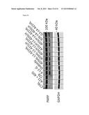 |
 | 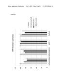 |
 |  |
 |  |
 |  |
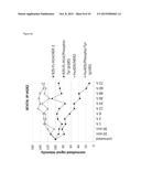 |  |
 |  |
 |  |
 |  |
 |  |
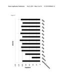 |  |
 |  |
 | 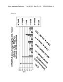 |
 | 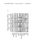 |
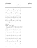 | 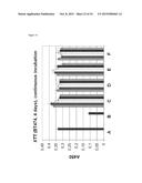 |
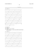 |  |
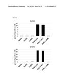 | 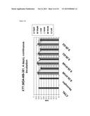 |
 | 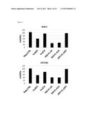 |
 |  |
 |  |
 |  |
 | 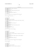 |
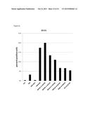 |  |
 |  |
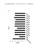 |  |
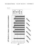 |  |
 | 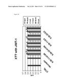 |
 | 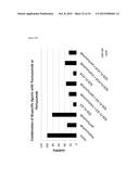 |
 |  |
 |  |
 |  |
 | 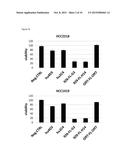 |
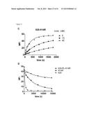 |  |
 |  |
 |  |
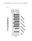 |  |
 |  |
 |  |
 |  |
 |  |
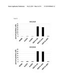 |  |
 |  |
 |  |
 |  |
 |  |
 | 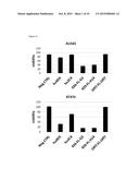 |
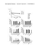 |  |
 |  |
 |  |
 | 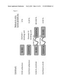 |
 |  |
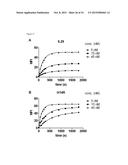 |  |
 | 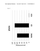 |
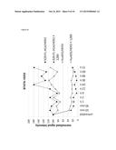 |  |
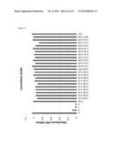 | 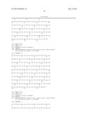 |
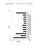 | 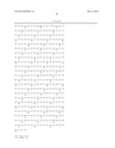 |
 | 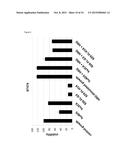 |
 |
| New patent applications in this class: | |
| Date | Title |
|---|---|
| 2022-09-08 | Shrub rose plant named 'vlr003' |
| 2022-08-25 | Cherry tree named 'v84031' |
| 2022-08-25 | Miniature rose plant named 'poulty026' |
| 2022-08-25 | Information processing system and information processing method |
| 2022-08-25 | Data reassembly method and apparatus |
| New patent applications from these inventors: | |
| Date | Title |
|---|---|
| 2021-12-23 | Her2-binding tetrameric polypeptides |
| 2021-10-07 | Cellular high throughput encapsulation for screening or selection |
| 2015-10-01 | Repeat protein from collection of repeat proteins comprising repeat modules |