Patent application title: METHOD FOR IDENTIFYING COMPOUND FOR INHIBITING AN ACTIVITY OF A HISTONE LYSINE DEMETHYLASE
Inventors:
Wen-Ching Wang (Hsinchu City, TW)
Hsing-Jien Kung (Miaoli County, TW)
Chia-Han Chu (Taipei City, TW)
Jing-Moon Yang (Hsinchu City, TW)
Assignees:
NATIONAL TSING HUA UNIVERSITY
IPC8 Class: AC12Q126FI
USPC Class:
Class name:
Publication date: 2015-09-24
Patent application number: 20150267241
Abstract:
The present invention relates to a method for identifying a compound that
inhibits an activity of a histone lysine demethylase.Claims:
1. A method for identifying a compound which inhibits an activity of a
histone lysine demethylase, comprising: (a) using a computer program to
generate a three-dimensional structure of a pocket of a histone lysine
demethylase, wherein the pocket comprises three sites: an
alpha-ketoglutarate (AKG), a methylated lysine, and a NIQ; (b) screening
for a compound that interacts with the three sites of said pocket; and
(c) testing the compound screened in (b) by in vitro or in vivo assay for
its ability to inhibit the activity of the histone lysine demethylase,
thereby identifying a compound that inhibits the activity of the histone
lysine demethylase.
2. The method of claim 1, wherein the pocket of the histone lysine demethylase is formed by residues including Gln85, Tyr133, Asp136, Tyr176, Tyr178, Phe186, His189, Glu191, Asn199, Lys207, His241, Lys242, His277 and Asn291, wherein the amino acid position refers to the full length histone lysine demethylase shown in SEQ ID NO: 11.
3. The method of claim 1, wherein the pocket of the histone lysine demethylase is formed by residues including Gln84, Tyr132, Asp135, Tyr175, Tyr177, Phe185, His188, Glu190, Asn198, Lys206, His240, Lys241, His276 and Asn290, wherein the amino acid position refers to the full length histone lysine demethylase shown in SEQ ID NO: 12.
4. The method of claim 1, wherein the pocket of the histone lysine demethylase is formed by residues including Gln86, Tyr134, Asp137, Tyr177, Tyr179, Phe187, His190, Glu192, Asn200, Lys208, His242, Lys243, His278 and Asn292, wherein the amino acid position refers to the full length histone lysine demethylase shown in SEQ ID NO: 13.
5. The method of claim 1, wherein the activity is the demethylating activity.
Description:
CROSS-REFERENCE TO RELATED APPLICATIONS
[0001] The present application is a non-provisional utility application which claims priority to U.S. Provisional Application No. 61/955,225, filed on Mar. 19, 2014, incorporated herein by reference in its entirety. The sequence listing text file, file name 2118_WWJ_SEQ, created Mar. 18, 2015, file size 38,571 bytes, is incorporated herein by reference in its entirety.
FIELD OF THE INVENTION
[0002] The present invention relates to a method for identifying a compound that inhibits an activity of a histone lysine demethylase.
DESCRIPTION OF PRIOR ART
[0003] Histone lysine demethylases (KDMs) that regulate a dynamic, reversible status of "methyl" histone codes have gained much attention. Mutations, amplifications, deletions and aberrant expression of KDMs have been identified in a variety of cancers and their roles in modulating the behaviours of cancer cells have been substantiated. As such, increasing attention has been paid to evaluate KDMs as potential therapeutic targets for cancer. There are now 8 KDM families including 28 members identified. KDM2-KDM8 constitute a large superfamily that shares a Jumonji C (JmjC) domain which functions as α-ketoglutarate (AKG) and Fe(II)-dependent demethylase. Notably, each family exhibits its exquisite substrate specificity toward different histone lysine residues, thereby effectively integrating the upstream signals and modulating the chromatin conformation.
[0004] The largest KDM4 gene family (four paralogs KDM4A-KDM4D and two pseudogenes KDM4E and KDM4F) has been shown to be an "eraser" of a repressive mark H3K9me3/me2, while its subfamily KDM4A-KDM4C also demethylates H3K36me3/me2. KDM4A and KDM4B are over expressed in a variety of cancers including prostate, breast, colorectal, lung, gastric, esophageal, lymphoma, renal cancer, and medulloblastoma. For prostate and breast cancers, this family of demethlases have the added significance in being coactivators of androgen receptor (AR) (KDM4A, B, C and D) and estrogen receptor (ER) (KDM4A and B). They function to stimulate the transcriptional potential of the receptors. KDM4B also regulates the turnover of AR. Given the important roles of AR and ER in prostate and breast carcinogenesis, KDM4A/4B are considered as promising drug targets of intervention in these malignancies.
[0005] Thus far, inhibitors for KDM4 proteins described are largely AKG analogues: N-oxalylglycines (OGAs) that inhibit KDM4A, KDM4C and KDM4D, pyridine 2,4-dicarboxylic acids (PD2s) developed based on KDM4E, and 8-hydroxyquinolines (8HQs) of which 5-carboxy-8HQ displays the highest potency on KDM4E in vitro (IC50=0.2 μM). Yet, as a prodrug, cytotoxic IC50 of PD2 is at ˜mM range in cultured cells, due to its poor cell-penetrating ability, while 5-carbxoxy-8HQ exhibits a relatively high cytotoxic IC50 in HeLa cells (86.5 μM).
SUMMARY OF THE INVENTION
[0006] The present invention provides a method for identifying a compound which inhibits an activity of a histone lysine demethylase, comprising: (a) using a computer program to generate a three-dimensional structure of a pocket of a histone lysine demethylase, wherein the pocket comprises three sites: alpha-ketoglutarate (AKG), a methylated lysine, and a NIQ; (b) screening for a compound that interacts with the three sites of said pocket; and (c) testing the compound screened in (b) by in vitro or in vivo assay for its ability to inhibit the activity of the histone lysine demethylase, thereby identifying a compound that inhibits the activity of the histone lysine demethylase.
BRIEF DESCRIPTION OF THE DRAWINGS
[0007] FIG. 1 shows (A) the electron density map for the Ni (II), PD2, and the H3K9me3 peptide. The 2Fo-Fc electron density maps are contoured at 1.0 σ. (B) The JmjC domain of KDM4B folds into a β-barrel structure.
[0008] FIG. 2 shows the differential ligand binding region between KDM4A/KDM4B/KDM4C and KDM4D. Superposition of KDM4A, KDM4B, KDM4C, and KDM4D reveals two heterogeneous regions, RKDM and NIQ. Surface representation of KDM4A, KDM4B, and KDM4D shows that the RKDM, NIQ regions and a crucial isoleucine (KDM4A, 171; KDM4B, 172) make several contacts with the peptide at (-2) and (+3, +4) sites in KDM4A and KDM4B, respectively. The corresponding GEAR and HKK sites deviate away from H3K9me3. PDB used in this Figure are: KDM4A (PDB: 20Q6); KDM4B (PDB: 4LXL); KDM4D (PDB: 4HON).
[0009] FIG. 3(A) shows demethylation of calf thymus H3 by bacteria-expressed KDM4A (upper panel) and KDM4B (lower panel) determined in the presence of 1,5-bis[(E)-2-(3,4-dichlorophenyl)ethenyl]-2,4-dinitrobenzene by western blotting analysis. H3 lysine modifications are probed with H3K9me3 and H3K36me3 antisera, respectively. FIG. 3(B) shows inhibition kinetics of KDM4A/4B demethylation activity by 1,5-bis[(E)-2-(3,4-dichlorophenyl)ethenyl]-2,4-dinitrobenzene. The inset in each panel shows the double reciprocal form, where the 1/relative activity is plotted versus 1/[H3K9me3] at various fixed concentrations of the inhibitor.
[0010] FIG. 4(A) shows surface representation of superimposed KDM4B.PD2.H3K9me3 and KDM4B.1,5-bis[(E)-2-(3,4-dichlorophenyl)ethenyl]-2,4-dinitrobenzene complexes. PD2 and H3K9me3 are drawn as stick models. 1,5-bis[(E)-2-(3,4-dichlorophenyl)ethenyl]-2,4-dinitrobenzene is show as ball-and-stick models. AKG site is labeled as site II, methylated lysine site as site I, RKDM as site III and NIQ as site IV. FIG. 4(B) shows schematic representation of interactions between 1,5-bis[(E)-2-(3,4-dichlorophenyl)ethenyl]-2,4-dinitrobenzene and KDM4B. FIG. 4(C) shows schematic representation of interactions between PD2 and KDM4B. The hydrogen-bonding contacts are shown as broken lines.
[0011] FIG. 5 shows (A) interaction profiles between residues from KDM4B and compounds. These compounds are divided into three groups based on their docked locations. The profile contains two interaction types [hydrogen-bonding (H) and van der Waals (V)]. A cell is colored in gray if a compound yields hydrogen-bonding or van der Waals interaction with a residue; otherwise, the cell is colored in black. FIG. 5 shows the docked conformations of (B) NSC107408 (in group 1), (C) NSC15975 (in group 2), and (D) NSC640999 (in group 3). NSC107408, NSC15975, and NSC640999 lack the interactions in the AKG site, the methylated lysine site, and the NIQ site, respectively, compared to 1,5-bis[(E)-2-(3,4-dichlorophenyl)ethenyl]-2,4-dinitrobenzene.
[0012] FIG. 6(A) shows the expression values of KDM4A in normal prostate gland and tumor tissues from the selected studies, which were obtained from Oncomine® (Compendia Bioscience, Ann Arbor, Mich., USA) database. The box whisker plots show the box encompasses 25th-75th percentile, median as line within the box, and 10th and 90th percentiles as error bars. FIG. 6(B) shows the expression values of KDM4B in normal prostate gland and tumor tissues from the selected studies, which are obtained from Oncomine® database. The box whisker plots show the box encompasses 25th-75th percentile, median as line within the box, and 10th and 90th percentiles as error bars. P values are determined by one-tailed Student's T-test and calculated based on the comparison of normal vs. cancer; normal vs. primary sites; or normal vs. metastasis. FIG. 6(C) shows the qRT-PCR analysis of KDM4A and KDM4B expression in normal prostate primary cell (PrEC), normal prostate epithelial cell lines (RWPE-1 and PNT2), and prostate cancer cell lines (LNCaP, C4-2, C4-2B, CWR22rv1, CWR-R1, VCaP, DU145 and PC3). Asterisks indicate significant over-expression as compared to normal cells.
[0013] FIG. 7(A) shows KDM4A and KDM4B are crucial for growth of LNCaP cells. LNCaP cells are infected with lentivirus with control shRNA (control) and shKDM4A or shKDM4B as indicated (left panel). The qRT-PCR analysis is performed to evaluate the expression of KDM4s (right panel). FIG. 7(B) shows cytotoxicity IC50 (CC50) and inhibition of H3K9me3 by 1,5-bis[(E)-2-(3,4-dichlorophenyl)ethenyl]-2,4-dinitrobenzene in LNCaP cells. LNCaP cells are treated with 1,5-bis[(E)-2-(3,4-dichlorophenyl)ethenyl]-2,4-dinitrobenzene for three days, and the viability is measured by viable cell count. SD is derived from biological triplicates. The level of H3K9me3 of 1,5-bis[(E)-2-(3,4-dichlorophenyl)ethenyl]-2,4-dinitrobenzene-treated cells (24 hr) is detected and quantified by AlphaView SA (Cell Biosciences Inc.). The level of H3K9me3 is shown by bar graph. FIG. 7(C) shows flow cytometric analysis of DNA contents in LNCaP cells treated with DMSO (mock) or 50 μM 1,5-bis[(E)-2-(3,4-dichlorophenyl)ethenyl]-2,4-dinitrobenzene for 3 days.
[0014] FIG. 8(A) shows DAVID functional annotation of the genes that shows two-folds alterations in expression. GO terms associated with the altered genes that show statistically strong enrichment with low P-values are listed. Bar graph and the numbers labelled indicate gene count of each pathway. % of hits indicates the percentage of genes that are altered in each GO. FIG. 8(B) shows expression of tumour suppressors and oncogenes that are up- and down-regulated in the inhibitor treated cell, respectively. FIG. 8(C) shows overlap of the inhibitor-altered genes with androgen responsive genes.
[0015] FIG. 9 shows AR-signature genes are differentially expressed in 1,5-bis[(E)-2-(3,4-dichlorophenyl)ethenyl]-2,4-dinitrobenzene-treated LNCaP cell.
[0016] FIG. 10 shows the binding pocket in KDM4s. Strictly conserved residues that bind to Fe(II) (H189, E191 and H276 in KDM4B) and AKG(Y133, N199, K207) are shown (upper left). Superposition of H3K9me3 liganded structures reveals four strictly conserved residues of KDM4s (D136, E170, Y176, and K242 in KDM4B) make contacts with the peptide at R8 (-1), K9, S10 (+1) and T11 (+2) positions. PDB used in the left-top figure are: KDM4A (PDB: 2OQ6); KDM4B (PDB: 4LXL); and KDM4D (PDB: 4HON). H bonds (<3.5 Å) are shown as dash lines.
[0017] FIG. 11 shows the analysis of KDM4A and KDM4B that demethylate H3K9me3/me2 and H3K36/me3/me2 of histones but not H3K27me3/me1. Demethylation of calf thymus histones by KDM4A and KDM4B is detected by various antisera as indicated. Histones are incubated without or with KDM4A/KDM4B for a certain time as indicated.
[0018] FIG. 12 shows top five results of the self-docking tests for the KDM4B.PD2 complex model. Self-docking test of KDM4B (PDB code: 4LXL) is a complex structure within PD2 and H3K9me3. The top five docking poses are shown as stick. The reference pose is shown as ball-and-stick. Ni is shown as a sphere.
[0019] FIG. 13(A) shows that the rest of datasets exhibit higher median values of KDM4A expression in PCa sites, despite no statistical significance. The expression values of KDM4A and KDM4B in normal prostate gland and tumor tissues from the selected studies are obtained from Oncomine® (Compendia Bioscience, Ann Arbor, Mich., USA) database. The box whisker plots show the box encompasses 25th-75th percentile, median as line within the box, and 10th and 90th percentiles as error bars. FIG. 13(B) shows that the rest of datasets exhibit higher median values of KDM4B expression in PCa sites, despite no statistical significance. The expression values of KDM4A and KDM4B in normal prostate gland and tumor tissues from the selected studies are obtained from Oncomine® database. The box whisker plots show the box encompasses 25th-75th percentile, median as line within the box, and 10th and 90th percentiles as error bars. P values are determined by one-tailed Student's T-test and calculated based on the comparison of normal vs. cancer; normal vs. primary sites; or normal vs. metastasis.
[0020] FIG. 14 is an illustration of linked T01 functional group. When the original CL atom of the R2 group is replaced by the T01 fragment, the Naphthalene-like group generates two cation-pi interaction against the LYS242 residues of KDM4B to stabilize interactions of the receptor-ligand complex.
DETAILED DESCRIPTION OF THE INVENTION
[0021] As used herein in the specification, "a" or "an" may mean one or more. As used herein in the claim(s), when used in conjunction with the word "comprising", the words "a" or "an" may mean one or more than one.
[0022] The present invention identifies a selective inhibitor 1,5-bis[(E)-2-(3,4-dichlorophenyl)ethenyl]-2,4-dinitrobenzene (NSC636819) toward KDM4A/4B/4C subfamily. Kinetic and docking analyses reveal crucial binding sites of 1,5-bis[(E)-2-(3,4-dichlorophenyl)ethenyl]-2,4-dinitrobenzene unique in the KDM4A/KDM4B/KDM4C subfamily. Further, the pharmacological and genetic inhibition of KDM4A/4B significantly lowers the viability of prostate cancer cells, principally due to its potency to inhibit AR transcriptional network.
[0023] A method for identifying a compound that modulates the activity of a histone lysine demethylase having a pocket formed by residues including Gln85, Tyr133, Asp136, Tyr176, Tyr178, Phe186, His189, Glu191, Asn199, Lys207, His241, Lys242, His277 and/or Asn291, wherein the amino acid position refers to the full length histone lysine demethylase shown in SEQ ID NO: 11 or a fragment or derivative thereof; a pocket formed by Gln84, Tyr132, Asp135, Tyr175, Tyr177, Phe185, His188, Glu190, Asn198, Lys206, His240, Lys241, His276 and/or Asn290, wherein the amino acid position refers to the full length histone lysine demethylase shown in SEQ ID NO: 12 or a fragment or derivative thereof; a pocket formed by Gln86, Tyr134, Asp137, Tyr177, Tyr179, Phe187, His190, Glu192, Asn200, Lys208, His242, Lys243, His278 and/or Asn292, wherein the amino acid position refers to the full length histone lysine demethylase shown in SEQ ID NO: 13 or a fragment or derivative thereof; a pocket formed by Gln88, Tyr136, Asp139, Tyr179, Tyr181, Phe189, His192, Glu194, Asn202, Lys210, His244, Lys245, His280 and/or Asn294, wherein the amino acid position refers to the full length histone lysine demethylase shown in SEQ ID NO: 14 or a fragment or derivative thereof; or a pocket formed by Gln85, Tyr133, Asp136, Tyr176, Tyr178, Phe186, His189, Glu191, Asn199, Lys207, His241, Lys242, His277 and/or Asn291, wherein the amino acid position refers to the full length histone lysine demethylase shown in SEQ ID NO: 15 or a fragment or derivative thereof; the method comprises modeling the compound in the pocket of the histone lysine demethylase; and determining the effect of the compound on the rate or degree of methylation of a substrate of the histone lysine demethylase.
[0024] The present invention provides a method for identifying a compound which inhibits an activity of a histone lysine demethylase, comprising: (a) using a computer program to generate a three-dimensional structure of a pocket of a histone lysine demethylase, wherein the pocket comprises three sites: an alpha-ketoglutarate (AKG), a methylated lysine, and a NIQ; (b) screening for a compound that interacts with the three sites of said pocket; and (c) testing the compound screened in (b) by in vitro or in vivo assay for its ability to inhibit the activity of the histone lysine demethylase, thereby identifying a compound that inhibits the activity of the histone lysine demethylase.
[0025] As used herein, the histone lysine demethylase (KDM) comprises a KDM4A, a KDM4B, a KDM4C, a KDM4D, a KDM4E. The peptide sequence of the KDM4A is SEQ ID NO: 11. The peptide sequence of the KDM4B is SEQ ID NO: 12. The peptide sequence of the KDM4C is SEQ ID NO: 13. The peptide sequence of the KDM4D is SEQ ID NO: 14. The peptide sequence of the KDM4C is SEQ ID NO: 13. The peptide sequence of the KDM4E is SEQ ID NO: 15.
[0026] In one embodiment, the pocket of the histone lysine demethylase is formed by residues including Gln85, Tyr133, Asp136, Tyr176, Tyr178, Phe186, His189, Glu191, Asn199, Lys207, His241, Lys242, His277 and Asn291, wherein the amino acid position refers to the full length histone lysine demethylase shown in SEQ ID NO: 11 or a fragment or derivative thereof. The alpha-ketoglutarate site is defined as the cavity occupied by alpha-ketoglutarate in the active-site histone lysine demethylase shown in SEQ ID NO: 11. The alpha-ketoglutarate site is surrounded by Y133, F186, H189, E191, S197, N199, K207, W209, T271, H277, and S289 of the SEQ ID NO: 11. The methylated lysine site is defined as the cavity occupied by the methylated lysine in the active site of the histone lysine demethylase shown in SEQ ID NO: 11. The methylated lysine site is enclosed by E170, G171, V172, Y176, Y178, E191, S197, S289, T290 and N291 of the SEQ ID NO: 11. The NIQ site is defined to comprise amino acid residues N87, 172, and Q89 of the SEQ ID NO: 11.
[0027] In another embodiment, the pocket of the histone lysine demethylase is formed by residues including Gln84, Tyr132, Asp135, Tyr175, Tyr177, Phe185, His188, Glu190, Asn198, Lys206, His240, Lys241, His276 and Asn290, wherein the amino acid position refers to the full length histone lysine demethylase shown in SEQ ID NO: 12 or a fragment or derivative thereof. The alpha-ketoglutarate site is defined as the cavity occupied alpha-ketoglutarate in the active site of histone lysine demethylase shown in SEQ ID NO: 12. The alpha-ketoglutarate site is surrounded by Y132, F185, H188, E190, S196, N198, K206, W208, T270, H276, and S288 of the SEQ ID NO: 12. The methylated lysine site is defined as the cavity occupied by the methylated lysine in the active site of the histone lysine demethylase shown in SEQ ID NO: 12. The methylated lysine site is enclosed by E169, G170, V171, Y175, Y177, E190, S196, S288, T289 and N290 of the SEQ ID NO: 12.The NIQ site is defined to comprise amino acid residues N86, 171, and Q88 of the SEQ ID NO: 12.
[0028] In one embodiment, the pocket of the histone lysine demethylase is formed by residues including Gln86, Tyr134, Asp137, Tyr177, Tyr179, Phe187, His190, Glu192, Asn200, Lys208, His242, Lys243, His278 and Asn292, wherein the amino acid position refers to the full length histone lysine demethylase shown in SEQ ID NO: 13 or a fragment or derivative thereof. The alpha-ketoglutarate site is defined as the cavity occupied alpha-ketoglutarate in the active site of histone lysine demethylase shown in SEQ ID NO: 13. The alpha-ketoglutarate site is surrounded by Y134, F187, H190, E192, S198, N200, K208, W210, T272, H278, and S290 of the SEQ ID NO: 13. The methylated lysine site is defined as the cavity occupied by the methylated lysine in the active site of the histone lysine demethylase shown in SEQ ID NO: 13. The methylated lysine site is enclosed by E171, G172, V173, Y177, Y179, E192, S198, S290, T291 and N292 of the SEQ ID NO: 13. The NIQ site is defined to comprise amino acid residues N88, 173, and Q90 of the SEQ ID NO: 13.
[0029] In another embodiment, the pocket of the histone lysine demethylase is formed by residues including Gln88, Tyr136, Asp139, Tyr179, Tyr181, Phe189, His192, Glu194, Asn202, Lys210, His244, Lys245, His280 and Asn294, wherein the amino acid position refers to the full length histone lysine demethylase shown in SEQ ID NO: 14 or a fragment or derivative thereof.
[0030] In one embodiment, the pocket of the histone lysine demethylase is formed by residues including Gln85, Tyr133, Asp136, Tyr176, Tyr178, Phe186, His189, Glu191, Asn199, Lys207, His241, Lys242, His277 and Asn291, wherein the amino acid position refers to the full length histone lysine demethylase shown in SEQ ID NO: 15 or a fragment or derivative thereof.
[0031] In another embodiment, the activity is the demethylating activity.
[0032] The histone lysine demethylase (KDM) has the function for demethylating the histone code. In one embodiment, the histone code comprises a H3K9me3, a H3K9me2, a H3K36me3, and a H3K36me2. In a preferred embodiment, the KDM4A and KDM4B demethylate H3K9me3/me2.
[0033] The compounds of the present invention can also be designed by visually inspecting the three-dimensional structure of the KDM to determine more effective inhibitors. This type of modeling is generally referred to as "manual" drug design. Manual drug design can employ visual inspection and analysis using a graphics visualization program. Initially compounds are selected by manual drug design. The structural analog thus designed can then be modified by computer modeling programs to better define the most likely effective candidates. Reduction of the number of potential candidates is useful as it may not be possible to synthesize and screen a countless number of compound variations that may have some similarity to known inhibitory molecules.
[0034] In another embodiment, the compound that inhibits the activity of the histone lysine demethylase further is used to treat cancer.
[0035] The present invention also provides a method for treating cancer, which comprises administering an effective amount of compound and a pharmaceutically acceptable carrier to a subject in need thereof, wherein the compound has the structure of formula I:
##STR00001##
[0036] wherein R1-4 is F, Cl, Br, I, At, hydroxyl or CxHyN.sub.zO.sub.αS.sub.β respectively,
[0037] wherein x=1-11, y=3-15, z=0-3, α=0-2 and β=0-1.
[0038] The present invention may be used to treat, alleviate, ameliorate, relieve, delay onset of, inhibit progression of, reduce severity of, and/or reduce incidence of one or more symptoms or features of a disease, disorder, and/or condition induced by the cancer. In a preferred embodiment, the method of the present invention further treats a prostate cancer.
[0039] In one embodiment, the cancer is selected from prostate cancer, breast cancer, colorectal cancer, lung cancer, gastric cancer, esophageal cancer, lymphoma, renal cancer or medulloblastoma. In a preferred embodiment, the cancer is a prostate cancer.
[0040] In another embodiment, the compound is a 1,5-bis[(E)-2-(3,4-dichlorophenyl)ethenyl]-2,4-dinitrobenzene.
[0041] A "effective amount" is an amount effective to prevent, lower, stop or reverse the development of, or to partially or totally alleviate the existing symptoms of a particular condition for which the subject being treated.
[0042] The "compound" or "1,5-bis[(E)-2-(3,4-dichlorophenyl)ethenyl]-2,4-dinitrobenzene" may be formulated for administering via sterile aqueous solution or dispersion, aqueous suspension, oil emulsion, water in oil emulsion, site-specific emulsion, long-residence emulsion, sticky-emulsion, microemulsion, nanoemulsion, liposomes, microparticles, microspheres, nanospheres, nanoparticles, minipumps, and with various natural or synthetic polymers that allow for sustained release. The compounds comprising the NRIP may also be formulated into aerosols, tablets, pills, sterile powders, suppositories, lotions, creams, ointments, pastes, gels, hydrogels, sustained-delivery devices, or other formulations used in drug delivery.
[0043] As used herein, the term "pharmaceutically acceptable carriers" are determined in part by the particular composition being administered, as well as by particular method used to administer the composition. As used herein, "carrier" includes any and all solvents, dispersion media, vehicles, coatings, diluents, antibacterial and antifungal agents, isotonic and absorption delaying agents, buffers, carrier solutions, suspensions, colloids, and the like. The use of such media and agents for pharmaceutical active substances is well known in the art. Except insofar as any conventional media or agent is incompatible with the active ingredient, its use in the therapeutic compositions is contemplated. Supplementary active ingredients can also be incorporated into the compositions. The phrase "pharmaceutically-acceptable" refers to molecular entities and compositions that do not produce an allergic or similar untoward reaction when administered to a subject. The preparation of an aqueous composition that contains a protein as an active ingredient is well understood in the art. Typically, such compositions are prepared as injectables, either as liquid solutions or suspensions; solid forms suitable for solution in, or suspension in, liquid prior to injection can also be prepared. The preparation can also be emulsified.
[0044] In one embodiment, the subject is an animal. Preferably, the subject is a mammal. More preferably, the subject is a human.
[0045] In one embodiment, the compound inhibits a cancer cell growth by inhibiting an expression of a histone lysine demethylase (KDM). In a preferred embodiment, the compound induces an apoptosis of a cancer cell by inhibiting an expression of the histone lysine demethylase (KDM). In a more preferred embodiment, the compound inhibits the demthylating function of the KDM. In another embodiment, the KDM is a KDM4A or a KDM4B.
[0046] In another embodiment, the KDM demethylates a histone. In a preferred embodiment, the histone comprises a H3K9me3, a H3K9me2, a H3K36me3, and a H3K36me2. In a preferred embodiment, the KDM4A and the KDM4B demethylate the H3K9me3 and the H3K9me2.
[0047] In one embodiment, the compound further inhibits the cancer cell growth or induces an apoptosis of a cancer cell by inhibiting an androgen receptor (AR), wherein the AR is a coactivator of the KDM4A and the KDM4B.
EXAMPLES
[0048] The examples below are non-limiting and are merely representative of various aspects and features of the present invention.
[0049] Material and Methods:
[0050] Cloning, Expression, and Purification
[0051] Human KDM4B 1-348 and KDM4A 1-347 were PCR amplified from chromosomal DNA using forward and reverse primers as follows:
TABLE-US-00001 SEQ ID Gene Primer sequence No. KDM4B Forward: 5'-AAA CAT ATG GGG TCT GAG 1 1-348 GAC CAC GGC GCC-3' (NdeI) Reverse: 5'- AAA AAA CTC GGG GCT CTC 2 GAG CTA CGT GGG CCG -3' (XhoI) KDM4A Forward: 5'- AAA CAT ATG GCG AGC GAA 3 1-347 AGC GAA ACT CTG -3' (NdeI) Reverse: 5'- AAA GGA TCC CTA CGT GGG 4 CAG AGT ATG GTC-3' (BamHI)
[0052] PCR was performed with HiFi DNA polymerase kit using a C1000 Touch® Thermal Cycler (Bio-Rad Laboratories, Inc., USA): initial denaturation, at 95° C. for 5 min followed by 25 cycles of denaturation at 95° C. for 30 s, annealing at 55° C. for 30 s, and extension at 72° C. for 70 s. The amplified product was inserted into pET28a (Novagen, Inc., USA) to generate pET28a-KDM4B 1-348 and pET28a-KDM4A 1-347 which were introduced into Escherichia coli BL21 (DE3) cells. Expression of protein was induced by addition of 0.5 mM isopropyl-β-d-thiogalacto-pyranoside (IPTG) at 16° C. for 21 h. Bacterial pellets were fractionated by sonication to collect soluble proteins in cytosolic fractions. The His6-tagged KDM4B or KDM4A proteins were purified by a nickel affinity column (Ni Sepharose® High Performance, GE Healthcare) using the elution buffer containing 500 mM NaCl, 250 mM imidazole and 50 mM HEPES (pH 7.5). The protein was concentrated and further purified by a 16/60 Superdex 75 gel filtration column equilibrated with 50 mM HEPES pH 7.5, 500 mM NaCl. The protein purity was analyzed by SDS-PAGE analysis. Protein concentration was assayed by the Bradford method using bovine serum albumin as the standard.
[0053] Enzyme Assay
[0054] Formaldehyde dehydrogenase-coupled demethylase assay was used to determine the demethylase activity and select the potent inhibitors. All inhibitors were dissolved in dimethyl sulfoxide (DMSO) at various concentrations, and added to the mixture that the final DMSO concentration is 5%. The reagents for demethylase reactions were dissolved in HEPES buffer (50 mM, pH 7.5), with the exception of Fe(II) solutions, which were made using (NH4)2Fe(SO4)2 dissolved in 20 mM HCl to make 400 mM stock solution. All the reagents were stored at -30° C. FDH, NAD+, H-TKQTARK(Me3)STGGKAPR-OH (TR-15, H33-17K9me3, Kelowna), DMSO, and KDM4B were added first to 96-well black immune plate (SPL Life Science) and incubated together on ice for 15 min. Then the plate was put into FLUOStar OPTIMA ELISA reader (BMG LABTECH) with 37° C., and the reaction was started by adding ascorbic acid (ascorbate), Fe(II), and α-ketoglutarate (AKG) to final concentration of 50 mM HEPES, pH 7.5, 2 μM of KDM4B, 5% DMSO, 0.01 U FDH (Sigma), 1 mM NAD+, 1 mM 2-OG, 2 mM ascorbate, and 50 μM Fe(II), various concentration of H3K9me3 peptide, and the final volume was 50 μl. Each reaction was incubated at 37° C. for 30 min and the production of NADH would be detected by using the fluorescence Ex 360/Em 470.
[0055] Crystallization
[0056] Crystallization was performed by the hanging-drop vapor-diffusion method at 4° C. Equal volumes of a protein sample and the reservoir solution were mixed. Initial crystallization screening was automated using a robot Oryx8 (Douglas Instruments, UK) and the reagents of seven sets of crystallization kits: Crystal Screen I and II kits (Hampton Research), Index kit (Hampton Research), Clear Strategy Screen I and II kits (Molecular Dimension), Wizard kit (Emerald), and JB Screen classic HTS I and II kits (Jena Bioscience). Crystals of KDM4B (10 mg/ml protein, 4 mM PD2 and 5 mM H3K9me3 peptide) were grown in 0.1 M MES (pH 6.5), 0.2 M magnesium acetate, 20% (w/v) polyethylene glycol (PEG) 8000. Optimized crystals used for diffraction (12 mg/ml within 4 mM PD2 and 5 mM H3K9me3 peptide) were grown in 0.1 M MES (pH 6.5), 0.2 M magnesium acetate, 24% (w/v) polyethylene glycol (PEG) 8000. The crystal diffracted to 1.87 Å, belonged to space group P212121, and had unit cell dimensions of a=54.36, b=78.48, c=83.89 Å. The asymmetric unit contained one molecule.
[0057] X-ray Data Collection and Processing
[0058] Crystals were flash frozen in a stream of liquid nitrogen and then screened and characterized using an RU-300 rotating-anode X-ray generator (Rigaku/MSC Inc., USA) at the Macromolecular X-ray Crystallographic Laboratory of the National Tsing Hua University, Taiwan. The KDM4B.PD2.H3K9me3 dataset was collected at the SPring-8 BL12B2 beamline, Japan, with an ADSC Quantum 4R detector. All datasets were indexed, integrated, and scaled using HKL-2000. Data collection statistics are shown in Table 1.
TABLE-US-00002 TABLE 1 Crystallographic Data and Refinement Statistics of KDM4B PD2 H3K9me3 Data Collection Beamline SPring-8 BL-12B2a Wavelength (Å) 1.0000 Space group P212121 Cell dimensions a, b, c (Å) 54.4, 78.5,83.9 α, β, γ (0) 90.0, 90.0,90.0 Resolution limit (Å) 30-1.87 Unique reflections 30463 Completeness (%)b 99.4 (98.0) Avg I/σ(I)b 31.2 (6.0) Rmerge (%)b,c 4.3 (36.8) Redundancyb 12.5 (11.2) Refinement Resolution limit (Å) 28.67-1.87 Number of atoms 3125 Protein atoms 2768 Solvent atoms 285 Ligand atoms 72 Estimated coordinate error (Å) 0.142 Rworkd/Rfreee 17.6/22.5 Overall B-factor (Å2) 28.01 RMSD bond lengths (Å)f 0.012 RMSD bond angles (0)f 1.39 Ramachandran Analysis (%)g Favored 95.9 Allowed 3.8 Disallowed 0.3 aBL-12B2 Taiwan beamline at SPring-8, Hyogo, Japan bValues in parentheses refer to statistics in the highest-resolution shell. cRmerge = Σ|Iobs - <I>|/ΣIobs. dRwork = Σ|Fobs - Fcalc|/ΣFobs, where Fobs and Fcalc are the observed and calculated structure-amplitudes, respectively. eRfree was computed using 5% of the data assigned randomly. fRoot mean square deviation. gEstimated standard uncertainties based on maximum likelihood.
[0059] Structure Determination and Refinement
[0060] Crystallographic refinement used the maximum-likelihood target function module in REFMAC5. The KDM4B.PD2.H3K9me3 structures were constructed by MOLREP with the KDM4A (PDB: 2YBS) as the template and were refined using REFMAC5 coupled with ARP/wARP, which automatically added water molecules. The 2Fo-Fc electron density maps were generated by FFT and plotted by PyMOL. The validities of the KDM4B.PD2.H3K9me3 structure were assessed by PROCHECK.
[0061] Structural Comparison
[0062] The KDM4B structure was compared with protein structures in the DALI server. The structures of KDM4A.H31-17K9me3 (PDB code: 2P5B), KDM4B.PD2 (PDB code: 4LXL; this study), KDM4C.OGA (PDB code: 2XML), KDM4D.AKG.H36-15K9me3 (PDB code: 4HON), and KDM4E (PD2; PDB code: 2W2I) were superimposed by LSQMAN in O. ESPript was used for the combined sequence and secondary structure alignments and Figure preparation. PyMol was used to prepare the figures.
[0063] Virtual Screening and Molecular Modeling
[0064] The binding site for virtual docking screening of putative inhibitors was prepared by including protein atoms located ≦10-Å-radius sphere centered around the bound ligand of KDM4A (PDB code 2YBK. The present invention utilized GEMDOCK to screen the NCI database (236,962 compounds). Top ranked, available compounds were selected for testing in the KDM4A/KDM4B inhibitory assay.
[0065] Molecular Modeling
[0066] Discovery Studio v3.0 (Accelrys Inc., USA) was used to prepare, energy minimize, and refine a KDM4B model for molecular dynamics. The default parameters of ChiRotor were used to optimize side-chain conformations. Energies of the protein models were further minimized using CHARMm. 1,5-bis[(E)-2-(3,4-dichlorophenyl)ethenyl]-2,4-dinitrobenzene was then docked into KDM4B by GEMDOCK, a robust flexible ligand docking tool, was first used in conjunction with its default settings to generate conformations and carry out a docking analysis for ligand-containing KDM4B. Top 10 of compounds ranked by the docking energy were derived. CDOCKER with CHARMm forcefield was used to refine the docked models. To estimate the practicability of the proposed docking procedure, the present invention performed self-docking against the co-crystal KDM4B-PD2 structure (PDB code: 4LXL). Top five docked poses with minimum RMSD≦1 Å were derived (FIG. 12). The average of RMSD for self-docked poses of KDM4B-PD2 is 0.86 Å.
[0067] Cell Culture
[0068] Primary PrEC cells were purchased from Clonetics (Walkersville, Md.) and cultured in serum-free prostate epithelial cell growth medium following the vendor's directions. Cell lines RWPE1, LNCaP (LNCaP-FGC), CWR22Rv1 (22Rv1), VCaP, DU145, PC3 (all purchased from ATCC), and PNT2 (Sigma Aldrich, Mo.), were cultured under condition as recommended. CWR-R1 cell (40) and LNCaP derived C4-2, C4-2B cells were cultured in RPMI1640 medium containing 10% FBS.
[0069] RNA Interference and Quantitative RT-PCR
[0070] Lentiviral vector pLKO.1 carrying sequences encoding a shRNA that specifically targets KDM4A and KDM4B gene (TRC library Clone ID TRCN0000234910 and TRCN000018014) were co-transfected with viral packaging plasmids in 293T cells to generate the shRNA lentiviral particles. Empty pLKO.1 plasmid was used as negative control. The lentiviral supernatant was collected after 48-hr transfection and concentrated by Lenti-X Concentrator (Clontech, Calif.). The precipitated viral particle was resuspended in fresh RPMI1640 medium with 10% FBS for subsequent LNCaP infection and transduced into LNCaP cells for 72 hrs. Cells were then harvested and total RNA was isolated, followed by cDNA synthesis and real-time PCR analysis using iQ5 iCycler thermal cycler (Bio-Rad, Calif.). Threshold cycle values were normalized against actin transcript level. Individual samples were performed in triplicate and converted to relative gene expression using QGene96 software. Primer sequences used are as follows:
TABLE-US-00003 SEQ ID Gene Primer sequence No. KDM4A Forward: 5'- AGG AGA GTG AAC TGC CTC 5 CA -3' Reverse: 5'- GGT CTC CTT CCT CTC CAT 6 CC -3' KDM4B Forward: 5'- TCA CGC AGT ACA ATA TCC 7 AG -3' Reverse: 5'- TCG TCA TCA TAC AAA GAG 8 CC -3' actin Forward: 5'- GTA CCA CTG GCA TCG TGA 9 TGG ACT -3' Reverse: 5'- CCG CTC ATT GCC AAT GGT 10 GAT -3'
[0071] Cell Proliferation Assay
[0072] LNCaP cell was seeded in 48-well plate one day prior to lentivirus infection. After subjected to the shRNA lentivirus (day 0), cell proliferation was measured every two days by MTT colorimetric assay according to the manufacturer's instruction (Roche, Ind.).
[0073] Immunoblotting and Flow Cytometry
[0074] Total cell lysates were obtained by lysing the cell with buffer (50 mM Tris-HCl pH7.5, 150 mM NaCl, 0.5% Triton X-100, 10% glycerol, 1 mM EDTA, protease inhibitors) for 15 min on ice, followed by 10 min of sonication cycle (30 sec on, 30 sec off) on ice. The level of total histone H3 and trimethylated histone H3 Lys9 was analyzed by western blotting using anti-histone H3 and anti-H3K9me3 antibodies (Abcam, Mass.). Cells treated with mock and the inhibitor were harvested and fixed by 70% ethanol for >4 hours at -20° C., followed by propidium iodide (Sigma Aldrich) staining The DNA content was analyzed by Becton Dickinson FACScan flow cytometry, and the sub-G1 population was quantified by WinMDI 2.9.
[0075] Microarray
[0076] LNCaP cells treated with mock and the 1,5-bis[(E)-2-(3,4-dichlorophenyl)ethenyl]-2,4-dinitrobenzene inhibitor for 3 days were harvested and the total RNA was extracted using Trizol reagent (Life Technologies, N.Y.). Microarray analysis was performed by the University of California Davis Cancer Center Gene Expression Resource, using Affymetrix Human Genome U133A (HG-U133A) GeneChip arrays (Affymetrix, Calif.), which permit expression analysis of the entire Genbank RefSeq database. Array scanning and generation of raw signal data files were done with GeneChip Operating Software (Affymetrix). Subsequent data analysis was done by GeneSpring (Agilent Technologies, Calif.) and DAVID Bioinformatic Resources 6.7 (NIH).
[0077] RESULTS :
[0078] KDM4B.PD2.H3K9me3 Crystal Structure
[0079] In an effort to understand the detailed structure-function relationship of KDM4B at an atomic resolution, the recombinant JmjC domain of KDM4B was subjected to crystallization in the presence of a peptide, Ni(II), and AKG or PD2. After extensive trials, a well-diffracting crystal was found to consist of a large piece of residual density in the binding pocket, which could be modeled as the H3K9me3 peptide, an inhibitor PD2 and Ni (FIG. 1A). The final crystal structure shows a 1.87 Å-resolution monomer (R=21.8%, Rfree=26.2%) that consisted of the KDM4B catalytic domain (residues 9-337), PD2, and an H3K9me3 peptide (residues 7-14) within the active site (Table 1). A Ni(II) ion was observed to be located on a site corresponding to the Fe(II) position on the bottom of the catalytic pocket. The JmjC domain of KDM4B folded into a β-barrel structure, characteristic of members in the KDM4 family (FIG. 1B). Superposition of KDM4A.OGA.H3K9me3 (PDB code: 2OQ6), KDM4B.PD2.H3K9me3, KDM4C.OGA (PDB code: 2XML), and KDM4D.AKG.H3K9me3 (PDB code: 4HON) showed limited conformational change in overall Cα atoms of the JmjC domain. RMSD between KDM4A and KDM4B was 0.54 Å (residues 9-337 of KMD4B).
[0080] In the active site, PD2 was situated at a position nearly overlapped with AKG in which one of its carboxyl moieties contacted with H189, E191 and K242, while the other made H bonds with Y133 and K207, similar to those that contacted with AKG (Y132, N198, and K206 in KDM4A). Ni(II) that occupied the site of Fe(II) made contacts with three strictly conserved residues (H189, E191, and H277 in KDM4B). G171, Y176, T290, and N291 in KDM4B that surround the methylated lysine were also strictly conserved (FIG. 10).
[0081] The most prominent feature of the KDM4 family was its potent catalytic activity toward H3K9me3/me2. Analysis of superimposed H3K9me3 liganded structures including KDM4A, KDM4B, and KDM4D [KDM4A.Fe(II).OGA.H3K9me3 (PDB code: 2OQ6), KDM4B.Ni(II).PD2.H3K9me3 (this study; PDB code: 4LXL), KDM4D.Ni(II).2-OG.H3K9me3 (PDB code: 4HON)] revealed a conserved region to accommodate R8 (-1) and the methylated K9 of H3. Notably, three conserved residues (KDM4A: D135, E169, Y175; KDM4B: D136, E170, Y176; KDM4D: D139, E173, Y179) from β7 and β8 made H contacts with the guanidinium group of R8, the peptide O and N atoms of K9 and the peptide N atom of T11 from H3. In the interior of this cleft, a lysyl side chain (KDM4A: K241; KDM4B: K242; KDM4D: K245) forms a strong bond to the peptide O atom of S10 (FIG. 10), hence together properly orienting the H3K9me3/me2 for similarly efficient catalysis in KDMs. Interestingly, the KDM4A/KDM4B/KDM4C subfamily but not KDM4D exhibited additional specificity to demethylate H3K36me3/me2. Consistent with the structural analysis for KDM4D, the present invention observed two heterogeneous regions in KDM4B accounting for the substrate specificity: (1) RKDM vs. GEAR and (2) NIQ vs. HKK (FIG. 2).
[0082] The RKDM site (residues 310-313 in KDM4B) from a long U-shaped loop resided near the (-1, -2) site of the peptide-binding cleft. The aspartate side chain of RKDM (D311 in KDM4A) faced toward the peptide (-1, and -2 sites) and could make contacts with the peptide (FIG. 2). Additionally, the long and positively charged side chain of R from RKDM contributed to contact with the plus side of the peptide as demonstrated in two liganded structures: KDM4A.AKG.H3K9 (PDB code: 2Q8C) [KDM4A/R309 (NH1)-H3/G12 (O): 3.8 Å] and KDM4A.N-oxalylglycine.H3K36 (PDB code: 2P5B) [KDM4A/R309 (NH1)-H3/H39 (N): 3.8 Å]. A subtle difference was also noted at the other side of this U loop between KDM4A and KDM4B; there was a T308-D236 contact in KDM4B but not in KDM4A (the corresponding residues are S307 and E235). In contrast, the GEAR motif deviated away from the peptide-binding cleft, hence no contacts with the peptide.
[0083] The other region was NIQ site from the β4-β5 segment shared in KDM4A/KDM4B/KDM4C (residues 87-89 in KDM4B) while KDM4D had HKK at the corresponding region. Q89 was noted to contact with H3H39 and R40 (+3 and +4), whereas KDM4D consisted of HKK with positively charged side chains at the corresponding site (FIG. 2), which was likely to yield steric hindrance and electrostatic repulsion against H39 and R40 of H3K36me3. 171 that was nearby NIQ (KDMA, 171; KDM4B, 172) also played a crucial role (Krishnan and Trievel, 2013).
[0084] The present invention used the formaldehyde dehydrogenase (FDH)-coupled continuous fluorescent demethylase method to assess the enzymatic activity of the recombinant KDM4A and KDM4B expressed in Escherichia coli. Using an H3K9me3 peptide (residues 3-17) as the substrate, KDM4A and KDM4B exhibited comparable catalytic activity (Table 2), consistent with Hillringhaus et al. The present invention were able to measure the kinetic parameters with an H3K36me3 peptide (H331-41K36me3) and obtained an analogous kcat value while a higher Km value as compared with those with the H3K9me3 peptide, suggesting that KDM4A/4B had a lower binding affinity toward H3K36me3 than H3K9me3.
TABLE-US-00004 TABLE 2 Kinetic parameters for KDM4A and KDM4B using H33-17K9me3 or H331-41K36me3 as the substrate. KDM4 kcat (s-1) Km (μM) kcat/Km (s-1μM-1) H33-17K9me3 KDM4A 0.017 ± 0.001 92.5 ± 5.9 1.8 × 10⁻⁴ KDM4B 0.014 ± 0.001 88.3 ± 8.6 1.6 × 10⁻⁴ H331-41K36me3 KDM4A 0.015 ± 0.001 169.9 ± 19.9 8.8 × 10⁻⁵ KDM4B 0.013 ± 0.001 138.5 ± 14.1 9.4 × 10⁻⁵
[0085] The present invention further utilized calf thymus histones as the substrate and probed for H3K9, H3K27 and H3K36me3/me2/me1 in the presence of the recombinant KDM4A or KDM4B using western blotting analysis. As shown in FIG. 11, the signal of H3K9me3/me2 was significantly reduced while that of H3K9me1 increased in the presence KDM4A (upper panel) or KDM4B (lower panel) via a time-dependent manner as compared with the controls, indicating an active KDM4A/KDM4B form to remove the methyl group from H3K9me3/me2. For H3K36 signal, a longer time was needed to remove the signal of H3K36me3/me2. No difference was found for H3K27me3 or H3K27me1. These results collectively suggested that KDM4A and KDM4B demethylated H3K9me3/me2 more efficiently than did H3K36me3/me2 and that there was no activity toward H3K27me3 /me2/me1, confirming the results in Table 2.
[0086] Virtual Screening to Identify 1,5-bis[(E)-2-(3,4-dichlorophenyl)ethenyl]-2,4-dinitrobenzene as a Novel Active-Site Inhibitor Toward KDM4A and KDM4B
[0087] The present invention utilized GEMDOCK to screen for putative hits against the NCI database. The known inhibitor PD2 was used as a positive control which showed significant inhibition [21% (KDM4A) and 24% (KDM4B) of residual activity]. Of the selected 10 top-ranked compounds, 1,5-bis[(E)-2-(3,4-dichlorophenyl)ethenyl]-2,4-dinitrobenzene exhibited the highest inhibitory effect toward both KDM4A (28%) and KDM4B (35%) (Table 3). To confirm the FDH-demethylase coupled results, the present invention utilized histones as the substrate and probed for H3K9me3 and H3K36me3 in the absence or presence of the recombinant KDM4A or KDM4B using western blotting analysis. FIG. 3A shows that PD2 and 1,5-bis[(E)-2-(3,4-dichlorophenyl)ethenyl]-2,4-dinitrobenzene indeed blocked the demethylation activity. Further kinetic inhibition characterization of 1,5-bis[(E)-2-(3,4-dichlorophenyl)enthenyl]-2,4-dinitrobenzene demonstrated a competitive inhibitory mode against H33-17K9me3 for KDM4A [IC50=6.4 μM; Ki (H3K9me3)=5.5 μM; FIG. 3B). KDM4B also showed analogous inhibition kinetics [IC50=9.3 μM; Ki (H3K9me3)=3 μM). These results together provide strong evidence that 1,5-bis[(E)-2-(3,4-dichlorophenyl)ethenyl]-2,4-dinitrobenzene was a potent selective active-site inhibitor.
[0088] The De Novo Link protocol of Discovery Studio v3.0 (Accelrys Inc., USA) was used to suggest modifications and additions to the specific CL functional group of the docked NSC636819 in order to enhance binding to the KDM4B. This approach suggested modifications to a ligand scaffold to increase binding by placing fragments from the specified Ludi library into the specified binding site in accordance of the Ludi-generated interaction map.
[0089] The parameters used were as follows. Four CL atoms of the NSC636819 were defined as linked atoms with KDM4B as an input receptor and set an input sphere which includes whole Jmjc. Ludi-based fragment libraries were used here as input fragment libraries.
[0090] Then the simulated results showed that 54, 94, 11 and 3 for R1, R2, R3 and R4 groups respectively. The T01 (C11H10) fragment was illustrated as an example to show the enhanced binding capability due to additional cation-Pi interactions (See FIG. 14).
[0091] The KDM4B.1,5-bis[(E)-2-(3,4-dichlorophenyl)ethenyl]-2,4-dinitroben- zene complex model
[0092] An in silico approach was used to generate KDM4B.1,5-bis[(E)-2-(3,4-dichlorophenyl)ethenyl]-2,4-dinitrobenzene complex model by means of energy minimization and molecular dynamics simulations (FIG. 4). Given the model, 1,5-bis[(E)-2-(3,4-dichlorophenyl)ethenyl]-2,4-dinitrobenzene was situated at a position that occupied across three sites: the AKG site (e.g., H189, E191, and H277), the key lysine binding region (e.g., D136, E170, Y176, K242), and a portion of the H3K36-specific NIQ region (e.g., 172, Q85, N87) (FIG. 4). Of note, the middle benzene ring with two nitro groups reached the terminal methylated N(ε) position as well as Fe(II) position. The other two 1,2-dichlorobenzene rings were situated at the (0) and (+2) sites of the peptide-binding cleft, thereby specifically inhibiting KDM4A and KDM4B activity. Thus, 1,5-bis[(E)-2-(3,4-dichlorophenyl)ethenyl]-2,4-dinitrobenzene represented a novel KDM4A and KDM4B-specific inhibitor.
[0093] The present invention next compared the interactions of all tested compounds based on their docking poses. Three groups were classified: group 1 included all three sites (AKG, the methylated lysine, and NIQ); group 2 included AKG and the NIQ sites; and group 3 consisted of AKG and the methylated lysine sites (FIG. 5). Overall, compounds in group 2 and group 3 had low to modest inhibitory effects compared with 1,5-bis[(E)-2-(3,4-dichlorophenyl)ethenyl]-2,4-dinitrobenzene from group 1, suggesting the positive contribution of contacts from all three sites (FIG. 5). Above all, the meta-positioned NO2 moieties on the benzene ring that occupied at the AKG and the methylated lysine sites (S179, H277, H189, and N199) were found for 1,5-bis[(E)-2-(3,4-dichlorophenyl)ethenyl]-2,4-dinitrobenzene that possessed inhibitory activity. In support of this notion, NSC26820 that had ortho-positioned COOH moieties completely lost the inhibitory effects (Table 3). In addition, NSC107408 showed no inhibition because it lacked the 1,3-dinitrobenzene moiety and was unable to form interactions with residues S 197, H277, H189, and N199 of the AKG site (FIGS. 5A and 5B).
TABLE-US-00005 TABLE 3 Inhibition effects of selected hits on KDM4A and KDM4B. Demethylasea FDHb Compound Residual IC50 Residual Compound Ranking ID KDM Activity (%) (μM) Activity (%) structure 4B NSC636819 4A 4B 28 35 15.5 9.3 >80 ##STR00002## 100 NSC107408 4A 4B 99 64 -- -- >80 ##STR00003## 3 NSC85021 4A 4B 82 65 -- -- >80 ##STR00004## 223 NSC28620 4A 4B 111 110 -- -- >80 ##STR00005## 271 NSC15975 4A 4B 73 90 -- -- >80 ##STR00006## 83 NSC640999 4A 4B 77 49 -- -- >80 ##STR00007## 2 NSC659196 4A 4B 92 56 -- -- >80 ##STR00008## 33 NSC124029 4A 4B 93 62 -- -- 97 ##STR00009## 159 NSC1018 4A 4B 85 62 -- -- >80 ##STR00010## 93 NSC6129 4A 4B 100 52 -- -- >80 ##STR00011## 62
[0094] Genetic and pharmacological inhibition of KDM4A and KDM4B induced apoptosis
[0095] Several studies have reported that KDM4 family members are over-expressed in various cancers. To further support clinical relevance of KDM4A and KDM4B in prostate cancer, the present invention took advantage of the comprehensive database collection on Oncomine® (Compendia Bioscience, Ann Arbor, Mich., USA) database to examine their expression profile between normal prostate gland and tumor tissues. Among 14 datasets available, a statistically significant (p<0.05) elevation of KDM4A was seen for 6, and KDM4B for 8 in PCa compared to norma;/benign samples (FIGS. 6A and 6B). The rest of datasets also exhibited higher median values of KDM4A/KDM4B expression in PCa sites, despite no statistical significance (FIGS. 13A and 13B). Strikingly, the level of KDM4A and B's expression was positively correlated with prostate cancer progression (normal, primary PCa, and metastatic PCa).
[0096] The present invention also examined the expression of KDM4A and KDM4B in several laboratory-cultured prostate cancer cell models: normal prostate epithelial cells (PrEC, RWPE-1 and PNT2) and a number of prostate cancer cell lines (LNCaP, C4-2, C4-2B, CWR22rv1, CWR-R1, VCap, DU145 and PC3). Essentially all prostate cancer cells exhibited higher expression of KDM4A as compared with normal prostate epithelial cell lines, in which statistical significance was found for LNCaP, C4-2, C4-2B, CWR22rv1, and VCap cells (FIG. 6C). Similarly, with the exception of DU145 and PC3, KDM4B was over-expressed in all other malignant cell lines.
[0097] To assess whether KDM4A or KDM4B was crucial for prostate cancer cell growth, LNCaP cells were treated with shKDM4A or shKDM4B to knockdown the expression of KDM4A or KDM4B (FIG. 7A). A significantly reduced level of cell growth was found in KDM4A and KDM4B knockdown cells (FIG. 7A), indicating that these two molecules were critical to the viability of the cancer cells and thus were potentially useful targets for intervention.
[0098] Due to the shKDM4A and 4B knockdown data whether 1,5-bis[(E)-2-(3,4-dichlorophenyl)ethenyl]-2,4-dinitrobenzene, which inhibited both KDM4A and 4B would similarly reduce the viability of LNCaP was tested. As shown in FIG. 7B, this compound effectively killed LNCaP cells with a cytotoxicity IC50 of 21.2 μM. Cell flow cytometric analysis showed that there were nearly four-fold apoptotic LNCaP cells produced upon 1,5-bis[(E)-2-(3,4-dichlorophenyl)ethenyl]-2,4-dinitrobenzene treatment as compared to mock cells (11.9% vs. 39.5%) (FIG. 7C). To validate the effect of 1,5-bis[(E)-2-(3,4-dichlorophenyl)ethenyl]-2,4-dinitrobenzene, the present invention examined the cellular level of H3K9me3 of the treated cells. As shown in FIG. 7B, 1,5-bis[(E)-2-(3,4-dichlorophenyl)ethenyl]-2,4-dinitrobenzene-treated cells had a notable increase in the level of H3K9me3 in a dose-dependent manner; 5 μM of 1,5-bis[(E)-2-(3,4-dichlorophenyl)ethenyl]-2,4-dinitrobenzene treatment essentially completely blocked the demethylating activity toward H3K9me3. These results collectively suggested that inhibition of KDM4A/KDM4B by shRNA or by 1,5-bis[(E)-2-(3,4-dichlorophenyl)ethenyl]-2,4-dinitrobenzene specifically inhibited the demethylating activity of H3K9me3 and strongly blocked cell growth. As a comparison, the present invention utilized the dimethyl ester form of PD2, the most potent inhibitor against KDM4E (in vitro assay), allowing to penetrate into the cells. Consistent with their results, a high CC50 value was found in LNCaP cells (588.7 μM).
[0099] Inhibition of KDM4 by 1,5-bis[(E)-2-(3,4-dichlorophenyl)ethenyl]-2,4-dinitrobenzene negatively regulateed AR responsive genes
[0100] To understand the mechanisms associated with growth inhibition and apoptosis induction by 1,5-bis[(E)-2-(3,4-dichlorophenyl)ethenyl]-2,4-dinitrobenzene, the present invention characterized the differential gene expression profiles in LNCaP cells treated with or without 1,5-bis[(E)-2-(3,4-dichlorophenyl)ethenyl]-2,4-dinitrobenzene using microarray analysis (≧two-fold alterations). As shown in FIG. 8A, functional annotations indicated a number of differentially expressed genes related to cell division and DNA processes (Bar graph and the numbers labeled indicate gene count of each pathway). GO terms associated with the altered genes that showed statistically strong enrichment with low P-values were listed. % of hits indicated the percentage of genes that were altered in each GO (Table 4).
TABLE-US-00006 TABLE 4 The data of the GO term Total genes in Index P-Value each GO term % of hits a 2.32E-48 776 16 b 8.58E-45 220 31 c 2.65E-11 566 10 d 2.71E-09 485 10 e 3.91E-09 284 12 f 8.23E-08 98 18 g 1.13E-06 117 15 h 1.07E-05 436 8 i 4.63E-05 217 10 j 1.10E-04 857 6 k 9.51E-04 466 6 l 5.00E-03 105 10 m 5.90E-03 815 6 n 9.09E-03 564 6 o 1.23E-02 622 6
[0101] Most intriguingly, a significant portion (27%=178/656) of the altered genes were found to be androgen responsive genes (FIG. 8C). The mRNA expression of AR signature genes was validated using qRT-PCR (FIG. 9). This is consistent with the previous results that both KDM4A and KDM4B (as well as KDM4C) are coactivators of AR. In addition to the alteration of androgen responsive genes, 1,5-bis[(E)-2-(3,4-dichlorophenyl)ethenyl]-2,4-dinitrobenzene induced up-regulation of tumour suppressors RB1, CDH1; as well as down-regulation of oncogenes IGF1R, FGFR3, CCNE2, AURKA and AURKB (FIG. 8B) which might contribute to the loss of proliferating and survival advantages for the tumour cell. Thus, 1,5-bis[(E)-2-(3,4-dichlorophenyl)ethenyl]-2,4-dinitrobenzene specifically inhibited the expression of genes involved in DNA-dependent processes, cell proliferation and AR-dependent signaling in prostate cancer cells. Given the importance of AR in prostate carcinogenesis, compounds inhibited KDM4A and 4B could be beneficially used to overcome castration-resistance.
[0102] The invention illustratively described herein suitably may be practiced in the absence of any element or elements, limitation or limitations, which are not specifically disclosed herein. The terms and expressions which have been employed are used as terms of description and not of limitation, and there is no intention that in the use of such terms and expressions of excluding any equivalents of the features shown and described or portions thereof, but it is recognized that various modifications are possible within the scope of the invention claimed. Thus, it should be understood that although the present invention has been specifically disclosed by preferred embodiments and optional features, modification and variation of the concepts herein disclosed may be resorted to by those skilled in the art, and that such modifications and variations are considered to be within the scope of this invention as defined by the appended claims.
Sequence CWU
1
1
15130DNAHomo sapiensprimer_bind(1)..(30)KDM4B forward 1aaacatatgg
ggtctgagga ccacggcgcc 30233DNAHomo
sapiensprimer_bind(1)..(33)KDM4B reverse 2aaaaaactcg gggctctcga
gctacgtggg ccg 33330DNAHomo
sapiensprimer_bind(1)..(30)KDM4A forward 3aaacatatgg cgagcgaaag
cgaaactctg 30430DNAHomo
sapiensprimer_bind(1)..(30)KDM4A reverse 4aaaggatccc tacgtgggca
gagtatggtc 30520DNAHomo
sapiensprimer_bind(1)..(20)KDM4A forward 5aggagagtga actgcctcca
20620DNAHomo
sapiensprimer_bind(1)..(20)KDM4A reverse 6ggtctccttc ctctccatcc
20720DNAHomo
sapiensprimer_bind(1)..(20)KDM4B forward 7tcacgcagta caatatccag
20820DNAHomo
sapiensprimer_bind(1)..(20)KDM4B reverse 8tcgtcatcat acaaagagcc
20924DNAHomo
sapiensprimer_bind(1)..(24)actin forward 9gtaccactgg catcgtgatg gact
241021DNAHomo
sapiensprimer_bind(1)..(21)actin reverse 10ccgctcattg ccaatggtga t
21111096PRTHomo
sapiensPEPTIDE(1)..(1096)KDM4B 11Met Gly Ser Glu Asp His Gly Ala Gln Asn
Pro Ser Cys Lys Ile Met 1 5 10
15 Thr Phe Arg Pro Thr Met Glu Glu Phe Lys Asp Phe Asn Lys Tyr
Val 20 25 30 Ala
Tyr Ile Glu Ser Gln Gly Ala His Arg Ala Gly Leu Ala Lys Ile 35
40 45 Ile Pro Pro Lys Glu Trp
Lys Pro Arg Gln Thr Tyr Asp Asp Ile Asp 50 55
60 Asp Val Val Ile Pro Ala Pro Ile Gln Gln Val
Val Thr Gly Gln Ser 65 70 75
80 Gly Leu Phe Thr Gln Tyr Asn Ile Gln Lys Lys Ala Met Thr Val Gly
85 90 95 Glu Tyr
Arg Arg Leu Ala Asn Ser Glu Lys Tyr Cys Thr Pro Arg His 100
105 110 Gln Asp Phe Asp Asp Leu Glu
Arg Lys Tyr Trp Lys Asn Leu Thr Phe 115 120
125 Val Ser Pro Ile Tyr Gly Ala Asp Ile Ser Gly Ser
Leu Tyr Asp Asp 130 135 140
Asp Val Ala Gln Trp Asn Ile Gly Ser Leu Arg Thr Ile Leu Asp Met 145
150 155 160 Val Glu Arg
Glu Cys Gly Thr Ile Ile Glu Gly Val Asn Thr Pro Tyr 165
170 175 Leu Tyr Phe Gly Met Trp Lys Thr
Thr Phe Ala Trp His Thr Glu Asp 180 185
190 Met Asp Leu Tyr Ser Ile Asn Tyr Leu His Phe Gly Glu
Pro Lys Ser 195 200 205
Trp Tyr Ala Ile Pro Pro Glu His Gly Lys Arg Leu Glu Arg Leu Ala 210
215 220 Ile Gly Phe Phe
Pro Gly Ser Ser Gln Gly Cys Asp Ala Phe Leu Arg 225 230
235 240 His Lys Met Thr Leu Ile Ser Pro Ile
Ile Leu Lys Lys Tyr Gly Ile 245 250
255 Pro Phe Ser Arg Ile Thr Gln Glu Ala Gly Glu Phe Met Ile
Thr Phe 260 265 270
Pro Tyr Gly Tyr His Ala Gly Phe Asn His Gly Phe Asn Cys Ala Glu
275 280 285 Ser Thr Asn Phe
Ala Thr Leu Arg Trp Ile Asp Tyr Gly Lys Val Ala 290
295 300 Thr Gln Cys Thr Cys Arg Lys Asp
Met Val Lys Ile Ser Met Asp Val 305 310
315 320 Phe Val Arg Ile Leu Gln Pro Glu Arg Tyr Glu Leu
Trp Lys Gln Gly 325 330
335 Lys Asp Leu Thr Val Leu Asp His Thr Arg Pro Thr Ala Leu Thr Ser
340 345 350 Pro Glu Leu
Ser Ser Trp Ser Ala Ser Arg Ala Ser Leu Lys Ala Lys 355
360 365 Leu Leu Arg Arg Ser His Arg Lys
Arg Ser Gln Pro Lys Lys Pro Lys 370 375
380 Pro Glu Asp Pro Lys Phe Pro Gly Glu Gly Thr Ala Gly
Ala Ala Leu 385 390 395
400 Leu Glu Glu Ala Gly Gly Ser Val Lys Glu Glu Ala Gly Pro Glu Val
405 410 415 Asp Pro Glu Glu
Glu Glu Glu Glu Pro Gln Pro Leu Pro His Gly Arg 420
425 430 Glu Ala Glu Gly Ala Glu Glu Asp Gly
Arg Gly Lys Leu Arg Pro Thr 435 440
445 Lys Ala Lys Ser Glu Arg Lys Lys Lys Ser Phe Gly Leu Leu
Pro Pro 450 455 460
Gln Leu Pro Pro Pro Pro Ala His Phe Pro Ser Glu Glu Ala Leu Trp 465
470 475 480 Leu Pro Ser Pro Leu
Glu Pro Pro Val Leu Gly Pro Gly Pro Ala Ala 485
490 495 Met Glu Glu Ser Pro Leu Pro Ala Pro Leu
Asn Val Val Pro Pro Glu 500 505
510 Val Pro Ser Glu Glu Leu Glu Ala Lys Pro Arg Pro Ile Ile Pro
Met 515 520 525 Leu
Tyr Val Val Pro Arg Pro Gly Lys Ala Ala Phe Asn Gln Glu His 530
535 540 Val Ser Cys Gln Gln Ala
Phe Glu His Phe Ala Gln Lys Gly Pro Thr 545 550
555 560 Trp Lys Glu Pro Val Ser Pro Met Glu Leu Thr
Gly Pro Glu Asp Gly 565 570
575 Ala Ala Ser Ser Gly Ala Gly Arg Met Glu Thr Lys Ala Arg Ala Gly
580 585 590 Glu Gly
Gln Ala Pro Ser Thr Phe Ser Lys Leu Lys Met Glu Ile Lys 595
600 605 Lys Ser Arg Arg His Pro Leu
Gly Arg Pro Pro Thr Arg Ser Pro Leu 610 615
620 Ser Val Val Lys Gln Glu Ala Ser Ser Asp Glu Glu
Ala Ser Pro Phe 625 630 635
640 Ser Gly Glu Glu Asp Val Ser Asp Pro Asp Ala Leu Arg Pro Leu Leu
645 650 655 Ser Leu Gln
Trp Lys Asn Arg Ala Ala Ser Phe Gln Ala Glu Arg Lys 660
665 670 Phe Asn Ala Ala Ala Ala Arg Thr
Glu Pro Tyr Cys Ala Ile Cys Thr 675 680
685 Leu Phe Tyr Pro Tyr Cys Gln Ala Leu Gln Thr Glu Lys
Glu Ala Pro 690 695 700
Ile Ala Ser Leu Gly Lys Gly Cys Pro Ala Thr Leu Pro Ser Lys Ser 705
710 715 720 Arg Gln Lys Thr
Arg Pro Leu Ile Pro Glu Met Cys Phe Thr Ser Gly 725
730 735 Gly Glu Asn Thr Glu Pro Leu Pro Ala
Asn Ser Tyr Ile Gly Asp Asp 740 745
750 Gly Thr Ser Pro Leu Ile Ala Cys Gly Lys Cys Cys Leu Gln
Val His 755 760 765
Ala Ser Cys Tyr Gly Ile Arg Pro Glu Leu Val Asn Glu Gly Trp Thr 770
775 780 Cys Ser Arg Cys Ala
Ala His Ala Trp Thr Ala Glu Cys Cys Leu Cys 785 790
795 800 Asn Leu Arg Gly Gly Ala Leu Gln Met Thr
Thr Asp Arg Arg Trp Ile 805 810
815 His Val Ile Cys Ala Ile Ala Val Pro Glu Ala Arg Phe Leu Asn
Val 820 825 830 Ile
Glu Arg His Pro Val Asp Ile Ser Ala Ile Pro Glu Gln Arg Trp 835
840 845 Lys Leu Lys Cys Val Tyr
Cys Arg Lys Arg Met Lys Lys Val Ser Gly 850 855
860 Ala Cys Ile Gln Cys Ser Tyr Glu His Cys Ser
Thr Ser Phe His Val 865 870 875
880 Thr Cys Ala His Ala Ala Gly Val Leu Met Glu Pro Asp Asp Trp Pro
885 890 895 Tyr Val
Val Ser Ile Thr Cys Leu Lys His Lys Ser Gly Gly His Ala 900
905 910 Val Gln Leu Leu Arg Ala Val
Ser Leu Gly Gln Val Val Ile Thr Lys 915 920
925 Asn Arg Asn Gly Leu Tyr Tyr Arg Cys Arg Val Ile
Gly Ala Ala Ser 930 935 940
Gln Thr Cys Tyr Glu Val Asn Phe Asp Asp Gly Ser Tyr Ser Asp Asn 945
950 955 960 Leu Tyr Pro
Glu Ser Ile Thr Ser Arg Asp Cys Val Gln Leu Gly Pro 965
970 975 Pro Ser Glu Gly Glu Leu Val Glu
Leu Arg Trp Thr Asp Gly Asn Leu 980 985
990 Tyr Lys Ala Lys Phe Ile Ser Ser Val Thr Ser His
Ile Tyr Gln Val 995 1000 1005
Glu Phe Glu Asp Gly Ser Gln Leu Thr Val Lys Arg Gly Asp Ile
1010 1015 1020 Phe Thr Leu
Glu Glu Glu Leu Pro Lys Arg Val Arg Ser Arg Leu 1025
1030 1035 Ser Leu Ser Thr Gly Ala Pro Gln
Glu Pro Ala Phe Ser Gly Glu 1040 1045
1050 Glu Ala Lys Ala Ala Lys Arg Pro Arg Val Gly Thr Pro
Leu Ala 1055 1060 1065
Thr Glu Asp Ser Gly Arg Ser Gln Asp Tyr Val Ala Phe Val Glu 1070
1075 1080 Ser Leu Leu Gln Val
Gln Gly Arg Pro Gly Ala Pro Phe 1085 1090
1095 121064PRTHomo sapiensPEPTIDE(1)..(1064)KDM4A 12Met Ala Ser
Glu Ser Glu Thr Leu Asn Pro Ser Ala Arg Ile Met Thr 1 5
10 15 Phe Tyr Pro Thr Met Glu Glu Phe
Arg Asn Phe Ser Arg Tyr Ile Ala 20 25
30 Tyr Ile Glu Ser Gln Gly Ala His Arg Ala Gly Leu Ala
Lys Val Val 35 40 45
Pro Pro Lys Glu Trp Lys Pro Arg Ala Ser Tyr Asp Asp Ile Asp Asp 50
55 60 Leu Val Ile Pro
Ala Pro Ile Gln Gln Leu Val Thr Gly Gln Ser Gly 65 70
75 80 Leu Phe Thr Gln Tyr Asn Ile Gln Lys
Lys Ala Met Thr Val Arg Glu 85 90
95 Phe Arg Lys Ile Ala Asn Ser Asp Lys Tyr Cys Thr Pro Arg
Tyr Ser 100 105 110
Glu Phe Glu Glu Leu Glu Arg Lys Tyr Trp Lys Asn Leu Thr Phe Asn
115 120 125 Pro Pro Ile Tyr
Gly Ala Asp Val Asn Gly Thr Leu Tyr Glu Lys His 130
135 140 Val Asp Glu Trp Asn Ile Gly Arg
Leu Arg Thr Ile Leu Asp Leu Val 145 150
155 160 Glu Lys Glu Ser Gly Ile Thr Ile Glu Gly Val Asn
Thr Pro Tyr Leu 165 170
175 Tyr Phe Gly Met Trp Lys Thr Ser Phe Ala Trp His Thr Glu Asp Met
180 185 190 Asp Leu Tyr
Ser Ile Asn Tyr Leu His Phe Gly Glu Pro Lys Ser Trp 195
200 205 Tyr Ser Val Pro Pro Glu His Gly
Lys Arg Leu Glu Arg Leu Ala Lys 210 215
220 Gly Phe Phe Pro Gly Ser Ala Gln Ser Cys Glu Ala Phe
Leu Arg His 225 230 235
240 Lys Met Thr Leu Ile Ser Pro Leu Met Leu Lys Lys Tyr Gly Ile Pro
245 250 255 Phe Asp Lys Val
Thr Gln Glu Ala Gly Glu Phe Met Ile Thr Phe Pro 260
265 270 Tyr Gly Tyr His Ala Gly Phe Asn His
Gly Phe Asn Cys Ala Glu Ser 275 280
285 Thr Asn Phe Ala Thr Arg Arg Trp Ile Glu Tyr Gly Lys Gln
Ala Val 290 295 300
Leu Cys Ser Cys Arg Lys Asp Met Val Lys Ile Ser Met Asp Val Phe 305
310 315 320 Val Arg Lys Phe Gln
Pro Glu Arg Tyr Lys Leu Trp Lys Ala Gly Lys 325
330 335 Asp Asn Thr Val Ile Asp His Thr Leu Pro
Thr Pro Glu Ala Ala Glu 340 345
350 Phe Leu Lys Glu Ser Glu Leu Pro Pro Arg Ala Gly Asn Glu Glu
Glu 355 360 365 Cys
Pro Glu Glu Asp Met Glu Gly Val Glu Asp Gly Glu Glu Gly Asp 370
375 380 Leu Lys Thr Ser Leu Ala
Lys His Arg Ile Gly Thr Lys Arg His Arg 385 390
395 400 Val Cys Leu Glu Ile Pro Gln Glu Val Ser Gln
Ser Glu Leu Phe Pro 405 410
415 Lys Glu Asp Leu Ser Ser Glu Gln Tyr Glu Met Thr Glu Cys Pro Ala
420 425 430 Ala Leu
Ala Pro Val Arg Pro Thr His Ser Ser Val Arg Gln Val Glu 435
440 445 Asp Gly Leu Thr Phe Pro Asp
Tyr Ser Asp Ser Thr Glu Val Lys Phe 450 455
460 Glu Glu Leu Lys Asn Val Lys Leu Glu Glu Glu Asp
Glu Glu Glu Glu 465 470 475
480 Gln Ala Ala Ala Ala Leu Asp Leu Ser Val Asn Pro Ala Ser Val Gly
485 490 495 Gly Arg Leu
Val Phe Ser Gly Ser Lys Lys Lys Ser Ser Ser Ser Leu 500
505 510 Gly Ser Gly Ser Ser Arg Asp Ser
Ile Ser Ser Asp Ser Glu Thr Ser 515 520
525 Glu Pro Leu Ser Cys Arg Ala Gln Gly Gln Thr Gly Val
Leu Thr Val 530 535 540
His Ser Tyr Ala Lys Gly Asp Gly Arg Val Thr Val Gly Glu Pro Cys 545
550 555 560 Thr Arg Lys Lys
Gly Ser Ala Ala Arg Ser Phe Ser Glu Arg Glu Leu 565
570 575 Ala Glu Val Ala Asp Glu Tyr Met Phe
Ser Leu Glu Glu Asn Lys Lys 580 585
590 Ser Lys Gly Arg Arg Gln Pro Leu Ser Lys Leu Pro Arg His
His Pro 595 600 605
Leu Val Leu Gln Glu Cys Val Ser Asp Asp Glu Thr Ser Glu Gln Leu 610
615 620 Thr Pro Glu Glu Glu
Ala Glu Glu Thr Glu Ala Trp Ala Lys Pro Leu 625 630
635 640 Ser Gln Leu Trp Gln Asn Arg Pro Pro Asn
Phe Glu Ala Glu Lys Glu 645 650
655 Phe Asn Glu Thr Met Ala Gln Gln Ala Pro His Cys Ala Val Cys
Met 660 665 670 Ile
Phe Gln Thr Tyr His Gln Val Glu Phe Gly Gly Phe Asn Gln Asn 675
680 685 Cys Gly Asn Ala Ser Asp
Leu Ala Pro Gln Lys Gln Arg Thr Lys Pro 690 695
700 Leu Ile Pro Glu Met Cys Phe Thr Ser Thr Gly
Cys Ser Thr Asp Ile 705 710 715
720 Asn Leu Ser Thr Pro Tyr Leu Glu Glu Asp Gly Thr Ser Ile Leu Val
725 730 735 Ser Cys
Lys Lys Cys Ser Val Arg Val His Ala Ser Cys Tyr Gly Val 740
745 750 Pro Pro Ala Lys Ala Ser Glu
Asp Trp Met Cys Ser Arg Cys Ser Ala 755 760
765 Asn Ala Leu Glu Glu Asp Cys Cys Leu Cys Ser Leu
Arg Gly Gly Ala 770 775 780
Leu Gln Arg Ala Asn Asp Asp Arg Trp Val His Val Ser Cys Ala Val 785
790 795 800 Ala Ile Leu
Glu Ala Arg Phe Val Asn Ile Ala Glu Arg Ser Pro Val 805
810 815 Asp Val Ser Lys Ile Pro Leu Pro
Arg Phe Lys Leu Lys Cys Ile Phe 820 825
830 Cys Lys Lys Arg Arg Lys Arg Thr Ala Gly Cys Cys Val
Gln Cys Ser 835 840 845
His Gly Arg Cys Pro Thr Ala Phe His Val Ser Cys Ala Gln Ala Ala 850
855 860 Gly Val Met Met
Gln Pro Asp Asp Trp Pro Phe Val Val Phe Ile Thr 865 870
875 880 Cys Phe Arg His Lys Ile Pro Asn Leu
Glu Arg Ala Lys Gly Ala Leu 885 890
895 Gln Ser Ile Thr Ala Gly Gln Lys Val Ile Ser Lys His Lys
Asn Gly 900 905 910
Arg Phe Tyr Gln Cys Glu Val Val Arg Leu Thr Thr Glu Thr Phe Tyr
915 920 925 Glu Val Asn Phe
Asp Asp Gly Ser Phe Ser Asp Asn Leu Tyr Pro Glu 930
935 940 Asp Ile Val Ser Gln Asp Cys Leu
Gln Phe Gly Pro Pro Ala Glu Gly 945 950
955 960 Glu Val Val Gln Val Arg Trp Thr Asp Gly Gln Val
Tyr Gly Ala Lys 965 970
975 Phe Val Ala Ser His Pro Ile Gln Met Tyr Gln Val Glu Phe Glu Asp
980 985 990 Gly Ser Gln
Leu Val Val Lys Arg Asp Asp Val Tyr Thr Leu Asp Glu 995
1000 1005 Glu Leu Pro Lys Arg Val
Lys Ser Arg Leu Ser Val Ala Ser Asp 1010 1015
1020 Met Arg Phe Asn Glu Ile Phe Thr Glu Lys Glu
Val Lys Gln Glu 1025 1030 1035
Lys Lys Arg Gln Arg Val Ile Asn Ser Arg Tyr Arg Glu Asp Tyr
1040 1045 1050 Ile Glu Pro
Ala Leu Tyr Arg Ala Ile Met Glu 1055 1060
131056PRTHomo sapiensPEPTIDE(1)..(1056)KDM4C 13Met Glu Val Ala Glu
Val Glu Ser Pro Leu Asn Pro Ser Cys Lys Ile 1 5
10 15 Met Thr Phe Arg Pro Ser Met Glu Glu Phe
Arg Glu Phe Asn Lys Tyr 20 25
30 Leu Ala Tyr Met Glu Ser Lys Gly Ala His Arg Ala Gly Leu Ala
Lys 35 40 45 Val
Ile Pro Pro Lys Glu Trp Lys Pro Arg Gln Cys Tyr Asp Asp Ile 50
55 60 Asp Asn Leu Leu Ile Pro
Ala Pro Ile Gln Gln Met Val Thr Gly Gln 65 70
75 80 Ser Gly Leu Phe Thr Gln Tyr Asn Ile Gln Lys
Lys Ala Met Thr Val 85 90
95 Lys Glu Phe Arg Gln Leu Ala Asn Ser Gly Lys Tyr Cys Thr Pro Arg
100 105 110 Tyr Leu
Asp Tyr Glu Asp Leu Glu Arg Lys Tyr Trp Lys Asn Leu Thr 115
120 125 Phe Val Ala Pro Ile Tyr Gly
Ala Asp Ile Asn Gly Ser Ile Tyr Asp 130 135
140 Glu Gly Val Asp Glu Trp Asn Ile Ala Arg Leu Asn
Thr Val Leu Asp 145 150 155
160 Val Val Glu Glu Glu Cys Gly Ile Ser Ile Glu Gly Val Asn Thr Pro
165 170 175 Tyr Leu Tyr
Phe Gly Met Trp Lys Thr Thr Phe Ala Trp His Thr Glu 180
185 190 Asp Met Asp Leu Tyr Ser Ile Asn
Tyr Leu His Phe Gly Glu Pro Lys 195 200
205 Ser Trp Tyr Ala Ile Pro Pro Glu His Gly Lys Arg Leu
Glu Arg Leu 210 215 220
Ala Gln Gly Phe Phe Pro Ser Ser Ser Gln Gly Cys Asp Ala Phe Leu 225
230 235 240 Arg His Lys Met
Thr Leu Ile Ser Pro Ser Val Leu Lys Lys Tyr Gly 245
250 255 Ile Pro Phe Asp Lys Ile Thr Gln Glu
Ala Gly Glu Phe Met Ile Thr 260 265
270 Phe Pro Tyr Gly Tyr His Ala Gly Phe Asn His Gly Phe Asn
Cys Ala 275 280 285
Glu Ser Thr Asn Phe Ala Thr Val Arg Trp Ile Asp Tyr Gly Lys Val 290
295 300 Ala Lys Leu Cys Thr
Cys Arg Lys Asp Met Val Lys Ile Ser Met Asp 305 310
315 320 Ile Phe Val Arg Lys Phe Gln Pro Asp Arg
Tyr Gln Leu Trp Lys Gln 325 330
335 Gly Lys Asp Ile Tyr Thr Ile Asp His Thr Lys Pro Thr Pro Ala
Ser 340 345 350 Thr
Pro Glu Val Lys Ala Trp Leu Gln Arg Arg Arg Lys Val Arg Lys 355
360 365 Ala Ser Arg Ser Phe Gln
Cys Ala Arg Ser Thr Ser Lys Arg Pro Lys 370 375
380 Ala Asp Glu Glu Glu Glu Val Ser Asp Glu Val
Asp Gly Ala Glu Val 385 390 395
400 Pro Asn Pro Asp Ser Val Thr Asp Asp Leu Lys Val Ser Glu Lys Ser
405 410 415 Glu Ala
Ala Val Lys Leu Arg Asn Thr Glu Ala Ser Ser Glu Glu Glu 420
425 430 Ser Ser Ala Ser Arg Met Gln
Val Glu Gln Asn Leu Ser Asp His Ile 435 440
445 Lys Leu Ser Gly Asn Ser Cys Leu Ser Thr Ser Val
Thr Glu Asp Ile 450 455 460
Lys Thr Glu Asp Asp Lys Ala Tyr Ala Tyr Arg Ser Val Pro Ser Ile 465
470 475 480 Ser Ser Glu
Ala Asp Asp Ser Ile Pro Leu Ser Ser Gly Tyr Glu Lys 485
490 495 Pro Glu Lys Ser Asp Pro Ser Glu
Leu Ser Trp Pro Lys Ser Pro Glu 500 505
510 Ser Cys Ser Ser Val Ala Glu Ser Asn Gly Val Leu Thr
Glu Gly Glu 515 520 525
Glu Ser Asp Val Glu Ser His Gly Asn Gly Leu Glu Pro Gly Glu Ile 530
535 540 Pro Ala Val Pro
Ser Gly Glu Arg Asn Ser Phe Lys Val Pro Ser Ile 545 550
555 560 Ala Glu Gly Glu Asn Lys Thr Ser Lys
Ser Trp Arg His Pro Leu Ser 565 570
575 Arg Pro Pro Ala Arg Ser Pro Met Thr Leu Val Lys Gln Gln
Ala Pro 580 585 590
Ser Asp Glu Glu Leu Pro Glu Val Leu Ser Ile Glu Glu Glu Val Glu
595 600 605 Glu Thr Glu Ser
Trp Ala Lys Pro Leu Ile His Leu Trp Gln Thr Lys 610
615 620 Ser Pro Asn Phe Ala Ala Glu Gln
Glu Tyr Asn Ala Thr Val Ala Arg 625 630
635 640 Met Lys Pro His Cys Ala Ile Cys Thr Leu Leu Met
Pro Tyr His Lys 645 650
655 Pro Asp Ser Ser Asn Glu Glu Asn Asp Ala Arg Trp Glu Thr Lys Leu
660 665 670 Asp Glu Val
Val Thr Ser Glu Gly Lys Thr Lys Pro Leu Ile Pro Glu 675
680 685 Met Cys Phe Ile Tyr Ser Glu Glu
Asn Ile Glu Tyr Ser Pro Pro Asn 690 695
700 Ala Phe Leu Glu Glu Asp Gly Thr Ser Leu Leu Ile Ser
Cys Ala Lys 705 710 715
720 Cys Cys Val Arg Val His Ala Ser Cys Tyr Gly Ile Pro Ser His Glu
725 730 735 Ile Cys Asp Gly
Trp Leu Cys Ala Arg Cys Lys Arg Asn Ala Trp Thr 740
745 750 Ala Glu Cys Cys Leu Cys Asn Leu Arg
Gly Gly Ala Leu Lys Gln Thr 755 760
765 Lys Asn Asn Lys Trp Ala His Val Met Cys Ala Val Ala Val
Pro Glu 770 775 780
Val Arg Phe Thr Asn Val Pro Glu Arg Thr Gln Ile Asp Val Gly Arg 785
790 795 800 Ile Pro Leu Gln Arg
Leu Lys Leu Lys Cys Ile Phe Cys Arg His Arg 805
810 815 Val Lys Arg Val Ser Gly Ala Cys Ile Gln
Cys Ser Tyr Gly Arg Cys 820 825
830 Pro Ala Ser Phe His Val Thr Cys Ala His Ala Ala Gly Val Leu
Met 835 840 845 Glu
Pro Asp Asp Trp Pro Tyr Val Val Asn Ile Thr Cys Phe Arg His 850
855 860 Lys Val Asn Pro Asn Val
Lys Ser Lys Ala Cys Glu Lys Val Ile Ser 865 870
875 880 Val Gly Gln Thr Val Ile Thr Lys His Arg Asn
Thr Arg Tyr Tyr Ser 885 890
895 Cys Arg Val Met Ala Val Thr Ser Gln Thr Phe Tyr Glu Val Met Phe
900 905 910 Asp Asp
Gly Ser Phe Ser Arg Asp Thr Phe Pro Glu Asp Ile Val Ser 915
920 925 Arg Asp Cys Leu Lys Leu Gly
Pro Pro Ala Glu Gly Glu Val Val Gln 930 935
940 Val Lys Trp Pro Asp Gly Lys Leu Tyr Gly Ala Lys
Tyr Phe Gly Ser 945 950 955
960 Asn Ile Ala His Met Tyr Gln Val Glu Phe Glu Asp Gly Ser Gln Ile
965 970 975 Ala Met Lys
Arg Glu Asp Ile Tyr Thr Leu Asp Glu Glu Leu Pro Lys 980
985 990 Arg Val Lys Ala Arg Phe Ser Thr
Ala Ser Asp Met Arg Phe Glu Asp 995 1000
1005 Thr Phe Tyr Gly Ala Asp Ile Ile Gln Gly Glu
Arg Lys Arg Gln 1010 1015 1020
Arg Val Leu Ser Ser Arg Phe Lys Asn Glu Tyr Val Ala Asp Pro
1025 1030 1035 Val Tyr Arg
Thr Phe Leu Lys Ser Ser Phe Gln Lys Lys Cys Gln 1040
1045 1050 Lys Arg Gln 1055
14523PRTHomo sapiensPEPTIDE(1)..(523)KDM4D 14Met Glu Thr Met Lys Ser Lys
Ala Asn Cys Ala Gln Asn Pro Asn Cys 1 5
10 15 Asn Ile Met Ile Phe His Pro Thr Lys Glu Glu
Phe Asn Asp Phe Asp 20 25
30 Lys Tyr Ile Ala Tyr Met Glu Ser Gln Gly Ala His Arg Ala Gly
Leu 35 40 45 Ala
Lys Ile Ile Pro Pro Lys Glu Trp Lys Ala Arg Glu Thr Tyr Asp 50
55 60 Asn Ile Ser Glu Ile Leu
Ile Ala Thr Pro Leu Gln Gln Val Ala Ser 65 70
75 80 Gly Arg Ala Gly Val Phe Thr Gln Tyr His Lys
Lys Lys Lys Ala Met 85 90
95 Thr Val Gly Glu Tyr Arg His Leu Ala Asn Ser Lys Lys Tyr Gln Thr
100 105 110 Pro Pro
His Gln Asn Phe Glu Asp Leu Glu Arg Lys Tyr Trp Lys Asn 115
120 125 Arg Ile Tyr Asn Ser Pro Ile
Tyr Gly Ala Asp Ile Ser Gly Ser Leu 130 135
140 Phe Asp Glu Asn Thr Lys Gln Trp Asn Leu Gly His
Leu Gly Thr Ile 145 150 155
160 Gln Asp Leu Leu Glu Lys Glu Cys Gly Val Val Ile Glu Gly Val Asn
165 170 175 Thr Pro Tyr
Leu Tyr Phe Gly Met Trp Lys Thr Thr Phe Ala Trp His 180
185 190 Thr Glu Asp Met Asp Leu Tyr Ser
Ile Asn Tyr Leu His Leu Gly Glu 195 200
205 Pro Lys Thr Trp Tyr Val Val Pro Pro Glu His Gly Gln
Arg Leu Glu 210 215 220
Arg Leu Ala Arg Glu Leu Phe Pro Gly Ser Ser Arg Gly Cys Gly Ala 225
230 235 240 Phe Leu Arg His
Lys Val Ala Leu Ile Ser Pro Thr Val Leu Lys Glu 245
250 255 Asn Gly Ile Pro Phe Asn Arg Ile Thr
Gln Glu Ala Gly Glu Phe Met 260 265
270 Val Thr Phe Pro Tyr Gly Tyr His Ala Gly Phe Asn His Gly
Phe Asn 275 280 285
Cys Ala Glu Ala Ile Asn Phe Ala Thr Pro Arg Trp Ile Asp Tyr Gly 290
295 300 Lys Met Ala Ser Gln
Cys Ser Cys Gly Glu Ala Arg Val Thr Phe Ser 305 310
315 320 Met Asp Ala Phe Val Arg Ile Leu Gln Pro
Glu Arg Tyr Asp Leu Trp 325 330
335 Lys Arg Gly Gln Asp Arg Ala Val Val Asp His Met Glu Pro Arg
Val 340 345 350 Pro
Ala Ser Gln Glu Leu Ser Thr Gln Lys Glu Val Gln Leu Pro Arg 355
360 365 Arg Ala Ala Leu Gly Leu
Arg Gln Leu Pro Ser His Trp Ala Arg His 370 375
380 Ser Pro Trp Pro Met Ala Ala Arg Ser Gly Thr
Arg Cys His Thr Leu 385 390 395
400 Val Cys Ser Ser Leu Pro Arg Arg Ser Ala Val Ser Gly Thr Ala Thr
405 410 415 Gln Pro
Arg Ala Ala Ala Val His Ser Ser Lys Lys Pro Ser Ser Thr 420
425 430 Pro Ser Ser Thr Pro Gly Pro
Ser Ala Gln Ile Ile His Pro Ser Asn 435 440
445 Gly Arg Arg Gly Arg Gly Arg Pro Pro Gln Lys Leu
Arg Ala Gln Glu 450 455 460
Leu Thr Leu Gln Thr Pro Ala Lys Arg Pro Leu Leu Ala Gly Thr Thr 465
470 475 480 Cys Thr Ala
Ser Gly Pro Glu Pro Glu Pro Leu Pro Glu Asp Gly Ala 485
490 495 Leu Met Asp Lys Pro Val Pro Leu
Ser Pro Gly Leu Gln His Pro Val 500 505
510 Lys Ala Ser Gly Cys Ser Trp Ala Pro Val Pro
515 520 15506PRTHomo
sapiensPEPTIDE(1)..(506)KDM4E 15Met Lys Ser Val His Ser Ser Pro Gln Asn
Thr Ser His Thr Ile Met 1 5 10
15 Thr Phe Tyr Pro Thr Met Glu Glu Phe Ala Asp Phe Asn Thr Tyr
Val 20 25 30 Ala
Tyr Met Glu Ser Gln Gly Ala His Gln Ala Gly Leu Ala Lys Val 35
40 45 Ile Pro Pro Lys Glu Trp
Lys Ala Arg Gln Met Tyr Asp Asp Ile Glu 50 55
60 Asp Ile Leu Ile Ala Thr Pro Leu Gln Gln Val
Thr Ser Gly Gln Gly 65 70 75
80 Gly Val Phe Thr Gln Tyr His Lys Lys Lys Lys Ala Met Arg Val Gly
85 90 95 Gln Tyr
Arg Arg Leu Ala Asn Ser Lys Lys Tyr Gln Thr Pro Pro His 100
105 110 Gln Asn Phe Ala Asp Leu Glu
Gln Arg Tyr Trp Lys Ser His Pro Gly 115 120
125 Asn Pro Pro Ile Tyr Gly Ala Asp Ile Ser Gly Ser
Leu Phe Glu Glu 130 135 140
Ser Thr Lys Gln Trp Asn Leu Gly His Leu Gly Thr Ile Leu Asp Leu 145
150 155 160 Leu Glu Gln
Glu Cys Gly Val Val Ile Glu Gly Val Asn Thr Pro Tyr 165
170 175 Leu Tyr Phe Gly Met Trp Lys Thr
Thr Phe Ala Trp His Thr Glu Asp 180 185
190 Met Asp Leu Tyr Ser Ile Asn Tyr Leu His Phe Gly Glu
Pro Lys Thr 195 200 205
Trp Tyr Val Val Pro Pro Glu His Gly Gln His Leu Glu Arg Leu Ala 210
215 220 Arg Glu Leu Phe
Pro Asp Ile Ser Arg Gly Cys Glu Ala Phe Leu Arg 225 230
235 240 His Lys Val Ala Leu Ile Ser Pro Thr
Val Leu Lys Glu Asn Gly Ile 245 250
255 Pro Phe Asn Cys Met Thr Gln Glu Ala Gly Glu Phe Met Val
Thr Phe 260 265 270
Pro Tyr Gly Tyr His Ala Gly Phe Asn His Gly Phe Asn Cys Ala Glu
275 280 285 Ala Ile Asn Phe
Ala Thr Pro Arg Trp Ile Asp Tyr Gly Lys Met Ala 290
295 300 Ser Gln Cys Ser Cys Gly Glu Ser
Thr Val Thr Phe Ser Met Asp Pro 305 310
315 320 Phe Val Arg Ile Val Gln Pro Glu Ser Tyr Glu Leu
Trp Lys His Arg 325 330
335 Gln Asp Leu Ala Ile Val Glu His Thr Glu Pro Arg Val Ala Glu Ser
340 345 350 Gln Glu Leu
Ser Asn Trp Arg Asp Asp Ile Val Leu Arg Arg Ala Ala 355
360 365 Leu Gly Leu Arg Leu Leu Pro Asn
Leu Thr Ala Gln Cys Pro Thr Gln 370 375
380 Pro Val Ser Ser Gly His Cys Tyr Asn Pro Lys Gly Cys
Gly Thr Asp 385 390 395
400 Ala Val Pro Gly Ser Ala Phe Gln Ser Ser Ala Tyr His Thr Gln Thr
405 410 415 Gln Ser Leu Thr
Leu Gly Met Ser Ala Arg Val Leu Leu Pro Ser Thr 420
425 430 Gly Ser Trp Gly Ser Gly Arg Gly Arg
Gly Arg Gly Gln Gly Gln Gly 435 440
445 Arg Gly Cys Ser Arg Gly Arg Gly His Gly Cys Cys Thr Arg
Glu Leu 450 455 460
Gly Thr Glu Glu Pro Thr Val Gln Pro Ala Ser Lys Arg Arg Leu Leu 465
470 475 480 Met Gly Thr Arg Ser
Arg Ala Gln Gly His Arg Pro Gln Leu Pro Leu 485
490 495 Ala Asn Asp Leu Met Thr Asn Leu Ser Leu
500 505
User Contributions:
Comment about this patent or add new information about this topic:
 |  |
 |  |
 | 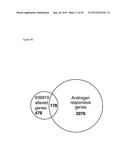 |
 |  |
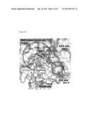 |  |
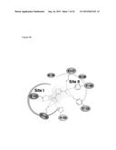 |  |
 | 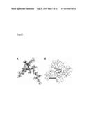 |
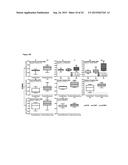 |  |
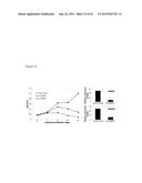 | 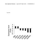 |
 | 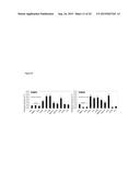 |
 | 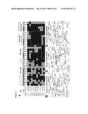 |
 |  |
 |  |
 | 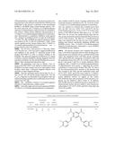 |
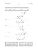 |  |
 | 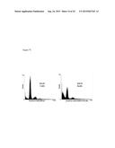 |
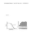 |  |
 |
| New patent applications in this class: | |
| Date | Title |
|---|---|
| 2022-09-08 | Shrub rose plant named 'vlr003' |
| 2022-08-25 | Cherry tree named 'v84031' |
| 2022-08-25 | Miniature rose plant named 'poulty026' |
| 2022-08-25 | Information processing system and information processing method |
| 2022-08-25 | Data reassembly method and apparatus |