Patent application title: ANTIBODIES WITH ENGINEERED IGG FC DOMAINS
Inventors:
George Georgiou (Austin, TX, US)
William Kelton (Austin, TX, US)
Nishant Mehta (Basel, CH)
Assignees:
The Board of Regents of the University of Texas System
IPC8 Class: AC07K1628FI
USPC Class:
Class name:
Publication date: 2015-09-24
Patent application number: 20150266960
Abstract:
Antibody Fc polypeptides are provided that efficiently bind to both
Fcγ and Fcα receptors. In certain aspects, a Fc polypeptide
comprises a chimeric IgG CH2 domain having a α1 loop from IgA and
an IgA CH3 domain. Recombinant antibodies comprising Fc polypeptides of
the embodiments and methods for making and using such antibodies are
likewise provided.Claims:
1. A recombinant polypeptide comprising: (a) a chimeric CH2 domain
comprising a mammalian IgG Fc CH2 domain having a al loop of a mammalian
IgA Fc CH2 domain; and (b) a mammalian IgA Fc CH3 domain.
2. The polypeptide of claim 1, further comprising a mammalian IgG Fc CH1 domain.
3. The polypeptide of claim 1, further comprising an immunoglobulin variable domain.
4. The polypeptide of claim 1, wherein the α1 loop of a mammalian IgA is defined as a mammalian α1 loop corresponding to amino acids 245-257 of a human IgA Fc, according to the Kabat numbering scheme.
5. The polypeptide of claim 4, wherein the α1 loop of a mammalian IgA Fc CH2 domain comprises the sequence PALEDLLLGSEAN.
6. The polypeptide of claim 1, wherein the chimeric CH2 domain comprises the sequence PALEDLLLGSEANG.
7. The polypeptide of claim 1, wherein the mammalian IgA Fc CH3 domain is defined as that mammalian IgA Fc CH3 domain corresponding to amino acids 336-443 of human IgA, according to the Kabat numbering scheme.
8. The polypeptide of claim 7, wherein the mammalian IgA Fc CH3 domain comprises the amino acid sequence of SEQ ID NO: 9.
9. The polypeptide of claim 1, wherein the chimeric CH2 domain comprises one, two or three amino acid positions from the α2 loop of a mammalian IgA Fc CH2 domain.
10. The polypeptide of claim 1, comprising an amino acid sequence at least 90% identical to SEQ ID NO: 7 or SEQ ID NO: 8.
11. The polypeptide of claim 10, comprising an amino acid identical to SEQ ID NOs: 3, 4, 7 or 8.
12. The polypeptide of claim 1, further comprising a FcRn-binding peptide.
13. The polypeptide of claim 12, wherein the FcRn-binding peptide is the linear peptide of SEQ ID NO: 67 or the cyclic peptide of SEQ ID NO: 68.
14. The polypeptide of claim 1, wherein the polypeptide is aglycosylated.
15. A recombinant polypeptide comprising an amino acid sequence at least 90% identical to SEQ ID NO: 7 or SEQ ID NO: 8.
16. The polypeptide of claim 15, comprising an amino acid identical to SEQ ID NOs: 3, 4, 7 or 8.
17. A recombinant antibody comprising the polypeptide of any one of claims 1-16.
18. The antibody of claim 17, wherein the antibody is a human or humanized antibody.
19. The antibody of claim 17, wherein the antibody is aglycosylated.
20. The antibody of claim 17, wherein the antibody binds to a cell surface antigen.
21. The antibody of claim 17, wherein the antibody binds to a cancer cell antigen.
22. The antibody of claim 20, wherein the antibody binds to an EGFR or VEGFR.
23. The antibody of claim 22, wherein the antibody binds to HER2.
24. The antibody of claim 17, wherein the antibody is coupled to a therapeutic, a reporter, or a targeting moiety.
25. The antibody of claim 24, wherein the therapeutic is a nucleotide, a peptide, a small molecule, a therapeutic radionuclide, a chemotherapeutic, a tumor suppressor, an apoptosis inducer, an enzyme, a second antibody, an siRNA, a hormone, a prodrug, or an immunostimulant.
26. The antibody of claim 17, wherein the antibody comprises one or more of the following: (a) decreased affinity for FcγRIIb relative to a wild type IgG antibody; (b) increased affinity for FcγRIIa relative to a wild type IgA antibody; (c) increased affinity for FcγRI relative to a wild type IgA antibody; and (d) increased affinity for FcαRI relative to a wild type IgG antibody.
27. A pharmaceutical composition comprising a recombinant antibody of claim 17.
28. A recombinant polynucleotide molecule encoding a polypeptide of claim 1 or 15.
29. A method of making a polypeptide comprising: (a) expressing a recombinant polynucleotide of claim 28; and (b) obtaining a polypeptide expressed by the cell.
30. A composition for use in treating a subject, the composition comprising an effective amount of a an antibody in accordance with claim 17.
31. A method treating a subject comprising administering an effective amount of an antibody in accordance with claim 17 to the subject.
Description:
[0001] This application claims the benefit of U.S. Provisional Patent
Application No. 61/717,568, filed Oct. 23, 2012, the entirety of which is
incorporated herein by reference.
INCORPORATION OF SEQUENCE LISTING
[0002] The sequence listing that is contained in the file named "UTFB_P1003WO_ST25.txt", which is 53 KB (as measured in Microsoft Windows®) and was created on Aug. 22, 2013, is filed herewith by electronic submission and is incorporated by reference herein.
BACKGROUND OF THE INVENTION
[0003] 1. Field of the Invention
[0004] The present invention relates generally to the field of molecular biology and immunology. More particularly, it concerns molecular antibody design and antibody-based reagents and therapeutics.
[0005] 2. Description of Related Art
[0006] Antibody therapeutics have a dominant market share in the treatment of cancers and other human diseases with estimated sales exceeding USD $56 billion in 2011. Antibodies kill cancer cells in part by engaging and activating immune cells. One important mechanism underlying the potency of antibody therapeutics is the ability of antibody to recruit immune cells to a target antigen (or cell). Thus, the Fc region of an antibody is crucial for recruitment of immunological cells and antibody dependent cytotoxicity (ADCC). In particular, the nature of the ADCC response elicited by antibodies depends on the interaction of the Fc region with receptors (FcRs) located on the surface of many cell types. Humans contain five different classes of Fc receptors. In addition haplotypes, or genetic variants of different FcRs belonging to a particular class are known. The binding of an antibody to FcRs determines its ability to recruit other immunological cells and the type of cell recruited. Hence, the ability to engineer antibodies that can recruit only certain kinds of cells can be critically important for therapy.
[0007] Currently, all antibody therapeutics are of the IgG subclass. Therapeutic IgG antibodies engage the Fcγ class of receptors via Fc domain interaction on the surface of various blood cells which in turn become activated to kill tumor cells. For some therapeutics efficacy could be improved by the additional recruitment of cells that display Fcα or Fcα receptors. However, to date, antibody Fc polypeptides have not been identified that can bind to additional Fc receptors.
SUMMARY OF THE INVENTION
[0008] In a first embodiment there is provided a chimeric polypeptide comprising a CH2 and CH3 domain of a mammalian immunoglobulin molecules, wherein the polypeptide binds to at least one Fcα receptor and at least one Fcγ receptor. For example, in certain aspects, a chimeric polypeptide binds to a FcγRI or FcγRIIa with a higher affinity than an IgA antibody. In further aspect, a chimeric polypeptide binds to FcαRI with a higher affinity than an IgG antibody. In still a further aspect, a chimeric polypeptide binds to a FcγRIIb with a lower affinity than an IgG antibody.
[0009] In a further embodiment, a chimeric polypeptide is provided comprising (a) a chimeric CH2 domain comprising a mammalian IgG Fc CH2 domain having a α1 and/or α2 loop of a mammalian IgA Fc CH2 domain; and (b) a mammalian IgA Fc CH3 domain. In some aspects, the mammalian IgA Fc CH3 domain is defined as a mammalian IgA Fc CH3 domain corresponding to amino acids 336-443 of a human IgA Fc, such as a human IgA1 (SEQ ID NO:9) or IgA2 CH3 domain, based on the Kabat numbering scheme. IgA CH3 domains from several exemplary mammalian species are depicted in the alignment shown in FIG. 19. In certain aspects, the α1 loop of a mammalian IgA is defined as a mammalian IgA al loop corresponding to amino acids 245-257 of a human IgA Fc, according to the Kabat numbering scheme (e.g., a human IgA1 or IgA2 α1 loop). IgA α1 loop domains from several exemplary mammalian species are depicted in the alignment shown in FIG. 19. Thus, in some aspects, the α1 loop of a mammalian IgA Fc CH2 domain comprises the sequence PALEDLLLGSEAN (SEQ ID NO: 14) or PALEDLLLGSEANG (SEQ ID NO: 15) (which includes an additional such that the length of the sequence corresponds with that of the IgG1 loop sequence). In yet a further aspect, a chimeric polypeptide is provided comprising (a) a chimeric CH2 domain comprising a mammalian IgG Fc CH2 domain having a α1 loop of a mammalian IgA Fc CH2 domain; and one, two or three amino acid positions from the α2 loop of a mammalian IgA Fc CH2 domain; and (b) a mammalian IgA Fc CH3 domain. Thus, in certain aspects, a chimeric polypeptide of the embodiments comprises an amino acid sequence at least 85%, 90%, 91%, 92%, 93%, 94%, 95%, 96%, 97%, 98% or 99% identical to SEQ ID NO: 7 or SEQ ID NO: 8. For example, the chimeric polypeptide can comprise a sequence identical to SEQ ID NOs: 3, 4, 7 or 8.
[0010] In further aspects, a chimeric antibody of the embodiments further comprises a FcRn-binding peptide, such as FcRn-binding peptide positioned at the c-terminus of the chimeric antibody. For example, the FcRn-binding peptide can comprise the linear peptide of SEQ ID NO: 67 or the cyclic peptide of SEQ ID NO: 68.
[0011] In still a further embodiment there is provided a chimeric polypeptide of the embodiments comprises an amino acid sequence at least 85%, 90%, 91%, 92%, 93%, 94%, 95%, 96%, 97%, 98% or 99% identical to SEQ ID NO: 7 or SEQ ID NO: 8. For instance, the chimeric polypeptide can comprise a sequence identical to SEQ ID NOs: 3, 4, 7 or 8.
[0012] In a further embodiment there is provided a polynucleotide molecule encoding a chimeric polypeptide of the embodiments. In certain aspects the polynucleotide further encodes a promoter, enhancer, polyadenylation sequence, intron, drug selection marker, origin of replication, a reporter or a purification tag. For example, the isolated polynucleotide can be comprised in a polynucleotide expression vector.
[0013] In a further aspect, a chimeric polypeptide further comprises a mammalian IgG Fc CH1 domain, an IgG VH domain and/or a hinge domain. Thus, in some aspects, a recombinant antibody is provided comprising a chimeric polypeptide of the embodiments. For example, the recombinant antibody can be a human or humanized antibody. In yet further aspects, the chimeric antibody is an aglycosylated antibody.
[0014] In certain aspects, an antibody of the embodiments binds to a cell surface antigen or to a cancer cell antigen. For example, in certain aspects, an antibody binds to an EGFR (e.g., HER1 or HER2) or VEGFR. In certain aspects, an antibody of the embodiments bind to the same antigen as one of the antibodies selected from Table A. In still a further aspect, an antibody of the embodiments comprise CDRs 1, 2, 3, 4, 5 and 6 of an antibody of Table A.
[0015] In further aspects an antibody according to the embodiments is coupled to a therapeutic, a reporter, or a targeting moiety. For example, the therapeutic can be a polynucleotide (e.g., a miRNA, a siRNA or an therapeutic gene), a peptide, a small molecule, a therapeutic radionuclide, a chemotherapeutic, a tumor suppressor, an apoptosis inducer, an enzyme, a second antibody, an siRNA, a hormone, a prodrug, or an immunostimulant. Examples of reporters include, but are not limited to, radionuclides, florophores, MRI contrast agents, enzymes, dyes or molecules detectable by positron emission tomography (PET).
[0016] In yet further aspects, an antibody of the embodiments comprises one or more of the following: (a) decreased affinity for FcγRIIb relative to a wild type IgG1 antibody; (b) increased affinity for FcγRIIa relative to a wild type IgA1 antibody; (c) increased affinity for FcγRI relative to a wild type IgA1 antibody; and/or (d) increased affinity for FcαRI relative to a wild type IgG1 antibody.
[0017] In some embodiments, the invention provides a pharmaceutical composition comprising a chimeric polypeptide or a recombinant antibody of the embodiments in a pharmaceutically acceptable carrier.
[0018] In a further embodiment there is provided a method of making a polypeptide comprising: (a) expressing a recombinant polynucleotide encoding a chimeric polypeptide of the embodiments in a cell; and (b) obtaining a polypeptide expressed by the cell. For example, the cell can be a bacterial cell, a yeast cell, a ciliate cell, or an insect cell.
[0019] In still a further embodiment, there is provided a method treating a subject comprising administering an effective amount of an antibody of the embodiment to the subject. In certain aspects, the subject has an autoimmune disease or a cancer. For example, in some aspects, the subject has one of the diseases indicated in Table A. Routes for administering an antibody include, without limitation, by intravenous administration, intracardiac administration, intradermal administration, intralesional administration, intrathecal administration, intracranial administration, intrapericardial administration, intraumbilical administration, intraocular administration, intraarterial administration, intraperitoneal administration, intraosseous administration, intrahemmorhage administration, intratrauma administration, intratumor administration, subcutaneous administration, intramuscular administration, intravitreous administration, direct injection into a normal tissue, or by direct injection into a tumor.
[0020] In certain embodiments, there is provided an isolated or recombinant HER2-binding antibody wherein the antibody comprises a chimeric immunoglobulin heavy constant region of the embodiments. In some aspects, a HER2-binding antibody of the embodiments comprises 1, 2, 3, 4, 5, or all 6 of the CDRs of the Trastuzumab monoclonal antibody. For example, the HER2-binding antibody can comprises an amino acid sequence at least about 75%, 80%, 85%, 90%, 95%, 98% or 99% identical to the heavy chain and/or light chain variable region amino acid sequence of the Trastuzumab monoclonal antibody.
[0021] In certain embodiments, there is provided an isolated or recombinant HER1-binding antibody wherein the antibody comprises a chimeric immunoglobulin heavy constant region of the embodiments. In some aspects, a HER1-binding antibody of the embodiments comprises 1, 2, 3, 4, 5, or all 6 of the CDRs of the Cetuximab monoclonal antibody. For example, the HER1-binding antibody can comprises an amino acid sequence at least about 75%, 80%, 85%, 90%, 95%, 98% or 99% identical to the heavy chain and/or light chain variable region amino acid sequence of the Cetuximab monoclonal antibody.
[0022] As used herein the specification, "a" or "an" may mean one or more. As used herein in the claim(s), when used in conjunction with the word "comprising", the words "a" or "an" may mean one or more than one.
[0023] The use of the term "or" in the claims is used to mean "and/or" unless explicitly indicated to refer to alternatives only or the alternatives are mutually exclusive, although the disclosure supports a definition that refers to only alternatives and "and/or." As used herein "another" may mean at least a second or more.
[0024] Throughout this application, the term "about" is used to indicate that a value includes the inherent variation of error for the device, the method being employed to determine the value, or the variation that exists among the study subjects.
[0025] Other objects, features and advantages of the present invention will become apparent from the following detailed description. It should be understood, however, that the detailed description and the specific examples, while indicating preferred embodiments of the invention, are given by way of illustration only, since various changes and modifications within the spirit and scope of the invention will become apparent to those skilled in the art from this detailed description.
BRIEF DESCRIPTION OF THE DRAWINGS
[0026] The following drawings form part of the present specification and are included to further demonstrate certain aspects of the present invention. The invention may be better understood by reference to one or more of these drawings in combination with the detailed description of specific embodiments presented herein.
[0027] FIG. 1A-B--FIG. 1A: Amino acid alignment of mutants B (SEQ ID NO: 2), C (SEQ ID NO: 3), and D (SEQ ID NO: 4) with wild-type IgG1 (SEQ ID NO: 1). Black lettering is CH2 sequence; gray lettering is CH3 sequence; underlined lettering is derived from IgA1. The additional glycine in mutant D is italicized. FIG. 1B: Wild-type IgA sequence with alanine scanning mutations underlined (SEQ ID NO: 13).
[0028] FIG. 2: Show the results of SDS-PAGE (4%-20%) analysis of chimeric Fc polypeptides of the embodiments.
[0029] FIG. 3: Graph shows the binding of chimeric Fc polypeptides of the embodiments to FcαRI-GST as assessed by ELISA assay.
[0030] FIG. 4: Graph shows the binding of chimeric Fc polypeptides of the embodiments to FcγRI as assessed by ELISA assay.
[0031] FIG. 5: Graph shows the binding of chimeric Fc polypeptides of the embodiments to FcγRIIa as assessed by ELISA assay.
[0032] FIG. 6: Graph shows the binding of chimeric Fc polypeptides of the embodiments to FcγRIIb as assessed by ELISA assay.
[0033] FIG. 7: Sites mutated to alanine to scan the binding pocket of FcαRI in IgA for the relative binding contribution of each position. Picture shows a model of the IgA binding pocket.
[0034] FIG. 8: Graph shows the results of ELISA analysis of FcαRI binding to IgA Fc domains containing alanine scanning point mutations. Signal has been normalized to a wild type IgA Fc domain.
[0035] FIG. 9: Figure shows results of a 4-20% SDS-PAGE separation of full length antibodies expressed from HEK293F cells. Lane A trastuzumab IgG, Lane B trastuzumab IgG N297D, Lane C trastuzumab MutD, Lane D trastuzmab IgA
[0036] FIG. 10: SPR kinetic analysis of MutD binding to soluble FcγRI as fit by a 1:1 Langmuir isotherm model.
[0037] FIG. 11: SPR kinetic analysis of MutD binding to soluble FcαRI-GST as fit by a 2:1 bivalent analyte model.
[0038] FIG. 12: SPR kinetic analysis of IgA binding to soluble FcαRI-GST as fit by a 2:1 bivalent analyte model.
[0039] FIG. 13: SPR equilibrium analysis of IgG binding to soluble monmeric FcγRIIaR131 as fit by a 1:1 Langmuir isotherm model.
[0040] FIG. 14: SPR equilibrium analysis of MutD binding to soluble monmeric FcγRIIaR131 as fit by a 1:1 Langmuir isotherm model
[0041] FIG. 15: SPR equilibrium analysis of IgG binding to soluble monmeric FcγRIIb as fit by a 1:1 Langmuir isotherm model.
[0042] FIG. 16: SPR equilibrium analysis of MutD binding to soluble monmeric FcγRIIb as fit by a 1:1 Langmuir isotherm model.
[0043] FIG. 17: Graphs show neutrophil killing of Her2+SkBR-3 cells by ADCC using IgG, aglycosylated IgG N297D, IgA and MutD trastuzumab variants. Cells were seeded at a ratio of 80:1 neutrophils to tumor cells. Bars in the graph indicate the results with decreasing concentration of the indicated antibodies (20, 2 or 0.2 μg/ml) from left to right.
[0044] FIG. 18: Graph show the results of ELISA analysis of FcRN binding to MutD fusions with cyclic or linear C-terminus peptides at pH 7.4 and pH 5.8.
[0045] FIG. 19: An example alignment showing various mammalian IgA Fc domains with the locations of the CH2 α1 loop and CH3 domains indicated. Sequences in the alignment from top to bottom include those from the human IgA1 Fc (SEQ ID NO: 77); human IgA2 Fc (SEQ ID NO: 78); mouse IgA Fc (SEQ ID NO: 79); rabbit IgA Fc (SEQ ID NO: 80); gorilla IgA Fc (SEQ ID NO: 81); and pig IgA Fc (SEQ ID NO: 82).
DESCRIPTION OF ILLUSTRATIVE EMBODIMENTS
I. The Present Invention
[0046] Antibody-based therapeutics have recently been approved for treatment of a wide range of diseases from cancer to autoimmune disease (see, e.g., Table A). Recently, recombinant antibodies have even been developed for treatment of infectious disease. However, the efficacy of most antibody-based therapeutics is dependent on the effector function of cells and factors that bind to the antibodies, such as complement. For example, antibodies that target specific cell types are currently used as anticancer therapies, but many antibodies are either not directly toxic to the cancer cells or have low cytotoxicity. Thus, despite the targeting specificity of such antibodies, many fail to exhibit sufficient effect (e.g., cell killing) on target cells.
[0047] In contrast to IgG, which have been the only type of therapeutic antibody used to date, antibodies that belong to the IgA class engage the widely expressed FcαRI receptor on neutrophils. These cells comprise the greatest fraction of innate effector cells found in circulation and can activate potent inflammatory action when FcαRI is cross-linked by IgA immune complexes. Both antibody dependent cell mediated cytotoxicity (ADCC) (oxidative burst, degranulation) and antibody dependent cell mediated phagocytosis (ADCP) responses have been observed upon FcαRI activation. However, IgG antibodies do not engage the FcαRI receptor and hence poorly activate neutrophils and other granulocytes.
[0048] Despite their potential promise, use of the IgA antibody isotype to exploit the potent inflammatory effects of FcαRI in a therapeutic setting has so far been disfavored due to low expression yields and expensive purification schemes required for IgA antibodies. Further, IgA antibodies do not display the favorable therapeutic functions that IgG antibodies do. Combination therapy of IgA with IgG is likewise undesirable both due to regulatory and cost concerns and also due to the blocking of neutrophils activated via FcαRI action by the engagement of the FcγRIIb receptor by IgG.
[0049] The polypeptides and antibodies provided herein address these issues by providing Fc domains that are able to bind both Fcα and Fcγ receptors. Specifically, an IgG1 antibody Fc domain has been engineered to introduce de novo binding to the IgA FcαRI receptor while retaining existing affinity for FcγRs. For example, chimeric Fc domains that comprise sequences from both IgG and IgA Fc's are shown to have significant FcαRI binding (see FIG. 3). Nonetheless, the polypeptides retain significant binding to FcγRI and FcγRIIa (FIGS. 4-5). Perhaps most importantly, the new chimeric Fc polypeptides retain only limited affinity for FcγRIIb, a receptor that can block activation of key effect cells such as neutrophils (FIG. 6). Moreover, chimeric antibodies could be further modified by the additional of an FcRn binding peptide (FIG. 18), a modification that may increase serum half life of the molecules. Thus, the new chimeric Fc polypeptides and antibodies that incorporate the polypeptides offer a broader range of effector function relative to either IgG or IgA alone, which could greatly improve the efficacy of new antibody therapeutics. Moreover, the new chimeric Fc polypeptides can be used to replace the Fc domains of existing antibody therapeutics, thereby rendering them more effective. This effect is exemplified in studies presented here. For example, when a chimeric Fc domain (MutD) was fused to trastuzumab the new chimeric antibody was able to mediate neutrophil killing of Her expressing cells with high efficiency (see FIG. 17).
II. IgA and FcαRI
[0050] Monomeric IgA is the second most abundant immunoglobulin in human serum after the IgG isotype. IgA antibodies, typically implicated in mucosal surface defense, are represented in higher serum concentrations in humans than in the sera of other animal species. Together, the two IgA allotypes comprise up to 25% of human serum immunoglobulins. Also unique to humans is the high proportion of monomeric serum IgA (˜90%) as opposed to J chain linked dimeric forms (Woof and Kerr, 2006).
[0051] IgA binds selectively to the FcαR1 (CD89) receptor, which is constitutively expressed on cells of the myeloid lineage, including neutrophils, macrophages, and eosinophils, in humans. Membrane anchored FcαRI is found expressed with or without association to the Fcγ signaling chain. If unassociated, FcαRI internalizes and recycles IgA to the cell surface. However, even with this recycling mechanism, the circulation half-life is significantly less than that of IgG at 5.9 days for serum IgA1 and 4.5 days for serum IgA2 (Morell et al., 1973). On the other hand, association of IgA chains with CD89 is essential for IgA facilitated Ag presentation, oxidative burst, cytokine release, and tumor killing (Jacob et al., 2008). There are at least ten spliced isoforms of CD89 reported, the majority of which have undefined functions (Qian et al., 2008). Despite significant effort, a murine homologue of this receptor has not been identified. It has been suggested that mice lost the FcαR1 gene during a translocation event from chromosome 7 to the X chromosome (Woof and Kerr, 2004). IgA immune complexes have been reported to induce strong inflammatory responses and demonstrate efficient tumor killing by neutrophil recruitment upon binding FcαRI (van Egmond et al., 2001; Stockmeyer et al., 2000). In fact, tumor killing by IgA activation of the FcαR1 receptor on neutrophils has been shown to be more potent than IgG tumor killing by activation of FcγR1 and co-activation of both receptors by bispecific antibodies showed an additive effect in tumor treatment. However, therapeutic dosing with full-length IgA and IgG in combination was less successful as the Fc domain of IgG1 binds FcγRIIb, an inhibitory receptor, which reduced the tumor killing efficiency of IgA. Development of an IgA Fc domain engineered to bind the FcγR1 receptor has the potential to exploit both tumor killing pathways without activation of inhibitory functions.
[0052] IgA antibodies have not yet received attention for therapeutic applications partially because their production is costly and inefficient but also because their half-life in serum is shorter than that of IgG antibodies. Part of the concern in IgA expression is the heavily glycosylated hinge region that includes up to five O-linked glycosylation sites. Glycosylation not only impacts the expression level but also introduces the potential for large glycan heterogeneity during production. By engineering FcαRI binding into an IgG molecule, many of these concerns can be alleviated. To further define the FcαRI binding pocket in IgA1, point mutations were generated in either of two interdomain loops (Leu-257-Gly-259 in the CH2 domain; Pro-440-Phe-443 in the CH3 domain). Each substitution, with the exception of G259R, resulted in greatly ablated FcαRI binding.
II. Modified Proteins and Polypeptides
[0053] Embodiments concerns modified polypeptides, in particular polypeptides comprising chimeric Fc domains that confer binding to both IgGγ and IgGa receptors. Preferably the chimeric polypeptides have limited binding affinity for IgGγIIb. Polypeptides of the embodiments comprise portions of an IgG Fc domain (e.g., an IgG1 Fc) and portions of an IgA Fc domain. In certain aspects, a chimeric polypeptide comprises a Fc CH2 and CH3 domain sequence that is composed of chimeric domains of human IgA (e.g., IgA1 or IgA2, NCBI accession nos. P01876.2 and P01877.3, respectively incorporated herein by reference; see also SEQ ID NO: 9) and human IgG (e.g., IgG1, IgG2, IgG3 or IgG4, NCBI accession nos. P01857.1; P01859.2; P01860.2; and P01861.1, respectively, incorporated herein by reference). As used herein, numbering of amino acid position in a chimeric polypeptide refers to the standard nomenclature set forth, e.g., in Kabat et al., 1991, incorporated herein by reference.
[0054] CH3 Domain
[0055] In certain aspects, a chimeric polypeptide of the embodiments comprises a CH3 from a mammalian immunoglobulin. Preferably the CH3 is substantially homologous to a human IgA CH3 domain. For example, the CH3 domain can be at least about 85%, 90%, 91%, 92%, 93%, 94%, 95%, 96%, 97%, 98%, 99% or 100% identical to the CH3 of a human IgA1 or IgA2 (see, e.g., NCBI accession nos. P01876.2 and P01877.3, incorporated herein by reference). In further aspects, a CH3 domain of the embodiment may comprise 1, 2, 3, 4, 5, 6, 7, 8, 9 or 10 amino acid positions that are substituted with the amino at a corresponding position of an IgG CH3 domain, such as a human IgG1. Thus, in certain specific aspects, a chimeric polypeptide of the embodiments comprises a CH3 domain that is 85%, 90%, 91%, 92%, 93%, 94%, 95%, 96%, 97%, 98%, 99% or 100% identical to a CH3 domain (positions 336-443; SEQ ID NO: 9) of a human IgA1.
[0056] In still further aspects, a chimeric polypeptide of the embodiments comprises a CH3 domain that is 85%, 90%, 91%, 92%, 93%, 94% or 95%, identical to a CH3 domain (positions 336-443; SEQ ID NO: 10) of a human IgG1 and comprising 1, 2, 3, 4, 5, 6, 7, 8, 9 or 10 amino acid positions that are substituted with the amino at a corresponding position of an IgA CH3 domain, such as a human IgA1. For example, the CH3 domain of a chimeric polypeptide can comprise an amino acid substitution corresponding to F443, E437, M433, E389 and/or R382, as shown in FIG. 1B.
[0057] CH2 Domain
[0058] A chimeric polypeptide of the embodiments, in certain aspects, comprises a chimeric CH2 from a mammalian immunoglobulin. Preferably, the chimeric CH2 domain comprises chimeric domains of human IgG and IgA CH2 domains. For example, the CH2 domain can comprise a IgG CH2 domain having an α1 and/or α2 loop domain from an IgA. In some specific aspects, a chimeric CH2 domain comprises a CH2 domain from a human IgG1 having an α1 and/or α2 loop domain from a human IgA1. In still further aspects a chimeric CH2 domain comprises a CH2 domain from a human IgG1 having 1, 2, 3, 4 or 5 amino acids substituted for an amino acid at a corresponding position in an α1 and/or α2 loop domain from a human IgA1. Thus, in some aspects, a chimeric CH2 domain of the embodiments is at least about 85%, 90%, 91%, 92%, 93%, 94%, 95%, 96%, 97%, 98%, 99% or 100% identical to the CH2 of the "Mutant C" or "Mutant D" polypeptide (SEQ ID NOs:11 and 12, respectively). In still further aspects, a CH2 domain of the embodiment may comprise 1, 2, 3, 4, 5, 6, 7, 8, 9 or 10 amino acid positions that is a substituted with the amino at a corresponding position of an IgA CH2 domain, such as a human IgA1. For example, the CH2 domain of a chimeric polypeptide can comprise an amino acid substitution corresponding to N316, E313 and/or L258, as shown in FIG. 1B.
[0059] In some aspects, a chimeric CH2 domain of the embodiments can comprise a conservative amino acid insertion, deletion of substitution as further outlined below. For instance the chimeric CH2 domain can comprise an amino acid insertion to maintain the relative lengths of domains between IgG and IgA, as was done in the case of the "Mutant D" polypeptide where a Gly was inserted after the IgA α1 loop sequence.
[0060] Additional Domains
[0061] In certain aspects, a chimeric polypeptide of the embodiments further comprises a CH1 and/or hinge domain from a mammalian immunoglobulin. Preferably the CH1 and/or hinge domain are from a human IgG polypeptide. For example, the CH1 and/or hinge domain can be a at least about 85%, 90%, 91%, 92%, 93%, 94%, 95%, 96%, 97%, 98%, 99% or 100% identical to the CH1 and/or hinge domain of a human IgG1, IgG2, IgG3 or IgG4 (see, e.g., NCBI accession nos. P01857.1; P01859.2; P01860.2; and P01861.1, incorporated herein by reference). In some aspects, a CH1 domain of the embodiments can comprise a conservative amino acid insertion, deletion of substitution as outlined below.
[0062] In still further aspects a chimeric polypeptide of the embodiments further comprises a VH domain. In further aspects, a chimeric polypeptide is further associated with (e.g., via disulfides bonds) an immunoglobulin light chain. Thus, in some aspects, a chimeric polypeptide of the embodiments comprises an antibody.
[0063] It is further contemplated that additional modifications could be made to a chimeric polypeptide of the embodiments. For example, a modified chimeric polypeptide can be made that exhibits at least one functional activity that is comparable to the unmodified version, yet the modified protein or polypeptide possesses an additional advantage over the unmodified version, such as cheaper to production, eliciting fewer side effects, and/or having better or longer efficacy or bioavailability. Thus, when the present application refers to the function or activity of a "modified chimeric polypeptide" one of ordinary skill in the art would understand that this includes, for example, polypeptide that 1) performs at least one of the same activities or has at least one of the same specificities as the unmodified polypeptide, but that may have a different level of another activity or specificity; and 2) possesses an additional advantage over the unmodified polypeptide. Determination of activity may be achieved using assays familiar to those of skill in the art, particularly with respect to the protein's activity, and may include for comparison purposes, for example, the use of native and/or recombinant versions of either the modified or unmodified protein or polypeptide. In addition to the modified polypeptides discussed herein, embodiments may involve domains, polypeptides, described in WO 2008/137475, which is hereby specifically incorporated by reference.
[0064] Modified chimeric polypeptides may possess deletions and/or substitutions of amino acids; thus, a protein with a deletion, a protein with a substitution, and a protein with a deletion and a substitution are modified proteins. In some embodiments these modified proteins may further include insertions or added amino acids, such as with fusion proteins or proteins with linkers, for example (see, e.g., the Gly insertion in "Mutant D" polypeptide). A "modified deleted polypeptide" lacks one or more residues of the native protein, but possesses the specificity and/or activity of the native polypeptide. A "modified deleted polypeptide" may also have reduced immunogenicity or antigenicity. An example of a modified deleted polypeptide is one that has an amino acid residue deleted from at least one antigenic region--that is, a region of the protein determined to be antigenic in a particular organism, such as the type of organism that may be administered the modified protein.
[0065] Substitutional or replacement variants typically contain the exchange of one amino acid for another at one or more sites within the polypeptide and may be designed to modulate one or more properties of the polypeptide, particularly its effector functions and/or bioavailability. Certain specific amino acid exchanges in chimeric polypeptides of the embodiments are detailed above. Further substitutions may or may not be conservative, that is, one amino acid is replaced with one of similar shape and charge. Conservative substitutions are well known in the art and include, for example, the changes of: alanine to serine; arginine to lysine; asparagine to glutamine or histidine; aspartate to glutamate; cysteine to serine; glutamine to asparagine; glutamate to aspartate; glycine to proline; histidine to asparagine or glutamine; isoleucine to leucine or valine; leucine to valine or isoleucine; lysine to arginine; methionine to leucine or isoleucine; phenylalanine to tyrosine, leucine or methionine; serine to threonine; threonine to serine; tryptophan to tyrosine; tyrosine to tryptophan or phenylalanine; and valine to isoleucine or leucine.
[0066] In addition to a deletion or substitution, a modified polypeptide may possess an insertion of residues, which typically involves the addition of at least one residue in the polypeptide. This may include the insertion of a targeting polypeptide or simply a single residue. Terminal additions, called fusion proteins, are discussed below.
[0067] It also will be understood that amino acid and nucleic acid sequences may include additional residues, such as additional N- or C-terminal amino acids or 5' or 3' sequences, and yet still be essentially as set forth in one of the sequences disclosed herein, so long as the sequence meets the criteria set forth above, including the maintenance of biological protein activity where protein expression is concerned. The addition of terminal sequences particularly applies to nucleic acid sequences that may, for example, include various non-coding sequences flanking either of the 5' or 3' portions of the coding region or may include various internal sequences, i.e., introns, which are known to occur within genes. Thus, in certain, aspects a chimeric polypeptide of the embodiments comprises from N- to C-terminus: a VH domain-CH1 domain-hinge domain-chimeric CH2 domain-a CH3 domain (e.g., an IgA CH3 domain).
[0068] The following is a discussion on strategies for changing of the amino acids of a polypeptide to create an equivalent, or even an improved, second-generation molecule. For example, certain amino acids may be substituted for other amino acids in a polypeptide structure with or without appreciable loss of interactive binding capacity with structures such as, for example, binding sites to substrate molecules. This is especially possible in polypeptides that have a well know structure, such as antibody polypeptides (see, e.g., in Kabat et al., 1991).
[0069] In making changes, in some cases, the hydropathic index of amino acids may be considered. The importance of the hydropathic amino acid index in conferring interactive biologic function on a protein is generally understood in the art (Kyte & Doolittle, 1982). It is accepted that the relative hydropathic character of the amino acid contributes to the secondary structure of the resultant protein, which in turn defines the interaction of the protein with other molecules, for example, enzymes, substrates, receptors, DNA, antibodies, antigens, and the like.
[0070] It also is understood in the art that the substitution of like amino acids can be made effectively on the basis of hydrophilicity. U.S. Pat. No. 4,554,101, incorporated herein by reference, states that the greatest local average hydrophilicity of a protein, as governed by the hydrophilicity of its adjacent amino acids, correlates with a biological property of the protein. As detailed in U.S. Pat. No. 4,554,101, the following hydrophilicity values have been assigned to amino acid residues: arginine (+3.0); lysine (+3.0); aspartate (+3.0±1); glutamate (+3.0±1); serine (+0.3); asparagine (+0.2); glutamine (+0.2); glycine (0); threonine (-0.4); proline (-0.5±1); alanine (-0.5); histidine (-0.5); cysteine (-1.0); methionine (-1.3); valine (-1.5); leucine (-1.8); isoleucine (-1.8); tyrosine (-2.3); phenylalanine (-2.5); tryptophan (-3.4).
[0071] It is understood that an amino acid can be substituted for another having a similar hydrophilicity value and still produce a biologically equivalent and immunologically equivalent protein. In such changes, the substitution of amino acids whose hydrophilicity values are within ±2 is preferred, those that are within ±1 are particularly preferred, and those within ±0.5 are even more particularly preferred.
[0072] As outlined above, amino acid substitutions generally are based on the relative similarity of the amino acid side-chain substituents, for example, their hydrophobicity, hydrophilicity, charge, size, and the like. Exemplary substitutions that take into consideration the various foregoing characteristics are well known to those of skill in the art and include: arginine and lysine; glutamate and aspartate; serine and threonine; glutamine and asparagine; and valine, leucine and isoleucine.
III. Nucleic Acid-Based Expression Systems
[0073] Nucleic acid-based expression systems may find use, in certain embodiments of the invention, for the expression of recombinant polypeptides and antibodies of the embodiments. Thus, certain aspects of the embodiments concern polynucleotide that encode a chimeric polypeptide of the embodiments. Below is a discussion of polynucleotide vector for expression of chimeric polypeptides and methods for production of the polypeptides. As used herein the terms "polynucleotide" and "nucleic acids" are used interchangeably.
[0074] A. Methods of Polynucleotide Delivery
[0075] Certain aspects of the embodiments may comprise delivery of polynucleotide to target cells (e.g., host cells). For example, bacterial, yeast or mammalian host cells may be transformed with nucleic acids encoding chimeric polypeptides. Suitable methods for nucleic acid delivery for transformation of a cell are believed to include virtually any method by which a nucleic acid (e.g., DNA) can be introduced into such a cell, or even an organelle thereof. Such methods include, but are not limited to, direct delivery of DNA such as by injection (U.S. Pat. Nos. 5,994,624, 5,981,274, 5,945,100, 5,780,448, 5,736,524, 5,702,932, 5,656,610, 5,589,466 and 5,580,859, each incorporated herein by reference), including microinjection (Harland and Weintraub, 1985; U.S. Pat. No. 5,789,215, incorporated herein by reference); by electroporation (U.S. Pat. No. 5,384,253, incorporated herein by reference); by calcium phosphate precipitation (Graham and Van Der Eb, 1973; Chen and Okayama, 1987; Rippe et al., 1990); by using DEAE-dextran followed by polyethylene glycol (Gopal, 1985); by direct sonic loading (Fechheimer et al., 1987); by liposome mediated transfection (Nicolau and Sene, 1982; Fraley et al., 1979; Nicolau et al., 1987; Wong et al., 1980; Kaneda et al., 1989; Kato et al., 1991); by microprojectile bombardment (PCT Application Nos. WO 94/09699 and 95/06128; U.S. Pat. Nos. 5,610,042; 5,322,783, 5,563,055, 5,550,318, 5,538,877 and 5,538,880, and each incorporated herein by reference); or by agitation with silicon carbide fibers (Kaeppler et al., 1990; U.S. Pat. Nos. 5,302,523 and 5,464,765, each incorporated herein by reference); by desiccation/inhibition-mediated DNA uptake (Potrykus et al., 1985). Through the application of techniques such as these, cells may be stably or transiently transformed.
[0076] B. Vectors
[0077] Vectors may find use with the current invention, for example, in the transformation of a host cells with a nucleic acid sequence encoding a chimeric polypeptides. The term "vector" is used to refer to a carrier nucleic acid molecule into which a nucleic acid sequence can be inserted for introduction into a cell where it can be replicated. A nucleic acid sequence can be "exogenous," or "heterologous", which means that it is foreign to the cell into which the vector is being introduced or that the sequence is homologous to a sequence in the cell but in a position within the host cell nucleic acid in which the sequence is ordinarily not found. Vectors include plasmids, cosmids and viruses (e.g., bacteriophage). One of skill in the art may construct a vector through standard recombinant techniques, which are described in Maniatis et al., 1988 and Ausubel et al., 1994, both of which references are incorporated herein by reference.
[0078] The term "expression vector" refers to a vector containing a nucleic acid sequence coding for at least part of a gene product capable of being transcribed. In some cases, RNA molecules are then translated into a polypeptide (e.g., a chimeric polypeptide). Expression vectors can contain a variety of "control sequences," which refer to nucleic acid sequences necessary for the transcription and possibly translation of an operably linked coding sequence in a particular host organism. In addition to control sequences that govern transcription and translation, vectors and expression vectors may contain nucleic acid sequences that serve other functions as well and are described infra.
[0079] 1. Promoters and Enhancers
[0080] A "promoter" is a control sequence that is a region of a nucleic acid sequence at which initiation and rate of transcription are controlled. It may contain genetic elements at which regulatory proteins and molecules may bind such as RNA polymerase and other transcription factors. The phrases "operatively positioned," "operatively linked," "under control," and "under transcriptional control" mean that a promoter is in a correct functional location and/or orientation in relation to a nucleic acid sequence to control transcriptional initiation and/or expression of that sequence. A promoter may or may not be used in conjunction with an "enhancer," which refers to a cis-acting regulatory sequence involved in the transcriptional activation of a nucleic acid sequence.
[0081] A promoter may be one naturally associated with a gene or sequence, as may be obtained by isolating the 5' non-coding sequences located upstream of the coding segment and/or exon. Such a promoter can be referred to as "endogenous." Similarly, an enhancer may be one naturally associated with a nucleic acid sequence, located either downstream or upstream of that sequence. Alternatively, certain advantages will be gained by positioning the coding nucleic acid segment under the control of a recombinant or heterologous promoter, which refers to a promoter that is not normally associated with a nucleic acid sequence in its natural environment. A recombinant or heterologous enhancer refers also to an enhancer not normally associated with a nucleic acid sequence in its natural environment. Such promoters or enhancers may include promoters or enhancers of other genes, and promoters or enhancers isolated from any other prokaryotic cell, and promoters or enhancers not "naturally occurring," i.e., containing different elements of different transcriptional regulatory regions, and/or mutations that alter expression. In addition to producing nucleic acid sequences of promoters and enhancers synthetically, sequences may be produced using recombinant cloning and/or nucleic acid amplification technology, including PCR®, in connection with the compositions disclosed herein (see U.S. Pat. No. 4,683,202, U.S. Pat. No. 5,928,906, each incorporated herein by reference).
[0082] Naturally, it will be important to employ a promoter and/or enhancer that effectively directs the expression of the DNA segment in the cell type chosen for expression. One example of such promoter that may be used with the invention is the E. coli arabinose or T7 promoter. Those of skill in the art of molecular biology generally are familiar with the use of promoters, enhancers, and cell type combinations for protein expression, for example, see Sambrook et al. (1989), incorporated herein by reference. The promoters employed may be constitutive, tissue-specific, inducible, and/or useful under the appropriate conditions to direct high level expression of the introduced DNA segment, such as is advantageous in the large-scale production of recombinant proteins and/or peptides. The promoter may be heterologous or endogenous.
[0083] 2. Initiation Signals and Internal Ribosome Binding Sites
[0084] A specific initiation signal also may be required for efficient translation of coding sequences. These signals include the ATG initiation codon or adjacent sequences. Exogenous translational control signals, including the ATG initiation codon, may need to be provided. One of ordinary skill in the art would readily be capable of determining this and providing the necessary signals. It is well known that the initiation codon must be "in-frame" with the reading frame of the desired coding sequence to ensure translation of the entire insert. The exogenous translational control signals and initiation codons can be either natural or synthetic. The efficiency of expression may be enhanced by the inclusion of appropriate transcription enhancer elements.
[0085] 3. Multiple Cloning Sites
[0086] Vectors can include a multiple cloning site (MCS), which is a nucleic acid region that contains multiple restriction enzyme sites, any of which can be used in conjunction with standard recombinant technology to digest the vector (see Carbonelli et al., 1999, Levenson et al., 1998, and Cocea, 1997, incorporated herein by reference.) "Restriction enzyme digestion" refers to catalytic cleavage of a nucleic acid molecule with an enzyme that functions only at specific locations in a nucleic acid molecule. Many of these restriction enzymes are commercially available. Use of such enzymes is understood by those of skill in the art. Frequently, a vector is linearized or fragmented using a restriction enzyme that cuts within the MCS to enable exogenous sequences to be ligated to the vector. "Ligation" refers to the process of forming phosphodiester bonds between two nucleic acid fragments, which may or may not be contiguous with each other. Techniques involving restriction enzymes and ligation reactions are well known to those of skill in the art of recombinant technology.
[0087] 4. Termination Signals
[0088] The vectors or constructs prepared in accordance with the present invention will generally comprise at least one termination signal. A "termination signal" or "terminator" is comprised of the DNA sequences involved in specific termination of an RNA transcript by an RNA polymerase. Thus, in certain embodiments, a termination signal that ends the production of an RNA transcript is contemplated. A terminator may be necessary in vivo to achieve desirable message levels.
[0089] Terminators contemplated for use in the invention include any known terminator of transcription described herein or known to one of ordinary skill in the art, including but not limited to, for example, rhp dependent or rho independent terminators. In certain embodiments, the termination signal may be a lack of transcribable or translatable sequence, such as due to a sequence truncation.
[0090] 5. Origins of Replication
[0091] In order to propagate a vector in a host cell, it may contain one or more origins of replication sites (often termed "ori"), which is a specific nucleic acid sequence at which replication is initiated.
[0092] 6. Selectable and Screenable Markers
[0093] In certain embodiments of the invention, cells containing a nucleic acid construct of the present invention may be identified in vitro or in vivo by including a marker in the expression vector. Such markers would confer an identifiable change to the cell permitting easy identification of cells containing the expression vector. Generally, a selectable marker is one that confers a property that allows for selection. A positive selectable marker is one in which the presence of the marker allows for its selection, while a negative selectable marker is one in which its presence prevents its selection. An example of a positive selectable marker is a drug resistance marker.
[0094] Usually the inclusion of a drug selection marker aids in the cloning and identification of transformants, for example, genes that confer resistance to neomycin, puromycin, hygromycin, DHFR, GPT, zeocin and histidinol are useful selectable markers. In addition to markers conferring a phenotype that allows for the discrimination of transformants based on the implementation of conditions, other types of markers including screenable markers such as GFP, whose basis is colorimetric analysis, are also contemplated. Alternatively, screenable enzymes such as chloramphenicol acetyltransferase (CAT) may be utilized. One of skill in the art would also know how to employ immunologic markers, possibly in conjunction with FACS analysis. The marker used is not believed to be important, so long as it is capable of being expressed simultaneously with the nucleic acid encoding a gene product. Further examples of selectable and screenable markers are well known to one of skill in the art.
[0095] C. Host Cells
[0096] In the context of expressing a chimeric polypeptide, "host cell" refers to a cell, of any transformable organism that is capable of replicating a vector and/or expressing a heterologous gene encoded by a vector. A host cell can, and has been, used as a recipient for vectors. A host cell may be "transfected" or "transformed," which refers to a process by which exogenous nucleic acid is transferred or introduced into the host cell. A transformed cell includes the primary subject cell and its progeny. Host cells for use according to the embodiments include, without limitation, insect cells, mammalian cell (e.g., CHO cells), ciliate cells, yeast cells and bacterial cells.
[0097] For example, numerous prokaryotic cell lines and cultures are available for use as a host cell, and they can be obtained through the American Type Culture Collection (ATCC), which is an organization that serves as an archive for living cultures and genetic materials (www.atcc.org). An appropriate host can be determined by one of skill in the art based on the vector backbone and the desired result. A plasmid or cosmid, for example, can be introduced into a prokaryote host cell for replication of many vectors. Bacterial cells used as host cells for vector replication and/or expression include DH5α, JM109, and KC8, as well as a number of commercially available bacterial hosts such as SURE® Competent Cells and SOLOPACK® Gold Cells (STRATAGENE, La Jolla). Alternatively, bacterial cells such as E. coli LE392 could be used as host cells for bacteriophage.
[0098] Many host cells from various cell types and organisms are available and would be known to one of skill in the art. Similarly, a viral vector may be used in conjunction with a prokaryotic host cell, particularly one that is permissive for replication or expression of the vector. Some vectors may employ control sequences that allow it to be replicated and/or expressed in both prokaryotic and eukaryotic cells. One of skill in the art would further understand the conditions under which to incubate all of the above described host cells to maintain them and to permit replication of a vector. Also understood and known are techniques and conditions that would allow large-scale production of vectors, as well as production of the nucleic acids and chimeric polypeptides of the embodiments.
[0099] D. Expression Systems
[0100] Numerous expression systems exist that comprise at least a part or all of the compositions discussed above. Prokaryote- and/or eukaryote-based systems can be employed for use with the present invention to produce nucleic acid sequences, or their cognate polypeptides, proteins and peptides. Many such systems are commercially and widely available.
[0101] The insect cell/baculovirus system can produce a high level of protein expression of a heterologous nucleic acid segment, such as described in U.S. Pat. Nos. 5,871,986, 4,879,236, both herein incorporated by reference, and which can be bought, for example, under the name MAXBAC® 2.0 from INVITROGEN® and BACPACK® BACULOVIRUS EXPRESSION SYSTEM FROM CLONTECH®.
[0102] In addition to the disclosed expression systems of the invention, other examples of expression systems include STRATAGENE®'s COMPLETE CONTROL® Inducible Mammalian Expression System, which involves a synthetic ecdysone-inducible receptor, or its pET Expression System, an E. coli expression system. Another example of an inducible expression system is available from INVITROGEN®, which carries the T-REX® (tetracycline-regulated expression) System, an inducible mammalian expression system that uses the full-length CMV promoter. INVITROGEN® also provides a yeast expression system called the Pichia methanolica Expression System, which is designed for high-level production of recombinant proteins in the methylotrophic yeast Pichia methanolica. One of skill in the art would know how to express a vector, such as an expression construct, to produce a nucleic acid sequence or its cognate polypeptide, protein, or peptide.
IV. Antibodies
[0103] Certain aspects of the embodiments provide antibodies that comprise the chimeric polypeptides of the embodiments. Thus such antibodies can engage the effector functions of Fcα and Fcγ receptors. Such antibodies can be produced recombinantly, for example, by fusing chimeric polypeptides of the embodiments with an appropriate VH domain and expressing it with an appropriate Ig light chain. For example, an antibody of the embodiments can comprise a chimeric polypeptide of the embodiments and the CDRs of a known therapeutic antibody such as one of those in Table A below.
TABLE-US-00001 TABLE A Therapeutic antibodies: Antibody Indication Cancer Alemtuzumab B-CLL, CTCL, T-cell lymphoma Bevacizumab Certain metastatis cancers (colon, lung, renal, glioblastoma) Brentuximab Anaplastic large vedotin cell lymphoma (ALCL), Hodgkin lymphoma Catumaxomab Malignant ascites Cetuximab Colorectal cancer, Head and neck cancer Clivatruzumab Pancreatic cancer tetraxetan Denosumab Postmenopausal osteoporosis, Solid tumor bony metasteses Gemtuzumab Acute ozogamicin myelogenous leukemia Ibritumomab B cell non- tiuxetan Hodgkin's lymphoma Ipilimumab Melanoma Ofatumumab Chronic lymphocytic leukemia Panitumumab Colorectal cancer Pertuzumab HER2-positive metastatic breast cancer Rituximab Non-Hodgkin lymphoma Tositumomab Follicular lymphoma Trastuzumab Breast cancer Immune Adalimumab Several auto- immune disorders Basiliximab Transplant rejection Belimumab Systemic lupus erythematosus Canakinumab Cryopyrin- associated periodic syndrome (CAPS) Certolizumab Crohn's disease, pegol rheumatoid arthritis Daclizumab Transplant rejection Golimumab Rheumatoid arthritis, Psoriatic arthritis, Ankylosing spondylitis Infliximab Several auto- immune disorders Mepolizumab Asthma, white blood cell diseases Muromonab- Transplant rejection CD3 Natalizumab Multiple sclerosis, Crohn's disease Omalizumab Allergy-related asthma Tocilizumab Rheumatoid (or arthritis Atlizumab) Ustekinumab Plaque psoriasis Other Ranibizumab Macular degeneration Abciximab Cardiovascular disease; angioplasty Eculizumab Paroxysmal nocturnal hemoglobinuria, aHUS Palivizumab RSV infection
V. Examples
[0104] The following examples are included to demonstrate preferred embodiments of the invention. It should be appreciated by those of skill in the art that the techniques disclosed in the examples which follow represent techniques discovered by the inventor to function well in the practice of the invention, and thus can be considered to constitute preferred modes for its practice. However, those of skill in the art should, in light of the present disclosure, appreciate that many changes can be made in the specific embodiments which are disclosed and still obtain a like or similar result without departing from the spirit and scope of the invention.
Example 1
Design of the IgG-IgA Chimeras
[0105] In order to recapitulate binding affinity for FcαRI in an IgG1 scaffold, several domain swap variants were constructed by replacement of residues in the IgG1 CH2 and CH3 domains with the corresponding IgA1 sequences. Mutant B replaces IgG1 residues 336-443 (KGQPR . . . LSPGK, Kabat numbering; SEQ ID NO: 10) with IgA1 residues 335-444 (SGNTF . . . KTIDR; SEQ ID NO: 9). This mutant was generated as a negative control for ELISA assays and has previously been shown not to have affinity for the FcαRI receptor (Pleass et al., 1999). Mutant C replaces IgG1 residues 336-443 (KGQPR . . . LSPGK; SEQ ID NO: 10) and 242-254 (PKPKDTLMISRTPE; SEQ ID NO: 16) with IgA1 residues 336-443 (SGNTF . . . KTIDR; SEQ ID NO: 9) and 245-257 (PALEDLLLGSEAN; SEQ ID NO: 14), respectively. In the IgA:FcαRI crystal structure the α1 IgA CH2 domain loop (PALEDLLLGSEAN; SEQ ID NO: 14) makes extensive contacts with the FcαRI receptor (Herr et al., 2003). The entire α1 loop (PALEDLLLGSEAN; SEQ ID NO: 66) from IgA was substituted into IgG in order to preserve native contacts. Mutant D replaces IgG1 residues 336-443 (KGQPR . . . LSPGK; SEQ ID NO: 10) and 242-254 (PKPKDTLMISRTPE; SEQ ID NO: 16) with IgA1 residues 336-443 (SGNTF . . . KTIDR; SEQ ID NO: 9) and 245-257 (PALEDLLLGSEAN; SEQ ID NO: 14), respectively, but contains an additional glycine following IgA1 residue 257 (FIG. 1A). The glycine residue was added to ensure that the length of the modified IgG CH2 polypeptide is consistent with that of the authentic IgG, thus preserving secondary structure contacts.
Example 2
Construction and Expression of Fc Domain IgA/G Chimeras
[0106] All plasmids and primers are described in Tables 1 and 2. DNA sequences for mutants B-D were assembled by PCR using oligonucleotide primers synthesized in house shown in Table 2 (Mutant B--WK188, 190, 194, 196, 198, 200, 202, 204, 206, 207; Mutant C--WK188, 191, 194, 196, 198, 200, 202, 204, 206, 207; Mutant D--WK188, 192, 194, 196, 198, 200, 202, 204, 206, 207). Each of the IgG mutants were then cloned into pMaz-FcγRI-His by Overlap Extension PCR. Double stranded megaprimers for mutant B (pMaz-IgH MutB) were generated with primers WK366 and WK209 and for both mutants C (pMaz-IgH MutC) and D (pMaz-IgH MutD) using primers WK370 and WK209. Following DpnI digestion and transformation into JUDE-1 cells, plasmid DNA was prepared (Qiagen, HiSpeed Plasmid Midi Kit) for transfection. 293Fectin Transfection Reagent (Invitrogen) was used to transfect HEK293F (Invitrogen) cells cultured in GIBCO FreeStyle® 293 Expression Medium (Invitrogen) in accordance with the manufacturer's instructions. Five to six days after transfection, the cell suspension was centrifuged at 2,000 rpm for 10 min to recover the supernatant fraction. PBS (25×) was added to a final concentration of 1× in addition to 10 mM imidazole, and the solution was passed through Ni-NTA affinity columns for IMAC chromatographic isolation of the Fc proteins. Bound protein was washed with PBS containing 20 mM imidazole and eluted in PBS with 250 mM imidazole directly into Amicon 10 kDa spin columns for buffer exchange and concentration. SDS-PAGE analysis showed good assembly for mutant D but not for mutants B and C indicating the addition of the glycine to the CH2 loop in the IgG structure is likely required for good assembly (FIG. 2).
Example 3
Binding Properties to FcγRs
[0107] Mammalian Expression of Fc Receptors.
[0108] FcαRI-GST, FcγRI-GST, FcγRIIa-R131-GST, FcγRIIb-GST, and FcγRIIIa-F158-GST were produced by transient transfection of HEK293F cells (Invitrogen) using the pMAZ-IgH (U.S. Pat. No. 8,043,621, incorporated herein by reference) derived expression vectors described in Table 1. Glutathione Sepharose (GE Healthcare) affinity chromatography was used according to the manufacturer's instructions to purify each variant to >95% purity as assessed by SDS-PAGE. PBS at 25× concentration was added to the filtered supernatants six days after transfection to make a final PBS concentration of 1×, and the mixture was passed twice over the column. The column was washed with 100 mL of 1×PBS to remove nonspecifically bound protein. Four milliliters of 1×PBS containing 10 mM reduced glutathione was used for elution into 10 kDa filter columns.
[0109] ELISA Testing.
[0110] ELISA plates (Qiagen) were coated with 4 μg/mL of each of the mutant Fc domains in 1×PBS (pH 7.4) overnight at 4° C. The next day the plates were blocked for 2 hours at room temperature with 2% milk in 1×PBS containing 0.05% Tween (PBST) and washed three times in PBST at pH 7.4. To the first well, 66 μL of 20 μg/mL of either FcγRI-GST, FcγRI-GST, FcγRIIa-R131-GST, FcγRIIb-GST, or FcγRIIIa-F158-GST dissolved in PBS with 2% milk (PBSM) was added followed by 1:4 serial dilution. After 1 h of incubation at room temperature, the plates were washed and 50 μL PBSM was added containing 1:5000 goat anti-GST HRP (GE Healthcare) for 1 hour. To develop the plates, the wells were washed 3× with PBST, 50 μL TMB substrate was added per well (Thermo Scientific), 50 μL of 1 M H2SO4 was added to neutralize, and the absorbance at 450 nm was recorded. Mutant D was shown to have binding for both FcαRI (FIG. 3) and the Fey class of receptors (FIGS. 4-6). FcαRI binding was shown to be reduced compared to wild-type glycosylated IgA, whereas affinity for FcγRI was largely retained. Mutant D has reduced affinity to FcγRIIb but only marginally reduced affinity to FcγRIIa.
Example 4
Alanine Mutagenesis
[0111] Alanine scanning mutagenesis on IgA was performed to help identify residues important for binding to FcαRI. Based on crystal structure and Rosetta modeling, 11 sites in the IgA Fc domain were selected for alanine scanning mutagenesis (FIG. 1B; FIG. 7). The following IgA mutants containing L257A (constructed using primers TKA4, TKA5), L258A (constructed using primers TKA6, TKA7), E313A (constructed using primers TKA8, TKA9), N316A (constructed using primers TKB1, TKB2), H317A (constructed using primers TKB3, TKB4), R382A (constructed using primers TKB7, TKB8), E389A (constructed using primers TKC1, TKC2), M433A (constructed using primers TKC5, TKC6), E437A (constructed using primers TKD1, TKD2), L441A (constructed using primers TKD5, TKD6), and F443A (constructed using primers TKE1, TKE2) were generated using Quickchange PCR with pTrc-DsbA IgA CH2 CH3 as the DNA template. Cloning into the mammalian expression vector pMaz-IgH was achieved by Gibson assembly (Gibson et al., 2009) after amplification of the individual Fc domains (Primers WK346 and WK347) and amplification of the pMaz-IgH-FcγRI-His backbone (Primers WK212 and WK314). Following transformation into E. coli JUDE-1 cells, the mutant genes were sequenced and DNA was prepared for transient transfection in HEK293F cells (Invitrogen). IgA was purified as in Example 2 above. ELISA analysis was used to evaluate binding to FcαRI. Briefly, each IgA variant was coated at 4 μg/mL onto ELISA plates (Qiagen), then FcαRI-GST at 10 μg/mL was added to the first well and serially diluted before detection with goat anti-GST HRP as described in Example 2. Alanine substitutions at positions 258, 313, 316, and 433 resulted in very significant decrease in binding activity (FIG. 8). Moderate reductions in affinity occurred when mutations were made at positions 382, 389, and 443, while little effect on binding was observed at positions 257, 317, 437 and 441. Residues that have a relatively modest effect on binding to the receptor may be optimized by mutagenesis and screening to impart higher binding affinity for the IgG/A chimera, mutant D, onto FcαRI.
Example 5
Construction and Expression of Full Length IgA/G Chimeras and Control Antibodies
[0112] All plasmids and primers are described in Tables 1 and 2. Plasmids for expression of wild type trastuzumab IgG heavy chain (pMaz-IgH-trastuzumab), trastuzumab kappa light chain (pMaz-IgL-trastuzumab) and trastuzumab IgG N297D heavy chain (pMaz-IgH-N297D-trastuzumab) were constructed as described previously (Jung et al. 2012). Inserts for trastuzumab IgA heavy chain (Primers WK353 and WK354) and trastuzumab MutD heavy chain (Primers WK364 and WK 356) were amplified for cloning into the pMaz-IgH (U.S. Pat. No. 8,043,621) expression plasmid by OLE PCR. In each case plasmid pMaz-IgH-trastuzumab was used as the DNA template. After transformation into E. coli JUDE-1 cells, colonies were isolated for sequencing and DNA was prepared for each heavy chain and combined with an equal mass of light chain plasmid for transient transfection in HEK293F cells (Invitrogen). Five to six days after transfection, the cell suspension was centrifuged at 2000 rpm for 10 min to recover the supernatant fraction. Trastuzumab IgG and trastuzumab IgG N297D were purified over protein A affinity columns whereas trastuzumab IgA and trastuzumab MutD were purified over protein L affinity columns. Briefly, the supernatants were passed through 0.22 μm filters before addition to polypropylene columns packed with either Protein A high capacity agarose resin (Thermo Scientific) or Protein L agarose resin (Invivogen). The resulting flow-through was collected and passed twice more through the column before any unbound protein was washed away with >10 CV (Column Volume) of 1×PBS. All antibodies were eluted with 3 ml of 100 mM citrate buffer (pH 3.0) and immediately neutralized with 1 ml of 1 M Tris (pH 8.0). Samples were buffer-exchanged into 1×PBS using Amicon Ultra-4 (Millipore) spin columns with a 10 kDa cutoff and the purity of purified samples was assessed by 4-20% gradient SDS-PAGE gel (NuSep). SDS-PAGE analysis revealed better assembly of MutD than IgA as seen in FIG. 9.
Example 6
Binding Properties of Full Length Antibodies to FcγRs
[0113] Mammalian Expression of Monomeric FcγRIIa and FcγRIIb Receptors.
[0114] The genes encoding monomeric low affinity Fc receptors (FcγRIIa and FcγRIIb) were generated by Gibson assembly as follows: The backbone of the pcDNA3.4 vector (Invitrogen) was amplified in two segments; a 2.5 kb fragment (using primers WK426 and WK463) and a 3.5 kb fragment (using primers WK425 and WK464). The insert for FcγRIIa-R131 was created using primers WK448 and WK461 with plasmid pMaz-IgH-FcγRIIa.sub.R131-His (Jung et al. 2012) as a template. Similarly the insert for FcγRIIb was generated using primers WK450 and WK462 with pMaz-IgH-FcγRIIb-GST (Jung et al. 2012) as a template. Each insert was combined with the two backbone segments for 60 minutes at 50° C. in a Gibson master mix (Gibson et al. 2009), to create the plasmids pcDNA3.4-FcγRIIb-His and pcDNA3.4-FcγRIIaR131-His, before transformation into E. coli. The respective plasmids for FcγRIIa-R131 and FcγRIIb expression were transfected for 5-6 days using Expi293F cells (Invitrogen). Purification was performed by Ni-NTA affinity chromatography as described previously and expression confirmed by 4-20% SDS page gel.
[0115] SPR Affinity Measurements.
[0116] A Biacore 3000 instrument (GE Healthcare) was used to determine KD values for MutD binding to FcγRI, FcγRIIa-R131, FcγRIIb and FcαRI. In all cases amine coupled BSA reference channels were used to subtract nonspecific binding signal. To determine MutD affinity for FcγRI, CM5 chips (GE Healthcare) were covalently immobilized with trastuzumab MutD by amine coupling at pH 5.0. Concentrations ranging from 400 nM to 0 nM of soluble FcγRI (R&D systems) were flowed in duplicate across the chip at 20 μl/min. The resulting binding profiles were fit to a 1:1 Langmuir isotherm model with local Rmax using Biaevaluation 3.0 software. The chip was regenerated after each binding event with 100 mM Sodium Citrate pH 3.0 with a contact time of 30 seconds. MutD retains close to wild type affinity (6 nM) for FcγRI when compared to values previously reported, as seen in FIG. 10 (Jung et al. 2010).
[0117] For evaluation of FcαRI binding to trastuzumab MutD and trastuzumab IgA, CM5 chips were immobilized with each of the antibodies by amine coupling. Concentrations ranging from 300 nM to 0 nM of soluble dimeric FcαRI-GST were flowed in duplicate across the chip at 30 μl/min. A three step regeneration was used after each binding event; 2 minutes of 50 mM glycine buffer pH 4.0, 2 minutes of 50 mM glycine pH 9.5 and 2 minute of 3 M NaCl. Because of the dimeric nature of the receptor, the data was fit using a 2:1 bivalent analyte model with Biaevaluation 3.0 software. Trastuzumab MutD, as shown in the sensorgram in FIG. 11, displays significant binding for FcαRI (1000 nM) within the range of what was detected for trastuzumab IgA (500 nM) shown in FIG. 12.
[0118] For affinity determination of FcγRIIa and FcγRIIa-R131 binding to trastuzumab MutD and trastuzumab IgG, antibodies were immobilized by amine coupling to CM5 chips at pH 5.0 in 50 mM Sodium Acetate buffer. Concentrations ranging from 10 μM to 0 μM of monomeric FcγRIIb and FcγRIIa-R131 were flowed in duplicate across the chip at 20 μl/min until an equilibrium state was achieved. Dissociation was performed using an 8 minute step in the HBS-EP running buffer (GE Healthcare). A 1:1 Langmuir isotherm model was fit to the equilibrium data to obtain KD values for each of the interactions. As indicated in FIG. 13 FcγRIIb displays and affinity of 4.33 μM for MutD and a higher 2.55 μM affinity for IgG, presented in FIG. 14. MutD also retains an affinity of 2.02 μM for the low affinity R131 allele of FcγRIIa as seen in FIG. 15 in comparison to 1.14 μM for binding of this receptor to IgG as seen in FIG. 16.
Example 7
Antibody Dependent Cell Cytotoxicity Assays for the Killing of Cancer Cells by the Engineered Antibodies and Using Neutrophils as the Effector Cells
[0119] ADCC Assays.
[0120] Human neutrophils were isolated from Heparin-treated fresh human blood gathered from anonymous donors (IRB protocol 2012-08-0031) on the day prior to the ADCC assay. 10 ml of blood was diluted 1:1 with PBS (Mediatech) and layered over 13 ml of room temperature Histopaque 1077 (Invitrogen) in 50 ml conical tubes. The mixture was centrifuged at 2500 rpm for 30 minutes at 25° C. with no brake during the deceleration phase. Serum, Histopaque and mononuclear cells were discarded. The pellet was resuspended in 4° C. cold shock buffer (155 mM Ammonium Chloride, 10 mM Potassium Bicarbonate and 0.1 mM EDTA) and incubated on ice for 10 minutes to lyse the red blood cells. The neutrophil fraction was isolated by centrifuging at 1300 rpm for 7 minutes and washing twice with 50 ml PBS. After the final wash, the neutrophils were resuspended in RPMI media containing 10% FBS as well as 50 ng/ml IFN-γ (Peprotech). The cells were cultured overnight at a concentration of 5×10⁶ cells/ml in a 24 well plate under a humidified atmosphere of 5% CO2. The following day expression of FcγRI and FcαRI was determined by FACS to confirm activation. Meanwhile, SkBR-3 tumor cells (ATCC) were cultured in McCoy's 5a media containing 10% FBS (Invitrogen) and Penicillin-Streptomycin (Invitrogen). On the day of the assay the tumor cells were recovered by trypsinization and labeled for 90 minutes with Cr51 (Perkin Elmer) in a 1 ml volume at a density of 1×10⁶ cells/ml. The tumor cells were washed 3 times in RPMI containing 10% FBS and seeded in 96 well v bottom plates at 5000 cells/well. Antibody variants at various concentrations were added to opsonize the tumor cells for 30 mins at 37° C. Neutrophils were then added at a 1:80 tumor:effector cell ratio and the plates incubated at 37° C. with 5% CO2 for 4 hours. Supernatants were recovered and 50 μl was added to Uncoated Scintiplate-96 plates (PerkinElmer). After overnight drying the plates were read in a MicroBeta scintillation counter (PerkinElmer). The ADCC assays presented in FIG. 17 show trastuzumab MutD is able to activate neutrophils to kill SkBR3 breast cancer tumor cells. In contrast, clinical grade trastuzumab cannot mediate killing of SkBR-3 cells with neutrophils as effectors. Trastuzumab MutD displayed a killing efficiency towards SkBr3 breast cancer cells similar to that of trastuzumab IgA comprising the Fab domain of trastuzumab fused to an IgA Fc domain. Unlike trastuzumab MutD which binds to Fcγ receptors as described in Example 6, trastuzumab IgA shows no binding to the Fcγ receptors.
Example 8
Conjugation of MutD with a FcRn Binding Peptide
[0121] Mammalian expression of FcRn peptide fusion to MutD: In order to reintroduce FcRn Binding to the MutD framework a C-terminal fusions with either a linear (QRFVTGHFGGLYPANG; SEQ ID NO: 67) or a cyclic binding peptide (QRFCTGHFGGLHPCNG; SEQ ID NO: 68) were generated by Gibson assembly (Sockolosky et al. 2012). The linear peptide insert was created using primers JL-GS-L-Rn 1 and JL-GS-L-Rn 2 and the cyclic peptide insert was generated with JL-GS-C-Rn 1 and JL-GS-L-Rn 2 primers. The vector backbone was created by amplifying a 6.5 kb fragment from pcDNA3.4-IgH-Trastuzumab-MutD using primers JL-Gibson R and JL-Gibson F. Following transformation into E. coli and sequencing to verify cloning success the linear and cyclic variants were transfected into Expi293 cells as above. After 5 days of culture the proteins were isolated by Protein L chromatography as described in Example 5 and eluted in 100 mM pH 2.7 glycine HCl.
[0122] ELISA Assay:
[0123] ELISA plates were coated with 4 μg/ml FcRn in PBS overnight. The following day wells were blocked with PBST containing a 2% milk solution for 2 hours at room temperature and washed 3× with PBST at pH 7.4. To the first well antibody variant was added at 140 μg/ml in either pH 7.4 PBS or pH 5.8 PBS with 40 mM MES and serially diluted by a factor of 4. After 1 hour at room temperature the plates were washed 3× with either pH 7.4 PBS or pH 5.8 PBS with 40 mM MES respectively depending on the pH at which the antibody was bound. The presence of antibody was detected by 1 hour incubation with a 1:5000 dilution of goat anti-human Cκ HRP in PBST with 2% milk, washing with pH 7.4 PBS or pH 5.8 PBS with 40 mM MES buffer and development with 50 ul TMB substrate. The reaction as quenched with 50 ul 1M H2SO4 was added to neutralize, and the absorbance at 450 nm was recorded. The cyclic peptide exhibited the highest binding to FcRn at both pH 5.8 and pH 7.4 with higher binding observed at the lower pH. The linear peptide also showed higher binding than wild type IgG at each pH as shown in FIG. 19. The increased affinity over wild type IgG is likely caused by the improved avidity of this construct.
TABLE-US-00002 TABLE 1 Primers used in this study (provided as SEQ ID NOs: 17-53) Primer Name Primer nucleotide sequence (5' → 3') JL-Gibson F TGAGCGGCCGCTCGAGGC (SEQ ID NO: 40) JL-Gibson TTTGCCCGCCAGCCTGTCGATGGTCTTCTGGGTGAA (SEQ ID NO: 41) R JL-GS-C- CGACAGGCTGGCGGGCAAAGGCGGCGGCGGCAGCCAGAGATTCTG Rn-1 CACCGGCCACTTCGGCG (SEQ ID NO: 42) JL-GS-C- GCCTCGAGCGGCCGCTCAGCCGTTGCAGGGGTGCAGGCCGCCGAA Rn-2 GTGGCCGGTGCA (SEQ ID NO: 48) JL-GS-L- CGACAGGCTGGCGGGCAAAGGCGGCGGCGGCAGCCAGAGATTCGT Rn-1 GACCGGCCACTTCGGCG (SEQ ID NO: 49) JL-GS-L- GCCTCGAGCGGCCGCTCAGCCGTTGGCGGGGTACAGGCCGCCGAA Rn-2 GTGGCCGGTCAC (SEQ ID NO: 51) TKA4 GCGTTGGAAGACCTTGCGCTTGGTAGCGAAGCG (SEQ ID NO: 17) TKA5 CGCTTCGCTACCAAGCGCAAGGTCTTCCAACGC (SEQ ID NO: 18) TKA6 CGTTGGAAGACCTTCTGGCGGGTAGCGAAGCGAATCTG (SEQ ID NO: 19) TKA7 CAGATTCGCTTCGCTACCCGCCAGAAGGTCTTCCAACG (SEQ ID NO: 20) TKA8 CTGTCGGGTTGCGCAGCGCCGTGGAATCATGGC (SEQ ID NO: 21) TKA9 GCCATGATTCCACGGCGCTGCGCAACCCGACAG (SEQ ID NO: 22) TKB1 GGTTGCGCAGAACCGTGGGCGCATGGCAAAACATTCAC (SEQ ID NO: 23) TKB2 GTGAATGTTTTGCCATGCGCCCACGGTTCTGCGCAACC (SEQ ID NO: 24) TKB3 GCGCAGAACCGTGGAATGCGGGCAAAACATTCACCTG (SEQ ID NO: 25) TKB4 CAGGTGAATGTTTTGCCCGCATTCCACGGTTCTGCGC (SEQ ID NO: 26) TKB7 CGAAAGATGTTCTGGTGGCGTGGCTGCAGGGAAGCC (SEQ ID NO: 27) TKB8 GGCTTCCCTGCAGCCACGCCACCAGAACATCTTTCG (SEQ ID NO: 28) TKC1 TGCAGGGAAGCCAAGCGCTGCCCCGTGAAAAG (SEQ ID NO: 29) TKC2 CTTTTCACGGGGCAGCGCTTGGCTTCCCTGCA (SEQ ID NO: 30) TKC5 GATACGTTCAGCTGCGCGGTGGGCCATGAGGC (SEQ ID NO: 31) TKC6 GCCTCATGGCCCACCGCGCAGCTGAACGTATC (SEQ ID NO: 32) TKD1 ATGGTGGGCCATGCGGCACTTCCGCTG (SEQ ID NO: 33) TKD2 CAGCGGAAGTGCCGCATGGCCCACCAT (SEQ ID NO: 34) TKD5 GCCATGAGGCACTTCCGGCGGCCTTTACTCAAAAAAC (SEQ ID NO: 35) TKD6 GTTTTTTGAGTAAAGGCCGCCGGAAGTGCCTCATGGC (SEQ ID NO: 36) TKE1 GAGGCACTTCCGCTGGCCGCGACTCAAAAAACTATTGATC (SEQ ID NO: 37) TKE2 GATCAATAGTTTTTTGAGTCGCGGCCAGCGGAAGTGCCTC (SEQ ID NO: 38) WK209 CGAGGCTGATCAGCGAGCT (SEQ ID NO: 39) WK314 TGATCTAGAAGCTCGCTGATCAGCCTC (SEQ ID NO: 43) WK346 GCCTTTCTCTCCACAGGCGCGCACTCCGTGCCGAGCACCCCCC (SEQ ID NO: 44) WK347 CGAGGCTGATCAGCGAGCTTCTAGATCAGTGATGGTGATGATGGTG TTTGCCC (SEQ ID NO: 45) WK353 GGACAATGGTCACCGTCTCCTCAGCGAGCCCGACGTCTCC (SEQ ID NO: 46) WK354 GGCTGATCAGCGAGCTTCTAGATCATTTGCCCGCCAGACGATCAA TAGTTTTTTGAGTAAAGGCCAGCGG (SEQ ID NO: 47) WK364 ACCGTCAGTCTTCCTCTTCCCCCCCGCGTTGGAAGACCTTCTGCTTG GTAGCGAAGCGAATGGCGTCACATGCGTGGTGGTGGA (SEQ ID NO: 50) WK366 CCCATCGAGAAAACCATCTCCAAAGCCAGCGGCAACACCTTCAGAC C (SEQ ID NO: 52) WK370 TCAGTCTTCCTCTTCCCCCCAGCCCTGGAGGACTTGCTG (SEQ ID NO: 53) WK425 TGAGCGGCCGCTCGAG (SEQ ID NO: 69) WK426 ACACTGGACACCTTTGAGCACAGC (SEQ ID NO: 70) WK448 CGCTGTGCTCAAAGGTGTCCAGTGTCAAGCTGCTCCCCCAAAGGC TG (SEQ ID NO: 71) WK450 CGCTGTGCTCAAAGGTGTCCAGTGTACACCTGCAGCTCCCCCAAA GG (SEQ ID NO: 72) WK461 GTGGTGATGGTGATGATGCCCCATTGGTGAAGAGCTGCCC (SEQ ID NO: 73) WK462 GTGGTGATGGTGATGATGGGGAGCTTGGACAGTGATGGTCAC (SEQ ID NO: 74) WK463 CGCCTTATCCGGTAACTATCGTCTTG (SEQ ID NO: 75) WK464 GACTCAAGACGATAGTTACCGGATAA (SEQ ID NO: 76)
TABLE-US-00003 TABLE 2 Oligonucleotides used for gene assembly in this study (SEQ ID NOs: 54-65) Primer Name Oligonucleotide sequence (5' → 3') WK188 TTTCTCTCCACAGGCGCGCACTCCGACAAAACTCACACATGCCCACCG TGCCCAGCACCTGAACTCCTGGGGGGACCGTCAGTCTTCCTCTTCC WK190 GGTCTTCGTGGCTCACGTCCACCACCACGCATGTGACCTCAGGGGTCC GGGAGATCATGAGGGTGTCCTTGGGTTTTGGGGGGAAGAGGAAGACT GACGGTCCC WK191 GGTCTTCGTGGCTCACGTCCACCACCACGCACGTCACGTTGGCCTCGC TGCCCAGCAGCAAGTCCTCCAGGGCTGGGGGGAAGAGGAAGACTGAC GGTCCC WK192 GGTCTTCGTGGCTCACGTCCACCACCACGCACGTCACGCCGTTGGCCT CGCTGCCCAGCAGCAAGTCCTCCAGGGCTGGGGGGAAGAGGAAGACT GACGGTCCC WK194 GTGGACGTGAGCCACGAAGACCCTGAGGTCAAGTTCAACTGGTACGT GGACGGCGTGGAGGTGCATAATGCCAAGACAAAGCCGCGGGAGGAGC AGTAC WK196 GGAGACCTTGCACTTGTACTCCTTGCCATTCAGCCAGTCCTGGTGCAG GACGGTGAGGACGCTGACCACACGGTACGTGCTGTTGTACTGCTCCTC CCGCGG WK198 GCAAGGAGTACAAGTGCAAGGTCTCCAACAAAGCCCTCCCAGCCCCC ATCGAGAAAACCATCTCCAAAGCCAGCGGCAACACCTTCAGACCCGA GGTGCATCTGC WK200 GCACGTCCTTGGGGCTGAAGCCCCTCGCAAGGCAGGTGAGAGTGACC AGCTCGTTCAGGGCGAGCTCCTCGCTGGGAGGGGGCAGCAGATGCAC CTCGGGTCTGAAG WK202 CTTCAGCCCCAAGGACGTGCTCGTGAGGTGGCTGCAGGGCTCCCAGGA GCTGCCCAGGGAGAAGTACCTGACCTGGGCCAGCAGGCAGGAGCCCA GCCAAGGCACC WK204 CCTCGTGGCCAACCATGCAAGAGAAAGTGTCGCCCTTCTTCCAGTCCT CCGCAGCGACCCTCAGGATGCTGGTGACGGCGAAGGTGGTGGTGCCTT GGCTGGGCTC WK206 CTTGCATGGTTGGCCACGAGGCCCTGCCCCTGGCCTTCACCCAGAAGA CCATCGACAGGCACCACCATCATCACCACTGATCTAGAAGC WK207 CGAGGCTGATCAGCGAGCTTCTAGATCAGTGGTGATGATGGTGGTG
TABLE-US-00004 TABLE 3 Plasmids used in this study Reference or Plasmids Relevant characteristics Source pTrc-DsbA IgA CH2 IgA Fc domain with truncated tailpiece This study CH3 (Residues 222-445) in bacterial expression vector pMaz-IgH-FcαRI-GST FcaRI gene in pMaz-IgH for dimeric This study mammalian expression pMaz-IgH-FcγRI-His FcyRI gene in pMaz-IgH for monomeric This study mammalian expression pMaz-IgH-FcγRI-GST FcyRI gene in pMaz-IgH for dimeric (Jung et al., mammalian expression 2012) pMaz-IgH-FcγRIIa.sub.R131- FcyRIIa gene in pMaz-IgH for dimeric (Jung et al., GST mammalian expression 2012) pMaz-IgH-FcγRIIb- FcyRIIb gene in pMaz-IgH for dimeric (Jung et al., GST mammalian expression 2012) pMaz-IgH-FcγRIIIa- FcyRIIIa gene in pMaz-IgH for dimeric (Jung et al., GST mammalian expression 2012) pMaz-IgH-MutB Mutant B Fc domain only in dimeric This study mammalian expression vector pMaz-IgH-MutC Mutant C Fc domain only in dimeric This study mammalian expression vector pMaz-IgH-MutD Mutant D Fc domain only in dimeric This study mammalian expression vector pMaz-IgH-trastuzumab Wild type IgG1 trastuzumab heavy chain (Jung et al., mammalian expression vector 2012) pMaz-IgL-trastuzumab Wild type IgG1 trastuzumab kappa light chain (Jung et al., mammalian expression vector 2012) pMaz-IgH-IgA- Wild type IgA1 trastuzumab mammalian This study trastuzumab expression vector pMaz-IgH-N297D- IgG1 trastuzumab heavy chain mammalian (Jung et al., trastuzumab expression vector with glycosylation knockout 2012) at N297 pMaz-IgH-MutD- Mutant D trastuzumab in a mammalian This study trastuzumab expression vector pcDNA3.4-FcγRIIb- Soluble His tagged monomeric FcγRIIb This study His mammalian expression vector pcDNA3.4- Soluble His tagged monomeric FcγRIIa.sub.R131 This study FcγRIIa.sub.R131-His mammalian expression vector
Example 9
Screening for Higher Affinity
[0124] Structure-Based Library Design.
[0125] At Fc residues found to have the modest influence on interaction with FcαRI and at residues in close proximity to energetically important residues, mutations can be introduced to create combinatorial libraries of mutant D likely to give rise to variants displaying higher affinity to FcαRI. Biased codons can be chosen where possible to reduce the size of the library required to cover the total theoretical diversity. Gene assembly is used to generate a library based on mutant D with a theoretical diversity of about 1×10⁸. Primers specific for the library can be used for amplification and cloning into SfiI-digested pPelBFLAG. The resulting plasmid library was transformed into E. coli Jude-1 (F' [Tn10(Tetr) proAB+ lacIq Δ(lacZ)M15] mcrA Δ(mrr-hsdRMS-mcrBC) φ80dlacZΔM15 ΔlacX74 deoR recA1 araD139 Δ(ara leu)7697 galU galK rpsL endA1 nupG) and plated on LB agar plates containing chloramphenicol and 2% glucose (w/v).
[0126] Error Prone Library Construction.
[0127] In addition to the library design guided by alanine scanning mutagenesis, the mutant D gene can be randomly mutated by error prone PCR since there are established instances where mutations located distal to a receptor binding site can have a large influence on the affinity of the interaction (Jung et al., 2010; Jung et al., 2012). Using a standard error prone PCR method (Fromant et al., 1995) and mutant D as a template, a library insert can be generated. The amplified fragment can be SfiI digested and ligated into pPelBFLAG digested in the same manner. The resulting plasmids are then transformed into JUDE-1 cells by electroporation and plated on LB agar plates containing chloramphenicol and 2% glucose (w/v). An error prone library exceeding 1×10⁸ individual variants can be created with an error rate of 0.6% as calculated from the sequencing of 10 randomly chosen variants.
[0128] Library Screening--Spheroplast Preparation.
[0129] Thawed aliquots of library cells can be cultured for six hours at 37° C. with 250 rpm shaking in Terrific Broth (Becton Dickinson) with 2% (w/v) glucose supplemented with chloramphenicol (50 μg/mL) then diluted 1:50 in fresh TB media containing 0.5 M trehalose (Fisher Scientific) and chloramphenicol (40 μg/mL). After three hours incubation at 37° C. with 250 rpm shaking and a 20 min cooling step at 25° C., the expression of Fc fragments is induced with 1 mM isopropyl-1-thio-β-D-galactopyranoside (IPTG). After culture overnight at 25° C., 8 OD equivalents of the culture broth can be harvested by centrifugation and washed two times in 1 mL of cold 10 mM Tris-HCl (pH 8.0). After resuspension in 1 mL of cold STE solution (0.5 M Sucrose, 10 mM Tris-HCl, 10 mM EDTA, pH 8.0), the cells are incubated with rotating mixing at 37° C. for 30 min, recovered by centrifugation at 12,000×g for 1, and washed in 1 mL of cold Solution A (0.5 M sucrose, 20 mM MgCl2, 10 mM MOPS, pH 6.8). The washed cells are incubated in 1 mL of Solution A containing 1 mg/mL of hen egg lysozyme at 37° C. for 15 min. After centrifugation at 12,000×g for 1 min the resulting spheroplasts pellets were resuspended in 1 mL of cold PBS.
[0130] Library Screening--Cell Sorting.
[0131] For library screening, the extracellular domain of recombinant expressed FcαRI-GST/CD89 can be fluorescently labeled with an Alexa Fluor 488 labeling kit (Invitrogen, Carlsbad, Calif.). After the labeling reaction, the activity of FcαRI-GST was confirmed using ELISA by determining receptor association with coated human IgA from serum using goat anti-GST HRP for detection. Spheroplasts are labeled with 100 nM of FcαRI-GST-488 in PBS at 4° C. for 1 hour. During the 4th round of sorting, the concentration of FcαRI-GST was reduced to 50 nM and finally to 20 nM in the final round. More than 4×10⁸ spheroplasted cells can be sorted on an Aria flow cytometer (BD Biosciences) equipped with a 488 nm laser for excitation. In each round the top 3% of the population showing the highest fluorescence due to FcαRIa-GST-488 binding can be isolated and resorted immediately after the initial sorting.
[0132] Library Screening--Library Recovery.
[0133] After each round of sorting the pool of Fc genes in the spheroplasts can be rescued by PCR amplification using specific primers, ligated into pPelBFLAG on SfiI restriction enzyme sites, and transformed back into electrocompetent E. coli Jude-1 cells. The resulting transformants are plated on agar plates containing chloramphenicol, recovered, and then grown in liquid culture for spheroplasting in subsequent sorting rounds. After five rounds of screening with FcαRI-GST-, a panel of variants with higher fluorescence than mutant D may be isolated.
[0134] All of the methods disclosed and claimed herein can be made and executed without undue experimentation in light of the present disclosure. While the compositions and methods of this invention have been described in terms of preferred embodiments, it will be apparent to those of skill in the art that variations may be applied to the methods and in the steps or in the sequence of steps of the method described herein without departing from the concept, spirit and scope of the invention. More specifically, it will be apparent that certain agents which are both chemically and physiologically related may be substituted for the agents described herein while the same or similar results would be achieved. All such similar substitutes and modifications apparent to those skilled in the art are deemed to be within the spirit, scope and concept of the invention as defined by the appended claims.
REFERENCES
[0135] The following references, to the extent that they provide exemplary procedural or other details supplementary to those set forth herein, are specifically incorporated herein by reference.
[0136] Woof, J. M., and Kerr, M. A. (2006) The function of immunoglobulin A in immunity, The Journal of Pathology 208, 270-282.
[0137] Morell, A., Skvaril, F., Noseda, G., and Brandun, S. (1973) Metabolic properties of human IgA subclasses, Clinical and Experimental Immunology 13, 521.
[0138] Jacob, C., Pastorino, A., Fahl, K., Carneiro-Sampaio, M., and Monteiro, R. (2008) Autoimmunity in IgA deficiency: revisiting the role of IgA as a silent housekeeper, Journal of Clinical Immunology 28, 56-61.
[0139] Qian, K., Xie, F., Gibson, A., Edberg, J., Kimberly, R., and Wu, J. (2008) Functional expression of IgA receptor Fc {alpha} RI on human platelets, Journal of Leukocyte Biology 84, 1492.
[0140] Woof, J., and Kerr, M. (2004) IgA function--variations on a theme, Immunology 113, 175.
[0141] van Egmond, M., Damen, C., van Spriel, A., Vidarsson, G., van Garderen, E., and van de Winkel, J. (2001) IgA and the IgA Fc receptor, TRENDS in Immunology 22, 205-211.
[0142] Stockmeyer, B., Dechant, M., van Egmond, M., Tutt, A., Sundarapandiyan, K., Graziano, R., Repp, R., Kalden, J., Gramatzki, M., and Glennie, M. (2000) Triggering Fc {alpha}-receptor I (CD89) recruits neutrophils as effector cells for CD20-directed antibody therapy, The Journal of Immunology 165, 5954.
[0143] Pasquier, B., Launay, P., Kanamaru, Y., Moura, I., Pfirsch, S., Ruffle, C., Henin, D., Benhamou, M., Pretolani, M., and Blank, U. (2005) Identification of Fc RI as an Inhibitory Receptor that Controls Inflammation Dual Role of FcR ITAM, Immunity 22, 31-42.
[0144] Pleass, R. J., Dunlop, J. I., Anderson, C. M., and Woof, J. M. (1999) Identification of Residues in the CH2/CH3 Domain Interface of IgA Essential for Interaction with the Human Fcα Receptor (FcαR) CD89, Journal of Biological Chemistry 274, 23508-23514.
[0145] Herr, A. B., Ballister, E. R., and Bjorkman, P. J. (2003) Insights into IgA-mediated immune responses from the crystal structures of human Fc RI and its complex with IgA1-Fc, Nature 423, 614-620.
[0146] Benhar, I., and Mazor, Y. (2008) Recombinant fusion protein and polynucleotide construct for immunotoxin production, Google Patents.
[0147] Gibson, D. G., Young, L., Chuang, R. Y., Venter, J. C., Hutchison, C. A., and Smith, H. O. (2009) Enzymatic assembly of DNA molecules up to several hundred kilobases, Nature methods 6, 343-345.
[0148] Jung, S., Reddy, S., Kang, T., Borrok, M., Sandlie, I., Tucker, P., and Georgiou, G. (2010) Aglycosylated IgG variants expressed in bacteria that selectively bind Fc RI potentiate tumor cell killing by monocyte-dendritic cells, Proceedings of the National Academy of Sciences 107, 604.
[0149] Jung, S. T., Kelton, W., Kang, T. H., Ng, D. T. W., Andersen, J. T., Sandlie, I., Sarkar, C. A., and Georgiou, G. (2012) Effective Phagocytosis of Low Her2 Tumor Cell Lines with Engineered, Aglycosylated IgG Displaying High FcγRIIa Affinity and Selectivity, ACS Chemical Biology.
[0150] Fromant, M., Blanquet, S., and Plateau, P. (1995) Direct random mutagenesis of gene-sized DNA fragments using polymerase chain reaction, Analytical Biochemistry 224, 347-353.
[0151] Pier, G. B., Kelly-Quintos, C. A., Cavacini, L., and Posner, M. R. (2005) Poly-N-acetyl glucosamine (PNAG/DPNAG)-binding peptides and methods of use thereof, WO Patent WO/2005/103,084.
[0152] Kabat et al., In: Sequences of Proteins of Immunological Interest, 5th Ed., Public Health Service, National Institute of Health, Bethesda, Md., 1991.
Sequence CWU
1
1
821227PRTHomo sapiens 1Asp Lys Thr His Thr Cys Pro Pro Cys Pro Ala Pro Glu
Leu Leu Gly 1 5 10 15
Gly Pro Ser Val Phe Leu Phe Pro Pro Lys Pro Lys Asp Thr Leu Met
20 25 30 Ile Ser Arg Thr
Pro Glu Val Thr Cys Val Val Val Asp Val Ser His 35
40 45 Glu Asp Pro Glu Val Lys Phe Asn Trp
Tyr Val Asp Gly Val Glu Val 50 55
60 His Asn Ala Lys Thr Lys Pro Arg Glu Glu Gln Tyr Asn
Ser Thr Tyr 65 70 75
80 Arg Val Val Ser Val Leu Thr Val Leu His Gln Asp Trp Leu Asn Gly
85 90 95 Lys Glu Tyr Lys
Cys Lys Val Ser Asn Lys Ala Leu Pro Ala Pro Ile 100
105 110 Glu Lys Thr Ile Ser Lys Ala Lys Gly
Gln Pro Arg Glu Pro Gln Val 115 120
125 Tyr Thr Leu Pro Pro Ser Arg Asp Glu Leu Thr Lys Asn Gln
Val Ser 130 135 140
Leu Thr Cys Leu Val Lys Gly Phe Tyr Pro Ser Asp Ile Ala Val Glu 145
150 155 160 Trp Glu Ser Asn Gly
Gln Pro Glu Asn Asn Tyr Lys Thr Thr Pro Pro 165
170 175 Val Leu Asp Ser Asp Gly Ser Phe Phe Leu
Tyr Ser Lys Leu Thr Val 180 185
190 Asp Lys Ser Arg Trp Gln Gln Gly Asn Val Phe Ser Cys Ser Val
Met 195 200 205 His
Glu Ala Leu His Asn His Tyr Thr Gln Lys Ser Leu Ser Leu Ser 210
215 220 Pro Gly Lys 225
2230PRTArtificial SequenceRecombinant Polypeptide 2Asp Lys Thr His Thr
Cys Pro Pro Cys Pro Ala Pro Glu Leu Leu Gly 1 5
10 15 Gly Pro Ser Val Phe Leu Phe Pro Pro Lys
Pro Lys Asp Thr Leu Met 20 25
30 Ile Ser Arg Thr Pro Glu Val Thr Cys Val Val Val Asp Val Ser
His 35 40 45 Glu
Asp Pro Glu Val Lys Phe Asn Trp Tyr Val Asp Gly Val Glu Val 50
55 60 His Asn Ala Lys Thr Lys
Pro Arg Glu Glu Gln Tyr Asn Ser Thr Tyr 65 70
75 80 Arg Val Val Ser Val Leu Thr Val Leu His Gln
Asp Trp Leu Asn Gly 85 90
95 Lys Glu Tyr Lys Cys Lys Val Ser Asn Lys Ala Leu Pro Ala Pro Ile
100 105 110 Glu Lys
Thr Ile Ser Lys Ala Lys Ser Gly Asn Thr Phe Arg Pro Glu 115
120 125 Val His Leu Leu Pro Pro Pro
Ser Glu Glu Leu Ala Leu Asn Glu Leu 130 135
140 Val Thr Leu Thr Cys Leu Ala Arg Gly Phe Ser Pro
Lys Asp Val Leu 145 150 155
160 Val Arg Trp Leu Gln Gly Ser Gln Glu Leu Pro Arg Glu Lys Tyr Leu
165 170 175 Thr Trp Ala
Ser Arg Gln Glu Pro Ser Gln Gly Thr Thr Thr Phe Ala 180
185 190 Val Thr Ser Ile Leu Arg Val Ala
Ala Glu Asp Trp Lys Lys Gly Asp 195 200
205 Thr Phe Ser Cys Met Val Gly His Glu Ala Leu Pro Leu
Ala Phe Thr 210 215 220
Gln Lys Thr Ile Asp Arg 225 230 3229PRTArtificial
SequenceRecombinant Polypeptide 3Asp Lys Thr His Thr Cys Pro Pro Cys Pro
Ala Pro Glu Leu Leu Gly 1 5 10
15 Gly Pro Ser Val Phe Leu Phe Pro Pro Ala Leu Glu Asp Leu Leu
Leu 20 25 30 Gly
Ser Glu Ala Asn Val Thr Cys Val Val Val Asp Val Ser His Glu 35
40 45 Asp Pro Glu Val Lys Phe
Asn Trp Tyr Val Asp Gly Val Glu Val His 50 55
60 Asn Ala Lys Thr Lys Pro Arg Glu Glu Gln Tyr
Asn Ser Thr Tyr Arg 65 70 75
80 Val Val Ser Val Leu Thr Val Leu His Gln Asp Trp Leu Asn Gly Lys
85 90 95 Glu Tyr
Lys Cys Lys Val Ser Asn Lys Ala Leu Pro Ala Pro Ile Glu 100
105 110 Lys Thr Ile Ser Lys Ala Lys
Ser Gly Asn Thr Phe Arg Pro Glu Val 115 120
125 His Leu Leu Pro Pro Pro Ser Glu Glu Leu Ala Leu
Asn Glu Leu Val 130 135 140
Thr Leu Thr Cys Leu Ala Arg Gly Phe Ser Pro Lys Asp Val Leu Val 145
150 155 160 Arg Trp Leu
Gln Gly Ser Gln Glu Leu Pro Arg Glu Lys Tyr Leu Thr 165
170 175 Trp Ala Ser Arg Gln Glu Pro Ser
Gln Gly Thr Thr Thr Phe Ala Val 180 185
190 Thr Ser Ile Leu Arg Val Ala Ala Glu Asp Trp Lys Lys
Gly Asp Thr 195 200 205
Phe Ser Cys Met Val Gly His Glu Ala Leu Pro Leu Ala Phe Thr Gln 210
215 220 Lys Thr Ile Asp
Arg 225 4230PRTArtificial SequenceRecombinant Polypeptide
4Asp Lys Thr His Thr Cys Pro Pro Cys Pro Ala Pro Glu Leu Leu Gly 1
5 10 15 Gly Pro Ser Val
Phe Leu Phe Pro Pro Ala Leu Glu Asp Leu Leu Leu 20
25 30 Gly Ser Glu Ala Asn Gly Val Thr Cys
Val Val Val Asp Val Ser His 35 40
45 Glu Asp Pro Glu Val Lys Phe Asn Trp Tyr Val Asp Gly Val
Glu Val 50 55 60
His Asn Ala Lys Thr Lys Pro Arg Glu Glu Gln Tyr Asn Ser Thr Tyr 65
70 75 80 Arg Val Val Ser Val
Leu Thr Val Leu His Gln Asp Trp Leu Asn Gly 85
90 95 Lys Glu Tyr Lys Cys Lys Val Ser Asn Lys
Ala Leu Pro Ala Pro Ile 100 105
110 Glu Lys Thr Ile Ser Lys Ala Lys Ser Gly Asn Thr Phe Arg Pro
Glu 115 120 125 Val
His Leu Leu Pro Pro Pro Ser Glu Glu Leu Ala Leu Asn Glu Leu 130
135 140 Val Thr Leu Thr Cys Leu
Ala Arg Gly Phe Ser Pro Lys Asp Val Leu 145 150
155 160 Val Arg Trp Leu Gln Gly Ser Gln Glu Leu Pro
Arg Glu Lys Tyr Leu 165 170
175 Thr Trp Ala Ser Arg Gln Glu Pro Ser Gln Gly Thr Thr Thr Phe Ala
180 185 190 Val Thr
Ser Ile Leu Arg Val Ala Ala Glu Asp Trp Lys Lys Gly Asp 195
200 205 Thr Phe Ser Cys Met Val Gly
His Glu Ala Leu Pro Leu Ala Phe Thr 210 215
220 Gln Lys Thr Ile Asp Arg 225 230
5218PRTHomo sapiens 5Pro Ala Pro Glu Leu Leu Gly Gly Pro Ser Val Phe Leu
Phe Pro Pro 1 5 10 15
Lys Pro Lys Asp Thr Leu Met Ile Ser Arg Thr Pro Glu Val Thr Cys
20 25 30 Val Val Val Asp
Val Ser His Glu Asp Pro Glu Val Lys Phe Asn Trp 35
40 45 Tyr Val Asp Gly Val Glu Val His Asn
Ala Lys Thr Lys Pro Arg Glu 50 55
60 Glu Gln Tyr Asn Ser Thr Tyr Arg Val Val Ser Val Leu
Thr Val Leu 65 70 75
80 His Gln Asp Trp Leu Asn Gly Lys Glu Tyr Lys Cys Lys Val Ser Asn
85 90 95 Lys Ala Leu Pro
Ala Pro Ile Glu Lys Thr Ile Ser Lys Ala Lys Gly 100
105 110 Gln Pro Arg Glu Pro Gln Val Tyr Thr
Leu Pro Pro Ser Arg Asp Glu 115 120
125 Leu Thr Lys Asn Gln Val Ser Leu Thr Cys Leu Val Lys Gly
Phe Tyr 130 135 140
Pro Ser Asp Ile Ala Val Glu Trp Glu Ser Asn Gly Gln Pro Glu Asn 145
150 155 160 Asn Tyr Lys Thr Thr
Pro Pro Val Leu Asp Ser Asp Gly Ser Phe Phe 165
170 175 Leu Tyr Ser Lys Leu Thr Val Asp Lys Ser
Arg Trp Gln Gln Gly Asn 180 185
190 Val Phe Ser Cys Ser Val Met His Glu Ala Leu His Asn His Tyr
Thr 195 200 205 Gln
Lys Ser Leu Ser Leu Ser Pro Gly Lys 210 215
6221PRTArtificial SequenceRecombinant polypeptide 6Pro Ala Pro Glu Leu
Leu Gly Gly Pro Ser Val Phe Leu Phe Pro Pro 1 5
10 15 Lys Pro Lys Asp Thr Leu Met Ile Ser Arg
Thr Pro Glu Val Thr Cys 20 25
30 Val Val Val Asp Val Ser His Glu Asp Pro Glu Val Lys Phe Asn
Trp 35 40 45 Tyr
Val Asp Gly Val Glu Val His Asn Ala Lys Thr Lys Pro Arg Glu 50
55 60 Glu Gln Tyr Asn Ser Thr
Tyr Arg Val Val Ser Val Leu Thr Val Leu 65 70
75 80 His Gln Asp Trp Leu Asn Gly Lys Glu Tyr Lys
Cys Lys Val Ser Asn 85 90
95 Lys Ala Leu Pro Ala Pro Ile Glu Lys Thr Ile Ser Lys Ala Lys Ser
100 105 110 Gly Asn
Thr Phe Arg Pro Glu Val His Leu Leu Pro Pro Pro Ser Glu 115
120 125 Glu Leu Ala Leu Asn Glu Leu
Val Thr Leu Thr Cys Leu Ala Arg Gly 130 135
140 Phe Ser Pro Lys Asp Val Leu Val Arg Trp Leu Gln
Gly Ser Gln Glu 145 150 155
160 Leu Pro Arg Glu Lys Tyr Leu Thr Trp Ala Ser Arg Gln Glu Pro Ser
165 170 175 Gln Gly Thr
Thr Thr Phe Ala Val Thr Ser Ile Leu Arg Val Ala Ala 180
185 190 Glu Asp Trp Lys Lys Gly Asp Thr
Phe Ser Cys Met Val Gly His Glu 195 200
205 Ala Leu Pro Leu Ala Phe Thr Gln Lys Thr Ile Asp Arg
210 215 220 7220PRTArtificial
SequenceRecombinant polypeptide 7Pro Ala Pro Glu Leu Leu Gly Gly Pro Ser
Val Phe Leu Phe Pro Pro 1 5 10
15 Ala Leu Glu Asp Leu Leu Leu Gly Ser Glu Ala Asn Val Thr Cys
Val 20 25 30 Val
Val Asp Val Ser His Glu Asp Pro Glu Val Lys Phe Asn Trp Tyr 35
40 45 Val Asp Gly Val Glu Val
His Asn Ala Lys Thr Lys Pro Arg Glu Glu 50 55
60 Gln Tyr Asn Ser Thr Tyr Arg Val Val Ser Val
Leu Thr Val Leu His 65 70 75
80 Gln Asp Trp Leu Asn Gly Lys Glu Tyr Lys Cys Lys Val Ser Asn Lys
85 90 95 Ala Leu
Pro Ala Pro Ile Glu Lys Thr Ile Ser Lys Ala Lys Ser Gly 100
105 110 Asn Thr Phe Arg Pro Glu Val
His Leu Leu Pro Pro Pro Ser Glu Glu 115 120
125 Leu Ala Leu Asn Glu Leu Val Thr Leu Thr Cys Leu
Ala Arg Gly Phe 130 135 140
Ser Pro Lys Asp Val Leu Val Arg Trp Leu Gln Gly Ser Gln Glu Leu 145
150 155 160 Pro Arg Glu
Lys Tyr Leu Thr Trp Ala Ser Arg Gln Glu Pro Ser Gln 165
170 175 Gly Thr Thr Thr Phe Ala Val Thr
Ser Ile Leu Arg Val Ala Ala Glu 180 185
190 Asp Trp Lys Lys Gly Asp Thr Phe Ser Cys Met Val Gly
His Glu Ala 195 200 205
Leu Pro Leu Ala Phe Thr Gln Lys Thr Ile Asp Arg 210
215 220 8221PRTArtificial SequenceRecombinant polypeptide
8Pro Ala Pro Glu Leu Leu Gly Gly Pro Ser Val Phe Leu Phe Pro Pro 1
5 10 15 Ala Leu Glu Asp
Leu Leu Leu Gly Ser Glu Ala Asn Gly Val Thr Cys 20
25 30 Val Val Val Asp Val Ser His Glu Asp
Pro Glu Val Lys Phe Asn Trp 35 40
45 Tyr Val Asp Gly Val Glu Val His Asn Ala Lys Thr Lys Pro
Arg Glu 50 55 60
Glu Gln Tyr Asn Ser Thr Tyr Arg Val Val Ser Val Leu Thr Val Leu 65
70 75 80 His Gln Asp Trp Leu
Asn Gly Lys Glu Tyr Lys Cys Lys Val Ser Asn 85
90 95 Lys Ala Leu Pro Ala Pro Ile Glu Lys Thr
Ile Ser Lys Ala Lys Ser 100 105
110 Gly Asn Thr Phe Arg Pro Glu Val His Leu Leu Pro Pro Pro Ser
Glu 115 120 125 Glu
Leu Ala Leu Asn Glu Leu Val Thr Leu Thr Cys Leu Ala Arg Gly 130
135 140 Phe Ser Pro Lys Asp Val
Leu Val Arg Trp Leu Gln Gly Ser Gln Glu 145 150
155 160 Leu Pro Arg Glu Lys Tyr Leu Thr Trp Ala Ser
Arg Gln Glu Pro Ser 165 170
175 Gln Gly Thr Thr Thr Phe Ala Val Thr Ser Ile Leu Arg Val Ala Ala
180 185 190 Glu Asp
Trp Lys Lys Gly Asp Thr Phe Ser Cys Met Val Gly His Glu 195
200 205 Ala Leu Pro Leu Ala Phe Thr
Gln Lys Thr Ile Asp Arg 210 215 220
9110PRTHomo sapiens 9Ser Gly Asn Thr Phe Arg Pro Glu Val His Leu Leu
Pro Pro Pro Ser 1 5 10
15 Glu Glu Leu Ala Leu Asn Glu Leu Val Thr Leu Thr Cys Leu Ala Arg
20 25 30 Gly Phe Ser
Pro Lys Asp Val Leu Val Arg Trp Leu Gln Gly Ser Gln 35
40 45 Glu Leu Pro Arg Glu Lys Tyr Leu
Thr Trp Ala Ser Arg Gln Glu Pro 50 55
60 Ser Gln Gly Thr Thr Thr Phe Ala Val Thr Ser Ile Leu
Arg Val Ala 65 70 75
80 Ala Glu Asp Trp Lys Lys Gly Asp Thr Phe Ser Cys Met Val Gly His
85 90 95 Glu Ala Leu Pro
Leu Ala Phe Thr Gln Lys Thr Ile Asp Arg 100
105 110 10108PRTHomo sapiens 10Lys Gly Gln Pro Arg Glu
Pro Gln Val Tyr Thr Leu Pro Pro Ser Arg 1 5
10 15 Asp Glu Leu Thr Lys Asn Gln Val Ser Leu Thr
Cys Leu Val Lys Gly 20 25
30 Phe Tyr Pro Ser Asp Ile Ala Val Glu Trp Glu Ser Asn Gly Gln
Pro 35 40 45 Glu
Asn Asn Tyr Lys Thr Thr Pro Pro Val Leu Asp Ser Asp Gly Ser 50
55 60 Phe Phe Leu Tyr Ser Lys
Leu Thr Val Asp Lys Ser Arg Trp Gln Gln 65 70
75 80 Gly Asn Val Phe Ser Cys Ser Val Met His Glu
Ala Leu His Asn His 85 90
95 Tyr Thr Gln Lys Ser Leu Ser Leu Ser Pro Gly Lys 100
105 11119PRTArtificial SequenceSynthetic
polypeptide 11Asp Lys Thr His Thr Cys Pro Pro Cys Pro Ala Pro Glu Leu Leu
Gly 1 5 10 15 Gly
Pro Ser Val Phe Leu Phe Pro Pro Ala Leu Glu Asp Leu Leu Leu
20 25 30 Gly Ser Glu Ala Asn
Val Thr Cys Val Val Val Asp Val Ser His Glu 35
40 45 Asp Pro Glu Val Lys Phe Asn Trp Tyr
Val Asp Gly Val Glu Val His 50 55
60 Asn Ala Lys Thr Lys Pro Arg Glu Glu Gln Tyr Asn Ser
Thr Tyr Arg 65 70 75
80 Val Val Ser Val Leu Thr Val Leu His Gln Asp Trp Leu Asn Gly Lys
85 90 95 Glu Tyr Lys Cys
Lys Val Ser Asn Lys Ala Leu Pro Ala Pro Ile Glu 100
105 110 Lys Thr Ile Ser Lys Ala Lys
115 12120PRTArtificial SequenceSynthetic polypeptide
12Asp Lys Thr His Thr Cys Pro Pro Cys Pro Ala Pro Glu Leu Leu Gly 1
5 10 15 Gly Pro Ser Val
Phe Leu Phe Pro Pro Ala Leu Glu Asp Leu Leu Leu 20
25 30 Gly Ser Glu Ala Asn Gly Val Thr Cys
Val Val Val Asp Val Ser His 35 40
45 Glu Asp Pro Glu Val Lys Phe Asn Trp Tyr Val Asp Gly Val
Glu Val 50 55 60
His Asn Ala Lys Thr Lys Pro Arg Glu Glu Gln Tyr Asn Ser Thr Tyr 65
70 75 80 Arg Val Val Ser Val
Leu Thr Val Leu His Gln Asp Trp Leu Asn Gly 85
90 95 Lys Glu Tyr Lys Cys Lys Val Ser Asn Lys
Ala Leu Pro Ala Pro Ile 100 105
110 Glu Lys Thr Ile Ser Lys Ala Lys 115
120 13233PRTHomo sapiens 13Val Pro Cys Pro Val Pro Ser Thr Pro Pro Thr
Pro Ser Pro Ser Thr 1 5 10
15 Pro Pro Thr Pro Ser Pro Ser Cys Cys His Pro Arg Leu Ser Leu His
20 25 30 Arg Pro
Ala Leu Glu Asp Leu Leu Leu Gly Ser Glu Ala Asn Leu Thr 35
40 45 Cys Thr Leu Thr Gly Leu Arg
Asp Ala Ser Gly Val Thr Phe Thr Trp 50 55
60 Thr Pro Ser Ser Gly Lys Ser Ala Val Gln Gly Pro
Pro Glu Arg Asp 65 70 75
80 Leu Cys Gly Cys Tyr Ser Val Ser Ser Val Leu Pro Gly Cys Ala Glu
85 90 95 Pro Trp Asn
His Gly Lys Thr Phe Thr Cys Thr Ala Ala Tyr Pro Glu 100
105 110 Ser Lys Thr Pro Leu Thr Ala Thr
Leu Ser Lys Ser Gly Asn Thr Phe 115 120
125 Arg Pro Glu Val His Leu Leu Pro Pro Pro Ser Glu Glu
Leu Ala Leu 130 135 140
Asn Glu Leu Val Thr Leu Thr Cys Leu Ala Arg Gly Phe Ser Pro Lys 145
150 155 160 Asp Val Leu Val
Arg Trp Leu Gln Gly Ser Gln Glu Leu Pro Arg Glu 165
170 175 Lys Tyr Leu Thr Trp Ala Ser Arg Gln
Glu Pro Ser Gln Gly Thr Thr 180 185
190 Thr Phe Ala Val Thr Ser Ile Leu Arg Val Ala Ala Glu Asp
Trp Lys 195 200 205
Lys Gly Asp Thr Phe Ser Cys Met Val Gly His Glu Ala Leu Pro Leu 210
215 220 Ala Phe Thr Gln Lys
Thr Ile Asp Arg 225 230 1413PRTHomo sapiens
14Pro Ala Leu Glu Asp Leu Leu Leu Gly Ser Glu Ala Asn 1 5
10 1514PRTArtificial SequenceSynthetic
peptide 15Pro Ala Leu Glu Asp Leu Leu Leu Gly Ser Glu Ala Asn Gly 1
5 10 1614PRTArtificial
SequenceSynthetic polypeptide 16Pro Lys Pro Lys Asp Thr Leu Met Ile Ser
Arg Thr Pro Glu 1 5 10
1733DNAArtificial SequenceSynthetic primer 17gcgttggaag accttgcgct
tggtagcgaa gcg 331833DNAArtificial
SequenceSynthetic primer 18cgcttcgcta ccaagcgcaa ggtcttccaa cgc
331938DNAArtificial SequenceSynthetic primer
19cgttggaaga ccttctggcg ggtagcgaag cgaatctg
382038DNAArtificial SequenceSynthetic primer 20cagattcgct tcgctacccg
ccagaaggtc ttccaacg 382133DNAArtificial
SequenceSynthetic primer 21ctgtcgggtt gcgcagcgcc gtggaatcat ggc
332233DNAArtificial SequenceSynthetic primer
22gccatgattc cacggcgctg cgcaacccga cag
332338DNAArtificial SequenceSynthetic primer 23ggttgcgcag aaccgtgggc
gcatggcaaa acattcac 382438DNAArtificial
SequenceSynthetic primer 24gtgaatgttt tgccatgcgc ccacggttct gcgcaacc
382537DNAArtificial SequenceSynthetic primer
25gcgcagaacc gtggaatgcg ggcaaaacat tcacctg
372637DNAArtificial SequenceSynthetic primer 26caggtgaatg ttttgcccgc
attccacggt tctgcgc 372736DNAArtificial
SequenceSynthetic primer 27cgaaagatgt tctggtggcg tggctgcagg gaagcc
362836DNAArtificial SequenceSynthetic primer
28ggcttccctg cagccacgcc accagaacat ctttcg
362932DNAArtificial SequenceSynthetic primer 29tgcagggaag ccaagcgctg
ccccgtgaaa ag 323032DNAArtificial
SequenceSynthetic primer 30cttttcacgg ggcagcgctt ggcttccctg ca
323132DNAArtificial SequenceSynthetic primer
31gatacgttca gctgcgcggt gggccatgag gc
323232DNAArtificial SequenceSynthetic primer 32gcctcatggc ccaccgcgca
gctgaacgta tc 323327DNAArtificial
SequenceSynthetic primer 33atggtgggcc atgcggcact tccgctg
273427DNAArtificial SequenceSynthetic primer
34cagcggaagt gccgcatggc ccaccat
273537DNAArtificial SequenceSynthetic primer 35gccatgaggc acttccggcg
gcctttactc aaaaaac 373637DNAArtificial
SequenceSynthetic primer 36gttttttgag taaaggccgc cggaagtgcc tcatggc
373740DNAArtificial SequenceSynthetic primer
37gaggcacttc cgctggccgc gactcaaaaa actattgatc
403840DNAArtificial SequenceSynthetic primer 38gatcaatagt tttttgagtc
gcggccagcg gaagtgcctc 403919DNAArtificial
SequenceSynthetic primer 39cgaggctgat cagcgagct
194018DNAArtificial SequenceSynthetic primer
40tgagcggccg ctcgaggc
184136DNAArtificial SequenceSynthetic primer 41tttgcccgcc agcctgtcga
tggtcttctg ggtgaa 364262DNAArtificial
SequenceSynthetic primer 42cgacaggctg gcgggcaaag gcggcggcgg cagccagaga
ttctgcaccg gccacttcgg 60cg
624327DNAArtificial SequenceSynthetic primer
43tgatctagaa gctcgctgat cagcctc
274443DNAArtificial SequenceSynthetic primer 44gcctttctct ccacaggcgc
gcactccgtg ccgagcaccc ccc 434553DNAArtificial
SequenceSynthetic primer 45cgaggctgat cagcgagctt ctagatcagt gatggtgatg
atggtgtttg ccc 534640DNAArtificial SequenceSynthetic primer
46ggacaatggt caccgtctcc tcagcgagcc cgacgtctcc
404779DNAArtificial SequenceSynthetic primer 47agatcatttg gctgatcagc
gagcttctag atcatttgcc cgccagacga tcaatagttt 60tttgagtaaa ggccagcgg
794857DNAArtificial
SequenceSynthetic primer 48gcctcgagcg gccgctcagc cgttgcaggg gtgcaggccg
ccgaagtggc cggtgca 574962DNAArtificial SequenceSynthetic primer
49cgacaggctg gcgggcaaag gcggcggcgg cagccagaga ttcgtgaccg gccacttcgg
60cg
625084DNAArtificial SequenceSynthetic primer 50accgtcagtc ttcctcttcc
cccccgcgtt ggaagacctt ctgcttggta gcgaagcgaa 60tggcgtcaca tgcgtggtgg
tgga 845157DNAArtificial
SequenceSynthetic primer 51gcctcgagcg gccgctcagc cgttggcggg gtacaggccg
ccgaagtggc cggtcac 575247DNAArtificial SequenceSynthetic primer
52cccatcgaga aaaccatctc caaagccagc ggcaacacct tcagacc
475339DNAArtificial SequenceSynthetic primer 53tcagtcttcc tcttcccccc
agccctggag gacttgctg 395494DNAArtificial
SequenceSynthetic primer 54tttctctcca caggcgcgca ctccgacaaa actcacacat
gcccaccgtg cccagcacct 60gaactcctgg ggggaccgtc agtcttcctc ttcc
9455104DNAArtificial SequenceSynthetic primer
55ggtcttcgtg gctcacgtcc accaccacgc atgtgacctc aggggtccgg gagatcatga
60gggtgtcctt gggttttggg gggaagagga agactgacgg tccc
10456101DNAArtificial SequenceSynthetic primer 56ggtcttcgtg gctcacgtcc
accaccacgc acgtcacgtt ggcctcgctg cccagcagca 60agtcctccag ggctgggggg
aagaggaaga ctgacggtcc c 10157104DNAArtificial
SequenceSynthetic primer 57ggtcttcgtg gctcacgtcc accaccacgc acgtcacgcc
gttggcctcg ctgcccagca 60gcaagtcctc cagggctggg gggaagagga agactgacgg
tccc 1045899DNAArtificial SequenceSynthetic primer
58gtggacgtga gccacgaaga ccctgaggtc aagttcaact ggtacgtgga cggcgtggag
60gtgcataatg ccaagacaaa gccgcgggag gagcagtac
9959102DNAArtificial SequenceSynthetic primer 59ggagaccttg cacttgtact
ccttgccatt cagccagtcc tggtgcagga cggtgaggac 60gctgaccaca cggtacgtgc
tgttgtactg ctcctcccgc gg 10260105DNAArtificial
SequenceSynthetic primer 60gcaaggagta caagtgcaag gtctccaaca aagccctccc
agcccccatc gagaaaacca 60tctccaaagc cagcggcaac accttcagac ccgaggtgca
tctgc 10561107DNAArtificial SequenceSynthetic primer
61gcacgtcctt ggggctgaag cccctcgcaa ggcaggtgag agtgaccagc tcgttcaggg
60cgagctcctc gctgggaggg ggcagcagat gcacctcggg tctgaag
10762106DNAArtificial SequenceSynthetic primer 62cttcagcccc aaggacgtgc
tcgtgaggtg gctgcagggc tcccaggagc tgcccaggga 60gaagtacctg acctgggcca
gcaggcagga gcccagccaa ggcacc 10663106DNAArtificial
SequenceSynthetic primer 63cctcgtggcc aaccatgcaa gagaaagtgt cgcccttctt
ccagtcctcc gcagcgaccc 60tcaggatgct ggtgacggcg aaggtggtgg tgccttggct
gggctc 1066489DNAArtificial SequenceSynthetic primer
64cttgcatggt tggccacgag gccctgcccc tggccttcac ccagaagacc atcgacaggc
60accaccatca tcaccactga tctagaagc
896546DNAArtificial SequenceSynthetic primer 65cgaggctgat cagcgagctt
ctagatcagt ggtgatgatg gtggtg 466613PRTHomo sapiens
66Pro Ala Leu Glu Asp Leu Leu Leu Gly Ser Glu Ala Asn 1 5
10 6716PRTArtificial SequenceSynthetic
pepetide 67Gln Arg Phe Val Thr Gly His Phe Gly Gly Leu Tyr Pro Ala Asn
Gly 1 5 10 15
6816PRTArtificial SequenceSynthetic pepetide 68Gln Arg Phe Cys Thr Gly
His Phe Gly Gly Leu His Pro Cys Asn Gly 1 5
10 15 6916DNAArtificial SequenceSynthetic primer
69tgagcggccg ctcgag
167024DNAArtificial SequenceSynthetic primer 70acactggaca cctttgagca cagc
247147DNAArtificial
SequenceSynthetic primer 71cgctgtgctc aaaggtgtcc agtgtcaagc tgctccccca
aaggctg 477247DNAArtificial SequenceSynthetic primer
72cgctgtgctc aaaggtgtcc agtgtacacc tgcagctccc ccaaagg
477340DNAArtificial SequenceSynthetic primer 73gtggtgatgg tgatgatgcc
ccattggtga agagctgccc 407442DNAArtificial
SequenceSynthetic primer 74gtggtgatgg tgatgatggg gagcttggac agtgatggtc ac
427526DNAArtificial SequenceSynthetic primer
75cgccttatcc ggtaactatc gtcttg
267626DNAArtificial SequenceSynthetic primer 76gactcaagac gatagttacc
ggataa 2677353PRTHomo sapiens
77Ala Ser Pro Thr Ser Pro Lys Val Phe Pro Leu Ser Leu Cys Ser Thr 1
5 10 15 Gln Pro Asp Gly
Asn Val Val Ile Ala Cys Leu Val Gln Gly Phe Phe 20
25 30 Pro Gln Glu Pro Leu Ser Val Thr Trp
Ser Glu Ser Gly Gln Gly Val 35 40
45 Thr Ala Arg Asn Phe Pro Pro Ser Gln Asp Ala Ser Gly Asp
Leu Tyr 50 55 60
Thr Thr Ser Ser Gln Leu Thr Leu Pro Ala Thr Gln Cys Leu Ala Gly 65
70 75 80 Lys Ser Val Thr Cys
His Val Lys His Tyr Thr Asn Pro Ser Gln Asp 85
90 95 Val Thr Val Pro Cys Pro Val Pro Ser Thr
Pro Pro Thr Pro Ser Pro 100 105
110 Ser Thr Pro Pro Thr Pro Ser Pro Ser Cys Cys His Pro Arg Leu
Ser 115 120 125 Leu
His Arg Pro Ala Leu Glu Asp Leu Leu Leu Gly Ser Glu Ala Asn 130
135 140 Leu Thr Cys Thr Leu Thr
Gly Leu Arg Asp Ala Ser Gly Val Thr Phe 145 150
155 160 Thr Trp Thr Pro Ser Ser Gly Lys Ser Ala Val
Gln Gly Pro Pro Glu 165 170
175 Arg Asp Leu Cys Gly Cys Tyr Ser Val Ser Ser Val Leu Pro Gly Cys
180 185 190 Ala Glu
Pro Trp Asn His Gly Lys Thr Phe Thr Cys Thr Ala Ala Tyr 195
200 205 Pro Glu Ser Lys Thr Pro Leu
Thr Ala Thr Leu Ser Lys Ser Gly Asn 210 215
220 Thr Phe Arg Pro Glu Val His Leu Leu Pro Pro Pro
Ser Glu Glu Leu 225 230 235
240 Ala Leu Asn Glu Leu Val Thr Leu Thr Cys Leu Ala Arg Gly Phe Ser
245 250 255 Pro Lys Asp
Val Leu Val Arg Trp Leu Gln Gly Ser Gln Glu Leu Pro 260
265 270 Arg Glu Lys Tyr Leu Thr Trp Ala
Ser Arg Gln Glu Pro Ser Gln Gly 275 280
285 Thr Thr Thr Phe Ala Val Thr Ser Ile Leu Arg Val Ala
Ala Glu Asp 290 295 300
Trp Lys Lys Gly Asp Thr Phe Ser Cys Met Val Gly His Glu Ala Leu 305
310 315 320 Pro Leu Ala Phe
Thr Gln Lys Thr Ile Asp Arg Leu Ala Gly Lys Pro 325
330 335 Thr His Val Asn Val Ser Val Val Met
Ala Glu Val Asp Gly Thr Cys 340 345
350 Tyr 78340PRTHomo sapiens 78Ala Ser Pro Thr Ser Pro Lys
Val Phe Pro Leu Ser Leu Asp Ser Thr 1 5
10 15 Pro Gln Asp Gly Asn Val Val Val Ala Cys Leu
Val Gln Gly Phe Phe 20 25
30 Pro Gln Glu Pro Leu Ser Val Thr Trp Ser Glu Ser Gly Gln Asn
Val 35 40 45 Thr
Ala Arg Asn Phe Pro Pro Ser Gln Asp Ala Ser Gly Asp Leu Tyr 50
55 60 Thr Thr Ser Ser Gln Leu
Thr Leu Pro Ala Thr Gln Cys Pro Asp Gly 65 70
75 80 Lys Ser Val Thr Cys His Val Lys His Tyr Thr
Asn Pro Ser Gln Asp 85 90
95 Val Thr Val Pro Cys Pro Val Pro Pro Pro Pro Pro Cys Cys His Pro
100 105 110 Arg Leu
Ser Leu His Arg Pro Ala Leu Glu Asp Leu Leu Leu Gly Ser 115
120 125 Glu Ala Asn Leu Thr Cys Thr
Leu Thr Gly Leu Arg Asp Ala Ser Gly 130 135
140 Ala Thr Phe Thr Trp Thr Pro Ser Ser Gly Lys Ser
Ala Val Gln Gly 145 150 155
160 Pro Pro Glu Arg Asp Leu Cys Gly Cys Tyr Ser Val Ser Ser Val Leu
165 170 175 Pro Gly Cys
Ala Gln Pro Trp Asn His Gly Glu Thr Phe Thr Cys Thr 180
185 190 Ala Ala His Pro Glu Leu Lys Thr
Pro Leu Thr Ala Asn Ile Thr Lys 195 200
205 Ser Gly Asn Thr Phe Arg Pro Glu Val His Leu Leu Pro
Pro Pro Ser 210 215 220
Glu Glu Leu Ala Leu Asn Glu Leu Val Thr Leu Thr Cys Leu Ala Arg 225
230 235 240 Gly Phe Ser Pro
Lys Asp Val Leu Val Arg Trp Leu Gln Gly Ser Gln 245
250 255 Glu Leu Pro Arg Glu Lys Tyr Leu Thr
Trp Ala Ser Arg Gln Glu Pro 260 265
270 Ser Gln Gly Thr Thr Thr Phe Ala Val Thr Ser Ile Leu Arg
Val Ala 275 280 285
Ala Glu Asp Trp Lys Lys Gly Asp Thr Phe Ser Cys Met Val Gly His 290
295 300 Glu Ala Leu Pro Leu
Ala Phe Thr Gln Lys Thr Ile Asp Arg Met Ala 305 310
315 320 Gly Lys Pro Thr His Val Asn Val Ser Val
Val Met Ala Glu Val Asp 325 330
335 Gly Thr Cys Tyr 340 79344PRTMus musculus 79Glu
Ser Ala Arg Asn Pro Thr Ile Tyr Pro Leu Thr Leu Pro Pro Ala 1
5 10 15 Leu Ser Ser Asp Pro Val
Ile Ile Gly Cys Leu Ile His Asp Tyr Phe 20
25 30 Pro Ser Gly Thr Met Asn Val Thr Trp Gly
Lys Ser Gly Lys Asp Ile 35 40
45 Thr Thr Val Asn Phe Pro Pro Ala Leu Ala Ser Gly Gly Arg
Tyr Thr 50 55 60
Met Ser Asn Gln Leu Thr Leu Pro Ala Val Glu Cys Pro Glu Gly Glu 65
70 75 80 Ser Val Lys Cys Ser
Val Gln His Asp Ser Asn Pro Val Gln Glu Leu 85
90 95 Asp Val Asn Cys Ser Gly Pro Thr Pro Pro
Pro Pro Ile Thr Ile Pro 100 105
110 Ser Cys Gln Pro Ser Leu Ser Leu Gln Arg Pro Ala Leu Glu Asp
Leu 115 120 125 Leu
Leu Gly Ser Asp Ala Ser Ile Thr Cys Thr Leu Asn Gly Leu Arg 130
135 140 Asn Pro Glu Gly Ala Val
Phe Thr Trp Glu Pro Ser Thr Gly Lys Asp 145 150
155 160 Ala Val Gln Lys Lys Ala Val Gln Asn Ser Cys
Gly Cys Tyr Ser Val 165 170
175 Ser Ser Val Leu Pro Gly Cys Ala Glu Arg Trp Asn Ser Gly Ala Ser
180 185 190 Phe Lys
Cys Thr Val Thr His Pro Glu Ser Gly Thr Leu Thr Gly Thr 195
200 205 Ile Ala Lys Val Thr Val Asn
Thr Phe Pro Pro Gln Val His Leu Leu 210 215
220 Pro Pro Pro Ser Glu Glu Leu Ala Leu Asn Glu Leu
Leu Ser Leu Thr 225 230 235
240 Cys Leu Val Arg Ala Phe Asn Pro Lys Glu Val Leu Val Arg Trp Leu
245 250 255 His Gly Asn
Glu Glu Leu Ser Pro Glu Ser Tyr Leu Val Phe Glu Pro 260
265 270 Leu Lys Glu Pro Gly Glu Gly Ala
Thr Thr Tyr Leu Val Thr Ser Val 275 280
285 Leu Arg Val Ser Ala Glu Thr Trp Lys Gln Gly Asp Gln
Tyr Ser Cys 290 295 300
Met Val Gly His Glu Ala Leu Pro Met Asn Phe Thr Gln Lys Thr Ile 305
310 315 320 Asp Arg Leu Ser
Gly Lys Pro Thr Asn Val Ser Val Ser Val Ile Met 325
330 335 Ser Glu Gly Asp Gly Ile Cys Tyr
340 80299PRTOryctolagus cuniculus 80Gln Ser Gly
Thr Ser Gly Pro Tyr Thr Ala Cys Ser Glu Leu Ile Leu 1 5
10 15 Pro Val Thr Gln Cys Leu Gly Gln
Lys Ser Ala Ala Cys His Val Glu 20 25
30 Tyr Asn Ser Val Ile Asn Glu Ser Leu Pro Val Pro Phe
Pro Asp Cys 35 40 45
Cys Pro Ala Asn Ser Cys Cys Thr Cys Pro Ser Ser Ser Ser Arg Asn 50
55 60 Leu Ile Ser Gly
Cys Gln Pro Ser Leu Ser Leu Gln Arg Pro Asp Leu 65 70
75 80 Gly Asp Leu Leu Leu Gly Arg Asp Ala
Ser Leu Thr Cys Thr Leu Ser 85 90
95 Gly Leu Lys Asn Pro Glu Asp Ala Val Phe Thr Trp Glu Pro
Thr Asn 100 105 110
Gly Asn Glu Pro Val Gln Gln Arg Ala Gln Arg Asp Leu Ser Gly Cys
115 120 125 Tyr Ser Val Ser
Ser Val Leu Pro Ser Ser Ala Glu Thr Trp Lys Ala 130
135 140 Arg Thr Glu Phe Thr Cys Thr Val
Thr His Pro Glu Ile Asp Ser Gly 145 150
155 160 Ser Leu Thr Ala Thr Ile Ser Arg Gly Val Val Thr
Pro Pro Gln Val 165 170
175 His Leu Leu Pro Pro Pro Ser Glu Glu Leu Ala Leu Asn Glu Gln Val
180 185 190 Thr Leu Thr
Cys Leu Val Arg Gly Phe Ser Pro Lys Asp Val Leu Val 195
200 205 Ser Trp Arg His Gln Gly Gln Glu
Val Pro Glu Asp Ser Phe Leu Val 210 215
220 Trp Lys Ser Met Pro Glu Ser Ser Gln Asp Lys Ala Thr
Tyr Ala Ile 225 230 235
240 Thr Ser Leu Leu Arg Val Pro Ala Glu Asp Trp Asn Gln Gly Asp Thr
245 250 255 Tyr Ser Cys Met
Val Gly His Glu Gly Leu Ala Glu His Phe Thr Gln 260
265 270 Lys Thr Ile Asp Arg Leu Ala Gly Lys
Pro Thr His Val Asn Val Ser 275 280
285 Val Val Val Ala Asp Val Glu Ala Val Cys Tyr 290
295 81353PRTGorilla gorilla 81Ala Ser Pro Thr
Ser Pro Lys Val Phe Pro Leu Ser Leu Cys Ser Thr 1 5
10 15 Gln Pro Asp Gly Asp Val Val Val Ala
Cys Leu Val Gln Gly Phe Phe 20 25
30 Pro Gln Glu Pro Leu Ser Val Thr Trp Ser Glu Ser Gly Gln
Gly Val 35 40 45
Thr Ala Arg Asn Phe Pro Pro Ser Gln Asp Ala Ser Gly Asp Leu Tyr 50
55 60 Thr Thr Ser Ser Gln
Leu Thr Leu Pro Ala Thr Gln Cys Pro Asp Gly 65 70
75 80 Lys Ser Val Thr Cys His Val Asn His Tyr
Thr Asn Pro Ser Gln Asp 85 90
95 Val Thr Val Pro Cys Arg Val Pro Ser Thr Pro Pro Thr Pro Ser
Pro 100 105 110 Ser
Thr Pro Pro Thr Pro Ser Pro Pro Cys Cys His Pro Arg Leu Ser 115
120 125 Leu His Arg Pro Ala Leu
Glu Asp Leu Leu Leu Gly Ser Glu Ala Asn 130 135
140 Leu Thr Cys Thr Leu Thr Gly Leu Arg Asp Ala
Ser Gly Val Thr Phe 145 150 155
160 Thr Trp Thr Pro Ser Ser Gly Lys Ser Ala Val Glu Gly Pro Pro Glu
165 170 175 Arg Asp
Leu Cys Gly Cys Tyr Ser Val Ser Ser Val Leu Pro Gly Cys 180
185 190 Ala Glu Pro Trp Asn His Gly
Lys Thr Phe Thr Cys Thr Ala Ala Tyr 195 200
205 Pro Glu Ser Lys Thr Pro Leu Thr Ala Thr Leu Ser
Lys Ser Gly Asn 210 215 220
Met Phe Arg Pro Glu Val His Leu Leu Pro Pro Pro Ser Glu Glu Leu 225
230 235 240 Ala Leu Asn
Glu Leu Val Thr Leu Thr Cys Leu Ala Arg Gly Phe Ser 245
250 255 Pro Lys Asp Val Leu Val Arg Trp
Leu Gln Gly Ser Gln Glu Leu Pro 260 265
270 Arg Glu Lys Tyr Leu Thr Trp Ala Ser Arg Gln Glu Pro
Ser Gln Gly 275 280 285
Thr Thr Thr Phe Ala Val Thr Ser Ile Leu Arg Val Ala Ala Glu Asp 290
295 300 Trp Lys Lys Gly
Asp Thr Phe Ser Cys Met Val Gly His Glu Ala Leu 305 310
315 320 Pro Leu Ala Phe Thr Gln Lys Thr Ile
Asp Arg Leu Ala Gly Lys Pro 325 330
335 Thr His Val Asn Val Ser Val Val Met Ala Glu Val Asp Gly
Thr Cys 340 345 350
Tyr 82337PRTSus scrofa 82Ser Glu Thr Ser Pro Lys Ile Phe Pro Leu Thr Leu
Gly Ser Ser Glu 1 5 10
15 Pro Ala Gly Tyr Val Val Ile Ala Cys Leu Val Arg Asp Phe Phe Pro
20 25 30 Ser Glu Pro
Leu Thr Val Thr Trp Ser Pro Ser Arg Glu Gly Val Ile 35
40 45 Val Arg Asn Phe Pro Pro Ala Gln
Ala Gly Gly Leu Tyr Thr Met Ser 50 55
60 Ser Gln Leu Thr Leu Pro Val Glu Gln Cys Pro Ala Asp
Gln Ile Leu 65 70 75
80 Lys Cys Gln Val Gln His Leu Ser Lys Ser Ser Gln Ser Val Asn Val
85 90 95 Pro Cys Lys Asp
Pro Cys Pro Gln Cys Cys Lys Pro Ser Leu Ser Leu 100
105 110 Gln Pro Pro Ala Leu Ala Asp Leu Leu
Leu Gly Ser Asn Ala Ser Leu 115 120
125 Thr Cys Thr Leu Ser Gly Leu Lys Lys Ser Glu Gly Val Ser
Phe Thr 130 135 140
Trp Gln Pro Ser Gly Gly Lys Asp Ala Val Gln Ala Ser Pro Thr Arg 145
150 155 160 Asp Ser Cys Gly Cys
Tyr Ser Val Ser Ser Ile Leu Pro Gly Cys Ala 165
170 175 Asp Pro Trp Asn Lys Gly Glu Thr Phe Ser
Cys Thr Ala Ala His Ser 180 185
190 Glu Leu Lys Ser Ala Leu Thr Ala Thr Ile Thr Lys Pro Lys Val
Asn 195 200 205 Thr
Phe Arg Pro Gln Val His Leu Leu Pro Pro Pro Ser Glu Glu Leu 210
215 220 Ala Leu Asn Glu Leu Val
Thr Leu Thr Cys Leu Val Arg Gly Phe Ser 225 230
235 240 Pro Lys Asp Val Leu Val Arg Trp Leu Gln Gly
Gly Gln Glu Leu Pro 245 250
255 Arg Asp Lys Tyr Leu Val Trp Glu Ser Leu Pro Glu Pro Gly Gln Ala
260 265 270 Ile Pro
Thr Tyr Ala Val Thr Ser Val Leu Arg Val Asp Ala Glu Asp 275
280 285 Trp Lys Gln Gly Asp Thr Phe
Ser Cys Met Val Gly His Glu Ala Leu 290 295
300 Pro Leu Ala Phe Thr Gln Lys Thr Ile Asp Arg Leu
Ala Gly Lys Pro 305 310 315
320 Thr His Val Asn Val Ser Val Val Met Ala Glu Ala Glu Gly Ile Cys
325 330 335 Tyr
User Contributions:
Comment about this patent or add new information about this topic:
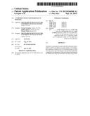 |  |
 | 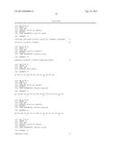 |
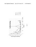 | 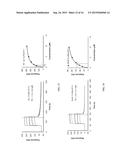 |
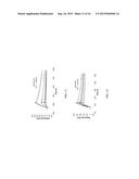 | 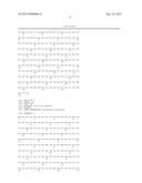 |
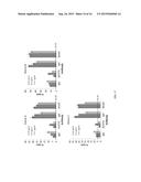 | 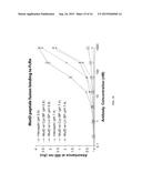 |
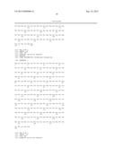 | 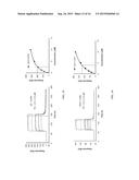 |
 | 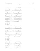 |
 | 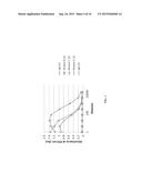 |
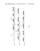 | 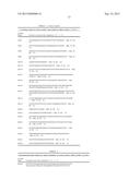 |
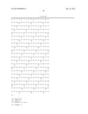 | 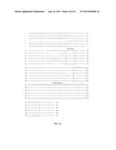 |
 | 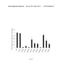 |
 |  |
 | 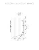 |
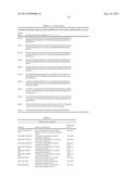 |  |
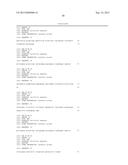 | 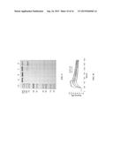 |
 | 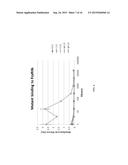 |
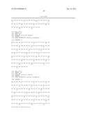 | 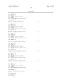 |
 | 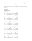 |
 |  |
 |  |
 | 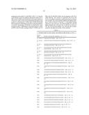 |
 | 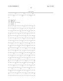 |
| New patent applications in this class: | |
| Date | Title |
|---|---|
| 2022-09-08 | Shrub rose plant named 'vlr003' |
| 2022-08-25 | Cherry tree named 'v84031' |
| 2022-08-25 | Miniature rose plant named 'poulty026' |
| 2022-08-25 | Information processing system and information processing method |
| 2022-08-25 | Data reassembly method and apparatus |
| New patent applications from these inventors: | |
| Date | Title |
|---|---|
| 2022-09-01 | Administration of kynurenine depleting enzymes for tumor therapy |
| 2021-12-16 | Human-enzyme mediated depletion of homocysteine for treating patients with hyperhomocysteinemia and homocystinuria |
| 2021-11-11 | Engineered antibody fc variants for enhanced serum half life |
| 2020-03-19 | Engineered enzymes with methionine-gamma-lyase enzymes and pharmacological preparations thereof |
| 2019-01-03 | Administration of kynurenine depleting enzymes for tumor therapy |