Patent application title: ANTIBODY BINDING MICROBIAL HEPARIN BINDING MOTIF TO RETARD OR PREVENT MICROBIAL BIOFILM FORMATION ON IMPLANTED MEDICAL DEVICES
Inventors:
Margaret K. Hostetter (Cincinnati, OH, US)
Long Lu (Cincinnati, OH, US)
Julianne Vernadette Green (Cincinnati, OH, US)
Alexey Porollo (Cincinnati, OH, US)
Kris I. Orsborn (Terrace Park, OH, US)
Khoon Ghee Queenie Tan (Durham, NC, US)
Kenneth Greis (Fort Thomas, KY, US)
David Andes (Madison, WI, US)
Assignees:
CHILDREN'S HOSPITAL MEDICAL CENTER
IPC8 Class: AC07K1614FI
USPC Class:
4241391
Class name: Drug, bio-affecting and body treating compositions immunoglobulin, antiserum, antibody, or antibody fragment, except conjugate or complex of the same with nonimmunoglobulin material binds antigen or epitope whose amino acid sequence is disclosed in whole or in part (e.g., binds specifically-identified amino acid sequence, etc.)
Publication date: 2015-02-19
Patent application number: 20150050284
Abstract:
Methods and reagents for ameliorating biofilm formation on a surface of
an indwelling or implanted device in a patient resulting in decreased
virulence of microorganisms such as Candida species and/or Staphylococcus
species.Claims:
1. A composition comprising at least one biocompatible excipient and an
antibody reactive with peptide HKQEKQKKHQIHKV (SEQ ID NO: 4) or a
fragment of at least seven amino acids of SEQ ID NO: 4.
2. A composition comprising at least one biocompatible excipient and an antibody reactive with a heparin binding motif expressed on the surface of a microorganism.
3. The composition of claim 2 where the microorganism is a Candida species or a Staphylococcus species.
4. A composition comprising an isolated peptide HKQEKQKKHQIHKV (SEQ ID NO: 4) or a fragment of at least seven amino acids of SEQ ID NO: 4.
5. The composition of claim 4 where the peptide is a heparin binding motif, or a portion thereof, that is expressed on the surface of a microorganism.
6. The composition of claim 5 where the microorganism is a Candida species or a Staphylococcus species.
7. The composition of claim 1 where the peptide is derived from Int1 protein.
8. The composition of claim 5 where the surface expression of the heparin binding motif depends on the life-cycle stage of the microorganism.
9. The composition of claim 1 where the antibody is a polyclonal antibody.
10. The composition of claim 1 where the antibody is a monoclonal antibody.
11. A method to reduce or prevent biofilm on a surface of an indwelling or implanted medical device in a patient, the method comprising administering before, during, and/or after installation or implantation of a medical device in the patient a composition comprising at least one biocompatible excipient and an antibody reactive with a peptide HKQEKQKKHQIHKV (SEQ ID NO: 4) or a fragment of at least seven amino acids of SEQ ID NO: 4 under conditions to result in reduced heparin-binding to a surface of the implanted medical device, resulting in reduced or prevented biofilm formation on the medical device.
12. The method of claim 11 where, when the composition comprises the peptide, the peptide antigenically immunizes the patient by endogenously generating anti-peptide antibodies that reduce or prevent biofilm formation on the surface of the indwelling or implanted device by reducing or preventing microorganism heparin-mediated-binding to the device surface.
13. The method of claim 11 where the implanted device is selected from the group consisting of a venous catheter, an arterial catheter, a central line, a hemodialysis catheter, a peritoneal catheter, a peripherally inserted central catheter, a urinary tract catheter, a central nervous system shunt, a peritoneal dialysis catheter, a dialysis shunt, and combinations thereof.
14. The method of claim 11 resulting in decreased patient infection.
15. The method of claim 11 resulting in decreased microorganism virulence.
16. A kit comprising instructions for administering a composition to a patient candidate for medical device installation or implantation, and at least one of a composition comprising at least one biocompatible excipient and an antibody reactive with a peptide HKQEKQKKHQIHKV (SEQ ID NO: 4) or a fragment of at least seven amino acids of SEQ ID NO: 4, or a composition comprising a peptide HKQEKQKKHQIHKV (SEQ ID NO: 4) or a fragment of at least seven amino acids of SEQ ID NO: 4.
17. An antibody reactive with peptide HKQEKQKKHQIHKV (SEQ ID NO: 4) or a fragment of at least seven amino acids of SEQ ID NO: 4.
18. An antibody reactive with peptide QKKHQIHK (SEQ ID NO: 1), THKGRF (SEQ ID NO: 40), MKRGKP (SEQ ID NO: 48), FKKRFFKL (SEQ ID NO: 2), or SHKTRA (SEQ ID NO: 3).
Description:
[0001] This application is a Continuation-In-Part of co-pending
PCT/US2013/031499 filed Mar. 14, 2013, which claims priority to U.S.
application Ser. No. 61/636,243 filed Apr. 20, 2012, each of which is
expressly incorporated by reference herein in its entirety.
[0002] A novel interaction between Candida albicans (C. albicans) surface proteins and heparin, a drug commonly used in patients at risk for catheter-associated biofilms and candidemia, is disclosed with an antibody that reduced or prevented a microorganism surface protein from binding to heparin, and reduced catheter-associated biofilm formation, where the reduction in biofilm formation can reduce the microorganism's virulence in a patient, with reduction encompassing any level of decrease.
[0003] Heparin is a highly sulfated, non-branched, anionic disaccharide composed of uronic acid, predominantly iduronic acid, in 1,4 linkage with glucosamine. Heparin is a known anticoagulant that is often infused in indwelling catheters to prevent the blood flowing through the catheter from clotting. Heparan sulfate is composed of the same disaccharide but is less sulfated and has a more varied structure than heparin. Heparan sulfate linked to core proteins, such as syndecans or glypicans, forms heparan sulfate proteoglycans (HSPGs). HSPGs are widely expressed on mammalian cell surfaces and in the extracellular matrix. Heparin binding motifs can bind to both heparin and heparan sulfate.
[0004] Several microbial proteins bind heparin and heparan sulfate, e.g., HIV-1 gp120, CypA, and Tat; hepatitis C envelope glycoprotein E2; herpes simplex virus glycoproteins B, C, and D; dengue virus envelope protein; Plasmodium falciparum circumsporozoite (CS) protein, Listeria monocytogenes ActA, and LcI of Legionella pneumophila (1-11). Such microbial protein interactions with surface HSPGs typically promote binding and entry of the microbial organism into the mammalian cell (except for HIV-1 gp120 and Tat), but the mechanisms by which individual microbial proteins recognize heparin or heparan sulfates are largely undefined. Positively charged tripeptides containing Lys or Arg in the amino terminus of L. monocytogenes ActA (aa 40-230) and in L. pneumophila LcI (aa 69-349) were shared with tripeptides in a number of heparin-binding mammalian proteins such as EGF-like growth factor and vonWillebrand factor, but the precise amino acids that mediated binding to heparin were not defined for ActA or LcI (10-11).
[0005] Eukaryotic proteins that bind heparin express either linear or conformational heparin binding motifs (HBM). Linear heparin binding motifs are short, conserved peptides containing both basic (B) and hydropathic (X) amino acids in specific patterns first identified by Cardin (XBBXBX) and Weintraub (XBBBXXBX) and subsequently expanded by Sobel (XBBBXXBBBXXBBX) (12-14). Molecular modeling studies suggested that basic amino acids such as lysine, arginine, and histidine were critical for interaction with anionic sulfate or carboxylate groups in heparin through electrostatic and hydrogen bonds. For example, substituting alanine for basic amino acids in linear HBM in the morphogen sonic hedgehog abolished binding to heparin (15). However, because many heparin-binding proteins failed to exhibit linear motifs, the concept of spatial orientation of basic residues was propounded (16). The CPC clip motif, a structural signature in which two cationic residues surround one polar residue, was conserved in eukaryotic heparin binding proteins, as well as in vaccinia complement protein and papillomavirus 18 capsid protein (17)
[0006] Because of its propensity to form biofilms in implanted medical devices such as indwelling central venous catheters, Candida albicans (C. albicans) is a leading pathogen in infections of indwelling catheters. A biofilm is a multilayered structure of microbes embedded in a polysaccharide matrix that forms on central venous catheters as well as implanted prosthetic joints, contact lenses, and other such medical devices (18, 19). A biofilm is undesirable because antibiotics and host defenses cannot penetrate it, thus a biofilm prevents elimination of microorganisms that have attached or adhered to a surface of an implanted medical device. For example, because of the high flow-through in a central venous catheter, sessile projections from a biofilm break off and are carried into the bloodstream to cause infection that can be carried by the blood to other sites in the body.
[0007] Heparin is a known anticoagulant, and catheters are often infused with heparin to prevent blood flowing through the catheter from clotting (20). Because heparin inhibited attachment of C. albicans to extracellular matrix proteins (21), the inventors hypothesized that C. albicans interacted with heparin through linear heparin binding motifs (HBMs): conserved sequences of basic (Arg, Lys, H is) and hydropathic amino acids in prescribed patterns.
BRIEF DESCRIPTION OF THE DRAWINGS
[0008] FIG. 1 schematically illustrates the internal lumen of an indwelling catheter.
[0009] FIGS. 2A-E demonstrate lack of an effect by heparin on wild type Candida albicans (C. albicans) planktonic growth and morphology.
[0010] FIGS. 3A-D demonstrate C. albicans binding to heparin and heparin analogs in vitro.
[0011] FIGS. 4A-C show that two lysine residues in linear heparin binding Motif 1 in C. albicans Int1 mediate binding to heparin, as measured by an ELISA assay in vitro. FIG. 4A discloses SEQ ID NOS 1, 40, 48, 2, 3, 52 and 53, respectively, in order of appearance. FIG. 4B discloses SEQ ID NO: 2. FIG. 4C discloses SEQ ID NOS 52-53, respectively, in order of appearance.
[0012] FIG. 5 compares pre-immune rabbit IgG with affinity-purified IgG raised against the sequence HKQEKQKKHQIHKV (SEQ ID NO:4) for ability to inhibit binding of C. albicans to heparin.
[0013] FIGS. 6A-C show consequences of heparin binding in vitro. FIG. 6A discloses SEQ ID NO: 65. FIG. 6B discloses SEQ ID NO: 66. FIG. 6C discloses SEQ ID NO: 67.
[0014] FIGS. 7A-B show consequences of heparin binding in vitro.
[0015] FIGS. 8A-F demonstrate that heparin binding motifs contribute to biofilm formation in vivo.
[0016] FIG. 9 demonstrates that affinity-purified IgG raised against the sequence HKQEKQKKHQIHKV (SEQ ID NO:4) substantially inhibited biofilm production in jugular venous catheters in rats. FIG. 9 discloses SEQ ID NO: 6.
[0017] FIG. 10 schematically illustrates the internal lumen of an indwelling catheter in the presence of antibody against heparin binding motifs on cell wall proteins of C. albicans.
[0018] FIG. 11 shows Anti-KKHQ specific binding to Candida species.
[0019] FIGS. 12A-C show Anti-KKHQ effect on biofilm progression.
[0020] Thirty-four C. albicans surface proteins encoding at least one linear heparin binding motif (HBM) were identified from a sequence-based search. Twelve of these 34 proteins flanked recently described transcriptionally active regions that are involved in biofilm production. Thirty-three of these 34 proteins are known targets of master biofilm regulators (22).
[0021] C. albicans binding to 2.5 units (12.5 μg) solid-phase heparin in an ELISA assay was significantly decreased by desulfation of heparin at the 2-O or 2-N groups, and by preincubation of C. albicans with heparin. The protein Int1 contained the largest number of HBMs; Int1 is a surface protein that is involved in adhesion, filamentation, and antigenic recognition. In the Int1 sequence 804QKKHQIHK (SEQ ID NO: 1), alanine mutation of lysine residues at positions 805/806 significantly reduced binding of the Int1 mutant to heparin. Rabbit IgG recognizing the polypeptide 799HKQEKQKKHQIHKV812 (SEQ ID NO: 4) inhibited C. albicans binding to heparin by 19%; pre-immune rabbit IgG had no effect.
[0022] Consequences of heparin binding in vitro included removal of C. albicans surface antigens such as Eno1, Pgk1, Tdh3, and Ssa1/2, which themselves contain putative HBMs; impairment of histatin-mediated killing; and modulation of gene expression. In vivo, substitution of alanine residues for lysines at positions 805/806 in 804QKKHQIHK (SEQ ID NO: 1) markedly attenuated biofilm formation in central venous catheters in rats. In addition, pre-incubation of C. albicans with rabbit IgG recognizing the polypeptide 799HKQEKQKKHQIHKV812 (SEQ ID NO: 4) inhibited biofilm formation in vivo; pre-immune IgG had no inhibitory effect. These results identify linear HBM in C. albicans surface proteins, characterize specific lysine residues that mediate heparin binding, and demonstrate relevance for innate and adaptive immunity in vitro and biofilm formation in vivo.
[0023] The inventive method and composition ameliorated this undesirable situation by using antibodies against a specific heparin-binding motif that is expressed by surface proteins on C. albicans; other microorganisms also express similar heparin binding motifs. Without being held to a single theory, the disclosed antibodies reduce or prevent the undesirable biofilm from forming on the surface of a medical device, particularly a plastic device, implanted in a patient. Microorganisms include bacteria, yeast, fungi, etc. as known to one skilled in the art. Medical devices include those implanted or implantable in a patient, particularly plastic medical devices, including but not limited to catheters and central lines but excluding current non-plastic implanted joints, pacemakers, pacemaker wires, and spinal rods.
[0024] In one embodiment of the inventive method, antibodies were produced against a linear heparin binding motif that was expressed by a surface protein of Candida albicans (C. albicans). Related heparin binding motifs occur in surface proteins from Staphylococcus epidermidis (S. epidermidis) and Staphylococcus aureus (S. aureus).
[0025] Linear heparin binding motifs are sequential amino acids that conform to one of three consensus motifs (Cardin, Sobel, or Weintraub motifs); they have been identified in multiple mammalian proteins that bind heparin. While conformational heparin binding motifs also occur in some proteins, they must be identified by crystal structure of the protein. Antibodies to these linear heparin binding motifs inhibited C. albicans from adhering to heparin bound to a plastic surface (a microtiter plate).
[0026] Of the six million patients who have a central line implanted each year, more than 75,000 will develop bloodstream infections (23). Central line-associated blood stream infections (CLABSI) are a major source of hospital-acquired infection; over 43,000 CLABSI occurred among patients hospitalized in U.S. intensive care units (ICUs) in 2001, which is 3.2 infections for every 1000 line days. With a protocol for their sterile insertion and daily maintenance, CLABSI in ICUs dropped to 18,000 in 2009, but even with these improvements in ICUs procedures, more than 23,000 CLABSI occurred in patients on inpatient wards and 37,000 CLABSI occurred in outpatient hemodialysis patients in 2009. Even with a 50% reduction in CLABSI in ICU patients, the annual cost of these infections across the U.S. is more than $13 billion (23)
[0027] Staphylococcus epidermidis is the most common cause of CLABSI in central lines, with gram-negative rods second, S. aureus third; and Candida species fourth (24). In peripherally inserted central catheters (PICC), i.e., central lines inserted through peripheral veins in the arm, Candida species are the second most common cause of line infection (25). Candida spp. are equivalent to S. aureus in infections in central lines in the ambulatory setting (26). Among Candida spp, C. albicans is the most common cause of CLABSI (24).
[0028] When Candida species infect a central venous catheter, the organism enters the bloodstream to cause candidemia. Hosts at highest risk of candidemia, include burn patients and patients on a pump during coronary artery bypass procedures (27). Neutropenic oncology patients, premature newborns, and patients with major abdominal surgery such as intestinal resections are also high-risk patients with 8- to 16-fold the number of infections of other patient groups (27).
[0029] A major risk factor in candidemia is the presence of central lines (28-33), whose lumens are a site for formation of microbial biofilms that are effectively shielded from antimicrobial agents and host defenses. A second commonality is the use of heparin as an anticoagulant in most catheters. The amount of heparin is often considerable; e.g., a premature newborn in a neonatal intensive care unit may receive 150 units/kg/day, compared to 2.8 units/kg for a 70 kg adult receiving a 100 unit flush twice a day (20).
[0030] The ability to bind heparin is a unifying feature among S. epidermidis, S. aureus, and, Candida albicans (34-36), three of the leading causes of catheter-associated bloodstream infections. In experiments with S. aureus, high concentrations of heparin have been shown to increase S. aureus biofilm formation in vitro (37).
[0031] A first step in biofilm formation is microorganism adhesion or adherence to a surface of the implanted device, such as a catheter (38). In FIG. 1, negatively charged heparin molecules (circles) injected into the catheter lumen bind to the positively charged catheter surface, and heparin sulfate moieties (curved lines) are exposed on vascular endothelium lining catheter lumens. Linear heparin binding motifs present on the surface of microorganisms, such as C. albicans, interact either with heparin (A) or with heparin sulfate (B) and enable the organism to attach to the inside of the catheter. As the attached microorganisms replicate, they form the multilayered structure of a biofilm (C) and secrete the polysaccharide mortar-like matrix that holds the biofilm together. The inventors hypothesize that interactions between heparin and surface proteins on microorganisms such as C. albicans facilitate biofilm formation, and subsequent infection of the bloodstream when projections from biofilm break off into the catheter lumen and enter the bloodstream.
[0032] Heparin is associated with central line infections, particularly those due to Candida spp. In a randomized trial of 260 central lines, 128 were coated with chlorhexidine and 132 were coated with heparin; all Candida colonization and all candidemias occurred in patients whose central lines were coated with heparin (39). A second study in renal dialysis patients showed that those patients who received 5000 units of heparin in the middle of dialysis had considerably more catheter loss due to infection compared to patients not treated with heparin. Although staphylococcal infections were most common in this patient group, candidemia also occurred (40).
[0033] One mechanism by which heparin interacts with proteins is through linear heparin binding motifs, conserved sequences of basic amino acids such as lysine, arginine, and histidine interspersed with hydropathic amino acids (14). Microbial heparin-binding proteins are identified by short consensus motifs of basic (B) and hydropathic (X) amino acids, as defined by Cardin [XBBXBX], Weintraub [XBBBXXBX], or Sobel [XBBBXXBBBXBBX].
[0034] In the invention, a computer-based algorithm examined all 400 C. albicans surface and cell wall proteins for heparin binding motifs (HBM). Of the 6,000 known Candida albicans proteins, about 400 are localized to the cell wall of yeast or hyphae, depending upon the extraction technique (41). A sequence-based search identified putative Cardin, Sobel, or Weintraub motifs in 159 C. albicans proteins. Table 1 shows HBM in cell surface proteins expressed by C. albicans. The 34 proteins in Table 1 include only those that have the attribution "cell surface", "fungal cell wall", "yeast cell wall", or "hyphal cell wall" as the cellular component in the Candida Genome Database, where *=Weintraub motif; #=Cardin motif; =Identified by molecular modeling studies; A="cell wall" in protein description (as of January 2011); obtained from Candida Genome Database (CGD); B="cell wall" in Gene Ontology (GO) annotation (as of January 2011); obtained from CGD; C=cell wall proteins as reviewed by Alberti-Segui (42) and D=cell wall proteins as reviewed by Chaffin (41).
[0035] The following procedure was used to identify a set of cell wall proteins in C. albicans with HBM. The first step combined evidence to identify possible cell wall proteins in C. albicans. Specifically, using "cell wall" as keywords, 154 putative cell wall proteins were identified from protein descriptions, and another 245 unique cell wall proteins were identified from Gene Ontology functional annotations from Candida Genome Database (www.candidagenome.org; Assembly 21). Also included were 125 non-redundant cell wall proteins identified by Alberti-Segui (50) and another 174 reported by Chaffin (22) and mapped annotations to Assembly 21. The second step screened for consensus HBM in protein ORF sequences of these cell wall proteins from Assembly 21. Three types of consensus motifs were included: Weintraub (XBBBXXBX), Sobel (XBBBXXBBBXXBBX), and Cardin (XBBXBX) motifs, where B is a basic amino acid and X is a hydropathic amino acid. In the algorithm, basic amino acids are H, K, and R. Hydropathic amino acids are W, F, Y, L, I, C, M, G, V, S, T, A, N, P, and Q. A total of 159 cell wall proteins were identified that have possible HBM, and some have multiple motifs. The 159 proteins that met the search criteria were then manually curated to select only those proteins whose cell wall localization was confirmed by manual or computational methods in the Candida Genome Database.
[0036] Proteins with GPI anchoring sequences within these 159 cell wall proteins with HBM were further identified. Three Web servers were used to perform GPI anchoring sequence prediction: SignalP (43), GPI-SOM (44), and PredGPI (45). Combined results of these three prediction methods indicated that 15 out of these 159 proteins had GPI anchoring sequences, four of which overlapped with reviewed GPI anchoring proteins (46).
[0037] The algorithm identified 34 cell wall proteins with potential linear heparin binding sequences that matched the Cardin, Weintraub, and Sobel motifs. The 34 identified cell wall proteins were Ahp1, Als7, Atcl, Bud2, Cat1, Cef3, Chs1, Crh12, Dot4, Not5, Pdi1, Pga4, Pgk1, Phr3, Rbt1, Rps6A, Sam2, Srb1, Ssa2, Ssb1, Tdh3, Eft2, Eno1, Gap1, Gph1, Gpm2, Hem13, Hsp104, Hsp70 (also called Ssa1), Ino1, Int1, Ipp1, Ugp1, Xyl2. Three of these proteins, Als7, Pga4, and Rbt1, have GPI anchors. The genes encoding eleven of these proteins, Als7, Cat1, Dot4, Eno1, Gph1, Ino1, Rbt1, Sam2, Srb1, Ssa2, and Ugp1, are located in newly defined transcriptionally active regions that are critically involved in biofilm formation (22). Moreover, many of these proteins are known to be regulated by at least one of six master biofilm regulators; by RNA-seq, where RNA-seq is defined as the use of high-throughput sequencing techniques to sequence cDNA in order to get sequence information about the transcriptome, the sample's RNA content; CAT1, GAP1, GPH1, and HSP104 are up-regulated in biofilm formation, while CHS1, EN01, GPM2, HEM13, INO1, IPP1, PGK1, RPS6A, SAM2, SRB1, SSA2, SSB1, and TDH3 are down-regulated (22)
[0038] Given the presence of putative HBM among C. albicans surface proteins and the possible interactions with heparin in central venous catheters or with heparin sulfate proteoglycans expressed on host tissues such as vascular endothelium, the biochemical determinants and immunologic consequences of this interaction were defined. The protein Int1 (SEQ ID NO: 5) (accession number P53705.2; GI 187608862) had the highest number of HBM (five). Int1 (SEQ ID NO: 5) spans 1711 amino acids and is localized to the cell wall of the bud neck in C. albicans. It mediates adhesion, hyphal formation, and virulence, defined as the ability to cause disease such as bloodstream infection (47). Using the same search technique, HBM were also detected in surface proteins of S. epidermidis and S. aureus.
TABLE-US-00001 TABLE 1 Gene ID # of (Assembly 21) Protein Ref. motifs Motif 1 Motif 2 Motif 3 Motif 4 Motif 5 orf19.4257 Int1 C 5 804QKKHQIHK.sup..dagger-dbl. .sub.1383THKGRF# .sub.1530MKRGKP# .sub.1593FKKRFFKL* 1612SHKTRA# (SEQ ID NO: 1) (SEQ ID NO: 40) (SEQ ID NO: 48) (SEQ ID (SEQ ID NO: 2) NO: 3) orf19.1738 Ugp1 B, D 3 176SHRIRV# 310IKKFKY# 368IRHFKG# (SEQ ID NO: 7) (SEQ ID NO: 41) (SEQ ID NO: 49) orf19.3370 Dot4 B 3 442NKKGKS# 519CHKCHN# 635FKRFKF# (SEQ ID NO: 8) (SEQ ID NO: 42) (SEQ ID NO: 50) orf19.3651 Pgk1 A, B, D 3 136GKKVKA# 146VKKFRQ# 168AHRAHS# (SEQ ID NO: 9) (SEQ ID NO: 43) (SEQ ID NO: 51) orf19.4660 Rps6A B, D 2 185QRKRALKA* 192AKKVKN# (SEQ ID NO: 10) (SEQ ID NO: 44) orf19.5107 Not5 B 2 114QKRSRF# 333VKKLKP# (SEQ ID NO: 11) (SEQ ID NO: 45) orf19.5130 Pdi1 D 2 210NKKFKN# 301GKKYRG# (SEQ ID NO: 12) (SEQ ID NO: 46) orf19.6387 Hsp104 D 2 53VKRARY# 199ARRSKS# (SEQ ID NO: 13) (SEQ ID NO: 47) orf19.1065 Ssa2 B, D 1 258LRRLRT# (SEQ ID NO: 14) orf19.1067 Gpm2 B 1 45IKKNHL# (SEQ ID NO: 15) orf19.1327 Rbt1 A, B, C 1 121GKKVKQ# (SEQ ID NO: 16) orf19.2762 Ahp1 B, D 1 171LKRIHN# (SEQ ID NO: 17) orf19.2803 Hem13 B, D 1 257IRRGRY# (SEQ ID NO: 18) orf19.3590 Ipp1 B, D 1 74TKKGKL# (SEQ ID NO: 19) orf19.377 Phr3 B, C 1 115PHHHLNRY* (SEQ ID NO: 20) orf19.395 Eno1 B, D 1 141AKKGKF# (SEQ ID NO: 21) orf19.3966 Crh12 A, B, C 1 420TKHIHN# (SEQ ID NO: 22) orf19.4035 Pga4 B, C, D 1 259AKRPRP# (SEQ ID NO: 23) orf19.4152 Cef3 B 1 623LRKYKG# (SEQ ID NO: 24) orf19.4304 Gap1 B 1 72QRKLKT# (SEQ ID NO: 25) orf19.4980 Ssa1 B, D 1 259LRRLRT# (SEQ ID NO: 26) orf19.5188 Chs1 B, C 1 126PKRQKT# (SEQ ID NO: 27) orf19.5788 Eft2 B, D 1 581NKHNRI# (SEQ ID NO: 28) orf19.6190 Srb1 B, D 1 123FHKAHG# (SEQ ID NO: 29) orf19.6214 Atc1 A, B 1 959PKRVKV# (SEQ ID NO: 30) orf19.6229 Cat1 B 1 82GKKTRI# (SEQ ID NO: 31) orf19.6367 Ssb1 B, D 1 263LRRLRT# (SEQ ID NO: 32) orf19.657 Sam2 B, D 1 380PKKLKF# (SEQ ID NO: 33) orf19.6814 Tdh3 B, D 1 70GHKIKV# (SEQ ID NO: 34) orf19.7021 Gph1 B, D 1 656TKHHIPKA* (SEQ ID NO: 35) orf19.7400 Als7 B, C 1 .sub.1482SKRNKN# (SEQ ID NO: 36) orf19.7585 Ino1 B, D 1 161MKRAKV# (SEQ ID NO: 37) orf19.7676 Xyl2 A, B, D 1 330THRFKF# (SEQ ID NO: 38) orf19.940 Bud2 B 1 716LRKGKS# (SEQ ID NO: 39)
[0039] Heparin effect on Candida albicans growth and morphology was determined. A single colony of BWP17 wt was inoculated into 3 ml YPD medium and incubated overnight at 30° C. with shaking at 225 rpm. Overnight cultures were diluted to an OD600 of 0.1 in 5 ml of YPD (yeast) or RPMI-HEPES (hyphae) in a 50 ml conical polypropylene tube and incubated in the presence or absence of 100 units/ml preservative-free pharmaceutical heparin at 30° C. for six hours. Cells (1 ml) were fixed with an equal volume of 4% formaldehyde in FACS buffer at 4° C. for one hour, then pelleted (7,000 rpm for 3 minutes) and washed with PBS. After reconstitution in 0.5 ml PBS, filipin (50 mg/ml stock solution in DMSO) was added to a final concentration of 100 μg/ml and incubated with cells at room temperature for five minutes. Cells were pelleted, washed with PBS and mounted on a slide using Fluoromount G. For calcofluor white staining, after six hours incubation, 1 μl of 1 mg/ml stock solution of calcofluor white in 0.1 N NaOH was added to 100 μl cells to a final calcofluor white concentration of 10 μg/ml.
[0040] Microscopy was performed by imaging filipin, PKH26, DAPI, and heparin-Alexa Fluor 488 on a Nikon Ti-E inverted microscope with a 100×CFI APO oil NA 1.49 objective. Filipin was excited with a Prior Lumen 200 metal halide light source set at 10% light output. This light was further attenuated by ND4 and ND8 neutral density filters in series to reduce light output to approximately 3% of output from the liquid light guide. The filters used for imaging were EX 360/40, dichroic 400 nm Ip, EM 460/50. Images were acquired with an Andor iXon emccd camera. Exposure times were 391 ms. Excitation of PKH26/DAPI/Heparin-Alexa Fluor 488 triple staining was accomplished with a Nikon A1R si laser scanning confocal. DAPI was imaged with 405 nm excitation and a 450/50 filter with a laser power of 16.3 and a photomultiplier tube (pmt) voltage of 92. Alexa Fluor 488 was imaged with 488 nm excitation from an Argon-ion laser and a 525/50 filter with a laser power of 6.0 and a pmt voltage of 84. PKH26 was imaged with 561 nm excitation and a 595/50 filter with a laser power of 13.2 and a pmt voltage of 108. Gains were kept below 110 to eliminate contribution from C. albicans autofluorescence; lack of autofluorescence was confirmed by comparison to a control sample without heparin-Alexa Fluor 488. Images were processed with Nikon NIS-Elements AR 4.11.00 64-bit software. Calcofluor white slides were examined using a Zeiss Axiovert 200M fluorescent microscope equipped with a Plan-Apochromat 63×/1.40 oil objective lens with 1.6× optivar and DAPI filter at 350 nm excitation and 460 nm emission. Images were captured using a Zeiss Axiocam color camera and processed with Axiovision version 4.8.2.
[0041] Flow cytometry was performed by inoculating a single colony of BWP17 wt into 3 ml YPD medium and incubating overnight at 30° C. with shaking at 225 rpm. Overnight cultures were diluted to OD600 of 0.1 in 5 ml of YPD or RPMI-HEPES and incubated in the presence or absence of 100 units/ml preservative-free pharmaceutical heparin at 30° C. for one hour. One ml aliquots of each mixture were removed, pelleted (7,000 rpm for 3 min), washed twice with PBS, and reconstituted in 1 ml PBS. Flow cytometry analysis was performed using the ImagestreamX flow cytometer (Amnis, Seattle Wash.) equipped with a 405 nm, 488 nm, 653 nm, laser and multi-mag function. The 40× magnification, 10 mm/sec flow rate, and 488 nm laser were used to collect SSC and Brightfield parameters. The flow cell allows particles up to 100 μm wide (height unlimited) to be collected in the instrument. INSPIRE (v.6.0) software was used to acquire events. Software analysis using IDEAS (v5.0) identified percent hyphae using aspect ratio, height and width features using the side scatter (Channel 6) and brightfield (Channel 1) parameters. Objects were selected within each file and tagged to use as identification of truth sets within the population.
[0042] For growth curves in the presence or absence of soluble heparin, a single colony of BWP17 wt was inoculated into 3 ml YPD medium and incubated overnight at 30° C. with shaking at 225 rpm. Overnight cultures were diluted to an OD600 of 0.1 in 5 ml of YPD, RPMI-MOPS, RPMI-HEPES or CSM in a 50 ml conical polypropylene tube. Preservative-free pharmaceutical heparin (1000 units/ml) was added to yield a final concentration of 100 units/ml in the heparin-treated samples. Cultures were grown at 30° C. with shaking and OD600 measured periodically. Doubling times (td) were calculated based on the equation td=ln 2/μ (μ=specific growth rate (48). One hundred units/ml heparin, the concentration recommended to prevent clotting of central venous catheters (20), had no effect on doubling times of planktonic yeast cells grown in YPD, RPMI-HEPES, RPMI-MOPS or CSM, as shown in FIGS. 2A and 2B.
[0043] FIGS. 2A-E demonstrate lack of an effect by heparin on wild type Candida albicans (C. albicans) planktonic growth and morphology.
[0044] FIG. 2A shows that growth curves in the presence (open circles) or absence (closed diamonds) of soluble heparin were identical.
[0045] FIG. 2B shows that doubling times in various media at 30° C. in the absence and presence of heparin (100 units/ml) were identical.
[0046] FIG. 2C shows that percent hyphae as determined by flow cytometry did not differ after incubation with or without heparin (100 units/ml) under conditions that favor yeast (YPD, 30° C.) or hyphae (RPMI-HEPES, 37° C.).
[0047] FIGS. 2D-E show that the integrity of membrane sterols identified with filipin (FIG. 2D) and the location of septin rings identified with calcofluor white staining (FIG. 2E) were identical between untreated and heparin-treated organisms. Thus, concentrations of heparin commonly used to maintain patency of central venous catheters (100 units/ml) did not lead to readily observable differences in growth or cellular architecture of the organism.
[0048] Binding of heparin by C. albicans was examined in vitro. Heparin was labeled with Alexa Fluor 488 by a method modified from Osmond (49). Briefly, 5 mg heparin (Sigma) was dissolved in 0.5 ml MES buffer (0.1M 2-(N-morpholino)ethanesulfonic acid hydrate, pH 4.5, Sigma), then mixed with a solution of 1 mg Alexa Fluor 488 hydrazide (Life Technologies) in 0.4 ml MES buffer. After adding 0.2 ml of EDC solution (15 mg 1-ethyl-3-(3-dimethylaminopropyl) carbo-diimide/ml water, Thermo Scientific), the mixture was stirred at room temperature for 30 min. A second 0.2 ml aliquot of EDC solution was added and the mixture stirred at room temperature for an additional 30 min. After adding 1.3 ml NaOAc (1 M, pH 4.8) and stirring at room temperature for one hour, heparin-Alexa Fluor 488 was purified on a PD-10 desalting column (GE Healthcare) equilibrated in autoclaved nanopure water. Fractions containing the labeled material detected at 490 nm were combined and dried overnight on a SpeedVac concentrator (Savant) with heating. The resulting solid was dissolved in autoclaved nanopure water to 10 mg/ml and stored at 4° C.
[0049] Heparin-Alexa Fluor 488 bound to PKH26- and DAPI-labeled C. albicans. A single colony of BWP17 wt was inoculated into 3 ml YPD medium and incubated overnight at 30° C. with shaking at 225 rpm. Using a PKH26 Red Fluorescent Cell staining kit (including PKH26 dye and diluent C, Sigma), 2×10⁷ cells were mixed with 1 ml diluent C. To this solution was added a mixture of 4 μl PKH26 dye in 1 ml diluent C, and the mixture allowed to stand at room temperature for 3 min. After adding 10 ml 3% BSA, the mixture was centrifuged at 3,000 rpm for 7 min. The resultant magenta-colored cells were washed with 10 ml each of 1% BSA and PBS, with centrifugation after each step. After supernatant removal, the cells were reconstituted in 10 ml fresh PBS. Two 1 ml (2×10⁶ cells) aliquots were placed in separate 15 ml conical tubes, centrifuged, and supernatants removed. Each was reconstituted with 2 ml YPD, and heparin-Alexa Fluor 488 (0.1 ml of above 10 mg/ml solution) was added to the experimental sample. Control and experimental samples were incubated at 30° C. with shaking (225 rpm) for 30 min, at which time 0.4 ml aliquots of each were removed, pelleted (10,000 rpm for 3 min), and washed twice with PBS. After reconstitution with 0.5 ml PBS, DAPI (4',6-diamidino-2-phenylindole dihydrochloride (Sigma, stock solution of 5 mg/ml) was added to a final concentration of 1 μg/ml and the solution allowed to stand at room temperature for 10 min. Cells were pelleted and washed twice with PBS, then mounted on a microscope slide using Fluoromount G.
[0050] To measure the binding of C. albicans to solid-phase heparin, a heparin-binding ELISA assay was developed, as follows. A single colony of each C. albicans wild type or mutant strain from a YPD plate was inoculated into 3 ml YPD medium and incubated overnight at 30° C. with shaking at 225 rpm. Sigma heparin (fresh solution made daily), diluted to 25 units/ml in autoclaved, sterile-filtered PBS was added as 0.1 ml aliquots to each well of an allyl amine-coated 96-well heparin binding microtiter plate (BD Biosciences). The plate was incubated at room temperature overnight in the dark (50). In the morning, the plate was washed with acetate buffer (100 mM NaCl, 50 μM NaOAc, 0.2% Tween 20, pH 7.2), incubated with 3% bovine serum albumin (BSA) in PBS at 30° C. for 1 hour, then washed with PBS. In the meantime, overnight cultures of C. albicans were subcultured by dilution to OD600 of 0.2 in 25 ml of YPD, and grown at 30° C. to mid-log phase (OD600 0.6-0.7). Cells were pelleted (3,000 rpm for 7 minutes), washed twice with PBS and reconstituted in RPMI-HEPES to 4×10⁵, 2×10⁵, and 1×10⁵CFU/ml, respectively. One hundred μl of each C. albicans dilution was applied to the microtiter wells; experiments for each strain were performed in quadruplicate. The plate was incubated at 30° C. for one hour and washed with PBS to remove non-adherent C. albicans. One-tenth ml of a biotinylated polyclonal rabbit anti-C. albicans antibody raised against soluble proteins in a C. albicans lysate (Meridian Life Science), which had been diluted 1:2500 in FACS-Tween (0.05% Tween 20 in FACS buffer consisting of 0.3% BSA), was added to each well. The plate was incubated at 30° C. for one hour, then washed with PBS-Tween (0.05% Tween 20 in PBS). One-tenth ml streptavidin alkaline phosphatase (Biolegend) diluted 1:10,000 in FACS-Tween was then added to each well, and the plate was incubated at 30° C. for 30 minutes. After washing with PBS-Tween and AKP buffer (100 mM Tris base, 50 μM MgCl2, 100 mM NaCl, pH 9.5.), 0.1 ml alkaline phosphatase substrate (KPL) was added to each well. After 45 minutes, absorbance at 595 nM was read on a Beckman Coulter DTX 880. When desulfated heparin analogs, chondroitin sulfate, or dermatan sulfate were used, equimolar amounts of the analogs (with respect to heparin) were applied to the allyl amine-coated 96-well plate instead of heparin.
[0051] The ability of soluble heparin to inhibit C. albicans binding to plate-bound heparin was demonstrated by pre-incubating 1×10⁶ CFSE-labeled C. albicans with 200 units heparin at 37° C. for one hour in wells of a 96-well black plate. C. albicans was labeled with carboxyfluorescein succinimidyl ester (CFSE), succinimidyl ester (CFSE; Invitrogen), which is used to track cell division and therefore does not kill the organism (28), as follows. A single colony of C. albicans was suspended in CSM to an OD600 of 0.1, diluted 1:200 into 25 ml CSM and grown at 30° C. overnight to mid-log phase (OD600 0.6-0.7). Cells were washed twice in sterile PBS and suspended in PBS at 2×10⁷ CFU/ml. Carboxyfluorescein succinimidyl ester (Life Technologies) was dissolved in DMSO, diluted in PBS to 50 μM, and 0.5 ml CFSE mixed with 0.5 ml C. albicans suspension. The mixture was incubated for 20 min at room temperature on a rotator, washed once with FACS buffer, once with PBS, then suspended in 1 ml FACS buffer. Intensity of labeling was determined as the mean fluorescence intensity using a BD Accuri C6 cytometer (San Jose, Calif.) with excitation at 488 nm and a 533/30 emission filter. Wells of a black 96-well microtiter plate were incubated with 0.1 mg poly-D-lysine for one hour at room temperature, washed three times with PBS, then incubated with 200 units of heparin in 100 μl RPMI-HEPES overnight at room temperature. The following afternoon, C. albicans wild type strain and mutants were labeled with CFSE as follows: after growth of C. albicans to mid-log phase in CSM medium, organisms were washed two times in PBS and suspended in PBS at a concentration of 2×10⁷/ml. 0.5 ml cells was mixed with 0.5 ml CFSE, covered in foil, and rotated on a mixer for 20 minutes at room temperature. CFSE-labeled C. albicans cells were pelleted in a minifuge at 3000 rpm for four minutes, washed once with FACS buffer (0.3% bovine serum albumin in PBS), washed again with PBS, and then suspended in 1 ml FACS buffer at a concentration of 1×10⁷/ml. 100 μl CFSE-labeled C. albicans (1×10⁶) were deposited in test wells for 60 minutes at room temperature. At the end of the incubation period, wells were washed three times with PBS, and fluorescence in each well was measured (Beckman Coulter Multimode detector DTX880).
[0052] FIGS. 3A-D demonstrate C. albicans binding to heparin in vitro. FIG. 3A shows confocal microscopy of C. albicans in the presence of heparin-Alexa Fluor 488, with (i) C. albicans cell wall stained with PKH26, (ii) nucleus stained with DAPI, (iii) cell wall outlined by heparin-Alexa Fluor 488, and (iv) arrows indicating co-localization of heparin with C. albicans cell surface. Incubation of C. albicans with 100 units/ml heparin-Alexa Fluor 488 for 30 mins at 30° C. demonstrated co-localization of heparin with the C. albicans cellular surface (FIG. 3A). Heparin deposition was seen at the interface of adjoining yeast cells and in individual cells.
[0053] FIG. 3B shows binding of 10,000 colony-forming units (CFU) C. albicans (OD595) to increasing concentrations of heparin immobilized on an allyl amine-coated 96-well microtiter plate; values are ±SD of duplicate wells. Binding of C. albicans at 10,000 CFU/well did not change over heparin concentrations ranging from 1.25 units/well (6.3 μg/well) to 20 units/well (100 μg/well) (FIG. 3B), indicating saturation of the allyl amine plate by heparin in low concentrations.
[0054] FIG. 3C shows the linear dose-response for the heparin binding ELISA assay with 2.5 units/well heparin and increasing C. albicans input from 10,000 (left-most bar), 20,000 (center bar), and 40,000 (right-most bar) CFUs; the graph represents mean±SD of four experiments, *p<0.007 for all inputs. FIG. 3C shows that the OD595 of the ELISA assay increases linearly as input of C. albicans increased from 10,000 colony forming units (CFU)/well to 40,000 CFU/well.
[0055] FIG. 3D shows binding of C. albicans (40,000 CFU/well) to equimolar amounts of heparin analogs desulfated at the 2-O (second from the left bar), 2-N (second from the right bar), or 6-O (right-most bar) positions versus heparin control (left-most bar) (normalized to 100%), with the graph representing mean±SD of three experiments, performed in triplicate *p≦0.003, ** p=0.20 vs. heparin control. With equimolar amounts of desulfated heparins, binding of C. albicans was decreased by 11% when heparin was desulfated at the 2-0 position of iduronic acid (p=0.003), and by 21% when heparin was desulfated at the 2-N position of glucosamine (p=0.002) (FIG. 3D). Removal of the sulfate group at the 6-O position of glucosamine did not significantly reduce binding.
[0056] Pre-incubation of CFSE-labeled C. albicans with 100 units/ml heparin decreased binding to heparin by 27% (p=0.022; data not shown). C. albicans also recognizes the related structures of chondroitin sulfate and dermatan sulfate. Binding of C. albicans to heparin and to chondroitin sulfate was equivalent, but binding to dermatan sulfate was reduced (p=0.019; data not shown). These results confirmed that the binding of C. albicans to heparin and similar glycosaminoglycans (chondroitin sulfate and dermatan sulfate) could be reproducibly measured and, in the case of heparin, specifically inhibited by pre-incubating the organisms with heparin. These results also suggested that surface components of C. albicans may mediate C. albican binding to heparin.
[0057] Location of HBM in Int1 (SEQ ID NO: 5) were determined. Five overlapping polypeptides spanning amino acids 51-1711 of Int1 were expressed with a 6×His tag (SEQ ID NO: 54) in S. cerevisiae BJ3501 and purified by affinity chromatography (HisTrap column; GELifesciences). Fractions containing the His-tagged polypeptide were pooled, diluted with loading buffer (10 mM phosphate, pH 7.0 plus 250 mM NaCl) and applied to a heparin sepharose column (HiTrap Heparin HP; GE Lifesciences). Heparin-binding polypeptides were eluted with a step gradient of NaCl (0.5-2 M) in loading buffer. Eluted fractions were analyzed by SDS-PAGE and immunoblot using anti-His tagged antibody (Santa Cruz) and chemiluminescent detection (SuperSignal West Pico Mouse IgG Detection Kit, Pierce) according to manufacturers' instructions. Polypeptides spanning aa 656-1193 and aa 1548-1711 bound to heparin-Sepharose and were eluted with NaCl, indicating that the HBM in those domains, schematized as Motif 1 (SEQ ID NO: 1), Motif 4 (SEQ ID NO: 2), and Motif 5 (SEQ ID NO: 3) (FIG. 4A), were candidates for mediating binding to heparin. Bolded letters represent basic amino acids in heparin binding motifs; unbolded letters represent hydropathic residues. Polypeptides spanning aa 51-385, aa 385-659, and 1188-1551 failed to bind to a heparin-Sepharose column.
[0058] The linear polypeptide spanning aa 656-1193 contains one potential heparin binding site, 804QKKHQIHK (SEQ ID NO: 1) (basic residues shown in red) (Motif 1 in Table 1 and FIG. 4A). The linear polypeptide spanning aa 1548-1711 contains a canonical Weintraub motif .sub.1593FKKRFFKL (SEQ ID NO: 2) (Motif 4 in Table 1 and FIG. 4A) and a canonical Cardin motif 1612SHKTRA (SEQ ID NO: 3) (Motif 5 in Table 1 and FIG. 4A). Sequence homology search using BLAST indicated that aa 1548-1711 contains a Pleckstrin homology domain (PHD), which is structurally resolved. Using homology-based 3-D modeling, we found that the three lysine residues and single arginine residue in Motif 4 were located on the rim of the PHD and might facilitate binding to a strong anion such as heparin by electrostatic interaction (FIG. 4B, arrows). The Cardin motif (Motif 5) did not share this conformation.
[0059] To test whether 804QKKHQIHK (SEQ ID NO: 1) (Motif 1) and/or .sub.1593FKKRFFKL (SEQ ID NO: 2) (Motif 4) mediated the binding of heparin to C. albicans, standard PCR-mediated mutagenesis (51) was used to construct a set of isogenic INT1 disruptants and mutants (Table 2).
TABLE-US-00002 TABLE 2 Strain Abbreviation Genotype Source BWP17 -- ura3::imm434/ura3::imm434 his1::hisG/his1::hisG [27] arg4::hisG/arg4::hisG BWP17WT WT BWP17 plus arg4::ARG4::URA3/his1::hisG::HIS1 [27] VBIDM2 -- as BWP17 plus int1::ARG4/int1::URA3 [60] VBIDM6-2 DD as VBIDM2 plus his1::hisG/HIS1 [60] KO503 Motif 4 VBIDM2 plus his1::hisG::HIS1-INT1 (KK1595AA) current KO507 Motif 1 VBIDM2 plus his1::hisG::HIS1-INT1(KK804AA) current KO508 Motifs 1&4 VBIDM2 plus his1::hisG::HIS1-INT1(KK804AA, KK1595AA) current KO509 Reint VBIDM2 plus his1::hisG::HIS1-INT1 (WT) current
[0060] Construction of mutants was as follows. C. albicans genomic DNA was isolated from saturated overnight cultures using glass beads as described (52). A lithium acetate method was used to transform C. albicans (41). Plasmids and PCR products were purified using kits (Fermentas/ThermoFisher, Pittsburg Pa.) or established methods (53). Pfu enzyme (New England Biolabs) with High Fidelity buffer was employed for all amplifications. Products were sequenced to affirm fidelity prior to use. Primers are described in Table 3.
TABLE-US-00003 TABLE 3 Primer Internal reference Sequence Purpose 1 SAC2035UP gggagctcGTTACTTGTCATTAATTAGTTACTTCC SacI 5' INTI (SEQ ID NO: 55) 2 MLU3'UTR ggacgcgtTTTTATCTTTTTATGTAAATATATACTA MluI 3'INT1 (SEQ ID NO: 56) 3 INT1 KR1595AA F ATTGTCCAATTTTTAAGGCTGCTTTTTTCAAATTAAT mutate Motif 1 GGG (SEQ ID NO: 57) 4 INT1 KR1595AA R CCATTAATTTGAAAAAAGCAGCCTTAAAAATTGGAC mutate Motif 1 AATC (SEQ ID NO: 58) 5 INT1 KK805AA F GCATAAACAAGAAAAGCAGGCCGCCCATCAAATTC mutate Motif 4 ATAAAGTTCC (SEQ ID NO: 59) 6 INT1 KK805AA R GGAACTTTATGAATTTGATGGGCGGCCTGCTTTTC mutate Motif 4 TTGTTTATGC (SEQ ID NO: 60) 7 GHISR CTCCCGGCCGCCATGGCCGC (SEQ ID NO: 61) check integration 8 HIS3AMP GTTGGTGTGGCCCAGAGACTCT (SEQ ID NO: 62) check integration
[0061] A single copy of Int1, including 1450 bp upstream and 548 bp downstream from the Int1 open reading frame (www.candidagenome.org, Assembly 21), was integrated into the hisG locus of the int1-/- strain VBIDM2 (54) to produce the reconstituted strain KO509. Briefly, a copy of Int1 was generated by PCR, using primers 1 and 2 with BWP17 wt DNA as template and cloned into the SacI/MluI sites of pGEMHIS (51) to create pKO509. pKO509 was digested with SwaI and transformed into VBIDM2 to create the reconstituted strain KO509. PCR-mediated overlap extension mutagenesis (55) was used to produce copies of Int1 mutated at putative heparin binding domains. Briefly, primer pairs 1+3 and 2+4 (or 1+5 and 2+6) were used to produce two overlapping fragments of INT1 in which putative heparin binding domains were mutated (FKKRFFKL (SEQ ID NO: 2)→FKAAFFKL (SEQ ID NO: 53) or KQKKHQ (SEQ ID NO: 64)→KQAAHQ (SEQ ID NO: 63)), and a full length mutated sequence generated in a third per using primers 1+2 with the fragments as template. The mutated sequences were cloned into the SacI/MluI sites of pGEMHIS to create pKO503 and pKO507, respectively. A construct mutated at both sites (pKO508) was produced using primers 1+5 and 2+6 with plasmid DNA from pKO503 as template. Full length mutated products cloned into pGEMHIS and used to transform VBIDM2 as above, producing strains KO503, KO507, and KO508. The correct insertion and orientation of all constructs was confirmed by PCR. In growth curves performed with and without 100 units/ml heparin at 30° C. in RPMI-HEPES, there was no difference in the doubling times of the wild type, Δint1 mutant, INT1 reintegrant, or the reintegrants containing alanine substitutions in Motif 1 (804QKKHQIHK (SEQ ID NO: 1) to QAAHQIHK (SEQ ID NO: 52)), Motif 4 (.sub.1593FKKRFFKL (SEQ ID NO: 2) to FKAAFFKL (SEQ ID NO: 53)), or Motifs 1 and 4. Percent binding of each mutant to heparin in the ELISA assay was compared to the wild type according to the formula [Absorbance595 mutant]/[Absorbance595 wild type]×100.
[0062] The ELISA assay used a commercially available 96-well microtiter plate coated with polymerized allylamine (BD Biosciences) (27), in which wells were inoculated with 2.5 units (12.5 μg) heparin in 100 μl phosphate buffered saline (PBS) per well. The plate was covered and allowed to stand at room temperature overnight, then washed with acetate buffer (100 mM NaCl, 50 mM NaOAc, 0.2% Tween, pH 7.2). Wells were washed three times with PBS, then inoculated with 1×10⁴ to 4×10⁴ Candida albicans yeast cells that were grown overnight at 30° C. in yeast peptone dextrose (YPD) broth, washed, and resuspended in RPMI-HEPES at 1×10⁷ organisms/ml. The plate was then incubated at 30° C. for one hour, then washed with PBS-Tween (PBST, PBS/0.05% Tween). A commercially available biotinylated anti-Candida antibody (Meridian Life Science, Memphis Tenn.) was diluted 1:1500 in FACS-TWEEN (0.3% BSA/0.05% Tween), added to the wells, incubated at 30° C. for one hour, then washed with PBST. Streptavidin-alkaline phosphatase (BioLegend, San Diego Calif.) was then added to the wells at a 1:10,000 dilution in FACS-TWEEN and incubated at 30° C. for 30 min, then washed with PBST and AKP buffer (100 mM Tris, 100 mM NaCl, 50 mM MgCl2*6H2O). BluePhos.sup.@ microwell substrate (Kirkegaard and Perry Laboratories, Gaithersburg Md.) was added to the wells and allowed to react for 45 min at room temperature. The color change was read spectrophotometrically at absorbance of 595 nm.
[0063] FIGS. 4A-D show that linear heparin binding motifs in C. albicans Int1 mediate binding to heparin.
[0064] FIG. 4A illustrates the five putative heparin binding sites in Int1. Mutations were made by substituting alanine residues in Motif 1: 804QKKHQIHK (SEQ ID NO: 1) to QAAHQIHK (SEQ ID NO: 52) (Table 1, Motif 1) and in Motif 4: .sub.1593FKKRFFKL (SEQ ID NO: 2) to FKAAFFKL (SEQ ID NO: 53) (Table 1, Motif 4). A third reintegrant had alanine substitutions at both sites.
[0065] FIG. 4B shows molecular modeling of Motif 4, which predicts that the three lysine residues and the single arginine residue (arrows) might facilitate binding to a strong anion like heparin.
[0066] FIG. 4C shows heparin binding of C. albicans WT (normalized to 100%), Δint1 double disruptant (DD), and four single-copy reintegrants: (1) Reint contains one wild type copy of Int1; (2) Motif 1 mutant contains alanine substitutions for lysine residues at positions 805/806 (804QAAHQIHK (SEQ ID NO: 52)); (3) Motif 4 mutant contains alanine substitutions for lysine 1595 and arginine 1596 (.sub.1593FKAAFFKL (SEQ ID NO: 53)), and (4) Motif 1&4 mutant contains alanine substitutions at both sites. Binding to heparin was highest for the wild-type C. albicans strain that expressed both copies of the INT1 gene. Disruption of both copies of INT1 (DD) reduced the binding of heparin by 40% (p=0.031) despite removing just one of the 34 C. albicans genes encoding putative linear HBM. Reintegration of one copy of the wild type INT1 gene (Reint) partially restored the ability of C. albicans to bind to heparin. Differences in heparin binding among the WT, Reint and DD strains were statistically significant (p=0.031 in all cases). Among the three isogenic strains with alanine mutations in putative heparin binding sites, mutation of Motif 1 was associated with the largest reduction in heparin binding, compared to the wild type strain (p=0.031), to the Motif 4 mutant (p=0.036), and to the Motif 1&4 mutant (p=0.031). Results with the Motif 1 mutant were not significantly different from the percent binding observed with the double disruptant (p=0.063) or the reintegrant (p=0.115), nevertheless, this trend of reduced binding suggested that lysine residues in Motif 1 might be likely mediators of heparin binding. Results are presented as mean±SD for n=5 experiments performed in quadruplicate, * p=0.031, ** p=0.036.
[0067] Because Motif 1 in Int1 appeared to mediate a considerable proportion of the binding of C. albicans to heparin, a peptide encompassing this motif HKQEKQKKHQIHKV (SEQ ID NO: 4) was used to immunize rabbits with a commercial protocol (Pacific Immunology). Affinity-purified immune IgG and IgG from pre-immune rabbit serum (both from Pacific Immunology) were compared for the ability to inhibit binding of C. albicans to heparin (FIG. 5). Antibody and heparin inhibition studies were performed as follows. Antibody was produced and tested by Pacific Immunology, Ramona, Calif. (www.pacificimmunology.com; NIH Animal Welfare Assurance Number A41820-01; USDA License 93-R-283). A peptide corresponding to amino acids 799-812 of Int1 (HKQEKQKKHQIHKV (SEQ ID NO: 4)), encompassing Motif 1, was conjugated to KLH via an N-terminal cysteine residue and used to immunize two NZW rabbits. The animals were immunized once with conjugated peptide in a proprietary formulation of Freund's complete adjuvant and boosted 3 times with conjugated peptide in Freund's incomplete adjuvant. The same peptide was conjugated to CNBr-Sepharose and used for affinity purification of epitope-specific IgG. The final serum titer for both animals was >1:100,000 by ELISA. Both pre-immune rabbit serum and serum from bleed 3 were chromatographed on a 2 ml Protein A column (Thermo Scientific) to yield pre- and post-immune IgG and brought to a concentration of 1.6 mg/ml. 100 μl poly-D-lysine (MP Biomedicals, Solon Ohio) diluted as 1 mg in 10 ml distilled water was added to each well of black 96-well plates (Costar) and incubated at room temperature for 60 min. After three washes with PBS, 100 μl pharmacologic heparin (2000 units/ml in RPMI-HEPES) was added to each well and incubated overnight at room temperature in the dark. In the morning, the plate was washed three times with PBS, then a 1:1000 dilution of pre- or post-immune IgG in PBS was added to each well and incubated for 30 min at room temperature. After overnight growth to mid-exponential phase in CSM, C. albicans wild type and double disruptant strains were labeled with CFSE as above. After labeling, samples of each strain were removed to determine the intensity of labeling by flow cytometry, as described above. CFSE-labeled C. albicans were diluted in RPMI-HEPES to yield 1×10⁶ cells per 100 μl and added to each well for 60 min at 30° C. The plate was washed three times with PBS; fluorescence in each well was measured with a Beckman DTS 880. Experiments were performed in triplicate. For heparin inhibition studies, wild type C. albicans were grown as above, labeled with CFSE, and washed to remove excess CFSE. Organisms were pelleted, the supernatant was removed, and the pellets were suspended in 2 ml pharmacologic heparin (20,000 units/ml) diluted with RPMI-HEPES to a concentration of 250 units/ml (1.3 mg/ml). CFSE-labeled C. albicans were incubated with heparin for 60 min at 37° C. with shaking at 400 RPM, then added to the wells of a black plate, incubated for 60 min at RT, and washed three times with PBS; fluorescence in each well was measured with a Beckman DTS 880. The assay was performed in a 96-well black plate with CFSE-labeled C. albicans. Wells were coated with poly-D-lysine, 100 μg/100 μl PBS for 60 min at room temperature, then 200 units heparin in 50 μl RPMI-HEPES were added to each well. The plate was incubated overnight at room temperature. The following afternoon, pre-immune IgG and post-immune IgG were diluted in PBS to a final concentration of 13.2 μg/ml. 50 μl of pre- or post-immune IgG was deposited in the appropriate well for 30 min at room temperature, then 1×10⁶ CFSE-labeled C. albicans in 50 μl RPMI-HEPES were added for two hours at room temperature. After this incubation, the wells were washed three times with PBS, and fluorescence was measured on the multimode detector as previously described.
[0068] Polyclonal IgG to the HKQEKQKKHQIHKV (SEQ ID NO: 4) motif in domain 3 of Int1 blocked 19% of binding of wild type C. albicans (WT) to heparin. FIG. 5 shows heparin binding of wild type C. albicans preincubated with pre-immune IgG (left closed circles) and wild type C. albicans pre-incubated with post-immune IgG raised to the polypeptide HKQEKQKKHQIHKV (SEQ ID NO: 4) (right closed circles), where data represent mean±SD of n=15 experiments performed in duplicate, * p=0.023, by paired statistics. These results confirmed the importance of Motif 1, as a mediator of heparin binding in C. albicans.
[0069] In other systems, heparin has been reported to cleave surface proteins (56), change protein conformation (57), and bind trypsin-sensitive lysine and arginine residues in histone H1 (58). In order to understand whether incubation with heparin changed the conformation of C. albicans surface proteins, 1×10⁷ C. albicans were incubated for one hour at 3T C on a rotator with pharmacologic heparin (20,000 units/ml) diluted in RPMI-HEPES to a working concentration of 250 units/ml. C. albicans 1×10⁷ C. albicans in an equal volume of RPMI-HEPES served as control. Supernatants were removed. 300 μl of Hep+ and Hep- supernatants were incubated with 100 μl avidin agarose beads (Thermo Scientific) for 60 min at room temperature on a rotator in the presence of 50 units heparin (Hep+); an equal volume of RPMI-HEPES was substituted for heparin in the Hep- supernatants. Beads were pelleted, and 100 μl beads were incubated with 100 μl 3.0 M NaCl for 30 mins at room temperature on a rotator. Beads were pelleted and the supernatants were withdrawn; 100 μl TCA was added to 100 μl Hep+ or 100 μl Hep- supernatants and incubated on ice overnight at 4° C. The TCA precipitates were stored at -80° C. until analysis by mass spectroscopy.
[0070] Six biological replicates of TCA precipitated protein pellets from equal cellular equivalence of heparin-treated (Hep+) and untreated (Hep-) conditioned medium from cultures of C. albicans were each solubilized in 50 μL Laemmli gel buffer. To remove any residual TCA and to further concentrate the samples, each was subjected to buffer exchange and concentration using an Amicon ultra 3 kDa microfuge filtration cartridge at 14,000×g for 15 min with five subsequent additions of 50 μL of 1× Laemmli gel buffer between spins. The resulting retained proteins (6 Hep+ and 6 Hep-) were subsequently prepared for SDS-PAGE by combining into two pools of three samples for the Hep+ and Hep- conditioned medium. The replicate sample pools were loaded onto two 4-12% mini gels and separated using the MOPS buffer system followed by silver staining to visualize the proteins using the Sigma Proteosilver system. The proteins were prepared for identification and quantitation by mass spectrometry by gridding the gel lanes from the replicates of Hep+ and Hep- samples into 11 equal regions followed by in gel trypsin digestion and extraction of peptides as described (59)
[0071] The recovered peptides from the gridded gel sections were analyzed by liquid chromatography coupled nano-electrospray mass spectrometry (nLC-MSMS) on a TripleTOF 5600 mass spectrometer (AB Sciex, Toronto Canada) attached to an Eksigent (Dublin Calif.) nanoLC ultra nanoflow system. Recovered peptides from each fraction were loaded on to IntegraFrit Trap Column (outer diameter of 360 μm, inner diameter of 100, and 25 μm packed bed) from New Objective, Inc. (Woburn Mass.) at 2 μl/min in FA/H2O 0.4/99.2 (v/v) for 10 min to desalt and concentrate the samples. For the chromatographic separation of peptides, the trap-column was switched to align with the analytical column, Acclaim PepMap100 (inner diameter of 75 μm, length of 15 cm, C18 particle sizes of 3 μm and pore sizes of 100 Å) from Dionex-Thermo Fisher Scientific (Sunnyvale Calif.). The peptides were eluted using a linear gradient from 95% phase A (FA/H2O 0.4/99.6, v/v) to 40% phase B (FA/ACN 0.4/99.6, v/v) from 5 min to 22.5 min (2% ACN/min) at a flow of 300 mL/min. As the peptides eluted from the column they were sprayed into the mass spectrometer using NANOSpray® III Source (AB Sciex, Toronto Canada). Ion source gas 1 (GS1), ion source gas 2 (GS2) and curtain gas (CUR) were respectively kept at 7, 0 and 25 vendor specified arbitrary units. Interface heater temperature and ion spray voltage was kept at 150 C, and at 2.3 kV respectively. Mass spectrometer method was operated in positive ion mode set to go through 25 minutes, where each cycle performing one TOF-MS scan type (0.25 sec accumulation time, in a 400 to 1600 m/z window) followed by twenty information dependent acquisition (IDA)-mode MS/MS-scans on the most intense candidate ions selected from initially performed TOF-MS scan during each cycle having a minimum 150 counts. Each product ion scan was operated under vender specified high-sensitivity mode with an accumulation time of 0.05 sec and a mass tolerance of 50 mDa. Former MS/MS-analyzed candidate ions were excluded for 10 sec after its first occurrence, and data were recorded using Analyst®-TF (1.5.1) software.
[0072] NanoLC-MSMS data collected from the Hep+ and Hep- samples were converted to Mascot generic files and searched against the SwissProt fungal database on an in house server running Mascot version 2.2.07 (Matrix Science, Ltd). Specific search parameters included up to two missed tryptic cleavages, carbamidomethylation of Cys, oxidation of Met, peptide and fragmentation mass tolerance of 0.1 Da. Only proteins with a minimum of two peptides with Mascot peptide score indicating a peptide identity and a false discovery rate (FDR) against and inverse database at less than 1% were reported. Semi-quantitative measurements between the Hep+ and Hep- proteins were generated using a minimum of two tryptic peptides from each protein as surrogates for the amount of proteins from the two groups. This was accomplished by capturing extracted ion profiles for each peptide and then comparing the mono-isotopic peak intensity at the apex of the signal for the M+2H or M+3H signal for each peptide. An average ratio of the maximal peak intensity between two independent peptides for each protein presented was used to determine semi-quantitative ratios of proteins between the Hep+ and Hep- samples.
[0073] To determine the levels of these proteins in control versus heparin-treated organisms, the relative level of several tryptic peptides were used as surrogates for the protein levels.
[0074] Compared to untreated organisms, heparin treatment led to a seven-fold increase in intensity of IDVVDQAK (SEQ ID NO: 65) from Eno1 (FIG. 6A), a ten-fold increase in intensity of SLLDAAVK (SEQ ID NO: 66) from Pgk1 (FIG. 6B), and a five-fold increase in VPTTDVSVVDLTVR (SEQ ID NO: 67) from Tdh3 (FIG. 6C) in supernatants.
[0075] Other peptides from Eno1, Pgk1, and Tdh3 also exhibited increased intensities, ranging from 10-16 fold the intensity of the corresponding peptides from untreated organisms (data not shown), further validating significant increases in the protein levels in the supernatants for heparin-treated cultures. Of the 12 proteins whose peptides were found in highest concentration in the supernatant (Table 4), all but one are known to be localized to the cell wall; cellular localization of Eft1 is not known. Eight of the twelve, including Eno1, Pgk1, Tdh3 and Ssa1/2, themselves contain putative heparin binding motifs (Table 1) and are considered critical antigens for innate and adaptive immune responses against C. albicans (31-36).
TABLE-US-00004 TABLE 4 ID Prot Score Prot Mass Peptide Matches** Eno1*.sup.,# 2877 47202 213 Ssa2*.sup.,# 1938 70199 73 Pgk1*.sup.,# 1826 45266 131 Tdh3*.sup.,# 1751 35508 192 Hsp90# 1666 80773 65 Ssb1*.sup.,# 1608 66562 82 Met6# 1566 85763 70 Ssa1*.sup.,# 1455 70452 61 Adh1# 1218 37255 67 Eft1 1187 50426 99 Ino1*.sup.,# 1006 57857 39 Eft2*.sup.,# 933 93865 25 *proteins with putative linear heparin binding motifs; **total peptides identified by MSMS including duplicates; and #cell wall proteins by Gene Ontology.
[0076] In order to evaluate the consequences of removal of Ssa1 and Ssa2 (Table 4), both targets of histatin, a histatin killing assay was performed as follows. C. albicans strains were grown to late log phase overnight in YPD. A single colony was suspended in 1 ml YPD, diluted 1:500 into 10 ml YPD, and incubated overnight at 30° C. and 225 rpm to OD600 1.0. Yeast cells were washed twice in PBS and 2×10⁴ cells suspended in 250 μl RPMI+10 mM HEPES, pH 7.0, with or without 500 units/ml heparin (Sigma). The cells were incubated at 37° C. for one hour with shaking, washed twice with 10 mM phosphate buffer, pH 7.4, and suspended in 20 μl phosphate buffer. Histatin 5 (final concentration 15 μM; Peptides International) or 10 mM phosphate buffer was added to the preincubated cells (total volume 40 μl) and incubated further at 37° C. for 90 minutes with shaking. YPD (360 ul) was added to each tube, a 40 μl aliquot spread onto YPD plates, and colonies counted after two days. The effect of heparin treatment was determined by the formula % change=[(cfu+heparin)-(cfu-heparin)]/(cfu+heparin).
[0077] Heparin binding to trypsin-sensitive lysine and arginine residues in histone H1 unfolds chromatin and increases its accessibility (58). To test whether heparin binding could influence gene expression in C. albicans, qRT-PCR was performed on a selective set of 13 genes involved in adhesion, cell-cell interaction, and biofilm formation after incubating C. albicans with and without 100 units/ml heparin at 37° C. for 75 minutes. Gene expression studies were performed as follows. Overnight cultures (3 ml YPD at 30° C., 225 rpm) were diluted in 25 ml YPD to a nominal concentration of 8×10⁵/ml and grown to OD600 approximately 1.0 (30° C., 225 rpm), collected by centrifugation, and washed in PBS. Cells (5×10⁷) were suspended in 5 ml RPMI1640 with 25 mM MOPS pH7.4, with or without 100 units/ml Sigma heparin in 50 ml polypropylene tubes, and incubated at 37° C. with shaking for 75 minutes. One ml aliquots (approximately 107 cells) were collected by centrifugation, washed once with room temperature PBS, and frozen at -80°. Pellets were thawed on ice and suspended in 1 ml Tri-Reagent (MRC Research) in a 2 ml screw capped tube containing about 0.2 g acid washed glass beads (Sigma) and vortexed three times for 1 minute with a 1 minute rest on ice between each vortex. The lysates were rested for five min at room temperature, centrifuged for five min at 12,000 rpm, and RNA isolated from the supernatant using the DirectZol kit (Zymo), including DNAse digestion, per manufacturer's instructions. cDNA was produced from equivalent quantities of RNA (between 300 and 900 ng) for each treatment using the Maxima Reverse Transcriptase Kit for qRT-PCR (Fermentas). Two μl of 1:5 dilution of cDNA was used in each qPCR reaction with 500 nM of each primer and Fast SYBR Green Master Mix (Life Technologies) on a 7500 FAST Instrument (Applied Biosystems) according to manufacturer's instructions. Relative expression was determined using the ΔΔCt method (60) with 18S RNA as the reference. Primers are shown in Table 5.
TABLE-US-00005 TABLE 5 Primer Sequence SEQ ID NO: 18S F TCTTGTGAAACTCCGTCGTG 68 18S R AGGGACGTAATCAACGCAAG 69 AHP1 F TGTGCCTGGTGCTTTTACC 70 AHP1 R TTAGCCCAAGCTGCCATTAC 71 ALS1 F TCATTTGCCACCACTACCAC 72 ALS1 R TGGCATAGGATTGTGACCAG 73 ALS3b F GCTGGTGGTTATTGGCAACGTGC 74 ALS3b R TGGTAAGGTGGTCACGGCGG 75 CDC10 F AGATCAAGGGCAAACCTCAC 76 CDC10 R ATAGGAGCATTTGGCACACC 77 EAP1 F TACCCAGGCCAATACAAAGG 78 EAP1 R TAATGGGCTTGACCTTGGAG 79 ECE1 F CTAATGCCGTCGTCAGATTG 80 ECE1 R AACATCTGGAACGCCATCTC 81 ENO1 F CCATTGACAAAGCCGGTTAC 82 ENO1 R TTAGATGGGTCGGATTCTGG 83 HGC1 F AGGTCGCAAGCAACAACAAC 84 HGC1 R AAGAAACAGCACGAGAACCAG 85 HWP1b F TCCTGCCACTGAACCTTCCCCAG 86 HWP1b R CCACTTGAGCCAGCTGGAGCG 87 HWP2 F CCACCAAAACCAAGTGCTAC 88 HWP2 R AACTCCAGATGATCCCGAAG 89 INT1 F TGTGCCCACTGAAGTCAAAG 90 INT1 R GCTTTACCGGTGATTTGGTC 91 RBT1 F CACCTCATGCTCCAACAATG 92 RBT1 R GATGATTCTGGGGCTGATTC 93 RBT5 F TGCTGAAAGTTCTGCACCAG 94 RBT5 R GCTTCAACGGAAACAGAAGC 95
[0078] FIG. 7A shows CFU of C. albicans WT and DD after 75-minute incubation without (left bar of each set) or with (right bar of each set) 500 units/ml heparin followed by histatin 5 (15 μM), performed in duplicate. Confirming proteomic results showing removal of Ssa1/2 (Table 3), incubation of wild type C. albicans with 500 units/ml heparin led to a 25% decrease in histatin-mediated killing (FIG. 7A); in contrast, heparin treatment of the INT1 double disruptant did not impair killing (1.5% decrease).
[0079] FIG. 7B shows relative mRNA expression of thirteen genes measured by qRT-PCR after incubation of C. albicans with heparin, with the level of expression compared to C. albicans without heparin (=1). Results showed a 2.5 to 3-fold increase in mRNA for HWP1 and HWP2.
[0080] In order to understand whether heparin binding motifs 1 and 4 influenced C. albicans pathogenesis in vivo, we employed a rat model of biofilm formation in central venous catheters inserted into the jugular vein, as previously described (61). After insertion into the jugular vein of an anesthetized female Sprague-Dawley rat, a silastic catheter was heparinized with 100 units heparin/mil and remained in place for 24 hours. At the 24 hour timepoint, 500 μl of blood was withdrawn and cultures to insure sterility, and then 1×10⁶ CFU of the desired C. albicans strain was instilled into the catheter and allowed to dwell for 6 hours. The animal was then sacrificed, the catheter was removed aseptically and processed for scanning electron microscopy. Biofilm formation on the intra-luminal surface of the catheter was assessed by scanning electron microscopy (SEM) at 100× and 2000×.
[0081] FIGS. 8A-F demonstrated that heparin binding motifs contribute to biofilm formation in vivo.
[0082] The INT1 wild type strain (WT) produced a profuse biofilm with intertwined hyphae and visible exopolysaccharide matrix (FIG. 8A). Biofilm formation by the Int1 double disruptant was much reduced on SEM (FIG. 8B), as expected. Reintegration of one wild type copy of INT1 restored a profuse biofilm (FIG. 8C). However, alanine substitution of lysines805/806 in Motif 1 greatly impaired biofilm formation (FIG. 8D). Although alanine substitution of lysine1595 and arginine1596 in Motif 4 did not reduce biofilm formation (FIG. 8E), the Motif 1&4 mutant again produced sparse biofilm (FIG. 8F). These results showed that lysine residues 805/806 in Motif 1 were critical for biofilm formation in vivo.
[0083] Inhibition of biofilm formation by the antibody raised against the peptide sequence HKQEKQKKHQIHKV (SEQ ID NO:4) was also tested in the rat central venous catheter model. A 1:10 dilution of affinity-purified IgG against the peptide HKQEKQKKHQIHKV (SEQ ID NO: 4) was incubated with wild type C. albicans at 30° C. for one hour. A 1:10 dilution of pre-immune IgG was used as a control. The strains were then instilled into separate jugular venous catheters in individual rats. After 6 hours, catheters were removed and aseptically processed for scanning electron microscopy (100× and 2000×) as described above.
[0084] Central venous catheters from animals that received C. albicans pre-incubated with IgG against HKQEKQKKHQIHKV (SEQ ID NO:4) exhibited substantially reduced biofilm formation. FIG. 9 (left panel) shows intraluminal biofilm (100× and 2000×) from C. albicans incubated with pre-immune IgG; there is no diminution in biofilm, hyphae, or production of exopolysaccharide matrix. FIG. 9 (right panel) shows intraluminal biofilm (100× and 2000×) from C. albicans incubated with post-immune IgG recognizing the sequence HKQEKQKKHQIHKV (SEQ ID NO: 4); there is a marked diminution in biofilm with sparse hyphae and no matrix.
[0085] In vitro study results showed that C. albicans binds heparin through HBM in Int1 (FIG. 4C). The specificity of this interaction was confirmed by inhibition with heparin (data not shown) and with antibodies directed against a peptide that encompasses Motif 1 in Int1 (FIG. 5). Binding of heparin results in several consequences that could potentially impact virulence in vivo: removal of Candida surface proteins that serve as targets for innate (FIG. 7A) and adaptive (FIG. 6A-C) immune defenses and modulation of gene expression (FIG. 7B).
[0086] The in vivo studies of biofilm formation in heparinized central venous catheters in rats showed an obvious reduction in biofilm formation after mutation of lysine residues 805/806 in Motif 1 (FIGS. 8D, 8F). In addition, a rabbit IgG antibody directed against a peptide encompassing Motif 1 dramatically inhibited biofilm formation in the rat central venous catheter model as well (FIG. 9, right panel). These results not only demonstrate the central role of lysine residues in Motif 1 but also have important clinical implications because of the use of heparin in central venous catheters, in which setting Candida spp. are the fourth most common cause of infections (23, 24).
[0087] Putative linear HBM are also present in Staphylococcus epidermidis and Staphylococcus aureus (Long and Hostetter unpublished data), two organisms that are even more common causes of catheter-associated infection (43), as shown in Tables I-IV. For example, putative linear HBM were identified in the following cell wall or putative cell wall proteins, where the motif and the beginning position of the motif is indicated in parenthesis, from methicillin resistant Staphylococcus aureus, strain 252 (MRSA252): sasC (LKKNKY; 4; and IRKYKV; 11), isdB (YKKAKT; 289), sasF (SRRNKL; 618), glcB (IRKFKL; 415), sasA (MHHTHS; 1263), SAR0879 (LKKIKG; 573), SAR0986 (YRHLKP; 754), SAR1559 (IRKAHQ; 206), SAR2393 (PKRKVVKI; 149), and sasG (VRKARS; 140); from methicillin sensitive Staphylococcus aureus, strain 476 (MSSA476): SAS2383 (VRKARS; 140; and VKKSKI; 1319), SAS1682 (LKKNKY; 4; and IRKYKV; 11), SAS1063 (YKKAKT; 282), SAS1657 (YHKAKT; 484), SAS2532 (SRRNKL; 626), SAS2540 (MHHTHS; 2187), SAS0082 (LKKIKG; 573), SAS1011 (IRKAHQ; 206), SAS2035 (PKRKVVKI; 149), and SAS2424 (IRKFKL; 415); and from Staphylococcus epidermidis, strain RP62A: SERP1316 (FRKQKF; 4; VHRLKV; 352; and IHKIKP; 3234), SERP0660 (LKKWKV; 4; and IRRAHQ; 212), SERP0719 (TRKNHY; 13), SERP1482 (VKRFKN; 1730), SERP1654 (MKKSKV; 1), SERP2264 (MKRIKT; 393), SERP0207 (NRKNKN; 887), and SERP1691 (PKKIKN; 72).
[0088] In one embodiment, an antibody is generated against a linear heparin binding motif which is conserved among MSSA, MRSA, and S. epidermidis. In one embodiment, the conserved heparin binding motif is selected from the group consisting of LKKNKY, LKKNKY, LKKWKV, LKKIKG, LKKIKG, VRKARS, VRKARS, YKKAKT, YKKAKT, YHKAKT, PKRKVVKI, PKRKWKI, IRKAHQ, IRKAHQ, and IRRAHQ.
[0089] Putative linear HBM were identified in the following cell wall or putative cell wall proteins from various yeast species, as shown in Table V where a check mark indicates that the motif is identical to the motif found in C. albicans, including C. dubliniensis (Int1 (YKKRFFKL), Eno1 (AKKGKF), and Tdh3 (GHKIKV) proteins), C. parapsilosis (Tdh3 protein (GHKIKV)), C. tropicalis (Int1 (FKRRFFKL), and Tdh3 (GHKIKV) proteins), C. glabrata (Int1 (FKKRFFTL) protein), Lodderomyces elongisporus (Int1 (FKKFIFKL) and Tdh3 (GHKIKV) proteins), and A. nidulans (Int1 (FKKRFFKL) protein). In embodiments, an antibody directed to a region of these proteins containing the putative HBM or the region of these proteins containing the putative HBM may be used in the described methods.
TABLE-US-00006 TABLE I Methicillin resistant Staphylococcus aureus, strain 252 MRSA252 "Cell wall" "Cell wall" Have in in GO signaling Motif in Start GI Accession Name Description Description annotation peptide Sequence Motif Location G1:49484003 sasC putative surface anchored Yes Yes LPNTG 2153 protein GI:49483291 isdB iron-regulated heme-iron Yes Yes LPQTG 616 binding protein GI:49484843 sasF putative surface anchored Yes Yes LPKAG 588 protein GI:49484739 glcB PTS system, glucose- Yes LPAAG 22 specific IIABC component G1:49484850 sasA putative serine rich Yes LPDTG 1308 repeat containing protein GI:49482351 SAR0879 putative myosin- Yes LPKAG 57 crossreactive antigen GI:49482500 SAR0986 putative nitric oxide Yes LPSAG 230 reductase GI:49483239 SAR1559 putative cobalt transport Yes LPITG 258 protein GI:49484356 SAR2393 hypothetical protein Yes LPTAG 177 sasG virulence associated cell wall protein Ortholog RP62A Have heparin in MSSA476 ortholog Ortholog orthology binding #of heparin GI Accession MSSA476 has motif? in RP62A has motif: motif? binding motifs GI:49484003 SAS1682 Yes SERP1316 Yes Yes 2 GI:49483291 SAS1063 Yes Yes 1 GI:49484843 SAS2532 Yes SERP2264 Yes Yes 1 GI:49484739 SAS2424 Yes Yes 1 GI:49484850 SAS2540 Yes GI:57865710 No Yes 1 GI:49482351 SAS0082 Yes GI:57866574 No Yes 1 GI:49482500 Yes 1 GI:49483239 SAS1011 Yes SERP0660 Yes Yes 1 GI:49484356 SAS2035 Yes SERP1739 Yes Yes 1 Yes 1 Motif 1 Motif GI Accession type Motif 1 seq after Motif 2 type Motif 2 seq Motif lifter GI:49484003 Cardin LKKNKY 3 Cardin IRKYKV 10 GI:49483291 Cardin YKKAKT 288 GI:49484843 Cardin SRRNKL 617 GI:49484739 Cardin IRKFKL 414 GI:49484850 Cardin MHHTHS 1262 GI:49482351 Cardin LKKIKG 572 GI:49482500 Cardin YRHLKP 753 GI:49483239 Cardin IRKAHQ 205 GI:49484356 Wentraub PKRKVVKI 148 Cardin VRKARS 139 found by MKH
TABLE-US-00007 TABLE II Methicillin sensitiveie Staphylococcus aureus, strain 476 MSSA476 "Cell wall" GI "Cell wall" in in GO ID MSSA476 refSeq Accession Name Description Description annotation SAS2383 YP_044496.1 GI:49487275 SAS2383 putative cell wall- Yes Yes anchored protein SAS1682 YP_043802.1 GI:49486581 SAS1682 putative surface anchored protein SAS1063 YP_043187.1 GI:49485966 SAS1063 iron-regulated heme- Yes iron binding protein SAS1657 YP_043776.1 GI:49486555 SAS1657 haptoglobin-binding Yes surface anchored protein SAS2532 YP_044646.1 GI:49487425 SAS2532 putative surface anchored protein SAS2540 YP_044654.1 GI:49487433 SAS2540 putative cell wall- Yes Yes anchored protein SAS0082 YP_042206.1 GI:49484985 SAS0082 putative myosin- crossreactive antigen SAS1011 YP_043135.1 GI:49485914 SAS1011 putative cobalt transport protein SAS2035 YP_044146.1 GI:49486925 SAS2035 hypothetical protein SAS2424 YP_044538.1 GI:49487317 SAS2424 PTS system, glucose- specific IIABC component MRSA252 RP62A Have Have Start Orthlog ortholog orthology heparin signaling Motif in Loca- in has Ortholog in has binding ID MSSA476 peptide Sequence Motif tion MRSA252 motif? RP62A motif? motif? SAS2383 Yes Yes LPKTG 1338 sasG aap Yes Yes SAS1682 Yes Yes LPNTG 2150 sasC Yes SERP1316 Yes Yes SAS1063 Yes Yes LPQTG 609 isdB Yes Yes SAS1657 Yes Yes LPKTG 860 Yes SAS2532 Yes Yes LPKAG 596 sasF Yes SERP2264 Yes Yes SAS2540 Yes LPDTG 2232 sasA Yes GI:57865710 No Yes SAS0082 Yes LPKAG 57 SAR0879 Yes GI:57866574 No Yes SAS1011 Yes LPITG 258 SAR1559 Yes SERP0660 Yes Yes SAS2035 Yes LPTAG 177 SAR2393 Yes SERP1739 Yes Yes SAS2424 Yes LPAAG 22 glcB Yes Yes # of heparin binding Motif Motif Motif ID MSSA476 motifs 1 type Motif 1 seq after 2 type Motif 2 seq Motif after SAS2383 2 Cardin VRKARS 139 Cardin VKKSKI 1318 SAS1682 2 Cardin LKKNKY 3 Cardin IRKYKV 10 SAS1063 1 Cardin YKKAKT 281 SAS1657 1 Cardin YHKAKT 483 SAS2532 1 Cardin SRRNKL 625 SAS2540 1 Cardin MHHTHS 2186 SAS0082 1 Cardin LKKIKG 572 SAS1011 1 Cardin IRKAHQ 205 SAS2035 1 Wentraub PKRKVVKI 148 SAS2424 1 Cardin IRKFKL 414
TABLE-US-00008 TABLE III Staphylococcus epidermidis, strain RP62A "Cell wall" in "Cell wall" in ID RP62A refSeq GI Accession Name Description Description GO annotation SERP1316 YP_188888.1 GI:57867198 SERP1316 cell wall surface anchor family protein Yes SERP0660 YP_188245.1 GI:57866567 SERP0660 cobalt transport family protein SERP0719 YP_188302.1 GI:57866639 SERP0719 cell wall surface anchor family protein Yes SERP1482 YP_189048.1 GI:57867352 SERP1482 cell wall surface anchor family protein Yes SERP1654 YP_189219.1 GI:57867536 SERP1654 cell wall surface anchor family protein Yes SERP2264 YP_189815.1 GI:57865679 SERP2264 cell wall surface anchor family protein Yes SERP0207 YP_187803.1 GI:57866125 SERP0207 sdrG protein SERP1691 YP_189256.1 GI:57867615 SERP1691 cell division protein, FtsW/RodA/SpoVE family Strain RP62A (continuation) Have MSSA476 Ortholog MRSA252 Have heparin # of heparin signaling Motif in Start Orthlog in ortholog in orthology binding binding ID RP62A peptide Sequence Motif Location MSSA476 has motif? MRSA252 has motif? motif? motifs SERP1316 Yes Yes LPEAG 3654 SAS1682 Yes sasC Yes Yes 3 SERP0660 Yes LPITG 264 SAS1011 Yes SAR1559 Yes Yes 2 SERP0719 Yes Yes LPETG 787 Yes 1 SERP1482 Yes Yes LPDTG 1937 Yes 1 SERP1654 Yes Yes LPETG 165 Yes 1 SERP2264 Yes Yes LPATG 639 SAS2532 Yes sasF Yes Yes 1 SERP0207 Yes LPDTG 852 SAS0521 Yes bbp Yes Yes 1 SERP1691 Yes LPITG 352 SAS1988 Yes SAR2371 Yes Yes 1 Strain RP62A (continuation) ID RP62A Motif 1 type Motif 1 seq Motif after Motif 2 type Motif 2 seq Motif after Motif 3 type Moitif 3 seq Motif after SERP1316 Cardin FRKQKF 3 Cardin VHRLKV 351 Cardin IHKIKP 3233 SERP0660 Cardin LKKWKV 3 Cardin IRRAHQ 211 SERP0719 Cardin TRKNHY 12 SERP1482 Cardin VKRFKN 1729 SERP1654 Cardin MKKSKV 0 SERP2264 Cardin MKRIKT 392 SERP0207 Cardin NRKNKN 886 SERP1691 Cardin PKKIKN 71
TABLE-US-00009 TABLE IV Linear Heparin Binding Motifs Conserved Among 2 or 3 Staphylococcus species MRSA sasC LKKNKY sar0879 LKKIKG sasG VRKARS isdB YKKAKT sar2892 MSSA sasC LKKNYY sas0082 LKKIKG sas2383 VRKARS sas1063 YKKAKT sas1657 YHKAKT sas2035 St. epi serp0660 LKKWKV MRSA PKRKWKI sari 559 IRKAHQ MSSA PKRKWKI sas1011 IRKAHQ St. epi serp0660 IRKAHQ
TABLE-US-00010 TABLE V Heparin Binding Motifs in various yeast species HEPARIN-BINDING MOTIF Lodderomyces C. albicans XXXXXXX C. albicans C. dubliniensis C. parapsilosis C. tropicalis C. glabrata elongisporus A. nidulans Motif Protein FKKRFFKL Int1 YKKRFFKL no FKRRFFKL FKKRFFTL FKKFIFKL LRRLRT Ssa2/Ssb1 no AKKGKF Eno1 no no no GHKIKV Tdh3 identical
[0090] The interaction of C. albicans with heparin or heparin-like compounds may play a key role in clinical settings where heparin is used (e.g. central venous catheters, dialysis catheters) or possibly in tissues where heparan sulfates are preferentially expressed.
[0091] Because antibodies raised against a linear heparin binding motif inhibited binding of C. albicans to heparin, the heparin binding peptide HKQEKQKKHQIHKV (SEQ ID NO: 4) could be used as an immunizing antigen in immunocompetent patients to elicit antibodies that protect against heparin binding.
[0092] For example, a peptide from streptococcal M protein has been used to make a vaccine for rheumatic fever (62). Alternatively, antibodies raised against this peptide in immunized humans can be used for passive immunization. This technology is used with commercially available antibodies such as hepatitis B immune globulin (HBIG) or botulism immune globulin (BabyBIG®) (63). Hepatitis B immune globulin is commercially available; botulism immune globulin is made and distributed by the California Department of Public Health and is FDA-approved. A humanized monoclonal antibody recognizing a desired heparin binding motif could be made using technology for the production of palivizumab (Synagis®), a humanized monoclonal antibody that is given monthly to premature newborns to prevent infection with respiratory syncytial virus (64). Because similar heparin binding motifs are also found in S. epidermidis and S. aureus, using heparin binding motifs as antigens is a first step in developing vaccines against three of the most common causes of central line-associated bloodstream infection.
[0093] Antibodies raised against a linear heparin binding motif expressed by a surface protein of C. albicans inhibited adhesion of the yeast to heparin in vitro and abolished biofilm formation in vivo. Similar heparin binding motifs occur in surface proteins from S. epidermidis and S. aureus, two organisms which are also major causes of biofilm-related infections.
[0094] In one embodiment, an antibody reactive with a heparin binding motif expressed on the surface of a microorganism is administered to an individual. The microorganism may be, but is not limited to, a Candida species or a Staphylococcus species. In one embodiment, the microorganism is C. albicans, S. epidermidis and/or S. aureus. In one embodiment, the heparin binding motif is identified by a computer-based algorithm analysis of protein sequences expressed by a microorganism. In one embodiment, the heparin binding motif to which the antibody was generated is derived from Int1. In one embodiment, the antibody is directed to the peptide HKQEKQKKHQIHKV (SEQ ID NO: 4), or a fragment of this peptide. In one embodiment, the fragment is at least seven amino acids of the peptide. In one embodiment, the surface expression of the heparin binding motif depends on the life-cycle stage of the microorganism. In one embodiment, the antibody is a polyclonal antibody. In one embodiment, the antibody is a monoclonal antibody.
[0095] In one embodiment, a peptide that corresponds to a linear heparin binding motif expressed on the surface of a microorganism, or a portion of the peptide, is provided. In one embodiment, the peptide is used to generate antibodies directed against the peptide, using methods known in the art. Alternatively, a peptide is provided combined with additional carriers or as a component of a complex, e.g., an adjuvant non-toxic to humans. Such an adjuvant could be aluminum hydroxide used in Engerix-B® (commercially available hepatitis B vaccine), and in Comvax® (commercially available vaccine for Haemophilus influenzae type b and hepatitis B). The peptide corresponds to a linear heparin binding motif, or a portion of the motif, of a microorganism. In one embodiment, the microorganism is a Candida species and/or a Staphylococcus species. In one embodiment, the microorganism is C. albicans, S. epidermidis and/or S. aureus. In one embodiment, the heparin binding motif is identified by a computer-based algorithm analysis of protein sequences expressed by a microorganism. In one embodiment, the heparin binding motif is derived from Int1. In one embodiment, the peptide is HKQEKQKKHQIHKV (SEQ ID NO: 4), or a fragment of SEQ ID NO: 4. In one embodiment, the peptide is a fragment of the peptide HKQEKQKKHQIHKV (SEQ ID NO: 4), where the fragment contains at least seven amino acids of the peptide. In one embodiment, the surface expression of the heparin binding motif depends on the life-cycle stage of the microorganism. In one embodiment, the peptide is used to generate an antibody using methods known in the art.
[0096] In one embodiment, a method is provided for ameliorating biofilm formation on a surface of an implanted device in a patient. In one embodiment, the method comprises administering an antibody to a patient, and ameliorating biofilm formation on a surface of an implanted device in a patient, wherein the antibody is directed to a heparin binding motif expressed on the surface of a microorganism. In one embodiment, the antibody is administered intravenously at a dose not more than 2 grams/kg given as an infusion over 10-24 hours. Since the half-life of antibody preparations is about four weeks, antibody administration would be repeated at monthly intervals over the life of the catheter. In one embodiment, the antibody has been modified to increase safety and/or efficacy in a human patient. In one embodiment, the antibody has been humanized using methods known in the art. In one embodiment, the described antibody is provided as a pharmaceutical composition with at least one biocompatible excipient, e.g., buffers, preservatives, tonicity adjusting agents, pH adjusting agents, osmolality adjusting agents, etc, as known to one skilled in the art. Without being held to a single theory, the antibody blocks binding between a heparin binding motif-containing protein expressed on the surface of a microorganism and heparin or heparin sulfate, which is expressed by human cells lining the lumen of the implanted device (FIG. 10). Binding of the antibody to the microorganism prevented its attachment to heparin or to heparin sulfate moieties. In the rat model of Candida infection of central venous catheters, the disclosed antibody recognizing a peptide encompassing Motif 1 in Candida albicans Int1 (also called anti-KKHQ antibody ("KKHQ" disclosed as SEQ ID NO: 6)) showed excellent in vivo results. The antibody recognizing Motif 1 (anti-KKHQ antibody ("KKHQ" disclosed as SEQ ID NO: 6)) substantially inhibited biofilm production, as shown in FIG. 9. FIGS. 11 and 12A-C demonstrate the anti-KKHQ antibody is effective against various Candida species.
[0097] The administration of the described antibody is referred to as passive immunization.
[0098] In one embodiment, the method comprises administering a peptide to a patient, where the peptide encompasses a heparin binding motif, or a fragment of the motif, that is expressed on the surface of a microorganism. The peptide is 14 amino acids in length, with amino acids both preceding and following the 8 amino acids of the heparin binding motif. In one embodiment, the peptide is HKQEKQKKHQIHKV (SEQ ID NO: 4), or a fragment thereof. In one embodiment, the described peptide is provided as a pharmaceutical composition comprising at least one biocompatible excipient including but not limited to buffers, stabilizing agents, solubility enhancing agents, tonicity agents, etc. as known to one skilled in the art.
[0099] Without being held to a single theory, the peptide serves as an antigen to immunize the patient. It provides immunity by promoting endogenous generation of antibodies directed to the peptide, where the endogenously-produced antibodies block binding between heparin, which is administered through the catheter or is bound to a surface of an implanted medical device, and a heparin binding motif-containing protein expressed on a microorganism surface. Administration of the described peptide is referred to as active immunization. Peptide vaccines are typically administered subcutaneously or intramuscularly. Immunizing dose of the peptide and frequency of immunization are experimentally determined with humans.
[0100] The patient being treated may be naturally immunodeficient by having an underdeveloped immune response, e.g., a premature newborn. The patient being treated may have a normal immune response, but may be immunocompromised due to particular circumstances, e.g., as a result of treatment such as chemotherapy, or disease, or burns. In one embodiment, the disclosed antibody is administered to an immunodeficient patient. In one embodiment, the disclosed peptide is administered to an non-immunodeficient patient, e.g., an otherwise well adult or child who will receive long term antibiotics through a central line for the treatment of a serious infection, e.g., endocarditis.
[0101] In one embodiment, the implanted medical device, e.g., catheter, central line, hemodialysis catheter, peritoneal catheter, PICC line, plastic catheters such as central venous catheters, urinary tract catheters, central nervous system shunt catheters, peritoneal dialysis catheters, dialysis shunts, etc., is implanted in a vein, artery, or a body cavity. In one embodiment, the implanted medical device is a plastic device. In one embodiment, the implanted device is present in the patient's body for a period of time ranging from minutes to six weeks or longer. In one embodiment, the above described methods may be performed before the device is implanted in the patient. In one embodiment, a first dose of the antibody is administered before the medical device is implanted. In this embodiment, a peripheral intravenous line could administer this first dose of the antibody if the device to be implanted is a catheter.
[0102] In one embodiment, the described methods decrease the virulence of a microorganism, i.e., the method decrease the ability of a microorganism to cause infection in a patient.
[0103] All references cited are expressly incorporated by reference herein in their entirety.
[0104] 1. Harrop H A, Coombe D R, Rider C C. Heparin specifically inhibits binding of V3 loop antibodies to HIV-1 gp120, an effect potentiated by C D4 binding. AIDS. 1994; 8(2):183-92.
[0105] 2. Saphire A C, Bobardt M D, Gallay P A. Host cyclophilin A mediates HIV-1 attachment to target cells via heparans. The EMBO journal. 1999; 18(23):6771-85. PMCID: 1171739.
[0106] 3. Rusnati M, Tulipano G, Spillmann D, Tanghetti E, Oreste P, Zoppetti G, Giacca M, Presta M. Multiple interactions of HIV-I Tat protein with size-defined heparin oligosaccharides. J Biol Chem. 1999; 274(40):28198-205.
[0107] 4. Barth H, Schafer C, Adah M I, Zhang F, Linhardt R J, Toyoda H, Kinoshita-Toyoda A, Toida T, Van Kuppevelt T H, Depla E, Von Weizsacker F, Blum H E, Baumert T F. Cellular binding of hepatitis C virus envelope glycoprotein E2 requires cell surface heparan sulfate. J Biol Chem. 2003; 278(42):41003-12.
[0108] 5. Williams R K, Straus S E. Specificity and affinity of binding of herpes simplex virus type 2 glycoprotein B to glycosaminoglycans. J Virol. 1997; 71(2):1375-80. PMCID: 191193.
[0109] 6. Spear P G, Shieh M T, Herold B C, WuDunn D, Koshy T I. Heparan sulfate glycosaminoglycans as primary cell surface receptors for herpes simplex virus. Adv Exp Med Biol. 1992; 313:341-53.
[0110] 7. Shukla D, Liu J, Blaiklock P, Shworak N R, Bai X, Esko J D, Cohen G H, Eisenberg R J, Rosenberg R D, Spear P G. A novel role for 3-O-sulfated heparan sulfate in herpes simplex virus 1 entry. Cell 1999; 99(1):13-22.
[0111] 8. Chen Y, Maguire T, Hileman R E, Fromm J R, Esko J D, Linhardt R J, Marks R M. Dengue virus infectivity depends on envelope protein binding to target cell heparan sulfate. Nat Med. 1997; 3(8):866-71.
[0112] 9. Frevert U, Sinnis P, Cerami C, Shreffler W, Takacs B, Nussenzweig V. Malaria circumsporozoite protein binds to heparan sulfate proteoglycans associated with the surface membrane of hepatocytes. The Journal of experimental medicine. 1993; 177(5):1287-98. PMCID: 2190997.
[0113] 10. Alvarez-Dominguez C, Vazquez-Boland J A, Carrasco-Marin E, Lopez-Mato P, Leyva-Cobian F. Host cell heparan sulfate proteoglycans mediate attachment and entry of Listeria monocytogenes, and the listerial surface protein ActA is involved in heparan sulfate receptor recognition. Infect Immun. 1997; 65(1):78-88. PMCID: 174559.
[0114] 11. Duncan C, Prashar A, So J, Tang P, Low D E, Terebiznik M, Guyard C. LcI of Legionella pneumophila is an immunogenic GAG binding adhesin that promotes interactions with lung epithelial cells and plays a crucial role in biofilm formation. Infect Immun. 2011; 79(6):2168-81. PMCID: 3125840.
[0115] 12. Cardin A D, Weintraub H J. Molecular modeling of protein-glycosaminoglycan interactions. Arteriosclerosis. 1989; 9(1):21-32.
[0116] 13. Sobel M, Soler D F, Kermode J C, Harris R B. Localization and characterization of a heparin binding domain peptide of human von Willebrand factor. J Biol Chem. 1992; 267(13):8857-62.
[0117] 14. Capila I, Linhardt R J. Heparin-protein interactions. Angew Chem Int Ed Engl. 2002; 41(3):391-412.
[0118] 15. Farshi P, Ohlig S, Pickhinke U, Hoing S, Jochmann K, Lawrence R, Dreier R, Dierker T, Grobe K. Dual roles of the Cardin-Weintraub motif in multimeric Sonic hedgehog. J Biol Chem. 2011; 286(26):23608-19. PMCID: 3123124.
[0119] 16. Margalit H, Fischer N, Ben-Sasson S A. Comparative analysis of structurally defined heparin binding sequences reveals a distinct spatial distribution of basic residues. J Biol Chem. 1993; 268(26):19228-31.
[0120] 17. Torrent M, Nogues M V, Andreu D, Boix E. The "CPC clip motif": a conserved structural signature for heparin-binding proteins. PLoS One. 2012; 7(8):e42692. PMCID: 3412806.
[0121] 18. Finkel J S, Mitchell A P. Genetic control of Candida albicans biofilm development. Nat Rev Microbiol. 2011; 9(2):109-18.
[0122] 19. Nobile C J, Mitchell A P. Genetics and genomics of Candida albicans biofilm formation. Cell Microbiol. 2006; 8(9):1382-91.
[0123] 20. Hamilton R A, Plis J M, Clay C, Sylvan L. Heparin sodium versus 0.9% sodium chloride injection for maintaining patency of indwelling intermittent infusion devices. Clin Pharm. 1988; 7(6):439-43.
[0124] 21. Klotz S A, Smith R L. Glycosaminoglycans inhibit Candida albicans adherence to extracellular matrix proteins. FEMS Microbiol Lett. 1992; 78(2-3):205-8.
[0125] 22. Nobile C J, Fox F P, Nett J E, Sorrells T R. Mitrovich Q M, Hernday A D, Tuch B B, Andes D R, Johnson A D. A recently evolved transcriptional network controls biofilm development in Candida albicans. Cell. 2012; 148(1-2):126-38. PMCID: 3266547.
[0126] 23. Vital signs: central line-associated blood stream infections--United States, 2001, 2008, and 2009. MMWR Morbidity and mortality weekly report. 2011; 60(8):243-8.
[0127] 24. Mermel L A, Allon M, Bouza E, Craven D E, Flynn P, O'Grady N P, Raad, I I, Rijnders B J, Sherertz R J, Warren D K. Clinical practice guidelines for the diagnosis and management of intravascular catheter-related infection: 2009 Update by the Infectious Diseases Society of America. Clin Infect Dis. 2009; 49(1):1-45.
[0128] 25. Advani S, Reich N G, Sengupta A, Gosey L, Milstone A M. Central line-associated bloodstream infection in hospitalized children with peripherally inserted central venous catheters: extending risk analyses outside the intensive care unit. Clinical Infectious Diseases. 2011; 52(9):1108-15. PMCID: 3070870.
[0129] 26. Downes K J, Metlay J P, Bell L M, McGowan K L, Elliott M R, Shah S S. Polymicrobial bloodstream infections among children and adolescents with central venous catheters evaluated in ambulatory care. Clinical Infectious Diseases 2008; 46(3):387-94.
[0130] 27. Beck-Sague C, Jarvis W R. Secular trends in the epidemiology of nosocomial fungal infections in the United States, 1980-1990. National Nosocomial Infections Surveillance System. J Infect Dis. 1993; 167(5):1247-51.
[0131] 28. Salman L, Ludington E, Pfaller M, Rangel-Frausto S, Wiblin R T, Dawson J, Blumberg H, Patterson J, Rinaldi M, Edwards J E, Wenzel R P, Jarvis W, Group TNEoMSS. Risk factors for candidemia in neonatal intensive care unit patients. Pediatr Infect Dis J. 2000; 19(4):319-24.
[0132] 29. Almirante B, Rodriguez D, Park B J, Cuenca-Estrella M, Planes A M, Almela M, Mensa J, Sanchez F, Ayats J, Gimenez M, Saballs P, Fridkin S K, Morgan J, Rodriguez-Tudela J L, Warnock D W, Pahissa A. Epidemiology and predictors of mortality in cases of Candida bloodstream infection: results from population-based surveillance, Barcelona, Spain, from 2002 to 2003. J Clin Microbiol. 2005; 43(4):1829-35.
[0133] 30. Kao A S, Brandt M E, Pruitt W R, Conn L A, Perkins B A, Stephens D S, Baughman W S, Reingold A L, Rothrock G A, Pfaller M A, Pinner R W, Hajjeh R A. The epidemiology of candidemia in two United States cities: results of a population-based active surveillance. Clin Infect Dis. 1999; 29(5):1164-70.
[0134] 31. MacDonald L, Baker C, Chenoweth C. Risk factors for candidemia in a children's hospital. Clin Infect Dis. 1998; 26(3):642-5.
[0135] 32. Morgan J, Meltzer M I, Plikaytis B D, Sofair A N, Huie-White S, Wilcox S, Harrison L H, Seaberg E C, Hajjeh R A, Teutsch S M. Excess mortality, hospital stay, and cost due to candidemia: a case-control study using data from population-based candidemia surveillance. Infect Control Hosp Epidemiol. 2005; 26(6):540-7.
[0136] 33. Pappas P G, Rex J H, Lee J, Hamill R J, Larsen R A, Powderly W, Kauffman C A, Hyslop N, Mangino J E, Chapman S, Horowitz H W, Edwards J E, Dismukes W E. A prospective observational study of candidemia: epidemiology, therapy, and influences on mortality in hospitalized adult and pediatric patients. Clin Infect Dis. 2003; 37(5):634-43.
[0137] 34. Li D Q, Lundberg F, Ljungh A. Binding of von Willebrand factor by coagulase-negative staphylococci. J Med Microbiol. 2000; 49(3):217-25.
[0138] 35. Pascu C, Ljungh A, Wadstrom T. Staphylococci bind heparin-binding host growth factors. Curr Microbiol. 1996; 32(4):201-7.
[0139] 36. Paulsson M, Gouda I, Larm O, Ljungh A. Adherence of coagulase-negative staphylococci to heparin and other glycosaminoglycans immobilized on polymer surfaces. Journal of biomedical materials research. 1994; 28(3):311-7.
[0140] 37. Shanks R M, Donegan N P, Graber M L, Buckingham S E, Zegans M E, Cheung A L, O'Toole G A. Heparin stimulates Staphylococcus aureus biofilm formation. Infect Immun. 2005; 73(8):4596-606.
[0141] 38. Chandra J, Kuhn D M, Mukherjee P K, Hoyer L L, McCormick T, Ghannoum M A. Biofilm formation by the fungal pathogen Candida albicans: development, architecture, and drug resistance. Journal of Bacteriology. 2001; 183(18):5385-94.
[0142] 39. Carrasco M N, Bueno A, de las Cuevas C, Jimenez S, Salinas I, Sartorius A, Recio T, Generelo M, Ruiz-Ocana F. Evaluation of a triple-lumen central venous heparin-coated catheter versus a catheter coated with chlorhexidine and silver sulfadiazine in critically ill patients. Intensive Care Med. 2004; 30(4):633-8.
[0143] 40. Diskin C J. Catheter-related sepsis in dialysis patients. QJM: monthly journal of the Association of Physicians. 2007; 100(10):666-7; author reply 7.
[0144] 41. Chaffin W L. Candida albicans cell wall proteins. Microbiol Mol Biol Rev. 2008; 72(3):495-544. PMCID: 2546859.
[0145] 42. Alberti-Segui C, Morales A J, Xing H, Kessler M M, Willins D A, Weinstock K G, Cottarel G, Fechtel K, Rogers B. Identification of potential cell-surface proteins in Candida albicans and investigation of the role of a putative cell-surface glycosidase in adhesion and virulence. Yeast. 2004; 21(4):285-302.
[0146] 43. Bendtsen J D, Nielsen H, von Heijne G, Brunak S. Improved prediction of signal peptides: SignalP 3.0. J Mol Biol. 2004; 340(4):783-95.
[0147] 44. Fankhauser N, Maser P. Identification of GPI anchor attachment signals by a Kohonen self-organizing map. Bioinformatics. 2005; 21(9):1846-52.
[0148] 45. Pierleoni A, Martelli P L, Casadio R. PredGPl: a GPI-anchor predictor. BMC Bioinformatics. 2008; 9:392. PMCID: 2571997.
[0149] 46. Mao Y, Zhang Z, Gast C, Wong B. C-terminal signals regulate targeting of glycosylphosphatidylinositol-anchored proteins to the cell wall or plasma membrane in Candida albicans. Eukaryot Cell. 2008; 7(11):1906-15. PMCID: 2583546.
[0150] 47. Gale C A, Bendel C M, McClellan M, Hauser M, Becker J M, Berman J, Hostetter M K. Linkage of adhesion, filamentous growth, and virulence in Candida albicans to a single gene, INT1. Science. 1998; 279(5355):1355-8.
[0151] 48. Drew S W. Liquid Culture. In: Gerhardt P, editor. Manual of Methods for General Bacteriology. Washington, D.C.: American Society for Microbiology; 1981.
[0152] 49. Osmond R I, Kett W C, Skett S E, Coombe D R. Protein-heparin interactions measured by BIAcore 2000 are affected by the method of heparin immobilization. Anal Biochem. 2002; 310(2):199-207.
[0153] 50. Marson A, Robinson D E, Brookes P N, Mulloy B, Wiles M, Clark S J, Fielder H L, Collinson L J, Cain S A, Kielty C M, McArthur S, Buttle D J, Short R D, Whittle J D, Day A J. Development of a microtiter plate-based glycosaminoglycan array for the investigation of glycosaminoglycan-protein interactions. Glycobiology. 2009; 19(12):1537-46.
[0154] 51. Wilson R B, Davis D, Mitchell A P. Rapid hypothesis testing with Candida albicans through gene disruption with short homology regions. Journal of Bacteriology. 1999; 181(6):1868-74.
[0155] 52. Hoffman C S, Winston F. A ten-minute DNA preparation from yeast efficiently releases autonomous plasmids for transformation of Escherichia coli. Gene. 1987; 57(2-3):267-72.
[0156] 53. Sambrook J, Fritsch E, Maniatis T. Molecular Cloning: A Laboratory Manual. 2nd ed. New York: Cold Spring Harbor Laboratory Press; 1989.
[0157] 54. Devore-Carter D, Kar S, Vellucci V, Bhattacherjee V, Domanski P, Hostetter M K. Superantigen-like effects of a Candida albicans polypeptide. J Infect Dis. 2008; 197(7):981-9.
[0158] 55. Cormack B. Directed mutagenesis using the polymerase chain reaction. Curr Protoc Mol Biol2001.
[0159] 56. Xiao K, Shenoy S K, Nobles K, Lefkowitz R J. Activation-dependent conformational changes in {beta}-arrestin 2. J Biol Chem. 2004; 279(53):55744-53.
[0160] 57. Lortat-Jacob H, Grimaud J A. Interferon-gamma C-terminal function: new working hypothesis. Heparan sulfate and heparin, new targets for IFN-gamma, protect, relax the cytokine and regulate its activity. Cellular and molecular biology. 1991; 37(3):253-60.
[0161] 58. Villeponteau B. Heparin increases chromatin accessibility by binding the trypsin-sensitive basic residues in histones. Biochem J. 1992; 288 (Pt 3):953-8. PMCID: 1131979.
[0162] 59. Eismann T, Huber N, Shin T, Kuboki S, Galloway E, Wyder M, Edwards M J, Greis K D, Shertzer H G, Fisher A B, Lentsch A B. Peroxiredoxin-6 protects against mitochondrial dysfunction and liver injury during ischemia-reperfusion in mice. Am J Physiol Gastrointest Liver Physiol. 2009; 296(2):G266-74. PMCID: 2643922.
[0163] 60. Pfaffl M W. A new mathematical model for relative quantification in real-time RT-PCR. Nucleic aAcids Res. 2001; 29:e:45.
[0164] 61. Andes D, Nett J, Oschel P, Albrecht R, Marchillo K, Pitula A. Development and characterization of an in vivo central venous catheter Candida albicans biofilm model. Infect Immun. 2004; 72(10):6023-31. PMCID: 517581.
[0165] 62. Guerino M T, Postol E, Demarchi L M, Martins C O, Mundel L R, Kalil J, L. G. HLA class II transgenic mice develop a safe and long lasting immune response against StreptInCor, an anti-group A streptococcus vaccine candidate. Vaccine. 2011; 29(46):8250-6.
[0166] 63. 2012 Report of the Committee on Infectious Diseases. Elk Grove Village, Ill.: American Academy of Pediatrics.
[0167] 64. Schenerman M A, Hope J N, Kletke C, Singh J K, Kimura R, Tsao E1, Folena-Wasserman G. Comparability testing of a humanized monoclonal antibody (Synagis) to support cell line stability, process validation, and scale-up for manufacturing. Biologicals. 1999; 27(3):203-15.
[0168] The instant application contains a Sequence Listing which has been submitted in ASCII format via EFS-Web and is hereby incorporated by reference in its entirety. The ASCII copy, created on Mar. 8, 2013, is named 070248--94_SL.txt and is 30,824 bytes in size.
[0169] The embodiments described in the specification are only specific embodiments of the inventors who are skilled in the art and are not limiting. Therefore, various changes, modifications, or alterations to those embodiments may be made without departing from the spirit of the invention or the scope of the following claims.
Sequence CWU
1
1
9518PRTCandida albicans 1Gln Lys Lys His Gln Ile His Lys 1 5
28PRTCandida albicans 2Phe Lys Lys Arg Phe Phe Lys Leu 1
5 36PRTCandida albicans 3Ser His Lys Thr Arg Ala
1 5 414PRTCandida albicans 4His Lys Gln Glu Lys Gln
Lys Lys His Gln Ile His Lys Val 1 5 10
51711PRTCandida albicans 5Met Gln Thr Ser Ile Ser Thr Thr
Thr Ile Glu Asp His Leu His His 1 5 10
15 Tyr Ser Pro Glu Glu Ser Gln Lys Leu Leu Ser Arg Glu
Ser Ser Ile 20 25 30
Asn Thr Asp Leu Phe Lys His Glu Asn Glu Ser Val Asp Leu Leu Leu
35 40 45 Lys Glu Met Asn
Ser Thr Pro Ser Lys Leu Leu Pro Ile Asp Lys His 50
55 60 Ser His Leu Gln Leu Gln Pro Gln
Ser Ser Ser Ala Ser Ile Phe Asn 65 70
75 80 Ser Pro Thr Lys Pro Leu Asn Phe Pro Arg Thr Asn
Ser Lys Pro Ser 85 90
95 Leu Asp Pro Asn Ser Ser Ser Asp Thr Tyr Thr Ser Glu Gln Asp Gln
100 105 110 Glu Lys Gly
Lys Glu Glu Lys Lys Asp Thr Thr Phe Gln Thr Ser Phe 115
120 125 Asp Arg Asn Phe Asp Leu Asp Asn
Ser Ile Asp Ile Gln Gln Thr Ile 130 135
140 Gln His Gln Gln Gln Gln Pro Gln Gln Gln Gln Gln Leu
Ser Gln Thr 145 150 155
160 Asp Asn Asn Leu Ile Asp Glu Phe Ser Phe Gln Thr Pro Met Thr Ser
165 170 175 Thr Leu Asp Leu
Thr Lys Gln Asn Pro Thr Val Asp Lys Val Asn Glu 180
185 190 Asn His Ala Pro Thr Tyr Ile Asn Thr
Ser Pro Asn Lys Ser Ile Met 195 200
205 Lys Lys Ala Thr Pro Lys Val Ser Pro Lys Lys Val Ala Phe
Thr Ala 210 215 220
Thr Asn Pro Glu Ile His His Tyr Pro Asp Asn Arg Val Glu Glu Glu 225
230 235 240 Asp Gln Ser Gln Gln
Lys Glu Asp Ser Val Glu Pro Pro Ser Ile Gln 245
250 255 His Gln Trp Lys Asp Pro Ser Gln Phe Asn
Tyr Ser Asp Glu Asp Thr 260 265
270 Asn Ala Ser Val Pro Pro Thr Pro Pro Leu His Thr Thr Lys Pro
Thr 275 280 285 Phe
Ala Gln Leu Leu Asn Lys Asn Asn Glu Val Asn Ser Glu Pro Glu 290
295 300 Ala Leu Thr Asp Met Lys
Leu Lys His Glu Asn Phe Ser Asn Leu Ser 305 310
315 320 Leu Asp Glu Lys Val Asn Leu Tyr Leu Ser Pro
Thr Asn Asn Asn Asn 325 330
335 Ser Lys Asn Val Ser Asp Met Asp Ser His Leu Gln Asn Leu Gln Asp
340 345 350 Ala Ser
Lys Asn Lys Thr Asn Glu Asn Ile His Asn Leu Ser Phe Ala 355
360 365 Leu Lys Ala Pro Lys Asn Asp
Ile Glu Asn Pro Leu Asn Ser Leu Thr 370 375
380 Asn Ala Asp Ile Ser Leu Arg Ser Ser Gly Ser Ser
Gln Ser Ser Leu 385 390 395
400 Gln Ser Leu Arg Asp Asp Asn Arg Val Leu Glu Ser Thr Pro Gly Ser
405 410 415 Pro Lys Lys
Val Asn Pro Gly Leu Ser Leu Asn Asp Gly Ile Lys Gly 420
425 430 Phe Ser Asp Glu Val Val Glu Ser
Leu Leu Pro Arg Asp Leu Ser Arg 435 440
445 Asp Lys Leu Glu Thr Thr Lys Glu Asn Asp Ala Pro Glu
His Asn Asn 450 455 460
Glu Asn Phe Ile Asp Ala Lys Ser Thr Asn Thr Asn Lys Gly Gln Leu 465
470 475 480 Leu Val Ser Ser
Asp Asp His Leu Asp Ser Phe Asp Arg Ser Tyr Asn 485
490 495 His Thr Glu Gln Ser Ile Leu Asn Leu
Leu Asn Ser Ala Ser Gln Ser 500 505
510 Gln Ile Ser Leu Asn Ala Leu Glu Lys Gln Lys Gln Ile Gln
Glu Gln 515 520 525
Glu Gln Thr Gln Ala Ala Glu Pro Glu Glu Glu Thr Ser Phe Ser Asp 530
535 540 Asn Ile Lys Val Lys
Gln Glu Pro Lys Ser Asn Leu Glu Phe Val Lys 545 550
555 560 Val Thr Ile Lys Lys Glu Pro Val Ser Ala
Thr Glu Ile Lys Ala Pro 565 570
575 Lys Arg Glu Phe Ser Ser Arg Ile Leu Arg Ile Lys Asn Glu Asp
Glu 580 585 590 Ile
Ala Glu Pro Ala Asp Ile His Pro Lys Lys Glu Asn Glu Ala Asn 595
600 605 Ser His Val Glu Asp Thr
Asp Ala Leu Leu Lys Lys Ala Leu Asn Asp 610 615
620 Asp Glu Glu Ser Asp Thr Thr Gln Asn Ser Thr
Lys Met Ser Ile Arg 625 630 635
640 Phe His Ile Asp Ser Asp Trp Lys Leu Glu Asp Ser Asn Asp Gly Asp
645 650 655 Arg Glu
Asp Asn Asp Asp Ile Ser Arg Phe Glu Lys Ser Asp Ile Leu 660
665 670 Asn Asp Val Ser Gln Thr Ser
Asp Ile Ile Gly Asp Lys Tyr Gly Asn 675 680
685 Ser Ser Ser Glu Ile Thr Thr Lys Thr Leu Ala Pro
Pro Arg Ser Asp 690 695 700
Asn Asn Asp Lys Glu Asn Ser Lys Ser Phe Glu Asp Pro Ala Asn Asn 705
710 715 720 Glu Ser Ser
Gln Gln Gln Leu Glu Val Pro His Thr Lys Glu Asp Asp 725
730 735 Ser Ile Leu Ala Asn Ser Ser Asn
Ile Ala Pro Pro Glu Glu Leu Thr 740 745
750 Leu Pro Val Val Glu Ala Asn Asp Tyr Ser Ser Phe Asn
Asp Val Thr 755 760 765
Lys Thr Phe Asp Ala Tyr Ser Ser Phe Glu Glu Ser Leu Ser Arg Glu 770
775 780 His Glu Thr Asp
Ser Lys Pro Ile Asn Phe Ile Ser Ile Trp His Lys 785 790
795 800 Gln Glu Lys Gln Lys Lys His Gln Ile
His Lys Val Pro Thr Lys Gln 805 810
815 Ile Ile Ala Ser Tyr Gln Gln Tyr Lys Asn Glu Gln Glu Ser
Arg Val 820 825 830
Thr Ser Asp Lys Val Lys Ile Pro Asn Ala Ile Gln Ser Lys Lys Phe
835 840 845 Lys Glu Val Asn
Val Met Ser Arg Arg Val Val Ser Pro Asp Met Asp 850
855 860 Asp Leu Asn Val Ser Gln Phe Leu
Pro Glu Leu Ser Glu Asp Ser Gly 865 870
875 880 Phe Lys Asp Leu Asn Phe Ala Asn Tyr Ser Asn Asn
Thr Asn Arg Pro 885 890
895 Arg Ser Phe Thr Pro Leu Ser Thr Lys Asn Val Leu Ser Asn Ile Asp
900 905 910 Asn Asp Pro
Asn Val Val Glu Pro Pro Glu Pro Lys Ser Tyr Ala Glu 915
920 925 Ile Arg Asn Ala Arg Arg Leu Ser
Ala Asn Lys Ala Ala Pro Asn Gln 930 935
940 Ala Pro Pro Leu Pro Pro Gln Arg Gln Pro Ser Ser Thr
Arg Ser Asn 945 950 955
960 Ser Asn Lys Arg Val Ser Arg Phe Arg Val Pro Thr Phe Glu Ile Arg
965 970 975 Arg Thr Ser Ser
Ala Leu Ala Pro Cys Asp Met Tyr Asn Asp Ile Phe 980
985 990 Asp Asp Phe Gly Ala Gly Ser Lys
Pro Thr Ile Lys Ala Glu Gly Met 995 1000
1005 Lys Thr Leu Pro Ser Met Asp Lys Asp Asp Val
Lys Arg Ile Leu 1010 1015 1020
Asn Ala Lys Lys Gly Val Thr Gln Asp Glu Tyr Ile Asn Ala Lys
1025 1030 1035 Leu Val Asp
Gln Lys Pro Lys Lys Asn Ser Ile Val Thr Asp Pro 1040
1045 1050 Glu Asp Arg Tyr Glu Glu Leu Gln
Gln Thr Ala Ser Ile His Asn 1055 1060
1065 Ala Thr Ile Asp Ser Ser Ile Tyr Gly Arg Pro Asp Ser
Ile Ser 1070 1075 1080
Thr Asp Met Leu Pro Tyr Leu Ser Asp Glu Leu Lys Lys Pro Pro 1085
1090 1095 Thr Ala Leu Leu Ser
Ala Asp Arg Leu Phe Met Glu Gln Glu Val 1100 1105
1110 Arg His Pro Leu Arg Ser Asn Ser Val Leu
Val His Pro Gly Ala 1115 1120 1125
Gly Ala Ala Thr Asn Ser Ser Met Leu Pro Glu Pro Asp Phe Glu
1130 1135 1140 Leu Ile
Asn Ser Pro Ala Arg Asn Val Ser Asn Asn Ser Asp Asn 1145
1150 1155 Val Ala Ile Ser Gly Asn Ala
Ser Thr Ile Ser Phe Asn Pro Leu 1160 1165
1170 Asp Met Asn Phe Asp Asp Gln Ala Thr Ile Gly Gln
Asn Ile Gln 1175 1180 1185
Glu Gln Pro Ala Ser Lys Ser Ala Asn Thr Val Arg Gly Asp Asp 1190
1195 1200 Asp Gly Leu Ala Ser
Ala Pro Glu Thr Pro Arg Thr Pro Thr Lys 1205 1210
1215 Lys Glu Ser Ile Ser Ser Lys Pro Ala Lys
Leu Ser Ser Ala Ser 1220 1225 1230
Pro Arg Lys Ser Pro Ile Lys Ile Gly Ser Pro Val Arg Val Ile
1235 1240 1245 Lys Lys
Asn Gly Ser Ile Ala Gly Ile Glu Pro Ile Pro Lys Ala 1250
1255 1260 Thr His Lys Pro Lys Lys Ser
Phe Gln Gly Asn Glu Ile Ser Asn 1265 1270
1275 His Lys Val Arg Asp Gly Gly Ile Ser Pro Ser Ser
Gly Ser Glu 1280 1285 1290
His Gln Gln His Asn Pro Ser Met Val Ser Val Pro Ser Gln Tyr 1295
1300 1305 Thr Asp Ala Thr Ser
Thr Asp Pro Asp Glu Asn Lys Asp Val Gln 1310 1315
1320 His Lys Pro Arg Glu Lys Gln Lys Gln Lys
His His His Arg His 1325 1330 1335
His His His Lys Gln Lys Thr Asp Ile Pro Gly Val Val Asp Asp
1340 1345 1350 Glu Ile
Pro Asp Val Gly Ser Gln Glu Arg Gly Lys Leu Phe Phe 1355
1360 1365 Arg Val Leu Gly Ile Lys Asn
Ile Asn Leu Pro Asp Ile Asn Thr 1370 1375
1380 His Lys Gly Arg Phe Thr Leu Thr Leu Asp Asn Gly
Val His Cys 1385 1390 1395
Val Thr Thr Pro Glu Tyr Asn Met Asp Asp His Asn Val Ala Ile 1400
1405 1410 Gly Lys Glu Phe Glu
Leu Thr Val Ala Asp Ser Leu Glu Phe Ile 1415 1420
1425 Leu Thr Leu Lys Ala Ser Tyr Glu Lys Pro
Arg Gly Thr Leu Val 1430 1435 1440
Glu Val Thr Glu Lys Lys Val Val Lys Ser Arg Asn Arg Leu Ser
1445 1450 1455 Arg Leu
Phe Gly Leu Lys Asp Ile Ile Thr Thr Thr Lys Phe Val 1460
1465 1470 Pro Thr Glu Val Lys Asp Thr
Trp Ala Asn Lys Phe Ala Pro Asp 1475 1480
1485 Gly Ser Phe Ala Arg Cys Tyr Ile Asp Leu Gln Gln
Phe Glu Asp 1490 1495 1500
Gln Ile Thr Gly Lys Ala Ser Gln Phe Asp Leu Asn Cys Phe Asn 1505
1510 1515 Glu Trp Glu Thr Met
Ser Asn Gly Asn Gln Pro Met Lys Arg Gly 1520 1525
1530 Lys Pro Tyr Lys Ile Ala Gln Leu Glu Val
Lys Met Leu Tyr Val 1535 1540 1545
Pro Arg Ser Asp Pro Arg Glu Ile Leu Pro Thr Ser Ile Arg Ser
1550 1555 1560 Ala Tyr
Glu Ser Ile Asn Glu Leu Asn Asn Glu Gln Asn Asn Tyr 1565
1570 1575 Phe Glu Gly Tyr Leu His Gln
Glu Gly Gly Asp Cys Pro Ile Phe 1580 1585
1590 Lys Lys Arg Phe Phe Lys Leu Met Gly Thr Ser Leu
Leu Ala His 1595 1600 1605
Ser Glu Ile Ser His Lys Thr Arg Ala Lys Ile Asn Leu Ser Lys 1610
1615 1620 Val Val Asp Leu Ile
Tyr Val Asp Lys Glu Asn Ile Asp Arg Ser 1625 1630
1635 Asn His Arg Asn Phe Ser Asp Val Leu Leu
Leu Asp His Ala Phe 1640 1645 1650
Lys Ile Lys Phe Ala Asn Gly Glu Leu Ile Asp Phe Cys Ala Pro
1655 1660 1665 Asn Lys
His Glu Met Lys Ile Trp Ile Gln Asn Leu Gln Glu Ile 1670
1675 1680 Ile Tyr Arg Asn Arg Phe Arg
Arg Gln Pro Trp Val Asn Leu Met 1685 1690
1695 Leu Gln Gln Gln Gln Gln Gln Gln Gln Ser Ser Gln
Gln 1700 1705 1710 64PRTCandida
albicans 6Lys Lys His Gln 1 76PRTCandida albicans 7Ser His
Arg Ile Arg Val 1 5 86PRTCandida albicans 8Asn Lys
Lys Gly Lys Ser 1 5 96PRTCandida albicans 9Gly Lys
Lys Val Lys Ala 1 5 108PRTCandida albicans 10Gln Arg
Lys Arg Ala Leu Lys Ala 1 5 116PRTCandida
albicans 11Gln Lys Arg Ser Arg Phe 1 5 126PRTCandida
albicans 12Asn Lys Lys Phe Lys Asn 1 5 136PRTCandida
albicans 13Val Lys Arg Ala Arg Tyr 1 5 146PRTCandida
albicans 14Leu Arg Arg Leu Arg Thr 1 5 156PRTCandida
albicans 15Ile Lys Lys Asn His Leu 1 5 166PRTCandida
albicans 16Gly Lys Lys Val Lys Gln 1 5 176PRTCandida
albicans 17Leu Lys Arg Ile His Asn 1 5 186PRTCandida
albicans 18Ile Arg Arg Gly Arg Tyr 1 5 196PRTCandida
albicans 19Thr Lys Lys Gly Lys Leu 1 5 208PRTCandida
albicans 20Pro His His His Leu Asn Arg Tyr 1 5
216PRTCandida albicans 21Ala Lys Lys Gly Lys Phe 1 5
226PRTCandida albicans 22Thr Lys His Ile His Asn 1 5
236PRTCandida albicans 23Ala Lys Arg Pro Arg Pro 1 5
246PRTCandida albicans 24Leu Arg Lys Tyr Lys Gly 1 5
256PRTCandida albicans 25Gln Arg Lys Leu Lys Thr 1 5
266PRTCandida albicans 26Leu Arg Arg Leu Arg Thr 1 5
276PRTCandida albicans 27Pro Lys Arg Gln Lys Thr 1 5
286PRTCandida albicans 28Asn Lys His Asn Arg Ile 1 5
296PRTCandida albicans 29Phe His Lys Ala His Gly 1 5
306PRTCandida albicans 30Pro Lys Arg Val Lys Val 1 5
316PRTCandida albicans 31Gly Lys Lys Thr Arg Ile 1 5
326PRTCandida albicans 32Leu Arg Arg Leu Arg Thr 1 5
336PRTCandida albicans 33Pro Lys Lys Leu Lys Phe 1 5
346PRTCandida albicans 34Gly His Lys Ile Lys Val 1 5
358PRTCandida albicans 35Thr Lys His His Ile Pro Lys Ala 1
5 366PRTCandida albicans 36Ser Lys Arg Asn Lys Asn 1
5 376PRTCandida albicans 37Met Lys Arg Ala Lys Val 1
5 386PRTCandida albicans 38Thr His Arg Phe Lys Phe 1
5 396PRTCandida albicans 39Leu Arg Lys Gly Lys Ser 1
5 406PRTCandida albicans 40Thr His Lys Gly Arg Phe 1
5 416PRTCandida albicans 41Ile Lys Lys Phe Lys Tyr 1
5 426PRTCandida albicans 42Cys His Lys Cys His Asn 1
5 436PRTCandida albicans 43Val Lys Lys Phe Arg Gln 1
5 446PRTCandida albicans 44Ala Lys Lys Val Lys Asn 1
5 456PRTCandida albicans 45Val Lys Lys Leu Lys Pro 1
5 466PRTCandida albicans 46Gly Lys Lys Tyr Arg Gly 1
5 476PRTCandida albicans 47Ala Arg Arg Ser Lys Ser 1
5 486PRTCandida albicans 48Met Lys Arg Gly Lys Pro 1
5 496PRTCandida albicans 49Ile Arg His Phe Lys Gly 1
5 506PRTCandida albicans 50Phe Lys Arg Phe Lys Phe 1
5 516PRTCandida albicans 51Ala His Arg Ala His Ser 1
5 528PRTCandida albicans 52Gln Ala Ala His Gln Ile His Lys 1
5 538PRTCandida albicans 53Phe Lys Ala Ala Phe Phe
Lys Leu 1 5 546PRTArtificial
SequenceDescription of Artificial Sequence Synthetic 6xHis tag 54His
His His His His His 1 5 5535DNAArtificial
SequenceDescription of Artificial Sequence Synthetic primer
55gggagctcgt tacttgtcat taattagtta cttcc
355636DNAArtificial SequenceDescription of Artificial Sequence Synthetic
primer 56ggacgcgttt ttatcttttt atgtaaatat atacta
365740DNAArtificial SequenceDescription of Artificial Sequence
Synthetic primer 57attgtccaat ttttaaggct gcttttttca aattaatggg
405840DNAArtificial SequenceDescription of Artificial
Sequence Synthetic primer 58ccattaattt gaaaaaagca gccttaaaaa
ttggacaatc 405945DNAArtificial
SequenceDescription of Artificial Sequence Synthetic primer
59gcataaacaa gaaaagcagg ccgcccatca aattcataaa gttcc
456045DNAArtificial SequenceDescription of Artificial Sequence Synthetic
primer 60ggaactttat gaatttgatg ggcggcctgc ttttcttgtt tatgc
456120DNAArtificial SequenceDescription of Artificial Sequence
Synthetic primer 61ctcccggccg ccatggccgc
206222DNAArtificial SequenceDescription of Artificial
Sequence Synthetic primer 62gttggtgtgg cccagagact ct
22636PRTCandida albicans 63Lys Gln Ala Ala
His Gln 1 5 646PRTCandida albicans 64Lys Gln Lys Lys
His Gln 1 5 658PRTCandida albicans 65Ile Asp Val Val
Asp Gln Ala Lys 1 5 668PRTCandida albicans
66Ser Leu Leu Asp Ala Ala Val Lys 1 5
6714PRTCandida albicans 67Val Pro Thr Thr Asp Val Ser Val Val Asp Leu Thr
Val Arg 1 5 10
6820DNAArtificial SequenceDescription of Artificial Sequence Synthetic
primer 68tcttgtgaaa ctccgtcgtg
206920DNAArtificial SequenceDescription of Artificial Sequence
Synthetic primer 69agggacgtaa tcaacgcaag
207019DNAArtificial SequenceDescription of Artificial
Sequence Synthetic primer 70tgtgcctggt gcttttacc
197120DNAArtificial SequenceDescription of
Artificial Sequence Synthetic primer 71ttagcccaag ctgccattac
207220DNAArtificial
SequenceDescription of Artificial Sequence Synthetic primer
72tcatttgcca ccactaccac
207320DNAArtificial SequenceDescription of Artificial Sequence Synthetic
primer 73tggcatagga ttgtgaccag
207423DNAArtificial SequenceDescription of Artificial Sequence
Synthetic primer 74gctggtggtt attggcaacg tgc
237520DNAArtificial SequenceDescription of Artificial
Sequence Synthetic primer 75tggtaaggtg gtcacggcgg
207620DNAArtificial SequenceDescription of
Artificial Sequence Synthetic primer 76agatcaaggg caaacctcac
207720DNAArtificial
SequenceDescription of Artificial Sequence Synthetic primer
77ataggagcat ttggcacacc
207820DNAArtificial SequenceDescription of Artificial Sequence Synthetic
primer 78tacccaggcc aatacaaagg
207920DNAArtificial SequenceDescription of Artificial Sequence
Synthetic primer 79taatgggctt gaccttggag
208020DNAArtificial SequenceDescription of Artificial
Sequence Synthetic primer 80ctaatgccgt cgtcagattg
208120DNAArtificial SequenceDescription of
Artificial Sequence Synthetic primer 81aacatctgga acgccatctc
208220DNAArtificial
SequenceDescription of Artificial Sequence Synthetic primer
82ccattgacaa agccggttac
208320DNAArtificial SequenceDescription of Artificial Sequence Synthetic
primer 83ttagatgggt cggattctgg
208420DNAArtificial SequenceDescription of Artificial Sequence
Synthetic primer 84aggtcgcaag caacaacaac
208521DNAArtificial SequenceDescription of Artificial
Sequence Synthetic primer 85aagaaacagc acgagaacca g
218623DNAArtificial SequenceDescription of
Artificial Sequence Synthetic primer 86tcctgccact gaaccttccc cag
238721DNAArtificial
SequenceDescription of Artificial Sequence Synthetic primer
87ccacttgagc cagctggagc g
218820DNAArtificial SequenceDescription of Artificial Sequence Synthetic
primer 88ccaccaaaac caagtgctac
208920DNAArtificial SequenceDescription of Artificial Sequence
Synthetic primer 89aactccagat gatcccgaag
209020DNAArtificial SequenceDescription of Artificial
Sequence Synthetic primer 90tgtgcccact gaagtcaaag
209120DNAArtificial SequenceDescription of
Artificial Sequence Synthetic primer 91gctttaccgg tgatttggtc
209220DNAArtificial
SequenceDescription of Artificial Sequence Synthetic primer
92cacctcatgc tccaacaatg
209320DNAArtificial SequenceDescription of Artificial Sequence Synthetic
primer 93gatgattctg gggctgattc
209420DNAArtificial SequenceDescription of Artificial Sequence
Synthetic primer 94tgctgaaagt tctgcaccag
209520DNAArtificial SequenceDescription of Artificial
Sequence Synthetic primer 95gcttcaacgg aaacagaagc
20
User Contributions:
Comment about this patent or add new information about this topic:
| People who visited this patent also read: | |
| Patent application number | Title |
|---|---|
| 20210303176 | CONTROLLER, OPERATING METHOD OF THE CONTROLLER AND MEMORY SYSTEM |
| 20210303175 | STORAGE SYSTEM AND SSD SWAPPING METHOD OF STORAGE SYSTEM |
| 20210303174 | Data Storage Device and Method for Memory Operation and Iterative Polling |
| 20210303173 | DATA PROCESSING METHOD, CONTROLLER, STORAGE DEVICE, AND STORAGE SYSTEM |
| 20210303172 | MONITORING FLASH MEMORY ERASE PROGRESS USING ERASE CREDITS |
 | 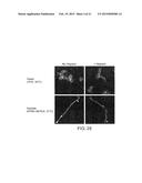 |
 |  |
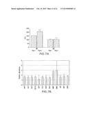 | 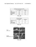 |
 |  |
 |  |
 |  |
 | 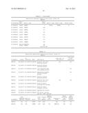 |
 | 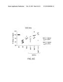 |
 |  |
 | 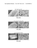 |
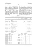 |  |
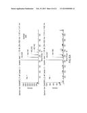 |  |
 | 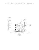 |
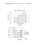 |  |
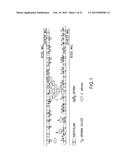 |  |
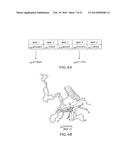 |  |
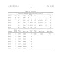 | 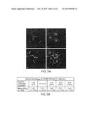 |
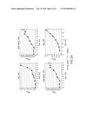 | 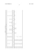 |
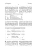 |  |
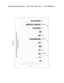 |  |
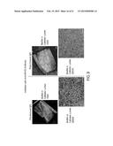 | 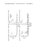 |
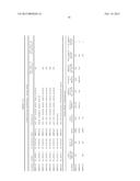 | 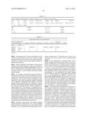 |
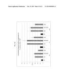 |  |
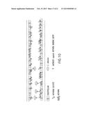 | 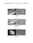 |
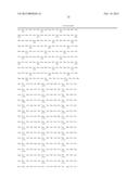 |
| Similar patent applications: | |
| Date | Title |
|---|---|
| 2015-03-12 | Stable peptide mimetics of the hiv-1 gp41 pre-hairpin intermediate |
| 2015-03-05 | Antigen binding proteins to oncostatin m (osm) |
| 2015-03-12 | Variants of group 6 allergens of the true grasses having reduced allergeneity due to mutagenesis of proline residues |
| 2015-02-26 | Trichoderma asperellum to remediate phytophthora ramorum-infested soil |
| 2015-03-12 | Treatment for the rapid amelioration of clinical depression |
| New patent applications in this class: | |
| Date | Title |
|---|---|
| 2022-05-05 | Anti-icos antibodies for the treatment of cancer |
| 2019-05-16 | T cell receptor-like antibodies specific for a wti peptide presented by hla-a2 |
| 2019-05-16 | Immune modulation and treatment of solid tumors with antibodies that specifically bind cd38 |
| 2019-05-16 | Human antibodies to the glucagon receptor |
| 2019-05-16 | Anti-alpha-v integrin antibody for the treatment of fibrosis and/or fibrotic disorders |
| New patent applications from these inventors: | |
| Date | Title |
|---|---|
| 2014-12-11 | Forazolines, compositions thereof and uses thereof |
| Top Inventors for class "Drug, bio-affecting and body treating compositions" | |
| Rank | Inventor's name |
|---|---|
| 1 | David M. Goldenberg |
| 2 | Hy Si Bui |
| 3 | Lowell L. Wood, Jr. |
| 4 | Roderick A. Hyde |
| 5 | Yat Sun Or |