Patent application title: ANTIBODIES SPECIFICALLY BINDING TO THE EPIDERMAL GROWTH FACTOR RECEPTOR
Inventors:
Se-Ho Kim (Yongin-Si, KR)
Ki-Hwan Chang (Yongin-Si, KR)
Kwang-Won Hong (Yongin-Si, KR)
Yong-Won Shin (Yongin-Si, KR)
Min Soo Kim (Yongin-Si, KR)
Min Soo Kim (Yongin-Si, KR)
Hae-Won Lee (Yongin-Si, KR)
Yong-Nam Shin (Yongin-Si, KR)
Kyung-Hwan Ryoo (Yongin-Si, KR)
Dong-Hyuck Seo (Yongin-Si, KR)
Assignees:
GREEN CROSS CORPORATION
IPC8 Class: AA61K39395FI
USPC Class:
4241741
Class name: Immunoglobulin, antiserum, antibody, or antibody fragment, except conjugate or complex of the same with nonimmunoglobulin material binds eukaryotic cell or component thereof or substance produced by said eukaryotic cell (e.g., honey, etc.) cancer cell
Publication date: 2012-09-13
Patent application number: 20120231021
Abstract:
Provided are antibodies specifically binding to the epidermal growth
factor receptor (EGFR) which are effective for the treatment of
EGFR-mediated cancers.Claims:
1. An antibody specifically binding to the epidermal growth factor
receptor (EGFR), comprising: a) a heavy chain variable region comprising
complementarity determining regions (CDRs) 1, 2, and 3 having the amino
acid sequences of SEQ ID NOs: 1, 2, and 3, respectively; b) a light chain
variable region comprising CDR 1, CDR 2, and CDR 3 having the amino acid
sequences of SEQ ID NOs: 4, 5, and 6, respectively; c) a heavy chain
constant region; and d) a light chain constant region.
2. The antibody of claim 1, comprising: a) a heavy chain variable region having the amino acid sequence of SEQ ID NO:7; b) a light chain variable region having the amino acid sequence of SEQ ID NO:8; c) a heavy chain constant region; and d) a light chain constant region.
3. An antibody specifically binding to the epidermal growth factor receptor (EGFR), comprising: a) a heavy chain variable region comprising CDR 1, CDR 2, and CDR 3 having the amino acid sequences of SEQ ID NOs: 1, 2, and 3, respectively; b) a light chain variable region comprising CDR 1, CDR 2, and CDR 3 having the amino acid sequences of SEQ ID NOs: 9, 5, and 6, respectively; c) a heavy chain constant region; and d) a light chain constant region.
4. The antibody of claim 3, comprising: a) a heavy chain variable region having the amino acid sequence of SEQ ID NO:7; b) a light chain variable region having the amino acid sequence of SEQ ID No:10; c) a heavy chain constant region; and d) a light chain constant region.
5. The antibody of any one of claims 1-4, wherein the antibody is a humanized antibody.
6. The antibody of any one of claims 1-4, wherein the antibody blocks the signal transduction induced by the epidermal growth factor (EGF).
7. A DNA encoding an antibody heavy chain variable region comprising CDR 1, CDR 2, and CDR 3 having the amino acid sequences of SEQ ID NOs: 1, 2, and 3, respectively.
8. The DNA of claim 7, wherein the DNA comprises the polynucleotide having the nucleotide sequence of SEQ ID NO: 11 encoding the amino acid sequence of SEQ ID NO: 1, the polynucleotide having the nucleotide sequence of SEQ ID NO: 12 encoding the amino acid sequence of SEQ ID NO: 2, and the polynucleotide having the nucleotide sequence of SEQ ID NO: 13 encoding the amino acid sequence of SEQ ID NO: 3.
9. A DNA encoding an antibody heavy chain variable region having the amino acid sequence of SEQ ID NO: 7.
10. The DNA of claim 9, wherein the DNA comprises the polynucleotide having the nucleotide sequence of SEQ ID NO: 14 encoding the amino acid sequences of SEQ ID NO: 7.
11. A DNA encoding an antibody light chain variable region comprising CDR 1, CDR 2, and CDR 3 having the amino acid sequences of SEQ ID NOs: 4, 5, and 6, respectively.
12. The DNA of claim 11, wherein the DNA comprises the polynucleotide having the nucleotide sequence of SEQ ID NO: 15 encoding the amino acid sequence of SEQ ID NO: 4, the polynucleotide having the nucleotide sequence of SEQ ID NO: 16 encoding the amino acid sequence of SEQ ID NO: 5, and the polynucleotide having the nucleotide sequence of SEQ ID NO: 17 encoding the amino acid sequence of SEQ ID NO: 6.
13. A DNA encoding an antibody light chain variable region having the amino acid sequences of SEQ ID NO: 8.
14. The DNA of claim 13, wherein the DNA comprises the polynucleotide having the nucleotide sequence of SEQ ID NO: 18 encoding the amino acid sequences of SEQ ID NO: 8.
15. A DNA encoding an antibody light chain variable region comprising CDR 1, CDR 2, and CDR 3 having the amino acid sequences of SEQ ID NOs: 9, 5, and 6, respectively.
16. The DNA of claim 15, wherein the DNA comprises the polynucleotide having the nucleotide sequence of SEQ ID NO: 19 encoding the amino acid sequence of SEQ ID NO: 9, the polynucleotide having the nucleotide sequence of SEQ ID NO: 16 encoding the amino acid sequence of SEQ ID NO: 5, and the polynucleotide having the nucleotide sequence of SEQ ID NO: 17 encoding the amino acid sequence of SEQ ID NO: 6.
17. A DNA encoding an antibody light chain variable region having the amino acid sequences of SEQ ID NO: 10.
18. The DNA of claim 17, wherein the DNA comprises the polynucleotide having the nucleotide sequence of SEQ ID NO: 20 encoding the amino acid sequences of SEQ ID NO: 10.
19. An expression vector for expressing the heavy chain variable region of the antibody specifically binding to the epidermal growth factor receptor (EGFR), comprising the DNA of claim 7.
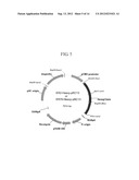
20. The expression vector of claim 19, wherein the vector is ER2-Heavy-pRC13 or ER79-Heavy-pRC13 whose cleavage map is shown in FIG. 5.
21. An expression vector for expressing the light chain variable region of the antibody specifically binding to the epidermal growth factor receptor (EGFR), comprising the DNA of claim 11.
22. The expression vector of claim 21, wherein the vector is ER2-Light-pKC12 whose cleavage map is shown in FIG. 6.
23. An expression vector for expressing the light chain variable region of the antibody specifically binding to the epidermal growth factor receptor (EGFR), comprising the DNA of claim 15.
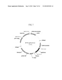
24. The expression vector of claim 23, wherein the vector is ER79-Light-pKC12 whose cleavage map is shown in FIG. 7.
25. An animal cell line transformed with the expression vector for expressing a heavy chain variable region of the antibody specifically binding to the epidermal growth factor receptor (EGFR), comprising a first DNA encoding an antibody heavy chain variable region and the expression vector for expressing a light chain variable region of the antibody specifically binding to the epidermal growth factor receptor (EGFR), comprising a second DNA encoding an antibody light chain variable region, wherein the first DNA encoding an antibody heavy chain variable region comprises CDR 1, CDR 2, and CDR 3 having the amino acid sequences of SEQ ID NOs: 1, 2, and 3, respectively, and wherein the second DNA encoding an antibody light chain variable region comprises CDR 1, CDR 2, and CDR 3 having the amino acid sequences of SEQ ID NOs: 4, 5, and 6, respectively.
26. The animal cell line of claim 25, wherein the animal cell line is CHO (Chinese hamster ovary), HEK 293, or NSO cell line.
27. An animal cell line transformed with the expression vector for expressing a heavy chain variable region of the antibody specifically binding to the epidermal growth factor receptor (EGFR), comprising a first DNA encoding an antibody heavy chain variable region and the expression vector for expressing a light chain variable region of the antibody specifically binding to the epidermal growth factor receptor (EGFR), comprising a second DNA encoding an antibody light chain variable region, wherein the first DNA encoding an antibody heavy chain variable region comprises CDR 1, CDR 2, and CDR 3 having the amino acid sequences of SEQ ID NOs: 1, 2, and 3, respectively, and wherein the second DNA encoding an antibody light chain variable region comprises CDR 1, CDR 2, and CDR 3 having the amino acid sequences of SEQ ID NOs: 9, 5, and 6, respectively.
28. The animal cell line of claim 27, wherein the animal cell line is CHO (Chinese hamster ovary), HEK 293, or NSO cell line.
29. A composition for treating a cancer, comprising the antibody of claim 1 or 3.
30. The composition of claim 29, which further comprises at least one selected from the group consisting of cisplatin, gemcitabine, doxorubicin, 5-FU, irrinotecan, and paclitaxel.
Description:
FIELD OF THE INVENTION
[0001] The present invention relates to antibodies specifically binding to the epidermal growth factor receptor (EGFR).
BACKGROUND OF THE INVENTION
[0002] The epidermal growth factor receptor (EGFR) is a 170 kDa type I transmembrane protein and is known to be overexpressed in many human tumors, e.g., carcinoma of the lung, breast, colon, stomach, brain, bladder, head, neck, ovaries and prostate. Its overexpression is frequently accompanied by the production of EGFR-ligands, TGF-α (transforming growth factor-α) and EGF (epidermal growth factor), and the binding of the ligands to EGFR was confirmed to induce cell proliferation and tumor growth. Blocking the interaction between such ligands and EGFR using an antibody against EGFR therefore can inhibit tumor growth, which has been proven effective by experiments that employed monoclonal antibodies against EGFR.
[0003] Antibody C225 (trade name: Erbitux; ImClone, U.S), which is currently used in clinical trials for the treatment of metastatic colorectal cancers, is a chimeric antibody, comprising the mouse antibody variable regions linked to human antibody IgG1 constant regions (about 30% of mouse amino acid sequence is included therein). C225 has been shown to inhibit tumor cell growth, EGFR phosphorylation in vitro and tumor formation in a nude mouse, and also to completely eradicate human tumor xenografts in mice when used together with a specific chemotherapeutic agent. However, the antibody has the problem of inducing immune reactions in some (˜10%) of the patients treated therewith. Accordingly, there exists a need for improved therapeutic antibodies against EGFR.
[0004] Therapeutic agents for target therapy constitute about 50% of anticancer drugs recently approved by U.S. Food and Drug Administration (FDA). Such antibodies provide target specificity and a capability to effectively engage the immune system, which in combination with long biological half-lives thereof have alerted researchers to the therapeutic potentials thereof. As a result, the U.S. FDA has recently approved the use of several antibodies for cancer treatment. Antibodies play prominent roles in many therapeutic approaches to diseases, which has become even more attractive with the recent advent of technologies that allow the development of fully human antibodies.
[0005] The present inventors have endeavored to develop novel, improved antibodies having new complementarity determining regions (CDRs) and have found that such antibodies can be used in cancer treatment by blocking the EGFR-mediated signal transduction.
SUMMARY OF THE INVENTION
[0006] Therefore, it is an object of the present invention to provide a novel antibody which specifically binds to the epidermal growth factor receptor.
[0007] It is another object of the present invention to provide DNAs which respectively encode the heavy chain variable region and the light chain variable region of said antibody, and an expression vector comprising the same.
[0008] It is still another object of the present invention to provide a cell line transformed with the expression vector.
[0009] It is a further object of the present invention to provide a pharmaceutical composition for treating a cancer, comprising said antibody.
[0010] In accordance with one aspect of the present invention, there is provided an antibody specifically binding to the epidermal growth factor receptor (EGFR), comprising: a) a heavy chain variable region comprising complementarity determining regions (CDRs) 1, 2, and 3 having the amino acid sequences of SEQ ID NOs: 1, 2, and 3, respectively; b) a light chain variable region comprising CDR 1, CDR 2, and CDR 3 having the amino acid sequences of SEQ ID NOs: 4, 5, and 6, respectively; c) a heavy chain constant region; and d) a light chain constant region.
[0011] Further, there is provided an antibody specifically binding to the epidermal growth factor receptor (EGFR), comprising: a) a heavy chain variable region comprising CDR 1, CDR 2, and CDR 3 having the amino acid sequences of SEQ ID NOs: 1, 2, and 3, respectively; b) a light chain variable region comprising CDR 1, CDR 2, and CDR 3 having the amino acid sequences of SEQ ID NOs: 9, 5, and 6, respectively; c) a heavy chain constant region; and d) a light chain constant region.
[0012] In accordance with another aspect of the present invention, there is provided a DNA encoding the heavy chain variable region or the light chain variable region of the antibody, and an expression vector comprising the same.
[0013] In accordance with a still another aspect of the present invention, there is provided a cell line transformed with said expression vector.
[0014] In accordance with a further aspect of the present invention, there is provided a composition for treating a cancer, comprising said antibody.
BRIEF DESCRIPTION OF THE DRAWINGS
[0015] The above and other objects and features of the present invention will become apparent from the following description of the invention, when taken in conjunction with the accompanying drawings, which respectively show:
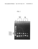
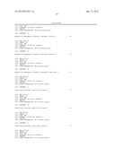
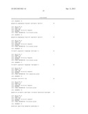
[0016] FIG. 1: a photograph of electrophoresis (1% agarose gel) exhibiting DNAs which respectively encode the inventive heavy chain variable (VH) and the light chain variable regions (VL) synthesized by PCR;
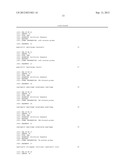
[0017] FIG. 2: a cleavage map of the phage-display vector, pKS4H, comprising the heavy chain variable region and the light chain variable region of the inventive antibody;
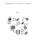
[0018] FIG. 3: a diagram showing a process of selecting an antibody from an antibody library using the biopanning technique;
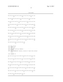
[0019] FIG. 4: amino acid sequences of the single chain variable fragments (scFv) of the inventive antibodies, ER2 and ER79;
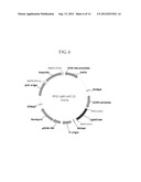
[0020] FIG. 5: a cleavage map of the expression vector for expressing the heavy chain of the human antibody of the present invention, ER2-Heavy-pRC13 or ER79-Heavy-pRC13;
[0021] FIGS. 6 and 7: cleavage maps of expression vectors for expressing the light chains of the human antibodies of the present invention, ER2-Light-pKC12 and ER79-Light-pKC12;
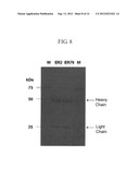
[0022] FIG. 8: SDS-PAGE results obtained for the heavy chain and light chain expressed from the transformant;
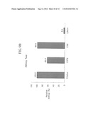
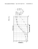
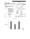
[0023] FIG. 9: relative affinities of the human antibodies (ER2 and ER79), a chimeric antibody (C225, Erbitux), and other antibody (ER414) to the epidermal growth factor receptor;
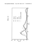
[0024] FIG. 10: a flow cytometer exhibiting the binding of the inventive antibodies with the epidermal growth factor receptor overexpressed in a cancer cell line (A431);
[0025] FIG. 11: the inhibitory effect of the inventive antibodies on the phosphorylation of the epidermal growth factor receptor; and
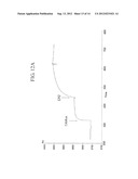
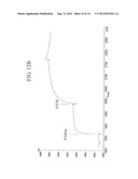
[0026] FIG. 12: surface plasmon resonance measurement results revealing the binding sites of the inventive antibodies with the epidermal growth factor, and those of chimeric antibody C225 (Erbitux).
DETAILED DESCRIPTION OF THE INVENTION
[0027] Hereinafter, the present invention is described in detail.
[0028] The present invention provides an antibody specifically binding to the epidermal growth factor receptor (EGFR), comprising a) a heavy chain variable region comprising complementarity determining regions (CDRs) 1, 2, and 3 having the amino acid sequences of SEQ ID NOs: 1, 2, and 3, respectively; b) a light chain variable region comprising CDR 1, CDR 2, and CDR 3 having the amino acid sequences of SEQ ID NOs: 4, 5, and 6, respectively; c) a heavy chain constant region; and d) a light chain constant region. Preferably, the antibody may be one, comprising: a) a heavy chain variable region having the amino acid sequence of SEQ ID NO:7; b) a light chain variable region having the amino acid sequence of SEQ ID NO:8; c) a heavy chain constant region; and d) a light chain constant region.
[0029] Further, the present invention provides an antibody specifically binding to the epidermal growth factor receptor (EGFR), comprising: a) a heavy chain variable region comprising CDR 1, CDR 2, and CDR 3 having the amino acid sequences of SEQ ID NOs: 1, 2, and 3, respectively; b) a light chain variable region comprising CDR 1, CDR 2, and CDR 3 having the amino acid sequences of SEQ ID NOs: 9, 5, and 6, respectively; c) a heavy chain constant region; and d) a light chain constant region. Preferably, the antibody may be one, comprising: a) a heavy chain variable region having the amino acid sequence of SEQ ID NO:7; b) a light chain variable region having the amino acid sequence of SEQ ID No:10; c) a heavy chain constant region; and d) a light chain constant region.
[0030] The inventive antibodies may be preferably human antibodies, and is characterized in blocking the signal transduction induced by the epidermal growth factor (EGF).
[0031] The antibodies specifically binding to the epidermal growth factor receptor may be preferably selected by a modification of a phage display method (Smith, Science, 228, 1315-1317, 1985; and Hoogenboom & Chames, Immunol Today, 21, 371-378, 2000). In the phage display method, a gene (gene III) encoding a surface protein of filamentous phage (e.g. M13, Fd or F1) is fused with a gene encoding an antibody of interest, thereby virus particles having the fused antibody exposed on the surface is produced as an antibody-phage form. Subsequently, an antibody of interest can be selected from a phage library through the biopanning technique using high specificity and affinity of the antibody and high infective property of the phage (Burton & Barbas, Adv. Immunol., 57, 191-280, 1994; Winter et al., Annu. Rev. Immunol., 12, 433-455, 1994; and Hoogenboom et al., Immunotechnology, 4, 1-20, 1998). The phage display vector may be pKS4H (see Korean Patent no. 0635370) or pCANTAB5E, preferably, pKS4H.
[0032] In the present invention, a human antibody ER414 was selected from a phage library and its affinity and neutralizing power against the epidermal growth factor receptor were checked (FIGS. 9 and 11). The ER414 antibody has the neutralizing power, but its affinity was 16 times lower than commercially available antibody, C225. Accordingly, improved antibodies with similar affinities were selected using the affinity maturation process. That is, a library was generated through the amino acid randomization of complementarity determining regions of the antibody primarily selected, antibodies having the affinity matured were selected using biopanning technique, and finally antibodies (ER2 and ER79) having similar affinities to C225 antibody were selected by the competitive ELISA method.
[0033] In case of the antibody ER2, CDR 1, CDR 2, and CDR 3 of the heavy chain variable region have the amino acid sequences of SEQ ID NOs: 1, 2, and 3, respectively, and CDR 1, CDR 2, and CDR 3 of the light chain variable region have the amino acid sequences of SEQ ID NOs: 4, 5, and 6, respectively, as a result of sequence analysis. On the other hand, CDR 1, CDR 2, and CDR 3 of the heavy chain variable region of the antibody ER79 have the amino acid sequences of SEQ ID NOs: 1, 2, and 3, respectively, and CDR 1, CDR 2, and CDR 3 of the light chain variable region have the amino acid sequences of SEQ ID NOs: 9, 5, and 6, respectively.
[0034] The heavy chain constant regions or light chain constant regions of the inventive antibodies may be identical to those of a human antibody, and may be preferably amino acids having the amino acid sequences of SEQ ID NOs: 43 and 44, respectively.
[0035] The present invention provides a DNA encoding an antibody heavy chain variable region comprising CDR 1, CDR 2, and CDR 3 having the amino acid sequences of SEQ ID NOs: 1, 2, and 3, respectively. Preferably, the DNA may comprise the polynucleotide having the nucleotide sequence of SEQ ID NO: 11 encoding the amino acid sequence of SEQ ID NO: 1, the polynucleotide having the nucleotide sequence of SEQ ID NO: 12 encoding the amino acid sequence of SEQ ID NO: 2 and the polynucleotide having the nucleotide sequence of SEQ ID NO: 13 encoding the amino acid sequence of SEQ ID NO: 3.
[0036] The present invention provides a DNA encoding an antibody heavy chain variable region having the amino acid sequence of SEQ ID NO: 7. Preferably, the DNA may comprise the polynucleotide having the nucleotide sequence of SEQ ID NO: 14 encoding the amino acid sequences of SEQ ID NO: 7.
[0037] Further, the present invention provides a DNA encoding an antibody light chain variable region comprising CDR 1, CDR 2, and CDR 3 having the amino acid sequences of SEQ ID NOs: 4, 5, and 6, respectively. Preferably, the DNA may comprise the polynucleotide having the nucleotide sequence of SEQ ID NO: 15 encoding the amino acid sequence of SEQ ID NO: 4, the polynucleotide having the nucleotide sequence of SEQ ID NO: 16 encoding the amino acid sequence of SEQ ID NO: 5, and the polynucleotide having the nucleotide sequence of SEQ ID NO: 17 encoding the amino acid sequence of SEQ ID NO: 6.
[0038] The present invention provides a DNA encoding an antibody light chain variable region having the amino acid sequences of SEQ ID NO: 8. Preferably, the DNA may comprise the polynucleotide having the nucleotide sequence of SEQ ID NO: 18 encoding the amino acid sequences of SEQ ID NO: 8.
[0039] Further, the present invention provides a DNA encoding an antibody light chain variable region comprising CDR 1, CDR 2, and CDR 3 having the amino acid sequences of SEQ ID NOs: 9, 5, and 6, respectively. Preferably, the DNA may comprise the polynucleotide having the nucleotide sequence of SEQ ID NO: 19 encoding the amino acid sequence of SEQ ID NO: 9, the polynucleotide having the nucleotide sequence of SEQ ID NO: 16 encoding the amino acid sequence of SEQ ID NO: 5 and the polynucleotide having the nucleotide sequence of SEQ ID NO: 17 encoding the amino acid sequence of SEQ ID NO: 6.
[0040] The present invention provides a DNA encoding an antibody light chain variable region having the amino acid sequences of SEQ ID NO: 10. Preferably, the DNA may comprise the polynucleotide having the nucleotide sequence of SEQ ID NO: 20 encoding the amino acid sequences of SEQ ID NO: 10.
[0041] The present invention provides an expression vector for expressing the heavy chain variable region of the antibody specifically binding to the epidermal growth factor receptor (EGFR), comprising the DNA encoding the heavy chain variable region of the antibody. Preferably, the expression vector may be "ER2-Heavy-pRC13" or "ER79-Heavy-pRC13" whose cleavage map is shown in FIG. 5.
[0042] Specifically, the vector may be prepared by inserting the VH fragment (1-a: ER2Ab-H or 1-b: ER79Ab-H) of the antibody selected using panning and affinity maturation processes into a suitable vector, e.g., pRC13 vector (deposit No. KCLRF-BP-00054; Korean Patent No. 523732).
[0043] The present invention provides an expression vector for expressing the light chain variable region of the antibody specifically binding to the epidermal growth factor receptor (EGFR), comprising the DNA encoding the light chain variable region of the antibody. Preferably, the expression vector may be "ER2-Light-pKC12" whose cleavage map is shown in FIG. 6, or "ER79-Light-pKC12" whose cleavage map is shown in FIG. 7.
[0044] Specifically, the vectors may be prepared by inserting each VL fragment (2-a: ER2Ab-L or 1-b: ER79Ab-L) of the antibodies selected using the panning and affinity maturation processes into a suitable vector, e.g., pKC12 vector (deposit No. KCLRF-BP-00054; Korean Patent No. 523732).
[0045] The present invention provides an animal cell line transformed with the expression vector for expressing the heavy chain variable region of the inventive antibody, and the expression vector for expressing the light chain variable region of the inventive antibody. The expression vector for expressing the heavy chain variable region of the inventive antibody may be preferably ER2-Heavy-pRC13, or ER79-Heavy-pRC13, and the expression vector for expressing the light chain variable region of the inventive antibody may be preferably ER2-Light-pKC12, or ER79-Light-pKC12. The animal cell line may be CHO (Chinese hamster ovary), HEK 293, or NSO cell line, preferably, CHO (Chinese hamster ovary) cell line.
[0046] The antibodies according to the present invention may be prepared by which the heavy chain variable region and the light chain variable region are combined together.
[0047] The affinity of the inventive antibodies to the antigen may be measured, e.g., by the competitive ELISA (Kim et al., Hybridoma, 20, 265-272, 2001). As shown in FIG. 9, the affinity of ER2 antibody of the present invention is similar to that of C225 antibody, whereas the affinity of ER79 antibody is two times lower than that of C225 antibody. Further, the antibodies was demonstrated to bind to the epidermal growth factor receptor overexpressed in a cancer cell line using a flow cytometer (FACS) (FIG. 10), and confirmed to have the neutralizing power through the experiment of the epidermal growth factor receptor phosphorylation inhibition in a breast cancer cell (FIG. 11). Therefore, the antibodies of the present invention may be used as an antibody for treating a cancer by inhibiting the signal transduction through the epidermal growth factor receptor.
[0048] In view of the result, the present invention provides a composition, preferably pharmaceutical composition, for treating a cancer, comprising the antibody. The composition may further comprise at least one selected from the group consisting of cisplatin, gemcitabine, doxorubicin, 5-FU, irrinotecan, and paclitaxel.
[0049] The composition contains ER2 or ER79 antibody or transformants containing the same as an active ingredient and additionally includes one or more effective ingredients having the same or similar functions to the said active ingredient. In addition to the active ingredient, the composition of the present invention can include one or more pharmaceutically acceptable carriers such as saline, sterilized water, Ringer's solution, buffered saline, dextrose solution, maltodextrin solution, glycerol, ethanol, liposome and a mixture comprising one or more of those components. If necessary, a general additive such as an antioxidant, a buffer, and a bacteriostatic agent can be additionally added. The composition of the present invention can be formulated in different forms including aqueous solutions, suspensions and emulsions for injection, pills, capsules, granules or tablets by mixing with diluents, dispersing agents, surfactants, binders and lubricants. A target cell specific antibody or other ligands can be mixed with one of the said carriers to be delivered to the target cell. The composition can further be prepared in suitable forms according to ingredients by following the method represented in Remington's Pharmaceutical Science, Mack Publishing Company, Easton Pa.
[0050] The pharmaceutical composition of the present invention can be administered parenterally (for example, intravenous, hypodermic, peritoneal or local injection), and intravenous injection is preferred. In some cases of solid cancer, local administration which favors fast and easy access of antibody is more preferred. The effective dosage of the composition can be determined according to weight, age, gender, health condition, diet, administration frequency, administration method, excretion and severity of a disease. One time dosage of the composition containing humanized antibody or transformant approximately 5-500 mg/m2, which can be administered daily or weekly. The effective dosage can be adjusted by a doctor who treats malignant tumor patients.
[0051] The pharmaceutical composition of the present invention can be administered alone or together with surgical operation, hormone therapy, chemo-therapy and biological regulators to treat malignant tumors.
[0052] The following Examples are given for the purpose of illustration only, and are not intended to limit the scope of the invention.
Example 1
Isolation of RNA
[0053] In order to select antibodies specifically binding to the epidermal growth factor receptor, a gene library of antibodies was constructed. Human bone marrow total RNA, human thymus total RNA, human spleen total RNA and human B cell RNA were used as a mix. All RNAs except for human B cell RNA were purchased from Clontech (U.S) and human B cell RNA was isolated as follows:
[0054] 50 mL of blood taken from a healthy adult was diluted by mixing with 50 mL of HBSS (Hank's balanced salt solution; Sigma, US) in a mixing ratio of 1:1, and stored until use. 10 mL of Histoprep (Sigma) was put in a 50 mL tube and 20 mL of the diluted blood was added thereto. The mixture was centrifuged at 3,000 rpm to isolate a white blood cell. 2 mL of the isolated white blood cell was mixed with 6 mL of HBSS and centrifuged at 1,000 rpm. 100 μL of the white blood cell was mixed with 1 mL of Trizole (Life Technology, U.S) to isolate RNA.
[0055] Meanwhile, the isolated RNA was diluted with distilled water, and the absorbance at 260 nm was measured to calculate its amount (1.8 μg/μL; Ultraspec 2000 UV-VIS spectrophotometer, GE, U.S). Detailed procedure is as follows:
[0056] 1 mL of trizole was added to 100 μL of white blood cell, shook well, and left at room temperature for 5 min. Then, 200 μL of chloroform was added, vigorously shook for 15 sec, and left for 3 min. Subsequently, the mixture was centrifuged under a condition of 2˜8° C., 15 min and 15,000 rpm, and the supernatant were transferred into a new tube. 500 μL of isopropyl alcohol was added and mixed well, and left at room temperature for 10 min. Then, the mixture was centrifuged at 2˜8° C. and 15,000 rpm for 5 min to remove the supernatant. 1 mL of 75% ethanol was added thereto and the mixture was centrifuged under a condition of 2˜8° C., 5 min and 15,000 rpm to remove ethanol, and the RNA pellet was dried at room temperature for 5 min. 150 μL of distilled water was added thereto to suspend the RNA pellet, and the absorbance at 260 nm of the suspension was measured. The remnant was stored at -20° C.
Example 2
Amplification of Antibody Genes
[0057] 1 μg of RNA isolated in Example 1 and 1 μL of pd(T)12-18 (0.5 μg/μL) were mixed with distilled water to make final volume into 12.5 μL. The mixture was reacted at 70° C. for 2 min and cooled using ices. Then, 5× reaction buffer, 10 mM dNTP mix, recombinant RNase inhibitor and MMLV reverse transcriptase (Clontech, U.S) were added thereto to make final volume into 20 μL, followed by the reaction at 42° C. for 1 hr and at 95° C. for 5 min to synthesize cDNA. PCR reaction was carried out using LiquiMix QM Premix, Magenta (Neurotics Inc, Korea), 4 μL of cDNA as a template, 19 μL of distilled water, and 1 μL of primers designed to homogenously bind to scFv, light chain variable region and light chain variable region (kappa and lambda), respectively. Primers used in PCR and their nucleotide sequences were shown in Table 1.
TABLE-US-00001 TABLE 1 Primers used in PCR reaction Primers Nucleotide sequence SEQ ID NO. scFv-Forward 5'-GTTGTTCCTTTCTATGCGGCCCAGCCGGCCATGGCC-3' 21 scFv-Reverse 5'-GAGTCATTCTCGACTTGCGGCCGCACGTTT-3' 22 scFv-Reverse 5'-GAGTCATTCTCGACTTGCGGCCGCACC-3' 23 VH1-Forward 5'-CAGCCGGCCATGGCCCAGGTGCAGCTGGTGCAGTCTGGG-3' 24 VH3-Forward 5'-CAGCCGGCCATGGCCSAGGTGCAGCTGGTGGAGTCTGGG-3' 25 VH4-Forward 5'-CAGCCGGCCATGGCCCAGGTGCAGCTGCAGGAGTCGGGC-3' 26 VH-Reverse 5'-CGATCCGCCACCTCCGGAGCCACCTCCGCCTGAACCGCCTCCACC-3' 27 VK1/3A-Forward 5'-GGTGGCTCCGGAGGTGGCGGATCGGACATCCAGATGACCCAGTCTCCA-3' 28 VK1/3B-Forward 5'-GGTGGCTCCGGAGGTGGCGGATCGGAAATTGTGTTGACGCAGTCTCCA-3' 29 VK2-Forward 5'-GGTGGCTCCGGAGGTGGCGGATCGGATATTGTGATGACCCAGACTCCACTC-3' 30 JK_A-Reverse 5'-TCGACTTGCGGCCGCACGTTTGATWTCCACYTTGGTCCC-3' 31 JK_B-Reverse 5'-TCGACTTGCGGCCGCACGTTTGATCTCCASCTTGGTCCC-3' 32 JK_C-Reverse 5'-TCGACTTGCGGCCGCACGTTTAATCTCCAGTCGTGTCCC-3' 33 VL_A-Forward 5'-GGTGGCTCCGGAGGTGGCGGATCGCAGTCTGYSCTGACTCAGCCACCC-3' 34 VL_B-Forward 5'-GGTGGCTCCGGAGGTGGCGGATCGTCCTATGAGCTGACWCAGCCACCC-3' 35 JL_A-Reverse 5'-TTCTCGACTTGCGGCCGCACCTAGGACGGTSASCTTGGTCCC-3' 36 JL_B-Reverse 5'-TTCTCGACTTGCGGCCGCACCGAGGACGGTCAGCTGGGTGCC-3' 37
[0058] PCR reaction was carried out at 95° C. for 5 min, 55° C. for 2 min, 72° C. for 2 min with 30 cycles, finally 72° C. for 15 min.
[0059] The amplified antibody DNAs were identified by an electrophoresis in 1.2% agarose gel (FIG. 1). As shown in FIG. 1, 350 bp of DNA bands specific to the heavy chain and light chain (kappa and lambda) variable regions were obtained. In FIG. 1, M refers to a size marker, VH to heavy chain variable region (lane 1: heavy chain variable region type I; lane 2: heavy chain variable region type III; and lane 3: heavy chain variable region type IV), VL to light chain variable region (lane 4: light chain variable region 1/3 κ; lane 5: light chain variable region 2 κ; and lane 6: light chain variable region λ).
Example 3
Restriction Enzyme Digestion of Antibody DNAs
[0060] VH and VL (kappa and lambda) prepared in Example 2 were digested with restriction enzymes SfiI/BspEI and BspEI/NotI, respectively, and the digested fragments were isolated from a 1.2% agarose gel and purified using Qiagen kit.
Example 4
Ligation of the Antibody DNAs and Preparation of Libraries
[0061] Phage-display vector, pKS4H (Green cross Corp., Korea, see Korean Patent No. 0635370), were digested using a restriction enzyme, SfiI/BspEI, and was separated using 1.2% agarose gel electrophoresis, followed by purification using Qiagen kit. 30 μg of the pKS4H was mixed with 3 μg of VH prepared in Example 3, and T4 DNA ligase (New England BioLabs, U.S) was added thereto, followed by the reaction overnight at 25° C. The ligation mixture was purified using Qiagen kit, and was transformed into E. coli XL1-blue (Stratagene, U.S) by electroporation. The transformant was cultured in 100 mL of medium overnight, and the plasmid was isolated. The plasmid was designated as "pKS4H-VH-ΔVL".
[0062] The plasmid, pKS4H-VH-ΔVL, was digested with a restriction enzyme, BspEI/NotI, and purified as described above. Then, 30 μg of pKS4H-VH-ΔVL plasmid was mixed with 3 μg of VL PCR DNA and T4 DNA ligase (New England BioLabs, U.S), and reacted overnight at 25° C. The ligation mixture was purified using Qiagen kit, and was transformed into E. coli XL1-blue by electroporation. The transformant was cultured in 100 mL of medium containing carbenicillin and tetracyclin at 37° C. for 2 hours. Then, M13 helper phage (Stratagene, U.S) was inoculated to the medium and cultured for 16 hr to prepare a phage library as reported in Engberg et al (Mol. Biotechnol., 6, 287-310, 1995). Meanwhile, a plasmid was isolated from the E. coli, and designated as "pKS4H-VH-VL". The cleavage map of the plasmid is depicted in FIG. 2.
Example 5
Selection of Antibodies Binding to the Epidermal Growth Factor Receptor
[0063] Antibodies binding to EGFR were selected by a modification of panning technique (Engberg et al., Mol. Biotechnol., 6, 287-310, 1996; and Kim et al., Gene, 241, 19-25, 2000). Specifically, EGFR (Sigma, U.S) was diluted with PBS and coated onto each immunotube (NUNC, Denmark). Then, the phage library prepared in Example 4 was added to the coated immunotube and reacted. Phages binding to EGFR were detached using 0.1M of glycine buffer (pH 2.0). Subsequently, E. coli XL1-blue was infected with the phages and a helper phage was added. The E. coli was incubated overnight and PEG solution containing 20% PEG 8,000 and 15% NaCl was added thereto. Then, precipitated phages were collected (phage rescue). The phages were again reacted to the EGFR-coated immunotube and the procedure was repeated 4 times (panning). Through the procedure, human antibodies ER2 and ER79 were selected as antibodies binding to EGFR. The process of selecting antibodies using phage-display libraries was depicted in FIG. 3.
[0064] Each colony of libraries completed 4 times panning was incubated in 2 mL of medium, according to the known method (Kim et al., Gene, 241, 19-25, 2000), and expression of antibody was induced by treatment of IPTG (isopropyl β-D-1-thiogalactopyranoside). The induction of antibody was measured by ELISA (Enzyme-Linked ImmunoSorbent Assay) using an EGFR coated 96-well plate.
Example 6
Sequence Analysis of Selected Antibodies
[0065] Colonies which secrete human antibodies ER2 and ER79 selected in Example 5 were incubated overnight in 10 mL of LB medium containing 50 μg/mL of carbenicillin and recombinant plasmids were isolated using Qiagen plasmid mini kit (Qiagen, Valencia, Calif., U.S) therefrom. The plasmids were digested with SfiI/NotI, identified of the insertion of fragments of antibodies by an electrophoresis in agarose gel. The DNA sequence of scFv inserted into the plamid was analyzed.
[0066] p033 of SEQ ID NO: 38 was used as a sequencing primer, and sequences were analyzed in Genotech (Daejeon, Korea) according to the conventional method. The DNA sequences of scFv of ER2, ER79 and M96 (mouse antibody) were translated into amino acids using a web-based program (www.expasy.org: DNA to Protein translate tool), and the translated amino acid sequences were shown in FIG. 4. In FIG. 4, M96, ER2 and ER79 refer to amino acid of scFv of M96 (mouse antibody) and ER2 and ER79 of the present invention, respectively. As shown in FIG. 4, human antibodies ER2 and ER79 had different amino acid sequence.
Example 7
Construction of Expression Vectors
[0067] In order to convert the antibody fragments into intact immunoglobulins, antibody expression vectors, pRC13 and pKC12 (plasmids for insertion of a variable region of a human antibody against the surface antigen of hepatitis B virus; Korean Patent No. 523732; Deposit No. KCLRF-BP-00054) were used.
[0068] Each VH fragment was inserted into HindIII and ApaI site of the heavy chain expression vector, pRC13. As exemplified in FIG. 5, the DNAs encoding the heavy chain variable regions (VHs) of the human antibodies ER2 and ER79 were amplified by PCR using respective primer of SEQ ID NOs: 39 and 40, digested with HindIII/ApaI, and inserted into pRC13 which was digested with same restriction enzymes. The recombinant vector was designated "ER2-Heavy-pRC13" or "ER79-Heavy-pRC13". The primers used are shown in Table 2.
TABLE-US-00002 TABLE 2 Primers used in PCR SEQ Primers Nucleotide sequence ID NO VH-Forward 5'-GGAGACCCAAGCTTGGTACCGAGCTCGGAT 39 CCACTAGTAACGGCCGCCAGTGTGCTGGAA-3' VH-Reverse 5'-GAAGACCGATGGGCCCTTGGTGGAGGCTGA 40 GGAGACGGTGAC-3'
[0069] Meanwhile, each VL fragment was inserted into NheI and ApaI site of the light chain expression vector, pKC12. As exemplified in FIGS. 6 and 7, each DNA encoding the light chain variable region (VL) of the human antibodies ER2 and ER79 was amplified by PCR using respective primer of SEQ ID NOs: 41 and 42, digested with NheI/ApaI, and inserted into pKC12 which was digested with same restriction enzymes. The recombinant vector was designated "ER2-Light-pKC12" or "ER79-Light-pKC12". The primers used are shown in Table 3.
TABLE-US-00003 TABLE 3 Primers used in PCR SEQ Primers Nucleotide sequence ID NO VL-Forward 5'-TAGGGAGACCCGCTAGCGGAGCAAGATGGA 41 TTCACAGGCCCAGGT-3' VL-Reverse 5'-TATAGAATAGGGCCCCCCCTCGAGGTCGAC 41 CTAACACTCTCCCCT-3'
Example 8
Construction of Animal Cell Lines Secreting Antibodies
[0070] 2×10⁵ CHO (Chinese hamster ovary) cells were incubated in T-25 flask (NUNC, Denmark) filled with α-MEM medium (Life Technologies, U.S) containing 10% FBS (Life Technologies, U.S), 24 hours prior to transformation. The incubation was carried out in 37° C. incubator in the presence of 5% CO2, until confluency reaches 50%. Next day, 30 μg of lipofectin (Life Technologies, U.S) was added to 1.5 mL of opti-MEM (Life Technologies, U.S) and left undisturbed at room temperature for 90 min. After 90 min, the medium was mixed with the medium containing ER2-Heavy-pRC13, ER2-Light-pKC12, ER79-Heavy-pRC13 and ER79-Light-pKC12, respectively, to react at room temperature for 15 min. During the reaction, cells for transformation was separated from the medium, and washed three times with PBS. To the washed cells, the reaction mixture was added for incubation. After 6 hours, the reaction mixture was removed, and α-MEM medium was added for incubation for 48 hours. The cells incubated for 48 hours were treated with trypsin (Life Technologies, U.S) to detach from the flask, diluted with α-MEM medium, and subcultured at 96-well plate (NUNC, Denmark). At the time, the α-MEM medium does not contain ribonucleoside and deoxyribonucleoside, while contains 10% of dialyzed FBS (Life Technologies, U.S) and 550 μg/mL of G418 (Sigma, U.S). The medium was replaced with a new medium every two days. The culture supernatant forming colonies was collected for ELISA, and selected cells were transferred into 12-well plate. The cells were transferred into 6-well plate if the cells grow well in 12-well plate, and methotrexate (MTX, Choongwae Pharma Corporation, Korea) was treated if the cells grow well in 6-well plate. The initial concentration of MTX was 20 nM, and increased to 80 nM, 320 nM and 1 μM according to the cell's growth. Cell lines which survived at a concentration of 1 μM and had a high antibody secretion amount were selected, and mass-cultured. The mass culture was carried out in an incubator with 65 rpm, 5% CO2 and 37° C., using spinner flask and serum-free medium. The cell lines (108 cells) were cultured in 250 mL flask filled with 100 mL of serum-free medium. When the number of the cells became 2 times higher, supernatant and cells were collected by centrifugation at 1,000 rpm for 5 min, respectively. The collected cells were cultured again in 500 mL flask filled with 200 mL of medium. When the number of the cells became 2 times higher, cells were separated and transferred into 3 L spinner flask filled with 1 L of medium. Sodium butyrate (Aldrich, U.S) were added thereto to a final concentration of 2 mM, the cells were cultured for 5 days, and the supernatant was collected from the medium. From all supernatants collected by culturing in spinner flasks, antibodies were purified using a protein A-agarose column (Amersham Pharmacia Biotech, U.S) and were analyzed using SDS-PAGE electrophoresis.
[0071] As shown in FIG. 8, about 50 kd of heavy chain band and 25 kd of light chain band were observed, indicating that antibodies were certainly produced.
Example 9
Measurement of Antibody Affinity
[0072] The affinities of the antibodies obtained in Example 8 to EGFR were determined by a competitive ELISA method (Kim et al., Hybridoma, 20, 265-272, 2001), and the results were shown in FIG. 9. Brief procedure is as follows:
(1) Determination of Optimum Concentration of Antibodies
A. Preparation of a Plate
[0073] 100 μL of EGFR (Sigma, U.S) at a 2 μg/mL dilution in PBS was added to each well of a plate and incubated overnight at 4° C. Each well of the plate was washed once with PBST, 300 μL of 1% BSA-PBS solution was added to each well, and stored for 1 hour at room temperature.
B. 1st Reaction
[0074] 100 μL of each purified antibody (0.5 μg/mL) was added to each well of plate, reacted for 2 hours at room temperature, and washed four times with PBST.
C. 2nd Reaction
[0075] 100 μL of goat anti-human IgG (Fab specific)-perxoidase conjugate (Sigma) at a 1:5000 dilution in 1% BSA-PBS was added to each well, incubated for 1 hour at room temperature, and washed four times with PBST.
D. Substrate Reaction
[0076] 100 μL of TMB (3,3',5,5'-tetramethylbenzidine, Microwell peroxidase substrate system (KPL, MD, U.S)) was added to each well and O.D value was measured at 405 nm. Optimum concentrations of antibodies were determined as 1/2 of concentrations at which maximum binding appears.
(2) Competitive ELISA
A. Preparation of a Plate
[0077] 100 μL of EGFR (Sigma, U.S) at a 2 μg/mL dilution in PBS was added to each well of a plate and incubated overnight at 4° C. Each well was washed once with PBST, 300 μL of 1% BSA-PBS solution was added to each well, and stored for 1 hour at room temperature.
B. 1st Reaction
[0078] 2 μg of EGFR was diluted by a two-fold and 10 μL of the diluted EGFR was added to each well of the plate. Then, 90 μL of the antibody diluted to the optimum concentration determined in (1) was added to each well, incubated for 2 hours at room temperature, and washed 4 times with PBST.
C. 2nd Reaction 100 μL of goat anti-human IgG (Fab specific)-perxoidase conjugate (Sigma) at a 1:5000 dilution in 1% BSA-PBS was added to each well, incubated for 1 hour at room temperature, and washed four times with PBST.
D. Substrate Reaction
[0079] 100 μL of TMB (3,3',5,5'-tetramethylbenzidine, Microwell peroxidase substrate system (KPL, MD, U.S)) was added to each well and O.D value was measured at 405 nm. Concentration of EGFR which inhibits 50% of maximum binding (O.D value in which no competing EGFR exists) was determined as Kd.
[0080] As shown in FIG. 9, the human antibody ER2 showed a similar affinity and ER79 showed about 63% of affinity, relative to those of a chimeric antibody (C225) and a mouse antibody (M96). Further, the affinities of the inventive antibodies were higher than those of ER414 (human antibody prior to biopanning).
Example 10
Verification of the Binding of the Inventive Antibodies to EGFR in a Tumor Cell Line
[0081] In order to verify that the inventive antibodies, ER2 and ER79, bind to EGFR overexpressed in a tumor cell line, a flow cytometry was used. Briefly, A431 cells (Deposit No. KCLB 80005), an epidermoid carcinoma cell line which overexpresses EGFR, were washed with 1% BSA-PBS. The washed cells (1×10⁶ cells) were incubated with 10 μg of the inventive antibodies for 2 hours at 4° C. and washed two times with 1% BSA-PBS. Mock (without antibody) and hTT-2 (anti-tetanus monoclonal antibody; 10 μg; Green cross incorporation; Korean Patent No. 0624011) were used as negative controls, and M96 (mouse anti-EGFR; 10 μg) as a positive control. FITC-labeled goat anti-mouse (Fab-specific) conjugate was added to the antibody cell solution and incubated for 40 minutes on ice. The cells were washed two times with 1% BSA-PBS and suspended in 1 mL of 1% BSA-PBS to be analyzed using flow cytometry (FACS Calibur, BD Bioscience). The results are shown in FIG. 10. These results indicate that the inventive antibodies, ER2 and ER79, bind to EGFR in A431 cells, while hTT2 (anti-tetanus monoclonal antibody) does not bind.
Example 11
Effect of the Inventive Antibodies on EGFR Phosphorylation
[0082] The inventive antibodies, ER2 and ER79, were tested for their ability to inhibit the EGFR phosphorylation. Briefly, MDA-MB-231 cells (Deposit No. KCLB 30026), a breast cancer cell line, were incubated in 24-well plates (NUNC) at a cell concentration of 1×10⁵. Two days later, the inventive antibodies were added to each well, in amounts of 5, 25, 50, and 100 μg, respectively, and then 50 ng of EGF was added to each well and incubated for 30 minutes. For comparison, M96 antibody (Green cross incorporation, Korea; see Korean Patent No. 0680141), C225 antibody (trade name: Erbitux; ImClone, U.S), and ER 414 antibody were used instead of the inventive antibodies. Cell extracts were prepared using 0.5 mL of lysis buffer (10 mM Tris, 150 mM NaCl, 5 mM EDTA, 1% Triton X-100, 1 mM sodium orthovanadate) per well. The cell extracts were subjected to SDS-PAGE electrophoresis, and separated proteins were electrotransferred into a nitrocellulose membrane. The membrane was blocked for 30 minutes using 5% BSA solution in order to reduce non-specific binding of the transferred proteins, and immunoblotted overnight at 4° C. using anti-phosphotyrosine specific peroxidase conjugate (Zymed, U.S) which specifically reacts with phosphorylated EGFR. The immunoblotted membrane was washed with PBS containing 0.05% tween and developed using a substrate of 0.018% (v/v) 4-chloro-1-naphthol and 0.045% hydrogen peroxide in PBS and methanol. The results were shown in FIG. 11. These results indicate that the amounts of antibodies affect the EGFR phosphorylation and ER2 and ER79 have similar inhibitory abilities of EGFR phosphorylation compared to the positive control group treated with Erbitux.
Example 12
Identification of Binding Sites of the Antibodies to EGFR
[0083] In order to check if the inventive antibodies, ER2 and ER79, has the same binding sites to EFGR with a chimeric antibody C225 (Erbitux, ImClone, U.S), a surface plasmon resonance technology (SPR; Biacore 2000) was used. EGFR antigen was immobilized onto a carboxymethylated dextran surface chip (CM5 chip, Pharmacia) in response units of about 1,000. Then, C225 antibody was injected over the chip, and ER2 and ER79 were immediately injected without dissociation between the antigens and antibodies, respectively, followed by measurement of the binding reaction at 25° C. The results were shown in FIG. 12. The inventive human antibodies were shown to have different binding sites with C225 antibody.
[0084] While the invention has been described with respect to the above specific embodiments, it should be recognized that various modifications and changes may be made to the invention by those skilled in the art which also fall within the scope of the invention as defined by the appended claims.
Sequence CWU
1
4415PRTArtificial SequenceSynthetic construct of CDR 1 of heavy chain
variable region 1Asp Tyr Asp Met Ser1 5217PRTArtificial
SequenceSynthetic construct of CDR 2 of heavy chain variable region
2Gly Ile Leu Gly Gly Ser Glu Arg Ser Tyr Tyr Arg Asp Ser Val Lys1
5 10 15Gly310PRTArtificial
SequenceSynthetic construct of CDR 3 of heavy chain variable region
3His Gly Ser Pro Gly Tyr Thr Leu Tyr Ala1 5
10416PRTArtificial SequenceSynthetic construct of CDR 1 of light chain
variable region 4Arg Ser Asn Gln Asp Leu Thr His Ser Asn Gly Asn Thr
Tyr Leu Glu1 5 10
1557PRTArtificial SequenceSynthetic construct of CDR 2 of light chain
variable region 5Lys Val Ser Asn Arg Phe Ser1
569PRTArtificial SequenceSynthetic construct of CDR 3 of light chain
variable region 6Met Gln Gly Thr His Trp Pro Trp Thr1
57122PRTArtificial SequenceSynthetic construct of Heavy chain
variable region 7Glu Val Gln Leu Val Glu Ser Gly Gly Gly Val Val Gln Pro
Gly Gly1 5 10 15Ser Leu
Arg Leu Ser Cys Ala Ala Ser Gly Phe Thr Phe Ser Asp Tyr 20
25 30Asp Met Ser Trp Ile Arg Gln Ala Pro
Gly Lys Gly Leu Glu Trp Val 35 40
45Ser Gly Ile Leu Gly Gly Ser Glu Arg Ser Tyr Tyr Arg Asp Ser Val 50
55 60Lys Gly Arg Phe Thr Ile Ser Arg Asp
Asn Ser Arg Lys Thr Leu Tyr65 70 75
80Leu Gln Met Asn Ser Leu Arg Ala Glu Asp Thr Ala Val Tyr
Tyr Cys 85 90 95Ala Arg
His Gly Ser Pro Gly Tyr Thr Leu Tyr Ala Trp Asp Tyr Trp 100
105 110Gly Gln Gly Thr Thr Val Thr Val Ser
Ser 115 1208113PRTArtificial SequenceSynthetic
construct of Light chain variable region 8Asp Ile Val Met Thr Gln
Thr Pro Leu Ser Leu Pro Val Thr Pro Gly1 5
10 15Glu Pro Ala Ser Ile Ser Cys Arg Ser Asn Gln Asp Leu
Thr His Ser 20 25 30Asn Gly
Asn Thr Tyr Leu Glu Trp Tyr Leu Gln Lys Pro Gly Gln Ser 35
40 45Pro Arg Leu Leu Ile Tyr Lys Val Ser Asn
Arg Phe Ser Gly Val Pro 50 55 60Asp
Arg Phe Ser Gly Ser Gly Ala Gly Thr Asp Phe Thr Leu Arg Ile65
70 75 80Ser Arg Val Glu Ala Glu
Asp Val Gly Val Tyr Tyr Cys Met Gln Gly 85
90 95Thr His Trp Pro Trp Thr Phe Gly Gln Gly Thr Lys
Val Asp Ile Lys 100 105 110Arg
916PRTArtificial SequenceSynthetic construct of CDR 1 of light chain
variable region 9Arg Ser Ser Gln Ser Val Asp Met Gly Ile Gly Asn Asn Tyr
Leu Glu1 5 10
1510113PRTArtificial SequenceSynthetic construct of Light chain
variable region 10Asp Ile Val Met Thr Gln Thr Pro Leu Ser Leu Pro Val Thr
Pro Gly1 5 10 15Glu Pro
Ala Ser Ile Ser Cys Arg Ser Ser Gln Ser Val Asp Met Gly 20
25 30Ile Gly Asn Asn Tyr Leu Glu Trp Tyr
Leu Gln Lys Pro Gly Gln Ser 35 40
45Pro Arg Leu Leu Ile Tyr Lys Val Ser Asn Arg Phe Ser Gly Val Pro 50
55 60Asp Arg Phe Ser Gly Ser Gly Ala Gly
Thr Asp Phe Thr Leu Arg Ile65 70 75
80Ser Arg Val Glu Ala Glu Asp Val Gly Val Tyr Tyr Cys Met
Gln Gly 85 90 95Thr His
Trp Pro Trp Thr Phe Gly Gln Gly Thr Lys Val Asp Ile Lys 100
105 110Arg 1115DNAArtificial
SequenceSynthetic construct of CDR 1 of heavy chain variable region
11gactacgaca tgagc
151251DNAArtificial SequenceSynthetic construct of CDR 2 of heavy chain
variable region 12gggatccttg gtggtagtga gcgttcgtac tatagggact
ccgtgaaggg c 511339DNAArtificial SequenceSynthetic construct
of CDR 3 of heavy chain variable region 13cacggcagcc cgggatacac
gttgtatgcg tgggactac 3914366DNAArtificial
SequenceSynthetic construct of Heavy chain variable region
14gaggtgcagc tggtggagtc tgggggaggc gtggtacagc ctggagggtc cctgagactc
60tcctgtgcag cctctggatt caccttcagt gactacgaca tgagctggat ccgccaggct
120ccagggaagg ggctggagtg ggtctcaggg atccttggtg gtagtgagcg ttcgtactat
180agggactccg tgaagggccg gttcaccatc tccagagaca attccaggaa aaccctgtat
240ctgcaaatga acagcctgag agccgaggac acggctgtgt attactgtgc gagacacggc
300agcccgggat acacgttgta tgcgtgggac tactggggcc aagggaccac ggtcaccgtc
360tcctca
3661548DNAArtificial SequenceSynthetic construct of CDR 1 of light chain
variable region 15aggtctaatc aggacttgac ccatagtaac ggaaacacct
atttggag 481621DNAArtificial SequenceSynthetic construct
of CDR 2 of light chain variable region 16aaggtttcta accggttctc t
211727DNAArtificial
SequenceSynthetic construct of CDR 3 of light chain variable region
17atgcaaggta cacactggcc gtggacg
2718339DNAArtificial SequenceSynthetic construct of Light chain
variable region 18gatattgtga tgacccagac tccactctcc ctgcccgtca cccctggaga
gccggcctcc 60atctcatgca ggtctaatca ggacttgacc catagtaacg gaaacaccta
tttggagtgg 120tacctgcaga agccagggca gtctccaaga ctcctaattt ataaggtttc
taaccggttc 180tctgtctcca aagacaaccg gtgtggcagt ggggcaggta caaccgtcac
actgagaatc 240agcagggtgg aagctgagga tgttggggtt tattactgca tgcaaggtac
acactggccg 300tggacgttcg gccaagggac caaggtggat atcaaacgt
3391948DNAArtificial SequenceSynthetic construct of CDR 1 of
light chain variable region 19aggtctagtc agagcgtcga catggggatc
ggaaacaact atttggag 4820339DNAArtificial
SequenceSynthetic construct of Light chain variable region
20gatattgtga tgacccagac tccactctcc ctgcccgtca cccctggaga gccggcctcc
60atctcatgca ggtctagtca gagcgtcgac atggggatcg gaaacaacta tttggagtgg
120tacctgcaga agccagggca gtctccaaga ctcctaattt ataaggtttc taaccggttc
180tctggggtcc cagacagatt cagtggcagt ggggcaggta cagatttcac actgagaatc
240agcagggtgg aagctgagga tgttggggtt tattactgca tgcaaggtac acactggccg
300tggacgttcg gccaagggac caaggtggat atcaaacgt
3392136DNAArtificial SequencescFv-forward primer 21gttgttcctt tctatgcggc
ccagccggcc atggcc 362230DNAArtificial
SequencescFv-reverse primer 22gagtcattct cgacttgcgg ccgcacgttt
302327DNAArtificial SequencescFv-reverse primer
23gagtcattct cgacttgcgg ccgcacc
272439DNAArtificial SequenceVH1-forward primer 24cagccggcca tggcccaggt
gcagctggtg cagtctggg 392539DNAArtificial
SequenceVH3-forward primer 25cagccggcca tggccsaggt gcagctggtg gagtctggg
392639DNAArtificial SequenceVH4-forward primer
26cagccggcca tggcccaggt gcagctgcag gagtcgggc
392745DNAArtificial SequenceVH-reverse primer 27cgatccgcca cctccggagc
cacctccgcc tgaaccgcct ccacc 452848DNAArtificial
SequenceVK1/3A-forward primer 28ggtggctccg gaggtggcgg atcggacatc
cagatgaccc agtctcca 482948DNAArtificial
SequenceVK1/3B-forward primer 29ggtggctccg gaggtggcgg atcggaaatt
gtgttgacgc agtctcca 483051DNAArtificial
SequenceVK2-forward primer 30ggtggctccg gaggtggcgg atcggatatt gtgatgaccc
agactccact c 513139DNAArtificial SequenceJK_A-reverse primer
31tcgacttgcg gccgcacgtt tgatwtccac yttggtccc
393239DNAArtificial SequenceJK_B-reverse primer 32tcgacttgcg gccgcacgtt
tgatctccas cttggtccc 393339DNAArtificial
SequenceJK_C-reverse primer 33tcgacttgcg gccgcacgtt taatctccag tcgtgtccc
393448DNAArtificial SequenceVL_A-forward primer
34ggtggctccg gaggtggcgg atcgcagtct gysctgactc agccaccc
483548DNAArtificial SequenceVL_B-forward primer 35ggtggctccg gaggtggcgg
atcgtcctat gagctgacwc agccaccc 483642DNAArtificial
SequenceJL_A-reverse primer 36ttctcgactt gcggccgcac ctaggacggt sascttggtc
cc 423742DNAArtificial SequenceJL_B-reverse primer
37ttctcgactt gcggccgcac cgaggacggt cagctgggtg cc
423824DNAArtificial SequenceP033 sequencing primer 38caacgtgaaa
aaattattat tcgc
243960DNAArtificial SequenceVH-forward primer 39ggagacccaa gcttggtacc
gagctcggat ccactagtaa cggccgccag tgtgctggaa 604042DNAArtificial
SequenceVH-reverse primer 40gaagaccgat gggcccttgg tggaggctga ggagacggtg
ac 424145DNAArtificial SequenceVL-forward primer
41tagggagacc cgctagcgga gcaagatgga ttcacaggcc caggt
454245DNAArtificial SequenceVL-reverse primer 42tatagaatag ggccccccct
cgaggtcgac ctaacactct cccct 4543330PRTArtificial
SequenceSynthetic construct of Heavy chain constant region 43Ala
Ser Thr Lys Gly Pro Ser Val Phe Pro Leu Ala Pro Ser Ser Lys1
5 10 15Ser Thr Ser Gly Gly Thr Ala Ala
Leu Gly Cys Leu Val Lys Asp Tyr 20 25
30Phe Pro Glu Pro Val Thr Val Ser Trp Asn Ser Gly Ala Leu Thr
Ser 35 40 45Gly Val His Thr Phe
Pro Ala Val Leu Gln Ser Ser Gly Leu Tyr Ser 50 55
60Leu Ser Ser Val Val Thr Val Pro Ser Ser Ser Leu Gly Thr
Gln Thr65 70 75 80Tyr
Ile Cys Asn Val Asn His Lys Pro Ser Asn Thr Lys Val Asp Lys
85 90 95Lys Val Glu Pro Lys Ser Cys
Asp Lys Thr His Thr Cys Pro Pro Cys 100 105
110Pro Ala Pro Glu Leu Leu Gly Gly Pro Ser Val Phe Leu Phe
Pro Pro 115 120 125Lys Pro Lys Asp
Thr Leu Met Ile Ser Arg Thr Pro Glu Val Thr Cys 130
135 140Val Val Val Asp Val Ser His Glu Asp Pro Glu Val
Lys Phe Asn Trp145 150 155
160Tyr Val Asp Gly Val Glu Val His Asn Ala Lys Thr Lys Pro Arg Glu
165 170 175Glu Gln Tyr Asn Ser
Thr Tyr Arg Val Val Ser Val Leu Thr Val Leu 180
185 190His Gln Asp Trp Leu Asn Gly Lys Glu Tyr Lys Cys
Lys Val Ser Asn 195 200 205Lys Ala
Leu Pro Ala Pro Ile Glu Lys Thr Ile Ser Lys Ala Lys Gly 210
215 220Gln Pro Arg Glu Pro Gln Val Tyr Thr Leu Pro
Pro Ser Arg Asp Glu225 230 235
240Leu Thr Lys Asn Gln Val Ser Leu Thr Cys Leu Val Lys Gly Phe Tyr
245 250 255Pro Ser Asp Ile
Ala Val Glu Trp Glu Ser Asn Gly Gln Pro Glu Asn 260
265 270Asn Tyr Lys Thr Thr Pro Pro Val Leu Asp Ser
Asp Gly Ser Phe Phe 275 280 285Leu
Tyr Ser Lys Leu Thr Val Asp Lys Ser Arg Trp Gln Gln Gly Asn 290
295 300Val Phe Ser Cys Ser Val Met His Glu Ala
Leu His Asn His Tyr Thr305 310 315
320Gln Lys Ser Leu Ser Leu Ser Pro Gly Lys 325
33044106PRTArtificial SequenceSynthetic construct of Light
chain constant region 44Thr Val Ala Ala Pro Ser Val Phe Ile Phe Pro
Pro Ser Asp Glu Gln1 5 10
15Leu Lys Ser Gly Thr Ala Ser Val Val Cys Leu Leu Asn Asn Phe Tyr
20 25 30Pro Arg Glu Ala Lys Val Gln
Trp Lys Val Asp Asn Ala Leu Gln Ser 35 40
45Gly Asn Ser Gln Glu Ser Val Thr Glu Gln Asp Ser Lys Asp Ser
Thr 50 55 60Tyr Ser Leu Ser Ser Thr
Leu Thr Leu Ser Lys Ala Asp Tyr Glu Lys65 70
75 80His Lys Val Tyr Ala Cys Glu Val Thr His Gln
Gly Leu Ser Ser Pro 85 90
95Val Thr Lys Ser Phe Asn Arg Gly Glu Cys 100
105
User Contributions:
Comment about this patent or add new information about this topic:
 |  |
 |  |
 |  |
 |  |
 |  |
 |  |
 |  |
 |  |
 |  |
 |  |
 |  |
 |  |
| Similar patent applications: | |
| Date | Title |
|---|---|
| 2011-05-26 | Agonistic binding molecules to the human ox40 receptor |
| 2011-06-02 | Compositions and methods for treating or preventing gastrointestinal disorders and gerd-related respiratory disorders |
| 2011-05-26 | Silicon dioxide nanoparticles and the use thereof for vaccination |
| 2011-04-28 | Angiogenesis promoted by caged growth factors |
| 2011-05-19 | Monoclonal antibody specific to anthrax toxin |
| New patent applications in this class: | |
| Date | Title |
|---|---|
| 2019-05-16 | Antibodies, uses thereof and conjugates thereof |
| 2019-05-16 | Monoclonal antibodies that specifically recognize canine dla-dr antigen and their uses |
| 2018-01-25 | Anti-ceacam5 antibodies and uses thereof |
| 2018-01-25 | Tnfrsf-binding agents and uses thereof |
| 2018-01-25 | Antiproliferative compounds and methods of use thereof |
| New patent applications from these inventors: | |
| Date | Title |
|---|---|
| 2022-09-08 | Image sensor and electronic apparatus including the same |
| 2022-07-21 | Image sensor and electronic apparatus including the same |
| 2022-01-06 | Sol-gel composition |
| 2020-12-31 | Pharmaceutical composition containing (r)-n-[1-(3,5-difluoro-4-methanesulfonylamino-phenyl)-ethyl]-3-(2-propyl-- 6-trifluoromethylpyridin-3-yl)-acrylamide and method for inhibiting crystal formation thereof |
| 2016-04-07 | Flexible display apparatus and method of manufacturing the same |
| Top Inventors for class "Drug, bio-affecting and body treating compositions" | |
| Rank | Inventor's name |
|---|---|
| 1 | David M. Goldenberg |
| 2 | Hy Si Bui |
| 3 | Lowell L. Wood, Jr. |
| 4 | Roderick A. Hyde |
| 5 | Yat Sun Or |