Patent application title: CD93 OR USE OF SOLUBLE FRAGMENT THEREOF
Inventors:
Young-Woo Park (Daejeon, KR)
Young-Woo Park (Daejeon, KR)
Jae -Won Jeon (Daejeon, KR)
Joon-Goo Jung (Daejeon, KR)
Hye In Choi (Daejeon, KR)
Myung-Ho Sohn (Daejeon, KR)
Ho-Youn Kim (Seoul, KR)
Mi-La Cho (Seoul, KR)
Young-Soon Jang (Daejeon, KR)
Ji-Hun Moon (Daejeon, KR)
Ji Hyun Park (Gyeongsangnam-Do, KR)
Assignees:
KOREA RESEARCH INSTITUTE OF BIOSCIENCE AND BIOTECH
IPC8 Class: AA61K39395FI
USPC Class:
4241721
Class name: Drug, bio-affecting and body treating compositions immunoglobulin, antiserum, antibody, or antibody fragment, except conjugate or complex of the same with nonimmunoglobulin material binds eukaryotic cell or component thereof or substance produced by said eukaryotic cell (e.g., honey, etc.)
Publication date: 2012-02-16
Patent application number: 20120039911
Abstract:
The present invention relates to an anti-inflammatory composition using
the antibody specifically binding to CD93 or its soluble fragment, and a
diagnostic method and a diagnostic kit for inflammatory disease using
CD93 or its soluble fragment specific antibody or aptamer.Claims:
1-20. (canceled)
21. A method of inhibiting inflammation in a subject comprising: administering a pharmaceutically effective amount of an antibody specific for a CD 93 soluble fragment (sCD93) having an amino acid sequence of SEQ ID NO: 1 to a subject in need thereof
22. The method according to claim 21, wherein the antibody is selected from the group consisting of a monoclonal antibody, a polyclonal antibody, a multispecific antibody, a humanized antibody, a human antibody, an antibody fragment, a recombinant antibody, and a chemically modified antibody.
23. The method according to claim 22, wherein the antibody fragment is selected from the group consisting of Fab, F(ab'), F(ab')2, scFv, Fv, Fab/c, antibody fragments obtained by protease cleavage, and antibody fragments obtained by genetic recombination technique.
24. The method according to claim 21, wherein the antibody is a monoclonal antibody or a polyclonal antibody.
25. The method according to claim 24, wherein the monoclonal antibody is prepared by a method comprising steps of: immunizing mammals with an immunogen selected from the group consisting of sCD93, sCD93 fragment, and full-length CD93, and collecting the antibody producing cells from the mammals; constructing hybridomas by fusion of the antibody producing cells and myeloma cells; and obtaining the monoclonal antibody from the hybridomas.
26. The method according to claim 24, wherein the polyclonal antibody is prepared by a method comprising steps of: immunizing mammals with an immunogen selected from the group consisting of sCD93, sCD93 fragment, and full-length CD93; and isolating sCD93 specific antibody from blood taken from and collecting the antibody producing cells from the mammals.
27. A method of inhibiting inflammation in a subject comprising: administering a pharmaceutically effective amount of one or more substances selected from the group consisting of siRNA, shRNA, and shRNA expression vector capable of suppressing expression of CD93 gene having the nucleotide sequence of represented by SEQ ID NO: 4 to a subject in need thereof
28. A method for diagnosing inflammatory disease comprising: coating a proteins from a biological sample and a control on a fixture; inducing an antigen-antibody reaction by adding an antibody specifically binding to CD93 or its soluble fragment to the above fixture; detecting the antigen-antibody reaction product produced by the above antigen-antibody reaction by using a secondary antibody conjugate marker and chromogenic substrate solution; comparing the detection results between the biological sample and the control; and diagnosing inflammatory disease or high risk of inflammatory disease when CD93 or its soluble fragment is up-regulated in the biological sample, compared with the level in the control.
29. The method according to claim 28, wherein the biological sample is selected form the group consisting of tissue, cell, blood, serum, peritoneal lavage fluid, synovial fluid, saliva, urine, and feces.
30. The method according to claim 28, wherein the fixture is selected from the group consisting of nitrocellulose membrane, PVDF membrane (polyvinylidene difluoride membrane), 96-well plate made of polyvinyl resin or polystylene resin, and glass side glass.
31. The method according to claim 28, wherein the antigen-antibody reaction is measured by a method selected from the group consisting of ELISA, radioimmunoassay (RIA), sandwich ELISA, Northern blotting, Western blotting, immunoprecipitation, immunohistochemical staining, immunofluorescence method, enzyme-substrate colorimetric method, antigen-antibody agglutination, SPR (surface plasmon resonance), biochip (DNA, RNA), electrophoresis, PCR, and RT-PCR.
32. The method according to claim 28, wherein the secondary antibody conjugate marker is selected from the group consisting of HRP (horseradish peroxidase), alkaline hosphatase, colloid gold, FITC (poly L-lysine-fluorescein isothiocyanate), RITC (rhodamine-B-isothiocyanate), and dye.
33. The method according to claim 28, wherein the chromogenic substrate solution is the one selected from the group consisting of TMB (3,3',5, 5'-tetramethyl bezidine), ABTS [2,2'-azino-bis (3-ethylbenzothiazoline-6-sulfonic acid)], and OPD (o-phenylenediamine).
34. The method according to claim 28, wherein inflammatory disease is selected from the group consisting of asthma, allergic and non-allergic rhinitis, acute and chronic rhinitis, acute and chronic gastritis or enteritis, ulcerative gastritis, acute and chronic nephritis, acute and chronic hepatitis, chronic obstructive pulmonary disease, pulmonary fibrosis, irritable bowel syndrome, inflammatory pain, migraine, headache, lumbago, fibromyalgia, myofascial disease, viral infection, bacterial infection, fungal infectin, burn, wound by surgical or dental operation, hyper-prostaglandin E syndrome, atherosclerosis, gout, arthritis, rheumatoid arthritis, ankylosing spondylitis, Hodgkin's disease, pancreatitis, conjunctivitis, iritis, scleritis, uveitis, dermatitis, eczema, and multiple sclerosis.
35. The method according to claim 28, wherein CD93 has an amino acid sequence of SEQ ID NO: 33.
36. The method according to claim 28, wherein the soluble fragment is the 95kDa ectodomain of CD93 protein released after cell culture.
37. The method according to claim 28, wherein the soluble fragment has an amino acid sequence of SEQ ID NO:1.
Description:
BACKGROUND OF THE INVENTION
[0001] 1. Field of the Invention
[0002] The present invention relates to an anti-inflammatory composition, a diagnostic method and a diagnostic kit for inflammatory disease prepared by using antibody specifically binding to CD93 or soluble fragment of the same.
[0003] 2. Description of the Related Art
[0004] Human CD93 (C1qRp, AA4.1(mouse)) is type 1 transmembrane glycoprotein located on chromosome 20, p11.21 (Malhotra, R. et al., 1993. Immunology 78: 341-348; Nepomuceno, R. R. et al., 1997. Immunity 6: 119-129; Steinberger, P. et al., 2002. Biol. 71: 133-140). According to the previous reports, this protein is involved in cell-cell interaction during B cell development and phagocytosis (Petrenko, O. et al., 1999. Immunity 10: 691-700; Norsworthy, P. J. et al., 2004. J. Immunol. 172: 3406-3414; Nepomuceno, R. R. et al., 1997. Immunity 6: 119-129; McGreal, E. P. et al., 2002. J. Immunol. 168: 5222-5232).
[0005] CD93 is composed of 652 amino acids including leader sequence, c-type carbohydrate-sensing domain, 5 EGF-like domains, mucin domain, one transmembrane domain, and intracellular domain of 47 amino acids (Nepomuceno R. R. et al., 1997. Immunity 6:119-129; Park, M. et al., 2003. J. Cell Physiol. 196: 512-522; Nepomuceno, R. R. et al., J. Immunol. 162:3583). CD93 is expressed in myeloid lineages, hematopoietic stem cells, NK cells, platelets, microglia, and endothelial cells, etc (Nepomuceno, R. R. et al., 1997. Immunity 6:119; Nepomuceno, R. R. et al., 1998. J. Immunol. 160:1929; Danet G. H. et al., Proc Natl Acad Sci USA 2002; 99:10441-5; Lovik G. et al., J Immunol 2000; 30:3355-62; Fonseca M. I. et al., J Leukoc Biol 2001; 70:793-800; Webster, S. D. et al., 2000. J. Leukocyte Biol. 67:109). A previous report stated that CD93 was not expressed in tissue macrophages but expressed in vascular endothelial cells in a large scale (Petrenko, O. et al., 1999. Immunity 10:691; Dean, Y. D. et al., J. Immunol. 31:1370; Fonseca, M. I. et al., 2001. J. Leukocyte Biol. 70:793.). CD93 was presumed to be involved in C1q-mediated enhancement of phagocytosis, according to a research using anti-CD93 antibody, suggesting that CD93 might be acting as C1q receptor. However, consequent experiments proved that CD93 did not bind directly to C1q (Nepomuceno, R. R. et al. 1999. J. Immunol. 162:3583; McGreal, E. P. et al., 2002. J. Immunol. 168:5222; Steinberger, P. et al., 2002. J. Leukocyte Biol. 71:133).
[0006] In the experiment with CD93 knock-out mouse, a problem in eliminating killed cells was caused by the absence of CD93. However, in the in vitro experiment, no functional abnormality was observed (Guan, E. et al., 1994. J. Immunol. 152: 4005-4016; Norsworthy, P. J. et al., 2004. J. Immunol. 172: 3406-3414; Norsworthy, P. J. et al., 2004. J. Immunol. 172: 3406-3414). Phagocytosis plays an important role in the early stage of immune response, that is the function is very important in eliminating killed cells (Brown, E. J. (1995) Phagocytosis. Bioessays 17, 109-117).
[0007] Recent studies provided some clues to answer the questions about such functions. For example, it has been reported that shedding of CD93 occurs in the pro-inflammatory state of TNF-α and LPS, etc., so that ectodomain of CD93 is cut off (Park, M. et al., 2003. J. Cell Physiol. 196: 512-522.; Suzanne S. Bohlson et al., 2005. J. immunol. 175: 1239-1247). It has also been reported that CD93 is fragmentized into soluble fragments in U937 cells by the treatment of PMA (phorbol-12-myristate-13-acetate) (Ikewaki N., Tamauchi H., and Inoko H. 2007. Decrease in CD93 expression in a human monocyte-like cell line (U937) treated with various apoptosis-inducing chemical substances. Microbiol. Immunol., 51(12):1189-1200). Besides, WO 2008/082519 states that CD93-related polymorphs are very useful for the diagnosis of type-1 diabetes and Lupus Erythematosus. However, there have no reports made yet about the use of CD93 for the treatment or diagnosis of inflammatory disease.
[0008] Thus, the present inventors studied the possibility of the involvement of CD93 or its soluble fragment in inflammation. As a result, the present inventors confirmed that a soluble fragment of CD93 has excellent anti-inflammatory effect and CD93 or its soluble fragment is over-expressed in the presence of inflammation inducing substances or inflammatory environment, so that the inventors further completed this invention by proving that a soluble fragment of CD3 can be used as an active ingredient of an anti-inflammatory composition and CD93 or a soluble fragment thereof can also be used as a diagnostic marker for inflammatory disease.
SUMMARY OF THE INVENTION
[0009] It is an object of the present invention to provide an anti-inflammatory composition prepared by using soluble fragment of CD93.
[0010] It is another object of the present invention to provide a diagnostic kit for inflammatory disease and a diagnostic method for inflammatory disease using CD93 or soluble fragment of the same.
[0011] To achieve the above objects, the present invention provides an anti-inflammatory composition containing antibody specific to the soluble fragment of CD93 composed of the amino acid sequence represented by SEQ. ID. NO: 1 as an active ingredient.
[0012] The present invention also provides an anti-inflammatory composition containing one or more substances selected from the group consisting of siRNA, shRNA, and shRNA expression vector capable of suppressing the expression of CD93 gene composed of the nucleotide sequence represented by SEQ. ID. NO: 4 as an active ingredient.
[0013] The present invention further provides a method for treating inflammation containing the step of administering the said composition to a subject having inflammation.
[0014] The present invention also provides a method for preventing inflammation containing the step of administering the said composition to a subject.
[0015] The present invention also provides a diagnostic kit for inflammatory disease containing antibody or aptamer specifically binding to CD93 or soluble fragment of the same.
[0016] In addition, the present invention provides a diagnostic method for inflammatory disease using the said diagnostic kit for inflammatory disease.
Advantageous Effect
[0017] As explained hereinbefore, the present invention can provide an anti-inflammatory composition containing antibody specific to the soluble fragment of CD93 as an active ingredient, and the said composition can be effectively used for the prevention or treatment of inflammatory disease. The present invention can also provide a use of CD93 or its soluble fragment as a diagnostic marker for inflammatory disease, and this marker can be effectively used for diagnosis or monitoring of inflammatory disease.
BRIEF DESCRIPTION OF THE DRAWINGS
[0018] The application of the preferred embodiments of the present invention is best understood with reference to the accompanying drawings, wherein:
[0019] FIG. 1 is a set of diagrams showing the amino acid sequence of full length CD93 protein (FIG. 1A) and the amino acid sequence of sCD93 protein (FIG. 1B).
[0020] FIG. 2 is an electrophoresis photograph illustrating sCD93_Fc expressed in HEK293E cells and purified thereafter. Herein, N indicates non-reduced protein and R indicates reduced protein.
[0021] FIG. 3-FIG. 6 illustrate the differentiation of THP-1 cells into macrophages induced by sCD93.
[0022] FIG. 3 is a set of photographs illustrating the adherence of THP-1 cells on the surface of dish.
[0023] FIG. 4 is a graph illustrating the adherence rate of THP-1 cells.
[0024] FIG. 5 is a set of photographs illustrating the differentiation of THP-1 cells into macrophages.
[0025] FIG. 6 is a set of graphs illustrating the expressions of macrophage differentiation markers.
[0026] FIG. 7-FIG. 11 illustrate the effect of sCD93 protein on MAPK signal activation.
[0027] FIG. 7 is a set of electrophoresis photographs illustrating the quantification of phospho-Erk 1/2, phospho-p38 and whole p38 by Western blotting.
[0028] FIG. 8 is a set of electrophoresis photographs illustrating the quantification of phospho-Erk 1/2 over the treatment time of sCD93 protein.
[0029] FIG. 9 is a set of graphs illustrating the expressions of TNF-α and IL-6 measured at RNA level after the treatment of sCD93 protein.
[0030] FIG. 10 is a set of graphs illustrating the expressions of IL-1β and iNOS measured at protein level after the treatment of sCD93 protein.
[0031] FIG. 11 is a set of graphs illustrating the expressions of IL-12, CXCL12, SPHK-1 and CLEC4A measured at protein level after the treatment of sCD93 protein.
[0032] FIG. 12 is a graph illustrating the expression of TNF-α measured at protein level after the treatment of sCD93 protein.
[0033] FIG. 13 is an electrophoresis photograph illustrating the effect of sCD93 protein on the expression of matrix metalloprotease (MMP).
[0034] FIG. 14 is a set of photographs illustrating the inducing effect of sCD93 protein on phagocytosis.
[0035] FIG. 15 is a FACS graph illustrating the inducing effect of sCD93 protein on phagocytosis.
[0036] FIG. 16 is a graph illustrating the inhibitory effect of anti-CD93 antibody on the production of TNF-α.
[0037] FIG. 17 is a graph illustrating the inhibitory effect of CD93 siRNA on the productions of TNF-α and IL-6.
[0038] FIG. 18 is a set of graphs illustrating the expression of CD93 on the cell surface after treating THP-1 cells (human mononuclear cells) with LPS, TNF-α or PMA, measured by FACS.
[0039] FIG. 19 is a set of photographs illustrating the expressions of CD93 and its soluble fragment on the cell surface after treating THP-1 cells with LPS, TNF-α or PMA, measured by Western blotting.
[0040] FIG. 20 is a graph illustrating the expression of CD93 mRNA on the cell surface after treating THP-1 cells with LPS or PMA, measured by RT-PCR.
[0041] FIG. 21 is a graph illustrating the expression of mouse CD93 in peritoneal macrophages after injecting PBS, thioglycollate or LPS to a mouse, analyzed by FACS.
[0042] FIG. 22 is a set of photographs illustrating the expressions of mouse CD93 in PBMC and peritoneal macrophages after injecting PBS, thioglycollate or LPS to a mouse, observed by Western blotting.
[0043] FIG. 23 is a graph illustrating the amount of mouse CD93 soluble fragment in serum and peritoneal lavage fluid after injecting PBS, thioglycollate or LPS to a mouse, measured by sandwich ELISA.
[0044] FIG. 24 is a set of photographs illustrating the expression of CD93 in synovial membrane tissue obtained from a patient with rheumatoid arthritis (RA) or degenerative arthritis (OA), analyzed by immunohistochemical technique after staining the tissue with anti-CD93 antibody.
[0045] FIG. 24 is a graph illustrating the amount of CD93 soluble fragment in synovial fluid obtained from a patient with rheumatoid arthritis (RA) or degenerative arthritis (OA), measured by sandwich ELISA.
DESCRIPTION OF THE PREFERRED EMBODIMENTS
[0046] Hereinafter, the present invention is described in detail.
[0047] The present invention provides an anti-inflammatory composition containing antibody specific to sCD93, the soluble fragment of CD93, as an active ingredient.
[0048] The present invention also provides a use of the antibody specific to sCD93, the soluble fragment of CD93, for the preparation of the anti-inflammatory composition.
[0049] The present inventors confirmed that THP-1 cells were differentiated into macrophages mediated by the activation of mitogen-activated protein kinase (MAPK), phagocytosis was induced, secretion of inflammatory cytokines such as TNF-α, IL-β, and IL-6 was increased, and secretion of matrix metalloproteinase (MMP) was accelerated, when THP-1 (human derived mononuclear cell line) was treated with sCD93.
[0050] The said MAPK is activated by inflammation inducing substances such as bacterial lipopolysaccarides, etc. Once it is activated, it accelerates the secretion of inflammatory cytokines. So, the secretion of MMP is accelerated in the process of inflammation response.
[0051] Considering such inflammation response inducing effect of sCD93, the present inventors studied and confirmed that the cytokine TNF-α known to be generated primarily by activated mononuclear cells or macrophages was decreased when THP-1 cells were treated with anti-sCD93 antibody.
[0052] The present invention has been built based on the result of the experiment proving the statement above. Therefore, the anti-inflammatory composition of the present invention characteristically contains sCD93 specific antibody as an active ingredient.
[0053] In this description, "sCD93" indicates the soluble fragment of CD93 which is fallen apart from the whole CD93, and the amino acid sequence and nucleotide sequence of the fragment are shown in <FIG. 1B> and represented by <SEQ. ID. NO: 1 and NO:2>. In this description, the target protein indicates sCD93, unless stated otherwise. The sequence of total length CD93 is shown in <FIG. 1A> and represented by <SEQ. ID. NO: 3>. The nucleotide sequence of the gene of the protein is represented by <SEQ. ID. NO: 4>.
[0054] In this description, "anti-inflammatory" indicates prevention, improvement, treatment, and delay of inflammatory disease.
[0055] In this description, the said "inflammatory disease" indicates any state defined by local or systemic defense response against infection with foreign infectious agents such as physical or chemical stimuli, bacteria, fungi, virus, and various allergens, etc. This response accompanies a series of complicated physiological responses including activation of various inflammatory mediators and immune cell related enzymes (iNOS, COX-2, etc), secretion of inflammatory mediators (NO, TNF-α, IL-6, IL-β, and PGE2), infiltration of body fluid, cell migration, tissue destruction, etc, and is shown as such symptoms as erythema, pain, edema, pyrexia, functional deficiency or loss, etc. The said inflammatory disease can be acute, chronic, ulcerative, allergenic, or necrotic, so if a disease is defined as inflammatory disease, it does not matter whether it is acute, chronic, ulcerative, allergenic, or necrotic. Particularly, the inflammatory disease herein is exemplified by asthma, allergic and non-allergic rhinitis, acute and chronic rhinitis, acute and chronic gastritis or enteritis, ulcerative gastritis, acute and chronic nephritis, acute and chronic hepatitis, chronic obstructive pulmonary disease, pulmonary fibrosis, irritable bowel syndrome, inflammatory pain, migraine, headache, lumbago, fibromyalgia, myofascial disease, viral infection (hepatitis C virus), bacterial infection, fungal infection, burn, wound by surgical or dental operation, hyper-prostaglandin E syndrome, atherosclerosis, gout, arthritis, rheumatoid arthritis, ankylosing spondylitis, Hodgkin's disease, pancreatitis, conjunctivitis, iritis, scleritis, uveitis, dermatitis, eczema, and multiple sclerosis, etc.
[0056] In this description, "an active ingredient" indicates an ingredient that is active by itself or with a pharmaceutically acceptable non-active carrier. The meaning of "pharmaceutically acceptable" is explained hereinafter.
[0057] In this description, "specific binding" indicates that antibody is conjugated with its target protein sCD93 to produce antigen-antibody complex but is not practically conjugated with any other proteins. Herein, "practically" means it can produce a non-specific conjugate with low chance, yet. Herein, "specific binding" can also be described as the binding that can be determined by antigenic determinant site, the specific protein structure called epitope.
[0058] In this description, "epitope" indicates the amino acid region (antigen determinant site) having antigenicity or immunogenicity in sCD93, the target protein. Epitope generally includes at least 10 amino acids. The epitope can be identified by the conventional method, such as phage display and reverse immunogenetics, which have been well-informed to those in the art.
[0059] In this description, "antibody" includes every kinds that can be specifically bind to sCD93, the target protein, which is exemplified not only by monoclonal antibody, polyclonal antibody, multispecific antibody (antibody that has specificity against at least two or more antigens or epitopes, ex, bispecific antibody), humanized antibody, human antibody, but also by antibody fragment, recombinant antibody, and chemically modified antibody that are able to bind specifically to sCD93, the target protein.
[0060] The humanized antibody indicates the antibody in which complementarity determining region (CDR) of human immunoglobulin is substituted with non-human CDR such as CDR of mouse, rabbit, rat, and apes, so as to minimize immunorejection. The preparation method of such antibody has been well-informed to those in the art. Details can be found in the following references (Riechmann L. et al., Nature, 332: 323-327, 1988; Nakatani T. et al., Protein Engineering, 7: 435-443, 1994; Jones et al., Nature, 321: 522-525, 1986; Presta, Curr. Op. Struct. Biol., 2: 593-596, 1992).
[0061] The human antibody indicates the antibody obtained from an animal immunized with a specific antigen. In the animal such as mouse, the gene responsible for producing endogenous immunoglobulin has been destroyed and human immunoglobulin gene is inserted therein. Details can be found in the following references (Jakobovits et al., Proc, Natl. Acad, Sci. USA, 90: 2551, 1993; Jakobovits et al., Nature, 362: 255-258, 1993; Bruggermann et al., Year in Immuno., 7: 33, 1993; Hoogenboom et al., J. Mol. Biol., 227: 381, 1991; Marks et al., J. Mol. Bio., 222: 581-597, 1991).
[0062] The antibody fragment herein is exemplified by Fab, F(ab'), F(ab')2, scFv (antibody whose Fv of heavy chain or light chain is connected by a proper linker), Fv fragment, Fab/c (antibody having one Fab and complete Fc), linear antibody (Zapata et al., Protein Eng. 8(10):1057-1062(1995)), antibody fragments obtained by treating antibody with protease such as papain and pepsin, and those antibody fragments obtained by genetic recombination technique characterized by introduction of gene corresponding to the fragment to host cells and expression therein. Globulin type of the said antibody is not limited as long as it can be specifically bound to the target protein sCD93, which can be selected from the group consisting of IgG, IgM, IgA, IgE, IgY, and IgD.
[0063] In the meantime, the polyclonal antibody can be prepared by immunizing such animal as birds (for example, fowl, etc), and mammals (for example, rabbit, goat, horse, sheep, rat, non-human apes like monkey, chimpanzee, and gorilla) with sCD93, the target protein. In general, antibody titer is measured by enzyme immunoassay (EIA and ELISA) or radioimmunoassay (RIA) after 6-60 days from the final immunization. When the antibody titer reaches the peak, blood work is performed. Antibody is purified from the blood sample by the conventional method such as ion exchange chromatography and affinity chromatography, etc.
[0064] The monoclonal antibody can be obtained from the hybridoma cell line producing sCD93 specific monoclonal antibody. To generate such hybridoma cell line, an animal (for example, mouse) is immunized with the target protein sCD93, and then spleen cells are extracted from the immunized animal. The spleen cells are fused with myeloma cells, leading to the production of hybridoma cells. Finally, the hybridoma cell line producing monoclonal antibody is identified. Isolation or recovery of monoclonal antibody from the hybridoma cell line can be performed by the conventional method well-informed to those in the art.
[0065] The preparation of the monoclonal antibody is described in detail hereinafter.
[0066] First, to obtain the monoclonal antibody, the target protein sCD93 is administered to mammals, for example, rat, mouse, rabbit, monkey, goat, and non human apes (monkey, chimpanzee, and gorilla), etc. One time dose of immunogen is determined by those in the art with considering the kind of test animal, administration pathway, etc, which should be appropriate and generally acceptable. In general, one-time dose can be 50-200 μg per animal. For the administration, immunogen is diluted with or suspended in PBS (phosphate-buffered saline) or saline, which is then mixed with a proper adjuvant. After emulsification, it is administered hypodermically or intraperitoneally. Administration is preferably performed 2-10 times at a few days or a few weeks intervals, more preferably 1-4 weeks intervals, and most preferably 3-4 times at that intervals. During the administration, antibody titer of the immunized animal serum is measured by ELISA. When the antibody titer reaches plateau, the immunogen is administered intravenously or intraperitoneally one last time. After 2-5 days from the final administration, antibody producing cells are collected. The antibody producing cells can be exemplified by spleen cells, lymph node cells, peripheral blood cells, etc, but spleen cells or lymph node cells are preferred.
[0067] After collecting antibody producing cells, hybridoma cell line producing the monoclonal antibody specific to the target protein sCD93, the immunogen introduced therein, is constructed. Such hybridoma cell line can be produced and identified by the conventional method well-informed to those in the art. In general, antibody producing cells, preferably spleen cells are extracted from the immunized animal, and then the extracted spleen cells are fused with myeloma cells to construct hybridoma cells, followed by identification of the hybridoma cell line producing monoclonal antibody specifically binding to the immunogen. As the myeloma cell line used for fusion with the antibody producing cells, the cell line originated from an animal such as a mouse can be used. Such cell line can also be purchased. The preferable myeloma cell line is the one originated from the animal which is the same line of the immunized animal, has drug selectivity to antibiotics, etc, and cannot survive in HAT selection medium containing hypoxantine, aminopterine, and thymine without being fused with spleen cells, but only can survive as being fused with spleen cells. The myeloma cell line is exemplified by P3X63 (ATCC TIB9), the HGPRT.sup.- (hypoxantine guanine phosporibosyl-transferase negative) cell line originated from BALB/c mouse.
[0068] The fusion of antibody producing spleen cells with myeloma cell line is performed in serum-free DMEM or RPMI-1640 at a proper ratio (1:1-20:1) in the presence of cell fusion accelerator. As the cell fusion accelerator, poly ethylene glycol (average molecular weight: 1500-4000) can be used at the concentration of 10-80%. To increase fusion efficiency in some cases, such adjuvant as dimethylsulfoxide can be added. Or commercial cell fusion device can also be used for the fusion.
[0069] After cell fusion, target hybridoma is selected. In general, cell suspension is properly diluted with RPMI-1640 supplemented with FBS, followed by distribution on micro-titer plate at the density of 20,000 cells per well. Selection medium is added to each well and the medium is replaced with the same selection medium properly to provide fresh medium for culture. Preferable culture temperature is 20-40° C. When the myeloma cell line is HGPRT deficient or thymidine kinase deficient cell line, the selection medium containing hypoxantine, aminopterine and thymidine (HAT medium) is used to culture and proliferate hybridoma cells of antibody producing cells and myeloma cell line selectively. Approximately around 14 days from beginning of the culture in such selection medium, hybridoma cells can be obtained. Target antibody is screened with the supernatant of the growing hybridoma culture solution. The screening can be performed by the conventional method well-known to those in the art. For example, enzyme immunoassay (EIA and ELISA) and radioimmunoassay, etc can be used. Cloning of fused cell can be performed by limiting dilution method.
[0070] The cloned hybridoma is cultured in animal cell culture medium such as RPMI-1640 containing 10% FBS, DMEM, or serum-free medium under the conventional culture condition (For example, 37° C., 5% CO2). Duration of culture is preferably 2-10 days. Monoclonal antibody can be obtained from the supernatant of the culture solution.
[0071] Collection of monoclonal antibody can be performed by the conventional method well-known to those in the art, which is selected from the group consisting of ammonium sulfate fractionation, ion exchange chromatography, affinity chromatography, gel filtration chromatography, and combinations of those.
[0072] For the production of the monoclonal antibody of the present invention, genetic recombination technique, that is the process comprising cloning antibody gene from hybridoma; introducing the gene into a proper vector; transfecting host cells with the vector; and expressing the gene, can be used (Vandamme, A. M. et al., Eur. J. Biochem., 192, 767-775, 1990).
[0073] Particularly, mRNA encoding variable region (V region) of the antibody of the present invention is obtained from the hybridoma producing the antibody of the invention. Total RNA is obtained by the conventional method well-informed to those in the art such as guanidine ultracentrifugation (Chirgwin, J. M. et al., Biochemistry., Vol 18, 5294-5299, 1979) and AGPC (Chomczynski, P. et al., Anal. Biochem., 162, 156-159), etc. Then, target mRNA is obtained from the total RNA by using mRNA purification kit (Pharmacia). As an alternative, QuickPrep mRNA purification kit (Pharmacia) can also be used to obtain mRNA directly.
[0074] From the obtained mRNA, cDNA of variable region of the antibody is synthesized by using reverse transcriptase. If necessary, RACE PCR can be used for cDNA synthesis or amplification. The synthesized cDNA encoding variable region is introduced into an expression vector comprising DNA encoding constant region (C region). This expression vector can contain regulatory sequence such as promoter, enhancer, replication origin, polyadenylation signal, and ribosome binding site, etc. Once host cells are transformed with this expression vector, they can produce antibody. For the expression of antibody gene, DNA encoding antibody heavy chain (H chain) or light chain (L chain) is inserted in the expression vector respectively to transform host cells. Or it is still possible to introduce DNA encoding heavy chain and light chain in the expression vector to transform host cells (WO 94/11523).
[0075] The immunogen used for the production of antibody of the present invention, that is the target protein of the invention sCD93, can be prepared by the conventional genetic recombination technique well-known to those in the art. In general, cDNA of the target protein sCD93 is constructed, which is then inserted in an expression vector. Prokaryotic or eukaryotic host cells are transformed with the expression vector, which are then cultured in proper medium. Then, the target protein is obtained from the culture solution or cultured cells. The said cDNA can be constructed by the conventional method based on gene sequences provided by gene/protein database such as GenBank, etc or sequences provided by this description.
[0076] For the construction of cDNA, DNA synthesizer using phosphoramidite, RT-PCR, or hybridization using cDNA library can be used. If necessary, target cDNA can be amplified by PCR.
[0077] The expression vector herein can be purchased from one of biotech companies such as Novagen, Dakara Shuzo, Qiagen, Stratagene, Promega, Roche Diagnositics, Invitrogen, and Genetics Institute, etc.
[0078] Such expression vector can include control elements such as promoter, enhancer, polyadenylation signal, ribosome binding site, replication origin, terminator, selection marker, and marker peptide sequence for deletion or purification (for example, nucleotide sequence encoding histidine repeats), in addition to DNA encoding the target protein sCD93.
[0079] As the host cells, prokaryotic cells including bacteria (for example, E. coli and Bacillus subtilis) and eukaryotic cells including yeast (for example, Saccharomyces cerevisiae), insect cells (for example, sf cells), mammalian cells (for example, COS, CHO, BHK), etc can be used.
[0080] For the purification of the target protein of the present invention from host cells or culture solution thereof, ultrafiltration, gel filtration, ion exchange chromatography, affinity chromatography (if marker peptide is conjugated), HPLC, hydrophobic chromatography, isoelectric chromatography, or their combinations can be used.
[0081] Production of the target protein of the present invention based on DNA recombination technique is referred to the following references (Sambrook et al, Molecular Cloning A Laboratory Manual, Cold Spring Harbor Laboratory Press, US(1989); Ausubel et al, Current Protocols in Molecular Biology, Jon Willey & Sons, US(1993); and Sambrook, J. & Russel, D., Molecular Cloning, A LABORATORY MANUAL, Cold Spring Harbor Laboratory Press, 1:7.42-7.45, 2:8.9-8.17, 2001). Those references are considered as a part of this description.
[0082] As the immunogen used for the production of antibody of the present invention, sCD93 fragment or total length CD93 can be used because antibody obtained by using either fragment or full-length protein is capable of being bound specifically to the target protein of the present invention.
[0083] The present invention also provides an anti-inflammatory composition containing one or more substances selected from the group consisting of siRNA, shRNA, and shRNA expression vectors capable of suppressing the expression of CD93 gene composed of the nucleotide sequence represented by SEQ. ID. NO: 4 as an active ingredient.
[0084] The present invention further provides a use of one or more substances selected from the group consisting of siRNA, shRNA, and shRNA expression vectors capable of suppressing the expression of CD93 gene for the production of an anti-inflammatory composition.
[0085] The present inventors confirmed that inflammatory cytokines such as TNF-α and IL-6 which are increased usually by the treatment of LPS (lipopolysaccharide) were down-regulated when siRNA against CD93 gene was treated to THP-1.
[0086] When siRNA (small interfering RNA) or shRNA (short hairpin RNA) which are gene specific short double stranded RNAs are introduced in cells, they are processed by ribonuclease dicer to be bound to RISC (RNA induced gene silencing complex) to induce RNAi (Nature 391, 806-811, 1998; Genes Dev. 15, 485-490, 2001; Nature 411, 494-498, 2001; Science 287, 2494-2497, 2000; Cell 101, 25-33, 2000). Helicase and endonuclease are involved in RNAi. Particularly, double stranded RNA of siRNA is untied by helicase. Antisense strand of them is bound to mRNA of target gene and the binding site is hydrolyzed by endonuclease (Cell 101, 25-33, 2000; Science 296, 1265-1269, 2002; J. Biol. Chem. 2003 Feb. 28: 278(9):7108-18).
[0087] In this description, "siRNA" indicates double stranded RNA comprising 10-40 base pairs, more preferably 15-30 base pairs, and most preferably 20-22 base pairs capable of inducing RNAi phenomenon against target gene RNA. This siRNA has either blunt end or overhang. When it has overhang, it can be both on 3'and 5'ends, but 3'end is preferred. The number of nucleotides on overhang is 1-5, and more preferably 2-3.
[0088] In this description, "shRNA" indicates hairpin structured double stranded RNA having loop region comprising 2-10 nucleotides. Nucleotides of the loop region can be selected as ones well-known to those in the art (Proc. Natl. Acad. Sci. US A 99(8): 5515-5520, 2002; Nature Biotechnology 20: 505-508, 2002; Nature Biotechnology 20 : 500-505, 2002; Nat Cell Biol. 5:489-490, 2003; Proc. Natl. Acad. Sci. USA 99(9):6047-6052, 2002). Constitution of double stranded region of the shRNA is same as that of the siRNA, and thus no further explanations are required.
[0089] In this description, "shRNA expression vector" indicates the vector able to express shRNA in host cells by containing shRNA coding sequence therein. The said shRNA coding sequence is composed of a part of CD93 gene, spacer sequence composing loop region, and complementary sequence to the part of CD93 gene. The shRNA coding sequence can be either RNA or DNA, depending on the expression vector to be used, and is operatively linked to regulatory sequence of the expression vector (for example, promoter, polyadenylation signal, etc). The expression vector can be plasmid vector or lentivirus vector. The plasmid vector can be purchased from one of biotech companies such as Promega (Cat. Nos. C8750, C8760, C8770, C8780, C8790, C7800, etc) and Genscript (Cat. Nos. SD1201, SD1202, SD1207, etc), or can be constructed. The use of lentivirus vector is described in W02004/06549.
[0090] The composition of the present invention can include pharmaceutically acceptable carriers in addition to anti-sCD93 antibody, siRNA, and shRNA. Herein, "pharmaceutically acceptable" indicates that the composition has no more toxicity than applicable to a treatment subject (low and safe enough toxicity) without limiting activity of the active ingredient. Such carrier is exemplified by water, saline, dextrose, glycerol, ethanol, polyalcohol (for example, mannitol, sorbitol, etc), and sodium chloride, etc.
[0091] The composition of the present invention can also include preservatives, anti-oxidants, wetting agents, emulsifying agents, buffers, and non-ionic surfactants to extend shelf-life or to reserve effectiveness. Proper examples of those preservatives, anti-oxidants, and wetting agents are well-informed to those in the art.
[0092] The composition of the present invention can be formulated in a variety of forms, for example, solution, suspension, syrup, tablet, pill, etc, but not always limited thereto.
[0093] The composition of the present invention can be administered orally or parenterally to mammals including human. In general, like any other bio medicines including antibody as an active ingredient, the composition of the present invention can be administered by intravenous injection, hypodermic injection, intraperitoneal injection, and intramuscular injection, but not always limited thereto.
[0094] The effective dose of the composition of the present invention can be determined by an expert in the field of medicine considering effectiveness, severity of disease, patient weight, drug formulation, administration pathway and duration, etc, but generally it is determined in the range of 0.01 μg/kg/day-100 mg/kg/day, but not always limited thereto.
[0095] The present invention also provides a diagnostic marker for inflammatory disease comprising CD93 or its soluble fragment as an active ingredient.
[0096] The said CD93 has preferably the amino acid sequence represented by SEQ. ID. NO: 33, but not always limited thereto.
[0097] The soluble fragment of CD93 is preferably the 95 kDa ectodomain of CD93, the transmembrane protein, released after cell culture, but not always limited thereto.
[0098] The expression of CD93 in this invention was increased in the human mononuclear cell line THP-1 not by the treatment of PMA but by the treatment of LPS and TNF-α. However, the amount of CD93 soluble fragment was increased by the treatment of LPS, TNF-α, and PMA as well. After injecting PBS, thioglycollate, and LPS to a mouse, CD93 expression in mouse macrophages was confirmed to increase mostly by LPS treatment and the treatment of thioglycollate brought a similar result. The amount of CD93 soluble fragment in serum was not changed much by the treatment of LPS or thioglycollate. However, the amount of CD93 soluble fragment in peritoneal lavage fluid was significantly increased by the treatment of LPS or thioglycollate.
[0099] In the meantime, CD93 expression was significantly high in synovial membrane tissue obtained from rheumatoid arthritis patient, compared with in synovial membrane tissue obtained from osteo-arthritis patient. Consistently, the amount of CD93 soluble fragment was significantly high in synovial fluid obtained from rheumatoid arthritis patient, compared with in that in synovial fluid obtained from osteo-arthritis patient.
[0100] As explained hereinbefore, the CD93 or its soluble fragment of the present invention is up-regulated under inflammatory environment, that is in the presence of inflammation inducing substances. Therefore, the CD93 or its soluble fragment of the present invention can be effectively used as a diagnostic marker for inflammatory disease.
[0101] The inflammatory disease herein is exemplified by asthma, allergic and non-allergic rhinitis, acute and chronic rhinitis, acute and chronic gastritis or enteritis, ulcerative gastritis, acute and chronic nephritis, acute and chronic hepatitis, chronic obstructive pulmonary disease, pulmonary fibrosis, irritable bowel syndrome, inflammatory pain, migraine, headache, lumbago, fibromyalgia, myofascial disease, viral infection, bacterial infection, fungal infection, burn, wound by surgical or dental operation, hyper-prostaglandin E syndrome, atherosclerosis, gout, arthritis, rheumatoid arthritis, ankylosing spondylitis, Hodgkin's disease, pancreatitis, conjunctivitis, iritis, scleritis, uveitis, dermatitis, eczema, and multiple sclerosis, etc, but not always limited thereto.
[0102] The present invention also provides a diagnostic kit for inflammatory disease containing antibody or aptamer specifically binding to CD93 or soluble fragment of the same.
[0103] The present invention also provides a use of antibody or aptamer specifically binding to CD93 or its soluble fragment for the preparation of the diagnostic kit for inflammatory disease.
[0104] The diagnostic kit for inflammatory disease herein can be easily prepared by the conventional method well-known to those in the art using the CD93 or its soluble fragment marker of the present invention. The said aptamer is single stranded nucleic acid (DNA, RNA or modified nucleic acid) characterized by the stable tertiary structure, high affinity to target molecule, and specificity to target.
[0105] The diagnostic kit for inflammatory disease herein can include antibody specifically binding to CD93 or its soluble fragment, secondary antibody conjugate conjugated with a marker colored by reaction with substrate, chromogenic substrate solution to respond with the marker, washing solution, and enzyme reaction terminating solution, etc.
[0106] The present invention also provides a diagnostic method for inflammatory disease using the CD93 or its soluble fragment marker of the present invention.
[0107] Particularly, the said method is preferably performed by the following steps, but not always limited thereto:
[0108] 1) coating biological sample and control protein on a fixture;
[0109] 2) inducing antigen-antibody reaction by adding the antibody specifically binding to CD93 or its soluble fragment, the diagnostic marker for inflammatory disease, to the above fixture;
[0110] 3) detecting the antigen-antibody reaction product produced by the above antigen-antibody reaction by using a secondary antibody conjugate marker and chromogenic substrate solution; and
[0111] 4) comparing the detection results between the biological sample and the control.
[0112] The biological sample herein is preferably one or more materials selected from the group consisting of tissue, cell, blood, serum, peritoneal lavage fluid, synovial fluid, saliva, urine, and feces, but not always limited thereto.
[0113] In the above method, the secondary antibody conjugate marker is preferably the conventional coloring agent appropriate for color development, which is exemplified by such fluoresceins as HRP (horseradish peroxidase), alkaline phosphatase, colloid gold, FITC (poly L-lysine-fluorescein isothiocyanate), RITC (rhodamine-B-isothiocyanate, and dye, etc.
[0114] In this method, the chromogenic substrate solution can be used according to the marker, which is exemplified by TMB (3,3',5,5'-tetramethyl bezidine), ABTS [2,2'-azino-bis(3-ethylbenzothiazoline-6-sulfonic acid)], and OPD (o-phenylenediamine), etc. At this time, the chromogenic substrate is preferably provided as dissolved in buffer (0.1M NaOAc, pH 5.5).
[0115] The washing solution herein preferably includes phosphate buffer, NaCl, and Tween 20, and is more preferably the buffer (PBST) composed of 0.02 M phosphate buffer, 0.13 M NaCl, and 0.05% tween 20. This washing solution is added to the fixture after the reaction of secondary antibody with the antigen-antibody conjugate, followed by washing 3-6 times, but not always limited thereto. As the reaction terminating solution, sulfuric acid solution can be used.
[0116] According to the present invention, early diagnosis or prognosis of inflammatory disease can be possible by detecting CD93 or its soluble fragment by antigen-antibody reaction using the antibody specifically binding to CD93 or its soluble fragment in biological sample. The biological sample herein is selected from the group consisting of tissue, cell, blood, serum, peritoneal lavage fluid, synovial fluid, saliva, urine, and feces, but not always limited thereto. Particularly, CD93 or its soluble fragment included in a biological sample is fragmented by SDS-PAGE, and the resultant fragment is transferred onto the fixture. The antibody specifically binding to the fixed CD93 or its soluble fragment is added to induce antigen/antibody reaction. Then, the expression of CD93 or its soluble fragment is measured. If the expression of CD93 or its soluble fragment in the biological sample is high, it can be diagnosed as inflammatory disease or high risk of getting inflammatory disease.
[0117] In this method, the fixture used for antigen-antibody reaction can be nitrocellulose membrane, PVDF membrane (polyvinylidene difluoride membrane), 96-well plate made of polyvinyl resin or polystylene resin, and glass slide glass, etc.
[0118] In this method, the antigen/antibody reaction can be measured by the conventional ELISA, radioimmunoassay (RIA), sandwich ELISA, Northern blotting, Western blotting, immunoprecipitation, immunohistochemical staining, immunofluorescence method, enzyme-substrate colorimetric method, antigen-antibody agglutination, SPR (surface plasmon resonance), biochip (DNA, RNA), electrophoresis, PCR, and RT-PCR, etc.
[0119] Practical and presently preferred embodiments of the present invention are illustrative as shown in the following Examples, Experimental Examples and Manufacturing Examples.
[0120] However, it will be appreciated that those skilled in the art, on consideration of this disclosure, may make modifications and improvements within the spirit and scope of the present invention.
EXAMPLE 1
Preparation of sCD93 Protein
[0121] <1-1> Construction of pYW600 <1-1-1> Insertion of oriP
[0122] First, to clone oriP, PCR was performed using pMEP4 (Invitrogen, USA) as a template with a forward primer (5'-gtagatctgcaggaaaaggacaagc-3'; SEQ. ID. NO: 27) and a reverse primer (5'-cgagatctggttgacttccctaatgt-3'; SEQ. ID. NO: 28) with the cycle of 95r for 30 seconds, 60° C. for 30 seconds, and 72° C. for 1 minute (30 cycles). Then, 2187 by PCR product was purified by using PCR purification kit (Promega, USA). Next, the PCR product was digested with BglII (Invitrogen, USA), and the fragment was purified. The purified fragment was inserted into the BglII recognition site located upstream of CMV promoter of pcDNA3.1 (Invitrogen, USA) linearized by BglII (Invitrogen, USA). Transformant (pcDNA3.1-oriP) was obtained by transfecting E. coli with the vector. From the transformant, plasmid was extracted by the conventional method. Then, the insertion of oriP was confirmed by nucleotide sequencing.
<1-1-2> Insertion of Polynucleotide Encoding Human Antibody Fc
[0123] To insert polynucleotide encoding human antibody Fc into pcDNA3.1 vector in which oriP was introduced, PCR was performed using pLCN-MoH vector (Korean Patent No. 450266) as a template with a forward primer (5'-cgg gat cc g gcc gtg ggg gcc gac aaa a ct cac aca tgc c-3'; SEQ. ID. NO: 29) composed of `Bam HI-SfiI-human immunoglobulin Fc N-terminal sequence` and a reverse primer (5'-cgagtc tca ttt acc cgg aga cag gga-3'; SEQ. ID. NO: 30) composed of `XbaI-stop codon-human immunoglobulin Fc C-terminal sequence` as follows: 94° C. for 4 minutes; 95° C. for 30 seconds, 60° C. for 30 seconds, 72° C. for 1 minute, 30 cycles; and final extension at 72° C. for 10 minutes.
[0124] Then, the PCR product was purified by using PCR purification kit (Promega, USA). The fragment was inserted in pcDNA3.1-oriP vector predigested with BamHI and XbaI, resulting in the construction of pcDNA3.1-oriP-Fc. E. coli (DH5a) was transfected with the constructed recombinant plasmid. Then, the resultant transformant was isolated. Plasmid was extracted and investigated to confirm whether or not the polynucleotide encoding Fc was correctly inserted. As a result, pYW600 was constructed.
<1-1-3> Expression and Purification of sCD93-Fc
[0125] To express/purify sCD93_Fc in the animal cell line HEK293E, an expression vector capable of expressing sCD93_Fc was constructed.
[0126] The constructed pYW600 containing human IgG1 Fc fragment was digested with SfiI, to which CD93 fragment obtained by PCR and digested with SfiI was inserted. To amplify CD93 fragment, PCR was performed using CD93 plasmid (Kugi, hMU003993, Kugi No. IRAT-49-A06) as a template with a forward primer (5'-CAGGGGGCCGTGGGGGCCACGGGAGCTGACACGGAGGC-3'; SEQ. ID. NO: 31) and a reverse primer (5'-TAGCGGCCGACGCGGCCAAGTCCACACAGTCCAGCTGAC-3'; SEQ. ID. NO: 32). For the PCR, 100 ng of the template, 10 μmol of each primer, and 0.5 ul of Pfu DNA polymerase (2.5 unit/ul) were used. Total volume of the reaction mixture was 50 ul. The conditions for CD93 PCR were as follows: predenaturation at 94° C. for 2 minutes, denaturation at 94° C. for 30 seconds, annealing at 55° C. for 30 seconds, polymerization at 72° C. for 1 minute, 30 cycles from denaturation to polymerization, and final extension at 72° C. for 10 minutes. The PCR product was digested with SfiI, which was inserted in pYK 602 vector wherein CMV I.E enhancer/promoter, leader sequence, CD93 gene, 6× His tag, Fc, Myc, and 8× His tag were sequentially located. As a result, pYK-CD93_Fc vector was constructed.
[0127] 20 μg of the pCD93_Fc constructed above was mixed with 40 μg of polyethylenimine (PEI, Polysciences), which was introduced in HEK293E cells grown in 150 mm dish in advance. The culture medium was collected every other day for 8 days, followed by filtering. The filtrate was purified using protein A sepharose (Amersham). The purified sample was quantified by PEIRCE (Coomassie protein assay reagent), and the size was investigated on SDS-PAGE gel (FIG. 2). As reduced (R), sCD93_Fc was 70 kDA and as non-reduced (N) it was 140˜150 kDa.
EXAMPLE 2
Induction of Differentiation of THP-1 Cells into Macrophages by sCD93 Protein
[0128] THP-1 cells, the human originated acute monocytic leukemia cells, are eventually differentiated into macrophages via adherence, spreading, and maturation by chemical substances such as PMA, etc. In this experiment, induction of THP-1 cell differentiation into macrophages by sCD93 was investigated.
[0129] First, 5×10³ THP-1 cells (purchased from ATCC) were distributed in 96-well plate. Negative control THP-1 cells were treated with PBS, Fc (5 μg/ml), and normal human IgG (hIgG, 5 μg/ml). Positive control THP-1 cells were treated with PMA (Sigma, 0.1 μg/ml). Experimental group THP-1 cells were treated with sCD93_Fc at the different concentrations of 5, 1, 0.2, and 0.04 μg/ml. After the treatment, they stayed in 37° C. incubator for 30 minutes. Then, all the groups were taken out to eliminate non-attached cells with tapping carefully not to lose any attached cells. After washing twice with PBS, photographs were taken under optical microscope (Leika) (FIG. 3). Non-attached cells in the culture supernatant were counted using hematocytometer (Marienfeld, Germany). Non-attached cell number was subtracted from the original cell number and the produced number was presented as %. THP-1 cells were not attached in the negative control, but highly attached in the experimental group treated with sCD93_Fc dose-dependently (presented by %) (FIG. 4).
[0130] Next, effect of sCD93_Fc on THP-1 cell differentiation was investigated by the same manner as described hereinbefore. Particularly, negative control THP-1 cells were treated with PBS(-) and 5 μg/ml of hIgG. Positive control THP-1 cells were treated with 0.1 μg/ml of PMA. Experimental group THP-1 cells were treated with 5 μg/ml of sCD93_Fc. Each group was cultured for three days, followed by taking pictures under optical microscope. Differentiation was not induced in the negative control. THP-1 cells treated with sCD93_Fc were differentiated to macrophage-like cells similarly to the cells treated with PMA (FIG. 5).
[0131] To investigate whether THP-1 cells, the human mononuclear cells, were actually differentiated to macrophages, real-time PCR was performed to measure RNA expressions of CD14, CD1a and CD64, the markers of macrophages, dendrites, and neutrophils. Negative control THP-1 cells were treated with PBS(-) and 5 μg/ml of hIgG, and positive control THP-1 cells were treated with 0.1 μg/ml of PMA. Experimental group THP-cells were treated with 5 μg/ml of sCD93_Fc. 24 hours later, LPS was treated thereto to investigate susceptibility. 24 hours after the treatment, RNA was extracted and purified (Qiagen RNA miniprep kit). 1 μg of the purified RNA proceeded to reverse transcription (iScript cDNA synthesis kit, Bio-Rad) to synthesize cDNA. Quantitative-PCR was performed using the synthesized cDNA, the primers shown in Table 1, and iG SYBR Green supermix (Bio-Rad) with CFX-96 Real-time system (Bio-Rad).
[0132] As a result, it was confirmed that THP-1 cells could be differentiated to macrophages by the treatment of sCD93. In the meantime, LPS treatment triggered the increase of CD14 RNA, the differentiation marker (FIG. 6).
TABLE-US-00001 TABLE 1 Primer sequence Differen- tiation Marker Forward Reverse CD14-New GCCTAGACCTCAGCCACAACTC CCAGCCCAGCGAACGACAG (SEQ. ID. NO: 5) (SEQ. ID. NO: 6) CD64-New GGGTCAGCGTGTTCCAAGAGG GCACCTGTATTCACCACTGTCATTG (SEQ. ID. NO: 7) (SEQ. ID. NO: 8) CD1a-New ACT CAT ACC TGG GAC AGC AAT CTG CAA TTC ATG GGC GTA TCT (SEQ. ID. NO: 9) (SEQ. ID. NO: 10)
EXAMPLE 3
Induction of Mitogen-Activated Protein Kinase (MAPK) Activity by sCD93 Protein
[0133] To investigate molecular action involved in THP-1 cell differentiation, MAPK (mitogen-activated protein kinase) signaling pathway was investigated.
[0134] Particularly, negative control THP-1 cells were treated with PBS(-), 5 μg/ml of Fc, and 5 μg/ml of hIgG. Positive control THP-1 cells were treated with 0.1 μg/ml of PMA. Experimental group THP-1 cells were treated with sCD93_Fc at the different concentrations of 5, 1, 0.2, and 0.04 μg/ml. They were stayed in 37 incubator for 30 minutes after the treatment. Then, the cells were lysed by lysis buffer, followed by Western blotting. For the blotting, phospho-Erk 1/2 (cell signaling) and phospho-p38 (cell signaling) were used as primary antibodies (cell signaling), and total-p38 (cell signaling) was used for quantification. As a result, it was confirmed that phospho-Erk 1/2 and phospho-p38 were increased by sCD93_Fc dose dependently (FIG. 7).
[0135] To observe changes of phosphor-Erk 1/2 level over the time, THP-1 cells were treated with 5 μg/ml of sCD93_Fc for 0, 10, 30, 60, and 180 minutes respectively by the same manner as described above, followed by Western blotting. Positive control THP-1 cells were treated with 0.1 μg/ml of PMA and 1 μg/ml of LPS by the same manner as described above, followed by Western blotting. From 10 minutes after the treatment of sCD93_Fc, phospho-Erk 1/2 level began to increase and when 30 minutes passed, the increase reached top. From 60 minutes after the treatment, the level began to slightly decrease (FIG. 8).
[0136] To investigate cytokine changes according to THP-1 differentiation and MAPK signaling changes, expressions of various pro-inflammatory cytokines were measured at RNA level. Particularly, negative control THP-1 cells were treated with PBS(-) and 5 μg/ml of hIgG. Positive control THP-1 cells were treated with 0.1 μg/ml of PMA. In the meantime, experimental group THP-1 cells were treated with sCD93_Fc. 24 hours later, LPS was treated thereto to investigate LPS susceptibility. 24 hours after the treatment, RNA was extracted and purified (Qiagen RNA miniprep kit). 1 μg of the purified RNA proceeded to reverse transcription (iScript cDNA synthesis kit, Bio-Rad) to synthesize cDNA. Quantitative-PCR was performed using the synthesized cDNA, the primers shown in Table 2, and iG SYBR Green supermix (Bio-Rad) with CFX-96 Real-time system (Bio-Rad).
[0137] As a result, sCD93_Fc treatment caused the increase of pro-inflammatory cytokines TNF-α, IL-1b, and IL-6 at RNA level and this increase was more significant when LPS was additionally treated thereto (FIGS. 9-11).
TABLE-US-00002 TABLE 2 Primer sequence Cyto- kine Forward Reverse IL-6 TGG CTG AAA AAG ATG TCT GGC TTG TTC CTC GAT GCT (SEQ. ID. ACT ACT (SEQ. ID. NO: 11) NO: 12) IL-12 TGT ACC AGG TGG AGT GGA GGA TTT TTG TGG TCA AGA (SEQ. ID. CAC AGT (SEQ. ID. NO: 13) NO: 14) iNOS ATC TTG CCT GGG GTC CCT GGC CAG ATG TTC CAT TAT (SEQ. ID. CTC TAT (SEQ. ID. NO: 15) NO: 16) CXCL11 CCT GGG GTA AAA GCA TTG CTT GCT TCG ATT GTG AAA (SEQ. ID. TGG GAT (SEQ. ID. NO: 17) NO: 18) CLEC4A TTG GCA AGA CAG TGA AAT GTC GCT GAC CTT GAA GGA (SEQ. ID. CTG GAT (SEQ. ID. NO: 19) NO: 20) SPHK-1 CTG TAG GAA GAG TGG GTG CCA GGA CTA GCA GTT CCA (SEQ. ID. CAA AG (SEQ. ID. NO: 21) NO: 22) IL-1β ACA GCT GGA GAG TGT TTT TCT GCT TGA GAG AGA TCC (SEQ. ID. GTG CTG (SEQ. ID. NO: 23) NO: 24) TNF-α TAG CCC ATG TTG TAG AGA GGA CCT GGG AGT CAA ACC (SEQ. ID. AGA TGA (SEQ. ID. NO: 25) NO: 26)
[0138] Next, changes of TNF-α expression at protein level was investigated according to THP-1 cell differentiation and MAPK signaling changes.
[0139] Particularly, negative control THP-1 cells were treated with PBS(-) and 5 μg/ml of hIgG. Positive control THP-1 cells were treated with 0.1 μg/ml of PMA. In the meantime, experimental group THP-1 cells were treated with sCD93_Fc. 24 hours later, LPS was treated thereto to investigate LPS susceptibility. 24 hours after the LPS treatment, TNF-α level in the culture supernatant was measured by using Quantikine TNF-a assay kit (R&D system). As a result, TNF-α was slightly increased in the experimental group treated with sCD93_Fc alone, while TNF-α was significantly increased in the experimental group treated with both sCD93_Fc and LPS together. This result indicates that THP-1 cells were differentiated to macrophages by the treatment of sCD93_Fc with increasing susceptibility to TLR signal (FIG. 12).
EXAMPLE 4
Induction of Matrix Metalloproteinase (MMP) Expression by sCD93 Protein
[0140] Effect of sCD93_Fc on matrix metalloproteinase (MMP) expression in THP-1 cells was investigated. Particularly, negative control THP-1 cells were treated with 5 μg/ml of hIgG, while positive control THP-1 cells were treated with 0.1 μg/ml of PMA. In the meantime experimental group THP-1 cells were treated with sCD93_Fc at the different concentrations of 5, 1, and 0.2 μg/ml. 24 hours after the treatment, Gelatin Zymography assay was performed to investigate expressions of MMP9 (92 kDa) and MMP-2 (72 kDa) in the culture supernatant. The supernatant obtained from 24 hour culture was electrophoresed on 8% SDS-PAGE gel containing 2 mg/ml of gelatin (Sigma). Then, the gel was stained with Coomassie blue. The gel was destained with destaining solution, followed by taking pictures.
[0141] As a result, MMP-9 level was increased by the treatment of sCD_Fc dose-dependently (FIG. 13).
EXAMPLE 5
Induction of Phagocytosis by sCD93 Protein
[0142] Phagocytosis was measured using E. coli expressing mRFP to investigate phagocytosis activity according to the differentiation of THP-1 cells to macrophage-like cells. Particularly, THP-1 cells were treated with 5 μg/ml of sCD93_Fc, followed by culture for 3 days. Pre-cultured E. coli transformed with RFP (red fluorescent protein) expression vector were added to the differentiated THP-1 cells. 12 hours later, phagocytosis of E. coli by THP-1 cells was confirmed under fluorescent microscope (FIG. 14). Those cells were isolated by using cell dissociation buffer (EDTA buffer), which proceeded to FACS (Coolter) (FIG. 15).
[0143] As a result, phagocytosis in the differentiated cells by the treatment of sCD93_Fc was far greater with showing fluorescence than in the negative control cells treated with hIgG.
EXAMPLE 6
Inhibition of TNF-α Production by CD93 Protein Antibody
[0144] To investigate the possibility of development of an anti-inflammatory agent using the antibody against sCD93_Fc based on its effect on inflammation and differentiation, THP-1 cells were treated with anti-CD93 antibody, and the effect on inflammation and differentiation was examined (FIG. 16). Particularly, negative control THP-1 cells were treated with PBS(-) and 5 μg/ml of hIgG. Positive control THP-1 cells were treated with 0.1 μg/ml of PMA. In the meantime, experimental group THP-1 cells were treated with 5 μg/ml of sCD93_Fc and 10 μg/ml of anti-CD93 polyclonal antibody (R&D system). 24 hours later, 1 μg/ml of LPS was treated thereto to investigate LPS susceptibility. 24 hours after the LPS treatment, the amount of TNF-α in the culture supernatant was measured by suing Quantikine TNF-a assay kit (R&D system).
[0145] As a result, the amount of TNF-α was slightly increased in the experimental group treated with sCD93_Fc alone. In the meantime, TNF-α level was higher in the experimental group treated with both sCD93_Fc and LPS together than in the experimental group treated with LPS alone. However, the treatment of anti-CD93 antibody inhibited such increase of TNF-α at least 50˜60%.
EXAMPLE 7
Expressions of Various Pro-Inflammatory Cytokines Investigated by Using siRNA
[0146] CD93 in THP-1 was knock-downed by using CD93 siRNA, and changes of various cytokines were investigated by real-time PCR.
[0147] First, 2×10⁵ THP-1 cells (medium: 10% FBS (Hyclone) +RPMI1640 (Hyclone)) were distributed in 24-well plate, to which 1 ul of Lipofectamine RNAiMax (Invitrogen) and 10 pmol of CD93 siRNA (siCD93, Bioneer) were added, which stood for 10 minutes to induce transfection of the THP-1 cells. For negative control, negative control siRNA (Bioneer) was used. For positive control, full-length CD93 (CD93_Full) gene (Kugi, hMU003993, Kugi No. IRAT-49-A06) was used, followed by transfection by the same manner as described above. The cells were cultured for 12 hours in CO2 incubator, followed by LPS treatment. The cells were cultured for 12 more hours. Real-time PCR was performed with TNF-α and IL-6 by the same manner as described in Examples 2 and 3.
[0148] As a result, levels of the two pro-inflammatory cytokines TNF-α and IL-6 were decreased by the treatment of siCD93. The increase by LPS treatment was also decreased. On the other hand, the levels of TNF-α and IL-6 were increased in the experiment group treated with CD93_Full. The levels of TNF-α and IL-6 were more increased by the treatment of LPS (FIG. 17).
EXAMPLE 8
CD93 Expression and CD93 Soluble Fragment Level in THP-1 Cells, the Human Mononuclear Cells
[0149] To investigate the expression of CD93 and its fragmentation by various inflammation inducing substances or under inflammation environment, CD93 expression in THP-cells, the human mononuclear cells, and CD93 soluble fragment level in the culture supernatant were measured.
<8-1> CD93 Expression in THP-1 Cells, the Human Mononuclear Cells: FACS
[0150] THP-1 cells were cultured in RPMI 1640 supplemented with 10% FBS, penicillin G (100 IU/ml), streptomycin (100 μg/ml), L-glutamine (2 mM), HEPES (10 mM), and sodium pyruvate (1.0 mM) in a 37 incubator. The cultured THP-1 cells were treated with PBS, LPS (1.0 μg/ml), TNF-α (0.5 μg/ml) or PMA (0.1 μg/ml) for 24 hours, followed by fixation in 4% paraformaldehyde for 20 minutes. After washing with PBS once, blocking was performed with 3% NGS (normal goat serum) for 30 minutes, followed by washing three times. The cells were reacted with ice-cold goat anti-human CD93 antibody (R&D systems) for 1 hour. After washing with PBS three times again, the cells were reacted with ice-cold anti-goat IgG-FITC antibody for 1 hour for fluorescence staining. Control groups were treated with anti-CD14-FITC antibody respectively for fluorescence staining. The stained cells were analyzed by flow cytometry (EPICS Elite, Coulter).
[0151] As a result, as shown in FIG. 18, CD93 expression on the cell surface of THP-1 was increased by LPS and TNF-α but hardly increased by PMA (FIG. 18). The expression of CD14 used for the control group on the cell surface was decreased by LPS, TNF-α, and PMA, which was assumed to be attributed to CD14 fragmentation. CD14 fragmentation has been known as an important mechanism in down-modulation of monocyte-macrophage activation (Schutt, C. 1999. Cd14. Int. J. Biochem. Cell Biol. 31:545. 23. Le-Barillec, K., M. Si-Tahar, V. Balloy, and M. Chignard. 1999. Proteolysis of monocyte CD14 by human leukocyte elastase inhibits lipopolysaccharide-mediated cell activation. J. Clin. Invest. 103:1039. Bazil, V., and J. L. Strominger. 1991. Shedding as a mechanism of down-modulation of CD14 on stimulated human monocytes. J. Immunol. 147:1567). According to many previous research results, collagenase, neutrophil elastase, and cathepsin G can induce fragmentation of CD14 on cell surface (Bryniarski, K., K. Maresz, M. Szczepanik, M. Ptak, and W. Ptak. 2003. Modulation of macrophage activity by proteolytic enzymes. Differential regulation of IL-6 and reactive oxygen intermediates (ROIs) synthesis as a possible homeostatic mechanism in the control of inflammation. Inflammation 27:333. Le-Barillec, K., M. Si-Tahar, V. Balloy, and M. Chignard. 1999. Proteolysis of monocyte CD14 by human leukocyte elastase inhibits lipopolysaccharide-mediated cell activation. J. Clin. Invest. 103:1039. Coyne, C. P., T. Howell III, H. Smodlaka, C. Willetto, B. W. Fenwick, and E. Chenney. 2002. Alterations in membrane-associated CD14 expression and the simultaneous liberation of soluble CD14 fragment in adherent macrophages mediated by a leukocyte carboxyl/aspartate protease. J. Endotoxin. Res. 8:273.).
<8-2> Western Blotting
[0152] THP-1 cells were cultured in RPMI 1640 supplemented with 10% FBS, penicillin G (100 IU/ml), streptomycin (100 μg/ml), L-glutamine (2 mM), HEPES (10 mM), and sodium pyruvate (1.0 mM) in a 37 incubator. After treating the cultured THP-1 cells with PBS, LPS (1.0 μg/ml), TNF-α (0.5 μg/ml) or PMA (0.1 μg/ml) for 24 hours, lysate and supernatant were obtained. The samples were boiled for 5 minutes and frozen for storage. After loading the samples on 8% SDS-PAGE gel, electrophoresis was performed. The gel was transferred onto nitrocellulose membrane, followed by blocking with 4% skim milk. The gel was reacted with goat anti-CD93 antibody (0.5 μg/ml, R&D systems) at room temperature for 1 hour. After washing with PBST three times, the gel was reacted with anti-goat IgG-HRP antibody (0.5 μg/ml, Santa Cruz) at room temperature for 1 hour. As internal controls, anti-β actin (Santa Cruz) and anti-goat IgG-HRP (Santa Cruz) were used. Experiment was performed using ECL kit (ECL Plus, Amersham, USA) according to the manufacturer's instructions.
[0153] As a result, as shown in FIG. 19, the expression of CD93 itself was increased by the treatment of LPS and TNF-α, which was consistant with the result of FACS, but it was not increased by the treatment of PMA. In the meantime, the expression of CD93 soluble fragment was increased by the treatment of LPS, TNF-α and even PMA (FIG. 19). The above results indicate that not only CD93 itself but also CD93 soluble fragment are up-regulated in inflammation condition or by inflammation inducing substances in THP-1 and Jukat cells. Therefore, CD93 or its soluble fragment is expected to be an effective diagnostic marker for inflammatory disease.
<8-3> RT-PCR
[0154] THP-1 cells were cultured in 6 well-plate, and then treated with PBS, LPS (1.0 μg/ml, InvivoGen), and PMA (0.1 μg/ml, Sigma) respectively. 24 hours after the treatment, RNA was identified by using RNA identification kit (Qiagen). For reverse transcription, reverse transcriptase (Bio-Rad) and dNTP were mixed, leading to reaction for one hour at to obtain cDNA of each. For real-time PCR, CFX96 system (Bio-Rad) was used. PCR was performed as follows: predenaturation at 95 for 3 minutes, annealing at 95 for 15 seconds, polymerization at 60 for 1 minute, 40˜45 cycles. Fluorescence was measured at each annealing stage and data was recorded automatically stage by stage. Particularly, 2 μl of cDNA template and CD93 primer were mixed with 23 iQ SYBR Supermix (Bio-Rad) for reaction. Every reaction was repeated twice. To measure the relative mRNA expression, average threshold cycle (Ct) value was set as standard value. Every Ct value was standardized by GAPDH. Primer sets used herein are as follows: CD93: forward primer CTC TGG GGC TAC TGG TCT ATC (SEQ. ID. NO: 34), reverse primer TGT CGG ACT GTA CTG GTT CTC (SEQ. ID. NO: 35); GAPDH: forward primer CAT GTT CGT CAT GGG TGT GAA (SEQ. ID. NO: 36), reverse primer GGA CTG TGG TCA TGA GTC CTT (SEQ. ID. NO: 37).
[0155] As a result, as shown in FIG. 20, CD93 expression in THP-1 cells was significantly increased by the treatment of LPS and PMA (FIG. 20).
EXAMPLE 9
CD93 Expression in Mouse Peripheral Blood Mononuclear Cells and Peritoneal Cells and Quantification of CD93 Soluble Fragment in Serum and Peritoneal Lavage Fluid (In Vivo)
[0156] Like the above in vitro quantification of CD93 expression, in vivo experiment was performed as follows to investigate CD93 expression and fragmentation thereof. Particularly, peritonitis was induced in the test mouse or inflammation inducing substance (thioglycollate or LPS) was injected to the mouse. Then, CD93 expression in the mouse peripheral blood mononuclear cells and peritoneal cells was investigated and at the same time the amount of CD93 soluble fragment in serum and peritoneal lavage fluid (PLF) was measured.
<9-1> CD93 Expression in Mouse Peripheral Blood Mononuclear Cells and Peritoneal Cells: FACS
[0157] Balb/c mice at 6 weeks of age were injected with 1 ml of PBS, 4% thioglycollate or 0.1 mg/ml of LPS. Three days later, blood was drawn from retro-orbital vein of the mice using heparin-coated glass capillary tube. Then, serum and PBMC cells were obtained by centrifugation. After scarifying the mice, peritoneal lavage fluid (PLF) and peritoneal cells (mostly intraabdominal macrophages) were obtained from the abdominal cavity. To eliminate red blood cells from the cells, 10 ml of red blood cell lysing buffer (Sigma) was added, followed by reaction twice at 37, resulting in the complete elimination of red blood cells. Fluorescence staining was performed by the same manner as described in Example <8-1>. At that time, anti-mouse CD93 antibody (R&D systems) and anti-goat IgG-FITC (Santa Cruz) were used. To investigate macrophage differentiation, double staining was performed by using CD11b antibody (BD Sciences), the differentiation marker.
[0158] As a result, as shown in FIG. 21, CD93 expression in macrophages of peritoneal cells of the mouse was significantly increased by the treatment of LPS. The expression was similarly increased by the treatment of thioglycollate (FIG. 21). Therefore, it was confirmed that macrophages existing in PBMCs were moved into peritoneal fluid by the treatment of thioglycollate and LPS.
<9-2> Western Blotting
[0159] The remaining half of the cells not used in Example <9-1> was lysed, followed by Western blotting. Western blotting was performed by the same manner as described in Example <8-2> except that sheep anti-mouse CD93 antibody (R&D systems) and anti-sheep IgG-HRP (Santa Cruz) were used to detect mouse CD93. To investigate macrophage differentiation, double staining was performed by using CD11b antibody (BD Sciences), the differentiation marker. As internal controls, anti-β actin (Santa Cruz) and anti-goat IgG-HRP (Santa Cruz) were used. Experiment was performed using ECL kit (ECL Plus, Amersham, USA) according to the manufacturer's instructions.
[0160] As a result, as shown in FIG. 22, CD93 expression was significantly increased in mouse peritoneal cells by the treatment of thioglycollate and LPS (FIG. 22), which was consistent with the result of FACS above.
<9-3> Quantification of CD93 Soluble Fragment: Sandwich ELISA
[0161] After confirming by FACS and Western blotting that the amount of CD93 molecule itself was increased, the inventors further investigated the changes of CD93 soluble fragment. Particularly, the amount of CD93 soluble fragment in serum and peritoneal lavage fluid (PLF) was measured by sandwich ELISA.
[0162] First, 93-well falcon plate (Becton Dickinson) was coated with rat anti-mouse CD93 antibody (R&D Systems) at the concentration of 100 ng/well for overnight. After washing the plate with PBST three times, blocking was performed with 3% skim milk at room temperature for 30 minutes. After washing the plate with PBST three times again, each of mouse serum and peritoneal lavage fluid was loaded thereto (100 μl/well), which stood for 2 hours. As standard material for quantification, mouse CD93 soluble fragment was used. After washing the plate 3 times, 1 μg/ml of sheep anti-mouse CD93 antibody (R&D systems) was added thereto, which stood for 1 hour. One μg/ml of anti-sheep IgG-HPR antibody (Santa Cruz) was added thereto, followed by reaction for 1 hour. After washing the plate with PBST three times again, color development was induced by using OPD (Sigma). OD was measured by suing spectrophotometer (VERSAmax tunable microplate reader, Molecular Devices). Absolute quantification was performed with the measured value using standard curve.
[0163] As a result, as shown in FIG. 23, the amount of CD93 soluble fragment in serum was hardly changed by the treatment of LPS or thioglycollate. However, the amount of CD93 soluble fragment in peritoneal lavage fluid was significantly increased by the treatment of LPS or thioglycollate (FIG. 23). Therefore, it was suggested that CD93 soluble fragment could be effectively used as a diagnostic marker for inflammatory disease.
EXAMPLE 10
CD93 Expression in Synovial Membrane Tissue and Quantification of CD93 Soluble Fragment in Synovial Fluid
<10-1> CD93 Expression in Synovial Membrane Tissue: Immunohistochemistry
[0164] Synovial membrane (synovium) tissues were obtained from three rheumatoid-arthritis (RA) patients and 2 osteo-arthritis (OA) patients, which were fixed in 4% paraformaldehyde for overnight. The tissues were dehydrated with alcohol and embedded in paraffin, followed by sectioning. To eliminate peroxidase activity therein, the tissues were reacted with methanolic H2O2. To eliminate non-specific reaction, blocking was performed with 3% NGS (normal goat serum for 2 hours. The tissues were reacted with goat anti-human CD93 antibody (R&D), viotinylated anti-goat IgG, and streptavidine-peroxidase complex (Vector, Peterborough, UK) stepwise for 1 hour each. The tissues were counter-stained with hematoxyline, followed by observation under Olympus microscope (Tokyo, Japan).
[0165] As a result, as shown in FIG. 24, CD93 up-regulation was more significant in synovial membrane tissues obtained from rheumatoid arthritis patients than in synovial membrane tissues from osteo-arthritis patients (FIG. 24). Particularly, CD93 expression was significantly increased in synovial lining where lymphocytes are infiltrated, sublining, and perivascular region.
<10-2> Quantification of CD93 Soluble Fragment in Synovial Fluid: Sandwitch ELISA
[0166] The amount of CD93 soluble fragment in synovial fluid obtained from 8 rheumatoid arthritis patients and 8 osteo-arthritis patients was measured by sandwich ELISA.
[0167] First, 93-well falcon plate (Becton Dickinson) was coated with rat anti-human CD93 antibody (R&D Systems) at the concentration of 100 ng/well for overnight. After washing the plate with PBST three times, blocking was performed with 3% skim milk at room temperature for 30 minutes. After washing the plate with PBST three times again, each of synovial fluid (SF) obtained from 8 rheumatoid arthritis patients and osteo-arthritis patients was loaded thereto (100 μg/well), which stood for 2 hours. As standard material for quantification, human soluble CD93 was used. After washing the plate with 3 times, 1 μg/ml of goat anti-human CD93 antibody (R&D systems) was added thereto, which stood for 1 hour. One μg/ml of anti-goat IgG-HPR antibody (Santa Cruz) was added thereto, followed by reaction for 1 hour. After washing the plate with PBST three times again, color development was induced by using OPD (Sigma). OD was measured by suing spectrophotometer. Absolute quantification was performed with the measured value using standard curve.
[0168] As a result, as shown in FIG. 25, the amount of CD93 soluble fragment in synovial fluid obtained from rheumatoid arthritis patients was significantly higher than that in synovial fluid obtained from osteo-arthritis patients (FIG. 25).
[0169] Therefore, it was suggested that CD93 molecule or its soluble fragment could be effectively used as a diagnostic marker for inflammatory disease.
INDUSTRIAL APPLICABILITY
[0170] As explained hereinbefore, the present invention can be effectively used for the development of a therapeutic agent and a diagnostic kit for various inflammatory diseases.
[0171] Those skilled in the art will appreciate that the conceptions and specific embodiments disclosed in the foregoing description may be readily utilized as a basis for modifying or designing other embodiments for carrying out the same purposes of the present invention. Those skilled in the art will also appreciate that such equivalent embodiments do not depart from the spirit and scope of the invention as set forth in the appended Claims.
Sequence CWU
1
371325PRTHomo sapiens 1Gly Thr Gly Ala Asp Thr Glu Ala Val Val Cys Val Gly
Thr Ala Cys1 5 10 15Tyr
Thr Ala His Ser Gly Lys Leu Ser Ala Ala Glu Ala Gln Asn His 20
25 30Cys Asn Gln Asn Gly Gly Asn Leu
Ala Thr Val Lys Ser Lys Glu Glu 35 40
45Ala Gln His Val Gln Arg Val Leu Ala Gln Leu Leu Arg Arg Glu Ala
50 55 60Ala Leu Thr Ala Arg Met Ser Lys
Phe Trp Ile Gly Leu Gln Arg Glu65 70 75
80Lys Gly Lys Cys Leu Asp Pro Ser Leu Pro Leu Lys Gly
Phe Ser Trp 85 90 95Val
Gly Gly Gly Glu Asp Thr Pro Tyr Ser Asn Trp His Lys Glu Leu
100 105 110Arg Asn Ser Cys Ile Ser Lys
Arg Cys Val Ser Leu Leu Leu Asp Leu 115 120
125Ser Gln Pro Leu Leu Pro Ser Arg Leu Pro Lys Trp Ser Glu Gly
Pro 130 135 140Cys Gly Ser Pro Gly Ser
Pro Gly Ser Asn Ile Glu Gly Phe Val Cys145 150
155 160Lys Phe Ser Phe Lys Gly Met Cys Arg Pro Leu
Ala Leu Gly Gly Pro 165 170
175Gly Gln Val Thr Tyr Thr Thr Pro Phe Gln Thr Thr Ser Ser Ser Leu
180 185 190Glu Ala Val Pro Phe Ala
Ser Ala Ala Asn Val Ala Cys Gly Glu Gly 195 200
205Asp Lys Asp Glu Thr Gln Ser His Tyr Phe Leu Cys Lys Glu
Lys Ala 210 215 220Pro Asp Val Phe Asp
Trp Gly Ser Ser Gly Pro Leu Cys Val Ser Pro225 230
235 240Lys Tyr Gly Cys Asn Phe Asn Asn Gly Gly
Cys His Gln Asp Cys Phe 245 250
255Glu Gly Gly Asp Gly Ser Phe Leu Cys Gly Cys Arg Pro Gly Phe Arg
260 265 270Leu Leu Asp Asp Leu
Val Thr Cys Ala Ser Arg Asn Pro Cys Ser Ser 275
280 285Ser Pro Cys Arg Gly Gly Ala Thr Cys Val Leu Gly
Pro His Gly Lys 290 295 300Asn Tyr Thr
Cys Arg Cys Pro Gln Gly Tyr Gln Leu Asp Ser Ser Gln305
310 315 320Leu Asp Cys Val Asp
3252975DNAHomo sapiens 2ccccttgggg cccagctggg agccgagata gaagctcctg
tcgccgctgg gcttctcgcc 60tcccgcagag ggccacacag agaccgggat ggccacctcc
atgggcctgc tgctgctgct 120gctgctgctc ctgacccagc ccggggcggg gacgggagct
gacacggagg cggtggtctg 180cgtggggacc gcctgctaca cggcccactc gggcaagctg
agcgctgccg aggcccagaa 240ccactgcaac cagaacgggg gcaacctggc cactgtgaag
agcaaggagg aggcccagca 300cgtccagcga gtactggccc agctcctgag gcgggaggca
gccctgacgg cgaggatgag 360caagttctgg attgggctcc agcgagagaa gggcaagtgc
ctggacccta gtctgccgct 420gaagggcttc agctgggtgg gcggggggga ggacacgcct
tactctaact ggcacaagga 480gctccggaac tcgtgcatct ccaagcgctg tgtgtctctg
ctgctggacc tgtcccagcc 540gctccttccc agccgcctcc ccaagtggtc tgagggcccc
tgtgggagcc caggctcccc 600cggaagtaac attgagggct tcgtgtgcaa gttcagcttc
aaaggcatgt gccggcctct 660ggccctgggg ggcccaggtc aggtgaccta caccaccccc
ttccagacca ccagttcctc 720cttggaggct gtgccctttg cctctgcggc caatgtagcc
tgtggggaag gtgacaagga 780cgagactcag agtcattatt tcctgtgcaa ggagaaggcc
cccgatgtgt tcgactgggg 840cagctcgggc cccctctgtg tcagccccaa gtatggctgc
aacttcaaca atgggggctg 900ccaccaggac tgctttgaag ggggggatgg ctccttcctc
tgcggctgcc gaccaggatt 960ccggctgctg gatga
9753652PRTHomo sapiens 3Met Ala Thr Ser Met Gly
Leu Leu Leu Leu Leu Leu Leu Leu Leu Thr1 5
10 15Gln Pro Gly Ala Gly Thr Gly Ala Asp Thr Glu Ala
Val Val Cys Val 20 25 30Gly
Thr Ala Cys Tyr Thr Ala His Ser Gly Lys Leu Ser Ala Ala Glu 35
40 45Ala Gln Asn His Cys Asn Gln Asn Gly
Gly Asn Leu Ala Thr Val Lys 50 55
60Ser Lys Glu Glu Ala Gln His Val Gln Arg Val Leu Ala Gln Leu Leu65
70 75 80Arg Arg Glu Ala Ala
Leu Thr Ala Arg Met Ser Lys Phe Trp Ile Gly 85
90 95Leu Gln Arg Glu Lys Gly Lys Cys Leu Asp Pro
Ser Leu Pro Leu Lys 100 105
110Gly Phe Ser Trp Val Gly Gly Gly Glu Asp Thr Pro Tyr Ser Asn Trp
115 120 125His Lys Glu Leu Arg Asn Ser
Cys Ile Ser Lys Arg Cys Val Ser Leu 130 135
140Leu Leu Asp Leu Ser Gln Pro Leu Leu Pro Ser Arg Leu Pro Lys
Trp145 150 155 160Ser Glu
Gly Pro Cys Gly Ser Pro Gly Ser Pro Gly Ser Asn Ile Glu
165 170 175Gly Phe Val Cys Lys Phe Ser
Phe Lys Gly Met Cys Arg Pro Leu Ala 180 185
190Leu Gly Gly Pro Gly Gln Val Thr Tyr Thr Thr Pro Phe Gln
Thr Thr 195 200 205Ser Ser Ser Leu
Glu Ala Val Pro Phe Ala Ser Ala Ala Asn Val Ala 210
215 220Cys Gly Glu Gly Asp Lys Asp Glu Thr Gln Ser His
Tyr Phe Leu Cys225 230 235
240Lys Glu Lys Ala Pro Asp Val Phe Asp Trp Gly Ser Ser Gly Pro Leu
245 250 255Cys Val Ser Pro Lys
Tyr Gly Cys Asn Phe Asn Asn Gly Gly Cys His 260
265 270Gln Asp Cys Phe Glu Gly Gly Asp Gly Ser Phe Leu
Cys Gly Cys Arg 275 280 285Pro Gly
Phe Arg Leu Leu Asp Asp Leu Val Thr Cys Ala Ser Arg Asn 290
295 300Pro Cys Ser Ser Ser Pro Cys Arg Gly Gly Ala
Thr Cys Val Leu Gly305 310 315
320Pro His Gly Lys Asn Tyr Thr Cys Arg Cys Pro Gln Gly Tyr Gln Leu
325 330 335Asp Ser Ser Gln
Leu Asp Cys Val Asp Val Asp Glu Cys Gln Asp Ser 340
345 350Pro Cys Ala Gln Glu Cys Val Asn Thr Pro Gly
Gly Phe Arg Cys Glu 355 360 365Cys
Trp Val Gly Tyr Glu Pro Gly Gly Pro Gly Glu Gly Ala Cys Gln 370
375 380Asp Val Asp Glu Cys Ala Leu Gly Arg Ser
Pro Cys Ala Gln Gly Cys385 390 395
400Thr Asn Thr Asp Gly Ser Phe His Cys Ser Cys Glu Glu Gly Tyr
Val 405 410 415Leu Ala Gly
Glu Asp Gly Thr Gln Cys Gln Asp Val Asp Glu Cys Val 420
425 430Gly Pro Gly Gly Pro Leu Cys Asp Ser Leu
Cys Phe Asn Thr Gln Gly 435 440
445Ser Phe His Cys Gly Cys Leu Pro Gly Trp Val Leu Ala Pro Asn Gly 450
455 460Val Ser Cys Thr Met Gly Pro Val
Ser Leu Gly Pro Pro Ser Gly Pro465 470
475 480Pro Asp Glu Glu Asp Lys Gly Glu Lys Glu Gly Ser
Thr Val Pro Arg 485 490
495Ala Ala Thr Ala Ser Pro Thr Arg Gly Pro Glu Gly Thr Pro Lys Ala
500 505 510Thr Pro Thr Thr Ser Arg
Pro Ser Leu Ser Ser Asp Ala Pro Ile Thr 515 520
525Ser Ala Pro Leu Lys Met Leu Ala Pro Ser Gly Ser Pro Gly
Val Trp 530 535 540Arg Glu Pro Ser Ile
His His Ala Thr Ala Ala Ser Gly Pro Gln Glu545 550
555 560Pro Ala Gly Gly Asp Ser Ser Val Ala Thr
Gln Asn Asn Asp Gly Thr 565 570
575Asp Gly Gln Lys Leu Leu Leu Phe Tyr Ile Leu Gly Thr Val Val Ala
580 585 590Ile Leu Leu Leu Leu
Ala Leu Ala Leu Gly Leu Leu Val Tyr Arg Lys 595
600 605Arg Arg Ala Lys Arg Glu Glu Lys Lys Glu Lys Lys
Pro Gln Asn Ala 610 615 620Ala Asp Ser
Tyr Ser Trp Val Pro Glu Arg Ala Glu Ser Arg Ala Met625
630 635 640Glu Asn Gln Tyr Ser Pro Thr
Pro Gly Thr Asp Cys 645 65046701DNAHomo
sapiens 4aaagccctca gcctttgtgt ccttctctgc gccggagtgg ctgcagctca
cccctcagct 60ccccttgggg cccagctggg agccgagata gaagctcctg tcgccgctgg
gcttctcgcc 120tcccgcagag ggccacacag agaccgggat ggccacctcc atgggcctgc
tgctgctgct 180gctgctgctc ctgacccagc ccggggcggg gacgggagct gacacggagg
cggtggtctg 240cgtggggacc gcctgctaca cggcccactc gggcaagctg agcgctgccg
aggcccagaa 300ccactgcaac cagaacgggg gcaacctggc cactgtgaag agcaaggagg
aggcccagca 360cgtccagcga gtactggccc agctcctgag gcgggaggca gccctgacgg
cgaggatgag 420caagttctgg attgggctcc agcgagagaa gggcaagtgc ctggacccta
gtctgccgct 480gaagggcttc agctgggtgg gcggggggga ggacacgcct tactctaact
ggcacaagga 540gctccggaac tcgtgcatct ccaagcgctg tgtgtctctg ctgctggacc
tgtcccagcc 600gctccttccc agccgcctcc ccaagtggtc tgagggcccc tgtgggagcc
caggctcccc 660cggaagtaac attgagggct tcgtgtgcaa gttcagcttc aaaggcatgt
gccggcctct 720ggccctgggg ggcccaggtc aggtgaccta caccaccccc ttccagacca
ccagttcctc 780cttggaggct gtgccctttg cctctgcggc caatgtagcc tgtggggaag
gtgacaagga 840cgagactcag agtcattatt tcctgtgcaa ggagaaggcc cccgatgtgt
tcgactgggg 900cagctcgggc cccctctgtg tcagccccaa gtatggctgc aacttcaaca
atgggggctg 960ccaccaggac tgctttgaag ggggggatgg ctccttcctc tgcggctgcc
gaccaggatt 1020ccggctgctg gatgacctgg tgacctgtgc ctctcgaaac ccttgcagct
ccagcccatg 1080tcgtgggggg gccacgtgcg tcctgggacc ccatgggaaa aactacacgt
gccgctgccc 1140ccaagggtac cagctggact cgagtcagct ggactgtgtg gacgtggatg
aatgccagga 1200ctccccctgt gcccaggagt gtgtcaacac ccctgggggc ttccgctgcg
aatgctgggt 1260tggctatgag ccgggcggtc ctggagaggg ggcctgtcag gatgtggatg
agtgtgctct 1320gggtcgctcg ccttgcgccc agggctgcac caacacagat ggctcatttc
actgctcctg 1380tgaggagggc tacgtcctgg ccggggagga cgggactcag tgccaggacg
tggatgagtg 1440tgtgggcccg gggggccccc tctgcgacag cttgtgcttc aacacacaag
ggtccttcca 1500ctgtggctgc ctgccaggct gggtgctggc cccaaatggg gtctcttgca
ccatggggcc 1560tgtgtctctg ggaccaccat ctgggccccc cgatgaggag gacaaaggag
agaaagaagg 1620gagcaccgtg ccccgtgctg caacagccag tcccacaagg ggccccgagg
gcacccccaa 1680ggctacaccc accacaagta gaccttcgct gtcatctgac gcccccatca
catctgcccc 1740actcaagatg ctggccccca gtgggtcccc aggcgtctgg agggagccca
gcatccatca 1800cgccacagct gcctctggcc cccaggagcc tgcaggtggg gactcctccg
tggccacaca 1860aaacaacgat ggcactgacg ggcaaaagct gcttttattc tacatcctag
gcaccgtggt 1920ggccatccta ctcctgctgg ccctggctct ggggctactg gtctatcgca
agcggagagc 1980gaagagggag gagaagaagg agaagaagcc ccagaatgcg gcagacagtt
actcctgggt 2040tccagagcga gctgagagca gggccatgga gaaccagtac agtccgacac
ctgggacaga 2100ctgctgaaag tgaggtggcc ctagagacac tagagtcacc agccaccatc
ctcagagctt 2160tgaactcccc attccaaagg ggcacccaca tttttttgaa agactggact
ggaatcttag 2220caaacaattg taagtctcct ccttaaaggc cccttggaac atgcaggtat
tttctacggg 2280tgtttgatgt tcctgaagtg gaagctgtgt gttggcgtgc cacggtgggg
atttcgtgac 2340tctataatga ttgttactcc ccctcccttt tcaaattcca atgtgaccaa
ttccggatca 2400gggtgtgagg aggccggggc taaggggctc ccctgaatat cttctctgct
cacttccacc 2460atctaagagg aaaaggtgag ttgctcatgc tgattaggat tgaaatgatt
tgtttctctt 2520cctaggatga aaactaaatc aattaattat tcaattaggt aagaagatct
ggttttttgg 2580tcaaagggaa catgttcgga ctggaaacat ttctttacat ttgcattcct
ccatttcgcc 2640agcacaagtc ttgctaaatg tgatactgtt gacatcctcc agaatggcca
gaagtgcaat 2700taacctctta ggtggcaagg aggcaggaag tgcctcttta gttcttacat
ttctaatagc 2760cttgggttta tttgcaaagg aagcttgaaa aatatgagaa aagttgcttg
aagtgcatta 2820caggtgtttg tgaagtcaca taatctacgg ggctagggcg agagaggcca
gggatttgtt 2880cacagatact tgaattaatt catccaaatg tactgaggtt accacacact
tgactacgga 2940tgtgatcaac actaacaagg aaacaaattc aaggacaacc tgtctttgag
ccagggcagg 3000cctcagacac cctgcctgtg gccccgcctc cacttcatcc tgcccggaat
gccagtgctc 3060cgagctcaga cagaggaagc cctgcagaaa gttccatcag gctgtttcct
aaaggatgtg 3120tgaacgggag atgatgcact gtgttttgaa agttgtcatt ttaaagcatt
ttagcacagt 3180tcatagtcca cagttgatgc agcatcctga gattttaaat cctgaagtgt
gggtggcgca 3240cacaccaagt agggagctag tcaggcagtt tgcttaagga acttttgttc
tctgtctctt 3300ttccttaaaa ttgggggtaa ggagggaagg aagagggaaa gagatgacta
actaaaatca 3360tttttacagc aaaaactgct caaagccatt taaattatat cctcatttta
aaagttacat 3420ttgcaaatat ttctccctat gataatgtag tcgatagtgt gcactctttc
tctctctctc 3480tctctctcac acacacacac acacacacac acacacacac agagacacgg
caccattctg 3540cctggggcac tggaacacat tcctgggggt caccgatggt cagagtcact
agaagttacc 3600tgagtatctc tgggaggcct catgtctcct gtgggctttt taccaccact
gtgcaggaga 3660acagacagag gaaatgtgtc tccctccaag gccccaaagc ctcagagaaa
gggtgtttct 3720ggttttgcct tagcaatgca tcggtctctg aggtgacact ctggagtggt
tgaagggcca 3780caaggtgcag ggttaatact cttgccagtt ttgaaatata gatgctatgg
ttcagattgt 3840ttttaataga aaactaaagg ggcaggggaa gtgaaaggaa agatggaggt
tttgtgcggc 3900tcgatggggc atttggaact tctttttaaa gtcatctcat ggtctccagt
tttcagttgg 3960aactctggtg tttaacactt aagggagaca aaggctgtgt ccatttggca
aaacttcctt 4020ggccacgaga ctctaggtga tgtgtgaagc tgggcagtct gtggtgtgga
gagcagccat 4080ctgtctggcc attcagagga ttctaaagac atggctggat gcgctgctga
ccaacatcag 4140cacttaaata aatgcaaatg caacatttct ccctctgggc cttgaaaatc
cttgccctta 4200tcatttgggg tgaaggagac atttctgtcc ttggcttccc acagccccaa
cgcagtctgt 4260gtatgattcc tgggatccaa cgagccctcc tattttcaca gtgttctgat
tgctctcaca 4320gcccaggccc atcgtctgtt ctctgaatgc agccctgttc tcaacaacag
ggaggtcatg 4380gaacccctct gtggaaccca caaggggaga aatgggtgat aaagaatcca
gttcctcaaa 4440accttccctg gcaggctggg tccctctcct gctgggtggt gctttctctt
gcacaccact 4500cccaccacgg ggggagagcc agcaacccaa ccagacagct caggttgtgc
atctgatgga 4560aaccactggg ctcaaacacg tgctttattc tcctgtttat ttttgctgtt
actttgaagc 4620atggaaattc ttgtttgggg gatcttgggg ctacagtagt gggtaaacaa
atgcccaccg 4680gccaagaggc cattaacaaa tcgtccttgt cctgaggggc cccagcttgc
tcgggcgtgg 4740cacagtgggg aatccaaggg tcacagtatg gggagaggtg caccctgcca
cctgctaact 4800tctcgctaga cacagtgttt ctgcccaggt gacctgttca gcagcagaac
aagccagggc 4860catggggacg ggggaagttt tcacttggag atggacacca agacaatgaa
gatttgttgt 4920ccaaataggt caataattct gggagactct tggaaaaaac tgaatatatt
caggaccaac 4980tctctccctc ccctcatccc acatctcaaa gcagacaatg taaagagaga
acatctcaca 5040cacccagctc gccatgccta ctcattcctg aatttcaggt gccatcactg
ctctttcttt 5100cttctttgtc atttgagaaa ggatgcagga ggacaattcc cacagataat
ctgaggaatg 5160cagaaaaacc agggcaggac agttatcgac aatgcattag aacttggtga
gcatcctctg 5220tagagggact ccacccctgc tcaacagctt ggcttccagg caagaccaac
cacatctggt 5280ctctgccttc ggtggcccac acacctaagc gtcatcgtca ttgccatagc
atcatgatgc 5340aacacatcta cgtgtagcac tacgacgtta tgtttgggta atgtggggat
gaactgcatg 5400aggctctgat taaggatgtg gggaagtggg ctgcggtcac tgtcggcctt
gcaaggccac 5460ctggaggcct gtctgttagc cagtggtgga ggagcaaggc ttcaggaagg
gccagccaca 5520tgccatcttc cctgcgatca ggcaaaaaag tggaattaaa aagtcaaacc
tttatatgca 5580tgtgttatgt ccattttgca ggatgaactg agtttaaaag aatttttttt
tctcttcaag 5640ttgctttgtc ttttccatcc tcatcacaag cccttgtttg agtgtcttat
ccctgagcaa 5700tctttcgatg gatggagatg atcattaggt acttttgttt caacctttat
tcctgtaaat 5760atttctgtga aaactaggag aacagagatg agatttgaca aaaaaaaatt
gaattaaaaa 5820taacacagtc tttttaaaac taacatagga aagcctttcc tattatttct
cttcttagct 5880tctccattgt ctaaatcagg aaaacaggaa aacacagctt tctagcagct
gcaaaatggt 5940ttaatgcccc ctacatattt ccatcacctt gaacaatagc tttagcttgg
gaatctgaga 6000tatgatccca gaaaacatct gtctctactt cggctgcaaa acccatggtt
taaatctata 6060tggtttgtgc attttctcaa ctaaaaatag agatgataat ccgaattctc
catatattca 6120ctaatcaaag acactatttt catactagat tcctgagaca aatactcact
gaagggcttg 6180tttaaaaata aattgtgttt tggtctgttc ttgtagataa tgcccttcta
ttttaggtag 6240aagctctgga atccctttat tgtgctgttg ctcttatctg caaggtggca
agcagttctt 6300ttcagcagat tttgcccact attcctctga gctgaagttc tttgcataga
tttggcttaa 6360gcttgaatta gatccctgca aaggcttgct ctgtgatgtc agatgtaatt
gtaaatgtca 6420gtaatcactt catgaatgct aaatgagaat gtaagtattt ttaaatgtgt
gtatttcaaa 6480tttgtttgac taattctgga attacaagat ttctatgcag gatttacctt
catcctgtgc 6540atgtttccca aactgtgagg agggaaggct cagagatcga gcttctcctc
tgagttctaa 6600caaaatggtg ctttgagggt cagcctttag gaaggtgcag ctttgttgtc
ctttgagctt 6660tctgttatgt gcctatccta ataaactctt aaacacattg a
6701522DNAArtificial SequenceCD14-New forward primer
5gcctagacct cagccacaac tc
22619DNAArtificial SequenceCD14-New reverse primer 6ccagcccagc gaacgacag
19721DNAArtificial
SequenceCD64-New forward primer 7gggtcagcgt gttccaagag g
21825DNAArtificial SequenceCD64-New reverse
primer 8gcacctgtat tcaccactgt cattg
25921DNAArtificial SequenceCD1a-New forward primer 9actcatacct
gggacagcaa t
211021DNAArtificial SequenceCD1a-New reverse primer 10ctgcaattca
tgggcgtatc t
211121DNAArtificial SequenceIL-6 forward primer 11tggctgaaaa agatggatgc t
211221DNAArtificial
SequenceIL-6 reverse primer 12tctggcttgt tcctcactac t
211321DNAArtificial SequenceIL-12 forward
primer 13tgtaccaggt ggagttcaag a
211421DNAArtificial SequenceIL-12 reverse primer 14ggaggatttt
tgtggcacag t
211521DNAArtificial SequenceiNOS forward primer 15atcttgcctg gggtccatta t
211621DNAArtificial
SequenceiNOS reverse primer 16cctggccaga tgttcctcta t
211721DNAArtificial SequenceCXCL11 forward
primer 17cctggggtaa aagcagtgaa a
211821DNAArtificial SequenceCXCL11 reverse primer 18ttgcttgctt
cgatttggga t
211921DNAArtificial SequenceCLEC4A forward primer 19ttggcaagac agtgagaagg
a 212021DNAArtificial
SequenceCLEC4A reverse primer 20aatgtcgctg accttctgga t
212121DNAArtificial SequenceSPHK-1 forward
primer 21ctgtaggaag agtgggttcc a
212220DNAArtificial SequenceSPHK-1 reverse primer 22gtgccaggac
tagcacaaag
202321DNAArtificial SequenceIL-1 beta forward primer 23acagctggag
agtgtagatc c
212421DNAArtificial SequenceIL-1 beta reverse primer 24ttttctgctt
gagaggtgct g
212521DNAArtificial SequenceTNF-alpha forward primer 25tagcccatgt
tgtagcaaac c
212621DNAArtificial SequenceTNF-alpha reverse primer 26agaggacctg
ggagtagatg a
212725DNAArtificial SequencepMEP4 forward primer 27gtagatctgc aggaaaagga
caagc 252826DNAArtificial
SequencepMEP4 reverse primer 28cgagatctgg ttgacttccc taatgt
262940DNAArtificial SequenceBam HI SfiI human
immunoglobulin Fc N terminal sequence forward primer 29cgggatccgg
ccgtgggggc cgacaaaact cacacatgcc
403027DNAArtificial SequenceXbaI stop codon human immunoglobulin Fc C
terminal sequence reverse primer 30cgagtctcat ttacccggag acaggga
273138DNAArtificial SequenceCD93 plasmid
forward primer 31cagggggccg tgggggccac gggagctgac acggaggc
383239DNAArtificial SequenceCD93 plasmid reverse primer
32tagcggccga cgcggccaag tccacacagt ccagctgac
3933652PRTHomo sapiens 33Met Ala Thr Ser Met Gly Leu Leu Leu Leu Leu Leu
Leu Leu Leu Thr1 5 10
15Gln Pro Gly Ala Gly Thr Gly Ala Asp Thr Glu Ala Val Val Cys Val
20 25 30Gly Thr Ala Cys Tyr Thr Ala
His Ser Gly Lys Leu Ser Ala Ala Glu 35 40
45Ala Gln Asn His Cys Asn Gln Asn Gly Gly Asn Leu Ala Thr Val
Lys 50 55 60Ser Lys Glu Glu Ala Gln
His Val Gln Arg Val Leu Ala Gln Leu Leu65 70
75 80Arg Arg Glu Ala Ala Leu Thr Ala Arg Met Ser
Lys Phe Trp Ile Gly 85 90
95Leu Gln Arg Glu Lys Gly Lys Cys Leu Asp Pro Ser Leu Pro Leu Lys
100 105 110Gly Phe Ser Trp Val Gly
Gly Gly Glu Asp Thr Pro Tyr Ser Asn Trp 115 120
125His Lys Glu Leu Arg Asn Ser Cys Ile Ser Lys Arg Cys Val
Ser Leu 130 135 140Leu Leu Asp Leu Ser
Gln Pro Leu Leu Pro Ser Arg Leu Pro Lys Trp145 150
155 160Ser Glu Gly Pro Cys Gly Ser Pro Gly Ser
Pro Gly Ser Asn Ile Glu 165 170
175Gly Phe Val Cys Lys Phe Ser Phe Lys Gly Met Cys Arg Pro Leu Ala
180 185 190Leu Gly Gly Pro Gly
Gln Val Thr Tyr Thr Thr Pro Phe Gln Thr Thr 195
200 205Ser Ser Ser Leu Glu Ala Val Pro Phe Ala Ser Ala
Ala Asn Val Ala 210 215 220Cys Gly Glu
Gly Asp Lys Asp Glu Thr Gln Ser His Tyr Phe Leu Cys225
230 235 240Lys Glu Lys Ala Pro Asp Val
Phe Asp Trp Gly Ser Ser Gly Pro Leu 245
250 255Cys Val Ser Pro Lys Tyr Gly Cys Asn Phe Asn Asn
Gly Gly Cys His 260 265 270Gln
Asp Cys Phe Glu Gly Gly Asp Gly Ser Phe Leu Cys Gly Cys Arg 275
280 285Pro Gly Phe Arg Leu Leu Asp Asp Leu
Val Thr Cys Ala Ser Arg Asn 290 295
300Pro Cys Ser Ser Ser Pro Cys Arg Gly Gly Ala Thr Cys Val Leu Gly305
310 315 320Pro His Gly Lys
Asn Tyr Thr Cys Arg Cys Pro Gln Gly Tyr Gln Leu 325
330 335Asp Ser Ser Gln Leu Asp Cys Val Asp Val
Asp Glu Cys Gln Asp Ser 340 345
350Pro Cys Ala Gln Glu Cys Val Asn Thr Pro Gly Gly Phe Arg Cys Glu
355 360 365Cys Trp Val Gly Tyr Glu Pro
Gly Gly Pro Gly Glu Gly Ala Cys Gln 370 375
380Asp Val Asp Glu Cys Ala Leu Gly Arg Ser Pro Cys Ala Gln Gly
Cys385 390 395 400Thr Asn
Thr Asp Gly Ser Phe His Cys Ser Cys Glu Glu Gly Tyr Val
405 410 415Leu Ala Gly Glu Asp Gly Thr
Gln Cys Gln Asp Val Asp Glu Cys Val 420 425
430Gly Pro Gly Gly Pro Leu Cys Asp Ser Leu Cys Phe Asn Thr
Gln Gly 435 440 445Ser Phe His Cys
Gly Cys Leu Pro Gly Trp Val Leu Ala Pro Asn Gly 450
455 460Val Ser Cys Thr Met Gly Pro Val Ser Leu Gly Pro
Pro Ser Gly Pro465 470 475
480Pro Asp Glu Glu Asp Lys Gly Glu Lys Glu Gly Ser Thr Val Pro Arg
485 490 495Ala Ala Thr Ala Ser
Pro Thr Arg Gly Pro Glu Gly Thr Pro Lys Ala 500
505 510Thr Pro Thr Thr Ser Arg Pro Ser Leu Ser Ser Asp
Ala Pro Ile Thr 515 520 525Ser Ala
Pro Leu Lys Met Leu Ala Pro Ser Gly Ser Pro Gly Val Trp 530
535 540Arg Glu Pro Ser Ile His His Ala Thr Ala Ala
Ser Gly Pro Gln Glu545 550 555
560Pro Ala Gly Gly Asp Ser Ser Val Ala Thr Gln Asn Asn Asp Gly Thr
565 570 575Asp Gly Gln Lys
Leu Leu Leu Phe Tyr Ile Leu Gly Thr Val Val Ala 580
585 590Ile Leu Leu Leu Leu Ala Leu Ala Leu Gly Leu
Leu Val Tyr Arg Lys 595 600 605Arg
Arg Ala Lys Arg Glu Glu Lys Lys Glu Lys Lys Pro Gln Asn Ala 610
615 620Ala Asp Ser Tyr Ser Trp Val Pro Glu Arg
Ala Glu Ser Arg Ala Met625 630 635
640Glu Asn Gln Tyr Ser Pro Thr Pro Gly Thr Asp Cys
645 6503421DNAArtificial SequenceCD93 forward primer
34ctctggggct actggtctat c
213521DNAArtificial SequenceCD93 reverse prime 35tgtcggactg tactggttct c
213621DNAArtificial
SequenceGAPDH forward primer 36catgttcgtc atgggtgtga a
213721DNAArtificial SequenceGAPDH reverse
primer 37ggactgtggt catgagtcct t
21
User Contributions:
Comment about this patent or add new information about this topic:
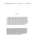 | 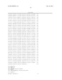 |
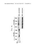 | 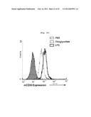 |
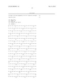 | 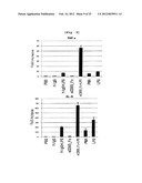 |
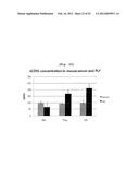 | 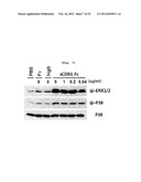 |
 | 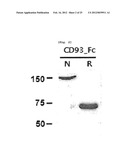 |
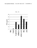 |  |
 | 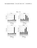 |
 | 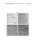 |
 | 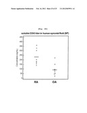 |
 |  |
 | 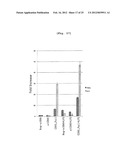 |
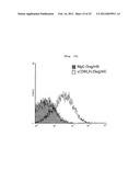 | 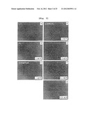 |
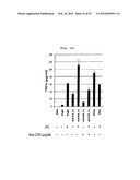 | 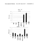 |
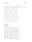 | 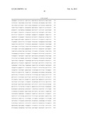 |
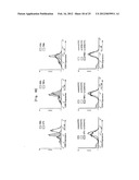 |  |
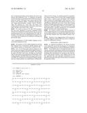 | 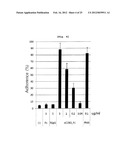 |
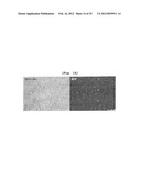 | 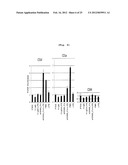 |
 |  |
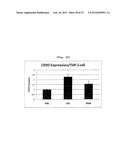 |
| Similar patent applications: | |
| Date | Title |
|---|---|
| 2009-11-12 | Delivery of double-stranded rna into the central nervous system |
| 2009-12-03 | Estrogenic extracts of anemarrhena asphodeloides bge. from the liliaceae family and uses thereof |
| 2009-12-10 | Soluble fragments of the sars-cov spike glycoprotein |
| 2009-12-31 | Method of measuring quantity of in-vivo substance by use of coherent anti-stokes raman scattered light |
| 2009-01-08 | Chimeric soluble hyper il-11 and use thereof |
| Top Inventors for class "Drug, bio-affecting and body treating compositions" | |
| Rank | Inventor's name |
|---|---|
| 1 | David M. Goldenberg |
| 2 | Hy Si Bui |
| 3 | Lowell L. Wood, Jr. |
| 4 | Roderick A. Hyde |
| 5 | Yat Sun Or |