Patent application title: ANTI-CD20 ANTIBODIES AND FUSION PROTEINS THEROF AND METHODS OF USE
Inventors:
Hans Hansen (Picayune, MS, US)
Zhengxing Qu (Warren, NJ, US)
David M. Goldenberg (Mendham, NJ, US)
Assignees:
IMMUNOMEDICS, INC.
IPC8 Class: AA61K3821FI
USPC Class:
424 854
Class name: Drug, bio-affecting and body treating compositions lymphokine interferon
Publication date: 2012-02-09
Patent application number: 20120034185
Abstract:
The present invention provides humanized, chimeric and human anti-CD20
antibodies and CD20 antibody fusion proteins that bind to a human B cell
marker, referred to as CD20, which is useful for the treatment and
diagnosis of B-cell disorders, such as B-cell malignancies and autoimmune
diseases, and methods of treatment and diagnosis.Claims:
1. An isolated nucleic acid encoding a chimeric or humanized monoclonal
antibody or antigen-binding fragment thereof that binds to CD20, said
antibody having a light chain variable region CDR1 comprising the
sequence of RASSSVSYIH (SEQ ID NO:1); CDR2 comprising the sequence of
ATSNLAS (SEQ ID NO:4); and CDR3 comprising the sequence of QQWTSNPPT (SEQ
ID NO:4); and a heavy chain variable region CDR1 comprising the sequence
of SYNMH (SEQ ID NO:8); CDR2 comprising the sequence of AIYPGNGDTSYNQKFKG
(SEQ ID NO:9); and CDR3 comprising the sequence of STYYGGDWYFDV (SEQ ID
NO:10) or the sequence of VVYYSNSYWYFDV (SEQ ID NO:13).
2. The isolated nucleic acid according to claim 1, wherein said antibody is a chimeric monoclonal antibody.
3. The isolated nucleic acid according to claim 1, wherein said antibody is a humanized monoclonal antibody.
4. The isolated nucleic acid according to claim 1, wherein the heavy chain variable region CDR3 comprises the sequence of STYYGGDWYFDV (SEQ ID NO:10).
5. The isolated nucleic acid according to claim 1, wherein the heavy chain variable region CDR3 comprises the sequence of VVYYSNSYWYFDV (SEQ ID NO:13).
6. The isolated nucleic acid according to claim 1, wherein said antibody or fragment thereof is a fusion protein.
7. The isolated nucleic acid according to claim 6, wherein said fusion protein comprises a second monoclonal antibody or antigen-binding fragment thereof.
8. The isolated nucleic acid according to claim 7, wherein said second monoclonal antibody or fragment thereof is an anti-CD20 antibody or fragment thereof.
9. The isolated nucleic acid according to claim 7, wherein said second monoclonal antibody or fragment thereof binds to an antigen selected from the group consisting of CD4, CD5, CD8, CD14, CD15, CD19, CD21, CD22, CD23, CD25, CD33, CD37, CD38, CD40, CD40L, CD46, CD52, CD54, CD74, CD80, CD126, B7, MUC1, MUC2, MUC3, MUC4, Ia, HM1.24, HLA-DR, tenascin, VEGF, PlGF, and an oncogene product.
10. An expression vector comprising the isolated nucleic acid of claim 1.
11. The expression vector according to claim 10, wherein the expression vector is a pdHL2 vector.
12. A host cell comprising the expression vector of claim 10.
13. The host cell according to claim 12, wherein the host cell is a bacterial cell, a eukaryotic cell or a mammalian cell.
14. A method for production of a chimeric or humanized monoclonal antibody or antigen-binding fragment thereof that binds to CD20 comprising: a. transfecting a host cell with an expression vector according to claim 10; and b. culturing the host cell in culture medium so that it produces the chimeric or humanized monoclonal anti-CD20 antibody or fragment thereof or fusion protein.
15. The method of claim 14, wherein the host cell secretes the anti-CD20 antibody or fragment thereof or fusion protein into the culture medium.
16. The method of claim 15, further comprising purifying the anti-CD20 antibody or fragment thereof or fusion protein from the culture medium.
17. The method of claim 15, wherein the host cell is a bacterial host cell, a eukaryotic host cell or a mammalian host cell.
18. A pharmaceutical composition comprising a chimeric or humanized monoclonal antibody or antigen-binding fragment thereof that binds to CD20, said antibody having a light chain variable region CDR1 comprising the sequence of RASSSVSYIH (SEQ ID NO:1); CDR2 comprising the sequence of ATSNLAS (SEQ ID NO:4); and CDR3 comprising the sequence of QQWTSNPPT (SEQ ID NO:4); and a heavy chain variable region CDR1 comprising the sequence of SYNMH (SEQ ID NO:8); CDR2 comprising the sequence of AIYPGNGDTSYNQKFKG (SEQ ID NO:9); and CDR3 comprising the sequence of STYYGGDWYFDV (SEQ ID NO:10) or the sequence of VVYYSNSYWYFDV (SEQ ID NO:13).
19. The pharmaceutical composition according to claim 18, wherein said antibody is a chimeric monoclonal antibody.
20. The pharmaceutical composition according to claim 18, wherein said antibody is a humanized monoclonal antibody.
21. The pharmaceutical composition according to claim 18, wherein the heavy chain variable region CDR3 comprises the sequence of STYYGGDWYFDV (SEQ ID NO:10).
22. The pharmaceutical composition according to claim 18, wherein the heavy chain variable region CDR3 comprises the sequence of VVYYSNSYWYFDV (SEQ ID NO:13).
23. The pharmaceutical composition according to claim 18, wherein said antibody or fragment thereof is a fusion protein.
24. The pharmaceutical composition according to claim 23, wherein said fusion protein comprises a second monoclonal antibody or antigen-binding fragment thereof.
25. The pharmaceutical composition according to claim 24, wherein said second monoclonal antibody or fragment thereof is an anti-CD20 antibody or fragment thereof.
26. The pharmaceutical composition according to claim 24, wherein said second monoclonal antibody or fragment thereof binds to an antigen selected from the group consisting of CD4, CD5, CD8, CD14, CD15, CD19, CD21, CD22, CD23, CD25, CD33, CD37, CD38, CD40, CD40L, CD46, CD52, CD54, CD74, CD80, CD126, B7, MUC1, MUC2, MUC3, MUC4, Ia, HM1.24, HLA-DR, tenascin, VEGF, PlGF, and an oncogene product.
27. A kit comprising a chimeric or humanized monoclonal antibody or antigen-binding fragment thereof that binds to CD20, said antibody having a light chain variable region CDR1 comprising the sequence of RASSSVSYIH (SEQ ID NO:1); CDR2 comprising the sequence of ATSNLAS (SEQ ID NO:4); and CDR3 comprising the sequence of QQWTSNPPT (SEQ ID NO:4); and a heavy chain variable region CDR1 comprising the sequence of SYNMH (SEQ ID NO:8); CDR2 comprising the sequence of AIYPGNGDTSYNQKFKG (SEQ ID NO:9); and CDR3 comprising the sequence of STYYGGDWYFDV (SEQ ID NO:10) or the sequence of VVYYSNSYWYFDV (SEQ ID NO:13).
28. The kit according to claim 27, wherein said antibody is a chimeric monoclonal antibody.
29. The kit according to claim 27, wherein said antibody is a humanized monoclonal antibody.
30. The kit according to claim 27, wherein the heavy chain variable region CDR3 comprises the sequence of STYYGGDWYFDV (SEQ ID NO:10).
31. The kit according to claim 27, wherein the heavy chain variable region CDR3 comprises the sequence of VVYYSNSYWYFDV (SEQ ID NO:13).
32. The kit according to claim 27, wherein said antibody or fragment thereof is a fusion protein.
33. The kit according to claim 32, wherein said fusion protein comprises a second monoclonal antibody or antigen-binding fragment thereof.
34. The kit according to claim 33, wherein said second monoclonal antibody or fragment thereof is an anti-CD20 antibody or fragment thereof.
35. The kit according to claim 33, wherein said second monoclonal antibody or fragment thereof binds to an antigen selected from the group consisting of CD4, CD5, CD8, CD14, CD15, CD19, CD21, CD22, CD23, CD25, CD33, CD37, CD38, CD40, CD40L, CD46, CD52, CD54, CD74, CD80, CD126, B7, MUC1, MUC2, MUC3, MUC4, Ia, HM1.24, HLA-DR, tenascin, VEGF, PlGF, and an oncogene product.
36. An immunoconjugate comprising: a. a chimeric or humanized monoclonal antibody or antigen-binding fragment thereof that binds to CD20, said antibody having a light chain variable region CDR1 comprising the sequence of RASSSVSYIH (SEQ ID NO:1); CDR2 comprising the sequence of ATSNLAS (SEQ ID NO:4); and CDR3 comprising the sequence of QQWTSNPPT (SEQ ID NO:4); and a heavy chain variable region CDR1 comprising the sequence of SYNMH (SEQ ID NO:8); CDR2 comprising the sequence of AIYPGNGDTSYNQKFKG (SEQ ID NO:9); and CDR3 comprising the sequence of STYYGGDWYFDV (SEQ ID NO:10) or the sequence of VVYYSNSYWYFDV (SEQ ID NO:13); and b. at least one therapeutic and/or diagnostic agent attached to the chimeric or humanized monoclonal antibody or fragment thereof.
37. The immunoconjugate according to claim 36, wherein said chimeric or humanized anti-CD20 antibody or fragment thereof is a fusion protein.
38. The immunoconjugate according to claim 36, wherein said therapeutic agent is selected from the group consisting of a radioisotope, a cytotoxic agent, a drug, a toxin, a second antibody, a second antigen-binding antibody fragment and an immunomodulator.
39. The immunoconjugate according to claim 38, wherein said immunomodulator is selected from the group consisting of a cytokine, an interferon, a stem cell growth factor, thrombopoietin, a lymphotoxin and a colony stimulating factor.
40. A method of treating an autoimmune disease or a B-cell lymphoma or leukemia in a subject comprising administering to said subject a pharmaceutical composition according to claim 18.
41. A method of treating an autoimmune disease or a B-cell lymphoma or leukemia in a subject comprising administering to said subject an immunoconjugate according to claim 39.
Description:
CROSS-REFERENCE TO RELATED APPLICATION(S)
[0001] This application is a continuation of U.S. patent application Ser. No. 12/212,359, filed Sep. 17, 2008, which is a continuation of U.S. patent application Ser. No. 11/534,103, filed Sep. 21, 2006, now U.S. Pat. No. 7,435,803, which is a continuation of U.S. patent application Ser. No. 10/366,709, filed Feb. 14, 2003, now U.S. Pat. No. 7,151,164, which claims priority to U.S. Provisional Application No. 60/356,132, filed Feb. 14, 2002 and U.S. Provisional Application No. 60/416,232, filed Oct. 7, 2002.
BACKGROUND OF THE INVENTION
[0002] 1. Field of the Invention
[0003] The present invention relates to humanized, chimeric and human anti-CD20 antibodies, particularly monoclonal antibodies (mAbs) therapeutic and diagnostic conjugates of humanized, chimeric and human anti-CD20 antibodies and methods of treating B cell lymphomas and leukemias and various autoimmune diseases using humanized, chimeric and human anti-CD20 antibodies. The present invention relates to antibody fusion proteins or fragments thereof comprising at least two anti-CD20 mAbs or fragments thereof or at least one anti-CD20 MAb or fragment thereof and at least one second MAb or fragment thereof, other than the antiCD20 MAb or fragment thereof. The humanized, chimeric and human anti-CD20 mAbs, fragments thereof, antibody fusion proteins thereof or fragments thereof may be administered alone, as a therapeutic conjugate or in combination with a therapeutic immunoconjugate, with other naked antibodies, or with therapeutic agents or as a diagnostic conjugate. The present invention relates to DNA sequences encoding humanized, chimeric and human anti-CD20 antibodies, and antibody fusion proteins, vectors and host cells containing the DNA sequences, and methods of making the humanized, chimeric and human anti-CD20 antibodies.
[0004] 2. Background
[0005] The immune system of vertebrates consists of a number of organs and cell types which have evolved to accurately recognize foreign antigens, specifically bind to, and eliminate/destroy such foreign antigens. Lymphocytes, amongst others, are critical to the immune system. Lymphocytes are divided into two major sub-populations, T cells and B cells. Although inter-dependent, T cells are largely responsible for cell-mediated immunity and B cells are largely responsible for antibody production (humoral immunity).
[0006] In humans, each B cell can produce an enormous number of antibody molecules. Such antibody production typically ceases (or substantially decreases) when a foreign antigen has been neutralized. Occasionally, however, proliferation of a particular B cell will continue unabated and may result in a cancer known as a B cell lymphoma. B-cell lymphomas, such as the B-cell subtype of non-Hodgkin's lymphoma, are significant contributors to cancer mortality. The response of B-cell malignancies to various forms of treatment is mixed. For example, in cases in which adequate clinical staging of non-Hodgkin's lymphoma is possible, field radiation therapy can provide satisfactory treatment. Still, about one-half of the patients die from the disease. Devesa et al., J. Nat'l Cancer Inst. 79:701 (1987).
[0007] The majority of chronic lymphocytic leukemias are of B-cell lineage. Freedman, Hematol. Oncol. Clin. North Am. 4:405 (1990). This type of B-cell malignancy is the most common leukemia in the Western world. Goodman et al., Leukemia and Lymphoma 22:1 (1996). The natural history of chronic lymphocytic leukemia falls into several phases. In the early phase, chronic lymphocytic leukemia is an indolent disease, characterized by the accumulation of small mature functionally-incompetent malignant B-cells having a lengthened life span. Eventually, the doubling time of the malignant B-cells decreases and patients become increasingly symptomatic. While treatment can provide symptomatic relief, the overall survival of the patients is only minimally affected. The late stages of chronic lymphocytic leukemia are characterized by significant anemia and/or thrombocytopenia. At this point, the median survival is less than two years. Foon et al., Annals Int. Medicine 113:525 (1990). Due to the very low rate of cellular proliferation, chronic lymphocytic leukemia is resistant to cytotoxic drug treatment.
[0008] Traditional methods of treating B-cell malignancies, including chemotherapy and radiotherapy, have limited utility due to toxic side effects. The use of monoclonal antibodies to direct radionuclides, toxins, or other therapeutic agents offers the possibility that such agents can be delivered selectively to tumor sites, thus limiting toxicity to normal tissues. Also, the presence of B-cell antigens on these B-cell malignancies makes them optimal targets for therapy with unconjugated B-cell antibodies, such as against CD19, CD20, CD21, CD23, and CD22 markers on B-cells. HLA-DR and other antigens may serve as targets for normal and malignant B-cells, although they are also expressed on other cell types. Further, certain MUC1, MUC2, MUC3, and MUC4 antigens, preferably MUC1, are also expressed in different hematopoietic malignancies, including B-cell tumors expressing CD20 and other B-cell markers. Still other antigen targets, such as those associated with the vascular endothelium of tumors, including tenascin, vascular endothelium growth factor (VEGF), and placental growth factor (PlGF), as well as other categories of antigens associated with B-cell malignancies, such as oncogene products, are also suitable targets for said complementary antibodies for use in the present invention.
[0009] B cells comprise cell surface proteins which can be utilized as markers for differentiation and identification. One such human B-cell marker is the human B lymphocyte-restricted differentiation antigen Bp35, referred to as CD20. CD20 is expressed during early pre-B cell development and remains until plasma cell differentiation. CD20 is expressed on both normal B cells and malignant B cells whose abnormal growth can lead to B-cell lymphomas. Antibodies against the CD20 antigen have been investigated for the therapy of B-cell lymphomas. For example, a chimeric anti-CD20 antibody, designated as "IDEC-C2B8," has activity against B-cell lymphomas when provided as unconjugated antibodies at repeated injections of doses exceeding 500 mg per injection. Maloney et al., Blood 84:2457 (1994); Longo, Curr. Opin. Oncol. 8:353 (1996). About 50 percent of non-Hodgkin's patients, having the low-grade indolent form, treated with this regimen showed responses. Therapeutic responses have also been obtained using 131]-labeled B1 anti-CD20 murine monoclonal antibody when provided as repeated doses exceeding 600 mg per injection. Kaminski et al., N. Engl. J. Med. 329:459 (1993); Press et al., N. Engl. J. Med. 329:1219 (1993); Press et al., Lancet 346:336 (1995). However, these antibodies, whether provided as unconjugated forms or radiolabeled forms, have not shown high rates of objective and durable responses in patients with the more prevalent and lethal form of B-cell lymphoma, the intermediate or aggressive type. Therefore, a need exists to develop an immunotherapy for B-cell malignancies that achieves a therapeutic response of significant duration.
[0010] Additional studies targeting CD20 surface antigen have been demonstrated using an anti-CD20 murine monoclonal antibody, IF5, which was administered by continuous intravenous infusion to B cell lymphoma patients. Extremely high levels (>2 grams) of 1F5 were reportedly required to deplete circulating tumor cells, and the results were described as being "transient." Press et al., "Monoclonal Antibody 1F5 (Anti-CD20) Serotherapy of Human B-Cell Lymphomas." Blood 69/2:584-591 (1987). However, a potential problem with this approach is that non-human monoclonal antibodies (e.g., murine monoclonal antibodies) typically lack human effector functionality, i.e., they are unable to mediate complement-dependent lysis or lyse human target cells through antibody-dependent cellular toxicity or Fc-receptor mediated phagocytosis. Furthermore, non-human monoclonal antibodies can be recognized by the human host as a foreign protein and, therefore, repeated injections of such foreign antibodies can lead to the induction of immune responses leading to harmful hypersensitivity reactions. For murine-based monoclonal antibodies, this is often referred to as a Human Anti-Mouse Antibody (HAMA) response.
[0011] The use of chimeric antibodies is more preferred because they do not elicit as strong a HAMA response as murine antibodies. Chimeric antibodies are antibodies which comprise portions from two or more different species. For example, Liu, A. Y. et al, "Production of a Mouse-Human Chimeric Monoclonal Antibody to CD20 with Potent Fc-Dependent Biologic Activity" J. Immun. 139/10:3521-3526 (1987), describe a mouse/human chimeric antibody directed against the CD20 antigen. See also, PCT Publication No. WO 88/04936. However, no information is provided as to the ability, efficacy or practicality of using such chimeric antibodies for the treatment of B cell disorders in the reference. It is noted that in vitro functional assays (e.g., complement-dependent lysis (CDC); antibody dependent cellular cytotoxicity (ADCC), etc.) cannot inherently predict the in vivo capability of a chimeric antibody to destroy or deplete target cells expressing the specific antigen. See, for example, Robinson, R. D. et al., "Chimeric mouse-human anti-carcinoma antibodies that mediate different anti-tumor cell biological activities," Hum. Antibod. Hybridomas 2:84-93 (1991) (chimeric mouse-human antibody having undetectable ADCC activity). Therefore, the potential therapeutic efficacy of a chimeric antibody can only truly be assessed by in vivo experimentation, preferably in the species of interest for the specific therapy.
[0012] One approach that has improved the ability of murine monoclonal antibodies to be effective in the treatment of B-cell disorders has been to conjugate a radioactive label or chemotherapeutic agent to the antibody, such that the label or agent is localized at the tumor site. For example, the above-referenced 1F5 antibody and other B-cell antibodies have been labeled with 131I and were reportedly evaluated for biodistribution in two patients. See Eary, J. F. et al., "Imaging and Treatment of B-Cell Lymphoma" J. Nuc. Med. 31/8:1257-1268 (1990); see also, Press, O. W. et al., "Treatment of Refractory Non-Hodgkin's Lymphoma with Radiolabeled MB-1 (Anti-CD37) Antibody" J. Clin. One. 7/8:1027-1038 (1989) (indication that one patient treated with 131I-labeled IF-5 achieved a partial response); Goldenberg, D. M. et al., "Targeting, Dosimetry and Radioimmunotherapy of B-Cell Lymphomas with 131I-Labeled LL2 Monoclonal Antibody" J. Clin. Oncol. 9/4:548-564 (1991) (three of eight patients receiving multiple injections reported to have developed a HAMA response to this CD22 murine antibody); Appelbaum, F. R. "Radiolabeled Monoclonal Antibodies in the Treatment of Non-Hodgkin's Lymphoma" Hem./Oncol. Clinics of N. A. 5/5:1013-1025 (1991) (review article); Press, O. W. et al. "Radiolabeled-Antibody Therapy of B-Cell Lymphoma with Autologous Bone Marrow Support." New England Journal of Medicine 329/17: 1219-12223 (1993) (131I-labeled anti-CD20 antibody IF5 and B1); and Kaminski, M. G. et al "Radioimmunotherapy of B-Cell Lymphoma with [131I] Anti-B1 (Anti-CD20) Antibody". NEJM 329/7:459 (1993) (131I-labeled anti-CD20 antibody B1; hereinafter "Kaminski"); PCT published application WO 92/07466 (antibodies conjugated to chemotherapeutic agents such as doxorubicin or mitomycin). However, these approaches have not eliminated the obstacles associated with using murine antibodies, despite the fact that many patients with lymphoma who have received prior aggressive cytotoxic chemotherapy are immune suppressed, thus having lower HAMA rates than lymphoma patients who have not been heavily pretreated.
[0013] Autoimmune diseases are a class of diseases associated with B-cell disorders. Examples include immune-mediated thrombocytopenias, such as acute idiopathic thrombocytopenic purpura and chronic idiopathic thrombocytopenic purpura, myasthenia gravis, lupus nephritis, lupus erythematosus, and rheumatoid arthritis. The most common treatments are corticosteroids and cytotoxic drugs, which can be very toxic. These drugs also suppress the entire immune system, can result in serious infection, and have adverse affects on the bone marrow, liver and kidneys. Other therapeutics that have been used to treat Class III autoimmune diseases to date have been directed against T-cells and macrophages. There is a need for more effective methods of treating autoimmune diseases, particularly Class III autoimmune diseases.
[0014] To address the many issues related to B-cell disorders and their treatment, the present invention provides humanized, chimeric and human anti-CD20 monoclonal antibodies with the same complementarity determining regions (CDRs) that bind to the CD20 antigen of the present invention used alone, conjugated to a therapeutic agent or in combination with other treatment modalities, for the treatment of B cell lymphomas and leukemias and autoimmune disorders in humans and other mammals without the adverse responses associated with using murine antibodies.
SUMMARY OF THE INVENTION
[0015] Accordingly, the present invention provides humanized, chimeric and human anti-CD20 antibodies that bind to a human B cell marker, referred to as CD20, which is useful for the treatment and diagnosis of B-cell disorders, such as B-cell malignancies and autoimmune diseases.
[0016] The present invention further provides methods of treatment of mammalian subjects, such as humans or domestic animals, with one or more humanized, chimeric and human CD20 antibodies, alone, as an antibody fusion protein, as a therapeutic conjugate alone or as part of an antibody fusion protein, in combination, or as a multimodal therapy, with other antibodies, other therapeutic agents or immunomodulators or as an immunoconjugate linked to at least one therapeutic agent, therapeutic radionuclide or immunomodulator. These humanized, chimeric and human CD20 antibodies can also be used as a diagnostic imaging agent alone, in combination with other diagnostic imaging agents, and/or in conjunction with therapeutic applications.
[0017] The present invention additionally is directed to anti-CD20 mAbs or fragments thereof that contain specific murine CDRs or a combination of murine CDRs from more than one murine or chimeric anti-CD20 MAb that have specificity for CD20. These mAbs can be humanized, chimeric or human anti-CD20 mAbs. The present invention is further directed to light and/or heavy chain variable regions or fragments thereof of these anti-CD20 Mabs and to light and/or heavy chains or fragments thereof that have specficity for CD20.
[0018] The present invention is also directed to antibody fusion proteins comprising at least two anti-CD20 mAbs or fragments thereof or a first MAb comprising an anti-CD20 mAbs or fragments thereof and a second MAb.
[0019] The present invention is further directed to a therapeutic or diagnostic conjugates of the anti-CD20 mAbs or fragments thereof or antibody fusion proteins of the anti-CD20 mAbs or other mAbs or fragments thereof bound to at least one therapeutic agent or at least one diagnostic agent. Antibody fusion proteins with multiple therapeutic agents of the same or different type are encompassed by the present invention.
[0020] The present invention is additionally directed to a method of using the anti-CD20 mAbs or fragments thereof or antibody fusion proteins thereof or fragments thereof for therapy, either alone, in combination with each other, as the antibody component of a therapeutic immunoconjugate with one or more therapeutic agents or each administered in combination with one or more therapeutic agents or with an immunoconjugate with one or more therapeutic agents.
[0021] The present invention further is directed to a method of using the anti-CD20 mAbs or fragments thereof or antibody fusion proteins thereof or fragments thereof as a diagnostic bound to one or more diagnostic agents.
[0022] The present invention additionally is directed to a method of pretargeting a cell in a patients suffering from a B-cell lymphoma or leukemia or an autoimmune disease using an antibody fusion protein or fragment thereof of the present invention.
BRIEF DESCRIPTION OF THE FIGURES
[0023] FIG. 1 discloses the V gene sequences cloned by RT-PCR from a hybridoma cell line producing a murine anti-CD20, and the deduced amino acid sequences of the variable light (FIG. 1A) (SEQ ID NOS: 32 & 33) and heavy chain (FIG. 1B) (SEQ ID NOS: 34 & 35) of the A20 antibody, designated as A20Vk and A20VH, respectively. Underlined arrows indicate the sequences of the PCR primers used for cloning. The putative CDR region sequences, as defined by the Kabat numbering scheme, are shown in bold and underlined Amino acid sequences are given as single-letter codes below the corresponding nucleotide sequence. The Kabat numbering scheme was used for amino acid residues Amino acid residues numbered by a letter represent the insertion residue according to Kabat, and have the same number as that of the previous residue. For example, residues 82, 82A, 82B and 82C in FIG. 1B are indicated as 82 A, B, and C, respectively.
[0024] FIG. 2 discloses the Vk, the variable light chain, and the VH, the variable heavy chain, sequences of cA20, a chimeric anti-CD20 antibody. The CDR region sequences are shown in bold and underlined. The amino acid residues and the nucleotides are numbered sequentially and same numbering system is used for humanized V sequences. The light chain variable region is shown in FIG. 2A (SEQ ID NOS 36 & 37) and the heavy chain variable region is shown in FIG. 2B (SEQ ID NOS: 38 & 39). The numbering system is the same as for FIG. 1. The restriction sites used for constructing cA20 are underlined.
[0025] FIG. 3 shows a comparison of the binding affinities of the chimeric A20 (cA20), and murine A20, (A20), in a cell surface competitive binding assay against 125I-labled A20. Increasing concentrations of cA20 blocked the binding of radiolabeled A20 to Raji cells (as depicted by closed circles) in a comparable manner as that of murine A20 (depicted by closed diamonds).
[0026] FIG. 4 compares the amino acid sequences of the variable heavy chain (VH) and variable light chain (Vk) of human antibodies, and chimeric and humanized anti-CD20 antibodies. FIG. 4A compares the amino acid sequences of the variable heavy chain (VH) of the human antibodies, EU (SEQ ID NO: 40) and NEWM (SEQ ID NO: 43) (FR4 only), the chimeric antibody, (cA20VH) (SEQ ID NO: 39) and two humanized antibodies, (hA20VH1 (SEQ ID NO: 41) and hA20VH2 (SEQ ID NO: 42)) and FIG. 4B compares the amino acid sequences of the variable light chain (Vk) of the human antibody, (REIVk) (SEQ ID NO: 44), a chimeric antibody, (cA20Vk) (SEQ ID NO: 37), and a humanized antibody, (hA20Vk) (residues 20-125 of SEQ ID NO: 46). Dots indicate that the residues in A20 are identical to the corresponding residue in the human antibody. The CDRs are identified as a boxed region. The Kabat numbering scheme was used to number the amino acid residues.
[0027] FIG. 5 discloses the nucleotide sequences of hA20 light chain V genes, (hA20Vk) (FIG. 5A) (SEQ ID NOS: 45 & 46), and heavy chain V genes, hA20VH1 (FIG. 5B) (SEQ ID NOS: 47 & 48) and hA20VH2 (FIG. 5C) (SEQ ID NOS: 49 & 50), as well as the adjacent flanking sequences of the VKpBR2 (FIG. 5A) and VHpBS2 (FIGS. 5B and 5C) staging vectors, respectively. The non-translated nucleotide sequences are shown in lowercase. The restriction sites used for subcloning are underlined and indicated. The secretion signal peptide sequence is indicated by a double underline. Numbering of Vk and VH amino acid residues is same as that in FIG. 2.
[0028] FIG. 6 shows the results of a cell surface competitive binding assay to compare the binding activity of two humanized A20 antibodies, (hA20-1 and hA20-2), with that of A20, cA20 and a chimeric anti-CD20 MAb, c2B8. FIG. 6A shows hA20-1 (closed triangles) and hA20-2 (closed circles) and the murine anti-CD20 antibody, A20 (closed squares) competed equally well for the binding of 125I-A20 to Raji cells. FIG. 6B shows hA20-1 (closed circles), cA20 (closed squares) and c2B8 (closed diamonds) competed equally well for the binding of 125I-c2B8 to Raji cells.
[0029] FIG. 7 discloses the constant region of a human IgG1 (CH-hinge) (FIG. 7A) (SEQ ID NOS: 51 & 52) and the constant region of a human kappa chain (Ck) (FIG. 7B) (SEQ ID NOS: 53 & 54).
[0030] FIG. 8 is a competitive cell surface binding assay. Ag-binding specificity and affinity studies of humanized anti-CD20 Abs (cA20, hA20, and c1F5, purified by affinity chromatography on a Protein A column) were evaluated by a cell surface competitive binding assay with murine 2B8 and rituximab (IDEC Pharmaceuticals Corp., San Diego, Calif.). FIG. 8 (A) is a comparison of the binding activities of cA20 (square), hA20-1 (triangle) and hA20-1 (circle) with that of m2B8 (diamond); FIG. 8 (B) compares of the binding activities of cA20 (square), c1F5 (triangle) and rituximab (diamond).
[0031] FIG. 9 is a study comparing the binding activities of hA20 with other anti-CD20 Abs, including rituximab and murine B1, by a cell surface competitive binding assay. A constant amount (100,000 cpm, ˜10 Ci/ g) of 125I-labeled rituximab was incubated with Raji cells in the presence of varying concentrations (0.2-700 nM) of competing Abs, hA20 (triangle), mB1 (Downward triangle) or rituximab (square) at 4° C. for 1-2 h.
[0032] FIG. 10 depicts the cytotoxic effect of crosslinked hA20 and other CD20 Abs on cultured lymphoma cells. Total cell and viable cell populations were measured by (A) trypan blue staining and cell counting or (B) MTT assay.
[0033] FIG. 11 is a graph of in vivo therapy studies with various anti-CD20 and other Abs. Raji cells administered i.v. to SCID mice, to create a Raji lymphoma model of disseminated disease.
[0034] FIG. 12 is a graph depicting in vivo therapy with hA20 and hLL2. Raji cells administered i.v. to SCID mice, to create a Raji lymphoma model of disseminated disease.
DETAILED DESCRIPTION OF THE INVENTION
1. Overview
[0035] As discussed above, anti-CD20 antibodies that are unconjugated or labeled with a therapeutic radionuclide, have failed to provide high rates of objective and lasting responses in patients with intermediate or aggressive forms of B-cell lymphoma. The present invention provides a humanized, a chimeric and a human anti-CD20 antibody, and antibody fusion proteins thereof, useful for treatment of mammalian subjects, humans and domestic animals, alone, as a conjugate or administered in combination with other therapeutic agents, including other naked antibodies and antibody therapeutic conjugates.
[0036] The anti-CD20 mAbs of the present invention contain specific murine CDRs or a combination of murine CDRs from more than one murine or chimeric anti-CD20 MAb that have specificity for the CD20 antigen. The anti-CD20 mAbs of the present invention are humanized, chimeric or human mAbs, light and/or heavy chains thereof or light and/or heavy chain variable regions thereof, and they contain the amino acids of the CDRs of a murine anti-CD20 MAb and retain substantially the B-cell and B-cell lymphoma and leukemia cell targeting of the murine anti-CD20 MAb. The CDRs of the light chain variable region of the anti-CD20 MAb comprises CDR1 comprising amino acids RASSSVSYIH (SEQ ID NO: 1), RASSSLSFMH (SEQ ID NO: 2) or RASSSVSYMH (SEQ ID NO: 3) CDR2 comprising amino acids ATSNLAS (SEQ ID NO: 4) and CDR3 comprising amino acids QQWTSNPPT (SEQ ID NO: 5), HQWSSNPLT (SEQ ID NO: 6) or QQSFSNPPT (SEQ ID NO: 7) and the CDRs of the heavy chain variable region of the anti-CD20 MAb comprises CDR1 comprising amino acids SYNMH (SEQ ID NO: 8) CDR2 comprising amino acids AIYPGNGDTSYNQKFKG (SEQ ID NO: 9) and CDR3 comprising amino acids STYYGGDWYFDV (SEQ ID NO: 10), STYYGGDWYFNV (SEQ ID NO: 11), SHYGSNYVDYFDV (SEQ ID NO: 12) or VVYYSNSYWYFDV (SEQ ID NO: 13). The humanized antibody further comprises the framework regions of the light and heavy chain constant regions of a human antibody.
[0037] In one embodiment, the humanized and chimeric MAb or fragment thereof does not contain the CDR3 of the heavy chain variable region comprising STYYGGDWYFNV (SEQ ID NO: 11). More preferably, CDR1 of the light chain variable region does not comprise RASSSLSFMH (SEQ ID NO: 2) when the CDR3 of the light chain variable region comprises HQWSSNPLT (SEQ ID NO: 6) and the CDR3 of the heavy chain variable region comprises SHYGSNYVDYFDV (SEQ ID NO: 12). In another embodiment, the CDR3 of the light chain variable region does not comprise HQWSSNPLT (SEQ ID NO: 6) when CDR1 of the light chain variable region comprises RASSSLSFMH (SEQ ID NO: 2) and when CDR3 of the heavy chain variable region comprises SHYGSNYVDYFDV (SEQ ID NO: 12). In a further embodiment, the CDR3 of the heavy chain variable region does not comprise SHYGSNYVDYFDV (SEQ ID NO: 12) when the CDR1 of the light chain variable region comprises RASSSLSFMH (SEQ ID NO: 2) and the CDR3 of the light chain variable region comprises HQWSSNPLT (SEQ ID NO: 6). In another embodiment, the CDR1 of the light chain variable region does not comprise RASSSVSYMH (SEQ ID NO: 3) when the CDR3 of the light chain variable region comprises QQSFSNPPT (SEQ ID NO: 7) and the CDR3 of the heavy chain variable region comprises VVYYSNSYWYFDV (SEQ ID NO:13).
[0038] Further, in another embodiment, the anti-CD20 monoclonal antibody (MAb) or fragment thereof does not contain CDR3 of the light chain variable region of amino acids QQSFSNPPT (SEQ ID NO: 7) when CDR1 of the light chain variable region comprises RASSSVSYMH (SEQ ID NO: 3) and the CDR3 of the heavy chain variable region comprises VVYYSNSYWYFDV (SEQ ID NO: 13). Additionally, the anti-CD20 MAb does not contain CDR3 of the heavy chain variable region with amino acids VVYYSNSYWYFDV (SEQ ID NO: 13) when the CDR1 of the light chain variable region comprises RASSSVSYMH (SEQ ID NO: 3) and the CDR3 of the light chain variable region comprises QQSFSNPPT (SEQ ID NO: 7).
[0039] In a preferred embodiment, the humanized anti-CD20 (hCD20) monoclonal antibody or antigen-binding fragment thereof comprising the complementarity determining regions (CDRs) of at least one murine anti-CD20 MAb variable region and the framework regions (FRs) of at least one human MAb variable region, wherein said humanized anti-CD20 MAb or fragment thereof retains substantially the B-cell and B-cell lymphoma and leukemia cell targeting of said murine anti-CD20 MAb. The humanized antibody's variable region may comprise a light chain variable region, a heavy chain variable region or a both light and heavy chain variable regions. The humanized antibody or fragment thereof may further comprise light and heavy chain constant regions of at least one human antibody.
[0040] The humanized anti-CD20 MAb or fragment thereof of the present invention comprises the CDRs of a murine anti-CD20 MAb and the framework (FR) regions of the light and heavy chain variable regions of a human antibody, while retaining substantially the B-cell, and B-cell lymphoma and leukemia cell targeting of the parent murine antiCD20 MAb, and wherein the CDRs of the light chain variable region of the murine antiCD20 MAb comprises CDR1 comprising amino acids RASSSVSYIH (SEQ ID NO: 1), CDR2 comprising amino acids ATSNLAS (SEQ ID NO: 4) and CDR3 comprising QQWTSNPPT (SEQ ID NO: 5) and the CDRs of the heavy chain variable region of murine anti-CD20 MAb comprises CDR1 comprising amino acids SYNMH (SEQ ID NO: 8), CDR2 comprising amino acids AIYPGNGDTSYNQKFKG (SEQ ID NO: 9) and CDR3 comprising amino acids STYYGGDWYFDV (SEQ ID NO: 10). But the humanized anti-CD20 MAb or fragment thereof may further contain in the FRs of the light and heavy chain variable regions of the antibody at least one amino acid from the corresponding FRs of the murine MAb. The humanized MAbs may further contain the light and heavy chain constant regions of a human antibody. Specifically, the humanized anti-CD20 MAb or fragment thereof contains at least one amino acid residue 1, 5, 27, 30, 38, 48, 67, 68, 70, 95, 115 and 116 of the murine heavy chain variable region of FIG. 4A, designated as hA20VH1 or hA20VH2 and of at least one amino acid residue 4, 21, 35, 38, 45, 46, 59, 99, 104 and 106 of the murine light chain variable region FIG. 4B, designated hA20Vk. One or more of the murine amino acid sequences can be maintained in the human FR regions of the light and heavy variable chains if necessary to maintain proper binding or to enhance binding to the CD20 antigen. More preferably the humanized antiCD20 MAb or fragment thereof of the present invention comprises the hA20Vk of FIG. 4B and the hA2VH1 of FIG. 4A. Most preferably, the humanized anti-CD20 MAb or fragment thereof of the present invention comprises the hA20Vk of FIG. 4B and the hA20VH2 of FIG. 4A. This latter sequence contains more human amino acid sequences in the FRs of the VH2 chain than the VH1, and thus is more humanized.
[0041] The preferred chimeric anti-CD20 (cCD20) MAb or fragment thereof of the present invention comprises the CDRs of a murine anti-CD20 MAb and the FR regions of the light and heavy chain variable regions of the murine anti-CD 20 MAb, i.e., the Fvs of the parental murine MAb, and the light and heavy chain constant regions of a human antibody, wherein the chimeric anti-CD20 MAb or fragment thereof retains substantially the B-cell, and B-cell lymphoma and leukemia cell targeting of the murine anti-CD20 MAb, wherein the CDRs of the light chain variable region of the chimeric anti-CD20 MAb comprise CDR1 comprising amino acids RASSSVSYIH (SEQ ID NO: 1), RASSSLSFMH (SEQ ID NO: 2) or RASSSVSYMH (SEQ ID NO: 3) CDR2 comprising amino acids ATSNLAS (SEQ ID NO: 4) and CDR3 comprising amino acids QQWTSNPPT (SEQ ID NO: 5), HQWSSNPLT (SEQ ID NO: 6) or QQSFSNPPT (SEQ ID NO: 7) and the CDRs of the heavy chain variable region of the chimeric anti-CD20 MAb comprise CDR1 comprising amino acids SYNMH (SEQ ID NO: 8) CDR2 comprising amino acids AIYPGNGDTSYNQKFKG (SEQ ID NO: 9) and CDR3 comprising STYYGGDWYFDV (SEQ ID NO: 10), STYYGGDWYFNV (SEQ ID NO: 11), SHYGSNYVDYFDV (SEQ ID NO: 12) or VVYYSNSYWYFDV (SEQ ID NO: 13) with the following provisos,
[0042] (a) wherein the CDR3 of the heavy chain variable region does not comprise STYYGGDWYFNV (SEQ ID NO: 11), when the CDR1 of the light chain variable region comprises amino acids RASSSVSYIH (SEQ ID NO: 1), CDR2 of the light chain variable region comprises amino acids ATSNLAS (SEQ ID NO: 4), CDR3 of the light chain variable region comprises amino acids QQWTSNPPT (SEQ ID NO: 5), CDR1 of the heavy chain variable region comprises amino acids SYNMH (SEQ ID NO: 8), and CDR2 of the light chain variable region comprises amino acids AIYPGNGDTSYNQKFKG (SEQ ID NO: 9)
[0043] (b) wherein the CDR3 of the heavy chain variable region does not comprise SHYGSNYVDYFDV (SEQ ID NO: 12), when the CDR1 of the light chain variable region comprises amino acids RASSSLSFMH (SEQ ID NO: 2), CDR2 of the light chain variable region comprises amino acids ATSNLAS (SEQ ID NO: 4), CDR3 of the light chain variable region comprises amino acids HQWSSNPLT (SEQ ID NO: 6), CDR1 of the heavy chain variable region comprises amino acids SYNMH (SEQ ID NO: 8), and CDR2 of the light chain variable region comprises amino acids AIYPGNGDTSYNQKFKG (SEQ ID NO: 9) and
[0044] (c) wherein the CDR3 of the heavy chain variable region does not comprise VVYYSNSYWYFDV (SEQ ID NO: 13), when the CDR1 of the light chain variable region comprises amino acids RASSSVSYMH (SEQ ID NO: 3), CDR2 of the light chain variable region comprises amino acids ATSNLAS (SEQ ID NO: 4), CDR3 of the light chain variable region comprises amino acids QQSFSNPPT (SEQ ID NO: 7), CDR1 of the heavy chain variable region comprises amino acids SYNMH (SEQ ID NO: 8), and CDR2 of the light chain variable region comprises amino acids AIYPGNGDTSYNQKFKG (SEQ ID NO: 9).
[0045] More preferably the chimeric anti-CD20 MAb or fragment thereof comprising the complementarity-determining regions (CDRs) of a murine anti-CD20 MAb and the framework (FR) regions of the light and heavy chain variable regions of the murine anti-CD20 MAb and further, the light and heavy chain constant regions of a human antibody, wherein the chimeric anti-CD20 MAb or fragment thereof retains substantially the B-cell, and B-cell lymphoma and leukemia cell targeting of the murine anti-CD20 MAb, wherein the CDRs of the light chain variable region of the chimeric anti-CD20 MAb comprises the CDRs shown in FIGS. 4B and 4A, respectively, designated cA20Vk and cA20VH. Most preferably, the chimeric anti-CD20 MAb or fragment thereof comprises the light and heavy chain variable regions of murine anti-CD20 MAb shown in FIGS. 4B and 4A, respectively, designated cA20Vk and cA20 VH.
[0046] The present invention also encompasses a human anti-CD20 MAb or fragment thereof comprising the light and heavy chain variable, wherein said human CD20 MAb retains substantially the B-cell, and B-cell lymphoma and leukemia cell targeting and cell binding characteristics of a murine anti-CD20 MAb, wherein the CDRs of the light chain variable region of the human anti-CD20 MAb comprises the same CDRs as set forth above for the chimeric and humanized anti-CD20 mAbs and as shown in FIGS. 4A and 4B. This human anti-CD20 MAb or fragment thereof further comprises light and heavy chain constant regions of at least one human antibody.
[0047] The present invention is also intended to encompass antibody fusion proteins or fragments thereof comprising at least two anti-CD20 mAbs or fragments thereof, as described above. The antibody fusion protein or fragment thereof of the present invention is also intended to encompass an antibody fusion protein or fragment thereof comprising at least one first anti-CD20 MAb or fragment thereof as described above and at least one second MAb or fragment thereof, other than the antiCD20 MAb or fragment described above. More preferably this second MAb is a MAb reactive with CD4, CD5, CD8, CD14, CD15, CD19, CD21, CD22, CD23, CD25, CD33, CD37, CD38, CD40, CD40L, CD46, CD52, CD54, CD74, CD80, CD126, B7, MUC1, MUC2, MUC3, MUC4, Ia, HM1.24, HLA-DR, tenascin, VEGF, PlGF, an oncogene, oncogene product, or a combination thereof, and even an anti-CD20 MAb that is different than the anti-CD20 MAb described herein. The antibody fusion proteins of the present invention may be composed of one CD20 MAb and one or more of the second mAbs to provide specificity to different antigens, and are described in more detail below.
[0048] The humanized, chimeric and human anti-CD20 antibody may possess enhanced affinity binding with the epitope, as well as antitumor and anti-B-cell activity, as a result of CDR mutation and manipulation of the CDR and other sequences in the variable region to obtain a superior therapeutic agent for the treatment of B-cell disorders, including B-cell lymphomas and leukemias and autoimmune diseases. Modification to the binding specificity, affinity or avidity of an antibody is known and described in WO 98/44001, as affinity maturation, and this application summarizes methods of modification and is incorporated in its entirety by reference.
[0049] It may also be desirable to modify the antibodies of the present invention to improve effector function, e.g., so as to enhance antigen-dependent cell-mediated cytotoxicity (ADCC) and/or complement dependent cytotoxicity (CDC) of the antagonist. One or more amino acid substitutions or the introduction of cysteine in the Fc region may be made, thereby improving internalization capability and/or increased complement-mediated cell killing and ADCC. See Caron et al., J. Ex. Med. 176:1191-1195 (1991) and Shopes, B. J. Immunol. 148:2918-2022 (1992), incorporated herein by reference in their entirety. An antibody fusion protein may be prepared that has dual Fc regions with both enhanced complement lysis and ADCC capabilities.
[0050] The present invention is also directed to DNA sequences comprising a nucleic acid encoding a MAb or fragment thereof selected from the group consisting
[0051] (a) an anti-CD20 MAb or fragment thereof as described herein,
[0052] (b) an antibody fusion protein or fragment thereof comprising at least two of the anti-CD20 mAbs or fragments thereof,
[0053] (c) an antibody fusion protein or fragment thereof comprising at least one first MAb or fragment thereof comprising the anti-CD20 MAb or fragment thereof as described herein and at least one second MAb or fragment thereof, other than the antiCD20 MAb or fragment thereof, and
[0054] (d) an antibody fusion protein or fragment thereof comprising at least one first MAb or fragment thereof comprising the anti-CD20 MAb or fragment thereof and at least one second MAb or fragment thereof, wherein the second MAb is a MAb reactive with CD4, CD5, CD8, CD14, CD15, CD19, CD21, CD22, CD23, CD25, CD33, CD37, CD38, CD40, CD40L, CD46, CD52, CD54, CD74, CD80, CD126, B7, MUC1, MUC2, MUC3, MUC4, Ia, HM1.24, HLA-DR, tenascin, VEGF, PlGF, an oncogene, oncogene product, or a combination thereof.
[0055] Also encompassed by the present invention are expression vectors comprising the DNA sequences. These vectors contain the light and heavy chain constant regions and the hinge region of the human immunoglobulin, in the case of vectors for use in preparing the humanized, chimeric and human anti-CD20 mAbs or antibody fusion proteins thereof or fragments thereof. These vectors additionally contain, where required, promoters that express the mAbs in the selected host cell, immunoglobulin enhances and signal or leader sequences. Vectors that are particularly useful in the present invention are pdHL2 or GS, particularly when used to express a chimeric, humanized or human antibodies, such as gigs, where the vector codes for the heavy and light chain constant regions and hinge region of IgG1. More preferably, the light and heavy chain constant regions and hinge region are from a human EU myeloma immunoglobulin, where optionally at least one of the amino acid in the allotype positions is changed to that found in a different IgG1 allotype, and wherein optionally amino acid 253 of the heavy chain of EU based on the EU number system may be replaced with alanine. See Edelman et al., Proc. Natl. Acad. Sci. USA 63: 78-85 (1969), incorporated herein in its entirety by reference.
[0056] Host cells containing the DNA sequences encoding the anti-CD20 mAbs or fragments thereof or antibody fusion proteins or fragments thereof of the present invention or host cells containing the vectors that contain these DNA sequences are encompassed by the present invention. Particularly useful host cells are mammalian cells, more specifically lymphocytic cells, such as myeloma cells, discussed in more detail below.
[0057] Also encompassed by the present invention is the method of expressing the anti-CD20 MAb or fragment thereof or antibody fusion protein or fragment thereof comprising: (a) transfecting a mammalian cell with a DNA sequence of encoding the anti-CD20 mAbs or fragments thereof or antibody fusion proteins or fragments thereof, and (b) culturing the cell transfected with the DNA sequence that secretes the anti-CD20 or fragment thereof or antibody fusion protein or fragment thereof. Known techniques may be used that include a selection marker on the vector so that host cells that express the mAbs and the marker can be easily selected.
[0058] The present invention particularly encompasses B-lymphoma cell and leukemia cell targeting diagnostic or therapeutic conjugates comprising an antibody component comprising an anti-CD20 MAb or fragment thereof or an antibody fusion protein or fragment thereof of the present invention that binds to the B-lymphoma or leukemia cell, that is bound to at least one diagnostic or at least one therapeutic agent.
[0059] The diagnostic conjugate comprises the antibody component comprising an anti-CD20 MAb or fragment thereof or an antibody fusion protein or fragment thereof, wherein the diagnostic agent comprises at least one photoactive diagnostic agent, and more preferably wherein the label is a radioactive label with an energy between 60 and 4,000 keV or a non-radioactive label. The radioactive label is preferably a gamma-, beta-, and positron-emitting isotope and is selected from the group consisting of 125I, 131I, 123I, 124I, 86Y, 186Re, 188Re, 62Cu, 64Cu, 111In, 67Ga, 68Ga, 99mTc, 94mTc, 18F, 11C, 13N, 15O, 76Br and combinations thereof.
[0060] The diagnostic conjugate of the present invention also utilizes a diagnostic agent, such as a contrast agent, for example, such as manganese, iron or gadolinium.
[0061] The therapeutic conjugate of the present invention comprises an antibody component comprising an antibody fusion protein or fragment thereof, wherein each of said mAbs or fragments thereof are bound to at least one therapeutic agent. The therapeutic conjugate of preferably is selected from the group consisting of a radioactive label, an immunomodulator, a hormone, a photoactive therapeutic agent, a cytotoxic agent, which may be a drug or a toxin, and a combination thereof. The drugs useful in the present invention are those drugs that possess the pharmaceutical property selected from the group consisting of antimitotic, antikinase, alkylating, antimetabolite, antibiotic, alkaloid, antiangiogenic, apoptotic agents and combinations thereof. More specifically, these drugs are selected from the group consisting of nitrogen mustards, ethylenimine derivatives, alkyl sulfonates, nitrosoureas, triazenes, folic acid analogs, COX-2 inhibitors, pyrimidine analogs, purine analogs, antibiotics, enzymes, epipodophyllotoxins, platinum coordination complexes, vinca alkaloids, substituted ureas, methyl hydrazine derivatives, adrenocortical suppressants, antagonists, endostatin, taxols, camptothecins, anthracyclines, taxanes, and their analogs, and a combination thereof. The toxins encompassed by the present invention are selected from the group consisting of ricin, abrin, alpha toxin, saporin, ribonuclease (RNase), e.g., onconase, DNase I, Staphylococcal enterotoxin-A, pokeweed antiviral protein, gelonin, diphtherin toxin, Pseudomonas exotoxin, and Pseudomonas endotoxin.
[0062] Useful therapeutic conjugates of the present invention are immunomodulators selected from the group consisting of a cytokine, a stem cell growth factor, a lymphotoxin, a hematopoietic factor, a colony stimulating factor (CSF), an interferon (IFN), erythropoietin, thrombopoietin and a combination thereof. Specifically useful are lymphotoxins such as tumor necrosis factor (TNF), hematopoietic factors, such as interleukin (IL), colony stimulating factor, such as granulocyte-colony stimulating factor (G-CSF) or granulocyte macrophage-colony stimulating factor (GM-CSF)), interferon, such as interferons-α, -β or -γ, and stem cell growth factor, such as designated "S1 factor". More specifically, immunomodulator, such as IL-1, IL-2, IL-3, IL-6, IL-10, IL-12, IL-18, IL-21 interferon-γ, TNF-α or a combination thereof are useful in the present invention.
[0063] Particularly useful therapeutic conjugates comprise one or more radioactive labels that have an energy between 60 and 700 keV. Such radioactive labels are selected from the group consisting of 225Ac, 67Ga, 90Y, 111In, 131I, 125I, 186Re, 188Re, 177Lu, 32P, 64Cu, 67Cu, 212Bi, 213Bi, 211At and combinations thereof. Other useful therapeutic conjugates are photoactive therapeutic agent, such as a chromogen or dye.
[0064] Other useful therapeutic conjugates comprise oligonucleotides, especially antisense oligonucleotides that preferably are directed against oncogenes and oncogene products of B-cell malignancies, such as bcl-2.
[0065] The present invention particularly encompasses methods of treating a B-cell lymphoma or leukemia cell disease or an autoimmune disease in a subject, such as a mammal, including humans, domestic or companion pets, such as dogs and cats, comprising administering to the subject a therapeutically effective amount of an anti-CD20 MAb or a fragment thereof of the present invention, formulated in a pharmaceutically acceptable vehicle. This therapy utilizes a "naked antibody" that does not have a therapeutic agent bound to it. The administration of the "naked anti-CD20 antibody" can be supplemented by administering to the subject concurrently or sequentially a therapeutically effective amount of another "naked antibody" that binds to or is reactive with another antigen on the surface of the target cell or that has other functions, such as effector functions in the Fc portion of the MAb, that is therapeutic and which is discussed herein. Preferred additional mAbs are at least one humanized, chimeric, human or murine (in the case of non-human animals) MAb selected from the group consisting of a MAb reactive with CD4, CD5, CD8, CD14, CD15, CD19, CD20, CD21, CD22, CD23, CD25, CD33, CD37, CD38, CD40, CD40L, CD46, CD52, CD54, CD74, CD80, CD126, B7, MUC1, Ia, HM1.24, and HLA-DR, tenascin, VEGF, PlGF, an oncogene, oncogene product, or a combination thereof, formulated in a pharmaceutically acceptable vehicle.
[0066] Both the naked anti-CD20 therapy alone or in combination with other naked mAbs as discussed above can be further supplemented with the administration, either concurrently or sequentially, of a therapeutically effective amount of at least one therapeutic agent, formulated in a pharmaceutically acceptable vehicle. As discussed herein the therapeutic agent may comprises a cytotoxic agent, a radioactive label, an oligonucleotide, an immunomodulator, a hormone, an enzyme, an oligonucleotide, a photoactive therapeutic agent or a combination thereof, formulated in a pharmaceutically acceptable vehicle.
[0067] In another therapeutic method, both the naked anti-CD20 therapy alone or in combination with other naked mAbs, as discussed above, can be further supplemented with the administration, either concurrently or sequentially, of a therapeutically effective amount of at least one therapeutic conjugate, described herein and formulated in a pharmaceutically acceptable vehicle. The antibody component of the therapeutic conjugate comprises at least one humanized, chimeric, human or murine (for non-human subjects) MAb selected from the group consisting of a MAb reactive with CD4, CD5, CD8, CD14, CD15, CD19, CD20, CD21, CD22, CD23, CD25, CD33, CD37, CD38, CD40, CD40L, CD46, CD52, CD54, CD74, CD80, CD126, B7, MUC1, MUC2, MUC3, MUC4, Ia, HM1.24, and HLA-DR, tenascin, VEGF, PlGF, an oncogene, oncogene product, or a combination thereof, formulated in a pharmaceutically acceptable vehicle. As discussed herein the therapeutic agent may comprise a cytotoxic agent, a radioactive label, an immunomodulator, a hormone, a photoactive therapeutic agent or a combination thereof, formulated in a pharmaceutically acceptable vehicle.
[0068] As described herein the present invention particularly encompasses a method of treating a B-cell lymphoma or leukemia or an autoimmune disease in a subject comprising administering to a subject a therapeutically effective amount of an antibody fusion protein or fragment thereof comprising at least two anti-CD20 mAbs or fragments thereof of the present invention or comprising at least one anti-CD20 MAb or fragment thereof of the present invention and at least one additional MAb, preferably selected from the group consisting of mAbs reactive with CD4, CD5, CD8, CD14, CD15, CD19, CD20, CD21, CD22, CD23, CD25, CD33, CD37, CD38, CD40, CD40L, CD46, CD52, CD54, CD74, CD80, CD126, B7, MUC1, MUC2, MUC3, MUC4, Ia, HM1.24, and HLA-DR, tenascin, VEGF, PlGF, an oncogene, oncogene product, or a combination thereof, formulated in a pharmaceutically acceptable vehicle.
[0069] This therapeutic method can further be supplemented with the administration to the subject concurrently or sequentially of a therapeutically effective amount of at least one therapeutic agent, formulated in a pharmaceutically acceptable vehicle, wherein the therapeutic agent is preferably a cytotoxic agent, a radioactive label, an immunomodulator, a hormone, a photoactive therapeutic agent or a combination thereof, formulated in a pharmaceutically acceptable vehicle.
[0070] Further, the antibody fusion proteins can be administered to a subject concurrently or sequentially a therapeutically effective amount of a therapeutic conjugate comprising at least one MAb bound to at least one therapeutic agent, wherein said MAb component of the conjugate preferably comprises at least one humanized, chimeric, human or murine (for non-human subjects) MAb selected from the group consisting of a MAb reactive with CD4, CD5, CD8, CD14, CD15, CD19, CD20, CD21, CD22, CD23, CD25, CD33, CD37, CD38, CD40, CD40L, CD46, CD52, CD54, CD74, CD80, CD126, B7, MUC1, MUC2, MUC3, MUC4, Ia, HM1.24, and HLA-DR, tenascin, VEGF, PlGF, an oncogene, oncogene product, or a combination thereof, formulated in a pharmaceutically acceptable vehicle. The antibody fusion protein itself can be conjugated to a therapeutic agent and thus provides a vehicle to attach more than one therapeutic agent to an antibody component and these therapeutic agents can be a combination of different recited agents or combinations of the same agents, such as two different therapeutic radioactive labels. Also encompassed by the present invention is a method of diagnosing a B-cell lymphoma or leukemia in a subject comprising administering to the subject, such as a mammal, including humans and domestic and companion pets, such as dogs, cats, rabbits, guinea pigs, a diagnostic conjugate comprising an anti-CD20 MAb or fragment thereof or an antibody fusion protein or fragment thereof of the present invention that binds to the lymphoma or leukemia cell, wherein the anti-CD20 MAb or fragment thereof or antibody fusion protein or fragment thereof is bound to at least one diagnostic agent, formulated in a pharmaceutically acceptable vehicle. The useful diagnostic agents are described herein.
2. Definitions
[0071] In the description that follows, a number of terms are used and the following definitions are provided to facilitate understanding of the present invention.
[0072] An antibody, as described herein, refers to a full-length (i.e., naturally occurring or formed by normal immunoglobulin gene fragment recombinatorial processes) immunoglobulin molecule (e.g., an IgG antibody) or an immunologically active (i.e., specifically binding) portion of an immunoglobulin molecule, like an antibody fragment.
[0073] An antibody fragment is a portion of an antibody such as F(ab')2, F(ab)2, Fab', Fab, Fv, scFv and the like. Regardless of structure, an antibody fragment binds with the same antigen that is recognized by the intact antibody. For example, an anti-CD20 monoclonal antibody fragment binds with an epitope of CD20. The term "antibody fragment" also includes any synthetic or genetically engineered protein that acts like an antibody by binding to a specific antigen to form a complex. For example, antibody fragments include isolated fragments consisting of the variable regions, such as the "Fv" fragments consisting of the variable regions of the heavy and light chains, recombinant single chain polypeptide molecules in which light and heavy variable regions are connected by a peptide linker ("scFv proteins"), and minimal recognition units consisting of the amino acid residues that mimic the hypervariable region.
[0074] A naked antibody is generally an entire antibody which is not conjugated to a therapeutic agent. This is so because the Fc portion of the antibody molecule provides effector functions, such as complement fixation and ADCC (antibody dependent cell cytotoxicity), which set mechanisms into action that may result in cell lysis. However, it is possible that the Fc portion is not required for therapeutic function, with other mechanisms, such as apoptosis, coming into play. Naked antibodies include both polyclonal and monoclonal antibodies, as well as certain recombinant antibodies, such as chimeric, humanized or human antibodies.
[0075] A chimeric antibody is a recombinant protein that contains the variable domains including the complementarity determining regions (CDRs) of an antibody derived from one species, preferably a rodent antibody, while the constant domains of the antibody molecule is derived from those of a human antibody. For veterinary applications, the constant domains of the chimeric antibody may be derived from that of other species, such as a cat or dog.
[0076] A humanized antibody is a recombinant protein in which the CDRs from an antibody from one species; e.g., a rodent antibody, is transferred from the heavy and light variable chains of the rodent antibody into human heavy and light variable domains. The constant domains of the antibody molecule is derived from those of a human antibody.
[0077] A human antibody is an antibody obtained from transgenic mice that have been "engineered" to produce specific human antibodies in response to antigenic challenge. In this technique, elements of the human heavy and light chain locus are introduced into strains of mice derived from embryonic stem cell lines that contain targeted disruptions of the endogenous heavy chain and light chain loci. The transgenic mice can synthesize human antibodies specific for human antigens, and the mice can be used to produce human antibody-secreting hybridomas. Methods for obtaining human antibodies from transgenic mice are described by Green et al., Nature Genet. 7:13 (1994), Lonberg et al., Nature 368:856 (1994), and Taylor et al., Int. Immun. 6:579 (1994). A fully human antibody also can be constructed by genetic or chromosomal transfection methods, as well as phage display technology, all of which are known in the art. See for example, McCafferty et al., Nature 348:552-553 (1990) for the production of human antibodies and fragments thereof in vitro, from immunoglobulin variable domain gene repertoires from unimmunized donors. In this technique, antibody variable domain genes are cloned in-frame into either a major or minor coat protein gene of a filamentous bacteriophage, and displayed as functional antibody fragments on the surface of the phage particle. Because the filamentous particle contains a single-stranded DNA copy of the phage genome, selections based on the functional properties of the antibody also result in selection of the gene encoding the antibody exhibiting those properties. In this way, the phage mimics some of the properties of the B cell. Phage display can be performed in a variety of formats, for their review, see e.g. Johnson and Chiswell, Current Opinion in Structural Biology 3:5564-571 (1993).
[0078] Human antibodies may also be generated by in vitro activated B cells. See U.S. Pat. Nos. 5,567,610 and 5,229,275, which are incorporated in their entirety by reference.
[0079] A therapeutic agent is a molecule or atom which is administered separately, concurrently or sequentially with an antibody moiety or conjugated to an antibody moiety, i.e., antibody or antibody fragment, or a subfragment, and is useful in the treatment of a disease. Examples of therapeutic agents include antibodies, antibody fragments, drugs, toxins, nucleases, hormones, immunomodulators, chelators, boron compounds, photoactive agents or dyes and radioisotopes.
[0080] A diagnostic agent is a molecule or atom which is administered conjugated to an antibody moiety, i.e., antibody or antibody fragment, or subfragment, and is useful in diagnosing a disease by locating the cells containing the antigen. Useful diagnostic agents include, but are not limited to, radioisotopes, dyes (such as with the biotin-streptavidin complex), contrast agents, fluorescent compounds or molecules and enhancing agents (e.g. paramagnetic ions) for magnetic resonance imaging (MRI). U.S. Pat. No. 6,331,175 describes MRI technique and the preparation of antibodies conjugated to a MRI enhancing agent and is incorporated in its entirety by reference. Preferably, the diagnostic agents are selected from the group consisting of radioisotopes, enhancing agents for use in magnetic resonance imaging, and fluorescent compounds. In order to load an antibody component with radioactive metals or paramagnetic ions, it may be necessary to react it with a reagent having a long tail to which are attached a multiplicity of chelating groups for binding the ions. Such a tail can be a polymer such as a polylysine, polysaccharide, or other derivatized or derivatizable chain having pendant groups to which can be bound chelating groups such as, e.g., ethylenediaminetetraacetic acid (EDTA), diethylenetriaminepentaacetic acid (DTPA), porphyrins, polyamines, crown ethers, bis-thiosemicarbazones, polyoximes, and like groups known to be useful for this purpose. Chelates are coupled to the peptide antigens using standard chemistries. The chelate is normally linked to the antibody by a group which enables formation of a bond to the molecule with minimal loss of immunoreactivity and minimal aggregation and/or internal cross-linking. other, more unusual, methods and reagents for conjugating chelates to antibodies are disclosed in U.S. Pat. No. 4,824,659 to Hawthorne, entitled "Antibody Conjugates", issued Apr. 25, 1989, the disclosure of which is incorporated herein in its entirety by reference. Particularly useful metal-chelate combinations include 2-benzyl-DTPA and its monomethyl and cyclohexyl analogs, used with diagnostic isotopes in the general energy range of 60 to 4,000 keV, such as 125I, 131I, 123I, 124I, 62CU, 64Cu, 18F, 111In, 67Ga, 68Ga, 99mTc, 94mTc, 11C, 13N, 15O, 76Br, for radio-imaging. The same chelates, when complexed with non-radioactive metals, such as manganese, iron and gadolinium are useful for MRI, when used along with the antibodies of the invention. Macrocyclic chelates such as NOTA, DOTA, and TETA are of use with a variety of metals and radiometals, most particularly with radionuclides of gallium, yttrium and copper, respectively. Such metal-chelate complexes can be made very stable by tailoring the ring size to the metal of interest. Other ring-type chelates such as macrocyclic polyethers, which are of interest for stably binding nuclides, such as 223Ra for RAIT are encompassed by the invention.
[0081] An immunoconjugate is a conjugate of an antibody component with a therapeutic or diagnostic agent. The diagnostic agent can comprise a radioactive or non-radioactive label, a contrast agent (such as for magnetic resonance imaging, computed tomography or ultrasound), and the radioactive label can be a gamma-, beta-, alpha-, Auger electron-, or positron-emitting isotope.
[0082] An expression vector is a DNA molecules comprising a gene that is expressed in a host cell. Typically, gene expression is placed under the control of certain regulatory elements, including constitutive or inducible promoters, tissue-specific regulatory elements and enhancers. Such a gene is said to be "operably linked to" the regulatory elements.
[0083] A recombinant host may be any prokaryotic or eukaryotic cell that contains either a cloning vector or expression vector. This term also includes those prokaryotic or eukaryotic cells, as well as an transgenic animal, that have been genetically engineered to contain the cloned gene(s) in the chromosome or genome of the host cell or cells of the host cells. Suitable mammalian host cells include myeloma cells, such as SP2/0 cells, and NS0 cells, as well as Chinese Hamster Ovary (CHO) cells, hybridoma cell lines and other mammalian host cell useful for expressing antibodies. Also particularly useful to express mAbs and other fusion proteins, is a human cell line, PER.C6 disclosed in WO 0063403 A2, which produces 2 to 200-fold more recombinant protein as compared to conventional mammalian cell lines, such as CHO, COS, Vero, Hela, BHK and SP2-cell lines. Special transgenic animals with a modified immune system are particularly useful for making fully human antibodies.
[0084] As used herein, the term antibody fusion protein is a recombinantly produced antigen-binding molecule in which two or more of the same or different single-chain antibody or antibody fragment segments with the same or different specificities are linked. Valency of the fusion protein indicates how many binding arms or sites the fusion protein has to a single antigen or epitope; i.e., monovalent, bivalent, trivalent or mutlivalent. The multivalency of the antibody fusion protein means that it can take advantage of multiple interactions in binding to an antigen, thus increasing the avidity of binding to the antigen. Specificity indicates how many antigens or epitopes an antibody fusion protein is able to bind; i.e., monospecific, bispecific, trispecific, multispecific. Using these definitions, a natural antibody, e.g., an IgG, is bivalent because it has two binding arms but is monospecific because it binds to one epitope. Monospecific, multivalent fusion proteins have more than one binding site for an epitope but only binds with one epitope, for example a diabody with two binding site reactive with the same antigen. The fusion protein may comprise a single antibody component, a multivalent or multispecific combination of different antibody components or multiple copies of the same antibody component. The fusion protein may additionally comprise an antibody or an antibody fragment and a therapeutic agent. Examples of therapeutic agents suitable for such fusion proteins include immunomodulators ("antibody-immunomodulator fusion protein") and toxins ("antibody-toxin fusion protein"). One preferred toxin comprises a ribonuclease (RNase), preferably a recombinant RNase.
[0085] A multispecific antibody is an antibody that can bind simultaneously to at least two targets that are of different structure, e.g., two different antigens, two different epitopes on the same antigen, or a hapten and/or an antigen or epitope. One specificity would be for a B-cell, T-cell, myeloid-, plasma-, and mast-cell antigen or epitope. Another specificity could be to a different antigen on the same cell type, such as CD20, CD19, CD21, CD23, CD46, CD80, HLA-DR, CD74, MUC1, and CD22 on B-cells. Multispecific, multivalent antibodies are constructs that have more than one binding site, and the binding sites are of different specificity. For example, a diabody, where one binding site reacts with one antigen and the other with the other antigen.
[0086] A bispecific antibody is an antibody that can bind simultaneously to two targets which are of different structure. Bispecific antibodies (bsAb) and bispecific antibody fragments (bsFab) have at least one arm that specifically binds to, for example, a B-cell, T-cell, myeloid-, plasma-, and mast-cell antigen or epitope and at least one other arm that specifically binds to a targetable conjugate that bears a therapeutic or diagnostic agent. A variety of bispecific fusion proteins can be produced using molecular engineering. In one form, the bispecific fusion protein is monovalent, consisting of, for example, a scFv with a single binding site for one antigen and a Fab fragment with a single binding site for a second antigen. In another form, the bispecific fusion protein is divalent, consisting of, for example, an IgG with a binding site for one antigen and two scFv with two binding sites for a second antigen.
[0087] Caninized or felinized antibodies are recombinant proteins in which rodent (or another species) complementarity determining regions of a monoclonal antibody have been transferred from heavy and light variable chains of rodent (or another species) immunoglobulin into a dog or cat, respectively, immunoglobulin variable domain.
[0088] Domestic animals include large animals such as horses, cattle, sheep, goats, llamas, alpacas, and pigs, as well as companion animals. In a preferred embodiment, the domestic animal is a horse.
[0089] Companion animals include animals kept as pets. These are primarily dogs and cats, although small rodents, such as guinea pigs, hamsters, rats, and ferrets, are also included, as are subhuman primates such as monkeys. In a preferred embodiment the companion animal is a dog or a cat.
3. Preparation of Monoclonal Antibodies Including Chimeric, Humanized and Human Antibodies
[0090] Monoclonal antibodies (MAbs) are a homogeneous population of antibodies to a particular antigen and the antibody comprises only one type of antigen binding site and binds to only one epitope on an antigenic determinant. Rodent monoclonal antibodies to specific antigens may be obtained by methods known to those skilled in the art. See, for example, Kohler and Milstein, Nature 256: 495 (1975), and Coligan et al. (eds.), CURRENT PROTOCOLS IN IMMUNOLOGY, VOL. 1, pages 2.5.1-2.6.7 (John Wiley & Sons 1991) [hereinafter "Coligan"]. Briefly, monoclonal antibodies can be obtained by injecting mice with a composition comprising an antigen, verifying the presence of antibody production by removing a serum sample, removing the spleen to obtain B-lymphocytes, fusing the B-lymphocytes with myeloma cells to produce hybridomas, cloning the hybridomas, selecting positive clones which produce antibodies to the antigen, culturing the clones that produce antibodies to the antigen, and isolating the antibodies from the hybridoma cultures.
[0091] MAbs can be isolated and purified from hybridoma cultures by a variety of well-established techniques. Such isolation techniques include affinity chromatography with Protein-A Sepharose, size-exclusion chromatography, and ion-exchange chromatography. See, for example, Coligan at pages 2.7.1-2.7.12 and pages 2.9.1-2.9.3. Also, see Baines et al., "Purification of Immunoglobulin G (IgG)," in METHODS IN MOLECULAR BIOLOGY, VOL. 10, pages 79-104 (The Humana Press, Inc. 1992).
[0092] After the initial raising of antibodies to the immunogen, the antibodies can be sequenced and subsequently prepared by recombinant techniques. Humanization and chimerization of murine antibodies and antibody fragments are well known to those skilled in the art. For example, humanized monoclonal antibodies are produced by transferring mouse complementary determining regions from heavy and light variable chains of the mouse immunoglobulin into a human variable domain, and then, substituting human residues in the framework regions of the murine counterparts. The use of antibody components derived from humanized monoclonal antibodies obviates potential problems associated with the immunogenicity of murine constant regions.
[0093] General techniques for cloning murine immunoglobulin variable domains are described, for example, by the publication of Orlandi et al., Proc. Nat'l Acad. Sci. USA 86: 3833 (1989), which is incorporated by reference in its entirety. Techniques for constructing chimeric antibodies are well known to those of skill in the art. As an example, Leung et al., Hybridoma 13:469 (1994), describe how they produced an LL2 chimera by combining DNA sequences encoding the V.sub.κ and VH domains of LL2 monoclonal antibody, an anti-CD22 antibody, with respective human κ and IgG1 constant region domains. This publication also provides the nucleotide sequences of the LL2 light and heavy chain variable regions, V.sub.κ and VH, respectively. Techniques for producing humanized MAbs are described, for example, by Jones et al., Nature 321: 522 (1986), Riechmann et al., Nature 332: 323 (1988), Verhoeyen et al., Science 239: 1534 (1988), Carter et al., Proc. Nat'l Acad. Sci. USA 89: 4285 (1992), Sandhu, Crit. Rev. Biotech. 12: 437 (1992), and Singer et al., J. Immun. 150: 2844 (1993), each of which is hereby incorporated by reference.
[0094] A chimeric antibody is a recombinant protein that contains the variable domains including the CDRs derived from one species of animal, such as a rodent antibody, while the remainder of the antibody molecule; i.e., the constant domains, is derived from a human antibody. Accordingly, a chimeric monoclonal antibody can also be humanized by replacing the sequences of the murine FR in the variable domains of the chimeric MAb with one or more different human FR. Specifically, mouse CDRs are transferred from heavy and light variable chains of the mouse immunoglobulin into the corresponding variable domains of a human antibody. As simply transferring mouse CDRs into human FRs often results in a reduction or even loss of antibody affinity, additional modification might be required in order to restore the original affinity of the murine antibody. This can be accomplished by the replacement of one or more some human residues in the FR regions with their murine counterparts to obtain an antibody that possesses good binding affinity to its epitope. See, for example, Tempest et al., Biotechnology 9:266 (1991) and Verhoeyen et al., Science 239: 1534 (1988). Further, the affinity of humanized, chimeric and human MAbs to a specific epitope can be increased by mutagenesis of the CDRs, so that a lower dose of antibody may be as effective as a higher dose of a lower affinity MAb prior to mutagenesis. See for example, WO0029584A1
[0095] Another method for producing the antibodies of the present invention is by production in the milk of transgenic livestock. See, e.g., Colman, A., Biochem. Soc. Symp., 63: 141-147, 1998; U.S. Pat. No. 5,827,690, both of which are incorporated in their entirety by reference. Two DNA constructs are prepared which contain, respectively, DNA segments encoding paired immunoglobulin heavy and light chains. The DNA segments are cloned into expression vectors which contain a promoter sequence that is preferentially expressed in mammary epithelial cells. Examples include, but are not limited to, promoters from rabbit, cow and sheep casein genes, the cow α-lactoglobulin gene, the sheep β-lactoglobulin gene and the mouse whey acid protein gene. Preferably, the inserted fragment is flanked on its 3' side by cognate genomic sequences from a mammary-specific gene. This provides a polyadenylation site and transcript-stabilizing sequences. The expression cassettes are co-injected into the pronuclei of fertilized, mammalian eggs, which are then implanted into the uterus of a recipient female and allowed to gestate. After birth, the progeny are screened for the presence of both transgenes by Southern analysis. In order for the antibody to be present, both heavy and light chain genes must be expressed concurrently in the same cell. Milk from transgenic females is analyzed for the presence and functionality of the antibody or antibody fragment using standard immunological methods known in the art. The antibody can be purified from the milk using standard methods known in the art.
[0096] A fully human antibody of the present invention, i.e., human anti-CD20 MAbs or other human antibodies, such as anti-CD22, anti-CD19, anti-CD23, or anti-CD21 MAbs for combination therapy with humanized, chimeric or human anti-CD20 antibodies, can be obtained from a transgenic non-human animal. See, e.g., Mendez et al., Nature Genetics, 15: 146-156 (1997); U.S. Pat. No. 5,633,425, which are incorporated in their entirety by reference. For example, a human antibody can be recovered from a transgenic mouse possessing human immunoglobulin loci. The mouse humoral immune system is humanized by inactivating the endogenous immunoglobulin genes and introducing human immunoglobulin loci. The human immunoglobulin loci are exceedingly complex and comprise a large number of discrete segments which together occupy almost 0.2% of the human genome. To ensure that transgenic mice are capable of producing adequate repertoires of antibodies, large portions of human heavy- and light-chain loci must be introduced into the mouse genome. This is accomplished in a stepwise process beginning with the formation of yeast artificial chromosomes (YACs) containing either human heavy- or light-chain immunoglobulin loci in germline configuration. Since each insert is approximately 1 Mb in size, YAC construction requires homologous recombination of overlapping fragments of the immunoglobulin loci. The two YACs, one containing the heavy-chain loci and one containing the light-chain loci, are introduced separately into mice via fusion of YAC-containing yeast spheroblasts with mouse embryonic stem cells. Embryonic stem cell clones are then microinjected into mouse blastocysts. Resulting chimeric males are screened for their ability to transmit the YAC through their germline and are bred with mice deficient in murine antibody production. Breeding the two transgenic strains, one containing the human heavy-chain loci and the other containing the human light-chain loci, creates progeny which produce human antibodies in response to immunization.
[0097] Further recent methods for producing bispecific mAbs include engineered recombinant mAbs which have additional cysteine residues so that they crosslink more strongly than the more common immunoglobulin isotypes. See, e.g., FitzGerald et al., Protein Eng. 10(10):1221-1225, 1997. Another approach is to engineer recombinant fusion proteins linking two or more different single-chain antibody or antibody fragment segments with the needed dual specificities. See, e.g., Coloma et al., Nature Biotech. 15:159-163, 1997. A variety of bispecific fusion proteins can be produced using molecular engineering. In one form, the bispecific fusion protein is monovalent, consisting of, for example, a scFv with a single binding site for one antigen and a Fab fragment with a single binding site for a second antigen. In another form, the bispecific fusion protein is divalent, consisting of, for example, an IgG with two binding sites for one antigen and two scFv with two binding sites for a second antigen.
[0098] Bispecific fusion proteins linking two or more different single-chain antibodies or antibody fragments are produced in similar manner. Recombinant methods can be used to produce a variety of fusion proteins. For example a fusion protein comprising a Fab fragment derived from a humanized monoclonal anti-CD20 antibody and a scFv derived from a murine anti-diDTPA can be produced. A flexible linker, such as GGGS (SEQ ID NO: 55) connects the scFv to the constant region of the heavy chain of the anti-CD20 antibody. Alternatively, the scFv can be connected to the constant region of the light chain of another humanized antibody. Appropriate linker sequences necessary for the in-frame connection of the heavy chain Fd to the scFv are introduced into the VL and VK domains through PCR reactions. The DNA fragment encoding the scFv is then ligated into a staging vector containing a DNA sequence encoding the CH1 domain. The resulting scFv-CH1 construct is excised and ligated into a vector containing a DNA sequence encoding the VH region of an anti-CD20 antibody. The resulting vector can be used to transfect an appropriate host cell, such as a mammalian cell for the expression of the bispecific fusion protein.
4. Production of Antibody Fragments
[0099] Antibody fragments which recognize specific epitopes can be generated by known techniques. The antibody fragments are antigen binding portions of an antibody, such as F(ab)2, Fab', Fab, Fv, sFv and the like. Other antibody fragments include, but are not limited to: the F(ab)'2 fragments which can be produced by pepsin digestion of the antibody molecule and the Fab' fragments, which can be generated by reducing disulfide bridges of the F(ab)'2 fragments. Alternatively, Fab' expression libraries can be constructed (Huse et al., 1989, Science, 246:1274-1281) to allow rapid and easy identification of monoclonal Fab' fragments with the desired specificity. The present invention encompasses antibodies and antibody fragments.
[0100] A single chain Fv molecule (scFv) comprises a VL domain and a VH domain. The VL and VH domains associate to form a target binding site. These two domains are further covalently linked by a peptide linker (L). A scFv molecule is denoted as either VL-L-VH if the VL domain is the N-terminal part of the scFv molecule, or as VH-L-VL if the VH domain is the N-terminal part of the scFv molecule. Methods for making scFv molecules and designing suitable peptide linkers are described in U.S. Pat. No. 4,704,692, U.S. Pat. No. 4,946,778, R. Raag and M. Whitlow, "Single Chain Fvs." FASEB Vol 9:73-80 (1995) and R. E. Bird and B. W. Walker, "Single Chain Antibody Variable Regions," TIBTECH, Vol 9: 132-137 (1991). These references are incorporated herein by reference.
[0101] An antibody fragment can be prepared by proteolytic hydrolysis of the full length antibody or by expression in E. coli or another host of the DNA coding for the fragment. An antibody fragment can be obtained by pepsin or papain digestion of full length antibodies by conventional methods. For example, an antibody fragment can be produced by enzymatic cleavage of antibodies with pepsin to provide a 5S fragment denoted F(ab')2. This fragment can be further cleaved using a thiol reducing agent, and optionally a blocking group for the sulfhydryl groups resulting from cleavage of disulfide linkages, to produce 3.5S Fab' monovalent fragments. Alternatively, an enzymatic cleavage using papain produces two monovalent Fab fragments and an Fc fragment directly. These methods are described, for example, by Goldenberg, U.S. Pat. Nos. 4,036,945 and 4,331,647 and references contained therein, which patents are incorporated herein in their entireties by reference. Also, see Nisonoff et al., Arch Biochem. Biophys. 89: 230 (1960); Porter, Biochem. J. 73: 119 (1959), Edelman et al., in METHODS IN ENZYMOLOGY VOL. 1, page 422 (Academic Press 1967), and Coligan at pages 2.8.1-2.8.10 and 2.10.-2.10.4.
[0102] Another form of an antibody fragment is a peptide coding for a single complementarity-determining region (CDR). A CDR is a segment of the variable region of an antibody that is complementary in structure to the epitope to which the antibody binds and is more variable than the rest of the variable region. Accordingly, a CDR is sometimes referred to as hypervariable region. A variable region comprises three CDRs. CDR peptides can be obtained by constructing genes encoding the CDR of an antibody of interest. Such genes are prepared, for example, by using the polymerase chain reaction to synthesize the variable region from RNA of antibody-producing cells. See, for example, Larrick et al., Methods: A Companion to Methods in Enzymology 2: 106 (1991); Courtenay-Luck, "Genetic Manipulation of Monoclonal Antibodies," in MONOCLONAL ANTIBODIES: PRODUCTION, ENGINEERING AND CLINICAL APPLICATION, Ritter et al. (eds.), pages 166-179 (Cambridge University Press 1995); and Ward et al., "Genetic Manipulation and Expression of Antibodies," in MONOCLONAL ANTIBODIES: PRINCIPLES AND APPLICATIONS, Birch et al., (eds.), pages 137-185 (Wiley-Liss, Inc. 1995).
[0103] Other methods of cleaving antibodies, such as separation of heavy chains to form monovalent light-heavy chain fragments, further cleavage of fragments, or other enzymatic, chemical or genetic techniques may also be used, so long as the fragments bind to the antigen that is recognized by the intact antibody.
5. Multispecific and Multivalent Antibodies
[0104] The anti-CD20 antibodies, as well as other antibodies with different specificities for use in combination therapy, described herein, can also be made as multispecific antibodies (comprising at least one binding site to a CD20 epitope or antigen and at least one binding site to another epitope on CD20 or another antigen) and multivalent antibodies (comprising multiple binding sites to the same epitope or antigen). Multivalent target binding proteins are described in U.S. Ser. No. 09/911,610 (Leung et al.), which is incorporated herein by reference in its entirety.
[0105] The present invention provides a bispecific antibody or antibody fragment having at least a binding region that specifically binds a targeted cell marker and at least one other binding region that specifically binds a targetable conjugate. The targetable conjugate comprises a carrier portion which comprises or bears at least one epitope recognized by at least one binding region of the bispecific antibody or antibody fragment.
[0106] A variety of recombinant methods can be used to produce bispecific antibodies and antibody fragments as described above.
[0107] An anti-CD20 multivalent antibody is also contemplated in the present invention. This multivalent target binding protein is constructed by association of a first and a second polypeptide. The first polypeptide comprises a first single chain Fv molecule covalently linked to a first immunoglobulin-like domain which preferably is an immunoglobulin light chain variable region domain. The second polypeptide comprises a second single chain Fv molecule covalently linked to a second immunoglobulin-like domain which preferably is an immunoglobulin heavy chain variable region domain. Each of the first and second single chain Fv molecules forms a target binding site, and the first and second immunoglobulin-like domains associate to form a third target binding site.
[0108] A single chain Fv molecule with the VL-L-VH configuration, wherein L is a linker, may associate with another single chain Fv molecule with the VH-L-VL configuration to form a bivalent dimer. In this case, the VL domain of the first scFv and the VH domain of the second scFv molecule associate to form one target binding site, while the VH domain of the first scFv and the VL domain of the second scFv associate to form the other target binding site.
[0109] Another embodiment of the present invention is a CD20 bispecific, trivalent targeting protein comprising two heterologous polypeptide chains associated non-covalently to form three binding sites, two of which have affinity for one target and a third which has affinity for a hapten that can be made and attached to a carrier for a diagnostic and/or therapeutic agent. Preferably, the binding protein has two CD20 binding sites and one CD22 binding site. The bispecific, trivalent targeting agents have two different scFvs, one scFv contains two VH domains from one antibody connected by a short linker to the VL domain of another antibody and the second scFv contains two VL domains from the first antibody connected by a short linker to the VH domain of the other antibody. The methods for generating multivalent, multispecific agents from VH and VL domains provide that individual chains synthesized from a DNA plasmid in a host organism are composed entirely of VH domains (the VH-chain) or entirely of VL domains (the VL-chain) in such a way that any agent of multivalency and multispecificity can be produced by non-covalent association of one VH-chain with one VL-chain. For example, forming a trivalent, trispecific agent, the VH-chain will consist of the amino acid sequences of three VH domains, each from an antibody of different specificity, joined by peptide linkers of variable lengths, and the VL-chain will consist of complementary VL domains, joined by peptide linkers similar to those used for the VH-chain. Since the VH and VL domains of antibodies associate in an anti-parallel fashion, the preferred method in this invention has the VL domains in the VL-chain arranged in the reverse order of the VH domains in the VH-chain.
6. Diabodies, Triabodies and Tetrabodies
[0110] The anti-CD20 antibodies of the present invention can also be used to prepare functional bispecific single-chain antibodies (bscAb), also called diabodies, and can be produced in mammalian cells using recombinant methods. See, e.g., Mack et al., Proc. Natl. Acad. Sci., 92: 7021-7025, 1995, incorporated. For example, bscAb are produced by joining two single-chain Fv fragments via a glycine-serine linker using recombinant methods. The V light-chain (VL) and V heavy-chain (VH) domains of two antibodies of interest are isolated using standard PCR methods. The VL and VH cDNA's obtained from each hybridoma are then joined to form a single-chain fragment in a two-step fusion PCR. The first PCR step introduces the (Gly4-Ser1)3 linker, and the second step joins the VL and VH amplicons. Each single chain molecule is then cloned into a bacterial expression vector. Following amplification, one of the single-chain molecules is excised and sub-cloned into the other vector, containing the second single-chain molecule of interest. The resulting bscAb fragment is subcloned into an eukaryotic expression vector. Functional protein expression can be obtained by transfecting the vector into chinese hamster ovary cells. Bispecific fusion proteins are prepared in a similar manner. Bispecific single-chain antibodies and bispecific fusion proteins are included within the scope of the present invention.
[0111] For example, a humanized, chimeric or human anti-CD20 monoclonal antibody can be used to produce antigen specific diabodies, triabodies, and tetrabodies. The monospecific diabodies, triabodies, and tetrabodies bind selectively to targeted antigens and as the number of binding sites on the molecule increases, the affinity for the target cell increases and a longer residence time is observed at the desired location. For diabodies, the two chains comprising the VH polypeptide of the humanized CD20 MAb connected to the VK polypeptide of the humanized CD20 MAb by a five amino acid residue linker are utilized. Each chain forms one half of the humanized CD20 diabody. In the case of triabodies, the three chains comprising VH polypeptide of the humanized CD20 MAb connected to the VK polypeptide of the humanized CD20 MAb by no linker are utilized. Each chain forms one third of the hCD20 triabody.
[0112] The ultimate use of the bispecific diabodies described herein is for pre-targeting CD20 positive tumors for subsequent specific delivery of diagnostic or therapeutic agents. These diabodies bind selectively to targeted antigens allowing for increased affinity and a longer residence time at the desired location. Moreover, non-antigen bound diabodies are cleared from the body quickly and exposure of normal tissues is minimized Bispecific antibody point mutations for enhancing the rate of clearance can be found in U.S. Provisional Application No. 60/361,037 to Qu et al. (Atty Docket No. 18733/1037), which is incorporated herein by reference in its entirety. Bispecific diabodies for affinity enhancement are disclosed in U.S. application Ser. Nos. 10/270,071 (Rossi et al.), 10/270,073 (Rossi et al.) and 10/328,190 (Rossi et al.), which are incorporated herein by reference in their entirety. The diagnostic and therapeutic agents can include isotopes, drugs, toxins, cytokines, hormones, growth factors, conjugates, radionuclides, and metals. For example, gadolinium metal is used for magnetic resonance imaging (MRI). Examples of radionuclides are 225Ac, 18F, 68Ga, 67Ga, 90Y, 86Y, 111In, 125I, 123I, 99m Tc, 94mTc, 186Re, 188Re, 177Lu, 62Cu, 64Cu, 67Cu, 212Bi, 213Bi, 32P, 11C, 13N, 15O, 76Br, and 211At. Other radionuclides are also available as diagnostic and therapeutic agents, especially those in the energy range of 60 to 4,000 keV.
[0113] More recently, a tetravalent tandem diabody (termed tandab) with dual specificity has also been reported (Cochlovius et al., Cancer Research (2000) 60: 4336-4341). The bispecific tandab is a dimer of two identical polypeptides, each containing four variable domains of two different antibodies (VH1, VL1, VH2, VL2) linked in an orientation to facilitate the formation of two potential binding sites for each of the two different specificities upon self-association.
7. Conjugated Multivalent and Multispecific Anti-CD20 Antibodies
[0114] In another embodiment of the instant invention is a conjugated multivalent anti-CD20 antibody. Additional amino acid residues may be added to either the N- or C-terminus of the first or the second polypeptide. The additional amino acid residues may comprise a peptide tag, a signal peptide, a cytokine, an enzyme (for example, a pro-drug activating enzyme), a hormone, a peptide toxin, such as pseudomonas extoxin, a peptide drug, a cytotoxic protein or other functional proteins. As used herein, a functional protein is a protein which has a biological function.
[0115] In one embodiment, drugs, toxins, radioactive compounds, enzymes, hormones, cytotoxic proteins, chelates, cytokines and other functional agents may be conjugated to the multivalent target binding protein, preferably through covalent attachments to the side chains of the amino acid residues of the multivalent target binding protein, for example amine, carboxyl, phenyl, thiol or hydroxyl groups. Various conventional linkers may be used for this purpose, for example, diisocyanates, diisothiocyanates, bis(hydroxysuccinimide) esters, carbodiimides, maleimide-hydroxysuccinimide esters, glutaraldehyde and the like. Conjugation of agents to the multivalent protein preferably does not significantly affect the protein's binding specificity or affinity to its target. As used herein, a functional agent is an agent which has a biological function. A preferred functional agent is a cytotoxic agent.
[0116] In still other embodiments, bispecific antibody-directed delivery of therapeutics or prodrug polymers to in vivo targets can be combined with bispecific antibody delivery of radionuclides, such that combination chemotherapy and radioimmunotherapy is achieved. Each therapy can be conjugated to the targetable conjugate and administered simultaneously, or the nuclide can be given as part of a first targetable conjugate and the drug given in a later step as part of a second targetable conjugate.
[0117] In another embodiment, cytotoxic agents may be conjugated to a polymeric carrier, and the polymeric carrier may subsequently be conjugated to the multivalent target binding protein. For this method, see Ryser et al., Proc. Natl. Acad. Sci. USA, 75:3867-3870, 1978, U.S. Pat. No. 4,699,784 and U.S. Pat. No. 4,046,722, which are incorporated herein by reference. Conjugation preferably does not significantly affect the binding specificity or affinity of the multivalent binding protein.
8. Humanized, Chimeric and Human Antibodies Use for Treatment and Diagnosis
[0118] Humanized, chimeric and human monoclonal antibodies, i.e., anti-CD20 MAbs and other MAbs described herein, in accordance with this invention are suitable for use in therapeutic methods and diagnostic methods. Accordingly, the present invention contemplates the administration of the humanized, chimeric and human antibodies of the present invention alone as a naked antibody or administered as a multimodal therapy, temporally according to a dosing regimen, but not conjugated to, a therapeutic agent. The efficacy of the naked anti-CD20 MAbs can be enhanced by supplementing naked antibodies with one or more other naked antibodies, i.e., MAbs to specific antigens, such as CD4, CD5, CD8, CD14, CD15, CD19, CD21, CD22, CD23, CD25, CD33, CD37, CD38, CD40, CD40L, CD46, CD52, CD54, CD74, CD80, CD126, B7, MUC1, Ia, HM1.24, or HLA-DR, tenascin, VEGF, PlGF, an oncogene, an oncogene product, or a combination thereofwith one or more immunoconjugates of anti-CD20, or antibodies to theses recited antigens, conjugated with therapeutic agents, including drugs, toxins, immunomodulators, hormones, therapeutic radionuclides, etc., with one or more therapeutic agents, including drugs, oligonucleotide, toxins, immunomodulators, hormones, therapeutic radionuclides, etc., administered concurrently or sequentially or according to a prescribed dosing regimen, with the MAbs. Preferred B-cell antigens include those equivalent to human CD19, CD20, CD21, CD22, CD23, CD46, CD52, CD74, CD80, and CD5 antigens. Preferred T-cell antigens include those equivalent to human CD4, CD8 and CD25 (the IL-2 receptor) antigens. An equivalent to HLA-DR antigen can be used in treatment of both B-cell and T-cell disorders. Particularly preferred B-cell antigens are those equivalent to human CD19, CD22, CD21, CD23, CD74, CD80, and HLA-DR antigens. Particularly preferred T-cell antigens are those equivalent to human CD4, CD8 and CD25 antigens. CD46 is an antigen on the surface of cancer cells that block complement-dependent lysis (CDC).
[0119] Further, the present invention contemplates the administration of an immunoconjugate for diagnostic and therapeutic uses in B cell lymphomas and other disease or disorders. An immunoconjugate, as described herein, is a molecule comprising an antibody component and a therapeutic or diagnostic agent, including a peptide which may bear the diagnostic or therapeutic agent. An immunoconjugate retains the immunoreactivity of the antibody component, i.e., the antibody moiety has about the same or slightly reduced ability to bind the cognate antigen after conjugation as before conjugation.
[0120] A wide variety of diagnostic and therapeutic agents can be advantageously conjugated to the antibodies of the invention. The therapeutic agents recited here are those agents that also are useful for administration separately with the naked antibody as described above. Therapeutic agents include, for example, chemotherapeutic drugs such as vinca alkaloids, anthracyclines, epidophyllotoxin, taxanes, antimetabolites, alkylating agents, antikinase agents, antibiotics, Cox-2 inhibitors, antimitotics, antiangiogenic and apoptotoic agents, particularly doxorubicin, methotrexate, taxol, CPT-11, camptothecans, and others from these and other classes of anticancer agents, and the like. Other useful cancer chemotherapeutic drugs for the preparation of immunoconjugates and antibody fusion proteins include nitrogen mustards, alkyl sulfonates, nitrosoureas, triazenes, folic acid analogs, COX-2 inhibitors, pyrimidine analogs, purine analogs, platinum coordination complexes, hormones, and the like. Suitable chemotherapeutic agents are described in REMINGTON'S PHARMACEUTICAL SCIENCES, 19th Ed. (Mack Publishing Co. 1995), and in GOODMAN AND GILMAN'S THE PHARMACOLOGICAL BASIS OF THERAPEUTICS, 7th Ed. (MacMillan Publishing Co. 1985), as well as revised editions of these publications. Other suitable chemotherapeutic agents, such as experimental drugs, are known to those of skill in the art.
[0121] Additionally, a chelator such as DTPA (such as Mx-DTPA), DOTA, TETA, or NOTA or a suitable peptide, to which a detectable label, such as a fluorescent molecule, or cytotoxic agent, such as a heavy metal or radionuclide, can be conjugated. For example, a diagnostically or therapeutically useful immunoconjugate can be obtained by conjugating a photoactive agent or dye to an antibody composite. Fluorescent compositions, such as fluorochrome, and other chromogens, or dyes, such as porphyrins sensitive to visible light, have been used to detect and to treat lesions by directing the suitable light to the lesion. In therapy, this has been termed photoradiation, phototherapy, or photodynamic therapy (Jori et al. (eds.), PHOTODYNAMIC THERAPY OF TUMORS AND OTHER DISEASES (Libreria Progetto 1985); van den Bergh, Chem. Britain 22:430 (1986)). Moreover, monoclonal antibodies have been coupled with photoactivated dyes for achieving phototherapy. Mew et al., J. Immunol. 130:1473 (1983); idem., Cancer Res. 45:4380 (1985); Oseroff et al., Proc. Natl. Acad. Sci. USA 83:8744 (1986); idem., Photochem. Photobiol. 46:83 (1987); Hasan et al., Prog. Clin. Biol. Res. 288:471 (1989); Tatsuta et al., Lasers Surg. Med. 9:422 (1989); Pelegrin et al., Cancer 67:2529 (1991). However, these earlier studies did not include use of endoscopic therapy applications, especially with the use of antibody fragments or subfragments. Thus, the present invention contemplates the therapeutic use of immunoconjugates comprising photoactive agents or dyes.
[0122] Also contemplated by the present invention are the use of radioactive and non-radioactive agents as diagnostic agents. A suitable non-radioactive diagnostic agent is a contrast agent suitable for magnetic resonance imaging, computed tomography or ultrasound. Magnetic imaging agents include, for example, non-radioactive metals, such as manganese, iron and gadolinium, complexed with metal-chelate combinations that include 2-benzyl-DTPA and its monomethyl and cyclohexyl analogs, when used along with the antibodies of the invention. See U.S. Ser. No. 09/921,290 filed on Oct. 10, 2001, which is incorporated in its entirety by reference.
[0123] Furthermore, a radiolabeled antibody or immunoconjugate may comprise a [gamma]-emitting radioisotope or a positron-emitter useful for diagnostic imaging. Suitable radioisotopes, particularly in the energy range of 60 to 4,000 keV, include 131I, 121I, 124I, 86Y, 62Cu, 64Cu, 111In, 67Ga, 68Ga, 99mTc, 94mTc, 18F, 11C, 13N, 15O, 75Br, and the like. See for example, U.S. patent application entitled "Labeling Targeting Agents with Gallium-68"-Inventors G. L. Griffiths and W. J. McBride, (U.S. Ser. No. 10/318,401, which claims benefit of U.S. Provisional Application No. 60/342,104, and now has issued as U.S. Pat. No. 7,011,816), which discloses positron emitters, such as 18F, 68Ga, 94mTc and the like, for imaging purposes and which is incorporated in its entirety by reference. Particularly useful therapeutic radionuclides include, but are not limited to, 32P, 33P, 47Sc, 64Cu, 67Cu, 67Ga, 90Y, 111Ag, 111In, 125I, 131I, 142Pr, 153Sm, 161Tb, 166Dy, 166Ho, 177Lu, 186Re, 188Re. 189Re, 212Bi, 213Bi, 211At, 223Ra and 225Ac. Particularly useful diagnostic/detection radionuclides include, but are not limited to, 18F, 52Fe, 62Cu, 64Cu, 67Cu, 67Ga, 68Ga, 86Y, 89Zr, 94mTc, 94Tc, 99mTc, 111In, 123I, 124I, 125I, 131I, 154-158Gd, 32P, 90Y, 188Re, and 175Lu.
[0124] A toxin, such as Pseudomonas exotoxin, may also be complexed to or form the therapeutic agent portion of an antibody fusion protein of an anti-CD20 antibody of the present invention. Other toxins suitably employed in the preparation of such conjugates or other fusion proteins, include ricin, abrin, ribonuclease (RNase), DNase I, Staphylococcal enterotoxin-A, pokeweed antiviral protein, gelonin, diphtherin toxin, Pseudomonas exotoxin, and Pseudomonas endotoxin. See, for example, Pastan et al., Cell 47:641 (1986), and Goldenberg, C A--A Cancer Journal for Clinicians 44:43 (1994). Additional toxins suitable for use in the present invention are known to those of skill in the art and are disclosed in U.S. Pat. No. 6,077,499, which is incorporated in its entirety by reference.
[0125] An immunomodulator, such as a cytokine may also be conjugated to, or form the therapeutic agent portion of an antibody fusion protein or be administered with the humanized anti-CD20 antibodies of the present invention. Suitable cytokines for the present invention include, but are not limited to, interferons and interleukins, as described below.
[0126] An oligonucleotide, such the antisense molecules inhibiting bcl-2 expression that are described in U.S. Pat. No. 5,734,033 (Reed) which is incorporated by reference in its entirety, may also be conjugated to, or form the therapeutic agent portion of an antibody fusion protein or be administered with the humanized anti-CD20 antibodies of the present invention.
9. Preparation of Immunoconjugates
[0127] Any of the antibodies or antibody fusion proteins of the present invention can be conjugated with one or more therapeutic or diagnostic agents. Generally, one therapeutic or diagnostic agent is attached to each antibody or antibody fragment but more than one therapeutic agent or diagnostic agent can be attached to the same antibody or antibody fragment. The antibody fusion proteins of the present invention comprise two or more antibodies or fragments thereof and each of the antibodies that composes this fusion protein can contain a therapeutic agent or diagnostic agent. Additionally, one or more of the antibodies of the antibody fusion protein can have more than one therapeutic of diagnostic agent attached. Further, the therapeutic agents do not need to be the same but can be different therapeutic agents. For example, one can attach a drug and a radioisotope to the same fusion protein. Particularly, an IgG can be radiolabeled with 131I and attached to a drug. The 131I can be incorporated into the tyrosine of the IgG and the drug attached to the epsilon amino group of the IgG lysines. Both therapeutic and diagnostic agents also can be attached to reduced SH groups and to the carbohydrate side chains.
[0128] Radionuclides suitable for treating a disease tissue substantially decay by beta-particle emission and include, but are not limited to: 32P, 33P, 47Sc, 59Fe, 64Cu, 67Cu, 75Se, 77As, 89Sr, 90Y, 99Mo, 105Rh, 109Pd, 111Ag, 125I, 131I, 142Pr, 143Pr, 149Pm, 153Sm, 161Tb, 166Ho, 169Er, 177Lu, 186Re, 189Re, 194Ir, 198Au, 199Au, 211Pb, 212Pb and 213Bi. Maximum decay energies of useful beta-particle-emitting nuclides are preferably 20-5,000 keV, more preferably 100-4,000 keV, and most preferably 500-2,500 keV. Also preferred are radionuclides that substantially decay with Auger-emitting particles. For example, 58Co, 67Ga, 80mBr, 99mTc, .sup.103mRh, 109Pt, 111In, 119Sb, 125I, 161Ho, 189mOs and 192Ir. Decay energies of useful Auger-particle-emitting nuclides are preferably <1,000 keV, more preferably <100 keV, and most preferably <70 keV. Also preferred are radionuclides that substantially decay with generation of alpha-particles. Such radionuclides include, but are not limited to: 152Dy, 211At, 212Bi, 223Ra, 219Rn, 215Po, 211Bi, 225Ac, 221Fr, 217At, 213Bi and 255Fm. Decay energies of useful alpha-particle-emitting radionuclides are preferably 2,000-10,000 keV, more preferably 3,000-8,000 keV, and most preferably 4,000-7,000 keV.
[0129] Radionuclides useful as diagnostic agents utilizing gamma-ray detection include, but are not limited to: 51Cr, 57Co, 58Co, 59Fe, 67Cu, 67Ga, 75Se, 97Ru, 99mTc, 111In, 114mIn, 123I, 125I, 131I, 169Yb, 197Hg, and 201Tl. Decay energies of useful gamma-ray emitting radionuclides are preferably 20-2000 keV, more preferably 60-600 keV, and most preferably 100-300 keV.
[0130] Radionuclides useful for positron emission tomography include, but are not limited to: 18F, 51Mn, 52mMn, 52Fe, 55Co, 62Cu, 64Cu, 68Ga, 72As, 75Br, 76Br, 82mRb, 83Sr, 86Y, 89Zr, 94mTc, 110In, 120I and 124I. Total decay energies of useful positron-emitting radionuclides are preferably <2,000 keV, more preferably under 1,000 keV, and most preferably <700 keV.
[0131] Bispecific antibodies of the present invention are useful in pretargeting methods and provide a preferred way to deliver two therapeutic agents or two diagnostic agents to a subject. U.S. Ser. Nos. 09/382,186 and 09/337,756, now U.S. Pat. Nos. 7,052,872 and 7,074,405, respectively, disclose a method of pretargeting using a bispecific antibody, in which the bispecific antibody is labeled with 125I and delivered to a subject, followed by a divalent peptide labeled with 99mTc, and are incorporated herein by reference in their entirety. Pretargeting methods are also described in U.S. Ser. Nos. 09/823,746 (now U.S. Pat. No. 6,962,702--Hansen et al.) and 10/150,654 (now U.S. Pat. No. 7,138,103--Goldenberg et al.), and U.S. Provisional Application 60/444,357, filed Jan. 31, 2003, entitled "Methods and Compositions for Administration of Therapeutic and Diagnostic Agents, Atty Docket No. 018733/1103 (McBride et al.), which are all also incorporated herein by reference in their entirety. The delivery results in excellent tumor/normal tissue ratios for 125I and 99mTc, thus showing the utility of two diagnostic radioisotopes. Any combination of known therapeutic agents or diagnostic agents can be used to label the antibodies and antibody fusion proteins. The binding specificity of the antibody component of the MAb conjugate, the efficacy of the therapeutic agent or diagnostic agent and the effector activity of the Fc portion of the antibody can be determined by standard testing of the conjugates.
[0132] The invention is directed to a method for pretargeting a cell in a patients suffering from a B-cell lymphoma or leukemia or an autoimmune disease comprising:
[0133] (i) administering an antibody fusion protein or fragment thereof that is multispecific having at least one arm that specifically binds the cell and at least one other arm that specifically binds a targetable conjugate; (ii) optionally, administering to the patient a clearing composition, and allowing the composition to clear non-antigen bound antibody fusion protein or fragment thereof from circulation; and (iii) administering to the patient a targetable conjugate comprising a carrier portion which comprises or bears at least one epitope recognizable by at least one other arm of the antibody fusion protein or fragment thereof, and is conjugated at least one first therapeutic or diagnostic agent. The antibody fusion protein of the present invention should be multispecific antibody. In a preferred embodiment the antibody is a bispecific antibody, and can be a diabody. The first therapeutic agent is selected from the group consisting of a radioactive label, an immunomodulator, a hormone, a photoactive therapeutic agent, a cytotoxic agent, an oligonucleotide and a combination thereof and wherein the first diagnostic agent is at least one of a radioactive label, a photoactive diagnostic agent or a non-radioactive label.
[0134] The antibody fusion protein or fragment thereof also may be conjugated to a second therapeutic, such as at least one radioactive label, an immunomodulator, a hormone, a photoactive therapeutic agent, a cytotoxic agent, an oligonucleotide and a combination thereof or may be conjugated the second diagnostic agent, such as at least one of a radioactive label, a photoactive diagnostic agent or a non-radioactive label. In one embodiment, the first and second therapeutic agent or diagnostic agent are the same.
[0135] A therapeutic or diagnostic agent can be attached at the hinge region of a reduced antibody component via disulfide bond formation. As an alternative, such peptides can be attached to the antibody component using a heterobifunctional cross-linker, such as N-succinyl 3-(2-pyridyldithio)propionate (SPDP). Yu et al., Int. J. Cancer 56: 244 (1994). General techniques for such conjugation are well-known in the art. See, for example, Wong, CHEMISTRY OF PROTEIN CONJUGATION AND CROSS-LINKING (CRC Press 1991); Upeslacis et al., "Modification of Antibodies by Chemical Methods," in MONOCLONAL ANTIBODIES: PRINCIPLES AND APPLICATIONS, Birch et al. (eds.), pages 187-230 (Wiley-Liss, Inc. 1995); Price, "Production and Characterization of Synthetic Peptide-Derived Antibodies," in MONOCLONAL ANTIBODIES: PRODUCTION, ENGINEERING AND CLINICAL APPLICATION, Ritter et al. (eds.), pages 60-84 (Cambridge University Press 1995). Alternatively, the therapeutic or diagnostic agent can be conjugated via a carbohydrate moiety in the Fc region of the antibody. The carbohydrate group can be used to increase the loading of the same peptide that is bound to a thiol group, or the carbohydrate moiety can be used to bind a different peptide.
[0136] Methods for conjugating peptides to antibody components via an antibody carbohydrate moiety are well-known to those of skill in the art. See, for example, Shih et al., Int. J. Cancer 41: 832 (1988); Shih et al., Int. J. Cancer 46: 1101 (1990); and Shih et al., U.S. Pat. No. 5,057,313, all of which are incorporated in their entirety by reference. The general method involves reacting an antibody component having an oxidized carbohydrate portion with a carrier polymer that has at least one free amine function and that is loaded with a plurality of peptide. This reaction results in an initial Schiff base (imine) linkage, which can be stabilized by reduction to a secondary amine to form the final conjugate.
[0137] The Fc region is absent if the antibody used as the antibody component of the immunoconjugate is an antibody fragment. However, it is possible to introduce a carbohydrate moiety into the light chain variable region of a full length antibody or antibody fragment. See, for example, Leung et al., J. Immunol. 154: 5919 (1995); Hansen et al., U.S. Pat. No. 5,443,953 (1995), Leung et al., U.S. Pat. No. 6,254,868, all of which are incorporated in their entirety by reference. The engineered carbohydrate moiety is used to attach the therapeutic or diagnostic agent.
10. Pharmaceutically Acceptable Excipients
[0138] The humanized, chimeric and human anti-CD20 mAbs to be delivered to a subject can consist of the MAb alone, immunoconjugate, fusion protein, or can comprise one or more pharmaceutically suitable excipients, one or more additional ingredients, or some combination of these.
[0139] The immunoconjugate or naked antibody of the present invention can be formulated according to known methods to prepare pharmaceutically useful compositions, whereby the immunoconjugate or naked antibody are combined in a mixture with a pharmaceutically suitable excipient. Sterile phosphate-buffered saline is one example of a pharmaceutically suitable excipient. Other suitable excipients are well-known to those in the art. See, for example, Ansel et al., PHARMACEUTICAL DOSAGE FORMS AND DRUG DELIVERY SYSTEMS, 5th Edition (Lea & Febiger 1990), and Gennaro (ed.), REMINGTON'S PHARMACEUTICAL SCIENCES, 18th Edition (Mack Publishing Company 1990), and revised editions thereof.
[0140] The immunoconjugate or naked antibody of the present invention can be formulated for intravenous administration via, for example, bolus injection or continuous infusion. Preferably, the antibody of the present invention is infused over a period of less than about 4 hours, and more preferably, over a period of less than about 3 hours. For example, the first 25-50 mg could be infused within 30 minutes, preferably even 15 min, and the remainder infused over the next 2-3 hrs. Formulations for injection can be presented in unit dosage form, e.g., in ampules or in multi-dose containers, with an added preservative. The compositions can take such forms as suspensions, solutions or emulsions in oily or aqueous vehicles, and can contain formulatory agents such as suspending, stabilizing and/or dispersing agents. Alternatively, the active ingredient can be in powder form for constitution with a suitable vehicle, e.g., sterile pyrogen-free water, before use.
[0141] Additional pharmaceutical methods may be employed to control the duration of action of the therapeutic or diagnostic conjugate or naked antibody. Control release preparations can be prepared through the use of polymers to complex or adsorb the immunoconjugate or naked antibody. For example, biocompatible polymers include matrices of poly(ethylene-co-vinyl acetate) and matrices of a polyanhydride copolymer of a stearic acid dimer and sebacic acid. Sherwood et al., Bio/Technology 10: 1446 (1992). The rate of release of an immunoconjugate or antibody from such a matrix depends upon the molecular weight of the immunoconjugate or antibody, the amount of immunoconjugate, antibody within the matrix, and the size of dispersed particles. Saltzman et al., Biophys. J. 55: 163 (1989); Sherwood et al., supra. Other solid dosage forms are described in Ansel et al., PHARMACEUTICAL DOSAGE FORMS AND DRUG DELIVERY SYSTEMS, 5th Edition (Lea & Febiger 1990), and Gennaro (ed.), REMINGTON'S PHARMACEUTICAL SCIENCES, 18th Edition (Mack Publishing Company 1990), and revised editions thereof.
[0142] The immunoconjugate, antibody fusion proteins, or naked antibody may also be administered to a mammal subcutaneously or even by other parenteral routes. Moreover, the administration may be by continuous infusion or by single or multiple boluses. Preferably, the antibody of the present invention us infused over a period of less than about 4 hours, and more preferably, over a period of less than about 3 hours. This is preferably performed by infusing slowly at first. For example, a dose of 25 to 50 mg is infused within 15-30 minutes and the remainder of the dose is infused over a period of up to 2-3 hrs. In general, the dosage of an administered immunoconjugate, fusion protein or naked antibody for humans will vary depending upon such factors as the patient's age, weight, height, sex, general medical condition and previous medical history. Typically, it is desirable to provide the recipient with a dosage of immunoconjugate, antibody fusion protein or naked antibody that is in the range of from about 1 mg/kg to 20 mg/kg as a single intravenous infusion, although a lower or higher dosage also may be administered as circumstances dictate. Therefore, 1-20 mg/kg for a 70 kg patient, for example, is a dose of 70-1,400 mg, or 41-824 mg/m2 for a 1.7-m patient. This dosage may be repeated as needed, for example, once per week for 4-10 weeks, preferably once per week for 8 weeks, and more preferably, once per week for 4 weeks. It may also be given less frequently, such as every other week for several months. More specifically, an antibody of the present invention, such as naked anti-CD20, may be administered as one dosage every 2 or 3 weeks, repeated for a total of at least 3 dosages. Also preferred, the antibodies of the present invention may be administered once per week for 4-8 weeks. In other words, if the dosage is lowered to approximately 200-300 mg/m2 (which is 340 mg per dosage for a 1.7-m patient, or 4.9 mg/kg for a 70 kg patient), it may be administered once weekly for 4 to 8 weeks. Alternatively, the dosage schedule may be decreased, namely every 2 or 3 weeks for 2-3 months; for example, if the dosage is 300-500 mg/m2 (i.e., 510-850 mg for a 1.7-m patient, or 7.3-12 mg/kg for a 70 kg patient). The dosing schedule can optionally be repeated at other intervals and dosage may be given through various parenteral routes, with appropriate adjustment of the dose and schedule.
[0143] For purposes of therapy, the immunoconjugate, fusion protein, or naked antibody is administered to a mammal in a therapeutically effective amount. A suitable subject for the present invention are usually a human, although a non-human animal subject is also contemplated. An antibody preparation is said to be administered in a "therapeutically effective amount" if the amount administered is physiologically significant. An agent is physiologically significant if its presence results in a detectable change in the physiology of a recipient mammal. In particular, an antibody preparation of the present invention is physiologically significant if its presence invokes an antitumor response or mitigates the signs and symptoms of an autoimmune disease state. A physiologically significant effect could also be the evocation of a humoral and/or cellular immune response in the recipient mammal.
11. Methods of Treatment
[0144] The present invention contemplates the use of naked anti-CD20 antibodies of the present invention as the primary composition for treatment of B cell disorders and other diseases. In particular, the compositions described herein are particularly useful for treatment of various autoimmune as well as indolent forms of B-cell lymphomas, aggressive forms of B-cell lymphomas, chronic lymphatic leukemias, acute lymphatic leukemias, and Waldenstrom's macroglobulinemia. For example, the humanized anti-CD20 antibody components and immunoconjugates can be used to treat both indolent and aggressive forms of non-Hodgkin's lymphoma.
[0145] The compositions for treatment contain at least one humanized, chimeric or human monoclonal anti-CD20 antibody alone or in combination with other antibodies, such as other humanized, chimeric, or human antibodies, therapeutic agents or immunomodulators. In particular, combination therapy with a fully human antibody is also contemplated and is produced by the methods as set forth above.
[0146] Naked or conjugated antibodies to the same or different epitope or antigen may be also be combined with one or more of the antibodies of the present invention. For example, a humanized, chimeric or human naked anti-CD20 antibody may be combined with another naked humanized, naked chimeric or naked human anti-CD20, a humanized, chimeric or human naked anti-CD20 antibody may be combined with an anti-CD20 immunoconjugate, a naked anti-CD20 antibody may be combined with an anti-CD22 radioconjugate or an anti-CD22 naked antibody may be combined with a humanized, chimeric or human anti-CD20 antibody conjugated to an isotope, or one or more chemotherapeutic agents, cytokines, toxins or a combination thereof. A fusion protein of a humanized, chimeric or human CD20 antibody and a toxin or immunomodulator, or a fusion protein of at least two different B-cell antibodies (e.g., a CD20 and a CD22 MAb) may also be used in this invention. Many different antibody combinations, targeting at least two different antigens associated with B-cell disorders, as listed already above, may be constructed, either as naked antibodies or as partly naked and partly conjugated with a therapeutic agent or immunomodulator, or merely in combination with another therapeutic agents, such as a cytotoxic drug or with radiation.
[0147] As used herein, the term "immunomodulator" includes cytokines, stem cell growth factors, lymphotoxins, such as tumor necrosis factor (TNF), and hematopoietic factors, such as interleukins (e.g., interleukin-1 (IL-1), IL-2, IL-3, IL-6, IL-10, IL-12, IL-21 and IL-18), colony stimulating factors (e.g., granulocyte-colony stimulating factor (G-CSF) and granulocyte macrophage-colony stimulating factor (GM-CSF)), interferons (e.g., interferons-α, -β and -γ), the stem cell growth factor designated "S1 factor," erythropoietin and thrombopoietin. Examples of suitable immunomodulator moieties include IL-2, IL-6, IL-10, IL-12, IL-18, IL-21, interferon-γ, TNF-α, and the like. Alternatively, subjects can receive naked anti-CD20 antibodies and a separately administered cytokine, which can be administered before, concurrently or after administration of the naked anti-CD20 antibodies. As discussed supra, the anti-CD20 antibody may also be conjugated to the immunomodulator. The immunomodulator may also be conjugated to a hybrid antibody consisting of one or more antibodies binding to different antigens.
[0148] Multimodal therapies of the present invention further include immunotherapy with naked anti-CD20 antibodies supplemented with administration of anti-CD22, anti-CD19, anti-CD21, anti-CD74, anti-CD80, anti-CD23, anti-CD46 or HLA-DR (including the invariant chain) antibodies in the form of naked antibodies, fusion proteins, or as immunoconjugates. The naked anti-CD20 antibodies or fragments thereof may also be supplemented with naked antibodies against a MUC1 antigen that is expressed on certain B-cells. These antibodies include polyclonal, monoclonal, chimeric, human or humanized antibodies that recognize at least one epitope on these antigenic determinants. Anti-CD19 and anti-CD22 antibodies are known to those of skill in the art. See, for example, Ghetie et al., Cancer Res. 48:2610 (1988); Hekman et al., Cancer Immunol. Immunother. 32:364 (1991); Longo, Curr. Opin. Oncol. 8:353 (1996) and U.S. Pat. Nos. 5,798,554 and 6,187,287, incorporated in their entirety by reference.
[0149] In another form of multimodal therapy, subjects receive naked anti-CD20 antibodies, and/or immunoconjugates, in conjunction with standard cancer chemotherapy. For example, "CVB" (1.5 g/m2 cyclophosphamide, 200-400 mg/m2 etoposide, and 150-200 mg/m2 carmustine) is a regimen used to treat non-Hodgkin's lymphoma. Patti et al., Eur. J. Haematol. 51: 18 (1993). Other suitable combination chemotherapeutic regimens are well-known to those of skill in the art. See, for example, Freedman et al., "Non-Hodgkin's Lymphomas," in CANCER MEDICINE, VOLUME 2, 3rd Edition, Holland et al. (eds.), pages 2028-2068 (Lea & Febiger 1993). As an illustration, first generation chemotherapeutic regimens for treatment of intermediate-grade non-Hodgkin's lymphoma (NHL) include C-MOPP (cyclophosphamide, vincristine, procarbazine and prednisone) and CHOP (cyclophosphamide, doxorubicin, vincristine, and prednisone). A useful second generation chemotherapeutic regimen is m-BACOD (methotrexate, bleomycin, doxorubicin, cyclophosphamide, vincristine, dexamethasone and leucovorin), while a suitable third generation regimen is MACOP-B (methotrexate, doxorubicin, cyclophosphamide, vincristine, prednisone, bleomycin and leucovorin). Additional useful drugs include phenyl butyrate and brostatin-1. In a preferred multimodal therapy, both chemotherapeutic drugs and cytokines are co-administered with an antibody, immunoconjugate or fusion protein according to the present invention. The cytokines, chemotherapeutic drugs and antibody or immunoconjugate can be administered in any order, or together.
[0150] In a preferred embodiment, NHL or the autoimmune disease is treated with 4 weekly infusions of the humanized anti-CD20 antibody at a does of 200-400 mg/m2 weekly for 4 consecutive weeks (iv over 2-6 hours), repeated as needed over the next months/yrs. Preferably, the humanized anti-CD-20 antibody is administered at a dose of 200-300 mg/m2 once every other week or every third week, for 4 to 8 injections. Also preferred, NHL is treated with 4 weekly infusions as above, or injections less frequently as above, but combined with epratuzumab (anti-CD22 humanized antibody) on the same days, at a dose of 360 mg/m2, given as iv infusion over 1 hour, either before, during or after the anti-CD20 monoclonal antibody infusion. Or, the antibodies used in combination therapy may also be infused in alternative sequences, such that they are alternated on different weeks, resulting in each being given every other week for a total injection sequence for each of 4 to 8 or more doses. These dosage schedules can then be repeated at different intervals, such as every 3-6 months, depending on the patient's clinical status and response to each therapy regimen. Still preferred, NHL is treated with 4 weekly infusions, or less frequent infusions, of the anti-CD20 antibody as above, combined with one or more injections of CD22 MAb radiolabeled with a therapeutic isotope such as yttrium-90 (at a total dose of Y-90 between 5 and 35 mCi/meter-square as one or more injections over a period of weeks or months). U.S. Ser. No. 09/590,284 (now U.S. Pat. No. 7,074,403--Goldenberg et al.) discloses immunotherapy of autoimmune disorders using an anti-CD22 antibody, which is incorporated herein by reference in its entirety.
[0151] In addition, a therapeutic composition of the present invention can contain a mixture or hybrid molecules of monoclonal naked anti-CD20 antibodies directed to different, non-blocking CD20 epitopes. Accordingly, the present invention contemplates therapeutic compositions comprising a mixture of monoclonal anti-CD20 antibodies that bind at least two CD20 epitopes. Additionally, the therapeutic composition described herein may contain a mixture of anti-CD20 antibodies with varying CDR sequences.
[0152] Although naked anti-CD20 antibodies are the primary therapeutic compositions for treatment of B cell lymphoma and autoimmune diseases, the efficacy of such antibody therapy can be enhanced by supplementing the naked antibodies, with supplemental agents, such as immunomodulators, like interferons, including IFNα, IFNβ and IFNγ, interleukins including IL-1, IL-2, IL-6, IL-12, IL-15, IL-18, IL-21, and cytokines including G-CSF and GM-CSF. Accordingly, the CD20 antibodies can be combined not only with antibodies and cytokines, either as mixtures (given separately or in some predetermined dosing regiment) or as conjugates or fusion proteins to the anti-CD20 antibody, but also can be given as a combination with drugs. For example, the anti-CD20 antibody may be combined with CHOP as a 4-drug chemotherapy regimen. Additionally, a naked anti-CD20 antibody may be combined with a naked anti-CD22 antibodies and CHOP or fludarabine as a drug combination for NHL therapy Immunotherapy of B-cell malignancies using an anti-CD22 antibody is described in U.S. Pat. No. 6,183,744 (Goldenberg et al.) and U.S. Ser. No. 09/307,816 (now U.S. Pat. No. 6,306,393--Goldenberg et al.), which are incorporated herein by reference in their entirety. The supplemental therapeutic compositions can be administered before, concurrently or after administration of the anti-CD20 antibodies.
[0153] As discussed supra, the antibodies of the present invention can be used for treating B cell lymphoma and leukemia, and other B cell diseases or disorders. For example, anti-CD20 antibodies can be used to treat B-cell related autoimmune diseases, including Class III autoimmune diseases such as immune-mediated thrombocytopenias, such as acute idiopathic thrombocytopenic purpura and chronic idiopathic thrombocytopenic purpura, dermatomyositis, Sjogren's syndrome, multiple sclerosis, Sydenham's chorea, myasthenia gravis, systemic lupus erythematosus, lupus nephritis, rheumatic fever, rheumatoid arthritis, polyglandular syndromes, bullous pemphigoid, diabetes mellitus, Henoch-Schonlein purpura, post-streptococcal nephritis, erythema nodosum, Takayasu's arteritis, Addison's disease, rheumatoid arthritis, sarcoidosis, ulcerative colitis, erythema multiforme, IgA nephropathy, polyarteritis nodosa, ankylosing spondylitis, Goodpasture's syndrome, thromboangitis ubiterans, primary biliary cirrhosis, Hashimoto's thyroiditis, thyrotoxicosis, scleroderma, chronic active hepatitis, polymyositis/dermatomyositis, polychondritis, pamphigus vulgaris, Wegener's granulomatosis, membranous nephropathy, amyotrophic lateral sclerosis, tabes dorsalis, giant cell arteritis/polymyalgia, pernicious anemia, rapidly progressive glomerulonephritis and fibrosing alveolitis.
[0154] Anti-CD20 antibodies may also induce apoptosis in cells expressing the CD20 antigen. Evidence of this induction is supported in the literature. For example, it was demonstrated that apoptosis could be induced using lymphoid cells that have Fc-receptors reactive with the IgG1-Fc of CD20 MAbs that crosslinked. See Shan et al., Cancer Immunol. Immunother. 48(12):673-683 (2000). Further, it was reported that aggregates of a chimeric CD20 MAb, i.e., homopolymers, induced apoptosis. See Ghetie et al., Blood 97(5): 1392-1398 (2000) and Ghetie et al., Proc. Natl. Acad. Sci. USA 94(14): 7509-7514 (1997).
[0155] Antibodies specific to the CD20 surface antigen of B cells can be injected into a mammalian subject, which then bind to the CD20 cell surface antigen of both normal and malignant B cells. A mammalian subject includes humans and domestic animals, including pets, such as dogs and cats. The anti-CD20 mAbs of the present invention, i.e., humanized, chimeric, human, caninized and felinized, and even murine anti-CD20 mAbs, can be used to treat the non-human mammalian subjects when there is a species crossreactivity for the CD20 antigen. See Examples 10 and 11, below. The murine mAbs, which are immunogenic in humans, are usually less immunogenic in non-human mammalian subjects. The anti-CD20 antibody bound to the CD20 surface antigen leads to the destruction and depletion of neoplastic B cells. Because both normal and malignant B cells express the CD20 antigen, the anti-CD20 antibody will result in B cell death. However, only normal B cells will repopulate and the malignant B cells will be eradicated or significantly reduced. Additionally, chemical agents or radioactive labels having the potential to destroy the tumor can be conjugated to the anti-CD20 antibody such that the agent is specifically targeted to the neoplastic B cells.
12. Expression Vectors
[0156] The DNA sequence encoding a humanized, chimeric or human anti-CD20 MAb can be recombinantly engineered into a variety of known host vectors that provide for replication of the nucleic acid. These vectors can be designed, using known methods, to contain the elements necessary for directing transcription, translation, or both, of the nucleic acid in a cell to which it is delivered. Known methodology can be used to generate expression constructs the have a protein-coding sequence operably linked with appropriate transcriptional/translational control signals. These methods include in vitro recombinant DNA techniques and synthetic techniques. For example, see Sambrook et al., 1989, MOLECULAR CLONING: A LABORATORY MANUAL, Cold Spring Harbor Laboratory (New York); Ausubel et al., 1997, CURRENT PROTOCOLS IN MOLECULAR BIOLOGY, John Wiley & Sons (New York). Also provided for in this invention is the delivery of a polynucleotide not associated with a vector.
[0157] Vectors suitable for use in the instant invention can be viral or non-viral. Particular examples of viral vectors include adenovirus, AAV, herpes simplex virus, lentivirus, and retrovirus vectors. An example of a non-viral vector is a plasmid. In a preferred embodiment, the vector is a plasmid.
[0158] An expression vector, as described herein, is a polynucleotide comprising a gene that is expressed in a host cell. Typically, gene expression is placed under the control of certain regulatory elements, including constitutive or inducible promoters, tissue-specific regulatory elements, and enhancers. Such a gene is said to be "operably linked to" the regulatory elements.
[0159] Preferably, the expression vector of the instant invention comprises the DNA sequence encoding a humanized, chimeric or human anti-CD20 MAb, which includes both the heavy and the light chain variable and constant regions. However, two expression vectors may be used, with one comprising the heavy chain variable and constant regions and the other comprising the light chain variable and constant regions. Still preferred, the expression vector further comprises a promoter. Because any strong promoter can be used, a DNA sequence encoding a secretion signal peptide, a genomic sequence encoding a human IgG1 heavy chain constant region, an Ig enhancer element and at least one DNA sequence encoding a selection marker.
[0160] Also contemplated herein is a method for expressing a humanized anti-CD20 MAb, comprising (i) linearizing at least one expression vector comprising a DNA sequence encoding a humanized, chimeric, or human anti-CD20 MAb, (ii) transfecting mammalian cells with at least one of said linearized vector, (iii) selecting transfected cells which express a marker gene, and (iv) identifying the cells secreting the humanized anti-CD20 MAb from the transfected cells.
13. Methods of Making Anti-CD20 Antibodies
[0161] In general, the Vκ (variable light chain) and VH (variable heavy chain) sequences for an anti-CD20 MAb can be obtained by a variety of molecular cloning procedures, such as RT-PCR, 5'-RACE, and cDNA library screening. Specifically, the V genes of an anti-CD20 MAb can be cloned by PCR amplification from a cell that expresses a murine or chimeric anti-CD20 MAb, sequenced. To confirm their authenticity, the cloned VL and VH genes can be expressed in cell culture as a chimeric Ab as described by Orlandi et al., (Proc. Natl. Acad. Sci., USA, 86: 3833 (1989)) which is incorporated by reference. Based on the V gene sequences, a humanized anti-CD20 MAb can then be designed and constructed as described by Leung et al. (Mol. Immunol., 32: 1413 (1995)), which is incorporated by reference. cDNA can be prepared from any known hybridoma line or transfected cell line producing a murine or chimeric anti-CD20 MAb by general molecular cloning techniques (Sambrook et al., Molecular Cloning, A laboratory manual, 2nd Ed (1989)). The Vκ sequence for the MAb may be amplified using the primers VK1BACK and VK1FOR (Orlandi et al., 1989) or the extended primer set described by Leung et al. (BioTechniques, 15: 286 (1993)), which is incorporated by reference, while VH sequences can be amplified using the primer pair VH1BACK/VH1FOR (Orlandi et al., 1989 above), or the primers annealing to the constant region of murine IgG described by Leung et al. (Hybridoma, 13:469 (1994)), which is incorporated by reference. The PCR reaction mixtures containing 10 μl of the first strand cDNA product, 10 μl of 10×PCR buffer [500 mM KCl, 100 mM Tris-HCl (pH 8.3), 15 mM MgCl2, and 0.01% (w/v) gelatin] (Perkin Elmer Cetus, Norwalk, Conn.), 250 μM of each dNTP, 200 nM of the primers, and 5 units of Taq DNA polymerase (Perkin Elmer Cetus) can be subjected to 30 cycles of PCR. Each PCR cycle preferably consists of denaturation at 94° C. for 1 min, annealing at 50° C. for 1.5 min, and polymerization at 72° C. for 1.5 min. Amplified Vκ and VH fragments can be purified on 2% agarose (BioRad, Richmond, Calif.). Similarly, the humanized V genes can be constructed by a combination of long oligonucleotide template syntheses and PCR amplification as described by Leung et al. (Mol. Immunol., 32: 1413 (1995)). See Example 3 for a method for the synthesis of an oligo A and an oligo B on an automated RNA/DNA synthesizer (Applied Biosystems, foster City, Calif.) for use in constructing humanized V genes.
[0162] PCR products for Vκ can be subcloned into a staging vector, such as a pBR327-based staging vector, VKpBR, that contains an Ig promoter, a signal peptide sequence and convenient restriction sites to facilitate in-frame ligation of the Vκ PCR products. PCR products for VH can be subcloned into a similar staging vector, such as the pBluescript-based VHpBS. Individual clones containing the respective PCR products may be sequenced by, for example, the method of Sanger et al. (Proc. Natl. Acad. Sci., USA, 74: 5463 (1977)), which is incorporated by reference.
[0163] The DNA sequences described herein are to be taken as including all alleles, mutants and variants thereof, whether occurring naturally or induced.
[0164] The expression cassettes containing the Vκ and VH, together with the promoter and signal peptide sequences can be excised from VKpBR and VHpBS, respectively, by double restriction digestion as HindIII-BamHI fragments. The Vκ and VH expression cassettes can then be ligated into appropriate expression vectors, such as pKh and pG1g, respectively (Leung et al., Hybridoma, 13:469 (1994)). The expression vectors can be co-transfected into an appropriate cell, e.g., myeloma Sp2/0-Ag14 (ATCC, VA), colonies selected for hygromycin resistance, and supernatant fluids monitored for production of a chimeric or humanized anti-CD20 MAb by, for example, an ELISA assay, as described below. Alternately, the Vκ and VH expression cassettes can be assembled in the modified staging vectors, VKpBR2 and VHpBS2, excised as XbaI/BamHI and XhoI/BamHI fragments, respectively, and subcloned into a single expression vector, such as pdHL2, as described by Gilles et al. (J. Immunol. Methods 125:191 (1989) and also shown in Losman et al., Cancer, 80:2660 (1997)) for the expression in Sp2/0-Ag14 cells. Another vector that is useful in the present invention is the GS vector, as described in Barnes et al., Cytotechnology 32:109-123 (2000), which is preferably expressed in the NS0 cell line and CHO cells. Other appropriate mammalian expression systems are described in Werner et al., Arzneim.-Forsch./Drug Res. 48(II), Nr. 8, 870-880 (1998).
[0165] Co-transfection and assay for antibody secreting clones by ELISA, can be carried out as follows. About 10 μg of VKpKh (light chain expression vector) and 20 μg of VHpG1g (heavy chain expression vector) can be used for the transfection of 5×10⁶ SP2/0 myeloma cells by electroporation (BioRad, Richmond, Calif.) according to Co et al., J. Immunol., 148: 1149 (1992) which is incorporated by reference. Following transfection, cells may be grown in 96-well microtiter plates in complete HSFM medium (Life Technologies, Inc., Grand Island, N.Y.) at 37° C., 5% CO2. The selection process can be initiated after two days by the addition of hygromycin selection medium (Calbiochem, San Diego, Calif.) at a final concentration of 500 units/ml of hygromycin. Colonies typically emerge 2-3 weeks post-electroporation. The cultures can then be expanded for further analysis.
[0166] Transfectoma clones that are positive for the secretion of chimeric or humanized heavy chain can be identified by ELISA assay. Briefly, supernatant samples (˜100 μl) from transfectoma cultures are added in triplicate to ELISA microtiter plates precoated with goat anti-human (GAH)-IgG, F(ab')2 fragment-specific antibody (Jackson ImmunoResearch, West Grove, Pa.). Plates are incubated for 1 h at room temperature. Unbound proteins are removed by washing three times with wash buffer (PBS containing 0.05% polysorbate 20). Horseradish peroxidase (HRP) conjugated GAH-IgG, Fc fragment-specific antibodies (Jackson ImmunoResearch) are added to the wells, (100 μl of antibody stock diluted ×104, supplemented with the unconjugated antibody to a final concentration of 1.0 μg/ml). Following an incubation of 1 h, the plates are washed, typically three times. A reaction solution, [100 μl, containing 167 μg of orthophenylene-diamine (OPD) (Sigma, St. Louis, Mo.), 0.025% hydrogen peroxide in PBS], is added to the wells. Color is allowed to develop in the dark for 30 minutes. The reaction is stopped by the addition of 50 μl of 4 N HCl solution into each well before measuring absorbance at 490 nm in an automated ELISA reader (Bio-Tek instruments, Winooski, Vt.). Bound chimeric antibodies are than determined relative to an irrelevant chimeric antibody standard (obtainable from Scotgen, Ltd., Edinburg, Scotland).
[0167] Antibodies can be isolated from cell culture media as follows. Transfectoma cultures are adapted to serum-free medium. For production of chimeric antibody, cells are grown as a 500 ml culture in roller bottles using HSFM. Cultures are centrifuged and the supernatant filtered through a 0.2μ membrane. The filtered medium is passed through a protein A column (1×3 cm) at a flow rate of 1 ml/min. The resin is then washed with about 10 column volumes of PBS and protein A-bound antibody is eluted from the column with 0.1 M glycine buffer (pH 3.5) containing 10 mM EDTA. Fractions of 1.0 ml are collected in tubes containing 10 μl of 3 M Tris (pH 8.6), and protein concentrations determined from the absorbance at 280/260 nm. Peak fractions are pooled, dialyzed against PBS, and the antibody concentrated, for example, with the Centricon 30 (Amicon, Beverly, Mass.). The antibody concentration is determined by ELISA, as before, and its concentration adjusted to about 1 mg/ml using PBS. Sodium azide, 0.01% (w/v), is conveniently added to the sample as preservative.
[0168] The following are the nucleotide sequences of the primers used to prepare the anti-CD20 antibodies:
TABLE-US-00001 hA20VKA (SEQ ID NO: 14) 5'-CATCTCTGAG CGCATCTGTT GGAGATAGGG TCACTATGAC TTGTAGGGCC AGCTCAAGTG TAAGTTACAT CCACTGGTTC CAGCAGAAAC CAGGGAAAGC ACCTAAACCC TGGATTTATG-3' hA20VKB (SEQ ID NO: 15) 5'-GGTGTCCCTG TCCGATTCTC TGGCAGCGGA TCTGGGACAG ATTACACTTT CACCATCAGC TCTCTTCAAC CAGAAGACAT TGCAACATAT TATTGTCAGC AGTGGACTAG TAACCCACCC ACGTTCGGTG-3' hA20VKA-Backward (SEQ ID NO: 16) 5'-CAGCTGACCC AGTCTCCATC ATCTCTGAGC GCATCTGTTG-3' hA20VKA-Forward (SEQ ID NO: 17) 5'-AGGTTCGAAG TGGCATAAAT CCAGGGTTTA GGTGCT-3' hA20VKB Backward (SEQ ID NO: 18) 5'-CACTTCGAAC CTGGCTTCTG GTGTCCCTGT CCGATTCTC-3' hA20VKB Forward (SEQ ID NO: 19) 5'-ACGTTAGATC TCCAGCTTGG TCCCTCCACC GAACGTGGGT GGGTTA-3' hA20VHA (SEQ ID NO: 20) 5'-CTGAAGTCAA GAAACCTGGG TCATCGGTGA AGGTCTCCTG CAAGGCTTCT GGCTACACCT TTACTAGTTA CAATATGCAC TGGGTCAAGC AGGCACCTGG ACAGGGTCTG GAATGGATTG G-3' hA20VHB (SEQ ID NO: 21) 5'-ATCAGAAGTT CAAGGGTAAA GCCACACTGA CTGCCGACGA ATCCACCAAT ACAGCCTACA TGGAGCTGAG CAGCCTGAGG TCTGAGGACA CGGCATTTTA TTACTGTGCA AGATCGACTT ACTACGGCGG TGACTGGTAC TTCGATGTCT G-3' hA20VHA Backward (SEQ ID NO: 22) 5'-CAGCTGCAGC AATCAGGGGC TGAAGTCAAG AAACCTGGG-3' hA20VHA Forward (SEQ ID NO: 23) 5'-TTCCGGGATA AATAGCTCCA ATCCATTCCA GACCCTG-3' hA20VHB Backward (SEQ ID NO: 24) 5'-ATCCCGGAAA TGGTGATACT TCCTACAATC AGAAGTTCAA GGGTAAAGCC A-3' hA20VHB Forward (SEQ ID NO: 25) 5'-GGAGACGGTG ACCGTGGTGC CTTGGCCCCA GACATCGAAG TACCAG-3' hA20VH2A (SEQ ID NO: 26) 5'-CTGAAGTCAA GAAACCTGGG TCATCAGTGA AGGTCTCCTG CAAGGCTTCT GGCTACACCT TTAGTAGTTA CAATATGCAC TGGGTCAGAC AGGCACCTGG ACAGGGTCTG GAATGGATGG G-3' hA20VH2B (SEQ ID NO: 27) 5'-ATCAGAAGTT CAAGGGTAGA GCCACAATAA CTGCCGACGA ATCCACCAAT ACAGCCTACA TGGAGCTGAG CAGCCTGAGG TCTGAGGACA CGGCATTTTA TTTTTGTGCA AGATCGACTT ACTACGGCGG TGACTGGTAC TTCGATGTCT G-3' hA20VH2A Forward (SEQ ID NO: 28) 5'-TTCCGGGATA AATAGCTCCC ATCCATTCCA GACCCTG-3' hA20VH2B Backward (SEQ ID NO: 29) 5'-ATCCCGGAAA TGGTGATACT TCCTACAATC AGAAGTTCAA GGGTAGAGCC A-3'
[0169] The invention is further described by reference to the following examples, which are provided for illustration only. The invention is not limited to the examples but rather includes all variations that are evident from the teachings provided herein.
EXAMPLES
Example 1
Construction of a Humanized Anti-CD20 Antibody
[0170] The VH and Vκ genes of A20, an anti-CD20 antibody, was obtained by RT-PCR using the primer pairs VH1BACK/VH1FOR and VK1BACK/VK1FOR, respectively Orlandi et al., (Proc. Natl. Acad. Sci., USA, 86: 3833 (1989)). Multiple independent clones were sequenced to eliminate possible errors resulting from the PCR reaction. The cloned murine VH and Vκ sequences as the final PCR product were designated A20Vk (FIG. 1A) and A20VH (FIG. 1B), respectively. A chimeric A20 (cA20) antibody was constructed and expressed in Sp2/0 cell. The Vk and VH of sequences of cA20 are shown in FIG. 2. The cA20 antibody bound to Raji cell and competed with radiolabeled A20 purified from the hybridoma cell culture supernatant (FIG. 3). This result confirmed the authenticity of the cloned V genes.
[0171] A single light chain and two heavy chain variable region sequences encoding the humanized anti-hCD20 (hA20) antibody were designed and constructed. Human REI framework sequences were used for Vκ (FIG. 1A), and a combination of EU and NEWM framework sequences were used for VH (FIG. 1B). There are a number of amino acid changes in each chain outside of the CDR regions when compared to the starting human antibody frameworks. The heavy chain of hA20, hA20VH1, contains nine changes, while hA20VH2 contains three changes from the human EU frameworks (FIG. 4A). hA20VH2 is preferred because it contains more amino acids from the human antibody framework region than hA20VH1. The light chain of hA20, hA20Vκ, contains seven amino acid changes from the REI framework (FIG. 4B).
Example 2
Method of hA20 Antibody Construction
[0172] Each variable chain was constructed in two parts, a 5'- and 3'-half, designated as "A" and "B" respectively. Each half was produced by PCR amplification of a single strand synthetic oligonucleotide template with two short flanking primers, using Taq polymerase. The amplified fragments were first cloned into the pCR4TA cloning vector from Invitrogen (Carlsbad, Calif.) and subjected to DNA sequencing. The templates and primer pairs are listed as follows:
TABLE-US-00002 Template Primers VKA VkA-Backward/VkA-Forward VKB VkB-Backward/VkB-Forward VH1A VHA-Backward/VH1A-Forward VH1B VH1B-Backward/VHB-Forward VH2A VHA-Backward/VH2A-Forward VH2B VH2B-Backward/VHB-Forward
[0173] Light Chain
[0174] For constructing the full-length DNA of the humanized Vκ sequence, oligo hA20VKA (120 mer) and hA20VKB (130 mer) were synthesized on an automated RNA/DNA synthesizer (Applied Biosystems). hA20VKA and B represent the nt 26-145 and 166-195, respectively, of the hA20 Vκ (See FIG. 5A) Oligo hA20VKA and B were cleaved from the support and deprotected by treatment with concentrated ammonium hydroxide. After samples were vacuum-tried and resuspended in 100 μl of water, incomplete oligomers (less than 100-mer) were removed by centrifugation through a ChormaSpin-100 column (Clontech, Palo Alto, Calif.). All flanking primers were prepared similarly, except ChromaSpin-30 columns were used to remove synthesis by-products. 1 μl of ChromaSpin column purified hA20VKA was PCR amplified in a reaction volume of 100 μl containing 10 μl of 10×PCR buffer [500 mM KCl, 100 mM Tris-HCl (pH 8.3), 15 mM MgCl2, and 0.01% (w/v) gelatin] (Perkin Elmer Cetus, Norwalk, Conn.), 250 μM of each dNTP, 200 nM of VkA-Backward and VkA-Forward, and 5 units of Taq DNA polymerase (Perkin Elmer Cetus). This reaction mixture was subjected to 30 cycles of PCR reaction consisting of denaturation at 94° C. for 1 min, annealing at 50° C. for 1.5 min, and polymerization at 72° C. for 1.5 min hA20VKB was PCR-amplified by the primer pair VkB-Backward and VkB-Forward under similar condition. The amplified VKA and VKA fragments were purified on 2% agarose (BioRad, Richmond, Calif.). Unique restriction sites were designed at the ends of each fragment to facilitate joining through DNA ligation. The amplified VKA fragment contained a PvuII restriction site, CAGCTG, at its 5'-end and a BstBI restriction site, TTCGAA, at the 3'-end. The amplified VKB fragment contained a BstBI restriction site at its 5'-end and a BglII restriction site, AGATCT, at the 3'-end. Assembly of the full-length Vκ chain was accomplished by restriction enzyme digestion of each fragment with the appropriate 5'- and 3'-enzymes and ligation into the VKpBR2 vector previously digested with PvuII and BclI (BclI digested end is compatible with that of BglII). The resulting ligated product contains the A fragment ligated to the PvuII site, the B fragment ligated to the BclI site, and the A and B fragments joined together at the BstBI site (FIG. 5A). VKpBR2 is a modified staging vector of VKpBR (Leung et al., Hybridoma, 13:469 (1994)), into which a XbaI restriction site was introduced at 14 bases upstream of the translation initiation codon. Upon confirmation of a correct open reading frame by DNA sequencing, the intact chain was removed from VKpBR2 as a XbaI to BamHI fragment and ligated into the pdHL2 expression vector. The vector containing only Vκ sequence was designated as hA20VκpdHL2. pdHL2 contains the expression cassettes for both human IgG1 C1, C2, C3, and hinge regions (FIG. 7A) and the human kappa chain Ck (FIG. 7B) under the control of IgH enhancer and MTI promoter, as well as a mouse dhfr gene, controlled by a weak SV40 promotor, as a marker for selection of transfectants and co-amplification of the trans-genes (Gillies et al., J. Immunol. Methods 125:191 (1989); Losman et al., Cancer 80:2660 (1997)). By replacing the Vκ and VH segments of pdHL2, different chimeric or humanized Abs can be expressed.
[0175] Heavy Chain
[0176] For the construction of hA20VH1, oligo VH1A (121 mer) and VH1B (151 mer), representing the nt 23-143 and 179-329, respectively, (See FIG. 5B) were synthesized as described above. Similarly, for hA20VH2, oligo VH2A and VH2B were prepared. These oligos were PCR-amplified by their respective primer pairs as listed in Example 2. The same construction method as done for V was carried out for both types of VH, with the following modifications: the 5'-end restriction site of the A fragments was PstI (CTGCAG) and the 3'-end restriction site of B fragments was BstElI (GGTCACC). These fragments were joined together upon ligation into the VHpBS2 vector at a common NciI site (CCCGG), resulting in full-length VH sequences (FIGS. 5B and 5C) and confirmed by DNA sequencing. VHpBS2 is a modified staging vector of VHpBS (Leung et al., Hybridoma, 13:469 (1994)), into which a Xho1 restriction site was introduced at 16 bases upstream of the translation initiation codon. The assembled Vii genes were subcloned as XhoI-BamHI restriction fragments into the expression vector containing the V sequence, hA20V pdHL2. Since the heavy chain region of pdHL2 lacks a BamH1 restriction site, this ligation required use of the HNB linker to provide a bridge between the BamH1 site of the variable chain and the Hindlll site present in the pdHL2 vector. The resulting expression vectors were designated as hA20-1 pdHL2 and hA20-2 pdHL2.
TABLE-US-00003 HNB linker (SEQ ID NO: 30) 5'-AGCTTGCGGCCGC-3' (SEQ ID NO: 31) 3'-ACGCCGGCGCTAG-5'
Example 3
Transfection and Expression of hA20 Antibodies
[0177] Approximately 30 μg of the expression vectors for hA20 were linearized by digestion with SalI and transfected into Sp2/0-Ag14 cells by electroporation (450V and 25 μF). The transfected cells were plated into 96-well plates for 2 days and then selected for drug-resistance by adding MTX into the medium at a final concentration of 0.025 μM. MTX-resistant colonies emerged in the wells 2-3 weeks. Supernatants from colonies surviving selection were screened for human Ab secretion by ELISA assay. Briefly, 100 μl supernatants were added into the wells of a microtiter plate precoated with GAH-IgG, F(ab')2 fragment-specific Ab and incubated for 1 h at room temperature. Unbound proteins were removed by washing three times with wash buffer (PBS containing 0.05% polysorbate 20). HRP-conjugated GAH-IgG, Fc fragment-specific Ab was added to the wells. Following an incubation of 1 h, the plate was washed. The bound HRP-conjugated Ab was revealed by reading A490 nm after the addition of a substrate solution containing 4 mM OPD and 0.04% H2O2. Positive cell clones were expanded and hA20-1 and hA20-2 were purified from cell culture supernatant by affinity chromatography on a Protein A column.
Example 4
Binding Activity of Anti-CD20 Antibodies
[0178] A competition cell-binding assay was carried out to assess the immunoreactivity of hA20 relative to the parent cA20 and the anti-CD20 Ab c2B8. A constant amount of 125I-labeled murine A20 or c2B8 (100,000 cpm, ˜10 μCi/μg) was incubated with Raji cell in the presence of varying concentrations (0.2-700 nM) of hA20-1, -2, murine A20, cA20, or c2B8 at 4° C. for 1-2 h. Unbound Abs were removed by washing the cells in PBS. The radioactivity associated with cells was determined after washing. As shown in FIG. 6, both humanized A20 mAbs, hA20-1 and hA20-2, exhibited comparable binding activities as A20, the murine anti-CD20 MAb, cA20, and c2B8, a chimeric anti-CD20 MAb, when competing with binding of 125I-A20 or 125I-c2B8 to Raji cells.
[0179] By direct binding of radiolabeled Mabs to Raji cells and Scatchard plot anlaysis, the dissociation constants were measured to be 2.9 and 4.2 nm for cA20 and hA20, respectively, in comparison to 3.9 nM for C2B8. In vitro crosslinking experiments, using a goat anti-human IgG, Fc fragment specific antibody to complex with the antibodies showed similar killing of Raji NHL cells between cA20 and hA20, as well as C2B8.
Example 5
Treatment of a Patient with Relapsed Intermediate-Grade Non-Hodgkin's Lymphoma
[0180] A patient with intermediate grade non-Hodgkin's lymphoma has failed prior aggressive chemotherapy, consisting of CHOP×6, which led to a complete remission for four months, another course of CHOP×6, resulting in progression, D-MOPP×2, resulting in stable disease for three months, and CVB with peripheral stem cell transplantation, which led to a partial remission for five months. The patient presents with recurrent lymphoma in a neck lymph node, measurable by computerized tomography and palpation.
[0181] The patient is infused within 3 hrs with 450 mg of humanized CD20 monoclonal antibody A20 on days 0, 14, 28, and 42 with no serious adverse effects noted either during or immediately after the infusions. Eight weeks later, palpation of the neck node enlargement shows a measurable decrease of about 50%. Follow-up measurements made at twenty weeks post therapy show no evidence of the disease in the neck, and nowhere else, as confirmed by computed tomography studies of the body. Since new disease is not detected elsewhere, the patient is considered to be in complete remission. Follow-up studies every 10-12 weeks confirms a complete remission for at least ten months post therapy.
Example 6
Treatment of a Patient with Chronic Idiopathic Thrombocytopenia Purpura
[0182] A 45-year-old female with chronic idiopathic thrombocytopenia purpura has been treated with prednisone, gamma globulins, and high dose dexamethasone, but the disease progresses. She undergoes splenectomy, which fails to stabilize the disease. Her platelet count falls to less than 30,000/microliter, and hemorrhagic events increase in frequency. The patient is then treated with the humanized CD20 A20 MAb, 500 mg intravenously on the first week, followed by a dose of 250 mg given once every other week for a total of 4 injections. Ten weeks after the last dose of A20 a marked increase in platelet number is observed, to 150,000/microliter, and the hemorrhagic events disappear. Five months after the last antibody infusion the disease is in remission.
Example 7
Treatment of a Patient with Progressive Rheumatoid Arthritis
[0183] A 70 year old female, with severe progressive rheumatoid arthritis of the finger joints, wrists, and elbows, has failed therapy with methotrexate, and obtains only minor relief when placed on Enbrel therapy. The patient is then treated with A20 humanized CD20 MAb, 300 mg intravenously each week, for a period of four weeks. After 3 months, a 40% improvement in measures of disease activity is observed, which is maintained for 5 months. The patient is again treated with A20, at the same dose and frequency. The patient continues to improve, and 6 months after the second A20 MAb therapy, a 60% improvement is observed. No human anti-A20 antibodies are observed at any time during, or after the A20 therapy. Although normal B-cells are depleted from the blood, no infectious complications, or other drug-related severe toxicity is observed.
Example 8
Treatment of a Patient with Myasthenia Gravis
[0184] A 65 year old male has failed all conventional therapy for myasthenia gravis, and is admitted to a neurological intensive therapy unit. The patient was stabilized by plasma exchange, and given intravenous immunoglobulin to reduce the titer of antiacetylcholine receptor antibody. The patient remained bedridden, and was then treated with A20 humanized CD20 MAb, 400 mg intravenously once every other week, for a period of ten weeks. One week after the last dose of A20, no blood B-cells were detectable, and a significant drop in the titer of the anti-acetylcholine antibody was observed. Four months after the last A20 MAb dose the patient was mobile, and was released from the hospital.
Example 9
Treatment of a Dog with Aggressive Non-Hodgkin's B-Cell Lymphoma in Lymph Nodes and Bone Marrow
[0185] A 65-pound, 7-year old male Golden Retriever is diagnosed with diffuse large cell aggressive lymphoma. The dog is placed on combination chemotherapy with vincristine, cyclophosphamide, prednisolone, and doxorubicin, with good response. However, the dog subsequently is characterized as having progressive lymphadenopathy, and seven months after this is found to have extensive lymphoma infiltration of bone marrow, extensive lymphoadenopathy of neck, chest, abdomen, pelvis, and hepatosplenomegaly.
[0186] The dog is given therapy with 1F5 chimeric monoclonal antibody. The dog is infused intravenously with 120 mg of 1F5 antibody, and the treatment is repeated weekly for 4 weeks following this initial treatment. Four months after the final dose of 1F5, a computerized tomography scan of the patient shows no evidence of lymphoma, and all signs and symptoms of the disease were not evident.
Example 10
Treatment of a Dog with Relapsed Intermediate-Grade Non-Hodgkin's Lymphoma
[0187] A 78-pound, 9-year old, German Shepherd dog with intermediate grade non-Hodgkin's lymphoma receives chemotherapy, which initially leads to a complete remission for five months, followed by another course of chemotherapy which results in stable disease for six months. The dog then presents with recurrent lymphoma in the chest and in a neck lymph node, both measurable by computerized tomography and palpation, respectively.
[0188] The patient is infused with a 90Y-labeled immunoconjugate of L243 (HLA-DR) monoclonal antibody weekly for two weeks, at a radiation dose of 8 mCi in 50 mg of antibody protein, in combination with the A20 humanized CD20 antibody at a dose of 100 mg per each weekly infusion. Three weeks later, palpation of the neck node enlargement shows a measurable decrease, while a repeat computerized tomography scan of the chest shows a marked reduction in tumor. Follow-up measurements made at ten weeks post therapy show evidence of the disease in the neck or the chest being reduced by a about 60 percent. Since new disease is not detected elsewhere, the patient is considered to be in partial remission. Follow-up studies every 10-12 weeks confirms a partial remission for at least 7 months post therapy.
Example 11
Treatment of a Cat with Relapsed Lymphoma
[0189] A 10-pound, 12-year-old, domestic short hair presents with enlargement of a single submandibular lymph node. After excision, there is recurrence of the lesion at 6 months. The lesion is again excised, but then reappears 6 months later. The cat is then given weekly treatments for 4 weeks with an 131I-labeled immunoconjugate of anti-CD20 B1 monoclonal antibody, at a radiation dose of 15 mCi in 45 mg antibody protein. The treatment is repeated 3 months later. When examined 3 months after the last treatment, a marked decrease can be palpated. No recurrence of the disease is observed for over one year.
Example 12
Evaluation of Chimeric and Humanized Anti-CD20 Mabs in Human NHL Cells in Culture or Xenografted in SCID Mice
[0190] The properties of a chimeric (cA20) and humanized (hA20) CD20 antibody was assessed in NHL cell lines. The results demonstrate that cA20 and hA20 behave similarly to Rituximab, staining more than 99% of Raji, Ramos, RL, Daudi and Su-DHL-6 cells and reacting with approximately 5% of lymphocytes (expected % B-cells). In all B-cell lines, specific growth inhibition was seen with the Mabs, but the level of inhibition varied between the cell lines, with Su-DHL being the most sensitive. In the absence of cross-linking, murine anti-CD20, cA20, hA20 and rituximab all yielded between 77 and 90% inhibition. With cross-linking, inhibition of proliferataion ranged from 94-98%. Rituximab, cA20, and hA20 were also similar in their ability to induce apoptosis in Raji cells in the presence of a cross-linking second monoclonal antibody.
[0191] Also, SCID mice were injected intravenously with 2.5×10⁶ Raji cells on day 0. Injections of murine, chimeric and humanized anti-CD20 antibodies, and the cA20 F(ab')2 fragment were initiated on day-1 with 100g/injection of intact antibody, or 67g/injection F(ab')2 fragment, five times per week for two weeks, the twice weekly for three weeks. In one study, control mice died of disseminated disease with a median survival time of 15 days post tumor inoculation, but median survival was extended to 38 days for cA20, 22.5 days for hA20, and 21 days for murine anti-CD20 treated mice (all statistically significant by log-rank analysis (p<0.005)). In another study, control mice died of disseminated disease manifested with CNS paralysis with a median survival time of 16.5 days post tumor inoculation, but median survival was extended to 105 days for cA20, 70 days for hA20, and 98 days for rituximab treated mice (all statistically significant extensions by log-rank analysis (p<0.0001), FIG. 11).
Example 13
Competitive Cell Surface Binding Assay
[0192] Ag-binding specificity and affinity studies of humanized anti-CD20 Abs (cA20, hA20, and c1F5), purified by affinity chromatography on a Protein A column) were evaluated by a cell surface competitive binding assay with murine 2B8 and rituximab (IDEC Pharmaceuticals Corp., San Diego, Calif.) (FIG. 8). Briefly, a constant amount (100,000 cpm, ˜10 Ci/ g) of 125I-labeled (A) m2B8 or (B) rituximab was incubated with Raji cells in the presence of varying concentrations (0.2-700 nM) of competing Abs (cA20, hA20, m2B8, c1F5, or rituximab) at 4° C. for 1-2 h. Unbound Abs were removed by washing the cells with PBS. Radioactivity associated with the cells was determined after washing. FIG. 8 (A) is a comparison of the binding activities of cA20 (square), hA20-1 (triangle) and hA20-1 (circle) with that of m2B8 (diamond); FIG. 8 (B) Compares the binding activities of cA20 (square), c1F5 (triangle) and rituximab (diamond).
[0193] In another study, the binding activities of hA20 with other anti-CD20 Abs, rituximab and murine B1 were compared by a cell surface competitive binding assay (FIG. 9). Briefly, a constant amount (100,000 cpm, ˜10 Ci/ g) of 125I-labeled rituximab was incubated with Raji cells in the presence of varying concentrations (0.2-700 nM) of competing Abs, hA20 (triangle), mB1 (Downward triangle) or rituximab (square) at 4° C. for 1-2 h. Unbound Abs were removed by washing the cells with PBS. Radioactivity associated with the cells was determined after washing. The IC50 values for these three Abs were calculated to be 6.8, 34, and 5, respectively.
Example 14
Cytotoxic Effect of Crosslinked hA20 and Other CD20 Abs on Cultured Lymphoma Cells
[0194] Raji cells were treated with various CD20 Abs in the presence of a crosslinker (an anti-human IgG, Fc fragment specific antibody) to complex the CD20 antibodies (FIG. 10). A final concentration of 5 g/ml of hA20, cA20, rituximab, or a positive control Ab, hLL1, was incubated with Raji cells, with 20 g/ml of the crosslinker (red), without the crosslinker (orange), or with an anti-mouse IgG, Fc fragment specific antibody (blue) for 48 h. Total cell and viable cell populations were measured by (A) trypan blue staining and cell counting or (B) MTT assay (B). The data show a similar effect of hA20 and rituximab on Raji NHL cell survival, and that the cytotoxic effect is dependent on the specific crosslinking of the antibodies.
Example 15
In Vivo Therapy with hA20 and hLL2
[0195] Raji cells wer administered i.v. to 60 SCID mice, at 2.5×10⁶ cells/100 μl/mouse (FIG. 12). MAbs were administered i.p. on days 1 to 11, followed by MAb injections twice per week, for approximately 3 weeks. The body weight of the animals was measured weekly until the study was terminated. The animals were examined daily for paralysis of the hind legs. When paralysis occurred, the animals were sacrificed and necropsied for visual inspection of disseminated tumor nodules (specifically in lungs, kidneys, and ovaries). Control mice treated with a control humanized IgG1 Ab, hMN-14 (an anti-CEA antibody), died of disseminated disease manifested with CNS paralysis. The median survival time was 13 days post tumor i.v. inoculation. Median survival in the group treated with hA20 was extended to about 25 days. This value represents median survival increase of approximately 2 fold for hA20. Although the group treated with hLL2 alone showed the same median survival time compared to the control mice, treatment with combination of hA20 and hLL2 increased the median survival time of the mice to approximately 30 days.
[0196] Although the foregoing refers to particular preferred embodiments, it will be understood that the present invention is not so limited. It will occur to those of ordinary skill in the art that various modifications may be made to the disclosed embodiments and that such modifications are intended to be within the scope of the present invention, which is defined by the following claims.
[0197] All of the publications and patent applications and patents cited in this specification are herein incorporated in their entirety by reference.
Sequence CWU
1
55110PRTArtificial SequenceDescription of Artificial Sequence Synthetic
peptide 1Arg Ala Ser Ser Ser Val Ser Tyr Ile His1 5
10210PRTArtificial SequenceDescription of Artificial Sequence
Synthetic peptide 2Arg Ala Ser Ser Ser Leu Ser Phe Met His1
5 10310PRTArtificial SequenceDescription of
Artificial Sequence Synthetic peptide 3Arg Ala Ser Ser Ser Val Ser
Tyr Met His1 5 1047PRTArtificial
SequenceDescription of Artificial Sequence Synthetic peptide 4Ala
Thr Ser Asn Leu Ala Ser1 559PRTArtificial
SequenceDescription of Artificial Sequence Synthetic peptide 5Gln
Gln Trp Thr Ser Asn Pro Pro Thr1 569PRTArtificial
SequenceDescription of Artificial Sequence Synthetic peptide 6His
Gln Trp Ser Ser Asn Pro Leu Thr1 579PRTArtificial
SequenceDescription of Artificial Sequence Synthetic peptide 7Gln
Gln Ser Phe Ser Asn Pro Pro Thr1 585PRTArtificial
SequenceDescription of Artificial Sequence Synthetic peptide 8Ser
Tyr Asn Met His1 5917PRTArtificial SequenceDescription of
Artificial Sequence Synthetic peptide 9Ala Ile Tyr Pro Gly Asn Gly
Asp Thr Ser Tyr Asn Gln Lys Phe Lys1 5 10
15Gly1012PRTArtificial SequenceDescription of Artificial
Sequence Synthetic peptide 10Ser Thr Tyr Tyr Gly Gly Asp Trp Tyr Phe
Asp Val1 5 101112PRTArtificial
SequenceDescription of Artificial Sequence Synthetic peptide 11Ser
Thr Tyr Tyr Gly Gly Asp Trp Tyr Phe Asn Val1 5
101213PRTArtificial SequenceDescription of Artificial Sequence
Synthetic peptide 12Ser His Tyr Gly Ser Asn Tyr Val Asp Tyr Phe Asp
Val1 5 101313PRTArtificial
SequenceDescription of Artificial Sequence Synthetic peptide 13Val
Val Tyr Tyr Ser Asn Ser Tyr Trp Tyr Phe Asp Val1 5
1014120DNAArtificial SequenceDescription of Artificial Sequence
Primer 14catctctgag cgcatctgtt ggagataggg tcactatgac ttgtagggcc
agctcaagtg 60taagttacat ccactggttc cagcagaaac cagggaaagc acctaaaccc
tggatttatg 12015130DNAArtificial SequenceDescription of Artificial
Sequence Primer 15ggtgtccctg tccgattctc tggcagcgga tctgggacag attacacttt
caccatcagc 60tctcttcaac cagaagacat tgcaacatat tattgtcagc agtggactag
taacccaccc 120acgttcggtg
1301640DNAArtificial SequenceDescription of Artificial
Sequence Primer 16cagctgaccc agtctccatc atctctgagc gcatctgttg
401736DNAArtificial SequenceDescription of Artificial
Sequence Primer 17aggttcgaag tggcataaat ccagggttta ggtgct
361839DNAArtificial SequenceDescription of Artificial
Sequence Primer 18cacttcgaac ctggcttctg gtgtccctgt ccgattctc
391946DNAArtificial SequenceDescription of Artificial
Sequence Primer 19acgttagatc tccagcttgg tccctccacc gaacgtgggt gggtta
4620121DNAArtificial SequenceDescription of Artificial
Sequence Primer 20ctgaagtcaa gaaacctggg tcatcggtga aggtctcctg caaggcttct
ggctacacct 60ttactagtta caatatgcac tgggtcaagc aggcacctgg acagggtctg
gaatggattg 120g
12121151DNAArtificial SequenceDescription of Artificial Sequence
Primer 21atcagaagtt caagggtaaa gccacactga ctgccgacga atccaccaat
acagcctaca 60tggagctgag cagcctgagg tctgaggaca cggcatttta ttactgtgca
agatcgactt 120actacggcgg tgactggtac ttcgatgtct g
1512239DNAArtificial SequenceDescription of Artificial
Sequence Primer 22cagctgcagc aatcaggggc tgaagtcaag aaacctggg
392337DNAArtificial SequenceDescription of Artificial
Sequence Primer 23ttccgggata aatagctcca atccattcca gaccctg
372451DNAArtificial SequenceDescription of Artificial
Sequence Primer 24atcccggaaa tggtgatact tcctacaatc agaagttcaa gggtaaagcc
a 512546DNAArtificial SequenceDescription of Artificial
Sequence Primer 25ggagacggtg accgtggtgc cttggcccca gacatcgaag taccag
4626121DNAArtificial SequenceDescription of Artificial
Sequence Primer 26ctgaagtcaa gaaacctggg tcatcagtga aggtctcctg caaggcttct
ggctacacct 60ttagtagtta caatatgcac tgggtcagac aggcacctgg acagggtctg
gaatggatgg 120g
12127151DNAArtificial SequenceDescription of Artificial
Sequence Primer 27atcagaagtt caagggtaga gccacaataa ctgccgacga atccaccaat
acagcctaca 60tggagctgag cagcctgagg tctgaggaca cggcatttta tttttgtgca
agatcgactt 120actacggcgg tgactggtac ttcgatgtct g
1512837DNAArtificial SequenceDescription of Artificial
Sequence Primer 28ttccgggata aatagctccc atccattcca gaccctg
372951DNAArtificial SequenceDescription of Artificial
Sequence Primer 29atcccggaaa tggtgatact tcctacaatc agaagttcaa gggtagagcc
a 513013DNAArtificial SequenceDescription of Artificial
Sequence Synthetic HNB linker 30agcttgcggc cgc
133113DNAArtificial SequenceDescription
of Artificial Sequence Synthetic HNB linker 31gatcgcggcc gca
1332319DNAArtificial
SequenceDescription of Artificial Sequence Synthetic DNA encoding
light chain A20Vk sequence 32gac att cag ctg acc cag tct cca gca atc ctg
tct gca tct cca ggg 48Asp Ile Gln Leu Thr Gln Ser Pro Ala Ile Leu
Ser Ala Ser Pro Gly1 5 10
15gag aag gtc aca atg act tgc agg gcc agc tca agt gta agt tac atc
96Glu Lys Val Thr Met Thr Cys Arg Ala Ser Ser Ser Val Ser Tyr Ile
20 25 30cac tgg ttc cag cag aag cca
gga tcc tcc ccc aaa ccc tgg att tat 144His Trp Phe Gln Gln Lys Pro
Gly Ser Ser Pro Lys Pro Trp Ile Tyr 35 40
45gcc aca tcc aac ctg gct tct gga gtc cct gtt cgc ttc agt ggc
agt 192Ala Thr Ser Asn Leu Ala Ser Gly Val Pro Val Arg Phe Ser Gly
Ser 50 55 60ggg tct ggg act tct tac
tct ctc aca atc agc aga gtg gag gct gaa 240Gly Ser Gly Thr Ser Tyr
Ser Leu Thr Ile Ser Arg Val Glu Ala Glu65 70
75 80gat gct gcc act tat tac tgc cag cag tgg act
agt aac cca ccc acg 288Asp Ala Ala Thr Tyr Tyr Cys Gln Gln Trp Thr
Ser Asn Pro Pro Thr 85 90
95ttc gga ggg ggg acc aag ctg gag atc taac
319Phe Gly Gly Gly Thr Lys Leu Glu Ile 100
10533105PRTArtificial SequenceDescription of Artificial Sequence
Synthetic light chain A20Vk amino acid sequence 33Asp Ile Gln Leu
Thr Gln Ser Pro Ala Ile Leu Ser Ala Ser Pro Gly1 5
10 15Glu Lys Val Thr Met Thr Cys Arg Ala Ser
Ser Ser Val Ser Tyr Ile 20 25
30His Trp Phe Gln Gln Lys Pro Gly Ser Ser Pro Lys Pro Trp Ile Tyr
35 40 45Ala Thr Ser Asn Leu Ala Ser Gly
Val Pro Val Arg Phe Ser Gly Ser 50 55
60Gly Ser Gly Thr Ser Tyr Ser Leu Thr Ile Ser Arg Val Glu Ala Glu65
70 75 80Asp Ala Ala Thr Tyr
Tyr Cys Gln Gln Trp Thr Ser Asn Pro Pro Thr 85
90 95Phe Gly Gly Gly Thr Lys Leu Glu Ile
100 10534360DNAArtificial SequenceDescription of
Artificial Sequence Synthetic DNA encoding heavy chain A20VH
sequence 34gta caa ctg cag cag cct ggg gct gag ctg gtg aag cct ggg gcc
tca 48Val Gln Leu Gln Gln Pro Gly Ala Glu Leu Val Lys Pro Gly Ala
Ser1 5 10 15gtg aag atg
tcc tgc aag gct tct ggc tac aca ttt acc agt tac aat 96Val Lys Met
Ser Cys Lys Ala Ser Gly Tyr Thr Phe Thr Ser Tyr Asn 20
25 30atg cac tgg gta aaa cag aca cct ggt cgg
ggc ctg gaa tgg att gga 144Met His Trp Val Lys Gln Thr Pro Gly Arg
Gly Leu Glu Trp Ile Gly 35 40
45gct att tat ccc gga aat ggt gat act tcc tac aat cag aag ttc aaa
192Ala Ile Tyr Pro Gly Asn Gly Asp Thr Ser Tyr Asn Gln Lys Phe Lys 50
55 60ggc aag gcc aca ttg act gca gac aaa
tcc tcc agc aca gcc tac atg 240Gly Lys Ala Thr Leu Thr Ala Asp Lys
Ser Ser Ser Thr Ala Tyr Met65 70 75
80cag ctc agc agc ctg aca tct gag gac tct gcg gtc tat tac
tgt gca 288Gln Leu Ser Ser Leu Thr Ser Glu Asp Ser Ala Val Tyr Tyr
Cys Ala 85 90 95aga tcg
act tac tac ggc ggt gac tgg tac ttc gat gtc tgg ggc caa 336Arg Ser
Thr Tyr Tyr Gly Gly Asp Trp Tyr Phe Asp Val Trp Gly Gln 100
105 110ggg acc acg gtc acc gtc tcc tca
360Gly Thr Thr Val Thr Val Ser Ser
115 12035120PRTArtificial SequenceDescription of
Artificial Sequence Synthetic heavy chain A20VH amino acid sequence
35Val Gln Leu Gln Gln Pro Gly Ala Glu Leu Val Lys Pro Gly Ala Ser1
5 10 15Val Lys Met Ser Cys Lys
Ala Ser Gly Tyr Thr Phe Thr Ser Tyr Asn 20 25
30Met His Trp Val Lys Gln Thr Pro Gly Arg Gly Leu Glu
Trp Ile Gly 35 40 45Ala Ile Tyr
Pro Gly Asn Gly Asp Thr Ser Tyr Asn Gln Lys Phe Lys 50
55 60Gly Lys Ala Thr Leu Thr Ala Asp Lys Ser Ser Ser
Thr Ala Tyr Met65 70 75
80Gln Leu Ser Ser Leu Thr Ser Glu Asp Ser Ala Val Tyr Tyr Cys Ala
85 90 95Arg Ser Thr Tyr Tyr Gly
Gly Asp Trp Tyr Phe Asp Val Trp Gly Gln 100
105 110Gly Thr Thr Val Thr Val Ser Ser 115
12036318DNAArtificial SequenceDescription of Artificial Sequence
Synthetic DNA encoding light chain cA20Vk sequence 36gac atc cag ctg
acc cag tct cca gca atc ctg tct gca tct cca ggg 48Asp Ile Gln Leu
Thr Gln Ser Pro Ala Ile Leu Ser Ala Ser Pro Gly1 5
10 15gag aag gtc aca atg act tgc agg gcc agc
tca agt gta agt tac atc 96Glu Lys Val Thr Met Thr Cys Arg Ala Ser
Ser Ser Val Ser Tyr Ile 20 25
30cac tgg ttc cag cag aag cca gga tcc tcc ccc aaa ccc tgg att tat
144His Trp Phe Gln Gln Lys Pro Gly Ser Ser Pro Lys Pro Trp Ile Tyr
35 40 45gcc aca tcc aac ctg gct tct gga
gtc cct gtt cgc ttc agt ggc agt 192Ala Thr Ser Asn Leu Ala Ser Gly
Val Pro Val Arg Phe Ser Gly Ser 50 55
60ggg tct ggg act tct tac tct ctc aca atc agc aga gtg gag gct gaa
240Gly Ser Gly Thr Ser Tyr Ser Leu Thr Ile Ser Arg Val Glu Ala Glu65
70 75 80gat gct gcc act tat
tac tgc cag cag tgg act agt aac cca ccc acg 288Asp Ala Ala Thr Tyr
Tyr Cys Gln Gln Trp Thr Ser Asn Pro Pro Thr 85
90 95ttc gga ggg ggg acc aag ctg gag atc aaa
318Phe Gly Gly Gly Thr Lys Leu Glu Ile Lys
100 10537106PRTArtificial SequenceDescription of
Artificial Sequence Synthetic light chain cA20Vk amino acid sequence
37Asp Ile Gln Leu Thr Gln Ser Pro Ala Ile Leu Ser Ala Ser Pro Gly1
5 10 15Glu Lys Val Thr Met Thr
Cys Arg Ala Ser Ser Ser Val Ser Tyr Ile 20 25
30His Trp Phe Gln Gln Lys Pro Gly Ser Ser Pro Lys Pro
Trp Ile Tyr 35 40 45Ala Thr Ser
Asn Leu Ala Ser Gly Val Pro Val Arg Phe Ser Gly Ser 50
55 60Gly Ser Gly Thr Ser Tyr Ser Leu Thr Ile Ser Arg
Val Glu Ala Glu65 70 75
80Asp Ala Ala Thr Tyr Tyr Cys Gln Gln Trp Thr Ser Asn Pro Pro Thr
85 90 95Phe Gly Gly Gly Thr Lys
Leu Glu Ile Lys 100 10538363DNAArtificial
SequenceDescription of Artificial Sequence Synthetic DNA encoding
heavy chain cA20VH sequence 38cag gtc caa ctg cag cag cct ggg gct gag ctg
gtg aag cct ggg gcc 48Gln Val Gln Leu Gln Gln Pro Gly Ala Glu Leu
Val Lys Pro Gly Ala1 5 10
15tca gtg aag atg tcc tgc aag gct tct ggc tac aca ttt acc agt tac
96Ser Val Lys Met Ser Cys Lys Ala Ser Gly Tyr Thr Phe Thr Ser Tyr
20 25 30aat atg cac tgg gta aaa cag
aca cct ggt cgg ggc ctg gaa tgg att 144Asn Met His Trp Val Lys Gln
Thr Pro Gly Arg Gly Leu Glu Trp Ile 35 40
45gga gct att tat ccc gga aat ggt gat act tcc tac aat cag aag
ttc 192Gly Ala Ile Tyr Pro Gly Asn Gly Asp Thr Ser Tyr Asn Gln Lys
Phe 50 55 60aaa ggc aag gcc aca ttg
act gca gac aaa tcc tcc agc aca gcc tac 240Lys Gly Lys Ala Thr Leu
Thr Ala Asp Lys Ser Ser Ser Thr Ala Tyr65 70
75 80atg cag ctc agc agc ctg aca tct gag gac tct
gcg gtc tat tac tgt 288Met Gln Leu Ser Ser Leu Thr Ser Glu Asp Ser
Ala Val Tyr Tyr Cys 85 90
95gca aga tcg act tac tac ggc ggt gac tgg tac ttc gat gtc tgg ggc
336Ala Arg Ser Thr Tyr Tyr Gly Gly Asp Trp Tyr Phe Asp Val Trp Gly
100 105 110caa ggg acc acg gtc acc
gtc tcc tca 363Gln Gly Thr Thr Val Thr
Val Ser Ser 115 12039121PRTArtificial
SequenceDescription of Artificial Sequence Synthetic heavy chain
cA20VH amino acid sequence 39Gln Val Gln Leu Gln Gln Pro Gly Ala Glu Leu
Val Lys Pro Gly Ala1 5 10
15Ser Val Lys Met Ser Cys Lys Ala Ser Gly Tyr Thr Phe Thr Ser Tyr
20 25 30Asn Met His Trp Val Lys Gln
Thr Pro Gly Arg Gly Leu Glu Trp Ile 35 40
45Gly Ala Ile Tyr Pro Gly Asn Gly Asp Thr Ser Tyr Asn Gln Lys
Phe 50 55 60Lys Gly Lys Ala Thr Leu
Thr Ala Asp Lys Ser Ser Ser Thr Ala Tyr65 70
75 80Met Gln Leu Ser Ser Leu Thr Ser Glu Asp Ser
Ala Val Tyr Tyr Cys 85 90
95Ala Arg Ser Thr Tyr Tyr Gly Gly Asp Trp Tyr Phe Asp Val Trp Gly
100 105 110Gln Gly Thr Thr Val Thr
Val Ser Ser 115 12040116PRTHomo sapiens 40Pro Val
Gln Leu Val Gln Ser Gly Ala Glu Val Lys Lys Pro Gly Ser1 5
10 15Ser Val Lys Val Ser Cys Lys Ala
Ser Gly Gly Thr Phe Ser Arg Ser 20 25
30Ala Ile Ile Trp Val Arg Gln Ala Pro Gly Gln Gly Leu Glu Trp
Met 35 40 45Gly Gly Ile Val Pro
Met Phe Gly Pro Pro Asn Tyr Ala Gln Lys Phe 50 55
60Gln Gly Arg Val Thr Ile Thr Ala Asp Glu Ser Thr Asn Thr
Ala Tyr65 70 75 80Met
Glu Leu Ser Ser Leu Arg Ser Glu Asp Thr Ala Phe Tyr Phe Cys
85 90 95Ala Gly Gly Tyr Gly Ile Tyr
Ser Pro Glu Glu Tyr Asn Gly Gly Leu 100 105
110Val Thr Val Ser 11541121PRTArtificial
SequenceDescription of Artificial Sequence Synthetic ha20VH1
sequence 41Gln Val Gln Leu Gln Gln Ser Gly Ala Glu Val Lys Lys Pro Gly
Ser1 5 10 15Ser Val Lys
Val Ser Cys Lys Ala Ser Gly Tyr Thr Phe Thr Ser Tyr 20
25 30Asn Met His Trp Val Lys Gln Ala Pro Gly
Gln Gly Leu Glu Trp Ile 35 40
45Gly Ala Ile Tyr Pro Gly Asn Gly Asp Thr Ser Tyr Asn Gln Lys Phe 50
55 60Lys Gly Lys Ala Thr Leu Thr Ala Asp
Glu Ser Thr Asn Thr Ala Tyr65 70 75
80Met Glu Leu Ser Ser Leu Arg Ser Glu Asp Thr Ala Phe Tyr
Tyr Cys 85 90 95Ala Arg
Ser Thr Tyr Tyr Gly Gly Asp Trp Tyr Phe Asp Val Trp Gly 100
105 110Gln Gly Thr Thr Val Thr Val Ser Ser
115 12042121PRTArtificial SequenceDescription of
Artificial Sequence Synthetic ha20VH2 sequence 42Gln Val Gln Leu Gln
Gln Ser Gly Ala Glu Val Lys Lys Pro Gly Ser1 5
10 15Ser Val Lys Val Ser Cys Lys Ala Ser Gly Tyr
Thr Phe Ser Ser Tyr 20 25
30Asn Met His Trp Val Arg Gln Ala Pro Gly Gln Gly Leu Glu Trp Met
35 40 45Gly Ala Ile Tyr Pro Gly Asn Gly
Asp Thr Ser Tyr Asn Gln Lys Phe 50 55
60Lys Gly Arg Ala Thr Ile Thr Ala Asp Glu Ser Thr Asn Thr Ala Tyr65
70 75 80Met Glu Leu Ser Ser
Leu Arg Ser Glu Asp Thr Ala Phe Tyr Phe Cys 85
90 95Ala Arg Ser Thr Tyr Tyr Gly Gly Asp Trp Tyr
Phe Asp Val Trp Gly 100 105
110Gln Gly Thr Thr Val Thr Val Ser Ser 115
1204311PRTHomo sapiens 43Trp Gly Gln Gly Ser Leu Val Thr Val Ser Ser1
5 1044107PRTHomo sapiens 44Asp Ile Gln Met
Thr Gln Ser Pro Ser Ser Leu Ser Ala Ser Val Gly1 5
10 15Asp Arg Val Thr Ile Thr Cys Gln Ala Ser
Gln Asp Ile Ile Lys Tyr 20 25
30Leu Asn Trp Tyr Gln Gln Thr Pro Gly Lys Ala Pro Lys Leu Leu Ile
35 40 45Tyr Glu Ala Ser Asn Leu Gln Ala
Gly Val Pro Ser Arg Phe Ser Gly 50 55
60Ser Gly Ser Gly Thr Asp Tyr Thr Phe Thr Ile Ser Ser Leu Gln Pro65
70 75 80Glu Asp Ile Ala Thr
Tyr Tyr Cys Gln Gln Tyr Gln Ser Leu Pro Tyr 85
90 95Thr Phe Gly Gln Gly Thr Lys Leu Gln Ile Thr
100 10545516DNAArtificial SequenceDescription of
Artificial Sequence Synthetic DNA encoding light chain hA20 sequence
45tctagacaca ggacctcacc atg gga tgg agc tgt atc atc ctc ttc ttg
50 Met Gly Trp Ser Cys Ile Ile Leu Phe Leu
1 5 10gta gca aca gct ac
aggtaagggg ctcacagtag caggcttgag gtctggacat 104Val Ala Thr Ala Thr
15atatatgggt gacaatgaca tccactttgc ctttctctcc ac a ggt gtc
cac tcc 159 Gly Val
His Sergac atc cag ctg acc cag tct cca tca tct ctg agc gca tct gtt gga
207Asp Ile Gln Leu Thr Gln Ser Pro Ser Ser Leu Ser Ala Ser Val Gly20
25 30 35gat agg gtc act
atg act tgt agg gcc agc tca agt gta agt tac atc 255Asp Arg Val Thr
Met Thr Cys Arg Ala Ser Ser Ser Val Ser Tyr Ile 40
45 50cac tgg ttc cag cag aaa cca ggg aaa gca
cct aaa ccc tgg att tat 303His Trp Phe Gln Gln Lys Pro Gly Lys Ala
Pro Lys Pro Trp Ile Tyr 55 60
65gcc act tcg aac ctg gct tct ggt gtc cct gtc cga ttc tct ggc agc
351Ala Thr Ser Asn Leu Ala Ser Gly Val Pro Val Arg Phe Ser Gly Ser
70 75 80gga tct ggg aca gat tac act ttc
acc atc agc tct ctt caa cca gaa 399Gly Ser Gly Thr Asp Tyr Thr Phe
Thr Ile Ser Ser Leu Gln Pro Glu 85 90
95gac att gca aca tat tat tgt cag cag tgg act agt aac cca ccc acg
447Asp Ile Ala Thr Tyr Tyr Cys Gln Gln Trp Thr Ser Asn Pro Pro Thr100
105 110 115ttc ggt gga ggg
acc aag ctg gag atc aaa cgtgagtaga atttaaactt 497Phe Gly Gly Gly
Thr Lys Leu Glu Ile Lys 120 125tgcttcctca
gttggatcc
51646125PRTArtificial SequenceDescription of Artificial Sequence
Synthetic light chain hA20 amino acid sequence 46Met Gly Trp Ser Cys
Ile Ile Leu Phe Leu Val Ala Thr Ala Thr Gly1 5
10 15Val His Ser Asp Ile Gln Leu Thr Gln Ser Pro
Ser Ser Leu Ser Ala 20 25
30Ser Val Gly Asp Arg Val Thr Met Thr Cys Arg Ala Ser Ser Ser Val
35 40 45Ser Tyr Ile His Trp Phe Gln Gln
Lys Pro Gly Lys Ala Pro Lys Pro 50 55
60Trp Ile Tyr Ala Thr Ser Asn Leu Ala Ser Gly Val Pro Val Arg Phe65
70 75 80Ser Gly Ser Gly Ser
Gly Thr Asp Tyr Thr Phe Thr Ile Ser Ser Leu 85
90 95Gln Pro Glu Asp Ile Ala Thr Tyr Tyr Cys Gln
Gln Trp Thr Ser Asn 100 105
110Pro Pro Thr Phe Gly Gly Gly Thr Lys Leu Glu Ile Lys 115
120 12547726DNAArtificial SequenceDescription of
Artificial Sequence Synthetic DNA encoding heavy chain hA20VH1
sequence 47ctcgagcaca caggacctca cc atg gga tgg agc tgt atc atc ctc ttc
ttg 52 Met Gly Trp Ser Cys Ile Ile Leu Phe
Leu 1 5 10gta gca
aca gct ac aggtaagggg ctcacagtag caggcttgag gtctggacat 106Val Ala
Thr Ala Thr 15atatatgggt gacaatgaca tccactttgc ctttctctcc
ac a ggt gtc cac tcc 161Gly Val His Sercag gtc caa ctg cag caa tca
ggg gct gaa gtc aag aaa cct ggg tca 209Gln Val Gln Leu Gln Gln Ser
Gly Ala Glu Val Lys Lys Pro Gly Ser20 25
30 35tcg gtg aag gtc tcc tgc aag gct tct ggc tac acc
ttt act agt tac 257Ser Val Lys Val Ser Cys Lys Ala Ser Gly Tyr Thr
Phe Thr Ser Tyr 40 45
50aat atg cac tgg gtc aag cag gca cct gga cag ggt ctg gaa tgg att
305Asn Met His Trp Val Lys Gln Ala Pro Gly Gln Gly Leu Glu Trp Ile
55 60 65gga gct att tat ccc gga aat
ggt gat act tcc tac aat cag aag ttc 353Gly Ala Ile Tyr Pro Gly Asn
Gly Asp Thr Ser Tyr Asn Gln Lys Phe 70 75
80aag ggt aaa gcc aca ctg act gcc gac gaa tcc acc aat aca gcc
tac 401Lys Gly Lys Ala Thr Leu Thr Ala Asp Glu Ser Thr Asn Thr Ala
Tyr 85 90 95atg gag ctg agc agc ctg
agg tct gag gac acg gca ttt tat tac tgt 449Met Glu Leu Ser Ser Leu
Arg Ser Glu Asp Thr Ala Phe Tyr Tyr Cys100 105
110 115gca aga tcg act tac tac ggc ggt gac tgg tac
ttc gat gtc tgg ggc 497Ala Arg Ser Thr Tyr Tyr Gly Gly Asp Trp Tyr
Phe Asp Val Trp Gly 120 125
130caa ggc acc acg gtc acc gtc tcc tca ggtgagtcct tacaacctct
544Gln Gly Thr Thr Val Thr Val Ser Ser 135
140ctcttctatt cagcttaaat agattttact gcatttgttg ggggggaaat gtgtgtatct
604gaatttcagg tcatgaagga ctagggacac cttgggagtc agaaagggtc attgggagcc
664cgggctgatg cagacagaca tcctcagctc ccagacttca tggccagaga tttataggat
724cc
72648140PRTArtificial SequenceDescription of Artificial Sequence
Synthetic heavy chain hA20VH1 amino acid sequence 48Met Gly Trp Ser
Cys Ile Ile Leu Phe Leu Val Ala Thr Ala Thr Gly1 5
10 15Val His Ser Gln Val Gln Leu Gln Gln Ser
Gly Ala Glu Val Lys Lys 20 25
30Pro Gly Ser Ser Val Lys Val Ser Cys Lys Ala Ser Gly Tyr Thr Phe
35 40 45Thr Ser Tyr Asn Met His Trp Val
Lys Gln Ala Pro Gly Gln Gly Leu 50 55
60Glu Trp Ile Gly Ala Ile Tyr Pro Gly Asn Gly Asp Thr Ser Tyr Asn65
70 75 80Gln Lys Phe Lys Gly
Lys Ala Thr Leu Thr Ala Asp Glu Ser Thr Asn 85
90 95Thr Ala Tyr Met Glu Leu Ser Ser Leu Arg Ser
Glu Asp Thr Ala Phe 100 105
110Tyr Tyr Cys Ala Arg Ser Thr Tyr Tyr Gly Gly Asp Trp Tyr Phe Asp
115 120 125Val Trp Gly Gln Gly Thr Thr
Val Thr Val Ser Ser 130 135
14049726DNAArtificial SequenceDescription of Artificial Sequence
Synthetic DNA encoding heavy chain hA20VH2 sequence 49ctcgagcaca
caggacctca cc atg gga tgg agc tgt atc atc ctc ttc ttg 52
Met Gly Trp Ser Cys Ile Ile Leu Phe Leu
1 5 10gta gca aca gct ac aggtaagggg
ctcacagtag caggcttgag gtctggacat 106Val Ala Thr Ala Thr
15atatatgggt gacaatgaca tccactttgc ctttctctcc ac a ggt gtc cac tcc
161 Gly Val His Sercag
gtc caa ctg cag caa tca ggg gct gaa gtc aag aaa cct ggg tca 209Gln
Val Gln Leu Gln Gln Ser Gly Ala Glu Val Lys Lys Pro Gly Ser20
25 30 35tca gtg aag gtc tcc tgc
aag gct tct ggc tac acc ttt agt agt tac 257Ser Val Lys Val Ser Cys
Lys Ala Ser Gly Tyr Thr Phe Ser Ser Tyr 40
45 50aat atg cac tgg gtc aga cag gca cct gga cag ggt
ctg gaa tgg atg 305Asn Met His Trp Val Arg Gln Ala Pro Gly Gln Gly
Leu Glu Trp Met 55 60 65gga
gct att tat ccc gga aat ggt gat act tcc tac aat cag aag ttc 353Gly
Ala Ile Tyr Pro Gly Asn Gly Asp Thr Ser Tyr Asn Gln Lys Phe 70
75 80aag ggt aga gcc aca ata act gcc gac
gaa tcc acc aat aca gcc tac 401Lys Gly Arg Ala Thr Ile Thr Ala Asp
Glu Ser Thr Asn Thr Ala Tyr 85 90
95atg gag ctg agc agc ctg agg tct gag gac acg gca ttt tat ttt tgt
449Met Glu Leu Ser Ser Leu Arg Ser Glu Asp Thr Ala Phe Tyr Phe Cys100
105 110 115gca aga tcg act
tac tac ggc ggt gac tgg tac ttc gat gtc tgg ggc 497Ala Arg Ser Thr
Tyr Tyr Gly Gly Asp Trp Tyr Phe Asp Val Trp Gly 120
125 130caa ggc acc acg gtc acc gtc tcc tca
ggtgagtcct tacaacctct 544Gln Gly Thr Thr Val Thr Val Ser Ser
135 140ctcttctatt cagcttaaat agattttact
gcatttgttg ggggggaaat gtgtgtatct 604gaatttcagg tcatgaagga ctagggacac
cttgggagtc agaaagggtc attgggagcc 664cgggctgatg cagacagaca tcctcagctc
ccagacttca tggccagaga tttataggat 724cc
72650140PRTArtificial
SequenceDescription of Artificial Sequence Synthetic heavy chain
hA20VH2 amino acid sequence 50Met Gly Trp Ser Cys Ile Ile Leu Phe Leu Val
Ala Thr Ala Thr Gly1 5 10
15Val His Ser Gln Val Gln Leu Gln Gln Ser Gly Ala Glu Val Lys Lys
20 25 30Pro Gly Ser Ser Val Lys Val
Ser Cys Lys Ala Ser Gly Tyr Thr Phe 35 40
45Ser Ser Tyr Asn Met His Trp Val Arg Gln Ala Pro Gly Gln Gly
Leu 50 55 60Glu Trp Met Gly Ala Ile
Tyr Pro Gly Asn Gly Asp Thr Ser Tyr Asn65 70
75 80Gln Lys Phe Lys Gly Arg Ala Thr Ile Thr Ala
Asp Glu Ser Thr Asn 85 90
95Thr Ala Tyr Met Glu Leu Ser Ser Leu Arg Ser Glu Asp Thr Ala Phe
100 105 110Tyr Phe Cys Ala Arg Ser
Thr Tyr Tyr Gly Gly Asp Trp Tyr Phe Asp 115 120
125Val Trp Gly Gln Gly Thr Thr Val Thr Val Ser Ser 130
135 14051993DNAHomo sapiensCDS(1)..(993)
51gcc tcc acc aag ggc cca tcg gtc ttc ccc ctg gca ccc tcc tcc aag
48Ala Ser Thr Lys Gly Pro Ser Val Phe Pro Leu Ala Pro Ser Ser Lys1
5 10 15agc acc tct ggg ggc aca
gcg gcc ctg ggc tgc ctg gtc aag gac tac 96Ser Thr Ser Gly Gly Thr
Ala Ala Leu Gly Cys Leu Val Lys Asp Tyr 20 25
30ttc ccc gaa ccg gtg acg gtg tcg tgg aac tca ggc gcc
ctg acc agc 144Phe Pro Glu Pro Val Thr Val Ser Trp Asn Ser Gly Ala
Leu Thr Ser 35 40 45ggc gtg cac
acc ttc ccg gct gtc cta cag tcc tca gga ctc tac tcc 192Gly Val His
Thr Phe Pro Ala Val Leu Gln Ser Ser Gly Leu Tyr Ser 50
55 60ctc agc agc gtg gtg acc gtg ccc tcc agc agc ttg
ggc acc cag acc 240Leu Ser Ser Val Val Thr Val Pro Ser Ser Ser Leu
Gly Thr Gln Thr65 70 75
80tac atc tgc aac gtg aat cac aag ccc agc aac acc aag gtg gac aag
288Tyr Ile Cys Asn Val Asn His Lys Pro Ser Asn Thr Lys Val Asp Lys
85 90 95aga gtt gag ccc aaa tct
tgt gac aaa act cac aca tgc cca ccg tgc 336Arg Val Glu Pro Lys Ser
Cys Asp Lys Thr His Thr Cys Pro Pro Cys 100
105 110cca gca cct gaa ctc ctg ggg gga ccg tca gtc ttc
ctc ttc ccc cca 384Pro Ala Pro Glu Leu Leu Gly Gly Pro Ser Val Phe
Leu Phe Pro Pro 115 120 125aaa ccc
aag gac acc ctc atg atc tcc cgg acc cct gag gtc aca tgc 432Lys Pro
Lys Asp Thr Leu Met Ile Ser Arg Thr Pro Glu Val Thr Cys 130
135 140gtg gtg gtg gac gtg agc cac gaa gac cct gag
gtc aag ttc aac tgg 480Val Val Val Asp Val Ser His Glu Asp Pro Glu
Val Lys Phe Asn Trp145 150 155
160tac gtg gac ggc gtg gag gtg cat aat gcc aag aca aag ccg cgg gag
528Tyr Val Asp Gly Val Glu Val His Asn Ala Lys Thr Lys Pro Arg Glu
165 170 175gag cag tac aac agc
acg tac cgt gtg gtc agc gtc ctc acc gtc ctg 576Glu Gln Tyr Asn Ser
Thr Tyr Arg Val Val Ser Val Leu Thr Val Leu 180
185 190cac cag gac tgg ctg aat ggc aag gag tac aag tgc
aag gtc tcc aac 624His Gln Asp Trp Leu Asn Gly Lys Glu Tyr Lys Cys
Lys Val Ser Asn 195 200 205aaa gcc
ctc cca gcc ccc atc gag aaa acc atc tcc aaa gcc aaa ggg 672Lys Ala
Leu Pro Ala Pro Ile Glu Lys Thr Ile Ser Lys Ala Lys Gly 210
215 220cag ccc cga gaa cca cag gtg tac acc ctg ccc
cca tcc cgg gag gag 720Gln Pro Arg Glu Pro Gln Val Tyr Thr Leu Pro
Pro Ser Arg Glu Glu225 230 235
240atg acc aag aac cag gtc agc ctg acc tgc ctg gtc aaa ggc ttc tat
768Met Thr Lys Asn Gln Val Ser Leu Thr Cys Leu Val Lys Gly Phe Tyr
245 250 255ccc agc gac atc gcc
gtg gag tgg gag agc aat ggg cag ccg gag aac 816Pro Ser Asp Ile Ala
Val Glu Trp Glu Ser Asn Gly Gln Pro Glu Asn 260
265 270aac tac aag acc acg cct ccc gtg ctg gac tcc gac
ggc tcc ttc ttc 864Asn Tyr Lys Thr Thr Pro Pro Val Leu Asp Ser Asp
Gly Ser Phe Phe 275 280 285ctc tat
agc aag ctc acc gtg gac aag agc agg tgg cag cag ggg aac 912Leu Tyr
Ser Lys Leu Thr Val Asp Lys Ser Arg Trp Gln Gln Gly Asn 290
295 300gtc ttc tca tgc tcc gtg atg cat gag gct ctg
cac aac cac tac acg 960Val Phe Ser Cys Ser Val Met His Glu Ala Leu
His Asn His Tyr Thr305 310 315
320cag aag agc ctc tcc ctg tct ccg ggt aaa tga
993Gln Lys Ser Leu Ser Leu Ser Pro Gly Lys 325
33052330PRTHomo sapiens 52Ala Ser Thr Lys Gly Pro Ser Val Phe Pro
Leu Ala Pro Ser Ser Lys1 5 10
15Ser Thr Ser Gly Gly Thr Ala Ala Leu Gly Cys Leu Val Lys Asp Tyr
20 25 30Phe Pro Glu Pro Val Thr
Val Ser Trp Asn Ser Gly Ala Leu Thr Ser 35 40
45Gly Val His Thr Phe Pro Ala Val Leu Gln Ser Ser Gly Leu
Tyr Ser 50 55 60Leu Ser Ser Val Val
Thr Val Pro Ser Ser Ser Leu Gly Thr Gln Thr65 70
75 80Tyr Ile Cys Asn Val Asn His Lys Pro Ser
Asn Thr Lys Val Asp Lys 85 90
95Arg Val Glu Pro Lys Ser Cys Asp Lys Thr His Thr Cys Pro Pro Cys
100 105 110Pro Ala Pro Glu Leu
Leu Gly Gly Pro Ser Val Phe Leu Phe Pro Pro 115
120 125Lys Pro Lys Asp Thr Leu Met Ile Ser Arg Thr Pro
Glu Val Thr Cys 130 135 140Val Val Val
Asp Val Ser His Glu Asp Pro Glu Val Lys Phe Asn Trp145
150 155 160Tyr Val Asp Gly Val Glu Val
His Asn Ala Lys Thr Lys Pro Arg Glu 165
170 175Glu Gln Tyr Asn Ser Thr Tyr Arg Val Val Ser Val
Leu Thr Val Leu 180 185 190His
Gln Asp Trp Leu Asn Gly Lys Glu Tyr Lys Cys Lys Val Ser Asn 195
200 205Lys Ala Leu Pro Ala Pro Ile Glu Lys
Thr Ile Ser Lys Ala Lys Gly 210 215
220Gln Pro Arg Glu Pro Gln Val Tyr Thr Leu Pro Pro Ser Arg Glu Glu225
230 235 240Met Thr Lys Asn
Gln Val Ser Leu Thr Cys Leu Val Lys Gly Phe Tyr 245
250 255Pro Ser Asp Ile Ala Val Glu Trp Glu Ser
Asn Gly Gln Pro Glu Asn 260 265
270Asn Tyr Lys Thr Thr Pro Pro Val Leu Asp Ser Asp Gly Ser Phe Phe
275 280 285Leu Tyr Ser Lys Leu Thr Val
Asp Lys Ser Arg Trp Gln Gln Gly Asn 290 295
300Val Phe Ser Cys Ser Val Met His Glu Ala Leu His Asn His Tyr
Thr305 310 315 320Gln Lys
Ser Leu Ser Leu Ser Pro Gly Lys 325
33053324DNAHomo sapiensCDS(1)..(324) 53cga act gtg gct gca cca tct gtc
ttc atc ttc ccg cca tct gat gag 48Arg Thr Val Ala Ala Pro Ser Val
Phe Ile Phe Pro Pro Ser Asp Glu1 5 10
15cag ttg aaa tct gga act gcc tct gtt gtg tgc ctg ctg aat
aac ttc 96Gln Leu Lys Ser Gly Thr Ala Ser Val Val Cys Leu Leu Asn
Asn Phe 20 25 30tat ccc aga
gag gcc aaa gta cag tgg aag gtg gat aac gcc ctc caa 144Tyr Pro Arg
Glu Ala Lys Val Gln Trp Lys Val Asp Asn Ala Leu Gln 35
40 45tcg ggt aac tcc cag gag agt gtc aca gag cag
gac agc aag gac agc 192Ser Gly Asn Ser Gln Glu Ser Val Thr Glu Gln
Asp Ser Lys Asp Ser 50 55 60acc tac
agc ctc agc agc acc ctg acg ctg agc aaa gca gac tac gag 240Thr Tyr
Ser Leu Ser Ser Thr Leu Thr Leu Ser Lys Ala Asp Tyr Glu65
70 75 80aaa cac aaa gtc tac gcc tgc
gaa gtc acc cat cag ggc ctg agc tcg 288Lys His Lys Val Tyr Ala Cys
Glu Val Thr His Gln Gly Leu Ser Ser 85 90
95ccc gtc aca aag agc ttc aac agg gga gag tgt tga
324Pro Val Thr Lys Ser Phe Asn Arg Gly Glu Cys
100 10554107PRTHomo sapiens 54Arg Thr Val Ala Ala Pro Ser
Val Phe Ile Phe Pro Pro Ser Asp Glu1 5 10
15Gln Leu Lys Ser Gly Thr Ala Ser Val Val Cys Leu Leu
Asn Asn Phe 20 25 30Tyr Pro
Arg Glu Ala Lys Val Gln Trp Lys Val Asp Asn Ala Leu Gln 35
40 45Ser Gly Asn Ser Gln Glu Ser Val Thr Glu
Gln Asp Ser Lys Asp Ser 50 55 60Thr
Tyr Ser Leu Ser Ser Thr Leu Thr Leu Ser Lys Ala Asp Tyr Glu65
70 75 80Lys His Lys Val Tyr Ala
Cys Glu Val Thr His Gln Gly Leu Ser Ser 85
90 95Pro Val Thr Lys Ser Phe Asn Arg Gly Glu Cys
100 105554PRTArtificial SequenceDescription of
Artificial Sequence Synthetic linker sequence 55Gly Gly Gly Ser1
User Contributions:
Comment about this patent or add new information about this topic:
| People who visited this patent also read: | |
| Patent application number | Title |
|---|---|
| 20120033056 | Stereoscopic Video Display Apparatus and Display Method |
| 20120033055 | Stereoscopic Video Display Apparatus and Display Method |
| 20120033054 | Stereoscopic Video Display Apparatus and Display Method |
| 20120033053 | STEREOSCOPIC IMAGE DISPLAY DEVICE AND DRIVING METHOD THEREOF |
| 20120033052 | ESTABLISHING Z-AXIS LOCATION OF GRAPHICS PLANE IN 3D VIDEO DISPLAY |
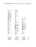 | 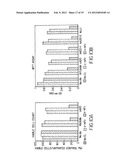 |
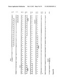 | 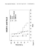 |
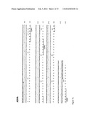 |  |
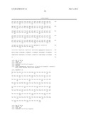 |  |
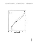 | 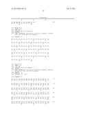 |
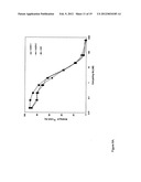 | 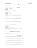 |
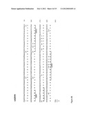 | 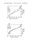 |
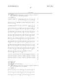 | 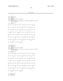 |
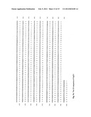 | 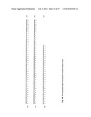 |
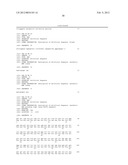 | 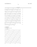 |
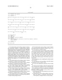 | 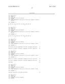 |
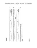 |  |
 | 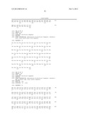 |
 |  |
 |  |
 |  |
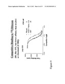 |  |
 | 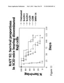 |
 |  |
 |
| Similar patent applications: | |
| Date | Title |
|---|---|
| 2009-01-01 | Rage fusion proteins and methods of use |
| 2009-03-05 | Rage fusion proteins and methods of use |
| 2009-01-29 | Galnts as modifiers of the igfr pathway and methods of use |
| 2009-08-13 | Gene expressed in prostate cancer and methods of use |
| 2009-01-01 | Kifs as modifiers of the rho pathway and methods of use |
| Top Inventors for class "Drug, bio-affecting and body treating compositions" | |
| Rank | Inventor's name |
|---|---|
| 1 | David M. Goldenberg |
| 2 | Hy Si Bui |
| 3 | Lowell L. Wood, Jr. |
| 4 | Roderick A. Hyde |
| 5 | Yat Sun Or |