Patent application title: PARASPORIN-1 RECEPTOR AND USE THEREOF
Inventors:
Hideki Katayama (Munakata-Shi, JP)
Yoshitomo Kusaka (Kurume-Shi, JP)
Eiichi Mizuki (Kurume-Shi, JP)
Assignees:
FUKUOKA PREFECTURAL GOVERNMENT
IPC8 Class: AA61K39395FI
USPC Class:
4241741
Class name: Immunoglobulin, antiserum, antibody, or antibody fragment, except conjugate or complex of the same with nonimmunoglobulin material binds eukaryotic cell or component thereof or substance produced by said eukaryotic cell (e.g., honey, etc.) cancer cell
Publication date: 2011-02-17
Patent application number: 20110038880
Inventors list |
Agents list |
Assignees list |
List by place |
Classification tree browser |
Top 100 Inventors |
Top 100 Agents |
Top 100 Assignees |
Usenet FAQ Index |
Documents |
Other FAQs |
Patent application title: PARASPORIN-1 RECEPTOR AND USE THEREOF
Inventors:
Hideki Katayama
Yoshitomo Kusaka
Eiichi Mizuki
Agents:
LEYDIG VOIT & MAYER, LTD
Assignees:
Origin: CHICAGO, IL US
IPC8 Class: AA61K39395FI
USPC Class:
Publication date: 02/17/2011
Patent application number: 20110038880
Abstract:
The receptor of parasporin-1 has been found to be beclin-1. Based on the
finding, the present invention provides a method of determining the
sensitivity of a cell to parasporin-1, comprising measuring the
expression level of beclin-1 polypeptide on the cell membrane or in a
membrane fraction of a cell; an inhibitor of the cytotoxicity of
parasporin-1, containing beclin-1 polypeptide etc.; a method of screening
for a substance capable of inhibiting the cytotoxicity of parasporin-1,
comprising evaluating the degree of binding of parasporin-1 to beclin-1
polypeptide in the presence of a test substance; a cell death inducer
containing an antibody that specifically recognizes beclin-1 polypeptide;
a method of screening for a substance capable of inducing cell death,
comprising evaluating the binding of a test substance to beclin-1
polypeptide, and the like.Claims:
1. A method of determining the sensitivity of a cell to parasporin-1,
comprising measuring the expression level of beclin-1 polypeptide on the
cell membrane or in a membrane fraction of a cell, and determining the
sensitivity of the cell to parasporin-1 on the basis of a positive
correlation between the expression level of beclin-1 polypeptide and the
sensitivity to parasporin-1.
2. The method according to claim 1, wherein the measurement of the expression level of beclin-1 polypeptide is performed using an antibody that specifically recognizes beclin-1 polypeptide.
3.-4. (canceled)
5. A method of inhibiting the cytotoxicity of parasporin-1, comprising administering beclin-1 polypeptide or an antibody that specifically recognizes beclin-1 polypeptide and is capable of inhibiting the specific binding of beclin-1 polypeptide and parasporin-1 polypeptide.
6. A method of screening for a substance capable of inhibiting the cytotoxicity of parasporin-1, comprising the steps shown below:(I) evaluating the degree of binding of parasporin-1 to beclin-1 polypeptide in the presence of a test substance, and(II) selecting a substance that has inhibited the binding of parasporin-1 to beclin-1 polypeptide as a substance capable of inhibiting the cytotoxicity of parasporin-1.
7. A combination comprising parasporin-1 and beclin-1 polypeptide.
8.-9. (canceled)
10. A method of inducing cell death, comprising bringing an antibody that specifically recognizes beclin-1 polypeptide into contact with a cell expressing beclin-1 polypeptide on the cell surface, wherein the antibody exhibits an activity to induce cell death to a cell expressing beclin-1 polypeptide on the cell surface when coming into contact with the cell.
11. The method according to claim 10, wherein the cell is a tumor cell.
12. A method of screening for a substance capable of inducing cell death, comprising the steps shown below:(I) evaluating the degree of binding of a test substance to beclin-1 polypeptide, and(II) selecting a test substance that has bound to beclin-1 polypeptide as a substance capable of inducing cell death.
13. The method according to claim 12, wherein the beclin-1 polypeptide used in (I) is beclin-1 polypeptide expressed on the cell membrane.
14.-15. (canceled)
16. A method of inducing cell death of a cell expressing beclin-1 polypeptide on the cell surface in a mammal, comprising administering to the mammal an effective amount of an antibody that specifically recognizes beclin-1 polypeptide and exhibits an activity to induce cell death to a cell expressing beclin-1 polypeptide on the cell surface when coming into contact with the cell.
17. A method of preventing or treating a tumor expressing beclin-1 polypeptide on the cell surface in a mammal, comprising administering to the mammal an effective amount of an antibody that specifically recognizes beclin-1 polypeptide and exhibits an activity to induce cell death to a cell expressing beclin-1 polypeptide on the cell surface when coming into contact with cell.
Description:
TECHNICAL FIELD
[0001]The present invention relates to a use of beclin-1 as a receptor of parasporin-1. More specifically, the present invention relates to a method and agent for determining the sensitivity of cells to parasporin-1 using beclin-1 or an antibody against beclin-1, a method and agent for inhibiting the cytotoxicity of parasporin-1, a method of screening for a cell death inducer, an antitumor agent or a substance capable of inducing cell death, and the like.
BACKGROUND ART
[0002]Bacillus thuringiensis produces a proteinous inclusion body in the sporulation stage. This inclusion body is toxic to the larvae of certain insects (Non-patent Document 1). The present inventors discovered a toxin protein that is toxic to mammalian cells in large screening of inclusion bodies of B. thuringiensis (Non-patent Document 2). The biochemical properties of these proteins, including cell specificity and cytotoxicity, are very diverse; that is, some toxin proteins are toxic to a broad range of cell lines, and other proteins are toxic to a very limited range of cell lines (Non-patent Documents 2 to 5). These proteins differed from the Cyt protein of B. thuringiensis, which is toxic to mammalian cells and possesses hemolytic activity, because these proteins did not exhibit hemolytic activity.
[0003]Parasporin proteins can roughly be classified into four groups according to primary structure similarity, and 13 kinds of parasporin proteins have been identified so far (see the website http://parasporin.fitc.pref.fukuoka.jp/). The individual parasporin proteins have totally different specificities to cells and molecular weights.
[0004]Parasporin-1 is produced as an 81-kDa toxin precursor by the B. thuringiensis A1190 strain, and exhibits cytotoxicity upon partial degradation by treatment with a protease such as trypsin. The active form parasporin-1 is a complex consisting of a 56-kDa protein and a 15-kDa protein, exhibiting specific cytotoxic activity (Non-patent Document 14). HeLa, which is a human cervical carcinoma cell line, and HL-60, which is a human promyelocytic leukemia cell line, are highly sensitive to parasporin-1, whereas normal human uterine smooth muscle cells and Caco-2, which is a colorectal cancer cell line, are little sensitive. So far, in a test of the sensitivity of 15 kinds of cell lines, including normal cell lines, to parasporin, sensitivity was noted in 4 cell lines. This demonstrates that parasporin-1 is a cytotoxin possessing extremely high cell specificity (Non-patent Document 14). Parasporin-1, in its toxicity induction process, induces an elevation of the concentration of cytoplasmic free Ca2+ accompanying a Ca2+ influx, which triggers cell death. Since this was suppressed by suramin, which is an inhibitor of G-protein, it has been estimated that the Ca2+ influx induced by parasporin-1 is mediated by an intracellular signal transduction system via G-protein (Non-patent Document 6). Furthermore, in parasporin-1-treated HeLa cells, not only degradation of Poly(ADP-ribose) polymerase and Caspase-3 activation (Non-patent Document 6), but also degradation of DNA into nucleosome units and translocation of phosphatidylserine are noted; therefore, parasporin-1 is a toxin that induces apoptosis to target cells.
[0005]Because parasporin-1 is toxic to a very limited range of cells and induces apoptosis to targeted cells, its applications to the fields of pharmaceuticals, cell identifying reagents, and cell biology are expectable by understanding the action mechanisms of parasporin-1. Particularly, because the binding of the toxin to a receptor is the most fundamental reaction in the toxic action, its identification is thought to be important. However, the receptor of parasporin-1 has not yet been identified.
[0006]Meanwhile, beclin-1 is a publicly known protein first identified as an autophagy-related protein (Non-patent Document 7), and has been reported to act as a cancer suppressor (Non-patent Documents 8 to 10). Furthermore, beclin-1 has been reported to be an intracellular protein that interacts with Bcl-2 and Bcl-XL, which are factors that control apoptosis (Non-patent Documents 11 to 13). However, it is not known at all that beclin-1 protein is capable of being exposed to cell surfaces, and that beclin-1 is capable of functioning as a receptor. [0007]non-patent document 1: Schnepf, E. et al., Microbiol. Mol. Biol. Rev. 62, 775-806 (1998) [0008]non-patent document 2: Mizuki, E. et al., J. Appl. Microbiol. 86, 477-486 (1999) [0009]non-patent document 3: Ito, A. et al., J. Biol. Chem. 279, 21282-21286 (2004) [0010]non-patent document 4: Lee, D. W. et al., Biochem. Biophys. Res. Commun. 272, 218-223 (2000) [0011]non-patent document 5: Yamashita, S. et al., Can J. Microbiol. 46, 913-919 (2000) [0012]non-patent document 6: Katayama, H. et al., J. Biol. Chem. 282, 7742-7752 (2007) [0013]non-patent document 7: Liang, H. X. et al., Nature 402, 672-676 (1999) [0014]non-patent document 8: Atia, V. M. et al., Genomics 59, 59-65 (1999) [0015]non-patent document 9: Qu, X. et al., J. Clin. Investig. 112, 1809-1820 (2003) [0016]non-patent document 10: Yue, Z. et al., Proc. Natl. Acad. U.S. A., b, 15077-15082(2003) [0017]non-patent document 11: Pattingre, S. at al., Cell, 122, 927-939 (2005) [0018]non-patent document 12: Liang, X. H. et al., J. Virol. 72, 8586-8596 (1998) [0019]non-patent document 13: Furuya, D. et al., Exp. Cell Res. 307, 26-40 (2005) [0020]non-patent document 14: Katayama, H. et al., J. Biochem. 137, 17-25 (2005)
DISCLOSURE OF THE INVENTION
Problems to be Solved by the Invention
[0021]In view of the above-described circumstances, the present invention aims to identify the parasporin-1 receptor on a target cell, which is thought to be most important in the induction of the cytotoxicity of parasporin-1, and provide a new antitumor drug that targets the receptor, a screening method therefor, a tool for cell sorting and the like.
Means of Solving the Problems
[0022]The present inventors, in order to explain the cell specificity of parasporin-1, searched for, and identified, the receptor of parasporin-1 using a photo-reactive chemical cross linker. Several candidate proteins for the receptor of parasporin-1 were separated by two-dimensional electrophoresis, and analyzed by the mass finger printing method utilizing a mass spectrometer; as a result, it was found that the parasporin-1-binding protein is beclin-1. An analysis using an anti-beclin-1 antibody and flow cytometry showed that beclin-1, which had been thought to be an intracellular protein, was localized outside of the cell membrane, and the binding of parasporin-1 to HeLa cells was inhibited by the anti-beclin-1 antibody. Furthermore, when the expression level of beclin-1 exposed on cell surfaces by the anti-beclin-1 antibody was examined, the expression level was high in HeLa cells, which are parasporin-1-sensitive cells, and the expression level was extremely low in Caco-2 and normal uterine smooth muscle cells, which are non-parasporin-1-sensitive cells. The anti-beclin-1 antibody (anti-peptide antibody) suppressed the binding of parasporin-1 to cell surfaces, and inhibited the cytotoxic effect of parasporin-1. Meanwhile, the anti-beclin-1 antibody (as a whole) exhibited cytotoxic activity by itself, suggesting that in the cell death, caspase-8 activation may be involved as with parasporin-1. In the results above, the present inventors found that the receptor of parasporin-1 is beclin-1, and that beclin-1 exposed on cell surfaces is one factor to determine the parasporin-1 sensitivity of cells, and have completed the present invention described below.
[0023]Accordingly, the present invention related to the following: [0024][1] A method of determining the sensitivity of a cell to parasporin-1, comprising measuring the expression level of beclin-1 polypeptide on the cell membrane or in a membrane fraction of a cell, and determining the sensitivity of the cell to parasporin-1 on the basis of a positive correlation between the expression level of beclin-1 polypeptide and the sensitivity to parasporin-1. [0025][2] The method described in [1], wherein the measurement of the expression level of beclin-1 polypeptide is performed using an antibody that specifically recognizes beclin-1 polypeptide. [0026][3] An agent for determining the sensitivity of a cell to parasporin-1, comprising an antibody that specifically recognizes beclin-1 polypeptide. [0027][4] An inhibitor of the cytotoxicity of parasporin-1, comprising beclin-1 polypeptide or an antibody that specifically recognizes beclin-1 polypeptide. [0028][5] A method of inhibiting the cytotoxicity of parasporin-1, comprising administering beclin-1 polypeptide or an antibody that specifically recognizes beclin-1 polypeptide. [0029][6] A method of screening for a substance capable of inhibiting the cytotoxicity of parasporin-1, comprising the steps shown below: [0030](I) evaluating the degree of binding of parasporin-1 to beclin-1 polypeptide in the presence of a test substance, and [0031](II) selecting a substance that has inhibited the binding of parasporin-1 to beclin-1 polypeptide as a substance capable of inhibiting the cytotoxicity of parasporin-1. [0032][7] A combination comprising parasporin-1 and beclin-1 polypeptide. [0033][8] A cell death inducer comprising an antibody that specifically recognizes beclin-1 polypeptide. [0034][9] An antitumor agent comprising an antibody that specifically recognizes beclin-1 polypeptide. [0035][10] A method of inducing cell death, comprising bringing an antibody that specifically recognizes beclin-1 polypeptide into contact with a cell. [0036][11] The method described in [10], wherein the cell is a tumor cell. [0037][12] A method of screening for a substance capable of inducing cell death, comprising the steps shown below: [0038](I) evaluating the degree of binding of a test substance to beclin-1 polypeptide, and [0039](II) selecting a test substance that has bound to beclin-1 polypeptide as a substance capable of inducing cell death. [0040][13] The method described in [12], wherein the beclin-1 polypeptide used in (I) is beclin-1 polypeptide expressed on the cell membrane. [0041][14] An antibody that specifically recognizes beclin-1 polypeptide, for use in inducing cell death. [0042][15] An antibody that specifically recognizes beclin-1 polypeptide, for use in preventing or treating a tumor. [0043][16] A method of inducing cell death in a mammal, comprising administering to the mammal an effective amount of an antibody that specifically recognizes beclin-1 polypeptide. [0044][17] A method of preventing or treating a tumor in a mammal, comprising administering to the mammal an effective amount of an antibody that specifically recognizes beclin-1 polypeptide.
Effect of the Invention
[0045]Provided by the present invention are various uses of beclin-1 as a receptor of parasporin-1.
[0046]Using the method of the present invention for determining sensitivity to parasporin-1, it is possible to objectively determine the sensitivity of various cells to parasporin-1 on the basis of the expression level of parasporin-1 receptor definitely supported by molecular biologically.
[0047]Using the inhibitor of the cytotoxicity of parasporin-1 of the present invention, it is possible to potently suppress the cell death (apoptosis) induced by parasporin-1.
[0048]Using the cell death inducer of the present invention, it is possible to induce cell death (apoptosis) specifically to cells expressing beclin-1 polypeptide on the cell membrane or in a membrane fraction, and the inducer is useful as a cell-specific antitumor agent.
BRIEF DESCRIPTION OF THE DRAWINGS
[0049]FIG. 1 shows the binding of parasporin-1 labeled with Alexa Fluor 488 dye to HeLa cells. (A) Observations using a light microscope (A-C) and a fluorescence microscope (D-F). (B) Observations using a flow cytometer.
[0050]FIG. 2 shows correlations between parasporin-1 sensitivity and parasporin-1 affinity. (A) Parasporin-1 susceptibilities of various cell lines. (B) Affinity of parasporin-1 to cell surfaces of various cell lines.
[0051]FIG. 3 shows screening for and identification of parasporin-1 receptor using SBED-labeled parasporin-1. (A) Detection of parasporin-1-binding protein by Western blotting (one-dimensional electrophoresis). (B and C) Separation of parasporin-1-binding protein by two-dimensional electrophoresis. Left panel: control (SBED-labeled BSA). Right panel: SBED-labeled parasporin-1.
[0052]FIG. 4 shows effects of anti-beclin-1 antibody (upper panel), anti-TMOD-3 antibody (middle panel) and anti-cytochrome C antibody (lower panel) on the binding of parasporin-1 to HeLa cells.
[0053]FIG. 5 shows the localization of beclin-1 to the cell membrane. (A) Flow cytometric analysis of beclin-1 expression on HeLa cell surface. (B) Analysis of beclin-1 expression by Western blotting. Upper panel: whole cells, lower panel: membrane fractions containing organelle.
[0054]FIG. 6 shows cell death of HeLa cells induced by anti-beclin-1 antibody.
BEST MODE FOR EMBODYING THE INVENTION
1. Method and Agent for Determining the Sensitivity of Cells to Parasporin-1
[0055]The present invention provides a method of determining the sensitivity of cells to parasporin-1. The method of sensitivity determination of the present invention comprises measuring the expression level of beclin-1 polypeptide on the cell membrane or in a membrane fraction of a cell, and determining the sensitivity of the cell to parasporin-1 on the basis of a positive correlation between the expression level and sensitivity to parasporin-1. The method of sensitivity determination of the present invention is useful for, for example, distinguishing tumors on which an antitumor drug is effective and tumors on which the same is ineffective, in developing parasporin-1 as the antitumor drug.
[0056]Parasporin-1 is a publicly known protein that was isolated from the B. thuringiensis A1190 strain (Katayama, H. et al., J. Biochem. 137, 17-25 (2005)). Parasporin-1 is produced as an about 81-kDa precursor, and upon partial degradation by protease treatment, it forms a complex consisting of an about 56-kDa polypeptide and an about 15-kDa polypeptide. This complex possesses binding activity for beclin-1, and binds to beclin-1 expressed on a cell surface to induce cell death due to apoptosis to the cell. Herein, "parasporin-1" means this active complex unless otherwise stated.
[0057]"The sensitivity of a cell to parasporin-1" refers to the degree of inducibility of cell death (particularly apoptosis) when the cell is exposed to parasporin-1. "A cell is sensitive to parasporin-1" means that cell death (particularly apoptosis) is induced when the cell is exposed to parasporin-1.
[0058]Cells used in the method of sensitivity determination of the present invention are mammalian cells. Mammals include laboratory animals, including rodents such as mice, rats, hamsters and guinea pigs, and rabbits; domestic animals such as pigs, cattle, goat, horses, sheep, and minks; companion animals such as dogs and cats; primates such as humans, monkeys, rhesus monkeys, marmosets, orangutans, and chimpanzees, and the like. Cells used in the method of sensitivity determination of the present invention are preferably human cells.
[0059]Cells used in the method of sensitivity determination of the present invention include cultured cells, cells separated from living organisms, cells comprised in tissues separated from living organisms, cells in living organisms and the like. Cells used in the method of sensitivity determination of the present invention are tumor cells or non-tumor cells, preferably tumor cells.
[0060]Beclin-1 is a publicly known protein (Liang, H. X. et al., Nature 402, 672-676 (1999); Atia, V. M. et al., Genomics 59, 59-65 (1999); Qu, X. et al., J. Clin. Investig. 112, 1809-1820 (2003); Yue, Z. et al., Proc. Natl. Acad. Sci. U.S.A., b, 15077-15082 (2003); Pattingre, S. et al., Cell, 122, 927-939 (2005); Liang, X. H. et al., J. Virol. 72, 8586-8596 (1998); Furuya, D. et al., Exp. Cell Res. 307, 26-40 (2005)), and the amino acid sequence and mRNA (cDNA) sequence thereof are also publicly known. The beclin-1 that can be used in the present invention is the beclin-1 of one of the above-described mammals. As the amino acid sequence of human beclin-1, the amino acid sequence shown by SEQ ID NO:2 is known (NCBI Integrated Database Accession No. NP--003757, version NP--003757.1); as the nucleotide sequence of human beclin-1 mRNA (cDNA) that encodes the same, the nucleotide sequence shown by SEQ ID NO:1 is known (NCBI Integrated Database Accession No. NM--003766, version NM--003766.2/coding region: 134-1486). As orthologs thereof, mouse beclin-1 [amino acid sequence: SEQ ID NO:4 (NCBI Integrated Database Accession No. NP--062530, version NP--062530.2), mRNA(cDNA) nucleotide sequence: SEQ ID NO:3 (NCBI Integrated Database Accession No. NM--019584, version NM--019584.3/coding region: 139-1485)], rat beclin-1[amino acid sequence: SEQ ID NO:6 (NCBI Integrated Database Accession No. NP--446191, version NP--446191.1), mRNA (cDNA) nucleotide sequence: SEQ ID NO:5 (NCBI Integrated Database Accession No. NM--053739, version NM--053739.2/coding region: 225-1571)] and the like are known.
[0061]A measurement of the expression level of beclin-1 polypeptide on the cell membrane or in a membrane fraction is normally performed ex vivo.
[0062]The expression level of beclin-1 polypeptide can be measured by an immunochemical technique using an antibody that specifically recognizes beclin-1 polypeptide. Immunochemical techniques include flow cytometric analysis, radioimmunoassay (RIA method), ELISA (Methods in Enzymol. 70: 419-439 (1980)), Western blotting, immunohistological staining and the like.
[0063]Parasporin-1 binds to beclin-1 expressed on the cell surface and induces cell death to the cell. Therefore, the expression level of beclin-1 polypeptide on the cell membrane (that is, on the cell surface) or in a membrane fraction better correlates with the sensitivity of the cell to parasporin-1 than the expression level of beclin-1 polypeptide in the whole cell. Therefore, in the method of sensitivity determination of the present invention, the expression level of beclin-1 polypeptide on the cell membrane or in a membrane fraction, more preferably the expression level of beclin-1 polypeptide on the cell membrane, is measured. "A membrane fraction" includes the membranes constituting organellae and the cell membrane. A membrane fraction can be prepared from a cell by a method known per se such as centrifugation. In measuring the expression level of beclin-1 polypeptide on the cell membrane, immunochemical techniques such as flow cytometric analysis and immunohistological staining are suitably used. In measuring the expression level of beclin-1 polypeptide in a membrane fraction, immunochemical techniques such as radioimmunoassay (RIA method), ELISA (Methods in Enzymol. 70: 419-439 (1980)), and Western blotting are suitably used.
[0064]An antibody that specifically recognizes beclin-1 polypeptide can be produced by conventional method of antibody production using beclin-1 polypeptide or a partial peptide having the antigenicity thereof as an immunogen. As mentioned herein, antibodies include, but are not limited to, natural type antibodies such as polyclonal antibodies and monoclonal antibodies (mAbs), chimeric antibodies, humanized antibodies, single-stranded antibodies and human antibodies that can be produced using gene recombination technology, and binding fragments thereof. Preferably, the antibody is a polyclonal antibody, a monoclonal antibody or a binding fragment thereof. A binding fragment means a partial region of one of the above-described antibodies having the specific binding activity; specifically, for example, F(ab')2, Fab', Fab, Fv, sFv, dsFv, sdAb and the like can be mentioned (Exp. Opin. Ther. Patents, Vol. 6, No. 5, p. 441-456, 1996). The class of antibody is not particularly limited; antibodies of any itotypes such as IgG, IgM, IgA, IgD and IgE are encompassed. Preferably, the class is IgG or IgM, and in view of the ease of purification and the like, the class is more preferably IgG.
[0065]Next, on the basis of the measured expression level of beclin-1 polypeptide on the cell membrane or in a membrane fraction, the sensitivity of a cell to parasporin-1 is determined. As shown in an Example described below, as the expression level of beclin-1 polypeptide on the cell membrane or in a membrane fraction in a cell increases, the sensitivity of the cell to parasporin increases. The above-described determination is made on the basis of such a positive correlation between the expression level of beclin-1 polypeptide and sensitivity to parasporin-1.
[0066]In an embodiment, for example, if the cell expresses beclin-1 polypeptide on the cell membrane or in a membrane fraction, it can be determined that the cell is sensitive to parasporin-1. If the cell does not express beclin-1 polypeptide on the cell membrane or in a membrane fraction, it can be determined that the cell is non-sensitive to parasporin-1.
[0067]In another embodiment, for example, a cell that does not express beclin-1 polypeptide on the cell membrane or in a membrane fraction, and that is non-sensitive to parasporin-1 (negative control), and a cell that expresses beclin-1 polypeptide on the cell membrane or in a membrane fraction, and that is sensitive to parasporin-1 (positive control), are provided in advance, and the expression level of beclin-1 polypeptide on the cell membrane or in a membrane fraction in the measurement subject cell is compared with those of the positive control and negative control. Alternatively, a diagram of the correlation between the expression level of beclin-1 polypeptide on the cell membrane or in a membrane fraction in a cell and the sensitivity to parasporin-1 may be generated in advance, and the expression level of beclin-1 polypeptide on the cell membrane or in a membrane fraction in the measurement subject cell may be compared with the correlation diagram. This comparison of expression level is preferably made on the basis of the presence or absence of a significant difference.
[0068]Then, judging from the comparative results for beclin-1 polypeptide expression levels, if the beclin-1 polypeptide expression level on the cell membrane or in a membrane fraction in the measurement subject cell is relatively high, it can be determined that the sensitivity of the cell to parasporin-1 is relatively high. Conversely, if the beclin-1 polypeptide expression level on the cell membrane or in a membrane fraction in the measurement subject cell is relatively low, it can be determined that the sensitivity of the cell to parasporin-1 is relatively low.
[0069]The present invention also provides an agent for determining the sensitivity of a cell to parasporin-1, containing the above-described antibody that specifically recognizes beclin-1 polypeptide (referred to as the agent (1) of the present invention). The agent (I) of the present invention can be a kit for determining the sensitivity of a cell to parasporin-1. By using the agent (I) of the present invention, it is possible to easily determine the sensitivity of a cell to parasporin-1 by the above-described method.
[0070]An antibody that specifically recognizes beclin-1 polypeptide can be dissolved in water or an appropriate buffer solution (e.g., TE buffer, PBS and the like) to obtain an appropriate concentration, and preserved at about -20° C. to 4° C.
[0071]The agent (I) of the present invention may further contain other ingredients necessary for embodying the method according to the method of measuring the expression level of beclin-1 polypeptide.
[0072]For example, the agent (I) of the present invention can further contain a labeled secondary antibody, color developing substrate, blocking liquid, washing buffer solution, ELISA plate, blotting membrane and the like.
2. Agent and Method for Inhibiting the Cytotoxicity of Parasporin-1
[0073]As stated above, parasporin-1 binds to beclin-1 polypeptide on the cell surface of a target cell to induce cell death (particularly apoptosis) to the cell. Therefore, by administering an effective amount of beclin-1 polypeptide or an antibody that specifically recognizes beclin-1 polypeptide, the binding of parasporin-1 to beclin-1 polypeptide on the cell surface is inhibited, and the cytotoxicity of parasporin-1 is inhibited. Therefore, the present invention provides an inhibitor of the cytotoxicity of parasporin-1 comprising beclin-1 polypeptide or an antibody that specifically recognizes beclin-1 polypeptide (the inhibitor of the present invention).
[0074]The beclin-1 polypeptide that can be used in the present invention is the beclin-1 polypeptide of one of the above-described mammals. As mammalian beclin-1 polypeptides, human beclin-1 polypeptides (for example, a polypeptide comprising the amino acid sequence shown by SEQ ID NO:2), mouse beclin-1 polypeptides (for example, a polypeptide comprising the amino acid sequence shown by SEQ ID NO:4), rat beclin-1 polypeptides (for example, a polypeptide comprising the amino acid sequence shown by SEQ ID NO:6) and the like can be mentioned. Beclin-1 polypeptide can be obtained by culturing a transformant incorporating a beclin-1 expression vector designed on the basis of the gene sequence thereof, and isolating/purifying the desired product from the culture using a commonly known means of purification such as column chromatography.
[0075]As an antibody that specifically recognizes beclin-1 polypeptide, an antibody that can be used in the above-described method of sensitivity determination of the present invention can be used; as the antibody, an antibody capable of inhibiting the specific binding of beclin-1 polypeptide and parasporin-1 polypeptide (neutralizing antibody) is preferable. The antibody used for the inhibitor of the present invention is preferably an antibody that does not exhibit an activity to induce cell death to a beclin-1-expressing cell when brought into contact with the cell. The epitope recognized by the antibody used in the inhibitor of the present invention is not particularly limited, as far as the antibody inhibits the cytotoxicity of parasporin-1. As an excellent antibody that potently inhibits the cytotoxicity of parasporin-1 and little induces cell death to beclin-1-expressing cells, an antibody that recognizes a site of human beclin-1 polypeptide comprising threonine 72 (Thr72) as an epitope can be mentioned.
[0076]The inhibitor of the present invention may contain, in addition to the above-described beclin-1 polypeptide or antibody that specifically recognizes beclin-1 polypeptide, an optionally chosen carrier, for example, a pharmaceutically acceptable carrier.
[0077]As examples of the pharmaceutically acceptable carrier, excipients such as sucrose and starch; binders such as cellulose and methylcellulose; disintegrants such as starch and carboxymethylcellulose; lubricants such as magnesium stearate and Aerosil; flavoring agents such as citric acid and menthol; preservatives such as sodium benzoate and sodium hydrogen sulfite; stabilizers such as citric acid and sodium citrate; suspending agents such as methylcellulose and polyvinylpyrrolidone; dispersing agents such as surfactants; diluents such as water and physiological saline; base waxes; and the like can be mentioned, which, however, are not to be construed as limiting.
[0078]The content of beclin-1 polypeptide or an antibody that specifically recognizes beclin-1 polypeptide in the inhibitor of the present invention is, for example, about 0.01 to 100% is by weight of the entire pharmaceutical composition.
[0079]In an embodiment, the inhibitor of the present invention is used in vivo. In this case, the inhibitor is orally or parenterally administered to a subject animal. As preparations suitable for oral administration, liquids, capsules, sachets, tablets, suspensions, emulsions and the like can be mentioned. Preparations suitable for parenteral administration (for example, subcutaneous injection, intramuscular injection, topical injection, intraperitoneal administration and the like) include aqueous and non-aqueous isotonic sterile injectable liquids, which may comprise an antioxidant, a buffer solution, a bacteriostatic agent, an isotonizing agent and the like. Aqueous and non-aqueous sterile suspensions can also be mentioned, which may comprise a suspending agent solubilizer, a thickening agent, a stabilizer, an antiseptic and the like. These preparations can be encapsulated in containers such as ampoules and vials for unit dosage or a plurality of dosages. It is also possible to freeze-dry the active ingredient and a pharmaceutically acceptable carrier, and store the preparation in a state that may be dissolved or suspended in an appropriate sterile vehicle just before use.
[0080]When the inhibitor of the present invention is used in vivo, the dosage of the inhibitor of the present invention varies depending on the activity and choice of the active ingredient, seriousness of illness, recipient animal species, the recipient's drug receptivity, body weight, age, and the like, and cannot be generalized; however, the dosage is normally about 0.001 to about 500 mg/kg, based on the amount of active ingredient, per day for an adult human.
[0081]The inhibitor of the present invention is safely administered to the above-described mammal in a way such that the active ingredient thereof will be delivered to a cell that is a target of parasporin-1 (a cell that expresses beclin-1 on cell surface).
[0082]In another embodiment, the inhibitor of the present invention is used in vitro. In this case, beclin-1 polypeptide or an antibody that specifically recognizes beclin-1 polypeptide is administered (added) to a medium comprising parasporin-1 and a target cell therefor (a cell that expresses beclin-1 on cell surface).
[0083]When the inhibitor of the present invention is used in vitro, the dosage of the inhibitor of the present invention varies depending on the activity and choice of the active ingredient and the like, and cannot be generalized; however, the dosage is normally about 0.1 to 500 μg/ml, based on the final concentration of active ingredient in the culture.
[0084]Because beclin-1 polypeptide or an antibody that specifically recognizes beclin-1 polypeptide is capable of inhibiting the binding of parasporin-1 to beclin-1 polypeptide on cell surface to inhibit the manifestation of cytotoxicity by parasporin-1, the inhibitor of the present invention is useful in preventing/treating cell death and poisoning that can be induced by parasporin-1. The inhibitor of the present invention is also useful in analyzing the mechanism for the manifestation of cytotoxicity by parasporin-1.
3. Method of Screening for Substances Capable of Inhibiting the Cytotoxicity of Parasporin-1
[0085]The present invention provides a method of screening for substances capable of inhibiting the cytotoxicity of parasporin-1, comprising the steps shown below (the screening method (1) of the present invention): [0086](I) evaluating the degree of binding of parasporin-1 to beclin-1 polypeptide in the presence of a test substance, and [0087](II) selecting a substance that has inhibited the binding of parasporin-1 to beclin-1 polypeptide as a substance capable of inhibiting the cytotoxicity of parasporin-1.
[0088]The step (I) can further comprise comparing the degree of binding of parasporin-1 to beclin-1 polypeptide in the presence of the test substance with the degree of binding of parasporin-1 to beclin-1 polypeptide in the absence of the test substance.
[0089]The test substance subjected to the screening method (I) of the present invention may be any commonly known compound or a novel compound; examples include nucleic acids, sugars, lipids, proteins, peptides, organic low molecular compounds, compound libraries prepared using combinatorial chemistry technology, random peptide libraries, or naturally occurring ingredients derived from microorganisms, animals, plants, marine organisms and the like, and the like.
[0090]As stated above, parasporin-1 is produced as an about 81-kDa precursor, and upon partial degradation by protease treatment, it forms an active (possessing cytotoxicity) complex consisting of an about 56-kDa polypeptide and an about 15-kDa polypeptide. In the screening method (I) of the present invention, this active complex is preferably used as parasporin-1. The parasporin-1 used in the screening method (I) of the present invention is preferably one that has been isolated and purified. "Isolated and purified" means having been treated to remove compounds other than the desired product.
[0091]In the screening method (I) of the present invention, a beclin-1 polypeptide that can be used in the above-described inhibitor of the present invention can be used as beclin-1 polypeptide. In an embodiment, the beclin-1 polypeptide used in the screening method (I) of the present invention has been isolated and purified. In another embodiment, beclin-1 polypeptide expressed on the cell surface of a cell naturally or forcibly expressing beclin-1 (preferably a cell of one of the above-described mammals) is used in the screening method (I) of the present invention. The cell naturally expressing beclin-1 is not particularly limited, as far as it potentially expresses beclin-1; as the cell, a primary culture cell, a cell line induced from the primary culture cell and the like can be used. Examples of cells naturally expressing beclin-1 include cervical carcinoma cells (HeLa cells and the like). Cells forcibly expressing beclin-1 include a transformant incorporating an expression vector capable of expressing beclin-1, and the like. In the vector, a polynucleotide that encodes beclin-1 polypeptide is integrated downstream of a promoter functional in the host cell in a way such that beclin-1 can be expressed in the host cell.
[0092]The evaluation in the step (I) is normally performed in an appropriate culture medium or buffer solution. The degree of binding of parasporin-1 to beclin-1 polypeptide can be evaluated by a method of interaction analysis well known in the art, for example, surface plasmon resonance, binding assay, immunological techniques and the like.
[0093]For example, parasporin-1 and beclin-1 polypeptide are brought into contact with each other in the presence of a test substance by immobilizing either parasporin-1 or beclin-1 polypeptide on a chip, and loading a solution containing the other polypeptide and the test substance on the chip. Next, the binding and dissociation of parasporin-1 to and from beclin-1 polypeptide are measured in the presence of the test substance by the surface plasmon resonance method, and are compared with the binding and dissociation obtained when a control solution without the test substance is loaded on the chip.
[0094]Alternatively, parasporin-1 and beclin-1 polypeptide are brought into contact with each other in the presence of a test substance by immobilizing either parasporin-1 or beclin-1 polypeptide on a plate, and adding a solution containing the other polypeptide labeled with an appropriate radioisotope or fluorescer and the test substance to the plate. Next, with the amount of labeling substance bound onto the plate as an indicator, the binding of parasporin-1 to beclin-1 polypeptide in the presence of the test substance is measured, and compared with the binding in the absence of the test substance.
[0095]Alternatively, parasporin-1 and beclin-1 polypeptide are brought into contact with each other in the presence of a test substance by adding a solution containing parasporin-1 labeled with an appropriate fluorescer and the test substance to a cell expressing beclin-1 polypeptide on the cell surface. Next, the binding of parasporin-1 to beclin-1 polypeptide on the cell surface in the presence of the test substance is measured by flow cytometry, and compared with the binding in the absence of the test substance.
[0096]Then, on the basis of the comparative results for binding and dissociation rates or the amount bound, a test substance that inhibits the binding of parasporin-1 and beclin-1 polypeptide is selected as a substance capable of inhibiting the cytotoxicity of parasporin-1.
[0097]Because a substance which can be obtained by the screening method (I) of the present invention is capable of inhibiting the manifestation of cytotoxicity by parasporin-1, it is useful as a candidate substance for the prevention/treatment of cell death and poisoning that can be induced by parasporin-1. A substance which can be obtained by the screening method (I) of the present invention is also useful in analyzing the mechanism for the manifestation of cytotoxicity by parasporin-1.
4. Combination Comprising Parasporin-1 and Beclin-1 Polypeptide
[0098]The present invention provides a combination comprising parasporin-1 and beclin-1 polypeptide (the combination of the present invention). The combination of the present invention is useful as a reagent for screening for a substance capable of inhibiting the cytotoxicity of parasporin-1.
[0099]In an embodiment, parasporin-1 and beclin-1 polypeptide are comprised in the combination of the present invention while in a separated state. In this embodiment, parasporin-1 and beclin-1 are housed in different containers, respectively, and the plurality of containers are housed in one package.
[0100]In another embodiment, parasporin-1 and beclin-1 polypeptide are comprised in the combination of the present invention while in a mixed state, to constitute a composition.
[0101]The parasporin-1 comprised in the combination of the present invention is the same as one that can be used in the screening method (I) of the present invention.
[0102]The beclin-1 polypeptide comprised in the combination of the present invention is the same as one that can be used in the screening method (I) of the present invention. The beclin-1 polypeptide comprised in the combination of the present invention is preferably an isolated and purified polypeptide.
[0103]Using the combination of the present invention, it is possible to easily perform the screening method (I) of the present invention.
5. Agent and Method for Inducing Cell Death Using an Antibody that Specifically Recognizes Beclin-1 Polypeptide
[0104]As stated in an Example described below, when an effective amount of an antibody that specifically recognizes beclin-1 polypeptide is administered, the antibody binds to beclin-1 on the cell surface of a target cell, resulting in the induction of cell death (particularly apoptosis) to the cell as with the use of parasporin-1. Therefore, by administering an effective amount of beclin-1 polypeptide or an antibody that specifically recognizes beclin-1 polypeptide, the binding of parasporin-1 to beclin-1 polypeptide on the cell surface is inhibited, whereby the cytotoxicity of parasporin-1 is inhibited.
[0105]A targeted cell for the agent (II) of the present invention is a cell expressing beclin-1 polypeptide on the cell surface. Targeted cells for the agent (II) of the present invention include cultured cells, cells separated from living organisms, cells comprised in tissues separated from living organisms, cells in living organisms and the like. The cell is a tumor cell or non-tumor cell, preferably a tumor cell.
[0106]As an antibody that specifically recognizes beclin-1 polypeptide, that can be used in the agent (II) of the present invention, the same as one that can be used in the above-described agent (I) of the present invention can be mentioned. The antibody is preferably an antibody that exhibits an activity to induce cell death to a beclin-1-expressing cell when coming into contact with the cell. The epitope recognized by the antibody used in the agent (II) of the present invention is not particularly limited, as far as the antibody possesses an activity to induce cell death. Because a polyclonal antibody capable of recognizing whole beclin-1 polypeptide is potently active to induce cell death, it is preferably used in the agent (II) of the present invention. Such a polyclonal antibody can be acquired by administering a polypeptide comprising the full-length amino acid sequence of beclin-1 polypeptide, along with a commercially available adjuvant (for example, Freund complete or incomplete adjuvant), to an animal subcutaneously or intraperitoneally about 2 to 4 times at intervals of 2 to 3 weeks (the antibody titer of partially drawn serum should be measured by a publicly known antigen-antibody reaction, and an elevation thereof should be confirmed in advance), collecting whole blood about 3 to 10 days after final immunization, and purifying the antiserum. Animals to which the antigen is to be administered include non-human mammals such as rats, mice, rabbits, goat, guinea pigs, and hamsters.
[0107]The agent (II) of the present invention may contain, in addition to the above-described antibody that specifically recognizes beclin-1 polypeptide, an optionally chosen carrier, for example, a pharmaceutically acceptable carrier.
[0108]As examples of the pharmaceutically acceptable carrier, excipients such as sucrose and starch; binders such as cellulose and methylcellulose; disintegrants such as starch and carboxymethylcellulose; lubricants such as magnesium stearate and Aerosil; flavoring agents such as citric acid and menthol; preservatives such as sodium benzoate and sodium hydrogen sulfite; stabilizers such as citric acid and sodium citrate; suspending agents such as methylcellulose and polyvinylpyrrolidone; dispersing agents such as surfactants; diluents such as water and physiological saline; base waxes; and the like can be mentioned, which, however, are not to be construed as limiting.
[0109]The content of an antibody that specifically recognizes beclin-1 polypeptide in the agent (II) of the present invention is, for example, about 0.01 to 100% by weight of the entire pharmaceutical composition.
[0110]In an embodiment, the agent (II) of the present invention is used in vivo. In this case, the inhibitor is orally or parenterally administered to a subject animal. As preparations suitable for oral administration, liquids, capsules, sachets, tablets, suspensions, emulsions and the like can be mentioned. Preparations suitable for parenteral administration (for example, subcutaneous injection, intramuscular injection, topical injection, intraperitoneal administration and the like) include aqueous and non-aqueous isotonic sterile injectable liquids, which may contain an antioxidant, a buffer solution, a bacteriostatic agent, an isotonizing agent and the like. Aqueous and non-aqueous sterile suspensions can also be mentioned, which may contain a suspending agent, a solubilizer, a thickening agent, a stabilizer, an antiseptic and the like. These preparations can be encapsulated in containers such as ampoules and vials for unit dosage or a plurality of dosages. It is also possible to freeze-dry the active ingredient and a pharmaceutically acceptable carrier, and store the preparation in a state that may be dissolved or suspended in an appropriate sterile vehicle just before use.
[0111]When the agent (II) of the present invention is used in vivo, the dosage of the agent (II) of the present invention varies depending on the activity and choice of the active ingredient, seriousness of illness, recipient animal species, the recipient's drug receptivity, body weight, age, and the like, and cannot be generalized; however, the dosage is normally about 0.001 to about 500 mg/kg, based on the amount of active ingredient, per day for an adult human.
[0112]The agent (II) of the present invention is safely administered to the above-described mammal in a way such that the active ingredient thereof will be delivered to a targeted cell (a cell expressing beclin-1 on the cell surface).
[0113]In another embodiment, the agent (II) of the present invention is used in vitro. In this case, an antibody that specifically recognizes beclin-1 polypeptide is administered (added) to a medium containing a targeted cell (a cell that expresses beclin-1 on the cell surface).
[0114]When the agent (II) of the present invention is used in vitro, the dosage of the agent (II) of the present invention varies depending on the activity and choice of the active ingredient and the like, and cannot be generalized; however, the dosage is normally about 0.1 to 500 μg/ml, based on the final concentration of active ingredient in the culture.
[0115]The agent (II) of the present invention is useful as an antitumor agent in preventing and treating a tumor (preferably a tumor expressing beclin-1 polypeptide on the cell surface). The tumor is exemplified by, but not limited to, cervical carcinoma, gastric cancer, lung cancer, colorectal cancer, brain tumor, ovarian cancer, prostatic cancer, liver cancer, malignant lymphoma and the like.
6. Screening Method for Substances Capable of Inducing Cell Death
[0116]As stated above, an antibody that specifically recognizes beclin-1 polypeptide or parasporin-1 binds to beclin-1 polypeptide expressed on the cell surface to induce cell death (apoptosis) to the cell. Therefore, any substance, like parasporin-1, capable of binding to beclin-1 polypeptide is capable of inducing cell death to cells that express beclin-1 on the cell surface. Therefore, the present invention provides a method of screening for a substance capable of inducing cell death (apoptosis), comprising the steps shown below (the screening method (II) of the present invention): [0117](I) evaluating the degree of binding of a test substance to beclin-1 polypeptide, and [0118](II) selecting a test substance that has bound to beclin-1 polypeptide as a substance capable of inducing cell death.
[0119]The step (I) can further comprise comparing the degree of binding of the test substance to beclin-1 polypeptide with the degree of binding of the test substance to a polypeptide other than beclin-1 (control polypeptide).
[0120]The test substance subjected to the screening method (II) of the present invention, as in the screening method (I), may be any commonly known compound or a novel compound.
[0121]In the screening method (II) of the present invention, the same beclin-1 polypeptide as one used in the screening method (I) can be used. In the screening method (II) of the present invention, as in the screening method (I), an isolated and purified beclin-1 polypeptide or beclin-1 polypeptide expressed on the cell surface of a cell naturally or forcibly expressing beclin-1 is used as beclin-1 polypeptide.
[0122]The evaluation in the step (I) is normally performed in an appropriate culture medium or buffer solution. The degree of binding of a test substance to beclin-1 polypeptide can be evaluated, as in the screening method (I), by a method of interaction analysis well known in the art, for example, surface plasmon resonance, binding assay, immunological techniques and the like.
[0123]For example, a test substance and beclin-1 polypeptide are brought into contact with each other by immobilizing beclin-1 polypeptide on a chip, and loading a solution containing the test substance onto the chip. Next, the binding and dissociation of the test substance to or from beclin-1 polypeptide are measured by the surface plasmon resonance method, and compared with the binding and dissociation of the test substance to or from the control peptide when the control peptide is immobilized on the chip.
[0124]Alternatively, a test substance and beclin-1 polypeptide are brought into contact with each other by immobilizing beclin-1 polypeptide on a plate, and adding a solution containing the test substance labeled with an appropriate radioisotope or fluorescer to the plate. Next, with the amount of labeling substance bound onto the plate as an indicator, the binding of the test substance to beclin-1 polypeptide is measured, and compared with the binding to the control polypeptide.
[0125]Alternatively, a test substance and beclin-1 polypeptide are brought into contact with each other by adding a solution containing the test substance labeled with an appropriate fluorescer to a cell expressing beclin-1 polypeptide on the cell surface. Next, the binding of the test substance to beclin-1 polypeptide on the cell surface is measured by flow cytometry, and compared with the binding of the test substance to a control cell not expressing beclin-1.
[0126]Then, on the basis of the comparative results for binding and dissociation rates or the amount bound, a test substance that has bound to beclin-1 polypeptide is selected as a substance (candidate substance) capable of inducing cell death.
[0127]The screening method (II) of the present invention may comprise after the step (II) a step for bringing a substance selected through the step (II) into contact with a cell expressing beclin-1 on the cell surface, and selecting a substance that has induced cell death (preferably apoptosis) to the cell as a substance confirmed to induce cell death.
[0128]Contact of the test substance and the cell expressing beclin-1 on the cell surface is performed in a culture medium. A culture medium is chosen as appropriate according to the kind of cell; examples include a minimal essential medium (MEM), Dulbecco's Modified Eagle's Medium (DMEM) and the like containing about 5 to 20% fetal calf serum. Culturing conditions are also determined as appropriate; for example, the pH of the medium is about 6 to about 8, culturing temperature is normally about 30 to about 40° C., and culturing time is about 12 to about 72 hours. Cell death induction can be evaluated by the TUNNEL method, DNA ladder detection and the like.
[0129]Substances obtained by the screening method (II) of the present invention are useful as candidate substances for the development of new antitumor agents.
[0130]The present invention is explained in more detail in the following by referring to Examples and the like, which are not to be construed as limitative.
Examples
1. Binding of Parasporin-1 to HeLa Cell
[0131]To confirm that parasporin-1 can bind to the target cell surface, HeLa cells were treated with parasporin-1 labeled with Alexa Fluor 488 dye, and immunostained with an anti-Alexa Fluor 488 dye antibody. The cells treated with active parasporin-1 could be stained with a fluorescence antibody, but staining could not be detected in control and the cells treated with inactive parasporin-1 treated at 100° C. for 5. min (FIG. 1A). HeLa cells were treated with parasporin-1 labeled with Alexa Fluor 488 dye, and the binding of parasporin-1 to the cells was analyzed by flow cytometry (FIG. 1B). The cells treated with active labeled parasporin-1 showed increased fluorescence, and the increased fluorescence disappeared by adding non-labeled parasporin-1 in much excess (40-fold of labeled parasporin-1) (FIG. 1B, upper left panel).
2. Comparison of Binding to Cell Lines with Various Parasporin-1 Sensitivity
[0132]Parasporin-1 is a cytotoxin with high cell specificity. FIG. 2A shows sensitivity of HeLa, A549, Sawano and normal uterus smooth muscle cells (UtSMC) to parasporin-1 (1 μg/ml). To show correlation between parasporin-i sensitivity of cell and binding of parasporin-1 to cell membrane, the binding property of parasporin-1 to various cells was analyzed by flow cytometry. HeLa cell showed parasporin-1 dependent peak shift but other cell lines hardly showed a peak shift.
3. Screening and Identification of Parasporin-1 Receptor Using Photoreactive Chemical Crosslinking Agent
[0133]Parasporin-1 receptor was screened for using parasporin-1 labeled with a photoreactive chemical crosslinking agent (SEED). As a result of chemical crosslinking, since biotin transfer occurs in a protein that has interacted with parasporin-1, a protein of HeLa cell to which biotin was transferred was screened for by Western blotting. As a control, SBED-labeled BSA was used. As a result of Western blot, proteins to which biotin was transferred in a parasporin-1 dependent manner were observed (FIG. 3A, lane C). It was further confirmed that biotin translocation clearly disappeared by the coexistence of non-labeled parasporin-1 in a part of proteins in which biotin transfer had been observed (FIG. 3A, lane D). Although the protein was separated by one-dimensional electrophoresis and identification of a protein showing biotin translocation was tried, the identification failed since separation of protein was insufficient. Thus, the protein of the cell was further separated by two-dimensional electrophoresis, and a protein showing induction of biotin translocation was screened for. HeLa cells were treated with SBED-labeled BSA or SBED-labeled parasporin-1, and a crosslinking reaction was performed. The cell membrane was recovered from the treated cells, and a protein was separated by two-dimensional electrophoresis. For detection of a Biotin-transferred protein, Western blotting using avidin-HRP was performed (FIG. 3B). As a result of two-dimensional electrophoresis, protein spots (R1, R2) were observed in pH 6 to pH 8, molecular weight 37-80 kDa, where parasporin-1 dependent biotin translocation was induced (FIG. 3C, right panel). R1 and R2 were prepared by two-dimensional electrophoresis. The prepared protein was identified by mass finger printing method. As a result, R1 and R2 were tropomodulin 3 (TMOD-3) and beclin-1, respectively.
4. Inhibitory Effect of Antibody on Cell Binding of Parasporin-1
[0134]To confirm that parasporin-1 receptor candidate protein identified by mass finger printing method has a parasporin-1 receptor function, inhibition of cell membrane binding of parasporin-1 by antibodies against each candidate protein was examined. HeLa cells were treated in advance with anti-beclin-1 antibody, anti-cytochrome C antibody or anti-TMOD-3 antibody, and binding of fluorescence-labeled parasporin-1 to HeLa cells was detected by flow cytometry. As anti-beclin-1 antibody, a polyclonal antibody manufactured by Cell Signaling Technology was used. The polyclonal antibody was prepared by immunizing a rabbit with a synthesized partial peptide (KLH-conjugated) corresponding to the residue adjacent to threonine 72 of human beclin-1. The binding of parasporin-1 to HeLa cell was inhibited by anti-beclin-1 antibody (FIG. 4A), but it was not observed in anti-TMOD-3 antibody and anti-cytochrome C antibody (FIGS. 4B and C). In addition, anti-beclin-1 antibody that inhibits binding of parasporin-1 to HeLa cell could also partially inhibit cytotoxicity of parasporin-1 against HeLa cell.
5. Localization of Beclin-1 in Cell Membrane
[0135]To show that beclin-1 is present on the cell membrane, HeLa cells were treated with anti-beclin-1 antibody (manufactured by Cell Signaling Technology), and the anti-beclin-1 antibody on the HeLa cell surface was detected with a fluorescence antibody by flow cytometry. As a result, fluorescence peak shift was observed in an anti-beclin-1 antibody dependent manner, suggesting that beclin-1 is present on the cell surface of HeLa cells (FIG. 5A). Then, the cellular components were fractionated by ultracentrifugation, and whether the membrane fractions including organelle contain beclin-1 was examined. Beclin-1 was detected by Western blotting. The experiment was performed not only with HeLa cells but also with other cell lines (Sawano, UtSMC) with different parasporin-1 sensitivity. The content of beclin-1 present in the cells was not much different between the cell lines (FIG. 5B, upper panel). However, the content of beclin-1 present in membrane fractions in Sawano and UtSMC was low as compared to HeLa cells (FIG. 5B, lower panel).
6. Cytotoxicity of Anti-Beclin-1 Antibody
[0136]Cell death is induced by binding of parasporin-1 to beclin-1. Therefore, whether anti-beclin-1 antibody having higher affinity than parasporin-1 shows a cytotoxicity induction effect similar to that of parasporin-1 was examined. The anti-beclin-1 antibody used for this experiment was a polyclonal antibody manufactured by Affinity Bioreagents. The polyclonal antibody was prepared by immunizing a rabbit with a human beclin-1 protein (whole protein). The anti-beclin-1 antibody was diluted and HeLa cells were treated with the antibody at 37° C. for 20 h. The survival rate of the cell was calculated by a color development method using an MTT reagent. The cell survival rate did not decrease in the control (pre-immunized rabbit serum), whereas the cell survival rate remarkably decreased with the anti-beclin-1 antibody (FIG. 6). However, cell death was scarcely induced by the above-mentioned anti-peptide antibody (manufactured by Cell Signaling Technology) against beclin-1 having a limited binding region.
Discussion
[0137]Experimental results suggesting the presence of a proteinous receptor of parasporin-1 on the HeLa cell membrane have been obtained heretofore. In this experiment, the receptor of cytotoxin parasporin-1 was identified to be beclin-1 by using a photoreactive chemical crosslinking agent.
[0138]The binding ability of parasporin-1 to the cell membrane surface of cell lines having various sensitivities to parasporin-1 showed high correlation with parasporin-1 sensitivity. Therefrom it was assumed that parasporin-1 sensitivity of a cell line is determined by the binding of parasporin-1 to the cell surface, namely, the amount or the presence or absence of a receptor. The amount of beclin-1 in the membrane fraction containing organelle of highly sensitive HeLa cell was high and that in the cells (Sawano and UtSMC) having low sensitivity was small. However, the content of beclin-1 in the whole cells of HeLa, Sawano and UtSMC was almost the same. These results suggest that the selectivity of cells to parasporin-1 toxicity depends on expression of beclin-1 on the cell surface. In addition, it is shown that a parasporin-1 treatment induces an increase in the intracellular calcium concentration of parasporin-1 sensitive cells, which triggers cytotoxicity (Katayama, H. et al., J. Biol. Chem. 282, 7742-7752 (2007)). This suggests that beclin-1 that functions as a extracellularly exposed receptor of parasporin-1 regulates the mechanism of calcium influx.
[0139]Beclin-1 has been identified as a protein relating to autophagy, and acts as a cancer suppressive factor. In addition, beclin-1 has been reported to be an intracellular protein that interacts with Bcl-2 and Bcl-XL, which are the factors regulating apoptosis. However, it is not known that beclin-1 appears on the cell surface, and functions as a receptor. Therefore, it can be said that parasporin-1 has indicated new and unknown function of beclin-1. While the physiological meaning of beclin-1 present on the cell surface is completely unknown at the present stage, beclin-1 is assumed to be at least involved in the cellular phenomena other than autophagy and apoptosis induction.
[0140]Parasporin-1 is a toxic protein showing selective toxicity to the cell, and localization of its receptor, beclin-1, on the cell membrane surface was found in the parasporin-1 sensitive cells. Moreover, a treatment of HeLa cells (parasporin-1 high sensitive cell) with an anti-beclin-1 antibody resulted in the apoptosis induction via caspase-8, like that of parasporin-1. The foregoing suggests that cell membrane localization of beclin-1, which is a parasporin-1 receptor, can be used as one marker for distinguishing the kind of cells, and that the development of a new antitumor agent targeting extracellularly exposed beclin-1 is possible.
INDUSTRIAL APPLICABILITY
[0141]The present invention provides various uses of beclin-1 as a receptor of parasporin-1.
[0142]Using the method of the present invention for determining sensitivity to parasporin-1, it is possible to objectively determine the sensitivity of various cells to parasporin-1 on the basis of the expression level of parasporin-1 receptor definitely supported by molecular biologically.
[0143]Using the inhibitor of the cytotoxicity of parasporin-1 of the present invention, it is possible to potently suppress the cell death (apoptosis) induced by parasporin-1.
[0144]Using the cell death inducer of the present invention, it is possible to induce cell death (apoptosis) specifically to cells expressing beclin-1, and the inducer is useful as a cell-specific antitumor agent.
[0145]This application is based on a Japanese patent application No. 2008-090190 (filing date: Mar. 31, 2008, the contents of which are incorporated in full herein.
Sequence CWU
1
612144DNAHomo sapiensCDS(134)..(1486) 1gctgaagaca gagcgatggt agttctggag
gcctcgctcc ggggccgacc cgaggccaca 60gtgcctccgc ggtagaccgg acttgggtga
cgggctccgg gctcccgagg tgaagagcat 120cgggggctga ggg atg gaa ggg tct aag
acg tcc aac aac agc acc atg 169 Met Glu Gly Ser Lys
Thr Ser Asn Asn Ser Thr Met 1 5
10cag gtg agc ttc gtg tgc cag cgc tgc agc cag ccc ctg aaa ctg gac
217Gln Val Ser Phe Val Cys Gln Arg Cys Ser Gln Pro Leu Lys Leu Asp
15 20 25acg agt ttc aag atc ctg gac
cgt gtc acc atc cag gaa ctc aca gct 265Thr Ser Phe Lys Ile Leu Asp
Arg Val Thr Ile Gln Glu Leu Thr Ala 30 35
40cca tta ctt acc aca gcc cag gcg aaa cca gga gag acc cag gag gaa
313Pro Leu Leu Thr Thr Ala Gln Ala Lys Pro Gly Glu Thr Gln Glu Glu45
50 55 60gag act aac tca
gga gag gag cca ttt att gaa act cct cgc cag gat 361Glu Thr Asn Ser
Gly Glu Glu Pro Phe Ile Glu Thr Pro Arg Gln Asp 65
70 75ggt gtc tct cgc aga ttc atc ccc cca gcc
agg atg atg tcc aca gaa 409Gly Val Ser Arg Arg Phe Ile Pro Pro Ala
Arg Met Met Ser Thr Glu 80 85
90agt gcc aac agc ttc act ctg att ggg gag gca tct gat ggc ggc acc
457Ser Ala Asn Ser Phe Thr Leu Ile Gly Glu Ala Ser Asp Gly Gly Thr
95 100 105atg gag aac ctc agc cga aga
ctg aag gtc act ggg gac ctt ttt gac 505Met Glu Asn Leu Ser Arg Arg
Leu Lys Val Thr Gly Asp Leu Phe Asp 110 115
120atc atg tcg ggc cag aca gat gtg gat cac cca ctc tgt gag gaa tgc
553Ile Met Ser Gly Gln Thr Asp Val Asp His Pro Leu Cys Glu Glu Cys125
130 135 140aca gat act ctt
tta gac cag ctg gac act cag ctc aac gtc act gaa 601Thr Asp Thr Leu
Leu Asp Gln Leu Asp Thr Gln Leu Asn Val Thr Glu 145
150 155aat gag tgt cag aac tac aaa cgc tgt ttg
gag atc tta gag caa atg 649Asn Glu Cys Gln Asn Tyr Lys Arg Cys Leu
Glu Ile Leu Glu Gln Met 160 165
170aat gag gat gac agt gaa cag tta cag atg gag cta aag gag ctg gca
697Asn Glu Asp Asp Ser Glu Gln Leu Gln Met Glu Leu Lys Glu Leu Ala
175 180 185cta gag gag gag agg ctg atc
cag gag ctg gaa gac gtg gaa aag aac 745Leu Glu Glu Glu Arg Leu Ile
Gln Glu Leu Glu Asp Val Glu Lys Asn 190 195
200cgc aag ata gtg gca gaa aat ctc gag aag gtc cag gct gag gct gag
793Arg Lys Ile Val Ala Glu Asn Leu Glu Lys Val Gln Ala Glu Ala Glu205
210 215 220aga ctg gat cag
gag gaa gct cag tat cag aga gaa tac agt gaa ttt 841Arg Leu Asp Gln
Glu Glu Ala Gln Tyr Gln Arg Glu Tyr Ser Glu Phe 225
230 235aaa cga cag cag ctg gag ctg gat gat gag
ctg aag agt gtt gaa aac 889Lys Arg Gln Gln Leu Glu Leu Asp Asp Glu
Leu Lys Ser Val Glu Asn 240 245
250cag atg cgt tat gcc cag acg cag ctg gat aag ctg aag aaa acc aac
937Gln Met Arg Tyr Ala Gln Thr Gln Leu Asp Lys Leu Lys Lys Thr Asn
255 260 265gtc ttt aat gca acc ttc cac
atc tgg cac agt gga cag ttt ggc aca 985Val Phe Asn Ala Thr Phe His
Ile Trp His Ser Gly Gln Phe Gly Thr 270 275
280atc aat aac ttc agg ctg ggt cgc ctg ccc agt gtt ccc gtg gaa tgg
1033Ile Asn Asn Phe Arg Leu Gly Arg Leu Pro Ser Val Pro Val Glu Trp285
290 295 300aat gag att aat
gct gct tgg ggc cag act gtg ttg ctg ctc cat gct 1081Asn Glu Ile Asn
Ala Ala Trp Gly Gln Thr Val Leu Leu Leu His Ala 305
310 315ctg gcc aat aag atg ggt ctg aaa ttt cag
aga tac cga ctt gtt cct 1129Leu Ala Asn Lys Met Gly Leu Lys Phe Gln
Arg Tyr Arg Leu Val Pro 320 325
330tac gga aac cat tca tat ctg gag tct ctg aca gac aaa tct aag gag
1177Tyr Gly Asn His Ser Tyr Leu Glu Ser Leu Thr Asp Lys Ser Lys Glu
335 340 345ctg ccg tta tac tgt tct ggg
ggg ttg cgg ttt ttc tgg gac aac aag 1225Leu Pro Leu Tyr Cys Ser Gly
Gly Leu Arg Phe Phe Trp Asp Asn Lys 350 355
360ttt gac cat gca atg gtg gct ttc ctg gac tgt gtg cag cag ttc aaa
1273Phe Asp His Ala Met Val Ala Phe Leu Asp Cys Val Gln Gln Phe Lys365
370 375 380gaa gag gtt gag
aaa ggc gag aca cgt ttt tgt ctt ccc tac agg atg 1321Glu Glu Val Glu
Lys Gly Glu Thr Arg Phe Cys Leu Pro Tyr Arg Met 385
390 395gat gtg gag aaa ggc aag att gaa gac aca
gga ggc agt ggc ggc tcc 1369Asp Val Glu Lys Gly Lys Ile Glu Asp Thr
Gly Gly Ser Gly Gly Ser 400 405
410tat tcc atc aaa acc cag ttt aac tct gag gag cag tgg aca aaa gct
1417Tyr Ser Ile Lys Thr Gln Phe Asn Ser Glu Glu Gln Trp Thr Lys Ala
415 420 425ctc aag ttc atg ctg acg aat
ctt aag tgg ggt ctt gct tgg gtg tcc 1465Leu Lys Phe Met Leu Thr Asn
Leu Lys Trp Gly Leu Ala Trp Val Ser 430 435
440tca caa ttt tat aac aaa tga cttttttcct tagggggagg tttgccttaa
1516Ser Gln Phe Tyr Asn Lys445 450aggcttttaa ttttgttttg
tttgcaaaca tgttttaaat taaattcggg taatattaaa 1576cagtacatgt ttacaatacc
aaaaaagaaa aaatccacaa aagccacttt attttaaaat 1636atcatgtgac agatactttc
cagagctaca acatgccatc tatagttgcc agccctggtc 1696agttttgatt cttaacccca
tggactcctt tccctttctt ctctgaaaaa aactaattta 1756aatttgcttt tctttttttt
aactgagttg aattgagatt gatgtgtttt cactggattt 1816ttatctctct caacttcctg
cacttaacaa tatgaaatag aaacttttgt ctttactgag 1876atgaggatat gtttgagatg
cacagttgga taatgtggga aaatgacatc taagctttac 1936ctggtcacca tgtgatgtga
tcagatgctt gaaatttaac acttttcact tggttcttat 1996actgaatgcc gactctgctc
tgtgttagag atatgaaatg gtgtttgata ctgtttgaga 2056cattatggag agatttaatt
atttgtaata aaagatttgc tgcagtctga aaactgaaaa 2116aaaaaaaaaa aaaaaaaaaa
aaaaaaaa 21442450PRTHomo sapiens
2Met Glu Gly Ser Lys Thr Ser Asn Asn Ser Thr Met Gln Val Ser Phe1
5 10 15Val Cys Gln Arg Cys Ser
Gln Pro Leu Lys Leu Asp Thr Ser Phe Lys 20 25
30Ile Leu Asp Arg Val Thr Ile Gln Glu Leu Thr Ala Pro
Leu Leu Thr 35 40 45Thr Ala Gln
Ala Lys Pro Gly Glu Thr Gln Glu Glu Glu Thr Asn Ser 50
55 60Gly Glu Glu Pro Phe Ile Glu Thr Pro Arg Gln Asp
Gly Val Ser Arg65 70 75
80Arg Phe Ile Pro Pro Ala Arg Met Met Ser Thr Glu Ser Ala Asn Ser
85 90 95Phe Thr Leu Ile Gly Glu
Ala Ser Asp Gly Gly Thr Met Glu Asn Leu 100
105 110Ser Arg Arg Leu Lys Val Thr Gly Asp Leu Phe Asp
Ile Met Ser Gly 115 120 125Gln Thr
Asp Val Asp His Pro Leu Cys Glu Glu Cys Thr Asp Thr Leu 130
135 140Leu Asp Gln Leu Asp Thr Gln Leu Asn Val Thr
Glu Asn Glu Cys Gln145 150 155
160Asn Tyr Lys Arg Cys Leu Glu Ile Leu Glu Gln Met Asn Glu Asp Asp
165 170 175Ser Glu Gln Leu
Gln Met Glu Leu Lys Glu Leu Ala Leu Glu Glu Glu 180
185 190Arg Leu Ile Gln Glu Leu Glu Asp Val Glu Lys
Asn Arg Lys Ile Val 195 200 205Ala
Glu Asn Leu Glu Lys Val Gln Ala Glu Ala Glu Arg Leu Asp Gln 210
215 220Glu Glu Ala Gln Tyr Gln Arg Glu Tyr Ser
Glu Phe Lys Arg Gln Gln225 230 235
240Leu Glu Leu Asp Asp Glu Leu Lys Ser Val Glu Asn Gln Met Arg
Tyr 245 250 255Ala Gln Thr
Gln Leu Asp Lys Leu Lys Lys Thr Asn Val Phe Asn Ala 260
265 270Thr Phe His Ile Trp His Ser Gly Gln Phe
Gly Thr Ile Asn Asn Phe 275 280
285Arg Leu Gly Arg Leu Pro Ser Val Pro Val Glu Trp Asn Glu Ile Asn 290
295 300Ala Ala Trp Gly Gln Thr Val Leu
Leu Leu His Ala Leu Ala Asn Lys305 310
315 320Met Gly Leu Lys Phe Gln Arg Tyr Arg Leu Val Pro
Tyr Gly Asn His 325 330
335Ser Tyr Leu Glu Ser Leu Thr Asp Lys Ser Lys Glu Leu Pro Leu Tyr
340 345 350Cys Ser Gly Gly Leu Arg
Phe Phe Trp Asp Asn Lys Phe Asp His Ala 355 360
365Met Val Ala Phe Leu Asp Cys Val Gln Gln Phe Lys Glu Glu
Val Glu 370 375 380Lys Gly Glu Thr Arg
Phe Cys Leu Pro Tyr Arg Met Asp Val Glu Lys385 390
395 400Gly Lys Ile Glu Asp Thr Gly Gly Ser Gly
Gly Ser Tyr Ser Ile Lys 405 410
415Thr Gln Phe Asn Ser Glu Glu Gln Trp Thr Lys Ala Leu Lys Phe Met
420 425 430Leu Thr Asn Leu Lys
Trp Gly Leu Ala Trp Val Ser Ser Gln Phe Tyr 435
440 445Asn Lys 45032031DNAMus musculusCDS(139)..(1485)
3agtcttcagc tagccgaccg ggaagtagct gaagaccggg cgatgggaac tctggaggtc
60tcgctccggg gcggacccga ggccaccgtg cccccgcggt agaacgagcc tgggtggcgg
120gctccgggct cctgaggc atg gag ggg tct aag gcg tcc agc agc acc atg
171 Met Glu Gly Ser Lys Ala Ser Ser Ser Thr Met
1 5 10cag gtg agc ttc gtg tgc
cag cgc tgt agc cag cct ctg aaa ctg gac 219Gln Val Ser Phe Val Cys
Gln Arg Cys Ser Gln Pro Leu Lys Leu Asp 15 20
25acg agc ttc aag atc ctg gac cgg gtc acc atc cag gaa
ctc aca gct 267Thr Ser Phe Lys Ile Leu Asp Arg Val Thr Ile Gln Glu
Leu Thr Ala 30 35 40cca tta ctt
acc aca gcc cag gcg aaa cca gga gag acc cag gag gaa 315Pro Leu Leu
Thr Thr Ala Gln Ala Lys Pro Gly Glu Thr Gln Glu Glu 45
50 55gag gct aac tca gga gag gag cca ttt att gaa act
cgc cag gat ggt 363Glu Ala Asn Ser Gly Glu Glu Pro Phe Ile Glu Thr
Arg Gln Asp Gly60 65 70
75gtc tct cga aga ttc atc ccc cca gcc agg atg atg tct aca gaa agt
411Val Ser Arg Arg Phe Ile Pro Pro Ala Arg Met Met Ser Thr Glu Ser
80 85 90gct aat agc ttc act ctg
atc ggg gag gca tct gat ggt ggc acc atg 459Ala Asn Ser Phe Thr Leu
Ile Gly Glu Ala Ser Asp Gly Gly Thr Met 95
100 105gag aac ctc agc cgg aga ctc aag gtc act gga gac
ctg ttt gac atc 507Glu Asn Leu Ser Arg Arg Leu Lys Val Thr Gly Asp
Leu Phe Asp Ile 110 115 120atg tcg
ggc cag acc gat gtg gat cac ccg ctg tgt gag gaa tgc aca 555Met Ser
Gly Gln Thr Asp Val Asp His Pro Leu Cys Glu Glu Cys Thr 125
130 135gac act ctt tta gac cag ctg gac act cag ctc
aat gtc act gag aat 603Asp Thr Leu Leu Asp Gln Leu Asp Thr Gln Leu
Asn Val Thr Glu Asn140 145 150
155gaa tgt cag aac tac aaa cgc tgt ttg gag atc cta gag cag atg aat
651Glu Cys Gln Asn Tyr Lys Arg Cys Leu Glu Ile Leu Glu Gln Met Asn
160 165 170gag gat gac agt gag
cag ctg cag agg gag ctg aag gag ctg gcc ttg 699Glu Asp Asp Ser Glu
Gln Leu Gln Arg Glu Leu Lys Glu Leu Ala Leu 175
180 185gag gag gag agg ctg atc cag gag ctg gaa gat gtg
gaa aag aac cgc 747Glu Glu Glu Arg Leu Ile Gln Glu Leu Glu Asp Val
Glu Lys Asn Arg 190 195 200aag gtg
gtg gca gag aac ctg gag aag gtc cag gct gag gcg gag aga 795Lys Val
Val Ala Glu Asn Leu Glu Lys Val Gln Ala Glu Ala Glu Arg 205
210 215ttg gac cag gag gaa gct cag tac cag cgg gag
tat agt gag ttt aaa 843Leu Asp Gln Glu Glu Ala Gln Tyr Gln Arg Glu
Tyr Ser Glu Phe Lys220 225 230
235agg cag cag ctg gag ttg gat gac gaa ctc aag agt gtg gag aac cag
891Arg Gln Gln Leu Glu Leu Asp Asp Glu Leu Lys Ser Val Glu Asn Gln
240 245 250gtg cgc tac gcc cag
atc cag ctg gac aag ctc aag aaa acc aat gtc 939Val Arg Tyr Ala Gln
Ile Gln Leu Asp Lys Leu Lys Lys Thr Asn Val 255
260 265ttc aat gcc acc ttc cac atc tgg cac agc gga cag
ttt ggc aca atc 987Phe Asn Ala Thr Phe His Ile Trp His Ser Gly Gln
Phe Gly Thr Ile 270 275 280aat aat
ttc aga ctg ggt cgc ttg ccc agt gtt cct gtg gag tgg aat 1035Asn Asn
Phe Arg Leu Gly Arg Leu Pro Ser Val Pro Val Glu Trp Asn 285
290 295gaa atc aat gct gcc tgg ggc cag aca gtg ctg
ctg ctc cat gct ttg 1083Glu Ile Asn Ala Ala Trp Gly Gln Thr Val Leu
Leu Leu His Ala Leu300 305 310
315gcc aat aag atg ggt ctg aag ttt cag agg tac cga ctt gtt ccc tat
1131Ala Asn Lys Met Gly Leu Lys Phe Gln Arg Tyr Arg Leu Val Pro Tyr
320 325 330gga aat cat tcc tat
ctg gag tct ctg aca gac aaa tct aag gag ttg 1179Gly Asn His Ser Tyr
Leu Glu Ser Leu Thr Asp Lys Ser Lys Glu Leu 335
340 345ccg tta tac tgt tct ggg ggt ttg cgg ttt ttc tgg
gac aac aag ttt 1227Pro Leu Tyr Cys Ser Gly Gly Leu Arg Phe Phe Trp
Asp Asn Lys Phe 350 355 360gac cat
gca atg gta gct ttt ctg gac tgt gtg cag cag ttc aaa gaa 1275Asp His
Ala Met Val Ala Phe Leu Asp Cys Val Gln Gln Phe Lys Glu 365
370 375gag gtg gaa aaa gga gag act cga ttt tgt ctt
ccg tac agg atg gac 1323Glu Val Glu Lys Gly Glu Thr Arg Phe Cys Leu
Pro Tyr Arg Met Asp380 385 390
395gtg gag aaa ggc aag att gaa gac act gga ggc agt ggc ggc tcc tat
1371Val Glu Lys Gly Lys Ile Glu Asp Thr Gly Gly Ser Gly Gly Ser Tyr
400 405 410tcc atc aaa acc cag
ttt aac tcg gag gag cag tgg aca aaa gcg ctc 1419Ser Ile Lys Thr Gln
Phe Asn Ser Glu Glu Gln Trp Thr Lys Ala Leu 415
420 425aag ttc atg ctg acc aat ctc aag tgg ggt ctt gcc
tgg gtg tcc tca 1467Lys Phe Met Leu Thr Asn Leu Lys Trp Gly Leu Ala
Trp Val Ser Ser 430 435 440cag ttc
tat aac aag tga cttgctcctt aggggatgtt tgcctttaag 1515Gln Phe
Tyr Asn Lys 445gttttatact ttgtttggtt tggaaagatg ctttaaatta aatttgggta
atattaaacc 1575acatgtttac aataccaaaa tccacaaaag ctactttatt ttcaaatatg
acagatagtt 1635tccagagtac gccatgtata gcaaagaacc ctgccatagt tttgactcag
ccccatgcat 1695cctttccctc tttcctgaaa acaactaatt taaatttgct ttgttttctt
ttttaagttg 1755aattgacgtt aatgtgtttt cactggattt tatctctctc aacttcctgc
acttaaaatt 1815tgaaacagca aaggtttgag atgagatgct tgtggcacac agttgggtga
tgtggggaaa 1875ggacaccggg tcaggagttg caagtttaac tccgtcctca cttgtagcat
tgaatgcctc 1935ctgtgctgtc tagtgggact acagaatgct gtttgatact gtgtgcgacg
tggagagatt 1995taattatttg taataaagga tttgctatgg tctatt
20314448PRTMus musculus 4Met Glu Gly Ser Lys Ala Ser Ser Ser
Thr Met Gln Val Ser Phe Val1 5 10
15Cys Gln Arg Cys Ser Gln Pro Leu Lys Leu Asp Thr Ser Phe Lys
Ile 20 25 30Leu Asp Arg Val
Thr Ile Gln Glu Leu Thr Ala Pro Leu Leu Thr Thr 35
40 45Ala Gln Ala Lys Pro Gly Glu Thr Gln Glu Glu Glu
Ala Asn Ser Gly 50 55 60Glu Glu Pro
Phe Ile Glu Thr Arg Gln Asp Gly Val Ser Arg Arg Phe65 70
75 80Ile Pro Pro Ala Arg Met Met Ser
Thr Glu Ser Ala Asn Ser Phe Thr 85 90
95Leu Ile Gly Glu Ala Ser Asp Gly Gly Thr Met Glu Asn Leu
Ser Arg 100 105 110Arg Leu Lys
Val Thr Gly Asp Leu Phe Asp Ile Met Ser Gly Gln Thr 115
120 125Asp Val Asp His Pro Leu Cys Glu Glu Cys Thr
Asp Thr Leu Leu Asp 130 135 140Gln Leu
Asp Thr Gln Leu Asn Val Thr Glu Asn Glu Cys Gln Asn Tyr145
150 155 160Lys Arg Cys Leu Glu Ile Leu
Glu Gln Met Asn Glu Asp Asp Ser Glu 165
170 175Gln Leu Gln Arg Glu Leu Lys Glu Leu Ala Leu Glu
Glu Glu Arg Leu 180 185 190Ile
Gln Glu Leu Glu Asp Val Glu Lys Asn Arg Lys Val Val Ala Glu 195
200 205Asn Leu Glu Lys Val Gln Ala Glu Ala
Glu Arg Leu Asp Gln Glu Glu 210 215
220Ala Gln Tyr Gln Arg Glu Tyr Ser Glu Phe Lys Arg Gln Gln Leu Glu225
230 235 240Leu Asp Asp Glu
Leu Lys Ser Val Glu Asn Gln Val Arg Tyr Ala Gln 245
250 255Ile Gln Leu Asp Lys Leu Lys Lys Thr Asn
Val Phe Asn Ala Thr Phe 260 265
270His Ile Trp His Ser Gly Gln Phe Gly Thr Ile Asn Asn Phe Arg Leu
275 280 285Gly Arg Leu Pro Ser Val Pro
Val Glu Trp Asn Glu Ile Asn Ala Ala 290 295
300Trp Gly Gln Thr Val Leu Leu Leu His Ala Leu Ala Asn Lys Met
Gly305 310 315 320Leu Lys
Phe Gln Arg Tyr Arg Leu Val Pro Tyr Gly Asn His Ser Tyr
325 330 335Leu Glu Ser Leu Thr Asp Lys
Ser Lys Glu Leu Pro Leu Tyr Cys Ser 340 345
350Gly Gly Leu Arg Phe Phe Trp Asp Asn Lys Phe Asp His Ala
Met Val 355 360 365Ala Phe Leu Asp
Cys Val Gln Gln Phe Lys Glu Glu Val Glu Lys Gly 370
375 380Glu Thr Arg Phe Cys Leu Pro Tyr Arg Met Asp Val
Glu Lys Gly Lys385 390 395
400Ile Glu Asp Thr Gly Gly Ser Gly Gly Ser Tyr Ser Ile Lys Thr Gln
405 410 415Phe Asn Ser Glu Glu
Gln Trp Thr Lys Ala Leu Lys Phe Met Leu Thr 420
425 430Asn Leu Lys Trp Gly Leu Ala Trp Val Ser Ser Gln
Phe Tyr Asn Lys 435 440
44552140DNARattus norvegicusCDS(225)..(1571) 5tggatctgga ccaggggtcc
ttgcgtcagc tctcgtcaag gcgtcacttc tgggcccggg 60cggaagtctt cagctagccg
accgggaagt agctgaagac cgggcgatgg gaactctgga 120ggtctcgctc tggggcggac
ccgaggccac cgttcccccg cggtagagcg agcctaggtg 180gcgggctccg gggtcctgag
gtgaagagcg tcggggccta aaga atg gag ggg tct 236
Met Glu Gly Ser
1aag gcg tcc agc agc acc atg cag gtg agc ttc gtg tgc cag
cgc tgt 284Lys Ala Ser Ser Ser Thr Met Gln Val Ser Phe Val Cys Gln
Arg Cys5 10 15 20agc
cag cct ctg aaa ctg gac acg agc ttc aag atc ctg gac cga gtg 332Ser
Gln Pro Leu Lys Leu Asp Thr Ser Phe Lys Ile Leu Asp Arg Val
25 30 35acc att cag gaa ctc aca gct
cca tta ctt acc aca gcc cag gcg aaa 380Thr Ile Gln Glu Leu Thr Ala
Pro Leu Leu Thr Thr Ala Gln Ala Lys 40 45
50cca gga gag agc cag gag gaa gag gct aac tca gga gag gag
cca ttt 428Pro Gly Glu Ser Gln Glu Glu Glu Ala Asn Ser Gly Glu Glu
Pro Phe 55 60 65att gaa act cgc
cag gat ggt gtc tct cga aga ttc atc ccc cca gcc 476Ile Glu Thr Arg
Gln Asp Gly Val Ser Arg Arg Phe Ile Pro Pro Ala 70 75
80agg atg atg tct aca gaa agt gct aat agc ttc act ctg
atc ggg gag 524Arg Met Met Ser Thr Glu Ser Ala Asn Ser Phe Thr Leu
Ile Gly Glu85 90 95
100gca tct gat ggt ggc acc atg gag aac ctc agc cgg aga ctc aag gtc
572Ala Ser Asp Gly Gly Thr Met Glu Asn Leu Ser Arg Arg Leu Lys Val
105 110 115act gga gac ctt ttt
gac atc atg tct ggc cag aca gat gtg gat cac 620Thr Gly Asp Leu Phe
Asp Ile Met Ser Gly Gln Thr Asp Val Asp His 120
125 130cca ctg tgt gag gaa tgc aca gat act ctt tta gac
cag ctg gac act 668Pro Leu Cys Glu Glu Cys Thr Asp Thr Leu Leu Asp
Gln Leu Asp Thr 135 140 145cag ctc
aat gtt act gaa aac gag tgt cag aac tac aaa cgc tgt ttg 716Gln Leu
Asn Val Thr Glu Asn Glu Cys Gln Asn Tyr Lys Arg Cys Leu 150
155 160gag atg ttg gag caa atg aat gag ggc gac agt
gaa cag cta cag agg 764Glu Met Leu Glu Gln Met Asn Glu Gly Asp Ser
Glu Gln Leu Gln Arg165 170 175
180gag ctg aag gag ttg gcc ttg gag gag gag agg ctg atc cag gag ctg
812Glu Leu Lys Glu Leu Ala Leu Glu Glu Glu Arg Leu Ile Gln Glu Leu
185 190 195gaa gat gtg gaa aaa
aac cga aag gtg gtg gca gaa aac ctg gag aag 860Glu Asp Val Glu Lys
Asn Arg Lys Val Val Ala Glu Asn Leu Glu Lys 200
205 210gtc cag gct gag gcg gag aga ctg gac cag gag gaa
gct cag tac cag 908Val Gln Ala Glu Ala Glu Arg Leu Asp Gln Glu Glu
Ala Gln Tyr Gln 215 220 225cga gaa
tat agt gaa ttt aaa agg cag cag ctg gag ctg gat gat gag 956Arg Glu
Tyr Ser Glu Phe Lys Arg Gln Gln Leu Glu Leu Asp Asp Glu 230
235 240ctc aag agt gta gag aac cag atg cgc tat gcc
cag atg cag ctg gac 1004Leu Lys Ser Val Glu Asn Gln Met Arg Tyr Ala
Gln Met Gln Leu Asp245 250 255
260aag ctc aag aaa acc aat gtc ttc aat gcg acc ttc cat atc tgg cac
1052Lys Leu Lys Lys Thr Asn Val Phe Asn Ala Thr Phe His Ile Trp His
265 270 275agc gga caa ttt ggc
acg atc aat aat ttc aga ctg ggt cgc ttg ccc 1100Ser Gly Gln Phe Gly
Thr Ile Asn Asn Phe Arg Leu Gly Arg Leu Pro 280
285 290agt gct cct gtg gaa tgg aat gaa atc aat gct gcc
tgg ggc cag aca 1148Ser Ala Pro Val Glu Trp Asn Glu Ile Asn Ala Ala
Trp Gly Gln Thr 295 300 305gtg ttg
ttg ctc cat gct ttg gcc aat aag atg ggt ctg aaa ttt cag 1196Val Leu
Leu Leu His Ala Leu Ala Asn Lys Met Gly Leu Lys Phe Gln 310
315 320agg tac cga ctt gtt ccc tat gga aat cac tcg
tat ctg gag tcc ctg 1244Arg Tyr Arg Leu Val Pro Tyr Gly Asn His Ser
Tyr Leu Glu Ser Leu325 330 335
340aca gac aaa tct aag gag ttg ccg ttg tac tgt tct ggg ggt ttg cgg
1292Thr Asp Lys Ser Lys Glu Leu Pro Leu Tyr Cys Ser Gly Gly Leu Arg
345 350 355ttt ttc tgg gac aac
aag ttt gac cat gca atg gta gct ttt ctg gac 1340Phe Phe Trp Asp Asn
Lys Phe Asp His Ala Met Val Ala Phe Leu Asp 360
365 370tgt gtg cag cag ttc aaa gaa gag gtg gaa aaa gga
gag act cga ttt 1388Cys Val Gln Gln Phe Lys Glu Glu Val Glu Lys Gly
Glu Thr Arg Phe 375 380 385tgt ctt
ccg tac agg atg gac gtg gag aaa ggc aag att gaa gac act 1436Cys Leu
Pro Tyr Arg Met Asp Val Glu Lys Gly Lys Ile Glu Asp Thr 390
395 400gga ggc agt ggc ggc tcc tat tcc atc aaa acc
cag ttt aac tct gag 1484Gly Gly Ser Gly Gly Ser Tyr Ser Ile Lys Thr
Gln Phe Asn Ser Glu405 410 415
420gag cag tgg aca aag gcg ctc aag ttc atg ctg acg aat ctc aag tgg
1532Glu Gln Trp Thr Lys Ala Leu Lys Phe Met Leu Thr Asn Leu Lys Trp
425 430 435ggt ctt gct tgg gtg
tcc tca cag ttc tat aac aag tga cttgctcctt 1581Gly Leu Ala Trp Val
Ser Ser Gln Phe Tyr Asn Lys 440 445aggggatatt
tgcctttaag gttttacatt ttgtttggtt tggaaagatg ctttaaatta 1641aatttgggta
atattaaacc acatgtttac aataccagaa tccacaaaag ctactttatt 1701ttacaatatg
acagttttca gagcacagca cgccatgtat agcaaagagc cctgccgtag 1761tttggctcaa
ccccatgctg tcctttccct cttccctgga aacaactaat ttaaatttgc 1821tttgttttct
tttttaagtt gaattgacat taatgtgttt tcactggatt ttatctctct 1881caacttcctg
tacttgaaac agaaaagttt tgagatgaga tgcttgtggc acacagttgg 1941gtaatgtggg
gaagggacaa gaggtaaagg tttaactctg tcctcacttc tagtattgaa 2001cgcttactgt
gctaactatt ggaattacaa aatgctgttt gatactgttt gagatgtgga 2061aggatttaat
tatttgtaat aaaggatttg ctatggtcta ttaattaaaa aaaaaaaaaa 2121aaaaaaaaaa
aaaaaaaaa
21406448PRTRattus norvegicus 6Met Glu Gly Ser Lys Ala Ser Ser Ser Thr Met
Gln Val Ser Phe Val1 5 10
15Cys Gln Arg Cys Ser Gln Pro Leu Lys Leu Asp Thr Ser Phe Lys Ile
20 25 30Leu Asp Arg Val Thr Ile Gln
Glu Leu Thr Ala Pro Leu Leu Thr Thr 35 40
45Ala Gln Ala Lys Pro Gly Glu Ser Gln Glu Glu Glu Ala Asn Ser
Gly 50 55 60Glu Glu Pro Phe Ile Glu
Thr Arg Gln Asp Gly Val Ser Arg Arg Phe65 70
75 80Ile Pro Pro Ala Arg Met Met Ser Thr Glu Ser
Ala Asn Ser Phe Thr 85 90
95Leu Ile Gly Glu Ala Ser Asp Gly Gly Thr Met Glu Asn Leu Ser Arg
100 105 110Arg Leu Lys Val Thr Gly
Asp Leu Phe Asp Ile Met Ser Gly Gln Thr 115 120
125Asp Val Asp His Pro Leu Cys Glu Glu Cys Thr Asp Thr Leu
Leu Asp 130 135 140Gln Leu Asp Thr Gln
Leu Asn Val Thr Glu Asn Glu Cys Gln Asn Tyr145 150
155 160Lys Arg Cys Leu Glu Met Leu Glu Gln Met
Asn Glu Gly Asp Ser Glu 165 170
175Gln Leu Gln Arg Glu Leu Lys Glu Leu Ala Leu Glu Glu Glu Arg Leu
180 185 190Ile Gln Glu Leu Glu
Asp Val Glu Lys Asn Arg Lys Val Val Ala Glu 195
200 205Asn Leu Glu Lys Val Gln Ala Glu Ala Glu Arg Leu
Asp Gln Glu Glu 210 215 220Ala Gln Tyr
Gln Arg Glu Tyr Ser Glu Phe Lys Arg Gln Gln Leu Glu225
230 235 240Leu Asp Asp Glu Leu Lys Ser
Val Glu Asn Gln Met Arg Tyr Ala Gln 245
250 255Met Gln Leu Asp Lys Leu Lys Lys Thr Asn Val Phe
Asn Ala Thr Phe 260 265 270His
Ile Trp His Ser Gly Gln Phe Gly Thr Ile Asn Asn Phe Arg Leu 275
280 285Gly Arg Leu Pro Ser Ala Pro Val Glu
Trp Asn Glu Ile Asn Ala Ala 290 295
300Trp Gly Gln Thr Val Leu Leu Leu His Ala Leu Ala Asn Lys Met Gly305
310 315 320Leu Lys Phe Gln
Arg Tyr Arg Leu Val Pro Tyr Gly Asn His Ser Tyr 325
330 335Leu Glu Ser Leu Thr Asp Lys Ser Lys Glu
Leu Pro Leu Tyr Cys Ser 340 345
350Gly Gly Leu Arg Phe Phe Trp Asp Asn Lys Phe Asp His Ala Met Val
355 360 365Ala Phe Leu Asp Cys Val Gln
Gln Phe Lys Glu Glu Val Glu Lys Gly 370 375
380Glu Thr Arg Phe Cys Leu Pro Tyr Arg Met Asp Val Glu Lys Gly
Lys385 390 395 400Ile Glu
Asp Thr Gly Gly Ser Gly Gly Ser Tyr Ser Ile Lys Thr Gln
405 410 415Phe Asn Ser Glu Glu Gln Trp
Thr Lys Ala Leu Lys Phe Met Leu Thr 420 425
430Asn Leu Lys Trp Gly Leu Ala Trp Val Ser Ser Gln Phe Tyr
Asn Lys 435 440 445
User Contributions:
comments("1"); ?> comment_form("1"); ?>Inventors list |
Agents list |
Assignees list |
List by place |
Classification tree browser |
Top 100 Inventors |
Top 100 Agents |
Top 100 Assignees |
Usenet FAQ Index |
Documents |
Other FAQs |
User Contributions:
Comment about this patent or add new information about this topic:
 | 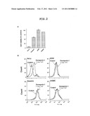 |
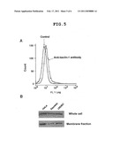 |  |
 |  |
 |  |
 |  |
 |  |
 | 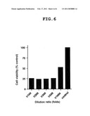 |
 |  |
| Similar patent applications: | |
| Date | Title |
|---|---|
| 2012-07-12 | Chemosensory receptor ligand-based therapies |
| 2012-09-27 | Non-aqueous, single tube dentrifice whitening compositions, methods of use and manufacture thereof |
| 2012-08-09 | Epha2 t-cell epitopes and uses therefor |
| 2012-09-13 | Glp-1 receptor agonist compounds for sleep enhancement |
| 2012-09-20 | Process to obtain a homeopathic medicament and use thereof |
| New patent applications in this class: | |
| Date | Title |
|---|---|
| 2019-05-16 | Antibodies, uses thereof and conjugates thereof |
| 2019-05-16 | Monoclonal antibodies that specifically recognize canine dla-dr antigen and their uses |
| 2018-01-25 | Anti-ceacam5 antibodies and uses thereof |
| 2018-01-25 | Tnfrsf-binding agents and uses thereof |
| 2018-01-25 | Antiproliferative compounds and methods of use thereof |
| Top Inventors for class "Drug, bio-affecting and body treating compositions" | |
| Rank | Inventor's name |
|---|---|
| 1 | David M. Goldenberg |
| 2 | Hy Si Bui |
| 3 | Lowell L. Wood, Jr. |
| 4 | Roderick A. Hyde |
| 5 | Yat Sun Or |