Patent application title: TUMOUR-CELL-FIXING CELLS
Inventors:
Massoud Mirshahi (Saint Gratien, FR)
Loic Vincent (Evry, FR)
Pezhman Mirshahi (Saint Gratien, FR)
Jeannette Soria (Taverny, FR)
Jean-Pierre Marie (Sevres, FR)
Arash Rafii Tabrizi (Cachan, FR)
Assignees:
Université Pierre et Marie Curie (Paris VI)
IPC8 Class: AC12Q102FI
USPC Class:
435 29
Class name: Chemistry: molecular biology and microbiology measuring or testing process involving enzymes or micro-organisms; composition or test strip therefore; processes of forming such composition or test strip involving viable micro-organism
Publication date: 2010-08-19
Patent application number: 20100209959
Inventors list |
Agents list |
Assignees list |
List by place |
Classification tree browser |
Top 100 Inventors |
Top 100 Agents |
Top 100 Assignees |
Usenet FAQ Index |
Documents |
Other FAQs |
Patent application title: TUMOUR-CELL-FIXING CELLS
Inventors:
Jeannette Soria
Massoud Mirshahi
Loic Vincent
Pezhman Mirshahi
Jean-Pierre Marie
Arash Rafii Tabrizi
Agents:
YOUNG & THOMPSON
Assignees:
Origin: ALEXANDRIA, VA US
IPC8 Class: AC12Q102FI
USPC Class:
Publication date: 08/19/2010
Patent application number: 20100209959
Abstract:
The present invention relates to an isolated cell capable of fixing tumour
cells, expressing the CD10 protein and expressing at least one MDR
protein, and to the use of this cell for screening for anti-tumour
compounds.Claims:
1-14. (canceled)
15. Isolated cell capable of fixing tumour cells, which cell expresses the CD10 protein and at least one MDR protein.
16. Isolated cell according to claim 15, wherein the MDR protein is selected from the group consisting in the LRP protein (Lung Resistance Protein), the MDR1 protein, the MRP1 protein, the MRP2 protein, the MRP3 protein, the MRP5 protein and the MXR protein.
17. Isolated cell according to claim 15, likely to exhibit or which exhibits pseudopods.
18. Isolated cell according to claim 15, derived from:the differentiation of stem cells or of mononucleated cells of bone marrow; orcells of an effusion of a patient suffering from a cancer.
19. Isolated cell according to claim 15, which cell has been immortalized.
20. Isolated cell according to claim 15, derived from a cell culture deposited on 20 Jun. 2006 under the Budapest Treaty at the CNCM under number I-3627.
21. Method for obtaining a cell capable of fixing tumour cells, said method comprising the following steps:a) cultivating bone marrow mononucleated cells or bone marrow stem cells for a period of time and in a medium suitable for cell differentiation;b) optionally removing the monocytes from the cells cultivated in step a) when the cultivated cells are bone marrow mononucleated cells;c) incubating the cells obtained in step a) and optionally in step b) with tumour cells, removing the unfixed tumour cells, and recovering the cells to which the tumour cells have become fixed.
22. Method for obtaining a cell capable of fixing tumour cells, said method comprising the following steps:a) cultivating cells taken from an ascitic fluid or from a pleural fluid of a patient suffering from a cancer;b) optionally removing the monocytes from the cells cultivated in step a);c) incubating the cells obtained in step b) with tumour cells, removing the unfixed tumour cells, and recovering the cells to which the tumour cells have become fixed; ord) depositing the cells obtained in step b) on a solid support and recovering the cells that adhere the most rapidly to said support.
23. Cell likely to be obtained by a method as defined in claim 21.
24. Method of screening for anti-tumour compounds, wherein isolated cells as defined in claim 15 are used.
25. Method of screening for anti-tumour compounds, which comprises:contacting isolated cells as defined in claim 15 with compounds to be screened;determining the cell growth and the cell death of the cells contacted with the compounds to be screened; andselecting the compounds that induce a decrease in the cell growth or an increase in the cell death of the cells with which they have been contacted, relative to identical cells that have not been contacted with the compounds to be screened.
26. Method of screening for anti-tumour compounds, which comprises:contacting a co-culture of isolated cells as defined in claim 15 and tumour cells with compounds to be screened;determining the cell growth and the cell death of the tumour cells of the co-culture contacted with the compounds to be screened; andselecting the compounds that induce a decrease in the cell growth or an increase in the cell death of the tumour cells of the co-culture, relative to tumour cells in co-culture with the isolated cells that have not been contacted with the compounds to be screened.
27. Method of screening for anti-tumour compounds, which comprises:contacting compounds to be screened with isolated cells as defined in claim 15, and with tumour cells;determining the amount of tumour cells fixed by the isolated cells; andselecting the compounds that induce a decrease in the amount of tumour cells fixed by the isolated cells relative to the amount of tumour cells fixed by the isolated cells, in the absence of the compounds to be screened.
28. In vitro method of diagnosing a cancer, in which the presence of isolated cells as defined in claim 15 in a sample derived from a tissue suspected of containing a tumour is determined, the presence of said isolated cells being indicative of the presence of a tumour.
Description:
[0001]The present invention relates to a novel type of cells which fix
tumour cells in vivo and in vitro, and to the use of these cells in the
screening of compounds for anti-tumour action.
[0002]Many cytotoxic molecules capable of killing tumour cells have been identified by the pharmaceutical industry.
[0003]Nevertheless, it has been found that, in oncological therapy, the efficacy of the molecules is reduced by a resistance phenomenon. This resistance to anti-tumour agents continues to be a major obstacle to the success of anti-cancer treatments.
[0004]Several cellular events have been proposed as resistance factors. They include efflux mechanisms (for example via MDR channels; multi drug resistance), inactivation of the anti-tumour agents (for example by a resistance to antipyrimidic, antimetabolic, etc. agents), mutation of the targets of the anti-tumour agents (for example mutation of the topoisomerase), resistance to apoptosis (for example due to mutation of p53, overexpression of bcl-2, etc.).
[0005]It has also been suggested that tumour stem cells would be present in solid tumours. Such tumour stem cells would be more resistant to anti-cancer treatments and would be the origin of resistance and recurrence phenomena (Dean et al. (2005) Nature Rev. Cancer 5:275-284).
[0006]No other resistance factor extrinsic to the tumour cell has been identified to date.
[0007]The inventors have now succeeded in isolating a novel type of cells which fix and protect tumour cells and which constitute one of the causes of the resistance of tumours to treatments.
Tumour Cell Fixing Cell
[0008]The present invention relates to an isolated cell which is capable of fixing tumour cells, and which expresses the CD10 protein.
[0009]This cell which is capable of fixing tumour cells has been named "Hospicell" by the inventors and is also referred to as "fixing cell" or "protecting cell" in the present description.
[0010]The ability of an isolated cell according to the invention to fix tumour cells can be demonstrated by numerous techniques which are well known to the person skilled in the art. Mention may thus be made of a method in which (i) cells of the invention are fixed to the walls of a vessel, (ii) tumour cells expressing a fluorescent protein are added to the vessel, (iii) the vessel is washed, and (iv) the fluorescence emitted by the vessel is measured and compared with that emitted by a control vessel to the walls of which cells of the invention have been fixed but to which no tumour cell has been added. The ability to fix tumour cells is proven when the value of the fluorescence measured for the tested cells is greater than that of the cells of the control vessel. Such a method is illustrated in the following Examples. Mention may also be made of methods which use direct observations by optical or electron microscopy, as illustrated in Example 4 and in FIGS. 1 to 3, in which clusters of tumour cells in membrane contact with cells of the invention are observed.
[0011]The cells of the invention are capable of fixing tumour cells, such as, for example, leukaemia cells, breast cancer cells or ovarian cancer cells. These cells which are generally large in size (namely about fifty times the size of a tumour cell), are preferably capable of fixing up to 200 tumour cells simultaneously.
[0012]The "CD10 protein" (reference in the international classification of enzymes: E.C. 3.4.24.11) is also known by the names Neprilysin, Neutral endopeptidase 24.11, or Common Acute Lymphocytic Leukemia Antigen (CALLA). It is a membrane metallo-peptidase which preferably cleaves polypeptides having fewer than 30 amino acids between basic residues. It has been described as being present at the surface of a limited number of normal or malignant lymphoid progenitors and on some epithelial cells, mainly in the region of the kidney. It is namely described in Shipp et al. (1988) Proc. Natl. Acad. Sci. U.S.A. 85:4819-4823 and under reference P08473 in the UniProtKB database. By way of example, the CD10 protein is represented by SEQ ID NO: 1.
[0013]Expression of the CD10 protein may be determined either by detection of its mRNA, or of the RNA precursors thereof, or by detection of the protein itself. Detection of the mRNA of CD10, or of its precursors, can be carried out by various techniques which are well known to the person skilled in the art, such as RT-PCR, for example. Detection of the protein itself can also be carried out by various techniques which are well known to the person skilled in the art. Preferably, detection of the protein itself makes use of specific ligands of CD10, such as antibodies, which can be employed in techniques such as flow cytometry, immunohistochemistry or immunocytochemistry.
[0014]Preferably, the level of expression of the CD10 protein is such that it is considered to be strong by the anatomopathologist of the art when it is evaluated, according to the conventional techniques of anatomopathology, on samples included in paraffin with the aid of peroxidase-labelled anti-CD10 antibodies.
[0015]In a preferred embodiment of the invention, the cell as defined above expresses an MDR protein.
[0016]The expression "MDR protein" denotes a membrane protein which effects active transport of at least one drug and is capable of thus imparting multi-drug resistance to the cell which expresses it. The MDR proteins are notably described in Stavrovskaya (1999) Biochemistry (Moscow) 65:95-106. Preferably, the MDR protein is a ABC type protein which transports drugs from the cytoplasm of the cell which expresses it to the extracellular medium. ABC proteins are characterized in that they comprise at least one adenosine triphosphate (ATP) binding motif called ATP Binding Cassette (ABC) and are well known to the person skilled in the art. ABC type proteins are notably described in Dean et al. (2001) J. Lipid Res. 42:1007-1017 and Szakacs et al. (2006) Nature Reviews Drug Discovery 5:219-234. More preferably, the MDR proteins of ABC type are selected from the proteins of the subfamilies ABCB, ABCC and ABCG.
[0017]Preferably, the MDR protein is selected from the group constituted by the LRP protein (Lung Resistance Protein, also called Major Vault Protein (MVP)), the MDR1 protein (MultiDrug Resistance 1, also called ABCB1, or glycoprotein P (Pgp)), the MRP1 protein (Multidrug Resistance-associated Protein 1, also called ABCC1), the MRP2 protein (also called ABCC2), the MRP3 protein (also called ABCC3), the MRP5 protein (also called ABCC5), and the MXR protein (MitoXantrone Resistance protein, also called ABCG2 or Breast Cancer Resistance Protein (BCRP)). More preferably, the MDR protein is selected from the group consisting of the LRP protein, the MDR1 protein, the MRP1 protein, and the MXR protein. Particularly preferably, the cell as defined above expresses the LRP protein and the MDR1 protein at the same time. Yet more preferably, the cell as defined above expresses the LRP protein, the MDR1 protein, the MRP1 protein and the MXR protein at the same time.
[0018]The LRP protein is notably represented by SEQ ID NO: 2.
[0019]The MDR1 protein is notably represented by SEQ ID NO: 3.
[0020]The MXR protein is notably represented by SEQ ID NO: 4.
[0021]The MRP1 protein is notably represented by SEQ ID NO: 5.
[0022]Expression of the MDR protein can be determined either by detection of its mRNA, or of the RNA precursors thereof, or by detection of the protein itself. Detection of the mRNA of the MDR protein, or of its precursors, can be carried out by various techniques which are well known to the person skilled in the art, such as RT-PCR, for example. Detection of the protein itself can also be carried out by various techniques which are well known to the person skilled in the art. Preferably, detection of the protein itself makes use of specific ligands of the MDR protein, such as antibodies, which can be employed in techniques such as flow cytometry, immunohistochemistry or immunocytochemistry.
[0023]When measured by flow cytometry, the level of expression of a MDR protein can be evaluated as follows:
(i) determining the mean fluorescence intensity measured for the cells of the invention with the aid of an antibody directed against the MDR protein;(ii) determining, under the same conditions as in (i), the mean fluorescence intensity and the fluorescence standard deviation, measured for the cells of the invention with the aid of a control antibody which is of the same isotype as the antibody directed against the MDR protein but does not recognize any antigen of the cells of the invention;(iii) relating the mean fluorescence intensity determined in (i) to that determined in (ii);(iv) applying the Kolmorogov-Smirnov test to the ratio determined in (iii).
[0024]The Kolmorogov-Smirnov test, which is well known to the person skilled in the art, gives a value, conventionally denoted D, which varies from 0 to 1 and enables measuring a difference between two distributions (here the mean fluorescence intensity measured in (i) and that measured in (ii)). If D=0, the two distributions are superposed; if D=1, the two distributions are completely separate. Two distributions are considered to be significantly distinct when the D value is greater than or equal to 0.15, preferably greater than or equal to 0.2. By way of example, Legrand et al. (2001) Blood 97:502-508 describe the application of the Kolmorogov-Smirnov test to the evaluation of the expression of the Pgp protein.
[0025]Accordingly, when measured as indicated above with the aid of the Kolmorogov-Smirnov test, the level of expression of the MDR proteins by the cells of the invention is preferably greater than 0.15, more preferably greater than 0.2.
[0026]Preferably, the mean level of expression, by the cells of the invention, given as indicated above with the aid of the Kolmorogov-Smirnov test is: [0027]0.34 for MDR1; [0028]0.57 for MRP1; [0029]0.28 for MRP2; [0030]0.41 for MRP3; [0031]0.70 for MXR; and/or [0032]0.78 for LRP.
[0033]Still preferably, the level of expression of the MDR protein is such that it effectively imparts to the cell which expresses it the resistance to a drug that the protein is capable of imparting, when the cell is brought into contact with an amount of drug which is normally toxic for a cell of the same type that does not express the MDR protein. This will be thus herein referred to as the effective level of expression for imparting resistance.
[0034]Preferably, the isolated cell as defined above is capable of transferring one or more copies of the MDR protein that it expresses to the tumour cells that it fixes, especially by trogocytosis.
[0035]"Trogocytosis" refers to the phenomenon of transfer of molecules from one cell to another which is described especially by Joly & Hudrisier (2003) Nature Immunol. 4:815.
[0036]The transfer of MDR protein from an isolated cell of the invention to a tumour cell can be demonstrated in various ways which are well known to the person skilled in the art. By way of example, it is notably possible to carry out an immunodetection of MDR protein expressed by tumour cells before and after they have been brought into contact with cells of the invention.
[0037]Preferably, the isolated cell as defined above does not express the following markers: cytokeratin and EMA (Epithelial Membrane Antigen) (specific to epithelial cell lines), vimentin (specific to mesenchymatous cell lines), CD45 (marker of haematopoietic cells, such as granulocytes, monocytes and B and T lymphocytes), CD20 (specific to B lymphocytes), CD3 (specific to T lymphocytes), CD68 (specific to macrophages and histiocytes), CD34 (specific to bone marrow stem cells), S100 protein (specific to melanocytes), myeloperoxidase (specific to polynuclear lines).
[0038]In a preferred embodiment of the invention, the isolated cell as defined above is likely to exhibit pseudopods and/or filopods.
[0039]"Pseudopods" or "filopods" refer to the evaginations of the cell plasma membrane. Pseudopods can be visualized, for example, by confocal, optical or electron microscopy.
[0040]In another preferred embodiment of the invention, the isolated cell as defined above is derived from: [0041]the differentiation of stem cells or of mononucleated cells of bone marrow; or [0042]cells from an effusion of a patient suffering from a cancer.
[0043]Preferably, as understood here, the bone marrow stem cells are characterized by the expression of the CD34+ marker and/or the CD133+ marker.
[0044]An "effusion" is understood as being a biological fluid which accumulates in a cavity or tissue which does not normally contain any. There may be mentioned by way of example an ascitic fluid, for example from a patient suffering from a ovary or pancreas cancer, or a pleural fluid, for example from a patient suffering from breast cancer.
[0045]Preferably, the isolated cell as defined above is not derived from cancer cells taken from an individual.
[0046]In another preferred embodiment of the invention, the isolated cell as defined above has been immortalized.
[0047]The isolated cell as defined above can be immortalized by any technique known to the person skilled in the art. There may be mentioned by way of example, without implying any limitation, the use of the SV 40 virus T antigen, the use of the EIA region of the adenovirus 2 genome, the use of oncogens such as c-myc or Ha-ras, or the use of human telomerase reverse transcriptase (hTRT) or of a sequence which activates the endogenous hTRT gene.
[0048]According to a preferred embodiment of the present invention, immortalization is performed using the SV 40 virus T antigen.
[0049]Preferably, the isolated cell as defined above is derived from a cell culture deposited on 20 Jun. 2006 under the Budapest Treaty at the CNCM under number I-3627.
Obtaining Tumour Cell Fixing Cells
[0050]The present invention also relates to a method for obtaining a cell capable of fixing tumour cells, said method comprising the following steps of: [0051]a) cultivating bone marrow mononucleated cells or bone marrow stem cells for a period of time and in a medium suitable for cell differentiation; [0052]b) optionally removing the monocytes from the cells obtained in step a) when the cultivated cells are bone marrow mononucleated cells; [0053]c) incubating the cells obtained in step a) and optionally in step b) with tumour cells, removing the unfixed tumour cells, and recovering the cells to which the tumour cells have become fixed.
[0054]The bone marrow mononucleated cells can especially be isolated from bone marrow by Ficoll gradient centrifugation. Before their culture in step a), it is optionally possible to remove the monocytes from the bone marrow mononucleated cells. With regard to bone marrow stem cells, especially CD34+ and/or CD133+, it is, for example, possible to isolate them by immunofixation with the aid of anti-CD34 or anti-CD133 antibodies.
[0055]There can be used in step a) a culture medium for stem cells or mononucleated cells of bone marrow which is well known to the person skilled in the art and which contains growth and differentiation factors. This culture is preferably carried out in bottles which have previously been coated with 0.2% gelatin. There may be mentioned as examples, without implying any limitation, the media MV2, HEM (HEPES buffered Eagles medium), DMEM (Dulbecco's modified Eagles medium), GMEM (Glasgow modification of Eagles medium), F-12, etc. Preferably, the medium MV2 (ECBM MV2, Promocell, Heidelberg, Germany) may be used. The growth factors may be selected, without implying any limitation, from fibroblast growth factor (FGF), epidermal growth factor (EGF), insulin-like growth factor (IGF), vascular endothelial growth factor (VEGF), transforming growth factor (TGF-β), stem cell growth factor (SCGF), platelet-derived growth factor (PDGF) or derivatives thereof, combinations of those factors being preferably used. There are optionally added to the culture medium supplements required for cell metabolism, such as amino acids, vitamins such as ascorbic acid, minerals and proteins such as transferrin and derivatives thereof. The culture medium may contain foetal calf serum, chicken serum or equine serum. The culture medium may also contain antibiotics in order to avoid contamination with yeasts, bacteria or fungi, such as penicillin, streptomycin, gentamicin and derivatives thereof. Preferably, after about 6 days' culture, the non-adherent cells are removed and the cells are reincubated in the same medium.
[0056]The cells are then cultivated for a period of preferably about 4 weeks, until the cells differentiate into endothelial cells, smooth muscle cells, fibroblasts and other cells, including the desired cells.
[0057]In step c), the cells cultivated in step a) are incubated with tumour cells. This step is generally carried out by recovering the adherent cells cultivated in step a) and incubating them with tumour cells suspended in a suitable medium, such as RPMI or DMEM medium. The tumour cells used may be any type of tumour cells. According to a preferred embodiment of the present invention, the tumour cells are HL60 cells (human leukaemia line). The incubation period of the cultivated cells and of the tumour cells varies from about 30 minutes to about 4 hours, preferably about 2 hours. The incubation temperature is preferably 37° C.
[0058]In step c), the unfixed tumour cells are generally removed by rinsing with culture medium (optionally the same medium, such as RPMI medium) or with any type of wash solution well known to the person skilled in the art.
[0059]The cells to which the tumour cells have become fixed are then recovered, preferably by very brief centrifugation at from 3000 rpm to 6000 rpm.
[0060]In an optional subsequent step, the tumour cells are detached from the agglomerate formed by said cell to which residual tumour cells are fixed, by enzymatic or non-enzymatic treatment (trypsin, Accutase® or EDTA 2 mM, followed by very vigorous stirring and repeated washing with a buffer such as PBS).
[0061]By way of example to remove the monocytes in step b) or before step a), the mononucleated cells are dispersed on a solid support, such as Petri dishes, or cultured on a solid support (for example gelatin-coated plates) in a suitable medium as defined above. After incubation for about 15 to 30 minutes, the monocytes adhere to the support and the cells that have not adhered yet are collected.
[0062]Optionally, in order to confirm that the recovered cells are the fixing cells of interest, they are incubated with tumour cells, preferably HL60 cells (106 HL60 cells/2×105 adherent cells) and the ability of the tumour cells to become fixed to the fixing cells is observed.
[0063]The present invention relates also to a method for obtaining a cell capable of fixing tumour cells, said method comprising the following steps of: [0064]a) cultivating cells taken from an effusion of a patient suffering from a cancer; [0065]b) optionally removing the monocytes from the cells obtained in step a); [0066]c) incubating the cells obtained in step a) or b) with tumour cells, removing the unfixed tumour cells, and recovering the cells to which the tumour cells have become fixed; or [0067]d) depositing the cells obtained in step a) or b) on a solid support and recovering the cells that adhere the most rapidly to said support.
[0068]Preferably, before being cultured in step a), the cells derived from an effusion are isolated. Still preferably, the cells derived from an effusion are freed of the monocytes prior to being cultured in step a).
[0069]Moreover, in step a), the culture medium used may be any cell culture medium known to the person skilled in the art. There may be mentioned by way of example MV2, RPMI, Iscove's MDM or DMEM. The culture medium may optionally contain growth factors, such as those mentioned above, supplements required for cell metabolism, such as amino acids, vitamins such as ascorbic acid, minerals and proteins such as transferrin and their derivatives. The culture medium may optionally contain foetal calf serum, chicken serum or equine serum. The culture medium may also contain antibiotics in order to avoid contamination with yeasts, bacteria or fungi, such as penicillin, streptomycin, gentamicin and their derivatives. Under these conditions, the fixing cells show as early as the first days of culture (1 to 4 days).
[0070]Steps b) and c), and the removal of the monocytes prior to step a), may be carried out as described above for the bone marrow mononucleated cells or the bone marrow stem cells.
[0071]Preferably, in step d), the cells, previously freed of the monocytes, are deposited on a solid support. The cells that have become fixed to said support are then treated, preferably with Accutase®, and the cells that become detached the most rapidly from the support are recovered. Preferably, the cells that become detached within 5 minutes are recovered.
[0072]The support to which the cells become fixed can be any type of solid support well known to the person skilled in the art. As examples of solid supports there may be mentioned glass, plastics, metals, resins or other suitable solid supports to which the cells can be fixed. The term "solid support" also includes materials considered to be semi-solid supports. The solid support can have any suitable form, such as a bead or microparticle, a tube, a Petri dish, a microscope slide, etc.
[0073]Optionally, in order to confirm that the recovered cells are the fixing cells of interest, they are incubated with tumour cells, preferably HL60 cells (106 HL60 cells/2×105 adherent cells) and the ability of the tumour cells to become fixed to the fixing cells is observed.
[0074]Optionally, at the end of the methods above, the resulting cells can be immortalized, especially as described above.
[0075]The invention also relates to a cell likely to be obtained by one of the methods described above.
[0076]The cell cultures produced from the isolated cell as defined above, or from the cell likely to be obtained by one of the methods described above, also form part of the invention.
Screening
[0077]The present invention relates to the use of isolated cells as defined above, or of cells likely to be obtained by one of the methods described above, for screening for anti-tumour compounds.
[0078]"Anti-tumour compounds" are here understood as being any compound which enables tumour progression to be prevented and/or slowed. In particular, the anti-tumour compounds are compounds that induce or facilitate, directly or indirectly, the death of the tumour cells. More particularly, the anti-tumour compounds according to the invention can induce or facilitate the death of the cells protecting the tumour cells in vivo.
[0079]The present invention thus relates to a method of screening for anti-tumour compounds, in which: [0080]isolated cells as defined above, or cells likely to be obtained by one of the methods described above, are contacted with compounds to be screened; [0081]the cell growth and the cell death of the cells contacted with the compounds to be screened are determined; [0082]the compounds that induce a decrease in the cell growth or an increase in the cell death of the cells with which they have been contacted, relative to identical cells that have not been contacted with the compounds to be screened, are selected.
[0083]Advantageously, the compounds screened by this method specifically target the cells according to the invention.
[0084]The present invention also relates to a method of screening for anti-tumour compounds in which: [0085]a co-culture of isolated cells as defined above, or of cells likely to be obtained by one of the methods described above, and tumour cells is contacted with compounds to be screened; [0086]the cell growth and the cell death of the tumour cells of the co-culture contacted with the compounds to be screened are determined; [0087]the compounds that induce a decrease in the cell growth or an increase in the cell death of the tumour cells of the co-culture, relative to tumour cells in co-culture with isolated cells as defined above, or with cells likely to be obtained by one of the methods described above, that have not been contacted with the compounds to be screened, are selected.
[0088]Advantageously, this method permits the selection of the compounds having an anti-tumour action which is not hindered by the resistance to anti-tumour agents provided by the cells of the invention.
[0089]The present invention also relates to a method of screening for anti-tumour compounds in which: [0090]compounds to be screened are contacted with isolated cells as defined above, or with cells likely to be obtained by one of the methods described above, and with tumour cells; [0091]the amount of tumour cells fixed by the isolated cells as defined above, or by the cells likely to be obtained by one of the methods described above, is determined; [0092]the compounds that induce a decrease in the amount of tumour cells fixed by the isolated cells as defined above, or by the cells likely to be obtained by one of the methods described above, relative to the amount of tumour cells fixed by the isolated cells as defined above, or by the cells likely to be obtained by one of the methods described above, in the absence of the compounds to be screened, are selected.
[0093]This method advantageously permits the selection of the compounds which inhibit the fixing of the tumour cells by the cells of the invention and therefore the protection imparted by the cells of the invention to the tumour cells. These compounds therefore increase the sensitivity of the tumour cells to the anti-tumour compounds.
[0094]"Cell death" is understood as being apoptosis, necrosis or any other mechanism that induces the death of the cell. According to a preferred embodiment, the ability of the candidate compound to induce cell death by apoptosis is determined. Any technique well known to the person skilled in the art can be used to measure cell death. The following techniques may be mentioned by way of example, without implying any limitation: labelling with annexin V, use of trypan blue, use of propidium iodide, the TUNEL (Transferase dUTP Nick End Labeling) assay, evaluation of the products of DNA degradation, measurement of caspases (quantitative evaluation and evaluation by activity), etc.
[0095]In the above screening methods, the tumour cells used may be any tumour cell. According to a preferred embodiment, the tumour cells are HL60 cells (human leukaemia line) or MDA-MB 231 cells (human breast cancer line) or the patient's own cells (for example ovarian cancer).
[0096]The compound to be screened may be any compound of natural or synthetic origin, whether it is already being marketed as a chemotherapeutic agent or is in the course of development or characterization. It may be a mixture of several identified or unidentified molecules, such as for example an extract of animal or plant origin.
[0097]The cells according to the invention may readily be employed in high throughput screening protocols (HTS) in order to optimize the current methods of finding candidate compounds that are effective for anti-tumour therapy in general.
[0098]In addition, the cells of the invention may also be used for testing the efficacy of candidate compounds for an anti-tumour therapy for a given individual, in order to propose the most appropriate therapy for each individual affected by a cancer. Within this context, it is advantageous to test a panel of chemotherapeutic agents as candidate compounds, with tumour cells taken from the patient himself, in the presence of the fixing cells of the invention.
[0099]In a particular embodiment, the fixing cells used in the screening test are taken from the patient himself.
Diagnostics
[0100]The present invention also relates to an in vitro method for diagnosing a cancer, in which the presence of isolated cells as defined above in a sample derived from a tissue suspected of containing a tumour is determined, the presence of isolated cells as defined above being indicative of the presence of a tumour.
[0101]Indeed, as has been shown by the inventors, the presence of cells according to the invention in the tissue generally implies the presence of tumour cells close by.
[0102]The following examples and figures illustrate the invention without limiting the scope thereof.
LEGEND OF THE FIGURES
[0103]FIGS. 1 and 2 are optical microscopy images (objective 20) of hospicells (obtained by differentiation of CD34+ bone marrow stem cells) which have been contacted with HL60 cells, after incubation for 4 hours (FIG. 1) and 36 hours (FIG. 2).
[0104]FIG. 3 is an optical microscopy image (objective 20) of hospicells of the ascitic fluid of a patient suffering from ovarian cancer, after addition of HL60 cells to the fluid.
[0105]FIG. 4 is an electron microscopy image of an MDA cell adhering to a hospicell.
[0106]FIGS. 5 and 6 are graphs showing the influence of the fixing cells on the sensitivity of HL60 cells to aracytin (AraC) or daunorubicin (DNR) (FIG. 5) and of the fixing cells ("hospicells") to those drugs (FIG. 6).
[0107]FIG. 7 is a graph showing the sensitivity of the fixing cells to various known agents used in anti-tumour therapy.
[0108]FIG. 8 shows the amount of fluorescence emitted by OVCAR3 cells (Y-axis, arbitrary units) that express GFP, which cells were cultivated with hospicells (first column), alone (second column), with OVCAR3 cells that do not express GFP (third column), with fibroblasts (fourth column) or with HBMECs (fifth column), in the absence or in the presence of carboplatin and/or paclitaxel.
[0109]FIG. 9 shows the amount of fluorescence emitted by OVCAR3 cells (Y-axis, arbitrary units) that express GFP, which cells were cultivated with hospicells (first column), alone (second column), with hospicells in a transwell system (third column), in the absence or in the presence of carboplatin and/or paclitaxel.
EXAMPLES
Example 1
Obtaining Hospicells from Bone Marrow Stem Cells
[0110]CD34+ cells were isolated from a normal (or pathological) bone marrow sample by density gradient centrifugation in Ficoll-400. The cells so isolated were distributed in a culture bottle coated with 0.2% gelatin and were cultivated in an MV2 medium (ECBM MV2, Promocell, Heidelberg, Germany) supplemented with amphotericin B 50 ng/ml, gentamicin 50 μg/ml, ascorbic acid 1 μg/ml, human fibroblast growth factor (h-FGF) 10 ng/ml, human epidermal growth factor (h-EGF) 5 ng/ml, Long R3 IGF-1 (insulin-like growth factor) 20 ng/ml, human vascular endothelial cells growth factor (h-VEGF) 10 ng/ml and 5% foetal calf serum.
[0111]After 6 days' culture, the non-adherent cells were removed and the adherent cells were cultured for a further 3 weeks in fresh selective medium having the same composition (as detailed above).
[0112]The adherent cells so obtained were washed with RPMI and then detached by rapid incubation with Accutase®. The cells so detached were washed with RPMI and then [0113]either resuspended in RPMI containing glutamine and antibiotics and then incubated with immortalized HL60 cells of leukaemic origin. After incubation for 120 minutes at 4° C. with gentle stirring, the cell suspension was centrifuged at 6000 rpm for a few seconds ("centrifugation pulse"), the hospicells that had fixed the HL60 cells being found in the centrifugation pellet. The cells that had settled in the bottom of the tube were resuspended in a culture medium (RPMI+foetal calf serum+glutamine+antibiotics) and distributed in plates coated with 0.2% gelatin. [0114]or placed on a plate 6 well-plate coated with gelatin and containing RPMI medium containing foetal calf serum, glutamine and antibiotics. After incubation in a cell incubator, immortalized HL60 tumour cells of leukaemic origin were added to the wells.
[0115]After incubation in the cell incubator, the cells were washed with RPMI, and then Accutase® was added. The cells that become detached within 5 minutes are cells that fix the HL60 cells, while the cells that become detached within 10 minutes and 15 minutes are cells that do not fix the HL60 cells. The cells that become detached within 5 minutes were recovered and were distributed on another 6 well-plate coated with gelatin. HL60 cells were added to one of the wells and, after incubation, the cells were washed and the operation of fixing the HL60 cells was repeated. Finally, Accutase® was added to the wells and the cells that became detached within 5 minutes were recovered and cultivated in complete RPMI medium. The cells obtained under these conditions are cells of the invention or "Hospicells".
Example 2
Obtaining Hospicells from the Ascitic Fluid of a Patient Suffering from Ovarian Cancer
[0116]The ascitic fluid of a patient suffering from ovarian cancer was taken with the aid of a biopsy trocar. The mononucleated cells of the ascites were isolated by Ficoll gradient centrifugation. The mononucleated cells so isolated were distributed in the wells of a culture plate. After incubation for 30 minutes, the monocytes adhere to the plastic and the cells that have not yet adhered were collected and placed in a gelatin-coated culture plate. The hospicells were then isolated as described in Example 1.
[0117]It was also possible to obtain the hospicells directly from the ascites by placing in a gelatin-coated culture plate a small cluster of cells present in the ascites suspended in RPMI enriched with foetal calf serum, glutamine and antibiotics.
[0118]Alternatively, the hospicells were obtained from cell aggregates present in the ascitic fluid obtained from patients suffering from stage III ovarian cancer. Briefly, the ascitic fluid was centrifuged in order to obtain a cell pellet. The cell pellet was freed of the lymphocytes and erythrocytes by Ficoll gradient centrifugation, and then the aggregates of hospicells and ovarian cancer cells were separated by dilution. The hospicells were then detached from the ovarian cancer cells by tryptic digestion.
Example 3
Phenotypical Characterization of the Hospicells
[0119]It was not possible to observe by immunohistochemistry the expression by the hospicells of the following conventional membrane markers: cytokeratin (antibody KL1, Beckman Coulter) EMA (antibody E29, Dako) (specific to epithelial cell lines), vimentin (antibody V9, Beckman Coulter) (specific to mesenchymatous cell lines), CD45 (antibody 2b11 and PD7/26, Dako) (specific to haematopoietic cells), CD20 (antibody L26, Dako) (specific to B lymphocytes), CD3 (antibody SP7, Neomarkers) (specific to T lymphocytes), CD68 (antibody KP1 and PG-M1, Dako) (specific to macrophages and histiocytes), CD34 (antibody OBend10, Dako) (specific to blood stem cells), S100 protein (polyclonal antibody, Dako) (specific to melanocytes), myeloperoxidase (polyclonal antibody, Dako) (specific to polynuclear lines).
[0120]However, labelling of CD9 protein (antibody 56C6, Novocastra) and CD10 protein (Novocastra) was found to be positive.
[0121]Briefly, immunohistochemistry was carried out on paraffin sections having a thickness of 4 μm. A technique of preliminary recovery of the antigen based on heating in an EDTA buffer (pH 8) was employed prior to incubation with the antibody. The antibody at the appropriate dilution was incubated for 30 minutes and then revealed with the aid of a streptavidin-biotin complex. The sections were then counterstained with hematoxylin. The technique as a whole was carried out automatically on an Autostainer system (Dako).
Example 4
Demonstration of the Fixing of the Cancer Cells to the Hospicells
[0122]a) To Hospicells Obtained from Bone Marrow Stem Cells
[0123]Hospicells obtained according to the protocol of Example 1 were cultured in RPMI medium in the presence of HL60 leukaemia cells.
[0124]The HL60 cells adhere to the hospicells and form cell clusters in the culture medium. After 4 hours, the fixing of 4 to 8 cells to the fixing cells was observed (FIG. 1). After 36 hours, nodules of malignant cells have formed around the hospicells (FIG. 2).
b) To the Hospicells Present in the Ascitic Fluid of a Patient Suffering from Ovarian Cancer
[0125]Ascitic fluid was taken from a patient suffering from ovarian cancer using a biopsy trocar. It was also possible to obtain the hospicells directly from the ascites by placing in a gelatin-coated culture plate a small cluster of cells present in the ascites suspended in RPMI enriched with foetal calf serum, glutamine and antibiotics. The cell clusters of fixing cells already present in the ascitic fluid that have fixed tumour cells were observed by optical microscopy.
[0126]A portion of that fluid was cultured in RPMI medium enriched with foetal calf serum, glutamine and in the presence of HL60 cells for 1 day. FIG. 3 shows the joint fixing of the ovarian cancer cells and the added HL60 cells to the fixing cells.
[0127]In addition, the hospicells obtained from cell aggregates of ascitic fluid exhibited particular structures under optical microscopy. Those cells indeed develop long pseudopods, which form a type of "cell thread". Furthermore, interaction of the hospicells with ovarian cancer cells is visible by confocal microscopy. The hospicells appear therein as cells of large size which are able to establish interactions with several cancer cells at the same time, thus providing a hammock, as it were, for the cancer cells.
[0128]Moreover, an immunohistochemical experiment carried out on peritoneal biopsies of patients suffering from ovarian cancer with the aid of the CD10 marker shows the presence of hospicells around cancerous cell aggregates, the hospicells forming a thread around the aggregates.
[0129]Finally, analysis of the cell aggregates of ascitic fluid by electron microscopy shows that they are formed of hospicells interacting with cancer cells (FIG. 4). It is also possible to observe that the hospicells develop pseudopods, which increases their potential for interaction with cancer cells. More precise observations, carried out on primocultures of hospicells and of cancer cells, showed punctual areas of membrane fusion between the two cell types.
Example 5
Interaction Between the Hospicells and the HL60 Cells or the MDA-MB 231 Cells
[0130]The inventors have tried to demonstrate the mechanisms of action on which the interaction between the hospicells and the tumour cells is based.
a) Proteins Involved in the Fixing of HL60
[0131]HL60 cells were incubated in RPMI medium under 6 different conditions: [0132]HL60 cells alone (control) [0133]HL60 cells+anti-arrestin antibody (control antibody) [0134]HL60 cells+RGD (Arg-Gly-Asp, integrin consensus sequence, which partially prevents integrin dependent adhesion) [0135]HL60 cells+anti-CD11a antibody [0136]HL60 cells+anti-CD49d antibody [0137]HL60 cells+anti-CD11a antibody+anti-CD49d antibody [0138]HL60 cells+anti-CD11a antibody+anti-CD49d antibody+RGD
[0139]For each condition, HL60 cells were deposited separately and in identical amounts in 6 different wells containing complete RPMI medium and fixing cells which had adhered to the support. The ratio between tumour cells and hospicells was 5/1. After incubation for 24 hours at 37° C., the HL60 cells that had not adhered were recovered and counted. The percentage of HL60 cells adhering to the hospicells was then calculated for each condition.
[0140]The results show that incubating the HL60 cells beforehand with anti-CD11a or anti-CD49d antibodies or with RGD partially blocks the adhesion of the HL60 cells to the hospicells. Blocking is greater when the HL60 cells have been incubated with the anti-CD11a antibody and the anti-CD49d antibody simultaneously. These results suggest that the CD11a and CD49d integrins are involved in the adhesion between the HL60 cell and the hospicell.
b) Proteins Involved in the Fixing of MDA
[0141]MDA-MB231 cells which had previously been labelled with rhodamine were incubated in complete RPMI medium under 3 different conditions: [0142]MDA cells alone (control) [0143]MDA cells+anti-SDF1 antibody; SDF1 is the ligand of CXCR4 and is a cytokine promoting the formation of breast cancer metastases in the bone marrow [0144]MDA cells+anti-CXCR4 antibody.
[0145]MDA cells of each condition were deposited separately and in identical amounts (5 cancer cells per hospicell) in 3 different wells containing RPMI medium and hospicells which had adhered to the support. After incubation for 2 hours at 4° C., the MDA cells that had not adhered were removed, and the amount of cells that had adhered to the hospicells was determined for each condition by measuring the fluorescence with the aid of a plate fluorimeter (Victor fluorimeter).
[0146]The results show that incubating the MDA cells beforehand with anti-CXCR4 antibodies induces a marked decrease in the adhesion relative to the control.
[0147]These results suggest that CXCR4 modulates the expression of integrins at the surface of MDA tumour cells, which integrins are likely to play a role in the fixing of the tumour cells to the cells of the invention.
Example 6
Protective Effect of the Hospicells on the HL60 Cells Against Agents Used in Chemotherapy
[0148]Proliferation of HL60 Cells Fixed to Hospicells after Exposure to Aracytin (AraC) or Daunorubicin (DNR)
[0149]HL60 cells cultured with hospicells in RPMI medium were treated for 5 days with either aracytin (AraC) or daunorubicin (DNR).
[0150]After 5 days' treatment, the drug was removed and the cells were cultured again in fresh medium.
[0151]The number of living HL60 cells was measured throughout the experiment using the image analysis system.
[0152]The results show that the free HL60 cells die when they are treated with AraC or DNR and that they are no longer detectable when they are fixed to the hospicells. However, after the treatment is stopped, the free HL60 cells do not grow, whereas it is noted that living HL60 cells are identifiable on the hospicells. This effect of "regrowth" of the cells is greater with aracytin than with daunorubicin, the difference being linked to the toxicity of daunorubicin for the hospicells.
[0153]These results show that, following treatment with AraC and DNR, the free HL60 cells die, whereas some of the cells bound to the fixing cells remain alive and proliferate after the treatment is stopped.
[0154]An example of the evaluation of the living cells is shown in FIGS. 5 and 6.
Example 7
Sensitivity of Hospicells to Various Known Agents Used in Anti-Tumour Chemotherapy
[0155]Immortalized hospicells (M16) were incubated in RPMI medium with adriblastin, bleomycin, deticene, fluorouracil, navelbin, Taxotere or Leustatin for 1 to 3 days.
[0156]The cells were then detached with Accutase® and the number of cells in apoptosis was detected by annexin V. The percentage of apoptotic cells was then determined by flow cytometry (FIG. 7).
[0157]The results show that the hospicells are poorly sensitive to the action of numerous drugs.
Example 8
Interaction Between Hospicells and OVCAR3 Cells
[0158]The specificity of the interaction of hospicells with cancer cells was then demonstrated with the aid of cells of the ovarian cell line OVCAR3 (Manetta et al. (1988) Eur. J. Gynaecol. Oncol. 9:222-7). An adhesion assay was carried out between hospicells derived from primocultures and OVCAR3 cells expressing GFP (Green Fluorescent Protein). The chosen negative controls represent cell types described in the literature as being microenvironmental cells, namely fibroblasts and bone marrow endothelial cells (HBMEC). In order to be able to distinguish specific hospicell-OVCAR3 cell adhesion from the adhesion of the OVCAR3 cells between themselves, OVCAR3 cells that do not express GFP were also used.
[0159]The OVCAR3 cells expressing GFP were obtained by transfection from an RRV virus (Ross River Virus) carrying the VSV-G protein and containing the sequence coding for GFP obtained substantially as described in De Vos et al. (2003) Human Gene Ther. 10:1727-1739. The OVCAR3 cells were spread in culture dishes having a diameter of 35 mm, 24 hours before the transfection. The cells were then transfected with the viruses with a multiplicity of infection of 100:1, the titre of the viral solution having been determined on NIH 3T3 cells as described by Burns et al. (1993) Proc. Natl. Acad. Sci. USA 90:8033-8037. 48 hours after transfection, the expression of GFP was checked by flow cytometry on an FACScan device (Becton Dickinson).
[0160]The adhesion assays were conducted as follows. 96-well cell culture dishes containing 0.2% gelatin were covered with hospicells at 70% confluence. The OVCAR3 cells expressing GFP were then seeded at 5104 cells per well in 200 ml of serum-free medium and were allowed to adhere for 2 hours at 37° C. The non-adherent cells were removed by gentle washing with PBS, and then the amount of adherent cells was determined by measuring the fluorescence of each well with the aid of a Wallac Flite fluorimeter (reading at 560 nm). For each condition, the mean cell density and the standard deviation were calculated from the data obtained for 6 wells. The experiments were repeated 4 times.
[0161]The results obtained show that the adhesion of the hospicells is specific to cancer cells. In addition, it is noted that the OVCAR3 cells do not exhibit significant adhesion between themselves, which reinforces the role of the hospicells in the formation of the ascitic aggregates. Finally, the adhesion of the hospicells to the OVCAR3 cells is inhibited by wortmannin, which suggests an active adhesion involving the cytoskeleton.
[0162]Secondly, the inventors examined the extent to which the hospicells might impart chemoresistance to the cells to which they bind.
[0163]Briefly, the hospicells were cultivated to 60% confluence. 2104 OVCAR3 cells expressing GFP were then co-cultivated with the hospicells for 24 hours before being brought into contact with a chemotherapeutic agent (carboplatin 22.2 μM and paclitaxel 1.4 μM). The chemotherapeutic effect was determined with the aid of a quantitative colorimetric assay with sulforhodamine B (SRB) as described by Skehan et al. (1990) J. Natl. Cancer Inst. 82:1107-1112. The pink colour of the SRB was quantified by measuring the absorbance at 540 nm. For each condition, the mean cell density and the standard deviation were calculated from the data obtained for 6 wells. The experiments were repeated 3 times.
[0164]Co-culture of the hospicells and OVCAR3 cells induces chemoresistance with differences of from 2.2 to 2.5 times, according to the therapeutic agent used, in the resistance profile, relative to OVCAR3 cells alone (FIG. 8).
[0165]In addition, co-cultures with other control cell types (fibroblasts, HBMEC) do not induce any protection (FIG. 8).
[0166]Finally, co-culture experiments in which the culture media of the hospicells and the OVCAR3 cells are able to circulate freely, but contact between hospicells and OVCAR3 cells is excluded (Transwell system), show that direct contact between the cells is necessary for the acquisition of chemoresistance, and that chemoresistance does not pass via any soluble factor (FIG. 9).
Example 9
Expression of MDR Proteins by the Hospicells
[0167]It is known that the MDR1 protein has been involved in the acquisition, inter alia, of chemoresistance to paclitaxel in tumour cells. Consequently, the expression of MDR proteins by the hospicells was then examined.
[0168]The expression of mRNA coding for MDR proteins within the hospicells was first confirmed by RT-PCR. Secondly, the level of expression of the proteins was analyzed by immunofluorescence and flow cytometry.
[0169]For the immunofluorescence, the hospicells were deposited on glass coverslides in 6-well plates (Nunc) at a density of 8104 cells/well in RPMI medium with 10% FCS. 48 hours later, the cells were deprived of serum for 48 hours. The hospicells were then fixed with 3% paraformaldehyde and permeabilized with 0.1% Triton X-100 in PBS. The hospicells were then incubated overnight at 4° C. in a PBS-SAB-Triton 1% mixture with primary antibodies directed against the MDR proteins. Then, a biotinylated secondary antibody and streptavidin associated with fluorescein (Molecular Probes) were used to label the primary antibodies. The antibodies directed against MRP1, MRP2, MRP3, MXR and LRP were supplied by Alexis. The antibodies directed against MDR1 were supplied by Immunotech. The hospicells were then observed under an Axiophot fluorescence microscope (Zeiss) and the images were taken with the aid of a Princeton camera.
[0170]For the flow cytometry, the expression of the MDR proteins was determined with the same antibodies as above, with the aid of the Intraprep permeabilization kit (Beckman-Coulter) according to the supplier's instructions, on an EPICS Altra flow cytometry device (Beckman Coulter).
[0171]The immunofluorescence results indicate the expression of MDR1, LRP and MXR by the hospicells. Furthermore, the flow cytometry data show that the MDR1, MRP1, MRP2, MRP3, MXR and LRP proteins are expressed by the hospicells (Table 1) with a particularly high level of expression of the MDR1 and LRP proteins, both of which are known to be involved in carboplatin and paclitaxel resistance. The level of expression is determined by relating the mean fluorescence intensity measured with the aid of the anti-MDR antibody to that measured in the presence of an antibody of the same isotype as anti-MDR but not having specificity towards hospicells, then applying the Kolmorogov-Smirnov test (Legrand et al. (2001) Blood 97:502-508).
TABLE-US-00001 TABLE 1 Expression of MDR proteins by the hospicells Level of expression of the proteins MDR protein (Kolmorogov-Smirnov) MDR1 0.34 MRP1 0.57 MRP2 0.28 MRP3 0.41 MXR 0.70 LRP 0.78
[0172]These observations were completed by determining the functionality of the MDR proteins expressed by the hospicells by flow cytometry.
[0173]This functionality could be established for the MDR1 protein using rhodamine as probe. In addition, the transport of the probe was inhibited by cyclosporin (an MDR1 protein inhibitor). It was also possible to confirm the functionality of the LRP and MXR proteins.
[0174]The role of the MDR proteins in the chemoresistance imparted by the hospicells was confirmed by repeating the above co-culture experiments of hospicells and OVCAR3 cells in the presence of carboplatin and paclitaxel, and adding to the culture medium verapamil (1.4 μM), an MDR1 protein inhibitor.
[0175]The addition of this inhibitor dramatically reduces the chemoresistance imparted by the hospicells to the OVCAR3 cells, thus demonstrating the involvement of the MDR proteins in this resistance.
[0176]In addition, the expression of the MDR1 protein by OVCAR3 cells, cultivated in the presence or in the absence of hospicells, was determined by flow cytometry.
[0177]The results obtained show the presence of the MDR1 protein by the OVCAR3 cells. Consequently, the transfer of MDR proteins between the hospicells and the tumour cells is possible. In addition, that transfer also takes place in the presence of a protein translation inhibitor (cycloheximidine A), which suggests an active transfer from the hospicells rather than activation of the translation of those proteins in the tumour cells.
[0178]Finally, the inventors have been able to show that the hospicells could transfer membrane fragments to the tumour cells with which they interact.
[0179]To that end, the inventors modified the evaluation assay of trogocytosis (active transfer of membrane portions between two cells in close contact) described by Poupot et al. (2003) J. Immunol. 171:2517-2523.
[0180]Briefly, the hospicells or the OVCAR3 cells were stained with the green-coloured lipophilic fluorophore PKH67 according to the manufacturer's instructions. The stained cells were then co-cultured with non-stained cells for 0 minutes, 3 minutes and 3 hours, respectively. The co-cultures were conducted in 96 well-culture plates with U-shaped bottoms, with a final concentration of 6×10⁵ cells in 120 μl of complete RPMI 1640 medium supplemented with 10% FCS. The culture plates were then centrifuged for 1 minute at 700 rpm in order to promote contacts between the cells, and were then maintained at 37° C. for one hour in a humid atmosphere containing 5% CO2. The cells were washed twice in PBS with 0.5 mM EDTA and analyzed by flow cytometry with the aid of an LSRII device and DIVA software (BD Biosciences).
[0181]Comparison of the mean fluorescence intensity (mfi) of PKH67 of the unlabelled cells at 0, 3 minutes' or 3 hours' incubation shows an increase consecutive to the co-culture. Accordingly, the cancer cells exhibit an increase in fluorescence after co-culture (mfi from 211 to 3660), which suggests that the cancer cells have acquired membrane fragments from the hospicells.
Sequence CWU
1
51750PRTHomo sapiens 1Met Gly Lys Ser Glu Ser Gln Met Asp Ile Thr Asp Ile
Asn Thr Pro1 5 10 15Lys
Pro Lys Lys Lys Gln Arg Trp Thr Pro Leu Glu Ile Ser Leu Ser 20
25 30Val Leu Val Leu Leu Leu Thr Ile
Ile Ala Val Thr Met Ile Ala Leu 35 40
45Tyr Ala Thr Tyr Asp Asp Gly Ile Cys Lys Ser Ser Asp Cys Ile Lys
50 55 60Ser Ala Ala Arg Leu Ile Gln Asn
Met Asp Ala Thr Thr Glu Pro Cys65 70 75
80Thr Asp Phe Phe Lys Tyr Ala Cys Gly Gly Trp Leu Lys
Arg Asn Val 85 90 95Ile
Pro Glu Thr Ser Ser Arg Tyr Gly Asn Phe Asp Ile Leu Arg Asp
100 105 110Glu Leu Glu Val Val Leu Lys
Asp Val Leu Gln Glu Pro Lys Thr Glu 115 120
125Asp Ile Val Ala Val Gln Lys Ala Lys Ala Leu Tyr Arg Ser Cys
Ile 130 135 140Asn Glu Ser Ala Ile Asp
Ser Arg Gly Gly Glu Pro Leu Leu Lys Leu145 150
155 160Leu Pro Asp Ile Tyr Gly Trp Pro Val Ala Thr
Glu Asn Trp Glu Gln 165 170
175Lys Tyr Gly Ala Ser Trp Thr Ala Glu Lys Ala Ile Ala Gln Leu Asn
180 185 190Ser Lys Tyr Gly Lys Lys
Val Leu Ile Asn Leu Phe Val Gly Thr Asp 195 200
205Asp Lys Asn Ser Val Asn His Val Ile His Ile Asp Gln Pro
Arg Leu 210 215 220Gly Leu Pro Ser Arg
Asp Tyr Tyr Glu Cys Thr Gly Ile Tyr Lys Glu225 230
235 240Ala Cys Thr Ala Tyr Val Asp Phe Met Ile
Ser Val Ala Arg Leu Ile 245 250
255Arg Gln Glu Glu Arg Leu Pro Ile Asp Glu Asn Gln Leu Ala Leu Glu
260 265 270Met Asn Lys Val Met
Glu Leu Glu Lys Glu Ile Ala Asn Ala Thr Ala 275
280 285Lys Pro Glu Asp Arg Asn Asp Pro Met Leu Leu Tyr
Asn Lys Met Thr 290 295 300Leu Ala Gln
Ile Gln Asn Asn Phe Ser Leu Glu Ile Asn Gly Lys Pro305
310 315 320Phe Ser Trp Leu Asn Phe Thr
Asn Glu Ile Met Ser Thr Val Asn Ile 325
330 335Ser Ile Thr Asn Glu Glu Asp Val Val Val Tyr Ala
Pro Glu Tyr Leu 340 345 350Thr
Lys Leu Lys Pro Ile Leu Thr Lys Tyr Ser Ala Arg Asp Leu Gln 355
360 365Asn Leu Met Ser Trp Arg Phe Ile Met
Asp Leu Val Ser Ser Leu Ser 370 375
380Arg Thr Tyr Lys Glu Ser Arg Asn Ala Phe Arg Lys Ala Leu Tyr Gly385
390 395 400Thr Thr Ser Glu
Thr Ala Thr Trp Arg Arg Cys Ala Asn Tyr Val Asn 405
410 415Gly Asn Met Glu Asn Ala Val Gly Arg Leu
Tyr Val Glu Ala Ala Phe 420 425
430Ala Gly Glu Ser Lys His Val Val Glu Asp Leu Ile Ala Gln Ile Arg
435 440 445Glu Val Phe Ile Gln Thr Leu
Asp Asp Leu Thr Trp Met Asp Ala Glu 450 455
460Thr Lys Lys Arg Ala Glu Glu Lys Ala Leu Ala Ile Lys Glu Arg
Ile465 470 475 480Gly Tyr
Pro Asp Asp Ile Val Ser Asn Asp Asn Lys Leu Asn Asn Glu
485 490 495Tyr Leu Glu Leu Asn Tyr Lys
Glu Asp Glu Tyr Phe Glu Asn Ile Ile 500 505
510Gln Asn Leu Lys Phe Ser Gln Ser Lys Gln Leu Lys Lys Leu
Arg Glu 515 520 525Lys Val Asp Lys
Asp Glu Trp Ile Ser Gly Ala Ala Val Val Asn Ala 530
535 540Phe Tyr Ser Ser Gly Arg Asn Gln Ile Val Phe Pro
Ala Gly Ile Leu545 550 555
560Gln Pro Pro Phe Phe Ser Ala Gln Gln Ser Asn Ser Leu Asn Tyr Gly
565 570 575Gly Ile Gly Met Val
Ile Gly His Glu Ile Thr His Gly Phe Asp Asp 580
585 590Asn Gly Arg Asn Phe Asn Lys Asp Gly Asp Leu Val
Asp Trp Trp Thr 595 600 605Gln Gln
Ser Ala Ser Asn Phe Lys Glu Gln Ser Gln Cys Met Val Tyr 610
615 620Gln Tyr Gly Asn Phe Ser Trp Asp Leu Ala Gly
Gly Gln His Leu Asn625 630 635
640Gly Ile Asn Thr Leu Gly Glu Asn Ile Ala Asp Asn Gly Gly Leu Gly
645 650 655Gln Ala Tyr Arg
Ala Tyr Gln Asn Tyr Ile Lys Lys Asn Gly Glu Glu 660
665 670Lys Leu Leu Pro Gly Leu Asp Leu Asn His Lys
Gln Leu Phe Phe Leu 675 680 685Asn
Phe Ala Gln Val Trp Cys Gly Thr Tyr Arg Pro Glu Tyr Ala Val 690
695 700Asn Ser Ile Lys Thr Asp Val His Ser Pro
Gly Asn Phe Arg Ile Ile705 710 715
720Gly Thr Leu Gln Asn Ser Ala Glu Phe Ser Glu Ala Phe His Cys
Arg 725 730 735Lys Asn Ser
Tyr Met Asn Pro Glu Lys Lys Cys Arg Val Trp 740
745 7502893PRTHomo sapiens 2Met Ala Thr Glu Glu Phe Ile
Ile Arg Ile Pro Pro Tyr His Tyr Ile1 5 10
15His Val Leu Asp Gln Asn Ser Asn Val Ser Arg Val Glu
Val Gly Pro 20 25 30Lys Thr
Tyr Ile Arg Gln Asp Asn Glu Arg Val Leu Phe Ala Pro Met 35
40 45Arg Met Val Thr Val Pro Pro Arg His Tyr
Cys Thr Val Ala Asn Pro 50 55 60Val
Ser Arg Asp Ala Gln Gly Leu Val Leu Phe Asp Val Thr Gly Gln65
70 75 80Val Arg Leu Arg His Ala
Asp Leu Glu Ile Arg Leu Ala Gln Asp Pro 85
90 95Phe Pro Leu Tyr Pro Gly Glu Val Leu Glu Lys Asp
Ile Thr Pro Leu 100 105 110Gln
Val Val Leu Pro Asn Thr Ala Leu His Leu Lys Ala Leu Leu Asp 115
120 125Phe Glu Asp Lys Asp Gly Asp Lys Val
Val Ala Gly Asp Glu Trp Leu 130 135
140Phe Glu Gly Pro Gly Thr Tyr Ile Pro Arg Lys Glu Val Glu Val Val145
150 155 160Glu Ile Ile Gln
Ala Thr Ile Ile Arg Gln Asn Gln Ala Leu Arg Leu 165
170 175Arg Ala Arg Lys Glu Cys Trp Asp Arg Asp
Gly Lys Glu Arg Val Thr 180 185
190Gly Glu Glu Trp Leu Val Thr Thr Val Gly Ala Tyr Leu Pro Ala Val
195 200 205Phe Glu Glu Val Leu Asp Leu
Val Asp Ala Val Ile Leu Thr Glu Lys 210 215
220Thr Ala Leu His Leu Arg Ala Arg Arg Asn Phe Arg Asp Phe Arg
Gly225 230 235 240Val Ser
Arg Arg Thr Gly Glu Glu Trp Leu Val Thr Val Gln Asp Thr
245 250 255Glu Ala His Val Pro Asp Val
His Glu Glu Val Leu Gly Val Val Pro 260 265
270Ile Thr Thr Leu Gly Pro His Asn Tyr Cys Val Ile Leu Asp
Pro Val 275 280 285Gly Pro Asp Gly
Lys Asn Gln Leu Gly Gln Lys Arg Val Val Lys Gly 290
295 300Glu Lys Ser Phe Phe Leu Gln Pro Gly Glu Gln Leu
Glu Gln Gly Ile305 310 315
320Gln Asp Val Tyr Val Leu Ser Glu Gln Gln Gly Leu Leu Leu Arg Ala
325 330 335Leu Gln Pro Leu Glu
Glu Gly Glu Asp Glu Glu Lys Val Ser His Gln 340
345 350Ala Gly Asp His Trp Leu Ile Arg Gly Pro Leu Glu
Tyr Val Pro Ser 355 360 365Ala Lys
Val Glu Val Val Glu Glu Arg Gln Ala Ile Pro Leu Asp Glu 370
375 380Asn Glu Gly Ile Tyr Val Gln Asp Val Lys Thr
Gly Lys Val Arg Ala385 390 395
400Val Ile Gly Ser Thr Tyr Met Leu Thr Gln Asp Glu Val Leu Trp Glu
405 410 415Lys Glu Leu Pro
Pro Gly Val Glu Glu Leu Leu Asn Lys Gly Gln Asp 420
425 430Pro Leu Ala Asp Arg Gly Glu Lys Asp Thr Ala
Lys Ser Leu Gln Pro 435 440 445Leu
Ala Pro Arg Asn Lys Thr Arg Val Val Ser Tyr Arg Val Pro His 450
455 460Asn Ala Ala Val Gln Val Tyr Asp Tyr Arg
Glu Lys Arg Ala Arg Val465 470 475
480Val Phe Gly Pro Glu Leu Val Ser Leu Gly Pro Glu Glu Gln Phe
Thr 485 490 495Val Leu Ser
Leu Ser Ala Gly Arg Pro Lys Arg Pro His Ala Arg Arg 500
505 510Ala Leu Cys Leu Leu Leu Gly Pro Asp Phe
Phe Thr Asp Val Ile Thr 515 520
525Ile Glu Thr Ala Asp His Ala Arg Leu Gln Leu Gln Leu Ala Tyr Asn 530
535 540Trp His Phe Glu Val Asn Asp Arg
Lys Asp Pro Gln Glu Thr Ala Lys545 550
555 560Leu Phe Ser Val Pro Asp Phe Val Gly Asp Ala Cys
Lys Ala Ile Ala 565 570
575Ser Arg Val Arg Gly Ala Val Ala Ser Val Thr Phe Asp Asp Phe His
580 585 590Lys Asn Ser Ala Arg Ile
Ile Arg Thr Ala Val Phe Gly Phe Glu Thr 595 600
605Ser Glu Ala Lys Gly Pro Asp Gly Met Ala Leu Pro Arg Pro
Arg Asp 610 615 620Gln Ala Val Phe Pro
Gln Asn Gly Leu Val Val Ser Ser Val Asp Val625 630
635 640Gln Ser Val Glu Pro Val Asp Gln Arg Thr
Arg Asp Ala Leu Gln Arg 645 650
655Ser Val Gln Leu Ala Ile Glu Ile Thr Thr Asn Ser Gln Glu Ala Ala
660 665 670Ala Lys His Glu Ala
Gln Arg Leu Glu Gln Glu Ala Arg Gly Arg Leu 675
680 685Glu Arg Gln Lys Ile Leu Asp Gln Ser Glu Ala Glu
Lys Ala Arg Lys 690 695 700Glu Leu Leu
Glu Leu Glu Ala Leu Ser Met Ala Val Glu Ser Thr Gly705
710 715 720Thr Ala Lys Ala Glu Ala Glu
Ser Arg Ala Glu Ala Ala Arg Ile Glu 725
730 735Gly Glu Gly Ser Val Leu Gln Ala Lys Leu Lys Ala
Gln Ala Leu Ala 740 745 750Ile
Glu Thr Glu Ala Glu Leu Gln Arg Val Gln Lys Val Arg Glu Leu 755
760 765Glu Leu Val Tyr Ala Arg Ala Gln Leu
Glu Leu Glu Val Ser Lys Ala 770 775
780Gln Gln Leu Ala Glu Val Glu Val Lys Lys Phe Lys Gln Met Thr Glu785
790 795 800Ala Ile Gly Pro
Ser Thr Ile Arg Asp Leu Ala Val Ala Gly Pro Glu 805
810 815Met Gln Val Lys Leu Leu Gln Ser Leu Gly
Leu Lys Ser Thr Leu Ile 820 825
830Thr Asp Gly Ser Thr Pro Ile Asn Leu Phe Asn Thr Ala Phe Gly Leu
835 840 845Leu Gly Met Gly Pro Glu Gly
Gln Pro Leu Gly Arg Arg Val Ala Ser 850 855
860Gly Pro Ser Pro Gly Glu Gly Ile Ser Pro Gln Ser Ala Gln Ala
Pro865 870 875 880Gln Ala
Pro Gly Asp Asn His Val Val Pro Val Leu Arg 885
89031280PRTHomo sapiens 3Met Asp Leu Glu Gly Asp Arg Asn Gly Gly Ala
Lys Lys Lys Asn Phe1 5 10
15Phe Lys Leu Asn Asn Lys Ser Glu Lys Asp Lys Lys Glu Lys Lys Pro
20 25 30Thr Val Ser Val Phe Ser Met
Phe Arg Tyr Ser Asn Trp Leu Asp Lys 35 40
45Leu Tyr Met Val Val Gly Thr Leu Ala Ala Ile Ile His Gly Ala
Gly 50 55 60Leu Pro Leu Met Met Leu
Val Phe Gly Glu Met Thr Asp Ile Phe Ala65 70
75 80Asn Ala Gly Asn Leu Glu Asp Leu Met Ser Asn
Ile Thr Asn Arg Ser 85 90
95Asp Ile Asn Asp Thr Gly Phe Phe Met Asn Leu Glu Glu Asp Met Thr
100 105 110Arg Tyr Ala Tyr Tyr Tyr
Ser Gly Ile Gly Ala Gly Val Leu Val Ala 115 120
125Ala Tyr Ile Gln Val Ser Phe Trp Cys Leu Ala Ala Gly Arg
Gln Ile 130 135 140His Lys Ile Arg Lys
Gln Phe Phe His Ala Ile Met Arg Gln Glu Ile145 150
155 160Gly Trp Phe Asp Val His Asp Val Gly Glu
Leu Asn Thr Arg Leu Thr 165 170
175Asp Asp Val Ser Lys Ile Asn Glu Gly Ile Gly Asp Lys Ile Gly Met
180 185 190Phe Phe Gln Ser Met
Ala Thr Phe Phe Thr Gly Phe Ile Val Gly Phe 195
200 205Thr Arg Gly Trp Lys Leu Thr Leu Val Ile Leu Ala
Ile Ser Pro Val 210 215 220Leu Gly Leu
Ser Ala Ala Val Trp Ala Lys Ile Leu Ser Ser Phe Thr225
230 235 240Asp Lys Glu Leu Leu Ala Tyr
Ala Lys Ala Gly Ala Val Ala Glu Glu 245
250 255Val Leu Ala Ala Ile Arg Thr Val Ile Ala Phe Gly
Gly Gln Lys Lys 260 265 270Glu
Leu Glu Arg Tyr Asn Lys Asn Leu Glu Glu Ala Lys Arg Ile Gly 275
280 285Ile Lys Lys Ala Ile Thr Ala Asn Ile
Ser Ile Gly Ala Ala Phe Leu 290 295
300Leu Ile Tyr Ala Ser Tyr Ala Leu Ala Phe Trp Tyr Gly Thr Thr Leu305
310 315 320Val Leu Ser Gly
Glu Tyr Ser Ile Gly Gln Val Leu Thr Val Phe Phe 325
330 335Ser Val Leu Ile Gly Ala Phe Ser Val Gly
Gln Ala Ser Pro Ser Ile 340 345
350Glu Ala Phe Ala Asn Ala Arg Gly Ala Ala Tyr Glu Ile Phe Lys Ile
355 360 365Ile Asp Asn Lys Pro Ser Ile
Asp Ser Tyr Ser Lys Ser Gly His Lys 370 375
380Pro Asp Asn Ile Lys Gly Asn Leu Glu Phe Arg Asn Val His Phe
Ser385 390 395 400Tyr Pro
Ser Arg Lys Glu Val Lys Ile Leu Lys Gly Leu Asn Leu Lys
405 410 415Val Gln Ser Gly Gln Thr Val
Ala Leu Val Gly Asn Ser Gly Cys Gly 420 425
430Lys Ser Thr Thr Val Gln Leu Met Gln Arg Leu Tyr Asp Pro
Thr Glu 435 440 445Gly Met Val Ser
Val Asp Gly Gln Asp Ile Arg Thr Ile Asn Val Arg 450
455 460Phe Leu Arg Glu Ile Ile Gly Val Val Ser Gln Glu
Pro Val Leu Phe465 470 475
480Ala Thr Thr Ile Ala Glu Asn Ile Arg Tyr Gly Arg Glu Asn Val Thr
485 490 495Met Asp Glu Ile Glu
Lys Ala Val Lys Glu Ala Asn Ala Tyr Asp Phe 500
505 510Ile Met Lys Leu Pro His Lys Phe Asp Thr Leu Val
Gly Glu Arg Gly 515 520 525Ala Gln
Leu Ser Gly Gly Gln Lys Gln Arg Ile Ala Ile Ala Arg Ala 530
535 540Leu Val Arg Asn Pro Lys Ile Leu Leu Leu Asp
Glu Ala Thr Ser Ala545 550 555
560Leu Asp Thr Glu Ser Glu Ala Val Val Gln Val Ala Leu Asp Lys Ala
565 570 575Arg Lys Gly Arg
Thr Thr Ile Val Ile Ala His Arg Leu Ser Thr Val 580
585 590Arg Asn Ala Asp Val Ile Ala Gly Phe Asp Asp
Gly Val Ile Val Glu 595 600 605Lys
Gly Asn His Asp Glu Leu Met Lys Glu Lys Gly Ile Tyr Phe Lys 610
615 620Leu Val Thr Met Gln Thr Ala Gly Asn Glu
Val Glu Leu Glu Asn Ala625 630 635
640Ala Asp Glu Ser Lys Ser Glu Ile Asp Ala Leu Glu Met Ser Ser
Asn 645 650 655Asp Ser Arg
Ser Ser Leu Ile Arg Lys Arg Ser Thr Arg Arg Ser Val 660
665 670Arg Gly Ser Gln Ala Gln Asp Arg Lys Leu
Ser Thr Lys Glu Ala Leu 675 680
685Asp Glu Ser Ile Pro Pro Val Ser Phe Trp Arg Ile Met Lys Leu Asn 690
695 700Leu Thr Glu Trp Pro Tyr Phe Val
Val Gly Val Phe Cys Ala Ile Ile705 710
715 720Asn Gly Gly Leu Gln Pro Ala Phe Ala Ile Ile Phe
Ser Lys Ile Ile 725 730
735Gly Val Phe Thr Arg Ile Asp Asp Pro Glu Thr Lys Arg Gln Asn Ser
740 745 750Asn Leu Phe Ser Leu Leu
Phe Leu Ala Leu Gly Ile Ile Ser Phe Ile 755 760
765Thr Phe Phe Leu Gln Gly Phe Thr Phe Gly Lys Ala Gly Glu
Ile Leu 770 775 780Thr Lys Arg Leu Arg
Tyr Met Val Phe Arg Ser Met Leu Arg Gln Asp785 790
795 800Val Ser Trp Phe Asp Asp Pro Lys Asn Thr
Thr Gly Ala Leu Thr Thr 805 810
815Arg Leu Ala Asn Asp Ala Ala Gln Val Lys Gly Ala Ile Gly Ser Arg
820 825 830Leu Ala Val Ile Thr
Gln Asn Ile Ala Asn Leu Gly Thr Gly Ile Ile 835
840 845Ile Ser Phe Ile Tyr Gly Trp Gln Leu Thr Leu Leu
Leu Leu Ala Ile 850 855 860Val Pro Ile
Ile Ala Ile Ala Gly Val Val Glu Met Lys Met Leu Ser865
870 875 880Gly Gln Ala Leu Lys Asp Lys
Lys Glu Leu Glu Gly Ala Gly Lys Ile 885
890 895Ala Thr Glu Ala Ile Glu Asn Phe Arg Thr Val Val
Ser Leu Thr Gln 900 905 910Glu
Gln Lys Phe Glu His Met Tyr Ala Gln Ser Leu Gln Val Pro Tyr 915
920 925Arg Asn Ser Leu Arg Lys Ala His Ile
Phe Gly Ile Thr Phe Ser Phe 930 935
940Thr Gln Ala Met Met Tyr Phe Ser Tyr Ala Gly Cys Phe Arg Phe Gly945
950 955 960Ala Tyr Leu Val
Ala His Lys Leu Met Ser Phe Glu Asp Val Leu Leu 965
970 975Val Phe Ser Ala Val Val Phe Gly Ala Met
Ala Val Gly Gln Val Ser 980 985
990Ser Phe Ala Pro Asp Tyr Ala Lys Ala Lys Ile Ser Ala Ala His Ile
995 1000 1005Ile Met Ile Ile Glu Lys
Thr Pro Leu Ile Asp Ser Tyr Ser Thr 1010 1015
1020Glu Gly Leu Met Pro Asn Thr Leu Glu Gly Asn Val Thr Phe
Gly 1025 1030 1035Glu Val Val Phe Asn
Tyr Pro Thr Arg Pro Asp Ile Pro Val Leu 1040 1045
1050Gln Gly Leu Ser Leu Glu Val Lys Lys Gly Gln Thr Leu
Ala Leu 1055 1060 1065Val Gly Ser Ser
Gly Cys Gly Lys Ser Thr Val Val Gln Leu Leu 1070
1075 1080Glu Arg Phe Tyr Asp Pro Leu Ala Gly Lys Val
Leu Leu Asp Gly 1085 1090 1095Lys Glu
Ile Lys Arg Leu Asn Val Gln Trp Leu Arg Ala His Leu 1100
1105 1110Gly Ile Val Ser Gln Glu Pro Ile Leu Phe
Asp Cys Ser Ile Ala 1115 1120 1125Glu
Asn Ile Ala Tyr Gly Asp Asn Ser Arg Val Val Ser Gln Glu 1130
1135 1140Glu Ile Val Arg Ala Ala Lys Glu Ala
Asn Ile His Ala Phe Ile 1145 1150
1155Glu Ser Leu Pro Asn Lys Tyr Ser Thr Lys Val Gly Asp Lys Gly
1160 1165 1170Thr Gln Leu Ser Gly Gly
Gln Lys Gln Arg Ile Ala Ile Ala Arg 1175 1180
1185Ala Leu Val Arg Gln Pro His Ile Leu Leu Leu Asp Glu Ala
Thr 1190 1195 1200Ser Ala Leu Asp Thr
Glu Ser Glu Lys Val Val Gln Glu Ala Leu 1205 1210
1215Asp Lys Ala Arg Glu Gly Arg Thr Cys Ile Val Ile Ala
His Arg 1220 1225 1230Leu Ser Thr Ile
Gln Asn Ala Asp Leu Ile Val Val Phe Gln Asn 1235
1240 1245Gly Arg Val Lys Glu His Gly Thr His Gln Gln
Leu Leu Ala Gln 1250 1255 1260Lys Gly
Ile Tyr Phe Ser Met Val Ser Val Gln Ala Gly Thr Lys 1265
1270 1275Arg Gln 12804655PRTHomo sapiens 4Met Ser
Ser Ser Asn Val Glu Val Phe Ile Pro Val Ser Gln Gly Asn1 5
10 15Thr Asn Gly Phe Pro Ala Thr Ala
Ser Asn Asp Leu Lys Ala Phe Thr 20 25
30Glu Gly Ala Val Leu Ser Phe His Asn Ile Cys Tyr Arg Val Lys
Leu 35 40 45Lys Ser Gly Phe Leu
Pro Cys Arg Lys Pro Val Glu Lys Glu Ile Leu 50 55
60Ser Asn Ile Asn Gly Ile Met Lys Pro Gly Leu Asn Ala Ile
Leu Gly65 70 75 80Pro
Thr Gly Gly Gly Lys Ser Ser Leu Leu Asp Val Leu Ala Ala Arg
85 90 95Lys Asp Pro Ser Gly Leu Ser
Gly Asp Val Leu Ile Asn Gly Ala Pro 100 105
110Arg Pro Ala Asn Phe Lys Cys Asn Ser Gly Tyr Val Val Gln
Asp Asp 115 120 125Val Val Met Gly
Thr Leu Thr Val Arg Glu Asn Leu Gln Phe Ser Ala 130
135 140Ala Leu Arg Leu Ala Thr Thr Met Thr Asn His Glu
Lys Asn Glu Arg145 150 155
160Ile Asn Arg Val Ile Gln Glu Leu Gly Leu Asp Lys Val Ala Asp Ser
165 170 175Lys Val Gly Thr Gln
Phe Ile Arg Gly Val Ser Gly Gly Glu Arg Lys 180
185 190Arg Thr Ser Ile Gly Met Glu Leu Ile Thr Asp Pro
Ser Ile Leu Phe 195 200 205Leu Asp
Glu Pro Thr Thr Gly Leu Asp Ser Ser Thr Ala Asn Ala Val 210
215 220Leu Leu Leu Leu Lys Arg Met Ser Lys Gln Gly
Arg Thr Ile Ile Phe225 230 235
240Ser Ile His Gln Pro Arg Tyr Ser Ile Phe Lys Leu Phe Asp Ser Leu
245 250 255Thr Leu Leu Ala
Ser Gly Arg Leu Met Phe His Gly Pro Ala Gln Glu 260
265 270Ala Leu Gly Tyr Phe Glu Ser Ala Gly Tyr His
Cys Glu Ala Tyr Asn 275 280 285Asn
Pro Ala Asp Phe Phe Leu Asp Ile Ile Asn Gly Asp Ser Thr Ala 290
295 300Val Ala Leu Asn Arg Glu Glu Asp Phe Lys
Ala Thr Glu Ile Ile Glu305 310 315
320Pro Ser Lys Gln Asp Lys Pro Leu Ile Glu Lys Leu Ala Glu Ile
Tyr 325 330 335Val Asn Ser
Ser Phe Tyr Lys Glu Thr Lys Ala Glu Leu His Gln Leu 340
345 350Ser Gly Gly Glu Lys Lys Lys Lys Ile Thr
Val Phe Lys Glu Ile Ser 355 360
365Tyr Thr Thr Ser Phe Cys His Gln Leu Arg Trp Val Ser Lys Arg Ser 370
375 380Phe Lys Asn Leu Leu Gly Asn Pro
Gln Ala Ser Ile Ala Gln Ile Ile385 390
395 400Val Thr Val Val Leu Gly Leu Val Ile Gly Ala Ile
Tyr Phe Gly Leu 405 410
415Lys Asn Asp Ser Thr Gly Ile Gln Asn Arg Ala Gly Val Leu Phe Phe
420 425 430Leu Thr Thr Asn Gln Cys
Phe Ser Ser Val Ser Ala Val Glu Leu Phe 435 440
445Val Val Glu Lys Lys Leu Phe Ile His Glu Tyr Ile Ser Gly
Tyr Tyr 450 455 460Arg Val Ser Ser Tyr
Phe Leu Gly Lys Leu Leu Ser Asp Leu Leu Pro465 470
475 480Met Arg Met Leu Pro Ser Ile Ile Phe Thr
Cys Ile Val Tyr Phe Met 485 490
495Leu Gly Leu Lys Pro Lys Ala Asp Ala Phe Phe Val Met Met Phe Thr
500 505 510Leu Met Met Val Ala
Tyr Ser Ala Ser Ser Met Ala Leu Ala Ile Ala 515
520 525Ala Gly Gln Ser Val Val Ser Val Ala Thr Leu Leu
Met Thr Ile Cys 530 535 540Phe Val Phe
Met Met Ile Phe Ser Gly Leu Leu Val Asn Leu Thr Thr545
550 555 560Ile Ala Ser Trp Leu Ser Trp
Leu Gln Tyr Phe Ser Ile Pro Arg Tyr 565
570 575Gly Phe Thr Ala Leu Gln His Asn Glu Phe Leu Gly
Gln Asn Phe Cys 580 585 590Pro
Gly Leu Asn Ala Thr Gly Asn Asn Pro Cys Asn Tyr Ala Thr Cys 595
600 605Thr Gly Glu Glu Tyr Leu Val Lys Gln
Gly Ile Asp Leu Ser Pro Trp 610 615
620Gly Leu Trp Lys Asn His Val Ala Leu Ala Cys Met Ile Val Ile Phe625
630 635 640Leu Thr Ile Ala
Tyr Leu Lys Leu Leu Phe Leu Lys Lys Tyr Ser 645
650 65551531PRTHomo sapiens 5Met Ala Leu Arg Gly Phe
Cys Ser Ala Asp Gly Ser Asp Pro Leu Trp1 5
10 15Asp Trp Asn Val Thr Trp Asn Thr Ser Asn Pro Asp
Phe Thr Lys Cys 20 25 30Phe
Gln Asn Thr Val Leu Val Trp Val Pro Cys Phe Tyr Leu Trp Ala 35
40 45Cys Phe Pro Phe Tyr Phe Leu Tyr Leu
Ser Arg His Asp Arg Gly Tyr 50 55
60Ile Gln Met Thr Pro Leu Asn Lys Thr Lys Thr Ala Leu Gly Phe Leu65
70 75 80Leu Trp Ile Val Cys
Trp Ala Asp Leu Phe Tyr Ser Phe Trp Glu Arg 85
90 95Ser Arg Gly Ile Phe Leu Ala Pro Val Phe Leu
Val Ser Pro Thr Leu 100 105
110Leu Gly Ile Thr Thr Leu Leu Ala Thr Phe Leu Ile Gln Leu Glu Arg
115 120 125Arg Lys Gly Val Gln Ser Ser
Gly Ile Met Leu Thr Phe Trp Leu Val 130 135
140Ala Leu Val Cys Ala Leu Ala Ile Leu Arg Ser Lys Ile Met Thr
Ala145 150 155 160Leu Lys
Glu Asp Ala Gln Val Asp Leu Phe Arg Asp Ile Thr Phe Tyr
165 170 175Val Tyr Phe Ser Leu Leu Leu
Ile Gln Leu Val Leu Ser Cys Phe Ser 180 185
190Asp Arg Ser Pro Leu Phe Ser Glu Thr Ile His Asp Pro Asn
Pro Cys 195 200 205Pro Glu Ser Ser
Ala Ser Phe Leu Ser Arg Ile Thr Phe Trp Trp Ile 210
215 220Thr Gly Leu Ile Val Arg Gly Tyr Arg Gln Pro Leu
Glu Gly Ser Asp225 230 235
240Leu Trp Ser Leu Asn Lys Glu Asp Thr Ser Glu Gln Val Val Pro Val
245 250 255Leu Val Lys Asn Trp
Lys Lys Glu Cys Ala Lys Thr Arg Lys Gln Pro 260
265 270Val Lys Val Val Tyr Ser Ser Lys Asp Pro Ala Gln
Pro Lys Glu Ser 275 280 285Ser Lys
Val Asp Ala Asn Glu Glu Val Glu Ala Leu Ile Val Lys Ser 290
295 300Pro Gln Lys Glu Trp Asn Pro Ser Leu Phe Lys
Val Leu Tyr Lys Thr305 310 315
320Phe Gly Pro Tyr Phe Leu Met Ser Phe Phe Phe Lys Ala Ile His Asp
325 330 335Leu Met Met Phe
Ser Gly Pro Gln Ile Leu Lys Leu Leu Ile Lys Phe 340
345 350Val Asn Asp Thr Lys Ala Pro Asp Trp Gln Gly
Tyr Phe Tyr Thr Val 355 360 365Leu
Leu Phe Val Thr Ala Cys Leu Gln Thr Leu Val Leu His Gln Tyr 370
375 380Phe His Ile Cys Phe Val Ser Gly Met Arg
Ile Lys Thr Ala Val Ile385 390 395
400Gly Ala Val Tyr Arg Lys Ala Leu Val Ile Thr Asn Ser Ala Arg
Lys 405 410 415Ser Ser Thr
Val Gly Glu Ile Val Asn Leu Met Ser Val Asp Ala Gln 420
425 430Arg Phe Met Asp Leu Ala Thr Tyr Ile Asn
Met Ile Trp Ser Ala Pro 435 440
445Leu Gln Val Ile Leu Ala Leu Tyr Leu Leu Trp Leu Asn Leu Gly Pro 450
455 460Ser Val Leu Ala Gly Val Ala Val
Met Val Leu Met Val Pro Val Asn465 470
475 480Ala Val Met Ala Met Lys Thr Lys Thr Tyr Gln Val
Ala His Met Lys 485 490
495Ser Lys Asp Asn Arg Ile Lys Leu Met Asn Glu Ile Leu Asn Gly Ile
500 505 510Lys Val Leu Lys Leu Tyr
Ala Trp Glu Leu Ala Phe Lys Asp Lys Val 515 520
525Leu Ala Ile Arg Gln Glu Glu Leu Lys Val Leu Lys Lys Ser
Ala Tyr 530 535 540Leu Ser Ala Val Gly
Thr Phe Thr Trp Val Cys Thr Pro Phe Leu Val545 550
555 560Ala Leu Cys Thr Phe Ala Val Tyr Val Thr
Ile Asp Glu Asn Asn Ile 565 570
575Leu Asp Ala Gln Thr Ala Phe Val Ser Leu Ala Leu Phe Asn Ile Leu
580 585 590Arg Phe Pro Leu Asn
Ile Leu Pro Met Val Ile Ser Ser Ile Val Gln 595
600 605Ala Ser Val Ser Leu Lys Arg Leu Arg Ile Phe Leu
Ser His Glu Glu 610 615 620Leu Glu Pro
Asp Ser Ile Glu Arg Arg Pro Val Lys Asp Gly Gly Gly625
630 635 640Thr Asn Ser Ile Thr Val Arg
Asn Ala Thr Phe Thr Trp Ala Arg Ser 645
650 655Asp Pro Pro Thr Leu Asn Gly Ile Thr Phe Ser Ile
Pro Glu Gly Ala 660 665 670Leu
Val Ala Val Val Gly Gln Val Gly Cys Gly Lys Ser Ser Leu Leu 675
680 685Ser Ala Leu Leu Ala Glu Met Asp Lys
Val Glu Gly His Val Ala Ile 690 695
700Lys Gly Ser Val Ala Tyr Val Pro Gln Gln Ala Trp Ile Gln Asn Asp705
710 715 720Ser Leu Arg Glu
Asn Ile Leu Phe Gly Cys Gln Leu Glu Glu Pro Tyr 725
730 735Tyr Arg Ser Val Ile Gln Ala Cys Ala Leu
Leu Pro Asp Leu Glu Ile 740 745
750Leu Pro Ser Gly Asp Arg Thr Glu Ile Gly Glu Lys Gly Val Asn Leu
755 760 765Ser Gly Gly Gln Lys Gln Arg
Val Ser Leu Ala Arg Ala Val Tyr Ser 770 775
780Asn Ala Asp Ile Tyr Leu Phe Asp Asp Pro Leu Ser Ala Val Asp
Ala785 790 795 800His Val
Gly Lys His Ile Phe Glu Asn Val Ile Gly Pro Lys Gly Met
805 810 815Leu Lys Asn Lys Thr Arg Ile
Leu Val Thr His Ser Met Ser Tyr Leu 820 825
830Pro Gln Val Asp Val Ile Ile Val Met Ser Gly Gly Lys Ile
Ser Glu 835 840 845Met Gly Ser Tyr
Gln Glu Leu Leu Ala Arg Asp Gly Ala Phe Ala Glu 850
855 860Phe Leu Arg Thr Tyr Ala Ser Thr Glu Gln Glu Gln
Asp Ala Glu Glu865 870 875
880Asn Gly Val Thr Gly Val Ser Gly Pro Gly Lys Glu Ala Lys Gln Met
885 890 895Glu Asn Gly Met Leu
Val Thr Asp Ser Ala Gly Lys Gln Leu Gln Arg 900
905 910Gln Leu Ser Ser Ser Ser Ser Tyr Ser Gly Asp Ile
Ser Arg His His 915 920 925Asn Ser
Thr Ala Glu Leu Gln Lys Ala Glu Ala Lys Lys Glu Glu Thr 930
935 940Trp Lys Leu Met Glu Ala Asp Lys Ala Gln Thr
Gly Gln Val Lys Leu945 950 955
960Ser Val Tyr Trp Asp Tyr Met Lys Ala Ile Gly Leu Phe Ile Ser Phe
965 970 975Leu Ser Ile Phe
Leu Phe Met Cys Asn His Val Ser Ala Leu Ala Ser 980
985 990Asn Tyr Trp Leu Ser Leu Trp Thr Asp Asp Pro
Ile Val Asn Gly Thr 995 1000
1005Gln Glu His Thr Lys Val Arg Leu Ser Val Tyr Gly Ala Leu Gly
1010 1015 1020Ile Ser Gln Gly Ile Ala
Val Phe Gly Tyr Ser Met Ala Val Ser 1025 1030
1035Ile Gly Gly Ile Leu Ala Ser Arg Cys Leu His Val Asp Leu
Leu 1040 1045 1050His Ser Ile Leu Arg
Ser Pro Met Ser Phe Phe Glu Arg Thr Pro 1055 1060
1065Ser Gly Asn Leu Val Asn Arg Phe Ser Lys Glu Leu Asp
Thr Val 1070 1075 1080Asp Ser Met Ile
Pro Glu Val Ile Lys Met Phe Met Gly Ser Leu 1085
1090 1095Phe Asn Val Ile Gly Ala Cys Ile Val Ile Leu
Leu Ala Thr Pro 1100 1105 1110Ile Ala
Ala Ile Ile Ile Pro Pro Leu Gly Leu Ile Tyr Phe Phe 1115
1120 1125Val Gln Arg Phe Tyr Val Ala Ser Ser Arg
Gln Leu Lys Arg Leu 1130 1135 1140Glu
Ser Val Ser Arg Ser Pro Val Tyr Ser His Phe Asn Glu Thr 1145
1150 1155Leu Leu Gly Val Ser Val Ile Arg Ala
Phe Glu Glu Gln Glu Arg 1160 1165
1170Phe Ile His Gln Ser Asp Leu Lys Val Asp Glu Asn Gln Lys Ala
1175 1180 1185Tyr Tyr Pro Ser Ile Val
Ala Asn Arg Trp Leu Ala Val Arg Leu 1190 1195
1200Glu Cys Val Gly Asn Cys Ile Val Leu Phe Ala Ala Leu Phe
Ala 1205 1210 1215Val Ile Ser Arg His
Ser Leu Ser Ala Gly Leu Val Gly Leu Ser 1220 1225
1230Val Ser Tyr Ser Leu Gln Val Thr Thr Tyr Leu Asn Trp
Leu Val 1235 1240 1245Arg Met Ser Ser
Glu Met Glu Thr Asn Ile Val Ala Val Glu Arg 1250
1255 1260Leu Lys Glu Tyr Ser Glu Thr Glu Lys Glu Ala
Pro Trp Gln Ile 1265 1270 1275Gln Glu
Thr Ala Pro Pro Ser Ser Trp Pro Gln Val Gly Arg Val 1280
1285 1290Glu Phe Arg Asn Tyr Cys Leu Arg Tyr Arg
Glu Asp Leu Asp Phe 1295 1300 1305Val
Leu Arg His Ile Asn Val Thr Ile Asn Gly Gly Glu Lys Val 1310
1315 1320Gly Ile Val Gly Arg Thr Gly Ala Gly
Lys Ser Ser Leu Thr Leu 1325 1330
1335Gly Leu Phe Arg Ile Asn Glu Ser Ala Glu Gly Glu Ile Ile Ile
1340 1345 1350Asp Gly Ile Asn Ile Ala
Lys Ile Gly Leu His Asp Leu Arg Phe 1355 1360
1365Lys Ile Thr Ile Ile Pro Gln Asp Pro Val Leu Phe Ser Gly
Ser 1370 1375 1380Leu Arg Met Asn Leu
Asp Pro Phe Ser Gln Tyr Ser Asp Glu Glu 1385 1390
1395Val Trp Thr Ser Leu Glu Leu Ala His Leu Lys Asp Phe
Val Ser 1400 1405 1410Ala Leu Pro Asp
Lys Leu Asp His Glu Cys Ala Glu Gly Gly Glu 1415
1420 1425Asn Leu Ser Val Gly Gln Arg Gln Leu Val Cys
Leu Ala Arg Ala 1430 1435 1440Leu Leu
Arg Lys Thr Lys Ile Leu Val Leu Asp Glu Ala Thr Ala 1445
1450 1455Ala Val Asp Leu Glu Thr Asp Asp Leu Ile
Gln Ser Thr Ile Arg 1460 1465 1470Thr
Gln Phe Glu Asp Cys Thr Val Leu Thr Ile Ala His Arg Leu 1475
1480 1485Asn Thr Ile Met Asp Tyr Thr Arg Val
Ile Val Leu Asp Lys Gly 1490 1495
1500Glu Ile Gln Glu Tyr Gly Ala Pro Ser Asp Leu Leu Gln Gln Arg
1505 1510 1515Gly Leu Phe Tyr Ser Met
Ala Lys Asp Ala Gly Leu Val 1520 1525
1530
User Contributions:
comments("1"); ?> comment_form("1"); ?>Inventors list |
Agents list |
Assignees list |
List by place |
Classification tree browser |
Top 100 Inventors |
Top 100 Agents |
Top 100 Assignees |
Usenet FAQ Index |
Documents |
Other FAQs |
User Contributions:
Comment about this patent or add new information about this topic:
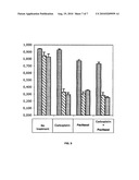 |  |
 |  |
 |  |
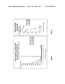 |  |
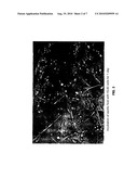 | 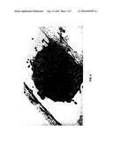 |
 |  |
 |  |
 |  |
 | 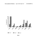 |
 |  |
 | 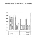 |
| Similar patent applications: | |
| Date | Title |
|---|---|
| 2010-07-22 | Tumour-associated peptides that bind to mhc molecules |
| 2010-09-30 | Method for introducing foreign substance into cell having cell wall |
| 2010-10-07 | Tumor cell-derived microvesicles |
| 2008-10-16 | Microcytoxicity assay by pre-labeling target cells |
| 2009-12-17 | Method for molecular delivery into cells using naonotube spearing |
| New patent applications in this class: | |
| Date | Title |
|---|---|
| 2019-05-16 | Non-invasive method for assessing the presence and severity of esophageal varices |
| 2019-05-16 | Method for evaluating protrusion-forming ability of cell spheroids |
| 2019-05-16 | Product demonstration device |
| 2019-05-16 | In vitro model for blood-brain barrier and method for producing in vitro model for blood-brain barrier |
| 2019-05-16 | Microbeads for cell culture and method of monitoring cell culture using the same |
| New patent applications from these inventors: | |
| Date | Title |
|---|---|
| 2014-09-18 | Combinations of kinase inhibitors for the treatment of cancer |
| 2012-03-22 | Method for in vitro assay of soluble fibrin by generating specific degradation products |
| 2009-12-10 | Detection of venous thromboembolic diseases by measurement of d-dimers and soluble fibrin levels |
| Top Inventors for class "Chemistry: molecular biology and microbiology" | |
| Rank | Inventor's name |
|---|---|
| 1 | Marshall Medoff |
| 2 | Anthony P. Burgard |
| 3 | Mark J. Burk |
| 4 | Robin E. Osterhout |
| 5 | Rangarajan Sampath |