Patent application title: UTILIZING LIVER CELL LINE QSG-7701 TO BE INFECTED WITH HEPATITIS B VIRUS
Inventors:
Xiaoben Pan (Beijing, CN)
Lai Wei (Beijing, CN)
Assignees:
Peking University People's Hospital
IPC8 Class: AC12Q170FI
USPC Class:
435 5
Class name: Chemistry: molecular biology and microbiology measuring or testing process involving enzymes or micro-organisms; composition or test strip therefore; processes of forming such composition or test strip involving virus or bacteriophage
Publication date: 2010-03-04
Patent application number: 20100055674
Inventors list |
Agents list |
Assignees list |
List by place |
Classification tree browser |
Top 100 Inventors |
Top 100 Agents |
Top 100 Assignees |
Usenet FAQ Index |
Documents |
Other FAQs |
Patent application title: UTILIZING LIVER CELL LINE QSG-7701 TO BE INFECTED WITH HEPATITIS B VIRUS
Inventors:
Xiaoben Pan
Lai Wei
Agents:
OSHA LIANG L.L.P.
Assignees:
PEKING UNIVERSITY PEOPLE'S HOSPITAL
Origin: HOUSTON, TX US
IPC8 Class: AC12Q170FI
USPC Class:
435 5
Patent application number: 20100055674
Abstract:
The use of the liver cell line QSG-7701 for HBV infection includes the
following steps: directly infecting QSG-7701 cells with purified HBV
particles and facilitating the infection by DMSO and/or PEG treatment.
The easily available QSG-7701 liver cell line may not require
pre-differentiation induction and is naturally susceptible for HBV
infection. This cell line provides near normal physiological conditions
for HBV infection, especially the infection conditions that are
characterized with Chinese origin. This cell line is suitable for
investigating the life cycle of HBV. Therefore, this cell line is useful
for the investigation of viral infection processes and for the
development of drugs that specifically target these processes.Claims:
1. A liver cell line of QSG-7701 infected with HBV.
2. The liver cell line of QSG-7701 infected with HBV according to claim 1, wherein QSG-7701 cell line is directly infected with purified HBV particles.
3. The liver cell line of QSG-7701 infected with HBV according to claim 1, wherein QSG-7701 cell line is treated with 1-2% DMSO, and then infected with purified HBV virus particles.
4. The liver cell line of QSG-7701 infected with HBV according to claim 1, wherein QSG-7701 cell line is treated with 2-4% PEG to facilitate the infection.
5. The liver cell line of QSG-7701 infected with HBV according to claim 1, wherein QSG-7701 cell line is treated with 1-2% DMSO and 2-4% PEG to facilitate the infection.
6. Use of QSG-7701 cell line for the study of HBV infection.
7. The use of QSG-7701 cell line for the study of HBV infection according to claim 6, wherein QSG-7701 cell line is directly infected with purified HBV particles.
8. The use of QSG-7701 cell line for the study of HBV infection according to claim 6, wherein QSG-7701 cell line is treated with 1-2% DMSO and then infected with purified HBV particles.
9. The use of QSG-7701 cell line for the study of HBV infection according to claim 6, wherein QSG-7701 cell line is treated with 2-4% PEG to facilitate the infection.
10. The use of QSG-7701 cell line for the study of HBV infection according to claim 6, wherein QSG-7701 cell line is treated with 1-2% DMSO and 2-4% PEG to facilitate the infection.
11. Use of QSG-7701 cell line for the study of HBV infection and replication.
12. The use of claim 11, wherein the QSG-7701 cell line is directly infected with purified HBV particles.
13. The use of claim 11, wherein the QSG-7701 cell line is treated with 1-2% DMSO, and then infected with purified HBV particles.
14. The use of claim 11, wherein the QSG-7701 cell line is treated with 2-4% PEG to facilitate the infection.
15. The use of claim 11, wherein the QSG-7701 cell line is treated with 1-2% DMSO and 2-4% PEG to facilitate the infection.
16. A method for infecting QSG-7701 cells, comprising an infection step, wherein the QSG-7701 cells are directly infected with purified HBV particles.
17. The method of claim 16, further comprising a pre-treatment step prior to the infection step, wherein the pre-treatment step comprises treating the QSG-7701 cells with an agent to facilitate the infecting.
18. The method of claim 17, wherein the agent is 1-2% DMSO.
19. The method of claim 17, wherein the agent is 2-4% PEG.
20. The method of claim 17, wherein the agent is 1-2% DMSO and 2-4% PEG.
Description:
FIELD OF THE INVENTION
[0001]The present invention relates to a cell line. In particular, it relates to the use of the cell line for hepatitis B virus infection.
BACKGROUND OF THE INVENTION
[0002]Although it has been over 40 years since the discovery of "Australia antigen" of the hepatitis B virus (HBV) in hemophiliac patients in 1965 by Blumberg, a thorough understanding of the life cycle of HBV has remained elusive so far, and there is no effective drug for the treatment of chronic hepatitis B. The life cycle of hepatitis B is pretty complex. The basic steps comprise the binding of the virus envelope to the specific receptor on the cell surface, virus entry by direct fusion of the viral external envelope with cell plasma membrane or phagocytosis, disassembly of the outer capsid in inclusion bodies, nuclear entry of core protein carrying HBV DNA, which is repaired into cccDNA, and followed by transcription of various viral mRNAs and translation of the viral mRNAs into viral proteins for the assembly and secretion of viral particles (see Seeger C, Mason WS. Hepatitis B virus biology. Microbiol. Mol. Biol. Rev. 2000; 64:51-68). The in-depth research on HBV life cycle depends on a good HBV infection cell model system. So far, only a few cell lines known to be susceptible to HBV infection and replication. These cells lines include primary human liver cell (including embryo liver cell), primary tree shrew liver cell, and the recently established HepRG cell. Due to scarcity of the available sources, critical requirements for isolation operation and culture condition, and shorter survival time of primary cells (generally less than 1 month), the human primary cell and primary tree shrew liver cell may not meet the requirements for long-term scientific research or for being able to grow to large quantity in cell culture (Guha C, Mohan S, Roy-Chowdhury N, et al. Cell culture and animal models of viral hepatitis, Part I: Hepatitis B. Lab. Anim. (NY) 2004; 33:37-46). HepRG cell line was established in 2002. This cell line is a liver progenitor cell line, which can be infected by HBV after pre-differentiation induction by DMSO. This cell line is not commercially available but is only used in the lab where it was initially established. HepRG cell line may require pre-differentiation induction before HBV infection. Stability and reproducibility of this model, however, may still need to be further tested (see Gripon P, Rumin S, Urban S, et al. Infection of a human hepatoma cell line by the hepatitis B virus. Proc. Nat. Acad. Sci. USA. 2002; 99:15655-15660). So far, there has not been a reproducible cell line, which can be used for research for the study of entire HBV infection cycle. This deficiency has greatly impeded an in-depth research on the life cycle of HBV, especially the study on the specific cell receptor and the nuclear entry mechanism of HBV DNA in the early stages of life cycle. The lack of a suitable cell line for HBV infection has hindered the efforts to screen and develop novel anti-HBV drugs. Therefore, it is very important to identify cell lines that are naturally susceptible to HBV infection to advance the progress in virology research and in chronic hepatitis therapeutic development.
DESCRIPTION OF THE INVENTION
[0003]The main objective of the present invention is to provide cell lines that are naturally susceptible to HBV infection.
[0004]Another objective of the present invention is to provide methods that may be employed to infect the described cell lines with HBV.
[0005]The above described objectives in the present invention are implemented with the following technical procedures.
[0006]The inventors of the present invention unexpectedly discover that the cell line QST-7701 may be susceptible to HBV infection and replication.
[0007]The liver cell line QSG-7701 was established in 1977, which was isolated from the tissue located 6 cm away from a tumor of a 35 year old female patient having the hepatocellular carcinoma. This cell line has an abnormal mitosis index of 6.2-14.3%. The chromosome of the cell line is subtriploid karyotype (mode 57). These cells express albumin. A small portion of QSG-7701 cells express alpha-fetoprotein. This cell line is considered to be precancerous hepatic cell line (see Dehou Zhu and Jinbing Wang, "Comparative study of hepatic cell line QSG-7701 and hepatoma cell line from human liver cancer," Cancer Prevention and Treatment Research, 1979:7-9. In Chinese). This cell line is commercially available and can be purchased from suppliers, such as Shanghai Institute of Cell Biology, Chinese Academy of Science (Shanghai).
[0008]HBV infection of QSG-7701 liver cell line comprises the following step: directly infecting cultured QSG-7701 cells using purified HBV virus particles. A preferred procedure is: treating the cultured QSG-7701 cells with 1-2% DMSO, and then the cells may be infected with purified HBV virus particles and, simultaneously, add 2-4% PEG as infection adjuvant. A more preferred procedure is to infect the cultured QSG-7701 cells with purified HBV virus particles and, simultaneously, add 1-2% DMSO and 2-4% PEG as infection adjuvant.
[0009]QSG-7701 cell line has been used for about 30 years since its establishment. This cell line is stable and commercially available and, thus, can be obtained easily. In addition, this cell line may be used to overcome some disadvantages associated with using primary liver cell lines (e.g., human primary liver cell, embryo liver cell, and primary tree shrew liver cell line), such as difficulty to obtain and shorter in vitro life-span. Furthermore, HepRG cell line has not been commercialized. Therefore, it may be difficult to obtain HepRG cells because they may only be used by certain individual laboratories.
[0010]Different from HepRG cells in that cells become susceptible to HBV infection after pre-differentiation induction, QSG-7701 cells can be naturally infected by purified HBV. To a large extent, QSG-7701 cell line closely mimics the in vivo process by which HBV infects liver cell. Therefore, QSG-7701 cell line may be a suitable tool to study the life cycle of HBV. In particular, this cell line may be suitable for studying viral infection and for identifying relevant targets in the infection processes for the development of anti-viral drugs. For example, this cell line may be used to study the specific cell membrane receptors that interact with HBV, and the dynamics of nuclear translocation of the viral core particles carrying HBV DNA. Based on the data obtained from these studies, drugs may be developed, for example, to inhibit the binding of HBV to specific cell membrane receptors, to abolish disassembly of the core particles, and/or to inhibit nuclear translocation of HBV DNA.
[0011]The widely used liver (cancer) cell lines, such as HepG2 and Huh7, are derived from the tissues of Caucasian origin. In contrast, QSG-7701 cell line is derived from a Chinese female liver cancer patient. This cell line is derived from a surgically removed tissue around a tumor of this patient. In addition, QSG-7701 cell line has been identified as precancerous hepatic cells. Because this cell line was isolated from a Chinese, and is close to normal liver cells, this cell line, therefore, represents a suitable tool to study HBV replication in liver cells, specifically those of Chinese origin.
BRIEF DESCRIPTION OF DRAWINGS
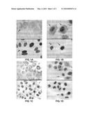
[0012]FIGS. 1A and 1C show the in vivo distribution of the core proteins in HepG2 cells 2-days after HBV infection. The core proteins (red) and the nuclei (blue) are indicated. The core proteins are mainly distributed in the cytoplasm after infection and HBV DNA may not be transported into the nucleus.
[0013]FIGS. 1B and 1D show the in vivo distribution of the core proteins in QSG-7701 cells 2-days after HBV infection. The core proteins (red) and the nuclei (blue) are indicated. The core proteins are mainly distributed in the nucleus and the peripheral regions of the nucleus.
[0014]FIG. 2 shows the detection of HBV replication intermediates 3 days after HBV infection.
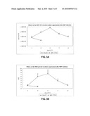
[0015]FIG. 3A shows the changes of HBV DNA levels in culture supernatant over time after infection.
[0016]FIG. 3B shows the changes of HBsAg levels in culture supernatant over time after infection.
EXAMPLES
[0017]The following embodiments and figures are used to further illustrate the present invention.
[0018]HepG2 cell line is a classic liver cancer cell line, which supports HBV replication but may not be infected by HBV. To compare with this cell line, HBV-positive serum may be used to infect both HepG2 and QSG-7701 cells. The following examples describe the experimental procedures used for the viral infection.
Example 1
[0019](1) Cell culture and infection: HBV-positive serum may be obtained from certain clinically tested samples. HBV may be concentrated up to 2×10¹⁰ copies/ml using density gradient ultracentrifugation purification. HepG2 and QSG-7701 cell lines (purchased from Shanghai Institute of Cell Biology, Chinese Academy of Sciences, Shanghai, China) may be thawed, and cultured in 6-well plates containing 10% FBS (Hyclone, USA) DMEM culture medium (Sigma, USA). Infect the cells when they have grown to about 80% confluence. Replace the growth media with 2 ml of serum-free DMEM. Add 0.5 ml of HBV-positive serum (total amount of virus may be 1×10¹⁰ copies/well). Cells may be incubated at 37° C. for 16 h. After incubation, cells may be washed 2 times with PBS followed by incubation for 2 min in the acid elution buffer (50 mM glycine, 150 mM NaCl, and add 1 M HCl to adjust to pH 2.2). Cells may then be washed 3 times with PBS to remove as many viruses as possible. Add 5% FBS DMEM culture medium and change to fresh culture media the next day. The supernatants may be centrifuged and stored at -20° C. for further testing.
[0020](2) Detection of the HBcAg distribution using immunofluorescence cytochemistry: because a large amount of filamentous or spherical HBsAg particles may be present in the HBV-positive serum and only the intact Dane's viral particles contain core particles, HBcAg may be tagged to track the movement of viral particles. To detect cell entry and cellular distribution of HBcAg, SABC immunofluorescence cytochemistry staining technique may be used. The detection method may be as follows: after HepG2 and QSG-7701 cell lines are infected with HBV, they may be fixed in 4% paraformaldehyde overnight at 4° C., and then washed with 0.2% Trinton-PBS for 10 min. Add the primary anti-HBcAg antibody (1:100 dilutions, rabbit source, Signet, USA) overnight at 4° C., the secondary antibody may be the biotin-labeled goat anti-rabbit IgG (1:100), and then incubated at ambient temperature for 1 hour. Add streptavidin-labeled Cy3 (red fluorescence; Bioshide Biotechnology Co., China). Add immunofluorescent dye, DAPI (blue fluorescence; Vector, USA), for nuclear DNA staining. The slides may then be mounted. Digital images may be taken under a fluorescent microscope (Olympus, Japan) (FIG. 1). FIG. 1 is a black and white photo converted from a color photo taken in a test.
[0021](3) Detection of viral replication intermediates: the viral replication intermediates may be detected using Southern blot analysis. Three days after infection, cells (2×10⁶) may be lysed in lysis buffer (50 mM Tris/HCl, pH 7.4, 1 mM EDTA, and 1% NP40). DNA may be extracted using the phenol/chloroform method. The Southern blot membranes may be exposed to X-ray film (Kodak, USA) for 30 min. The whole genomic probe of HBV may be prepared using digoxin labeling PCR kit (Roche, USA). The results are shown in FIG. 2.
[0022](4) Detection of HBsAg and HBV DNA in culture supernatants: the amount of HBsAg in the cell culture supernatant may be quantified using an electrochemical illumination detection kit (Roche, USA) and an automatic immunoassays analyzer (Roche, Elecsys 2010). The HBsAg standards may be provided by the kit. HBV DNA may be detected using a real time PCR detection kit (Shenzhen Piji Biotechnology Company, China) and a LightCycler real time quantitative PCR cycler (Roche). The PCR standards may be provided by the kit. Real time PCR may be performed according to the manual provided by the kit. This process may be repeated 5 times. FIGS. 3A and 3B show the results with statistical analysis.
[0023]FIG. 1 shows that some HepG2 cells infected with HBV (2 day after infection) have the core proteins distributed mainly in the cytoplasm but not in the nucleus. (As shown in FIGS. 1A and 1C, the arrows in the top panels indicate the black spotty signals from the core proteins. The arrows in the bottom panels indicate the black signals from the nucleus. 400× amplification) The data show that the core proteins may not enter the nucleus in HepG2 cells. In contrast, 2 days after HBV infection, there are strong core protein signals within the nucleus as well as the peripheral areas around the nucleus membrane in QSG-7701 cells. This observation suggests that there may be little barrier for the core proteins that carry HBV DNA, to enter the nucleus. (As shown in FIGS. 1B and 1D, the arrows in the top panels indicate the black spotty signals from the core proteins. The arrows in the bottom panels indicate the black signals from the nucleus. 400× amplification).
[0024]FIG. 2 shows the detection of HBV replication intermediates in cells 3 days after HBV infection. Only weak rcDNA signals may be detected in HepG2 cells without other replication intermediates. In contrast, after HBV infection, both dsDNA and ssDNA replication intermediate are detected in QSG-7701 cells.
[0025]FIG. 3A shows the changes of HBV DNA levels in the supernatants after HBV infection. FIG. 3B shows the changes of HBsAg levels in the supernatants after HBV infection. On the first and the third day post HepG2 infection, only trace amount of HBV DNA and HBsAg may be detected, and they become undetectable after the third day. This may be caused by the detachment of viruses from the cell surface and in the culture supernatant to be subsequently washed away. In contrast, the HBV DNA and HBsAg levels increase in the supernatants of QSG-7701 cells after HBV infection. In particular, the HBV DNA and HBsAg levels reach the maximum on the fifth day after infection, but gradually decrease thereafter. The HBV DNA and HBsAg levels remain detectable for about 9 days. It is possible that the cellular innate immunity inhibits the HBV replication.
[0026]The observation that the HBV-infected HepG2 cells do not have the viral replication intermediates indicates that HepG2 cells may not support HBV infection. This possibility is supported by other observations (see Paran N, Geiger B, Shaul Y., HBV infection of cell culture: evidence for multivalent and cooperative attachment. EMBO J. 2001; 20:4443-4453). In contrast, the observation that the core proteins are accumulated inside the nucleus of QSG-7701 cells infected with HBV indicates that QSG-7701 cells may support both the cell entry of HBV and the nuclear translocation of the core proteins carrying HBV DNA. The production of viral replication intermediates, and HBsAg and HBV DNA in the culture supernatants further support the idea that QSG-7701 cells can be successfully infected by HBV and that the cell line supports HBV replication.
Example 2
[0027]It may be performed basically according to the same procedure described in example 1, except that 1-2% DMSO is added to facilitate the viral infection.
Example 3
[0028]It may be performed basically according to the same procedure described in example 1, except that 2-4% PEG is added to facilitate the viral infection.
Example 4
[0029]It may be performed basically according to the same procedure described in example 1, except that both 1-2% DMSO and 2-4% PEG to facilitate the viral infection.
[0030]DMSO and PEG treatment may increase the number of viral particles attached to the cell surface, thus, facilitating HBV infection by enhancing the cellular intake of HBV particles. (see Glebe D, Berting, A, Broehl S, et al. Gerlich W H, and Schaefer S., Optimized conditions for the production of the hepatitis B virus from cell culture. Intervirology, 2001; 44(6):370-378). In addition, DMSO treatment may increase the levels of HBV DNA replication and viral protein expression. The mechanisms underlying these effects remain unclear.
[0031]These results show that, by simultaneously adding 1-2% DMSO and/or 2-4% PEG at viral infection, may further increase the stability of the model system as well as the viral replication levels.
[0032]The above embodiments are described only for the purpose of illustrating the present invention. Therefore, none of these embodiments should be regarded as limiting to the present invention. Those skilled in the art having an understanding of the essence of the present invention can modify or alter certain embodiments disclosed above. However, all such modifications are within the scope of the present invention.
User Contributions:
comments("1"); ?> comment_form("1"); ?>Inventors list |
Agents list |
Assignees list |
List by place |
Classification tree browser |
Top 100 Inventors |
Top 100 Agents |
Top 100 Assignees |
Usenet FAQ Index |
Documents |
Other FAQs |
User Contributions:
Comment about this patent or add new information about this topic:
| People who visited this patent also read: | |
| Patent application number | Title |
|---|---|
| 20170132791 | CUTTING THREE-DIMENSIONAL IMAGE |
| 20170132790 | IMAGE PHOTOGRAPHING APPARATUS AND METHOD FOR CONTROLLING THE SAME |
| 20170132789 | METHODS, SYSTEMS AND DEVICES FOR SPINAL SURGERY POSITION OPTIMIZATION |
| 20170132788 | SYSTEM AND METHOD FOR ESTIMATING ARTERIAL PULSE WAVE VELOCITY |
| 20170132787 | FLOW CELL ALIGNMENT METHODS AND SYSTEMS |
Images included with this patent application:
 |  |
 |
| New patent applications in this class: | |
| Date | Title |
|---|---|
| 2022-05-05 | Method for diagnosing human t-cell leukemia virus type 1 (htlv-1) associated diseases |
| 2022-05-05 | Systems and methods for assay processing |
| 2022-05-05 | Rapid pathology/cytology without wash |
| 2019-05-16 | Biofluidic triggering system and method |
| 2019-05-16 | Method and system for detection of disease agents in blood |
| New patent applications from these inventors: | |
| Date | Title |
|---|---|
| 2019-01-03 | Organic light-emitting diode (oled) display panel and controlling method |
| 2012-06-07 | Hepatitis b virus mutation strain with resistance to adefovir dipivoxil and the uses thereof |
| 2010-12-09 | anti-hcv vaccine and preparation methods and uses thereof |
| Top Inventors for class "Chemistry: molecular biology and microbiology" | |
| Rank | Inventor's name |
|---|---|
| 1 | Marshall Medoff |
| 2 | Anthony P. Burgard |
| 3 | Mark J. Burk |
| 4 | Robin E. Osterhout |
| 5 | Rangarajan Sampath |