Patent application title: NOVEL ANTIGEN CONSTRUCTS USEFUL IN THE DETECTION AND DIFFERENTIATION OF ANTIBODIES TO HIV
Inventors:
John R. Hackett, Jr. (Libertyville, IL, US)
Julie Yamaguchi (Chicago, IL, US)
Alan M. Golden (Wilmette, IL, US)
Catherine A. Brennan (Libertyville, IL, US)
Robert K. Hickman (Mundelein, IL, US)
Sushil G. Devare (Northbrook, IL, US)
IPC8 Class: AC12Q170FI
USPC Class:
435 5
Class name: Chemistry: molecular biology and microbiology measuring or testing process involving enzymes or micro-organisms; composition or test strip therefore; processes of forming such composition or test strip involving virus or bacteriophage
Publication date: 2010-02-11
Patent application number: 20100035237
Inventors list |
Agents list |
Assignees list |
List by place |
Classification tree browser |
Top 100 Inventors |
Top 100 Agents |
Top 100 Assignees |
Usenet FAQ Index |
Documents |
Other FAQs |
Patent application title: NOVEL ANTIGEN CONSTRUCTS USEFUL IN THE DETECTION AND DIFFERENTIATION OF ANTIBODIES TO HIV
Inventors:
Sushil G. Devare
John R. Hackett, JR.
Julie Yamaguchi
Alan M. Golden
Catherine A. Brennan
Robert K. Hickman
Agents:
Abbott Laboratories;c/o Polsinelli Shughart PC
Assignees:
Origin: CHICAGO, IL US
IPC8 Class: AC12Q170FI
USPC Class:
435 5
Patent application number: 20100035237
Abstract:
Isolated HIV-1 Group O env polypeptides obtained from the HIV-1 isolate
HAM112 are claimed, as well as (a) antigen constructs comprising fusions
of one or more of each of HIV-1 Group O env polypeptides and HIV-1 Group
M env polypeptide and (b) further antigen constructs containing
additional Group O sequences and especially the gp41 IDR of isolate
HAM112. Also claimed are polynucleotide sequences encoding the above,
expression vectors comprising the same, host cells transformed thereby,
and immunoassay methods and kits utilizing the antigen constructs of the
invention.Claims:
1.-68. (canceled)
69. A method for detecting antibodies to HIV-1 in a test sample comprising the steps of: (a) combining at least one antigen construct with the test sample to form a mixture; (b) incubating the mixture under conditions suitable for formation of complexes between the antigen and antibodies, if any, which are present in the sample and are immunologically reactive with the antigen; and (c) detecting the presence of any complexes formed, wherein the antigen construct comprises a first HIV-1 group O env polypeptide fused to a second HIV-1 Group O env polypeptide, and wherein the antigen construct is selected from the group consisting of pGO-9CKS and pGO-11 CKS.
70.-71. (canceled)
72. A method according to claim 69 wherein detecting the presence of complexes in step (c) is carried out using an antibody directed to the complexes formed in step (b) to which is attached a signal-generating compound.
73. A method according to claim 69 wherein detecting the presence of complexes in step (c) is carried out using an antibody directed to the complexes formed in step (b) to which is attached a first member of a specific binding pair, and further using an indicator reagent comprising a second member of the specific binding pair to which is attached a signal-generating compound.
74. An immunoassay kit for the detection of antibodies to HIV-1 comprising an antigen construct, wherein the antigen construct comprises a first HIV-1 group O env polypeptide fused to a second HIV-1 Group O env polypeptide, and wherein the antigen construct is selected from the group consisting of pGO-9CKS and pGO-11 CKS.
75. An immunoassay kit according to claim 74 wherein the antigen construct is a capture reagent.
76. An immunoassay kit according to claim 74 wherein the antigen construct is an indicator reagent.
77. An immunoassay kit according to claim 74 wherein the antigen construct is attached to a first member of a specific binding pair, the kit additionally comprising an indicator reagent comprising a second member of the specific binding pair attached to a signal-generating compound.
Description:
RELATED APPLICATION INFORMATION
[0001]This application is a divisional of U.S. patent application Ser. No. 11/008,351 filed on Dec. 9, 2004, which is a divisional of U.S. patent application Ser. No. 08/911,824 filed on Aug. 15, 1997, which is now U.S. Pat. No. 6,846,905.
BACKGROUND OF THE INVENTION
[0002]This invention relates generally to immunoassays for the detection and differentiation of antibodies to Human Immunodeficiency Virus Type 1 (HIV-1) Group M, HIV-1 Group O and Human Immunodeficiency Virus Type 2 (HIV-2). More particularly, the invention relates to novel antigen constructs useful as reagents in such assays, as well as polynucleotides, DNA clones, expression vectors, transformed host cells and the like which are useful in the preparation of such antigens.
[0003]Detection of HIV infection in a patient, and characterization of the viral type, are typically carried out using immunoassays which rely on the highly specific interaction between antigens used as reagents in the assay and circulating antibodies in the patient's serum. The immunoreactivity of patient antibodies with some antigens, and to a lesser extent or not at all with others, permits the identification of the type and subtype of the HIV which is present.
[0004]Currently, there are two major phylogenetic groups of HIV-1 designated as Groups "M" and "O." G. Meyers et al., Human Retroviruses and AIDS 1995, Los Alamos National Laboratory, Los Alamos, N. Mex. (1995). HIV-1 Group M isolates further have been divided into subgroups (A to J) that are phylogenetically approximately equidistant from each other. Group M isolates predominate worldwide. The earliest reports about the sequence of HIV-1 Group O indicated that these viruses were as closely related to a chimpanzee virus as to other HIV-1 subgroups. See, for example, L. G. Gurtler et al., J. Virology 68:1581-1585 (1994); M. Vanden Haesevelde et al., J. Virology 68:1586-1596 (1994); De Leys et al., J. Virology 64:1207-1216 (1990); DeLeys et al., U.S. Pat. No. 5,304,466; L. G. Gurtler et al., European Patent Publication No. 591914 A2. The Group O sequences are the most divergent of the HIV-1 sequences described to date. Although HIV-1 Group O strains are endemic to west central Africa (Cameroon, Equatorial Guinea, Nigeria and Gabon), patients infected with Group O isolates now have been identified in Belgium, France, Germany, Spain and the United States. See, for example, R. DeLeys et al., supra; P. Chameau et al., Virology 205:247-253 (1994); I. Loussert-Ajaka et al., J. Virology 69:5640-5649 (1995); H. Hampl et al., Infection 23:369-370 (1995); A. Mas et al., AIDS Res. Hum. Retroviruses 12:1647-1649 (1996); M. Peters et al., AIDS 11:493-498 (1997); and M. A. Rayfield et al., Emerging Infectious Diseases 2:209-212 (1996).
[0005]HIV-1 Group M serology is characterized in large part by the amino acid sequences of the expressed viral proteins (antigens), particularly those comprising the core and envelope (env) regions. As between various strains of this rapidly-mutating virus, these antigens are structurally and functionally similar but have divergent amino acid sequences which elicit antibodies that are similar but not identical in their specificity for a particular antigen.
[0006]One of the key serological targets for detection of HIV-1 infection is the 41,000 MW transmembrane protein (TMP), glycoprotein 41 (gp41). gp41 is a highly immunogenic protein which elicits a strong and sustained antibody response in individuals considered seropositive for HIV. Antibodies to this protein are among the first to appear at seroconversion. The immune response to gp41 apparently remains relatively strong throughout the course of the disease, as evidenced by the near universal presence of anti-gp41 antibodies in asymptomatic patients as well as those exhibiting clinical stages of AIDS. A significant proportion of the antibody response to gp41 is directed toward a well-characterized immunodominant region (IDR) within gp41.
[0007]Infections with HIV Type 2 (HIV-2), a virus initially found in individuals from Africa, now have been identified in humans outside of the initial endemic area of West Africa, and have been reported in Europeans who have lived in West Africa or those who have had sexual relations with individuals from this region. See, for example, A. G. Saimot et al., Lancet i:688 (1987); M. A. Rey et al., Lancet i:388-389 (1987); A. Werner et al., Lancet i:868-869 (1987); G. Brucker et al., Lancet i:223 (1987); K. Marquart et al., AIDS 2:141 (1988); CDC, MMWR 37:33-35 (1987); Anonymous, Nature 332:295 (1988). Cases of AIDS due to HIV-2 have been documented world-wide. Serologic studies indicate that while HIV-1 and HIV-2 share multiple common epitopes in their core antigens, the envelope glycoproteins of these two viruses are much less cross-reactive. F. Clavel, AIDS 1:135-140 (1987). This limited cross-reactivity of the envelope antigens is believed to explain why currently available serologic assays for HIV-1 may fail to react with certain sera from individuals with antibody to HIV-2. F. Denis et al., J. Clin. Micro. 26:1000-1004 (1988). Recently-issued U.S. Pat. No. 5,055,391 maps the HIV-2 genome and provides assays to detect the virus.
[0008]These viral strains are, for the most part, readily identified and characterized using commercially-available diagnostic tests. However, concerns have arisen regarding the capability of currently-available immunoassays, designed for the detection of antibody to HIV-1 (Group M) and/or HIV-2, to detect the presence of antibody to HIV-1 Group O. 1. Loussert-Ajaka et al., Lancet 343:1393-1394 (1994); C. A. Schable et al., Lancet 344:1333-1334 (1994); L. Gurtler et al., J. Virol. Methods 51:177-184 (1995). Although, to date, few patients outside of west Central Africa have been found to be infected with HIV-1 Group O isolates, health officials fear the emergence of this subtype in other geographic areas as well.
[0009]Consequently, there is a continued need for new antigens, suitable for use in immunoassays, which alone or in conjunction with other antigens permit the recognition of all HIV-1 (Group M and Group O) and HIV-2 isolates and/or infections.
SUMMARY OF THE INVENTION
[0010]It has now been found that certain polypeptides or combinations of are particularly useful in the detection of HIV-1 Group O and other HIV infections. Consequently, in a first aspect of the present invention is disclosed an isolated HIV-1 Group O env polypeptide having an amino acid sequence consisting essentially of the sequence of SEQ ID NO:61 representing the full-length env region of the HIV-1 Group O isolate HAM112. Similarly disclosed is an isolated HIV-1 Group O env polypeptide comprising an immunoreactive portion of the above full-length polypeptide, as well as polynucleotides encoding such polypeptides.
[0011]In a second aspect of the present invention, an antigen construct is disclosed which comprises a first HIV-1 Group O env polypeptide fused to a second HIV-1 Group O env polypeptide. Preferably, the first polypeptide of such an antigen construct is a gp120 polypeptide and the second polypeptide is a gp41 polypeptide, optionally with a portion of the hydrophobic region of the gp41 polypeptide being deleted so as to facilitate expression when expressed as a recombinant product. Also preferred among the above antigen constructs are those in which at least one of the first and second HIV-1 Group O env polypeptides is derived from HIV-1 Group O isolate HAM112, as are those in which the first polypeptide comprises an immunoreactive portion of the gp120 protein of HIV-1 Group O isolate HAM112.
[0012]In the above Group O env constructs, the first polypeptide may have an amino acid sequence which consists essentially of residues 1 through 520 of the sequence of SEQ ID NO:61, or alternatively an immunoreactive portion thereof. A shortened and preferred first polypeptide is one having an amino acid sequence consisting essentially of residues 476 through 520 of the sequence of SEQ ID NO:61. Along with any of the above polypeptides, the second polypeptide used in the constructs of the invention may be an immunoreactive portion of the gp41 protein of HIV-1 Group O isolate HAM112, from which a portion of the hydrophobic region of the gp41 protein of HIV-1 Group O isolate HAM112 is optionally absent. In particular, the deleted portion may be that part of gp41 which has an amino acid sequence consisting essentially of residues 690 through 715 of the sequence of SEQ ID NO:61.
[0013]The above second polypeptide will preferably have an amino acid sequence consisting essentially of residues 521 through 873 of the sequence of SEQ ID NO:61 or a portion thereof. More preferably, the second polypeptide may have an amino acid sequence consisting essentially of residues 47 through 373 of the sequence of SEQ ID NO:52; still more preferably, the amino acid sequence may consist essentially of residues 47 through 245 of the sequence of SEQ ID NO:48; and even more preferably, the amino acid sequence may consist essentially of residues 47 through 215 of the sequence of SEQ ID NO:58. Representative of the Group O env constructs of the invention are constructs pGO-8PL, pGO-8CKS, pGO-9PL, pGO-9CKS, pGO-11PL and pGO-11CKS, as well as any derivatives, variants and analogs thereof.
[0014]In a further aspect of the present invention, there is disclosed an antigen construct comprising a fusion of at least one HIV-1 Group O env polypeptide with at least one HIV-1 Group M env polypeptide, and more preferably an antigen construct comprising a fusion of:
[0015](a) a first HIV-1 Group O env polypeptide;
[0016](b) a second HIV-1 Group O env polypeptide;
[0017](c) a first HIV-1 Group M env polypeptide; and
[0018](d) a second HIV-1 Group M env polypeptide.
The HIV-1 Group M polypeptides in the above constructs may be derived from an HIV-1 isolate of Subtype B, and preferably at least one is derived from HIV-1 Group M isolate HXB2R. In any of these Group O/Group M env constructs, at least one of the HIV-1 Group O sequences may be derived from HIV-1 Group O isolate HAM112.
[0019]More particularly, the first Group O env polypeptide and the first Group M env polypeptide may both be gp120 polypeptides, while the second Group O env polypeptide and the second Group M env polypeptide may both be gp41 polypeptides. To enhance expression, a portion of the hydrophobic region of at least one of the gp41 polypeptides may be deleted. Antigen constructs included among the above are those in which:
[0020](a) the first HIV-1 Group O env polypeptide comprises an immunoreactive portion of the gp120 protein of HIV-1 Group O isolate HAM112;
[0021](b) the second HIV-1 Group O env polypeptide comprises an immunoreactive portion of the gp41 protein of HIV-1 Group O isolate HAM12
[0022](c) the first HIV-1 Group M env polypeptide comprises an immunoreactive portion of the gp120 protein of a first HIV-1 Group M isolate of Subtype B; and
[0023](d) the second HIV-1 Group M env polypeptide comprises an immunoreactive portion of the gp41 protein of a second HIV-1 Group M isolate of Subtype B.
[0024]Preferred among these are constructs wherein the first and second HIV-1 Group M isolates of Subtype B are the same and are HIV-1 Group M isolate HXB2R, as well as those wherein a portion of the hydrophobic region of the gp41 protein of HIV-1 Group M isolate HXB2R is absent from the second HIV-1 Group M env polypeptide.
[0025]Preferred Group O/Group M env constructs include those in which (a) the first HIV-1 Group M env polypeptide has an amino acid sequence consisting essentially of residues 251 through 292 of the sequence of SEQ ID NO:108, and (b) the second HIV-1 Group M env polypeptide has an amino acid sequence consisting essentially of residues 293 through 599 of the sequence of SEQ ID NO:108 or a portion thereof. Especially preferred are those in which the second HIV-1 Group M env polypeptide has an amino acid sequence consisting essentially of residues 293 through 492 of the sequence of SEQ ID NO:108.
[0026]Also preferred are the above Group O/Group M env constructs in which the first HIV-1 Group O env polypeptide has an amino acid sequence consisting essentially of residues 1 through 520 of the sequence of SEQ ID NO:61 or a portion thereof, and especially those comprising a first HIV-1 Group O env polypeptide which has an amino acid sequence consisting essentially of residues 476 through 520 of the sequence of SEQ ID NO:61. The second HIV-1 Group O env polypeptide may be one having an amino acid sequence consisting essentially of residues 521 through 873 of the sequence of SEQ ID NO:61 or a portion thereof, from which a portion of the hydrophobic region of the gp41 protein of HIV-1 Group O isolate HAM112 may optionally be absent. Preferred constructs are those in which such second HIV-1 Group O env polypeptides have an amino acid sequence consisting essentially of residues 47 through 373 of the sequence of SEQ ID NO:52; more preferred are those in which the second HIV-1 Group O env polypeptide has an amino acid sequence consisting essentially of residues 47 through 245 of the sequence of SEQ ID NO:48; and even more preferred are those in which the second HIV-1 Group O env polypeptide has an amino acid sequence consisting essentially of residues 47 through 215 of the sequence of SEQ ID NO:58. Representative of the Group O/Group M env constructs of the invention are constructs pGO-12CKS, pGO-13CKS and pGO-14PL, and derivatives, variants and analogs thereof.
[0027]In yet another aspect of the present invention, an antigen construct is disclosed which comprises a fusion of a first HIV-1 env polypeptide, a second HIV-1 env polypeptide, and at least one additional HIV-1 polypeptide, and especially one in which each such HIV-1 env polypeptides are HIV-1 Group O polypeptides. The first HIV-1 Group O env polypeptide of this construct may be a gp120 polypeptide, and the second HIV-1 Group O env polypeptide a gp41 polypeptide. More particularly, the first HIV-1 Group O env polypeptide of this construct may comprise an immunoreactive portion of the gp120 protein of HIV-1 Group O isolate HAM112, while the second HIV-1 Group O env polypeptide may comprise an immunoreactive portion of the gp41 protein of HIV-1 Group O isolate HAM12.
[0028]Among these constructs, those in which the first HIV-1 Group O env polypeptide has an amino acid sequence consisting essentially of residues 1 through 520 of the sequence of SEQ ID NO:61, or a portion thereof, are preferred; more preferred are those in which the first HIV-1 Group O env polypeptide has an amino acid sequence consisting essentially of residues 476 through 520 of the sequence of SEQ ID NO:61. As to the second HIV-1 Group O env polypeptide, which may have an amino acid sequence consisting essentially of residues 521 through 873 of the sequence of SEQ ID NO:61 or a portion thereof and from which a portion of the hydrophobic region of the gp41 protein of HIV-1 Group O isolate HAM112 may optionally be absent, preferred are those constructs in which that second HIV-1 Group O env polypeptide has an amino acid sequence consisting essentially of residues 47 through 373 of the sequence of SEQ ID NO:52. Even more preferred are those having a second HIV-1 Group O env polypeptide with an amino acid sequence consisting essentially of residues 47 through 245 of the sequence of SEQ ID NO:48, and especially those in which the amino acid sequence consists essentially of residues 47 through 215 of the sequence of SEQ ID NO:58.
[0029]The additional HIV-1 polypeptide in any of these constructs may be a Group O env polypeptide; however, it is intended that it may alternatively be an immunogenic polypeptide from any of HIV-1 Groups M or O or HIV-2, including env, gag, pol, reverse transcriptase, and regulatory and other viral components. Preferred in any case are those constructs in which the additional HIV-1 Group O polypeptide comprises an immunoreactive portion of the gp41 protein of HIV-1 Group O isolate HAM112. Also preferred are those wherein the additional HIV-1 Group O polypeptide has an amino acid sequence consisting essentially of residues 521 through 873 of the sequence of SEQ ID NO:61 or a portion thereof, from which the hydrophobic region of the gp41 protein of HIV-1 Group O isolate HAM112 may optionally be absent. Even more preferred are constructs in which the additional HIV-1 Group O env polypeptide has an amino acid sequence consisting essentially of residues 47 through 373 of the sequence of SEQ ID NO:52; particularly preferred are those in which the additional HIV-1 Group O env polypeptide has an amino acid sequence consisting essentially of residues 47 through 245 of the sequence of SEQ ID NO:48, and especially those wherein the additional HIV-1 Group O env polypeptide has an amino acid sequence consisting essentially of residues 47 through 215 of the sequence of SEQ ID NO:58. Most preferred are constructs having as the additional HIV-1 Group O env polypeptide the so-called immunodominant region (IDR) of HIV-1 Group O, which has an amino acid sequence consisting essentially of residues 250 through 281 of the sequence of SEQ ID NO:120. Representative of the above constructs are pGO-15CKS and pGO-15PL, as well as any derivatives, variants and analogs thereof.
[0030]In still another aspect of the present invention is disclosed an antigen construct comprising a first HIV-2 env polypeptide fused to a second HIV-2 env polypeptide, and especially one in which the first HIV-2 env polypeptide is a gp120 polypeptide and the second HIV-2 env polypeptide is a gp36 polypeptide. Preferred among the such constructs are those in which:
[0031](a) the first HIV-2 env polypeptide has an amino acid sequence consisting essentially of residues 248 through 307 of the sequence of SEQ ID NO:55 or a portion thereof; and
[0032](b) the second HIV-2 env polypeptide has an amino acid sequence consisting essentially of residues 308 through 466 of the sequence of SEQ ID NO:55 or a portion thereof.
Representative of the HIV-2 constructs of the invention is pHIV-210 (SEQ ID NO:55), as well as any derivatives, variants and analogs thereof.
[0033]An additional aspect of the present invention comprises polynucleotides encoding any of the above antigen construct, which polynucleotide may be operably linked to a control sequence capable of directing expression in a suitable host and/or have a coding sequence which has been modified to provide a codon bias appropriate to the expression host. Still other aspects of the present invention include expression vectors comprising such polynucleotides and host cells transformed thereby, particularly where the host is Escherichia coli.
[0034]In a further aspect of the present invention, there is disclosed a method for detecting antibodies to HIV-1 in a test sample comprising the steps of:
[0035](a) combining at least one antigen construct according to the invention with the test sample to form a mixture;
[0036](b) incubating the mixture under conditions suitable for formation of complexes between the antigen and antibodies, if any, which are present in the sample and are immunologically reactive with the antigen; and
[0037](c) detecting the presence of any complexes formed.
In one embodiment of the method, detection of the presence of complexes in step (c) is carried out using an additional antigen construct of the invention to which a signal-generating compound has been attached. In another embodiment, detection is carried out using an additional antigen construct of the invention to which a first member of a specific binding pair is attached, and further using an indicator reagent comprising a second member of the specific binding pair to which is attached a signal-generating compound. A further embodiment provides that detection of the presence of complexes in step (c) is carried out using an antibody directed to the complexes formed in step (b), to which antibody is attached a signal-generating compound. Still another embodiment provides that detection of the presence of complexes in step (c) is carried out using an antibody directed to the complexes formed in step (b) and attached thereto a first member of a specific binding pair; such detection further requires the use of an indicator reagent comprising a second member of the specific binding pair to which is attached a signal-generating compound.
[0038]In a final aspect of the present invention are disclosed immunoassay kits for the detection of antibodies to HIV-1, which kits comprise an antigen construct of the invention. Such construct may be used as a capture reagent or an indicator reagent. Alternatively, the antigen construct may be attached to a first member of a specific binding pair, the kit additionally comprising an indicator reagent comprising a second member of the specific binding pair attached to a signal-generating compound.
DETAILED DESCRIPTION OF THE INVENTION
[0039]In one embodiment of an isolated polypeptide of the present invention, the amino acid sequence of the env protein of the HIV-1 Group O isolate HAM112 is shown as SEQ ID NO:61. In the present context, "isolated" is intended to mean that such polypeptides are relatively purified with respect to other viral or cellular components which normally would be present in situ, up to and including a substantially pure preparation of the protein. Such polypeptides can be utilized as assay reagents, for the production of monoclonal or polyclonal antibodies, in the manufacture of vaccines, or otherwise.
[0040]Immunoreactive portions, or fragments, of the above polypeptides are also expected to be useful. By "immunoreactive" is meant portions of such length as are capable of eliciting an immune response in a host and/or of reacting with antibodies directed specifically thereto; preferably, such partial polypeptides will be five or more amino acids in length. It should also be noted that the term "portion" as used herein is directed to both terminally truncated sequences and those which are shortened by the removal of an intervening sequence.
[0041]The above polypeptides and portions will best be produced by expression of polynucleotides encoding the same. These too permit a degree of variability in their sequence, as for example due to degeneracy of the genetic code, codon bias in favor of the host cell expressing the polypeptide, and conservative amino acid substitutions in the resulting protein. Moreover, it is anticipated that some variation of sequences will occur between--and possibly even within--a given HIV-1 isolate or other phylogenetic unit. Consequently, the polypeptides and constructs of the invention include not only those which are identical in sequence to the above sequence but also those which have an amino acid sequence that consist essentially of that reference sequence, where the term "consisting essentially" is meant to embrace variant polypeptides the structural and functional characteristics of which remain substantially the same. Preferably, such variants (or "analogs") will have a sequence homology ("identity") of 80% or more with the reference sequence of SEQ ID NO:61. In this sense, techniques for determining amino acid sequence "similarity" are well-known in the art. In general, "similarity" means the exact amino acid to amino acid comparison of two or more polypeptides at the appropriate place, where amino acids are identical or possess similar chemical and/or physical properties such as charge or hydrophobicity. A so-termed "percent similarity" then can be determined between the compared polypeptide sequences. Techniques for determining nucleic acid and amino acid sequence identity also are well known in the art and include determining the nucleotide sequence of the mRNA for that gene (usually via a cDNA intermediate) and determining the amino acid sequence encoded therein, and comparing this to a second amino acid sequence. In general, "identity" refers to an exact nucleotide to nucleotide or amino acid to amino acid correspondence of two polynucleotides or polypeptide sequences, respectively. Two or more polynucleotide sequences can be compared by determining their "percent identity", as can two or more amino acid sequences. The programs available in the Wisconsin Sequence Analysis Package, Version 8 (available from Genetics Computer Group, Madison, Wis.), for example, the GAP program, are capable of calculating both the identity between two polynucleotides and the identity and similarity between two polypeptide sequences, respectively. Other programs for calculating identity or similarity between sequences are known in the art.
[0042]According to another embodiment of the invention, antigen constructs are provided which are suitable for use in the detection of anti-HIV-1 antibodies. As described in greater detail below, such constructs may be prepared by recombinant means, as synthetic peptides, or otherwise; moreover, they may be glycosylated or unglycosylated depending on the manner and/or host cell by which they are made. Consequently, although referred to as if comprising glycoproteins (for example, "a gp120 polypeptide"), the antigen constructs of the invention are intended to include those which are expressed in bacterial hosts such as E. coli and are therefore unglycosylated.
[0043]It should be noted that the above constructs are fusions of various sequences, that is, the constructs are formed by joining various epitope-containing sequences, as for example by co-expression, ligation or sequential synthesis. Also joined thereto, and optionally included in the constructs of the invention, are other polypeptide sequences such as expression (CKS) polylinkers and other linker sequences. The order of the various polypeptide sequences is not critical; consequently, the polypeptides and their epitopes may be re-arranged as a matter of convenience. Further modifications are also possible, as for example by random mutation or site-directed mutagenesis or even the deletion (removal or omission) of certain regions such as the gp41 hydrophobic region, the absence of which has been found to enhance expression of the remaining polypeptide. In any case, whether nearly the same or substantially changed, polypeptides which undergo these modifications may be said to be "derived" from their respective sources, and the resulting polypeptides may be regarded as "derivatives".
[0044]In yet another aspect of the present invention, assay methods are provided which utilize the constructs of the invention in the detection of anti-HIV-1 antibodies in test samples. Such methods permit the direct testing of biological specimens; however, the assay methods may also be modified to permit the testing of pre-processed specimens such as sera, lysed cells, and extracts or preparations made therefrom (as by concentration, dilution, separation, fixation and/or immobilization). Depending on the desired assay format, the antigen constructs may also be modified for use in such assays, as for example by labeling, immobilization on a solid phase or otherwise, or conjugation to other assay reagents.
[0045]Certain terms used herein are intended to have specialized meanings. Unless otherwise stated, the terms below shall have the following meanings:
[0046]The term "primer" denotes a specific oligonucleotide sequence complementary to a target nucleotide sequence and used to hybridize to the target nucleotide sequence. It serves as an initiation point for nucleotide polymerization catalyzed by either DNA polymerase, RNA polymerase or reverse transcriptase.
[0047]The term "polynucleotide" as used herein means a polymeric form of nucleotides of any length, either ribonucleotides or deoxyribonucleotides. This term refers only to the primary structure of the molecule. Thus, the term includes double- and single-stranded DNA as well as double- and single-stranded RNA. It also includes modifications, such as methylation or capping, and unmodified forms of the polynucleotide.
[0048]"Encoded by" refers to a nucleic acid sequence which codes for a polypeptide sequence. Also encompassed are polypeptide sequences which are immunologically identifiable with a polypeptide encoded by the sequence. Thus, a "polypeptide," "protein," or "amino acid" sequence as claimed herein may have at least 60% similarity, more preferably at least about 70% similarity, and most preferably about 80% similarity to a particular polypeptide or amino acid sequence specified below.
[0049]The terms "recombinant polypeptide" or "recombinant protein", used interchangeably herein, describe a polypeptide which by virtue of its origin or manipulation is not associated with all or a portion of the polypeptide with which it is associated in nature and/or is linked to a polypeptide other than that to which it is linked in nature. A recombinant or encoded polypeptide or protein is not necessarily translated from a designated nucleic acid sequence. It also may be generated in any manner, including chemical synthesis or expression of a recombinant expression system.
[0050]"Polypeptide" and "protein" are used interchangeably herein and indicate a molecular chain of amino acids linked through covalent and/or noncovalent bonds. The terms do not refer to a specific length of the product. Thus, peptides, oligopeptides and proteins are included within the definition of polypeptide. The terms include post-expression modifications of the polypeptide, for example, glycosylations, acetylations, phosphorylations and the like. In addition, protein fragments, analogs, mutated or variant proteins, fusion proteins and the like are included within the meaning of polypeptide.
[0051]A "fragment" of a specified polypeptide refers to an amino acid sequence which comprises at least about 3-5 amino acids, more preferably at least about 8-10 amino acids, and even more preferably at least about 15-20 amino acids, derived from the specified polypeptide.
[0052]The term "synthetic peptide" as used herein means a polymeric form of amino acids of any length, which may be chemically synthesized by methods well-known to those skilled in the art. These synthetic peptides are useful in various applications.
[0053]"Purified polypeptide" means a polypeptide of interest or fragment thereof which is essentially free, that is, contains less than about 50%, preferably less than about 70%, and more preferably, less than about 90% of cellular components with which the polypeptide of interest is naturally associated. Methods for purifying are known in the art.
[0054]The term "isolated" means that the material is removed from its original environment (e.g., the natural environment if it is naturally occurring). For example, a naturally-occurring polynucleotide or polypeptide present in a living animal is not isolated, but the same polynucleotide or DNA or polypeptide, which is separated from some or all of the coexisting materials in the natural system, is isolated. Such polynucleotide could be part of a vector and/or such polynucleotide or polypeptide could be part of a composition, and still be isolated in that the vector or composition is not part of its natural environment.
[0055]"Recombinant host cells," "host cells," "cells," "cell lines," "cell cultures," and other such terms denoting microorganisms or higher eukaryotic cell lines cultured as unicellular entities refer to cells which can be, or have been, used as recipients for recombinant vector or other transferred DNA, and include the original progeny of the original cell which has been transfected.
[0056]As used herein "replicon" means any genetic element, such as a plasmid, a chromosome or a virus, that behaves as an autonomous unit of polynucleotide replication within a cell.
[0057]A "vector" is a replicon to which another polynucleotide segment is attached, such as to bring about the replication and/or expression of the attached segment.
[0058]The term "control sequence" refers to polynucleotide sequences which are necessary to effect the expression of coding sequences to which they are ligated. The nature of such control sequences differs depending upon the host organism. In prokaryotes, such control sequences generally include promoter, ribosomal binding site and terminators; in eukaryotes, such control sequences generally include promoters, terminators and, in some instances, enhancers. The term "control sequence" thus is intended to include at a minimum all components whose presence is necessary for expression, and also may include additional components whose presence is advantageous, for example, leader sequences.
[0059]"Operably linked" refers to a situation wherein the components described are in a relationship permitting them to function in their intended manner. Thus, for example, a control sequence "operably linked" to a coding sequence is ligated in such a manner that expression of the coding sequence is achieved under conditions compatible with the control sequences.
[0060]A "coding sequence" is a polynucleotide sequence which is transcribed into mRNA and translated into a polypeptide when placed under the control of appropriate regulatory sequences. The boundaries of the coding sequence are determined by and include a translation start codon at the 5'-terminus and one or more translation stop codons at the 3'-terminus. A coding sequence can include, but is not limited to, mRNA, cDNA, and recombinant polynucleotide sequences.
[0061]The term "immunologically identifiable with/as" refers to the presence of epitope(s) and polypeptide(s) which also are present in and are unique to the designated polypeptide(s). Immunological identity may be determined by antibody binding and/or competition in binding. These techniques are known to the skilled artisan and also are described herein. The uniqueness of an epitope also can be determined by computer searches of known data banks, such as GenBank, for the polynucleotide sequences which encode the epitope, and by amino acid sequence comparisons with other known proteins.
[0062]As used herein, "epitope" means an antigenic determinant of a polypeptide. Conceivably, an epitope can comprise three amino acids in a spatial conformation which is unique to the epitope. Generally, an epitope consists of at least five such amino acids, and more usually, it consists of at least eight to ten amino acids. Methods of examining spatial conformation are known in the art and include, for example, x-ray crystallography and two-dimensional nuclear magnetic resonance.
[0063]A "conformational epitope" is an epitope that is comprised of specific juxtaposition of amino acids in an immunologically recognizable structure, such amino acids being present on the same polypeptide in a contiguous or non-contiguous order or present on different polypeptides.
[0064]A polypeptide is "immunologically reactive" with an antibody when it binds to an antibody due to antibody recognition of a specific epitope contained within the polypeptide. Immunological reactivity may be determined by antibody binding, more particularly by the kinetics of antibody binding, and/or by competition in binding using as competitor(s) a known polypeptide(s) containing an epitope against which the antibody is directed. The methods for determining whether a polypeptide is immunologically reactive with an antibody are known in the art.
[0065]The term "transformation" refers to the insertion of an exogenous polynucleotide into a host cell, irrespective of the method used for the insertion. For example, direct uptake, transduction or f-mating are included. The exogenous polynucleotide may be maintained as a non-integrated vector, for example, a plasmid, or alternatively, may be integrated into the host genome.
[0066]The term "test sample" refers to a component of an individual's body which is the source of the analyte (such as, antibodies of interest or antigens of interest). These components are well known in the art. These test samples include biological samples which can be tested by the methods of the present invention described herein and include human and animal body fluids such as whole blood, serum, plasma, cerebrospinal fluid, urine, lymph fluids, and various external secretions of the respiratory, intestinal and genitorurinary tracts, tears, saliva, milk, white blood cells, myelomas and the like; biological fluids such as cell culture supernatants; fixed tissue specimens; and fixed cell specimens.
[0067]"Purified product" refers to a preparation of the product which has been isolated from the cellular constituents with which the product is normally associated, and from other types of cells which may be present in the sample of interest.
[0068]The present invention provides assays which utilize specific binding members. A "specific binding member," as used herein, is a member of a specific binding pair. That is, two different molecules where one of the molecules through chemical or physical means specifically binds to the second molecule. Therefore, in addition to antigen and antibody specific binding pairs of common immunoassays, other specific binding pairs can include biotin and avidin, carbohydrates and lectins, complementary nucleotide sequences, effector and receptor molecules, cofactors and enzymes, enzyme inhibitors and enzymes, and the like. Furthermore, specific binding pairs can include members that are analogs of the original specific binding members, for example, an analyte-analog. Immunoreactive specific binding members include antigens, antigen fragments, antibodies and antibody fragments, both monoclonal and polyclonal, and complexes thereof, including those formed by recombinant DNA molecules.
[0069]A "capture reagent," as used herein, refers to an unlabeled specific binding member which is specific either for the analyte as in a sandwich assay, for the indicator reagent or analyte as in a competitive assay, or for an ancillary specific binding member, which itself is specific for the analyte, as in an indirect assay. The capture reagent can be directly or indirectly bound to a solid phase material before the performance of the assay or during the performance of the assay, thereby enabling the separation of immobilized complexes from the test sample.
[0070]The "indicator reagent" comprises a "signal-generating compound" ("label") which is capable of generating and generates a measurable signal detectable by external means, conjugated ("attached") to a specific binding member. "Specific binding member" as used herein means a member of a specific binding pair. That is, two different molecules where one of the molecules through chemical or physical means specifically binds to the second molecule. In addition to being an antibody member of a specific binding pair, the indicator reagent also can be a member of any specific binding pair, including either hapten-anti-hapten systems such as biotin or anti-biotin, avidin or biotin, a carbohydrate or a lectin, a complementary nucleotide sequence, an effector or a receptor molecule, an enzyme cofactor and an enzyme, an enzyme inhibitor or an enzyme, and the like. An immunoreactive specific binding member can be an antibody, an antigen, or an antibody/antigen complex that is capable of binding either to polypeptide of interest as in a sandwich assay, to the capture reagent as in a competitive assay, or to the ancillary specific binding member as in an indirect assay.
[0071]The various "signal-generating compounds" (labels) contemplated include chromogens, catalysts such as enzymes, luminescent compounds such as fluorescein and rhodamine, chemiluminescent compounds such as dioxetanes, acridiniums, phenanthridiniums and luminol, radioactive elements, and direct visual labels. Examples of enzymes include alkaline phosphatase, horseradish peroxidase, beta-galactosidase, and the like. The selection of a particular label is not critical, but it will be capable of producing a signal either by itself or in conjunction with one or more additional substances.
[0072]"Solid phases" ("solid supports") are known to those in the art and include the walls of wells of a reaction tray, test tubes, polystyrene beads, magnetic beads, nitrocellulose strips, membranes, microparticles such as latex particles, sheep (or other animal) red blood cells, and Duracytes® (red blood cells "fixed" by pyruvic aldehyde and formaldehyde, available from Abbott Laboratories, Abbott Park, Ill.) and others. The "solid phase" is not critical and can be selected by one skilled in the art. Thus, latex particles, microparticles, magnetic or non-magnetic beads, membranes, plastic tubes, walls of microtiter wells, glass or silicon chips, sheep (or other suitable animal's) red blood cells and Duracytes® are all suitable examples. Suitable methods for immobilizing peptides on solid phases include ionic, hydrophobic, covalent interactions and the like. A "solid phase", as used herein, refers to any material which is insoluble, or can be made insoluble by a subsequent reaction. The solid phase can be chosen for its intrinsic ability to attract and immobilize the capture reagent. Alternatively, the solid phase can retain an additional receptor which has the ability to attract and immobilize the capture reagent. The additional receptor can include a charged substance that is oppositely charged with respect to the capture reagent itself or to a charged substance conjugated to the capture reagent. As yet another alternative, the receptor molecule can be any specific binding member which is immobilized upon (attached to) the solid phase and which has the ability to immobilize the capture reagent through a specific binding reaction. The receptor molecule enables the indirect binding of the capture reagent to a solid phase material before the performance of the assay or during the performance of the assay. The solid phase thus can be a plastic, derivatized plastic, magnetic or non-magnetic metal, glass or silicon surface of a test tube, microtiter well, sheet, bead, microparticle, chip, sheep (or other suitable animal's) red blood cells, Duracytes® and other configurations known to those of ordinary skill in the art.
[0073]It is contemplated and within the scope of the present invention that the solid phase also can comprise any suitable porous material with sufficient porosity to allow access by detection antibodies and a suitable surface affinity to bind antigens. Microporous structure generally are preferred, but materials with gel structure in the hydrated state may be used as well. Such useful solid supports include but are not limited to nitrocellulose and nylon. It is contemplated that such porous solid supports described herein preferably are in the form of sheets of thickness from about 0.01 to 0.5 mm, preferably about 0.1 mm. The pore size may vary within wide limits, and preferably is from about 0.025 to 15 microns, especially from about 0.15 to 15 microns. The surface of such supports may be activated by chemical processes which cause covalent linkage of the antigen or antibody to the support. The irreversible binding of the antigen or antibody is obtained, however, in general, by adsorption on the porous material by poorly understood hydrophobic forces. Other suitable solid supports are known in the art.
[0074]The present invention provides polynucleotide sequences derived from human immunodeficiency viruses of interest and polypeptides encoded thereby. The polynucleotide(s) may be in the form of mRNA or DNA. Polynucleotides in the form of DNA, cDNA, genomic DNA, and synthetic DNA are within the scope of the present invention. The DNA may be double-stranded or single-stranded, and if single stranded may be the coding (sense) strand or non-coding (anti-sense) strand. The coding sequence which encodes the polypeptide may be identical to the coding sequence provided herein or may be a different coding sequence which coding sequence, as a result of the redundancy or degeneracy of the genetic code, encodes the same polypeptide as the DNA provided herein.
[0075]This polynucleotide may include only the coding sequence for the polypeptide, or the coding sequence for the polypeptide and additional coding sequence such as a leader or secretory sequence or a proprotein sequence, or the coding sequence for the polypeptide (and optionally additional coding sequence) and non-coding sequence, such as a non-coding sequence 5' and/or 3' of the coding sequence for the polypeptide.
[0076]In addition, the invention includes variant polynucleotides containing modifications such as polynucleotide deletions, substitutions or additions; and any polypeptide modification resulting from the variant polynucleotide sequence. A polynucleotide of the present invention also may have a coding sequence which is a naturally-occurring variant of the coding sequence provided herein.
[0077]In addition, the coding sequence for the polypeptide may be fused in the same reading frame to a polynucleotide sequence which aids in expression and secretion of a polypeptide from a host cell, for example, a leader sequence which functions as a secretory sequence for controlling transport of a polypeptide from the cell. The polypeptide having a leader sequence is a preprotein and may have the leader sequence cleaved by the host cell to form the form of the polypeptide. The polynucleotides may also encode for a proprotein which is the protein plus additional 5' amino acid residues. A protein having a prosequence is a proprotein and may in some cases be an inactive form of the protein. Once the prosequence is cleaved an active protein remains. Thus, the polynucleotide of the present invention may encode for a protein, or for a protein having a prosequence or for a protein having both a presequence (leader sequence) and a prosequence.
[0078]The polynucleotides of the present invention may also have the coding sequence fused in frame to a marker sequence which allows for purification of the polypeptide of the present invention. The marker sequence may be a hexa-histidine tag supplied by a pQE-9 vector to provide for purification of the polypeptide fused to the marker in the case of a bacterial host, or, for example, the marker sequence may be a hemagglutinin (HA) tag when a mammalian host, e.g. COS-7 cells, is used. The HA tag corresponds to an epitope derived from the influenza hemagglutinin protein. See, for example, I. Wilson et al., Cell 37:767 (1984).
[0079]The present invention further relates to HIV-1 polypeptides which have the deduced amino acid sequence as provided herein, as well as fragments, analogs and derivatives of such polypeptides. The polypeptides of the present invention may be recombinant polypeptides, natural purified polypeptides or synthetic polypeptides. The fragment, derivative or analog of such a polypeptide may be one in which one or more of the amino acid residues is substituted with a conserved or non-conserved amino acid residue (preferably a conserved amino acid residue) and such substituted amino acid residue may or may not be one encoded by the genetic code; or it may be one in which one or more of the amino acid residues includes a substituent group; or it may be one in which the polypeptide is fused with another compound, such as a compound to increase the half-life of the polypeptide (for example, polyethylene glycol); or it may be one in which the additional amino acids are fused to the polypeptide, such as a leader or secretory sequence or a sequence which is employed for purification of the polypeptide or a proprotein sequence. Such fragments, derivatives and analogs are within the scope of the present invention. The polypeptides and polynucleotides of the present invention are preferably provided in an isolated form, and preferably purified.
[0080]Thus, a polypeptide of the present invention may have an amino acid sequence that is identical to that of the naturally-occurring polypeptide or that is different by minor variations due to one or more amino acid substitutions. The variation may be a "conservative change" typically in the range of about 1 to 5 amino acids, wherein the substituted amino acid has similar structural or chemical properties, e.g., replacement of leucine with isoleucine or threonine with serine. In contrast, variations may include nonconservative changes, e.g., replacement of a glycine with a tryptophan. Similar minor variations may also include amino acid deletions or insertions, or both. Guidance in determining which and how many amino acid residues may be substituted, inserted or deleted without changing biological or immunological activity may be found using computer programs well known in the art, for example, DNASTAR software (DNASTAR Inc., Madison Wis.).
[0081]The recombinant polypeptides of the present invention can be produced not only as demonstrated below, but also according to a number of alternative methods and using a variety of host cells and expression vectors. Host cells are genetically engineered (transduced or transformed or transfected) with the vectors of this invention which may be a cloning vector or an expression vector. The vector may be in the form of a plasmid, a viral particle, a phage, etc. The engineered host cells can be cultured in conventional nutrient media modified as appropriate for activating promoters, selecting transformants or amplifying HIV-derived genes. The culture conditions, such as temperature, pH and the like, are those previously used with the host cell selected for expression, and will be apparent to the ordinarily skilled artisan.
[0082]The polynucleotides of the present invention may be employed for producing a polypeptide by recombinant techniques. Thus, the polynucleotide sequence may be included in any one of a variety of expression vehicles, in particular vectors or plasmids for expressing a polypeptide. Such vectors include chromosomal, nonchromosomal and synthetic DNA sequences, e.g., derivatives of SV40; bacterial plasmids; phage DNA; yeast plasmids; vectors derived from combinations of plasmids and phage DNA, viral DNA such as vaccinia, adenovirus, fowl pox virus, and pseudorabies. However, any other plasmid or vector may be used so long as it is replicable and viable in the host.
[0083]The appropriate DNA sequence may be inserted into the vector by a variety of procedures. In general, the DNA sequence is inserted into appropriate restriction endonuclease sites by procedures known in the art. Such procedures and others are deemed to be within the scope of those skilled in the art. The DNA sequence in the expression vector is operatively linked to an appropriate expression control sequence(s) (promoter) to direct mRNA synthesis. Representative examples of such promoters include but are not limited to LTR or SV40 promoter, the E. coli lac or trp, the phage lambda P sub L promoter and other promoters known to control expression of genes in prokaryotic or eukaryotic cells or their viruses. The expression vector also contains a ribosome binding site for translation initiation and a transcription terminator. The vector may also include appropriate sequences for amplifying expression. In addition, the expression vectors preferably contain a gene to provide a phenotypic trait for selection of transformed host cells such as dihydrofolate reductase or neomycin resistance for eukaryotic cell culture, or such as tetracycline or ampicillin resistance in E. coli.
[0084]The vector containing the appropriate DNA sequence as hereinabove described, as well as an appropriate promoter or control sequence, may be employed to transform an appropriate host to permit the host to express the protein. As representative examples of appropriate hosts, there may be mentioned: bacterial cells, such as E. coli, Salmonella typhimurium; Streptomyces sp.; fungal cells, such as yeast; insect cells such as Drosophila and Sf9; animal cells such as chinese hamster ovary (CHO), COS or Bowes melanoma; plant cells, etc. The selection of an appropriate host is deemed to be within the scope of those skilled in the art from the teachings provided herein.
[0085]More particularly, the present invention also includes recombinant constructs comprising one or more of the sequences as broadly described above. The constructs comprise a vector, such as a plasmid or viral vector, into which a sequence of the invention has been inserted, in a forward or reverse orientation. In a preferred aspect of this embodiment, the construct further comprises regulatory sequences, including, for example, a promoter, operably linked to the sequence. Large numbers of suitable vectors and promoters are known to those of skill in the art, and are commercially available. The following vectors are provided by way of example. Bacterial: pINCY (Incyte Pharmaceuticals Inc., Palo Alto, Calif.), pSPORT1 (Life Technologies, Gaithersburg, Md.), pQE70, pQE60, pQE-9 (Qiagen) pBs, phagescript, psiX174, pBluescript SK, pBsKS, pNH8a, pNH16a, pNH18a, pNH46a (Stratagene); pTrc99A, pKK223-3, pKK233-3, pDR540, pRIT5 (Pharmacia). Eukaryotic: pWLneo, pSV2cat, pOG44, pXT1, pSG (Stratagene) pSVK3, pBPV, pMSG, pSVL (Pharmacia). However, any other plasmid or vector may be used as long as it is replicable and viable in the host.
[0086]Promoter regions can be selected from any desired gene using CAT (chloramphenicol transferase) vectors or other vectors with selectable markers. Two appropriate vectors are pKK232-8 and pCM7. Particular named bacterial promoters include lacI, lacZ, T3, SP6, T7, gpt, lambda P sub R, P sub L and trp. Eukaryotic promoters include cytomegalovirus (CMV) immediate early, herpes simplex virus (HSV) thymidine kinase, early and late SV40, LTRs from retrovirus, and mouse metallothionein-I. Selection of the appropriate vector and promoter is well within the level of ordinary skill in the art.
[0087]The host cell used herein can be a higher eukaryotic cell, such as a mammalian cell, or a lower eukaryotic cell, such as a yeast cell, or the host cell can be a prokaryotic cell, such as a bacterial cell. Introduction of the construct into the host cell can be effected by calcium phosphate transfection, DEAE-Dextran mediated transfection, or electroporation (L. Davis et al., "Basic Methods in Molecular Biology", 2nd edition, Appleton and Lang, Paramount Publishing, East Norwalk, Conn. [1994]).
[0088]The constructs in host cells can be used in a conventional manner to produce the gene product encoded by the recombinant sequence. Alternatively, the polypeptides of the invention can be synthetically produced by conventional peptide synthesizers.
[0089]Proteins can be expressed in mammalian cells, yeast, bacteria, or other cells under the control of appropriate promoters. Cell-free translation systems also can be employed to produce such proteins using RNAs derived from the DNA constructs of the present invention. Appropriate cloning and expression vectors for use with prokaryotic and eukaryotic hosts are described by Sambrook et al., Molecular Cloning: A Laboratory Manual, Second Edition, (Cold Spring Harbor, N.Y., 1989), which is hereby incorporated by reference.
[0090]Transcription of a DNA encoding the polypeptides of the present invention by higher eukaryotes is increased by inserting an enhancer sequence into the vector. Enhancers are cis-acting elements of DNA, usually about from 10 to 300 bp, that act on a promoter to increase its transcription. Examples include the SV40 enhancer on the late side of the replication origin (bp 100 to 270), a cytomegalovirus early promoter enhancer, a polyoma enhancer on the late side of the replication origin, and adenovirus enhancers.
[0091]Generally, recombinant expression vectors will include origins of replication and selectable markers permitting transformation of the host cell, e.g., the ampicillin resistance gene of E. coli and the S. cerevisiae TRP1 gene, and a promoter derived from a highly-expressed gene to direct transcription of a downstream structural sequence. Such promoters can be derived from operons encoding glycolytic enzymes such as 3-phosphoglycerate kinase (PGK), alpha factor, acid phosphatase, or heat shock proteins, among others. The heterologous structural sequence is assembled in appropriate phase with translation initiation and termination sequences, and preferably, a leader sequence capable of directing secretion of translated protein into the periplasmic space or extracellular medium. Optionally, the heterologous sequence can encode a fusion protein including an N-terminal identification peptide imparting desired characteristics, e.g., stabilization or simplified purification of expressed recombinant product.
[0092]Useful expression vectors for bacterial use are constructed by inserting a structural DNA sequence encoding a desired protein together with suitable translation initiation and termination signals in operable reading phase with a functional promoter. The vector will comprise one or more phenotypic selectable markers and an origin of replication to ensure maintenance of the vector and to, if desirable, provide amplification within the host. Suitable prokaryotic hosts for transformation include E. coli, Bacillus subtilis, Salmonella typhimurium and various species within the genera Pseudomonas, Streptomyces, and Staphylococcus, although others may also be employed as a routine matter of choice.
[0093]Useful expression vectors for bacterial use comprise a selectable marker and bacterial origin of replication derived from plasmids comprising genetic elements of the well-known cloning vector pBR322 (ATCC 37017). Other vectors include but are not limited to PKK223-3 (Pharmacia Fine Chemicals, Uppsala, Sweden) and GEM1 (Promega Biotec, Madison, Wis.). These pBR322 "backbone" sections are combined with an appropriate promoter and the structural sequence to be expressed.
[0094]Following transformation of a suitable host strain and growth of the host strain to an appropriate cell density, the selected promoter is derepressed by appropriate means (e.g., temperature shift or chemical induction), and cells are cultured for an additional period. Cells are typically harvested by centrifugation, disrupted by physical or chemical means, and the resulting crude extract retained for further purification. Microbial cells employed in expression of proteins can be disrupted by any convenient method, including freeze-thaw cycling, sonication, mechanical disruption, or use of cell lysing agents; such methods are well-known to the ordinary artisan.
[0095]Various mammalian cell culture systems can also be employed to express recombinant protein. Examples of mammalian expression systems include the COS-7 lines of monkey kidney fibroblasts described by Gluzman, Cell 23:175 (1981), and other cell lines capable of expressing a compatible vector, such as the C127, 3T3, CHO, HeLa and BHK cell lines. Mammalian expression vectors will comprise an origin of replication, a suitable promoter and enhancer, and also any necessary ribosome binding sites, polyadenylation site, splice donor and acceptor sites, transcriptional termination sequences, 5' flanking nontranscribed sequences, and selectable markers such as the neomycin phosphotransferase gene. DNA sequences derived from the SV40 viral genome, for example, SV40 origin, early promoter, enhancer, splice, and polyadenylation sites may be used to provide the required nontranscribed genetic elements. Representative, useful vectors include pRc/CMV and pcDNA3 (available from Invitrogen, San Diego, Calif.).
[0096]The HIV-derived polypeptides are recovered and purified from recombinant cell cultures by known methods including ammonium sulfate or ethanol precipitation, acid extraction, anion or cation exchange chromatography, phosphocellulose chromatography, hydrophobic interaction chromatography, hydroxyapatite chromatography or lectin chromatography. It is preferred to have low concentrations (approximately 0.1-5 mM) of calcium ion present during purification (Price et al., J. Biol. Chem. 244:917 [1969]). Protein refolding steps can be used, as necessary, in completing configuration of the protein. Finally, high performance liquid chromatography (HPLC) can be employed for final purification steps.
[0097]The polypeptides of the present invention may be naturally purified products expressed from a high expressing cell line, or a product of chemical synthetic procedures, or produced by recombinant techniques from a prokaryotic or eukaryotic host (for example, by bacterial, yeast, higher plant, insect and mammalian cells in culture). Depending upon the host employed in a recombinant production procedure, the polypeptides of the present invention may be glycosylated with mammalian or other eukaryotic carbohydrates or may be non-glycosylated. The polypeptides of the invention may also include an initial methionine amino acid residue.
[0098]The present invention further includes modified versions of the recombinant polypeptide to preclude glycosylation while allowing expression of a reduced carbohydrate form of the protein in yeast, insect or mammalian expression systems. Known methods for inactivating gylcosylation sites include, but are not limited to, those presented in U.S. Pat. No. 5,071,972 and EP 276,846, which are incorporated herein by reference.
[0099]Other variants included in the present invention include those obtained by removal removal of sequences encoding cystein residues, thereby preventing formation of incorrect intramolecular disulfide bridges which decrease biological activity of the protein product. The constructs of the present invention also may be prepared by removal of the site of proteolytic processing, allowing expression in systems which contain a problematic protease, for example the KEX2 protease in yeast. Known methods for removing such protease sites include but are not limited to one method for removing KEX2 sites presented in EP212,914.
[0100]The present invention includes the above peptides in the form of oligomers, dimers, trimers and higher order oligomers. Oligomers may be formed by several means including but not limited to disulfide bonds between peptides, non-covalent interactions between peptides, and poly-ethylene-glycol linkages between peptides.
[0101]The fusion of the above peptides to peptide linkers or peptides that are capable of promoting oligomers is also encompassed in this invention. Such peptides include but are not limited to leucine zippers and antibody derived peptides, such as is described in Landschulz et al., Science 240:1759 (1988); Hollenbaugh and Aruffo, "Construction of Immunoglobin Fusion Proteins", in Current Protocols in Immunology, Supplement 4, pgs 10.19.1-10.19.11 (1992) John Wiley and sons, New York, N.Y.
[0102]The starting plasmids can be constructed from available plasmids in accord with published, known procedures. In addition, equivalent plasmids to those described are known in the art and will be apparent to the ordinarily skilled artisan.
[0103]Once homogeneous cultures of recombinant cells are obtained, large quantities of recombinantly produced protein can be recovered from the conditioned medium and analyzed using chromatographic methods well known in the art. An alternative method for the production of large amounts of secreted protein involves the transformation of mammalian embryos and the recovery of the recombinant protein from milk produced by transgenic cows, goats, sheep, etc. Polypeptides and closely related molecules may be expressed recombinantly in such a way as to facilitate protein purification. One approach involves expression of a chimeric protein which includes one or more additional polypeptide domains not naturally present on human polypeptides. Such purification-facilitating domains include, but are not limited to, metal-chelating peptides such as histidine-tryptophan domains that allow purification on immobilized metals, protein A domains that allow purification on immobilized immunoglobulin, and the domain utilized in the FLAGS extension/affinity purification system (Immunex Corp, Seattle, Wash.). The inclusion of a cleavable linker sequence such as Factor XA or enterokinase from Invitrogen (San Diego, Calif.) between the polypeptide sequence and the purification domain may be useful for recovering the polypeptide.
[0104]It is also contemplated and within the scope of the present invention that the above recombinant antigens will be used in a variety of immunoassay formats, including but not limited to direct and indirect assays. The means for adapting the antigens to such various formats--as by conjugation to labels or macromolecules, or immobilization on suitable support surfaces--are well-understood and should be familiar to those skilled in the art.
[0105]For example, the polypeptides including their fragments or derivatives or analogs thereof of the present invention, or cells expressing them, can be used for the detection of antibodies to HIV (as well as an immunogen to produce antibodies). These antibodies can be, for example, polyclonal or monoclonal antibodies, chimeric, single chain and humanized antibodies, as well as Fab fragments, or the product of an Fab expression library. Various procedures known in the art may be used for the production of such antibodies and fragments.
[0106]Further, antibodies generated against a polypeptide corresponding to a sequence of the present invention can be obtained by direct injection of the polypeptide into an animal or by administering the polypeptide to an animal such as a mouse, rabbit, goat or human. A mouse, rabbit or goat is preferred. The antibody so obtained then will bind the polypeptide itself. In this manner, even a sequence encoding only a fragment of the polypeptide can be used to generate antibodies that bind the native polypeptide. Such antibodies can then be used to isolate the polypeptide from test samples such as tissue suspected of containing that polypeptide. For preparation of monoclonal antibodies, any technique which provides antibodies produced by continuous cell line cultures can be used. Examples include the hybridoma technique as described by Kohler and Milstein, Nature 256:495-497 (1975), the trioma technique, the human B-cell hybridoma technique as described by Kozbor et al, Immun. Today 4:72 (1983), and the EBV-hybridoma technique to produce human monoclonal antibodies as described by Cole et al., in Monoclonal Antibodies and Cancer Therapy, Alan R. Liss, Inc, New York, N.Y., pp. 77-96 (1985). Techniques described for the production of single chain antibodies can be adapted to produce single chain antibodies to immunogenic polypeptide products of this invention. See, for example, U.S. Pat. No. 4,946,778, which is incorporated herein by reference.
[0107]Various assay formats may utilize such antibodies, including "sandwich" immunoassays and probe assays. For example, the monoclonal antibodies or fragment as described above can be employed in various assay systems to determine the presence, if any, of HIV-derived polypeptide in a test sample. For example, in a first assay format, a polyclonal or monoclonal antibody or fragment thereof, or a combination of these antibodies, which has been coated on a solid phase, is contacted with a test sample, to form a first mixture. This first mixture is incubated for a time and under conditions sufficient to form antigen/antibody complexes. Then, an indicator reagent comprising a monoclonal or a polyclonal antibody or a fragment thereof, or a combination of these antibodies, to which a signal generating compound has been attached, is contacted with the antigen/antibody complexes to form a second mixture. This second mixture then is incubated for a time and under conditions sufficient to form antibody/antigen/antibody complexes. The presence of an HIV-derived polypeptide antigen present in the test sample and captured on the solid phase, if any, is determined by detecting the measurable signal generated by the signal generating compound. The amount of HIV-derived polypeptide antigen present in the test sample is proportional to the signal generated.
[0108]Or, a polyclonal or monoclonal HIV-derived polypeptide antibody or fragment thereof, or a combination of these antibodies which is bound to a solid support, the test sample and an indicator reagent comprising a monoclonal or polyclonal antibody or fragments thereof, which specifically binds to HIV-derived polypeptide antigen, or a combination of these antibodies to which a signal generating compound is attached, are contacted to form a mixture. This mixture is incubated for a time and under conditions sufficient to form antibody/antigen/antibody complexes. The presence, if any, of HIV-derived polypeptide present in the test sample and captured on the solid phase is determined by detecting the measurable signal generated by the signal generating compound. The amount of HIV-derived polypeptide proteins present in the test sample is proportional to the signal generated.
[0109]In another assay format, one or a combination of at least two monoclonal antibodies can be employed as a competitive probe for the detection of antibodies to HIV-derived polypeptide protein. For example, HIV-derived polypeptide proteins such as the recombinant antigens disclosed herein, either alone or in combination, are coated on a solid phase. A test sample suspected of containing antibody to HIV-derived polypeptide antigen then is incubated with an indicator reagent comprising a signal generating compound and at least one monoclonal antibody for a time and under conditions sufficient to form antigen/antibody complexes of either the test sample and indicator reagent bound to the solid phase or the indicator reagent bound to the solid phase. The reduction in binding of the monoclonal antibody to the solid phase can be quantitatively measured.
[0110]In yet another detection method, each of the monoclonal or polyclonal antibodies can be employed in the detection of HIV-derived polypeptide antigens in fixed tissue sections, as well as fixed cells by immunohistochemical analysis. Cytochemical analysis wherein these antibodies are labeled directly (with, for example, fluorescein, colloidal gold, horseradish peroxidase, alkaline phosphatase, etc.) or are labeled by using secondary labeled anti-species antibodies (with various labels as exemplified herein) may be used to track the histopathology of disease.
[0111]In addition, these monoclonal antibodies can be bound to matrices similar to CNBr-activated Sepharose and used for the affinity purification of specific HIV-derived polypeptide proteins from cell cultures or biological tissues such as to purify recombinant and native HIV-derived polypeptide antigens and proteins.
[0112]Monoclonal antibodies can also be used for the generation of chimeric antibodies for therapeutic use, or other similar applications.
[0113]The monoclonal antibodies or fragments thereof can be provided individually to detect HIV-derived polypeptide antigens. Combinations of the monoclonal antibodies (and fragments thereof) also may be used together as components in a mixture or "cocktail" of at least one HIV-derived polypeptide antibody with antibodies to other HIV-derived polypeptide regions, each having different binding specificities. Thus, this cocktail can include monoclonal antibodies which are directed to HIV-derived polypeptide proteins of HIV and other monoclonal antibodies to other antigenic determinants of the HIV-derived polypeptide genome.
[0114]The polyclonal antibody or fragment thereof which can be used in the assay formats should specifically bind to an HIV-derived polypeptide region or other HIV-derived polypeptide proteins used in the assay. The polyclonal antibody used preferably is of mammalian origin; human, goat, rabbit or sheep anti-HIV-derived polypeptide polyclonal antibody can be used. Most preferably, the polyclonal antibody is rabbit polyclonal anti-HIV-derived polypeptide antibody. The polyclonal antibodies used in the assays can be used either alone or as a cocktail of polyclonal antibodies. Since the cocktails used in the assay formats are comprised of either monoclonal antibodies or polyclonal antibodies having different HIV-derived polypeptide specificity, they would be useful for diagnosis, evaluation and prognosis of HIV-derived polypeptide condition, as well as for studying HIV-derived polypeptide protein differentiation and specificity.
[0115]It is contemplated and within the scope of the present invention that HIV-derived polypeptides may be detectable in assays by use of recombinant antigens as well as by use of synthetic peptides or purified peptides, which contain amino acid sequences of HIV-derived polypeptides. It also is within the scope of the present invention that different synthetic, recombinant or purified peptides identifying different epitopes of each such HIV-derived polypeptide can be used in combination in an assay to diagnose, evaluate, or prognosticate the HIV disease condition. In this case, these peptides can be coated onto one solid phase, or each separate peptide may be coated on separate solid phases, such as microparticles, and then combined to form a mixture of peptides which can be later used in assays. Furthermore, it is contemplated that multiple peptides which define epitopes from different polypeptides may be used in combination to make a diagnosis, evaluation, or prognosis of HIV disease. Peptides coated on solid phases or labeled with detectable labels are then allowed to compete with peptides from a patient sample for a limited amount of antibody. A reduction in binding of the synthetic, recombinant, or purified peptides to the antibody (or antibodies) is an indication of the presence of HIV-secreted polypeptides in the patient sample which in turn indicates the presence of HIV gene in the patient. Such variations of assay formats are known to those of ordinary skill in the art and are discussed herein below.
[0116]In another assay format, the presence of antigens and/or antibodies to HIV-derived polypeptides can be detected in a simultaneous assay, as follows. A test sample is simultaneously contacted with a capture reagent of a first analyte, wherein said capture reagent comprises a first binding member specific for a first analyte attached to a solid phase and a capture reagent for a second analyte, wherein said capture reagent comprises a first binding member for a second analyte attached to a second solid phase, to thereby form a mixture. This mixture is incubated for a time and under conditions sufficient to form capture reagent/first analyte and capture reagent/second analyte complexes. These so-formed complexes then are contacted with an indicator reagent comprising a member of a binding pair specific for the first analyte labeled with a signal generating compound and an indicator reagent comprising a member of a binding pair specific for the second analyte labeled with a signal generating compound to form a second mixture. This second mixture is incubated for a time and under conditions sufficient to form capture reagent/first analyte/indicator reagent complexes and capture reagent/second analyte/indicator reagent complexes. The presence of one or more analytes is determined by detecting a signal generated in connection with the complexes formed on either or both solid phases as an indication of the presence of one or more analytes in the test sample. In this assay format, recombinant antigens may be utilized as well as monoclonal antibodies produced therefrom. Such assay systems are described in greater detail in EP Publication No. 0473065.
[0117]In yet other assay formats, the polypeptides disclosed herein may be utilized to detect the presence of antibodies specific for HIV-derived polypeptides in test samples. For example, a test sample is incubated with a solid phase to which at least one recombinant protein has been attached. These are reacted for a time and under conditions sufficient to form antigen/antibody complexes. Following incubation, the antigen/antibody complex is detected. Indicator reagents may be used to facilitate detection, depending upon the assay system chosen. In another assay format, a test sample is contacted with a solid phase to which a recombinant protein produced as described herein is attached and also is contacted with a monoclonal or polyclonal antibody specific for the protein, which preferably has been labeled with an indicator reagent. After incubation for a time and under conditions sufficient for antibody/antigen complexes to form, the solid phase is separated from the free phase, and the label is detected in either the solid or free phase as an indication of the presence of HIV-derived polypeptide antibody. Other assay formats utilizing the recombinant antigens disclosed herein are contemplated. These include contacting a test sample with a solid phase to which at least one antigen from a first source has been attached, incubating the solid phase and test sample for a time and under conditions sufficient to form antigen/antibody complexes, and then contacting the solid phase with a labeled antigen, which antigen is derived from the same source or, alternatively, a second source different from the first source. For example, a recombinant protein derived from a first source such as E. coli is used as a capture antigen on a solid phase, a test sample is added to the so-prepared solid phase, and a recombinant protein derived from a different source (i.e., non-E. coli) is utilized as a part of an indicator reagent. Likewise, combinations of a recombinant antigen on a solid phase and synthetic peptide in the indicator phase also are possible. Any assay format which utilizes an antigen specific for HIV-derived polypeptide from a first source as the capture antigen and an antigen specific for HIV-derived polypeptide from a second source are contemplated. Thus, various combinations of recombinant antigens, as well as the use of synthetic peptides, purified proteins, and the like, are within the scope of this invention. Assays such as this and others are described in U.S. Pat. No. 5,254,458, which enjoys common ownership and is incorporated herein by reference.
[0118]Other embodiments which utilize various other solid phases also are contemplated and are within the scope of this invention. For example, ion capture procedures for immobilizing an immobilizable reaction complex with a negatively charged polymer (described in EP publication 0326100 and EP publication No. 0406473), can be employed according to the present invention to effect a fast solution-phase immunochemical reaction. An immobilizable immune complex is separated from the rest of the reaction mixture by ionic interactions between the negatively charged poly-anion/immune complex and the previously treated, positively charged porous matrix and detected by using various signal generating systems previously described, including those described in chemiluminescent signal measurements as described in EPO Publication No. 0 273,115.
[0119]Also, the methods of the present invention can be adapted for use in systems which utilize microparticle technology including in automated and semi-automated systems wherein the solid phase comprises a microparticle (magnetic or non-magnetic). Such systems include those described in published EPO applications Nos. EP 0 425 633 and EP 0 424 634, respectively.
[0120]The use of scanning probe microscopy (SPM) for immunoassays also is a technology to which the monoclonal antibodies of the present invention are easily adaptable. In scanning probe microscopy, in particular in atomic force microscopy, the capture phase, for example, at least one of the monoclonal antibodies of the invention, is adhered to a solid phase and a scanning probe microscope is utilized to detect antigen/antibody complexes which may be present on the surface of the solid phase. The use of scanning tunneling microscopy eliminates the need for labels which normally must be utilized in many immunoassay systems to detect antigen/antibody complexes. The use of SPM to monitor specific binding reactions can occur in many ways. In one embodiment, one member of a specific binding partner (analyte specific substance which is the monoclonal antibody of the invention) is attached to a surface suitable for scanning. The attachment of the analyte specific substance may be by adsorption to a test piece which comprises a solid phase of a plastic or metal surface, following methods known to those of ordinary skill in the art. Or, covalent attachment of a specific binding partner (analyte specific substance) to a test piece which test piece comprises a solid phase of derivatized plastic, metal, silicon, or glass may be utilized. Covalent attachment methods are known to those skilled in the art and include a variety of means to irreversibly link specific binding partners to the test piece. If the test piece is silicon or glass, the surface must be activated prior to attaching the specific binding partner. Also, polyelectrolyte interactions may be used to immobilize a specific binding partner on a surface of a test piece by using techniques and chemistries. The preferred method of attachment is by covalent means. Following attachment of a specific binding member, the surface may be further treated with materials such as serum, proteins, or other blocking agents to minimize non-specific binding. The surface also may be scanned either at the site of manufacture or point of use to verify its suitability for assay purposes. The scanning process is not anticipated to alter the specific binding properties of the test piece.
[0121]While the present invention discloses the preference for the use of solid phases, it is contemplated that the reagents such as antibodies, proteins and peptides of the present invention can be utilized in non-solid phase assay systems. These assay systems are known to those skilled in the art, and are considered to be within the scope of the present invention.
[0122]The present invention will be better understood in connection with the following examples, which are meant to illustrate, but not to limit, the spirit and scope of the invention.
Example 1
Cloning Procedures
[0123]Oligonucleotides for gene construction and sequencing were synthesized at Abbott Laboratories, Synthetic Genetics (San Diego, Calif.) or Oligo Etc. (Wilsonville, Calif.). All polymerase chain reaction (PCR) reagents, including AmpliTaq DNA polymerase and U1Tma DNA polymerase, were purchased from Perkin-Elmer Corporation (Foster City, Calif.) and used according to the manufacturer's specifications unless otherwise indicated. PCR amplifications were performed on a GeneAmp 9600 thermal cycler (Perkin-Elmer). Unless indicated otherwise, restriction enzymes were purchased from New England BioLabs (Beverly, Mass.) and digests were performed as recommended by the manufacturer. DNA fragments used for cloning were isolated on agarose (Life Technologies, Gaithersburg, Md.) gels, unless otherwise indicated.
[0124]Desired fragments were excised and the DNA was extracted with a QIAEX II gel extraction kit or the QIAquick gel extraction kit (Qiagen Inc., Chatsworth, Calif.) as recommended by the manufacturer. DNA was resuspended in H20 or TE [1 mM ethylenediaminetetraacetic acid (EDTA; pH 8.0; BRL Life Technologies), 10 mM tris(hydroxymethyl)aminomethane-hydrochloride (Tris-HCl; pH 8.0; BRL Life Technologies)]. Ligations were performed using a Stratagene DNA ligation kit (Stratagene Cloning Systems, La Jolla, Calif.) as recommended by the manufacturer. Ligations were incubated at 16° C. overnight.
[0125]Bacterial transformations were performed using MAX EFFICIENCY DH5α competent cells (BRL Life Technologies) or Epicurian Coli XL1-Blue supercompetent cells (Stratagene Cloning Systems) following the manufacturer's protocols. Unless indicated otherwise, transformations and bacterial restreaks were plated on LB agar (Lennox) plates with 150 μg/ml ampicillin (M1090; MicroDiagnostics, Lombard, Ill.) or on LB agar+ampicillin plates supplemented with glucose to a final concentration of 20 mM, as noted. All bacterial incubations (plates and overnight cultures) were conducted overnight (˜16 hours) at 37° C.
[0126]Screening of transformants to identify desired clones was accomplished by sequencing of miniprep DNA and/or by colony PCR. Miniprep DNA was prepared with a Qiagen Tip 20 Plasmid Prep Kit or a Qiagen QIAwell 8 Plasmid Prep Kit following the manufacturer's specifications, unless otherwise indicated. For colony PCR screening, individual colonies were picked from transformation plates and transferred into a well in a sterile flat-bottom 96-well plate (Costar, Cambridge, Mass.) containing 100 μl sterile H2O. One-third of the volume was transferred to a second plate and stored at 4° C. The original 96-well plate was microwaved for 5 minutes to disrupt the cells. 1 μl volume then was transferred to a PCR tube as template. 9 μl of a PCR master mix containing 1 μl 10×PCR buffer, 1 μl 2 mM dNTPs, 1 μl (10 pmol) sense primer, 1 μl (10 pmol) anti-sense primer, 0.08 μl AmpliTaq DNA polymerase (0.4 units), and 4.2 μl H2O was added to the PCR tube. Reactions were generally amplified for 20-25 cycles of 94° C. for 30 seconds, 50-60° C. (depending on primer annealing temperatures) for 30 seconds and 72° C. for 60 seconds. Primers were dependent on the insert and cycle conditions were modified based on primer annealing temperatures and the length of the expected product. After cycling, approximately 1/3 of the reaction volume was loaded on an agarose gel for analysis. Colonies containing desired clones were propagated from the transfer plate.
[0127]Unless otherwise indicated, DNA sequencing was performed on an automated ABI Model 373A Stretch Sequencer (Perkin Elmer). Sequencing reactions were set up with reagents from a FS TACS Dye Term Ready Reaction Kit (Perkin Elmer) and 250-500 ng plasmid DNA according to the manufacturer's specifications. Reactions were processed on Centri-Sep columns (Princeton Separations, Adelphia, N.J.) prior to loading on the Sequencer. Sequence data was analyzed using Sequencher 3.0 (Gene Codes Corporation, Ann Arbor, Mich.) and GeneWorks 2.45 (Oxford Molecular Group, Inc., Campbell, Calif.).
Example 2
Determination of the env Sequence of the HIV-1 Group O Isolate HAM112
[0128]Viral RNA was extracted from culture supernatants of human peripheral blood mononuclear cells infected with the HIV-1 Group O isolate designated HAM112 (H. Hampl et al., Infection 23:369-370 [1995]) using a QIAamp Blood Kit (Qiagen) and the manufacturer's recommended procedure. RNA was eluted in a 50 μl volume of nuclease-free water (5Prime-3Prime, Inc., Boulder, Colo.) and stored at -70° C. The strategy for obtaining the env region sequence involved cDNA synthesis and PCR (nested) amplification of four overlapping env gene fragments. The amplified products were sequenced directly on an automated ABI Model 373A Stretch Sequencer. Amplification reactions were carried out with GeneAmp RNA PCR and GeneAmp PCR Kits (Perkin Elmer) as outlined by the manufacturer. Oligonucleotide primer positions correspond to the HIV-1 ANT70 env sequence (G. Myers et al., eds., supra). The primers env10R [nucleotide (nt) 791-772; SEQ ID NO:62], eny15R (nt 1592-1574; SEQ ID NO:63), env22R (nt 2321-2302; SEQ ID NO:64), env26R (nt 250-232 3' of env; SEQ ID NO:65) were used for cDNA synthesis of fragments 1-4, respectively. Reverse transcription reactions were incubated at 42° C. for 30 minutes then at 99° C. for 5 minutes. First-round PCR amplifications consisted of 30 cycles of 95° C. for 30 seconds, 52° C. for 30 seconds, and 72° C. for 1 minute using the primer combinations: env1F (nt 184-166 5' of env; SEQ ID NO:66) and env10R (SEQ ID NO:62), env7F (nt 564-586; SEQ ID NO:67) and eny15R (SEQ ID NO:63), eny12F (nt 1289-1308; SEQ ID NO:68) and env22R (SEQ ID NO:64), eny19F (nt 2020-2040; SEQ ID NO:69) and env26R (SEQ ID NO:65) for fragments 1 through 4, respectively. For the second round of amplification (nested PCR), 5 μl of the respective first-round PCR reactions was used as template along with the primer combinations env2F (nt 37-15 5' of env; SEQ ID NO:70) and env9R (nt 740-721; SEQ ID NO:71), env8F (nt 631-650; SEQ ID NO:72) and eny14R (nt 1437-1416; SEQ ID NO:73), eny13F (nt 1333-1354; SEQ ID NO:74) and env21R (nt 2282-2265; SEQ ID NO:75), env20F (nt 2122-2141; SEQ ID NO:76) and env25R (nt 111-94 3' of env; SEQ ID NO:77) for fragments 1 through 4, respectively. Second-round amplification conditions were identical to those used for the first round. Fragments were agarose gel-purified and extracted with a Qiagen QIAEX II Gel Extraction Kit. Fragments were sequenced directly with the primers used for nested PCR along with primers env4F (SEQ ID NO:78) and env5R (SEQ ID NO:79) for fragment 1; primers eny10F (SEQ ID NO:80), env11F (SEQ ID NO:81), eny11R (SEQ ID NO:82), eny12F (SEQ ID NO:68), and AG1 (SEQ ID NO:87) for fragment 2; primers eny15F (SEQ ID NO:83) and eny19R (SEQ ID NO:84) for fragment 3; primers env22F (SEQ ID NO:85) and env24R (SEQ ID NO:86) for fragment 4. The deduced amino acid sequence of env from the HIV-1 Group O isolate HAM112 is shown in SEQ ID NO:61.
Example 3
Construction of Synthetic HIV-1 Group O env gp120/gp41 Genes
[0129]Synthetic HIV-1 Group O env gp120/gp41 gene constructs were generated. The env gp120/gp41 sequences were based on the HIV-1 Group O isolate HAM112 (SEQ ID NO:61). Determination of the env sequence of HAM112 is outlined in Example 2, hereinabove. Oligonucleotides were designed that encode the C-terminal 45 amino acids of the env gp120 and 327 amino acids of env gp41 (nucleotide #1 is the first base of the first codon of gp120 in the synthetic gene). The synthetic gene has a 26 amino acid deletion (nucleotides 643 through 720), relative to the native HAM112 gp41, that encompasses a highly hydrophobic (H) region (transmembrane region) of gp41. Thus, the full-length synthetic gp41 gene constructed is 327 amino acids.
[0130]In the synthetic oligonucleotides, the native HIV-1 codons were altered to conform to E. coli codon bias in an effort to increase expression levels of the recombinant protein in E. coli. See, for example, M. Gouy and C. Gautier, Nucleic Acids Research 10:7055 (1982); H. Grosjean and W. Fiers, Gene 18:199 (1982); J. Watson et al. (eds.), Molecular Biology of the Gene, 4th Ed., Benjamin Kumming Publishing Co., pp. 440 (1987). The gene construction strategy involved synthesis of a series of overlapping oligonucleotides with complementary ends (Osyn-A through Osyn-L, depicted as A through L). When annealed, the ends served as primers for the extension of the complementary strand.
[0131]The fragments then were amplified by PCR. This process ("PCR knitting" of oligonucleotides) was reiterated to progressively enlarge the gene fragment. Oligonucleotide Osyn-5' was designed for cloning into the PL vector pKRR826. The expression vector, pKRR826, is a modified form of the lambda pL promoter vector pSDKR816, described in U.S. Ser. No. 08/314,570, incorporated herein by reference. pKRR826 is a high copy number derivative of pBR322 that contains the temperature sensitive cI repressor gene (Benard et al., Gene 5:59 [1979]). However, pKRR826 lacks the translational terminator rmBt1 and has the lambda pL and lambda pR promoters in the reverse orientation, relative to pSDKR816. The polylinker region of pKRR826 contains Eco RI and Bam HI restriction enzyme sites but lacks an ATG start codon. Optimal expression is obtained when the 5' end of the gene insert (including an N-terminal methionine) is cloned into the EcoRI site. Osyn-5' was designed to contain an Eco RI restriction site for cloning and an ATG codon (methionine) to provide for proper translational initiation of the recombinant proteins. The anti-sense oligonucleotides Osyn-O3' (SEQ ID NO:15), Osyn-P3' (SEQ ID NO:16), and Osyn-M (M) (SEQ ID NO:14) each contain two sequential translational termination codons (TAA,TAG) and a Bam HI restriction site. When outside primers Osyn-5' (SEQ ID NO:11) and Osyn-M (M) (SEQ ID NO:14) were used, a full-length gp41 (327 amino acids) gene was synthesized (pGO-11PL; SEQ ID NO:52). Outside oligonucleotides Osyn-5' (SEQ ID NO:11) and Osyn-O3' (SEQ ID NO:15) resulted in a truncated gp41 product of 199 amino acids (pGO-9PL; SEQ ID NO:48). Alternatively, outside oligonucleotides Osyn-5' (SEQ ID NO:11) and Osyn-P3' (SEQ ID NO:16) resulted in a truncated gp41 product 169 amino acids in length (pGO-8PL; SEQ ID NO:58).
[0132]The synthetic genes also were expressed as CMP-KDO synthetase (CKS) fusion proteins. PCR-mediated transfer of the synthetic genes from pKRR826 into pJO200 (described in U.S. Serial No. 572,822, and incorporated herein by reference) was accomplished with an alternative outside sense oligonucleotide PCR primer (5' end), Osyn-5'CKS (SEQ ID NO:25). Osyn-5'CKS contained an Eco RI restriction site and resulted in the in-frame fusion of the synthetic gene insert to CKS in the expression vector pJO200. The 3' outside primers (antisense) Osyn-M (SEQ ID NO:14), Osyn-O3' (SEQ ID NO:15) and Osyn-P3' (SEQ ID NO:16) were used in combination with Osyn-5'CKS (SEQ ID NO:25) to generate pGO-11CKS (SEQ ID NO:54), pGO-9CKS (SEQ ID NO:50), and pGO-8 CKS (SEQ ID NO:60), respectively. These steps are detailed hereinbelow.
A. PCR Knitting of Synthetic Oligonucleotides.
[0133]Three PCR reactions (100 μl volume) were set up as follows:
[0134](1) Reaction 1B: AmpliTaq DNA polymerase (2.5U) and 1× buffer, along with 40 μM of each dNTP (dATP, dCTP, dGTP, and dTTP), 25 pmol each of oligonucleotides Osyn-A (SEQ ID NO:3) and Osyn-D (SEQ ID NO:5), and 0.25 pmol each of oligonucleotides Osyn-B (SEQ ID NO:17) and Osyn-C (SEQ ID NO:4);
[0135](2) Reaction 2A: U1Tma DNA Polymerase (3U) and 1× buffer along with 1.5 mM MgCl2, 40 μM of each dNTP, 25 pmol each of oligonucleotides Osyn-E (SEQ ID NO:6) and Osyn-H (SEQ ID NO:9), and 0.25 pmol each of oligonucleotides Osyn-F (SEQ ID NO:7) and Osyn-G (SEQ ID NO:8); and
[0136](3) Reaction 3A: U1Tma DNA Polymerase (3U) and 1× buffer along with 1.5 mM MgCl2, 40 μM of each dNTP, 25 pmol each of oligonucleotides Osyn-I (SEQ ID NO:10) and Osyn-L (SEQ ID NO:13), and 0.25 pmol each of oligonucleotides Osyn-J (SEQ ID NO:18) and Osyn-K (SEQ ID NO:12).
[0137]Amplifications consisted of 20 cycles of 97° C. for 30 seconds, 52° C. for 30 seconds and 72° C. for 60 seconds. Reactions were then incubated at 72° C. for 7 minutes and held at 4° C. PCR-derived products 1B, 2A and 3B were gel isolated on a 1% agarose gel.
B. PCR Knitting of PCR Products From Reaction 1B and Reaction 2A.
[0138]A PCR reaction was set up with U1Tma DNA Polymerase (3U) and 1× buffer along with 1.5 mM MgCl2, 40 μM of each dNTP, 24.4 pmol of oligonucleotide Osyn-5' (SEQ ID NO:11), 25 pmol of oligonucleotide Osyn-P3' (SEQ ID NO:16), and ˜10 ng each of gel-isolated 1B and 2A products from Example 3, Section 1A, hereinabove. Cycling conditions were the same as in Example 3, Section 1A. A second round of amplification was used to generate more of the desired product. This was performed by making an U1Tma mix as described hereinabove (100 μl reaction volume) with 49 pmol Osyn-5' (SEQ ID NO:11), 50 pmol Osyn-P3' (SEQ ID NO:16) and 5 μl of the PCR product from the first round as template. These reactions were incubated at 94° C. for 90 seconds, and then cycled as above (Section 3A). The Osyn-5'/Osyn-P3' PCR product was gel-isolated as described hereinabove.
C. Cloning of the Osyn-5'-Osyn-P3' PCR Product.
[0139]The Osyn-5'-Osyn-P3' PCR product was digested with the restriction endonucleases Eco RI+Bam HI and ligated into the vector pKRR826 (described hereinabove) that had been digested with Eco RI+Bam HI and gel-isolated. The ligation product was used to transform DH5α competent cells. The desired clone was identified by colony PCR using oligonucleotides pKRREcoRI Forward (SEQ ID NO:38) and pKRRBamHI Reverse (SEQ ID NO:39). Miniprep DNA was prepared from an overnight culture of pGO-8 candidate clone A2 and the Osyn-5'-Osyn-P3' plasmid insert was sequenced with the oligonucleotide primers pKRREcoRI Forward (SEQ ID NO:38), pKRRBamHI Reverse (SEQ ID NO:39), 41sy-1 (SEQ ID NO:44), and 41sy-2 (SEQ ID NO:41).
D. Modification of pGO-8 Candidate Clone A2.
[0140]A 100 μl volume PCR reaction was set up with U1Tma DNA Polymerase (3U) and 1× buffer, along with 1.5 mM MgCl2, 40 μM of each dNTP, 50 pmol of oligonucleotides Osyn-5'-repair (SEQ ID NO:24), 50 pmol Osyn-P3' (SEQ ID NO:16), and ˜1 ng of pGO-8 candidate clone A2 miniprep DNA as template (obtained from the reactions set forth hereinabove). The reaction was incubated at 94° C. for 90 seconds, and then amplified with 20 cycles of 94° C. for 30 seconds, 50° C. for 30 seconds and 72° C. for 60 seconds. The Osyn-5'-repair/Osyn-P3' PCR product then was gel isolated and digested with Eco RI+Bam HI. The digested product was ligated into Eco RI+Bam HI digested pKRR826 vector. The ligation product was used to transform DH5α competent cells. The desired clone was identified by colony PCR using oligonucleotides pKRREcoRI Forward (SEQ ID NO:38) and pKRRBamHI Reverse (SEQ ID NO:39). An overnight culture of pGO-8 candidate clone 6 was set up and a miniprep DNA was prepared. The Osyn-5'repair/Osyn-P3' plasmid insert was sequenced with the oligonucleotide primers pKRREcoRI Forward (SEQ ID NO:38), pKRRBamHI Reverse (SEQ ID NO:39), 41sy-1 (SEQ ID NO:44), and 41sy-2 (SEQ ID NO:41). Based on the sequencing results, pGO-8 candidate clone #6 was designated pGO-8PL/DHSα. SEQ ID NO:57 presents the nucleotide sequence of the coding region. SEQ ID NO:58 presents the amino acid sequence of the pGO-8PL recombinant protein. The pGO-8PL recombinant protein consists of a N-terminal methionine, 45 amino acids gion of env gp120 (HIV-1 Group O, HAM112 isolate), and 169 amino acids of env gp41 (HIV-1 Group O, HAM112 isolate).
E. Construction of pGO-8CKS/XL1.
[0141]pGO-8CKS/XL1 (SEQ ID NO:59) presents the nucleotide sequence of the coding region) encodes the recombinant protein pGO-8CKS. SEQ ID NO:60 is the amino acid sequence of pGO-8CKS. This protein consists of 246 amino acids of CKS/polylinker, 45 amino acids of env gp120 (HIV-1 Group O, HAM112 isolate), and 169 amino acids of env gp41 (HIV-1 Group O, HAM112 isolate). The construction of pGO-8CKS/XL1 was accomplished as follows.
[0142]A PCR reaction (100 μl volume) was set up with U1Tma DNA Polymerase (3U) and 1× buffer along with 1.5 mM MgCl2, 40 μM of each dNTP, 50 pmol of Osyn-5'CKS (SEQ ID NO:25), 50 pmol Osyn-P3' (SEQ ID NO:16), and 1 ng pGO-8PL clone #6 miniprep DNA. The reaction was incubated at 94° C. for 90 seconds then amplified with 25 cycles of 94° C. for 30 seconds; 55° C. for 30 seconds; 72° C. for 90 seconds. Then, the Osyn-5'CKS/Osyn-P3' PCR product was gel isolated. EcoR I+Bam HI digested the Osyn-5'CKS/Osyn-P3' PCR product and the vector pJO200. The digested pJO200 vector was gel isolated and ligated to digested Osyn-5'CKS/Osyn-P3' PCR product. XL1-Blue supercompetent cells were transformed with the ligation and plated on LB+ampicillin plates supplemented with 20 mM glucose. Colonies were restreaked for isolation on the same type of plates. An overnight culture of clone pGO-8CKS/XL1 was grown in LB broth+100 μg/ml carbenicillin (Sigma Chemical Co.)+20 mM glucose (Sigma Chemical Co.). Frozen stocks (0.5 ml overnight culture+0.5 ml glycerol) were made and DNA was prepared for sequence analysis. The following oligonucleotides were used as sequencing primers: CKS-1 (SEQ ID NO:30), CKS-2 (SEQ ID NO:31), CKS-3 (SEQ ID NO:32), CKS-4 (SEQ ID NO:33), 43461 (SEQ ID NO:2), 43285 (SEQ ID NO:1), 41sy-1B (SEQ ID NO:29), 41sy-2B (SEQ ID NO:34), CKS176.1 (SEQ ID NO:19), and CKS3583 (SEQ ID NO:20).
F. Construction of pGO-9PL/DH5α.
[0143]The construct pGO-9PL/DH5α encodes the recombinant protein pGO-9PL. SEQ ID NO:47 is the nucleotide sequence of the coding region of pGO-9PL/DH5α and SEQ ID NO: 48 is the amino acid sequence of the pGO-9PL recombinant protein. This protein consists of an N-terminal methionine, 45 amino acids of env gp120 (HIV-1 Group O, HAM112 isolate), and 199 amino acids of env gp41 (HIV-1 Group O, HAM112 isolate). Construction of pGO-9PL/DH5α was accomplished as follows.
[0144]Step 1: A 100 μl PCR reaction was set up with U1Tma DNA Polymerase (3U) and 1× buffer, along with 1.5 mM MgCl2, 40 μM of each dNTP, 50 pmol of Osyn-5' (SEQ ID NO:11), 50 pmol of Osyn-H (SEQ ID NO:9), and ˜2 ng of pGO-8 candidate clone 6 miniprep DNA (obtained from Example 3, Section D hereinabove) as template. The reaction was incubated at 94° C. for 120 seconds, and then amplified with 8 cycles of 94° C. for 30 seconds, 55° C. for 30 seconds and 72° C. for 60 seconds.
[0145]Step 2: A 100 μl PCR reaction was set up with U1Tma DNA Polymerase (3U) and 1× buffer along with 1.5 mM MgCl2, 40 μM of each dNTP, 50 pmol of Osyn-5' (SEQ ID NO:11), 50 pmol Osyn-O3' (SEQ ID NO:15), and 10 μl of the PCR reaction from step 1 as template. The reaction was incubated at 94° C. for 120 seconds then amplified with 18 cycles of 94° C. for 30 seconds, 55° C. for 30 seconds, 72° C. for 60 seconds, followed by incubation at 72° C. for 5 minutes.
[0146]The Osyn-5'/Osyn-O3' PCR product (2A/2B) then was gel-isolated and digested with Eco RI+Bam HI. The digested product was ligated into Eco RI+Bam HI digested pKRR826 vector. The ligation product next was used to transform DH5α competent cells. An overnight culture of pGO-9PL candidate clone 3 was set up and a miniprep DNA was prepared. The Osyn-5'/Osyn-O3' plasmid insert was sequenced with the following oligonucleotides as primers: pKRREcoR1 Forward (SEQ ID NO:38), pKRRBamHI Reverse (SEQ ID NO:39), 41sy-1C (SEQ ID NO:40), 41sy-2 (SEQ ID NO:41), 41sy-3 (SEQ ID NO:42) and 41sy-4 (SEQ ID NO:23). pGO-9PL clone #3 then was restreaked for isolation. An isolated colony was picked, an overnight culture of it was grown, and a frozen stock (0.5 ml glycerol+0.5 ml overnight culture) was made. The stock was stored at -80° C. The sequence was confirmed using the primers indicated hereinabove, and this clone was designated as pGO-9PL/DH5α (SEQ ID NO:47 presents the nucleotide sequence of the coding region, and SEQ ID NO:48 presents the amino acid sequence of coding region). pGO-9PL/DH5α was restreaked, an overnight culture was grown, and a miniprep DNA was prepared (this prep was designated as H5).
G. Construction of pGO-9CKS/XL1
[0147]The construct pGO-9CKS/XL1 encodes the recombinant protein pGO-9CKS. SEQ ID NO:50 is the amino sequence of the pGO-9CKS recombinant protein. This protein consists of 246 amino acids of CKS and polylinker followed by 45 amino acids of env gp120 (HIV-1 Group O, HAM112 isolate), and 199 amino acids of env gp41 (HIV-1 Group O, HAM112 isolate). The construction of pGO-9CKS/XL1 was accomplished as follows.
[0148]Two PCR reactions (100 μl volume) were set up with U1Tma DNA Polymerase (3U) and 1× buffer, along with 1.5 mM MgCl2, 40 μM of each dNTP, 50 pmol of Osyn-5'CKS (SEQ ID NO:25), 50 pmol Osyn-O3' (SEQ ID NO:15) and 1 ng pGO-9PL candidate clone 3 miniprep DNA (obtained from Example 3, Section F, hereinabove). Each reaction was incubated at 94° C. for 120 seconds, then amplified with 24 cycles of 94° C. for 30 seconds, 55° C. for 30 seconds, 72° C. for 120 seconds, followed by incubation at 72° C. for 5 minutes. The Osyn-5'CKS/Osyn-O3' PCR product then was gel isolated. The Osyn-5'CKS/Osyn-O3' PCR product and the vector pJO200 was digested with EcoR I+Bam HI. The digested pJO200 vector was gel isolated and ligated to the digested Osyn-5'CKS/Osyn-O3' PCR product. XL1-Blue supercompetent cells were transformed with the ligation and plated on LB+ampicillin plates supplemented with 20 mM glucose. Colonies were restreaked for isolation on the same type of plates. An overnight culture of clone pGO-9CKS candidate clone 4 was grown in LB broth+100 mg/ml carbenicillin (Sigma Chemical Co.)+20 mM glucose (Sigma Chemical Co.). Made frozen stocks (0.5 ml overnight culture+0.5 ml glycerol) and prepared DNA for sequence analysis. The following oligonucleotides were used as sequencing primers: CKS-1 (SEQ ID NO:30), CKS-2 (SEQ ID NO:31), CKS-3 (SEQ ID NO:32), CKS-4 (SEQ ID NO:33), 43461 (SEQ ID NO:2), 43285 (SEQ ID NO:1), 41sy-1B (SEQ ID NO:29), 41sy-2B (SEQ ID NO:34), 41sy-3B (SEQ ID NO:35), CKS176.1 (SEQ ID NO:19), CKS3583 (SEQ ID NO:20), and pTB-S8 (SEQ ID NO:28). Clone pGO-9CKS candidate clone 4 was designated as pGO-9CKS/XL1 (SEQ ID NO:49 presents the nucleotide sequence of coding region, and SEQ ID NO:50 presents the amino acid sequence of coding region).
H. Construction of Osyn I-M Fragment.
[0149]The Osyn-O-M fragment was constructed as follows. A 100 μl PCR reaction was set up using AmpliTaq DNA Polymerase (2.5U), 1× buffer, 50 μM of each dNTP, 50 pmol I-PCR (SEQ ID NO:26), 50 pmol Osyn-M (SEQ ID NO:14) and 10 ng of gel-isolated PCR fragment 3A (Example 3, section A, hereinabove). The reaction was incubated at 95° C. for 105 seconds, and then it was amplified with 15 cycles of 95° C. for 30 seconds, 55° C. for 30 seconds, 72° C. for 60 seconds, and then it was held at 72° C. for 7 minutes. The product, designated as Osyn I-M, was gel-isolated and cloned into the PCR II vector (TA Cloning Kit; Invitrogen, San Diego, Calif.) following the manufacturer's recommended procedure. The resulting ligation product was used to transform DH5α competent cells. Plasmid miniprep DNA was generated from an overnight culture of clone IM-6, and the gene insert was sequenced with oligonucleotides 56759 (SEQUENCE ID NO: 45) and 55848 (SEQ ID NO:46).
I. Synthesis and Knitting of PCR Fragments I/6R and IM-6F.
[0150]These procedures were performed as follows.
[0151]Step 1: The following PCR reactions (100 μl volume) were set up: (a) I/6R with AmpliTaq DNA Polymerase (2.5U), 1× buffer, 50 μM of each dNTP, 50 pmol I-PCR (SEQ ID NO:26), 50 pmol IM-6R (SEQ ID NO:22) and 281 ng of clone IM-6 (obtained from Example 3, Section H) as template; (b) 6F/M with AmpliTaq DNA Polymerase (2.5U), 1× buffer, 50 μM of each dNTP, 50 pmol IM-6F (SEQ ID NO:21), 50 pmol M-PCR (SEQ ID NO:27) and 281 ng of clone IM-6 (obtained from Example 3, Section H) as template.
[0152]The reactions were incubated at 95° C. for 105 seconds, and then amplified with 20 cycles of 94° C. for 15 seconds, 60° C. for 30 seconds, 72° C. for 60 seconds, then incubated at 72° C. for 7 minutes. The PCR products I/6R and 6F/M next were gel isolated following the procedures as described hereinabove.
[0153]Step 2: A PCR reaction (100 μl volume) was set up with U1Tma DNA Polymerase (3U) and 1× buffer along with 1.5 mM MgCl2, 40 μM of each dNTP, 50 pmol of I-PCR (SEQ ID NO:26), 50 pmol M-PCR (SEQ ID NO:27), ˜50 ng I/6R, and ˜20 ng 6F/M. The reaction was incubated at 95° C. for 105 seconds, and then it was amplified with 20 cycles of 94° C. for 15 seconds, 55° C. for 30 seconds, 72° C. for 60 seconds, followed by incubation at 72° C. for 7 minutes. The PCR product was processed on a Centri-sep column (Princeton Separations) following the manufacturer's instructions.
J. Construction of pGO-11PL/DH5α.
[0154]The construct pGO-11PL/DH5α encodes the recombinant protein pGO-11PL. SEQ ID NO:52 is the amino acid sequence of the pGO-11PL recombinant protein. This protein consists of an N-terminal methionine, 45 amino acids of env gp120 (HIV-1 Group O, HAM112 isolate), and 327 amino acids of env gp41 (HIV-1 Group O, HAM112 isolate). pGO-11PL/DH5α was constructed as follows.
[0155]The final PCR product from Example 3, Section I and pGO-9PL vector (miniprep H5 from Example 3, section F) were digested sequentially with Age I and Bam HI. The digested pGO-9PL was then treated with calf intestinal alkaline phosphatase (BRL Life Technologies) for 15 minutes at 37° C., phenol/chloroform extracted, and precipitated with NaOAc and EtOH. The vector (pGO-9PL) was subsequently gel-isolated. The digested pGO-9PL and the digested PCR product were ligated, and the ligation product was used to transform DH5α competent cells. Colonies were restreaked for isolation. Clone pGO11-4 then was identified and restreaked for isolation. An overnight culture of pGO11-4 was prepared in order to generate frozen stocks and perform miniprep DNA for sequencing. Clone pGO11-4 was sequenced with the following oligonucleotide primers: pKRREcoR1 Forward (SEQ ID NO:38), pKRRBamHI Reverse (SEQ ID NO:39), 41sy-1C (SEQ ID NO:40), 41sy-2 (SEQ ID NO:41), 41sy-3 (SEQUENCE ID NO: 42), 41sy-4 (SEQ ID NO:23), 41sy-5B (SEQ ID NO:43), 41sy-5C (SEQ ID NO:36) and 41sy-6B (SEQ ID NO:37). Based on the sequencing results, this clone was designated as pGO-11PL/DH5α (SEQ ID NO:51 presents the nucleotide sequence of the coding region, and SEQ ID NO:52 presents the amino acid sequence of coding region).
K. Construction of pGO-11CKS/XL1.
[0156]The construct pGO-11CKS/XL1 encodes the recombinant protein pGO-11CKS. SEQ ID NO:54 is the amino sequence of the pGO-11CKS recombinant protein. This protein consists of 246 amino acids of CKS and polylinker followed by 45 amino acids of env gp120 (HIV-1 Group O, HAM112 isolate), and 327 amino acids of env gp41 (HIV-1 Group O, HAM112 isolate). pGO-11CKS/XL1 was constructed as follows.
[0157]A PCR reaction (100 μl volume) was set up with U1Tma DNA Polymerase (3U) and 1× buffer along with 1.5 mM MgCl2, 40 μM of each dNTP, 50 pmol of Osyn-5'CKS (SEQ ID NO:25), 50 pmol Osyn-M (SEQ ID NO:14), and 1 ng pG011-4 (obtained from Example 3, Section J) as template. The reaction was incubated at 94° C. for 105 seconds, and then amplified with 20 cycles of 94° C. for 30 seconds, 55° C. for 30 seconds, 72° C. for 120 seconds, followed by incubation at 72° C. for 7 minutes. The Osyn-5'CKS/Osyn-M PCR product was gel isolated. Next, the Osyn-5'CKS/Osyn-M PCR product and the vector pJO200 were EcoR I+Bam HI digested. The digested pJO200 vector was gel isolated. Overnight (16° C.) ligations were set up with the digested PCR product. XL1-Blue supercompetent cells were transformed with the ligation and plated on LB+ampicillin plates supplemented with 20 mM glucose. Colonies were restreaked for isolation on the same plates. An overnight culture (LB medium+100 μg/ml carbenicillin+20 mM glucose) of clone pGO-11CKS clone candidate 2 then was set up. Frozen stocks (0.5 ml 80% glycerol+0.5 ml overnight culture) were made as well as miniprep DNA for sequencing. The following oligonucleotides were used as primers for sequence analysis: CKS-1 (SEQ ID NO:30), CKS-2 (SEQ ID NO:31), CKS-3 (SEQ ID NO:32), CKS-4 (SEQ ID NO:33), 43461 (SEQ ID NO:2), 43285 (SEQ ID NO:1), 41sy-1B (SEQ ID NO:29), 41sy-2B (SEQ ID NO:34), 41sy-3B (SEQ ID NO:35), 41sy-4 (SEQ ID NO:23), 41sy-5C (SEQ ID NO:36), 41sy-6B (SEQ ID NO:37), CKS176.1 (SEQ ID NO:19), CKS3583 (SEQ ID NO:20), and pTB-S8 (SEQ ID NO:28). pGO-11 CKS clone #2 was designated as pGO-11CKS/XL1. SEQ ID NO:53 presents the nucleotide sequence of the coding region of pGO-11CKS/XL1, and SEQ ID NO:54 presents the amino acid sequence of the coding region of pGO-11CKS/XL1.
Example 4
Construction of pHIV210/XL1-Blue
[0158]SEQ ID NO:55 is the amino acid sequence of the pHIV-210 recombinant protein. This protein consists of 247 amino acids of CKS/linker sequences, 60 amino acids from env gp120 (#432-491; HIV-2 isolate D194.10), and 159 amino acids of env gp36 (#492-650; HIV-2 isolate D194.10). The construction of pHIV210/XL1-Blue was accomplished as follows.
[0159]The genomic DNA of HIV-2 isolate D194.10 [H. Kuhnel et al., Nucleic Acids Research 18: 6142 (1990)] was cloned into the EMBL3 lambda cloning vector. See H. Kuhnel et al., Proc. Nat'l. Acad. Sci. USA 86: 2383-2387 (1989), and H. Kuhnel et al., Nucleic Acids Research 18: 6142 (1990), incorporated herein by reference. The lambda clone containing D194.10 (lambda A10) was obtained from Diagen Corporation (Dusseldorf, Germany). A PCR reaction (100 μl volume) was set up using AmpliTaq DNA polymerase (3.75 units), 200 μM each dATP, dCTP, dGTP, and dTTP, 0.5 μg primer 3634 (SEQ ID NO:88; annealing to positions 7437-7455 on the HIV-2 isolate D194.10 (EMBL accession #X52223), 0.5 μg primer 3636 (SEQ ID NO:89, annealing to positions 8095-8077), 1×PCR buffer, and 5 μl of the lambda A10 DNA diluted 1:50. The reaction was incubated 5 minutes at 94° C. then amplified with 35 cycles of 94° C. for 1 minute, 45° C. for 1 minute, 72° C. for 2 minutes; followed by an incubation at 72° C. for 5 minutes. The PCR reaction was extracted with phenol/chloroform (Boehringer Mannheim Corporation, Indianapolis, Ind.) and the DNA was ethanol (AAPER Alcohol & Chemical Company, Shelbyville, Ky.) precipitated. The DNA was digested with EcoRI+Bam HI and gel purified on an 1.5% agarose gel (SeaKem GTG agarose, FMC Corporation, Rockland, Me.). The purified product was ligated into EcoRI+Bam HI digested pJO200 vector using 800 units of T4 DNA ligase (New England BioLabs). XL1-Blue supercompetent cells (Stratagene) were transformed with 2 μl of the ligation as outlined by the manufacturer and plated on LB plates supplemented with ampicillin (Sigma Chemical Company). Overnight cultures were established by inoculating single colonies into Superbroth II media (GIBCO BRL, Grand Island, N.Y.) supplemented with 50 μg/ml ampicillin (Sigma) and 20 mM glucose (Sigma). Frozen stocks were established by adding 0.3 ml of 80% glycerol to 0.7 ml of overnight. After mixing stocks were stored at -70° C. Miniprep DNA was prepared from the overnight cultures using the alkaline lysis method followed by PEG precipitation. Sequence reactions were performed with a 7-deaza-dGTP Reagent Kit with Sequenase Version 2.0 (United States Biochemical Corporation, Cleveland, Ohio) as outlined by the manufacturer. Reactions were run on 6% acrylamide gels (GIBCO BRL Gel-Mix 6) using the IBI gel apparatus as recommended by the manufacturer. Based on sequencing results, pHIV-210 clone #7 was designated as pHIV-210. The amino acid sequence of the pHIV-210 coding region is presented as SEQ ID NO:55.
Example 5
Growth And Induction of E. coli Strains with HIV-1 Group O Recombinant gp41 Antigen Construct
[0160]Overnight seed cultures of pGO-9CKS/XL1 and pGO-11CKS/XL1 were prepared in 500 ml sterile Excell Terrific Broth (available from Sigma Chemical Corp., St. Louis Mo.) supplemented with 100 μg/ml sodium ampicillin, and placed in a shaking orbital incubator at 32° C. or 37° C. One hundred milliliter (100 μl) inocula from seed cultures were transferred to flasks containing 1 liter sterile Excell Terrific Broth supplemented with 100 μg/ml sodium ampicillin. Cultures were incubated at 37° C. until the culture(s) reached mid-logarithmic growth and then induced with 1 mM ITPG (isopropylthiogalactoside) for 3 hours at 37° C. (In the case of PL vector constructs, cultures were incubated at 32° C. until the culture(s) reached mid-logarithmic growth and then induced for 3 hours by shifting the temperature of the culture(s) to 42° C.) After the induction period, cells were pelleted by centrifugation and harvested following standard procedures. Pelleted cells were stored at -70° C. until further processed.
Example 6
Isolation and Solubilization of HIV-1 Group O Recombinant gp41 Antigen Produced as Insoluble Inclusion Bodies in E. coli
[0161]Frozen cells obtained from Example 5 were resuspended by homogenization in cold lysis buffer comprising 50 mM Tris pH 8, 10 mM Na EDTA, 150 mM NaCl, 8% (w/v) sucrose, 5% Triton X-100® (v/v), 1 mM PMSF and 1 μM pepstatin A. Lysozyme was added to the homogenates at a concentration of 1.3 mg per gram of cells processed, and the resultant mixture was incubated for 30 minutes on ice to lyse the cells. Inclusion bodies were separated from soluble proteins by centrifugation. These pelleted inclusion bodies were washed and pelleted sequentially in (1) Lysis Buffer; (2) 10 mM Na EDTA pH 8, 30% (w/v) sucrose; and (3) water. The washed inclusion bodies were resuspended in 50 mM Tris pH 8, mM Na EDTA, 150 mM NaCl and 3 M urea, and incubated on ice for 1 hour. The inclusion bodies then were separated from the solubilized proteins by centrifugation. The pelleted inclusion bodies were fully solubilized in 7 M guanidine-HCl, 50 mM Tris pH 8, 0.1% (v/v) beta-mercaptoethanol (BME) overnight at 4° C. The solubilized recombinant antigens were clarified by centrifugation, passed through a 0.2 μm filter and stored at ≦-20° C. until purified by chromatography.
Example 7
Purification of Recombinant HIV-1 Group O gp41 Antigen by Chromatography
[0162]Solubilized HIV-1 Group O recombinant gp41 antigens obtained from Example 6 were purified by a two-step method, as follows. Guanidine-HCl extracts of insoluble antigens were purified by size exclusion chromatography on a Sephacryl S-300 column equilibrated with 50 mM Tris pH 8, 8 M Urea and 0.1% BME (v/v). SDS-polyacrylamide electrophoresis was used to analyze fractions. Fractions containing the recombinant gp41 antigen were pooled and then concentrated by ultrafiltration. The recombinant antigen concentrate was treated with 4% SDS (w/v) and 5% BME (w/v) at room temperature for 3 hours. SDS treated antigen was further purified by size exclusion chromatography on a Sephacryl S-300 column equilibrated with 25 mM Tris pH 8, 0.15 M NaCl, 0.1% v/v BME, 0.1% SDS (w/v). SDS-polyacrylamide electrophoresis was used to analyze the fractions. Fractions containing purified recombinant antigen were pooled, passed through a 0.2 μm filter and stored at -70° C.
Example 8
Preparation of HIV-1 Group M antigen
[0163]Cells containing the plasmid pTB319 were grown and induced as described in Example 5. Cells were lysed and inclusion bodies were processed essentially as described in Example 5 of U.S. Pat. No. 5,124,255, incorporated herein by reference. The pellet material was subsequently solubilized in SDS, Phosphate, pH 6.8 and then subjected to chromatography on an S-300 column.
Example 9
Preparation of HIV-2 Antigen
[0164]pHIV-210/XL1-Blue cells (Example 4, hereinabove) were grown and induced as described in Example 5. Cells were lysed with a buffer containing phosphate, MgCl2, Na EDTA, Triton X-100® pH 7.4 supplemented with Benzonase, Lysozyme, and PMSF. Inclusion bodies were separated from soluble proteins by centrifugation. The pellet was washed sequentially with: distilled H2O; Triton X-100®, deoxycholate, NaCl, Phosphate pH 7.0; 50 mM Phosphate, pH 7.0; urea, SDS in phosphate, pH 7.0+BME. Proteins were solubilized in SDS, phosphate, pH 7.0 and BME then subjected to chromatography on an S300 column.
Example 10
One-Step Immunochromatographic Assay for Simultaneous Detection and Differentiation of HIV-1 Group M, HIV-1 Group O and HIV-2
A. Reagent Preparation
[0165]1. A selenium (Se) colloid suspension was prepared substantially as follows: SeO2 was dissolved in water to a concentration of 0.0625 gm/ml. Ascorbate then was dissolved in water to a concentration of 0.32 gm/ml and heated in a 70° C. water bath for 24 hours. The ascorbate solution then was diluted to 0.0065 gm/ml in water. The SeO2 solution was quickly added to the diluted ascorbate solution and incubated at 42° C. Incubation was ended after a minimum of 42 hours when the absorbance maximum exceeded 30 at a wavelength between 542 nm and 588 nm. The colloid suspension was cooled to 2-8° C., then stored. Selenium colloid suspension is available from Abbott Laboratories, Abbott Park, Ill. (Code 25001).
[0166]2. Selenium colloid/antibody conjugates were prepared as follows. The selenium colloid suspension was concentrated to an absorbance of 25 (OD 500-570) in distilled water. Then, 1M MOPS was added to a final concentration of 10 mM pH 7.2. Goat antibodies specific for human IgG Fc region (or other species of antibody specific for human IgG Fc region) were diluted to a concentration of 0.75 mg/ml with 50 mM Phosphate buffer, and the resultant antibody preparation then was added with mixing to the selenium colloid suspension prepared as described hereinabove, to a final antibody concentration of 75 μg/ml. Stirring was continued for 40 minutes. Then, 1% (by weight) bovine serum albumin (BSA) was added to the solution, and the selenium colloid/antibody conjugate solution was stirred for an additional 15 minutes and centrifuged at 5000×g for 90 minutes. Following this, 90% of the supernatant was removed, and the pellet was resuspended with the remaining supernatant. Immediately prior to coating this selenium-IgG conjugate to a glass fiber pad, it was diluted 1:10 with conjugate diluent (1% [by weight] casein, 0.1% [weight] Triton X-405®, and 50 mM Tris, pH 8.2).
[0167]3. Procedural control reagent was prepared as a mixture of HIV-1 (group M), HIV-1 (group O), and HIV-2 positive sera, and is utilized on a separate strip device as a positive control of the assay.
[0168]4. Negative control reagent used was normal human utilized on a separate test device as a negative control of the assay.
B. Application Pad Preparation.
[0169]The application pad material comprises resin bonded glass fiber paper (Lydall). Approximately 0.1 ml of the prepared conjugate (described in preceding paragraph 2) is applied to the application pad.
C. Chromatographic Material Preparation.
[0170]All reagents are applied to a nitrocellulose membrane by charge and deflect reagent jetting. The nitrocellulose is supported by a MYLAR membrane that is coated with a pressure sensitive adhesive.
[0171]The test sample capture reagents were prepared by (a) diluting the specific antigen prepared as described hereinabove to a concentration of 0.5 mg/ml in jetting diluent (100 mM Tris, pH 7.6 with 1% sucrose (by weight), 0.9% NaCl and 5 μg/ml fluorescein) for HIV-1 group O capture reagent (pGO-9/CKS, SEQ ID NO:50), (b) for HIV-1 group M, subgroup B capture reagent (pTB319, SEQ ID NO:56), and (c) for HIV-2 capture reagent (pHIV-210, SEQ ID NO:55). 0.098 μl of a first capture reagent (reagent HIV-1 group M subgroup B; SEQ ID NO:56) was applied to the strip at the designated capture location and constituted one patient capture site. Likewise, 0.098 μl of a second capture reagent (reagent HIV-1 group O; SEQ ID NO:50) was applied to the strip at the designated capture location and constituted one patient capture site, and 0.098 μl of a third capture reagent (reagent HIV-2; SEQ ID NO:55) was applied to the strip at the designated capture location and constituted one patient capture site.
D. Rapid Assay for the Presence of Antibodies to HIV.
[0172]A rapid assay for the presence of antibodies to HIV in test samples serum, whole blood, saliva, and urine samples was performed as follows. In a 1.5 ml Eppendorf tube, 5 μl of serum and 600 μl of sample elution buffer (SEB) (containing 50 mM Tris, 1% BSA (w/v), 0.4% Triton X-405® (v/v), 1.5% Casein (w/v), 3% Bovine IgG (w/v), 4% E. coli lysate (v/v), [pH 8.2]) was mixed. Four drops of this mixture was applied to the sample well of the STAR housing. Next, 1 μl of serum or whole blood was added to 100 μl of SEB in a well of a microtiter plate, and the nitrocellulose strip was added in the well. Following this, 1 μl of serum or whole blood was spotted in the test device of the invention's sample well directly and 4 drops of SEB was added. When testing saliva, 50 or 75 μl of saliva was added to 50 μl or 25 μl of SEB, respectively, in a well of a microtiter plate, and the nitrocellulose test strip then was added to the well. When testing urine, 50 μl of urine was added to 50 ul of SEB in a well of a microtiter plate, and the nitrocellulose test strip was added in the well. Alternatively, 100 μl of urine was used in the well of a microtiter plate, and the nitrocellulose test strip was added, without using SEB.
[0173]The IgG in the sample was bound by the selenium-goat anti-human IgG colloid in the conjugate pad, and the complexes were chromatographed along the length of the nitrocellulose membrane test strips on which the three recombinant antigens pGO-9 CKS SEQ ID NO:50), pTB319 (HIV-1 group M (subgroup B), SEQ ID NO:56) and pHIV210 (HIV-2, SEQ ID NO:55) previously were applied at a concentration of 1 mg/ml using a biodot machine, which provided positive displacement dispensing using precise drop sizes. The test device then was incubated at room temperature for two minutes, and the results were read visually.
E. Spiked Whole Blood Assay.
[0174]In a 1.5 ml Eppendorf tube, the equivalent of 1 μl blood from either confirmed positive HIV-1 group O, HIV-1 group M or HIV-2, or confirmed negative for HIV-1 group 0, HIV-1 group M or HIV-2 whole blood test sample was added to 5 μl of a confirmed negative HIV-1 group O, HIV-1 group M or HIV-2 serum along with 100 μl of SEB, and mixed. This mixture was applied to the sample well of the test device of the invention.
[0175]The IgG in the sample was bound by the selenium-goat anti-human IgG colloid in the conjugate pad, and the complexes were chromatographed along the length of the nitrocellulose membrane test strips on which the three recombinant antigens pGO-9 CKS SEQ ID NO:50), pTB319 (HIV-1 group M (subgroup B), SEQ ID NO:56) and pHIV210 (HIV-2, SEQ ID NO:55) previously were applied at a concentration of 1 mg/ml using a biodot machine, which provided positive displacement dispensing using precise drop sizes. The test device then was incubated at room temperature for two minutes, and the results were read visually.
F. Results.
[0176]If antibody to antigen 1 was present in the test sample, a visible reaction was indicated in the capture zone area of antigen 1 and in the assay completion zone, and not in the zones of antigen 2 or antigen 3. If antibody to antigen 2 was present in the test sample, a visible reaction was indicated in the capture zone area of antigen 2 and in the assay completion zone, and not in the zones of antigen 1 or antigen 3. If antibody to antigen 3 was present in the test sample, a visible reaction was indicated in the capture zone area of antigen 3 and in the assay completion zone, and not in the zones of antigen 1 or antigen 2. Also, a negative control should be non-reactive (show no visible reaction) in the zones of antigen 1, antigen 2 and antigen 3, but should be reactive in the assay completion zone. A positive control (known reactive antibody to antigen 1, 2 and/or 3) should be reactive in the zone of the appropriate antigen to which it specifically binds in an antigen/antibody reaction. A result was considered invalid when a positive reaction occurred in one of the antigen capture zones but not in the assay completion zone, and the test was repeated.
[0177](i) Assaying for Antibodies in Blood, Urine and Saliva. The blood, urine, and saliva of three patients (identified by patient numbers 0109, 4068, and 4475) were tested on nitrocellulose solid phase devices of the invention as described herein and following the assay protocol as set forth hereinabove. Each blood and urine test sample of each patient 0109, 4068 and 4475 was reactive with antigen 1 (pTB319; SEQ ID NO:56). The saliva test sample of patients 4068 and 4475 also were reactive with antigen 1, while patient 0109's saliva test sample was non-reactive in the test device of the invention. The saliva test sample of patient 0109 was later retested by a standard EIA and confirmed non-reactive for antibodies to HIV-1 gp41, indicating that the results obtained for the saliva test sample of patient 0109 were valid.
[0178](ii) Assaying Negative Samples for HIV Antibodies. Two negative sera and two negative whole blood test samples, each spiked with the same two negative sera, were tested. Samples contained no antibodies specific for the relevant antigens and the test samples were negative after assay on the test (i.e. no reactivity, as indicated by no visible bar signifying a reaction in either position O, M or 2). Test sample was present in each test device, as indicated by a positive reaction bar in the test sample reactivity zone.
[0179](iii) Assaying for HIV-1 Group M Antibody. Five HIV-1 Group M sera and five whole blood samples spiked with the HIV-1 Group M positive sera were tested using ten devices. HIV-1 Group M samples were seen to contain antibodies specific for HIV-1 Group M antigen (pTB319) as shown by development of a reaction line at the HIV-1 Group M antigen zone, and visible reaction lines could be seen in the assay completion zone of nine out of 10 test devices. Although a band was present in one particular test device in the capture zone for HIV-1 group M antibody, test sample did not reach the assay completion zone and, thus, the assay needed to be repeated for this particular sample. No cross-reactivity was observed with the capture reagents for HIV group O and HIV-2.
[0180](iv) Assaying for HIV-1 Group O Antibodies. Two confirmed positive HIV-1 Group O sera and two whole blood test samples spiked with HIV-1 Group O sera were tested using an additional four devices. The HIV-1 Group O samples were found to contain antibodies specific for HIV-1 Group O antigen as indicated by a positive bar result in the HIV-1 Group O antigen capture zone area, with reaction lines visible in the assay completion zone of each device. No cross-reaction with HIV-1 group M or HIV-2 capture antigens (no visible bar) was observed.
[0181](v) Assaying for HIV-2 Antibodies. Ten further test devices were used to test five HIV-2 confirmed positive sera and whole blood spiked with the 5 HIV-2 sera. The HIV-2 samples were found to contain antibodies specific for HIV-2 antigen (pHIV210) as shown by reaction bars at the HIV-2 antigen zone. No reaction was observed between these test samples and the HIV-1 Group O or HIV-1 Group M antigens; visible reaction lines were seen in the assay completion zone of each device.
[0182](vi) Assaying HIV-1 Group M, HIV-1 Group O, HIV-2 and Negative Samples. Four final devices were used to test an HIV-1 Group M-positive test sample, an HIV-1 Group O-positive test sample, an HIV-2-positive test sample and a negative control sample. The negative test serum did not react with any antigen in the antigen capture zone; the HIV-1 Group M-positive test sample was reactive only with the HIV-1 Group M antigen; the HIV-1 Group O-positive test sample was reactive only with the HIV-1 Group O antigen; and the HIV-2-positive test sample was reactive only with the HIV-2 antigen. Visible reaction lines were seen in the assay completion zone of each device.
[0183]The five HIV-1 group M and the two HIV-1 group O test samples used were confirmed seropositive samples which had been previously tested using a commercially-available enzyme immunoassay (Abbott #3A77) and had been PCR amplified, sequenced and subtyped based on phylogenetic analysis. The five HIV-2 samples used were seropositive using the same EIA and were confirmed as HIV-2-positive samples using an HIV-2 Western blot test (Sanofi).
Example 11
Construction of Synthetic HIV-1 Group M and HIV-1 Group O Hybrid Genes
[0184]A. Modification of pTB319
[0185]The plasmid pTB319 (U.S. Pat. No. 5,124,255, incorporated herein by reference) encodes a truncated gp41 recombinant protein due to a one base deletion within the synthetic HIV-1 Group M gp41 gene resulting in a frame-shift. In order to facilitate the generation of HIV-1 Group M and Group O hybrid gene constructs, site-specific mutagenesis was used to eliminate the frame-shift within the gp41 coding region in pTB319. This was accomplished by sequentially digesting the plasmid pTB319 with the restriction endonucleases Rsr II and Bst XI. The synthetic oligonucleotides pTB319+A (SEQ ID NO:98) and pTB319+T (SEQ ID NO:99) were annealed and ligated into the Rsr II and Bst XI digested pTB319. The ligation product was used to transform supercompetent XL1-Blue cells and the cells were plated on LB agar plates supplemented with 150 μg/ml ampicillin. Colony PCR was used to identify correctly modified clones using the primer combinations pTB-S4 (SEQ ID NO:100)/pTB-S7 (SEQ ID NO:101) and pTB-S4 (SEQUENCE ID NO:100)/63168 (SEQ ID NO:121). Overnight cultures were established for candidate clones in LB broth supplemented with 3 mM glucose and 200 μg/ml ampicillin for preparation of miniprep DNA. The entire coding region was sequenced using the oligonucleotide primers: 43461 (SEQ ID NO:2), 43285 (SEQ ID NO:1), CKS-1 (SEQ ID NO:30), CKS-3 (SEQ ID NO:32), pTB-S1 (SEQ ID NO:102), pTB-S2 (SEQ ID NO:103), pTB-S3 (SEQ ID NO:104), pTB-S4 (SEQ ID NO:100), pTB-S5 (SEQ ID NO:105), pTB-S6 (SEQ ID NO:106), pTB-S7 (SEQ ID NO:101), and pTB-S8 (SEQ ID NO:28). Based on sequencing results, clone pTB319+A-#31 (pGMcks-1) has the desired coding region sequence. This clone was subsequently designated as pGM-1CKS/XL1 (SEQ ID NO:107 presents the nucleotide sequence of the coding region). SEQ ID NO:108 is the amino acid sequence of the pGM-1CKS recombinant protein.
B. Construction of pGO-12CKS/XL1
[0186]The construct pGO-12CKS/XL1 encodes the recombinant protein pGO-12CKS, the amino acid sequence of which is shown in SEQ ID NO:91. This protein consists of 250 amino acids of CKS/polylinker fused to 42 amino acids of env gp120 (HIV-1 Group M, HXB2R isolate), 200 amino acids of env gp41 (HIV-1 Group M, HXB2R isolate), 45 amino acids of env gp120 (HIV-1 Group O, HAM112 isolate), and 199 amino acids of env gp41 (HIV-1 Group O, HAM112 isolate). pGO-12CKS/XL1 was constructed as follows:
[0187]A PCR reaction (100 μl volume) was set up with U1Tma DNA Polymerase (3U) and 1× buffer along with 1.5 mM MgCl2, 40 μM of each dNTP, 50 pmol of pTB/O-5' (SEQ ID NO:109), 50 pmol pGO-9/Kpn (SEQ ID NO:110), and 1 ng pG0-9PL DNA (miniprep H5; obtained from Example 3, Section F above) as template. The reaction was incubated at 94° C. for 105 seconds then amplified with 22 cycles of 94° C. for 30 seconds, 55° C. for 30 seconds, and 72° C. for 75 seconds, followed by incubation at 72° C. for 5 minutes. The pTB/O-5'/pGO-9/Kpn PCR product was isolated on gel. The pTB/O-5'/pGO-9/Kpn PCR product and pGM-1CKS plasmid (described in Section A hereinabove) were digested sequentially with Asp 718 (Boehringer Mannheim Biochemicals) and Bst XI. The digested vector was then treated with calf intestinal alkaline phosphatase (Boehringer Mannheim Biochemicals), extracted with phenol/chloroform, and precipitated with ethanol. The digested PCR product was purified on a Centri-Sep column (Princeton Separations). Digested PCR product was ligated into the digested and phosphatased pGM-1CKS vector overnight at 16° C. XL1-Blue supercompetent cells were transformed with the ligation product and plated on LB+ampicillin plates supplemented with 20 mM glucose. Colonies were restreaked for isolation on the same type of plates. An overnight culture (LB medium+100 μg/ml carbenicillin+20 mM glucose) of clone pGO-12CKS clone #1 was set up. Frozen stocks (0.5 ml 80% glycerol+0.5 ml overnight culture) were made and miniprep DNA was prepared for sequencing. The following oligonucleotides were used as primers for sequence analysis: CKS-1 (SEQ ID NO:30), CKS-2 (SEQ ID NO:31), CKS-3 (SEQ ID NO:32), CKS-4 (SEQ ID NO:33), CKS 176.1 (SEQ ID NO:19), 3962 (SEQ ID NO:111), 3965 (SEQ ID NO:113), pTB-S2 (SEQ ID NO:103), pTB-S3 (SEQ ID NO:104), pTB-S4 (SEQ ID NO:100), pTB-S5 (SEQ ID NO:105), sy120-S1 (SEQ ID NO:112), 41sy-1B (SEQ ID NO:29), 41sy-2B (SEQ ID NO:34), 41sy-4 (SEQ ID NO:23), pTB-S8 (SEQ ID NO:28). Based on the results of the sequence analysis, pGO-12CKS candidate clone #1 was designated as pGO-12CKS/XL1. (SEQ ID NO:90 presents the nucleotide sequence of the coding region, and SEQ ID NO:91 presents the encoded amino acid sequence.)
C. Construction of pGO-13CKS/XL1
[0188]The construct pGO-13CKS/XL1 encodes the recombinant protein pGO-13CKS, the amino acid sequence of which is shown in SEQ ID NO:93. This protein consists of 250 amino acids of CKS/polylinker fused to 42 amino acids of env gp120 (HIV-1 Group M, HXB2R isolate), 200 amino acids of env gp41 (HIV-1 Group M, HXB2R isolate), 45 amino acids of env gp120 (HIV-1 Group O, HAM112 isolate), and 169 amino acids of env gp41 (HIV-1 Group O, HAM112 isolate). pGO-13CKS/XL1 was constructed as follows:
[0189]A PCR reaction (100 μl volume) was set up with U1Tma DNA Polymerase (3U) and 1× buffer along with 1.5 mM MgCl2, 40 μM of each dNTP, 50 pmol of pTB/O-5' (SEQ ID NO:109), 50 pmol pGO-8/Kpn (SEQ ID NO:114), and 1 ng pGO-9PL DNA (miniprep H5; obtained from Example 3, Section F hereinabove) as template. The reaction was incubated at 94° C. for 105 seconds then amplified with 22 cycles of 94° C. for 30 seconds, 55° C. for 30 seconds, and 72° C. for 75 seconds, followed by incubation at 72° C. for 5 minutes. The pTB/O-5'/pGO-8/Kpn PCR product was isolated on gel. The pTB/O-5'/pGO-8/Kpn PCR product and pGM-1CKS plasmid (described in Section A above) were digested sequentially with Asp 718 (Boehringer Mannheim Biochemicals) and Bst XI. The digested vector was then treated with calf intestinal alkaline phosphatase (Boehringer Mannheim Biochemicals), extracted with phenol/chloroform, and precipitated with ethanol. The digested PCR product was purified on a Centri-Sep column (Princeton Separations). Digested PCR product was ligated into the digested and phosphatased pGM-1CKS vector overnight at 16° C. XL1-Blue supercompetent cells were transformed with the ligation product and plated on LB+ampicillin plates supplemented with 20 mM glucose. Colonies were restreaked for isolation on the same type of plates. An overnight culture (LB medium+100 μg/ml carbenicillin+20 mM glucose) of clone pGO-13CKS clone #1 was set up. Frozen stocks (0.5 ml 80% glycerol+0.5 ml overnight culture) were made and miniprep DNA was prepared for sequencing. The following oligonucleotides were used as primers for sequence analysis: CKS-1 (SEQ ID NO:30), CKS-2 (SEQ ID NO:31), CKS-3 (SEQ ID NO:32), CKS-4 (SEQ ID NO:33), 43461 (SEQ ID NO:2), 43285 (SEQ ID NO:1), pTB-S1 (SEQ ID NO:102), pTB-S2 (SEQ ID NO:103), pTB-S3 (SEQ ID NO:104), pTB-S4 (SEQ ID NO:100), pTB-S5 (SEQ ID NO:105), sy120-S1 (SEQ ID NO:112), 41sy-1B (SEQ ID NO:29), 41sy-2B (SEQ ID NO:34), 41sy-4 (SEQ ID NO:23), pTB-S8 (SEQ ID NO:28). Based on the results of the sequence analysis, pGO-13CKS candidate clone #1 was designated as pGO-13CKS/XL1. (SEQ ID NO:92 presents the nucleotide sequence of the coding region, and SEQ ID NO:93 presents the encoded amino acid sequence.)
D. Construction of pGO-14PL/DH5α
[0190]The construct pGO-14PL/DH5α encodes the recombinant protein pGO-14PL, the amino acid sequence of which is shown in SEQ ID NO:95. This protein consists of an N-terminal methionine followed by 45 amino acids of env gp120 (HIV-1 Group O, HAM12 isolate), 200 amino acids of env gp41 (HIV-1 Group O, HAM112 isolate) fused to 42 amino acids of env gp120 (HIV-1 Group M, HXB2R isolate), and 200 amino acids of env gp41 (HIV-1 Group M, HXB2R isolate). pGO-14PL/DH5α was constructed as follows:
[0191]A PCR reaction (100 μl volume) was set up with U1Tma DNA Polymerase (3U) and 1× buffer along with 1.5 mM MgCl2, 40 μM of each dNTP, 50 pmol of pTB/Age5' (SEQ ID NO:115), 50 pmol pGO/B-3' (SEQ ID NO:116), and 1 ng pGM-1CKS DNA (miniprep of pTB319+A-#31; obtained from Section A above) as template. The reaction was incubated at 95° C. for 30 seconds then amplified with 22 cycles of 94° C. for 30 seconds, 55° C. for 30 seconds, and 72° C. for 60 seconds, followed by incubation at 72° C. for 5 minutes. The pTB/Age5'/pGO/B-3' PCR product was isolated on gel. The pTB/Age5'/pGO/B-3' PCR product and pGO-9PL plasmid (obtained from Example 3, Section F hereinabove) were digested sequentially with Age I and Bam HI. The digested vector was then treated with calf intestinal alkaline phosphatase (Boehringer Mannheim Biochemicals), extracted with phenol/chloroform, and precipitated with ethanol. The digested PCR product was purified on a Centri-Sep column (Princeton Separations). Digested PCR product was ligated into the digested and phosphatased pGM-1CKS vector overnight at 16° C. DH5α competent cells were transformed with the ligation product and plated on LB+ampicillin (150 μg/ml) plates. Colonies were analyzed for the presence of the proper insert by colony PCR using the vector primers pKRR EcoR1 forward (SEQ ID NO:38) and pKRR BamH1 reverse (SEQ ID NO:39). Colonies containing candidate clones were restreaked for isolation on the same type of plates. Overnight cultures (LB medium+100 μg/ml carbenicillin) were set up to generate frozen stocks and miniprep DNA. Frozen stocks (0.5 ml 80% glycerol+0.5 ml overnight culture) were made and miniprep DNA was prepared for sequencing. The following oligonucleotides were used as primers for sequence analysis: pTB-S1 (SEQ ID NO:102), pTB-S2 (SEQ ID NO:103), pTB-S3 (SEQ ID NO:104), pTB-S4 (SEQ ID NO:100), pTB-S5 (SEQ ID NO:105), 41sy-1C (SEQ ID NO:40), 41sy-2 (SEQ ID NO:41), 41sy-3 (SEQ ID NO:42), 41sy-4 (SEQ ID NO:23), pKRREcoR1 forward (SEQ ID NO:38), pKRR BamH1 reverse (SEQ ID NO:39). Based on the results of the sequence analysis, pGO-14PL candidate clone #11 was designated as pGO-14PL/DH5α. (SEQ ID NO:94 presents the nucleotide sequence of the coding region, and SEQ ID NO:95 presents the encoded amino acid sequence.)
Example 12
Construction of a HIV-1 Group O env gp120/gp41 Synthetic Gene with a Second Copy of the gp41 Immunodominant Region (IDR) Fused to the C-terminus
[0192]A. Construction of pGO-15CKS/XL1
[0193]The construct pGO-15CKS/XL1 encodes the recombinant protein pGO-15CKS, the amino acid sequence of which is shown in SEQ ID NO:97. This protein consists of 246 amino acids of CKS/polylinker fused to 45 amino acids of env gp120 (HIV-1 Group O, HAM112 isolate), 199 amino acids of env gp41 (HIV-1 Group O, HAM112 isolate), followed by a 4 amino acid linker (Gly, Gly, Gly, Ser) and 32 amino acids encompassing the IDR region of env gp41 (HIV-1 Group O, HAM112 isolate). pGO-15CKS/XL1 was constructed as follows:
[0194]The plasmid pGO-11CKS propagated in XL1-Blue cells (obtained from Example 3, Section K) was digested sequentially with Age I and Bam HI, extracted with phenol/chloroform, and precipitated with ethanol. The synthetic oligonucleotides synIDR#2-A (SEQ ID NO:117) and synIDR#2-B (SEQ ID NO:118) were kinased with polynucleotide kinase (Boehringer Mannheim Biochemicals) following the manufacturer's recommended procedure. The kinased oligonucleotides were annealed and the duplex ligated to the digested (Age I+Bam HI) pGO-11CKS vector. Supercompetent XL1-Blue cells were transformed with the ligation product, and the cells were plated on LB plates supplemented with 150 μg/ml ampicillin and incubated overnight. Colony PCR (primers 41sy-1B SEQ ID NO:29 and pTB-S8 SEQ ID NO:28) was used to identify candidate clones. Colonies were restreaked for isolation on LB plates supplemented with 150 μg/ml ampicillin. Overnight cultures of the candidate clones were established in 2×LB broth (Life Technologies, Inc.) supplemented with 100 mg/ml carbenicillin and 20 mM glucose (Sigma Chemical Co.). Miniprep DNA was prepared from the overnight cultures using a Promega 373 DNA isolation kit (Promega Corporation, Madison, Wis.) following the manufacturer's recommended procedure. The overnight cultures were also used to establish frozen stocks. Cells were pelleted and resuspended in 2×LB broth with 20% glycerol (J. T. Baker, Phillipsburg, N.J.) and frozen at -70° C. The following oligonucleotide primers were used for sequence analysis: CKS-1 (SEQ ID NO:30), CKS-3 (SEQ ID NO:32), 43285 (SEQ ID NO:1), 43461 (SEQ ID NO:2), 41sy-1B (SEQ ID NO:29), 41sy-2B (SEQ ID NO:34), 41sy-3B (SEQ ID NO:35), 41sy-4 (SEQ ID NO:23), and CKS3583 (SEQ ID NO:20). Based on sequencing results, candidate clone pGO-15CKS-48 was designated as pGO-15CKS/XL1. (SEQ ID NO:96 presents the nucleotide sequence of the coding region, and SEQ ID NO:97 presents the encoded amino acid sequence.)
B. Construction of pGO-15PL/DH5α.
[0195]The construct pGO-15PL/DH5α encodes the recombinant protein pGO-15PL, the amino acid sequence of which is shown in SEQ ID NO:120. This protein consists of an N-terminal methionine followed by 45 amino acids of env gp120 (HIV-1 Group O, HAM12 isolate), 199 amino acids of env gp41 (HIV-1 Group O, HAM112 isolate), a 4 amino acid linker (Gly, Gly, Gly, Ser) and 32 amino acids encompassing the IDR region of env gp41 (HIV-1 Group O, HAM112 isolate). pGO-15PL/DH5 was constructed as follows:
[0196]A PCR reaction (100 μl volume) was set up with AmpliTaq DNA Polymerase (2.5U) and 1× buffer along with 40CM of each dNTP, 50 pmol of 41sy-3B (SEQ ID NO:35), 50 pmol pTB-S8 (SEQ ID NO:28), and 1 ng pGO-15CKS DNA (miniprep of candidate clone pGO-15CKS-48; obtained from Section A above) as template. The reaction was incubated at 95° C. for 30 seconds, then amplified with 35 cycles of 94° C. for 20 seconds, 50° C. for 30 seconds, and 72° C. for 60 seconds, followed by incubation at 72° C. for 7 minutes. The amplified product was purified using a QIAquick PCR Purification Kit (Qiagen). The purified 41sy-3B/pTB-S8 amplification product was digested sequentially with Age I and Bam HI, then ligated to pGO-9PL (Age I+Bam HI digested/phosphatased vector prep from Example 3, Section J above). Competent DH5α cells were transformed using the ligation product and plated on LB plates supplemented with 150 μg/ml ampicillin. Candidate clones were identified by colony PCR with the primers 41sy-3 (SEQ ID NO:42) and pKRR BamHI reverse (SEQ ID NO:39), followed by digestion of the PCR product with Age I. Candidate clone #4 was restreaked for isolation. A culture of clone #4 was established in 2×LB broth (Life Technologies) supplemented with 100 μg/ml carbenicillin (Sigma Chemical Co.) and incubated at 34° C. overnight. Miniprep DNA was prepared from part of the overnight culture using a Promega 373 DNA Isolation Kit (Promega Corp.) as outlined by the manufacturer. Frozen stocks were established by pelleting the remaining overnight culture and resuspending the cells in Terrific Broth with 20% glycerol (J.T. Baker Co.) and freezing at -70° C. The following oligonucleotide primers were used for sequence analysis: pKRR EcoRI forward (SEQ ID NO:38), pKRR BamHI reverse (SEQ ID NO:39), 41sy-1C (SEQ ID NO:40), 41sy-2 (SEQ ID NO:41), 41sy-3 (SEQ ID NO:42), 41sy-3B (SEQ ID NO:35) and 41sy-4 (SEQ ID NO:23). Based on sequencing results, candidate pGO-15PL clone #4 was designated as pGO-15PL/DH5α. (SEQ ID NO:119 presents the nucleotide sequence of the coding region, and SEQ ID NO:120 presents the encoded amino acid sequence.)
Example 13
Preparation and Purification of HIV-1 Group O Recombinant gp41 Antigens pGO-8 PL, pGO-9 PL, pGO-12CKS, pGO-14 PL and pGO-15CKS
[0197]The above antigens were prepared by growing and inducing E. coli strains containing the respective HIV-1 Group O recombinant gp41 antigen constructs as described in Example 5. The resulting frozen cells were resuspended by homogenization in cold lysis buffer comprising 50 mM Tris pH 8, 10 mM Na EDTA, 150 mM NaCl, 8% (w/v) sucrose, 5% Triton X-100® (v/v), 1 mM PMSF and 1 μM pepstatin A. Lysozyme was added to the homogenates at a concentration of 1.3 mg per gram of cells processed, and incubated for 30 minutes on ice to lyse the cells. Inclusion bodies were separated from soluble proteins by centrifugation. These pelleted inclusion bodies were washed and pelleted sequentially in 1) Lysis Buffer; 2) 10 mM Na EDTA pH 8, 30% (w/v) sucrose; and 3) water. The washed inclusion bodies were resuspended in 50 mM Tris pH 8, 10 mM Na EDTA, 150 mM NaCl and 3 M urea, and incubated on ice for 1 hour. The inclusion bodies then were separated from the solubilized proteins by centrifugation. The pelleted inclusion bodies were fully solubilized in 7 M guanidine-HCl, 50 mM Tris pH 8, 0.1% (v/v) beta-mercaptoethanol (BME) overnight at 4° C. The solubilized recombinant antigen(s) were clarified by centrifugation, passed through a 0.2 μm filter. The solubilized gp41 antigen(s) were precipitated from the 7 M Guanidine-HCl solution by dilution (1:7) with water to a final concentration of 1 M Guanidine-HCl. After incubation at 4° C. for 30 minutes, the precipitated proteins were centrifuged and resolubilized in 50 mM Tris pH 8, 9 M Urea, 0.1% BME (v/v) overnight at 4° C.
[0198]Solubilized HIV-1 Group O recombinant gp41 antigens were next purified as follows: The recombinant antigens were first purified by anion and/or cation exchange chromatography using Q-Sepharose (Pharmacia) or S-Sepharose (Pharmacia) columns. The solubilized gp41 antigen solutions were loaded onto either a Q-Sepharose or S-Sepharose column that had been previously equilibrated with 50 mM Tris pH 8, 8M Urea, 0.1% BME (v/v). The gp41 antigens either (1) passed though the column directly and were collected in the void volume or (2) were bound to the column matrix. If adsorbed, the gp41 antigens were eluted from the columns by a 0-1M NaCl gradient. SDS-polyacrylamide gel electrophoresis was used to analyze fractions from the Q-Sepharose or S-Sepharose columns. Fractions containing the recombinant gp41 antigens were pooled and then concentrated by ultrafiltration. The recombinant antigen concentrates were treated with 4% SDS (w/v) and 5% BME (w/v) at room temperature for three hours. SDS treated antigens were further purified by size exclusion chromatography on a Sephacryl S-300 (Pharmacia) column equilibrated with 25 mM Tris pH 8, 0.15 M NaCl, 0.1% v/v BME, 0.1% SDS (w/v). SDS-polyacrylamide gel electrophoresis was used to analyze the fractions from the S-300 column. Fractions containing purified recombinant antigens were pooled, passed through a 0.2 μm filter and stored at -70° C.
Example 14
Test of Recombinant Antigen Reactivity with HIV-1 Group M and Group O Samples
A. Bead Coating
[0199]In order to examine the reactivity of recombinant HIV-1 antigens, purified recombinants were coated on quarter inch polystyrene beads. These antigen coated beads were used in a series of capture assays to access reactivity to both HIV-1 Group M and Group O samples.
[0200]Recombinant antigens were coated on quarter inch beads at 0.5 μg/ml in PBS. The following recombinant antigens were coated: pTB319 (Group M), pGO-9/CKS, pGO-11/PL, pGO-12/CKS, pGO-14/PL and pGO-15/CKS (all Group O).
[0201]The procedure for coating the recombinant antigens on the beads is as follows: For each antigen, 35.5 gm. (˜250) of beads, (Abbott Laboratories code 93-2556, lot 6840M100), were washed in 15% N-propanol in water for 30 minutes at 40° C. All incubations and washes were done in small brown glass jars on a shaker platform. The N-propanol solution was aspirated off, 58.25 ml of antigen solution was added, and the beads were incubated for two hours at 40° C. The antigen solution was aspirated off, and 60 ml of a 0.1% Triton X-100 solution in PBS was added for 30 minutes at 40° C. The beads were then washed with 60 ml of PBS two times and incubated with 60 ml of 2% BSA in PBS for 30 minutes at 40° C. The BSA was aspirated and the beads were washed again in PBS. The beads were then incubated with 60 ml 0.5% sucrose in PBS for 15 minutes at room temperature. After 15 minutes, the sucrose was aspirated and the beads were allowed to air dry. Coated beads were stored in polypropylene bottles with a desiccant at 4° C.
B. Assays
[0202]Recombinant antigen coated beads were tested for reactivity against a variety of samples using the Abbott Laboratories 3A11 kit (first generation, indirect assay format). Samples were diluted and added to wells in polystyrene trays. Beads were added and the trays were incubated at 40° C. for 1 hour. The trays were washed with water in an Abbott Laboratories QUICKWASH device. Next the kit conjugate, an anti-human IgG-Horseraddish Peroxidase, was added and the trays were again incubated at 40° C. for one hour. The trays were again washed and 300 μl of substrate solution, (1.28 mg/ml o-Phenylenediamine.HCl in Citrate-Phosphate buffer containing 0.02% Hydrogen Peroxide), was added to each well for 30 minutes at room temperature. 1 ml of 1N sulfuric acid was added to stop the reaction, and the trays were read in an Abbott QUANTUM spectrophotometer.
[0203]The samples used for this study were Normal human plasma, (Abbott Laboratories code 99800, lot 17535M400), used as a negative control; HIVPL-31 (Group M positive sera), and the following Group O positive sera: 14283, 189404, 193Ha, 14791, 267Ha and ESP-1. All samples except the Normal human plasma control were run at three dilutions; 1:1,000, 1:10,000 and 1:100,000 in the kit specimen diluent. Each dilution of each sample was run in duplicate against each of the six beads, and the results of each dilution were averaged and plotted for each bead.
C. Results
[0204]The results of the above tests demonstrate the improvements in sensitivity and selectivity available by use of the recombinant antigens of the present invention. The bead coated with the HIV-1 Group M recombinant antigen (pTB319) detected the Group M serum sample, but failed to detect all but one of the Group O samples. The beads coated with only HIV-1 Group O recombinant antigens (pGO-9/CKS, pGO-11/PL, and PGO-15/CKS) detected the Group O serum samples, but showed lower sensitivity in detection of the HIV-1 Group M sample. Beads that were coated with hybrid Group M and Group O recombinant antigens (pGO-12/CKS, and pGO-14/PL) were able to detect both HIV-1 Group M- and Group O-positive samples. Lastly, pGO-15/CKS, which has an additional sequence representing the Group O immunodominant region of gp41 linked by recombinant means to the carboxy end of the protein, showed greater reactivity to low-titer Group O samples.
Example 15
Examination of Assay Sensitivity for HIV-1 Group O-Infected Samples Using Group O Recombinant Antigens pGO-9CKS and pGO-11CKS
A. Assays
[0205]In order to evaluate the performance in immunoassays of antigen constructs of the present invention, recombinant antigens pGO-9CKS and pGO-11CKS were incorporated into four HIV-1/HIV-2 immunoassays containing HIV-1 Group M (subtype B) reagents. The constructs were tested using one bead assay (Assay 1) and 3 automated microparticle-based assays (Assays 2-4). In all cases, the reactivity of HIV-1 Group O-infected specimens was assessed with (format 2) and without (format 1) incorporation of the HIV-1 group O recombinants. The coated beads/microparticles were reacted with multiple dilutions of the following HIV-1 Group O-positive human sera: ESP1, 189404, 193Ha, 341 Ha, 2156 and ABB 9/96.
[0206]For Assay 1, purified pGO-11CKS was incorporated into a commercially-available bead-based assay by coating the antigen construct onto quarter-inch polystyrene beads. The coated beads were reacted with a range of dilutions of HIV-1 Group O-positive human sera, washed, and then reacted with purified pGO-9CKS conjugated to horseradish peroxidase. After washing/separation of bound from unbound pGO-9CKS conjugate, substrate was added and the assay was completed as indicated in Example 14.
[0207]For Assay 2, purified pGO-11CKS was incorporated into a second commercially-available assay by coating the antigen construct onto microparticles. The coated microparticles were reacted with the same range of dilutions of HIV-1 Group O-positive human sera utilized in Assay 1. The microparticles were then washed and subsequently reacted with biotinylated pGO-9CKS. After further washing, the microparticles were reacted with a polyclonal anti-biotin antibody conjugated to alkaline phosphatase. The assay signal was developed by addition of the substrate methylumbelliferyl phosphate.
[0208]For Assay 3, purified pGO-11CKS was incorporated into a third commercially-available assay by coating the antigen construct on microparticles. The coated microparticles were again reacted with the same range of dilutions of HIV-1 Group O-positive human sera utilized in Assay 1. Next, the microparticles were washed and then reacted with biotinylated pGO-9CKS. After washing, the microparticles were reacted with an anti-biotin antibody conjugated to acridinium as the signal-generating compound.
[0209]For Assay 4, purified pGO-11CKS was incorporated into a developmental assay by coating the antigen construct onto magnetic microparticles. As in Assay 1, the coated microparticles were reacted with a range of dilutions of HIV-1 Group O-positive human sera, washed, and subsequently reacted with pGO-9CKS conjugated to acridinium.
B. Results
[0210]The results of the above tests are presented as signal/cutoff (S/CO) ratios. Format 1 refers to the conventional assay without the antigen constructs of the present invention, while Format 2 refers to the assay supplemented with the HIV-1 group O constructs.
[0211]From these data, it can be seen that the addition of the HIV-1 Group O recombinants resulted in a significant enhancement of assay sensitivity for the HIV-1 Group O-infected sera at all of the dilutions tested. For example, in the case of Assay 1 and sample 193Ha a S/CO ratio of 7.14 was obtained at a 1:10 dilution using Format 1, while a similar S/CO (7.22) was obtained at a 160-fold greater dilution (1:1600) using Format 2. This trend was maintained across all of the tested assay platforms. The utility of the group O recombinants was particularly evident for sample 2156, which tested negative (S/CO<1) in all 4 assays prior to the addition of the group O recombinants. With the addition of the HIV-1 Group O constructs, however, this sample 2156 tested positive in all four assays at a 1:400 dilution. In Assay 1, 2156 was still positive at a dilution of 1:5000. Addition of the recombinant reagents pGO-9CKS and pGO-11CKS was thus seen to provide a substantially better sensitivity for HIV-1 Group O-infected sera when using the above direct-format immunoassays.
Sequence CWU
1
121119DNAHuman Immunodeficiency VirusSequencing Primer 43285 1gagatcttca
ggggtatcc 19220DNAHuman
Immunodeficiency VirusSequencing Primer 43461 2ggatcatcgg ttcatcaccc
203114DNAHuman
Immunodeficiency VirusSynthetic oligonucleotide (Osyn-A) for PCR
3catgatcggt ggtgacatga aagacatctg gcgtaacgaa ctgttcaaat acaaagttgt
60tcgtgttaaa ccgttctctg ttgctccgac cccgatcgct cgtccggtta tcgg
1144111DNAHuman Immunodeficiency VirusSynthetic oligonucleotide (Osyn-C)
for PCR 4gcaggttcca ctatgggtgc tgcagctacc gctctgaccg tacagaccca
ctctgttatc 60aaaggtatcg tacagcagca cgacaacctg ctgcgtgcaa tccaggcaca g
1115110DNAHuman Immunodeficiency VirusSynthetic
oligonucleotide (Osyn-D) for PCR 5agctgctggt tctggatcag ggtttccagt
gccagcagac gagcacgcag ctgacggata 60ccccatacag acagacgcag cagttcctgc
tgtgcctgga ttgcacgcag 1106111DNAHuman Immunodeficiency
VirusSynthetic oligonucleotide (Osyn-E) for PCR 6ctgatccaga accagcagct
gctgaacctg tggggctgca aaggtcgtct gatctgctac 60acctccgtta aatggaacga
aacctggcgt aacaccacca acatcaacca g 1117117DNAHuman
Immunodeficiency VirusSynthetic oligonucleotide (Osyn-F) for PCR
7ctgaacctga gctttctgga tttcttcgta gatggtggaa gaaacgttgt cgatctgctg
60gtcccattcc tgccaggtca ggttacccca gatctggttg atgttggtgg tgttacg
1178101DNAHuman Immunodeficiency VirusSynthetic oligonucleotide (Osyn-G)
for PCR 8tccagaaagc tcaggttcag caggaacaga acgaaaaaaa actgctggaa
ctggacgaat 60gggcttctct gtggaactgg ctggacatca ccaaatggct g
1019114DNAHuman Immunodeficiency VirusSynthetic
oligonucleotide (Osyn-H) for PCR 9accttcaccg gtacgacccg gagtttcagc
ttcagactgc tgacgggtcg ggatctgcag 60ggacagcggc tggtagccct gacggatgtt
acgcagccat ttggtgatgt ccag 11410107DNAHuman Immunodeficiency
VirusSynthetic oligonucleotide (Osyn-I) for PCR 10cgggtcgtac cggtgaaggt
ggtggtgacg aaggccgtcc gcgtctgatc ccgtctccgc 60agggtttcct gccgctgctg
tacaccgacc tgcgtaccat catcctg 1071131DNAHuman
Immunodeficiency VirusHIV-1 Group O PCR Primer Osyn-5' (outside)
11ctacaagaat tccatgatcg gtggtgacat g
3112109DNAHuman Immunodeficiency VirusSynthetic oligonucleotide (Osyn-K)
for PCR 12gtctgtggat tctgggtcag aaaatcatcg acgcttgccg tatctgcgct
gctgttatcc 60actactggct gcaggaactg cagaaatccg ctacctccct gatcgacac
10913114DNAHuman Immunodeficiency VirusSynthetic
oligonucleotide (Osyn-L) for PCR 13gcgaacacga cgcgggatgt tcaggatacc
acgacccaga cgctggatac cacggatgat 60gtcgtcagtc cagttagcaa ctgcaacagc
gaaggtgtcg atcagggagg tagc 1141460DNAHuman Immunodeficiency
VirusHIV-1 Group O PCR Primer Osyn-M (antisense) 14atagtaggat cctattacag
cagagagcgt tcgaagccct ggcgaacacg acgcgggatg 601543DNAHuman
Immunodeficiency VirusHIV-1 Group O PCR Primer Osyn-O3' (antisense)
15atagtaggat cctattattc accggtacga cccggagttt cag
431638DNAHuman Immunodeficiency VirusHIV-1 Group O PCR Primer Osyn-P3'
(antisense) 16atagtaggat cctattacag ccatttggtg atgtccag
3817106DNAHuman Immunodeficiency VirusSynthetic oligonucleotide
(Osyn-B) for PCR 17gcacccatag tggaacctgc tgcagacaga acgcccagga acagcatacc
cagacctaca 60gcacgttttt cacggtgggt gccagtaccg ataaccggac gagcga
10618108DNAHuman Immunodeficiency VirusSynthetic
oligonucleotide (Osyn-J) for PCR 18ctgacccaga atccacagac ccagacgcag
gtgagagata acagtctgag taccagagat 60caggttagac agcaggtggt aggaccacag
gatgatggta cgcaggtc 1081926DNAHuman Immunodeficiency
VirusSequencing Primer CKS 176.1 19gcagcttcgt gttctgtggt acggcg
262019DNAHuman Immunodeficiency
VirusSequencing Primer CKS3583 20cgtaacggta cgacactcc
192126DNAHuman Immunodeficiency VirusPCR
Primer IM-6F (Forward) 21ccgctacctc cctgatcgac accttc
262226DNAHuman Immunodeficiency VirusPCR Primer
IM-6R (Reverse) 22gaaggtgtcg atcagggagg tagcgg
262319DNAHuman Immunodeficiency VirusPCR Primer 41sy-4
23gatgtccagc cagttccac
192464DNAHuman Immunodeficiency VirusSynthetic oligonucleotide (Osyn-5'
repair) for PCR 24ctacaagaat tccatgatcg gtggtgacat gaaagacatc
tggcgtaacg aactgttcaa 60atac
642534DNAHuman Immunodeficiency VirusPCR Primer
Osyn-5'CKS 25ctacaagaat tctatcggtg gtgacatgaa agac
342620DNAHuman Immunodeficiency VirusPCR Primer I-PCR
26cgggtcgtac cggtgaaggt
202723DNAHuman Immunodeficiency VirusPCR Primer M-PCR 27atagtaggat
cctattacag cag 232819DNAHuman
Immunodeficiency VirusSequencing Primer pTB-S8 28gccggaagcg agaagaatc
192919DNAHuman
Immunodeficiency VirusSequencing Primer 41sy-1B 29tatcgtacag cagcaggac
193021DNAHuman
Immunodeficiency VirusSequencing Primer CKS-1 30cccattaatg tgagttagct c
213120DNAHuman
Immunodeficiency VirusSequencing Primer CKS-2 31cctgacgaat gattgtcgca
203219DNAHuman
Immunodeficiency VirusSequencing Primer CKS-3 32attcagcgac gacacggtg
193318DNAHuman
Immunodeficiency VirusSequencing Primer CKS-4 33gtatccacac ctgtgcca
183419DNAHuman
Immunodeficiency VirusSequencing Primer 41sy-2B 34agagtgggtc tgtacggtc
193520DNAHuman
Immunodeficiency VirusSequencing Primer 41sy-3B 35aatgggcttc tctgtggaac
203620DNAHuman
Immunodeficiency VirusSequencing Primer 41sy-5C 36ctgtctaacc tgatctctgg
203720DNAHuman
Immunodeficiency VirusSequencing Primer 41sy-6B 37acgcaggtga gagataacag
203822DNAHuman
Immunodeficiency VirusPCR Primer pKRREcoR1 (Forward) 38gtgatacgaa
acgaagcatt gg 223921DNAHuman
Immunodeficiency VirusPCR Primer pKRRBamHI (Reverse) 39gcgatatagg
cgccagcaac c 214021DNAHuman
Immunodeficiency VirusPCR Primer 41sy-1C 40ctctgttatc aaaggtatcg t
214118DNAHuman Immunodeficiency
VirusPCR Primer 41sy-2 41agcagacgag cacgcagc
184218DNAHuman Immunodeficiency VirusPCR Primer
41sy-3 42ttcagcagga acagaacg
184318DNAHuman Immunodeficiency VirusPCR Primer 41sy-5B 43tccgcgtctg
atcccgtc 184417DNAHuman
Immunodeficiency VirusPCR Primer 41sy-1 44ccaggcacag caggaac
174520DNAHuman Immunodeficiency
VirusSequencing Primer 56759 45acactataga atactcaagc
204620DNAHuman Immunodeficiency
VirusSequencing Primer 55848 46taatacgact cactataggg
2047741DNAHuman Immunodeficiency
VirusNucleotide sequence of the coding region of pGO-9PL
47atgatcggtg gtgacatgaa agacatctgg cgtaacgaac tgttcaaata caaagttgtt
60cgtgttaaac cgttctctgt tgctccgacc ccgatcgctc gtccggttat cggtactggc
120acccaccgtg aaaaacgtgc tgtaggtctg ggtatgctgt tcctgggcgt tctgtctgca
180gcaggttcca ctatgggtgc tgcagctacc gctctgaccg tacagaccca ctctgttatc
240aaaggtatcg tacagcagca ggacaacctg ctgcgtgcaa tccaggcaca gcaggaactg
300ctgcgtctgt ctgtatgggg tatccgtcag ctgcgtgctc gtctgctggc actggaaacc
360ctgatccaga accagcagct gctgaacctg tggggctgca aaggtcgtct gatctgctac
420acctccgtta aatggaacga aacctggcgt aacaccacca acatcaacca gatctggggt
480aacctgacct ggcaggaatg ggaccagcag atcgacaacg tttcttccac catctacgaa
540gaaatccaga aagctcaggt tcagcaggaa cagaacgaaa aaaaactgct ggaactggac
600gaatgggctt ctctgtggaa ctggctggac atcaccaaat ggctgcgtaa catccgtcag
660ggctaccagc cgctgtccct gcagatcccg acccgtcagc agtctgaagc tgaaactccg
720ggtcgtaccg gtgaataata g
74148245PRTHuman Immunodeficiency VirusEncodes recombinant protein
pGO-9PL 48Met Ile Gly Gly Asp Met Lys Asp Ile Trp Arg Asn Glu Leu Phe
Lys1 5 10 15Tyr Lys Val
Val Arg Val Lys Pro Phe Ser Val Ala Pro Thr Pro Ile 20
25 30Ala Arg Pro Val Ile Gly Thr Gly Thr His
Arg Glu Lys Arg Ala Val 35 40
45Gly Leu Gly Met Leu Phe Leu Gly Val Leu Ser Ala Ala Gly Ser Thr 50
55 60Met Gly Ala Ala Ala Thr Ala Leu Thr
Val Gln Thr His Ser Val Ile65 70 75
80Lys Gly Ile Val Gln Gln Gln Asp Asn Leu Leu Arg Ala Ile
Gln Ala 85 90 95Gln Gln
Glu Leu Leu Arg Leu Ser Val Trp Gly Ile Arg Gln Leu Arg 100
105 110Ala Arg Leu Leu Ala Leu Glu Thr Leu
Ile Gln Asn Gln Gln Leu Leu 115 120
125Asn Leu Trp Gly Cys Lys Gly Arg Leu Ile Cys Tyr Thr Ser Val Lys
130 135 140Trp Asn Glu Thr Trp Arg Asn
Thr Thr Asn Ile Asn Gln Ile Trp Gly145 150
155 160Asn Leu Thr Trp Gln Glu Trp Asp Gln Gln Ile Asp
Asn Val Ser Ser 165 170
175Thr Ile Tyr Glu Glu Ile Gln Lys Ala Gln Val Gln Gln Glu Gln Asn
180 185 190Glu Lys Lys Leu Leu Glu
Leu Asp Glu Trp Ala Ser Leu Trp Asn Trp 195 200
205Leu Asp Ile Thr Lys Trp Leu Arg Asn Ile Arg Gln Gly Tyr
Gln Pro 210 215 220Leu Ser Leu Gln Ile
Pro Thr Arg Gln Gln Ser Glu Ala Glu Thr Pro225 230
235 240Gly Arg Thr Gly Glu
245491476DNAHuman Immunodeficiency VirusNucleotide sequence of the coding
region of pGO-9CKS 49atgagttttg tggtcattat tcccgcgcgc tacgcgtcga
cgcgtctgcc cggtaaacca 60ttggttgata ttaacggcaa acccatgatt gttcatgttc
ttgaacgcgc gcgtgaatca 120ggtgccgagc gcatcatcgt ggcaaccgat catgaggatg
ttgcccgcgc cgttgaagcc 180gctggcggtg aagtatgtat gacgcgcgcc gatcatcagt
caggaacaga acgtctggcg 240gaagttgtcg aaaaatgcgc attcagcgac gacacggtga
tcgttaatgt gcagggtgat 300gaaccgatga tccctgcgac aatcattcgt caggttgctg
ataacctcgc tcagcgtcag 360gtgggtatga cgactctggc ggtgccaatc cacaatgcgg
aagaagcgtt taacccgaat 420gcggtgaaag tggttctcga cgctgaaggg tatgcactgt
acttctctcg cgccaccatt 480ccttgggatc gtgatcgttt tgcagaaggc cttgaaaccg
ttggcgataa cttcctgcgt 540catcttggta tttatggcta ccgtgcaggc tttatccgtc
gttacgtcaa ctggcagcca 600agtccgttag aacacatcga aatgttagag cagcttcgtg
ttctgtggta cggcgaaaaa 660atccatgttg ctgttgctca ggaagttcct ggcacaggtg
tggatacccc tgaagatctc 720gacccgtcga cgaattctat cggtggtgac atgaaagaca
tctggcgtaa cgaactgttc 780aaatacaaag ttgttcgtgt taaaccgttc tctgttgctc
cgaccccgat cgctcgtccg 840gttatcggta ctggcaccca ccgtgaaaaa cgtgctgtag
gtctgggtat gctgttcctg 900ggcgttctgt ctgcagcagg ttccactatg ggtgctgcag
ctaccgctct gaccgtacag 960acccactctg ttatcaaagg tatcgtacag cagcaggaca
acctgctgcg tgcaatccag 1020gcacagcagg aactgctgcg tctgtctgta tggggtatcc
gtcagctgcg tgctcgtctg 1080ctggcactgg aaaccctgat ccagaaccag cagctgctga
acctgtgggg ctgcaaaggt 1140cgtctgatct gctacacctc cgttaaatgg aacgaaacct
ggcgtaacac caccaacatc 1200aaccagatct ggggtaacct gacctggcag gaatgggacc
agcagatcga caacgtttct 1260tccaccatct acgaagaaat ccagaaagct caggttcagc
aggaacagaa cgaaaaaaaa 1320ctgctggaac tggacgaatg ggcttctctg tggaactggc
tggacatcac caaatggctg 1380cgtaacatcc gtcagggcta ccagccgctg tccctgcaga
tcccgacccg tcagcagtct 1440gaagctgaaa ctccgggtcg taccggtgaa taatag
147650490PRTHuman Immunodeficiency VirusEncodes
recombinant protein pGO-9CKS 50Met Ser Phe Val Val Ile Ile Pro Ala Arg
Tyr Ala Ser Thr Arg Leu1 5 10
15Pro Gly Lys Pro Leu Val Asp Ile Asn Gly Lys Pro Met Ile Val His
20 25 30Val Leu Glu Arg Ala Arg
Glu Ser Gly Ala Glu Arg Ile Ile Val Ala 35 40
45Thr Asp His Glu Asp Val Ala Arg Ala Val Glu Ala Ala Gly
Gly Glu 50 55 60Val Cys Met Thr Arg
Ala Asp His Gln Ser Gly Thr Glu Arg Leu Ala65 70
75 80Glu Val Val Glu Lys Cys Ala Phe Ser Asp
Asp Thr Val Ile Val Asn 85 90
95Val Gln Gly Asp Glu Pro Met Ile Pro Ala Thr Ile Ile Arg Gln Val
100 105 110Ala Asp Asn Leu Ala
Gln Arg Gln Val Gly Met Thr Thr Leu Ala Val 115
120 125Pro Ile His Asn Ala Glu Glu Ala Phe Asn Pro Asn
Ala Val Lys Val 130 135 140Val Leu Asp
Ala Glu Gly Tyr Ala Leu Tyr Phe Ser Arg Ala Thr Ile145
150 155 160Pro Trp Asp Arg Asp Arg Phe
Ala Glu Gly Leu Glu Thr Val Gly Asp 165
170 175Asn Phe Leu Arg His Leu Gly Ile Tyr Gly Tyr Arg
Ala Gly Phe Ile 180 185 190Arg
Arg Tyr Val Asn Trp Gln Pro Ser Pro Leu Glu His Ile Glu Met 195
200 205Leu Glu Gln Leu Arg Val Leu Trp Tyr
Gly Glu Lys Ile His Val Ala 210 215
220Val Ala Gln Glu Val Pro Gly Thr Gly Val Asp Thr Pro Glu Asp Leu225
230 235 240Asp Pro Ser Thr
Asn Ser Ile Gly Gly Asp Met Lys Asp Ile Trp Arg 245
250 255Asn Glu Leu Phe Lys Tyr Lys Val Val Arg
Val Lys Pro Phe Ser Val 260 265
270Ala Pro Thr Pro Ile Ala Arg Pro Val Ile Gly Thr Gly Thr His Arg
275 280 285Glu Lys Arg Ala Val Gly Leu
Gly Met Leu Phe Leu Gly Val Leu Ser 290 295
300Ala Ala Gly Ser Thr Met Gly Ala Ala Ala Thr Ala Leu Thr Val
Gln305 310 315 320Thr His
Ser Val Ile Lys Gly Ile Val Gln Gln Gln Asp Asn Leu Leu
325 330 335Arg Ala Ile Gln Ala Gln Gln
Glu Leu Leu Arg Leu Ser Val Trp Gly 340 345
350Ile Arg Gln Leu Arg Ala Arg Leu Leu Ala Leu Glu Thr Leu
Ile Gln 355 360 365Asn Gln Gln Leu
Leu Asn Leu Trp Gly Cys Lys Gly Arg Leu Ile Cys 370
375 380Tyr Thr Ser Val Lys Trp Asn Glu Thr Trp Arg Asn
Thr Thr Asn Ile385 390 395
400Asn Gln Ile Trp Gly Asn Leu Thr Trp Gln Glu Trp Asp Gln Gln Ile
405 410 415Asp Asn Val Ser Ser
Thr Ile Tyr Glu Glu Ile Gln Lys Ala Gln Val 420
425 430Gln Gln Glu Gln Asn Glu Lys Lys Leu Leu Glu Leu
Asp Glu Trp Ala 435 440 445Ser Leu
Trp Asn Trp Leu Asp Ile Thr Lys Trp Leu Arg Asn Ile Arg 450
455 460Gln Gly Tyr Gln Pro Leu Ser Leu Gln Ile Pro
Thr Arg Gln Gln Ser465 470 475
480Glu Ala Glu Thr Pro Gly Arg Thr Gly Glu 485
490511125DNAHuman Immunodeficiency VirusNucleotide sequence of
the coding region of pGO-11PL 51atgatcggtg gtgacatgaa agacatctgg
cgtaacgaac tgttcaaata caaagttgtt 60cgtgttaaac cgttctctgt tgctccgacc
ccgatcgctc gtccggttat cggtactggc 120acccaccgtg aaaaacgtgc tgtaggtctg
ggtatgctgt tcctgggcgt tctgtctgca 180gcaggttcca ctatgggtgc tgcagctacc
gctctgaccg tacagaccca ctctgttatc 240aaaggtatcg tacagcagca ggacaacctg
ctgcgtgcaa tccaggcaca gcaggaactg 300ctgcgtctgt ctgtatgggg tatccgtcag
ctgcgtgctc gtctgctggc actggaaacc 360ctgatccaga accagcagct gctgaacctg
tggggctgca aaggtcgtct gatctgctac 420acctccgtta aatggaacga aacctggcgt
aacaccacca acatcaacca gatctggggt 480aacctgacct ggcaggaatg ggaccagcag
atcgacaacg tttcttccac catctacgaa 540gaaatccaga aagctcaggt tcagcaggaa
cagaacgaaa aaaaactgct ggaactggac 600gaatgggctt ctctgtggaa ctggctggac
atcaccaaat ggctgcgtaa catccgtcag 660ggctaccagc cgctgtccct gcagatcccg
acccgtcagc agtctgaagc tgaaactccg 720ggtcgtaccg gtgaaggtgg tggtgacgaa
ggccgtccgc gtctgatccc gtctccgcag 780ggtttcctgc cgctgctgta caccgacctg
cgtaccatca tcctgtggtc ctaccacctg 840ctgtctaacc tgatctctgg tactcagact
gttatctctc acctgcgtct gggtctgtgg 900attctgggtc agaaaatcat cgacgcttgc
cgtatctgcg ctgctgttat ccactactgg 960ctgcaggaac tgcagaaatc cgctacctcc
ctgatcgaca ccttcgctgt tgcagttgct 1020aactggactg acgacatcat cctgggtatc
cagcgtctgg gtcgtggtat cctgaacatc 1080ccgcgtcgtg ttcgccaggg cttcgaacgc
tctctgctgt aatag 112552373PRTHuman Immunodeficiency
VirusEncodes recombinant protein pGO-11PL 52Met Ile Gly Gly Asp Met Lys
Asp Ile Trp Arg Asn Glu Leu Phe Lys1 5 10
15Tyr Lys Val Val Arg Val Lys Pro Phe Ser Val Ala Pro
Thr Pro Ile 20 25 30Ala Arg
Pro Val Ile Gly Thr Gly Thr His Arg Glu Lys Arg Ala Val 35
40 45Gly Leu Gly Met Leu Phe Leu Gly Val Leu
Ser Ala Ala Gly Ser Thr 50 55 60Met
Gly Ala Ala Ala Thr Ala Leu Thr Val Gln Thr His Ser Val Ile65
70 75 80Lys Gly Ile Val Gln Gln
Gln Asp Asn Leu Leu Arg Ala Ile Gln Ala 85
90 95Gln Gln Glu Leu Leu Arg Leu Ser Val Trp Gly Ile
Arg Gln Leu Arg 100 105 110Ala
Arg Leu Leu Ala Leu Glu Thr Leu Ile Gln Asn Gln Gln Leu Leu 115
120 125Asn Leu Trp Gly Cys Lys Gly Arg Leu
Ile Cys Tyr Thr Ser Val Lys 130 135
140Trp Asn Glu Thr Trp Arg Asn Thr Thr Asn Ile Asn Gln Ile Trp Gly145
150 155 160Asn Leu Thr Trp
Gln Glu Trp Asp Gln Gln Ile Asp Asn Val Ser Ser 165
170 175Thr Ile Tyr Glu Glu Ile Gln Lys Ala Gln
Val Gln Gln Glu Gln Asn 180 185
190Glu Lys Lys Leu Leu Glu Leu Asp Glu Trp Ala Ser Leu Trp Asn Trp
195 200 205Leu Asp Ile Thr Lys Trp Leu
Arg Asn Ile Arg Gln Gly Tyr Gln Pro 210 215
220Leu Ser Leu Gln Ile Pro Thr Arg Gln Gln Ser Glu Ala Glu Thr
Pro225 230 235 240Gly Arg
Thr Gly Glu Gly Gly Gly Asp Glu Gly Arg Pro Arg Leu Ile
245 250 255Pro Ser Pro Gln Gly Phe Leu
Pro Leu Leu Tyr Thr Asp Leu Arg Thr 260 265
270Ile Ile Leu Trp Ser Tyr His Leu Leu Ser Asn Leu Ile Ser
Gly Thr 275 280 285Gln Thr Val Ile
Ser His Leu Arg Leu Gly Leu Trp Ile Leu Gly Gln 290
295 300Lys Ile Ile Asp Ala Cys Arg Ile Cys Ala Ala Val
Ile His Tyr Trp305 310 315
320Leu Gln Glu Leu Gln Lys Ser Ala Thr Ser Leu Ile Asp Thr Phe Ala
325 330 335Val Ala Val Ala Asn
Trp Thr Asp Asp Ile Ile Leu Gly Ile Gln Arg 340
345 350Leu Gly Arg Gly Ile Leu Asn Ile Pro Arg Arg Val
Arg Gln Gly Phe 355 360 365Glu Arg
Ser Leu Leu 370531860DNAHuman Immunodeficiency VirusNucleotide
sequence of the coding region of pGO-11CKS 53atgagttttg tggtcattat
tcccgcgcgc tacgcgtcga cgcgtctgcc cggtaaacca 60ttggttgata ttaacggcaa
acccatgatt gttcatgttc ttgaacgcgc gcgtgaatca 120ggtgccgagc gcatcatcgt
ggcaaccgat catgaggatg ttgcccgcgc cgttgaagcc 180gctggcggtg aagtatgtat
gacgcgcgcc gatcatcagt caggaacaga acgtctggcg 240gaagttgtcg aaaaatgcgc
attcagcgac gacacggtga tcgttaatgt gcagggtgat 300gaaccgatga tccctgcgac
aatcattcgt caggttgctg ataacctcgc tcagcgtcag 360gtgggtatga cgactctggc
ggtgccaatc cacaatgcgg aagaagcgtt taacccgaat 420gcggtgaaag tggttctcga
cgctgaaggg tatgcactgt acttctctcg cgccaccatt 480ccttgggatc gtgatcgttt
tgcagaaggc cttgaaaccg ttggcgataa cttcctgcgt 540catcttggta tttatggcta
ccgtgcaggc tttatccgtc gttacgtcaa ctggcagcca 600agtccgttag aacacatcga
aatgttagag cagcttcgtg ttctgtggta cggcgaaaaa 660atccatgttg ctgttgctca
ggaagttcct ggcacaggtg tggatacccc tgaagatctc 720gacccgtcga cgaattctat
cggtggtgac atgaaagaca tctggcgtaa cgaactgttc 780aaatacaaag ttgttcgtgt
taaaccgttc tctgttgctc cgaccccgat cgctcgtccg 840gttatcggta ctggcaccca
ccgtgaaaaa cgtgctgtag gtctgggtat gctgttcctg 900ggcgttctgt ctgcagcagg
ttccactatg ggtgctgcag ctaccgctct gaccgtacag 960acccactctg ttatcaaagg
tatcgtacag cagcaggaca acctgctgcg tgcaatccag 1020gcacagcagg aactgctgcg
tctgtctgta tggggtatcc gtcagctgcg tgctcgtctg 1080ctggcactgg aaaccctgat
ccagaaccag cagctgctga acctgtgggg ctgcaaaggt 1140cgtctgatct gctacacctc
cgttaaatgg aacgaaacct ggcgtaacac caccaacatc 1200aaccagatct ggggtaacct
gacctggcag gaatgggacc agcagatcga caacgtttct 1260tccaccatct acgaagaaat
ccagaaagct caggttcagc aggaacagaa cgaaaaaaaa 1320ctgctggaac tggacgaatg
ggcttctctg tggaactggc tggacatcac caaatggctg 1380cgtaacatcc gtcagggcta
ccagccgctg tccctgcaga tcccgacccg tcagcagtct 1440gaagctgaaa ctccgggtcg
taccggtgaa ggtggtggtg acgaaggccg tccgcgtctg 1500atcccgtctc cgcagggttt
cctgccgctg ctgtacaccg acctgcgtac catcatcctg 1560tggtcctacc acctgctgtc
taacctgatc tctggtactc agactgttat ctctcacctg 1620cgtctgggtc tgtggattct
gggtcagaaa atcatcgacg cttgccgtat ctgcgctgct 1680gttatccact actggctgca
ggaactgcag aaatccgcta cctccctgat cgacaccttc 1740gctgttgcag ttgctaactg
gactgacgac atcatcctgg gtatccagcg tctgggtcgt 1800ggtatcctga acatcccgcg
tcgtgttcgc cagggcttcg aacgctctct gctgtaatag 186054618PRTHuman
Immunodeficiency VirusEncodes recombinant protein pGO-11CKS 54Met Ser Phe
Val Val Ile Ile Pro Ala Arg Tyr Ala Ser Thr Arg Leu1 5
10 15Pro Gly Lys Pro Leu Val Asp Ile Asn
Gly Lys Pro Met Ile Val His 20 25
30Val Leu Glu Arg Ala Arg Glu Ser Gly Ala Glu Arg Ile Ile Val Ala
35 40 45Thr Asp His Glu Asp Val Ala
Arg Ala Val Glu Ala Ala Gly Gly Glu 50 55
60Val Cys Met Thr Arg Ala Asp His Gln Ser Gly Thr Glu Arg Leu Ala65
70 75 80Glu Val Val Glu
Lys Cys Ala Phe Ser Asp Asp Thr Val Ile Val Asn 85
90 95Val Gln Gly Asp Glu Pro Met Ile Pro Ala
Thr Ile Ile Arg Gln Val 100 105
110Ala Asp Asn Leu Ala Gln Arg Gln Val Gly Met Thr Thr Leu Ala Val
115 120 125Pro Ile His Asn Ala Glu Glu
Ala Phe Asn Pro Asn Ala Val Lys Val 130 135
140Val Leu Asp Ala Glu Gly Tyr Ala Leu Tyr Phe Ser Arg Ala Thr
Ile145 150 155 160Pro Trp
Asp Arg Asp Arg Phe Ala Glu Gly Leu Glu Thr Val Gly Asp
165 170 175Asn Phe Leu Arg His Leu Gly
Ile Tyr Gly Tyr Arg Ala Gly Phe Ile 180 185
190Arg Arg Tyr Val Asn Trp Gln Pro Ser Pro Leu Glu His Ile
Glu Met 195 200 205Leu Glu Gln Leu
Arg Val Leu Trp Tyr Gly Glu Lys Ile His Val Ala 210
215 220Val Ala Gln Glu Val Pro Gly Thr Gly Val Asp Thr
Pro Glu Asp Leu225 230 235
240Asp Pro Ser Thr Asn Ser Ile Gly Gly Asp Met Lys Asp Ile Trp Arg
245 250 255Asn Glu Leu Phe Lys
Tyr Lys Val Val Arg Val Lys Pro Phe Ser Val 260
265 270Ala Pro Thr Pro Ile Ala Arg Pro Val Ile Gly Thr
Gly Thr His Arg 275 280 285Glu Lys
Arg Ala Val Gly Leu Gly Met Leu Phe Leu Gly Val Leu Ser 290
295 300Ala Ala Gly Ser Thr Met Gly Ala Ala Ala Thr
Ala Leu Thr Val Gln305 310 315
320Thr His Ser Val Ile Lys Gly Ile Val Gln Gln Gln Asp Asn Leu Leu
325 330 335Arg Ala Ile Gln
Ala Gln Gln Glu Leu Leu Arg Leu Ser Val Trp Gly 340
345 350Ile Arg Gln Leu Arg Ala Arg Leu Leu Ala Leu
Glu Thr Leu Ile Gln 355 360 365Asn
Gln Gln Leu Leu Asn Leu Trp Gly Cys Lys Gly Arg Leu Ile Cys 370
375 380Tyr Thr Ser Val Lys Trp Asn Glu Thr Trp
Arg Asn Thr Thr Asn Ile385 390 395
400Asn Gln Ile Trp Gly Asn Leu Thr Trp Gln Glu Trp Asp Gln Gln
Ile 405 410 415Asp Asn Val
Ser Ser Thr Ile Tyr Glu Glu Ile Gln Lys Ala Gln Val 420
425 430Gln Gln Glu Gln Asn Glu Lys Lys Leu Leu
Glu Leu Asp Glu Trp Ala 435 440
445Ser Leu Trp Asn Trp Leu Asp Ile Thr Lys Trp Leu Arg Asn Ile Arg 450
455 460Gln Gly Tyr Gln Pro Leu Ser Leu
Gln Ile Pro Thr Arg Gln Gln Ser465 470
475 480Glu Ala Glu Thr Pro Gly Arg Thr Gly Glu Gly Gly
Gly Asp Glu Gly 485 490
495Arg Pro Arg Leu Ile Pro Ser Pro Gln Gly Phe Leu Pro Leu Leu Tyr
500 505 510Thr Asp Leu Arg Thr Ile
Ile Leu Trp Ser Tyr His Leu Leu Ser Asn 515 520
525Leu Ile Ser Gly Thr Gln Thr Val Ile Ser His Leu Arg Leu
Gly Leu 530 535 540Trp Ile Leu Gly Gln
Lys Ile Ile Asp Ala Cys Arg Ile Cys Ala Ala545 550
555 560Val Ile His Tyr Trp Leu Gln Glu Leu Gln
Lys Ser Ala Thr Ser Leu 565 570
575Ile Asp Thr Phe Ala Val Ala Val Ala Asn Trp Thr Asp Asp Ile Ile
580 585 590Leu Gly Ile Gln Arg
Leu Gly Arg Gly Ile Leu Asn Ile Pro Arg Arg 595
600 605Val Arg Gln Gly Phe Glu Arg Ser Leu Leu 610
61555466PRTHuman Immunodeficiency VirusHIV-2 recombinant
peptide (pHIV-210) 55Met Ser Phe Val Val Ile Ile Pro Ala Arg Tyr Ala Ser
Thr Arg Leu1 5 10 15Pro
Gly Lys Pro Leu Val Asp Ile Asn Gly Lys Pro Met Ile Val His 20
25 30Val Leu Glu Arg Ala Arg Glu Ser
Gly Ala Glu Arg Ile Ile Val Ala 35 40
45Thr Asp His Glu Asp Val Ala Arg Ala Val Glu Ala Ala Gly Gly Glu
50 55 60Val Cys Met Thr Arg Ala Asp His
Gln Ser Gly Thr Glu Arg Leu Ala65 70 75
80Glu Val Val Glu Lys Cys Ala Phe Ser Asp Asp Thr Val
Ile Val Asn 85 90 95Val
Gln Gly Asp Glu Pro Met Ile Pro Ala Thr Ile Ile Arg Gln Val
100 105 110Ala Asp Asn Leu Ala Gln Arg
Gln Val Gly Met Thr Thr Leu Ala Val 115 120
125Pro Ile His Asn Ala Glu Glu Ala Phe Asn Pro Asn Ala Val Lys
Val 130 135 140Val Leu Asp Ala Glu Gly
Tyr Ala Leu Tyr Phe Ser Arg Ala Thr Ile145 150
155 160Pro Trp Asp Arg Asp Arg Phe Ala Glu Gly Leu
Glu Thr Val Gly Asp 165 170
175Asn Phe Leu Arg His Leu Gly Ile Tyr Gly Tyr Arg Ala Gly Phe Ile
180 185 190Arg Arg Tyr Val Asn Trp
Gln Pro Ser Pro Leu Glu His Ile Glu Met 195 200
205Leu Glu Gln Leu Arg Val Leu Trp Tyr Gly Glu Lys Ile His
Val Ala 210 215 220Val Ala Gln Glu Val
Pro Gly Thr Gly Val Asp Thr Pro Glu Asp Leu225 230
235 240Asp Pro Ser Thr Asn Ser Met Glu Gly Glu
Leu Thr Cys Asn Ser Thr 245 250
255Val Thr Ser Ile Ile Ala Asn Ile Asp Ser Asp Gly Asn Gln Thr Asn
260 265 270Ile Thr Phe Ser Ala
Glu Val Ala Glu Leu Tyr Arg Leu Glu Leu Gly 275
280 285Asp Tyr Lys Leu Ile Glu Val Thr Pro Ile Gly Phe
Ala Pro Thr Lys 290 295 300Glu Lys Arg
Tyr Ser Ser Ala Pro Val Arg Asn Lys Arg Gly Val Phe305
310 315 320Val Leu Gly Phe Leu Gly Phe
Leu Ala Thr Ala Gly Ser Ala Met Gly 325
330 335Ala Ala Ser Leu Thr Leu Ser Ala Gln Ser Arg Thr
Leu Leu Ala Gly 340 345 350Ile
Val Gln Gln Gln Gln Gln Leu Leu Asp Val Val Lys Arg Gln Gln 355
360 365Glu Met Leu Arg Leu Thr Val Trp Gly
Thr Lys Asn Leu Gln Ala Arg 370 375
380Val Thr Ala Ile Glu Lys Tyr Leu Lys Asp Gln Ala Gln Leu Asn Ser385
390 395 400Trp Gly Cys Ala
Phe Arg Gln Val Cys His Thr Thr Val Pro Trp Val 405
410 415Asn Asp Ser Leu Thr Pro Asp Trp Asn Asn
Met Thr Trp Gln Glu Trp 420 425
430Glu Lys Arg Val His Tyr Leu Glu Ala Asn Ile Ser Gln Ser Leu Glu
435 440 445Gln Ala Gln Ile Gln Gln Glu
Lys Asn Met Tyr Glu Leu Gln Lys Leu 450 455
460Asn Ser46556491PRTHuman Immunodeficiency VirusHIV-1 Group M
recombinant peptide (pTB319) 56Met Ser Phe Val Val Ile Ile Pro Ala Arg
Tyr Ala Ser Thr Arg Leu1 5 10
15Pro Gly Lys Pro Leu Val Asp Ile Asn Gly Lys Pro Met Ile Val His
20 25 30Val Leu Glu Arg Ala Arg
Glu Ser Gly Ala Glu Arg Ile Ile Val Ala 35 40
45Thr Asp His Glu Asp Val Ala Arg Ala Val Glu Ala Ala Gly
Gly Glu 50 55 60Val Cys Met Thr Arg
Ala Asp His Gln Ser Gly Thr Glu Arg Leu Ala65 70
75 80Glu Val Val Glu Lys Cys Ala Phe Ser Asp
Asp Thr Val Ile Val Asn 85 90
95Val Gln Gly Asp Glu Pro Met Ile Pro Ala Thr Ile Ile Arg Gln Val
100 105 110Ala Asp Asn Leu Ala
Gln Arg Gln Val Gly Met Ala Thr Leu Ala Val 115
120 125Pro Ile His Asn Ala Glu Glu Ala Phe Asn Pro Asn
Ala Val Lys Val 130 135 140Val Leu Asp
Ala Glu Gly Tyr Ala Leu Tyr Phe Ser Arg Ala Thr Ile145
150 155 160Pro Trp Asp Arg Asp Arg Phe
Ala Glu Gly Leu Glu Thr Val Gly Asp 165
170 175Asn Phe Leu Arg His Leu Gly Ile Tyr Gly Tyr Arg
Ala Gly Phe Ile 180 185 190Arg
Arg Tyr Val Asn Trp Gln Pro Ser Pro Leu Glu His Ile Glu Met 195
200 205Leu Glu Gln Leu Arg Val Leu Trp Tyr
Gly Glu Lys Ile His Val Ala 210 215
220Val Ala Gln Glu Val Pro Gly Thr Gly Val Asp Thr Pro Glu Asp Pro225
230 235 240Ser Thr Ala Leu
Met Lys Ile Pro Gly Asp Pro Gly Gly Gly Asp Met 245
250 255Arg Asp Asn Trp Arg Ser Glu Leu Tyr Lys
Tyr Lys Val Val Lys Ile 260 265
270Glu Pro Leu Gly Val Ala Pro Thr Lys Ala Lys Arg Arg Val Val Gln
275 280 285Arg Glu Lys Arg Ala Val Gly
Ile Gly Ala Leu Phe Leu Gly Phe Leu 290 295
300Gly Ala Ala Gly Ser Thr Met Gly Ala Ala Ser Met Thr Leu Thr
Val305 310 315 320Gln Ala
Arg Gln Leu Leu Ser Gly Ile Val Gln Gln Gln Asn Asn Leu
325 330 335Leu Arg Ala Ile Glu Ala Gln
Gln His Leu Leu Gln Leu Thr Val Trp 340 345
350Gly Ile Lys Gln Leu Gln Ala Arg Ile Leu Ala Val Glu Arg
Tyr Leu 355 360 365Lys Asp Gln Gln
Leu Leu Gly Ile Trp Gly Cys Ser Gly Lys Leu Ile 370
375 380Cys Thr Thr Ala Val Pro Trp Asn Ala Ser Trp Ser
Asn Lys Ser Leu385 390 395
400Glu Gln Ile Trp Asn Asn Met Thr Trp Met Glu Trp Asp Arg Glu Ile
405 410 415Asn Asn Tyr Thr Ser
Leu Ile His Ser Leu Ile Glu Glu Ser Gln Asn 420
425 430Gln Gln Glu Lys Asn Glu Gln Glu Leu Leu Glu Leu
Asp Lys Trp Val 435 440 445Asn Arg
Val Arg Gln Gly Tyr Ser Pro Leu Ser Phe Gln Thr His Leu 450
455 460Pro Ile Pro Arg Gly Pro Asp Arg Pro Glu Gly
Ile Glu Lys Lys Ala465 470 475
480Ala Asn Val Thr Val Thr Val Pro Phe Val Trp 485
49057651DNAHuman Immunodeficiency VirusNucleotide sequence of
the coding region of pGO-8PL 57atgatcggtg gtgacatgaa agacatctgg
cgtaacgaac tgttcaaata caaagttgtt 60cgtgttaaac cgttctctgt tgctccgacc
ccgatcgctc gtccggttat cggtactggc 120acccaccgtg aaaaacgtgc tgtaggtctg
ggtatgctgt tcctgggcgt tctgtctgca 180gcaggttcca ctatgggtgc tgcagctacc
gctctgaccg tacagaccca ctctgttatc 240aaaggtatcg tacagcagca ggacaacctg
ctgcgtgcaa tccaggcaca gcaggaactg 300ctgcgtctgt ctgtatgggg tatccgtcag
ctgcgtgctc gtctgctggc actggaaacc 360ctgatccaga accagcagct gctgaacctg
tggggctgca aaggtcgtct gatctgctac 420acctccgtta aatggaacga aacctggcgt
aacaccacca acatcaacca gatctggggt 480aacctgacct ggcaggaatg ggaccagcag
atcgacaacg tttcttccac catctacgaa 540gaaatccaga aagctcaggt tcagcaggaa
cagaacgaaa aaaaactgct ggaactggac 600gaatgggctt ctctgtggaa ctggctggac
atcaccaaat ggctgtaata g 65158215PRTHuman Immunodeficiency
VirusEncodes recombinant protein pGO-8PL 58Met Ile Gly Gly Asp Met Lys
Asp Ile Trp Arg Asn Glu Leu Phe Lys1 5 10
15Tyr Lys Val Val Arg Val Lys Pro Phe Ser Val Ala Pro
Thr Pro Ile 20 25 30Ala Arg
Pro Val Ile Gly Thr Gly Thr His Arg Glu Lys Arg Ala Val 35
40 45Gly Leu Gly Met Leu Phe Leu Gly Val Leu
Ser Ala Ala Gly Ser Thr 50 55 60Met
Gly Ala Ala Ala Thr Ala Leu Thr Val Gln Thr His Ser Val Ile65
70 75 80Lys Gly Ile Val Gln Gln
Gln Asp Asn Leu Leu Arg Ala Ile Gln Ala 85
90 95Gln Gln Glu Leu Leu Arg Leu Ser Val Trp Gly Ile
Arg Gln Leu Arg 100 105 110Ala
Arg Leu Leu Ala Leu Glu Thr Leu Ile Gln Asn Gln Gln Leu Leu 115
120 125Asn Leu Trp Gly Cys Lys Gly Arg Leu
Ile Cys Tyr Thr Ser Val Lys 130 135
140Trp Asn Glu Thr Trp Arg Asn Thr Thr Asn Ile Asn Gln Ile Trp Gly145
150 155 160Asn Leu Thr Trp
Gln Glu Trp Asp Gln Gln Ile Asp Asn Val Ser Ser 165
170 175Thr Ile Tyr Glu Glu Ile Gln Lys Ala Gln
Val Gln Gln Glu Gln Asn 180 185
190Glu Lys Lys Leu Leu Glu Leu Asp Glu Trp Ala Ser Leu Trp Asn Trp
195 200 205Leu Asp Ile Thr Lys Trp Leu
210 215591386DNAHuman Immunodeficiency VirusNucleotide
sequence of the coding region of pGO-8CKS 59atgagttttg tggtcattat
tcccgcgcgc tacgcgtcga cgcgtctgcc cggtaaacca 60ttggttgata ttaacggcaa
acccatgatt gttcatgttc ttgaacgcgc gcgtgaatca 120ggtgccgagc gcatcatcgt
ggcaaccgat catgaggatg ttgcccgcgc cgttgaagcc 180gctggcggtg aagtatgtat
gacgcgcgcc gatcatcagt caggaacaga acgtctggcg 240gaagttgtcg aaaaatgcgc
attcagcgac gacacggtga tcgttaatgt gcagggtgat 300gaaccgatga tccctgcgac
aatcattcgt caggttgctg ataacctcgc tcagcgtcag 360gtgggtatga cgactctggc
ggtgccaatc cacaatgcgg aagaagcgtt taacccgaat 420gcggtgaaag tggttctcga
cgctgaaggg tatgcactgt acttctctcg cgccaccatt 480ccttgggatc gtgatcgttt
tgcagaaggc cttgaaaccg ttggcgataa cttcctgcgt 540catcttggta tttatggcta
ccgtgcaggc tttatccgtc gttacgtcaa ctggcagcca 600agtccgttag aacacatcga
aatgttagag cagcttcgtg ttctgtggta cggcgaaaaa 660atccatgttg ctgttgctca
ggaagttcct ggcacaggtg tggatacccc tgaagatctc 720gacccgtcga cgaattctat
cggtggtgac atgaaagaca tctggcgtaa cgaactgttc 780aaatacaaag ttgttcgtgt
taaaccgttc tctgttgctc cgaccccgat cgctcgtccg 840gttatcggta ctggcaccca
ccgtgaaaaa cgtgctgtag gtctgggtat gctgttcctg 900ggcgttctgt ctgcagcagg
ttccactatg ggtgctgcag ctaccgctct gaccgtacag 960acccactctg ttatcaaagg
tatcgtacag cagcaggaca acctgctgcg tgcaatccag 1020gcacagcagg aactgctgcg
tctgtctgta tggggtatcc gtcagctgcg tgctcgtctg 1080ctggcactgg aaaccctgat
ccagaaccag cagctgctga acctgtgggg ctgcaaaggt 1140cgtctgatct gctacacctc
cgttaaatgg aacgaaacct ggcgtaacac caccaacatc 1200aaccagatct ggggtaacct
gacctggcag gaatgggacc agcagatcga caacgtttct 1260tccaccatct acgaagaaat
ccagaaagct caggttcagc aggaacagaa cgaaaaaaaa 1320ctgctggaac tggacgaatg
ggcttctctg tggaactggc tggacatcac caaatggctg 1380taatag
138660460PRTHuman
Immunodeficiency VirusEncodes recombinant protein pGO-8CKS 60Met Ser Phe
Val Val Ile Ile Pro Ala Arg Tyr Ala Ser Thr Arg Leu1 5
10 15Pro Gly Lys Pro Leu Val Asp Ile Asn
Gly Lys Pro Met Ile Val His 20 25
30Val Leu Glu Arg Ala Arg Glu Ser Gly Ala Glu Arg Ile Ile Val Ala
35 40 45Thr Asp His Glu Asp Val Ala
Arg Ala Val Glu Ala Ala Gly Gly Glu 50 55
60Val Cys Met Thr Arg Ala Asp His Gln Ser Gly Thr Glu Arg Leu Ala65
70 75 80Glu Val Glu Lys
Cys Ala Phe Ser Asp Asp Thr Val Ile Val Asn Val 85
90 95Val Gln Gly Asp Glu Pro Met Ile Pro Ala
Thr Ile Ile Arg Gln Val 100 105
110Ala Asp Asn Leu Ala Gln Arg Gln Val Gly Met Thr Thr Leu Ala Val
115 120 125Pro Ile His Asn Ala Glu Glu
Ala Phe Asn Pro Asn Ala Val Lys Val 130 135
140Val Leu Asp Ala Glu Gly Tyr Ala Leu Tyr Phe Ser Arg Ala Thr
Ile145 150 155 160Pro Trp
Asp Arg Asp Arg Phe Ala Glu Gly Leu Glu Thr Val Gly Asp
165 170 175Asn Phe Leu Arg His Leu Gly
Ile Tyr Gly Tyr Arg Ala Gly Phe Ile 180 185
190Arg Arg Tyr Val Asn Trp Gln Pro Ser Pro Leu Glu His Ile
Glu Met 195 200 205Leu Glu Gln Leu
Arg Val Leu Trp Tyr Gly Glu Lys Ile His Val Ala 210
215 220Val Ala Gln Glu Val Pro Gly Thr Gly Val Asp Thr
Pro Glu Asp Leu225 230 235
240Asp Pro Ser Thr Asn Ser Ile Gly Gly Asp Met Lys Asp Ile Trp Arg
245 250 255Asn Glu Leu Phe Lys
Tyr Lys Val Val Arg Val Lys Pro Phe Ser Val 260
265 270Ala Pro Thr Pro Ile Ala Arg Pro Val Ile Gly Thr
Gly Thr His Arg 275 280 285Glu Lys
Arg Ala Val Gly Leu Gly Met Leu Phe Leu Gly Val Leu Ser 290
295 300Ala Ala Gly Ser Thr Met Gly Ala Ala Ala Thr
Ala Leu Thr Val Gln305 310 315
320Thr His Ser Val Ile Lys Gly Ile Val Gln Gln Gln Asp Asn Leu Leu
325 330 335Arg Ala Ile Gln
Ala Gln Gln Glu Leu Leu Arg Leu Ser Val Trp Gly 340
345 350Ile Arg Gln Leu Arg Ala Arg Leu Leu Ala Leu
Glu Thr Leu Ile Gln 355 360 365Asn
Gln Gln Leu Leu Asn Leu Trp Gly Cys Lys Gly Arg Leu Ile Cys 370
375 380Tyr Thr Ser Val Lys Trp Asn Glu Thr Trp
Arg Asn Thr Thr Asn Ile385 390 395
400Asn Gln Ile Trp Gly Asn Leu Thr Trp Gln Glu Trp Asp Gln Gln
Ile 405 410 415Asp Asn Val
Ser Ser Thr Ile Tyr Glu Glu Ile Gln Lys Ala Gln Val 420
425 430Gln Gln Glu Gln Asn Glu Lys Lys Leu Leu
Glu Leu Asp Glu Trp Ala 435 440
445Ser Leu Trp Asn Trp Leu Asp Ile Thr Lys Trp Leu 450
455 46061873PRTHuman Immunodeficiency VirusHIV-1 Group O
isolate HAM112 61Met Ile Val Thr Met Arg Ala Met Gly Lys Arg Asn Arg Lys
Leu Gly1 5 10 15Ile Leu
Tyr Ile Val Met Ala Leu Ile Ile Pro Cys Leu Ser Ser Ser 20
25 30Gln Leu Tyr Ala Thr Val Tyr Ala Gly
Val Pro Val Trp Glu Asp Ala 35 40
45Ala Pro Val Leu Phe Cys Ala Ser Asp Ala Asn Leu Thr Ser Thr Glu 50
55 60Lys His Asn Val Trp Ala Ser Gln Ala
Cys Val Pro Thr Asp Pro Thr65 70 75
80Pro His Glu Tyr Leu Leu Thr Asn Val Thr Asp Asn Phe Asn
Ile Trp 85 90 95Glu Asn
Tyr Met Val Glu Gln Met Gln Glu Asp Ile Ile Ser Leu Trp 100
105 110Asp Gln Ser Leu Lys Pro Cys Ile Gln
Met Thr Phe Met Cys Ile Gln 115 120
125Met Asn Cys Thr Asp Ile Lys Asn Asn Asn Thr Ser Gly Thr Glu Asn
130 135 140Arg Thr Ser Ser Ser Glu Asn
Pro Met Lys Thr Cys Glu Phe Asn Ile145 150
155 160Thr Thr Val Leu Lys Asp Lys Lys Glu Lys Lys Gln
Ala Leu Phe Tyr 165 170
175Val Ser Asp Leu Thr Lys Leu Ala Asp Asn Asn Thr Thr Asn Thr Met
180 185 190Tyr Thr Leu Ile Asn Cys
Asn Ser Thr Thr Ile Lys Gln Ala Cys Pro 195 200
205Lys Val Ser Phe Glu Pro Ile Pro Ile Tyr Tyr Cys Ala Pro
Ala Gly 210 215 220Tyr Ala Ile Phe Lys
Cys Asn Ser Ala Glu Phe Asn Gly Thr Gly Lys225 230
235 240Cys Ser Asn Ile Ser Val Val Thr Cys Thr
His Gly Ile Lys Pro Thr 245 250
255Val Ser Thr Gln Leu Ile Leu Asn Gly Thr Leu Ser Lys Glu Lys Ile
260 265 270Arg Ile Met Gly Lys
Asn Ile Ser Asp Ser Gly Lys Asn Ile Ile Val 275
280 285Thr Leu Ser Ser Asp Ile Glu Ile Thr Cys Val Arg
Pro Gly Asn Asn 290 295 300Gln Thr Val
Gln Glu Met Lys Ile Gly Pro Met Ala Trp Tyr Ser Met305
310 315 320Ala Leu Gly Thr Gly Ser Asn
Arg Ser Arg Val Ala Tyr Cys Gln Tyr 325
330 335Asn Thr Thr Glu Trp Glu Lys Ala Leu Lys Asn Thr
Ala Glu Arg Tyr 340 345 350Leu
Glu Leu Ile Asn Asn Thr Glu Gly Asn Thr Thr Met Ile Phe Asn 355
360 365Arg Ser Gln Asp Gly Ser Asp Val Glu
Val Thr His Leu His Phe Asn 370 375
380Cys His Gly Glu Phe Phe Tyr Cys Asn Thr Ser Glu Met Phe Asn Tyr385
390 395 400Thr Phe Leu Cys
Asn Gly Thr Asn Cys Asn Asn Thr Gln Ser Ile Asn 405
410 415Ser Ala Asn Gly Met Ile Pro Cys Lys Leu
Lys Gln Val Val Arg Ser 420 425
430Trp Met Arg Gly Gly Ser Gly Leu Tyr Ala Pro Pro Ile Pro Gly Asn
435 440 445Leu Thr Cys Ile Ser His Ile
Thr Gly Met Ile Leu Gln Met Asp Ala 450 455
460Pro Trp Asn Lys Thr Glu Asn Thr Phe Arg Pro Ile Gly Gly Asp
Met465 470 475 480Lys Asp
Ile Trp Arg Asn Glu Leu Phe Lys Tyr Lys Val Val Arg Val
485 490 495Lys Pro Phe Ser Val Ala Pro
Thr Pro Ile Ala Arg Pro Val Ile Gly 500 505
510Thr Gly Thr His Arg Glu Lys Arg Ala Val Gly Leu Gly Met
Leu Phe 515 520 525Leu Gly Val Leu
Ser Ala Ala Gly Ser Thr Met Gly Ala Ala Ala Thr 530
535 540Ala Leu Thr Val Gln Thr His Ser Val Ile Lys Gly
Ile Val Gln Gln545 550 555
560Gln Asp Asn Leu Leu Arg Ala Ile Gln Ala Gln Gln Glu Leu Leu Arg
565 570 575Leu Ser Val Trp Gly
Ile Arg Gln Leu Arg Ala Arg Leu Leu Ala Leu 580
585 590Glu Thr Leu Ile Gln Asn Gln Gln Leu Leu Asn Leu
Trp Gly Cys Lys 595 600 605Gly Arg
Leu Ile Cys Tyr Thr Ser Val Lys Trp Asn Glu Thr Trp Arg 610
615 620Asn Thr Thr Asn Ile Asn Gln Ile Trp Gly Asn
Leu Thr Trp Gln Glu625 630 635
640Trp Asp Gln Gln Ile Asp Asn Val Ser Ser Thr Ile Tyr Glu Glu Ile
645 650 655Gln Lys Ala Gln
Val Gln Gln Glu Gln Asn Glu Lys Lys Leu Leu Glu 660
665 670Leu Asp Glu Trp Ala Ser Leu Trp Asn Trp Leu
Asp Ile Thr Lys Trp 675 680 685Leu
Trp Tyr Ile Lys Ile Ala Ile Ile Ile Val Gly Ala Leu Ile Gly 690
695 700Val Arg Ile Val Met Ile Val Leu Asn Leu
Val Arg Asn Ile Arg Gln705 710 715
720Gly Tyr Gln Pro Leu Ser Leu Gln Ile Pro Thr Arg Gln Gln Ser
Glu 725 730 735Ala Glu Thr
Pro Gly Arg Thr Gly Glu Gly Gly Gly Asp Glu Gly Arg 740
745 750Pro Arg Leu Ile Pro Ser Pro Gln Gly Phe
Leu Pro Leu Leu Tyr Thr 755 760
765Asp Leu Arg Thr Ile Ile Leu Trp Ser Tyr His Leu Leu Ser Asn Leu 770
775 780Ile Ser Gly Thr Gln Thr Val Ile
Ser His Leu Arg Leu Gly Leu Trp785 790
795 800Ile Leu Gly Gln Lys Ile Ile Asp Ala Cys Arg Ile
Cys Ala Ala Val 805 810
815Ile His Tyr Trp Leu Gln Glu Leu Gln Lys Ser Ala Thr Ser Leu Ile
820 825 830Asp Thr Phe Ala Val Ala
Val Ala Asn Trp Thr Asp Asp Ile Ile Leu 835 840
845Gly Ile Gln Arg Leu Gly Arg Gly Ile Leu Asn Ile Pro Arg
Arg Val 850 855 860Arg Gln Gly Phe Glu
Arg Ser Leu Leu865 8706220DNAHuman Immunodeficiency
VirusHIV-1 Group O (env10R) PCR reverse primer 62yctytagaga gtgtcccatt
206319DNAHuman
Immunodeficiency VirusHIV-1 Group O (env15R) PCR reverse primer
63gtgctwcctg ctgcactta
196420DNAHuman Immunodeficiency VirusHIV-1 Group O (env22R) PCR reverse
primer 64aagttgctca agaggtggta
206519DNAHuman Immunodeficiency VirusHIV-1 Group O (env26R) PCR
reverse primer 65ccttagaggc acttgaggt
196619DNAHuman Immunodeficiency VirusHIV-1 Group O (env1F)
PCR forward primer 66ccaragcagt aagtaacgc
196723DNAHuman Immunodeficiency VirusHIV-1 Group O
(env7F) PCR forward primer 67rttaaytaat tgtaactcca caa
236820DNAHuman Immunodeficiency VirusHIV-1 Group
O (env12F) PCR forward primer 68gamtytatgc acctcccatc
206921DNAHuman Immunodeficiency VirusHIV-1
Group O (env19F) PCR forward primer 69gacataacta aatggttgtg g
217023DNAHuman Immunodeficiency
VirusHIV-1 Group O (env2F) PCR forward primer 70atacttgara grttaagrag aat
237120DNAHuman
Immunodeficiency VirusHIV-1 Group O (env9R) PCR reverse primer
71atgccatgtg tacaagtaac
207220DNAHuman Immunodeficiency VirusHIV-1 Group O (env8F) PCR forward
primer 72atacactatt gtgctccarc
207322DNAHuman Immunodeficiency VirusHIV-1 Group O (env14R) PCR
reverse primer 73agttctccat atatctttca tr
227422DNAHuman Immunodeficiency VirusHIV-1 Group O (env13F)
PCR forward primer 74aacataactg gaatgatyct ac
227518DNAHuman Immunodeficiency VirusHIV-1 Group O
(env21R) PCR reverse primer 75ctgagrtccg tgtacaac
187620DNAHuman Immunodeficiency VirusHIV-1
Group O (env20F) PCR forward primer 76attaggcagg gatatcaacc
207718DNAHuman Immunodeficiency
VirusHIV-1 Group O (env25R) PCR reverse primer 77cctactccag gtgcrcat
187819DNAHuman
Immunodeficiency VirusHIV-1 Group O (env4F) PCR forward primer
78cawcacaagc ctgygttcc
197920DNAHuman Immunodeficiency VirusHIV-1 Group O (env5R) PCR reverse
primer 79atgtcttcvt gcatttgktc
208020DNAHuman Immunodeficiency VirusHIV-1 Group O (env10F) PCR
forward primer 80aatgggacac tctctaragr
208122DNAHuman Immunodeficiency VirusHIV-1 Group O (env11F)
PCR forward primer 81ttaactgtca tggagaattc tt
228222DNAHuman Immunodeficiency VirusHIV-1 Group O
(env11R) PCR reverse primer 82aagaattctc catgacagtt aa
228319DNAHuman Immunodeficiency VirusHIV-1
Group O (env15F) PCR forward primer 83taagtgcagc aggwagcac
198421DNAHuman Immunodeficiency
VirusHIV-1 Group O (env19R) PCR reverse primer 84ccacaaccat ttagttatgt c
218520DNAHuman
Immunodeficiency VirusHIV-1 Group O (env22F) PCR forward primer
85taccacctct tgagcaactt
208619DNAHuman Immunodeficiency VirusHIV-1 Group O (env24R) PCR reverse
primer 86cytgtctaat yctycttgg
198719DNAHuman Immunodeficiency VirusHIV-1 Group O PCR primer AG1
87tggcctggta cagcatggg
198832DNAHuman Immunodeficiency VirusPCR Primer 3634 88gtacgaattc
catggaaggg gagttgacct gc 328934DNAHuman
Immunodeficiency VirusPCR Primer 3636 89tattggatcc ttatcagcta tttagttttt
gtag 34902214DNAHuman Immunodeficiency
VirusNucleotide sequence of the coding region of pGO-12CKS
90atgagttttg tggtcattat tcccgcgcgc tacgcgtcga cgcgtctgcc cggtaaacca
60ttggttgata ttaacggcaa acccatgatt gttcatgttc ttgaacgcgc gcgtgaatca
120ggtgccgagc gcatcatcgt ggcaaccgat catgaggatg ttgcccgcgc cgttgaagcc
180gctggcggtg aagtatgtat gacgcgcgcc gatcatcagt caggaacaga acgtctggcg
240gaagttgtcg aaaaatgcgc attcagcgac gacacggtga tcgttaatgt gcagggtgat
300gaaccgatga tccctgcgac aatcattcgt caggttgctg ataacctcgc tcagcgtcag
360gtgggtatgg cgactctggc ggtgccaatc cacaatgcgg aagaagcgtt taacccgaat
420gcggtgaaag tggttctcga cgctgaaggg tatgcactgt acttctctcg cgccaccatt
480ccttgggatc gtgatcgttt tgcagaaggc cttgaaaccg ttggcgataa cttcctgcgt
540catcttggta tttatggcta ccgtgcaggc tttatccgtc gttacgtcaa ctggcagcca
600agtccgttag aacacatcga aatgttagag cagcttcgtg ttctgtggta cggcgaaaaa
660atccatgttg ctgttgctca ggaagttcct ggcacaggtg tggatacccc tgaagatccg
720tcgacagccc ttatgaagat ccccggcgac ccgggtggtg gtgacatgcg tgacaactgg
780cgttctgaac tgtacaaata caaagttgtt aaaatcgaac cgctgggtgt tgctccgact
840aaagctaaac gtcgtgttgt tcagcgtgaa aaacgcgccg ttggtatcgg tgcactgttc
900ctgggtttcc tgggtgctgc tggttctacc atgggtgctg cttctatgac cctgactgtt
960caggcccgtc agcttctgtc tggtatcgtt cagcagcaga acaatctgct gcgtgctatc
1020gaagctcagc agcatctgct gcaactgacc gtttggggta tcaaacagct tcaggctcgt
1080atcctggctg ttgaacgtta cctgaaagac cagcagctgc tgggtatctg gggttgctct
1140ggtaaactga tctgcactac tgctgttccg tggaacgctt cttggtctaa caaatctctg
1200gaacagatct ggaacaacat gacttggatg gaatgggacc gtgaaatcaa caactacaca
1260agcttgatcc actctctgat cgaagaaagc cagaaccagc aggaaaaaaa cgaacaggaa
1320cttctagaac tggacaaatg ggttaaccgt gttcgtcagg gttactctcc gctgtctttc
1380cagacccatc tgccgatccc gcgtggtccg gaccgtccgg aaggtatcga agaagaaggc
1440ggcgaacgtg accgtgaccg ttccattcgt ctggtaatcg gtggtgacat gaaagacatc
1500tggcgtaacg aactgttcaa atacaaagtt gttcgtgtta aaccgttctc tgttgctccg
1560accccgatcg ctcgtccggt tatcggtact ggcacccacc gtgaaaaacg tgctgtaggt
1620ctgggtatgc tgttcctggg cgttctgtct gcagcaggtt ccactatggg tgctgcagct
1680accgctctga ccgtacagac ccactctgtt atcaaaggta tcgtacagca gcaggacaac
1740ctgctgcgtg caatccaggc acagcaggaa ctgctgcgtc tgtctgtatg gggtatccgt
1800cagctgcgtg ctcgtctgct ggcactggaa accctgatcc agaaccagca gctgctgaac
1860ctgtggggct gcaaaggtcg tctgatctgc tacacctccg ttaaatggaa cgaaacctgg
1920cgtaacacca ccaacatcaa ccagatctgg ggtaacctga cctggcagga atgggaccag
1980cagatcgaca acgtttcttc caccatctac gaagaaatcc agaaagctca ggttcagcag
2040gaacagaacg aaaaaaaact gctggaactg gacgaatggg cttctctgtg gaactggctg
2100gacatcacca aatggctgcg taacatccgt cagggctacc agccgctgtc cctgcagatc
2160ccgacccgtc agcagtctga agctgaaact ccgggtcgta ccggtgaata atag
221491736PRTHuman Immunodeficiency VirusEncodes recombinant protein
pGO-12CKS 91Met Ser Phe Val Val Ile Ile Pro Ala Arg Tyr Ala Ser Thr Arg
Leu1 5 10 15Pro Gly Lys
Pro Leu Val Asp Ile Asn Gly Lys Pro Met Ile Val His 20
25 30Val Leu Glu Arg Ala Arg Glu Ser Gly Ala
Glu Arg Ile Ile Val Ala 35 40
45Thr Asp His Glu Asp Val Ala Arg Ala Val Glu Ala Ala Gly Gly Glu 50
55 60Val Cys Met Thr Arg Ala Asp His Gln
Ser Gly Thr Glu Arg Leu Ala65 70 75
80Glu Val Val Glu Lys Cys Ala Phe Ser Asp Asp Thr Val Ile
Val Asn 85 90 95Val Gln
Gly Asp Glu Pro Met Ile Pro Ala Thr Ile Ile Arg Gln Val 100
105 110Ala Asp Asn Leu Ala Gln Arg Gln Val
Gly Met Ala Thr Leu Ala Val 115 120
125Pro Ile His Asn Ala Glu Glu Ala Phe Asn Pro Asn Ala Val Lys Val
130 135 140Val Leu Asp Ala Glu Gly Tyr
Ala Leu Tyr Phe Ser Arg Ala Thr Ile145 150
155 160Pro Trp Asp Arg Asp Arg Phe Ala Glu Gly Leu Glu
Thr Val Gly Asp 165 170
175Asn Phe Leu Arg His Leu Gly Ile Tyr Gly Tyr Arg Ala Gly Phe Ile
180 185 190Arg Arg Tyr Val Asn Trp
Gln Pro Ser Pro Leu Glu His Ile Glu Met 195 200
205Leu Glu Gln Leu Arg Val Leu Trp Tyr Gly Glu Lys Ile His
Val Ala 210 215 220Val Ala Gln Glu Val
Pro Gly Thr Gly Val Asp Thr Pro Glu Asp Pro225 230
235 240Ser Thr Ala Leu Met Lys Ile Pro Gly Asp
Pro Gly Gly Gly Asp Met 245 250
255Arg Asp Asn Trp Arg Ser Glu Leu Tyr Lys Tyr Lys Val Val Lys Ile
260 265 270Glu Pro Leu Gly Val
Ala Pro Thr Lys Ala Lys Arg Arg Val Val Gln 275
280 285Arg Glu Lys Arg Ala Val Gly Ile Gly Ala Leu Phe
Leu Gly Phe Leu 290 295 300Gly Ala Ala
Gly Ser Thr Met Gly Ala Ala Ser Met Thr Leu Thr Val305
310 315 320Gln Ala Arg Gln Leu Leu Ser
Gly Ile Val Gln Gln Gln Asn Asn Leu 325
330 335Leu Arg Ala Ile Glu Ala Gln Gln His Leu Leu Gln
Leu Thr Val Trp 340 345 350Gly
Ile Lys Gln Leu Gln Ala Arg Ile Leu Ala Val Glu Arg Tyr Leu 355
360 365Lys Asp Gln Gln Leu Leu Gly Ile Trp
Gly Cys Ser Gly Lys Leu Ile 370 375
380Cys Thr Thr Ala Val Pro Trp Asn Ala Ser Trp Ser Asn Lys Ser Leu385
390 395 400Glu Gln Ile Trp
Asn Asn Met Thr Trp Met Glu Trp Asp Arg Glu Ile 405
410 415Asn Asn Tyr Thr Ser Leu Ile His Ser Leu
Ile Glu Glu Ser Gln Asn 420 425
430Gln Gln Glu Lys Asn Glu Gln Glu Leu Leu Glu Leu Asp Lys Trp Val
435 440 445Asn Arg Val Arg Gln Gly Tyr
Ser Pro Leu Ser Phe Gln Thr His Leu 450 455
460Pro Ile Pro Arg Gly Pro Asp Arg Pro Glu Gly Ile Glu Glu Glu
Gly465 470 475 480Gly Glu
Arg Asp Arg Asp Arg Ser Ile Arg Leu Val Ile Gly Gly Asp
485 490 495Met Lys Asp Ile Trp Arg Asn
Glu Leu Phe Lys Tyr Lys Val Val Arg 500 505
510Val Lys Pro Phe Ser Val Ala Pro Thr Pro Ile Ala Arg Pro
Val Ile 515 520 525Gly Thr Gly Thr
His Arg Glu Lys Arg Ala Val Gly Leu Gly Met Leu 530
535 540Phe Leu Gly Val Leu Ser Ala Ala Gly Ser Thr Met
Gly Ala Ala Ala545 550 555
560Thr Ala Leu Thr Val Gln Thr His Ser Val Ile Lys Gly Ile Val Gln
565 570 575Gln Gln Asp Asn Leu
Leu Arg Ala Ile Gln Ala Gln Gln Glu Leu Leu 580
585 590Arg Leu Ser Val Trp Gly Ile Arg Gln Leu Arg Ala
Arg Leu Leu Ala 595 600 605Leu Glu
Thr Leu Ile Gln Asn Gln Gln Leu Leu Asn Leu Trp Gly Cys 610
615 620Lys Gly Arg Leu Ile Cys Tyr Thr Ser Val Lys
Trp Asn Glu Thr Trp625 630 635
640Arg Asn Thr Thr Asn Ile Asn Gln Ile Trp Gly Asn Leu Thr Trp Gln
645 650 655Glu Trp Asp Gln
Gln Ile Asp Asn Val Ser Ser Thr Ile Tyr Glu Glu 660
665 670Ile Gln Lys Ala Gln Val Gln Gln Glu Gln Asn
Glu Lys Lys Leu Leu 675 680 685Glu
Leu Asp Glu Trp Ala Ser Leu Trp Asn Trp Leu Asp Ile Thr Lys 690
695 700Trp Leu Arg Asn Ile Arg Gln Gly Tyr Gln
Pro Leu Ser Leu Gln Ile705 710 715
720Pro Thr Arg Gln Gln Ser Glu Ala Glu Thr Pro Gly Arg Thr Gly
Glu 725 730
735922124DNAHuman Immunodeficiency VirusNucleotide sequence of the coding
regionof pGO-13CKS 92atgagttttg tggtcattat tcccgcgcgc tacgcgtcga
cgcgtctgcc cggtaaacca 60ttggttgata ttaacggcaa acccatgatt gttcatgttc
ttgaacgcgc gcgtgaatca 120ggtgccgagc gcatcatcgt ggcaaccgat catgaggatg
ttgcccgcgc cgttgaagcc 180gctggcggtg aagtatgtat gacgcgcgcc gatcatcagt
caggaacaga acgtctggcg 240gaagttgtcg aaaaatgcgc attcagcgac gacacggtga
tcgttaatgt gcagggtgat 300gaaccgatga tccctgcgac aatcattcgt caggttgctg
ataacctcgc tcagcgtcag 360gtgggtatgg cgactctggc ggtgccaatc cacaatgcgg
aagaagcgtt taacccgaat 420gcggtgaaag tggttctcga cgctgaaggg tatgcactgt
acttctctcg cgccaccatt 480ccttgggatc gtgatcgttt tgcagaaggc cttgaaaccg
ttggcgataa cttcctgcgt 540catcttggta tttatggcta ccgtgcaggc tttatccgtc
gttacgtcaa ctggcagcca 600agtccgttag aacacatcga aatgttagag cagcttcgtg
ttctgtggta cggcgaaaaa 660atccatgttg ctgttgctca ggaagttcct ggcacaggtg
tggatacccc tgaagatccg 720tcgacagccc ttatgaagat ccccggcgac ccgggtggtg
gtgacatgcg tgacaactgg 780cgttctgaac tgtacaaata caaagttgtt aaaatcgaac
cgctgggtgt tgctccgact 840aaagctaaac gtcgtgttgt tcagcgtgaa aaacgcgccg
ttggtatcgg tgcactgttc 900ctgggtttcc tgggtgctgc tggttctacc atgggtgctg
cttctatgac cctgactgtt 960caggcccgtc agcttctgtc tggtatcgtt cagcagcaga
acaatctgct gcgtgctatc 1020gaagctcagc agcatctgct gcaactgacc gtttggggta
tcaaacagct tcaggctcgt 1080atcctggctg ttgaacgtta cctgaaagac cagcagctgc
tgggtatctg gggttgctct 1140ggtaaactga tctgcactac tgctgttccg tggaacgctt
cttggtctaa caaatctctg 1200gaacagatct ggaacaacat gacttggatg gaatgggacc
gtgaaatcaa caactacaca 1260agcttgatcc actctctgat cgaagaaagc cagaaccagc
aggaaaaaaa cgaacaggaa 1320cttctagaac tggacaaatg ggttaaccgt gttcgtcagg
gttactctcc gctgtctttc 1380cagacccatc tgccgatccc gcgtggtccg gaccgtccgg
aaggtatcga agaagaaggc 1440ggcgaacgtg accgtgaccg ttccattcgt ctggtaatcg
gtggtgacat gaaagacatc 1500tggcgtaacg aactgttcaa atacaaagtt gttcgtgtta
aaccgttctc tgttgctccg 1560accccgatcg ctcgtccggt tatcggtact ggcacccacc
gtgaaaaacg tgctgtaggt 1620ctgggtatgc tgttcctggg cgttctgtct gcagcaggtt
ccactatggg tgctgcagct 1680accgctctga ccgtacagac ccactctgtt atcaaaggta
tcgtacagca gcaggacaac 1740ctgctgcgtg caatccaggc acagcaggaa ctgctgcgtc
tgtctgtatg gggtatccgt 1800cagctgcgtg ctcgtctgct ggcactggaa accctgatcc
agaaccagca gctgctgaac 1860ctgtggggct gcaaaggtcg tctgatctgc tacacctccg
ttaaatggaa cgaaacctgg 1920cgtaacacca ccaacatcaa ccagatctgg ggtaacctga
cctggcagga atgggaccag 1980cagatcgaca acgtttcttc caccatctac gaagaaatcc
agaaagctca ggttcagcag 2040gaacagaacg aaaaaaaact gctggaactg gacgaatggg
cttctctgtg gaactggctg 2100gacatcacca aatggctgta atag
212493706PRTHuman Immunodeficiency VirusEncodes
recombinant protein pGO-13CKS 93Met Ser Phe Val Val Ile Ile Pro Ala Arg
Tyr Ala Ser Thr Arg Leu1 5 10
15Pro Gly Lys Pro Leu Val Asp Ile Asn Gly Lys Pro Met Ile Val His
20 25 30Val Leu Glu Arg Ala Arg
Glu Ser Gly Ala Glu Arg Ile Ile Val Ala 35 40
45Thr Asp His Glu Asp Val Ala Arg Ala Val Glu Ala Ala Gly
Gly Glu 50 55 60Val Cys Met Thr Arg
Ala Asp His Gln Ser Gly Thr Glu Arg Leu Ala65 70
75 80Glu Val Val Glu Lys Cys Ala Phe Ser Asp
Asp Thr Val Ile Val Asn 85 90
95Val Gln Gly Asp Glu Pro Met Ile Pro Ala Thr Ile Ile Arg Gln Val
100 105 110Ala Asp Asn Leu Ala
Gln Arg Gln Val Gly Met Ala Thr Leu Ala Val 115
120 125Pro Ile His Asn Ala Glu Glu Ala Phe Asn Pro Asn
Ala Val Lys Val 130 135 140Val Leu Asp
Ala Glu Gly Tyr Ala Leu Tyr Phe Ser Arg Ala Thr Ile145
150 155 160Pro Trp Asp Arg Asp Arg Phe
Ala Glu Gly Leu Glu Thr Val Gly Asp 165
170 175Asn Phe Leu Arg His Leu Gly Ile Tyr Gly Tyr Arg
Ala Gly Phe Ile 180 185 190Arg
Arg Tyr Val Asn Trp Gln Pro Ser Pro Leu Glu His Ile Glu Met 195
200 205Leu Glu Gln Leu Arg Val Leu Trp Tyr
Gly Glu Lys Ile His Val Ala 210 215
220Val Ala Gln Glu Val Pro Gly Thr Gly Val Asp Thr Pro Glu Asp Pro225
230 235 240Ser Thr Ala Leu
Met Lys Ile Pro Gly Asp Pro Gly Gly Gly Asp Met 245
250 255Arg Asp Asn Trp Arg Ser Glu Leu Tyr Lys
Tyr Lys Val Val Lys Ile 260 265
270Glu Pro Leu Gly Val Ala Pro Thr Lys Ala Lys Arg Arg Val Val Gln
275 280 285Arg Glu Lys Arg Ala Val Gly
Ile Gly Ala Leu Phe Leu Gly Phe Leu 290 295
300Gly Ala Ala Gly Ser Thr Met Gly Ala Ala Ser Met Thr Leu Thr
Val305 310 315 320Gln Ala
Arg Gln Leu Leu Ser Gly Ile Val Gln Gln Gln Asn Asn Leu
325 330 335Leu Arg Ala Ile Glu Ala Gln
Gln His Leu Leu Gln Leu Thr Val Trp 340 345
350Gly Ile Lys Gln Leu Gln Ala Arg Ile Leu Ala Val Glu Arg
Tyr Leu 355 360 365Lys Asp Gln Gln
Leu Leu Gly Ile Trp Gly Cys Ser Gly Lys Leu Ile 370
375 380Cys Thr Thr Ala Val Pro Trp Asn Ala Ser Trp Ser
Asn Lys Ser Leu385 390 395
400Glu Gln Ile Trp Asn Asn Met Thr Trp Met Glu Trp Asp Arg Glu Ile
405 410 415Asn Asn Tyr Thr Ser
Leu Ile His Ser Leu Ile Glu Glu Ser Gln Asn 420
425 430Gln Gln Glu Lys Asn Glu Gln Glu Leu Leu Glu Leu
Asp Lys Trp Val 435 440 445Asn Arg
Val Arg Gln Gly Tyr Ser Pro Leu Ser Phe Gln Thr His Leu 450
455 460Pro Ile Pro Arg Gly Pro Asp Arg Pro Glu Gly
Ile Glu Glu Glu Gly465 470 475
480Gly Glu Arg Asp Arg Asp Arg Ser Ile Arg Leu Val Ile Gly Gly Asp
485 490 495Met Lys Asp Ile
Trp Arg Asn Glu Leu Phe Lys Tyr Lys Val Val Arg 500
505 510Val Lys Pro Phe Ser Val Ala Pro Thr Pro Ile
Ala Arg Pro Val Ile 515 520 525Gly
Thr Gly Thr His Arg Glu Lys Arg Ala Val Gly Leu Gly Met Leu 530
535 540Phe Leu Gly Val Leu Ser Ala Ala Gly Ser
Thr Met Gly Ala Ala Ala545 550 555
560Thr Ala Leu Thr Val Gln Thr His Ser Val Ile Lys Gly Ile Val
Gln 565 570 575Gln Gln Asp
Asn Leu Leu Arg Ala Ile Gln Ala Gln Gln Glu Leu Leu 580
585 590Arg Leu Ser Val Trp Gly Ile Arg Gln Leu
Arg Ala Arg Leu Leu Ala 595 600
605Leu Glu Thr Leu Ile Gln Asn Gln Gln Leu Leu Asn Leu Trp Gly Cys 610
615 620Lys Gly Arg Leu Ile Cys Tyr Thr
Ser Val Lys Trp Asn Glu Thr Trp625 630
635 640Arg Asn Thr Thr Asn Ile Asn Gln Ile Trp Gly Asn
Leu Thr Trp Gln 645 650
655Glu Trp Asp Gln Gln Ile Asp Asn Val Ser Ser Thr Ile Tyr Glu Glu
660 665 670Ile Gln Lys Ala Gln Val
Gln Gln Glu Gln Asn Glu Lys Lys Leu Leu 675 680
685Glu Leu Asp Glu Trp Ala Ser Leu Trp Asn Trp Leu Asp Ile
Thr Lys 690 695 700Trp
Leu705941470DNAHuman Immunodeficiency VirusNucleotide sequence of the
coding region of pGO-14pL 94atgatcggtg gtgacatgaa agacatctgg
cgtaacgaac tgttcaaata caaagttgtt 60cgtgttaaac cgttctctgt tgctccgacc
ccgatcgctc gtccggttat cggtactggc 120acccaccgtg aaaaacgtgc tgtaggtctg
ggtatgctgt tcctgggcgt tctgtctgca 180gcaggttcca ctatgggtgc tgcagctacc
gctctgaccg tacagaccca ctctgttatc 240aaaggtatcg tacagcagca ggacaacctg
ctgcgtgcaa tccaggcaca gcaggaactg 300ctgcgtctgt ctgtatgggg tatccgtcag
ctgcgtgctc gtctgctggc actggaaacc 360ctgatccaga accagcagct gctgaacctg
tggggctgca aaggtcgtct gatctgctac 420acctccgtta aatggaacga aacctggcgt
aacaccacca acatcaacca gatctggggt 480aacctgacct ggcaggaatg ggaccagcag
atcgacaacg tttcttccac catctacgaa 540gaaatccaga aagctcaggt tcagcaggaa
cagaacgaaa aaaaactgct ggaactggac 600gaatgggctt ctctgtggaa ctggctggac
atcaccaaat ggctgcgtaa catccgtcag 660ggctaccagc cgctgtccct gcagatcccg
acccgtcagc agtctgaagc tgaaactccg 720ggtcgtaccg gtgaaggtcc gggtggtggt
gacatgcgtg acaactggcg ttctgaactg 780tacaaataca aagttgttaa aatcgaaccg
ctgggtgttg ctccgactaa agctaaacgt 840cgtgttgttc agcgtgaaaa acgcgccgtt
ggtatcggtg cactgttcct gggtttcctg 900ggtgctgctg gttctaccat gggtgctgct
tctatgaccc tgactgttca ggcccgtcag 960cttctgtctg gtatcgttca gcagcagaac
aatctgctgc gtgctatcga agctcagcag 1020catctgctgc aactgaccgt ttggggtatc
aaacagcttc aggctcgtat cctggctgtt 1080gaacgttacc tgaaagacca gcagctgctg
ggtatctggg gttgctctgg taaactgatc 1140tgcactactg ctgttccgtg gaacgcttct
tggtctaaca aatctctgga acagatctgg 1200aacaacatga cttggatgga atgggaccgt
gaaatcaaca actacacaag cttgatccac 1260tctctgatcg aagaaagcca gaaccagcag
gaaaaaaacg aacaggaact tctagaactg 1320gacaaatggg ttaaccgtgt tcgtcagggt
tactctccgc tgtctttcca gacccatctg 1380ccgatcccgc gtggtccgga ccgtccggaa
ggtatcgaag aagaaggcgg cgaacgtgac 1440cgtgaccgtt ccattcgtct ggtataatag
147095488PRTHuman Immunodeficiency
VirusEncodes recombinant protein pGO-14PL 95Met Ile Gly Gly Asp Met Lys
Asp Ile Trp Arg Asn Glu Leu Phe Lys1 5 10
15Tyr Lys Val Val Arg Val Lys Pro Phe Ser Val Ala Pro
Thr Pro Ile 20 25 30Ala Arg
Pro Val Ile Gly Thr Gly Thr His Arg Glu Lys Arg Ala Val 35
40 45Gly Leu Gly Met Leu Phe Leu Gly Val Leu
Ser Ala Ala Gly Ser Thr 50 55 60Met
Gly Ala Ala Ala Thr Ala Leu Thr Val Gln Thr His Ser Val Ile65
70 75 80Lys Gly Ile Val Gln Gln
Gln Asp Asn Leu Leu Arg Ala Ile Gln Ala 85
90 95Gln Gln Glu Leu Leu Arg Leu Ser Val Trp Gly Ile
Arg Gln Leu Arg 100 105 110Ala
Arg Leu Leu Ala Leu Glu Thr Leu Ile Gln Asn Gln Gln Leu Leu 115
120 125Asn Leu Trp Gly Cys Lys Gly Arg Leu
Ile Cys Tyr Thr Ser Val Lys 130 135
140Trp Asn Glu Thr Trp Arg Asn Thr Thr Asn Ile Asn Gln Ile Trp Gly145
150 155 160Asn Leu Thr Trp
Gln Glu Trp Asp Gln Gln Ile Asp Asn Val Ser Ser 165
170 175Thr Ile Tyr Glu Glu Ile Gln Lys Ala Gln
Val Gln Gln Glu Gln Asn 180 185
190Glu Lys Lys Leu Leu Glu Leu Asp Glu Trp Ala Ser Leu Trp Asn Trp
195 200 205Leu Asp Ile Thr Lys Trp Leu
Arg Asn Ile Arg Gln Gly Tyr Gln Pro 210 215
220Leu Ser Leu Gln Ile Pro Thr Arg Gln Gln Ser Glu Ala Glu Thr
Pro225 230 235 240Gly Arg
Thr Gly Glu Gly Pro Gly Gly Gly Asp Met Arg Asp Asn Trp
245 250 255Arg Ser Glu Leu Tyr Lys Tyr
Lys Val Val Lys Ile Glu Pro Leu Gly 260 265
270Val Ala Pro Thr Lys Ala Lys Arg Arg Val Val Gln Arg Glu
Lys Arg 275 280 285Ala Val Gly Ile
Gly Ala Leu Phe Leu Gly Phe Leu Gly Ala Ala Gly 290
295 300Ser Thr Met Gly Ala Ala Ser Met Thr Leu Thr Val
Gln Ala Arg Gln305 310 315
320Leu Leu Ser Gly Ile Val Gln Gln Gln Asn Asn Leu Leu Arg Ala Ile
325 330 335Glu Ala Gln Gln His
Leu Leu Gln Leu Thr Val Trp Gly Ile Lys Gln 340
345 350Leu Gln Ala Arg Ile Leu Ala Val Glu Arg Tyr Leu
Lys Asp Gln Gln 355 360 365Leu Leu
Gly Ile Trp Gly Cys Ser Gly Lys Leu Ile Cys Thr Thr Ala 370
375 380Val Pro Trp Asn Ala Ser Trp Ser Asn Lys Ser
Leu Glu Gln Ile Trp385 390 395
400Asn Asn Met Thr Trp Met Glu Trp Asp Arg Glu Ile Asn Asn Tyr Thr
405 410 415Ser Leu Ile His
Ser Leu Ile Glu Glu Ser Gln Asn Gln Gln Glu Lys 420
425 430Asn Glu Gln Glu Leu Leu Glu Leu Asp Lys Trp
Val Asn Arg Val Arg 435 440 445Gln
Gly Tyr Ser Pro Leu Ser Phe Gln Thr His Leu Pro Ile Pro Arg 450
455 460Gly Pro Asp Arg Pro Glu Gly Ile Glu Glu
Glu Gly Gly Glu Arg Asp465 470 475
480Arg Asp Arg Ser Ile Arg Leu Val
485961584DNAHuman Immunodeficiency VirusNucleotide sequence of the coding
region of pGO-15CKS 96atgagttttg tggtcattat tcccgcgcgc tacgcgtcga
cgcgtctgcc cggtaaacca 60ttggttgata ttaacggcaa acccatgatt gttcatgttc
ttgaacgcgc gcgtgaatca 120ggtgccgagc gcatcatcgt ggcaaccgat catgaggatg
ttgcccgcgc cgttgaagcc 180gctggcggtg aagtatgtat gacgcgcgcc gatcatcagt
caggaacaga acgtctggcg 240gaagttgtcg aaaaatgcgc attcagcgac gacacggtga
tcgttaatgt gcagggtgat 300gaaccgatga tccctgcgac aatcattcgt caggttgctg
ataacctcgc tcagcgtcag 360gtgggtatga cgactctggc ggtgccaatc cacaatgcgg
aagaagcgtt taacccgaat 420gcggtgaaag tggttctcga cgctgaaggg tatgcactgt
acttctctcg cgccaccatt 480ccttgggatc gtgatcgttt tgcagaaggc cttgaaaccg
ttggcgataa cttcctgcgt 540catcttggta tttatggcta ccgtgcaggc tttatccgtc
gttacgtcaa ctggcagcca 600agtccgttag aacacatcga aatgttagag cagcttcgtg
ttctgtggta cggcgaaaaa 660atccatgttg ctgttgctca ggaagttcct ggcacaggtg
tggatacccc tgaagatctc 720gacccgtcga cgaattctat cggtggtgac atgaaagaca
tctggcgtaa cgaactgttc 780aaatacaaag ttgttcgtgt taaaccgttc tctgttgctc
cgaccccgat cgctcgtccg 840gttatcggta ctggcaccca ccgtgaaaaa cgtgctgtag
gtctgggtat gctgttcctg 900ggcgttctgt ctgcagcagg ttccactatg ggtgctgcag
ctaccgctct gaccgtacag 960acccactctg ttatcaaagg tatcgtacag cagcaggaca
acctgctgcg tgcaatccag 1020gcacagcagg aactgctgcg tctgtctgta tggggtatcc
gtcagctgcg tgctcgtctg 1080ctggcactgg aaaccctgat ccagaaccag cagctgctga
acctgtgggg ctgcaaaggt 1140cgtctgatct gctacacctc cgttaaatgg aacgaaacct
ggcgtaacac caccaacatc 1200aaccagatct ggggtaacct gacctggcag gaatgggacc
agcagatcga caacgtttct 1260tccaccatct acgaagaaat ccagaaagct caggttcagc
aggaacagaa cgaaaaaaaa 1320ctgctggaac tggacgaatg ggcttctctg tggaactggc
tggacatcac caaatggctg 1380cgtaacatcc gtcagggcta ccagccgctg tccctgcaga
tcccgacccg tcagcagtct 1440gaagctgaaa ctccgggtcg taccggtgaa ggtggcggtt
ctcgcctgct ggctctggaa 1500actctgattc agaaccagca actgcttaac ctgtggggtt
gcaagggccg cctgatttgc 1560tacacttctg taaaatggta atag
158497526PRTHuman Immunodeficiency VirusEncodes
recombinant protein pGO-15CKS 97Met Ser Phe Val Val Ile Ile Pro Ala Arg
Tyr Ala Ser Thr Arg Leu1 5 10
15Pro Gly Lys Pro Leu Val Asp Ile Asn Gly Lys Pro Met Ile Val His
20 25 30Val Leu Glu Arg Ala Arg
Glu Ser Gly Ala Glu Arg Ile Ile Val Ala 35 40
45Thr Asp His Glu Asp Val Ala Arg Ala Val Glu Ala Ala Gly
Gly Glu 50 55 60Val Cys Met Thr Arg
Ala Asp His Gln Ser Gly Thr Glu Arg Leu Ala65 70
75 80Glu Val Val Glu Lys Cys Ala Phe Ser Asp
Asp Thr Val Ile Val Asn 85 90
95Val Gln Gly Asp Glu Pro Met Ile Pro Ala Thr Ile Ile Arg Gln Val
100 105 110Ala Asp Asn Leu Ala
Gln Arg Gln Val Gly Met Thr Thr Leu Ala Val 115
120 125Pro Ile His Asn Ala Glu Glu Ala Phe Asn Pro Asn
Ala Val Lys Val 130 135 140Val Leu Asp
Ala Glu Gly Tyr Ala Leu Tyr Phe Ser Arg Ala Thr Ile145
150 155 160Pro Trp Asp Arg Asp Arg Phe
Ala Glu Gly Leu Glu Thr Val Gly Asp 165
170 175Asn Phe Leu Arg His Leu Gly Ile Tyr Gly Tyr Arg
Ala Gly Phe Ile 180 185 190Arg
Arg Tyr Val Asn Trp Gln Pro Ser Pro Leu Glu His Ile Glu Met 195
200 205Leu Glu Gln Leu Arg Val Leu Trp Tyr
Gly Glu Lys Ile His Val Ala 210 215
220Val Ala Gln Glu Val Pro Gly Thr Gly Val Asp Thr Pro Glu Asp Leu225
230 235 240Asp Pro Ser Thr
Asn Ser Ile Gly Gly Asp Met Lys Asp Ile Trp Arg 245
250 255Asn Glu Leu Phe Lys Tyr Lys Val Val Arg
Val Lys Pro Phe Ser Val 260 265
270Ala Pro Thr Pro Ile Ala Arg Pro Val Ile Gly Thr Gly Thr His Arg
275 280 285Glu Lys Arg Ala Val Gly Leu
Gly Met Leu Phe Leu Gly Val Leu Ser 290 295
300Ala Ala Gly Ser Thr Met Gly Ala Ala Ala Thr Ala Leu Thr Val
Gln305 310 315 320Thr His
Ser Val Ile Lys Gly Ile Val Gln Gln Gln Asp Asn Leu Leu
325 330 335Arg Ala Ile Gln Ala Gln Gln
Glu Leu Leu Arg Leu Ser Val Trp Gly 340 345
350Ile Arg Gln Leu Arg Ala Arg Leu Leu Ala Leu Glu Thr Leu
Ile Gln 355 360 365Asn Gln Gln Leu
Leu Asn Leu Trp Gly Cys Lys Gly Arg Leu Ile Cys 370
375 380Tyr Thr Ser Val Lys Trp Asn Glu Thr Trp Arg Asn
Thr Thr Asn Ile385 390 395
400Asn Gln Ile Trp Gly Asn Leu Thr Trp Gln Glu Trp Asp Gln Gln Ile
405 410 415Asp Asn Val Ser Ser
Thr Ile Tyr Glu Glu Ile Gln Lys Ala Gln Val 420
425 430Gln Gln Glu Gln Asn Glu Lys Lys Leu Leu Glu Leu
Asp Glu Trp Ala 435 440 445Ser Leu
Trp Asn Trp Leu Asp Ile Thr Lys Trp Leu Arg Asn Ile Arg 450
455 460Gln Gly Tyr Gln Pro Leu Ser Leu Gln Ile Pro
Thr Arg Gln Gln Ser465 470 475
480Glu Ala Glu Thr Pro Gly Arg Thr Gly Glu Gly Gly Gly Ser Arg Leu
485 490 495Leu Ala Leu Glu
Thr Leu Ile Gln Asn Gln Gln Leu Leu Asn Leu Trp 500
505 510Gly Cys Lys Gly Arg Leu Ile Cys Tyr Thr Ser
Val Lys Trp 515 520
5259860DNAHuman Immunodeficiency VirusSynthetic oligonucleotide
(pTB319+A) 98gaccgtccgg aaggtatcga agaagaaggc ggcgaacgtg accgtgaccg
ttccattcgt 609953DNAHuman Immunodeficiency VirusSynthetic
oligonucleotide (pTB319+B) 99atggaacggt cacggtcacg ttcgccgcct tcttcttcga
taccttccgg acg 5310020DNAHuman Immunodeficiency
VirusSequencing primer pTB-S4 100atctctggaa cagatctgga
2010120DNAHuman Immunodeficiency
VirusSequencing primer pTB-S7 101agtactgaag cagattccac
2010219DNAHuman Immunodeficiency
VirusSequencing primer pTB-S1 102ccgtcgttac gtcaactgg
1910318DNAHuman Immunodeficiency
VirusSequencing primer pTB-S2 103cgccgttggt atcggtgc
1810419DNAHuman Immunodeficiency
VirusSequencing primer pTB-S3 104taccagacag aagctgacg
1910520DNAHuman Immunodeficiency
VirusSequencing primer pTB-S5 105cttcgatcag agagtggatc
2010620DNAHuman Immunodeficiency
VirusSequencing primer pTB-S6 106gacgatctgc gttctctgtg
201071800DNAHuman Immunodeficiency
VirusNucleotide sequence of the coding region of pGM-1CKS
107atgagttttg tggtcattat tcccgcgcgc tacgcgtcga cgcgtctgcc cggtaaacca
60ttggttgata ttaacggcaa acccatgatt gttcatgttc ttgaacgcgc gcgtgaatca
120ggtgccgagc gcatcatcgt ggcaaccgat catgaggatg ttgcccgcgc cgttgaagcc
180gctggcggtg aagtatgtat gacgcgcgcc gatcatcagt caggaacaga acgtctggcg
240gaagttgtcg aaaaatgcgc attcagcgac gacacggtga tcgttaatgt gcagggtgat
300gaaccgatga tccctgcgac aatcattcgt caggttgctg ataacctcgc tcagcgtcag
360gtgggtatgg cgactctggc ggtgccaatc cacaatgcgg aagaagcgtt taacccgaat
420gcggtgaaag tggttctcga cgctgaaggg tatgcactgt acttctctcg cgccaccatt
480ccttgggatc gtgatcgttt tgcagaaggc cttgaaaccg ttggcgataa cttcctgcgt
540catcttggta tttatggcta ccgtgcaggc tttatccgtc gttacgtcaa ctggcagcca
600agtccgttag aacacatcga aatgttagag cagcttcgtg ttctgtggta cggcgaaaaa
660atccatgttg ctgttgctca ggaagttcct ggcacaggtg tggatacccc tgaagatccg
720tcgacagccc ttatgaagat ccccggcgac ccgggtggtg gtgacatgcg tgacaactgg
780cgttctgaac tgtacaaata caaagttgtt aaaatcgaac cgctgggtgt tgctccgact
840aaagctaaac gtcgtgttgt tcagcgtgaa aaacgcgccg ttggtatcgg tgcactgttc
900ctgggtttcc tgggtgctgc tggttctacc atgggtgctg cttctatgac cctgactgtt
960caggcccgtc agcttctgtc tggtatcgtt cagcagcaga acaatctgct gcgtgctatc
1020gaagctcagc agcatctgct gcaactgacc gtttggggta tcaaacagct tcaggctcgt
1080atcctggctg ttgaacgtta cctgaaagac cagcagctgc tgggtatctg gggttgctct
1140ggtaaactga tctgcactac tgctgttccg tggaacgctt cttggtctaa caaatctctg
1200gaacagatct ggaacaacat gacttggatg gaatgggacc gtgaaatcaa caactacaca
1260agcttgatcc actctctgat cgaagaaagc cagaaccagc aggaaaaaaa cgaacaggaa
1320cttctagaac tggacaaatg ggttaaccgt gttcgtcagg gttactctcc gctgtctttc
1380cagacccatc tgccgatccc gcgtggtccg gaccgtccgg aaggtatcga agaagaaggc
1440ggcgaacgtg accgtgaccg ttccattcgt ctggtaaacg gttctctggc tctgatctgg
1500gacgatctgc gttctctgtg cctgttctct taccaccgtc tgcgtgatct gctgctgatc
1560gtgactcgta tcgttgaact gctcggccgt cgtggttggg aagctctgaa atactggtgg
1620aatctgcttc agtactggtc ccaggaactg aaaaactctg ctgtttctct gctgaacgct
1680actgctatcg ctgttgctga aggcaccgat cgtgttatcg aagtagttca gggtgcttac
1740cgtgctatcc gtcacattcc gcgtcgtatc cgtcagggtc tggaacgtat cctgctgtaa
1800108599PRTHuman Immunodeficiency VirusEncodes recombinant protein
pGM-1CKS 108Met Ser Phe Val Val Ile Ile Pro Ala Arg Tyr Ala Ser Thr Arg
Leu1 5 10 15Pro Gly Lys
Pro Leu Val Asp Ile Asn Gly Lys Pro Met Ile Val His 20
25 30Val Leu Glu Arg Ala Arg Glu Ser Gly Ala
Glu Arg Ile Ile Val Ala 35 40 45
Thr Asp His Glu Asp Val Ala Arg Ala Val Glu Ala Ala Gly Gly Glu 50
55 60Val Cys Met Thr Arg Ala Asp His Gln
Ser Gly Thr Glu Arg Leu Ala65 70 75
80Glu Val Val Glu Lys Cys Ala Phe Ser Asp Asp Thr Val Ile
Val Asn 85 90 95Val Gln
Gly Asp Glu Pro Met Ile Pro Ala Thr Ile Ile Arg Gln Val 100
105 110Ala Asp Asn Leu Ala Gln Arg Gln Val
Gly Met Ala Thr Leu Ala Val 115 120
125Pro Ile His Asn Ala Glu Glu Ala Phe Asn Pro Asn Ala Val Lys Val
130 135 140Val Leu Asp Ala Glu Gly Tyr
Ala Leu Tyr Phe Ser Arg Ala Thr Ile145 150
155 160Pro Trp Asp Arg Asp Arg Phe Ala Glu Gly Leu Glu
Thr Val Gly Asp 165 170
175Asn Phe Leu Arg His Leu Gly Ile Tyr Gly Tyr Arg Ala Gly Phe Ile
180 185 190Arg Arg Tyr Val Asn Trp
Gln Pro Ser Pro Leu Glu His Ile Glu Met 195 200
205Leu Glu Gln Leu Arg Val Leu Trp Tyr Gly Glu Lys Ile His
Val Ala 210 215 220Val Ala Gln Glu Val
Pro Gly Thr Gly Val Asp Thr Pro Glu Asp Pro225 230
235 240Ser Thr Ala Leu Met Lys Ile Pro Gly Asp
Pro Gly Gly Gly Asp Met 245 250
255Arg Asp Asn Trp Arg Ser Glu Leu Tyr Lys Tyr Lys Val Val Lys Ile
260 265 270Glu Pro Leu Gly Val
Ala Pro Thr Lys Ala Lys Arg Arg Val Val Gln 275
280 285Arg Glu Lys Arg Ala Val Gly Ile Gly Ala Leu Phe
Leu Gly Phe Leu 290 295 300Gly Ala Ala
Gly Ser Thr Met Gly Ala Ala Ser Met Thr Leu Thr Val305
310 315 320Gln Ala Arg Gln Leu Leu Ser
Gly Ile Val Gln Gln Gln Asn Asn Leu 325
330 335Leu Arg Ala Ile Glu Ala Gln Gln His Leu Leu Gln
Leu Thr Val Trp 340 345 350Gly
Ile Lys Gln Leu Gln Ala Arg Ile Leu Ala Val Glu Arg Tyr Leu 355
360 365Lys Asp Gln Gln Leu Leu Gly Ile Trp
Gly Cys Ser Gly Lys Leu Ile 370 375
380Cys Thr Thr Ala Val Pro Trp Asn Ala Ser Trp Ser Asn Lys Ser Leu385
390 395 400Glu Gln Ile Trp
Asn Asn Met Thr Trp Met Glu Trp Asp Arg Glu Ile 405
410 415Asn Asn Tyr Thr Ser Leu Ile His Ser Leu
Ile Glu Glu Ser Gln Asn 420 425
430Gln Gln Glu Lys Asn Glu Gln Glu Leu Leu Glu Leu Asp Lys Trp Val
435 440 445Asn Arg Val Arg Gln Gly Tyr
Ser Pro Leu Ser Phe Gln Thr His Leu 450 455
460Pro Ile Pro Arg Gly Pro Asp Arg Pro Glu Gly Ile Glu Glu Glu
Gly465 470 475 480Gly Glu
Arg Asp Arg Asp Arg Ser Ile Arg Leu Val Asn Gly Ser Leu
485 490 495Ala Leu Ile Trp Asp Asp Leu
Arg Ser Leu Cys Leu Phe Ser Tyr His 500 505
510Arg Leu Arg Asp Leu Leu Leu Ile Val Thr Arg Ile Val Glu
Leu Leu 515 520 525Gly Arg Arg Gly
Trp Glu Ala Leu Lys Tyr Trp Trp Asn Leu Leu Gln 530
535 540Tyr Trp Ser Gln Glu Leu Lys Asn Ser Ala Val Ser
Leu Leu Asn Ala545 550 555
560Thr Ala Ile Ala Val Ala Glu Gly Thr Asp Arg Val Ile Glu Val Val
565 570 575Gln Gly Ala Tyr Arg
Ala Ile Arg His Ile Pro Arg Arg Ile Arg Gln 580
585 590Gly Leu Glu Arg Ile Leu Leu
59510947DNAHuman Immunodeficiency VirusPCR primer pTB/0-5' 109gactacttgt
agccattcgt ctggtaatcg gtggtgacat gaaagac
4711033DNAHuman Immunodeficiency VirusSequencing primer pGO-9/Kpn
110acaatgatgg tacctattat tcaccggtac gac
3311118DNAHuman Immunodeficiency VirusSequencing primer 3962
111attggttgat attaacgg
1811220DNAHuman Immunodeficiency VirusSequencing primer Sy120-S1
112tcggtggtga catgaaagac
2011320DNAHuman Immunodeficiency VirusSequencing primer 3965
113aaaataggcg tatcacgagg
2011440DNAHuman Immunodeficiency VirusPCR primer pGO-8/Kpn 114acaatgatgg
tacctattac agccatttgg tgatgtccag
4011546DNAHuman Immunodeficiency VirusPCR primer pTB/Age5' 115taacgatcag
ctaccggtga aggtccgggt ggtggtgaca tgcgtg
4611638DNAHuman Immunodeficiency VirusPCR primer pGO/B-3' 116caagatggat
cctattatac cagacgaatg gaacggtc
38117122DNAHuman Immunodeficiency VirusSynthetic oligonucleotide
(synIDR#2-A) 117ccggtgaagg tggcggttct cgcctgctgg ctctggaaac tctgattcag
aaccagcaac 60tgcttaacct gtggggttgc aagggccgcc tgatttgcta cacttctgta
aaatggtaat 120ag
122118122DNAHuman Immunodeficiency VirusSynthetic
oligonucleotide (synIDR#2-B) 118gatcctatta ccattttaca gaagtgtagc
aaatcaggcg gcccttgcaa ccccacaggt 60taagcagttg ctggttctga atcagagttt
ccagagccag caggcgagaa ccgccacctt 120ca
122119849DNAHuman Immunodeficiency
VirusNucleotide sequence of the coding region of pGO-15PL
119atgatcggtg gtgacatgaa agacatctgg cgtaacgaac tgttcaaata caaagttgtt
60cgtgttaaac cgttctctgt tgctccgacc ccgatcgctc gtccggttat cggtactggc
120acccaccgtg aaaaacgtgc tgtaggtctg ggtatgctgt tcctgggcgt tctgtctgca
180gcaggttcca ctatgggtgc tgcagctacc gctctgaccg tacagaccca ctctgttatc
240aaaggtatcg tacagcagca ggacaacctg ctgcgtgcaa tccaggcaca gcaggaactg
300ctgcgtctgt ctgtatgggg tatccgtcag ctgcgtgctc gtctgctggc actggaaacc
360ctgatccaga accagcagct gctgaacctg tggggctgca aaggtcgtct gatctgctac
420acctccgtta aatggaacga aacctggcgt aacaccacca acatcaacca gatctggggt
480aacctgacct ggcaggaatg ggaccagcag atcgacaacg tttcttccac catctacgaa
540gaaatccaga aagctcaggt tcagcaggaa cagaacgaaa aaaaactgct ggaactggac
600gaatgggctt ctctgtggaa ctggctggac atcaccaaat ggctgcgtaa catccgtcag
660ggctaccagc cgctgtccct gcagatcccg acccgtcagc agtctgaagc tgaaactccg
720ggtcgtaccg gtgaaggtgg cggttctcgc ctgctggctc tggaaactct gattcagaac
780cagcaactgc ttaacctgtg gggttgcaag ggccgcctga tttgctacac ttctgtaaaa
840tggtaatag
849120281PRTHuman Immunodeficiency VirusEncodes recombinant protein
pGO-15PL 120Met Ile Gly Gly Asp Met Lys Asp Ile Trp Arg Asn Glu Leu Phe
Lys1 5 10 15Tyr Lys Val
Val Arg Val Lys Pro Phe Ser Val Ala Pro Thr Pro Ile 20
25 30Ala Arg Pro Val Ile Gly Thr Gly Thr His
Arg Glu Lys Arg Ala Val 35 40
45Gly Leu Gly Met Leu Phe Leu Gly Val Leu Ser Ala Ala Gly Ser Thr 50
55 60Met Gly Ala Ala Ala Thr Ala Leu Thr
Val Gln Thr His Ser Val Ile65 70 75
80Lys Gly Ile Val Gln Gln Gln Asp Asn Leu Leu Arg Ala Ile
Gln Ala 85 90 95Gln Gln
Glu Leu Leu Arg Leu Ser Val Trp Gly Ile Arg Gln Leu Arg 100
105 110Ala Arg Leu Leu Ala Leu Glu Thr Leu
Ile Gln Asn Gln Gln Leu Leu 115 120
125Asn Leu Trp Gly Cys Lys Gly Arg Leu Ile Cys Tyr Thr Ser Val Lys
130 135 140Trp Asn Glu Thr Trp Arg Asn
Thr Thr Asn Ile Asn Gln Ile Trp Gly145 150
155 160Asn Leu Thr Trp Gln Glu Trp Asp Gln Gln Ile Asp
Asn Val Ser Ser 165 170
175Thr Ile Tyr Glu Glu Ile Gln Lys Ala Gln Val Gln Gln Glu Gln Asn
180 185 190Glu Lys Lys Leu Leu Glu
Leu Asp Glu Trp Ala Ser Leu Trp Asn Trp 195 200
205Leu Asp Ile Thr Lys Trp Leu Arg Asn Ile Arg Gln Gly Tyr
Gln Pro 210 215 220Leu Ser Leu Gln Ile
Pro Thr Arg Gln Gln Ser Glu Ala Glu Thr Pro225 230
235 240Gly Arg Thr Gly Glu Gly Gly Gly Ser Arg
Leu Leu Ala Leu Glu Thr 245 250
255Leu Ile Gln Asn Gln Gln Leu Leu Asn Leu Trp Gly Cys Lys Gly Arg
260 265 270Leu Ile Cys Tyr Thr
Ser Val Lys Trp 275 28012122DNAHuman
Immunodeficiency VirusPCR Primer 63168 121acgttcgccg ccttcttctt cg
22
User Contributions:
comments("1"); ?> comment_form("1"); ?>Inventors list |
Agents list |
Assignees list |
List by place |
Classification tree browser |
Top 100 Inventors |
Top 100 Agents |
Top 100 Assignees |
Usenet FAQ Index |
Documents |
Other FAQs |
User Contributions:
Comment about this patent or add new information about this topic:
 |  |
 |  |
 |  |
 |  |
 |  |
 |  |
 |  |
 |  |
 |  |
 |  |
 |  |
 |  |
 |  |
 |  |
 |  |
 |  |
 |  |
 |  |
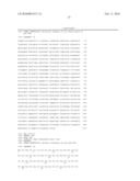 | 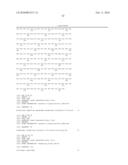 |
 | 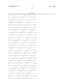 |
 |  |
 |  |
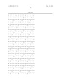 | 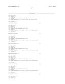 |
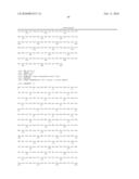 | 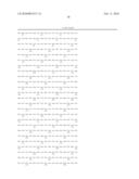 |
| New patent applications in this class: | |
| Date | Title |
|---|---|
| 2022-05-05 | Method for diagnosing human t-cell leukemia virus type 1 (htlv-1) associated diseases |
| 2022-05-05 | Systems and methods for assay processing |
| 2022-05-05 | Rapid pathology/cytology without wash |
| 2019-05-16 | Biofluidic triggering system and method |
| 2019-05-16 | Method and system for detection of disease agents in blood |
| New patent applications from these inventors: | |
| Date | Title |
|---|---|
| 2013-07-04 | Compositions and methods for detecting viral infection using direct-label fluorescence in situ hybridization |
| 2012-11-15 | Markers of xmrv infection and uses thereof |
| 2012-07-05 | Markers of xmrv infection and uses thereof |
| 2011-11-17 | Human immunodeficiency virus and uses thereof |
| Top Inventors for class "Chemistry: molecular biology and microbiology" | |
| Rank | Inventor's name |
|---|---|
| 1 | Marshall Medoff |
| 2 | Anthony P. Burgard |
| 3 | Mark J. Burk |
| 4 | Robin E. Osterhout |
| 5 | Rangarajan Sampath |