Patent application title: GLUTAMINYL CYCLASE AS A DIAGNOSTIC/PROGNOSTIC INDICATOR FOR NEURODEGENERATIVE DISEASES
Inventors:
Hans-Ulrich Demuth (Halle/saale, DE)
Stephan Schilling (Halle/saale, DE)
Martin Kleinschmidt (Halle/saale, DE)
Martin Kleinschmidt (Halle/saale, DE)
Jens-Ulrich Rahfeld (Lieskau, DE)
Jens-Ulrich Rahfeld (Lieskau, DE)
Astrid Kehlen (Halle/saale, DE)
Monique Haegele (Leipzig, DE)
Assignees:
PROBIODRUG AG
IPC8 Class: AG01N3353FI
USPC Class:
435 792
Class name: Involving antigen-antibody binding, specific binding protein assay or specific ligand-receptor binding assay assay in which an enzyme present is a label heterogeneous or solid phase assay system (e.g., elisa, etc.)
Publication date: 2010-02-04
Patent application number: 20100028918
Inventors list |
Agents list |
Assignees list |
List by place |
Classification tree browser |
Top 100 Inventors |
Top 100 Agents |
Top 100 Assignees |
Usenet FAQ Index |
Documents |
Other FAQs |
Patent application title: GLUTAMINYL CYCLASE AS A DIAGNOSTIC/PROGNOSTIC INDICATOR FOR NEURODEGENERATIVE DISEASES
Inventors:
Stephan Schilling
Jens-Ulrich Rahfeld
Hans-Ulrich Demuth
Martin Kleinschmidt
Astrid Kehlen
Monique Haegele
Agents:
SONNENSCHEIN NATH & ROSENTHAL LLP
Assignees:
PROBIODRUG AG
Origin: CHICAGO, IL US
IPC8 Class: AG01N3353FI
USPC Class:
435 792
Patent application number: 20100028918
Abstract:
A method for predicting, diagnosing and prognosticating a
neurodegenerative disease, such as Alzheimer's disease (AD), Mild
Cognitive Impairment (MCI) and neurodegeneration in Down's syndrome (NDS)
using glutaminyl cyclase (QC) as a diagnostic/prognostic indicator. The
use of antibodies binding to QC and kits for performing said diagnostic
method are also provided.Claims:
1. A method for diagnosing a neurodegenerative disease in a subject, the
method comprising:detecting an amount of glutaminyl cyclase (QC), or an
isoform thereof, in a biological sample of said subject; andcomparing the
detected amount of QC in the biological sample with an amount of QC
characteristic of a normal control;whereinan elevated amount of QC in
said biological sample relative to the normal control is a positive
indicator of the neurodegenerative disease; andthe neurodegenerative
disease is selected from the group consisting of Alzheimer's Disease
(AD), Neurodegeneration in Down's Syndrome (NDS) and Mild Cognitive
Impairment (MCI).
2. The method of claim 1 further comprising:detecting an amount of Aβ N3pE-X,comparing the detected amount Aβ N3pE-X in the biological sample with an amount of Aβ N3pE-X characteristic of a normal control;whereinan elevated amount of QC and Aβ N3pE-X in said biological sample relative to the normal control is a positive indicator of the neurodegenerative disease; andX is an integer selected from the group consisting of 38, 40 and 42.
3. The method of claim 1 further comprising:detecting an amount of a chemokine;comparing the detected amount of the chemokine in the biological sample with an amount of chemokine characteristic of a normal control;wherein an elevated amount of QC and chemokine in said biological sample relative to the normal control is a positive indicator of the neurodegenerative disease.
4. The method according to claim 1, wherein said QC is human QC or an isoform thereof, having an amino acid sequence selected from the group consisting of SEQ ID NO 1; SEQ ID NO: 2; SEQ IDNO: 3; SEQ ID: NO: 4; and SEQ ID NO: 5.
5. The method according to claim 4, wherein said QC is human QC of SEQ ID NO: 1.
6. The method according to claim 2, wherein said QC is human QC or an isoform thereof, having an amino acid sequence selected from the group consisting of SEQ ID NO 1; SEQ ID NO: 2; SEQ IDNO: 3; SEQ ID: NO: 4; and SEQ ID NO: 5.
7. The method according to claim 6, wherein said QC is human QC of SEQ ID NO: 1.
8. The method according to claim 3, wherein said QC is human QC or an isoform thereof, having an amino acid sequence selected from the group consisting of SEQ ID NO 1; SEQ ID NO: 2; SEQ IDNO: 3; SEQ ID: NO: 4; and SEQ ID NO: 5.
9. The method according to claim 8, wherein said QC is human QC of SEQ ID NO: 1.
10. The method according to claim 1, wherein said biological sample is serum, plasma, urine or cerebrospinal fluid.
11. The method according to claim 10, wherein said biological sample is plasma.
12. The method according to claim 2, wherein said biological sample is serum, plasma, urine or cerebrospinal fluid.
13. The method according to claim 12, wherein said biological sample is plasma.
14. The method according to claim 3, wherein said biological sample is serum, plasma, urine or cerebrospinal fluid.
15. The method according to claim 14, wherein said biological sample is plasma.
16. The method according to claim 1, wherein the amount of QC is detected by immunoturbidimetric assay, immunofluorescence, immunodiffusion, enzyme-linked immunosorbent assay (ELISA), radioimmunoassay (RIA), Western blot, protein activity assay, Northern Blot, PCR, high performance liquid chromatography (HPLC), mass spectrometry (MS), gas chromatography (GC), GC-MS, LC-MS, or LC-MS/MS.
17. The method according to claim 2, wherein the amount of QC is detected by immunoturbidimetric assay, immunofluorescence, immunodiffusion, enzyme-linked immunosorbent assay (ELISA), radioimmunoassay (RIA), Western blot, protein activity assay, Northern Blot, PCR, high performance liquid chromatography (HPLC), mass spectrometry (MS), gas chromatography (GC), GC-MS, LC-MS, or LC-MS/MS.
18. The method according to claim 3, wherein the amount of QC is detected by immunoturbidimetric assay, immunofluorescence, immunodiffusion, enzyme-linked immunosorbent assay (ELISA), radioimmunoassay (RIA), Western blot, protein activity assay, Northern Blot, PCR, high performance liquid chromatography (HPLC), mass spectrometry (MS), gas chromatography (GC), GC-MS, LC-MS, or LC-MS/MS.
19. The method according to claim 1, wherein the amount of QC, or an isoform thereof, is detected on the basis of the protein level of said QC or isoform thereof.
20. The method according to claim 2, wherein the amount of QC, or an isoform thereof, is detected on the basis of the protein level of said QC or isoform thereof.
21. The method according to claim 3, wherein the amount of QC, or an isoform thereof, is detected on the basis of the protein level of said QC or isoform thereof.
22. The method according to claim 1, wherein the amount of QC is detected using an antibody that specifically binds to QC, or an isoform thereof.
23. The method according to claim 2, wherein the amount of QC is detected using an antibody that specifically binds to QC, or an isoform thereof.
24. The method according to claim 3, wherein the amount of QC is detected using an antibody that specifically binds to QC, or an isoform thereof.
25. The method according to claim 1, wherein the amount of QC is detected by measuring the enzymatic activity of QC, or an isoform thereof.
26. The method according to claim 2, wherein the amount of QC is detected by measuring the enzymatic activity of QC, or an isoform thereof.
27. The method according to claim 3, wherein the amount of QC is detected by measuring the enzymatic activity of QC, or an isoform thereof.
28. The method according to claim 1, wherein the amount of QC, or an isoform thereof, is detected on the basis of the mRNA level of said QC or isoform thereof.
29. The method according to claim 2, wherein the amount of QC, or an isoform thereof, is detected on the basis of the mRNA level of said QC or isoform thereof.
30. The method according to claim 3, wherein the amount of QC, or an isoform thereof, is detected on the basis of the mRNA level of said QC or isoform thereof.
31. The method of claim 2, wherein X is 42.
32. The method of claim 2, wherein X is 40.
33. The method of claim 2, wherein X is 38.
34. The method according to claim 2, wherein detecting an amount of Aβ N3pE-X comprises detecting (i) one or more of Aβ N3pE-42, Aβ N3pE-40, and Aβ N3pE-38 and (ii) at least one of pGluABri or pGluADan.
35. The method according to claim 34, wherein detecting an amount of Aβ N3pE-X comprises detecting (i) two or more of Aβ N3pE-42, Aβ N3pE-40, and Aβ N3pE-38 and (ii) at least one of pGluABri or pGluADan.
36. The method according to claim 3, wherein said chemokine is selected from CCL2, CCL7, CCL8, CCL9/10, CCL13, CCL15, CCL16, CCL25 and Fractalkine.
37. The method according to claim 36, wherein said chemokine is CCL2.
38. The method of claim 1 further comprising:obtaining a biological sample from said subject;wherein detecting the amount of glutaminyl cyclase (QC), or an isoform thereof, in the biological sample of said subject comprisescontacting said biological sample with an antibody that binds to glutaminyl cyclase (QC), or its isoforms;allowing the antibody and QC to form an immune complex; anddetecting the amount of immune complex formed as an indication of the amount of QC in said biological sample.
39. The method of claim 1, wherein detecting the amount of QC, or an isoform thereof, occurs in vitro.
40. The method of claim 2, wherein detecting the amount of QC and Aβ N3pE-X occurs in vitro.
41. The method of claim 3, wherein detecting the amount of QC and chemokine occurs in vitro.
42. The method of claim 1, wherein the neurodegenerative disease is AD.
43. The method of claim 1, wherein the neurodegenerative disease is NDS.
44. The method of claim 1, wherein the neurodegenerative disease is MCI.
45. A kit for diagnosing a neurodegenerative disease comprising an antibody that binds to QC and an established standard of an amount of QC characteristic of a normal control.
46. The kit of claim 27, wherein the neurodegenerative disease is selected from the group consisting of Alzheimer's Disease (AD), Neurodegeneration in Down's Syndrome (NDS) and Mild Cognitive Impairment (MCI).
Description:
CROSS-REFERENCE TO RELATED APPLICATIONS
[0001]This application claims priority to U.S. Provisional Application Ser. No. 61/085,154 filed on Jul. 31, 2008, which is incorporated herein by reference in its entirety.
INCORPORATION-BY-REFERENCE OF MATERIAL SUBMITTED IN COMPUTER READABLE FORMAT (CRF)
[0002]The Sequence Listing, which is a part of the present disclosure, includes a computer readable form comprising nucleotide and/or amino acid sequences of the present invention. The subject matter of the Sequence Listing is incorporated herein by reference in its entirety.
FIELD OF THE INVENTION
[0003]The present invention relates to a method for predicting, diagnosing and prognosticating a neurodegenerative disease, such as Alzheimer's disease (AD), Mild Cognitive Impairment (MCI) and neurodegeneration in Down's syndrome (NDS) using glutaminyl cyclase (QC) as a diagnostic/prognostic indicator.
BACKGROUND OF THE INVENTION
[0004]Alzheimer Disease (AD) is a neurodegenerative disease that causes dementia. The terms "Alzheimer Disease" and "Alzheimer's Disease" are both utilized in the art, these terms being equivalent and are used interchangeably here and elsewhere. The period from first detection of AD to termination can range from a few years to 15 years, during which time the patient progressively suffers loss of both mental function and control of bodily functions. There is significant variability in the progress of the disease. While the majority of patients have a gradual, inexorable progression (losing on average 3 to 4 points on the 30 point Folstein mini-mental state score annually), approximately 30% of AD cases have a prolonged stable initial plateau phase lasting several years (Haxby J. V., et al., Individual trajectories of cognitive decline in patients with dementia of the Alzheimer type, J. Clin. Exp. Neuropsychol 14:575-592, 1992.). A subgroup of patients has a fulminant, rapidly progressive downhill course over several years (Mann, U., et al., Heterogeneity in Alzheimer's disease: Progression rate segregated by distinct neuropsychological and cerebral metabolic profiles, J. Neurol. Neurosurg. Psychiatry 55:956-959, 1992). Other patients (about 10% of cohorts) remain slowly progressive, showing only gradual decline from year to year (Grossi, D., et al., Senile dementias, II International Symposium (pp. 97-99), Paris: John Libbey Eurotext, 1988.). The pathological, chemical and molecular bases of this heterogeneity remain undetermined. Recognition of the variability of AD progression represents an important clinical insight, and may explain the diagnostic difficulties presented by "atypical" cases. While in certain cases, there is a familial manifestation of the AD disease, it appears that the majority of AD cases are non-familial, and until recently (see below), no simple biological marker for the disease had been determined.
[0005]Current methods used to diagnose AD involve analysis of cerebrospinal fluid (CSF) or brain tissue obtained from postmortem patients. Thus, among the markers currently under consideration are those related to the proteins, which account for the features found in Alzheimer brains postmortem. The neurofibrillary tangle is composed primarily of a hyperphosphorylated tau protein, a cytoskeletal protein. The neuritic plaque contains a core of amyloid protein, much of which is a 42-amino acid peptide (Aβ42) derived from proteolytic cleavage of a larger precursor protein. Another form of this protein derived from the same precursor contains only 40 amino acids (Aβ40). Deposits of this protein are found in the brains of Aβ victims. However, alterations in tau and the aforementioned beta amyloid peptides do not occur with sufficient frequency and magnitude so as to afford diagnostic value and therefore, blood tests based on these proteins do not seem to correlate well with AD. In addition to C-terminal variability, N-terminally modified Aβ peptides are abundant (Saido, T. C. et al. Dominant and differential deposition of distinct beta-amyloid peptide species, Aβ N3(pE), in senile plaques. Neuron 14, 457-466 (1995); Russo, C. et al. Presenilin-1 mutations in Alzheimer's disease. Nature 405, 531-532 (2000); Saido, T. C., Yamao, H., Iwatsubo, T. & Kawashima, S. Amino- and carboxyl-terminal heterogeneity of beta-amyloid peptides deposited in human brain. Neurosci. Lett. 215, 173-176 (1996)). It appears that a major proportion of the Aβ peptides undergoes N-terminal truncation by two amino acids, exposing a glutamate residue, which is subsequently cyclized into pyroglutamate (pE), resulting in Aβ3(pE)-42 peptides (Saido, T. C. et al. Dominant and differential deposition of distinct beta-amyloid peptide species, Aβ N3(pE), in senile plaques. Neuron 14, 457-466 (1995); Saido, T. C., Yamao, H., Iwatsubo, T. & Kawashima, S. Amino- and carboxyl-terminal heterogeneity of beta-amyloid peptides deposited in human brain. Neurosci. Lett. 215, 173-176 (1996)). Alternatively, pE may be formed following β'-cleavage by BACE1, resulting in Aβ N11(pE)-42 (Naslund, J. et al. Relative abundance of Alzheimer Aβ amyloid peptide variants in Alzheimer disease and normal aging. Proc. Natl. Acad. Sci. U.S.A. 91, 8378-8382 (1994); Liu, K. et al. Characterization of Aβ11-40/42 peptide deposition in Alzheimer's disease and young Down's syndrome brains: implication of N-terminally truncated Abeta species in the pathogenesis of Alzheimer's disease. Acta Neuropathol. 112, 163-174 (2006)). In particular Aβ N3(pE)-42 has been shown to be a major constituent of Aβ deposits in sporadic and familial AD (Saido, T. C. et al. Dominant and differential deposition of distinct beta-amyloid peptide species, Aβ N3(pE), in senile plaques. Neuron 14, 457-466 (1995); Miravalle, L. et al. Amino-terminally truncated Aβ peptide species are the main component of cotton wool plaques. Biochemistry 44, 10810-10821 (2005)).
[0006]The Aβ N3pE-42 peptides coexist with Aβ 1-40/1-42 peptides (Saido, T. C. et al. Dominant and differential deposition of distinct beta-amyloid peptide species, Abeta N3pE, in senile plaques. Neuron 14, 457-466 (1995); Saido, T. C., Yamao, H., Iwatsubo, T. & Kawashima, S. Amino- and carboxyl-terminal heterogeneity of beta-amyloid peptides deposited in human brain. Neurosci. Lett. 215, 173-176 (1996)), and, based on a number of observations, could play a prominent role in the pathogenesis of AD. For example, a particular neurotoxicity of Aβ N3pE-42 peptides has been outlined (Russo, C. et al. Pyroglutamate-modified amyloid beta-peptides--AbetaN3(pE)--strongly affect cultured neuron and astrocyte survival. J. Neurochem. 82, 1480-1489 (2002) and the pE-modification of N-truncated Aβ peptides confers resistance to degradation by most aminopeptidases as well as Aβ-degrading endopeptidases (Russo, C. et al. Pyroglutamate-modified amyloid beta-peptides--AbetaN3(pE)--strongly affect cultured neuron and astrocyte survival. J. Neurochem. 82, 1480-1489 (2002); Saido, T. C. Alzheimer's disease as proteolytic disorders: anabolism and catabolism of beta-amyloid. Neurobiol. Aging 19, S69-S75 (1998)). The cyclization of glutamic acid into pE leads to a loss of N-terminal charge resulting in accelerated aggregation of Aβ N3pE compared to the unmodified Aβ peptides (He, W. & Barrow, C. J. The Aβ 3-pyroglutamyl and 11-pyroglutamyl peptides found in senile plaque have greater beta-sheet forming and aggregation propensities in vitro than full-length Aβ. Biochemistry 38, 10871-10877 (1999); Schilling, S. et al. On the seeding and oligomerization of pGlu-amyloid peptides (in vitro). Biochemistry 45, 12393-12399 (2006)). Thus, reduction of Aβ N3pE-42 formation should destabilize the peptides by making them more accessible to degradation and would, in turn, prevent the formation of higher molecular weight Aβ aggregates and enhance neuronal survival.
[0007]However, for a long time it was not known how the pE-modification of Aβ peptides occurs. The present Applicant discovered that glutaminyl cyclase (QC) is capable to catalyze Aβ N3pE-42 formation under mildly acidic conditions, that specific QC inhibitors prevent Aβ N3pE-42 generation in vitro and that, therefore, inhibition of glutaminyl cyclase is a novel therapeutic concept for the causative treatment of Alzheimer's disease (Schilling, S., Hoffmann, T., Manhart, S., Hoffmann, M. & Demuth, H.-U. Glutaminyl cyclases unfold glutamyl cyclase activity under mild acid conditions. FEBS Lett. 563, 191-196 (2004); Cynis, H. et al. Inhibition of glutaminyl cyclase alters pyroglutamate formation in mammalian cells. Biochim. Biophys. Acta 1764, 1618-1625 (2006); Schilling et al. Inhibition of glutaminyl cyclase--a novel therapeutic concept for the causative treatment of Alzheimer's disease. Nature Medicine 14, 1106-1111 (2008)).
[0008]At present, there appears to be no satisfactory-diagnostic marker for existing AD, or for a subject, who although exhibiting normal cognitive responses, will inevitably, or most likely, develop AD.
[0009]Age-Associated Cognitive Decline (AACD) and Mild Cognitive Impairment (MCI) are terms used to identify individuals who experience a cognitive decline that falls short of dementia. These terms are equivalent, MCI being a more recently adopted term, and are used interchangeably throughout this application. Satisfaction of criteria (World Health Organization) for this diagnosis requires a report by the individual or family of a decline in cognitive function, which is gradual, and present at least 6 months. There may be difficulties across any cognitive domains (although memory is impaired in the vast majority of cases), and these must be supported by abnormal performance on quantitative cognitive assessments for which age and education norms are available for relatively healthy individuals (i.e., the patient is compared to normal subjects his/her own age). Performance must be at least 1 SD below the mean value for the appropriate population on such tests. Neither dementia, nor significant depression or drug effects may be present. No cerebral or systemic disease or condition known to cause cerebral cognitive dysfunction may be present. In Applicant's experience, all patients who were classified as CDR.5 ("questionable dementia") on the Clinical Dementia rating scale and who met these exclusions, also met the criteria for AACD/MCI. About 1/3 of Alzheimer's patients have had a clearly definable period of isolated memory deficit which preceded their more global cognitive decline. (Haxby J. V., et al., Individual trajectories of cognitive decline in patients with dementia of the Alzheimer type, J. Clin. Exp. Neuropsychology 14:575-592, 1992.) Using AACD/MCI criteria, which look at other domains in addition to memory, the percentage with an identifiable prodrome is likely higher. Fortunately, not all AACD/MCI individuals seem to decline. It appears that a significant number of these subjects show a stable, non-progressive memory deficit on testing.
[0010]Attempts at predicting the onset of AD, MCI or NDS, or monitoring their progression have met with limited success.
SUMMARY OF THE INVENTION
[0011]It has been discovered by the inventors of this application that an amount of QC in a biological sample obtained from a subject that deviates from a reference amount in a control person can be positively correlated to a neurological disease state. Thus, the correlation of the presence of QC with the disease state represents a positive and more direct test for diagnosis in a patient suffering from one of the neurodegenerative diseases described above.
[0012]Accordingly, the invention provides an easily administered biological sample test for predicting, diagnosing, or prognosticating AD, MCI and NDS using QC as a diagnostic marker.
[0013]The present invention is based at least in part on the discovery that an amount of glutaminyl cyclase (QC) in a biological sample obtained from a subject suffering from AD or MCI is elevated compared to an amount of QC in the biological sample obtained from a normal (i.e. healthy) control subject.
[0014]The indication that the amount of QC differs between these neurological diseases and normal controls, forms the basis for the development of a test for diagnosing AD, MCI or NDS in a subject. As such, the methods for diagnosing AD, MCI or NDS of the present invention by measuring the amount of QC in patient sample will greatly improve current clinical diagnostic assessment for patients suffering from these neurodegenerative diseases.
[0015]Based on the newly discovered differences in the amount of QC present in a biological sample obtained from a patient compared to that of a normal control, a strong correlation of the amount of QC can be made to a probable diagnosis of a neurodegenerative disease. A statistically significant elevation in the amount of QC relative to control samples is reasonably predictive that the patient has AD, NDS or MCI. A normal amount of QC as determined by an amount of QC characteristic of a control QC sample isolated from a normal age-matched population indicates that the patient does not have a neurodegenerative disease, such as AD, MCI or NDS. A positive indication of a neurodegenerative disease based on an elevated or reduced amount of QC in a biological sample relative to a normal control is generally considered together with other factors in making a definitive determination of a particular disease. Therefore, the elevated or reduced QC levels of the subject being tested will usually be considered together with other accepted clinical symptoms of AD, MCI or NDS-related conditions in making a determinative diagnosis of a neurodegenerative disease.
[0016]Thus, according to a first aspect of the invention, there is provided a method for diagnosing probable Alzheimer's Disease (AD), Neurodegeneration in Down's syndrome (NDS) or Mild Cognitive Impairment (MCI) in a subject, the method comprising: (a) detecting the amount of glutaminyl cyclase (QC), or its isoforms, in a biological sample obtained from said subject; and (b) comparing the detected amount of QC in the biological sample with an amount of QC characteristic of a normal control; whereby an elevated amount of QC in said biological sample relative to the normal control is a positive indicator of AD or MCI.
[0017]According to a preferred embodiment of the invention, the biological sample is a fluid body sample such as serum, plasma, urine or cerebrospinal fluid. More preferably, the fluid body sample is plasma.
[0018]According to a further embodiment of the present invention, the amount of QC is detected either on the basis of the QC protein level or the QC mRNA level.
[0019]The amount of QC detected or quantified in a biological sample from a subject can be accomplished by any means known in the art. Such means may include, but are not limited to, for example by immunoturbidimetric assay, immunofluorescence, immunodiffusion, enzyme-linked immunosorbent assay (ELISA), radioimmunoassay (RIA), Western Blot, protein activity assay or, for the determination of the QC mRNA level, Northern Blot or polymerase chain reaction (PCR) analysis, for example real-time PCR. Also useful are high performance liquid chromatography (HPLC), mass spectrometry (MS) and gas chromatography (GC), as well as their various configurations, including gas chromatograph-mass spectrometry (GC-MS), liquid chromatography-mass spectrometry (LC-MS) and liquid-chromatography-tandem mass spectrometry (LC-MS/MS) systems.
[0020]Preferably, the amount of QC in the biological sample is detected using an antibody that binds to QC in an immunoassay format. Thus, according to a preferred embodiment of the invention, there is provided a method of diagnosing a neurodegenerative disease in a subject, the method comprising: (a) obtaining a biological sample from said subject; (b) contacting said biological sample with an antibody that binds to glutaminyl cyclase (QC), or its isoforms; (c) allowing the antibody and QC to form an immune complex; and (d) detecting the amount of immune complex formed as an indication of the amount of QC in said biological sample; and (e) comparing the detected amount to a normal control; whereby a detected amount that is elevated or reduced relative to the normal control is a positive indicator of a neurodegenerative disease.
[0021]According to yet a further aspect of the invention, there is provided a diagnostic kit for determining whether a subject is suffering from a neurodegenerative disease comprising an antibody that binds to QC and an established standard of an amount of QC characteristic of a normal control. Reagents and instructions for carrying out the assays may also be included.
[0022]Other objects and features will be in part apparent and in part pointed out hereinafter.
DESCRIPTION OF THE DRAWINGS
[0023]Those of skill in the art will understand that the drawings, described below, are for illustrative purposes only. The drawings are not intended to limit the scope of the present teachings in any way.
[0024]FIG. 1: FIG. 1(a) shows the analysis of QC transcript levels applying quantitative RT-PCR. Total RNA from human neocortical brain samples (Brodmann area 22) was isolated from normally aged and AD brains of different Braak stages as indicated. The QC transcript level was normalized to house-keeping transcript concentration. FIG. 1(b) shows the Western-Blot analysis for QC from the same cases and brain region as used for QC mRNA analysis. The extraction of soluble protein was normalized to the tissue weight. FIG. 1(c) shows the quantification of Aβ N3(pE)-42 (indicated as Aβ3(PE)-42) and of Aβ 1-42 (Aβ1-42) concentrations from the same cases and brain region applying ELISA analysis of SDS- and formic acid extracts of human neocortical brain samples. Note the robust increase in Aβ N3(pE)-42 peptide concentrations at early AD stages compared to the much more moderate increase in Aβ 1-42 peptides. FIG. 1(d) shows the immunohistochemical detection of total Aβ peptides by the antibody 4G8 and of Aβ N3(pE)-42 peptides in Brodmann area 22 from normally aged subjects and different AD stages. Sparse Aβ plaques were detected in normal aging but these deposits lacked Aβ N3(pE)-42 immunoreactivity. At all AD stages, however, the majority of Aβ plaques contains Aβ N3(pE)-42 peptides.
[0025]FIG. 2 shows the results of the determination of the gene expression rate of QC and CCL2 in stimulated THP-1 cells.
[0026]FIG. 3 shows the results of the determination of the specific QC activity in conditioned medium of THP-1 cells.
TABLE-US-00001 SEQUENCES OF AMYLOID PEPTIDES AND CHEMOKINES Aβ(1-42) Asp-Ala-Glu-Phe-Arg-His-Asp-Ser- (SEQ ID NO: 6) Gly-Tyr-Glu-Val-His-His-Gln-Lys- Leu-Val-Phe-Phe-Ala-Glu-Asp-Val- Gly-Ser-Asn-Lys-Gly-Ala-Ile-Ile- Gly-Leu-Met-Val-Gly-Gly-Val-Val- Ile-Ala Aβ(1-40) Asp-Ala-Glu-Phe-Arg-His-Asp-Ser- (SEQ ID NO: 7) Gly-Tyr-Glu-Val-His-His-Gln-Lys- Leu-Val-Phe-Phe-Ala-Glu-Asp-Val- Gly-Ser-Asn-Lys-Gly-Ala-Ile-Ile- Gly-Leu-Met-Val-Gly-Gly-Val-Val Aβ(3-42) Glu-Phe-Arg-His-Asp-Ser-Gly-Tyr- (SEQ ID NO: 8) Glu-Val-His-His-Gln-Lys-Leu-Val- Phe-Phe-Ala-Glu-Asp-Val-Gly-Ser- Asn-Lys-Gly-Ala-Ile-Ile-Gly-Leu- Met-Val-Gly-Gly-Val-Val-Ile-Ala Aβ(3-40) Glu-Phe-Arg-His-Asp-Ser-Gly-Tyr- (SEQ ID NO: 9) Glu-Val-His-His-Gln-Lys-Leu-Val- Phe-Phe-Ala-Glu-Asp-Val-Gly-Ser- Asn-Lys-Gly-Ala-Ile-Ile-Gly-Leu- Met-Val-Gly-Gly-Val-Val Aβ(1-38) Asp-Ala-Glu-Phe-Arg-His-Asp-Ser- (SEQ ID NO: 10) Gly-Tyr-Glu-Val-His-His-Gln-Lys- Leu-Val-Phe-Phe-Ala-Glu-Asp-Val- Gly-Ser-Asn-Lys-Gly-Ala-Ile-Ile- Gly-Leu-Met-Val-Gly-Gly Aβ(3-38) Glu-Phe-Arg-His-Asp-Ser-Gly-Tyr- (SEQ ID NO: 11) Glu-Val-His-His-Gln-Lys-Leu-Val- Phe-Phe-Ala-Glu-Asp-Val-Gly-Ser- Asn-Lys-Gly-Ala-Ile-Ile-Gly-Leu- Met-Val-Gly-Gly ABri EASNCFAIRHFENKFAVETLICSRTVKKNIIE (SEQ ID NO: 12) EN ADan EASNCFAIRHFENKFAVETLICFNLFLNSQEK (SEQ ID NO: 13) HY CCL2 QPDAINAPVTCCYNFTNRKISVQRLASYRRIT (small inducible SSKCPKEAVIFKTIVAKEICADPKQKWVQDSM cytokine A2) DHLDKQTQTPKT (SEQ ID NO: 14) Swiss-Prot: P13500 CCL7 QPVGINTSTTCCYRFINKKIPKQRLESYRRTT (Small-inducible SSHCPREAVIFKTKLDKEICADPTQKWVQDFM cytokine A7) KHLDKKTQTPKL (SEQ ID NO: 15) Swiss-Prot: P80098 CCL8 QPDSVSIPITCCFNVINRKIPIQRLESYTRIT small inducible NIQCPKEAVIFKTKRGKEVCADPKERWVRDSM cytokine A8) KHLDQIFQNLKP (SEQ ID NO: 16) Swiss-Prot: P80075 CCL9/10 QITHATETKEVQSSLKAQQGLEIEMFHMGFQD (Small-inducible SSDCCLSYNSRIQCSRFIGYFPTSGGCTRPGI cytokine A9) IFISKRGFQVCANPSDRRVQRCIERLEQNSQP (SEQ ID NO: 17) RTYKQ Swiss-Prot: P51670 CCL13 QPDALNVPSTCCFTFSSKKISLQRLKSYVITT (Small-inducible SRCPQKAVIFRTKLGKEICADPKEKWVQNYMK cytokine A13) HLGRKAHTLKT (SEQ ID NO: 18) Swiss-Prot: Q99616 CCL15 QFINDAETELMMSKLPLENPVVLNSFHFAADC (Small-inducible CTSYISQSIPCSLMKSYFETSSECSKPGVIFL cytokine A15) TKKGRQVCAKPSGPGVQDCMKKLKPYSI (SEQ ID NO: 19) Swiss-Prot: Q16663 CCL16 QPKVPEWVNTPSTCCLKYYEKVLPRRLWGYRK (Small-inducible ALNCHLPAIIFVTKRNREVCTNPNDDWVQEYI cytokine A16) KDPNLPLLPTRNLSTVKIITAKNGQPQLLNSQ (SEQ ID NO: 20) Swiss-Prot: O15467 Fractalkine QHHGVTKCNITCSKMTSKIPVALLIHYQQNQA (neurotactin) SCGKRAIILETRQHRLFCADPKEQWVKDAMQH (SEQ ID NO: 21) LDRQAAALTRNGGTFEKQIGEVKPRTTPAAGG Swiss-Prot: P78423 MDESVVLEPEATGESSSLEPTPSSQEAQRALG TSPELPTGVTGSSGTRLPPTPKAQDGGPVGTE LFRVPPVSTAATWQSSAPHQPGPSLWAEAKTS EAPSTQDPSTQASTASSPAPEENAPSEGQRVW GQGQSPRPENSLEREEMGPVPAHTDAFQDWGP GSMAHVSVVPVSSEGTPSREPVASGSWTPKAE EPIHATMDPQRLGVLITPVPDAQAATRRQAVG LLAFLGLLFCLGVAMFTYQSLQGCPRKMAGEM AEGLRYIPRSCGSNSYVLVPV CCL25 QGVFEDCCLAYHYPIGWAVLRRAWTYRIQEVS (Small-inducible GSCNLPAAIFYLPKRHRKVCGNPKSREVQRAM cytokine A25) KLLDARNKVFAKLHHNTQTFQAGPHAVKKLSS (SEQ ID NO: 22) GNSKLSSSKFSNPISSSKRNVSLLISANSGL Swiss-Prot: O15444
DETAILED DESCRIPTION OF THE PRESENTLY PREFERRED EMBODIMENTS
[0027]The present invention provides an efficient and rapid in vitro method for diagnosing a neurodegenerative disease by directly detecting an amount of QC in a biological sample obtained from a subject and comparing the detected amount of QC with an amount of QC characteristic of a normal control. An elevated amount of QC in the biological sample of the subject is a positive indication of AD or MCI or NDS. Thus, as described herein, it is demonstrated that QC is consistently and significantly elevated in a biological sample of AD, NDS or MCI patients compared to normal controls. As such, the methods for diagnosing AD, MCI or NDS of the present invention by detecting or quantifying the amount of QC in a patient sample will greatly improve current clinical diagnostic assessment for patients suffering from these neurodegenerative diseases.
[0028]Accordingly, there is provided a method for assessing whether a subject may be suffering from AD, MCI or NDS using QC as a biological marker.
[0029]Glutaminyl cyclase or glutaminyl-peptide cyclotransferase (QC, EC 2.3.2.5) catalyzes the intramolecular cyclization of N-terminal glutaminyl residues into pyroglutamic acid (5-oxo-proline, pGlu*) under liberation of ammonia and the intramolecular cyclization of N-terminal glutamyl residues into pyroglutamic acid under liberation of water.
[0030]A QC was first isolated by Messer from the Latex of the tropical plant Carica papaya in 1963 (Messer, M. 1963 Nature 4874, 1299). 24 years later, a corresponding enzymatic activity was discovered in animal pituitary (Busby, W. H. J. et al. 1987 J Biol Chem 262, 8532-8536; Fischer, W. H. and Spiess, J. 1987 Proc Natl Acad Sci USA 84, 3628-3632). For the mammalian QCs, the conversion of Gln into pGlu by QC could be shown for the precursors of TRH and GnRH (Busby, W. H. J. et al. 1987 J Biol Chem 262, 8532-8536; Fischer, W. H. and Spiess, J. 1987 Proc Natl Acad Sci USA 84, 3628-3632). In addition, initial localization experiments of QC revealed a co-localization with its putative products of catalysis in the bovine tractus hypothalamo-hypophysalisfurther improving the suggested function in peptide hormone maturation (Bockers, T. M. et al. 1995 J Neuroendocrinol 7, 445-453). In contrast, the physiological function of the plant QC is less clear. In case of the enzyme from C. papaya, a role in the plant defence against pathogenic microorganisms was suggested (El Moussaoui, A. et al. 2001 Cell Mol Life Sci 58, 556-570). Putative QCs from other plants were identified by sequence comparisons recently (Dahl, S. W. et al. 2000 Protein Expr Purif 20, 27-36). The physiological function of these enzymes, however, is still ambiguous.
[0031]The QCs known from plants and animals show a strict specificity for L-Glutamine in the N-terminal position of the substrates and their kinetic behaviour was found to obey the Michaelis-Menten equation (Pohl, T. et al. 1991 Proc Natl Acad Sci USA 88, 10059-10063; Consalvo, A. P. et al. 1988 Anal Biochem 175, 131-138; Gololobov, M. Y. et al. 1996 Biol Chem Hoppe Seyler 377, 395-398). A comparison of the primary structures of the QCs from C. papaya and that of the highly conserved QC from mammals, however, did not reveal any sequence homology (Dahl, S. W. et al. (2000) Protein Expr Purif 20, 27-36). Whereas the plant QCs appear to belong to a new enzyme family (Dahl, S. W. et al. (2000) Protein Expr Purif 20, 27-36), the mammalian QCs were found to have a pronounced sequence homology to bacterial aminopeptidases (Bateman, R. C. et al. 2001 Biochemistry 40, 11246-11250), leading to the conclusion that the QCs from plants and animals have different evolutionary origins.
[0032]Gostranova et al. have found that glutaminyl cyclase activity is a characteristic feature of cerebrospinal fluid in multiple sclerosis patients and controls (Gostranova et al., Clin Chim Acta. 2008 389 (1-2), pp. 152-159).
[0033]Different isoforms of QC, the glutaminyl-peptide cyclotransferase-like proteins (QPCTLs) have been observed (WO 2008/034891). These novel proteins have significant sequence similarity to glutaminyl cyclase, e.g. the QPCTL from human (further named as isoQC) (GenBank accession no. NM--017659).
[0034]Multiple isoforms of a protein, such as QC or human isoQC, can also be produced from a single gene by a variety of mechanisms, including alternative RNA splicing, post-translational proteolytic processing and cell type-specific glycosylation. Thus, the terms "glutaminyl cyclase", "QC" and "isoQC" as used herein refer to QC in its native form, as well as any of its isoforms.
[0035]Preferred for the use of the present invention are human QC or its isoforms, having an amino acid sequence selected from the group of SEQ ID NO's: 1, 2, 3, 4 and 5.
[0036]More preferred for use in the methods of the present invention is the human QPCTL having an amino acid sequence of SEQ ID NO. 2, or even preferred of SEQ ID NO: 3.
[0037]Even preferred for use in the methods of the present invention are spliceforms of human QPCTL having an amino acid sequence of SEQ ID NO. 4 or of SEQ ID NO: 5.
[0038]Most preferred for use in the methods of the present invention is human QC having the amino acid sequence of SEQ ID NO: 1.
[0039]Thus, according to a first aspect of the present invention, there is provided a method for diagnosing probable Alzheimer's Disease (AD), Neurodegeneration in Down's Syndrome (NDS) or Mild Cognitive Impairment (MCI) in a subject, the method comprising:
[0040](a) detecting the amount of glutaminyl cyclase (QC), or an isoform thereof, in a biological sample obtained from said subject; and
[0041](b) comparing the detected amount of QC in the biological sample with an amount of QC characteristic of a normal control;
[0042]whereby an elevated amount of QC in said biological sample relative to the normal control is a positive indicator of AD, NDS or MCI.
[0043]It has been demonstrated by inventors of the present invention that an elevated amount of QC in a biological sample may correlate with an elevated amount of N-terminally truncated and pyroglutamated amyloid beta peptides, such as for example Aβ N3pE-42 and/or Aβ N3pE-40 and/or Aβ N3pE-38.
[0044]Thus, according to a further aspect of the present invention, there is provided a method for diagnosing probable Alzheimer's Disease (AD), Neurodegeneration in Down's Syndrome (NDS) or Mild Cognitive Impairment (MCI) in a subject, the method comprising:
[0045](a) detecting the amount of glutaminyl cyclase (QC), or an isoform thereof, in a biological sample obtained from said subject; and
[0046](b) further detecting the amount of Aβ N3pE-X,
[0047](c) comparing the detected amount of QC and Aβ N3pE-X in the biological sample with an amount of QC and Aβ N3pE-X characteristic of a normal control;
[0048]whereby an elevated amount of QC and Aβ N3pE-X in said biological sample relative to the normal control is a positive indicator of AD, NDS or MCI, and
[0049]wherein X is an integer selected from 38, 40 and 42.
[0050]In a preferred embodiment, X is 42.
[0051]In a further preferred embodiment, X is 40.
[0052]In a yet preferred embodiment, X is 38.
[0053]Further preferred are methods, wherein not only a single form of the N-terminally truncated and pyroglutamated amyloid beta peptides but a combination of Aβ N3pE-42 and/or Aβ N3pE-40 and/or Aβ N3pE-38 is detected together with QC.
[0054]Further preferred are methods, wherein not only a single form of the N-terminally truncated and pyroglutamated amyloid beta peptides but a combination of Aβ N3pE-42 and/or Aβ N3pE-40 and/or Aβ N3pE-38 and/or peptides occurring in familial Alzheimer's dementias, such as pGluABri or pGluADan, is detected together with QC.
[0055]"pGlu-A" or "Aβ N3pE" refers to N-terminally truncated forms of Aβ, that start at the glutamic acid residue at position 3 in the amino acid sequence of Aβ, and wherein said glutamic acid residue is cyclized to form a pyroglutamic acid residue. In particular, by pGlu-Aβ as used herein are meant those fragments which are involved in or associated with the amyloid pathologies including, but not limited to, pGlu-Aβ 3-38, pGlu-Aβ 3-40, p-Glu-Aβ 3-42.
[0056]It has further been demonstrated by the inventors of the present invention that an elevated amount of QC in a biological sample may correlate with an elevated amount of a chemokine, such as for example CCL2, CCL7, CCL8, CCL9/10, CCL13, CCL15, CCL16, CCL25 and Fractalkine.
[0057]Thus, according to a further aspect of the present invention, there is provided a method for diagnosing Alzheimer's Disease (AD), Neurodegeneration in Down's Syndrome (NDS) or Mild Cognitive Impairment (MCI) in a subject, the method comprising:
[0058](a) detecting the amount of glutaminyl cyclase (QC), or an isoform thereof, in a biological sample obtained from said subject; and
[0059](b) further detecting the amount of a chemokine,
[0060](c) comparing the detected amount of QC and the chemokine in the biological sample with an amount of QC and the chemokine characteristic of a normal control;
[0061]whereby an elevated amount of QC and chemokine in said biological sample relative to the normal control is a positive indicator of AD, NDS or MCI.
[0062]In a preferred embodiment, said chemokine is of mammalian origin. More preferably, said chemokine is a human chemokine. Most preferably, said chemokine is human CCL2.
[0063]In a further preferred embodiment, any of the aforementioned methods for diagnosing Alzheimer's Disease (AD), Neurodegeneration in Down's Syndrome (NDS) or Mild Cognitive Impairment (MCI) may also be performed in vitro in a biological sample of a subject.
[0064]The term "subject" refers to a mammal which is afflicted with, or suspected to be afflicted with a neurogenerative disease such as AD, MCI or NDS. Preferably, "subject" refers to a human.
[0065]The term "biological sample" refers to any source of biological material, including, but are not limited to, peripheral blood, plasma, lymphocytes, cerebrospinal fluid, urine, saliva, epithelia, fibroblasts, or any other sample comprising QC protein.
[0066]In a preferred embodiment, the amount of QC is detected in a body fluid sample obtained from a mammal, most preferably a human. The term "body fluid" refers to all fluids that are present in the human body including but not limited to blood, lymph, urine and cerebrospinal fluid (CSF) comprising QC. The blood sample may include a plasma sample or a serum sample, or fractions derived from these samples. The sample can be treated prior to use, such as preparing plasma from blood, diluting viscous fluids, and the like. Preferably, the plasma sample is treated with an anti-coagulant, such as EDTA.
[0067]According to a preferred embodiment of the present invention, the amount of QC is detected in a blood sample taken from the subject, more preferably a plasma sample. Thus, the present invention preferably relates to a method as described above, comprising the steps of: obtaining a plasma sample from said subject; detecting the amount of QC in the plasma sample; comparing the detected amount of QC in the plasma sample with the amount of QC in a plasma sample from a normal control, whereby an elevated amount of QC relative to the normal control is a positive indication of AD, NDS or MCI. Elevated amounts of QC have been shown to correlate with and are useful in aiding the diagnosis of AD, NDS and MCI.
[0068]An "elevated amount" of QC (or an isoform thereof) means that the amount of QC detected in the samples of the subjects is greater than the mean amount of QC characteristic of a normal control person beyond the range of experimental error, as known in the art. Preferably, the amount of QC detected in the samples of the subjects is 10% greater than said mean amount of QC characteristic of a normal control person. More preferably, the amount of QC (or an isoform thereof) detected in the samples of the subjects is 25% greater, or, even more preferred 50% or 75% greater than said mean amount of QC characteristic of a normal control person. Most preferably, the amount of QC (or an isoform thereof) detected in the samples of the subjects is several times greater than said mean amount of QC characteristic of a normal control person, e.g. 2, 3, 4, 5, 6, 7, 8, 9, 10 or more times greater.
[0069]A "normal control" is a biological sample of the same type obtained from the subject, for example that is obtained from at least one normal age-matched control person or from the patient at another time. In an embodiment, the normal control is taken from the patient at an earlier time. A normal control sample from a normal age-matched population should be isolated from an adequate population sample of healthy age matched controls with no history of AD, MCI or NDS in their family. By way of example, a plasma QC level higher than the control levels of QC, as determined by an adequate control population sample size, is indicative of AD, NDS or MCI. One of skill in the art will appreciate that the sample from the subject to be diagnosed is assessed against a normal age-matched control and that a significant elevation or reduction in the amount of QC in the subject's protein sample is determined based on comparison to the controls used in the given assay.
[0070]According to a further embodiment of the present invention, the amount of QC, or an isoform thereof, is detected either on the basis of the protein level or the mRNA level of said QC or isoform thereof.
[0071]The amount of QC detected or quantified in a subject's biological sample can be accomplished by any means known in the art. Such means may include, but are not limited to, for example by immunoturbidimetric assay, immunofluorescence, immunodiffusion, enzyme-linked immunosorbent assay (ELISA), radioimmunoassay (RIA), Western Blot, protein activity assay, or, for the determination of the QC mRNA level, Northern Blot or polymerase chain reaction (PCR) analysis, for example real-time PCR. Also useful are high performance liquid chromatography (HPLC), mass spectrometry (MS) and gas chromatography (GC), as well as their various configurations, including gas chromatograph-mass spectrometry (GC-MS), liquid chromatography-mass spectrometry (LC-MS) and liquid-chromatography-tandem mass spectrometry (LC-MS/MS) systems, to name a few.
[0072]While detection of QC can be accomplished by methods known in the art for detecting peptides, the use of immmunological detection techniques using antibodies, antibody fragments, recombinant antibodies, and the like, is preferred. Therefore, such detection of QC includes, but is not limited to, the use of antibodies, which specifically bind to QC, or its isoforms, to form an immune complex, as well as reagents for detecting the formation of the immune complex. Particularly suitable detection techniques employing one or more antibodies include immunoturbidimetric assay, immunofluorescence, immunodiffusion, ELISA, RIA and the like.
[0073]Such antibodies may be polyclonal or monoclonal. Methods to produce polyclonal or monoclonal antibodies are well known in the art. For a review, see Harlow and Lane (Harlow, E. and Lane, D., Antibodies: A Laboratory Manual, Cold Spring Harbor Laboratory Press, Cold Spring Harbor, N.Y., 1988) and Yelton et al. (Yelton D. E. and Scharff M. D. Monoclonal Antibodies: a powerful new tool in biology and medicine. Ann. Rev. Biochem. 50:657-680, 1981), both of which are herein incorporated by reference. For monoclonal antibodies, see Kohler and Milstein (Kohler G. and Milstein C, Continuous cultures of fused cells secreting antibody of predefined specificity, Nature 256:495-497, 1975), herein incorporated by reference. The antibodies of the invention are of any isotype, e.g., IgG or IgA, and polyclonal antibodies are of a single isotype or a mixture of isotypes.
[0074]According to a preferred embodiment of the invention, the anti-QC antibody is a monoclonal antibody. Although anti-QC antibodies are widely commercially available, antibodies for use in the various immunoassays described herein, can be produced according to standard methods.
[0075]Further, the monoclonal anti-QC antibody is capable of recognizing QC in its native form, as well as any of its isoforms. Thus, any monoclonal antibody that specifically recognizes QC, including its isoforms, can be used in said method for the quantification of QC.
[0076]Preferred are monoclonal antibodies, that specifically recognize QC but show low, or more preferably, no crossreactivity with isoforms of QC. Alternatively preferred are monoclonal antibodies that specifically recognize a particular isoform of QC but show low, or more preferably, no crossreactivity with QC.
[0077]Suitable anti-QC antibodies are, for example, those which are commercially available from Abnova (Taipei City, Taiwan), e.g. a mouse polyclonal antibody (Cat. #H00025797-B01P) and a rabbit polyclonal antibody (Cat. #H00025797-D01P).
[0078]A suitable anti-QPCTL antibody is, for example, the commercially available mouse polyclonal antibody from Abnova (Taipei City, Taiwan, Cat. #H00054814-B01P).
[0079]Also fragments derived from these monoclonal antibodies such as Fab, F(ab)2/ ssFv (single chain variable fragment) and other antibody-like constructs that retain the variable region of the antibody, providing they have retained the original binding properties, can be used in a method of the present invention. Such fragments are commonly generated by, for instance, enzymatic digestion of the antibodies with papain, pepsin, or other proteases. It is well known to the person skilled in the art that monoclonal antibodies, or fragments thereof, can be modified for various uses. Thus, antibodies of the invention, may be recombinant, e.g., chimeric (e.g., constituted by a variable region of murine origin associated with a human constant region), humanized (a human immunoglobulin constant backbone together with hypervariable region of animal, e.g., murine, origin), and/or single chain.
[0080]An antibody specific for QC, or its isoforms, used in a method of the present invention may be labelled by an appropriate label and identified in the biological sample based upon the presence of the label. The label allows for the detection of the antibody when it is bound to QC. Examples of labels include, but are not limited to, the following: radioisotopes (e.g., 3H, 14C, 35S, 125I, 131I), fluorescent labels (e.g., FITC, rhodamine, lanthanide phosphors), luminescent labels, enzymatic labels (e.g., horseradish peroxidase, beta-galactosidase, luciferase, alkaline phosphatase), chemiluminescent, and biotinyl groups.
[0081]Methods for conjugating or labelling the antibodies discussed above may be readily accomplished by one of ordinary skill in the art (see for example Inman, "Methods In Enzymology", Vol. 34, Affinity Techniques, Enzyme Purification: Part B, Jakoby and Wichek (eds.), Academic Press, New York, p. 30, 1974; and Wilchek and Bayer, "The Avidin-Biotin Complex in Bioanalytical Applications," Anal. Biochem. 171:1-32, 1988).
[0082]For diagnostic applications, the anti-QC antibody is either in a free state or immobilized on a solid support, such as a tube, a bead, or any other conventional support used in the field. Immobilization is achieved using direct or indirect means. "Direct means" include passive adsorption (non-covalent binding) or covalent binding between the support and the reagent. By "indirect means" is meant that an anti-reagent compound that interacts with a reagent is first attached to the solid support. Indirect means may also employ a ligand-receptor system, for example, where a molecule such as a vitamin is grafted onto the reagent and the corresponding receptor immobilized on the solid phase. This is illustrated by the biotin-streptavidin system.
[0083]Those skilled in the art will readily understand that an immune complex is formed between QC in the biological sample and the antibody, and that any unbound material is removed prior to detecting the complex. It is understood that an antibody of the invention is used for quantifying an amount of QC in the biological sample, such as, for example, blood, plasma, lymphocytes, cerebrospinal fluid, urine, saliva, epithelia and fibroblasts.
[0084]As is known in the art, the determination of such antibody binding can be performed using a great variety of immunoassay formats including, but not limited to immunoturbidimetric assay (agglutination) , enzyme-linked immunosorbent assay (ELISA) and radioimmunoassay (RIA) (see, for example, "Principles and Practice of Immunoassay" (1991) Christopher P. Price and David J. Neoman (eds), Stockton Press, New York, N.Y. and Ausubel et al. (eds) (1987) in "Current Protocols in Molecular Biology" John Wiley and Sons, New York, N.Y., both of which are incorporated herein by reference). Detection may be by colormetic or radioactive methods or any other conventional methods known to one skill in the art. Other standard techniques known in the art are described in "Methods in Immunodiagnosis", 2nd Edition, Rose and Bigazzi, eds., John Wiley and Sons, New York 1980 and Campbell et al.; "Methods of Immunology", W. A. Benjamin, Inc., 1964; U.S. Pat. Nos. 4,366,241; 4,376,110; 4,517,288; and 4,837,168, the disclosures of which are incorporated herein by reference. For a review of the general immunoassays, see also "Methods In Cell Biology", Vol. 37, Asai, ed. Academic Press, Inc. New York (1993); "Basic And Clinical Immunology" 7th Edition, Stites & Terr, eds. (1991).
[0085]Such assays for detecting QC may be a direct, indirect, competitive, or noncompetitive immunoassay as described in the art (see, for example, "Principles and Practice of Immunoassay" (1991) Christopher P. Price and David J. Neoman (eds), Stockton Press, New York, N.Y.; Ausubel et al. (eds) (1987) in "Current Protocols in Molecular Biology" John Wiley and Sons, New York, N.Y.; and Oellirich, M. 1984. J. Clin. Chem. Clin. Biochem. 22: 895-904, incorporated herein by reference).
[0086]Noncompetitive immunoassays are assays in which the amount of QC is directly detected. In the "sandwich" assay, for example, the anti-QC antibodies can be bound directly to a solid substrate where they are immobilized. These immobilized antibodies then capture the QC present in the biological sample. The QC thus immobilized is then bound by a labeling agent, such as a second human QC antibody bearing a label.
[0087]In a competitive immunoassay, the amount of antigen present in the biological sample is determined indirectly following addition of a known amount of labeled antigen to the sample and detecting the amount of labeled antigen bound with antibodies. For example, a known amount of, in this case, labeled QC is added to the biological sample and the sample is then contacted with anti-QC antibodies. The amount of labeled QC bound to the anti-QC antibody is inversely proportional to the concentration of QC in the biological sample. This is because the greater the amount of labeled QC detected, the less the amount of QC was available in the biological sample to compete with the labeled QC.
[0088]Diagnostic kits for carrying out the assays for diagnosing AD, MCI or NDS in a subject are also provided. Thus, the present invention can be practiced using a diagnostic kit that includes at least one antibody specific for QC, and its isoforms, as described herein as well as any reagents necessary for the detection of antibody-QC binding immune complexes. Generally, the kit may include a single antibody that specifically recognizes QC, and its isoforms. On the other hand, the kit may include a primary antibody that specifically recognizes QC, and its isoforms, as well as a secondary antibody that is conjugated with a signal-producing label and is capable of binding to the primary antibody, or at a site different from the site where the primary antibody binds. The signal-producing label linked to the secondary antibody may be, but is not limited to, an enzyme, such as horseradish peroxidase or alkaline phosphatase. The kits may further comprise other reagents for carrying out the assay such as buffers, a solid support, solutions and the like. The kit may also contain instructions for carrying out the method of the invention using one or more antibodies in diagnostic assays.
[0089]In some embodiments, the numbers expressing quantities of ingredients, properties such as molecular weight, reaction conditions, and so forth, used to describe and claim certain embodiments of the invention are to be understood as being modified in some instances by the term "about." Accordingly, in some embodiments, the numerical parameters set forth in the written description and attached claims are approximations that can vary depending upon the desired properties sought to be obtained by a particular embodiment. In some embodiments, the numerical parameters should be construed in light of the number of reported significant digits and by applying ordinary rounding techniques. Notwithstanding that the numerical ranges and parameters setting forth the broad scope of some embodiments of the invention are approximations, the numerical values set forth in the specific examples are reported as precisely as practicable. The numerical values presented in some embodiments of the invention may contain certain errors necessarily resulting from the standard deviation found in their respective testing measurements.
[0090]In some embodiments, the terms "a" and "an" and "the" and similar references used in the context of describing a particular embodiment of the invention (especially in the context of certain of the following claims) can be construed to cover both the singular and the plural. The recitation of ranges of values herein is merely intended to serve as a shorthand method of referring individually to each separate value falling within the range. Unless otherwise indicated herein, each individual value is incorporated into the specification as if it were individually recited herein. All methods described herein can be performed in any suitable order unless otherwise indicated herein or otherwise clearly contradicted by context. The use of any and all examples, or exemplary language (e.g. "such as") provided with respect to certain embodiments herein is intended merely to better illuminate the invention and does not pose a limitation on the scope of the invention otherwise claimed. No language in the specification should be construed as indicating any non-claimed element essential to the practice of the invention.
[0091]Groupings of alternative elements or embodiments of the invention disclosed herein are not to be construed as limitations. Each group member can be referred to and claimed individually or in any combination with other members of the group or other elements found herein. One or more members of a group can be included in, or deleted from, a group for reasons of convenience or patentability. When any such inclusion or deletion occurs, the specification is herein deemed to contain the group as modified thus fulfilling the written description of all Markush groups used in the appended claims.
[0092]All publications, patents, patent applications, and other references cited in this application are incorporated herein by reference in their entirety for all purposes to the same extent as if each individual publication, patent, patent application or other reference was specifically and individually indicated to be incorporated by reference in its entirety for all purposes. Citation of a reference herein shall not be construed as an admission that such is prior art to the present invention.
[0093]Having described the invention in detail, it will be apparent that modifications, variations, and equivalent embodiments are possible without departing the scope of the invention defined in the appended claims. Furthermore, it should be appreciated that all examples in the present disclosure are provided as non-limiting examples.
Examples
[0094]The following non-limiting examples are provided to further illustrate the present invention. It should be appreciated by those of skill in the art that the techniques disclosed in the examples that follow represent approaches the inventors have found function well in the practice of the invention, and thus can be considered to constitute examples of modes for its practice. However, those of skill in the art should, in light of the present disclosure, appreciate that many changes can be made in the specific embodiments that are disclosed and still obtain a like or similar result without departing from the spirit and scope of the invention.
Example 1
Formation of Aβ N3PE-42 and QC Expression In Vivo
[0095]A widespread QC distribution has been detected in mammalian brain with considerable expression in hippocampus and cortex. In order to assess whether QC expression in AD can be correlated with generation of Aβ N3pE-42, QC mRNA and protein concentrations were analyzed in human neocortical brain samples post mortem (see e.g., FIG. 1a, b). Intriguingly, the inventors found an upregulation of QC mRNA and protein in AD brain samples, compared to normal aging. Moreover, significant concentrations of Aβ N3pE-42 were detected in samples from AD patients in contrast to non-demented individuals supporting a role of QC in generation of Aβ N3pE-42 (see e.g., FIG. 1c). On the other hand, ELISA analysis revealed high Aβ x-42 concentrations in normally aged control subjects and a much smaller increase at early AD stages (see e.g., FIG. 1c). This observation was corroborated by immunohistochemistry applying antibodies detecting total Aβ (4G8) or specifically Aβ N3pE-42 (see e.g., FIG. 1d). Conspicuous immunoreactivity for Aβ was detected in brain sections from all groups. In contrast, Aβ N3pE-42 staining was absent in normal aging but specific for AD brain tissue, where Aβ N3pE-42-immunoreactive plaque load was almost as high as the total of Aβ plaque density.
[0096]Human Brain Tissue
[0097]The definite diagnosis of AD for all cases used in this study was based on the presence of neurofibrillary tangles and neuritic plaques in the hippocampal formation and neocortical areas and met the criteria of the National Institute of Neurologic and Communicative Disorders and Stroke (NINDS) and the Alzheimer's Disease and Related Disorders Association (ADRDA). Cortical tissue (Brodmann area 22) from the same cases was used for the quantification of QC mRNA concentrations, QC protein and Aβ N3pE-42. In total, 10 control cases and 10 AD cases each of Braak staging I-II and V-VI were analyzed. The groups were matched for gender and age (control: mean 72 years±6.6 years; AD I-II: mean 73 years±3.1 years; AD V-VI: mean 77 years±6.6 years). The mean post morten interval (PMI) was similar among the groups and ranged from 26 to 96 hours. The duration of PMI was neither related to the detection of QC by Western blot analysis nor to quantification of Aβ by ELISA. For QC mRNA detection by qRT-PCR, only tissue samples with a PMI below 48 hours were included.
[0098]QC mRNA Quantification and QC Western Blot Analysis
[0099]Tissue samples were homogenized by means of the homogenizer Precellys with 1.4 mm ceramic beads (5000 rpm, 30 sec, peqlab). RNA was isolated using the NucleoSpin RNA II kit (Macherey Nagel) according to the manufacturer's instructions. Constant 100 ng of RNA were reverse transcribed to cDNA using random primers (Roche) and Superscript II (Invitrogen). Quantitative real-time PCR was performed in a Rotorgene3000 (Corbett Research) using the QuantiTect Primer Assay for QPCT (QT00013881, Qiagen) as well as the QuantiTect SYBR Green RT-PCR kit (Qiagen). Absolute amounts of QC were determined using six dilutions of the external QC standard DNA (full length QC cloned in the pcDNA3 vector) in duplicate. For verification of the PCR, product melting curves were generated and single amplicons were confirmed by agarose gel electrophoresis. Absolute amounts were determined with the Rotorgene software version 4.6 in quantitation mode. Normalization was done against the two most stably expressed housekeeping genes HPRT and GAPDH (geNorm). For Western-Blot analysis, the brain samples (50 mg) were homogenized in buffer (1 ml) containing 10 mM Tris pH 7.5, 100 mM NaCl, 5 mM EDTA and 0.5% Triton X-100 and 10% glycerol. The tissue was homogenized by several strokes in Downs-homogenizer and subjected to 3×10 s of ultrasonic shock. The resulting homogenate was cleared by centrifugation at 20000×g for 25 min. A total of 12 μg protein of each sample was separated in Tris-Glycine SDS-PAGE. QC was detected using purified rabbit polyclonal antibodies raised against recombinant human QC. For visualization, blot membranes were incubated with secondary antibody conjugated with horseradish peroxidase (Cell Signaling) in TBS-T containing 5% (w/v) dry milk and subsequently developed using the SuperSignal West Pico System (Pierce) according to the manufacturer's protocol.
Example 2
Determination of Gene Expression Rate of QC and CCL2 in Stimulated THP-1 Cells
[0100]Human monocytic leukaemia cell line THP-1 cells were cultivated in suspension (5×10⁵ cells per ml medium) in RPMI-1640 (Rosewell Park Memorial Institute Medium 1640 (Invitrogen)) containing 10% FCS (=FBS, Fetal Bovine Serum (Invitrogen)) and 60 μg/ml gentamycin (Invitrogen) at 37° C. in 5% CO2 and 95% air humidified atmosphere.
[0101]To investigate stimulation effects of QC and CCL2 2×10⁶ cells were seeded in 24 well plates (Greiner) into 1 ml culture medium without FCS containing different concentrations of lipopolysaccharides (LPS; Sigma). After 24 h incubation the medium was removed from the cells by centrifugation (5 min 300×g).
[0102]RNA isolation was carried out with the Nucleo-Spin® RNA II Kit (Macherey & Nagel) followed by the determination of the RNA concentration. Using the SuperScript® II Reverse Transcriptase Kit from Invitrogen 1 μg RNA was transcribed into cDNA.
[0103]The gene expression rate of QC and CCL2 was determined via quantitative PCR with the real time cycler Rotor-Gene® 3000. Using the comparative method of the operating software the change of the gene expression rate of the stimulated probes compared to the unstimulated control could be shown. The normalisation was performed against the reference gene YWHAZ (Tyrosine 3-monooxygenase/tryptophan 5-monooxygenase activation protein). The results are shown in FIG. 2.
Example 3
Determination of the Specific QC Activity in Conditioned Medium of THP-1 Cells
[0104]5×10⁶ THP-1 cells were seeded into 5 ml RPMI-1640 (Invitrogen) without phenol red and without FCS into 25 cm2 suspension flasks (Greiner) and stimulated with different concentrations of LPS (Sigma). After 24 h incubation at 37° C. and 5% CO2 cells were separated from the medium, which was reduced by centrifugation (4000×g) using U-Tube® Concentrators 6-10 (Merck, Novagen) with a MWCO (Moleculare Weight Cut Off) 10 kDa to a final volume of 250 μl. The analysis of the protein concentration via Bradford method followed. The determination of the specific QC activity was realised by using a in-house established HPLC method. The results are shown in FIG. 3.
Example 4
Determination of QC Activity
[0105]Fluorometric Assays
[0106]All measurements were performed with a BioAssay Reader HTS-7000Plus for microplates (Perkin Elmer) at 30° C. QC activity was evaluated fluorometrically using H-Gln-bNA. The samples consisted of 0.2 mM fluorogenic substrate, 0.25 U pyroglutamyl aminopeptidase (Unizyme, Horsholm, Denmark) in 0.2 M Tris/HCl, pH 8.0 containing 20 mM EDTA and an appropriately diluted aliquot of QC in a final volume of 250 μl. Excitation/emission wavelengths were 320/410 nm. The assay reactions were initiated by addition of glutaminyl cyclase. QC activity was determined from a standard curve of b-naphthylamine under assay conditions. One unit is defined as the amount of QC catalyzing the formation of 1 μmol pGlu-bNA from H-Gln-bNA per minute under the described conditions.
[0107]In a second fluorometric assay, QC activity was determined using H-Gln-AMC as substrate. Reactions were carried out at 30° C. utilizing the NOVOStar reader for microplates (BMG labtechnologies). The samples consisted of varying concentrations of the fluorogenic substrate, 0.1 U pyroglutamyl aminopeptidase (Qiagen) in 0.05 M Tris/HCl, pH 8.0 containing 5 mM EDTA and an appropriately diluted aliquot of QC in a final volume of 250 μl. Excitation/emission wavelengths were 380/460 nm. The assay reactions were initiated by addition of glutaminyl cyclase. QC activity was determined from a standard curve of 7-amino-4-methylcoumarin under assay conditions. The kinetic data were evaluated using GraFit software.
[0108]Spectrophotometric Assay of QC
[0109]In this assay, QC activity was analyzed spectrophotometrically using a continuous method, that was derived by adapting a previous discontinuous assay (Bateman, R. C. J. 1989 J Neurosci Methods 30, 23-28) utilizing glutamate dehydrogenase as auxiliary enzyme. Samples consisted of the respective QC substrate, 0.3 mM NADH, 14 mM a-Ketoglutaric acid and 30 U/ml glutamate dehydrogenase in a final volume of 250 μl. Reactions were started by addition of QC and persued by monitoring of the decrease in absorbance at 340 nm for 8-15 min.
[0110]The initial velocities were evaluated and the enzymatic activity was determined from a standard curve of ammonia under assay conditions. All samples were measured at 30° C., using either the SPECTRAFluor Plus or the Sunrise (both from TECAN) reader for microplates. Kinetic data was evaluated using GraFit software.
Sequence CWU
1
221361PRTHomo sapiens 1Met Ala Gly Gly Arg His Arg Arg Val Val Gly Thr Leu
His Leu Leu1 5 10 15Leu
Leu Val Ala Ala Leu Pro Trp Ala Ser Arg Gly Val Ser Pro Ser 20
25 30Ala Ser Ala Trp Pro Glu Glu Lys
Asn Tyr His Gln Pro Ala Ile Leu 35 40
45Asn Ser Ser Ala Leu Arg Gln Ile Ala Glu Gly Thr Ser Ile Ser Glu
50 55 60Met Trp Gln Asn Asp Leu Gln Pro
Leu Leu Ile Glu Arg Tyr Pro Gly65 70 75
80Ser Pro Gly Ser Tyr Ala Ala Arg Gln His Ile Met Gln
Arg Ile Gln 85 90 95Arg
Leu Gln Ala Asp Trp Val Leu Glu Ile Asp Thr Phe Leu Ser Gln
100 105 110Thr Pro Tyr Gly Tyr Arg Ser
Phe Ser Asn Ile Ile Ser Thr Leu Asn 115 120
125Pro Thr Ala Lys Arg His Leu Val Leu Ala Cys His Tyr Asp Ser
Lys 130 135 140Tyr Phe Ser His Trp Asn
Asn Arg Val Phe Val Gly Ala Thr Asp Ser145 150
155 160Ala Val Pro Cys Ala Met Met Leu Glu Leu Ala
Arg Ala Leu Asp Lys 165 170
175Lys Leu Leu Ser Leu Lys Thr Val Ser Asp Ser Lys Pro Asp Leu Ser
180 185 190Leu Gln Leu Ile Phe Phe
Asp Gly Glu Glu Ala Phe Leu His Trp Ser 195 200
205Pro Gln Asp Ser Leu Tyr Gly Ser Arg His Leu Ala Ala Lys
Met Ala 210 215 220Ser Thr Pro His Pro
Pro Gly Ala Arg Gly Thr Ser Gln Leu His Gly225 230
235 240Met Asp Leu Leu Val Leu Leu Asp Leu Ile
Gly Ala Pro Asn Pro Thr 245 250
255Phe Pro Asn Phe Phe Pro Asn Ser Ala Arg Trp Phe Glu Arg Leu Gln
260 265 270Ala Ile Glu His Glu
Leu His Glu Leu Gly Leu Leu Lys Asp His Ser 275
280 285Leu Glu Gly Arg Tyr Phe Gln Asn Tyr Ser Tyr Gly
Gly Val Ile Gln 290 295 300Asp Asp His
Ile Pro Phe Leu Arg Arg Gly Val Pro Val Leu His Leu305
310 315 320Ile Pro Ser Pro Phe Pro Glu
Val Trp His Thr Met Asp Asp Asn Glu 325
330 335Glu Asn Leu Asp Glu Ser Thr Ile Asp Asn Leu Asn
Lys Ile Leu Gln 340 345 350Val
Phe Val Leu Glu Tyr Leu His Leu 355 3602382PRTHomo
sapiens 2Met Arg Ser Gly Gly Arg Gly Arg Pro Arg Leu Arg Leu Gly Glu Arg1
5 10 15Gly Leu Met Glu
Pro Leu Leu Pro Pro Lys Arg Arg Leu Leu Pro Arg 20
25 30Val Arg Leu Leu Pro Leu Leu Leu Ala Leu Ala
Val Gly Ser Ala Phe 35 40 45Tyr
Thr Ile Trp Ser Gly Trp His Arg Arg Thr Glu Glu Leu Pro Leu 50
55 60Gly Arg Glu Leu Arg Val Pro Leu Ile Gly
Ser Leu Pro Glu Ala Arg65 70 75
80Leu Arg Arg Val Val Gly Gln Leu Asp Pro Gln Arg Leu Trp Ser
Thr 85 90 95Tyr Leu Arg
Pro Leu Leu Val Val Arg Thr Pro Gly Ser Pro Gly Asn 100
105 110Leu Gln Val Arg Lys Phe Leu Glu Ala Thr
Leu Arg Ser Leu Thr Ala 115 120
125Gly Trp His Val Glu Leu Asp Pro Phe Thr Ala Ser Thr Pro Leu Gly 130
135 140Pro Val Asp Phe Gly Asn Val Val
Ala Thr Leu Asp Pro Arg Ala Ala145 150
155 160Arg His Leu Thr Leu Ala Cys His Tyr Asp Ser Lys
Leu Phe Pro Pro 165 170
175Gly Ser Thr Pro Phe Val Gly Ala Thr Asp Ser Ala Val Pro Cys Ala
180 185 190Leu Leu Leu Glu Leu Ala
Gln Ala Leu Asp Leu Glu Leu Ser Arg Ala 195 200
205Lys Lys Gln Ala Ala Pro Val Thr Leu Gln Leu Leu Phe Leu
Asp Gly 210 215 220Glu Glu Ala Leu Lys
Glu Trp Gly Pro Lys Asp Ser Leu Tyr Gly Ser225 230
235 240Arg His Leu Ala Gln Leu Met Glu Ser Ile
Pro His Ser Pro Gly Pro 245 250
255Thr Arg Ile Gln Ala Ile Glu Leu Phe Met Leu Leu Asp Leu Leu Gly
260 265 270Ala Pro Asn Pro Thr
Phe Tyr Ser His Phe Pro Arg Thr Val Arg Trp 275
280 285Phe His Arg Leu Arg Ser Ile Glu Lys Arg Leu His
Arg Leu Asn Leu 290 295 300Leu Gln Ser
His Pro Gln Glu Val Met Tyr Phe Gln Pro Gly Glu Pro305
310 315 320Phe Gly Ser Val Glu Asp Asp
His Ile Pro Phe Leu Arg Arg Gly Val 325
330 335Pro Val Leu His Leu Ile Ser Thr Pro Phe Pro Ala
Val Trp His Thr 340 345 350Pro
Ala Asp Thr Glu Val Asn Leu His Pro Pro Thr Val His Asn Leu 355
360 365Cys Arg Ile Leu Ala Val Phe Leu Ala
Glu Tyr Leu Gly Leu 370 375
3803364PRTHomo sapiens 3Met Glu Pro Leu Leu Pro Pro Lys Arg Arg Leu Leu
Pro Arg Val Arg1 5 10
15Leu Leu Pro Leu Leu Leu Ala Leu Ala Val Gly Ser Ala Phe Tyr Thr
20 25 30Ile Trp Ser Gly Trp His Arg
Arg Thr Glu Glu Leu Pro Leu Gly Arg 35 40
45Glu Leu Arg Val Pro Leu Ile Gly Ser Leu Pro Glu Ala Arg Leu
Arg 50 55 60Arg Val Val Gly Gln Leu
Asp Pro Gln Arg Leu Trp Ser Thr Tyr Leu65 70
75 80Arg Pro Leu Leu Val Val Arg Thr Pro Gly Ser
Pro Gly Asn Leu Gln 85 90
95Val Arg Lys Phe Leu Glu Ala Thr Leu Arg Ser Leu Thr Ala Gly Trp
100 105 110His Val Glu Leu Asp Pro
Phe Thr Ala Ser Thr Pro Leu Gly Pro Val 115 120
125Asp Phe Gly Asn Val Val Ala Thr Leu Asp Pro Arg Ala Ala
Arg His 130 135 140 Leu Thr Leu Ala
Cys His Tyr Asp Ser Lys Leu Phe Pro Pro Gly Ser145 150
155 160Thr Pro Phe Val Gly Ala Thr Asp Ser
Ala Val Pro Cys Ala Leu Leu 165 170
175Leu Glu Leu Ala Gln Ala Leu Asp Leu Glu Leu Ser Arg Ala Lys
Lys 180 185 190Gln Ala Ala Pro
Val Thr Leu Gln Leu Leu Phe Leu Asp Gly Glu Glu 195
200 205Ala Leu Lys Glu Trp Gly Pro Lys Asp Ser Leu Tyr
Gly Ser Arg His 210 215 220Leu Ala Gln
Leu Met Glu Ser Ile Pro His Ser Pro Gly Pro Thr Arg225
230 235 240Ile Gln Ala Ile Glu Leu Phe
Met Leu Leu Asp Leu Leu Gly Ala Pro 245
250 255Asn Pro Thr Phe Tyr Ser His Phe Pro Arg Thr Val
Arg Trp Phe His 260 265 270Arg
Leu Arg Ser Ile Glu Lys Arg Leu His Arg Leu Asn Leu Leu Gln 275
280 285Ser His Pro Gln Glu Val Met Tyr Phe
Gln Pro Gly Glu Pro Phe Gly 290 295
300Ser Val Glu Asp Asp His Ile Pro Phe Leu Arg Arg Gly Val Pro Val305
310 315 320Leu His Leu Ile
Ser Thr Pro Phe Pro Ala Val Trp His Thr Pro Ala 325
330 335Asp Thr Glu Val Asn Leu His Pro Pro Thr
Val His Asn Leu Cys Arg 340 345
350Ile Leu Ala Val Phe Leu Ala Glu Tyr Leu Gly Leu 355
3604481PRTHomo sapiens 4Val Trp Tyr Arg Phe Gln Gly Lys Ala Ala Met
Arg Ser Gly Gly Arg1 5 10
15Gly Arg Pro Arg Leu Arg Leu Gly Glu Arg Gly Leu Met Glu Pro Leu
20 25 30Leu Pro Pro Lys Arg Arg Leu
Leu Pro Arg Val Arg Leu Leu Pro Leu 35 40
45Leu Leu Ala Leu Ala Val Gly Ser Ala Phe Tyr Thr Ile Trp Ser
Gly 50 55 60Trp His Arg Arg Thr Glu
Glu Leu Pro Leu Gly Arg Glu Leu Arg Val65 70
75 80Pro Leu Ile Gly Ser Leu Pro Glu Ala Arg Leu
Arg Arg Val Val Gly 85 90
95Gln Leu Asp Pro Gln Arg Leu Trp Ser Thr Tyr Leu Arg Pro Leu Leu
100 105 110Val Val Arg Thr Pro Gly
Ser Pro Gly Asn Leu Gln Val Arg Lys Phe 115 120
125Leu Glu Ala Thr Leu Arg Ser Leu Thr Ala Gly Trp His Val
Glu Leu 130 135 140Asp Pro Phe Thr Ala
Ser Thr Pro Leu Gly Pro Val Asp Phe Gly Asn145 150
155 160Val Val Ala Thr Leu Asp Pro Arg Ala Ala
Arg His Leu Thr Leu Ala 165 170
175Cys His Tyr Asp Ser Lys Leu Phe Pro Pro Gly Ser Thr Pro Phe Val
180 185 190Gly Ala Thr Asp Ser
Ala Val Pro Cys Ala Leu Leu Leu Glu Leu Ala 195
200 205Gln Ala Leu Asp Leu Glu Leu Ser Arg Ala Lys Lys
Gln Ala Ala Pro 210 215 220Val Thr Leu
Gln Leu Leu Phe Leu Asp Gly Glu Glu Ala Leu Lys Glu225
230 235 240Trp Gly Pro Lys Asp Ser Leu
Tyr Gly Ser Arg His Leu Ala Gln Leu 245
250 255Met Glu Ser Ile Pro His Ser Pro Gly Pro Thr Arg
Ile Gln Ala Ile 260 265 270Glu
Leu Phe Met Leu Leu Asp Leu Leu Gly Ala Pro Asn Pro Thr Phe 275
280 285Tyr Ser His Phe Pro Arg Thr Val Arg
Trp Phe His Arg Leu Arg Ser 290 295
300Ile Glu Lys Arg Leu His Arg Leu Asn Leu Leu Gln Ser His Pro Gln305
310 315 320Glu Val Met Tyr
Phe Gln Pro Gly Glu Pro Phe Gly Ser Val Glu Asp 325
330 335Asp His Ile Pro Phe Leu Arg Arg Gly Val
Pro Val Leu His Leu Ile 340 345
350Ser Thr Pro Phe Pro Ala Val Trp His Thr Pro Ala Asp Thr Glu Val
355 360 365Asn Leu His Pro Pro Thr Val
His Asn Leu Cys Arg Ile Leu Ala Val 370 375
380Phe Leu Ala Glu Tyr Leu Gly Leu Arg Ala Trp Pro Met Thr Val
Glu385 390 395 400Arg Thr
Val Arg Glu Lys Val Pro Ala Gly Ala Ser Glu Ala Gln Ala
405 410 415Gly Ser Ala Gly Val Leu Val
Cys Pro Phe His Thr Phe Val Ser Leu 420 425
430Cys Tyr Asn Trp Lys Thr Phe Phe Leu Leu Ile Val Ser Ser
Cys His 435 440 445Pro Ser Arg Thr
Gly Lys Arg Pro Leu Trp Asp Asp Ser Gln Arg Asn 450
455 460Lys Asn Leu Leu Pro Pro Gln Arg Thr Leu Gly Pro
Lys Val Cys Arg465 470 475
480Asp5359PRTHomo sapiens 5Ala Ala Met Arg Ser Gly Gly Arg Gly Arg Pro
Arg Leu Arg Leu Gly1 5 10
15Glu Arg Gly Leu Met Glu Pro Leu Leu Pro Pro Lys Arg Arg Leu Leu
20 25 30Pro Arg Val Arg Leu Leu Pro
Leu Leu Leu Ala Leu Ala Val Gly Ser 35 40
45Ala Phe Tyr Thr Ile Trp Ser Gly Trp His Arg Arg Thr Glu Glu
Leu 50 55 60Pro Leu Gly Arg Glu Leu
Arg Val Pro Leu Ile Gly Ser Leu Pro Glu65 70
75 80Ala Arg Leu Arg Arg Val Val Gly Gln Leu Asp
Pro Gln Arg Leu Trp 85 90
95Ser Thr Tyr Leu Arg Pro Leu Leu Val Val Arg Thr Pro Gly Ser Pro
100 105 110Gly Asn Leu Gln Val Arg
Lys Ala Ala Pro Val Thr Leu Gln Leu Leu 115 120
125Phe Leu Asp Gly Glu Glu Ala Leu Lys Glu Trp Gly Pro Lys
Asp Ser 130 135 140Leu Tyr Gly Ser Arg
His Leu Ala Gln Leu Met Glu Ser Ile Pro His145 150
155 160Ser Pro Gly Pro Thr Arg Ile Gln Ala Ile
Glu Leu Phe Met Leu Leu 165 170
175Asp Leu Leu Gly Ala Pro Asn Pro Thr Phe Tyr Ser His Phe Pro Arg
180 185 190Thr Val Arg Trp Phe
His Arg Leu Arg Ser Ile Glu Lys Arg Leu His 195
200 205Arg Leu Asn Leu Leu Gln Ser His Pro Gln Glu Val
Met Tyr Phe Gln 210 215 220Pro Gly Glu
Pro Phe Gly Ser Val Glu Asp Asp His Ile Pro Phe Leu225
230 235 240Arg Arg Gly Val Pro Val Leu
His Leu Ile Ser Thr Pro Phe Pro Ala 245
250 255Val Trp His Thr Pro Ala Asp Thr Glu Val Asn Leu
His Pro Pro Thr 260 265 270Val
His Asn Leu Cys Arg Ile Leu Ala Val Phe Leu Ala Glu Tyr Leu 275
280 285Gly Leu Arg Ala Trp Pro Met Thr Val
Glu Arg Thr Val Arg Glu Lys 290 295
300Val Pro Ala Gly Ala Ser Glu Ala Gln Ala Gly Ser Ala Gly Val Leu305
310 315 320Val Cys Pro Phe
His Thr Phe Val Ser Leu Cys Tyr Asn Trp Lys Thr 325
330 335Phe Phe Leu Leu Ile Val Ser Ser Cys His
Pro Ser Arg Thr Gly Lys 340 345
350Arg Pro Leu Trp Asp Asp Ser 355642PRTHomo sapiens 6Asp Ala Glu
Phe Arg His Asp Ser Gly Tyr Glu Val His His Gln Lys1 5
10 15Leu Val Phe Phe Ala Glu Asp Val Gly
Ser Asn Lys Gly Ala Ile Ile 20 25
30Gly Leu Met Val Gly Gly Val Val Ile Ala 35
40740PRTHomo sapiens 7Asp Ala Glu Phe Arg His Asp Ser Gly Tyr Glu Val His
His Gln Lys1 5 10 15Leu
Val Phe Phe Ala Glu Asp Val Gly Ser Asn Lys Gly Ala Ile Ile 20
25 30Gly Leu Met Val Gly Gly Val Val
35 40840PRTHomo sapiens 8Glu Phe Arg His Asp Ser Gly
Tyr Glu Val His His Gln Lys Leu Val1 5 10
15Phe Phe Ala Glu Asp Val Gly Ser Asn Lys Gly Ala Ile
Ile Gly Leu 20 25 30Met Val
Gly Gly Val Val Ile Ala 35 40938PRTHomo sapiens
9Glu Phe Arg His Asp Ser Gly Tyr Glu Val His His Gln Lys Leu Val1
5 10 15Phe Phe Ala Glu Asp Val
Gly Ser Asn Lys Gly Ala Ile Ile Gly Leu 20 25
30Met Val Gly Gly Val Val 351038PRTHomo sapiens
10Asp Ala Glu Phe Arg His Asp Ser Gly Tyr Glu Val His His Gln Lys1
5 10 15Leu Val Phe Phe Ala Glu
Asp Val Gly Ser Asn Lys Gly Ala Ile Ile 20 25
30Gly Leu Met Val Gly Gly 351136PRTHomo sapiens
11Glu Phe Arg His Asp Ser Gly Tyr Glu Val His His Gln Lys Leu Val1
5 10 15Phe Phe Ala Glu Asp Val
Gly Ser Asn Lys Gly Ala Ile Ile Gly Leu 20 25
30Met Val Gly Gly 351234PRTHomo sapiens 12Glu Ala
Ser Asn Cys Phe Ala Ile Arg His Phe Glu Asn Lys Phe Ala1 5
10 15Val Glu Thr Leu Ile Cys Ser Arg
Thr Val Lys Lys Asn Ile Ile Glu 20 25
30Glu Asn1334PRTHomo sapiens 13Glu Ala Ser Asn Cys Phe Ala Ile
Arg His Phe Glu Asn Lys Phe Ala1 5 10
15Val Glu Thr Leu Ile Cys Phe Asn Leu Phe Leu Asn Ser Gln
Glu Lys 20 25 30His
Tyr1476PRTHomo sapiens 14Gln Pro Asp Ala Ile Asn Ala Pro Val Thr Cys Cys
Tyr Asn Phe Thr1 5 10
15Asn Arg Lys Ile Ser Val Gln Arg Leu Ala Ser Tyr Arg Arg Ile Thr
20 25 30Ser Ser Lys Cys Pro Lys Glu
Ala Val Ile Phe Lys Thr Ile Val Ala 35 40
45Lys Glu Ile Cys Ala Asp Pro Lys Gln Lys Trp Val Gln Asp Ser
Met 50 55 60Asp His Leu Asp Lys Gln
Thr Gln Thr Pro Lys Thr65 70
751576PRTHomo sapiens 15Gln Pro Val Gly Ile Asn Thr Ser Thr Thr Cys Cys
Tyr Arg Phe Ile1 5 10
15Asn Lys Lys Ile Pro Lys Gln Arg Leu Glu Ser Tyr Arg Arg Thr Thr
20 25 30Ser Ser His Cys Pro Arg Glu
Ala Val Ile Phe Lys Thr Lys Leu Asp 35 40
45Lys Glu Ile Cys Ala Asp Pro Thr Gln Lys Trp Val Gln Asp Phe
Met 50 55 60Lys His Leu Asp Lys Lys
Thr Gln Thr Pro Lys Leu65 70
751676PRTHomo sapiens 16Gln Pro Asp Ser Val Ser Ile Pro Ile Thr Cys Cys
Phe Asn Val Ile1 5 10
15Asn Arg Lys Ile Pro Ile Gln Arg Leu Glu Ser Tyr Thr Arg Ile Thr
20 25 30Asn Ile Gln Cys Pro Lys Glu
Ala Val Ile Phe Lys Thr Lys Arg Gly 35 40
45Lys Glu Val Cys Ala Asp Pro Lys Glu Arg Trp Val Arg Asp Ser
Met 50 55 60Lys His Leu Asp Gln Ile
Phe Gln Asn Leu Lys Pro65 70
7517101PRTMus musculus 17Gln Ile Thr His Ala Thr Glu Thr Lys Glu Val Gln
Ser Ser Leu Lys1 5 10
15Ala Gln Gln Gly Leu Glu Ile Glu Met Phe His Met Gly Phe Gln Asp
20 25 30Ser Ser Asp Cys Cys Leu Ser
Tyr Asn Ser Arg Ile Gln Cys Ser Arg 35 40
45Phe Ile Gly Tyr Phe Pro Thr Ser Gly Gly Cys Thr Arg Pro Gly
Ile 50 55 60Ile Phe Ile Ser Lys Arg
Gly Phe Gln Val Cys Ala Asn Pro Ser Asp65 70
75 80Arg Arg Val Gln Arg Cys Ile Glu Arg Leu Glu
Gln Asn Ser Gln Pro 85 90
95Arg Thr Tyr Lys Gln 1001875PRTHomo sapiens 18Gln Pro Asp
Ala Leu Asn Val Pro Ser Thr Cys Cys Phe Thr Phe Ser1 5
10 15Ser Lys Lys Ile Ser Leu Gln Arg Leu
Lys Ser Tyr Val Ile Thr Thr 20 25
30 Ser Arg Cys Pro Gln Lys Ala Val Ile Phe Arg Thr Lys Leu Gly Lys
35 40 45Glu Ile Cys Ala Asp Pro
Lys Glu Lys Trp Val Gln Asn Tyr Met Lys 50 55
60His Leu Gly Arg Lys Ala His Thr Leu Lys Thr65
70 751992PRTHomo sapiens 19Gln Phe Ile Asn Asp Ala Glu
Thr Glu Leu Met Met Ser Lys Leu Pro1 5 10
15Leu Glu Asn Pro Val Val Leu Asn Ser Phe His Phe Ala
Ala Asp Cys 20 25 30Cys Thr
Ser Tyr Ile Ser Gln Ser Ile Pro Cys Ser Leu Met Lys Ser 35
40 45Tyr Phe Glu Thr Ser Ser Glu Cys Ser Lys
Pro Gly Val Ile Phe Leu 50 55 60Thr
Lys Lys Gly Arg Gln Val Cys Ala Lys Pro Ser Gly Pro Gly Val65
70 75 80Gln Asp Cys Met Lys Lys
Leu Lys Pro Tyr Ser Ile 85 902097PRTHomo
sapiens 20Gln Pro Lys Val Pro Glu Trp Val Asn Thr Pro Ser Thr Cys Cys
Leu1 5 10 15Lys Tyr Tyr
Glu Lys Val Leu Pro Arg Arg Leu Val Val Gly Tyr Arg 20
25 30Lys Ala Leu Asn Cys His Leu Pro Ala Ile
Ile Phe Val Thr Lys Arg 35 40
45Asn Arg Glu Val Cys Thr Asn Pro Asn Asp Asp Trp Val Gln Glu Tyr 50
55 60Ile Lys Asp Pro Asn Leu Pro Leu Leu
Pro Thr Arg Asn Leu Ser Thr65 70 75
80Val Lys Ile Ile Thr Ala Lys Asn Gly Gln Pro Gln Leu Leu
Asn Ser 85 90
95Gln21373PRTHomo sapiens 21Gln His His Gly Val Thr Lys Cys Asn Ile Thr
Cys Ser Lys Met Thr1 5 10
15Ser Lys Ile Pro Val Ala Leu Leu Ile His Tyr Gln Gln Asn Gln Ala
20 25 30Ser Cys Gly Lys Arg Ala Ile
Ile Leu Glu Thr Arg Gln His Arg Leu 35 40
45Phe Cys Ala Asp Pro Lys Glu Gln Trp Val Lys Asp Ala Met Gln
His 50 55 60Leu Asp Arg Gln Ala Ala
Ala Leu Thr Arg Asn Gly Gly Thr Phe Glu65 70
75 80Lys Gln Ile Gly Glu Val Lys Pro Arg Thr Thr
Pro Ala Ala Gly Gly 85 90
95Met Asp Glu Ser Val Val Leu Glu Pro Glu Ala Thr Gly Glu Ser Ser
100 105 110Ser Leu Glu Pro Thr Pro
Ser Ser Gln Glu Ala Gln Arg Ala Leu Gly 115 120
125Thr Ser Pro Glu Leu Pro Thr Gly Val Thr Gly Ser Ser Gly
Thr Arg 130 135 140Leu Pro Pro Thr Pro
Lys Ala Gln Asp Gly Gly Pro Val Gly Thr Glu145 150
155 160Leu Phe Arg Val Pro Pro Val Ser Thr Ala
Ala Thr Trp Gln Ser Ser 165 170
175Ala Pro His Gln Pro Gly Pro Ser Leu Trp Ala Glu Ala Lys Thr Ser
180 185 190Glu Ala Pro Ser Thr
Gln Asp Pro Ser Thr Gln Ala Ser Thr Ala Ser 195
200 205Ser Pro Ala Pro Glu Glu Asn Ala Pro Ser Glu Gly
Gln Arg Val Trp 210 215 220Gly Gln Gly
Gln Ser Pro Arg Pro Glu Asn Ser Leu Glu Arg Glu Glu225
230 235 240Met Gly Pro Val Pro Ala His
Thr Asp Ala Phe Gln Asp Trp Gly Pro 245
250 255Gly Ser Met Ala His Val Ser Val Val Pro Val Ser
Ser Glu Gly Thr 260 265 270Pro
Ser Arg Glu Pro Val Ala Ser Gly Ser Trp Thr Pro Lys Ala Glu 275
280 285Glu Pro Ile His Ala Thr Met Asp Pro
Gln Arg Leu Gly Val Leu Ile 290 295
300Thr Pro Val Pro Asp Ala Gln Ala Ala Thr Arg Arg Gln Ala Val Gly305
310 315 320Leu Leu Ala Phe
Leu Gly Leu Leu Phe Cys Leu Gly Val Ala Met Phe 325
330 335Thr Tyr Gln Ser Leu Gln Gly Cys Pro Arg
Lys Met Ala Gly Glu Met 340 345
350Ala Glu Gly Leu Arg Tyr Ile Pro Arg Ser Cys Gly Ser Asn Ser Tyr
355 360 365Val Leu Val Pro Val
37022127PRTHomo sapiens 22Gln Gly Val Phe Glu Asp Cys Cys Leu Ala Tyr His
Tyr Pro Ile Gly1 5 10
15Trp Ala Val Leu Arg Arg Ala Trp Thr Tyr Arg Ile Gln Glu Val Ser
20 25 30 Gly Ser Cys Asn Leu Pro Ala
Ala Ile Phe Tyr Leu Pro Lys Arg His 35 40
45Arg Lys Val Cys Gly Asn Pro Lys Ser Arg Glu Val Gln Arg Ala
Met 50 55 60Lys Leu Leu Asp Ala Arg
Asn Lys Val Phe Ala Lys Leu His His Asn65 70
75 80Thr Gln Thr Phe Gln Ala Gly Pro His Ala Val
Lys Lys Leu Ser Ser 85 90
95Gly Asn Ser Lys Leu Ser Ser Ser Lys Phe Ser Asn Pro Ile Ser Ser
100 105 110Ser Lys Arg Asn Val Ser
Leu Leu Ile Ser Ala Asn Ser Gly Leu 115 120
125
User Contributions:
comments("1"); ?> comment_form("1"); ?>Inventors list |
Agents list |
Assignees list |
List by place |
Classification tree browser |
Top 100 Inventors |
Top 100 Agents |
Top 100 Assignees |
Usenet FAQ Index |
Documents |
Other FAQs |
User Contributions:
Comment about this patent or add new information about this topic:
 | 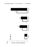 |
 |  |
 |  |
 |  |
 | 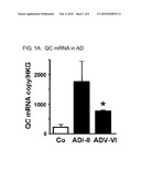 |
 |  |
 | 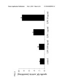 |
 |  |
| New patent applications in this class: | |
| Date | Title |
|---|---|
| 2022-05-05 | Detection of misfolded tau protein |
| 2022-05-05 | Device for methods of detecting cancer |
| 2022-05-05 | Device and method for detecting protein-based marker, and method for manufacturing chip |
| 2022-05-05 | Anti-gliadin antibodies |
| 2019-05-16 | Methods of measuring adamts 13 activity |
| New patent applications from these inventors: | |
| Date | Title |
|---|---|
| 2016-02-25 | Diagnostic antibody assay |
| 2016-02-11 | Novel inhibitors |
| Top Inventors for class "Chemistry: molecular biology and microbiology" | |
| Rank | Inventor's name |
|---|---|
| 1 | Marshall Medoff |
| 2 | Anthony P. Burgard |
| 3 | Mark J. Burk |
| 4 | Robin E. Osterhout |
| 5 | Rangarajan Sampath |