Patent application title: Method Of In-Vitro Detection And Quantification Of HIV DNA By Quantitative PCR
Inventors:
Maria Angeles Muñoz Fernandez (Madrid, ES)
Maria Angeles Muñoz Fernandez (Madrid, ES)
Gerónimo Fernandez Gomez-Chacon (Madrid, ES)
Assignees:
GENOMADRID S.A.
IPC8 Class: AC12Q168FI
USPC Class:
435 6
Class name: Chemistry: molecular biology and microbiology measuring or testing process involving enzymes or micro-organisms; composition or test strip therefore; processes of forming such composition or test strip involving nucleic acid
Publication date: 2009-08-20
Patent application number: 20090208929
Inventors list |
Agents list |
Assignees list |
List by place |
Classification tree browser |
Top 100 Inventors |
Top 100 Agents |
Top 100 Assignees |
Usenet FAQ Index |
Documents |
Other FAQs |
Patent application title: Method Of In-Vitro Detection And Quantification Of HIV DNA By Quantitative PCR
Inventors:
Maria Angeles Munoz Fernandez
Geronimo Fernandez Gomez-Chacon
Agents:
LADAS & PARRY LLP
Assignees:
GENOMADRID S.A.
Origin: NEW YORK, NY US
IPC8 Class: AC12Q168FI
USPC Class:
435 6
Abstract:
Process for the detection and quantification in-vitro of HIV DNA by
quantitative PCR. The invention consists of developing pairs of
oligonucleotides capable of hybridizing with fragments of the gag gene
sequence present in the genome of the HIV virus. These oligonucleotides
permit amplification of the viral DNA by quantitative PCR. Even in the
case of samples with low viral load, the invention permits detection of
the HIV DNA virus by juxtaposition either of a conventional PCR with a
quantitative PCR, or with a double nested quantitative PCR, using
different pairs of primers in each amplification. The invention, compared
with the known methods, allows determining qualitatively and
quantitatively, in-vitro, the presence of HIV DNA in samples, in a rapid,
reproducible manner and with high sensitivity.Claims:
1-27. (canceled)
28. An in-vitro method of quantitative detection of DNA of the HIV virus that comprises a double amplification of the viral DNA present in the sample using a double nested quantitative PCR carried out in such a way that a first pair of primers which bind specifically to the gag gene is used in the first amplification and a second pair of primers which bind to the gag gene inside the fragment amplified by the first pair of primers, characterized in that the amplification and the detection of the viral DNA are carried out in a single capillary, in which the amplified DNA is detected by means of DNA probes which bind specifically to the amplified product.
29. The method according to claim 28, characterized in that the first pair of primers binds specifically to the gag gene between positions 1286 and 1435 of said gene.
30. The method according to claim 29, characterized in that one of the oligonucleotides that form the first pair of primers is as represented by SEQ ID NO: 1.
31. The method according to claim 30, characterized in that the other oligonucleotide of the first pair of primers is as represented by SEQ ID NO:2.
32. The method according to claim 29, characterized in that one of the oligonucleotides that form the second pair of primers is as represented by SEQ ID NO:7.
33. The method according to claim 32, characterized in that the other oligonucleotide of the second pair of primers is as represented by SEQ ID NO:8.
34. The method according to claim 29, characterized in that the sequence of nucleotides of one of the probes is as represented by SEQ ID NO: 5.
35. The method according to claim 34, characterized in that the sequence of nucleotides of the second probe is as represented by SEQ ID NO:6.
36. The method according to claims 29, characterized in that the first pair of primers are as represented by SEQ ID NO:1 and SEQ ID NO:2, the second pair of primers are as represented by SEQ ID NO:7 and SEQ ID NO:8 and the sequence of nucleotides of the probes are as represented by SEQ ID NO:5 and SEQ ID NO:6.
37. The method according to claim 28, characterized in that the probes are labelled with F.R.E.T. type fluorochromes, thereafter the number of copies of viral DNA present in the test sample is quantified by extrapolating the fluorescence obtained to a previously calibrated reference straight or curve.
38. Test kit for quantifying DNA of the HIV virus, characterized in that if comprises a reagent that contains:i) at least one first pair of oligonucleotides PCR primers which bind specifically to the gag gene between positions 1286 and 1435 of said gene,ii) at least one second pair of oligonucleotides PCR primers with a melting point (Tm) different from that of the first pair of primers and which bind specifically to the gag gene inside the fragment determined by the first pair of primers, that uses as template the product previously amplified by said first pair of primers, and, jointly or separately in a separate reagent,iii) a pair of oligonucleotides probes which bind specifically to the fragment of the gag gene comprised between the second pair of primers.
39. Test kit according to claim 38, characterized in that one of the oligonucleotides that form the first pair of primers is as represented by SEQ ID NO: 1.
40. Test kit according to claim 39, characterized in that the other oligonucleotide of the first pair of primers is as represented by SEQ ID NO:2.
41. Test kit according to claim 38, characterized in that one of the oligonucleotides that form the second pair of primers is as represented by SEQ ID NO:7.
42. Test kit according to claim 41, characterized in that the other oligonucleotide that form the second pair of primers is as represented by SEQ ID NO: 8.
43. Test kit according to claim 38, characterized in that one of the oligonucleotide probes is as represented by SEQ ID NO:5.
44. Test kit according to claim 43, characterized in that the second oligonucleotide probe is as represented by SEQ ID NO: 6.
45. Test kit according to claim 38, characterized in that the first pair of oligonucleotide primers is as represented by SEQ ID NO: 1 and SEQ ID NO:2, the second pair of oligonucleotide primers is as represented by SEQ ID NO:7 and SEQ ID NO:8 and the pair of oligonucleotide probes is as represented by SEQ ID NO:5 and SEQ ID NO:6.
46. Test kit according to claim 38, characterized by the pair of oligonucleotide probes being labelled with FRET-type fluorochromes.
Description:
FIELD OF THE INVENTION
[0001]The invention relates to the field of PCR-based methods and devices for diagnosis. Concretely, it relates to the detection and quantification of HIV DNA in patients.
STATE OF THE ART
[0002]The main parameters used for monitoring HIV infection are the plasma "viral load" (quantification of HIV RNA) and the percentage or number of CD4 lymphocytes [1]. It is very important to know the number of copies of HIV DNA present in cells, especially in patients undergoing Highly Active Antiretroviral Therapy (HAART) in which the viral load has decreased, even to an undetectable level, and an increase or stabilization of the percentage or number of CD4 lymphocytes has been observed, as well as in individuals when the treatment is interrupted [3].
[0003]Benefits of quantification of viral DNA: [0004]Diagnosis of HIV infection in children of seropositive mothers and in adults during the window (when treatment is interrupted). The advantage is that, being quantitative, it will give the number of copies of HIV DNA present in the cells at the time closest to infection, which can be converted to a prognostic marker of the development of infection. [0005]In patients treated with highly active antiretroviral therapy (HAART), in which the viral load is undetectable (<400 copies/ml; ultrasensitive<50 copies/ml), at present the CD4 lymphocytes are the only existing immune system marker, it would be very important to possess a virological marker and in this sense it would be possible to use the number of copies of HIV DNA present in the cells as a prognostic marker of development, which predicts when a jump in viral load is going to occur and avoid the appearance of resistant strains. [0006]In patients when treatment is interrupted. Before the antiretroviral treatment is interrupted, one of the requirements that the patient must satisfy is an undetectable viral load, so it would be very useful to know the number of copies of HIV DNA present in the cells prior to interruption and to know how long they can remain without treatment so there is no jump in viral load and development of resistance to the antiretrovirals. [0007]In patients for whom an immunological treatment or therapeutic vaccine is to be applied, where it would be very important to know the number of copies of HIV DNA present in the cells in order to see up to what point the number of cells infected is important for achieving an immunological system that is functional in the long term and which can control the replication of HIV by itself. [0008]To detect infected cell subpopulations that can act as a reservoir of HIV and that potentially cannot be infected by it. [0009]Application to basic research in various studies in vitro and in vivo. [0010]As a parameter for the detection and monitoring of inactive HIV. [0011]In general it will be a very useful technique for application in routine clinical practice and in clinical tests of HIV+ patients.
[0012]The application of new antiretroviral therapies to HIV infection has caused dramatic changes in the natural development of this infection, therefore it is very interesting to know the relationship between reconstitution of the immune system and persistence of HIV in the patient, moreover we would be able to find out at what moment the immune system would be able to control the infection and antiretroviral treatment can be withdrawn. The existing techniques of quantification of viral load and genotyping or phenotyping are not able to predict the development of the infection when the viral load is undetectable independently of the percentage or number of CD4 T lymphocytes.
[0013]The quantification of viral DNA proposed by the present invention represents a novel technique which could be useful as a diagnostic and prognostic marker of development of the infection and for monitoring it. In addition, it would enable us to detect the cellular subpopulations that act as a reservoir for HIV.
[0014]We present an innovative method for quantifying HIV DNA (viral DNA load) in infected cells, in a simple, reproducible and rapid manner by quantitative PCR in real time. The method described detects and quantifies all the forms of HIV DNA present in the cell, both integrated and non-integrated, even though there is a very small number of copies, applying in this case an embodiment of the invention that makes the method ultrasensitive. At the present time there is no method that is capable of detecting proviral HIV DNA (integrated into the genome) in a specific, reproducible, simple and rapid manner.
[0015]The ultimate objective of any living organism is its perpetuation in time. Like other retroviruses, the only way that HIV has of achieving this is to insert its genome in the cellular DNA.
[0016]In the first stages of the infection cycle, at the end of the uncoating process, the reverse transcription reaction begins, giving rise to the Pre-Integration Complex (PIC), with the viral genome now in the form of DNA, which moves to the nucleus. There, the PIC can undergo various processes: recircularization of the viral DNA with one or two long terminal repeats (LTR), or integration into the cell chromosome [2]. It is only in this last case that the functional provirus, the only infectious form, is established. In this way, the HIV will not be detected by the immune system and will be able to remain in the cell, and it will replicate with the rest of the cellular DNA, generating new cells carrying HIV. In optimum conditions this HIV DNA will be transcribed and translated by the cellular machinery, generating new HIV particles which will infect new cells [2].
[0017]At present it is possible to quantify the number of viral particles circulating in plasma by the "viral load" technique (detection of HIV RNA) which, together with the immunological marker for quantification of CD4 lymphocytes, are the two parameters usually employed for monitoring HIV infection in patients by medical personnel, for introducing, or changing, antiretroviral treatment.
[0018]The number of viral particles that can infect the same cell is variable, as is the number of times that the same RNA is reverse transcribed, so that the amount of viral DNA contained in a cell is variable, and there may be a very large number of copies or a very small number of them.
[0019]The proviral DNA represents a hidden threat to the life of the cell, and therefore to the life of the infected person, because the viral load can be activated and can increase without this being predicted by any of the parameters used, leading to failure of the antiretroviral therapy and emergence of resistant strains. For this reason, in patients who are beginning antiretroviral treatment for the first time, or are being treated with HAART, some other therapy, or beginning interruption thereof, it may be important to know the behaviour and variations of the proviral DNA, "Proviral Load".
[0020]Various tests have been developed in recent years for the detection and quantification of proviral DNA using the PCR (polymerase chain reaction) technique, but these methods have not been able to detect it in all cases [4][5][6] since they only operate with indications of the position at which the viral DNA is integrated into the cellular genome. The sequence of the HIV genome is now well known and therefore it has been possible to design pairs of primers or oligonucleotides which form rings in some gene of the HIV and thus its presence can be detected by PCR and the total DNA can be quantified.
[0021]Methods are described in the state of the art for the detection of HIV DNA by PCR, by designing primers that form circles in the pol viral gene and in the gag gene, as they are the genes that have a less variable sequence. Concretely, the gag gene has 1300 bp, from which it is possible to obtain hundreds or thousands of pairs of different primers, each one with characteristics that are also different, in particular on the basis of their "melting point" (Tm). Some techniques use simple PCR with primers of po1 or gag, or alternatively "nested PCR", that is, two consecutive PCRs with the same or a different pair of primers for each, using the product amplified in the first as the DNA template for the second. Detection of the product in these two types of PCR is achieved by visualization of the amplified product in agarose gel. Consequently, the information obtained is only qualitative, not quantitative, in contrast to the method of our invention.
[0022]Other methods propose quantifying the PCR product by ELISA or hybridization on the amplified product after carrying out electrophoresis in agarose gel.
[0023]The method of the invention uses the technique of quantitative PCR in real time from Roche "LightCycler-qPCR" (LC-qPCR), although it would be possible to use any system of quantitative PCR (Applied Bio-System), with appropriate adaptation of the amplification programme, reference curves and other parameters known by any person skilled in the art who uses methods of quantitative PCR of this type. For the method of the invention, primers were designed for the gag gene, and a pair of probes of the F.R.E.T. type (Fluorescence Resonance Transfer) for detecting the amplified DNA product. This permits greater accuracy in detection of the specific product, without detecting possible undesirable by-products, since it is necessary for said probes to form circles in their exact positions of the product for it to be quantified, in contrast to the Taqmane probe, which when acting on request, can quantify products not specific to circle formation in products with sequence similar to their target. To date, no method has been described for quantification of proviral DNA of HIV using the LightCycler-qPCR system of quantitative PCR.
[0024]One object of the present invention is to develop pairs of oligonucleotides as in claim 1, which bind specifically to the gag gene of the HIV virus and permit amplification of the viral DNA by PCR.
[0025]A second object of the invention is to develop a pair of probes, as in claim 2 and the dependent claims, which permits efficient detection, by fluorescence, of the product of viral DNA of HIV amplified by PCR, eliminating the nonspecific background.
[0026]A third object of the invention is a method of in-vitro detection of viral DNA of HIV, as described in claim 7 and the dependent claims, which permits the detection of proviral DNA of HIV.
[0027]Another object of the invention is the test kits, as in claim 17 and the dependent claims, which comprise the oligonucleotides of the invention, optionally also using the probes of the invention.
[0028]A preferred embodiment of the invention comprises applying the method of detection, as envisaged in claim 11 and the dependent claims, also to samples with low viral load (<100 copies).
[0029]Another preferred embodiment of the invention comprises modifying the method of detection and quantification of HIV DNA by developing a 2nd pair of primers, which makes it possible to shorten the test times while at the same time increasing the specific fluorescence signal of the viral DNA present in the test sample, as protected in claim 13 and the dependent claims.
[0030]A further object of the invention comprises the uses envisaged in claims 24 to 27, both of the oligonucleotides and of the probes, in kits and/or methods of detection of viral DNA of HIV.
DESCRIPTION OF THE INVENTION
[0031]The invention comprises an innovative method for the quantification of HIV DNA in cells. This method is characterized by sensitivity, reproducibility and specificity for the detection/quantification of HIV DNA in samples with a medium/high number of initial copies and moreover a preferred embodiment of the invention comprises an ultrasensitive method for the detection of samples with low initial copies.
[0032]An important point that should be emphasized in this method is the extraction of the DNA, which must be obtained in pure and clean form, and its quantification by UV spectrophotometry in order to find out the amount of template to be used in the PCR reaction, thus permitting standardization of the results per μg of DNA or per 106 cells.
[0033]It is not possible to assess a priori whether the amount of HIV DNA present in each sample can be detected by the conventional method of the invention or whether it is necessary to have recourse to the ultrasensitive method. For this reason, LC-qPCR-Gag is carried out first, and if amplification is not obtained, the ultrasensitive method for samples with low number of initial copies of HIV DNA is applied. The ultrasensitive method for samples with low number of initial copies of HIV DNA is only applied directly in the case when the number of CD4 lymphocytes is below 400 cells/ml.
[0034]Only the T cells present in the mononuclear cells of peripheral blood of the patient are potential carriers of the viral DNA and, in accordance with the information obtained by this method, the number of copies of viral DNA per T lymphocyte varies in relation to the number of CD4 lymphocytes, therefore the quantification of the HIV DNA must be analysed in comparison with the number of CD4 lymphocytes and the viral load to permit better assessment of development of the infection.
[0035]To apply the method of the invention, the quantitative PCR apparatus called LightCycler (Roche) is used, which comprises a thermal cycler with air cooling and heating of the samples. This apparatus is connected to a fluorometer which detects the fluorescence emitted, for example that can be attributed to probes of the FRET type that bind to the product formed in each cycle of the polymerase chain reaction (PCR). For this apparatus to give a number of copies or counts in the samples it must extrapolate the fluorescence obtained in these to a reference straight line or curve that is obtained by putting a known number of DNA copies at each point on said straight line. The apparatus, connected to a computer, relates the fluorescence obtained at each point on the curve to the respective number of copies of DNA and shows it on a graph of fluorescence versus number of copies, this graph being adjusted to a straight line that enables us to calculate, on the basis of the fluorescence measured in the test sample, the number of copies of viral DNA present in the latter.
[0036]For detecting the HIV DNA by PCR we require a pair of primers, oligonucleotides or initiators which form circles in some viral gene, and use an appropriate programme [8]. HIV is a retrovirus with high capacity for mutation, mainly at the env gene and, in the presence of antiretroviral treatment, also at the pol gene. For this reason the initiators must form circles in more conserved genes, with less variability, such as the gag gene, thus making it possible to detect all the possible variants of HIV, in all the forms present in the cell, integrated or not integrated.
[0037]For this innovative method, two primers have been designed, F-Gag and R-Gag (SEQ ID NO: 1 and SEQ ID NO: 2), which amplify a 222 bp fragment of the gag gene (at positions 1260 to 1482) since, as already mentioned, it is a gene with a highly conserved sequence. Throughout this specification, the terms primer, initiator or oligonucleotide are used as synonyms.
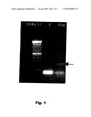
[0038]To determine the best conditions of the programme that has to be applied for carrying out the PCR, various experiments were repeated, varying the temperature of circle formation of the primers, their concentration, the concentration of MgCl2 and the number of cycles. The conditions selected were: a programme that has initial denaturing of 10 minutes at 95° C., followed by 35 cycles of amplification with 1 minute at 95° C., 30 seconds at 65° C. and 1 minute at 72° C., with a final extension of 4 minutes at 72° C. Each 25 μl of reaction mixture contains 0.5 to 1 μg of genomic DNA, 0.8 μM of each primer, 0.10 mM dCTP, 0.10 mM dGTP, 0.10 mM DATP, 0.10 mM dTTP, 1 U of DNA polymerase enzyme Amplitaq Roche, 50 mM KCl, 3.5 mM MgCl2 and 10 mM of Tris-HCl (pH 8.3). The amplified product was tested in 2% agarose gel (FIG. 1).
[0039]A preferred embodiment of the invention comprises modifying the method, carrying out nested PCR using, in addition to the 1st pair of primers (SEQ ID NO: 1 and 2) in a 1st amplification, a 2nd pair of primers F2-Gag and R2--Gag (SEQ ID NO: 7 and 8), which amplifies in its turn, in a 2nd stage, the amplified product resulting from the 1st stage of amplification (product Gag1). This 2nd pair of primers forms circles inside the fragment amplified by the 1st pair (between positions 1286 and 1435), giving a product Gag2 of 149 bp, on which the probes P1-Gag and P2-Gag form circles (between positions 1320 and 1381).
DETAILED DESCRIPTION OF THE INVENTION
Example 1
Quantitative PCR-Gag with the LightCycler System
[0040]The genomic DNA was extracted from 106 cells with the "Wizard SV Genomic DNA purification system" kit from PROMEGA (cat. #A2360), and resuspended in 100 μl of nuclease-free water PROMEGA (cat. #P119C). Finally, this DNA was quantified with the Nano-prop 3.0.0/3 spectrophotometer.
[0041]The equipment for carrying out the quantitative PCR is a LightCycler, a thermal cycler using air which incorporates a fluorometer for the detection and quantification of the amplified products without the need for post-PCR analysis in agarose gel of the products obtained, as occurs when carrying out conventional PCR. Amplification was carried out in glass capillaries using a modified DNA polymerase enzyme of high efficiency, high rate of elongation and capacity for supporting rapid temperature changes (20° C./s). The advantage of using glass capillaries is that it permits rapid heating and cooling of the reaction mixture, and therefore very short cycles and programmes are carried out [7][9]. The amplified products are detected by SYBR-Green, a marker compound that binds to double-stranded DNA by intercalation and emits fluorescence when excited with light of a suitable wavelength. Another means of detection/quantification is the use of DNA probes (SEQ ID NO: 5 and SEQ ID NO: 6) labelled with fluorochromes (F.R.E.T. type), which bind specifically to the amplified product. The products are quantified by extrapolation of the fluorescence obtained in each of the samples relative to a reference curve obtained with a known number of copies.
Example 2
Detection of the Amplified Product by SYBR-Green (Cambrex)
[0042]This embodiment of the invention is used for samples whose viral DNA load is assumed to be high. At the start of development of the method of quantification of HIV DNA, conventional PCR-Gag was adapted to the conditions of the LightCycler quantitative PCR (LC-qPCR). The programme for the new thermal cycler begins with 10 minutes of denaturing at 95° C., followed by 40 cycles with 15 seconds of denaturing at 95° C., 10 seconds for circle formation of primers and single data collection of fluorescence, and 20 seconds of elongation at 72° C. Each 20 μl of reaction mixture contains 0.5 μg to 1 μg of genomic DNA, 0.5 μM of each primer (F-Gag and R-Gag), 0.10 mM of DATP, 0.10 mM of dCTP, 0.10 mM of dTTP, 0.10 mM of dGTP, and 1 U of LightCycler FastStart DNA polymerase enzyme (Roche), 50 mM KCl, 3.5 mM MgCl2, and 10 mM Tris-HCl (pH 8.3). The amplified products are detected with SYBR-Green. This marker only detects double-stranded DNA, therefore it does not interfere with the primers used, unless they form dimers. The reference curve was obtained using serial dilution in steps of an order of magnitude from 104 to 1 copy per reaction mixture, with DNA from cells of the 8E5 line, a lymphoid cell line that is characterized in that it carries a single HIV provirus per cell. To produce this reference straight line or curve, the DNA is extracted from 10 aliquots with 1 million cells per aliquot, with measurement or quantification in a UV spectrophotometer. The average weight of the genomic DNA obtained is found and the weight of the genomic DNA of an 8E5 cell is determined at an average of 5 μg, meaning that 5 pg of DNA corresponds to one copy of HIV DNA.
[0043]The system for detecting the amplified product by SYBR-Green is nonspecific, since it binds to all the double-stranded DNA present in the reaction mixture, for example the dimers of primers, therefore producing much background and generating some quantification data with errors. Although the computer program of the LightCycler-qPCR system makes it possible to subtract the background fluorescence obtained, negative control of the remaining samples, a significant error would be made, since the background due to the dimers of primers is different in each sample, depending on their initial amount of template, larger amount of initial template leading to less formation of dimers of primers, therefore it is not possible to subtract the same amount of fluorescence from each sample. However, this error is acceptable in the case of samples with a high viral DNA load.
Example 3
Detection of the Amplified Product by Means of F.R.E.T. Probes
[0044]So as to be able to avoid these problems in the quantification of HIV DNA, when the viral DNA load is not high or is not assumed to be high, a pair of DNA probes labelled with fluorochromes was designed, of the F.R.E.T. type, designated P1-Gag and P2-Gag (SEQ ID NO: 5 and SEQ ID NO: 6), which hybridize specifically in a region between the F-Gag and R-Gag primers of the product amplified in PCR-Gag. In this way only the specific PCR product without a background signal, and being real data, is detected and quantified.
[0045]To improve and increase the accuracy of the method of detection with SYBR-Green, a new reference curve was constructed that is more exact than the previous one with DNA from 8E5 cells. To construct this reference curve, the product amplified with conventional PCR-Gag, with DNA from the 8E5 cell, was cloned in the targets EcoRI (SEQ ID NO: 4) and BamHI (SEQ ID NO: 3) within the plasmid Bluescript II(-) (pBSK II-), Stratagene #212216, grown in Escherichia coli DH5a bacteria and purified by Maxiprep. The plasmid with the insert was designated "pBSKGag" and after quantifying it in a UV spectrophotometer and determining the number of molecules of plasmid per μl, a serial dilution was prepared from it with steps of the order of magnitude from 1010 to 1 molecule/μl. The primers shown as SEQ ID NO: 3 and SEQ ID NO: 4 are used for cloning the amplified product between the targets BamHI and EcoRI of the plasmid pBSK(-) for use as template DNA in the reference straight line. These primers shown by SEQ ID NO: 3 and SEQ ID NO: 4 are the same as SEQ ID NO: 1 and SEQ ID NO: 2 but with the bases added at 5' corresponding to the targets of the restriction enzymes described. Each 20 μl of reaction mixture of LC-qPCR-Gag contains from 0.5 to 1 μg of genomic DNA as template, 0.2 μM of each fluorescent probe P1-Gag and P2-Gag, 0.5 μM of each primer F-Gag and R-Gag, 0.10 mM of dATP, 0.10 mM of dCTP, 0.10 mM of dTTP, 0.10 mM of dGTP, and 1 U of LightCycler FastStart DNA polymerase enzyme (Roche), 50 mM KCl, 3.5 mM MgCl2, and 10 mM Tris-HCl (pH 8.3).
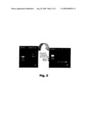
[0046]With the aim of verifying the exactness of the reference curve and ensuring that all the conformers of plasmid pBSK-Gag present and quantified by UV spectrophotometry in said curve give the expected product in said PCR, electrophoresis was carried out in 1% agarose gel (FIG. 2), with 500 ng of said plasmid to observe its various conformers: linear, circular relaxed (with a cut in one of its strands) and supercoiled. The three conformers were cut out of the gel and extracted separately with GENECLEAN® Spin kit--BIO101 (Cat. #1101-200) in 20 μl of nuclease-free water. 5 μl was collected as template from each one for PCR-Gag and in order to see the result 10 μl of said PCR was loaded in a 2% agarose gel. The Gag product was obtained from the three conformers, which clearly indicates that all the plasmid pBSK-Gag quantified gives product in PCR-Gag. In conclusion, the theoretical DNA copies of the reference curve are very close to the real ones.
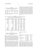
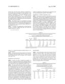
[0047]We deduce, from the results obtained in various tests with the reference curve (FIG. 4), that it is possible to take at least 4 points, from the total of 7. The average rate of variation between the different reference curves is less at the points from 103 to 105 (from -0.1 to +0.1) than at the points 104 and 103, -0.2 to +0.2, and at point 100 it is -3.5. The remaining points 10 and 1 have an average rate of variation of -59.1 and -319.0 respectively, too high, since they have problems of amplification associated with the samples with target DNA at low number of copies; therefore these points are never included in the real reference curve, so that the lower limit of detection with this qPCR (Table 1) would be 100 copies.
TABLE-US-00001 TABLE 1 Reference curves pBSK-Gag of LC-qPCR Gag. Theoretical number Exp 1: 18 Exp 2: 20 Exp 3: 20 Exp 4: 23 Exp 5: 23 Exp 6: 2 Average rate of Tube of copies Mar. 2004 Feb. 2004 Feb. 2004b Feb. 2004 Feb. 2004b Mar. 2004 variation 1 100000000 112500000 131100000 97580000 102400000 104000000 -0.1 2 10000000 8339000 12390000 10970000 9607600 13100000 -0.1 3 1000000 1055000 1180000 895000 1393000 965000 1133000 -0.1 4 100000 98600 74160 1040001 96600 92000 87300 0.1 5 10000 10070 4739 7300 5946 10800 9028 0.2 6 1000 1018 1 874 16561 839 1900 1120 -0.2 7 100 222 542 795 150 568 431 -3.5 8 10 600 602 -59.10 9 1 250 390 -319.00 NEG control 0 0 0 0 0 0 0 H2O control 0 0 0 0 0 0 0 Internal C 1 100000 1070001 Internal C 2 1000 1078 1 1177 15871 R 1 1 1 1 1 1 Error 0.05 0.1 0.07 0.1 0.04 0.08 Standard Average error of the reference curve: 0.07 Exp 1-5: Experiments on different dates. C1 and C2 are internal controls which are incorporated in the reference straight line without this being indicated in the programme
Example 4
Detection of the Product Amplified in Samples with Low Number of Copies. Nested PCR or Double PCR Using the Same Pair of Primers Twice in Succession
[0048]Quantification in samples with low number of copies in the template DNA is very difficult because copies may not be detected or a higher number of copies may be detected than are really present in the sample. The formation of dimers of the primer and nonspecific amplification products are problems associated with samples of this type, since they are generated more easily than the product of interest. These amplification artefacts compete with the target sequences for the reagents of the PCR reaction mixture, giving abnormal values of amplification and quantification.
[0049]To increase the number of copies in the template of LC-qPCR, we carry out a "nested PCR" or double PCR, a combination of conventional PCR-Gag and LC-qPCR-Gag, using the same primers in the two PCRs, F-Gag and R-Gag. In this way it is possible to detect the target DNA when there is a very small amount in the sample. The first conventional PCR-Gag makes it possible to generate sufficient product from the initial sample, for use subsequently as template in LC-qPCR-Gag. In this way there is sufficient starting target DNA without the artefacts or dimers of the primer competing for the specific product of PCR, thus permitting its correct quantification.
[0050]The template used for carrying out LC-qPCR is 10 μl of the 25 μl of the conventional PCR-Gag of samples from patients, and three points are put on the reference curve with low number of copies: 100, 10 and 1 copy per reaction mixture. The quantification obtained in LC-qPCR corresponds to the number of copies present in the 10 μl of conventional PCR used as template in LC-qPCR, the real copies do not exist in the sample. Using the counts obtained at each of the points on the curve with 100, 10 and 1 initial copy, the efficiency (E) of conventional PCR-Gag is obtained using the equation: [8]
Tn=To (E)n
where Tn is the number of copies at the end of PCR, To is the number of initial copies in the template, E is the efficiency of the PCR reaction (it is usually between 1.8 and 2), and n is the number of cycles of PCR. The point on the reference curve 100 has To(100)=100 and its Tn(100) in conventional PCR-Gag is its To(100)q in LC-qPCR-Gag. Inserting these values in equation (1), we get the value of E:
E=n Tn/To (2)
E=35 To(100)q/100 (3)
[0051]The E obtained in (3) of conventional PCR-Gag is calculated for the three points of the reference curve, 100-10-1, and the average E (1.8-2) is determined, which can be assumed to be the same for all the samples of the same reaction, so that on this basis it is possible to determine the number of initial copies present in patients' samples, To(pi), (4). The E of a PCR is not exactly the same for all the samples, nor in all of the reaction cycles, since it depends on the number of target DNA present in the template. In the initial cycles, the template is small, and so is E, and as the amount of template increases so too does E, since there is more template and more copies are produced, amplification becoming exponential, therefore E approaches 2.
[0052]In LC-qPCR-Gag, for each patient sample the To(pi)q is obtained, which is the Tn(pi) from conventional PCR-Gag, which when inserted in (4) gives the To(pi).
Topi=Tnpi/E35 (4)
[0053]The index of E depends on the number of PCR cycles applied.
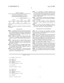
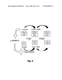
[0054]The algorithms adopted constitute the computer program designed and included in the present invention for correct application of the method of measurement of viral DNA described (explanatory diagram in FIG. 3).
Example 5
Results of Application of the Gag1 Ultrasensitive Method
[0055]The technique described was applied to a group of HIV infected patients undergoing HAART therapy with low or undetectable "viral load". LC-qPCR-Gag was carried out, without obtaining counts in any of them, therefore the method was applied for samples with low number of initial copies, first carrying out a conventional PCR-Gag, and then 10 μl of this is used as template for LC-qPCR-Gag.
[0056]The results obtained in these samples were below 435 copies/μg DNA (1306 copies/106 cells), there being samples with less than 1 copy/μg DNA, i.e. 1 copy per 333333 cells (since the genome of a human cell is approximately equivalent to 3 pg). Only in the cases in which amplification was not obtained is it possible to say that they have "undetectable HIV DNA".
[0057]Samples with medium/high values of HIV DNA are detected in the first LC-qPCR-Gag and are between 1874 and 3367 copies/μg DNA (from 5623 to 10100 copies/106 cells). (Tables 2 and 3).
TABLE-US-00002 TABLE 2 Result from samples with medium/high number of copies Vol DNA 1 N N cop Vol million cop/million Ncop/μg Sample qPCR template(μl) cells(μl) cells DNA E 3.0 562.3 10 100 5623 1874 E 3.4 761.4 10 100 7614 2538 E 4.0 513.3 10 100 5133 1711 E 4.4 1010 10 100 10100 3367 1 cell = 3 pg DNA; 106 cells = 3 μg DNA
TABLE-US-00003 TABLE 3 Determination of E of PCR-Gag. Results of samples with low number of copies N Cop Dilution N Cop (Efficiency)35 N Cop/μg N Cop/ N Cop/ Sample 10 μl PCR factor 25 μl PCR Tn 100 DNA million cells million cells 1 228900000 1 572250000 850500000 0.67284 2.01852 2.01852 2 868300000 1 21707500000 850500000 25.52322 76.56967 76.56967 3 37580 1 93950 850500000 0.00011 0.00033 0.00033 4 531.3 1 1328.25 850500000 0.00000 4.68519E-06 4.68519E- 5 345100000 1 8627500000 850500000 10.14403 30.43210 30.43210 6 578600 2 2893000 850500000 0.00340 0.01020 0.01020 7 107000000 2 53500000000 850500000 62.90417 188.71254 188.71254 8 740700000 2 3.7035E+11 850500000 435.44974 1306.34934 1306.34934 9 232100000 2 1.1605E+11 850500000 136.44915 409.34748 409.34748 10 606.6 2 3033 850500000 0.00000 1.06984E-05 1.06984E- 11 11440 2 57200 850500000 0.00007 0.00020 0.00020 12 978 2 4890 850500000 0.00001 1.72487E-05 1.72487E- 13 1260 2 6300 850500000 0.00001 2.22222E-05 2.22222E- 14 686 2 3430 850500000 0.00000 1.20988E-05 1.20988E- 15 617 2 3085 850500000 0.00000 1.08818E-05 1.08818E- 16 2167 2 10835 850500000 0.00001 3.82187E-05 3.82187E- 17 457200 2 2286000 850500000 0.00269 0.00806 0.00806 18 221800000 2 11090000000 850500000 13.03939 39.11817 39.11817 19 117900000 2 58950000000 850500000 69.31217 207.93653 207.93653 20 432800000 2 21640000000 850500000 25.44386 76.33158 76.33158 21 330900000 2 16545000000 850500000 19.45326 58.35979 58.35979 22 17300000 2 86500000 850500000 0.10170 0.30511 0.30511
Example 6
Modification of the Ultrasensitive Method of Nested Quantitative PCR Using a 2nd Pair of Primers Different from the 1st
[0058]For the purpose of avoiding deficiencies in the detection of the HIV genome in samples with small amount of DNA, and moreover to simplify the method and reduce the test duration, by processing all the samples equally without taking into account whether they might contain a high or low number of copies, another preferred embodiment of the invention was developed, comprising nested quantitative PCR.
[0059]The method comprises carrying out a nested PCR [8][10][11] using a 2nd pair of primers that form circles in an internal region of the amplified product with the F-Gag/R-Gag primers of 222 bp (Gag1). This 2nd pair of primers, F2-Gag/R2--Gag, forms circles in the sequences that separate the oligonucleotides F-Gag--P1-Gag, and P2-Gag--R-Gag, respectively, but with a melting point (Tm) 10° C. lower than the pair F-Gag/R-Gag, giving a product of 149 bp, Gag2. These differences in the Tm values make it possible to design a programme of "nested quantitative PCR" in a single capillary per sample with a reaction mixture containing the two pairs of primers and the probes. Each 20 μl of reaction mixture of LC-qPCR-Gag 2.0 contains from 10 to 200 ng of genomic DNA as template, 0.2 μM of each fluorescent probe P1-Gag and P2-Gag, 0.05 μM of the primers F-Gag and R-Gag, 0.5 μM of the primers F2-Gag and R2--Gag, 0.10 mM of DATP, 0.10 mM of dCTP, 0.10 mM of dTTP, 0.10 mM of dGTP, and 1 U of LightCycler FastStart DNA polymerase enzyme (Roche), 50 mM KCl, 3.5 mM MgCl2, and 10 mM Tris-HCl (pH 8.3).
LC-qPCR-Gag 2.0 Programme:
TABLE-US-00004 [0060]1- Denaturing 1: 95° C., 10 minutes (20° C./s) 2- Amplification 1, 15 cycles 95° C., 15 seconds (20° C./s) 65° C., 12 seconds (20° C./s) 72° C., 12 seconds (20° C./s) 3- Denaturing 2: 95° C., 2 minutes (20° C./s) 4- Amplification 2, 30 cycles 92° C., 4 seconds (20° C./s) Fluorescence measurement 56° C., 10 seconds (20° C./s) 72° C., 8 seconds (20° C./s) 5- Final denaturing: 91° C., 10 seconds (20° C./s) 60° C., 10 seconds (20° C./s) 91° C., 1 second (0.20° C./s).sup. 6- End of programme: 40° C., 30 minutes.
[0061]A first amplification is carried out, in which the F-Gag/R-Gag primers are hybridized in the template DNA exclusively, the F2-Gag and R2--Gag primers do not, even though their circle forming sequence is present, since their temperature of circle formation is 10° C. lower than the pair F-Gag and R-Gag. After the 15 cycles of this first amplification, denaturing is effected at 95° C., which is the Tm of the product Gag1, thus the products obtained in Amplification 1 are denatured and are arranged for circle formation of the primers of Amplification 2. Then said amplification is initiated where the melting point is 92° C., at which only the product Gag2 is denatured, but not Gag1, hence the product that is obtained in this amplification comes only from Gag2, thus different products originating from the same initial template DNA will not be counted.
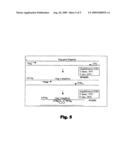
[0062]The products Gag1 are not quantified even though the probes form circles in them since the LightCycler thermal cycler is programmed for it, via its information application, so that fluorescence is only quantified during Amplification 2 (see diagram in FIG. 5).
[0063]To calculate Tm (melting point or denaturing temperature), a formula is applied that takes into account the number of bases G/C and A/T:
Tm=(wA+xT)*2+(yG+zC)*4
where w, x, y, z denote the respective number of bases A, T, G, C in the sequence.
[0064]In the present invention the sequence was inserted in the program on the Internet page: http://www.basic.nwu.edu/biotools/oligocalc.html and the program itself calculated each value of Tm.
Quantification and Sensitivity
[0065]In order to be able to quantify the samples it is necessary to extrapolate the fluorescence obtained in them on a reference straight line for which the plasmid pBSK-Gag was used, which has the cloned amplification product Gag1.
[0066]The main problem that this method may present is the actual quantification of the samples, since the quantification of fluorescence takes place in the 2nd amplification, accordingly this fluorescence is associated with the product obtained in the 1st amplification, and not with the number of initial copies present in each sample. This problem is overcome by the actual quantification technique, on referring it to the reference straight line. The same occurs on this reference straight line as in the remaining samples, but on assigning them to the fluorescence obtained in the 2nd amplification the number of real copies present at the beginning of the PCR the fluorescence extrapolated on said reference straight line of the various samples will give, as the result, the initial copies present in each of them. Finally, what is achieved with this method is amplification of the fluorescence signal from each unit Gag, increasing the sensitivity of the method.
Amplification 1, Number of Optimum Cycles
[0067]The increase in sensitivity of the method is given by Amplification 1, but if excessive amplification occurs it could saturate and equalize the template DNA for Amplification 2 in all the samples, obtaining a similar number of counts in all the samples even though not all had similar quantities initially. To determine the number of cycles of amplification 1 that increase the sensitivity of the method if the template is saturated for the 2nd amplification, several LC-qPCR Gag 2.0 were carried out with different amounts of DNA from cells 8E5 as template, from 550 ng to 0.00055 ng, in various inter- and intra-test repetitions, concluding that the number of cycles at which greatest accuracy (least error) is obtained, even with small amounts of template DNA (0.55 ng per reaction) is 15 cycles (Table 4).
TABLE-US-00005 TABLE 4 Results of LC-qPCR Gag 2.0 with different numbers of cycles in Amplification 1: 20, 10 or 15, compared with the expected theoretical result. Summary of quantification Gag 2.0 in DNA from cells 8E5 Theoretical Total DNA number of Sample (ng) copies/cell 20 cycles 10 cycles 15 cycles 15 cycles 15 cycles E1 550 1 0.07 0.03 -- 0.10 0.06 E2 55 1 0.43 0.12 0.12 0.26 0.63 E3 5.5 1 4.88 0.84 0.13 0.65 1.21 E4 0.55 1 18.82 10.20 3.85 4.02 4.97 E5 0.055 1 50.91 27.00 29.55 422.14 805.45 E6 0.0055 1 2176.36 393.64 1619.09 -- -- E7 0.00055 1 29727.27 2372.73 26681.82 -- -- Amplification 2 always has 30 cycles.
Reproducibility
[0068]The reproducibility of the results obtained with this method is assessed in Table 5. The variation in the intra-test duplicates is very low, except when the amount of template DNA is very low, starting from 0.55 ng, the inter-test variation is also very low, and does not exceed 1 unit up to 0.55 ng of template DNA, with smaller amounts the variability is very high, therefore from 5 ng to 500 ng has been taken as optimum amount of template DNA for this method.
TABLE-US-00006 TABLE 5 Data obtained in 2 different experiments carried out in the same conditions (amplification 1 of 15 cycles), with the same samples duplicated in each of them. Number of copies/Cell Total DNA average average (ng) exp 1 exp 1 exp 2 exp 2 E1a 550 0.12 0.10 0.07 0.06 E1b 550 0.09 0.06 E2a 55 0.29 0.26 1.01 0.63 E2b 55 0.22 0.26 E3a 5.5 0.84 0.65 1.12 1.21 E3b 5.5 0.46 1.29 E4a 0.55 7.74 4.02 3.44 4.97 E4b 0.55 0.30 6.51 E5a 0.055 41.92 422.14 55.45 805.45 E5b 0.055 802.36 1555.45 a,bduplicates
[0069]The reference straight line is obtained as described in Example 3.
REFERENCES
[0070]1. Guidelines for the Use of Antiretroviral Agents in HIV-1-Infected Adults and Adolescents. Developed by the Panel of Clinical Practices for Treatment of HIV Infection convened by the Department of Health and Human Services (DHHS). Mar. 23, 2004. [0071]2. Warner C. Greene and B. Matija Peterlin. 2002. Charting HIV's remarkable voyage through the cell:basis science as a passport to future therapy. Nature medicine. 8 (7). [0072]3. Yupi Zhao, Min Yu, Johann W. Miller, and Ram Yogev. 2002. Quantification of HIV-1 proviral DNA by using Taqman Technology. Journal of Clinical Microbiology. 40(2). p. 675-678. [0073]4. Una O'Doherty, W. J. Swiggard, Deepa Jeyakumar, David McGain and Michal H. Malin. 2002. A sensitive, quantitative assay for HIV-1 integration. Journal of Virology. 76(21). p 10942-10950. [0074]5. Scott L. Butter, Mark S. T. Hansen and Frederic D. Bushman. 2001. Aquantitative assay for HIV DNA integration in vivo. Nature Medicine. 7(5). [0075]6. L. G. Kostrikis, G. Toulomi, R. Karinicolas, and A. Hatzakis. 2002. Quantitation of HIV-1 DNA forms with the second template swich in peripheral blood cells predics disease progression independently of plasma RNA load. 76(20). p. 10099-10108. [0076]7. Stefan Mener, Carl Witter, Kan-Ichi Nakagawara. 2001. Rapid Cycle Real Time PCR. Methods and Applications. Ed. Springer. [0077]8. A. Rolfs, I. Schuller, U. Finckh, I. Weber-Rolfs. 1992. PCR: Clinical Diagnostics and Research. Ed. Springer. [0078]9. I. A. Teo, J. W. Choi, J. Morlese, G. Taylor, S. Shaunak. 2002. LightCycler qPCR optimisation for low copy number target DNA. J Immunology Methods.
DESCRIPTION OF THE DRAWINGS
[0079]FIG. 1: PCR-Gag, 222 bp product. The bands obtained correspond to the product amplified with the primers F-Gag and R-Gag on the viral DNA.
[0080]FIG. 2: A) Electrophoresis in 1% agarose gel of the plasmid pBSK-Gag. The three conformers are assessed, 1 linear, 2 relaxed, 3 supercoiled. B) Electrophoresis in 2% agarose gel of the products from PCR-Gag with each conformer of the plasmid pBSK-Gag. The marker in the two gels is a reference standard in 100 bp steps in each band.
[0081]FIG. 3: Explanatory diagram of the system for quantification in samples with low number of copies.
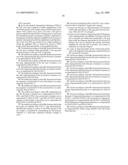
[0082]FIG. 4: Graphs of the reference curve pBSK-Gag of LC-qPCR Gag from Table 1. Graphs a-f show the reference curves obtained in different experiments and demonstrate the reproducibility of the method, the coefficient of correlation and the error obtained in each one of the said experiments.
[0083]FIG. 5: Explanatory diagram of double nested quantitative PCR using different pairs of primers in each amplification step.
Sequence CWU
1
8126DNAARTIFICIALPrimer 1tagtagaaga gaaggctttc agccca
26224DNAARTIFICIALPrimer 2ttggttctct catctggcct ggtg
24336DNAARTIFICIALPrimer
3cgggatcccg tagtagaaga gaaggctttc agccca
36434DNAARTIFICIALPrimer 4cggaattccg ttggttctct catctggcct ggtg
34530DNAARTIFICIALProbe 5gagccacccc acaagattta
aacaccatgt 30629DNAARTIFICIALProbe
6aacacagtgg ggggacatca agcagccat
29722DNAARTIFICIALPrimer 7gaagtaatac ccatgttttc ag
22821DNAARTIFICIALPrimer 8tgcagaatgg gatagattgc a
21
User Contributions:
comments("1"); ?> comment_form("1"); ?>Inventors list |
Agents list |
Assignees list |
List by place |
Classification tree browser |
Top 100 Inventors |
Top 100 Agents |
Top 100 Assignees |
Usenet FAQ Index |
Documents |
Other FAQs |
User Contributions:
Comment about this patent or add new information about this topic:
 |  |
 |  |
 |  |
 |  |
 |  |
 |
| New patent applications in this class: | |
| Date | Title |
|---|---|
| 2011-06-30 | Apparatus and method of authenticating product using polynucleotides |
| 2011-06-30 | Cyanine compounds, compositions including these compounds and their use in cell analysis |
| 2011-06-30 | Method for detecting multiple small nucleic acids |
| 2011-06-30 | Solid-phase chelators and electronic biosensors |
| 2011-06-30 | Cell-based screening assay to identify molecules that stimulate ifn-alpha/beta target genes |
| New patent applications from these inventors: | |
| Date | Title |
|---|---|
| 2010-02-11 | Novel carbosilane dendrimers, preparation method thereof and use of same |
| Top Inventors for class "Chemistry: molecular biology and microbiology" | |
| Rank | Inventor's name |
|---|---|
| 1 | Marshall Medoff |
| 2 | Anthony P. Burgard |
| 3 | Mark J. Burk |
| 4 | Robin E. Osterhout |
| 5 | Rangarajan Sampath |